Coccidioidin
Coccidioidomycosis
Histoplasmin
Skin Tests
Complement Fixation Tests
Immunoelectrophoresis, Two-Dimensional
Cell Migration Inhibition
Immunodiffusion
Microbial relatives of the seed storage proteins of higher plants: conservation of structure and diversification of function during evolution of the cupin superfamily. (1/26)
This review summarizes the recent discovery of the cupin superfamily (from the Latin term "cupa," a small barrel) of functionally diverse proteins that initially were limited to several higher plant proteins such as seed storage proteins, germin (an oxalate oxidase), germin-like proteins, and auxin-binding protein. Knowledge of the three-dimensional structure of two vicilins, seed proteins with a characteristic beta-barrel core, led to the identification of a small number of conserved residues and thence to the discovery of several microbial proteins which share these key amino acids. In particular, there is a highly conserved pattern of two histidine-containing motifs with a varied intermotif spacing. This cupin signature is found as a central component of many microbial proteins including certain types of phosphomannose isomerase, polyketide synthase, epimerase, and dioxygenase. In addition, the signature has been identified within the N-terminal effector domain in a subgroup of bacterial AraC transcription factors. As well as these single-domain cupins, this survey has identified other classes of two-domain bicupins including bacterial gentisate 1, 2-dioxygenases and 1-hydroxy-2-naphthoate dioxygenases, fungal oxalate decarboxylases, and legume sucrose-binding proteins. Cupin evolution is discussed from the perspective of the structure-function relationships, using data from the genomes of several prokaryotes, especially Bacillus subtilis. Many of these functions involve aspects of sugar metabolism and cell wall synthesis and are concerned with responses to abiotic stress such as heat, desiccation, or starvation. Particular emphasis is also given to the oxalate-degrading enzymes from microbes, their biological significance, and their value in a range of medical and other applications. (+info)Delayed-type hypersensitivity responses to a cell wall fraction of the mycelial phase of Coccidioides immitis. (2/26)
A skin test-active fraction was isolated from the mycelial-phase cell walls of Coccidioides immitis. This alkali-soluble, water-soluble antigen (C-ASWS) elicited positive reactions in 22 of 24 (92%) of the Coccidioides-sensitized guinea pigs whereas only 14 (54%) of the same guinea pigs reacted to commercial coccidioidin (BioCox). None of the 21 Histoplasma-sensitized guinea pigs cross-reacted with the C-ASWS antigen. Footpad tests in mice actively infected with Coccidioides further established the efficacy of the C-ASWS antigen in eliciting a delayed-type hypersensitivity response. One-microgram doses of C-ASWS produced reactions comparable to 100-mug doses of nondialyzable coccidioidin (Smith's lot 64 D4). The C-ASWS fractions isolated from three different C. immitis strains showed similar reactivity in terms of the number of positive reactions produced in Coccidioides-sensitized guinea pigs. However, the induration responses (diameter in millimeters) elicited by the C-ASWS fraction of one strain were significantly less than those elicited by the C-ASWS fractions of the other two C. immitis strains. (+info)Homologies between members of the germin gene family in hexaploid wheat and similarities between these wheat germins and certain Physarum spherulins. (3/26)
By screening approximately 10(6) plaques in a wheat DNA library with a "full-length" germin cDNA probe, two genomic clones were detected. When digested with EcoRI, one clone yielded a 2.8-kilobase pair fragment (gf-2.8) and the other yielded a 3.8-kilobase pair fragment (gf-3.8). By nucleotide sequencing, each of gf-2.8 and gf-3.8 was found to encode a complete sequence for germin and germin mRNA, and to contain appreciable amounts of 5'- and 3'-flanking sequences. The "cap" site in gf-2.8 was determined by primer extension and the corresponding site in gf-3.8 was deduced by analogy. The mRNA coding sequences in gf-2.8 and gf-3.8 are intronless and 87% homologous with one another. The 5'-flanking regions in gf-2.8 and gf-3.8 contain recognizable sites of what are probably cis-acting elements but there is otherwise little if any significant similarity between them. In addition to putative TATA and CAAT boxes in the 5'-flanking regions of gf-2.8 and gf-3.8, there are AT-rich inverted-repeats, GC boxes, long purine-rich sequences, two 19-base pair direct-repeat sequences in gf-2.8, and a remarkably long (200-base pair) inverted-repeat sequence (approximately 90% homology) in gf-3.8. An 8% difference between the mature-protein coding regions in gf-2.8 and gf-3.8 is reflected by a corresponding 7% difference between the corresponding 201-residue proteins. Most significantly, the same 8% difference between the mature-protein coding regions in gf-2.8 and gf-3.8 is allied with no change whatever in a central part (61-151) of the encoded polypeptide sequences. It seems likely that this central, strongly conserved core in the germins is of first importance in the biochemical involvements of the proteins. When an equivalence is assumed between like amino acids, the gf-2.8 and gf-3.8 germins show significant (approximately 44%) similarity to spherulins 1a and 1b of Physarum polycephalum, a similarity that increases to approximately 50% in the conserved core of germin. Near the middle (87-96) of the conserved core in the germins is a rare PH(I/T)HPRATEI decapeptide sequence which is shared by spherulins (1a and 1b) and germins (gf-2.8 and gf-3.8). These similarities are discussed in the context of evidence which can be interpreted to suggest that the biochemistry of germins and spherulins is involved with cellular, perhaps cell-wall responses to desiccation, hydration, and osmotic stress.(ABSTRACT TRUNCATED AT 400 WORDS) (+info)An immunoreactive apoglycoprotein purified from Coccidioides immitis. (4/26)
Deglycosylation of glycoproteins in a lysate of spherules of Coccidioides immitis has permitted purification and partial characterization of a proline-rich pronase-sensitive antigen. Moreover, soluble antigen specifically stimulated lymphocytes from persons with dermal delayed-type hypersensitivity to coccidioidal antigens. When related to reference coccidioidin by tandem two-dimensional immunoelectrophoresis, the antigen fused in the anodal region with a specific reference antigen (antigen 2). It did not show identity with coccidioidal antigens used in conventional serologic assays. Although immunoblots of the purified protein with monospecific rabbit antiserum showed a single antigen at 33 kDa, the parent spherule lysate bound the same antibody in a broad band between 70 and greater than 200 kDa, which could be explained by microheterogeneity of glycosylation. Immunoelectron microscopy using affinity-purified human antibodies localized the antigen to the cell wall and internal septa of spherules. These findings suggest that the apoglycoprotein may be important in human immune responses to coccidioidal infection. (+info)Induction of tumor necrosis factor alpha by spherules of Coccidioides immitis. (5/26)
The cytokine tumor necrosis factor alpha (TNF-alpha) functions as an immunomodulatory protein and as a mediator of cachexia. We report that viable or Formalin-killed spherules of Coccidioides immitis induced the secretion of TNF-alpha by peritoneal-exudate cells from BALB/c mice. The identification of the cytokine as TNF-alpha was based on its lytic activity against the TNF-alpha-sensitive LS murine fibrosarcoma cell line but not the TNF-alpha-resistant LR cell line, its neutralization by rabbit anti-TNF-alpha, and its secretion by peritoneal cells having characteristics of macrophages. The induction of TNF-alpha was to spherules and not to contaminating lipopolysaccharide (endotoxin), as evidenced by the finding that polymyxin B, a reagent that blocks the TNF-alpha-inducing component of lipopolysaccharide, did not negate the production of TNF-alpha in response to spherules, whereas pretreatment of spherules with hyperimmune goat antiserum to spherulin neutralized the induction of TNF-alpha by these cells. The demonstration that C. immitis activates macrophages to secrete TNF-alpha in vitro is a new finding and warrants studies to determine whether this cytokine is produced during active coccidioidomycosis. (+info)Isolation of a coccidioidin component that reacts with immunoglobulin M precipitin antibody. (6/26)
Detection of immunoglobulin M (IgM) precipitin antibody to coccidioidin, the autolysate of mycelial-phase cells of Coccidioides immitis, is an important serologic aid in establishing a diagnosis of primary coccidioidomycosis. In the present study, the component of coccidioidin that reacts with IgM precipitin antibody was isolated by a combination of immunoaffinity and anion-exchange chromatography. Antigenic analysis of the purified antigen in two-dimensional immunoelectrophoresis against goat anti-coccidioidin revealed a precipitinogen characterized by a complete cathodal leg and a partial anodal leg. The reactivity of this incomplete precipitating antigen with anti-C. immitis IgM was established by serologic assays and by the adsorption of reference IgM precipitin antibody on solid-phase immunosorbents containing the purified precipitinogen. The isolation of the coccidioidin component that reacts with IgM precipitin antibody and the production of monospecific antibody will provide the necessary reagents for the development of a sensitive immunoassay for detecting this serodiagnostic response. (+info)Symptoms and routine laboratory abnormalities associated with coccidioidomycosis. (7/26)
To assess the relationships of various symptoms and other early findings to the diagnosis of primary coccidioidomycosis, we devised a 40-question survey that was completed by 556 college students seeking medical care for illness possibly due to Coccidioides immitis. The results of routine laboratory studies on these patients were also compiled. Of 269 who had coccidioidal antibody determinations and other diagnostic tests, coccidioidomycosis was diagnosed in 36 (13%). By logistic regression procedures, an elevated erythrocyte sedimentation rate, male gender, "red lumps on shins," recent arrival to an endemic area, acuteness of symptoms, and decreased total peripheral blood lymphocyte counts were independent factors positively associated with infection (P less than .05). Relative risk analysis indicated that 60% of patients with four or more of these factors were found to have coccidioidomycosis. Other significantly but not independently associated factors were an increased total leukocyte count, chest pain with breathing, fever, an absence of hoarseness, and an abnormal chest roentgenogram. (+info)Induction and expression of cell-mediated immune responses in inbred mice infected with Coccidioides immitis. (8/26)
Comparisons of the course of coccidioidomycosis in two strains of inbred mice established that BALB/c mice are significantly more susceptible to pulmonary infection with Coccidioides immitis than are DBA/2 mice. The susceptibility of BALB/c mice does not reside in their inability to mount a delayed-type hypersensitivity response to C. immitis antigen. That is, BALB/c mice manifested footpad hypersensitivity to coccidioidin early during the course of disease, to a level comparable to that of DBA/2 mice. In contrast to the more resistant DBA/2 mouse strain, however, BALB/c mice developed anergy by day 15 postinfection. Suppression of the delayed-type hypersensitivity response was not specific for C. immitis antigen, as evidenced by the finding that BALB/c mice immunized with mycobacterial purified protein derivative prior to infection with C. immitis were suppressed in their footpad response to mycobacterial antigen at day 15 postinfection. Taken together, these results establish that genetically determined susceptibility to this fungus is associated with an acquired suppression of cell-mediated immune reactivity. (+info)Coccidioidin is a preparation derived from the filtrate of a culture of Coccidioides immitis, a fungus that is the causative agent of coccidioidomycosis, also known as Valley Fever. It is used in skin tests to diagnose coccidioidomycosis infection and determine if a person has developed immunity to the disease.
When Coccidioidin is injected into the skin, a positive reaction (induration or swelling) may indicate a current or past infection with Coccidioides immitis. However, it's important to note that a negative result does not necessarily rule out an infection, and further diagnostic tests may be needed for confirmation.
It's also worth noting that skin testing with coccidioidin can have false-positive results in people who have been vaccinated against other types of fungal infections or have certain medical conditions. Therefore, the test should be interpreted carefully and used in conjunction with other clinical findings and diagnostic tests.
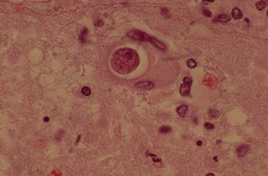
'Coccidioides' is a genus of fungi that are commonly found in the soil in certain geographical areas, including the southwestern United States and parts of Mexico and Central and South America. The two species of this genus, C. immitis and C. posadasii, can cause a serious infection known as coccidioidomycosis (also called Valley Fever) in humans and animals who inhale the spores of the fungi.
The infection typically begins in the lungs and can cause symptoms such as cough, fever, chest pain, fatigue, and weight loss. In some cases, the infection can spread to other parts of the body, leading to more severe and potentially life-threatening complications. People with weakened immune systems, such as those with HIV/AIDS or who are receiving immunosuppressive therapy, are at higher risk for developing severe coccidioidomycosis.
Coccidioidomycosis is a fungal infection caused by the inhalation of spores of the Coccidioides species, mainly C. immitis and C. posadasii. These fungi are commonly found in the soil of dry regions such as the southwestern United States, Mexico, and Central and South America.
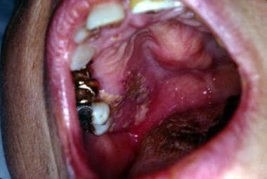
The infection often begins when a person inhales the microscopic spores, which can lead to respiratory symptoms resembling a common cold or pneumonia. Some people may develop more severe symptoms, especially those with weakened immune systems. The infection can disseminate to other parts of the body, causing skin lesions, bone and joint inflammation, meningitis, or other complications in rare cases.
Diagnosis typically involves a combination of clinical evaluation, imaging studies, and laboratory tests such as fungal cultures, histopathological examination, or serological tests to detect antibodies against Coccidioides antigens. Treatment depends on the severity of the infection and the patient's immune status. Antifungal medications like fluconazole, itraconazole, or amphotericin B are commonly used for treating coccidioidomycosis. Preventive measures include avoiding inhaling dust in endemic areas, especially during excavation or construction activities.
Fungal antigens are substances found on or produced by fungi that can stimulate an immune response in a host organism. They can be proteins, polysaccharides, or other molecules that are recognized as foreign by the host's immune system. Fungal antigens can be used in diagnostic tests to identify fungal infections, and they can also be targets of immune responses during fungal infections. In some cases, fungal antigens may contribute to the pathogenesis of fungal diseases by inducing inflammatory or allergic reactions. Examples of fungal antigens include the cell wall components of Candida albicans and the extracellular polysaccharide galactomannan produced by Aspergillus fumigatus.
Histoplasmin is not a medical condition or diagnosis itself, but it's a term related to a skin test used in medicine. Histoplasmin is an antigen extract derived from the histoplasmoma (a form of the fungus Histoplasma capsulatum) used in the histoplasmin skin test. This test is utilized to determine whether a person has been infected with the histoplasmosis fungus, which causes the disease histoplasmosis.
The histoplasmin skin test involves injecting a small amount of histoplasmin under the surface of the skin, usually on the forearm. If the person has previously been exposed to Histoplasma capsulatum, their immune system will recognize the antigen and produce a reaction (a hard, red, swollen area) at the injection site within 24-72 hours. The size of this reaction helps healthcare professionals determine if the person has developed an immune response to the fungus, indicating past or current infection with histoplasmosis.
It's important to note that a positive histoplasmin skin test does not necessarily mean that the person is currently sick with histoplasmosis. Instead, it shows that they have been exposed to the fungus at some point in their life and have developed an immune response to it.
Skin tests are medical diagnostic procedures that involve the application of a small amount of a substance to the skin, usually through a scratch, prick, or injection, to determine if the body has an allergic reaction to it. The most common type of skin test is the patch test, which involves applying a patch containing a small amount of the suspected allergen to the skin and observing the area for signs of a reaction, such as redness, swelling, or itching, over a period of several days. Another type of skin test is the intradermal test, in which a small amount of the substance is injected just beneath the surface of the skin. Skin tests are used to help diagnose allergies, including those to pollen, mold, pets, and foods, as well as to identify sensitivities to medications, chemicals, and other substances.
Complement fixation tests are a type of laboratory test used in immunology and serology to detect the presence of antibodies in a patient's serum. These tests are based on the principle of complement activation, which is a part of the immune response. The complement system consists of a group of proteins that work together to help eliminate pathogens from the body.
In a complement fixation test, the patient's serum is mixed with a known antigen and complement proteins. If the patient has antibodies against the antigen, they will bind to it and activate the complement system. This results in the consumption or "fixation" of the complement proteins, which are no longer available to participate in a secondary reaction.
A second step involves adding a fresh source of complement proteins and a dye-labeled antibody that recognizes a specific component of the complement system. If complement was fixed during the first step, it will not be available for this secondary reaction, and the dye-labeled antibody will remain unbound. Conversely, if no antibodies were present in the patient's serum, the complement proteins would still be available for the second reaction, leading to the binding of the dye-labeled antibody.
The mixture is then examined under a microscope or using a spectrophotometer to determine whether the dye-labeled antibody has bound. If it has not, this indicates that the patient's serum contains antibodies specific to the antigen used in the test, and a positive result is recorded.
Complement fixation tests have been widely used for the diagnosis of various infectious diseases, such as syphilis, measles, and influenza. However, they have largely been replaced by more modern serological techniques, like enzyme-linked immunosorbent assays (ELISAs) and nucleic acid amplification tests (NAATs), due to their increased sensitivity, specificity, and ease of use.
Two-dimensional immunoelectrophoresis (2DE) is a specialized laboratory technique used in the field of clinical pathology and immunology. This technique is a refined version of traditional immunoelectrophoresis that adds an additional electrophoretic separation step, enhancing its resolution and allowing for more detailed analysis of complex protein mixtures.
In two-dimensional immunoelectrophoresis, proteins are first separated based on their isoelectric points (pI) in the initial dimension using isoelectric focusing (IEF). This process involves applying an electric field to a protein mixture contained within a gel matrix, where proteins will migrate and stop migrating once they reach the pH that matches their own isoelectric point.
Following IEF, the separated proteins are then subjected to a second electrophoretic separation in the perpendicular direction (second dimension) based on their molecular weights using sodium dodecyl sulfate-polyacrylamide gel electrophoresis (SDS-PAGE). SDS is a negatively charged molecule that binds to proteins, giving them a uniform negative charge and allowing for separation based solely on size.
Once the two-dimensional separation is complete, the gel is then overlaid with specific antisera to detect and identify proteins of interest. The resulting precipitin arcs formed at the intersection of the antibody and antigen are compared to known standards or patterns to determine the identity and quantity of the separated proteins.
Two-dimensional immunoelectrophoresis is particularly useful in identifying and quantifying proteins in complex mixtures, such as those found in body fluids like serum, urine, or cerebrospinal fluid (CSF). It can be applied to various clinical scenarios, including diagnosis and monitoring of monoclonal gammopathies, autoimmune disorders, and certain infectious diseases.
Cell migration inhibition refers to the process or agents that restrict the movement of cells, particularly in the context of cancer metastasis. Cell migration is a critical biological process involved in various physiological and pathological conditions, including embryonic development, wound healing, and tumor cell dissemination. Inhibiting cell migration can help prevent the spread of cancer to distant organs, thereby improving treatment outcomes and patient survival rates.
Various factors and mechanisms contribute to cell migration inhibition, such as:
1. Modulation of signaling pathways: Cell migration is regulated by complex intracellular signaling networks that control cytoskeletal rearrangements, adhesion molecules, and other components required for cell motility. Inhibiting specific signaling proteins or pathways can suppress cell migration.
2. Extracellular matrix (ECM) modifications: The ECM provides structural support and biochemical cues that guide cell migration. Altering the composition or organization of the ECM can hinder cell movement.
3. Inhibition of adhesion molecules: Cell-cell and cell-matrix interactions are mediated by adhesion molecules, such as integrins and cadherins. Blocking these molecules can prevent cells from attaching to their surroundings and migrating.
4. Targeting cytoskeletal components: The cytoskeleton is responsible for the mechanical forces required for cell migration. Inhibiting cytoskeletal proteins, such as actin or tubulin, can impair cell motility.
5. Use of pharmacological agents: Several drugs and compounds have been identified to inhibit cell migration, either by targeting specific molecules or indirectly affecting the overall cellular environment. These agents include chemotherapeutic drugs, natural compounds, and small molecule inhibitors.
Understanding the mechanisms underlying cell migration inhibition can provide valuable insights into developing novel therapeutic strategies for cancer treatment and other diseases involving aberrant cell migration.
Fungal antibodies are a type of protein called immunoglobulins that are produced by the immune system in response to the presence of fungi in the body. These antibodies are specifically designed to recognize and bind to antigens on the surface of fungal cells, marking them for destruction by other immune cells.
There are several types of fungal antibodies, including IgA, IgG, IgM, and IgE, each with a specific role in the immune response. For example, IgG antibodies are the most common type of antibody found in the blood and provide long-term immunity to fungi, while IgE antibodies are associated with allergic reactions to fungi.
Fungal antibodies can be measured in the blood or other bodily fluids to help diagnose fungal infections, monitor the effectiveness of treatment, or assess immune function in individuals who are at risk for fungal infections, such as those with weakened immune systems due to HIV/AIDS, cancer, or organ transplantation.
Immunodiffusion is a laboratory technique used in immunology to detect and measure the presence of specific antibodies or antigens in a sample. It is based on the principle of diffusion, where molecules move from an area of high concentration to an area of low concentration until they reach equilibrium. In this technique, a sample containing an unknown quantity of antigen or antibody is placed in a gel or agar medium that contains a known quantity of antibody or antigen, respectively.
The two substances then diffuse towards each other and form a visible precipitate at the point where they meet and reach equivalence, which indicates the presence and quantity of the specific antigen or antibody in the sample. There are several types of immunodiffusion techniques, including radial immunodiffusion (RID) and double immunodiffusion (Ouchterlony technique). These techniques are widely used in diagnostic laboratories to identify and measure various antigens and antibodies, such as those found in infectious diseases, autoimmune disorders, and allergic reactions.
Delayed hypersensitivity, also known as type IV hypersensitivity, is a type of immune response that takes place several hours to days after exposure to an antigen. It is characterized by the activation of T cells (a type of white blood cell) and the release of various chemical mediators, leading to inflammation and tissue damage. This reaction is typically associated with chronic inflammatory diseases, such as contact dermatitis, granulomatous disorders (e.g. tuberculosis), and certain autoimmune diseases.
The reaction process involves the following steps:
1. Sensitization: The first time an individual is exposed to an antigen, T cells are activated and become sensitized to it. This process can take several days.
2. Memory: Some of the activated T cells differentiate into memory T cells, which remain in the body and are ready to respond quickly if the same antigen is encountered again.
3. Effector phase: Upon subsequent exposure to the antigen, the memory T cells become activated and release cytokines, which recruit other immune cells (e.g. macrophages) to the site of inflammation. These cells cause tissue damage through various mechanisms, such as phagocytosis, degranulation, and the release of reactive oxygen species.
4. Chronic inflammation: The ongoing immune response can lead to chronic inflammation, which may result in tissue destruction and fibrosis (scarring).
Examples of conditions associated with delayed hypersensitivity include:
* Contact dermatitis (e.g. poison ivy, nickel allergy)
* Tuberculosis
* Leprosy
* Sarcoidosis
* Rheumatoid arthritis
* Type 1 diabetes mellitus
* Multiple sclerosis
* Inflammatory bowel disease (e.g. Crohn's disease, ulcerative colitis)
 Coccidioides posadasii
List of MeSH codes (D23)
 A positive control for coccidioidin complement fixation
 An archived lot of coccidioidin induces specific coccidioidal delayed-type hypersensitivity and correlates with in vitro assays...
 Advanced Search Results - Public Health Image Library(PHIL)
 Coccidioidomycosis and Valley Fever Workup: Approach Considerations, Serologic Studies, Cultures
 Valley Fever (Coccidioidomycosis) - Symptoms and Diagnosis
Dermatologic Manifestations of Coccidioidomycosis: Overview, Patient History, Physical Examination
Symptoms of Valley Fever | Coccidioidomycosis | Types of Fungal Diseases | Fungal | CDC
 Purified Protein Derivative (PPD)-Tuberculin Anergy and HIV Infection: Guidelines For Anergy Testing And Management Of Anergic...
Noncandidal Fungal Infections of the Mouth: Practice Essentials, Background, Pathophysiology
Coccidioides posadasii - Wikipedia
 SAN FRANCISCO DERMATOLOGICAL SOCIETY | JAMA Dermatology | JAMA Network
 DeCS
DeCS
 Mycoses. Medical search
Coccidioidomycosis symptoms and treatment | Health Dictionary
Noncandidal Fungal Infections of the Mouth Workup: Laboratory Studies, Imaging Studies, Procedures
 Valley Fever Facts - What is Valley Fever? - Nielsen Biosciences
 Coccidioidomycosis - Definition, Life Cycle, Pathogenesis
 Кокцидіоїдомікоз - Інфекційні хвороби - MSD Manual Professional Edition
 The use of coccidioidin - PubMed
Fever, Weight Loss, and Lung Nodules in a 54-Year-Old Woman
Purified Protein Derivative (PPD)-Tuberculin
Anergy and HIV Infection:
Guidelines For Anergy Testing And Management Of ...
 Browsing by Title
 Browsing by Author "Uragoda, C. G."
Coccidioidomycosis and Valley Fever Medication: Antifungals
Coccidioides immitis - microbewiki
Code System Concept
 MeSH Browser
MeSH Browser
 Prefix: anti
Indigenous Case of Disseminated Histoplasmosis, Taiwan - Volume 13, Number 1-January 2007 - Emerging Infectious Diseases...
Antifungal agents. Medical search
PMID- 669642
 NDF-RT Code NDF-RT Name
Noncandidal Fungal Infections of the Mouth: Practice Essentials, Background, Pathophysiology
 DailyMed - SPHERUSOL- coccidioides immitis spherule-derived skin test antigen injection, solution
TRANSFER FACTORS
Histoplasmin and coccidioidin2
Tuberculin1
- However, only four are available as standardized antigens for use by a Mantoux-type procedure (0.1 ml of antigen administered intracutaneously): tuberculin, coccidioidin, histoplasmin, and mumps. (cdc.gov)
Skin1
- Studies by Cooke on the immunology of the disease, and in 1927 a filtrate of culture specimens, later named coccidioidin, began to be used in skin testing to delineate the epidemiology of infection. (wikipedia.org)