Insects
Chromosomes
Chromosome Mapping
Chromosome Banding
X Chromosome
Chromosome Aberrations
Sex Chromosomes
Chromosomes, Human, Pair 1
Chromosomes, Human
Chromosomes, Bacterial
Chromosomes, Human, Pair 7
Chromosomes, Human, Pair 11
Chromosomes, Human, Pair 17
Chromosomes, Human, Pair 6
Chromosomes, Human, Pair 9
Chromosomes, Human, Pair 21
Chromosomes, Plant
Chromosomes, Fungal
Chromosomes, Human, 6-12 and X
Chromosomes, Human, Pair 2
Chromosomes, Human, Pair 16
Chromosomes, Human, Pair 22
Chromosomes, Mammalian
Chromosomes, Human, Pair 13
Chromosomes, Human, Pair 4
Chromosomes, Human, Pair 10
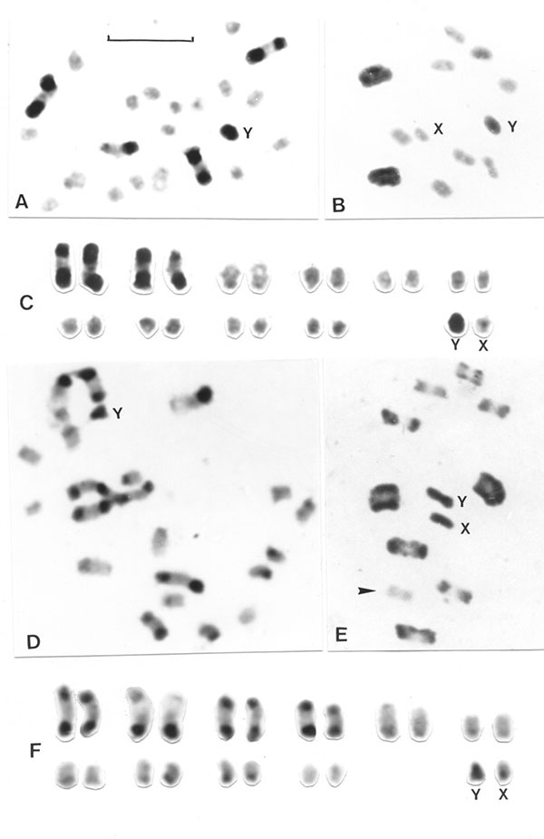
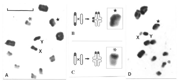
Chromosomes, Human, Y
Chromosomes, Human, Pair 8
Chromosomes, Human, Pair 19
Chromosome Disorders
Chromosomes, Artificial, Bacterial
Chromosomes, Human, X
Chromosomes, Human, 1-3
Chromosomes, Human, Pair 12
Chromosome Painting
Chromosomes, Human, Pair 5
Chromosomes, Human, Pair 15
Chromosomes, Human, Pair 14
Chromosomes, Human, Pair 18
In Situ Hybridization, Fluorescence
Molecular Sequence Data
Chromosomes, Human, 16-18
Chromosomes, Human, Pair 20
Chromosomes, Artificial, Yeast
Chromosomes, Human, 13-15
Genetic Linkage
Chromosome Breakage
Chromosomes, Human, 21-22 and Y
Base Sequence
Genetic Markers
Chromosome Inversion
Chromosome Positioning
Chromosomes, Human, 4-5
X Chromosome Inactivation
Centromere
Meiosis
Translocation, Genetic
Hybrid Cells
Chromosomes, Human, 19-20
Amino Acid Sequence
Aneuploidy
Mitosis
Metaphase
Recombination, Genetic
Cloning, Molecular
Mutation
Insect Hormones
Insect Control
Crosses, Genetic
Microsatellite Repeats
Larva
Phenotype
Drosophila melanogaster
Diptera
Spodoptera
Lod Score
Pedigree
Hemiptera
Sequence Analysis, DNA
Baculoviridae
Insect Vectors
Alleles
Beetles
Evolution, Molecular
DNA
Nondisjunction, Genetic
Nucleic Acid Hybridization
Chromosomes, Artificial, Human
Kinetochores
Grasshoppers
Models, Genetic
Telomere
Species Specificity
Chromosome Walking
Blotting, Southern
Polymerase Chain Reaction
Chromosomal Proteins, Non-Histone
Genotype
Spindle Apparatus
Chromosomal Instability
Sequence Homology, Amino Acid
Chromosome Fragility
Drosophila
Haplotypes
Multigene Family
DNA, Satellite
Repetitive Sequences, Nucleic Acid
Cockroaches
DNA Probes
Genes
Diploidy
Heteroptera
Sequence Alignment
Mosaicism
Chromatids
Gene Deletion
Biological Evolution
DNA-Binding Proteins
Heterozygote
Drosophila Proteins
Wasps
Abnormalities, Multiple
Tribolium
Polyploidy
Polytene Chromosomes
DNA, Complementary
Bees
Plasmids
Gene Dosage
DNA Primers
Nuclear Proteins
Cell Cycle Proteins
Bombyx
Polymorphism, Genetic
Prophase
Interphase
Loss of Heterozygosity
Cosmids
Orthoptera
Genome, Human
DNA Transposable Elements
Cytogenetic Analysis
Cytogenetics
Karyotype
Periplaneta
Gene Rearrangement
Spermatocytes
Aphids
Sequence Homology, Nucleic Acid
Genome
Chromatin
Chromosome Fragile Sites
Polymorphism, Restriction Fragment Length
Sex Chromosome Disorders
Saccharomyces cerevisiae
Monosomy
Escherichia coli
Sequence Tagged Sites
Pest Control, Biological
Genes, Dominant
Cell Nucleus
Polymorphism, Single Nucleotide
Contig Mapping
Gryllidae
Genetic Predisposition to Disease
Haploidy
Transcription, Genetic
Philadelphia Chromosome
Azure Stains
Chromosomes, Archaeal
JIL-1 and Su(var)3-7 interact genetically and counteract each other's effect on position-effect variegation in Drosophila. (1/169)
(+info)Evolution of a distinct genomic domain in Drosophila: comparative analysis of the dot chromosome in Drosophila melanogaster and Drosophila virilis. (2/169)
(+info)Multiple functions for Drosophila Mcm10 suggested through analysis of two Mcm10 mutant alleles. (3/169)
(+info)Introgression of Drosophila simulans nuclear pore protein 160 in Drosophila melanogaster alone does not cause inviability but does cause female sterility. (4/169)
(+info)Statistical analysis of nondisjunction assays in Drosophila. (5/169)
(+info)Step-by-step evolution of neo-sex chromosomes in geographical populations of wild silkmoths, Samia cynthia ssp. (6/169)
(+info)Mutagenesis as a genetic research strategy. (7/169)
(+info)Coupling between microtubule sliding, plus-end growth and spindle length revealed by kinesin-8 depletion. (8/169)
(+info)I apologize for any confusion, but "insects" are not a medical term. Insects are small air-breathing arthropods that have a segmented body with six legs and usually have wings. They make up the largest group of animals on Earth, with over a million described species.
If you're looking for information about a specific medical condition or topic, please provide more details so I can offer a relevant response.
Chromosomes are thread-like structures that exist in the nucleus of cells, carrying genetic information in the form of genes. They are composed of DNA and proteins, and are typically present in pairs in the nucleus, with one set inherited from each parent. In humans, there are 23 pairs of chromosomes for a total of 46 chromosomes. Chromosomes come in different shapes and forms, including sex chromosomes (X and Y) that determine the biological sex of an individual. Changes or abnormalities in the number or structure of chromosomes can lead to genetic disorders and diseases.
Chromosome mapping, also known as physical mapping, is the process of determining the location and order of specific genes or genetic markers on a chromosome. This is typically done by using various laboratory techniques to identify landmarks along the chromosome, such as restriction enzyme cutting sites or patterns of DNA sequence repeats. The resulting map provides important information about the organization and structure of the genome, and can be used for a variety of purposes, including identifying the location of genes associated with genetic diseases, studying evolutionary relationships between organisms, and developing genetic markers for use in breeding or forensic applications.
Chromosome banding is a technique used in cytogenetics to identify and describe the physical structure and organization of chromosomes. This method involves staining the chromosomes with specific dyes that bind differently to the DNA and proteins in various regions of the chromosome, resulting in a distinct pattern of light and dark bands when viewed under a microscope.
The most commonly used banding techniques are G-banding (Giemsa banding) and R-banding (reverse banding). In G-banding, the chromosomes are stained with Giemsa dye, which preferentially binds to the AT-rich regions, creating a characteristic banding pattern. The bands are numbered from the centromere (the constriction point where the chromatids join) outwards, with the darker bands (rich in A-T base pairs and histone proteins) labeled as "q" arms and the lighter bands (rich in G-C base pairs and arginine-rich proteins) labeled as "p" arms.
R-banding, on the other hand, uses a different staining procedure that results in a reversed banding pattern compared to G-banding. The darker R-bands correspond to the lighter G-bands, and vice versa. This technique is particularly useful for identifying and analyzing specific regions of chromosomes that may be difficult to visualize with G-banding alone.
Chromosome banding plays a crucial role in diagnosing genetic disorders, identifying chromosomal abnormalities, and studying the structure and function of chromosomes in both clinical and research settings.
The X chromosome is one of the two types of sex-determining chromosomes in humans (the other being the Y chromosome). It's one of the 23 pairs of chromosomes that make up a person's genetic material. Females typically have two copies of the X chromosome (XX), while males usually have one X and one Y chromosome (XY).
The X chromosome contains hundreds of genes that are responsible for the production of various proteins, many of which are essential for normal bodily functions. Some of the critical roles of the X chromosome include:
1. Sex Determination: The presence or absence of the Y chromosome determines whether an individual is male or female. If there is no Y chromosome, the individual will typically develop as a female.
2. Genetic Disorders: Since females have two copies of the X chromosome, they are less likely to be affected by X-linked genetic disorders than males. Males, having only one X chromosome, will express any recessive X-linked traits they inherit.
3. Dosage Compensation: To compensate for the difference in gene dosage between males and females, a process called X-inactivation occurs during female embryonic development. One of the two X chromosomes is randomly inactivated in each cell, resulting in a single functional copy per cell.
The X chromosome plays a crucial role in human genetics and development, contributing to various traits and characteristics, including sex determination and dosage compensation.
Chromosome aberrations refer to structural and numerical changes in the chromosomes that can occur spontaneously or as a result of exposure to mutagenic agents. These changes can affect the genetic material encoded in the chromosomes, leading to various consequences such as developmental abnormalities, cancer, or infertility.
Structural aberrations include deletions, duplications, inversions, translocations, and rings, which result from breaks and rearrangements of chromosome segments. Numerical aberrations involve changes in the number of chromosomes, such as aneuploidy (extra or missing chromosomes) or polyploidy (multiples of a complete set of chromosomes).
Chromosome aberrations can be detected and analyzed using various cytogenetic techniques, including karyotyping, fluorescence in situ hybridization (FISH), and comparative genomic hybridization (CGH). These methods allow for the identification and characterization of chromosomal changes at the molecular level, providing valuable information for genetic counseling, diagnosis, and research.
Sex chromosomes, often denoted as X and Y, are one of the 23 pairs of human chromosomes found in each cell of the body. Normally, females have two X chromosomes (46,XX), and males have one X and one Y chromosome (46,XY). The sex chromosomes play a significant role in determining the sex of an individual. They contain genes that contribute to physical differences between men and women. Any variations or abnormalities in the number or structure of these chromosomes can lead to various genetic disorders and conditions related to sexual development and reproduction.
Human chromosome pair 1 refers to the first pair of chromosomes in a set of 23 pairs found in the cells of the human body, excluding sex cells (sperm and eggs). Each cell in the human body, except for the gametes, contains 46 chromosomes arranged in 23 pairs. These chromosomes are rod-shaped structures that contain genetic information in the form of DNA.
Chromosome pair 1 is the largest pair, making up about 8% of the total DNA in a cell. Each chromosome in the pair consists of two arms - a shorter p arm and a longer q arm - connected at a centromere. Chromosome 1 carries an estimated 2,000-2,500 genes, which are segments of DNA that contain instructions for making proteins or regulating gene expression.
Defects or mutations in the genes located on chromosome 1 can lead to various genetic disorders and diseases, such as Charcot-Marie-Tooth disease type 1A, Huntington's disease, and certain types of cancer.
Chromosomes are thread-like structures that contain genetic material, i.e., DNA and proteins, present in the nucleus of human cells. In humans, there are 23 pairs of chromosomes, for a total of 46 chromosomes, in each diploid cell. Twenty-two of these pairs are called autosomal chromosomes, which come in identical pairs and contain genes that determine various traits unrelated to sex.
The last pair is referred to as the sex chromosomes (X and Y), which determines a person's biological sex. Females have two X chromosomes (46, XX), while males possess one X and one Y chromosome (46, XY). Chromosomes vary in size, with the largest being chromosome 1 and the smallest being the Y chromosome.
Human chromosomes are typically visualized during mitosis or meiosis using staining techniques that highlight their banding patterns, allowing for identification of specific regions and genes. Chromosomal abnormalities can lead to various genetic disorders, including Down syndrome (trisomy 21), Turner syndrome (monosomy X), and Klinefelter syndrome (XXY).
Bacterial chromosomes are typically circular, double-stranded DNA molecules that contain the genetic material of bacteria. Unlike eukaryotic cells, which have their DNA housed within a nucleus, bacterial chromosomes are located in the cytoplasm of the cell, often associated with the bacterial nucleoid.
Bacterial chromosomes can vary in size and structure among different species, but they typically contain all of the genetic information necessary for the survival and reproduction of the organism. They may also contain plasmids, which are smaller circular DNA molecules that can carry additional genes and can be transferred between bacteria through a process called conjugation.
One important feature of bacterial chromosomes is their ability to replicate rapidly, allowing bacteria to divide quickly and reproduce in large numbers. The replication of the bacterial chromosome begins at a specific origin point and proceeds in opposite directions until the entire chromosome has been copied. This process is tightly regulated and coordinated with cell division to ensure that each daughter cell receives a complete copy of the genetic material.
Overall, the study of bacterial chromosomes is an important area of research in microbiology, as understanding their structure and function can provide insights into bacterial genetics, evolution, and pathogenesis.
Chromosome segregation is the process that occurs during cell division (mitosis or meiosis) where replicated chromosomes are separated and distributed equally into two daughter cells. Each chromosome consists of two sister chromatids, which are identical copies of genetic material. During chromosome segregation, these sister chromatids are pulled apart by a structure called the mitotic spindle and moved to opposite poles of the cell. This ensures that each new cell receives one copy of each chromosome, preserving the correct number and composition of chromosomes in the organism.
Human chromosome pair 7 consists of two rod-shaped structures present in the nucleus of each cell in the human body. Each member of the pair is a single chromosome, and together they contain the genetic material that is inherited from both parents. They are identical in size, shape, and banding pattern and are therefore referred to as homologous chromosomes.
Chromosome 7 is one of the autosomal chromosomes, meaning it is not a sex chromosome (X or Y). It is composed of double-stranded DNA that contains approximately 159 million base pairs and around 1,200 genes. Chromosome 7 contains several important genes associated with human health and disease, including those involved in the development of certain types of cancer, such as colon cancer and lung cancer, as well as genetic disorders such as Williams-Beuren syndrome and Charcot-Marie-Tooth disease.
Abnormalities in chromosome 7 have been linked to various genetic conditions, including deletions, duplications, translocations, and other structural changes. These abnormalities can lead to developmental delays, intellectual disabilities, physical abnormalities, and increased risk of certain types of cancer.
I'm sorry for any confusion, but "Insect Proteins" is not a recognized medical term. However, I can provide some information about insect protein from a nutritional and food science perspective.
Insect proteins refer to the proteins that are obtained from insects. Insects are a rich source of protein, and their protein content varies by species. For example, mealworms and crickets have been found to contain approximately 47-63% and 60-72% protein by dry weight, respectively.
In recent years, insect proteins have gained attention as a potential sustainable source of nutrition due to their high protein content, low environmental impact, and the ability to convert feed into protein more efficiently compared to traditional livestock. Insect proteins can be used in various applications such as food and feed additives, nutritional supplements, and even cosmetics.
However, it's important to note that the use of insect proteins in human food is not widely accepted in many Western countries due to cultural and regulatory barriers. Nonetheless, research and development efforts continue to explore the potential benefits and applications of insect proteins in the global food system.
Human chromosome pair 11 consists of two rod-shaped structures present in the nucleus of each cell in the human body. Each member of the pair is a single chromosome, and together they contain the genetic material that is inherited from both parents. They are located on the eleventh position in the standard karyotype, which is a visual representation of the 23 pairs of human chromosomes.
Chromosome 11 is one of the largest human chromosomes and contains an estimated 135 million base pairs. It contains approximately 1,400 genes that provide instructions for making proteins, as well as many non-coding RNA molecules that play a role in regulating gene expression.
Chromosome 11 is known to contain several important genes and genetic regions associated with various human diseases and conditions. For example, it contains the Wilms' tumor 1 (WT1) gene, which is associated with kidney cancer in children, and the neurofibromatosis type 1 (NF1) gene, which is associated with a genetic disorder that causes benign tumors to grow on nerves throughout the body. Additionally, chromosome 11 contains the region where the ABO blood group genes are located, which determine a person's blood type.
It's worth noting that human chromosomes come in pairs because they contain two copies of each gene, one inherited from the mother and one from the father. This redundancy allows for genetic diversity and provides a backup copy of essential genes, ensuring their proper function and maintaining the stability of the genome.
Human chromosome pair 17 consists of two rod-shaped structures present in the nucleus of each human cell. Each chromosome is made up of DNA tightly coiled around histone proteins, forming a complex called chromatin. Chromosomes carry genetic information in the form of genes, which are segments of DNA that contain instructions for the development and function of an organism.
Human cells typically have 23 pairs of chromosomes, for a total of 46 chromosomes. Pair 17 is one of the autosomal pairs, meaning it is not a sex chromosome (X or Y). Chromosome 17 is a medium-sized chromosome and contains an estimated 800 million base pairs of DNA. It contains approximately 1,500 genes that provide instructions for making proteins and regulating various cellular processes.
Chromosome 17 is associated with several genetic disorders, including inherited cancer syndromes such as Li-Fraumeni syndrome and hereditary nonpolyposis colorectal cancer (HNPCC). Mutations in genes located on chromosome 17 can increase the risk of developing various types of cancer, including breast, ovarian, colon, and pancreatic cancer.
Human chromosome pair 6 consists of two rod-shaped structures present in the nucleus of each human cell. They are identical in size and shape and contain genetic material, made up of DNA and proteins, that is essential for the development and function of the human body.
Chromosome pair 6 is one of the 23 pairs of chromosomes found in humans, with one chromosome inherited from each parent. Each chromosome contains thousands of genes that provide instructions for the production of proteins and regulate various cellular processes.
Chromosome pair 6 contains several important genes, including those involved in the development and function of the immune system, such as the major histocompatibility complex (MHC) genes. It also contains genes associated with certain genetic disorders, such as hereditary neuropathy with liability to pressure palsies (HNPP), a condition that affects the nerves, and Waardenburg syndrome, a disorder that affects pigmentation and hearing.
Abnormalities in chromosome pair 6 can lead to various genetic disorders, including numerical abnormalities such as trisomy 6 (three copies of chromosome 6) or monosomy 6 (only one copy of chromosome 6), as well as structural abnormalities such as deletions, duplications, or translocations of parts of the chromosome.
A chromosome deletion is a type of genetic abnormality that occurs when a portion of a chromosome is missing or deleted. Chromosomes are thread-like structures located in the nucleus of cells that contain our genetic material, which is organized into genes.
Chromosome deletions can occur spontaneously during the formation of reproductive cells (eggs or sperm) or can be inherited from a parent. They can affect any chromosome and can vary in size, from a small segment to a large portion of the chromosome.
The severity of the symptoms associated with a chromosome deletion depends on the size and location of the deleted segment. In some cases, the deletion may be so small that it does not cause any noticeable symptoms. However, larger deletions can lead to developmental delays, intellectual disabilities, physical abnormalities, and various medical conditions.
Chromosome deletions are typically detected through a genetic test called karyotyping, which involves analyzing the number and structure of an individual's chromosomes. Other more precise tests, such as fluorescence in situ hybridization (FISH) or chromosomal microarray analysis (CMA), may also be used to confirm the diagnosis and identify the specific location and size of the deletion.
Human chromosome pair 9 consists of two rod-shaped structures present in the nucleus of each cell of the human body. Each member of the pair contains thousands of genes and other genetic material, encoded in the form of DNA molecules. The two chromosomes in a pair are identical or very similar to each other in terms of their size, shape, and genetic makeup.
Chromosome 9 is one of the autosomal chromosomes, meaning that it is not a sex chromosome (X or Y) and is present in two copies in all cells of the body, regardless of sex. Chromosome 9 is a medium-sized chromosome, and it is estimated to contain around 135 million base pairs of DNA and approximately 1200 genes.
Chromosome 9 contains several important genes that are associated with various human traits and diseases. For example, mutations in the gene that encodes the protein APOE on chromosome 9 have been linked to an increased risk of developing Alzheimer's disease. Additionally, variations in the gene that encodes the protein EGFR on chromosome 9 have been associated with an increased risk of developing certain types of cancer.
Overall, human chromosome pair 9 plays a critical role in the development and function of the human body, and variations in its genetic makeup can contribute to a wide range of traits and diseases.
Human chromosome pair 21 consists of two rod-shaped structures present in the nucleus of each cell in the human body. Each member of the pair is a single chromosome, and they are identical to each other. Chromosomes are made up of DNA, which contains genetic information that determines many of an individual's traits and characteristics.
Chromosome pair 21 is one of the 23 pairs of human autosomal chromosomes, meaning they are not sex chromosomes (X or Y). Chromosome pair 21 is the smallest of the human chromosomes, and it contains approximately 48 million base pairs of DNA. It contains around 200-300 genes that provide instructions for making proteins and regulating various cellular processes.
Down syndrome, a genetic disorder characterized by intellectual disability, developmental delays, distinct facial features, and sometimes heart defects, is caused by an extra copy of chromosome pair 21 or a part of it. This additional genetic material can lead to abnormalities in brain development and function, resulting in the characteristic symptoms of Down syndrome.
Chromosomes in plants are thread-like structures that contain genetic material, DNA, and proteins. They are present in the nucleus of every cell and are inherited from the parent plants during sexual reproduction. Chromosomes come in pairs, with each pair consisting of one chromosome from each parent.
In plants, like in other organisms, chromosomes play a crucial role in inheritance, development, and reproduction. They carry genetic information that determines various traits and characteristics of the plant, such as its physical appearance, growth patterns, and resistance to diseases.
Plant chromosomes are typically much larger than those found in animals, making them easier to study under a microscope. The number of chromosomes varies among different plant species, ranging from as few as 2 in some ferns to over 1000 in certain varieties of wheat.
During cell division, the chromosomes replicate and then separate into two identical sets, ensuring that each new cell receives a complete set of genetic information. This process is critical for the growth and development of the plant, as well as for the production of viable seeds and offspring.
Chromosomes in fungi are thread-like structures that contain genetic material, composed of DNA and proteins, present in the nucleus of a cell. Unlike humans and other eukaryotes that have a diploid number of chromosomes in their somatic cells, fungal chromosome numbers can vary widely between and within species.
Fungal chromosomes are typically smaller and fewer in number compared to those found in plants and animals. The chromosomal organization in fungi is also different from other eukaryotes. In many fungi, the chromosomes are condensed throughout the cell cycle, whereas in other eukaryotes, chromosomes are only condensed during cell division.
Fungi can have linear or circular chromosomes, depending on the species. For example, the model organism Saccharomyces cerevisiae (budding yeast) has a set of 16 small circular chromosomes, while other fungi like Neurospora crassa (red bread mold) and Aspergillus nidulans (a filamentous fungus) have linear chromosomes.
Fungal chromosomes play an essential role in the growth, development, reproduction, and survival of fungi. They carry genetic information that determines various traits such as morphology, metabolism, pathogenicity, and resistance to environmental stresses. Advances in genomic technologies have facilitated the study of fungal chromosomes, leading to a better understanding of their structure, function, and evolution.
Chromosomes are thread-like structures that contain genetic material, made up of DNA and proteins, in the nucleus of cells. In humans, there are typically 46 chromosomes arranged in 23 pairs, with one member of each pair coming from each parent. The six pairs of chromosomes numbered 6 through 12, along with the X chromosome, are part of these 23 pairs and are referred to as autosomal chromosomes and a sex chromosome.
Human chromosome 6 is one of the autosomal chromosomes and contains an estimated 170 million base pairs and around 1,500 genes. It plays a role in several important functions, including immune response, cell signaling, and nervous system function.
Human chromosome 7 is another autosomal chromosome that contains approximately 159 million base pairs and around 1,200 genes. Chromosome 7 is best known for containing the gene for the cystic fibrosis transmembrane conductance regulator (CFTR) protein, whose mutations can lead to cystic fibrosis.
Human chromosome 8 is an autosomal chromosome that contains around 146 million base pairs and approximately 900 genes. Chromosome 8 has been associated with several genetic disorders, including Smith-Magenis syndrome and 8p deletion syndrome.
Human chromosome 9 is an autosomal chromosome that contains around 139 million base pairs and approximately 950 genes. Chromosome 9 has been linked to several genetic disorders, including Hereditary Spherocytosis and CHARGE syndrome.
Human chromosome 10 is an autosomal chromosome that contains around 135 million base pairs and approximately 800 genes. Chromosome 10 has been associated with several genetic disorders, including Dyschondrosteosis and Melanoma.
Human chromosome 11 is an autosomal chromosome that contains around 135 million base pairs and approximately 800 genes. Chromosome 11 has been linked to several genetic disorders, including Wilms tumor and Beckwith-Wiedemann syndrome.
Human chromosome 12 is an autosomal chromosome that contains around 133 million base pairs and approximately 750 genes. Chromosome 12 has been associated with several genetic disorders, including Charcot-Marie-Tooth disease type 1A and Hereditary Neuropathy with Liability to Pressure Palsies (HNPP).
The X chromosome is one of the two sex chromosomes in humans. Females have two X chromosomes, while males have one X and one Y chromosome. The X chromosome contains around 155 million base pairs and approximately 1,000 genes. It has been linked to several genetic disorders, including Duchenne muscular dystrophy and Fragile X syndrome.
The Y chromosome is the other sex chromosome in humans. Males have one X and one Y chromosome, while females have two X chromosomes. The Y chromosome contains around 59 million base pairs and approximately 70 genes. It is primarily responsible for male sexual development and fertility.
In summary, the human genome consists of 23 pairs of chromosomes, including 22 autosomal pairs and one sex chromosome pair (XX in females and XY in males). The total length of the human genome is approximately 3 billion base pairs, and it contains around 20,000-25,000 protein-coding genes. Chromosomes are made up of DNA and proteins called histones, which help to package the DNA into a compact structure. The chromosomes contain genetic information that is passed down from parents to their offspring through reproduction.
Human chromosome pair 2 consists of two rod-shaped structures present in the nucleus of each cell of the human body. Each member of the pair contains thousands of genes and other genetic material, encoded in the form of DNA molecules. Chromosomes are the physical carriers of inheritance, and human cells typically contain 23 pairs of chromosomes for a total of 46 chromosomes.
Chromosome pair 2 is one of the autosomal pairs, meaning that it is not a sex chromosome (X or Y). Each member of chromosome pair 2 is approximately 247 million base pairs in length and contains an estimated 1,000-1,300 genes. These genes play crucial roles in various biological processes, including development, metabolism, and response to environmental stimuli.
Abnormalities in chromosome pair 2 can lead to genetic disorders, such as cat-eye syndrome (CES), which is characterized by iris abnormalities, anal atresia, hearing loss, and intellectual disability. This disorder arises from the presence of an extra copy of a small region on chromosome 2, resulting in partial trisomy of this region. Other genetic conditions associated with chromosome pair 2 include proximal 2q13.3 microdeletion syndrome and Potocki-Lupski syndrome (PTLS).
Human chromosome pair 16 consists of two rod-shaped structures present in the nucleus of each cell in the human body. Each chromosome is made up of DNA tightly coiled around histone proteins, forming a complex structure called a chromatin.
Chromosomes come in pairs, with one chromosome inherited from each parent. Chromosome pair 16 contains two homologous chromosomes, which are similar in size, shape, and genetic content but may have slight variations due to differences in the DNA sequences inherited from each parent.
Chromosome pair 16 is one of the 22 autosomal pairs, meaning it contains non-sex chromosomes that are present in both males and females. Chromosome 16 is a medium-sized chromosome, and it contains around 2,800 genes that provide instructions for making proteins and regulating various cellular processes.
Abnormalities in chromosome pair 16 can lead to genetic disorders such as chronic myeloid leukemia, some forms of mental retardation, and other developmental abnormalities.
Human chromosome pair 22 consists of two rod-shaped structures present in the nucleus of each cell in the human body. Each chromosome is made up of DNA tightly coiled around histone proteins, forming a complex structure called a chromatin.
Chromosome pair 22 is one of the 22 autosomal pairs of human chromosomes, meaning they are not sex chromosomes (X or Y). Chromosome 22 is the second smallest human chromosome, with each arm of the chromosome designated as p and q. The short arm is labeled "p," and the long arm is labeled "q."
Chromosome 22 contains several genes that are associated with various genetic disorders, including DiGeorge syndrome, velocardiofacial syndrome, and cat-eye syndrome, which result from deletions or duplications of specific regions on the chromosome. Additionally, chromosome 22 is the location of the NRXN1 gene, which has been associated with an increased risk for autism spectrum disorder (ASD) and schizophrenia when deleted or disrupted.
Understanding the genetic makeup of human chromosome pair 22 can provide valuable insights into human genetics, evolution, and disease susceptibility, as well as inform medical diagnoses, treatments, and research.
Chromosome pairing, also known as chromosome synapsis, is a process that occurs during meiosis, which is the type of cell division that results in the formation of sex cells or gametes (sperm and eggs).
In humans, each cell contains 23 pairs of chromosomes, for a total of 46 chromosomes. Of these, 22 pairs are called autosomal chromosomes, and they are similar in size and shape between the two copies in a pair. The last pair is called the sex chromosomes (X and Y), which determine the individual's biological sex.
During meiosis, homologous chromosomes (one from each parent) come together and pair up along their lengths in a process called synapsis. This pairing allows for the precise alignment of corresponding genes and genetic regions between the two homologous chromosomes. Once paired, the chromosomes exchange genetic material through a process called crossing over, which increases genetic diversity in the resulting gametes.
After crossing over, the homologous chromosomes separate during meiosis I, followed by the separation of sister chromatids (the two copies of each chromosome) during meiosis II. The end result is four haploid cells, each containing 23 chromosomes, which then develop into sperm or eggs.
Chromosome pairing is a crucial step in the process of sexual reproduction, ensuring that genetic information is accurately passed from one generation to the next while also promoting genetic diversity through recombination and independent assortment of chromosomes.
Mammalian chromosomes are thread-like structures that exist in the nucleus of mammalian cells, consisting of DNA, hist proteins, and RNA. They carry genetic information that is essential for the development and function of all living organisms. In mammals, each cell contains 23 pairs of chromosomes, for a total of 46 chromosomes, with one set inherited from the mother and the other from the father.
The chromosomes are typically visualized during cell division, where they condense and become visible under a microscope. Each chromosome is composed of two identical arms, separated by a constriction called the centromere. The short arm of the chromosome is labeled as "p," while the long arm is labeled as "q."
Mammalian chromosomes play a critical role in the transmission of genetic information from one generation to the next and are essential for maintaining the stability and integrity of the genome. Abnormalities in the number or structure of mammalian chromosomes can lead to various genetic disorders, including Down syndrome, Turner syndrome, and Klinefelter syndrome.
Human chromosome pair 13 consists of two rod-shaped structures present in the nucleus of each cell in the human body. Each chromosome is made up of DNA tightly coiled around histone proteins, forming a complex structure called a chromatin.
Chromosomes carry genetic information in the form of genes, which are sequences of DNA that code for specific traits and functions. Human cells typically have 23 pairs of chromosomes, for a total of 46 chromosomes. Chromosome pair 13 is one of the autosomal pairs, meaning it is not a sex chromosome (X or Y).
Chromosome pair 13 contains several important genes that are associated with various genetic disorders, such as cri-du-chat syndrome and Phelan-McDermid syndrome. Cri-du-chat syndrome is caused by a deletion of the short arm of chromosome 13 (13p), resulting in distinctive cat-like crying sounds in infants, developmental delays, and intellectual disabilities. Phelan-McDermid syndrome is caused by a deletion or mutation of the terminal end of the long arm of chromosome 13 (13q), leading to developmental delays, intellectual disability, absent or delayed speech, and autistic behaviors.
It's important to note that while some genetic disorders are associated with specific chromosomal abnormalities, many factors can contribute to the development and expression of these conditions, including environmental influences and interactions between multiple genes.
Human chromosome pair 4 consists of two rod-shaped structures present in the nucleus of each cell in the human body. Each member of the pair is a single chromosome, and they are identical or very similar in length and gene content. Chromosomes are made up of DNA, which contains genetic information, and proteins that package and organize the DNA.
Human chromosomes are numbered from 1 to 22, with chromosome pair 4 being one of the autosomal pairs, meaning it is not a sex chromosome (X or Y). Chromosome pair 4 is a medium-sized pair and contains an estimated 1,800-2,000 genes. These genes provide instructions for making proteins that are essential for various functions in the body, such as development, growth, and metabolism.
Abnormalities in chromosome pair 4 can lead to genetic disorders, including Wolf-Hirschhorn syndrome, which is caused by a deletion of part of the short arm of chromosome 4, and 4p16.3 microdeletion syndrome, which is caused by a deletion of a specific region on the short arm of chromosome 4. These conditions can result in developmental delays, intellectual disability, physical abnormalities, and other health problems.
Human chromosome pair 10 refers to a group of genetic materials that are present in every cell of the human body. Chromosomes are thread-like structures that carry our genes and are located in the nucleus of most cells. They come in pairs, with one set inherited from each parent.
Chromosome pair 10 is one of the 22 autosomal chromosome pairs, meaning they contain genes that are not related to sex determination. Each member of chromosome pair 10 is a single, long DNA molecule that contains thousands of genes and other genetic material.
Chromosome pair 10 is responsible for carrying genetic information that influences various traits and functions in the human body. Some of the genes located on chromosome pair 10 are associated with certain medical conditions, such as hereditary breast and ovarian cancer syndrome, neurofibromatosis type 1, and Waardenburg syndrome type 2A.
It's important to note that while chromosomes carry genetic information, not all variations in the DNA sequence will result in a change in phenotype or function. Some variations may have no effect at all, while others may lead to changes in how proteins are made and function, potentially leading to disease or other health issues.
Human Y chromosomes are one of the two sex-determining chromosomes in humans (the other being the X chromosome). They are found in the 23rd pair of human chromosomes and are significantly smaller than the X chromosome.
The Y chromosome is passed down from father to son through the paternal line, and it plays a crucial role in male sex determination. The SRY gene (sex-determining region Y) on the Y chromosome initiates the development of male sexual characteristics during embryonic development.
In addition to the SRY gene, the human Y chromosome contains several other genes that are essential for sperm production and male fertility. However, the Y chromosome has a much lower gene density compared to other chromosomes, with only about 80 protein-coding genes, making it one of the most gene-poor chromosomes in the human genome.
Because of its small size and low gene density, the Y chromosome is particularly susceptible to genetic mutations and deletions, which can lead to various genetic disorders and male infertility. Nonetheless, the Y chromosome remains a critical component of human genetics and evolution, providing valuable insights into sex determination, inheritance patterns, and human diversity.
Human chromosome pair 8 consists of two rod-shaped structures present in the nucleus of each cell of the human body. Each chromosome is made up of DNA tightly coiled around histone proteins, forming a complex structure known as a chromatin.
Human cells have 23 pairs of chromosomes, for a total of 46 chromosomes. Pair 8 is one of the autosomal pairs, meaning that it is not a sex chromosome (X or Y). Each member of chromosome pair 8 has a similar size, shape, and banding pattern, and they are identical in males and females.
Chromosome pair 8 contains several genes that are essential for various cellular functions and human development. Some of the genes located on chromosome pair 8 include those involved in the regulation of metabolism, nerve function, immune response, and cell growth and division.
Abnormalities in chromosome pair 8 can lead to genetic disorders such as Wolf-Hirschhorn syndrome, which is caused by a partial deletion of the short arm of chromosome 4, or partial trisomy 8, which results from an extra copy of all or part of chromosome 8. Both of these conditions are associated with developmental delays, intellectual disability, and various physical abnormalities.
Human chromosome pair 19 refers to a group of 19 identical chromosomes that are present in every cell of the human body, except for the sperm and egg cells which contain only 23 chromosomes. Chromosomes are thread-like structures that carry genetic information in the form of DNA (deoxyribonucleic acid) molecules.
Each chromosome is made up of two arms, a shorter p arm and a longer q arm, separated by a centromere. Human chromosome pair 19 is an acrocentric chromosome, which means that the centromere is located very close to the end of the short arm (p arm).
Chromosome pair 19 contains approximately 58 million base pairs of DNA and encodes for around 1,400 genes. It is one of the most gene-dense chromosomes in the human genome, with many genes involved in important biological processes such as metabolism, immunity, and neurological function.
Abnormalities in chromosome pair 19 have been associated with various genetic disorders, including Sotos syndrome, which is characterized by overgrowth, developmental delay, and distinctive facial features, and Smith-Magenis syndrome, which is marked by intellectual disability, behavioral problems, and distinct physical features.
Chromosome disorders are a group of genetic conditions caused by abnormalities in the number or structure of chromosomes. Chromosomes are thread-like structures located in the nucleus of cells that contain most of the body's genetic material, which is composed of DNA and proteins. Normally, humans have 23 pairs of chromosomes, for a total of 46 chromosomes.
Chromosome disorders can result from changes in the number of chromosomes (aneuploidy) or structural abnormalities in one or more chromosomes. Some common examples of chromosome disorders include:
1. Down syndrome: a condition caused by an extra copy of chromosome 21, resulting in intellectual disability, developmental delays, and distinctive physical features.
2. Turner syndrome: a condition that affects only females and is caused by the absence of all or part of one X chromosome, resulting in short stature, lack of sexual development, and other symptoms.
3. Klinefelter syndrome: a condition that affects only males and is caused by an extra copy of the X chromosome, resulting in tall stature, infertility, and other symptoms.
4. Cri-du-chat syndrome: a condition caused by a deletion of part of the short arm of chromosome 5, resulting in intellectual disability, developmental delays, and a distinctive cat-like cry.
5. Fragile X syndrome: a condition caused by a mutation in the FMR1 gene on the X chromosome, resulting in intellectual disability, behavioral problems, and physical symptoms.
Chromosome disorders can be diagnosed through various genetic tests, such as karyotyping, chromosomal microarray analysis (CMA), or fluorescence in situ hybridization (FISH). Treatment for these conditions depends on the specific disorder and its associated symptoms and may include medical interventions, therapies, and educational support.
Artificial bacterial chromosomes (ABCs) are synthetic replicons that are designed to function like natural bacterial chromosomes. They are created through the use of molecular biology techniques, such as recombination and cloning, to construct large DNA molecules that can stably replicate and segregate within a host bacterium.
ABCs are typically much larger than traditional plasmids, which are smaller circular DNA molecules that can also replicate in bacteria but have a limited capacity for carrying genetic information. ABCs can accommodate large DNA inserts, making them useful tools for cloning and studying large genes, gene clusters, or even entire genomes of other organisms.
There are several types of ABCs, including bacterial artificial chromosomes (BACs), P1-derived artificial chromosomes (PACs), and yeast artificial chromosomes (YACs). BACs are the most commonly used type of ABC and can accommodate inserts up to 300 kilobases (kb) in size. They have been widely used in genome sequencing projects, functional genomics studies, and protein production.
Overall, artificial bacterial chromosomes provide a powerful tool for manipulating and studying large DNA molecules in a controlled and stable manner within bacterial hosts.
A chromosome is a thread-like structure that contains genetic material, made up of DNA and proteins, in the nucleus of a cell. In humans, there are 23 pairs of chromosomes, for a total of 46 chromosomes, in each cell of the body, with the exception of the sperm and egg cells which contain only 23 chromosomes.
The X chromosome is one of the two sex-determining chromosomes in humans. Females typically have two X chromosomes (XX), while males have one X and one Y chromosome (XY). The X chromosome contains hundreds of genes that are responsible for various functions in the body, including some related to sexual development and reproduction.
Humans inherit one X chromosome from their mother and either an X or a Y chromosome from their father. In females, one of the two X chromosomes is randomly inactivated during embryonic development, resulting in each cell having only one active X chromosome. This process, known as X-inactivation, helps to ensure that females have roughly equal levels of gene expression from the X chromosome, despite having two copies.
Abnormalities in the number or structure of the X chromosome can lead to various genetic disorders, such as Turner syndrome (X0), Klinefelter syndrome (XXY), and fragile X syndrome (an X-linked disorder caused by a mutation in the FMR1 gene).
Human chromosomes are the thread-like structures located in the nucleus of human cells, which carry genetic information in the form of DNA. Humans have a total of 46 chromosomes arranged in 23 pairs. The first 22 pairs are called autosomes, and the last pair are the sex chromosomes, X and Y.
Chromosomes 1-3 are the largest human chromosomes, and they contain a significant portion of the human genome. Here is a brief overview of each:
1. Chromosome 1: This is the largest human chromosome, spanning about 8% of the human genome. It contains approximately 2,800 genes that are responsible for various functions such as cell growth and division, nerve function, and response to stimuli.
2. Chromosome 2: The second largest human chromosome, spanning about 7% of the human genome. It contains approximately 2,300 genes that are involved in various functions such as metabolism, development, and immune response.
3. Chromosome 3: This is the third largest human chromosome, spanning about 6% of the human genome. It contains approximately 1,900 genes that are responsible for various functions such as DNA repair, cell signaling, and response to stress.
It's worth noting that while these chromosomes contain a large number of genes, they also have significant amounts of non-coding DNA, which means that not all of the genetic material on these chromosomes is responsible for encoding proteins or other functional elements.
Human chromosome pair 12 consists of two rod-shaped structures present in the nucleus of each cell in the human body. Each chromosome is made up of DNA tightly coiled around histone proteins, forming a complex structure called a chromatin.
Chromosomes come in pairs, with one chromosome inherited from each parent. In humans, there are 23 pairs of chromosomes, for a total of 46 chromosomes in each cell. Chromosome pair 12 is the 12th pair of autosomal chromosomes, meaning they are not sex chromosomes (X or Y).
Chromosome 12 is a medium-sized chromosome and contains an estimated 130 million base pairs of DNA. It contains around 1,200 genes that provide instructions for making proteins and regulating various cellular processes. Some of the genes located on chromosome 12 include those involved in metabolism, development, and response to environmental stimuli.
Abnormalities in chromosome 12 can lead to genetic disorders, such as partial trisomy 12q, which is characterized by an extra copy of the long arm of chromosome 12, and Jacobsen syndrome, which is caused by a deletion of the distal end of the long arm of chromosome 12.
Chromosome painting is a molecular cytogenetic technique used to identify and visualize the specific chromosomes or chromosomal regions that are present in an abnormal location or number in a cell. This technique uses fluorescent probes that bind specifically to different chromosomes or chromosomal regions, allowing for their identification under a fluorescence microscope.
The process of chromosome painting involves labeling different chromosomes or chromosomal regions with fluorescent dyes of distinct colors. The labeled probes are then hybridized to the metaphase chromosomes of a cell, and any excess probe is washed away. The resulting fluorescent pattern allows for the identification of specific chromosomes or chromosomal regions that have been gained, lost, or rearranged in the genome.
Chromosome painting has numerous applications in medical genetics, including prenatal diagnosis, cancer cytogenetics, and constitutional genetic disorders. It can help to identify chromosomal abnormalities such as translocations, deletions, and duplications that may contribute to disease or cancer development.
Human chromosome pair 5 consists of two rod-shaped structures present in the nucleus of human cells, which contain genetic material in the form of DNA and proteins. Each member of chromosome pair 5 is a single chromosome, and humans typically have 23 pairs of chromosomes for a total of 46 chromosomes in every cell of their body (except gametes or sex cells, which contain 23 chromosomes).
Chromosome pair 5 is one of the autosomal pairs, meaning it is not a sex chromosome. Each member of chromosome pair 5 is approximately 197 million base pairs in length and contains around 800-900 genes that provide instructions for making proteins and regulating various cellular processes.
Chromosome pair 5 is associated with several genetic disorders, including cri du chat syndrome (resulting from a deletion on the short arm of chromosome 5), Prader-Willi syndrome and Angelman syndrome (both resulting from abnormalities in gene expression on the long arm of chromosome 5).
Human chromosome pair 15 consists of two rod-shaped structures present in the nucleus of each cell in the human body. Each chromosome is made up of DNA tightly coiled around histone proteins, forming a complex structure called a chromatin.
Chromosomes come in pairs, with one chromosome inherited from each parent. Chromosome pair 15 includes two homologous chromosomes, meaning they have the same size, shape, and gene content but may contain slight variations in their DNA sequences.
These chromosomes play a crucial role in inheritance and the development and function of the human body. Chromosome pair 15 contains around 100 million base pairs of DNA and approximately 700 protein-coding genes, which are involved in various biological processes such as growth, development, metabolism, and regulation of gene expression.
Abnormalities in chromosome pair 15 can lead to genetic disorders, including Prader-Willi syndrome and Angelman syndrome, which are caused by the loss or alteration of specific regions on chromosome 15.
Karyotyping is a medical laboratory test used to study the chromosomes in a cell. It involves obtaining a sample of cells from a patient, usually from blood or bone marrow, and then staining the chromosomes so they can be easily seen under a microscope. The chromosomes are then arranged in pairs based on their size, shape, and other features to create a karyotype. This visual representation allows for the identification and analysis of any chromosomal abnormalities, such as extra or missing chromosomes, or structural changes like translocations or inversions. These abnormalities can provide important information about genetic disorders, diseases, and developmental problems.
Human chromosome pair 14 consists of two rod-shaped structures present in the nucleus of human cells, which contain genetic material in the form of DNA and proteins. Each member of the pair contains a single very long DNA molecule that carries an identical set of genes and other genetic elements, totaling approximately 105 million base pairs. These chromosomes play a crucial role in the development, functioning, and reproduction of human beings.
Chromosome 14 is one of the autosomal chromosomes, meaning it is not involved in determining the sex of an individual. It contains around 800-1,000 genes that provide instructions for producing various proteins responsible for numerous cellular functions and processes. Some notable genes located on chromosome 14 include those associated with neurodevelopmental disorders, cancer susceptibility, and immune system regulation.
Human cells typically have 23 pairs of chromosomes, including 22 autosomal pairs (numbered 1-22) and one pair of sex chromosomes (XX for females or XY for males). Chromosome pair 14 is the eighth largest autosomal pair in terms of its total length.
It's important to note that genetic information on chromosome 14, like all human chromosomes, can vary between individuals due to genetic variations and mutations. These differences contribute to the unique characteristics and traits found among humans.
Human chromosome pair 18 consists of two rod-shaped structures present in the nucleus of each cell of the human body. Chromosomes are made up of DNA, protein, and RNA, and they carry genetic information that determines an individual's physical characteristics, biochemical processes, and susceptibility to disease.
Chromosome pair 18 is one of the 23 pairs of chromosomes that make up the human genome. Each member of chromosome pair 18 has a length of about 75 million base pairs and contains around 600 genes. Chromosome pair 18 is also known as the "smart chromosome" because it contains many genes involved in brain development, function, and cognition.
Abnormalities in chromosome pair 18 can lead to genetic disorders such as Edwards syndrome (trisomy 18), in which there is an extra copy of chromosome 18, or deletion of a portion of the chromosome, leading to various developmental and cognitive impairments.
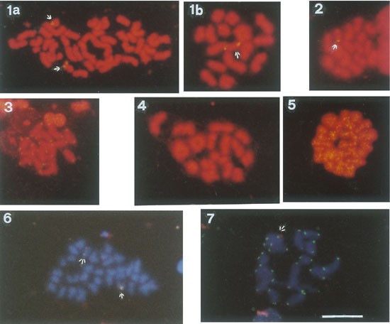
In situ hybridization, fluorescence (FISH) is a type of molecular cytogenetic technique used to detect and localize the presence or absence of specific DNA sequences on chromosomes through the use of fluorescent probes. This technique allows for the direct visualization of genetic material at a cellular level, making it possible to identify chromosomal abnormalities such as deletions, duplications, translocations, and other rearrangements.
The process involves denaturing the DNA in the sample to separate the double-stranded molecules into single strands, then adding fluorescently labeled probes that are complementary to the target DNA sequence. The probe hybridizes to the complementary sequence in the sample, and the location of the probe is detected by fluorescence microscopy.
FISH has a wide range of applications in both clinical and research settings, including prenatal diagnosis, cancer diagnosis and monitoring, and the study of gene expression and regulation. It is a powerful tool for identifying genetic abnormalities and understanding their role in human disease.
Molecular sequence data refers to the specific arrangement of molecules, most commonly nucleotides in DNA or RNA, or amino acids in proteins, that make up a biological macromolecule. This data is generated through laboratory techniques such as sequencing, and provides information about the exact order of the constituent molecules. This data is crucial in various fields of biology, including genetics, evolution, and molecular biology, allowing for comparisons between different organisms, identification of genetic variations, and studies of gene function and regulation.
Chromosomes are thread-like structures located in the nucleus of cells that contain most of the DNA present in cells. They come in pairs, with one set inherited from each parent. In humans, there are typically 23 pairs of chromosomes, for a total of 46 chromosomes.
Chromosomes 16-18 refer to the specific chromosomes that make up the 16th and 17th pairs in human cells. Chromosome 16 is an acrocentric chromosome, meaning it has a short arm (p arm) and a long arm (q arm), with the centromere located near the middle of the chromosome. It contains around 115 million base pairs of DNA and encodes approximately 1,100 genes.
Chromosome 17 is a metacentric chromosome, meaning it has a centromere located in the middle, dividing the chromosome into two arms of equal length. It contains around 81 million base pairs of DNA and encodes approximately 1,300 genes.
Chromosome 18 is a small acrocentric chromosome with a short arm (p arm) and a long arm (q arm), with the centromere located near the end of the short arm. It contains around 76 million base pairs of DNA and encodes approximately 1,200 genes.
Abnormalities in these chromosomes can lead to various genetic disorders, such as Edwards syndrome (trisomy 18), Patau syndrome (trisomy 13), and some forms of Down syndrome (translocation between chromosomes 14 and 21).
Human chromosome pair 20 is one of the 23 pairs of human chromosomes present in every cell of the body, except for the sperm and egg cells which contain only 23 individual chromosomes. Chromosomes are thread-like structures that carry genetic information in the form of genes.
Human chromosome pair 20 is an acrocentric chromosome, meaning it has a short arm (p arm) and a long arm (q arm), with the centromere located near the junction of the two arms. The short arm of chromosome 20 is very small and contains few genes, while the long arm contains several hundred genes that play important roles in various biological processes.
Chromosome pair 20 is associated with several genetic disorders, including DiGeorge syndrome, which is caused by a deletion of a portion of the long arm of chromosome 20. This syndrome is characterized by birth defects affecting the heart, face, and immune system. Other conditions associated with abnormalities of chromosome pair 20 include some forms of intellectual disability, autism spectrum disorder, and cancer.
Artificial chromosomes, yeast are synthetic chromosomes that have been created in the laboratory and can function in yeast cells. They are made up of DNA sequences that have been chemically synthesized or engineered from existing yeast chromosomes. These artificial chromosomes can be used to introduce new genes or modify existing ones in yeast, allowing for the study of gene function and genetic interactions in a controlled manner.
The creation of artificial chromosomes in yeast has been an important tool in biotechnology and synthetic biology, enabling the development of novel industrial processes and the engineering of yeast strains with enhanced properties for various applications, such as biofuel production or the manufacture of pharmaceuticals. Additionally, the study of artificial chromosomes in yeast has provided valuable insights into the fundamental principles of genome organization, replication, and inheritance.
Human chromosomes 13-15 are part of a set of 23 pairs of chromosomes found in the cells of the human body. Chromosomes are thread-like structures that contain genetic material, or DNA, that is inherited from each parent. They are responsible for the development and function of all the body's organs and systems.
Chromosome 13 is a medium-sized chromosome and contains an estimated 114 million base pairs of DNA. It is associated with several genetic disorders, including cri du chat syndrome, which is caused by a deletion on the short arm of the chromosome. Chromosome 13 also contains several important genes, such as those involved in the production of enzymes and proteins that help regulate growth and development.
Chromosome 14 is a medium-sized chromosome and contains an estimated 107 million base pairs of DNA. It is known to contain many genes that are important for the normal functioning of the brain and nervous system, as well as genes involved in the production of immune system proteins. Chromosome 14 is also associated with a number of genetic disorders, including Wolf-Hirschhorn syndrome, which is caused by a deletion on the short arm of the chromosome.
Chromosome 15 is a medium-sized chromosome and contains an estimated 102 million base pairs of DNA. It is associated with several genetic disorders, including Prader-Willi syndrome and Angelman syndrome, which are caused by abnormalities in the expression of genes on the chromosome. Chromosome 15 also contains important genes involved in the regulation of growth and development, as well as genes that play a role in the production of neurotransmitters, the chemical messengers of the brain.
It is worth noting that while chromosomes 13-15 are important for normal human development and function, abnormalities in these chromosomes can lead to a variety of genetic disorders and developmental issues.
Genetic linkage is the phenomenon where two or more genetic loci (locations on a chromosome) tend to be inherited together because they are close to each other on the same chromosome. This occurs during the process of sexual reproduction, where homologous chromosomes pair up and exchange genetic material through a process called crossing over.
The closer two loci are to each other on a chromosome, the lower the probability that they will be separated by a crossover event. As a result, they are more likely to be inherited together and are said to be linked. The degree of linkage between two loci can be measured by their recombination frequency, which is the percentage of meiotic events in which a crossover occurs between them.
Linkage analysis is an important tool in genetic research, as it allows researchers to identify and map genes that are associated with specific traits or diseases. By analyzing patterns of linkage between markers (identifiable DNA sequences) and phenotypes (observable traits), researchers can infer the location of genes that contribute to those traits or diseases on chromosomes.
Chromosome breakage is a medical term that refers to the breaking or fragmentation of chromosomes, which are thread-like structures located in the nucleus of cells that carry genetic information. Normally, chromosomes are tightly coiled and consist of two strands called chromatids, joined together at a central point called the centromere.
Chromosome breakage can occur spontaneously or be caused by environmental factors such as radiation or chemicals, or inherited genetic disorders. When a chromosome breaks, it can result in various genetic abnormalities, depending on the location and severity of the break.
For instance, if the break occurs in a region containing important genes, it can lead to the loss or alteration of those genes, causing genetic diseases or birth defects. In some cases, the broken ends of the chromosome may rejoin incorrectly, leading to chromosomal rearrangements such as translocations, deletions, or inversions. These rearrangements can also result in genetic disorders or cancer.
Chromosome breakage is commonly observed in individuals with certain inherited genetic conditions, such as Bloom syndrome, Fanconi anemia, and ataxia-telangiectasia, which are characterized by an increased susceptibility to chromosome breakage due to defects in DNA repair mechanisms.
Human chromosomes are thread-like structures that contain genetic material, composed of DNA and proteins, present in the nucleus of human cells. Each chromosome is a single, long DNA molecule that carries hundreds to thousands of genes.
Chromosomes 21, 22, and Y are three of the 23 pairs of human chromosomes. Here's what you need to know about each:
* Chromosome 21 is the smallest human autosomal chromosome, with a total length of about 47 million base pairs. It contains an estimated 200-300 genes and is associated with several genetic disorders, most notably Down syndrome, which occurs when there is an extra copy of this chromosome (trisomy 21).
* Chromosome 22 is the second smallest human autosomal chromosome, with a total length of about 50 million base pairs. It contains an estimated 500-600 genes and is associated with several genetic disorders, including DiGeorge syndrome and cat-eye syndrome.
* The Y chromosome is one of the two sex chromosomes (the other being the X chromosome) and is found only in males. It is much smaller than the X chromosome, with a total length of about 59 million base pairs and an estimated 70-200 genes. The Y chromosome determines maleness by carrying the gene for the testis-determining factor (TDF), which triggers male development in the embryo.
It's worth noting that while we have a standard set of 23 pairs of chromosomes, there can be variations and abnormalities in the number or structure of these chromosomes that can lead to genetic disorders.
Genes in insects refer to the hereditary units of DNA that are passed down from parents to offspring and contain the instructions for the development, function, and reproduction of an organism. These genetic materials are located within the chromosomes in the nucleus of insect cells. They play a crucial role in determining various traits such as physical characteristics, behavior, and susceptibility to diseases.
Insect genes, like those of other organisms, consist of exons (coding regions) that contain information for protein synthesis and introns (non-coding regions) that are removed during the process of gene expression. The expression of insect genes is regulated by various factors such as transcription factors, enhancers, and silencers, which bind to specific DNA sequences to activate or repress gene transcription.
Understanding the genetic makeup of insects has important implications for various fields, including agriculture, public health, and evolutionary biology. For example, genes associated with insect pests' resistance to pesticides can be identified and targeted to develop more effective control strategies. Similarly, genes involved in disease transmission by insect vectors such as mosquitoes can be studied to develop novel interventions for preventing the spread of infectious diseases.
A ring chromosome is a structurally abnormal chromosome that has formed a circle or ring shape. This occurs when both ends of the chromosome break off and the resulting fragments join together to form a circular structure. Ring chromosomes can vary in size, and the loss of genetic material during the formation of the ring can lead to genetic disorders and developmental delays. The effects of a ring chromosome depend on the location of the breakpoints and the amount of genetic material lost. Some individuals with ring chromosomes may have mild symptoms, while others may have severe disabilities or health problems.
A base sequence in the context of molecular biology refers to the specific order of nucleotides in a DNA or RNA molecule. In DNA, these nucleotides are adenine (A), guanine (G), cytosine (C), and thymine (T). In RNA, uracil (U) takes the place of thymine. The base sequence contains genetic information that is transcribed into RNA and ultimately translated into proteins. It is the exact order of these bases that determines the genetic code and thus the function of the DNA or RNA molecule.
Genetic markers are specific segments of DNA that are used in genetic mapping and genotyping to identify specific genetic locations, diseases, or traits. They can be composed of short tandem repeats (STRs), single nucleotide polymorphisms (SNPs), restriction fragment length polymorphisms (RFLPs), or variable number tandem repeats (VNTRs). These markers are useful in various fields such as genetic research, medical diagnostics, forensic science, and breeding programs. They can help to track inheritance patterns, identify genetic predispositions to diseases, and solve crimes by linking biological evidence to suspects or victims.
A chromosome inversion is a genetic rearrangement where a segment of a chromosome has been reversed end to end, so that its order of genes is opposite to the original. This means that the gene sequence on the segment of the chromosome has been inverted.
In an inversion, the chromosome breaks in two places, and the segment between the breaks rotates 180 degrees before reattaching. This results in a portion of the chromosome being inverted, or turned upside down, relative to the rest of the chromosome.
Chromosome inversions can be either paracentric or pericentric. Paracentric inversions involve a segment that does not include the centromere (the central constriction point of the chromosome), while pericentric inversions involve a segment that includes the centromere.
Inversions can have various effects on an individual's phenotype, depending on whether the inversion involves genes and if so, how those genes are affected by the inversion. In some cases, inversions may have no noticeable effect, while in others they may cause genetic disorders or predispose an individual to certain health conditions.
Chromosome positioning, also known as chromosome organization or chromosome architecture, refers to the specific location and spatial arrangement of chromosomes within the nucleus of a eukaryotic cell. This complex process is critical for proper regulation of gene expression, DNA replication, and chromosomal stability during the cell cycle.
Chromosomes are not randomly positioned in the nucleus; instead, they occupy distinct territories that are non-randomly organized with respect to each other. Chromosome positioning is influenced by several factors, including the presence of nuclear bodies, such as the nucleolus and nuclear speckles, as well as by the interactions between chromatin regions and the nuclear lamina.
The spatial organization of chromosomes can have significant consequences for gene regulation, as genes that are located in close proximity to each other may be more likely to interact and influence each other's expression. Chromosome positioning has also been implicated in various diseases, including cancer, where abnormalities in chromosome organization have been associated with changes in gene expression and genomic instability.
Overall, the medical definition of 'chromosome positioning' refers to the complex and dynamic process by which chromosomes are organized within the nucleus of a cell, and how this organization influences various cellular processes and functions.
Chromosomes are thread-like structures located in the nucleus of cells that carry genetic information in the form of genes. In humans, there are 23 pairs of chromosomes for a total of 46 chromosomes in every cell of the body, except for the sperm and egg cells which contain only 23 chromosomes.
Human chromosomes are numbered from 1 to 22, based on their size, with chromosome 1 being the largest and chromosome 22 being the smallest. The last two pairs of human chromosomes are known as the sex chromosomes because they determine a person's biological sex. These are labeled X and Y, with females having two X chromosomes (44+XX) and males having one X and one Y chromosome (44+XY).
Therefore, "Chromosomes, Human, 4-5" refers to the fourth and fifth pairs of human chromosomes. Chromosome 4 is an acrocentric chromosome, meaning its centromere is located near one end, resulting in a short arm (p) and a long arm (q). It contains about 190 million base pairs and encodes approximately 700 genes.
Chromosome 5 is a submetacentric chromosome, with the centromere located closer to the middle, creating two arms of roughly equal length: the short arm (p) and the long arm (q). It contains about 182 million base pairs and encodes approximately 900 genes.
Both chromosomes 4 and 5 are involved in various genetic disorders when abnormalities occur, such as deletions, duplications, or translocations. Some of the well-known genetic conditions associated with these chromosomes include:
* Chromosome 4: Wolf-Hirschhorn syndrome (deletion), Charcot-Marie-Tooth disease type 1A (duplication)
* Chromosome 5: Cri du Chat syndrome (deletion), Duchenne muscular dystrophy (deletion or mutation in a gene located on chromosome 5)
Insect viruses, also known as entomoviruses, are viruses that specifically infect and replicate in insect hosts. These viruses can be found in various insect species, including those of medical and agricultural importance. Insect viruses can cause diseases in insect populations, leading to significant impacts on their growth, development, and survival. Some insect viruses have been studied as potential biological control agents for managing pest insects that affect crops or transmit diseases. Examples of insect viruses include Baculoviridae, Reoviridae, and Picornaviridae families.
X chromosome inactivation (XCI) is a process that occurs in females of mammalian species, including humans, to compensate for the difference in gene dosage between the sexes. Females have two X chromosomes, while males have one X and one Y chromosome. To prevent females from having twice as many X-linked genes expressed as males, one of the two X chromosomes in each female cell is randomly inactivated during early embryonic development.
XCI results in the formation of a condensed and transcriptionally inactive structure called a Barr body, which can be observed in the nucleus of female cells. This process ensures that females express similar levels of X-linked genes as males, maintaining a balanced gene dosage. The choice of which X chromosome is inactivated (maternal or paternal) is random and occurs independently in each cell, leading to a mosaic expression pattern of X-linked genes in different cells and tissues of the female body.
Chromosomes in insects are thread-like structures that contain genetic material, made up of DNA and proteins, found in the nucleus of a cell. In insects, like other eukaryotes, chromosomes come in pairs, with one set inherited from each parent. They are crucial for the inheritance, storage, and transmission of genetic information from one generation to the next.
Insects typically have a diploid number of chromosomes (2n), which varies among species. The chromosomes are present in the cell's nucleus during interphase as loosely coiled structures called chromatin. During cell division, they condense and become visible under the microscope as distinct, X-shaped structures called metaphase chromosomes.
The insect chromosome set includes autosomal chromosomes, which are identical in appearance and function between males and females, and sex chromosomes, which differ between males and females. In many insects, the males have an XY sex chromosome constitution, while the females have an XX sex chromosome constitution. The sex chromosomes carry genes that determine the sex of the individual.
Insect chromosomes play a vital role in various biological processes, including development, reproduction, and evolution. They are also essential for genetic research and breeding programs in agriculture and medicine.
A centromere is a specialized region found on chromosomes that plays a crucial role in the separation of replicated chromosomes during cell division. It is the point where the sister chromatids (the two copies of a chromosome formed during DNA replication) are joined together. The centromere contains highly repeated DNA sequences and proteins that form a complex structure known as the kinetochore, which serves as an attachment site for microtubules of the mitotic spindle during cell division.
During mitosis or meiosis, the kinetochore facilitates the movement of chromosomes by interacting with the microtubules, allowing for the accurate distribution of genetic material to the daughter cells. Centromeres can vary in their position and structure among different species, ranging from being located near the middle of the chromosome (metacentric) to being positioned closer to one end (acrocentric). The precise location and characteristics of centromeres are essential for proper chromosome segregation and maintenance of genomic stability.
I believe there may be some confusion in your question. "Moths" are not a medical term, but rather they are a group of insects closely related to butterflies. They belong to the order Lepidoptera and are characterized by their scales covering their wings and body. If you have any questions about moths or if you meant to ask something else, please let me know!
Meiosis is a type of cell division that results in the formation of four daughter cells, each with half the number of chromosomes as the parent cell. It is a key process in sexual reproduction, where it generates gametes or sex cells (sperm and eggs).
The process of meiosis involves one round of DNA replication followed by two successive nuclear divisions, meiosis I and meiosis II. In meiosis I, homologous chromosomes pair, form chiasma and exchange genetic material through crossing over, then separate from each other. In meiosis II, sister chromatids separate, leading to the formation of four haploid cells. This process ensures genetic diversity in offspring by shuffling and recombining genetic information during the formation of gametes.
Translocation, genetic, refers to a type of chromosomal abnormality in which a segment of a chromosome is transferred from one chromosome to another, resulting in an altered genome. This can occur between two non-homologous chromosomes (non-reciprocal translocation) or between two homologous chromosomes (reciprocal translocation). Genetic translocations can lead to various clinical consequences, depending on the genes involved and the location of the translocation. Some translocations may result in no apparent effects, while others can cause developmental abnormalities, cancer, or other genetic disorders. In some cases, translocations can also increase the risk of having offspring with genetic conditions.
I'm sorry for any confusion, but "hybrid cells" is not a standard medical term with a widely accepted or specific definition in the field of medicine. The term "hybrid" is used in various scientific and medical contexts to describe combinations or mixtures of different elements, such as hybridoma cells (a type of fusion cell used in research, created by combining a B cell and a tumor cell) or hybridization (in genetics, the process of combining DNA from two different sources).
Without more specific context, it's difficult to provide an accurate medical definition for "hybrid cells." If you could provide more information about the context in which this term was used, I would be happy to help you further!
Chromosomes are thread-like structures located in the nucleus of cells that carry genetic information in the form of genes. A chromosome is made up of one long DNA molecule coiled tightly with proteins called histones to form a compact structure. In humans, there are 23 pairs of chromosomes, for a total of 46 chromosomes in every cell of the body, except for the sperm and egg cells which contain only 23 chromosomes each.
Chromosome structures can be described by their number, shape, size, and banding pattern. The number of chromosomes in a cell is usually constant for a species, but can vary between species. Chromosomes come in different shapes, including rod-shaped, V-shaped, or J-shaped, depending on the position of the centromere, which is the constricted region where the chromatids (the two copies of chromosome) are joined together.
The size of chromosomes also varies, with some being much larger than others. Chromosomes can be classified into several groups based on their size and banding pattern, which is a series of light and dark bands that appear when chromosomes are stained with certain dyes. The banding pattern is unique to each chromosome and can be used to identify specific regions or genes on the chromosome.
Chromosome structures can also be affected by genetic changes, such as mutations, deletions, duplications, inversions, and translocations, which can lead to genetic disorders and diseases. Understanding the structure and function of chromosomes is essential for diagnosing and treating genetic conditions, as well as for advancing our knowledge of genetics and human health.
Human chromosomes are thread-like structures that contain genetic information in the form of DNA and proteins. Each human cell typically contains 46 chromosomes arranged in 23 pairs, except for the sperm and egg cells which contain only 23 chromosomes (one half of the full set).
Chromosome 19 is one of the autosomal chromosomes, meaning it is not a sex chromosome. It is the fifth smallest human chromosome, spanning about 58 million base pairs and representing approximately 1.9% of the total DNA in cells. Chromosome 19 contains more than 1,200 genes that provide instructions for making proteins and RNA molecules involved in various cellular processes.
Chromosome 20 is also an autosomal chromosome, slightly smaller than chromosome 19. It spans about 54 million base pairs and contains around 800 genes that code for proteins and RNA molecules. Chromosome 20 is known to contain several important genes involved in cancer development, such as the tumor suppressor gene TP53.
Together, chromosomes 19 and 20 carry crucial genetic information necessary for normal human growth, development, and health. Abnormalities in these chromosomes can lead to various genetic disorders and diseases.
An amino acid sequence is the specific order of amino acids in a protein or peptide molecule, formed by the linking of the amino group (-NH2) of one amino acid to the carboxyl group (-COOH) of another amino acid through a peptide bond. The sequence is determined by the genetic code and is unique to each type of protein or peptide. It plays a crucial role in determining the three-dimensional structure and function of proteins.
Aneuploidy is a medical term that refers to an abnormal number of chromosomes in a cell. Chromosomes are thread-like structures located inside the nucleus of cells that contain genetic information in the form of genes.
In humans, the normal number of chromosomes in a cell is 46, arranged in 23 pairs. Aneuploidy occurs when there is an extra or missing chromosome in one or more of these pairs. For example, Down syndrome is a condition that results from an extra copy of chromosome 21, also known as trisomy 21.
Aneuploidy can arise during the formation of gametes (sperm or egg cells) due to errors in the process of cell division called meiosis. These errors can result in eggs or sperm with an abnormal number of chromosomes, which can then lead to aneuploidy in the resulting embryo.
Aneuploidy is a significant cause of birth defects and miscarriages. The severity of the condition depends on which chromosomes are affected and the extent of the abnormality. In some cases, aneuploidy may have no noticeable effects, while in others it can lead to serious health problems or developmental delays.
Mitosis is a type of cell division in which the genetic material of a single cell, called the mother cell, is equally distributed into two identical daughter cells. It's a fundamental process that occurs in multicellular organisms for growth, maintenance, and repair, as well as in unicellular organisms for reproduction.
The process of mitosis can be broken down into several stages: prophase, prometaphase, metaphase, anaphase, and telophase. During prophase, the chromosomes condense and become visible, and the nuclear envelope breaks down. In prometaphase, the nuclear membrane is completely disassembled, and the mitotic spindle fibers attach to the chromosomes at their centromeres.
During metaphase, the chromosomes align at the metaphase plate, an imaginary line equidistant from the two spindle poles. In anaphase, sister chromatids are pulled apart by the spindle fibers and move toward opposite poles of the cell. Finally, in telophase, new nuclear envelopes form around each set of chromosomes, and the chromosomes decondense and become less visible.
Mitosis is followed by cytokinesis, a process that divides the cytoplasm of the mother cell into two separate daughter cells. The result of mitosis and cytokinesis is two genetically identical cells, each with the same number and kind of chromosomes as the original parent cell.
A genome in the context of insects refers to the complete set of genetic material, including all of the DNA and RNA, that is present in the cells of an insect. The genome contains all of the genes that provide the instructions for the development, growth, and function of the insect. It also includes non-coding regions of DNA that may have regulatory functions or may be the result of historical processes.
The genome of an insect is typically divided into several chromosomes, which are structures in the cell's nucleus that contain long stretches of DNA. The number and appearance of these chromosomes can vary between different species of insects. For example, some insects may have a diploid number of two sets of chromosomes (one set from each parent), while others may have a haploid number of a single set of chromosomes.
The genome size of insects can also vary significantly, with some species having genomes that are only a few hundred million base pairs in length, while others have genomes that are several billion base pairs long. The genome sequence of an insect can provide valuable insights into its evolutionary history, as well as information about the genes and regulatory elements that are important for its biology and behavior.
Metaphase is a phase in the cell division process (mitosis or meiosis) where the chromosomes align in the middle of the cell, also known as the metaphase plate or equatorial plane. During this stage, each chromosome consists of two sister chromatids attached to each other by a protein complex called the centromere. The spindle fibers from opposite poles of the cell attach to the centromeres of each chromosome, and through a process called congression, they align the chromosomes in the middle of the cell. This alignment allows for accurate segregation of genetic material during the subsequent anaphase stage.
Genetic recombination is the process by which genetic material is exchanged between two similar or identical molecules of DNA during meiosis, resulting in new combinations of genes on each chromosome. This exchange occurs during crossover, where segments of DNA are swapped between non-sister homologous chromatids, creating genetic diversity among the offspring. It is a crucial mechanism for generating genetic variability and facilitating evolutionary change within populations. Additionally, recombination also plays an essential role in DNA repair processes through mechanisms such as homologous recombinational repair (HRR) and non-homologous end joining (NHEJ).
Molecular cloning is a laboratory technique used to create multiple copies of a specific DNA sequence. This process involves several steps:
1. Isolation: The first step in molecular cloning is to isolate the DNA sequence of interest from the rest of the genomic DNA. This can be done using various methods such as PCR (polymerase chain reaction), restriction enzymes, or hybridization.
2. Vector construction: Once the DNA sequence of interest has been isolated, it must be inserted into a vector, which is a small circular DNA molecule that can replicate independently in a host cell. Common vectors used in molecular cloning include plasmids and phages.
3. Transformation: The constructed vector is then introduced into a host cell, usually a bacterial or yeast cell, through a process called transformation. This can be done using various methods such as electroporation or chemical transformation.
4. Selection: After transformation, the host cells are grown in selective media that allow only those cells containing the vector to grow. This ensures that the DNA sequence of interest has been successfully cloned into the vector.
5. Amplification: Once the host cells have been selected, they can be grown in large quantities to amplify the number of copies of the cloned DNA sequence.
Molecular cloning is a powerful tool in molecular biology and has numerous applications, including the production of recombinant proteins, gene therapy, functional analysis of genes, and genetic engineering.
A mutation is a permanent change in the DNA sequence of an organism's genome. Mutations can occur spontaneously or be caused by environmental factors such as exposure to radiation, chemicals, or viruses. They may have various effects on the organism, ranging from benign to harmful, depending on where they occur and whether they alter the function of essential proteins. In some cases, mutations can increase an individual's susceptibility to certain diseases or disorders, while in others, they may confer a survival advantage. Mutations are the driving force behind evolution, as they introduce new genetic variability into populations, which can then be acted upon by natural selection.
Insect hormones are chemical messengers that regulate various physiological and behavioral processes in insects. They are produced and released by endocrine glands and organs, such as the corpora allata, prothoracic glands, and neurosecretory cells located in the brain. Insect hormones play crucial roles in the regulation of growth and development, reproduction, diapause (a state of dormancy), metamorphosis, molting, and other vital functions. Some well-known insect hormones include juvenile hormone (JH), ecdysteroids (such as 20-hydroxyecdysone), and neuropeptides like the brain hormone and adipokinetic hormone. These hormones act through specific receptors, often transmembrane proteins, to elicit intracellular signaling cascades that ultimately lead to changes in gene expression, cell behavior, or organ function. Understanding insect hormones is essential for developing novel strategies for pest management and control, as well as for advancing our knowledge of insect biology and evolution.
'Insect control' is not a term typically used in medical definitions. However, it generally refers to the methods and practices used to manage or reduce the population of insects that can be harmful or disruptive to human health, food supply, or property. This can include various strategies such as chemical pesticides, biological control agents, habitat modification, and other integrated pest management techniques.
In medical terms, 'vector control' is a more relevant concept, which refers to the specific practices used to reduce or prevent the transmission of infectious diseases by insects and other arthropods that act as disease vectors (such as mosquitoes, ticks, and fleas). Vector control measures may include the use of insecticides, larvicides, biological control agents, environmental management, personal protection methods, and other integrated vector management strategies.
"Genetic crosses" refer to the breeding of individuals with different genetic characteristics to produce offspring with specific combinations of traits. This process is commonly used in genetics research to study the inheritance patterns and function of specific genes.
There are several types of genetic crosses, including:
1. Monohybrid cross: A cross between two individuals that differ in the expression of a single gene or trait.
2. Dihybrid cross: A cross between two individuals that differ in the expression of two genes or traits.
3. Backcross: A cross between an individual from a hybrid population and one of its parental lines.
4. Testcross: A cross between an individual with unknown genotype and a homozygous recessive individual.
5. Reciprocal cross: A cross in which the male and female parents are reversed to determine if there is any effect of sex on the expression of the trait.
These genetic crosses help researchers to understand the mode of inheritance, linkage, recombination, and other genetic phenomena.
Microsatellite repeats, also known as short tandem repeats (STRs), are repetitive DNA sequences made up of units of 1-6 base pairs that are repeated in a head-to-tail manner. These repeats are spread throughout the human genome and are highly polymorphic, meaning they can have different numbers of repeat units in different individuals.
Microsatellites are useful as genetic markers because of their high degree of variability. They are commonly used in forensic science to identify individuals, in genealogy to trace ancestry, and in medical research to study genetic diseases and disorders. Mutations in microsatellite repeats have been associated with various neurological conditions, including Huntington's disease and fragile X syndrome.
A larva is a distinct stage in the life cycle of various insects, mites, and other arthropods during which they undergo significant metamorphosis before becoming adults. In a medical context, larvae are known for their role in certain parasitic infections. Specifically, some helminth (parasitic worm) species use larval forms to infect human hosts. These invasions may lead to conditions such as cutaneous larva migrans, visceral larva migrans, or gnathostomiasis, depending on the specific parasite involved and the location of the infection within the body.
The larval stage is characterized by its markedly different morphology and behavior compared to the adult form. Larvae often have a distinct appearance, featuring unsegmented bodies, simple sense organs, and undeveloped digestive systems. They are typically adapted for a specific mode of life, such as free-living or parasitic existence, and rely on external sources of nutrition for their development.
In the context of helminth infections, larvae may be transmitted to humans through various routes, including ingestion of contaminated food or water, direct skin contact with infective stages, or transmission via an intermediate host (such as a vector). Once inside the human body, these parasitic larvae can cause tissue damage and provoke immune responses, leading to the clinical manifestations of disease.
It is essential to distinguish between the medical definition of 'larva' and its broader usage in biology and zoology. In those fields, 'larva' refers to any juvenile form that undergoes metamorphosis before reaching adulthood, regardless of whether it is parasitic or not.
A phenotype is the physical or biochemical expression of an organism's genes, or the observable traits and characteristics resulting from the interaction of its genetic constitution (genotype) with environmental factors. These characteristics can include appearance, development, behavior, and resistance to disease, among others. Phenotypes can vary widely, even among individuals with identical genotypes, due to differences in environmental influences, gene expression, and genetic interactions.
'Drosophila melanogaster' is the scientific name for a species of fruit fly that is commonly used as a model organism in various fields of biological research, including genetics, developmental biology, and evolutionary biology. Its small size, short generation time, large number of offspring, and ease of cultivation make it an ideal subject for laboratory studies. The fruit fly's genome has been fully sequenced, and many of its genes have counterparts in the human genome, which facilitates the understanding of genetic mechanisms and their role in human health and disease.
Here is a brief medical definition:
Drosophila melanogaster (droh-suh-fih-luh meh-lon-guh-ster): A species of fruit fly used extensively as a model organism in genetic, developmental, and evolutionary research. Its genome has been sequenced, revealing many genes with human counterparts, making it valuable for understanding genetic mechanisms and their role in human health and disease.
Diptera is an order of insects that includes flies, mosquitoes, and gnats. The name "Diptera" comes from the Greek words "di," meaning two, and "pteron," meaning wing. This refers to the fact that all members of this order have a single pair of functional wings for flying, while the other pair is reduced to small knob-like structures called halteres, which help with balance and maneuverability during flight.
Some common examples of Diptera include houseflies, fruit flies, horseflies, tsetse flies, and midges. Many species in this order are important pollinators, while others can be significant pests or disease vectors. The study of Diptera is called dipterology.
"Spodoptera" is not a medical term, but a genus name in the insect family Noctuidae. It includes several species of moths commonly known as armyworms or cutworms due to their habit of consuming leaves and roots of various plants, causing significant damage to crops.
Some well-known species in this genus are Spodoptera frugiperda (fall armyworm), Spodoptera litura (tobacco cutworm), and Spodoptera exigua (beet armyworm). These pests can be a concern for medical entomology when they transmit pathogens or cause allergic reactions. For instance, their frass (feces) and shed skins may trigger asthma symptoms in susceptible individuals. However, the insects themselves are not typically considered medical issues unless they directly affect human health.
A LOD (Logarithm of Odds) score is not a medical term per se, but rather a statistical concept that is used in genetic research and linkage analysis to determine the likelihood of a gene or genetic marker being linked to a particular disease or trait. The LOD score compares the odds of observing the pattern of inheritance of a genetic marker in a family if the marker is linked to the disease, versus the odds if the marker is not linked. A LOD score of 3 or higher is generally considered evidence for linkage, while a score of -2 or lower is considered evidence against linkage.
I must clarify that the term "pedigree" is not typically used in medical definitions. Instead, it is often employed in genetics and breeding, where it refers to the recorded ancestry of an individual or a family, tracing the inheritance of specific traits or diseases. In human genetics, a pedigree can help illustrate the pattern of genetic inheritance in families over multiple generations. However, it is not a medical term with a specific clinical definition.
I'm sorry for any confusion, but "Lepidoptera" is not a medical term. It is a taxonomic order that includes moths and butterflies, which are insects known for their distinctive wing scales. This term is used in the field of biology, not medicine.
I'm sorry for any confusion, but "Hemiptera" is not a medical term. It is a taxonomic order in the classification of living things, also known as "true bugs." This group includes species such as cicadas, aphids, and bedbugs. If you have a medical term in mind, please provide it so I can give you an accurate definition.
Insect repellents are substances that are applied to the skin, clothing, or other surfaces to deter insects from landing or crawling on that surface. They work by masking the scents that attract insects or by repelling them with unpleasant odors. Insect repellents can be chemical-based, such as those containing DEET (N,N-diethyl-m-toluamide), picaridin, or IR3535, or they can be natural, such as those containing oil of lemon eucalyptus or citronella. These substances work by interfering with the insect's ability to detect human scent, making it less likely that they will come into contact with the person using the repellent. Insect repellents are an important tool in preventing insect-borne diseases such as Lyme disease, West Nile virus, and Zika virus.
DNA Sequence Analysis is the systematic determination of the order of nucleotides in a DNA molecule. It is a critical component of modern molecular biology, genetics, and genetic engineering. The process involves determining the exact order of the four nucleotide bases - adenine (A), guanine (G), cytosine (C), and thymine (T) - in a DNA molecule or fragment. This information is used in various applications such as identifying gene mutations, studying evolutionary relationships, developing molecular markers for breeding, and diagnosing genetic diseases.
The process of DNA Sequence Analysis typically involves several steps, including DNA extraction, PCR amplification (if necessary), purification, sequencing reaction, and electrophoresis. The resulting data is then analyzed using specialized software to determine the exact sequence of nucleotides.
In recent years, high-throughput DNA sequencing technologies have revolutionized the field of genomics, enabling the rapid and cost-effective sequencing of entire genomes. This has led to an explosion of genomic data and new insights into the genetic basis of many diseases and traits.
Baculoviridae is a family of large, double-stranded DNA viruses that infect arthropods, particularly insects. The virions (virus particles) are enclosed in a rod-shaped or occlusion body called a polyhedron, which provides protection and stability in the environment. Baculoviruses have a wide host range within the order Lepidoptera (moths and butterflies), Hymenoptera (sawflies, bees, wasps, and ants), and Diptera (flies). They are important pathogens in agriculture and forestry, causing significant damage to insect pests.
The Baculoviridae family is divided into four genera: Alphabaculovirus, Betabaculovirus, Gammabaculovirus, and Deltabaculovirus. The two most well-studied and economically important genera are Alphabaculovirus (nuclear polyhedrosis viruses or NPVs) and Betabaculovirus (granulosis viruses or GVs).
Baculoviruses have a biphasic replication cycle, consisting of a budded phase and an occluded phase. During the budded phase, the virus infects host cells and produces enveloped virions that can spread to other cells within the insect. In the occluded phase, large numbers of non-enveloped virions are produced and encapsidated in a protein matrix called a polyhedron. These polyhedra accumulate in the infected insect's tissues, providing protection from environmental degradation and facilitating transmission to new hosts through oral ingestion or other means.
Baculoviruses have been extensively studied as models for understanding viral replication, gene expression, and host-pathogen interactions. They also have potential applications in biotechnology and pest control, including the production of recombinant proteins, gene therapy vectors, and environmentally friendly insecticides.
Insect vectors are insects that transmit disease-causing pathogens (such as viruses, bacteria, parasites) from one host to another. They do this while feeding on the host's blood or tissues. The insects themselves are not infected by the pathogen but act as mechanical carriers that pass it on during their bite. Examples of diseases spread by insect vectors include malaria (transmitted by mosquitoes), Lyme disease (transmitted by ticks), and plague (transmitted by fleas). Proper prevention measures, such as using insect repellent and reducing standing water where mosquitoes breed, can help reduce the risk of contracting these diseases.
An allele is a variant form of a gene that is located at a specific position on a specific chromosome. Alleles are alternative forms of the same gene that arise by mutation and are found at the same locus or position on homologous chromosomes.
Each person typically inherits two copies of each gene, one from each parent. If the two alleles are identical, a person is said to be homozygous for that trait. If the alleles are different, the person is heterozygous.
For example, the ABO blood group system has three alleles, A, B, and O, which determine a person's blood type. If a person inherits two A alleles, they will have type A blood; if they inherit one A and one B allele, they will have type AB blood; if they inherit two B alleles, they will have type B blood; and if they inherit two O alleles, they will have type O blood.
Alleles can also influence traits such as eye color, hair color, height, and other physical characteristics. Some alleles are dominant, meaning that only one copy of the allele is needed to express the trait, while others are recessive, meaning that two copies of the allele are needed to express the trait.
"Beetles" is not a medical term. It is a common name used to refer to insects belonging to the order Coleoptera, which is one of the largest orders in the class Insecta. Beetles are characterized by their hardened forewings, known as elytra, which protect their hind wings and body when not in use for flying.
There are many different species of beetles found all over the world, and some can have an impact on human health. For example, certain types of beetles, such as bed bugs and carpet beetles, can cause skin irritation and allergic reactions in some people. Other beetles, like the Colorado potato beetle, can damage crops and lead to economic losses for farmers. However, it is important to note that most beetles are not harmful to humans and play an essential role in ecosystems as decomposers and pollinators.
Phylogeny is the evolutionary history and relationship among biological entities, such as species or genes, based on their shared characteristics. In other words, it refers to the branching pattern of evolution that shows how various organisms have descended from a common ancestor over time. Phylogenetic analysis involves constructing a tree-like diagram called a phylogenetic tree, which depicts the inferred evolutionary relationships among organisms or genes based on molecular sequence data or other types of characters. This information is crucial for understanding the diversity and distribution of life on Earth, as well as for studying the emergence and spread of diseases.
Molecular evolution is the process of change in the DNA sequence or protein structure over time, driven by mechanisms such as mutation, genetic drift, gene flow, and natural selection. It refers to the evolutionary study of changes in DNA, RNA, and proteins, and how these changes accumulate and lead to new species and diversity of life. Molecular evolution can be used to understand the history and relationships among different organisms, as well as the functional consequences of genetic changes.
Insect bites and stings refer to the penetration of the skin by insects, such as mosquitoes, fleas, ticks, or bees, often resulting in localized symptoms including redness, swelling, itching, and pain. The reaction can vary depending on the individual's sensitivity and the type of insect. In some cases, systemic reactions like anaphylaxis may occur, which requires immediate medical attention. Treatment typically involves relieving symptoms with topical creams, antihistamines, or in severe cases, epinephrine. Prevention measures include using insect repellent and protective clothing.
Deoxyribonucleic acid (DNA) is the genetic material present in the cells of organisms where it is responsible for the storage and transmission of hereditary information. DNA is a long molecule that consists of two strands coiled together to form a double helix. Each strand is made up of a series of four nucleotide bases - adenine (A), guanine (G), cytosine (C), and thymine (T) - that are linked together by phosphate and sugar groups. The sequence of these bases along the length of the molecule encodes genetic information, with A always pairing with T and C always pairing with G. This base-pairing allows for the replication and transcription of DNA, which are essential processes in the functioning and reproduction of all living organisms.
Trisomy is a genetic condition where there is an extra copy of a particular chromosome, resulting in 47 chromosomes instead of the typical 46 in a cell. This usually occurs due to an error in cell division during the development of the egg, sperm, or embryo.
Instead of the normal pair, there are three copies (trisomy) of that chromosome. The most common form of trisomy is Trisomy 21, also known as Down syndrome, where there is an extra copy of chromosome 21. Other forms include Trisomy 13 (Patau syndrome) and Trisomy 18 (Edwards syndrome), which are associated with more severe developmental issues and shorter lifespans.
Trisomy can also occur in a mosaic form, where some cells have the extra chromosome while others do not, leading to varying degrees of symptoms depending on the proportion of affected cells.
Nondisjunction is a genetic term that refers to the failure of homologous chromosomes or sister chromatids to properly separate during cell division, resulting in an abnormal number of chromosomes in the daughter cells. This can occur during either mitosis (resulting in somatic mutations) or meiosis (leading to gametes with an incorrect number of chromosomes).
In humans, nondisjunction of chromosome 21 during meiosis is the most common cause of Down syndrome, resulting in three copies of chromosome 21 (trisomy 21) in the affected individual. Nondisjunction can also result in other aneuploidies, such as Turner syndrome (X monosomy), Klinefelter syndrome (XXY), and Edwards syndrome (trisomy 18).
Nondisjunction is typically a random event, although maternal age has been identified as a risk factor for nondisjunction during meiosis. In some cases, structural chromosomal abnormalities or genetic factors may predispose an individual to nondisjunction events.
Nucleic acid hybridization is a process in molecular biology where two single-stranded nucleic acids (DNA, RNA) with complementary sequences pair together to form a double-stranded molecule through hydrogen bonding. The strands can be from the same type of nucleic acid or different types (i.e., DNA-RNA or DNA-cDNA). This process is commonly used in various laboratory techniques, such as Southern blotting, Northern blotting, polymerase chain reaction (PCR), and microarray analysis, to detect, isolate, and analyze specific nucleic acid sequences. The hybridization temperature and conditions are critical to ensure the specificity of the interaction between the two strands.
Artificial human chromosomes are artificially constructed chromosomes that contain human genetic material. They are created in a laboratory setting and can be used for various research purposes, such as studying the function of specific genes or creating cell lines with modified genetic characteristics. Artificial human chromosomes are typically created by combining pieces of human DNA with a scaffold made of non-human DNA, which provides structural support and allows the artificial chromosome to behave like a natural human chromosome. These chromosomes can then be introduced into human cells through various methods, such as microcell-mediated chromosome transfer or direct injection into the cell nucleus. It is important to note that artificial human chromosomes are not present in nature and are solely created for research purposes.
Kinetochores are specialized protein structures that form on the centromere region of a chromosome. They play a crucial role in the process of cell division, specifically during mitosis and meiosis. The primary function of kinetochores is to connect the chromosomes to the microtubules of the spindle apparatus, which is responsible for separating the sister chromatids during cell division. Through this connection, kinetochores facilitate the movement of chromosomes towards opposite poles of the cell during anaphase, ensuring equal distribution of genetic material to each resulting daughter cell.
I'm sorry for any confusion, but "grasshoppers" is not a term used in medical definitions. Grasshoppers are a type of insect that belongs to the order Orthoptera and suborder Caelifera. They are known for their long hind legs which they use for jumping, and some species can jump over 20 times their own body length. If you have any questions about medical terminology or topics, I'd be happy to help with those instead!
Genetic models are theoretical frameworks used in genetics to describe and explain the inheritance patterns and genetic architecture of traits, diseases, or phenomena. These models are based on mathematical equations and statistical methods that incorporate information about gene frequencies, modes of inheritance, and the effects of environmental factors. They can be used to predict the probability of certain genetic outcomes, to understand the genetic basis of complex traits, and to inform medical management and treatment decisions.
There are several types of genetic models, including:
1. Mendelian models: These models describe the inheritance patterns of simple genetic traits that follow Mendel's laws of segregation and independent assortment. Examples include autosomal dominant, autosomal recessive, and X-linked inheritance.
2. Complex trait models: These models describe the inheritance patterns of complex traits that are influenced by multiple genes and environmental factors. Examples include heart disease, diabetes, and cancer.
3. Population genetics models: These models describe the distribution and frequency of genetic variants within populations over time. They can be used to study evolutionary processes, such as natural selection and genetic drift.
4. Quantitative genetics models: These models describe the relationship between genetic variation and phenotypic variation in continuous traits, such as height or IQ. They can be used to estimate heritability and to identify quantitative trait loci (QTLs) that contribute to trait variation.
5. Statistical genetics models: These models use statistical methods to analyze genetic data and infer the presence of genetic associations or linkage. They can be used to identify genetic risk factors for diseases or traits.
Overall, genetic models are essential tools in genetics research and medical genetics, as they allow researchers to make predictions about genetic outcomes, test hypotheses about the genetic basis of traits and diseases, and develop strategies for prevention, diagnosis, and treatment.
A telomere is a region of repetitive DNA sequences found at the end of chromosomes, which protects the genetic data from damage and degradation during cell division. Telomeres naturally shorten as cells divide, and when they become too short, the cell can no longer divide and becomes senescent or dies. This natural process is associated with aging and various age-related diseases. The length of telomeres can also be influenced by various genetic and environmental factors, including stress, diet, and lifestyle.
Species specificity is a term used in the field of biology, including medicine, to refer to the characteristic of a biological entity (such as a virus, bacterium, or other microorganism) that allows it to interact exclusively or preferentially with a particular species. This means that the biological entity has a strong affinity for, or is only able to infect, a specific host species.
For example, HIV is specifically adapted to infect human cells and does not typically infect other animal species. Similarly, some bacterial toxins are species-specific and can only affect certain types of animals or humans. This concept is important in understanding the transmission dynamics and host range of various pathogens, as well as in developing targeted therapies and vaccines.
Chromosome walking is a historical term used in genetics to describe the process of mapping and sequencing DNA along a chromosome. It involves the identification and characterization of a specific starting point, or "landmark," on a chromosome, followed by the systematic analysis of adjacent DNA segments, one after another, in a step-by-step manner.
The technique typically employs the use of molecular biology tools such as restriction enzymes, cloning vectors, and genetic markers to physically isolate and characterize overlapping DNA fragments that cover the region of interest. By identifying shared sequences or markers between adjacent fragments, researchers can "walk" along the chromosome, gradually building up a more detailed map of the genetic sequence.
Chromosome walking was an important technique in the early days of genetics and genomics research, as it allowed scientists to systematically analyze large stretches of DNA before the advent of high-throughput sequencing technologies. Today, while whole-genome sequencing has largely replaced chromosome walking for many applications, the technique is still used in some specialized contexts where a targeted approach is required.
Southern blotting is a type of membrane-based blotting technique that is used in molecular biology to detect and locate specific DNA sequences within a DNA sample. This technique is named after its inventor, Edward M. Southern.
In Southern blotting, the DNA sample is first digested with one or more restriction enzymes, which cut the DNA at specific recognition sites. The resulting DNA fragments are then separated based on their size by gel electrophoresis. After separation, the DNA fragments are denatured to convert them into single-stranded DNA and transferred onto a nitrocellulose or nylon membrane.
Once the DNA has been transferred to the membrane, it is hybridized with a labeled probe that is complementary to the sequence of interest. The probe can be labeled with radioactive isotopes, fluorescent dyes, or chemiluminescent compounds. After hybridization, the membrane is washed to remove any unbound probe and then exposed to X-ray film (in the case of radioactive probes) or scanned (in the case of non-radioactive probes) to detect the location of the labeled probe on the membrane.
The position of the labeled probe on the membrane corresponds to the location of the specific DNA sequence within the original DNA sample. Southern blotting is a powerful tool for identifying and characterizing specific DNA sequences, such as those associated with genetic diseases or gene regulation.
Polymerase Chain Reaction (PCR) is a laboratory technique used to amplify specific regions of DNA. It enables the production of thousands to millions of copies of a particular DNA sequence in a rapid and efficient manner, making it an essential tool in various fields such as molecular biology, medical diagnostics, forensic science, and research.
The PCR process involves repeated cycles of heating and cooling to separate the DNA strands, allow primers (short sequences of single-stranded DNA) to attach to the target regions, and extend these primers using an enzyme called Taq polymerase, resulting in the exponential amplification of the desired DNA segment.
In a medical context, PCR is often used for detecting and quantifying specific pathogens (viruses, bacteria, fungi, or parasites) in clinical samples, identifying genetic mutations or polymorphisms associated with diseases, monitoring disease progression, and evaluating treatment effectiveness.
Chromosomal proteins, non-histone, are a diverse group of proteins that are associated with chromatin, the complex of DNA and histone proteins, but do not have the characteristic structure of histones. These proteins play important roles in various nuclear processes such as DNA replication, transcription, repair, recombination, and chromosome condensation and segregation during cell division. They can be broadly classified into several categories based on their functions, including architectural proteins, enzymes, transcription factors, and structural proteins. Examples of non-histone chromosomal proteins include high mobility group (HMG) proteins, poly(ADP-ribose) polymerases (PARPs), and condensins.
Genotype, in genetics, refers to the complete heritable genetic makeup of an individual organism, including all of its genes. It is the set of instructions contained in an organism's DNA for the development and function of that organism. The genotype is the basis for an individual's inherited traits, and it can be contrasted with an individual's phenotype, which refers to the observable physical or biochemical characteristics of an organism that result from the expression of its genes in combination with environmental influences.
It is important to note that an individual's genotype is not necessarily identical to their genetic sequence. Some genes have multiple forms called alleles, and an individual may inherit different alleles for a given gene from each parent. The combination of alleles that an individual inherits for a particular gene is known as their genotype for that gene.
Understanding an individual's genotype can provide important information about their susceptibility to certain diseases, their response to drugs and other treatments, and their risk of passing on inherited genetic disorders to their offspring.
The spindle apparatus is a microtubule-based structure that plays a crucial role in the process of cell division, specifically during mitosis and meiosis. It consists of three main components:
1. The spindle poles: These are organized structures composed of microtubules and associated proteins that serve as the anchoring points for the spindle fibers. In animal cells, these poles are typically formed by centrosomes, while in plant cells, they form around nucleation sites called microtubule-organizing centers (MTOCs).
2. The spindle fibers: These are dynamic arrays of microtubules that extend between the two spindle poles. They can be categorized into three types: kinetochore fibers, which connect to the kinetochores on chromosomes; astral fibers, which radiate from the spindle poles and help position the spindle within the cell; and interpolar fibers, which lie between the two spindle poles and contribute to their separation during anaphase.
3. Regulatory proteins: Various motor proteins, such as dynein and kinesin, as well as non-motor proteins like tubulin and septins, are involved in the assembly, maintenance, and dynamics of the spindle apparatus. These proteins help to generate forces that move chromosomes, position the spindle, and ultimately segregate genetic material between two daughter cells during cell division.
The spindle apparatus is essential for ensuring accurate chromosome separation and maintaining genomic stability during cell division. Dysfunction of the spindle apparatus can lead to various abnormalities, including aneuploidy (abnormal number of chromosomes) and chromosomal instability, which have been implicated in several diseases, such as cancer and developmental disorders.
Chromosomal instability is a term used in genetics to describe a type of genetic alteration where there are abnormalities in the number or structure of chromosomes within cells. Chromosomes are thread-like structures that contain our genetic material, and they usually exist in pairs in the nucleus of a cell.
Chromosomal instability can arise due to various factors, including errors in DNA replication or repair, problems during cell division, or exposure to environmental mutagens. This instability can lead to an increased frequency of chromosomal abnormalities, such as deletions, duplications, translocations, or changes in the number of chromosomes.
Chromosomal instability is associated with several human diseases, including cancer. In cancer cells, chromosomal instability can contribute to tumor heterogeneity, drug resistance, and disease progression. It is also observed in certain genetic disorders, such as Down syndrome, where an extra copy of chromosome 21 is present, and in some rare inherited syndromes, such as Bloom syndrome and Fanconi anemia, which are characterized by a high risk of cancer and other health problems.
Quantitative Trait Loci (QTL) are regions of the genome that are associated with variation in quantitative traits, which are traits that vary continuously in a population and are influenced by multiple genes and environmental factors. QTLs can help to explain how genetic variations contribute to differences in complex traits such as height, blood pressure, or disease susceptibility.
Quantitative trait loci are identified through statistical analysis of genetic markers and trait values in experimental crosses between genetically distinct individuals, such as strains of mice or plants. The location of a QTL is inferred based on the pattern of linkage disequilibrium between genetic markers and the trait of interest. Once a QTL has been identified, further analysis can be conducted to identify the specific gene or genes responsible for the variation in the trait.
It's important to note that QTLs are not themselves genes, but rather genomic regions that contain one or more genes that contribute to the variation in a quantitative trait. Additionally, because QTLs are identified through statistical analysis, they represent probabilistic estimates of the location of genetic factors influencing a trait and may encompass large genomic regions containing multiple genes. Therefore, additional research is often required to fine-map and identify the specific genes responsible for the variation in the trait.
A cell line is a culture of cells that are grown in a laboratory for use in research. These cells are usually taken from a single cell or group of cells, and they are able to divide and grow continuously in the lab. Cell lines can come from many different sources, including animals, plants, and humans. They are often used in scientific research to study cellular processes, disease mechanisms, and to test new drugs or treatments. Some common types of human cell lines include HeLa cells (which come from a cancer patient named Henrietta Lacks), HEK293 cells (which come from embryonic kidney cells), and HUVEC cells (which come from umbilical vein endothelial cells). It is important to note that cell lines are not the same as primary cells, which are cells that are taken directly from a living organism and have not been grown in the lab.
Sequence homology, amino acid, refers to the similarity in the order of amino acids in a protein or a portion of a protein between two or more species. This similarity can be used to infer evolutionary relationships and functional similarities between proteins. The higher the degree of sequence homology, the more likely it is that the proteins are related and have similar functions. Sequence homology can be determined through various methods such as pairwise alignment or multiple sequence alignment, which compare the sequences and calculate a score based on the number and type of matching amino acids.
Chromosome fragility refers to the susceptibility of specific regions on chromosomes to break or become unstable during cell division. These fragile sites are prone to forming gaps or breaks in the chromosome structure, which can lead to genetic rearrangements, including deletions, duplications, or translocations.
Chromosome fragility is often associated with certain genetic disorders and syndromes. For example, the most common fragile site in human chromosomes is FRAXA, located on the X chromosome, which is linked to Fragile X Syndrome, a leading cause of inherited intellectual disability and autism.
Environmental factors such as exposure to chemicals or radiation can also increase chromosome fragility, leading to an increased risk of genetic mutations and diseases.
"Drosophila" is a genus of small flies, also known as fruit flies. The most common species used in scientific research is "Drosophila melanogaster," which has been a valuable model organism for many areas of biological and medical research, including genetics, developmental biology, neurobiology, and aging.
The use of Drosophila as a model organism has led to numerous important discoveries in genetics and molecular biology, such as the identification of genes that are associated with human diseases like cancer, Parkinson's disease, and obesity. The short reproductive cycle, large number of offspring, and ease of genetic manipulation make Drosophila a powerful tool for studying complex biological processes.
Hemolymph is not a term typically used in human medicine, but it is commonly used in the study of invertebrates, particularly arthropods such as insects and crustaceans. Hemolymph is the fluid that circulates within the open circulatory system of these animals, serving multiple functions similar to both blood and lymphatic systems in vertebrates.
In simpler terms, hemolymph is a combined fluid that performs the functions of both blood and lymph in invertebrates. It serves as a transport medium for nutrients, waste products, hormones, and immune cells (hemocytes) throughout the body. Hemolymph does not contain red and white blood cells like human blood; instead, hemocytes are the primary cellular components responsible for immune responses and wound healing in these animals.
A haplotype is a group of genes or DNA sequences that are inherited together from a single parent. It refers to a combination of alleles (variant forms of a gene) that are located on the same chromosome and are usually transmitted as a unit. Haplotypes can be useful in tracing genetic ancestry, understanding the genetic basis of diseases, and developing personalized medical treatments.
In population genetics, haplotypes are often used to study patterns of genetic variation within and between populations. By comparing haplotype frequencies across populations, researchers can infer historical events such as migrations, population expansions, and bottlenecks. Additionally, haplotypes can provide information about the evolutionary history of genes and genomic regions.
In clinical genetics, haplotypes can be used to identify genetic risk factors for diseases or to predict an individual's response to certain medications. For example, specific haplotypes in the HLA gene region have been associated with increased susceptibility to certain autoimmune diseases, while other haplotypes in the CYP450 gene family can affect how individuals metabolize drugs.
Overall, haplotypes provide a powerful tool for understanding the genetic basis of complex traits and diseases, as well as for developing personalized medical treatments based on an individual's genetic makeup.
A multigene family is a group of genetically related genes that share a common ancestry and have similar sequences or structures. These genes are arranged in clusters on a chromosome and often encode proteins with similar functions. They can arise through various mechanisms, including gene duplication, recombination, and transposition. Multigene families play crucial roles in many biological processes, such as development, immunity, and metabolism. Examples of multigene families include the globin genes involved in oxygen transport, the immune system's major histocompatibility complex (MHC) genes, and the cytochrome P450 genes associated with drug metabolism.
Satellite DNA is a type of DNA sequence that is repeated in a tandem arrangement in the genome. These repeats are usually relatively short, ranging from 2 to 10 base pairs, and are often present in thousands to millions of copies arranged in head-to-tail fashion. Satellite DNA can be found in centromeric and pericentromeric regions of chromosomes, as well as at telomeres and other heterochromatic regions of the genome.
Due to their repetitive nature, satellite DNAs are often excluded from the main part of the genome during DNA sequencing projects, and therefore have been referred to as "satellite" DNA. However, recent studies suggest that satellite DNA may play important roles in chromosome structure, function, and evolution.
It's worth noting that not all repetitive DNA sequences are considered satellite DNA. For example, microsatellites and minisatellites are also repetitive DNA sequences, but they have different repeat lengths and arrangements than satellite DNA.
Chromosome duplication is a genetic alteration where a segment of a chromosome or the entire chromosome is present in an extra copy. This results in an additional portion of genetic material, leading to an abnormal number of genes. In humans, chromosomes typically occur in pairs (23 pairs for a total of 46 chromosomes), and any deviation from this normal number can cause genetic disorders or developmental abnormalities.
Duplication can occur in various ways:
1. Duplication of a chromosome segment: A specific region of a chromosome is repeated, leading to an extra copy of the genes present in that area. This type of duplication may not always cause noticeable effects, depending on the size and location of the duplicated segment. However, if the duplicated region contains important genes or growth regulatory elements, it can lead to genetic disorders or developmental abnormalities.
2. Duplication of a whole chromosome: An entire chromosome is present in an extra copy, leading to 3 copies instead of the typical 2 copies (one from each parent). This condition is called trisomy and can result in various genetic disorders, depending on which chromosome is duplicated. For example, Trisomy 21 or Down syndrome occurs when there are three copies of chromosome 21.
3. Mosaicism: When an individual has some cells with a normal number of chromosomes and others with the extra copy, it is called mosaicism. The severity of symptoms depends on the proportion of cells carrying the duplication and the specific genes involved in the duplicated region.
Chromosome duplications can occur spontaneously during cell division or may be inherited from a parent. They are often detected through prenatal testing, such as amniocentesis or chorionic villus sampling (CVS), or through genetic testing for individuals with developmental delays, intellectual disabilities, or birth defects.
Repetitive sequences in nucleic acid refer to repeated stretches of DNA or RNA nucleotide bases that are present in a genome. These sequences can vary in length and can be arranged in different patterns such as direct repeats, inverted repeats, or tandem repeats. In some cases, these repetitive sequences do not code for proteins and are often found in non-coding regions of the genome. They can play a role in genetic instability, regulation of gene expression, and evolutionary processes. However, certain types of repeat expansions have been associated with various neurodegenerative disorders and other human diseases.
Cockroaches are not a medical condition or disease. They are a type of insect that can be found in many parts of the world. Some species of cockroaches are known to carry diseases and allergens, which can cause health problems for some people. Cockroach allergens can trigger asthma symptoms, especially in children. Additionally, cockroaches can contaminate food and surfaces with bacteria and other germs, which can lead to illnesses such as salmonellosis and gastroenteritis.
If you have a problem with cockroaches in your home or workplace, it is important to take steps to eliminate them to reduce the risk of health problems. This may include cleaning up food and water sources, sealing entry points, and using pesticides or hiring a professional pest control service.
A DNA probe is a single-stranded DNA molecule that contains a specific sequence of nucleotides, and is labeled with a detectable marker such as a radioisotope or a fluorescent dye. It is used in molecular biology to identify and locate a complementary sequence within a sample of DNA. The probe hybridizes (forms a stable double-stranded structure) with its complementary sequence through base pairing, allowing for the detection and analysis of the target DNA. This technique is widely used in various applications such as genetic testing, diagnosis of infectious diseases, and forensic science.
A gene is a specific sequence of nucleotides in DNA that carries genetic information. Genes are the fundamental units of heredity and are responsible for the development and function of all living organisms. They code for proteins or RNA molecules, which carry out various functions within cells and are essential for the structure, function, and regulation of the body's tissues and organs.
Each gene has a specific location on a chromosome, and each person inherits two copies of every gene, one from each parent. Variations in the sequence of nucleotides in a gene can lead to differences in traits between individuals, including physical characteristics, susceptibility to disease, and responses to environmental factors.
Medical genetics is the study of genes and their role in health and disease. It involves understanding how genes contribute to the development and progression of various medical conditions, as well as identifying genetic risk factors and developing strategies for prevention, diagnosis, and treatment.
Diploidy is a term used in genetics to describe the state of having two sets of chromosomes in each cell. In diploid organisms, one set of chromosomes is inherited from each parent, resulting in a total of 2 sets of chromosomes.
In humans, for example, most cells are diploid and contain 46 chromosomes arranged in 23 pairs. This includes 22 pairs of autosomal chromosomes and one pair of sex chromosomes (XX in females or XY in males). Diploidy is a characteristic feature of many complex organisms, including animals, plants, and fungi.
Diploid cells can undergo a process called meiosis, which results in the formation of haploid cells that contain only one set of chromosomes. These haploid cells can then combine with other haploid cells during fertilization to form a new diploid organism.
Abnormalities in diploidy can lead to genetic disorders, such as Down syndrome, which occurs when an individual has three copies of chromosome 21 instead of the typical two. This extra copy of the chromosome can result in developmental delays and intellectual disabilities.
Heteroptera is not a medical term, but a taxonomic category in zoology. It refers to a suborder of insects within the order Hemiptera, also known as true bugs. This group includes a wide variety of species, such as bed bugs, assassin bugs, and stink bugs. While Heteroptera is not directly related to human health or medicine, some species can have medical importance as disease vectors or pests.
Genetic variation refers to the differences in DNA sequences among individuals and populations. These variations can result from mutations, genetic recombination, or gene flow between populations. Genetic variation is essential for evolution by providing the raw material upon which natural selection acts. It can occur within a single gene, between different genes, or at larger scales, such as differences in the number of chromosomes or entire sets of chromosomes. The study of genetic variation is crucial in understanding the genetic basis of diseases and traits, as well as the evolutionary history and relationships among species.
In genetics, sequence alignment is the process of arranging two or more DNA, RNA, or protein sequences to identify regions of similarity or homology between them. This is often done using computational methods to compare the nucleotide or amino acid sequences and identify matching patterns, which can provide insight into evolutionary relationships, functional domains, or potential genetic disorders. The alignment process typically involves adjusting gaps and mismatches in the sequences to maximize the similarity between them, resulting in an aligned sequence that can be visually represented and analyzed.
Mosaicism, in the context of genetics and medicine, refers to the presence of two or more cell lines with different genetic compositions in an individual who has developed from a single fertilized egg. This means that some cells have one genetic makeup, while others have a different genetic makeup. This condition can occur due to various reasons such as errors during cell division after fertilization.
Mosaicism can involve chromosomes (where whole or parts of chromosomes are present in some cells but not in others) or it can involve single genes (where a particular gene is present in one form in some cells and a different form in others). The symptoms and severity of mosaicism can vary widely, depending on the type and location of the genetic difference and the proportion of cells that are affected. Some individuals with mosaicism may not experience any noticeable effects, while others may have significant health problems.
Chromatids are defined as the individual strands that make up a duplicated chromosome. They are formed during the S phase of the cell cycle, when replication occurs and each chromosome is copied, resulting in two identical sister chromatids. These chromatids are connected at a region called the centromere and are held together by cohesin protein complexes until they are separated during mitosis or meiosis.
During mitosis, the sister chromatids are pulled apart by the mitotic spindle apparatus and distributed equally to each daughter cell. In meiosis, which is a type of cell division that occurs in the production of gametes (sex cells), homologous chromosomes pair up and exchange genetic material through a process called crossing over. After crossing over, each homologous chromosome consists of two recombinant chromatids that are separated during meiosis I, and then sister chromatids are separated during meiosis II.
Chromatids play an essential role in the faithful transmission of genetic information from one generation to the next, ensuring that each daughter cell or gamete receives a complete set of chromosomes with intact and functional genes.
Gene deletion is a type of mutation where a segment of DNA, containing one or more genes, is permanently lost or removed from a chromosome. This can occur due to various genetic mechanisms such as homologous recombination, non-homologous end joining, or other types of genomic rearrangements.
The deletion of a gene can have varying effects on the organism, depending on the function of the deleted gene and its importance for normal physiological processes. If the deleted gene is essential for survival, the deletion may result in embryonic lethality or developmental abnormalities. However, if the gene is non-essential or has redundant functions, the deletion may not have any noticeable effects on the organism's phenotype.
Gene deletions can also be used as a tool in genetic research to study the function of specific genes and their role in various biological processes. For example, researchers may use gene deletion techniques to create genetically modified animal models to investigate the impact of gene deletion on disease progression or development.
Biological evolution is the change in the genetic composition of populations of organisms over time, from one generation to the next. It is a process that results in descendants differing genetically from their ancestors. Biological evolution can be driven by several mechanisms, including natural selection, genetic drift, gene flow, and mutation. These processes can lead to changes in the frequency of alleles (variants of a gene) within populations, resulting in the development of new species and the extinction of others over long periods of time. Biological evolution provides a unifying explanation for the diversity of life on Earth and is supported by extensive evidence from many different fields of science, including genetics, paleontology, comparative anatomy, and biogeography.
DNA-binding proteins are a type of protein that have the ability to bind to DNA (deoxyribonucleic acid), the genetic material of organisms. These proteins play crucial roles in various biological processes, such as regulation of gene expression, DNA replication, repair and recombination.
The binding of DNA-binding proteins to specific DNA sequences is mediated by non-covalent interactions, including electrostatic, hydrogen bonding, and van der Waals forces. The specificity of binding is determined by the recognition of particular nucleotide sequences or structural features of the DNA molecule.
DNA-binding proteins can be classified into several categories based on their structure and function, such as transcription factors, histones, and restriction enzymes. Transcription factors are a major class of DNA-binding proteins that regulate gene expression by binding to specific DNA sequences in the promoter region of genes and recruiting other proteins to modulate transcription. Histones are DNA-binding proteins that package DNA into nucleosomes, the basic unit of chromatin structure. Restriction enzymes are DNA-binding proteins that recognize and cleave specific DNA sequences, and are widely used in molecular biology research and biotechnology applications.
A heterozygote is an individual who has inherited two different alleles (versions) of a particular gene, one from each parent. This means that the individual's genotype for that gene contains both a dominant and a recessive allele. The dominant allele will be expressed phenotypically (outwardly visible), while the recessive allele may or may not have any effect on the individual's observable traits, depending on the specific gene and its function. Heterozygotes are often represented as 'Aa', where 'A' is the dominant allele and 'a' is the recessive allele.
'Drosophila proteins' refer to the proteins that are expressed in the fruit fly, Drosophila melanogaster. This organism is a widely used model system in genetics, developmental biology, and molecular biology research. The study of Drosophila proteins has contributed significantly to our understanding of various biological processes, including gene regulation, cell signaling, development, and aging.
Some examples of well-studied Drosophila proteins include:
1. HSP70 (Heat Shock Protein 70): A chaperone protein involved in protein folding and protection from stress conditions.
2. TUBULIN: A structural protein that forms microtubules, important for cell division and intracellular transport.
3. ACTIN: A cytoskeletal protein involved in muscle contraction, cell motility, and maintenance of cell shape.
4. BETA-GALACTOSIDASE (LACZ): A reporter protein often used to monitor gene expression patterns in transgenic flies.
5. ENDOGLIN: A protein involved in the development of blood vessels during embryogenesis.
6. P53: A tumor suppressor protein that plays a crucial role in preventing cancer by regulating cell growth and division.
7. JUN-KINASE (JNK): A signaling protein involved in stress response, apoptosis, and developmental processes.
8. DECAPENTAPLEGIC (DPP): A member of the TGF-β (Transforming Growth Factor Beta) superfamily, playing essential roles in embryonic development and tissue homeostasis.
These proteins are often studied using various techniques such as biochemistry, genetics, molecular biology, and structural biology to understand their functions, interactions, and regulation within the cell.
I'm sorry for any confusion, but "wasps" is not a term that has a specific medical definition. Wasps are a type of insect in the order Hymenoptera, and some people can have allergic reactions to their stings. However, there is no medical condition or disease specifically associated with wasps. If you have any specific medical concerns or questions, I would be happy to try to help if I can!
'Abnormalities, Multiple' is a broad term that refers to the presence of two or more structural or functional anomalies in an individual. These abnormalities can be present at birth (congenital) or can develop later in life (acquired). They can affect various organs and systems of the body and can vary greatly in severity and impact on a person's health and well-being.
Multiple abnormalities can occur due to genetic factors, environmental influences, or a combination of both. Chromosomal abnormalities, gene mutations, exposure to teratogens (substances that cause birth defects), and maternal infections during pregnancy are some of the common causes of multiple congenital abnormalities.
Examples of multiple congenital abnormalities include Down syndrome, Turner syndrome, and VATER/VACTERL association. Acquired multiple abnormalities can result from conditions such as trauma, infection, degenerative diseases, or cancer.
The medical evaluation and management of individuals with multiple abnormalities depend on the specific abnormalities present and their impact on the individual's health and functioning. A multidisciplinary team of healthcare professionals is often involved in the care of these individuals to address their complex needs.
"Tribolium" is not a term commonly used in medical definitions. It is actually the name of a genus of beetles, also known as flour beetles, which are often used in scientific research, particularly in the fields of genetics and evolution. If you have any confusion with a specific medical context where this term was used, I would recommend checking the source again for clarification.
DNA replication is the biological process by which DNA makes an identical copy of itself during cell division. It is a fundamental mechanism that allows genetic information to be passed down from one generation of cells to the next. During DNA replication, each strand of the double helix serves as a template for the synthesis of a new complementary strand. This results in the creation of two identical DNA molecules. The enzymes responsible for DNA replication include helicase, which unwinds the double helix, and polymerase, which adds nucleotides to the growing strands.
Polyploidy is a condition in which a cell or an organism has more than two sets of chromosomes, unlike the typical diploid state where there are only two sets (one from each parent). Polyploidy can occur through various mechanisms such as errors during cell division, fusion of egg and sperm cells that have an abnormal number of chromosomes, or through the reproduction process in plants.
Polyploidy is common in the plant kingdom, where it often leads to larger size, increased biomass, and sometimes hybrid vigor. However, in animals, polyploidy is less common and usually occurs in only certain types of cells or tissues, as most animals require a specific number of chromosomes for normal development and reproduction. In humans, polyploidy is typically not compatible with life and can lead to developmental abnormalities and miscarriage.
"Tenebrio" is the genus name for mealworm beetles, which are insects commonly found in stored grains and animal feed. The most common species is Tenebrio molitor. Mealworms and their larvae are often used as a food source for pets, such as reptiles and birds, but they can also cause damage to crops and structures if they infest them. They have been studied in various medical and scientific research fields including nutrition, toxicology, and allergies. Some people may have allergic reactions to mealworms or their byproducts.
Polytene chromosomes are highly specialized and significantly enlarged chromosomes that are formed by the endoreduplication process, where multiple rounds of DNA replication occur without cell division. This results in the formation of several identical sister chromatids that remain tightly associated with each other, forming a single, visually thick and banded structure. These chromosomes are typically found in the cells of certain insects, such as dipteran flies, and are particularly prominent during the larval stages of development. Polytene chromosomes play crucial roles in various biological processes, including growth, development, and gene regulation. The distinctive banding pattern observed in polytene chromosomes is often used in genetic studies to map the locations of specific genes within the genome.
Complementary DNA (cDNA) is a type of DNA that is synthesized from a single-stranded RNA molecule through the process of reverse transcription. In this process, the enzyme reverse transcriptase uses an RNA molecule as a template to synthesize a complementary DNA strand. The resulting cDNA is therefore complementary to the original RNA molecule and is a copy of its coding sequence, but it does not contain non-coding regions such as introns that are present in genomic DNA.
Complementary DNA is often used in molecular biology research to study gene expression, protein function, and other genetic phenomena. For example, cDNA can be used to create cDNA libraries, which are collections of cloned cDNA fragments that represent the expressed genes in a particular cell type or tissue. These libraries can then be screened for specific genes or gene products of interest. Additionally, cDNA can be used to produce recombinant proteins in heterologous expression systems, allowing researchers to study the structure and function of proteins that may be difficult to express or purify from their native sources.
"Bees" are not a medical term, as they refer to various flying insects belonging to the Apidae family in the Apoidea superfamily. They are known for their role in pollination and honey production. If you're looking for medical definitions or information, please provide relevant terms.
A plasmid is a small, circular, double-stranded DNA molecule that is separate from the chromosomal DNA of a bacterium or other organism. Plasmids are typically not essential for the survival of the organism, but they can confer beneficial traits such as antibiotic resistance or the ability to degrade certain types of pollutants.
Plasmids are capable of replicating independently of the chromosomal DNA and can be transferred between bacteria through a process called conjugation. They often contain genes that provide resistance to antibiotics, heavy metals, and other environmental stressors. Plasmids have also been engineered for use in molecular biology as cloning vectors, allowing scientists to replicate and manipulate specific DNA sequences.
Plasmids are important tools in genetic engineering and biotechnology because they can be easily manipulated and transferred between organisms. They have been used to produce vaccines, diagnostic tests, and genetically modified organisms (GMOs) for various applications, including agriculture, medicine, and industry.
Gene dosage, in genetic terms, refers to the number of copies of a particular gene present in an organism's genome. Each gene usually has two copies (alleles) in diploid organisms, one inherited from each parent. An increase or decrease in the number of copies of a specific gene can lead to changes in the amount of protein it encodes, which can subsequently affect various biological processes and phenotypic traits.
For example, gene dosage imbalances have been associated with several genetic disorders, such as Down syndrome (trisomy 21), where an individual has three copies of chromosome 21 instead of the typical two copies, leading to developmental delays and intellectual disabilities. Similarly, in certain cases of cancer, gene amplification (an increase in the number of copies of a particular gene) can result in overexpression of oncogenes, contributing to tumor growth and progression.
DNA primers are short single-stranded DNA molecules that serve as a starting point for DNA synthesis. They are typically used in laboratory techniques such as the polymerase chain reaction (PCR) and DNA sequencing. The primer binds to a complementary sequence on the DNA template through base pairing, providing a free 3'-hydroxyl group for the DNA polymerase enzyme to add nucleotides and synthesize a new strand of DNA. This allows for specific and targeted amplification or analysis of a particular region of interest within a larger DNA molecule.
Nuclear proteins are a category of proteins that are primarily found in the nucleus of a eukaryotic cell. They play crucial roles in various nuclear functions, such as DNA replication, transcription, repair, and RNA processing. This group includes structural proteins like lamins, which form the nuclear lamina, and regulatory proteins, such as histones and transcription factors, that are involved in gene expression. Nuclear localization signals (NLS) often help target these proteins to the nucleus by interacting with importin proteins during active transport across the nuclear membrane.
"Animal Flight" is not a medical term per se, but it is a concept that is studied in the field of comparative physiology and biomechanics, which are disciplines related to medicine. Animal flight refers to the ability of certain animal species to move through the air by flapping their wings or other appendages. This mode of locomotion is most commonly associated with birds, bats, and insects, but some mammals such as flying squirrels and sugar gliders are also capable of gliding through the air.
The study of animal flight involves understanding the biomechanics of how animals generate lift and propulsion, as well as the physiological adaptations that allow them to sustain flight. For example, birds have lightweight skeletons and powerful chest muscles that enable them to flap their wings rapidly and generate lift. Bats, on the other hand, use a more complex system of membranes and joints to manipulate their wings and achieve maneuverability in flight.
Understanding animal flight has important implications for the design of aircraft and other engineering systems, as well as for our broader understanding of how animals have evolved to adapt to their environments.
Cell cycle proteins are a group of regulatory proteins that control the progression of the cell cycle, which is the series of events that take place in a eukaryotic cell leading to its division and duplication. These proteins can be classified into several categories based on their functions during different stages of the cell cycle.
The major groups of cell cycle proteins include:
1. Cyclin-dependent kinases (CDKs): CDKs are serine/threonine protein kinases that regulate key transitions in the cell cycle. They require binding to a regulatory subunit called cyclin to become active. Different CDK-cyclin complexes are activated at different stages of the cell cycle.
2. Cyclins: Cyclins are a family of regulatory proteins that bind and activate CDKs. Their levels fluctuate throughout the cell cycle, with specific cyclins expressed during particular phases. For example, cyclin D is important for the G1 to S phase transition, while cyclin B is required for the G2 to M phase transition.
3. CDK inhibitors (CKIs): CKIs are regulatory proteins that bind to and inhibit CDKs, thereby preventing their activation. CKIs can be divided into two main families: the INK4 family and the Cip/Kip family. INK4 family members specifically inhibit CDK4 and CDK6, while Cip/Kip family members inhibit a broader range of CDKs.
4. Anaphase-promoting complex/cyclosome (APC/C): APC/C is an E3 ubiquitin ligase that targets specific proteins for degradation by the 26S proteasome. During the cell cycle, APC/C regulates the metaphase to anaphase transition and the exit from mitosis by targeting securin and cyclin B for degradation.
5. Other regulatory proteins: Several other proteins play crucial roles in regulating the cell cycle, such as p53, a transcription factor that responds to DNA damage and arrests the cell cycle, and the polo-like kinases (PLKs), which are involved in various aspects of mitosis.
Overall, cell cycle proteins work together to ensure the proper progression of the cell cycle, maintain genomic stability, and prevent uncontrolled cell growth, which can lead to cancer.
"Bombyx" is a genus name that refers to a group of insects in the family Bombycidae, which are known as silk moths. The most well-known species in this genus is "Bombyx mori," which is the domesticated silkworm used for commercial silk production.
The term "Bombyx" itself does not have a specific medical definition, but it is sometimes used in medical or scientific contexts to refer to this group of insects or their characteristics. For example, researchers might study the effects of Bombyx mori silk on wound healing or tissue regeneration.
It's worth noting that while some species of moths and butterflies can be harmful to human health in certain circumstances (such as by acting as vectors for diseases), the Bombyx genus is not typically considered a medical concern.
Genetic polymorphism refers to the occurrence of multiple forms (called alleles) of a particular gene within a population. These variations in the DNA sequence do not generally affect the function or survival of the organism, but they can contribute to differences in traits among individuals. Genetic polymorphisms can be caused by single nucleotide changes (SNPs), insertions or deletions of DNA segments, or other types of genetic rearrangements. They are important for understanding genetic diversity and evolution, as well as for identifying genetic factors that may contribute to disease susceptibility in humans.
Bacterial DNA refers to the genetic material found in bacteria. It is composed of a double-stranded helix containing four nucleotide bases - adenine (A), thymine (T), guanine (G), and cytosine (C) - that are linked together by phosphodiester bonds. The sequence of these bases in the DNA molecule carries the genetic information necessary for the growth, development, and reproduction of bacteria.
Bacterial DNA is circular in most bacterial species, although some have linear chromosomes. In addition to the main chromosome, many bacteria also contain small circular pieces of DNA called plasmids that can carry additional genes and provide resistance to antibiotics or other environmental stressors.
Unlike eukaryotic cells, which have their DNA enclosed within a nucleus, bacterial DNA is present in the cytoplasm of the cell, where it is in direct contact with the cell's metabolic machinery. This allows for rapid gene expression and regulation in response to changing environmental conditions.
Prophase is the first phase of mitosis, the process by which eukaryotic cells divide and reproduce. During prophase, the chromosomes condense and become visible. The nuclear envelope breaks down, allowing the spindle fibers to attach to the centromeres of each chromatid in the chromosome. This is a critical step in preparing for the separation of genetic material during cell division. Prophase is also marked by the movement of the centrosomes to opposite poles of the cell, forming the mitotic spindle.
Interphase is a phase in the cell cycle during which the cell primarily performs its functions of growth and DNA replication. It is the longest phase of the cell cycle, consisting of G1 phase (during which the cell grows and prepares for DNA replication), S phase (during which DNA replication occurs), and G2 phase (during which the cell grows further and prepares for mitosis). During interphase, the chromosomes are in their relaxed, extended form and are not visible under the microscope. Interphase is followed by mitosis, during which the chromosomes condense and separate to form two genetically identical daughter cells.
Loss of Heterozygosity (LOH) is a term used in genetics to describe the loss of one copy of a gene or a segment of a chromosome, where there was previously a pair of different genes or chromosomal segments (heterozygous). This can occur due to various genetic events such as mutation, deletion, or mitotic recombination.
LOH is often associated with the development of cancer, as it can lead to the loss of tumor suppressor genes, which normally help to regulate cell growth and division. When both copies of a tumor suppressor gene are lost or inactivated, it can result in uncontrolled cell growth and the formation of a tumor.
In medical terms, LOH is used as a biomarker for cancer susceptibility, progression, and prognosis. It can also be used to identify individuals who may be at increased risk for certain types of cancer, or to monitor patients for signs of cancer recurrence.
Cosmids are a type of cloning vector, which are self-replicating DNA molecules that can be used to introduce foreign DNA fragments into a host organism. Cosmids are plasmids that contain the cos site from bacteriophage λ, allowing them to be packaged into bacteriophage heads during an in vitro packaging reaction. This enables the transfer of large DNA fragments (up to 45 kb) into a host cell through transduction. Cosmids are widely used in molecular biology for the construction and analysis of genomic libraries, physical mapping, and DNA sequencing.
Orthoptera is not a medical term, but rather a taxonomic order in zoology. It includes grasshoppers, crickets, and related insects. These insects are characterized by their long antennae, rear wings that are typically narrower than the front pair, and jumping or leaping locomotion.
While not directly related to medicine, some species of Orthoptera can have medical implications for humans. For example, certain types of ticks (which belong to a different order) can transmit diseases, and chigger mites (also not Orthoptera) can cause itchy skin rashes. However, the order Orthoptera itself does not have specific relevance to medical definitions or human health.
I'm sorry for any confusion, but "Hymenoptera" is not a medical term. It is a scientific classification in biology, specifically referring to an order of insects that includes sawflies, bees, ants, and wasps. These insects are characterized by having two pairs of membranous wings (except for some species that have only one pair or are wingless) and a slender waist. Some people may have allergic reactions to the venom of stinging Hymenoptera, such as bees and wasps, which can cause medical issues. However, "Hymenoptera" itself is not a medical term.
A human genome is the complete set of genetic information contained within the 23 pairs of chromosomes found in the nucleus of most human cells. It includes all of the genes, which are segments of DNA that contain the instructions for making proteins, as well as non-coding regions of DNA that regulate gene expression and provide structural support to the chromosomes.
The human genome contains approximately 3 billion base pairs of DNA and is estimated to contain around 20,000-25,000 protein-coding genes. The sequencing of the human genome was completed in 2003 as part of the Human Genome Project, which has had a profound impact on our understanding of human biology, disease, and evolution.
DNA transposable elements, also known as transposons or jumping genes, are mobile genetic elements that can change their position within a genome. They are composed of DNA sequences that include genes encoding the enzymes required for their own movement (transposase) and regulatory elements. When activated, the transposase recognizes specific sequences at the ends of the element and catalyzes the excision and reintegration of the transposable element into a new location in the genome. This process can lead to genetic variation, as the insertion of a transposable element can disrupt the function of nearby genes or create new combinations of gene regulatory elements. Transposable elements are widespread in both prokaryotic and eukaryotic genomes and are thought to play a significant role in genome evolution.
Cytogenetic analysis is a laboratory technique used to identify and study the structure and function of chromosomes, which are the structures in the cell that contain genetic material. This type of analysis involves examining the number, size, shape, and banding pattern of chromosomes in cells, typically during metaphase when they are at their most condensed state.
There are several methods used for cytogenetic analysis, including karyotyping, fluorescence in situ hybridization (FISH), and comparative genomic hybridization (CGH). Karyotyping involves staining the chromosomes with a dye to visualize their banding patterns and then arranging them in pairs based on their size and shape. FISH uses fluorescent probes to label specific DNA sequences, allowing for the detection of genetic abnormalities such as deletions, duplications, or translocations. CGH compares the DNA content of two samples to identify differences in copy number, which can be used to detect chromosomal imbalances.
Cytogenetic analysis is an important tool in medical genetics and is used for a variety of purposes, including prenatal diagnosis, cancer diagnosis and monitoring, and the identification of genetic disorders.
Cytogenetics is a branch of genetics that deals with the study of chromosomes and their structure, function, and abnormalities. It involves the examination of chromosome number and structure in the cells of an organism, usually through microscopic analysis of chromosomes prepared from cell cultures or tissue samples. Cytogenetic techniques can be used to identify chromosomal abnormalities associated with genetic disorders, cancer, and other diseases.
The process of cytogenetics typically involves staining the chromosomes to make them visible under a microscope, and then analyzing their number, size, shape, and banding pattern. Chromosomal abnormalities such as deletions, duplications, inversions, translocations, and aneuploidy (abnormal number of chromosomes) can be detected through cytogenetic analysis.
Cytogenetics is an important tool in medical genetics and has many clinical applications, including prenatal diagnosis, cancer diagnosis and monitoring, and identification of genetic disorders. Advances in molecular cytogenetic techniques, such as fluorescence in situ hybridization (FISH) and comparative genomic hybridization (CGH), have improved the resolution and accuracy of chromosome analysis and expanded its clinical applications.
A karyotype is a method used in genetics to describe the number and visual appearance of chromosomes in the nucleus of a cell. It includes the arrangement of the chromosomes by length, position of the centromeres, and banding pattern. A karyotype is often represented as a photograph or image of an individual's chromosomes, arranged in pairs from largest to smallest, that has been stained to show the bands of DNA. This information can be used to identify genetic abnormalities, such as extra or missing chromosomes, or structural changes, such as deletions, duplications, or translocations. A karyotype is typically obtained by culturing cells from a sample of blood or tissue, then arresting the cell division at metaphase and staining the chromosomes to make them visible for analysis.
"Periplaneta" is a genus name that refers to a group of large, winged insects commonly known as cockroaches. The two most common species in this genus are the American cockroach (Periplaneta americana) and the German cockroach (Periplaneta germantica). These insects are typically found in warm, humid environments and can often be seen scurrying across floors or walls in homes, restaurants, and other buildings. They are known to carry diseases and can cause allergies and asthma attacks in some people.
"Gene rearrangement" is a process that involves the alteration of the order, orientation, or copy number of genes or gene segments within an organism's genome. This natural mechanism plays a crucial role in generating diversity and specificity in the immune system, particularly in vertebrates.
In the context of the immune system, gene rearrangement occurs during the development of B-cells and T-cells, which are responsible for adaptive immunity. The process involves breaking and rejoining DNA segments that encode antigen recognition sites, resulting in a unique combination of gene segments and creating a vast array of possible antigen receptors.
There are two main types of gene rearrangement:
1. V(D)J recombination: This process occurs in both B-cells and T-cells. It involves the recombination of variable (V), diversity (D), and joining (J) gene segments to form a functional antigen receptor gene. In humans, there are multiple copies of V, D, and J segments for each antigen receptor gene, allowing for a vast number of possible combinations.
2. Class switch recombination: This process occurs only in mature B-cells after antigen exposure. It involves the replacement of the constant (C) region of the immunoglobulin heavy chain gene with another C region, resulting in the production of different isotypes of antibodies (IgG, IgA, or IgE) that have distinct effector functions while maintaining the same antigen specificity.
These processes contribute to the generation of a diverse repertoire of antigen receptors, allowing the immune system to recognize and respond effectively to a wide range of pathogens.
Spermatocytes are a type of cell that is involved in the process of spermatogenesis, which is the formation of sperm in the testes. Specifically, spermatocytes are the cells that undergo meiosis, a special type of cell division that results in the production of four haploid daughter cells, each containing half the number of chromosomes as the parent cell.
There are two types of spermatocytes: primary and secondary. Primary spermatocytes are diploid cells that contain 46 chromosomes (23 pairs). During meiosis I, these cells undergo a process called crossing over, in which genetic material is exchanged between homologous chromosomes. After crossing over, the primary spermatocytes divide into two secondary spermatocytes, each containing 23 chromosomes (but still with 23 pairs).
Secondary spermatocytes then undergo meiosis II, which results in the formation of four haploid spermatids. Each spermatid contains 23 single chromosomes and will eventually develop into a mature sperm cell through a process called spermiogenesis.
It's worth noting that spermatocytes are only found in males, as they are specific to the male reproductive system.
Aphids, also known as plant lice, are small sap-sucking insects that belong to the superfamily Aphidoidea in the order Hemiptera. They are soft-bodied and pear-shaped, with most species measuring less than 1/8 inch (3 millimeters) long.
Aphids feed on a wide variety of plants by inserting their needle-like mouthparts into the plant's vascular system to extract phloem sap. This feeding can cause stunted growth, yellowing, curling, or distortion of leaves and flowers, and may even lead to the death of the plant in severe infestations.
Aphids reproduce rapidly and can produce several generations per year. Many species give birth to live young (nymphs) rather than laying eggs, which allows them to increase their population numbers quickly. Aphids also have a complex life cycle that may involve sexual reproduction, parthenogenesis (reproduction without fertilization), and winged or wingless forms.
Aphids are an important pest in agriculture and horticulture, causing significant damage to crops and ornamental plants. They can also transmit plant viruses and produce honeydew, a sticky substance that attracts ants and supports the growth of sooty mold fungi.
Controlling aphids may involve cultural practices such as pruning, watering, and removing weeds; biological control using natural enemies such as lady beetles, lacewings, and parasitic wasps; or chemical control using insecticides.
Sequence homology in nucleic acids refers to the similarity or identity between the nucleotide sequences of two or more DNA or RNA molecules. It is often used as a measure of biological relationship between genes, organisms, or populations. High sequence homology suggests a recent common ancestry or functional constraint, while low sequence homology may indicate a more distant relationship or different functions.
Nucleic acid sequence homology can be determined by various methods such as pairwise alignment, multiple sequence alignment, and statistical analysis. The degree of homology is typically expressed as a percentage of identical or similar nucleotides in a given window of comparison.
It's important to note that the interpretation of sequence homology depends on the biological context and the evolutionary distance between the sequences compared. Therefore, functional and experimental validation is often necessary to confirm the significance of sequence homology.
A genome is the complete set of genetic material (DNA, or in some viruses, RNA) present in a single cell of an organism. It includes all of the genes, both coding and noncoding, as well as other regulatory elements that together determine the unique characteristics of that organism. The human genome, for example, contains approximately 3 billion base pairs and about 20,000-25,000 protein-coding genes.
The term "genome" was first coined by Hans Winkler in 1920, derived from the word "gene" and the suffix "-ome," which refers to a complete set of something. The study of genomes is known as genomics.
Understanding the genome can provide valuable insights into the genetic basis of diseases, evolution, and other biological processes. With advancements in sequencing technologies, it has become possible to determine the entire genomic sequence of many organisms, including humans, and use this information for various applications such as personalized medicine, gene therapy, and biotechnology.
Chromatin is the complex of DNA, RNA, and proteins that make up the chromosomes in the nucleus of a cell. It is responsible for packaging the long DNA molecules into a more compact form that fits within the nucleus. Chromatin is made up of repeating units called nucleosomes, which consist of a histone protein octamer wrapped tightly by DNA. The structure of chromatin can be altered through chemical modifications to the histone proteins and DNA, which can influence gene expression and other cellular processes.
Chromosome fragile sites are specific locations along the length of a chromosome that are prone to breakage or rearrangement when exposed to certain chemicals or conditions, such as replication stress during cell division. These sites are often characterized by the presence of repetitive DNA sequences and proteins that help maintain the stability of the chromosome.
Fragile sites can be classified into two categories: common and rare. Common fragile sites are present in most individuals and are typically not associated with genetic disorders, while rare fragile sites are less common and may be linked to specific genetic conditions or increased risk for cancer.
When a chromosome breaks at a fragile site, it can lead to various genetic abnormalities such as deletions, duplications, inversions, or translocations of genetic material. These changes can have significant consequences on gene expression and function, potentially leading to developmental disorders, intellectual disability, cancer, or other health issues.
It is important to note that not all fragile sites will result in genetic abnormalities, as some may remain stable under normal conditions. However, certain factors such as environmental exposures, aging, or inherited genetic predispositions can increase the likelihood of chromosomal instability at fragile sites.
Restriction Fragment Length Polymorphism (RFLP) is a term used in molecular biology and genetics. It refers to the presence of variations in DNA sequences among individuals, which can be detected by restriction enzymes. These enzymes cut DNA at specific sites, creating fragments of different lengths.
In RFLP analysis, DNA is isolated from an individual and treated with a specific restriction enzyme that cuts the DNA at particular recognition sites. The resulting fragments are then separated by size using gel electrophoresis, creating a pattern unique to that individual's DNA. If there are variations in the DNA sequence between individuals, the restriction enzyme may cut the DNA at different sites, leading to differences in the length of the fragments and thus, a different pattern on the gel.
These variations can be used for various purposes, such as identifying individuals, diagnosing genetic diseases, or studying evolutionary relationships between species. However, RFLP analysis has largely been replaced by more modern techniques like polymerase chain reaction (PCR)-based methods and DNA sequencing, which offer higher resolution and throughput.
Sex chromosome disorders are genetic conditions that occur due to an atypical number or structure of the sex chromosomes, which are X and Y. Normally, females have two X chromosomes (XX), and males have one X and one Y chromosome (XY). However, in sex chromosome disorders, there is a variation in the number or composition of these chromosomes.
The most common sex chromosome disorders include:
1. Turner syndrome (Monosomy X): Occurs when a female has only one X chromosome (45,X). This condition affects about 1 in every 2,500 female births and can lead to short stature, infertility, heart defects, and learning disabilities.
2. Klinefelter syndrome (XXY): Occurs when a male has an extra X chromosome (47,XXY). This condition affects about 1 in every 500-1,000 male births and can lead to tall stature, infertility, breast development, and learning disabilities.
3. Jacobs syndrome (XYY): Occurs when a male has an extra Y chromosome (47,XYY). This condition affects about 1 in every 1,000 male births and can lead to tall stature, learning disabilities, and behavioral issues.
4. Triple X syndrome (XXX): Occurs when a female has an extra X chromosome (47,XXX). This condition affects about 1 in every 1,000 female births and can lead to mild developmental delays and learning disabilities.
5. Other rare sex chromosome disorders: These include conditions like 48,XXXX, 49,XXXXY, and mosaicism (a mixture of cells with different chromosome compositions).
Sex chromosome disorders can have varying degrees of impact on an individual's physical and cognitive development. While some individuals may experience significant challenges, others may have only mild or no symptoms at all. Early diagnosis and appropriate interventions can help improve outcomes for those affected by sex chromosome disorders.
"Saccharomyces cerevisiae" is not typically considered a medical term, but it is a scientific name used in the field of microbiology. It refers to a species of yeast that is commonly used in various industrial processes, such as baking and brewing. It's also widely used in scientific research due to its genetic tractability and eukaryotic cellular organization.
However, it does have some relevance to medical fields like medicine and nutrition. For example, certain strains of S. cerevisiae are used as probiotics, which can provide health benefits when consumed. They may help support gut health, enhance the immune system, and even assist in the digestion of certain nutrients.
In summary, "Saccharomyces cerevisiae" is a species of yeast with various industrial and potential medical applications.
Monosomy is a type of chromosomal abnormality in which there is only one copy of a particular chromosome instead of the usual pair in a diploid cell. In monosomy, an individual has one less chromosome than the normal diploid number (46 chromosomes) due to the absence of one member of a chromosome pair. This condition arises from the loss of one chromosome in an egg or sperm during gamete formation or at conception.
Examples of monosomy include Turner syndrome, which is characterized by the presence of only one X chromosome (45,X), and Cri du Chat syndrome, which results from a deletion of a portion of the short arm of chromosome 5 (46,del(5)(p15.2)). Monosomy can lead to developmental abnormalities, physical defects, intellectual disabilities, and various health issues depending on the chromosome involved.
'Escherichia coli' (E. coli) is a type of gram-negative, facultatively anaerobic, rod-shaped bacterium that commonly inhabits the intestinal tract of humans and warm-blooded animals. It is a member of the family Enterobacteriaceae and one of the most well-studied prokaryotic model organisms in molecular biology.
While most E. coli strains are harmless and even beneficial to their hosts, some serotypes can cause various forms of gastrointestinal and extraintestinal illnesses in humans and animals. These pathogenic strains possess virulence factors that enable them to colonize and damage host tissues, leading to diseases such as diarrhea, urinary tract infections, pneumonia, and sepsis.
E. coli is a versatile organism with remarkable genetic diversity, which allows it to adapt to various environmental niches. It can be found in water, soil, food, and various man-made environments, making it an essential indicator of fecal contamination and a common cause of foodborne illnesses. The study of E. coli has contributed significantly to our understanding of fundamental biological processes, including DNA replication, gene regulation, and protein synthesis.
X-linked genes are those genes that are located on the X chromosome. In humans, females have two copies of the X chromosome (XX), while males have one X and one Y chromosome (XY). This means that males have only one copy of each X-linked gene, whereas females have two copies.
X-linked genes are important in medical genetics because they can cause different patterns of inheritance and disease expression between males and females. For example, if a mutation occurs in an X-linked gene, it is more likely to affect males than females because males only have one copy of the gene. This means that even a single mutated copy of the gene can cause the disease in males, while females may be carriers of the mutation and not show any symptoms due to their second normal copy of the gene.
X-linked recessive disorders are more common in males than females because they only have one X chromosome. Examples of X-linked recessive disorders include Duchenne muscular dystrophy, hemophilia, and color blindness. In contrast, X-linked dominant disorders can affect both males and females, but females may have milder symptoms due to their second normal copy of the gene. Examples of X-linked dominant disorders include Rett syndrome and incontinentia pigmenti.
Sequence Tagged Sites (STSs) are specific, defined DNA sequences that are mapped to a unique location in the human genome. They were developed as part of a physical mapping strategy for the Human Genome Project and serve as landmarks for identifying and locating genetic markers, genes, and other features within the genome. STSs are typically short (around 200-500 base pairs) and contain unique sequences that can be amplified by PCR, allowing for their detection and identification in DNA samples. The use of STSs enables researchers to construct physical maps of large genomes with high resolution and accuracy, facilitating the study of genome organization, variation, and function.
Biological pest control, also known as biocontrol, is a method of managing or eliminating pests such as insects, mites, weeds, and plant diseases using natural enemies or other organisms. These biological control agents include predators, parasites, pathogens, and competitors that regulate pest populations and reduce the need for chemical pesticides. Biological pest control is a key component of integrated pest management (IPM) programs and has minimal impact on the environment compared to traditional pest control methods.
Dominant genes refer to the alleles (versions of a gene) that are fully expressed in an individual's phenotype, even if only one copy of the gene is present. In dominant inheritance patterns, an individual needs only to receive one dominant allele from either parent to express the associated trait. This is in contrast to recessive genes, where both copies of the gene must be the recessive allele for the trait to be expressed. Dominant genes are represented by uppercase letters (e.g., 'A') and recessive genes by lowercase letters (e.g., 'a'). If an individual inherits one dominant allele (A) from either parent, they will express the dominant trait (A).
The cell nucleus is a membrane-bound organelle found in the eukaryotic cells (cells with a true nucleus). It contains most of the cell's genetic material, organized as DNA molecules in complex with proteins, RNA molecules, and histones to form chromosomes.
The primary function of the cell nucleus is to regulate and control the activities of the cell, including growth, metabolism, protein synthesis, and reproduction. It also plays a crucial role in the process of mitosis (cell division) by separating and protecting the genetic material during this process. The nuclear membrane, or nuclear envelope, surrounding the nucleus is composed of two lipid bilayers with numerous pores that allow for the selective transport of molecules between the nucleoplasm (nucleus interior) and the cytoplasm (cell exterior).
The cell nucleus is a vital structure in eukaryotic cells, and its dysfunction can lead to various diseases, including cancer and genetic disorders.
Single Nucleotide Polymorphism (SNP) is a type of genetic variation that occurs when a single nucleotide (A, T, C, or G) in the DNA sequence is altered. This alteration must occur in at least 1% of the population to be considered a SNP. These variations can help explain why some people are more susceptible to certain diseases than others and can also influence how an individual responds to certain medications. SNPs can serve as biological markers, helping scientists locate genes that are associated with disease. They can also provide information about an individual's ancestry and ethnic background.
Contig mapping, short for contiguous mapping, is a process used in genetics and genomics to construct a detailed map of a particular region or regions of a genome. It involves the use of molecular biology techniques to physically join together, or "clone," overlapping DNA fragments from a specific region of interest in a genome. These joined fragments are called "contigs" because they are continuous and contiguous stretches of DNA that represent a contiguous map of the region.
Contig mapping is often used to study large-scale genetic variations, such as deletions, duplications, or rearrangements, in specific genomic regions associated with diseases or other traits. It can also be used to identify and characterize genes within those regions, which can help researchers understand their function and potential role in disease processes.
The process of contig mapping typically involves several steps, including:
1. DNA fragmentation: The genomic region of interest is broken down into smaller fragments using physical or enzymatic methods.
2. Cloning: The fragments are inserted into a vector, such as a plasmid or bacteriophage, which can be replicated in bacteria to produce multiple copies of each fragment.
3. Library construction: The cloned fragments are pooled together to create a genomic library, which contains all the DNA fragments from the region of interest.
4. Screening and selection: The library is screened using various methods, such as hybridization or PCR, to identify clones that contain overlapping fragments from the region of interest.
5. Contig assembly: The selected clones are ordered based on their overlapping regions to create a contiguous map of the genomic region.
6. Sequencing and analysis: The DNA sequence of the contigs is determined and analyzed to identify genes, regulatory elements, and other features of the genomic region.
Overall, contig mapping is an important tool for studying the structure and function of genomes, and has contributed significantly to our understanding of genetic variation and disease mechanisms.
"Gryllidae" is not a medical term. It is the family designation for crickets in the order Orthoptera, which includes various species of insects that are characterized by their long antennae and ability to produce chirping sounds. The misinterpretation might have arisen from the fact that some scientific research or studies may reference these creatures; however, it is not a medical term or concept.
Recessive genes refer to the alleles (versions of a gene) that will only be expressed when an individual has two copies of that particular allele, one inherited from each parent. If an individual inherits one recessive allele and one dominant allele for a particular gene, the dominant allele will be expressed and the recessive allele will have no effect on the individual's phenotype (observable traits).
Recessive genes can still play a role in determining an individual's genetic makeup and can be passed down through generations even if they are not expressed. If two carriers of a recessive gene have children, there is a 25% chance that their offspring will inherit two copies of the recessive allele and exhibit the associated recessive trait.
Examples of genetic disorders caused by recessive genes include cystic fibrosis, sickle cell anemia, and albinism.
Genetic predisposition to disease refers to an increased susceptibility or vulnerability to develop a particular illness or condition due to inheriting specific genetic variations or mutations from one's parents. These genetic factors can make it more likely for an individual to develop a certain disease, but it does not guarantee that the person will definitely get the disease. Environmental factors, lifestyle choices, and interactions between genes also play crucial roles in determining if a genetically predisposed person will actually develop the disease. It is essential to understand that having a genetic predisposition only implies a higher risk, not an inevitable outcome.
Haploidy is a term used in genetics to describe the condition of having half the normal number of chromosomes in a cell or an organism. In humans, for example, a haploid cell contains 23 chromosomes, whereas a diploid cell has 46 chromosomes.
Haploid cells are typically produced through a process called meiosis, which is a type of cell division that occurs in the reproductive organs of sexually reproducing organisms. During meiosis, a diploid cell undergoes two rounds of division to produce four haploid cells, each containing only one set of chromosomes.
In humans, haploid cells are found in the sperm and egg cells, which fuse together during fertilization to create a diploid zygote with 46 chromosomes. Haploidy is important for maintaining the correct number of chromosomes in future generations and preventing genetic abnormalities that can result from having too many or too few chromosomes.
Genetic transcription is the process by which the information in a strand of DNA is used to create a complementary RNA molecule. This process is the first step in gene expression, where the genetic code in DNA is converted into a form that can be used to produce proteins or functional RNAs.
During transcription, an enzyme called RNA polymerase binds to the DNA template strand and reads the sequence of nucleotide bases. As it moves along the template, it adds complementary RNA nucleotides to the growing RNA chain, creating a single-stranded RNA molecule that is complementary to the DNA template strand. Once transcription is complete, the RNA molecule may undergo further processing before it can be translated into protein or perform its functional role in the cell.
Transcription can be either "constitutive" or "regulated." Constitutive transcription occurs at a relatively constant rate and produces essential proteins that are required for basic cellular functions. Regulated transcription, on the other hand, is subject to control by various intracellular and extracellular signals, allowing cells to respond to changing environmental conditions or developmental cues.
The Philadelphia chromosome is a specific genetic alteration in certain types of leukemia and lymphoma, including chronic myelogenous leukemia (CML) and acute lymphoblastic leukemia (ALL). It is the result of a translocation between chromosomes 9 and 22, which forms an abnormal fusion gene called BCR-ABL. This gene produces an abnormal protein that leads to unregulated cell growth and division, causing cancer. The Philadelphia chromosome was first discovered in Philadelphia, USA, hence the name.
'Azure stains' is a term used in pathology to describe a histological staining technique that uses a type of dye called methyl blue, which turns the stained structures a blue-purple color. This technique is often used to stain acid mucins, which are found in various types of tissues and can be indicative of certain medical conditions.
In particular, azure stains are sometimes used to help diagnose certain types of cancer, such as mucoepidermoid carcinoma, a type of salivary gland tumor that produces acid mucins. The staining technique can help pathologists identify the presence and distribution of these mucins within the tumor cells, which can aid in making an accurate diagnosis and determining the best course of treatment.
It's worth noting that there are several different types of histological stains that use various dyes to highlight different structures or features within tissues. Azure stains are just one example of these techniques, and they are typically used in conjunction with other staining methods to provide a comprehensive picture of the tissue being examined.
Archaeal chromosomes refer to the genetic material present in Archaea, a domain of single-celled microorganisms. Like bacteria and eukaryotes, Archaea have their genetic material organized into a single circular chromosome, which is typically smaller than bacterial chromosomes. The archaeal chromosome contains all the genetic information necessary for the organism's survival, including genes coding for proteins, RNA molecules, and regulatory elements that control gene expression.
Archaeal chromosomes are structurally similar to bacterial chromosomes, with a histone-like protein called histone-like protein A (HLP) that helps compact the DNA into a more condensed form. However, archaeal chromosomes also share some features with eukaryotic chromosomes, such as the presence of nucleosome-like structures and the use of similar mechanisms for DNA replication and repair.
Overall, archaeal chromosomes are an important area of study in molecular biology, as they provide insights into the evolution and diversity of life on Earth.
A homozygote is an individual who has inherited the same allele (version of a gene) from both parents and therefore possesses two identical copies of that allele at a specific genetic locus. This can result in either having two dominant alleles (homozygous dominant) or two recessive alleles (homozygous recessive). In contrast, a heterozygote has inherited different alleles from each parent for a particular gene.
The term "homozygote" is used in genetics to describe the genetic makeup of an individual at a specific locus on their chromosomes. Homozygosity can play a significant role in determining an individual's phenotype (observable traits), as having two identical alleles can strengthen the expression of certain characteristics compared to having just one dominant and one recessive allele.
I must clarify that the term 'pupa' is not typically used in medical contexts. Instead, it is a term from the field of biology, particularly entomology, which is the study of insects.
In insect development, a pupa refers to a stage in the life cycle of certain insects undergoing complete metamorphosis. During this phase, the larval body undergoes significant transformation and reorganization within a protective casing called a chrysalis (in butterflies and moths) or a cocoon (in other insects). The old larval tissues are broken down and replaced with new adult structures. Once this process is complete, the pupal case opens, and the adult insect emerges.
Since 'pupa' is not a medical term, I couldn't provide a medical definition for it. However, I hope this explanation helps clarify its meaning in the context of biology.
A chromosome breakpoint is a specific location on a chromosome where a chromosomal rearrangement, such as a translocation or inversion, has occurred. A breakpoint is the point at which the chromosome has broken and then rejoined, often with another chromosome, resulting in a changed genetic sequence. These changes can have various consequences, including altered gene expression, loss of genetic material, or gain of new genetic material, which can lead to genetic disorders or predisposition to certain diseases. The identification and characterization of breakpoints are important for understanding the molecular basis of genomic rearrangements and their associated phenotypes.
Ploidy is a term used in genetics to describe the number of sets of chromosomes in a cell or an organism. The ploidy level can have important implications for genetic inheritance and expression, as well as for evolutionary processes such as speciation and hybridization.
In most animals, including humans, the normal ploidy level is diploid, meaning that each cell contains two sets of chromosomes - one set inherited from each parent. However, there are also many examples of polyploidy, in which an organism has more than two sets of chromosomes.
Polyploidy can arise through various mechanisms, such as genome duplication or hybridization between different species. In some cases, polyploidy may confer evolutionary advantages, such as increased genetic diversity and adaptability to new environments. However, it can also lead to reproductive isolation and the formation of new species.
In plants, polyploidy is relatively common and has played a significant role in their evolution and diversification. Many crop plants are polyploids, including wheat, cotton, and tobacco. In some cases, artificial induction of polyploidy has been used to create new varieties with desirable traits for agriculture and horticulture.
Overall, ploidy is an important concept in genetics and evolution, with implications for a wide range of biological processes and phenomena.
Genetic hybridization is a biological process that involves the crossing of two individuals from different populations or species, which can lead to the creation of offspring with new combinations of genetic material. This occurs when the gametes (sex cells) from each parent combine during fertilization, resulting in a zygote with a unique genetic makeup.
In genetics, hybridization can also refer to the process of introducing new genetic material into an organism through various means, such as genetic engineering or selective breeding. This type of hybridization is often used in agriculture and biotechnology to create crops or animals with desirable traits, such as increased disease resistance or higher yields.
It's important to note that the term "hybrid" can refer to both crosses between different populations within a single species (intraspecific hybrids) and crosses between different species (interspecific hybrids). The latter is often more challenging, as significant genetic differences between the two parental species can lead to various reproductive barriers, making it difficult for the hybrid offspring to produce viable offspring of their own.
"Sex determination processes" refer to the series of genetic and biological events that occur during embryonic and fetal development which lead to the development of male or female physical characteristics. In humans, this process is typically determined by the presence or absence of a Y chromosome in the fertilized egg. If the egg has a Y chromosome, it will develop into a male (genetically XY) and if it does not have a Y chromosome, it will develop into a female (genetically XX).
The sex determination process involves the activation and repression of specific genes on the sex chromosomes, which direct the development of the gonads (ovaries or testes) and the production of hormones that influence the development of secondary sexual characteristics. This includes the development of internal and external genitalia, as well as other sex-specific physical traits.
It is important to note that while sex is typically determined by genetics and biology, gender identity is a separate construct that can be self-identified and may not align with an individual's biological sex.
"Rhodnius" is not a medical term, but rather it refers to a genus of true bugs in the family Reduviidae. These small, wingless insects are known as "bugs" and are commonly found in tropical regions of the Americas. They feed on plant sap and are also known to be vectors for certain diseases, such as Chagas disease, which is caused by the parasite Trypanosoma cruzi. However, they are not typically associated with human medicine or medical conditions.
A genetic locus (plural: loci) is a specific location on a chromosome where a particular gene or DNA sequence is found. It is the precise position where a specific genetic element, such as a gene or marker, is located on a chromsomere. This location is defined in terms of its relationship to other genetic markers and features on the same chromosome. Genetic loci can be used in linkage and association studies to identify the inheritance patterns and potential relationships between genes and various traits or diseases.
A "gene library" is not a recognized term in medical genetics or molecular biology. However, the closest concept that might be referred to by this term is a "genomic library," which is a collection of DNA clones that represent the entire genetic material of an organism. These libraries are used for various research purposes, such as identifying and studying specific genes or gene functions.
Bacterial proteins are a type of protein that are produced by bacteria as part of their structural or functional components. These proteins can be involved in various cellular processes, such as metabolism, DNA replication, transcription, and translation. They can also play a role in bacterial pathogenesis, helping the bacteria to evade the host's immune system, acquire nutrients, and multiply within the host.
Bacterial proteins can be classified into different categories based on their function, such as:
1. Enzymes: Proteins that catalyze chemical reactions in the bacterial cell.
2. Structural proteins: Proteins that provide structural support and maintain the shape of the bacterial cell.
3. Signaling proteins: Proteins that help bacteria to communicate with each other and coordinate their behavior.
4. Transport proteins: Proteins that facilitate the movement of molecules across the bacterial cell membrane.
5. Toxins: Proteins that are produced by pathogenic bacteria to damage host cells and promote infection.
6. Surface proteins: Proteins that are located on the surface of the bacterial cell and interact with the environment or host cells.
Understanding the structure and function of bacterial proteins is important for developing new antibiotics, vaccines, and other therapeutic strategies to combat bacterial infections.
A bacterial gene is a segment of DNA (or RNA in some viruses) that contains the genetic information necessary for the synthesis of a functional bacterial protein or RNA molecule. These genes are responsible for encoding various characteristics and functions of bacteria such as metabolism, reproduction, and resistance to antibiotics. They can be transmitted between bacteria through horizontal gene transfer mechanisms like conjugation, transformation, and transduction. Bacterial genes are often organized into operons, which are clusters of genes that are transcribed together as a single mRNA molecule.
It's important to note that the term "bacterial gene" is used to describe genetic elements found in bacteria, but not all genetic elements in bacteria are considered genes. For example, some DNA sequences may not encode functional products and are therefore not considered genes. Additionally, some bacterial genes may be plasmid-borne or phage-borne, rather than being located on the bacterial chromosome.
A lethal gene is a type of gene that causes the death of an organism or prevents it from surviving to maturity. This can occur when the gene contains a mutation that disrupts the function of a protein essential for the organism's survival. In some cases, the presence of two copies of a lethal gene (one inherited from each parent) can result in a condition that is incompatible with life, and the organism will not survive beyond embryonic development or shortly after birth.
Lethal genes can also contribute to genetic disorders, where the disruption of protein function caused by the mutation leads to progressive degeneration and ultimately death. In some cases, lethal genes may only cause harm when expressed in certain tissues or at specific stages of development, leading to a range of phenotypes from embryonic lethality to adult-onset disorders.
It's important to note that the term "lethal" is relative and can depend on various factors such as genetic background, environmental conditions, and the presence of modifier genes. Additionally, some lethal genes may be targeted for gene editing or other therapeutic interventions to prevent their harmful effects.
Exons are the coding regions of DNA that remain in the mature, processed mRNA after the removal of non-coding intronic sequences during RNA splicing. These exons contain the information necessary to encode proteins, as they specify the sequence of amino acids within a polypeptide chain. The arrangement and order of exons can vary between different genes and even between different versions of the same gene (alternative splicing), allowing for the generation of multiple protein isoforms from a single gene. This complexity in exon structure and usage significantly contributes to the diversity and functionality of the proteome.
Gene amplification is a process in molecular biology where a specific gene or set of genes are copied multiple times, leading to an increased number of copies of that gene within the genome. This can occur naturally in cells as a response to various stimuli, such as stress or exposure to certain chemicals, but it can also be induced artificially through laboratory techniques for research purposes.
In cancer biology, gene amplification is often associated with tumor development and progression, where the amplified genes can contribute to increased cell growth, survival, and drug resistance. For example, the overamplification of the HER2/neu gene in breast cancer has been linked to more aggressive tumors and poorer patient outcomes.
In diagnostic and research settings, gene amplification techniques like polymerase chain reaction (PCR) are commonly used to detect and analyze specific genes or genetic sequences of interest. These methods allow researchers to quickly and efficiently generate many copies of a particular DNA sequence, facilitating downstream analysis and detection of low-abundance targets.
Insecticides are substances or mixtures of substances intended for preventing, destroying, or mitigating any pest, including insects, arachnids, or other related pests. They can be chemical or biological agents that disrupt the growth, development, or behavior of these organisms, leading to their death or incapacitation. Insecticides are widely used in agriculture, public health, and residential settings for pest control. However, they must be used with caution due to potential risks to non-target organisms and the environment.
I believe you may have accidentally omitted the word "in" from your search. Based on that, I'm assuming you are looking for a medical definition related to the term "ants." However, ants are not typically associated with medical terminology. If you meant to ask about a specific condition or concept, please provide more context so I can give a more accurate response.
If you are indeed asking about ants in the insect sense, they belong to the family Formicidae and order Hymenoptera. Some species of ants may pose public health concerns due to their ability to contaminate food sources or cause structural damage. However, ants do not have a direct medical definition associated with human health.
Sex chromatin, also known as the Barr body, is an inactive X chromosome found in the nucleus of female cells. In females, one of the two X chromosomes is randomly inactivated during embryonic development to ensure that the dosage of X-linked genes is equivalent between males (who have one X chromosome) and females (who have two X chromosomes). The inactive X chromosome condenses and forms a compact structure called a sex chromatin body or Barr body, which can be observed during microscopic examination of cell nuclei. This phenomenon is known as X-inactivation and helps to prevent an overexpression of X-linked genes that could lead to developmental abnormalities.
A plant genome refers to the complete set of genetic material or DNA present in the cells of a plant. It contains all the hereditary information necessary for the development and functioning of the plant, including its structural and functional characteristics. The plant genome includes both coding regions that contain instructions for producing proteins and non-coding regions that have various regulatory functions.
The plant genome is composed of several types of DNA molecules, including chromosomes, which are located in the nucleus of the cell. Each chromosome contains one or more genes, which are segments of DNA that code for specific proteins or RNA molecules. Plants typically have multiple sets of chromosomes, with each set containing a complete copy of the genome.
The study of plant genomes is an active area of research in modern biology, with important applications in areas such as crop improvement, evolutionary biology, and medical research. Advances in DNA sequencing technologies have made it possible to determine the complete sequences of many plant genomes, providing valuable insights into their structure, function, and evolution.
Juvenile hormones (JHs) are a class of sesquiterpenoid compounds that play a crucial role in the regulation of insect development, reproduction, and other physiological processes. They are primarily produced by the corpora allata, a pair of endocrine glands located in the head of insects.
JHs are essential for maintaining the larval or nymphal stage of insects, preventing the expression of adult characteristics during molting. As the concentration of JH decreases in the hemolymph (insect blood), a molt to the next developmental stage occurs, and if the insect has reached its final instar, it will metamorphose into an adult.
In addition to their role in development, JHs also influence various aspects of insect reproductive physiology, such as vitellogenesis (yolk protein synthesis), oocyte maturation, and spermatogenesis. Furthermore, JHs have been implicated in regulating diapause (a period of suspended development during unfavorable environmental conditions) and caste determination in social insects like bees and ants.
Overall, juvenile hormones are vital regulators of growth, development, and reproduction in insects, making them attractive targets for the development of novel pest management strategies.
Genomic imprinting is a epigenetic process that leads to the differential expression of genes depending on their parental origin. It involves the methylation of certain CpG sites in the DNA, which results in the silencing of one of the two copies of a gene, either the maternal or paternal allele. This means that only one copy of the gene is active and expressed, while the other is silent.
This phenomenon is critical for normal development and growth, and it plays a role in the regulation of genes involved in growth and behavior. Genomic imprinting is also associated with certain genetic disorders, such as Prader-Willi and Angelman syndromes, which occur when there are errors in the imprinting process that lead to the absence or abnormal expression of certain genes.
It's important to note that genomic imprinting is a complex and highly regulated process that is not yet fully understood. Research in this area continues to provide new insights into the mechanisms underlying gene regulation and their impact on human health and disease.
Fungal DNA refers to the genetic material present in fungi, which are a group of eukaryotic organisms that include microorganisms such as yeasts and molds, as well as larger organisms like mushrooms. The DNA of fungi, like that of all living organisms, is made up of nucleotides that are arranged in a double helix structure.
Fungal DNA contains the genetic information necessary for the growth, development, and reproduction of fungi. This includes the instructions for making proteins, which are essential for the structure and function of cells, as well as other important molecules such as enzymes and nucleic acids.
Studying fungal DNA can provide valuable insights into the biology and evolution of fungi, as well as their potential uses in medicine, agriculture, and industry. For example, researchers have used genetic engineering techniques to modify the DNA of fungi to produce drugs, biofuels, and other useful products. Additionally, understanding the genetic makeup of pathogenic fungi can help scientists develop new strategies for preventing and treating fungal infections.
Gene duplication, in the context of genetics and genomics, refers to an event where a segment of DNA that contains a gene is copied, resulting in two identical copies of that gene. This can occur through various mechanisms such as unequal crossing over during meiosis, retrotransposition, or whole genome duplication. The duplicate genes are then passed on to the next generation.
Gene duplications can have several consequences. Often, one copy may continue to function normally while the other is free to mutate without affecting the organism's survival, potentially leading to new functions (neofunctionalization) or subfunctionalization where each copy takes on some of the original gene's roles.
Gene duplication plays a significant role in evolution by providing raw material for the creation of novel genes and genetic diversity. However, it can also lead to various genetic disorders if multiple copies of a gene become dysfunctional or if there are too many copies, leading to an overdose effect.
Transcription factors are proteins that play a crucial role in regulating gene expression by controlling the transcription of DNA to messenger RNA (mRNA). They function by binding to specific DNA sequences, known as response elements, located in the promoter region or enhancer regions of target genes. This binding can either activate or repress the initiation of transcription, depending on the properties and interactions of the particular transcription factor. Transcription factors often act as part of a complex network of regulatory proteins that determine the precise spatiotemporal patterns of gene expression during development, differentiation, and homeostasis in an organism.
Gene expression is the process by which the information encoded in a gene is used to synthesize a functional gene product, such as a protein or RNA molecule. This process involves several steps: transcription, RNA processing, and translation. During transcription, the genetic information in DNA is copied into a complementary RNA molecule, known as messenger RNA (mRNA). The mRNA then undergoes RNA processing, which includes adding a cap and tail to the mRNA and splicing out non-coding regions called introns. The resulting mature mRNA is then translated into a protein on ribosomes in the cytoplasm through the process of translation.
The regulation of gene expression is a complex and highly controlled process that allows cells to respond to changes in their environment, such as growth factors, hormones, and stress signals. This regulation can occur at various stages of gene expression, including transcriptional activation or repression, RNA processing, mRNA stability, and translation. Dysregulation of gene expression has been implicated in many diseases, including cancer, genetic disorders, and neurological conditions.
I'm sorry for any confusion, but "weevils" are not a medical term. They are small beetles, typically characterized by their elongated snouts. Some species are known to infest and damage crops such as grains, causing agricultural harm. If you have any questions related to medical terminology or health concerns, I'd be happy to help!
Microtubules are hollow, cylindrical structures composed of tubulin proteins in the cytoskeleton of eukaryotic cells. They play crucial roles in various cellular processes such as maintaining cell shape, intracellular transport, and cell division (mitosis and meiosis). Microtubules are dynamic, undergoing continuous assembly and disassembly, which allows them to rapidly reorganize in response to cellular needs. They also form part of important cellular structures like centrioles, basal bodies, and cilia/flagella.
Recombinant proteins are artificially created proteins produced through the use of recombinant DNA technology. This process involves combining DNA molecules from different sources to create a new set of genes that encode for a specific protein. The resulting recombinant protein can then be expressed, purified, and used for various applications in research, medicine, and industry.
Recombinant proteins are widely used in biomedical research to study protein function, structure, and interactions. They are also used in the development of diagnostic tests, vaccines, and therapeutic drugs. For example, recombinant insulin is a common treatment for diabetes, while recombinant human growth hormone is used to treat growth disorders.
The production of recombinant proteins typically involves the use of host cells, such as bacteria, yeast, or mammalian cells, which are engineered to express the desired protein. The host cells are transformed with a plasmid vector containing the gene of interest, along with regulatory elements that control its expression. Once the host cells are cultured and the protein is expressed, it can be purified using various chromatography techniques.
Overall, recombinant proteins have revolutionized many areas of biology and medicine, enabling researchers to study and manipulate proteins in ways that were previously impossible.
Intellectual disability (ID) is a term used when there are significant limitations in both intellectual functioning and adaptive behavior, which covers many everyday social and practical skills. This disability originates before the age of 18.
Intellectual functioning, also known as intelligence, refers to general mental capacity, such as learning, reasoning, problem-solving, and other cognitive skills. Adaptive behavior includes skills needed for day-to-day life, such as communication, self-care, social skills, safety judgement, and basic academic skills.
Intellectual disability is characterized by below-average intelligence or mental ability and a lack of skills necessary for day-to-day living. It can be mild, moderate, severe, or profound, depending on the degree of limitation in intellectual functioning and adaptive behavior.
It's important to note that people with intellectual disabilities have unique strengths and limitations, just like everyone else. With appropriate support and education, they can lead fulfilling lives and contribute to their communities in many ways.
The term "DNA, neoplasm" is not a standard medical term or concept. DNA refers to deoxyribonucleic acid, which is the genetic material present in the cells of living organisms. A neoplasm, on the other hand, is a tumor or growth of abnormal tissue that can be benign (non-cancerous) or malignant (cancerous).
In some contexts, "DNA, neoplasm" may refer to genetic alterations found in cancer cells. These genetic changes can include mutations, amplifications, deletions, or rearrangements of DNA sequences that contribute to the development and progression of cancer. Identifying these genetic abnormalities can help doctors diagnose and treat certain types of cancer more effectively.
However, it's important to note that "DNA, neoplasm" is not a term that would typically be used in medical reports or research papers without further clarification. If you have any specific questions about DNA changes in cancer cells or neoplasms, I would recommend consulting with a healthcare professional or conducting further research on the topic.
"Triticum" is the genus name for a group of cereal grains that includes common wheat (T. aestivum), durum wheat (T. durum), and spelt (T. spelta). These grains are important sources of food for humans, providing carbohydrates, proteins, and various nutrients. They are used to make a variety of foods such as bread, pasta, and breakfast cereals. Triticum species are also known as "wheat" in layman's terms.
Artificial chromosomes are human-made DNA structures that contain genetic material and can behave like natural chromosomes in cells. They are created in a laboratory and can be used for various research purposes, including studying gene function and developing new gene therapy techniques. Artificial chromosomes are typically constructed by combining specific DNA sequences that are necessary for replication, segregation, and stability within the cell. These synthetic chromosomes do not exist in nature and are not naturally occurring in humans or any other organisms.
Sister chromatid exchange (SCE) is a type of genetic recombination that takes place between two identical sister chromatids during the DNA repair process in meiosis or mitosis. It results in an exchange of genetic material between the two chromatids, creating a new combination of genes on each chromatid. This event is a normal part of cell division and helps to increase genetic variability within a population. However, an increased rate of SCEs can also be indicative of exposure to certain genotoxic agents or conditions that cause DNA damage.
A syndrome, in medical terms, is a set of symptoms that collectively indicate or characterize a disease, disorder, or underlying pathological process. It's essentially a collection of signs and/or symptoms that frequently occur together and can suggest a particular cause or condition, even though the exact physiological mechanisms might not be fully understood.
For example, Down syndrome is characterized by specific physical features, cognitive delays, and other developmental issues resulting from an extra copy of chromosome 21. Similarly, metabolic syndromes like diabetes mellitus type 2 involve a group of risk factors such as obesity, high blood pressure, high blood sugar, and abnormal cholesterol or triglyceride levels that collectively increase the risk of heart disease, stroke, and diabetes.
It's important to note that a syndrome is not a specific diagnosis; rather, it's a pattern of symptoms that can help guide further diagnostic evaluation and management.
The pachytene stage is a phase in the meiotic division of sex cells (gametes) such as sperm and egg cells, specifically during prophase I. In this stage, homologous chromosomes are fully paired and have formed tetrads, or four-stranded structures called chiasma where genetic recombination occurs between the non-sister chromatids of each homologous chromosome. This is a crucial step in the creation of genetic diversity in the offspring. The pachytene stage is characterized by the presence of a protein matrix called the synaptonemal complex, which holds the homologous chromosomes together and facilitates crossing over.
Histones are highly alkaline proteins found in the chromatin of eukaryotic cells. They are rich in basic amino acid residues, such as arginine and lysine, which give them their positive charge. Histones play a crucial role in packaging DNA into a more compact structure within the nucleus by forming a complex with it called a nucleosome. Each nucleosome contains about 146 base pairs of DNA wrapped around an octamer of eight histone proteins (two each of H2A, H2B, H3, and H4). The N-terminal tails of these histones are subject to various post-translational modifications, such as methylation, acetylation, and phosphorylation, which can influence chromatin structure and gene expression. Histone variants also exist, which can contribute to the regulation of specific genes and other nuclear processes.
Messenger RNA (mRNA) is a type of RNA (ribonucleic acid) that carries genetic information copied from DNA in the form of a series of three-base code "words," each of which specifies a particular amino acid. This information is used by the cell's machinery to construct proteins, a process known as translation. After being transcribed from DNA, mRNA travels out of the nucleus to the ribosomes in the cytoplasm where protein synthesis occurs. Once the protein has been synthesized, the mRNA may be degraded and recycled. Post-transcriptional modifications can also occur to mRNA, such as alternative splicing and addition of a 5' cap and a poly(A) tail, which can affect its stability, localization, and translation efficiency.
DNA, or deoxyribonucleic acid, is the genetic material present in the cells of all living organisms, including plants. In plants, DNA is located in the nucleus of a cell, as well as in chloroplasts and mitochondria. Plant DNA contains the instructions for the development, growth, and function of the plant, and is passed down from one generation to the next through the process of reproduction.
The structure of DNA is a double helix, formed by two strands of nucleotides that are linked together by hydrogen bonds. Each nucleotide contains a sugar molecule (deoxyribose), a phosphate group, and a nitrogenous base. There are four types of nitrogenous bases in DNA: adenine (A), guanine (G), cytosine (C), and thymine (T). Adenine pairs with thymine, and guanine pairs with cytosine, forming the rungs of the ladder that make up the double helix.
The genetic information in DNA is encoded in the sequence of these nitrogenous bases. Large sequences of bases form genes, which provide the instructions for the production of proteins. The process of gene expression involves transcribing the DNA sequence into a complementary RNA molecule, which is then translated into a protein.
Plant DNA is similar to animal DNA in many ways, but there are also some differences. For example, plant DNA contains a higher proportion of repetitive sequences and transposable elements, which are mobile genetic elements that can move around the genome and cause mutations. Additionally, plant cells have cell walls and chloroplasts, which are not present in animal cells, and these structures contain their own DNA.
In the context of medicine and biology, symbiosis is a type of close and long-term biological interaction between two different biological organisms. Generally, one organism, called the symbiont, lives inside or on another organism, called the host. This interaction can be mutually beneficial (mutualistic), harmful to the host organism (parasitic), or have no effect on either organism (commensal).
Examples of mutualistic symbiotic relationships in humans include the bacteria that live in our gut and help us digest food, as well as the algae that live inside corals and provide them with nutrients. Parasitic symbioses, on the other hand, involve organisms like viruses or parasitic worms that live inside a host and cause harm to it.
It's worth noting that while the term "symbiosis" is often used in popular culture to refer to any close relationship between two organisms, in scientific contexts it has a more specific meaning related to long-term biological interactions.
Herbivory is not a medical term, but rather a term used in biology and ecology. It refers to the practice of consuming plants or plant matter for food. Herbivores are animals that eat only plants, and their diet can include leaves, stems, roots, flowers, fruits, seeds, and other parts of plants.
While herbivory is not a medical term, it is still relevant to the field of medicine in certain contexts. For example, understanding the diets and behaviors of herbivores can help inform public health initiatives related to food safety and disease transmission. Additionally, research on herbivory has contributed to our understanding of the evolution of plant-animal interactions and the development of ecosystems.
Cricetinae is a subfamily of rodents that includes hamsters, gerbils, and relatives. These small mammals are characterized by having short limbs, compact bodies, and cheek pouches for storing food. They are native to various parts of the world, particularly in Europe, Asia, and Africa. Some species are popular pets due to their small size, easy care, and friendly nature. In a medical context, understanding the biology and behavior of Cricetinae species can be important for individuals who keep them as pets or for researchers studying their physiology.
Biological models, also known as physiological models or organismal models, are simplified representations of biological systems, processes, or mechanisms that are used to understand and explain the underlying principles and relationships. These models can be theoretical (conceptual or mathematical) or physical (such as anatomical models, cell cultures, or animal models). They are widely used in biomedical research to study various phenomena, including disease pathophysiology, drug action, and therapeutic interventions.
Examples of biological models include:
1. Mathematical models: These use mathematical equations and formulas to describe complex biological systems or processes, such as population dynamics, metabolic pathways, or gene regulation networks. They can help predict the behavior of these systems under different conditions and test hypotheses about their underlying mechanisms.
2. Cell cultures: These are collections of cells grown in a controlled environment, typically in a laboratory dish or flask. They can be used to study cellular processes, such as signal transduction, gene expression, or metabolism, and to test the effects of drugs or other treatments on these processes.
3. Animal models: These are living organisms, usually vertebrates like mice, rats, or non-human primates, that are used to study various aspects of human biology and disease. They can provide valuable insights into the pathophysiology of diseases, the mechanisms of drug action, and the safety and efficacy of new therapies.
4. Anatomical models: These are physical representations of biological structures or systems, such as plastic models of organs or tissues, that can be used for educational purposes or to plan surgical procedures. They can also serve as a basis for developing more sophisticated models, such as computer simulations or 3D-printed replicas.
Overall, biological models play a crucial role in advancing our understanding of biology and medicine, helping to identify new targets for therapeutic intervention, develop novel drugs and treatments, and improve human health.
Y-linked genes are a type of sex-limited gene that is located on the Y chromosome. These genes are only present in males because they are passed from father to son through the paternal Y chromosome during reproduction. They are not paired with any corresponding genes on the X chromosome, and therefore, they do not have a counterpart to complement their function.
Y-linked genes play an essential role in sex determination and male development. For example, the SRY gene, which is located on the Y chromosome, encodes a protein that triggers testis development during embryonic development. Other Y-linked genes are involved in spermatogenesis, the process of producing sperm cells.
Since Y-linked genes are not present in females, they do not have any direct impact on female traits or characteristics. However, mutations in Y-linked genes can cause various genetic disorders that affect male fertility and development, such as Klinefelter syndrome, XYY syndrome, and other sex chromosome aneuploidies.
Gene order, in the context of genetics and genomics, refers to the specific sequence or arrangement of genes along a chromosome. The order of genes on a chromosome is not random, but rather, it is highly conserved across species and is often used as a tool for studying evolutionary relationships between organisms.
The study of gene order has also provided valuable insights into genome organization, function, and regulation. For example, the clustering of genes that are involved in specific pathways or functions can provide information about how those pathways or functions have evolved over time. Similarly, the spatial arrangement of genes relative to each other can influence their expression levels and patterns, which can have important consequences for phenotypic traits.
Overall, gene order is an important aspect of genome biology that continues to be a focus of research in fields such as genomics, genetics, evolutionary biology, and bioinformatics.
Euchromatin is a type of chromatin, which is the complex of DNA, RNA, and proteins that make up chromosomes, found in the nucleus of eukaryotic cells. Euchromatin is characterized by its relaxed or open structure, which allows for the transcription of genes into messenger RNA (mRNA). This means that the genetic information encoded in the DNA can be accessed and used to produce proteins.
Euchromatin is often compared to heterochromatin, which is a more tightly packed form of chromatin that is generally not accessible for transcription. Heterochromatin is typically found in areas of the genome that contain repetitive sequences or genes that are not actively expressed.
The structure of euchromatin is regulated by various proteins, including histones, which are small, positively charged proteins that help to compact and organize DNA. The modification of histones through the addition or removal of chemical groups, such as methyl or acetyl groups, can alter the structure of euchromatin and influence gene expression.
It's important to note that the balance between euchromatin and heterochromatin is critical for normal cell function, and disruptions in this balance can contribute to the development of diseases such as cancer.
A quantitative trait is a phenotypic characteristic that can be measured and displays continuous variation, meaning it can take on any value within a range. Examples include height, blood pressure, or biochemical measurements like cholesterol levels. These traits are usually influenced by the combined effects of multiple genes (polygenic inheritance) as well as environmental factors.
Heritability, in the context of genetics, refers to the proportion of variation in a trait that can be attributed to genetic differences among individuals in a population. It is estimated using statistical methods and ranges from 0 to 1, with higher values indicating a greater contribution of genetics to the observed phenotypic variance.
Therefore, a heritable quantitative trait would be a phenotype that shows continuous variation, influenced by multiple genes and environmental factors, and for which a significant portion of the observed variation can be attributed to genetic differences among individuals in a population.
'Gene expression regulation' refers to the processes that control whether, when, and where a particular gene is expressed, meaning the production of a specific protein or functional RNA encoded by that gene. This complex mechanism can be influenced by various factors such as transcription factors, chromatin remodeling, DNA methylation, non-coding RNAs, and post-transcriptional modifications, among others. Proper regulation of gene expression is crucial for normal cellular function, development, and maintaining homeostasis in living organisms. Dysregulation of gene expression can lead to various diseases, including cancer and genetic disorders.
Expressed Sequence Tags (ESTs) are short, single-pass DNA sequences that are derived from cDNA libraries. They represent a quick and cost-effective method for large-scale sequencing of gene transcripts and provide an unbiased view of the genes being actively expressed in a particular tissue or developmental stage. ESTs can be used to identify and study new genes, to analyze patterns of gene expression, and to develop molecular markers for genetic mapping and genome analysis.
Tumor suppressor genes are a type of gene that helps to regulate and prevent cells from growing and dividing too rapidly or in an uncontrolled manner. They play a critical role in preventing the formation of tumors and cancer. When functioning properly, tumor suppressor genes help to repair damaged DNA, control the cell cycle, and trigger programmed cell death (apoptosis) when necessary. However, when these genes are mutated or altered, they can lose their ability to function correctly, leading to uncontrolled cell growth and the development of tumors. Examples of tumor suppressor genes include TP53, BRCA1, and BRCA2.
Aurora kinases are a family of serine/threonine protein kinases that play crucial roles in the regulation of cell division. There are three members of the Aurora kinase family, designated as Aurora A, Aurora B, and Aurora C. These kinases are involved in the proper separation of chromosomes during mitosis and meiosis, and their dysregulation has been implicated in various types of cancer.
Aurora A is primarily located at the centrosomes and spindle poles during cell division, where it regulates centrosome maturation, bipolar spindle formation, and chromosome segregation. Aurora B, on the other hand, is a component of the chromosomal passenger complex (CPC) that localizes to the centromeres during prophase and moves to the spindle midzone during anaphase. It plays essential roles in kinetochore-microtubule attachment, chromosome alignment, and cytokinesis. Aurora C is most similar to Aurora B and appears to have overlapping functions with it, although its specific roles are less well understood.
Dysregulation of Aurora kinases has been associated with various types of cancer, including breast, ovarian, colon, and lung cancers. Overexpression or amplification of Aurora A is observed in many cancers, leading to chromosomal instability and aneuploidy. Inhibition of Aurora kinases has emerged as a potential therapeutic strategy for cancer treatment, with several small molecule inhibitors currently under investigation in clinical trials.
A conserved sequence in the context of molecular biology refers to a pattern of nucleotides (in DNA or RNA) or amino acids (in proteins) that has remained relatively unchanged over evolutionary time. These sequences are often functionally important and are highly conserved across different species, indicating strong selection pressure against changes in these regions.
In the case of protein-coding genes, the corresponding amino acid sequence is deduced from the DNA sequence through the genetic code. Conserved sequences in proteins may indicate structurally or functionally important regions, such as active sites or binding sites, that are critical for the protein's activity. Similarly, conserved non-coding sequences in DNA may represent regulatory elements that control gene expression.
Identifying conserved sequences can be useful for inferring evolutionary relationships between species and for predicting the function of unknown genes or proteins.
A sequence deletion in a genetic context refers to the removal or absence of one or more nucleotides (the building blocks of DNA or RNA) from a specific region in a DNA or RNA molecule. This type of mutation can lead to the loss of genetic information, potentially resulting in changes in the function or expression of a gene. If the deletion involves a critical portion of the gene, it can cause diseases, depending on the role of that gene in the body. The size of the deleted sequence can vary, ranging from a single nucleotide to a large segment of DNA.
Introns are non-coding sequences of DNA that are present within the genes of eukaryotic organisms, including plants, animals, and humans. Introns are removed during the process of RNA splicing, in which the initial RNA transcript is cut and reconnected to form a mature, functional RNA molecule.
After the intron sequences are removed, the remaining coding sequences, known as exons, are joined together to create a continuous stretch of genetic information that can be translated into a protein or used to produce non-coding RNAs with specific functions. The removal of introns allows for greater flexibility in gene expression and regulation, enabling the generation of multiple proteins from a single gene through alternative splicing.
In summary, introns are non-coding DNA sequences within genes that are removed during RNA processing to create functional RNA molecules or proteins.
Down syndrome is a genetic disorder caused by the presence of all or part of a third copy of chromosome 21. It is characterized by intellectual and developmental disabilities, distinctive facial features, and sometimes physical growth delays and health problems. The condition affects approximately one in every 700 babies born in the United States.
Individuals with Down syndrome have varying degrees of cognitive impairment, ranging from mild to moderate or severe. They may also have delayed development, including late walking and talking, and may require additional support and education services throughout their lives.
People with Down syndrome are at increased risk for certain health conditions, such as congenital heart defects, respiratory infections, hearing loss, vision problems, gastrointestinal issues, and thyroid disorders. However, many individuals with Down syndrome live healthy and fulfilling lives with appropriate medical care and support.
The condition is named after John Langdon Down, an English physician who first described the syndrome in 1866.
Comparative genomic hybridization (CGH) is a molecular cytogenetic technique used to detect and measure changes in the DNA content of an individual's genome. It is a type of microarray-based analysis that compares the DNA of two samples, typically a test sample and a reference sample, to identify copy number variations (CNVs), including gains or losses of genetic material.
In CGH, the DNA from both samples is labeled with different fluorescent dyes, typically one sample with a green fluorophore and the other with a red fluorophore. The labeled DNAs are then co-hybridized to a microarray, which contains thousands of DNA probes representing specific genomic regions. The intensity of each spot on the array reflects the amount of DNA from each sample that has hybridized to the probe.
By comparing the ratio of green to red fluorescence intensities for each probe, CGH can detect gains or losses of genetic material in the test sample relative to the reference sample. A ratio of 1 indicates no difference in copy number between the two samples, while a ratio greater than 1 suggests a gain of genetic material, and a ratio less than 1 suggests a loss.
CGH is a powerful tool for detecting genomic imbalances associated with various genetic disorders, including cancer, developmental delay, intellectual disability, and congenital abnormalities. It can also be used to study the genomics of organisms in evolutionary biology and ecological studies.
A genomic library is a collection of cloned DNA fragments that represent the entire genetic material of an organism. It serves as a valuable resource for studying the function, organization, and regulation of genes within a given genome. Genomic libraries can be created using different types of vectors, such as bacterial artificial chromosomes (BACs), yeast artificial chromosomes (YACs), or plasmids, to accommodate various sizes of DNA inserts. These libraries facilitate the isolation and manipulation of specific genes or genomic regions for further analysis, including sequencing, gene expression studies, and functional genomics research.
A gene in plants, like in other organisms, is a hereditary unit that carries genetic information from one generation to the next. It is a segment of DNA (deoxyribonucleic acid) that contains the instructions for the development and function of an organism. Genes in plants determine various traits such as flower color, plant height, resistance to diseases, and many others. They are responsible for encoding proteins and RNA molecules that play crucial roles in the growth, development, and reproduction of plants. Plant genes can be manipulated through traditional breeding methods or genetic engineering techniques to improve crop yield, enhance disease resistance, and increase nutritional value.
DNA restriction enzymes, also known as restriction endonucleases, are a type of enzyme that cut double-stranded DNA at specific recognition sites. These enzymes are produced by bacteria and archaea as a defense mechanism against foreign DNA, such as that found in bacteriophages (viruses that infect bacteria).
Restriction enzymes recognize specific sequences of nucleotides (the building blocks of DNA) and cleave the phosphodiester bonds between them. The recognition sites for these enzymes are usually palindromic, meaning that the sequence reads the same in both directions when facing the opposite strands of DNA.
Restriction enzymes are widely used in molecular biology research for various applications such as genetic engineering, genome mapping, and DNA fingerprinting. They allow scientists to cut DNA at specific sites, creating precise fragments that can be manipulated and analyzed. The use of restriction enzymes has been instrumental in the development of recombinant DNA technology and the Human Genome Project.
A genetic complementation test is a laboratory procedure used in molecular genetics to determine whether two mutated genes can complement each other's function, indicating that they are located at different loci and represent separate alleles. This test involves introducing a normal or wild-type copy of one gene into a cell containing a mutant version of the same gene, and then observing whether the presence of the normal gene restores the normal function of the mutated gene. If the introduction of the normal gene results in the restoration of the normal phenotype, it suggests that the two genes are located at different loci and can complement each other's function. However, if the introduction of the normal gene does not restore the normal phenotype, it suggests that the two genes are located at the same locus and represent different alleles of the same gene. This test is commonly used to map genes and identify genetic interactions in a variety of organisms, including bacteria, yeast, and animals.
An open reading frame (ORF) is a continuous stretch of DNA or RNA sequence that has the potential to be translated into a protein. It begins with a start codon (usually "ATG" in DNA, which corresponds to "AUG" in RNA) and ends with a stop codon ("TAA", "TAG", or "TGA" in DNA; "UAA", "UAG", or "UGA" in RNA). The sequence between these two points is called a coding sequence (CDS), which, when transcribed into mRNA and translated into amino acids, forms a polypeptide chain.
In eukaryotic cells, ORFs can be located in either protein-coding genes or non-coding regions of the genome. In prokaryotic cells, multiple ORFs may be present on a single strand of DNA, often organized into operons that are transcribed together as a single mRNA molecule.
It's important to note that not all ORFs necessarily represent functional proteins; some may be pseudogenes or result from errors in genome annotation. Therefore, additional experimental evidence is typically required to confirm the expression and functionality of a given ORF.
Spermatozoa are the male reproductive cells, or gametes, that are produced in the testes. They are microscopic, flagellated (tail-equipped) cells that are highly specialized for fertilization. A spermatozoon consists of a head, neck, and tail. The head contains the genetic material within the nucleus, covered by a cap-like structure called the acrosome which contains enzymes to help the sperm penetrate the female's egg (ovum). The long, thin tail propels the sperm forward through fluid, such as semen, enabling its journey towards the egg for fertilization.
Meiotic Prophase I is a stage in the meiotic division of cellular reproduction that results in the formation of gametes or sex cells (sperm and egg). It is the first of five stages in Meiosis I, which is a type of cell division that reduces the chromosome number by half.
During Meiotic Prophase I, homologous chromosomes pair and form tetrads (four-stranded structures), which then undergo genetic recombination or crossing over, resulting in new combinations of alleles on the chromatids of each homologous chromosome. This stage can be further divided into several substages: leptonema, zygonema, pachynema, diplonema, and diakinesis. These substages are characterized by distinct changes in chromosome structure and behavior, including the condensation and movement of the chromosomes, as well as the formation and dissolution of the synaptonemal complex, a protein structure that holds the homologous chromosomes together during crossing over.
Overall, Meiotic Prophase I is a critical stage in meiosis that ensures genetic diversity in offspring by shuffling the genetic material between homologous chromosomes and creating new combinations of alleles.
Saccharomyces cerevisiae proteins are the proteins that are produced by the budding yeast, Saccharomyces cerevisiae. This organism is a single-celled eukaryote that has been widely used as a model organism in scientific research for many years due to its relatively simple genetic makeup and its similarity to higher eukaryotic cells.
The genome of Saccharomyces cerevisiae has been fully sequenced, and it is estimated to contain approximately 6,000 genes that encode proteins. These proteins play a wide variety of roles in the cell, including catalyzing metabolic reactions, regulating gene expression, maintaining the structure of the cell, and responding to environmental stimuli.
Many Saccharomyces cerevisiae proteins have human homologs and are involved in similar biological processes, making this organism a valuable tool for studying human disease. For example, many of the proteins involved in DNA replication, repair, and recombination in yeast have human counterparts that are associated with cancer and other diseases. By studying these proteins in yeast, researchers can gain insights into their function and regulation in humans, which may lead to new treatments for disease.
Radiation hybrid (RH) mapping is a genetic mapping technique used to determine the relative order and distance between DNA markers or genes on a chromosome. This technique involves exposing donor cells, which contain the chromosome of interest, to high-dose radiation. The radiation causes breaks in the chromosomes, which are then repaired by fusing the donor cells with irradiated hamster cells (the recipient cells).
During the repair process, the broken chromosomal fragments from the donor cell randomly assort and integrate into the genome of the recipient cell. The resulting hybrid cells contain a mosaic of donor chromosomal fragments, which can be analyzed to determine the order and distance between DNA markers or genes on the original chromosome.
The frequency of co-occurrence of two markers in the same hybrid cell is used as an estimate of their physical proximity on the chromosome. The greater the frequency of co-occurrence, the closer the two markers are assumed to be. RH mapping can provide high-resolution maps of large genomes and has been widely used for mapping human and other mammalian genomes. However, with the advent of next-generation sequencing technologies, RH mapping has largely been replaced by sequence-based methods such as whole-genome sequencing and optical mapping.
HeLa cells are a type of immortalized cell line used in scientific research. They are derived from a cancer that developed in the cervical tissue of Henrietta Lacks, an African-American woman, in 1951. After her death, cells taken from her tumor were found to be capable of continuous division and growth in a laboratory setting, making them an invaluable resource for medical research.
HeLa cells have been used in a wide range of scientific studies, including research on cancer, viruses, genetics, and drug development. They were the first human cell line to be successfully cloned and are able to grow rapidly in culture, doubling their population every 20-24 hours. This has made them an essential tool for many areas of biomedical research.
It is important to note that while HeLa cells have been instrumental in numerous scientific breakthroughs, the story of their origin raises ethical questions about informed consent and the use of human tissue in research.
*Photorhabdus* is a genus of gram-negative, bioluminescent bacteria that are symbiotic with certain species of entomopathogenic nematodes (nematodes that infect and kill insects). These bacteria are found in the gut of the nematodes and are released into the insect host when the nematode infects it. The bacteria produce toxins and other virulence factors that help to kill the insect and provide a nutrient-rich environment for the nematodes to reproduce. After reproduction, the nematodes and *Photorhabdus* bacteria work together again to seek out a new insect host. Some species of *Photorhabdus* have also been shown to have potential as biological control agents for certain insect pests.
The cell cycle is a series of events that take place in a cell leading to its division and duplication. It consists of four main phases: G1 phase, S phase, G2 phase, and M phase.
During the G1 phase, the cell grows in size and synthesizes mRNA and proteins in preparation for DNA replication. In the S phase, the cell's DNA is copied, resulting in two complete sets of chromosomes. During the G2 phase, the cell continues to grow and produces more proteins and organelles necessary for cell division.
The M phase is the final stage of the cell cycle and consists of mitosis (nuclear division) and cytokinesis (cytoplasmic division). Mitosis results in two genetically identical daughter nuclei, while cytokinesis divides the cytoplasm and creates two separate daughter cells.
The cell cycle is regulated by various checkpoints that ensure the proper completion of each phase before progressing to the next. These checkpoints help prevent errors in DNA replication and division, which can lead to mutations and cancer.
Genetic heterogeneity is a phenomenon in genetics where different genetic variations or mutations in various genes can result in the same or similar phenotypic characteristics, disorders, or diseases. This means that multiple genetic alterations can lead to the same clinical presentation, making it challenging to identify the specific genetic cause based on the observed symptoms alone.
There are two main types of genetic heterogeneity:
1. Allelic heterogeneity: Different mutations in the same gene can cause the same or similar disorders. For example, various mutations in the CFTR gene can lead to cystic fibrosis, a genetic disorder affecting the respiratory and digestive systems.
2. Locus heterogeneity: Mutations in different genes can result in the same or similar disorders. For instance, mutations in several genes, such as BRCA1, BRCA2, and PALB2, are associated with an increased risk of developing breast cancer.
Genetic heterogeneity is essential to consider when diagnosing genetic conditions, evaluating recurrence risks, and providing genetic counseling. It highlights the importance of comprehensive genetic testing and interpretation for accurate diagnosis and appropriate management of genetic disorders.
Arthropods are a phylum of animals characterized by the presence of a segmented body, a pair of jointed appendages on each segment, and a tough exoskeleton made of chitin. This phylum includes insects, arachnids (spiders, scorpions, mites), crustaceans (crabs, lobsters, shrimp), and myriapods (centipedes, millipedes). They are the largest group of animals on Earth, making up more than 80% of all described species. Arthropods can be found in nearly every habitat, from the deep sea to mountaintops, and play important roles in ecosystems as decomposers, pollinators, and predators.
DNA Mutational Analysis is a laboratory test used to identify genetic variations or changes (mutations) in the DNA sequence of a gene. This type of analysis can be used to diagnose genetic disorders, predict the risk of developing certain diseases, determine the most effective treatment for cancer, or assess the likelihood of passing on an inherited condition to offspring.
The test involves extracting DNA from a patient's sample (such as blood, saliva, or tissue), amplifying specific regions of interest using polymerase chain reaction (PCR), and then sequencing those regions to determine the precise order of nucleotide bases in the DNA molecule. The resulting sequence is then compared to reference sequences to identify any variations or mutations that may be present.
DNA Mutational Analysis can detect a wide range of genetic changes, including single-nucleotide polymorphisms (SNPs), insertions, deletions, duplications, and rearrangements. The test is often used in conjunction with other diagnostic tests and clinical evaluations to provide a comprehensive assessment of a patient's genetic profile.
It is important to note that not all mutations are pathogenic or associated with disease, and the interpretation of DNA Mutational Analysis results requires careful consideration of the patient's medical history, family history, and other relevant factors.
"Molting" is not a term typically used in medical contexts. It is primarily used to describe the shedding and replacement of feathers, hair, or skin in animals, including birds, reptiles, insects, and other invertebrates. In humans and other mammals, this process is more commonly referred to as "shedding" or "growing new hair/skin."
However, if you are referring to the medical term "molt," it is a rare genetic disorder that affects the skin's pigmentation and causes it to shed in patches. It is also known as "congenital ichthyosiform erythroderma" or "non-bullous congenital ichthyosiform erythroderma." The condition is present at birth, and affected individuals have red, scaly skin that sheds in a pattern similar to snake skin. Molting is not contagious and has no known cure, but various treatments can help manage its symptoms.
Disorders/Differences of Sex Development (DSDs) related to sex chromosomes are conditions in which the development of chromosomal, gonadal, or anatomical sex is atypical. These disorders are caused by differences in the number or structure of the sex chromosomes (X and Y). Some examples of DSDs related to sex chromosomes include:
1. Turner Syndrome (45,X): This condition occurs when an individual has only one X chromosome instead of the typical pair. Affected individuals typically have female physical characteristics but may have short stature, webbed neck, and other features. They usually have underdeveloped ovaries and are unable to menstruate or bear children without medical intervention.
2. Klinefelter Syndrome (47,XXY): This condition occurs when an individual has an extra X chromosome, resulting in a total of 3 sex chromosomes (XXY). Affected individuals typically have male physical characteristics but may have reduced fertility, breast development, and other features.
3. Triple X Syndrome (47,XXX): This condition occurs when an individual has an extra X chromosome, resulting in a total of 3 sex chromosomes (XXX). Affected individuals typically have normal female physical characteristics but may have learning disabilities and other developmental delays.
4. Jacobs Syndrome (47,XYY): This condition occurs when an individual has an extra Y chromosome, resulting in a total of 3 sex chromosomes (XYY). Affected individuals typically have normal male physical characteristics but may have learning disabilities and other developmental delays.
5. Other variations such as 45,X/46,XY mosaicism or 46,XX/46,XY true hermaphroditism can also occur, leading to a range of physical and developmental characteristics that may not fit typical definitions of male or female.
It's important to note that individuals with DSDs should receive comprehensive medical care from a team of specialists who can provide individualized treatment plans based on their specific needs and circumstances.
Pseudogenes are defined in medical and genetics terminology as non-functional segments of DNA that resemble functional genes, such as protein-coding genes or RNA genes, but have lost their ability to be expressed or produce a functional product. They are often characterized by the presence of mutations, such as frameshifts, premature stop codons, or deletions, that prevent them from being transcribed or translated into functional proteins or RNAs.
Pseudogenes can arise through various mechanisms, including gene duplication followed by degenerative mutations, retrotransposition of processed mRNA, and the insertion of transposable elements. While they were once considered "genomic fossils" with no biological relevance, recent research has shown that pseudogenes may play important roles in regulating gene expression, modulating protein function, and contributing to disease processes.
It's worth noting that there is ongoing debate in the scientific community about the precise definition and functional significance of pseudogenes, as some may still retain residual functions or regulatory potential.
Linkage disequilibrium (LD) is a term used in genetics that refers to the non-random association of alleles at different loci (genetic locations) on a chromosome. This means that certain combinations of genetic variants, or alleles, at different loci occur more frequently together in a population than would be expected by chance.
Linkage disequilibrium can arise due to various factors such as genetic drift, selection, mutation, and population structure. It is often used in the context of genetic mapping studies to identify regions of the genome that are associated with particular traits or diseases. High levels of LD in a region of the genome suggest that the loci within that region are in linkage, meaning they tend to be inherited together.
The degree of LD between two loci can be measured using various statistical methods, such as D' and r-squared. These measures provide information about the strength and direction of the association between alleles at different loci, which can help researchers identify causal genetic variants underlying complex traits or diseases.
RNA interference (RNAi) is a biological process in which RNA molecules inhibit the expression of specific genes. This process is mediated by small RNA molecules, including microRNAs (miRNAs) and small interfering RNAs (siRNAs), that bind to complementary sequences on messenger RNA (mRNA) molecules, leading to their degradation or translation inhibition.
RNAi plays a crucial role in regulating gene expression and defending against foreign genetic elements, such as viruses and transposons. It has also emerged as an important tool for studying gene function and developing therapeutic strategies for various diseases, including cancer and viral infections.
An oocyte, also known as an egg cell or female gamete, is a large specialized cell found in the ovary of female organisms. It contains half the number of chromosomes as a normal diploid cell, as it is the product of meiotic division. Oocytes are surrounded by follicle cells and are responsible for the production of female offspring upon fertilization with sperm. The term "oocyte" specifically refers to the immature egg cell before it reaches full maturity and is ready for fertilization, at which point it is referred to as an ovum or egg.
Congenic animals are genetically identical organisms, except for a specific genetic locus or region that has been intentionally altered. In the context of animal research, congenic animals are created through selective breeding to transfer a particular gene or genes from one strain to another while keeping the rest of the genetic background as similar as possible.
The process involves repeatedly backcrossing the offspring of the initial cross between two strains to one of the parental strains for several generations, followed by brother-sister mating to establish a congenic strain. The resulting congenic animals share more than 99% of their genetic material with the recipient strain but carry the donor strain's gene(s) at the specific locus of interest.
Congenic animal models are essential tools in biomedical research, as they allow researchers to study the effects of a particular gene or genetic variant while minimizing the influence of other genetic factors. These models help isolate the contribution of a single gene to a phenotype, disease susceptibility, or drug response, facilitating a better understanding of complex biological processes and potential therapeutic interventions.
Uniparental disomy (UPD) is a chromosomal abnormality where an individual receives two copies of a chromosome, or part of a chromosome, from one parent and no copies from the other parent. This occurs when there is an error in gamete formation, such as nondisjunction or segregation defects during meiosis, resulting in the production of gametes with abnormal numbers of chromosomes.
There are two types of UPD: heterodisomy and isodisomy. Heterodisomy occurs when an individual receives two different copies of a chromosome from one parent, while isodisomy occurs when an individual receives two identical copies of a chromosome from one parent.
UPD can have significant genetic consequences, particularly if the affected chromosome contains imprinted genes, which are genes that are expressed differently depending on whether they are inherited from the mother or father. UPD can lead to abnormal gene expression and may result in developmental disorders, growth abnormalities, and increased risk of certain diseases, such as Prader-Willi syndrome and Angelman syndrome.
It is important to note that UPD is a rare event and occurs in less than 1% of the population. However, it can have serious health consequences, and genetic counseling and testing may be recommended for individuals with a family history of chromosomal abnormalities or developmental disorders.
Fluorescence microscopy is a type of microscopy that uses fluorescent dyes or proteins to highlight and visualize specific components within a sample. In this technique, the sample is illuminated with high-energy light, typically ultraviolet (UV) or blue light, which excites the fluorescent molecules causing them to emit lower-energy, longer-wavelength light, usually visible light in the form of various colors. This emitted light is then collected by the microscope and detected to produce an image.
Fluorescence microscopy has several advantages over traditional brightfield microscopy, including the ability to visualize specific structures or molecules within a complex sample, increased sensitivity, and the potential for quantitative analysis. It is widely used in various fields of biology and medicine, such as cell biology, neuroscience, and pathology, to study the structure, function, and interactions of cells and proteins.
There are several types of fluorescence microscopy techniques, including widefield fluorescence microscopy, confocal microscopy, two-photon microscopy, and total internal reflection fluorescence (TIRF) microscopy, each with its own strengths and limitations. These techniques can provide valuable insights into the behavior of cells and proteins in health and disease.
A bacterial genome is the complete set of genetic material, including both DNA and RNA, found within a single bacterium. It contains all the hereditary information necessary for the bacterium to grow, reproduce, and survive in its environment. The bacterial genome typically includes circular chromosomes, as well as plasmids, which are smaller, circular DNA molecules that can carry additional genes. These genes encode various functional elements such as enzymes, structural proteins, and regulatory sequences that determine the bacterium's characteristics and behavior.
Bacterial genomes vary widely in size, ranging from around 130 kilobases (kb) in Mycoplasma genitalium to over 14 megabases (Mb) in Sorangium cellulosum. The complete sequencing and analysis of bacterial genomes have provided valuable insights into the biology, evolution, and pathogenicity of bacteria, enabling researchers to better understand their roles in various diseases and potential applications in biotechnology.
Fungal genes refer to the genetic material present in fungi, which are eukaryotic organisms that include microorganisms such as yeasts and molds, as well as larger organisms like mushrooms. The genetic material of fungi is composed of DNA, just like in other eukaryotes, and is organized into chromosomes located in the nucleus of the cell.
Fungal genes are segments of DNA that contain the information necessary to produce proteins and RNA molecules required for various cellular functions. These genes are transcribed into messenger RNA (mRNA) molecules, which are then translated into proteins by ribosomes in the cytoplasm.
Fungal genomes have been sequenced for many species, revealing a diverse range of genes that encode proteins involved in various cellular processes such as metabolism, signaling, and regulation. Comparative genomic analyses have also provided insights into the evolutionary relationships among different fungal lineages and have helped to identify unique genetic features that distinguish fungi from other eukaryotes.
Understanding fungal genes and their functions is essential for advancing our knowledge of fungal biology, as well as for developing new strategies to control fungal pathogens that can cause diseases in humans, animals, and plants.
Turner Syndrome is a genetic disorder that affects females, caused by complete or partial absence of one X chromosome. The typical karyotype is 45,X0 instead of the normal 46,XX in women. This condition leads to distinctive physical features and medical issues in growth, development, and fertility. Characteristic features include short stature, webbed neck, low-set ears, and swelling of the hands and feet. Other potential symptoms can include heart defects, hearing and vision problems, skeletal abnormalities, kidney issues, and learning disabilities. Not all individuals with Turner Syndrome will have every symptom, but most will require medical interventions and monitoring throughout their lives to address various health concerns associated with the condition.
Xenorhabdus is a genus of gram-negative, aerobic, rod-shaped bacteria that are symbiotically associated with nematodes of the family Heterorhabditidae. These bacteria are pathogenic to insects and have been studied for their potential as biocontrol agents. They produce a variety of bioactive compounds that are toxic to insects and also have antibacterial, antifungal, and insecticidal properties. When the nematodes infect an insect host, they release the bacteria into the insect's hemocoel (the equivalent of the mammalian bloodstream), where the bacteria multiply and produce toxins that kill the insect. The nematodes then feed on the bacterial mass and use it as a food source, allowing them to reproduce within the dead insect.
I'm sorry for any confusion, but "Houseflies" are not a medical condition or term. Houseflies (Musca domestica) are a species of fly that are commonly found in human habitats. They are often considered nuisance pests because they can contaminate food and spread diseases. However, they do have an important role in the ecosystem as decomposers. If you have any questions related to medical terminology or health concerns, I'd be happy to try to help with those!
Host-parasite interactions refer to the relationship between a parasitic organism (the parasite) and its host, which can be an animal, plant, or human body. The parasite lives on or inside the host and derives nutrients from it, often causing harm in the process. This interaction can range from relatively benign to severe, depending on various factors such as the species of the parasite, the immune response of the host, and the duration of infection.
The host-parasite relationship is often categorized based on the degree of harm caused to the host. Parasites that cause little to no harm are called commensals, while those that cause significant damage or disease are called parasitic pathogens. Some parasites can even manipulate their hosts' behavior and physiology to enhance their own survival and reproduction, leading to complex interactions between the two organisms.
Understanding host-parasite interactions is crucial for developing effective strategies to prevent and treat parasitic infections, as well as for understanding the ecological relationships between different species in natural ecosystems.
"Oryza sativa" is the scientific name for Asian rice, which is a species of grass and one of the most important food crops in the world. It is a staple food for more than half of the global population, providing a significant source of calories and carbohydrates. There are several varieties of Oryza sativa, including indica and japonica, which differ in their genetic makeup, growth habits, and grain characteristics.
Oryza sativa is an annual plant that grows to a height of 1-2 meters and produces long slender leaves and clusters of flowers at the top of the stem. The grains are enclosed within a tough husk, which must be removed before consumption. Rice is typically grown in flooded fields or paddies, which provide the necessary moisture for germination and growth.
Rice is an important source of nutrition for people around the world, particularly in developing countries where it may be one of the few reliable sources of food. It is rich in carbohydrates, fiber, and various vitamins and minerals, including thiamin, riboflavin, niacin, iron, and magnesium. However, rice can also be a significant source of arsenic, a toxic heavy metal that can accumulate in the grain during growth.
In medical terms, Oryza sativa may be used as a component of nutritional interventions for individuals who are at risk of malnutrition or who have specific dietary needs. It may also be studied in clinical trials to evaluate its potential health benefits or risks.
Insertional mutagenesis is a process of introducing new genetic material into an organism's genome at a specific location, which can result in a change or disruption of the function of the gene at that site. This technique is often used in molecular biology research to study gene function and regulation. The introduction of the foreign DNA is typically accomplished through the use of mobile genetic elements, such as transposons or viruses, which are capable of inserting themselves into the genome.
The insertion of the new genetic material can lead to a loss or gain of function in the affected gene, resulting in a mutation. This type of mutagenesis is called "insertional" because the mutation is caused by the insertion of foreign DNA into the genome. The effects of insertional mutagenesis can range from subtle changes in gene expression to the complete inactivation of a gene.
This technique has been widely used in genetic research, including the study of developmental biology, cancer, and genetic diseases. It is also used in the development of genetically modified organisms (GMOs) for agricultural and industrial applications.
A genetic vector is a vehicle, often a plasmid or a virus, that is used to introduce foreign DNA into a host cell as part of genetic engineering or gene therapy techniques. The vector contains the desired gene or genes, along with regulatory elements such as promoters and enhancers, which are needed for the expression of the gene in the target cells.
The choice of vector depends on several factors, including the size of the DNA to be inserted, the type of cell to be targeted, and the efficiency of uptake and expression required. Commonly used vectors include plasmids, adenoviruses, retroviruses, and lentiviruses.
Plasmids are small circular DNA molecules that can replicate independently in bacteria. They are often used as cloning vectors to amplify and manipulate DNA fragments. Adenoviruses are double-stranded DNA viruses that infect a wide range of host cells, including human cells. They are commonly used as gene therapy vectors because they can efficiently transfer genes into both dividing and non-dividing cells.
Retroviruses and lentiviruses are RNA viruses that integrate their genetic material into the host cell's genome. This allows for stable expression of the transgene over time. Lentiviruses, a subclass of retroviruses, have the advantage of being able to infect non-dividing cells, making them useful for gene therapy applications in post-mitotic tissues such as neurons and muscle cells.
Overall, genetic vectors play a crucial role in modern molecular biology and medicine, enabling researchers to study gene function, develop new therapies, and modify organisms for various purposes.
Developmental gene expression regulation refers to the processes that control the activation or repression of specific genes during embryonic and fetal development. These regulatory mechanisms ensure that genes are expressed at the right time, in the right cells, and at appropriate levels to guide proper growth, differentiation, and morphogenesis of an organism.
Developmental gene expression regulation is a complex and dynamic process involving various molecular players, such as transcription factors, chromatin modifiers, non-coding RNAs, and signaling molecules. These regulators can interact with cis-regulatory elements, like enhancers and promoters, to fine-tune the spatiotemporal patterns of gene expression during development.
Dysregulation of developmental gene expression can lead to various congenital disorders and developmental abnormalities. Therefore, understanding the principles and mechanisms governing developmental gene expression regulation is crucial for uncovering the etiology of developmental diseases and devising potential therapeutic strategies.
Biological metamorphosis is a complex process of transformation that certain organisms undergo during their development from embryo to adult. This process involves profound changes in form, function, and structure of the organism, often including modifications of various body parts, reorganization of internal organs, and changes in physiology.
In metamorphosis, a larval or juvenile form of an animal is significantly different from its adult form, both morphologically and behaviorally. This phenomenon is particularly common in insects, amphibians, and some fish and crustaceans. The most well-known examples include the transformation of a caterpillar into a butterfly or a tadpole into a frog.
The mechanisms that drive metamorphosis are regulated by hormonal signals and genetic programs. In many cases, metamorphosis is triggered by environmental factors such as temperature, moisture, or food availability, which interact with the organism's internal developmental cues to initiate the transformation. The process of metamorphosis allows these organisms to exploit different ecological niches at different stages of their lives and contributes to their evolutionary success.
Genetic transformation is the process by which an organism's genetic material is altered or modified, typically through the introduction of foreign DNA. This can be achieved through various techniques such as:
* Gene transfer using vectors like plasmids, phages, or artificial chromosomes
* Direct uptake of naked DNA using methods like electroporation or chemically-mediated transfection
* Use of genome editing tools like CRISPR-Cas9 to introduce precise changes into the organism's genome.
The introduced DNA may come from another individual of the same species (cisgenic), from a different species (transgenic), or even be synthetically designed. The goal of genetic transformation is often to introduce new traits, functions, or characteristics that do not exist naturally in the organism, or to correct genetic defects.
This technique has broad applications in various fields, including molecular biology, biotechnology, and medical research, where it can be used to study gene function, develop genetically modified organisms (GMOs), create cell lines for drug screening, and even potentially treat genetic diseases through gene therapy.
Malpighian tubules are specialized excretory structures found in the circulatory system of many arthropods, including insects. They are named after Marcello Malpighi, an Italian physician and biologist who was one of the first to describe them. These tubules play a crucial role in eliminating waste products and maintaining water and ion balance within the insect's body.
Functionally, Malpighian tubules are analogous to the vertebrate kidneys as they filter the hemolymph (insect blood) and reabsorb necessary substances while excreting waste materials. The main waste product excreted by these tubules is uric acid, which is a less toxic form of nitrogenous waste compared to urea or ammonia, making it more suitable for terrestrial arthropods.
Malpighian tubules originate from the midgut epithelium and extend into the hemocoel (insect body cavity). They are lined with a single layer of epithelial cells that contain microvilli, increasing their surface area for efficient filtration. The tubules receive nutrient-rich hemolymph from the hemocoel through open-ended or blind-ended structures called ostia.
The filtrate formed by Malpighian tubules passes through a series of cellular transport processes involving both active and passive transport mechanisms. These processes help in reabsorbing water, ions, and nutrients back into the hemolymph while concentrating waste products for excretion. The final waste-laden fluid is then released into the hindgut, where it gets mixed with fecal material before being eliminated from the body through the anus.
In summary, Malpighian tubules are vital excretory organs in arthropods that filter hemolymph, reabsorb essential substances, and excrete waste products to maintain homeostasis within their bodies.
A "fat body" is not a medical term that is typically used to describe human anatomy. It is more commonly used in the context of insects and other invertebrates, where it refers to a specialized tissue that functions to store energy in the form of fat.
However, in humans, we do have adipose tissue, which is the medical term for body fat. Adipose tissue is found throughout the body, but is particularly concentrated in certain areas such as the abdomen, hips, and thighs. It serves a variety of functions, including storing energy, insulating the body, and producing hormones that regulate metabolism and appetite.
If you are looking for information on obesity or excess body fat in humans, there are many medical resources available to help you understand these topics better.
Artificial chromosomes refer to synthetic DNA constructs that behave like natural chromosomes in terms of replication, segregation, and stability. They are created in the laboratory and can be used as vectors for genetic engineering, allowing large pieces of DNA to be cloned and inherited in a stable manner.
P1 bacteriophage is a type of virus that infects the bacterium Escherichia coli (E. coli). The P1 bacteriophage has a linear double-stranded DNA genome, which is around 97 kilobases in size. It is known for its ability to integrate into the host bacterial chromosome and replicate as a plasmid, allowing it to stably maintain and transmit its genetic material.
Artificial chromosomes based on P1 bacteriophage are created by modifying the P1 genome to remove unnecessary genes and adding specific sequences that allow for the insertion of large DNA fragments. These artificial chromosomes can then be used to clone and propagate large pieces of DNA, making them useful tools in genetic engineering and biotechnology.
Therefore, 'Chromosomes, Artificial, P1 Bacteriophage' refers to synthetic DNA constructs based on the genome of the P1 bacteriophage that can be used as vectors for cloning and propagating large DNA fragments in a stable manner.
"Cells, cultured" is a medical term that refers to cells that have been removed from an organism and grown in controlled laboratory conditions outside of the body. This process is called cell culture and it allows scientists to study cells in a more controlled and accessible environment than they would have inside the body. Cultured cells can be derived from a variety of sources, including tissues, organs, or fluids from humans, animals, or cell lines that have been previously established in the laboratory.
Cell culture involves several steps, including isolation of the cells from the tissue, purification and characterization of the cells, and maintenance of the cells in appropriate growth conditions. The cells are typically grown in specialized media that contain nutrients, growth factors, and other components necessary for their survival and proliferation. Cultured cells can be used for a variety of purposes, including basic research, drug development and testing, and production of biological products such as vaccines and gene therapies.
It is important to note that cultured cells may behave differently than they do in the body, and results obtained from cell culture studies may not always translate directly to human physiology or disease. Therefore, it is essential to validate findings from cell culture experiments using additional models and ultimately in clinical trials involving human subjects.
Genomics is the scientific study of genes and their functions. It involves the sequencing and analysis of an organism's genome, which is its complete set of DNA, including all of its genes. Genomics also includes the study of how genes interact with each other and with the environment. This field of study can provide important insights into the genetic basis of diseases and can lead to the development of new diagnostic tools and treatments.
Gene expression profiling is a laboratory technique used to measure the activity (expression) of thousands of genes at once. This technique allows researchers and clinicians to identify which genes are turned on or off in a particular cell, tissue, or organism under specific conditions, such as during health, disease, development, or in response to various treatments.
The process typically involves isolating RNA from the cells or tissues of interest, converting it into complementary DNA (cDNA), and then using microarray or high-throughput sequencing technologies to determine which genes are expressed and at what levels. The resulting data can be used to identify patterns of gene expression that are associated with specific biological states or processes, providing valuable insights into the underlying molecular mechanisms of diseases and potential targets for therapeutic intervention.
In recent years, gene expression profiling has become an essential tool in various fields, including cancer research, drug discovery, and personalized medicine, where it is used to identify biomarkers of disease, predict patient outcomes, and guide treatment decisions.
Feeding behavior refers to the various actions and mechanisms involved in the intake of food and nutrition for the purpose of sustaining life, growth, and health. This complex process encompasses a coordinated series of activities, including:
1. Food selection: The identification, pursuit, and acquisition of appropriate food sources based on sensory cues (smell, taste, appearance) and individual preferences.
2. Preparation: The manipulation and processing of food to make it suitable for consumption, such as chewing, grinding, or chopping.
3. Ingestion: The act of transferring food from the oral cavity into the digestive system through swallowing.
4. Digestion: The mechanical and chemical breakdown of food within the gastrointestinal tract to facilitate nutrient absorption and eliminate waste products.
5. Assimilation: The uptake and utilization of absorbed nutrients by cells and tissues for energy production, growth, repair, and maintenance.
6. Elimination: The removal of undigested material and waste products from the body through defecation.
Feeding behavior is regulated by a complex interplay between neural, hormonal, and psychological factors that help maintain energy balance and ensure adequate nutrient intake. Disruptions in feeding behavior can lead to various medical conditions, such as malnutrition, obesity, eating disorders, and gastrointestinal motility disorders.
Mammals are a group of warm-blooded vertebrates constituting the class Mammalia, characterized by the presence of mammary glands (which produce milk to feed their young), hair or fur, three middle ear bones, and a neocortex region in their brain. They are found in a diverse range of habitats and come in various sizes, from tiny shrews to large whales. Examples of mammals include humans, apes, monkeys, dogs, cats, bats, mice, raccoons, seals, dolphins, horses, and elephants.
Gene frequency, also known as allele frequency, is a measure in population genetics that reflects the proportion of a particular gene or allele (variant of a gene) in a given population. It is calculated as the number of copies of a specific allele divided by the total number of all alleles at that genetic locus in the population.
For example, if we consider a gene with two possible alleles, A and a, the gene frequency of allele A (denoted as p) can be calculated as follows:
p = (number of copies of allele A) / (total number of all alleles at that locus)
Similarly, the gene frequency of allele a (denoted as q) would be:
q = (number of copies of allele a) / (total number of all alleles at that locus)
Since there are only two possible alleles for this gene in this example, p + q = 1. These frequencies can help researchers understand genetic diversity and evolutionary processes within populations.
A replication origin is a specific location in a DNA molecule where the process of DNA replication is initiated. It serves as the starting point for the synthesis of new strands of DNA during cell division. The origin of replication contains regulatory elements and sequences that are recognized by proteins, which then recruit and assemble the necessary enzymes to start the replication process. In eukaryotic cells, replication origins are often found in clusters, with multiple origins scattered throughout each chromosome.
Salivary glands are exocrine glands that produce saliva, which is secreted into the oral cavity to keep the mouth and throat moist, aid in digestion by initiating food breakdown, and help maintain dental health. There are three major pairs of salivary glands: the parotid glands located in the cheeks, the submandibular glands found beneath the jaw, and the sublingual glands situated under the tongue. Additionally, there are numerous minor salivary glands distributed throughout the oral cavity lining. These glands release their secretions through a system of ducts into the mouth.
Protein-Serine-Threonine Kinases (PSTKs) are a type of protein kinase that catalyzes the transfer of a phosphate group from ATP to the hydroxyl side chains of serine or threonine residues on target proteins. This phosphorylation process plays a crucial role in various cellular signaling pathways, including regulation of metabolism, gene expression, cell cycle progression, and apoptosis. PSTKs are involved in many physiological and pathological processes, and their dysregulation has been implicated in several diseases, such as cancer, diabetes, and neurodegenerative disorders.
In situ hybridization (ISH) is a molecular biology technique used to detect and localize specific nucleic acid sequences, such as DNA or RNA, within cells or tissues. This technique involves the use of a labeled probe that is complementary to the target nucleic acid sequence. The probe can be labeled with various types of markers, including radioisotopes, fluorescent dyes, or enzymes.
During the ISH procedure, the labeled probe is hybridized to the target nucleic acid sequence in situ, meaning that the hybridization occurs within the intact cells or tissues. After washing away unbound probe, the location of the labeled probe can be visualized using various methods depending on the type of label used.
In situ hybridization has a wide range of applications in both research and diagnostic settings, including the detection of gene expression patterns, identification of viral infections, and diagnosis of genetic disorders.
In the field of medicine, "time factors" refer to the duration of symptoms or time elapsed since the onset of a medical condition, which can have significant implications for diagnosis and treatment. Understanding time factors is crucial in determining the progression of a disease, evaluating the effectiveness of treatments, and making critical decisions regarding patient care.
For example, in stroke management, "time is brain," meaning that rapid intervention within a specific time frame (usually within 4.5 hours) is essential to administering tissue plasminogen activator (tPA), a clot-busting drug that can minimize brain damage and improve patient outcomes. Similarly, in trauma care, the "golden hour" concept emphasizes the importance of providing definitive care within the first 60 minutes after injury to increase survival rates and reduce morbidity.
Time factors also play a role in monitoring the progression of chronic conditions like diabetes or heart disease, where regular follow-ups and assessments help determine appropriate treatment adjustments and prevent complications. In infectious diseases, time factors are crucial for initiating antibiotic therapy and identifying potential outbreaks to control their spread.
Overall, "time factors" encompass the significance of recognizing and acting promptly in various medical scenarios to optimize patient outcomes and provide effective care.
Hemocytes are specialized cells found in the open circulatory system of invertebrates, including insects, crustaceans, and mollusks. They play crucial roles in the immune response and defense mechanisms of these organisms. Hemocytes can be categorized into several types based on their functions and morphologies, such as phagocytic cells, encapsulating cells, and clotting cells. These cells are responsible for various immunological activities, including recognition and removal of foreign particles, pathogens, and debris; production of immune effector molecules; and contribution to the formation of blood clots to prevent excessive bleeding. In some invertebrates, hemocytes also participate in wound healing, tissue repair, and other physiological processes.
'Zea mays' is the biological name for corn or maize, which is not typically considered a medical term. However, corn or maize can have medical relevance in certain contexts. For example, cornstarch is sometimes used as a diluent for medications and is also a component of some skin products. Corn oil may be found in topical ointments and creams. In addition, some people may have allergic reactions to corn or corn-derived products. But generally speaking, 'Zea mays' itself does not have a specific medical definition.
In medical terms, "wing" is not a term that is used as a standalone definition. However, it can be found in the context of certain anatomical structures or medical conditions. For instance, the "wings" of the lungs refer to the upper and lower portions of the lungs that extend from the main body of the organ. Similarly, in dermatology, "winging" is used to describe the spreading out or flaring of the wings of the nose, which can be a characteristic feature of certain skin conditions like lupus.
It's important to note that medical terminology can be highly specific and context-dependent, so it's always best to consult with a healthcare professional for accurate information related to medical definitions or diagnoses.
Carrier proteins, also known as transport proteins, are a type of protein that facilitates the movement of molecules across cell membranes. They are responsible for the selective and active transport of ions, sugars, amino acids, and other molecules from one side of the membrane to the other, against their concentration gradient. This process requires energy, usually in the form of ATP (adenosine triphosphate).
Carrier proteins have a specific binding site for the molecule they transport, and undergo conformational changes upon binding, which allows them to move the molecule across the membrane. Once the molecule has been transported, the carrier protein returns to its original conformation, ready to bind and transport another molecule.
Carrier proteins play a crucial role in maintaining the balance of ions and other molecules inside and outside of cells, and are essential for many physiological processes, including nerve impulse transmission, muscle contraction, and nutrient uptake.
Tephritidae is a family of flies commonly known as "fruit flies" or "vinegar flies." The term "Tephritidae" is derived from the Greek word "tephra," which means "ash," likely referring to the often gray or sooty coloration of some members of this family.
Tephritidae includes over 4,000 species worldwide, many of which are important agricultural pests. These flies are known for their habit of laying eggs in or on fruits and vegetables, leading to the development of larvae that feed on the plant tissue and cause damage. Some well-known examples of Tephritidae include the Mediterranean fruit fly (Ceratitis capitata) and the apple maggot (Rhagoletis pomonella).
It is worth noting that "fruit flies" is also a common name for Drosophilidae, another family of small flies. While both families are sometimes referred to as "fruit flies," Tephritidae species tend to be larger and more brightly colored than Drosophilidae species.
"Secale cereale" is the scientific name for a type of grass that is more commonly known as rye or ergot. It is often used as a food grain and also in the production of certain medicines. However, it's worth noting that ergot, which is a fungus that infects rye and other grains, can produce harmful compounds that can cause serious health problems if ingested. Therefore, it's important to handle and consume rye grain properly to avoid any potential risks.
Male infertility is a condition characterized by the inability to cause pregnancy in a fertile female. It is typically defined as the failure to achieve a pregnancy after 12 months or more of regular unprotected sexual intercourse.
The causes of male infertility can be varied and include issues with sperm production, such as low sperm count or poor sperm quality, problems with sperm delivery, such as obstructions in the reproductive tract, or hormonal imbalances that affect sperm production. Other factors that may contribute to male infertility include genetic disorders, environmental exposures, lifestyle choices, and certain medical conditions or treatments.
It is important to note that male infertility can often be treated or managed with medical interventions, such as medication, surgery, or assisted reproductive technologies (ART). A healthcare provider can help diagnose the underlying cause of male infertility and recommend appropriate treatment options.
Consanguinity is a medical and genetic term that refers to the degree of genetic relationship between two individuals who share common ancestors. Consanguineous relationships exist when people are related by blood, through a common ancestor or siblings who have children together. The closer the relationship between the two individuals, the higher the degree of consanguinity.
The degree of consanguinity is typically expressed as a percentage or fraction, with higher values indicating a closer genetic relationship. For example, first-degree relatives, such as parents and children or full siblings, share approximately 50% of their genes and have a consanguinity coefficient of 0.25 (or 25%).
Consanguinity can increase the risk of certain genetic disorders and birth defects in offspring due to the increased likelihood of sharing harmful recessive genes. The risks depend on the degree of consanguinity, with closer relationships carrying higher risks. It is important for individuals who are planning to have children and have a history of consanguinity to consider genetic counseling and testing to assess their risk of passing on genetic disorders.
Protein binding, in the context of medical and biological sciences, refers to the interaction between a protein and another molecule (known as the ligand) that results in a stable complex. This process is often reversible and can be influenced by various factors such as pH, temperature, and concentration of the involved molecules.
In clinical chemistry, protein binding is particularly important when it comes to drugs, as many of them bind to proteins (especially albumin) in the bloodstream. The degree of protein binding can affect a drug's distribution, metabolism, and excretion, which in turn influence its therapeutic effectiveness and potential side effects.
Protein-bound drugs may be less available for interaction with their target tissues, as only the unbound or "free" fraction of the drug is active. Therefore, understanding protein binding can help optimize dosing regimens and minimize adverse reactions.
'Bacillus thuringiensis' (Bt) is a gram-positive, soil-dwelling bacterium that produces crystalline parasporal proteins during sporulation. These proteins are insecticidal and have the ability to kill certain insects when ingested. Different strains of Bt produce different types of insecticidal proteins, allowing them to target specific insect pests.
Bt is widely used in organic farming and integrated pest management programs as a natural alternative to chemical pesticides. It can be applied as a spray or incorporated into the genetic material of crops through biotechnology, producing transgenic plants known as Bt crops. These crops express the insecticidal proteins and protect themselves from specific pests, reducing the need for external applications of Bt formulations.
Bt is considered safe for humans, animals, and non-target organisms when used properly, as the parasporal proteins are not toxic to them. However, misuse or overreliance on Bt can lead to resistance development in target pests, reducing its effectiveness.
A nucleopolyhedrovirus (NPV) is a type of large, complex DNA virus that infects insects, particularly members of the order Lepidoptera (moths and butterflies). NPVs are characterized by their ability to produce multiple virions within a single polyhedral occlusion body, which provides protection for the virions in the environment and facilitates their transmission between hosts.
NPVs replicate in the nucleus of infected cells, where they induce the production of large quantities of viral proteins that ultimately lead to the lysis of the host cell. The virions are then released and can infect other cells or be transmitted to other insects. NPVs are important pathogens of many agricultural pests, and some species have been developed as biological control agents for use in integrated pest management programs.
"Aedes" is a genus of mosquitoes that are known to transmit various diseases, including Zika virus, dengue fever, chikungunya, and yellow fever. These mosquitoes are typically found in tropical and subtropical regions around the world. They are distinguished by their black and white striped legs and thorax. Aedes aegypti is the most common species associated with disease transmission, although other species such as Aedes albopictus can also transmit diseases. It's important to note that only female mosquitoes bite and feed on blood, while males feed solely on nectar and plant juices.
"Butterflies" is not a medical term, but rather a colloquial or informal term that is often used to describe a feeling of nervousness or excitement in the stomach. It is thought to be due to the release of adrenaline and the increased heart rate and breathing that can occur when someone is anxious or excited. The sensation may be caused by the contraction of the muscles in the stomach, which can feel like fluttering or flips. This feeling is not a medical condition and does not typically require treatment, but if it is severe or persistent, it may be helpful to speak with a healthcare provider to address any underlying anxiety or stress.
Reproduction, in the context of biology and medicine, refers to the process by which organisms produce offspring. It is a complex process that involves the creation, development, and growth of new individuals from parent organisms. In sexual reproduction, this process typically involves the combination of genetic material from two parents through the fusion of gametes (sex cells) such as sperm and egg cells. This results in the formation of a zygote, which then develops into a new individual with a unique genetic makeup.
In contrast, asexual reproduction does not involve the fusion of gametes and can occur through various mechanisms such as budding, fragmentation, or parthenogenesis. Asexual reproduction results in offspring that are genetically identical to the parent organism.
Reproduction is a fundamental process that ensures the survival and continuation of species over time. It is also an area of active research in fields such as reproductive medicine, where scientists and clinicians work to understand and address issues related to human fertility, contraception, and genetic disorders.
Lymphocytes are a type of white blood cell that is an essential part of the immune system. They are responsible for recognizing and responding to potentially harmful substances such as viruses, bacteria, and other foreign invaders. There are two main types of lymphocytes: B-lymphocytes (B-cells) and T-lymphocytes (T-cells).
B-lymphocytes produce antibodies, which are proteins that help to neutralize or destroy foreign substances. When a B-cell encounters a foreign substance, it becomes activated and begins to divide and differentiate into plasma cells, which produce and secrete large amounts of antibodies. These antibodies bind to the foreign substance, marking it for destruction by other immune cells.
T-lymphocytes, on the other hand, are involved in cell-mediated immunity. They directly attack and destroy infected cells or cancerous cells. T-cells can also help to regulate the immune response by producing chemical signals that activate or inhibit other immune cells.
Lymphocytes are produced in the bone marrow and mature in either the bone marrow (B-cells) or the thymus gland (T-cells). They circulate throughout the body in the blood and lymphatic system, where they can be found in high concentrations in lymph nodes, the spleen, and other lymphoid organs.
Abnormalities in the number or function of lymphocytes can lead to a variety of immune-related disorders, including immunodeficiency diseases, autoimmune disorders, and cancer.
Oviposition is a medical/biological term that refers to the process of laying or depositing eggs by female organisms, including birds, reptiles, insects, and fish. In humans and other mammals, the term is not applicable since they give birth to live young rather than laying eggs.
I'm sorry for any confusion, but "Nymph" does not have a medical definition. In general, nymph is a term used in mythology to refer to a minor nature deity typically represented as an attractive maiden or beautiful young woman who animates nature and is often associated with various natural phenomena.
If you have any medical concerns or questions, I would be happy to help if you could provide more information.
I apologize for any confusion, but "plants" is not a term that has a medical definition. The term "plants" refers to a large and diverse group of organisms that obtain their energy through photosynthesis, which is the process of converting sunlight into chemical energy. Plants are typically characterized by having cells with cell walls containing cellulose, chloroplasts containing the pigment chlorophyll, and the ability to synthesize their own food through photosynthesis.
In a medical or biological context, you might be thinking of "plant-based" or "phytomedicine," which refer to the use of plants or plant extracts as a form of medicine or treatment. Phytomedicines have been used for thousands of years in many traditional systems of medicine, and some plant-derived compounds have been found to have therapeutic benefits in modern medicine as well. However, "plants" itself does not have a medical definition.
"Metarhizium" is not a medical term, but rather it refers to a genus of fungi that are widely distributed in soil and on insects. Some species of Metarhizium are entomopathogenic, meaning they can cause diseases in insects and are being studied as potential biological control agents for pest insects. There is no direct medical relevance or definition associated with the term "Metarhizium."
Klinefelter Syndrome: A genetic disorder in males, caused by the presence of one or more extra X chromosomes, typically resulting in XXY karyotype. It is characterized by small testes, infertility, gynecomastia (breast enlargement), tall stature, and often mild to moderate intellectual disability. The symptoms can vary greatly among individuals with Klinefelter Syndrome. Some men may not experience any significant health problems and may never be diagnosed, while others may have serious medical or developmental issues that require treatment. It is one of the most common chromosomal disorders, affecting about 1 in every 500-1,000 newborn males.
Genetic selection, also known as natural selection, is a fundamental mechanism of evolution. It refers to the process by which certain heritable traits become more or less common in a population over successive generations due to differential reproduction of organisms with those traits.
In genetic selection, traits that increase an individual's fitness (its ability to survive and reproduce) are more likely to be passed on to the next generation, while traits that decrease fitness are less likely to be passed on. This results in a gradual change in the distribution of traits within a population over time, leading to adaptation to the environment and potentially speciation.
Genetic selection can occur through various mechanisms, including viability selection (differential survival), fecundity selection (differences in reproductive success), and sexual selection (choices made by individuals during mating). The process of genetic selection is driven by environmental pressures, such as predation, competition for resources, and changes in the availability of food or habitat.
Ecdysterone is a type of steroid hormone that occurs naturally in various plants and animals. In animals, ecdysterones are known to play important roles in the growth, development, and reproduction of arthropods, such as insects and crustaceans. They are called "ecdysteroids" and are crucial for the process of molting, in which the arthropod sheds its exoskeleton to grow a new one.
In plants, ecdysterones are believed to function as growth regulators and defense compounds. Some studies suggest that they may help protect plants against pests and pathogens.
Ecdysterone has also gained attention in the context of human health and performance enhancement. While it is not a hormone naturally produced by the human body, some research suggests that ecdysterone may have anabolic effects, meaning it could potentially promote muscle growth and improve physical performance. However, more studies are needed to confirm these findings and establish the safety and efficacy of ecdysterone supplementation in humans.
It is important to note that the use of performance-enhancing substances, including ecdysterone, may be subject to regulations and anti-doping rules in various sports organizations. Always consult with a healthcare professional before starting any new supplement regimen.
Recombinant fusion proteins are artificially created biomolecules that combine the functional domains or properties of two or more different proteins into a single protein entity. They are generated through recombinant DNA technology, where the genes encoding the desired protein domains are linked together and expressed as a single, chimeric gene in a host organism, such as bacteria, yeast, or mammalian cells.
The resulting fusion protein retains the functional properties of its individual constituent proteins, allowing for novel applications in research, diagnostics, and therapeutics. For instance, recombinant fusion proteins can be designed to enhance protein stability, solubility, or immunogenicity, making them valuable tools for studying protein-protein interactions, developing targeted therapies, or generating vaccines against infectious diseases or cancer.
Examples of recombinant fusion proteins include:
1. Etaglunatide (ABT-523): A soluble Fc fusion protein that combines the heavy chain fragment crystallizable region (Fc) of an immunoglobulin with the extracellular domain of the human interleukin-6 receptor (IL-6R). This fusion protein functions as a decoy receptor, neutralizing IL-6 and its downstream signaling pathways in rheumatoid arthritis.
2. Etanercept (Enbrel): A soluble TNF receptor p75 Fc fusion protein that binds to tumor necrosis factor-alpha (TNF-α) and inhibits its proinflammatory activity, making it a valuable therapeutic option for treating autoimmune diseases like rheumatoid arthritis, ankylosing spondylitis, and psoriasis.
3. Abatacept (Orencia): A fusion protein consisting of the extracellular domain of cytotoxic T-lymphocyte antigen 4 (CTLA-4) linked to the Fc region of an immunoglobulin, which downregulates T-cell activation and proliferation in autoimmune diseases like rheumatoid arthritis.
4. Belimumab (Benlysta): A monoclonal antibody that targets B-lymphocyte stimulator (BLyS) protein, preventing its interaction with the B-cell surface receptor and inhibiting B-cell activation in systemic lupus erythematosus (SLE).
5. Romiplostim (Nplate): A fusion protein consisting of a thrombopoietin receptor agonist peptide linked to an immunoglobulin Fc region, which stimulates platelet production in patients with chronic immune thrombocytopenia (ITP).
6. Darbepoetin alfa (Aranesp): A hyperglycosylated erythropoiesis-stimulating protein that functions as a longer-acting form of recombinant human erythropoietin, used to treat anemia in patients with chronic kidney disease or cancer.
7. Palivizumab (Synagis): A monoclonal antibody directed against the F protein of respiratory syncytial virus (RSV), which prevents RSV infection and is administered prophylactically to high-risk infants during the RSV season.
8. Ranibizumab (Lucentis): A recombinant humanized monoclonal antibody fragment that binds and inhibits vascular endothelial growth factor A (VEGF-A), used in the treatment of age-related macular degeneration, diabetic retinopathy, and other ocular disorders.
9. Cetuximab (Erbitux): A chimeric monoclonal antibody that binds to epidermal growth factor receptor (EGFR), used in the treatment of colorectal cancer and head and neck squamous cell carcinoma.
10. Adalimumab (Humira): A fully humanized monoclonal antibody that targets tumor necrosis factor-alpha (TNF-α), used in the treatment of various inflammatory diseases, including rheumatoid arthritis, psoriasis, and Crohn's disease.
11. Bevacizumab (Avastin): A recombinant humanized monoclonal antibody that binds to VEGF-A, used in the treatment of various cancers, including colorectal, lung, breast, and kidney cancer.
12. Trastuzumab (Herceptin): A humanized monoclonal antibody that targets HER2/neu receptor, used in the treatment of breast cancer.
13. Rituximab (Rituxan): A chimeric monoclonal antibody that binds to CD20 antigen on B cells, used in the treatment of non-Hodgkin's lymphoma and rheumatoid arthritis.
14. Palivizumab (Synagis): A humanized monoclonal antibody that binds to the F protein of respiratory syncytial virus, used in the prevention of respiratory syncytial virus infection in high-risk infants.
15. Infliximab (Remicade): A chimeric monoclonal antibody that targets TNF-α, used in the treatment of various inflammatory diseases, including Crohn's disease, ulcerative colitis, rheumatoid arthritis, and ankylosing spondylitis.
16. Natalizumab (Tysabri): A humanized monoclonal antibody that binds to α4β1 integrin, used in the treatment of multiple sclerosis and Crohn's disease.
17. Adalimumab (Humira): A fully human monoclonal antibody that targets TNF-α, used in the treatment of various inflammatory diseases, including rheumatoid arthritis, psoriatic arthritis, ankylosing spondylitis, Crohn's disease, and ulcerative colitis.
18. Golimumab (Simponi): A fully human monoclonal antibody that targets TNF-α, used in the treatment of rheumatoid arthritis, psoriatic arthritis, ankylosing spondylitis, and ulcerative colitis.
19. Certolizumab pegol (Cimzia): A PEGylated Fab' fragment of a humanized monoclonal antibody that targets TNF-α, used in the treatment of rheumatoid arthritis, psoriatic arthritis, ankylosing spondylitis, and Crohn's disease.
20. Ustekinumab (Stelara): A fully human monoclonal antibody that targets IL-12 and IL-23, used in the treatment of psoriasis, psoriatic arthritis, and Crohn's disease.
21. Secukinumab (Cosentyx): A fully human monoclonal antibody that targets IL-17A, used in the treatment of psoriasis, psoriatic arthritis, and ankylosing spondylitis.
22. Ixekizumab (Taltz): A fully human monoclonal antibody that targets IL-17A, used in the treatment of psoriasis and psoriatic arthritis.
23. Brodalumab (Siliq): A fully human monoclonal antibody that targets IL-17 receptor A, used in the treatment of psoriasis.
24. Sarilumab (Kevzara): A fully human monoclonal antibody that targets the IL-6 receptor, used in the treatment of rheumatoid arthritis.
25. Tocilizumab (Actemra): A humanized monoclonal antibody that targets the IL-6 receptor, used in the treatment of rheumatoid arthritis, systemic juvenile idiopathic arthritis, polyarticular juvenile idiopathic arthritis, giant cell arteritis, and chimeric antigen receptor T-cell-induced cytokine release syndrome.
26. Siltuximab (Sylvant): A chimeric monoclonal antibody that targets IL-6, used in the treatment of multicentric Castleman disease.
27. Satralizumab (Enspryng): A humanized monoclonal antibody that targets IL-6 receptor alpha, used in the treatment of neuromyelitis optica spectrum disorder.
28. Sirukumab (Plivensia): A human monoclonal antibody that targets IL-6, used in the treatment
Promoter regions in genetics refer to specific DNA sequences located near the transcription start site of a gene. They serve as binding sites for RNA polymerase and various transcription factors that regulate the initiation of gene transcription. These regulatory elements help control the rate of transcription and, therefore, the level of gene expression. Promoter regions can be composed of different types of sequences, such as the TATA box and CAAT box, and their organization and composition can vary between different genes and species.
Fungal proteins are a type of protein that is specifically produced and present in fungi, which are a group of eukaryotic organisms that include microorganisms such as yeasts and molds. These proteins play various roles in the growth, development, and survival of fungi. They can be involved in the structure and function of fungal cells, metabolism, pathogenesis, and other cellular processes. Some fungal proteins can also have important implications for human health, both in terms of their potential use as therapeutic targets and as allergens or toxins that can cause disease.
Fungal proteins can be classified into different categories based on their functions, such as enzymes, structural proteins, signaling proteins, and toxins. Enzymes are proteins that catalyze chemical reactions in fungal cells, while structural proteins provide support and protection for the cell. Signaling proteins are involved in communication between cells and regulation of various cellular processes, and toxins are proteins that can cause harm to other organisms, including humans.
Understanding the structure and function of fungal proteins is important for developing new treatments for fungal infections, as well as for understanding the basic biology of fungi. Research on fungal proteins has led to the development of several antifungal drugs that target specific fungal enzymes or other proteins, providing effective treatment options for a range of fungal diseases. Additionally, further study of fungal proteins may reveal new targets for drug development and help improve our ability to diagnose and treat fungal infections.
Sex determination analysis is a medical or biological examination used to establish the genetic or phenotypic sex of an individual. This can be done through various methods, including:
1. Genetic testing: Examination of an individual's DNA to identify the presence of specific sex chromosomes (XX for females and XY for males). This is typically performed through a blood or tissue sample.
2. Chromosomal analysis: Microscopic examination of an individual's chromosomes to determine their number and structure. In humans, females typically have 46 chromosomes, including two X chromosomes (46,XX), while males typically have 46 chromosomes, including one X and one Y chromosome (46,XY).
3. Phenotypic analysis: Observation of an individual's physical characteristics, such as the presence or absence of certain sex organs or secondary sexual characteristics, to determine their phenotypic sex.
Sex determination analysis is used in various medical and research contexts, including prenatal testing, diagnosis of disorders of sex development (DSDs), forensic investigations, and population studies. It's important to note that while sex determination analysis can provide information about an individual's genetic or phenotypic sex, it does not necessarily reflect their gender identity, which is a personal sense of being male, female, or something else.
'Beauveria' is a genus of fungi that belongs to the family Cordycipitaceae. These fungi are known for their ability to parasitize various insects and arthropods, and they have been studied for their potential as biocontrol agents. The most well-known species in this genus is Beauveria bassiana, which has been used to control a variety of pest insects in agriculture and forestry.
Beauveria fungi produce a range of bioactive compounds that can have toxic effects on insects and other organisms. When an infected insect comes into contact with the spores of Beauveria, the spores germinate and penetrate the insect's cuticle, eventually killing the host. The fungus then grows inside the insect's body, producing more spores that can infect other hosts.
In addition to their use as biocontrol agents, Beauveria fungi have also been studied for their potential medicinal properties. Some research has suggested that certain species of Beauveria may have antimicrobial, antitumor, and immunomodulatory effects, although more research is needed to confirm these findings and to understand the mechanisms behind them.
"Locusta migratoria" is the scientific name for the migratory locust, which is a species of short-horned grasshopper. While it is not a medical term, I can provide some general information about this insect.
Migratory locusts are known for their ability to form large swarms that can travel long distances, consuming vast amounts of vegetation and causing significant damage to crops and pastures. They are native to Africa, Asia, and Europe but have been introduced to other parts of the world as well. The migratory locust is a agricultural pest and can pose a serious threat to food security in areas where they occur in large numbers.
From a medical perspective, some species of grasshoppers, including the migratory locust, can carry various pathogens that may affect humans and animals if ingested or come into contact with mucous membranes. However, there are no specific medical concerns associated with "Locusta migratoria" itself.
Spermatogenesis is the process by which sperm cells, or spermatozoa, are produced in male organisms. It occurs in the seminiferous tubules of the testes and involves several stages:
1. Spermatocytogenesis: This is the initial stage where diploid spermatogonial stem cells divide mitotically to produce more spermatogonia, some of which will differentiate into primary spermatocytes.
2. Meiosis: The primary spermatocytes undergo meiotic division to form haploid secondary spermatocytes, which then divide again to form haploid spermatids. This process results in the reduction of chromosome number from 46 (diploid) to 23 (haploid).
3. Spermiogenesis: The spermatids differentiate into spermatozoa, undergoing morphological changes such as the formation of a head and tail. During this stage, most of the cytoplasm is discarded, resulting in highly compacted and streamlined sperm cells.
4. Spermation: The final stage where mature sperm are released from the seminiferous tubules into the epididymis for further maturation and storage.
The entire process takes approximately 72-74 days in humans, with continuous production throughout adulthood.
Oligonucleotide Array Sequence Analysis is a type of microarray analysis that allows for the simultaneous measurement of the expression levels of thousands of genes in a single sample. In this technique, oligonucleotides (short DNA sequences) are attached to a solid support, such as a glass slide, in a specific pattern. These oligonucleotides are designed to be complementary to specific target mRNA sequences from the sample being analyzed.
During the analysis, labeled RNA or cDNA from the sample is hybridized to the oligonucleotide array. The level of hybridization is then measured and used to determine the relative abundance of each target sequence in the sample. This information can be used to identify differences in gene expression between samples, which can help researchers understand the underlying biological processes involved in various diseases or developmental stages.
It's important to note that this technique requires specialized equipment and bioinformatics tools for data analysis, as well as careful experimental design and validation to ensure accurate and reproducible results.
DEET is a common abbreviation for N,N-diethyl-m-toluamide, which is a widely used active ingredient in insect repellents. It works by blocking the ability of insects to sense the presence of humans, making it difficult for them to land and bite. DEET can provide long-lasting protection against a variety of insects, including mosquitoes, ticks, and other arthropods.
DEET is available in various forms, such as lotions, sprays, and wipes, and its concentration can range from 5% to 100%. Higher concentrations provide longer protection but may also increase the risk of skin irritation and other adverse effects. It is important to follow the manufacturer's instructions when using DEET-containing products and avoid applying them to broken or damaged skin, eyes, mouth, and mucous membranes.
DEET has been extensively studied for its safety and efficacy, and it is considered safe for use by people of all ages, including pregnant and breastfeeding women. However, it should be used with caution in young children due to their higher surface area-to-mass ratio and the potential for accidental ingestion or eye contact. The American Academy of Pediatrics recommends using DEET products with a concentration of no more than 30% on children over two months of age.
Reverse Transcriptase Polymerase Chain Reaction (RT-PCR) is a laboratory technique used in molecular biology to amplify and detect specific DNA sequences. This technique is particularly useful for the detection and quantification of RNA viruses, as well as for the analysis of gene expression.
The process involves two main steps: reverse transcription and polymerase chain reaction (PCR). In the first step, reverse transcriptase enzyme is used to convert RNA into complementary DNA (cDNA) by reading the template provided by the RNA molecule. This cDNA then serves as a template for the PCR amplification step.
In the second step, the PCR reaction uses two primers that flank the target DNA sequence and a thermostable polymerase enzyme to repeatedly copy the targeted cDNA sequence. The reaction mixture is heated and cooled in cycles, allowing the primers to anneal to the template, and the polymerase to extend the new strand. This results in exponential amplification of the target DNA sequence, making it possible to detect even small amounts of RNA or cDNA.
RT-PCR is a sensitive and specific technique that has many applications in medical research and diagnostics, including the detection of viruses such as HIV, hepatitis C virus, and SARS-CoV-2 (the virus that causes COVID-19). It can also be used to study gene expression, identify genetic mutations, and diagnose genetic disorders.
Wolbachia is a genus of intracellular bacteria that naturally infects a wide variety of arthropods (insects, spiders, mites) and filarial nematodes (roundworms). These bacteria are transmitted vertically from mother to offspring, often through the cytoplasm of eggs. Wolbachia can manipulate the reproductive biology of their hosts in various ways, such as feminization, parthenogenesis, male killing, and cytoplasmic incompatibility, which favor the spread and maintenance of the bacteria within host populations. The interactions between Wolbachia and their hosts have implications for insect pest management, disease transmission, and evolutionary biology.
Ribosomal DNA (rDNA) refers to the specific regions of DNA in a cell that contain the genes for ribosomal RNA (rRNA). Ribosomes are complex structures composed of proteins and rRNA, which play a crucial role in protein synthesis by translating messenger RNA (mRNA) into proteins.
In humans, there are four types of rRNA molecules: 18S, 5.8S, 28S, and 5S. These rRNAs are encoded by multiple copies of rDNA genes that are organized in clusters on specific chromosomes. In humans, the majority of rDNA genes are located on the short arms of acrocentric chromosomes 13, 14, 15, 21, and 22.
Each cluster of rDNA genes contains both transcribed and non-transcribed spacer regions. The transcribed regions contain the genes for the four types of rRNA, while the non-transcribed spacers contain regulatory elements that control the transcription of the rRNA genes.
The number of rDNA copies varies between species and even within individuals of the same species. The copy number can also change during development and in response to environmental factors. Variations in rDNA copy number have been associated with various diseases, including cancer and neurological disorders.
The testis, also known as the testicle, is a male reproductive organ that is part of the endocrine system. It is located in the scrotum, outside of the abdominal cavity. The main function of the testis is to produce sperm and testosterone, the primary male sex hormone.
The testis is composed of many tiny tubules called seminiferous tubules, where sperm are produced. These tubules are surrounded by a network of blood vessels, nerves, and supportive tissues. The sperm then travel through a series of ducts to the epididymis, where they mature and become capable of fertilization.
Testosterone is produced in the Leydig cells, which are located in the interstitial tissue between the seminiferous tubules. Testosterone plays a crucial role in the development and maintenance of male secondary sexual characteristics, such as facial hair, deep voice, and muscle mass. It also supports sperm production and sexual function.
Abnormalities in testicular function can lead to infertility, hormonal imbalances, and other health problems. Regular self-examinations and medical check-ups are recommended for early detection and treatment of any potential issues.
Prader-Willi Syndrome (PWS) is a genetic disorder that affects several parts of the body and is characterized by a range of symptoms including:
1. Developmental delays and intellectual disability.
2. Hypotonia (low muscle tone) at birth, which can lead to feeding difficulties in infancy.
3. Excessive appetite and obesity, typically beginning around age 2, due to a persistent hunger drive and decreased satiety.
4. Behavioral problems such as temper tantrums, stubbornness, and compulsive behaviors.
5. Hormonal imbalances leading to short stature, small hands and feet, incomplete sexual development, and decreased bone density.
6. Distinctive facial features including a thin upper lip, almond-shaped eyes, and a narrowed forehead.
7. Sleep disturbances such as sleep apnea or excessive daytime sleepiness.
PWS is caused by the absence of certain genetic material on chromosome 15, which results in abnormal gene function. It affects both males and females equally and has an estimated incidence of 1 in 10,000 to 30,000 live births. Early diagnosis and management can help improve outcomes for individuals with PWS.
Medical Definition of "Multiprotein Complexes" :
Multiprotein complexes are large molecular assemblies composed of two or more proteins that interact with each other to carry out specific cellular functions. These complexes can range from relatively simple dimers or trimers to massive structures containing hundreds of individual protein subunits. They are formed through a process known as protein-protein interaction, which is mediated by specialized regions on the protein surface called domains or motifs.
Multiprotein complexes play critical roles in many cellular processes, including signal transduction, gene regulation, DNA replication and repair, protein folding and degradation, and intracellular transport. The formation of these complexes is often dynamic and regulated in response to various stimuli, allowing for precise control of their function.
Disruption of multiprotein complexes can lead to a variety of diseases, including cancer, neurodegenerative disorders, and infectious diseases. Therefore, understanding the structure, composition, and regulation of these complexes is an important area of research in molecular biology and medicine.
Proteins are complex, large molecules that play critical roles in the body's functions. They are made up of amino acids, which are organic compounds that are the building blocks of proteins. Proteins are required for the structure, function, and regulation of the body's tissues and organs. They are essential for the growth, repair, and maintenance of body tissues, and they play a crucial role in many biological processes, including metabolism, immune response, and cellular signaling. Proteins can be classified into different types based on their structure and function, such as enzymes, hormones, antibodies, and structural proteins. They are found in various foods, especially animal-derived products like meat, dairy, and eggs, as well as plant-based sources like beans, nuts, and grains.
"Silene" is a genus of flowering plants in the family Caryophyllaceae. It includes over 700 species that are found worldwide, particularly in temperate regions. These plants are commonly known as catchflies or campions. They are usually herbaceous and can vary in size from small annuals to large perennials. The flowers of Silene species are typically radial symmetrical with five distinct petals, often with notched or lobed ends. Some species have inflated calyxes that enclose the flower buds, giving them a bladder-like appearance.
However, it's important to note that "Silene" is not a medical term and does not have a direct application in human health or medicine.
Aurora Kinase B is a type of enzyme that plays a crucial role in the regulation of cell division and mitosis. It is a member of the Aurora kinase family, which includes three different isoforms (Aurora A, B, and C). Among these, Aurora Kinase B is specifically involved in the proper alignment and separation of chromosomes during cell division.
During mitosis, Aurora Kinase B forms a complex with other proteins to form the chromosomal passenger complex (CPC), which plays a critical role in ensuring accurate chromosome segregation. The CPC is responsible for regulating various events during mitosis, including the attachment of microtubules to kinetochores (protein structures that connect chromosomes to spindle fibers), the correction of erroneous kinetochore-microtubule attachments, and the regulation of the anaphase promoting complex/cyclosome (APC/C), which targets specific proteins for degradation during mitosis.
Dysregulation of Aurora Kinase B has been implicated in various human diseases, including cancer. Overexpression or amplification of this kinase can lead to chromosomal instability and aneuploidy, contributing to tumorigenesis and cancer progression. As a result, Aurora Kinase B is considered a promising target for the development of anti-cancer therapies, with several inhibitors currently being investigated in preclinical and clinical studies.
Arthropod antennae are the primary sensory organs found in arthropods, which include insects, crustaceans, arachnids, and myriapods. These paired appendages are usually located on the head or nearest segment to the head and are responsible for detecting various stimuli from the environment such as touch, taste, smell, temperature, humidity, vibration, and air motion.
The structure of arthropod antennae varies among different groups but generally consists of one or more segments called flagellum or funicle that may be further divided into subsegments called annuli. The number and arrangement of these segments are often used to classify and identify specific taxa.
Insect antennae, for example, typically have a distinct shape and can be thread-like, feathery, or clubbed depending on the species. They contain various sensory receptors such as olfactory neurons that detect odor molecules, mechanoreceptors that respond to touch or movement, and thermoreceptors that sense temperature changes.
Overall, arthropod antennae play a crucial role in enabling these organisms to navigate their environment, find food, avoid predators, and communicate with conspecifics.
Retroelements are a type of mobile genetic element that can move within a host genome by reverse transcription of an RNA intermediate. They are called "retro" because they replicate through a retrotransposition process, which involves the reverse transcription of their RNA into DNA, and then integration of the resulting cDNA into a new location in the genome.
Retroelements are typically divided into two main categories: long terminal repeat (LTR) retrotransposons and non-LTR retrotransposons. LTR retrotransposons have direct repeats of several hundred base pairs at their ends, similar to retroviruses, while non-LTR retrotransposons lack these repeats.
Retroelements are widespread in eukaryotic genomes and can make up a significant fraction of the DNA content. They are thought to play important roles in genome evolution, including the creation of new genes and the regulation of gene expression. However, they can also cause genetic instability and disease when they insert into or near functional genes.
Ecdysteroids are a class of steroid hormones that are primarily known for their role in the regulation of molting and growth in arthropods, such as insects and crustaceans. They are structurally similar to vertebrate steroid hormones, such as estrogens and androgens, but have different physiological functions.
Ecdysteroids bind to specific receptors in the cell nucleus, leading to changes in gene expression that regulate various processes related to molting and growth, including the synthesis of new exoskeleton components and the breakdown of old ones. They also play a role in other physiological processes, such as reproduction, development, and stress response.
In recent years, ecdysteroids have attracted interest in the medical community due to their potential therapeutic applications. Some studies suggest that certain ecdysteroids may have anabolic effects, promoting muscle growth and protein synthesis, while others have shown anti-inflammatory, antioxidant, and immunomodulatory properties. However, more research is needed to fully understand the potential therapeutic uses of ecdysteroids in humans.
Tandem Repeat Sequences (TRS) in genetics refer to repeating DNA sequences that are arranged directly after each other, hence the term "tandem." These sequences consist of a core repeat unit that is typically 2-6 base pairs long and is repeated multiple times in a head-to-tail fashion. The number of repetitions can vary between individuals and even between different cells within an individual, leading to genetic heterogeneity.
TRS can be classified into several types based on the number of repeat units and their stability. Short Tandem Repeats (STRs), also known as microsatellites, have fewer than 10 repeats, while Minisatellites have 10-60 repeats. Variations in the number of these repeats can lead to genetic instability and are associated with various genetic disorders and diseases, including neurological disorders, cancer, and forensic identification.
It's worth noting that TRS can also occur in protein-coding regions of genes, leading to the production of repetitive amino acid sequences. These can affect protein structure and function, contributing to disease phenotypes.
I apologize for any confusion, but "Muridae" is not a medical term. It is a taxonomic category in biology, specifically it is a family of rodents that includes mice, rats, hamsters, gerbils, and many other species. If you have any questions about medical terminology or concepts, I would be happy to help with those.
A Genome-Wide Association Study (GWAS) is an analytical approach used in genetic research to identify associations between genetic variants, typically Single Nucleotide Polymorphisms (SNPs), and specific traits or diseases across the entire genome. This method involves scanning the genomes of many individuals, usually thousands, to find genetic markers that occur more frequently in people with a particular disease or trait than in those without it.
The goal of a GWAS is to identify genetic loci (positions on chromosomes) associated with a trait or disease, which can help researchers understand the underlying genetic architecture and biological mechanisms contributing to the condition. It's important to note that while GWAS can identify associations between genetic variants and traits/diseases, these studies do not necessarily prove causation. Further functional validation studies are often required to confirm the role of identified genetic variants in the development or progression of a trait or disease.
I apologize for any confusion, but "Schizosaccharomyces" is not a medical term. It is a genus name in the field of microbiology and genetics, referring to a group of budding, tear-shaped yeasts that are widely used as model organisms in scientific research. The most well-known species within this genus is Schizosaccharomyces pombe, which has been extensively studied for its cell cycle regulation, DNA repair mechanisms, and other fundamental biological processes.
If you have any questions related to medical terminology or health-related topics, I would be happy to help.
The sex ratio is not a medical term per se, but it is a term used in demography and population health. The sex ratio is the ratio of males to females in a given population. It is typically expressed as the number of males for every 100 females. A sex ratio of 100 would indicate an equal number of males and females.
In the context of human populations, the sex ratio at birth is usually around 103-107 males per 100 females, reflecting a slightly higher likelihood of male births. However, due to biological factors such as higher male mortality rates in infancy and childhood, as well as social and behavioral factors, the sex ratio tends to equalize over time and can even shift in favor of women in older age groups.
It's worth noting that significant deviations from the expected sex ratio at birth or in a population can indicate underlying health issues or societal problems. For example, skewed sex ratios may be associated with gender discrimination, selective abortion of female fetuses, or exposure to environmental toxins that affect male reproductive health.
'Caenorhabditis elegans' is a species of free-living, transparent nematode (roundworm) that is widely used as a model organism in scientific research, particularly in the fields of biology and genetics. It has a simple anatomy, short lifespan, and fully sequenced genome, making it an ideal subject for studying various biological processes and diseases.
Some notable features of C. elegans include:
* Small size: Adult hermaphrodites are about 1 mm in length.
* Short lifespan: The average lifespan of C. elegans is around 2-3 weeks, although some strains can live up to 4 weeks under laboratory conditions.
* Development: C. elegans has a well-characterized developmental process, with adults developing from eggs in just 3 days at 20°C.
* Transparency: The transparent body of C. elegans allows researchers to observe its internal structures and processes easily.
* Genetics: C. elegans has a fully sequenced genome, which contains approximately 20,000 genes. Many of these genes have human homologs, making it an excellent model for studying human diseases.
* Neurobiology: C. elegans has a simple nervous system, with only 302 neurons in the hermaphrodite and 383 in the male. This simplicity makes it an ideal organism for studying neural development, function, and behavior.
Research using C. elegans has contributed significantly to our understanding of various biological processes, including cell division, apoptosis, aging, learning, and memory. Additionally, studies on C. elegans have led to the discovery of many genes associated with human diseases such as cancer, neurodegenerative disorders, and metabolic conditions.
A plant disease is a disorder that affects the normal growth and development of plants, caused by pathogenic organisms such as bacteria, viruses, fungi, parasites, or nematodes, as well as environmental factors like nutrient deficiencies, extreme temperatures, or physical damage. These diseases can cause various symptoms, including discoloration, wilting, stunted growth, necrosis, and reduced yield or productivity, which can have significant economic and ecological impacts.
The integumentary system is the largest organ system in the human body, encompassing the skin, hair, nails, and various glands. Its primary function is to act as a barrier, protecting the body from external damage, radiation, and pathogens while also helping regulate body temperature, prevent water loss, and maintain fluid balance. The integumentary system plays crucial roles in sensory perception through nerve endings in the skin, synthesizing vitamin D via sunlight exposure, and excreting waste products through sweat. Overall, it serves as a vital organ system that ensures the body's integrity and homeostasis.
Inbred strains of mice are defined as lines of mice that have been brother-sister mated for at least 20 consecutive generations. This results in a high degree of homozygosity, where the mice of an inbred strain are genetically identical to one another, with the exception of spontaneous mutations.
Inbred strains of mice are widely used in biomedical research due to their genetic uniformity and stability, which makes them useful for studying the genetic basis of various traits, diseases, and biological processes. They also provide a consistent and reproducible experimental system, as compared to outbred or genetically heterogeneous populations.
Some commonly used inbred strains of mice include C57BL/6J, BALB/cByJ, DBA/2J, and 129SvEv. Each strain has its own unique genetic background and phenotypic characteristics, which can influence the results of experiments. Therefore, it is important to choose the appropriate inbred strain for a given research question.
I'm sorry for any confusion, but the term "platypus" is not a medical concept or condition. It is a type of mammal native to Australia, recognized by its unique appearance with a duck-like bill, webbed feet, and a body covered in dense, waterproof fur. If you have any questions about medical terminology or concepts, I'd be happy to help clarify!
Gene silencing is a process by which the expression of a gene is blocked or inhibited, preventing the production of its corresponding protein. This can occur naturally through various mechanisms such as RNA interference (RNAi), where small RNAs bind to and degrade specific mRNAs, or DNA methylation, where methyl groups are added to the DNA molecule, preventing transcription. Gene silencing can also be induced artificially using techniques such as RNAi-based therapies, antisense oligonucleotides, or CRISPR-Cas9 systems, which allow for targeted suppression of gene expression in research and therapeutic applications.
Genomic instability is a term used in genetics and molecular biology to describe a state of increased susceptibility to genetic changes or mutations in the genome. It can be defined as a condition where the integrity and stability of the genome are compromised, leading to an increased rate of DNA alterations such as point mutations, insertions, deletions, and chromosomal rearrangements.
Genomic instability is a hallmark of cancer cells and can also be observed in various other diseases, including genetic disorders and aging. It can arise due to defects in the DNA repair mechanisms, telomere maintenance, epigenetic regulation, or chromosome segregation during cell division. These defects can result from inherited genetic mutations, acquired somatic mutations, exposure to environmental mutagens, or age-related degenerative changes.
Genomic instability is a significant factor in the development and progression of cancer as it promotes the accumulation of oncogenic mutations that contribute to tumor initiation, growth, and metastasis. Therefore, understanding the mechanisms underlying genomic instability is crucial for developing effective strategies for cancer prevention, diagnosis, and treatment.
An ovum is the female reproductive cell, or gamete, produced in the ovaries. It is also known as an egg cell and is released from the ovary during ovulation. When fertilized by a sperm, it becomes a zygote, which can develop into a fetus. The ovum contains half the genetic material necessary to create a new individual.
Population Genetics is a subfield of genetics that deals with the genetic composition of populations and how this composition changes over time. It involves the study of the frequency and distribution of genes and genetic variations in populations, as well as the evolutionary forces that contribute to these patterns, such as mutation, gene flow, genetic drift, and natural selection.
Population genetics can provide insights into a wide range of topics, including the history and relationships between populations, the genetic basis of diseases and other traits, and the potential impacts of environmental changes on genetic diversity. This field is important for understanding evolutionary processes at the population level and has applications in areas such as conservation biology, medical genetics, and forensic science.
Transfection is a term used in molecular biology that refers to the process of deliberately introducing foreign genetic material (DNA, RNA or artificial gene constructs) into cells. This is typically done using chemical or physical methods, such as lipofection or electroporation. Transfection is widely used in research and medical settings for various purposes, including studying gene function, producing proteins, developing gene therapies, and creating genetically modified organisms. It's important to note that transfection is different from transduction, which is the process of introducing genetic material into cells using viruses as vectors.
Electron microscopy (EM) is a type of microscopy that uses a beam of electrons to create an image of the sample being examined, resulting in much higher magnification and resolution than light microscopy. There are several types of electron microscopy, including transmission electron microscopy (TEM), scanning electron microscopy (SEM), and reflection electron microscopy (REM).
In TEM, a beam of electrons is transmitted through a thin slice of the sample, and the electrons that pass through the sample are focused to form an image. This technique can provide detailed information about the internal structure of cells, viruses, and other biological specimens, as well as the composition and structure of materials at the atomic level.
In SEM, a beam of electrons is scanned across the surface of the sample, and the electrons that are scattered back from the surface are detected to create an image. This technique can provide information about the topography and composition of surfaces, as well as the structure of materials at the microscopic level.
REM is a variation of SEM in which the beam of electrons is reflected off the surface of the sample, rather than scattered back from it. This technique can provide information about the surface chemistry and composition of materials.
Electron microscopy has a wide range of applications in biology, medicine, and materials science, including the study of cellular structure and function, disease diagnosis, and the development of new materials and technologies.
Cell division is the process by which a single eukaryotic cell (a cell with a true nucleus) divides into two identical daughter cells. This complex process involves several stages, including replication of DNA, separation of chromosomes, and division of the cytoplasm. There are two main types of cell division: mitosis and meiosis.
Mitosis is the type of cell division that results in two genetically identical daughter cells. It is a fundamental process for growth, development, and tissue repair in multicellular organisms. The stages of mitosis include prophase, prometaphase, metaphase, anaphase, and telophase, followed by cytokinesis, which divides the cytoplasm.
Meiosis, on the other hand, is a type of cell division that occurs in the gonads (ovaries and testes) during the production of gametes (sex cells). Meiosis results in four genetically unique daughter cells, each with half the number of chromosomes as the parent cell. This process is essential for sexual reproduction and genetic diversity. The stages of meiosis include meiosis I and meiosis II, which are further divided into prophase, prometaphase, metaphase, anaphase, and telophase.
In summary, cell division is the process by which a single cell divides into two daughter cells, either through mitosis or meiosis. This process is critical for growth, development, tissue repair, and sexual reproduction in multicellular organisms.
Fertility is the natural ability to conceive or to cause conception of offspring. In humans, it is the capacity of a woman and a man to reproduce through sexual reproduction. For women, fertility usually takes place during their reproductive years, which is from adolescence until menopause. A woman's fertility depends on various factors including her age, overall health, and the health of her reproductive system.
For men, fertility can be affected by a variety of factors such as age, genetics, general health, sexual function, and environmental factors that may affect sperm production or quality. Factors that can negatively impact male fertility include exposure to certain chemicals, radiation, smoking, alcohol consumption, drug use, and sexually transmitted infections (STIs).
Infertility is a common medical condition affecting about 10-15% of couples trying to conceive. Infertility can be primary or secondary. Primary infertility refers to the inability to conceive after one year of unprotected sexual intercourse, while secondary infertility refers to the inability to conceive following a previous pregnancy.
Infertility can be treated with various medical and surgical interventions depending on the underlying cause. These may include medications to stimulate ovulation, intrauterine insemination (IUI), in vitro fertilization (IVF), or surgery to correct anatomical abnormalities.
Repressor proteins are a type of regulatory protein in molecular biology that suppress the transcription of specific genes into messenger RNA (mRNA) by binding to DNA. They function as part of gene regulation processes, often working in conjunction with an operator region and a promoter region within the DNA molecule. Repressor proteins can be activated or deactivated by various signals, allowing for precise control over gene expression in response to changing cellular conditions.
There are two main types of repressor proteins:
1. DNA-binding repressors: These directly bind to specific DNA sequences (operator regions) near the target gene and prevent RNA polymerase from transcribing the gene into mRNA.
2. Allosteric repressors: These bind to effector molecules, which then cause a conformational change in the repressor protein, enabling it to bind to DNA and inhibit transcription.
Repressor proteins play crucial roles in various biological processes, such as development, metabolism, and stress response, by controlling gene expression patterns in cells.
Deoxyribonucleases, Type II Site-Specific are a type of enzymes that cleave phosphodiester bonds in DNA molecules at specific recognition sites. They are called "site-specific" because they cut DNA at particular sequences, rather than at random or nonspecific locations. These enzymes belong to the class of endonucleases and play crucial roles in various biological processes such as DNA recombination, repair, and restriction.
Type II deoxyribonucleases are further classified into several subtypes based on their cofactor requirements, recognition site sequences, and cleavage patterns. The most well-known examples of Type II deoxyribonucleases are the restriction endonucleases, which recognize specific DNA motifs in double-stranded DNA and cleave them, generating sticky ends or blunt ends. These enzymes are widely used in molecular biology research for various applications such as genetic engineering, cloning, and genome analysis.
It is important to note that the term "Deoxyribonucleases, Type II Site-Specific" refers to a broad category of enzymes with similar properties and functions, rather than a specific enzyme or family of enzymes. Therefore, providing a concise medical definition for this term can be challenging, as it covers a wide range of enzymes with distinct characteristics and applications.
Tertiary protein structure refers to the three-dimensional arrangement of all the elements (polypeptide chains) of a single protein molecule. It is the highest level of structural organization and results from interactions between various side chains (R groups) of the amino acids that make up the protein. These interactions, which include hydrogen bonds, ionic bonds, van der Waals forces, and disulfide bridges, give the protein its unique shape and stability, which in turn determines its function. The tertiary structure of a protein can be stabilized by various factors such as temperature, pH, and the presence of certain ions. Any changes in these factors can lead to denaturation, where the protein loses its tertiary structure and thus its function.
Sequence homology is a term used in molecular biology to describe the similarity between the nucleotide or amino acid sequences of two or more genes or proteins. It is a measure of the degree to which the sequences are related, indicating a common evolutionary origin.
In other words, sequence homology implies that the compared sequences have a significant number of identical or similar residues in the same order, suggesting that they share a common ancestor and have diverged over time through processes such as mutation, insertion, deletion, or rearrangement. The higher the degree of sequence homology, the more closely related the sequences are likely to be.
Sequence homology is often used to identify similarities between genes or proteins from different species, which can provide valuable insights into their functions, structures, and evolutionary relationships. It is commonly assessed using various bioinformatics tools and algorithms, such as BLAST (Basic Local Alignment Search Tool), Clustal Omega, and multiple sequence alignment (MSA) methods.
Adenosine triphosphatases (ATPases) are a group of enzymes that catalyze the conversion of adenosine triphosphate (ATP) into adenosine diphosphate (ADP) and inorganic phosphate. This reaction releases energy, which is used to drive various cellular processes such as muscle contraction, transport of ions across membranes, and synthesis of proteins and nucleic acids.
ATPases are classified into several types based on their structure, function, and mechanism of action. Some examples include:
1. P-type ATPases: These ATPases form a phosphorylated intermediate during the reaction cycle and are involved in the transport of ions across membranes, such as the sodium-potassium pump and calcium pumps.
2. F-type ATPases: These ATPases are found in mitochondria, chloroplasts, and bacteria, and are responsible for generating a proton gradient across the membrane, which is used to synthesize ATP.
3. V-type ATPases: These ATPases are found in vacuolar membranes and endomembranes, and are involved in acidification of intracellular compartments.
4. A-type ATPases: These ATPases are found in the plasma membrane and are involved in various functions such as cell signaling and ion transport.
Overall, ATPases play a crucial role in maintaining the energy balance of cells and regulating various physiological processes.
DNA damage refers to any alteration in the structure or composition of deoxyribonucleic acid (DNA), which is the genetic material present in cells. DNA damage can result from various internal and external factors, including environmental exposures such as ultraviolet radiation, tobacco smoke, and certain chemicals, as well as normal cellular processes such as replication and oxidative metabolism.
Examples of DNA damage include base modifications, base deletions or insertions, single-strand breaks, double-strand breaks, and crosslinks between the two strands of the DNA helix. These types of damage can lead to mutations, genomic instability, and chromosomal aberrations, which can contribute to the development of diseases such as cancer, neurodegenerative disorders, and aging-related conditions.
The body has several mechanisms for repairing DNA damage, including base excision repair, nucleotide excision repair, mismatch repair, and double-strand break repair. However, if the damage is too extensive or the repair mechanisms are impaired, the cell may undergo apoptosis (programmed cell death) to prevent the propagation of potentially harmful mutations.
Genetic techniques refer to a variety of methods and tools used in the field of genetics to study, manipulate, and understand genes and their functions. These techniques can be broadly categorized into those that allow for the identification and analysis of specific genes or genetic variations, and those that enable the manipulation of genes in order to understand their function or to modify them for therapeutic purposes.
Some examples of genetic analysis techniques include:
1. Polymerase Chain Reaction (PCR): a method used to amplify specific DNA sequences, allowing researchers to study small amounts of DNA.
2. Genome sequencing: the process of determining the complete DNA sequence of an organism's genome.
3. Genotyping: the process of identifying and analyzing genetic variations or mutations in an individual's DNA.
4. Linkage analysis: a method used to identify genetic loci associated with specific traits or diseases by studying patterns of inheritance within families.
5. Expression profiling: the measurement of gene expression levels in cells or tissues, often using microarray technology.
Some examples of genetic manipulation techniques include:
1. Gene editing: the use of tools such as CRISPR-Cas9 to modify specific genes or genetic sequences.
2. Gene therapy: the introduction of functional genes into cells or tissues to replace missing or nonfunctional genes.
3. Transgenic technology: the creation of genetically modified organisms (GMOs) by introducing foreign DNA into their genomes.
4. RNA interference (RNAi): the use of small RNA molecules to silence specific genes and study their function.
5. Induced pluripotent stem cells (iPSCs): the creation of stem cells from adult cells through genetic reprogramming, allowing for the study of development and disease in vitro.
Insecticide resistance is a genetic selection process in insect populations that allows them to survive and reproduce despite exposure to insecticides. It's the result of changes in the genetic makeup of insects, which can be caused by natural selection when insecticides are used repeatedly. Over time, this leads to the prevalence of genes that provide resistance to the insecticide, making the pest control methods less effective. Insecticide resistance is a significant challenge in public health and agriculture, as it can reduce the efficacy of interventions aimed at controlling disease-carrying insects or protecting crops from pests.
The Mad2 (Mitotic Arrest Deficient 2) proteins are a part of the spindle assembly checkpoint (SAC), which is a crucial surveillance mechanism that ensures accurate chromosome segregation during cell division. The primary function of Mad2 proteins is to prevent the onset of anaphase until all chromosomes have achieved proper attachment and tension on the mitotic spindle.
Mad2 proteins exist in two major conformational states: open (O-Mad2) and closed (C-Mad2). The transition between these two forms plays a critical role in the regulation of the SAC. In response to unattached kinetochores, Mad2 proteins bind to and inhibit the anaphase-promoting complex/cyclosome (APC/C), thereby preventing premature chromosome separation.
There are two main isoforms of Mad2 in humans: Mad2L1 (Mad2A) and Mad2L2 (Mad2B). While both isoforms share similar functions, they exhibit distinct biochemical properties and interact with other SAC components differently. Dysregulation of the Mad2 proteins has been implicated in various diseases, including cancer and neurological disorders.
Genetic testing is a type of medical test that identifies changes in chromosomes, genes, or proteins. The results of a genetic test can confirm or rule out a suspected genetic condition or help determine a person's chance of developing or passing on a genetic disorder. Genetic tests are performed on a sample of blood, hair, skin, amniotic fluid (the fluid that surrounds a fetus during pregnancy), or other tissue. For example, a physician may recommend genetic testing to help diagnose a genetic condition, confirm the presence of a gene mutation known to increase the risk of developing certain cancers, or determine the chance for a couple to have a child with a genetic disorder.
There are several types of genetic tests, including:
* Diagnostic testing: This type of test is used to identify or confirm a suspected genetic condition in an individual. It may be performed before birth (prenatal testing) or at any time during a person's life.
* Predictive testing: This type of test is used to determine the likelihood that a person will develop a genetic disorder. It is typically offered to individuals who have a family history of a genetic condition but do not show any symptoms themselves.
* Carrier testing: This type of test is used to determine whether a person carries a gene mutation for a genetic disorder. It is often offered to couples who are planning to have children and have a family history of a genetic condition or belong to a population that has an increased risk of certain genetic disorders.
* Preimplantation genetic testing: This type of test is used in conjunction with in vitro fertilization (IVF) to identify genetic changes in embryos before they are implanted in the uterus. It can help couples who have a family history of a genetic disorder or who are at risk of having a child with a genetic condition to conceive a child who is free of the genetic change in question.
* Pharmacogenetic testing: This type of test is used to determine how an individual's genes may affect their response to certain medications. It can help healthcare providers choose the most effective medication and dosage for a patient, reducing the risk of adverse drug reactions.
It is important to note that genetic testing should be performed under the guidance of a qualified healthcare professional who can interpret the results and provide appropriate counseling and support.
Sf9 cells are a type of insect cell line that are derived from the ovary of the fall armyworm, Spodoptera frugiperda. They are widely used in molecular biology and biochemistry research, particularly for the production of recombinant proteins using baculovirus expression systems. Sf9 cells have the ability to infect with baculoviruses and support high levels of foreign gene expression, making them a popular choice for this purpose. They are also relatively easy to culture and maintain in the laboratory.
'Anopheles gambiae' is a species of mosquito that is a major vector for the transmission of malaria. The female Anopheles gambiae mosquito bites primarily during the nighttime hours and preferentially feeds on human blood, which allows it to transmit the Plasmodium parasite that causes malaria. This species is widely distributed throughout much of Africa and is responsible for transmitting a significant proportion of the world's malaria cases.
The Anopheles gambiae complex actually consists of several closely related species or forms, which can be difficult to distinguish based on morphological characteristics alone. However, advances in molecular techniques have allowed for more accurate identification and differentiation of these species. Understanding the biology and behavior of Anopheles gambiae is crucial for developing effective strategies to control malaria transmission.
Isochromosomes are abnormal chromosomes that contain identical arms on both sides, instead of having one arm longer than the other. This occurs due to an error in cell division where the centromere, the region where the chromatids (the two copies of chromosome) are attached, is duplicated and then separated improperly. As a result, each new chromosome has two identical arms.
Isochromosomes can lead to genetic disorders because they can disrupt the balance of genes on the chromosome. For example, if an isochromosome forms for chromosome 18 (i(18)), there will be three copies of the genes on one arm and only one copy on the other arm, leading to an overexpression of some genes and a loss of expression of others. This can cause developmental abnormalities and intellectual disabilities.
Isochromosomes are often associated with certain types of cancer, as well as genetic disorders such as Turner syndrome and Klinefelter syndrome.
'Bacillus subtilis' is a gram-positive, rod-shaped bacterium that is commonly found in soil and vegetation. It is a facultative anaerobe, meaning it can grow with or without oxygen. This bacterium is known for its ability to form durable endospores during unfavorable conditions, which allows it to survive in harsh environments for long periods of time.
'Bacillus subtilis' has been widely studied as a model organism in microbiology and molecular biology due to its genetic tractability and rapid growth. It is also used in various industrial applications, such as the production of enzymes, antibiotics, and other bioproducts.
Although 'Bacillus subtilis' is generally considered non-pathogenic, there have been rare cases of infection in immunocompromised individuals. It is important to note that this bacterium should not be confused with other pathogenic species within the genus Bacillus, such as B. anthracis (causative agent of anthrax) or B. cereus (a foodborne pathogen).
"Cricetulus" is a genus of rodents that includes several species of hamsters. These small, burrowing animals are native to Asia and have a body length of about 8-15 centimeters, with a tail that is usually shorter than the body. They are characterized by their large cheek pouches, which they use to store food. Some common species in this genus include the Chinese hamster (Cricetulus griseus) and the Daurian hamster (Cricetulus dauuricus). These animals are often kept as pets or used in laboratory research.
C57BL/6 (C57 Black 6) is an inbred strain of laboratory mouse that is widely used in biomedical research. The term "inbred" refers to a strain of animals where matings have been carried out between siblings or other closely related individuals for many generations, resulting in a population that is highly homozygous at most genetic loci.
The C57BL/6 strain was established in 1920 by crossing a female mouse from the dilute brown (DBA) strain with a male mouse from the black strain. The resulting offspring were then interbred for many generations to create the inbred C57BL/6 strain.
C57BL/6 mice are known for their robust health, longevity, and ease of handling, making them a popular choice for researchers. They have been used in a wide range of biomedical research areas, including studies of cancer, immunology, neuroscience, cardiovascular disease, and metabolism.
One of the most notable features of the C57BL/6 strain is its sensitivity to certain genetic modifications, such as the introduction of mutations that lead to obesity or impaired glucose tolerance. This has made it a valuable tool for studying the genetic basis of complex diseases and traits.
Overall, the C57BL/6 inbred mouse strain is an important model organism in biomedical research, providing a valuable resource for understanding the genetic and molecular mechanisms underlying human health and disease.
Mutagenesis is the process by which the genetic material (DNA or RNA) of an organism is changed in a way that can alter its phenotype, or observable traits. These changes, known as mutations, can be caused by various factors such as chemicals, radiation, or viruses. Some mutations may have no effect on the organism, while others can cause harm, including diseases and cancer. Mutagenesis is a crucial area of study in genetics and molecular biology, with implications for understanding evolution, genetic disorders, and the development of new medical treatments.
Kinesin is not a medical term per se, but a term from the field of cellular biology. However, understanding how kinesins work is important in the context of medical and cellular research.
Kinesins are a family of motor proteins that play a crucial role in transporting various cargoes within cells, such as vesicles, organelles, and chromosomes. They move along microtubule filaments, using the energy derived from ATP hydrolysis to generate mechanical force and motion. This process is essential for several cellular functions, including intracellular transport, mitosis, and meiosis.
In a medical context, understanding kinesin function can provide insights into various diseases and conditions related to impaired intracellular transport, such as neurodegenerative disorders (e.g., Alzheimer's disease, Parkinson's disease, and Huntington's disease) and certain genetic disorders affecting motor neurons. Research on kinesins can potentially lead to the development of novel therapeutic strategies targeting these conditions.
Temperature, in a medical context, is a measure of the degree of hotness or coldness of a body or environment. It is usually measured using a thermometer and reported in degrees Celsius (°C), degrees Fahrenheit (°F), or kelvin (K). In the human body, normal core temperature ranges from about 36.5-37.5°C (97.7-99.5°F) when measured rectally, and can vary slightly depending on factors such as time of day, physical activity, and menstrual cycle. Elevated body temperature is a common sign of infection or inflammation, while abnormally low body temperature can indicate hypothermia or other medical conditions.
Odorant receptors are a type of G protein-coupled receptor (GPCR) that are primarily found in the cilia of olfactory sensory neurons in the nose. These receptors are responsible for detecting and transmitting information about odorants, or volatile molecules that we perceive as smells.
Each odorant receptor can bind to a specific set of odorant molecules, and when an odorant binds to its corresponding receptor, it triggers a signaling cascade that ultimately leads to the generation of an electrical signal in the olfactory sensory neuron. This signal is then transmitted to the brain, where it is processed and interpreted as a particular smell.
There are thought to be around 400 different types of odorant receptors in humans, each with its own unique binding profile. The combinatorial coding of these receptors allows for the detection and discrimination of a vast array of different smells, from sweet to sour, floral to fruity, and everything in between.
Overall, the ability to detect and respond to odorants is critical for many important functions, including the identification of food, mates, and potential dangers in the environment.
DNA methylation is a process by which methyl groups (-CH3) are added to the cytosine ring of DNA molecules, often at the 5' position of cytospine phosphate-deoxyguanosine (CpG) dinucleotides. This modification is catalyzed by DNA methyltransferase enzymes and results in the formation of 5-methylcytosine.
DNA methylation plays a crucial role in the regulation of gene expression, genomic imprinting, X chromosome inactivation, and suppression of transposable elements. Abnormal DNA methylation patterns have been associated with various diseases, including cancer, where tumor suppressor genes are often silenced by promoter methylation.
In summary, DNA methylation is a fundamental epigenetic modification that influences gene expression and genome stability, and its dysregulation has important implications for human health and disease.
A transgene is a segment of DNA that has been artificially transferred from one organism to another, typically between different species, to introduce a new trait or characteristic. The term "transgene" specifically refers to the genetic material that has been transferred and has become integrated into the host organism's genome. This technology is often used in genetic engineering and biomedical research, including the development of genetically modified organisms (GMOs) for agricultural purposes or the creation of animal models for studying human diseases.
Transgenes can be created using various techniques, such as molecular cloning, where a desired gene is isolated, manipulated, and then inserted into a vector (a small DNA molecule, such as a plasmid) that can efficiently enter the host organism's cells. Once inside the cell, the transgene can integrate into the host genome, allowing for the expression of the new trait in the resulting transgenic organism.
It is important to note that while transgenes can provide valuable insights and benefits in research and agriculture, their use and release into the environment are subjects of ongoing debate due to concerns about potential ecological impacts and human health risks.
"Family Health" is not a term that has a single, widely accepted medical definition. However, in the context of healthcare and public health, "family health" often refers to the physical, mental, and social well-being of all members of a family unit. It includes the assessment, promotion, and prevention of health conditions that affect individual family members as well as the family as a whole.
Family health may also encompass interventions and programs that aim to strengthen family relationships, communication, and functioning, as these factors can have a significant impact on overall health outcomes. Additionally, family health may involve addressing social determinants of health, such as poverty, housing, and access to healthcare, which can affect the health of families and communities.
Overall, family health is a holistic approach to healthcare that recognizes the importance of considering the needs and experiences of all family members in promoting and maintaining good health.
Northern blotting is a laboratory technique used in molecular biology to detect and analyze specific RNA molecules (such as mRNA) in a mixture of total RNA extracted from cells or tissues. This technique is called "Northern" blotting because it is analogous to the Southern blotting method, which is used for DNA detection.
The Northern blotting procedure involves several steps:
1. Electrophoresis: The total RNA mixture is first separated based on size by running it through an agarose gel using electrical current. This separates the RNA molecules according to their length, with smaller RNA fragments migrating faster than larger ones.
2. Transfer: After electrophoresis, the RNA bands are denatured (made single-stranded) and transferred from the gel onto a nitrocellulose or nylon membrane using a technique called capillary transfer or vacuum blotting. This step ensures that the order and relative positions of the RNA fragments are preserved on the membrane, similar to how they appear in the gel.
3. Cross-linking: The RNA is then chemically cross-linked to the membrane using UV light or heat treatment, which helps to immobilize the RNA onto the membrane and prevent it from washing off during subsequent steps.
4. Prehybridization: Before adding the labeled probe, the membrane is prehybridized in a solution containing blocking agents (such as salmon sperm DNA or yeast tRNA) to minimize non-specific binding of the probe to the membrane.
5. Hybridization: A labeled nucleic acid probe, specific to the RNA of interest, is added to the prehybridization solution and allowed to hybridize (form base pairs) with its complementary RNA sequence on the membrane. The probe can be either a DNA or an RNA molecule, and it is typically labeled with a radioactive isotope (such as ³²P) or a non-radioactive label (such as digoxigenin).
6. Washing: After hybridization, the membrane is washed to remove unbound probe and reduce background noise. The washing conditions (temperature, salt concentration, and detergent concentration) are optimized based on the stringency required for specific hybridization.
7. Detection: The presence of the labeled probe is then detected using an appropriate method, depending on the type of label used. For radioactive probes, this typically involves exposing the membrane to X-ray film or a phosphorimager screen and analyzing the resulting image. For non-radioactive probes, detection can be performed using colorimetric, chemiluminescent, or fluorescent methods.
8. Data analysis: The intensity of the signal is quantified and compared to controls (such as housekeeping genes) to determine the relative expression level of the RNA of interest. This information can be used for various purposes, such as identifying differentially expressed genes in response to a specific treatment or comparing gene expression levels across different samples or conditions.
In the context of medical and biological sciences, a "binding site" refers to a specific location on a protein, molecule, or cell where another molecule can attach or bind. This binding interaction can lead to various functional changes in the original protein or molecule. The other molecule that binds to the binding site is often referred to as a ligand, which can be a small molecule, ion, or even another protein.
The binding between a ligand and its target binding site can be specific and selective, meaning that only certain ligands can bind to particular binding sites with high affinity. This specificity plays a crucial role in various biological processes, such as signal transduction, enzyme catalysis, or drug action.
In the case of drug development, understanding the location and properties of binding sites on target proteins is essential for designing drugs that can selectively bind to these sites and modulate protein function. This knowledge can help create more effective and safer therapeutic options for various diseases.
Oligospermia is a medical term used to describe a condition in which the semen contains a lower than normal number of sperm. Generally, a sperm count of less than 15 million sperm per milliliter (ml) of semen is considered to be below the normal range.
Oligospermia can make it more difficult for a couple to conceive naturally and may require medical intervention such as intracytoplasmic sperm injection (ICSI) or in vitro fertilization (IVF). The condition can result from various factors, including hormonal imbalances, genetic abnormalities, varicocele, environmental factors, and certain medications.
It's important to note that oligospermia is not the same as azoospermia, which is a condition where there is no sperm present in the semen at all.
I'm sorry for any confusion, but "spiders" is not a term that has a specific medical definition. It is a common name used to refer to arachnids of the order Araneae, characterized by having two main body parts (the cephalothorax and abdomen), eight legs, and fangs that inject venom.
However, in a medical context, "spider" or "spider bite" may be used to describe skin lesions or reactions resulting from the bite of certain spiders, particularly those with medically significant venoms. For example, necrotic arachnidism is a condition caused by the bite of some spider species, such as recluse spiders (Loxosceles spp.). The bites can cause skin necrosis and other systemic symptoms in severe cases.
If you are looking for information on a specific medical topic or condition, please provide more details so I can offer a more accurate response.
Genetically modified animals (GMAs) are those whose genetic makeup has been altered using biotechnological techniques. This is typically done by introducing one or more genes from another species into the animal's genome, resulting in a new trait or characteristic that does not naturally occur in that species. The introduced gene is often referred to as a transgene.
The process of creating GMAs involves several steps:
1. Isolation: The desired gene is isolated from the DNA of another organism.
2. Transfer: The isolated gene is transferred into the target animal's cells, usually using a vector such as a virus or bacterium.
3. Integration: The transgene integrates into the animal's chromosome, becoming a permanent part of its genetic makeup.
4. Selection: The modified cells are allowed to multiply, and those that contain the transgene are selected for further growth and development.
5. Breeding: The genetically modified individuals are bred to produce offspring that carry the desired trait.
GMAs have various applications in research, agriculture, and medicine. In research, they can serve as models for studying human diseases or testing new therapies. In agriculture, GMAs can be developed to exhibit enhanced growth rates, improved disease resistance, or increased nutritional value. In medicine, GMAs may be used to produce pharmaceuticals or other therapeutic agents within their bodies.
Examples of genetically modified animals include mice with added genes for specific proteins that make them useful models for studying human diseases, goats that produce a human protein in their milk to treat hemophilia, and pigs with enhanced resistance to certain viruses that could potentially be used as organ donors for humans.
It is important to note that the use of genetically modified animals raises ethical concerns related to animal welfare, environmental impact, and potential risks to human health. These issues must be carefully considered and addressed when developing and implementing GMA technologies.
The Sex-Determining Region Y (SRY) protein is a transcription factor that plays a critical role in male sex determination. It is encoded by the SRY gene, which is located on the Y chromosome in humans and many other mammal species. The primary function of the SRY protein is to initiate the development of the testes during embryonic development.
In the absence of a functional SRY protein, the gonads will develop into ovaries. With a functional SRY protein, the gonads will develop into testes, which then produce androgens, including testosterone, that are necessary for the development of male secondary sexual characteristics. Mutations in the SRY gene can lead to sex reversal, where an individual with a Y chromosome develops as a female due to non-functional or absent SRY protein.
A centrosome is a microtubule-organizing center found in animal cells. It consists of two barrel-shaped structures called centrioles, which are surrounded by a protein matrix called the pericentriolar material. The centrosome plays a crucial role in organizing the microtubules that form the cell's cytoskeleton and help to shape the cell, as well as in separating the chromosomes during cell division.
During mitosis, the two centrioles of the centrosome separate and move to opposite poles of the cell, where they nucleate the formation of the spindle fibers that pull the chromosomes apart. The centrosome also helps to ensure that the genetic material is equally distributed between the two resulting daughter cells.
It's worth noting that while centrioles are present in many animal cells, they are not always present in all types of cells. For example, plant cells do not have centrioles or centrosomes, and instead rely on other mechanisms to organize their microtubules.
RNA (Ribonucleic Acid) is a single-stranded, linear polymer of ribonucleotides. It is a nucleic acid present in the cells of all living organisms and some viruses. RNAs play crucial roles in various biological processes such as protein synthesis, gene regulation, and cellular signaling. There are several types of RNA including messenger RNA (mRNA), ribosomal RNA (rRNA), transfer RNA (tRNA), small nuclear RNA (snRNA), microRNA (miRNA), and long non-coding RNA (lncRNA). These RNAs differ in their structure, function, and location within the cell.
I apologize for any confusion, but "Muntjacs" is not a medical term. It refers to a group of small deer species native to southern and southeastern Asia. The term "Muntjac" is a common name used in zoology, not medicine. If you have any questions related to medical terminology or concepts, I would be happy to help with those instead.
Diapause is a state of suspended development and reduced metabolism found in many insects, allowing them to survive harsh environmental conditions such as cold winters or dry seasons. During diapause, the insect's growth, development, and reproduction are temporarily halted, and its metabolic processes are significantly reduced. This allows the insect to conserve energy and withstand adverse conditions until more favorable conditions return.
Diapause is often triggered by environmental cues such as temperature, photoperiod (day length), and food availability. Insects that undergo diapause may enter this state during their embryonic, larval, pupal, or adult stages, depending on the species. Diapause can last for several months to over a year, depending on the duration of unfavorable conditions.
Diapause is an essential adaptation for many insects that allows them to survive in environments with seasonal fluctuations and ensures their continued survival and reproduction when conditions improve.
The Fluorescent Antibody Technique (FAT) is a type of immunofluorescence assay used in laboratory medicine and pathology for the detection and localization of specific antigens or antibodies in tissues, cells, or microorganisms. In this technique, a fluorescein-labeled antibody is used to selectively bind to the target antigen or antibody, forming an immune complex. When excited by light of a specific wavelength, the fluorescein label emits light at a longer wavelength, typically visualized as green fluorescence under a fluorescence microscope.
The FAT is widely used in diagnostic microbiology for the identification and characterization of various bacteria, viruses, fungi, and parasites. It has also been applied in the diagnosis of autoimmune diseases and certain cancers by detecting specific antibodies or antigens in patient samples. The main advantage of FAT is its high sensitivity and specificity, allowing for accurate detection and differentiation of various pathogens and disease markers. However, it requires specialized equipment and trained personnel to perform and interpret the results.
Prometaphase is a stage in the cell division process called mitosis, where the nuclear membrane has broken down and the chromosomes are now moved into the center of the cell, also known as the metaphase plate. This movement is facilitated by the mitotic spindle, which attaches to specialized structures on the chromosomes called kinetochores. The prometaphase stage follows prophase and precedes metaphase in the mitosis process. It's characterized by the beginning of chromosome separation and the reorganization of the cell for the upcoming division into two daughter cells.
Genetic engineering, also known as genetic modification, is a scientific process where the DNA or genetic material of an organism is manipulated to bring about a change in its characteristics. This is typically done by inserting specific genes into the organism's genome using various molecular biology techniques. These new genes may come from the same species (cisgenesis) or a different species (transgenesis). The goal is to produce a desired trait, such as resistance to pests, improved nutritional content, or increased productivity. It's widely used in research, medicine, and agriculture. However, it's important to note that the use of genetically engineered organisms can raise ethical, environmental, and health concerns.
'Tumor cells, cultured' refers to the process of removing cancerous cells from a tumor and growing them in controlled laboratory conditions. This is typically done by isolating the tumor cells from a patient's tissue sample, then placing them in a nutrient-rich environment that promotes their growth and multiplication.
The resulting cultured tumor cells can be used for various research purposes, including the study of cancer biology, drug development, and toxicity testing. They provide a valuable tool for researchers to better understand the behavior and characteristics of cancer cells outside of the human body, which can lead to the development of more effective cancer treatments.
It is important to note that cultured tumor cells may not always behave exactly the same way as they do in the human body, so findings from cell culture studies must be validated through further research, such as animal models or clinical trials.
A nonmammalian embryo refers to the developing organism in animals other than mammals, from the fertilized egg (zygote) stage until hatching or birth. In nonmammalian species, the developmental stages and terminology differ from those used in mammals. The term "embryo" is generally applied to the developing organism up until a specific stage of development that is characterized by the formation of major organs and structures. After this point, the developing organism is referred to as a "larva," "juvenile," or other species-specific terminology.
The study of nonmammalian embryos has played an important role in our understanding of developmental biology and evolutionary developmental biology (evo-devo). By comparing the developmental processes across different animal groups, researchers can gain insights into the evolutionary origins and diversification of body plans and structures. Additionally, nonmammalian embryos are often used as model systems for studying basic biological processes, such as cell division, gene regulation, and pattern formation.
In the context of mental health and psychology, "predatory behavior" is not a term that is commonly used as a medical diagnosis or condition. However, it generally refers to aggressive or exploitative behavior towards others with the intention of taking advantage of them for personal gain or pleasure. This could include various types of harmful behaviors such as sexual harassment, assault, stalking, bullying, or financial exploitation.
In some cases, predatory behavior may be associated with certain mental health conditions, such as antisocial personality disorder or psychopathy, which are characterized by a disregard for the rights and feelings of others. However, it's important to note that not all individuals who engage in predatory behavior have a mental health condition, and many people who do may not necessarily exhibit these behaviors.
If you or someone else is experiencing harm or exploitation, it's important to seek help from a trusted authority figure, such as a healthcare provider, law enforcement officer, or social worker.
Telomerase is an enzyme that adds repetitive DNA sequences (telomeres) to the ends of chromosomes, which are lost during each cell division due to the incomplete replication of the ends of linear chromosomes. Telomerase is not actively present in most somatic cells, but it is highly expressed in germ cells and stem cells, allowing them to divide indefinitely. However, in many types of cancer cells, telomerase is abnormally activated, which leads to the maintenance or lengthening of telomeres, contributing to their unlimited replicative potential and tumorigenesis.
I'm sorry for any confusion, but "Schizosaccharomyces pombe proteins" is not a medical term or concept. Schizosaccharomyces pombe is a type of single-celled microorganism called a yeast, which is often used as a model organism in scientific research. Proteins are complex molecules that do most of the work in cells and are necessary for the structure, function, and regulation of the body's tissues and organs.
In the context of scientific research, "Schizosaccharomyces pombe proteins" would refer to the specific proteins found in or studied using this particular type of yeast. These proteins may have similarities to human proteins and can be used to help understand basic biological processes, as well as diseases that occur in humans. However, it is important to note that while research using model organisms like Schizosaccharomyces pombe has led to many important discoveries, the findings may not always translate directly to humans.
Membrane proteins are a type of protein that are embedded in the lipid bilayer of biological membranes, such as the plasma membrane of cells or the inner membrane of mitochondria. These proteins play crucial roles in various cellular processes, including:
1. Cell-cell recognition and signaling
2. Transport of molecules across the membrane (selective permeability)
3. Enzymatic reactions at the membrane surface
4. Energy transduction and conversion
5. Mechanosensation and signal transduction
Membrane proteins can be classified into two main categories: integral membrane proteins, which are permanently associated with the lipid bilayer, and peripheral membrane proteins, which are temporarily or loosely attached to the membrane surface. Integral membrane proteins can further be divided into three subcategories based on their topology:
1. Transmembrane proteins, which span the entire width of the lipid bilayer with one or more alpha-helices or beta-barrels.
2. Lipid-anchored proteins, which are covalently attached to lipids in the membrane via a glycosylphosphatidylinositol (GPI) anchor or other lipid modifications.
3. Monotopic proteins, which are partially embedded in the membrane and have one or more domains exposed to either side of the bilayer.
Membrane proteins are essential for maintaining cellular homeostasis and are targets for various therapeutic interventions, including drug development and gene therapy. However, their structural complexity and hydrophobicity make them challenging to study using traditional biochemical methods, requiring specialized techniques such as X-ray crystallography, nuclear magnetic resonance (NMR) spectroscopy, and single-particle cryo-electron microscopy (cryo-EM).
Base composition in genetics refers to the relative proportion of the four nucleotide bases (adenine, thymine, guanine, and cytosine) in a DNA or RNA molecule. In DNA, adenine pairs with thymine, and guanine pairs with cytosine, so the base composition is often expressed in terms of the ratio of adenine + thymine (A-T) to guanine + cytosine (G-C). This ratio can vary between species and even between different regions of the same genome. The base composition can provide important clues about the function, evolution, and structure of genetic material.
Congenic mice are strains that have been developed through a specific breeding process to be genetically identical, except for a small region of interest (ROI) that has been introgressed from a donor strain. This is achieved by repeatedly backcrossing the donor ROI onto the genetic background of a recipient strain for many generations, followed by intercrossing within the resulting congenic line to ensure homozygosity of the ROI.
The goal of creating congenic mice is to study the effects of a specific gene or genomic region while minimizing the influence of other genetic differences between strains. This allows researchers to investigate the relationship between genotype and phenotype more accurately, which can be particularly useful in biomedical research for understanding complex traits, diseases, and potential therapeutic targets.
Medical Definition:
Microtubule-associated proteins (MAPs) are a diverse group of proteins that bind to microtubules, which are key components of the cytoskeleton in eukaryotic cells. MAPs play crucial roles in regulating microtubule dynamics and stability, as well as in mediating interactions between microtubules and other cellular structures. They can be classified into several categories based on their functions, including:
1. Microtubule stabilizers: These MAPs promote the assembly of microtubules and protect them from disassembly by enhancing their stability. Examples include tau proteins and MAP2.
2. Microtubule dynamics regulators: These MAPs modulate the rate of microtubule polymerization and depolymerization, allowing for dynamic reorganization of the cytoskeleton during cell division and other processes. Examples include stathmin and XMAP215.
3. Microtubule motor proteins: These MAPs use energy from ATP hydrolysis to move along microtubules, transporting various cargoes within the cell. Examples include kinesin and dynein.
4. Adapter proteins: These MAPs facilitate interactions between microtubules and other cellular structures, such as membranes, organelles, or signaling molecules. Examples include MAP4 and CLASPs.
Dysregulation of MAPs has been implicated in several diseases, including neurodegenerative disorders like Alzheimer's disease (where tau proteins form abnormal aggregates called neurofibrillary tangles) and cancer (where altered microtubule dynamics can contribute to uncontrolled cell division).
Hemolysins are a type of protein toxin produced by certain bacteria, fungi, and plants that have the ability to damage and destroy red blood cells (erythrocytes), leading to their lysis or hemolysis. This results in the release of hemoglobin into the surrounding environment. Hemolysins can be classified into two main categories:
1. Exotoxins: These are secreted by bacteria and directly damage host cells. They can be further divided into two types:
* Membrane attack complex/perforin-like proteins (MACPF): These hemolysins create pores in the membrane of red blood cells, disrupting their integrity and causing lysis. Examples include alpha-hemolysin from Staphylococcus aureus and streptolysin O from Streptococcus pyogenes.
* Enzymatic hemolysins: These hemolysins are enzymes that degrade specific components of the red blood cell membrane, ultimately leading to lysis. An example is streptolysin S from Streptococcus pyogenes, which is a thiol-activated, oxygen-labile hemolysin.
2. Endotoxins: These are part of the outer membrane of Gram-negative bacteria and can cause indirect hemolysis by activating the complement system or by stimulating the release of inflammatory mediators from host cells.
Hemolysins play a significant role in bacterial pathogenesis, contributing to tissue damage, impaired immune responses, and disease progression.
'Culicidae' is the biological family that includes all species of mosquitoes. It consists of three subfamilies: Anophelinae, Culicinae, and Toxorhynchitinae. Mosquitoes are small, midge-like flies that are known for their ability to transmit various diseases to humans and other animals, such as malaria, yellow fever, dengue fever, and Zika virus. The medical importance of Culicidae comes from the fact that only female mosquitoes require blood meals to lay eggs, and during this process, they can transmit pathogens between hosts.
Green Fluorescent Protein (GFP) is not a medical term per se, but a scientific term used in the field of molecular biology. GFP is a protein that exhibits bright green fluorescence when exposed to light, particularly blue or ultraviolet light. It was originally discovered in the jellyfish Aequorea victoria.
In medical and biological research, scientists often use recombinant DNA technology to introduce the gene for GFP into other organisms, including bacteria, plants, and animals, including humans. This allows them to track the expression and localization of specific genes or proteins of interest in living cells, tissues, or even whole organisms.
The ability to visualize specific cellular structures or processes in real-time has proven invaluable for a wide range of research areas, from studying the development and function of organs and organ systems to understanding the mechanisms of diseases and the effects of therapeutic interventions.
I believe there may be a slight misunderstanding in your question. "Plant leaves" are not a medical term, but rather a general biological term referring to a specific organ found in plants.
Leaves are organs that are typically flat and broad, and they are the primary site of photosynthesis in most plants. They are usually green due to the presence of chlorophyll, which is essential for capturing sunlight and converting it into chemical energy through photosynthesis.
While leaves do not have a direct medical definition, understanding their structure and function can be important in various medical fields, such as pharmacognosy (the study of medicinal plants) or environmental health. For example, certain plant leaves may contain bioactive compounds that have therapeutic potential, while others may produce allergens or toxins that can impact human health.
Ecdysone is a steroid hormone that triggers molting in arthropods, including insects. It's responsible for the regulation of growth and development in these organisms. When ecdysone binds to specific receptors within the cell, it initiates a cascade of events leading to the shedding of the old exoskeleton and the formation of a new one. This process is essential for the growth and survival of arthropods, as their rigid exoskeletons do not allow for expansion. By understanding ecdysone and its role in insect development, researchers can develop targeted strategies to control pest insect populations.
Saccharomycetales is an order of fungi that are commonly known as "true yeasts." They are characterized by their single-celled growth and ability to reproduce through budding or fission. These organisms are widely distributed in nature and can be found in a variety of environments, including soil, water, and on the surfaces of plants and animals.
Many species of Saccharomycetales are used in industrial processes, such as the production of bread, beer, and wine. They are also used in biotechnology to produce various enzymes, vaccines, and other products. Some species of Saccharomycetales can cause diseases in humans and animals, particularly in individuals with weakened immune systems. These infections, known as candidiasis or thrush, can affect various parts of the body, including the skin, mouth, and genital area.
Epistasis is a phenomenon in genetics where the effect of one gene (the "epistatic" gene) is modified by one or more other genes (the "modifier" genes). This interaction can result in different phenotypic expressions than what would be expected based on the individual effects of each gene.
In other words, epistasis occurs when the expression of one gene is influenced by the presence or absence of another gene. The gene that is being masked or modified is referred to as the hypostatic gene, while the gene doing the masking or modifying is called the epistatic gene.
Epistasis can take many forms and can be involved in complex genetic traits and diseases. It can also make it more difficult to map genes associated with certain traits or conditions because the phenotypic expression may not follow simple Mendelian inheritance patterns.
There are several types of epistasis, including recessive-recessive, dominant-recessive, and dominant-dominant epistasis. In recessive-recessive epistasis, for example, the presence of two copies of the epistatic gene prevents the expression of the hypostatic gene, even if the individual has two copies of the hypostatic gene.
Understanding epistasis is important in genetics because it can help researchers better understand the genetic basis of complex traits and diseases, as well as improve breeding programs for plants and animals.
DNA replication timing refers to the specific point during the cell cycle when a particular segment or region of the DNA molecule is copied or replicated. The genome of an organism is composed of millions of base pairs of DNA, and not all of these regions are replicated at the same time. Instead, DNA replication is a highly regulated process that occurs in a specific order and pattern during the S phase of the cell cycle.
During DNA replication, the double helix structure of DNA is unwound, and each strand serves as a template for the synthesis of a new complementary strand. The timing of DNA replication can vary between different regions of the genome, with some regions replicating early in the S phase and others replicating later. This temporal organization of DNA replication is known as the DNA replication program or timing profile.
The regulation of DNA replication timing is critical for maintaining genomic stability and ensuring that all regions of the genome are accurately replicated before cell division. Abnormalities in DNA replication timing have been associated with various diseases, including cancer and developmental disorders. Therefore, understanding the mechanisms that control DNA replication timing is an important area of research in molecular biology and genetics.
"Sex characteristics" refer to the anatomical, chromosomal, and genetic features that define males and females. These include both primary sex characteristics (such as reproductive organs like ovaries or testes) and secondary sex characteristics (such as breasts or facial hair) that typically develop during puberty. Sex characteristics are primarily determined by the presence of either X or Y chromosomes, with XX individuals usually developing as females and XY individuals usually developing as males, although variations and exceptions to this rule do occur.
Methoprene is an insect growth regulator (IGR) that disrupts the developmental process in insects, preventing them from reaching maturity and reproducing. It works by mimicking the natural hormones found in insects, specifically juvenile hormone, which regulates their molting and metamorphosis. By interfering with this process, methoprene causes immature insects to continue molting without ever becoming adults, or it prevents larvae from transforming into pupae or adults.
Methoprene is commonly used in pest control applications, including public health, agriculture, and household settings, to control a wide range of insect pests, such as mosquitoes, fleas, ticks, ants, cockroaches, and stored product pests. It has low toxicity to non-target organisms, including mammals, making it a relatively safe option for use in sensitive environments. However, like any pesticide, methoprene should be used responsibly and according to label instructions to minimize potential risks to human health and the environment.
DNA topoisomerases are enzymes that regulate the topological state of DNA during various cellular processes such as replication, transcription, and repair. They do this by introducing temporary breaks in the DNA strands and allowing the strands to rotate around each other, thereby relieving torsional stress and supercoiling. Topoisomerases are classified into two types: type I and type II.
Type II topoisomerases are further divided into two subtypes: type IIA and type IIB. These enzymes function by forming a covalent bond with the DNA strands, cleaving them, and then passing another segment of DNA through the break before resealing the original strands. This process allows for the removal of both positive and negative supercoils from DNA as well as the separation of interlinked circular DNA molecules (catenanes) or knotted DNA structures.
Type II topoisomerases are essential for cell viability, and their dysfunction has been linked to various human diseases, including cancer and neurodegenerative disorders. They have also emerged as important targets for the development of anticancer drugs that inhibit their activity and induce DNA damage leading to cell death. Examples of type II topoisomerase inhibitors include etoposide, doxorubicin, and mitoxantrone.
Fibroblasts are specialized cells that play a critical role in the body's immune response and wound healing process. They are responsible for producing and maintaining the extracellular matrix (ECM), which is the non-cellular component present within all tissues and organs, providing structural support and biochemical signals for surrounding cells.
Fibroblasts produce various ECM proteins such as collagens, elastin, fibronectin, and laminins, forming a complex network of fibers that give tissues their strength and flexibility. They also help in the regulation of tissue homeostasis by controlling the turnover of ECM components through the process of remodeling.
In response to injury or infection, fibroblasts become activated and start to proliferate rapidly, migrating towards the site of damage. Here, they participate in the inflammatory response, releasing cytokines and chemokines that attract immune cells to the area. Additionally, they deposit new ECM components to help repair the damaged tissue and restore its functionality.
Dysregulation of fibroblast activity has been implicated in several pathological conditions, including fibrosis (excessive scarring), cancer (where they can contribute to tumor growth and progression), and autoimmune diseases (such as rheumatoid arthritis).
In the context of medicine and pharmacology, "kinetics" refers to the study of how a drug moves throughout the body, including its absorption, distribution, metabolism, and excretion (often abbreviated as ADME). This field is called "pharmacokinetics."
1. Absorption: This is the process of a drug moving from its site of administration into the bloodstream. Factors such as the route of administration (e.g., oral, intravenous, etc.), formulation, and individual physiological differences can affect absorption.
2. Distribution: Once a drug is in the bloodstream, it gets distributed throughout the body to various tissues and organs. This process is influenced by factors like blood flow, protein binding, and lipid solubility of the drug.
3. Metabolism: Drugs are often chemically modified in the body, typically in the liver, through processes known as metabolism. These changes can lead to the formation of active or inactive metabolites, which may then be further distributed, excreted, or undergo additional metabolic transformations.
4. Excretion: This is the process by which drugs and their metabolites are eliminated from the body, primarily through the kidneys (urine) and the liver (bile).
Understanding the kinetics of a drug is crucial for determining its optimal dosing regimen, potential interactions with other medications or foods, and any necessary adjustments for special populations like pediatric or geriatric patients, or those with impaired renal or hepatic function.
Spectral karyotyping (SKY) is a molecular cytogenetic technique used to analyze the chromosomal composition and structure of cells. It involves the use of fluorescent probes that bind specifically to each chromosome pair, with each probe labeled with a different color. This allows for the visualization of individual chromosomes in multiple colors throughout the genome, creating a "spectrum" of colors for each chromosome pair.
The technique is particularly useful in identifying complex chromosomal rearrangements, such as translocations, deletions, and duplications, that may be associated with various genetic disorders or cancer. By comparing the spectral karyotype of a patient's cells to a normal reference karyotype, researchers and clinicians can identify abnormalities and gain insights into the underlying genetic causes of diseases.
Overall, spectral karyotyping is an important tool in the field of genetics and genomics, providing a powerful means of visualizing and analyzing chromosomal structure and composition at the molecular level.
Micronuclei, chromosome-defective, refer to small additional nuclei that form during cell division when the genetic material is not properly divided between the two resulting daughter cells. These micronuclei can contain whole chromosomes or fragments of chromosomes that were not incorporated into either of the main nuclei during cell division. Chromosome-defective micronuclei are often associated with genomic instability, DNA damage, and chromosomal aberrations, which can lead to various health issues, including cancer and developmental defects. They can be used as a biomarker for genetic damage in cells and are commonly observed in response to exposure to mutagenic agents such as radiation or chemicals.
Recombinant DNA is a term used in molecular biology to describe DNA that has been created by combining genetic material from more than one source. This is typically done through the use of laboratory techniques such as molecular cloning, in which fragments of DNA are inserted into vectors (such as plasmids or viruses) and then introduced into a host organism where they can replicate and produce many copies of the recombinant DNA molecule.
Recombinant DNA technology has numerous applications in research, medicine, and industry, including the production of recombinant proteins for use as therapeutics, the creation of genetically modified organisms (GMOs) for agricultural or industrial purposes, and the development of new tools for genetic analysis and manipulation.
It's important to note that while recombinant DNA technology has many potential benefits, it also raises ethical and safety concerns, and its use is subject to regulation and oversight in many countries.
Genetic conjugation is a type of genetic transfer that occurs between bacterial cells. It involves the process of one bacterium (the donor) transferring a piece of its DNA to another bacterium (the recipient) through direct contact or via a bridge-like connection called a pilus. This transferred DNA may contain genes that provide the recipient cell with new traits, such as antibiotic resistance or virulence factors, which can make the bacteria more harmful or difficult to treat. Genetic conjugation is an important mechanism for the spread of antibiotic resistance and other traits among bacterial populations.
'Animal behavior' refers to the actions or responses of animals to various stimuli, including their interactions with the environment and other individuals. It is the study of the actions of animals, whether they are instinctual, learned, or a combination of both. Animal behavior includes communication, mating, foraging, predator avoidance, and social organization, among other things. The scientific study of animal behavior is called ethology. This field seeks to understand the evolutionary basis for behaviors as well as their physiological and psychological mechanisms.
Sexual behavior in animals refers to a variety of behaviors related to reproduction and mating that occur between members of the same species. These behaviors can include courtship displays, mating rituals, and various physical acts. The specific forms of sexual behavior displayed by a given species are influenced by a combination of genetic, hormonal, and environmental factors.
In some animals, sexual behavior is closely tied to reproductive cycles and may only occur during certain times of the year or under specific conditions. In other species, sexual behavior may be more frequent and less closely tied to reproduction, serving instead as a means of social bonding or communication.
It's important to note that while humans are animals, the term "sexual behavior" is often used in a more specific sense to refer to sexual activities between human beings. The study of sexual behavior in animals is an important area of research within the field of animal behavior and can provide insights into the evolutionary origins of human sexual behavior as well as the underlying mechanisms that drive it.
Pregnancy is a physiological state or condition where a fertilized egg (zygote) successfully implants and grows in the uterus of a woman, leading to the development of an embryo and finally a fetus. This process typically spans approximately 40 weeks, divided into three trimesters, and culminates in childbirth. Throughout this period, numerous hormonal and physical changes occur to support the growing offspring, including uterine enlargement, breast development, and various maternal adaptations to ensure the fetus's optimal growth and well-being.
A point mutation is a type of genetic mutation where a single nucleotide base (A, T, C, or G) in DNA is altered, deleted, or substituted with another nucleotide. Point mutations can have various effects on the organism, depending on the location of the mutation and whether it affects the function of any genes. Some point mutations may not have any noticeable effect, while others might lead to changes in the amino acids that make up proteins, potentially causing diseases or altering traits. Point mutations can occur spontaneously due to errors during DNA replication or be inherited from parents.
'Arabidopsis' is a genus of small flowering plants that are part of the mustard family (Brassicaceae). The most commonly studied species within this genus is 'Arabidopsis thaliana', which is often used as a model organism in plant biology and genetics research. This plant is native to Eurasia and Africa, and it has a small genome that has been fully sequenced. It is known for its short life cycle, self-fertilization, and ease of growth, making it an ideal subject for studying various aspects of plant biology, including development, metabolism, and response to environmental stresses.
Double-stranded DNA breaks (DSBs) refer to a type of damage that occurs in the DNA molecule when both strands of the double helix are severed or broken at the same location. This kind of damage is particularly harmful to cells because it can disrupt the integrity and continuity of the genetic material, potentially leading to genomic instability, mutations, and cell death if not properly repaired.
DSBs can arise from various sources, including exposure to ionizing radiation, chemical agents, free radicals, reactive oxygen species (ROS), and errors during DNA replication or repair processes. Unrepaired or incorrectly repaired DSBs have been implicated in numerous human diseases, such as cancer, neurodegenerative disorders, and premature aging.
Cells possess several mechanisms to repair double-stranded DNA breaks, including homologous recombination (HR) and non-homologous end joining (NHEJ). HR is a more accurate repair pathway that uses a homologous template, typically the sister chromatid, to restore the original DNA sequence. NHEJ, on the other hand, directly ligates the broken ends together, often resulting in small deletions or insertions at the break site and increased risk of errors. The choice between these two pathways depends on various factors, such as the cell cycle stage, the presence of nearby breaks, and the availability of repair proteins.
In summary, double-stranded DNA breaks are severe forms of DNA damage that can have detrimental consequences for cells if not properly repaired. Cells employ multiple mechanisms to address DSBs, with homologous recombination and non-homologous end joining being the primary repair pathways.
Fungal spores are defined as the reproductive units of fungi that are produced by specialized structures called hyphae. These spores are typically single-celled and can exist in various shapes such as round, oval, or ellipsoidal. They are highly resistant to extreme environmental conditions like heat, cold, and dryness, which allows them to survive for long periods until they find a suitable environment to germinate and grow into a new fungal organism. Fungal spores can be found in the air, water, soil, and on various surfaces, making them easily dispersible and capable of causing infections in humans, animals, and plants.
I'm sorry for any confusion, but "Hordeum" is not a medical term. It is actually the genus name for barley in botany. If you have any medical terms or concepts that you would like me to explain, please let me know!
Entomology is the scientific study of insects, including their behavior, classification, and evolution. It is a branch of zoology that deals with the systematic study of insects and their relationship with humans, animals, and the environment. Entomologists may specialize in various areas such as medical entomology, agricultural entomology, or forensic entomology, among others. Medical entomology focuses on the study of insects that can transmit diseases to humans and animals, while agricultural entomology deals with insects that affect crops and livestock. Forensic entomology involves using insects found in crime scenes to help determine the time of death or other relevant information for legal investigations.
Oncogenes are genes that have the potential to cause cancer. They can do this by promoting cell growth and division (cellular proliferation), preventing cell death (apoptosis), or enabling cells to invade surrounding tissue and spread to other parts of the body (metastasis). Oncogenes can be formed when normal genes, called proto-oncogenes, are mutated or altered in some way. This can happen as a result of exposure to certain chemicals or radiation, or through inherited genetic mutations. When activated, oncogenes can contribute to the development of cancer by causing cells to divide and grow in an uncontrolled manner.
The digestive system is a complex group of organs and glands that process food. It converts the food we eat into nutrients, which the body uses for energy, growth, and cell repair. The digestive system also eliminates waste from the body. It is made up of the gastrointestinal tract (GI tract) and other organs that help the body break down and absorb food.
The GI tract includes the mouth, esophagus, stomach, small intestine, large intestine, and anus. Other organs that are part of the digestive system include the liver, pancreas, gallbladder, and salivary glands.
The process of digestion begins in the mouth, where food is chewed and mixed with saliva. The food then travels down the esophagus and into the stomach, where it is broken down further by stomach acids. The digested food then moves into the small intestine, where nutrients are absorbed into the bloodstream. The remaining waste material passes into the large intestine, where it is stored until it is eliminated through the anus.
The liver, pancreas, and gallbladder play important roles in the digestive process as well. The liver produces bile, a substance that helps break down fats in the small intestine. The pancreas produces enzymes that help digest proteins, carbohydrates, and fats. The gallbladder stores bile until it is needed in the small intestine.
Overall, the digestive system is responsible for breaking down food, absorbing nutrients, and eliminating waste. It plays a critical role in maintaining our health and well-being.
Mutagens are physical or chemical agents that can cause permanent changes in the structure of genetic material, including DNA and chromosomes, leading to mutations. These mutations can be passed down to future generations and may increase the risk of cancer and other diseases. Examples of mutagens include ultraviolet (UV) radiation, tobacco smoke, and certain chemicals found in industrial settings. It is important to note that not all mutations are harmful, but some can have negative effects on health and development.
Inheritance patterns refer to the way in which a particular genetic trait or disorder is passed down from one generation to the next, following the rules of Mendelian genetics. There are several different inheritance patterns, including:
1. Autosomal dominant: A single copy of the altered gene in each cell is sufficient to cause the disorder. An affected parent has a 50% chance of passing on the altered gene to each offspring.
2. Autosomal recessive: Two copies of the altered gene in each cell are necessary for the disorder to occur. Both parents must be carriers of the altered gene and have a 25% chance of passing on the altered gene to each offspring, who may then develop the disorder.
3. X-linked dominant: The altered gene is located on the X chromosome, and one copy of the altered gene in each cell is sufficient to cause the disorder. Females are more likely to be affected than males, and an affected female has a 50% chance of passing on the altered gene to each offspring.
4. X-linked recessive: The altered gene is located on the X chromosome, and two copies of the altered gene in each cell are necessary for the disorder to occur. Males are more likely to be affected than females, and an affected male will pass on the altered gene to all of his daughters (who will be carriers) but none of his sons.
5. Mitochondrial inheritance: The altered gene is located in the mitochondria, the energy-producing structures in cells. Both males and females can pass on mitochondrial genetic disorders, but only through the female line because offspring inherit their mother's mitochondria.
Understanding inheritance patterns helps medical professionals predict the likelihood of a genetic disorder occurring in families and provides information about how a disorder may be passed down through generations.
A chimera, in the context of medicine and biology, is a single organism that is composed of cells with different genetics. This can occur naturally in some situations, such as when fraternal twins do not fully separate in utero and end up sharing some organs or tissues. The term "chimera" can also refer to an organism that contains cells from two different species, which can happen in certain types of genetic research or medical treatments. For example, a patient's cells might be genetically modified in a lab and then introduced into their body to treat a disease; if some of these modified cells mix with the patient's original cells, the result could be a chimera.
It's worth noting that the term "chimera" comes from Greek mythology, where it referred to a fire-breathing monster that was part lion, part goat, and part snake. In modern scientific usage, the term has a specific technical meaning related to genetics and organisms, but it may still evoke images of fantastical creatures for some people.
'Anopheles' is a genus of mosquitoes that are known for their role in transmitting malaria parasites to humans. These mosquitoes have a distinctive resting posture, with their abdomens raised and heads down, and they typically feed on human hosts at night. Only female Anopheles mosquitoes transmit the malaria parasite, as they require blood meals to lay eggs.
There are over 400 species of Anopheles mosquitoes worldwide, but only about 30-40 of these are considered significant vectors of human malaria. The distribution and behavior of these mosquitoes can vary widely depending on the specific species and geographic location.
Preventing and controlling the spread of malaria involves a variety of strategies, including the use of insecticide-treated bed nets, indoor residual spraying, antimalarial drugs, and vaccines. Public health efforts to reduce the burden of malaria have made significant progress in recent decades, but the disease remains a major global health challenge, particularly in sub-Saharan Africa.
Prenatal diagnosis is the medical testing of fetuses, embryos, or pregnant women to detect the presence or absence of certain genetic disorders or birth defects. These tests can be performed through various methods such as chorionic villus sampling (CVS), amniocentesis, or ultrasound. The goal of prenatal diagnosis is to provide early information about the health of the fetus so that parents and healthcare providers can make informed decisions about pregnancy management and newborn care. It allows for early intervention, treatment, or planning for the child's needs after birth.
'Isoptera' is an outdated term for a taxonomic order of social insects commonly known as termites. These eusocial insects are closely related to cockroaches and share some similarities in their appearance, but they have specialized castes including workers, soldiers, and reproductives that live in colonies. Termites feed on wood, plant fibers, and other materials containing cellulose, which they break down with the help of symbiotic protozoa living in their gut. The order Isoptera is no longer recognized by modern taxonomists, who now place termites within the cockroach family Blattodea.
Organ specificity, in the context of immunology and toxicology, refers to the phenomenon where a substance (such as a drug or toxin) or an immune response primarily affects certain organs or tissues in the body. This can occur due to various reasons such as:
1. The presence of specific targets (like antigens in the case of an immune response or receptors in the case of drugs) that are more abundant in these organs.
2. The unique properties of certain cells or tissues that make them more susceptible to damage.
3. The way a substance is metabolized or cleared from the body, which can concentrate it in specific organs.
For example, in autoimmune diseases, organ specificity describes immune responses that are directed against antigens found only in certain organs, such as the thyroid gland in Hashimoto's disease. Similarly, some toxins or drugs may have a particular affinity for liver cells, leading to liver damage or specific drug interactions.
Gene expression regulation in bacteria refers to the complex cellular processes that control the production of proteins from specific genes. This regulation allows bacteria to adapt to changing environmental conditions and ensure the appropriate amount of protein is produced at the right time.
Bacteria have a variety of mechanisms for regulating gene expression, including:
1. Operon structure: Many bacterial genes are organized into operons, which are clusters of genes that are transcribed together as a single mRNA molecule. The expression of these genes can be coordinately regulated by controlling the transcription of the entire operon.
2. Promoter regulation: Transcription is initiated at promoter regions upstream of the gene or operon. Bacteria have regulatory proteins called sigma factors that bind to the promoter and recruit RNA polymerase, the enzyme responsible for transcribing DNA into RNA. The binding of sigma factors can be influenced by environmental signals, allowing for regulation of transcription.
3. Attenuation: Some operons have regulatory regions called attenuators that control transcription termination. These regions contain hairpin structures that can form in the mRNA and cause transcription to stop prematurely. The formation of these hairpins is influenced by the concentration of specific metabolites, allowing for regulation of gene expression based on the availability of those metabolites.
4. Riboswitches: Some bacterial mRNAs contain regulatory elements called riboswitches that bind small molecules directly. When a small molecule binds to the riboswitch, it changes conformation and affects transcription or translation of the associated gene.
5. CRISPR-Cas systems: Bacteria use CRISPR-Cas systems for adaptive immunity against viruses and plasmids. These systems incorporate short sequences from foreign DNA into their own genome, which can then be used to recognize and cleave similar sequences in invading genetic elements.
Overall, gene expression regulation in bacteria is a complex process that allows them to respond quickly and efficiently to changing environmental conditions. Understanding these regulatory mechanisms can provide insights into bacterial physiology and help inform strategies for controlling bacterial growth and behavior.
Pheromones are chemical signals that one organism releases into the environment that can affect the behavior or physiology of other organisms of the same species. They are primarily used for communication in animals, including insects and mammals. In humans, the existence and role of pheromones are still a subject of ongoing research and debate.
In a medical context, pheromones may be discussed in relation to certain medical conditions or treatments that involve olfactory (smell) stimuli, such as some forms of aromatherapy. However, it's important to note that the use of pheromones as a medical treatment is not widely accepted and more research is needed to establish their effectiveness and safety.
Genetic suppression is a concept in genetics that refers to the phenomenon where the expression or function of one gene is reduced or silenced by another gene. This can occur through various mechanisms such as:
* Allelic exclusion: When only one allele (version) of a gene is expressed, while the other is suppressed.
* Epigenetic modifications: Chemical changes to the DNA or histone proteins that package DNA can result in the suppression of gene expression.
* RNA interference: Small RNAs can bind to and degrade specific mRNAs (messenger RNAs), preventing their translation into proteins.
* Transcriptional repression: Proteins called transcription factors can bind to DNA and prevent the recruitment of RNA polymerase, which is necessary for gene transcription.
Genetic suppression plays a crucial role in regulating gene expression and maintaining proper cellular function. It can also contribute to diseases such as cancer when genes that suppress tumor growth are suppressed themselves.
Gene conversion is a process in genetics that involves the non-reciprocal transfer of genetic information from one region of a chromosome to a corresponding region on its homologous chromosome. This process results in a segment of DNA on one chromosome being replaced with a corresponding segment from the other chromosome, leading to a change in the genetic sequence and potentially the phenotype.
Gene conversion can occur during meiosis, as a result of homologous recombination between two similar or identical sequences. It is a natural process that helps maintain genetic diversity within populations and can also play a role in the evolution of genes and genomes. However, gene conversion can also lead to genetic disorders if it occurs in an important gene and results in a deleterious mutation.
"Gossypium" is the scientific name for the cotton plant. It belongs to the Malvaceae family and is native to tropical and subtropical regions around the world. The cotton plant produces soft, fluffy fibers that are used to make a wide variety of textiles, including clothing, bedding, and other household items.
The medical community may use the term "Gossypium" in certain contexts, such as when discussing allergic reactions or sensitivities to cotton products. However, it is more commonly used in botany and agriculture than in medical terminology.
Heterozygote detection is a method used in genetics to identify individuals who carry one normal and one mutated copy of a gene. These individuals are known as heterozygotes and they do not typically show symptoms of the genetic disorder associated with the mutation, but they can pass the mutated gene on to their offspring, who may then be affected.
Heterozygote detection is often used in genetic counseling and screening programs for recessive disorders such as cystic fibrosis or sickle cell anemia. By identifying heterozygotes, individuals can be informed of their carrier status and the potential risks to their offspring. This information can help them make informed decisions about family planning and reproductive options.
Various methods can be used for heterozygote detection, including polymerase chain reaction (PCR) based tests, DNA sequencing, and genetic linkage analysis. The choice of method depends on the specific gene or mutation being tested, as well as the availability and cost of the testing technology.
A micronucleus test is a type of genetic toxicology assay used to detect the presence of micronuclei in cells, which are small chromosomal fragments or whole chromosomes that have been missegregated during cell division. The test measures the frequency of micronuclei in cells exposed to a potential genotoxic agent, such as a chemical or radiation, and compares it to the frequency in untreated control cells.
The assay is typically performed on cultured mammalian cells, such as human lymphocytes or Chinese hamster ovary (CHO) cells, and involves exposing the cells to the test agent for a specific period of time, followed by staining and examination of the cells under a microscope. The micronuclei are identified based on their size, shape, and staining characteristics, and the frequency of micronucleated cells is calculated as a measure of genotoxic potential.
Micronucleus tests are widely used in regulatory toxicology to assess the genetic safety of chemicals, drugs, and other substances, and can provide valuable information on potential risks to human health. The test is also used in basic research to study the mechanisms of genotoxicity and chromosomal instability.
Electrophoresis, Agar Gel is a laboratory technique used to separate and analyze DNA, RNA, or proteins based on their size and electrical charge. In this method, the sample is mixed with agarose gel, a gelatinous substance derived from seaweed, and then solidified in a horizontal slab-like format. An electric field is applied to the gel, causing the negatively charged DNA or RNA molecules to migrate towards the positive electrode. The smaller molecules move faster through the gel than the larger ones, resulting in their separation based on size. This technique is widely used in molecular biology and genetics research, as well as in diagnostic testing for various genetic disorders.
DNA helicases are a group of enzymes that are responsible for separating the two strands of DNA during processes such as replication and transcription. They do this by unwinding the double helix structure of DNA, using energy from ATP to break the hydrogen bonds between the base pairs. This allows other proteins to access the individual strands of DNA and carry out functions such as copying the genetic code or transcribing it into RNA.
During replication, DNA helicases help to create a replication fork, where the two strands of DNA are separated and new complementary strands are synthesized. In transcription, DNA helicases help to unwind the DNA double helix at the promoter region, allowing the RNA polymerase enzyme to bind and begin transcribing the DNA into RNA.
DNA helicases play a crucial role in maintaining the integrity of the genetic code and are essential for the normal functioning of cells. Defects in DNA helicases have been linked to various diseases, including cancer and neurological disorders.
Triatoma is a genus of insects in the family Reduviidae, also known as "kissing bugs" or "conenose bugs." These insects are called "kissing bugs" because they often bite humans around the mouth and face. They are found primarily in the Americas, ranging from the southern United States to Argentina.
Triatoma species are of medical importance because they can transmit a parasitic infection called Chagas disease (American trypanosomiasis) to humans through their feces. The parasite that causes Chagas disease, Trypanosoma cruzi, is found in the bug's feces and can enter the human body through mucous membranes or breaks in the skin.
Chagas disease can cause serious health problems, including heart damage and digestive system complications, if left untreated. Therefore, it is important to take precautions to prevent Triatoma bites and seek medical attention promptly if bitten by one of these insects.
'Life cycle stages' is a term used in the context of public health and medicine to describe the different stages that an organism goes through during its lifetime. This concept is particularly important in the field of epidemiology, where understanding the life cycle stages of infectious agents (such as bacteria, viruses, parasites) can help inform strategies for disease prevention and control.
The life cycle stages of an infectious agent may include various forms such as spores, cysts, trophozoites, schizonts, or vectors, among others, depending on the specific organism. Each stage may have different characteristics, such as resistance to environmental factors, susceptibility to drugs, and ability to transmit infection.
For example, the life cycle stages of the malaria parasite include sporozoites (the infective form transmitted by mosquitoes), merozoites (the form that infects red blood cells), trophozoites (the feeding stage inside red blood cells), schizonts (the replicating stage inside red blood cells), and gametocytes (the sexual stage that can be taken up by mosquitoes to continue the life cycle).
Understanding the life cycle stages of an infectious agent is critical for developing effective interventions, such as vaccines, drugs, or other control measures. For example, targeting a specific life cycle stage with a drug may prevent transmission or reduce the severity of disease. Similarly, designing a vaccine to elicit immunity against a particular life cycle stage may provide protection against infection or disease.
"SRY" (Sex Determining Region Y) is not a gene itself but a specific region on the Y chromosome that contains the genetic information necessary to initiate male sex determination. The SRY region encodes a protein called the testis-determining factor (TDF), which plays a crucial role in the development of the male phenotype by triggering the differentiation of the gonadal ridge into testes.
The SRY gene is typically found only on the Y chromosome and is considered one of the primary genetic factors that distinguish males from females in many mammalian species, including humans. Mutations or abnormalities in the SRY region can lead to sex chromosome-related disorders of sexual development (DSDs), such as Swyer syndrome or XY female disorder of sex development, where individuals with a 46,XY karyotype develop female phenotypes due to the absence or dysfunction of the SRY protein.
Germ cells are the reproductive cells, also known as sex cells, that combine to form offspring in sexual reproduction. In females, germ cells are called ova or egg cells, and in males, they are called spermatozoa or sperm cells. These cells are unique because they carry half the genetic material necessary for creating new life. They are produced through a process called meiosis, which reduces their chromosome number by half, ensuring that when two germ cells combine during fertilization, the normal diploid number of chromosomes is restored.
Microcephaly is a medical condition where an individual has a smaller than average head size. The circumference of the head is significantly below the normal range for age and sex. This condition is typically caused by abnormal brain development, which can be due to genetic factors or environmental influences such as infections or exposure to harmful substances during pregnancy.
Microcephaly can be present at birth (congenital) or develop in the first few years of life. People with microcephaly often have intellectual disabilities, delayed development, and other neurological problems. However, the severity of these issues can vary widely, ranging from mild to severe. It is important to note that not all individuals with microcephaly will experience significant impairments or challenges.
Viral DNA refers to the genetic material present in viruses that consist of DNA as their core component. Deoxyribonucleic acid (DNA) is one of the two types of nucleic acids that are responsible for storing and transmitting genetic information in living organisms. Viruses are infectious agents much smaller than bacteria that can only replicate inside the cells of other organisms, called hosts.
Viral DNA can be double-stranded (dsDNA) or single-stranded (ssDNA), depending on the type of virus. Double-stranded DNA viruses have a genome made up of two complementary strands of DNA, while single-stranded DNA viruses contain only one strand of DNA.
Examples of dsDNA viruses include Adenoviruses, Herpesviruses, and Poxviruses, while ssDNA viruses include Parvoviruses and Circoviruses. Viral DNA plays a crucial role in the replication cycle of the virus, encoding for various proteins necessary for its multiplication and survival within the host cell.
Western blotting is a laboratory technique used in molecular biology to detect and quantify specific proteins in a mixture of many different proteins. This technique is commonly used to confirm the expression of a protein of interest, determine its size, and investigate its post-translational modifications. The name "Western" blotting distinguishes this technique from Southern blotting (for DNA) and Northern blotting (for RNA).
The Western blotting procedure involves several steps:
1. Protein extraction: The sample containing the proteins of interest is first extracted, often by breaking open cells or tissues and using a buffer to extract the proteins.
2. Separation of proteins by electrophoresis: The extracted proteins are then separated based on their size by loading them onto a polyacrylamide gel and running an electric current through the gel (a process called sodium dodecyl sulfate-polyacrylamide gel electrophoresis or SDS-PAGE). This separates the proteins according to their molecular weight, with smaller proteins migrating faster than larger ones.
3. Transfer of proteins to a membrane: After separation, the proteins are transferred from the gel onto a nitrocellulose or polyvinylidene fluoride (PVDF) membrane using an electric current in a process called blotting. This creates a replica of the protein pattern on the gel but now immobilized on the membrane for further analysis.
4. Blocking: The membrane is then blocked with a blocking agent, such as non-fat dry milk or bovine serum albumin (BSA), to prevent non-specific binding of antibodies in subsequent steps.
5. Primary antibody incubation: A primary antibody that specifically recognizes the protein of interest is added and allowed to bind to its target protein on the membrane. This step may be performed at room temperature or 4°C overnight, depending on the antibody's properties.
6. Washing: The membrane is washed with a buffer to remove unbound primary antibodies.
7. Secondary antibody incubation: A secondary antibody that recognizes the primary antibody (often coupled to an enzyme or fluorophore) is added and allowed to bind to the primary antibody. This step may involve using a horseradish peroxidase (HRP)-conjugated or alkaline phosphatase (AP)-conjugated secondary antibody, depending on the detection method used later.
8. Washing: The membrane is washed again to remove unbound secondary antibodies.
9. Detection: A detection reagent is added to visualize the protein of interest by detecting the signal generated from the enzyme-conjugated or fluorophore-conjugated secondary antibody. This can be done using chemiluminescent, colorimetric, or fluorescent methods.
10. Analysis: The resulting image is analyzed to determine the presence and quantity of the protein of interest in the sample.
Western blotting is a powerful technique for identifying and quantifying specific proteins within complex mixtures. It can be used to study protein expression, post-translational modifications, protein-protein interactions, and more. However, it requires careful optimization and validation to ensure accurate and reproducible results.
I'm sorry for any confusion, but "Potoroidae" is not a medical term. It is a taxonomic family within the order Diprotodontia, which includes several species of rat-kangaroos that are native to Australia. These small marsupials are known for their hopping locomotion and nocturnal behavior. If you have any questions about veterinary or medical terminology, I would be happy to help with those!
A fungal genome refers to the complete set of genetic material or DNA present in the cells of a fungus. It includes all the genes and non-coding regions that are essential for the growth, development, and survival of the organism. The fungal genome is typically haploid, meaning it contains only one set of chromosomes, unlike diploid genomes found in many animals and plants.
Fungal genomes vary widely in size and complexity, ranging from a few megabases to hundreds of megabases. They contain several types of genetic elements such as protein-coding genes, regulatory regions, repetitive elements, and mobile genetic elements like transposons. The study of fungal genomes can provide valuable insights into the evolution, biology, and pathogenicity of fungi, and has important implications for medical research, agriculture, and industrial applications.
Demecolcine is a medication that belongs to the class of drugs called anticholinergics. It is derived from the plant alkaloid colchicine and has been used in medical research for its ability to arrest cells in metaphase, a specific stage of cell division. This property makes demecolcine useful in various laboratory procedures such as chromosome analysis and the production of cultured cell lines.
In clinical settings, demecolcine is not commonly used due to its narrow therapeutic index and potential for toxicity. However, it has been used off-label in some cases to treat conditions associated with uncontrolled cell division, such as certain types of cancer. Its use in these situations is typically reserved for when other treatments have failed or are not well tolerated.
It's important to note that demecolcine should only be administered under the close supervision of a healthcare professional and its use is generally avoided in pregnant women due to the risk of fetal harm.
"Plant proteins" refer to the proteins that are derived from plant sources. These can include proteins from legumes such as beans, lentils, and peas, as well as proteins from grains like wheat, rice, and corn. Other sources of plant proteins include nuts, seeds, and vegetables.
Plant proteins are made up of individual amino acids, which are the building blocks of protein. While animal-based proteins typically contain all of the essential amino acids that the body needs to function properly, many plant-based proteins may be lacking in one or more of these essential amino acids. However, by consuming a variety of plant-based foods throughout the day, it is possible to get all of the essential amino acids that the body needs from plant sources alone.
Plant proteins are often lower in calories and saturated fat than animal proteins, making them a popular choice for those following a vegetarian or vegan diet, as well as those looking to maintain a healthy weight or reduce their risk of chronic diseases such as heart disease and cancer. Additionally, plant proteins have been shown to have a number of health benefits, including improving gut health, reducing inflammation, and supporting muscle growth and repair.
Pulsed-field gel electrophoresis (PFGE) is a type of electrophoresis technique used in molecular biology to separate DNA molecules based on their size and conformation. In this method, the electric field is applied in varying directions, which allows for the separation of large DNA fragments that are difficult to separate using traditional gel electrophoresis methods.
The DNA sample is prepared by embedding it in a semi-solid matrix, such as agarose or polyacrylamide, and then subjected to an electric field that periodically changes direction. This causes the DNA molecules to reorient themselves in response to the changing electric field, which results in the separation of the DNA fragments based on their size and shape.
PFGE is a powerful tool for molecular biology research and has many applications, including the identification and characterization of bacterial pathogens, the analysis of genomic DNA, and the study of gene organization and regulation. It is also used in forensic science to analyze DNA evidence in criminal investigations.
A clone is a group of cells that are genetically identical to each other because they are derived from a common ancestor cell through processes such as mitosis or asexual reproduction. Therefore, the term "clone cells" refers to a population of cells that are genetic copies of a single parent cell.
In the context of laboratory research, cells can be cloned by isolating a single cell and allowing it to divide in culture, creating a population of genetically identical cells. This is useful for studying the behavior and characteristics of individual cell types, as well as for generating large quantities of cells for use in experiments.
It's important to note that while clone cells are genetically identical, they may still exhibit differences in their phenotype (physical traits) due to epigenetic factors or environmental influences.
Computational biology is a branch of biology that uses mathematical and computational methods to study biological data, models, and processes. It involves the development and application of algorithms, statistical models, and computational approaches to analyze and interpret large-scale molecular and phenotypic data from genomics, transcriptomics, proteomics, metabolomics, and other high-throughput technologies. The goal is to gain insights into biological systems and processes, develop predictive models, and inform experimental design and hypothesis testing in the life sciences. Computational biology encompasses a wide range of disciplines, including bioinformatics, systems biology, computational genomics, network biology, and mathematical modeling of biological systems.
Alternative splicing is a process in molecular biology that occurs during the post-transcriptional modification of pre-messenger RNA (pre-mRNA) molecules. It involves the removal of non-coding sequences, known as introns, and the joining together of coding sequences, or exons, to form a mature messenger RNA (mRNA) molecule that can be translated into a protein.
In alternative splicing, different combinations of exons are selected and joined together to create multiple distinct mRNA transcripts from a single pre-mRNA template. This process increases the diversity of proteins that can be produced from a limited number of genes, allowing for greater functional complexity in organisms.
Alternative splicing is regulated by various cis-acting elements and trans-acting factors that bind to specific sequences in the pre-mRNA molecule and influence which exons are included or excluded during splicing. Abnormal alternative splicing has been implicated in several human diseases, including cancer, neurological disorders, and cardiovascular disease.
A neoplasm is a tumor or growth that is formed by an abnormal and excessive proliferation of cells, which can be benign or malignant. Neoplasm proteins are therefore any proteins that are expressed or produced in these neoplastic cells. These proteins can play various roles in the development, progression, and maintenance of neoplasms.
Some neoplasm proteins may contribute to the uncontrolled cell growth and division seen in cancer, such as oncogenic proteins that promote cell cycle progression or inhibit apoptosis (programmed cell death). Others may help the neoplastic cells evade the immune system, allowing them to proliferate undetected. Still others may be involved in angiogenesis, the formation of new blood vessels that supply the tumor with nutrients and oxygen.
Neoplasm proteins can also serve as biomarkers for cancer diagnosis, prognosis, or treatment response. For example, the presence or level of certain neoplasm proteins in biological samples such as blood or tissue may indicate the presence of a specific type of cancer, help predict the likelihood of cancer recurrence, or suggest whether a particular therapy will be effective.
Overall, understanding the roles and behaviors of neoplasm proteins can provide valuable insights into the biology of cancer and inform the development of new diagnostic and therapeutic strategies.
Pigmentation, in a medical context, refers to the coloring of the skin, hair, or eyes due to the presence of pigment-producing cells called melanocytes. These cells produce a pigment called melanin, which determines the color of our skin, hair, and eyes.
There are two main types of melanin: eumelanin and pheomelanin. Eumelanin is responsible for brown or black coloration, while pheomelanin produces a red or yellow hue. The amount and type of melanin produced by melanocytes can vary from person to person, leading to differences in skin color and hair color.
Changes in pigmentation can occur due to various factors such as genetics, exposure to sunlight, hormonal changes, inflammation, or certain medical conditions. For example, hyperpigmentation refers to an excess production of melanin that results in darkened patches on the skin, while hypopigmentation is a condition where there is a decreased production of melanin leading to lighter or white patches on the skin.
Artificial mammalian chromosomes (AMCs) are man-made DNA structures that contain all the necessary elements found in natural chromosomes, such as centromeres, telomeres, and origins of replication. They do not contain any genetic material from the original organism and are created using biotechnological methods. AMCs can be used for various research purposes, including studying gene function and regulation, developing new gene therapy strategies, and understanding the mechanisms of chromosome stability and inheritance.
Nucleic acid conformation refers to the three-dimensional structure that nucleic acids (DNA and RNA) adopt as a result of the bonding patterns between the atoms within the molecule. The primary structure of nucleic acids is determined by the sequence of nucleotides, while the conformation is influenced by factors such as the sugar-phosphate backbone, base stacking, and hydrogen bonding.
Two common conformations of DNA are the B-form and the A-form. The B-form is a right-handed helix with a diameter of about 20 Å and a pitch of 34 Å, while the A-form has a smaller diameter (about 18 Å) and a shorter pitch (about 25 Å). RNA typically adopts an A-form conformation.
The conformation of nucleic acids can have significant implications for their function, as it can affect their ability to interact with other molecules such as proteins or drugs. Understanding the conformational properties of nucleic acids is therefore an important area of research in molecular biology and medicine.
An ovary is a part of the female reproductive system in which ova or eggs are produced through the process of oogenesis. They are a pair of solid, almond-shaped structures located one on each side of the uterus within the pelvic cavity. Each ovary measures about 3 to 5 centimeters in length and weighs around 14 grams.
The ovaries have two main functions: endocrine (hormonal) function and reproductive function. They produce and release eggs (ovulation) responsible for potential fertilization and development of an embryo/fetus during pregnancy. Additionally, they are essential in the production of female sex hormones, primarily estrogen and progesterone, which regulate menstrual cycles, sexual development, and reproduction.
During each menstrual cycle, a mature egg is released from one of the ovaries into the fallopian tube, where it may be fertilized by sperm. If not fertilized, the egg, along with the uterine lining, will be shed, leading to menstruation.
Hypocreales is an order of fungi in the class Sordariomycetes. This group includes many species that are saprophytic (growing on dead or decaying organic matter) as well as pathogenic, causing various diseases in plants and animals. Some notable members of Hypocreales include the genera Trichoderma, Hypocrea, Nectria, and Fusarium. These fungi are characterized by their perithecial ascomata (sexual fruiting bodies) and often produce colorful, flask-shaped structures called ascostromata. Some species in this order produce toxic compounds known as mycotoxins, which can have harmful effects on humans and animals if ingested or inhaled.
Nocodazole is not a medical condition or disease, but rather a pharmacological agent used in medical research and clinical settings. It's a synthetic chemical compound that belongs to the class of drugs known as microtubule inhibitors. Nocodazole works by binding to and disrupting the dynamic assembly and disassembly of microtubules, which are important components of the cell's cytoskeleton and play a critical role in cell division.
Nocodazole is primarily used in research settings as a tool for studying cell biology and mitosis, the process by which cells divide. It can be used to synchronize cells in the cell cycle or to induce mitotic arrest, making it useful for investigating various aspects of cell division and chromosome behavior.
In clinical settings, nocodazole has been used off-label as a component of some cancer treatment regimens, particularly in combination with other chemotherapeutic agents. Its ability to disrupt microtubules can interfere with the proliferation of cancer cells and enhance the effectiveness of certain anti-cancer drugs. However, its use is not widespread due to potential side effects and the availability of alternative treatments.
'Caenorhabditis elegans' (C. elegans) is a type of free-living, transparent nematode (roundworm) that is often used as a model organism in scientific research. C. elegans proteins refer to the various types of protein molecules that are produced by the organism's genes and play crucial roles in maintaining its biological functions.
Proteins are complex molecules made up of long chains of amino acids, and they are involved in virtually every cellular process, including metabolism, DNA replication, signal transduction, and transportation of molecules within the cell. In C. elegans, proteins are encoded by genes, which are transcribed into messenger RNA (mRNA) molecules that are then translated into protein sequences by ribosomes.
Studying C. elegans proteins is important for understanding the basic biology of this organism and can provide insights into more complex biological systems, including humans. Because C. elegans has a relatively simple nervous system and a short lifespan, it is often used to study neurobiology, aging, and development. Additionally, because many of the genes and proteins in C. elegans have counterparts in other organisms, including humans, studying them can provide insights into human disease processes and potential therapeutic targets.
The nucleolus is a structure found within the nucleus of eukaryotic cells (cells that contain a true nucleus). It plays a central role in the production and assembly of ribosomes, which are complex molecular machines responsible for protein synthesis. The nucleolus is not a distinct organelle with a membrane surrounding it, but rather a condensed region within the nucleus where ribosomal biogenesis takes place.
The process of ribosome formation begins in the nucleolus with the transcription of ribosomal DNA (rDNA) genes into long precursor RNA molecules called rRNAs (ribosomal RNAs). Within the nucleolus, these rRNA molecules are cleaved, modified, and assembled together with ribosomal proteins to form small and large ribosomal subunits. Once formed, these subunits are transported through the nuclear pores to the cytoplasm, where they come together to form functional ribosomes that can engage in protein synthesis.
In addition to its role in ribosome biogenesis, the nucleolus has been implicated in other cellular processes such as stress response, cell cycle regulation, and aging. Changes in nucleolar structure and function have been associated with various diseases, including cancer and neurodegenerative disorders.
Penetrance, in medical genetics, refers to the proportion of individuals with a particular genetic variant or mutation who exhibit clinical features or symptoms of a resulting disease. It is often expressed as a percentage, with complete penetrance indicating that all individuals with the genetic change will develop the disease, and reduced or incomplete penetrance suggesting that not all individuals with the genetic change will necessarily develop the disease, even if they express some of its characteristics.
Penetrance can vary depending on various factors such as age, sex, environmental influences, and interactions with other genes. Incomplete penetrance is common in many genetic disorders, making it challenging to predict who will develop symptoms based solely on their genotype.
Oxylipins are a class of bioactive lipid molecules derived from the oxygenation of polyunsaturated fatty acids (PUFAs). They play crucial roles in various physiological and pathophysiological processes, including inflammation, immunity, and cellular signaling. Oxylipins can be further categorized based on their precursor PUFAs, such as arachidonic acid (AA), eicosapentaenoic acid (EPA), docosahexaenoic acid (DHA), and linoleic acid (LA). These oxylipins are involved in the regulation of vascular tone, platelet aggregation, neurotransmission, and pain perception. They exert their effects through various receptors and downstream signaling pathways, making them important targets for therapeutic interventions in several diseases, such as cardiovascular disorders, cancer, and neurological conditions.
Triatominae is a subfamily of insects in the family Reduviidae, also known as assassin bugs. Triatomines are commonly called "kissing bugs" because they often bite humans near the mouth or eyes while they sleep. They are called this because of their habit of feeding on the blood of mammals, including humans, and prefer to bite near the lips or eyes where the skin is thin.
Triatomines are vectors for Trypanosoma cruzi, a parasitic protozoan that causes Chagas disease, a potentially life-threatening illness endemic in the Americas. The transmission of T. cruzi to humans occurs when feces or urine from an infected triatomine is accidentally rubbed into the bite wound or mucous membranes, such as those found in the eyes or mouth.
Triatomines are typically nocturnal and hide during the day in crevices in walls, roofs, or beds. They are attracted to light and can be found near human dwellings, particularly in rural areas with poor housing conditions. Preventing triatomine infestations and reducing contact with these insects is an important part of Chagas disease prevention.
"Chickens" is a common term used to refer to the domesticated bird, Gallus gallus domesticus, which is widely raised for its eggs and meat. However, in medical terms, "chickens" is not a standard term with a specific definition. If you have any specific medical concern or question related to chickens, such as food safety or allergies, please provide more details so I can give a more accurate answer.
Telomere-binding proteins are specialized proteins that bind to the telomeres, which are the repetitive DNA sequences found at the ends of chromosomes. These proteins play a crucial role in protecting the structural integrity and stability of chromosomes by preventing the degradation of telomeres during cell division and preventing the chromosomes from being recognized as damaged or broken.
One of the most well-known telomere-binding proteins is called TRF2 (telomeric repeat-binding factor 2), which helps to maintain the structure of the telomere "T-loop" and prevent the activation of DNA repair mechanisms that can lead to chromosomal instability. Another important telomere-binding protein is called POT1 (protection of telomeres 1), which specifically binds to the single-stranded overhang of the telomere and helps to regulate the activity of telomerase, an enzyme that adds DNA repeats to the ends of chromosomes during cell division.
Mutations in telomere-binding proteins have been linked to a variety of human diseases, including premature aging disorders, cancer, and bone marrow failure syndromes. Therefore, understanding the function and regulation of these proteins is an important area of research in molecular biology and genetics.
Recombinases are enzymes that catalyze the process of recombination between two or more DNA molecules by breaking and rejoining their strands. They play a crucial role in various biological processes such as DNA repair, genetic recombination during meiosis, and site-specific genetic modifications.
Recombinases recognize and bind to specific DNA sequences, called recognition sites or crossover sites, where they cleave the phosphodiester bonds of the DNA backbone, forming a Holliday junction intermediate. The recombinase then catalyzes the exchange of strands between the two DNA molecules at the junction and subsequently ligates the broken ends to form new phosphodiester bonds, resulting in the recombination of the DNA molecules.
There are several types of recombinases, including serine recombinases, tyrosine recombinases, and lambda integrase. These enzymes differ in their recognition sites, catalytic mechanisms, and biological functions. Recombinases have important applications in molecular biology and genetic engineering, such as generating targeted DNA deletions or insertions, constructing genetic circuits, and developing gene therapy strategies.
I'm sorry for any confusion, but "pollination" is not a term typically used in medical definitions. Pollination is a process in biology, specifically in botany, that refers to the transfer of pollen from the male reproductive organ (anther) of a flower to the female reproductive organ (stigma) of the same or another flower, leading to fertilization and the production of fruits and seeds.
If you have any medical terms or concepts in mind, please provide them so I can offer an accurate definition or explanation.
Salamandridae is not a medical term, but a taxonomic designation in the field of biology. It refers to a family of amphibians commonly known as newts and salamanders. These creatures are characterized by their slender bodies, moist skin, and four legs. Some species have the ability to regenerate lost body parts, including limbs, spinal cord, heart, and more.
If you're looking for a medical term, please provide more context or check if you may have made a typo in your question.
Electrophoresis, polyacrylamide gel (EPG) is a laboratory technique used to separate and analyze complex mixtures of proteins or nucleic acids (DNA or RNA) based on their size and electrical charge. This technique utilizes a matrix made of cross-linked polyacrylamide, a type of gel, which provides a stable and uniform environment for the separation of molecules.
In this process:
1. The polyacrylamide gel is prepared by mixing acrylamide monomers with a cross-linking agent (bis-acrylamide) and a catalyst (ammonium persulfate) in the presence of a buffer solution.
2. The gel is then poured into a mold and allowed to polymerize, forming a solid matrix with uniform pore sizes that depend on the concentration of acrylamide used. Higher concentrations result in smaller pores, providing better resolution for separating smaller molecules.
3. Once the gel has set, it is placed in an electrophoresis apparatus containing a buffer solution. Samples containing the mixture of proteins or nucleic acids are loaded into wells on the top of the gel.
4. An electric field is applied across the gel, causing the negatively charged molecules to migrate towards the positive electrode (anode) while positively charged molecules move toward the negative electrode (cathode). The rate of migration depends on the size, charge, and shape of the molecules.
5. Smaller molecules move faster through the gel matrix and will migrate farther from the origin compared to larger molecules, resulting in separation based on size. Proteins and nucleic acids can be selectively stained after electrophoresis to visualize the separated bands.
EPG is widely used in various research fields, including molecular biology, genetics, proteomics, and forensic science, for applications such as protein characterization, DNA fragment analysis, cloning, mutation detection, and quality control of nucleic acid or protein samples.
A mammalian embryo is the developing offspring of a mammal, from the time of implantation of the fertilized egg (blastocyst) in the uterus until the end of the eighth week of gestation. During this period, the embryo undergoes rapid cell division and organ differentiation to form a complex structure with all the major organs and systems in place. This stage is followed by fetal development, which continues until birth. The study of mammalian embryos is important for understanding human development, evolution, and reproductive biology.
Blattellidae is a family of small to medium-sized insects commonly known as cockroaches or wood cockroaches. They are closely related to the larger Blaberidae family, which includes many of the tropical cockroaches. Blattellidae species are found worldwide and include some of the most common and widespread cockroaches, such as the German cockroach (Blattella germanica) and the brown-banded cockroach (Supella longipalpa).
These insects are generally small, with adults ranging in size from about 1/2 to 3/4 inch (1.2 to 1.9 centimeters) in length. They have a flattened body and long, slender antennae. The wings of Blattellidae species are well-developed, but they are not strong flyers. Instead, they tend to scuttle quickly away when disturbed.
Blattellidae cockroaches are omnivorous, feeding on a wide variety of plant and animal materials. They can be found in a range of habitats, including forests, grasslands, and urban environments. Some species are adapted to living in close association with humans and can be found in homes, restaurants, and other buildings.
Like all cockroaches, Blattellidae species have the potential to carry and transmit diseases, as well as cause allergic reactions in some people. It is important to take steps to prevent and control infestations of these pests in order to maintain a healthy living environment.
Hypoxanthine-guanine phosphoribosyltransferase (HGPRT) is an enzyme that plays a crucial role in the salvage pathway of nucleotide synthesis. This enzyme catalyzes the conversion of hypoxanthine and guanine to their respective nucleotides, inosine monophosphate (IMP) and guanosine monophosphate (GMP), by transferring the phosphoribosyl group from 5-phosphoribosyl-1 pyrophosphate (PRPP) to the purine bases.
HGPRT deficiency is a genetic disorder known as Lesch-Nyhan syndrome, which is characterized by mental retardation, self-mutilation, spasticity, and uric acid overproduction due to the accumulation of hypoxanthine and guanine. This disorder is caused by mutations in the HPRT1 gene, leading to a decrease or absence of HGPRT enzyme activity.
Nematoda is a phylum of pseudocoelomate, unsegmented worms with a round or filiform body shape. They are commonly known as roundworms or threadworms. Nematodes are among the most diverse and numerous animals on earth, with estimates of over 1 million species, of which only about 25,000 have been described.
Nematodes are found in a wide range of habitats, including marine, freshwater, and terrestrial environments. Some nematode species are free-living, while others are parasitic, infecting a variety of hosts, including plants, animals, and humans. Parasitic nematodes can cause significant disease and economic losses in agriculture, livestock production, and human health.
The medical importance of nematodes lies primarily in their role as parasites that infect humans and animals. Some common examples of medically important nematodes include:
* Ascaris lumbricoides (human roundworm)
* Trichuris trichiura (whipworm)
* Ancylostoma duodenale and Necator americanus (hookworms)
* Enterobius vermicularis (pinworm or threadworm)
* Wuchereria bancrofti, Brugia malayi, and Loa loa (filarial nematodes that cause lymphatic filariasis, onchocerciasis, and loiasis, respectively)
Nematode infections can cause a range of clinical symptoms, depending on the species and the location of the parasite in the body. Common symptoms include gastrointestinal disturbances, anemia, skin rashes, and lymphatic swelling. In some cases, nematode infections can lead to serious complications or even death if left untreated.
Medical management of nematode infections typically involves the use of anthelmintic drugs, which are medications that kill or expel parasitic worms from the body. The choice of drug depends on the species of nematode and the severity of the infection. In some cases, preventive measures such as improved sanitation and hygiene can help reduce the risk of nematode infections.
An operon is a genetic unit in prokaryotic organisms (like bacteria) consisting of a cluster of genes that are transcribed together as a single mRNA molecule, which then undergoes translation to produce multiple proteins. This genetic organization allows for the coordinated regulation of genes that are involved in the same metabolic pathway or functional process. The unit typically includes promoter and operator regions that control the transcription of the operon, as well as structural genes encoding the proteins. Operons were first discovered in bacteria, but similar genetic organizations have been found in some eukaryotic organisms, such as yeast.
Phosphorylation is the process of adding a phosphate group (a molecule consisting of one phosphorus atom and four oxygen atoms) to a protein or other organic molecule, which is usually done by enzymes called kinases. This post-translational modification can change the function, localization, or activity of the target molecule, playing a crucial role in various cellular processes such as signal transduction, metabolism, and regulation of gene expression. Phosphorylation is reversible, and the removal of the phosphate group is facilitated by enzymes called phosphatases.
Single-Stranded Conformational Polymorphism (SSCP) is not a medical condition but rather a laboratory technique used in molecular biology and genetics. It refers to the phenomenon where a single-stranded DNA or RNA molecule can adopt different conformations or shapes based on its nucleotide sequence, even if the difference in the sequence is as small as a single base pair change. This property is used in SSCP analysis to detect mutations or variations in DNA or RNA sequences.
In SSCP analysis, the denatured single-stranded DNA or RNA sample is subjected to electrophoresis on a non-denaturing polyacrylamide gel. The different conformations of the single-stranded molecules migrate at different rates in the gel, creating multiple bands that can be visualized by staining or other detection methods. The presence of additional bands or shifts in band patterns can indicate the presence of a sequence variant or mutation.
SSCP analysis is often used as a screening tool for genetic diseases, cancer, and infectious diseases to identify genetic variations associated with these conditions. However, it has largely been replaced by more sensitive and accurate methods such as next-generation sequencing.
Catechol oxidase, also known as polyphenol oxidase, is an enzyme that catalyzes the oxidation of catechols and other phenolic compounds to quinones. These quinones can then undergo further reactions to form various pigmented compounds, such as melanins. Catechol oxidase is widely distributed in nature and is found in plants, fungi, and some bacteria. In humans, catechol oxidase is involved in the metabolism of neurotransmitters such as dopamine and epinephrine.
Endotoxins are toxic substances that are associated with the cell walls of certain types of bacteria. They are released when the bacterial cells die or divide, and can cause a variety of harmful effects in humans and animals. Endotoxins are made up of lipopolysaccharides (LPS), which are complex molecules consisting of a lipid and a polysaccharide component.
Endotoxins are particularly associated with gram-negative bacteria, which have a distinctive cell wall structure that includes an outer membrane containing LPS. These toxins can cause fever, inflammation, and other symptoms when they enter the bloodstream or other tissues of the body. They are also known to play a role in the development of sepsis, a potentially life-threatening condition characterized by a severe immune response to infection.
Endotoxins are resistant to heat, acid, and many disinfectants, making them difficult to eliminate from contaminated environments. They can also be found in a variety of settings, including hospitals, industrial facilities, and agricultural operations, where they can pose a risk to human health.
Tetraploidy is a genetic condition where an individual has four sets of chromosomes in their cells instead of the typical two sets (two from each parent). This means that the person has twice the normal number of chromosomes, resulting in a total of 92 chromosomes compared to the usual 46.
Tetraploidy can occur as a result of errors during cell division, such as during fertilization when two sperm fertilize a single egg, or during mitosis when an abnormal number of chromosomes are distributed unevenly between two daughter cells.
Tetraploidy is often associated with developmental delays, intellectual disability, physical abnormalities, and increased risk of certain medical conditions. However, the severity of symptoms can vary widely depending on the specific genetic makeup of the individual and the degree to which the extra chromosomes are present in different cells throughout the body.
It is important to note that tetraploidy is a rare condition, and its diagnosis typically requires specialized genetic testing and evaluation by medical professionals with expertise in genetics and developmental disorders.
M Phase cell cycle checkpoints are control mechanisms that ensure the proper completion of the M phase (mitosis or meiosis) of the cell cycle. These checkpoints verify that certain conditions are met before the cell proceeds to the next phase of the cell cycle, thus helping to maintain genomic stability and prevent errors such as chromosomal mutations or aneuploidy.
There are two main M Phase cell cycle checkpoints:
1. The G2/M Checkpoint: This checkpoint is activated at the end of the G2 phase and verifies that all DNA has been replicated accurately, and that there are no DNA damages or other issues that could interfere with mitosis. If any problems are detected, the cell cycle is halted until they can be resolved.
2. The Mitotic Spindle Checkpoint: This checkpoint ensures that all chromosomes have attached properly to the spindle apparatus and that they will be equally distributed to the two resulting daughter cells during mitosis. If any chromosomes are not properly attached or if there is an issue with the spindle apparatus, the cell cycle is paused until these problems are corrected.
These checkpoints play a crucial role in maintaining genomic stability and preventing the development of cancer and other diseases.
Homeodomain proteins are a group of transcription factors that play crucial roles in the development and differentiation of cells in animals and plants. They are characterized by the presence of a highly conserved DNA-binding domain called the homeodomain, which is typically about 60 amino acids long. The homeodomain consists of three helices, with the third helix responsible for recognizing and binding to specific DNA sequences.
Homeodomain proteins are involved in regulating gene expression during embryonic development, tissue maintenance, and organismal growth. They can act as activators or repressors of transcription, depending on the context and the presence of cofactors. Mutations in homeodomain proteins have been associated with various human diseases, including cancer, congenital abnormalities, and neurological disorders.
Some examples of homeodomain proteins include PAX6, which is essential for eye development, HOX genes, which are involved in body patterning, and NANOG, which plays a role in maintaining pluripotency in stem cells.
Intergenic DNA refers to the stretches of DNA that are located between genes. These regions do not contain coding sequences for proteins or RNA and thus were once thought to be "junk" DNA with no function. However, recent research has shown that intergenic DNA can play important roles in the regulation of gene expression, chromosome structure and stability, and other cellular processes. Intergenic DNA may contain various types of regulatory elements such as enhancers, silencers, insulators, and promoters that control the transcription of nearby genes. Additionally, intergenic DNA can also include repetitive sequences, transposable elements, and other non-coding RNAs that have diverse functions in the cell.
I believe there might be a slight misunderstanding in your question. In genetics, there are no specific "gene components." However, genes themselves are made up of DNA (deoxyribonucleic acid) molecules, which consist of two complementary strands that twist around each other to form a double helix.
The DNA molecule is composed of four nucleotide bases - adenine (A), thymine (T), guanine (G), and cytosine (C). These bases pair up with each other in specific ways: Adenine with thymine, and guanine with cytosine.
The gene is a segment of DNA that contains the instructions for making a particular protein or performing a specific function within an organism. The sequence of these nucleotide bases determines the genetic information encoded in a gene.
So, if you're referring to the parts of a gene, they can be described as:
1. Promoter: A region at the beginning of a gene that acts as a binding site for RNA polymerase, an enzyme responsible for transcribing DNA into RNA.
2. Introns and exons: Introns are non-coding sequences within a gene, while exons are coding sequences that contain information for protein synthesis. Introns are removed during RNA processing, and exons are spliced together to form the final mature mRNA (messenger RNA) molecule.
3. Regulatory elements: These are specific DNA sequences that control gene expression, such as enhancers, silencers, and transcription factor binding sites. They can be located upstream, downstream, or even within introns of a gene.
4. Terminator: A region at the end of a gene that signals RNA polymerase to stop transcribing DNA into RNA.
Genetically modified plants (GMPs) are plants that have had their DNA altered through genetic engineering techniques to exhibit desired traits. These modifications can be made to enhance certain characteristics such as increased resistance to pests, improved tolerance to environmental stresses like drought or salinity, or enhanced nutritional content. The process often involves introducing genes from other organisms, such as bacteria or viruses, into the plant's genome. Examples of GMPs include Bt cotton, which has a gene from the bacterium Bacillus thuringiensis that makes it resistant to certain pests, and golden rice, which is engineered to contain higher levels of beta-carotene, a precursor to vitamin A. It's important to note that genetically modified plants are subject to rigorous testing and regulation to ensure their safety for human consumption and environmental impact before they are approved for commercial use.
Preimplantation Diagnosis (PID) is a genetic testing procedure performed on embryos created through in vitro fertilization (IVF), before they are implanted in the uterus. The purpose of PID is to identify genetic disorders or chromosomal abnormalities in the embryos, allowing only those free of such issues to be transferred to the uterus, thereby reducing the risk of passing on genetic diseases to offspring. It involves biopsying one or more cells from an embryo and analyzing its DNA for specific genetic disorders or chromosomal abnormalities. PID is often recommended for couples with a known history of genetic disorders or those who have experienced multiple miscarriages or failed IVF cycles.
Methylation, in the context of genetics and epigenetics, refers to the addition of a methyl group (CH3) to a molecule, usually to the nitrogenous base of DNA or to the side chain of amino acids in proteins. In DNA methylation, this process typically occurs at the 5-carbon position of cytosine residues that precede guanine residues (CpG sites) and is catalyzed by enzymes called DNA methyltransferases (DNMTs).
DNA methylation plays a crucial role in regulating gene expression, genomic imprinting, X-chromosome inactivation, and suppression of repetitive elements. Hypermethylation or hypomethylation of specific genes can lead to altered gene expression patterns, which have been associated with various human diseases, including cancer.
In summary, methylation is a fundamental epigenetic modification that influences genomic stability, gene regulation, and cellular function by introducing methyl groups to DNA or proteins.
The Founder Effect is a concept in population genetics that refers to the loss of genetic variation that occurs when a new colony is established by a small number of individuals from a larger population. This decrease in genetic diversity can lead to an increase in homozygosity, which can in turn result in a higher frequency of certain genetic disorders or traits within the founding population and its descendants. The Founder Effect is named after the "founding" members of the new colony who carry and pass on their particular set of genes to the next generations. It is one of the mechanisms that can lead to the formation of distinct populations or even new species over time.
In the context of medicine, "odors" refer to smells or scents that are produced by certain medical conditions, substances, or bodily functions. These odors can sometimes provide clues about underlying health issues. For example, sweet-smelling urine could indicate diabetes, while foul-smelling breath might suggest a dental problem or gastrointestinal issue. However, it's important to note that while odors can sometimes be indicative of certain medical conditions, they are not always reliable diagnostic tools and should be considered in conjunction with other symptoms and medical tests.
The term "extremities" in a medical context refers to the most distant parts of the body, including the hands and feet (both fingers and toes), as well as the arms and legs. These are the farthest parts from the torso and head. Medical professionals may examine a patient's extremities for various reasons, such as checking circulation, assessing nerve function, or looking for injuries or abnormalities.
Virus integration, in the context of molecular biology and virology, refers to the insertion of viral genetic material into the host cell's genome. This process is most commonly associated with retroviruses, such as HIV (Human Immunodeficiency Virus), which have an enzyme called reverse transcriptase that converts their RNA genome into DNA. This DNA can then integrate into the host's chromosomal DNA, becoming a permanent part of the host's genetic material.
This integration is a crucial step in the retroviral life cycle, allowing the virus to persist within the host cell and evade detection by the immune system. It also means that the viral genome can be passed on to daughter cells when the host cell divides.
However, it's important to note that not all viruses integrate their genetic material into the host's genome. Some viruses, like influenza, exist as separate entities within the host cell and do not become part of the host's DNA.
Pollen, in a medical context, refers to the fine powder-like substance produced by the male reproductive organ of seed plants. It contains microscopic grains known as pollen grains, which are transported by various means such as wind, water, or insects to the female reproductive organ of the same or another plant species for fertilization.
Pollen can cause allergic reactions in some individuals, particularly during the spring and summer months when plants release large amounts of pollen into the air. These allergies, also known as hay fever or seasonal allergic rhinitis, can result in symptoms such as sneezing, runny nose, congestion, itchy eyes, and coughing.
It is important to note that while all pollen has the potential to cause allergic reactions, certain types of plants, such as ragweed, grasses, and trees, are more likely to trigger symptoms in sensitive individuals.
Inbreeding, in a medical context, refers to the practice of mating closely related individuals within a given family or breeding population. This leads to an increased proportion of homozygous genes, meaning that the same alleles (versions of a gene) are inherited from both parents. As a result, recessive traits and disorders become more likely to be expressed because the necessary dominant allele may be absent.
In human medicine, consanguinity is the term often used instead of inbreeding, and it refers to relationships between individuals who share a common ancestor. Consanguinity increases the risk of certain genetic disorders due to the increased likelihood of sharing harmful recessive genes. The closer the relationship, the higher the risk.
In animal breeding, inbreeding can lead to reduced fertility, lower birth weights, higher infant mortality, and a decreased lifespan. It is crucial to maintain genetic diversity within populations to ensure their overall health and vigor.
A protozoan genome refers to the complete set of genetic material or DNA present in a protozoan organism. Protozoa are single-celled eukaryotic microorganisms that lack cell walls and have diverse morphology and nutrition modes. The genome of a protozoan includes all the genes that code for proteins, as well as non-coding DNA sequences that regulate gene expression and other cellular processes.
The size and complexity of protozoan genomes can vary widely depending on the species. Some protozoa have small genomes with only a few thousand genes, while others have larger genomes with tens of thousands of genes or more. The genome sequencing of various protozoan species has provided valuable insights into their evolutionary history, biology, and potential as model organisms for studying eukaryotic cellular processes.
It is worth noting that the study of protozoan genomics is still an active area of research, and new discoveries are continually being made about the genetic diversity and complexity of these fascinating microorganisms.
Rhabditida is an order of nematodes, or roundworms. These are microscopic worms that have a long, slender, and unsegmented body. Rhabditida includes both free-living and parasitic species. Some free-living species live in soil and decaying organic matter, where they play an important role in the breakdown of organic material.
Parasitic species of Rhabditida can infect a wide range of hosts, including humans, animals, and plants. They can cause various diseases and conditions, depending on the species and the location of the infection. For example, some parasitic Rhabditida species can infect the gastrointestinal tract and cause diarrhea, abdominal pain, and other symptoms. Other species can infect the lungs and cause respiratory problems.
Rhabditida are characterized by several distinctive features, including a mouth equipped with three lips and teeth, and a unique reproductive system that allows them to reproduce both sexually and asexually. They are also known for their ability to form resistant structures called resting spores, which can survive in unfavorable conditions and germinate when conditions improve.
It's worth noting that the classification of nematodes is complex and constantly evolving, so different sources may use slightly different terminology or groupings when discussing Rhabditida and other orders of nematodes.
Bacteriophage P1 is a type of bacterial virus that infects and replicates within a specific host, which is the bacterium Escherichia coli (E. coli). It is a double-stranded DNA virus that can integrate its genetic material into the chromosome of the host bacterium and replicate along with it (lysogenic cycle), or it can choose to reproduce independently by causing the lysis (breaking open) of the host cell (lytic cycle).
Bacteriophage P1 is known for its ability to package its DNA into large, head-full structures, and it has been widely studied as a model system for understanding bacterial genetics, virus-host interactions, and DNA packaging mechanisms. It also serves as a valuable tool in molecular biology for various applications such as cloning, mapping, and manipulating DNA.
Viral proteins are the proteins that are encoded by the viral genome and are essential for the viral life cycle. These proteins can be structural or non-structural and play various roles in the virus's replication, infection, and assembly process. Structural proteins make up the physical structure of the virus, including the capsid (the protein shell that surrounds the viral genome) and any envelope proteins (that may be present on enveloped viruses). Non-structural proteins are involved in the replication of the viral genome and modulation of the host cell environment to favor viral replication. Overall, a thorough understanding of viral proteins is crucial for developing antiviral therapies and vaccines.
Immunohistochemistry (IHC) is a technique used in pathology and laboratory medicine to identify specific proteins or antigens in tissue sections. It combines the principles of immunology and histology to detect the presence and location of these target molecules within cells and tissues. This technique utilizes antibodies that are specific to the protein or antigen of interest, which are then tagged with a detection system such as a chromogen or fluorophore. The stained tissue sections can be examined under a microscope, allowing for the visualization and analysis of the distribution and expression patterns of the target molecule in the context of the tissue architecture. Immunohistochemistry is widely used in diagnostic pathology to help identify various diseases, including cancer, infectious diseases, and immune-mediated disorders.
Molecular weight, also known as molecular mass, is the mass of a molecule. It is expressed in units of atomic mass units (amu) or daltons (Da). Molecular weight is calculated by adding up the atomic weights of each atom in a molecule. It is a useful property in chemistry and biology, as it can be used to determine the concentration of a substance in a solution, or to calculate the amount of a substance that will react with another in a chemical reaction.
DNA Copy Number Variations (CNVs) refer to deletions or duplications of sections of the DNA molecule that are larger than 1 kilobase (kb). These variations result in gains or losses of genetic material, leading to changes in the number of copies of a particular gene or genes. CNVs can affect the expression level of genes and have been associated with various genetic disorders, complex diseases, and phenotypic differences among individuals. They are typically detected through techniques such as array comparative genomic hybridization (aCGH), single nucleotide polymorphism (SNP) arrays, or next-generation sequencing (NGS).
Angelman Syndrome is a genetic disorder that affects the nervous system and is characterized by intellectual disability, developmental delay, lack of speech or limited speech, movement and balance disorders, and a happy, excitable demeanor. Individuals with Angelman Syndrome often have a distinctive facial appearance, including widely spaced teeth, a wide mouth, and protruding tongue. Seizures are also common in individuals with this condition.
The disorder is caused by the absence or malfunction of a gene called UBE3A, which is located on chromosome 15. In about 70% of cases, the deletion of a portion of chromosome 15 that includes the UBE3A gene is responsible for the syndrome. In other cases, mutations in the UBE3A gene or inheritance of two copies of chromosome 15 from the father (uniparental disomy) can cause the disorder.
There is no cure for Angelman Syndrome, but early intervention with physical therapy, speech therapy, and other supportive therapies can help improve outcomes. Anticonvulsant medications may be used to manage seizures. The prognosis for individuals with Angelman Syndrome varies, but most are able to live active, fulfilling lives with appropriate support and care.
A group of chordate animals (Phylum Chordata) that have a vertebral column, or backbone, made up of individual vertebrae. This group includes mammals, birds, reptiles, amphibians, and fish. Vertebrates are characterized by the presence of a notochord, which is a flexible, rod-like structure that runs along the length of the body during development; a dorsal hollow nerve cord; and pharyngeal gill slits at some stage in their development. The vertebral column provides support and protection for the spinal cord and allows for the development of complex movements and behaviors.
Cyclopentanes are a class of hydrocarbons that contain a cycloalkane ring of five carbon atoms. The chemical formula for cyclopentane is C5H10. It is a volatile, flammable liquid that is used as a solvent and in the production of polymers. Cyclopentanes are also found naturally in petroleum and coal tar.
Cyclopentanes have a unique structure in which the carbon atoms are arranged in a pentagonal shape, with each carbon atom bonded to two other carbon atoms and one or two hydrogen atoms. This structure gives cyclopentane its characteristic "bowl-shaped" geometry, which allows it to undergo various chemical reactions, such as ring-opening reactions, that can lead to the formation of other chemicals.
Cyclopentanes have a variety of industrial and commercial applications. For example, they are used in the production of plastics, resins, and synthetic rubbers. They also have potential uses in the development of new drugs and medical technologies, as their unique structure and reactivity make them useful building blocks for the synthesis of complex molecules.
I could not find a widely accepted medical definition for "sex attractants" as it is not a standard term used in medical literature. However, the concept of sex attractants is often discussed in the context of animal behavior and can refer to chemical substances that animals produce and release to attract mates. These substances are also known as pheromones.
In humans, there is ongoing scientific debate about whether or not pheromones play a significant role in sexual attraction and mate selection. Some studies suggest that humans may have a functional vomeronasal organ (VNO), which is involved in the detection of pheromones in other animals. However, many scientists remain skeptical about the role of human sex attractants or pheromones due to limited evidence and conflicting results from various studies.
Therefore, it's essential to note that while there may be some scientific interest in the concept of human sex attractants, it is not a well-established area of study within medical research.
Genes in protozoa refer to the hereditary units of these single-celled organisms that carry genetic information necessary for their growth, development, and reproduction. These genes are made up of DNA (deoxyribonucleic acid) molecules, which contain sequences of nucleotide bases that code for specific proteins or RNA molecules. Protozoan genes are responsible for various functions, such as metabolism, response to environmental stimuli, and reproduction.
It is important to note that the study of protozoan genes has contributed significantly to our understanding of genetics and evolution, particularly in areas such as molecular biology, cell biology, and genomics. However, there is still much to be learned about the genetic diversity and complexity of these organisms, which continue to be an active area of research.
A nuclear family, in medical and social sciences, refers to a family structure consisting of two married parents and their biological or adopted children living together in one household. It's the basic unit of a traditional family structure, typically comprising of a father (male parent), a mother (female parent) and their direct offspring. However, it's important to note that there are many different types of families and none is considered universally superior or normative. The concept of a nuclear family has evolved over time and varies across cultures and societies.
An "ecosystem" is not a term that has a specific medical definition, as it is a concept that is more commonly used in the fields of ecology and environmental science. However, I can provide you with the general ecological definition of an ecosystem:
An ecosystem is a community of living organisms interacting with each other and their non-living environment, including both biotic factors (plants, animals, microorganisms) and abiotic factors (climate, soil, water, and air). These interactions create a complex network of relationships that form the foundation of ecological processes, such as energy flow, nutrient cycling, and population dynamics.
While there is no direct medical definition for an ecosystem, understanding the principles of ecosystems can have important implications for human health. For example, healthy ecosystems can provide clean air and water, regulate climate, support food production, and offer opportunities for recreation and relaxation, all of which contribute to overall well-being. Conversely, degraded ecosystems can lead to increased exposure to environmental hazards, reduced access to natural resources, and heightened risks of infectious diseases. Therefore, maintaining the health and integrity of ecosystems is crucial for promoting human health and preventing disease.
Tumor suppressor proteins are a type of regulatory protein that helps control the cell cycle and prevent cells from dividing and growing in an uncontrolled manner. They work to inhibit tumor growth by preventing the formation of tumors or slowing down their progression. These proteins can repair damaged DNA, regulate gene expression, and initiate programmed cell death (apoptosis) if the damage is too severe for repair.
Mutations in tumor suppressor genes, which provide the code for these proteins, can lead to a decrease or loss of function in the resulting protein. This can result in uncontrolled cell growth and division, leading to the formation of tumors and cancer. Examples of tumor suppressor proteins include p53, Rb (retinoblastoma), and BRCA1/2.
Signal transduction is the process by which a cell converts an extracellular signal, such as a hormone or neurotransmitter, into an intracellular response. This involves a series of molecular events that transmit the signal from the cell surface to the interior of the cell, ultimately resulting in changes in gene expression, protein activity, or metabolism.
The process typically begins with the binding of the extracellular signal to a receptor located on the cell membrane. This binding event activates the receptor, which then triggers a cascade of intracellular signaling molecules, such as second messengers, protein kinases, and ion channels. These molecules amplify and propagate the signal, ultimately leading to the activation or inhibition of specific cellular responses.
Signal transduction pathways are highly regulated and can be modulated by various factors, including other signaling molecules, post-translational modifications, and feedback mechanisms. Dysregulation of these pathways has been implicated in a variety of diseases, including cancer, diabetes, and neurological disorders.
Tubulin is a type of protein that forms microtubules, which are hollow cylindrical structures involved in the cell's cytoskeleton. These structures play important roles in various cellular processes, including maintaining cell shape, cell division, and intracellular transport. There are two main types of tubulin proteins: alpha-tubulin and beta-tubulin. They polymerize to form heterodimers, which then assemble into microtubules. The assembly and disassembly of microtubules are dynamic processes that are regulated by various factors, including GTP hydrolysis, motor proteins, and microtubule-associated proteins (MAPs). Tubulin is an essential component of the eukaryotic cell and has been a target for anti-cancer drugs such as taxanes and vinca alkaloids.
'Escherichia coli (E. coli) proteins' refer to the various types of proteins that are produced and expressed by the bacterium Escherichia coli. These proteins play a critical role in the growth, development, and survival of the organism. They are involved in various cellular processes such as metabolism, DNA replication, transcription, translation, repair, and regulation.
E. coli is a gram-negative, facultative anaerobe that is commonly found in the intestines of warm-blooded organisms. It is widely used as a model organism in scientific research due to its well-studied genetics, rapid growth, and ability to be easily manipulated in the laboratory. As a result, many E. coli proteins have been identified, characterized, and studied in great detail.
Some examples of E. coli proteins include enzymes involved in carbohydrate metabolism such as lactase, sucrase, and maltose; proteins involved in DNA replication such as the polymerases, single-stranded binding proteins, and helicases; proteins involved in transcription such as RNA polymerase and sigma factors; proteins involved in translation such as ribosomal proteins, tRNAs, and aminoacyl-tRNA synthetases; and regulatory proteins such as global regulators, two-component systems, and transcription factors.
Understanding the structure, function, and regulation of E. coli proteins is essential for understanding the basic biology of this important organism, as well as for developing new strategies for combating bacterial infections and improving industrial processes involving bacteria.
"Likelihood functions" is a statistical concept that is used in medical research and other fields to estimate the probability of obtaining a given set of data, given a set of assumptions or parameters. In other words, it is a function that describes how likely it is to observe a particular outcome or result, based on a set of model parameters.
More formally, if we have a statistical model that depends on a set of parameters θ, and we observe some data x, then the likelihood function is defined as:
L(θ | x) = P(x | θ)
This means that the likelihood function describes the probability of observing the data x, given a particular value of the parameter vector θ. By convention, the likelihood function is often expressed as a function of the parameters, rather than the data, so we might instead write:
L(θ) = P(x | θ)
The likelihood function can be used to estimate the values of the model parameters that are most consistent with the observed data. This is typically done by finding the value of θ that maximizes the likelihood function, which is known as the maximum likelihood estimator (MLE). The MLE has many desirable statistical properties, including consistency, efficiency, and asymptotic normality.
In medical research, likelihood functions are often used in the context of Bayesian analysis, where they are combined with prior distributions over the model parameters to obtain posterior distributions that reflect both the observed data and prior knowledge or assumptions about the parameter values. This approach is particularly useful when there is uncertainty or ambiguity about the true value of the parameters, as it allows researchers to incorporate this uncertainty into their analyses in a principled way.
Micromanipulation is a term used in the field of medicine, specifically in assisted reproductive technologies (ARTs) such as in vitro fertilization (IVF). It refers to a technique that involves the manipulation of oocytes (human eggs), sperm, and/or embryos under a microscope using micromanipulative tools and equipment.
The most common form of micromanipulation is intracytoplasmic sperm injection (ICSI), where a single sperm is selected and injected directly into the cytoplasm of an oocyte to facilitate fertilization. Other forms of micromanipulation include assisted hatching (AH), where a small opening is made in the zona pellucida (the protective layer surrounding the embryo) to help the embryo hatch and implant into the uterus, and embryo biopsy, which involves removing one or more cells from an embryo for genetic testing.
Micromanipulation requires specialized training and equipment and is typically performed in IVF laboratories by experienced embryologists. The goal of micromanipulation is to improve the chances of successful fertilization, implantation, and pregnancy, particularly in cases where conventional methods have been unsuccessful or when there are specific fertility issues, such as male factor infertility or genetic disorders.
The nuclear envelope is a complex and double-membrane structure that surrounds the eukaryotic cell's nucleus. It consists of two distinct membranes: the outer nuclear membrane, which is continuous with the endoplasmic reticulum (ER) membrane, and the inner nuclear membrane, which is closely associated with the chromatin and nuclear lamina.
The nuclear envelope serves as a selective barrier between the nucleus and the cytoplasm, controlling the exchange of materials and information between these two cellular compartments. Nuclear pore complexes (NPCs) are embedded in the nuclear envelope at sites where the inner and outer membranes fuse, forming aqueous channels that allow for the passive or active transport of molecules, such as ions, metabolites, and RNA-protein complexes.
The nuclear envelope plays essential roles in various cellular processes, including DNA replication, transcription, RNA processing, and chromosome organization. Additionally, it is dynamically regulated during the cell cycle, undergoing disassembly and reformation during mitosis to facilitate equal distribution of genetic material between daughter cells.
Proto-oncogenes are normal genes that are present in all cells and play crucial roles in regulating cell growth, division, and death. They code for proteins that are involved in signal transduction pathways that control various cellular processes such as proliferation, differentiation, and survival. When these genes undergo mutations or are activated abnormally, they can become oncogenes, which have the potential to cause uncontrolled cell growth and lead to cancer. Oncogenes can contribute to tumor formation through various mechanisms, including promoting cell division, inhibiting programmed cell death (apoptosis), and stimulating blood vessel growth (angiogenesis).
The "age of onset" is a medical term that refers to the age at which an individual first develops or displays symptoms of a particular disease, disorder, or condition. It can be used to describe various medical conditions, including both physical and mental health disorders. The age of onset can have implications for prognosis, treatment approaches, and potential causes of the condition. In some cases, early onset may indicate a more severe or progressive course of the disease, while late-onset symptoms might be associated with different underlying factors or etiologies. It is essential to provide accurate and precise information regarding the age of onset when discussing a patient's medical history and treatment plan.
Poaceae is not a medical term but a taxonomic category, specifically the family name for grasses. In a broader sense, you might be asking for a medical context where knowledge of this plant family could be relevant. For instance, certain members of the Poaceae family can cause allergies or negative reactions in some people.
In a medical definition, Poaceae would be defined as:
The family of monocotyledonous plants that includes grasses, bamboo, and sedges. These plants are characterized by narrow leaves with parallel veins, jointed stems (called "nodes" and "internodes"), and flowers arranged in spikelets. Some members of this family are important food sources for humans and animals, such as rice, wheat, corn, barley, oats, and sorghum. Other members can cause negative reactions, like skin irritation or allergies, due to their silica-based defense structures called phytoliths.
Genetic transduction is a process in molecular biology that describes the transfer of genetic material from one bacterium to another by a viral vector called a bacteriophage (or phage). In this process, the phage infects one bacterium and incorporates a portion of the bacterial DNA into its own genetic material. When the phage then infects a second bacterium, it can transfer the incorporated bacterial DNA to the new host. This can result in the horizontal gene transfer (HGT) of traits such as antibiotic resistance or virulence factors between bacteria.
There are two main types of transduction: generalized and specialized. In generalized transduction, any portion of the bacterial genome can be packaged into the phage particle, leading to a random assortment of genetic material being transferred. In specialized transduction, only specific genes near the site where the phage integrates into the bacterial chromosome are consistently transferred.
It's important to note that genetic transduction is not to be confused with transformation or conjugation, which are other mechanisms of HGT in bacteria.
In medical terms, the sense of smell is referred to as olfaction. It is the ability to detect and identify different types of chemicals in the air through the use of the olfactory system. The olfactory system includes the nose, nasal passages, and the olfactory bulbs located in the brain.
When a person inhales air containing volatile substances, these substances bind to specialized receptor cells in the nasal passage called olfactory receptors. These receptors then transmit signals to the olfactory bulbs, which process the information and send it to the brain's limbic system, including the hippocampus and amygdala, as well as to the cortex. The brain interprets these signals and identifies the various scents or smells.
Impairment of the sense of smell can occur due to various reasons such as upper respiratory infections, sinusitis, nasal polyps, head trauma, or neurodegenerative disorders like Parkinson's disease and Alzheimer's disease. Loss of smell can significantly impact a person's quality of life, including their ability to taste food, detect dangers such as smoke or gas leaks, and experience emotions associated with certain smells.
Seminal plasma proteins are a group of proteins that are present in the seminal fluid, which is the liquid component of semen. These proteins originate primarily from the accessory sex glands, including the prostate, seminal vesicles, and bulbourethral glands, and play various roles in the maintenance of sperm function and fertility.
Some of the key functions of seminal plasma proteins include:
1. Nutrition: Seminal plasma proteins provide energy sources and essential nutrients to support sperm survival and motility during their journey through the female reproductive tract.
2. Protection: These proteins help protect sperm from oxidative stress, immune attack, and other environmental factors that could negatively impact sperm function or viability.
3. Lubrication: Seminal plasma proteins contribute to the formation of a fluid medium that facilitates the ejaculation and transport of sperm through the female reproductive tract.
4. Coagulation and liquefaction: Some seminal plasma proteins are involved in the initial coagulation and subsequent liquefaction of semen, which helps ensure proper sperm release and distribution during ejaculation.
5. Interaction with female reproductive system: Seminal plasma proteins can interact with components of the female reproductive tract to modulate immune responses, promote implantation, and support early embryonic development.
Examples of seminal plasma proteins include prostate-specific antigen (PSA), prostate-specific acid phosphatase (PSAP), and semenogelins. Abnormal levels or dysfunctions in these proteins have been associated with various reproductive disorders, such as infertility, prostatitis, and prostate cancer.
Separase is not a medical term itself, but it is a biological term used in the field of cell biology and genetics. Separase is an enzyme that plays a crucial role in the separation of chromosomes during cell division (mitosis and meiosis).
In more detail, separase is a protease enzyme that contributes to the breakdown of cohesin complexes, which are protein structures that hold sister chromatids together after DNA replication. Separase's function is essential for the proper separation of chromosomes during anaphase, the stage of mitosis where sister chromatids are pulled apart and moved to opposite poles of the cell.
While not a medical term per se, understanding separase and its role in cell division can help researchers better understand certain genetic disorders or diseases that may be caused by errors in cell division.
XYY karyotype is a chromosomal abnormality where an individual's cells have one extra Y chromosome, resulting in a 47, XYY pattern of sex chromosomes. This condition is also known as Jacob's syndrome or XYY syndrome. Typically, human cells contain 23 pairs of chromosomes, for a total of 46 chromosomes, with one pair being the sex chromosomes (XX in females and XY in males). In an XYY karyotype, there are two Y chromosomes and one X chromosome, which can lead to developmental differences and various health concerns.
Individuals with XYY karyotype may have a higher risk of developing learning disabilities, speech and language delays, and behavioral issues such as attention deficit hyperactivity disorder (ADHD) or autism spectrum disorders. However, many people with XYY karyotype do not experience significant health problems and can lead typical lives with appropriate support and interventions.
It is important to note that an XYY karyotype does not typically affect physical characteristics, and most individuals with this condition are phenotypically male. However, they may be taller than their peers due to the influence of the extra Y chromosome on growth hormones.
Phytoplasmas are tiny, wall-less, bacteria-like organisms that lack a cell wall and have a unique, small circular DNA genome. They are classified in the class Mollicutes and are obligate parasites, meaning they can only survive inside living cells. They infect a wide range of plant species, as well as some insects, and can cause various diseases that affect the growth, development, and yield of crops and ornamental plants.
Phytoplasmas are typically transmitted from plant to plant by sap-sucking insects such as leafhoppers, planthoppers, and psyllids. Once inside a plant host, they manipulate the host's metabolism and cause various symptoms, including yellowing of leaves, stunting, witches' broom (excessive branching), virescence (greening of flowers), and phyllody (transformation of floral parts into leaf-like structures).
Phytoplasmas are difficult to culture in the laboratory, which has made their study challenging. However, advances in molecular biology techniques such as PCR and DNA sequencing have facilitated their identification and characterization. Controlling phytoplasma diseases is also a challenge due to their complex transmission cycles and the lack of effective chemical treatments. Management strategies typically involve integrated pest management (IPM) approaches that combine cultural, biological, and chemical methods to reduce disease incidence and spread.
Neoplastic cell transformation is a process in which a normal cell undergoes genetic alterations that cause it to become cancerous or malignant. This process involves changes in the cell's DNA that result in uncontrolled cell growth and division, loss of contact inhibition, and the ability to invade surrounding tissues and metastasize (spread) to other parts of the body.
Neoplastic transformation can occur as a result of various factors, including genetic mutations, exposure to carcinogens, viral infections, chronic inflammation, and aging. These changes can lead to the activation of oncogenes or the inactivation of tumor suppressor genes, which regulate cell growth and division.
The transformation of normal cells into cancerous cells is a complex and multi-step process that involves multiple genetic and epigenetic alterations. It is characterized by several hallmarks, including sustained proliferative signaling, evasion of growth suppressors, resistance to cell death, enabling replicative immortality, induction of angiogenesis, activation of invasion and metastasis, reprogramming of energy metabolism, and evading immune destruction.
Neoplastic cell transformation is a fundamental concept in cancer biology and is critical for understanding the molecular mechanisms underlying cancer development and progression. It also has important implications for cancer diagnosis, prognosis, and treatment, as identifying the specific genetic alterations that underlie neoplastic transformation can help guide targeted therapies and personalized medicine approaches.
Radiation genetics is a field of study that focuses on the effects of ionizing radiation on genetic material, including DNA and chromosomes. It examines how exposure to radiation can cause mutations in genes and chromosomes, which can then be passed down from one generation to the next. This field of study is important for understanding the potential health risks associated with exposure to ionizing radiation, such as those experienced by nuclear industry workers, medical professionals who use radiation in their practice, and people living near nuclear power plants or waste disposal sites. It also has applications in cancer treatment, where radiation is used to kill cancer cells but can also cause genetic damage.
Tissue distribution, in the context of pharmacology and toxicology, refers to the way that a drug or xenobiotic (a chemical substance found within an organism that is not naturally produced by or expected to be present within that organism) is distributed throughout the body's tissues after administration. It describes how much of the drug or xenobiotic can be found in various tissues and organs, and is influenced by factors such as blood flow, lipid solubility, protein binding, and the permeability of cell membranes. Understanding tissue distribution is important for predicting the potential effects of a drug or toxin on different parts of the body, and for designing drugs with improved safety and efficacy profiles.
Luminescent proteins are a type of protein that emit light through a chemical reaction, rather than by absorbing and re-emitting light like fluorescent proteins. This process is called bioluminescence. The light emitted by luminescent proteins is often used in scientific research as a way to visualize and track biological processes within cells and organisms.
One of the most well-known luminescent proteins is Green Fluorescent Protein (GFP), which was originally isolated from jellyfish. However, GFP is actually a fluorescent protein, not a luminescent one. A true example of a luminescent protein is the enzyme luciferase, which is found in fireflies and other bioluminescent organisms. When luciferase reacts with its substrate, luciferin, it produces light through a process called oxidation.
Luminescent proteins have many applications in research, including as reporters for gene expression, as markers for protein-protein interactions, and as tools for studying the dynamics of cellular processes. They are also used in medical imaging and diagnostics, as well as in the development of new therapies.
Population dynamics, in the context of public health and epidemiology, refers to the study of the changes in size and structure of a population over time, as well as the factors that contribute to those changes. This can include birth rates, death rates, migration patterns, aging, and other demographic characteristics. Understanding population dynamics is crucial for planning and implementing public health interventions, such as vaccination programs or disease prevention strategies, as they allow researchers and policymakers to identify vulnerable populations, predict future health trends, and evaluate the impact of public health initiatives.
DiGeorge syndrome is a genetic disorder caused by the deletion of a small piece of chromosome 22. It is also known as 22q11.2 deletion syndrome. The symptoms and severity can vary widely among affected individuals, but often include birth defects such as congenital heart disease, poor immune system function, and palatal abnormalities. Characteristic facial features, learning disabilities, and behavioral problems are also common. Some people with DiGeorge syndrome may have mild symptoms while others may be more severely affected. The condition is typically diagnosed through genetic testing. Treatment is focused on managing the specific symptoms and may include surgery, medications, and therapy.
Gamma rays are a type of ionizing radiation that is released from the nucleus of an atom during radioactive decay. They are high-energy photons, with wavelengths shorter than 0.01 nanometers and frequencies greater than 3 x 10^19 Hz. Gamma rays are electromagnetic radiation, similar to X-rays, but with higher energy levels and the ability to penetrate matter more deeply. They can cause damage to living tissue and are used in medical imaging and cancer treatment.
Azoospermia is a medical condition where there is no measurable level of sperm in the semen. This means that during ejaculation, the seminal fluid does not contain any sperm cells. Azoospermia can be caused by various factors including problems with testicular function, obstruction of the genital tract, or hormonal imbalances. It is an important cause of male infertility and may require further medical evaluation and treatment to determine the underlying cause and explore potential options for fertility.
There are two types of azoospermia: obstructive azoospermia and non-obstructive azoospermia. Obstructive azoospermia is caused by blockages or obstructions in the genital tract that prevent sperm from being released into the semen, while non-obstructive azoospermia is due to problems with sperm production in the testicles.
In some cases, men with azoospermia may still be able to father children through assisted reproductive technologies such as intracytoplasmic sperm injection (ICSI), where a single sperm is injected directly into an egg for fertilization. However, this will depend on the underlying cause of the azoospermia and whether or not there are viable sperm available for extraction.
I'm sorry for any confusion, but "Sorghum" is not a medical term. It is actually the name of a genus of various grasses, one species of which is cultivated as a cereal crop. Sorghum is primarily used for food, animal feed, and the production of biofuel. If you have any questions about medical terminology or health-related topics, I'd be happy to help with those instead!
Virulence, in the context of medicine and microbiology, refers to the degree or severity of damage or harm that a pathogen (like a bacterium, virus, fungus, or parasite) can cause to its host. It is often associated with the ability of the pathogen to invade and damage host tissues, evade or suppress the host's immune response, replicate within the host, and spread between hosts.
Virulence factors are the specific components or mechanisms that contribute to a pathogen's virulence, such as toxins, enzymes, adhesins, and capsules. These factors enable the pathogen to establish an infection, cause tissue damage, and facilitate its transmission between hosts. The overall virulence of a pathogen can be influenced by various factors, including host susceptibility, environmental conditions, and the specific strain or species of the pathogen.
"Sensilla" is a term used in anatomy and physiology, particularly in insects and other arthropods. It refers to the sensory structures or receptors found on the external surface of these organisms, which are responsible for detecting various environmental stimuli such as touch, taste, smell, temperature, and humidity.
These sensilla are often small, hair-like or peg-like projections that contain one or more sensory cells or neurons. They can be found on different parts of the insect body, including the antennae, legs, mouthparts, and cerci. The structure and function of sensilla vary depending on their location and the type of stimuli they detect.
Overall, sensilla play a crucial role in helping insects and other arthropods navigate and interact with their environment, allowing them to respond to various stimuli and make appropriate behavioral decisions.
I'm sorry for any confusion, but "pest control" is not a term typically used in medical definitions. Pest control refers to the regulation or management of species considered to be pests, which can include insects, rodents, and other organisms that can cause damage to crops, transmit diseases, or otherwise negatively impact human activities.
In a medical context, you might be looking for information on "pesticide exposure" or "insect-borne diseases." Pesticide exposure refers to the contact with pesticides, which are substances used to control pests. These exposures can occur through inhalation, ingestion, or skin contact and may lead to a variety of health effects depending on the type and amount of pesticide involved. Insect-borne diseases are illnesses transmitted to humans through the bite of infected insects, such as mosquitoes, ticks, or fleas. Examples include malaria, Lyme disease, and Zika virus infection.
Polydnaviridae is a family of viruses that have a unique relationship with parasitic wasps in the order Hymenoptera. These viruses are double-stranded DNA viruses that are integrated into the genome of their wasp hosts and are transmitted vertically from one generation to the next through the eggs.
Polydnaviruses (PDVs) have a complex structure, consisting of multiple circular DNA molecules encapsidated in enveloped particles. They do not replicate in the wasp host but instead are produced in the calyx cells of the wasp's ovary and incorporated into the venom that is injected into the caterpillar or other insect host during oviposition.
Once inside the host, PDVs alter the host's immune system to prevent encapsulation and destruction of the wasp egg, allowing the wasp larva to develop within the host. The PDV genome also encodes various proteins that can manipulate the host's physiology and development, providing nutrients for the developing wasp larvae.
Overall, Polydnaviridae is a fascinating example of a virus-insect symbiosis that has evolved over millions of years to benefit both the wasp and the virus.
Host-pathogen interactions refer to the complex and dynamic relationship between a living organism (the host) and a disease-causing agent (the pathogen). This interaction can involve various molecular, cellular, and physiological processes that occur between the two entities. The outcome of this interaction can determine whether the host will develop an infection or not, as well as the severity and duration of the illness.
During host-pathogen interactions, the pathogen may release virulence factors that allow it to evade the host's immune system, colonize tissues, and obtain nutrients for its survival and replication. The host, in turn, may mount an immune response to recognize and eliminate the pathogen, which can involve various mechanisms such as inflammation, phagocytosis, and the production of antimicrobial agents.
Understanding the intricacies of host-pathogen interactions is crucial for developing effective strategies to prevent and treat infectious diseases. This knowledge can help identify new targets for therapeutic interventions, inform vaccine design, and guide public health policies to control the spread of infectious agents.
Molecular models are three-dimensional representations of molecular structures that are used in the field of molecular biology and chemistry to visualize and understand the spatial arrangement of atoms and bonds within a molecule. These models can be physical or computer-generated and allow researchers to study the shape, size, and behavior of molecules, which is crucial for understanding their function and interactions with other molecules.
Physical molecular models are often made up of balls (representing atoms) connected by rods or sticks (representing bonds). These models can be constructed manually using materials such as plastic or wooden balls and rods, or they can be created using 3D printing technology.
Computer-generated molecular models, on the other hand, are created using specialized software that allows researchers to visualize and manipulate molecular structures in three dimensions. These models can be used to simulate molecular interactions, predict molecular behavior, and design new drugs or chemicals with specific properties. Overall, molecular models play a critical role in advancing our understanding of molecular structures and their functions.
"Cattle" is a term used in the agricultural and veterinary fields to refer to domesticated animals of the genus *Bos*, primarily *Bos taurus* (European cattle) and *Bos indicus* (Zebu). These animals are often raised for meat, milk, leather, and labor. They are also known as bovines or cows (for females), bulls (intact males), and steers/bullocks (castrated males). However, in a strict medical definition, "cattle" does not apply to humans or other animals.
Bacterial toxins are poisonous substances produced and released by bacteria. They can cause damage to the host organism's cells and tissues, leading to illness or disease. Bacterial toxins can be classified into two main types: exotoxins and endotoxins.
Exotoxins are proteins secreted by bacterial cells that can cause harm to the host. They often target specific cellular components or pathways, leading to tissue damage and inflammation. Some examples of exotoxins include botulinum toxin produced by Clostridium botulinum, which causes botulism; diphtheria toxin produced by Corynebacterium diphtheriae, which causes diphtheria; and tetanus toxin produced by Clostridium tetani, which causes tetanus.
Endotoxins, on the other hand, are components of the bacterial cell wall that are released when the bacteria die or divide. They consist of lipopolysaccharides (LPS) and can cause a generalized inflammatory response in the host. Endotoxins can be found in gram-negative bacteria such as Escherichia coli and Pseudomonas aeruginosa.
Bacterial toxins can cause a wide range of symptoms depending on the type of toxin, the dose, and the site of infection. They can lead to serious illnesses or even death if left untreated. Vaccines and antibiotics are often used to prevent or treat bacterial infections and reduce the risk of severe complications from bacterial toxins.
Cell fusion is the process by which two or more cells combine to form a single cell with a single nucleus, containing the genetic material from all of the original cells. This can occur naturally in certain biological processes, such as fertilization (when a sperm and egg cell fuse to form a zygote), muscle development (where multiple muscle precursor cells fuse together to create multinucleated muscle fibers), and during the formation of bone (where osteoclasts, the cells responsible for breaking down bone tissue, are multinucleated).
Cell fusion can also be induced artificially in laboratory settings through various methods, including chemical treatments, electrical stimulation, or viral vectors. Induced cell fusion is often used in research to create hybrid cells with unique properties, such as cybrid cells (cytoplasmic hybrids) and heterokaryons (nuclear hybrids). These hybrid cells can help scientists study various aspects of cell biology, genetics, and disease mechanisms.
In summary, cell fusion is the merging of two or more cells into one, resulting in a single cell with combined genetic material. This process occurs naturally during certain biological processes and can be induced artificially for research purposes.
Regulator genes are a type of gene that regulates the activity of other genes in an organism. They do not code for a specific protein product but instead control the expression of other genes by producing regulatory proteins such as transcription factors, repressors, or enhancers. These regulatory proteins bind to specific DNA sequences near the target genes and either promote or inhibit their transcription into mRNA. This allows regulator genes to play a crucial role in coordinating complex biological processes, including development, differentiation, metabolism, and response to environmental stimuli.
There are several types of regulator genes, including:
1. Constitutive regulators: These genes are always active and produce regulatory proteins that control the expression of other genes in a consistent manner.
2. Inducible regulators: These genes respond to specific signals or environmental stimuli by producing regulatory proteins that modulate the expression of target genes.
3. Negative regulators: These genes produce repressor proteins that bind to DNA and inhibit the transcription of target genes, thereby reducing their expression.
4. Positive regulators: These genes produce activator proteins that bind to DNA and promote the transcription of target genes, thereby increasing their expression.
5. Master regulators: These genes control the expression of multiple downstream target genes involved in specific biological processes or developmental pathways.
Regulator genes are essential for maintaining proper gene expression patterns and ensuring normal cellular function. Mutations in regulator genes can lead to various diseases, including cancer, developmental disorders, and metabolic dysfunctions.
Ribosomal RNA (rRNA) is a type of RNA molecule that is a key component of ribosomes, which are the cellular structures where protein synthesis occurs in cells. In ribosomes, rRNA plays a crucial role in the process of translation, where genetic information from messenger RNA (mRNA) is translated into proteins.
Ribosomal RNA is synthesized in the nucleus and then transported to the cytoplasm, where it assembles with ribosomal proteins to form ribosomes. Within the ribosome, rRNA provides a structural framework for the assembly of the ribosome and also plays an active role in catalyzing the formation of peptide bonds between amino acids during protein synthesis.
There are several different types of rRNA molecules, including 5S, 5.8S, 18S, and 28S rRNA, which vary in size and function. These rRNA molecules are highly conserved across different species, indicating their essential role in protein synthesis and cellular function.
'Staining and labeling' are techniques commonly used in pathology, histology, cytology, and molecular biology to highlight or identify specific components or structures within tissues, cells, or molecules. These methods enable researchers and medical professionals to visualize and analyze the distribution, localization, and interaction of biological entities, contributing to a better understanding of diseases, cellular processes, and potential therapeutic targets.
Medical definitions for 'staining' and 'labeling' are as follows:
1. Staining: A process that involves applying dyes or stains to tissues, cells, or molecules to enhance their contrast and reveal specific structures or components. Stains can be categorized into basic stains (which highlight acidic structures) and acidic stains (which highlight basic structures). Common staining techniques include Hematoxylin and Eosin (H&E), which differentiates cell nuclei from the surrounding cytoplasm and extracellular matrix; special stains, such as PAS (Periodic Acid-Schiff) for carbohydrates or Masson's trichrome for collagen fibers; and immunostains, which use antibodies to target specific proteins.
2. Labeling: A process that involves attaching a detectable marker or tag to a molecule of interest, allowing its identification, quantification, or tracking within a biological system. Labels can be direct, where the marker is directly conjugated to the targeting molecule, or indirect, where an intermediate linker molecule is used to attach the label to the target. Common labeling techniques include fluorescent labels (such as FITC, TRITC, or Alexa Fluor), enzymatic labels (such as horseradish peroxidase or alkaline phosphatase), and radioactive labels (such as ³²P or ¹⁴C). Labeling is often used in conjunction with staining techniques to enhance the specificity and sensitivity of detection.
Together, staining and labeling provide valuable tools for medical research, diagnostics, and therapeutic development, offering insights into cellular and molecular processes that underlie health and disease.
An "AT-rich sequence" in genetics refers to a region within DNA or RNA that has a high concentration of adenine (A) and thymine (T) base pairs. In DNA, adenine pairs with thymine via two hydrogen bonds, whereas cytosine (C) pairs with guanine (G) via three hydrogen bonds. Therefore, AT-rich sequences tend to have lower melting temperatures (the temperature at which the double-stranded structure separates into single strands) compared to GC-rich sequences. This property is exploited in various molecular biology techniques such as polymerase chain reaction (PCR), where increasing the AT content can lower the annealing temperature and make the reaction more efficient. However, AT-rich regions can also pose challenges in sequencing and assembly of genomic data due to their repetitive nature and lower complexity.
Cytokinesis is the part of the cell division process (mitosis or meiosis) in which the cytoplasm of a single eukaryotic cell divides into two daughter cells. It usually begins after telophase, and it involves the constriction of a contractile ring composed of actin filaments and myosin motor proteins that forms at the equatorial plane of the cell. This results in the formation of a cleavage furrow, which deepens and eventually leads to the physical separation of the two daughter cells. Cytokinesis is essential for cell reproduction and growth in multicellular organisms, and its failure can lead to various developmental abnormalities or diseases.
Leukemia, myeloid is a type of cancer that originates in the bone marrow, where blood cells are produced. Myeloid leukemia affects the myeloid cells, which include red blood cells, platelets, and most types of white blood cells. In this condition, the bone marrow produces abnormal myeloid cells that do not mature properly and accumulate in the bone marrow and blood. These abnormal cells hinder the production of normal blood cells, leading to various symptoms such as anemia, fatigue, increased risk of infections, and easy bruising or bleeding.
There are several types of myeloid leukemias, including acute myeloid leukemia (AML) and chronic myeloid leukemia (CML). AML progresses rapidly and requires immediate treatment, while CML tends to progress more slowly. The exact causes of myeloid leukemia are not fully understood, but risk factors include exposure to radiation or certain chemicals, smoking, genetic disorders, and a history of chemotherapy or other cancer treatments.
"Sex differentiation" is a term used in the field of medicine, specifically in reproductive endocrinology and genetics. It refers to the biological development of sexual characteristics that distinguish males from females. This process is regulated by hormones and genetic factors.
There are two main stages of sex differentiation: genetic sex determination and gonadal sex differentiation. Genetic sex determination occurs at fertilization, where the combination of X and Y chromosomes determines the sex of the individual (typically, XX = female and XY = male). Gonadal sex differentiation then takes place during fetal development, where the genetic sex signals the development of either ovaries or testes.
Once the gonads are formed, they produce hormones that drive further sexual differentiation, leading to the development of internal reproductive structures (such as the uterus and fallopian tubes in females, and the vas deferens and seminal vesicles in males) and external genitalia.
It's important to note that while sex differentiation is typically categorized as male or female, there are individuals who may have variations in their sexual development, leading to intersex conditions. These variations can occur at any stage of the sex differentiation process and can result in a range of physical characteristics that do not fit neatly into male or female categories.
The term "Fathers" is a general term used to describe male parents or parental figures. It does not have a specific medical definition. In the context of genetics and reproduction, the father is the biological male who contributes his sperm to fertilize an egg, resulting in conception and pregnancy. However, it's important to note that there are many different types of families and parental relationships, and not all fathers are biological parents or male.
The brain is the central organ of the nervous system, responsible for receiving and processing sensory information, regulating vital functions, and controlling behavior, movement, and cognition. It is divided into several distinct regions, each with specific functions:
1. Cerebrum: The largest part of the brain, responsible for higher cognitive functions such as thinking, learning, memory, language, and perception. It is divided into two hemispheres, each controlling the opposite side of the body.
2. Cerebellum: Located at the back of the brain, it is responsible for coordinating muscle movements, maintaining balance, and fine-tuning motor skills.
3. Brainstem: Connects the cerebrum and cerebellum to the spinal cord, controlling vital functions such as breathing, heart rate, and blood pressure. It also serves as a relay center for sensory information and motor commands between the brain and the rest of the body.
4. Diencephalon: A region that includes the thalamus (a major sensory relay station) and hypothalamus (regulates hormones, temperature, hunger, thirst, and sleep).
5. Limbic system: A group of structures involved in emotional processing, memory formation, and motivation, including the hippocampus, amygdala, and cingulate gyrus.
The brain is composed of billions of interconnected neurons that communicate through electrical and chemical signals. It is protected by the skull and surrounded by three layers of membranes called meninges, as well as cerebrospinal fluid that provides cushioning and nutrients.
Integrases are enzymes that are responsible for the integration of genetic material into a host's DNA. In particular, integrases play a crucial role in the life cycle of retroviruses, such as HIV (Human Immunodeficiency Virus). These viruses have an RNA genome, which must be reverse-transcribed into DNA before it can be integrated into the host's chromosomal DNA.
The integrase enzyme, encoded by the virus's pol gene, is responsible for this critical step in the retroviral replication cycle. It mediates the cutting and pasting of the viral cDNA into a specific site within the host cell's genome, leading to the formation of a provirus. This provirus can then be transcribed and translated by the host cell's machinery, resulting in the production of new virus particles.
Integrase inhibitors are an important class of antiretroviral drugs used in the treatment of HIV infection. They work by blocking the activity of the integrase enzyme, thereby preventing the integration of viral DNA into the host genome and halting the replication of the virus.
Epigenetics is the study of heritable changes in gene function that occur without a change in the underlying DNA sequence. These changes can be caused by various mechanisms such as DNA methylation, histone modification, and non-coding RNA molecules. Epigenetic changes can be influenced by various factors including age, environment, lifestyle, and disease state.
Genetic epigenesis specifically refers to the study of how genetic factors influence these epigenetic modifications. Genetic variations between individuals can lead to differences in epigenetic patterns, which in turn can contribute to phenotypic variation and susceptibility to diseases. For example, certain genetic variants may predispose an individual to develop cancer, and environmental factors such as smoking or exposure to chemicals can interact with these genetic variants to trigger epigenetic changes that promote tumor growth.
Overall, the field of genetic epigenesis aims to understand how genetic and environmental factors interact to regulate gene expression and contribute to disease susceptibility.
A zygote is the initial cell formed when a sperm fertilizes an egg, also known as an oocyte. This occurs in the process of human reproduction and marks the beginning of a new genetic identity, containing 46 chromosomes - 23 from the sperm and 23 from the egg. The zygote starts the journey of cell division and growth, eventually developing into a blastocyst, then an embryo, and finally a fetus over the course of pregnancy.
PTEN phosphohydrolase, also known as PTEN protein or phosphatase and tensin homolog deleted on chromosome ten, is a tumor suppressor protein that plays a crucial role in regulating cell growth and division. It works by dephosphorylating (removing a phosphate group from) the lipid second messenger PIP3, which is involved in signaling pathways that promote cell proliferation and survival. By negatively regulating these pathways, PTEN helps to prevent uncontrolled cell growth and tumor formation. Mutations in the PTEN gene can lead to a variety of cancer types, including breast, prostate, and endometrial cancer.
Homeobox genes are a specific class of genes that play a crucial role in the development and regulation of an organism's body plan. They encode transcription factors, which are proteins that regulate the expression of other genes. The homeobox region within these genes contains a highly conserved sequence of about 180 base pairs that encodes a DNA-binding domain called the homeodomain. This domain is responsible for recognizing and binding to specific DNA sequences, thereby controlling the transcription of target genes.
Homeobox genes are particularly important during embryonic development, where they help establish the anterior-posterior axis and regulate the development of various organs and body segments. They also play a role in maintaining adult tissue homeostasis and have been implicated in certain diseases, including cancer. Mutations in homeobox genes can lead to developmental abnormalities and congenital disorders.
Some examples of homeobox gene families include HOX genes, PAX genes, and NKX genes, among others. These genes are highly conserved across species, indicating their fundamental role in the development and regulation of body plans throughout the animal kingdom.
"Primed In Situ Labeling" (PRINS) is not a widely recognized medical term, but it is a technique used in molecular biology and pathology. Here's a definition of the PRINS technique:
Primed In Situ Labeling (PRINS) is a cytogenetic method that allows for the detection and visualization of specific DNA sequences within chromosomes or interphase nuclei through fluorescence in situ hybridization (FISH). The technique involves denaturing double-stranded DNA in fixed cells, followed by annealing a primer to a specific target sequence. A DNA polymerase then extends the primer, incorporating labeled nucleotides that can be visualized under a fluorescence microscope.
The PRINS technique offers several advantages over traditional FISH methods, including higher sensitivity and specificity, lower background signal, and the ability to analyze multiple targets simultaneously using different colored probes. It is commonly used in the diagnosis and monitoring of various genetic disorders, cancer, and infectious diseases.
A biological assay is a method used in biology and biochemistry to measure the concentration or potency of a substance (like a drug, hormone, or enzyme) by observing its effect on living cells or tissues. This type of assay can be performed using various techniques such as:
1. Cell-based assays: These involve measuring changes in cell behavior, growth, or viability after exposure to the substance being tested. Examples include proliferation assays, apoptosis assays, and cytotoxicity assays.
2. Protein-based assays: These focus on measuring the interaction between the substance and specific proteins, such as enzymes or receptors. Examples include enzyme-linked immunosorbent assays (ELISAs), radioimmunoassays (RIAs), and pull-down assays.
3. Genetic-based assays: These involve analyzing the effects of the substance on gene expression, DNA structure, or protein synthesis. Examples include quantitative polymerase chain reaction (qPCR) assays, reporter gene assays, and northern blotting.
Biological assays are essential tools in research, drug development, and diagnostic applications to understand biological processes and evaluate the potential therapeutic efficacy or toxicity of various substances.
Mitochondrial DNA (mtDNA) is the genetic material present in the mitochondria, which are specialized structures within cells that generate energy. Unlike nuclear DNA, which is present in the cell nucleus and inherited from both parents, mtDNA is inherited solely from the mother.
MtDNA is a circular molecule that contains 37 genes, including 13 genes that encode for proteins involved in oxidative phosphorylation, a process that generates energy in the form of ATP. The remaining genes encode for rRNAs and tRNAs, which are necessary for protein synthesis within the mitochondria.
Mutations in mtDNA can lead to a variety of genetic disorders, including mitochondrial diseases, which can affect any organ system in the body. These mutations can also be used in forensic science to identify individuals and establish biological relationships.
I'm sorry for any confusion, but "geography" is not a term that has a medical definition. Geography is a field of study that deals with the location and distribution of physical and cultural features on Earth's surface, as well as how humans interact with and affect those features. It is not a concept that is typically used in a medical context. If you have any questions related to medicine or healthcare, I would be happy to try to help answer them for you!
Hominidae, also known as the "great apes," is a family of primates that includes humans (Homo sapiens), orangutans (Pongo pygmaeus), gorillas (Gorilla gorilla and Gorilla beringei), bonobos (Pan paniscus), and chimpanzees (Pan troglodytes). This family is characterized by their upright walking ability, although not all members exhibit this trait. Hominidae species are known for their high intelligence, complex social structures, and expressive facial features. They share a common ancestor with the Old World monkeys, and fossil records suggest that this split occurred around 25 million years ago.
Parthenogenesis is a form of asexual reproduction in which offspring develop from unfertilized eggs or ovums. It occurs naturally in some plant and insect species, as well as a few vertebrates such as reptiles and fish. Parthenogenesis does not involve the fusion of sperm and egg cells; instead, the development of offspring is initiated by some other trigger, such as a chemical or physical stimulus. This type of reproduction results in offspring that are genetically identical to the parent organism. In humans and other mammals, parthenogenesis is not a natural occurrence and would require scientific intervention to induce.
Bedbugs are small, wingless insects that belong to the family Cimicidae. The scientific name for the most common species of bedbug is Cimex lectularius. Adult bedbugs are oval-shaped, flat, and reddish-brown in color, while nymphs (immature bedbugs) are smaller, lighter in color, and translucent.
Bedbugs feed on the blood of humans and other warm-blooded animals, usually at night when their hosts are asleep. They are attracted to body heat and carbon dioxide exhaled by their hosts. Bedbug bites can cause itchy red welts or bumps on the skin, but they are not known to transmit any diseases.
Bedbugs can be found in a variety of places where people sleep or rest for extended periods, including homes, hotels, hostels, and college dormitories. They can hide in cracks and crevices in furniture, walls, floors, and bedding, making them difficult to detect and eliminate.
To prevent bedbug infestations, it is recommended to inspect second-hand furniture carefully before bringing it into your home, use protective encasements on mattresses and box springs, and avoid storing items under beds or near walls. If you suspect a bedbug infestation, contact a pest management professional for assistance.
Centromere Protein B (CENP-B) is a protein that plays a crucial role in the organization and function of centromeres, which are specialized regions of chromosomes where the spindle fibers attach during cell division. CENP-B is one of the proteins that make up the constitutive centromere-associated network (CCAN), which is a complex of proteins that forms the foundation of the kinetochore, the structure that connects the chromosome to the spindle fibers.
CENP-B has a unique ability to recognize and bind to specific DNA sequences within the centromere region called CENP-B boxes. This binding helps to establish and maintain the structural integrity of the centromere, ensuring that it functions correctly during cell division. Mutations in the CENP-B gene can lead to chromosomal instability and may contribute to the development of certain genetic disorders.
It's worth noting that while CENP-B is an important protein involved in centromere function, it is not present in all centromeres, and its absence does not necessarily mean that a centromere will be nonfunctional. Other proteins can compensate for the lack of CENP-B and help maintain centromere function.
Cytoplasm is the material within a eukaryotic cell (a cell with a true nucleus) that lies between the nuclear membrane and the cell membrane. It is composed of an aqueous solution called cytosol, in which various organelles such as mitochondria, ribosomes, endoplasmic reticulum, Golgi apparatus, lysosomes, and vacuoles are suspended. Cytoplasm also contains a variety of dissolved nutrients, metabolites, ions, and enzymes that are involved in various cellular processes such as metabolism, signaling, and transport. It is where most of the cell's metabolic activities take place, and it plays a crucial role in maintaining the structure and function of the cell.
Substrate specificity in the context of medical biochemistry and enzymology refers to the ability of an enzyme to selectively bind and catalyze a chemical reaction with a particular substrate (or a group of similar substrates) while discriminating against other molecules that are not substrates. This specificity arises from the three-dimensional structure of the enzyme, which has evolved to match the shape, charge distribution, and functional groups of its physiological substrate(s).
Substrate specificity is a fundamental property of enzymes that enables them to carry out highly selective chemical transformations in the complex cellular environment. The active site of an enzyme, where the catalysis takes place, has a unique conformation that complements the shape and charge distribution of its substrate(s). This ensures efficient recognition, binding, and conversion of the substrate into the desired product while minimizing unwanted side reactions with other molecules.
Substrate specificity can be categorized as:
1. Absolute specificity: An enzyme that can only act on a single substrate or a very narrow group of structurally related substrates, showing no activity towards any other molecule.
2. Group specificity: An enzyme that prefers to act on a particular functional group or class of compounds but can still accommodate minor structural variations within the substrate.
3. Broad or promiscuous specificity: An enzyme that can act on a wide range of structurally diverse substrates, albeit with varying catalytic efficiencies.
Understanding substrate specificity is crucial for elucidating enzymatic mechanisms, designing drugs that target specific enzymes or pathways, and developing biotechnological applications that rely on the controlled manipulation of enzyme activities.
Gonadal dysgenesis is a condition characterized by the abnormal development of the gonads, which are the reproductive organs that produce sex hormones and gametes (sperm or eggs). In individuals with gonadal dysgenesis, the gonads may be underdeveloped, structurally abnormal, or completely absent. This condition can affect people of any gender and is often associated with other genetic disorders, such as Turner or Klinefelter syndromes.
The clinical presentation of gonadal dysgenesis varies widely depending on the severity of the disorder and the presence of other associated conditions. Some individuals may have normal sexual development and fertility, while others may experience delayed puberty, infertility, or ambiguous genitalia. Gonadal dysgenesis can also increase the risk of developing gonadal tumors, particularly in individuals with complete or partial absence of the gonads.
The diagnosis of gonadal dysgenesis is typically made through a combination of clinical evaluation, imaging studies, and genetic testing. Treatment may include hormone replacement therapy to support sexual development and prevent complications associated with hormonal imbalances. In some cases, surgical removal of the gonads may be recommended to reduce the risk of tumor development.
"Body size" is a general term that refers to the overall physical dimensions and proportions of an individual's body. It can encompass various measurements, including height, weight, waist circumference, hip circumference, blood pressure, and other anthropometric measures.
In medical and public health contexts, body size is often used to assess health status, risk factors for chronic diseases, and overall well-being. For example, a high body mass index (BMI) may indicate excess body fat and increase the risk of conditions such as diabetes, hypertension, and cardiovascular disease. Similarly, a large waist circumference or high blood pressure may also be indicators of increased health risks.
It's important to note that body size is just one aspect of health and should not be used as the sole indicator of an individual's overall well-being. A holistic approach to health that considers multiple factors, including diet, physical activity, mental health, and social determinants of health, is essential for promoting optimal health outcomes.
Social behavior, in the context of medicine and psychology, refers to the ways in which individuals interact and engage with others within their social environment. It involves various actions, communications, and responses that are influenced by cultural norms, personal values, emotional states, and cognitive processes. These behaviors can include but are not limited to communication, cooperation, competition, empathy, altruism, aggression, and conformity.
Abnormalities in social behavior may indicate underlying mental health conditions such as autism spectrum disorder, schizophrenia, or personality disorders. Therefore, understanding and analyzing social behavior is an essential aspect of diagnosing and treating various psychological and psychiatric conditions.
Embryonic development is the series of growth and developmental stages that occur during the formation and early growth of the embryo. In humans, this stage begins at fertilization (when the sperm and egg cell combine) and continues until the end of the 8th week of pregnancy. During this time, the fertilized egg (now called a zygote) divides and forms a blastocyst, which then implants into the uterus. The cells in the blastocyst begin to differentiate and form the three germ layers: the ectoderm, mesoderm, and endoderm. These germ layers will eventually give rise to all of the different tissues and organs in the body.
Embryonic development is a complex and highly regulated process that involves the coordinated interaction of genetic and environmental factors. It is characterized by rapid cell division, migration, and differentiation, as well as programmed cell death (apoptosis) and tissue remodeling. Abnormalities in embryonic development can lead to birth defects or other developmental disorders.
It's important to note that the term "embryo" is used to describe the developing organism from fertilization until the end of the 8th week of pregnancy in humans, after which it is called a fetus.
Cluster analysis is a statistical method used to group similar objects or data points together based on their characteristics or features. In medical and healthcare research, cluster analysis can be used to identify patterns or relationships within complex datasets, such as patient records or genetic information. This technique can help researchers to classify patients into distinct subgroups based on their symptoms, diagnoses, or other variables, which can inform more personalized treatment plans or public health interventions.
Cluster analysis involves several steps, including:
1. Data preparation: The researcher must first collect and clean the data, ensuring that it is complete and free from errors. This may involve removing outlier values or missing data points.
2. Distance measurement: Next, the researcher must determine how to measure the distance between each pair of data points. Common methods include Euclidean distance (the straight-line distance between two points) or Manhattan distance (the distance between two points along a grid).
3. Clustering algorithm: The researcher then applies a clustering algorithm, which groups similar data points together based on their distances from one another. Common algorithms include hierarchical clustering (which creates a tree-like structure of clusters) or k-means clustering (which assigns each data point to the nearest centroid).
4. Validation: Finally, the researcher must validate the results of the cluster analysis by evaluating the stability and robustness of the clusters. This may involve re-running the analysis with different distance measures or clustering algorithms, or comparing the results to external criteria.
Cluster analysis is a powerful tool for identifying patterns and relationships within complex datasets, but it requires careful consideration of the data preparation, distance measurement, and validation steps to ensure accurate and meaningful results.
A missense mutation is a type of point mutation in which a single nucleotide change results in the substitution of a different amino acid in the protein that is encoded by the affected gene. This occurs when the altered codon (a sequence of three nucleotides that corresponds to a specific amino acid) specifies a different amino acid than the original one. The function and/or stability of the resulting protein may be affected, depending on the type and location of the missense mutation. Missense mutations can have various effects, ranging from benign to severe, depending on the importance of the changed amino acid for the protein's structure or function.
A newborn infant is a baby who is within the first 28 days of life. This period is also referred to as the neonatal period. Newborns require specialized care and attention due to their immature bodily systems and increased vulnerability to various health issues. They are closely monitored for signs of well-being, growth, and development during this critical time.
A cell line that is derived from tumor cells and has been adapted to grow in culture. These cell lines are often used in research to study the characteristics of cancer cells, including their growth patterns, genetic changes, and responses to various treatments. They can be established from many different types of tumors, such as carcinomas, sarcomas, and leukemias. Once established, these cell lines can be grown and maintained indefinitely in the laboratory, allowing researchers to conduct experiments and studies that would not be feasible using primary tumor cells. It is important to note that tumor cell lines may not always accurately represent the behavior of the original tumor, as they can undergo genetic changes during their time in culture.
I'm sorry for any confusion, but "Marsupialia" is not a medical term. It is a taxonomic category in biology, specifically an order that includes mammals known as marsupials. These are mammals that carry their young in a pouch after birth. Examples of marsupials include kangaroos, koalas, and opossums. If you have any medical questions or terms you would like defined, I'd be happy to help!
Ethyl methanesulfonate (EMS) is an alkylating agent that is commonly used as a mutagen in genetic research. It works by introducing point mutations into the DNA of organisms, which can then be studied to understand the function of specific genes. EMS modifies DNA by transferring an ethyl group (-C2H5) to the oxygen atom of guanine bases, leading to mispairing during DNA replication and resulting in a high frequency of GC to AT transitions. It is highly toxic and mutagenic, and appropriate safety precautions must be taken when handling this chemical.
"Carica" is a genus name that refers to a group of plants commonly known as papayas. The most widely cultivated and well-known species in this genus is Carica papaya, which is native to Central America and southern Mexico. This plant produces large, edible fruits that are rich in nutrients such as vitamin C, vitamin A, and potassium.
The fruit of the Carica papaya tree is often used for its medicinal properties, including its anti-inflammatory and digestive benefits. The leaves, stems, and roots of the plant also have various traditional uses in different cultures, such as treating wounds, reducing fever, and alleviating symptoms of digestive disorders.
It's worth noting that while Carica papaya has been studied for its potential health benefits, more research is needed to fully understand its effects and safety profile. As with any treatment or supplement, it's important to consult with a healthcare provider before using Carica papaya for medicinal purposes.
Gene expression regulation in plants refers to the processes that control the production of proteins and RNA from the genes present in the plant's DNA. This regulation is crucial for normal growth, development, and response to environmental stimuli in plants. It can occur at various levels, including transcription (the first step in gene expression, where the DNA sequence is copied into RNA), RNA processing (such as alternative splicing, which generates different mRNA molecules from a single gene), translation (where the information in the mRNA is used to produce a protein), and post-translational modification (where proteins are chemically modified after they have been synthesized).
In plants, gene expression regulation can be influenced by various factors such as hormones, light, temperature, and stress. Plants use complex networks of transcription factors, chromatin remodeling complexes, and small RNAs to regulate gene expression in response to these signals. Understanding the mechanisms of gene expression regulation in plants is important for basic research, as well as for developing crops with improved traits such as increased yield, stress tolerance, and disease resistance.
Circular DNA is a type of DNA molecule that forms a closed loop, rather than the linear double helix structure commonly associated with DNA. This type of DNA is found in some viruses, plasmids (small extrachromosomal DNA molecules found in bacteria), and mitochondria and chloroplasts (organelles found in plant and animal cells).
Circular DNA is characterized by the absence of telomeres, which are the protective caps found on linear chromosomes. Instead, circular DNA has a specific sequence where the two ends join together, known as the origin of replication and the replication terminus. This structure allows for the DNA to be replicated efficiently and compactly within the cell.
Because of its circular nature, circular DNA is more resistant to degradation by enzymes that cut linear DNA, making it more stable in certain environments. Additionally, the ability to easily manipulate and clone circular DNA has made it a valuable tool in molecular biology and genetic engineering.
Genetic speciation is not a widely used term in the scientific literature, but it generally refers to the process by which new species arise due to genetic differences and reproductive isolation. This process can occur through various mechanisms such as mutation, gene flow, genetic drift, natural selection, or chromosomal changes that lead to the accumulation of genetic differences between populations. Over time, these genetic differences can result in the development of reproductive barriers that prevent interbreeding between the populations, leading to the formation of new species.
In other words, genetic speciation is a type of speciation that involves the evolution of genetic differences that ultimately lead to the formation of new species. It is an essential concept in the field of evolutionary biology and genetics, as it explains how biodiversity arises over time.
A case-control study is an observational research design used to identify risk factors or causes of a disease or health outcome. In this type of study, individuals with the disease or condition (cases) are compared with similar individuals who do not have the disease or condition (controls). The exposure history or other characteristics of interest are then compared between the two groups to determine if there is an association between the exposure and the disease.
Case-control studies are often used when it is not feasible or ethical to conduct a randomized controlled trial, as they can provide valuable insights into potential causes of diseases or health outcomes in a relatively short period of time and at a lower cost than other study designs. However, because case-control studies rely on retrospective data collection, they are subject to biases such as recall bias and selection bias, which can affect the validity of the results. Therefore, it is important to carefully design and conduct case-control studies to minimize these potential sources of bias.
Congenital hand deformities refer to physical abnormalities or malformations of the hand, wrist, and/or digits (fingers) that are present at birth. These deformities can result from genetic factors, environmental influences during pregnancy, or a combination of both. They may affect the bones, muscles, tendons, joints, and other structures in the hand, leading to varying degrees of impairment in function and appearance.
There are numerous types of congenital hand deformities, some of which include:
1. Polydactyly: The presence of extra digits on the hand, which can be fully formed or rudimentary.
2. Syndactyly: Webbing or fusion of two or more fingers, which may involve soft tissue only or bone as well.
3. Clinodactyly: A curved finger due to a sideways deviation of the fingertip, often affecting the little finger.
4. Camptodactyly: Permanent flexion or bending of one or more fingers, typically involving the proximal interphalangeal joint.
5. Trigger Finger/Thumb: A condition where a finger or thumb becomes locked in a bent position due to thickening and narrowing of the tendon sheath.
6. Radial Club Hand (Radial Ray Deficiency): Underdevelopment or absence of the radius bone, resulting in a short, curved forearm and hand deformity.
7. Ulnar Club Hand (Ulnar Ray Deficiency): Underdevelopment or absence of the ulna bone, leading to a short, curved forearm and hand deformity.
8. Cleidocranial Dysplasia: A genetic disorder affecting bone growth, resulting in underdeveloped or absent collarbones, dental abnormalities, and occasionally hand deformities.
9. Apert Syndrome: A rare genetic disorder characterized by the fusion of fingers and toes (syndactyly) and other skeletal abnormalities.
10. Holt-Oram Syndrome: A genetic disorder involving heart defects and upper limb deformities, such as radial ray deficiency or thumb anomalies.
Treatment for hand deformities varies depending on the specific condition and severity. Options may include physical therapy, bracing, splinting, medications, or surgical intervention.
In the context of cell biology, "S phase" refers to the part of the cell cycle during which DNA replication occurs. The "S" stands for synthesis, reflecting the active DNA synthesis that takes place during this phase. It is preceded by G1 phase (gap 1) and followed by G2 phase (gap 2), with mitosis (M phase) being the final stage of the cell cycle.
During S phase, the cell's DNA content effectively doubles as each chromosome is replicated to ensure that the two resulting daughter cells will have the same genetic material as the parent cell. This process is carefully regulated and coordinated with other events in the cell cycle to maintain genomic stability.
Amniocentesis is a medical procedure in which a small amount of amniotic fluid, which contains fetal cells, is withdrawn from the uterus through a hollow needle inserted into the abdomen of a pregnant woman. This procedure is typically performed between the 16th and 20th weeks of pregnancy.
The main purpose of amniocentesis is to diagnose genetic disorders and chromosomal abnormalities in the developing fetus, such as Down syndrome, Edwards syndrome, and neural tube defects. The fetal cells obtained from the amniotic fluid can be cultured and analyzed for various genetic characteristics, including chromosomal structure and number, as well as specific gene mutations.
Amniocentesis carries a small risk of complications, such as miscarriage, infection, or injury to the fetus. Therefore, it is generally offered to women who have an increased risk of having a baby with a genetic disorder or chromosomal abnormality, such as those over the age of 35, those with a family history of genetic disorders, or those who have had a previous pregnancy affected by a genetic condition.
It's important to note that while amniocentesis can provide valuable information about the health of the fetus, it does not guarantee a completely normal baby, and there are some risks associated with the procedure. Therefore, the decision to undergo amniocentesis should be made carefully, in consultation with a healthcare provider, taking into account the individual circumstances and preferences of each woman.
Flow cytometry is a medical and research technique used to measure physical and chemical characteristics of cells or particles, one cell at a time, as they flow in a fluid stream through a beam of light. The properties measured include:
* Cell size (light scatter)
* Cell internal complexity (granularity, also light scatter)
* Presence or absence of specific proteins or other molecules on the cell surface or inside the cell (using fluorescent antibodies or other fluorescent probes)
The technique is widely used in cell counting, cell sorting, protein engineering, biomarker discovery and monitoring disease progression, particularly in hematology, immunology, and cancer research.
Biological adaptation is the process by which a organism becomes better suited to its environment over generations as a result of natural selection. It involves changes in an organism's structure, metabolism, or behavior that increase its fitness, or reproductive success, in a given environment. These changes are often genetic and passed down from one generation to the next through the process of inheritance.
Examples of biological adaptation include the development of camouflage in animals, the ability of plants to photosynthesize, and the development of antibiotic resistance in bacteria. Biological adaptation is an important concept in the field of evolutionary biology and helps to explain the diversity of life on Earth.
Bisbenzimidazoles are a class of chemical compounds consisting of two benzimidazole rings joined by a bridge. They are often used in biochemistry and molecular biology as fluorescent dyes for the staining and detection of DNA in various applications, such as DNA sequencing, Southern blotting, and fluorescence in situ hybridization (FISH).
One of the most commonly used bisbenzimidazoles is 4',6-diamidino-2-phenylindole (DAPI), which binds to the minor groove of DNA and emits blue fluorescence upon excitation. This property makes DAPI a useful tool for visualizing nuclei in cells and tissues, as well as for detecting and quantifying DNA in various experimental settings.
It's important to note that while bisbenzimidazoles have many uses in scientific research, they are not typically used as therapeutic agents in medicine.
Extrachromosomal inheritance refers to the transmission of genetic information that occurs outside of the chromosomes, which are the structures in the cell nucleus that typically contain and transmit genetic material. This type of inheritance is relatively rare and can involve various types of genetic elements, such as plasmids or transposons.
In extrachromosomal inheritance, these genetic elements can replicate independently of the chromosomes and be passed on to offspring through mechanisms other than traditional Mendelian inheritance. This can lead to non-Mendelian patterns of inheritance, where traits do not follow the expected dominant or recessive patterns.
One example of extrachromosomal inheritance is the transmission of mitochondrial DNA (mtDNA), which occurs in the cytoplasm of the cell rather than on the chromosomes. Mitochondria are organelles that produce energy for the cell, and they contain their own small circular genome that is inherited maternally. Mutations in mtDNA can lead to a variety of genetic disorders, including mitochondrial diseases.
Overall, extrachromosomal inheritance is an important area of study in genetics, as it can help researchers better understand the complex ways in which genetic information is transmitted and expressed in living organisms.
An oligonucleotide probe is a short, single-stranded DNA or RNA molecule that contains a specific sequence of nucleotides designed to hybridize with a complementary sequence in a target nucleic acid (DNA or RNA). These probes are typically 15-50 nucleotides long and are used in various molecular biology techniques, such as polymerase chain reaction (PCR), DNA sequencing, microarray analysis, and blotting methods.
Oligonucleotide probes can be labeled with various reporter molecules, like fluorescent dyes or radioactive isotopes, to enable the detection of hybridized targets. The high specificity of oligonucleotide probes allows for the precise identification and quantification of target nucleic acids in complex biological samples, making them valuable tools in diagnostic, research, and forensic applications.
I'm happy to help! However, I believe there may be a slight mistake in your question. The abbreviation "cdc" is not typically associated with genetics or genes in the context of medical definitions.
If you meant to ask for a definition of "genes," here it is:
Genes are segments of DNA (deoxyribonucleic acid) that contain the instructions for the development, function, and reproduction of all living organisms. They are the basic units of heredity, passed down from one generation to the next. Genes encode specific proteins or RNA molecules that play critical roles in the structure, function, and regulation of the body's cells, tissues, and organs.
If you had a different term in mind, please let me know, and I will be happy to provide a definition for it!
Endodeoxyribonucleases are a type of enzyme that cleave, or cut, phosphodiester bonds within the backbone of DNA molecules. These enzymes are also known as restriction endonucleases or simply restriction enzymes. They are called "restriction" enzymes because they were first discovered in bacteria, where they function to protect the organism from foreign DNA by cleaving and destroying invading viral DNA.
Endodeoxyribonucleases recognize specific sequences of nucleotides within the DNA molecule, known as recognition sites or restriction sites, and cut the phosphodiester bonds at specific locations within these sites. The cuts made by endodeoxyribonucleases can be either "sticky" or "blunt," depending on whether the enzyme leaves single-stranded overhangs or creates blunt ends at the site of cleavage, respectively.
Endodeoxyribonucleases are widely used in molecular biology research for various applications, including DNA cloning, genome mapping, and genetic engineering. They allow researchers to cut DNA molecules at specific sites, creating defined fragments that can be manipulated and recombined in a variety of ways.
RNA-binding proteins (RBPs) are a class of proteins that selectively interact with RNA molecules to form ribonucleoprotein complexes. These proteins play crucial roles in the post-transcriptional regulation of gene expression, including pre-mRNA processing, mRNA stability, transport, localization, and translation. RBPs recognize specific RNA sequences or structures through their modular RNA-binding domains, which can be highly degenerate and allow for the recognition of a wide range of RNA targets. The interaction between RBPs and RNA is often dynamic and can be regulated by various post-translational modifications of the proteins or by environmental stimuli, allowing for fine-tuning of gene expression in response to changing cellular needs. Dysregulation of RBP function has been implicated in various human diseases, including neurological disorders and cancer.
Respiratory transport, in the context of physiology and medicine, refers to the process of gas exchange between the environment and an organism's body or between different parts of the body. Specifically, it involves the uptake of oxygen (O2) from the atmosphere into the lungs during inhalation, its transportation through the bloodstream to the body's cells, and the release of carbon dioxide (CO2), a waste product, from the cells to the lungs during exhalation.
The process of respiratory transport is essential for providing oxygen to the body's tissues and removing carbon dioxide, which is produced as a byproduct of cellular metabolism. The efficiency of respiratory transport can be affected by various factors, including lung function, cardiovascular health, and the presence of respiratory diseases or conditions that impair gas exchange.
Proto-oncogene proteins are normal cellular proteins that play crucial roles in various cellular processes, such as signal transduction, cell cycle regulation, and apoptosis (programmed cell death). They are involved in the regulation of cell growth, differentiation, and survival under physiological conditions.
When proto-oncogene proteins undergo mutations or aberrations in their expression levels, they can transform into oncogenic forms, leading to uncontrolled cell growth and division. These altered proteins are then referred to as oncogene products or oncoproteins. Oncogenic mutations can occur due to various factors, including genetic predisposition, environmental exposures, and aging.
Examples of proto-oncogene proteins include:
1. Ras proteins: Involved in signal transduction pathways that regulate cell growth and differentiation. Activating mutations in Ras genes are found in various human cancers.
2. Myc proteins: Regulate gene expression related to cell cycle progression, apoptosis, and metabolism. Overexpression of Myc proteins is associated with several types of cancer.
3. EGFR (Epidermal Growth Factor Receptor): A transmembrane receptor tyrosine kinase that regulates cell proliferation, survival, and differentiation. Mutations or overexpression of EGFR are linked to various malignancies, such as lung cancer and glioblastoma.
4. Src family kinases: Intracellular tyrosine kinases that regulate signal transduction pathways involved in cell proliferation, survival, and migration. Dysregulation of Src family kinases is implicated in several types of cancer.
5. Abl kinases: Cytoplasmic tyrosine kinases that regulate various cellular processes, including cell growth, differentiation, and stress responses. Aberrant activation of Abl kinases, as seen in chronic myelogenous leukemia (CML), leads to uncontrolled cell proliferation.
Understanding the roles of proto-oncogene proteins and their dysregulation in cancer development is essential for developing targeted cancer therapies that aim to inhibit or modulate these aberrant signaling pathways.
Angiosperms, also known as flowering plants, are a group of plants that produce seeds enclosed within an ovary. The term "angiosperm" comes from the Greek words "angeion," meaning "case" or "capsule," and "sperma," meaning "seed." This group includes the majority of plant species, with over 300,000 known species.
Angiosperms are characterized by their reproductive structures, which consist of flowers. The flower contains male and female reproductive organs, including stamens (which produce pollen) and carpels (which contain the ovules). After fertilization, the ovule develops into a seed, while the ovary matures into a fruit, which provides protection and nutrition for the developing embryo.
Angiosperms are further divided into two main groups: monocots and eudicots. Monocots have one cotyledon or embryonic leaf, while eudicots have two. Examples of monocots include grasses, lilies, and orchids, while examples of eudicots include roses, sunflowers, and legumes.
Angiosperms are ecologically and economically important, providing food, shelter, and other resources for many organisms, including humans. They have evolved a wide range of adaptations to different environments, from the desert to the ocean floor, making them one of the most diverse and successful groups of plants on Earth.
Genome size refers to the total amount of genetic material, or DNA, contained within the cell of an organism. It is usually measured in terms of base pair (bp) length and can vary greatly between different species. The genome size includes all the genes, non-coding DNA, and repetitive elements present in the genome.
It's worth noting that genome size does not necessarily correlate with the complexity of an organism. For example, some plants have much larger genomes than humans, while some bacteria have smaller genomes. Additionally, genome size can also vary within a single species due to differences in the amount of repetitive DNA or other genetic elements.
Transgenic mice are genetically modified rodents that have incorporated foreign DNA (exogenous DNA) into their own genome. This is typically done through the use of recombinant DNA technology, where a specific gene or genetic sequence of interest is isolated and then introduced into the mouse embryo. The resulting transgenic mice can then express the protein encoded by the foreign gene, allowing researchers to study its function in a living organism.
The process of creating transgenic mice usually involves microinjecting the exogenous DNA into the pronucleus of a fertilized egg, which is then implanted into a surrogate mother. The offspring that result from this procedure are screened for the presence of the foreign DNA, and those that carry the desired genetic modification are used to establish a transgenic mouse line.
Transgenic mice have been widely used in biomedical research to model human diseases, study gene function, and test new therapies. They provide a valuable tool for understanding complex biological processes and developing new treatments for a variety of medical conditions.
"Essential genes" refer to a category of genes that are vital for the survival or reproduction of an organism. They encode proteins that are necessary for fundamental biological processes, such as DNA replication, transcription, translation, and cell division. Mutations in essential genes often result in lethality or infertility, making them indispensable for the organism's existence. The identification and study of essential genes can provide valuable insights into the basic mechanisms of life and disease.
I'm sorry for any confusion, but "population density" is actually a term used in population geography and epidemiology, rather than medical terminology. It refers to the number of people living in a specific area or region, usually measured as the number of people per square mile or square kilometer.
However, understanding population density can be important in public health and medicine because it can influence various factors related to health outcomes and healthcare delivery, such as:
1. Disease transmission rates: Higher population densities can facilitate the spread of infectious diseases, particularly those that are transmitted through close contact between individuals.
2. Access to healthcare services: Areas with lower population density might have fewer healthcare resources and providers available, making it more challenging for residents to access necessary medical care.
3. Health disparities: Population density can contribute to health inequities, as urban areas often have better access to healthcare, education, and economic opportunities than rural areas, leading to differences in health outcomes between these populations.
4. Environmental factors: Higher population densities might lead to increased pollution, noise, and other environmental hazards that can negatively impact health.
Therefore, while "population density" is not a medical definition per se, it remains an essential concept for understanding various public health and healthcare issues.
Agricultural crops refer to plants that are grown and harvested for the purpose of human or animal consumption, fiber production, or other uses such as biofuels. These crops can include grains, fruits, vegetables, nuts, seeds, and legumes, among others. They are typically cultivated using various farming practices, including traditional row cropping, companion planting, permaculture, and organic farming methods. The choice of crop and farming method depends on factors such as the local climate, soil conditions, and market demand. Proper management of agricultural crops is essential for ensuring food security, promoting sustainable agriculture, and protecting the environment.
In medical terms, "breeding" is not a term that is commonly used. It is more frequently used in the context of animal husbandry to refer to the process of mating animals in order to produce offspring with specific desired traits or characteristics. In human medicine, the term is not typically applied to people and instead, related concepts such as reproduction, conception, or pregnancy are used.
X-rays, also known as radiographs, are a type of electromagnetic radiation with higher energy and shorter wavelength than visible light. In medical imaging, X-rays are used to produce images of the body's internal structures, such as bones and organs, by passing the X-rays through the body and capturing the resulting shadows or patterns on a specialized film or digital detector.
The amount of X-ray radiation used is carefully controlled to minimize exposure and ensure patient safety. Different parts of the body absorb X-rays at different rates, allowing for contrast between soft tissues and denser structures like bone. This property makes X-rays an essential tool in diagnosing and monitoring a wide range of medical conditions, including fractures, tumors, infections, and foreign objects within the body.
Bacterial transformation is a natural process by which exogenous DNA is taken up and incorporated into the genome of a bacterial cell. This process was first discovered in 1928 by Frederick Griffith, who observed that dead virulent bacteria could transfer genetic material to live avirulent bacteria, thereby conferring new properties such as virulence to the recipient cells.
The uptake of DNA by bacterial cells typically occurs through a process called "competence," which can be either naturally induced under certain environmental conditions or artificially induced in the laboratory using various methods. Once inside the cell, the exogenous DNA may undergo recombination with the host genome, resulting in the acquisition of new genes or the alteration of existing ones.
Bacterial transformation has important implications for both basic research and biotechnology. It is a powerful tool for studying gene function and for engineering bacteria with novel properties, such as the ability to produce valuable proteins or degrade environmental pollutants. However, it also poses potential risks in the context of genetic engineering and biocontainment, as transformed bacteria may be able to transfer their newly acquired genes to other organisms in the environment.
Craniofacial abnormalities refer to a group of birth defects that affect the development of the skull and face. These abnormalities can range from mild to severe and may involve differences in the shape and structure of the head, face, and jaws, as well as issues with the formation of facial features such as the eyes, nose, and mouth.
Craniofacial abnormalities can be caused by genetic factors, environmental influences, or a combination of both. Some common examples of craniofacial abnormalities include cleft lip and palate, craniosynostosis (premature fusion of the skull bones), and hemifacial microsomia (underdevelopment of one side of the face).
Treatment for craniofacial abnormalities may involve a team of healthcare professionals, including plastic surgeons, neurosurgeons, orthodontists, speech therapists, and other specialists. Treatment options may include surgery, bracing, therapy, and other interventions to help improve function and appearance.
Rad51 recombinase is a protein involved in the repair of double-stranded DNA breaks through homologous recombination, a process that helps maintain genomic stability. This protein forms a nucleoprotein filament on single-stranded DNA, facilitating the search for and invasion of homologous sequences in double-stranded DNA. Rad51 recombinase is highly conserved across various species, including humans, and plays a crucial role in preventing genetic disorders, cancer, and aging caused by DNA damage.
"Solidago" is the genus name for a group of flowering plants commonly known as goldenrods. These plants are native to North America and are known for their tall, slender stems and bright yellow flowers that bloom in the late summer and fall. While "Solidago" is a scientific name and not a medical term per se, some species of Solidago have been used in traditional medicine for their alleged medicinal properties. For example, Solidago virgaurea (European goldenrod) has been used in herbal medicine as a diuretic, astringent, and anti-inflammatory agent. However, it's important to note that the effectiveness of Solidago for medicinal purposes is not well-established by scientific research, and its use as a treatment for any medical condition should be discussed with a healthcare provider.
A "mutant strain of mice" in a medical context refers to genetically engineered mice that have specific genetic mutations introduced into their DNA. These mutations can be designed to mimic certain human diseases or conditions, allowing researchers to study the underlying biological mechanisms and test potential therapies in a controlled laboratory setting.
Mutant strains of mice are created through various techniques, including embryonic stem cell manipulation, gene editing technologies such as CRISPR-Cas9, and radiation-induced mutagenesis. These methods allow scientists to introduce specific genetic changes into the mouse genome, resulting in mice that exhibit altered physiological or behavioral traits.
These strains of mice are widely used in biomedical research because their short lifespan, small size, and high reproductive rate make them an ideal model organism for studying human diseases. Additionally, the mouse genome has been well-characterized, and many genetic tools and resources are available to researchers working with these animals.
Examples of mutant strains of mice include those that carry mutations in genes associated with cancer, neurodegenerative disorders, metabolic diseases, and immunological conditions. These mice provide valuable insights into the pathophysiology of human diseases and help advance our understanding of potential therapeutic interventions.
Disease resistance, in a medical context, refers to the inherent or acquired ability of an organism to withstand or limit infection by a pathogen, such as bacteria, viruses, fungi, or parasites. This resistance can be due to various factors including the presence of physical barriers (e.g., intact skin), chemical barriers (e.g., stomach acid), and immune responses that recognize and eliminate the pathogen.
Inherited disease resistance is often determined by genetics, where certain genetic variations can make an individual more or less susceptible to a particular infection. For example, some people are naturally resistant to certain diseases due to genetic factors that prevent the pathogen from infecting their cells or replicating within them.
Acquired disease resistance can occur through exposure to a pathogen, which triggers an immune response that confers immunity or resistance to future infections by the same pathogen. This is the basis of vaccination, where a weakened or dead form of a pathogen is introduced into the body to stimulate an immune response without causing disease.
Overall, disease resistance is an important factor in maintaining health and preventing the spread of infectious diseases.
Sequence analysis in the context of molecular biology and genetics refers to the systematic examination and interpretation of DNA or protein sequences to understand their features, structures, functions, and evolutionary relationships. It involves using various computational methods and bioinformatics tools to compare, align, and analyze sequences to identify patterns, conserved regions, motifs, or mutations that can provide insights into molecular mechanisms, disease associations, or taxonomic classifications.
In a medical context, sequence analysis can be applied to diagnose genetic disorders, predict disease susceptibility, inform treatment decisions, and guide research in personalized medicine. For example, analyzing the sequence of a gene associated with a particular inherited condition can help identify the specific mutation responsible for the disorder, providing valuable information for genetic counseling and family planning. Similarly, comparing the sequences of pathogens from different patients can reveal drug resistance patterns or transmission dynamics, informing infection control strategies and therapeutic interventions.
Defensins are small, cationic host defense peptides that contribute to the innate immune system's response against microbial pathogens. They are produced by various cell types, including neutrophils, epithelial cells, and some bone marrow-derived cells. Defensins have a broad spectrum of antimicrobial activity against bacteria, fungi, viruses, and enveloped lipid bilayers.
Defensins are classified into two main groups: α-defensins and β-defensins. Human α-defensins include human neutrophil peptides (HNP) 1-4 and human defensin 5, 6 (HD5, HD6). These are primarily produced by neutrophils and Paneth cells in the small intestine. β-defensins, on the other hand, are produced by various epithelial cells throughout the body.
Defensins work by disrupting the microbial membrane's integrity, leading to cell lysis and death. They also have immunomodulatory functions, such as chemotaxis of immune cells, modulation of cytokine production, and enhancement of adaptive immune responses. Dysregulation of defensin expression has been implicated in several diseases, including inflammatory bowel disease, chronic obstructive pulmonary disease, and certain skin disorders.
Odonata is not a medical term, but rather an order in the classification system used in zoology and entomology. It refers to a group of insects commonly known as dragonflies and damselflies. These insects are characterized by their large, multifaceted eyes, two pairs of strong wings that are similar in size and shape, and elongated bodies.
While Odonata is not directly related to medical terminology or human health, it is worth noting that dragonflies and damselflies play a beneficial role in controlling the populations of various insects that can be pests or vectors of diseases. For example, some species feed on mosquitoes, which can transmit harmful pathogens to humans and animals.
In summary, Odonata is an order of insects that includes dragonflies and damselflies, and while not a medical term itself, these insects contribute to the ecosystem by helping control other insect populations that may impact human health.
Drug resistance, also known as antimicrobial resistance, is the ability of a microorganism (such as bacteria, viruses, fungi, or parasites) to withstand the effects of a drug that was originally designed to inhibit or kill it. This occurs when the microorganism undergoes genetic changes that allow it to survive in the presence of the drug. As a result, the drug becomes less effective or even completely ineffective at treating infections caused by these resistant organisms.
Drug resistance can develop through various mechanisms, including mutations in the genes responsible for producing the target protein of the drug, alteration of the drug's target site, modification or destruction of the drug by enzymes produced by the microorganism, and active efflux of the drug from the cell.
The emergence and spread of drug-resistant microorganisms pose significant challenges in medical treatment, as they can lead to increased morbidity, mortality, and healthcare costs. The overuse and misuse of antimicrobial agents, as well as poor infection control practices, contribute to the development and dissemination of drug-resistant strains. To address this issue, it is crucial to promote prudent use of antimicrobials, enhance surveillance and monitoring of resistance patterns, invest in research and development of new antimicrobial agents, and strengthen infection prevention and control measures.
Duplicate genes refer to two or more identical or very similar copies of a gene that have the same function or very similar functions in an organism's genome. These genes arise through various genetic processes such as gene duplication events, including whole-genome duplications, segmental duplications, and unequal crossing over during meiosis.
Duplicate genes can be classified into two main categories:
1. Ohnologs: These are genes that result from whole-genome duplications (WGD), also known as autotetraploidization or polyploidization events, where the entire genome is duplicated. Ohnologs typically retain their original function and are often retained in the genome because they can provide evolutionary advantages, such as allowing for functional innovation and adaptability.
2. Paralogs: These are genes that result from smaller-scale gene duplication events, such as segmental duplications or unequal crossing over during meiosis. Paralogs may undergo various evolutionary fates, including neofunctionalization (one copy acquires a new function), subfunctionalization (both copies share the original function but become specialized in different aspects of it), or pseudogenization (one copy becomes non-functional).
Duplicate genes play an essential role in genome evolution and adaptation by providing raw material for functional innovation, allowing organisms to respond to environmental changes, and contributing to phenotypic diversity.
In genetics, "overlapping genes" refer to a situation where two or more genes share the same region of DNA, with different parts of the DNA sequence encoding each gene. This means that the genetic information for one gene overlaps with the genetic information for another gene. In such cases, the direction of transcription of the genes can be either the same (in the same direction) or opposite (in opposite directions).
Overlapping genes are relatively rare in eukaryotic organisms, but they are more common in viruses and prokaryotes like bacteria. They can arise due to various genetic events such as genome rearrangements, gene duplications, or mutations. The existence of overlapping genes can have implications for the regulation of gene expression, evolution, and functional diversity of organisms.
It is important to note that the study of overlapping genes poses unique challenges in terms of their identification, characterization, and analysis due to the complex nature of their genomic organization and regulatory mechanisms.
A codon is a sequence of three adjacent nucleotides in DNA or RNA that specifies the insertion of a particular amino acid during protein synthesis, or signals the beginning or end of translation. In DNA, these triplets are read during transcription to produce a complementary mRNA molecule, which is then translated into a polypeptide chain during translation. There are 64 possible codons in the standard genetic code, with 61 encoding for specific amino acids and three serving as stop codons that signal the termination of protein synthesis.
I'm sorry for any confusion, but "flowers" is not a term that has a medical definition. The term "flowers" is commonly used to refer to the reproductive structures of flowering plants (angiosperms), which are characterized by having both male and female reproductive organs or separate male and female flowers.
If you have any questions related to medical terminology or health conditions, I would be happy to try to help answer those for you!
Genetic association studies are a type of epidemiological research that aims to identify statistical associations between genetic variations and particular traits or diseases. These studies typically compare the frequency of specific genetic markers, such as single nucleotide polymorphisms (SNPs), in individuals with a given trait or disease to those without it.
The goal of genetic association studies is to identify genetic factors that contribute to the risk of developing common complex diseases, such as diabetes, heart disease, or cancer. By identifying these genetic associations, researchers hope to gain insights into the underlying biological mechanisms of these diseases and develop new strategies for prevention, diagnosis, and treatment.
It's important to note that while genetic association studies can identify statistical associations between genetic markers and traits or diseases, they cannot prove causality. Further research is needed to confirm and validate these findings and to understand the functional consequences of the identified genetic variants.
Nerve tissue proteins are specialized proteins found in the nervous system that provide structural and functional support to nerve cells, also known as neurons. These proteins include:
1. Neurofilaments: These are type IV intermediate filaments that provide structural support to neurons and help maintain their shape and size. They are composed of three subunits - NFL (light), NFM (medium), and NFH (heavy).
2. Neuronal Cytoskeletal Proteins: These include tubulins, actins, and spectrins that provide structural support to the neuronal cytoskeleton and help maintain its integrity.
3. Neurotransmitter Receptors: These are specialized proteins located on the postsynaptic membrane of neurons that bind neurotransmitters released by presynaptic neurons, triggering a response in the target cell.
4. Ion Channels: These are transmembrane proteins that regulate the flow of ions across the neuronal membrane and play a crucial role in generating and transmitting electrical signals in neurons.
5. Signaling Proteins: These include enzymes, receptors, and adaptor proteins that mediate intracellular signaling pathways involved in neuronal development, differentiation, survival, and death.
6. Adhesion Proteins: These are cell surface proteins that mediate cell-cell and cell-matrix interactions, playing a crucial role in the formation and maintenance of neural circuits.
7. Extracellular Matrix Proteins: These include proteoglycans, laminins, and collagens that provide structural support to nerve tissue and regulate neuronal migration, differentiation, and survival.
CHO cells, or Chinese Hamster Ovary cells, are a type of immortalized cell line that are commonly used in scientific research and biotechnology. They were originally derived from the ovaries of a female Chinese hamster (Cricetulus griseus) in the 1950s.
CHO cells have several characteristics that make them useful for laboratory experiments. They can grow and divide indefinitely under appropriate conditions, which allows researchers to culture large quantities of them for study. Additionally, CHO cells are capable of expressing high levels of recombinant proteins, making them a popular choice for the production of therapeutic drugs, vaccines, and other biologics.
In particular, CHO cells have become a workhorse in the field of biotherapeutics, with many approved monoclonal antibody-based therapies being produced using these cells. The ability to genetically modify CHO cells through various methods has further expanded their utility in research and industrial applications.
It is important to note that while CHO cells are widely used in scientific research, they may not always accurately represent human cell behavior or respond to drugs and other compounds in the same way as human cells do. Therefore, results obtained using CHO cells should be validated in more relevant systems when possible.
Immunoblotting, also known as western blotting, is a laboratory technique used in molecular biology and immunogenetics to detect and quantify specific proteins in a complex mixture. This technique combines the electrophoretic separation of proteins by gel electrophoresis with their detection using antibodies that recognize specific epitopes (protein fragments) on the target protein.
The process involves several steps: first, the protein sample is separated based on size through sodium dodecyl sulfate-polyacrylamide gel electrophoresis (SDS-PAGE). Next, the separated proteins are transferred onto a nitrocellulose or polyvinylidene fluoride (PVDF) membrane using an electric field. The membrane is then blocked with a blocking agent to prevent non-specific binding of antibodies.
After blocking, the membrane is incubated with a primary antibody that specifically recognizes the target protein. Following this, the membrane is washed to remove unbound primary antibodies and then incubated with a secondary antibody conjugated to an enzyme such as horseradish peroxidase (HRP) or alkaline phosphatase (AP). The enzyme catalyzes a colorimetric or chemiluminescent reaction that allows for the detection of the target protein.
Immunoblotting is widely used in research and clinical settings to study protein expression, post-translational modifications, protein-protein interactions, and disease biomarkers. It provides high specificity and sensitivity, making it a valuable tool for identifying and quantifying proteins in various biological samples.
"Gorilla gorilla" is the scientific name for the Western Gorilla, a subspecies of the Gorilla genus. Western Gorillas are divided into two subspecies: the Western Lowland Gorilla (Gorilla gorilla gorilla) and the Cross River Gorilla (Gorilla gorilla diehli). Western Gorillas are native to the forests of central Africa, with Western Lowland Gorillas found in countries such as Gabon, Cameroon, Congo, and Equatorial Guinea, and Cross River Gorillas having a more restricted range along the border region of Nigeria and Cameroon.
Western Lowland Gorillas are the most numerous and widespread of all gorilla subspecies, but they still face significant threats from habitat loss, poaching, and disease. Cross River Gorillas are one of the world's 25 most endangered primates, with only a few hundred individuals remaining in the wild. Conservation efforts are underway to protect both subspecies and their habitats, including anti-poaching patrols, habitat restoration, and community education programs.
Neuropeptides are small protein-like molecules that are used by neurons to communicate with each other and with other cells in the body. They are produced in the cell body of a neuron, processed from larger precursor proteins, and then transported to the nerve terminal where they are stored in secretory vesicles. When the neuron is stimulated, the vesicles fuse with the cell membrane and release their contents into the extracellular space.
Neuropeptides can act as neurotransmitters or neuromodulators, depending on their target receptors and the duration of their effects. They play important roles in a variety of physiological processes, including pain perception, appetite regulation, stress response, and social behavior. Some neuropeptides also have hormonal functions, such as oxytocin and vasopressin, which are produced in the hypothalamus and released into the bloodstream to regulate reproductive and cardiovascular function, respectively.
There are hundreds of different neuropeptides that have been identified in the nervous system, and many of them have multiple functions and interact with other signaling molecules to modulate neural activity. Dysregulation of neuropeptide systems has been implicated in various neurological and psychiatric disorders, such as chronic pain, addiction, depression, and anxiety.
'Animal structures' is a broad term that refers to the various physical parts and organs that make up animals. These structures can include everything from the external features, such as skin, hair, and scales, to the internal organs and systems, such as the heart, lungs, brain, and digestive system.
Animal structures are designed to perform specific functions that enable the animal to survive, grow, and reproduce. For example, the heart pumps blood throughout the body, delivering oxygen and nutrients to the cells, while the lungs facilitate gas exchange between the animal and its environment. The brain serves as the control center of the nervous system, processing sensory information and coordinating motor responses.
Animal structures can be categorized into different systems based on their function, such as the circulatory system, respiratory system, nervous system, digestive system, and reproductive system. Each system is made up of various structures that work together to perform a specific function.
Understanding animal structures and how they function is essential for understanding animal biology and behavior. It also has important implications for human health, as many animals serve as models for studying human disease and developing new treatments.
Volatile Organic Compounds (VOCs) are organic chemicals that have a low boiling point and easily evaporate at room temperature. They can be liquids or solids. VOCs include a variety of chemicals, such as benzene, toluene, xylene, and formaldehyde, which are found in many household products, including paints, paint strippers, and other solvents; cleaning supplies; pesticides; building materials and furnishings; office equipment such as copiers and printers, correction fluids and carbonless copy paper; and glues and adhesives.
VOCs can cause both short- and long-term health effects. Short-term exposure to high levels of VOCs can cause headaches, dizziness, visual disturbances, and memory problems. Long-term exposure can cause damage to the liver, kidneys, and central nervous system. Some VOCs are also suspected or known carcinogens.
It is important to properly use, store, and dispose of products that contain VOCs to minimize exposure. Increasing ventilation by opening windows and doors or using fans can also help reduce exposure to VOCs.
The transcriptome refers to the complete set of RNA molecules, including messenger RNA (mRNA), ribosomal RNA (rRNA), transfer RNA (tRNA), and other non-coding RNAs, that are present in a cell or a population of cells at a given point in time. It reflects the genetic activity and provides information about which genes are being actively transcribed and to what extent. The transcriptome can vary under different conditions, such as during development, in response to environmental stimuli, or in various diseases, making it an important area of study in molecular biology and personalized medicine.
Brachiaria is a genus of tropical and subtropical grasses that are native to Africa, but have since been introduced and naturalized in many other parts of the world. They are important pasture grasses for grazing livestock, particularly in areas with low soil fertility and high temperatures. Some species of Brachiaria have also been found to have potential as cover crops and for erosion control.
There is no medical definition of 'Brachiaria' as it is a term used in botany and agriculture, not medicine.
Segmental duplications, genomic (also known as copy number variants or CNVs) refer to stretches of DNA that are present in two or more copies in the same individual's genome. These segments are usually larger than 1 kilobase (kb) in size and share >90% sequence identity with each other. They can arise due to errors during DNA replication, repair, or recombination, leading to the duplication of genetic material.
Segmental duplications can have various effects on genomic function and stability. They can lead to changes in gene dosage, disrupt gene structure and regulation, and create new hybrid genes with novel functions. Additionally, they are often associated with genomic disorders, susceptibility to diseases, and evolutionary innovation. Segmental duplications are a significant source of genetic variation and play an essential role in shaping genomes.
"Lycopersicon esculentum" is the scientific name for the common red tomato. It is a species of fruit from the nightshade family (Solanaceae) that is native to western South America and Central America. Tomatoes are widely grown and consumed in many parts of the world as a vegetable, although they are technically a fruit. They are rich in nutrients such as vitamin C, potassium, and lycopene, which has been studied for its potential health benefits.
A "cell line, transformed" is a type of cell culture that has undergone a stable genetic alteration, which confers the ability to grow indefinitely in vitro, outside of the organism from which it was derived. These cells have typically been immortalized through exposure to chemical or viral carcinogens, or by introducing specific oncogenes that disrupt normal cell growth regulation pathways.
Transformed cell lines are widely used in scientific research because they offer a consistent and renewable source of biological material for experimentation. They can be used to study various aspects of cell biology, including signal transduction, gene expression, drug discovery, and toxicity testing. However, it is important to note that transformed cells may not always behave identically to their normal counterparts, and results obtained using these cells should be validated in more physiologically relevant systems when possible.
Beckwith-Wiedemann syndrome (BWS) is a genetic overgrowth disorder that affects several parts of the body. It is characterized by an increased risk of developing certain tumors, especially during the first few years of life. The symptoms and features of BWS can vary widely among affected individuals.
The medical definition of Beckwith-Wiedemann syndrome includes the following major criteria:
1. Excessive growth before birth (macrosomia) or in infancy (infantile gigantism)
2. Enlargement of the tongue (macroglossia)
3. Abdominal wall defects, such as an omphalocele (protrusion of abdominal organs through the belly button) or a diastasis recti (separation of the abdominal muscles)
4. Enlargement of specific internal organs, like the kidneys, liver, or pancreas
5. A distinctive facial appearance, which may include ear creases or pits, wide-set eyes, and a prominent jaw
Additional findings in BWS can include:
1. Increased risk of developing embryonal tumors, such as Wilms tumor (a type of kidney cancer), hepatoblastoma (a liver cancer), and neuroblastoma (a nerve tissue cancer)
2. Hypoglycemia (low blood sugar) in infancy due to hyperinsulinism (overproduction of insulin)
3. Asymmetric growth, where one side of the body or a specific region is significantly larger than the other
4. Ear abnormalities, such as cupped ears or low-set ears
5. Developmental delays and learning disabilities in some cases
Beckwith-Wiedemann syndrome is caused by changes in the chromosome 11p15 region, which contains several genes that regulate growth and development. The most common cause of BWS is an epigenetic abnormality called paternal uniparental disomy (UPD), where both copies of this region come from the father instead of one copy from each parent. Other genetic mechanisms, such as mutations in specific genes or imprinting center defects, can also lead to BWS.
The diagnosis of Beckwith-Wiedemann syndrome is typically based on clinical findings and confirmed by molecular testing. Management includes regular monitoring for tumor development, controlling hypoglycemia, and addressing any other complications as needed. Surgical intervention may be required in cases of organ enlargement or structural abnormalities. Genetic counseling is recommended for affected individuals and their families to discuss the risks of recurrence and available reproductive options.
Invertebrate hormones refer to the chemical messengers that regulate various physiological processes in invertebrate animals, which include insects, mollusks, worms, and other animals without a backbone. These hormones are produced by specialized endocrine cells or glands and released into the bloodstream to target organs, where they elicit specific responses that help control growth, development, reproduction, metabolism, and behavior.
Examples of invertebrate hormones include:
1. Ecdysteroids: These are steroid hormones found in arthropods such as insects and crustaceans. They regulate molting (ecdysis) and metamorphosis by stimulating the growth and differentiation of new cuticle layers.
2. Juvenile hormone (JH): This is a sesquiterpenoid hormone produced by the corpora allata glands in insects. JH plays a crucial role in maintaining the juvenile stage, regulating reproduction, and controlling diapause (a period of suspended development during unfavorable conditions).
3. Neuropeptides: These are short chains of amino acids that act as hormones or neurotransmitters in invertebrates. They regulate various functions such as feeding behavior, growth, reproduction, and circadian rhythms. Examples include the neuropeptide F (NPF), which controls food intake and energy balance, and the insulin-like peptides (ILPs) that modulate metabolism and growth.
4. Molluscan cardioactive peptides: These are neuropeptides found in mollusks that regulate heart function by controlling heart rate and contractility. An example is FMRFamide, which has been identified in various mollusk species and influences several physiological processes, including feeding behavior, muscle contraction, and reproduction.
5. Vertebrate-like hormones: Some invertebrates produce hormones that are structurally and functionally similar to those found in vertebrates. For example, some annelids (segmented worms) and cephalopods (squid and octopus) have insulin-like peptides that regulate metabolism and growth, while certain echinoderms (starfish and sea urchins) produce steroid hormones that control reproduction.
In summary, invertebrates utilize various types of hormones to regulate their physiological functions, including neuropeptides, cardioactive peptides, insulin-like peptides, and vertebrate-like hormones. These hormones play crucial roles in controlling growth, development, reproduction, feeding behavior, and other essential processes that maintain homeostasis and ensure survival. Understanding the mechanisms of hormone action in invertebrates can provide valuable insights into the evolution of hormonal systems and their functions across different animal taxa.
I am not aware of a medical definition for the term "birds." Birds are a group of warm-blooded vertebrates constituting the class Aves, characterized by feathers, toothless beaked jaws, the laying of hard-shelled eggs, and lightweight but strong skeletons. Some birds, such as pigeons and chickens, have been used in medical research, but the term "birds" itself does not have a specific medical definition.
Protein kinases are a group of enzymes that play a crucial role in many cellular processes by adding phosphate groups to other proteins, a process known as phosphorylation. This modification can activate or deactivate the target protein's function, thereby regulating various signaling pathways within the cell. Protein kinases are essential for numerous biological functions, including metabolism, signal transduction, cell cycle progression, and apoptosis (programmed cell death). Abnormal regulation of protein kinases has been implicated in several diseases, such as cancer, diabetes, and neurological disorders.
1. Genes: These are hereditary units that carry genetic information from parents to offspring and determine various characteristics such as eye color, hair color, and height in living organisms. In fungi, genes are responsible for encoding different traits, including mating type.
2. Mating Type: Fungi have a complex sexual reproduction system involving two or more mating types that must come together to reproduce sexually. The mating type of a fungus is determined by the presence or absence of specific genes called "mating type loci" (MAT). These genes control the ability of fungal cells to recognize and fuse with each other during sexual reproduction.
3. Fungal: This term refers to any member of the kingdom Fungi, which includes a diverse group of organisms such as yeasts, molds, and mushrooms. Fungi are eukaryotic, meaning they have complex cells with a true nucleus and other membrane-bound organelles. They play essential roles in various ecosystems, decomposing organic matter, recycling nutrients, and forming mutualistic relationships with plants and animals.
In summary, 'Genes, Mating Type, Fungal' refers to the genetic factors that determine the mating type of fungi, which is crucial for their sexual reproduction and survival in various environments.
Tsetse flies are not a medical condition but rather insects that can transmit diseases. Here is their medical relevance:
Tsetse flies (Glossina spp.) are large, biting flies found primarily in tropical Africa. They are vectors for African trypanosomiasis, also known as sleeping sickness in humans and Nagana in animals. The fly ingests the parasite when it takes a blood meal from an infected host, then transmits the disease to another host through its saliva during subsequent feedings. This makes tsetse flies medically relevant due to their role in spreading these diseases.
A micronucleus is a small extranuclear body that can be formed when chromosome fragments or whole chromosomes fail to incorporate into the main nucleus during cell division. A germline micronucleus specifically refers to this occurrence in the cells that give rise to gametes, or reproductive cells (such as sperm or egg cells). Germline micronuclei are of particular interest in genetic toxicology and genetics research because they can indicate genetic damage or mutations, which may have implications for the health of future generations.
"Pan troglodytes" is the scientific name for a species of great apes known as the Common Chimpanzee. They are native to tropical rainforests in Western and Central Africa. Common Chimpanzees are our closest living relatives, sharing about 98.6% of our DNA. They are highly intelligent and social animals, capable of using tools, exhibiting complex behaviors, and displaying a range of emotions.
Here is a medical definition for 'Pan troglodytes':
The scientific name for the Common Chimpanzee species (genus Pan), a highly intelligent and social great ape native to tropical rainforests in Western and Central Africa. They are our closest living relatives, sharing approximately 98.6% of our DNA. Known for their complex behaviors, tool use, and emotional expression, Common Chimpanzees have been extensively studied in the fields of anthropology, psychology, and primatology to better understand human evolution and behavior.
Bacteriophages, often simply called phages, are viruses that infect and replicate within bacteria. They consist of a protein coat, called the capsid, that encases the genetic material, which can be either DNA or RNA. Bacteriophages are highly specific, meaning they only infect certain types of bacteria, and they reproduce by hijacking the bacterial cell's machinery to produce more viruses.
Once a phage infects a bacterium, it can either replicate its genetic material and create new phages (lytic cycle), or integrate its genetic material into the bacterial chromosome and replicate along with the bacterium (lysogenic cycle). In the lytic cycle, the newly formed phages are released by lysing, or breaking open, the bacterial cell.
Bacteriophages play a crucial role in shaping microbial communities and have been studied as potential alternatives to antibiotics for treating bacterial infections.
Trypanosoma brucei brucei is a species of protozoan flagellate parasite that causes African trypanosomiasis, also known as sleeping sickness in humans and Nagana in animals. This parasite is transmitted through the bite of an infected tsetse fly (Glossina spp.). The life cycle of T. b. brucei involves two main stages: the insect-dwelling procyclic trypomastigote stage and the mammalian-dwelling bloodstream trypomastigote stage.
The distinguishing feature of T. b. brucei is its ability to change its surface coat, which helps it evade the host's immune system. This allows the parasite to establish a long-term infection in the mammalian host. However, T. b. brucei is not infectious to humans; instead, two other subspecies, Trypanosoma brucei gambiense and Trypanosoma brucei rhodesiense, are responsible for human African trypanosomiasis.
In summary, Trypanosoma brucei brucei is a non-human-infective subspecies of the parasite that causes African trypanosomiasis in animals and serves as an essential model organism for understanding the biology and pathogenesis of related human-infective trypanosomes.
A consensus sequence in genetics refers to the most common nucleotide (DNA or RNA) or amino acid at each position in a multiple sequence alignment. It is derived by comparing and analyzing several sequences of the same gene or protein from different individuals or organisms. The consensus sequence provides a general pattern or motif that is shared among these sequences and can be useful in identifying functional regions, conserved domains, or evolutionary relationships. However, it's important to note that not every sequence will exactly match the consensus sequence, as variations can occur naturally due to mutations or genetic differences among individuals.
Chromatin assembly and disassembly refer to the processes by which chromatin, the complex of DNA, histone proteins, and other molecules that make up chromosomes, is organized within the nucleus of a eukaryotic cell.
Chromatin assembly refers to the process by which DNA wraps around histone proteins to form nucleosomes, which are then packed together to form higher-order structures. This process is essential for compacting the vast amount of genetic material contained within the cell nucleus and for regulating gene expression. Chromatin assembly is mediated by a variety of protein complexes, including the histone chaperones and ATP-dependent chromatin remodeling enzymes.
Chromatin disassembly, on the other hand, refers to the process by which these higher-order structures are disassembled during cell division, allowing for the equal distribution of genetic material to daughter cells. This process is mediated by phosphorylation of histone proteins by kinases, which leads to the dissociation of nucleosomes and the decondensation of chromatin.
Both Chromatin assembly and disassembly are dynamic and highly regulated processes that play crucial roles in the maintenance of genome stability and the regulation of gene expression.
Lysogeny is a process in the life cycle of certain viruses, known as bacteriophages or phages, which can infect bacteria. In lysogeny, the viral DNA integrates into the chromosome of the host bacterium and replicates along with it, remaining dormant and not producing any new virus particles. This state is called lysogeny or the lysogenic cycle.
The integrated viral DNA is known as a prophage. The bacterial cell that contains a prophage is called a lysogen. The lysogen can continue to grow and divide normally, passing the prophage onto its daughter cells during reproduction. This dormant state can last for many generations of the host bacterium.
However, under certain conditions such as DNA damage or exposure to UV radiation, the prophage can be induced to excise itself from the bacterial chromosome and enter the lytic cycle. In the lytic cycle, the viral DNA replicates rapidly, producing many new virus particles, which eventually leads to the lysis (breaking open) of the host cell and the release of the newly formed virions.
Lysogeny is an important mechanism for the spread and survival of bacteriophages in bacterial populations. It also plays a role in horizontal gene transfer between bacteria, as genes carried by prophages can be transferred to other bacteria during transduction.
"Amber" is not a medical term. It is a fossilized tree resin that is often used in jewelry and ornamental objects. Amber can sometimes contain preserved insects, plants, and other organic material that became trapped in the resin millions of years ago. While amber itself is not a medical term, it may be mentioned in a medical context if it is used as a decorative element in medical equipment or if it contains inclusions of potential interest to paleopathology, the study of ancient diseases.
A "reporter gene" is a type of gene that is linked to a gene of interest in order to make the expression or activity of that gene detectable. The reporter gene encodes for a protein that can be easily measured and serves as an indicator of the presence and activity of the gene of interest. Commonly used reporter genes include those that encode for fluorescent proteins, enzymes that catalyze colorimetric reactions, or proteins that bind to specific molecules.
In the context of genetics and genomics research, a reporter gene is often used in studies involving gene expression, regulation, and function. By introducing the reporter gene into an organism or cell, researchers can monitor the activity of the gene of interest in real-time or after various experimental treatments. The information obtained from these studies can help elucidate the role of specific genes in biological processes and diseases, providing valuable insights for basic research and therapeutic development.
A computer simulation is a process that involves creating a model of a real-world system or phenomenon on a computer and then using that model to run experiments and make predictions about how the system will behave under different conditions. In the medical field, computer simulations are used for a variety of purposes, including:
1. Training and education: Computer simulations can be used to create realistic virtual environments where medical students and professionals can practice their skills and learn new procedures without risk to actual patients. For example, surgeons may use simulation software to practice complex surgical techniques before performing them on real patients.
2. Research and development: Computer simulations can help medical researchers study the behavior of biological systems at a level of detail that would be difficult or impossible to achieve through experimental methods alone. By creating detailed models of cells, tissues, organs, or even entire organisms, researchers can use simulation software to explore how these systems function and how they respond to different stimuli.
3. Drug discovery and development: Computer simulations are an essential tool in modern drug discovery and development. By modeling the behavior of drugs at a molecular level, researchers can predict how they will interact with their targets in the body and identify potential side effects or toxicities. This information can help guide the design of new drugs and reduce the need for expensive and time-consuming clinical trials.
4. Personalized medicine: Computer simulations can be used to create personalized models of individual patients based on their unique genetic, physiological, and environmental characteristics. These models can then be used to predict how a patient will respond to different treatments and identify the most effective therapy for their specific condition.
Overall, computer simulations are a powerful tool in modern medicine, enabling researchers and clinicians to study complex systems and make predictions about how they will behave under a wide range of conditions. By providing insights into the behavior of biological systems at a level of detail that would be difficult or impossible to achieve through experimental methods alone, computer simulations are helping to advance our understanding of human health and disease.
Animal communication is the transmission of information from one animal to another. This can occur through a variety of means, including visual, auditory, tactile, and chemical signals. For example, animals may use body postures, facial expressions, vocalizations, touch, or the release of chemicals (such as pheromones) to convey messages to conspecifics.
Animal communication can serve a variety of functions, including coordinating group activities, warning others of danger, signaling reproductive status, and establishing social hierarchies. In some cases, animal communication may also involve the use of sophisticated cognitive abilities, such as the ability to understand and interpret complex signals or to learn and remember the meanings of different signals.
It is important to note that while animals are capable of communicating with one another, this does not necessarily mean that they have language in the same sense that humans do. Language typically involves a system of arbitrary symbols that are used to convey meaning, and it is not clear to what extent animals are able to use such symbolic systems. However, many animals are certainly able to communicate effectively using their own species-specific signals and behaviors.
The term "European Continental Ancestry Group" is a medical/ethnic classification that refers to individuals who trace their genetic ancestry to the continent of Europe. This group includes people from various ethnic backgrounds and nationalities, such as Northern, Southern, Eastern, and Western European descent. It is often used in research and medical settings for population studies or to identify genetic patterns and predispositions to certain diseases that may be more common in specific ancestral groups. However, it's important to note that this classification can oversimplify the complex genetic diversity within and between populations, and should be used with caution.
Glucosinolates are naturally occurring compounds found in various plants, particularly in cruciferous vegetables such as broccoli, Brussels sprouts, cabbage, and mustard greens. They are sulfur-containing glucosides that can be hydrolyzed by the enzyme myrosinase when the plant tissue is damaged, leading to the formation of biologically active compounds like isothiocyanates, thiocyanates, and nitriles. These breakdown products have been shown to exhibit various health benefits, such as anti-cancer, anti-inflammatory, and antimicrobial activities. However, excessive intake or exposure may also cause adverse effects in some individuals.
I apologize for any confusion, but "Thysanoptera" is not a medical term. It is an order of small, thin-bodied insects, also known as thrips. Thysanoptera species are typically less than 2 mm long and have delicate fringed wings. They are commonly found in various environments such as flowers, leaves, and even soil. While they can be plant pests and occasionally transmit plant viruses, they do not have a direct relevance to human medicine.
Attachment sites in microbiology refer to specific locations on the surface of a host cell (such as a human or animal cell) where microorganisms such as bacteria, viruses, fungi, or parasites can bind and establish an infection. These sites may be receptors, proteins, or other molecules on the cell surface that the microorganism recognizes and interacts with through its own adhesive structures, such as pili or fimbriae in bacteria, or glycoprotein spikes in viruses. The ability of a microorganism to attach to a host cell is a critical first step in the infection process, and understanding these attachment sites can provide important insights into the pathogenesis of infectious diseases and potential targets for prevention and treatment.
Horizontal gene transfer (HGT), also known as lateral gene transfer, is the movement of genetic material between organisms in a manner other than from parent to offspring (vertical gene transfer). In horizontal gene transfer, an organism can take up genetic material directly from its environment and incorporate it into its own genome. This process is common in bacteria and archaea, but has also been observed in eukaryotes including plants and animals.
Horizontal gene transfer can occur through several mechanisms, including:
1. Transformation: the uptake of free DNA from the environment by a cell.
2. Transduction: the transfer of genetic material between cells by a virus (bacteriophage).
3. Conjugation: the direct transfer of genetic material between two cells in physical contact, often facilitated by a conjugative plasmid or other mobile genetic element.
Horizontal gene transfer can play an important role in the evolution and adaptation of organisms, allowing them to acquire new traits and functions rapidly. It is also of concern in the context of genetically modified organisms (GMOs) and antibiotic resistance, as it can facilitate the spread of genes that confer resistance or other undesirable traits.
"Xenopus" is not a medical term, but it is a genus of highly invasive aquatic frogs native to sub-Saharan Africa. They are often used in scientific research, particularly in developmental biology and genetics. The most commonly studied species is Xenopus laevis, also known as the African clawed frog.
In a medical context, Xenopus might be mentioned when discussing their use in research or as a model organism to study various biological processes or diseases.
A viral genome is the genetic material (DNA or RNA) that is present in a virus. It contains all the genetic information that a virus needs to replicate itself and infect its host. The size and complexity of viral genomes can vary greatly, ranging from a few thousand bases to hundreds of thousands of bases. Some viruses have linear genomes, while others have circular genomes. The genome of a virus also contains the information necessary for the virus to hijack the host cell's machinery and use it to produce new copies of the virus. Understanding the genetic makeup of viruses is important for developing vaccines and antiviral treatments.
Confocal microscopy is a powerful imaging technique used in medical and biological research to obtain high-resolution, contrast-rich images of thick samples. This super-resolution technology provides detailed visualization of cellular structures and processes at various depths within a specimen.
In confocal microscopy, a laser beam focused through a pinhole illuminates a small spot within the sample. The emitted fluorescence or reflected light from this spot is then collected by a detector, passing through a second pinhole that ensures only light from the focal plane reaches the detector. This process eliminates out-of-focus light, resulting in sharp images with improved contrast compared to conventional widefield microscopy.
By scanning the laser beam across the sample in a raster pattern and collecting fluorescence at each point, confocal microscopy generates optical sections of the specimen. These sections can be combined to create three-dimensional reconstructions, allowing researchers to study cellular architecture and interactions within complex tissues.
Confocal microscopy has numerous applications in medical research, including studying protein localization, tracking intracellular dynamics, analyzing cell morphology, and investigating disease mechanisms at the cellular level. Additionally, it is widely used in clinical settings for diagnostic purposes, such as analyzing skin lesions or detecting pathogens in patient samples.
Electrophoresis is a laboratory technique used in the field of molecular biology and chemistry to separate charged particles, such as DNA, RNA, or proteins, based on their size and charge. This technique uses an electric field to drive the movement of these charged particles through a medium, such as gel or liquid.
In electrophoresis, the sample containing the particles to be separated is placed in a matrix, such as a gel or a capillary tube, and an electric current is applied. The particles in the sample have a net charge, either positive or negative, which causes them to move through the matrix towards the oppositely charged electrode.
The rate at which the particles move through the matrix depends on their size and charge. Larger particles move more slowly than smaller ones, and particles with a higher charge-to-mass ratio move faster than those with a lower charge-to-mass ratio. By comparing the distance that each particle travels in the matrix, researchers can identify and quantify the different components of a mixture.
Electrophoresis has many applications in molecular biology and medicine, including DNA sequencing, genetic fingerprinting, protein analysis, and diagnosis of genetic disorders.
Spermatids are immature sperm cells that are produced during the process of spermatogenesis in the male testes. They are the product of the final stage of meiosis, where a diploid spermatocyte divides into four haploid spermatids. Each spermatid then undergoes a series of changes, including the development of a tail for motility and the condensation of its nucleus to form a head containing the genetic material. Once this process is complete, the spermatids are considered mature spermatozoa and are capable of fertilizing an egg.
Macropodidae is not a medical term, but a taxonomic family in the order Diprotodontia, which includes large marsupials commonly known as kangaroos, wallabies, and tree-kangaroos. These animals are native to Australia and New Guinea. They are characterized by their strong hind legs, large feet adapted for leaping, and a long muscular tail used for balance. Some members of this family, particularly the larger kangaroo species, can pose a risk to humans in certain situations, such as vehicle collisions or aggressive encounters during breeding season. However, they are not typically associated with medical conditions or human health.
Chitinase is an enzyme that breaks down chitin, a complex carbohydrate and a major component of the exoskeletons of arthropods, the cell walls of fungi, and the microfilamentous matrices of many invertebrates. Chitinases are found in various organisms, including bacteria, fungi, plants, and animals. In humans, chitinases are involved in immune responses to certain pathogens and have been implicated in the pathogenesis of several inflammatory diseases, such as asthma and chronic obstructive pulmonary disease (COPD).
Viral genes refer to the genetic material present in viruses that contains the information necessary for their replication and the production of viral proteins. In DNA viruses, the genetic material is composed of double-stranded or single-stranded DNA, while in RNA viruses, it is composed of single-stranded or double-stranded RNA.
Viral genes can be classified into three categories: early, late, and structural. Early genes encode proteins involved in the replication of the viral genome, modulation of host cell processes, and regulation of viral gene expression. Late genes encode structural proteins that make up the viral capsid or envelope. Some viruses also have structural genes that are expressed throughout their replication cycle.
Understanding the genetic makeup of viruses is crucial for developing antiviral therapies and vaccines. By targeting specific viral genes, researchers can develop drugs that inhibit viral replication and reduce the severity of viral infections. Additionally, knowledge of viral gene sequences can inform the development of vaccines that stimulate an immune response to specific viral proteins.
Glycosylation is the enzymatic process of adding a sugar group, or glycan, to a protein, lipid, or other organic molecule. This post-translational modification plays a crucial role in modulating various biological functions, such as protein stability, trafficking, and ligand binding. The structure and composition of the attached glycans can significantly influence the functional properties of the modified molecule, contributing to cell-cell recognition, signal transduction, and immune response regulation. Abnormal glycosylation patterns have been implicated in several disease states, including cancer, diabetes, and neurodegenerative disorders.
 List of organisms by chromosome count
Sex-chromosome dosage compensation
Polytene chromosome
Holocentric chromosome
Genomic imprinting
Sex
Salix scouleriana
Sexual differentiation
X chromosome
Endophenotype
Chromosome 1 open reading frame 162
Inherited sterility in insects
Chemosterilant
Bombyx mori
Sex linkage
Schizodactylus monstrosus
Torbjörn Caspersson
Otto Bütschli
Lasthenia conjugens
Brunneria borealis
Ajuga pyramidalis
Bignoniaceae
Oligoryzomys nigripes
Homeotic selector gene
Transmembrane protein 268
Chilocorus stigma
Wolbachia
Microevolution
Selfish genetic element
Evolution
 Dryad | Data -- Generation of a chromosome-scale genome assembly of the insect-repellant terpenoid-producing Lamiaceae species,...
List of organisms by chromosome count - Wikipedia
Genomic imprinting - Wikipedia
 The TASTY Locus on Chromosome 1 of Arabidopsis Affects Feeding of the Insect Herbivore Trichoplusia ni - Boyce Thompson...
 Tetraose steroidal glycoalkaloids from potato can provide complete protection against fungi and insects
Figure 2 - Genomic Changes of Chagas Disease Vector, South America - Volume 10, Number 3-March 2004 - Emerging Infectious...
 Introgression of chromosome segments from multiple alien species in wheat breeding lines with wheat streak mosaic virus...
 PI3 kinase (p110a/p85a) Active human recombinant, expressed in baculovirus infected insect cells, ≥80% (SDS-PAGE) | Sigma...
Single mutation makes Escherichia coli an insect mutualist | Nature Microbiology
Postgraduate
 Publication : USDA ARS
Publication : USDA ARS
 Polytene Chromosomes, Banding Patterns and Puffs | Cell Biology | JoVE
Cytogenetic Analysis and Chromosomal Mapping of Repetitive DNA in Melipona Species (Hymenoptera, Meliponini) | Cytogenetic and...
 Ethics of Genetic Alteration - Arizona PBS
 Epigenetics - Google Libros
 Using BioNano Maps to Improve an Insect Genome Assembly | PPT
 Frontiers | Incomplete Sterility of Chromosomal Hybrids: Implications for Karyotype Evolution and Homoploid Hybrid Speciation
Chromosome - Wikipedia
 Conditional embryonic lethality to improve the sterile insect technique in Ceratitis capitata(Diptera: Tephritidae) | BMC...
BMC Biology | Articles
 Michael BONSALL | Professor of Mathematical Biology | PhD (London) Insect Population Dynamics (1997) | University of Oxford,...
 J! Archive - Show #8926, aired 2023-09-11
The End of Bad Science and Beginning Again with Life
 CIL advanced search
CIL advanced search
 X chromosome Archives - PacBio
 Dr Verena Kriechbaumer - Oxford Brookes University
Sex - The Art and Popular Culture Encyclopedia
Coevolution of the CDCA7-HELLS ICF-related nucleosome remodeling complex and DNA methyltransferases
Drosophila3
- A first approach to cause reproductive sterility by transgene-based embryonic lethality without the need of radiation has been successfully shown in the non-pest insect Drosophila melanogaster [ 6 ]. (biomedcentral.com)
- Most mammals , but also some insects ( Drosophila ) and plants ( Ginkgo ) use XY sex-determination . (artandpopularculture.com)
- Here, we demonstrate that microtubules play a key role in poleward movement of acentric chromosome fragments generated in Drosophila melanogaster neuroblasts. (ncsu.edu)
Diploid1
- Typically, when a diploid cell divides, it first duplicates each of its chromosomes. (jove.com)
Males13
- The proposal to develop genetic sexing strains described in this paper takes advantage of the fact that in Lepidoptera, the female is the heterogametic sex, with most species possessing a WZ sex chromosome pair, while the males are ZZ. (usda.gov)
- To put a simple example on the table, it would be a case where you would use one of these gene drives, for example, in a species that has two sexes, males and females, where the sex of a baby is determined by the male whether it produces a male chromosome or a female chromosome. (azpbs.org)
- The males would produce only male chromosomes. (azpbs.org)
- Among humans and other mammals , males typically carry an X and a Y chromosome (XY), whereas females typically carry two X chromosomes (XX), which are a part of the XY sex-determination system . (artandpopularculture.com)
- The formation of the sex in mammals and other species is controlled by two special chromosomes - X and Y. Females have two X chromosomes, whereas males possess one X chromosome and the much smaller Y chromosome. (mpg.de)
- While females have two X chromosomes in their somatic cells, males possess only one X and one copy of the Y chromosome, which contains only very few genes. (mpg.de)
- In the early embryonic development of a female mammal, one of the two X chromosomes is inactivated so that its genes are not read twice as often as in males. (mpg.de)
- Males, however, have one X and one Y chromosome. (utah.edu)
- For genes that appear only on the X chromosome or only on the Y (some do appear on both), males inherit just one copy. (utah.edu)
- Unlike with X and Y, males have two Z chromosomes, and females have one Z and one W. Some insects and a few mammals have only an X chromosome. (utah.edu)
- Both of these disorders are more common in males because they have just one X chromosome. (utah.edu)
- it has also been supported in other insects with haploid males, such as ants and wasps. (americanbeejournal.com)
- Having located the gene behind the female colour variation, the researchers can now go further and identify different genotypes in the males, and in the aquatic larval stage of these insects. (lu.se)
Mammals4
- No genes are shared between the avian ZW and mammal XY chromosomes, and from a comparison between chicken and human, the Z chromosome appeared similar to the autosomal chromosome 9 in human, rather than X or Y, suggesting that the ZW and XY sex-determination systems do not share an origin, but that the sex chromosomes are derived from autosomal chromosomes of the common ancestor of birds and mammals. (artandpopularculture.com)
- In most mammals, the X and Y sex chromosomes determine whether an individual is male or female. (utah.edu)
- The Y-chromosome is one of a pair of chromosomes that determine the genetic sex of individuals in mammals, some insects, and some plants. (asu.edu)
- [ 1 ] T cruzi mostly is found in blood-sucking triatomine insects (kissing bugs) and small mammals in a sylvatic cycle that is enzootic from the southern and southwestern United States to central Argentina and Chile. (medscape.com)
Genes10
- This CEW genome assembly is composed of intact chromosomes and a comprehensive set of predicted genes. (usda.gov)
- It is thought that this is due to species differences in the organization of the Y chromosome, differences in the set of genes escaping X-inactivation, and the presence of species/lineage specific sex-linked genes with functions in development and reproduction. (pacb.com)
- For the first time, they were able to show that the MSL complex also controls essential developmental genes on other chromosomes. (mpg.de)
- It has to be ensured that genes on these chromosomes are read equally to generate the same amount of output. (mpg.de)
- Flies do the opposite: if they are male, the genes on their X chromosome are read twice as much as in females. (mpg.de)
- But instead of up-regulating the transcription of one entire X chromosome with several hundred genes, here it only targets individual genes. (mpg.de)
- Traits that involve genes on the sex chromosomes have different inheritance patterns than non-sex-linked traits. (utah.edu)
- Color-blindness and Duchenne muscular dystrophy, for example, are linked to genes on the X chromosome. (utah.edu)
- But instead of modifying seeds in a lab, farmers would send fleets of insects into their crops, where the genetically modified bugs would do their work, "infecting" the plants with a special virus that passes along the new resilience genes. (gizmodo.com)
- Some scientists suggest that drones are so fragile because they have only one set of chromosomes (also known as being "haploid"), giving them no opportunity to compensate for inferior genes with alternate gene versions (alleles). (americanbeejournal.com)
Females6
- In females, all chromosomes remain euchromatic and functional. (wikipedia.org)
- This means that if a conditional lethal gene can be inserted into the W chromosome then all females should die following the application of the restrictive condition. (usda.gov)
- This is explained by relatively low gene density in both the X and the Y chromosome and by random X chromosome inactivation in mammalian females buffering the effect of X-aneuploidies. (pacb.com)
- They studied how exactly a group of proteins and non-coding RNAs, namely the MSL complex increases the gene expression of the single X chromosome in male flies to achieve the same dosage as XX females. (mpg.de)
- Females have two X chromosomes, and therefore two copies of every gene. (utah.edu)
- By sequencing the DNA of the three colour forms of the bluetail damselfly and comparing it to the two colour forms in its closely related tropical relative Ischnura senegalensis , the researchers were able to demonstrate that this genetic colour variation in females arose at least five million years ago - through several different mutations in a specific genetic region on the damselfly's thirteenth chromosome. (lu.se)
Genome assembly2
- Findings: We report the chromosome-scale genome assembly of Callicarpa americana L. (American beautyberry), a species within the early diverging Callicarpoideae clade of the Lamiaceae, known for its metallic purple fruits and use as an insect repellent due to its production of terpenoids. (datadryad.org)
- Here we present a de novo human genome assembly that surpasses the continuity of GRCh38, along with the first gapless, telomere-to-telomere assembly of a human chromosome. (pacb.com)
Vectors4
- As viral diseases cannot be controlled directly by agrochemicals, management of insect vectors and agronomic conditions must be used. (nature.com)
- The occurrence of sibling or cryptic species among insect vectors that have overlapping morphological characters is very well documented. (biomedcentral.com)
- The infective forms of T cruzi are contained in the feces of the insect vectors and gain entry into its mammalian hosts through contamination. (medscape.com)
- Epimastigotes are an extracellular and noninfective form of the parasite found in the midgut of insect vectors, where they multiply by binary fission. (medscape.com)
Sets of each chromosome2
Autosomal chromosome1
- It shows 22 homologous autosomal chromosome pairs, both the female (XX) and male (XY) versions of the two sex chromosomes, as well as the mitochondrial genome (at bottom left). (wikipedia.org)
Entire chromosome1
- We always wondered how dosage compensation as a process to equalize gene expression of one entire chromosome could arise in the evolution of these insects," explains Asifa Akhtar the main motivation of the study. (mpg.de)
Karyotype3
- This number, along with the visual appearance of the chromosome, is known as the karyotype, and can be found by looking at the chromosomes through a microscope. (wikipedia.org)
- To better understand the karyotype organization in Melipona and the relationship among the subgenera, we mapped repetitive sequences and analyzed previously reported cytogenetic data with the aim to identify cytogenetic markers to be used for investigating the phylogenetic relationships and chromosome evolution in the genus. (karger.com)
- We argue that the observed process of chromosome sorting would, if continued, result in a new homozygous chromosomal race, i.e., in a new karyotype with intermediate chromosome number and, possibly, in a new incipient homoploid hybrid species. (frontiersin.org)
Gene8
- However, in 2004, experimental manipulation by Japanese researchers of a paternal methylation imprint controlling the Igf2 gene led to the birth of a mouse (named Kaguya) with two maternal sets of chromosomes, though it is not a true parthenogenone since cells from two different female mice were used. (wikipedia.org)
- This gene is localized to human chromosome 3q. (sigmaaldrich.com)
- This plan details how transgenesis of the sex determining chromosome of female Lepidoptera, which is the heterogametic sex, can be targeted for transformation with a marker gene and a temperature sensitive dominant conditional lethal mutation. (usda.gov)
- Ted Simons: Gene modifying technology is increasingly being seen as a way to eliminate everything from invasive species to eliminating certain insect-borne diseases. (azpbs.org)
- If not, in phenomena such as Haploinsufficiency, aneuploidy or other forms of chromosome abnormality, these gene dosage alterations can cause severe defects ultimately resulting in developmental failures and disease. (mpg.de)
- Lap-Chee Tsui is a geneticist who discovered the cystic fibrosis (CF) gene, and his research team sequenced human chromosome 7. (asu.edu)
- The mecA gene is carried as a mobile genetic element in a staphylococcal chromosome cassette (SCC), with the entire complex known as the SCCmec element. (medscape.com)
- The human tryptase gene is encoded on chromosome 16 and codes for four isoenzymes: alpha, beta, gamma, delta, and epsilon (Table 1). (medscape.com)
Metaphase4
- E: First meiotic metaphase of the same insect shown in 2D. (cdc.gov)
- Chromosomes are normally visible under a light microscope only during the metaphase of cell division (where all chromosomes are aligned in the center of the cell in their condensed form). (wikipedia.org)
- During metaphase the X-shaped structure is called a metaphase chromosome, which is highly condensed and thus easiest to distinguish and study. (wikipedia.org)
- Normally, dividing cells form a single metaphase plate of paired chromosomes which are pulled apart by opposing microtubule spindle. (cellimagelibrary.org)
Organism2
- A chromosome is a long DNA molecule with part or all of the genetic material of an organism. (wikipedia.org)
- For HEGAAs to work, a lab-developed genetic modification needs to be inserted into the chromosome of a target organism. (gizmodo.com)
Inheritance3
- Walter Sutton (left) and Theodor Boveri (right) independently developed the chromosome theory of inheritance in 1902. (wikipedia.org)
- Aided by the rediscovery at the start of the 1900s of Gregor Mendel 's earlier work, Boveri was able to point out the connection between the rules of inheritance and the behaviour of the chromosomes. (wikipedia.org)
- In his famous textbook The Cell in Development and Heredity , Wilson linked together the independent work of Boveri and Sutton (both around 1902) by naming the chromosome theory of inheritance the Boveri-Sutton chromosome theory (the names are sometimes reversed). (wikipedia.org)
Chromosomal3
- Almost all chromosomes present C-bands in one or both chromosomal ends. (cdc.gov)
- However, some species, especially plants and animals with holocentric chromosomes, are known to tolerate chromosomal heterozygosity even for multiple rearrangements. (frontiersin.org)
- Here, we studied male meiotic chromosome behavior in four hybrid generations (F1-F4) between two chromosomal races of the Wood White butterfly Leptidea sinapis differentiated by at least 24 chromosomal fusions/fissions. (frontiersin.org)
Arthropods1
- In contrast to eggs of other arthropods, most insect eggs are drought resistant. (parrot.org)
Polytene2
Euchromatic1
- The Y (heterochromatic) and X (euchromatic) chromosomes appear as univalents, as typically observed in hemipteran insects. (cdc.gov)
Species of insects1
- Some species of insects, like the cockroach Blaptica dubia, as well as juvenile aphids and tsetse flies, are ovoviviparous. (parrot.org)
Sterile6
- The sterile insect technique (SIT) is a method currently used to eradicate or suppress pest species populations. (usda.gov)
- The Sterile Insect Technique (SIT) is currently being used for the control of many agricultural pests including some lepidopteran species. (usda.gov)
- The SIT relies on the rearing and release of large numbers of genetically sterile insects into a wild population. (usda.gov)
- The SIT reduces a pest population by mass release of reproductively sterile male insects into a wildtype (WT) population of the same species. (biomedcentral.com)
- These programs have to use 100% sterile flies to avoid a novel introduction of insect pests. (biomedcentral.com)
- The sterile insect technique (SIT) has been effective in the control of several invertebrate pest species, and is part of a management strategy that regulatory agencies would like to expand to other important pests such as the melon fly. (usda.gov)
Homologous1
- The homologous chromosomes appear as darkly stained disc. (cellimagelibrary.org)
Phenotypes2
- Each mutation reproduced the mutualistic phenotypes when introduced into wild-type E. coli , confirming that single carbon catabolite repression mutations can make E. coli an insect mutualist. (nature.com)
- In this talk I will discuss comparative organization and function of animal sex chromosomes and related phenotypes proceeding from our research in horses. (pacb.com)
Patterns1
- These heterochromatin patterns and the number of chromosomes are characteristics exclusive to Melipona karyotypes that distinguish them from the other genera of the Meliponini. (karger.com)
Lepidoptera4
- Recent research in insect transgenesis has led to the development plan for a new method to create genetic sexing lines in Lepidoptera. (usda.gov)
- The holokinetic chromosomes of Lepidoptera respond differently to radiation than do species where there is a localized centromere. (usda.gov)
- This difference has enabled a variation of the SIT to be developed for Lepidoptera where a sub-sterilizing dose of radiation is given to the insects prior to their release with the result that a certain level of sterility is inherited by the F1 offspring. (usda.gov)
- Helicoverpa zea (Lepidoptera: Noctuidae) is an insect pest of major cultivated crops in North and South America. (usda.gov)
Segregation2
- [5] In animal cells, chromosomes reach their highest compaction level in anaphase during chromosome segregation . (wikipedia.org)
- Although poleward segregation of acentric chromosomes is well documented, the underlying mechanisms remain poorly understood. (ncsu.edu)
Abnormalities1
- However, it is not well understood why apparently similar sex chromosome abnormalities, such as X-monosomy or certain Y chromosome rearrangements, result in different phenotypic effects in different species. (pacb.com)
Translocation2
- An anomalous chromosome generated by translocation . (biologyonline.com)
- Synonym: translocation chromosome. (biologyonline.com)
Important insect1
- New genomic resources have been developed for M. persicae, an agriculturally important insect pest. (nih.gov)
Mammalian1
- As with other parasites that infect both mammalian and insect hosts, the life cycle of T cruzi is complex (see image below). (medscape.com)
Meiotic1
- We demonstrate that (i) F1 hybrids are highly heterozygous with nearly all chromosomes participating in the formation of trivalents at the first meiotic division, and (ii) that from F1 to F4 the number of trivalents decreases and the number of bivalents increases. (frontiersin.org)
Genetically2
- PCR-based markers and cytogenetic analysis with genomic in situ hybridisation were applied to identify alien chromatin in four genetically diverse populations of wheat ( Triticum aestivum ) lines incorporating chromosome segments from Thinopyrum intermedium and Secale cereale (rye). (nature.com)
- A new technology in which insects are used to genetically modify crops could be converted into a dangerous, and possibly illegal, bioweapon, alleges a Science Policy Forum report released today. (gizmodo.com)
Typically1
- The Y chromosome, on the other hand, is typically not included in the reference genome and is studied separately, whereas complete sequence assembly of the male-specific portion of the Y is not yet available for any domestic species. (pacb.com)
Autosomes5
- This specimen from Argentina presents the lowest number of C-banded autosomes (four chromosomes). (cdc.gov)
- Heterochromatic C-bands are clearly detected in four autosomes and in the Y sex chromosome. (cdc.gov)
- Heterochromatic blocks are localized in most autosomes and in both sex chromosomes. (cdc.gov)
- Hi-C data scaffolded an initial 375.2 Mb contig assembly into 30 autosomes and the Z sex chromosome (scaffold N50 = 13.0 Mb and L50 = 14). (usda.gov)
- It is a common knowledge that sex chromosome mutations are better tolerated and more viable compared to changes in autosomes. (pacb.com)
Reptiles2
- Other animals have various sex-determination systems , such as the ZW system in birds, the X0 system in insects, and various environmental systems , for example in reptiles and crustaceans. (artandpopularculture.com)
- Birds and reptiles have Z and W sex chromosomes. (utah.edu)
Resistance1
- This valuable study links natural variation in a steroidal glycoalkaloid to disease and insect resistance in potato species. (elifesciences.org)
Mutations1
- through several different mutations in a specific genetic region on the damselfly's thirteenth chromosome. (lu.se)
Embryos1
- 129, 131, 134â 135 Some insects , like parasitic wasps, show polyembryony, where a single fertilized egg divides into many and in some cases thousands of separate embryos. (parrot.org)
Evolutionary2
- The lead author of the new Science Policy Forum report, Richard Guy Reeves from the Department of Evolutionary Genetics at the Max Planck Institute for Evolutionary Biology, Plön, says the Insect Allies program is a disturbing example of dual-use research in which DARPA, in addition to helping out farmers, is also working on a potential weapon. (gizmodo.com)
- The great colour variation in insects fascinates the general public, and raises questions about the function of colour signals and its evolutionary consequences for partner choice and conflicts between the sexes", says Erik Svensson. (lu.se)
Sequence1
- It appears that sequence assembly of the X chromosome in most species is rather patchy containing multiple gaps and possible misassemblies, being the poorest in the pseudoautosomal region and in regions containing putative lineage-specific sequences. (pacb.com)
Characteristics3
- Attention is paid to their length, the position of the centromeres, banding pattern, any differences between the sex chromosomes, and any other physical characteristics. (wikipedia.org)
- In order to facilitate understanding of these biological and ecological characteristics, we generated a high-quality chromosome-level assembly for a single H. zea male from Bt resistant strain, HzStark_Cry1AcR. (usda.gov)
- In general, Melipona species have 2n = 18 chromosomes, and the species of each subgenus share the same characteristics in relation to heterochromatin regions, DAPI/CMA 3 fluorophores, and the number and distribution of 18S rDNA sites. (karger.com)
Pest1
- The green peach aphid, Myzus persicae (Sulzer), is a world-wide insect pest capable of infesting more than 40 plant families, including many crop species. (nih.gov)
Humans1
- Infected insects take blood meals from humans and their domestic animals and deposit parasite-laden feces. (medscape.com)
Resistant3
- Syntenic analyses with Tectona grandis L. f. (teak), a member of the Tectonoideae clade of Lamiaceae known for exceptionally strong wood resistant to insects, revealed 963 collinear blocks and 21,297 C. americana syntelogs. (datadryad.org)
- The results will facilitate directed chromosome engineering producing agronomically desirable WSMV-resistant germplasm. (nature.com)
- This work will be of interest to government, university, and industry researchers as well as stakeholders in corn and cotton commodity groups and regulatory agencies concerned about the development of insecticide resistant populations and the sustainability of insect control strategies. (usda.gov)
Copies2
- Here, hundreds to thousands of copies of each chromosome are generated without any cell division. (jove.com)
- [4] Before this happens, each chromosome is duplicated ( S phase ), and both copies are joined by a centromere , resulting either in an X-shaped structure (pictured above), if the centromere is located equatorially, or a two-arm structure, if the centromere is located distally. (wikipedia.org)
Male1
- The different forms of the male (top) and female (bottom) tussock moth Orgyia recens is an example of sexual dimorphism in insects. (parrot.org)
Plants2
- The list of organisms by chromosome count describes ploidy or numbers of chromosomes in the cells of various plants, animals, protists, and other living organisms. (wikipedia.org)
- These resources will help elucidate the adaptations that allow M. persicae to develop compatible interactions with its host plants, complementing ongoing work illuminating plant molecular responses to phloem-feeding insects. (nih.gov)
Chromatin1
- Some use the term chromosome in a wider sense, to refer to the individualized portions of chromatin in cells, either visible or not under light microscopy. (wikipedia.org)
Bivalents2
Receives2
- Then, the cell distributes a copy of each chromosome between the two daughter cells so that each daughter cell receives a complete set of chromosomes. (jove.com)
- 143 In haplodiploidy, which is a type of sex-determination system, the offspring's sex is determined by the number of sets of chromosomes an individual receives. (parrot.org)
Human1
- As other non-human extant hominidae have 48 chromosomes it is believed that the human chromosome 2 is the result of the merging of two chromosomes. (wikipedia.org)
Differences1
- Current knowledge about the species differences in sex chromosome organization and function is limited, this despite the availability of reference genome assemblies for most domestic species. (pacb.com)
Traits1
- In peramorphosis, an opposite sort of phenomenon, insects take on previously unseen traits after they have matured into adults. (parrot.org)
Variation1
- Cytogenetically, the species of the genus Melipona show variation in the amount and distribution of heterochromatin along their chromosomes and can be separated into 2 groups: the first with low content of heterochromatin and the second with high content of heterochromatin. (karger.com)
System1
- Here we report an experimental system in which non-symbiotic Escherichia coli evolves into an insect mutualist. (nature.com)