Chromaffin System
Chromaffin Cells
Chromaffin Granules
Adrenal Medulla
Adrenal Glands
Cattle
Exocytosis
Chromogranins
Chromogranin A
Dopamine beta-Hydroxylase
Phenylethanolamine N-Methyltransferase
Veratridine
Calcium
Secretory Vesicles
Digitonin
Para-Aortic Bodies
Cells, Cultured
Dimethylphenylpiperazinium Iodide
Nicotine
Enkephalin, Methionine
Norepinephrine
Tyrosine 3-Monooxygenase
Epinephrine
Muscarine
Chromogranin B
Enkephalins
Enterochromaffin Cells
Paraganglia, Chromaffin
Pheochromocytoma
Intracellular Membranes
Annexin A7
Tetrabenazine
Reserpine
PC12 Cells
Action potentials in the rat chromaffin cell and effects of acetylcholine. (1/442)
1. Electrophysiological properties of the rat chromaffin cell were studied using intracellular recording techniques. 2. The resting potential in the chromaffin cell was -49 +/- 6 mV (mean +/- S.D., n = 14) in standard saline containing 10 mM-Ca whereas that in Na-free saline was -63 +/- 9 mV (n = 17). At rest, the membrane has a substantial Na permeability. 3. Action potentials were evoked by passing current through the recording electrode. In standard saline the major fraction of the action potential disappeared either upon omission of external Na ions from standard saline or addition of 1 muM tetrodotoxin (TTX). We conclude that action potentials in the chromaffin cell are due mainly to an increase in the permeability of the membrane to Na ions. 4. Small but significant regenerative action potentials were observed in Na-free saline, and when Ca in Na-free saline was replaced by Ba, prolonged action potentials occurred. We conclude that action potentials in the chromaffin cell also have a Ca component. 5. Iontophoretic application of acetylcholine (ACh) produced a transient membrane depolarization in standard saline. 6. Spontaneous action potentials were recorded extracellularly by microsuction electrodes. They occurred at a rate of 0-05-0-1/sec in almost all cells. 7. When the perfusion fluid contained 3 x 10(-7) M to 10(-4) M ACh the spike frequency increased up to about 2/sec. This stimulatory effect of ACh was blocked by 10(-7) M atropine but not by 10(-3) M hexamethonium nor by 10(-5) M-d-tubocurarine. 8. The importance of Ca entry during action potentials for catecholamine secretion is discussed (+info)Modulation of gastrin processing by vesicular monoamine transporter type 1 (VMAT1) in rat gastrin cells. (2/442)
1. Gastrointestinal endocrine cells produce biogenic amines which are transported into secretory vesicles by one of two proton-amine exchangers, vesicular monoamine transporters type 1 and 2 (VMAT1 and 2). We report here the presence of VMAT1 in rat gastrin (G) cells and the relevance of VMAT1 function for the modulation of progastrin processing by biogenic and dietary amines. 2. In immunocytochemical studies VMAT1, but not VMAT2, was localized to subpopulations of G cells and enterochromaffin (EC) cells; neither was found in antral D cells. The expression of VMAT1 in antral mucosa was confirmed by Northern blot analysis, which revealed an mRNA band of approximately 3.2 kb, and by Western blot analysis, which revealed a major protein of 55 kDa. 3. In pulse-chase labelling experiments, the conversion of the amidated gastrin G34 to G17 was inhibited by biogenic amine precursors (L-DOPA and 5-hydroxytryptophan). This inhibition was stereospecific and sensitive to reserpine (50 nM), which blocks VMAT1 and VMAT2, but resistant to tetrabenazine, which is a selective inhibitor of VMAT2. 4. Dietary amines such as tyramine and tryptamine also inhibited G34 cleavage. This effect was associated with a loss of the electron-dense core of G cell secretory vesicles. It was not stereospecific or reserpine sensitive, but was correlated with hydrophobicity. 5. Thus rat antral G cells can express VMAT1; transport of biogenic amines into secretory vesicles by VMAT1 is associated with inhibition of G34 cleavage, perhaps by raising intravesicular pH. Dietary amines also modulate cleavage of progastrin-derived peptides, but do so by a VMAT1-independent mechanism; they may act as weak bases that passively permeate secretory vesicle membranes and raise intravesicular pH. (+info)Subcellualr distribution of protein carboxymethylase and its endogenous substrates in the adrenal medulla: possible role in excitation-secretion coupling. (3/442)
Protein carboxymethylase (S-adenosyl-L-methionine:protein O-methyltransferase, EC 2.1.1.24) transfers a methyl group from S-adenoxyl-L-methionine to carboxyl side chains of proteins to form labile protein-methyl esters which, thus, neutralize negative charges. This enzyme was examined for its possible participation in excitation-secretion coupling in the adrenal medulla. Protein carboxymethylase has a specific activity several times higher in the adrenal medulla than in the adrenal cortex; also, the medulla has a higher concentration of methyl-acceptor proteins. In the adrenal medulla, 97% of the enzyme was localized in the cytosol. Of the various subcellular fractions of the medulla, the catecholamine-containing chromaffin vesicles had the highest concentrations of substrat(s) for protein carboxymethylase. Carboxymethylation of proteins in intact chromaffin vesicles results in stripping of methylated protein(s) from the membranes. Thus, protein carboxymethylase appears to be involved in the neutralization of charges on the surface of chromaffin vesicles and in the release of surface proteins; both phenomena are likely to be required for exocytosis. (+info)Desensitisation of chromaffin cell nicotinic receptors does not impede catecholamine secretion during acute hypoxia in rainbow trout (Oncorhynchus mykiss). (4/442)
Experiments were performed on adult rainbow trout (Oncorhynchus mykiss) in vivo using chronically cannulated fish and in situ using a perfused posterior cardinal vein preparation (i) to characterise the desensitisation of chromaffin cell nicotinic receptors and (ii) to assess the ability of fish to secrete catecholamines during acute hypoxia with or without functional nicotinic receptors. Intra-arterial injection of nicotine (6.0x10(-)(7 )mol kg(-)(1)) caused a rapid increase in plasma adrenaline and noradrenaline levels; the magnitude of this response was unaffected by an injection of nicotine given 60 min earlier. Evidence for nicotinic receptor desensitisation, however, was provided during continuous intravenous infusion of nicotine (1.3x10(-)(5 )mol kg(-)(1 )h(-)(1)) in which plasma catecholamine levels increased initially but then returned to baseline levels. To ensure that the decline in circulating catecholamine concentrations during continuous nicotine infusion was not related to changes in storage levels or altered rates of degradation/clearance, in situ posterior cardinal vein preparations were derived from fish previously experiencing 60 min of saline or nicotine infusion. Confirmation of nicotinic receptor desensitisation was provided by demonstrating that the preparations derived from nicotine-infused fish were unresponsive to nicotine (10(-)(5 )mol l(-)(1)), yet remained responsive to angiotensin II (500 pmol kg(-)(1)). The in situ experiments demonstrated that desensitisation of the nicotinic receptor occurred within 5 min of receptor stimulation and that resensitisation was established 40 min later. The ability to elevate plasma catecholamine levels during acute hypoxia (40-45 mmHg; 5.3-6.0 kPa) was not impaired in fish experiencing nicotinic receptor desensitisation. Indeed, peak plasma adrenaline levels were significantly higher in the desensitised fish during hypoxia than in controls (263+/-86 versus 69+/-26 nmol l(-)(1); means +/- s.e.m., N=6-9). Thus, the results of the present study demonstrate that activation of preganglionic sympathetic cholinergic nerve fibres and the resultant stimulation of nicotinic receptors is not the sole mechanism for eliciting catecholamine secretion during hypoxia. (+info)Tumours of the adrenal gland and paraganglia. (5/442)
This classification is arranged in two parts in order to take into account the different origins, structures, and functions of the cortex and medulla. The tabular classification is a simplified version of that suggested for adrenal tumours in man, and includes cortical adenoma and carcinoma, phaeochromocytoma, chemodectoma, neurofibroma, ganglioneuroma and ganglioneuroblastoma, and neuroblastoma. A detailed functional classification is not given, since the hormonal activity of many adrenal tumours in animals is less well known than it is in man. Of the tumour-like lesions listed, cortical hyperplasia is particularly important in several species. (+info)Ion permeability of isolated chromaffin granules. (6/442)
The passive ion permeability, regulation of volume, and internal pH of isolated bovine chromaffin granules were studied by radiochemical, potentiometric, gravimetric, and spectrophotometric techniques. Chromaffin granules behave as perfect osmometers between 340 and 1,000 mosM in choline chloride, NaCl, and KCl as measured by changes in absorbance at 430 nm or from intragranular water measurements using 3H2O and [14C]polydextran. By suspending chromaffin granules in iso-osmotic media of various metal ions and selectively increasing the permeability to either the cation or the anion by intrinsically permeable ions or specific ionophores, it was possible to determine by turbidity and potentiometric measurements the permeability to the counterion. These measurements indicate that the chromaffin granule is impermeable to the cations tested (Na+, K+, and H+). Limited H+ permeability across the chromaffin granule membrane was also shown by means of the time course of pH re-equilibration after pulsed pH changes in the surrounding media. The measurement of [14C]methylamine distribution indicates that a significant deltapH exists across the membrane, inside acidic, which at an external value of 6.85 has a value of 1.16. The deltapH is relatively insensitive to changes in the composition of the external media and can be enhanced or collapsed by the addition of ionophores and uncouplers. Measurement at various values of external pH indicates an internal pH of 5.5. Use of the ionophore A23187 indicates that Ca++ and Mg++ can be accumulated against an apparent concentration gradient with calcium uptake exceeding 50 nmol/mg of protein at saturation. These measurements also show that Ca++ and Mg++ are impermeable. Measurement of catecholamine release under conditions where intravesicular calcium accumulation is maximal indicates that catecholamine release does not occur. The physiological significance of the high impermeability to ions and the existence of a large deltapH are discussed in terms of regulation of uptake, storage, and release of catecholamines in chromaffin granules. (+info)Release of catecholamines and dopamine beta-hydroxylase from the perfused adrenal gland of the cat. (7/442)
1. Secretion of catecholamines (CA) and dopamine beta-hydroxylase (DBH) activity from the perfused cat adrenal gland was studied following splanchnic nerve stimulation or infusion of acetylcholine (ACh). 2. Splanchnic nerve stimulation (30 Hz) or perfusion with a low concentration of ACh (10-minus5 M) caused a marked release of CA in the venous effluent, but release of DBH activity was minimal while a higher concentration of ACh (10-minus 4 M) enhanced the release of CA and DBH. 3. The ratio of DBH/CA released in the perfusate by splanchnic nerve stimulation or ACh infusion was only a small fraction of the ratio in the soluble lysate of purified chromaffin vesicles. 4. Following reserpine treatment, adrenal CA levels fell to 25% of the control value in 24 hr, remained depressed on days 2, 3, 4 and 5 at 5% of the control and recovered to 60% of the control value on the 6th day. DBH activity was unchanged from the control value at 24 hr after treatment, then rose as high as 5 times the control on the 5th day and was still twice the control value on the 6th day. 5. CA secretion in response to ACh (10-minus 4 M) perfusion was reduced to 30% of the control value on the first day after reserpine treatment, while DBH secretion was unchanged. On the 2nd day, CA secretion was depressed further to 5% of the control and remained at this low level up to 5 days after treatment while DBH secretion was twice the control value at 48 hr and then on days 3, 4 and 5 rose up to 5 times the control value. On the 6th day, secretion of CA recovered to 30% of the control while DBH secretion was now twice the control. 6. Isopycnic sucrose density (discontinuous) gradient centrifugation of vesicles from adrenal glands of control cats, and of cats given reserpine 1 or 2 days perviously, indicated that new vesicles or vesicles depleted of CA by reserpine had a lower equilibrium density than the original population of vesicles. 7. These results suggest that the release of CA is quantal in nature, but the release of DBH is not necessarily coupled with it. Release of DBH by ACh from reserpinized glands suggests that the vesicles which were once involved in secretion may be re-used for synthesis and storage of CA. (+info)Discrimination of monoamine uptake by membranes of adrenal chromaffin granules. (8/442)
1 The accumulation of various radioactive monoamines by isolated membranes of bovine adrenal chromaffin granules was measured by equilibrium dialysis. 2 Adenosine-5'-triphosphate (ATP) in the presence of Mg++ stimulated the uptake of all the amines tested, but the accumulation of dopamine, (-)-noradrenaline (NA), 5-hydroxytryptamine (5-HT), (plus or minus)-adrenaline and (plus or minus)-octopamine was greater than that of tyramine, (plus or minus)-metaraminol, tryptamine, beta-phenylethylamine and histamine. 3 At the higher concentration levels of the amines in the medium the ATP-dependent accumulation of dopamine, NA, adrenaline and 5-HT in the membranes reached a saturation level, whereas in the absence of the nucleotide no saturation level was attained. 4 Octopamine and 5-HT competitively inhibited the ATP-dependent uptake of NA, 5 Decrease in the incubation temperature or the presence of N-ethylameimide greatly reduced the ATP-stimulated amine accumulation. Ouabain had no effect on uptake. 6 Reserpine virtually abolished the ATP-dependent uptake of dopamine, NA and 5-HT, caused a partial inhibition of the metaraminol, octopamine and tyramine accumulation, but did not interfere with the uptake of tryptamine. 7 The content of endogenous catecholamines of the membranes was changed very little by incubation of NA and 5-HT in the presence of ATP. However, the membranes lost over 80% of their endogenous amines if incubated for 30 min without ATP. 8 The ATP content of the medium progressively decreased during the incubation of granular membranes. 9 It is concluded that the membrane of adrenal chromaffin granules discriminates between the various monoamines with regard to the magnitude of their uptake and that two mechanisms of ATP-stimulated uptake, one responsive and the other resistant to reserpine, exist at the level of this membrane. The ATP-stimulated transport at the granular membrane level may be an important factor in determining the intraneuronal storage of a physiological or false neurotransmitter. (+info)The chromaffin system is a part of the autonomic nervous system that consists of specialized cells called chromaffin cells. These cells are found in two main locations: the adrenal medulla, which is the inner portion of the adrenal glands located on top of the kidneys; and scattered throughout various nerve ganglia along the sympathetic trunk, a chain of ganglia that runs parallel to the spinal cord.
Chromaffin cells are responsible for synthesizing, storing, and releasing catecholamines, which are hormones and neurotransmitters that help regulate various bodily functions such as heart rate, blood pressure, and metabolism. The most well-known catecholamines are adrenaline (epinephrine) and noradrenaline (norepinephrine), which are released in response to stress or excitement.
The term "chromaffin" refers to the ability of these cells to take up chromium salts and produce a brown coloration, which is why they are called chromaffin cells. The chromaffin system plays an important role in the body's fight-or-flight response, helping to prepare the body for immediate action in response to perceived threats or stressors.
Chromaffin cells are specialized neuroendocrine cells that are responsible for the synthesis and release of catecholamines, which are hormones such as adrenaline (epinephrine) and noradrenaline (norepinephrine). These cells are located in the medulla of the adrenal gland and in some autonomic ganglia outside the central nervous system. Chromaffin cells contain secretory granules that stain brown with chromium salts, hence their name. They play a crucial role in the body's response to stress by releasing catecholamines into the bloodstream, which helps prepare the body for the "fight or flight" response.
Chromaffin granules are membrane-bound organelles found in the cytoplasm of chromaffin cells, which are a type of neuroendocrine cell. These cells are located in the adrenal medulla and some sympathetic ganglia and play a crucial role in the body's stress response.
Chromaffin granules contain a variety of substances, including catecholamines such as epinephrine (adrenaline) and norepinephrine (noradrenaline), as well as proteins and other molecules. When the chromaffin cell is stimulated, the granules fuse with the cell membrane and release their contents into the extracellular space, where they can bind to receptors on nearby cells and trigger a variety of physiological responses.
The name "chromaffin" comes from the fact that these granules contain enzymes that can react with chromium salts to produce a brown color, which is why they are also sometimes referred to as "black-brown granules."
The adrenal medulla is the inner part of the adrenal gland, which is located on top of the kidneys. It is responsible for producing and releasing hormones such as epinephrine (also known as adrenaline) and norepinephrine (also known as noradrenaline). These hormones play a crucial role in the body's "fight or flight" response, preparing the body for immediate action in response to stress.
Epinephrine increases heart rate, blood pressure, and respiratory rate, while also increasing blood flow to muscles and decreasing blood flow to the skin and digestive system. Norepinephrine has similar effects but is generally less potent than epinephrine. Together, these hormones help to prepare the body for physical activity and increase alertness and focus.
Disorders of the adrenal medulla can lead to a variety of symptoms, including high blood pressure, rapid heart rate, anxiety, and tremors. Some conditions that affect the adrenal medulla include pheochromocytoma, a tumor that causes excessive production of epinephrine and norepinephrine, and neuroblastoma, a cancerous tumor that arises from immature nerve cells in the adrenal gland.
Catecholamines are a group of hormones and neurotransmitters that are derived from the amino acid tyrosine. The most well-known catecholamines are dopamine, norepinephrine (also known as noradrenaline), and epinephrine (also known as adrenaline). These hormones are produced by the adrenal glands and are released into the bloodstream in response to stress. They play important roles in the "fight or flight" response, increasing heart rate, blood pressure, and alertness. In addition to their role as hormones, catecholamines also function as neurotransmitters, transmitting signals in the nervous system. Disorders of catecholamine regulation can lead to a variety of medical conditions, including hypertension, mood disorders, and neurological disorders.
The adrenal glands are a pair of endocrine glands that are located on top of the kidneys. Each gland has two parts: the outer cortex and the inner medulla. The adrenal cortex produces hormones such as cortisol, aldosterone, and androgens, which regulate metabolism, blood pressure, and other vital functions. The adrenal medulla produces catecholamines, including epinephrine (adrenaline) and norepinephrine (noradrenaline), which help the body respond to stress by increasing heart rate, blood pressure, and alertness.
"Cattle" is a term used in the agricultural and veterinary fields to refer to domesticated animals of the genus *Bos*, primarily *Bos taurus* (European cattle) and *Bos indicus* (Zebu). These animals are often raised for meat, milk, leather, and labor. They are also known as bovines or cows (for females), bulls (intact males), and steers/bullocks (castrated males). However, in a strict medical definition, "cattle" does not apply to humans or other animals.
Exocytosis is the process by which cells release molecules, such as hormones or neurotransmitters, to the extracellular space. This process involves the transport of these molecules inside vesicles (membrane-bound sacs) to the cell membrane, where they fuse and release their contents to the outside of the cell. It is a crucial mechanism for intercellular communication and the regulation of various physiological processes in the body.
Chromogranins are a group of proteins that are stored in the secretory vesicles of neuroendocrine cells, including neurons and endocrine cells. These proteins are co-released with neurotransmitters and hormones upon stimulation of the cells. Chromogranin A is the most abundant and best studied member of this protein family.
Chromogranins have several functions in the body. They play a role in the biogenesis, processing, and storage of neuropeptides and neurotransmitters within secretory vesicles. Additionally, chromogranins can be cleaved into smaller peptides, some of which have hormonal or regulatory activities. For example, vasostatin-1, a peptide derived from chromogranin A, has been shown to have vasodilatory and cardioprotective effects.
Measurement of chromogranin levels in blood can be used as a biomarker for the diagnosis and monitoring of neuroendocrine tumors, which are characterized by excessive secretion of chromogranins and other neuroendocrine markers.
Chromogranin A is a protein that is widely used as a marker for neuroendocrine tumors. These are tumors that arise from cells of the neuroendocrine system, which is a network of cells throughout the body that produce hormones and help to regulate various bodily functions. Chromogranin A is stored in secretory granules within these cells and is released into the bloodstream when the cells are stimulated to release their hormones.
Chromogranin A is measured in the blood as a way to help diagnose neuroendocrine tumors, monitor the effectiveness of treatment, and track the progression of the disease. Elevated levels of chromogranin A in the blood may indicate the presence of a neuroendocrine tumor, although other factors can also cause an increase in this protein.
It's important to note that while chromogranin A is a useful marker for neuroendocrine tumors, it is not specific to any one type of tumor and should be used in conjunction with other diagnostic tests and clinical evaluation.
Dopamine beta-hydroxylase (DBH) is an enzyme that plays a crucial role in the synthesis of catecholamines, which are important neurotransmitters and hormones in the human body. Specifically, DBH converts dopamine into norepinephrine, another essential catecholamine.
DBH is primarily located in the adrenal glands and nerve endings of the sympathetic nervous system. It requires molecular oxygen, copper ions, and vitamin C (ascorbic acid) as cofactors to perform its enzymatic function. Deficiency or dysfunction of DBH can lead to various medical conditions, such as orthostatic hypotension and neuropsychiatric disorders.
Phenylethanolamine N-Methyltransferase (PNMT) is a enzyme that plays a crucial role in the synthesis of epinephrine (also known as adrenaline). It catalyzes the transfer of a methyl group from S-adenosylmethionine to the nitrogen atom of the amine group of normetanephrine, resulting in the formation of epinephrine.
PNMT is primarily found in the chromaffin cells of the adrenal medulla, where it is responsible for the final step in the biosynthesis of epinephrine. The activity of PNMT is regulated by several factors, including glucocorticoids, which increase its expression and activity, leading to an elevation in epinephrine levels.
Epinephrine is a hormone and neurotransmitter that plays a critical role in the body's response to stress, preparing it for the "fight or flight" response by increasing heart rate, blood pressure, and respiration, among other effects.
Veratridine is not a medical term, but it is a chemical compound that has been used in scientific research. It's a plant alkaloid found primarily in the seeds and roots of various Veratrum species (also known as false hellebore or white hellebore).
In a pharmacological context, veratridine can be defined as:
A steroidal alkaloid that acts as a potent agonist at voltage-gated sodium channels in excitable membranes. It causes persistent activation of these channels, leading to sustained depolarization and increased neuronal excitability. Veratridine has been used in research to study the properties and functions of sodium channels, as well as neurotransmission and nerve impulse transmission.
However, it is not a term typically used in clinical medicine or patient care.
Calcium is an essential mineral that is vital for various physiological processes in the human body. The medical definition of calcium is as follows:
Calcium (Ca2+) is a crucial cation and the most abundant mineral in the human body, with approximately 99% of it found in bones and teeth. It plays a vital role in maintaining structural integrity, nerve impulse transmission, muscle contraction, hormonal secretion, blood coagulation, and enzyme activation.
Calcium homeostasis is tightly regulated through the interplay of several hormones, including parathyroid hormone (PTH), calcitonin, and vitamin D. Dietary calcium intake, absorption, and excretion are also critical factors in maintaining optimal calcium levels in the body.
Hypocalcemia refers to low serum calcium levels, while hypercalcemia indicates high serum calcium levels. Both conditions can have detrimental effects on various organ systems and require medical intervention to correct.
Secretory vesicles are membrane-bound organelles found within cells that store and transport secretory proteins and other molecules to the plasma membrane for exocytosis. Exocytosis is the process by which these molecules are released from the cell, allowing them to perform various functions, such as communication with other cells or participation in biochemical reactions. Secretory vesicles can be found in a variety of cell types, including endocrine cells, exocrine cells, and neurons. The proteins and molecules contained within secretory vesicles are synthesized in the rough endoplasmic reticulum and then transported to the Golgi apparatus, where they are processed, modified, and packaged into the vesicles for subsequent release.
Digitonin is a type of saponin, which is a natural substance found in some plants. It is often used in laboratory settings as a detergent to disrupt cell membranes and make it easier to study the contents of cells. Digitonin specifically binds to cholesterol in cell membranes, making it a useful tool for studying cholesterol-rich structures such as lipid rafts. It is not used as a medication in humans.
Para-aortic bodies, also known as autonomic ganglia or para-aortic chains, are clusters of nerve cells (ganglia) located near the aorta, the largest artery in the body. These ganglia are part of the autonomic nervous system, which controls involuntary bodily functions such as heart rate, digestion, and respiratory rate.
The para-aortic bodies are primarily responsible for regulating the function of the organs in the abdomen and pelvis. They receive input from sensory neurons and send output to effector organs through a complex network of nerves. The neurotransmitters acetylcholine and noradrenaline are released at these ganglia to mediate the transmission of signals between nerve cells.
These structures can be important in the diagnosis and treatment of certain medical conditions, such as neuroblastoma, a type of cancer that arises from immature nerve cells in infants and children. In some cases, surgical removal of para-aortic bodies may be necessary to treat this condition.
"Cells, cultured" is a medical term that refers to cells that have been removed from an organism and grown in controlled laboratory conditions outside of the body. This process is called cell culture and it allows scientists to study cells in a more controlled and accessible environment than they would have inside the body. Cultured cells can be derived from a variety of sources, including tissues, organs, or fluids from humans, animals, or cell lines that have been previously established in the laboratory.
Cell culture involves several steps, including isolation of the cells from the tissue, purification and characterization of the cells, and maintenance of the cells in appropriate growth conditions. The cells are typically grown in specialized media that contain nutrients, growth factors, and other components necessary for their survival and proliferation. Cultured cells can be used for a variety of purposes, including basic research, drug development and testing, and production of biological products such as vaccines and gene therapies.
It is important to note that cultured cells may behave differently than they do in the body, and results obtained from cell culture studies may not always translate directly to human physiology or disease. Therefore, it is essential to validate findings from cell culture experiments using additional models and ultimately in clinical trials involving human subjects.
Dimethylphenylpiperazinium iodide is not a medical term or a medication commonly used in clinical practice. It's a chemical compound with the formula (C12H18N2)I, where dimethylphenylpiperazinium is the cation and iodide is the anion.
The dimethylphenylpiperazinium portion of the molecule consists of a phenyl ring with two methyl groups attached to it and a piperazine ring, which contains two nitrogen atoms. This compound may be used in research settings for various purposes, including as a reagent or an intermediate in chemical synthesis.
As this compound is not a medication, there is no medical definition associated with it. If you have any questions about its use or potential applications, please consult a relevant professional such as a chemist or pharmacologist.
Nicotine is defined as a highly addictive psychoactive alkaloid and stimulant found in the nightshade family of plants, primarily in tobacco leaves. It is the primary component responsible for the addiction to cigarettes and other forms of tobacco. Nicotine can also be produced synthetically.
When nicotine enters the body, it activates the release of several neurotransmitters such as dopamine, norepinephrine, and serotonin, leading to feelings of pleasure, stimulation, and relaxation. However, with regular use, tolerance develops, requiring higher doses to achieve the same effects, which can contribute to the development of nicotine dependence.
Nicotine has both short-term and long-term health effects. Short-term effects include increased heart rate and blood pressure, increased alertness and concentration, and arousal. Long-term use can lead to addiction, lung disease, cardiovascular disease, and reproductive problems. It is important to note that nicotine itself is not the primary cause of many tobacco-related diseases, but rather the result of other harmful chemicals found in tobacco smoke.
Enkephalins are naturally occurring opioid peptides in the body that bind to opiate receptors and help reduce pain and produce a sense of well-being. There are two major types of enkephalins: Leu-enkephalin and Met-enkephalin, which differ by only one amino acid at the N-terminus.
Methionine-enkephalin (Met-enkephalin) is a type of enkephalin that contains methionine as its N-terminal amino acid. Its chemical formula is Tyr-Gly-Gly-Phe-Met, and it is derived from the precursor protein proenkephalin. Met-enkephalin has a shorter half-life than Leu-enkephalin due to its susceptibility to enzymatic degradation by aminopeptidases.
Met-enkephalin plays an essential role in pain modulation, reward processing, and addiction. It is also involved in various physiological functions, including respiration, cardiovascular regulation, and gastrointestinal motility. Dysregulation of enkephalins has been implicated in several pathological conditions, such as chronic pain, drug addiction, and neurodegenerative disorders.
Norepinephrine, also known as noradrenaline, is a neurotransmitter and a hormone that is primarily produced in the adrenal glands and is released into the bloodstream in response to stress or physical activity. It plays a crucial role in the "fight-or-flight" response by preparing the body for action through increasing heart rate, blood pressure, respiratory rate, and glucose availability.
As a neurotransmitter, norepinephrine is involved in regulating various functions of the nervous system, including attention, perception, motivation, and arousal. It also plays a role in modulating pain perception and responding to stressful or emotional situations.
In medical settings, norepinephrine is used as a vasopressor medication to treat hypotension (low blood pressure) that can occur during septic shock, anesthesia, or other critical illnesses. It works by constricting blood vessels and increasing heart rate, which helps to improve blood pressure and perfusion of vital organs.
Tyrosine 3-Monooxygenase (also known as Tyrosinase or Tyrosine hydroxylase) is an enzyme that plays a crucial role in the synthesis of catecholamines, which are neurotransmitters and hormones in the body. This enzyme catalyzes the conversion of the amino acid L-tyrosine to 3,4-dihydroxyphenylalanine (L-DOPA) by adding a hydroxyl group to the 3rd carbon atom of the tyrosine molecule.
The reaction is as follows:
L-Tyrosine + O2 + pterin (co-factor) -> L-DOPA + pterin (oxidized) + H2O
This enzyme requires molecular oxygen and a co-factor such as tetrahydrobiopterin to carry out the reaction. Tyrosine 3-Monooxygenase is found in various tissues, including the brain and adrenal glands, where it helps regulate the production of catecholamines like dopamine, norepinephrine, and epinephrine. Dysregulation of this enzyme has been implicated in several neurological disorders, such as Parkinson's disease.
Epinephrine, also known as adrenaline, is a hormone and a neurotransmitter that is produced in the body. It is released by the adrenal glands in response to stress or excitement, and it prepares the body for the "fight or flight" response. Epinephrine works by binding to specific receptors in the body, which causes a variety of physiological effects, including increased heart rate and blood pressure, improved muscle strength and alertness, and narrowing of the blood vessels in the skin and intestines. It is also used as a medication to treat various medical conditions, such as anaphylaxis (a severe allergic reaction), cardiac arrest, and low blood pressure.
Muscarine is a naturally occurring organic compound that is classified as an alkaloid. It is found in various mushrooms, particularly those in the Amanita genus such as Amanita muscaria (the fly agaric) and Amanita pantherina. Muscarine acts as a parasympathomimetic, which means it can bind to and stimulate the same receptors as the neurotransmitter acetylcholine in the parasympathetic nervous system. This can lead to various effects on the body, including slowed heart rate, increased salivation, constricted pupils, and difficulty breathing. In high doses, muscarine can be toxic and even life-threatening.
Cytoplasmic granules are small, membrane-bound organelles or inclusions found within the cytoplasm of cells. They contain various substances such as proteins, lipids, carbohydrates, and genetic material. Cytoplasmic granules have diverse functions depending on their specific composition and cellular location. Some examples include:
1. Secretory granules: These are found in secretory cells and store hormones, neurotransmitters, or enzymes before they are released by exocytosis.
2. Lysosomes: These are membrane-bound organelles that contain hydrolytic enzymes for intracellular digestion of waste materials, foreign substances, and damaged organelles.
3. Melanosomes: Found in melanocytes, these granules produce and store the pigment melanin, which is responsible for skin, hair, and eye color.
4. Weibel-Palade bodies: These are found in endothelial cells and store von Willebrand factor and P-selectin, which play roles in hemostasis and inflammation.
5. Peroxisomes: These are single-membrane organelles that contain enzymes for various metabolic processes, such as β-oxidation of fatty acids and detoxification of harmful substances.
6. Lipid bodies (also called lipid droplets): These are cytoplasmic granules that store neutral lipids, such as triglycerides and cholesteryl esters. They play a role in energy metabolism and intracellular signaling.
7. Glycogen granules: These are cytoplasmic inclusions that store glycogen, a polysaccharide used for energy storage in animals.
8. Protein bodies: Found in plants, these granules store excess proteins and help regulate protein homeostasis within the cell.
9. Electron-dense granules: These are found in certain immune cells, such as mast cells and basophils, and release mediators like histamine during an allergic response.
10. Granules of unknown composition or function may also be present in various cell types.
Chromogranin B is a protein that is primarily found in the secretory granules of neuroendocrine cells, including neurons and endocrine cells. These granules are specialized organelles where hormones and neurotransmitters are stored before being released into the extracellular space. Chromogranin B is co-synthesized and packaged with these secretory products and is therefore often used as a marker for neuroendocrine differentiation.
Chromogranin B is a member of the chromogranin/secretogranin family of proteins, which are characterized by their ability to form large aggregates in the acidic environment of secretory granules. These aggregates play a role in the sorting and processing of secretory products, as well as in the regulation of granule biogenesis and exocytosis.
Chromogranin B has been shown to have various biological activities, including inhibition of protein kinase C, stimulation of calmodulin-dependent processes, and modulation of ion channel activity. However, its precise physiological functions remain to be fully elucidated. Dysregulation of chromogranin B expression and processing has been implicated in several pathological conditions, including neurodegenerative diseases and neoplasia.
Enkephalins are naturally occurring opioid peptides that bind to opiate receptors in the brain and other organs, producing pain-relieving and other effects. They are derived from the precursor protein proenkephalin and consist of two main types: Leu-enkephalin and Met-enkephalin. Enkephalins play a role in pain modulation, stress response, mood regulation, and addictive behaviors. They are also involved in the body's reward system and have been implicated in various physiological processes such as respiration, gastrointestinal motility, and hormone release.
Enterochromaffin cells, also known as Kulchitsky cells or enteroendocrine cells, are a type of neuroendocrine cell found in the epithelial lining of the gastrointestinal tract. These cells are responsible for producing and secreting a variety of hormones and neuropeptides that play important roles in regulating gastrointestinal motility, secretion, and sensation.
Enterochromaffin cells are named for their ability to take up chromaffin stains, which contain silver salts and oxidizing agents that react with the catecholamines stored within the cells. These cells can be further classified based on their morphology, location within the gastrointestinal tract, and the types of hormones they produce.
Some examples of hormones produced by enterochromaffin cells include serotonin (5-hydroxytryptamine), histamine, gastrin, somatostatin, and cholecystokinin. Serotonin is one of the most well-known hormones produced by these cells, and it plays a critical role in regulating gastrointestinal motility and secretion, as well as mood and cognition.
Abnormalities in enterochromaffin cell function have been implicated in a number of gastrointestinal disorders, including irritable bowel syndrome (IBS), functional dyspepsia, and gastroparesis. Additionally, mutations in genes associated with enterochromaffin cells have been linked to several inherited cancer syndromes, such as multiple endocrine neoplasia type 1 (MEN1) and neurofibromatosis type 1 (NF1).
Paraganglia, chromaffin are neuroendocrine tissues that are derived from the neural crest and are located outside the adrenal gland. They are capable of producing catecholamines, including epinephrine (adrenaline) and norepinephrine (noradrenaline), in response to various stimuli such as stress or changes in blood pressure.
Chromaffin paraganglia are named for their ability to undergo a chemical reaction that results in brown coloration when exposed to chromium salts, a characteristic known as "chromaffinity." These tissues are found throughout the body, but the majority of them are clustered around the sympathetic and parasympathetic ganglia of the autonomic nervous system.
Examples of chromaffin paraganglia include the adrenal medulla (the inner part of the adrenal gland), the sympathetic paraganglia (such as the organ of Zuckerkandl, which is located near the aorta and is particularly prominent in fetuses and young children), and the parasympathetic paraganglia (such as the carotid body, which is located near the bifurcation of the common carotid artery).
Abnormal growths or tumors of chromaffin paraganglia are called pheochromocytomas if they arise from the adrenal medulla and paragangliomas if they arise from extra-adrenal locations. These tumors can cause excessive production of catecholamines, leading to hypertension, tachycardia, sweating, and other symptoms associated with the "fight or flight" response.
Pheochromocytoma is a rare type of tumor that develops in the adrenal glands, which are triangular-shaped glands located on top of each kidney. These tumors produce excessive amounts of hormones called catecholamines, including adrenaline and noradrenaline. This can lead to a variety of symptoms such as high blood pressure, sweating, headaches, rapid heartbeat, and anxiety.
Pheochromocytomas are typically slow-growing and can be benign or malignant (cancerous). While the exact cause of these tumors is not always known, some genetic factors have been identified that may increase a person's risk. Treatment usually involves surgical removal of the tumor, along with medications to manage symptoms and control blood pressure before and after surgery.
Intracellular membranes refer to the membrane structures that exist within a eukaryotic cell (excluding bacteria and archaea, which are prokaryotic and do not have intracellular membranes). These membranes compartmentalize the cell, creating distinct organelles or functional regions with specific roles in various cellular processes.
Major types of intracellular membranes include:
1. Nuclear membrane (nuclear envelope): A double-membraned structure that surrounds and protects the genetic material within the nucleus. It consists of an outer and inner membrane, perforated by nuclear pores that regulate the transport of molecules between the nucleus and cytoplasm.
2. Endoplasmic reticulum (ER): An extensive network of interconnected tubules and sacs that serve as a major site for protein folding, modification, and lipid synthesis. The ER has two types: rough ER (with ribosomes on its surface) and smooth ER (without ribosomes).
3. Golgi apparatus/Golgi complex: A series of stacked membrane-bound compartments that process, sort, and modify proteins and lipids before they are transported to their final destinations within the cell or secreted out of the cell.
4. Lysosomes: Membrane-bound organelles containing hydrolytic enzymes for breaking down various biomolecules (proteins, carbohydrates, lipids, and nucleic acids) in the process called autophagy or from outside the cell via endocytosis.
5. Peroxisomes: Single-membrane organelles involved in various metabolic processes, such as fatty acid oxidation and detoxification of harmful substances like hydrogen peroxide.
6. Vacuoles: Membrane-bound compartments that store and transport various molecules, including nutrients, waste products, and enzymes. Plant cells have a large central vacuole for maintaining turgor pressure and storing metabolites.
7. Mitochondria: Double-membraned organelles responsible for generating energy (ATP) through oxidative phosphorylation and other metabolic processes, such as the citric acid cycle and fatty acid synthesis.
8. Chloroplasts: Double-membraned organelles found in plant cells that convert light energy into chemical energy during photosynthesis, producing oxygen and organic compounds (glucose) from carbon dioxide and water.
9. Endoplasmic reticulum (ER): A network of interconnected membrane-bound tubules involved in protein folding, modification, and transport; it is divided into two types: rough ER (with ribosomes on the surface) and smooth ER (without ribosomes).
10. Nucleus: Double-membraned organelle containing genetic material (DNA) and associated proteins involved in replication, transcription, RNA processing, and DNA repair. The nuclear membrane separates the nucleoplasm from the cytoplasm and contains nuclear pores for transporting molecules between the two compartments.
Annexin A7 is a type of protein that belongs to the annexin family, which are characterized by their ability to bind to cell membranes in a calcium-dependent manner. Specifically, Annexin A7 (also known as Syntaxin-binding protein 1 or SBP1) is involved in various cellular processes such as exocytosis, endocytosis, and signal transduction. It has been shown to interact with other proteins, including syntaxins, which are important for vesicle trafficking and fusion. Additionally, Annexin A7 may have a role in regulating apoptosis (programmed cell death) and has been implicated in several diseases, including cancer and neurodegenerative disorders. However, more research is needed to fully understand the functions and regulatory mechanisms of this protein.
Tetrabenazine is a prescription medication used to treat conditions associated with abnormal involuntary movements, such as chorea in Huntington's disease. It works by depleting the neurotransmitter dopamine in the brain, which helps to reduce the severity and frequency of these movements.
Here is the medical definition:
Tetrabenazine is a selective monoamine-depleting agent, with preferential uptake by dopamine neurons. It is used in the treatment of chorea associated with Huntington's disease. Tetrabenazine inhibits vesicular monoamine transporter 2 (VMAT2), leading to depletion of presynaptic dopamine and subsequent reduction in post-synaptic dopamine receptor activation. This mechanism of action is thought to underlie its therapeutic effect in reducing chorea severity and frequency.
(Definitions provided by Stedman's Medical Dictionary and American Society of Health-System Pharmacists)
In the context of medicine and pharmacology, "kinetics" refers to the study of how a drug moves throughout the body, including its absorption, distribution, metabolism, and excretion (often abbreviated as ADME). This field is called "pharmacokinetics."
1. Absorption: This is the process of a drug moving from its site of administration into the bloodstream. Factors such as the route of administration (e.g., oral, intravenous, etc.), formulation, and individual physiological differences can affect absorption.
2. Distribution: Once a drug is in the bloodstream, it gets distributed throughout the body to various tissues and organs. This process is influenced by factors like blood flow, protein binding, and lipid solubility of the drug.
3. Metabolism: Drugs are often chemically modified in the body, typically in the liver, through processes known as metabolism. These changes can lead to the formation of active or inactive metabolites, which may then be further distributed, excreted, or undergo additional metabolic transformations.
4. Excretion: This is the process by which drugs and their metabolites are eliminated from the body, primarily through the kidneys (urine) and the liver (bile).
Understanding the kinetics of a drug is crucial for determining its optimal dosing regimen, potential interactions with other medications or foods, and any necessary adjustments for special populations like pediatric or geriatric patients, or those with impaired renal or hepatic function.
Reserpine is an alkaloid derived from the Rauwolfia serpentina plant, which has been used in traditional medicine for its sedative and hypotensive effects. In modern medicine, reserpine is primarily used to treat hypertension (high blood pressure) due to its ability to lower both systolic and diastolic blood pressure.
Reserpine works by depleting catecholamines, including norepinephrine, epinephrine, and dopamine, from nerve terminals in the sympathetic nervous system. This leads to a decrease in peripheral vascular resistance and heart rate, ultimately resulting in reduced blood pressure.
Reserpine is available in various forms, such as tablets or capsules, and is typically administered orally. Common side effects include nasal congestion, dizziness, sedation, and gastrointestinal disturbances like diarrhea and nausea. Long-term use of reserpine may also lead to depression in some individuals. Due to its potential for causing depression, other antihypertensive medications are often preferred over reserpine when possible.
PC12 cells are a type of rat pheochromocytoma cell line, which are commonly used in scientific research. Pheochromocytomas are tumors that develop from the chromaffin cells of the adrenal gland, and PC12 cells are a subtype of these cells.
PC12 cells have several characteristics that make them useful for research purposes. They can be grown in culture and can be differentiated into a neuron-like phenotype when treated with nerve growth factor (NGF). This makes them a popular choice for studies involving neuroscience, neurotoxicity, and neurodegenerative disorders.
PC12 cells are also known to express various neurotransmitter receptors, ion channels, and other proteins that are relevant to neuronal function, making them useful for studying the mechanisms of drug action and toxicity. Additionally, PC12 cells can be used to study the regulation of cell growth and differentiation, as well as the molecular basis of cancer.
Electric capacitance is a measure of the amount of electrical charge that a body or system can hold for a given electric potential. In other words, it is a measure of the capacity of a body or system to store an electric charge. The unit of electric capacitance is the farad (F), which is defined as the capacitance of a conductor that, when charged with one coulomb of electricity, has a potential difference of one volt between its surfaces.
In medical terms, electric capacitance may be relevant in the context of electrical stimulation therapies, such as transcutaneous electrical nerve stimulation (TENS) or functional electrical stimulation (FES). In these therapies, electrodes are placed on the skin and a controlled electric current is applied to stimulate nerves or muscles. The electric capacitance of the tissue and electrodes can affect the distribution and intensity of the electric field, which in turn can influence the therapeutic effect.
It is important to note that while electric capacitance is a fundamental concept in physics and engineering, it is not a commonly used term in medical practice or research. Instead, terms such as impedance or resistance are more commonly used to describe the electrical properties of biological tissues.
 Chromaffin cell
Neurohormone
Trunk neural crest
PC12 cell line
Pheochromocytoma
Vesicular monoamine transporter 1
Adrenal medulla
Hes3 signaling axis
Vesicular monoamine transporter
Postganglionic nerve fibers
Tyrosine kinase
DOC2B
Carboxypeptidase E
Small intensely fluorescent cell
CACNA1G
Adrenaline
Paraganglioma
Adrenal gland
Catecholamine
Sympathoadrenal system
Muscarinic acetylcholine receptor
Thomas C. Südhof
PHOX2B
Enterochromaffin-like cell
Aortic body
Enteroendocrine cell
Paraganglion
Fast-scan cyclic voltammetry
Nicotine
Emil Zuckerkandl
Chromaffin cell - Wikipedia
Munc18-1: Sequential Interactions with the Fusion Machinery Stimulate Vesicle Docking and Priming | Journal of Neuroscience
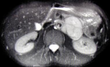
 Pheochromocytoma: Practice Essentials, Pathophysiology, Etiology
 Frontiers | MicroRNA Exocytosis by Vesicle Fusion in Neuroendocrine Cells
 Pheochromocytomas in Animals - Endocrine System - Merck Veterinary Manual
 Anal sphincter | Radiology Reference Article | Radiopaedia.org
Dartos muscle | Radiology Reference Article | Radiopaedia.org
Enterochromaffin-like Cells | Profiles RNS
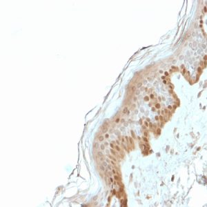
Chromaffin Cells | Profiles RNS
 chromaffin cells | NAL Agricultural Thesaurus
 Pharmacotherapy Exam 1 September 27: Lecture 3 - Sympathomimetic Drugs, ETC - ProProfs Quiz
Pheochromocytoma: Practice Essentials, Pathophysiology, Etiology
 Human Metabolome Database: Showing metabocard for Metanephrine (HMDB0004063)
 DeCS
DeCS
The chromaffin cell and its development<...
 Lack of an adrenal cortex in Sf1 mutant mice is compatible with the generation and differentiation of chromaffin cells |...
 Lexaria Discovers Potential Novel Mechanism From Hypertension Study HYPER-H21-4
 Do you know your CATs? Quiz / Test | Alphabetics | 10 Questions
Development of Tolerance to Ethanol in Cultured Adrenal Chromaffin Cells<...
 Toxic Effects of Mercury on the Cardiovascular and Central Nervous Systems
 Adrenal Diseases | List of High Impact Articles | PPts | Journals | Videos
 Migratory neuronal progenitors in Tunicates provide insights into Neural Crest evolution - the Node
 autonomic nervous system - Ontology Browser - Rat Genome Database
 Peripheral chemoreceptors (video) | Khan Academy
Code System Concept
 Ascorbic acid. Medical search
 Endocrine Glands - Endocrine System - Human Physiology - Picmonic for Pre-Health
DeCS
 MeSH Browser
 Adrenal Cortex - DocsBay
Cells of the adrenal medulla4
- Chromaffin cells of the adrenal medulla are innervated by the splanchnic nerve and secrete adrenaline (epinephrine), noradrenaline (norepinephrine), some dopamine, enkephalin and enkephalin-containing peptides, and a few other hormones into the blood stream. (wikipedia.org)
- It is produced in neurons of the central nervous system and in the chromaffin cells of the adrenal medulla from the amino acids phenylalanine and tyrosine. (omicsonline.org)
- This is because NCCs are a population of stem cell-like progenitors that delaminate and migrate to give rise to a dizzying array of cell types all throughout our bodies and most of the skull: pigment cells, sensory neurons, glia, cartilage, bone, connective tissue, smooth muscle, and chromaffin cells of the adrenal medulla. (biologists.com)
- [ 6 ] PHEOs are derived from the chromaffin cells of the adrenal medulla, and SPGLs are found in close relationship to the peripheral sympathetic nervous system from the level of the superior cervical ganglion down the trunk into the pelvis. (medscape.com)
Composed of chromaffin cells2
- The adrenal medulla, composed of chromaffin cells, secretes the hormone epinephrine, also called adrenaline, in response to stimulation of the sympathetic nervous system at times of stress. (funtrivia.com)
- It is composed of chromaffin cells. (difference.wiki)
Granules2
- In adrenal chromaffin cells, leakage of norepinephrine and epinephrine from storage granules leads to substantial intracellular production of the O-methylated metabolite metanephrine. (hmdb.ca)
- The journal presents papers on functional neurochemistry, nervous system receptors, neurotransmitters, myelin, chromaffin granules and other components of the nervous system, as well as neurophysiological and clinical aspects, behavioral reactions, etc. (scijournal.org)
Neurons11
- They are in close proximity to pre-synaptic sympathetic ganglia of the sympathetic nervous system, with which they communicate, and structurally they are similar to post-synaptic sympathetic neurons. (wikipedia.org)
- Although a common sympathoadrenal (SA) progenitor cell for chromaffin cells and sympathetic neurons has been postulated, there is evidence to suggest that chromaffin progenitors are already distinct, at least in part, from neuronal SA progenitors prior to invading the adrenal gland. (huji.ac.il)
- Distinct developmental requirements of chromaffin cells and sympathetic neurons must also be assumed based on the analyses of mice carrying targeted mutations of the genes for two transcription factors, MASH1 and Phox2B. (huji.ac.il)
- Both genes are expressed by SA progenitors, but are distinctly required for the development of chromaffin cells and sympathetic neurons. (huji.ac.il)
- Such molecules may be candidates for triggering the distinct developmental pathway of chromaffin cells, as opposed to sympathetic neurons. (huji.ac.il)
- The diversification of neural-crest-derived sympathoadrenal (SA) progenitor cells into sympathetic neurons and neuroendocrine adrenal chromaffin cells was thought to be largely understood. (silverchair.com)
- The autonomic nervous system is composed of neurons that are not under conscious control, and is comprised of two antagonistic components, the sympathetic and parasympathetic nervous systems. (mcw.edu)
- Neurons in central nervous system control the release of hormones from the pituitary which provides an access point for environmental stimuli to alter physiological parameters in the body. (tomsk.ru)
- The posterior pituitary contains the axons of neurons whose cell bodies reside in the hypothalamus which is part of the central nervous system. (tomsk.ru)
- Using this framework to analyze the SM (Sec1/Munc18)-SNARE ( N -ethylmaleimide-sensitive factor activating protein receptor) system in exocytic membrane fusion in yeast and neurons, we find that the SM-SNARE network motifs of yeast and neurons show distinct dynamical behaviors. (biomedcentral.com)
- We identify the closed binding mode of neuronal SM (Munc18-1) and SNARE (syntaxin-1) as the key factor leading to mechanistic divergence of membrane fusion systems in yeast and neurons. (biomedcentral.com)
PARAGANGLIA5
- 2) Chromaffin cells (or pheochromocytes): These cells will migrate to the area adjacent to the sympathetic ganglia (hence the name paraganglia) and to the adrenal medulla where they will be the most abundant type of cells. (wikipedia.org)
- Characteristically, they are located in the adrenal medulla and paraganglia (PARAGANGLIA, CHROMAFFIN) of the sympathetic nervous system. (jefferson.edu)
- There are (i.) a series of isolated masses, the paraganglia, associated singly or in groups with the ganglia of the sympathetic nervous system, (ii. (co.ma)
- The paraganglia are rounded masses of chromaphil tissue, 1-3 mm. in diameter, placed inside, half inside, or immediately outside the capsules of the ganglia of the sympathetic system. (co.ma)
- Sympathetic paraganglia consist of chromaffin cells and are involved in the secretion of catecholamines (norepinephrine, epinephrine, and dopamine), while parasympathetic paraganglia consist of glomus (nonchromaffin) cells and act as chemoreceptors [ 1 ] . (encyclopedia.pub)
Sympatho-chromaffin system1
- Overall, these latest results from study HYPER-H21-4 imply that the antihypertensive effects of DehydraTECH-CBD may be explained, at least in part, by its interaction with the sympatho-chromaffin system via catestatin modulation. (yahoo.com)
Catecholamines5
- The chromaffin cells release catecholamines: ~80% of adrenaline (epinephrine) and ~20% of noradrenaline (norepinephrine) into systemic circulation for systemic effects on multiple organs (similarly to secretory neurones of the hypothalamus), and can also send paracrine signals. (wikipedia.org)
- This increased sympathetic activity leads to chronically increased synthesis and secretion of catecholamines from the adrenal chromaffin cells. (wikipedia.org)
- This chronic increase of epinephrine and norepinephrine secretion causes desensitization of the chromaffin cells to catecholamines resulting in a decrease in production and presence of α2 adrenergic receptors on their cell membrane. (wikipedia.org)
- Pheochromocytomas arise from the adrenal medullary chromaffin cells that normally synthesize and secrete the catecholamines epinephrine and norepinephrine. (merckvetmanual.com)
- In humans, about 93 percent of circulating metanephrine is derived from catecholamines metabolized within adrenal chromaffin cells. (hmdb.ca)
Organs9
- Organelles in CHROMAFFIN CELLS located in the adrenal glands and various other organs. (lookformedical.com)
- Small bodies containing chromaffin cells occurring outside of the adrenal medulla, most commonly near the sympathetic ganglia and in organs such as the kidney, liver, heart and gonads. (nih.gov)
- Overview of the Endocrine System The endocrine system coordinates functioning between different organs through hormones, which are chemicals released into the bloodstream from specific types of cells within endocrine (ductless). (msdmanuals.com)
- these buds wander still further ventrally to become the cells of the ganglia of the cardiac, coeliac, and other great ganglionic nerve plexuses, as well as to form the chromaffin cells of the chromaffin organs. (co.ma)
- The autonomic nervous system regulates many of the internal organs through a balance of two aspects, or divisions. (openstax.org)
- To respond to a threat-to fight or to run away-the sympathetic system causes divergent effects as many different effector organs are activated together for a common purpose. (openstax.org)
- To coordinate all these responses, the connections in the sympathetic system diverge from a limited region of the central nervous system (CNS) to a wide array of ganglia that project to the many effector organs simultaneously. (openstax.org)
- Along with the nervous system, the endocrine system coordinates the activities of cells in different organs and tissues to control physiological parameters or generate system-wide responses to environmental changes. (tomsk.ru)
- The pituitary gland is the central organ of the endocrine system as it provides an interface between the nervous system and cells in several different organs and tissues. (tomsk.ru)
Parasympathetic1
- Neoplasms arising from these cells are pheochromocytomas (also called chromaffin or sympathetic paragangliomas, in contrast to non-chromaffin or parasympathetic paragangliomas of glomus cells). (wikipedia.org)
Pheochromocytoma3
- These terms can be used interchangeably but usually paraganglioma refer to a tumor originating from chromaffin cells outside the adrenal gland, which can also be called extra-adrenal pheochromocytoma, whereas pheochromocytoma typically refer to a tumor originating from the chromaffin cells within the adrenal gland. (wikipedia.org)
- A pheochromocytoma (see the image below) is a rare, catecholamine-secreting tumor derived from chromaffin cells. (medscape.com)
- A pheochromocytoma is a catecholamine-secreting tumor of chromaffin cells. (merckvetmanual.com)
Medulla6
- Chromaffin cells, also called pheochromocytes (or phaeochromocytes), are neuroendocrine cells found mostly in the medulla of the adrenal glands in mammals. (wikipedia.org)
- In order to activate chromaffin cells, the splanchnic nerve of the sympathetic nervous system releases acetylcholine, which then binds to nicotinic acetylcholine receptors on the adrenal medulla. (wikipedia.org)
- In the chromaffin cells of adrenal medulla, paracrinally or autocrinally released neurotransmitters induce profound changes in Ca2+ channel gating and Ca2+-dependent events controlling catecholamine secretion and cell activity. (unito.it)
- These secondary cell buds are the rudiments of the sympathetic ganglion cells and of the chromaffin tissue which is found in the sympathetic nerve plexuses, the medulla of the suprarenal glands, and in the carotid glands. (co.ma)
- Adrenal cortex has no cooperation with the sympathetic nervous system, on the flip side, the sympathetic nervous system functions with the adrenal medulla as an integrated system which is called as the sympatheticoadrenal system. (difference.wiki)
- We provisionally categorized the adrenal gland as nerve tissue because of the presence of chromaffin cells in the medulla of the gland. (cdc.gov)
Tumor3
- Se analizaron el tamaño ganglionar y del tumor primario en la TC, y su valoración cualitativa y semicuantitativa (SUVmáx) en la PET. (bvsalud.org)
- This transformation depends on various factors related to the tumor (such as the over expression or not of N-myc, the presence or absence of Treks and their receptors), the host (the intervention of the immune system) and to other external factors. (lupinepublishers.com)
- Neuroblastoma is a neuroectodermal tumor that originates from precursor cells of the sympathetic nervous system. (oncotarget.com)
Lacking the glucocorticoid receptor2
- The concept of an essential role of glucocorticoid signalling for chromaffin cell development has been shaken by the observation that chromaffin cells in mice lacking the glucocorticoid receptor develop largely normal. (huji.ac.il)
- However, analysis of mice lacking the glucocorticoid receptor gene had revealed that adrenal chromaffin cells develop mostly normally in these mice. (silverchair.com)
Neuroendocrine cells1
- Recently, miRNA exocytosis by vesicle fusion in response to stimulation was observed in chromaffin cells, which are neuroendocrine cells in the sympathetic nervous system ( 24 ). (frontiersin.org)
Progenitor1
- In-vitro studies with isolated SA progenitor cells had suggested that chromaffin cell differentiation depends crucially on glucocorticoids provided by adrenal cortical cells. (silverchair.com)
Tissues2
Paraganglion1
- Extra-adrenal pheochromocytomas develop in the paraganglion chromaffin tissue of the nervous system. (medscape.com)
Glomus2
- In fact in some book they are referred as Glomus caroticum (carotid body) and Glomus aorticum (aortic body) be careful with this one no to mistake with para-aortic bodies which are chromaffin cell which manufacture catecholamine. (khanacademy.org)
- the spleen and the glomus coccygeum, which are associated with the circulatory system. (co.ma)
Receptors3
- This means that the alpha receptors it binds to are located in the central nervous system (CNS) rather than on the effector organ, which in this case is the heart. (proprofs.com)
- These hormones are released by cells in one part of the body, enter the circulatory system to distribute throughout the body and bind specific receptors in other cells. (tomsk.ru)
- Hormone receptors in the endocrine system often trigger changes in gene expression to alter the activities of key biochemical or physiological pathways. (tomsk.ru)
Sympathetic nerve1
- The particularity of neuroblastoma lies in its development from cells whose embryological maturation into adult sympathetic nerve cells or chromaffin cells is incomplete. (lupinepublishers.com)
Ganglia of the sympathetic system1
- The majority of ganglia of the sympathetic system belong to a network of sympathetic chain ganglia that runs alongside the vertebral column. (openstax.org)
Exocytosis1
- As L-channels play a crucial role in the control of catecholamine release in chromaffin cells, the two opposite modulations mediated by G(i/o) proteins and cAMP may represent an effective way to broaden the dynamic range of Ca2+ signals controlling exocytosis. (unito.it)
Neuroblastoma1
- Neuroblastoma is a relatively common pediatric pathology of the sympathetic nervous system. (lupinepublishers.com)
Autonomic nervou7
- The autonomic nervous system regulates key functions including the activity of the cardiac (heart) muscle, smooth muscles (e.g. of the gut), and glands[GO]. (mcw.edu)
- The nervous system can be divided into two functional parts: the somatic nervous system and the autonomic nervous system. (openstax.org)
- The autonomic nervous system controls cardiac and smooth muscle, as well as glandular tissue. (openstax.org)
- The somatic nervous system is associated with voluntary responses (though many can happen without conscious awareness, like breathing), and the autonomic nervous system is associated with involuntary responses, such as those related to homeostasis. (openstax.org)
- In addition to the endocrine system, the autonomic nervous system is instrumental in homeostatic mechanisms in the body. (openstax.org)
- His body's reaction is the result of the sympathetic division of the autonomic nervous system causing system-wide changes as it prepares for extreme responses. (openstax.org)
- The sympathetic division of the autonomic nervous system influences the various organ systems of the body through connections emerging from the thoracic and upper lumbar spinal cord. (openstax.org)
Cell3
- There are two types of cells that originate from the neural crest and are related to the sympathetic nervous system (originate from a cell called sympathogonia): 1) Neuroblasts: These cells migrate, during the fourth to the fifth week of fetal development in humans, on both sides of the spinal cord toward the region just behind the dorsal aorta forming the two chains of sympathetic ganglia (Sympathetic chain). (wikipedia.org)
- Extracellular miRNAs were observed in cell culture system ( 6 ), in blood plasma and serum ( 7 - 10 ), and in other biological fluids ( 11 ) including cerebrospinal fluid ( 12 ), saliva ( 13 ), breast milk, urine, and tears ( 14 ). (frontiersin.org)
- Other cell types are likely vertebrate-specific, like chromaffin cells. (biologists.com)
Adrenaline1
- The secreted adrenaline and noradrenaline play an important role in the sympathetic nervous system response, commonly called the fight-or-flight response. (wikipedia.org)
Organ4
- The largest extra-adrenal cluster of chromaffin cells in mammals is the organ of Zuckerkandl. (wikipedia.org)
- In non-mammals, chromaffin cells are found in a variety of places, generally not organised as an individual organ, and may be without innervation, relying only on endocrine or paracrine signals for secretion. (wikipedia.org)
- In fact, the adrenals constitute the single largest source out of any organ system including the liver for circulating metanephrine. (hmdb.ca)
- Glucocorticoids promote and inhibit gene transcription in many cells and organ systems. (msdmanuals.com)
Molecules3
- There is an ongoing search for molecules selectively operating at the sites, where chromaffin cells develop. (huji.ac.il)
- Information in the endocrine system is transmitted through several small molecules, peptides and proteins that function as hormones. (tomsk.ru)
- The production of hormones in the endocrine system can be regulated by the concentration of certain molecules or ions (e.g. glucose, calcium) or by the nervous system which allows for integration of external stimuli or input from higher parts of the brain. (tomsk.ru)
Concentrations2
- In lower concentrations, extra-adrenal chromaffin cells also reside in the bladder wall, prostate, and behind the liver. (wikipedia.org)
- Peripheral chemoreceptors are extensions of the peripheral nervous system that respond to changes in blood molecule concentrations (such as oxygen or carbon dioxide) and help maintain cardiorespiratory homeostasis. (khanacademy.org)
Hormones4
- The adrenal glands regulate metabolism, immune system, blood pressure, and even response to stress by synthesizing and secreting steroid hormones in the bloodstream. (anatomy.app)
- These hormones affect metabolism, increase blood glucose levels, and take part in regulating the immune system. (anatomy.app)
- The hypothalamus is a component of the central nervous system and regulates the release of hormones from cells in the pituitary. (tomsk.ru)
- In both cases, the hormones released from the pituitary change key physiological parameters and/or alter system-wide behavior. (tomsk.ru)
Immune system1
- It can also promote breathing and boost the immune system. (promindbuild.com)
Proteins1
- By capturing the evolutionary dynamics of target biological systems, the comparative modeling framework is empowered to (i) identify the functional roles of poorly characterized proteins and interactions and (ii) further decipher the underlying regulatory mechanisms of complicated cellular processes. (biomedcentral.com)
Signals2
- During times of stress, the nervous system signals the vesicles to secrete their hormonal content. (jefferson.edu)
- Using 'information' as signals and 'information webs' as signaling networks, all systems, as large as the world and as small as an intracellular organelle, function in the same fashion, from the distant past to the immediate present, and this will not change in the indefinite future. (rsc.org)
Nervous Systems2
- Although the peripheral nervous systems of tunicate larvae have several sensory neuron subtypes 9 , none of them have been decisively linked to NCCs, either because they do not arise from the neural plate borders or because they more closely resemble non-NCC-derived sensory cells in vertebrates. (biologists.com)
- The pituitary gland resides just outside the central nervous systems and comprises two anatomical and functional structures: anterior and posterior. (tomsk.ru)
Neural crest1
- These cells are derivatives of the neural crest and are intimately associated with the sympathetic nervous system. (huji.ac.il)