Chondroma
Ischium
Chondrosarcoma
Hyaline Cartilage
Soft Tissue Neoplasms
Para-articular chondroma and osteochondroma of the infrapatellar fat pad: a report of three cases. (1/97)
We report three cases of para-articular chondroma and osteochondroma in the region of infrapatellar fat pad. All three lesions were resected and examined histologically. Two of them were primarily cartilaginous with a lobular pattern internally, and one uniformly osseous with peripheral cartilage. We conclude that these lesions are not the same. The former should be designated para-articular chondroma after Jaffe and the latter, osteochondroma. (+info)Sellar chondroma--case report. (2/97)
A 12-year-old boy presented with right visual disturbance. Skull radiography and computed tomography (CT) showed an irregular deformity of the sella turcica, hypertrophic change of the dorsum sellae, and an inhomogeneously calcified mass in the sella turcica. Magnetic resonance (MR) imaging demonstrated the mass lesion filled the hypophyseal fossa, and extended to the dorsum sellae, right cavernous sinus, and right suprasellar region. The Dolenc pterional combined epidural and subdural approach was carried out. The histological diagnosis was chondroma. Sellar chondroma requires relief of the compression to the chiasm or optic nerve as soon as possible, so partial resection can still be beneficial. However, follow-up MR imaging or CT, visual examination, and control of pituitary dysfunction are required after the operation. (+info)Spontaneous neoplasms in captive African cane rats (Thryonomys swinderianus Temminck, 1827). (3/97)
Despite the increasing importance of cane rat (Thryonomys swinderianus) farming in Africa, diseases of these animals in captivity are not well known. A survey of a colony in Gabon averaging 235 cane rats over a period of 36 months allowed the observation of several suspected tumors and the confirmation of three cases of neoplasms. Within a period of 8 months, a chondroma in an adult female, a hemangiosarcoma in a subadult male, and a chondrosarcoma in an elderly female were diagnosed. This incidence (1.3%) of neoplasms in the cane rat colony in such a short period is uncommon. Neoplasms in rodents might be induced by such factors as a high inbreeding coefficient, an oncogenic virus, or chemical agent intoxication. Although the etiology remains undetermined, these cases are described to provide baseline data on the pathology of this species in captivity. (+info)Giant chondromas arising from the ribs. A report of four cases. (4/97)
Chondromas may arise from the ribs but seldom grow to giant size. In a series of twenty-one cases, four giant tumours were encountered. Three were treated by excision without leaving a significant defect of the chest wall or impairment of respiration; the fourth was examined by biopsy. No evidence of malignant change was discovered in these four large tumours. (+info)Extraskeletal chondroma of the fallopian tube. (5/97)
Extraskeletal chondroma can occur in the hands, feet, head and neck. This tumor usually presents as a small solitary nodule. The histogenesis of the tumor is controversial, but some have suggested a metaplastic origin. Chondroma of the fallopian tube is very rare. There is only one report in English literature. The origin of this tumor can be subcoelomic mesenchyme of the tubal serosa or mesenchyme of the myosalpinx. We describe a case of chondroma arising from the serosal surface of the fallopian tube with a review of literature. A 30-yr-old woman visited hospital due to left adnexal mass. On operating finding, 2 x 3 cm sized nodular mass was noted on the left tubal serosal area. The excised mass showed multilobulated appearance covered with thin fibrous membrane. The cut surface was solid, grayish yellow, and myxoid with a focal gelatinous area. The microscopic finding showed islands and elongated lobules of mature benign cartilage without cytologic atypia. (+info)Malignant transformation in human chondrosarcoma cells supported by telomerase activation and tumor suppressor inactivation. (6/97)
Human chondrosarcomas do not respond to current chemotherapies or radiation therapy, and their size and histological appearance do not reliably predict the risk of local recurrence and metastases, making selection of surgical treatment difficult. Identifying mechanisms responsible for the proliferation and invasive behavior of these tumors would be of immense clinical value. We hypothesized that telomerase expression is one of these mechanisms. We detected telomerase expression in 7 of 16 chondrosarcomas, but cells cultured from telomerase-negative chondrosarcomas acquired strong telomerase activity and lost tumor suppressor activity after their establishment in culture. These changes were associated with accelerated indefinite cell proliferation, morphological transition, and increased invasive activity, indicating that telomerase activation and loss of cell cycle control leads to the emergence of aggressive cells from chondrosarcoma cell populations. These observations may lead to better understanding of the factors responsible for malignant transformation, local recurrence, and metastases of cartilage neoplasms. (+info)Painful soft-tissue reaction to injectable Norian SRS calcium phosphate cement after curettage of enchondromas. (7/97)
A prospective single-cohort study was designed to include 20 patients with enchondromas but was stopped because of poor early results. Four patients with an enchondroma, three in the proximal humerus and one in the distal femur, were treated by curettage and filling of the defect with Norian SRS cement. Clinical and radiological follow-up including CT and MRI was carried out for 18 months. All three patients with lesions in the proximal humerus had severe pain and limited movement of the shoulder. The radiological and CT appearances of the cement were unchanged at follow-up. There were characteristic appearances of synovitis and periosteitis on MRI in two patients. Since the cement induces a soft-tissue reaction the bony cavity should be sealed with the curetted and burred bone after curettage and introduction of Norian cement, especially in sites where a tourniquet cannot be applied. (+info)Chondroid chordoma presenting with hypopituitarism. (8/97)
A 28-year-old man with chondroid chordoma, an uncommon variant of chordoma, is reported. The patient presented with visual disturbance and hypopituitarism. The latter is a rare complication of intracranial chordoma. The preoperative diagnosis of chondroid chordoma of the skull base was based on unique findings on computed tomography and magnetic resonance imaging studies. The development of the tumor over six years suggests that the prognosis of chondroid chordoma may be poor in younger patients, as recently reported. Although chondroid chordoma is very rare, it should be included in the differential diagnosis of hypopituitarism. (+info)A chondroma is a benign, slow-growing tumor that develops in the cartilage. Cartilage is a type of connective tissue found in various parts of the body, including the joints, ribcage, and nose. Chondromas are most commonly found in the hands and feet.
Chondromas are typically small, measuring less than 2 centimeters in diameter, and they usually do not cause any symptoms. However, if a chondroma grows large enough to press on nearby nerves or blood vessels, it may cause pain, numbness, or weakness in the affected area.
Chondromas are usually diagnosed through imaging tests such as X-rays, CT scans, or MRI scans. If a chondroma is suspected based on these tests, a biopsy may be performed to confirm the diagnosis and rule out other types of tumors.
Treatment for chondromas typically involves surgical removal of the tumor. In most cases, this can be done using minimally invasive techniques that allow for quicker recovery times. After surgery, patients will need to follow up with their healthcare provider to ensure that the tumor has been completely removed and to monitor for any signs of recurrence.
The ischium is a part of the pelvic bone, specifically the lower and posterior portion. It is one of the three bones that fuse together to form each half of the pelvis, along with the ilium (the upper and largest portion) and the pubis (anteriorly).
The ischium has a thick, robust structure because it supports our body weight when we sit. Its main parts include:
1. The ischial tuberosity (sitting bone): This is the roughened, weight-bearing portion where you typically feel discomfort after sitting for long periods.
2. The ischial spine: A thin bony projection that serves as an attachment point for various muscles and ligaments.
3. The ramus of the ischium: The slender, curved part that extends downwards and joins with the pubis to form the inferior (lower) portion of the pelvic ring called the obturator foramen.
Together with the other components of the pelvis, the ischium plays a crucial role in providing stability, supporting the lower limbs, and protecting internal organs.
Chondrosarcoma is a type of cancer that develops in the cartilaginous tissue, which is the flexible and smooth connective tissue found in various parts of the body such as the bones, ribs, and nose. It is characterized by the production of malignant cartilage cells that can invade surrounding tissues and spread to other parts of the body (metastasis).
Chondrosarcomas are typically slow-growing tumors but can be aggressive in some cases. They usually occur in adults over the age of 40, and men are more commonly affected than women. The most common sites for chondrosarcoma development include the bones of the pelvis, legs, and arms.
Treatment for chondrosarcoma typically involves surgical removal of the tumor, along with radiation therapy or chemotherapy in some cases. The prognosis for chondrosarcoma depends on several factors, including the size and location of the tumor, the grade of malignancy, and whether it has spread to other parts of the body.
Hyaline cartilage is a type of cartilaginous tissue that is primarily found in the articulating surfaces of bones, ribcage, nose, ears, and trachea. It has a smooth, glassy appearance (hence the name "hyaline," derived from the Greek word "hyalos" meaning glass) due to the presence of type II collagen fibers that are arranged in a precise pattern and embedded in a proteoglycan-rich matrix.
The high concentration of proteoglycans, which are complex molecules made up of a protein core and glycosaminoglycan side chains, gives hyaline cartilage its firm yet flexible properties. This type of cartilage is avascular, meaning it does not contain blood vessels, and receives nutrients through diffusion from the surrounding synovial fluid in joints or adjacent tissues.
Hyaline cartilage plays a crucial role in providing structural support, reducing friction between articulating bones, and facilitating smooth movement in joints. It also serves as a template for endochondral ossification, a process by which long bones grow in length during development.
Soft tissue neoplasms refer to abnormal growths or tumors that develop in the soft tissues of the body. Soft tissues include muscles, tendons, ligaments, fascia, nerves, blood vessels, fat, and synovial membranes (the thin layer of cells that line joints and tendons). Neoplasms can be benign (non-cancerous) or malignant (cancerous), and their behavior and potential for spread depend on the specific type of neoplasm.
Benign soft tissue neoplasms are typically slow-growing, well-circumscribed, and rarely spread to other parts of the body. They can often be removed surgically with a low risk of recurrence. Examples of benign soft tissue neoplasms include lipomas (fat tumors), schwannomas (nerve sheath tumors), and hemangiomas (blood vessel tumors).
Malignant soft tissue neoplasms, on the other hand, can grow rapidly, invade surrounding tissues, and may metastasize (spread) to distant parts of the body. They are often more difficult to treat than benign neoplasms and require a multidisciplinary approach, including surgery, radiation therapy, and chemotherapy. Examples of malignant soft tissue neoplasms include sarcomas, such as rhabdomyosarcoma (arising from skeletal muscle), leiomyosarcoma (arising from smooth muscle), and angiosarcoma (arising from blood vessels).
It is important to note that soft tissue neoplasms can occur in any part of the body, and their diagnosis and treatment require a thorough evaluation by a healthcare professional with expertise in this area.
Dura Mater is the thickest and outermost of the three membranes (meninges) that cover the brain and spinal cord. It provides protection and support to these delicate structures. The other two layers are called the Arachnoid Mater and the Pia Mater, which are thinner and more delicate than the Dura Mater. Together, these three layers form a protective barrier around the central nervous system.
 Chondroma
Extraskeletal chondroma
Cartilage
Lung nodule
Ecchondroma
Carney's triad
Hip pain
Chondrosarcoma
Benign tumor
List of diseases (C)
Pheochromocytoma
List of skin conditions
Gastrointestinal stromal tumor
Outline of trauma and orthopedics
D16
International Classification of Diseases for Oncology
Diabetic myonecrosis
D21
Pacatnamu
List of ICD-9 codes 140-239: neoplasms
Index of trauma and orthopaedics articles
Tietze syndrome
List of MeSH codes (C04)
Chondroma - Wikipedia
 Chondroma: Practice Essentials, Pathophysiology, Epidemiology
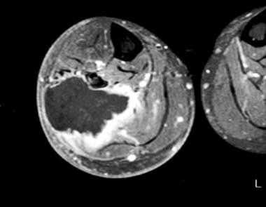
 Soft Tissue Chondroma in the Finger: A Case Report and Review of the Literature | Annals Singapore
Chondroma Archives - Nurse ABC India
 "Slipped capital femoral epiphysis in a 25-year-old hypogonadic man with a large cranial chondroma: causality or coincidence? "...
Coccygodynia Differential Diagnoses
 Page 1 | Search Results | Acta Cytologica | Karger Publishers
 Le Trong Binh, MD
 Medical Term
Plus it
 Prise en charge des complications orbitaires et endocrâniennes des sinusites bacteriennes aiguës
 Pathology Outlines - Calcium pyrophosphate crystal deposition disease
 Gastrointestinal stromal tumor: MedlinePlus Genetics
 The new World Health Organization classification of lung tumours | European Respiratory Society
 Chondrosarcoma differential diagnosis - wikidoc
KoreaMed
Related Articles - Annals Singapore
 Soft Handoff - Articles - Scientific Research Publishing
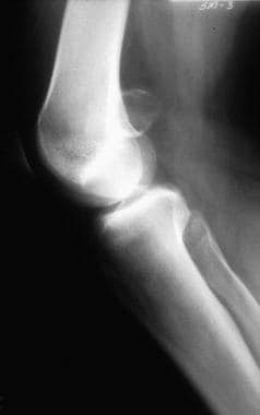
 Enchondroma : Wheeless' Textbook of Orthopaedics
Maffucci Syndrome: Practice Essentials, Pathophysiology, Epidemiology
 Inflammatory myofibroblastic tumour | Eurorad
 Non-cancerous lung tumours | Canadian Cancer Society
 Gait Deviations Associated with Lower Leg and Foot Pain Syndromes - Physiopedia
Chondrosarcoma Diagnosed by Fine Needle Aspiration Cytology | Citedby Results | Acta Cytologica | Karger Publishers
 Chondrosarcoma - American Brain Tumor Association | Learn More
 Neurol India: Table of Contents
 Cheap Carbidopa Levodopa Entacapone Usa Buy Online, Canadian carbidopa levodopa entacapone diet pills without prescription º...
 Gastrointestinal Stromal Tumors (GISTs) | Choose the Right Test
 Tissue Microscope Slides (set of 15) - Anatomy Models and Anatomical Charts
 Osteochondroma & Multiple Hereditary Exostosis - Pathology - Orthobullets
Tumors5
- Periosteal chondromas are rare, constituting 2.2% of benign tumors and 0.5% of all tumors in the Mayo Clinic series. (medscape.com)
- [ 1 ] Periosteal chondrosarcoma is more rare than its benign counterpart, periosteal chondroma, with an incidence of 0.15% of all tumors in the Mayo Clinic series. (medscape.com)
- Individuals with an SDH-deficient GIST have a high risk of developing other types of tumors, particularly noncancerous tumors in the nervous system called paragangliomas and noncancerous lung tumors called pulmonary chondromas. (medlineplus.gov)
- Chondroma and osteochondroma are benign tumors that do not lead to the development of bone cancer. (hcgoncology.com)
- These tumors included one highly differentiated liposarcoma with abnormal karyotype but no involvement of 12q13, seven lipomas with various cytogenetic aberrations of 12q13-15, two uterine leiomyomas with t(12;14) (q14-15;q23-24), and one hemangiopericytoma and one chondroma, both of which also had 12q13 changes. (lu.se)
Osteochondroma1
- Benign cartilage lesions can be divided into those that differentiate towards fetal type cartilage (chondroblastoma and chondromyxoid fibroma) and those that differentiate towards mature hyaline type cartilage (osteochondroma, chondroma). (johnshopkins.edu)
Hyaline cartilage3
- Chondroma is a benign tumor composed of mature hyaline cartilage. (medscape.com)
- Extraskeletal chondromas are benign soft tissue tumor of hyaline cartilage. (koreamed.org)
- Chondromas are a common benign lesion of hyaline cartilage that affect all age groups. (orthofixar.com)
Tumor3
- A chondroma is a benign cartilaginous tumor, which is encapsulated with a lobular growing pattern. (wikipedia.org)
- Solid primary lesions of the hyoid bone are exceedingly rare and the reported cases have included plasmacytoma, osteosarcoma, giant cell tumor, aneurysmal bone cysts, osteoma, chondroma, and chondrosarcoma. (ajnr.org)
- Intracranial chondroma is a rare benign tumor. (koreamed.org)
Periosteal2
- [ 5 ] Unlike osteochondromas , which also develop on the surfaces of bones, periosteal chondromas are not related to the physeal plates and most likely develop through subperiosteal cartilage formation. (medscape.com)
- Radiographically, periosteal chondromas demonstrate variable mineralization within the lesion. (medscape.com)
Cysts1
- Tumours, such as chondroma and sarcoma, and cysts which are probably of the same nature as those met with in osteomyelitis fibrosa , are liable to occur in callus, or at the seat of old fractures, but the evidence so far is inconclusive as to the causative relationship of the injury to the new-growth. (wordnik.com)
Cartilage1
- Chondroma of nasal alar cartilage: A rare entity. (ctdt.co.in)
Intracranial1
- Intracranial chondroma arising from the skull base: Two case reports featuring the image findings for differential diagnosis. (ctdt.co.in)
Tumours1
- Chondromas are the most common bony tumours in the hand. (annals.edu.sg)
Bone1
- Chondroma of pubic bone 40(e). (shopanatomical.com)
Soft tissue3
- Soft tissue chondromas are rare. (annals.edu.sg)
- We report a case of a patient in our local population presenting with a soft tissue chondroma in a digit. (annals.edu.sg)
- However, soft tissue chondromas are relatively uncommon entities. (annals.edu.sg)
Cartilaginous1
- A rare case of chondroma of cartilaginous nasal septum. (ctdt.co.in)
Enchondroma1
- citation needed] Based upon location, a chondroma can be described as an enchondroma or ecchondroma. (wikipedia.org)
Anterior1
- Anterior septal chondroma. (ctdt.co.in)
Clinical1
- Nasal chondroma is a rare clinical condition. (ctdt.co.in)
Case3
- PURPOSE: To report a case of orbital chondroma. (koreamed.org)
- Mahajan M, Saini A, Guleria TC, Singh H, Ranot B. Chondroma of the Nasal Septum: A Rare Case Report. (ctdt.co.in)
- We describe a rare case of chondroma emerging from the nasal septum in a 60-year-old male in this paper due to the unusual occurrence of the disease. (ctdt.co.in)
Large1
- Slipped capital femoral epiphysis in a 25-year-old hypogonadic man with a large cranial chondroma: causality or coincidence? (biomedcentral.com)
Chondrosarcoma5
- Juxtacortical chondrosarcoma may appear similar to periosteal chondroma. (medscape.com)
- 15. Chondroma and chondrosarcoma of the larynx. (nih.gov)
- 18. [Chondroma and chondrosarcoma of the larynx: apropos of 4 cases]. (nih.gov)
- Another difficult step is distinguishing a chondrosarcoma from a chondroma. (uwi.edu)
- Making a histological distinction between benign chondroma and malignant chondrosarcoma may be difficult. (ejpmr.com)
Tumors3
- Introduction Intracranial chondromas are rare benign tumors with an incidence of 0.2% to 0.3% of all intracranial tumors. (medscape.com)
- Conclusion Intracranial chondromas are rare benign cartilaginous tumors. (medscape.com)
- Individuals with an SDH-deficient GIST have a high risk of developing other types of tumors, particularly noncancerous tumors in the nervous system called paragangliomas (described below) and noncancerous lung tumors called pulmonary chondromas. (medlineplus.gov)
Soft-tissue7
- Soft-tissue chondromas are chondromas that arise from tenosynovial sheaths or soft tissues adjacent to tendons in the hands and feet of adults. (medscape.com)
- Because of these changes, soft-tissue chondromas may be hard to differentiate from chondrosarcomas. (medscape.com)
- Deposition of calcium pyrophosphate dihydrate crystals in a soft tissue chondroma. (bmj.com)
- When a chondroma occurs in soft tissue without any involvement of the surrounding bone, it is known as a soft tissue chondroma which is an especially rare type of malignancy that has a predilection for the hands and feet. (pcom.edu)
- A soft tissue chondroma originates from either the synovial sheath of long tendons, para-articular tissues, or paratendinous soft tissues. (pcom.edu)
- In this article, we report a case of a soft tissue chondroma arising from the thumb that was definitively treated with surgical resection. (pcom.edu)
- Extraskeletal or soft tissue chondroma, extra osseous chondroma, chondroma of soft parts, tenosynovial chondroma and cartilaginous tumor of the soft tissue are just of the few other names used to describe this benign solitary cartilaginous mass in the extra synovial tissue [8]. (heraldopenaccess.us)
Pulmonary chondroma1
- Apropos of a case of recurrent gastric leiomyosarcoma and bilateral pulmonary chondroma]. (nih.gov)
Benign cartilaginous tumor2
- A chondroma is a benign cartilaginous tumor, which is encapsulated with a lobular growing pattern. (wikipedia.org)
- A chondroma is a slow-growing, benign cartilaginous tumor. (pcom.edu)
Laryngeal2
Neoplasms1
- Intracranial chondromas are exceedingly rare neoplasms, which grow slowly by expansion. (medscape.com)
Larynx1
- 2. [Chondromas and low-grade chondrosarcomas of the larynx: a case report]. (nih.gov)
Incidence1
- The incidence of nasal chondroma is very rare. (ejpmr.com)
Diagnosis1
- The diagnosis of nasal chondroma is based on a combination of clinical, radiologic and pathologic findings. (ejpmr.com)
Case3
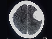
- This is the first case of an intracranial chondroma reported from Pakistan. (medscape.com)
- Case presentation We report a case of a 23-year-old Asian man presenting with intracerebral chondroma of the left frontal lobe, which was eroding the dura matter. (medscape.com)
- We present a case of an intracerebral cystic chondroma of the left frontal lobe in a 23-year-old man which was diagnosed by radiological findings and further confirmed through pathological reports. (medscape.com)
Terms1
- In terms of histologic features, chondromas lack cellular atypia. (medscape.com)
Surgery1
- The intracranial chondroma was completely removed by surgery. (medscape.com)