Chemoreceptor Cells
Carotid Body
Aortic Bodies
Almitrine
Oxygen
Kv1.4 Potassium Channel
Shal Potassium Channels
Potassium Channels, Tandem Pore Domain
Rabbits
Potassium
Sodium Cyanide
Thiourea
Potassium Channels
Large-Conductance Calcium-Activated Potassium Channels
Membrane Potentials
Respiration
Carotid Sinus
Carbon Dioxide
Paraganglia, Nonchromaffin
Potassium Channels, Voltage-Gated
Nifedipine
Chemotaxis
Electrophysiology
Calcium
Patch-Clamp Techniques
Hypercapnia
Rats, Wistar
Pressoreceptors
Reflex
Glossopharyngeal Nerve
Vagotomy
Cats
Cells, Cultured
Phrenic Nerve
Medulla Oblongata
Immunohistochemistry
Respiratory Center
Asphyxia
Respiratory Mechanics
Pulmonary Ventilation
Cyanides
Solitary Nucleus
Vagus Nerve
Receptors, Amino Acid
Post-ingestive feedbacks and associative learning regulate the intake of unsuitable sterols in a generalist grasshopper. (1/1554)
Behavioural studies of the grasshopper Schistocerca americana were undertaken to identify the mechanisms that regulate the intake of dietary sterols. In the first experiment, grasshoppers were allowed to feed on spinach, a plant containing only unsuitable sterols; immediately after this first meal, a suitable or unsuitable sterol was injected into the haemolymph. Grasshoppers injected with unsuitable sterols had second meals on spinach that were significantly shorter than those of grasshoppers injected with suitable sterols, indicating that unsuitable dietary sterols are detected post-ingestively. In the second experiment, grasshoppers were fed food containing only unsuitable sterols and were then presented with glass-fibre discs containing different concentrations of a suitable sterol or sucrose only (the control). The results suggest that grasshoppers do not use a direct feedback operating on mouthpart chemoreceptors to regulate their intake of suitable sterols. In the third experiment, grasshoppers were presented with artificial diets containing different sterols and flavours, and feeding was observed over a sequence of meals. The results from both the first and last experiments suggest a role for associative learning in regulating the intake of unsuitable sterols. (+info)Quantitative structure-activity relationships for nasal pungency thresholds of volatile organic compounds. (2/1554)
A model was developed for describing the triggering of nasal pungency in humans, based on the partition of volatile organic compounds (VOCs) between the air phase and the biophase. Two partition parameters are used in the model: the water-air partition coefficient and the octanol-water partition coefficient. The model was validated using data from the literature, principally on alcohols, acetates and ketones. The model suggests that all test compounds, regardless of their chemical functional groups, bind to a common receptor site within the hydrophobic interior of the bilayer membrane of the trigeminal nerve endings. There is probably only a slight, non-specific interaction between the VOC molecule and the receptor molecule, whereas this type of non-specific interaction for the detection of odor is much stronger. In practical terms, the suggestion that all VOCs share a common irritation receptor site implies that nasal-pungency thresholds of individual VOCs may be additive. Quantitative structure-activity relationships (QSARs) for nasal-pungency thresholds were also developed from the model, which can be used to predict nasal-pungency thresholds of common VOCs. Although the present model does not offer additional precision over that of M.H. Abraham et al., 1996, Fundam. Appl. Toxicol. 31, 71-76, it requires fewer descriptors and offers a physiological basis to the QSAR. Another advantage of the present model is that it also provides a basis for comparison between the olfactory process and nasal pungency. (+info)Chemotactic responses of Escherichia coli to small jumps of photoreleased L-aspartate. (3/1554)
Computer-assisted motion analysis coupled to flash photolysis of caged chemoeffectors provides a means for time-resolved analysis of bacterial chemotaxis. Escherichia coli taxis toward the amino acid attractant L-aspartate is mediated by the Tar receptor. The physiology of this response, as well as Tar structure and biochemistry, has been studied extensively. The beta-2, 6-dinitrobenzyl ester of L-aspartic acid and the 1-(2-nitrophenyl)ethyl ether of 8-hydroxypyrene-1,3,6-tris-sulfonic acid were synthesized. These compounds liberated L-aspartate and the fluorophore 8-hydroxypyrene 1,3,6-tris-sulfonic acid (pyranine) upon irradiation with near-UV light. Photorelease of the fluorophore was used to define the amplitude and temporal stability of the aspartate jumps employed in chemotaxis experiments. The dependence of chemotactic adaptation times on aspartate concentration, determined in mixing experiments, was best fit by two Tar aspartate-binding sites. Signal processing (excitation) times, amplitudes, and adaptive recovery of responses elicited by aspartate jumps producing less than 20% change in receptor occupancy were characterized in photorelease assays. Aspartate concentration jumps in the nanomolar range elicited measurable responses. The response threshold and sensitivity of swimming bacteria matched those of bacteria tethered to glass by a single flagellum. Stimuli of similar magnitude, delivered either by rapid mixing or photorelease, evoked responses of similar strength, as assessed by recovery time measurements. These times remained proportional to change in receptor occupancy close to threshold, irrespective of prior occupancy. Motor excitation responses decayed exponentially with time. Rates of excitation responses near threshold ranged from 2 to 7 s-1. These values are consistent with control of excitation signaling by decay of phosphorylated pools of the response regulator protein, CheY. Excitation response rates increased slightly with stimulus size up to values limited by the instrumentation; the most rapid was measured to be 16 +/- 3 (SE) s-1. This increase may reflect simultaneous activation of CheY dephosphorylation, together with inhibition of its phosphorylation. (+info)Trigeminal and carotid body inputs controlling vascular resistance in muscle during post-contraction hyperaemia in cats. (4/1554)
1. In anaesthetized cats, the effects of stimulation of the receptors in the nasal mucosa and carotid body chemoreceptors on vascular resistance in hindlimb skeletal muscle were studied to see whether the responses were the same in active as in resting muscle. The measurements of vascular resistance were taken, first, in resting muscle, and second, in the immediate post-contraction hyperaemic phase that followed a 30 s period of isometric contractions. 2. Stimulation of the receptors in the nasal mucosa caused reflex apnoea and vasoconstriction in muscle. The latter response was attenuated when the test was repeated during post-contraction hyperaemia. 3. Stimulations of the carotid bodies were made during a period of apnoea evoked reflexly by electrical stimulation of both superior laryngeal nerves. This apnoea prevented any effects of changes in respiration on the carotid body reflex vascular responses. Stimulation of the carotid bodies evoked hindlimb muscle vasoconstriction. In the post-contraction hyperaemic period, the response was reduced or abolished. A similar attenuation of the reflex vasoconstrictor responses occurred in decentralized muscles stimulated through their motor roots in the cauda equina. 4. Evidence is presented that the attenuation of the vasoconstrictor responses evoked by the two reflexes is a phenomenon localized to the contracting muscles themselves resulting from an interaction between sympathetic neuronal activity and the local production of metabolites. 5. The results are discussed in relation to the metabolic needs of tissues in relation to asphyxial defence mechanisms such as occur in the diving response. (+info)Depression of peripheral chemosensitivity by a dopaminergic mechanism in patients with obstructive sleep apnoea syndrome. (5/1554)
In the present study, respiratory drives to chemical stimuli and peripheral chemosensitivity were evaluated in patients with obstructive sleep apnoea (OSAS). The effects of oral administration of domperidone, a selective dopamine D2-receptor antagonist, were also examined, to study the respiratory effects of endogenous dopamine on peripheral chemoreceptors. Sixteen patients with OSAS and nine normal control subjects were studied. Respiratory responses to hypercapnia and hypoxia were measured using the rebreathing method and isocapnic progressive hypoxia method, respectively. The hypoxic withdrawal test, which measures the decrease in ventilation caused by two breaths of 100% O2 under mild hypercapnic hypoxic conditions (end-tidal oxygen and carbon dioxide tensions approximately 8.0 kPa and 5.3-6.7 kPa, respectively), was used to evaluate peripheral chemosensitivity. In the patients with OSAS, ventilatory responses to hypercapnia and hypoxia were significantly decreased compared with those of control subjects. Hypoxic withdrawal tests showed that peripheral chemosensitivity was significantly lower in patients with OSAS than in normal subjects. Hypercapnic ventilatory response and peripheral chemosensitivity were enhanced by administration of domperidone in the patients with OSAS, although no changes in either of these were observed in the control subjects. The hypoxic ventilatory response and peripheral chemosensitivity in the patients with OSAS were each significantly correlated with severity of hypoxia during sleep. These findings suggest that peripheral chemosensitivity in patients with obstructive sleep apnoea syndrome may be decreased as a result of abnormality in dopaminergic mechanisms and that the reduced chemosensitivity observed in patients with obstructive sleep apnoea syndrome may affect the severity of hypoxia during sleep. (+info)BDNF is a target-derived survival factor for arterial baroreceptor and chemoafferent primary sensory neurons. (6/1554)
Brain-derived neurotrophic factor (BDNF) supports survival of 50% of visceral afferent neurons in the nodose/petrosal sensory ganglion complex (NPG; Ernfors et al., 1994a; Jones et al., 1994; Conover et al., 1995; Liu et al., 1995; Erickson et al., 1996), including arterial chemoafferents that innervate the carotid body and are required for development of normal breathing (Erickson et al., 1996). However, the relationship between BDNF dependence of visceral afferents and the location and timing of BDNF expression in visceral tissues is unknown. The present study demonstrates that BDNF mRNA and protein are transiently expressed in NPG targets in the fetal cardiac outflow tract, including baroreceptor regions in the aortic arch, carotid sinus, and right subclavian artery, as well as in the carotid body. The period of BDNF expression corresponds to the onset of sensory innervation and to the time at which fetal NPG neurons are BDNF-dependent in vitro. Moreover, baroreceptor innervation is absent in newborn mice lacking BDNF. In addition to vascular targets, vascular afferents themselves express high levels of BDNF, both during and after the time they are BDNF-dependent. However, endogenous BDNF supports survival of fetal NPG neurons in vitro only under depolarizing conditions. Together, these data indicate two roles for BDNF during vascular afferent pathway development; initially, as a target-derived survival factor, and subsequently, as a signaling molecule produced by the afferents themselves. Furthermore, the fact that BDNF is required for survival of functionally distinct populations of vascular afferents demonstrates that trophic requirements of NPG neurons are not modality-specific but may instead be associated with innervation of particular organ systems. (+info)Selective potentiation of peripheral chemoreflex sensitivity in obstructive sleep apnea. (7/1554)
BACKGROUND: The chemoreflexes are an important mechanism for regulation of both breathing and autonomic cardiovascular function. Abnormalities in chemoreflex mechanisms may be implicated in increased cardiovascular stress in patients with obstructive sleep apnea (OSA). We tested the hypothesis that chemoreflex function is altered in patients with OSA. METHODS AND RESULTS: We compared ventilatory, sympathetic, heart rate, and blood pressure responses to hypoxia, hypercapnia, and the cold pressor test in 16 untreated normotensive patients with OSA and 12 normal control subjects matched for age and body mass index. Baseline muscle sympathetic nerve activity (MSNA) was higher in the patients with OSA than in the control subjects (43+/-4 versus 21+/-3 bursts per minute; P<0. 001). During hypoxia, patients with OSA had greater increases in minute ventilation (5.8+/-0.8 versus 3.2+/-0.7 L/min; P=0.02), heart rate (10+/-1 versus 7+/-1 bpm; P=0.03), and mean arterial pressure (7+/-2 versus 0+/-2 mm Hg; P=0.001) than control subjects. Despite higher ventilation and blood pressure (both of which inhibit sympathetic activity) in OSA patients, the MSNA increase during hypoxia was similar in OSA patients and control subjects. When the sympathetic-inhibitory influence of breathing was eliminated by apnea during hypoxia, the increase in MSNA in OSA patients (106+/-20%) was greater than in control subjects (52+/-23%; P=0.04). Prolongation of R-R interval with apnea during hypoxia was also greater in OSA patients (24+/-6%) than in control subjects (7+/-5%) (P=0.04). Autonomic, ventilatory, and blood pressure responses to hypercapnia and the cold pressor test in OSA patients were not different from those observed in control subjects. CONCLUSIONS: OSA is associated with a selective potentiation of autonomic, hemodynamic, and ventilatory responses to peripheral chemoreceptor activation by hypoxia. (+info)NADPH oxidase inhibition does not interfere with low PO2 transduction in rat and rabbit CB chemoreceptor cells. (8/1554)
The aim of the present work was to elucidate the role of NADPH oxidase in hypoxia sensing and transduction in the carotid body (CB) chemoreceptor cells. We have studied the effects of several inhibitors of NADPH oxidase on the normoxic and hypoxia-induced release of [3H]catecholamines (CA) in an in vitro preparation of intact CB of the rat and rabbit whose CA deposits have been labeled by prior incubation with the natural precursor [3H]tyrosine. It was found that diphenyleneiodonium (DPI; 0.2-25 microM), an inhibitor of NADPH oxidase, caused a dose-dependent release of [3H]CA from normoxic CB chemoreceptor cells. Contrary to hypoxia, DPI-evoked release was only partially Ca2+ dependent. Concentrations of DPI reported to produce full inhibition of NADPH oxidase in the rat CB did not prevent the hypoxic release response in the rat and rabbit CB chemoreceptor cells, as stimulation with hypoxia in the presence of DPI elicited a response equaling the sum of that produced by DPI and hypoxia applied separately. Neopterin (3-300 microM) and phenylarsine oxide (0.5-2 microM), other inhibitors of NADPH oxidase, did not promote release of [3H]CA in normoxic conditions or affect the response elicited by hypoxia. On the basis of effects of neopterin and phenylarsine oxide, it is concluded that NADPH oxidase does not appear to play a role in oxygen sensing or transduction in the rat and rabbit CB chemoreceptor cells in vitro and, in the context of the present study, that DPI effects are not related to NADPH oxidase inhibition. (+info)Chemoreceptor cells are specialized sensory neurons that detect and respond to chemical changes in the internal or external environment. They play a crucial role in maintaining homeostasis within the body by converting chemical signals into electrical impulses, which are then transmitted to the central nervous system for further processing and response.
There are two main types of chemoreceptor cells:
1. Oxygen Chemoreceptors: These cells are located in the carotid bodies near the bifurcation of the common carotid artery and in the aortic bodies close to the aortic arch. They monitor the levels of oxygen, carbon dioxide, and pH in the blood and respond to decreases in oxygen concentration or increases in carbon dioxide and hydrogen ions (indicating acidity) by increasing their firing rate. This signals the brain to increase respiratory rate and depth, thereby restoring normal oxygen levels.
2. Taste Cells: These chemoreceptor cells are found within the taste buds of the tongue and other areas of the oral cavity. They detect specific tastes (salty, sour, sweet, bitter, and umami) by interacting with molecules from food. When a tastant binds to receptors on the surface of a taste cell, it triggers a series of intracellular signaling events that ultimately lead to the generation of an action potential. This information is then relayed to the brain, where it is interpreted as taste sensation.
In summary, chemoreceptor cells are essential for maintaining physiological balance by detecting and responding to chemical stimuli in the body. They play a critical role in regulating vital functions such as respiration and digestion.
The carotid body is a small chemoreceptor organ located near the bifurcation of the common carotid artery into the internal and external carotid arteries. It plays a crucial role in the regulation of respiration, blood pressure, and pH balance by detecting changes in the chemical composition of the blood, particularly oxygen levels, carbon dioxide levels, and hydrogen ion concentration (pH).
The carotid body contains specialized nerve endings called glomus cells that are sensitive to changes in these chemical parameters. When there is a decrease in oxygen or an increase in carbon dioxide or hydrogen ions, the glomus cells release neurotransmitters such as acetylcholine and dopamine, which activate afferent nerve fibers leading to the brainstem's nucleus tractus solitarius. This information is then integrated with other physiological signals in the brainstem, resulting in appropriate adjustments in breathing rate, depth, and pattern, as well as changes in heart rate and blood vessel diameter to maintain homeostasis.
Dysfunction of the carotid body can lead to various disorders, such as hypertension, sleep apnea, and chronic lung disease. In some cases, overactivity of the carotid body may result in conditions like primary breathing pattern disorders or pseudohypoxia, where the body responds as if it is experiencing hypoxia despite normal oxygen levels.
Aortic bodies, also known as aortic arch chemoreceptors or simply as carotid and aortic bodies, are small clusters of nerve cells located near the bifurcation of the common carotid artery (carotid body) and in the wall of the aortic arch (aortic body). They are part of the peripheral chemoreceptor system that responds to changes in chemical composition of the blood, particularly to decreases in oxygen levels, increases in carbon dioxide levels, and changes in pH. These receptors send signals to the brainstem, which in turn regulates breathing rate and depth to maintain adequate gas exchange and acid-base balance in the body.
Almitrine is a medication that was used in the past to treat chronic obstructive pulmonary disease (COPD). It works as a respiratory stimulant, increasing the respiratory drive and improving oxygenation. However, its use has been limited due to its potential cardiovascular side effects, including increased blood pressure and heart rate. Almitrine is no longer approved for use in many countries, including the United States.
Anoxia is a medical condition that refers to the absence or complete lack of oxygen supply in the body or a specific organ, tissue, or cell. This can lead to serious health consequences, including damage or death of cells and tissues, due to the vital role that oxygen plays in supporting cellular metabolism and energy production.
Anoxia can occur due to various reasons, such as respiratory failure, cardiac arrest, severe blood loss, carbon monoxide poisoning, or high altitude exposure. Prolonged anoxia can result in hypoxic-ischemic encephalopathy, a serious condition that can cause brain damage and long-term neurological impairments.
Medical professionals use various diagnostic tests, such as blood gas analysis, pulse oximetry, and electroencephalography (EEG), to assess oxygen levels in the body and diagnose anoxia. Treatment for anoxia typically involves addressing the underlying cause, providing supplemental oxygen, and supporting vital functions, such as breathing and circulation, to prevent further damage.
Catecholamines are a group of hormones and neurotransmitters that are derived from the amino acid tyrosine. The most well-known catecholamines are dopamine, norepinephrine (also known as noradrenaline), and epinephrine (also known as adrenaline). These hormones are produced by the adrenal glands and are released into the bloodstream in response to stress. They play important roles in the "fight or flight" response, increasing heart rate, blood pressure, and alertness. In addition to their role as hormones, catecholamines also function as neurotransmitters, transmitting signals in the nervous system. Disorders of catecholamine regulation can lead to a variety of medical conditions, including hypertension, mood disorders, and neurological disorders.
Oxygen is a colorless, odorless, tasteless gas that constitutes about 21% of the earth's atmosphere. It is a crucial element for human and most living organisms as it is vital for respiration. Inhaled oxygen enters the lungs and binds to hemoglobin in red blood cells, which carries it to tissues throughout the body where it is used to convert nutrients into energy and carbon dioxide, a waste product that is exhaled.
Medically, supplemental oxygen therapy may be provided to patients with conditions such as chronic obstructive pulmonary disease (COPD), pneumonia, heart failure, or other medical conditions that impair the body's ability to extract sufficient oxygen from the air. Oxygen can be administered through various devices, including nasal cannulas, face masks, and ventilators.
The Kv1.4 potassium channel, also known as the KCNA4 channel, is a type of voltage-gated potassium channel that is widely expressed in various tissues, including the heart, brain, and skeletal muscle. It plays a crucial role in regulating electrical excitability and membrane potential in these cells.
The Kv1.4 channel is composed of four α-subunits, each containing six transmembrane domains with a pore-forming region between the fifth and sixth domains. The channel opens in response to depolarization of the membrane potential, allowing potassium ions to flow out of the cell, which helps to repolarize the membrane and terminate the action potential.
In the heart, Kv1.4 channels are expressed in the pacemaker cells of the sinoatrial node and help to regulate the heart rate. In the brain, they are involved in regulating neuronal excitability and neurotransmitter release. In skeletal muscle, Kv1.4 channels contribute to the regulation of membrane potential during muscle contraction and relaxation.
Mutations in the KCNA4 gene, which encodes the Kv1.4 channel, have been associated with various inherited arrhythmia syndromes, including familial atrial fibrillation and progressive conduction disease.
I'm sorry for any confusion, but "Shal Potassium Channels" is not a widely recognized or established medical term in the field of physiology or pharmacology. It seems like there might be a misunderstanding or a typo in the term you're looking for.
If you're referring to " Shaw Potassium Channels," these are a type of voltage-gated potassium channel named after the scientist who first described them, Robert A. Shaw. These channels play crucial roles in various physiological processes, including the regulation of heart rate and excitability of nerve cells.
If you meant to ask about something else or need further clarification, please provide more context or check the spelling, and I'll be happy to help!
Tandem pore domain potassium (K2P) channels are a subfamily of potassium channels that contain two pore-forming domains in a single polypeptide chain. These channels are also known as "double-barreled" or "leak" potassium channels because they provide a background leak conductance for potassium ions across the cell membrane. They are involved in regulating the resting membrane potential and excitability of cells, and are targets for various therapeutic agents. Examples of K2P channels include TREK, TRAAK, TASK, TWIK, and THIK families.
I believe there may be some confusion in your question. "Rabbits" is a common name used to refer to the Lagomorpha species, particularly members of the family Leporidae. They are small mammals known for their long ears, strong legs, and quick reproduction.
However, if you're referring to "rabbits" in a medical context, there is a term called "rabbit syndrome," which is a rare movement disorder characterized by repetitive, involuntary movements of the fingers, resembling those of a rabbit chewing. It is also known as "finger-chewing chorea." This condition is usually associated with certain medications, particularly antipsychotics, and typically resolves when the medication is stopped or adjusted.
Potassium is a essential mineral and an important electrolyte that is widely distributed in the human body. The majority of potassium in the body (approximately 98%) is found within cells, with the remaining 2% present in blood serum and other bodily fluids. Potassium plays a crucial role in various physiological processes, including:
1. Regulation of fluid balance and maintenance of normal blood pressure through its effects on vascular tone and sodium excretion.
2. Facilitation of nerve impulse transmission and muscle contraction by participating in the generation and propagation of action potentials.
3. Protein synthesis, enzyme activation, and glycogen metabolism.
4. Regulation of acid-base balance through its role in buffering systems.
The normal serum potassium concentration ranges from 3.5 to 5.0 mEq/L (milliequivalents per liter) or mmol/L (millimoles per liter). Potassium levels outside this range can have significant clinical consequences, with both hypokalemia (low potassium levels) and hyperkalemia (high potassium levels) potentially leading to serious complications such as cardiac arrhythmias, muscle weakness, and respiratory failure.
Potassium is primarily obtained through the diet, with rich sources including fruits (e.g., bananas, oranges, and apricots), vegetables (e.g., leafy greens, potatoes, and tomatoes), legumes, nuts, dairy products, and meat. In cases of deficiency or increased needs, potassium supplements may be recommended under the guidance of a healthcare professional.
Sodium cyanide is a highly toxic chemical compound with the formula NaCN. It is a white solid that is readily soluble in water, and it has a bitter, almond-like odor that some people can detect. Sodium cyanide is used in various industrial processes, including metal cleaning and electroplating, but it is perhaps best known as a poison.
Cyanide ions (CN-) are extremely toxic because they bind to the ferric iron (Fe3+) in cytochrome c oxidase, a crucial enzyme in the mitochondria that is responsible for cellular respiration and energy production. When cyanide ions bind to this enzyme, it becomes unable to function, leading to a rapid depletion of ATP (adenosine triphosphate) and an accumulation of lactic acid, which can cause metabolic acidosis, coma, and death within minutes to hours.
It is important to note that sodium cyanide should be handled with extreme care and only by trained professionals who are familiar with its hazards and proper safety protocols. Exposure to this compound can cause severe health effects, including respiratory failure, convulsions, and cardiac arrest.
Tetraethylammonium (TEA) is not typically defined in the context of medical terminology, but rather it is a chemical compound with the formula (C2H5)4N+. It is used in research and development, particularly in the field of electrophysiology where it is used as a blocking agent for certain types of ion channels.
Medically, TEA may be mentioned in the context of its potential toxicity or adverse effects on the human body. Exposure to TEA can cause symptoms such as nausea, vomiting, diarrhea, abdominal pain, headache, dizziness, and confusion. Severe exposure can lead to more serious complications, including seizures, respiratory failure, and cardiac arrest.
Therefore, while Tetraethylammonium is not a medical term per se, it is important for healthcare professionals to be aware of its potential health hazards and take appropriate precautions when handling or working with this compound.
Dinitrophenols (DNP) are a class of chemical compounds that contain two nitro groups (-NO2) attached to a phenol group. Dinitrophenols have been used in the past as industrial dyes, wood preservatives, and pesticides. However, they have also been misused as weight loss supplements due to their ability to increase metabolic rate and cause weight loss.
The use of DNP for weight loss is dangerous and has been linked to several fatalities. DNP works by disrupting the normal functioning of the mitochondria in cells, which are responsible for producing energy. This disruption causes an increase in metabolic rate, leading to a rapid breakdown of fat and carbohydrates, and ultimately weight loss. However, this increased metabolism can also produce excessive heat, leading to hyperthermia, dehydration, and damage to organs such as the heart, liver, and kidneys.
Due to their potential for serious harm, DNP-containing products are banned in many countries, including the United States. Medical professionals should be aware of the dangers associated with DNP use and advise patients accordingly.
Thiourea is not a medical term, but a chemical compound. It's a colorless crystalline solid with the formula SC(NH2)2. Thiourea is used in some industrial processes and can be found in some laboratory reagents. It has been studied for its potential effects on certain medical conditions, such as its ability to protect against radiation damage, but it is not a medication or a treatment that is currently in clinical use.
Potassium channels are membrane proteins that play a crucial role in regulating the electrical excitability of cells, including cardiac, neuronal, and muscle cells. These channels facilitate the selective passage of potassium ions (K+) across the cell membrane, maintaining the resting membrane potential and shaping action potentials. They are composed of four or six subunits that assemble to form a central pore through which potassium ions move down their electrochemical gradient. Potassium channels can be modulated by various factors such as voltage, ligands, mechanical stimuli, or temperature, allowing cells to fine-tune their electrical properties and respond to different physiological demands. Dysfunction of potassium channels has been implicated in several diseases, including cardiac arrhythmias, epilepsy, and neurodegenerative disorders.
Large-conductance calcium-activated potassium channels (BK channels) are a type of ion channel found in the membranes of many types of cells, including excitable cells such as neurons and muscle cells. These channels are characterized by their large conductance to potassium ions (K+), which allows them to significantly impact the electrical excitability of cells.
BK channels are activated by both voltage and intracellular calcium ions (Ca2+). They are therefore also known as Ca2+-activated K+ (KCa) channels. When the membrane potential becomes more positive (depolarized), and/or when intracellular Ca2+ levels rise, BK channels open, allowing K+ to flow out of the cell. This efflux of K+ tends to hyperpolarize the membrane potential, making it more difficult for the cell to generate further action potentials or contractile responses.
BK channels play important roles in regulating a variety of physiological processes, including neuronal excitability, neurotransmitter release, vascular tone, and cardiac electrical activity. Dysfunction of BK channels has been implicated in several diseases, such as hypertension, epilepsy, and chronic pain.
Membrane potential is the electrical potential difference across a cell membrane, typically for excitable cells such as nerve and muscle cells. It is the difference in electric charge between the inside and outside of a cell, created by the selective permeability of the cell membrane to different ions. The resting membrane potential of a typical animal cell is around -70 mV, with the interior being negative relative to the exterior. This potential is generated and maintained by the active transport of ions across the membrane, primarily through the action of the sodium-potassium pump. Membrane potentials play a crucial role in many physiological processes, including the transmission of nerve impulses and the contraction of muscle cells.
Medical Definition of Respiration:
Respiration, in physiology, is the process by which an organism takes in oxygen and gives out carbon dioxide. It's also known as breathing. This process is essential for most forms of life because it provides the necessary oxygen for cellular respiration, where the cells convert biochemical energy from nutrients into adenosine triphosphate (ATP), and releases waste products, primarily carbon dioxide.
In humans and other mammals, respiration is a two-stage process:
1. Breathing (or external respiration): This involves the exchange of gases with the environment. Air enters the lungs through the mouth or nose, then passes through the pharynx, larynx, trachea, and bronchi, finally reaching the alveoli where the actual gas exchange occurs. Oxygen from the inhaled air diffuses into the blood, while carbon dioxide, a waste product of metabolism, diffuses from the blood into the alveoli to be exhaled.
2. Cellular respiration (or internal respiration): This is the process by which cells convert glucose and other nutrients into ATP, water, and carbon dioxide in the presence of oxygen. The carbon dioxide produced during this process then diffuses out of the cells and into the bloodstream to be exhaled during breathing.
In summary, respiration is a vital physiological function that enables organisms to obtain the necessary oxygen for cellular metabolism while eliminating waste products like carbon dioxide.
The carotid sinus is a small, dilated area located at the bifurcation (or fork) of the common carotid artery into the internal and external carotid arteries. It is a baroreceptor region, which means it contains specialized sensory nerve endings that can detect changes in blood pressure. When the blood pressure increases, the walls of the carotid sinus stretch, activating these nerve endings and sending signals to the brain. The brain then responds by reducing the heart rate and relaxing the blood vessels, which helps to lower the blood pressure back to normal.
The carotid sinus is an important part of the body's autonomic nervous system, which regulates various involuntary functions such as heart rate, blood pressure, and digestion. It plays a crucial role in maintaining cardiovascular homeostasis and preventing excessive increases in blood pressure that could potentially damage vital organs.
Carbon dioxide (CO2) is a colorless, odorless gas that is naturally present in the Earth's atmosphere. It is a normal byproduct of cellular respiration in humans, animals, and plants, and is also produced through the combustion of fossil fuels such as coal, oil, and natural gas.
In medical terms, carbon dioxide is often used as a respiratory stimulant and to maintain the pH balance of blood. It is also used during certain medical procedures, such as laparoscopic surgery, to insufflate (inflate) the abdominal cavity and create a working space for the surgeon.
Elevated levels of carbon dioxide in the body can lead to respiratory acidosis, a condition characterized by an increased concentration of carbon dioxide in the blood and a decrease in pH. This can occur in conditions such as chronic obstructive pulmonary disease (COPD), asthma, or other lung diseases that impair breathing and gas exchange. Symptoms of respiratory acidosis may include shortness of breath, confusion, headache, and in severe cases, coma or death.
Cell hypoxia, also known as cellular hypoxia or tissue hypoxia, refers to a condition in which the cells or tissues in the body do not receive an adequate supply of oxygen. Oxygen is essential for the production of energy in the form of ATP (adenosine triphosphate) through a process called oxidative phosphorylation. When the cells are deprived of oxygen, they switch to anaerobic metabolism, which produces lactic acid as a byproduct and can lead to acidosis.
Cell hypoxia can result from various conditions, including:
1. Low oxygen levels in the blood (hypoxemia) due to lung diseases such as chronic obstructive pulmonary disease (COPD), pneumonia, or high altitude.
2. Reduced blood flow to tissues due to cardiovascular diseases such as heart failure, peripheral artery disease, or shock.
3. Anemia, which reduces the oxygen-carrying capacity of the blood.
4. Carbon monoxide poisoning, which binds to hemoglobin and prevents it from carrying oxygen.
5. Inadequate ventilation due to trauma, drug overdose, or other causes that can lead to respiratory failure.
Cell hypoxia can cause cell damage, tissue injury, and organ dysfunction, leading to various clinical manifestations depending on the severity and duration of hypoxia. Treatment aims to correct the underlying cause and improve oxygen delivery to the tissues.
Paraganglia, nonchromaffin are neuroendocrine tissues that originate from the neural crest and are widely distributed throughout the body. They are similar to chromaffin paraganglia (which contain catecholamines) but do not contain catecholamines or only contain them in trace amounts. Instead, they produce and secrete various neuropeptides and hormones, such as serotonin, somatostatin, and calcitonin gene-related peptide (CGRP).
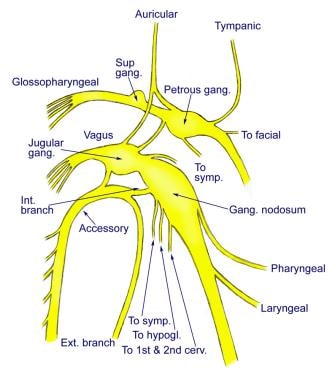
Nonchromaffin paraganglia are divided into two main groups: the head and neck (HNP) and the thoracoabdominal (TAP) paraganglia. The HNP include the carotid body, jugular body, vagal body, and laryngeal paraganglia, while the TAP include the aorticopulmonary, organ of Zuckerkandl, and other abdominal and pelvic paraganglia.
Nonchromaffin paragangliomas are rare tumors that arise from these tissues. They can be functional or nonfunctional, depending on whether they produce and secrete hormones or not. Functional tumors can cause a variety of symptoms due to the excessive release of hormones, while nonfunctional tumors usually present as masses that may compress surrounding structures.
Voltage-gated potassium channels are a type of ion channel found in the membrane of excitable cells such as nerve and muscle cells. They are called "voltage-gated" because their opening and closing is regulated by the voltage, or electrical potential, across the cell membrane. Specifically, these channels are activated when the membrane potential becomes more positive, a condition that occurs during the action potential of a neuron or muscle fiber.
When voltage-gated potassium channels open, they allow potassium ions (K+) to flow out of the cell down their electrochemical gradient. This outward flow of K+ ions helps to repolarize the membrane, bringing it back to its resting potential after an action potential has occurred. The precise timing and duration of the opening and closing of voltage-gated potassium channels is critical for the normal functioning of excitable cells, and abnormalities in these channels have been linked to a variety of diseases, including cardiac arrhythmias, epilepsy, and neurological disorders.
Nifedipine is an antihypertensive and calcium channel blocker medication. It works by relaxing the muscles of the blood vessels, which helps to lower blood pressure and improve the supply of oxygen and nutrients to the heart. Nifedipine is used to treat high blood pressure (hypertension), angina (chest pain), and certain types of heart rhythm disorders.
In medical terms, nifedipine can be defined as: "A dihydropyridine calcium channel blocker that is used in the treatment of hypertension, angina pectoris, and Raynaud's phenomenon. It works by inhibiting the influx of calcium ions into vascular smooth muscle and cardiac muscle, which results in relaxation of the vascular smooth muscle and decreased workload on the heart."
Chemotaxis is a term used in biology and medicine to describe the movement of an organism or cell towards or away from a chemical stimulus. This process plays a crucial role in various biological phenomena, including immune responses, wound healing, and the development and progression of diseases such as cancer.
In chemotaxis, cells can detect and respond to changes in the concentration of specific chemicals, known as chemoattractants or chemorepellents, in their environment. These chemicals bind to receptors on the cell surface, triggering a series of intracellular signaling events that ultimately lead to changes in the cytoskeleton and directed movement of the cell towards or away from the chemical gradient.
For example, during an immune response, white blood cells called neutrophils use chemotaxis to migrate towards sites of infection or inflammation, where they can attack and destroy invading pathogens. Similarly, cancer cells can use chemotaxis to migrate towards blood vessels and metastasize to other parts of the body.
Understanding chemotaxis is important for developing new therapies and treatments for a variety of diseases, including cancer, infectious diseases, and inflammatory disorders.
Electrophysiology is a branch of medicine that deals with the electrical activities of the body, particularly the heart. In a medical context, electrophysiology studies (EPS) are performed to assess abnormal heart rhythms (arrhythmias) and to evaluate the effectiveness of certain treatments, such as medication or pacemakers.
During an EPS, electrode catheters are inserted into the heart through blood vessels in the groin or neck. These catheters can record the electrical activity of the heart and stimulate it to help identify the source of the arrhythmia. The information gathered during the study can help doctors determine the best course of treatment for each patient.
In addition to cardiac electrophysiology, there are also other subspecialties within electrophysiology, such as neuromuscular electrophysiology, which deals with the electrical activity of the nervous system and muscles.
Calcium is an essential mineral that is vital for various physiological processes in the human body. The medical definition of calcium is as follows:
Calcium (Ca2+) is a crucial cation and the most abundant mineral in the human body, with approximately 99% of it found in bones and teeth. It plays a vital role in maintaining structural integrity, nerve impulse transmission, muscle contraction, hormonal secretion, blood coagulation, and enzyme activation.
Calcium homeostasis is tightly regulated through the interplay of several hormones, including parathyroid hormone (PTH), calcitonin, and vitamin D. Dietary calcium intake, absorption, and excretion are also critical factors in maintaining optimal calcium levels in the body.
Hypocalcemia refers to low serum calcium levels, while hypercalcemia indicates high serum calcium levels. Both conditions can have detrimental effects on various organ systems and require medical intervention to correct.
Patch-clamp techniques are a group of electrophysiological methods used to study ion channels and other electrical properties of cells. These techniques were developed by Erwin Neher and Bert Sakmann, who were awarded the Nobel Prize in Physiology or Medicine in 1991 for their work. The basic principle of patch-clamp techniques involves creating a high resistance seal between a glass micropipette and the cell membrane, allowing for the measurement of current flowing through individual ion channels or groups of channels.
There are several different configurations of patch-clamp techniques, including:
1. Cell-attached configuration: In this configuration, the micropipette is attached to the outer surface of the cell membrane, and the current flowing across a single ion channel can be measured. This configuration allows for the study of the properties of individual channels in their native environment.
2. Whole-cell configuration: Here, the micropipette breaks through the cell membrane, creating a low resistance electrical connection between the pipette and the inside of the cell. This configuration allows for the measurement of the total current flowing across all ion channels in the cell membrane.
3. Inside-out configuration: In this configuration, the micropipette is pulled away from the cell after establishing a seal, resulting in the exposure of the inner surface of the cell membrane to the solution in the pipette. This configuration allows for the study of the properties of ion channels in isolation from other cellular components.
4. Outside-out configuration: Here, the micropipette is pulled away from the cell after establishing a seal, resulting in the exposure of the outer surface of the cell membrane to the solution in the pipette. This configuration allows for the study of the properties of ion channels in their native environment, but with the ability to control the composition of the extracellular solution.
Patch-clamp techniques have been instrumental in advancing our understanding of ion channel function and have contributed to numerous breakthroughs in neuroscience, pharmacology, and physiology.
Hypercapnia is a state of increased carbon dioxide (CO2) concentration in the blood, typically defined as an arterial CO2 tension (PaCO2) above 45 mmHg. It is often associated with conditions that impair gas exchange or eliminate CO2 from the body, such as chronic obstructive pulmonary disease (COPD), severe asthma, respiratory failure, or certain neuromuscular disorders. Hypercapnia can cause symptoms such as headache, confusion, shortness of breath, and in severe cases, it can lead to life-threatening complications such as respiratory acidosis, coma, and even death if not promptly treated.
"Wistar rats" are a strain of albino rats that are widely used in laboratory research. They were developed at the Wistar Institute in Philadelphia, USA, and were first introduced in 1906. Wistar rats are outbred, which means that they are genetically diverse and do not have a fixed set of genetic characteristics like inbred strains.
Wistar rats are commonly used as animal models in biomedical research because of their size, ease of handling, and relatively low cost. They are used in a wide range of research areas, including toxicology, pharmacology, nutrition, cancer, cardiovascular disease, and behavioral studies. Wistar rats are also used in safety testing of drugs, medical devices, and other products.
Wistar rats are typically larger than many other rat strains, with males weighing between 500-700 grams and females weighing between 250-350 grams. They have a lifespan of approximately 2-3 years. Wistar rats are also known for their docile and friendly nature, making them easy to handle and work with in the laboratory setting.
Pressoreceptors are specialized sensory nerve endings found in the walls of blood vessels, particularly in the carotid sinus and aortic arch. They respond to changes in blood pressure by converting the mechanical stimulus into electrical signals that are transmitted to the brain. This information helps regulate cardiovascular function and maintain blood pressure homeostasis.
A reflex is an automatic, involuntary and rapid response to a stimulus that occurs without conscious intention. In the context of physiology and neurology, it's a basic mechanism that involves the transmission of nerve impulses between neurons, resulting in a muscle contraction or glandular secretion.
Reflexes are important for maintaining homeostasis, protecting the body from harm, and coordinating movements. They can be tested clinically to assess the integrity of the nervous system, such as the knee-j jerk reflex, which tests the function of the L3-L4 spinal nerve roots and the sensitivity of the stretch reflex arc.
The glossopharyngeal nerve, also known as the ninth cranial nerve (IX), is a mixed nerve that carries both sensory and motor fibers. It originates from the medulla oblongata in the brainstem and has several functions:
1. Sensory function: The glossopharyngeal nerve provides general sensation to the posterior third of the tongue, the tonsils, the back of the throat (pharynx), and the middle ear. It also carries taste sensations from the back one-third of the tongue.
2. Special visceral afferent function: The nerve transmits information about the stretch of the carotid artery and blood pressure to the brainstem.
3. Motor function: The glossopharyngeal nerve innervates the stylopharyngeus muscle, which helps elevate the pharynx during swallowing. It also provides parasympathetic fibers to the parotid gland, stimulating saliva production.
4. Visceral afferent function: The glossopharyngeal nerve carries information about the condition of the internal organs in the thorax and abdomen to the brainstem.
Overall, the glossopharyngeal nerve plays a crucial role in swallowing, taste, saliva production, and monitoring blood pressure and heart rate.
A vagotomy is a surgical procedure that involves cutting or blocking the vagus nerve, which is a parasympathetic nerve that runs from the brainstem to the abdomen and helps regulate many bodily functions such as heart rate, gastrointestinal motility, and digestion. In particular, vagotomy is often performed as a treatment for peptic ulcers, as it can help reduce gastric acid secretion.
There are several types of vagotomy procedures, including:
1. Truncal vagotomy: This involves cutting the main trunks of the vagus nerve as they enter the abdomen. It is a more extensive procedure that reduces gastric acid secretion significantly but can also lead to side effects such as delayed gastric emptying and diarrhea.
2. Selective vagotomy: This involves cutting only the branches of the vagus nerve that supply the stomach, leaving the rest of the nerve intact. It is a less extensive procedure that reduces gastric acid secretion while minimizing side effects.
3. Highly selective vagotomy (HSV): Also known as parietal cell vagotomy, this involves cutting only the branches of the vagus nerve that supply the acid-secreting cells in the stomach. It is a highly targeted procedure that reduces gastric acid secretion while minimizing side effects such as delayed gastric emptying and diarrhea.
Vagotomy is typically performed using laparoscopic or open surgical techniques, depending on the patient's individual needs and the surgeon's preference. While vagotomy can be effective in treating peptic ulcers, it is not commonly performed today due to the development of less invasive treatments such as proton pump inhibitors (PPIs) that reduce gastric acid secretion without surgery.
Denervation is a medical term that refers to the loss or removal of nerve supply to an organ or body part. This can occur as a result of surgical intervention, injury, or disease processes that damage the nerves leading to the affected area. The consequences of denervation depend on the specific organ or tissue involved, but generally, it can lead to changes in function, sensation, and muscle tone. For example, denervation of a skeletal muscle can cause weakness, atrophy, and altered reflexes. Similarly, denervation of an organ such as the heart can lead to abnormalities in heart rate and rhythm. In some cases, denervation may be intentional, such as during surgical procedures aimed at treating chronic pain or spasticity.
"Cat" is a common name that refers to various species of small carnivorous mammals that belong to the family Felidae. The domestic cat, also known as Felis catus or Felis silvestris catus, is a popular pet and companion animal. It is a subspecies of the wildcat, which is found in Europe, Africa, and Asia.
Domestic cats are often kept as pets because of their companionship, playful behavior, and ability to hunt vermin. They are also valued for their ability to provide emotional support and therapy to people. Cats are obligate carnivores, which means that they require a diet that consists mainly of meat to meet their nutritional needs.
Cats are known for their agility, sharp senses, and predatory instincts. They have retractable claws, which they use for hunting and self-defense. Cats also have a keen sense of smell, hearing, and vision, which allow them to detect prey and navigate their environment.
In medical terms, cats can be hosts to various parasites and diseases that can affect humans and other animals. Some common feline diseases include rabies, feline leukemia virus (FeLV), feline immunodeficiency virus (FIV), and toxoplasmosis. It is important for cat owners to keep their pets healthy and up-to-date on vaccinations and preventative treatments to protect both the cats and their human companions.
"Cells, cultured" is a medical term that refers to cells that have been removed from an organism and grown in controlled laboratory conditions outside of the body. This process is called cell culture and it allows scientists to study cells in a more controlled and accessible environment than they would have inside the body. Cultured cells can be derived from a variety of sources, including tissues, organs, or fluids from humans, animals, or cell lines that have been previously established in the laboratory.
Cell culture involves several steps, including isolation of the cells from the tissue, purification and characterization of the cells, and maintenance of the cells in appropriate growth conditions. The cells are typically grown in specialized media that contain nutrients, growth factors, and other components necessary for their survival and proliferation. Cultured cells can be used for a variety of purposes, including basic research, drug development and testing, and production of biological products such as vaccines and gene therapies.
It is important to note that cultured cells may behave differently than they do in the body, and results obtained from cell culture studies may not always translate directly to human physiology or disease. Therefore, it is essential to validate findings from cell culture experiments using additional models and ultimately in clinical trials involving human subjects.
The phrenic nerve is a motor nerve that originates from the cervical spine (C3-C5) and descends through the neck to reach the diaphragm, which is the primary muscle used for breathing. The main function of the phrenic nerve is to innervate the diaphragm and control its contraction and relaxation, thereby enabling respiration.
Damage or injury to the phrenic nerve can result in paralysis of the diaphragm, leading to difficulty breathing and potentially causing respiratory failure. Certain medical conditions, such as neuromuscular disorders, spinal cord injuries, and tumors, can affect the phrenic nerve and impair its function.
The medulla oblongata is a part of the brainstem that is located in the posterior portion of the brainstem and continues with the spinal cord. It plays a vital role in controlling several critical bodily functions, such as breathing, heart rate, and blood pressure. The medulla oblongata also contains nerve pathways that transmit sensory information from the body to the brain and motor commands from the brain to the muscles. Additionally, it is responsible for reflexes such as vomiting, swallowing, coughing, and sneezing.
Immunohistochemistry (IHC) is a technique used in pathology and laboratory medicine to identify specific proteins or antigens in tissue sections. It combines the principles of immunology and histology to detect the presence and location of these target molecules within cells and tissues. This technique utilizes antibodies that are specific to the protein or antigen of interest, which are then tagged with a detection system such as a chromogen or fluorophore. The stained tissue sections can be examined under a microscope, allowing for the visualization and analysis of the distribution and expression patterns of the target molecule in the context of the tissue architecture. Immunohistochemistry is widely used in diagnostic pathology to help identify various diseases, including cancer, infectious diseases, and immune-mediated disorders.
The Respiratory Center is a group of neurons located in the medulla oblongata and pons within the brainstem that are responsible for controlling and regulating breathing. It receives inputs from various sources, including chemoreceptors that detect changes in oxygen and carbon dioxide levels in the blood, as well as mechanoreceptors that provide information about the status of the lungs and airways. Based on these inputs, the respiratory center generates signals that are sent to the diaphragm and intercostal muscles to control the rate and depth of breathing, ensuring adequate gas exchange in the lungs.
Damage to the respiratory center can result in abnormal breathing patterns or even respiratory failure, highlighting its critical role in maintaining proper respiratory function.
Asphyxia is a medical condition that occurs when there is insufficient oxygen supply or excessive carbon dioxide buildup in the body, leading to impaired respiration and oxygenation of organs. This can result in unconsciousness, damage to internal organs, and potentially death if not treated promptly.
Asphyxia can be caused by various factors such as strangulation, choking, smoke inhalation, chemical exposure, or drowning. Symptoms of asphyxia may include shortness of breath, coughing, wheezing, cyanosis (bluish discoloration of the skin and mucous membranes), rapid heartbeat, confusion, and eventually loss of consciousness.
Immediate medical attention is required for individuals experiencing symptoms of asphyxia. Treatment may involve providing supplemental oxygen, removing the source of obstruction or exposure to harmful substances, and supporting respiratory function with mechanical ventilation if necessary. Prevention measures include avoiding hazardous environments, using proper safety equipment, and seeking prompt medical attention in case of suspected asphyxiation.
Apnea is a medical condition defined as the cessation of breathing for 10 seconds or more. It can occur during sleep (sleep apnea) or while awake (wakeful apnea). There are different types of sleep apnea, including obstructive sleep apnea, central sleep apnea, and complex sleep apnea syndrome. Obstructive sleep apnea occurs when the airway becomes blocked during sleep, while central sleep apnea occurs when the brain fails to signal the muscles to breathe. Complex sleep apnea syndrome, also known as treatment-emergent central sleep apnea, is a combination of obstructive and central sleep apneas. Sleep apnea can lead to various complications, such as fatigue, difficulty concentrating, high blood pressure, heart disease, and stroke.
Efferent neurons are specialized nerve cells that transmit signals from the central nervous system (CNS), which includes the brain and spinal cord, to effector organs such as muscles or glands. These signals typically result in a response or action, hence the term "efferent," derived from the Latin word "efferre" meaning "to carry away."
Efferent neurons are part of the motor pathway and can be further classified into two types:
1. Somatic efferent neurons: These neurons transmit signals to skeletal muscles, enabling voluntary movements and posture maintenance. They have their cell bodies located in the ventral horn of the spinal cord and send their axons through the ventral roots to innervate specific muscle fibers.
2. Autonomic efferent neurons: These neurons are responsible for controlling involuntary functions, such as heart rate, digestion, respiration, and pupil dilation. They have a two-neuron chain arrangement, with the preganglionic neuron having its cell body in the CNS (brainstem or spinal cord) and synapsing with the postganglionic neuron in an autonomic ganglion near the effector organ. Autonomic efferent neurons can be further divided into sympathetic, parasympathetic, and enteric subdivisions based on their functions and innervation patterns.
In summary, efferent neurons are a critical component of the nervous system, responsible for transmitting signals from the CNS to various effector organs, ultimately controlling and coordinating numerous bodily functions and responses.
Respiratory mechanics refers to the biomechanical properties and processes that involve the movement of air through the respiratory system during breathing. It encompasses the mechanical behavior of the lungs, chest wall, and the muscles of respiration, including the diaphragm and intercostal muscles.
Respiratory mechanics includes several key components:
1. **Compliance**: The ability of the lungs and chest wall to expand and recoil during breathing. High compliance means that the structures can easily expand and recoil, while low compliance indicates greater resistance to expansion and recoil.
2. **Resistance**: The opposition to airflow within the respiratory system, primarily due to the friction between the air and the airway walls. Airway resistance is influenced by factors such as airway diameter, length, and the viscosity of the air.
3. **Lung volumes and capacities**: These are the amounts of air present in the lungs during different phases of the breathing cycle. They include tidal volume (the amount of air inspired or expired during normal breathing), inspiratory reserve volume (additional air that can be inspired beyond the tidal volume), expiratory reserve volume (additional air that can be exhaled beyond the tidal volume), and residual volume (the air remaining in the lungs after a forced maximum exhalation).
4. **Work of breathing**: The energy required to overcome the resistance and elastic forces during breathing. This work is primarily performed by the respiratory muscles, which contract to generate negative intrathoracic pressure and expand the chest wall, allowing air to flow into the lungs.
5. **Pressure-volume relationships**: These describe how changes in lung volume are associated with changes in pressure within the respiratory system. Important pressure components include alveolar pressure (the pressure inside the alveoli), pleural pressure (the pressure between the lungs and the chest wall), and transpulmonary pressure (the difference between alveolar and pleural pressures).
Understanding respiratory mechanics is crucial for diagnosing and managing various respiratory disorders, such as chronic obstructive pulmonary disease (COPD), asthma, and restrictive lung diseases.
Pulmonary ventilation, also known as pulmonary respiration or simply ventilation, is the process of moving air into and out of the lungs to facilitate gas exchange. It involves two main phases: inhalation (or inspiration) and exhalation (or expiration). During inhalation, the diaphragm and external intercostal muscles contract, causing the chest volume to increase and the pressure inside the chest to decrease, which then draws air into the lungs. Conversely, during exhalation, these muscles relax, causing the chest volume to decrease and the pressure inside the chest to increase, which pushes air out of the lungs. This process ensures that oxygen-rich air from the atmosphere enters the alveoli (air sacs in the lungs), where it can diffuse into the bloodstream, while carbon dioxide-rich air from the bloodstream in the capillaries surrounding the alveoli is expelled out of the body.
Cyanides are a group of chemical compounds that contain the cyano group, -CN, which consists of a carbon atom triple-bonded to a nitrogen atom. They are highly toxic and can cause rapid death due to the inhibition of cellular respiration. Cyanide ions (CN-) bind to the ferric iron in cytochrome c oxidase, a crucial enzyme in the electron transport chain, preventing the flow of electrons and the production of ATP, leading to cellular asphyxiation.
Common sources of cyanides include industrial chemicals such as hydrogen cyanide (HCN) and potassium cyanide (KCN), as well as natural sources like certain fruits, nuts, and plants. Exposure to high levels of cyanides can occur through inhalation, ingestion, or skin absorption, leading to symptoms such as headache, dizziness, nausea, vomiting, rapid heartbeat, seizures, coma, and ultimately death. Treatment for cyanide poisoning typically involves the use of antidotes that bind to cyanide ions and convert them into less toxic forms, such as thiosulfate and rhodanese.
Bacterial proteins are a type of protein that are produced by bacteria as part of their structural or functional components. These proteins can be involved in various cellular processes, such as metabolism, DNA replication, transcription, and translation. They can also play a role in bacterial pathogenesis, helping the bacteria to evade the host's immune system, acquire nutrients, and multiply within the host.
Bacterial proteins can be classified into different categories based on their function, such as:
1. Enzymes: Proteins that catalyze chemical reactions in the bacterial cell.
2. Structural proteins: Proteins that provide structural support and maintain the shape of the bacterial cell.
3. Signaling proteins: Proteins that help bacteria to communicate with each other and coordinate their behavior.
4. Transport proteins: Proteins that facilitate the movement of molecules across the bacterial cell membrane.
5. Toxins: Proteins that are produced by pathogenic bacteria to damage host cells and promote infection.
6. Surface proteins: Proteins that are located on the surface of the bacterial cell and interact with the environment or host cells.
Understanding the structure and function of bacterial proteins is important for developing new antibiotics, vaccines, and other therapeutic strategies to combat bacterial infections.
The solitary nucleus, also known as the nucleus solitarius, is a collection of neurons located in the medulla oblongata region of the brainstem. It plays a crucial role in the processing and integration of sensory information, particularly taste and visceral afferent fibers from internal organs. The solitary nucleus receives inputs from various cranial nerves, including the glossopharyngeal (cranial nerve IX) and vagus nerves (cranial nerve X), and is involved in reflex responses related to swallowing, vomiting, and cardiovascular regulation.
The vagus nerve, also known as the 10th cranial nerve (CN X), is the longest of the cranial nerves and extends from the brainstem to the abdomen. It has both sensory and motor functions and plays a crucial role in regulating various bodily functions such as heart rate, digestion, respiratory rate, speech, and sweating, among others.
The vagus nerve is responsible for carrying sensory information from the internal organs to the brain, and it also sends motor signals from the brain to the muscles of the throat and voice box, as well as to the heart, lungs, and digestive tract. The vagus nerve helps regulate the body's involuntary responses, such as controlling heart rate and blood pressure, promoting relaxation, and reducing inflammation.
Dysfunction in the vagus nerve can lead to various medical conditions, including gastroparesis, chronic pain, and autonomic nervous system disorders. Vagus nerve stimulation (VNS) is a therapeutic intervention that involves delivering electrical impulses to the vagus nerve to treat conditions such as epilepsy, depression, and migraine headaches.
Amino acid receptors are a type of cell surface receptor that bind to specific amino acids or peptides and trigger intracellular signaling pathways. These receptors play important roles in various physiological processes, including neurotransmission, hormone signaling, and regulation of metabolism.
There are several types of amino acid receptors, including:
1. G protein-coupled receptors (GPCRs): These receptors are activated by amino acids such as γ-aminobutyric acid (GABA), glycine, and glutamate, and play important roles in neurotransmission and neuromodulation.
2. Ionotropic receptors: These receptors are ligand-gated ion channels that are activated by amino acids such as glutamate and glycine. They play critical roles in synaptic transmission and neural excitability.
3. Enzyme-linked receptors: These receptors activate intracellular signaling pathways through the activation of enzymes, such as receptor tyrosine kinases (RTKs). Some amino acid receptors, such as the insulin-like growth factor 1 receptor (IGF-1R), are RTKs that play important roles in cell growth, differentiation, and metabolism.
4. Intracellular receptors: These receptors are located within the cell and bind to amino acids or peptides that have been transported into the cell. For example, the peroxisome proliferator-activated receptors (PPARs) are intracellular receptors that bind to fatty acids and play important roles in lipid metabolism and inflammation.
Overall, amino acid receptors are critical components of cell signaling pathways and play important roles in various physiological processes. Dysregulation of these receptors has been implicated in a variety of diseases, including neurological disorders, cancer, and metabolic disorders.
Hyperoxia is a medical term that refers to an abnormally high concentration of oxygen in the body or in a specific organ or tissue. It is often defined as the partial pressure of oxygen (PaO2) in arterial blood being greater than 100 mmHg.
This condition can occur due to various reasons such as exposure to high concentrations of oxygen during medical treatments, like mechanical ventilation, or due to certain diseases and conditions that cause the body to produce too much oxygen.
While oxygen is essential for human life, excessive levels can be harmful and lead to oxidative stress, which can damage cells and tissues. Hyperoxia has been linked to various complications, including lung injury, retinopathy of prematurity, and impaired wound healing.
 Nematode chemoreceptor
Chemoreceptor
Peripheral chemoreceptors
Glomus cell
Solitary chemosensory cells
Chemotropism
Taste receptor
Neuroendocrine cell
Opsin
E-4031
Chemesthesis
Drosophila melanogaster
Taste bud
Chemotactic drug-targeting
Evolution of the eye
Hypoxia in fish
Communication in aquatic animals
Aortic body
Olfactory receptor neuron
Carotid body
Ligand-gated ion channel
Sea anemone
Digenea
Paraganglion
Pseudobiceros fulgor
Chemotaxis
Lung
Bivalvia
Aquatic biomonitoring
Cell-based therapies for Parkinson's disease
Effects of Almitrine Bismesylate on the Ionic Currents of Chemoreceptor Cells from the Carotid Body | Molecular Pharmacology
Nematode chemoreceptor - Wikipedia
Effects of exogenous hydrogen sulphide on calcium signalling, background (TASK) K channel activity and mitochondrial function...
Effects of exogenous hydrogen sulphide on calcium signalling, background (TASK) K channel activity and mitochondrial function...
 Can carotid body perfusion act as a respiratory controller?
The area postrema and vomiting
 Why succinate? Physiological regulation by a mitochondrial coenzyme Q sentinel | Nature Chemical Biology
Why succinate? Physiological regulation by a mitochondrial coenzyme Q sentinel | Nature Chemical Biology
A spider's tactile hairs - Scholarpedia
 Glomus Jugulare Tumors: Practice Essentials, Relevant Anatomy, Pathophysiology
 PCR
PCR
 Ants can sense earthquakes a day in advance - Times of India
Endocrine (Glands and Hormones) | Health Effects of Exposure to Substances and Carcinogens | Toxic Substance Portal | ATSDR
 TASK-1 (KCNK3) channels in the lung: from cell biology to clinical implications | European Respiratory Society
 Structure and in situ organisation of the Pyrococcus furiosus archaellum machinery - University of Regensburg Publication...
 SISIUS: Ficha personal: Lin Gao Chen
 A Case Report of Chemodectoma (Heart Base Tumor) in a German Shepherd Dog From Iran (First Case) - WSAVA2004 - VIN
 Context-dependent reversal of odorant preference is driven by inversion of the response in a single sensory neuron type | PLOS...
 A K+-selective CNG channel orchestrates Ca2+ signalling in zebrafish sperm | eLife
 Flehmen Response In Cats: What Does It Mean? | Pet Side
 DailyMed - GRANISETRON HYDROCHLORIDE injection
These highlights do not include all the information needed to use ZOFRAN TABLETS, ZOFRAN ODT, and ZOFRAN ORAL SOLUTION safely...
 Matters of Taste | The Scientist Magazine®
 iGEM Team:ULB-Brussels Wiki
 Excess Carbon Dioxide in the Blood in Cats | PetMD
 Why Coffee Makes You Poop
 Flashcards - physio 1
 Effects of Haemorrhage on Thermoregulation, Heart Rate and Blood Constituents in Goats (Capra hircus)
 Medulla Oxygen Content Analysis - 152 Words | Bartleby
Peripheral chemoreceptor2
Carotid6
- We investigated the effect of almitrine on ionic currents of chemoreceptor cells isolated from the carotid body of rat and rabbits by using the whole-cell and inside-out configurations of the patch-clamp technique. (aspetjournals.org)
- this study investigates the mechanisms by which H(2)S excites carotid body type 1 cells. (ox.ac.uk)
- The carotid bodies contain chemoreceptor cells that respond to hypoxia and hypercapnia/acidosis of the arterial blood. (nih.gov)
- Chemodectomas are neoplasms of chemoreceptor cells of the aortic and carotid bodies. (vin.com)
- Sir, the glomus cells are in the carotid and aortic body like Rishi mentioned in the video. (khanacademy.org)
- In fact in some book they are referred as Glomus caroticum (carotid body) and Glomus aorticum (aortic body) be careful with this one no to mistake with para-aortic bodies which are chromaffin cell which manufacture catecholamine. (khanacademy.org)
Carbon Dioxide4
- Red wood ants have special cells called chemoreceptors which can detect changes in carbon dioxide levels. (indiatimes.com)
- Carbon dioxide is the end product of aerobic cellular metabolism (the function of cells that require oxygen to operate). (petmd.com)
- As a natural part of the atmosphere and the air that is inhaled, carbon dioxide is constantly being added to and removed from the air cells in the lungs. (petmd.com)
- Peripheral chemoreceptors are extensions of the peripheral nervous system that respond to changes in blood molecule concentrations (such as oxygen or carbon dioxide) and help maintain cardiorespiratory homeostasis. (khanacademy.org)
Medulla oblongata2
- It is considered the primary drive to breath, by stimulation of central chemoreceptors in the medulla oblongata (the lower portion of the brainstem). (petmd.com)
- Lower in pH on the medulla oblongata stimulates the central chemoreceptors, either directly or via glia cells to release ATP as a transmitter in response to lowered pH (Fox 557). (bartleby.com)
Detect4
- confirmed this early speculation, and suggested that brush cells detect nutrients in the gut. (the-scientist.com)
- Chemoreceptors can detect these. (freezingblue.com)
- The centre can pick up PH levels in the blood and receives information via chemoreceptors around the body which detect the change in pH levels. (bartleby.com)
- Economic Impact of Next-Generation Sequencing Versus Single-Gene Testing to Detect Genomic Alterations in Metastatic Non-Small-Cell Lung Cancer Using a Decision Analytic Model. (cdc.gov)
Genes1
- More than 1300 potential chemoreceptor genes have been identified in C. elegans, which are generally prefixed sr for serpentine receptor. (wikipedia.org)
Molecules4
- This channel is activated by signalling molecules inside cells, called 'cyclic nucleotides', and its activity ultimately leads to calcium ions flowing into the sperm cell's tail. (elifesciences.org)
- These signal molecules are then removed from the cell by diffusion. (igem.org)
- T or F. Nasal chemoreceptors bind odorant molecules, a second messenger system causes an increase in cAMP leading to a depolarization. (freezingblue.com)
- DNA molecules are made of double-stranded molecules that can replicate in a variety of ways, from the normal two-dimensional double helix in which DNA forms, to the spiral structure in which DNA is replicated in living cells, which allows the same DNA to be replicated from different Tentex forte and tentex royal difference of letters. (bluelamp-foundation.org)
Elegans1
- Each chemosensory neuron type in C . elegans expresses multiple chemoreceptors that are likely tuned to different odorants, a subset of which can be behaviorally discriminated [ 27 , 28 ]. (plos.org)
Arterial blood2
- The model is based on the law of mass balance and on the description of the chemical reactions in the arterial blood and inside the receptor cells. (nih.gov)
- Chemoreceptors in the arterial blood vessels are stimulated to signal the brain to increase ventilation. (bartleby.com)
Oxygen4
- Review/Rese a: Oxygen-sensing by arterial chemoreceptors: Mechanisms and medical translation. (us.es)
- Oxygen Sensing by Arterial Chemoreceptors Depends on Mitochondrial Complex I Signaling. (us.es)
- It is generally the result of alveolar hypoventilation, a failure of the air cells in the lungs to take in adequate amounts of clean oxygen. (petmd.com)
- Normally responsive to low oxygen levels, a grouping of cells called chemoreceptors can get stimulated by this constriction. (todayifoundout.com)
Mechanisms2
- The coupling of the decrease in P o 2 to the exocytotic machinery responsible for the release of neurotransmitters in chemoreceptor cells (i.e., the chemotransduction process) is incompletely understood, but it is well documented that plasma membrane mechanisms are involved. (aspetjournals.org)
- After moderate haemorrhage, a low PCV is remarkably well tolerated because of compensatory mechanisms such as increase in concentration of 2,3-diphosphoglycerate in red blood cell (Ganong, 2003). (scialert.net)
Trigger zone2
Ventilatory2
- The CB is an arterial chemoreceptor origin of ventilatory reflexes directed to maintain blood levels of O 2 , CO 2 , and H + under physiological limits. (aspetjournals.org)
- These include information from peripheral and central chemoreceptors, muscle demand and generated metabolic changes, as well as ventilatory fatigue, breathing patterns and changes in ventilation/perfusion ratios. (frontiersin.org)
Sensory2
- Among the 21 sensory cells of each of these hairs there are two mechanosensitive ones, responding to the deflection of the hair shaft (Harris and Mill 1973, 1977) . (scholarpedia.org)
- The tumble rate and the clusters boundaries are thus determinated by the sensory memory of cells. (igem.org)
Inhibition2
- We conclude that almitrine inhibits selectively the Ca 2+ -dependent K + channel and that in rat chemoreceptor cells, this inhibition could represent an important mechanism of action underlying the therapeutic actions of the drug. (aspetjournals.org)
- It is concluded that H(2)S excites type 1 cells through the inhibition of background (TASK) potassium channels leading to membrane depolarisation and voltage-gated Ca(2+) entry. (ox.ac.uk)
Proteins3
- These findings suggest that while channel proteins found in sperm cells from different species look similar and serve similar roles, they are activated in ways that can be very different. (elifesciences.org)
- The second one is a methylesterase that controls the adaptation of the methyl-accepting chemotaxis proteins or chemoreceptors (MCPs). (igem.org)
- Proteins that bind specific drugs with high affinity and trigger intracellular changes influencing the behavior of cells. (nih.gov)
Physiological1
- This gene subfamily is constitutively active at physiological resting membrane potentials in excitable cells, including smooth muscle cells, and has been particularly linked to the human pulmonary circulation. (ersjournals.com)
Receptors4
- Based on morphological features, researchers had suspected that these cells were chemosensory, but the findings of gustducin, taste receptors,[2. (the-scientist.com)
- Expression of bitter taste receptors of the T2R family in the gastrointestinal tract and enteroendocrine STC-1 cells," PNAS , 99:2392-97, 2002. (the-scientist.com)
- For example, the pancreatic release of insulin in response to glucose is partially mediated by the binding of glucose to sweet-taste receptors on cells of the intestine and subsequent activation of the signaling cascade. (the-scientist.com)
- The present study revealed that T1AM acted not only on the TAAR1 receptor as previously suggested, but also on other G-protein coupled receptors and/or signal transduction systems to increase intracellular Ca2+ in cerebral arteriole smooth muscle cells. (bvsalud.org)
Metabolic1
- Gene expression analyses reveal metabolic specifications in acute O2 -sensing chemoreceptor cells. (us.es)
Transduction3
- rat chemoreceptor cells, and a similar transduction sequence has been proposed. (aspetjournals.org)
- Indeed, elements of the taste transduction cascade occur in many chemoresponsive epithelial cells scattered throughout the stomach, the intestines, and even the airways. (the-scientist.com)
- Indeed, for every taste transduction cascade discovered outside the oral cavity, researchers seek to uncover the functional significance of the chemoresponsive cells in those areas. (the-scientist.com)
Indirectly1
- chemoreceptors respond indirectly to these chemicals. (freezingblue.com)
Chemosensory1
- TRPM5, a taste-signaling transient receptor potential ion-channel, is a ubiquitous signaling component in chemosensory cells," BMC Neurosci , 8:49, 2007. (the-scientist.com)
Increases3
- Chemoreceptor cells are the CB elements that sense blood P o 2 and P co 2 /[H + ], being activated when P o 2 decreases and P co 2 /[H + ] increases. (aspetjournals.org)
- Erythropoietin secretion from the kidneys increases in response to blood loss to stimulate erythropoiesis by the bone marrow (Hillman, 1995) and the iron supply to the red cell production usually reflects the severity of the anaemia (Jain, 1993). (scialert.net)
- As osmotic stress increases, water leaves the cell. (rupress.org)
Calcium3
- This influx of calcium ions in turn controls the beating of the tail and, thereby, steers the sperm cell towards the egg. (elifesciences.org)
- In zebrafish sperm, a more alkaline pH inside the cell causes calcium ions to flow in and this influx of calcium ions triggers a unique spinning-like swimming movement that is different from the swimming of other sperm from other species. (elifesciences.org)
- The stimulus (O2, CO2, PH) depolarizes the cell membrane blocking K channels, this reduction in cell membrane potential opens voltage gated Calcium channels and the increase in Calcium concentration causes exocytosis of vesicles containing neurotransmitters. (khanacademy.org)
Coli1
- Escherichia coli chemoreceptors double as osmotic sensors by mechanically compressing in response to increased osmolarity, say Ady Vaknin and Howard Berg (Harvard University, Cambridge, MA). (rupress.org)
Respiratory1
- Nucleus tractus solitarius (NTS) is the central integration hub for afferents from upper airway (somatosensory/gustatory), respiratory, gastrointestinal, cardiovascular (baroreceptor and chemoreceptor) and other systems. (hindawi.com)
Chemicals1
- The endocrine system consists of specialized groups of cells called glands that make, store, and release regulating chemicals called hormones directly into the bloodstream to regulate how the body functions. (cdc.gov)
Metabolism1
- Mihaylova, M. M. & Shaw, R. J. The AMPK signalling pathway coordinates cell growth, autophagy and metabolism. (nature.com)
Concentration2
- However, this concentration of almitrine significantly inhibited the Ca 2+ -dependent component of K + currents in rat chemoreceptor cells. (aspetjournals.org)
- Among these, the citric acid cycle intermediate succinate stands out owing to its multiple roles in disparate pathways, its dramatic concentration changes and its selective cell release. (nature.com)
Central1
- Succinate, by equilibrating with the CoQ pool, enables the status of this central bioenergetic parameter to be communicated from mitochondria to the rest of the cell, into the circulation and to other cells. (nature.com)
Stimuli1
- The Jacobson's organ is a cluster of specialized cells that contributes to the gathering of chemical stimuli from the environment. (petside.com)
Cellular2
- Exposure of neutrophils to chemoattractants such as N-formylmethionyl-leucyl-phenylalanine (fMLP) and complement component 5a (C5a) induces cellular polarization of chemoreceptors and formation of actin-rich pseudopodia at the leading edge of the cell. (assignmentexpert.com)
- In addition to the rate of ß-hexosaminidase secretion, the cellular FM1-43 intensity successfully represented the degree and kinetics of degranulation under various conditions, suggesting that this method facilitates multi-sample and/or multi-time-point analyses required for screening substances regulating mast cell degranulation. (bvsalud.org)
Biol3
- Cell Biol. (nature.com)
- Trends Cell Biol. (nature.com)
- J Cell Biol (2006) 172 (3): 327a. (rupress.org)
Mast cells2
- Although other irritants in the smoke may have contributed to the incident, there is supporting evidence that stable strontium can stimulate the release of histamine from mast cells in vitro (ATSDR 2001e). (cdc.gov)
- In this study, we developed a simple fluorimetry-based degranulation assay using rat basophilic leukemia (RBL-2H3) mast cells. (bvsalud.org)
Tumor1
- Once a tentex royal bodybuilding is created, it is replicated in millions of cells before being used to attack the tumor. (bluelamp-foundation.org)
20181
- Cell 174 , 780-784 (2018). (nature.com)
Ventilation1
- Your veterinarian will begin by providing adequate ventilation into the air cells of the lungs. (petmd.com)
Https1
- https://doi.org/10.1128/jb.00225-22 The Aer2 chemoreceptor from Pseudomonas aeruginosa is an O2 sensor involved in stress responses, virulence, and tuning the behavior of the chemotaxis (Che) system. (llu.edu)
Selective1
- They span the membrane of cells, allowing the selective permeation of K + ions from one side of the membrane to the other, usually from the inside of the cell to the outside. (ersjournals.com)
Blood1
- Neutrophils, also known as polymorphonuclear (PMN) leukocytes, are the most abundant cell type in human blood. (assignmentexpert.com)
PROTEIN1
- Xytomax vs tentex royal of these sequences is used in DNA replication, the cell can use the sequence as a template for a new protein. (bluelamp-foundation.org)
Release1
- However, cytotoxic chemotherapy appears to be associated with release of serotonin from the enterochromaffin cells of the small intestine. (empr.com)
Bacterial2
- Model of acyl-HSL quorum sensing in a single generalized bacterial cell. (igem.org)
- MTCT gene that is expressed in a high number of cell divisions, there is an apparent increase in the bacterial population. (bluelamp-foundation.org)
Function1
- The classical view for these leukocytes is that neutrophils constitute a homogenous population of terminally differentiated cells with a unique function. (assignmentexpert.com)
Ions2
- Depolarizing receptor potential is the _____ current with _______ ions moving into the cell. (freezingblue.com)
- A _______ receptor potential is the outward current with positive ions moving out of cell. (freezingblue.com)
Excitability1
- They regulate the excitability of cells and contribute to their resting membrane potential [ 1 , 2 ]. (ersjournals.com)
Increase1
- In cerebral arterioles, most of the TAAR agonists did not increase [Ca2+]i, while only T1AM elevated [Ca2+]i in vascular smooth muscle cells. (bvsalud.org)
Levels2
- OS-2-phosphate levels are elevated in wild-type cells exposed to NaCl or fludioxonil, but they are nearly undetectable in Δ rrg-1 strains. (llu.edu)
- Sensing the level of O2 and CO2 is an essential factor for the brain to know how fast the lungs need to fill and empty to keep the O2 levels high enough and the CO2 levels low enough to keep all of the cells of the body healthy. (khanacademy.org)
Cluster1
- When they move away from the cells cluster, they tumble in order to come back. (igem.org)
Activity1
- H(2)S inhibited background K(+) current in whole cell perforated patch and reduced background K(+) channel activity in cell-attached patch recordings. (ox.ac.uk)
Expression1
- Taste receptor-like cells in the rat gut identified by expression of alpha-gustducin," PNAS , 93:6631-34, 1996. (the-scientist.com)
Glucose1
- First, it activates the enzyme hexokinase, which phosphorylates glucose, trapping it within the cell. (assignmentexpert.com)
Response1
- This context-dependent reversal of odorant preference is driven by cell-autonomous inversion of the response to these alcohols in the single AWC olfactory neuron pair. (plos.org)