Chemokine CCL27
Chemokine CCL21
Chemokine CCL22
Chemokine CCL17
Chemokine CCL2
Chemokine CCL19
Chemokine CCL5
Chemokine CCL20
Chemokine CCL1
Chemokines, CC
Receptors, Chemokine
Chemokine CCL3
Chemokine CCL7
Chemokines
Receptors, CCR10
Chemokine CCL4
Chemokine CXCL12
Receptors, CCR1
Chemokine CXCL10
Chemokine CCL8
Receptors, CCR2
Chemokine CCL11
Chemokine CCL24
Receptors, CCR7
Receptors, CCR8
Chemokine CXCL1
Chemotaxis, Leukocyte
Receptors, CCR4
Chemokines, CXC
Chemokine CX3CL1
Macrophage Inflammatory Proteins
Receptors, CCR5
Receptors, CCR3
Mice, Inbred C57BL
Cell Movement
Chemokine CXCL2
Chemokine CXCL13
Receptors, CXCR4
Chemokine CXCL11
Chemotaxis
Chemokine CXCL6
Cells, Cultured
Dendritic Cells
Chemokine CXCL5
Cytokines
Mice, Knockout
Receptors, CXCR3
Mice, Inbred BALB C
Monocytes
Macrophages
Gene Expression Regulation
RNA, Messenger
T-Lymphocytes
Inflammation
Reverse Transcriptase Polymerase Chain Reaction
Enzyme-Linked Immunosorbent Assay
Flow Cytometry
Receptors, Interleukin-8B
Signal Transduction
Dermatitis, Atopic
Up-Regulation
Monocyte Chemoattractant Proteins
Disease Models, Animal
Skin
Mice, Transgenic
Interleukin-8
Ligands
Receptors, CCR6
CD4-Positive T-Lymphocytes
Receptors, Interleukin-8A
Lymph Nodes
NF-kappa B
Carbon Tetrachloride
Immunohistochemistry
Receptors, Cytokine
T-Lymphocytes, Regulatory
Tumor Necrosis Factor-alpha
Chemokines, CX3C
Receptors, CXCR5
Protein Binding
Chemotactic Factors
CD8-Positive T-Lymphocytes
Endothelial Cells
Lymphocyte Activation
Monokines
Receptors, HIV
Carbon Tetrachloride Poisoning
Duffy Blood-Group System
Chemotactic Factors, Eosinophil
Neutrophil Infiltration
Neutrophils
Heterocyclic Compounds
Lung
Leukocytes
Gene Expression
Inflammation Mediators
Interferon-gamma
Th2 Cells
Cell Migration Inhibition
HIV-1
Molecular Sequence Data
Eosinophils
Intercellular Signaling Peptides and Proteins
Lipopolysaccharides
Down-Regulation
Amino Acid Sequence
Epithelial Cells
Leukocytes, Mononuclear
Th1 Cells
Lymphoid Tissue
T-Lymphocyte Subsets
Gene Expression Profiling
Platelet Factor 4
Stromal Cells
Immunity, Innate
Bronchoalveolar Lavage Fluid
Transfection
Drug-Induced Liver Injury
Endothelium, Lymphatic
Expression and cellular localization of the CC chemokines PARC and ELC in human atherosclerotic plaques. (1/275)
Local immune responses are thought to play an important role in the development of atherosclerosis. Histological studies have shown that human atherosclerotic lesions contain T lymphocytes throughout all stages of development, many of which are in an activated state. A number of novel CC chemokines have been described recently, which are potent chemoattractants for lymphocytes: PARC (pulmonary and activation-regulated chemokine), ELC (EBI1-ligand chemokine), LARC (liver and activation-regulated chemokine), and SLC (secondary lymphoid-tissue chemokine). Using reverse transcriptase-polymerase chain reaction and in situ hybridization, we have found gene expression for PARC and ELC but not for LARC or SLC in human atherosclerotic plaques. Immunohistochemical staining of serial plaque sections with specific cell markers revealed highly different expression patterns of PARC and ELC. PARC mRNA was restricted to CD68+ macrophages (n = 14 of 18), whereas ELC mRNA was widely expressed by macrophages and intimal smooth muscle cells (SMC) in nearly all of the lesions examined (n = 12 of 14). ELC mRNA was also found to be expressed in the medial SMC wall of highly calcified plaques (n = 4). Very low levels of ELC mRNA expression could also be detected in normal mammary arteries but no mRNA expression for PARC was detected in these vessels (n = 4). In vitro, ELC mRNA was found to be up-regulated in aortic SMC stimulated with tumor necrosis factor-a and interferon-gamma but not in SMC stimulated with serum. Both PARC and ELC mRNA were expressed by monocyte-derived macrophages but not monocytes. The expression patterns of PARC and ELC mRNA in human atherosclerotic lesions suggest a potential role for these two recently described CC chemokines in attracting T lymphocytes into atherosclerotic lesions. (+info)Cutting edge: immature dendritic cells generated from monocytes in the presence of TGF-beta 1 express functional C-C chemokine receptor 6. (2/275)
Although CD34+ progenitor-derived immature dendritic cells (DCs) express CCR6, several recent studies reported that monocyte-derived immature DCs do not do so. We observed that DCs generated from monocytes in the presence of GM-CSF, IL-4, and TGF-beta 1 consistently responded to liver and activation-regulated chemokine (LARC, also known as macrophage inflammatory protein-3 alpha). These immature DCs expressed one class of high-affinity binding sites for LARC, and expressed both CCR6 mRNA and protein. Therefore, LARC-CCR6 interaction presumably also contributes to the regulation of trafficking of monocyte-derived DCs, and utilization of TGF-beta can potentially provide a ready source of CCR6+ monocyte-derived DCs for therapeutic purposes. (+info)Beta-defensins: linking innate and adaptive immunity through dendritic and T cell CCR6. (3/275)
Defensins contribute to host defense by disrupting the cytoplasmic membrane of microorganisms. This report shows that human beta-defensins are also chemotactic for immature dendritic cells and memory T cells. Human beta-defensin was selectively chemotactic for cells stably transfected to express human CCR6, a chemokine receptor preferentially expressed by immature dendritic cells and memory T cells. The beta-defensin-induced chemotaxis was sensitive to pertussis toxin and inhibited by antibodies to CCR6. The binding of iodinated LARC, the chemokine ligand for CCR6, to CCR6-transfected cells was competitively displaced by beta-defensin. Thus, beta-defensins may promote adaptive immune responses by recruiting dendritic and T cells to the site of microbial invasion through interaction with CCR6. (+info)MIP-3alpha induces human eosinophil migration and activation of the mitogen-activated protein kinases (p42/p44 MAPK). (4/275)
The CC chemokine macrophage inflammatory protein-3alpha (MIP-3alpha) is the product of recent electronic cloning efforts, however, little characterization of its spectrum of biological effects has been undertaken. Human eosinophils exhibited pertussis-toxin-sensitive migration in response to human recombinant (hr)MIP-3alpha. Messenger RNA for the MIP-3alpha receptor, CCR-6, and low levels of surface expression were demonstrated by reverse transcriptase-polymerase chain reaction and FACS analysis. Analyses of cell signaling revealed dose-dependent increases in intracellular calcium mobilization, calcium transients that were, however, greatly reduced when compared with MCP-3-induced responses. Further investigations of MIP-3alpha-induced signal transduction revealed time- and dose-dependent, partially pertussis toxin-dependent, increases in phosphorylation of the p42/p44 mitogen-activated protein kinases (MAPK) that occurred at 10- to 100-fold lower concentrations, and that were linked to a phosphoinositide 3-kinase pathway. These results suggest that MIP-3alpha can regulate multiple, parallel signal transduction pathways in eosinophils, and suggest that MAPK activation by MIP-3alpha in eosinophils is a significant signaling pathway for migration induction. (+info)In breast carcinoma tissue, immature dendritic cells reside within the tumor, whereas mature dendritic cells are located in peritumoral areas. (5/275)
We have analyzed the presence of immature and mature dendritic cells (DCs) within adenocarcinoma of the breast using immunohistochemistry. Immature DCs were defined by expression of CD1a-, Langerin-, and intracellular major histocompatibility complex class II-rich vesicles. Mature DCs were defined by expression of CD83 and DC-Lamp. Breast carcinoma cells were defined by morphology and/or cytokeratin expression. We demonstrate two levels of heterogeneity of DCs infiltrating breast carcinoma tissue: (a) immature CD1a(+) DCs, mostly of the Langerhans cell type (Langerin(+)), were retained within the tumor bed in 32/32 samples and (b) mature DCs, CD83(+)DC-Lamp(+), present in 20/32 samples, are confined to peritumoral areas. The high numbers of immature DCs found in the tumor may be best explained by high levels of macrophage inflammatory protein 3alpha expression by virtually all tumor cells. Confirming the immature/mature DC compartmentalization pattern, in vitro-generated immature DCs adhere to the tumor cells, whereas mature DCs adhere selectively to peritumoral areas. In some cases, T cells are clustering around the mature DCs in peritumoral areas, thus resembling the DC-T cell clusters of secondary lymphoid organs, which are characteristic of ongoing immune reactions. (+info)Down-regulation of the beta-chemokine receptor CCR6 in dendritic cells mediated by TNF-alpha and IL-4. (6/275)
Chemokines are involved in the control of dendritic cell (DC) trafficking, which is critical for the immune response. We have generated DC from human umbilical cord blood CD34+ progenitors cultured with granulocyte-macrophage colony-stimulating factor, tumor necrosis factor alpha (TNF-alpha), and stem cell factor. Using an anti-CCR6 monoclonal antibody, we observed that these cells showed maximum expression of this beta-chemokine receptor when they were immature, as determined by their relatively low expression of several DC maturation markers such as CD1a, CD11c, CD14, CD40, CD80, and CD83. Immature DC responded strongly to macrophage inflammatory protein-3alpha (MIP-3alpha), the CCR6 ligand, in migration and calcium mobilization assays. CCR6 expression decreased in parallel with the DC maturation induced by prolonged TNF-alphaq treatments. Interleukin-4 was also able to decrease CCR6 protein levels. Our findings suggest that the MIP-3alpha/CCR6 interaction plays an important role in the trafficking of immature DC to chemokine production sites such as injured or inflamed peripheral tissues, where DC undergo maturation on contact with antigens. (+info)Macrophage inflammatory protein 3alpha is involved in the constitutive trafficking of epidermal langerhans cells. (7/275)
Certain types of dendritic cells (DCs) appear in inflammatory lesions of various etiologies, whereas other DCs, e.g., Langerhans cells (LCs), populate peripheral organs constitutively. Until now, the molecular mechanism behind such differential behavior has not been elucidated. Here, we show that CD1a(+) LC precursors respond selectively and specifically to the CC chemokine macrophage inflammatory protein (MIP)-3alpha. In contrast, CD14(+) precursors of DC and monocytes are not attracted by MIP-3alpha. LCs lose the migratory responsiveness to MIP-3alpha during their maturation, and non-LC DCs do not acquire MIP-3alpha sensitivity. The notion that MIP-3alpha may be responsible for selective LC recruitment into the epidermis is further supported by the following observations: (a) MIP-3alpha is expressed by keratinocytes and venular endothelial cells in clinically normal appearing human skin; (b) LCs express CC chemokine receptor (CCR)6, the sole MIP-3alpha receptor both in situ and in vitro; and (c) non-LC DCs that are not found in normal epidermis lack CCR6. The mature forms of LCs and non-LC DCs display comparable sensitivity for MIP-3beta, a CCR7 ligand, suggesting that DC subtype-specific chemokine responses are restricted to the committed precursor stage. Although LC precursors express primarily CCR6, non-LC DC precursors display a broad chemokine receptor repertoire. These findings reflect a scenario where the differential expression of chemokine receptors by two different subpopulations of DCs determines their functional behavior. One type, the LC, responds to MIP-3alpha and enters skin to screen the epidermis constitutively, whereas the other type, the "inflammatory" DC, migrates in response to a wide array of different chemokines and is involved in the amplification and modulation of the inflammatory tissue response. (+info)The exodus subfamily of CC chemokines inhibits the proliferation of chronic myelogenous leukemia progenitors. (8/275)
Chemokines are a family of related proteins that regulate leukocyte infiltration into inflamed tissue and play important roles in disease processes. Among the biologic activities of chemokines is inhibition of proliferation of normal hematopoietic progenitors. However, chemokines that inhibit normal progenitors rarely inhibit proliferation of hematopoietic progenitors from patients with chronic myelogenous leukemia (CML). We and others recently cloned a subfamily of CC chemokines that share similar amino-terminal peptide sequences and a remarkable ability to chemoattract T cells. These chemokines, Exodus-1/LARC/MIP-3alpha, Exodus-2/SLC/6Ckine/TCA4, and Exodus-3/CKbeta11/MIP-3beta, were found to inhibit proliferation of normal human marrow progenitors. The study described here found that these chemokines also inhibited the proliferation of progenitors in every sample of marrow from patients with CML that was tested. This demonstration of consistent inhibition of CML progenitor proliferation makes the 3 Exodus chemokines unique among chemokines. (Blood. 2000;95:1506-1508) (+info)Chemokine CCL27, also known as CTACK (Cutaneous T-cell attracting chemokine) or Exodus-3, is a small signaling protein that belongs to the CC chemokine family. Chemokines are a group of cytokines, or cell signaling molecules, that play an important role in immune function and inflammation by recruiting immune cells to sites of infection or tissue injury.
Chemokine CCL27 is primarily produced by keratinocytes, the major cell type in the epidermis, and it plays a crucial role in skin immunity by attracting specific subsets of T cells to the skin. It binds to and activates the CCR10 receptor on the surface of these T cells, leading to their migration towards the site of chemokine production.
In addition to its role in skin immunity, Chemokine CCL27 has also been implicated in several diseases, including psoriasis, atopic dermatitis, and certain types of cancer.
Chemokine (C-C motif) ligand 21 (CCL21), also known as secondary lymphoid tissue chemokine (SLC) or exodus-2, is a type of chemokine that belongs to the CC subfamily. Chemokines are small signaling proteins that play crucial roles in regulating immune responses and inflammation by recruiting various leukocytes to sites of infection or injury through specific receptor binding.
CCL21 is primarily expressed in high endothelial venules (HEVs) within lymphoid tissues, such as lymph nodes, spleen, and Peyer's patches. It functions as a chemoattractant for immune cells like dendritic cells, T cells, and B cells, guiding them to enter the HEVs and migrate into the lymphoid organs. This process is essential for initiating adaptive immune responses against pathogens or antigens.
CCL21 exerts its effects by binding to chemokine receptors CCR7 and atypical chemokine receptor ACKR3 (also known as CXCR7). The interaction between CCL21 and these receptors triggers intracellular signaling cascades, leading to cell migration and activation. Dysregulation of CCL21 expression or function has been implicated in various pathological conditions, including autoimmune diseases, cancer, and inflammatory disorders.
Chemokine (C-C motif) ligand 22, also known as CCL22 or MDC (macrophage-derived chemokine), is a type of protein that belongs to the CC chemokine family. Chemokines are small signaling proteins that are involved in immune responses and inflammation. They help to recruit immune cells to sites of infection or tissue injury by binding to specific receptors on the surface of these cells.
CCL22 is produced by a variety of cells, including macrophages, dendritic cells, and some types of tumor cells. It binds to a specific chemokine receptor called CCR4, which is found on the surface of regulatory T cells (Tregs), Th2 cells, and some other immune cells. By binding to CCR4, CCL22 helps to recruit these cells to sites where it is produced.
CCL22 has been shown to play a role in several physiological and pathological processes, including the development of allergic inflammation, the regulation of immune responses, and the progression of certain types of cancer.
Chemokine (C-C motif) ligand 17 (CCL17), also known as thymus and activation-regulated chemokine (TARC), is a small signaling protein that belongs to the CC chemokine family. Chemokines are a group of cytokines, or cell signaling molecules, that play an important role in immune function by recruiting immune cells to sites of infection or inflammation.
CCL17 is produced by various types of cells, including dendritic cells, macrophages, and endothelial cells, in response to stimulation by pro-inflammatory cytokines such as interleukin (IL)-4 and IL-13. CCL17 binds to its receptor, CCR4, which is expressed on the surface of Th2 cells, regulatory T cells, and some other immune cells.
CCL17 plays a role in the recruitment of these cells to sites of inflammation, and has been implicated in the pathogenesis of various diseases, including allergies, asthma, atopic dermatitis, and certain types of cancer. In particular, CCL17 has been shown to promote the migration of Th2 cells, which are involved in the immune response to parasites and allergens, to sites of inflammation.
In addition to its role in immune function, CCL17 has also been found to have angiogenic properties, meaning it can stimulate the growth of new blood vessels. This has led to interest in its potential as a therapeutic target for diseases characterized by abnormal blood vessel formation, such as cancer and diabetic retinopathy.
Chemokine (C-C motif) ligand 2, also known as monocyte chemoattractant protein-1 (MCP-1), is a small signaling protein that belongs to the chemokine family. Chemokines are a group of cytokines, or regulatory proteins, that play important roles in immune responses and inflammation by recruiting various immune cells to sites of infection or injury.
CCL2 specifically acts as a chemoattractant for monocytes, memory T cells, and dendritic cells, guiding them to migrate towards the source of infection or tissue damage. It does this by binding to its receptor, CCR2, which is expressed on the surface of these immune cells.
CCL2 has been implicated in several pathological conditions, including atherosclerosis, rheumatoid arthritis, and various cancers, where it contributes to the recruitment of immune cells that can exacerbate tissue damage or promote tumor growth and metastasis. Therefore, targeting CCL2 or its signaling pathways has emerged as a potential therapeutic strategy for these diseases.
Chemokine (C-C motif) ligand 19 (CCL19), also known as macrophage inflammatory protein-3 beta (MIP-3β) or exodus-3, is a small signaling protein that belongs to the CC chemokine family. Chemokines are a group of cytokines, or cell signaling molecules, that play crucial roles in immunity and inflammation by directing the migration of various immune cells to sites of infection, injury, or inflammation through a process called chemotaxis.
CCL19 is primarily produced by mature dendritic cells, a type of antigen-presenting cell that plays a key role in initiating and regulating adaptive immunity. CCL19 attracts various immune cells expressing its receptor, CCR7, including T cells, B cells, and dendritic cells, to the T cell zones of secondary lymphoid organs such as lymph nodes and spleen. This facilitates the encounter between antigen-presenting cells and T cells, leading to the activation of T cells and the generation of adaptive immune responses.
In addition to its role in immunity and inflammation, CCL19 has been implicated in various physiological and pathological processes, such as lymphoid organ development, angiogenesis, and cancer metastasis. Dysregulation of CCL19 expression or function has been associated with several diseases, including autoimmune disorders, chronic inflammation, and malignancies.
Chemokine (C-C motif) ligand 5, also known as RANTES (Regulated on Activation, Normal T cell Expressed and Secreted), is a chemokine that plays a crucial role in the immune system. It is a small signaling protein that attracts and activates immune cells, such as leukocytes, to the sites of infection or inflammation. Chemokine CCL5 binds to specific receptors on the surface of target cells, including CCR1, CCR3, and CCR5, and triggers a cascade of intracellular signaling events that result in cell migration and activation.
Chemokine CCL5 is involved in various physiological and pathological processes, such as wound healing, immune surveillance, and inflammation. It has been implicated in the pathogenesis of several diseases, including HIV infection, rheumatoid arthritis, multiple sclerosis, and cancer. In HIV infection, Chemokine CCL5 can bind to and inhibit the entry of the virus into CD4+ T cells by blocking the interaction between the viral envelope protein gp120 and the chemokine receptor CCR5. However, in advanced stages of HIV infection, the virus may develop resistance to this inhibitory effect, leading to increased viral replication and disease progression.
Chemokine (C-C motif) ligand 20, also known as CCL20 or EXODUS, is a small signaling protein that belongs to the chemokine family. Chemokines are a group of cytokines, or cell signaling molecules, that play crucial roles in immune responses and inflammation by recruiting various immune cells to the sites of infection or injury.
CCL20 specifically binds to its receptor CCR6 and plays an essential role in attracting immune cells like T lymphocytes (T cells), dendritic cells, and B lymphocytes (B cells) to the site of inflammation. It is produced by various cell types, including epithelial cells, fibroblasts, and immune cells, in response to infection, injury, or other stimuli.
CCL20 has been implicated in several physiological and pathological processes, such as:
1. Homeostatic regulation of immune cell trafficking: CCL20 helps maintain the normal migration and positioning of immune cells in various tissues under steady-state conditions.
2. Inflammatory responses: CCL20 is upregulated during inflammation, contributing to the recruitment of immune cells to the affected area.
3. Autoimmune diseases: Overexpression or dysregulation of CCL20 has been associated with several autoimmune disorders, such as rheumatoid arthritis, psoriasis, and multiple sclerosis.
4. Cancer: CCL20 is involved in tumorigenesis and cancer progression by promoting the recruitment of immune cells that can either support or suppress tumor growth.
5. Infectious diseases: CCL20 plays a role in host defense against various pathogens, including bacteria, viruses, and parasites, by attracting immune cells to the site of infection.
Chemokine (C-C motif) ligand 1 (CCL1), also known as I-309, is a small signaling protein belonging to the chemokine family. Chemokines are a group of cytokines, or cell signaling molecules, that play important roles in immune responses and inflammation. They mediate their effects by interacting with specific receptors on the surface of target cells, thereby inducing directed cell movement and activation.
CCL1 is produced by various types of cells, including T lymphocytes, monocytes, and endothelial cells. It primarily binds to and signals through CCR8, a chemokine receptor expressed on the surface of several immune cells, such as T helper 2 (Th2) cells, regulatory T cells (Tregs), and dendritic cells.
The primary function of CCL1 is to recruit immune cells, particularly Th2 cells and Tregs, to sites of inflammation or infection. This chemokine plays a role in the pathogenesis of various diseases, including allergies, asthma, and certain types of cancer. Modulating CCL1 activity has been suggested as a potential therapeutic strategy for these conditions; however, further research is needed to fully understand its functions and develop effective treatments.
Chemokines are a family of small proteins that are involved in immune responses and inflammation. They mediate the chemotaxis (directed migration) of various cells, including leukocytes (white blood cells). Chemokines are classified into four major subfamilies based on the arrangement of conserved cysteine residues near the amino terminus: CXC, CC, C, and CX3C.
CC chemokines, also known as β-chemokines, are characterized by the presence of two adjacent cysteine residues near their N-terminal end. There are 27 known human CC chemokines, including MCP-1 (monocyte chemoattractant protein-1), RANTES (regulated on activation, normal T cell expressed and secreted), and eotaxin.
CC chemokines play important roles in the recruitment of immune cells to sites of infection or injury, as well as in the development and maintenance of immune responses. They bind to specific G protein-coupled receptors (GPCRs) on the surface of target cells, leading to the activation of intracellular signaling pathways that regulate cell migration, proliferation, and survival.
Dysregulation of CC chemokines and their receptors has been implicated in various inflammatory and autoimmune diseases, as well as in cancer. Therefore, targeting CC chemokine-mediated signaling pathways has emerged as a promising therapeutic strategy for the treatment of these conditions.
Chemokine receptors are a type of G protein-coupled receptor (GPCR) that bind to chemokines, which are small signaling proteins involved in immune cell trafficking and inflammation. These receptors play a crucial role in the regulation of immune responses, hematopoiesis, and development. Chemokine receptors are expressed on the surface of various cells, including leukocytes, endothelial cells, and fibroblasts. Upon binding to their respective chemokines, these receptors activate intracellular signaling pathways that lead to cell migration, activation, or proliferation. There are several subfamilies of chemokine receptors, including CXCR, CCR, CX3CR, and XCR, each with distinct specificities for different chemokines. Dysregulation of chemokine receptor signaling has been implicated in various pathological conditions, such as autoimmune diseases, cancer, and viral infections.
Chemokine (C-C motif) ligand 3 (CCL3), also known as macrophage inflammatory protein-1 alpha (MIP-1α), is a small signaling protein belonging to the chemokine family. Chemokines are a group of cytokines, or cell signaling molecules, that play important roles in immune responses and inflammation. They mediate their effects by interacting with specific receptors on the surface of target cells, leading to various biological responses such as chemotaxis (directed migration) of immune cells.
CCL3 is primarily produced by activated T cells, monocytes, macrophages, and other immune cells in response to infection or injury. It plays a crucial role in recruiting immune cells like monocytes, neutrophils, and dendritic cells to the sites of inflammation or infection. CCL3 also contributes to the activation and differentiation of immune cells, thereby participating in the regulation of adaptive immunity. Dysregulation of CCL3 has been implicated in several pathological conditions, including autoimmune diseases, chronic inflammation, and cancer.
Chemokine (C-C motif) ligand 7 (CCL7), also known as monocyte chemotactic protein 3 (MCP-3), is a small signaling protein that belongs to the CC-chemokine family. Chemokines are a group of cytokines, or cell signaling molecules, that play crucial roles in immune responses and inflammation by recruiting various immune cells to the sites of infection or injury.
CCL7 is produced by different types of cells, including monocytes, macrophages, fibroblasts, endothelial cells, and certain tumor cells. It exerts its functions by binding to specific chemokine receptors found on the surface of target cells, primarily CCR1, CCR2, and CCR3. The primary role of CCL7 is to attract monocytes, memory T cells, and dendritic cells to the site of inflammation or injury, thereby contributing to the initiation and progression of immune responses.
CCL7 has been implicated in several pathological conditions, such as atherosclerosis, rheumatoid arthritis, cancer, and HIV infection. Its expression is often upregulated during these conditions, leading to excessive recruitment of immune cells, which can result in tissue damage and further exacerbate the disease process. Understanding the role of CCL7 in various diseases may provide insights into developing novel therapeutic strategies for their treatment.
Chemokines are a family of small cytokines, or signaling proteins, that are secreted by cells and play an important role in the immune system. They are chemotactic, meaning they can attract and guide the movement of various immune cells to specific locations within the body. Chemokines do this by binding to G protein-coupled receptors on the surface of target cells, initiating a signaling cascade that leads to cell migration.
There are four main subfamilies of chemokines, classified based on the arrangement of conserved cysteine residues near the amino terminus: CXC, CC, C, and CX3C. Different chemokines have specific roles in inflammation, immune surveillance, hematopoiesis, and development. Dysregulation of chemokine function has been implicated in various diseases, including autoimmune disorders, infections, and cancer.
In summary, Chemokines are a group of signaling proteins that play a crucial role in the immune system by directing the movement of immune cells to specific locations within the body, thus helping to coordinate the immune response.
CCR10 (C-C chemokine receptor type 10) is a type of protein called a G protein-coupled receptor that is found on the surface of certain cells, including immune cells. It binds to specific chemical signals called chemokines, which can attract these cells to different locations in the body. CCR10 has been shown to be involved in the migration and activation of immune cells, particularly during inflammation and immune responses.
CCR10 specifically binds to the chemokine CCL28 (also known as mucosae-associated epithelial chemokine or MEC), which is produced by various tissues, including the respiratory, gastrointestinal, and urogenital tracts. The binding of CCL28 to CCR10 can trigger a variety of cellular responses, including the activation of signaling pathways that regulate cell survival, proliferation, and migration.
In addition to its role in immune function, CCR10 has also been implicated in the development and progression of certain diseases, such as cancer and inflammatory bowel disease. For example, some studies have suggested that CCR10 may contribute to tumor growth and metastasis by promoting the migration of cancer cells to specific tissues. However, more research is needed to fully understand the functions and clinical relevance of CCR10 in health and disease.
Chemokine (C-C motif) ligand 4, also known as CCL4 or MIP-1β (Macrophage Inflammatory Protein-1β), is a small signaling protein that belongs to the chemokine family. Chemokines are a group of cytokines, or regulatory proteins, that play crucial roles in immunity and inflammation by directing the migration of various immune cells to sites of infection, injury, or tissue damage.
CCL4 is produced primarily by T cells, monocytes, macrophages, and dendritic cells. It exerts its functions by binding to specific chemokine receptors found on the surface of target cells, particularly CCR5 and CXCR3. The primary role of CCL4 is to recruit immune cells like T cells, eosinophils, and monocytes/macrophages to areas of inflammation or infection, where it contributes to the elimination of pathogens and facilitates tissue repair.
Aberrant regulation of chemokines, including CCL4, has been implicated in various disease conditions such as chronic inflammation, autoimmune disorders, and viral infections like HIV. In HIV infection, CCL4 plays a significant role in the viral replication and pathogenesis by acting as a co-receptor for virus entry into host cells.
Chemokine (C-X-C motif) ligand 12 (CXCL12), also known as stromal cell-derived factor 1 (SDF-1), is a small signaling protein belonging to the chemokine family. Chemokines are a group of cytokines, or signaling molecules, that play important roles in immune responses and inflammation by recruiting and activating various immune cells.
CXCL12 is produced by several types of cells, including stromal cells, endothelial cells, and certain immune cells. It exerts its effects by binding to a specific receptor called C-X-C chemokine receptor type 4 (CXCR4), which is found on the surface of various cell types, including immune cells, stem cells, and some cancer cells.
The CXCL12-CXCR4 axis plays crucial roles in various physiological processes, such as embryonic development, tissue homeostasis, hematopoiesis (the formation of blood cells), and neurogenesis (the formation of neurons). Additionally, this signaling pathway has been implicated in several pathological conditions, including cancer metastasis, inflammatory diseases, and HIV infection.
In summary, Chemokine CXCL12 is a small signaling protein that binds to the CXCR4 receptor and plays essential roles in various physiological processes and pathological conditions.
CCR1 (C-C chemokine receptor type 1) is a type of protein found on the surface of certain immune cells, including monocytes, neutrophils, and dendritic cells. It belongs to the family of G protein-coupled receptors that play a crucial role in the immune system's response to infection and inflammation.
CCR1 receptors bind to specific chemokines, which are small signaling proteins that help regulate the movement of immune cells throughout the body. When a chemokine binds to the CCR1 receptor, it triggers a series of intracellular signals that ultimately lead to the activation and migration of immune cells to the site of infection or inflammation.
CCR1 has been implicated in various physiological and pathological processes, including the development of atherosclerosis, rheumatoid arthritis, multiple sclerosis, and certain types of cancer. As such, CCR1 has become a target for the development of new therapies aimed at modulating the immune response in these conditions.
Chemokine (C-X-C motif) ligand 10 (CXCL10), also known as interferon-gamma-inducible protein 10 (IP-10), is a small cytokine protein that belongs to the chemokine family. Chemokines are a group of signaling proteins that play crucial roles in immune responses and inflammation by recruiting various immune cells to the sites of infection or injury.
CXCL10 is primarily produced by several cell types, including monocytes, endothelial cells, and fibroblasts, in response to stimulation by interferon-gamma (IFN-γ), a cytokine that is critical for the activation of immune cells during an immune response. CXCL10 specifically binds to and activates its receptor, CXCR3, which is expressed on various immune cells such as T lymphocytes, natural killer (NK) cells, and monocytes.
The binding of CXCL10 to CXCR3 triggers a cascade of intracellular signaling events that result in the activation and migration of these immune cells towards the site of inflammation or infection. Consequently, CXCL10 plays essential roles in various physiological and pathological processes, including the recruitment of immune cells to sites of viral infections, tumor growth, and autoimmune diseases.
In summary, Chemokine CXCL10 is a crucial signaling protein that mediates immune cell trafficking and activation during inflammation and immune responses.
Chemokine (C-C motif) ligand 8, also known as CCL8 or MCP-2 (monocyte chemoattractant protein-2), is a small signaling protein that belongs to the CC chemokine family. Chemokines are a group of cytokines, or cell signaling molecules, that play a crucial role in immune responses and inflammation by recruiting immune cells to sites of infection or injury.
CCL8 is produced by various cell types, including monocytes, macrophages, dendritic cells, and endothelial cells. It exerts its effects by binding to chemokine receptors, particularly CCR1, CCR2, CCR3, and CCR5, which are expressed on the surface of various immune cells such as monocytes, T cells, eosinophils, and basophils.
CCL8 is involved in several physiological and pathological processes, including:
1. Chemotaxis: It attracts immune cells to the site of inflammation or infection by inducing their migration through a concentration gradient.
2. Immune cell activation: CCL8 can activate immune cells, promoting their proliferation, differentiation, and effector functions.
3. Inflammatory responses: By recruiting immune cells to sites of injury or infection, CCL8 contributes to the development of inflammation.
4. Viral infections: CCL8 has been implicated in the recruitment of immune cells during viral infections, such as HIV and HCV.
5. Cancer: CCL8 may contribute to tumor progression by promoting angiogenesis, recruiting immunosuppressive cells, and enhancing cancer cell migration and invasion.
Abnormal regulation of CCL8 has been associated with various diseases, including inflammatory disorders, infections, and cancer.
CCR, or Chemokine Receptors, are a type of G protein-coupled receptors that bind to specific chemokines, which are small signaling proteins involved in immune responses and inflammation. There are several subtypes of CCRs, including CCR1, CCR2, CCR3, CCR4, CCR5, CCR6, CCR7, CCR8, CCR9, and CCR10, each with different functions and patterns of expression.
These receptors play a crucial role in the regulation of leukocyte trafficking, activation, and effector functions during immune responses. They are also involved in various physiological and pathological processes, such as hematopoiesis, development, angiogenesis, tissue repair, and cancer.
Some CCRs have been identified as co-receptors for HIV entry into host cells, particularly CCR5 and CXCR4, making them targets for HIV therapy and prevention strategies. Dysregulation of CCR signaling has been implicated in various diseases, including autoimmune disorders, chronic inflammation, and cancer.
CCR2 (C-C chemokine receptor type 2) is a type of protein found on the surface of certain immune cells, including monocytes and memory T cells. It is a type of G protein-coupled receptor that binds to specific chemokines, which are small signaling proteins that help regulate the movement of immune cells throughout the body.
CCR2 plays an important role in the immune response by mediating the migration of monocytes and other immune cells to sites of inflammation or injury. When a chemokine binds to CCR2, it triggers a series of intracellular signaling events that cause the cell to move towards the source of the chemokine.
In addition to its role in the immune response, CCR2 has been implicated in various disease processes, including atherosclerosis, rheumatoid arthritis, and cancer metastasis. In these contexts, CCR2 antagonists have been explored as potential therapeutic agents to block the recruitment of immune cells and reduce inflammation or tumor growth.
Chemokine CCL11, also known as eotaxin-1, is a small chemotactic cytokine that belongs to the CC subfamily of chemokines. Chemokines are a group of proteins that play crucial roles in immunity and inflammation by recruiting immune cells to sites of infection or tissue injury.
CCL11 specifically attracts eosinophils, a type of white blood cell that is involved in allergic reactions and the immune response to parasitic worm infections. It does this by binding to its specific receptor, CCR3, which is expressed on the surface of eosinophils and other cells.
CCL11 is produced by a variety of cells, including epithelial cells, endothelial cells, fibroblasts, and immune cells such as macrophages and Th2 lymphocytes. It has been implicated in the pathogenesis of several diseases, including asthma, allergies, and certain neurological disorders.
Chemokine CCL24, also known as Eotaxin-2, is a type of small signaling protein that belongs to the CC chemokine family. Chemokines are involved in immune responses and inflammation, and they help direct the movement of cells around the body by interacting with specific receptors on their surfaces.
CCL24 is primarily produced by epithelial cells, fibroblasts, and endothelial cells, and it plays a crucial role in recruiting eosinophils, a type of white blood cell that is involved in allergic reactions and inflammatory responses, to sites of injury or infection. CCL24 exerts its effects by binding to the CCR3 receptor on the surface of eosinophils and other immune cells.
Abnormal levels of CCL24 have been implicated in several diseases, including asthma, allergies, and certain types of cancer. For example, increased levels of CCL24 have been found in the airways of people with asthma, and they have been associated with more severe disease and poorer lung function. Similarly, elevated levels of CCL24 have been detected in the tumor microenvironment of several cancers, where they may contribute to the recruitment of immune cells that promote tumor growth and metastasis.
CCR7 (C-C chemokine receptor type 7) is a type of protein found on the surface of certain immune cells, including T cells and dendritic cells. It is a type of G protein-coupled receptor that binds to specific chemokines, which are small signaling proteins that help regulate the migration and activation of immune cells during an immune response.
CCR7 recognizes and binds to two main chemokines, CCL19 and CCL21, which are produced by specialized cells in lymphoid organs such as lymph nodes and the spleen. When CCR7 on an immune cell binds to one of these chemokines, it triggers a series of intracellular signaling events that cause the cell to migrate towards the source of the chemokine.
This process is important for the proper functioning of the immune system, as it helps to coordinate the movement of immune cells between different tissues and organs during an immune response. For example, dendritic cells in the peripheral tissues can use CCR7 to migrate to the draining lymph nodes, where they can present antigens to T cells and help stimulate an adaptive immune response. Similarly, activated T cells can use CCR7 to migrate to the site of an infection or inflammation, where they can carry out their effector functions.
CCR8 (C-C chemokine receptor type 8) is a type of cell surface receptor that belongs to the class of rhodopsin-like G protein-coupled receptors (GPCRs). It specifically binds to certain chemokines, which are a type of signaling molecule that can attract immune cells to sites of infection or inflammation. CCR8 has been shown to play a role in the regulation of immune cell trafficking and activation, particularly during allergic responses and the development of certain types of cancer. It is expressed on various immune cells including T helper 2 (Th2) cells, regulatory T cells (Tregs), and dendritic cells. The binding of chemokines to CCR8 triggers a signaling cascade that can activate various cellular responses, such as changes in gene expression and cell migration.
Chemokine (C-X-C motif) ligand 1 (CXCL1), also known as growth-regulated oncogene-alpha (GRO-α), is a small signaling protein belonging to the chemokine family. Chemokines are a group of cytokines, or cell signaling molecules, that play important roles in immune responses and inflammation by recruiting immune cells to sites of infection or tissue injury.
CXCL1 specifically binds to and activates the CXCR2 receptor, which is found on various types of immune cells, such as neutrophils, monocytes, and lymphocytes. The activation of the CXCR2 receptor by CXCL1 leads to a series of intracellular signaling events that result in the directed migration of these immune cells towards the site of chemokine production.
CXCL1 is involved in various physiological and pathological processes, including wound healing, angiogenesis, and tumor growth and metastasis. It has been implicated in several inflammatory diseases, such as rheumatoid arthritis, psoriasis, and atherosclerosis, as well as in cancer progression and metastasis.
Chemotaxis, Leukocyte is the movement of leukocytes (white blood cells) towards a higher concentration of a particular chemical substance, known as a chemotactic factor. This process plays a crucial role in the immune system's response to infection and injury.
When there is an infection or tissue damage, certain cells release chemotactic factors, which are small molecules or proteins that can attract leukocytes to the site of inflammation. Leukocytes have receptors on their surface that can detect these chemotactic factors and move towards them through a process called chemotaxis.
Once they reach the site of inflammation, leukocytes can help eliminate pathogens or damaged cells by phagocytosis (engulfing and destroying) or releasing toxic substances that kill the invading microorganisms. Chemotaxis is an essential part of the immune system's defense mechanisms and helps to maintain tissue homeostasis and prevent the spread of infection.
CCR4 (C-C chemokine receptor type 4) is a type of protein found on the surface of certain immune cells, including T lymphocytes and regulatory T cells. It is a type of G protein-coupled receptor that binds to specific chemokines, which are small signaling proteins involved in inflammation and immunity.
CCR4 binds to chemokines such as CCL17 (thymus and activation-regulated chemokine) and CCL22 (macrophage-derived chemokine), which are produced by various cell types, including dendritic cells, macrophages, and endothelial cells. The binding of these chemokines to CCR4 triggers a series of intracellular signaling events that regulate the migration and activation of immune cells.
CCR4 has been implicated in several physiological and pathological processes, including the development of Th2-mediated immune responses, allergic inflammation, and cancer. In particular, CCR4 has been identified as a potential therapeutic target for the treatment of certain types of cancer, such as adult T-cell leukemia/lymphoma and cutaneous T-cell lymphoma, due to its role in promoting the recruitment and activation of tumor-associated immune cells.
Chemokines are a family of small signaling proteins that are involved in immune regulation and inflammation. They mediate their effects by interacting with specific cell surface receptors, leading to the activation and migration of various types of immune cells. Chemokines can be divided into four subfamilies based on the arrangement of conserved cysteine residues near the N-terminus: CXC, CC, C, and CX3C.
CXC chemokines are characterized by the presence of a single amino acid (X) between the first two conserved cysteine residues. They play important roles in the recruitment and activation of neutrophils, which are critical effector cells in the early stages of inflammation. CXC chemokines can be further divided into two subgroups based on the presence or absence of a specific amino acid sequence (ELR motif) near the N-terminus: ELR+ and ELR-.
ELR+ CXC chemokines, such as IL-8, are potent chemoattractants for neutrophils and play important roles in the recruitment of these cells to sites of infection or injury. They bind to and activate the CXCR1 and CXCR2 receptors on the surface of neutrophils, leading to their migration towards the source of the chemokine.
ELR- CXC chemokines, such as IP-10 and MIG, are involved in the recruitment of T cells and other immune cells to sites of inflammation. They bind to and activate different receptors, such as CXCR3, on the surface of these cells, leading to their migration towards the source of the chemokine.
Overall, CXC chemokines play important roles in the regulation of immune responses and inflammation, and dysregulation of their expression or activity has been implicated in a variety of diseases, including cancer, autoimmune disorders, and infectious diseases.
Chemokine (C-X-C motif) ligand 1 (CX3CL1), also known as fractalkine, is a protein that belongs to the chemokine family. Chemokines are a group of small signaling proteins involved in immune responses and inflammation. CX3CL1 is unique among chemokines because it exists both as a soluble protein and as a membrane-bound protein on the surface of certain cells.
As a chemoattractant, CX3CL1 plays a crucial role in recruiting immune cells, particularly T cells and monocytes/macrophages, to sites of infection or injury. The interaction between CX3CL1 and its receptor, CX3CR1, expressed on the surface of these immune cells, mediates their migration and activation.
In addition to its role in immunity and inflammation, CX3CL1 has been implicated in various physiological and pathological processes, such as neuronal development, neuroinflammation, and neurodegenerative disorders like Alzheimer's disease and Parkinson's disease.
Macrophage Inflammatory Proteins (MIPs) are a group of chemokines, which are a type of signaling protein involved in immune responses and inflammation. Specifically, MIPs are chemotactic cytokines that attract monocytes, macrophages, and other immune cells to sites of infection or tissue damage. They play a crucial role in the recruitment and activation of these cells during the immune response.
There are several subtypes of MIPs, including MIP-1α, MIP-1β, and MIP-3α (also known as CCL3, CCL4, and CCL20, respectively). These proteins bind to specific G protein-coupled receptors on the surface of target cells, triggering a cascade of intracellular signaling events that lead to cell migration and activation.
MIPs have been implicated in a variety of inflammatory and immune-related conditions, including autoimmune diseases, cancer, and infectious diseases. They are also being studied as potential targets for the development of new therapies aimed at modulating the immune response in these conditions.
CCR5 (C-C chemokine receptor type 5) is a type of protein found on the surface of certain white blood cells, including T-cells, macrophages, and dendritic cells. It belongs to the family of G protein-coupled receptors, which are involved in various cellular responses.
CCR5 acts as a co-receptor for HIV (Human Immunodeficiency Virus) entry into host cells, along with CD4. The virus binds to both CCR5 and CD4, leading to fusion of the viral and cell membranes and subsequent infection of the cell.
Individuals who have a genetic mutation that prevents CCR5 from functioning are resistant to HIV infection, highlighting its importance in the viral life cycle. Additionally, CCR5 antagonists have been developed as potential therapeutic agents for the treatment of HIV infection.
CCR3 (C-C chemokine receptor type 3) is a type of cell surface receptor that binds to specific chemokines, which are a group of small signaling proteins involved in immune responses and inflammation. CCR3 is primarily expressed on the surface of certain types of immune cells, including eosinophils, basophils, and Th2 lymphocytes.
The binding of chemokines to CCR3 triggers a series of intracellular signaling events that regulate various cellular functions, such as chemotaxis (directed migration), activation, and degranulation. CCR3 plays an important role in the pathophysiology of several diseases, including asthma, allergies, and inflammatory bowel disease, where it contributes to the recruitment and activation of immune cells that mediate tissue damage and inflammation.
Therefore, CCR3 is a potential target for the development of therapies aimed at modulating immune responses and reducing inflammation in these conditions.
Chemokine (C-X-C motif) ligand 9 (CXCL9), also known as monokine induced by interferon-gamma (MIG), is a small protein that belongs to the chemokine family. Chemokines are a group of signaling proteins that play crucial roles in immune responses, including attracting and activating specific types of immune cells to sites of infection or inflammation.
CXCL9 is primarily produced by various cell types, such as monocytes, endothelial cells, and fibroblasts, upon stimulation with interferon-gamma (IFN-γ). This chemokine specifically binds to the C-X-C motif receptor 3 (CXCR3) on the surface of various immune cells, such as T lymphocytes, natural killer (NK) cells, and monocytes.
The primary function of CXCL9 is to recruit and activate these immune cells to areas where it is expressed, which typically occurs in response to infection or tissue damage. By attracting and activating these immune cells, CXCL9 helps to orchestrate the immune response against pathogens and contributes to the resolution of inflammation. Dysregulation of CXCL9 expression has been implicated in various diseases, including autoimmune disorders, chronic inflammatory conditions, and cancer.
C57BL/6 (C57 Black 6) is an inbred strain of laboratory mouse that is widely used in biomedical research. The term "inbred" refers to a strain of animals where matings have been carried out between siblings or other closely related individuals for many generations, resulting in a population that is highly homozygous at most genetic loci.
The C57BL/6 strain was established in 1920 by crossing a female mouse from the dilute brown (DBA) strain with a male mouse from the black strain. The resulting offspring were then interbred for many generations to create the inbred C57BL/6 strain.
C57BL/6 mice are known for their robust health, longevity, and ease of handling, making them a popular choice for researchers. They have been used in a wide range of biomedical research areas, including studies of cancer, immunology, neuroscience, cardiovascular disease, and metabolism.
One of the most notable features of the C57BL/6 strain is its sensitivity to certain genetic modifications, such as the introduction of mutations that lead to obesity or impaired glucose tolerance. This has made it a valuable tool for studying the genetic basis of complex diseases and traits.
Overall, the C57BL/6 inbred mouse strain is an important model organism in biomedical research, providing a valuable resource for understanding the genetic and molecular mechanisms underlying human health and disease.
Cell movement, also known as cell motility, refers to the ability of cells to move independently and change their location within tissue or inside the body. This process is essential for various biological functions, including embryonic development, wound healing, immune responses, and cancer metastasis.
There are several types of cell movement, including:
1. **Crawling or mesenchymal migration:** Cells move by extending and retracting protrusions called pseudopodia or filopodia, which contain actin filaments. This type of movement is common in fibroblasts, immune cells, and cancer cells during tissue invasion and metastasis.
2. **Amoeboid migration:** Cells move by changing their shape and squeezing through tight spaces without forming protrusions. This type of movement is often observed in white blood cells (leukocytes) as they migrate through the body to fight infections.
3. **Pseudopodial extension:** Cells extend pseudopodia, which are temporary cytoplasmic projections containing actin filaments. These protrusions help the cell explore its environment and move forward.
4. **Bacterial flagellar motion:** Bacteria use a whip-like structure called a flagellum to propel themselves through their environment. The rotation of the flagellum is driven by a molecular motor in the bacterial cell membrane.
5. **Ciliary and ependymal movement:** Ciliated cells, such as those lining the respiratory tract and fallopian tubes, have hair-like structures called cilia that beat in coordinated waves to move fluids or mucus across the cell surface.
Cell movement is regulated by a complex interplay of signaling pathways, cytoskeletal rearrangements, and adhesion molecules, which enable cells to respond to environmental cues and navigate through tissues.
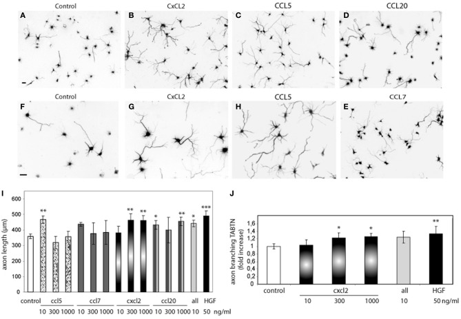
Chemokine (C-X-C motif) ligand 2, also known as CXCL2, is a small signaling protein that belongs to the chemokine family. Chemokines are a group of cytokines, or cell signaling molecules, that play crucial roles in immune responses and inflammation. They mediate their effects by interacting with specific receptors on the surface of target cells, guiding the migration of various immune cells to sites of infection, injury, or inflammation.
CXCL2 is primarily produced by activated monocytes, macrophages, and neutrophils, as well as endothelial cells, fibroblasts, and certain types of tumor cells. Its primary function is to attract and activate neutrophils, which are key effector cells in the early stages of inflammation and host defense against invading pathogens. CXCL2 exerts its effects by binding to its specific receptor, CXCR2, which is expressed on the surface of neutrophils and other immune cells.
In addition to its role in inflammation and immunity, CXCL2 has been implicated in various pathological conditions, including cancer, atherosclerosis, and autoimmune diseases. Its expression can be regulated by several factors, such as pro-inflammatory cytokines, bacterial products, and growth factors. Understanding the role of CXCL2 in health and disease may provide insights into the development of novel therapeutic strategies for treating inflammation-associated disorders.
Chemokine (C-X-C motif) ligand 13 (CXCL13), also known as B cell-attracting chemokine 1 (BCA-1) or B lymphocyte chemoattractant (BLC), is a small signaling protein belonging to the CXC chemokine family. Chemokines are a group of chemotactic cytokines that play crucial roles in immunological and inflammatory processes, mainly by recruiting and activating various leukocytes.
CXCL13 is primarily produced by stromal cells, including follicular dendritic cells (FDCs) within secondary lymphoid organs such as lymph nodes, spleen, and Peyer's patches. This chemokine specifically binds to the C-X-C chemokine receptor type 5 (CXCR5), which is expressed on various immune cells, most notably B cells, follicular helper T cells (Tfh), and some dendritic cell subsets.
The primary function of CXCL13 is to orchestrate the migration and positioning of immune cells, particularly B cells, within secondary lymphoid organs during an immune response. By attracting CXCR5-expressing B cells and Tfh cells, CXCL13 plays a critical role in the formation and maintenance of germinal centers (GCs), which are specialized microanatomical structures where affinity maturation and class switch recombination of B cells occur.
Abnormal levels or functions of CXCL13 have been implicated in several pathological conditions, including autoimmune diseases such as rheumatoid arthritis and systemic lupus erythematosus (SLE), certain types of cancer, and neurological disorders like multiple sclerosis (MS) and Alzheimer's disease.
C-X-C chemokine receptor type 4 (CXCR4) is a type of protein found on the surface of some cells, including white blood cells, and is a type of G protein-coupled receptor (GPCR). CXCR4 binds specifically to the chemokine ligand CXCL12 (also known as stromal cell-derived factor 1, or SDF-1), which plays a crucial role in the trafficking and homing of immune cells, particularly hematopoietic stem cells and lymphocytes. The binding of CXCL12 to CXCR4 triggers various intracellular signaling pathways that regulate cell migration, proliferation, survival, and differentiation.
In addition to its role in the immune system, CXCR4 has been implicated in several physiological and pathological processes, such as embryonic development, neurogenesis, angiogenesis, cancer metastasis, and HIV infection. In cancer, the overexpression of CXCR4 or increased levels of its ligand CXCL12 have been associated with poor prognosis, tumor growth, and metastasis in various types of malignancies, including breast, lung, prostate, colon, and ovarian cancers. In HIV infection, the CXCR4 coreceptor, together with CD4, facilitates viral entry into host cells, particularly during the later stages of the disease when the virus shifts its preference from CCR5 to CXCR4 as a coreceptor.
In summary, CXCR4 is a cell-surface receptor that binds specifically to the chemokine ligand CXCL12 and plays essential roles in immune cell trafficking, hematopoiesis, cancer metastasis, and HIV infection.
Chemokine (C-X-C motif) ligand 11 (CXCL11) is a small cytokine protein that belongs to the chemokine family, which are chemotactic cytokines involved in immune cell trafficking and inflammation. CXCL11 specifically binds to the CXCR3 receptor found on the surface of certain immune cells, including T lymphocytes and natural killer (NK) cells, and plays a role in their recruitment to sites of infection or injury.
CXCL11 is produced by various cell types, including monocytes, endothelial cells, and fibroblasts, in response to pro-inflammatory signals such as interferon-gamma (IFN-γ). It has been shown to have potent chemoattractant properties for Th1 lymphocytes and NK cells, contributing to the development of cell-mediated immune responses. Additionally, CXCL11 has been implicated in several physiological and pathological processes, including angiogenesis, tumorigenesis, and autoimmune diseases.
Chemotaxis is a term used in biology and medicine to describe the movement of an organism or cell towards or away from a chemical stimulus. This process plays a crucial role in various biological phenomena, including immune responses, wound healing, and the development and progression of diseases such as cancer.
In chemotaxis, cells can detect and respond to changes in the concentration of specific chemicals, known as chemoattractants or chemorepellents, in their environment. These chemicals bind to receptors on the cell surface, triggering a series of intracellular signaling events that ultimately lead to changes in the cytoskeleton and directed movement of the cell towards or away from the chemical gradient.
For example, during an immune response, white blood cells called neutrophils use chemotaxis to migrate towards sites of infection or inflammation, where they can attack and destroy invading pathogens. Similarly, cancer cells can use chemotaxis to migrate towards blood vessels and metastasize to other parts of the body.
Understanding chemotaxis is important for developing new therapies and treatments for a variety of diseases, including cancer, infectious diseases, and inflammatory disorders.
Chemokine CXCL6 is a type of small signaling protein that belongs to the CXC chemokine family. Its primary function is to attract and guide the movement (chemotaxis) of specific types of immune cells, such as neutrophils, to sites of inflammation or infection in the body.
CXCL6 is also known as granulocyte chemotactic protein 2 (GCP-2) and is produced by various cell types, including monocytes, macrophages, endothelial cells, and fibroblasts. It binds to and activates the CXCR1 and CXCR2 receptors found on the surface of neutrophils, which triggers a series of intracellular signaling events that lead to the migration of these cells towards the source of the chemokine.
In addition to its role in inflammation and immune response, CXCL6 has been implicated in several disease processes, including cancer, atherosclerosis, and rheumatoid arthritis. Elevated levels of CXCL6 have been found in the tumor microenvironment, where it may promote tumor growth and metastasis by recruiting immune cells that support tumor progression.
"Cells, cultured" is a medical term that refers to cells that have been removed from an organism and grown in controlled laboratory conditions outside of the body. This process is called cell culture and it allows scientists to study cells in a more controlled and accessible environment than they would have inside the body. Cultured cells can be derived from a variety of sources, including tissues, organs, or fluids from humans, animals, or cell lines that have been previously established in the laboratory.
Cell culture involves several steps, including isolation of the cells from the tissue, purification and characterization of the cells, and maintenance of the cells in appropriate growth conditions. The cells are typically grown in specialized media that contain nutrients, growth factors, and other components necessary for their survival and proliferation. Cultured cells can be used for a variety of purposes, including basic research, drug development and testing, and production of biological products such as vaccines and gene therapies.
It is important to note that cultured cells may behave differently than they do in the body, and results obtained from cell culture studies may not always translate directly to human physiology or disease. Therefore, it is essential to validate findings from cell culture experiments using additional models and ultimately in clinical trials involving human subjects.
Dendritic cells (DCs) are a type of immune cell that play a critical role in the body's defense against infection and cancer. They are named for their dendrite-like projections, which they use to interact with and sample their environment. DCs are responsible for processing antigens (foreign substances that trigger an immune response) and presenting them to T cells, a type of white blood cell that plays a central role in the immune system's response to infection and cancer.
DCs can be found throughout the body, including in the skin, mucous membranes, and lymphoid organs. They are able to recognize and respond to a wide variety of antigens, including those from bacteria, viruses, fungi, and parasites. Once they have processed an antigen, DCs migrate to the lymph nodes, where they present the antigen to T cells. This interaction activates the T cells, which then go on to mount a targeted immune response against the invading pathogen or cancerous cells.
DCs are a diverse group of cells that can be divided into several subsets based on their surface markers and function. Some DCs, such as Langerhans cells and dermal DCs, are found in the skin and mucous membranes, where they serve as sentinels for invading pathogens. Other DCs, such as plasmacytoid DCs and conventional DCs, are found in the lymphoid organs, where they play a role in activating T cells and initiating an immune response.
Overall, dendritic cells are essential for the proper functioning of the immune system, and dysregulation of these cells has been implicated in a variety of diseases, including autoimmune disorders and cancer.
Chemokine (C-X-C motif) ligand 5 (CXCL5), also known as epithelial neutrophil-activating peptide 78 (ENA-78) or liver-activated peptide (LAP), is a small signaling protein belonging to the CXC chemokine family. Chemokines are a group of cytokines, or cell signaling molecules, that play important roles in immune responses and inflammation by recruiting various immune cells to sites of infection or injury through specific receptor-mediated interactions.
CXCL5 is primarily produced by epithelial cells, macrophages, and neutrophils in response to bacterial infections, tissue damage, or proinflammatory cytokines. This chemokine exerts its functions by binding to its receptor CXCR2, which is expressed on the surface of various immune cells, including neutrophils, monocytes, and lymphocytes. The primary role of CXCL5 is to attract neutrophils to the site of inflammation or infection, where they can help eliminate pathogens and promote tissue repair.
Apart from its involvement in immune responses and inflammation, CXCL5 has been implicated in several physiological and pathological processes, such as embryonic development, wound healing, cancer progression, and metastasis. Dysregulation of CXCL5 signaling has been associated with various diseases, including chronic inflammatory disorders, autoimmune diseases, and cancer.
Cytokines are a broad and diverse category of small signaling proteins that are secreted by various cells, including immune cells, in response to different stimuli. They play crucial roles in regulating the immune response, inflammation, hematopoiesis, and cellular communication.
Cytokines mediate their effects by binding to specific receptors on the surface of target cells, which triggers intracellular signaling pathways that ultimately result in changes in gene expression, cell behavior, and function. Some key functions of cytokines include:
1. Regulating the activation, differentiation, and proliferation of immune cells such as T cells, B cells, natural killer (NK) cells, and macrophages.
2. Coordinating the inflammatory response by recruiting immune cells to sites of infection or tissue damage and modulating their effector functions.
3. Regulating hematopoiesis, the process of blood cell formation in the bone marrow, by controlling the proliferation, differentiation, and survival of hematopoietic stem and progenitor cells.
4. Modulating the development and function of the nervous system, including neuroinflammation, neuroprotection, and neuroregeneration.
Cytokines can be classified into several categories based on their structure, function, or cellular origin. Some common types of cytokines include interleukins (ILs), interferons (IFNs), tumor necrosis factors (TNFs), chemokines, colony-stimulating factors (CSFs), and transforming growth factors (TGFs). Dysregulation of cytokine production and signaling has been implicated in various pathological conditions, such as autoimmune diseases, chronic inflammation, cancer, and neurodegenerative disorders.
A "knockout" mouse is a genetically engineered mouse in which one or more genes have been deleted or "knocked out" using molecular biology techniques. This allows researchers to study the function of specific genes and their role in various biological processes, as well as potential associations with human diseases. The mice are generated by introducing targeted DNA modifications into embryonic stem cells, which are then used to create a live animal. Knockout mice have been widely used in biomedical research to investigate gene function, disease mechanisms, and potential therapeutic targets.
CXCR3 is a type of chemokine receptor that is primarily expressed on the surface of certain immune cells, including T lymphocytes (a type of white blood cell involved in immune response). It belongs to the Class A orphan G protein-coupled receptors family.
CXCR3 has three known subtypes, CXCR3-A, CXCR3-B, and CXCR3-C, each with different roles in regulating immune cell functions. These receptors bind to specific chemokines, which are small signaling proteins that help direct the movement of immune cells towards sites of inflammation or infection.
The chemokines that bind to CXCR3 include CXCL9, CXCL10, and CXCL11, which are produced by various cell types in response to inflammation or injury. Once bound to these chemokines, CXCR3 activates intracellular signaling pathways that trigger a range of responses, such as cell migration, activation, and proliferation.
In the context of disease, CXCR3 has been implicated in various pathological conditions, including cancer, autoimmune diseases, and viral infections, due to its role in regulating immune cell trafficking and activation.
BALB/c is an inbred strain of laboratory mouse that is widely used in biomedical research. The strain was developed at the Institute of Cancer Research in London by Henry Baldwin and his colleagues in the 1920s, and it has since become one of the most commonly used inbred strains in the world.
BALB/c mice are characterized by their black coat color, which is determined by a recessive allele at the tyrosinase locus. They are also known for their docile and friendly temperament, making them easy to handle and work with in the laboratory.
One of the key features of BALB/c mice that makes them useful for research is their susceptibility to certain types of tumors and immune responses. For example, they are highly susceptible to developing mammary tumors, which can be induced by chemical carcinogens or viral infection. They also have a strong Th2-biased immune response, which makes them useful models for studying allergic diseases and asthma.
BALB/c mice are also commonly used in studies of genetics, neuroscience, behavior, and infectious diseases. Because they are an inbred strain, they have a uniform genetic background, which makes it easier to control for genetic factors in experiments. Additionally, because they have been bred in the laboratory for many generations, they are highly standardized and reproducible, making them ideal subjects for scientific research.
Monocytes are a type of white blood cell that are part of the immune system. They are large cells with a round or oval shape and a nucleus that is typically indented or horseshoe-shaped. Monocytes are produced in the bone marrow and then circulate in the bloodstream, where they can differentiate into other types of immune cells such as macrophages and dendritic cells.
Monocytes play an important role in the body's defense against infection and tissue damage. They are able to engulf and digest foreign particles, microorganisms, and dead or damaged cells, which helps to clear them from the body. Monocytes also produce cytokines, which are signaling molecules that help to coordinate the immune response.
Elevated levels of monocytes in the bloodstream can be a sign of an ongoing infection, inflammation, or other medical conditions such as cancer or autoimmune disorders.
Macrophages are a type of white blood cell that are an essential part of the immune system. They are large, specialized cells that engulf and destroy foreign substances, such as bacteria, viruses, parasites, and fungi, as well as damaged or dead cells. Macrophages are found throughout the body, including in the bloodstream, lymph nodes, spleen, liver, lungs, and connective tissues. They play a critical role in inflammation, immune response, and tissue repair and remodeling.
Macrophages originate from monocytes, which are a type of white blood cell produced in the bone marrow. When monocytes enter the tissues, they differentiate into macrophages, which have a larger size and more specialized functions than monocytes. Macrophages can change their shape and move through tissues to reach sites of infection or injury. They also produce cytokines, chemokines, and other signaling molecules that help coordinate the immune response and recruit other immune cells to the site of infection or injury.
Macrophages have a variety of surface receptors that allow them to recognize and respond to different types of foreign substances and signals from other cells. They can engulf and digest foreign particles, bacteria, and viruses through a process called phagocytosis. Macrophages also play a role in presenting antigens to T cells, which are another type of immune cell that helps coordinate the immune response.
Overall, macrophages are crucial for maintaining tissue homeostasis, defending against infection, and promoting wound healing and tissue repair. Dysregulation of macrophage function has been implicated in a variety of diseases, including cancer, autoimmune disorders, and chronic inflammatory conditions.
'Gene expression regulation' refers to the processes that control whether, when, and where a particular gene is expressed, meaning the production of a specific protein or functional RNA encoded by that gene. This complex mechanism can be influenced by various factors such as transcription factors, chromatin remodeling, DNA methylation, non-coding RNAs, and post-transcriptional modifications, among others. Proper regulation of gene expression is crucial for normal cellular function, development, and maintaining homeostasis in living organisms. Dysregulation of gene expression can lead to various diseases, including cancer and genetic disorders.
Messenger RNA (mRNA) is a type of RNA (ribonucleic acid) that carries genetic information copied from DNA in the form of a series of three-base code "words," each of which specifies a particular amino acid. This information is used by the cell's machinery to construct proteins, a process known as translation. After being transcribed from DNA, mRNA travels out of the nucleus to the ribosomes in the cytoplasm where protein synthesis occurs. Once the protein has been synthesized, the mRNA may be degraded and recycled. Post-transcriptional modifications can also occur to mRNA, such as alternative splicing and addition of a 5' cap and a poly(A) tail, which can affect its stability, localization, and translation efficiency.
T-lymphocytes, also known as T-cells, are a type of white blood cell that plays a key role in the adaptive immune system's response to infection. They are produced in the bone marrow and mature in the thymus gland. There are several different types of T-cells, including CD4+ helper T-cells, CD8+ cytotoxic T-cells, and regulatory T-cells (Tregs).
CD4+ helper T-cells assist in activating other immune cells, such as B-lymphocytes and macrophages. They also produce cytokines, which are signaling molecules that help coordinate the immune response. CD8+ cytotoxic T-cells directly kill infected cells by releasing toxic substances. Regulatory T-cells help maintain immune tolerance and prevent autoimmune diseases by suppressing the activity of other immune cells.
T-lymphocytes are important in the immune response to viral infections, cancer, and other diseases. Dysfunction or depletion of T-cells can lead to immunodeficiency and increased susceptibility to infections. On the other hand, an overactive T-cell response can contribute to autoimmune diseases and chronic inflammation.
Inflammation is a complex biological response of tissues to harmful stimuli, such as pathogens, damaged cells, or irritants. It is characterized by the following signs: rubor (redness), tumor (swelling), calor (heat), dolor (pain), and functio laesa (loss of function). The process involves the activation of the immune system, recruitment of white blood cells, and release of inflammatory mediators, which contribute to the elimination of the injurious stimuli and initiation of the healing process. However, uncontrolled or chronic inflammation can also lead to tissue damage and diseases.
Reverse Transcriptase Polymerase Chain Reaction (RT-PCR) is a laboratory technique used in molecular biology to amplify and detect specific DNA sequences. This technique is particularly useful for the detection and quantification of RNA viruses, as well as for the analysis of gene expression.
The process involves two main steps: reverse transcription and polymerase chain reaction (PCR). In the first step, reverse transcriptase enzyme is used to convert RNA into complementary DNA (cDNA) by reading the template provided by the RNA molecule. This cDNA then serves as a template for the PCR amplification step.
In the second step, the PCR reaction uses two primers that flank the target DNA sequence and a thermostable polymerase enzyme to repeatedly copy the targeted cDNA sequence. The reaction mixture is heated and cooled in cycles, allowing the primers to anneal to the template, and the polymerase to extend the new strand. This results in exponential amplification of the target DNA sequence, making it possible to detect even small amounts of RNA or cDNA.
RT-PCR is a sensitive and specific technique that has many applications in medical research and diagnostics, including the detection of viruses such as HIV, hepatitis C virus, and SARS-CoV-2 (the virus that causes COVID-19). It can also be used to study gene expression, identify genetic mutations, and diagnose genetic disorders.
An Enzyme-Linked Immunosorbent Assay (ELISA) is a type of analytical biochemistry assay used to detect and quantify the presence of a substance, typically a protein or peptide, in a liquid sample. It takes its name from the enzyme-linked antibodies used in the assay.
In an ELISA, the sample is added to a well containing a surface that has been treated to capture the target substance. If the target substance is present in the sample, it will bind to the surface. Next, an enzyme-linked antibody specific to the target substance is added. This antibody will bind to the captured target substance if it is present. After washing away any unbound material, a substrate for the enzyme is added. If the enzyme is present due to its linkage to the antibody, it will catalyze a reaction that produces a detectable signal, such as a color change or fluorescence. The intensity of this signal is proportional to the amount of target substance present in the sample, allowing for quantification.
ELISAs are widely used in research and clinical settings to detect and measure various substances, including hormones, viruses, and bacteria. They offer high sensitivity, specificity, and reproducibility, making them a reliable choice for many applications.
Flow cytometry is a medical and research technique used to measure physical and chemical characteristics of cells or particles, one cell at a time, as they flow in a fluid stream through a beam of light. The properties measured include:
* Cell size (light scatter)
* Cell internal complexity (granularity, also light scatter)
* Presence or absence of specific proteins or other molecules on the cell surface or inside the cell (using fluorescent antibodies or other fluorescent probes)
The technique is widely used in cell counting, cell sorting, protein engineering, biomarker discovery and monitoring disease progression, particularly in hematology, immunology, and cancer research.
Interleukin-8 (IL-8) receptors are a type of G protein-coupled receptor that bind to and are activated by the cytokine IL-8. There are two main types of IL-8 receptors, known as CXCR1 and CXCR2.
IL-8B, also known as CXCR2, is a gene that encodes for the Interleukin-8 receptor B. This receptor is found on the surface of various cells, including neutrophils, monocytes, and endothelial cells. It plays a crucial role in the immune response, particularly in the recruitment and activation of neutrophils to sites of infection or inflammation.
IL-8B has a high affinity for IL-8 and other related chemokines, such as CXCL1, CXCL5, and CXCL7. Upon binding to its ligand, IL-8B activates various signaling pathways that lead to the mobilization and migration of neutrophils towards the site of inflammation. This process is critical for the elimination of invading pathogens and the resolution of inflammation.
However, excessive or prolonged activation of IL-8B has been implicated in various pathological conditions, including chronic inflammation, cancer, and autoimmune diseases. Therefore, targeting IL-8B with therapeutic agents has emerged as a promising strategy for the treatment of these conditions.
Signal transduction is the process by which a cell converts an extracellular signal, such as a hormone or neurotransmitter, into an intracellular response. This involves a series of molecular events that transmit the signal from the cell surface to the interior of the cell, ultimately resulting in changes in gene expression, protein activity, or metabolism.
The process typically begins with the binding of the extracellular signal to a receptor located on the cell membrane. This binding event activates the receptor, which then triggers a cascade of intracellular signaling molecules, such as second messengers, protein kinases, and ion channels. These molecules amplify and propagate the signal, ultimately leading to the activation or inhibition of specific cellular responses.
Signal transduction pathways are highly regulated and can be modulated by various factors, including other signaling molecules, post-translational modifications, and feedback mechanisms. Dysregulation of these pathways has been implicated in a variety of diseases, including cancer, diabetes, and neurological disorders.
Atopic dermatitis is a chronic, inflammatory skin condition that is commonly known as eczema. It is characterized by dry, itchy, and scaly patches on the skin that can become red, swollen, and cracked over time. The condition often affects the skin on the face, hands, feet, and behind the knees, and it can be triggered or worsened by exposure to certain allergens, irritants, stress, or changes in temperature and humidity. Atopic dermatitis is more common in people with a family history of allergies, such as asthma or hay fever, and it often begins in infancy or early childhood. The exact cause of atopic dermatitis is not fully understood, but it is thought to involve a combination of genetic and environmental factors that affect the immune system and the skin's ability to maintain a healthy barrier function.
A cell line is a culture of cells that are grown in a laboratory for use in research. These cells are usually taken from a single cell or group of cells, and they are able to divide and grow continuously in the lab. Cell lines can come from many different sources, including animals, plants, and humans. They are often used in scientific research to study cellular processes, disease mechanisms, and to test new drugs or treatments. Some common types of human cell lines include HeLa cells (which come from a cancer patient named Henrietta Lacks), HEK293 cells (which come from embryonic kidney cells), and HUVEC cells (which come from umbilical vein endothelial cells). It is important to note that cell lines are not the same as primary cells, which are cells that are taken directly from a living organism and have not been grown in the lab.
Up-regulation is a term used in molecular biology and medicine to describe an increase in the expression or activity of a gene, protein, or receptor in response to a stimulus. This can occur through various mechanisms such as increased transcription, translation, or reduced degradation of the molecule. Up-regulation can have important functional consequences, for example, enhancing the sensitivity or response of a cell to a hormone, neurotransmitter, or drug. It is a normal physiological process that can also be induced by disease or pharmacological interventions.
Monocyte chemoattractant proteins (MCPs) are a group of chemokines, which are small signaling proteins that attract immune cells to sites of infection or inflammation. Specifically, MCPs are responsible for recruiting monocytes and other immune cells to areas of tissue damage or infection.
There are several subtypes of MCPs, including MCP-1 (CCL2), MCP-2 (CCL8), MCP-3 (CCL7), and MCP-4 (CCL13). These proteins bind to specific receptors on the surface of monocytes and other immune cells, triggering a series of intracellular signaling events that result in cell migration towards the site of injury or infection.
MCPs play an important role in the pathogenesis of various inflammatory diseases, such as atherosclerosis, rheumatoid arthritis, and cancer. For example, elevated levels of MCP-1 have been associated with increased monocyte recruitment to the arterial wall, leading to the formation of plaques that can cause heart attacks and strokes. Similarly, high levels of MCPs have been found in the synovial fluid of patients with rheumatoid arthritis, contributing to joint inflammation and damage.
Overall, Monocyte chemoattractant proteins are crucial components of the immune system's response to injury and infection, but their dysregulation can contribute to the development of various diseases.
Animal disease models are specialized animals, typically rodents such as mice or rats, that have been genetically engineered or exposed to certain conditions to develop symptoms and physiological changes similar to those seen in human diseases. These models are used in medical research to study the pathophysiology of diseases, identify potential therapeutic targets, test drug efficacy and safety, and understand disease mechanisms.
The genetic modifications can include knockout or knock-in mutations, transgenic expression of specific genes, or RNA interference techniques. The animals may also be exposed to environmental factors such as chemicals, radiation, or infectious agents to induce the disease state.
Examples of animal disease models include:
1. Mouse models of cancer: Genetically engineered mice that develop various types of tumors, allowing researchers to study cancer initiation, progression, and metastasis.
2. Alzheimer's disease models: Transgenic mice expressing mutant human genes associated with Alzheimer's disease, which exhibit amyloid plaque formation and cognitive decline.
3. Diabetes models: Obese and diabetic mouse strains like the NOD (non-obese diabetic) or db/db mice, used to study the development of type 1 and type 2 diabetes, respectively.
4. Cardiovascular disease models: Atherosclerosis-prone mice, such as ApoE-deficient or LDLR-deficient mice, that develop plaque buildup in their arteries when fed a high-fat diet.
5. Inflammatory bowel disease models: Mice with genetic mutations affecting intestinal barrier function and immune response, such as IL-10 knockout or SAMP1/YitFc mice, which develop colitis.
Animal disease models are essential tools in preclinical research, but it is important to recognize their limitations. Differences between species can affect the translatability of results from animal studies to human patients. Therefore, researchers must carefully consider the choice of model and interpret findings cautiously when applying them to human diseases.
In medical terms, the skin is the largest organ of the human body. It consists of two main layers: the epidermis (outer layer) and dermis (inner layer), as well as accessory structures like hair follicles, sweat glands, and oil glands. The skin plays a crucial role in protecting us from external factors such as bacteria, viruses, and environmental hazards, while also regulating body temperature and enabling the sense of touch.
Transgenic mice are genetically modified rodents that have incorporated foreign DNA (exogenous DNA) into their own genome. This is typically done through the use of recombinant DNA technology, where a specific gene or genetic sequence of interest is isolated and then introduced into the mouse embryo. The resulting transgenic mice can then express the protein encoded by the foreign gene, allowing researchers to study its function in a living organism.
The process of creating transgenic mice usually involves microinjecting the exogenous DNA into the pronucleus of a fertilized egg, which is then implanted into a surrogate mother. The offspring that result from this procedure are screened for the presence of the foreign DNA, and those that carry the desired genetic modification are used to establish a transgenic mouse line.
Transgenic mice have been widely used in biomedical research to model human diseases, study gene function, and test new therapies. They provide a valuable tool for understanding complex biological processes and developing new treatments for a variety of medical conditions.
Interleukin-8 (IL-8) is a type of cytokine, which is a small signaling protein involved in immune response and inflammation. IL-8 is also known as neutrophil chemotactic factor or NCF because it attracts neutrophils, a type of white blood cell, to the site of infection or injury.
IL-8 is produced by various cells including macrophages, epithelial cells, and endothelial cells in response to bacterial or inflammatory stimuli. It acts by binding to specific receptors called CXCR1 and CXCR2 on the surface of neutrophils, which triggers a series of intracellular signaling events leading to neutrophil activation, migration, and degranulation.
IL-8 plays an important role in the recruitment of neutrophils to the site of infection or tissue damage, where they can phagocytose and destroy invading microorganisms. However, excessive or prolonged production of IL-8 has been implicated in various inflammatory diseases such as chronic obstructive pulmonary disease (COPD), rheumatoid arthritis, and cancer.
A ligand, in the context of biochemistry and medicine, is a molecule that binds to a specific site on a protein or a larger biomolecule, such as an enzyme or a receptor. This binding interaction can modify the function or activity of the target protein, either activating it or inhibiting it. Ligands can be small molecules, like hormones or neurotransmitters, or larger structures, like antibodies. The study of ligand-protein interactions is crucial for understanding cellular processes and developing drugs, as many therapeutic compounds function by binding to specific targets within the body.
CCR6 (C-C Motif Chemokine Receptor 6) is a type of protein found on the surface of certain cells, including immune cells. It is a type of chemokine receptor, which are proteins that help cells migrate to specific locations in the body in response to chemical signals called chemokines.
CCR6 specifically binds to a chemokine known as CCL20 (C-C Motif Chemokine Ligand 20). When CCL20 binds to CCR6, it triggers a series of intracellular signaling events that can lead to the activation and migration of immune cells, particularly T cells and dendritic cells.
CCR6 has been implicated in various physiological and pathological processes, including inflammation, immune responses, and cancer. For example, CCR6 has been shown to play a role in the recruitment of Th17 cells, a type of T cell that is involved in the development of autoimmune diseases such as rheumatoid arthritis and multiple sclerosis. Additionally, CCR6 has been found to be overexpressed in certain types of cancer, where it may contribute to tumor growth and metastasis.
CD4-positive T-lymphocytes, also known as CD4+ T cells or helper T cells, are a type of white blood cell that plays a crucial role in the immune response. They express the CD4 receptor on their surface and help coordinate the immune system's response to infectious agents such as viruses and bacteria.
CD4+ T cells recognize and bind to specific antigens presented by antigen-presenting cells, such as dendritic cells or macrophages. Once activated, they can differentiate into various subsets of effector cells, including Th1, Th2, Th17, and Treg cells, each with distinct functions in the immune response.
CD4+ T cells are particularly important in the immune response to HIV (human immunodeficiency virus), which targets and destroys these cells, leading to a weakened immune system and increased susceptibility to opportunistic infections. The number of CD4+ T cells is often used as a marker of disease progression in HIV infection, with lower counts indicating more advanced disease.
Interleukin-8 (IL-8) receptors are a type of cell surface receptor that bind to and are activated by the cytokine IL-8. There are two main types of IL-8 receptors, known as CXCR1 and CXCR2. Both of these receptors belong to the G protein-coupled receptor (GPCR) family and play important roles in the immune response, particularly in the recruitment and activation of neutrophils, a type of white blood cell that helps to fight infection.
IL-8A, also known as CXCR1, is a specific subtype of IL-8 receptor. It is a 354-amino acid protein that is expressed on the surface of many different types of cells, including neutrophils, monocytes, and certain tumor cells. When IL-8 binds to CXCR1, it activates a variety of signaling pathways within the cell that lead to changes in gene expression, cell activation, and chemotaxis (directed movement) towards the source of IL-8.
CXCR1 plays an important role in the immune response to bacterial and fungal infections, as well as in the development and progression of certain inflammatory diseases and cancers. It is also a target for drug development, particularly in the areas of cancer therapy and inflammatory disease.
Lymph nodes are small, bean-shaped organs that are part of the immune system. They are found throughout the body, especially in the neck, armpits, groin, and abdomen. Lymph nodes filter lymph fluid, which carries waste and unwanted substances such as bacteria, viruses, and cancer cells. They contain white blood cells called lymphocytes that help fight infections and diseases by attacking and destroying the harmful substances found in the lymph fluid. When an infection or disease is present, lymph nodes may swell due to the increased number of immune cells and fluid accumulation as they work to fight off the invaders.
CXCR (C-X-C chemokine receptor) is a type of G protein-coupled receptor that binds to certain chemokines, which are small signaling proteins involved in immunity and inflammation. Specifically, CXCR receptors bind to C-X-C chemokines, a subfamily of chemokines characterized by the presence of three amino acids (X) between two conserved cysteine residues.
The CXCR family includes several members, such as CXCR1, CXCR2, CXCR3, CXCR4, and CXCR5, among others. These receptors play crucial roles in various physiological processes, including the recruitment of immune cells to sites of infection or injury, hematopoiesis, angiogenesis, and cancer metastasis.
One of the most well-studied members of this family is CXCR4, which binds to the chemokine CXCL12 (also known as stromal cell-derived factor 1 or SDF-1). The CXCR4/CXCL12 axis has been implicated in several diseases, including HIV infection, cancer, and inflammatory disorders.
In summary, CXCR receptors are a group of G protein-coupled receptors that bind to C-X-C chemokines, playing essential roles in immunity, inflammation, and other physiological processes.
A cell line that is derived from tumor cells and has been adapted to grow in culture. These cell lines are often used in research to study the characteristics of cancer cells, including their growth patterns, genetic changes, and responses to various treatments. They can be established from many different types of tumors, such as carcinomas, sarcomas, and leukemias. Once established, these cell lines can be grown and maintained indefinitely in the laboratory, allowing researchers to conduct experiments and studies that would not be feasible using primary tumor cells. It is important to note that tumor cell lines may not always accurately represent the behavior of the original tumor, as they can undergo genetic changes during their time in culture.
NF-κB (Nuclear Factor kappa-light-chain-enhancer of activated B cells) is a protein complex that plays a crucial role in regulating the immune response to infection and inflammation, as well as in cell survival, differentiation, and proliferation. It is composed of several subunits, including p50, p52, p65 (RelA), c-Rel, and RelB, which can form homodimers or heterodimers that bind to specific DNA sequences called κB sites in the promoter regions of target genes.
Under normal conditions, NF-κB is sequestered in the cytoplasm by inhibitory proteins known as IκBs (inhibitors of κB). However, upon stimulation by various signals such as cytokines, bacterial or viral products, and stress, IκBs are phosphorylated, ubiquitinated, and degraded, leading to the release and activation of NF-κB. Activated NF-κB then translocates to the nucleus, where it binds to κB sites and regulates the expression of target genes involved in inflammation, immunity, cell survival, and proliferation.
Dysregulation of NF-κB signaling has been implicated in various pathological conditions such as cancer, chronic inflammation, autoimmune diseases, and neurodegenerative disorders. Therefore, targeting NF-κB signaling has emerged as a potential therapeutic strategy for the treatment of these diseases.
Carbon tetrachloride is a colorless, heavy, and nonflammable liquid with a mild ether-like odor. Its chemical formula is CCl4. It was previously used as a solvent and refrigerant, but its use has been largely phased out due to its toxicity and ozone-depleting properties.
Inhalation, ingestion, or skin contact with carbon tetrachloride can cause harmful health effects. Short-term exposure can lead to symptoms such as dizziness, headache, nausea, and vomiting. Long-term exposure has been linked to liver and kidney damage, as well as an increased risk of cancer.
Carbon tetrachloride is also a potent greenhouse gas and contributes to climate change. Its production and use are regulated by international agreements aimed at protecting human health and the environment.
Immunohistochemistry (IHC) is a technique used in pathology and laboratory medicine to identify specific proteins or antigens in tissue sections. It combines the principles of immunology and histology to detect the presence and location of these target molecules within cells and tissues. This technique utilizes antibodies that are specific to the protein or antigen of interest, which are then tagged with a detection system such as a chromogen or fluorophore. The stained tissue sections can be examined under a microscope, allowing for the visualization and analysis of the distribution and expression patterns of the target molecule in the context of the tissue architecture. Immunohistochemistry is widely used in diagnostic pathology to help identify various diseases, including cancer, infectious diseases, and immune-mediated disorders.
Cytokine receptors are specialized protein molecules found on the surface of cells that selectively bind to specific cytokines. Cytokines are signaling molecules used for communication between cells, and they play crucial roles in regulating immune responses, inflammation, hematopoiesis, and cell survival.
Cytokine receptors have specific binding sites that recognize and interact with the corresponding cytokines. This interaction triggers a series of intracellular signaling events that ultimately lead to changes in gene expression and various cellular responses. Cytokine receptors can be found on many different types of cells, including immune cells, endothelial cells, and structural cells like fibroblasts.
Cytokine receptors are typically composed of multiple subunits, which may include both extracellular and intracellular domains. The extracellular domain is responsible for cytokine binding, while the intracellular domain is involved in signal transduction. Cytokine receptors can be classified into several families based on their structural features and signaling mechanisms, such as the hematopoietic cytokine receptor family, the interferon receptor family, the tumor necrosis factor receptor family, and the interleukin-1 receptor family.
Dysregulation of cytokine receptors and their signaling pathways has been implicated in various diseases, including autoimmune disorders, chronic inflammation, and cancer. Therefore, understanding the biology of cytokine receptors is essential for developing targeted therapies to treat these conditions.
Regulatory T-lymphocytes (Tregs), also known as suppressor T cells, are a subpopulation of T-cells that play a critical role in maintaining immune tolerance and preventing autoimmune diseases. They function to suppress the activation and proliferation of other immune cells, thereby regulating the immune response and preventing it from attacking the body's own tissues.
Tregs constitutively express the surface markers CD4 and CD25, as well as the transcription factor Foxp3, which is essential for their development and function. They can be further divided into subsets based on their expression of other markers, such as CD127 and CD45RA.
Tregs are critical for maintaining self-tolerance by suppressing the activation of self-reactive T cells that have escaped negative selection in the thymus. They also play a role in regulating immune responses to foreign antigens, such as those encountered during infection or cancer, and can contribute to the immunosuppressive microenvironment found in tumors.
Dysregulation of Tregs has been implicated in various autoimmune diseases, including type 1 diabetes, rheumatoid arthritis, and multiple sclerosis, as well as in cancer and infectious diseases. Therefore, understanding the mechanisms that regulate Treg function is an important area of research with potential therapeutic implications.
Tumor Necrosis Factor-alpha (TNF-α) is a cytokine, a type of small signaling protein involved in immune response and inflammation. It is primarily produced by activated macrophages, although other cell types such as T-cells, natural killer cells, and mast cells can also produce it.
TNF-α plays a crucial role in the body's defense against infection and tissue injury by mediating inflammatory responses, activating immune cells, and inducing apoptosis (programmed cell death) in certain types of cells. It does this by binding to its receptors, TNFR1 and TNFR2, which are found on the surface of many cell types.
In addition to its role in the immune response, TNF-α has been implicated in the pathogenesis of several diseases, including autoimmune disorders such as rheumatoid arthritis, inflammatory bowel disease, and psoriasis, as well as cancer, where it can promote tumor growth and metastasis.
Therapeutic agents that target TNF-α, such as infliximab, adalimumab, and etanercept, have been developed to treat these conditions. However, these drugs can also increase the risk of infections and other side effects, so their use must be carefully monitored.
Chemokines are a family of small signaling proteins that play a crucial role in the immune system by recruiting immune cells to sites of infection or injury. They do this by binding to specific receptors on the surface of immune cells and guiding their movement towards the source of the chemokine.
CX3C is a subfamily of chemokines that contains only one member, called fractalkine (CX3CL1). Fractalkine is unique among chemokines because it exists in two forms: a soluble form and a membrane-bound form. The soluble form acts as a chemoattractant for immune cells, while the membrane-bound form functions as an adhesion molecule that helps to tether immune cells to the site of inflammation.
Fractalkine plays important roles in the immune response, including the recruitment and activation of immune cells such as natural killer (NK) cells, T cells, and monocytes/macrophages. It is also involved in the development and maintenance of the nervous system, where it helps to regulate the migration and differentiation of neural progenitor cells.
Abnormalities in fractalkine signaling have been implicated in a variety of diseases, including neurological disorders such as multiple sclerosis, Alzheimer's disease, and Parkinson's disease, as well as inflammatory conditions such as rheumatoid arthritis and atherosclerosis.
CXCR5 is a type of chemokine receptor that is primarily expressed on the surface of certain immune cells, including B cells and some T cells. It belongs to the family of G protein-coupled receptors (GPCRs) and plays a crucial role in the trafficking and homing of these immune cells to specific tissues in the body.
CXCR5 specifically binds to a chemokine ligand called CXCL13, which is produced by various cell types, including stromal cells in lymphoid organs. The binding of CXCL13 to CXCR5 triggers a signaling cascade that leads to the activation of several downstream signaling pathways, ultimately resulting in the migration and accumulation of immune cells in the vicinity of the CXCL13 source.
In the context of the immune system, CXCR5 is essential for the formation of germinal centers, which are specialized structures within lymphoid organs where B cells undergo activation, proliferation, and differentiation into antibody-secreting plasma cells. The interaction between CXCL13 and CXCR5 helps to recruit B cells and follicular T helper (Tfh) cells to the germinal center, where they can engage in productive interactions that drive humoral immune responses.
Abnormalities in CXCR5 signaling have been implicated in various pathological conditions, including autoimmune diseases, cancer, and infectious diseases. Therefore, understanding the molecular mechanisms underlying CXCR5 function is of great interest for the development of novel therapeutic strategies to target these disorders.
Protein binding, in the context of medical and biological sciences, refers to the interaction between a protein and another molecule (known as the ligand) that results in a stable complex. This process is often reversible and can be influenced by various factors such as pH, temperature, and concentration of the involved molecules.
In clinical chemistry, protein binding is particularly important when it comes to drugs, as many of them bind to proteins (especially albumin) in the bloodstream. The degree of protein binding can affect a drug's distribution, metabolism, and excretion, which in turn influence its therapeutic effectiveness and potential side effects.
Protein-bound drugs may be less available for interaction with their target tissues, as only the unbound or "free" fraction of the drug is active. Therefore, understanding protein binding can help optimize dosing regimens and minimize adverse reactions.
Chemotactic factors are substances that attract or repel cells, particularly immune cells, by stimulating directional movement in response to a chemical gradient. These factors play a crucial role in the body's immune response and inflammation process. They include:
1. Chemokines: A family of small signaling proteins that direct the migration of immune cells to sites of infection or tissue damage.
2. Cytokines: A broad category of signaling molecules that mediate and regulate immunity, inflammation, and hematopoiesis. Some cytokines can also act as chemotactic factors.
3. Complement components: Cleavage products of the complement system can attract immune cells to the site of infection or tissue injury.
4. Growth factors: Certain growth factors, like colony-stimulating factors (CSFs), can stimulate the migration and proliferation of specific cell types.
5. Lipid mediators: Products derived from arachidonic acid metabolism, such as leukotrienes and prostaglandins, can also act as chemotactic factors.
6. Formyl peptides: Bacterial-derived formylated peptides can attract and activate neutrophils during an infection.
7. Extracellular matrix (ECM) components: Fragments of ECM proteins, like collagen and fibronectin, can serve as chemotactic factors for immune cells.
These factors help orchestrate the immune response by guiding the movement of immune cells to specific locations in the body where they are needed.
CD8-positive T-lymphocytes, also known as CD8+ T cells or cytotoxic T cells, are a type of white blood cell that plays a crucial role in the adaptive immune system. They are named after the CD8 molecule found on their surface, which is a protein involved in cell signaling and recognition.
CD8+ T cells are primarily responsible for identifying and destroying virus-infected cells or cancerous cells. When activated, they release cytotoxic granules that contain enzymes capable of inducing apoptosis (programmed cell death) in the target cells. They also produce cytokines such as interferon-gamma, which can help coordinate the immune response and activate other immune cells.
CD8+ T cells are generated in the thymus gland and are a type of T cell, which is a lymphocyte that matures in the thymus and plays a central role in cell-mediated immunity. They recognize and respond to specific antigens presented on the surface of infected or cancerous cells in conjunction with major histocompatibility complex (MHC) class I molecules.
Overall, CD8+ T cells are an essential component of the immune system's defense against viral infections and cancer.
Endothelial cells are the type of cells that line the inner surface of blood vessels, lymphatic vessels, and heart chambers. They play a crucial role in maintaining vascular homeostasis by controlling vasomotor tone, coagulation, platelet activation, and inflammation. Endothelial cells also regulate the transport of molecules between the blood and surrounding tissues, and contribute to the maintenance of the structural integrity of the vasculature. They are flat, elongated cells with a unique morphology that allows them to form a continuous, nonthrombogenic lining inside the vessels. Endothelial cells can be isolated from various tissues and cultured in vitro for research purposes.
Lymphocyte activation is the process by which B-cells and T-cells (types of lymphocytes) become activated to perform effector functions in an immune response. This process involves the recognition of specific antigens presented on the surface of antigen-presenting cells, such as dendritic cells or macrophages.
The activation of B-cells leads to their differentiation into plasma cells that produce antibodies, while the activation of T-cells results in the production of cytotoxic T-cells (CD8+ T-cells) that can directly kill infected cells or helper T-cells (CD4+ T-cells) that assist other immune cells.
Lymphocyte activation involves a series of intracellular signaling events, including the binding of co-stimulatory molecules and the release of cytokines, which ultimately result in the expression of genes involved in cell proliferation, differentiation, and effector functions. The activation process is tightly regulated to prevent excessive or inappropriate immune responses that can lead to autoimmunity or chronic inflammation.
Monokines are cytokines that are produced and released by monocytes, which are a type of white blood cell. These proteins play an important role in the immune response, including inflammation, immunoregulation, and hematopoiesis (the formation of blood cells).
Monokines include several types of cytokines, such as tumor necrosis factor-alpha (TNF-α), interleukin-1 (IL-1), interleukin-6 (IL-6), and interleukin-12 (IL-12). These molecules help to regulate the activity of other immune cells, such as T cells and B cells, and can also have direct effects on infected or damaged tissues.
Monokines are involved in a variety of physiological and pathological processes, including host defense against infection, tissue repair and regeneration, and the development of chronic inflammatory diseases such as rheumatoid arthritis and atherosclerosis.
HIV receptors are specific molecules found on the surface of certain human cells that the Human Immunodeficiency Virus (HIV) uses to enter and infect those cells. The two primary HIV receptors are CD4 and CCR5 or CXCR4 co-receptors.
1. CD4 Receptor: This is a glycoprotein found on the surface of helper T cells, macrophages, and dendritic cells. HIV first binds to the CD4 receptor via its envelope protein gp120. However, this binding alone is not sufficient for virus entry. The interaction between gp120 and CD4 triggers conformational changes in the viral envelope that expose the binding site for a co-receptor.
2. CCR5 or CXCR4 Co-receptors: These are chemokine receptors also found on the surface of certain cells, including helper T cells and macrophages. After HIV binds to the CD4 receptor, it interacts with either the CCR5 or CXCR4 co-receptor, which facilitates the fusion of the viral and cell membranes and the release of the viral genetic material into the host cell.
The specificity of HIV for these receptors plays a crucial role in its pathogenesis, as it determines which cells are susceptible to infection. Additionally, variations in the genes encoding these receptors can influence an individual's susceptibility to HIV infection and the rate of disease progression.
Carbon tetrachloride poisoning refers to the harmful effects on the body caused by exposure to carbon tetrachloride, a volatile and toxic chemical compound. This substance has been widely used in various industrial applications, such as a solvent for fats, oils, and rubber, a fire extinguishing agent, and a refrigerant. However, due to its high toxicity, the use of carbon tetrachloride has been significantly reduced or phased out in many countries.
Ingestion, inhalation, or skin absorption of carbon tetrachloride can lead to poisoning, which may cause various symptoms depending on the severity and duration of exposure. Acute exposure to high concentrations of carbon tetrachloride can result in:
1. Central nervous system depression: Dizziness, headache, confusion, drowsiness, and, in severe cases, loss of consciousness or even death.
2. Respiratory irritation: Coughing, wheezing, shortness of breath, and pulmonary edema (fluid accumulation in the lungs).
3. Cardiovascular effects: Increased heart rate, low blood pressure, and irregular heart rhythms.
4. Gastrointestinal symptoms: Nausea, vomiting, abdominal pain, and diarrhea.
5. Liver damage: Hepatitis, jaundice, and liver failure in severe cases.
6. Kidney damage: Acute kidney injury or failure.
Chronic exposure to carbon tetrachloride can lead to long-term health effects, including:
1. Liver cirrhosis (scarring of the liver) and liver cancer.
2. Kidney damage and kidney disease.
3. Peripheral neuropathy (damage to the nerves in the limbs), causing numbness, tingling, or weakness.
4. Increased risk of miscarriage and birth defects in pregnant women exposed to carbon tetrachloride.
Treatment for carbon tetrachloride poisoning typically involves supportive care, such as oxygen therapy, fluid replacement, and monitoring of vital signs. In some cases, specific treatments like activated charcoal or gastric lavage may be used to remove the substance from the body. Prevention is crucial in minimizing exposure to this harmful chemical by following safety guidelines when handling it and using appropriate personal protective equipment (PPE).
The Duffy blood group system is a system of identifying blood types based on the presence or absence of certain antigens on the surface of red blood cells. The antigens in this system are proteins called Duffy antigens, which are receptors for the malarial parasite Plasmodium vivax.
There are two major Duffy antigens, Fya and Fyb, and individuals can be either positive or negative for each of these antigens. This means that there are four main Duffy blood types: Fy(a+b-), Fy(a-b+), Fy(a+b+), and Fy(a-b-).
The Duffy blood group system is important in blood transfusions to prevent a potentially dangerous immune response known as a transfusion reaction. If a person receives blood that contains antigens that their body recognizes as foreign, their immune system may attack the transfused red blood cells, leading to symptoms such as fever, chills, and in severe cases, kidney failure or even death.
Additionally, the Duffy blood group system has been found to be associated with susceptibility to certain diseases. For example, individuals who are negative for both Fya and Fyb antigens (Fy(a-b-)) are resistant to infection by Plasmodium vivax, one of the malarial parasites that causes malaria in humans. This is because the Duffy antigens serve as receptors for the parasite to enter and infect red blood cells.
Chemotactic factors are substances that attract and guide cells, particularly immune cells, to specific locations in the body. Eosinophils are a type of white blood cell that play a role in the immune response, particularly against parasites and in allergic reactions. Therefore, chemotactic factors for eosinophils are substances that attract eosinophils to specific sites in the body.
These factors can be produced by various cells, including mast cells, basophils, and T-lymphocytes, in response to an infection or inflammation. They work by binding to receptors on the surface of eosinophils and activating signaling pathways that cause the eosinophils to migrate towards the source of the chemotactic factor.
Examples of chemotactic factors for eosinophils include:
1. Eotaxins: These are a group of chemokines (a type of signaling protein) that specifically attract eosinophils. They are produced by various cells, including endothelial cells, epithelial cells, and immune cells.
2. Leukotrienes: These are lipid mediators produced by mast cells and basophils in response to an allergic reaction or infection. They can attract eosinophils to the site of inflammation.
3. Platelet-activating factor (PAF): This is a lipid mediator produced by various cells, including endothelial cells and immune cells. It can attract eosinophils and activate them, leading to degranulation and release of their contents.
4. Complement components: The complement system is a group of proteins that play a role in the immune response. Some complement components, such as C3a and C5a, can act as chemotactic factors for eosinophils.
Overall, chemotactic factors for eosinophils play an important role in the immune response by recruiting these cells to sites of infection or inflammation. However, excessive activation of eosinophils and production of chemotactic factors can contribute to the development of various diseases, such as asthma and allergies.
Neutrophil infiltration is a pathological process characterized by the accumulation of neutrophils, a type of white blood cell, in tissue. It is a common feature of inflammation and occurs in response to infection, injury, or other stimuli that trigger an immune response. Neutrophils are attracted to the site of tissue damage by chemical signals called chemokines, which are released by damaged cells and activated immune cells. Once they reach the site of inflammation, neutrophils help to clear away damaged tissue and microorganisms through a process called phagocytosis. However, excessive or prolonged neutrophil infiltration can also contribute to tissue damage and may be associated with various disease states, including cancer, autoimmune disorders, and ischemia-reperfusion injury.
Neutrophils are a type of white blood cell that are part of the immune system's response to infection. They are produced in the bone marrow and released into the bloodstream where they circulate and are able to move quickly to sites of infection or inflammation in the body. Neutrophils are capable of engulfing and destroying bacteria, viruses, and other foreign substances through a process called phagocytosis. They are also involved in the release of inflammatory mediators, which can contribute to tissue damage in some cases. Neutrophils are characterized by the presence of granules in their cytoplasm, which contain enzymes and other proteins that help them carry out their immune functions.
Heterocyclic compounds are organic compounds that contain at least one atom within the ring structure, other than carbon, such as nitrogen, oxygen, sulfur or phosphorus. These compounds make up a large class of naturally occurring and synthetic materials, including many drugs, pigments, vitamins, and antibiotics. The presence of the heteroatom in the ring can have significant effects on the physical and chemical properties of the compound, such as its reactivity, stability, and bonding characteristics. Examples of heterocyclic compounds include pyridine, pyrimidine, and furan.
A lung is a pair of spongy, elastic organs in the chest that work together to enable breathing. They are responsible for taking in oxygen and expelling carbon dioxide through the process of respiration. The left lung has two lobes, while the right lung has three lobes. The lungs are protected by the ribcage and are covered by a double-layered membrane called the pleura. The trachea divides into two bronchi, which further divide into smaller bronchioles, leading to millions of tiny air sacs called alveoli, where the exchange of gases occurs.
Leukocytes, also known as white blood cells (WBCs), are a crucial component of the human immune system. They are responsible for protecting the body against infections and foreign substances. Leukocytes are produced in the bone marrow and circulate throughout the body in the bloodstream and lymphatic system.
There are several types of leukocytes, including:
1. Neutrophils - These are the most abundant type of leukocyte and are primarily responsible for fighting bacterial infections. They contain enzymes that can destroy bacteria.
2. Lymphocytes - These are responsible for producing antibodies and destroying virus-infected cells, as well as cancer cells. There are two main types of lymphocytes: B-lymphocytes and T-lymphocytes.
3. Monocytes - These are the largest type of leukocyte and help to break down and remove dead or damaged tissues, as well as microorganisms.
4. Eosinophils - These play a role in fighting parasitic infections and are also involved in allergic reactions and inflammation.
5. Basophils - These release histamine and other chemicals that cause inflammation in response to allergens or irritants.
An abnormal increase or decrease in the number of leukocytes can indicate an underlying medical condition, such as an infection, inflammation, or a blood disorder.
Gene expression is the process by which the information encoded in a gene is used to synthesize a functional gene product, such as a protein or RNA molecule. This process involves several steps: transcription, RNA processing, and translation. During transcription, the genetic information in DNA is copied into a complementary RNA molecule, known as messenger RNA (mRNA). The mRNA then undergoes RNA processing, which includes adding a cap and tail to the mRNA and splicing out non-coding regions called introns. The resulting mature mRNA is then translated into a protein on ribosomes in the cytoplasm through the process of translation.
The regulation of gene expression is a complex and highly controlled process that allows cells to respond to changes in their environment, such as growth factors, hormones, and stress signals. This regulation can occur at various stages of gene expression, including transcriptional activation or repression, RNA processing, mRNA stability, and translation. Dysregulation of gene expression has been implicated in many diseases, including cancer, genetic disorders, and neurological conditions.
Inflammation mediators are substances that are released by the body in response to injury or infection, which contribute to the inflammatory response. These mediators include various chemical factors such as cytokines, chemokines, prostaglandins, leukotrienes, and histamine, among others. They play a crucial role in regulating the inflammatory process by attracting immune cells to the site of injury or infection, increasing blood flow to the area, and promoting the repair and healing of damaged tissues. However, an overactive or chronic inflammatory response can also contribute to the development of various diseases and conditions, such as autoimmune disorders, cardiovascular disease, and cancer.
Interferon-gamma (IFN-γ) is a soluble cytokine that is primarily produced by the activation of natural killer (NK) cells and T lymphocytes, especially CD4+ Th1 cells and CD8+ cytotoxic T cells. It plays a crucial role in the regulation of the immune response against viral and intracellular bacterial infections, as well as tumor cells. IFN-γ has several functions, including activating macrophages to enhance their microbicidal activity, increasing the presentation of major histocompatibility complex (MHC) class I and II molecules on antigen-presenting cells, stimulating the proliferation and differentiation of T cells and NK cells, and inducing the production of other cytokines and chemokines. Additionally, IFN-γ has direct antiproliferative effects on certain types of tumor cells and can enhance the cytotoxic activity of immune cells against infected or malignant cells.
Th2 cells, or T helper 2 cells, are a type of CD4+ T cell that plays a key role in the immune response to parasites and allergens. They produce cytokines such as IL-4, IL-5, IL-13 which promote the activation and proliferation of eosinophils, mast cells, and B cells, leading to the production of antibodies such as IgE. Th2 cells also play a role in the pathogenesis of allergic diseases such as asthma, atopic dermatitis, and allergic rhinitis.
It's important to note that an imbalance in Th1/Th2 response can lead to immune dysregulation and disease states. For example, an overactive Th2 response can lead to allergic reactions while an underactive Th2 response can lead to decreased ability to fight off parasitic infections.
It's also worth noting that there are other subsets of CD4+ T cells such as Th1, Th17, Treg and others, each with their own specific functions and cytokine production profiles.
Cell migration inhibition refers to the process or agents that restrict the movement of cells, particularly in the context of cancer metastasis. Cell migration is a critical biological process involved in various physiological and pathological conditions, including embryonic development, wound healing, and tumor cell dissemination. Inhibiting cell migration can help prevent the spread of cancer to distant organs, thereby improving treatment outcomes and patient survival rates.
Various factors and mechanisms contribute to cell migration inhibition, such as:
1. Modulation of signaling pathways: Cell migration is regulated by complex intracellular signaling networks that control cytoskeletal rearrangements, adhesion molecules, and other components required for cell motility. Inhibiting specific signaling proteins or pathways can suppress cell migration.
2. Extracellular matrix (ECM) modifications: The ECM provides structural support and biochemical cues that guide cell migration. Altering the composition or organization of the ECM can hinder cell movement.
3. Inhibition of adhesion molecules: Cell-cell and cell-matrix interactions are mediated by adhesion molecules, such as integrins and cadherins. Blocking these molecules can prevent cells from attaching to their surroundings and migrating.
4. Targeting cytoskeletal components: The cytoskeleton is responsible for the mechanical forces required for cell migration. Inhibiting cytoskeletal proteins, such as actin or tubulin, can impair cell motility.
5. Use of pharmacological agents: Several drugs and compounds have been identified to inhibit cell migration, either by targeting specific molecules or indirectly affecting the overall cellular environment. These agents include chemotherapeutic drugs, natural compounds, and small molecule inhibitors.
Understanding the mechanisms underlying cell migration inhibition can provide valuable insights into developing novel therapeutic strategies for cancer treatment and other diseases involving aberrant cell migration.
HIV-1 (Human Immunodeficiency Virus type 1) is a species of the retrovirus genus that causes acquired immunodeficiency syndrome (AIDS). It is primarily transmitted through sexual contact, exposure to infected blood or blood products, and from mother to child during pregnancy, childbirth, or breastfeeding. HIV-1 infects vital cells in the human immune system, such as CD4+ T cells, macrophages, and dendritic cells, leading to a decline in their numbers and weakening of the immune response over time. This results in the individual becoming susceptible to various opportunistic infections and cancers that ultimately cause death if left untreated. HIV-1 is the most prevalent form of HIV worldwide and has been identified as the causative agent of the global AIDS pandemic.
Molecular sequence data refers to the specific arrangement of molecules, most commonly nucleotides in DNA or RNA, or amino acids in proteins, that make up a biological macromolecule. This data is generated through laboratory techniques such as sequencing, and provides information about the exact order of the constituent molecules. This data is crucial in various fields of biology, including genetics, evolution, and molecular biology, allowing for comparisons between different organisms, identification of genetic variations, and studies of gene function and regulation.
Eosinophils are a type of white blood cell that play an important role in the body's immune response. They are produced in the bone marrow and released into the bloodstream, where they can travel to different tissues and organs throughout the body. Eosinophils are characterized by their granules, which contain various proteins and enzymes that are toxic to parasites and can contribute to inflammation.
Eosinophils are typically associated with allergic reactions, asthma, and other inflammatory conditions. They can also be involved in the body's response to certain infections, particularly those caused by parasites such as worms. In some cases, elevated levels of eosinophils in the blood or tissues (a condition called eosinophilia) can indicate an underlying medical condition, such as a parasitic infection, autoimmune disorder, or cancer.
Eosinophils are named for their staining properties - they readily take up eosin dye, which is why they appear pink or red under the microscope. They make up only about 1-6% of circulating white blood cells in healthy individuals, but their numbers can increase significantly in response to certain triggers.
Intercellular signaling peptides and proteins are molecules that mediate communication and interaction between different cells in living organisms. They play crucial roles in various biological processes, including cell growth, differentiation, migration, and apoptosis (programmed cell death). These signals can be released into the extracellular space, where they bind to specific receptors on the target cell's surface, triggering intracellular signaling cascades that ultimately lead to a response.
Peptides are short chains of amino acids, while proteins are larger molecules made up of one or more polypeptide chains. Both can function as intercellular signaling molecules by acting as ligands for cell surface receptors or by being cleaved from larger precursor proteins and released into the extracellular space. Examples of intercellular signaling peptides and proteins include growth factors, cytokines, chemokines, hormones, neurotransmitters, and their respective receptors.
These molecules contribute to maintaining homeostasis within an organism by coordinating cellular activities across tissues and organs. Dysregulation of intercellular signaling pathways has been implicated in various diseases, such as cancer, autoimmune disorders, and neurodegenerative conditions. Therefore, understanding the mechanisms underlying intercellular signaling is essential for developing targeted therapies to treat these disorders.
Lipopolysaccharides (LPS) are large molecules found in the outer membrane of Gram-negative bacteria. They consist of a hydrophilic polysaccharide called the O-antigen, a core oligosaccharide, and a lipid portion known as Lipid A. The Lipid A component is responsible for the endotoxic activity of LPS, which can trigger a powerful immune response in animals, including humans. This response can lead to symptoms such as fever, inflammation, and septic shock, especially when large amounts of LPS are introduced into the bloodstream.
Down-regulation is a process that occurs in response to various stimuli, where the number or sensitivity of cell surface receptors or the expression of specific genes is decreased. This process helps maintain homeostasis within cells and tissues by reducing the ability of cells to respond to certain signals or molecules.
In the context of cell surface receptors, down-regulation can occur through several mechanisms:
1. Receptor internalization: After binding to their ligands, receptors can be internalized into the cell through endocytosis. Once inside the cell, these receptors may be degraded or recycled back to the cell surface in smaller numbers.
2. Reduced receptor synthesis: Down-regulation can also occur at the transcriptional level, where the expression of genes encoding for specific receptors is decreased, leading to fewer receptors being produced.
3. Receptor desensitization: Prolonged exposure to a ligand can lead to a decrease in receptor sensitivity or affinity, making it more difficult for the cell to respond to the signal.
In the context of gene expression, down-regulation refers to the decreased transcription and/or stability of specific mRNAs, leading to reduced protein levels. This process can be induced by various factors, including microRNA (miRNA)-mediated regulation, histone modification, or DNA methylation.
Down-regulation is an essential mechanism in many physiological processes and can also contribute to the development of several diseases, such as cancer and neurodegenerative disorders.
An amino acid sequence is the specific order of amino acids in a protein or peptide molecule, formed by the linking of the amino group (-NH2) of one amino acid to the carboxyl group (-COOH) of another amino acid through a peptide bond. The sequence is determined by the genetic code and is unique to each type of protein or peptide. It plays a crucial role in determining the three-dimensional structure and function of proteins.
Epithelial cells are types of cells that cover the outer surfaces of the body, line the inner surfaces of organs and glands, and form the lining of blood vessels and body cavities. They provide a protective barrier against the external environment, regulate the movement of materials between the internal and external environments, and are involved in the sense of touch, temperature, and pain. Epithelial cells can be squamous (flat and thin), cuboidal (square-shaped and of equal height), or columnar (tall and narrow) in shape and are classified based on their location and function.
Mononuclear leukocytes are a type of white blood cells (leukocytes) that have a single, large nucleus. They include lymphocytes (B-cells, T-cells, and natural killer cells), monocytes, and dendritic cells. These cells play important roles in the body's immune system, including defending against infection and disease, and participating in immune responses and surveillance. Mononuclear leukocytes can be found in the bloodstream as well as in tissues throughout the body. They are involved in both innate and adaptive immunity, providing specific and nonspecific defense mechanisms to protect the body from harmful pathogens and other threats.
Cell adhesion refers to the binding of cells to extracellular matrices or to other cells, a process that is fundamental to the development, function, and maintenance of multicellular organisms. Cell adhesion is mediated by various cell surface receptors, such as integrins, cadherins, and immunoglobulin-like cell adhesion molecules (Ig-CAMs), which interact with specific ligands in the extracellular environment. These interactions lead to the formation of specialized junctions, such as tight junctions, adherens junctions, and desmosomes, that help to maintain tissue architecture and regulate various cellular processes, including proliferation, differentiation, migration, and survival. Disruptions in cell adhesion can contribute to a variety of diseases, including cancer, inflammation, and degenerative disorders.
Recombinant proteins are artificially created proteins produced through the use of recombinant DNA technology. This process involves combining DNA molecules from different sources to create a new set of genes that encode for a specific protein. The resulting recombinant protein can then be expressed, purified, and used for various applications in research, medicine, and industry.
Recombinant proteins are widely used in biomedical research to study protein function, structure, and interactions. They are also used in the development of diagnostic tests, vaccines, and therapeutic drugs. For example, recombinant insulin is a common treatment for diabetes, while recombinant human growth hormone is used to treat growth disorders.
The production of recombinant proteins typically involves the use of host cells, such as bacteria, yeast, or mammalian cells, which are engineered to express the desired protein. The host cells are transformed with a plasmid vector containing the gene of interest, along with regulatory elements that control its expression. Once the host cells are cultured and the protein is expressed, it can be purified using various chromatography techniques.
Overall, recombinant proteins have revolutionized many areas of biology and medicine, enabling researchers to study and manipulate proteins in ways that were previously impossible.
Th1 cells, or Type 1 T helper cells, are a subset of CD4+ T cells that play a crucial role in the cell-mediated immune response. They are characterized by the production of specific cytokines, such as interferon-gamma (IFN-γ), tumor necrosis factor-alpha (TNF-α), and interleukin-2 (IL-2). Th1 cells are essential for protecting against intracellular pathogens, including viruses, bacteria, and parasites. They activate macrophages to destroy ingested microorganisms, stimulate the differentiation of B cells into plasma cells that produce antibodies, and recruit other immune cells to the site of infection. Dysregulation of Th1 cell responses has been implicated in various autoimmune diseases, such as multiple sclerosis, rheumatoid arthritis, and type 1 diabetes.
Lymphoid tissue is a specialized type of connective tissue that is involved in the immune function of the body. It is composed of lymphocytes (a type of white blood cell), which are responsible for producing antibodies and destroying infected or cancerous cells. Lymphoid tissue can be found throughout the body, but it is particularly concentrated in certain areas such as the lymph nodes, spleen, tonsils, and Peyer's patches in the small intestine.
Lymphoid tissue provides a site for the activation, proliferation, and differentiation of lymphocytes, which are critical components of the adaptive immune response. It also serves as a filter for foreign particles, such as bacteria and viruses, that may enter the body through various routes. The lymphatic system, which includes lymphoid tissue, helps to maintain the health and integrity of the body by protecting it from infection and disease.
T-lymphocyte subsets refer to distinct populations of T-cells, which are a type of white blood cell that plays a central role in cell-mediated immunity. The two main types of T-lymphocytes are CD4+ and CD8+ cells, which are defined by the presence or absence of specific proteins called cluster differentiation (CD) molecules on their surface.
CD4+ T-cells, also known as helper T-cells, play a crucial role in activating other immune cells, such as B-lymphocytes and macrophages, to mount an immune response against pathogens. They also produce cytokines that help regulate the immune response.
CD8+ T-cells, also known as cytotoxic T-cells, directly kill infected cells or tumor cells by releasing toxic substances such as perforins and granzymes.
The balance between these two subsets of T-cells is critical for maintaining immune homeostasis and mounting effective immune responses against pathogens while avoiding excessive inflammation and autoimmunity. Therefore, the measurement of T-lymphocyte subsets is essential in diagnosing and monitoring various immunological disorders, including HIV infection, cancer, and autoimmune diseases.
Gene expression profiling is a laboratory technique used to measure the activity (expression) of thousands of genes at once. This technique allows researchers and clinicians to identify which genes are turned on or off in a particular cell, tissue, or organism under specific conditions, such as during health, disease, development, or in response to various treatments.
The process typically involves isolating RNA from the cells or tissues of interest, converting it into complementary DNA (cDNA), and then using microarray or high-throughput sequencing technologies to determine which genes are expressed and at what levels. The resulting data can be used to identify patterns of gene expression that are associated with specific biological states or processes, providing valuable insights into the underlying molecular mechanisms of diseases and potential targets for therapeutic intervention.
In recent years, gene expression profiling has become an essential tool in various fields, including cancer research, drug discovery, and personalized medicine, where it is used to identify biomarkers of disease, predict patient outcomes, and guide treatment decisions.
Platelet Factor 4 (PF4), also known as CXCL4, is a chemokine that is primarily secreted by activated platelets and involved in hemostasis and inflammation. It is a small protein with a molecular weight of approximately 8 kDa and is stored in the alpha granules of resting platelets. Upon activation, platelets release PF4 into the bloodstream, where it plays a role in attracting immune cells to sites of injury or infection.
PF4 can bind to various negatively charged molecules, including heparin, DNA, and RNA, which can lead to the formation of immune complexes. In some cases, these immune complexes can trigger an abnormal immune response, resulting in conditions such as heparin-induced thrombocytopenia (HIT) or vaccine-induced immune thrombotic thrombocytopenia (VITT).
In summary, Platelet Factor 4 is a chemokine released by activated platelets that plays a role in hemostasis and inflammation but can also contribute to the development of certain immune-related disorders.
Stromal cells, also known as stromal/stroma cells, are a type of cell found in various tissues and organs throughout the body. They are often referred to as the "connective tissue" or "supporting framework" of an organ because they play a crucial role in maintaining the structure and function of the tissue. Stromal cells include fibroblasts, adipocytes (fat cells), and various types of progenitor/stem cells. They produce and maintain the extracellular matrix, which is the non-cellular component of tissues that provides structural support and biochemical cues for other cells. Stromal cells also interact with immune cells and participate in the regulation of the immune response. In some contexts, "stromal cells" can also refer to cells found in the microenvironment of tumors, which can influence cancer growth and progression.
In the field of medicine, "time factors" refer to the duration of symptoms or time elapsed since the onset of a medical condition, which can have significant implications for diagnosis and treatment. Understanding time factors is crucial in determining the progression of a disease, evaluating the effectiveness of treatments, and making critical decisions regarding patient care.
For example, in stroke management, "time is brain," meaning that rapid intervention within a specific time frame (usually within 4.5 hours) is essential to administering tissue plasminogen activator (tPA), a clot-busting drug that can minimize brain damage and improve patient outcomes. Similarly, in trauma care, the "golden hour" concept emphasizes the importance of providing definitive care within the first 60 minutes after injury to increase survival rates and reduce morbidity.
Time factors also play a role in monitoring the progression of chronic conditions like diabetes or heart disease, where regular follow-ups and assessments help determine appropriate treatment adjustments and prevent complications. In infectious diseases, time factors are crucial for initiating antibiotic therapy and identifying potential outbreaks to control their spread.
Overall, "time factors" encompass the significance of recognizing and acting promptly in various medical scenarios to optimize patient outcomes and provide effective care.
Innate immunity, also known as non-specific immunity or natural immunity, is the inherent defense mechanism that provides immediate protection against potentially harmful pathogens (like bacteria, viruses, fungi, and parasites) without the need for prior exposure. This type of immunity is present from birth and does not adapt to specific threats over time.
Innate immune responses involve various mechanisms such as:
1. Physical barriers: Skin and mucous membranes prevent pathogens from entering the body.
2. Chemical barriers: Enzymes, stomach acid, and lysozyme in tears, saliva, and sweat help to destroy or inhibit the growth of microorganisms.
3. Cellular responses: Phagocytic cells (neutrophils, monocytes, macrophages) recognize and engulf foreign particles and pathogens, while natural killer (NK) cells target and eliminate virus-infected or cancerous cells.
4. Inflammatory response: When an infection occurs, the innate immune system triggers inflammation to increase blood flow, recruit immune cells, and remove damaged tissue.
5. Complement system: A group of proteins that work together to recognize and destroy pathogens directly or enhance phagocytosis by coating them with complement components (opsonization).
Innate immunity plays a crucial role in initiating the adaptive immune response, which is specific to particular pathogens and provides long-term protection through memory cells. Both innate and adaptive immunity work together to maintain overall immune homeostasis and protect the body from infections and diseases.
Bronchoalveolar lavage (BAL) fluid is a type of clinical specimen obtained through a procedure called bronchoalveolar lavage. This procedure involves inserting a bronchoscope into the lungs and instilling a small amount of saline solution into a specific area of the lung, then gently aspirating the fluid back out. The fluid that is recovered is called bronchoalveolar lavage fluid.
BAL fluid contains cells and other substances that are present in the lower respiratory tract, including the alveoli (the tiny air sacs where gas exchange occurs). By analyzing BAL fluid, doctors can diagnose various lung conditions, such as pneumonia, interstitial lung disease, and lung cancer. They can also monitor the effectiveness of treatments for these conditions by comparing the composition of BAL fluid before and after treatment.
BAL fluid is typically analyzed for its cellular content, including the number and type of white blood cells present, as well as for the presence of bacteria, viruses, or other microorganisms. The fluid may also be tested for various proteins, enzymes, and other biomarkers that can provide additional information about lung health and disease.
Transfection is a term used in molecular biology that refers to the process of deliberately introducing foreign genetic material (DNA, RNA or artificial gene constructs) into cells. This is typically done using chemical or physical methods, such as lipofection or electroporation. Transfection is widely used in research and medical settings for various purposes, including studying gene function, producing proteins, developing gene therapies, and creating genetically modified organisms. It's important to note that transfection is different from transduction, which is the process of introducing genetic material into cells using viruses as vectors.
Drug-Induced Liver Injury (DILI) is a medical term that refers to liver damage or injury caused by the use of medications or drugs. This condition can vary in severity, from mild abnormalities in liver function tests to severe liver failure, which may require a liver transplant.
The exact mechanism of DILI can differ depending on the drug involved, but it generally occurs when the liver metabolizes the drug into toxic compounds that damage liver cells. This can happen through various pathways, including direct toxicity to liver cells, immune-mediated reactions, or metabolic idiosyncrasies.
Symptoms of DILI may include jaundice (yellowing of the skin and eyes), fatigue, abdominal pain, nausea, vomiting, loss of appetite, and dark urine. In severe cases, it can lead to complications such as ascites, encephalopathy, and bleeding disorders.
The diagnosis of DILI is often challenging because it requires the exclusion of other potential causes of liver injury. Liver function tests, imaging studies, and sometimes liver biopsies may be necessary to confirm the diagnosis. Treatment typically involves discontinuing the offending drug and providing supportive care until the liver recovers. In some cases, medications that protect the liver or promote its healing may be used.
The endothelium is a thin layer of cells that lines the interior surface of blood vessels and lymphatic vessels. The lymphatic endothelium, specifically, is the type of endothelial cell that forms the walls of lymphatic vessels. These vessels are an important part of the immune system and play a crucial role in transporting fluid, waste products, and immune cells throughout the body.
The lymphatic endothelium helps to regulate the movement of fluids and cells between the tissues and the bloodstream. It also contains specialized structures called valves that help to ensure the unidirectional flow of lymph fluid towards the heart. Dysfunction of the lymphatic endothelium has been implicated in a variety of diseases, including lymphedema, inflammation, and cancer metastasis.
Coculture techniques refer to a type of experimental setup in which two or more different types of cells or organisms are grown and studied together in a shared culture medium. This method allows researchers to examine the interactions between different cell types or species under controlled conditions, and to study how these interactions may influence various biological processes such as growth, gene expression, metabolism, and signal transduction.
Coculture techniques can be used to investigate a wide range of biological phenomena, including the effects of host-microbe interactions on human health and disease, the impact of different cell types on tissue development and homeostasis, and the role of microbial communities in shaping ecosystems. These techniques can also be used to test the efficacy and safety of new drugs or therapies by examining their effects on cells grown in coculture with other relevant cell types.
There are several different ways to establish cocultures, depending on the specific research question and experimental goals. Some common methods include:
1. Mixed cultures: In this approach, two or more cell types are simply mixed together in a culture dish or flask and allowed to grow and interact freely.
2. Cell-layer cultures: Here, one cell type is grown on a porous membrane or other support structure, while the second cell type is grown on top of it, forming a layered coculture.
3. Conditioned media cultures: In this case, one cell type is grown to confluence and its culture medium is collected and then used to grow a second cell type. This allows the second cell type to be exposed to any factors secreted by the first cell type into the medium.
4. Microfluidic cocultures: These involve growing cells in microfabricated channels or chambers, which allow for precise control over the spatial arrangement and flow of nutrients, waste products, and signaling molecules between different cell types.
Overall, coculture techniques provide a powerful tool for studying complex biological systems and gaining insights into the mechanisms that underlie various physiological and pathological processes.
 Airway basal cell
Chemokine
Macrophage inflammatory protein
Duodenal-type follicular lymphoma
CC chemokine receptors
Microbial symbiosis and immunity
CCL4
Sperm chemotaxis
Sperm guidance
C-C motif chemokine ligand 27
DEFB103A
CCL20
LARC
CGAS-STING cytosolic DNA sensing pathway
T helper cell 22
Index of immunology articles
C-C chemokine receptor type 6
Mucosal-associated invariant T cell
CXCL9
Memory B cell
Interleukin 17
Stimulator of interferon genes
Th 9 cell
IL17A
Airway basal cell - Wikipedia
 SMAD4 Suppresses Colitis-associated Carcinoma Through Inhibition of CCL20/CCR6-mediated Inflammation
Airway epithelial SPDEF integrates goblet cell differentiation and pulmonary Th2 inflammation
CCR6 | Cancer Genetics Web
 Chemokines and Chemokine Receptors
 MMRRC:039824-MU
 barium molecular entity - Ontology Report - Rat Genome Database
 Gut-licensed IFNγ+ NK cells drive LAMP1+TRAIL+ anti-inflammatory astrocytes | Nature
Changing Disease Course of Crimean-Congo Hemorrhagic Fever in Children, Turkey - Volume 29, Number 2-February 2023 - Emerging...
 Single-cell RNA sequencing in cornea research: Insights into limbal stem cells and their niche regulation
 IJMS | Free Full-Text | Dendritic Cell and T Cell Crosstalk in Liver Fibrogenesis and Hepatocarcinogenesis: Implications for...
 wwPDB: 6WWZ
 Journal Summary: Vitiligo
 CCL20 | Ligand page | IUPHAR/BPS Guide to PHARMACOLOGY
 Frontiers | Targeting the chemokine receptor CXCR4 with histamine analog to reduce inflammation in juvenile arthritis
CCL3L3, chemokine (C-C motif) ligand 3-like 3 - Creative BioMart
 Molecular Vision: Microarray analysis of gene expression in
West Nile virus-infected human retinal pigment epithelium
Molecular Vision: Microarray analysis of gene expression in
West Nile virus-infected human retinal pigment epithelium
 The Immunopathogenesis of Avian Influenza Virus in Avian Species | National Agricultural Library
Stephen Nishimura | UCSF Profiles
Differentiation and Recruitment of IL-22-Producing Helper T Cells Stimulated by Pleural Mesothelial Cells in Tuberculous...
 OPUS 4 | Search
 Leina Alrabadi - Stanford Medicine Children's Health
 Longitudinal analyses reveal age-specific immune correlates of COVID-19 severity | medRxiv
 STARD12/14 are diagnostic and prognostic biomarkers of lung adenocarcinoma associated with epigenetic regulation, immune...
Immunology of hepatocellular carcinoma
 Search | Preprints.org
 Rheumatoid arthritis bone marrow environment supports Th17 response | Arthritis Research & Therapy | Full Text
C-C Motif Chemokine Ligand 20 - Creative Biolabs
 MIP-3a | Quanterix
 Chemokine ccl27. Medical search
CCL23
- Another reported that IL-10, C-X-C motif chemokine ligand 10 (CXCL-10, formerly IFN-γ inducible protein 10) and CC chemokine ligand 2 (CCL2, formerly monocyte chemoattractant protein 1) levels were higher in patients with high viral loads ( 12 ), but patients with severe disease had higher levels of CXCL10 and CCL2 than did patients with less-severe cases. (cdc.gov)
- Functional annotation clustering of the microarray data showed that gene clusters involved in immune and antiviral responses ranked highly, involving genes such as chemokine (C-C motif) ligand 2 ( CCL2 ), chemokine (C-C motif) ligand 5 ( CCL5 ), chemokine (C-X-C motif) ligand 10 ( CXCL10 ), and toll like receptor 3 ( TLR3 ). (molvis.org)
- CCR receptors with specificity for CHEMOKINE CCL2 and several other CCL2-related chemokines. (lookformedical.com)
Receptors14
- This gene encodes a member of the beta chemokine receptor family, which is predicted to be a seven transmembrane protein similar to G protein-coupled receptors. (cancerindex.org)
- Chemokines receptors are seven transmembrane spanning G protein-coupled receptors that allow cells to migrate towards increasing chemokine gradients. (biolegend.com)
- Specific chemokine receptors are often required to gain entry (or exit) from certain organs and tissues like the thymus and bone marrow. (biolegend.com)
- A CC-type chemokine with specificity for CCR10 RECEPTORS. (lookformedical.com)
- A CC-type chemokine that is found at high levels in the THYMUS and has specificity for CCR4 RECEPTORS. (lookformedical.com)
- Like the CHEMOKINES themselves, the receptors can be divided into at least three structural branches: CR, CCR, and CXCR, according to variations in a shared cysteine motif. (lookformedical.com)
- A CC chemokine with specificity for CCR1 RECEPTORS and CCR5 RECEPTORS. (lookformedical.com)
- CCR receptors with specificity for CHEMOKINE CCL27. (lookformedical.com)
- CCR receptors with specificity for a broad variety of CC CHEMOKINES. (lookformedical.com)
- Chemokine CCL8 has specificity for CCR3 RECEPTORS and CCR5 RECEPTORS. (lookformedical.com)
- Chemokine receptors that are specific for CC CHEMOKINES. (lookformedical.com)
- A CC-type chemokine that is specific for CCR3 RECEPTORS. (lookformedical.com)
- CCR receptors with specificity for CHEMOKINE CCL19 and CHEMOKINE CCL21. (lookformedical.com)
- With the exception of CXCR4, which was expressed on 29% of endothelial cells, all other chemokine receptors (CXCR1, 2, 3, and CCR2) were expressed on less than 5% of the endothelial cell populations. (vu.nl)
CCR615
- Here, we examine the role of the chemokine/receptor pair CCL20/c-c motif chemokine receptor 6 (CCR6) in mediating colitis-associated colon carcinogenesis induced by SMAD4 loss. (nih.gov)
- In silico analysis of SMAD4, CCL20, and CCR6 messenger RNA expression was performed on published transcriptomic data from human ulcerative colitis (UC), and colon and rectal cancer samples. (nih.gov)
- Immunohistochemistry for CCL20 and CCR6 was performed on human tissue microarrays comprising human UC-associated cancer specimens, Mice with conditional, epithelial-specific Smad4 loss with and without germline deletion of the Ccr6 gene were subjected to colitis and followed for up to 3 months. (nih.gov)
- A key downstream node in this regulation is suppression of epithelial CCL20 signaling to CCR6 in immune cells. (nih.gov)
- Astrocyte-derived CCL20 reinforces HIF-1-mediated hypoxic responses in glioblastoma by stimulating the CCR6-NF-κB signaling pathway. (cancerindex.org)
- Mechanistically, the CCL20/CCR6 signaling pathway upregulates HIF-1α by stimulating nuclear factor kappa B-driven transactivation of the HIF1A gene. (cancerindex.org)
- It is the only identified ligand for the chemokine receptor CCR6. (guidetopharmacology.org)
- PPARδ dysregulation of CCL20/CCR6 axis promotes gastric adenocarcinoma carcinogenesis by remodeling gastric tumor microenvironment. (zhihuiya.com)
- CCR6 is the sole receptor of CCL20. (zhihuiya.com)
- Here, we examine the role of PPARδ-mediated Ccl20/Ccr6 signaling in GAC carcinogenesis and investigate the underlying mechanisms. (zhihuiya.com)
- RNAscope Duplex Assays were used to measure Ccl20 and Ccr6 levels in stomachs and spleens. (zhihuiya.com)
- PPARδ increased Ccl20 level to chemoattract Ccr6 + immunosuppressive cells, including tumor-associated macrophages, myeloid-derived suppressor cells and T regulatory cells, but decreased CD8 + T cells in gastric tissues. (zhihuiya.com)
- GSK3787 suppressed PPARδ-induced gastric immunosuppression by inhibiting Ccl20/Ccr6 axis. (zhihuiya.com)
- PPARδ dysregulation of Ccl20/Ccr6 axis promotes GAC carcinogenesis by remodeling gastric tumor microenvironment. (zhihuiya.com)
- MR120 (CCR6 antagonist 1b) is a potent, selective CCR6 antagonist, disrupts the CCL20-induced chemotaxis of CCR6+CD4+ T cells. (probechem.com)
Cytokines and chemokines2
- Once at the site of injury, immune cells can react by releasing additional cytokines and chemokines, bringing more cells into the fold. (biolegend.com)
- Increased levels of proinflammatory cytokines and chemokines in RA BM were also observed, indicating an ongoing inflammatory process in this compartment. (biomedcentral.com)
Chemotaxis2
- Chemokines are a class of cytokines that induce chemotaxis (migration) of target cells. (biolegend.com)
- While some chemotaxis is induced by inflammation or damaged cells, other chemokines function in homeostasis. (biolegend.com)
Ccl171
- In this study, we have characterized the T H 2-like chemokine profile (CCL17, CCL18, CCL20, CCL22) in patients with RRP and asked whether it was modulated in patients who had achieved significant clinical improvement. (biomedcentral.com)
Macrophage5
- Interferon pathway-related cytokines/chemokines, including interleukin (IL) 18, macrophage inflammatory protein 3α, and IL-33, were elevated, but tumor necrosis factor-α, IL-6, CXCL8 (formerly IL-8), and cytokines acting through C-C chemokine receptor 2 and CCR5 were lower among case-patients than controls. (cdc.gov)
- Chemokine (C-C motif) ligand 20 (CCL20) or liver activation regulated chemokine (LARC) or Macrophage Inflammatory Protein-3 (MIP3A) is a small cytokine belonging to the CC chemokine family. (creative-biolabs.com)
- Macrophage Inflammatory Protein-3 alpha (MIP-3a), also known as C-C Motif Chemokine Ligand 20 (CCL20) is a 70 amino acid chemokine (molecular weight 8.0 kDa). (quanterix.com)
- Finally, bulk RNA-seq was integrated to validate the prognosis effect of the marker gene and the maker gene of M2 macrophage, CCL20, showed the most related to LUAD prognosis. (aging-us.com)
- We assessed cardiac response in two experimental models of CKD and tested macrophage and chemokine implication in monocytopenic CCR2 −/− and anti-CXCL10 treated mice. (biomedcentral.com)
CCR21
- Interferon pathway activation and cytokines/chemokines acting through CCR2 and CCR5 improved health results among children with severe CCHF. (cdc.gov)
Cytokine5
- Astrocyte-derived chemokine C-C motif ligand 20 (CCL20) was identified using cytokine arrays, and its role in glioblastoma development was evaluated in orthotopic xenografts. (cancerindex.org)
- We examined cytokine/chemokine profiles of 9/12 case-patients compared with healthy controls at 3 time intervals. (cdc.gov)
- We selected most pathways CCL3L3 participated on our site, such as Cytokine-cytokine receptor interaction, Chemokine signaling pathway, Toll-like receptor signaling pathway, which may be useful for your reference. (creativebiomart.net)
- Although hallmarks of a cytokine storm were evident in both groups, older individuals exhibited elevated levels of chemokines that mobilize inflammatory myeloid cells, notably in those who succumbed to disease. (medrxiv.org)
- In this review, we have discussed the status and roles of various immune effector cells ( e.g ., dendritic cells, natural killer cells, macrophages, and T cells), their cytokine profile, and the chemokine-receptor axis in promoting or impeding HCC. (wjgnet.com)
CCL31
- Pro-inflammatory cytokines IL-1beta, IL-8, and chemokines K203, ah221 (CCL7), ah294, CCL3, CCL20, K60 (CXCL1) and CXCL14 were all upregulated from 1.3 to 26.7 fold in A/ph/CA/2373/98 (H9N2) virus infected Macrophages. (usda.gov)
CCL222
- It was found that PMCs expressed CCL20, CCL22, and CCL27, and that TPE and PMC supernatants were chemotactic for Th22 cells. (atsjournals.org)
- This activity was partly blocked by anti-CCL20, anti-CCL22, and anti-CCL27 antibodies. (atsjournals.org)
Inflammation2
- Several studies have demonstrated that IL-17F plays a pivotal role in allergic airway inflammation and induces several asthma-related molecules such as CCL20. (hindawi.com)
- IL-17F-induced CCL20 may attract Th17 cells into the airway resulting in the recruitment of additional Th17 cells to enhance allergic airway inflammation. (hindawi.com)
Inflammatory8
- We previously reported that colon epithelial cell silencing of Smad4 increased epithelial expression of inflammatory genes, including the chemokine c-c motif chemokine ligand 20 (CCL20), and increased susceptibility to colitis-associated cancer. (nih.gov)
- These chemokines also have a more diverse range of functions compared to inflammatory chemokines. (biolegend.com)
- In the event of infection, injury, or tissue damage, inflammatory chemokines are often released to address the problem. (biolegend.com)
- Many inflammatory chemokines attract a wide variety of cells in both the innate and adaptive arms of immunity. (biolegend.com)
- Upon sensing the inflammatory chemokine, cells will extravasate from the blood vessel and follow the gradient to its source. (biolegend.com)
- Cell surface glycoproteins that bind to chemokines and thus mediate the migration of pro-inflammatory molecules. (lookformedical.com)
- This study furthers the knowledge of MSC transendothelial migration and the effects of chemokines and shear stress which is of relevance to inflammatory diseases such as arteriosclerosis. (birmingham.ac.uk)
- Lazucirnon (KST4290, ALK4290) is a small molecule, orally active inhibitor against CCR3, the natural receptor for chemokine eotaxin, decreases inflammatory cytokines in preclinical models. (probechem.com)
Tumor1
- Furthermore, Ccl20 protein levels increased in sera of Ppard TG mice starting at the age preceding gastric tumor development and further increased with GAC progression as the mice aged. (zhihuiya.com)
Genes1
- This chemokine is encoded by multiple genes. (lookformedical.com)
Epithelium1
- Loss of SMAD4 in the colon epithelium increases CCL20 expression and chemoattraction of CCR6+ immune cells, contributing to greater susceptibility to colon cancer. (nih.gov)
Cells9
- In human UC-associated cancers, loss of epithelial SMAD4 was associated with increased CCL20 expression and CCR6+ cells. (nih.gov)
- CCL20 was secreted in a hypoxia-dependent manner from astrocytes and busted the hypoxic induction of HIF-1α in glioblastoma cells. (cancerindex.org)
- Attracted cells move toward areas of higher concentrations of the chemokine. (biolegend.com)
- Due to their function of targeting cells to specific organs, homeostatic chemokines can also be involved in cancer and metastasis. (biolegend.com)
- The chemoattractant activity of chemokines produced by PMCs for Th22 cells was observed. (atsjournals.org)
- The overrepresentation of Th22 cells in TPE may be due to pleural cytokines and to PMC-produced chemokines. (atsjournals.org)
- Our results suggest that the accumulation of Th22 cells in tuberculous pleural effusion may be due to the increased local proinflammatory cytokines and to pleural mesothelial cell-produced chemokines. (atsjournals.org)
- This study examines the interaction of murine MSCs (mMSCs) with, and their migration across, murine aortic endothelial cells (MAECs), and the effects of chemokines and shear stress. (birmingham.ac.uk)
- In stomachs, PPARδ significantly increases chemokine Ccl20 expression in gastric epithelial cells while inducing gastric adenocarcinoma (GAC). (zhihuiya.com)
Constitutively expressed1
- Homeostatic chemokines are constitutively expressed in particular organs or tissues. (biolegend.com)
CCL251
- CXCL9, CXCL16, CCL20 and CCL25 significantly enhanced transendothelial migration across MAECs. (birmingham.ac.uk)
Lymphocytes3
- A CC-type chemokine secreted by activated MONOCYTES and T-LYMPHOCYTES. (lookformedical.com)
- Group of chemokines with adjacent cysteines that are chemoattractants for lymphocytes, monocytes, eosinophils, basophils but not neutrophils. (lookformedical.com)
- A CXC chemokine that is chemotactic for T-LYMPHOCYTES and MONOCYTES. (lookformedical.com)
CCL271
- Chemokine ccl27. (lookformedical.com)
Biomarker2
- Thus, the change in expression of these three plasma T H 2-like chemokines may, with future studies, prove to serve as a useful biomarker for predicting disease prognosis. (biomedcentral.com)
- CCL20 might be a potential biomarker for the early detection and progression of GAC. (zhihuiya.com)
Chemoattractant1
- A chemokine that is a chemoattractant for MONOCYTES and may also cause cellular activation of specific functions related to host defense. (lookformedical.com)
Cysteines3
- Chemokines contain several (usually four) cysteines in conserved positions. (biolegend.com)
- These cysteines provide tertiary structure for the chemokine through disulfide bonds. (biolegend.com)
- The spacing between the first two cysteines determines the type of chemokine. (biolegend.com)
Enzyme-linke2
- Levels of IL-17AF, IL-17AA, IL-17FF, IL-1β, IL-6, IL-23, TGF-β and CCL20 in BM plasma were determined by specific enzyme-linked immunosorbent assay tests. (biomedcentral.com)
- A panel of 13 optimized proinflammatory chemokines in mouse sera were quantified by an enzyme-linked immunosorbent assay. (zhihuiya.com)
Stromal1
- SPDEF expression promoted DC recruitment and activation in association with induction of Il33, Csf2, thymic stromal lymphopoietin (Tslp), and Ccl20 transcripts. (nih.gov)
Selective1
- Overall, CCL20 mRNA expression was not increased, but there was intense, selective CCL20 protein expression in the basal layer of the papillomas. (biomedcentral.com)
Bind1
- Use this table to quickly identify the chemokines that bind to each receptor. (biolegend.com)
Concentrations1
- Increased concentrations of IL-1β, IL-6 and CCL20 were observed in RA compared to OA BM plasma. (biomedcentral.com)
Tissues1
- Among the results was evidence that there was differential chemokine mRNA expression by the papilloma tissues, which suggested that the virus might be able to polarize the patients' innate immune responses. (biomedcentral.com)
Levels1
- GSK3787 decreased the PPARδ-upregulated Ccl20 levels in sera of the mice. (zhihuiya.com)