Tooth Diseases
Arthropathy, Neurogenic
Anaplasma marginale
Anaplasmosis
Nail Diseases
Diabetic Foot
Neurology
Tarsal Bones
Hysteria
Diabetic Neuropathies
Foot Deformities, Acquired
Scalp
Foot Ulcer
Tooth Germ
Tooth, Deciduous
Tooth Crown
Tooth Root
Tooth Eruption
History of Nursing
Gait Disorders, Neurologic
Arthrodesis
Tooth, Supernumerary
Foot
France
Tooth, Nonvital
Molar
Tooth, Impacted
Tooth Discoloration
Tooth, Unerupted
Shoes
Incisor
Odontogenesis
Charcot-Marie-Tooth Disease
Tooth Cervix
Dental Enamel
Tooth Exfoliation
Immobilization
Tooth Avulsion
Fused Teeth
Cuspid
Tooth Calcification
Bicuspid
Tooth Ankylosis
Dental Pulp
Tooth Erosion
Tooth Socket
Tooth Replantation
Maxilla
Dentin
Tooth Resorption
Diabetes Complications
Dental Caries
Dentition
Root Canal Therapy
Mandible
Tooth Demineralization
Dentition, Permanent
Dental Restoration, Permanent
Anodontia
Tooth Preparation, Prosthodontic
Periodontal Ligament
Alveolar Process
Linkage relations of locus for X-borne type of Charcot-Marie-Tooth muscular atrophy and that for Xg blood groups. (1/499)
The locus for the X-borne type of Charcot-Marie-Tooth muscular atrophy is not close to the Xg locus and probably not within direct measurable distance of it. (+info)The Thr124Met mutation in the peripheral myelin protein zero (MPZ) gene is associated with a clinically distinct Charcot-Marie-Tooth phenotype. (2/499)
We observed a missense mutation in the peripheral myelin protein zero gene (MPZ, Thr124Met) in seven Charcot-Marie-Tooth (CMT) families and in two isolated CMT patients of Belgian ancestry. Allele-sharing analysis of markers flanking the MPZ gene indicated that all patients with the Thr124Met mutation have one common ancestor. The mutation is associated with a clinically distinct phenotype characterized by late onset, marked sensory abnormalities and, in some families, deafness and pupillary abnormalities. Nerve conduction velocities of the motor median nerve vary from <38 m/s to normal values in these patients. Clusters of remyelinating axons in a sural nerve biopsy demonstrate an axonal involvement, with axonal regeneration. Phenotype-genotype correlations in 30 patients with the Thr124Met MPZ mutation indicate that, based on nerve conduction velocity criteria, these patients are difficult to classify as CMT1 or CMT2. We therefore conclude that CMT patients with slightly reduced or nearly normal nerve conduction velocity should be screened for MPZ mutations, particularly when additional clinical features such as marked sensory disturbances, pupillary abnormalities or deafness are also present. (+info)Central visual, acoustic, and motor pathway involvement in a Charcot-Marie-Tooth family with an Asn205Ser mutation in the connexin 32 gene. (3/499)
BACKGROUND: X linked dominant Charcot-Marie-Tooth disease (CMT1X) is an inherited motor and sensory neuropathy that mainly affects the peripheral nervous system. CMT1X is associated with mutations in the gap junction protein connexin 32 (Cx32). Cx32 is expressed in Schwann cells and oligodendrocytes in the peripheral (PNS) and in the (CNS) respectively. METHODS: A CMT1X family with a Cx32 mutation was examined clinically and electrophysiologically to determine whether PNS, or CNS, or both pathways were affected. RESULTS: In a CMT1X family a novel mutation (Asn205Ser) was found in the fourth transmembrane domain of Cx32. The patients showed typical clinical and electrophysiological abnormalities in the PNS, but in addition visual, acoustic, and motor pathways of the CNS were affected subclinically. This was indicated by pathological changes in visually evoked potentials (VEPs), brainstem auditory evoked potentials (BAEPs), and central motor evoked potentials (CMEPs). CONCLUSIONS: These findings underscore the necessity of a careful analysis of CNS pathways in patients with CMT and Cx32 mutations. Abnormal electrophysiological findings in CNS pathway examinations should raise the suspicion of CMTX and a search for gene mutations towards Cx32 should be considered. (+info)Motor nerve conduction velocity in spinal muscular atrophy of childhood. (4/499)
The ulnar and posterior tibial conduction velocities were measured in 29 children with spinal muscular atrophy, 14 of whom had the servere form of the disease. The ulnar nerve velocity was slow in 12 of the 14 severely affected infants, but normal or fast in 11 of 14 children less severely affected. The corresponding results for the posterior tibial nerve were slow velocities in 11 of 12 infants in the severe group and normal or fast in all 11 infants less severely affected. The difficulty in distinguishing infantile spinal muscular atrophy from peripheral neuropathy is emphasized. (+info)Study on the gene and phenotypic characterisation of autosomal recessive demyelinating motor and sensory neuropathy (Charcot-Marie-Tooth disease) with a gene locus on chromosome 5q23-q33. (5/499)
OBJECTIVES: To report the occurrence of the autosomal recessive form of demyelinating Charcot-Marie-Tooth disease (CMT) with a locus on chromosome 5q23-33 in six non-related European families, to refine gene mapping, and to define the disease phenotype. METHODS: In an Algerian patient with autosomal recessive demyelinating CMT mapped to chromosome 5q23-q33 the same unique nerve pathology was established as previously described in families with a special form of autosomal recessive demyelinating CMT. Subsequently, the DNA of patients with this phenotype was tested from five Dutch families and one Turkish family for the 5q23-q33 locus. RESULTS: These patients and the Algerian families showed a similar and highly typical combination of clinical and morphological features, suggesting a common genetic defect. A complete cosegregation for markers D5S413, D5S434, D5S636, and D5S410 was found in the families. Haplotype construction located the gene to a 7 cM region between D5S643 and D5S670. In the present Dutch families linkage disequilibrium could be shown for various risk alleles and haplotypes indicating that most of these families may have inherited the underlying genetic defect form a common distant ancestor. CONCLUSIONS: This study refines the gene localisation of autosomal recessive demyelinating CMT, mapping to chromosome 5q23-33 and defines the phenotype characterised by a precocious and rapidly progressive scoliosis in combination with a relatively mild neuropathy and a unique pathology. Morphological alterations in Schwann cells of the myelinated and unmyelinated type suggest the involvement of a protein present in both Schwann cell types or an extracellular matrix protein rather than a myelin protein. The combination of pathological features possibly discerns autosomal recessive demyelinating CMT with a gene locus on chromosome 5q23-33 from other demyelinating forms of CMT disease. (+info)Altered formation of hemichannels and gap junction channels caused by C-terminal connexin-32 mutations. (6/499)
Hexamers of connexins (Cxs) form hemichannels that dock tightly in series via their extracellular domains to give rise to gap junction channels. Here we examined the ability of a variety of C-terminal Cx32 mutations, most of which have been identified in X-linked Charcot-Marie-Tooth disease, to form hemichannels and to complete gap junction channels using the Xenopus oocyte system. First, we show that undocked wild-type Cx32 hemichannels at the plasma membrane can be detected as opening channels activated by depolarization. We have been able to estimate the efficiency of assembly of complete channels by measuring the time-dependent incorporation of preformed hemichannels into gap junction channels after cell-to-cell contact. These data offer strong evidence that hemichannels are the direct precursors of gap junction channels. Of 11 Cx32 mutants tested, a group of 5 mutations prevented the formation of functional hemichannels at the cell surface, whereas 4 mutations were fully able to form precursors but reduced the ability of hemichannels to assemble into complete channels, and 2 mutants formed channels normally. The data revealed that a minimum length of human Cx32 including the residue Arg-215 is required for the expression of hemichannels at the cell surface and that the efficiency of hemichannel incorporation into complete channels decreased gradually with the progressive shortening of the cytoplasmic C-terminal domain. (+info)Axonal phenotype of Charcot-Marie-Tooth disease associated with a mutation in the myelin protein zero gene. (7/499)
A French family had Charcot-Marie-Tooth disease type 2 (CMT2) which was characterised by late onset of peripheral neuropathy involvement, Argyll Robertson-like pupils, dysphagia, and deafness. Electrophysiological studies and nerve biopsy defined the neuropathy as axonal type. Genetic analysis of myelin protein zero (MPZ) found a mutation in codon 124 resulting in substitution of threonine by methionine. One of the patients, presently 30 years old, showed only Argyll Robertson-like pupils as an objective sign but no clinical or electrophysiological signs of peripheral neuropathy. (+info)A unique point mutation in the PMP22 gene is associated with Charcot-Marie-Tooth disease and deafness. (8/499)
Charcot-Marie-Tooth disease (CMT) with deafness is clinically distinct among the genetically heterogeneous group of CMT disorders. Molecular studies in a large family with autosomal dominant CMT and deafness have not been reported. The present molecular study involves a family with progressive features of CMT and deafness, originally reported by Kousseff et al. Genetic analysis of 70 individuals (31 affected, 28 unaffected, and 11 spouses) revealed linkage to markers on chromosome 17p11.2-p12, with a maximum LOD score of 9.01 for marker D17S1357 at a recombination fraction of .03. Haplotype analysis placed the CMT-deafness locus between markers D17S839 and D17S122, a approximately 0.6-Mb interval. This critical region lies within the CMT type 1A duplication region and excludes MYO15, a gene coding an unconventional myosin that causes a form of autosomal recessive deafness called DFNB3. Affected individuals from this family do not have the common 1.5-Mb duplication of CMT type 1A. Direct sequencing of the candidate peripheral myelin protein 22 (PMP22) gene detected a unique G-->C transversion in the heterozygous state in all affected individuals, at position 248 in coding exon 3, predicted to result in an Ala67Pro substitution in the second transmembrane domain of PMP22. (+info)Tooth diseases are conditions that affect the teeth and can cause discomfort, pain, and even loss of teeth if left untreated. These diseases can be caused by various factors such as poor oral hygiene, bacterial infections, trauma, genetics, and certain medical conditions. Some common tooth diseases include:
1. Dental caries (tooth decay): This is a breakdown of the tooth enamel due to the action of acid-producing bacteria that feed on sugars and starches in the mouth. Over time, this can lead to cavities or holes in the teeth.
2. Gingivitis: This is an inflammation of the gums caused by the buildup of plaque and tartar at the gum line. If left untreated, gingivitis can progress to periodontitis, a more serious form of gum disease that can cause tooth loss.
3. Periodontitis: This is a severe infection of the gums and bones that support the teeth. It is caused by the buildup of plaque and tartar, which leads to the destruction of the tissue and bone that hold the teeth in place.
4. Abscess: This is a pocket of pus that forms in the tooth or gum due to a bacterial infection. An abscess can cause pain, swelling, and fever, and may require antibiotics or surgical drainage.
5. Tooth erosion: This is the loss of tooth structure due to acid wear, which can be caused by factors such as diet, stomach acid, and teeth grinding.
6. Hypersensitivity: This is a condition in which the teeth become sensitive to hot, cold, or sweet foods and drinks. It can be caused by factors such as gum recession, tooth decay, and tooth wear.
7. Oral cancer: This is a type of cancer that affects the mouth, lips, tongue, or throat. It can cause symptoms such as sores, lumps, or difficulty swallowing, and may require surgery, radiation therapy, or chemotherapy for treatment.
Neurogenic arthropathy is a joint disease that occurs as a result of nerve damage or dysfunction. Also known as Charcot joint, this condition is characterized by joint destruction and deformity due to the loss of sensation and proprioception, which normally help protect the joint from excessive stress and injury.
Neurogenic arthropathy often affects people with diabetes, syphilis, leprosy, spinal cord injuries, or other conditions that damage nerves. The damage impairs the ability to feel pain, temperature, and position, making it difficult for individuals to notice or respond to joint injuries. Over time, this can lead to joint degeneration, fractures, dislocations, and severe deformities if left untreated.
Treatment typically involves managing the underlying nerve condition, immobilizing the affected joint with a brace or cast, and in some cases, surgical intervention to repair or replace damaged joints. Regular exercise, physical therapy, and maintaining a healthy lifestyle can also help manage symptoms and prevent further complications.
Hypotrichosis is a medical term that refers to a condition characterized by an abnormal lack or sparseness of hair growth. This can apply to the eyebrows, eyelashes, or scalp hair. It's important to note that this is not a complete loss of hair, but rather a significant reduction in hair density. The onset and severity can vary greatly, and it can be inherited or acquired later in life due to various factors such as diseases, burns, or certain medications.
A tooth is a hard, calcified structure found in the jaws (upper and lower) of many vertebrates and used for biting and chewing food. In humans, a typical tooth has a crown, one or more roots, and three layers: the enamel (the outermost layer, hardest substance in the body), the dentin (the layer beneath the enamel), and the pulp (the innermost layer, containing nerves and blood vessels). Teeth are essential for proper nutrition, speech, and aesthetics. There are different types of teeth, including incisors, canines, premolars, and molars, each designed for specific functions in the mouth.
'Anaplasma marginale' is a gram-negative bacterium that infects red blood cells in various species of animals, including cattle. It is the causative agent of Anaplasmosis, which is a tick-borne disease that can lead to severe anemia, abortion, and even death in infected animals. The bacteria are transmitted through the bite of infected ticks or through contaminated blood transfusions, needles, or surgical instruments.
The bacterium has a unique life cycle, where it infects and replicates within the red blood cells, causing them to rupture and release more bacteria into the bloodstream. This results in the characteristic symptoms of Anaplasmosis, such as fever, weakness, icterus (yellowing of the mucous membranes), and anemia.
Diagnosis of Anaplasmosis can be confirmed through various laboratory tests, including blood smears, PCR assays, and serological tests. Treatment typically involves the use of antibiotics such as tetracyclines, which can help to reduce the severity of symptoms and clear the infection. Preventive measures include the control of tick populations, the use of protective clothing and insect repellents, and the implementation of strict biosecurity protocols in veterinary practices and farms.
Anaplasmosis is a tick-borne disease caused by the bacterium Anaplasma phagocytophilum. It is transmitted to humans through the bite of infected black-legged ticks (Ixodes scapularis) in the northeastern and upper midwestern United States and western black-legged ticks (Ixodes pacificus) in the western United States.
The bacterium infects and reproduces within certain white blood cells, leading to symptoms such as fever, headache, muscle aches, and chills that typically appear within 1-2 weeks after a tick bite. Other possible symptoms include nausea, vomiting, diarrhea, confusion, and a rash (although a rash is uncommon).
Anaplasmosis can be diagnosed through blood tests that detect the presence of antibodies against the bacterium or the DNA of the organism itself. It is usually treated with antibiotics such as doxycycline, which are most effective when started early in the course of the disease.
Preventing tick bites is the best way to avoid anaplasmosis and other tick-borne diseases. This can be done by using insect repellent, wearing protective clothing, avoiding wooded and brushy areas with high grass, and checking for ticks after being outdoors. If a tick is found, it should be removed promptly using fine-tipped tweezers, grasping the tick as close to the skin as possible and pulling straight upwards with steady pressure.
Nail diseases, also known as onychopathies, refer to a group of medical conditions that affect the nail unit, which includes the nail plate, nail bed, lunula, and surrounding skin (nail fold). These diseases can be caused by various factors such as fungal infections, bacterial infections, viral infections, systemic diseases, trauma, and neoplasms.
Some common examples of nail diseases include:
1. Onychomycosis - a fungal infection that affects the nail plate and bed, causing discoloration, thickening, and crumbling of the nail.
2. Paronychia - an infection or inflammation of the nail fold, caused by bacteria or fungi, resulting in redness, swelling, and pain.
3. Ingrown toenails - a condition where the nail plate grows into the surrounding skin, causing pain, redness, and infection.
4. Onycholysis - a separation of the nail plate from the nail bed, often caused by trauma or underlying medical conditions.
5. Psoriasis - a systemic disease that can affect the nails, causing pitting, ridging, discoloration, and onycholysis.
6. Lichen planus - an inflammatory condition that can affect the skin and nails, causing nail thinning, ridging, and loss.
7. Melanonychia - a darkening of the nail plate due to pigmentation, which can be benign or malignant.
8. Brittle nails - a condition characterized by weak, thin, and fragile nails that easily break or split.
9. Subungual hematoma - a collection of blood under the nail plate, often caused by trauma, resulting in discoloration and pain.
10. Tumors - abnormal growths that can develop in or around the nail unit, ranging from benign to malignant.
Accurate diagnosis and treatment of nail diseases require a thorough examination and sometimes laboratory tests, such as fungal cultures or skin biopsies. Treatment options vary depending on the underlying cause and may include topical or oral medications, surgical intervention, or lifestyle modifications.
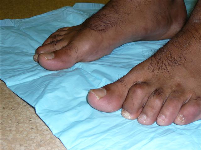
The term "diabetic foot" refers to a condition that affects the feet of people with diabetes, particularly when the disease is not well-controlled. It is characterized by a combination of nerve damage (neuropathy) and poor circulation (peripheral artery disease) in the feet and lower legs.
Neuropathy can cause numbness, tingling, or pain in the feet, making it difficult for people with diabetes to feel injuries, cuts, blisters, or other foot problems. Poor circulation makes it harder for wounds to heal and increases the risk of infection.
Diabetic foot ulcers are a common complication of diabetic neuropathy and can lead to serious infections, hospitalization, and even amputation if not treated promptly and effectively. Preventive care, including regular foot exams, proper footwear, and good blood glucose control, is essential for people with diabetes to prevent or manage diabetic foot problems.
"History, 19th Century" is not a medical term or concept. It refers to the historical events, developments, and figures related to the 1800s in various fields, including politics, culture, science, and technology. However, if you are looking for medical advancements during the 19th century, here's a brief overview:
The 19th century was a period of significant progress in medicine, with numerous discoveries and innovations that shaped modern medical practices. Some notable developments include:
1. Edward Jenner's smallpox vaccine (1796): Although not strictly within the 19th century, Jenner's discovery laid the foundation for vaccination as a preventive measure against infectious diseases.
2. Germ theory of disease: The work of Louis Pasteur, Robert Koch, and others established that many diseases were caused by microorganisms, leading to the development of antiseptic practices and vaccines.
3. Anesthesia: In 1842, Crawford Long first used ether as an anesthetic during surgery, followed by the introduction of chloroform in 1847 by James Simpson.
4. Antisepsis and asepsis: Joseph Lister introduced antiseptic practices in surgery, significantly reducing postoperative infections. Later, the concept of asepsis (sterilization) was developed to prevent contamination during surgical procedures.
5. Microbiology: The development of techniques for culturing and staining bacteria allowed for better understanding and identification of pathogens.
6. Physiology: Claude Bernard's work on the regulation of internal body functions, or homeostasis, contributed significantly to our understanding of human physiology.
7. Neurology: Jean-Martin Charcot made significant contributions to the study of neurological disorders, including multiple sclerosis and Parkinson's disease.
8. Psychiatry: Sigmund Freud developed psychoanalysis, a new approach to understanding mental illnesses.
9. Public health: The 19th century saw the establishment of public health organizations and initiatives aimed at improving sanitation, water quality, and vaccination programs.
10. Medical education reforms: The Flexner Report in 1910 led to significant improvements in medical education standards and practices.
Neurology is a branch of medicine that deals with the study and treatment of diseases and disorders of the nervous system, which includes the brain, spinal cord, peripheral nerves, muscles, and autonomic nervous system. Neurologists are medical doctors who specialize in this field, diagnosing and treating conditions such as stroke, Alzheimer's disease, epilepsy, Parkinson's disease, multiple sclerosis, and various types of headaches and pain disorders. They use a variety of diagnostic tests, including imaging studies like MRI and CT scans, electrophysiological tests like EEG and EMG, and laboratory tests to evaluate nerve function and identify any underlying conditions or abnormalities. Treatment options may include medication, surgery, rehabilitation, or lifestyle modifications.
The tarsal bones are a group of seven articulating bones in the foot that make up the posterior portion of the foot, located between the talus bone of the leg and the metatarsal bones of the forefoot. They play a crucial role in supporting the body's weight and facilitating movement.
There are three categories of tarsal bones:
1. Proximal row: This includes the talus, calcaneus (heel bone), and navicular bones. The talus articulates with the tibia and fibula to form the ankle joint, while the calcaneus is the largest tarsal bone and forms the heel. The navicular bone is located between the talus and the cuneiform bones.
2. Intermediate row: This includes the cuboid bone, which is located laterally (on the outside) to the navicular bone and articulates with the calcaneus, fourth and fifth metatarsals, and the cuneiform bones.
3. Distal row: This includes three cuneiform bones - the medial, intermediate, and lateral cuneiforms - which are located between the navicular bone proximally and the first, second, and third metatarsal bones distally. The medial cuneiform is the largest of the three and articulates with the navicular bone, first metatarsal, and the intermediate cuneiform. The intermediate cuneiform articulates with the medial and lateral cuneiforms and the second metatarsal. The lateral cuneiform articulates with the intermediate cuneiform, cuboid, and fourth metatarsal.
Together, these bones form a complex network of joints that allow for movement and stability in the foot. Injuries or disorders affecting the tarsal bones can result in pain, stiffness, and difficulty walking.
The term "hysteria" is an outdated and discredited concept in medicine, particularly in psychiatry and psychology. Originally, it was used to describe a condition characterized by dramatic, excessive emotional reactions and physical symptoms that couldn't be explained by a medical condition. These symptoms often included things like paralysis, blindness, or fits, which would sometimes be "hysterical" in nature - that is, they seemed to have no physical cause.
However, the concept of hysteria has been largely abandoned due to its lack of scientific basis and its use as a catch-all diagnosis for symptoms that doctors couldn't explain. Today, many of the symptoms once attributed to hysteria are now understood as manifestations of other medical or psychological conditions, such as conversion disorder, panic attacks, or malingering. It's important to note that using outdated and stigmatizing terms like "hysteria" can be harmful and misleading, so it's best to avoid them in favor of more precise and respectful language.
Diabetic neuropathies refer to a group of nerve disorders that are caused by diabetes. High blood sugar levels can injure nerves throughout the body, but diabetic neuropathies most commonly affect the nerves in the legs and feet.
There are four main types of diabetic neuropathies:
1. Peripheral neuropathy: This is the most common type of diabetic neuropathy. It affects the nerves in the legs and feet, causing symptoms such as numbness, tingling, burning, or shooting pain.
2. Autonomic neuropathy: This type of neuropathy affects the autonomic nerves, which control involuntary functions such as heart rate, blood pressure, digestion, and bladder function. Symptoms may include dizziness, fainting, digestive problems, sexual dysfunction, and difficulty regulating body temperature.
3. Proximal neuropathy: Also known as diabetic amyotrophy, this type of neuropathy affects the nerves in the hips, thighs, or buttocks, causing weakness, pain, and difficulty walking.
4. Focal neuropathy: This type of neuropathy affects a single nerve or group of nerves, causing symptoms such as weakness, numbness, or pain in the affected area. Focal neuropathies can occur anywhere in the body, but they are most common in the head, torso, and legs.
The risk of developing diabetic neuropathies increases with the duration of diabetes and poor blood sugar control. Other factors that may contribute to the development of diabetic neuropathies include genetics, age, smoking, and alcohol consumption.
Acquired foot deformities refer to structural abnormalities of the foot that develop after birth, as opposed to congenital foot deformities which are present at birth. These deformities can result from various factors such as trauma, injury, infection, neurological conditions, or complications from a medical condition like diabetes or arthritis.
Examples of acquired foot deformities include:
1. Hammertoe - A deformity where the toe bends downward at the middle joint, resembling a hammer.
2. Claw toe - A more severe form of hammertoe where the toe also curls under, forming a claw-like shape.
3. Mallet toe - A condition where the end joint of a toe is bent downward, causing it to resemble a mallet.
4. Bunions - A bony bump that forms on the inside of the foot at the big toe joint, often causing pain and difficulty wearing shoes.
5. Tailor's bunion (bunionette) - A similar condition to a bunion, but it occurs on the outside of the foot near the little toe joint.
6. Charcot foot - A severe deformity that can occur in people with diabetes or other neurological conditions, characterized by the collapse and dislocation of joints in the foot.
7. Cavus foot - A condition where the arch of the foot is excessively high, causing instability and increasing the risk of ankle injuries.
8. Flatfoot (pes planus) - A deformity where the arch of the foot collapses, leading to pain and difficulty walking.
9. Pronation deformities - Abnormal rotation or tilting of the foot, often causing instability and increasing the risk of injury.
Treatment for acquired foot deformities varies depending on the severity and underlying cause but may include orthotics, physical therapy, medication, or surgery.
Foot diseases refer to various medical conditions that affect the foot, including its structures such as the bones, joints, muscles, tendons, ligaments, blood vessels, and nerves. These conditions can cause symptoms like pain, swelling, numbness, difficulty walking, and skin changes. Examples of foot diseases include:
1. Plantar fasciitis: inflammation of the band of tissue that connects the heel bone to the toes.
2. Bunions: a bony bump that forms on the joint at the base of the big toe.
3. Hammertoe: a deformity in which the toe is bent at the middle joint, resembling a hammer.
4. Diabetic foot: a group of conditions that can occur in people with diabetes, including nerve damage, poor circulation, and increased risk of infection.
5. Athlete's foot: a fungal infection that affects the skin between the toes and on the soles of the feet.
6. Ingrown toenails: a condition where the corner or side of a toenail grows into the flesh of the toe.
7. Gout: a type of arthritis that causes sudden, severe attacks of pain, swelling, redness, and tenderness in the joints, often starting with the big toe.
8. Foot ulcers: open sores or wounds that can occur on the feet, especially in people with diabetes or poor circulation.
9. Morton's neuroma: a thickening of the tissue around a nerve between the toes, causing pain and numbness.
10. Osteoarthritis: wear and tear of the joints, leading to pain, stiffness, and reduced mobility.
Foot diseases can affect people of all ages and backgrounds, and some may be prevented or managed with proper foot care, hygiene, and appropriate medical treatment.
The scalp is the anatomical region located at the upper part of the human head, covering the skull except for the face and the ears. It is made up of several layers: the skin, the connective tissue, the galea aponeurotica (a strong, flat, tendinous sheet), loose areolar tissue, and the periosteum (the highly vascularized innermost layer that attaches directly to the skull bones). The scalp has a rich blood supply and is home to numerous sensory receptors, including those for touch, pain, and temperature. It also contains hair follicles, sebaceous glands, and sweat glands.
A foot ulcer is a wound or sore on the foot that occurs most commonly in people with diabetes, but can also affect other individuals with poor circulation or nerve damage. These ulcers can be challenging to heal and are prone to infection, making it essential for individuals with foot ulcers to seek medical attention promptly.
Foot ulcers typically develop due to prolonged pressure on bony prominences of the foot, leading to breakdown of the skin and underlying tissues. The development of foot ulcers can be attributed to several factors, including:
1. Neuropathy (nerve damage): This condition causes a loss of sensation in the feet, making it difficult for individuals to feel pain or discomfort associated with pressure points, leading to the formation of ulcers.
2. Peripheral artery disease (PAD): Reduced blood flow to the lower extremities can impair wound healing and make the body more susceptible to infection.
3. Deformities: Structural foot abnormalities, such as bunions or hammertoes, can cause increased pressure on specific areas of the foot, increasing the risk of ulcer formation.
4. Poorly fitting shoes: Shoes that are too tight, narrow, or ill-fitting can create friction and pressure points, contributing to the development of foot ulcers.
5. Trauma: Injuries or trauma to the feet can lead to the formation of ulcers, particularly in individuals with neuropathy who may not feel the initial pain associated with the injury.
6. Foot care neglect: Failure to inspect and care for the feet regularly can result in undetected wounds or sores that progress into ulcers.
Foot ulcers are classified based on their depth, severity, and extent of tissue involvement. Proper assessment, treatment, and prevention strategies are crucial in managing foot ulcers and minimizing the risk of complications such as infection, gangrene, and amputation.
Tooth loss is the condition or process characterized by the disappearance or absence of one or more teeth from their normal position in the dental arch. This can occur due to various reasons such as tooth decay, periodontal disease (gum disease), injury, or aging. The consequences of tooth loss include difficulties in chewing, speaking, and adversely affecting the aesthetics of a person's smile, which may lead to psychological impacts. Additionally, it can cause shifting of adjacent teeth, bone resorption, and changes in the bite, potentially leading to further dental issues if not treated promptly.
A tooth germ is a small cluster of cells that eventually develop into a tooth. It contains the dental papilla, which will become the dentin and pulp of the tooth, and the dental follicle, which will form the periodontal ligament, cementum, and alveolar bone. The tooth germ starts as an epithelial thickening called the dental lamina, which then forms a bud, cap, and bell stage before calcification occurs and the tooth begins to erupt through the gums. It is during the bell stage that the enamel organ, which will form the enamel of the tooth, is formed.
A deciduous tooth, also known as a baby tooth or primary tooth, is a type of temporary tooth that humans and some other mammals develop during childhood. They are called "deciduous" because they are eventually shed and replaced by permanent teeth, much like how leaves on a deciduous tree fall off and are replaced by new growth.
Deciduous teeth begin to form in the womb and start to erupt through the gums when a child is around six months old. By the time a child reaches age three, they typically have a full set of 20 deciduous teeth, including incisors, canines, and molars. These teeth are smaller and less durable than permanent teeth, but they serve important functions such as helping children chew food properly, speak clearly, and maintain space in the jaw for the permanent teeth to grow into.
Deciduous teeth usually begin to fall out around age six or seven, starting with the lower central incisors. This process continues until all of the deciduous teeth have been shed, typically by age 12 or 13. At this point, the permanent teeth will have grown in and taken their place, with the exception of the wisdom teeth, which may not erupt until later in adolescence or early adulthood.
A tooth crown is a type of dental restoration that covers the entire visible portion of a tooth, restoring its shape, size, and strength. It is typically made of materials like porcelain, ceramic, or metal alloys and is custom-made to fit over the prepared tooth. The tooth crown is cemented in place and becomes the new outer surface of the tooth, protecting it from further damage or decay.
The process of getting a tooth crown usually involves two dental appointments. During the first appointment, the dentist prepares the tooth by removing any decay or damaged tissue and shaping the tooth to accommodate the crown. An impression is then taken of the prepared tooth and sent to a dental laboratory where the crown is fabricated. In the meantime, a temporary crown is placed over the prepared tooth to protect it until the permanent crown is ready. At the second appointment, the temporary crown is removed, and the permanent crown is cemented in place.
Tooth crowns are often recommended for several reasons, including:
* To restore a broken or fractured tooth
* To protect a weakened tooth from further damage or decay
* To support a large filling when there isn't enough natural tooth structure left
* To cover a dental implant
* To improve the appearance of a discolored or misshapen tooth
Overall, a tooth crown is an effective and long-lasting solution for restoring damaged or decayed teeth and improving oral health.
A tooth root is the part of a tooth that is embedded in the jawbone and cannot be seen when looking at a person's smile. It is the lower portion of a tooth that typically has a conical shape and anchors the tooth to the jawbone through a periodontal ligament. The tooth root is covered by cementum, a specialized bone-like tissue, and contains nerve endings and blood vessels within its pulp chamber.
The number of roots in a tooth can vary depending on the type of tooth. For example, incisors typically have one root, canines may have one or two roots, premolars usually have one or two roots, and molars often have two to four roots. The primary function of the tooth root is to provide stability and support for the crown of the tooth, allowing it to withstand the forces of biting and chewing.
Foot injuries refer to any damage or trauma caused to the various structures of the foot, including the bones, muscles, tendons, ligaments, blood vessels, and nerves. These injuries can result from various causes such as accidents, sports activities, falls, or repetitive stress. Common types of foot injuries include fractures, sprains, strains, contusions, dislocations, and overuse injuries like plantar fasciitis or Achilles tendonitis. Symptoms may vary depending on the type and severity of the injury but often include pain, swelling, bruising, difficulty walking, and reduced range of motion. Proper diagnosis and treatment are crucial to ensure optimal healing and prevent long-term complications.
Tooth eruption is the process by which a tooth emerges from the gums and becomes visible in the oral cavity. It is a normal part of dental development that occurs in a predictable sequence and timeframe. Primary or deciduous teeth, also known as baby teeth, begin to erupt around 6 months of age and continue to emerge until approximately 2-3 years of age. Permanent or adult teeth start to erupt around 6 years of age and can continue to emerge until the early twenties.
The process of tooth eruption involves several stages, including the formation of the tooth within the jawbone, the movement of the tooth through the bone and surrounding tissues, and the final emergence of the tooth into the mouth. Proper tooth eruption is essential for normal oral function, including chewing, speaking, and smiling. Any abnormalities in the tooth eruption process, such as delayed or premature eruption, can indicate underlying dental or medical conditions that require further evaluation and treatment.
A "History of Nursing" in a medical context generally refers to the documentation of a patient's past experiences with nursing care, including any previous hospitalizations, treatments, medications, and interactions with nursing staff. This information is used by nurses to assess a patient's current health status, identify potential risks or complications, and develop an individualized plan of care.
The history of nursing can include information about the patient's medical history, surgical history, family medical history, social history, and lifestyle factors that may impact their health. It is important for nurses to gather this information accurately and thoroughly, as it can help them provide safe and effective care, communicate with other healthcare providers, and promote positive health outcomes for their patients.
In addition to its clinical importance, the history of nursing also plays a critical role in nursing education and research, helping to inform best practices, advance nursing science, and shape the future of the profession.
Osteitis is a medical term that refers to the inflammation of bone tissue. It can occur as a result of various conditions, such as infection (osteomyelitis), trauma, or autoimmune disorders. The symptoms of osteitis may include pain, swelling, warmth, and redness in the affected area, as well as fever and general malaise. Treatment typically involves addressing the underlying cause of the inflammation, which may involve antibiotics for infection or anti-inflammatory medications for other causes. In some cases, surgery may be necessary to remove infected or damaged bone tissue.
A gait disorder is a disturbance in the ability to walk that can't be attributed to physical disabilities such as weakness or paralysis. Neurologic gait disorders are those specifically caused by underlying neurological conditions. These disorders can result from damage to the brain, spinal cord, or peripheral nerves that disrupts communication between the muscles and the brain.
Neurologic gait disorders can present in various ways, including:
1. **Spastic Gait:** This is a stiff, foot-dragging walk caused by increased muscle tone (hypertonia) and stiffness (spasticity). It's often seen in conditions like cerebral palsy or multiple sclerosis.
2. **Ataxic Gait:** This is a broad-based, unsteady, and irregular walk caused by damage to the cerebellum, which affects balance and coordination. Conditions such as cerebellar atrophy or stroke can cause this type of gait disorder.
3. **Parkinsonian Gait:** This is a shuffling walk with small steps, flexed knees, and difficulty turning. It's often seen in Parkinson's disease.
4. **Neuropathic Gait:** This is a high-stepping walk caused by foot drop (difficulty lifting the front part of the foot), which results from damage to the peripheral nerves. Conditions such as diabetic neuropathy or Guillain-Barre syndrome can cause this type of gait disorder.
5. **Choreic Gait:** This is an irregular, dance-like walk caused by involuntary movements (chorea) seen in conditions like Huntington's disease.
6. **Mixed Gait:** Sometimes, a person may exhibit elements of more than one type of gait disorder.
The specific type of gait disorder can provide important clues about the underlying neurological condition and help guide diagnosis and treatment.
Arthrodesis is a surgical procedure to fuse together the bones of a joint, in order to restrict its movement and provide stability. This procedure is typically performed when a joint has been severely damaged by injury, arthritis, or other conditions, and non-surgical treatments have failed to relieve symptoms such as pain and instability.
During the surgery, the cartilage that normally cushions the ends of the bones is removed, and the bones are realigned and held in place with hardware such as plates, screws, or rods. Over time, the bones grow together, forming a solid fusion that restricts joint motion.
Arthrodesis can be performed on various joints throughout the body, including the spine, wrist, ankle, and knee. While this procedure can provide significant pain relief and improve function, it does limit the range of motion in the fused joint, which may impact mobility and daily activities. Therefore, arthrodesis is typically considered a last resort when other treatments have failed.
A supernumerary tooth, also known as hyperdontia, refers to an additional tooth or teeth that grow beyond the regular number of teeth in the dental arch. These extra teeth can erupt in various locations of the dental arch and may occur in any of the tooth types, but they are most commonly seen as extra premolars or molars, and less frequently as incisors or canines. Supernumerary teeth may be asymptomatic or may cause complications such as crowding, displacement, or impaction of adjacent teeth, and therefore, they often require dental treatment.
Tooth abnormalities refer to any variations or irregularities in the size, shape, number, structure, or development of teeth that deviate from the typical or normal anatomy. These abnormalities can occur in primary (deciduous) or permanent teeth and can be caused by genetic factors, environmental influences, systemic diseases, or localized dental conditions during tooth formation.
Some examples of tooth abnormalities include:
1. Microdontia - teeth that are smaller than normal in size.
2. Macrodontia - teeth that are larger than normal in size.
3. Peg-shaped teeth - teeth with a narrow, conical shape.
4. Talon cusps - additional cusps or points on the biting surface of a tooth.
5. Dens invaginatus - an abnormal development where the tooth crown has an extra fold or pouch that can trap bacteria and cause dental problems.
6. Taurodontism - teeth with large pulp chambers and short roots.
7. Supernumerary teeth - having more teeth than the typical number (20 primary and 32 permanent teeth).
8. Hypodontia - missing one or more teeth due to a failure of development.
9. Germination - two adjacent teeth fused together, usually occurring in the front teeth.
10. Fusion - two separate teeth that have grown together during development.
Tooth abnormalities may not always require treatment unless they cause functional, aesthetic, or dental health issues. A dentist can diagnose and manage tooth abnormalities through various treatments, such as fillings, extractions, orthodontic care, or restorative procedures.
Tooth wear is the progressive loss of tooth structure that can occur as a result of various factors. According to the medical definition, it refers to the wearing down, rubbing away, or grinding off of the hard tissues of the teeth (enamel and dentin) due to mechanical forces or chemical processes.
There are three primary types of tooth wear:
1. Abrasion: This is the loss of tooth structure caused by friction from external sources, such as incorrect brushing techniques, bite appliances, or habits like nail-biting and pipe smoking.
2. Attrition: This type of tooth wear results from the natural wearing down of teeth due to occlusal forces during biting, chewing, and grinding. However, excessive attrition can occur due to bruxism (teeth grinding) or clenching.
3. Erosion: Chemical processes, such as acid attacks from dietary sources (e.g., citrus fruits, sodas, and sports drinks) or gastric reflux, cause the loss of tooth structure in this type of tooth wear. The enamel dissolves when exposed to low pH levels, leaving the dentin underneath vulnerable to further damage.
Professional dental examination and treatment may be necessary to address significant tooth wear and prevent further progression, which can lead to sensitivity, pain, and functional or aesthetic issues.
Tooth extraction is a dental procedure in which a tooth that is damaged or poses a threat to oral health is removed from its socket in the jawbone. This may be necessary due to various reasons such as severe tooth decay, gum disease, fractured teeth, crowded teeth, or for orthodontic treatment purposes. The procedure is performed by a dentist or an oral surgeon, under local anesthesia to numb the area around the tooth, ensuring minimal discomfort during the extraction process.
In medical terms, the foot is the part of the lower limb that is distal to the leg and below the ankle, extending from the tarsus to the toes. It is primarily responsible for supporting body weight and facilitating movement through push-off during walking or running. The foot is a complex structure made up of 26 bones, 33 joints, and numerous muscles, tendons, ligaments, and nerves that work together to provide stability, balance, and flexibility. It can be divided into three main parts: the hindfoot, which contains the talus and calcaneus (heel) bones; the midfoot, which includes the navicular, cuboid, and cuneiform bones; and the forefoot, which consists of the metatarsals and phalanges that form the toes.
I'm sorry for any confusion, but "France" is not a medical term or concept. France is the largest country in Western Europe and the third-largest in Europe after Russia and Ukraine. It has been a major player in world affairs for centuries, with a significant cultural and artistic influence. If you have any questions about medical terms or concepts, I would be happy to help answer those for you.
A nonvital tooth is one that no longer has a living or viable pulp, which contains the nerves and blood vessels inside the tooth. This condition can occur due to various reasons such as tooth decay that has progressed deeply into the tooth, dental trauma, or previous invasive dental procedures. As a result, the tooth loses its sensitivity to temperature changes and may darken in color. Nonvital teeth typically require root canal treatment to remove the dead pulp tissue, disinfect the canals, and fill them with an inert material to preserve the tooth structure and function.
I'm sorry for any confusion, but "History, 20th Century" is a broad and complex topic that refers to the events, developments, and transformations that occurred throughout the world during the 1900s. It is not a medical term or concept. If you're interested in learning more about this historical period, I would recommend consulting a history textbook, reputable online resources, or speaking with a historian. They can provide detailed information about the political, social, economic, and cultural changes that took place during the 20th century.
In the context of dentistry, a molar is a type of tooth found in the back of the mouth. They are larger and wider than other types of teeth, such as incisors or canines, and have a flat biting surface with multiple cusps. Molars are primarily used for grinding and chewing food into smaller pieces that are easier to swallow. Humans typically have twelve molars in total, including the four wisdom teeth.
In medical terminology outside of dentistry, "molar" can also refer to a unit of mass in the apothecaries' system of measurement, which is equivalent to 4.08 grams. However, this usage is less common and not related to dental or medical anatomy.
An impacted tooth is a condition where a tooth fails to erupt into the oral cavity within its expected time frame, resulting in its partial or complete entrapment within the jawbone or soft tissues. This commonly occurs with wisdom teeth (third molars) but can affect any tooth. Impacted teeth may cause problems such as infection, decay of adjacent teeth, gum disease, or cyst formation, and they may require surgical removal.
Tooth discoloration, also known as tooth staining or tooth color change, refers to the darkening or staining of teeth. It can be categorized into two main types: extrinsic and intrinsic. Extrinsic discoloration occurs when the outer layer of the tooth (enamel) becomes stained due to exposure to colored substances such as coffee, tea, wine, tobacco, and certain foods. Intrinsic discoloration, on the other hand, occurs when the inner structure of the tooth (dentin) darkens or gets a yellowish tint due to factors like genetics, aging, trauma, or exposure to certain medications during tooth development. Tooth discoloration can also be caused by dental diseases or decay. It is important to note that while some forms of tooth discoloration are cosmetic concerns, others may indicate underlying oral health issues and should be evaluated by a dental professional.
A tooth is classified as "unerupted" when it has not yet penetrated through the gums and entered the oral cavity. This can apply to both primary (baby) teeth and permanent (adult) teeth. The reasons for a tooth's failure to erupt can vary, including crowding of teeth, lack of sufficient space, or anatomical barriers such as bone or soft tissue. In some cases, unerupted teeth may need to be monitored or treated, depending on the specific situation and any symptoms experienced by the individual.
I'm sorry for any confusion, but "shoes" are not a medical term. Shoes are items of footwear intended to protect and comfort the feet. They typically have a durable sole that provides protection from sharp objects, hot surfaces, and the effects of moisture. The upper part of a shoe can be made from various materials such as leather, plastic, or textiles, and is designed to provide coverage and support for the foot.
If you have any questions related to medical terminology or health-related topics, I'd be happy to help!
An incisor is a type of tooth that is primarily designed for biting off food pieces rather than chewing or grinding. They are typically chisel-shaped, flat, and have a sharp cutting edge. In humans, there are eight incisors - four on the upper jaw and four on the lower jaw, located at the front of the mouth. Other animals such as dogs, cats, and rodents also have incisors that they use for different purposes like tearing or gnawing.
Odontogenesis is the process of tooth development that involves the formation and calcification of teeth. It is a complex process that requires the interaction of several types of cells, including epithelial cells, mesenchymal cells, and odontoblasts. The process begins during embryonic development with the formation of dental lamina, which gives rise to the tooth bud. As the tooth bud grows and differentiates, it forms the various structures of the tooth, including the enamel, dentin, cementum, and pulp. Odontogenesis is completed when the tooth erupts into the oral cavity. Abnormalities in odontogenesis can result in developmental dental anomalies such as tooth agenesis, microdontia, or odontomas.
Charcot-Marie-Tooth disease (CMT) is a group of inherited disorders that cause nerve damage, primarily affecting the peripheral nerves. These are the nerves that transmit signals between the brain and spinal cord to the rest of the body. CMT affects both motor and sensory nerves, leading to muscle weakness and atrophy, as well as numbness or tingling in the hands and feet.
The disease is named after the three physicians who first described it: Jean-Martin Charcot, Pierre Marie, and Howard Henry Tooth. CMT is characterized by its progressive nature, meaning symptoms typically worsen over time, although the rate of progression can vary significantly among individuals.
There are several types of CMT, classified based on their genetic causes and patterns of inheritance. The two most common forms are CMT1 and CMT2:
1. CMT1: This form is caused by mutations in the genes responsible for the myelin sheath, which insulates peripheral nerves and allows for efficient signal transmission. As a result, demyelination occurs, slowing down nerve impulses and causing muscle weakness, particularly in the lower limbs. Symptoms usually begin in childhood or adolescence and include foot drop, high arches, and hammertoes.
2. CMT2: This form is caused by mutations in the genes responsible for the axons, the nerve fibers that transmit signals within peripheral nerves. As a result, axonal degeneration occurs, leading to muscle weakness and atrophy. Symptoms usually begin in early adulthood and progress more slowly than CMT1. They primarily affect the lower limbs but can also involve the hands and arms.
Diagnosis of CMT typically involves a combination of clinical evaluation, family history, nerve conduction studies, and genetic testing. While there is no cure for CMT, treatment focuses on managing symptoms and maintaining mobility and function through physical therapy, bracing, orthopedic surgery, and pain management.
The term "tooth cervix" is not commonly used in medical dentistry with a specific technical definition. However, if you are referring to the "cervical region of a tooth," it generally refers to the area where the crown (the visible part of the tooth) meets the root (the portion of the tooth that is below the gum line). This region is also sometimes referred to as the "cementoenamel junction" (CEJ), where the enamel covering of the crown meets the cementum covering of the root. Dental issues such as tooth decay, receding gums, or abrasion can affect this area and may require professional dental treatment.
Dental enamel is the hard, white, outermost layer of a tooth. It is a highly mineralized and avascular tissue, meaning it contains no living cells or blood vessels. Enamel is primarily composed of calcium and phosphate minerals and serves as the protective covering for the crown of a tooth, which is the portion visible above the gum line.
Enamel is the hardest substance in the human body, and its primary function is to provide structural support and protection to the underlying dentin and pulp tissues of the tooth. It also plays a crucial role in chewing and biting by helping to distribute forces evenly across the tooth surface during these activities.
Despite its hardness, dental enamel can still be susceptible to damage from factors such as tooth decay, erosion, and abrasion. Once damaged or lost, enamel cannot regenerate or repair itself, making it essential to maintain good oral hygiene practices and seek regular dental checkups to prevent enamel damage and protect overall oral health.
Tooth exfoliation is not a term that is commonly used in dental or medical literature. However, I believe you may be referring to the natural process of tooth loss that occurs with the shedding of primary (baby) teeth to make way for permanent (adult) teeth. This process is also known as physical or physiological tooth exfoliation.
Exfoliation in this context refers to the separation and shedding of the primary tooth's root from the underlying permanent tooth, allowing the permanent tooth to erupt into its proper position. The primary tooth becomes loose due to the resorption of its roots by the developing permanent tooth beneath it. Eventually, the primary tooth falls out, making room for the adult tooth to emerge and take its place in the dental arch.
It is essential to maintain good oral hygiene during this process to prevent any potential complications such as infection or premature loss of primary teeth.
Immobilization is a medical term that refers to the restriction of normal mobility or motion of a body part, usually to promote healing and prevent further injury. This is often achieved through the use of devices such as casts, splints, braces, slings, or traction. The goal of immobilization is to keep the injured area in a fixed position so that it can heal properly without additional damage. It may be used for various medical conditions, including fractures, dislocations, sprains, strains, and soft tissue injuries. Immobilization helps reduce pain, minimize swelling, and protect the injured site from movement that could worsen the injury or impair healing.
Tooth avulsion is the complete separation of a tooth from its socket in the alveolar bone due to traumatic injury. This occurs when the periodontal ligament, which holds the tooth in place, gets severed or torn, resulting in the tooth being displaced from its original position. Avulsed teeth can be either primary (baby) or permanent teeth, and the trauma can result in damage to the surrounding tissues, including the gingiva, alveolar bone, and sometimes even the nerves and blood vessels. Prompt and appropriate first aid, as well as professional dental care, are crucial for ensuring the best possible outcome for reimplantation and healing.
'Fused teeth', also known as congenitally missing or malformed teeth, is a dental condition where two or more teeth are fused together. This condition is called "gemination" when a single tooth bud fails to completely separate, resulting in two teeth that share a common pulp chamber and root canal. When this occurs with more than one tooth, it is referred to as "twinning." In contrast, "congenital fusion" or "synthesis" refers to the union of two separate tooth buds during development.
Fused teeth can cause cosmetic concerns, difficulty in biting and chewing, and may affect the alignment of surrounding teeth. Depending on the severity and location of the fusion, treatment options may include observation, dental restorations, or even orthodontic or surgical intervention to correct the malocclusion and improve oral function and aesthetics.
A cuspid, also known as a canine tooth or cuspid tooth, is a type of tooth in mammals. It is the pointiest tooth in the dental arch and is located between the incisors and bicuspids (or premolars). Cuspids have a single cusp or pointed tip that is used for tearing and grasping food. In humans, there are four cuspids, two on the upper jaw and two on the lower jaw, one on each side of the dental arch.
Tooth calcification, also known as dental calculus or tartar formation, refers to the hardening of plaque on the surface of teeth. This process occurs when minerals from saliva combine with bacterial deposits and dental plaque, resulting in a hard, calcified substance that adheres to the tooth surface. Calcification can occur both above and below the gum line, and if not removed through professional dental cleanings, it can lead to periodontal disease, tooth decay, and other oral health issues.
A bicuspid valve, also known as a mitral valve in the heart, is a heart valve that has two leaflets or cusps. It lies between the left atrium and the left ventricle and helps to regulate blood flow between these two chambers of the heart. In a healthy heart, the bicuspid valve opens to allow blood to flow from the left atrium into the left ventricle and closes tightly to prevent blood from flowing back into the left atrium during contraction of the ventricle.
A congenital heart defect known as a bicuspid aortic valve occurs when the aortic valve, which normally has three leaflets or cusps, only has two. This can lead to narrowing of the valve (aortic stenosis) or leakage of the valve (aortic regurgitation), which can cause symptoms and may require medical treatment.
Tooth ankylosis is a dental condition where the tooth becomes abnormally fused to the alveolar bone, which is the part of the jawbone that contains the tooth sockets. This fusion typically occurs through the cementum of the root surface and the adjacent alveolar bone, resulting in the loss of the periodontal ligament (PLD) space that normally separates the tooth from the bone.
Ankylosis can affect both primary (deciduous or baby) teeth and permanent teeth. In primary teeth, ankylosis may lead to early exfoliation or premature loss of the tooth due to the lack of PDL resorption, which is necessary for natural tooth shedding. In permanent teeth, ankylosis can result in infraocclusion, where the affected tooth fails to erupt fully and remains at a lower level than the surrounding teeth.
The causes of tooth ankylosis include trauma, infection, developmental disorders, or previous orthodontic treatment. It is essential to diagnose and manage this condition promptly, as it can lead to complications such as malocclusion, dental crowding, or periodontal issues if left untreated. Treatment options may include extraction of the affected tooth, surgical separation from the bone, or orthodontic treatment to correct any resulting occlusal discrepancies.
Dental pulp is the soft tissue located in the center of a tooth, surrounded by the dentin. It contains nerves, blood vessels, and connective tissue, and plays a vital role in the development and health of the tooth. The dental pulp helps to form dentin during tooth development and continues to provide nourishment to the tooth throughout its life. It also serves as a sensory organ, allowing the tooth to detect hot and cold temperatures and transmit pain signals to the brain. Injury or infection of the dental pulp can lead to serious dental problems, such as tooth decay or abscesses, and may require root canal treatment to remove the damaged tissue and save the tooth.
Tooth erosion is defined as the progressive, irreversible loss of dental hard tissue, primarily caused by chemical dissolution from acids, rather than mechanical forces such as abrasion or attrition. These acids can originate from extrinsic sources like acidic foods and beverages, or intrinsic sources like gastric reflux or vomiting. The erosion process leads to a reduction in tooth structure, altering the shape and function of teeth, and potentially causing sensitivity, pain, and aesthetical concerns. Early detection and management of tooth erosion are crucial to prevent further progression and preserve dental health.
A tooth socket, also known as an alveolus (plural: alveoli), refers to the hollow cavity or space in the jawbone where a tooth is anchored. The tooth socket is part of the alveolar process, which is the curved part of the maxilla or mandible that contains multiple tooth sockets for the upper and lower teeth, respectively.
Each tooth socket has a specialized tissue called the periodontal ligament, which attaches the root of the tooth to the surrounding bone. This ligament helps absorb forces generated during biting and chewing, allowing for comfortable and efficient mastication while also maintaining the tooth's position within the jawbone. The tooth socket is responsible for providing support, stability, and nourishment to the tooth through its blood vessels and nerves.
Tooth replantation is a dental procedure that involves the replanting and reattachment of a tooth that has been avulsed or knocked out due to trauma. The primary goal of this emergency procedure is to preserve the natural tooth and its periodontal ligament (PDL) tissue, allowing for potential reattachment and function.
The steps involved in tooth replantation include:
1. Locating the avulsed tooth: Carefully handle the knocked-out tooth by holding it by the crown (the chewing surface), avoiding touching the root area to prevent further damage to the periodontal ligament fibers.
2. Rinsing the tooth: Gently rinse the tooth with saline solution, sterile water, or milk to remove any debris or dirt, but avoid using alcohol or scrubbing the tooth as it may cause more damage to the PDL.
3. Replanting the tooth: As soon as possible, reposition the tooth back into its socket in the correct orientation and alignment. Apply gentle pressure to seat it in place while ensuring that it is facing the right direction. Ideally, this should be done within 30 minutes of avulsion for better prognosis.
4. Stabilizing the tooth: Use a splint or a wire to secure the replanted tooth to the adjacent teeth, providing stability and support during the healing process. This helps maintain the alignment and position of the replanted tooth.
5. Seeking professional dental care: Immediately consult with a dentist or endodontist for further evaluation, additional treatment, and follow-up care. The dentist will assess the success of the replantation and determine if any root canal therapy or other treatments are necessary to ensure long-term survival of the tooth.
The success of tooth replantation depends on several factors, including the timeliness of the procedure, the condition of the avulsed tooth, and the patient's overall oral health. Prompt action and professional care can significantly increase the likelihood of a successful outcome and preserve the natural tooth for years to come.
The maxilla is a paired bone that forms the upper jaw in vertebrates. In humans, it is a major bone in the face and plays several important roles in the craniofacial complex. Each maxilla consists of a body and four processes: frontal process, zygomatic process, alveolar process, and palatine process.
The maxillae contribute to the formation of the eye sockets (orbits), nasal cavity, and the hard palate of the mouth. They also contain the upper teeth sockets (alveoli) and help form the lower part of the orbit and the cheekbones (zygomatic arches).
Here's a quick rundown of its key functions:
1. Supports the upper teeth and forms the upper jaw.
2. Contributes to the formation of the eye sockets, nasal cavity, and hard palate.
3. Helps shape the lower part of the orbit and cheekbones.
4. Partakes in the creation of important sinuses, such as the maxillary sinus, which is located within the body of the maxilla.
Dentin is the hard, calcified tissue that lies beneath the enamel and cementum of a tooth. It forms the majority of the tooth's structure and is composed primarily of mineral salts (hydroxyapatite), collagenous proteins, and water. Dentin has a tubular structure, with microscopic channels called dentinal tubules that radiate outward from the pulp chamber (the center of the tooth containing nerves and blood vessels) to the exterior of the tooth. These tubules contain fluid and nerve endings that are responsible for the tooth's sensitivity to various stimuli such as temperature changes, pressure, or decay. Dentin plays a crucial role in protecting the dental pulp while also providing support and structure to the overlying enamel and cementum.
Tooth resorption is a process in which there is an abnormal loss or breakdown of tooth structure, either internally (internal resorption) or externally (external resorption), due to the action of specialized cells called odontoclasts. This can lead to weakening and destruction of the tooth, potentially causing sensitivity, pain, or even tooth loss if left untreated. The causes of tooth resorption can vary, including trauma, orthodontic treatment, periodontal disease, and certain systemic conditions. It is important to diagnose and treat tooth resorption early to prevent further damage and preserve the tooth structure.
Diabetes complications refer to a range of health issues that can develop as a result of poorly managed diabetes over time. These complications can affect various parts of the body and can be classified into two main categories: macrovascular and microvascular.
Macrovascular complications include:
* Cardiovascular disease (CVD): People with diabetes are at an increased risk of developing CVD, including coronary artery disease, peripheral artery disease, and stroke.
* Peripheral arterial disease (PAD): This condition affects the blood vessels that supply oxygen and nutrients to the limbs, particularly the legs. PAD can cause pain, numbness, or weakness in the legs and may increase the risk of amputation.
Microvascular complications include:
* Diabetic neuropathy: This is a type of nerve damage that can occur due to prolonged high blood sugar levels. It commonly affects the feet and legs, causing symptoms such as numbness, tingling, or pain.
* Diabetic retinopathy: This condition affects the blood vessels in the eye and can cause vision loss or blindness if left untreated.
* Diabetic nephropathy: This is a type of kidney damage that can occur due to diabetes. It can lead to kidney failure if not managed properly.
Other complications of diabetes include:
* Increased risk of infections, particularly skin and urinary tract infections.
* Slow healing of wounds, which can increase the risk of infection and amputation.
* Gum disease and other oral health problems.
* Hearing impairment.
* Sexual dysfunction.
Preventing or managing diabetes complications involves maintaining good blood sugar control, regular monitoring of blood glucose levels, following a healthy lifestyle, and receiving routine medical care.
Dental caries, also known as tooth decay or cavities, refers to the damage or breakdown of the hard tissues of the teeth (enamel, dentin, and cementum) due to the activity of acid-producing bacteria. These bacteria ferment sugars from food and drinks, producing acids that dissolve and weaken the tooth structure, leading to cavities.
The process of dental caries development involves several stages:
1. Demineralization: The acidic environment created by bacterial activity causes minerals (calcium and phosphate) to be lost from the tooth surface, making it weaker and more susceptible to decay.
2. Formation of a white spot lesion: As demineralization progresses, a chalky white area appears on the tooth surface, indicating early caries development.
3. Cavity formation: If left untreated, the demineralization process continues, leading to the breakdown and loss of tooth structure, resulting in a cavity or hole in the tooth.
4. Infection and pulp involvement: As the decay progresses deeper into the tooth, it can reach the dental pulp (the soft tissue containing nerves and blood vessels), causing infection, inflammation, and potentially leading to toothache, abscess, or even tooth loss.
Preventing dental caries involves maintaining good oral hygiene, reducing sugar intake, using fluoride toothpaste and mouthwash, and having regular dental check-ups and cleanings. Early detection and treatment of dental caries can help prevent further progression and more severe complications.
Dentition refers to the development, arrangement, and appearance of teeth in the dental arch. It includes the number, type, size, and shape of teeth, as well as their alignment and relationship with each other and the surrounding structures in the oral cavity. Dentition can be classified into two main types: deciduous (primary) dentition and permanent (secondary) dentition. Deciduous dentition consists of 20 temporary teeth that erupt during infancy and childhood, while permanent dentition consists of 32 teeth that replace the deciduous teeth and last for a lifetime, excluding the wisdom teeth which may or may not erupt. Abnormalities in dentition can indicate various dental and systemic conditions, making it an essential aspect of oral health assessment and diagnosis.
Amputation is defined as the surgical removal of all or part of a limb or extremity such as an arm, leg, foot, hand, toe, or finger. This procedure is typically performed to remove damaged or dead tissue due to various reasons like severe injury, infection, tumors, or chronic conditions that impair circulation, such as diabetes or peripheral arterial disease. The goal of amputation is to alleviate pain, prevent further complications, and improve the patient's quality of life. Following the surgery, patients may require rehabilitation and prosthetic devices to help them adapt to their new physical condition.
Root canal therapy, also known as endodontic treatment, is a dental procedure that involves the removal of infected or damaged pulp tissue from within a tooth's root canal system. The root canal system is a series of narrow channels that run from the center of the tooth (pulp chamber) down to the tip of the tooth roots, containing nerves, blood vessels, and connective tissues.
During the procedure, the dentist or endodontist will gain access to the pulp chamber, carefully clean and shape the root canals using specialized instruments, and then fill and seal them with a rubber-like material called gutta-percha. This helps prevent reinfection and preserves the structural integrity of the tooth. In many cases, a crown or other restoration is placed over the treated tooth to protect it and restore its function and appearance.
Root canal therapy is typically recommended when the pulp tissue becomes inflamed or infected due to deep decay, repeated dental procedures, cracks, or chips in the teeth. The goal of this treatment is to alleviate pain, preserve natural tooth structure, and prevent the need for extraction.
Odontometry is a term used in dentistry that refers to the measurement of teeth, particularly the size and length of teeth or tooth roots. It is often used in forensic dentistry for identification purposes, such as in age estimation, sex determination, or individual identification of human remains. The measurements can be taken using various methods, including radiographs (x-rays), calipers, or specialized software.
In some contexts, odontometry may also refer to the process of measuring the amount of dental work required for a particular treatment plan, although this usage is less common.
The mandible, also known as the lower jaw, is the largest and strongest bone in the human face. It forms the lower portion of the oral cavity and plays a crucial role in various functions such as mastication (chewing), speaking, and swallowing. The mandible is a U-shaped bone that consists of a horizontal part called the body and two vertical parts called rami.
The mandible articulates with the skull at the temporomandibular joints (TMJs) located in front of each ear, allowing for movements like opening and closing the mouth, protrusion, retraction, and side-to-side movement. The mandible contains the lower teeth sockets called alveolar processes, which hold the lower teeth in place.
In medical terminology, the term "mandible" refers specifically to this bone and its associated structures.
Tooth demineralization is a process that involves the loss of minerals, such as calcium and phosphate, from the hard tissues of the teeth. This process can lead to the development of dental caries or tooth decay. Demineralization occurs when acids produced by bacteria in the mouth attack the enamel of the tooth, dissolving its mineral content. Over time, these attacks can create holes or cavities in the teeth. Fluoride, found in many toothpastes and public water supplies, can help to remineralize teeth and prevent decay. Good oral hygiene practices, such as brushing and flossing regularly, can also help to prevent demineralization by removing plaque and bacteria from the mouth.
Permanent dentition is the second and final set of teeth that humans grow during their lifetime. These teeth are also known as adult or secondary teeth and typically begin to erupt in the mouth around the age of 6 or 7 years old, with all permanent teeth usually present by the time a person reaches their late teens or early twenties.
There are 32 teeth in a complete set of permanent dentition, including 8 incisors, 4 canines, 8 premolars (also called bicuspids), and 12 molars (including 4 third molars or wisdom teeth). The primary function of permanent teeth is to help with biting, chewing, and grinding food into smaller pieces that are easier to swallow and digest. Proper care and maintenance of permanent teeth through good oral hygiene practices, regular dental checkups, and a balanced diet can help ensure their longevity and health throughout a person's life.
A dental restoration, permanent, is a type of dental treatment that involves the use of materials such as gold, silver amalgam, porcelain, or composite resin to repair and restore the function, form, and aesthetics of a damaged or decayed tooth. Unlike temporary restorations, which are meant to be replaced with a permanent solution, permanent restorations are designed to last for many years, if not a lifetime.
Examples of permanent dental restorations include:
1. Dental fillings: These are used to fill cavities caused by tooth decay. The decayed portion of the tooth is removed, and the resulting space is filled with a material such as amalgam, composite resin, or gold.
2. Inlays and onlays: These are similar to dental fillings but are made in a laboratory and then bonded to the tooth. They are used when there is not enough tooth structure left to support a filling.
3. Dental crowns: Also known as caps, these are used to cover and protect a tooth that has been damaged or weakened by decay, injury, or wear. The crown fits over the entire tooth, restoring its shape, size, and strength.
4. Dental bridges: These are used to replace one or more missing teeth. A bridge consists of one or more artificial teeth (pontics) that are held in place by crowns on either side.
5. Dental implants: These are used to replace missing teeth. An implant is a small titanium post that is surgically placed in the jawbone, where it functions as an anchor for a replacement tooth or bridge.
Permanent dental restorations are custom-made for each patient and require careful planning and preparation. They are designed to blend in with the surrounding teeth and provide a natural-looking appearance. With proper care and maintenance, these restorations can last for many years and help preserve the health and function of the teeth and mouth.
Anodontia is a medical term that refers to the congenital absence or lack of development of all primary (deciduous) and/or permanent teeth. It is a rare dental condition that affects tooth development and can be isolated or associated with various syndromes and genetic disorders.
In anodontia, the dental tissues responsible for forming teeth, including the dental lamina, dental papilla, and dental follicle, fail to develop properly, resulting in missing teeth. The condition can affect all teeth or only some of them, leading to partial anodontia.
Anodontia is different from hypodontia, which refers to the congenital absence of one or more, but not all, teeth. It is also distinct from oligodontia, which is the absence of six or more permanent teeth, excluding third molars (wisdom teeth).
People with anodontia may experience difficulties in chewing, speaking, and maintaining oral hygiene, leading to various dental and social problems. Prosthodontic treatments, such as dentures or implants, are often necessary to restore oral function and aesthetics.
Tooth preparation in prosthodontics refers to the process of altering the clinical crown of a tooth or teeth to receive a restoration, such as a crown, veneer, or bridge. This procedure involves removing a portion of the enamel and dentin to create a suitable foundation for the prosthetic device. The preparation aims to achieve proper retention, resistance form, and marginal fit, ensuring the successful integration and longevity of the restoration. The process may also include the management of tooth structure loss due to decay, trauma, or wear, and the establishment of harmonious occlusion with the opposing teeth.
The periodontal ligament, also known as the "PDL," is the soft tissue that connects the tooth root to the alveolar bone within the dental alveolus (socket). It consists of collagen fibers organized into groups called principal fibers and accessory fibers. These fibers are embedded into both the cementum of the tooth root and the alveolar bone, providing shock absorption during biting and chewing forces, allowing for slight tooth movement, and maintaining the tooth in its position within the socket.
The periodontal ligament plays a crucial role in the health and maintenance of the periodontium, which includes the gingiva (gums), cementum, alveolar bone, and the periodontal ligament itself. Inflammation or infection of the periodontal ligament can lead to periodontal disease, potentially causing tooth loss if not treated promptly and appropriately.
The alveolar process is the curved part of the jawbone (mandible or maxilla) that contains sockets or hollow spaces (alveoli) for the teeth to be embedded. These processes are covered with a specialized mucous membrane called the gingiva, which forms a tight seal around the teeth to help protect the periodontal tissues and maintain oral health.
The alveolar process is composed of both compact and spongy bone tissue. The compact bone forms the outer layer, while the spongy bone is found inside the alveoli and provides support for the teeth. When a tooth is lost or extracted, the alveolar process begins to resorb over time due to the lack of mechanical stimulation from the tooth's chewing forces. This can lead to changes in the shape and size of the jawbone, which may require bone grafting procedures before dental implant placement.
"Age determination by teeth" is a method used in forensic dentistry to estimate the age of an individual based on the development and wear of their teeth. This process involves examining various features such as tooth eruption, crown and root formation, and dental attrition or wear.
The developmental stages of teeth can provide a rough estimate of age during childhood and adolescence, while dental wear patterns can offer insights into an individual's age during adulthood. However, it is important to note that there can be significant variation in tooth development and wear between individuals, making this method somewhat imprecise.
In addition to forensic applications, age determination by teeth can also be useful in archaeology and anthropology for studying past populations and their lifestyles.
 Charcot-Marie-Tooth disease
Charcot-Marie-Tooth disease classifications
X-linked Charcot-Marie-Tooth disease
Autosomal dominant Charcot-Marie-Tooth disease type 2 with giant axons
Jean-Martin Charcot
Aminoacyl tRNA synthetase
1884 in science
Mary Reilly (academic)
1886 in science
X-linked recessive inheritance
Degenerative disease
Dejerine-Sottas disease
Hearing loss
Pediatric podiatry
Causes of hearing loss
MFN2
List of OMIM disorder codes
SURF1
Roussy-Lévy syndrome
Peripheral myelin protein 22
INF2
List of eponyms (L-Z)
Hagemoser-Weinstein-Bresnick syndrome
Hereditary sensory and autonomic neuropathy
LITAF
RAB7A
NEFH
KIF1B
Vincristine
Distal hereditary motor neuropathy type V
Hereditary21
- Charcot-Marie-Tooth disease (CMT) is a hereditary motor and sensory neuropathy of the peripheral nervous system characterized by progressive loss of muscle tissue and touch sensation across various parts of the body. (wikipedia.org)
- in others (eg, certain cases of Charcot-Marie-Tooth disease type 1A (CMT1A) and inherited brachial plexus neuropathy [IBPN]/hereditary neuralgic amyotrophy [HNA]), proximal weakness predominates. (medscape.com)
- Charcot-Marie-Tooth (CMT) disease is a peripheral hereditary neuropathy associated with motor and sensory impairment and can result in profound sensorineural hearing loss (SNHL). (hindawi.com)
- Charcot-Marie-Tooth disease is the most commonly occurring hereditary motor and sensory neuropathy. (hindawi.com)
- Make a donation to the Hereditary Neuropathy Foundation to help find treatments and cures for those living with Charcot-Marie-Tooth and Inherited Neuropathies. (hnf-cure.org)
- America's Friendliest Marathon," held in Richmond, VA this past weekend, included some important friends of the Hereditary Neuropathy Foundation who ran to raise awareness of Charcot-Marie-Tooth and to generate funds for research to find a cure. (hnf-cure.org)
- 1987) Diaphragmatic dysfunction in siblings with hereditary motor and sensory neuropathy (Charcot-Marie-Tooth disease). (springer.com)
- Charcot-Marie Tooth - or CMT for short - is a rare neurological disease and one of the hereditary motor and sensory neuropathies of the peripheral nervous system. (trustedhealthproducts.com)
- Charcot MarieTooth disease or CMT , also called as hereditary motor and sensory neuropathy , is a group of inherited disorders that cause damage to peripheral nerves. (targetwoman.com)
- Charcot-Marie-Tooth Hereditary Neuropathy Overview. (medlineplus.gov)
- Charcot-Marie-Tooth disease is the most common hereditary peripheral neuropathy in humans, presenting incidence of 1: 2500 people. (arquivosdeorl.org.br)
- It was used as methodology consults the on line data bases such as Cochrane, LILACS, MEDLINE, OMIM e SciELO, applying the research the terms Charcot Marie Tooth disease, Hereditary Motor and Sensory Neuropathy Type I and Hereditary Motor and Sensory-Neuropathy Type II, to article published between years 1997 and 2007. (arquivosdeorl.org.br)
- Charcot-Marie-Tooth Disease (CMT) is the most common type of Hereditary Peripheral Neuropathy (1) in humans, together with Hereditary Motor and Sensory Neuropathy(2), affecting around 1 in 2,500 people(3). (arquivosdeorl.org.br)
- A list of data basis which was searched follows: Cochrane, LILACS, MEDLINE, OMIM and Scielo, applying to the research the terms Charcot Marie Tooth Disease, Hereditary Motor and Sensory Neuropathy Type I and Hereditary Motor and Sensory-Neuropathy Type II, for articles published between 1997 and 2007. (arquivosdeorl.org.br)
- Cmt1d Is also known as hmsn id, hmsn1d, hereditary motor and sensory neuropathy 1d, charcot-marie-tooth neuropathy, type 1d. (mendelian.co)
- Hereditary Neuropathy Foundation (HNF) is a non-profit 501(c)3 organization which mission is to increase awareness and accurate diagnosis of Charcot-Marie-Tooth (CMT) and related inherited neuropathies, support patients and families with critical information to improve quality of life, and fund research that will lead to treatments and cures. (globalgenes.org)
- Charcot-Marie-Tooth is the most common form of inherited peripheral neuropathy and represents the most prevalent hereditary neuromuscular disorder. (qxmd.com)
- Charcot-Marie Tooth disease is the most common hereditary neurological disease in the world. (biopharmanalyses.fr)
- The effect of treatment lasts for three weeks for severe forms and more than ten weeks for milder forms of the disease.This therapeutic strategy for hereditary peripheral neuropathies, developed entirely in France, is proof of concept for a new precision medicine based on siRNA normalization of the expression of an overexpressed gene. (biopharmanalyses.fr)
- Charcot-Marie-Tooth disease is a hereditary neuropathy in which the muscles of the lower legs become weak and waste away (atrophy). (msdmanuals.com)
- Charcot-Marie-Tooth disease (CMT) also known as hereditary sensory- motor neuropathies (HMSN) is the most common inherited neuropathy, with a prevalence of 1:2500 [1]. (fortuneonline.org)
Jean-Martin3
- It is named after those who classically described it: the Frenchman Jean-Martin Charcot (1825-1893), his pupil Pierre Marie (1853-1940), and the Briton Howard Henry Tooth (1856-1925). (wikipedia.org)
- In 1886, Professor Jean Martin Charcot of France (1825-1893) and his student Pierre Marie (1853-1940) published the first description of distal muscle weakness and wasting beginning in the legs, calling it peroneal muscular atrophy. (medscape.com)
- CMT was first described independently in 1886 by the French neurologists Jean Martin Charcot and his student Pierre Marie and by the British neurologist Howard Henry Tooth. (pharnext.com)
Types of Charcot-Mari1
- There are over 40 types of Charcot-Marie-Tooth disease. (rareguru.com)
Form of Charcot-Marie-T2
- Researchers have identified more than 120 MPZ gene mutations that cause a form of Charcot-Marie-Tooth disease known as type 1B. (medlineplus.gov)
- This form of Charcot-Marie-Tooth disease is sometimes called Dejerine-Sottas syndrome, congenital hypomyelination, or Roussy-Levy syndrome. (medlineplus.gov)
Axonal3
- However, even for CMT1 a heated debate has focused on the relative contribution of axonal versus demyelinative damage to the disease manifestations and progression. (medscape.com)
- A mild form of axonal Charcot-Marie-Tooth disease a peripheral sensorimotor neuropathy characterized by distal legs sensory loss and weakness that can be asymmetric. (globalgenes.org)
- Charcot-Marie-Tooth disease type 2A (CMT2A) is an inherited axonal peripheral neuropathy mainly caused by mutations in the mitofusin 2 (MFN2) gene encoding for the MFN2 protein, a GTPase involved in the mitochondrial dynamics and bioenergetics. (fortuneonline.org)
Neuropathies5
- When neuropathic pain is present as a symptom of CMT, it is comparable to that seen in other peripheral neuropathies, as well as postherpetic neuralgia and complex regional pain syndrome, among other diseases. (wikipedia.org)
- Charcot-Marie Tooth disease (CMT) encompasses several inherited peripheral motor-sensory neuropathies and is one of the most common inherited neuromuscular diseases. (springer.com)
- Berger P, Niemann A, Suter U. Schwann cells and the pathogenesis of inherited motor and sensory neuropathies (Charcot-Marie-Tooth disease). (medlineplus.gov)
- Charcot-Marie-Tooth (CMT) disease encompasses a heterogeneous group of inherited, severe, debilitating, progressive and chronic peripheral neuropathies. (pharnext.com)
- PMP22-Related neuropathies and other clinical manifestations in Chinese han patients with charcot-marie-tooth disease type 1. (bvsalud.org)
Pierre Marie2
- The disease is named after three physicians Jean Charcot, Pierre Marie, and Howard Henry Tooth, who researched about the condition. (targetwoman.com)
- Jean-Marie Charcot, Pierre Marie, and Howard Henry Tooth discovered CMT in the 19th century and it was also known as "peroneal fibular muscular atrophy"(4). (arquivosdeorl.org.br)
Autosomal9
- We describe three families with autosomal dominant CMT1, among whom a family member with a neoplastic disease suffered rapid onset, severe neuropathy after receiving initial doses of vincristine as a part of a routine chemotherapy protocol. (nih.gov)
- Heterozygous MFN2 mutation causes autosomal dominant CMT2A2A which is a less severe disease subtype, with later onset. (guidetopharmacology.org)
- Autosomal recessive Charcot-Marie-Tooth disease with hoarseness is a rare genetic disorder that affects the peripheral nerves. (rarediseaseshealthcenter.com)
- Is there a cure/medications for Autosomal recessive Charcot-Marie-Tooth disease with hoarseness? (rarediseaseshealthcenter.com)
- At this time, there is no cure for Autosomal recessive Charcot-Marie-Tooth disease. (rarediseaseshealthcenter.com)
- The responsible gene mutation is transmitted as an autosomal recessive as evidenced by the fact that normal parents are sometimes related and the disease appears in a horizontal pattern without direct transmission from affected parent to child. (arizona.edu)
- For a phenotypic description and a discussion of genetic heterogeneity of autosomal dominant Charcot-Marie-Tooth disease type 1, see CMT1B ( OMIM ). (mendelian.co)
- Newly diagnosed with Autosomal dominant Charcot-Marie-Tooth disease type 2N? (globalgenes.org)
- Charcot-Marie-Tooth disease type 2C (CMT2C) is an autosomal dominant neuropathy characterized by limb, diaphragm and laryngeal muscle weakness. (johnshopkins.edu)
Symptoms13
- Symptoms and progression of the disease can vary. (wikipedia.org)
- Tooth was the first to attribute symptoms correctly to neuropathy rather than to myelopathy, as physicians previously had done. (medscape.com)
- What are the symptoms of Charcot-Marie-Tooth disease? (hopkinsmedicine.org)
- Charcot-Marie-Tooth symptoms may vary from person to person, though they usually start in your feet and legs. (hopkinsmedicine.org)
- If you have no family history of this disease, your healthcare provider may consider looking for other causes of your symptoms. (hopkinsmedicine.org)
- Typically, a predilection exists for distal limbs as the site of disease onset and more severe symptoms and signs. (medscape.com)
- Many diseases can show their first symptoms in the mouth and can be discovered through routine dental examinations. (trustedhealthproducts.com)
- Charcot MarieTooth disease affects both motor and sensory nerves and hence results into both motor symptoms (weakness and muscle wasting) and sensory symptoms such as numbness and tingling in extremities. (targetwoman.com)
- There is currently no cure for Charcot-Marie-Tooth disease, but physical therapy, occupational therapy, braces and other orthopedic devices, pain medication, and orthopedic surgery can help manage and improve symptoms. (rareguru.com)
- To review the literature on different aspects of Charcot-Marie-Tooth Disease, with emphasis on ENT clinical symptoms. (arquivosdeorl.org.br)
- The use of Ayurvedic herbs can help in the Charcot Marie Tooth Disease natural treatment and can prevent the sensory and motor symptoms caused due to the involvement of the nerves and muscles. (pureherbalayurved.com.au)
- Ashwagandha can be used to treat the symptoms of CMT muscle disease such as weakness of muscles, and loss of nerve functions. (pureherbalayurved.com.au)
- Approximately 15% of patients follow a primary progressive or progressive relapsing course from disease onset, usually characterized by symptoms of progressive myelopathy (gait instability, spasticity, bladder symptoms) and cognitive impairment. (medscape.com)
20231
- In 2023, AFM-Telethon is supporting 39 trials underway or in preparation in 30 rare diseases . (afm-telethon.fr)
SENSORY5
- Charcot-Marie-Tooth syndrome is a disorder characterized by muscle weakness and sensory problems, especially in the hands and feet. (medlineplus.gov)
- As a result, peripheral nerves cannot trigger muscle movement or relay information from sensory cells back to the brain, leading to the weakness and sensory problems characteristic of Charcot-Marie-Tooth disease. (medlineplus.gov)
- Charcot Marie Tooth disease is basically a sensory motor neuropathy, which occurs due to the mutations in some genes. (pureherbalayurved.com.au)
- It presents with usual Charcot-Marie-Tooth disease clinical features of variable severity (progressive muscle weakness and atrophy of the distal extremities distal sensory loss reduced or absent deep tendon reflexes and feet deformities). (globalgenes.org)
- Charcot-Marie-Tooth disease affects nerves that control muscle movement and those that carry sensory information to the brain. (msdmanuals.com)
Howard Henry1
- Howard Henry Tooth (1856-1926) described the same disease in his Cambridge dissertation in 1886, calling the condition peroneal progressive muscular atrophy. (medscape.com)
Peripheral neuropathy2
- Charcot-Marie-Tooth is considered a peripheral neuropathy because it affects nerves outside of your brain and spinal cord. (hopkinsmedicine.org)
- Charcot-Marie-Tooth (CMT) Disease is the most commonly inherited sensorimotor peripheral neuropathy (1:2,500) and has no known cure. (cdc.gov)
Type31
- In a retrospective case series, we investigated the possible association between the DNA rearrangement found in patients with Charcot-Marie-Tooth Disease Type 1A (CMT1A) and susceptibility to the neurotoxicity of vincristine. (nih.gov)
- 2005) Charcot-Marie-Tooth disease type 1A: clinicopathological correlations in 24 patients. (springer.com)
- 2001) Charcot-Marie-Tooth disease associated with Type 2 diabetes mellitus. (springer.com)
- Dray TG, Robinson LR, Hillel AD (1999) Laryngeal electromyographic findings in Charcot-Marie-Tooth disease type II. (springer.com)
- Charcot-Marie-Tooth disease, Type 4C: CMT is an inherited neurological disease characterized by the gradual degeneration of nerves which starts in the hands and feet and results in progressive numbness, muscle weakness and loss of function. (checkorphan.org)
- Charcot-Marie-Tooth disease (CMT) is the most common inherited neuropathy in humans and has been associated with a partial duplication of chromosome 17 (CMT type 1A). (mpg.de)
- Some MPZ gene mutations cause a severe form of type 1B Charcot-Marie-Tooth disease. (medlineplus.gov)
- Several mutations in the MPZ gene cause other forms of Charcot-Marie-Tooth disease known as type 2I, type 2J, and dominant intermediate D. These forms of Charcot-Marie-Tooth disease, which often do not become evident until adulthood, affect the specialized outgrowths from nerve cells (axons) that transmit impulses to muscles and other nerve cells. (medlineplus.gov)
- People with type 2J Charcot-Marie-Tooth disease may also have hearing loss and abnormalities in the opening of the eye through which light passes (the pupil). (medlineplus.gov)
- You can search for more information on a particular type of Charcot-Marie-Tooth disease from the GARD Home page . (rareguru.com)
- Charcot-Marie-Tooth disease type 1A (CMT1A) , the most common type of CMT, is an orphan disease with a prevalence of 1/5000 people affecting at least 150,000 people in Europe and the U.S. and about 1,500,000 people worldwide. (pharnext.com)
- Clinical features on Charcot-Marie-Tooth disease normally begin between first and second life decade, varying accordingly with disease type, 1 or 2, and the linked genetic mutation. (arquivosdeorl.org.br)
- The type of Charcot-Marie-Tooth disease described here begins in the first decade of life with weakness in the legs and progression to involve the arms as well. (arizona.edu)
- Dhirtek Business Research and Consulting most recent study on the charcot-marie-tooth disease type i a drug market provides a comprehensive view of the entire market. (articlepedia.xyz)
- The research report delves deeply into the global charcot-marie-tooth disease type i a drug market's drivers and restraints. (articlepedia.xyz)
- Analysts have extensively researched the global charcot-marie-tooth disease type i a drug market's milestones and the current trends that are expected to determine its future. (articlepedia.xyz)
- Analysts have provided clients with unbiased perspectives on the global charcot-marie-tooth disease type i a drug industry to assist them in making well-informed business decisions. (articlepedia.xyz)
- The comprehensive research study employs Porter's five forces analysis and SWOT analysis to provide readers with a clear picture of the global charcot-marie-tooth disease type i a drug market's expected direction. (articlepedia.xyz)
- The SWOT analysis focuses on defining the global charcot-marie-tooth disease type i a drug market's strengths, weaknesses, opportunities, and threats, whereas Porter's five forces analysis emphasizes competitive competition. (articlepedia.xyz)
- The research report goes into great detail about the trends and consumer behavior patterns expected to shape the global charcot-marie-tooth disease type i a drug market's evolution. (articlepedia.xyz)
- Examines the charcot-marie-tooth disease type i a drug industry's prospects and quickly compares historical, current, and projected market figures. (articlepedia.xyz)
- Describes the expansion of the global charcot-marie-tooth disease type i a drug market across various industries and geographies. (articlepedia.xyz)
- The global charcot-marie-tooth disease type i a drug market research study's type, application, and region components are divided into three parts. (articlepedia.xyz)
- Furthermore, the study provides government forecasts for regional markets that affect the global charcot-marie-tooth disease type i a drug sector. (articlepedia.xyz)
- In the report on the charcot-marie-tooth disease type i a drug market, a detailed chapter on company profiles is included. (articlepedia.xyz)
- The leading players in the global charcot-marie-tooth disease type i a drug market are examined in this chapter. (articlepedia.xyz)
- This research aims to provide readers with a thorough understanding of the global charcot-marie-tooth disease type i a drug market's anticipated trajectory. (articlepedia.xyz)
- How will the global charcot-marie-tooth disease type i a drug market evolve in the coming years? (articlepedia.xyz)
- When PMP22 is made twice as much as normal, it causes type 1A of genetic Charcot-Marie Tooth disease to develop. (biopharmanalyses.fr)
- Reversal of neuropathy phenotypes in conditional mouse model of Charcot-Marie-Tooth disease type 2E. (ulaval.ca)
- Mutations in the gene encoding for the neurofilament light subunit (NF-L) are responsible for Charcot-Marie-Tooth (CMT) neuropathy type 2E. (ulaval.ca)
Rare diseases5
- Questions about rare diseases? (nih.gov)
- Orphanet is a globally renowned portal for rare diseases and orphan drugs. (rarevoices.org.au)
- The portal aims to provide high-quality information about rare diseases, and ensure equal access to knowledge for all stakeholders. (rarevoices.org.au)
- Almost half of them concern neuromuscular diseases, the other concern rare diseases. (afm-telethon.fr)
- Rare Disease PHGKB is an online, continuously updated, searchable database of published scientific literature, CDC and NIH resources, and other information that address the public health impact and translation of genomic and other precision health discoveries into improved health outcomes related to rare diseases. (cdc.gov)
Mutations that cause2
- Charcot-Marie-Tooth disease is caused by genetic mutations that cause defects in neuronal proteins. (wikipedia.org)
- Many of the MPZ gene mutations that cause Charcot-Marie-Tooth syndrome change single protein building blocks (amino acids) in myelin protein zero. (medlineplus.gov)
Peroneal muscula1
- Peroneal muscular atrophy or Charcot-Marie-Tooth Disease is a degenerative disorder of the peripheral nerves, motor nerve roots and spinal cord. (brainkart.com)
Disorders11
- Charcot-Marie-Tooth disease (CMT) is a group of genetic nerve disorders. (medlineplus.gov)
- CMT disease is a heterogeneous group of genetically distinct disorders with similar clinical presentations. (medscape.com)
- Charcot-Marie-Tooth disease (CMT), named after the three physicians who first described it in 1886, is a group of inherited neurological disorders that affect the peripheral nerves. (globenewswire.com)
- Charcot-Marie-Tooth (CMT) disease is a clinically and genetically heterogeneous group of disorders affecting the peripheral nervous system. (hindawi.com)
- Charcot-Marie-Tooth disease (CMT) is one of the most common inherited nerve disorders. (hnf-cure.org)
- Charcot-Marie-Tooth disease can be associated with several disorders that may be encountered by the pulmonary physician, including restrictive pulmonary impairment, sleep apnea, restless legs, and vocal cord dysfunction. (springer.com)
- Charcot Marie Tooth disease is one of the most common neurological disorders in the world, affecting about 1 in 2,500 people. (frederickearlstein.com)
- Charcot-Marie-Tooth disease is a group of disorders that affect the peripheral nerves, the nerves running from outside the brain and spine. (rareguru.com)
- Charcot-Marie-Tooth disease consists of a group of disorders with progressive nerve degeneration causing difficulties in movement and sensation. (arizona.edu)
- At that time, the differentiation between the spinal muscular atrophies and weakness associated with central nervous system disorders and primary muscle disease had not been established. (medscape.com)
- This group includes lysosomal storage disorders, various mitochondrial diseases, other neurometabolic disorders, and several other miscellaneous disorders. (medscape.com)
20212
- As per DelveInsight's estimates, total prevalent population of Charcot-Marie-Tooth disease in the 7MM was ~265K in 2021. (globenewswire.com)
- Following the "Faces of CMT" photography competition run by HNF , CMTA and ECMTF and supported by Pharnext to mark Rare Disease Day 2021, find the booklet regrouping all photos submitted. (pharnext.com)
Myelin4
- We have generated a transgenic rat model of this disease and provide experimental evidence that CMT1A is caused by increased expression of the gene for peripheral myelin protein-22 (PMP22, gas-3). (mpg.de)
- No treatment is currently available to fight this disease, which is due to the overexpression of a specific protein, peripheral myelin protein of 22 kDa (PMP22). (biopharmanalyses.fr)
- Researchers have shown that defective myelin actively promotes disease-related changes in Alzheimer's disease. (mpg.de)
- Most cases of Charcot-Marie- Tooth (CMT) disease are caused by mutations in the peripheral myelin protein 22 gene (PMP22), including heterozygous duplications (CMT1A), deletions (HNPP), and point mutations (CMT1E). (bvsalud.org)
Nerves5
- Charcot-Marie-Tooth disease is an inherited nerve defect that causes abnormalities in the nerves that supply your feet, legs, hands, and arms. (hopkinsmedicine.org)
- Charcot-Marie-Tooth Disease, or CMT, is a disorder of the peripheral nerves, responsible for transmitting information to and from the brain and spinal cord. (footdocmaven.com)
- No, CMT is a disease of the peripheral nerves that control muscles, whereas muscular dystrophies affect the muscles themselves. (pharnext.com)
- Charcot-Marie-Tooth disease (CMT) is an inherited neurological disorder that affects the peripheral nerves. (rarediseaseshealthcenter.com)
- Charcot Marie Tooth disease or CMT is a common neurological disorder affecting the nerves and muscles. (pureherbalayurved.com.au)
Disorder6
- This disease is the most commonly inherited neurological disorder, affecting about one in 2,500 people. (wikipedia.org)
- Charcot-Marie-Tooth (CMT) disease is the most common inherited neuromuscular disorder. (medscape.com)
- Currently, no proven medical treatment exists to reverse or slow the natural disease process for the underlying disorder. (medscape.com)
- Mutations on more than 90 genes have been positively linked to the disorder - and a patient needs just one of those mutations for the disease to emerge. (trustedhealthproducts.com)
- If you or a loved one has Charcot Marie Tooth disease, the best thing you can do is arm yourself with as much knowledge about the disorder as possible. (frederickearlstein.com)
- Do you have information about a disease, disorder, or syndrome? (rareguru.com)
Synonyms1
- Shows known synonyms for the disease. (guidetopharmacology.org)
Diagnosis2
- The process of getting a rare disease diagnosis can take several years. (nih.gov)
- Several aspects of Charcot-Marie-Tooth disease still continue obscure, being the otolaryngological changes are few investigated, rending difficult a premature and suitable diagnosis and treatment of this changes. (arquivosdeorl.org.br)
Syndrome3
- With the advent of genetic testing , it is likely that all of the diseases currently falling under the heading of CMT syndrome will eventually become distinguishable. (medscape.com)
- 2001) Charcot-Marie-Tooth disease and sleep apnoea syndrome: a family study. (springer.com)
- AIDS-like syndrome: AIDS-like disease (illness) (syndrome) ARC AIDS-related complex Pre-AIDS AIDS-related conditions Prodromal-AIDS 3. (cdc.gov)
Genes2
- CMT is a heterogeneous disease and the mutations linked to it may occur in a number of different genes. (wikipedia.org)
- Defects in many different genes cause different forms of this disease. (rareguru.com)
Atrophy5
- Severe weakness and atrophy of the muscles of the hands can occur in the later stages of this disease. (pureherbalayurved.com.au)
- The Charcot Marie Tooth muscular atrophy can be prevented and managed to some extent by using herbs recommended in Ayurveda . (pureherbalayurved.com.au)
- [ 5 ] Erb's patient had only shoulder-girdle weakness and atrophy, with sparing of other muscles of the body and a benign disease course compared with that described by Duchenne in the 1860s. (medscape.com)
- [ 11 ] Walton and Nattrass described the disease as a progressive muscle weakness with atrophy involving predominantly proximal muscles (eg, pelvis, shoulder). (medscape.com)
- the disease includes a component of skeletal myopathy with proximal-limb muscle weakness, mild muscular atrophy, elevated plasma concentrations of creatine kinase (CK), ophthalmologic involvement, possible mental retardation, and elevated hepatic enzyme levels. (medscape.com)
MFN23
- 2015) A cohort study of MFN2 mutations and phenotypic spectrums in Charcot-Marie-Tooth disease 2A patients. (guidetopharmacology.org)
- 3. (2018) Clinical and genetic diversities of Charcot-Marie-Tooth disease with MFN2 mutations in a large case study. (guidetopharmacology.org)
- We identified a novel mutation, that was inherited from her mother, in the MFN2 protein (Met234Ile) in a Charcot-Marie-Tooth patient. (fortuneonline.org)
Common inherited1
- For the first time in the UK, a short-film has been launched to raise awareness of the world's most common inherited neurological condition Charcot-Marie-Tooth (CMT). (acnr.co.uk)
Neuromuscular2
- Choose from one of many volunteer opportunities and make a difference for people living with neuromuscular diseases. (mda.org)
- Drugs in development or marketed with the support of AFM-Telethon concern neurological and neuromuscular diseases, diseases of the liver, vision, skin and immune system. (afm-telethon.fr)
Epidemiology1
- The Charcot-Marie-Tooth disease epidemiology section provides insights into the historical and current Charcot-Marie-Tooth disease patient pool and forecasted trends for the seven individual major countries. (globenewswire.com)
Nerve7
- Electromyography (EMG) and nerve conduction studies should be performed first if CMT disease is suggested. (medscape.com)
- It is one of the most common types of inherited nerve diseases. (hopkinsmedicine.org)
- Now a large and ever increasing number of genetic subtypes has been described, and major advances in molecular and cellular biology have clarified the understanding of the role of different proteins in the physiology of peripheral nerve conduction in health and in disease. (medscape.com)
- The disease can manifest in profound SNHL, hypothesized to result from disruption of synchronous activity of the cochlear nerve. (hindawi.com)
- No treatment is available for the nerve disease but many people have mild disease and can lead normal lives. (arizona.edu)
- Patients with CMT nerve disease can follow ayurvedic oils treatments to minimize pain and improve the muscle coordination and nerve functions. (pureherbalayurved.com.au)
- Remarkably, a 3-month treatment of hNF-LP22S;tTa mice with doxycycline after onset of disease efficiently down-regulated expression of hNF-LP22S and it caused reversal of CMT neurological phenotypes with restoration of muscle innervation and of neurofilament protein distribution along the sciatic nerve. (ulaval.ca)
LRSAM12
- Additionally, recent studies have linked LRSAM1 with other neurodegenerative diseases of peripheral and central nervous systems. (nih.gov)
- Transcriptomic analysis in the cells of several patients has allowed the identification of new factors associated with LRSAM1 dysfunction, which offer new therapeutic targets shared with amyotrophic lateral sclerosis and Alzheimer's disease. (idival.org)
Mutation1
- These diseases are a result of storage in cellular vacuoles and include Danon disease, Pompe disease, Fabry disease , and a form of HCM related to a mutation in the adenosine monophosphate (AMP)-activated, gamma-2 noncatalytic subunit of protein kinase ( PRKG2 ). (medscape.com)
19971
- Anand N, Levine DB, Burke S, Bansal M (1997) Neuropathic spinal arthropathy in Charcot-Marie-Tooth disease. (springer.com)
Findings1
- Inclusion in the update does not necessarily represent the views of the Centers for Disease Control and Prevention nor does it imply endorsement of the article's methods or findings. (cdc.gov)
Centers for Diseas2
Onset2
- Early- and late-onset forms occur with 'on and off' painful spasmodic muscular contractions that can be disabling when the disease activates. (wikipedia.org)
- In a study that included two girls, aged 10 and 13 years, Hedberg Oldfors et al found evidence that early onset HCM in Danon disease, in contrast to the late-onset cases that can occur, may result from an uneven distribution of LAMP2 protein in cardiac muscle, with the protein lacking in some large portions of the muscle, while its expression is preserved in other large myocardial areas. (medscape.com)
Genetic mutations1
- The genetic mutations of CMT are well understood, but the disease-causing mechanisms are still a mystery on a molecular and cellular level. (trustedhealthproducts.com)
Severe1
- The disease can be so mild you don't realize you have it or severe enough to make you weak. (medlineplus.gov)
18861
- This disease is named after the 3 doctors who first described the disease in 1886. (hopkinsmedicine.org)
Disrupt1
- The genetic defects that cause Charcot-Marie-Tooth (CMT) often disrupt these interactions. (mda.org)
Signs2
- When dentists and hygienists look at your teeth they also see early signs of certain diseases often before patients know they have them. (trustedhealthproducts.com)
- MRI of the leg muscles can be used to detect minimal signs of the disease. (idival.org)
Clinical Trials2
- Health-related, quality of life (HRQoL) is an important outcome in clinical trials of patients with Charcot-Marie-Tooth disease (CMT). (whiterose.ac.uk)
- For a list of clinical trials in this disease area, please click here . (globalgenes.org)
Therapeutic3
- drugs which target the disease and their therapeutic use and side-effects . (guidetopharmacology.org)
- This article focuses on recent advances in Charcot-Marie-Tooth disease, in particular additions to the genetic spectrum, novel paradigms in molecular techniques and an update on therapeutic strategies. (qxmd.com)
- These data suggest that therapeutic approaches aimed at abolishing expression or neutralizing hNF-L mutants might not only halt the progress of CMT2E disease, but also revert the disabilities. (ulaval.ca)
Protein2
- Researchers recently focused on enzymes - known as aminoacyl-tRNA synthetases - the largest protein family linked to CMT disease. (trustedhealthproducts.com)
- A study by Hashem et al using induced pluripotent stem cell-derived cardiomyocytes indicated that LAMP2 protein deficiency in Danon disease results in autophagic flux impairment, which in turn causes excessive oxidative stress and, subsequently, cardiomyocyte apoptosis. (medscape.com)