Intracranial Thrombosis
Cranial Sinuses
Sinus Thrombosis, Intracranial
Cerebral Angiography
Dura Mater
Sagittal Sinus Thrombosis
Superior Sagittal Sinus
Central Nervous System Vascular Malformations
Sphenoid Sinus
Cavernous Sinus
Intracranial Embolism and Thrombosis
Cerebral Infarction
Cerebral Palsy
Ultrasonography, Doppler, Color
Ultrasonography, Doppler, Transcranial
Femoral Vein
Middle Cerebral Artery
Intracranial Aneurysm
Pulmonary Veins
Jugular Veins
Tomography, X-Ray Computed
Magnetic Resonance Imaging
Image Processing, Computer-Assisted
Infarction, Middle Cerebral Artery
Brain
Malaria, Cerebral
Mesenteric Veins
Brain Ischemia
Umbilical Veins
Iliac Vein
Cerebral Hemorrhage
The trigeminovascular system in humans: pathophysiologic implications for primary headache syndromes of the neural influences on the cerebral circulation. (1/468)
Primary headache syndromes, such as cluster headache and migraine, are widely described as vascular headaches, although considerable clinical evidence suggests that both are primarily driven from the brain. The shared anatomical and physiologic substrate for both of these clinical problems is the neural innervation of the cranial circulation. Functional imaging with positron emission tomography has shed light on the genesis of both syndromes, documenting activation in the midbrain and pons in migraine and in the hypothalamic gray in cluster headache. These areas are involved in the pain process in a permissive or triggering manner rather than as a response to first-division nociceptive pain impulses. In a positron emission tomography study in cluster headache, however, activation in the region of the major basal arteries was observed. This is likely to result from vasodilation of these vessels during the acute pain attack as opposed to the rest state in cluster headache, and represents the first convincing activation of neural vasodilator mechanisms in humans. The observation of vasodilation was also made in an experimental trigeminal pain study, which concluded that the observed dilation of these vessels in trigeminal pain is not inherent to a specific headache syndrome, but rather is a feature of the trigeminal neural innervation of the cranial circulation. Clinical and animal data suggest that the observed vasodilation is, in part, an effect of a trigeminoparasympathetic reflex. The data presented here review these developments in the physiology of the trigeminovascular system, which demand renewed consideration of the neural influences at work in many primary headaches and, thus, further consideration of the physiology of the neural innervation of the cranial circulation. We take the view that the known physiologic and pathophysiologic mechanisms of the systems involved dictate that these disorders should be collectively regarded as neurovascular headaches to emphasize the interaction between nerves and vessels, which is the underlying characteristic of these syndromes. Moreover, the syndromes can be understood only by a detailed study of the cerebrovascular physiologic mechanisms that underpin their expression. (+info)Cortical lesions in multiple sclerosis. (2/468)
Although previous studies have shown that the lesions of multiple sclerosis may involve the cerebral cortex, there is little published research on the prevalence and distribution of such lesions. Using neuropathological techniques and MRI, a series of studies has been undertaken in order to assess this, in particular to identify their relationship to cortical veins. A serial MRI study showed that the use of gadolinium proffered an increase in cortical lesion detection of 140% and showed that 26% of active lesions arose within or adjacent to the cortex. In a post-mortem study, MRI under-reported lesions subsequently analysed neuropathologically, particularly those arising within the cortex. In a further 12 cases examined, 478 cortical lesions were identified, of which 372 also involved the subcortical white matter. Seven different lesion types were identified; the majority arose within the territory of the principal cortical veins, whilst the remaining quarter arose within the territory of the small branch or superficial veins. Small cortical lesions are common in multiple sclerosis and are under-reported by MRI. Investigation of the cortical venous supply shows how such lesions may arise, and why the majority also involve the underlying white matter. (+info)Nitric oxide is the predominant mediator for neurogenic vasodilation in porcine pial veins. (3/468)
The innervation pattern and the vasomotor response of the potential transmitters in the porcine pial veins were investigated morphologically and pharmacologically. The porcine pial veins were more densely innervated by vasoactive intestinal polypeptide (VIP)- and neuropeptide Y-immunoreactive (I) fibers than were calcitonin gene-related peptide (CGRP)-I, choline acetyltransferase-I, Substance P (SP)-I, and NADPH diaphorase fibers. Serotonin (5-HT)-I fibers, which were not detected in normal control pial veins, were observed in isolated pial veins after incubation with 5-HT (1 microM). 5-HT-I fibers, however, were not observed when incubation with 5-HT was performed in the presence of guanethidine (1 microM), suggesting that 5-HT was taken up into the sympathetic nerves. In vitro tissue bath studies demonstrated that porcine pial veins in the presence of active muscle tone relaxed on applications of exogenous 5-HT, CGRP, SP, VIP, and sodium nitroprusside, whereas exogenous norepinephrine and neuropeptide Y induced only constrictions. Transmural nerve stimulation (TNS) did not elicit any response in pial veins in the absence of active muscle tone. However, in the presence of active muscle tone, pial veins relaxed exclusively on TNS. This tetrodotoxin-sensitive relaxation was not affected by receptor antagonists for VIP, CGRP, 5-HT, or SP but was blocked by L-glutamine (1 mM) and abolished by Nomega-nitro-L-arginine (10 microM) and Nomega-nitro-L-arginine methyl ester (10 microM). The inhibition by L-glutamine, Nomega-nitro-L-arginine, and Nomega-nitro-L-arginine methyl ester was reversed by L-arginine and L-citrulline but not by their D-enantiomers. These results demonstrate that the vasomotor effect of all potential transmitters except 5-HT in the pial veins examined resembles that in cerebral arteries. Although porcine pial veins receive vasodilator and constrictor nerves, a lack of constriction on TNS suggests that the dilator nerves that release nitric oxide may play a predominant role in regulating porcine pial venous tone. (+info)Cerebral veins: comparative study of CT venography with intraarterial digital subtraction angiography. (4/468)
BACKGROUND AND PURPOSE: Our objective was to compare the reliability of CT venography with intraarterial digital subtraction angiography (DSA) in imaging cerebral venous anatomy and pathology. METHODS: In 25 consecutive patients, 426 venous structures were determined as present, partially present, or absent by three observers evaluating CT multiplanar reformatted (MPR) and maximum intensity projection (MIP) images. These results were compared with the results from intraarterial DSA and, in a second step, with the results of an intraobserver consensus. In addition, pathologic conditions were described. RESULTS: Using DSA as the standard of reference, MPR images had an overall sensitivity of 95% (specificity, 19%) and MIP images a sensitivity of 80% (specificity, 44%) in depicting the cerebral venous anatomy. On the basis of an intraobserver consensus including DSA, MPR, and MIP images (415 vessels present), the sensitivity/specificity was 95%/91% for MPR, 90%/100% for DSA, and 79%/91% for MIP images. MPR images were superior to DSA images in showing the cavernous sinus, the inferior sagittal sinus, and the basal vein of Rosenthal. Venous occlusive diseases were correctly recognized on both MPR and MIP images. Only DSA images provided reliable information of invasion of a sinus by an adjacent meningioma. CONCLUSION: CT venography proved to be a reliable method to depict the cerebral venous structures. MPR images were superior to MIP images. (+info)Cerebellar infarct caused by spontaneous thrombosis of a developmental venous anomaly of the posterior fossa. (5/468)
Spontaneous thrombosis of a posterior fossa developmental venous anomaly (DVA) caused a nonhemorrhagic cerebellar infarct in a 31-year-old man who also harbored a midbrain cavernous angioma. DVA thrombosis was well depicted on CT and MR studies and was proved at angiography by the demonstration of an endoluminal clot. (+info)Frontal bone windows for transcranial color-coded duplex sonography. (6/468)
BACKGROUND AND PURPOSE: The use of the conventional temporal bone window for transcranial color-coded duplex sonography (TCCS) often results in difficulties in obtaining angle-corrected flow velocity measurements of the A2 segment of the anterior cerebral artery, the posterior communicating artery, and the midline venous vasculature because of the unfavorable insonation angle. The same applies to B-mode imaging of the frontal parenchyma. However, transorbital TCCS raises problems with the insonation of the orbital lens. To overcome these drawbacks, we studied the feasibility of frontal bone windows for TCCS examinations. METHODS: In 75 healthy volunteers (mean age, 45.3+/-17.0 years; age range, 17 to 77 years), the circle of Willis and the venous midline vasculature were insonated through a lateral and paramedian frontal bone window. Insonation quality of parenchymal structures (B-mode) was graded on a 3-point scale depending on the visibility of typical parenchymal landmarks. In a similar manner, the quality of the color-/Doppler-mode imaging of the arteries of the circle of Willis and the internal cerebral veins was assessed. In 15 patients (mean age, 62.7+/-13.7 years; age range, 33 to 83 years), the color-/Doppler-mode imaging quality of the intracranial vessels before and after application of an ultrasound contrast-enhancing agent was compared. RESULTS: B-mode insonation quality was optimal to fair in 73.3% of cases using the lateral and in 52.0% of cases using the paramedian frontal bone window, with defined parenchymal structures used as reference. Insonation quality decreased in those older than 60 years. In those younger than 60 years, angle-corrected flow velocity measurements of the A2 segment of the anterior cerebral artery and the internal cerebral vein were possible in 73.6% and 60.0%, respectively. Contrast enhancement resulted in a highly significant improvement in the imaging quality of the intracranial vessels. CONCLUSIONS: The transfrontal bone windows offer new possibilities for TCCS examinations, although the insonation quality is inferior to the conventional temporal bone window in terms of failure of an acoustic window. This can be compensated for by application of an ultrasound contrast-enhancing agent. (+info)Color Doppler study of the venous circulation in the fetal brain and hemodynamic study of the cerebral transverse sinus. (7/468)
OBJECTIVES: To describe the venous circulation in the fetal brain; to describe the normal blood flow velocity waveform in the transverse sinus and to establish normal reference ranges for the second half of gestation. POPULATION: A total of 126 pregnant women with uncomplicated pregnancies at 20-42 weeks of gestation. METHODS: A combination of color-coded Doppler and two-dimensional real-time ultrasound was used to identify the main venous systems in the fetal brain. Blood flow velocity waveforms of the transverse sinus were obtained from a transverse plane of the head at the level of the cerebellum. RESULTS: A waveform could be obtained in the cerebral transverse sinus in 98% of the cases. The waveform obtained was triphasic with a forward systolic component, a forward early diastolic component and a lower forward component in late diastole. Reverse flow during atrial contraction was seen before 28 weeks and the diastolic flow increased with gestation thereafter. Pulsatility and resistance indices decreased and flow velocities increased in the transverse sinus throughout gestation. CONCLUSION: The venous circulation of the fetal brain can be identified by color Doppler. The gestational age-related decrease in resistance and increase in flow velocities suggest that hemodynamic studies of the cerebral transverse sinus might have clinical implications in studying compromised fetuses. (+info)Successful radiosurgical treatment of arteriovenous malformation accompanied by venous malformation. (8/468)
We present a patient with a rare cerebrovascular malformation consisting of a typical arteriovenous malformation (AVM) with a nidus and a venous malformation (VM) in a single lesion. The AVM component was successfully obliterated by radiosurgery, whereas the VM was completely preserved. Radiosurgery can be an effective treatment technique for treating this type of malformation because it allows targeted obliteration of the AVM yet carries a low risk of damaging the venous drainage toward and away from the VM. (+info)Cerebral veins are the blood vessels that carry deoxygenated blood from the brain to the dural venous sinuses, which are located between the layers of tissue covering the brain. The largest cerebral vein is the superior sagittal sinus, which runs along the top of the brain. Other major cerebral veins include the straight sinus, transverse sinus, sigmoid sinus, and cavernous sinus. These veins receive blood from smaller veins called venules that drain the surface and deep structures of the brain. The cerebral veins play an important role in maintaining normal circulation and pressure within the brain.
Intracranial thrombosis refers to the formation of a blood clot (thrombus) within the intracranial vessels, which supply blood to the brain. This condition can occur in any of the cerebral arteries or veins and can lead to serious complications such as ischemic stroke, transient ischemic attack (TIA), or venous sinus thrombosis.
The formation of an intracranial thrombus can be caused by various factors, including atherosclerosis, cardiac embolism, vasculitis, sickle cell disease, hypercoagulable states, and head trauma. Symptoms may vary depending on the location and extent of the thrombosis but often include sudden onset of headache, weakness or numbness in the face or limbs, difficulty speaking or understanding speech, vision changes, and loss of balance or coordination.
Diagnosis of intracranial thrombosis typically involves imaging studies such as computed tomography (CT) angiography, magnetic resonance angiography (MRA), or digital subtraction angiography (DSA). Treatment options may include anticoagulation therapy, thrombolysis, endovascular intervention, or surgical intervention, depending on the underlying cause and severity of the condition.
Cranial sinuses are a part of the venous system in the human head. They are air-filled spaces located within the skull and are named according to their location. The cranial sinuses include:
1. Superior sagittal sinus: It runs along the top of the brain, inside the skull, and drains blood from the scalp and the veins of the brain.
2. Inferior sagittal sinus: It runs along the bottom of the brain and drains into the straight sinus.
3. Straight sinus: It is located at the back of the brain and receives blood from the inferior sagittal sinus and great cerebral vein.
4. Occipital sinuses: They are located at the back of the head and drain blood from the scalp and skull.
5. Cavernous sinuses: They are located on each side of the brain, near the temple, and receive blood from the eye and surrounding areas.
6. Sphenoparietal sinus: It is a small sinus that drains blood from the front part of the brain into the cavernous sinus.
7. Petrosquamosal sinuses: They are located near the ear and drain blood from the scalp and skull.
The cranial sinuses play an essential role in draining blood from the brain and protecting it from injury.
Intracranial sinus thrombosis is a medical condition characterized by the formation of a blood clot (thrombus) within the intracranial venous sinuses, which are responsible for draining blood from the brain. The condition can lead to various neurological symptoms and complications, such as increased intracranial pressure, headaches, seizures, visual disturbances, and altered consciousness. Intracranial sinus thrombosis may result from various factors, including hypercoagulable states, infections, trauma, and malignancies. Immediate medical attention is necessary for proper diagnosis and treatment to prevent potential long-term neurological damage or even death.
Veins are blood vessels that carry deoxygenated blood from the tissues back to the heart. They have a lower pressure than arteries and contain valves to prevent the backflow of blood. Veins have a thin, flexible wall with a larger lumen compared to arteries, allowing them to accommodate more blood volume. The color of veins is often blue or green due to the absorption characteristics of light and the reduced oxygen content in the blood they carry.
Meningeal arteries refer to the branches of the major cerebral arteries that supply blood to the meninges, which are the protective membranes covering the brain and spinal cord. These arteries include:
1. The middle meningeal artery, a branch of the maxillary artery, which supplies the dura mater in the cranial cavity.
2. The anterior and posterior meningeal arteries, branches of the internal carotid and vertebral arteries, respectively, that supply blood to the dura mater in the anterior and posterior cranial fossae.
3. The vasorum nervorum, small arteries that arise from the spinal branch of the ascending cervical artery and supply the spinal meninges.
These arteries play a crucial role in maintaining the health and integrity of the meninges and the central nervous system they protect.
Cerebral arteries refer to the blood vessels that supply oxygenated blood to the brain. These arteries branch off from the internal carotid arteries and the vertebral arteries, which combine to form the basilar artery. The major cerebral arteries include:
1. Anterior cerebral artery (ACA): This artery supplies blood to the frontal lobes of the brain, including the motor and sensory cortices responsible for movement and sensation in the lower limbs.
2. Middle cerebral artery (MCA): The MCA is the largest of the cerebral arteries and supplies blood to the lateral surface of the brain, including the temporal, parietal, and frontal lobes. It is responsible for providing blood to areas involved in motor function, sensory perception, speech, memory, and vision.
3. Posterior cerebral artery (PCA): The PCA supplies blood to the occipital lobe, which is responsible for visual processing, as well as parts of the temporal and parietal lobes.
4. Anterior communicating artery (ACoA) and posterior communicating arteries (PComAs): These are small arteries that connect the major cerebral arteries, forming an important circulatory network called the Circle of Willis. The ACoA connects the two ACAs, while the PComAs connect the ICA with the PCA and the basilar artery.
These cerebral arteries play a crucial role in maintaining proper brain function by delivering oxygenated blood to various regions of the brain. Any damage or obstruction to these arteries can lead to serious neurological conditions, such as strokes or transient ischemic attacks (TIAs).
Cerebral angiography is a medical procedure that involves taking X-ray images of the blood vessels in the brain after injecting a contrast dye into them. This procedure helps doctors to diagnose and treat various conditions affecting the blood vessels in the brain, such as aneurysms, arteriovenous malformations, and stenosis (narrowing of the blood vessels).
During the procedure, a catheter is inserted into an artery in the leg and threaded through the body to the blood vessels in the neck or brain. The contrast dye is then injected through the catheter, and X-ray images are taken to visualize the blood flow through the brain's blood vessels.
Cerebral angiography provides detailed images of the blood vessels in the brain, allowing doctors to identify any abnormalities or blockages that may be causing symptoms or increasing the risk of stroke. Based on the results of the cerebral angiography, doctors can develop a treatment plan to address these issues and prevent further complications.
Dura Mater is the thickest and outermost of the three membranes (meninges) that cover the brain and spinal cord. It provides protection and support to these delicate structures. The other two layers are called the Arachnoid Mater and the Pia Mater, which are thinner and more delicate than the Dura Mater. Together, these three layers form a protective barrier around the central nervous system.
Cerebrovascular circulation refers to the network of blood vessels that supply oxygenated blood and nutrients to the brain tissue, and remove waste products. It includes the internal carotid arteries, vertebral arteries, circle of Willis, and the intracranial arteries that branch off from them.
The internal carotid arteries and vertebral arteries merge to form the circle of Willis, a polygonal network of vessels located at the base of the brain. The anterior cerebral artery, middle cerebral artery, posterior cerebral artery, and communicating arteries are the major vessels that branch off from the circle of Willis and supply blood to different regions of the brain.
Interruptions or abnormalities in the cerebrovascular circulation can lead to various neurological conditions such as stroke, transient ischemic attack (TIA), and vascular dementia.
Sagittal sinus thrombosis is a medical condition that refers to the formation of a blood clot (thrombus) in the sagittal sinus, which is a venous structure located in the brain. The sagittal sinus runs along the midline of the brain and receives blood from the superficial veins of the brain.
Sagittal sinus thrombosis can occur as a result of various conditions, such as head trauma, infection, cancer, or certain medical disorders that cause hypercoagulability (an increased tendency to form blood clots). The formation of a blood clot in the sagittal sinus can obstruct the flow of blood from the brain, leading to symptoms such as headache, seizures, altered consciousness, and focal neurological deficits.
Diagnosis of sagittal sinus thrombosis typically involves imaging studies such as computed tomography (CT) or magnetic resonance imaging (MRI) scans, which can show the presence of a blood clot in the sagittal sinus. Treatment may involve administering anticoagulant medications to prevent further growth of the blood clot and reduce the risk of complications such as pulmonary embolism or cerebral infarction. In some cases, surgical intervention may be necessary to remove the blood clot or alleviate pressure on the brain.
The Superior Sagittal Sinus is a medical term that refers to a venous sinus (a channel for blood flow) located in the superior part (highest portion) of the sagittal suture, which is the line along the top of the skull where the two parietal bones join in the middle. It runs from front to back, starting at the frontal bone and ending at the occipital bone, and it receives blood from veins that drain the cerebral hemispheres (the right and left halves of the brain).
The Superior Sagittal Sinus is an important structure in the circulatory system of the brain as it plays a critical role in draining venous blood from the cranial cavity. It also contains valveless venous channels that allow for the flow of cerebrospinal fluid (CSF) between the intracranial and extracranial compartments.
It is worth noting that any damage to this structure, such as through trauma or infection, can lead to serious neurological complications, including increased intracranial pressure, seizures, and even death.
Central nervous system (CNS) vascular malformations are abnormal tangles or masses of blood vessels in the brain or spinal cord. These malformations can be congenital (present at birth) or acquired (develop later in life). They can vary in size, location, and symptoms, which may include headaches, seizures, weakness, numbness, difficulty speaking or understanding speech, and vision problems.
There are several types of CNS vascular malformations, including:
1. Arteriovenous malformations (AVMs): These are tangles of arteries and veins with a direct connection between them, bypassing the capillary network. AVMs can cause bleeding in the brain or spinal cord, leading to stroke or neurological deficits.
2. Cavernous malformations: These are clusters of dilated, thin-walled blood vessels that form a sac-like structure. They can rupture and bleed, causing symptoms such as seizures, headaches, or neurological deficits.
3. Developmental venous anomalies (DVAs): These are benign vascular malformations characterized by an abnormal pattern of veins that drain blood from the brain. DVAs are usually asymptomatic but can be associated with other vascular malformations.
4. Capillary telangiectasias: These are small clusters of dilated capillaries in the brain or spinal cord. They are usually asymptomatic and found incidentally during imaging studies.
5. Moyamoya disease: This is a rare, progressive cerebrovascular disorder characterized by the narrowing or blockage of the internal carotid arteries and their branches. This can lead to decreased blood flow to the brain, causing symptoms such as headaches, seizures, and strokes.
The diagnosis of CNS vascular malformations typically involves imaging studies such as MRI or CT scans, and sometimes angiography. Treatment options may include observation, medication, surgery, or endovascular procedures, depending on the type, location, and severity of the malformation.
The sphenoid sinuses are air-filled spaces located within the sphenoid bone, which is one of the bones that make up the skull base. These sinuses are located deep inside the skull, behind the eyes and nasal cavity. They are paired and separated by a thin bony septum, and each one opens into the corresponding nasal cavity through a small opening called the sphenoethmoidal recess. The sphenoid sinuses vary greatly in size and shape between individuals. They develop during childhood and continue to grow until early adulthood. The function of the sphenoid sinuses, like other paranasal sinuses, is not entirely clear, but they may contribute to reducing the weight of the skull, resonating voice during speech, and insulating the brain from trauma.
The saphenous vein is a term used in anatomical description to refer to the great or small saphenous veins, which are superficial veins located in the lower extremities of the human body.
The great saphenous vein (GSV) is the longest vein in the body and originates from the medial aspect of the foot, ascending along the medial side of the leg and thigh, and drains into the femoral vein at the saphenofemoral junction, located in the upper third of the thigh.
The small saphenous vein (SSV) is a shorter vein that originates from the lateral aspect of the foot, ascends along the posterior calf, and drains into the popliteal vein at the saphenopopliteal junction, located in the popliteal fossa.
These veins are often used as conduits for coronary artery bypass grafting (CABG) surgery due to their consistent anatomy and length.
The cavernous sinus is a venous structure located in the middle cranial fossa, which is a depression in the skull that houses several important nerves and blood vessels. The cavernous sinus is situated on either side of the sphenoid bone, near the base of the skull, and it contains several important structures:
* The internal carotid artery, which supplies oxygenated blood to the brain
* The abducens nerve (cranial nerve VI), which controls lateral movement of the eye
* The oculomotor nerve (cranial nerve III), which controls most of the muscles that move the eye
* The trochlear nerve (cranial nerve IV), which controls one of the muscles that moves the eye
* The ophthalmic and maxillary divisions of the trigeminal nerve (cranial nerve V), which transmit sensory information from the face and head
The cavernous sinus is an important structure because it serves as a conduit for several critical nerves and blood vessels. However, it is also vulnerable to various pathological conditions such as thrombosis (blood clots), infection, tumors, or aneurysms, which can lead to serious neurological deficits or even death.
1. Intracranial Embolism: This is a medical condition that occurs when a blood clot or other particle (embolus) formed elsewhere in the body, travels through the bloodstream and lodges itself in the intracranial blood vessels, blocking the flow of blood to a part of the brain. This can lead to various neurological symptoms such as weakness, numbness, speech difficulties, or even loss of consciousness, depending on the severity and location of the blockage.
2. Intracranial Thrombosis: This is a medical condition that occurs when a blood clot (thrombus) forms within the intracranial blood vessels. The clot can partially or completely obstruct the flow of blood, leading to various symptoms such as headache, confusion, seizures, or neurological deficits, depending on the severity and location of the thrombosis. Intracranial thrombosis can occur due to various factors including atherosclerosis, hypertension, diabetes, and other medical conditions that increase the risk of blood clot formation.
Venous thrombosis is a medical condition characterized by the formation of a blood clot (thrombus) in the deep veins, often in the legs (deep vein thrombosis or DVT), but it can also occur in other parts of the body such as the arms, pelvis, or lungs (pulmonary embolism).
The formation of a venous thrombus can be caused by various factors, including injury to the blood vessel wall, changes in blood flow, and alterations in the composition of the blood. These factors can lead to the activation of clotting factors and platelets, which can result in the formation of a clot that blocks the vein.
Symptoms of venous thrombosis may include swelling, pain, warmth, and redness in the affected area. In some cases, the clot can dislodge and travel to other parts of the body, causing potentially life-threatening complications such as pulmonary embolism.
Risk factors for venous thrombosis include advanced age, obesity, smoking, pregnancy, use of hormonal contraceptives or hormone replacement therapy, cancer, recent surgery or trauma, prolonged immobility, and a history of previous venous thromboembolism. Treatment typically involves the use of anticoagulant medications to prevent further clotting and dissolve existing clots.
Cerebral infarction, also known as a "stroke" or "brain attack," is the sudden death of brain cells caused by the interruption of their blood supply. It is most commonly caused by a blockage in one of the blood vessels supplying the brain (an ischemic stroke), but can also result from a hemorrhage in or around the brain (a hemorrhagic stroke).
Ischemic strokes occur when a blood clot or other particle blocks a cerebral artery, cutting off blood flow to a part of the brain. The lack of oxygen and nutrients causes nearby brain cells to die. Hemorrhagic strokes occur when a weakened blood vessel ruptures, causing bleeding within or around the brain. This bleeding can put pressure on surrounding brain tissues, leading to cell death.
Symptoms of cerebral infarction depend on the location and extent of the affected brain tissue but may include sudden weakness or numbness in the face, arm, or leg; difficulty speaking or understanding speech; vision problems; loss of balance or coordination; and severe headache with no known cause. Immediate medical attention is crucial for proper diagnosis and treatment to minimize potential long-term damage or disability.
The portal vein is the large venous trunk that carries blood from the gastrointestinal tract, spleen, pancreas, and gallbladder to the liver. It is formed by the union of the superior mesenteric vein (draining the small intestine and a portion of the large intestine) and the splenic vein (draining the spleen and pancreas). The portal vein then divides into right and left branches within the liver, where the blood flows through the sinusoids and gets enriched with oxygen and nutrients before being drained by the hepatic veins into the inferior vena cava. This unique arrangement allows the liver to process and detoxify the absorbed nutrients, remove waste products, and regulate metabolic homeostasis.
Varicose veins are defined as enlarged, swollen, and twisting veins often appearing blue or dark purple, which usually occur in the legs. They are caused by weakened valves and vein walls that can't effectively push blood back toward the heart. This results in a buildup of blood, causing the veins to bulge and become varicose.
The condition is generally harmless but may cause symptoms like aching, burning, muscle cramp, or a feeling of heaviness in the legs. In some cases, varicose veins can lead to more serious problems, such as skin ulcers, blood clots, or chronic venous insufficiency. Treatment options include lifestyle changes, compression stockings, and medical procedures like sclerotherapy, laser surgery, or endovenous ablation.
Cerebral palsy (CP) is a group of disorders that affect a person's ability to move and maintain balance and posture. According to the Mayo Clinic, CP is caused by abnormal brain development or damage to the developing brain that affects a child's ability to control movement.
The symptoms of cerebral palsy can vary in severity and may include:
* Spasticity (stiff or tight muscles)
* Rigidity (resistance to passive movement)
* Poor coordination and balance
* Weakness or paralysis
* Tremors or involuntary movements
* Abnormal gait or difficulty walking
* Difficulty with fine motor skills, such as writing or using utensils
* Speech and language difficulties
* Vision, hearing, or swallowing problems
It's important to note that cerebral palsy is not a progressive condition, meaning that it does not worsen over time. However, the symptoms may change over time, and some individuals with CP may experience additional medical conditions as they age.
Cerebral palsy is usually caused by brain damage that occurs before or during birth, but it can also be caused by brain injuries that occur in the first few years of life. Some possible causes of cerebral palsy include:
* Infections during pregnancy
* Lack of oxygen to the brain during delivery
* Traumatic head injury during birth
* Brain bleeding or stroke in the newborn period
* Genetic disorders
* Maternal illness or infection during pregnancy
There is no cure for cerebral palsy, but early intervention and treatment can help improve outcomes and quality of life. Treatment may include physical therapy, occupational therapy, speech therapy, medications to manage symptoms, surgery, and assistive devices such as braces or wheelchairs.
Ultrasonography, Doppler, color is a type of diagnostic ultrasound technique that uses the Doppler effect to produce visual images of blood flow in vessels and the heart. The Doppler effect is the change in frequency or wavelength of a wave in relation to an observer who is moving relative to the source of the wave. In this context, it refers to the change in frequency of the ultrasound waves as they reflect off moving red blood cells.
In color Doppler ultrasonography, different colors are used to represent the direction and speed of blood flow. Red typically represents blood flowing toward the transducer (the device that sends and receives sound waves), while blue represents blood flowing away from the transducer. The intensity or brightness of the color is proportional to the velocity of blood flow.
Color Doppler ultrasonography is often used in conjunction with grayscale ultrasound imaging, which provides information about the structure and composition of tissues. Together, these techniques can help diagnose a wide range of conditions, including heart disease, blood clots, and abnormalities in blood flow.
Transcranial Doppler ultrasonography is a non-invasive diagnostic technique that uses high-frequency sound waves to visualize and measure the velocity of blood flow in the cerebral arteries located in the skull. This imaging modality employs the Doppler effect, which describes the change in frequency of sound waves as they reflect off moving red blood cells. By measuring the frequency shift of the reflected ultrasound waves, the velocity and direction of blood flow can be determined.
Transcranial Doppler ultrasonography is primarily used to assess cerebrovascular circulation and detect abnormalities such as stenosis (narrowing), occlusion (blockage), or embolism (obstruction) in the intracranial arteries. It can also help monitor patients with conditions like sickle cell disease, vasospasm following subarachnoid hemorrhage, and evaluate the effectiveness of treatments such as thrombolysis or angioplasty. The procedure is typically performed by placing a transducer on the patient's skull after applying a coupling gel, and it does not involve radiation exposure or contrast agents.
The femoral vein is the large vein that runs through the thigh and carries oxygen-depleted blood from the lower limbs back to the heart. It is located in the femoral triangle, along with the femoral artery and nerve. The femoral vein begins at the knee as the popliteal vein, which then joins with the deep vein of the thigh to form the femoral vein. As it moves up the leg, it is joined by several other veins, including the great saphenous vein, before it becomes the external iliac vein at the inguinal ligament in the groin.
The Middle Cerebral Artery (MCA) is one of the main blood vessels that supplies oxygenated blood to the brain. It arises from the internal carotid artery and divides into several branches, which supply the lateral surface of the cerebral hemisphere, including the frontal, parietal, and temporal lobes.
The MCA is responsible for providing blood flow to critical areas of the brain, such as the primary motor and sensory cortices, Broca's area (associated with speech production), Wernicke's area (associated with language comprehension), and the visual association cortex.
Damage to the MCA or its branches can result in a variety of neurological deficits, depending on the specific location and extent of the injury. These may include weakness or paralysis on one side of the body, sensory loss, language impairment, and visual field cuts.
An intracranial aneurysm is a localized, blood-filled dilation or bulging in the wall of a cerebral artery within the skull (intracranial). These aneurysms typically occur at weak points in the arterial walls, often at branching points where the vessel divides into smaller branches. Over time, the repeated pressure from blood flow can cause the vessel wall to weaken and balloon out, forming a sac-like structure. Intracranial aneurysms can vary in size, ranging from a few millimeters to several centimeters in diameter.
There are three main types of intracranial aneurysms:
1. Saccular (berry) aneurysm: This is the most common type, characterized by a round or oval shape with a narrow neck and a bulging sac. They usually develop at branching points in the arteries due to congenital weaknesses in the vessel wall.
2. Fusiform aneurysm: These aneurysms have a dilated segment along the length of the artery, forming a cigar-shaped or spindle-like structure. They are often caused by atherosclerosis and can affect any part of the cerebral arteries.
3. Dissecting aneurysm: This type occurs when there is a tear in the inner lining (intima) of the artery, allowing blood to flow between the layers of the vessel wall. It can lead to narrowing or complete blockage of the affected artery and may cause subarachnoid hemorrhage if it ruptures.
Intracranial aneurysms can be asymptomatic and discovered incidentally during imaging studies for other conditions. However, when they grow larger or rupture, they can lead to severe complications such as subarachnoid hemorrhage, stroke, or even death. Treatment options include surgical clipping, endovascular coiling, or flow diversion techniques to prevent further growth and potential rupture of the aneurysm.
Pulmonary veins are blood vessels that carry oxygenated blood from the lungs to the left atrium of the heart. There are four pulmonary veins in total, two from each lung, and they are the only veins in the body that carry oxygen-rich blood. The oxygenated blood from the pulmonary veins is then pumped by the left ventricle to the rest of the body through the aorta. Any blockage or damage to the pulmonary veins can lead to various cardiopulmonary conditions, such as pulmonary hypertension and congestive heart failure.
The jugular veins are a pair of large, superficial veins that carry blood from the head and neck to the heart. They are located in the neck and are easily visible when looking at the side of a person's neck. The external jugular vein runs along the surface of the muscles in the neck, while the internal jugular vein runs within the carotid sheath along with the carotid artery and the vagus nerve.
The jugular veins are important in clinical examinations because they can provide information about a person's cardiovascular function and intracranial pressure. For example, distention of the jugular veins may indicate heart failure or increased intracranial pressure, while decreased venous pulsations may suggest a low blood pressure or shock.
It is important to note that medical conditions such as deep vein thrombosis (DVT) can also affect the jugular veins and can lead to serious complications if not treated promptly.
X-ray computed tomography (CT or CAT scan) is a medical imaging method that uses computer-processed combinations of many X-ray images taken from different angles to produce cross-sectional (tomographic) images (virtual "slices") of the body. These cross-sectional images can then be used to display detailed internal views of organs, bones, and soft tissues in the body.
The term "computed tomography" is used instead of "CT scan" or "CAT scan" because the machines take a series of X-ray measurements from different angles around the body and then use a computer to process these data to create detailed images of internal structures within the body.
CT scanning is a noninvasive, painless medical test that helps physicians diagnose and treat medical conditions. CT imaging provides detailed information about many types of tissue including lung, bone, soft tissue and blood vessels. CT examinations can be performed on every part of the body for a variety of reasons including diagnosis, surgical planning, and monitoring of therapeutic responses.
In computed tomography (CT), an X-ray source and detector rotate around the patient, measuring the X-ray attenuation at many different angles. A computer uses this data to construct a cross-sectional image by the process of reconstruction. This technique is called "tomography". The term "computed" refers to the use of a computer to reconstruct the images.
CT has become an important tool in medical imaging and diagnosis, allowing radiologists and other physicians to view detailed internal images of the body. It can help identify many different medical conditions including cancer, heart disease, lung nodules, liver tumors, and internal injuries from trauma. CT is also commonly used for guiding biopsies and other minimally invasive procedures.
In summary, X-ray computed tomography (CT or CAT scan) is a medical imaging technique that uses computer-processed combinations of many X-ray images taken from different angles to produce cross-sectional images of the body. It provides detailed internal views of organs, bones, and soft tissues in the body, allowing physicians to diagnose and treat medical conditions.
Medical Definition:
Magnetic Resonance Imaging (MRI) is a non-invasive diagnostic imaging technique that uses a strong magnetic field and radio waves to create detailed cross-sectional or three-dimensional images of the internal structures of the body. The patient lies within a large, cylindrical magnet, and the scanner detects changes in the direction of the magnetic field caused by protons in the body. These changes are then converted into detailed images that help medical professionals to diagnose and monitor various medical conditions, such as tumors, injuries, or diseases affecting the brain, spinal cord, heart, blood vessels, joints, and other internal organs. MRI does not use radiation like computed tomography (CT) scans.
Computer-assisted image processing is a medical term that refers to the use of computer systems and specialized software to improve, analyze, and interpret medical images obtained through various imaging techniques such as X-ray, CT (computed tomography), MRI (magnetic resonance imaging), ultrasound, and others.
The process typically involves several steps, including image acquisition, enhancement, segmentation, restoration, and analysis. Image processing algorithms can be used to enhance the quality of medical images by adjusting contrast, brightness, and sharpness, as well as removing noise and artifacts that may interfere with accurate diagnosis. Segmentation techniques can be used to isolate specific regions or structures of interest within an image, allowing for more detailed analysis.
Computer-assisted image processing has numerous applications in medical imaging, including detection and characterization of lesions, tumors, and other abnormalities; assessment of organ function and morphology; and guidance of interventional procedures such as biopsies and surgeries. By automating and standardizing image analysis tasks, computer-assisted image processing can help to improve diagnostic accuracy, efficiency, and consistency, while reducing the potential for human error.
Middle Cerebral Artery (MCA) infarction is a type of ischemic stroke that occurs when there is an obstruction in the blood supply to the middle cerebral artery, which is one of the major blood vessels that supplies oxygenated blood to the brain. The MCA supplies blood to a large portion of the brain, including the motor and sensory cortex, parts of the temporal and parietal lobes, and the basal ganglia.
An infarction is the death of tissue due to the lack of blood supply, which can lead to damage or loss of function in the affected areas of the brain. Symptoms of MCA infarction may include weakness or numbness on one side of the body, difficulty speaking or understanding speech, vision problems, and altered levels of consciousness.
MCA infarctions can be caused by various factors, including embolism (a blood clot that travels to the brain from another part of the body), thrombosis (a blood clot that forms in the MCA itself), or stenosis (narrowing of the artery due to atherosclerosis or other conditions). Treatment for MCA infarction may include medications to dissolve blood clots, surgery to remove the obstruction, or rehabilitation to help regain lost function.
The brain is the central organ of the nervous system, responsible for receiving and processing sensory information, regulating vital functions, and controlling behavior, movement, and cognition. It is divided into several distinct regions, each with specific functions:
1. Cerebrum: The largest part of the brain, responsible for higher cognitive functions such as thinking, learning, memory, language, and perception. It is divided into two hemispheres, each controlling the opposite side of the body.
2. Cerebellum: Located at the back of the brain, it is responsible for coordinating muscle movements, maintaining balance, and fine-tuning motor skills.
3. Brainstem: Connects the cerebrum and cerebellum to the spinal cord, controlling vital functions such as breathing, heart rate, and blood pressure. It also serves as a relay center for sensory information and motor commands between the brain and the rest of the body.
4. Diencephalon: A region that includes the thalamus (a major sensory relay station) and hypothalamus (regulates hormones, temperature, hunger, thirst, and sleep).
5. Limbic system: A group of structures involved in emotional processing, memory formation, and motivation, including the hippocampus, amygdala, and cingulate gyrus.
The brain is composed of billions of interconnected neurons that communicate through electrical and chemical signals. It is protected by the skull and surrounded by three layers of membranes called meninges, as well as cerebrospinal fluid that provides cushioning and nutrients.
Cerebral malaria is a severe form of malaria that affects the brain. It is caused by Plasmodium falciparum parasites, which are transmitted to humans through the bites of infected Anopheles mosquitoes. In cerebral malaria, the parasites infect and destroy red blood cells, leading to their accumulation in small blood vessels in the brain. This can cause swelling of the brain, impaired consciousness, seizures, coma, and even death if left untreated.
The medical definition of cerebral malaria is:
A severe form of malaria caused by Plasmodium falciparum parasites that affects the brain and results in altered mental status, seizures, coma, or other neurological symptoms. It is characterized by the sequestration of infected red blood cells in the cerebral microvasculature, leading to inflammation, endothelial activation, and disruption of the blood-brain barrier. Cerebral malaria can cause long-term neurological deficits or death if not promptly diagnosed and treated with appropriate antimalarial therapy.
The mesenteric veins are a set of blood vessels that are responsible for draining deoxygenated blood from the small and large intestines. There are two main mesenteric veins: the superior mesenteric vein and the inferior mesenteric vein. The superior mesenteric vein drains blood from the majority of the small intestine, as well as the ascending colon and proximal two-thirds of the transverse colon. The inferior mesenteric vein drains blood from the distal third of the transverse colon, descending colon, sigmoid colon, and rectum. These veins ultimately drain into the portal vein, which carries the blood to the liver for further processing.
Brain ischemia is the medical term used to describe a reduction or interruption of blood flow to the brain, leading to a lack of oxygen and glucose delivery to brain tissue. This can result in brain damage or death of brain cells, known as infarction. Brain ischemia can be caused by various conditions such as thrombosis (blood clot formation), embolism (obstruction of a blood vessel by a foreign material), or hypoperfusion (reduced blood flow). The severity and duration of the ischemia determine the extent of brain damage. Symptoms can range from mild, such as transient ischemic attacks (TIAs or "mini-strokes"), to severe, including paralysis, speech difficulties, loss of consciousness, and even death. Immediate medical attention is required for proper diagnosis and treatment to prevent further damage and potential long-term complications.
The renal veins are a pair of large veins that carry oxygen-depleted blood and waste products from the kidneys to the inferior vena cava, which is the largest vein in the body that returns blood to the heart. The renal veins are formed by the union of several smaller veins that drain blood from different parts of the kidney.
In humans, the right renal vein is shorter and passes directly into the inferior vena cava, while the left renal vein is longer and passes in front of the aorta before entering the inferior vena cava. The left renal vein also receives blood from the gonadal (testicular or ovarian) veins, suprarenal (adrenal) veins, and the lumbar veins.
It is important to note that the renal veins are vulnerable to compression by surrounding structures, such as the overlying artery or a tumor, which can lead to renal vein thrombosis, a serious condition that requires prompt medical attention.
The umbilical veins are blood vessels in the umbilical cord that carry oxygenated and nutrient-rich blood from the mother to the developing fetus during pregnancy. There are typically two umbilical veins, one of which usually degenerates and becomes obliterated, leaving a single functional vein. This remaining vein is known as the larger umbilical vein or the venous duct. It enters the fetal abdomen through the umbilicus and passes through the liver, where it branches off to form the portal sinus. Ultimately, the blood from the umbilical vein mixes with the blood from the inferior vena cava and is pumped to the heart through the right atrium.
It's important to note that after birth, the umbilical veins are no longer needed and undergo involution, becoming the ligamentum teres in the adult.
The iliac veins are a pair of large veins in the human body that carry deoxygenated blood from the lower extremities and the pelvic area back to the heart. They are formed by the union of the common iliac veins, which receive blood from the lower abdomen and legs, at the level of the fifth lumbar vertebra.
The combined iliac vein is called the inferior vena cava, which continues upward to the right atrium of the heart. The iliac veins are located deep within the pelvis, lateral to the corresponding iliac arteries, and are accompanied by the iliac lymphatic vessels.
The left common iliac vein is longer than the right because it must cross the left common iliac artery to join the right common iliac vein. The external and internal iliac veins are the two branches of the common iliac vein, with the external iliac vein carrying blood from the lower limbs and the internal iliac vein carrying blood from the pelvic organs.
It is essential to maintain proper blood flow in the iliac veins to prevent deep vein thrombosis (DVT), a condition that can lead to serious complications such as pulmonary embolism.
A cerebral hemorrhage, also known as an intracranial hemorrhage or intracerebral hemorrhage, is a type of stroke that results from bleeding within the brain tissue. It occurs when a weakened blood vessel bursts and causes localized bleeding in the brain. This bleeding can increase pressure in the skull, damage nearby brain cells, and release toxic substances that further harm brain tissues.
Cerebral hemorrhages are often caused by chronic conditions like hypertension (high blood pressure) or cerebral amyloid angiopathy, which weakens the walls of blood vessels over time. Other potential causes include trauma, aneurysms, arteriovenous malformations, illicit drug use, and brain tumors. Symptoms may include sudden headache, weakness, numbness, difficulty speaking or understanding speech, vision problems, loss of balance, and altered level of consciousness. Immediate medical attention is required to diagnose and manage cerebral hemorrhage through imaging techniques, supportive care, and possible surgical interventions.
The hepatic veins are blood vessels that carry oxygen-depleted blood from the liver back to the heart. There are typically three major hepatic veins - right, middle, and left - that originate from the posterior aspect of the liver and drain into the inferior vena cava just below the diaphragm. These veins are responsible for returning the majority of the blood flow from the gastrointestinal tract and spleen to the heart. It's important to note that the hepatic veins do not have valves, which can make them susceptible to a condition called Budd-Chiari syndrome, where blood clots form in the veins and obstruct the flow of blood from the liver.
The popliteal vein is the continuation of the tibial and fibular (or anterior and posterior tibial) veins, forming in the lower leg's back portion or popliteal fossa. It carries blood from the leg towards the heart. The popliteal vein is located deep within the body and is accompanied by the popliteal artery, which supplies oxygenated blood to the lower leg. This venous structure is a crucial part of the venous system in the lower extremities and is often assessed during physical examinations for signs of venous insufficiency or deep vein thrombosis (DVT).
 Cerebral veins
Inferior cerebral veins
Internal cerebral veins
Superior cerebral veins
Superficial cerebral veins
Middle cerebral veins
Deep cerebral vein
Great cerebral vein
Inferior anastomotic vein
Arachnoid mater
Longitudinal fissure
Thrombotic storm
Cerebral venous sinus thrombosis
Cerebral infarction
Migrainous infarction
James Purdon Martin
Harold Leeming Sheehan
Multispectral optoacoustic tomography
Pediatric stroke
Vein of Galen aneurysmal malformations
Superior sagittal sinus
Chronic cerebrospinal venous insufficiency controversy
Superior anastomotic vein
Choroid veins
Embolic and thrombotic events after COVID-19 vaccination
Cerebellar veins
Gilbert Breschet
Basal vein
Cerebral cortex
Charles Labbé
Cerebral veins - Wikipedia
 Inferior cerebral veins - wikidoc
 Superficial cerebral vein thrombosis | Radiology Case | Radiopaedia.org
 Cerebral Veins | Profiles RNS
 Cerebral Vein Thrombosis - CTAGTT
 Internal Cerebral Vein | neuroangio.org
 Postural dependency of the cerebral venous outflow
 Successful Anticoagulation for Bilateral Central Retinal Vein Occlusions Accompanied by Cerebral Venous Thrombosis |...
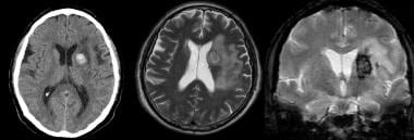
![Cerebral vein thrombosis secondary to closed head injury. Report of one case]. | Rev Med Chil;141(12): 1598-601, 2013 Dec. ...](data:image/png;base64,iVBORw0KGgoAAAANSUhEUgAAABAAAAAQCAYAAAAf8/9hAAACpklEQVQ4jaXTe2iNcRzH8ffz7Jzncp4ZZyZjZ6bmEvPPxrDIJSnJZZJIbn8Qk1CI2pQ7JX8h5fKHW4o/3EuaWnJvrUNqiFZssTWcs83z7DnPc87XH88Ziv98//z1+7369v38vvCfpQBY0w6LeHb2RCHl+fg9vaiGxuyq0ZQMzef0tSeoOSqGEQaR4Go4QghAPBvxHRzbBddjUGGUhXMmULN0KhVlxQDMLC+iZt9VEt+6MPNMCIwAAEj7aSrGxFg5r5KVCyoZGM2lx3Y5dbGB6jnlLJs7nvIxMZbvOE9TvAUjagXtAygVu2Tk3P3ieb6IiPjpjBw9Vy+F02uFYWulaEadnLn2WEREJCOyetdFYdRGMSfXSQ4ABVV7Jo4rYtWCiVyvf8nizWe4cuMZdkYwBlh863a4fa+JR/EWyscWs37pFAxD4/7T96hB/2lKhuYDcORsPW+aWzHz+6FrIRDB1MMY0Vwe3I9Td+IuALMmjyIjZAGBkiFRACKmBqb+77hyDQw9DED8TRukvCygKpQWFwCQyUb0zxIwtQD40NoZPAVQdY3S2EAAkj9cfo/3b0DJ+h8/fwdVCYA8S6d0WNBBbHB/6LJJeWmUPkiB3pQPPQ7RPDMAviQglBMAtuNSs/cqyW6HO8fXcaB2CZYWwk78IOWlcb72UDjA4tihFRzcOg+Ato4EaigIkUjVbmHEBimbf1Diza0iIvKupV2qN52W/MrtsuPoDUl22dJXl2+9EHP8NtEn1YnSB0i6F6fbwYronKxdwupFkwCwbZdIRKe9s5sLN59z6W4jr962oZs6OVrk91dGhEg/A8f1WbPzPI3Nnzi8ZT7NLe2cuPyQWw2vSXQmwdAwLIM/xvP3NmYyGXptl+GxAlo7kvi2S9gyCIfVX0sEwTb+BBBpI5QgousUAAAAAElFTkSuQmCC) Cerebral vein thrombosis secondary to closed head injury. Report of one case]. | Rev Med Chil;141(12): 1598-601, 2013 Dec. ...
Cerebral vein thrombosis secondary to closed head injury. Report of one case]. | Rev Med Chil;141(12): 1598-601, 2013 Dec. ...
 Acute Headache - StatPearls - NCBI Bookshelf
 Bassett Collection Large Image - Lane Medical Library, Stanford University Medical Center
 Choroid Plexus Papilloma Imaging: Practice Essentials, Pathophysiology, Epidemiology
Plus it
The perfect crime? CCSVI not leaving a trace in MS | Journal of Neurology, Neurosurgery & Psychiatry
Paroxysmal Nocturnal Hemoglobinuria: Practice Essentials, Pathophysiology, Etiology
 J&J Pause: 'Gasoline' for Vaccine Hesitancy | MedPage Today
 Gemmily: Package Insert - Drugs.com
Middle cerebral artery fenestration | Radiology Reference Article | Radiopaedia.org
 3b. 4. The Veins of the Brain - Collection at Bartleby.com
 Frontiers | Venous stroke-a stroke subtype that should not be ignored
 Thieme E-Books & E-Journals - Radiologie up2date / Neuroradiologie
Cerebral Angiographic Findings of Spontaneous Intracranial Hypotension | American Journal of Neuroradiology
Guidelines for the treatment of acute ischaemic stroke
 Barriers and opportunities of cortical stimulation via cerebral venous approach.
Complete Publications | Max Planck Institute for Human Cognitive and Brain Sciences
 COVID-19: Anyone with headache for more than four days after Oxford-AstraZeneca jab should seek medical attention, says UK...
 White matter haemodynamics: basic physiology and disruption in neuroinflammatory disease | bioRxiv
 Deep Vein Thrombosis | GreenMedInfo | Disease | Natural Medicine
 The Israel Medical Association Journal (IMAJ) | Search results
She Brushed Off Intense Headaches as Stress Until Her AVM (Arteriovenous Malformation) Diagnosis
Thrombosis32
- Red cell distribution width (RDW) is a marker of cardiovascular diseases and venous thromboembolism, but its role in cerebral vein thrombosis (CVT) is unknown. (efim.org)
- Objectives: To estimate the absolute risk of cerebral venous thrombosis (CVT) and portal vein thrombosis (PVT) in the two weeks following a diagnosis of COVID-19, and to assess the relative risks (RR) compared to influenza or the administration of an mRNA vaccine against COVID-19. (ox.ac.uk)
- Cerebral vein thrombosis secondary to closed head injury. (bvsalud.org)
- 1. Paediatric cerebral sinus vein thrombosis. (nih.gov)
- 2. [Thrombophilia and thrombosis in children--lessons from cerebral sinus vein thrombosis registry of a tertiary center]. (nih.gov)
- 3. Cerebral venous sinus thrombosis in children: risk factors, presentation, diagnosis and outcome. (nih.gov)
- 4. Long-term evaluation of the risk of recurrence after cerebral sinus-venous thrombosis. (nih.gov)
- 7. Conservative management of neonatal cerebral sinovenous thrombosis with coexisting thrombophilia. (nih.gov)
- 11. Cerebral sinus-venous thrombosis. (nih.gov)
- 13. Cerebral vein thrombosis in patients with Philadelphia-negative myeloproliferative neoplasms. (nih.gov)
- 15. Sagittal sinus compression is associated with neonatal cerebral sinovenous thrombosis. (nih.gov)
- 16. Laboratory Findings, Medical Imaging, and Clinical Outcome in Children with Cerebral Sinus Venous Thrombosis. (nih.gov)
- 17. Anticoagulants in pediatric cerebral sinovenous thrombosis: a safety and outcome study. (nih.gov)
- 18. Paediatric arterial ischaemic stroke and cerebral sinovenous thrombosis. (nih.gov)
- Symptomatic thrombotic or vaso-occlusive disease in past 90 days (e.g., cerebral infarction, myocardial infarction, pulmonary embolus, deep vein thrombosis, or unstable angina) 7. (nih.gov)
- A CT scan revealed multiple thromboses in his veins and arteries, including a pulmonary embolism and thrombosis in cerebral veins, the infrarenal aorta and two iliac arteries. (theepochtimes.com)
- Their platelets dropped to 1/5 to 1/7 of the lower limit of the normal count, and their blood vessels showed significant hypercoagulability, as well as cerebral vein thrombosis. (theepochtimes.com)
- In addition, the immediate cause of her death was "thrombosis with thrombocytopenia syndrome," resulting in brain infarction, elevated cerebral pressure and subsequent death from cerebral hemorrhage. (theepochtimes.com)
- On April 13, 2021, CDC and FDA recommended a pause in the use of Janssen COVID-19 vaccine after reports of thrombosis with thrombocytopenia syndrome (TTS), a rare condition characterized by low platelets and thrombosis, including at unusual sites such as the cerebral venous sinus (cerebral venous sinus thrombosis [CVST]), after receipt of the vaccine. (cdc.gov)
- BANU, L. A. M. Superior Ophthalmic Vein Thrombosis with Cerebral Venous Sinus Thrombosis: A Rare Entity in a Child. (banglajol.info)
- So all of these six cases happened in relatively younger women, and all of them involved blood clots in the brain, something that we call cerebral vein or cerebral sinus vein thrombosis. (nhpr.org)
- Jeremy Faust MD MS (ER physician) on Twitter: "Let's talk about the background risk of CVST (cerebral venous sinus thrombosis) versus in those who got J&J vaccine. (hypothes.is)
- The result was that, in rare cases, clots formed in the blood of vaccinated people which triggered cerebral venous sinus thrombosis. (technologynetworks.com)
- Those affected can safely be vaccinated a second time without the antibodies forming again and without having to fear a dangerous cerebral vein thrombosis. (technologynetworks.com)
- Vascular occlusions of the eye encompass thrombosis of retinal veins, arteries, and anterior ischaemic optic neuropathy. (bmj.com)
- Due to female-specific risk factors (oral contraceptive use and pregnancy), women are more prone than men to develop venous thrombosis at unusual sites, such as the cerebral circulation. (maastrichtuniversity.nl)
- Evidences were provided on the duration of oral contraceptive use as risk factor for venous thromboembolism, on the risk of obstetrical complications after a previous cerebral vein thrombosis, and on pregnancy-related risk of thrombosis in women with antithrombin type I deficit, a severe and rare thrombophilia abnormality. (maastrichtuniversity.nl)
- Cerebral vein and dural sinus thrombosis (CVT) is a rare but important complication of spontaneous intracranial hypotension (SIH). (surgicalneurologyint.com)
- However, during the procedure, the patient was diagnosed with low cerebrospinal fluid pressure and cerebral cortical vein thrombosis. (surgicalneurologyint.com)
- SIH is rarely known to cause cerebral vein and dural sinus thrombosis (CVT). (surgicalneurologyint.com)
- We confirmed low CSF pressure and thrombosis of the cortical vein during the craniotomy procedure and made a final diagnosis. (surgicalneurologyint.com)
- Class 3 (X-firm) medical-grade graduated compression also assists in the prevention of deep vein thrombosis. (bodyment.com)
Angiography7
- We investigated normal hemodynamic features of the SPS on cerebral angiography as well as the frequency and types of the SPS drainage from CSDAVFs. (ajnr.org)
- We evaluated 119 patients who underwent cerebral angiography by focusing on visualization and hemodynamic status of the SPS. (ajnr.org)
- It was demonstrated on carotid angiography in 11 sides (4.6%), receiving blood from the basal vein of Rosenthal or sphenopetrosal sinus, and on vertebral angiography in 235 sides (98.7%), receiving blood from the petrosal vein. (ajnr.org)
- For evaluating normal hemodynamic features of the SPS, we retrospectively reviewed 119 consecutive patients who underwent cerebral angiography between May 2010 and October 2011 at our institution. (ajnr.org)
- Three main techniques are used to visualize the brain and search for AVM: computed tomography (CT), magnetic resonance imaging (MRI), and cerebral angiography. (rochester.edu)
- The best images of an AVM are obtained through cerebral angiography. (rochester.edu)
- OBJECTIVE: We sought to determine if interhemispheric asymmetry of cortical and medullary veins evaluated on CT angiography can provide a more accurate prediction of outcome in patients with acute ischemic stroke when compared to hemispheric asymmetry of cortical or medullary vein drainage alone. (gwu.edu)
Posterior8
- A fetal (origin of the) posterior cerebral artery is a common variant in the posterior cerebral circulation , estimated to occur in 20-30% of individuals 2 . (radiopaedia.org)
- The posterior communicating artery (PCom) is larger than the P1 segment of the posterior cerebral artery (PCA) and supplies the bulk of the blood to the PCA 4 . (radiopaedia.org)
- 1. Yamamoto Y, Georgiadis A, Chang H, Caplan L. Posterior Cerebral Artery Territory Infarcts in the New England Medical Center Posterior Circulation Registry. (radiopaedia.org)
- Fetal-Type Variants of the Posterior Cerebral Artery and Concurrent Infarction in the Major Arterial Territories of the Cerebral Hemisphere. (radiopaedia.org)
- In all observed cases the posterior cerebral artery was involved on one or both sides, while the anterior cerebral artery was not always involved in the pathological circulation. (thieme-connect.com)
- The calcarine artery , named according to its course in the calcarine fissure , is a branch of the posterior cerebral artery , usually from the P3 segment. (radiopaedia.org)
- The retromandibular joins with the posterior auricular vein to create the external jugular vein. (healthline.com)
- Centrally located in the brain, the posterior cerebral artery makes up the lower portion of the circle of Willis. (healthline.com)
Amyloid angiopathy7
- Cerebral amyloid angiopathy (CAA) is a common SVD that has a high co-morbidity with Alzheimer's disease. (nih.gov)
- Instead, the brains contained large accumulations of prion protein plaques trapped outside blood vessels in a disease process called cerebral amyloid angiopathy. (nih.gov)
- Cerebral amyloid angiopathy is related to Alzheimer's disease and involves damage to arteries, veins and capillaries in the brain. (nih.gov)
- Cerebral amyloid angiopathy-related atraumatic convexal subarachnoid hemorrhage: an ARIA before the tsunami. (cdc.gov)
- Cortical superficial siderosis, APOE genotype, and hemorrhage risk in cerebral amyloid angiopathy. (cdc.gov)
- Cortical Superficial Siderosis in Memory Clinic Patients: Further Evidence for Underlying Cerebral Amyloid Angiopathy. (cdc.gov)
- Cortical superficial siderosis multifocality in cerebral amyloid angiopathy: A prospective study. (cdc.gov)
Galen7
- E , The vein of Galen was measured inferior to the splenium in the sagittal plane. (ajnr.org)
- Aneurysms of the vein of Galen develop as a result of a short circuit between the cerebral arteries and the internal cerebral veins. (thieme-connect.com)
- Three infants with vein Galen malformations, all presenting with congestive heart failure, underwent a total of five embolization procedures that employed a percutaneous transfemoral venous approach to catheterize the vein of Galen. (elsevierpure.com)
- In one instance, direct retrograde catheterization of feeding arterial pedicles to the vein of Galen and embolization of the fistulous connections was achieved via this route. (elsevierpure.com)
- Vein of Galen perforation with the catheter tip complicated one procedure. (elsevierpure.com)
- The transvenous route to the vein of Galen can be undertaken from a transfemoral approach, obviating surgical exposure of the torcular Herophili. (elsevierpure.com)
- In addition, we introduce the concept of direct retrograde catheterization of the feeding arteries to the vein of Galen malformation by a transfemoral venous approach, a procedure that has not been reported previously. (elsevierpure.com)
Hemorrhage1
- We work with neurologists and neuroradiologists in a multidisciplinary approach to care for patients who have suffered stroke or cerebral hemorrhage. (lifespan.org)
Infarction1
- [ 41 ] Neurological deterioration is potentially reversible if adequate blood flow through vasospastic cerebral vessels can be restored before infarction occurs. (medscape.com)
Anastomotic2
- The superior cerebral veins include the superior anastomotic vein. (wikipedia.org)
- Note that the great anastomotic vein (of Trolard) (31) lies along the postcentral sulcus instead of the central sulcus as it commonly does. (stanford.edu)
Superficial midd1
- OBJECTIVE: The drainage of the superficial middle cerebral vein (SMCV) has previously been classified into 4 subtypes. (elsevierpure.com)
Anatomy1
- In human anatomy, the cerebral veins are blood vessels in the cerebral circulation which drain blood from the cerebrum of the human brain. (wikipedia.org)
Basal veins1
- B , The diameters of the left and right basal veins of Rosenthal were measured at their termini in the axial plane and then averaged. (ajnr.org)
Medullary vein2
- Deep medullary vein (DMV) changes occur in cerebral small vessel diseases (SVD) and in Alzheimer's disease. (nih.gov)
- CONCLUSION: Combined cortical and medullary vein interhemispheric asymmetry is a stronger predictor of clinical outcome in acute ischemic stroke compared to cortical or medullary vein asymmetry alone. (gwu.edu)
Cerebrum1
- Veins draining the cerebrum. (bvsalud.org)
Inferior1
- The external cerebral veins known as the superficial cerebral veins are the superior cerebral veins, inferior cerebral veins, and middle cerebral veins. (wikipedia.org)
Arteries and veins4
- Cerebral AVMs are abnormal growths of tangled arteries and veins in the brain. (lifespan.org)
- Cerebral AVFs are abnormal short-cuts between arteries and veins that bypass the small capillaries that normally connect them. (lifespan.org)
- A cerebral arteriovenous malformation (AVM) is an abnormal connection between the arteries and veins in the brain. (rochester.edu)
- Detection of damage to blood vessels in the brain (both arteries and veins). (hostandcare.com)
Cortical vein2
- Cortical veins were evaluated using the adopted Prognostic Evaluation based on Cortical vein score difference In Stroke (PRECISE) system. (gwu.edu)
- In a multivariable logistic regression model, combined medullary and cortical vein asymmetry were independently associated with clinical outcomes (AUC=0.721). (gwu.edu)
Lesions2
- Cerebral lesions and neurologic signs develop in half of patients with IST. (bvsalud.org)
- Patients who had lesions affecting cerebral venous drainage were excluded from this study. (ajnr.org)
Dural1
- The authors performed 3 ATPA modifications to preserve the SMCV: epidural anterior petrosectomy with subdural visualization of the sphenobasal vein (SBV), modification of the dural incision, and subdural anterior petrosectomy. (elsevierpure.com)
Vascular2
- We report on recent research in cerebral vascular tortuosity (curvature in three dimensions) which would benefit greatly from internal pilots due to uncertainty in the parameters of the covariance matrix used for study planning. (nih.gov)
- El impacto que la enfermedad vascular cerebral (EVC) tiene sobre la mortalidad y morbilidad en pa ses en v as de desarrollo como en los desarrollados es alarmante. (medigraphic.com)
Arterial1
- [ 25 , 30 ] The effects of angioplasty on arterial size are very evident, but its effects on cerebral hemodynamics have not been thoroughly evaluated. (medscape.com)
Brain11
- Changes in blood oxygenation arising from local brain activity-related changes in blood flow propagate downstream in veins and can give rise to spurious activation at sites remote from neuronal activity. (nih.gov)
- Cerebral hypoxia occurs when there is not enough oxygen getting to the brain. (medlineplus.gov)
- Cerebral hypoxia affects the largest parts of the brain, called the cerebral hemispheres. (medlineplus.gov)
- Sometimes a person with cerebral hypoxia is cooled to slow down the activity of the brain cells and decrease their need for oxygen. (medlineplus.gov)
- Researchers know that the symptoms of cerebral malaria are caused when the brain swells within the confines of the skull, eventually impinging upon the brainstem, which causes breathing to stop. (nih.gov)
- Partial blockages inside the cerebral veins could slow the flow of blood leaving the brain, causing the blood vessels themselves to become engorged and expand the brain from within. (nih.gov)
- This illustration shows two different ways that cerebral malaria could cause brain swelling: fluid seeping into the brain (extravascular edema) or swollen blood vessels (venous congestion. (nih.gov)
- The researchers found that children with cerebral malaria had significantly higher concentrations of hemoglobin inside their brains than children who had uncomplicated malaria, or healthy children-and among those with cerebral malaria, higher hemoglobin concentrations were correlated with greater brain swelling. (nih.gov)
- In addition, hemoglobin concentrations fluctuated more unpredictably in children with cerebral malaria-suggesting that the normal mechanisms for controlling blood flow in the brain were disturbed. (nih.gov)
- Together, these results support the hypothesis that obstruction to the outflow of blood from the brain, likely because the blood vessels themselves are clogged by red blood cells infected with P. falciparum adhering to the walls, is a main driver of brain swelling in patients with cerebral malaria. (nih.gov)
- Most epidural hematomas, a condition where blood that accumulates between the skull and the dura mater (protective tissue surrounding the brain), are usually caused by skull fractures that cross either the middle meningeal artery or the middle meningeal vein. (healthline.com)
Retinal3
- 3-6 Branch retinal vein occlusions were particularly linked with hypertension. (bmj.com)
- 7 8 In addition, hyperviscosity syndromes including a raised haematocrit and elevated erythrocyte aggregation rate, as well as other prothrombotic states (for example, hypofibrinolysis, increased levels of tissue factor), induced by increased levels of lipoprotein (a), malignancy, pregnancy, oestrogen therapy, and paroxysmal nocturnal haemoglobulinuria have all been associated with retinal vein occlusion. (bmj.com)
- In addition to the factors described above, there are associations between retinal vein occlusions and inherited defects in the proteins of the coagulation pathways. (bmj.com)
Pulmonary embolism1
- The aim of this study is to investigate the appearance of cerebral veins on SWI, which is very sensitive to the deoxyhaemoglobin level in vessels, in pulmonary embolism (PE). (comu.edu.tr)
Superior3
- The superior cerebral veins on the upper side surfaces of the hemispheres drain into the superior sagittal sinus. (wikipedia.org)
- To evaluate the veins of Labbé and Trolard, the straight sinus, and the superior sagittal sinus (SSS), measurements were performed in a blinded manner with the aid of a magnification loupe. (medscape.com)
- The diagnostic diameter of superior ophthalmic vein (SOV) in tCCF was 4 mm with area under curve of 0.89. (cns.org)
Venous system1
- Because venous blood accounts for 60% of the intracranial blood volume, [ 39 ] assessment of the effect of endovascular therapy on the intracranial venous system may contribute to a greater understanding of cerebral hemodynamics. (medscape.com)
Trolard1
- The straight sinus and the SSS increased more in diameter than the veins of Labbé and Trolard. (medscape.com)
Acute cerebral1
- Axial image from diffusion-weighted magnetic resonance imaging (MRI) reveals no abnormalities that indicate acute cerebral ischemia (b). (surgicalneurologyint.com)
Circulation1
- Cerebral circulation and oxygen consumption elevate. (medscape.com)
Blockages1
- CT of the veins - CTV (abbreviation of CT venography) - makes it possible to get a picture of the cerebral veins and identify different pathologies such as blockages. (hostandcare.com)
Artery1
- The middle meningeal vein parallels the path of the middle meningeal artery. (healthline.com)
Cerebellar1
- We postulate that the mature fluke pair migrated from the mesenteric veins through Batson's vertebral-venous plexus to the cerebral veins at the cerebellar level. (cdc.gov)
Cortex2
- How much cortex can a vein drain? (nih.gov)
- Beta-adrenergic binding sites in the cerebral cortex are increased and gamma-aminobutyric acid (GABA) binding sites are decreased. (medscape.com)
Severe2
- The draining vein problem is recognized as one of the most severe constraints on the spatial resolution of BOLD contrast fMRI, used widely in imaging neuroscience. (nih.gov)
- In one of the deadliest forms of malaria, known as cerebral malaria, the patient experiences severe neurological symptoms, such as seizures and coma. (nih.gov)
Drain2
- They are divisible into external (superficial cerebral veins) and internal (internal cerebral veins) groups according to the outer or inner parts of the hemispheres they drain into. (wikipedia.org)
- The internal cerebral veins are also known as the deep cerebral veins and drain the deep internal parts of the hemispheres. (wikipedia.org)
Ischemia1
- Cerebral endothelial cells are more resistant to ischemia than are neurons and neuroglial cells. (medscape.com)
Diameters1
- A , The diameters of the left and right internal cerebral veins were measured in the axial plane and then averaged. (ajnr.org)
Drains1
- That vein drains into the subclavian vein. (healthline.com)
Patients1
- The Eye Disease Case Control Study Group identified a number of risk factors for branch, central, and hemiretinal vein occlusions including hypertension, diabetes, a history of cardiovascular disease, an increased body mass index at 20 years of age, and patients with open angle glaucoma. (bmj.com)
Lateral1
- Type 2 was subdivided into medial (Type 2a) and lateral (Type 2b), and Type 3 was subdivided into vein (Type 3a), vein and sinus (Type 3b), and sinus (Type 3c). (elsevierpure.com)
Complications1
- Complications of cerebral hypoxia include a prolonged vegetative state. (medlineplus.gov)