Maxillary Osteotomy
Palate, Soft
Orthognathic Surgery
Chin
Nasal Bone
Pharynx
Mandible
Vertical Dimension
Maxilla
Face
Sleep Apnea Syndromes
Quantitative assessment of the morphology of the pig's head used as a model in surgical experimentation. Part 1: Methods of Measurements. (1/1457)
Thirty-two surface measurements were described for assessment of the effect of complex surgical operations on the skeleton of the face in pigs. The methods of measurements imitate those of anthropometry. The surface measurements can complement cephalometry with data about the changes in the soft tissue and thus improve the documentation of the effect of surgery. This paper can help in the evaluation of complicated osteotomy procedures using the pig as the animal model, for facial reconstruction research in humans. (+info)Effect of obesity and erect/supine posture on lateral cephalometry: relationship to sleep-disordered breathing. (2/1457)
Craniofacial and upper airway anatomy, obesity and posture may all play a role in compromising upper airway patency in patients with the sleep apnoea/hypopnoea syndrome. The aim of this study was to investigate the relationship between obesity, facial structure and severity of sleep-disordered breathing using lateral cephalometric measurements and to assess the effect of body posture on cephalometric measurements of upper airway calibre variables in obese and non-obese subjects. Lateral cephalometry was carried out in erect and supine postures in 73 awake male subjects randomly selected from patients referred for polysomnography who had a wide range of apnoea/hypopnoea frequencies (1-131 events x h sleep(-1)). Subjects were divided into non-obese (body mass index (BMI) < 30 kg x m(-2); n=42) and obese (BMI > or = 30 kg x m(-2); n=31) groups. Significant but weak correlations were found between apnoea/hypopnoea index (AHI) and measurements reflecting upper airway dimensions: uvular protrusion-posterior pharyngeal wall (r=-0.26, p<0.05) and hyoid-posterior pharyngeal wall (r=0.26, p<0.05). Multiple regression using both upper airway dimensions improved the correlation to AHI (r=0.34, p=0.01). Obese subjects had greater hyoid-posterior pharyngeal wall distances than non-obese subjects, both erect (42+/-5 versus 39+/-4 mm, respectively (mean+/-SD) p<0.01) and supine (43+/-5 versus 40+/-4 mm, p<0.05). Skeletal craniofacial structure was similar in obese and non-obese subjects. In conclusion, measurements reflecting upper airway size were correlated with the severity of sleep-disordered breathing. Differences in upper airway size measurements between obese and non-obese subjects were independent of bony craniofacial structure. (+info)Cephalometric abnormalities in non-obese and obese patients with obstructive sleep apnoea. (3/1457)
The aim of this work was to comprehensively evaluate the cephalometric features in Japanese patients with obstructive sleep apnoea (OSA) and to elucidate the relationship between cephalometric variables and severity of apnoea. Forty-eight cephalometric variables were measured in 37 healthy males and 114 male OSA patients, who were classed into 54 non-obese (body mass index (BMI) <27 kg x m(-2), apnoea-hypopnoea index (AHI)=25.3+/-16.1 events x h(-1)) and 60 obese (BMI > or = 27 kg x m(-2), AHI=45.6+/-28.0 events h(-1)) groups. Diagnostic polysomnography was carried out in all of the OSA patients and in 19 of the normal controls. The non-obese OSA patients showed several cephalometric defects compared with their BMI-matched normal controls: 1) decreased facial A-P distance at cranial base, maxilla and mandible levels and decreased bony pharynx width; 2) enlarged tongue and inferior shift of the tongue volume; 3) enlarged soft palate; 4) inferiorly positioned hyoid bone; and 5) decreased upper airway width at four different levels. More extensive and severe soft tissue abnormalities with a few defects in craniofacial bony structures were found in the obese OSA group. For the non-obese OSA group, the stepwise regression model on AHI was significant with two bony structure variables as determinants: anterior cranial base length (S-N) and mandibular length (Me-Go). Although the regression model retained only linear distance between anterior vertebra and hyoid bone (H-VL) as an explainable determinant for AHI in the obese OSA group, H-VL was significantly correlated with soft tissue measurements such as overall tongue area (Ton), inferior tongue area (Ton2) and pharyngeal airway length (PNS-V). In conclusion, Japanese obstructive sleep apnoea patients have a series of cephalometric abnormalities similar to those described in Caucasian patients, and that the aetiology of obstructive sleep apnoea in obese patients may be different from that in non-obese patients. In obese patients, upper airway soft tissue enlargement may play a more important role in the development of obstructive sleep apnoea, whereas in non-obese patients, bony structure discrepancies may be the dominant contributing factors for obstructive sleep apnoea. (+info)Craniofacial modifications in children with habitual snoring and obstructive sleep apnoea: a case-control study. (4/1457)
Habitual snoring and obstructive sleep apnoea in children, which are frequently associated with adenotonsillar hypertrophy, may begin early in life and in relation with orocraniofacial features. The aim of this study was to detect the presence of early bone craniofacial modifications in young children with a long history of habitual snoring. Twenty-six habitually snoring children (mean age 4.6 yrs) were studied by nocturnal portable recording or diurnal polysomnography, cephalometry and orthodontic evaluation. A comparison of cephalometric findings was made between the studied group and 26 age-matched children (mean age 5.1 yrs) with no history of snoring or respiratory problems during sleep. The cephalometric analyses showed a significant increase in craniomandibular intermaxillar, lower and upper goniac angles with a retroposition and posterior rotation of the mandible (high angle face) and a reduction in the rhinopharynx space caused by higher thickness of adenoids in habitually snoring children compared with controls. Cross-bites and labial incompetence as well as daytime symptoms and familiarity for habitual snoring were found in most of the studied group of snorers compared with controls. The results indicate that upper airway obstruction during sleep is associated with mild but significant cephalometric and craniofacial modifications in children complaining of habitual snoring. Whether this skeletal conformation is genetically determined or influenced by the early onset of habitual snoring remains to be assessed. (+info)The problem of the class iii malocclusion. (5/1457)
The etiology and treatment of Class III malocclusion has been discussed. The value of electromyographic assessment in the assessment and prediction of Class III malocclusion has been shown. (+info)The interfrontal bone and mutant genes in the mouse. (6/1457)
The relationship between corrected skull width and the presence and size of an interfrontal bone is discussed with regard to the effect of certain mutant genes in the mouse known to affect the development of the neural tube. All genes reviewed which increase the incidence of the interfrontal bone and affect the neural tube also change the proportions of the adult skull. (+info)Comparison of cephalometric analysis using a non-radiographic sonic digitizer (DigiGraph Workstation) with conventional radiography. (7/1457)
Cephalometric analysis conventionally requires radiographic exposure which may not be compatible with the growing concern over radiation hazards. Recently, the Dolphin Workstation Imaging System introduced to the dental profession a non-radiographic system, called the DigiGraph Workstation which may be an alternative to cephalometric radiography. The aims of this study were to compare the validity and reproducibility of cephalometric measurements obtained from the DigiGraph Workstation with conventional cephalometric radiographs. The sample consisted of 30 human dry skulls. Two replicated sets of lateral cephalograms were obtained with steel ball markers placed at the majority of the cephalometric landmarks. Duplicate tracings prepared from each radiograph were digitized to obtain cephalometric measurements using the computer software, Dentofacial Planner. For the DigiGraph Workstation, double sonic digitizations were repeated twice for each skull, on two occasions. Fifteen angular and one linear measurements were obtained from both methods and these findings compared using ANOVA, paired t-tests and F-tests. All, except one, cephalometric measurement showed significant differences between the two methods (P < 0.0001). The DigiGraph Workstation consistently produced higher values in 11 measurements (mean differences +0.5 to +15.7 degrees or mm) and lower values in four measurements (mean differences -0.2 to -3.5 degrees). The standard deviations of the differences between readings of both methods were large (0.4-5.8 degrees or mm). The reproducibility of the DigiGraph Workstation measurements was lower than that of the radiographic measurements. The method error of the DigiGraph Workstation ranged from 7 to 70 per cent, while that of radiographic tracings was less than 2 per cent. It was concluded that measurements obtained with the DigiGraph Workstation should be interpreted with caution. (+info)Arrested eruption of the permanent lower second molar. (8/1457)
The incidence of retention/impaction of the permanent lower second molar (M2inf) lies between 0.6/1000 and 3/1000. Therefore, the purpose of the present study was to investigate the craniofacial morphology, the frequency of dental anomalies and the inclination of the affected M2inf and the adjacent first molar in patients with arrested eruption of M2inf. The overall goal was to elucidate the aetiology of arrested tooth eruption and to present the characteristics of these patients in order to improve diagnosis and treatment planning. Radiographic material (profile radiographs and orthopantomograms) from 19 patients (nine females and 10 males; 13-19 years of age at the time of referral) were analysed. The ages of the patients when profile radiographs were taken for cephalometric analysis varied from 8 to 16 years. The study shows that this group of patients, compared with a reference group, had an increased sagittal jaw relationship (Class II). Specifically, the mandibular prognathism was less, the mandibular gonial angle smaller, the mandibular alveolar prognathism enlarged and the maxillary incisor inclination less than in the reference group. Furthermore, this group of patients had a more frequent occurrence of morphological tooth anomalies, such as root deflections, invaginations, and taurodontism. However, none of the patients with arrested eruption of M2inf had agenesis of the lower third molar. The study did not reveal an association between the degree of inclination of the M2inf and that of the first molar in the same region. The results of this investigation show that conditions such as the craniofacial morphology and deviations in the dentition are associated with arrested eruption of M2inf. Therefore, it is important to evaluate these conditions in future diagnosis and treatment planning of patients with arrested eruption of M2inf. (+info)Cephalometry is a medical term that refers to the measurement and analysis of the skull, particularly the head face relations. It is commonly used in orthodontics and maxillofacial surgery to assess and plan treatment for abnormalities related to the teeth, jaws, and facial structures. The process typically involves taking X-ray images called cephalograms, which provide a lateral view of the head, and then using various landmarks and reference lines to make measurements and evaluate skeletal and dental relationships. This information can help clinicians diagnose problems, plan treatment, and assess treatment outcomes.
A maxillary osteotomy is a surgical procedure that involves making cuts in the bone of the upper jaw (maxilla). This type of surgery may be performed for various reasons, such as to correct jaw deformities, realign the jaws, or treat sleep apnea. In some cases, it may also be done in conjunction with other procedures, such as a genioplasty (chin surgery) or rhinoplasty (nose surgery).
During a maxillary osteotomy, an incision is made inside the mouth, and the surgeon carefully cuts through the bone of the upper jaw. The maxilla is then repositioned as needed and held in place with small plates and screws. In some cases, bone grafts may also be used to help support the new position of the jaw. After the surgery, the incision is closed with stitches.
It's important to note that a maxillary osteotomy is a complex surgical procedure that requires careful planning and execution. It should only be performed by an experienced oral and maxillofacial surgeon or craniofacial surgeon. As with any surgery, there are risks involved, including infection, bleeding, and reactions to anesthesia. It's important to discuss these risks with your surgeon and to follow all post-operative instructions carefully to help ensure a successful recovery.
The soft palate, also known as the velum, is the rear portion of the roof of the mouth that is made up of muscle and mucous membrane. It extends from the hard palate (the bony front part of the roof of the mouth) to the uvula, which is the small piece of tissue that hangs down at the back of the throat.
The soft palate plays a crucial role in speech, swallowing, and breathing. During swallowing, it moves upward and backward to block off the nasal cavity, preventing food and liquids from entering the nose. In speech, it helps to direct the flow of air from the mouth into the nose, which is necessary for producing certain sounds.
Anatomically, the soft palate consists of several muscles that allow it to change shape and move. These muscles include the tensor veli palatini, levator veli palatini, musculus uvulae, palatopharyngeus, and palatoglossus. The soft palate also contains a rich supply of blood vessels and nerves that provide sensation and help regulate its function.
Orthognathic surgery, also known as corrective jaw surgery, is a surgical procedure performed to correct and realign the bones of the jaws and face to improve their function and appearance. The surgery is typically recommended when there are significant skeletal discrepancies or dental malocclusions that cannot be corrected with orthodontic treatment alone.
Orthognathic surgery involves making precise cuts in the jawbones, repositioning them, and securing them in their new position using plates, screws, or wires. The procedure can be performed on the upper jaw (maxilla), lower jaw (mandible), or both, depending on the nature of the problem.
The goals of orthognathic surgery include improving bite function, chewing and swallowing ability, speech, breathing, and facial aesthetics. Patients who undergo this surgery often experience significant improvements in their quality of life and self-confidence. However, it is important to note that orthognathic surgery requires careful planning, coordination between the oral surgeon and orthodontist, and a commitment to post-surgical recovery and rehabilitation.
The "chin" is the lower, prominent part of the front portion of the jaw in humans and other animals. In medical terms, it is often referred to as the mentum or the symphysis of the mandible. The chin helps in protecting the soft tissues of the mouth and throat during activities such as eating, speaking, and swallowing. It also plays a role in shaping the overall appearance of the face. Anatomically, the chin is formed by the fusion of the two halves of the mandible (lower jawbone) at the symphysis menti.
The nasal bones are a pair of small, thin bones located in the upper part of the face, specifically in the middle of the nose. They articulate with each other at the nasal bridge and with the frontal bone above, the maxillae (upper jaw bones) on either side, and the septal cartilage inside the nose. The main function of the nasal bones is to form the bridge of the nose and protect the nasal cavity. Any damage to these bones can result in a fracture or broken nose.
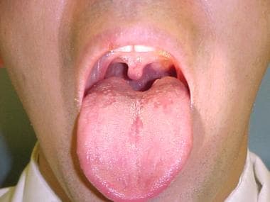
The pharynx is a part of the digestive and respiratory systems that serves as a conduit for food and air. It is a musculo-membranous tube extending from the base of the skull to the level of the sixth cervical vertebra where it becomes continuous with the esophagus.
The pharynx has three regions: the nasopharynx, oropharynx, and laryngopharynx. The nasopharynx is the uppermost region, which lies above the soft palate and is connected to the nasal cavity. The oropharynx is the middle region, which includes the area between the soft palate and the hyoid bone, including the tonsils and base of the tongue. The laryngopharynx is the lowest region, which lies below the hyoid bone and connects to the larynx.
The primary function of the pharynx is to convey food from the oral cavity to the esophagus during swallowing and to allow air to pass from the nasal cavity to the larynx during breathing. It also plays a role in speech, taste, and immune defense.
The mandible, also known as the lower jaw, is the largest and strongest bone in the human face. It forms the lower portion of the oral cavity and plays a crucial role in various functions such as mastication (chewing), speaking, and swallowing. The mandible is a U-shaped bone that consists of a horizontal part called the body and two vertical parts called rami.
The mandible articulates with the skull at the temporomandibular joints (TMJs) located in front of each ear, allowing for movements like opening and closing the mouth, protrusion, retraction, and side-to-side movement. The mandible contains the lower teeth sockets called alveolar processes, which hold the lower teeth in place.
In medical terminology, the term "mandible" refers specifically to this bone and its associated structures.
The term "vertical dimension" is used in dentistry, specifically in the field of prosthodontics, to refer to the measurement of the distance between two specific points in the vertical direction when the jaw is closed. The most common measurement is the "vertical dimension of occlusion," which is the distance between the upper and lower teeth when the jaw is in a balanced and comfortable position during resting closure.
The vertical dimension is an important consideration in the design and fabrication of dental restorations, such as dentures or dental crowns, to ensure proper function, comfort, and aesthetics. Changes in the vertical dimension can occur due to various factors, including tooth loss, jaw joint disorders, or muscle imbalances, which may require correction through dental treatment.
The maxilla is a paired bone that forms the upper jaw in vertebrates. In humans, it is a major bone in the face and plays several important roles in the craniofacial complex. Each maxilla consists of a body and four processes: frontal process, zygomatic process, alveolar process, and palatine process.
The maxillae contribute to the formation of the eye sockets (orbits), nasal cavity, and the hard palate of the mouth. They also contain the upper teeth sockets (alveoli) and help form the lower part of the orbit and the cheekbones (zygomatic arches).
Here's a quick rundown of its key functions:
1. Supports the upper teeth and forms the upper jaw.
2. Contributes to the formation of the eye sockets, nasal cavity, and hard palate.
3. Helps shape the lower part of the orbit and cheekbones.
4. Partakes in the creation of important sinuses, such as the maxillary sinus, which is located within the body of the maxilla.
In medical terms, the face refers to the front part of the head that is distinguished by the presence of the eyes, nose, and mouth. It includes the bones of the skull (frontal bone, maxilla, zygoma, nasal bones, lacrimal bones, palatine bones, inferior nasal conchae, and mandible), muscles, nerves, blood vessels, skin, and other soft tissues. The face plays a crucial role in various functions such as breathing, eating, drinking, speaking, seeing, smelling, and expressing emotions. It also serves as an important identifier for individuals, allowing them to be recognized by others.
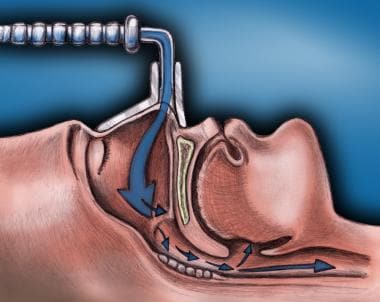
Sleep apnea syndromes refer to a group of disorders characterized by abnormal breathing patterns during sleep. These patterns can result in repeated pauses in breathing (apneas) or shallow breaths (hypopneas), causing interruptions in sleep and decreased oxygen supply to the body. There are three main types of sleep apnea syndromes:
1. Obstructive Sleep Apnea (OSA): This is the most common form, caused by the collapse or obstruction of the upper airway during sleep, often due to relaxation of the muscles in the throat and tongue.
2. Central Sleep Apnea (CSA): This type is less common and results from the brain's failure to send proper signals to the breathing muscles. It can be associated with conditions such as heart failure, stroke, or certain medications.
3. Complex/Mixed Sleep Apnea: In some cases, a person may experience both obstructive and central sleep apnea symptoms, known as complex or mixed sleep apnea.
Symptoms of sleep apnea syndromes can include loud snoring, excessive daytime sleepiness, fatigue, morning headaches, difficulty concentrating, and mood changes. Diagnosis typically involves a sleep study (polysomnography) to monitor breathing patterns, heart rate, brain activity, and other physiological factors during sleep. Treatment options may include lifestyle modifications, oral appliances, positive airway pressure therapy, or even surgery in severe cases.
 Cephalometry
Cecil C. Steiner
Holly Broadbent Sr.
Long face syndrome
Facial width to height ratio
Cephalometric analysis
Cephalogram
Cone beam computed tomography
Vineland Training School
Natural head position
Condylar hyperplasia
McGillivray syndrome
James A. McNamara
Jaw abnormality
Medical ultrasound
Craniometry
Kunság
Anthropometry
List of MeSH codes (G03)
List of MeSH codes (E01)
Historical race concepts
Cephalometry - Wikipedia
 Upper Airway Evaluation in Snoring and Obstructive Sleep Apnea: Overview of OSA, Relevant Upper Airway Anatomy, Pathologic...
 Orthodontic Cephalometry, 1st Edition - Dental-library.Net
 Subjects: Cephalometry - Digital Collections - National Library of Medicine Search Results
NLM Classification 2021 Summer Edition Now Available. NLM Technical Bulletin. 2021 Sep-Oct
 JCM | Special Issue : Positional Cranial Deformation: Etiology, Natural History, Prevention, Treatment and Sequelae
 All Discussions - Orthodontics - WebDental, LLC
JOMR | Assessment of Soft Tissue Changes by Cephalometry and Two-Dimensional Photogrammetry in Bilateral Sagittal Split Ramus...
Hypercapnia in Obstructive Sleep Apnea
 Browsing EMRO Journal Articles (EMHJ) by Subject
Publication Detail
IndexCat
 The fetal mandible: a 2D and 3D sonographic approach to the diagnosis of retrognathia and micrognathia - PubMed
 Facial anthropometry of Hong Kong Chinese babies - PubMed
 Fetal origins of coronary heart disease | The BMJ
Lexical Tools
Timothy Cootes - Research output - Research Explorer The University of Manchester
Biomarkers Search
 이화여자대학교 교수소개
Case Report 1, Issue 4.3
 Stereophotogrammetry-Based Facial Depth Measurements, A Novel Method for Quantifying Facial Projection. YSN Jayaratne, CK...
 An Evaluation of Cellular Neural Networks for the Automatic Identification of Cephalometric Landmarks on Digital Images
Breathing-Related Sleep Disorder: Overview, Etiology, Epidemiology
 Ortodonție - Quintessence Publishing East
 Francesco Pedetta | New Straight Wire | Quintessenz Verlags-GmbH
 DeCS
DeCS
 MeSH Browser
MeSH Browser
Code System Concept
 Pesquisa | Biblioteca Virtual em Saúde
Malocclusion2
- Using simplified cephalometry and treatment mechanics, this technique uses a table to systematically calculate and plan the necessary tooth movements before treatment, following the same parameters for each and every patient regardless of malocclusion. (quintessence-publishing.com)
- activator appliances, malocclusion, cephalometry. (bvsalud.org)
Lateral2
- 4. A lateral cephalometry study of patients with neurofibromatosis type 1. (nih.gov)
- [ 1 - 3 ] After the advent of cephalometry more and more stress was laid on the lateral cephalograms and the data provided them to decide the treatment plan. (apospublications.com)
Orthodontists2
- 1931, orthodontists consecrated the era of cephalometry. (wikipedia.org)
- A national survey among Dutch orthodontists by the end of 2000 demonstrated that 35% of Dutch orthodontists used digital cephalometry in their office. (ru.nl)
Fetal2
- Thus far, no ill effects have been reported to the fetus or the mother using the ultrasound fetal cephalometry. (wikipedia.org)
- Fetal cephalometry and thoracometry during pathological pregnancies]. (bvsalud.org)
Obstetric1
- Certain 3D imaging applications are now used in obstetric cephalometry. (wikipedia.org)
Craniometry1
- Craniometry, the measurement of the cranium (skull), is a large subset of cephalometry. (wikipedia.org)
Pantomography1
- We provide modern, comprehensive radiological diagnostics - 3D tapered tomography with wide field of imaging, 2D cephalometry, 3D pantomography and radiovision. (kcmclinic.com)
CBCT1
- 3D Cephalometry (ceph analysis using CBCT images). (digitalbuzznews.com)
Ultrasound3
- Ultrasound cephalometry is useful for determining baby growth in utero. (wikipedia.org)
- The use of ultrasound cephalometry is meant to be used in addition to other radiographic techniques. (wikipedia.org)
- The radiology department offers state-of-the-art equipment including a patient friendly open MRI (0.35 tesla) with computerized radiography system, ultrasound with color doppler, mammogram, X-Ray system, fluoroscopy, OPG, and cephalometry. (nmc.ae)
Assessment1
- 76 children aged 7 to 10 years old were examined by otorhinolaryngological evaluation, polysomnography, and orthodontic assessment, including cephalometry. (bjorl.org)
Scan1
- Possibly a Cephalometry scan. (surgeryinperu.com)
Facial3
- Cephalometry also has a history in phrenology, which is the study of personality and character as well as physiognomy, which is the study of facial features. (wikipedia.org)
- Cephalometry focuses on linear and angular dimensions established by bone, teeth, and facial measurements. (wikipedia.org)
- Traditional 2-dimensional cephalometry and photographic techniques do not provide data on facial depth. (3dmd.com)
Clinical1
- Case Management: A clinical examination showed a class I relationship between the dental and cephalometry measurements and highlighted a class I skeletal pattern. (unair.ac.id)
Possibility2
- Our findings suggest that cephalometry and two-dimensional photogrammetry offer the possibility to complement one another. (ejomr.org)
- A well-designed programme for digital cephalometry should have the possibility to calculate age and gender-related reference values based on values of the target population. (ru.nl)
Measurement3
- Cephalometry is the study and measurement of the head, usually the human head, especially by medical imaging such as radiography. (wikipedia.org)
- The history of cephalometry (cephalo- + -metry, "head measurement") can be traced through art, science, and anthropology. (wikipedia.org)
- Evidence for these explanations has been documented through application of upper airway manometry, measurement of pharyngeal cross-sectional area, videoendoscopy, and cephalometry. (clinicalgate.com)
Analysis2
- 2D Cephalometry (ceph analysis, surgical planning, and treatment follow-ups in 2D). (digitalbuzznews.com)
- With a digital head film and a computer programme for digital cephalometry an analysis can be performed easily. (ru.nl)
Methods1
- We aimed to compare the standard methods of cephalometry and two-dimensional photogrammetry, to evaluate the reliability and accuracy of both methods. (ejomr.org)
Patient1
- 2 ]. Cephalometry and two-dimensional photogrammetry have been more advantageous with regard to high patient comfort, portability, costs, and accessibility, compared with three-dimensional photogrammetry. (ejomr.org)
Head1
- Cephalometry is an important part of physical Anthropology and medicine which is used for the determination of the morphological characteristics of the head. (ijars.net)
Compare1
- All existing computer programmes for digital cephalometry use reference values to compare with the patient's values. (ru.nl)
Child1
- Cephalometry can also determine if an unborn child will pass through the birth canal. (wikipedia.org)