Celiac Artery
Mesenteric Artery, Superior
Splenic Artery
Mesenteric Vascular Occlusion
Constriction, Pathologic
Aneurysm
Ligaments
Venous Cutdown
Arterial Occlusive Diseases
Tuberculosis, Cardiovascular
Mesenteric Arteries
Celiac Plexus
Splanchnic Circulation
Splenic Infarction
Hepatic Artery
Pulmonary Artery
Radiography, Interventional
Tomography, X-Ray Computed
Vascular Access Devices
Carotid Arteries
Aortic Aneurysm, Thoracic
Blood Vessel Prosthesis Implantation
Stents
Decompression, Surgical
Basilar Artery
Aortography
Viscera
Aorta, Abdominal
Ischemia
Aneurysm, Dissecting
Iliac Artery
Treatment Outcome
Vertebral Artery
Coronary Artery Bypass
Angioplasty, Balloon
Radial Artery
Anastomosis, Surgical
Mammary Arteries
Collateral Circulation
Endovascular Procedures
Carotid Artery, Internal
Endoleak
Embolization, Therapeutic
Subclavian Artery
Blood Vessel Prosthesis
Carotid Artery Diseases
Observations on some additional abnormalities in situs inversus viscerum. (1/266)
The abnormal findings in a case of Situs inversus totalis are described. The duodenum was placed abnormally and retained its primitive mesentery. The proximal 22 in of jejunum were retroperitoneal. The attachment of the root of the mesentery to the posterior abdominal wall had a 7-shaped appearance, and there was a partial failure of the primitive mesocolon to adhere to the posterior abdominal wall. The common hepatic artery arose from the superior meseneric artery, which also provided a branch to the proximal jejunal loop. The right vagus nerve was found anterior to the oesophagus at the oesophageal hiatus in the diaphragm, and the left vagus was posterior. A double ureter was present on the right side. The findings are discussed in relation to mid-gut development. (+info)Celiomesenteric anomaly with concurrent aneurysm. (2/266)
We describe a rare case of a celiomesenteric anomaly with concurrent aneurysm. The patient, a 53-year-old man, had no abdominal pain or discomfort. The presence of a celiac artery aneurysm was suspected on the basis of the results of abdominal computerized tomographic scanning and echo ultrasound scanning performed because of proteinuria. Intra-arterial digital subtraction angiographic results showed the anomaly and aneurysm. Because of the risk of rupture of the aneurysm, the lesion was repaired surgically, with the placement of an interpositional prosthetic graft. We found no previous reports of celiomesenteric anomaly with concurrent aneurysm repaired with prosthetic graft. (+info)Repair of type IV thoracoabdominal aneurysm with a combined endovascular and surgical approach. (3/266)
We report an unusual case of type IV Thoracoabdominal Aneurysm (TAA) with Superior Mesenteric Artery (SMA), celiac artery, and bilateral renal artery aneurysms in a patient who underwent an earlier repair of two infrarenal Abdominal Aortic Aneurysm (AAA) ruptures. Because of the presence of the visceral artery aneurysms and the earlier operation through the retroperitoneum, standard surgical treatment via a retroperitoneal approach with an inclusion grafting technique was considered difficult. A combined surgical approach achieving retrograde perfusion of all four visceral vessels and endovascular grafting allowing exclusion of the TAA was accomplished. Complete exclusion of the aneurysm and normal perfusion of the patient's viscera was documented by means of follow-up examinations at 3 and 6 months. The repair of a type IV TAA with a Combined Endovascular and Surgical Approach (CESA) allowed us to manage both the aortic and visceral aneurysms without thoracotomy or re-do retroperitoneal exposure and minimized visceral ischemia time. If the durability of this approach is confirmed, it may represent an attractive alternative in patients with aneurysmal involvement of the visceral segment of the aorta. (+info)IL-6 knock-out mice exhibit resistance to splanchnic artery occlusion shock. (4/266)
We used IL-6 knock-out (KO) mice to evaluate a possible role for IL-6 in the pathogenesis of splanchnic artery occlusion shock (SAO). SAO shock was induced by clamping both the superior mesenteric artery and the celiac trunk, followed by release of the clamp. There was a marked increase in the peroxynitrite formation in the plasma of the SAO-shocked IL-6 wild-type (WT) mice after reperfusion. Immunohistochemical examination demonstrated a marked increase in the immunoreactivity to nitrotyrosine in the necrotic ileum in shocked IL-6 WT mice. SAO-shocked WT mice developed a significant increase of tissue myeloperoxidase (MPO) and malondialdehyde (MDA) activity and marked histological injury to the distal ileum. SAO shock was also associated with a significant mortality (0% survival). Reperfused ileum tissue sections from SAO-shocked WT mice showed positive staining for P-selectin. Little specific staining was observed in sham-WT mice. Staining of ileum tissue obtained from sham-operated WT mice with anti-ICAM-1 antibody showed weak but diffuse staining, demonstrating that ICAM-1 is constitutively expressed. However, after SAO shock the staining intensity increased substantially in the ileum section from WT mice. Intensity and degree of P-selectin and ICAM-1 were markedly reduced in tissue section from SAO-shocked IL-6 KO mice. SAO-shocked IL-6 KO mice also show significant reduction of neutrophil infiltration into the reperfused intestine, as evidenced by reduced MPO activity, improved histological status of the reperfused tissues, reduced peroxynitrite formation, reduced MDA levels, and improved survival. In vivo treatment with anti-IL-6 significantly prevents the inflammatory process. Our results clearly demonstrate that IL-6 plays an important role in ischemia and reperfusion injury and allows the hypothesis that inhibition of IL-6 may represent a novel and possible strategy. Part of this effect may be due to inhibition of the expression of adhesion molecules and subsequent reduction of neutrophil-mediated cellular injury. (+info)Two patterns of lipid deposition in the cholesterol-fed rabbit. (5/266)
A central feature of arterial lipid deposition is its nonuniform and variable distribution. In immature human and rabbit aortas, spontaneous lesions occur most frequently downstream of branch points, but they tend to occur upstream of the same branches at later ages. In cholesterol-fed rabbits, the juvenile pattern has been seen regardless of age. These distributions may be determined by transport properties of the arterial wall, because uptake of plasma macromolecules is elevated downstream of aortic branches in immature rabbits and upstream in mature ones, except during cholesterol feeding, when the juvenile pattern is seen in adult vessels. The effect of cholesterol could reflect its inhibitory influence on the nitric oxide (NO) pathway because the adult transport pattern is NO dependent. Using protocols expected to preserve NO function and the mature pattern of transport during hypercholesterolemia, we made 2 attempts to induce upstream disease in rabbits. In trial I, plasma concentrations of cholesterol were kept within the normal human range for 15 weeks by using dietary levels of 0.05% to 0.2%. Although disease patterns reverse with age in human vessels exposed to these concentrations, lesions in both immature and mature rabbits occurred downstream of intercostal branch ostia. Trial II used older rabbits, a different base diet containing more vitamin E (96 mg/kg rather than 57 mg/kg), and higher levels of cholesterol (1%, administered for 8 weeks). For some animals, extra vitamin E (2000 mg/kg) was added to the diet. The mature pattern of lipid deposition was apparent around intercostal branches in the first group and was accentuated by the additional vitamin E, a change that was associated with a significant increase in the plasma concentration of NO metabolites. Spontaneous lesions, assessed on the base diet, were too rare to have influenced these distributions. This is the first report of upstream disease in the cholesterol-fed rabbit. The results support but do not prove the view that NO and transport are important in atherogenesis. (+info)Subtype specific regulation of human vascular alpha(1)-adrenergic receptors by vessel bed and age. (6/266)
BACKGROUND: alpha(1)-adrenergic receptors (alpha(1)ARs) regulate blood pressure, regional vascular resistance, and venous capacitance; the exact subtype (alpha(1a), alpha(1b), alpha(1 d)) mediating these effects is unknown and varies with species studied. In order to understand mechanisms underlying cardiovascular responses to acute stress and chronic catecholamine exposure (as seen with aging), we tested two hypotheses: (1) human alpha(1)AR subtype expression differs with vascular bed, and (2) age influences human vascular alpha(1)AR subtype expression. METHODS AND RESULTS: Five hundred vessels from 384 patients were examined for alpha(1)AR subtype distribution at mRNA and protein levels (RNase protection assays, ligand binding, contraction assays). Overall vessel alpha(1)AR density is 16+/-2.3fmol/mg total protein. alpha(1a)AR predominates in arteries at mRNA (P<0.001) and protein (P<0.05) levels; all 3 subtypes are present in veins. Furthermore, alpha(1)AR mRNA subtype expression varies with vessel bed (alpha(1a) higher in splanchnic versus central arteries, P<0.05); competition analysis (selected vessels) and functional assays demonstrate alpha(1a) and alpha(1b)-mediated mammary artery contraction. Overall alpha(1)AR expression doubles with age (<55 versus > or = 65 years) in mammary artery (no change in saphenous vein), accompanied by increased alpha(1b)>alpha(1a) expression (P< = 0.001). CONCLUSIONS: Human vascular alpha(1)AR subtype distribution differs from animal models, varies with vessel bed, correlates with contraction in mammary artery, and is modulated by aging. These findings provide potential novel targets for therapeutic intervention in many clinical settings. (+info)Beneficial effects of peroxynitrite decomposition catalyst in a rat model of splanchnic artery occlusion and reperfusion. (7/266)
The aim of the present study was to investigate the protective effect of the peroxynitrite decomposition catalyst 5,10,15, 20-tetrakis(2,4,6-trimethyl-3,5-disulfonatophenyl)-porphyrinato iron (III) (FeTMPS) in a model of splanchnic artery occlusion shock (SAO). SAO shock was induced in rats by clamping both the superior mesenteric artery and the celiac trunk for 45 min, followed by release of the clamp (reperfusion). At 60 min after reperfusion, animals were killed for histological examination and biochemical studies. There was a marked increase in the oxidation of dihydrorhodamine 123 to rhodamine (a marker of peroxynitrite-induced oxidative processes) in the plasma of the SAO-shocked rats after reperfusion, but not during ischemia alone. Immunohistochemical examination demonstrated a marked increase in the immunoreactivity to nitrotyrosine, an index of nitrogen species such as peroxynitrite, in the necrotic ileum in shocked rats. SAO-shocked rats developed a significant increase of tissue myeloperoxidase and malonaldehyde activity, and marked histological injury to the distal ileum. SAO shock was also associated with a significant mortality (0% survival at 2 h after reperfusion). Reperfused ileum tissue sections from SAO-shocked rats showed positive staining for P-selectin localized mainly in the vascular endothelial cells. Ileum tissue sections obtained from SAO-shocked rats and stained with antibody to ICAM-1 showed a diffuse staining. Administration of FeTMPS significantly reduced ischemia/reperfusion injury in the bowel, and reduced lipid and the production of peroxynitrite during reperfusion. Treatment with PN catalyst also markedly reduced the intensity and degree of P-selectin and ICAM-1 staining in tissue sections from SAO-shocked rats and improved survival. Our results clearly demonstrate that peroxynitrite decomposition catalysts exert a protective effect in SAO and that this effect may be due to inhibition of the expression of adhesion molecules and the tissue damage associated with peroxynitrite-related pathways. (+info)Emergency arteriography in acute gastrointestinal bleeding. (8/266)
Emergency arteriography was carried out on 35 patients with acute gastrointestinal bleeding, in 31 of them within two hours of active bleeding (a haematemisis; a diagnostic change in central venous pressure, pulse rate, or blood pressure; or gastric aspiration of fresh blood). A definite site of bleeding was identified in 27 patients (77%)-this being a small-intestinal vascular abnormality in three--and a probable site in three. Confirmation of the bleeding site was obtained in 20 out of 23 patients treated surgically. An intra-arterial vasoconstrictor infusion was given as a temporary measure before surgery in seven patients, only one of whom showed active bleeding at operation. An intra-arterial vasoconstrictor infusion was tried as definitive treatment in an additional 10 patients, but in four out of seven with a chronic ulcer bleeding recurred after 5-68 hours and was therefore treated surgically. We recommend the diagnostic use of arteriography in patients with reliable evidence of active bleeding if its site cannot be determined by endoscopy. We do not recommend its therapeutic use in those with a chronic ulcer, except to facilitate resuscitation before surgery; further studies are needed to define its role in those with an acute lesion. (+info)The celiac artery, also known as the anterior abdominal aortic trunk, is a major artery that originates from the abdominal aorta and supplies oxygenated blood to the foregut, which includes the stomach, liver, spleen, pancreas, and upper part of the duodenum. It branches into three main branches: the left gastric artery, the splenic artery, and the common hepatic artery. The celiac artery plays a crucial role in providing blood to these vital organs, and any disruption or damage to it can lead to serious health consequences.
The superior mesenteric artery (SMA) is a major artery that supplies oxygenated blood to the intestines, specifically the lower part of the duodenum, jejunum, ileum, cecum, ascending colon, and the first and second parts of the transverse colon. It originates from the abdominal aorta, located just inferior to the pancreas, and passes behind the neck of the pancreas before dividing into several branches to supply the intestines. The SMA is an essential vessel in the digestive system, providing blood flow for nutrient absorption and overall gut function.
The splenic artery is the largest branch of the celiac trunk, which arises from the abdominal aorta. It supplies blood to the spleen and several other organs in the upper left part of the abdomen. The splenic artery divides into several branches that ultimately form a network of capillaries within the spleen. These capillaries converge to form the main venous outflow, the splenic vein, which drains into the hepatic portal vein.
The splenic artery is a vital structure in the human body, and any damage or blockage can lead to serious complications, including splenic infarction (reduced blood flow to the spleen) or splenic rupture (a surgical emergency that can be life-threatening).
Arteries are blood vessels that carry oxygenated blood away from the heart to the rest of the body. They have thick, muscular walls that can withstand the high pressure of blood being pumped out of the heart. Arteries branch off into smaller vessels called arterioles, which further divide into a vast network of tiny capillaries where the exchange of oxygen, nutrients, and waste occurs between the blood and the body's cells. After passing through the capillary network, deoxygenated blood collects in venules, then merges into veins, which return the blood back to the heart.
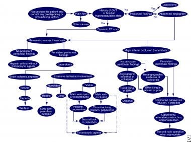
Mesenteric vascular occlusion refers to the blockage or obstruction of the blood vessels that supply the intestines, specifically the mesenteric arteries and veins. This condition can result in insufficient blood flow to the intestines, leading to ischemia (inadequate oxygen supply) and potential necrosis (tissue death).
There are two primary types of mesenteric vascular occlusion:
1. Mesenteric arterial occlusion: This occurs when the mesenteric artery, which carries oxygenated blood from the heart to the intestines, becomes blocked. The most common causes include atherosclerosis (plaque buildup in the arteries), embolism (a clot or particle that travels from another part of the body and lodges in the artery), and thrombosis (a blood clot forming directly in the artery).
2. Mesenteric venous occlusion: This happens when the mesenteric vein, which returns deoxygenated blood from the intestines to the heart, becomes obstructed. The most common causes include thrombophlebitis (inflammation and clot formation in the vein), tumors, or abdominal trauma.
Symptoms of mesenteric vascular occlusion may include severe abdominal pain, nausea, vomiting, diarrhea, and bloody stools. Rapid diagnosis and treatment are crucial to prevent intestinal tissue damage and potential life-threatening complications such as sepsis or shock. Treatment options typically involve surgical intervention, anticoagulation therapy, or endovascular procedures to restore blood flow.
Pathological constriction refers to an abnormal narrowing or tightening of a body passage or organ, which can interfere with the normal flow of blood, air, or other substances through the area. This constriction can occur due to various reasons such as inflammation, scarring, or abnormal growths, and can affect different parts of the body, including blood vessels, airways, intestines, and ureters. Pathological constriction can lead to a range of symptoms and complications depending on its location and severity, and may require medical intervention to correct.
An aneurysm is a localized, balloon-like bulge in the wall of a blood vessel. It occurs when the pressure inside the vessel causes a weakened area to swell and become enlarged. Aneurysms can develop in any blood vessel, but they are most common in arteries at the base of the brain (cerebral aneurysm) and the main artery carrying blood from the heart to the rest of the body (aortic aneurysm).
Aneurysms can be classified as saccular or fusiform, depending on their shape. A saccular aneurysm is a round or oval bulge that projects from the side of a blood vessel, while a fusiform aneurysm is a dilated segment of a blood vessel that is uniform in width and involves all three layers of the arterial wall.
The size and location of an aneurysm can affect its risk of rupture. Generally, larger aneurysms are more likely to rupture than smaller ones. Aneurysms located in areas with high blood pressure or where the vessel branches are also at higher risk of rupture.
Ruptured aneurysms can cause life-threatening bleeding and require immediate medical attention. Symptoms of a ruptured aneurysm may include sudden severe headache, neck stiffness, nausea, vomiting, blurred vision, or loss of consciousness. Unruptured aneurysms may not cause any symptoms and are often discovered during routine imaging tests for other conditions.
Treatment options for aneurysms depend on their size, location, and risk of rupture. Small, unruptured aneurysms may be monitored with regular imaging tests to check for growth or changes. Larger or symptomatic aneurysms may require surgical intervention, such as clipping or coiling, to prevent rupture and reduce the risk of complications.
Ligaments are bands of dense, fibrous connective tissue that surround joints and provide support, stability, and limits the range of motion. They are made up primarily of collagen fibers arranged in a parallel pattern to withstand tension and stress. Ligaments attach bone to bone, and their function is to prevent excessive movement that could cause injury or dislocation.
There are two main types of ligaments: extracapsular and intracapsular. Extracapsular ligaments are located outside the joint capsule and provide stability to the joint by limiting its range of motion. Intracapsular ligaments, on the other hand, are found inside the joint capsule and help maintain the alignment of the joint surfaces.
Examples of common ligaments in the body include the anterior cruciate ligament (ACL) and posterior cruciate ligament (PCL) in the knee, the medial collateral ligament (MCL) and lateral collateral ligament (LCL) in the elbow, and the coracoacromial ligament in the shoulder.
Injuries to ligaments can occur due to sudden trauma or overuse, leading to sprains, strains, or tears. These injuries can cause pain, swelling, bruising, and limited mobility, and may require medical treatment such as immobilization, physical therapy, or surgery.
A venous cutdown is a surgical procedure that involves making an incision into the skin and surrounding tissue to expose a vein, which is then isolated and separated from the surrounding tissue. A venous cutdown is typically performed to establish access to a vein for the purpose of administering fluids or medications, or to obtain blood samples. This procedure is often used when other forms of venous access are difficult or impossible to achieve. The most common site for a venous cutdown is the anterior tibial vein at the ankle.
Arterial occlusive diseases are medical conditions characterized by the blockage or narrowing of the arteries, which can lead to a reduction in blood flow to various parts of the body. This reduction in blood flow can cause tissue damage and may result in serious complications such as tissue death (gangrene), organ dysfunction, or even death.
The most common cause of arterial occlusive diseases is atherosclerosis, which is the buildup of plaque made up of fat, cholesterol, calcium, and other substances in the inner lining of the artery walls. Over time, this plaque can harden and narrow the arteries, restricting blood flow. Other causes of arterial occlusive diseases include blood clots, emboli (tiny particles that travel through the bloodstream and lodge in smaller vessels), inflammation, trauma, and certain inherited conditions.
Symptoms of arterial occlusive diseases depend on the location and severity of the blockage. Common symptoms include:
* Pain, cramping, or fatigue in the affected limb, often triggered by exercise and relieved by rest (claudication)
* Numbness, tingling, or weakness in the affected limb
* Coldness or discoloration of the skin in the affected area
* Slow-healing sores or wounds on the toes, feet, or legs
* Erectile dysfunction in men
Treatment for arterial occlusive diseases may include lifestyle changes such as quitting smoking, exercising regularly, and eating a healthy diet. Medications to lower cholesterol, control blood pressure, prevent blood clots, or manage pain may also be prescribed. In severe cases, surgical procedures such as angioplasty, stenting, or bypass surgery may be necessary to restore blood flow.
"Cardiovascular Tuberculosis" refers to a form of tuberculosis (TB) where the bacteria (Mycobacterium tuberculosis) infects the heart or the blood vessels. This is a less common manifestation of TB, but it can have serious consequences if left untreated.
In cardiovascular TB, the bacteria can cause inflammation and damage to the heart muscle (myocarditis), the sac surrounding the heart (pericarditis), or the coronary arteries that supply blood to the heart muscle. This can lead to symptoms such as chest pain, shortness of breath, coughing, fatigue, and fever. In severe cases, it can cause heart failure or life-threatening arrhythmias.
Cardiovascular TB is usually treated with a combination of antibiotics that are effective against the TB bacteria. The treatment may last for several months to ensure that all the bacteria have been eliminated. In some cases, surgery may be necessary to repair or replace damaged heart valves or vessels. Early diagnosis and treatment can help prevent serious complications and improve outcomes in patients with cardiovascular TB.
The mesenteric arteries are the arteries that supply oxygenated blood to the intestines. There are three main mesenteric arteries: the superior mesenteric artery, which supplies blood to the small intestine (duodenum to two-thirds of the transverse colon) and large intestine (cecum, ascending colon, and the first part of the transverse colon); the inferior mesenteric artery, which supplies blood to the distal third of the transverse colon, descending colon, sigmoid colon, and rectum; and the middle colic artery, which is a branch of the superior mesenteric artery that supplies blood to the transverse colon. These arteries are important in maintaining adequate blood flow to the intestines to support digestion and absorption of nutrients.
The celiac plexus, also known as the solar plexus or autonomic plexus, is a complex network of nerves located in the abdomen, near the stomach and other digestive organs. It plays a crucial role in regulating various automatic functions of the body, such as digestion, absorption, and secretion.
The celiac plexus is formed by the union of several splanchnic nerves that arise from the spinal cord and pass through the diaphragm to reach the abdomen. These nerves carry sensory information from the organs in the abdomen to the brain, as well as motor impulses that control the function of these organs.
In some medical procedures, such as celiac plexus block or neurolysis, the celiac plexus may be targeted to relieve chronic pain associated with conditions like pancreatitis, cancer, or abdominal surgery. These procedures involve injecting anesthetic or neurolytic agents into the area around the celiac plexus to interrupt nerve signals and reduce pain.
Abdominal pain is defined as discomfort or painful sensation in the abdomen. The abdomen is the region of the body between the chest and the pelvis, and contains many important organs such as the stomach, small intestine, large intestine, liver, gallbladder, pancreas, and spleen. Abdominal pain can vary in intensity from mild to severe, and can be acute or chronic depending on the underlying cause.
Abdominal pain can have many different causes, ranging from benign conditions such as gastritis, indigestion, or constipation, to more serious conditions such as appendicitis, inflammatory bowel disease, or abdominal aortic aneurysm. The location, quality, and duration of the pain can provide important clues about its cause. For example, sharp, localized pain in the lower right quadrant of the abdomen may indicate appendicitis, while crampy, diffuse pain in the lower abdomen may suggest irritable bowel syndrome.
It is important to seek medical attention if you experience severe or persistent abdominal pain, especially if it is accompanied by other symptoms such as fever, vomiting, or bloody stools. A thorough physical examination, including a careful history and a focused abdominal exam, can help diagnose the underlying cause of the pain and guide appropriate treatment.
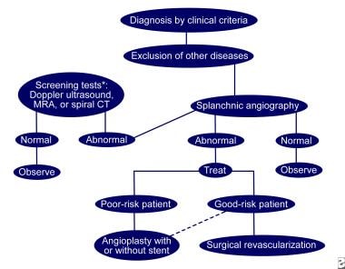
Splanchnic circulation refers to the blood flow to the visceral organs, including the gastrointestinal tract, pancreas, spleen, and liver. These organs receive a significant portion of the cardiac output, with approximately 25-30% of the total restingly going to the splanchnic circulation. The splanchnic circulation is regulated by a complex interplay of neural and hormonal mechanisms that help maintain adequate blood flow to these vital organs while also allowing for the distribution of blood to other parts of the body as needed.
The splanchnic circulation is unique in its ability to vasodilate and increase blood flow significantly in response to meals or other stimuli, such as stress or hormonal changes. This increased blood flow helps support the digestive process and absorption of nutrients. At the same time, the body must carefully regulate this blood flow to prevent a significant drop in blood pressure or overloading the heart with too much work.
Overall, the splanchnic circulation plays a critical role in maintaining the health and function of the body's vital organs, and dysregulation of this system can contribute to various diseases, including digestive disorders, liver disease, and cardiovascular disease.
Splenic infarction is the death of splenic tissue due to blockage of its arterial supply or, less commonly, its venous drainage. This results in ischemia and necrosis of the affected portion of the spleen. The most common cause is embolism from a distant source such as atrial fibrillation, infective endocarditis, or malignancy. Other causes include splenic artery thrombosis, sickle cell disease, hematologic disorders, and trauma. Clinical presentation can vary widely, ranging from being asymptomatic to acute abdominal pain, nausea, vomiting, and fever. Diagnosis is often made with imaging studies such as ultrasound or CT scan. Treatment depends on the underlying cause and severity of symptoms, but may include anticoagulation, antibiotics, or surgical intervention in severe cases.
The hepatic artery is a branch of the celiac trunk or abdominal aorta that supplies oxygenated blood to the liver. It typically divides into two main branches, the right and left hepatic arteries, which further divide into smaller vessels to supply different regions of the liver. The hepatic artery also gives off branches to supply other organs such as the gallbladder, pancreas, and duodenum.
It's worth noting that there is significant variability in the anatomy of the hepatic artery, with some individuals having additional branches or variations in the origin of the vessel. This variability can have implications for surgical procedures involving the liver and surrounding organs.
The pulmonary artery is a large blood vessel that carries deoxygenated blood from the right ventricle of the heart to the lungs for oxygenation. It divides into two main branches, the right and left pulmonary arteries, which further divide into smaller vessels called arterioles, and then into a vast network of capillaries in the lungs where gas exchange occurs. The thin walls of these capillaries allow oxygen to diffuse into the blood and carbon dioxide to diffuse out, making the blood oxygen-rich before it is pumped back to the left side of the heart through the pulmonary veins. This process is crucial for maintaining proper oxygenation of the body's tissues and organs.
Interventional radiography is a subspecialty of radiology that uses imaging guidance (such as X-ray fluoroscopy, ultrasound, CT, or MRI) to perform minimally invasive diagnostic and therapeutic procedures. These procedures typically involve the insertion of needles, catheters, or other small instruments through the skin or a natural body opening, allowing for targeted treatment with reduced risk, trauma, and recovery time compared to traditional open surgeries.
Examples of interventional radiography procedures include:
1. Angiography: Imaging of blood vessels to diagnose and treat conditions like blockages, narrowing, or aneurysms.
2. Biopsy: The removal of tissue samples for diagnostic purposes.
3. Drainage: The removal of fluid accumulations (e.g., abscesses, cysts) or the placement of catheters to drain fluids continuously.
4. Embolization: The blocking of blood vessels to control bleeding, tumor growth, or reduce the size of an aneurysm.
5. Stenting and angioplasty: The widening of narrowed or blocked vessels using stents (small mesh tubes) or balloon catheters.
6. Radiofrequency ablation: The use of heat to destroy tumors or abnormal tissues.
7. Cryoablation: The use of extreme cold to destroy tumors or abnormal tissues.
Interventional radiologists are medical doctors who have completed specialized training in both diagnostic imaging and interventional procedures, allowing them to provide comprehensive care for patients requiring image-guided treatments.
X-ray computed tomography (CT or CAT scan) is a medical imaging method that uses computer-processed combinations of many X-ray images taken from different angles to produce cross-sectional (tomographic) images (virtual "slices") of the body. These cross-sectional images can then be used to display detailed internal views of organs, bones, and soft tissues in the body.
The term "computed tomography" is used instead of "CT scan" or "CAT scan" because the machines take a series of X-ray measurements from different angles around the body and then use a computer to process these data to create detailed images of internal structures within the body.
CT scanning is a noninvasive, painless medical test that helps physicians diagnose and treat medical conditions. CT imaging provides detailed information about many types of tissue including lung, bone, soft tissue and blood vessels. CT examinations can be performed on every part of the body for a variety of reasons including diagnosis, surgical planning, and monitoring of therapeutic responses.
In computed tomography (CT), an X-ray source and detector rotate around the patient, measuring the X-ray attenuation at many different angles. A computer uses this data to construct a cross-sectional image by the process of reconstruction. This technique is called "tomography". The term "computed" refers to the use of a computer to reconstruct the images.
CT has become an important tool in medical imaging and diagnosis, allowing radiologists and other physicians to view detailed internal images of the body. It can help identify many different medical conditions including cancer, heart disease, lung nodules, liver tumors, and internal injuries from trauma. CT is also commonly used for guiding biopsies and other minimally invasive procedures.
In summary, X-ray computed tomography (CT or CAT scan) is a medical imaging technique that uses computer-processed combinations of many X-ray images taken from different angles to produce cross-sectional images of the body. It provides detailed internal views of organs, bones, and soft tissues in the body, allowing physicians to diagnose and treat medical conditions.
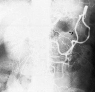
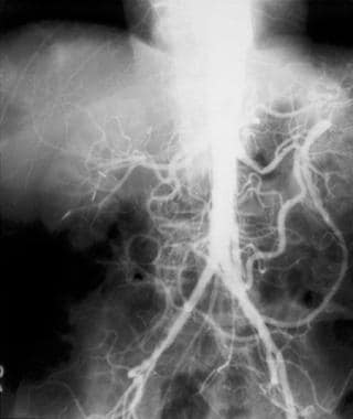
Angiography is a medical procedure in which an x-ray image is taken to visualize the internal structure of blood vessels, arteries, or veins. This is done by injecting a radiopaque contrast agent (dye) into the blood vessel using a thin, flexible catheter. The dye makes the blood vessels visible on an x-ray image, allowing doctors to diagnose and treat various medical conditions such as blockages, narrowing, or malformations of the blood vessels.
There are several types of angiography, including:
* Cardiac angiography (also called coronary angiography) - used to examine the blood vessels of the heart
* Cerebral angiography - used to examine the blood vessels of the brain
* Peripheral angiography - used to examine the blood vessels in the limbs or other parts of the body.
Angiography is typically performed by a radiologist, cardiologist, or vascular surgeon in a hospital setting. It can help diagnose conditions such as coronary artery disease, aneurysms, and peripheral arterial disease, among others.
Vascular access devices (VADs) are medical devices that are used to gain access to a patient's vascular system for the purpose of administering treatments, monitoring vital signs, or obtaining diagnostic samples. These devices can be categorized into short-term and long-term based on their intended duration of use.
Short-term VADs include peripheral intravenous catheters (PIVs), midline catheters, and peripherally inserted central catheters (PICCs). PIVs are thin, flexible tubes that are inserted into a vein in the arm or hand for short-term use. Midlines are similar to PIVs but are longer and can be used for up to 4 weeks. PICCs are inserted into a vein in the upper arm and threaded through to the larger veins near the heart, allowing for long-term access.
Long-term VADs include tunneled central venous catheters (CVCs), non-tunneled CVCs, and implanted ports. Tunneled CVCs are inserted into a large vein in the neck or chest and then threaded under the skin to an exit site, reducing the risk of infection. Non-tunneled CVCs are similar but do not have a tunnel, making them more prone to infection. Implanted ports are small devices that are surgically implanted under the skin, usually in the chest or arm, and connected to a catheter that is inserted into a large vein.
VADs can be used for various medical treatments such as chemotherapy, antibiotic therapy, parenteral nutrition, dialysis, and blood transfusions. Proper care and maintenance of VADs are essential to prevent complications such as infection, thrombosis, and catheter-related bloodstream infections (CRBSI).
Replantation is a surgical procedure in which a body part that has been completely detached or amputated is reattached to the body. This procedure involves careful reattachment of bones, muscles, tendons, nerves, and blood vessels to restore function and sensation to the greatest extent possible. The success of replantation depends on various factors such as the level of injury, the condition of the amputated part, and the patient's overall health.
The carotid arteries are a pair of vital blood vessels in the human body that supply oxygenated blood to the head and neck. Each person has two common carotid arteries, one on each side of the neck, which branch off from the aorta, the largest artery in the body.
The right common carotid artery originates from the brachiocephalic trunk, while the left common carotid artery arises directly from the aortic arch. As they ascend through the neck, they split into two main branches: the internal and external carotid arteries.
The internal carotid artery supplies oxygenated blood to the brain, eyes, and other structures within the skull, while the external carotid artery provides blood to the face, scalp, and various regions of the neck.
Maintaining healthy carotid arteries is crucial for overall cardiovascular health and preventing serious conditions like stroke, which can occur when the arteries become narrowed or blocked due to the buildup of plaque or fatty deposits (atherosclerosis). Regular check-ups with healthcare professionals may include monitoring carotid artery health through ultrasound or other imaging techniques.
Cerebral arteries refer to the blood vessels that supply oxygenated blood to the brain. These arteries branch off from the internal carotid arteries and the vertebral arteries, which combine to form the basilar artery. The major cerebral arteries include:
1. Anterior cerebral artery (ACA): This artery supplies blood to the frontal lobes of the brain, including the motor and sensory cortices responsible for movement and sensation in the lower limbs.
2. Middle cerebral artery (MCA): The MCA is the largest of the cerebral arteries and supplies blood to the lateral surface of the brain, including the temporal, parietal, and frontal lobes. It is responsible for providing blood to areas involved in motor function, sensory perception, speech, memory, and vision.
3. Posterior cerebral artery (PCA): The PCA supplies blood to the occipital lobe, which is responsible for visual processing, as well as parts of the temporal and parietal lobes.
4. Anterior communicating artery (ACoA) and posterior communicating arteries (PComAs): These are small arteries that connect the major cerebral arteries, forming an important circulatory network called the Circle of Willis. The ACoA connects the two ACAs, while the PComAs connect the ICA with the PCA and the basilar artery.
These cerebral arteries play a crucial role in maintaining proper brain function by delivering oxygenated blood to various regions of the brain. Any damage or obstruction to these arteries can lead to serious neurological conditions, such as strokes or transient ischemic attacks (TIAs).
The renal artery is a pair of blood vessels that originate from the abdominal aorta and supply oxygenated blood to each kidney. These arteries branch into several smaller vessels that provide blood to the various parts of the kidneys, including the renal cortex and medulla. The renal arteries also carry nutrients and other essential components needed for the normal functioning of the kidneys. Any damage or blockage to the renal artery can lead to serious consequences, such as reduced kidney function or even kidney failure.
The femoral artery is the major blood vessel that supplies oxygenated blood to the lower extremity of the human body. It is a continuation of the external iliac artery and becomes the popliteal artery as it passes through the adductor hiatus in the adductor magnus muscle of the thigh.
The femoral artery is located in the femoral triangle, which is bound by the sartorius muscle anteriorly, the adductor longus muscle medially, and the biceps femoris muscle posteriorly. It can be easily palpated in the groin region, making it a common site for taking blood samples, measuring blood pressure, and performing surgical procedures such as femoral artery catheterization and bypass grafting.
The femoral artery gives off several branches that supply blood to the lower limb, including the deep femoral artery, the superficial femoral artery, and the profunda femoris artery. These branches provide blood to the muscles, bones, skin, and other tissues of the leg, ankle, and foot.
A thoracic aortic aneurysm is a localized dilatation or bulging of the thoracic aorta, which is the part of the aorta that runs through the chest cavity. The aorta is the largest artery in the body, and it carries oxygenated blood from the heart to the rest of the body.
Thoracic aortic aneurysms can occur anywhere along the thoracic aorta, but they are most commonly found in the aortic arch or the descending thoracic aorta. These aneurysms can vary in size, and they are considered significant when they are 50% larger than the expected normal diameter of the aorta.
The exact cause of thoracic aortic aneurysms is not fully understood, but several factors can contribute to their development, including:
* Atherosclerosis (hardening and narrowing of the arteries)
* High blood pressure
* Genetic disorders such as Marfan syndrome or Ehlers-Danlos syndrome
* Infections or inflammation of the aorta
* Trauma to the chest
Thoracic aortic aneurysms can be asymptomatic and found incidentally on imaging studies, or they may present with symptoms such as chest pain, cough, difficulty swallowing, or hoarseness. If left untreated, thoracic aortic aneurysms can lead to serious complications, including aortic dissection (tearing of the inner layer of the aorta) or rupture, which can be life-threatening.
Treatment options for thoracic aortic aneurysms include medical management with blood pressure control and cholesterol-lowering medications, as well as surgical repair or endovascular stenting, depending on the size, location, and growth rate of the aneurysm. Regular follow-up imaging is necessary to monitor the size and progression of the aneurysm over time.
Blood vessel prosthesis implantation is a surgical procedure in which an artificial blood vessel, also known as a vascular graft or prosthetic graft, is inserted into the body to replace a damaged or diseased native blood vessel. The prosthetic graft can be made from various materials such as Dacron (polyester), PTFE (polytetrafluoroethylene), or bovine/human tissue.
The implantation of a blood vessel prosthesis is typically performed to treat conditions that cause narrowing or blockage of the blood vessels, such as atherosclerosis, aneurysms, or traumatic injuries. The procedure may be used to bypass blocked arteries in the legs (peripheral artery disease), heart (coronary artery bypass surgery), or neck (carotid endarterectomy). It can also be used to replace damaged veins for hemodialysis access in patients with kidney failure.
The success of blood vessel prosthesis implantation depends on various factors, including the patient's overall health, the location and extent of the vascular disease, and the type of graft material used. Possible complications include infection, bleeding, graft thrombosis (clotting), and graft failure, which may require further surgical intervention or endovascular treatments.
A stent is a small mesh tube that's used to treat narrow or weak arteries. Arteries are blood vessels that carry blood away from your heart to other parts of your body. A stent is placed in an artery as part of a procedure called angioplasty. Angioplasty restores blood flow through narrowed or blocked arteries by inflating a tiny balloon inside the blocked artery to widen it.
The stent is then inserted into the widened artery to keep it open. The stent is usually made of metal, but some are coated with medication that is slowly and continuously released to help prevent the formation of scar tissue in the artery. This can reduce the chance of the artery narrowing again.
Stents are also used in other parts of the body, such as the neck (carotid artery) and kidneys (renal artery), to help maintain blood flow and prevent blockages. They can also be used in the urinary system to treat conditions like ureteropelvic junction obstruction or narrowing of the urethra.
Surgical decompression is a medical procedure that involves relieving pressure on a nerve or tissue by creating additional space. This is typically accomplished through the removal of a portion of bone or other tissue that is causing the compression. The goal of surgical decompression is to alleviate symptoms such as pain, numbness, tingling, or weakness caused by the compression.
In the context of spinal disorders, surgical decompression is often used to treat conditions such as herniated discs, spinal stenosis, or bone spurs that are compressing nerves in the spine. The specific procedure used may vary depending on the location and severity of the compression, but common techniques include laminectomy, discectomy, and foraminotomy.
It's important to note that surgical decompression is a significant medical intervention that carries risks such as infection, bleeding, and injury to surrounding tissues. As with any surgery, it should be considered as a last resort after other conservative treatments have been tried and found to be ineffective. A thorough evaluation by a qualified medical professional is necessary to determine whether surgical decompression is appropriate in a given case.
The basilar artery is a major blood vessel that supplies oxygenated blood to the brainstem and cerebellum. It is formed by the union of two vertebral arteries at the lower part of the brainstem, near the junction of the medulla oblongata and pons.
The basilar artery runs upward through the center of the brainstem and divides into two posterior cerebral arteries at the upper part of the brainstem, near the midbrain. The basilar artery gives off several branches that supply blood to various parts of the brainstem, including the pons, medulla oblongata, and midbrain, as well as to the cerebellum.
The basilar artery is an important part of the circle of Willis, a network of arteries at the base of the brain that ensures continuous blood flow to the brain even if one of the arteries becomes blocked or narrowed.
Aortography is a medical procedure that involves taking X-ray images of the aorta, which is the largest blood vessel in the body. The procedure is usually performed to diagnose or assess various conditions related to the aorta, such as aneurysms, dissections, or blockages.
To perform an aortography, a contrast dye is injected into the aorta through a catheter that is inserted into an artery, typically in the leg or arm. The contrast dye makes the aorta visible on X-ray images, allowing doctors to see its structure and any abnormalities that may be present.
The procedure is usually performed in a hospital or outpatient setting and may require sedation or anesthesia. While aortography can provide valuable diagnostic information, it also carries some risks, such as allergic reactions to the contrast dye, damage to blood vessels, or infection. Therefore, it is typically reserved for situations where other diagnostic tests have been inconclusive or where more invasive treatment may be required.
Viscera is a medical term that refers to the internal organs of the body, specifically those contained within the chest and abdominal cavities. These include the heart, lungs, liver, pancreas, spleen, kidneys, and intestines. In some contexts, it may also refer to the reproductive organs. The term viscera is often used in anatomical or surgical descriptions, and is derived from the Latin word "viscus," meaning "an internal organ."
The abdominal aorta is the portion of the aorta, which is the largest artery in the body, that runs through the abdomen. It originates from the thoracic aorta at the level of the diaphragm and descends through the abdomen, where it branches off into several smaller arteries that supply blood to the pelvis, legs, and various abdominal organs. The abdominal aorta is typically divided into four segments: the suprarenal, infrarenal, visceral, and parietal portions. Disorders of the abdominal aorta can include aneurysms, atherosclerosis, and dissections, which can have serious consequences if left untreated.
Ischemia is the medical term used to describe a lack of blood flow to a part of the body, often due to blocked or narrowed blood vessels. This can lead to a shortage of oxygen and nutrients in the tissues, which can cause them to become damaged or die. Ischemia can affect many different parts of the body, including the heart, brain, legs, and intestines. Symptoms of ischemia depend on the location and severity of the blockage, but they may include pain, cramping, numbness, weakness, or coldness in the affected area. In severe cases, ischemia can lead to tissue death (gangrene) or organ failure. Treatment for ischemia typically involves addressing the underlying cause of the blocked blood flow, such as through medication, surgery, or lifestyle changes.
A dissecting aneurysm is a serious and potentially life-threatening condition that occurs when there is a tear in the inner layer of the artery wall, allowing blood to flow between the layers of the artery wall. This can cause the artery to bulge or balloon out, leading to a dissection aneurysm.
Dissecting aneurysms can occur in any artery, but they are most commonly found in the aorta, which is the largest artery in the body. When a dissecting aneurysm occurs in the aorta, it is often referred to as a "dissecting aortic aneurysm."
Dissecting aneurysms can be caused by various factors, including high blood pressure, atherosclerosis (hardening and narrowing of the arteries), genetic disorders that affect the connective tissue, trauma, or illegal drug use (such as cocaine).
Symptoms of a dissecting aneurysm may include sudden severe chest or back pain, which can feel like ripping or tearing, shortness of breath, sweating, lightheadedness, or loss of consciousness. If left untreated, a dissecting aneurysm can lead to serious complications, such as rupture of the artery, stroke, or even death.
Treatment for a dissecting aneurysm typically involves surgery or endovascular repair to prevent further damage and reduce the risk of rupture. The specific treatment approach will depend on various factors, including the location and size of the aneurysm, the patient's overall health, and their medical history.
The iliac arteries are major branches of the abdominal aorta, the large artery that carries oxygen-rich blood from the heart to the rest of the body. The iliac arteries divide into two branches, the common iliac arteries, which further bifurcate into the internal and external iliac arteries.
The internal iliac artery supplies blood to the lower abdomen, pelvis, and the reproductive organs, while the external iliac artery provides blood to the lower extremities, including the legs and feet. Together, the iliac arteries play a crucial role in circulating blood throughout the body, ensuring that all tissues and organs receive the oxygen and nutrients they need to function properly.
Treatment outcome is a term used to describe the result or effect of medical treatment on a patient's health status. It can be measured in various ways, such as through symptoms improvement, disease remission, reduced disability, improved quality of life, or survival rates. The treatment outcome helps healthcare providers evaluate the effectiveness of a particular treatment plan and make informed decisions about future care. It is also used in clinical research to compare the efficacy of different treatments and improve patient care.
The vertebral artery is a major blood vessel that supplies oxygenated blood to the brain and upper spinal cord. It arises from the subclavian artery, then ascends through the transverse processes of several cervical vertebrae before entering the skull through the foramen magnum. Inside the skull, it joins with the opposite vertebral artery to form the basilar artery, which supplies blood to the brainstem and cerebellum. The vertebral artery also gives off several important branches that supply blood to various regions of the brainstem and upper spinal cord.
Coronary artery bypass surgery, also known as coronary artery bypass grafting (CABG), is a surgical procedure used to improve blood flow to the heart in patients with severe coronary artery disease. This condition occurs when the coronary arteries, which supply oxygen-rich blood to the heart muscle, become narrowed or blocked due to the buildup of fatty deposits, called plaques.
During CABG surgery, a healthy blood vessel from another part of the body is grafted, or attached, to the coronary artery, creating a new pathway for oxygen-rich blood to flow around the blocked or narrowed portion of the artery and reach the heart muscle. This bypass helps to restore normal blood flow and reduce the risk of angina (chest pain), shortness of breath, and other symptoms associated with coronary artery disease.
There are different types of CABG surgery, including traditional on-pump CABG, off-pump CABG, and minimally invasive CABG. The choice of procedure depends on various factors, such as the patient's overall health, the number and location of blocked arteries, and the presence of other medical conditions.
It is important to note that while CABG surgery can significantly improve symptoms and quality of life in patients with severe coronary artery disease, it does not cure the underlying condition. Lifestyle modifications, such as regular exercise, a healthy diet, smoking cessation, and medication therapy, are essential for long-term management and prevention of further progression of the disease.
Angioplasty, balloon refers to a medical procedure used to widen narrowed or obstructed blood vessels, particularly the coronary arteries that supply blood to the heart muscle. This procedure is typically performed using a catheter-based technique, where a thin, flexible tube called a catheter is inserted into an artery, usually through the groin or wrist, and guided to the site of the narrowing or obstruction in the coronary artery.
Once the catheter reaches the affected area, a small balloon attached to the tip of the catheter is inflated, which compresses the plaque against the artery wall and stretches the artery, thereby restoring blood flow. The balloon is then deflated and removed, along with the catheter.
Balloon angioplasty is often combined with the placement of a stent, a small metal mesh tube that helps to keep the artery open and prevent it from narrowing again. This procedure is known as percutaneous coronary intervention (PCI) or coronary angioplasty and stenting.
Overall, balloon angioplasty is a relatively safe and effective treatment for coronary artery disease, although complications such as bleeding, infection, or re-narrowing of the artery can occur in some cases.
The radial artery is a key blood vessel in the human body, specifically a part of the peripheral arterial system. Originating from the brachial artery in the upper arm, the radial artery travels down the arm and crosses over the wrist, where it can be palpated easily. It then continues into the hand, dividing into several branches to supply blood to the hand's tissues and digits.
The radial artery is often used for taking pulse readings due to its easy accessibility at the wrist. Additionally, in medical procedures such as coronary angiography or bypass surgery, the radial artery can be utilized as a site for catheter insertion. This allows healthcare professionals to examine the heart's blood vessels and assess cardiovascular health.
Surgical anastomosis is a medical procedure that involves the connection of two tubular structures, such as blood vessels or intestines, to create a continuous passage. This technique is commonly used in various types of surgeries, including vascular, gastrointestinal, and orthopedic procedures.
During a surgical anastomosis, the ends of the two tubular structures are carefully prepared by removing any damaged or diseased tissue. The ends are then aligned and joined together using sutures, staples, or other devices. The connection must be secure and leak-free to ensure proper function and healing.
The success of a surgical anastomosis depends on several factors, including the patient's overall health, the location and condition of the structures being joined, and the skill and experience of the surgeon. Complications such as infection, bleeding, or leakage can occur, which may require additional medical intervention or surgery.
Proper postoperative care is also essential to ensure the success of a surgical anastomosis. This may include monitoring for signs of complications, administering medications to prevent infection and promote healing, and providing adequate nutrition and hydration.
The mammary arteries are a set of blood vessels that supply oxygenated blood to the mammary glands, which are the structures in female breasts responsible for milk production during lactation. The largest mammary artery, also known as the internal thoracic or internal mammary artery, originates from the subclavian artery and descends along the inner side of the chest wall. It then branches into several smaller arteries that supply blood to the breast tissue. These include the anterior and posterior intercostal arteries, lateral thoracic artery, and pectoral branches. The mammary arteries are crucial in maintaining the health and function of the breast tissue, and any damage or blockage to these vessels can lead to various breast-related conditions or diseases.
Collateral circulation refers to the alternate blood supply routes that bypass an obstructed or narrowed vessel and reconnect with the main vascular system. These collateral vessels can develop over time as a result of the body's natural adaptation to chronic ischemia (reduced blood flow) caused by various conditions such as atherosclerosis, thromboembolism, or vasculitis.
The development of collateral circulation helps maintain adequate blood flow and oxygenation to affected tissues, minimizing the risk of tissue damage and necrosis. In some cases, well-developed collateral circulations can help compensate for significant blockages in major vessels, reducing symptoms and potentially preventing the need for invasive interventions like revascularization procedures. However, the extent and effectiveness of collateral circulation vary from person to person and depend on factors such as age, overall health status, and the presence of comorbidities.
Endovascular procedures are minimally invasive medical treatments that involve accessing and repairing blood vessels or other interior parts of the body through small incisions or punctures. These procedures typically use specialized catheters, wires, and other tools that are inserted into the body through an artery or vein, usually in the leg or arm.
Endovascular procedures can be used to treat a wide range of conditions, including aneurysms, atherosclerosis, peripheral artery disease, carotid artery stenosis, and other vascular disorders. Some common endovascular procedures include angioplasty, stenting, embolization, and thrombectomy.
The benefits of endovascular procedures over traditional open surgery include smaller incisions, reduced trauma to surrounding tissues, faster recovery times, and lower risks of complications such as infection and bleeding. However, endovascular procedures may not be appropriate for all patients or conditions, and careful evaluation and consideration are necessary to determine the best treatment approach.
The internal carotid artery is a major blood vessel that supplies oxygenated blood to the brain. It originates from the common carotid artery and passes through the neck, entering the skull via the carotid canal in the temporal bone. Once inside the skull, it branches into several smaller vessels that supply different parts of the brain with blood.
The internal carotid artery is divided into several segments: cervical, petrous, cavernous, clinoid, and supraclinoid. Each segment has distinct clinical significance in terms of potential injury or disease. The most common conditions affecting the internal carotid artery include atherosclerosis, which can lead to stroke or transient ischemic attack (TIA), and dissection, which can cause severe headache, neck pain, and neurological symptoms.
It's important to note that any blockage or damage to the internal carotid artery can have serious consequences, as it can significantly reduce blood flow to the brain and lead to permanent neurological damage or even death. Therefore, regular check-ups and screening tests are recommended for individuals at high risk of developing vascular diseases.
An endoleak is a complication that can occur following minimally invasive endovascular aortic repair (EVAR) for abdominal aortic aneurysms. It refers to the persistence or recurrence of blood flow outside the lumen of the endograft but within the aneurysm sac. Endoleaks are classified into different types based on their source and can be categorized as follows:
1. Type I endoleak: This type of endoleak occurs due to inadequate sealing at the attachment sites between the endograft and the aortic wall. It can further be divided into two subtypes - Type Ia (proximal) and Type Ib (distal).
2. Type II endoleak: This type of endoleak results from retrograde flow from branch vessels that enter the aneurysm sac, such as lumbar arteries or inferior mesenteric artery. Type II endoleaks are often asymptomatic and may not require immediate treatment.
3. Type III endoleak: This type of endoleak occurs due to a defect in the structural integrity of the endograft itself, leading to communication between different components of the graft or between the graft and another vessel.
4. Type IV endoleak: This type of endoleak is caused by porosity in the graft material, allowing for leakage through the graft wall itself. It typically resolves on its own within 30 days post-procedure.
5. Type V endoleak (also known as endotension): This type of endoleak is characterized by an increase in sac size without a demonstrable endoleak on imaging. The exact cause remains unclear, but it may be related to continued pressurization of the aneurysm sac due to transmission of systemic pressure through the graft fabric.
Endoleaks can lead to persistent enlargement of the aneurysm sac and potential rupture if not addressed promptly. Therefore, regular follow-up imaging is essential after EVAR to monitor for endoleak development and address any issues that arise.
Therapeutic embolization is a medical procedure that involves intentionally blocking or obstructing blood vessels to stop excessive bleeding or block the flow of blood to a tumor or abnormal tissue. This is typically accomplished by injecting small particles, such as microspheres or coils, into the targeted blood vessel through a catheter, which is inserted into a larger blood vessel and guided to the desired location using imaging techniques like X-ray or CT scanning. The goal of therapeutic embolization is to reduce the size of a tumor, control bleeding, or block off abnormal blood vessels that are causing problems.
The subclavian artery is a major blood vessel that supplies the upper limb and important structures in the neck and head. It arises from the brachiocephalic trunk (in the case of the right subclavian artery) or directly from the aortic arch (in the case of the left subclavian artery).
The subclavian artery has several branches, including:
1. The vertebral artery, which supplies blood to the brainstem and cerebellum.
2. The internal thoracic artery (also known as the mammary artery), which supplies blood to the chest wall, breast, and anterior mediastinum.
3. The thyrocervical trunk, which gives rise to several branches that supply the neck, including the inferior thyroid artery, the suprascapular artery, and the transverse cervical artery.
4. The costocervical trunk, which supplies blood to the neck and upper back, including the posterior chest wall and the lower neck muscles.
The subclavian artery is a critical vessel in maintaining adequate blood flow to the upper limb, and any blockage or damage to this vessel can lead to significant morbidity, including arm pain, numbness, weakness, or even loss of function.
A syndrome, in medical terms, is a set of symptoms that collectively indicate or characterize a disease, disorder, or underlying pathological process. It's essentially a collection of signs and/or symptoms that frequently occur together and can suggest a particular cause or condition, even though the exact physiological mechanisms might not be fully understood.
For example, Down syndrome is characterized by specific physical features, cognitive delays, and other developmental issues resulting from an extra copy of chromosome 21. Similarly, metabolic syndromes like diabetes mellitus type 2 involve a group of risk factors such as obesity, high blood pressure, high blood sugar, and abnormal cholesterol or triglyceride levels that collectively increase the risk of heart disease, stroke, and diabetes.
It's important to note that a syndrome is not a specific diagnosis; rather, it's a pattern of symptoms that can help guide further diagnostic evaluation and management.
A blood vessel prosthesis is a medical device that is used as a substitute for a damaged or diseased natural blood vessel. It is typically made of synthetic materials such as polyester, Dacron, or ePTFE (expanded polytetrafluoroethylene) and is designed to mimic the function of a native blood vessel by allowing the flow of blood through it.
Blood vessel prostheses are used in various surgical procedures, including coronary artery bypass grafting, peripheral arterial reconstruction, and the creation of arteriovenous fistulas for dialysis access. The choice of material and size of the prosthesis depends on several factors, such as the location and diameter of the vessel being replaced, the patient's age and overall health status, and the surgeon's preference.
It is important to note that while blood vessel prostheses can be effective in restoring blood flow, they may also carry risks such as infection, thrombosis (blood clot formation), and graft failure over time. Therefore, careful patient selection, surgical technique, and postoperative management are crucial for the success of these procedures.
Carotid artery diseases refer to conditions that affect the carotid arteries, which are the major blood vessels that supply oxygen-rich blood to the head and neck. The most common type of carotid artery disease is atherosclerosis, which occurs when fatty deposits called plaques build up in the inner lining of the arteries.
These plaques can cause the arteries to narrow or become blocked, reducing blood flow to the brain and increasing the risk of stroke. Other carotid artery diseases include carotid artery dissection, which occurs when there is a tear in the inner lining of the artery, and fibromuscular dysplasia, which is a condition that affects the muscle and tissue in the walls of the artery.
Symptoms of carotid artery disease may include neck pain or pulsations, transient ischemic attacks (TIAs) or "mini-strokes," and strokes. Treatment options for carotid artery disease depend on the severity and type of the condition but may include lifestyle changes, medications, endarterectomy (a surgical procedure to remove plaque from the artery), or angioplasty and stenting (procedures to open blocked arteries using a balloon and stent).
Vascular surgical procedures are operations that are performed to treat conditions and diseases related to the vascular system, which includes the arteries, veins, and capillaries. These procedures can be invasive or minimally invasive and are often used to treat conditions such as peripheral artery disease, carotid artery stenosis, aortic aneurysms, and venous insufficiency.
Some examples of vascular surgical procedures include:
* Endarterectomy: a procedure to remove plaque buildup from the inside of an artery
* Bypass surgery: creating a new path for blood to flow around a blocked or narrowed artery
* Angioplasty and stenting: using a balloon to open a narrowed artery and placing a stent to keep it open
* Aneurysm repair: surgically repairing an aneurysm, a weakened area in the wall of an artery that has bulged out and filled with blood
* Embolectomy: removing a blood clot from a blood vessel
* Thrombectomy: removing a blood clot from a vein
These procedures are typically performed by vascular surgeons, who are trained in the diagnosis and treatment of vascular diseases.
The brachial artery is a major blood vessel in the upper arm. It supplies oxygenated blood to the muscles and tissues of the arm, forearm, and hand. The brachial artery originates from the axillary artery at the level of the shoulder joint and runs down the medial (inner) aspect of the arm, passing through the cubital fossa (the depression on the anterior side of the elbow) where it can be palpated during a routine blood pressure measurement. At the lower end of the forearm, the brachial artery bifurcates into the radial and ulnar arteries, which further divide into smaller vessels to supply the hand and fingers.
 Celiac artery
Left gastroepiploic artery
Randall Wolf
Greater omentum
Right gastric artery
Left gastric artery
Abdominal aorta
Splenic artery
Human digestive system
Duodenum
Inferior pancreaticoduodenal artery
Celiac lymph nodes
Vitelline arteries
Open aortic surgery
Small intestine
Hepatic artery proper
Pulmonary sequestration
Side stitch
Stomach
Foregut
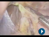
Median arcuate ligament syndrome
Hepatic portal system
Abdominal migraine
Hepatic arterial infusion
Michael L. Marin
Pancreaticoduodenectomy
Lumbar vertebrae
Celiac
Bühler's anastomotic artery
Neurolysis
Right gastri1
- The left gastric artery travels back upward and slightly to the left, bringing blood to the lower esophagus and to the top of the stomach, where it meets the right gastric artery along the stomach's upper curve. (thehealthboard.com)
Aneurysm14
- Aneurysm of Thoracoabdominal Aorta Involving the Celiac, Superior Mesenteric, and Renal Arteries. (nih.gov)
- DeBakey, Michael E., Oscar Creech Jr., and George C. Morris Jr. 'Aneurysm of Thoracoabdominal Aorta Involving the Celiac, Superior Mesenteric, and Renal Arteries. (nih.gov)
- Celiac Artery Aneurysm: Anyone else with same illness? (mayoclinic.org)
- I was recently diagnosis with a celiac artery aneurysm in June 2016. (mayoclinic.org)
- 4. Balloon-assisted coil embolization of the celiac trunk before endovascular aortic repair of thoracoabdominal aortic aneurysm. (nih.gov)
- 6. Endograft-assisted coil embolization of a celiac trunk aneurysm. (nih.gov)
- 11. Outcome after celiac artery coverage during endovascular thoracic aortic aneurysm repair: preliminary results. (nih.gov)
- 16. Inferior mesenteric artery embolization before endovascular aneurysm repair: technique and initial results. (nih.gov)
- 17. Outcomes of endovascular aneurysm repair with selective internal iliac artery coverage without coil embolization. (nih.gov)
- F: The splenic vein aneurysm disappeared after embolization of the splenic artery, including the splenic arteriovenous fistula and drainage vein. (wjgnet.com)
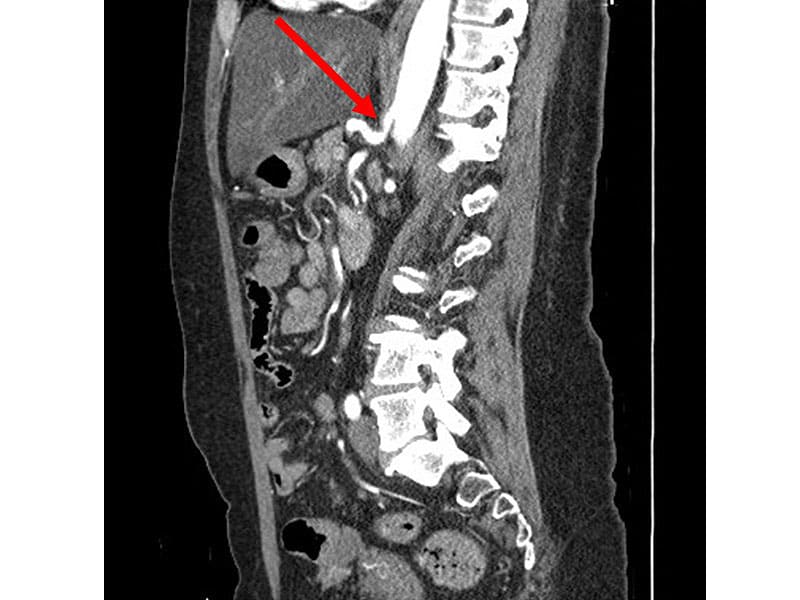
- A celiac artery aneurysm is a dilatation of the celiac artery. (angiologist.com)
- A spontaneous dissecting aneurysm of visceral arteries without trauma or an iatrogenic cause is rare. (archive.org)
- We would like to report a case of a dissecting aneurysm of the common and proper hepatic artery with dissection of the celiac axis and the superior mesenteric artery (SMA). (archive.org)
- We performed a bypass between the right hepatic artery and a celiac artery with a saphenous vein after exclusion of the aneurysm. (archive.org)
Splenic artery6
- From this point, the celiac artery runs forward and downward, dividing almost immediately into three smaller arteries: the left gastric artery, the common hepatic artery , and the splenic artery. (thehealthboard.com)
- To the left is the splenic artery, which transports blood to the spleen. (thehealthboard.com)
- The donor iliac artery is used for reconstruction on the back table to reconstruct the splenic artery and superior mesenteric artery of the pancreas graft. (merckmanuals.com)
- The left branch, the splenic artery, is directed toward the hilum of the spleen. (iame.com)
- The tortuosity of the splenic artery seen in this patient is also a common anatomic variation. (vesalius.com)
- Pancreatic tumors arising from the tail of the pancreas (20%-25% of exocrine pancreatic cancers) may spread to the common hepatic artery, celiac axis, splenic hilum, or splenic artery. (logicalimages.com)
Aorta32
- The celiac (/ˈsiːli.æk/) artery (also spelled coeliac), also known as the celiac trunk or truncus coeliacus, is the first major branch of the abdominal aorta. (wikipedia.org)
- Branching from the aorta at thoracic vertebra 12 (T12) in humans, it is one of three anterior/ midline branches of the abdominal aorta (the others are the superior and inferior mesenteric arteries). (wikipedia.org)
- The celiac artery is the first major branch of the descending abdominal aorta, branching at a 90° angle. (wikipedia.org)
- The celiac artery is vulnerable to compression from the crus of the diaphragm during ventilation where it originates from the abdominal aorta. (wikipedia.org)
- It is the largest branch of the abdominal aorta , the major blood vessel of the abdomen , which descends from the heart and ends in the pelvis , where it splits into the right and left iliac arteries. (thehealthboard.com)
- A diagram of the aorta, including the celiac artery. (thehealthboard.com)
- The deoxygenated blood from the celiac and hepatic arteries then returns to the heart and lungs via the inferior vena cava, the large vein of the abdomen that runs parallel to the abdominal aorta. (thehealthboard.com)
- [ 16 ] The celiac axis arises from the ventral surface of the aorta at the T12-L1 vertebral body. (medscape.com)
- it is of varying size and is wedged between the superior mesenteric vessels (vein on right, and artery on left) in front and the aorta behind it. (medscape.com)
- The aorta is located to the left of the spine and tapers gradually as it courses to its bifurcation (near the level of the umbilicus) where it divides into the right and left common iliac arteries (FIGURE 1). (iame.com)
- Computed tomographic angiogram of the abdominal aorta and iliac arteries. (iame.com)
- The celiac artery (a.k.a., trunk, axis) is the first major branch arising from the abdominal aorta (FIGURE 3), originating anteriorly at about the level of the first lumbar vertebra 3, 4 . (iame.com)
- If you rotate the transducer 90 degrees to image the aorta in a transverse plane at this level, you will obtain a longitudinal view of the celiac trunk arising from the anterior aortic wall. (iame.com)
- In optimal longitudinal images of the aorta and celiac trunk, the left gastric artery can be seen coursing cephalad for a short distance. (iame.com)
- You will encounter the next major branch of the abdominal aorta, the superior mesenteric artery (SMA), approximately one to two centimeters caudal to the origin of the celiac artery 3, 4 (refer to FIGURE 3). (iame.com)
- The abdominal aorta runs from the diaphragm and ends just above the pelvis, where it divides into the iliac arteries. (onteenstoday.com)
- There are five arteries that branch from the abdominal aorta: the celiac artery, the superior mesenteric artery, the inferior mesenteric artery, the renal arteries and the iliac arteries. (onteenstoday.com)
- Within the abdomen, the descending aorta branches into the two common iliac arteries that provide blood to the pelvis and, eventually, the legs. (onteenstoday.com)
- The descending aorta starts after the arch of the aorta and ends by splitting into two great arteries (the common iliac arteries) that go to the legs. (onteenstoday.com)
- The arch of the aorta has three branches: the brachiocephalic artery (which divides into right common carotid artery and the right subclavian artery), the left common carotid artery, and the left subclavian artery. (onteenstoday.com)
- The first branch of the aorta is normally the innominate artery, which is also referred to as the brachiocephalic trunk. (onteenstoday.com)
- Abdominal Aorta: The aorta, highlighted in red, includes the abdominal aorta which begins at the diaphragm and ends as it branches into the common iliac arteries. (onteenstoday.com)
- The abdominal aorta is the largest artery in the abdominal cavity. (onteenstoday.com)
- Is the iliac artery part of the aorta? (onteenstoday.com)
- The common iliac artery is a part of the abdominal aorta, supplying the blood further to the pelvis and legs. (onteenstoday.com)
- Are there any unpaired arteries in the abdominal aorta? (onteenstoday.com)
- The arterial trunk that arises from the abdominal aorta and after a short course divides into the left gastric, common hepatic and splenic arteries. (nih.gov)
- The superficial part of the cardiac plexus lies beneath the arch of the aorta, in front of the right pulmonary artery. (bartleby.com)
- The patient had a computed tomography scan showing a retroperitoneal mass encasing the aorta, celiac axis, and superior mesenteric artery. (escholarship.org)
- The celiac plexus is a group of nerves that surround the aorta, the major artery in the abdomen. (uclahealth.org)
- The branches of the abdominal aorta and pelvic arteries can be divided into five broad functional groups (→ = give rise to). (basicmedicalkey.com)
- The aortic bifurcation is the point where the abdominal aorta bifurcates into the two common iliac arteries, which give off multiple subbranches that supply the viscera and pelvic walls (see D ). (basicmedicalkey.com)
Gastroduodenal artery3
- The hepatic artery gives off the gastroduodenal artery, which branches further to the right gastroepiploic artery and the anterosuperior and posterosuperior pancreaticoduodenal arteries. (medscape.com)
- The common hepatic artery splits into the proper hepatic artery and the gastroduodenal artery. (healthline.com)
- At the point where the first major branch of the CHA (the gastroduodenal artery) descends to supply the pancreas, the CHA becomes the proper hepatic artery (FIGURE 5). (iame.com)
Inferior mesenteric6
- Similarly, the superior mesenteric artery and inferior mesenteric artery feed structures arising from the embryonic midgut and hindgut respectively. (wikipedia.org)
- In contrast to the drainage of midgut and hindgut structures by the superior mesenteric vein and inferior mesenteric vein respectively, venous return from the celiac artery is through either the splenic vein emptying into the hepatic portal vein or via smaller tributaries of the portal venous system. (wikipedia.org)
- This case also illustrates communications among the three main mesenteric arteries to the gut: the celiac, superior mesenteric and inferior mesenteric. (vesalius.com)
- This patient has a well developed marginal artery (of Drummond) from inferior mesenteric to the left branch of the middle colic artery. (vesalius.com)
- Of the visceral branches, the celiac artery and the superior and inferior mesenteric arteries are unpaired, while the suprarenals, renals, internal spermatics, and ovarian are paired. (onteenstoday.com)
- Blue: Branches of the inferior mesenteric artery. (basicmedicalkey.com)
Trunk5
- Celiac trunk compression syndrome requiring surgery in 3 adolescent patients. (nih.gov)
- Alternately known as the celiac trunk, the celiac artery originates a few inches below the xiphoid process of the sternum , or bottom tip of the breastbone, near the midline of the body. (thehealthboard.com)
- If you follow the celiac trunk to its bifurcation, you will note that the trunk divides into two branches which appear as seagull wings or, if you view the trunk and branches together, you will note that they resemble the letter "T" (FIGURE 4). (iame.com)
- The incidental finding of a replaced right hepatic artery in this case is one of the more common (10-15%) variations of the celiac trunk branches. (vesalius.com)
- Red: Branches of the celiac trunk. (basicmedicalkey.com)
Internal iliac1
- 13. Internal iliac artery coil embolization in the prevention of potential type 2 endoleak after endovascular repair of abdominal aortoiliac and iliac artery aneurysms: effect of total occlusion versus residual flow. (nih.gov)
Renal3
- In addition to displaying the aortic bifurcation well, this view is useful for visualizing the proximal renal arteries (especially the right renal artery) particularly in patients with juxta-renal abdominal aortic aneurysms. (iame.com)
- Note duplicated right renal arteries. (iame.com)
- If you image the SMA from a transverse aortic view, the vessel will appear disk-like lying immediately inferior to the celiac artery and superior to the left renal vein as it crosses over the anterior aortic wall (FIGURE 6). (iame.com)
Branches10
- There are three main divisions of the celiac artery, and each in turn has its own named branches: The celiac artery may also give rise to the inferior phrenic arteries. (wikipedia.org)
- The common hepatic artery is one of the final branches of the celiac artery. (healthline.com)
- The superior pancreaticoduodenal artery divides into anterior and posterior branches that circle the head of the pancreas and connect with the inferior pancreaticoduodenal artery. (healthline.com)
- The right hepatic artery usually branches off the proper hepatic artery or the left hepatic artery but this varies in different people. (healthline.com)
- The celiac has three major branches that can be visualized with ultrasound -the splenic, the common hepatic, and the left gastric arteries. (iame.com)
- The proper hepatic artery eventually branches into the right, middle, and left hepatic arteries within the liver 2 . (iame.com)
- Terminal branches: the common iliac arteries. (onteenstoday.com)
- It accompanies the right coronary artery, and gives branches to the right atrium and ventricle. (bartleby.com)
- this image shows the arteries that supply the lower limb showing their origin , course and branches (from anterior and posterior view) showing: 'anterior view' 1. (edoctoronline.com)
- Green: Branches of the superior mesenteric artery. (basicmedicalkey.com)
Oxygenated blood to the liver1
- citation needed] The celiac artery supplies oxygenated blood to the liver, stomach, abdominal esophagus, spleen, and the superior half of both the duodenum and the pancreas. (wikipedia.org)
Hepatic arteries1
- The proper hepatic artery enters the porta hepatis where it splits into the left and right hepatic arteries that supply the liver. (healthline.com)
Lumbar1
- The celiac plexus, the largest of the three sympathetic plexuses, is situated at the level of the upper part of the first lumbar vertebra and is composed of two large ganglia, the celiac ganglia, and a dense net-work of nerve fibers uniting them together. (bartleby.com)
Duodenum6
- human embryo at six weeks The pancreas and duodenum from behind Arteries and veins around the pancreas and spleen Celiac artery compression syndrome Williams, Timothy K. (wikipedia.org)
- The function of the celiac artery is to supply blood rich with oxygen and nutrients to the digestive organs situated in the upper portion of the abdomen, including the bottom segment of the esophagus, the stomach, pancreas, liver, spleen, and the duodenum, the uppermost portion of the small intestine. (thehealthboard.com)
- Curving away from the celiac artery to the right is the common hepatic artery, which supplies blood to the liver, to the pancreas, to the duodenum, and to the pylorus, which links the stomach to the duodenum. (thehealthboard.com)
- The gastroduodenal branch of the common hepatic artery passes behind the duodenum and divides into the right gastroepiploic artery and the superior pancreaticoduodenal artery. (healthline.com)
- These arteries supply blood to the pancreas and duodenum. (healthline.com)
- The pancreas is cooled in situ with iced saline slush, then removed en bloc with the liver (for transplantation into a different recipient) and the 2nd portion of the duodenum containing the ampulla of Vater. (merckmanuals.com)
Iliac artery2
- The iliac artery is also removed. (merckmanuals.com)
- left common iliac artery 3. (edoctoronline.com)
Compression syndrome6
- Cite this: Laparoscopic Release of Celiac Artery Compression Syndrome - Medscape - Aug 26, 2011. (medscape.com)
- Celiac artery compression syndrome has been referred to in the literature as an entity in limbo, yet there have been many cases that have been well documented. (nih.gov)
- A case study of abdominal angina secondary to celiac compression syndrome. (nih.gov)
- Laparoscopic treatment of celiac artery compression syndrome in children and adolescents. (nih.gov)
- Ultrasound, computed tomography (CT), magnetic resonance (MR) and angiography are employed to diagnose celiac artery compression syndrome. (viamedica.pl)
- Conditions commonly treated with intravenous ketamine infusions include neuropathic pain, CRPS, refractory headache or back pain in patients with Chiari malformations, refractory abdominal pain from inflammatory bowel disease or celiac artery compression syndrome, and pain related to vaso-occlusive sickle cell crisis. (asra.com)
Aortic2
- The deep part of the cardiac plexus is situated in front of the bifurcation of the trachea, above the point of division of the pulmonary artery, and behind the aortic arch. (bartleby.com)
- There it divides into the paired common iliac arteries (aortic bifurcation). (basicmedicalkey.com)
Axis4
- 2. Anterior superior pancreaticoduodenal artery pseudoaneurysm after distal pancreatectomy with en bloc celiac axis resection successfully treated with balloon-assisted coil embolization. (nih.gov)
- 9. Preoperative Embolization of the Celiac Axis or Common Hepatic Artery before Distal Pancreatectomy with Resection of the Celiac Axis. (nih.gov)
- 10. Arteriography after embolization before distal pancreatectomy with en bloc celiac axis resection. (nih.gov)
- Pancreatic tumors arising from the head and neck of the pancreas (60%-70% of exocrine pancreatic cancers) may spread locally to the celiac axis or the peripancreatic and periportal areas. (logicalimages.com)
Pancreaticoduodenal1
- The pancreaticoduodenal arcades connect the celiac and SMA circulations. (vesalius.com)
Plexus4
- The aorticorenal plexus (plural: plexuses) is an autonomic nerve plexus and ganglia located in the upper abdomen and is an inferior perivascular extension of the larger celiac plexus . (radiopaedia.org)
- those behind the pulmonary artery distribute a few filaments to the right atrium, and are then continued onward to form part of the posterior coronary plexus. (bartleby.com)
- The plexus and the ganglia receive the greater and lesser splanchnic nerves of both sides and some filaments from the right vagus, and give off numerous secondary plexuses along the neighboring arteries. (bartleby.com)
- Celiac plexus block may be performed to reduce abdominal pain caused by conditions such as cancer or pancreatitis. (uclahealth.org)
Vein2
- citation needed] The celiac artery is the only major artery that nourishes the abdominal digestive organs that does not have a similarly named vein. (wikipedia.org)
- While most arteries have a paired vein that cycles deoxygenated blood from the tissue nourished by the artery back to the heart and lungs, the celiac artery does not. (thehealthboard.com)
Aneurysms3
- Aneurysms in the celiac artery account for around 4% of visceral artery aneurysms. (wikipedia.org)
- Chapter 49 - Endovascular Treatment of Visceral Artery Aneurysms", Endovascular Surgery (Fourth Edition), Philadelphia: W.B. Saunders, pp. 521-527, ISBN 978-1-4160-6208-0, retrieved 2021-01-13 Rectenwald, John E. (wikipedia.org)
- chapter 21 - Splanchnic Artery Aneurysms", Comprehensive Vascular and Endovascular Surgery (Second Edition), Philadelphia: Mosby, pp. 358-370, ISBN 978-0-323-05726-4, retrieved 2021-01-13 Cutsforth-Gregory, Jeremy K. (wikipedia.org)
Left gastric arteries1
- It courses anteroinferiorly before branching into the common hepatic, splenic, and left gastric arteries. (medscape.com)
Pulmonary2
- The CardioMEMS™ device is a recently developed technology that enables remote monitoring of pulmonary artery pressures via an implanted sensor located in the distal left pulmonary artery. (go.jp)
- A saddle PE is a pulmonary embolism that is located over the main pulmonary arteries. (angiologist.com)
Transverse2
- These arteries supply blood to the stomach and greater omentum, a folded membrane that attaches the stomach to the transverse colon. (healthline.com)
- The third, and smallest, branch of the celiac artery, the left gastric artery, is not seen on transverse images. (iame.com)
Thoracic1
- Spontaneous recanalization of functionally occluded bilateral internal thoracic artery T graft. (researchmap.jp)
Spleen3
- This serious condition affects the celiac artery, which is responsible for feeding the vital organs in the abdominal area, including the spleen, the pancreas, and the liver. (miva-medical.com)
- The celiac artery provides the spleen and pancreas, as well as other organs, with blood. (thehealthboard.com)
- The celiac artery supplies blood and nutrients to the spleen, among several other organs. (thehealthboard.com)
Veins2
- Most blood returning from the digestive organs (including from the area of distribution of the celiac artery) is diverted to the liver via the portal venous system for further processing and detoxification in the liver before returning to the systemic circulation via the hepatic veins. (wikipedia.org)
- this image shows the pelvic arteries , veins and nerves in relation to each other and to the surrounding structures in the female pelvis region showing: 1. (edoctoronline.com)
Syndrome1
- Chapter 29 - Clinical neurophysiology of postural tachycardia syndrome", Handbook of Clinical Neurology, Clinical Neurophysiology: Diseases and Disorders, Elsevier, 161: 429-445, doi:10.1016/B978-0-444-64142-7.00066-7, ISBN 9780444641427, PMID 31307619, S2CID 196813489, retrieved 2021-01-13 Wikimedia Commons has media related to Human celiac artery. (wikipedia.org)
Divides1
- Shortly after its origin, the innominate artery divides into the right subclavian and right common carotid arteries. (onteenstoday.com)
Branch1
- The right branch, the common hepatic artery (CHA), courses toward the liver. (iame.com)
Liver2
- One-shot infusion of non-surgically administered mitomycin C in the celiac artery for liver metastases. (nih.gov)
- The artery then passes upward toward the porta hepatis, a deep groove in the back of the liver through which many neurovascular structures enter and leave the liver. (healthline.com)
Median2
- Treatment of this disease is based on median arcuate ligament lysis and celiac ganglionectomy either by laparoscopic or open method, with both techniques having similar outcomes. (viamedica.pl)
- Inability of conventional imaging findings to predict response to laparoscopic release of the median arcuate ligament in patients with celiac artery compression. (uchicago.edu)
Embolization1
- 5. Preoperative microcoil embolization of the common hepatic artery for pancreatic body cancer. (nih.gov)
Mesenteric arteries1
- They rarely cause human in- mesenteric arteries. (cdc.gov)
Pelvis1
- this image is a side view of the vertically sectioned pelvis showing the arteries and nerves of the pelvic region showing: 1. (edoctoronline.com)
Vessels2
- As much as 75 percent of the liver's blood flow comes from these vessels and only 25 percent from the hepatic artery, with each contributing roughly 50 percent of the liver's oxygen supply. (thehealthboard.com)
- This technique results in one artery for connection to the recipient blood vessels. (merckmanuals.com)
Stenosis3
- This report describes two cases of abdominal pain with epigastric bruits in which the diagnosis of celiac artery stenosis was made. (nih.gov)
- Celiac artery stenosis (CAS) occurs because of the clogging of arteries with a build-up of plaque. (miva-medical.com)
- An arteriogram is the best type of treatment for patients who have celiac artery stenosis. (miva-medical.com)
Superior4
- This may present no symptoms, but can cause pain due to restricted blood flow to the superior mesenteric artery. (wikipedia.org)
- Occasionally, blunt trauma may cause isolated dissection of the superior mesenteric artery (SMA) and lead to intestinal infarction. (medscape.com)
- The entire hepatic artery may also arise from the superior mesenteric artery as shown above. (vesalius.com)
- It surrounds the celiac artery and the root of the superior mesenteric artery. (bartleby.com)
Radial artery1
- Once the arteriogram is completed, our experts will proceed to drive a hollow needle through the femoral or radial artery and use ultrasound to guide it to the celiac artery. (miva-medical.com)
Disease6
- New research supported by the National Institute of Arthritis and Musculoskeletal and Skin Diseases (NIAMS) and reported in Nature Genetics suggests that the cause of four common diseases - rheumatoid arthritis, celiac disease, coronary artery disease, and type 2 diabetes - is multiple variants with small effects. (nih.gov)
- Getting a different combination of genetic factors and environmental factors could lead to celiac disease, coronary artery disease, type 2 diabetes, or other diseases. (nih.gov)
- Genetic variants associated with celiac disease and the risk for coronary artery disease. (cdc.gov)
- Join us for a family-friendly weekend November 11 and 12 for our annual Turkey Trot to support celiac disease research and receive gluten-free prizes! (celiac.org)
- Medical information provided on this site has been prepared by medical professionals and reviewed by the Celiac Disease Foundation's Medical Advisory Board for accuracy. (celiac.org)
- This is usually caused by narrowing or blockages of the coronary arteries which deliver oxygen-rich blood to the heart muscle (known as coronary artery disease ). (findmeacure.com)
Common5
- The hepatic artery proper splits off the common hepatic artery. (healthline.com)
- It terminates at the level of L4 by bifurcating into the left and right common iliac arteries. (onteenstoday.com)
- On the other hand, artery blockages are a common cause of calf pain when walking . (angiologist.com)
- The common iliac arteries divide further into the internal and external iliac arteries. (basicmedicalkey.com)
- This is a common condition where fatty deposits or plaques build up in the coronary arteries. (findmeacure.com)