Catalase
Hydrogen Peroxide
Superoxide Dismutase
Amitrole
Acatalasia
Glutathione Peroxidase
Oxidative Stress
Antioxidants
Peroxidases
Reactive Oxygen Species
Lipid Peroxidation
Microbodies
Oxidants
Oxidation-Reduction
Free Radicals
Glutathione
Glutathione Reductase
Free Radical Scavengers
Superoxides
Xanthine Oxidase
Thiobarbituric Acid Reactive Substances
Oxygen
Peroxides
Glucose Oxidase
Liver
Paraquat
Heme
Organoids
Peroxidase
Oxidoreductases
Molecular Sequence Data
Peroxisomes
Ascorbate Peroxidases
Reactive oxygen species play an important role in the activation of heat shock factor 1 in ischemic-reperfused heart. (1/4896)
BACKGROUND: The myocardial protective role of heat shock protein (HSP) has been demonstrated. Recently, we reported that ischemia/reperfusion induced a significant activation of heat shock factor (HSF) 1 and an accumulation of mRNA for HSP70 and HSP90. We examined the role of reactive oxygen species (ROSs) in the induction of stress response in the ischemic-reperfused heart. METHODS AND RESULTS: Rat hearts were isolated and perfused with Krebs-Henseleit buffer by the Langendorff method. Whole-cell extracts were prepared for gel mobility shift assay using oligonucleotides containing the heat shock element. Induction of mRNA for HSP70 and HSP90 was examined by Northern blot analysis. Repetitive ischemia/reperfusion, which causes recurrent bursts of free radical generation, resulted in burst activation of HSF1, and this burst activation was significantly reduced with either allopurinol 1 mmol/L (an inhibitor of xanthine oxidase) or catalase 2x10(5) U/L (a scavenger of H2O2). Significant activation of HSF1 was observed on perfusion with buffer containing H2O2 150 micromol/L or xanthine 1 mmol/L plus xanthine oxidase 5 U/L. The accumulation of mRNA for HSP70 or HSP90 after repetitive ischemia/reperfusion was reduced with either allopurinol or catalase. CONCLUSIONS: Our findings demonstrate that ROSs play an important role in the activation of HSF1 and the accumulation of mRNA for HSP70 and HSP90 in the ischemic-reperfused heart. (+info)Metal-catalyzed oxidation of phenylalanine-sensitive 3-deoxy-D-arabino-heptulosonate-7-phosphate synthase from Escherichia coli: inactivation and destabilization by oxidation of active-site cysteines. (2/4896)
The in vitro instability of the phenylalanine-sensitive 3-deoxy-D-arabino-heptulosonate-7-phosphate synthase [DAHPS(Phe)] from Escherichia coli has been found to be due to a metal-catalyzed oxidation mechanism. DAHPS(Phe) is one of three differentially feedback-regulated isoforms of the enzyme which catalyzes the first step of aromatic biosynthesis, the formation of DAHP from phosphoenolpyruvate and D-erythrose-4-phosphate. The activity of the apoenzyme decayed exponentially, with a half-life of about 1 day at room temperature, and the heterotetramer slowly dissociated to the monomeric state. The enzyme was stabilized by the presence of phosphoenolpyruvate or EDTA, indicating that in the absence of substrate, a trace metal(s) was the inactivating agent. Cu2+ and Fe2+, but none of the other divalent metals that activate the enzyme, greatly accelerated the rate of inactivation and subunit dissociation. Both anaerobiosis and the addition of catalase significantly reduced Cu2+-catalyzed inactivation. In the spontaneously inactivated enzyme, there was a net loss of two of the seven thiols per subunit; this value increased with increasing concentrations of added Cu2+. Dithiothreitol completely restored the enzymatic activity and the two lost thiols in the spontaneously inactivated enzyme but was only partially effective in reactivation of the Cu2+-inactivated enzyme. Mutant enzymes with conservative replacements at either of the two active-site cysteines, Cys61 or Cys328, were insensitive to the metal attack. Peptide mapping of the Cu2+-inactivated enzyme revealed a disulfide linkage between these two cysteine residues. All results indicate that DAHPS(Phe) is a metal-catalyzed oxidation system wherein bound substrate protects active-site residues from oxidative attack catalyzed by bound redox metal cofactor. A mechanism of inactivation of DAHPS is proposed that features a metal redox cycle that requires the sequential oxidation of its two active-site cysteines. (+info)Stabilization of L-ascorbic acid by superoxide dismutase and catalase. (3/4896)
The effects of superoxide dismutase (SOD) and catalase on the autoxidation rate of L-ascorbic acid (ASA) in the absence of metal ion catalysts were examined. The stabilization of ASA by SOD was confirmed, and the enzyme activity of SOD, which scavenges the superoxide anion formed during the autoxidation of ASA, contributed strongly to this stabilization. The stabilization of ASA by catalase was observed for the first time; however, the specific enzyme ability of catalase would not have been involved in the stabilization of ASA. Such proteins as bovine serum albumin (BSA) and ovalbumin also inhibited the autoxidation of ASA, therefore it seems that non-specific interaction between ASA and such proteins as catalase and BSA might stabilize ASA and that the non-enzymatic superoxide anion scavenging ability of proteins might be involved. (+info)Clusterin has chaperone-like activity similar to that of small heat shock proteins. (4/4896)
Clusterin is a highly conserved protein which is expressed at increased levels by many cell types in response to a broad variety of stress conditions. A genuine physiological function for clusterin has not yet been established. The results presented here demonstrate for the first time that clusterin has chaperone-like activity. At physiological concentrations, clusterin potently protected glutathione S-transferase and catalase from heat-induced precipitation and alpha-lactalbumin and bovine serum albumin from precipitation induced by reduction with dithiothreitol. Enzyme-linked immunosorbent assay data showed that clusterin bound preferentially to heat-stressed glutathione S-transferase and to dithiothreitol-treated bovine serum albumin and alpha-lactalbumin. Size exclusion chromatography and SDS-polyacrylamide gel electrophoresis analyses showed that clusterin formed high molecular weight complexes (HMW) with all four proteins tested. Small heat shock proteins (sHSP) also act in this way to prevent protein precipitation and protect cells from heat and other stresses. The stoichiometric subunit molar ratios of clusterin:stressed protein during formation of HMW complexes (which for the four proteins tested ranged from 1.0:1.3 to 1.0:11) is less than the reported ratios for sHSP-mediated formation of HMW complexes (1.0:1.0 or greater), indicating that clusterin is a very efficient chaperone. Our results suggest that clusterin may play a sHSP-like role in cytoprotection. (+info)Expression of antioxidant protective proteins in the rat retina during prenatal and postnatal development. (5/4896)
PURPOSE: In retinopathy of prematurity, capillary growth in the retina is attenuated. Subsequent cyclic elevation of oxygen levels leads to renewed capillary growth that may eventually result in retinal detachment. It is hypothesized that the sensitivity of the premature retina to oxidative shock results from the absence of antioxidant protective proteins. METHODS: The expression of heme oxygenase-1, metallothionein, superoxide dismutase, and catalase mRNAs was measured in retinas of rats from 6 days before birth to 4 days after birth using in situ hybridization and semiquantitative reverse transcription-polymerase chain reaction with Southern blot analysis. RESULTS: Superoxide dismutase mRNA was expressed to a similar extent at all time points. Metallothionein mRNA expression, which was high at embryonic days (E) 16 and 18, decreased to low levels by the time of birth and remained low at least until 4 days after birth. Catalase mRNA expression was low until birth and increased until at least postnatal day 4. Heme oxygenase-1 mRNA showed low expression at E16 and E18, increased before birth, and then diminished. CONCLUSIONS: Four antioxidant protein mRNAs showed very different patterns of expression in the rat retina. Two of these proteins, heme oxygenase-1 and catalase, were expressed at relatively low levels until approximately the time of birth. The former is important in protection against heme-mediated generation of reactive oxygen species, whereas the latter protects against hydrogen peroxide-generated damage. As a result of the low expression of these mRNAs, and presumably the proteins encoded by them, the premature rat (and probably the premature human) is likely to be born without a full complement of antioxidant defenses. (+info)Effect of alpha-hederin on hepatic detoxifying systems in mice. (6/4896)
AIM: To examine whether alpha-hederin (Hed) modulates hepatic detoxifying systems as a means of hepatoprotection. METHODS: Mice were injected Hed 10 and 30 mumol.kg-1 sc for 3 d, and liver cytosols were prepared 24 h after the last dose to study antioxidant enzymes and nonenzymatic defense components. RESULTS: Hed increased liver glutathione (GSH) content (20%), but had no effect on GSH peroxidase, GSH reductase, and GSH S-transferase. The activities of superoxide dismutase and quinone reductase were unaffected by Hed treatment. At the high dose of Hed, catalase activity was decreased by 20%. Hepatic content of metallothionein was dramatically increased (50-fold), along with elevations of hepatic Zn and Cu concentrations (25%-80%). Hed also increased ascorbic acid concentration (20%), but no effect on alpha-tocopherol in liver. CONCLUSION: Hed enhanced some nonenzymatic antioxidant components in liver, which play a partial role in Hed protection against hepatotoxicity produced by some chemicals. (+info)In vivo role of catalase-peroxidase in synechocystis sp. strain PCC 6803. (7/4896)
The katG gene coding for the only catalase-peroxidase in the cyanobacterium Synechocystis sp. strain PCC 6803 was deleted in this organism. Although the rate of H2O2 decomposition was about 30 times lower in the DeltakatG mutant than in the wild type, the strain had a normal phenotype and its doubling time as well as its resistance to H2O2 and methyl viologen were indistinguishable from those of the wild type. The residual H2O2-scavenging capacity was more than sufficient to deal with the rate of H2O2 production by the cell, estimated to be less than 1% of the maximum rate of photosynthetic electron transport in vivo. We propose that catalase-peroxidase has a protective role against environmental H2O2 generated by algae or bacteria in the ecosystem (for example, in mats). This protective role is most apparent at a high cell density of the cyanobacterium. The residual H2O2-scavenging activity in the DeltakatG mutant was a light-dependent peroxidase activity. However, neither glutathione peroxidase nor ascorbate peroxidase accounted for a significant part of this H2O2-scavenging activity. When a small thiol such as dithiothreitol was added to the medium, the rate of H2O2 decomposition in the DeltakatG mutant increased more than 10-fold, indicating that a thiol-specific peroxidase, for which thioredoxin may be the physiological electron donor, is present. Oxidized thioredoxin is likely to be reduced again by photosynthetic electron transport. Therefore, under laboratory conditions, there are only two enzymatic mechanisms for H2O2 decomposition present in Synechocystis sp. strain PCC 6803. One is catalyzed by a catalase-peroxidase, and the other is catalyzed by thiol-specific peroxidase. (+info)Superoxide dismutase and catalase in Photobacterium damselae subsp. piscicida and their roles in resistance to reactive oxygen species. (8/4896)
Photobacterium damselae subsp. piscicida (formerly Pasteurella piscicida) is the causative agent of pasteurellosis or pseudotuberculosis in warm water marine fish. Enzymes which neutralize reactive oxygen species, produced during aerobic metabolism or during respiratory burst in fish macrophages, are important virulence factors in many pathogens. This study characterizes a periplasmic superoxide dismutase (SOD) and a cytoplasmic catalase in P. damselae. Purification and partial amino-terminal sequencing confirmed the SOD to be iron-cofactored, with a high degree of homology to other bacterial FeSODs. The SOD was common to all strains analysed in terms of type, location and activity, whilst the catalase varied in activity between strains. The catalase was constitutively expressed, but the SOD appeared to be repressed under low oxygen conditions. In spite of the presence of a periplasmic SOD, P. damselae was susceptible to killing by exogenous superoxide anion generated in a cell-free system. Addition of exogenous SOD to this system did not abolish the bactericidal effect; however, addition of catalase was protective. These results suggest that lack of periplasmic catalase may be implicated in susceptiblity to killing by reactive oxygen species. (+info)Catalase is a type of enzyme that is found in many living organisms, including humans. Its primary function is to catalyze the decomposition of hydrogen peroxide (H2O2) into water (H2O) and oxygen (O2). This reaction helps protect cells from the harmful effects of hydrogen peroxide, which can be toxic at high concentrations.
The chemical reaction catalyzed by catalase can be represented as follows:
H2O2 + Catalase → H2O + O2 + Catalase
Catalase is a powerful antioxidant enzyme that plays an important role in protecting cells from oxidative damage. It is found in high concentrations in tissues that produce or are exposed to hydrogen peroxide, such as the liver, kidneys, and erythrocytes (red blood cells).
Deficiency in catalase activity has been linked to several diseases, including cancer, neurodegenerative disorders, and aging. On the other hand, overexpression of catalase has been shown to have potential therapeutic benefits in various disease models, such as reducing inflammation and oxidative stress.
Hydrogen peroxide (H2O2) is a colorless, odorless, clear liquid with a slightly sweet taste, although drinking it is harmful and can cause poisoning. It is a weak oxidizing agent and is used as an antiseptic and a bleaching agent. In diluted form, it is used to disinfect wounds and kill bacteria and viruses on the skin; in higher concentrations, it can be used to bleach hair or remove stains from clothing. It is also used as a propellant in rocketry and in certain industrial processes. Chemically, hydrogen peroxide is composed of two hydrogen atoms and two oxygen atoms, and it is structurally similar to water (H2O), with an extra oxygen atom. This gives it its oxidizing properties, as the additional oxygen can be released and used to react with other substances.
Medical Definition:
Superoxide dismutase (SOD) is an enzyme that catalyzes the dismutation of superoxide radicals (O2-) into oxygen (O2) and hydrogen peroxide (H2O2). This essential antioxidant defense mechanism helps protect the body's cells from damage caused by reactive oxygen species (ROS), which are produced during normal metabolic processes and can lead to oxidative stress when their levels become too high.
There are three main types of superoxide dismutase found in different cellular locations:
1. Copper-zinc superoxide dismutase (CuZnSOD or SOD1) - Present mainly in the cytoplasm of cells.
2. Manganese superoxide dismutase (MnSOD or SOD2) - Located within the mitochondrial matrix.
3. Extracellular superoxide dismutase (EcSOD or SOD3) - Found in the extracellular spaces, such as blood vessels and connective tissues.
Imbalances in SOD levels or activity have been linked to various pathological conditions, including neurodegenerative diseases, cancer, and aging-related disorders.
Amitrole is a non-selective herbicide that is used to control broadleaf weeds and some annual grasses. Its chemical name is 3-amino-1,2,4-triazole, and it works by inhibiting the enzyme responsible for the production of certain aromatic amino acids in plants, which are essential for their growth and development.
Amitrole is absorbed through the leaves and roots of plants and can be applied either before or after weed emergence. It is commonly used in agricultural settings, as well as in non-crop areas such as industrial sites, railways, and roadsides.
While amitrole is generally considered safe for use around humans and animals when used according to label instructions, it can cause eye and skin irritation, and may be harmful if swallowed or inhaled. It is important to follow all safety precautions when handling and applying this herbicide.
Acatalasia is a very rare inherited disorder that affects the body's ability to break down and remove hydrogen peroxide, a byproduct produced during normal cellular metabolism. This condition is caused by a deficiency or complete lack of the enzyme catalase, which is responsible for converting hydrogen peroxide into water and oxygen.
The medical definition of Acatalasia can be described as:
1. An autosomal recessive genetic disorder: Acatalasia is inherited in an autosomal recessive pattern, meaning that an individual must inherit two copies of the defective gene (one from each parent) to develop the condition. Individuals who inherit only one copy of the defective gene are carriers and do not typically show symptoms themselves.
2. Absence or deficiency of catalase enzyme: Acatalasia is characterized by a near-complete absence or significantly reduced levels of the catalase enzyme in the body, primarily in red blood cells and certain tissues such as the liver and spleen. This deficiency leads to an accumulation of hydrogen peroxide within cells.
3. Accumulation of hydrogen peroxide: The buildup of hydrogen peroxide can cause damage to cellular components, including proteins, lipids, and DNA, potentially leading to various health issues over time.
4. Clinical manifestations: Although Acatalasia is a rare condition, when it does occur, it can lead to several health problems, such as chronic granulomatous disease (CGD), which is characterized by recurrent bacterial and fungal infections due to impaired immune function. Additionally, individuals with Acatalasia may have an increased risk of developing certain types of cancer, particularly those related to the hematopoietic system (blood cells and bone marrow).
5. Diagnosis: Acatalasia can be diagnosed through various methods, including blood tests that measure catalase enzyme activity, genetic testing to identify mutations in the CAT gene (which encodes for the catalase enzyme), and clinical evaluation of symptoms and medical history.
6. Treatment and management: Currently, there is no specific treatment or cure for Acatalasia. Management typically focuses on addressing individual symptoms as they arise and implementing strategies to reduce the risk of complications. This may include antibiotics or antifungal medications to treat infections, cancer surveillance and prevention measures, and regular monitoring of overall health.
Glutathione peroxidase (GPx) is a family of enzymes with peroxidase activity whose main function is to protect the organism from oxidative damage. They catalyze the reduction of hydrogen peroxide, lipid peroxides, and organic hydroperoxides to water or corresponding alcohols, using glutathione (GSH) as a reducing agent, which is converted to its oxidized form (GSSG). There are several isoforms of GPx found in different tissues, including GPx1 (also known as cellular GPx), GPx2 (gastrointestinal GPx), GPx3 (plasma GPx), GPx4 (also known as phospholipid hydroperoxide GPx), and GPx5-GPx8. These enzymes play crucial roles in various biological processes, such as antioxidant defense, cell signaling, and apoptosis regulation.
Oxidative stress is defined as an imbalance between the production of reactive oxygen species (free radicals) and the body's ability to detoxify them or repair the damage they cause. This imbalance can lead to cellular damage, oxidation of proteins, lipids, and DNA, disruption of cellular functions, and activation of inflammatory responses. Prolonged or excessive oxidative stress has been linked to various health conditions, including cancer, cardiovascular diseases, neurodegenerative disorders, and aging-related diseases.
Antioxidants are substances that can prevent or slow damage to cells caused by free radicals, which are unstable molecules that the body produces as a reaction to environmental and other pressures. Antioxidants are able to neutralize free radicals by donating an electron to them, thus stabilizing them and preventing them from causing further damage to the cells.
Antioxidants can be found in a variety of foods, including fruits, vegetables, nuts, and grains. Some common antioxidants include vitamins C and E, beta-carotene, and selenium. Antioxidants are also available as dietary supplements.
In addition to their role in protecting cells from damage, antioxidants have been studied for their potential to prevent or treat a number of health conditions, including cancer, heart disease, and age-related macular degeneration. However, more research is needed to fully understand the potential benefits and risks of using antioxidant supplements.
Peroxidases are a group of enzymes that catalyze the oxidation of various substrates using hydrogen peroxide (H2O2) as the electron acceptor. These enzymes contain a heme prosthetic group, which plays a crucial role in their catalytic activity. Peroxidases are widely distributed in nature and can be found in plants, animals, and microorganisms. They play important roles in various biological processes, including defense against oxidative stress, lignin degradation, and host-pathogen interactions. Some common examples of peroxidases include glutathione peroxidase, which helps protect cells from oxidative damage, and horseradish peroxidase, which is often used in laboratory research.
Reactive Oxygen Species (ROS) are highly reactive molecules containing oxygen, including peroxides, superoxide, hydroxyl radical, and singlet oxygen. They are naturally produced as byproducts of normal cellular metabolism in the mitochondria, and can also be generated by external sources such as ionizing radiation, tobacco smoke, and air pollutants. At low or moderate concentrations, ROS play important roles in cell signaling and homeostasis, but at high concentrations, they can cause significant damage to cell structures, including lipids, proteins, and DNA, leading to oxidative stress and potential cell death.
Lipid peroxidation is a process in which free radicals, such as reactive oxygen species (ROS), steal electrons from lipids containing carbon-carbon double bonds, particularly polyunsaturated fatty acids (PUFAs). This results in the formation of lipid hydroperoxides, which can decompose to form a variety of compounds including reactive carbonyl compounds, aldehydes, and ketones.
Malondialdehyde (MDA) is one such compound that is commonly used as a marker for lipid peroxidation. Lipid peroxidation can cause damage to cell membranes, leading to changes in their fluidity and permeability, and can also result in the modification of proteins and DNA, contributing to cellular dysfunction and ultimately cell death. It is associated with various pathological conditions such as atherosclerosis, neurodegenerative diseases, and cancer.
Microbodies are small, membrane-bound organelles found in the cells of eukaryotic organisms. They typically measure between 0.2 to 0.5 micrometers in diameter and play a crucial role in various metabolic processes, particularly in the detoxification of harmful substances and the synthesis of lipids.
There are several types of microbodies, including:
1. Peroxisomes: These are the most common type of microbody. They contain enzymes that help break down fatty acids and amino acids, producing hydrogen peroxide as a byproduct. Another set of enzymes within peroxisomes then converts the harmful hydrogen peroxide into water and oxygen, thus detoxifying the cell.
2. Glyoxysomes: These microbodies are primarily found in plants and some fungi. They contain enzymes involved in the glyoxylate cycle, a metabolic pathway that helps convert stored fats into carbohydrates during germination.
3. Microbody-like particles (MLPs): These are smaller organelles found in certain protists and algae. Their functions are not well understood but are believed to be involved in lipid metabolism.
It is important to note that microbodies do not have a uniform structure or function across all eukaryotic cells, and their specific roles can vary depending on the organism and cell type.
Medical definitions of "oxidants" refer to them as oxidizing agents or substances that can gain electrons and be reduced. They are capable of accepting electrons from other molecules in chemical reactions, leading to the production of oxidation products. In biological systems, oxidants play a crucial role in various cellular processes such as energy production and immune responses. However, an imbalance between oxidant and antioxidant levels can lead to a state of oxidative stress, which has been linked to several diseases, including cancer, cardiovascular disease, and neurodegenerative disorders. Examples of oxidants include reactive oxygen species (ROS), such as superoxide anion, hydrogen peroxide, and hydroxyl radical, as well as reactive nitrogen species (RNS), such as nitric oxide and peroxynitrite.
Oxidation-Reduction (redox) reactions are a type of chemical reaction involving a transfer of electrons between two species. The substance that loses electrons in the reaction is oxidized, and the substance that gains electrons is reduced. Oxidation and reduction always occur together in a redox reaction, hence the term "oxidation-reduction."
In biological systems, redox reactions play a crucial role in many cellular processes, including energy production, metabolism, and signaling. The transfer of electrons in these reactions is often facilitated by specialized molecules called electron carriers, such as nicotinamide adenine dinucleotide (NAD+/NADH) and flavin adenine dinucleotide (FAD/FADH2).
The oxidation state of an element in a compound is a measure of the number of electrons that have been gained or lost relative to its neutral state. In redox reactions, the oxidation state of one or more elements changes as they gain or lose electrons. The substance that is oxidized has a higher oxidation state, while the substance that is reduced has a lower oxidation state.
Overall, oxidation-reduction reactions are fundamental to the functioning of living organisms and are involved in many important biological processes.
Free radicals are molecules or atoms that have one or more unpaired electrons in their outermost shell, making them highly reactive. They can be formed naturally in the body through processes such as metabolism and exercise, or they can come from external sources like pollution, radiation, and certain chemicals. Free radicals can cause damage to cells and contribute to the development of various diseases, including cancer, cardiovascular disease, and neurodegenerative disorders. Antioxidants are substances that can neutralize free radicals and help protect against their harmful effects.
Glutathione is a tripeptide composed of three amino acids: cysteine, glutamic acid, and glycine. It is a vital antioxidant that plays an essential role in maintaining cellular health and function. Glutathione helps protect cells from oxidative stress by neutralizing free radicals, which are unstable molecules that can damage cells and contribute to aging and diseases such as cancer, heart disease, and dementia. It also supports the immune system, detoxifies harmful substances, and regulates various cellular processes, including DNA synthesis and repair.
Glutathione is found in every cell of the body, with particularly high concentrations in the liver, lungs, and eyes. The body can produce its own glutathione, but levels may decline with age, illness, or exposure to toxins. As such, maintaining optimal glutathione levels through diet, supplementation, or other means is essential for overall health and well-being.
Glutathione reductase (GR) is an enzyme that plays a crucial role in maintaining the cellular redox state. The primary function of GR is to reduce oxidized glutathione (GSSG) to its reduced form (GSH), which is an essential intracellular antioxidant. This enzyme utilizes nicotinamide adenine dinucleotide phosphate (NADPH) as a reducing agent in the reaction, converting it to NADP+. The medical definition of Glutathione Reductase is:
Glutathione reductase (GSR; EC 1.8.1.7) is a homodimeric flavoprotein that catalyzes the reduction of oxidized glutathione (GSSG) to reduced glutathione (GSH) in the presence of NADPH as a cofactor. This enzyme is essential for maintaining the cellular redox balance and protecting cells from oxidative stress by regenerating the active form of glutathione, a vital antioxidant and detoxifying agent.
Free radical scavengers, also known as antioxidants, are substances that neutralize or stabilize free radicals. Free radicals are highly reactive atoms or molecules with unpaired electrons, capable of causing damage to cells and tissues in the body through a process called oxidative stress. Antioxidants donate an electron to the free radical, thereby neutralizing it and preventing it from causing further damage. They can be found naturally in foods such as fruits, vegetables, and nuts, or they can be synthesized and used as dietary supplements. Examples of antioxidants include vitamins C and E, beta-carotene, and selenium.
Superoxides are partially reduced derivatives of oxygen that contain one extra electron, giving them an overall charge of -1. They are highly reactive and unstable, with the most common superoxide being the hydroxyl radical (•OH-) and the superoxide anion (O2-). Superoxides are produced naturally in the body during metabolic processes, particularly within the mitochondria during cellular respiration. They play a role in various physiological processes, but when produced in excess or not properly neutralized, they can contribute to oxidative stress and damage to cells and tissues, potentially leading to the development of various diseases such as cancer, atherosclerosis, and neurodegenerative disorders.
Malondialdehyde (MDA) is a naturally occurring organic compound that is formed as a byproduct of lipid peroxidation, a process in which free radicals or reactive oxygen species react with polyunsaturated fatty acids. MDA is a highly reactive aldehyde that can modify proteins, DNA, and other biomolecules, leading to cellular damage and dysfunction. It is often used as a marker of oxidative stress in biological systems and has been implicated in the development of various diseases, including cancer, cardiovascular disease, and neurodegenerative disorders.
Xanthine oxidase is an enzyme that catalyzes the oxidation of xanthine to uric acid, which is the last step in purine metabolism. It's a type of molybdenum-containing oxidoreductase that generates reactive oxygen species (ROS) during its reaction mechanism.
The enzyme exists in two interconvertible forms: an oxidized state and a reduced state. The oxidized form, called xanthine oxidase, reduces molecular oxygen to superoxide and hydrogen peroxide, while the reduced form, called xanthine dehydrogenase, reduces NAD+ to NADH.
Xanthine oxidase is found in various tissues, including the liver, intestines, and milk. An overproduction of uric acid due to increased activity of xanthine oxidase can lead to hyperuricemia, which may result in gout or kidney stones. Some medications and natural compounds are known to inhibit xanthine oxidase, such as allopurinol and febuxostat, which are used to treat gout and prevent the formation of uric acid stones in the kidneys.
Thiobarbituric acid reactive substances (TBARS) is not a medical term per se, but rather a method used to measure lipid peroxidation in biological samples. Lipid peroxidation is a process by which free radicals steal electrons from lipids, leading to cellular damage and potential disease progression.
The TBARS assay measures the amount of malondialdehyde (MDA), a byproduct of lipid peroxidation, that reacts with thiobarbituric acid (TBA) to produce a pink-colored complex. The concentration of this complex is then measured and used as an indicator of lipid peroxidation in the sample.
While TBARS has been widely used as a measure of oxidative stress, it has limitations, including potential interference from other compounds that can react with TBA and produce similar-colored complexes. Therefore, more specific and sensitive methods for measuring lipid peroxidation have since been developed.
Oxygen is a colorless, odorless, tasteless gas that constitutes about 21% of the earth's atmosphere. It is a crucial element for human and most living organisms as it is vital for respiration. Inhaled oxygen enters the lungs and binds to hemoglobin in red blood cells, which carries it to tissues throughout the body where it is used to convert nutrients into energy and carbon dioxide, a waste product that is exhaled.
Medically, supplemental oxygen therapy may be provided to patients with conditions such as chronic obstructive pulmonary disease (COPD), pneumonia, heart failure, or other medical conditions that impair the body's ability to extract sufficient oxygen from the air. Oxygen can be administered through various devices, including nasal cannulas, face masks, and ventilators.
Peroxides, in a medical context, most commonly refer to chemical compounds that contain the peroxide ion (O2−2). Peroxides are characterized by the presence of an oxygen-oxygen single bond and can be found in various substances.
In dentistry, hydrogen peroxide (H2O2) is a widely used agent for teeth whitening or bleaching due to its oxidizing properties. It can help remove stains and discoloration on the tooth surface by breaking down into water and oxygen-free radicals, which react with the stain molecules, ultimately leading to their oxidation and elimination.
However, it is essential to note that high concentrations of hydrogen peroxide or prolonged exposure can cause tooth sensitivity, irritation to the oral soft tissues, and potential damage to the dental pulp. Therefore, professional supervision and appropriate concentration control are crucial when using peroxides for dental treatments.
Glucose oxidase (GOD) is an enzyme that catalyzes the oxidation of D-glucose to D-glucono-1,5-lactone, while reducing oxygen to hydrogen peroxide in the process. This reaction is a part of the metabolic pathway in some organisms that convert glucose into energy. The systematic name for this enzyme is D-glucose:oxygen 1-oxidoreductase.
Glucose oxidase is commonly found in certain fungi, such as Aspergillus niger, and it has various applications in industry, medicine, and research. For instance, it's used in the production of glucose sensors for monitoring blood sugar levels, in the detection and quantification of glucose in food and beverages, and in the development of biosensors for environmental monitoring.
It's worth noting that while glucose oxidase has many applications, it should not be confused with glutathione peroxidase, another enzyme involved in the reduction of hydrogen peroxide to water.
The liver is a large, solid organ located in the upper right portion of the abdomen, beneath the diaphragm and above the stomach. It plays a vital role in several bodily functions, including:
1. Metabolism: The liver helps to metabolize carbohydrates, fats, and proteins from the food we eat into energy and nutrients that our bodies can use.
2. Detoxification: The liver detoxifies harmful substances in the body by breaking them down into less toxic forms or excreting them through bile.
3. Synthesis: The liver synthesizes important proteins, such as albumin and clotting factors, that are necessary for proper bodily function.
4. Storage: The liver stores glucose, vitamins, and minerals that can be released when the body needs them.
5. Bile production: The liver produces bile, a digestive juice that helps to break down fats in the small intestine.
6. Immune function: The liver plays a role in the immune system by filtering out bacteria and other harmful substances from the blood.
Overall, the liver is an essential organ that plays a critical role in maintaining overall health and well-being.
Paraquat is a highly toxic herbicide that is used for controlling weeds and grasses in agricultural settings. It is a non-selective contact weed killer, meaning it kills any green plant it comes into contact with. Paraquat is a fast-acting chemical that causes rapid desiccation of plant tissues upon contact.
In a medical context, paraquat is classified as a toxicological emergency and can cause severe poisoning in humans if ingested, inhaled, or comes into contact with the skin or eyes. Paraquat poisoning can lead to multiple organ failure, including the lungs, kidneys, and liver, and can be fatal in severe cases. There is no specific antidote for paraquat poisoning, and treatment typically focuses on supportive care and managing symptoms.
It's important to note that paraquat is highly regulated and its use is restricted to licensed professionals due to its high toxicity. Proper protective equipment, including gloves, goggles, and respiratory protection, should be used when handling paraquat to minimize the risk of exposure.
A hydroxyl radical is defined in biochemistry and medicine as an extremely reactive species, characterized by the presence of an oxygen atom bonded to a hydrogen atom (OH-). It is formed when a water molecule (H2O) is split into a hydroxide ion (OH-) and a hydrogen ion (H+) in the process of oxidation.
In medical terms, hydroxyl radicals are important in understanding free radical damage and oxidative stress, which can contribute to the development of various diseases, including cancer, cardiovascular disease, and neurodegenerative disorders. They are also involved in the body's natural defense mechanisms against pathogens. However, an overproduction of hydroxyl radicals can cause damage to cellular components such as DNA, proteins, and lipids, leading to cell dysfunction and death.
Heme is not a medical term per se, but it is a term used in the field of medicine and biology. Heme is a prosthetic group found in hemoproteins, which are proteins that contain a heme iron complex. This complex plays a crucial role in various biological processes, including oxygen transport (in hemoglobin), electron transfer (in cytochromes), and chemical catalysis (in peroxidases and catalases).
The heme group consists of an organic component called a porphyrin ring, which binds to a central iron atom. The iron atom can bind or release electrons, making it essential for redox reactions in the body. Heme is also vital for the formation of hemoglobin and myoglobin, proteins responsible for oxygen transport and storage in the blood and muscles, respectively.
In summary, heme is a complex organic-inorganic structure that plays a critical role in several biological processes, particularly in electron transfer and oxygen transport.
Organoids are 3D tissue cultures grown from stem cells that mimic the structure and function of specific organs. They are used in research to study development, disease, and potential treatments. The term "organoid" refers to the fact that these cultures can organize themselves into structures that resemble rudimentary organs, with differentiated cell types arranged in a pattern similar to their counterparts in the body. Organoids can be derived from various sources, including embryonic stem cells, induced pluripotent stem cells (iPSCs), or adult stem cells, and they provide a valuable tool for studying complex biological processes in a controlled laboratory setting.
Hydroxides are inorganic compounds that contain the hydroxide ion (OH−). They are formed when a base, which is an electron pair donor, reacts with water. The hydroxide ion consists of one oxygen atom and one hydrogen atom, and it carries a negative charge. Hydroxides are basic in nature due to their ability to donate hydroxide ions in solution, which increases the pH and makes the solution more alkaline. Common examples of hydroxides include sodium hydroxide (NaOH), potassium hydroxide (KOH), and calcium hydroxide (Ca(OH)2). They have various applications in industry, medicine, and research.
Peroxidase is a type of enzyme that catalyzes the chemical reaction in which hydrogen peroxide (H2O2) is broken down into water (H2O) and oxygen (O2). This enzymatic reaction also involves the oxidation of various organic and inorganic compounds, which can serve as electron donors.
Peroxidases are widely distributed in nature and can be found in various organisms, including bacteria, fungi, plants, and animals. They play important roles in various biological processes, such as defense against oxidative stress, breakdown of toxic substances, and participation in metabolic pathways.
The peroxidase-catalyzed reaction can be represented by the following chemical equation:
H2O2 + 2e- + 2H+ → 2H2O
In this reaction, hydrogen peroxide is reduced to water, and the electron donor is oxidized. The peroxidase enzyme facilitates the transfer of electrons between the substrate (hydrogen peroxide) and the electron donor, making the reaction more efficient and specific.
Peroxidases have various applications in medicine, industry, and research. For example, they can be used for diagnostic purposes, as biosensors, and in the treatment of wastewater and medical wastes. Additionally, peroxidases are involved in several pathological conditions, such as inflammation, cancer, and neurodegenerative diseases, making them potential targets for therapeutic interventions.
Oxidoreductases are a class of enzymes that catalyze oxidation-reduction reactions, which involve the transfer of electrons from one molecule (the reductant) to another (the oxidant). These enzymes play a crucial role in various biological processes, including energy production, metabolism, and detoxification.
The oxidoreductase-catalyzed reaction typically involves the donation of electrons from a reducing agent (donor) to an oxidizing agent (acceptor), often through the transfer of hydrogen atoms or hydride ions. The enzyme itself does not undergo any permanent chemical change during this process, but rather acts as a catalyst to lower the activation energy required for the reaction to occur.
Oxidoreductases are classified and named based on the type of electron donor or acceptor involved in the reaction. For example, oxidoreductases that act on the CH-OH group of donors are called dehydrogenases, while those that act on the aldehyde or ketone groups are called oxidases. Other examples include reductases, peroxidases, and catalases.
Understanding the function and regulation of oxidoreductases is important for understanding various physiological processes and developing therapeutic strategies for diseases associated with impaired redox homeostasis, such as cancer, neurodegenerative disorders, and cardiovascular disease.
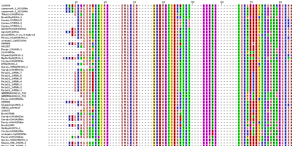
Molecular sequence data refers to the specific arrangement of molecules, most commonly nucleotides in DNA or RNA, or amino acids in proteins, that make up a biological macromolecule. This data is generated through laboratory techniques such as sequencing, and provides information about the exact order of the constituent molecules. This data is crucial in various fields of biology, including genetics, evolution, and molecular biology, allowing for comparisons between different organisms, identification of genetic variations, and studies of gene function and regulation.
Peroxisomes are membrane-bound subcellular organelles found in the cytoplasm of eukaryotic cells. They play a crucial role in various cellular processes, including the breakdown of fatty acids and the detoxification of harmful substances such as hydrogen peroxide (H2O2). Peroxisomes contain numerous enzymes, including catalase, which converts H2O2 into water and oxygen, thus preventing oxidative damage to cellular components. They also participate in the biosynthesis of ether phospholipids, a type of lipid essential for the structure and function of cell membranes. Additionally, peroxisomes are involved in the metabolism of reactive oxygen species (ROS) and contribute to the regulation of intracellular redox homeostasis. Dysfunction or impairment of peroxisome function has been linked to several diseases, including neurological disorders, developmental abnormalities, and metabolic conditions.
Ascorbate peroxidases (AHPX) are a group of enzymes that use ascorbic acid (vitamin C) as a reducing cofactor to catalyze the conversion of hydrogen peroxide (H2O2) into water (H2O) and oxygen (O2). This reaction helps protect cells from oxidative damage caused by the accumulation of H2O2, a byproduct of various metabolic processes. Ascorbate peroxidases are primarily found in plants, algae, and cyanobacteria, where they play a crucial role in the detoxification of reactive oxygen species generated during photosynthesis.
Aerobiosis is the process of living, growing, and functioning in the presence of oxygen. It refers to the metabolic processes that require oxygen to break down nutrients and produce energy in cells. This is in contrast to anaerobiosis, which is the ability to live and grow in the absence of oxygen.
In medical terms, aerobiosis is often used to describe the growth of microorganisms, such as bacteria and fungi, that require oxygen to survive and multiply. These organisms are called aerobic organisms, and they play an important role in many biological processes, including decomposition and waste breakdown.
However, some microorganisms are unable to grow in the presence of oxygen and are instead restricted to environments where oxygen is absent or limited. These organisms are called anaerobic organisms, and their growth and metabolism are referred to as anaerobiosis.
 Catalase
Catalase-peroxidase
Catalase-related immune-responsive domain
Taylorella
Mycobacterium pyrenivorans
Alexander Dounce
Glycine tabacina
ACAA1
Mycobacterium brumae
Clostridium tertium
Lactic acid bacteria
Streptococcus pyogenes
ACOX1
Granular cheese
Beta oxidation
Cellulosimicrobium cellulans
Enterobacteriaceae
Diagnostic microbiology
Bordetella trematum
Microcrystal electron diffraction
Alcanivorax pacificus
DNA-binding protein from starved cells
Xenophilus azovorans
Oleispira antarctica
Micrococcus luteus
Helicobacter pylori
Amphibacillus xylanus
Halomonas titanicae
Vibrio aerogenes
Iron
Catalase - Wikipedia
 Importance of catalase in the disposal of hydrogen peroxide within human erythrocytes
 Catalase (rat)
Re: How and Why is catalase's reaction rate affected by temp, pH
![Recombinant Anti-Catalase antibody [EPR20198] - Peroxisome Marker KO Tested (ab209211)](data:image/png;base64,iVBORw0KGgoAAAANSUhEUgAAABAAAAAQCAYAAAAf8/9hAAABm0lEQVQ4jaWTv0tbURTHP/cl75lqTIiNRFyEJIiUxNB2qf+D6NIuDg7WwcXFxU2yOznYte2klFIqpXVqoXQqgTYZKhURUURTlajJy++Xdx1eeJrmTc/vcuHc8/3cc869V8ileBZkClcSOcW9GUCmFPdmS16n4PfjKp/2KxQbJsmwxnwywOs/RUoNCcB0rI+xAbUb0DQls9tnbO7qHcC3OyWOigbn1RYA0aDXGbD8o8Dmro6qCOYS/Tx6qPHztMbGXx3Zzll5FuJppKe7hULNZD17jQA+TA0xGe21Nh4HmRj2sfjtAoDno36iQdUG2EPM5Gs0WpJ4SL01t7UwHqBPdZ63HTVMaxWOaWBK6Ri3AU8iPSgC9i6bfDmodCS9yhWpGs4AT3piIA3Qrykc6y1+ndV5v1cmX25xWGqy9vua1cyVbRh84GEk4CXk81gVy6WYja4YkpnP/9jaL3eckghrnOgGhZrV57vJCC9G/cB/19jrFXycHuLrUZXtgwpXdZPUoMbLZIA3dx5SMnx7jR0VuNG9/4ICIufaLWT2BlLHjkWr+SchAAAAAElFTkSuQmCC) Recombinant Anti-Catalase antibody [EPR20198] - Peroxisome Marker KO Tested (ab209211)
Recombinant Anti-Catalase antibody [EPR20198] - Peroxisome Marker KO Tested (ab209211)
IUCr) Polarized proton spin density images the tyrosyl radical locations in bovine liver catalase
 Why are lactic acid bacteria catalase negative? - Erasingdavid.com
 X-ray driven reduction of Cpd I of Catalase-3 from N. crassa reveals differential sensitivity of active sites and formation of...
 catalase assay kit - Hollis-Eden Pharmaceuticals Inc.
 Effects of Environmental Factors on Catalase and Lipase
 "Purification and Partial Characterization of Catalase from Chicken Ery" by TÜLİN AYDEMİR and KEVSER KURU
Investigating an enzyme-controlled reaction: catalase and hydrogen peroxide concentration
 Catalase Fusion Protein Ag16916 | Proteintech
Catalase
Modulation of hydrogen peroxide induced injury to corneal endothelium by virus mediated catalase gene transfer | British...
 Figures and data in The peroxisome counteracts oxidative stresses by suppressing catalase import via Pex14 phosphorylation |...
 CAT / Catalase antibody
Investigating the enzyme catalysed decomposition of hydrogen peroxide by catalase effects of changing temperature, pH and...
Keyword Catalases | Collection of Czechoslovak Chemical Communications
 Studies on Xanthine Oxidase: The Function of Catalase - Wikidata
 NAFENOPIN-INDUCED HEPATIC MICROBODY (PEROXISOME) PROLIFERATION AND CATALASE SYNTHESIS IN RATS AND MICE | Journal of Cell...
 Nutriversum VITA Catalase Enzim kapszula - 60db: v s rl s, hat anyagok, le r s - ProVitamin web ruh z
 Refubium - The Catalase KatA Contributes to Microaerophilic H2O2 Priming to Acquire an Improved Oxidative Stress Resistance in...
 Biotrophy-necrotrophy switch in pathogen evoke differential response in resistant and susceptible sesame involving multiple...
Catalase treatment
 Catalase - Opocrin
 Catalase Coursework - Kloepfer
Catalase | Profiles RNS
 catalase test - Bluegenics Europe
Assay2
- catalase functional activity in lysates was determined using a H 2 O 2 activity assay. (bmj.com)
- EMD Millipore Calbicohem™ Catalase Assay Kit is sensitive spectrophotometric assay. (fishersci.com)
Noncompetitive inhibitor of catalase4
- Any heavy metal ion (such as copper cations in copper(II) sulfate) can act as a noncompetitive inhibitor of catalase. (wikipedia.org)
- Furthermore, the poison cyanide is a noncompetitive inhibitor of catalase at high concentrations of hydrogen peroxide.Arsenate acts as an activator. (wikipedia.org)
- What is a noncompetitive inhibitor of catalase? (erasingdavid.com)
- Furthermore, the poison cyanide is a noncompetitive inhibitor of catalase at high concentrations of hydrogen peroxide. (erasingdavid.com)
Bovine2
- The amino acid sequence of bovine catalase was determined in 1969, and the three-dimensional structure in 1981. (wikipedia.org)
- The aim of this study was to evaluate the effects of catalase, 2% chlorhexidine gel, and 1% sodium hypochlorite on the microtensile bond strength to enamel of bovine teeth submitted to internal and external bleaching with 35% hydrogen peroxide. (bvsalud.org)
Amount of catalase2
- It needs to be well shaken before use so that each portion measured out has the same amount of catalase in it. (docbrown.info)
- The increase in microbody population in acatalasemic mice, although not accompanied by increase in catalase activity, was associated with a twofold increase in the amount of catalase protein. (rupress.org)
Hydrogen peroxide concentration2
- The decomposition of hydrogen peroxide by catalase proceeds according to first-order kinetics, the rate being proportional to the hydrogen peroxide concentration. (wikipedia.org)
- You can also keep the hydrogen peroxide concentration constant and do the investigation with a set of different concentrations of the potato-catalase mixture. (docbrown.info)
Anti-Catalase1
- By using the anti-catalase treatment significant amounts of peroxide can be saved. (api-paperchem.de)
Produce catalase2
- 1. Catalase Test Lactic acid bacteria do not produce catalase enzyme that converts hydrogen peroxide into water and oxygen. (erasingdavid.com)
- A few microbes produce catalase to shield themselves against assaults by hydrogen peroxide, a weapon generally utilized by the host's invulnerable framework, notwithstanding oxidative pressure. (eccscotland.com)
Protein2
- Three-dimensional protein structures of the peroxidated catalase intermediates are available at the Protein Data Bank. (wikipedia.org)
- The hepatic microbody proliferation in both male and female rats and wild type Cs a strain mice treated with nafenopin was of the same magnitude and was associated with a two-fold increase in catalase activity and in the concentration of catalase protein. (rupress.org)
Anaerobes6
- Catalase-negative bacteria may be anaerobes, or they may be facultative anaerobes that only ferment and do not respire using oxygen as a terminal electron acceptor (ie. (erasingdavid.com)
- Today I want to demolish two myths, one being that strict anaerobes (such as members of the Clostridia, a class of bacteria that includes the botulism organism as well as the C. diff bug that sickens half a million people a year in the U.S.) lack a catalase enzyme, and the other being that codons have very little degeneracy in base one. (blogspot.com)
- Bacteriology students are taught from Day One that strict anaerobes are killed by oxygen because they lack a catalase enzyme. (blogspot.com)
- The catechism about anaerobes being catalase-negative is true for some anaerobes, but by no means all. (blogspot.com)
- In fact, if you go to UniProt.org and do a search on "catalase clostridium" you'll get a long list of hits representing anaerobes that do, in fact, have a perfectly normal catalase enzyme. (blogspot.com)
- Some anaerobes have the classic katG ( E. coli ) version of catalase while others (like C. difficile ) have a special manganese-containing catalase. (blogspot.com)
Lysates1
- The Catalase Activity research-use-just pack is a colorimetric action examine intended for the evaluation and identification of catalase movement in serum, plasma, cells, tissues and erythrocyte lysates. (bluegenics.eu)
Liver8
- In 1937 catalase from beef liver was crystallized by James B. Sumner and Alexander Dounce and the molecular weight was measured in 1938. (wikipedia.org)
- However, "Copper deficiency can lead to a reduction in catalase activity in tissues, such as heart and liver. (wikipedia.org)
- However, catalase deficiency in mice may increase the likelihood of developing obesity, fatty liver, and type 2 diabetes. (wikipedia.org)
- Although widely distributed among animals, plants, bacteria, and fungi, catalase in mammalian liver and blood has been most intensively studied. (drugfuture.com)
- Catalase for commercial use obtained from animal liver, bacterial ( Micrococcus lysodeikticus), and fungal (Aspergillus niger) sources. (drugfuture.com)
- The serum glycerol-glycerides were markedly lowered in all the animals given nafenopin, which paralleled the increase in liver catalase. (rupress.org)
- Catalase is a major protective antioxidant in the eyes, brain, blood cells and other organs and tissues and it is especially active in the liver. (advancedhypnosisnj.com)
- Used for centuries for liver health, Picrorhiza helps the body’s Catalase production. (advancedhypnosisnj.com)
Concentration3
- This investigation looks at the rate of oxygen production by the catalase in pureed potato as the concentration of hydrogen peroxide varies. (practicalbiology.org)
- The tibialis anterior muscle (TA) was removed and assayed for biomarkers of oxidative stress that included: the ratio of reduced glutathione to oxidized glutathione (GSH/GSSG), which is a measure of redox status, malondialdehyde (MDA) which is a biomarker for lipid peroxidation, 8hydroxy-2'-deoxyguanosine (8-OHdG), which is a marker of oxidative damage to DNA, catalase concentration, and cytosolic hydrogen peroxide (H2O2) levels. (cdc.gov)
- Supplementation had no effect on catalase concentration. (cdc.gov)
Oxygen12
- Catalase is a common enzyme found in nearly all living organisms exposed to oxygen (such as bacteria, plants, and animals) which catalyzes the decomposition of hydrogen peroxide to water and oxygen. (wikipedia.org)
- one catalase molecule can convert millions of hydrogen peroxide molecules to water and oxygen each second. (wikipedia.org)
- To this end, catalase is frequently used by cells to rapidly catalyze the decomposition of hydrogen peroxide into less-reactive gaseous oxygen and water molecules. (wikipedia.org)
- Catalase is an enzyme that converts hydrogen peroxide to water and oxygen gas. (erasingdavid.com)
- If bubbles appear (due to the production of oxygen gas) the bacteria are catalase positive. (erasingdavid.com)
- Catalase plays a major role in the protection of tissues from the toxic effects of H_{2}O_{2} and partially reduced oxygen species. (tubitak.gov.tr)
- In this experiment you are measuring the rate at which oxygen is formed from the enzyme catalase decomposing hydrogen peroxide. (docbrown.info)
- In addition, catalase catalyses the dismutation of hydrogen peroxide into a molecule of oxygen and water, which is essential for the wound healing process. (opocrin.it)
- Catalase converts the free radical generating hydrogen peroxide into water and oxygen. (advancedhypnosisnj.com)
- Catalase is the nearly universal enzyme that breaks down peroxides into oxygen and water. (blogspot.com)
- Catalase is the second most plentiful enzymatic cell reinforcement (after superoxide dismutase), which weakens the degrees of receptive oxygen species that universally go with obsessive problems like maturing, waterfall, malignant growth, healthful lack, atherosclerosis, and diabetes [5]. (eccscotland.com)
- Different techniques for estimating catalase action have been created, including those including iodometry chemiluminescence , polarimetry, and checking the development of oxygen through an oxygen anode or a low-stream gas meter. (eccscotland.com)
H2O27
- Catalase was first noticed in 1818 by Louis Jacques Thénard, who discovered hydrogen peroxide (H2O2). (wikipedia.org)
- While the complete mechanism of catalase is not currently known, the reaction is believed to occur in two stages: H2O2 + Fe(III)-E → H2O + O=Fe(IV)-E(.+) H2O2 + O=Fe(IV)-E(.+) → H2O + Fe(III)-E + O2 Here Fe()-E represents the iron center of the heme group attached to the enzyme. (wikipedia.org)
- This rate was determined in control cells and in catalase-inactivated cells while the cells were exposed to H2O2, which was generated at various constant and predetermined rates by glucose oxidase. (nih.gov)
- The results indicated that catalase handles approximately half of the generated H2O2. (nih.gov)
- Catalases play a key role in the defense against oxidative stress in bacteria by catalyzing the decomposition of H2O2. (erasingdavid.com)
- Catalase Test: Purpose, Principle, Method, Interpretations, Precautions Hydrogen peroxide H2O2 made one of the oxidative final product of aerobic carbohydrate metabolism. (medicallabscientist.org)
- Elevated degrees of hydrogen peroxide (H2O2) quickly lead to restraint of the catalase catalyst by changing its dynamic site structure, in spite of the fact that there is variety in the degree to which this happens. (eccscotland.com)
Bacteria9
- Why are lactic acid bacteria catalase negative? (erasingdavid.com)
- Lactic acid bacteria (LAB) are a group of Gram-positive, non-spore forming, cocci or rods, catalase-negative, and fastidious organisms, with high tolerance for low pH [1,2,3]. (erasingdavid.com)
- If no bubbles appear, the bacteria are catalase negative. (erasingdavid.com)
- What type of bacteria are catalase negative? (erasingdavid.com)
- Lactobacillus bacteria are catalase negative, Gram positive and usually rod shaped. (erasingdavid.com)
- What is the importance of catalase in bacteria? (erasingdavid.com)
- What types of bacteria are catalase positive? (erasingdavid.com)
- Which bacteria are catalase positive? (erasingdavid.com)
- Many bacteria are catalase positive, but some are better catalase-producers than others. (erasingdavid.com)
Peroxide12
- Catalase-positive pathogens, such as Mycobacterium tuberculosis, Legionella pneumophila, and Campylobacter jejuni, make catalase to deactivate the peroxide radicals, thus allowing them to survive unharmed within the host. (erasingdavid.com)
- Cells make the enzyme catalase to remove hydrogen peroxide. (practicalbiology.org)
- To examine the effect of catalase gene transfer on survival of corneal endothelial cells (EC) following challenge with hydrogen peroxide (H 2 O 2 ) in an ex vivo model of oxidative stress. (bmj.com)
- vary either the hydrogen peroxide or the enzyme catalase). (docbrown.info)
- The 'stock' solutions of potato-catalase or hydrogen peroxide should be initially in separate boiling tubes and placed in the water bath so that everything starts at the right temperature. (docbrown.info)
- Catalase levels drop with age and as a result hydrogen peroxide activity increases. (advancedhypnosisnj.com)
- Tests showing catalase movement are hatched with hydrogen peroxide answer for 2 min before quick blending of the brooding enzymatic response combination with cobalt-bicarbonate reagent, which evaluates non-responding hydrogen peroxide. (eccscotland.com)
- Catalase action is generally straightforwardly corresponding to the pace of separation of hydrogen peroxide. (eccscotland.com)
- The mainly normal technique for estimating catalase action is the UV spectrophotometric strategy, which relies upon observing the difference in 240 nm absorbance at elevated degrees of hydrogen peroxide arrangement (≥30 mM). (eccscotland.com)
- This paper reports a straightforward examine for estimating catalase movement that incorporates the estimation of hydrogen peroxide spectrophotometrically. (eccscotland.com)
- Catalase is an omnipresent cell reinforcement catalyst that debases hydrogen peroxide into water and oxygen1. (eccscotland.com)
- A past report has truth be told shown that a catalase-lacking freak microbe was more helpless than its wild-type strain to the oxidative pressure incited by hydrogen peroxide and insusceptible cell assaults (which include hydrogen peroxide)2. (eccscotland.com)
Coli1
- Recombinant human CAT / Catalase purified from E. coli. (covalab.com)
Enzyme Activity1
- Enzyme activity was stable at temperatures between 10 and 30°C. The activity of purified catalase was inhibited by azide, cyanide, \beta -mercaptoethanol, dithiotreitol (DTT) and iodoacetamide. (tubitak.gov.tr)
ELISA1
- The Rat Catalase ELISA kit can be used to identify samples from the rat species. (elisakit.cc)
Antibody1
- Formalin-fixed, paraffin-embedded H. pylori-infected human stomach stained with Helicobacter pylori (Catalase) Mouse Monoclonal Antibody (HPYL/7227). (neobiotechnologies.com)
Activity5
- The maximal activity of catalase was observed between pH 6.0 and 8.0. (tubitak.gov.tr)
- Make fresh for each lesson, because catalase activity reduces noticeably over 2/3 hours. (practicalbiology.org)
- The absence of sex difference in microbody proliferative response in nafenopin-treated rats and wild type mice is of particular significance, since ethyl-α- p -chlorophenoxyisobutyrate (CPIB)-induced microbody proliferation and increase in catalase activity occurred only in males. (rupress.org)
- Used for measurement of catalase activity in plasma. (fishersci.com)
- The data indicate that compared to control muscles, repetitive loading significantly increased catalase activity and lowered MDA levels in the TA. (cdc.gov)
Hepatic1
- Su-13437), a potent hypolipidemic compound, was administered in varying concentrations in ground Purina Chow to male and female rats, wild type (Cs a strain) mice and acatalasemic (Cs b strain) mice to determine the hepatic microbody proliferative and catalase-inducing effects. (rupress.org)
Fastidious1
- Pneumococci are fastidious microorganisms that require catalase to grow on agar plates. (msdmanuals.com)
Acatalasia1
- Some humans have very low levels of catalase (acatalasia), yet show few ill effects. (wikipedia.org)
Antioxidant3
- Catalase is an antioxidant enzyme used as an active substance in topical drug products. (opocrin.it)
- Catalase, as an antioxidant enzyme, is essential for restoring this optimal balance. (opocrin.it)
- Catalase is a natural antioxidant enzyme in our body it is found in all of our cells and helps shield our brain, eyes, heart, kidneys, blood and all other tissues from oxidizing free radicals that can damage us. (advancedhypnosisnj.com)
Reaction1
- The optimum pH for human catalase is approximately 7, and has a fairly broad maximum: the rate of reaction does not change appreciably between pH 6.8 and 7.5. (wikipedia.org)
Superoxide2
- Sirtuin 1 attenuates oxidative stress via upregulation of superoxide dismutase 2 and catalase in astrocytes. (uchicago.edu)
- This product contains equal amounts of SOD and catalase to neutralize the superoxide free-radical, thereby reducing peroxynitrite. (hwofc.com)
Recombinant1
- A recombinant adenovirus vector (AdCL) was used to transfer human catalase cDNA into EC of whole thickness rabbit corneas ex vivo. (bmj.com)
Spectrophotometric1
- The subtleties of an exact, precise, and touchy spectrophotometric technique for it are introduced here to gauge catalase movement. (eccscotland.com)
Subunits3
- Human catalase forms a tetramer composed of four subunits, each of which can be conceptually divided into four domains. (wikipedia.org)
- SDS-gel electrophoresis results indicated that chicken erythrocyte catalase consists of four apparently identical subunits, with a molecular weight of around 57.5 kDa. (tubitak.gov.tr)
- All catalases so far isolated contain four tetrahedrally arranged subunits of equal size giving an approximate mol wt of 240,000. (drugfuture.com)
Deficiency1
- These results are in agreement with our earlier findings on erythrocytes of a subject with a genetic deficiency of catalase. (nih.gov)
Examine2
- These variations of environmental factors are use to conduct experiments to examine the effects it have on catalase and lipase. (ukessays.com)
- To examine the morphological effects of catalase gene transfer in modulation of H 2 O 2 induced injury, transduced corneas were maintained in ex vivo culture and challenged with H 2 O 2 . (bmj.com)
Species1
- The pH optimum for other catalases varies between 4 and 11 depending on the species. (wikipedia.org)
MeSH1
- Catalase" is a descriptor in the National Library of Medicine's controlled vocabulary thesaurus, MeSH (Medical Subject Headings) . (uchicago.edu)
Erythrocyte1
- The molecular weight of the native chicken erythrocyte catalase was estimated at 240 kDa by gel filtration. (tubitak.gov.tr)
Compounds1
- Because of microbody proliferation and catalase induction with increasing number of hypolipidemic compounds, additional studies are necessary to determine the interrelationships of microbody proliferation, catalase induction, and hypolipidemia. (rupress.org)
Human3
- The catalase within normal, intact human erythrocytes was completely inactivated with amino triazole. (nih.gov)
- Reacts with human CAT / Catalase. (covalab.com)
- It really is not very hard to attempt, in the event you know the necessities for the composition of your catalase coursework, regarding the resources, the place quite possibly the most significant advice are generally discovered, in addition to the human being, who'll enable you to organize your catalase coursework. (kloepfer.com)
Incorporates1
- This total, prepared to-utilize pack incorporates clear 96 well plate(s), catalase standard (100 Unit/mL), catalase substrate, and different parts to play out the measure. (bluegenics.eu)
Results1
- Your catalase coursework may want to existing your deep homework and the results within your deliver the results. (kloepfer.com)
Supplement1
- Thus, a sign that you need to supplement with Catalase Hx® is graying hair. (advancedhypnosisnj.com)
Amino1
- Catalase is a tetramer of four polypeptide chains, each over 500 amino acids long. (wikipedia.org)