Cartilage
Cartilage, Articular
Nasal Cartilages
Osteoarthritis
Laryngeal Cartilages
Hyaline Cartilage
Cartilage Oligomeric Matrix Protein
Osteoarthritis, Knee
Aggrecans
Collagen Type II
Glycosaminoglycans
Matrilin Proteins
Chondrogenesis
Extracellular Matrix Proteins
Collagen
Stifle
Tibia
Nasal Septum
Epiphyses
Arytenoid Cartilage
Cricoid Cartilage
Cattle
Thyroid Cartilage
Matrix Metalloproteinase 13
Weight-Bearing
Joints
Tissue Engineering
Compressive Strength
Bone and Bones
Hyaluronic Acid
Femur Head
Synovial Membrane
Extracellular Matrix
Magnetic Resonance Imaging
Synovial Fluid
Stress, Mechanical
Chondroitin Sulfates
Biomechanical Phenomena
Mandibular Condyle
Arthritis, Experimental
SOX9 Transcription Factor
Procollagen N-Endopeptidase
Uronic Acids
Anterior Cruciate Ligament
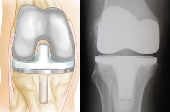
Arthroplasty, Subchondral
Matrix Metalloproteinase 3
Cells, Cultured
Microscopy, Polarization
Tissue Culture Techniques
Collagen Type IX
Ribs
Chondroitin
Lectins, C-Type
Bone Development
Friction
Rabbits
Collagen Type X
Rhinoplasty
Chick Embryo
Tissue Scaffolds
Calcification, Physiologic
Collagen Type XI
Joint Diseases
Periosteum
Hindlimb
Arthritis
Elastic Cartilage
Chondroitin Sulfate Proteoglycans
Chondrosarcoma
Matrix Metalloproteinases
Interleukin-1
Glycoproteins
Immunohistochemistry
Temporomandibular Joint
Culture Techniques
Alcian Blue
Synovitis
Bone Morphogenetic Proteins
Cell Differentiation
Hyalin
Arthritis, Rheumatoid
Tensile Strength
ADAM Proteins
Disease Models, Animal
RNA, Messenger
Talus
Osteophyte
Fibrocartilage
Organ Culture Techniques
Fibrillar Collagens
Osteochondritis Dissecans
Osteoarthritis, Hip
Swine
In Situ Hybridization
Bone Morphogenetic Protein 2
Sharks
Mesenchymal Stromal Cells
Horses
Osteochondrosis
Ankle Joint
Aging
Chondroitinases and Chondroitin Lyases
Hydrogels
Chondroma
Elastic Modulus
Hip Joint
Tissue Transplantation
Bone Matrix
Chondrocalcinosis
Growth Differentiation Factor 5
Achondroplasia
Biological Markers
Interleukin-1beta
Reverse Transcriptase Polymerase Chain Reaction
Matrix Metalloproteinase 1
Gene Expression
Hydroxyproline
Interleukin-1alpha
Bone Morphogenetic Protein 7
Microscopy, Electron, Scanning
Microscopy, Electron
Glucosamine
Models, Animal
Polychondritis, Relapsing
Oncostatin M
Arthrography
Collagen Type I
Reproducibility of Results
Metalloproteases
Branchial Region
Guided Tissue Regeneration
Ear, External
SOXD Transcription Factors
Hexosamines
Mandible
Transforming Growth Factor beta
Tarsal Joints
Disease Progression
Aminopropionitrile
Histocytochemistry
Patellofemoral Joint
Imaging, Three-Dimensional
Sternum
Hypertrophy
Hyaluronoglucosaminidase
Models, Biological
Sulfur Radioisotopes
Papain
Transforming Growth Factor beta3
Collagen Type VI
Chondroitin Lyases
Endopeptidases
Alkaline Phosphatase
Bone Remodeling
Finite Element Analysis
Dogs
Sepharose
Bony Callus
Hemarthrosis
Skeleton
Bone Marrow Diseases
Metatarsal Bones
Gene Expression Regulation, Developmental
Bone Diseases, Developmental
Tympanoplasty
Inhibition of transforming growth factor beta production by nitric oxide-treated chondrocytes: implications for matrix synthesis. (1/4540)
OBJECTIVE: Nitric oxide (NO) is generated copiously by articular chondrocytes activated by interleukin-1beta (IL-1beta). If NO production is blocked, much of the IL-1beta inhibition of proteoglycan synthesis is prevented. We tested the hypothesis that this inhibitory effect of NO on proteoglycan synthesis is secondary to changes in chondrocyte transforming growth factor beta (TGFbeta). METHODS: Monolayer, primary cultures of lapine articular chondrocytes and cartilage slices were studied. NO production was determined as nitrite accumulation in the medium. TGFbeta bioactivity in chondrocyte- and cartilage-conditioned medium (CM) was measured with the mink lung epithelial cell bioassay. Proteoglycan synthesis was measured as the incorporation of 35S-sodium sulfate into macromolecules separated from unincorporated label by gel filtration on PD-10 columns. RESULTS: IL-1beta increased active TGFbeta in chondrocyte CM by 12 hours; by 24 hours, significant increases in both active and latent TGFbeta were detectable. NG-monomethyl-L-arginine (L-NMA) potentiated the increase in total TGFbeta without affecting the early TGFbeta activation. IL-1beta stimulated a NO-independent, transient increase in TGFbeta3 at 24 hours; however, TGFbeta1 was not changed. When NO synthesis was inhibited with L-NMA, IL-1beta increased CM concentrations of TGFbeta1 from 24-72 hours of culture. L-arginine (10 mM) reversed the inhibitory effect of L-NMA on NO production and blocked the increases in TGFbeta1. Anti-TGFbeta1 antibody prevented the restoration of proteoglycan synthesis by chondrocytes exposed to IL-1beta + L-NMA, confirming that NO inhibition of TGFbeta1 in IL-1beta-treated chondrocytes effected, in part, the decreased proteoglycan synthesis. Furthermore, the increase in TGFbeta and proteoglycan synthesis seen with L-NMA was reversed by the NO donor S-nitroso-N-acetylpenicillamide. Similar results were seen with cartilage slices in organ culture. The autocrine increase in CM TGFbeta1 levels following prior exposure to TGFbeta1 was also blocked by NO. CONCLUSION: NO can modulate proteoglycan synthesis indirectly by decreasing the production of TGFbeta1 by chondrocytes exposed to IL-1beta. It prevents autocrine-stimulated increases in TGFbeta1, thus potentially diminishing the anabolic effects of this cytokine in chondrocytes. (+info)Expression of both P1 and P2 purine receptor genes by human articular chondrocytes and profile of ligand-mediated prostaglandin E2 release. (2/4540)
OBJECTIVE: To assess the expression and function of purine receptors in articular chondrocytes. METHODS: Reverse transcriptase-polymerase chain reaction (RT-PCR) was used to screen human chondrocyte RNA for expression of P1 and P2 purine receptor subtypes. Purine-stimulated prostaglandin E2 (PGE2) release from chondrocytes, untreated or treated with recombinant human interleukin-1alpha (rHuIL-1alpha), was assessed by radioimmunoassay. RESULTS: RT-PCR demonstrated that human articular chondrocytes transcribe messenger RNA for the P1 receptor subtypes A2a and A2b and the P2 receptor subtype P2Y2, but not for the P1 receptor subtypes A1 and A3. The P1 receptor agonists adenosine and 5'-N-ethylcarboxamidoadenosine did not change PGE2 release from chondrocytes. The P2Y2 agonists ATP and UTP stimulated a small release of PGE2 that was potentiated after pretreatment with rHuIL-1alpha. PGE2 release in response to ATP and UTP cotreatment was not additive, but release in response to coaddition of ATP and bradykinin (BK) or UTP and BK was additive, consistent with ATP and UTP competition for the same receptor site. The potentiation of PGE2 release in response to ATP and UTP after rHuIL-1alpha pretreatment was mimicked by phorbol myristate acetate. CONCLUSION: Human chondrocytes express both P1 and P2 purine receptor subtypes. The function of the P1 receptor subtype is not yet known, but stimulation of the P2Y2 receptor increases IL-1-mediated PGE2 release. (+info)Destruction of hyaline cartilage in the sigmoid notch of the human ulna. (3/4540)
In an ulna from an adolescent a fossa nudata divided the articular surface of the sigmoid notch into olecranon and coronoid areas. In the floor of the fossa a layer of loose avascular pannus covered a thin layer of articular cartilage. The pannus appeared to have been formed by removal of chondroitin from the cartilage, freeing the cells and unmasking the fibres. Probably the change followed loss of contact between the articular cartilages of the sigmoid notch and trochlea during postnatal growth. (+info)Transport of solutes through cartilage: permeability to large molecules. (4/4540)
A review of the transport of solutes through articular cartilage is given, with special reference to the effect of variations in matrix composition. Some physiological implications of our findings are discussed. Also, results of an experimental study of the permeability of articular cartilage to large globular proteins are presented. Because of the very low partition coefficients of large solutes between cartilage and an external solution new experimental techniques had to be devised, particularly for the study of diffusion. The partition coefficients of solutes were found to decrease very steeply with increase in size, up to serum albumin. There was, however, no further decrease for IGG. The diffusion coefficient of serum albumin in cartilage was relatively high (one quarter of the value in aqueous solution). These two facts taken together suggest that there may be a very small fraction of relatively large pores in cartilage through which the transport of large molecules is taking place. The permeability of cartilage to large molecules is extremely sensitive to variations in the glycosaminoglycan content: for a threefold increase in the latter there is a hundredfold decrease in the partition coefficient. For cartilage of fixed charge density around 0-19 m-equiv/g, there is no penetration at all of globular proteins of size equal to or larger than serum albumin. (+info)Association of the aggrecan keratan sulfate-rich region with collagen in bovine articular cartilage. (5/4540)
Aggrecan, the predominant large proteoglycan of cartilage, is a multidomain macromolecule with each domain contributing specific functional properties. One of the domains contains the majority of the keratan sulfate (KS) chain substituents and a protein segment with a proline-rich hexapeptide repeat sequence. The function of this domain is unknown but the primary structure suggests a potential for binding to collagen fibrils. We have examined binding of aggrecan fragments encompassing the KS-rich region in a solid-phase assay. A moderate affinity (apparent Kd = 1.1 microM) for isolated collagen II, as well as collagen I, was demonstrated. Enzymatic digestion of the KS chains did not alter the capacity of the peptide to bind to collagen, whereas cleavage of the protein core abolished the interaction. The distribution of the aggrecan KS-rich region in bovine tarsometatarsal joint cartilage was investigated using immunoelectron microscopy. Immunoreactivity was relatively low in the superficial zone and higher in the intermediate and deep zones of the uncalcified cartilage. Within the pericellular and territorial matrix compartments the epitopes representing the aggrecan KS-rich region were detected preferentially near or at collagen fibrils. Along the fibrils, epitope reactivity was non-randomly distributed, showing preference for the gap region within the D-period. Our data suggest that collagen fibrils interact with the KS-rich regions of several aggrecan monomers aligned within a proteoglycan aggregate. The fibril could therefore serve as a backbone in at least some of the aggrecan complexes. (+info)Distribution of chondroitin sulfate in cartilage proteoglycans under associative conditions. (6/4540)
Proteoglycan aggregates and proteoglycan subunits were extracted from bovine articular cartilage with guanidine-HC1 folowed by fractionation by equilibrium centrifugation in cesium chloride density gradients. The distribution of chondroitin sulfates (CS) in the cartilage proteoglycans was studied at the disaccharide level by digestion with chondroitinases. In the proteoglycan aggregate fraction, it was observed that the proportion of 4-sulfated disaccharide units to total CS increased from the bottom to the top fractions, whereas that of 6-sulfated disaccharide units was in the reverse order. Thus, the ratio of 4-sulfated disaccharide units to 6-sulfated disaccharide units increased significantly with decreasing density. The proportion of non-sulfated disaccharide units to total CS tended to increase with increasing density. These data indicate a polydisperse distribution of CS chains, under the conditions used here, in proteoglycan aggregates from bovine articular cartilage. (+info)Effect of anti-inflammatory drugs on sulphated glycosaminoglycan synthesis in aged human articular cartilage. (7/4540)
The anti-inflammatory drugs, sodium salicylate, indomethacin, hydrocortisone, ibuprofen, and flurbiprofen, were examined for their effects on sulphated glycosaminoglycan synthesis in aged human cartilage in vitro. Cartilage was obtained from femoral heads removed during surgery and drug effects were found to vary significantly from one head to another. Statistical analysis of the results showed that sodium salicylate exhibits concentration-dependent inhibition of glycosaminoglycan synthesis over the concentration range used. Indomethacin, hydrocortisone, and ibuprofen, at concentrations comparable to those attained in man, caused a statistically significant depression of sulphated glycosaminoglycan synthesis in cartilage from some femoral heads but not others, reflecting the variable response of human articular cartilage to anti-inflammatory drugs. Sodium salicylate and indomethacin at higher doses produced significant (Pless than 0-005) inhibition of sulphated glycosaminoglycan synthesis in all femoral heads studied. The results for flurbiprofen were less conclusive; this compound appears not to inhibit glycosaminoglycan synthesis over the concentration range used. (+info)Uridine diphosphate xylosyltransferase activity in cartilage from manganese-deficient chicks. (8/4540)
The glycosaminoglycan content of cartilage is decreased in manganese deficiency in the chick (perosis). The activity of xylosyltransferase, the first enzyme in the biosynthetic pathway of sulphated glycosaminoglycans, was studied in the epiphysial cartilage of 4-week-old chicks which had been maintained since hatching on a manganese-deficient diet. Enzymic activity was measured by the incorporation of [14C]xylose from UDP-[14C]xylose into trichloroacetic acid precipitates. Optimal conditions for the xylosyltransferase assay were established and shown to be the same for both control and manganese-deficient cartilage. Assay of the enzyme by using an exogenous xylose acceptor showed no difference in xylosyltransferase activity between control and manganese-deficient tissue. Further, the extent of xylose incorporation was greater in manganese-deficient than in control cartilage preparations, suggesting an increase in xylose-acceptor sites on the endogenous acceptor protein in the deficient cartilage. 35S turnover in the manganese-deficient cartilage was also increased. The data suggest that the decreased glycosaminoglycan content in manganese-deficient cartilage is due to decreased xylosylation of the acceptor protein plus increased degradation of glycosaminoglycan. (+info)Cartilage is a type of connective tissue that is found throughout the body in various forms. It is made up of specialized cells called chondrocytes, which are embedded in a firm, flexible matrix composed of collagen fibers and proteoglycans. This unique structure gives cartilage its characteristic properties of being both strong and flexible.
There are three main types of cartilage in the human body: hyaline cartilage, elastic cartilage, and fibrocartilage.
1. Hyaline cartilage is the most common type and is found in areas such as the articular surfaces of bones (where they meet to form joints), the nose, trachea, and larynx. It has a smooth, glassy appearance and provides a smooth, lubricated surface for joint movement.
2. Elastic cartilage contains more elastin fibers than hyaline cartilage, which gives it greater flexibility and resilience. It is found in structures such as the external ear and parts of the larynx and epiglottis.
3. Fibrocartilage has a higher proportion of collagen fibers and fewer chondrocytes than hyaline or elastic cartilage. It is found in areas that require high tensile strength, such as the intervertebral discs, menisci (found in joints like the knee), and the pubic symphysis.
Cartilage plays a crucial role in supporting and protecting various structures within the body, allowing for smooth movement and providing a cushion between bones to absorb shock and prevent wear and tear. However, cartilage has limited capacity for self-repair and regeneration, making damage or degeneration of cartilage tissue a significant concern in conditions such as osteoarthritis.
Articular cartilage is the smooth, white tissue that covers the ends of bones where they come together to form joints. It provides a cushion between bones and allows for smooth movement by reducing friction. Articular cartilage also absorbs shock and distributes loads evenly across the joint, protecting the bones from damage. It is avascular, meaning it does not have its own blood supply, and relies on the surrounding synovial fluid for nutrients. Over time, articular cartilage can wear down or become damaged due to injury or disease, leading to conditions such as osteoarthritis.
Cartilage diseases refer to conditions that affect the cartilaginous tissues in the body. Cartilage is a firm, flexible connective tissue found in many areas of the body, including the joints, ribcage, ears, and nose. It provides structure and support, allows for smooth movement between bones, and protects the ends of bones from friction.
There are several types of cartilage diseases, including:
1. Osteoarthritis (OA): This is a degenerative joint disease that occurs when the protective cartilage that cushions the ends of your bones wears down over time. It can cause pain, stiffness, and loss of mobility in the affected joints.
2. Rheumatoid arthritis (RA): This is an autoimmune disorder that causes inflammation in the lining of the joints, leading to cartilage damage and bone erosion.
3. Traumatic arthritis: This occurs when a joint is injured, causing damage to the cartilage and resulting in pain, stiffness, and loss of mobility.
4. Infectious arthritis: This occurs when a joint becomes infected, leading to inflammation and potential damage to the cartilage.
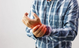
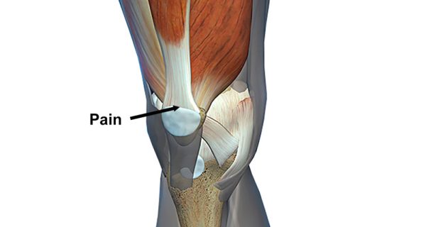
5. Chondromalacia patellae: This is a condition that affects the cartilage on the back of the kneecap, causing pain and stiffness in the knee.
6. Costochondritis: This is an inflammation of the cartilage in the ribcage, causing chest pain and discomfort.
7. Nasal septal deviation: This is a condition where the cartilage that separates the nostrils is crooked or off-center, causing difficulty breathing through the nose.
8. Osteochondritis dissecans (OCD): This is a joint condition that occurs when a piece of cartilage and bone in a joint becomes detached, causing pain and stiffness.
9. Synovial chondromatosis: This is a rare condition where nodules made up of cartilage form in the lining of a joint, causing pain, swelling, and limited mobility.
Treatment for cartilage diseases varies depending on the specific condition and severity, but may include medication, physical therapy, surgery, or a combination of these.
Chondrocytes are the specialized cells that produce and maintain the extracellular matrix of cartilage tissue. They are responsible for synthesizing and secreting the collagen fibers, proteoglycans, and other components that give cartilage its unique properties, such as elasticity, resiliency, and resistance to compression. Chondrocytes are located within lacunae, or small cavities, in the cartilage matrix, and they receive nutrients and oxygen through diffusion from the surrounding tissue fluid. They are capable of adapting to changes in mechanical stress by modulating the production and organization of the extracellular matrix, which allows cartilage to withstand various loads and maintain its structural integrity. Chondrocytes play a crucial role in the development, maintenance, and repair of cartilaginous tissues throughout the body, including articular cartilage, costal cartilage, and growth plate cartilage.
Nasal cartilages are the flexible, supportive structures in the nose that contribute to its shape and structure. They are made up of tough, but elastic tissue called cartilage. There are several nasal cartilages, including:
1. The septal cartilage, which is a thin, flat strip that forms the dividing wall between the two sides of the nose.
2. The upper and lower lateral cartilages, which are located on either side of the nostrils and help to shape them.
3. The sesamoid cartilages, which are small, round pieces of cartilage that can be found near the nasal opening.
These cartilages work together to provide support and flexibility to the nose, allowing it to withstand the forces of breathing and other facial movements while maintaining its shape.
Osteoarthritis (OA) is a type of joint disease that is characterized by the breakdown and eventual loss of cartilage - the tissue that cushions the ends of bones where they meet in the joints. This breakdown can cause the bones to rub against each other, causing pain, stiffness, and loss of mobility. OA can occur in any joint, but it most commonly affects the hands, knees, hips, and spine. It is often associated with aging and can be caused or worsened by obesity, injury, or overuse.
The medical definition of osteoarthritis is: "a degenerative, non-inflammatory joint disease characterized by the loss of articular cartilage, bone remodeling, and the formation of osteophytes (bone spurs). It is often associated with pain, stiffness, and decreased range of motion in the affected joint."
Ear cartilage, also known as auricular cartilage, refers to the flexible connective tissue that makes up the structural framework of the external ear or pinna. The ear cartilage provides support and shape to the ear, helping to direct sound waves into the ear canal and towards the eardrum.
The ear cartilage is composed of type II collagen fibers and proteoglycans, which give it its flexibility and resiliency. It is covered by a thin layer of skin on both sides and contains no bones. Instead, the ear cartilage is shaped and maintained by the surrounding muscles and connective tissue.
There are three main parts of the ear cartilage: the helix, the antihelix, and the tragus. The helix is the outer rim of the ear, while the antihelix is the curved ridge that runs parallel to the helix. The tragus is the small piece of cartilage that projects from the front of the ear canal.
Ear cartilage can be affected by various conditions, including trauma, infection, and degenerative changes associated with aging. In some cases, surgical procedures may be required to reshape or reconstruct damaged ear cartilage.
Laryngeal cartilages refer to the various pieces of cartilage that make up the structure of the larynx, also known as the voice box. The larynx is a crucial part of the respiratory system, located in the neck between the pharynx and the trachea. It plays a vital role in protecting the lower airways from food or drink entering the windpipe, as well as producing sound during speech.
There are several laryngeal cartilages, including:
1. Thyroid cartilage: This is the largest and most superior of the laryngeal cartilages. It forms the Adam's apple in men and has a prominent notch in the front called the thyroid notch. The thyroid cartilage protects the larynx and provides attachment for various muscles and ligaments.
2. Cricoid cartilage: This is the only complete ring of cartilage in the airway and lies inferior to the thyroid cartilage. It has a broad, flat superior portion called the cricoid lamina and a narrower, more curved inferior portion called the cricoid arch. The cricoid cartilage serves as an attachment site for several muscles and ligaments involved in breathing and swallowing.
3. Arytenoid cartilages: These are paired, pyramid-shaped structures that sit on top of the cricoid cartilage. They help form the posterior portion of the laryngeal inlet and provide attachment for the vocal cords (vocal folds). The arytenoid cartilages play a crucial role in voice production and respiration.
4. Corniculate cartilages: These are small, conical-shaped structures that project from the superior aspect of each arytenoid cartilage. They help form the most posterior portion of the laryngeal inlet.
5. Cuneiform cartilages: These are tiny, flat, crescent-shaped structures located near the corniculate cartilages. They also contribute to forming the posterior aspect of the laryngeal inlet.
These laryngeal cartilages work together to protect the airway, facilitate breathing, and enable voice production.
Hyaline cartilage is a type of cartilaginous tissue that is primarily found in the articulating surfaces of bones, ribcage, nose, ears, and trachea. It has a smooth, glassy appearance (hence the name "hyaline," derived from the Greek word "hyalos" meaning glass) due to the presence of type II collagen fibers that are arranged in a precise pattern and embedded in a proteoglycan-rich matrix.
The high concentration of proteoglycans, which are complex molecules made up of a protein core and glycosaminoglycan side chains, gives hyaline cartilage its firm yet flexible properties. This type of cartilage is avascular, meaning it does not contain blood vessels, and receives nutrients through diffusion from the surrounding synovial fluid in joints or adjacent tissues.
Hyaline cartilage plays a crucial role in providing structural support, reducing friction between articulating bones, and facilitating smooth movement in joints. It also serves as a template for endochondral ossification, a process by which long bones grow in length during development.
The knee joint, also known as the tibiofemoral joint, is the largest and one of the most complex joints in the human body. It is a synovial joint that connects the thighbone (femur) to the shinbone (tibia). The patella (kneecap), which is a sesamoid bone, is located in front of the knee joint and helps in the extension of the leg.
The knee joint is made up of three articulations: the femorotibial joint between the femur and tibia, the femoropatellar joint between the femur and patella, and the tibiofibular joint between the tibia and fibula. These articulations are surrounded by a fibrous capsule that encloses the synovial membrane, which secretes synovial fluid to lubricate the joint.
The knee joint is stabilized by several ligaments, including the medial and lateral collateral ligaments, which provide stability to the sides of the joint, and the anterior and posterior cruciate ligaments, which prevent excessive forward and backward movement of the tibia relative to the femur. The menisci, which are C-shaped fibrocartilaginous structures located between the femoral condyles and tibial plateaus, also help to stabilize the joint by absorbing shock and distributing weight evenly across the articular surfaces.
The knee joint allows for flexion, extension, and a small amount of rotation, making it essential for activities such as walking, running, jumping, and sitting.
Proteoglycans are complex, highly negatively charged macromolecules that are composed of a core protein covalently linked to one or more glycosaminoglycan (GAG) chains. They are a major component of the extracellular matrix (ECM) and play crucial roles in various biological processes, including cell signaling, regulation of growth factor activity, and maintenance of tissue structure and function.
The GAG chains, which can vary in length and composition, are long, unbranched polysaccharides that are composed of repeating disaccharide units containing a hexuronic acid (either glucuronic or iduronic acid) and a hexosamine (either N-acetylglucosamine or N-acetylgalactosamine). These GAG chains can be sulfated to varying degrees, which contributes to the negative charge of proteoglycans.
Proteoglycans are classified into four major groups based on their core protein structure and GAG composition: heparan sulfate/heparin proteoglycans, chondroitin/dermatan sulfate proteoglycans, keratan sulfate proteoglycans, and hyaluronan-binding proteoglycans. Each group has distinct functions and is found in specific tissues and cell types.
In summary, proteoglycans are complex macromolecules composed of a core protein and one or more GAG chains that play important roles in the ECM and various biological processes, including cell signaling, growth factor regulation, and tissue structure maintenance.
Cartilage oligomeric matrix protein (COMP) is a extracellular matrix protein that is found in high concentrations in cartilaginous tissues, such as articular cartilage and intervertebral discs. It is a member of the thrombospondin family and plays a role in the organization and stability of the extracellular matrix.
It is also known to be involved in the process of osteoarthritis, a degenerative joint disease. High levels of COMP are found in the synovial fluid of patients with osteoarthritis, and it is thought to contribute to the breakdown of cartilage. Additionally, genetic variations in the COMP gene have been associated with an increased risk of developing osteoarthritis.
It also plays a role in bone development and repair, as well as in the regulation of cell growth and differentiation.
Osteoarthritis (OA) of the knee is a degenerative joint disease that affects the articular cartilage and subchondral bone in the knee joint. It is characterized by the breakdown and eventual loss of the smooth, cushioning cartilage that covers the ends of bones and allows for easy movement within joints. As the cartilage wears away, the bones rub against each other, causing pain, stiffness, and limited mobility. Osteoarthritis of the knee can also lead to the formation of bone spurs (osteophytes) and cysts in the joint. This condition is most commonly found in older adults, but it can also occur in younger people as a result of injury or overuse. Risk factors include obesity, family history, previous joint injuries, and repetitive stress on the knee joint. Treatment options typically include pain management, physical therapy, and in some cases, surgery.
Aggrecan is a large, complex proteoglycan molecule found in the extracellular matrix of articular cartilage and other connective tissues. It is a key component of the structural framework of these tissues, helping to provide resiliency, cushioning, and protection to the cells within. Aggrecan contains numerous glycosaminoglycan (GAG) chains, which are negatively charged molecules that attract water and ions, creating a swelling pressure that contributes to the tissue's load-bearing capacity.
The medical definition of 'Aggrecans' can be described as:
1. A large proteoglycan molecule found in articular cartilage and other connective tissues.
2. Composed of a core protein with attached glycosaminoglycan (GAG) chains, primarily chondroitin sulfate and keratan sulfate.
3. Plays a crucial role in the biomechanical properties of articular cartilage by attracting water and ions, creating a swelling pressure that contributes to the tissue's load-bearing capacity.
4. Aggrecan degradation or loss is associated with various joint diseases, such as osteoarthritis, due to reduced structural integrity and shock-absorbing capabilities of articular cartilage.
Collagen Type II is a specific type of collagen that is a major component of the extracellular matrix in articular cartilage, which is the connective tissue that covers and protects the ends of bones in joints. It is also found in other tissues such as the vitreous humor of the eye and the inner ear.
Collagen Type II is a triple helix molecule composed of three polypeptide chains that contain a high proportion of the amino acids proline and hydroxyproline. This type of collagen provides structural support and elasticity to tissues, and it also plays a role in the regulation of cell behavior and signaling.
Collagen Type II is a target for autoimmune responses in conditions such as rheumatoid arthritis, where the immune system mistakenly attacks the body's own collagen, leading to joint inflammation and damage. It is also a common component of various dietary supplements and therapies used to support joint health and treat osteoarthritis.
Glycosaminoglycans (GAGs) are long, unbranched polysaccharides composed of repeating disaccharide units. They are a major component of the extracellular matrix and connective tissues in the body. GAGs are negatively charged due to the presence of sulfate and carboxyl groups, which allows them to attract positively charged ions and water molecules, contributing to their ability to retain moisture and maintain tissue hydration and elasticity.
GAGs can be categorized into four main groups: heparin/heparan sulfate, chondroitin sulfate/dermatan sulfate, keratan sulfate, and hyaluronic acid. These different types of GAGs have varying structures and functions in the body, including roles in cell signaling, inflammation, and protection against enzymatic degradation.
Heparin is a highly sulfated form of heparan sulfate that is found in mast cells and has anticoagulant properties. Chondroitin sulfate and dermatan sulfate are commonly found in cartilage and contribute to its resiliency and ability to withstand compressive forces. Keratan sulfate is found in corneas, cartilage, and bone, where it plays a role in maintaining the structure and function of these tissues. Hyaluronic acid is a large, nonsulfated GAG that is widely distributed throughout the body, including in synovial fluid, where it provides lubrication and shock absorption for joints.
Matrilin proteins are a group of extracellular matrix (ECM) proteins that are predominantly found in cartilaginous tissues, such as articular cartilage, costal cartilage, and intervertebral discs. They belong to the von Willebrand factor A (vWF-A) domain-containing protein family and play important roles in maintaining the structural integrity and organization of the ECM.
Matrilin proteins are composed of multiple domains, including vWF-A domains, coiled-coil domains, and calcium-binding epidermal growth factor (cbEGF)-like domains. They can form multimeric complexes through their coiled-coil domains, which helps to stabilize the ECM network.
There are four known matrilin proteins in humans, designated as Matrilin-1, Matrilin-2, Matrilin-3, and Matrilin-4. Each of these proteins has distinct tissue distribution patterns and functions. For example, Matrilin-1 is primarily found in hyaline cartilage and is involved in regulating chondrocyte differentiation and matrix assembly. Matrilin-2 is widely expressed in various tissues, including cartilage, tendon, and ligament, and plays a role in maintaining the organization of collagen fibrils. Matrilin-3 is specifically expressed in articular cartilage and is involved in regulating the formation and maintenance of the cartilaginous matrix. Matrilin-4 is found in both hyaline and fibrocartilage, as well as in tendons and ligaments, and has been implicated in regulating collagen fibrillogenesis and tissue development.
Mutations in matrilin genes have been associated with various musculoskeletal disorders, such as multiple epiphyseal dysplasia (MED) and spondyloepimetaphyseal dysplasia (SEMD). These genetic defects can lead to abnormalities in the structure and organization of the ECM, resulting in joint pain, stiffness, and reduced mobility.
Chondrogenesis is the process of cartilage formation during embryonic development and in the healing of certain types of injuries. It involves the differentiation of mesenchymal stem cells into chondrocytes, which are the specialized cells that produce and maintain the extracellular matrix of cartilage.
During chondrogenesis, the mesenchymal stem cells condense and form a template for the future cartilaginous tissue. These cells then differentiate into chondrocytes, which begin to produce and deposit collagen type II, proteoglycans, and other extracellular matrix components that give cartilage its unique biochemical and mechanical properties.
Chondrogenesis is a critical process for the development of various structures in the body, including the skeletal system, where it plays a role in the formation of articular cartilage, growth plates, and other types of cartilage. Understanding the molecular mechanisms that regulate chondrogenesis is important for developing therapies to treat cartilage injuries and degenerative diseases such as osteoarthritis.
Extracellular matrix (ECM) proteins are a group of structural and functional molecules that provide support, organization, and regulation to the cells in tissues and organs. The ECM is composed of a complex network of proteins, glycoproteins, and carbohydrates that are secreted by the cells and deposited outside of them.
ECM proteins can be classified into several categories based on their structure and function, including:
1. Collagens: These are the most abundant ECM proteins and provide strength and stability to tissues. They form fibrils that can withstand high tensile forces.
2. Proteoglycans: These are complex molecules made up of a core protein and one or more glycosaminoglycan (GAG) chains. The GAG chains attract water, making proteoglycans important for maintaining tissue hydration and resilience.
3. Elastin: This is an elastic protein that allows tissues to stretch and recoil, such as in the lungs and blood vessels.
4. Fibronectins: These are large glycoproteins that bind to cells and ECM components, providing adhesion, migration, and signaling functions.
5. Laminins: These are large proteins found in basement membranes, which provide structural support for epithelial and endothelial cells.
6. Tenascins: These are large glycoproteins that modulate cell adhesion and migration, and regulate ECM assembly and remodeling.
Together, these ECM proteins create a microenvironment that influences cell behavior, differentiation, and function. Dysregulation of ECM proteins has been implicated in various diseases, including fibrosis, cancer, and degenerative disorders.
A growth plate, also known as an epiphyseal plate or physis, is a layer of cartilaginous tissue found near the ends of long bones in children and adolescents. This region is responsible for the longitudinal growth of bones during development. The growth plate contains actively dividing cells that differentiate into chondrocytes, which produce and deposit new matrix, leading to bone elongation. Once growth is complete, usually in late adolescence or early adulthood, the growth plates ossify (harden) and are replaced by solid bone, transforming into the epiphyseal line.
A cartilage fracture is not a common injury because cartilage itself does not have bones, and it is difficult to fracture something that is not hard. However, there are situations where the term "cartilage fracture" can be used. One such situation is when the articular cartilage, which covers the ends of bones in joints, gets damaged or injured. This type of injury is also known as a chondral fracture or osteochondral fracture (if the bone beneath the cartilage is also involved). These injuries can occur due to trauma, such as a fall or a direct blow to the joint, and can cause pain, swelling, and limited mobility in the affected joint.
Collagen is the most abundant protein in the human body, and it is a major component of connective tissues such as tendons, ligaments, skin, and bones. Collagen provides structure and strength to these tissues and helps them to withstand stretching and tension. It is made up of long chains of amino acids, primarily glycine, proline, and hydroxyproline, which are arranged in a triple helix structure. There are at least 16 different types of collagen found in the body, each with slightly different structures and functions. Collagen is important for maintaining the integrity and health of tissues throughout the body, and it has been studied for its potential therapeutic uses in various medical conditions.
The term "stifle" is commonly used in veterinary medicine to refer to the joint in the leg of animals, specifically the knee joint in quadrupeds such as dogs and horses. In human anatomy, this joint is called the patellofemoral joint or knee joint. The stifle is a complex joint made up of several bones, including the femur, tibia, and patella (kneecap), as well as various ligaments, tendons, and cartilage that provide stability and support. Injuries or diseases affecting the stifle can cause lameness, pain, and decreased mobility in animals.
The patella, also known as the kneecap, is a sesamoid bone located at the front of the knee joint. It is embedded in the tendon of the quadriceps muscle and serves to protect the knee joint and increase the leverage of the extensor mechanism, allowing for greater extension force of the lower leg. The patella moves within a groove on the femur called the trochlea during flexion and extension of the knee.
The tibia, also known as the shin bone, is the larger of the two bones in the lower leg and part of the knee joint. It supports most of the body's weight and is a major insertion point for muscles that flex the foot and bend the leg. The tibia articulates with the femur at the knee joint and with the fibula and talus bone at the ankle joint. Injuries to the tibia, such as fractures, are common in sports and other activities that put stress on the lower leg.
The femur is the medical term for the thigh bone, which is the longest and strongest bone in the human body. It connects the hip bone to the knee joint and plays a crucial role in supporting the weight of the body and allowing movement during activities such as walking, running, and jumping. The femur is composed of a rounded head, a long shaft, and two condyles at the lower end that articulate with the tibia and patella to form the knee joint.
The menisci are crescent-shaped fibrocartilaginous structures located in the knee joint. There are two menisci in each knee: the medial meniscus and the lateral meniscus. The tibial menisci, also known as the medial and lateral menisci, are named according to their location in the knee joint. They lie on the top surface of the tibia (shin bone) and provide shock absorption, stability, and lubrication to the knee joint.
The tibial menisci have a complex shape, with a wider outer portion called the peripheral rim and a narrower inner portion called the central portion or root attachment. The menisci are attached to the bones of the knee joint by ligaments and have a rich blood supply in their outer portions, which helps in healing after injury. However, the inner two-thirds of the menisci have a poor blood supply, making them more prone to degeneration and less likely to heal after injury.
Damage to the tibial menisci can occur due to trauma or degenerative changes, leading to symptoms such as pain, swelling, stiffness, and limited mobility of the knee joint. Treatment for meniscal injuries may include physical therapy, bracing, or surgery, depending on the severity and location of the injury.
The nasal septum is the thin, flat wall of bone and cartilage that separates the two sides (nostrils) of the nose. Its primary function is to support the structures of the nose, divide the nostrils, and regulate airflow into the nasal passages. The nasal septum should be relatively centered, but it's not uncommon for a deviated septum to occur, where the septum is displaced to one side, which can sometimes cause blockage or breathing difficulties in the more affected nostril.
The epiphyses are the rounded ends of long bones in the body, which articulate with other bones to form joints. They are separated from the main shaft of the bone (diaphysis) by a growth plate called the physis or epiphyseal plate. The epiphyses are made up of spongy bone and covered with articular cartilage, which allows for smooth movement between bones. During growth, the epiphyseal plates produce new bone cells that cause the bone to lengthen until they eventually fuse during adulthood, at which point growth stops.
The arytenoid cartilages are paired, irregularly shaped pieces of elastic cartilage located in the larynx (voice box) of mammals. They play a crucial role in the process of vocalization and breathing.
Each arytenoid cartilage has a body and two projections: the vocal process, which provides attachment for the vocal cord, and the muscular process, which serves as an attachment site for various intrinsic laryngeal muscles. The arytenoid cartilages are connected to the cricoid cartilage below by the synovial cricoarytenoid joints, allowing for their movement during respiration and phonation.
These cartilages help in adjusting the tension of the vocal cords and controlling the opening and closing of the rima glottidis (the space between the vocal cords), which is essential for breathing, swallowing, and producing sounds. Any abnormalities or injuries to the arytenoid cartilages may result in voice disturbances or respiratory difficulties.
The cricoid cartilage is a ring-like piece of cartilage that forms the lower part of the larynx, or voice box. It is located in the front portion of the neck, and lies just below the thyroid cartilage, which is the largest cartilage in the larynx and forms the Adam's apple.
The cricoid cartilage serves as a attachment site for several important structures in the neck, including the vocal cords and the trachea (windpipe). It plays an important role in protecting the airway during swallowing by providing a stable platform against which the food pipe (esophagus) can open and close.
In medical procedures such as rapid sequence intubation, the cricoid cartilage may be pressed downward to compress the esophagus and help prevent stomach contents from entering the airway during intubation. This maneuver is known as the "cricoid pressure" or "Sellick's maneuver."
"Cattle" is a term used in the agricultural and veterinary fields to refer to domesticated animals of the genus *Bos*, primarily *Bos taurus* (European cattle) and *Bos indicus* (Zebu). These animals are often raised for meat, milk, leather, and labor. They are also known as bovines or cows (for females), bulls (intact males), and steers/bullocks (castrated males). However, in a strict medical definition, "cattle" does not apply to humans or other animals.
Thyroid cartilage is the largest and most superior of the laryngeal cartilages, forming the front and greater part of the larynx, also known as the "Adam's apple" in humans. It serves to protect the vocal cords and provides attachment for various muscles involved in voice production. The thyroid cartilage consists of two laminae that join in front at an angle, creating a noticeable prominence in the anterior neck. This structure is crucial in speech formation and swallowing functions.
Osteochondritis is a joint condition where a piece of cartilage or bone in the joint separates from its attachment due to a lack of blood supply. This can cause pain, stiffness, and potentially restricted movement in the affected joint. It often occurs in weight-bearing joints like the knee or ankle, and is more common in children and adolescents. The separated piece may sometimes float around in the joint space, causing further damage to the cartilage and bone. If left untreated, it can lead to long-term joint problems. Also known as osteochondrosis or osteochondritis dissecans.
Medical Definition:
Matrix Metalloproteinase 13 (MMP-13), also known as collagenase 3, is an enzyme belonging to the family of Matrix Metalloproteinases. These enzymes are involved in the degradation of extracellular matrix components, playing crucial roles in various physiological and pathological processes such as tissue remodeling, wound healing, and cancer progression.
MMP-13 has a specific affinity for cleaving type II collagen, one of the major structural proteins found in articular cartilage. It is also capable of degrading other extracellular matrix components like proteoglycans, elastin, and gelatin. This enzyme is primarily produced by chondrocytes, synovial fibroblasts, and osteoblasts.
Increased expression and activity of MMP-13 have been implicated in the pathogenesis of several diseases, most notably osteoarthritis (OA) and cancer. In OA, overexpression of MMP-13 leads to excessive degradation of articular cartilage, contributing to joint damage and degeneration. In cancer, MMP-13 facilitates tumor cell invasion and metastasis by breaking down the surrounding extracellular matrix.
Regulation of MMP-13 activity is essential for maintaining tissue homeostasis and preventing disease progression. Various therapeutic strategies aiming to inhibit MMP-13 activity are being explored as potential treatments for osteoarthritis and cancer.
"Weight-bearing" is a term used in the medical field to describe the ability of a body part or limb to support the weight or pressure exerted upon it, typically while standing, walking, or performing other physical activities. In a clinical setting, healthcare professionals often use the term "weight-bearing exercise" to refer to physical activities that involve supporting one's own body weight, such as walking, jogging, or climbing stairs. These exercises can help improve bone density, muscle strength, and overall physical function, particularly in individuals with conditions affecting the bones, joints, or muscles.
In addition, "weight-bearing" is also used to describe the positioning of a body part during medical imaging studies, such as X-rays or MRIs. For example, a weight-bearing X-ray of the foot or ankle involves taking an image while the patient stands on the affected limb, allowing healthcare providers to assess any alignment or stability issues that may not be apparent in a non-weight-bearing position.
A joint is the location at which two or more bones make contact. They are constructed to allow movement and provide support and stability to the body during motion. Joints can be classified in several ways, including structure, function, and the type of tissue that forms them. The three main types of joints based on structure are fibrous (or fixed), cartilaginous, and synovial (or diarthrosis). Fibrous joints do not have a cavity and have limited movement, while cartilaginous joints allow for some movement and are connected by cartilage. Synovial joints, the most common and most movable type, have a space between the articular surfaces containing synovial fluid, which reduces friction and wear. Examples of synovial joints include hinge, pivot, ball-and-socket, saddle, and condyloid joints.
Tissue engineering is a branch of biomedical engineering that combines the principles of engineering, materials science, and biological sciences to develop functional substitutes for damaged or diseased tissues and organs. It involves the creation of living, three-dimensional structures that can restore, maintain, or improve tissue function. This is typically accomplished through the use of cells, scaffolds (biodegradable matrices), and biologically active molecules. The goal of tissue engineering is to develop biological substitutes that can ultimately restore normal function and structure in damaged tissues or organs.
Compressive strength is a measure of the maximum compressive load that a material or structure can withstand before failure or deformation. It is typically expressed in units of pressure, such as pounds per square inch (psi) or megapascals (MPa). Compressive strength is an important property in the design and analysis of structures and materials, as it helps to ensure their safety and durability under compressive loads.
In medical terminology, compressive strength may refer to the ability of biological tissues, such as bone or cartilage, to withstand compressive forces without deforming or failing. For example, osteoporosis is a condition characterized by reduced bone density and compressive strength, which can increase the risk of fractures in affected individuals. Similarly, degenerative changes in articular cartilage can lead to decreased compressive strength and joint pain or stiffness.
"Bone" is the hard, dense connective tissue that makes up the skeleton of vertebrate animals. It provides support and protection for the body's internal organs, and serves as a attachment site for muscles, tendons, and ligaments. Bone is composed of cells called osteoblasts and osteoclasts, which are responsible for bone formation and resorption, respectively, and an extracellular matrix made up of collagen fibers and mineral crystals.
Bones can be classified into two main types: compact bone and spongy bone. Compact bone is dense and hard, and makes up the outer layer of all bones and the shafts of long bones. Spongy bone is less dense and contains large spaces, and makes up the ends of long bones and the interior of flat and irregular bones.
The human body has 206 bones in total. They can be further classified into five categories based on their shape: long bones, short bones, flat bones, irregular bones, and sesamoid bones.
Hyaluronic acid is a glycosaminoglycan, a type of complex carbohydrate, that is naturally found in the human body. It is most abundant in the extracellular matrix of soft connective tissues, including the skin, eyes, and joints. Hyaluronic acid is known for its remarkable capacity to retain water, which helps maintain tissue hydration, lubrication, and elasticity. Its functions include providing structural support, promoting wound healing, and regulating cell growth and differentiation. In the medical field, hyaluronic acid is often used in various forms as a therapeutic agent for conditions like osteoarthritis, dry eye syndrome, and skin rejuvenation.
The femoral head is the rounded, ball-like top portion of the femur (thigh bone) that fits into the hip socket (acetabulum) to form the hip joint. It has a smooth, articular cartilage surface that allows for smooth and stable articulation with the pelvis. The femoral head is connected to the femoral neck, which is a narrower section of bone that angles downward and leads into the shaft of the femur. Together, the femoral head and neck provide stability and range of motion to the hip joint.
The synovial membrane, also known as the synovium, is the soft tissue that lines the inner surface of the capsule of a synovial joint, which is a type of joint that allows for smooth movement between bones. This membrane secretes synovial fluid, a viscous substance that lubricates and nourishes the cartilage and helps to reduce friction within the joint during movement.
The synovial membrane has a highly specialized structure, consisting of two layers: the intima and the subintima. The intima is a thin layer of cells that are in direct contact with the synovial fluid, while the subintima is a more fibrous layer that contains blood vessels and nerves.
The main function of the synovial membrane is to produce and regulate the production of synovial fluid, as well as to provide nutrients to the articular cartilage. It also plays a role in the immune response within the joint, helping to protect against infection and inflammation. However, abnormalities in the synovial membrane can lead to conditions such as rheumatoid arthritis, where the membrane becomes inflamed and produces excess synovial fluid, leading to pain, swelling, and joint damage.
The extracellular matrix (ECM) is a complex network of biomolecules that provides structural and biochemical support to cells in tissues and organs. It is composed of various proteins, glycoproteins, and polysaccharides, such as collagens, elastin, fibronectin, laminin, and proteoglycans. The ECM plays crucial roles in maintaining tissue architecture, regulating cell behavior, and facilitating communication between cells. It provides a scaffold for cell attachment, migration, and differentiation, and helps to maintain the structural integrity of tissues by resisting mechanical stresses. Additionally, the ECM contains various growth factors, cytokines, and chemokines that can influence cellular processes such as proliferation, survival, and differentiation. Overall, the extracellular matrix is essential for the normal functioning of tissues and organs, and its dysregulation can contribute to various pathological conditions, including fibrosis, cancer, and degenerative diseases.
Medical Definition:
Magnetic Resonance Imaging (MRI) is a non-invasive diagnostic imaging technique that uses a strong magnetic field and radio waves to create detailed cross-sectional or three-dimensional images of the internal structures of the body. The patient lies within a large, cylindrical magnet, and the scanner detects changes in the direction of the magnetic field caused by protons in the body. These changes are then converted into detailed images that help medical professionals to diagnose and monitor various medical conditions, such as tumors, injuries, or diseases affecting the brain, spinal cord, heart, blood vessels, joints, and other internal organs. MRI does not use radiation like computed tomography (CT) scans.
Synovial fluid is a viscous, clear, and straw-colored fluid found in the cavities of synovial joints, bursae, and tendon sheaths. It is produced by the synovial membrane, which lines the inner surface of the capsule surrounding these structures.
The primary function of synovial fluid is to reduce friction between articulating surfaces, providing lubrication for smooth and painless movement. It also acts as a shock absorber, protecting the joints from external forces during physical activities. Synovial fluid contains nutrients that nourish the articular cartilage, hyaluronic acid, which provides its viscoelastic properties, and lubricin, a protein responsible for boundary lubrication.
Abnormalities in synovial fluid composition or volume can indicate joint-related disorders, such as osteoarthritis, rheumatoid arthritis, gout, infection, or trauma. Analysis of synovial fluid is often used diagnostically to determine the underlying cause of joint pain, inflammation, or dysfunction.
Mechanical stress, in the context of physiology and medicine, refers to any type of force that is applied to body tissues or organs, which can cause deformation or displacement of those structures. Mechanical stress can be either external, such as forces exerted on the body during physical activity or trauma, or internal, such as the pressure changes that occur within blood vessels or other hollow organs.
Mechanical stress can have a variety of effects on the body, depending on the type, duration, and magnitude of the force applied. For example, prolonged exposure to mechanical stress can lead to tissue damage, inflammation, and chronic pain. Additionally, abnormal or excessive mechanical stress can contribute to the development of various musculoskeletal disorders, such as tendinitis, osteoarthritis, and herniated discs.
In order to mitigate the negative effects of mechanical stress, the body has a number of adaptive responses that help to distribute forces more evenly across tissues and maintain structural integrity. These responses include changes in muscle tone, joint positioning, and connective tissue stiffness, as well as the remodeling of bone and other tissues over time. However, when these adaptive mechanisms are overwhelmed or impaired, mechanical stress can become a significant factor in the development of various pathological conditions.
Chondroitin sulfates are a type of complex carbohydrate molecules known as glycosaminoglycans (GAGs). They are a major component of cartilage, the tissue that cushions and protects the ends of bones in joints. Chondroitin sulfates are composed of repeating disaccharide units made up of glucuronic acid and N-acetylgalactosamine, which can be sulfated at various positions.
Chondroitin sulfates play a crucial role in the biomechanical properties of cartilage by attracting water and maintaining the resiliency and elasticity of the tissue. They also interact with other molecules in the extracellular matrix, such as collagen and proteoglycans, to form a complex network that provides structural support and regulates cell behavior.
Chondroitin sulfates have been studied for their potential therapeutic benefits in osteoarthritis, a degenerative joint disease characterized by the breakdown of cartilage. Supplementation with chondroitin sulfate has been shown to reduce pain and improve joint function in some studies, although the evidence is not consistent across all trials. The mechanism of action is thought to involve inhibition of enzymes that break down cartilage, as well as stimulation of cartilage repair and synthesis.
Biomechanics is the application of mechanical laws to living structures and systems, particularly in the field of medicine and healthcare. A biomechanical phenomenon refers to a observable event or occurrence that involves the interaction of biological tissues or systems with mechanical forces. These phenomena can be studied at various levels, from the molecular and cellular level to the tissue, organ, and whole-body level.
Examples of biomechanical phenomena include:
1. The way that bones and muscles work together to produce movement (known as joint kinematics).
2. The mechanical behavior of biological tissues such as bone, cartilage, tendons, and ligaments under various loads and stresses.
3. The response of cells and tissues to mechanical stimuli, such as the way that bone tissue adapts to changes in loading conditions (known as Wolff's law).
4. The biomechanics of injury and disease processes, such as the mechanisms of joint injury or the development of osteoarthritis.
5. The use of mechanical devices and interventions to treat medical conditions, such as orthopedic implants or assistive devices for mobility impairments.
Understanding biomechanical phenomena is essential for developing effective treatments and prevention strategies for a wide range of medical conditions, from musculoskeletal injuries to neurological disorders.
Knee injuries refer to damages or harm caused to the structures surrounding or within the knee joint, which may include the bones (femur, tibia, and patella), cartilage (meniscus and articular cartilage), ligaments (ACL, PCL, MCL, and LCL), tendons (patellar and quadriceps), muscles, bursae, and other soft tissues. These injuries can result from various causes, such as trauma, overuse, degeneration, or sports-related activities. Symptoms may include pain, swelling, stiffness, instability, reduced range of motion, and difficulty walking or bearing weight on the affected knee. Common knee injuries include fractures, dislocations, meniscal tears, ligament sprains or ruptures, and tendonitis. Proper diagnosis and treatment are crucial to ensure optimal recovery and prevent long-term complications.
The mandibular condyle is a part of the temporomandibular joint (TMJ) in the human body. It is a rounded eminence at the end of the mandible (lower jawbone) that articulates with the glenoid fossa of the temporal bone in the skull, allowing for movements such as opening and closing the mouth, chewing, speaking, and swallowing. The mandibular condyle has both a fibrocartilaginous articular surface and a synovial joint capsule surrounding it, which provides protection and lubrication during these movements.
Experimental arthritis refers to the induction of joint inflammation in animal models for the purpose of studying the disease process and testing potential treatments. This is typically achieved through the use of various methods such as injecting certain chemicals or proteins into the joints, genetically modifying animals to develop arthritis-like symptoms, or immunizing animals to induce an autoimmune response against their own joint tissues. These models are crucial for advancing our understanding of the underlying mechanisms of arthritis and for developing new therapies to treat this debilitating disease.
In medical terms, lubrication refers to the application of a slippery substance or fluid to reduce friction and facilitate smooth movement between two surfaces. This is particularly relevant in the context of human anatomy, where lubrication plays a crucial role in various bodily functions. For instance, the mucous membranes that line body cavities such as the mouth, vagina, and rectum secrete fluids to provide lubrication for easy movement of tissues and foreign substances (like food or during sexual intercourse). Similarly, synovial fluid, a viscous substance found in joints, provides lubrication that enables smooth articulation between bones. Artificial lubricants may also be used in medical procedures to facilitate the insertion and movement of medical devices such as catheters or endoscopes.
SOX9 (SRY-related HMG-box gene 9) is a transcription factor that belongs to the SOX family of proteins, which are characterized by a high mobility group (HMG) box DNA-binding domain. SOX9 plays crucial roles in various developmental processes, including sex determination, chondrogenesis, and neurogenesis.
As a transcription factor, SOX9 binds to specific DNA sequences in the promoter or enhancer regions of its target genes and regulates their expression. In the context of sex determination, SOX9 is essential for the development of Sertoli cells in the male gonad, which are responsible for supporting sperm production. SOX9 also plays a role in maintaining the undifferentiated state of stem cells and promoting cell differentiation in various tissues.
Mutations in the SOX9 gene have been associated with several human genetic disorders, including campomelic dysplasia, a severe skeletal disorder characterized by bowed legs, and sex reversal in individuals with XY chromosomes.
Procollagen N-Endopeptidase, also known as ADAMTS2 (A Disintegrin And Metalloproteinase with Thrombospondin type 1 motif, member 2), is an enzyme involved in the processing and maturation of procollagens. Specifically, it cleaves off the N-terminal extension peptides from procollagen types I, II, and III, allowing for the formation of stable collagen fibrils. Mutations in the ADAMTS2 gene can lead to various connective tissue disorders, such as Ehlers-Danlos syndrome and dermatosparaxis type of cutis laxa.
Uronic acids are a type of organic compound that are carboxylic acids derived from sugars (carbohydrates). They are formed by the oxidation of the primary alcohol group (-CH2OH) on a pentose sugar, resulting in a carboxyl group (-COOH) at that position.
The most common uronic acid is glucuronic acid, which is derived from glucose. Other examples include galacturonic acid (derived from galactose), iduronic acid (derived from glucose or galactose), and mannuronic acid (derived from mannose).
Uronic acids play important roles in various biological processes, such as the formation of complex carbohydrates like glycosaminoglycans, which are major components of connective tissues. They also serve as important intermediates in the metabolism of sugars and other carbohydrates.
The Anterior Cruciate Ligament (ACL) is a major stabilizing ligament in the knee. It is one of the four strong bands of tissue that connect the bones of the knee joint together. The ACL runs diagonally through the middle of the knee and helps to control the back and forth motion of the knee, as well as provide stability to the knee joint. Injuries to the ACL often occur during sports or physical activities that involve sudden stops, changes in direction, or awkward landings.
Arthroplasty is a surgical procedure to restore the function or relieve pain in a joint. Subchondral arthroplasty specifically refers to a type of arthroplasty that involves the removal and replacement of damaged or diseased subchondral bone, which is the layer of bone directly beneath the articular cartilage in a joint.
In this procedure, the surgeon removes the damaged or necrotic subchondral bone and replaces it with a graft or synthetic material to restore the smooth, cushioned surface of the joint. This can help to relieve pain, improve mobility, and prevent further degeneration of the joint.
Subchondral arthroplasty may be recommended for patients with advanced osteoarthritis, avascular necrosis, or other conditions that affect the subchondral bone. It is typically considered as a last resort when other treatments have failed to provide adequate relief.
Matrix metalloproteinase 3 (MMP-3), also known as stromelysin-1, is a member of the matrix metalloproteinase family. These are a group of enzymes involved in the degradation of the extracellular matrix, the network of proteins and other molecules that provides structural and biochemical support to surrounding cells. MMP-3 is secreted by various cell types, including fibroblasts, synovial cells, and chondrocytes, in response to inflammatory cytokines.
MMP-3 has the ability to degrade several extracellular matrix components, such as proteoglycans, laminin, fibronectin, and various types of collagen. It also plays a role in processing and activating other MMPs, thereby contributing to the overall breakdown of the extracellular matrix. This activity is crucial during processes like tissue remodeling, wound healing, and embryonic development; however, uncontrolled or excessive MMP-3 activation can lead to pathological conditions, including arthritis, cancer, and cardiovascular diseases.
In summary, Matrix metalloproteinase 3 (MMP-3) is a proteolytic enzyme involved in the degradation of the extracellular matrix and the activation of other MMPs. Its dysregulation has been implicated in several diseases.
"Cells, cultured" is a medical term that refers to cells that have been removed from an organism and grown in controlled laboratory conditions outside of the body. This process is called cell culture and it allows scientists to study cells in a more controlled and accessible environment than they would have inside the body. Cultured cells can be derived from a variety of sources, including tissues, organs, or fluids from humans, animals, or cell lines that have been previously established in the laboratory.
Cell culture involves several steps, including isolation of the cells from the tissue, purification and characterization of the cells, and maintenance of the cells in appropriate growth conditions. The cells are typically grown in specialized media that contain nutrients, growth factors, and other components necessary for their survival and proliferation. Cultured cells can be used for a variety of purposes, including basic research, drug development and testing, and production of biological products such as vaccines and gene therapies.
It is important to note that cultured cells may behave differently than they do in the body, and results obtained from cell culture studies may not always translate directly to human physiology or disease. Therefore, it is essential to validate findings from cell culture experiments using additional models and ultimately in clinical trials involving human subjects.
Polarized light microscopy is a type of microscopy that uses polarized light to enhance contrast and reveal unique optical properties in specimens. In this technique, a polarizing filter is placed under the light source, which polarizes the light as it passes through. The specimen is then illuminated with this linearly polarized light. As the light travels through the specimen, its plane of polarization may be altered due to birefringence, a property of certain materials that causes the light to split into two separate rays with different refractive indices.
A second polarizing filter, called an analyzer, is placed in the light path between the objective and the eyepiece. The orientation of this filter can be adjusted to either allow or block the transmission of light through the microscope. When the polarizer and analyzer are aligned perpendicularly, no light will pass through if the specimen does not exhibit birefringence. However, if the specimen has birefringent properties, it will cause the plane of polarization to rotate, allowing some light to pass through the analyzer and create a contrasting image.
Polarized light microscopy is particularly useful for observing structures in minerals, crystals, and certain biological materials like collagen fibers, muscle proteins, and starch granules. It can also be used to study stress patterns in plastics and other synthetic materials.
Tissue culture techniques refer to the methods used to maintain and grow cells, tissues or organs from multicellular organisms in an artificial environment outside of the living body, called an in vitro culture. These techniques are widely used in various fields such as biology, medicine, and agriculture for research, diagnostics, and therapeutic purposes.
The basic components of tissue culture include a sterile growth medium that contains nutrients, growth factors, and other essential components to support the growth of cells or tissues. The growth medium is often supplemented with antibiotics to prevent contamination by microorganisms. The cells or tissues are cultured in specialized containers called culture vessels, which can be plates, flasks, or dishes, depending on the type and scale of the culture.
There are several types of tissue culture techniques, including:
1. Monolayer Culture: In this technique, cells are grown as a single layer on a flat surface, allowing for easy observation and manipulation of individual cells.
2. Organoid Culture: This method involves growing three-dimensional structures that resemble the organization and function of an organ in vivo.
3. Co-culture: In co-culture, two or more cell types are grown together to study their interactions and communication.
4. Explant Culture: In this technique, small pieces of tissue are cultured to maintain the original structure and organization of the cells within the tissue.
5. Primary Culture: This refers to the initial culture of cells directly isolated from a living organism. These cells can be further subcultured to generate immortalized cell lines.
Tissue culture techniques have numerous applications, such as studying cell behavior, drug development and testing, gene therapy, tissue engineering, and regenerative medicine.
Intra-articular injections refer to the administration of medication directly into a joint space. This route of administration is used for treating various joint conditions such as inflammation, pain, and arthritis. Commonly injected medications include corticosteroids, local anesthetics, and viscosupplementation agents. The procedure is usually performed using imaging guidance, like ultrasound or fluoroscopy, to ensure accurate placement of the medication within the joint.
Collagen type IX is a type of collagen that is found in the extracellular matrix, particularly in the cartilage and vitreous humor of the eye. It is a heterotrimeric protein made up of three alpha chains (alpha1, alpha2, and alpha3), which are encoded by different genes (COL9A1, COL9A2, and COL9A3). Collagen type IX is thought to play a role in the organization and stability of collagen fibrils, as well as in the interaction between collagen and other extracellular matrix components. It has been implicated in various connective tissue disorders, such as Stickler syndrome and Marshall syndrome.
In medical terms, ribs are the long, curved bones that make up the ribcage in the human body. They articulate with the thoracic vertebrae posteriorly and connect to the sternum anteriorly via costal cartilages. There are 12 pairs of ribs in total, and they play a crucial role in protecting the lungs and heart, allowing room for expansion and contraction during breathing. Ribs also provide attachment points for various muscles involved in respiration and posture.
Chondroitin is a type of molecule known as a glycosaminoglycan, which is found in the connective tissues of the body, including cartilage. It is a major component of proteoglycans, which are complex molecules that provide structural support and help retain water within the cartilage, allowing it to function as a cushion between joints.
Chondroitin sulfate, a form of chondroitin, is commonly used in dietary supplements for osteoarthritis, a condition characterized by the breakdown of cartilage in joints. The idea behind using chondroitin sulfate as a treatment for osteoarthritis is that it may help to rebuild damaged cartilage and reduce inflammation in the affected joints. However, research on the effectiveness of chondroitin sulfate for osteoarthritis has had mixed results, with some studies showing modest benefits while others have found no significant effects.
It's important to note that dietary supplements containing chondroitin are not regulated by the U.S. Food and Drug Administration (FDA) in the same way that drugs are, so the quality and purity of these products can vary widely. As with any supplement, it's a good idea to talk to your doctor before starting to take chondroitin, especially if you have any medical conditions or are taking other medications.
Collagenases are a group of enzymes that have the ability to break down collagen, which is a structural protein found in connective tissues such as tendons, ligaments, and skin. Collagen is an important component of the extracellular matrix, providing strength and support to tissues throughout the body.
Collagenases are produced by various organisms, including bacteria, animals, and humans. In humans, collagenases play a crucial role in normal tissue remodeling and repair processes, such as wound healing and bone resorption. However, excessive or uncontrolled activity of collagenases can contribute to the development of various diseases, including arthritis, periodontitis, and cancer metastasis.
Bacterial collagenases are often used in research and medical applications for their ability to digest collagen quickly and efficiently. For example, they may be used to study the structure and function of collagen or to isolate cells from tissues. However, the clinical use of bacterial collagenases is limited due to concerns about their potential to cause tissue damage and inflammation.
Overall, collagenases are important enzymes that play a critical role in maintaining the health and integrity of connective tissues throughout the body.
C-type lectins are a family of proteins that contain one or more carbohydrate recognition domains (CRDs) with a characteristic pattern of conserved sequence motifs. These proteins are capable of binding to specific carbohydrate structures in a calcium-dependent manner, making them important in various biological processes such as cell adhesion, immune recognition, and initiation of inflammatory responses.
C-type lectins can be further classified into several subfamilies based on their structure and function, including selectins, collectins, and immunoglobulin-like receptors. They play a crucial role in the immune system by recognizing and binding to carbohydrate structures on the surface of pathogens, facilitating their clearance by phagocytic cells. Additionally, C-type lectins are involved in various physiological processes such as cell development, tissue repair, and cancer progression.
It is important to note that some C-type lectins can also bind to self-antigens and contribute to autoimmune diseases. Therefore, understanding the structure and function of these proteins has important implications for developing new therapeutic strategies for various diseases.
Bone development, also known as ossification, is the process by which bone tissue is formed and grows. This complex process involves several different types of cells, including osteoblasts, which produce new bone matrix, and osteoclasts, which break down and resorb existing bone tissue.
There are two main types of bone development: intramembranous and endochondral ossification. Intramembranous ossification occurs when bone tissue forms directly from connective tissue, while endochondral ossification involves the formation of a cartilage model that is later replaced by bone.
During fetal development, most bones develop through endochondral ossification, starting as a cartilage template that is gradually replaced by bone tissue. However, some bones, such as those in the skull and clavicles, develop through intramembranous ossification.
Bone development continues after birth, with new bone tissue being laid down and existing tissue being remodeled throughout life. This ongoing process helps to maintain the strength and integrity of the skeleton, allowing it to adapt to changing mechanical forces and repair any damage that may occur.
I'm sorry for any confusion, but "friction" is not a term that has a specific medical definition. Friction is a physical concept that describes the force that resists the sliding of one surface over another. It is commonly described as the "roughness" between two surfaces. While friction can have effects on various biological processes and medical devices, it does not have a unique medical meaning in and of itself.
I believe there may be some confusion in your question. "Rabbits" is a common name used to refer to the Lagomorpha species, particularly members of the family Leporidae. They are small mammals known for their long ears, strong legs, and quick reproduction.
However, if you're referring to "rabbits" in a medical context, there is a term called "rabbit syndrome," which is a rare movement disorder characterized by repetitive, involuntary movements of the fingers, resembling those of a rabbit chewing. It is also known as "finger-chewing chorea." This condition is usually associated with certain medications, particularly antipsychotics, and typically resolves when the medication is stopped or adjusted.
Collagen type X is a specific type of collagen that is primarily found in the hypertrophic zone of mature cartilage, which is located near the site of bone formation during endochondral ossification. It plays a crucial role in the mineralization process of the cartilage matrix and is essential for the formation of healthy bones. Collagen type X is composed of three identical alpha chains that form a triple helix structure, and it is synthesized by chondrocytes, which are the specialized cells found in cartilage tissue. Mutations in the gene that encodes collagen type X have been associated with certain skeletal disorders, such as Schmid metaphyseal chondrodysplasia.
The humerus is the long bone in the upper arm that extends from the shoulder joint (glenohumeral joint) to the elbow joint. It articulates with the glenoid cavity of the scapula to form the shoulder joint and with the radius and ulna bones at the elbow joint. The proximal end of the humerus has a rounded head that provides for movement in multiple planes, making it one of the most mobile joints in the body. The greater and lesser tubercles are bony prominences on the humeral head that serve as attachment sites for muscles that move the shoulder and arm. The narrow shaft of the humerus provides stability and strength for weight-bearing activities, while the distal end forms two articulations: one with the ulna (trochlea) and one with the radius (capitulum). Together, these structures allow for a wide range of motion in the shoulder and elbow joints.
Arthroscopy is a minimally invasive surgical procedure where an orthopedic surgeon uses an arthroscope (a thin tube with a light and camera on the end) to diagnose and treat problems inside a joint. The surgeon makes a small incision, inserts the arthroscope into the joint, and then uses the attached camera to view the inside of the joint on a monitor. They can then insert other small instruments through additional incisions to repair or remove damaged tissue.
Arthroscopy is most commonly used for joints such as the knee, shoulder, hip, ankle, and wrist. It offers several advantages over traditional open surgery, including smaller incisions, less pain and bleeding, faster recovery time, and reduced risk of infection. The procedure can be used to diagnose and treat a wide range of conditions, including torn ligaments or cartilage, inflamed synovial tissue, loose bone or cartilage fragments, and joint damage caused by arthritis.
Osteochondrodysplasias are a group of genetic disorders that affect the development of bones and cartilage. These conditions can result in dwarfism or short stature, as well as other skeletal abnormalities. Osteochondrodysplasias can be caused by mutations in genes that regulate bone and cartilage growth, and they are often characterized by abnormalities in the shape, size, and/or structure of the bones and cartilage.
There are many different types of osteochondrodysplasias, each with its own specific symptoms and patterns of inheritance. Some common examples include achondroplasia, thanatophoric dysplasia, and spondyloepiphyseal dysplasia. These conditions can vary in severity, and some may be associated with other health problems, such as respiratory difficulties or neurological issues.
Treatment for osteochondrodysplasias typically focuses on managing the symptoms and addressing any related health concerns. This may involve physical therapy, bracing or surgery to correct skeletal abnormalities, and treatment for any associated medical conditions. In some cases, genetic counseling may also be recommended for individuals with osteochondrodysplasias and their families.
Rhinoplasty is a surgical procedure performed on the nose to reshape its structure or improve its function. This may involve altering the bone, cartilage, or soft tissues of the nose to change its appearance, straighten its bridge, reduce or increase its size, narrow its width at the nostrils, or change the angle between the nose and upper lip. It can also be done to correct birth defects, injuries, or help relieve breathing problems. The procedure is usually performed by an otolaryngologist (ear, nose, and throat specialist) or a plastic surgeon, and it requires a thorough understanding of nasal anatomy and function.
A chick embryo refers to the developing organism that arises from a fertilized chicken egg. It is often used as a model system in biological research, particularly during the stages of development when many of its organs and systems are forming and can be easily observed and manipulated. The study of chick embryos has contributed significantly to our understanding of various aspects of developmental biology, including gastrulation, neurulation, organogenesis, and pattern formation. Researchers may use various techniques to observe and manipulate the chick embryo, such as surgical alterations, cell labeling, and exposure to drugs or other agents.
Osteogenesis is the process of bone formation or development. It involves the differentiation and maturation of osteoblasts, which are bone-forming cells that synthesize and deposit the organic matrix of bone tissue, composed mainly of type I collagen. This organic matrix later mineralizes to form the inorganic crystalline component of bone, primarily hydroxyapatite.
There are two primary types of osteogenesis: intramembranous and endochondral. Intramembranous osteogenesis occurs directly within connective tissue, where mesenchymal stem cells differentiate into osteoblasts and form bone tissue without an intervening cartilage template. This process is responsible for the formation of flat bones like the skull and clavicles.
Endochondral osteogenesis, on the other hand, involves the initial development of a cartilaginous model or template, which is later replaced by bone tissue. This process forms long bones, such as those in the limbs, and occurs through several stages involving chondrocyte proliferation, hypertrophy, and calcification, followed by invasion of blood vessels and osteoblasts to replace the cartilage with bone tissue.
Abnormalities in osteogenesis can lead to various skeletal disorders and diseases, such as osteogenesis imperfecta (brittle bone disease), achondroplasia (a form of dwarfism), and cleidocranial dysplasia (a disorder affecting skull and collarbone development).
Tissue scaffolds, also known as bioactive scaffolds or synthetic extracellular matrices, refer to three-dimensional structures that serve as templates for the growth and organization of cells in tissue engineering and regenerative medicine. These scaffolds are designed to mimic the natural extracellular matrix (ECM) found in biological tissues, providing a supportive environment for cell attachment, proliferation, differentiation, and migration.
Tissue scaffolds can be made from various materials, including naturally derived biopolymers (e.g., collagen, alginate, chitosan, hyaluronic acid), synthetic polymers (e.g., polycaprolactone, polylactic acid, poly(lactic-co-glycolic acid)), or a combination of both. The choice of material depends on the specific application and desired properties, such as biocompatibility, biodegradability, mechanical strength, and porosity.
The primary functions of tissue scaffolds include:
1. Cell attachment: Providing surfaces for cells to adhere, spread, and form stable focal adhesions.
2. Mechanical support: Offering a structural framework that maintains the desired shape and mechanical properties of the engineered tissue.
3. Nutrient diffusion: Ensuring adequate transport of nutrients, oxygen, and waste products throughout the scaffold to support cell survival and function.
4. Guided tissue growth: Directing the organization and differentiation of cells through spatial cues and biochemical signals.
5. Biodegradation: Gradually degrading at a rate that matches tissue regeneration, allowing for the replacement of the scaffold with native ECM produced by the cells.
Tissue scaffolds have been used in various applications, such as wound healing, bone and cartilage repair, cardiovascular tissue engineering, and neural tissue regeneration. The design and fabrication of tissue scaffolds are critical aspects of tissue engineering, aiming to create functional substitutes for damaged or diseased tissues and organs.
Physiologic calcification is the normal deposit of calcium salts in body tissues and organs. It is a natural process that occurs as part of the growth and development of the human body, as well as during the repair and remodeling of tissues.
Calcium is an essential mineral that plays a critical role in many bodily functions, including bone formation, muscle contraction, nerve impulse transmission, and blood clotting. In order to maintain proper levels of calcium in the body, excess calcium that is not needed for these functions may be deposited in various tissues as a normal part of the aging process.
Physiologic calcification typically occurs in areas such as the walls of blood vessels, the lungs, and the heart valves. While these calcifications are generally harmless, they can sometimes lead to complications, particularly if they occur in large amounts or in sensitive areas. For example, calcification of the coronary arteries can increase the risk of heart disease, while calcification of the lung tissue can cause respiratory symptoms.
It is important to note that pathologic calcification, on the other hand, refers to the abnormal deposit of calcium salts in tissues and organs, which can be caused by various medical conditions such as chronic kidney disease, hyperparathyroidism, and certain infections. Pathologic calcification is not a normal process and can lead to serious health complications if left untreated.
Collagen type XI is a fibrillar collagen that is found in the extracellular matrix of various tissues, including cartilage and the eye. It is a homotrimer made up of three identical alpha 1(XI) chains or a heterotrimer composed of two alpha 1(XI) chains and one alpha 2(XI) chain. Collagen type XI is closely associated with collagen type II fibrils and plays a role in regulating the diameter and organization of these fibrils. Mutations in the genes encoding collagen type XI can lead to skeletal disorders such as stiff skin syndrome and fibrodysplasia ossificans progressiva.
Joint diseases is a broad term that refers to various conditions affecting the joints, including but not limited to:
1. Osteoarthritis (OA): A degenerative joint disease characterized by the breakdown of cartilage and underlying bone, leading to pain, stiffness, and potential loss of function.
2. Rheumatoid Arthritis (RA): An autoimmune disorder causing inflammation in the synovial membrane lining the joints, resulting in swelling, pain, and joint damage if left untreated.
3. Infectious Arthritis: Joint inflammation caused by bacterial, viral, or fungal infections that spread through the bloodstream or directly enter the joint space.
4. Gout: A type of arthritis resulting from the buildup of uric acid crystals in the joints, typically affecting the big toe and characterized by sudden attacks of severe pain, redness, and swelling.
5. Psoriatic Arthritis (PsA): An inflammatory joint disease associated with psoriasis, causing symptoms such as pain, stiffness, and swelling in the joints and surrounding tissues.
6. Juvenile Idiopathic Arthritis (JIA): A group of chronic arthritis conditions affecting children, characterized by joint inflammation, pain, and stiffness.
7. Ankylosing Spondylitis: A form of arthritis primarily affecting the spine, causing inflammation, pain, and potential fusion of spinal vertebrae.
8. Bursitis: Inflammation of the fluid-filled sacs (bursae) that cushion joints, leading to pain and swelling.
9. Tendinitis: Inflammation or degeneration of tendons, which connect muscles to bones, often resulting in pain and stiffness near joints.
These conditions can impact the function and mobility of affected joints, causing discomfort and limiting daily activities. Proper diagnosis and treatment are essential for managing joint diseases and preserving joint health.
Ioxaglic acid is not a medical term or a substance used in medicine. It seems that there might be some confusion with the term "iohexol," which is a type of radiocontrast agent containing ioxaglate meglumine, used in medical imaging procedures such as CT scans to improve visualization of internal structures and tissues.
Iohexol is a non-ionic, low-osmolar contrast medium that is less likely to cause adverse reactions compared to high-osmolar contrast media. It works by increasing the X-ray absorption of the area being imaged, making it easier for radiologists to interpret the images and make accurate diagnoses.
Therefore, if you meant "iohexol" instead of "ioxaglic acid," then here is the definition:
Iohexol (trade name Omnipaque) is a radiocontrast agent used in medical imaging procedures such as CT scans to improve visualization of internal structures and tissues. It is a non-ionic, low-osmolar contrast medium that reduces the risk of adverse reactions compared to high-osmolar contrast media. Iohexol works by increasing X-ray absorption in the area being imaged, making it easier for radiologists to interpret the images and make accurate diagnoses.
The periosteum is a highly vascularized and innervated tissue that surrounds the outer surface of bones, except at the articular surfaces. It consists of two layers: an outer fibrous layer containing blood vessels, nerves, and fibroblasts; and an inner cellular layer called the cambium or osteogenic layer, which contains progenitor cells capable of bone formation and repair.
The periosteum plays a crucial role in bone growth, remodeling, and healing by providing a source of osteoprogenitor cells and blood supply. It also contributes to the sensation of pain in response to injury or inflammation of the bone. Additionally, the periosteum can respond to mechanical stress by activating bone formation, making it an essential component in orthopedic treatments such as distraction osteogenesis.
A hindlimb, also known as a posterior limb, is one of the pair of extremities that are located distally to the trunk in tetrapods (four-legged vertebrates) and include mammals, birds, reptiles, and amphibians. In humans and other primates, hindlimbs are equivalent to the lower limbs, which consist of the thigh, leg, foot, and toes.
The primary function of hindlimbs is locomotion, allowing animals to move from one place to another. However, they also play a role in other activities such as balance, support, and communication. In humans, the hindlimbs are responsible for weight-bearing, standing, walking, running, and jumping.
In medical terminology, the term "hindlimb" is not commonly used to describe human anatomy. Instead, healthcare professionals use terms like lower limbs or lower extremities to refer to the same region of the body. However, in comparative anatomy and veterinary medicine, the term hindlimb is still widely used to describe the corresponding structures in non-human animals.
Arthritis is a medical condition characterized by inflammation in one or more joints, leading to symptoms such as pain, stiffness, swelling, and reduced range of motion. There are many different types of arthritis, including osteoarthritis, rheumatoid arthritis, psoriatic arthritis, gout, and lupus, among others.
Osteoarthritis is the most common form of arthritis and is caused by wear and tear on the joints over time. Rheumatoid arthritis, on the other hand, is an autoimmune disorder in which the body's immune system mistakenly attacks the joint lining, causing inflammation and damage.
Arthritis can affect people of all ages, including children, although it is more common in older adults. Treatment for arthritis may include medications to manage pain and reduce inflammation, physical therapy, exercise, and in some cases, surgery.
Elastic cartilage is a type of cartilage that contains elastin fibers, which provide it with elasticity and flexibility. It is found in various areas of the body, including the external ear (auricle), epiglottis, and parts of the larynx. This type of cartilage allows these structures to maintain their shape while also being able to flex and move as needed. Elastic cartilage has a relatively high content of proteoglycans, which contribute to its resiliency and ability to resist compression forces. It is less flexible than hyaline cartilage but more flexible than fibrocartilage.
Chondroitin sulfate proteoglycans (CSPGs) are complex molecules found in the extracellular matrix of various connective tissues, including cartilage. They are composed of a core protein covalently linked to one or more glycosaminoglycan (GAG) chains, such as chondroitin sulfate and dermatan sulfate.
CSPGs play important roles in the structure and function of tissues, including:
1. Regulating water content and providing resilience to tissues due to their high negative charge, which attracts cations and bound water molecules.
2. Interacting with other matrix components, such as collagen and elastin, to form a highly organized network that provides tensile strength and elasticity.
3. Modulating cell behavior by interacting with various growth factors, cytokines, and cell surface receptors, thereby influencing processes like cell adhesion, proliferation, differentiation, and migration.
4. Contributing to the maintenance of the extracellular matrix homeostasis through their involvement in matrix turnover and remodeling.
In articular cartilage, CSPGs are particularly abundant and contribute significantly to its load-bearing capacity and overall health. Dysregulation of CSPGs has been implicated in various pathological conditions, such as osteoarthritis, where altered proteoglycan composition and content can lead to cartilage degradation and joint dysfunction.
Chondrosarcoma is a type of cancer that develops in the cartilaginous tissue, which is the flexible and smooth connective tissue found in various parts of the body such as the bones, ribs, and nose. It is characterized by the production of malignant cartilage cells that can invade surrounding tissues and spread to other parts of the body (metastasis).
Chondrosarcomas are typically slow-growing tumors but can be aggressive in some cases. They usually occur in adults over the age of 40, and men are more commonly affected than women. The most common sites for chondrosarcoma development include the bones of the pelvis, legs, and arms.
Treatment for chondrosarcoma typically involves surgical removal of the tumor, along with radiation therapy or chemotherapy in some cases. The prognosis for chondrosarcoma depends on several factors, including the size and location of the tumor, the grade of malignancy, and whether it has spread to other parts of the body.
Matrix metalloproteinases (MMPs) are a group of enzymes responsible for the degradation and remodeling of the extracellular matrix, the structural framework of most tissues in the body. These enzymes play crucial roles in various physiological processes such as tissue repair, wound healing, and embryonic development. They also participate in pathological conditions like tumor invasion, metastasis, and inflammatory diseases by breaking down the components of the extracellular matrix, including collagens, elastins, proteoglycans, and gelatins. MMPs are zinc-dependent endopeptidases that require activation from their proenzyme form to become fully functional. Their activity is tightly regulated at various levels, including gene expression, protein synthesis, and enzyme inhibition by tissue inhibitors of metalloproteinases (TIMPs). Dysregulation of MMPs has been implicated in several diseases, making them potential therapeutic targets for various clinical interventions.
Interleukin-1 (IL-1) is a type of cytokine, which are proteins that play a crucial role in cell signaling. Specifically, IL-1 is a pro-inflammatory cytokine that is involved in the regulation of immune and inflammatory responses in the body. It is produced by various cells, including monocytes, macrophages, and dendritic cells, in response to infection or injury.
IL-1 exists in two forms, IL-1α and IL-1β, which have similar biological activities but are encoded by different genes. Both forms of IL-1 bind to the same receptor, IL-1R, and activate intracellular signaling pathways that lead to the production of other cytokines, chemokines, and inflammatory mediators.
IL-1 has a wide range of biological effects, including fever induction, activation of immune cells, regulation of hematopoiesis (the formation of blood cells), and modulation of bone metabolism. Dysregulation of IL-1 production or activity has been implicated in various inflammatory diseases, such as rheumatoid arthritis, gout, and inflammatory bowel disease. Therefore, IL-1 is an important target for the development of therapies aimed at modulating the immune response and reducing inflammation.
Glycoproteins are complex proteins that contain oligosaccharide chains (glycans) covalently attached to their polypeptide backbone. These glycans are linked to the protein through asparagine residues (N-linked) or serine/threonine residues (O-linked). Glycoproteins play crucial roles in various biological processes, including cell recognition, cell-cell interactions, cell adhesion, and signal transduction. They are widely distributed in nature and can be found on the outer surface of cell membranes, in extracellular fluids, and as components of the extracellular matrix. The structure and composition of glycoproteins can vary significantly depending on their function and location within an organism.
The carpus is the region of the forelimb in animals that corresponds to the wrist in humans. It is located between the radius and ulna bones of the forearm and the metacarpal bones of the paw. The carpus is made up of several small bones called carpals, which provide flexibility and support for movement of the limb. The number and arrangement of these bones can vary among different animal species.
Immunohistochemistry (IHC) is a technique used in pathology and laboratory medicine to identify specific proteins or antigens in tissue sections. It combines the principles of immunology and histology to detect the presence and location of these target molecules within cells and tissues. This technique utilizes antibodies that are specific to the protein or antigen of interest, which are then tagged with a detection system such as a chromogen or fluorophore. The stained tissue sections can be examined under a microscope, allowing for the visualization and analysis of the distribution and expression patterns of the target molecule in the context of the tissue architecture. Immunohistochemistry is widely used in diagnostic pathology to help identify various diseases, including cancer, infectious diseases, and immune-mediated disorders.
The temporomandibular joint (TMJ) is the articulation between the mandible (lower jaw) and the temporal bone of the skull. It's a complex joint that involves the movement of two bones, several muscles, and various ligaments. The TMJ allows for movements like rotation and translation, enabling us to open and close our mouth, chew, speak, and yawn. Dysfunction in this joint can lead to temporomandibular joint disorders (TMD), which can cause pain, discomfort, and limited jaw movement.
Culture techniques are methods used in microbiology to grow and multiply microorganisms, such as bacteria, fungi, or viruses, in a controlled laboratory environment. These techniques allow for the isolation, identification, and study of specific microorganisms, which is essential for diagnostic purposes, research, and development of medical treatments.
The most common culture technique involves inoculating a sterile growth medium with a sample suspected to contain microorganisms. The growth medium can be solid or liquid and contains nutrients that support the growth of the microorganisms. Common solid growth media include agar plates, while liquid growth media are used for broth cultures.
Once inoculated, the growth medium is incubated at a temperature that favors the growth of the microorganisms being studied. During incubation, the microorganisms multiply and form visible colonies on the solid growth medium or turbid growth in the liquid growth medium. The size, shape, color, and other characteristics of the colonies can provide important clues about the identity of the microorganism.
Other culture techniques include selective and differential media, which are designed to inhibit the growth of certain types of microorganisms while promoting the growth of others, allowing for the isolation and identification of specific pathogens. Enrichment cultures involve adding specific nutrients or factors to a sample to promote the growth of a particular type of microorganism.
Overall, culture techniques are essential tools in microbiology and play a critical role in medical diagnostics, research, and public health.
Alcian Blue is a type of dye that is commonly used in histology, which is the study of the microscopic structure of tissues. It is particularly useful for staining acidic mucopolysaccharides and proteoglycans, which are important components of the extracellular matrix in many tissues.
Alcian Blue binds to these negatively charged molecules through ionic interactions, forming a complex that can be visualized under a microscope. The dye is often used in combination with other stains to provide contrast and highlight specific structures within tissues.
The intensity of the Alcian Blue stain can also provide information about the degree of sulfation or carboxylation of the mucopolysaccharides, which can be useful in diagnosing certain diseases or abnormalities. For example, changes in the staining pattern of proteoglycans have been associated with various types of arthritis and other joint disorders.
Overall, Alcian Blue is an important tool in the field of histology and has contributed significantly to our understanding of tissue structure and function.
In the context of medicine and biology, sulfates are ions or compounds that contain the sulfate group (SO4−2). Sulfate is a polyatomic anion with the structure of a sphere. It consists of a central sulfur atom surrounded by four oxygen atoms in a tetrahedral arrangement.
Sulfates can be found in various biological molecules, such as glycosaminoglycans and proteoglycans, which are important components of connective tissue and the extracellular matrix. Sulfate groups play a crucial role in these molecules by providing negative charges that help maintain the structural integrity and hydration of tissues.
In addition to their biological roles, sulfates can also be found in various medications and pharmaceutical compounds. For example, some laxatives contain sulfate salts, such as magnesium sulfate (Epsom salt) or sodium sulfate, which work by increasing the water content in the intestines and promoting bowel movements.
It is important to note that exposure to high levels of sulfates can be harmful to human health, particularly in the form of sulfur dioxide (SO2), a common air pollutant produced by burning fossil fuels. Prolonged exposure to SO2 can cause respiratory problems and exacerbate existing lung conditions.
Synovitis is a medical condition characterized by inflammation of the synovial membrane, which is the soft tissue that lines the inner surface of joint capsules and tendon sheaths. The synovial membrane produces synovial fluid, which lubricates the joint and allows for smooth movement.
Inflammation of the synovial membrane can cause it to thicken, redden, and become painful and swollen. This can lead to stiffness, limited mobility, and discomfort in the affected joint or tendon sheath. Synovitis may occur as a result of injury, overuse, infection, or autoimmune diseases such as rheumatoid arthritis.
If left untreated, synovitis can cause irreversible damage to the joint and surrounding tissues, including cartilage loss and bone erosion. Treatment typically involves a combination of medications, physical therapy, and lifestyle modifications to reduce inflammation and manage pain.
Bone Morphogenetic Proteins (BMPs) are a group of growth factors that play crucial roles in the development, growth, and repair of bones and other tissues. They belong to the Transforming Growth Factor-β (TGF-β) superfamily and were first discovered when researchers found that certain proteins extracted from demineralized bone matrix had the ability to induce new bone formation.
BMPs stimulate the differentiation of mesenchymal stem cells into osteoblasts, which are the cells responsible for bone formation. They also promote the recruitment and proliferation of these cells, enhancing the overall process of bone regeneration. In addition to their role in bone biology, BMPs have been implicated in various other biological processes, including embryonic development, wound healing, and the regulation of fat metabolism.
There are several types of BMPs (BMP-2, BMP-4, BMP-7, etc.) that exhibit distinct functions and expression patterns. Due to their ability to stimulate bone formation, recombinant human BMPs have been used in clinical applications, such as spinal fusion surgery and non-healing fracture treatment. However, the use of BMPs in medicine has been associated with certain risks and complications, including uncontrolled bone growth, inflammation, and cancer development, which necessitates further research to optimize their therapeutic potential.
Cell differentiation is the process by which a less specialized cell, or stem cell, becomes a more specialized cell type with specific functions and structures. This process involves changes in gene expression, which are regulated by various intracellular signaling pathways and transcription factors. Differentiation results in the development of distinct cell types that make up tissues and organs in multicellular organisms. It is a crucial aspect of embryonic development, tissue repair, and maintenance of homeostasis in the body.
Regeneration in a medical context refers to the process of renewal, restoration, and growth that replaces damaged or missing cells, tissues, organs, or even whole limbs in some organisms. This complex biological process involves various cellular and molecular mechanisms, such as cell proliferation, differentiation, and migration, which work together to restore the structural and functional integrity of the affected area.
In human medicine, regeneration has attracted significant interest due to its potential therapeutic applications in treating various conditions, including degenerative diseases, trauma, and congenital disorders. Researchers are actively studying the underlying mechanisms of regeneration in various model organisms to develop novel strategies for promoting tissue repair and regeneration in humans.
Examples of regeneration in human medicine include liver regeneration after partial hepatectomy, where the remaining liver lobes can grow back to their original size within weeks, and skin wound healing, where keratinocytes migrate and proliferate to close the wound and restore the epidermal layer. However, the regenerative capacity of humans is limited compared to some other organisms, such as planarians and axolotls, which can regenerate entire body parts or even their central nervous system.
'Hyalin' is not a medical condition or disease, but rather a histological term used to describe a particular type of tissue structure. Hyalin refers to the homogeneous, translucent, and eosinophilic (pink) appearance of a tissue under a microscope due to the accumulation of an amorphous, acellular, and protein-rich matrix.
Hyalinization can occur in various tissues, including blood vessels, cardiac valves, cartilage, and other connective tissues. It is often associated with aging, injury, inflammation, or degenerative changes, such as those seen in hyaline membrane disease (a respiratory disorder in premature infants) or hypertrophic cardiomyopathy (thickening of the heart muscle).
In summary, Hyalin is a histological term used to describe the appearance of tissue under a microscope due to the accumulation of an amorphous, acellular, and protein-rich matrix.
Rheumatoid arthritis (RA) is a systemic autoimmune disease that primarily affects the joints. It is characterized by persistent inflammation, synovial hyperplasia, and subsequent damage to the articular cartilage and bone. The immune system mistakenly attacks the body's own tissues, specifically targeting the synovial membrane lining the joint capsule. This results in swelling, pain, warmth, and stiffness in affected joints, often most severely in the hands and feet.
RA can also have extra-articular manifestations, affecting other organs such as the lungs, heart, skin, eyes, and blood vessels. The exact cause of RA remains unknown, but it is believed to involve a complex interplay between genetic susceptibility and environmental triggers. Early diagnosis and treatment are crucial in managing rheumatoid arthritis to prevent joint damage, disability, and systemic complications.
Tensile strength is a material property that measures the maximum amount of tensile (pulling) stress that a material can withstand before failure, such as breaking or fracturing. It is usually measured in units of force per unit area, such as pounds per square inch (psi) or pascals (Pa). In the context of medical devices or biomaterials, tensile strength may be used to describe the mechanical properties of materials used in implants, surgical tools, or other medical equipment. High tensile strength is often desirable in these applications to ensure that the material can withstand the stresses and forces it will encounter during use.
ADAM (A Disintegrin And Metalloprotease) proteins are a family of type I transmembrane proteins that contain several distinct domains, including a prodomain, a metalloprotease domain, a disintegrin-like domain, a cysteine-rich domain, a transmembrane domain, and a cytoplasmic tail. These proteins are involved in various biological processes such as cell adhesion, migration, proteolysis, and signal transduction.
ADAM proteins have been found to play important roles in many physiological and pathological conditions, including fertilization, neurodevelopment, inflammation, and cancer metastasis. For example, ADAM12 is involved in the fusion of myoblasts during muscle development, while ADAM17 (also known as TACE) plays a crucial role in the shedding of membrane-bound proteins such as tumor necrosis factor-alpha and epidermal growth factor receptor ligands.
Abnormalities in ADAM protein function have been implicated in various diseases, including cancer, Alzheimer's disease, and arthritis. Therefore, understanding the structure and function of these proteins has important implications for the development of novel therapeutic strategies.
Animal disease models are specialized animals, typically rodents such as mice or rats, that have been genetically engineered or exposed to certain conditions to develop symptoms and physiological changes similar to those seen in human diseases. These models are used in medical research to study the pathophysiology of diseases, identify potential therapeutic targets, test drug efficacy and safety, and understand disease mechanisms.
The genetic modifications can include knockout or knock-in mutations, transgenic expression of specific genes, or RNA interference techniques. The animals may also be exposed to environmental factors such as chemicals, radiation, or infectious agents to induce the disease state.
Examples of animal disease models include:
1. Mouse models of cancer: Genetically engineered mice that develop various types of tumors, allowing researchers to study cancer initiation, progression, and metastasis.
2. Alzheimer's disease models: Transgenic mice expressing mutant human genes associated with Alzheimer's disease, which exhibit amyloid plaque formation and cognitive decline.
3. Diabetes models: Obese and diabetic mouse strains like the NOD (non-obese diabetic) or db/db mice, used to study the development of type 1 and type 2 diabetes, respectively.
4. Cardiovascular disease models: Atherosclerosis-prone mice, such as ApoE-deficient or LDLR-deficient mice, that develop plaque buildup in their arteries when fed a high-fat diet.
5. Inflammatory bowel disease models: Mice with genetic mutations affecting intestinal barrier function and immune response, such as IL-10 knockout or SAMP1/YitFc mice, which develop colitis.
Animal disease models are essential tools in preclinical research, but it is important to recognize their limitations. Differences between species can affect the translatability of results from animal studies to human patients. Therefore, researchers must carefully consider the choice of model and interpret findings cautiously when applying them to human diseases.
Messenger RNA (mRNA) is a type of RNA (ribonucleic acid) that carries genetic information copied from DNA in the form of a series of three-base code "words," each of which specifies a particular amino acid. This information is used by the cell's machinery to construct proteins, a process known as translation. After being transcribed from DNA, mRNA travels out of the nucleus to the ribosomes in the cytoplasm where protein synthesis occurs. Once the protein has been synthesized, the mRNA may be degraded and recycled. Post-transcriptional modifications can also occur to mRNA, such as alternative splicing and addition of a 5' cap and a poly(A) tail, which can affect its stability, localization, and translation efficiency.
The talus is a bone in the foot that articulates with the tibia and fibula to form the ankle joint, also known as the talocrural joint. It is unique because it doesn't have any muscle attachments and gets its blood supply from surrounding vessels. Its main function is to transfer weight and force during movement from the lower leg to the foot.
An osteophyte, also known as a bone spur, is a bony projection that forms along the margins of joints, often as a result of degenerative changes in the cartilage and underlying bone. These changes are most commonly seen in conditions such as osteoarthritis, where the protective cartilage that cushions the ends of bones breaks down, leading to inflammation, pain, and reduced mobility.
Osteophytes can develop in any joint in the body, but they are most commonly found in the spine, hips, knees, and hands. They may vary in size from small bumps to large, irregular growths that can restrict joint movement and cause discomfort or pain. In some cases, osteophytes may also compress nearby nerves, leading to symptoms such as numbness, tingling, or weakness in the affected limb.
While osteophytes are often considered a sign of aging or joint degeneration, they can also be caused by other conditions that put excessive stress on the joints, such as injury, infection, or inflammatory arthritis. Treatment for osteophytes typically involves addressing the underlying cause of joint damage, along with pain management strategies such as physical therapy, medication, or in some cases, surgery.
Fibrocartilage is a type of tough, dense connective tissue that contains both collagen fibers and cartilaginous matrix. It is composed of fibroblasts embedded in a extracellular matrix rich in collagen types I and II, proteoglycans and elastin. Fibrocartilage is found in areas of the body where strong, flexible support is required, such as intervertebral discs, menisci (knee cartilage), labrum (shoulder and hip cartilage) and pubic symphysis. It has both the elasticity and flexibility of cartilage and the strength and durability of fibrous tissue. Fibrocartilage can withstand high compressive loads and provides cushioning, shock absorption and stability to the joints and spine.
The skull is the bony structure that encloses and protects the brain, the eyes, and the ears. It is composed of two main parts: the cranium, which contains the brain, and the facial bones. The cranium is made up of several fused flat bones, while the facial bones include the upper jaw (maxilla), lower jaw (mandible), cheekbones, nose bones, and eye sockets (orbits).
The skull also provides attachment points for various muscles that control chewing, moving the head, and facial expressions. Additionally, it contains openings for blood vessels, nerves, and the spinal cord to pass through. The skull's primary function is to protect the delicate and vital structures within it from injury and trauma.
Organ culture techniques refer to the methods used to maintain or grow intact organs or pieces of organs under controlled conditions in vitro, while preserving their structural and functional characteristics. These techniques are widely used in biomedical research to study organ physiology, pathophysiology, drug development, and toxicity testing.
Organ culture can be performed using a variety of methods, including:
1. Static organ culture: In this method, the organs or tissue pieces are placed on a porous support in a culture dish and maintained in a nutrient-rich medium. The medium is replaced periodically to ensure adequate nutrition and removal of waste products.
2. Perfusion organ culture: This method involves perfusing the organ with nutrient-rich media, allowing for better distribution of nutrients and oxygen throughout the tissue. This technique is particularly useful for studying larger organs such as the liver or kidney.
3. Microfluidic organ culture: In this approach, microfluidic devices are used to create a controlled microenvironment for organ cultures. These devices allow for precise control over the flow of nutrients and waste products, as well as the application of mechanical forces.
Organ culture techniques can be used to study various aspects of organ function, including metabolism, secretion, and response to drugs or toxins. Additionally, these methods can be used to generate three-dimensional tissue models that better recapitulate the structure and function of intact organs compared to traditional two-dimensional cell cultures.
Fibrillar collagens are a type of collagen that form rope-like fibrils in the extracellular matrix of connective tissues. They are composed of three polypeptide chains, called alpha chains, which are coiled together in a triple helix structure. The most common types of fibrillar collagens are Type I, II, III, V, and XI. These collagens provide strength and support to tissues such as tendons, ligaments, skin, and bones. They also play important roles in the regulation of cell behavior and tissue development. Mutations in genes encoding fibrillar collagens can lead to a variety of connective tissue disorders, including osteogenesis imperfecta, Ehlers-Danlos syndrome, and Marfan syndrome.
Osteochondritis dissecans (OCD) is a joint condition that occurs when a piece of cartilage or bone in the joint separates from its underlying bone due to a lack of blood supply. This condition most commonly affects the knee, but it can also occur in other joints such as the elbow, ankle, and wrist.
In OCD, the affected area of cartilage and bone may form a loose body that can move around within the joint, causing pain, swelling, and limited mobility. In some cases, the loose body may eventually heal on its own, but in other cases, surgical intervention may be necessary to remove or repair the damaged tissue.
OCD is more common in children and adolescents, particularly those who participate in sports that involve repetitive joint trauma. Treatment for OCD typically involves a combination of rest, physical therapy, and possibly surgery, depending on the severity of the condition.
Wound healing is a complex and dynamic process that occurs after tissue injury, aiming to restore the integrity and functionality of the damaged tissue. It involves a series of overlapping phases: hemostasis, inflammation, proliferation, and remodeling.
1. Hemostasis: This initial phase begins immediately after injury and involves the activation of the coagulation cascade to form a clot, which stabilizes the wound and prevents excessive blood loss.
2. Inflammation: Activated inflammatory cells, such as neutrophils and monocytes/macrophages, infiltrate the wound site to eliminate pathogens, remove debris, and release growth factors that promote healing. This phase typically lasts for 2-5 days post-injury.
3. Proliferation: In this phase, various cell types, including fibroblasts, endothelial cells, and keratinocytes, proliferate and migrate to the wound site to synthesize extracellular matrix (ECM) components, form new blood vessels (angiogenesis), and re-epithelialize the wounded area. This phase can last up to several weeks depending on the size and severity of the wound.
4. Remodeling: The final phase of wound healing involves the maturation and realignment of collagen fibers, leading to the restoration of tensile strength in the healed tissue. This process can continue for months to years after injury, although the tissue may never fully regain its original structure and function.
It is important to note that wound healing can be compromised by several factors, including age, nutrition, comorbidities (e.g., diabetes, vascular disease), and infection, which can result in delayed healing or non-healing chronic wounds.
Osteoarthritis (OA) of the hip is a degenerative joint disease that affects the articular cartilage and subchondral bone of the hip joint. It is characterized by the progressive loss of cartilage, remodeling of bone, osteophyte formation (bone spurs), cysts, and mild to moderate inflammation. The degenerative process can lead to pain, stiffness, limited range of motion, and crepitus (grating or crackling sound) during movement.
In the hip joint, OA typically affects the femoral head and acetabulum. As the articular cartilage wears away, the underlying bone becomes exposed and can lead to bone-on-bone contact, which is painful. The body responds by attempting to repair the damage through remodeling of the subchondral bone and formation of osteophytes. However, these changes can further limit joint mobility and exacerbate symptoms.
Risk factors for OA of the hip include age, obesity, genetics, previous joint injury or surgery, and repetitive stress on the joint. Treatment options may include pain management (such as NSAIDs, physical therapy, and injections), lifestyle modifications (such as weight loss and exercise), and, in severe cases, surgical intervention (such as hip replacement).
"Swine" is a common term used to refer to even-toed ungulates of the family Suidae, including domestic pigs and wild boars. However, in a medical context, "swine" often appears in the phrase "swine flu," which is a strain of influenza virus that typically infects pigs but can also cause illness in humans. The 2009 H1N1 pandemic was caused by a new strain of swine-origin influenza A virus, which was commonly referred to as "swine flu." It's important to note that this virus is not transmitted through eating cooked pork products; it spreads from person to person, mainly through respiratory droplets produced when an infected person coughs or sneezes.
A cadaver is a deceased body that is used for medical research or education. In the field of medicine, cadavers are often used in anatomy lessons, surgical training, and other forms of medical research. The use of cadavers allows medical professionals to gain a deeper understanding of the human body and its various systems without causing harm to living subjects. Cadavers may be donated to medical schools or obtained through other means, such as through consent of the deceased or their next of kin. It is important to handle and treat cadavers with respect and dignity, as they were once living individuals who deserve to be treated with care even in death.
In situ hybridization (ISH) is a molecular biology technique used to detect and localize specific nucleic acid sequences, such as DNA or RNA, within cells or tissues. This technique involves the use of a labeled probe that is complementary to the target nucleic acid sequence. The probe can be labeled with various types of markers, including radioisotopes, fluorescent dyes, or enzymes.
During the ISH procedure, the labeled probe is hybridized to the target nucleic acid sequence in situ, meaning that the hybridization occurs within the intact cells or tissues. After washing away unbound probe, the location of the labeled probe can be visualized using various methods depending on the type of label used.
In situ hybridization has a wide range of applications in both research and diagnostic settings, including the detection of gene expression patterns, identification of viral infections, and diagnosis of genetic disorders.
Bone Morphogenetic Protein 2 (BMP-2) is a growth factor that belongs to the transforming growth factor-beta (TGF-β) superfamily. It plays a crucial role in bone and cartilage formation, as well as in the regulation of wound healing and embryonic development. BMP-2 stimulates the differentiation of mesenchymal stem cells into osteoblasts, which are cells responsible for bone formation.
BMP-2 has been approved by the US Food and Drug Administration (FDA) as a medical device to promote bone growth in certain spinal fusion surgeries and in the treatment of open fractures that have not healed properly. It is usually administered in the form of a collagen sponge soaked with recombinant human BMP-2 protein, which is a laboratory-produced version of the natural protein.
While BMP-2 has shown promising results in some clinical applications, its use is not without risks and controversies. Some studies have reported adverse effects such as inflammation, ectopic bone formation, and increased rates of cancer, which have raised concerns about its safety and efficacy. Therefore, it is essential to weigh the benefits and risks of BMP-2 therapy on a case-by-case basis and under the guidance of a qualified healthcare professional.
I'm sorry for any confusion, but "sharks" are not a medical term. Sharks are a group of elasmobranch fish characterized by a cartilaginous skeleton, five to seven gill slits on the sides of the head, and pectoral and dorsal fins without spines.
If you have any medical questions or terms that you would like defined, I'd be happy to help!
Mesenchymal Stromal Cells (MSCs) are a type of adult stem cells found in various tissues, including bone marrow, adipose tissue, and umbilical cord blood. They have the ability to differentiate into multiple cell types, such as osteoblasts, chondrocytes, and adipocytes, under specific conditions. MSCs also possess immunomodulatory properties, making them a promising tool in regenerative medicine and therapeutic strategies for various diseases, including autoimmune disorders and tissue injuries. It is important to note that the term "Mesenchymal Stem Cells" has been replaced by "Mesenchymal Stromal Cells" in the scientific community to better reflect their biological characteristics and potential functions.
I'm sorry for any confusion, but "horses" is not a medical term. It is the common name given to the species *Equus ferus caballus*, which are large domesticated mammals used for transportation, work, and recreation. If you have any questions about horses or a related topic that you would like a medical perspective on, please let me know and I'd be happy to help!
Osteochondrosis is a group of orthopedic disorders that primarily affect the epiphyseal growth plates (the areas of growing tissue at the ends of long bones) and adjacent articular (joint) cartilage in children and adolescents. These disorders are characterized by abnormal development, degeneration, or fragmentation of the affected bone and/or cartilage, which can lead to pain, stiffness, and, in some cases, restricted mobility.
The term "osteochondrosis" is often used interchangeably with "osteochondritis dissecans," but they are not identical conditions. Osteochondrosis refers to the general category of disorders, while osteochondritis dissecans is a specific type of osteochondrosis that primarily affects the subchondral bone (the layer of bone directly beneath the articular cartilage) and results in the formation of loose fragments or "joint mice."
Examples of osteochondrosis include:
1. Legg-Calvé-Perthes disease, which affects the hip joint
2. Köhler's disease, which affects the navicular bone in the foot
3. Panner's disease, which affects the elbow joint
4. Scheuermann's disease, which affects the vertebral bodies in the spine
5. Freiberg's infarction, which affects the metatarsal heads in the foot
The exact cause of osteochondrosis remains unclear, but it is believed to involve a combination of genetic, biomechanical, and environmental factors that contribute to the abnormal growth and development of the affected bone and cartilage. Treatment typically involves rest, physical therapy, bracing or casting, and, in some cases, surgery to remove loose fragments or promote healing.
The ankle joint, also known as the talocrural joint, is the articulation between the bones of the lower leg (tibia and fibula) and the talus bone in the foot. It is a synovial hinge joint that allows for dorsiflexion and plantarflexion movements, which are essential for walking, running, and jumping. The ankle joint is reinforced by strong ligaments on both sides to provide stability during these movements.
The metacarpophalangeal (MCP) joint is the joint that connects the bones of the hand (metacarpals) to the bones of the fingers and thumb (phalanges). It's also commonly referred to as the "knuckle" joint. The MCP joint allows for flexion, extension, abduction, and adduction movements of the fingers and thumb. It is a synovial joint, which means it contains a lubricating fluid called synovial fluid that helps reduce friction during movement.
Aging is a complex, progressive and inevitable process of bodily changes over time, characterized by the accumulation of cellular damage and degenerative changes that eventually lead to increased vulnerability to disease and death. It involves various biological, genetic, environmental, and lifestyle factors that contribute to the decline in physical and mental functions. The medical field studies aging through the discipline of gerontology, which aims to understand the underlying mechanisms of aging and develop interventions to promote healthy aging and extend the human healthspan.
Chondroitinases and chondroitin lyases are enzymes that break down chondroitin sulfate, a type of glycosaminoglycan (GAG) found in connective tissues such as cartilage. Glycosaminoglycans are long, unbranched polysaccharides made up of repeating disaccharide units. In the case of chondroitin sulfate, the disaccharide unit consists of a glucuronic acid residue and a N-acetylgalactosamine residue that may be sulfated at various positions.
Chondroitinases are enzymes that cleave the linkage between the two sugars in the chondroitin sulfate chain, specifically between the carbon atom in the fourth position of the glucuronic acid and the nitrogen atom in the first position of the N-acetylgalactosamine. This results in the formation of unsaturated disaccharides. Chondroitinases are produced by certain bacteria and are used in research to study the structure and function of chondroitin sulfate and other GAGs.
Chondroitin lyases, on the other hand, are enzymes that cleave the same linkage but in the opposite direction, resulting in the formation of 4,5-unsaturated disaccharides. Chondroitin lyases are also produced by certain bacteria and are used in research to study the structure and function of chondroitin sulfate and other GAGs.
It is important to note that while both chondroitinases and chondroitin lyases break down chondroitin sulfate, they do so through different mechanisms and produce different products.
Hydrogels are defined in the medical and biomedical fields as cross-linked, hydrophilic polymer networks that have the ability to swell and retain a significant amount of water or biological fluids while maintaining their structure. They can be synthesized from natural, synthetic, or hybrid polymers.
Hydrogels are known for their biocompatibility, high water content, and soft consistency, which resemble natural tissues, making them suitable for various medical applications such as contact lenses, drug delivery systems, tissue engineering, wound dressing, and biosensors. The physical and chemical properties of hydrogels can be tailored to specific uses by adjusting the polymer composition, cross-linking density, and network structure.
A chondroma is a benign, slow-growing tumor that develops in the cartilage. Cartilage is a type of connective tissue found in various parts of the body, including the joints, ribcage, and nose. Chondromas are most commonly found in the hands and feet.
Chondromas are typically small, measuring less than 2 centimeters in diameter, and they usually do not cause any symptoms. However, if a chondroma grows large enough to press on nearby nerves or blood vessels, it may cause pain, numbness, or weakness in the affected area.
Chondromas are usually diagnosed through imaging tests such as X-rays, CT scans, or MRI scans. If a chondroma is suspected based on these tests, a biopsy may be performed to confirm the diagnosis and rule out other types of tumors.
Treatment for chondromas typically involves surgical removal of the tumor. In most cases, this can be done using minimally invasive techniques that allow for quicker recovery times. After surgery, patients will need to follow up with their healthcare provider to ensure that the tumor has been completely removed and to monitor for any signs of recurrence.
The Elastic Modulus, also known as Young's modulus, is a measure of the stiffness of a material. It is defined as the ratio of stress (force per unit area) to strain (partial deformation or change in length per unit length) in the elastic range of deformation of a material. In other words, it measures how much a material will deform (change in length or size) when subjected to a given amount of force. A higher elastic modulus indicates that a material is stiffer and less likely to deform, while a lower elastic modulus indicates that a material is more flexible and will deform more easily. The elastic modulus is typically expressed in units of Pascals (Pa) or Gigapascals (GPa).
In the field of medicine, "time factors" refer to the duration of symptoms or time elapsed since the onset of a medical condition, which can have significant implications for diagnosis and treatment. Understanding time factors is crucial in determining the progression of a disease, evaluating the effectiveness of treatments, and making critical decisions regarding patient care.
For example, in stroke management, "time is brain," meaning that rapid intervention within a specific time frame (usually within 4.5 hours) is essential to administering tissue plasminogen activator (tPA), a clot-busting drug that can minimize brain damage and improve patient outcomes. Similarly, in trauma care, the "golden hour" concept emphasizes the importance of providing definitive care within the first 60 minutes after injury to increase survival rates and reduce morbidity.
Time factors also play a role in monitoring the progression of chronic conditions like diabetes or heart disease, where regular follow-ups and assessments help determine appropriate treatment adjustments and prevent complications. In infectious diseases, time factors are crucial for initiating antibiotic therapy and identifying potential outbreaks to control their spread.
Overall, "time factors" encompass the significance of recognizing and acting promptly in various medical scenarios to optimize patient outcomes and provide effective care.
The hip joint, also known as the coxal joint, is a ball-and-socket type synovial joint that connects the femur (thigh bone) to the pelvis. The "ball" is the head of the femur, while the "socket" is the acetabulum, a concave surface on the pelvic bone.
The hip joint is surrounded by a strong fibrous capsule and is reinforced by several ligaments, including the iliofemoral, ischiofemoral, and pubofemoral ligaments. The joint allows for flexion, extension, abduction, adduction, medial and lateral rotation, and circumduction movements, making it one of the most mobile joints in the body.
The hip joint is also supported by various muscles, including the gluteus maximus, gluteus medius, gluteus minimus, iliopsoas, and other hip flexors and extensors. These muscles provide stability and strength to the joint, allowing for weight-bearing activities such as walking, running, and jumping.
Tissue transplantation is a medical procedure where tissues from one part of the body or from another individual's body are removed and implanted in a recipient to replace damaged, diseased, or missing tissues. The tissues may include skin, bone, tendons, ligaments, heart valves, corneas, or even entire organs such as hearts, lungs, livers, and kidneys.
The donor tissue must be compatible with the recipient's body to reduce the risk of rejection, which is the immune system attacking and destroying the transplanted tissue. This often requires matching certain proteins called human leukocyte antigens (HLAs) found on the surface of most cells in the body.
Tissue transplantation can significantly improve a patient's quality of life or, in some cases, save their life. However, it does carry risks such as infection, bleeding, and rejection, which require careful monitoring and management.
Bone matrix refers to the non-cellular component of bone that provides structural support and functions as a reservoir for minerals, such as calcium and phosphate. It is made up of organic and inorganic components. The organic component consists mainly of type I collagen fibers, which provide flexibility and tensile strength to the bone. The inorganic component is primarily composed of hydroxyapatite crystals, which give bone its hardness and compressive strength. Bone matrix also contains other proteins, growth factors, and signaling molecules that regulate bone formation, remodeling, and repair.
Chondrocalcinosis is a medical condition characterized by the deposition of calcium pyrophosphate dihydrate crystals in the fibrous cartilage (also known as chondral or articular cartilage) and/or the joint cavity (synovial fluid). This cartilage is present in various parts of the body, including the ears, nose, respiratory tract, and connective tissues such as those found in joints.
Calcium pyrophosphate dihydrate crystals are normally present in small amounts within the body; however, an overabundance of these crystals can lead to chondrocalcinosis. The condition is often associated with osteoarthritis and can affect people of all ages but is more common in older adults.
Chondrocalcinosis may not always cause symptoms, but when it does, they can include joint pain, stiffness, swelling, and warmth. These symptoms are similar to those seen in other forms of arthritis, making chondrocalcinosis difficult to diagnose based on symptoms alone. Diagnosis typically involves imaging techniques such as X-rays or ultrasounds, as well as joint fluid analysis to identify the presence of calcium pyrophosphate dihydrate crystals.
Treatment for chondrocalcinosis is generally focused on managing symptoms and addressing any underlying conditions that may contribute to the development or progression of the disease. This can include medications such as nonsteroidal anti-inflammatory drugs (NSAIDs) to reduce pain and inflammation, joint aspiration to remove excess fluid and crystals from the affected area, and physical therapy to maintain joint mobility and strength. In some cases, surgery may be necessary to repair or replace damaged joints.
Growth Differentiation Factor 5 (GDF5) is a member of the transforming growth factor-beta (TGF-β) superfamily of proteins, which are involved in various developmental processes such as cell growth, differentiation, and migration. GDF5 plays crucial roles in skeletal development, joint formation, and cartilage maintenance. It is a secreted signaling molecule that binds to specific receptors on the cell surface, activating intracellular signaling pathways that regulate gene expression and ultimately influence cell behavior.
GDF5 has been associated with several genetic disorders affecting the musculoskeletal system, such as brachydactyly type C (shortened fingers or toes), Grebe's recessive chondrodysplasia (disproportionate short stature and joint deformities), and Hunter-Thompson syndrome (a rare skeletal disorder characterized by abnormal bone growth, joint laxity, and other features). Additionally, GDF5 has been implicated in osteoarthritis, a degenerative joint disease, due to its role in maintaining cartilage homeostasis.
Achondroplasia is a genetic disorder that affects bone growth, leading to dwarfism. It is the most common form of short-limbed dwarfism and is caused by a mutation in the FGFR3 gene. This mutation results in impaired endochondral ossification, which is the process by which cartilage is converted into bone.
People with achondroplasia have a characteristic appearance, including:
* Short stature (typically less than 4 feet, 4 inches tall)
* Disproportionately short arms and legs
* Large head with a prominent forehead and flat nasal bridge
* Short fingers with a gap between the middle and ring fingers (known as a trident hand)
* Bowing of the lower legs
* A swayed back (lordosis)
Achondroplasia is usually inherited in an autosomal dominant manner, which means that a child has a 50% chance of inheriting the disorder if one parent has it. However, about 80% of cases result from new mutations in the FGFR3 gene and occur in people with no family history of the condition.
While achondroplasia can cause various medical issues, such as breathing difficulties, ear infections, and spinal cord compression, most individuals with this condition have normal intelligence and a typical lifespan. Treatment typically focuses on managing specific symptoms and addressing any related complications.
A biological marker, often referred to as a biomarker, is a measurable indicator that reflects the presence or severity of a disease state, or a response to a therapeutic intervention. Biomarkers can be found in various materials such as blood, tissues, or bodily fluids, and they can take many forms, including molecular, histologic, radiographic, or physiological measurements.
In the context of medical research and clinical practice, biomarkers are used for a variety of purposes, such as:
1. Diagnosis: Biomarkers can help diagnose a disease by indicating the presence or absence of a particular condition. For example, prostate-specific antigen (PSA) is a biomarker used to detect prostate cancer.
2. Monitoring: Biomarkers can be used to monitor the progression or regression of a disease over time. For instance, hemoglobin A1c (HbA1c) levels are monitored in diabetes patients to assess long-term blood glucose control.
3. Predicting: Biomarkers can help predict the likelihood of developing a particular disease or the risk of a negative outcome. For example, the presence of certain genetic mutations can indicate an increased risk for breast cancer.
4. Response to treatment: Biomarkers can be used to evaluate the effectiveness of a specific treatment by measuring changes in the biomarker levels before and after the intervention. This is particularly useful in personalized medicine, where treatments are tailored to individual patients based on their unique biomarker profiles.
It's important to note that for a biomarker to be considered clinically valid and useful, it must undergo rigorous validation through well-designed studies, including demonstrating sensitivity, specificity, reproducibility, and clinical relevance.
A hydrogel is a biomaterial that is composed of a three-dimensional network of crosslinked polymers, which are able to absorb and retain a significant amount of water or biological fluids while maintaining their structure. Hydrogels are similar to natural tissues in their water content, making them suitable for various medical applications such as contact lenses, wound dressings, drug delivery systems, tissue engineering, and regenerative medicine.
Hydrogels can be synthesized from a variety of materials, including synthetic polymers like polyethylene glycol (PEG) or natural polymers like collagen, hyaluronic acid, or chitosan. The properties of hydrogels, such as their mechanical strength, degradation rate, and biocompatibility, can be tailored to specific applications by adjusting the type and degree of crosslinking, the molecular weight of the polymers, and the addition of functional groups or drugs.
Hydrogels have shown great potential in medical research and clinical practice due to their ability to mimic the natural environment of cells and tissues, provide sustained drug release, and promote tissue regeneration.
The term "extremities" in a medical context refers to the most distant parts of the body, including the hands and feet (both fingers and toes), as well as the arms and legs. These are the farthest parts from the torso and head. Medical professionals may examine a patient's extremities for various reasons, such as checking circulation, assessing nerve function, or looking for injuries or abnormalities.
Interleukin-1 beta (IL-1β) is a member of the interleukin-1 cytokine family and is primarily produced by activated macrophages in response to inflammatory stimuli. It is a crucial mediator of the innate immune response and plays a key role in the regulation of various biological processes, including cell proliferation, differentiation, and apoptosis. IL-1β is involved in the pathogenesis of several inflammatory diseases, such as rheumatoid arthritis, inflammatory bowel disease, and atherosclerosis. It exerts its effects by binding to the interleukin-1 receptor, which triggers a signaling cascade that leads to the activation of various transcription factors and the expression of target genes.
Reverse Transcriptase Polymerase Chain Reaction (RT-PCR) is a laboratory technique used in molecular biology to amplify and detect specific DNA sequences. This technique is particularly useful for the detection and quantification of RNA viruses, as well as for the analysis of gene expression.
The process involves two main steps: reverse transcription and polymerase chain reaction (PCR). In the first step, reverse transcriptase enzyme is used to convert RNA into complementary DNA (cDNA) by reading the template provided by the RNA molecule. This cDNA then serves as a template for the PCR amplification step.
In the second step, the PCR reaction uses two primers that flank the target DNA sequence and a thermostable polymerase enzyme to repeatedly copy the targeted cDNA sequence. The reaction mixture is heated and cooled in cycles, allowing the primers to anneal to the template, and the polymerase to extend the new strand. This results in exponential amplification of the target DNA sequence, making it possible to detect even small amounts of RNA or cDNA.
RT-PCR is a sensitive and specific technique that has many applications in medical research and diagnostics, including the detection of viruses such as HIV, hepatitis C virus, and SARS-CoV-2 (the virus that causes COVID-19). It can also be used to study gene expression, identify genetic mutations, and diagnose genetic disorders.
Medical Definition of Matrix Metalloproteinase 1 (MMP-1):
Matrix metalloproteinase 1, also known as collagenase-1 or fibroblast collagenase, is a member of the matrix metalloproteinase family of enzymes. These enzymes are involved in degrading and remodeling extracellular matrix components, such as collagens, gelatins, and other proteins. MMP-1 specifically targets interstitial collagens (types I, II, III, VII, and X) and plays a crucial role in tissue repair, wound healing, and pathological processes like tumor invasion and metastasis. It is secreted as an inactive proenzyme and requires activation before it can carry out its proteolytic functions. MMP-1 activity is regulated at various levels, including transcription, activation, and inhibition by endogenous tissue inhibitors of metalloproteinases (TIMPs). Dysregulation of MMP-1 has been implicated in several diseases, such as arthritis, cancer, and fibrosis.
Gene expression is the process by which the information encoded in a gene is used to synthesize a functional gene product, such as a protein or RNA molecule. This process involves several steps: transcription, RNA processing, and translation. During transcription, the genetic information in DNA is copied into a complementary RNA molecule, known as messenger RNA (mRNA). The mRNA then undergoes RNA processing, which includes adding a cap and tail to the mRNA and splicing out non-coding regions called introns. The resulting mature mRNA is then translated into a protein on ribosomes in the cytoplasm through the process of translation.
The regulation of gene expression is a complex and highly controlled process that allows cells to respond to changes in their environment, such as growth factors, hormones, and stress signals. This regulation can occur at various stages of gene expression, including transcriptional activation or repression, RNA processing, mRNA stability, and translation. Dysregulation of gene expression has been implicated in many diseases, including cancer, genetic disorders, and neurological conditions.
Hydroxyproline is not a medical term per se, but it is a significant component in the medical field, particularly in the study of connective tissues and collagen. Here's a scientific definition:
Hydroxyproline is a modified amino acid that is formed by the post-translational modification of the amino acid proline in collagen and some other proteins. This process involves the addition of a hydroxyl group (-OH) to the proline residue, which alters its chemical properties and contributes to the stability and structure of collagen fibers. Collagen is the most abundant protein in the human body and is a crucial component of connective tissues such as tendons, ligaments, skin, and bones. The presence and quantity of hydroxyproline can serve as a marker for collagen turnover and degradation, making it relevant to various medical and research contexts, including the study of diseases affecting connective tissues like osteoarthritis, rheumatoid arthritis, and Ehlers-Danlos syndrome.
Interleukin-1 alpha (IL-1α) is a member of the interleukin-1 cytokine family, which plays a crucial role in the regulation of inflamation and immune responses. IL-1α is primarily produced by activated macrophages, epithelial cells, and fibroblasts. It is a potent proinflammatory cytokine that binds to the interleukin-1 receptor (IL-1R) and activates signaling pathways leading to the expression of genes involved in inflammation, fever, and cellular activation. IL-1α is involved in various physiological processes such as hematopoiesis, bone remodeling, and response to infection or injury. Dysregulation of IL-1α has been implicated in several pathological conditions including autoimmune diseases, atherosclerosis, and cancer.
Bone Morphogenetic Protein 7 (BMP-7) is a growth factor belonging to the transforming growth factor-beta (TGF-β) superfamily. It plays crucial roles in the development and maintenance of various tissues, including bones, cartilages, and kidneys. In bones, BMP-7 stimulates the differentiation of mesenchymal stem cells into osteoblasts, which are bone-forming cells, thereby promoting bone formation and regeneration. It also has potential therapeutic applications in the treatment of various musculoskeletal disorders, such as fracture healing, spinal fusion, and osteoporosis.
Scanning electron microscopy (SEM) is a type of electron microscopy that uses a focused beam of electrons to scan the surface of a sample and produce a high-resolution image. In SEM, a beam of electrons is scanned across the surface of a specimen, and secondary electrons are emitted from the sample due to interactions between the electrons and the atoms in the sample. These secondary electrons are then detected by a detector and used to create an image of the sample's surface topography. SEM can provide detailed images of the surface of a wide range of materials, including metals, polymers, ceramics, and biological samples. It is commonly used in materials science, biology, and electronics for the examination and analysis of surfaces at the micro- and nanoscale.
Electron microscopy (EM) is a type of microscopy that uses a beam of electrons to create an image of the sample being examined, resulting in much higher magnification and resolution than light microscopy. There are several types of electron microscopy, including transmission electron microscopy (TEM), scanning electron microscopy (SEM), and reflection electron microscopy (REM).
In TEM, a beam of electrons is transmitted through a thin slice of the sample, and the electrons that pass through the sample are focused to form an image. This technique can provide detailed information about the internal structure of cells, viruses, and other biological specimens, as well as the composition and structure of materials at the atomic level.
In SEM, a beam of electrons is scanned across the surface of the sample, and the electrons that are scattered back from the surface are detected to create an image. This technique can provide information about the topography and composition of surfaces, as well as the structure of materials at the microscopic level.
REM is a variation of SEM in which the beam of electrons is reflected off the surface of the sample, rather than scattered back from it. This technique can provide information about the surface chemistry and composition of materials.
Electron microscopy has a wide range of applications in biology, medicine, and materials science, including the study of cellular structure and function, disease diagnosis, and the development of new materials and technologies.
Glucosamine is a natural compound found in the body, primarily in the fluid around joints. It is a building block of cartilage, which is the tissue that cushions bones and allows for smooth joint movement. Glucosamine can also be produced in a laboratory and is commonly sold as a dietary supplement.
Medical definitions of glucosamine describe it as a type of amino sugar that plays a crucial role in the formation and maintenance of cartilage, ligaments, tendons, and other connective tissues. It is often used as a supplement to help manage osteoarthritis symptoms, such as pain, stiffness, and swelling in the joints, by potentially reducing inflammation and promoting cartilage repair.
There are different forms of glucosamine available, including glucosamine sulfate, glucosamine hydrochloride, and N-acetyl glucosamine. Glucosamine sulfate is the most commonly used form in supplements and has been studied more extensively than other forms. While some research suggests that glucosamine may provide modest benefits for osteoarthritis symptoms, its effectiveness remains a topic of ongoing debate among medical professionals.
An animal model in medicine refers to the use of non-human animals in experiments to understand, predict, and test responses and effects of various biological and chemical interactions that may also occur in humans. These models are used when studying complex systems or processes that cannot be easily replicated or studied in human subjects, such as genetic manipulation or exposure to harmful substances. The choice of animal model depends on the specific research question being asked and the similarities between the animal's and human's biological and physiological responses. Examples of commonly used animal models include mice, rats, rabbits, guinea pigs, and non-human primates.
Relapsing polychondritis is a rare autoimmune disease characterized by inflammation and damage to the cartilaginous structures in the body. The condition can affect multiple organs and tissues, including the ears, nose, trachea, bronchi, joints, and cardiovascular system. It is called "relapsing" because it tends to involve recurring episodes of inflammation and damage, followed by periods of remission.
The hallmark symptom of relapsing polychondritis is pain and swelling in the ears, nose, or airways. Other symptoms may include:
* Redness, tenderness, and warmth in affected areas
* Hearing loss or tinnitus (ringing in the ears)
* Nasal congestion, runny nose, or nosebleeds
* Hoarseness or difficulty speaking
* Wheezing, shortness of breath, or coughing
* Joint pain, stiffness, or swelling
* Skin rashes or sores
* Eye inflammation or dryness
* Heart murmurs or other cardiovascular symptoms
The exact cause of relapsing polychondritis is not known, but it is thought to involve an abnormal immune response in which the body's own antibodies attack and damage cartilage and other tissues. The diagnosis of relapsing polychondritis is typically based on a combination of clinical symptoms, laboratory tests, and imaging studies.
There is no cure for relapsing polychondritis, but treatment can help manage the symptoms and prevent complications. Treatment may include corticosteroids, immunosuppressive drugs, and other medications to reduce inflammation and suppress the immune system. In severe cases, surgery may be necessary to repair or replace damaged tissues.
In medicine, elasticity refers to the ability of a tissue or organ to return to its original shape after being stretched or deformed. This property is due to the presence of elastic fibers in the extracellular matrix of the tissue, which can stretch and recoil like rubber bands.
Elasticity is an important characteristic of many tissues, particularly those that are subjected to repeated stretching or compression, such as blood vessels, lungs, and skin. For example, the elasticity of the lungs allows them to expand and contract during breathing, while the elasticity of blood vessels helps maintain normal blood pressure by allowing them to expand and constrict in response to changes in blood flow.
In addition to its role in normal physiology, elasticity is also an important factor in the diagnosis and treatment of various medical conditions. For example, decreased elasticity in the lungs can be a sign of lung disease, while increased elasticity in the skin can be a sign of aging or certain genetic disorders. Medical professionals may use techniques such as pulmonary function tests or skin biopsies to assess elasticity and help diagnose these conditions.
Oncostatin M is a cytokine, specifically a member of the interleukin-6 (IL-6) family. It is produced by various cells including T lymphocytes, natural killer cells, and some tumor cells. Oncostatin M plays roles in several biological processes such as inflammation, hematopoiesis, and immune responses. In the context of cancer, it can have both pro-tumoral and anti-tumoral effects depending on the type of cancer and microenvironment. It has been studied for its potential role in cancer therapy due to its ability to inhibit the growth of some tumor cells.
Arthrography is a medical imaging technique used to diagnose problems within joints. It involves the injection of a contrast agent, such as a radiopaque dye or air, into the joint space, followed by the use of fluoroscopy or X-ray imaging to visualize the internal structures of the joint. This can help to identify injuries, tears, or other abnormalities in the cartilage, ligaments, tendons, or bones within the joint.
The procedure is typically performed on an outpatient basis and may be used to diagnose conditions such as shoulder dislocations, rotator cuff tears, meniscal tears in the knee, or hip labral injuries. It is a relatively safe and minimally invasive procedure, although there may be some temporary discomfort or swelling at the injection site. Patients are usually advised to avoid strenuous activity for a day or two following the procedure to allow the contrast agent to fully dissipate from the joint.
Collagen Type I is the most abundant form of collagen in the human body, found in various connective tissues such as tendons, ligaments, skin, and bones. It is a structural protein that provides strength and integrity to these tissues. Collagen Type I is composed of three alpha chains, two alpha-1(I) chains, and one alpha-2(I) chain, arranged in a triple helix structure. This type of collagen is often used in medical research and clinical applications, such as tissue engineering and regenerative medicine, due to its excellent mechanical properties and biocompatibility.
Reproducibility of results in a medical context refers to the ability to obtain consistent and comparable findings when a particular experiment or study is repeated, either by the same researcher or by different researchers, following the same experimental protocol. It is an essential principle in scientific research that helps to ensure the validity and reliability of research findings.
In medical research, reproducibility of results is crucial for establishing the effectiveness and safety of new treatments, interventions, or diagnostic tools. It involves conducting well-designed studies with adequate sample sizes, appropriate statistical analyses, and transparent reporting of methods and findings to allow other researchers to replicate the study and confirm or refute the results.
The lack of reproducibility in medical research has become a significant concern in recent years, as several high-profile studies have failed to produce consistent findings when replicated by other researchers. This has led to increased scrutiny of research practices and a call for greater transparency, rigor, and standardization in the conduct and reporting of medical research.
Metalloproteases are a group of enzymes that require a metal ion as a cofactor for their enzymatic activity. They are also known as matrix metalloproteinases (MMPs) or extracellular proteinases, and they play important roles in various biological processes such as tissue remodeling, wound healing, and cell migration. These enzymes are capable of degrading various types of extracellular matrix proteins, including collagens, gelatins, and proteoglycans. The metal ion cofactor is usually zinc, although other ions such as calcium or cobalt can also be involved. Metalloproteases are implicated in several diseases, including cancer, cardiovascular disease, and neurodegenerative disorders. Inhibitors of metalloproteases have been developed for therapeutic purposes.
The branchial region, also known as the pharyngeal region or viscerocranium, is a term used in human anatomy to refer to the area of the developing embryo that gives rise to structures derived from the branchial (or pharyngeal) arches. The branchial arches are a series of paired, rod-like structures that appear early in embryonic development and give rise to various head and neck structures, including the bones and muscles of the face, jaws, and neck, as well as the associated nerves, blood vessels, and connective tissues.
The branchial region is divided into several subregions, each corresponding to a specific branchial arch. The first branchial arch gives rise to structures such as the mandible (lower jaw), maxilla (upper jaw), and muscles of mastication (chewing). The second branchial arch forms the stapes and styloid process in the ear, as well as some neck muscles. The third and fourth branchial arches contribute to the formation of the larynx, thyroid cartilage, and other structures in the neck.
Abnormalities in the development of the branchial region can lead to a variety of congenital defects, such as cleft palate, micrognathia (small jaw), and branchial cysts or sinuses. These conditions may require surgical intervention to correct.
Guided Tissue Regeneration (GTR) is a surgical procedure used in periodontics and implant dentistry that aims to regenerate lost periodontal tissues, such as the alveolar bone, cementum, and periodontal ligament, which have been destroyed due to periodontal disease or trauma. The goal of GTR is to restore the architectural and functional relationship between the teeth and their supporting structures.
The procedure involves placing a barrier membrane between the tooth root and the surrounding soft tissues, creating a protected space that allows the periodontal tissues to regenerate. The membrane acts as a physical barrier, preventing the rapid growth of epithelial cells and fibroblasts from the soft tissue into the defect area, while allowing the slower-growing cells derived from the periodontal ligament and bone to repopulate the space.
There are two main types of membranes used in GTR: resorbable and non-resorbable. Resorbable membranes are made of materials that degrade over time, eliminating the need for a second surgical procedure to remove them. Non-resorbable membranes, on the other hand, must be removed after a period of healing.
GTR has been shown to be effective in treating intrabony defects, furcation involvements, and ridge augmentations, among other applications. However, the success of GTR depends on various factors, including the patient's overall health, the size and location of the defect, and the surgeon's skill and experience.
The external ear is the visible portion of the ear that resides outside of the head. It consists of two main structures: the pinna or auricle, which is the cartilaginous structure that people commonly refer to as the "ear," and the external auditory canal, which is the tubular passageway that leads to the eardrum (tympanic membrane).
The primary function of the external ear is to collect and direct sound waves into the middle and inner ear, where they can be converted into neural signals and transmitted to the brain for processing. The external ear also helps protect the middle and inner ear from damage by foreign objects and excessive noise.
Metalloendopeptidases are a type of enzymes that cleave peptide bonds in proteins, specifically at interior positions within the polypeptide chain. They require metal ions as cofactors for their catalytic activity, typically zinc (Zn2+) or cobalt (Co2+). These enzymes play important roles in various biological processes such as protein degradation, processing, and signaling. Examples of metalloendopeptidases include thermolysin, matrix metalloproteinases (MMPs), and neutrophil elastase.
SOXD (SRY-related HMG box gene D) transcription factors are a subgroup of the SOX family of proteins that regulate gene expression during development and differentiation. The SOXD group includes two closely related members, SOX5 and SOX6, which contain a highly conserved HMG (high mobility group) DNA-binding domain. These transcription factors play crucial roles in various biological processes, such as chondrogenesis, neurogenesis, and spermatogenesis, by binding to specific DNA sequences and regulating the transcription of target genes. SOX5 and SOX6 can form heterodimers or homodimers and interact with other transcription factors and cofactors to modulate their activities, contributing to the precise control of gene expression during development.
Hexosamines are amino sugars that are formed by the substitution of an amino group (-NH2) for a hydroxyl group (-OH) in a hexose sugar. The most common hexosamine is N-acetylglucosamine (GlcNAc), which is derived from glucose. Other hexosamines include galactosamine, mannosamine, and fucosamine.
Hexosamines play important roles in various biological processes, including the formation of glycosaminoglycans, proteoglycans, and glycoproteins. These molecules are involved in many cellular functions, such as cell signaling, cell adhesion, and protein folding. Abnormalities in hexosamine metabolism have been implicated in several diseases, including diabetes, cancer, and neurodegenerative disorders.
The mandible, also known as the lower jaw, is the largest and strongest bone in the human face. It forms the lower portion of the oral cavity and plays a crucial role in various functions such as mastication (chewing), speaking, and swallowing. The mandible is a U-shaped bone that consists of a horizontal part called the body and two vertical parts called rami.
The mandible articulates with the skull at the temporomandibular joints (TMJs) located in front of each ear, allowing for movements like opening and closing the mouth, protrusion, retraction, and side-to-side movement. The mandible contains the lower teeth sockets called alveolar processes, which hold the lower teeth in place.
In medical terminology, the term "mandible" refers specifically to this bone and its associated structures.
Transforming Growth Factor-beta (TGF-β) is a type of cytokine, which is a cell signaling protein involved in the regulation of various cellular processes, including cell growth, differentiation, and apoptosis (programmed cell death). TGF-β plays a critical role in embryonic development, tissue homeostasis, and wound healing. It also has been implicated in several pathological conditions such as fibrosis, cancer, and autoimmune diseases.
TGF-β exists in multiple isoforms (TGF-β1, TGF-β2, and TGF-β3) that are produced by many different cell types, including immune cells, epithelial cells, and fibroblasts. The protein is synthesized as a precursor molecule, which is cleaved to release the active TGF-β peptide. Once activated, TGF-β binds to its receptors on the cell surface, leading to the activation of intracellular signaling pathways that regulate gene expression and cell behavior.
In summary, Transforming Growth Factor-beta (TGF-β) is a multifunctional cytokine involved in various cellular processes, including cell growth, differentiation, apoptosis, embryonic development, tissue homeostasis, and wound healing. It has been implicated in several pathological conditions such as fibrosis, cancer, and autoimmune diseases.
The tarsal joints are a series of articulations in the foot that involve the bones of the hindfoot and midfoot. There are three main tarsal joints:
1. Talocrural joint (also known as the ankle joint): This is the joint between the talus bone of the lower leg and the tibia and fibula bones of the lower leg, as well as the calcaneus bone of the foot. It allows for dorsiflexion and plantarflexion movements of the foot.
2. Subtalar joint: This is the joint between the talus bone and the calcaneus bone. It allows for inversion and eversion movements of the foot.
3. Tarsometatarsal joints (also known as the Lisfranc joint): These are the joints between the tarsal bones of the midfoot and the metatarsal bones of the forefoot. They allow for flexion, extension, abduction, and adduction movements of the foot.
These joints play an important role in the stability and mobility of the foot, allowing for various movements during activities such as walking, running, and jumping.
Disease progression is the worsening or advancement of a medical condition over time. It refers to the natural course of a disease, including its development, the severity of symptoms and complications, and the impact on the patient's overall health and quality of life. Understanding disease progression is important for developing appropriate treatment plans, monitoring response to therapy, and predicting outcomes.
The rate of disease progression can vary widely depending on the type of medical condition, individual patient factors, and the effectiveness of treatment. Some diseases may progress rapidly over a short period of time, while others may progress more slowly over many years. In some cases, disease progression may be slowed or even halted with appropriate medical interventions, while in other cases, the progression may be inevitable and irreversible.
In clinical practice, healthcare providers closely monitor disease progression through regular assessments, imaging studies, and laboratory tests. This information is used to guide treatment decisions and adjust care plans as needed to optimize patient outcomes and improve quality of life.
Aminopropionitrile is a chemical compound with the formula NPN(H2)CH2CH2CN. It is an irritant that can cause damage to the eyes, skin, and respiratory system. It is used in the manufacture of certain plastics and resins, and has also been studied for its potential effects on the human body. Some research suggests that aminopropionitrile may interfere with the normal functioning of collagen, a protein that helps to provide structure and support to tissues and organs in the body. This has led to interest in the use of aminopropionitrile as a potential treatment for certain conditions related to collagen, such as scleroderma. However, more research is needed to determine the safety and effectiveness of this use.
Histochemistry is the branch of pathology that deals with the microscopic localization of cellular or tissue components using specific chemical reactions. It involves the application of chemical techniques to identify and locate specific biomolecules within tissues, cells, and subcellular structures. This is achieved through the use of various staining methods that react with specific antigens or enzymes in the sample, allowing for their visualization under a microscope. Histochemistry is widely used in diagnostic pathology to identify different types of tissues, cells, and structures, as well as in research to study cellular and molecular processes in health and disease.
The patellofemoral joint is the articulation between the patella (kneecap) and the femur (thigh bone). It is a synovial joint, which means it is surrounded by a joint capsule containing synovial fluid to lubricate the joint. This joint is responsible for providing stability to the knee extensor mechanism and allows for smooth movement of the patella during activities like walking, running, and jumping. Pain or dysfunction in this joint can result in various conditions such as patellofemoral pain syndrome, chondromalacia patella, or patellar dislocation.
Three-dimensional (3D) imaging in medicine refers to the use of technologies and techniques that generate a 3D representation of internal body structures, organs, or tissues. This is achieved by acquiring and processing data from various imaging modalities such as X-ray computed tomography (CT), magnetic resonance imaging (MRI), ultrasound, or confocal microscopy. The resulting 3D images offer a more detailed visualization of the anatomy and pathology compared to traditional 2D imaging techniques, allowing for improved diagnostic accuracy, surgical planning, and minimally invasive interventions.
In 3D imaging, specialized software is used to reconstruct the acquired data into a volumetric model, which can be manipulated and viewed from different angles and perspectives. This enables healthcare professionals to better understand complex anatomical relationships, detect abnormalities, assess disease progression, and monitor treatment response. Common applications of 3D imaging include neuroimaging, orthopedic surgery planning, cancer staging, dental and maxillofacial reconstruction, and interventional radiology procedures.
The sternum, also known as the breastbone, is a long, flat bone located in the central part of the chest. It serves as the attachment point for several muscles and tendons, including those involved in breathing. The sternum has three main parts: the manubrium at the top, the body in the middle, and the xiphoid process at the bottom. The upper seven pairs of ribs connect to the sternum via costal cartilages.
Hypertrophy, in the context of physiology and pathology, refers to an increase in the size of an organ or tissue due to an enlargement of its constituent cells. It is often used to describe the growth of muscle cells (myocytes) in response to increased workload or hormonal stimulation, resulting in an increase in muscle mass. However, hypertrophy can also occur in other organs such as the heart (cardiac hypertrophy) in response to high blood pressure or valvular heart disease.
It is important to note that while hypertrophy involves an increase in cell size, hyperplasia refers to an increase in cell number. In some cases, both hypertrophy and hyperplasia can occur together, leading to a significant increase in the overall size and function of the organ or tissue.
I'm sorry for any confusion, but "Hyaluronoglucosaminidase" appears to be a made-up term or a typographical error. The correct term related to hyaluronic acid metabolism is "hyaluronidase," which is an enzyme that degrades hyaluronic acid, a component of the extracellular matrix in various tissues. If you meant to ask about this enzyme or its functions, I'd be happy to provide more information on that. However, if "Hyaluronoglucosaminidase" is intended to represent another medical term, could you please clarify so I can provide an accurate and helpful response?
Biological models, also known as physiological models or organismal models, are simplified representations of biological systems, processes, or mechanisms that are used to understand and explain the underlying principles and relationships. These models can be theoretical (conceptual or mathematical) or physical (such as anatomical models, cell cultures, or animal models). They are widely used in biomedical research to study various phenomena, including disease pathophysiology, drug action, and therapeutic interventions.
Examples of biological models include:
1. Mathematical models: These use mathematical equations and formulas to describe complex biological systems or processes, such as population dynamics, metabolic pathways, or gene regulation networks. They can help predict the behavior of these systems under different conditions and test hypotheses about their underlying mechanisms.
2. Cell cultures: These are collections of cells grown in a controlled environment, typically in a laboratory dish or flask. They can be used to study cellular processes, such as signal transduction, gene expression, or metabolism, and to test the effects of drugs or other treatments on these processes.
3. Animal models: These are living organisms, usually vertebrates like mice, rats, or non-human primates, that are used to study various aspects of human biology and disease. They can provide valuable insights into the pathophysiology of diseases, the mechanisms of drug action, and the safety and efficacy of new therapies.
4. Anatomical models: These are physical representations of biological structures or systems, such as plastic models of organs or tissues, that can be used for educational purposes or to plan surgical procedures. They can also serve as a basis for developing more sophisticated models, such as computer simulations or 3D-printed replicas.
Overall, biological models play a crucial role in advancing our understanding of biology and medicine, helping to identify new targets for therapeutic intervention, develop novel drugs and treatments, and improve human health.
Sulfur radioisotopes are unstable forms of the element sulfur that emit radiation as they decay into more stable forms. These isotopes can be used in medical imaging and treatment, such as in the detection and treatment of certain cancers. Common sulfur radioisotopes used in medicine include sulfur-35 and sulfur-32. Sulfur-35 is used in research and diagnostic applications, while sulfur-32 is used in brachytherapy, a type of internal radiation therapy. It's important to note that handling and usage of radioisotopes should be done by trained professionals due to the potential radiation hazards they pose.
Papain is defined as a proteolytic enzyme that is derived from the latex of the papaya tree (Carica papaya). It has the ability to break down other proteins into smaller peptides or individual amino acids. Papain is widely used in various industries, including the food industry for tenderizing meat and brewing beer, as well as in the medical field for its digestive and anti-inflammatory properties.
In medicine, papain is sometimes used topically to help heal burns, wounds, and skin ulcers. It can also be taken orally to treat indigestion, parasitic infections, and other gastrointestinal disorders. However, its use as a medical treatment is not widely accepted and more research is needed to establish its safety and efficacy.
Transforming Growth Factor-beta 3 (TGF-β3) is a type of cytokine, specifically a growth factor that belongs to the TGF-β family. It plays crucial roles in regulating various cellular processes such as proliferation, differentiation, apoptosis, and extracellular matrix production.
TGF-β3 has been identified to have significant functions during embryonic development and tissue repair. In particular, it is known to be involved in the regulation of wound healing and scar formation. TGF-β3 can influence the behavior of various cell types, including fibroblasts, epithelial cells, and immune cells.
In some cases, TGF-β3 has been investigated for its potential therapeutic use in reducing fibrosis and promoting tissue regeneration. However, more research is needed to fully understand its mechanisms and potential clinical applications.
Collagen Type VI is a type of collagen that is widely expressed in various tissues, including skeletal muscle, skin, and blood vessels. It is a major component of the extracellular matrix and plays important roles in maintaining tissue structure and function. Collagen Type VI forms microfilaments that provide structural support to the basement membrane and regulate cell-matrix interactions. Mutations in the genes encoding collagen Type VI can lead to several inherited connective tissue disorders, such as Bethlem myopathy and Ullrich congenital muscular dystrophy.
Chondroitin lyases are a group of enzymes that breakdown chondroitin, which is a type of proteoglycan found in connective tissues such as cartilage. These enzymes cleave chondroitin at specific points by removing certain sugar units, thereby breaking down the large, complex molecule into smaller fragments. Chondroitin lyases are classified based on their site of action and the type of fragment they produce. They play important roles in various biological processes, including tissue remodeling, growth, and development. In some cases, chondroitin lyases may also be used in research and medical settings to study the structure and function of proteoglycans or for the production of smaller chondroitin fragments with therapeutic potential.
Endopeptidases are a type of enzyme that breaks down proteins by cleaving peptide bonds inside the polypeptide chain. They are also known as proteinases or endoproteinases. These enzymes work within the interior of the protein molecule, cutting it at specific points along its length, as opposed to exopeptidases, which remove individual amino acids from the ends of the protein chain.
Endopeptidases play a crucial role in various biological processes, such as digestion, blood coagulation, and programmed cell death (apoptosis). They are classified based on their catalytic mechanism and the structure of their active site. Some examples of endopeptidase families include serine proteases, cysteine proteases, aspartic proteases, and metalloproteases.
It is important to note that while endopeptidases are essential for normal physiological functions, they can also contribute to disease processes when their activity is unregulated or misdirected. For instance, excessive endopeptidase activity has been implicated in the pathogenesis of neurodegenerative disorders, cancer, and inflammatory conditions.
Alkaline phosphatase (ALP) is an enzyme found in various body tissues, including the liver, bile ducts, digestive system, bones, and kidneys. It plays a role in breaking down proteins and minerals, such as phosphate, in the body.
The medical definition of alkaline phosphatase refers to its function as a hydrolase enzyme that removes phosphate groups from molecules at an alkaline pH level. In clinical settings, ALP is often measured through blood tests as a biomarker for various health conditions.
Elevated levels of ALP in the blood may indicate liver or bone diseases, such as hepatitis, cirrhosis, bone fractures, or cancer. Therefore, physicians may order an alkaline phosphatase test to help diagnose and monitor these conditions. However, it is essential to interpret ALP results in conjunction with other diagnostic tests and clinical findings for accurate diagnosis and treatment.
In medical terms, the knee is referred to as the largest and one of the most complex joints in the human body. It is a hinge joint that connects the thigh bone (femur) to the shin bones (tibia and fibula), enabling movements like flexion, extension, and a small amount of rotation. The knee also contains several other components such as menisci, ligaments, tendons, and bursae, which provide stability, cushioning, and protection during movement.
Cell culture is a technique used in scientific research to grow and maintain cells from plants, animals, or humans in a controlled environment outside of their original organism. This environment typically consists of a sterile container called a cell culture flask or plate, and a nutrient-rich liquid medium that provides the necessary components for the cells' growth and survival, such as amino acids, vitamins, minerals, and hormones.
There are several different types of cell culture techniques used in research, including:
1. Adherent cell culture: In this technique, cells are grown on a flat surface, such as the bottom of a tissue culture dish or flask. The cells attach to the surface and spread out, forming a monolayer that can be observed and manipulated under a microscope.
2. Suspension cell culture: In suspension culture, cells are grown in liquid medium without any attachment to a solid surface. These cells remain suspended in the medium and can be agitated or mixed to ensure even distribution of nutrients.
3. Organoid culture: Organoids are three-dimensional structures that resemble miniature organs and are grown from stem cells or other progenitor cells. They can be used to study organ development, disease processes, and drug responses.
4. Co-culture: In co-culture, two or more different types of cells are grown together in the same culture dish or flask. This technique is used to study cell-cell interactions and communication.
5. Conditioned medium culture: In this technique, cells are grown in a medium that has been conditioned by previous cultures of other cells. The conditioned medium contains factors secreted by the previous cells that can influence the growth and behavior of the new cells.
Cell culture techniques are widely used in biomedical research to study cellular processes, develop drugs, test toxicity, and investigate disease mechanisms. However, it is important to note that cell cultures may not always accurately represent the behavior of cells in a living organism, and results from cell culture experiments should be validated using other methods.
Bone remodeling is the normal and continuous process by which bone tissue is removed from the skeleton (a process called resorption) and new bone tissue is formed (a process called formation). This ongoing cycle allows bones to repair microdamage, adjust their size and shape in response to mechanical stress, and maintain mineral homeostasis. The cells responsible for bone resorption are osteoclasts, while the cells responsible for bone formation are osteoblasts. These two cell types work together to maintain the structural integrity and health of bones throughout an individual's life.
During bone remodeling, the process can be divided into several stages:
1. Activation: The initiation of bone remodeling is triggered by various factors such as microdamage, hormonal changes, or mechanical stress. This leads to the recruitment and activation of osteoclast precursor cells.
2. Resorption: Osteoclasts attach to the bone surface and create a sealed compartment called a resorption lacuna. They then secrete acid and enzymes that dissolve and digest the mineralized matrix, creating pits or cavities on the bone surface. This process helps remove old or damaged bone tissue and releases calcium and phosphate ions into the bloodstream.
3. Reversal: After resorption is complete, the osteoclasts undergo apoptosis (programmed cell death), and mononuclear cells called reversal cells appear on the resorbed surface. These cells prepare the bone surface for the next stage by cleaning up debris and releasing signals that attract osteoblast precursors.
4. Formation: Osteoblasts, derived from mesenchymal stem cells, migrate to the resorbed surface and begin producing a new organic matrix called osteoid. As the osteoid mineralizes, it forms a hard, calcified structure that gradually replaces the resorbed bone tissue. The osteoblasts may become embedded within this newly formed bone as they differentiate into osteocytes, which are mature bone cells responsible for maintaining bone homeostasis and responding to mechanical stress.
5. Mineralization: Over time, the newly formed bone continues to mineralize, becoming stronger and more dense. This process helps maintain the structural integrity of the skeleton and ensures adequate calcium storage.
Throughout this continuous cycle of bone remodeling, hormones, growth factors, and mechanical stress play crucial roles in regulating the balance between resorption and formation. Disruptions to this delicate equilibrium can lead to various bone diseases, such as osteoporosis, where excessive resorption results in weakened bones and increased fracture risk.
Heterotopic ossification (HO) is a medical condition where bone tissue forms outside the skeleton, in locations where it does not typically exist. This process can occur in various soft tissues, such as muscles, tendons, ligaments, or even inside joint capsules. The abnormal bone growth can lead to pain, stiffness, limited range of motion, and, in some cases, loss of function in the affected area.
There are several types of heterotopic ossification, including:
1. Myositis ossificans - This form is often associated with trauma or injury, such as muscle damage from a fracture, surgery, or direct blow. It typically affects young, active individuals and usually resolves on its own within months to a few years.
2. Neurogenic heterotopic ossification (NHO) - Also known as "traumatic heterotopic ossification," this form is often linked to spinal cord injuries, brain injuries, or central nervous system damage. NHO can cause significant impairment and may require surgical intervention in some cases.
3. Fibrodysplasia ossificans progressiva (FOP) - This rare, genetic disorder causes progressive heterotopic ossification throughout the body, starting in early childhood. The condition significantly impacts mobility and quality of life, with no known cure.
The exact mechanisms behind heterotopic ossification are not fully understood, but it is believed that a combination of factors, including inflammation, tissue injury, and genetic predisposition, contribute to its development. Treatment options may include nonsteroidal anti-inflammatory drugs (NSAIDs), radiation therapy, physical therapy, or surgical removal of the abnormal bone growth, depending on the severity and location of the HO.
In animal anatomy, the tarsus is the section of the lower limb that is equivalent to the human ankle and rearfoot. It is the part of the leg between the metatarsus, which contains the bones of the toes, and the crus (the lower leg), which contains the tibia and fibula bones. The tarsus is made up of several bones, including the talus, calcaneus, cuboid, navicular, and three cuneiform bones in humans. In animals, these bones may be fused or partially fused, depending on the species. The tarsus plays a crucial role in weight-bearing and movement, providing stability and support for the animal's body.
I'm sorry for any confusion, but "Finite Element Analysis" (FEA) is not a medical term. It is a computational technique used in engineering and physical sciences. FEA is a computerized method for predicting how a product reacts to real-world forces, vibration, heat, fluid flow, and other physical effects. It's a way that engineers can simulate the performance of a product or system before it is built, which can help reduce costs, improve quality, and shorten the development time.
However, in a medical context, FEA might be used in the field of biomechanical engineering to analyze the mechanical behavior of biological systems, such as bones, joints, or soft tissues, under various loads and conditions. This can help researchers and clinicians better understand the mechanisms of injury, disease, or the effects of treatment, and develop more effective prevention, diagnostic, or therapeutic strategies.
I believe there might be a misunderstanding in your question. "Dogs" is not a medical term or condition. It is the common name for a domesticated carnivore of the family Canidae, specifically the genus Canis, which includes wolves, foxes, and other extant and extinct species of mammals. Dogs are often kept as pets and companions, and they have been bred in a wide variety of forms and sizes for different purposes, such as hunting, herding, guarding, assisting police and military forces, and providing companionship and emotional support.
If you meant to ask about a specific medical condition or term related to dogs, please provide more context so I can give you an accurate answer.
Sepharose is not a medical term itself, but it is a trade name for a type of gel that is often used in medical and laboratory settings. Sepharose is a type of cross-linked agarose gel, which is derived from seaweed. It is commonly used in chromatography, a technique used to separate and purify different components of a mixture based on their physical or chemical properties.
Sepharose gels are available in various forms, including beads and sheets, and they come in different sizes and degrees of cross-linking. These variations allow for the separation and purification of molecules with different sizes, charges, and other properties. Sepharose is known for its high porosity, mechanical stability, and low non-specific binding, making it a popular choice for many laboratory applications.
Bony callus is a medical term that refers to the specialized tissue that forms in response to a bone fracture. It is a crucial part of the natural healing process, as it helps to stabilize and protect the broken bone while it mends.
When a bone is fractured, the body responds by initiating an inflammatory response, which triggers the production of various cells and signaling molecules that promote healing. As part of this process, specialized cells called osteoblasts begin to produce new bone tissue at the site of the fracture. This tissue is initially soft and pliable, allowing it to bridge the gap between the broken ends of the bone.
Over time, this soft callus gradually hardens and calcifies, forming a bony callus that helps to stabilize the fracture and provide additional support as the bone heals. The bony callus is typically composed of a mixture of woven bone (which is less organized than normal bone) and more structured lamellar bone (which is similar in structure to normal bone).
As the bone continues to heal, the bony callus may be gradually remodeled and reshaped by osteoclasts, which are specialized cells that break down and remove excess or unwanted bone tissue. This process helps to restore the bone's original shape and strength, allowing it to function normally again.
It is worth noting that excessive bony callus formation can sometimes lead to complications, such as stiffness, pain, or decreased range of motion in the affected limb. In some cases, surgical intervention may be necessary to remove or reduce the size of the bony callus and promote proper healing.
Hemarthrosis is a medical term that refers to the presence of blood in a joint space. This condition usually occurs as a result of trauma or injury that causes bleeding into the joint, such as a fracture or dislocation. Certain medical conditions like hemophilia and other bleeding disorders can also make a person more prone to hemarthrosis.
The accumulation of blood in the joint space can cause pain, swelling, warmth, and stiffness, making it difficult for the individual to move the affected joint. In some cases, hemarthrosis may require medical intervention, such as draining the excess blood from the joint or administering clotting factors to help stop the bleeding. If left untreated, hemarthrosis can lead to complications like joint damage and chronic pain.
A skeleton is not a medical condition or term, but rather an anatomical structure. Medically, the skeletal system refers to the body's organic framework that provides support and shape to the body, protects vital organs, and enables motion through attachment to muscles. The human skeleton is made up of 206 bones in an adult, which are categorized into axial (80 bones) and appendicular (126 bones) skeletons.
The axial skeleton forms the central axis of the body and consists of the skull, vertebral column, sternum, and ribcage. The appendicular skeleton includes the upper and lower extremities (limbs), shoulder girdle, and pelvic girdle.
In summary, a skeleton is the collective term for all bones in an organism's body that provide structure, support, protection, and mobility.
Bone marrow diseases, also known as hematologic disorders, are conditions that affect the production and function of blood cells in the bone marrow. The bone marrow is the spongy tissue inside bones where all blood cells are produced. There are various types of bone marrow diseases, including:
1. Leukemia: A cancer of the blood-forming tissues, including the bone marrow. Leukemia causes the body to produce large numbers of abnormal white blood cells, which can crowd out healthy blood cells and impair their function.
2. Lymphoma: A cancer that starts in the lymphatic system, which is part of the immune system. Lymphoma can affect the bone marrow and cause an overproduction of abnormal white blood cells.
3. Multiple myeloma: A cancer of the plasma cells, a type of white blood cell found in the bone marrow. Multiple myeloma causes an overproduction of abnormal plasma cells, which can lead to bone pain, fractures, and other complications.
4. Aplastic anemia: A condition in which the bone marrow does not produce enough new blood cells. This can lead to symptoms such as fatigue, weakness, and an increased risk of infection.
5. Myelodysplastic syndromes (MDS): A group of disorders in which the bone marrow does not produce enough healthy blood cells. MDS can lead to anemia, infections, and bleeding.
6. Myeloproliferative neoplasms (MPNs): A group of disorders in which the bone marrow produces too many abnormal white or red blood cells, or platelets. MPNs can lead to symptoms such as fatigue, itching, and an increased risk of blood clots.
Treatment for bone marrow diseases depends on the specific condition and its severity. Treatment options may include chemotherapy, radiation therapy, stem cell transplantation, or targeted therapies that target specific genetic mutations.
The metatarsal bones are a group of five long bones in the foot that connect the tarsal bones in the hindfoot to the phalanges in the forefoot. They are located between the tarsal and phalangeal bones and are responsible for forming the arch of the foot and transmitting weight-bearing forces during walking and running. The metatarsal bones are numbered 1 to 5, with the first metatarsal being the shortest and thickest, and the fifth metatarsal being the longest and thinnest. Each metatarsal bone has a base, shaft, and head, and they articulate with each other and with the surrounding bones through joints. Any injury or disorder affecting the metatarsal bones can cause pain and difficulty in walking or standing.
Developmental gene expression regulation refers to the processes that control the activation or repression of specific genes during embryonic and fetal development. These regulatory mechanisms ensure that genes are expressed at the right time, in the right cells, and at appropriate levels to guide proper growth, differentiation, and morphogenesis of an organism.
Developmental gene expression regulation is a complex and dynamic process involving various molecular players, such as transcription factors, chromatin modifiers, non-coding RNAs, and signaling molecules. These regulators can interact with cis-regulatory elements, like enhancers and promoters, to fine-tune the spatiotemporal patterns of gene expression during development.
Dysregulation of developmental gene expression can lead to various congenital disorders and developmental abnormalities. Therefore, understanding the principles and mechanisms governing developmental gene expression regulation is crucial for uncovering the etiology of developmental diseases and devising potential therapeutic strategies.
Developmental bone diseases are a group of medical conditions that affect the growth and development of bones. These diseases are present at birth or develop during childhood and adolescence, when bones are growing rapidly. They can result from genetic mutations, hormonal imbalances, or environmental factors such as poor nutrition.
Some examples of developmental bone diseases include:
1. Osteogenesis imperfecta (OI): Also known as brittle bone disease, OI is a genetic disorder that affects the body's production of collagen, a protein necessary for healthy bones. People with OI have fragile bones that break easily and may also experience other symptoms such as blue sclerae (whites of the eyes), hearing loss, and joint laxity.
2. Achondroplasia: This is the most common form of dwarfism, caused by a genetic mutation that affects bone growth. People with achondroplasia have short limbs and a large head relative to their body size.
3. Rickets: A condition caused by vitamin D deficiency or an inability to absorb or use vitamin D properly. This leads to weak, soft bones that can bow or bend easily, particularly in children.
4. Fibrous dysplasia: A rare bone disorder where normal bone is replaced with fibrous tissue, leading to weakened bones and deformities.
5. Scoliosis: An abnormal curvature of the spine that can develop during childhood or adolescence. While not strictly a developmental bone disease, scoliosis can be caused by various underlying conditions such as cerebral palsy, muscular dystrophy, or spina bifida.
Treatment for developmental bone diseases varies depending on the specific condition and its severity. Treatment may include medication, physical therapy, bracing, or surgery to correct deformities and improve function. Regular follow-up with a healthcare provider is essential to monitor growth, manage symptoms, and prevent complications.
Tympanoplasty is a surgical procedure performed to reconstruct or repair the tympanic membrane (eardrum) and/or the small bones of the middle ear (ossicles). The primary goal of this surgery is to restore hearing, but it can also help manage chronic middle ear infections, traumatic eardrum perforations, or cholesteatoma (a skin growth in the middle ear).
During the procedure, a surgeon may use various techniques such as grafting tissue from another part of the body to rebuild the eardrum or using prosthetic materials to reconstruct the ossicles. The choice of technique depends on the extent and location of the damage. Tympanoplasty is typically an outpatient procedure, meaning patients can return home on the same day of the surgery.
In medical and embryological terms, the mesoderm is one of the three primary germ layers in the very early stages of embryonic development. It forms between the ectoderm and endoderm during gastrulation, and it gives rise to a wide variety of cell types, tissues, and organs in the developing embryo.
The mesoderm contributes to the formation of structures such as:
1. The connective tissues (including tendons, ligaments, and most of the bones)
2. Muscular system (skeletal, smooth, and cardiac muscles)
3. Circulatory system (heart, blood vessels, and blood cells)
4. Excretory system (kidneys and associated structures)
5. Reproductive system (gonads, including ovaries and testes)
6. Dermis of the skin
7. Parts of the eye and inner ear
8. Several organs in the urogenital system
Dysfunctions or abnormalities in mesoderm development can lead to various congenital disorders and birth defects, highlighting its importance during embryogenesis.
A nose, in a medical context, refers to the external part of the human body that is located on the face and serves as the primary organ for the sense of smell. It is composed of bone and cartilage, with a thin layer of skin covering it. The nose also contains nasal passages that are lined with mucous membranes and tiny hairs known as cilia. These structures help to filter, warm, and moisturize the air we breathe in before it reaches our lungs. Additionally, the nose plays an essential role in the process of verbal communication by shaping the sounds we make when we speak.
An intervertebral disc is a fibrocartilaginous structure found between the vertebrae of the spinal column in humans and other animals. It functions as a shock absorber, distributes mechanical stress during weight-bearing activities, and allows for varying degrees of mobility between adjacent vertebrae.
The disc is composed of two parts: the annulus fibrosus, which forms the tough, outer layer; and the nucleus pulposus, which is a gel-like substance in the center that contains proteoglycans and water. The combination of these components provides the disc with its unique ability to distribute forces and allow for movement.
The intervertebral discs are essential for the normal functioning of the spine, providing stability, flexibility, and protection to the spinal cord and nerves. However, they can also be subject to degeneration and injury, which may result in conditions such as herniated discs or degenerative disc disease.
Molecular sequence data refers to the specific arrangement of molecules, most commonly nucleotides in DNA or RNA, or amino acids in proteins, that make up a biological macromolecule. This data is generated through laboratory techniques such as sequencing, and provides information about the exact order of the constituent molecules. This data is crucial in various fields of biology, including genetics, evolution, and molecular biology, allowing for comparisons between different organisms, identification of genetic variations, and studies of gene function and regulation.
X-ray microtomography, often referred to as micro-CT, is a non-destructive imaging technique used to visualize and analyze the internal structure of objects with high spatial resolution. It is based on the principles of computed tomography (CT), where multiple X-ray images are acquired at different angles and then reconstructed into cross-sectional slices using specialized software. These slices can be further processed to create 3D visualizations, allowing researchers and clinicians to examine the internal structure and composition of samples in great detail. Micro-CT is widely used in materials science, biology, medicine, and engineering for various applications such as material characterization, bone analysis, and defect inspection.
A joint capsule is the fibrous sac that encloses a synovial joint, which is a type of joint characterized by the presence of a cavity filled with synovial fluid. The joint capsule provides stability and strength to the joint, while also allowing for a range of motion. It consists of two layers: an outer fibrous layer and an inner synovial membrane. The fibrous layer is made up of dense connective tissue that helps to stabilize the joint, while the synovial membrane produces synovial fluid, which lubricates the joint and reduces friction during movement.
Gel chromatography is a type of liquid chromatography that separates molecules based on their size or molecular weight. It uses a stationary phase that consists of a gel matrix made up of cross-linked polymers, such as dextran, agarose, or polyacrylamide. The gel matrix contains pores of various sizes, which allow smaller molecules to penetrate deeper into the matrix while larger molecules are excluded.
In gel chromatography, a mixture of molecules is loaded onto the top of the gel column and eluted with a solvent that moves down the column by gravity or pressure. As the sample components move down the column, they interact with the gel matrix and get separated based on their size. Smaller molecules can enter the pores of the gel and take longer to elute, while larger molecules are excluded from the pores and elute more quickly.
Gel chromatography is commonly used to separate and purify proteins, nucleic acids, and other biomolecules based on their size and molecular weight. It is also used in the analysis of polymers, colloids, and other materials with a wide range of applications in chemistry, biology, and medicine.
Viscosity is a physical property of a fluid that describes its resistance to flow. In medical terms, viscosity is often discussed in relation to bodily fluids such as blood or synovial fluid (found in joints). The unit of measurement for viscosity is the poise, although it is more commonly expressed in millipascals-second (mPa.s) in SI units. Highly viscous fluids flow more slowly than less viscous fluids. Changes in the viscosity of bodily fluids can have significant implications for health and disease; for example, increased blood viscosity has been associated with cardiovascular diseases, while decreased synovial fluid viscosity can contribute to joint pain and inflammation in conditions like osteoarthritis.
High mobility group proteins (HMG proteins) are a family of nuclear proteins that are characterized by their ability to bind to DNA and influence its structure and function. They are named "high mobility" because of their rapid movement in gel electrophoresis. HMG proteins are involved in various nuclear processes, including chromatin remodeling, transcription regulation, and DNA repair.
There are three main classes of HMG proteins: HMGA, HMGB, and HMGN. Each class has distinct structural features and functions. For example, HMGA proteins have a unique "AT-hook" domain that allows them to bind to the minor groove of AT-rich DNA sequences, while HMGB proteins have two "HMG-box" domains that enable them to bend and unwind DNA.
HMG proteins play important roles in many physiological and pathological processes, such as embryonic development, inflammation, and cancer. Dysregulation of HMG protein function has been implicated in various diseases, including neurodegenerative disorders, diabetes, and cancer. Therefore, understanding the structure, function, and regulation of HMG proteins is crucial for developing new therapeutic strategies for these diseases.
The acetabulum is the cup-shaped cavity in the pelvic bone (specifically, the os coxa) where the head of the femur bone articulates to form the hip joint. It provides a stable and flexible connection between the lower limb and the trunk, allowing for a wide range of movements such as flexion, extension, abduction, adduction, rotation, and circumduction. The acetabulum is lined with articular cartilage, which facilitates smooth and frictionless movement of the hip joint. Its stability is further enhanced by various ligaments, muscles, and the labrum, a fibrocartilaginous rim that deepens the socket and increases its contact area with the femoral head.
Biglycan is a type of small leucine-rich proteoglycan (SLRP) that is found in the extracellular matrix of various tissues, including bone, cartilage, and tendons. It plays important roles in the organization and stabilization of the extracellular matrix, as well as in the regulation of cell behavior and signaling pathways.
Biglycan is composed of a core protein and one or more glycosaminoglycan (GAG) chains, which are long, unbranched polysaccharides made up of repeating disaccharide units. The GAG chains attach to the core protein via specific serine residues, forming a proteoglycan.
In addition to its structural roles, biglycan has been shown to interact with various growth factors and cytokines, modulating their activity and influencing cellular responses such as proliferation, differentiation, and migration. Dysregulation of biglycan expression or function has been implicated in several diseases, including osteoarthritis, cancer, and fibrosis.
Decorin is a small proteoglycan, a type of protein with a attached sugar chain, that is found in the extracellular matrix of connective tissues in the body. It is composed of a core protein and one or more glycosaminoglycan (GAG) chains, specifically dermatan sulfate. Decorin plays important roles in the organization and biomechanical properties of collagen fibrils, regulation of cell proliferation and migration, and modulation of growth factor activity. It has been studied for its potential role in various physiological and pathological processes, including wound healing, fibrosis, and cancer.
Electrophoresis, polyacrylamide gel (EPG) is a laboratory technique used to separate and analyze complex mixtures of proteins or nucleic acids (DNA or RNA) based on their size and electrical charge. This technique utilizes a matrix made of cross-linked polyacrylamide, a type of gel, which provides a stable and uniform environment for the separation of molecules.
In this process:
1. The polyacrylamide gel is prepared by mixing acrylamide monomers with a cross-linking agent (bis-acrylamide) and a catalyst (ammonium persulfate) in the presence of a buffer solution.
2. The gel is then poured into a mold and allowed to polymerize, forming a solid matrix with uniform pore sizes that depend on the concentration of acrylamide used. Higher concentrations result in smaller pores, providing better resolution for separating smaller molecules.
3. Once the gel has set, it is placed in an electrophoresis apparatus containing a buffer solution. Samples containing the mixture of proteins or nucleic acids are loaded into wells on the top of the gel.
4. An electric field is applied across the gel, causing the negatively charged molecules to migrate towards the positive electrode (anode) while positively charged molecules move toward the negative electrode (cathode). The rate of migration depends on the size, charge, and shape of the molecules.
5. Smaller molecules move faster through the gel matrix and will migrate farther from the origin compared to larger molecules, resulting in separation based on size. Proteins and nucleic acids can be selectively stained after electrophoresis to visualize the separated bands.
EPG is widely used in various research fields, including molecular biology, genetics, proteomics, and forensic science, for applications such as protein characterization, DNA fragment analysis, cloning, mutation detection, and quality control of nucleic acid or protein samples.
A tendon is the strong, flexible band of tissue that connects muscle to bone. It helps transfer the force produced by the muscle to allow various movements of our body parts. Tendons are made up of collagen fibers arranged in parallel bundles and have a poor blood supply, making them prone to injuries and slow to heal. Examples include the Achilles tendon, which connects the calf muscle to the heel bone, and the patellar tendon, which connects the kneecap to the shinbone.
Microradiography is a radiographic technique that uses X-rays to produce detailed images of small specimens, such as microscopic slides or individual cells. In this process, the specimen is placed in close contact with a high-resolution photographic emulsion, and then exposed to X-rays. The resulting image shows the distribution of radiopaque materials within the specimen, providing information about its internal structure and composition at a microscopic level.
Microradiography can be used for various applications in medical research and diagnosis, including the study of bone and tooth microstructure, the analysis of tissue pathology, and the examination of mineralized tissues such as calcifications or osteogenic lesions. The technique offers high resolution and contrast, making it a valuable tool for researchers and clinicians seeking to understand the complex structures and processes that occur at the microscopic level in living organisms.
Autologous transplantation is a medical procedure where cells, tissues, or organs are removed from a person, stored and then returned back to the same individual at a later time. This is different from allogeneic transplantation where the tissue or organ is obtained from another donor. The term "autologous" is derived from the Greek words "auto" meaning self and "logos" meaning study.
In autologous transplantation, the patient's own cells or tissues are used to replace or repair damaged or diseased ones. This reduces the risk of rejection and eliminates the need for immunosuppressive drugs, which are required in allogeneic transplants to prevent the body from attacking the foreign tissue.
Examples of autologous transplantation include:
* Autologous bone marrow or stem cell transplantation, where stem cells are removed from the patient's blood or bone marrow, stored and then reinfused back into the same individual after high-dose chemotherapy or radiation therapy to treat cancer.
* Autologous skin grafting, where a piece of skin is taken from one part of the body and transplanted to another area on the same person.
* Autologous chondrocyte implantation, where cartilage cells are harvested from the patient's own knee, cultured in a laboratory and then implanted back into the knee to repair damaged cartilage.
Viscosupplements are a type of medication that contain a gel-like substance called hyaluronic acid, which is naturally found in the synovial fluid of joints. This fluid acts as a lubricant and shock absorber for the joints, allowing smooth movement and protecting them from wear and tear.
In osteoarthritis, the synovial fluid may become less viscous and contain lower levels of hyaluronic acid, leading to pain, stiffness, and reduced mobility in the affected joint. Viscosupplementation involves injecting a preparation of high molecular weight hyaluronic acid into the joint space to restore its normal viscoelastic properties and provide symptomatic relief from osteoarthritis.
Viscosupplements are typically administered through a series of injections, usually given once a week for 3-5 weeks, depending on the specific product used. They may help reduce pain, improve joint function, and increase mobility in people with osteoarthritis, particularly in the knee joint. However, their effectiveness varies from person to person, and they are not recommended for everyone with osteoarthritis. It is important to consult a healthcare provider to determine if viscosupplementation is appropriate for an individual's specific condition.
Proteins are complex, large molecules that play critical roles in the body's functions. They are made up of amino acids, which are organic compounds that are the building blocks of proteins. Proteins are required for the structure, function, and regulation of the body's tissues and organs. They are essential for the growth, repair, and maintenance of body tissues, and they play a crucial role in many biological processes, including metabolism, immune response, and cellular signaling. Proteins can be classified into different types based on their structure and function, such as enzymes, hormones, antibodies, and structural proteins. They are found in various foods, especially animal-derived products like meat, dairy, and eggs, as well as plant-based sources like beans, nuts, and grains.
'Staining and labeling' are techniques commonly used in pathology, histology, cytology, and molecular biology to highlight or identify specific components or structures within tissues, cells, or molecules. These methods enable researchers and medical professionals to visualize and analyze the distribution, localization, and interaction of biological entities, contributing to a better understanding of diseases, cellular processes, and potential therapeutic targets.
Medical definitions for 'staining' and 'labeling' are as follows:
1. Staining: A process that involves applying dyes or stains to tissues, cells, or molecules to enhance their contrast and reveal specific structures or components. Stains can be categorized into basic stains (which highlight acidic structures) and acidic stains (which highlight basic structures). Common staining techniques include Hematoxylin and Eosin (H&E), which differentiates cell nuclei from the surrounding cytoplasm and extracellular matrix; special stains, such as PAS (Periodic Acid-Schiff) for carbohydrates or Masson's trichrome for collagen fibers; and immunostains, which use antibodies to target specific proteins.
2. Labeling: A process that involves attaching a detectable marker or tag to a molecule of interest, allowing its identification, quantification, or tracking within a biological system. Labels can be direct, where the marker is directly conjugated to the targeting molecule, or indirect, where an intermediate linker molecule is used to attach the label to the target. Common labeling techniques include fluorescent labels (such as FITC, TRITC, or Alexa Fluor), enzymatic labels (such as horseradish peroxidase or alkaline phosphatase), and radioactive labels (such as ³²P or ¹⁴C). Labeling is often used in conjunction with staining techniques to enhance the specificity and sensitivity of detection.
Together, staining and labeling provide valuable tools for medical research, diagnostics, and therapeutic development, offering insights into cellular and molecular processes that underlie health and disease.
Experimental implants refer to medical devices that are not yet approved by regulatory authorities for general use in medical practice. These are typically being tested in clinical trials to evaluate their safety and efficacy. The purpose of experimental implants is to determine whether they can be used as a viable treatment option for various medical conditions. They may include, but are not limited to, devices such as artificial joints, heart valves, or spinal cord stimulators that are still in the developmental or testing stage. Participation in clinical trials involving experimental implants is voluntary and usually requires informed consent from the patient.
A Severity of Illness Index is a measurement tool used in healthcare to assess the severity of a patient's condition and the risk of mortality or other adverse outcomes. These indices typically take into account various physiological and clinical variables, such as vital signs, laboratory values, and co-morbidities, to generate a score that reflects the patient's overall illness severity.
Examples of Severity of Illness Indices include the Acute Physiology and Chronic Health Evaluation (APACHE) system, the Simplified Acute Physiology Score (SAPS), and the Mortality Probability Model (MPM). These indices are often used in critical care settings to guide clinical decision-making, inform prognosis, and compare outcomes across different patient populations.
It is important to note that while these indices can provide valuable information about a patient's condition, they should not be used as the sole basis for clinical decision-making. Rather, they should be considered in conjunction with other factors, such as the patient's overall clinical presentation, treatment preferences, and goals of care.
The neural crest is a transient, multipotent embryonic cell population that originates from the ectoderm (outermost layer) of the developing neural tube (precursor to the central nervous system). These cells undergo an epithelial-to-mesenchymal transition and migrate throughout the embryo, giving rise to a diverse array of cell types and structures.
Neural crest cells differentiate into various tissues, including:
1. Peripheral nervous system (PNS) components: sensory neurons, sympathetic and parasympathetic ganglia, and glial cells (e.g., Schwann cells).
2. Facial bones and cartilage, as well as connective tissue of the skull.
3. Melanocytes, which are pigment-producing cells in the skin.
4. Smooth muscle cells in major blood vessels, heart, gastrointestinal tract, and other organs.
5. Secretory cells in endocrine glands (e.g., chromaffin cells of the adrenal medulla).
6. Parts of the eye, such as the cornea and iris stroma.
7. Dental tissues, including dentin, cementum, and dental pulp.
Due to their wide-ranging contributions to various tissues and organs, neural crest cells play a crucial role in embryonic development and organogenesis. Abnormalities in neural crest cell migration or differentiation can lead to several congenital disorders, such as neurocristopathies.
Cathepsin K is a proteolytic enzyme, which belongs to the family of papain-like cysteine proteases. It is primarily produced by osteoclasts, which are specialized cells responsible for bone resorption. Cathepsin K plays a crucial role in the degradation and remodeling of the extracellular matrix, particularly in bone tissue.
This enzyme is capable of breaking down various proteins, including collagen, elastin, and proteoglycans, which are major components of the bone matrix. By doing so, cathepsin K helps osteoclasts to dissolve and remove mineralized and non-mineralized bone matrix during the process of bone resorption.
Apart from its function in bone metabolism, cathepsin K has also been implicated in several pathological conditions, such as osteoporosis, rheumatoid arthritis, and tumor metastasis to bones. Inhibitors of cathepsin K are being investigated as potential therapeutic agents for the treatment of these disorders.
Matrix Metalloproteinase 8 (MMP-8), also known as Collagenase-2 or Neutrophil Collagenase, is an enzyme that belongs to the Matrix Metalloproteinases family. MMP-8 is primarily produced by neutrophils and has the ability to degrade various components of the extracellular matrix (ECM), including collagens, gelatin, and elastin. It plays a crucial role in tissue remodeling, wound healing, and inflammatory responses. MMP-8 is also involved in the pathogenesis of several diseases, such as periodontitis, rheumatoid arthritis, and cancer, where it contributes to the breakdown of the ECM and promotes tissue destruction and invasion.
Fracture healing is the natural process by which a broken bone repairs itself. When a fracture occurs, the body responds by initiating a series of biological and cellular events aimed at restoring the structural integrity of the bone. This process involves the formation of a hematoma (a collection of blood) around the fracture site, followed by the activation of inflammatory cells that help to clean up debris and prepare the area for repair.
Over time, specialized cells called osteoblasts begin to lay down new bone matrix, or osteoid, along the edges of the broken bone ends. This osteoid eventually hardens into new bone tissue, forming a bridge between the fracture fragments. As this process continues, the callus (a mass of newly formed bone and connective tissue) gradually becomes stronger and more compact, eventually remodeling itself into a solid, unbroken bone.
The entire process of fracture healing can take several weeks to several months, depending on factors such as the severity of the injury, the patient's age and overall health, and the location of the fracture. In some cases, medical intervention may be necessary to help promote healing or ensure proper alignment of the bone fragments. This may include the use of casts, braces, or surgical implants such as plates, screws, or rods.
Hydroxylysine is a modified form of the amino acid lysine, which is formed by the addition of a hydroxyl group (-OH) to the lysine molecule. This process is known as hydroxylation and is catalyzed by the enzyme lysyl hydroxylase.
In the human body, hydroxylysine is an important component of collagen, which is a protein that provides structure and strength to tissues such as skin, tendons, ligaments, and bones. Hydroxylysine helps to stabilize the triple-helix structure of collagen by forming cross-links between individual collagen molecules.
Abnormalities in hydroxylysine metabolism can lead to various connective tissue disorders, such as Ehlers-Danlos syndrome and osteogenesis imperfecta, which are characterized by joint hypermobility, skin fragility, and bone fractures.
Immobilization is a medical term that refers to the restriction of normal mobility or motion of a body part, usually to promote healing and prevent further injury. This is often achieved through the use of devices such as casts, splints, braces, slings, or traction. The goal of immobilization is to keep the injured area in a fixed position so that it can heal properly without additional damage. It may be used for various medical conditions, including fractures, dislocations, sprains, strains, and soft tissue injuries. Immobilization helps reduce pain, minimize swelling, and protect the injured site from movement that could worsen the injury or impair healing.
I'm happy to help! However, I must clarify that "materials testing" is not a medical term per se. It is a term used in various fields, including engineering and materials science, to describe the process of subjecting materials to different tests to evaluate their properties and performance under various conditions.
In the context of medical devices or implants, materials testing may refer to the evaluation of the physical and mechanical properties of materials used in their construction. These tests can include assessments of strength, durability, biocompatibility, and other factors that are critical to ensuring the safety and efficacy of medical devices.
Medical device manufacturers must comply with regulatory standards for materials testing to ensure that their products meet specific requirements for performance, safety, and quality. These standards may vary depending on the type of device, its intended use, and the country or region in which it will be marketed and sold.
C57BL/6 (C57 Black 6) is an inbred strain of laboratory mouse that is widely used in biomedical research. The term "inbred" refers to a strain of animals where matings have been carried out between siblings or other closely related individuals for many generations, resulting in a population that is highly homozygous at most genetic loci.
The C57BL/6 strain was established in 1920 by crossing a female mouse from the dilute brown (DBA) strain with a male mouse from the black strain. The resulting offspring were then interbred for many generations to create the inbred C57BL/6 strain.
C57BL/6 mice are known for their robust health, longevity, and ease of handling, making them a popular choice for researchers. They have been used in a wide range of biomedical research areas, including studies of cancer, immunology, neuroscience, cardiovascular disease, and metabolism.
One of the most notable features of the C57BL/6 strain is its sensitivity to certain genetic modifications, such as the introduction of mutations that lead to obesity or impaired glucose tolerance. This has made it a valuable tool for studying the genetic basis of complex diseases and traits.
Overall, the C57BL/6 inbred mouse strain is an important model organism in biomedical research, providing a valuable resource for understanding the genetic and molecular mechanisms underlying human health and disease.
Connective tissue is a type of biological tissue that provides support, strength, and protection to various structures in the body. It is composed of cells called fibroblasts, which produce extracellular matrix components such as collagen, elastin, and proteoglycans. These components give connective tissue its unique properties, including tensile strength, elasticity, and resistance to compression.
There are several types of connective tissue in the body, each with its own specific functions and characteristics. Some examples include:
1. Loose or Areolar Connective Tissue: This type of connective tissue is found throughout the body and provides cushioning and support to organs and other structures. It contains a large amount of ground substance, which allows for the movement and gliding of adjacent tissues.
2. Dense Connective Tissue: This type of connective tissue has a higher concentration of collagen fibers than loose connective tissue, making it stronger and less flexible. Dense connective tissue can be further divided into two categories: regular (or parallel) and irregular. Regular dense connective tissue, such as tendons and ligaments, has collagen fibers that run parallel to each other, providing great tensile strength. Irregular dense connective tissue, such as the dermis of the skin, has collagen fibers arranged in a more haphazard pattern, providing support and flexibility.
3. Adipose Tissue: This type of connective tissue is primarily composed of fat cells called adipocytes. Adipose tissue serves as an energy storage reservoir and provides insulation and cushioning to the body.
4. Cartilage: A firm, flexible type of connective tissue that contains chondrocytes within a matrix of collagen and proteoglycans. Cartilage is found in various parts of the body, including the joints, nose, ears, and trachea.
5. Bone: A specialized form of connective tissue that consists of an organic matrix (mainly collagen) and an inorganic mineral component (hydroxyapatite). Bone provides structural support to the body and serves as a reservoir for calcium and phosphate ions.
6. Blood: Although not traditionally considered connective tissue, blood does contain elements of connective tissue, such as plasma proteins and leukocytes (white blood cells). Blood transports nutrients, oxygen, hormones, and waste products throughout the body.
Matrix metalloproteinase inhibitors (MMPIs) are a class of pharmaceutical compounds that work by inhibiting the activity of matrix metalloproteinases (MMPs), which are a family of enzymes involved in the breakdown and remodeling of extracellular matrix (ECM) proteins. MMPs play important roles in various physiological processes, including tissue repair, wound healing, and angiogenesis, but they can also contribute to the pathogenesis of several diseases, such as cancer, arthritis, and cardiovascular disease.
MMPIs are designed to block the activity of MMPs by binding to their active site or zinc-binding domain, thereby preventing them from degrading ECM proteins. These inhibitors can be broad-spectrum, targeting multiple MMPs, or selective, targeting specific MMP isoforms.
MMPIs have been studied as potential therapeutic agents for various diseases, including cancer, where they have shown promise in reducing tumor growth, invasion, and metastasis by inhibiting the activity of MMPs that promote these processes. However, clinical trials with MMPIs have yielded mixed results, and some studies have suggested that broad-spectrum MMPIs may have off-target effects that can lead to adverse side effects. Therefore, there is ongoing research into developing more selective MMPIs that target specific MMP isoforms involved in disease pathogenesis while minimizing off-target effects.
Iodoacetic acid is not typically defined in the context of medical terminology, but rather it is a chemical compound with the formula CH2ICO2H. It is a colorless, oily liquid that is used in organic synthesis as an alkylating agent and also has been studied for its potential antibacterial and antifungal properties.
In medical contexts, iodoacetic acid may be mentioned in relation to its use in research or in the discussion of certain chemical reactions that may occur in the body. For example, it can inhibit the enzyme glyceraldehyde 3-phosphate dehydrogenase (GAPDH), which plays a crucial role in energy metabolism. However, iodoacetic acid itself is not a medical treatment or therapy.
Lathyrism is a neurological disorder caused by the consumption of large amounts of food sources containing a toxin called β-N-oxalyl-L-α,β-diaminopropionic acid (ODAP), which is found in certain legumes of the genus Lathyrus, particularly in grass peas (L. sativus). This disorder is characterized by the irreversible spastic paralysis of lower limbs due to damage in the upper motor neurons of the spinal cord. The onset and severity of lathyrism depend on the amount and duration of ODAP-containing food intake, with higher doses and longer exposure leading to more severe symptoms. Lathyrism is more prevalent in regions where grass peas are a staple food and access to diverse nutrition is limited.
Computer-assisted image processing is a medical term that refers to the use of computer systems and specialized software to improve, analyze, and interpret medical images obtained through various imaging techniques such as X-ray, CT (computed tomography), MRI (magnetic resonance imaging), ultrasound, and others.
The process typically involves several steps, including image acquisition, enhancement, segmentation, restoration, and analysis. Image processing algorithms can be used to enhance the quality of medical images by adjusting contrast, brightness, and sharpness, as well as removing noise and artifacts that may interfere with accurate diagnosis. Segmentation techniques can be used to isolate specific regions or structures of interest within an image, allowing for more detailed analysis.
Computer-assisted image processing has numerous applications in medical imaging, including detection and characterization of lesions, tumors, and other abnormalities; assessment of organ function and morphology; and guidance of interventional procedures such as biopsies and surgeries. By automating and standardizing image analysis tasks, computer-assisted image processing can help to improve diagnostic accuracy, efficiency, and consistency, while reducing the potential for human error.
Femoroacetabular impingement (FAI) is a medical condition that affects the hip joint. It occurs when there is abnormal contact between the femoral head (the ball at the top of the thigh bone) and the acetabulum (the socket in the pelvis) during normal movement of the hip. This abnormal contact can cause damage to the cartilage and labrum (a ring of cartilage that helps to stabilize the hip joint) leading to pain, stiffness and decreased range of motion.
FAI is classified into two types: cam impingement and pincer impingement. Cam impingement occurs when there is an abnormal shape of the femoral head or neck, which leads to abnormal contact with the acetabulum during hip flexion and internal rotation. Pincer impingement occurs when there is overcoverage of the acetabulum, leading to abnormal contact with the femoral head or neck.
In some cases, both cam and pincer impingement can be present, which is referred to as mixed impingement. Symptoms of FAI may include hip pain, stiffness, limping, and reduced range of motion. Treatment options for FAI may include physical therapy, activity modification, medications, and in some cases, surgery.
Procollagen is the precursor protein of collagen, which is a major structural protein in the extracellular matrix of various connective tissues, such as tendons, ligaments, skin, and bones. Procollagen is synthesized inside the cell (in the rough endoplasmic reticulum) and then processed by enzymes to remove specific segments, resulting in the formation of tropocollagen, which are the basic units of collagen fibrils.
Procollagen consists of three polypeptide chains (two alpha-1 and one alpha-2 chain), each containing a central triple-helical domain flanked by non-helical regions at both ends. These non-helical regions, called propeptides, are cleaved off during the processing of procollagen to tropocollagen, allowing the individual collagen molecules to align and form fibrils through covalent cross-linking.
Abnormalities in procollagen synthesis or processing can lead to various connective tissue disorders, such as osteogenesis imperfecta (brittle bone disease) and Ehlers-Danlos syndrome (a group of disorders characterized by joint hypermobility, skin hyperextensibility, and tissue fragility).
A bioreactor is a device or system that supports and controls the conditions necessary for biological organisms, cells, or tissues to grow and perform their specific functions. It provides a controlled environment with appropriate temperature, pH, nutrients, and other factors required for the desired biological process to occur. Bioreactors are widely used in various fields such as biotechnology, pharmaceuticals, agriculture, and environmental science for applications like production of therapeutic proteins, vaccines, biofuels, enzymes, and wastewater treatment.
Iodoacetates are salts or esters of iodoacetic acid, an organic compound containing iodine. In medicine, iodoacetates have been used as topical antiseptics and anti-inflammatory agents. However, their use is limited due to potential skin irritation and the availability of safer alternatives.
In a broader context, iodoacetates are also known for their chemical properties. They can act as alkylating agents, which means they can react with proteins and enzymes in living organisms, disrupting their function. This property has been exploited in research to study various cellular processes.
Integrin-binding sialoprotein (IBSP) is a non-collagenous protein found in bones and teeth. It is also known as bone sialoprotein II or acidic glycoprotein 34. IBSP plays a role in the regulation of biomineralization, which is the process by which minerals are deposited in biological tissues.
IBSP contains several functional domains that allow it to interact with other proteins and molecules. One such domain is an arginine-glycine-aspartic acid (RGD) motif, which can bind to integrin receptors on the surface of cells. This interaction helps regulate the attachment and behavior of cells in bone tissue.
IBSP also contains a large number of sialic acid residues, which give it its name and contribute to its negative charge. These residues may play a role in protecting the protein from degradation and helping it interact with other molecules in the extracellular matrix.
Overall, IBSP is an important component of bone tissue and plays a key role in regulating the formation and maintenance of bones and teeth.
An amino acid sequence is the specific order of amino acids in a protein or peptide molecule, formed by the linking of the amino group (-NH2) of one amino acid to the carboxyl group (-COOH) of another amino acid through a peptide bond. The sequence is determined by the genetic code and is unique to each type of protein or peptide. It plays a crucial role in determining the three-dimensional structure and function of proteins.
Computer-assisted image interpretation is the use of computer algorithms and software to assist healthcare professionals in analyzing and interpreting medical images. These systems use various techniques such as pattern recognition, machine learning, and artificial intelligence to help identify and highlight abnormalities or patterns within imaging data, such as X-rays, CT scans, MRI, and ultrasound images. The goal is to increase the accuracy, consistency, and efficiency of image interpretation, while also reducing the potential for human error. It's important to note that these systems are intended to assist healthcare professionals in their decision making process and not to replace them.
'Gene expression regulation' refers to the processes that control whether, when, and where a particular gene is expressed, meaning the production of a specific protein or functional RNA encoded by that gene. This complex mechanism can be influenced by various factors such as transcription factors, chromatin remodeling, DNA methylation, non-coding RNAs, and post-transcriptional modifications, among others. Proper regulation of gene expression is crucial for normal cellular function, development, and maintaining homeostasis in living organisms. Dysregulation of gene expression can lead to various diseases, including cancer and genetic disorders.
Glucuronic acid is a physiological important organic acid, which is a derivative of glucose. It is formed by the oxidation of the primary alcohol group of glucose to form a carboxyl group at the sixth position. Glucuronic acid plays a crucial role in the detoxification process in the body as it conjugates with toxic substances, making them water-soluble and facilitating their excretion through urine or bile. This process is known as glucuronidation. It is also a component of various polysaccharides, such as heparan sulfate and chondroitin sulfate, which are found in the extracellular matrix of connective tissues.
Hexuronic acids are a type of uronic acid that contains six carbon atoms and is commonly found in various biological tissues and polysaccharides, such as pectins, heparin, and certain glycoproteins. The most common hexuronic acids are glucuronic acid and iduronic acid, which are formed from the oxidation of the corresponding hexoses, glucose and galactose, respectively. Hexuronic acids play important roles in various biological processes, including the detoxification and excretion of xenobiotics, the formation of proteoglycans, and the regulation of cell growth and differentiation.
Insulin-like growth factor I (IGF-I) is a hormone that plays a crucial role in growth and development. It is a small protein with structural and functional similarity to insulin, hence the name "insulin-like." IGF-I is primarily produced in the liver under the regulation of growth hormone (GH).
IGF-I binds to its specific receptor, the IGF-1 receptor, which is widely expressed throughout the body. This binding activates a signaling cascade that promotes cell proliferation, differentiation, and survival. In addition, IGF-I has anabolic effects on various tissues, including muscle, bone, and cartilage, contributing to their growth and maintenance.
IGF-I is essential for normal growth during childhood and adolescence, and it continues to play a role in maintaining tissue homeostasis throughout adulthood. Abnormal levels of IGF-I have been associated with various medical conditions, such as growth disorders, diabetes, and certain types of cancer.
Mesenchymal Stem Cell Transplantation (MSCT) is a medical procedure that involves the transplantation of mesenchymal stem cells (MSCs), which are multipotent stromal cells that can differentiate into a variety of cell types, including bone, cartilage, fat, and muscle. These cells can be obtained from various sources, such as bone marrow, adipose tissue, umbilical cord blood, or dental pulp.
In MSCT, MSCs are typically harvested from the patient themselves (autologous transplantation) or from a donor (allogeneic transplantation). The cells are then processed and expanded in a laboratory setting before being injected into the patient's body, usually through an intravenous infusion.
MSCT is being investigated as a potential treatment for a wide range of medical conditions, including degenerative diseases, autoimmune disorders, and tissue injuries. The rationale behind this approach is that MSCs have the ability to migrate to sites of injury or inflammation, where they can help to modulate the immune response, reduce inflammation, and promote tissue repair and regeneration.
However, it's important to note that while MSCT holds promise as a therapeutic option, more research is needed to establish its safety and efficacy for specific medical conditions.
Signal transduction is the process by which a cell converts an extracellular signal, such as a hormone or neurotransmitter, into an intracellular response. This involves a series of molecular events that transmit the signal from the cell surface to the interior of the cell, ultimately resulting in changes in gene expression, protein activity, or metabolism.
The process typically begins with the binding of the extracellular signal to a receptor located on the cell membrane. This binding event activates the receptor, which then triggers a cascade of intracellular signaling molecules, such as second messengers, protein kinases, and ion channels. These molecules amplify and propagate the signal, ultimately leading to the activation or inhibition of specific cellular responses.
Signal transduction pathways are highly regulated and can be modulated by various factors, including other signaling molecules, post-translational modifications, and feedback mechanisms. Dysregulation of these pathways has been implicated in a variety of diseases, including cancer, diabetes, and neurological disorders.
 Articular cartilage damage
Articular cartilage repair
Articular cartilage stem cell paste grafting
Stem cell transplantation for articular cartilage repair
Arthritis
Eirik Solheim
Immune privilege
Fracture of biological materials
Second-harmonic imaging microscopy
Lunate surface of acetabulum
Chondropathy
Cartilage
Decorin
Injury in humans
Hyaluronic acid
Biglycan
Articular processes
Articular bone
Janet A. W. Elliott
Cryofixation
Kim Yeon-koung
Microfracture surgery
Lumican
Artificial cartilage
Aggregate modulus
Lori Ann Setton
Nicholas A. Peppas
Tissue engineering
Mechanobiology
Kyriacos A. Athanasiou
Articular cartilage damage - Wikipedia
 Articular cartilage | KNEEguru
 Human amniotic membrane as a delivery matrix for articular cartilage repair
 frozen articular cartilage Archives - Business North Carolina
 Pluripotent stem cell-derived chondrocytes for articular cartilage repair - CIRM
Radiographic findings of osteoarthritis versus arthroscopic findings of articular cartilage degeneration in the tibiofemoral...
 Experimental Biotribological Testing of Hydrogels and Articular Cartilage for Medical Engineering Applications | Anton Paar
 Assessment of a Regenerative Therapy Strategy for Chondral Defects in Articular Cartilage
 PRIME PubMed | Nitrite and nitrotyrosine concentrations in articular cartilage, subchondral bone, and trabecular bone of normal...
 Validation of the Knee Injury and Osteoarthritis Outcome Score Subscales for Patients With Articular Cartilage Lesions of the...
 Articular cartilage - what it is, how to repair damaged cartilage
 Effect of Bath Condition on the Diffusion of Contrast Agents Across Articular Cartilage | TU Delft Repositories
 Articular Cartilage Injuries of the Knee | Northwestern Medicine
 Rehab Following Articular Cartilage Procedures - American Sports Medicine Institute
 Texture analysis of articular cartilage applied on magnetic resonance relaxation time maps. Gray level co-occurrence matrices...
Chondrocyte apoptosis and expression of Bcl-2, Bax, Fas, and iNOS in articular cartilage in patients with Kashin-Beck disease. ...
 OPUS 4 | Sympathetic influences on articular cartilage regeneration capacity and osteoarthritis manifestation
 Protein-based injectable hydrogels towards the regeneration of articular cartilage
 Correlation Of Articular Cartilage Thickness Measurements Made With Magnetic Resonance Imaging, Magnetic Resonance Arthrography...
 Articular Cartilage Glycosaminoglycans Inhibit the Adhesion of Endothelial Cells -ORCA
 Insulin-like growth factor-1 in articular cartilage repair for osteoarthritis treatment | Arthritis Research & Therapy | Full...
 Minimize Articular Cartilage Folding for Histology Staining
The Extracellular Matrix of Articular Cartilage Controls the Bioavailability of Pericellular Matrix-Bound Growth Factors to...
Biphasic Creep and Stress-Relaxation of Articular-Cartilage in Compression - Theory and Experiments<...
 Articular Cartilage Problems
 Articular Cartilage Injuries | Galloway Orthopedics
 Repairing Articular Cartilage - Sports Medicine Weekly
 articular cartilage Archives - Tango Advanced Nutrition
Articular Cartilage Rating Systems | Musculoskeletal Key
 Weight loss and knee articular cartilage
Defects14
- Several surgical techniques have been developed in the effort to repair articular cartilage defects. (wikipedia.org)
- 2006) found that small articular cartilage defects can progress to osteoarthritis over time if left untreated. (wikipedia.org)
- Untreated cartilage defects often lead to joint pain and degeneration over time, often requiring joint replacement. (ca.gov)
- The proposed candidate is a universal cell therapy designed to generate new articular cartilage in these defects and interrupt the cycle of degeneration. (ca.gov)
- These conservative strategies can alleviate pain symptoms, but they cannot terminate the progression of cartilage deterioration and repair cartilage defects. (biomedcentral.com)
- Microfracture is a surgical technique used to treat articular cartilage defects in joints. (thekneesurgeon.com)
- This method is used for smaller cartilage defects. (johnrudermd.com)
- In patients who do not have good edges of cartilage (well-shouldered) or with larger defects, there is a lesser chance that this clot will form in the correct position due to abrasion from the opposing cartilage surface or from the clot not having a proper edge to form along. (drrobertlaprademd.com)
- Knee cartilage restoration is a surgical technique to repair damaged articular cartilage in the knee joint by stimulating new growth of cartilage or by transplanting cartilage into areas with defects in order to relieve pain and restore normal function to the knee. (jonwhitehurstmd.com)
- 14.Steadman, J. R., Rodkey, W. G., Briggs, K. K. & Rodrigo, J. J. [The microfracture technic in the management of complete cartilage defects in the knee joint]. (howhelp.org)
- As such, Dr. Cunningham only considers Microfracture surgery for cartilage defects that are hard to access, and thus are not amenable to other cartilage procedures. (drrichardcunningham.com)
- Articular cartilage defects occur when cartilage of the knee becomes damaged and can eventually lead to osteoarthritis. (matthewprovenchermd.com)
- He will also perform a series of simple x-rays and an MRI scan to assess the amount of remaining cartilage and to identify defects in the cartilage surface. (matthewprovenchermd.com)
- This method is used to treat smaller cartilage defects since the graft that is taken from your own body will be limited. (ortholasvegas.com)
Hyaline12
- it is much denser and it doesn't withstand the demands of everyday activities as much as hyaline cartilage. (wikipedia.org)
- Articular cartilage is also called joint cartilage or hyaline cartilage . (kneeguru.co.uk)
- The defect area was successfully regenerated with hyaline cartilage in the Safranin-O stain and International Cartilage Repair Society (ICRS) scoring after 8 weeks of implantation. (nih.gov)
- However, this new formed tissue is a fibrocartilaginous type cartilage and no a hyaline cartilage, which finally leads to degeneration. (upv.es)
- However, clinical efficacy of these treatments is not conclusive, as these treatments do not consistently regenerate hyaline cartilage. (biomedcentral.com)
- Fibrocartilage is not the same as the original hyaline cartilage that covers joint surfaces, but it can provide some degree of cushioning and joint function. (thekneesurgeon.com)
- The fibrocartilage that forms after microfracture is not as durable or long-lasting as the original hyaline cartilage. (thekneesurgeon.com)
- Articular or hyaline cartilage is the tissue lining the surface of the two bones in the knee joint. (johnrudermd.com)
- The goal of cartilage replacement procedures is to stimulate the growth of new hyaline cartilage. (johnrudermd.com)
- Over time, the clot matures with the majority of patients having a combination of fibrocartilage and hyaline cartilage forming to repair the defect. (drrobertlaprademd.com)
- Articular or hyaline cartilage is a smooth tissue that covers the bone surfaces in a joint helping in smooth bony interactions during movements of the joint. (ortholasvegas.com)
- [ 1 ] It can be thought of as a degenerative disorder arising from the biochemical breakdown of articular (hyaline) cartilage in the synovial joints. (medscape.com)
Osteoarthritis12
- To retrospectively correlate radiographic findings of osteoarthritis of the tibiofemoral joint with arthroscopic findings of articular cartilage degeneration within the tibiofemoral joint in patients with chronic knee pain. (nih.gov)
- The sensitivity and specificity of the radiographic features of osteoarthritis for the detection of articular cartilage degeneration within the medial and lateral compartments of the tibiofemoral joint were determined. (nih.gov)
- The pathogenesis of osteoarthritis (OA) involves articular cartilage, synovial tissue and subchondral bone and is therefore a disease of the whole joint. (uni-frankfurt.de)
- Articular cartilage undergoes severe loss of proteoglycan and its constituent glycosaminoglycans (GAGs) in osteoarthritis. (cardiff.ac.uk)
- Osteoarthritis of the metacarpophalangeal joint is common cause of lameness in equine athletes, and is hallmarked by articular cartilage damage. (avmi.net)
- Articular cartilage repair is a critical issue in osteoarthritis (OA) treatment. (biomedcentral.com)
- Osteoarthritis (OA) is characterized by the progressive destruction of articular cartilage, which seriously restricts sports ability and impacts quality of life. (biomedcentral.com)
- Indeed, physiological loads are essential to maintain cartilage thickness in a healthy joint and excessive mechanical stress is associated with the breakdown of the matrix that is seen in osteoarthritis (OA). (ox.ac.uk)
- Obesity is associated with both the acceleration of cartilage degeneration and worsening of clinical symptoms in patients with osteoarthritis. (physio-network.com)
- Once the cartilage is torn it will not heal easily and can lead to degeneration of the articular surface, leading to the development of osteoarthritis. (johnrudermd.com)
- Over time, the articular cartilage will wear down to the bone and cause painful osteoarthritis. (matthewprovenchermd.com)
- However, the current view holds that osteoarthritis involves not only the articular cartilage but the entire joint organ, including the subchondral bone and synovium. (medscape.com)
Defect18
- citation needed] Articular cartilage does not usually regenerate (the process of repair by formation of the same type of tissue) after injury or disease leading to loss of tissue and formation of a defect. (wikipedia.org)
- An articular cartilage defect that initially may be small still has the potential to have a physical and chemical "domino effect" on the surrounding "normal" articular cartilage. (wikipedia.org)
- If left untreated, the cartilage lesions will gradually worsen and the grade of the lesion or defect will increase. (wikipedia.org)
- This finding provides early evidence that ASCs may be unsuitable for promoting chondrocyte migration in a cartilage defect model. (queensu.ca)
- Recently, injectable hydrogels have attracted attention for the tissue engineering of articular cartilage due to their ability to encapsulate cells, injectability in the injury with minimal invasive surgeries and adaptability to the shape of the defect. (upv.es)
- Once a cartilage defect occurs, self-repair is not easy [ 3 ]. (biomedcentral.com)
- When cartilage is damaged, MSCs derived from synovial fluid can partially move to the injured site and differentiate into chondrocytes to repair the defect, and IGF-1 induces chondrogenic differentiation [ 11 , 12 , 13 ]. (biomedcentral.com)
- What is an Articular Cartilage Defect? (drsathu.com.au)
- Microfracture surgery is performed to try to restore a full-thickness cartilage defect of the knee. (drrobertlaprademd.com)
- A thin wear spot in articular cartilage is called a chondral defect. (drrichardcunningham.com)
- The patient is then brought back to surgery, the knee joint is opened through a 3 inch incision, the area of exposed bone is prepared and the membrane with the patient's cartilage cells is imbedded into the cartilage defect and secured in place with a type of human glue. (drrichardcunningham.com)
- The cells will then continue to multiply, harden and fill the defect with actual articular cartilage as well as some fibrocartilage. (drrichardcunningham.com)
- If so, you may have an articular cartilage defect. (matthewprovenchermd.com)
- Articular cartilage treatment varies for each patient depending on patient's age, patient's activity level, defect size and location and associated knee injuries. (matthewprovenchermd.com)
- If a large, focal defect is present or conservative measures fail to alleviate symptoms, Dr. Provencher may recommend a surgical knee cartilage repair. (matthewprovenchermd.com)
- Autologous chondrocyte implantation (ACI): This is a two-step procedure, where the healthy cartilage cells are removed from the non-weight-bearing joint, grown in the laboratory and then implanted in the cartilage defect during the second procedure. (ortholasvegas.com)
- The new cartilage cells are then injected under the periosteum into the cartilage defect to allow the growth of new cartilage cells. (ortholasvegas.com)
- This is used as a graft and transplanted to the area of cartilage defect. (ortholasvegas.com)
Tissue33
- Scar tissue made up of a type of cartilage called fibrocartilage is then formed. (wikipedia.org)
- There are no nerves in cartilage tissue, but any lesions can cause the knee joint to become inflamed and painful. (zehrcenter.com)
- Moreover, the diffusion coefficient of Hexabrix was between 2.9 and 8.6 times lower than that of Visipaque that reflects the important effect of solute's charge on the transport through charged hydrated tissue such as articular cartilage. (tudelft.nl)
- We hypothesize that the low GAG content of osteoarthritic cartilage renders the tissue susceptible to pathological vascularization. (cardiff.ac.uk)
- EN] Articular cartilage is a tissue with low capacity for self-restoration due to its avascularity and low cell population. (upv.es)
- however, it does not have the adequate mechanical properties, does not provide the biological cues for cells and regenerated tissue is not articular cartilage but fibrocartilage. (upv.es)
- Their three-dimensionality plays a critical role in articular cartilage tissue engineering to maintain chondrocyte function, since monolayer culture of chondrocytes makes them dedifferentiate towards a fibroblast-like phenotype secreting fibrocartilage. (upv.es)
- Following this new approach we aimed at synthesizing two new families of injectable hydrogels based on the natural protein gelatin for the tissue engineering of articular cartilage. (upv.es)
- Localized areas of cartilage loss may be treated arthoscopically by various techniques including internal fixation, microfracture, cartilage replacement with patient's tissue or donor tissue, or cartilage regrowth techniques. (jeffreydcartermd.com)
- Literature review: The articular cartilage is a highly specialized tissue that reduces joint friction and distributes forces related to high mechanical loads between bone ends. (bvsalud.org)
- Accordingly, tissue engineering could overcome these limitations by producing in vitro cartilage substitutes. (bvsalud.org)
- The tough yet elastic tissue that covers the ends of bones in the knee is called articular cartilage. (sportsmedicineweekly.com)
- Aided by a small camera, surgeons can locate damaged tissue and trim away areas of torn cartilage. (sportsmedicineweekly.com)
- Microfracture aims to stimulate the growth of cartilage tissue within the knee. (sulishospital.com)
- Cartilage is the flexible tissue that covers the ends of your bones, enabling them to move over one another smoothly. (sulishospital.com)
- To identify mechanical changes due to cartilage treatments, a semiautomated indentation protocol for repeatable material characterization of the tissue was developed. (lievers.net)
- Crosslinking the impacted cartilage reversed the loss of the modulus and left the tissue 37% stiffer than initially. (lievers.net)
- Healthy cartilage tissue (graft) is taken from the bone that bears less weight and is transferred to the place of the injured joint. (johnrudermd.com)
- A cartilage tissue (graft) is taken from a donor and transplanted to the site of the injury. (johnrudermd.com)
- 50) have well-localized articular cartilage damage with good articular cartilage edges of the remaining cartilage present (well-shouldered) and who have good ligament stability of their knee (or who are undergoing concurrent ligament reconstruction), have normal lower extremity alignment, and who have good remaining meniscal tissue (the protective cushioning material for the articular cartilage). (drrobertlaprademd.com)
- In addition to making sure that one has the best prepared area for the clot to form, which involves removing the scar tissue and calcified cartilage layer, a well-shouldered rim of remaining cartilage and good joint stability is required. (drrobertlaprademd.com)
- We keep our patients non-weightbearing for 6-8 weeks after microfracture surgery and use a continuous passive motion machine for 8 hours a day to try to help the repair tissue to form the best quality-healing cartilage. (drrobertlaprademd.com)
- Articular cartilage is the smooth, shiny, white tissue covering the ends of bones that form a joint. (jonwhitehurstmd.com)
- This paper reclaims the term "mechanotherapy" and presents the current scientific knowledge underpinning how load may be used therapeutically to stimulate tissue repair and remodelling in tendon, muscle, cartilage and bone. (bmj.com)
- 1 - 4 But what happens at the tissue level to promote repair and remodelling of tendon, muscle, articular cartilage and bone? (bmj.com)
- Articular cartilage is a complex tissue, comprised of solid and electrolytic fluid constituents that continuously interact to generate remarkable mechanical responses. (uconn.edu)
- This thesis reports a novel tissue engineering strategy using a facile, very low cost-based microfluidic technique to produce visible light crosslinked microgels composed of protein-based material for human articular cartilage tissue regeneration. (monash.edu)
- This study demonstrates the superiority compared to conventional bulk hydrogel with encouraging potential of this system to be applied in the further cartilage tissue clinical translate studies, with additional potential for a broad range of regenerative medicine. (monash.edu)
- A variety of cell types emanate from the bone forming "fibrocartilage," which is a mixture of cartilage, bone, and scar tissue. (drrichardcunningham.com)
- Cartilage cells are harvested from the biopsy tissue, expanded in a lab and then replanted in the patient's knee during a second surgery, providing growth of new cartilage surfaces. (matthewprovenchermd.com)
- Osteochondral allograft transplantation: In this procedure, healthy cartilage tissue is taken from a donor from the bone bank. (ortholasvegas.com)
- Plain radiographic classification systems can describe clinical progression of arthropathy, but MRI has advantages over radiography because it can visualize soft tissue and cartilage changes in hemophilic joints. (medscape.com)
- Hemophilic arthropathy is caused by recurrent hemorrhage into joints and results in an arthritis that is characterized by soft tissue changes of proliferation of hemosiderin-laden synovium and osteochondral changes of subchondral erosions, cyst formation, and cartilage loss. (medscape.com)
Chondrocytes12
- Since articular cartilage does not have a blood supply and chondrocytes (cells in articular cartilage) have limited mobility, the articular cartilage has very limited ability to heal itself. (wikipedia.org)
- Rabbit articular chondrocytes were then seeded on three different HAM substrates: the epithelial side of intact HAM (IHE), basement side of denuded HAM (DHB), and stromal side of denuded HAM (DHS). (nih.gov)
- Long-term repair of porcine articular cartilage using cryopreservable, clinically compatible human embryonic stem cell-derived chondrocytes. (ca.gov)
- Mapping molecular landmarks of human skeletal ontogeny and pluripotent stem cell-derived articular chondrocytes. (ca.gov)
- Cartilage degeneration seemed to be driven mainly by the increased synovial inflammation accompanied by an increased MMP13 expression in synoviocytes and not in chondrocytes. (uni-frankfurt.de)
- Transplantation of autologous chondrocytes has been proposed to regenerate articular cartilage but this therapy fails mainly to the absence of a material support (scaffold) for the adequate stimulation of cells. (upv.es)
- Articular cartilage is mainly composed of chondrocytes and dense extracellular matrix (ECM) without blood vessels or innervation. (biomedcentral.com)
- Strategies for IGF delivery to chondrocytes and cartilage matrix are essential for its clinical application in OA treatment. (biomedcentral.com)
- Collagen, proteoglycans and chondrocytes can contribute to ultrasound scattering in articular cartilage. (jukkaliukkonen.fi)
- Cartilage is made up of cells called chondrocytes, which are contained in an extracellular matrix consisting of cross-linked type II collagen fibres. (trackactive.co)
- The displacement of water in and out of cartilage during loading assists in increasing the rate at which the chondrocytes receive nutrients (Handley, 1995). (trackactive.co)
- To understand the mechanism of cartilage degeneration and remodeling, it is important to know the mechanical stimuli to chondrocytes for physiological loading conditions. (cdc.gov)
Lesions10
- Our group of scientists and clinicians has been continuously funded by the California Institute of Regenerative Medicine (CIRM) to develop a first-in class pluripotent stem cell-based therapy for focal articular cartilage lesions. (ca.gov)
- The work described in this proposal is designed to produce a universal treatment for articular cartilage lesions. (ca.gov)
- To use the KOOS subscales in safety and efficacy trials assessing new treatments for patients with articular cartilage lesions, additional validation work, using input from patients with articular cartilage lesions, was necessary. (rti.org)
- Purpose: Qualitative and quantitative evaluations of the KOOS subscales' validity among patients with articular cartilage lesions were conducted to support their use as clinically meaningful end points in clinical trials. (rti.org)
- Results: Qualitative research confirmed that concepts measured on the KOOS are important to patients with articular cartilage lesions. (rti.org)
- Lesions can appear in the surface, damaging the articular cartilage. (zehrcenter.com)
- Grade 2 lesions were 1 to 2 cm in diameter and involved the "deeper layers" of cartilage. (musculoskeletalkey.com)
- Grade 3 indicated lesions that were 2 to 4 cm in diameter in which the cartilage was completely eroded and subchondral bone was exposed. (musculoskeletalkey.com)
- More than 50 percentage of the articular cartilage has lesions that have deep crevices. (epainassist.com)
- Quantitative T2 values enable early and sensitive detection of early cartilage lesions. (bvsalud.org)
Degeneration10
- The proposed treatment may be of major public benefit, as it would represent the first curative strategy for cartilage injury and subsequent degeneration, likely decreasing economic burden on the state and its people. (ca.gov)
- Data were correlated with cartilage damage, as quantified by the Cartilage Degeneration Index. (unboundmedicine.com)
- Sympathectomy leads to less pronounced cartilage degeneration (OARSI score) after DMM compared to DMM in WT mice. (uni-frankfurt.de)
- Furthermore, the release of the type II collagen degradation fragment CTX-II was abolished in Syx DMM mice compared to WT DMM mice, suggesting that less SNS activity due to sympathectomy reduced the cartilage degeneration during OA pathogenesis. (uni-frankfurt.de)
- The pathological changes in synovium and cartilage might also be linked to each other, as indicated by the moderate correlation between the synovial inflammation (synovitis score) and cartilage degeneration (OARSI score). (uni-frankfurt.de)
- The data on DMM induction in β2-AR deficient mice revealed that the β2-AR signaling is involved in cartilage degeneration and the aggravated subchondral bone changes as these mice had less pronounced cartilage degeneration compared to WT mice. (uni-frankfurt.de)
- While the cartilage degeneration was similar, the subchondral bone changes were more pronounced in β2-AR deficient mice compared to the Syx mice. (uni-frankfurt.de)
- A reduced SNS activity by sympathectomy attenuated cartilage degeneration and synovitis but aggravated the OA specific subchondral bone changes. (uni-frankfurt.de)
- Variations in the intrinsic mechanical properties of human articular cartilage with age, degeneration, and water content. (lievers.net)
- and degeneration of articular cartilage. (cdc.gov)
Bone26
- X-rays show only bone injuries and are therefore not very helpful in diagnosing cartilage damage, especially not in early stages. (wikipedia.org)
- TY - JOUR T1 - Nitrite and nitrotyrosine concentrations in articular cartilage, subchondral bone, and trabecular bone of normal juvenile, normal adult, and osteoarthritic adult equine metacarpophalangeal joints. (unboundmedicine.com)
- If the lesion is large enough, the bone below the cartilage will be exposed. (zehrcenter.com)
- OA is characterized by progressive degradation of cartilage, synovial inflammation, osteophyte formation and subchondral bone sclerosis. (uni-frankfurt.de)
- Subchondral bone volume as well the thickness of the subchondral bone plate (SCBP) and calcified cartilage (CC) were increased in Syx mice compared to WT after DMM. (uni-frankfurt.de)
- Overall, the SNS had differential effects in cartilage, synovium and subchondral bone. (uni-frankfurt.de)
- Articular cartilage thickness was measured in nine regions of the third metacarpal bone and proximal phalanx on sagittal plane MRI sequences. (avmi.net)
- Even with the use of intra-articular contrast, cartilage surfaces were difficult to differentiate in regions where the cartilage surfaces of the proximal phalanx and third metacarpal bone were in close contact with each other. (avmi.net)
- In a grade 4 lesion , the cartilage was eroded down to subchondral bone. (musculoskeletalkey.com)
- Also more pressure on knee cartilages over a period of time set off the cartilages thinner, then frictions starts off, then it leads to bone damage. (epainassist.com)
- The underlying sub chronal knee bone is visible and the knee articular cartilage has damaged severely. (epainassist.com)
- This technique, analogous to a hair-plug transfer, allows surgeons to remove a small section of the patient?s own bone and cartilage from an area of the knee that does not bear weight, and transfer the plug to a damaged portion of the knee. (sportsmedicineweekly.com)
- A procedure where a surgeon cuts the thigh or shin bone to realign the leg, shifting the weight-bearing burden from the painful portion of the knee, where cartilage is missing or damaged, to a healthier stronger portion of the knee. (sportsmedicineweekly.com)
- Tiny holes are drilled into the bone beneath the damaged cartilage, enabling some of the marrow to enter the cartilage, where it stimulates growth. (sulishospital.com)
- There is also a condition called Osteochondritis Dissecans, which is when a section of bone loses blood flow and breaks off, taking the attached cartilage with it. (sulishospital.com)
- Subfracture insult to a knee joint causes alterations in the bone and in the functional stiffness of overlying cartilage. (lievers.net)
- Using a specialized tools the surgeon creates multiple small holes (microfractures) in the underlying bone that lies beneath the damaged cartilage. (thekneesurgeon.com)
- When articular cartilage wears completely away from the ends of the bones in a joint, then the result is "bone on bone" arthritis. (drrichardcunningham.com)
- When areas of articular cartilage are lost in a knee joint and there is exposed bone with no overlying coating cartilage, patients typically experience pain, swelling, and catching in the knee. (drrichardcunningham.com)
- In this technique, an arthroscope is placed into an injured knee and two Tic Tac sized pieces of cartilage with some underlying bone are taken from peripheral areas of the knee. (drrichardcunningham.com)
- In an autograft OATs procedure, small plugs of cartilage and bone are removed from the healthy portion of a patient's knee and transferred to the damaged area. (matthewprovenchermd.com)
- In an allograft OATs procedure, the cartilage and bone plugs are harvested from a donor and transplanted to the damaged area. (matthewprovenchermd.com)
- The holes are made in the bone under the cartilage, called the subchondral bone. (ortholasvegas.com)
- Your knee joint is made up of bone, cartilage, ligaments and fluid. (medlineplus.gov)
- Intra-articular bleeding produces a direct chemical effect on the synovium, cartilage, and bone. (medscape.com)
- Damage to the bone and articular cartilage appears later. (medscape.com)
Fibrocartilage3
- Your body will form a scar in the area using a special type of cartilage called fibrocartilage. (zehrcenter.com)
- This cartilage fill is more durable and long lasting than the fibrocartilage that Microfracture results in. (drrichardcunningham.com)
- This gradually leads to a covering of new fibrocartilage that replaces the damaged cartilage. (matthewprovenchermd.com)
Microfracture6
- Microfracture surgery of the knee is indicated to resurface well-defined, small to medium size areas of full-thickness articular cartilage damage of the knee. (drrobertlaprademd.com)
- Through these studies, it was found that non-weightbearing with the use of a continuous passive motion machine for 6-8 weeks, having well-defined edges of the remaining cartilage, and removing the calcified cartilage layer was necessary to have optimal outcomes after a microfracture surgery. (drrobertlaprademd.com)
- Microfracture surgery has withstood the test of time in terms of the treatment of articular cartilage damage . (drrobertlaprademd.com)
- What Are The Symptoms Of An Articular Cartilage Injury That Can Be Treated With Microfracture Surgery? (drrobertlaprademd.com)
- After Articular Cartilage Damaged Has Been Diagnosed, What Factors Indicate Microfracture? (drrobertlaprademd.com)
- Chondroinduction is the main cartilage repair response to microfracture and microfracture with BST-CarGel: results as shown by ICRS-II histological scoring and a novel zonal collagen type scoring method of human clinical biopsy specimens. (howhelp.org)
Surface of the articular cartilage2
- However, it lacks the smooth, glassy surface of the articular cartilage that normally covers the surface of the knee joint. (zehrcenter.com)
- In addition, some water is displaced from the surface of the articular cartilage, which provides a lubricating film. (trackactive.co)
Function of articular cartilage2
- The primary function of articular cartilage is to reduce the friction between joints and make joint movement smooth, soft, and painless [ 1 ]. (biomedcentral.com)
- In this work we combined ex vivo mechanical experiments with imaging modalities to determine how mechanical impacts affect the structure and function of articular cartilage. (uconn.edu)
Analysis of articular cartilage2
- Dr. Karen Hambly, Articular Cartilage Repair of the Knee MRI-scans are becoming more valuable in the analysis of articular cartilage but their use is still expensive and time-consuming. (wikipedia.org)
- Texture analysis of articular cartilage applied on magnetic resonance relaxation time maps. (oulu.fi)
Regeneration of articular cartilage3
- Protein-based injectable hydrogels towards the regeneration of articular cartilage [Tesis doctoral]. (upv.es)
- This combination demonstrated ability for the differentiation of MSCs towards the chondrocytic lineage and makes these materials very good candidates for the regeneration of articular cartilage. (upv.es)
- Thus, IGF has great potential to promote the regeneration of articular cartilage after injur y[ 5 ]. (biomedcentral.com)
Intra-articular3
- After intra-articular contrast administration, the measurements were repeated on sagittal plane MRA and sagittal CTA reformations. (avmi.net)
- In an effort to increase cartilage conspicuity, the volume of intra-articular contrast was increased from 14.5 ml, to maximal distention for the second set of seven limbs. (avmi.net)
- Intra-articular hemarthroses affect approximately 90% of patients with severe hemophilia, with the most frequently involved joints being the ankles, knees, and elbows. (medscape.com)
Arthritis5
- It is damage to the articular cartilage which is called 'arthritis' and in the knee all efforts are geared towards preventing any damage or breakdown of this all-essential joint surface, as it has a poor blood supply and does not heal well if damaged. (kneeguru.co.uk)
- Cartilage replacement helps relieve pain, restore normal function, and can delay or prevent the onset of arthritis. (johnrudermd.com)
- The moral of the story is clear - when dealing with patients with articular damage such as arthritis, it is important that appropriate exercise programmes are prescribed in order to reduce disease progression. (trackactive.co)
- Because of its avascular nature (absence of blood supply), cartilage cannot repair itself and therefore surgical treatment is usually required to restore cartilage function and prevent progression of the damage into arthritis. (jonwhitehurstmd.com)
- This loss or wearing down of articular cartilage is called arthritis. (drrichardcunningham.com)
Porcine articular cartilage1
- The current work presents a tribological method for the characterization of frictional behavior of porcine articular cartilage and polyvinyl alcohol (PVA) hydrogels, with the latter as a possible replacement material for cartilage. (anton-paar.com)
Diagnose articular cartilage2
- No non-invasive tests are currently able to diagnose articular cartilage damage. (wikipedia.org)
- It was not easy to diagnose articular cartilage injury earlier, but now with the modern technology and smart tools and machineries it has become so convenient and less challenging to diagnose this injury. (epainassist.com)
Extracellular matrix1
- The second series of materials were inspired in the extracellular matrix of articular cartilage and consisted of injectable mixtures of gelatin and hyaluronic acid. (upv.es)
Healthy cartilage4
- Damaged cartilage needs to be replaced with healthy cartilage and the procedure is known as cartilage replacement. (johnrudermd.com)
- In this method, a piece of healthy cartilage from another site is removed using the arthroscopic technique and is cultured in a laboratory. (johnrudermd.com)
- The damaged cartilage needs to be replaced with healthy cartilage. (drsathu.com.au)
- Autologous chondrocyte implantation (ACI): ACI is a two stage procedure performed by Dr. Provencher where a biopsy of healthy cartilage is first removed from the knee. (matthewprovenchermd.com)
Injuries8
- Articular cartilage damage in the knee may be found on its own but it will more often be found in conjunction with injuries to ligaments and menisci. (wikipedia.org)
- Injuries to articular cartilage of the knee are becoming more common, and can result in significant pain and dysfunction. (nm.org)
- Articular cartilage injury often accompanies other knee injuries including knee instability, and meniscal tears. (jeffreydcartermd.com)
- Articular cartilage injuries often occur in conjunction with other hip injuries, and like labral tears, may require an MRI with a dye injection to confirm the diagnosis. (gallowayorthopedics.com)
- Sometimes articular cartilage injury in a knee is difficult to identify as the signs overlap with the sprain injuries. (epainassist.com)
- It is a surgical procedure performed to replace the worn out cartilage and is usually performed to treat patients with small areas of cartilage damage usually caused by sports or traumatic injuries. (johnrudermd.com)
- He is an expert at diagnosing and treating articular cartilage injuries for patients in Vail, Summit County, Aspen, and Denver, CO. (drrichardcunningham.com)
- Cartilage injuries are diagnosed by Dr. Provencher after he performs a thorough medical review and physical examination. (matthewprovenchermd.com)
Progressive destruction of articular1
- AbstractOsteoarthritis (OA) is a degenerative disease resulting in irreversible, progressive destruction of articular cartilage1. (howhelp.org)
Breakdown of articular1
- Furthermore, despite the progressive breakdown of articular cartilage in osteoarthritic joints, studies have revealed the beneficial effects of cyclical loading. (trackactive.co)
Meniscus12
- It tends to be diagnosed only after other structures have been ruled out - well if it isn't your meniscus or ligaments, what else could it be, perhaps we should look at the articular cartilage? (wikipedia.org)
- eBook authored by Dr Sheila Strover (Clinical Editor), explaining the difference between articular cartilage and meniscus cartilage. (kneeguru.co.uk)
- Quantitative MRI evaluation of articular cartilage in patients with meniscus tear. (bvsalud.org)
- The aim of this study was to assess quantitatively articular cartilage volume, thickness, and T2 value alterations in meniscus tear patients . (bvsalud.org)
- Mann-Whitney-U tests were utilized to determine if there were any significant differences among subregional articular cartilage volume, thickness and T2 value between patients with meniscus tear and the control group . (bvsalud.org)
- The articular cartilage T2 values in all subregions of the femur and tibia in the meniscus tear group were significantly higher (p control group . (bvsalud.org)
- The cartilage thickness of the femoral condyle medial, femur trochlea, femur condyle lateral central, tibia plateau medial anterior and patella facet medial inferior in the meniscus tear group were slightly higher than in the control group (p (bvsalud.org)
- In the femur trochlea medial, patella facet medial inferior, tibia plateau lateral posterior and tibia plateau lateral central, there were significant differences in relative cartilage volume percentage between the meniscus tear group and the healthy control group (p (bvsalud.org)
- Nineteen patients had no cartilage abnormalities (Grade 0) in the meniscus tear group, as confirmed by arthroscopic surgery , and their T2 values in most subregions were significantly higher (p control group . (bvsalud.org)
- The difference in articular cartilage indexes between patients with meniscus tears and healthy people without such tears can be detected by using quantitative MRI. (bvsalud.org)
- Hence, we aimed to quantify the most suitable and efficient constitutive model of meniscus for simulation of cartilage responses in the knee joint during walking. (lu.se)
- We showed that simpler constitutive material models can reproduce similar cartilage responses to a knee model with the FRPE meniscus, but only knee models that consider orthotropic elastic meniscus can also reproduce meniscus responses adequately. (lu.se)
Joint33
- People with previous surgical interventions face more chances of articular cartilage damage due to altered mechanics of the joint. (wikipedia.org)
- Yes you may experience locking if a piece of articular cartilage has broken off and is a loose body in the joint or you may experience catching or giving way. (wikipedia.org)
- Articular cartilage is the white gristle covering the ends of joint bones, where they articulate. (kneeguru.co.uk)
- Normal joint cartilage of femur (below) and patella (above) in the knee joint. (kneeguru.co.uk)
- Each articular surface of the tibiofemoral joint was graded at arthroscopy. (nih.gov)
- Articular cartilage covers the ends of the bones in your knee joint. (zehrcenter.com)
- If the cartilage injury isn't treated, it may cause other problems in the knee joint. (zehrcenter.com)
- Articular cartilage is the protective outer layer covering the ends of bones within a joint. (nm.org)
- One of the structures affected in OA is the articular cartilage (AC) that provides frictionless movement and load-dampening properties for the joint articulation. (oulu.fi)
- The objective of this methods comparison study was to compare computed tomographic arthrography (CTA), magnetic resonance imaging (MRI), and magnetic resonance arthrography (MRA) measurements of articular cartilage thickness with gross measurements in the metacarpophalangeal joint of Thoroughbred horses. (avmi.net)
- Thus, we are beginning to appreciate that the ECM is the key mechano-sensor and mechano-effector in cartilage, responsible for directing subsequent cellular events of relevance to joint health and disease. (ox.ac.uk)
- Generalized cartilage loss may require osteotomy or joint replacement depending on the patient's symptoms and age. (jeffreydcartermd.com)
- Articular cartilage is a layer of material in the hip joint that covers the surface of the femoral head and acetabulum, cushioning them and allowing them to move against each other without causing damage. (gallowayorthopedics.com)
- When articular cartilage is damaged, the torn fragment often protrudes into the joint, causing pain when the hip is flexed. (gallowayorthopedics.com)
- This problem leads to another serious problems of articular cartilage injury because weak muscles gives less support to knees as it absorbs less stress which apply on joint bones. (epainassist.com)
- Working in tandem with meniscal cartilage, articular cartilage acts as a shock absorber for the knee, allowing the joint to withstand the day-to-day pressures of walking, running, sitting and standing. (sportsmedicineweekly.com)
- When your knee cartilage is functioning as it should, the bones in the joint glide over one another smoothly. (sulishospital.com)
- If the cartilage gets worn down or is damaged, you might experience pain or loss of proper function in the knee joint. (sulishospital.com)
- Using a biological approach to knee cartilage repair can delay the need for a partial or total knee replacement, enabling you to make the most of the joint you were born with. (sulishospital.com)
- This procedure takes cartilage from another site within your knee joint (autograft) and secures it to the damaged area. (sulishospital.com)
- If the articular cartilage gets damaged, movement in the joint will be affected. (nsmi.org.uk)
- Patients with articular cartilage damage experience symptoms such as joint pain, swelling, stiffness, and a decrease in range of motion of the knee. (johnrudermd.com)
- As we know, articular cartilage is present on the ends of bones and is responsible for providing resistance to compressive forces, distributing load, and together with synovial fluid, allowing the near frictionless movement of the surfaces of the articulating joint. (trackactive.co)
- The symptoms of articular cartilage damage include joint pain, swelling, stiffness and a decrease in the range of motion of the knee. (drsathu.com.au)
- Loose, floating pieces of cartilage may catch as the knee joint bends, causing the knee to lock or have limited range of motion. (drrobertlaprademd.com)
- Most cartilage restoration procedures can be performed arthroscopically, a minimally invasive surgery that involves making 3 small keyhole incisions around the knee joint using an arthroscope, a small flexible tube with a light and video camera at the end that enables your surgeon to view inside of the joints and perform surgery. (jonwhitehurstmd.com)
- Articular cartilage is the coating cartilage on the ends of the bones which allows for smooth, nearly frictionless, pain free joint range of motion. (drrichardcunningham.com)
- However, articular cartilage can be lost either as the result of a traumatic injury or as the result of wear and tear in the joint over time. (drrichardcunningham.com)
- Although a challenging problem, there are some promising surgical treatments to repair and replace areas of articular cartilage loss in a joint. (drrichardcunningham.com)
- The articular cartilage within the knee joint helps support a healthy knee. (matthewprovenchermd.com)
- Osteochondral autograft transplantation: In this procedure, plugs of cartilage are removed from the non-weight-bearing areas of your joint and transferred to the damaged areas of the joint. (ortholasvegas.com)
- Following cartilage restoration, your doctor may recommend physical therapy to help improve mobility to the affected joint. (ortholasvegas.com)
- Recurrent hyperemia of the joint in the growing child causes juxta-articular osteoporosis and overgrowth of the epiphysis. (medscape.com)
Arthroscopy1
- The best tool for diagnosing articular damage is the use of arthroscopy. (wikipedia.org)
Surgical6
- Articular cartilage surgeon, Dr. Matthew Provencher provides diagnosis and both surgical and nonsurgical treatment options for patients in Vail who need articular cartilage treatment. (matthewprovenchermd.com)
- Dr. Matthew Provencher, Vail, Aspen, Colorado Springs and Denver, Colorado orthopedic knee surgeon, usually begins articular cartilage treatment with non-surgical measures. (matthewprovenchermd.com)
- If symptoms continue or worsen with conservative measures, a surgical knee cartilage repair may be performed by Dr. Provencher. (matthewprovenchermd.com)
- The type of surgical procedure performed depends on the size and location of the injured cartilage. (matthewprovenchermd.com)
- There are several surgical techniques performed by Dr. Provencher to treat articular cartilage damage. (matthewprovenchermd.com)
- What is the Recovery Following Surgical Articular Cartilage Treatment? (matthewprovenchermd.com)
Synovial4
- Cartilage-surrounding tissues are innervated by tyrosine hydroxylase (TH)-positive sympathetic nerve fibers with the most important sympathetic neurotransmitter norepinephrine (NE) detected in the synovial fluid of OA patients. (uni-frankfurt.de)
- Therefore, IGF-1 is the crucial factor in serum and synovial fluid that promotes cartilage matrix anabolism. (biomedcentral.com)
- As articular cartilage is avascular, diffusion from the synovial fluid is a major means by which cartilage obtains nutrients. (trackactive.co)
- Roosendaal and Lafeber found that the articular cartilage is sensitive to the presence of blood and that damage may occur to the cartilage independent of the synovial changes caused by bleeding. (medscape.com)
Thickness5
- A grade IV, or full-thickness, lesion is a tear that goes all the way through the cartilage. (zehrcenter.com)
- Full-thickness cartilage discs (Ø = 8.5 mm, n = 3) were extracted from healthy equine femoral condyle (n = 2). (tudelft.nl)
- An accurate, noninvasive method for measuring cartilage thickness would be beneficial to screen for cartilage injury and allow for prompt initiation of interventional therapy. (avmi.net)
- Mean and standard deviation values were calculated, and linear regression analysis was used to determine correlations between gross and imaging measurements of cartilage thickness. (avmi.net)
- This study failed to identify one imaging test that consistently yielded measurements correlating with gross cartilage thickness. (avmi.net)
Friction4
- This cartilage sometimes tears or becomes damaged, either from high impact sports like running or jumping, as a result of friction caused by hip impingement, or from basic wear and tear. (gallowayorthopedics.com)
- The effect of artificially crosslinking collagen with genipin, a naturally occurring crosslinking agent, on the modulus, coefficient of friction and wear factor of cartilage was quantified. (lievers.net)
- Ateshian GA. A theoretical formulation for boundary friction in articular cartilage. (lievers.net)
- Articular cartilage reduces friction when bones glide over each other, making the movements smooth and painless. (jonwhitehurstmd.com)
Symptoms2
- Symptoms of cartilage injury are often non-specific and may mimic other knee problems. (jeffreydcartermd.com)
- As time progresses, areas of surrounding articular cartilage are overloaded and these areas start to wear away, leading to worsening symptoms. (drrichardcunningham.com)
Regenerate1
- Here we investigate the ability of resident skeletal stem-cell (SSC) populations to regenerate cartilage in relation to age, a possible contributor to the development of osteoarthritis5,6,7. (howhelp.org)
Stimulate the growth of new cartilage2
- From techniques that stimulate the growth of new cartilage to create membranes filled with your own cells, discover how our world-class surgeons could help you get back on your feet. (sulishospital.com)
- Drilling holes creates blood supply and stimulate the growth of new cartilage. (johnrudermd.com)
Bovine5
- Bovine cartilage explants were treated with hyaluronidase to deplete GAG content and then seeded with fluorescently tagged human endothelial cells (HMEC-1). (cardiff.ac.uk)
- An ex vivo study showed that IGF-1 in fetal bovine serum was responsible for maintaining articular cartilage proteoglycan synthesis. (biomedcentral.com)
- Artificially crosslinking bovine cartilage in genipin solutions decreased the wear factor in a dose dependent manner. (lievers.net)
- Osteochondral samples were obtained from intact bovine patellas, and cartilage was imaged in two perpendicular directions: through articular and lateral surfaces. (jukkaliukkonen.fi)
- When bovine articular cartilage specimens were immerged in solutions with different salt concentration, a 50 MHz focused ultrasound beam was used to monitor the dynamic swelling or shrinkage process. (who.int)
Meniscal2
- Like meniscal cartilage, articular cartilage can become damaged through the trauma of injury or as a result of the wear and tear that occurs over a lifetime. (sportsmedicineweekly.com)
- There are two types of knee cartilage: Articular (which we will focus on here) and meniscal. (sulishospital.com)
Joints2
- Mow VC, Ratcliffe A, Robin Poole A. Cartilage and diarthrodial joints as paradigms for hierarchical materials and structures. (lievers.net)
- Articular cartilage damage can occur from normal wear and tear of the knee joints, increasing age, injury, or other disease conditions. (jonwhitehurstmd.com)
Areas of cartilage loss1
- The cells are subsequently cultured, then reimplanted in the knee to repair and resurface areas of cartilage loss. (sportsmedicineweekly.com)
Remove the damaged cartilage3
- High-speed metal-like object is used to remove the damaged cartilage. (johnrudermd.com)
- This procedure is similar to drilling, but a high-speed metal-like object is used to remove the damaged cartilage instead of drills or wires. (jonwhitehurstmd.com)
- Abrasion arthroplasty: This procedure is similar to drilling but involves the use of high-speed burs to remove the damaged cartilage. (ortholasvegas.com)
Repair14
- Articular cartilage has a very limited capacity for self repair. (wikipedia.org)
- Participants either were candidates for cartilage repair or had undergone cartilage repair 6 months or more before the study. (rti.org)
- The insulin-like growth factor (IGF) signaling pathway has been implicated in articular cartilage repair. (biomedcentral.com)
- Optimization of IGF delivery systems will facilitate treatment application in cartilage repair and improve OA treatment efficacy. (biomedcentral.com)
- Growth factors and their signaling pathways have recently attracted much attention in cartilage repair for OA treatment. (biomedcentral.com)
- Insulin-like growth factor-1 (IGF-1), a member of a family of growth factors that are structurally closely related to pro-insulin, has shown profound effects on chondrocyte biological behavior and fundamentally regulates cartilage matrix metabolism during cartilage repair. (biomedcentral.com)
- Emerging evidence suggests that the released growth factors act in a coordinated fashion to drive cartilage repair. (ox.ac.uk)
- Final considerations: Current treatments for articular cartilage repair have major limitations. (bvsalud.org)
- Your cartilage repair questions answered. (sulishospital.com)
- How do I know if I need knee cartilage repair surgery? (sulishospital.com)
- What type of cartilage repair surgery is right for me? (sulishospital.com)
- What happens during knee cartilage repair surgery? (sulishospital.com)
- Articular cartilage does not have a direct blood supply to it so has little capacity to repair itself. (johnrudermd.com)
- Patients will be prescribed a strict physical therapy rehabilitation program after a knee cartilage repair procedure. (matthewprovenchermd.com)
Implantation1
- For the quantitative analysis, a psychometric evaluation of the KOOS was conducted with clinical trial data from 54 patients, aged 18 to 55 years, evaluating the Cartilage Autograft Implantation System in the United States (n = 29) and the European Union (n = 25). (rti.org)
Treatments1
- If these treatments fail to provide sufficient relief, then surgery may be considered, especially in young patients who have lost areas of articular cartilage due to a sudden traumatic injury and not from slow wear and tear as a result of aging. (drrichardcunningham.com)
Injury11
- Unlike other parts of the body, cartilage cannot heal itself from injury, because it lacks a nourishing supply of blood vessels. (zehrcenter.com)
- Articular cartilage lacks a blood supply, which makes it harder to heal after an injury. (nm.org)
- Before we learn about the diagnosis and treatment for articular cartilage injury, it is important to know the risk factors that can cause injury to the articular cartilage. (epainassist.com)
- There are many risk factors which influence the articular cartilage injury. (epainassist.com)
- If a person is having biomechanical problems like uneven legs, flat feet, crooked knees and so on there is a high risk of experiencing articular cartilage injury. (epainassist.com)
- Regular practices and participating in tournaments more often creates a high risk on articular cartilage injury. (epainassist.com)
- There's a high risk of causing the articular cartilage as the previous injury has made the knees unstable. (epainassist.com)
- Even cartilage damages also could be detected, but there are instances where this injury could not be identified even if it exists. (epainassist.com)
- An injury to the articular cartilage which is less than 2 square centimeters is marked as tiny. (epainassist.com)
- What are the treatment options for articular cartilage injury? (johnrudermd.com)
- In patients who have sustained a spinal cord injury, there is progressive atrophy of the articular cartilage over time (Vanwanseele et al 2002). (trackactive.co)
Bones2
- Articular cartilage has a smooth, slippery surface that allows the bones to slide over each other without rubbing. (zehrcenter.com)
- Cartilage helps the bones move smoothly against each other and can withstand the weight of the body during activities such as running and jumping. (johnrudermd.com)
Proteoglycan1
- Studies on animals have shown that immobilisation can cause altered proteoglycan synthesis and softening of the cartilage (Vanwanseele et al. (trackactive.co)
Arthroscopic1
- Chondroplasty: This arthroscopic approach is designed to trim away the damaged cartilage, providing symptom relief and eliminating the risk of further cartilage damage. (matthewprovenchermd.com)
Lesion3
- The four-gradient scale was based on the appearance of the cartilage and, in two of the levels, the size of the lesion. (musculoskeletalkey.com)
- A grade 1 lesion was defined as softening and swelling of the cartilage. (musculoskeletalkey.com)
- The five-level gradient classification included both cartilage appearance and size of the lesion. (musculoskeletalkey.com)
Surgery2
- When is Articular Cartilage Surgery of the Knee Needed? (drrichardcunningham.com)
- What Are The Most Common Types of Articular Cartilage Surgery? (drrichardcunningham.com)
Avascular1
- As cartilage is aneural and avascular (lack of nerve and blood supply, respectively), shallow damage often does not trigger pain. (wikipedia.org)
Chondral1
- These data indicate that following MF, a resident stem-cell population can be induced to generate cartilage for treatment of localized chondral disease in OA. (howhelp.org)
Patient's1
- While it was the first technique developed, most level 1 randomized studies do not show any significant improvement of a patient's function compared to some of the newer, and much more expensive and invasive, articular cartilage resurfacing techniques. (drrobertlaprademd.com)
Analyzed in vitro1
- In the first part of this study, the effect of NE on the chondrogenesis of sASC, which are known to play an important role in cartilage regeneration was analyzed in vitro. (uni-frankfurt.de)
Treatment2
- What are the Treatment Options for Articular Cartilage Damage? (matthewprovenchermd.com)
- Are you a candidate for articular cartilage treatment? (matthewprovenchermd.com)
Behavior1
- Slightly different diffusion coefficient observed within dilute and concentrated Visipaque baths suggested deviation from ideal Fickean behavior within articular cartilage. (tudelft.nl)