Carotid Arteries
Carotid Artery Diseases
Carotid Artery, Internal
Carotid Artery, Common
Carotid Stenosis
Carotid Artery, External
Endarterectomy, Carotid
Carotid Artery Thrombosis
Carotid Artery, Internal, Dissection
Carotid Body
Tunica Intima
Carotid Sinus
Tunica Media
Pulmonary Artery
Cerebral Angiography
Stents
Carotid Intima-Media Thickness
Vertebral Artery
Carotid Artery Injuries
Basilar Artery
Ultrasonography, Doppler, Duplex
Arterial Occlusive Diseases
Mesenteric Arteries
Ischemic Attack, Transient
Angioplasty, Balloon
Magnetic Resonance Angiography
Arteriosclerosis
Subclavian Artery
Endarterectomy
Angiography, Digital Subtraction
Angioplasty
Coronary Artery Bypass
Radial Artery
Iliac Artery
Stroke
Brain Ischemia
Tomography, X-Ray Computed
Cerebrovascular Disorders
Brachial Artery
Catheterization
Risk Factors
Treatment Outcome
Middle Cerebral Artery
Ophthalmic Artery
Mammary Arteries
Aneurysm
Ultrasonography
Intracranial Embolism
Blood Flow Velocity
Ultrasonography, Doppler, Color
Atherosclerosis
Prospective Studies
Intracranial Arteriosclerosis
Intracranial Aneurysm
Ultrasonography, Doppler, Transcranial
Circle of Willis
Cerebral Infarction
Carotid Body Tumor
Temporal Arteries
Endothelium, Vascular
Cerebral Revascularization
Intracranial Embolism and Thrombosis
Amaurosis Fugax
Vasodilation
Follow-Up Studies
Splenic Artery
Ultrasonography, Doppler
Retinal Artery Occlusion
Cavernous Sinus
Embolization, Therapeutic
Constriction, Pathologic
Hyperplasia
Hepatic Artery
Retrospective Studies
Predictive Value of Tests
Hemodynamics
Postoperative Complications
Celiac Artery
Jugular Veins
Vasoconstriction
Mesenteric Artery, Superior
Umbilical Arteries
Rabbits
Severity of Illness Index
Renal Artery Obstruction
Disease Models, Animal
Compliance
Pulsatile Flow
Blood Vessel Prosthesis Implantation
Aneurysm, False
Brachiocephalic Trunk
Risk Assessment
Infarction, Middle Cerebral Artery
Fibromuscular Dysplasia
Aorta, Thoracic
Thoracic Arteries
Magnetic Resonance Imaging
Rats, Sprague-Dawley
Dogs
Coronary Angiography
Coronary Disease
Collateral Circulation
Sensitivity and Specificity
Maxillary Artery
Bronchial Arteries
Popliteal Artery
Neointima
Hypertension
Embolism
Ulnar Artery
Gerbillinae
Blood Vessel Prosthesis
Brain
Cranial Nerve Injuries
Embolic Protection Devices
Reproducibility of Results
Uterine Artery
Aneurysm, Ruptured
Chemoreceptor Cells
Doppler Effect
Swine
Axillary Artery
Horner Syndrome
Pressoreceptors
Cerebral Arterial Diseases
Neck Injuries
Vertebrobasilar Insufficiency
Rats, Wistar
Stress, Mechanical
Aneurysm, Dissecting
Acetylcholine
Models, Cardiovascular
Intraoperative Complications
Image Processing, Computer-Assisted
Polytetrafluoroethylene
Rupture, Spontaneous
Moyamoya Disease
Myocardial Infarction
Observer Variation
Hemorheology
Aorta, Abdominal
Vasomotor System
Myocytes, Smooth Muscle
Subarachnoid Hemorrhage
Acetazolamide
Anastomosis, Surgical
Imaging, Three-Dimensional
Dose-Response Relationship, Drug
Polyethylene Terephthalates
Feasibility Studies
Takayasu Arteritis
Biological Markers
Endovascular Procedures
Nitric Oxide
Disease Progression
Vertebral Artery Dissection
Age Factors
Cranial Nerve Diseases
Carotid-Cavernous Sinus Fistula
Monitoring, Intraoperative
Patient Selection
Arteriovenous Fistula
Cohort Studies
Pulse
Cervical Plexus
Donor MHC and adhesion molecules in transplant arteriosclerosis. (1/4744)
Transplant-associated arteriosclerosis remains an obstacle to long-term graft survival. To determine the contribution to transplant arteriosclerosis of MHC and adhesion molecules from cells of the donor vasculature, we allografted carotid artery loops from six mutant mouse strains into immunocompetent CBA/CaJ recipients. The donor mice were deficient in either MHC I molecules or MHC II molecules, both MHC I and MHC II molecules, the adhesion molecule P-selectin, intercellular adhesion molecule (ICAM)-1, or both P-selectin and ICAM-1. Donor arteries in which ICAM-1, MHC II, or both MHC I and MHC II were absent showed reductions in neointima formation of 52%, 33%, and 38%, respectively, due primarily to a reduction in smooth muscle cell (SMC) accumulation. In P-selectin-deficient donor arteries, neointima formation did not differ from that in controls. In donor arteries lacking both P-selectin and ICAM-1, the size of the neointima was similar to that in those lacking ICAM-1 alone. In contrast, neointima formation increased by 52% in MHC I-deficient donor arteries. The number of CD4-positive T cells increased by 2.8-fold in MHC I-deficient arteries, and that of alpha-actin-positive SMCs by twofold. These observations indicate that ICAM-1 and MHC II molecules expressed in the donor vessel wall may promote transplant-associated arteriosclerosis. MHC I molecules expressed in the donor may have a protective effect. (+info)Anti-monocyte chemoattractant protein-1/monocyte chemotactic and activating factor antibody inhibits neointimal hyperplasia in injured rat carotid arteries. (2/4744)
Monocyte chemoattractant protein-1 (MCP-1)/monocyte chemotactic and activating factor (MCAF) has been suggested to promote atherogenesis. The effects of in vivo neutralization of MCP-1 in a rat model were examined in an effort to clarify the role of MCP-1 in the development of neointimal hyperplasia. Competitive polymerase chain reaction analysis revealed maximum MCP-1 mRNA expression at 4 hours after carotid arterial injury. Increased immunoreactivities of MCP-1 were also detected at 2 and 8 hours after injury. Either anti-MCP-1 antibody or nonimmunized goat IgG (10 mg/kg) was then administered every 12 hours to rats that had undergone carotid arterial injury. Treatment with 3 consecutive doses of anti-MCP-1 antibody within 24 hours (experiment 1) and every 12 hours for 5 days (experiment 2) significantly inhibited neointimal hyperplasia at day 14, resulting in a 27.8% reduction of the mean intima/media ratio (P<0.05) in experiment 1 and a 43.6% reduction (P<0.01) in experiment 2. This effect was still apparent at day 56 (55.6% inhibition; P<0.05). The number of vascular smooth muscle cells in the neointima at day 4 was significantly reduced by anti-MCP-1 treatment, demonstrating the important role of MCP-1 in early neointimal lesion formation. However, recombinant MCP-1 did not stimulate chemotaxis of vascular smooth muscle cells in an in vitro migration assay. These results suggest that MCP-1 promotes neointimal hyperplasia in early neointimal lesion formation and that neutralization of MCP-1 before, and immediately after, arterial injury may be effective in preventing restenosis after angioplasty. Further studies are needed to clarify the mechanism underlying the promotion of neointimal hyperplasia by MCP-1. (+info)Vascular remodeling in response to altered blood flow is mediated by fibroblast growth factor-2. (3/4744)
Vascular structures adapt to changes in blood flow by adjusting their diameter accordingly. The factors mediating this process are only beginning to be identified. We have recently established a mouse model of arterial remodeling in which flow in the common carotid artery is interrupted by ligation of the vessel near the carotid bifurcation, resulting in a dramatic reduction in vessel diameter as a consequence of inward remodeling and intimal lesion formation. In the present study, we used this model to determine the role of fibroblast growth factor-2 (FGF-2) in the remodeling response by maintaining neutralizing serum levels of a mouse monoclonal antibody against FGF-2 for 4 weeks. Morphometric analysis revealed that intimal lesion formation was not affected by the antibody. However, lumen narrowing was significantly inhibited, resulting in a greater than 3-fold increase in lumen area in anti-FGF-2-treated animals compared with controls. Treatment with anti-FGF-2 antibody significantly inhibited the reduction in vessel diameter (inward remodeling) and shortening of the internal elastic lamina in the ligated vessel. In addition, anti-FGF-2 treatment also caused outward remodeling of the contralateral carotid artery. These findings identify FGF-2 as an important factor in vascular remodeling, and its effects are likely to be mediated by increasing vascular tone. The results are consistent with the recent observation of reduced vascular tone in the FGF-2-deficient mouse. (+info)Expression and cellular localization of the CC chemokines PARC and ELC in human atherosclerotic plaques. (4/4744)
Local immune responses are thought to play an important role in the development of atherosclerosis. Histological studies have shown that human atherosclerotic lesions contain T lymphocytes throughout all stages of development, many of which are in an activated state. A number of novel CC chemokines have been described recently, which are potent chemoattractants for lymphocytes: PARC (pulmonary and activation-regulated chemokine), ELC (EBI1-ligand chemokine), LARC (liver and activation-regulated chemokine), and SLC (secondary lymphoid-tissue chemokine). Using reverse transcriptase-polymerase chain reaction and in situ hybridization, we have found gene expression for PARC and ELC but not for LARC or SLC in human atherosclerotic plaques. Immunohistochemical staining of serial plaque sections with specific cell markers revealed highly different expression patterns of PARC and ELC. PARC mRNA was restricted to CD68+ macrophages (n = 14 of 18), whereas ELC mRNA was widely expressed by macrophages and intimal smooth muscle cells (SMC) in nearly all of the lesions examined (n = 12 of 14). ELC mRNA was also found to be expressed in the medial SMC wall of highly calcified plaques (n = 4). Very low levels of ELC mRNA expression could also be detected in normal mammary arteries but no mRNA expression for PARC was detected in these vessels (n = 4). In vitro, ELC mRNA was found to be up-regulated in aortic SMC stimulated with tumor necrosis factor-a and interferon-gamma but not in SMC stimulated with serum. Both PARC and ELC mRNA were expressed by monocyte-derived macrophages but not monocytes. The expression patterns of PARC and ELC mRNA in human atherosclerotic lesions suggest a potential role for these two recently described CC chemokines in attracting T lymphocytes into atherosclerotic lesions. (+info)Variations in acute multifocal histoplasmic choroiditis in the primate. (5/4744)
Experimental histoplasmic choroiditis was produced in primates by intracarotid injections of living H. capsulatum organisms. The severity of the choroiditis varied with inoculum size, as well as with site of injection (common carotid vs. internal carotid artery). A reproducible model of histoplasmic choroiditis in primates was produced with an internal carotid injection of 5,000 to 10,000 organisms/lb. The clinical and histopathological course of this acute choroiditis over the first 30 days is presented. (+info)3D angiography. Clinical interest. First applications in interventional neuroradiology. (6/4744)
3D angiography is a true technical revolution that allows improvement in the quality and safety of diagnostic and endovascular treatment procedures. 3D angiography images are obtained by reconstruction of a rotational angiography acquisition done on a C-arm (GE Medical Systems) spinning at 40 degrees per second. The carotid or vertebral selective injection of a total of 15 ml of non-ionic contrast media at 3 ml/sec over 5 seconds allows the selection of the "arterial phase". Four hundred sixty 3D angiographic studies were performed from December 1996 to September 1998 on 260 patients and have been analyzed in MIP (Maximum Intensity Projection) and SSD (Shaded Surface Display) views. The exploration of intracranial aneurysms is simplified and only requires, for each vascular axis, a biplane PA and Lateral run followed by a single rotational angiography run. The 3D angiography image is available on the workstation's screen (Advantage Workstation 3.1, GE Medical Systems) in less than 10 minutes after the acquisition of the rotational run. It therefore allows one to analyze, during the intervention, the aneurysm's angioarchitecture, in particular the neck, and select the best therapeutic technique. When endovascular treatment is the best indication, 3D angiography allows one to define the optimal angle of view and accurately select the microcoils dimensions. 3D angiography replaces the multiple oblique views that used to be required to analyze the complex aneurysms and therefore allows a reduction of the total contrast medium quantity, the patient X-ray dose and the length of the intervention time which is a safety factor. Also, in particular for complex cases, it brings additional elements complementing the results of standard 2D DSA and rotational angiograms. In the cervical vascular pathology, 3D angiography allows for a better assessment of the stenosis level and of dissection lesions. Our current research activities focus on the matching without stereotactic frame between 3D X-ray angiography and volumetric MR acquisition, which should allow us to improve the treatment of intracerebral arterio-venous malformations (AVMs). (+info)Expression of stromelysin-3 in atherosclerotic lesions: regulation via CD40-CD40 ligand signaling in vitro and in vivo. (7/4744)
Stromelysin-3 is an unusual matrix metalloproteinase, being released in the active rather than zymogen form and having a distinct substrate specificity, targeting serine proteinase inhibitors (serpins), which regulate cellular functions involved in atherosclerosis. We report here that human atherosclerotic plaques (n = 7) express stromelysin-3 in situ, whereas fatty streaks (n = 5) and normal arterial specimens (n = 5) contain little or no stromelysin-3. Stromelysin-3 mRNA and protein colocalized with endothelial cells, smooth muscle cells, and macrophages within the lesion. In vitro, usual inducers of matrix metalloproteinases such as interleukin-1, interferon-gamma, or tumor necrosis factor alpha did not augment stromelysin-3 in vascular wall cells. However, T cell-derived as well as recombinant CD40 ligand (CD40L, CD154), an inflammatory mediator recently localized in atheroma, induced de novo synthesis of stromelysin-3. In addition, stromelysin-3 mRNA and protein colocalized with CD40L and CD40 within atheroma. In accordance with the in situ and in vitro data obtained with human material, interruption of the CD40-CD40L signaling pathway in low density lipoprotein receptor-deficient hyperlipidemic mice substantially decreased expression of the enzyme within atherosclerotic plaques. These observations establish the expression of the unusual matrix metalloproteinase stromelysin-3 in human atherosclerotic lesions and implicate CD40-CD40L signaling in its regulation, thus providing a possible new pathway that triggers complications within atherosclerotic lesions. (+info)Accelerated intimal hyperplasia and increased endogenous inhibitors for NO synthesis in rabbits with alloxan-induced hyperglycaemia. (8/4744)
1. We examined whether endogenous inhibitors of NO synthesis are involved in the augmentation of intimal hyperplasia in rabbits with hyperglycaemia induced by alloxan. 2. Four weeks after the endothelial denudation of carotid artery which had been performed 12 weeks after alloxan, the intimal hyperplasia was greatly augmented with hyperglycaemia. The degree of hyperplasia was assessed using three different parameters of histopathological findings as well as changes in luminal area and intima: media ratio. 3. There were positive and significant correlations between intima:media ratio, plasma glucose, and concentrations of N(G)-monomethyl-L-arginine (L-NMMA) and N(G), N(G)-dimethyl-L-arginine (ADMA) in endothelial cells, that is, the intima:media ratio became greater as plasma glucose and endothelial L-NMMA and ADMA were increased. Furthermore, endothelial L-NMMA and ADMA were increased in proportion to the increase in plasma glucose. 4. In contrast, there were inverse and significant correlations between cyclic GMP production by carotid artery strips with endothelium and plasma glucose, between cyclic GMP production and endothelial L-NMMA and ADMA, and between the intima:media ratio and cyclic GMP production. 5. Exogenously applied L-NMMA and ADMA inhibited cyclic GMP production in a concentration-dependent manner. IC50 values were determined to be 12.1 microM for the former and 26.2 microM for the latter. The cyclic GMP production was abolished after the deliberate removal of endothelium from the artery strips. 6. These results suggest that the augmentation of intimal hyperplasia with hyperglycaemia is closely related to increased accumulation of L-NMMA and ADMA with hyperglycaemia, which would result in an accelerated reduction in NO production/release by endothelial cells. (+info)The carotid arteries are a pair of vital blood vessels in the human body that supply oxygenated blood to the head and neck. Each person has two common carotid arteries, one on each side of the neck, which branch off from the aorta, the largest artery in the body.
The right common carotid artery originates from the brachiocephalic trunk, while the left common carotid artery arises directly from the aortic arch. As they ascend through the neck, they split into two main branches: the internal and external carotid arteries.
The internal carotid artery supplies oxygenated blood to the brain, eyes, and other structures within the skull, while the external carotid artery provides blood to the face, scalp, and various regions of the neck.
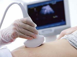
Maintaining healthy carotid arteries is crucial for overall cardiovascular health and preventing serious conditions like stroke, which can occur when the arteries become narrowed or blocked due to the buildup of plaque or fatty deposits (atherosclerosis). Regular check-ups with healthcare professionals may include monitoring carotid artery health through ultrasound or other imaging techniques.
Carotid artery diseases refer to conditions that affect the carotid arteries, which are the major blood vessels that supply oxygen-rich blood to the head and neck. The most common type of carotid artery disease is atherosclerosis, which occurs when fatty deposits called plaques build up in the inner lining of the arteries.
These plaques can cause the arteries to narrow or become blocked, reducing blood flow to the brain and increasing the risk of stroke. Other carotid artery diseases include carotid artery dissection, which occurs when there is a tear in the inner lining of the artery, and fibromuscular dysplasia, which is a condition that affects the muscle and tissue in the walls of the artery.
Symptoms of carotid artery disease may include neck pain or pulsations, transient ischemic attacks (TIAs) or "mini-strokes," and strokes. Treatment options for carotid artery disease depend on the severity and type of the condition but may include lifestyle changes, medications, endarterectomy (a surgical procedure to remove plaque from the artery), or angioplasty and stenting (procedures to open blocked arteries using a balloon and stent).
The internal carotid artery is a major blood vessel that supplies oxygenated blood to the brain. It originates from the common carotid artery and passes through the neck, entering the skull via the carotid canal in the temporal bone. Once inside the skull, it branches into several smaller vessels that supply different parts of the brain with blood.
The internal carotid artery is divided into several segments: cervical, petrous, cavernous, clinoid, and supraclinoid. Each segment has distinct clinical significance in terms of potential injury or disease. The most common conditions affecting the internal carotid artery include atherosclerosis, which can lead to stroke or transient ischemic attack (TIA), and dissection, which can cause severe headache, neck pain, and neurological symptoms.
It's important to note that any blockage or damage to the internal carotid artery can have serious consequences, as it can significantly reduce blood flow to the brain and lead to permanent neurological damage or even death. Therefore, regular check-ups and screening tests are recommended for individuals at high risk of developing vascular diseases.
The common carotid artery is a major blood vessel in the neck that supplies oxygenated blood to the head and neck. It originates from the brachiocephalic trunk or the aortic arch and divides into the internal and external carotid arteries at the level of the upper border of the thyroid cartilage. The common carotid artery is an important structure in the circulatory system, and any damage or blockage to it can have serious consequences, including stroke.
Carotid stenosis is a medical condition that refers to the narrowing or constriction of the lumen (inner space) of the carotid artery. The carotid arteries are major blood vessels that supply oxygenated blood to the head and neck. Carotid stenosis usually results from the buildup of plaque, made up of fat, cholesterol, calcium, and other substances, on the inner walls of the artery. This process is called atherosclerosis.
As the plaque accumulates, it causes the artery to narrow, reducing blood flow to the brain. Severe carotid stenosis can increase the risk of stroke, as a clot or debris from the plaque can break off and travel to the brain, blocking a smaller blood vessel and causing tissue damage or death.
Carotid stenosis is typically diagnosed through imaging tests such as ultrasound, CT angiography, or MRI angiography. Treatment options may include lifestyle modifications (such as quitting smoking, controlling blood pressure, and managing cholesterol levels), medications to reduce the risk of clots, or surgical procedures like endarterectomy or stenting to remove or bypass the blockage.
The external carotid artery is a major blood vessel in the neck that supplies oxygenated blood to the structures of the head and neck, excluding the brain. It originates from the common carotid artery at the level of the upper border of the thyroid cartilage, then divides into several branches that supply various regions of the head and neck, including the face, scalp, ears, and neck muscles.
The external carotid artery has eight branches:
1. Superior thyroid artery: Supplies blood to the thyroid gland, larynx, and surrounding muscles.
2. Ascending pharyngeal artery: Supplies blood to the pharynx, palate, and meninges of the brain.
3. Lingual artery: Supplies blood to the tongue and floor of the mouth.
4. Facial artery: Supplies blood to the face, nose, lips, and palate.
5. Occipital artery: Supplies blood to the scalp and muscles of the neck.
6. Posterior auricular artery: Supplies blood to the ear and surrounding muscles.
7. Maxillary artery: Supplies blood to the lower face, nasal cavity, palate, and meninges of the brain.
8. Superficial temporal artery: Supplies blood to the scalp, face, and temporomandibular joint.
The external carotid artery is an essential structure for maintaining adequate blood flow to the head and neck, and any damage or blockage can lead to serious medical conditions such as stroke or tissue necrosis.
Carotid endarterectomy is a surgical procedure to remove plaque buildup (atherosclerosis) from the carotid arteries, which are the major blood vessels that supply oxygen-rich blood to the brain. The surgery involves making an incision in the neck, opening the carotid artery, and removing the plaque from the inside of the artery wall. The goal of the procedure is to restore normal blood flow to the brain and reduce the risk of stroke caused by the narrowing or blockage of the carotid arteries.
Carotid artery thrombosis is a medical condition characterized by the formation of a blood clot (thrombus) inside the carotid artery, which is one of the major blood vessels that supplies oxygenated blood to the head and neck. This condition can lead to serious complications such as a stroke or transient ischemic attack (TIA), also known as a "mini-stroke," if the clot dislodges and travels to the brain, blocking the flow of blood and oxygen.
Carotid artery thrombosis can result from various factors, including atherosclerosis (the buildup of fats, cholesterol, and other substances in the artery walls), hypertension (high blood pressure), diabetes, smoking, and genetic predisposition. Symptoms may include neck pain or stiffness, weakness or numbness in the face or limbs, difficulty speaking or understanding speech, vision problems, and sudden severe headaches. Diagnosis typically involves imaging tests such as ultrasound, CT angiography, or MRI angiography. Treatment options may include anticoagulant or antiplatelet medications, endovascular procedures to remove the clot, or surgery to clean out the artery (carotid endarterectomy).
Arteries are blood vessels that carry oxygenated blood away from the heart to the rest of the body. They have thick, muscular walls that can withstand the high pressure of blood being pumped out of the heart. Arteries branch off into smaller vessels called arterioles, which further divide into a vast network of tiny capillaries where the exchange of oxygen, nutrients, and waste occurs between the blood and the body's cells. After passing through the capillary network, deoxygenated blood collects in venules, then merges into veins, which return the blood back to the heart.
A carotid artery, internal, dissection is a medical condition that affects the internal carotid artery, which is a major blood vessel in the neck that supplies oxygenated blood to the brain. In this condition, there is a separation (dissection) of the layers of the artery wall, causing blood to accumulate in the space between the layers. This can lead to narrowing or blockage of the artery, reducing blood flow to the brain and increasing the risk of stroke. Internal carotid artery dissection can be caused by trauma, high blood pressure, connective tissue disorders, or spontaneously. Symptoms may include neck pain, headache, facial pain, visual disturbances, weakness or numbness in the arms or legs, difficulty speaking or understanding speech, and dizziness or loss of balance.
The carotid body is a small chemoreceptor organ located near the bifurcation of the common carotid artery into the internal and external carotid arteries. It plays a crucial role in the regulation of respiration, blood pressure, and pH balance by detecting changes in the chemical composition of the blood, particularly oxygen levels, carbon dioxide levels, and hydrogen ion concentration (pH).
The carotid body contains specialized nerve endings called glomus cells that are sensitive to changes in these chemical parameters. When there is a decrease in oxygen or an increase in carbon dioxide or hydrogen ions, the glomus cells release neurotransmitters such as acetylcholine and dopamine, which activate afferent nerve fibers leading to the brainstem's nucleus tractus solitarius. This information is then integrated with other physiological signals in the brainstem, resulting in appropriate adjustments in breathing rate, depth, and pattern, as well as changes in heart rate and blood vessel diameter to maintain homeostasis.
Dysfunction of the carotid body can lead to various disorders, such as hypertension, sleep apnea, and chronic lung disease. In some cases, overactivity of the carotid body may result in conditions like primary breathing pattern disorders or pseudohypoxia, where the body responds as if it is experiencing hypoxia despite normal oxygen levels.
Tunica intima, also known as the intima layer, is the innermost layer of a blood vessel, including arteries and veins. It is in direct contact with the flowing blood and is composed of simple squamous endothelial cells that form a continuous, non-keratinized, stratified epithelium. These cells play a crucial role in maintaining vascular homeostasis by regulating the passage of molecules and immune cells between the blood and the vessel wall, as well as contributing to the maintenance of blood fluidity and preventing coagulation.
The tunica intima is supported by a thin layer of connective tissue called the basement membrane, which provides structural stability and anchorage for the endothelial cells. Beneath the basement membrane lies a loose network of elastic fibers and collagen, known as the internal elastic lamina, that separates the tunica intima from the middle layer, or tunica media.
In summary, the tunica intima is the innermost layer of blood vessels, primarily composed of endothelial cells and a basement membrane, which regulates various functions to maintain vascular homeostasis.
The carotid sinus is a small, dilated area located at the bifurcation (or fork) of the common carotid artery into the internal and external carotid arteries. It is a baroreceptor region, which means it contains specialized sensory nerve endings that can detect changes in blood pressure. When the blood pressure increases, the walls of the carotid sinus stretch, activating these nerve endings and sending signals to the brain. The brain then responds by reducing the heart rate and relaxing the blood vessels, which helps to lower the blood pressure back to normal.
The carotid sinus is an important part of the body's autonomic nervous system, which regulates various involuntary functions such as heart rate, blood pressure, and digestion. It plays a crucial role in maintaining cardiovascular homeostasis and preventing excessive increases in blood pressure that could potentially damage vital organs.
Cerebral arteries refer to the blood vessels that supply oxygenated blood to the brain. These arteries branch off from the internal carotid arteries and the vertebral arteries, which combine to form the basilar artery. The major cerebral arteries include:
1. Anterior cerebral artery (ACA): This artery supplies blood to the frontal lobes of the brain, including the motor and sensory cortices responsible for movement and sensation in the lower limbs.
2. Middle cerebral artery (MCA): The MCA is the largest of the cerebral arteries and supplies blood to the lateral surface of the brain, including the temporal, parietal, and frontal lobes. It is responsible for providing blood to areas involved in motor function, sensory perception, speech, memory, and vision.
3. Posterior cerebral artery (PCA): The PCA supplies blood to the occipital lobe, which is responsible for visual processing, as well as parts of the temporal and parietal lobes.
4. Anterior communicating artery (ACoA) and posterior communicating arteries (PComAs): These are small arteries that connect the major cerebral arteries, forming an important circulatory network called the Circle of Willis. The ACoA connects the two ACAs, while the PComAs connect the ICA with the PCA and the basilar artery.
These cerebral arteries play a crucial role in maintaining proper brain function by delivering oxygenated blood to various regions of the brain. Any damage or obstruction to these arteries can lead to serious neurological conditions, such as strokes or transient ischemic attacks (TIAs).
The tunica media is the middle layer of the wall of a blood vessel or hollow organ in the body. It is primarily composed of smooth muscle cells and elastic fibers, which allow the vessel or organ to expand and contract. This layer helps regulate the diameter of the lumen (the inner space) of the vessel or organ, thereby controlling the flow of fluids such as blood or lymph through it. The tunica media plays a crucial role in maintaining proper organ function and blood pressure regulation.
The pulmonary artery is a large blood vessel that carries deoxygenated blood from the right ventricle of the heart to the lungs for oxygenation. It divides into two main branches, the right and left pulmonary arteries, which further divide into smaller vessels called arterioles, and then into a vast network of capillaries in the lungs where gas exchange occurs. The thin walls of these capillaries allow oxygen to diffuse into the blood and carbon dioxide to diffuse out, making the blood oxygen-rich before it is pumped back to the left side of the heart through the pulmonary veins. This process is crucial for maintaining proper oxygenation of the body's tissues and organs.
The femoral artery is the major blood vessel that supplies oxygenated blood to the lower extremity of the human body. It is a continuation of the external iliac artery and becomes the popliteal artery as it passes through the adductor hiatus in the adductor magnus muscle of the thigh.
The femoral artery is located in the femoral triangle, which is bound by the sartorius muscle anteriorly, the adductor longus muscle medially, and the biceps femoris muscle posteriorly. It can be easily palpated in the groin region, making it a common site for taking blood samples, measuring blood pressure, and performing surgical procedures such as femoral artery catheterization and bypass grafting.
The femoral artery gives off several branches that supply blood to the lower limb, including the deep femoral artery, the superficial femoral artery, and the profunda femoris artery. These branches provide blood to the muscles, bones, skin, and other tissues of the leg, ankle, and foot.
Cerebral angiography is a medical procedure that involves taking X-ray images of the blood vessels in the brain after injecting a contrast dye into them. This procedure helps doctors to diagnose and treat various conditions affecting the blood vessels in the brain, such as aneurysms, arteriovenous malformations, and stenosis (narrowing of the blood vessels).
During the procedure, a catheter is inserted into an artery in the leg and threaded through the body to the blood vessels in the neck or brain. The contrast dye is then injected through the catheter, and X-ray images are taken to visualize the blood flow through the brain's blood vessels.
Cerebral angiography provides detailed images of the blood vessels in the brain, allowing doctors to identify any abnormalities or blockages that may be causing symptoms or increasing the risk of stroke. Based on the results of the cerebral angiography, doctors can develop a treatment plan to address these issues and prevent further complications.
A stent is a small mesh tube that's used to treat narrow or weak arteries. Arteries are blood vessels that carry blood away from your heart to other parts of your body. A stent is placed in an artery as part of a procedure called angioplasty. Angioplasty restores blood flow through narrowed or blocked arteries by inflating a tiny balloon inside the blocked artery to widen it.
The stent is then inserted into the widened artery to keep it open. The stent is usually made of metal, but some are coated with medication that is slowly and continuously released to help prevent the formation of scar tissue in the artery. This can reduce the chance of the artery narrowing again.
Stents are also used in other parts of the body, such as the neck (carotid artery) and kidneys (renal artery), to help maintain blood flow and prevent blockages. They can also be used in the urinary system to treat conditions like ureteropelvic junction obstruction or narrowing of the urethra.
Carotid intima-media thickness (CIMT) is a measurement of the thickness of the inner two layers of the carotid artery, which are the intima and media layers. This measurement is used as a marker for assessing cardiovascular disease risk, particularly the risk of atherosclerosis, or the buildup of plaque in the arteries.
CIMT can be measured using ultrasound imaging, and it is typically measured at several points along the length of the common carotid artery, as well as at the bifurcation where the common carotid artery divides into the internal and external carotid arteries. Increased CIMT has been associated with an increased risk of cardiovascular events such as heart attack and stroke.
It is important to note that while CIMT can provide valuable information about a person's cardiovascular health, it should not be used as the sole determinant of cardiovascular disease risk. Other factors, such as age, family history, smoking status, blood pressure, cholesterol levels, and diabetes status, should also be taken into account when assessing cardiovascular disease risk.
The vertebral artery is a major blood vessel that supplies oxygenated blood to the brain and upper spinal cord. It arises from the subclavian artery, then ascends through the transverse processes of several cervical vertebrae before entering the skull through the foramen magnum. Inside the skull, it joins with the opposite vertebral artery to form the basilar artery, which supplies blood to the brainstem and cerebellum. The vertebral artery also gives off several important branches that supply blood to various regions of the brainstem and upper spinal cord.
Carotid artery injuries refer to damages or traumas that affect the carotid arteries, which are a pair of major blood vessels located in the neck that supply oxygenated blood to the head and neck. These injuries can occur due to various reasons such as penetrating or blunt trauma, iatrogenic causes (during medical procedures), or degenerative diseases.
Carotid artery injuries can be categorized into three types:
1. Blunt carotid injury (BCI): This type of injury is caused by a sudden and severe impact to the neck, which can result in intimal tears, dissection, or thrombosis of the carotid artery. BCIs are commonly seen in motor vehicle accidents, sports-related injuries, and assaults.
2. Penetrating carotid injury: This type of injury is caused by a foreign object that penetrates the neck and damages the carotid artery. Examples include gunshot wounds, stab wounds, or other sharp objects that pierce the skin and enter the neck.
3. Iatrogenic carotid injury: This type of injury occurs during medical procedures such as endovascular interventions, surgical procedures, or the placement of central lines.
Symptoms of carotid artery injuries may include:
* Stroke or transient ischemic attack (TIA)
* Neurological deficits such as hemiparesis, aphasia, or visual disturbances
* Bleeding from the neck or mouth
* Pulsatile mass in the neck
* Hypotension or shock
* Loss of consciousness
Diagnosis of carotid artery injuries may involve imaging studies such as computed tomography angiography (CTA), magnetic resonance angiography (MRA), or conventional angiography. Treatment options include endovascular repair, surgical repair, or anticoagulation therapy, depending on the severity and location of the injury.
The basilar artery is a major blood vessel that supplies oxygenated blood to the brainstem and cerebellum. It is formed by the union of two vertebral arteries at the lower part of the brainstem, near the junction of the medulla oblongata and pons.
The basilar artery runs upward through the center of the brainstem and divides into two posterior cerebral arteries at the upper part of the brainstem, near the midbrain. The basilar artery gives off several branches that supply blood to various parts of the brainstem, including the pons, medulla oblongata, and midbrain, as well as to the cerebellum.
The basilar artery is an important part of the circle of Willis, a network of arteries at the base of the brain that ensures continuous blood flow to the brain even if one of the arteries becomes blocked or narrowed.
Ultrasonography, Doppler, and Duplex are diagnostic medical techniques that use sound waves to create images of internal body structures and assess their function. Here are the definitions for each:
1. Ultrasonography: Also known as ultrasound, this is a non-invasive imaging technique that uses high-frequency sound waves to produce images of internal organs and tissues. A small handheld device called a transducer is placed on the skin surface, which emits and receives sound waves. The returning echoes are then processed to create real-time visual images of the internal structures.
2. Doppler: This is a type of ultrasound that measures the velocity and direction of blood flow in the body by analyzing the frequency shift of the reflected sound waves. It can be used to assess blood flow in various parts of the body, such as the heart, arteries, and veins.
3. Duplex: Duplex ultrasonography is a combination of both gray-scale ultrasound and Doppler ultrasound. It provides detailed images of internal structures, as well as information about blood flow velocity and direction. This technique is often used to evaluate conditions such as deep vein thrombosis, carotid artery stenosis, and peripheral arterial disease.
In summary, ultrasonography is a diagnostic imaging technique that uses sound waves to create images of internal structures, Doppler is a type of ultrasound that measures blood flow velocity and direction, and duplex is a combination of both techniques that provides detailed images and information about blood flow.
The renal artery is a pair of blood vessels that originate from the abdominal aorta and supply oxygenated blood to each kidney. These arteries branch into several smaller vessels that provide blood to the various parts of the kidneys, including the renal cortex and medulla. The renal arteries also carry nutrients and other essential components needed for the normal functioning of the kidneys. Any damage or blockage to the renal artery can lead to serious consequences, such as reduced kidney function or even kidney failure.
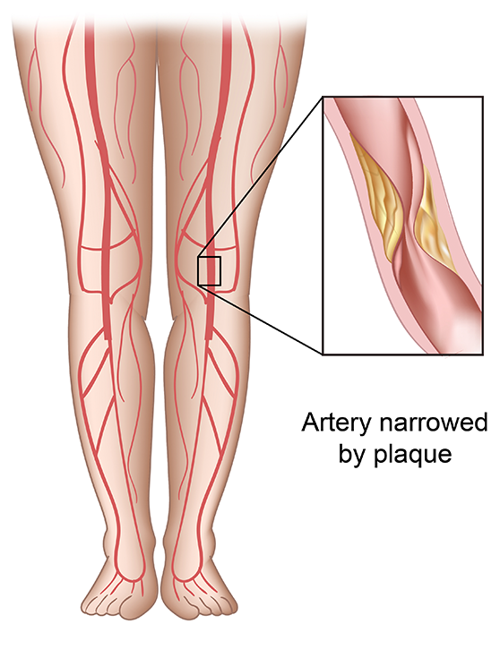
Arterial occlusive diseases are medical conditions characterized by the blockage or narrowing of the arteries, which can lead to a reduction in blood flow to various parts of the body. This reduction in blood flow can cause tissue damage and may result in serious complications such as tissue death (gangrene), organ dysfunction, or even death.
The most common cause of arterial occlusive diseases is atherosclerosis, which is the buildup of plaque made up of fat, cholesterol, calcium, and other substances in the inner lining of the artery walls. Over time, this plaque can harden and narrow the arteries, restricting blood flow. Other causes of arterial occlusive diseases include blood clots, emboli (tiny particles that travel through the bloodstream and lodge in smaller vessels), inflammation, trauma, and certain inherited conditions.
Symptoms of arterial occlusive diseases depend on the location and severity of the blockage. Common symptoms include:
* Pain, cramping, or fatigue in the affected limb, often triggered by exercise and relieved by rest (claudication)
* Numbness, tingling, or weakness in the affected limb
* Coldness or discoloration of the skin in the affected area
* Slow-healing sores or wounds on the toes, feet, or legs
* Erectile dysfunction in men
Treatment for arterial occlusive diseases may include lifestyle changes such as quitting smoking, exercising regularly, and eating a healthy diet. Medications to lower cholesterol, control blood pressure, prevent blood clots, or manage pain may also be prescribed. In severe cases, surgical procedures such as angioplasty, stenting, or bypass surgery may be necessary to restore blood flow.
The mesenteric arteries are the arteries that supply oxygenated blood to the intestines. There are three main mesenteric arteries: the superior mesenteric artery, which supplies blood to the small intestine (duodenum to two-thirds of the transverse colon) and large intestine (cecum, ascending colon, and the first part of the transverse colon); the inferior mesenteric artery, which supplies blood to the distal third of the transverse colon, descending colon, sigmoid colon, and rectum; and the middle colic artery, which is a branch of the superior mesenteric artery that supplies blood to the transverse colon. These arteries are important in maintaining adequate blood flow to the intestines to support digestion and absorption of nutrients.
A Transient Ischemic Attack (TIA), also known as a "mini-stroke," is a temporary period of symptoms similar to those you'd get if you were having a stroke. A TIA doesn't cause permanent damage and is often caused by a temporary decrease in blood supply to part of your brain, which may last as little as five minutes.
Like an ischemic stroke, a TIA occurs when a clot or debris blocks blood flow to part of your nervous system. However, unlike a stroke, a TIA doesn't leave lasting damage because the blockage is temporary.
Symptoms of a TIA can include sudden onset of weakness, numbness or paralysis in your face, arm or leg, typically on one side of your body. You could also experience slurred or garbled speech, or difficulty understanding others. Other symptoms can include blindness in one or both eyes, dizziness, or a severe headache with no known cause.
Even though TIAs usually last only a few minutes, they are a serious condition and should not be ignored. If you suspect you or someone else is experiencing a TIA, seek immediate medical attention. TIAs can be a warning sign that a full-blown stroke is imminent.
Angioplasty, balloon refers to a medical procedure used to widen narrowed or obstructed blood vessels, particularly the coronary arteries that supply blood to the heart muscle. This procedure is typically performed using a catheter-based technique, where a thin, flexible tube called a catheter is inserted into an artery, usually through the groin or wrist, and guided to the site of the narrowing or obstruction in the coronary artery.
Once the catheter reaches the affected area, a small balloon attached to the tip of the catheter is inflated, which compresses the plaque against the artery wall and stretches the artery, thereby restoring blood flow. The balloon is then deflated and removed, along with the catheter.
Balloon angioplasty is often combined with the placement of a stent, a small metal mesh tube that helps to keep the artery open and prevent it from narrowing again. This procedure is known as percutaneous coronary intervention (PCI) or coronary angioplasty and stenting.
Overall, balloon angioplasty is a relatively safe and effective treatment for coronary artery disease, although complications such as bleeding, infection, or re-narrowing of the artery can occur in some cases.
Magnetic Resonance Angiography (MRA) is a non-invasive medical imaging technique that uses magnetic fields and radio waves to create detailed images of the blood vessels or arteries within the body. It is a type of Magnetic Resonance Imaging (MRI) that focuses specifically on the circulatory system.
MRA can be used to diagnose and evaluate various conditions related to the blood vessels, such as aneurysms, stenosis (narrowing of the vessel), or the presence of plaques or tumors. It can also be used to plan for surgeries or other treatments related to the vascular system. The procedure does not use radiation and is generally considered safe, although people with certain implants like pacemakers may not be able to have an MRA due to safety concerns.
Arteriosclerosis is a general term that describes the hardening and stiffening of the artery walls. It's a progressive condition that can occur as a result of aging, or it may be associated with certain risk factors such as high blood pressure, high cholesterol, diabetes, smoking, and a sedentary lifestyle.
The process of arteriosclerosis involves the buildup of plaque, made up of fat, cholesterol, calcium, and other substances, in the inner lining of the artery walls. Over time, this buildup can cause the artery walls to thicken and harden, reducing the flow of oxygen-rich blood to the body's organs and tissues.
Arteriosclerosis can affect any of the body's arteries, but it is most commonly found in the coronary arteries that supply blood to the heart, the cerebral arteries that supply blood to the brain, and the peripheral arteries that supply blood to the limbs. When arteriosclerosis affects the coronary arteries, it can lead to heart disease, angina, or heart attack. When it affects the cerebral arteries, it can lead to stroke or transient ischemic attack (TIA). When it affects the peripheral arteries, it can cause pain, numbness, or weakness in the limbs, and in severe cases, gangrene and amputation.
The subclavian artery is a major blood vessel that supplies the upper limb and important structures in the neck and head. It arises from the brachiocephalic trunk (in the case of the right subclavian artery) or directly from the aortic arch (in the case of the left subclavian artery).
The subclavian artery has several branches, including:
1. The vertebral artery, which supplies blood to the brainstem and cerebellum.
2. The internal thoracic artery (also known as the mammary artery), which supplies blood to the chest wall, breast, and anterior mediastinum.
3. The thyrocervical trunk, which gives rise to several branches that supply the neck, including the inferior thyroid artery, the suprascapular artery, and the transverse cervical artery.
4. The costocervical trunk, which supplies blood to the neck and upper back, including the posterior chest wall and the lower neck muscles.
The subclavian artery is a critical vessel in maintaining adequate blood flow to the upper limb, and any blockage or damage to this vessel can lead to significant morbidity, including arm pain, numbness, weakness, or even loss of function.
Angiography is a medical procedure in which an x-ray image is taken to visualize the internal structure of blood vessels, arteries, or veins. This is done by injecting a radiopaque contrast agent (dye) into the blood vessel using a thin, flexible catheter. The dye makes the blood vessels visible on an x-ray image, allowing doctors to diagnose and treat various medical conditions such as blockages, narrowing, or malformations of the blood vessels.
There are several types of angiography, including:
* Cardiac angiography (also called coronary angiography) - used to examine the blood vessels of the heart
* Cerebral angiography - used to examine the blood vessels of the brain
* Peripheral angiography - used to examine the blood vessels in the limbs or other parts of the body.
Angiography is typically performed by a radiologist, cardiologist, or vascular surgeon in a hospital setting. It can help diagnose conditions such as coronary artery disease, aneurysms, and peripheral arterial disease, among others.
Endarterectomy is a surgical procedure in which the inner lining of an artery (the endothelium) that has become thickened, damaged, or narrowed due to the buildup of fatty deposits, called plaques, is removed. This process helps restore normal blood flow through the artery and reduces the risk of serious complications such as stroke or limb loss.
The procedure typically involves making an incision in the affected artery, carefully removing the plaque and inner lining, and then closing the artery with sutures or a patch graft. Endarterectomy is most commonly performed on the carotid arteries in the neck, but it can also be done on other arteries throughout the body, including the femoral artery in the leg and the iliac artery in the pelvis.
Endarterectomy is usually recommended for patients with significant narrowing of their arteries who are experiencing symptoms such as pain, numbness, or weakness in their limbs, or who have a high risk of stroke due to carotid artery disease. The procedure is generally safe and effective, but like any surgery, it carries risks such as bleeding, infection, and damage to nearby nerves or tissues.
Digital subtraction angiography (DSA) is a medical imaging technique used to visualize the blood vessels and blood flow within the body. It combines the use of X-ray technology with digital image processing to produce detailed images of the vascular system.
In DSA, a contrast agent is injected into the patient's bloodstream through a catheter, which is typically inserted into an artery in the leg and guided to the area of interest using fluoroscopy. As the contrast agent flows through the blood vessels, X-ray images are taken at multiple time points.
The digital subtraction process involves taking a baseline image without contrast and then subtracting it from subsequent images taken with contrast. This allows for the removal of background structures and noise, resulting in clearer images of the blood vessels. DSA can be used to diagnose and evaluate various vascular conditions, such as aneurysms, stenosis, and tumors, and can also guide interventional procedures such as angioplasty and stenting.
Angioplasty is a medical procedure used to open narrowed or blocked blood vessels, often referred to as coronary angioplasty when it involves the heart's blood vessels (coronary arteries). The term "angio" refers to an angiogram, which is a type of X-ray image that reveals the inside of blood vessels.
The procedure typically involves the following steps:
1. A thin, flexible catheter (tube) is inserted into a blood vessel, usually through a small incision in the groin or arm.
2. The catheter is guided to the narrowed or blocked area using real-time X-ray imaging.
3. Once in place, a tiny balloon attached to the tip of the catheter is inflated to widen the blood vessel and compress any plaque buildup against the artery walls.
4. A stent (a small mesh tube) may be inserted to help keep the blood vessel open and prevent it from narrowing again.
5. The balloon is deflated, and the catheter is removed.
Angioplasty helps improve blood flow, reduce symptoms such as chest pain or shortness of breath, and lower the risk of heart attack in patients with blocked arteries. It's important to note that angioplasty is not a permanent solution for coronary artery disease, and lifestyle changes, medications, and follow-up care are necessary to maintain long-term cardiovascular health.
Cerebrovascular circulation refers to the network of blood vessels that supply oxygenated blood and nutrients to the brain tissue, and remove waste products. It includes the internal carotid arteries, vertebral arteries, circle of Willis, and the intracranial arteries that branch off from them.
The internal carotid arteries and vertebral arteries merge to form the circle of Willis, a polygonal network of vessels located at the base of the brain. The anterior cerebral artery, middle cerebral artery, posterior cerebral artery, and communicating arteries are the major vessels that branch off from the circle of Willis and supply blood to different regions of the brain.
Interruptions or abnormalities in the cerebrovascular circulation can lead to various neurological conditions such as stroke, transient ischemic attack (TIA), and vascular dementia.
Coronary artery bypass surgery, also known as coronary artery bypass grafting (CABG), is a surgical procedure used to improve blood flow to the heart in patients with severe coronary artery disease. This condition occurs when the coronary arteries, which supply oxygen-rich blood to the heart muscle, become narrowed or blocked due to the buildup of fatty deposits, called plaques.
During CABG surgery, a healthy blood vessel from another part of the body is grafted, or attached, to the coronary artery, creating a new pathway for oxygen-rich blood to flow around the blocked or narrowed portion of the artery and reach the heart muscle. This bypass helps to restore normal blood flow and reduce the risk of angina (chest pain), shortness of breath, and other symptoms associated with coronary artery disease.
There are different types of CABG surgery, including traditional on-pump CABG, off-pump CABG, and minimally invasive CABG. The choice of procedure depends on various factors, such as the patient's overall health, the number and location of blocked arteries, and the presence of other medical conditions.
It is important to note that while CABG surgery can significantly improve symptoms and quality of life in patients with severe coronary artery disease, it does not cure the underlying condition. Lifestyle modifications, such as regular exercise, a healthy diet, smoking cessation, and medication therapy, are essential for long-term management and prevention of further progression of the disease.
The radial artery is a key blood vessel in the human body, specifically a part of the peripheral arterial system. Originating from the brachial artery in the upper arm, the radial artery travels down the arm and crosses over the wrist, where it can be palpated easily. It then continues into the hand, dividing into several branches to supply blood to the hand's tissues and digits.
The radial artery is often used for taking pulse readings due to its easy accessibility at the wrist. Additionally, in medical procedures such as coronary angiography or bypass surgery, the radial artery can be utilized as a site for catheter insertion. This allows healthcare professionals to examine the heart's blood vessels and assess cardiovascular health.
The iliac arteries are major branches of the abdominal aorta, the large artery that carries oxygen-rich blood from the heart to the rest of the body. The iliac arteries divide into two branches, the common iliac arteries, which further bifurcate into the internal and external iliac arteries.
The internal iliac artery supplies blood to the lower abdomen, pelvis, and the reproductive organs, while the external iliac artery provides blood to the lower extremities, including the legs and feet. Together, the iliac arteries play a crucial role in circulating blood throughout the body, ensuring that all tissues and organs receive the oxygen and nutrients they need to function properly.
A stroke, also known as cerebrovascular accident (CVA), is a serious medical condition that occurs when the blood supply to part of the brain is interrupted or reduced, leading to deprivation of oxygen and nutrients to brain cells. This can result in the death of brain tissue and cause permanent damage or temporary impairment to cognitive functions, speech, memory, movement, and other body functions controlled by the affected area of the brain.
Strokes can be caused by either a blockage in an artery that supplies blood to the brain (ischemic stroke) or the rupture of a blood vessel in the brain (hemorrhagic stroke). A transient ischemic attack (TIA), also known as a "mini-stroke," is a temporary disruption of blood flow to the brain that lasts only a few minutes and does not cause permanent damage.
Symptoms of a stroke may include sudden weakness or numbness in the face, arm, or leg; difficulty speaking or understanding speech; vision problems; loss of balance or coordination; severe headache with no known cause; and confusion or disorientation. Immediate medical attention is crucial for stroke patients to receive appropriate treatment and prevent long-term complications.
Brain ischemia is the medical term used to describe a reduction or interruption of blood flow to the brain, leading to a lack of oxygen and glucose delivery to brain tissue. This can result in brain damage or death of brain cells, known as infarction. Brain ischemia can be caused by various conditions such as thrombosis (blood clot formation), embolism (obstruction of a blood vessel by a foreign material), or hypoperfusion (reduced blood flow). The severity and duration of the ischemia determine the extent of brain damage. Symptoms can range from mild, such as transient ischemic attacks (TIAs or "mini-strokes"), to severe, including paralysis, speech difficulties, loss of consciousness, and even death. Immediate medical attention is required for proper diagnosis and treatment to prevent further damage and potential long-term complications.
X-ray computed tomography (CT or CAT scan) is a medical imaging method that uses computer-processed combinations of many X-ray images taken from different angles to produce cross-sectional (tomographic) images (virtual "slices") of the body. These cross-sectional images can then be used to display detailed internal views of organs, bones, and soft tissues in the body.
The term "computed tomography" is used instead of "CT scan" or "CAT scan" because the machines take a series of X-ray measurements from different angles around the body and then use a computer to process these data to create detailed images of internal structures within the body.
CT scanning is a noninvasive, painless medical test that helps physicians diagnose and treat medical conditions. CT imaging provides detailed information about many types of tissue including lung, bone, soft tissue and blood vessels. CT examinations can be performed on every part of the body for a variety of reasons including diagnosis, surgical planning, and monitoring of therapeutic responses.
In computed tomography (CT), an X-ray source and detector rotate around the patient, measuring the X-ray attenuation at many different angles. A computer uses this data to construct a cross-sectional image by the process of reconstruction. This technique is called "tomography". The term "computed" refers to the use of a computer to reconstruct the images.
CT has become an important tool in medical imaging and diagnosis, allowing radiologists and other physicians to view detailed internal images of the body. It can help identify many different medical conditions including cancer, heart disease, lung nodules, liver tumors, and internal injuries from trauma. CT is also commonly used for guiding biopsies and other minimally invasive procedures.
In summary, X-ray computed tomography (CT or CAT scan) is a medical imaging technique that uses computer-processed combinations of many X-ray images taken from different angles to produce cross-sectional images of the body. It provides detailed internal views of organs, bones, and soft tissues in the body, allowing physicians to diagnose and treat medical conditions.
Cerebrovascular disorders are a group of medical conditions that affect the blood vessels of the brain. These disorders can be caused by narrowing, blockage, or rupture of the blood vessels, leading to decreased blood flow and oxygen supply to the brain. The most common types of cerebrovascular disorders include:
1. Stroke: A stroke occurs when a blood vessel in the brain becomes blocked or bursts, causing a lack of oxygen and nutrients to reach brain cells. This can lead to permanent damage or death of brain tissue.
2. Transient ischemic attack (TIA): Also known as a "mini-stroke," a TIA occurs when blood flow to the brain is temporarily blocked, often by a blood clot. Symptoms may last only a few minutes to a few hours and typically resolve on their own. However, a TIA is a serious warning sign that a full-blown stroke may occur in the future.
3. Aneurysm: An aneurysm is a weakened or bulging area in the wall of a blood vessel. If left untreated, an aneurysm can rupture and cause bleeding in the brain.
4. Arteriovenous malformation (AVM): An AVM is a tangled mass of abnormal blood vessels that connect arteries and veins. This can lead to bleeding in the brain or stroke.
5. Carotid stenosis: Carotid stenosis occurs when the carotid arteries, which supply blood to the brain, become narrowed or blocked due to plaque buildup. This can increase the risk of stroke.
6. Vertebrobasilar insufficiency: This condition occurs when the vertebral and basilar arteries, which supply blood to the back of the brain, become narrowed or blocked. This can lead to symptoms such as dizziness, vertigo, and difficulty swallowing.
Cerebrovascular disorders are a leading cause of disability and death worldwide. Risk factors for these conditions include age, high blood pressure, smoking, diabetes, high cholesterol, and family history. Treatment may involve medications, surgery, or lifestyle changes to reduce the risk of further complications.
The brachial artery is a major blood vessel in the upper arm. It supplies oxygenated blood to the muscles and tissues of the arm, forearm, and hand. The brachial artery originates from the axillary artery at the level of the shoulder joint and runs down the medial (inner) aspect of the arm, passing through the cubital fossa (the depression on the anterior side of the elbow) where it can be palpated during a routine blood pressure measurement. At the lower end of the forearm, the brachial artery bifurcates into the radial and ulnar arteries, which further divide into smaller vessels to supply the hand and fingers.
A smooth muscle within the vascular system refers to the involuntary, innervated muscle that is found in the walls of blood vessels. These muscles are responsible for controlling the diameter of the blood vessels, which in turn regulates blood flow and blood pressure. They are called "smooth" muscles because their individual muscle cells do not have the striations, or cross-striped patterns, that are observed in skeletal and cardiac muscle cells. Smooth muscle in the vascular system is controlled by the autonomic nervous system and by hormones, and can contract or relax slowly over a period of time.
Catheterization is a medical procedure in which a catheter (a flexible tube) is inserted into the body to treat various medical conditions or for diagnostic purposes. The specific definition can vary depending on the area of medicine and the particular procedure being discussed. Here are some common types of catheterization:
1. Urinary catheterization: This involves inserting a catheter through the urethra into the bladder to drain urine. It is often performed to manage urinary retention, monitor urine output in critically ill patients, or assist with surgical procedures.
2. Cardiac catheterization: A procedure where a catheter is inserted into a blood vessel, usually in the groin or arm, and guided to the heart. This allows for various diagnostic tests and treatments, such as measuring pressures within the heart chambers, assessing blood flow, or performing angioplasty and stenting of narrowed coronary arteries.
3. Central venous catheterization: A catheter is inserted into a large vein, typically in the neck, chest, or groin, to administer medications, fluids, or nutrition, or to monitor central venous pressure.
4. Peritoneal dialysis catheterization: A catheter is placed into the abdominal cavity for individuals undergoing peritoneal dialysis, a type of kidney replacement therapy.
5. Neurological catheterization: In some cases, a catheter may be inserted into the cerebrospinal fluid space (lumbar puncture) or the brain's ventricular system (ventriculostomy) to diagnose or treat various neurological conditions.
These are just a few examples of catheterization procedures in medicine. The specific definition and purpose will depend on the medical context and the particular organ or body system involved.
Medical Definition:
"Risk factors" are any attribute, characteristic or exposure of an individual that increases the likelihood of developing a disease or injury. They can be divided into modifiable and non-modifiable risk factors. Modifiable risk factors are those that can be changed through lifestyle choices or medical treatment, while non-modifiable risk factors are inherent traits such as age, gender, or genetic predisposition. Examples of modifiable risk factors include smoking, alcohol consumption, physical inactivity, and unhealthy diet, while non-modifiable risk factors include age, sex, and family history. It is important to note that having a risk factor does not guarantee that a person will develop the disease, but rather indicates an increased susceptibility.
Treatment outcome is a term used to describe the result or effect of medical treatment on a patient's health status. It can be measured in various ways, such as through symptoms improvement, disease remission, reduced disability, improved quality of life, or survival rates. The treatment outcome helps healthcare providers evaluate the effectiveness of a particular treatment plan and make informed decisions about future care. It is also used in clinical research to compare the efficacy of different treatments and improve patient care.
The Middle Cerebral Artery (MCA) is one of the main blood vessels that supplies oxygenated blood to the brain. It arises from the internal carotid artery and divides into several branches, which supply the lateral surface of the cerebral hemisphere, including the frontal, parietal, and temporal lobes.
The MCA is responsible for providing blood flow to critical areas of the brain, such as the primary motor and sensory cortices, Broca's area (associated with speech production), Wernicke's area (associated with language comprehension), and the visual association cortex.
Damage to the MCA or its branches can result in a variety of neurological deficits, depending on the specific location and extent of the injury. These may include weakness or paralysis on one side of the body, sensory loss, language impairment, and visual field cuts.
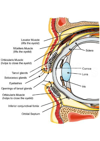
The ophthalmic artery is the first branch of the internal carotid artery, which supplies blood to the eye and its adnexa. It divides into several branches that provide oxygenated blood to various structures within the eye, including the retina, optic nerve, choroid, iris, ciliary body, and cornea. Any blockage or damage to the ophthalmic artery can lead to serious vision problems or even blindness.
In the field of medicine, "time factors" refer to the duration of symptoms or time elapsed since the onset of a medical condition, which can have significant implications for diagnosis and treatment. Understanding time factors is crucial in determining the progression of a disease, evaluating the effectiveness of treatments, and making critical decisions regarding patient care.
For example, in stroke management, "time is brain," meaning that rapid intervention within a specific time frame (usually within 4.5 hours) is essential to administering tissue plasminogen activator (tPA), a clot-busting drug that can minimize brain damage and improve patient outcomes. Similarly, in trauma care, the "golden hour" concept emphasizes the importance of providing definitive care within the first 60 minutes after injury to increase survival rates and reduce morbidity.
Time factors also play a role in monitoring the progression of chronic conditions like diabetes or heart disease, where regular follow-ups and assessments help determine appropriate treatment adjustments and prevent complications. In infectious diseases, time factors are crucial for initiating antibiotic therapy and identifying potential outbreaks to control their spread.
Overall, "time factors" encompass the significance of recognizing and acting promptly in various medical scenarios to optimize patient outcomes and provide effective care.
The mammary arteries are a set of blood vessels that supply oxygenated blood to the mammary glands, which are the structures in female breasts responsible for milk production during lactation. The largest mammary artery, also known as the internal thoracic or internal mammary artery, originates from the subclavian artery and descends along the inner side of the chest wall. It then branches into several smaller arteries that supply blood to the breast tissue. These include the anterior and posterior intercostal arteries, lateral thoracic artery, and pectoral branches. The mammary arteries are crucial in maintaining the health and function of the breast tissue, and any damage or blockage to these vessels can lead to various breast-related conditions or diseases.
An aneurysm is a localized, balloon-like bulge in the wall of a blood vessel. It occurs when the pressure inside the vessel causes a weakened area to swell and become enlarged. Aneurysms can develop in any blood vessel, but they are most common in arteries at the base of the brain (cerebral aneurysm) and the main artery carrying blood from the heart to the rest of the body (aortic aneurysm).
Aneurysms can be classified as saccular or fusiform, depending on their shape. A saccular aneurysm is a round or oval bulge that projects from the side of a blood vessel, while a fusiform aneurysm is a dilated segment of a blood vessel that is uniform in width and involves all three layers of the arterial wall.
The size and location of an aneurysm can affect its risk of rupture. Generally, larger aneurysms are more likely to rupture than smaller ones. Aneurysms located in areas with high blood pressure or where the vessel branches are also at higher risk of rupture.
Ruptured aneurysms can cause life-threatening bleeding and require immediate medical attention. Symptoms of a ruptured aneurysm may include sudden severe headache, neck stiffness, nausea, vomiting, blurred vision, or loss of consciousness. Unruptured aneurysms may not cause any symptoms and are often discovered during routine imaging tests for other conditions.
Treatment options for aneurysms depend on their size, location, and risk of rupture. Small, unruptured aneurysms may be monitored with regular imaging tests to check for growth or changes. Larger or symptomatic aneurysms may require surgical intervention, such as clipping or coiling, to prevent rupture and reduce the risk of complications.
Ultrasonography, also known as sonography, is a diagnostic medical procedure that uses high-frequency sound waves (ultrasound) to produce dynamic images of organs, tissues, or blood flow inside the body. These images are captured in real-time and can be used to assess the size, shape, and structure of various internal structures, as well as detect any abnormalities such as tumors, cysts, or inflammation.
During an ultrasonography procedure, a small handheld device called a transducer is placed on the patient's skin, which emits and receives sound waves. The transducer sends high-frequency sound waves into the body, and these waves bounce back off internal structures and are recorded by the transducer. The recorded data is then processed and transformed into visual images that can be interpreted by a medical professional.
Ultrasonography is a non-invasive, painless, and safe procedure that does not use radiation like other imaging techniques such as CT scans or X-rays. It is commonly used to diagnose and monitor conditions in various parts of the body, including the abdomen, pelvis, heart, blood vessels, and musculoskeletal system.
Blood pressure is the force exerted by circulating blood on the walls of the blood vessels. It is measured in millimeters of mercury (mmHg) and is given as two figures:
1. Systolic pressure: This is the pressure when the heart pushes blood out into the arteries.
2. Diastolic pressure: This is the pressure when the heart rests between beats, allowing it to fill with blood.
Normal blood pressure for adults is typically around 120/80 mmHg, although this can vary slightly depending on age, sex, and other factors. High blood pressure (hypertension) is generally considered to be a reading of 130/80 mmHg or higher, while low blood pressure (hypotension) is usually defined as a reading below 90/60 mmHg. It's important to note that blood pressure can fluctuate throughout the day and may be affected by factors such as stress, physical activity, and medication use.
An intracranial embolism is a medical condition that occurs when a blood clot or other foreign material (embolus) forms elsewhere in the body and travels to the blood vessels within the brain. This embolus then blocks the flow of blood in the cerebral arteries, leading to potential damage or death of brain tissue. Common sources of intracranial emboli include heart conditions such as atrial fibrillation, valvular heart disease, or following a heart attack; or from large-vessel atherosclerosis in the carotid arteries. Symptoms can vary depending on the location and size of the obstruction, but may include sudden weakness or numbness, confusion, difficulty speaking, vision loss, severe headache, or even loss of consciousness. Immediate medical attention is required to diagnose and treat intracranial embolism, often involving anticoagulation therapy, endovascular procedures, or surgery.
Blood flow velocity is the speed at which blood travels through a specific part of the vascular system. It is typically measured in units of distance per time, such as centimeters per second (cm/s) or meters per second (m/s). Blood flow velocity can be affected by various factors, including cardiac output, vessel diameter, and viscosity of the blood. Measuring blood flow velocity is important in diagnosing and monitoring various medical conditions, such as heart disease, stroke, and peripheral vascular disease.
Ultrasonography, Doppler, color is a type of diagnostic ultrasound technique that uses the Doppler effect to produce visual images of blood flow in vessels and the heart. The Doppler effect is the change in frequency or wavelength of a wave in relation to an observer who is moving relative to the source of the wave. In this context, it refers to the change in frequency of the ultrasound waves as they reflect off moving red blood cells.
In color Doppler ultrasonography, different colors are used to represent the direction and speed of blood flow. Red typically represents blood flowing toward the transducer (the device that sends and receives sound waves), while blue represents blood flowing away from the transducer. The intensity or brightness of the color is proportional to the velocity of blood flow.
Color Doppler ultrasonography is often used in conjunction with grayscale ultrasound imaging, which provides information about the structure and composition of tissues. Together, these techniques can help diagnose a wide range of conditions, including heart disease, blood clots, and abnormalities in blood flow.
Atherosclerosis is a medical condition characterized by the buildup of plaques, made up of fat, cholesterol, calcium, and other substances found in the blood, on the inner walls of the arteries. This process gradually narrows and hardens the arteries, reducing the flow of oxygen-rich blood to various parts of the body. Atherosclerosis can affect any artery in the body, including those that supply blood to the heart (coronary arteries), brain, limbs, and other organs. The progressive narrowing and hardening of the arteries can lead to serious complications such as coronary artery disease, carotid artery disease, peripheral artery disease, and aneurysms, which can result in heart attacks, strokes, or even death if left untreated.
The exact cause of atherosclerosis is not fully understood, but it is believed to be associated with several risk factors, including high blood pressure, high cholesterol levels, smoking, diabetes, obesity, physical inactivity, and a family history of the condition. Atherosclerosis can often progress without any symptoms for many years, but as the disease advances, it can lead to various signs and symptoms depending on which arteries are affected. Treatment typically involves lifestyle changes, medications, and, in some cases, surgical procedures to restore blood flow.
Prospective studies, also known as longitudinal studies, are a type of cohort study in which data is collected forward in time, following a group of individuals who share a common characteristic or exposure over a period of time. The researchers clearly define the study population and exposure of interest at the beginning of the study and follow up with the participants to determine the outcomes that develop over time. This type of study design allows for the investigation of causal relationships between exposures and outcomes, as well as the identification of risk factors and the estimation of disease incidence rates. Prospective studies are particularly useful in epidemiology and medical research when studying diseases with long latency periods or rare outcomes.
Intracranial arteriosclerosis is a medical condition characterized by the thickening and hardening of the walls of the intracranial arteries, which are the blood vessels that supply blood to the brain. This process is caused by the buildup of plaque, made up of fat, cholesterol, and other substances, within the walls of the arteries.
Intracranial arteriosclerosis can lead to a narrowing or blockage of the affected arteries, reducing blood flow to the brain. This can result in various neurological symptoms, such as headaches, dizziness, seizures, and transient ischemic attacks (TIAs) or strokes.
The condition is more common in older adults, particularly those with a history of hypertension, diabetes, smoking, and high cholesterol levels. Intracranial arteriosclerosis can be diagnosed through imaging tests such as magnetic resonance angiography (MRA) or computed tomographic angiography (CTA). Treatment typically involves managing risk factors and may include medications to control blood pressure, cholesterol levels, and prevent blood clots. In severe cases, surgical procedures such as angioplasty and stenting may be necessary to open up the affected arteries.
An intracranial aneurysm is a localized, blood-filled dilation or bulging in the wall of a cerebral artery within the skull (intracranial). These aneurysms typically occur at weak points in the arterial walls, often at branching points where the vessel divides into smaller branches. Over time, the repeated pressure from blood flow can cause the vessel wall to weaken and balloon out, forming a sac-like structure. Intracranial aneurysms can vary in size, ranging from a few millimeters to several centimeters in diameter.
There are three main types of intracranial aneurysms:
1. Saccular (berry) aneurysm: This is the most common type, characterized by a round or oval shape with a narrow neck and a bulging sac. They usually develop at branching points in the arteries due to congenital weaknesses in the vessel wall.
2. Fusiform aneurysm: These aneurysms have a dilated segment along the length of the artery, forming a cigar-shaped or spindle-like structure. They are often caused by atherosclerosis and can affect any part of the cerebral arteries.
3. Dissecting aneurysm: This type occurs when there is a tear in the inner lining (intima) of the artery, allowing blood to flow between the layers of the vessel wall. It can lead to narrowing or complete blockage of the affected artery and may cause subarachnoid hemorrhage if it ruptures.
Intracranial aneurysms can be asymptomatic and discovered incidentally during imaging studies for other conditions. However, when they grow larger or rupture, they can lead to severe complications such as subarachnoid hemorrhage, stroke, or even death. Treatment options include surgical clipping, endovascular coiling, or flow diversion techniques to prevent further growth and potential rupture of the aneurysm.
Transcranial Doppler ultrasonography is a non-invasive diagnostic technique that uses high-frequency sound waves to visualize and measure the velocity of blood flow in the cerebral arteries located in the skull. This imaging modality employs the Doppler effect, which describes the change in frequency of sound waves as they reflect off moving red blood cells. By measuring the frequency shift of the reflected ultrasound waves, the velocity and direction of blood flow can be determined.
Transcranial Doppler ultrasonography is primarily used to assess cerebrovascular circulation and detect abnormalities such as stenosis (narrowing), occlusion (blockage), or embolism (obstruction) in the intracranial arteries. It can also help monitor patients with conditions like sickle cell disease, vasospasm following subarachnoid hemorrhage, and evaluate the effectiveness of treatments such as thrombolysis or angioplasty. The procedure is typically performed by placing a transducer on the patient's skull after applying a coupling gel, and it does not involve radiation exposure or contrast agents.
The Circle of Willis is a circulatory arrangement in the brain where the major arteries that supply blood to the brain converge to form an almost circular structure. It is named after Thomas Willis, an English physician who first described it in 1664.
This circle is formed by the joining of the two internal carotid arteries, which divide into the anterior cerebral and middle cerebral arteries, with the basilar artery, which arises from the vertebral arteries. These vessels anastomose, or connect, to form a polygon-like structure at the base of the brain.
The Circle of Willis plays a crucial role in maintaining adequate blood flow to the brain, as it allows for collateral circulation. If one of the arteries that make up the circle becomes blocked or narrowed, blood can still reach the affected area through the other vessels in the circle. This helps to minimize the risk of stroke and other neurological disorders.
Cerebral infarction, also known as a "stroke" or "brain attack," is the sudden death of brain cells caused by the interruption of their blood supply. It is most commonly caused by a blockage in one of the blood vessels supplying the brain (an ischemic stroke), but can also result from a hemorrhage in or around the brain (a hemorrhagic stroke).
Ischemic strokes occur when a blood clot or other particle blocks a cerebral artery, cutting off blood flow to a part of the brain. The lack of oxygen and nutrients causes nearby brain cells to die. Hemorrhagic strokes occur when a weakened blood vessel ruptures, causing bleeding within or around the brain. This bleeding can put pressure on surrounding brain tissues, leading to cell death.
Symptoms of cerebral infarction depend on the location and extent of the affected brain tissue but may include sudden weakness or numbness in the face, arm, or leg; difficulty speaking or understanding speech; vision problems; loss of balance or coordination; and severe headache with no known cause. Immediate medical attention is crucial for proper diagnosis and treatment to minimize potential long-term damage or disability.
Regional blood flow (RBF) refers to the rate at which blood flows through a specific region or organ in the body, typically expressed in milliliters per minute per 100 grams of tissue (ml/min/100g). It is an essential physiological parameter that reflects the delivery of oxygen and nutrients to tissues while removing waste products. RBF can be affected by various factors such as metabolic demands, neural regulation, hormonal influences, and changes in blood pressure or vascular resistance. Measuring RBF is crucial for understanding organ function, diagnosing diseases, and evaluating the effectiveness of treatments.
A carotid body tumor is a rare, usually noncancerous (benign) growth that develops in the carotid body, a small structure located near the bifurcation (fork) of the common carotid artery in the neck. The carotid body is part of the chemoreceptor system that helps regulate breathing and blood pressure by responding to changes in oxygen, carbon dioxide, and pH levels in the blood.
Carotid body tumors are also known as carotid body paragangliomas or chemodectomas. They typically grow slowly and may not cause any symptoms for many years. However, as they enlarge, they can cause a visible or palpable mass in the neck, along with symptoms such as difficulty swallowing, hoarseness, or voice changes. In some cases, carotid body tumors can compress nearby nerves or blood vessels, leading to more serious complications like stroke or nerve damage.
Treatment for carotid body tumors typically involves surgical removal of the growth, which may be performed using traditional open surgery or minimally invasive techniques such as endovascular surgery or robotic-assisted surgery. Radiation therapy and chemotherapy are generally not effective in treating these tumors. Regular follow-up care is important to monitor for recurrence or development of new tumors.
Coronary vessels refer to the network of blood vessels that supply oxygenated blood and nutrients to the heart muscle, also known as the myocardium. The two main coronary arteries are the left main coronary artery and the right coronary artery.
The left main coronary artery branches off into the left anterior descending artery (LAD) and the left circumflex artery (LCx). The LAD supplies blood to the front of the heart, while the LCx supplies blood to the side and back of the heart.
The right coronary artery supplies blood to the right lower part of the heart, including the right atrium and ventricle, as well as the back of the heart.
Coronary vessel disease (CVD) occurs when these vessels become narrowed or blocked due to the buildup of plaque, leading to reduced blood flow to the heart muscle. This can result in chest pain, shortness of breath, or a heart attack.
Temporal arteries are the paired set of arteries that run along the temples on either side of the head. They are branches of the external carotid artery and play a crucial role in supplying oxygenated blood to the scalp and surrounding muscles. One of the most common conditions associated with temporal arteries is Temporal Arteritis (also known as Giant Cell Arteritis), which is an inflammation of these arteries that can lead to serious complications like vision loss if not promptly diagnosed and treated.
The endothelium is a thin layer of simple squamous epithelial cells that lines the interior surface of blood vessels, lymphatic vessels, and heart chambers. The vascular endothelium, specifically, refers to the endothelial cells that line the blood vessels. These cells play a crucial role in maintaining vascular homeostasis by regulating vasomotor tone, coagulation, platelet activation, inflammation, and permeability of the vessel wall. They also contribute to the growth and repair of the vascular system and are involved in various pathological processes such as atherosclerosis, hypertension, and diabetes.
Cerebral revascularization is a surgical procedure aimed at restoring blood flow to the brain. This is often performed in cases where there is narrowing or blockage of the cerebral arteries, a condition known as cerebrovascular disease. The most common type of cerebral revascularization is called carotid endarterectomy, which involves removing plaque buildup from the carotid artery in the neck to improve blood flow to the brain. Another type is extracranial-intracranial bypass, where a new connection is created between an external carotid artery and an intracranial artery to bypass a blockage.
1. Intracranial Embolism: This is a medical condition that occurs when a blood clot or other particle (embolus) formed elsewhere in the body, travels through the bloodstream and lodges itself in the intracranial blood vessels, blocking the flow of blood to a part of the brain. This can lead to various neurological symptoms such as weakness, numbness, speech difficulties, or even loss of consciousness, depending on the severity and location of the blockage.
2. Intracranial Thrombosis: This is a medical condition that occurs when a blood clot (thrombus) forms within the intracranial blood vessels. The clot can partially or completely obstruct the flow of blood, leading to various symptoms such as headache, confusion, seizures, or neurological deficits, depending on the severity and location of the thrombosis. Intracranial thrombosis can occur due to various factors including atherosclerosis, hypertension, diabetes, and other medical conditions that increase the risk of blood clot formation.
Amaurosis fugax is a medical term that describes a temporary loss of vision in one eye, which is often described as a "shade or curtain falling over the field of vision." It's usually caused by a temporary interruption of blood flow to the retina or optic nerve. This condition is often associated with conditions such as giant cell arteritis, carotid artery stenosis, and cardiovascular disease.
It's important to note that Amaurosis fugax can be a warning sign for a more serious medical event, such as a stroke, so it's essential to seek medical attention promptly if you experience any symptoms of this condition.
Vasodilation is the widening or increase in diameter of blood vessels, particularly the involuntary relaxation of the smooth muscle in the tunica media (middle layer) of the arteriole walls. This results in an increase in blood flow and a decrease in vascular resistance. Vasodilation can occur due to various physiological and pathophysiological stimuli, such as local metabolic demands, neural signals, or pharmacological agents. It plays a crucial role in regulating blood pressure, tissue perfusion, and thermoregulation.
Follow-up studies are a type of longitudinal research that involve repeated observations or measurements of the same variables over a period of time, in order to understand their long-term effects or outcomes. In medical context, follow-up studies are often used to evaluate the safety and efficacy of medical treatments, interventions, or procedures.
In a typical follow-up study, a group of individuals (called a cohort) who have received a particular treatment or intervention are identified and then followed over time through periodic assessments or data collection. The data collected may include information on clinical outcomes, adverse events, changes in symptoms or functional status, and other relevant measures.
The results of follow-up studies can provide important insights into the long-term benefits and risks of medical interventions, as well as help to identify factors that may influence treatment effectiveness or patient outcomes. However, it is important to note that follow-up studies can be subject to various biases and limitations, such as loss to follow-up, recall bias, and changes in clinical practice over time, which must be carefully considered when interpreting the results.
Ligation, in the context of medical terminology, refers to the process of tying off a part of the body, usually blood vessels or tissue, with a surgical suture or another device. The goal is to stop the flow of fluids such as blood or other substances within the body. It is commonly used during surgeries to control bleeding or to block the passage of fluids, gases, or solids in various parts of the body.
The splenic artery is the largest branch of the celiac trunk, which arises from the abdominal aorta. It supplies blood to the spleen and several other organs in the upper left part of the abdomen. The splenic artery divides into several branches that ultimately form a network of capillaries within the spleen. These capillaries converge to form the main venous outflow, the splenic vein, which drains into the hepatic portal vein.
The splenic artery is a vital structure in the human body, and any damage or blockage can lead to serious complications, including splenic infarction (reduced blood flow to the spleen) or splenic rupture (a surgical emergency that can be life-threatening).
Ultrasonography, Doppler refers to a non-invasive diagnostic medical procedure that uses high-frequency sound waves to create real-time images of the movement of blood flow through vessels, tissues, or heart valves. The Doppler effect is used to measure the frequency shift of the ultrasound waves as they bounce off moving red blood cells, which allows for the calculation of the speed and direction of blood flow. This technique is commonly used to diagnose and monitor various conditions such as deep vein thrombosis, carotid artery stenosis, heart valve abnormalities, and fetal heart development during pregnancy. It does not use radiation or contrast agents and is considered safe with minimal risks.
Retinal artery occlusion (RAO) is a medical condition characterized by the blockage or obstruction of the retinal artery, which supplies oxygenated blood to the retina. This blockage typically occurs due to embolism (a small clot or debris that travels to the retinal artery), thrombosis (blood clot formation in the artery), or vasculitis (inflammation of the blood vessels).
There are two types of retinal artery occlusions:
1. Central Retinal Artery Occlusion (CRAO): This type occurs when the main retinal artery is obstructed, affecting the entire inner layer of the retina. It can lead to severe and sudden vision loss in the affected eye.
2. Branch Retinal Artery Occlusion (BRAO): This type affects a branch of the retinal artery, causing visual field loss in the corresponding area. Although it is less severe than CRAO, it can still result in noticeable vision impairment.
Immediate medical attention is crucial for both types of RAO to improve the chances of recovery and minimize potential damage to the eye and vision. Treatment options may include medications, laser therapy, or surgery, depending on the underlying cause and the severity of the condition.
The cavernous sinus is a venous structure located in the middle cranial fossa, which is a depression in the skull that houses several important nerves and blood vessels. The cavernous sinus is situated on either side of the sphenoid bone, near the base of the skull, and it contains several important structures:
* The internal carotid artery, which supplies oxygenated blood to the brain
* The abducens nerve (cranial nerve VI), which controls lateral movement of the eye
* The oculomotor nerve (cranial nerve III), which controls most of the muscles that move the eye
* The trochlear nerve (cranial nerve IV), which controls one of the muscles that moves the eye
* The ophthalmic and maxillary divisions of the trigeminal nerve (cranial nerve V), which transmit sensory information from the face and head
The cavernous sinus is an important structure because it serves as a conduit for several critical nerves and blood vessels. However, it is also vulnerable to various pathological conditions such as thrombosis (blood clots), infection, tumors, or aneurysms, which can lead to serious neurological deficits or even death.
Therapeutic embolization is a medical procedure that involves intentionally blocking or obstructing blood vessels to stop excessive bleeding or block the flow of blood to a tumor or abnormal tissue. This is typically accomplished by injecting small particles, such as microspheres or coils, into the targeted blood vessel through a catheter, which is inserted into a larger blood vessel and guided to the desired location using imaging techniques like X-ray or CT scanning. The goal of therapeutic embolization is to reduce the size of a tumor, control bleeding, or block off abnormal blood vessels that are causing problems.
Pathological constriction refers to an abnormal narrowing or tightening of a body passage or organ, which can interfere with the normal flow of blood, air, or other substances through the area. This constriction can occur due to various reasons such as inflammation, scarring, or abnormal growths, and can affect different parts of the body, including blood vessels, airways, intestines, and ureters. Pathological constriction can lead to a range of symptoms and complications depending on its location and severity, and may require medical intervention to correct.
Hyperplasia is a medical term that refers to an abnormal increase in the number of cells in an organ or tissue, leading to an enlargement of the affected area. It's a response to various stimuli such as hormones, chronic irritation, or inflammation. Hyperplasia can be physiological, like the growth of breast tissue during pregnancy, or pathological, like in the case of benign or malignant tumors. The process is generally reversible if the stimulus is removed. It's important to note that hyperplasia itself is not cancerous, but some forms of hyperplasia can increase the risk of developing cancer over time.
The hepatic artery is a branch of the celiac trunk or abdominal aorta that supplies oxygenated blood to the liver. It typically divides into two main branches, the right and left hepatic arteries, which further divide into smaller vessels to supply different regions of the liver. The hepatic artery also gives off branches to supply other organs such as the gallbladder, pancreas, and duodenum.
It's worth noting that there is significant variability in the anatomy of the hepatic artery, with some individuals having additional branches or variations in the origin of the vessel. This variability can have implications for surgical procedures involving the liver and surrounding organs.
Retrospective studies, also known as retrospective research or looking back studies, are a type of observational study that examines data from the past to draw conclusions about possible causal relationships between risk factors and outcomes. In these studies, researchers analyze existing records, medical charts, or previously collected data to test a hypothesis or answer a specific research question.
Retrospective studies can be useful for generating hypotheses and identifying trends, but they have limitations compared to prospective studies, which follow participants forward in time from exposure to outcome. Retrospective studies are subject to biases such as recall bias, selection bias, and information bias, which can affect the validity of the results. Therefore, retrospective studies should be interpreted with caution and used primarily to generate hypotheses for further testing in prospective studies.
The Predictive Value of Tests, specifically the Positive Predictive Value (PPV) and Negative Predictive Value (NPV), are measures used in diagnostic tests to determine the probability that a positive or negative test result is correct.
Positive Predictive Value (PPV) is the proportion of patients with a positive test result who actually have the disease. It is calculated as the number of true positives divided by the total number of positive results (true positives + false positives). A higher PPV indicates that a positive test result is more likely to be a true positive, and therefore the disease is more likely to be present.
Negative Predictive Value (NPV) is the proportion of patients with a negative test result who do not have the disease. It is calculated as the number of true negatives divided by the total number of negative results (true negatives + false negatives). A higher NPV indicates that a negative test result is more likely to be a true negative, and therefore the disease is less likely to be present.
The predictive value of tests depends on the prevalence of the disease in the population being tested, as well as the sensitivity and specificity of the test. A test with high sensitivity and specificity will generally have higher predictive values than a test with low sensitivity and specificity. However, even a highly sensitive and specific test can have low predictive values if the prevalence of the disease is low in the population being tested.
Hemodynamics is the study of how blood flows through the cardiovascular system, including the heart and the vascular network. It examines various factors that affect blood flow, such as blood volume, viscosity, vessel length and diameter, and pressure differences between different parts of the circulatory system. Hemodynamics also considers the impact of various physiological and pathological conditions on these variables, and how they in turn influence the function of vital organs and systems in the body. It is a critical area of study in fields such as cardiology, anesthesiology, and critical care medicine.
Postoperative complications refer to any unfavorable condition or event that occurs during the recovery period after a surgical procedure. These complications can vary in severity and may include, but are not limited to:
1. Infection: This can occur at the site of the incision or inside the body, such as pneumonia or urinary tract infection.
2. Bleeding: Excessive bleeding (hemorrhage) can lead to a drop in blood pressure and may require further surgical intervention.
3. Blood clots: These can form in the deep veins of the legs (deep vein thrombosis) and can potentially travel to the lungs (pulmonary embolism).
4. Wound dehiscence: This is when the surgical wound opens up, which can lead to infection and further complications.
5. Pulmonary issues: These include atelectasis (collapsed lung), pneumonia, or respiratory failure.
6. Cardiovascular problems: These include abnormal heart rhythms (arrhythmias), heart attack, or stroke.
7. Renal failure: This can occur due to various reasons such as dehydration, blood loss, or the use of certain medications.
8. Pain management issues: Inadequate pain control can lead to increased stress, anxiety, and decreased mobility.
9. Nausea and vomiting: These can be caused by anesthesia, opioid pain medication, or other factors.
10. Delirium: This is a state of confusion and disorientation that can occur in the elderly or those with certain medical conditions.
Prompt identification and management of these complications are crucial to ensure the best possible outcome for the patient.
The celiac artery, also known as the anterior abdominal aortic trunk, is a major artery that originates from the abdominal aorta and supplies oxygenated blood to the foregut, which includes the stomach, liver, spleen, pancreas, and upper part of the duodenum. It branches into three main branches: the left gastric artery, the splenic artery, and the common hepatic artery. The celiac artery plays a crucial role in providing blood to these vital organs, and any disruption or damage to it can lead to serious health consequences.
The jugular veins are a pair of large, superficial veins that carry blood from the head and neck to the heart. They are located in the neck and are easily visible when looking at the side of a person's neck. The external jugular vein runs along the surface of the muscles in the neck, while the internal jugular vein runs within the carotid sheath along with the carotid artery and the vagus nerve.
The jugular veins are important in clinical examinations because they can provide information about a person's cardiovascular function and intracranial pressure. For example, distention of the jugular veins may indicate heart failure or increased intracranial pressure, while decreased venous pulsations may suggest a low blood pressure or shock.
It is important to note that medical conditions such as deep vein thrombosis (DVT) can also affect the jugular veins and can lead to serious complications if not treated promptly.
Vasoconstriction is a medical term that refers to the narrowing of blood vessels due to the contraction of the smooth muscle in their walls. This process decreases the diameter of the lumen (the inner space of the blood vessel) and reduces blood flow through the affected vessels. Vasoconstriction can occur throughout the body, but it is most noticeable in the arterioles and precapillary sphincters, which control the amount of blood that flows into the capillary network.
The autonomic nervous system, specifically the sympathetic division, plays a significant role in regulating vasoconstriction through the release of neurotransmitters like norepinephrine (noradrenaline). Various hormones and chemical mediators, such as angiotensin II, endothelin-1, and serotonin, can also induce vasoconstriction.
Vasoconstriction is a vital physiological response that helps maintain blood pressure and regulate blood flow distribution in the body. However, excessive or prolonged vasoconstriction may contribute to several pathological conditions, including hypertension, stroke, and peripheral vascular diseases.
Atherosclerotic plaque is a deposit of fatty (cholesterol and fat) substances, calcium, and other substances in the inner lining of an artery. This plaque buildup causes the artery to narrow and harden, reducing blood flow through the artery, which can lead to serious cardiovascular conditions such as coronary artery disease, angina, heart attack, or stroke. The process of atherosclerosis develops gradually over decades and can start in childhood.
Calcinosis is a medical condition characterized by the abnormal deposit of calcium salts in various tissues of the body, commonly under the skin or in the muscles and tendons. These calcium deposits can form hard lumps or nodules that can cause pain, inflammation, and restricted mobility. Calcinosis can occur as a complication of other medical conditions, such as autoimmune disorders, kidney disease, and hypercalcemia (high levels of calcium in the blood). In some cases, the cause of calcinosis may be unknown. Treatment for calcinosis depends on the underlying cause and may include medications to manage calcium levels, physical therapy, and surgical removal of large deposits.
The superior mesenteric artery (SMA) is a major artery that supplies oxygenated blood to the intestines, specifically the lower part of the duodenum, jejunum, ileum, cecum, ascending colon, and the first and second parts of the transverse colon. It originates from the abdominal aorta, located just inferior to the pancreas, and passes behind the neck of the pancreas before dividing into several branches to supply the intestines. The SMA is an essential vessel in the digestive system, providing blood flow for nutrient absorption and overall gut function.
The umbilical arteries are a pair of vessels that develop within the umbilical cord during fetal development. They carry oxygenated and nutrient-rich blood from the mother to the developing fetus through the placenta. These arteries arise from the internal iliac arteries in the fetus and pass through the umbilical cord to connect with the two umbilical veins within the placenta. After birth, the umbilical arteries become ligaments (the medial umbilical ligaments) that run along the inner abdominal wall.
I believe there may be some confusion in your question. "Rabbits" is a common name used to refer to the Lagomorpha species, particularly members of the family Leporidae. They are small mammals known for their long ears, strong legs, and quick reproduction.
However, if you're referring to "rabbits" in a medical context, there is a term called "rabbit syndrome," which is a rare movement disorder characterized by repetitive, involuntary movements of the fingers, resembling those of a rabbit chewing. It is also known as "finger-chewing chorea." This condition is usually associated with certain medications, particularly antipsychotics, and typically resolves when the medication is stopped or adjusted.
A Severity of Illness Index is a measurement tool used in healthcare to assess the severity of a patient's condition and the risk of mortality or other adverse outcomes. These indices typically take into account various physiological and clinical variables, such as vital signs, laboratory values, and co-morbidities, to generate a score that reflects the patient's overall illness severity.
Examples of Severity of Illness Indices include the Acute Physiology and Chronic Health Evaluation (APACHE) system, the Simplified Acute Physiology Score (SAPS), and the Mortality Probability Model (MPM). These indices are often used in critical care settings to guide clinical decision-making, inform prognosis, and compare outcomes across different patient populations.
It is important to note that while these indices can provide valuable information about a patient's condition, they should not be used as the sole basis for clinical decision-making. Rather, they should be considered in conjunction with other factors, such as the patient's overall clinical presentation, treatment preferences, and goals of care.
Renal artery obstruction is a medical condition that refers to the blockage or restriction of blood flow in the renal artery, which is the main vessel that supplies oxygenated and nutrient-rich blood to the kidneys. This obstruction can be caused by various factors, such as blood clots, atherosclerosis (the buildup of fats, cholesterol, and other substances in and on the artery walls), emboli (tiny particles or air bubbles that travel through the bloodstream and lodge in smaller vessels), or compressive masses like tumors.
The obstruction can lead to reduced kidney function, hypertension, and even kidney failure in severe cases. Symptoms may include high blood pressure, proteinuria (the presence of protein in the urine), hematuria (blood in the urine), and a decrease in kidney function as measured by serum creatinine levels. Diagnosis typically involves imaging studies like Doppler ultrasound, CT angiography, or magnetic resonance angiography to visualize the renal artery and assess the extent of the obstruction. Treatment options may include medications to control blood pressure and reduce kidney damage, as well as invasive procedures like angioplasty and stenting or surgical intervention to remove the obstruction and restore normal blood flow to the kidneys.
Balloon occlusion is a medical procedure that involves the use of a small, deflated balloon at the end of a catheter, which can be inserted into a blood vessel or other tubular structure in the body. Once the balloon is in position, it is inflated with a fluid or gas to create a blockage or obstruction in the vessel. This can be used for various medical purposes, such as:
1. Controlling bleeding: By inflating the balloon in a blood vessel, doctors can temporarily stop the flow of blood to a specific area, allowing them to treat injuries or abnormalities that are causing excessive bleeding.
2. Vessel narrowing or blockage assessment: Balloon occlusion can be used to assess the severity of narrowing or blockages in blood vessels. By inflating the balloon and measuring the pressure differences upstream and downstream, doctors can determine the extent of the obstruction and plan appropriate treatment.
3. Embolization therapy: In some cases, balloon occlusion is used to deliver embolic agents (such as coils, particles, or glue) that block off blood flow to specific areas. This can be useful in treating conditions like tumors, arteriovenous malformations, or aneurysms.
4. Temporary vessel occlusion during surgery: During certain surgical procedures, it may be necessary to temporarily stop the flow of blood to a specific area. Balloon occlusion can be used to achieve this quickly and safely.
5. Assisting in the placement of stents or other devices: Balloon occlusion can help position and deploy stents or other medical devices by providing temporary support or blocking off blood flow during the procedure.
It is important to note that balloon occlusion procedures carry potential risks, such as vessel injury, infection, or embolism (the blockage of a blood vessel by a clot or foreign material). These risks should be carefully weighed against the benefits when considering this type of treatment.
Animal disease models are specialized animals, typically rodents such as mice or rats, that have been genetically engineered or exposed to certain conditions to develop symptoms and physiological changes similar to those seen in human diseases. These models are used in medical research to study the pathophysiology of diseases, identify potential therapeutic targets, test drug efficacy and safety, and understand disease mechanisms.
The genetic modifications can include knockout or knock-in mutations, transgenic expression of specific genes, or RNA interference techniques. The animals may also be exposed to environmental factors such as chemicals, radiation, or infectious agents to induce the disease state.
Examples of animal disease models include:
1. Mouse models of cancer: Genetically engineered mice that develop various types of tumors, allowing researchers to study cancer initiation, progression, and metastasis.
2. Alzheimer's disease models: Transgenic mice expressing mutant human genes associated with Alzheimer's disease, which exhibit amyloid plaque formation and cognitive decline.
3. Diabetes models: Obese and diabetic mouse strains like the NOD (non-obese diabetic) or db/db mice, used to study the development of type 1 and type 2 diabetes, respectively.
4. Cardiovascular disease models: Atherosclerosis-prone mice, such as ApoE-deficient or LDLR-deficient mice, that develop plaque buildup in their arteries when fed a high-fat diet.
5. Inflammatory bowel disease models: Mice with genetic mutations affecting intestinal barrier function and immune response, such as IL-10 knockout or SAMP1/YitFc mice, which develop colitis.
Animal disease models are essential tools in preclinical research, but it is important to recognize their limitations. Differences between species can affect the translatability of results from animal studies to human patients. Therefore, researchers must carefully consider the choice of model and interpret findings cautiously when applying them to human diseases.
In medical terms, compliance refers to the degree to which a patient follows the recommendations or instructions of their healthcare provider. This may include taking prescribed medications as directed, following a treatment plan, making lifestyle changes, or attending follow-up appointments. Good compliance is essential for achieving the best possible health outcomes and can help prevent complications or worsening of medical conditions. Factors that can affect patient compliance include forgetfulness, lack of understanding of the instructions, cost of medications or treatments, and side effects of medications. Healthcare providers can take steps to improve patient compliance by providing clear and concise instructions, discussing potential barriers to compliance, and involving patients in their care plan.
Pulsatile flow is a type of fluid flow that occurs in a rhythmic, wave-like pattern, typically seen in the cardiovascular system. It refers to the periodic variation in the volume or velocity of a fluid (such as blood) that is caused by the regular beating of the heart. In pulsatile flow, there are periods of high flow followed by periods of low or no flow, which creates a distinct pattern on a graph or tracing. This type of flow is important for maintaining proper function and health in organs and tissues throughout the body.
Blood vessel prosthesis implantation is a surgical procedure in which an artificial blood vessel, also known as a vascular graft or prosthetic graft, is inserted into the body to replace a damaged or diseased native blood vessel. The prosthetic graft can be made from various materials such as Dacron (polyester), PTFE (polytetrafluoroethylene), or bovine/human tissue.
The implantation of a blood vessel prosthesis is typically performed to treat conditions that cause narrowing or blockage of the blood vessels, such as atherosclerosis, aneurysms, or traumatic injuries. The procedure may be used to bypass blocked arteries in the legs (peripheral artery disease), heart (coronary artery bypass surgery), or neck (carotid endarterectomy). It can also be used to replace damaged veins for hemodialysis access in patients with kidney failure.
The success of blood vessel prosthesis implantation depends on various factors, including the patient's overall health, the location and extent of the vascular disease, and the type of graft material used. Possible complications include infection, bleeding, graft thrombosis (clotting), and graft failure, which may require further surgical intervention or endovascular treatments.
A false aneurysm, also known as a pseudoaneurysm, is a type of aneurysm that occurs when there is a leakage or rupture of blood from a blood vessel into the surrounding tissues, creating a pulsating hematoma or collection of blood. Unlike true aneurysms, which involve a localized dilation or bulging of the blood vessel wall, false aneurysms do not have a complete covering of all three layers of the arterial wall (intima, media, and adventitia). Instead, they are typically covered by only one or two layers, such as the intima and adventitia, or by surrounding tissues like connective tissue or fascia.
False aneurysms can result from various factors, including trauma, infection, iatrogenic causes (such as medical procedures), or degenerative changes in the blood vessel wall. They are more common in arteries than veins and can occur in any part of the body. If left untreated, false aneurysms can lead to serious complications such as rupture, thrombosis, distal embolization, or infection. Treatment options for false aneurysms include surgical repair, endovascular procedures, or observation with regular follow-up imaging.
The brachiocephalic trunk, also known as the brachiocephalic artery or innominate artery, is a large vessel that branches off the aorta and divides into the right common carotid artery and the right subclavian artery. It supplies blood to the head, neck, and arms on the right side of the body.
Risk assessment in the medical context refers to the process of identifying, evaluating, and prioritizing risks to patients, healthcare workers, or the community related to healthcare delivery. It involves determining the likelihood and potential impact of adverse events or hazards, such as infectious diseases, medication errors, or medical devices failures, and implementing measures to mitigate or manage those risks. The goal of risk assessment is to promote safe and high-quality care by identifying areas for improvement and taking action to minimize harm.
Middle Cerebral Artery (MCA) infarction is a type of ischemic stroke that occurs when there is an obstruction in the blood supply to the middle cerebral artery, which is one of the major blood vessels that supplies oxygenated blood to the brain. The MCA supplies blood to a large portion of the brain, including the motor and sensory cortex, parts of the temporal and parietal lobes, and the basal ganglia.
An infarction is the death of tissue due to the lack of blood supply, which can lead to damage or loss of function in the affected areas of the brain. Symptoms of MCA infarction may include weakness or numbness on one side of the body, difficulty speaking or understanding speech, vision problems, and altered levels of consciousness.
MCA infarctions can be caused by various factors, including embolism (a blood clot that travels to the brain from another part of the body), thrombosis (a blood clot that forms in the MCA itself), or stenosis (narrowing of the artery due to atherosclerosis or other conditions). Treatment for MCA infarction may include medications to dissolve blood clots, surgery to remove the obstruction, or rehabilitation to help regain lost function.
Vasodilator agents are pharmacological substances that cause the relaxation or widening of blood vessels by relaxing the smooth muscle in the vessel walls. This results in an increase in the diameter of the blood vessels, which decreases vascular resistance and ultimately reduces blood pressure. Vasodilators can be further classified based on their site of action:
1. Systemic vasodilators: These agents cause a generalized relaxation of the smooth muscle in the walls of both arteries and veins, resulting in a decrease in peripheral vascular resistance and preload (the volume of blood returning to the heart). Examples include nitroglycerin, hydralazine, and calcium channel blockers.
2. Arterial vasodilators: These agents primarily affect the smooth muscle in arterial vessel walls, leading to a reduction in afterload (the pressure against which the heart pumps blood). Examples include angiotensin-converting enzyme (ACE) inhibitors, angiotensin receptor blockers (ARBs), and direct vasodilators like sodium nitroprusside.
3. Venous vasodilators: These agents primarily affect the smooth muscle in venous vessel walls, increasing venous capacitance and reducing preload. Examples include nitroglycerin and other organic nitrates.
Vasodilator agents are used to treat various cardiovascular conditions such as hypertension, heart failure, angina, and pulmonary arterial hypertension. It is essential to monitor their use carefully, as excessive vasodilation can lead to orthostatic hypotension, reflex tachycardia, or fluid retention.
Fibromuscular dysplasia (FMD) is a rare condition that affects the arterial walls, primarily in the medium and large-sized arteries. According to the American Heart Association, FMD is characterized by uneven growth or damage to the cells in the artery wall, leading to the formation of fibrous tissue and areas with narrowing (stenosis) or ballooning (aneurysm) of the artery.
FMD most commonly affects the renal (kidney) and carotid (neck) arteries but can also occur in other arteries, such as those in the abdomen, arms, and legs. The exact cause of FMD is unknown, but genetic factors and hormonal influences are believed to play a role.
Symptoms of FMD depend on which arteries are affected and may include high blood pressure, headaches, neck pain, dizziness, visual disturbances, or kidney problems. Diagnosis typically involves imaging tests like ultrasound, CT angiography, or magnetic resonance angiography (MRA). Treatment options for FMD include medications to manage symptoms and control high blood pressure, as well as various interventions such as angioplasty or stenting to open narrowed arteries.
Intra-arterial injection is a type of medical procedure where a medication or contrast agent is delivered directly into an artery. This technique is used for various therapeutic and diagnostic purposes.
For instance, intra-arterial chemotherapy may be used to deliver cancer drugs directly to the site of a tumor, while intra-arterial thrombolysis involves the administration of clot-busting medications to treat arterial blockages caused by blood clots. Intra-arterial injections are also used in diagnostic imaging procedures such as angiography, where a contrast agent is injected into an artery to visualize the blood vessels and identify any abnormalities.
It's important to note that intra-arterial injections require precise placement of the needle or catheter into the artery, and are typically performed by trained medical professionals using specialized equipment.
The thoracic aorta is the segment of the largest artery in the human body (the aorta) that runs through the chest region (thorax). The thoracic aorta begins at the aortic arch, where it branches off from the ascending aorta, and extends down to the diaphragm, where it becomes the abdominal aorta.
The thoracic aorta is divided into three parts: the ascending aorta, the aortic arch, and the descending aorta. The ascending aorta rises from the left ventricle of the heart and is about 2 inches (5 centimeters) long. The aortic arch curves backward and to the left, giving rise to the brachiocephalic trunk, the left common carotid artery, and the left subclavian artery. The descending thoracic aorta runs downward through the chest, passing through the diaphragm to become the abdominal aorta.
The thoracic aorta supplies oxygenated blood to the upper body, including the head, neck, arms, and chest. It plays a critical role in maintaining blood flow and pressure throughout the body.
The Thoracic Arteries are branches of the aorta that supply oxygenated blood to the thoracic region of the body. The pair of arteries originate from the descending aorta and divide into several smaller branches, including intercostal arteries that supply blood to the muscles between the ribs, and posterior intercostal arteries that supply blood to the back and chest wall. Other branches of the thoracic arteries include the superior phrenic arteries, which supply blood to the diaphragm, and the bronchial arteries, which supply blood to the lungs. These arteries play a crucial role in maintaining the health and function of the chest and respiratory system.
Medical Definition:
Magnetic Resonance Imaging (MRI) is a non-invasive diagnostic imaging technique that uses a strong magnetic field and radio waves to create detailed cross-sectional or three-dimensional images of the internal structures of the body. The patient lies within a large, cylindrical magnet, and the scanner detects changes in the direction of the magnetic field caused by protons in the body. These changes are then converted into detailed images that help medical professionals to diagnose and monitor various medical conditions, such as tumors, injuries, or diseases affecting the brain, spinal cord, heart, blood vessels, joints, and other internal organs. MRI does not use radiation like computed tomography (CT) scans.
In medical terms, constriction refers to the narrowing or tightening of a body part or passageway. This can occur due to various reasons such as spasms of muscles, inflammation, or abnormal growths. It can lead to symptoms like difficulty in breathing, swallowing, or blood flow, depending on where it occurs. For example, constriction of the airways in asthma, constriction of blood vessels in hypertension, or constriction of the esophagus in certain digestive disorders.
Sprague-Dawley rats are a strain of albino laboratory rats that are widely used in scientific research. They were first developed by researchers H.H. Sprague and R.C. Dawley in the early 20th century, and have since become one of the most commonly used rat strains in biomedical research due to their relatively large size, ease of handling, and consistent genetic background.
Sprague-Dawley rats are outbred, which means that they are genetically diverse and do not suffer from the same limitations as inbred strains, which can have reduced fertility and increased susceptibility to certain diseases. They are also characterized by their docile nature and low levels of aggression, making them easier to handle and study than some other rat strains.
These rats are used in a wide variety of research areas, including toxicology, pharmacology, nutrition, cancer, and behavioral studies. Because they are genetically diverse, Sprague-Dawley rats can be used to model a range of human diseases and conditions, making them an important tool in the development of new drugs and therapies.
Vascular patency is a term used in medicine to describe the state of a blood vessel (such as an artery or vein) being open, unobstructed, and allowing for the normal flow of blood. It is an important concept in the treatment and management of various cardiovascular conditions, such as peripheral artery disease, coronary artery disease, and deep vein thrombosis.
Maintaining vascular patency can help prevent serious complications like tissue damage, organ dysfunction, or even death. This may involve medical interventions such as administering blood-thinning medications to prevent clots, performing procedures to remove blockages, or using devices like stents to keep vessels open. Regular monitoring of vascular patency is also crucial for evaluating the effectiveness of treatments and adjusting care plans accordingly.
I believe there might be a misunderstanding in your question. "Dogs" is not a medical term or condition. It is the common name for a domesticated carnivore of the family Canidae, specifically the genus Canis, which includes wolves, foxes, and other extant and extinct species of mammals. Dogs are often kept as pets and companions, and they have been bred in a wide variety of forms and sizes for different purposes, such as hunting, herding, guarding, assisting police and military forces, and providing companionship and emotional support.
If you meant to ask about a specific medical condition or term related to dogs, please provide more context so I can give you an accurate answer.
Recurrence, in a medical context, refers to the return of symptoms or signs of a disease after a period of improvement or remission. It indicates that the condition has not been fully eradicated and may require further treatment. Recurrence is often used to describe situations where a disease such as cancer comes back after initial treatment, but it can also apply to other medical conditions. The likelihood of recurrence varies depending on the type of disease and individual patient factors.
Coronary angiography is a medical procedure that uses X-ray imaging to visualize the coronary arteries, which supply blood to the heart muscle. During the procedure, a thin, flexible catheter is inserted into an artery in the arm or groin and threaded through the blood vessels to the heart. A contrast dye is then injected through the catheter, and X-ray images are taken as the dye flows through the coronary arteries. These images can help doctors diagnose and treat various heart conditions, such as blockages or narrowing of the arteries, that can lead to chest pain or heart attacks. It is also known as coronary arteriography or cardiac catheterization.
Coronary artery disease, often simply referred to as coronary disease, is a condition in which the blood vessels that supply oxygen-rich blood to the heart become narrowed or blocked due to the buildup of fatty deposits called plaques. This can lead to chest pain (angina), shortness of breath, or in severe cases, a heart attack.
The medical definition of coronary artery disease is:
A condition characterized by the accumulation of atheromatous plaques in the walls of the coronary arteries, leading to decreased blood flow and oxygen supply to the myocardium (heart muscle). This can result in symptoms such as angina pectoris, shortness of breath, or arrhythmias, and may ultimately lead to myocardial infarction (heart attack) or heart failure.
Risk factors for coronary artery disease include age, smoking, high blood pressure, high cholesterol, diabetes, obesity, physical inactivity, and a family history of the condition. Lifestyle changes such as quitting smoking, exercising regularly, eating a healthy diet, and managing stress can help reduce the risk of developing coronary artery disease. Medical treatments may include medications to control blood pressure, cholesterol levels, or irregular heart rhythms, as well as procedures such as angioplasty or bypass surgery to improve blood flow to the heart.
Collateral circulation refers to the alternate blood supply routes that bypass an obstructed or narrowed vessel and reconnect with the main vascular system. These collateral vessels can develop over time as a result of the body's natural adaptation to chronic ischemia (reduced blood flow) caused by various conditions such as atherosclerosis, thromboembolism, or vasculitis.
The development of collateral circulation helps maintain adequate blood flow and oxygenation to affected tissues, minimizing the risk of tissue damage and necrosis. In some cases, well-developed collateral circulations can help compensate for significant blockages in major vessels, reducing symptoms and potentially preventing the need for invasive interventions like revascularization procedures. However, the extent and effectiveness of collateral circulation vary from person to person and depend on factors such as age, overall health status, and the presence of comorbidities.
Sensitivity and specificity are statistical measures used to describe the performance of a diagnostic test or screening tool in identifying true positive and true negative results.
* Sensitivity refers to the proportion of people who have a particular condition (true positives) who are correctly identified by the test. It is also known as the "true positive rate" or "recall." A highly sensitive test will identify most or all of the people with the condition, but may also produce more false positives.
* Specificity refers to the proportion of people who do not have a particular condition (true negatives) who are correctly identified by the test. It is also known as the "true negative rate." A highly specific test will identify most or all of the people without the condition, but may also produce more false negatives.
In medical testing, both sensitivity and specificity are important considerations when evaluating a diagnostic test. High sensitivity is desirable for screening tests that aim to identify as many cases of a condition as possible, while high specificity is desirable for confirmatory tests that aim to rule out the condition in people who do not have it.
It's worth noting that sensitivity and specificity are often influenced by factors such as the prevalence of the condition in the population being tested, the threshold used to define a positive result, and the reliability and validity of the test itself. Therefore, it's important to consider these factors when interpreting the results of a diagnostic test.
The maxillary artery is a branch of the external carotid artery that supplies the deep structures of the face and head. It originates from the external carotid artery just below the neck of the mandible and passes laterally to enter the parotid gland. Within the gland, it gives off several branches, including the deep auricular, anterior tympanic, and middle meningeal arteries.
After leaving the parotid gland, the maxillary artery travels through the infratemporal fossa, where it gives off several more branches, including the inferior alveolar, buccinator, and masseteric arteries. These vessels supply blood to the teeth, gums, and muscles of mastication.
The maxillary artery also gives off the sphenopalatine artery, which supplies the nasal cavity, nasopharynx, and palate. Additionally, it provides branches that supply the meninges, dura mater, and brain. Overall, the maxillary artery plays a critical role in providing blood flow to many structures in the head and neck region.
The bronchial arteries are a pair of arteries that originate from the descending thoracic aorta and supply oxygenated blood to the bronchi, bronchioles, and connected tissues within the lungs. They play a crucial role in providing nutrients and maintaining the health of the airways in the respiratory system. The bronchial arteries also help in the defense mechanism of the lungs by delivering immune cells and participating in the process of angiogenesis (the formation of new blood vessels) during lung injury or repair.
The popliteal artery is the continuation of the femoral artery that passes through the popliteal fossa, which is the area behind the knee. It is the major blood vessel that supplies oxygenated blood to the lower leg and foot. The popliteal artery divides into the anterior tibial artery and the tibioperoneal trunk at the lower border of the popliteus muscle. Any damage or blockage to this artery can result in serious health complications, including reduced blood flow to the leg and foot, which may lead to pain, cramping, numbness, or even tissue death (gangrene) if left untreated.
Vascular surgical procedures are operations that are performed to treat conditions and diseases related to the vascular system, which includes the arteries, veins, and capillaries. These procedures can be invasive or minimally invasive and are often used to treat conditions such as peripheral artery disease, carotid artery stenosis, aortic aneurysms, and venous insufficiency.
Some examples of vascular surgical procedures include:
* Endarterectomy: a procedure to remove plaque buildup from the inside of an artery
* Bypass surgery: creating a new path for blood to flow around a blocked or narrowed artery
* Angioplasty and stenting: using a balloon to open a narrowed artery and placing a stent to keep it open
* Aneurysm repair: surgically repairing an aneurysm, a weakened area in the wall of an artery that has bulged out and filled with blood
* Embolectomy: removing a blood clot from a blood vessel
* Thrombectomy: removing a blood clot from a vein
These procedures are typically performed by vascular surgeons, who are trained in the diagnosis and treatment of vascular diseases.
Neointima is a term used in pathology and refers to the layer of tissue that forms inside a blood vessel as part of the healing process after an injury, such as angioplasty or stenting. This new tissue is composed mainly of smooth muscle cells and extracellular matrix and can grow inward, potentially causing restenosis (re-narrowing) of the vessel lumen.
In simpler terms, Neointima is a type of scar tissue that forms inside blood vessels as part of the healing process after an injury, but its growth can sometimes cause problems by narrowing the vessel and restricting blood flow.
Hypertension is a medical term used to describe abnormally high blood pressure in the arteries, often defined as consistently having systolic blood pressure (the top number in a blood pressure reading) over 130 mmHg and/or diastolic blood pressure (the bottom number) over 80 mmHg. It is also commonly referred to as high blood pressure.
Hypertension can be classified into two types: primary or essential hypertension, which has no identifiable cause and accounts for about 95% of cases, and secondary hypertension, which is caused by underlying medical conditions such as kidney disease, hormonal disorders, or use of certain medications.
If left untreated, hypertension can lead to serious health complications such as heart attack, stroke, heart failure, and chronic kidney disease. Therefore, it is important for individuals with hypertension to manage their condition through lifestyle modifications (such as healthy diet, regular exercise, stress management) and medication if necessary, under the guidance of a healthcare professional.
In medicine, elasticity refers to the ability of a tissue or organ to return to its original shape after being stretched or deformed. This property is due to the presence of elastic fibers in the extracellular matrix of the tissue, which can stretch and recoil like rubber bands.
Elasticity is an important characteristic of many tissues, particularly those that are subjected to repeated stretching or compression, such as blood vessels, lungs, and skin. For example, the elasticity of the lungs allows them to expand and contract during breathing, while the elasticity of blood vessels helps maintain normal blood pressure by allowing them to expand and constrict in response to changes in blood flow.
In addition to its role in normal physiology, elasticity is also an important factor in the diagnosis and treatment of various medical conditions. For example, decreased elasticity in the lungs can be a sign of lung disease, while increased elasticity in the skin can be a sign of aging or certain genetic disorders. Medical professionals may use techniques such as pulmonary function tests or skin biopsies to assess elasticity and help diagnose these conditions.
An embolism is a medical condition that occurs when a substance, such as a blood clot or an air bubble, blocks a blood vessel. This can happen in any part of the body, but it is particularly dangerous when it affects the brain (causing a stroke) or the lungs (causing a pulmonary embolism). Embolisms can cause serious harm by preventing oxygen and nutrients from reaching the tissues and organs that need them. They are often the result of underlying medical conditions, such as heart disease or deep vein thrombosis, and may require immediate medical attention to prevent further complications.
The Ulnar Artery is a major blood vessel that supplies the forearm, hand, and fingers with oxygenated blood. It originates from the brachial artery in the upper arm and travels down the medial (towards the body's midline) side of the forearm, passing through the Guyon's canal at the wrist before branching out to supply the hand and fingers.
The ulnar artery provides blood to the palmar aspect of the hand and the ulnar side of the little finger and half of the ring finger. It also contributes to the formation of the deep palmar arch, which supplies blood to the deep structures of the hand. The ulnar artery is an important structure in the circulatory system, providing critical blood flow to the upper limb.
Gerbillinae is a subfamily of rodents that includes gerbils, jirds, and sand rats. These small mammals are primarily found in arid regions of Africa and Asia. They are characterized by their long hind legs, which they use for hopping, and their long, thin tails. Some species have adapted to desert environments by developing specialized kidneys that allow them to survive on minimal water intake.
A blood vessel prosthesis is a medical device that is used as a substitute for a damaged or diseased natural blood vessel. It is typically made of synthetic materials such as polyester, Dacron, or ePTFE (expanded polytetrafluoroethylene) and is designed to mimic the function of a native blood vessel by allowing the flow of blood through it.
Blood vessel prostheses are used in various surgical procedures, including coronary artery bypass grafting, peripheral arterial reconstruction, and the creation of arteriovenous fistulas for dialysis access. The choice of material and size of the prosthesis depends on several factors, such as the location and diameter of the vessel being replaced, the patient's age and overall health status, and the surgeon's preference.
It is important to note that while blood vessel prostheses can be effective in restoring blood flow, they may also carry risks such as infection, thrombosis (blood clot formation), and graft failure over time. Therefore, careful patient selection, surgical technique, and postoperative management are crucial for the success of these procedures.
The brain is the central organ of the nervous system, responsible for receiving and processing sensory information, regulating vital functions, and controlling behavior, movement, and cognition. It is divided into several distinct regions, each with specific functions:
1. Cerebrum: The largest part of the brain, responsible for higher cognitive functions such as thinking, learning, memory, language, and perception. It is divided into two hemispheres, each controlling the opposite side of the body.
2. Cerebellum: Located at the back of the brain, it is responsible for coordinating muscle movements, maintaining balance, and fine-tuning motor skills.
3. Brainstem: Connects the cerebrum and cerebellum to the spinal cord, controlling vital functions such as breathing, heart rate, and blood pressure. It also serves as a relay center for sensory information and motor commands between the brain and the rest of the body.
4. Diencephalon: A region that includes the thalamus (a major sensory relay station) and hypothalamus (regulates hormones, temperature, hunger, thirst, and sleep).
5. Limbic system: A group of structures involved in emotional processing, memory formation, and motivation, including the hippocampus, amygdala, and cingulate gyrus.
The brain is composed of billions of interconnected neurons that communicate through electrical and chemical signals. It is protected by the skull and surrounded by three layers of membranes called meninges, as well as cerebrospinal fluid that provides cushioning and nutrients.
Cranial nerve injuries refer to damages or trauma to one or more of the twelve cranial nerves (CN I through CN XII). These nerves originate from the brainstem and are responsible for transmitting sensory information (such as vision, hearing, smell, taste, and balance) and controlling various motor functions (like eye movement, facial expressions, swallowing, and speaking).
Cranial nerve injuries can result from various causes, including head trauma, tumors, infections, or neurological conditions. The severity of the injury may range from mild dysfunction to complete loss of function, depending on the extent of damage to the nerve. Treatment options vary based on the type and location of the injury but often involve a combination of medical management, physical therapy, surgical intervention, or rehabilitation.
The term "asymptomatic disease" refers to a medical condition or infection that does not cause any obvious symptoms in an affected individual. Some people with asymptomatic diseases may never develop any signs or symptoms throughout their lives, while others may eventually go on to develop symptoms at a later stage. In some cases, asymptomatic diseases may still be detected through medical testing or screening, even if the person feels completely well. A classic example of an asymptomatic disease is a person who has a positive blood test for a latent viral infection, such as HIV or HSV (herpes simplex virus), but does not have any symptoms related to the infection at that time.
Embolic protection devices are medical instruments used during certain surgical procedures, such as angioplasty and stenting, to prevent the displacement or dislodgement of plaque or other debris from the treatment site, which can travel downstream and cause blockages or embolisms in smaller vessels.
These devices typically consist of a filter or a balloon that is positioned distal to the treatment site to capture any debris that may break off during the procedure. Once the procedure is complete, the device is removed, along with the captured debris, reducing the risk of downstream complications such as stroke or tissue damage.
Embolic protection devices are commonly used in procedures involving the carotid arteries, peripheral arteries, and the heart, particularly during transcatheter aortic valve replacement (TAVR) procedures.
Reproducibility of results in a medical context refers to the ability to obtain consistent and comparable findings when a particular experiment or study is repeated, either by the same researcher or by different researchers, following the same experimental protocol. It is an essential principle in scientific research that helps to ensure the validity and reliability of research findings.
In medical research, reproducibility of results is crucial for establishing the effectiveness and safety of new treatments, interventions, or diagnostic tools. It involves conducting well-designed studies with adequate sample sizes, appropriate statistical analyses, and transparent reporting of methods and findings to allow other researchers to replicate the study and confirm or refute the results.
The lack of reproducibility in medical research has become a significant concern in recent years, as several high-profile studies have failed to produce consistent findings when replicated by other researchers. This has led to increased scrutiny of research practices and a call for greater transparency, rigor, and standardization in the conduct and reporting of medical research.
The uterine artery is a paired branch of the internal iliac (hip) artery that supplies blood to the uterus and vagina. It anastomoses (joins) with the ovarian artery to form a rich vascular network that nourishes the female reproductive organs. The right and left uterine arteries run along the sides of the uterus, where they divide into several branches to supply oxygenated blood and nutrients to the myometrium (uterine muscle), endometrium (lining), and cervix. These arteries undergo significant changes in size and structure during pregnancy to accommodate the growing fetus and placenta, making them crucial for maintaining a healthy pregnancy.
A ruptured aneurysm is a serious medical condition that occurs when the wall of an artery or a blood vessel weakens and bulges out, forming an aneurysm, which then bursts, causing bleeding into the surrounding tissue. This can lead to internal hemorrhage, organ damage, and even death, depending on the location and severity of the rupture.
Ruptured aneurysms are often caused by factors such as high blood pressure, smoking, aging, and genetic predisposition. They can occur in any part of the body but are most common in the aorta (the largest artery in the body) and the cerebral arteries (in the brain).
Symptoms of a ruptured aneurysm may include sudden and severe pain, weakness or paralysis, difficulty breathing, confusion, loss of consciousness, and shock. Immediate medical attention is required to prevent further complications and increase the chances of survival. Treatment options for a ruptured aneurysm may include surgery, endovascular repair, or medication to manage symptoms and prevent further bleeding.
Chemoreceptor cells are specialized sensory neurons that detect and respond to chemical changes in the internal or external environment. They play a crucial role in maintaining homeostasis within the body by converting chemical signals into electrical impulses, which are then transmitted to the central nervous system for further processing and response.
There are two main types of chemoreceptor cells:
1. Oxygen Chemoreceptors: These cells are located in the carotid bodies near the bifurcation of the common carotid artery and in the aortic bodies close to the aortic arch. They monitor the levels of oxygen, carbon dioxide, and pH in the blood and respond to decreases in oxygen concentration or increases in carbon dioxide and hydrogen ions (indicating acidity) by increasing their firing rate. This signals the brain to increase respiratory rate and depth, thereby restoring normal oxygen levels.
2. Taste Cells: These chemoreceptor cells are found within the taste buds of the tongue and other areas of the oral cavity. They detect specific tastes (salty, sour, sweet, bitter, and umami) by interacting with molecules from food. When a tastant binds to receptors on the surface of a taste cell, it triggers a series of intracellular signaling events that ultimately lead to the generation of an action potential. This information is then relayed to the brain, where it is interpreted as taste sensation.
In summary, chemoreceptor cells are essential for maintaining physiological balance by detecting and responding to chemical stimuli in the body. They play a critical role in regulating vital functions such as respiration and digestion.
The Doppler effect, also known as the Doppler shift, is a change in frequency or wavelength of a wave in relation to an observer who is moving relative to the source of the wave. It was first described by Austrian physicist Christian Doppler in 1842.
In the context of medical ultrasound, the Doppler effect is used to measure the velocity of blood flow in the body. When the ultrasound waves encounter moving red blood cells, the frequency of the reflected waves changes due to the Doppler effect. This change in frequency can be used to calculate the speed and direction of blood flow.
Doppler ultrasound is commonly used in medical imaging to assess conditions such as heart valve function, blood clots, and narrowed or blocked blood vessels. It can also be used to monitor fetal heart rate and blood flow during pregnancy.
In medical terms, the "neck" is defined as the portion of the body that extends from the skull/head to the thorax or chest region. It contains 7 cervical vertebrae, muscles, nerves, blood vessels, lymphatic vessels, and glands (such as the thyroid gland). The neck is responsible for supporting the head, allowing its movement in various directions, and housing vital structures that enable functions like respiration and circulation.
Meningeal arteries refer to the branches of the major cerebral arteries that supply blood to the meninges, which are the protective membranes covering the brain and spinal cord. These arteries include:
1. The middle meningeal artery, a branch of the maxillary artery, which supplies the dura mater in the cranial cavity.
2. The anterior and posterior meningeal arteries, branches of the internal carotid and vertebral arteries, respectively, that supply blood to the dura mater in the anterior and posterior cranial fossae.
3. The vasorum nervorum, small arteries that arise from the spinal branch of the ascending cervical artery and supply the spinal meninges.
These arteries play a crucial role in maintaining the health and integrity of the meninges and the central nervous system they protect.
The aorta is the largest artery in the human body, which originates from the left ventricle of the heart and carries oxygenated blood to the rest of the body. It can be divided into several parts, including the ascending aorta, aortic arch, and descending aorta. The ascending aorta gives rise to the coronary arteries that supply blood to the heart muscle. The aortic arch gives rise to the brachiocephalic, left common carotid, and left subclavian arteries, which supply blood to the head, neck, and upper extremities. The descending aorta travels through the thorax and abdomen, giving rise to various intercostal, visceral, and renal arteries that supply blood to the chest wall, organs, and kidneys.
"Swine" is a common term used to refer to even-toed ungulates of the family Suidae, including domestic pigs and wild boars. However, in a medical context, "swine" often appears in the phrase "swine flu," which is a strain of influenza virus that typically infects pigs but can also cause illness in humans. The 2009 H1N1 pandemic was caused by a new strain of swine-origin influenza A virus, which was commonly referred to as "swine flu." It's important to note that this virus is not transmitted through eating cooked pork products; it spreads from person to person, mainly through respiratory droplets produced when an infected person coughs or sneezes.
Vasoconstrictor agents are substances that cause the narrowing of blood vessels by constricting the smooth muscle in their walls. This leads to an increase in blood pressure and a decrease in blood flow. They work by activating the sympathetic nervous system, which triggers the release of neurotransmitters such as norepinephrine and epinephrine that bind to alpha-adrenergic receptors on the smooth muscle cells of the blood vessel walls, causing them to contract.
Vasoconstrictor agents are used medically for a variety of purposes, including:
* Treating hypotension (low blood pressure)
* Controlling bleeding during surgery or childbirth
* Relieving symptoms of nasal congestion in conditions such as the common cold or allergies
Examples of vasoconstrictor agents include phenylephrine, oxymetazoline, and epinephrine. It's important to note that prolonged use or excessive doses of vasoconstrictor agents can lead to rebound congestion and other adverse effects, so they should be used with caution and under the guidance of a healthcare professional.
Auscultation is a medical procedure in which a healthcare professional uses a stethoscope to listen to the internal sounds of the body, such as heart, lung, or abdominal sounds. These sounds can provide important clues about a person's health and help diagnose various medical conditions, such as heart valve problems, lung infections, or digestive issues.
During auscultation, the healthcare professional places the stethoscope on different parts of the body and listens for any abnormal sounds, such as murmurs, rubs, or wheezes. They may also ask the person to perform certain movements, such as breathing deeply or coughing, to help identify any changes in the sounds.
Auscultation is a simple, non-invasive procedure that can provide valuable information about a person's health. It is an essential part of a physical examination and is routinely performed by healthcare professionals during regular checkups and hospital visits.
The axillary artery is a major blood vessel in the upper limb. It is the continuation of the subclavian artery and begins at the lateral border of the first rib, where it becomes the brachial artery. The axillary artery supplies oxygenated blood to the upper extremity, chest wall, and breast.
The axillary artery is divided into three parts based on the surrounding structures:
1. First part: From its origin at the lateral border of the first rib to the medial border of the pectoralis minor muscle. It lies deep to the clavicle and is covered by the scalene muscles, the anterior and middle scalene being the most important. The branches arising from this portion are the superior thoracic artery and the thyrocervical trunk.
2. Second part: Behind the pectoralis minor muscle. The branches arising from this portion are the lateral thoracic artery and the subscapular artery.
3. Third part: After leaving the lower border of the pectoralis minor muscle, it becomes the brachial artery. The branches arising from this portion are the anterior circumflex humeral artery and the posterior circumflex humeral artery.
The axillary artery is a common site for surgical interventions such as angioplasty and stenting to treat peripheral arterial disease, as well as for bypass grafting in cases of severe atherosclerosis or occlusion.
Horner syndrome, also known as Horner's syndrome or oculosympathetic palsy, is a neurological disorder characterized by the interruption of sympathetic nerve pathways that innervate the head and neck, leading to a constellation of signs affecting the eye and face on one side of the body.
The classic triad of symptoms includes:
1. Ptosis (drooping) of the upper eyelid: This is due to the weakness or paralysis of the levator palpebrae superioris muscle, which is responsible for elevating the eyelid.
2. Miosis (pupillary constriction): The affected pupil becomes smaller in size compared to the other side, and it may not react as robustly to light.
3. Anhydrosis (decreased sweating): There is reduced or absent sweating on the ipsilateral (same side) of the face, particularly around the forehead and upper eyelid.
Horner syndrome can be caused by various underlying conditions, such as brainstem stroke, tumors, trauma, or certain medical disorders affecting the sympathetic nervous system. The diagnosis typically involves a thorough clinical examination, pharmacological testing, and sometimes imaging studies to identify the underlying cause. Treatment is directed towards managing the underlying condition responsible for Horner syndrome.
Thrombosis is the formation of a blood clot (thrombus) inside a blood vessel, obstructing the flow of blood through the circulatory system. When a clot forms in an artery, it can cut off the supply of oxygen and nutrients to the tissues served by that artery, leading to damage or tissue death. If a thrombus forms in the heart, it can cause a heart attack. If a thrombus breaks off and travels through the bloodstream, it can lodge in a smaller vessel, causing blockage and potentially leading to damage in the organ that the vessel supplies. This is known as an embolism.
Thrombosis can occur due to various factors such as injury to the blood vessel wall, abnormalities in blood flow, or changes in the composition of the blood. Certain medical conditions, medications, and lifestyle factors can increase the risk of thrombosis. Treatment typically involves anticoagulant or thrombolytic therapy to dissolve or prevent further growth of the clot, as well as addressing any underlying causes.
Pressoreceptors are specialized sensory nerve endings found in the walls of blood vessels, particularly in the carotid sinus and aortic arch. They respond to changes in blood pressure by converting the mechanical stimulus into electrical signals that are transmitted to the brain. This information helps regulate cardiovascular function and maintain blood pressure homeostasis.
Cerebral arterial diseases refer to conditions that affect the blood vessels supplying the brain. These diseases can result in reduced blood flow, blockages, or bleeding in the brain. The most common cerebral arterial diseases include:
1. Atherosclerosis: A buildup of plaque made up of fat, cholesterol, and other substances in the inner lining of an artery, which can lead to narrowing or blockage of the artery.
2. Embolism: A blood clot or other particle that forms elsewhere in the body and travels to the brain, where it blocks a cerebral artery.
3. Thrombosis: The formation of a blood clot within a cerebral artery.
4. Aneurysm: A weakened area in the wall of an artery that bulges out and can rupture, causing bleeding in the brain.
5. Arteriovenous malformation (AVM): An abnormal tangle of blood vessels in the brain that can cause bleeding or reduced blood flow to surrounding tissue.
6. Vasculitis: Inflammation of the blood vessels in the brain, which can lead to narrowing, blockage, or weakening of the vessel walls.
These conditions can lead to serious complications such as stroke, transient ischemic attack (TIA), or vascular dementia. Treatment options include medications, surgery, and lifestyle changes to manage risk factors.
Neck injuries refer to damages or traumas that occur in any part of the neck, including soft tissues (muscles, ligaments, tendons), nerves, bones (vertebrae), and joints (facet joints, intervertebral discs). These injuries can result from various incidents such as road accidents, falls, sports-related activities, or work-related tasks. Common neck injuries include whiplash, strain or sprain of the neck muscles, herniated discs, fractured vertebrae, and pinched nerves, which may cause symptoms like pain, stiffness, numbness, tingling, or weakness in the neck, shoulders, arms, or hands. Immediate medical attention is necessary for proper diagnosis and treatment to prevent further complications and ensure optimal recovery.
Vertebrobasilar insufficiency (VBI) is a medical condition characterized by inadequate blood flow to the vertebral and basilar arteries, which supply oxygenated blood to the brainstem and cerebellum. These arteries arise from the subclavian arteries and merge to form the basilar artery, which supplies critical structures in the posterior circulation of the brain.
VBI is often caused by atherosclerosis, or the buildup of plaque in the arterial walls, leading to narrowing (stenosis) or occlusion of these vessels. Other causes include embolism, arterial dissection, and vasculitis. The decreased blood flow can result in various neurological symptoms, such as dizziness, vertigo, imbalance, difficulty swallowing, slurred speech, visual disturbances, and even transient ischemic attacks (TIAs) or strokes.
Diagnosis of VBI typically involves a combination of clinical evaluation, imaging studies like MRA or CTA, and sometimes cerebral angiography to assess the extent and location of vascular narrowing or occlusion. Treatment options may include lifestyle modifications, medications to manage risk factors (such as hypertension, diabetes, or high cholesterol), antiplatelet therapy, or surgical interventions like endarterectomy or stenting in severe cases.
"Wistar rats" are a strain of albino rats that are widely used in laboratory research. They were developed at the Wistar Institute in Philadelphia, USA, and were first introduced in 1906. Wistar rats are outbred, which means that they are genetically diverse and do not have a fixed set of genetic characteristics like inbred strains.
Wistar rats are commonly used as animal models in biomedical research because of their size, ease of handling, and relatively low cost. They are used in a wide range of research areas, including toxicology, pharmacology, nutrition, cancer, cardiovascular disease, and behavioral studies. Wistar rats are also used in safety testing of drugs, medical devices, and other products.
Wistar rats are typically larger than many other rat strains, with males weighing between 500-700 grams and females weighing between 250-350 grams. They have a lifespan of approximately 2-3 years. Wistar rats are also known for their docile and friendly nature, making them easy to handle and work with in the laboratory setting.
Mechanical stress, in the context of physiology and medicine, refers to any type of force that is applied to body tissues or organs, which can cause deformation or displacement of those structures. Mechanical stress can be either external, such as forces exerted on the body during physical activity or trauma, or internal, such as the pressure changes that occur within blood vessels or other hollow organs.
Mechanical stress can have a variety of effects on the body, depending on the type, duration, and magnitude of the force applied. For example, prolonged exposure to mechanical stress can lead to tissue damage, inflammation, and chronic pain. Additionally, abnormal or excessive mechanical stress can contribute to the development of various musculoskeletal disorders, such as tendinitis, osteoarthritis, and herniated discs.
In order to mitigate the negative effects of mechanical stress, the body has a number of adaptive responses that help to distribute forces more evenly across tissues and maintain structural integrity. These responses include changes in muscle tone, joint positioning, and connective tissue stiffness, as well as the remodeling of bone and other tissues over time. However, when these adaptive mechanisms are overwhelmed or impaired, mechanical stress can become a significant factor in the development of various pathological conditions.
A dissecting aneurysm is a serious and potentially life-threatening condition that occurs when there is a tear in the inner layer of the artery wall, allowing blood to flow between the layers of the artery wall. This can cause the artery to bulge or balloon out, leading to a dissection aneurysm.
Dissecting aneurysms can occur in any artery, but they are most commonly found in the aorta, which is the largest artery in the body. When a dissecting aneurysm occurs in the aorta, it is often referred to as a "dissecting aortic aneurysm."
Dissecting aneurysms can be caused by various factors, including high blood pressure, atherosclerosis (hardening and narrowing of the arteries), genetic disorders that affect the connective tissue, trauma, or illegal drug use (such as cocaine).
Symptoms of a dissecting aneurysm may include sudden severe chest or back pain, which can feel like ripping or tearing, shortness of breath, sweating, lightheadedness, or loss of consciousness. If left untreated, a dissecting aneurysm can lead to serious complications, such as rupture of the artery, stroke, or even death.
Treatment for a dissecting aneurysm typically involves surgery or endovascular repair to prevent further damage and reduce the risk of rupture. The specific treatment approach will depend on various factors, including the location and size of the aneurysm, the patient's overall health, and their medical history.
Acetylcholine is a neurotransmitter, a type of chemical messenger that transmits signals across a chemical synapse from one neuron (nerve cell) to another "target" neuron, muscle cell, or gland cell. It is involved in both peripheral and central nervous system functions.
In the peripheral nervous system, acetylcholine acts as a neurotransmitter at the neuromuscular junction, where it transmits signals from motor neurons to activate muscles. Acetylcholine also acts as a neurotransmitter in the autonomic nervous system, where it is involved in both the sympathetic and parasympathetic systems.
In the central nervous system, acetylcholine plays a role in learning, memory, attention, and arousal. Disruptions in cholinergic neurotransmission have been implicated in several neurological disorders, including Alzheimer's disease, Parkinson's disease, and myasthenia gravis.
Acetylcholine is synthesized from choline and acetyl-CoA by the enzyme choline acetyltransferase and is stored in vesicles at the presynaptic terminal of the neuron. When a nerve impulse arrives, the vesicles fuse with the presynaptic membrane, releasing acetylcholine into the synapse. The acetylcholine then binds to receptors on the postsynaptic membrane, triggering a response in the target cell. Acetylcholine is subsequently degraded by the enzyme acetylcholinesterase, which terminates its action and allows for signal transduction to be repeated.
Cardiovascular models are simplified representations or simulations of the human cardiovascular system used in medical research, education, and training. These models can be physical, computational, or mathematical and are designed to replicate various aspects of the heart, blood vessels, and blood flow. They can help researchers study the structure and function of the cardiovascular system, test new treatments and interventions, and train healthcare professionals in diagnostic and therapeutic techniques.
Physical cardiovascular models may include artificial hearts, blood vessels, or circulation systems made from materials such as plastic, rubber, or silicone. These models can be used to study the mechanics of heart valves, the effects of different surgical procedures, or the impact of various medical devices on blood flow.
Computational and mathematical cardiovascular models use algorithms and equations to simulate the behavior of the cardiovascular system. These models may range from simple representations of a single heart chamber to complex simulations of the entire circulatory system. They can be used to study the electrical activity of the heart, the biomechanics of blood flow, or the distribution of drugs in the body.
Overall, cardiovascular models play an essential role in advancing our understanding of the human body and improving patient care.
Intraoperative complications refer to any unforeseen problems or events that occur during the course of a surgical procedure, once it has begun and before it is completed. These complications can range from minor issues, such as bleeding or an adverse reaction to anesthesia, to major complications that can significantly impact the patient's health and prognosis.
Examples of intraoperative complications include:
1. Bleeding (hemorrhage) - This can occur due to various reasons such as injury to blood vessels or organs during surgery.
2. Infection - Surgical site infections can develop if the surgical area becomes contaminated during the procedure.
3. Anesthesia-related complications - These include adverse reactions to anesthesia, difficulty maintaining the patient's airway, or cardiovascular instability.
4. Organ injury - Accidental damage to surrounding organs can occur during surgery, leading to potential long-term consequences.
5. Equipment failure - Malfunctioning surgical equipment can lead to complications and compromise the safety of the procedure.
6. Allergic reactions - Patients may have allergies to certain medications or materials used during surgery, causing an adverse reaction.
7. Prolonged operative time - Complications may arise if a surgical procedure takes longer than expected, leading to increased risk of infection and other issues.
Intraoperative complications require prompt identification and management by the surgical team to minimize their impact on the patient's health and recovery.
Computer-assisted image processing is a medical term that refers to the use of computer systems and specialized software to improve, analyze, and interpret medical images obtained through various imaging techniques such as X-ray, CT (computed tomography), MRI (magnetic resonance imaging), ultrasound, and others.
The process typically involves several steps, including image acquisition, enhancement, segmentation, restoration, and analysis. Image processing algorithms can be used to enhance the quality of medical images by adjusting contrast, brightness, and sharpness, as well as removing noise and artifacts that may interfere with accurate diagnosis. Segmentation techniques can be used to isolate specific regions or structures of interest within an image, allowing for more detailed analysis.
Computer-assisted image processing has numerous applications in medical imaging, including detection and characterization of lesions, tumors, and other abnormalities; assessment of organ function and morphology; and guidance of interventional procedures such as biopsies and surgeries. By automating and standardizing image analysis tasks, computer-assisted image processing can help to improve diagnostic accuracy, efficiency, and consistency, while reducing the potential for human error.
Polytetrafluoroethylene (PTFE) is not inherently a medical term, but it is a chemical compound with significant uses in the medical field. Medically, PTFE is often referred to by its brand name, Teflon. It is a synthetic fluoropolymer used in various medical applications due to its unique properties such as high resistance to heat, electrical and chemical interaction, and exceptional non-reactivity with body tissues.
PTFE can be found in medical devices like catheters, where it reduces friction, making insertion easier and minimizing trauma. It is also used in orthopedic and dental implants, drug delivery systems, and sutures due to its biocompatibility and non-adhesive nature.
Spontaneous rupture in medical terms refers to the sudden breaking or tearing of an organ, tissue, or structure within the body without any identifiable trauma or injury. This event can occur due to various reasons such as weakening of the tissue over time because of disease or degeneration, or excessive pressure on the tissue.
For instance, a spontaneous rupture of the appendix is called an "appendiceal rupture," which can lead to peritonitis, a serious inflammation of the abdominal cavity. Similarly, a spontaneous rupture of a blood vessel, like an aortic aneurysm, can result in life-threatening internal bleeding.
Spontaneous ruptures are often medical emergencies and require immediate medical attention for proper diagnosis and treatment.
Moyamoya Disease is a rare, progressive cerebrovascular disorder characterized by the narrowing or occlusion (blockage) of the internal carotid artery and its main branches. The name "moyamoya" means "puff of smoke" in Japanese and describes the look of the tangle of tiny vessels formed to compensate for the blockage. Over time, these fragile vessels can become less effective or rupture, leading to transient ischemic attacks (mini-strokes), strokes, bleeding in the brain, or cognitive decline. The exact cause of moyamoya disease is unknown, but it may be associated with genetic factors and certain medical conditions such as Down syndrome, neurofibromatosis type 1, and sickle cell anemia. Treatment options include surgical procedures to improve blood flow to the brain.
Myocardial infarction (MI), also known as a heart attack, is a medical condition characterized by the death of a segment of heart muscle (myocardium) due to the interruption of its blood supply. This interruption is most commonly caused by the blockage of a coronary artery by a blood clot formed on the top of an atherosclerotic plaque, which is a buildup of cholesterol and other substances in the inner lining of the artery.
The lack of oxygen and nutrients supply to the heart muscle tissue results in damage or death of the cardiac cells, causing the affected area to become necrotic. The extent and severity of the MI depend on the size of the affected area, the duration of the occlusion, and the presence of collateral circulation.
Symptoms of a myocardial infarction may include chest pain or discomfort, shortness of breath, nausea, lightheadedness, and sweating. Immediate medical attention is necessary to restore blood flow to the affected area and prevent further damage to the heart muscle. Treatment options for MI include medications, such as thrombolytics, antiplatelet agents, and pain relievers, as well as procedures such as percutaneous coronary intervention (PCI) or coronary artery bypass grafting (CABG).
Observer variation, also known as inter-observer variability or measurement agreement, refers to the difference in observations or measurements made by different observers or raters when evaluating the same subject or phenomenon. It is a common issue in various fields such as medicine, research, and quality control, where subjective assessments are involved.
In medical terms, observer variation can occur in various contexts, including:
1. Diagnostic tests: Different radiologists may interpret the same X-ray or MRI scan differently, leading to variations in diagnosis.
2. Clinical trials: Different researchers may have different interpretations of clinical outcomes or adverse events, affecting the consistency and reliability of trial results.
3. Medical records: Different healthcare providers may document medical histories, physical examinations, or treatment plans differently, leading to inconsistencies in patient care.
4. Pathology: Different pathologists may have varying interpretations of tissue samples or laboratory tests, affecting diagnostic accuracy.
Observer variation can be minimized through various methods, such as standardized assessment tools, training and calibration of observers, and statistical analysis of inter-rater reliability.
Contrast media are substances that are administered to a patient in order to improve the visibility of internal body structures or processes in medical imaging techniques such as X-rays, CT scans, MRI scans, and ultrasounds. These media can be introduced into the body through various routes, including oral, rectal, or intravenous administration.
Contrast media work by altering the appearance of bodily structures in imaging studies. For example, when a patient undergoes an X-ray examination, contrast media can be used to highlight specific organs, tissues, or blood vessels, making them more visible on the resulting images. In CT and MRI scans, contrast media can help to enhance the differences between normal and abnormal tissues, allowing for more accurate diagnosis and treatment planning.
There are several types of contrast media available, each with its own specific properties and uses. Some common examples include barium sulfate, which is used as a contrast medium in X-ray studies of the gastrointestinal tract, and iodinated contrast media, which are commonly used in CT scans to highlight blood vessels and other structures.
While contrast media are generally considered safe, they can sometimes cause adverse reactions, ranging from mild symptoms such as nausea or hives to more serious complications such as anaphylaxis or kidney damage. As a result, it is important for healthcare providers to carefully evaluate each patient's medical history and individual risk factors before administering contrast media.
Hemorheology is the study of the flow properties of blood and its components, including red blood cells, white blood cells, platelets, and plasma. Specifically, it examines how these components interact with each other and with the walls of blood vessels to affect the flow characteristics of blood under different conditions. Hemorheological factors can influence blood viscosity, which is a major determinant of peripheral vascular resistance and cardiac workload. Abnormalities in hemorheology have been implicated in various diseases such as atherosclerosis, hypertension, diabetes, and sickle cell disease.
The abdominal aorta is the portion of the aorta, which is the largest artery in the body, that runs through the abdomen. It originates from the thoracic aorta at the level of the diaphragm and descends through the abdomen, where it branches off into several smaller arteries that supply blood to the pelvis, legs, and various abdominal organs. The abdominal aorta is typically divided into four segments: the suprarenal, infrarenal, visceral, and parietal portions. Disorders of the abdominal aorta can include aneurysms, atherosclerosis, and dissections, which can have serious consequences if left untreated.
The vasomotor system is a part of the autonomic nervous system that controls the diameter of blood vessels, particularly the smooth muscle in the walls of arterioles and precapillary sphincters. It regulates blood flow to different parts of the body by constricting or dilating these vessels. The vasomotor center located in the medulla oblongata of the brainstem controls the system, receiving input from various sensory receptors and modulating the sympathetic and parasympathetic nervous systems' activity. Vasoconstriction decreases blood flow, while vasodilation increases it.
Smooth muscle myocytes are specialized cells that make up the contractile portion of non-striated, or smooth, muscles. These muscles are found in various organs and structures throughout the body, including the walls of blood vessels, the digestive system, the respiratory system, and the reproductive system.
Smooth muscle myocytes are smaller than their striated counterparts (skeletal and cardiac muscle cells) and have a single nucleus. They lack the distinctive banding pattern seen in striated muscles and instead have a uniform appearance of actin and myosin filaments. Smooth muscle myocytes are controlled by the autonomic nervous system, which allows them to contract and relax involuntarily.
These cells play an essential role in many physiological processes, such as regulating blood flow, moving food through the digestive tract, and facilitating childbirth. They can also contribute to various pathological conditions, including hypertension, atherosclerosis, and gastrointestinal disorders.
The vasa vasorum are small blood vessels that supply larger blood vessels, such as the arteries and veins, with oxygen and nutrients. They are located in the outer layers (the adventitia and media) of these larger vessels and form a network of vessels that surround and penetrate the walls of the larger vessels. The vasa vasorum are particularly important in supplying blood to the thicker walls of larger arteries, such as the aorta, where diffusion from the lumen may not be sufficient to meet the metabolic needs of the vessel wall.
Epistaxis is the medical term for nosebleed. It refers to the bleeding from the nostrils or nasal cavity, which can be caused by various factors such as dryness, trauma, inflammation, high blood pressure, or use of blood-thinning medications. Nosebleeds can range from minor nuisances to potentially life-threatening emergencies, depending on the severity and underlying cause. If you are experiencing a nosebleed that does not stop after 20 minutes of applying direct pressure, or if you are coughing up or vomiting blood, seek medical attention immediately.
A subarachnoid hemorrhage is a type of stroke that results from bleeding into the space surrounding the brain, specifically within the subarachnoid space which contains cerebrospinal fluid (CSF). This space is located between the arachnoid membrane and the pia mater, two of the three layers that make up the meninges, the protective covering of the brain and spinal cord.
The bleeding typically originates from a ruptured aneurysm, a weakened area in the wall of a cerebral artery, or less commonly from arteriovenous malformations (AVMs) or head trauma. The sudden influx of blood into the CSF-filled space can cause increased intracranial pressure, irritation to the brain, and vasospasms, leading to further ischemia and potential additional neurological damage.
Symptoms of a subarachnoid hemorrhage may include sudden onset of severe headache (often described as "the worst headache of my life"), neck stiffness, altered mental status, nausea, vomiting, photophobia, and focal neurological deficits. Rapid diagnosis and treatment are crucial to prevent further complications and improve the chances of recovery.
Acetazolamide is a medication that belongs to a class of drugs called carbonic anhydrase inhibitors. It works by decreasing the production of bicarbonate in the body, which helps to reduce the amount of fluid in the eye and brain, making it useful for treating conditions such as glaucoma and epilepsy.
In medical terms, acetazolamide can be defined as: "A carbonic anhydrase inhibitor that is used to treat glaucoma, epilepsy, altitude sickness, and other conditions. It works by decreasing the production of bicarbonate in the body, which helps to reduce the amount of fluid in the eye and brain."
Acetazolamide may also be used for other purposes not listed here, so it is important to consult with a healthcare provider for specific medical advice.
Surgical anastomosis is a medical procedure that involves the connection of two tubular structures, such as blood vessels or intestines, to create a continuous passage. This technique is commonly used in various types of surgeries, including vascular, gastrointestinal, and orthopedic procedures.
During a surgical anastomosis, the ends of the two tubular structures are carefully prepared by removing any damaged or diseased tissue. The ends are then aligned and joined together using sutures, staples, or other devices. The connection must be secure and leak-free to ensure proper function and healing.
The success of a surgical anastomosis depends on several factors, including the patient's overall health, the location and condition of the structures being joined, and the skill and experience of the surgeon. Complications such as infection, bleeding, or leakage can occur, which may require additional medical intervention or surgery.
Proper postoperative care is also essential to ensure the success of a surgical anastomosis. This may include monitoring for signs of complications, administering medications to prevent infection and promote healing, and providing adequate nutrition and hydration.
Three-dimensional (3D) imaging in medicine refers to the use of technologies and techniques that generate a 3D representation of internal body structures, organs, or tissues. This is achieved by acquiring and processing data from various imaging modalities such as X-ray computed tomography (CT), magnetic resonance imaging (MRI), ultrasound, or confocal microscopy. The resulting 3D images offer a more detailed visualization of the anatomy and pathology compared to traditional 2D imaging techniques, allowing for improved diagnostic accuracy, surgical planning, and minimally invasive interventions.
In 3D imaging, specialized software is used to reconstruct the acquired data into a volumetric model, which can be manipulated and viewed from different angles and perspectives. This enables healthcare professionals to better understand complex anatomical relationships, detect abnormalities, assess disease progression, and monitor treatment response. Common applications of 3D imaging include neuroimaging, orthopedic surgery planning, cancer staging, dental and maxillofacial reconstruction, and interventional radiology procedures.
Veins are blood vessels that carry deoxygenated blood from the tissues back to the heart. They have a lower pressure than arteries and contain valves to prevent the backflow of blood. Veins have a thin, flexible wall with a larger lumen compared to arteries, allowing them to accommodate more blood volume. The color of veins is often blue or green due to the absorption characteristics of light and the reduced oxygen content in the blood they carry.
A dose-response relationship in the context of drugs refers to the changes in the effects or symptoms that occur as the dose of a drug is increased or decreased. Generally, as the dose of a drug is increased, the severity or intensity of its effects also increases. Conversely, as the dose is decreased, the effects of the drug become less severe or may disappear altogether.
The dose-response relationship is an important concept in pharmacology and toxicology because it helps to establish the safe and effective dosage range for a drug. By understanding how changes in the dose of a drug affect its therapeutic and adverse effects, healthcare providers can optimize treatment plans for their patients while minimizing the risk of harm.
The dose-response relationship is typically depicted as a curve that shows the relationship between the dose of a drug and its effect. The shape of the curve may vary depending on the drug and the specific effect being measured. Some drugs may have a steep dose-response curve, meaning that small changes in the dose can result in large differences in the effect. Other drugs may have a more gradual dose-response curve, where larger changes in the dose are needed to produce significant effects.
In addition to helping establish safe and effective dosages, the dose-response relationship is also used to evaluate the potential therapeutic benefits and risks of new drugs during clinical trials. By systematically testing different doses of a drug in controlled studies, researchers can identify the optimal dosage range for the drug and assess its safety and efficacy.
I'm sorry for any confusion, but "Polyethylene Terephthalates" is not a medical term. It is a type of polymer used in the manufacturing of various products, such as plastic bottles and textile fibers. Medically, you might encounter the abbreviation "PET" or "PET scan," which stands for "Positron Emission Tomography." A PET scan is a type of medical imaging that provides detailed pictures of the body's interior. If you have any medical terms you would like defined, I'd be happy to help!
A feasibility study is a preliminary investigation or analysis conducted to determine the viability of a proposed project, program, or product. In the medical field, feasibility studies are often conducted before implementing new treatments, procedures, equipment, or facilities. These studies help to assess the practicality and effectiveness of the proposed intervention, as well as its potential benefits and risks.
Feasibility studies in healthcare typically involve several steps:
1. Problem identification: Clearly define the problem that the proposed project, program, or product aims to address.
2. Objectives setting: Establish specific, measurable, achievable, relevant, and time-bound (SMART) objectives for the study.
3. Literature review: Conduct a thorough review of existing research and best practices related to the proposed intervention.
4. Methodology development: Design a methodology for data collection and analysis that will help answer the research questions and achieve the study's objectives.
5. Resource assessment: Evaluate the availability and adequacy of resources, including personnel, time, and finances, required to carry out the proposed intervention.
6. Risk assessment: Identify potential risks and challenges associated with the implementation of the proposed intervention and develop strategies to mitigate them.
7. Cost-benefit analysis: Estimate the costs and benefits of the proposed intervention, including direct and indirect costs, as well as short-term and long-term benefits.
8. Stakeholder engagement: Engage relevant stakeholders, such as patients, healthcare providers, administrators, and policymakers, to gather their input and support for the proposed intervention.
9. Decision-making: Based on the findings of the feasibility study, make an informed decision about whether or not to proceed with the proposed project, program, or product.
Feasibility studies are essential in healthcare as they help ensure that resources are allocated efficiently and effectively, and that interventions are evidence-based, safe, and beneficial for patients.
The saphenous vein is a term used in anatomical description to refer to the great or small saphenous veins, which are superficial veins located in the lower extremities of the human body.
The great saphenous vein (GSV) is the longest vein in the body and originates from the medial aspect of the foot, ascending along the medial side of the leg and thigh, and drains into the femoral vein at the saphenofemoral junction, located in the upper third of the thigh.
The small saphenous vein (SSV) is a shorter vein that originates from the lateral aspect of the foot, ascends along the posterior calf, and drains into the popliteal vein at the saphenopopliteal junction, located in the popliteal fossa.
These veins are often used as conduits for coronary artery bypass grafting (CABG) surgery due to their consistent anatomy and length.
Takayasu arteritis is a rare inflammatory disease that affects the large blood vessels in the body, most commonly the aorta and its main branches. It's also known as pulseless disease or aortic arch syndrome. The condition primarily affects young to middle-aged women, although it can occur in anyone at any age.
The inflammation caused by Takayasu arteritis can lead to narrowing, thickening, and weakening of the affected blood vessels' walls, which can result in reduced blood flow to various organs and tissues. This can cause a variety of symptoms depending on the severity and location of the vessel involvement.
Common symptoms include:
* Weak or absent pulses in the arms and/or legs
* High blood pressure (hypertension)
* Dizziness, lightheadedness, or fainting spells due to reduced blood flow to the brain
* Headaches
* Visual disturbances
* Fatigue
* Weight loss
* Night sweats
* Fever
Diagnosis of Takayasu arteritis typically involves a combination of medical history, physical examination, laboratory tests, and imaging studies. Treatment usually includes corticosteroids or other immunosuppressive medications to control inflammation and maintain remission. Regular follow-up with a healthcare provider is essential to monitor disease activity and adjust treatment as necessary.
A biological marker, often referred to as a biomarker, is a measurable indicator that reflects the presence or severity of a disease state, or a response to a therapeutic intervention. Biomarkers can be found in various materials such as blood, tissues, or bodily fluids, and they can take many forms, including molecular, histologic, radiographic, or physiological measurements.
In the context of medical research and clinical practice, biomarkers are used for a variety of purposes, such as:
1. Diagnosis: Biomarkers can help diagnose a disease by indicating the presence or absence of a particular condition. For example, prostate-specific antigen (PSA) is a biomarker used to detect prostate cancer.
2. Monitoring: Biomarkers can be used to monitor the progression or regression of a disease over time. For instance, hemoglobin A1c (HbA1c) levels are monitored in diabetes patients to assess long-term blood glucose control.
3. Predicting: Biomarkers can help predict the likelihood of developing a particular disease or the risk of a negative outcome. For example, the presence of certain genetic mutations can indicate an increased risk for breast cancer.
4. Response to treatment: Biomarkers can be used to evaluate the effectiveness of a specific treatment by measuring changes in the biomarker levels before and after the intervention. This is particularly useful in personalized medicine, where treatments are tailored to individual patients based on their unique biomarker profiles.
It's important to note that for a biomarker to be considered clinically valid and useful, it must undergo rigorous validation through well-designed studies, including demonstrating sensitivity, specificity, reproducibility, and clinical relevance.
Endovascular procedures are minimally invasive medical treatments that involve accessing and repairing blood vessels or other interior parts of the body through small incisions or punctures. These procedures typically use specialized catheters, wires, and other tools that are inserted into the body through an artery or vein, usually in the leg or arm.
Endovascular procedures can be used to treat a wide range of conditions, including aneurysms, atherosclerosis, peripheral artery disease, carotid artery stenosis, and other vascular disorders. Some common endovascular procedures include angioplasty, stenting, embolization, and thrombectomy.
The benefits of endovascular procedures over traditional open surgery include smaller incisions, reduced trauma to surrounding tissues, faster recovery times, and lower risks of complications such as infection and bleeding. However, endovascular procedures may not be appropriate for all patients or conditions, and careful evaluation and consideration are necessary to determine the best treatment approach.
Nitric oxide (NO) is a molecule made up of one nitrogen atom and one oxygen atom. In the body, it is a crucial signaling molecule involved in various physiological processes such as vasodilation, immune response, neurotransmission, and inhibition of platelet aggregation. It is produced naturally by the enzyme nitric oxide synthase (NOS) from the amino acid L-arginine. Inhaled nitric oxide is used medically to treat pulmonary hypertension in newborns and adults, as it helps to relax and widen blood vessels, improving oxygenation and blood flow.
Disease progression is the worsening or advancement of a medical condition over time. It refers to the natural course of a disease, including its development, the severity of symptoms and complications, and the impact on the patient's overall health and quality of life. Understanding disease progression is important for developing appropriate treatment plans, monitoring response to therapy, and predicting outcomes.
The rate of disease progression can vary widely depending on the type of medical condition, individual patient factors, and the effectiveness of treatment. Some diseases may progress rapidly over a short period of time, while others may progress more slowly over many years. In some cases, disease progression may be slowed or even halted with appropriate medical interventions, while in other cases, the progression may be inevitable and irreversible.
In clinical practice, healthcare providers closely monitor disease progression through regular assessments, imaging studies, and laboratory tests. This information is used to guide treatment decisions and adjust care plans as needed to optimize patient outcomes and improve quality of life.
Vertebral artery dissection is a medical condition that involves a tear in the inner lining (the tunica intima) of the vertebral artery, one of the major blood vessels supplying oxygenated blood to the brain. This tear allows blood to enter the vessel wall, creating a false lumen and leading to narrowing or blockage of the true lumen. The dissection can occur spontaneously or following trauma to the neck, and it can result in decreased blood flow to the brainstem and cerebellum, potentially causing symptoms such as headache, neck pain, dizziness, vertigo, double vision, difficulty swallowing, slurred speech, and weakness or numbness on one side of the body. Vertebral artery dissection is a serious condition that requires prompt medical attention and management to prevent potential complications such as stroke.
"Age factors" refer to the effects, changes, or differences that age can have on various aspects of health, disease, and medical care. These factors can encompass a wide range of issues, including:
1. Physiological changes: As people age, their bodies undergo numerous physical changes that can affect how they respond to medications, illnesses, and medical procedures. For example, older adults may be more sensitive to certain drugs or have weaker immune systems, making them more susceptible to infections.
2. Chronic conditions: Age is a significant risk factor for many chronic diseases, such as heart disease, diabetes, cancer, and arthritis. As a result, age-related medical issues are common and can impact treatment decisions and outcomes.
3. Cognitive decline: Aging can also lead to cognitive changes, including memory loss and decreased decision-making abilities. These changes can affect a person's ability to understand and comply with medical instructions, leading to potential complications in their care.
4. Functional limitations: Older adults may experience physical limitations that impact their mobility, strength, and balance, increasing the risk of falls and other injuries. These limitations can also make it more challenging for them to perform daily activities, such as bathing, dressing, or cooking.
5. Social determinants: Age-related factors, such as social isolation, poverty, and lack of access to transportation, can impact a person's ability to obtain necessary medical care and affect their overall health outcomes.
Understanding age factors is critical for healthcare providers to deliver high-quality, patient-centered care that addresses the unique needs and challenges of older adults. By taking these factors into account, healthcare providers can develop personalized treatment plans that consider a person's age, physical condition, cognitive abilities, and social circumstances.
Cranial nerve diseases refer to conditions that affect the cranial nerves, which are a set of 12 pairs of nerves that originate from the brainstem and control various functions in the head and neck. These functions include vision, hearing, taste, smell, movement of the eyes and face, and sensation in the face.
Diseases of the cranial nerves can result from a variety of causes, including injury, infection, inflammation, tumors, or degenerative conditions. The specific symptoms that a person experiences will depend on which cranial nerve is affected and how severely it is damaged.
For example, damage to the optic nerve (cranial nerve II) can cause vision loss or visual disturbances, while damage to the facial nerve (cranial nerve VII) can result in weakness or paralysis of the face. Other common symptoms of cranial nerve diseases include pain, numbness, tingling, and hearing loss.
Treatment for cranial nerve diseases varies depending on the underlying cause and severity of the condition. In some cases, medication or surgery may be necessary to treat the underlying cause and relieve symptoms. Physical therapy or rehabilitation may also be recommended to help individuals regain function and improve their quality of life.
A Carotid-Cavernous Sinus Fistula (CCSF) is an abnormal connection between the carotid artery and the cavernous sinus, a venous structure in the skull. This connection can be either direct or indirect. Direct CCSFs are caused by trauma or rupture of an aneurysm, while indirect CCSFs are usually spontaneous and associated with conditions such as hypertension, atherosclerosis, or connective tissue disorders.
Symptoms of a CCSF may include headache, eye redness, protrusion of the eyeball, double vision, hearing disturbances, and pulsatile tinnitus (a rhythmic sound in the ear). The severity of symptoms can vary depending on the size of the fistula and the pressure within the cavernous sinus.
Treatment options for CCSF include endovascular repair with stenting or coiling, surgical closure, or observation, depending on the type and size of the fistula and the presence of symptoms.
Intraoperative monitoring (IOM) is the practice of using specialized techniques to monitor physiological functions or neural structures in real-time during surgical procedures. The primary goal of IOM is to provide continuous information about the patient's status and the effects of surgery on neurological function, allowing surgeons to make informed decisions and minimize potential risks.
IOM can involve various methods such as:
1. Electrophysiological monitoring: This includes techniques like somatosensory evoked potentials (SSEP), motor evoked potentials (MEP), and electroencephalography (EEG) to assess the integrity of neural pathways and brain function during surgery.
2. Neuromonitoring: Direct electrical stimulation of nerves or spinal cord structures can help identify critical neuroanatomical structures, evaluate their functional status, and guide surgical interventions.
3. Hemodynamic monitoring: Measuring blood pressure, heart rate, cardiac output, and oxygen saturation helps assess the patient's overall physiological status during surgery.
4. Imaging modalities: Intraoperative imaging techniques like ultrasound, computed tomography (CT), or magnetic resonance imaging (MRI) can provide real-time visualization of anatomical structures and surgical progress.
The specific IOM methods employed depend on the type of surgery, patient characteristics, and potential risks involved. Intraoperative monitoring is particularly crucial in procedures where there is a risk of neurological injury, such as spinal cord or brain surgeries, vascular interventions, or tumor resections near critical neural structures.
Heart rate is the number of heartbeats per unit of time, often expressed as beats per minute (bpm). It can vary significantly depending on factors such as age, physical fitness, emotions, and overall health status. A resting heart rate between 60-100 bpm is generally considered normal for adults, but athletes and individuals with high levels of physical fitness may have a resting heart rate below 60 bpm due to their enhanced cardiovascular efficiency. Monitoring heart rate can provide valuable insights into an individual's health status, exercise intensity, and response to various treatments or interventions.
Patient selection, in the context of medical treatment or clinical research, refers to the process of identifying and choosing appropriate individuals who are most likely to benefit from a particular medical intervention or who meet specific criteria to participate in a study. This decision is based on various factors such as the patient's diagnosis, stage of disease, overall health status, potential risks, and expected benefits. The goal of patient selection is to ensure that the selected individuals will receive the most effective and safe care possible while also contributing to meaningful research outcomes.
Graft occlusion in the context of vascular surgery refers to the complete or partial blockage of a blood vessel that has been surgically replaced or repaired with a graft. The graft can be made from either synthetic materials or autologous tissue (taken from another part of the patient's body).
Graft occlusion can occur due to various reasons, including:
1. Thrombosis: Formation of a blood clot within the graft, which can obstruct blood flow.
2. Intimal hyperplasia: Overgrowth of the inner lining (intima) of the graft or the adjacent native vessel, causing narrowing of the lumen and reducing blood flow.
3. Atherosclerosis: Deposition of cholesterol and other substances in the walls of the graft, leading to hardening and narrowing of the vessel.
4. Infection: Bacterial or fungal infection of the graft can cause inflammation, weakening, and ultimately occlusion of the graft.
5. Mechanical factors: Kinking, twisting, or compression of the graft can lead to obstruction of blood flow.
Graft occlusion is a significant complication following vascular surgery, as it can result in reduced perfusion to downstream tissues and organs, leading to ischemia (lack of oxygen supply) and potential tissue damage or loss.
An arteriovenous fistula is an abnormal connection or passageway between an artery and a vein. This connection causes blood to flow directly from the artery into the vein, bypassing the capillary network that would normally distribute the oxygen-rich blood to the surrounding tissues.
Arteriovenous fistulas can occur as a result of trauma, disease, or as a planned surgical procedure for patients who require hemodialysis, a treatment for advanced kidney failure. In hemodialysis, the arteriovenous fistula serves as a site for repeated access to the bloodstream, allowing for efficient removal of waste products and excess fluids.
The medical definition of an arteriovenous fistula is:
"An abnormal communication between an artery and a vein, usually created by surgical means for hemodialysis access or occurring as a result of trauma, congenital defects, or disease processes such as vasculitis or neoplasm."
A cohort study is a type of observational study in which a group of individuals who share a common characteristic or exposure are followed up over time to determine the incidence of a specific outcome or outcomes. The cohort, or group, is defined based on the exposure status (e.g., exposed vs. unexposed) and then monitored prospectively to assess for the development of new health events or conditions.
Cohort studies can be either prospective or retrospective in design. In a prospective cohort study, participants are enrolled and followed forward in time from the beginning of the study. In contrast, in a retrospective cohort study, researchers identify a cohort that has already been assembled through medical records, insurance claims, or other sources and then look back in time to assess exposure status and health outcomes.
Cohort studies are useful for establishing causality between an exposure and an outcome because they allow researchers to observe the temporal relationship between the two. They can also provide information on the incidence of a disease or condition in different populations, which can be used to inform public health policy and interventions. However, cohort studies can be expensive and time-consuming to conduct, and they may be subject to bias if participants are not representative of the population or if there is loss to follow-up.
A pulse is a medical term that refers to the tactile sensation of the heartbeat that can be felt in various parts of the body, most commonly at the wrist, neck, or groin. It is caused by the surge of blood through an artery as the heart pushes blood out into the body during systole (contraction). The pulse can provide important information about a person's heart rate, rhythm, and strength, which are all crucial vital signs that help healthcare professionals assess a patient's overall health and identify any potential medical issues.
In summary, a pulse is a palpable manifestation of the heartbeat felt in an artery due to the ejection of blood by the heart during systole.
The cervical plexus is a network of nerves that arises from the ventral rami (anterior divisions) of the first four cervical spinal nerves (C1-C4) and a portion of C5. These nerves form a series of loops and anastomoses (connections) that give rise to several major and minor branches.
The main functions of the cervical plexus include providing sensory innervation to the skin on the neck, shoulder, and back of the head, as well as supplying motor innervation to some of the muscles in the neck and shoulders, such as the sternocleidomastoid and trapezius.
Some of the major branches of the cervical plexus include:
* The lesser occipital nerve (C2), which provides sensory innervation to the skin over the back of the head and neck.
* The great auricular nerve (C2-C3), which provides sensory innervation to the skin over the ear and lower part of the face.
* The transverse cervical nerve (C2-C3), which provides sensory innervation to the skin over the anterior and lateral neck.
* The supraclavicular nerves (C3-C4), which provide sensory innervation to the skin over the shoulder and upper chest.
* The phrenic nerve (C3-C5), which supplies motor innervation to the diaphragm, the major muscle of respiration.
Overall, the cervical plexus plays a crucial role in providing sensory and motor innervation to the neck, head, and shoulders, allowing for normal movement and sensation in these areas.
Analysis of Variance (ANOVA) is a statistical technique used to compare the means of two or more groups and determine whether there are any significant differences between them. It is a way to analyze the variance in a dataset to determine whether the variability between groups is greater than the variability within groups, which can indicate that the groups are significantly different from one another.
ANOVA is based on the concept of partitioning the total variance in a dataset into two components: variance due to differences between group means (also known as "between-group variance") and variance due to differences within each group (also known as "within-group variance"). By comparing these two sources of variance, ANOVA can help researchers determine whether any observed differences between groups are statistically significant, or whether they could have occurred by chance.
ANOVA is a widely used technique in many areas of research, including biology, psychology, engineering, and business. It is often used to compare the means of two or more experimental groups, such as a treatment group and a control group, to determine whether the treatment had a significant effect. ANOVA can also be used to compare the means of different populations or subgroups within a population, to identify any differences that may exist between them.
Regression analysis is a statistical technique used in medicine, as well as in other fields, to examine the relationship between one or more independent variables (predictors) and a dependent variable (outcome). It allows for the estimation of the average change in the outcome variable associated with a one-unit change in an independent variable, while controlling for the effects of other independent variables. This technique is often used to identify risk factors for diseases or to evaluate the effectiveness of medical interventions. In medical research, regression analysis can be used to adjust for potential confounding variables and to quantify the relationship between exposures and health outcomes. It can also be used in predictive modeling to estimate the probability of a particular outcome based on multiple predictors.
Interventional radiography is a subspecialty of radiology that uses imaging guidance (such as X-ray fluoroscopy, ultrasound, CT, or MRI) to perform minimally invasive diagnostic and therapeutic procedures. These procedures typically involve the insertion of needles, catheters, or other small instruments through the skin or a natural body opening, allowing for targeted treatment with reduced risk, trauma, and recovery time compared to traditional open surgeries.
Examples of interventional radiography procedures include:
1. Angiography: Imaging of blood vessels to diagnose and treat conditions like blockages, narrowing, or aneurysms.
2. Biopsy: The removal of tissue samples for diagnostic purposes.
3. Drainage: The removal of fluid accumulations (e.g., abscesses, cysts) or the placement of catheters to drain fluids continuously.
4. Embolization: The blocking of blood vessels to control bleeding, tumor growth, or reduce the size of an aneurysm.
5. Stenting and angioplasty: The widening of narrowed or blocked vessels using stents (small mesh tubes) or balloon catheters.
6. Radiofrequency ablation: The use of heat to destroy tumors or abnormal tissues.
7. Cryoablation: The use of extreme cold to destroy tumors or abnormal tissues.
Interventional radiologists are medical doctors who have completed specialized training in both diagnostic imaging and interventional procedures, allowing them to provide comprehensive care for patients requiring image-guided treatments.
Vascular stiffness, also known as arterial stiffness, refers to the reduced elasticity of the blood vessels, particularly the arteries. This results in less efficient pumping of the heart and increased force on the artery walls during each heartbeat. Vascular stiffness can contribute to various cardiovascular diseases, including hypertension, atherosclerosis, and heart failure. It is often assessed through measurements such as pulse wave velocity (PWV) or augmentation index (AI).
Coronary stenosis is a medical condition that refers to the narrowing of the coronary arteries, which supply oxygen-rich blood to the heart muscle. This narrowing is typically caused by the buildup of plaque, made up of fat, cholesterol, and other substances, on the inner walls of the arteries. Over time, as the plaque hardens and calcifies, it can cause the artery to become narrowed or blocked, reducing blood flow to the heart muscle.
Coronary stenosis can lead to various symptoms and complications, including chest pain (angina), shortness of breath, irregular heart rhythms (arrhythmias), and heart attacks. Treatment options for coronary stenosis may include lifestyle changes, medications, medical procedures such as angioplasty or bypass surgery, or a combination of these approaches. Regular check-ups and diagnostic tests, such as stress testing or coronary angiography, can help detect and monitor coronary stenosis over time.
Anoxia is a medical condition that refers to the absence or complete lack of oxygen supply in the body or a specific organ, tissue, or cell. This can lead to serious health consequences, including damage or death of cells and tissues, due to the vital role that oxygen plays in supporting cellular metabolism and energy production.
Anoxia can occur due to various reasons, such as respiratory failure, cardiac arrest, severe blood loss, carbon monoxide poisoning, or high altitude exposure. Prolonged anoxia can result in hypoxic-ischemic encephalopathy, a serious condition that can cause brain damage and long-term neurological impairments.
Medical professionals use various diagnostic tests, such as blood gas analysis, pulse oximetry, and electroencephalography (EEG), to assess oxygen levels in the body and diagnose anoxia. Treatment for anoxia typically involves addressing the underlying cause, providing supplemental oxygen, and supporting vital functions, such as breathing and circulation, to prevent further damage.
Reference values, also known as reference ranges or reference intervals, are the set of values that are considered normal or typical for a particular population or group of people. These values are often used in laboratory tests to help interpret test results and determine whether a patient's value falls within the expected range.
The process of establishing reference values typically involves measuring a particular biomarker or parameter in a large, healthy population and then calculating the mean and standard deviation of the measurements. Based on these statistics, a range is established that includes a certain percentage of the population (often 95%) and excludes extreme outliers.
It's important to note that reference values can vary depending on factors such as age, sex, race, and other demographic characteristics. Therefore, it's essential to use reference values that are specific to the relevant population when interpreting laboratory test results. Additionally, reference values may change over time due to advances in measurement technology or changes in the population being studied.
In medical terms, dissection refers to the separation of the layers of a biological tissue or structure by cutting or splitting. It is often used to describe the process of surgically cutting through tissues, such as during an operation to separate organs or examine their internal structures.
However, "dissection" can also refer to a pathological condition in which there is a separation of the layers of a blood vessel wall by blood, creating a false lumen or aneurysm. This type of dissection is most commonly seen in the aorta and can be life-threatening if not promptly diagnosed and treated.
In summary, "dissection" has both surgical and pathological meanings related to the separation of tissue layers, and it's essential to consider the context in which the term is used.
The sphenoid bone is a complex, irregularly shaped bone located in the middle cranial fossa and forms part of the base of the skull. It articulates with several other bones, including the frontal, parietal, temporal, ethmoid, palatine, and zygomatic bones. The sphenoid bone has two main parts: the body and the wings.
The body of the sphenoid bone is roughly cuboid in shape and contains several important structures, such as the sella turcica, which houses the pituitary gland, and the sphenoid sinuses, which are air-filled cavities within the bone. The greater wings of the sphenoid bone extend laterally from the body and form part of the skull's lateral walls. They contain the superior orbital fissure, through which important nerves and blood vessels pass between the cranial cavity and the orbit of the eye.
The lesser wings of the sphenoid bone are thin, blade-like structures that extend anteriorly from the body and form part of the floor of the anterior cranial fossa. They contain the optic canal, which transmits the optic nerve and ophthalmic artery between the brain and the orbit of the eye.
Overall, the sphenoid bone plays a crucial role in protecting several important structures within the skull, including the pituitary gland, optic nerves, and ophthalmic arteries.
Vascular diseases are medical conditions that affect the circulatory system, specifically the blood vessels (arteries, veins, and capillaries). These diseases can include conditions such as:
1. Atherosclerosis: The buildup of fats, cholesterol, and other substances in and on the walls of the arteries, which can restrict blood flow.
2. Peripheral Artery Disease (PAD): A condition caused by atherosclerosis where there is narrowing or blockage of the peripheral arteries, most commonly in the legs. This can lead to pain, numbness, and cramping.
3. Coronary Artery Disease (CAD): Atherosclerosis of the coronary arteries that supply blood to the heart muscle. This can lead to chest pain, shortness of breath, or a heart attack.
4. Carotid Artery Disease: Atherosclerosis of the carotid arteries in the neck that supply blood to the brain. This can increase the risk of stroke.
5. Cerebrovascular Disease: Conditions that affect blood flow to the brain, including stroke and transient ischemic attack (TIA or "mini-stroke").
6. Aneurysm: A weakened area in the wall of a blood vessel that causes it to bulge outward and potentially rupture.
7. Deep Vein Thrombosis (DVT): A blood clot that forms in the deep veins, usually in the legs, which can cause pain, swelling, and increased risk of pulmonary embolism if the clot travels to the lungs.
8. Varicose Veins: Swollen, twisted, and often painful veins that have filled with an abnormal collection of blood, usually appearing in the legs.
9. Vasculitis: Inflammation of the blood vessels, which can cause damage and narrowing, leading to reduced blood flow.
10. Raynaud's Phenomenon: A condition where the small arteries that supply blood to the skin become narrowed, causing decreased blood flow, typically in response to cold temperatures or stress.
These are just a few examples of vascular conditions that fall under the umbrella term "cerebrovascular disease." Early diagnosis and treatment can significantly improve outcomes for many of these conditions.
Phenylephrine is a medication that belongs to the class of drugs known as sympathomimetic amines. It primarily acts as an alpha-1 adrenergic receptor agonist, which means it stimulates these receptors, leading to vasoconstriction (constriction of blood vessels). This effect can be useful in various medical situations, such as:
1. Nasal decongestion: When applied topically in the nose, phenylephrine causes constriction of the blood vessels in the nasal passages, which helps to relieve congestion and swelling. It is often found in over-the-counter (OTC) cold and allergy products.
2. Ocular circulation: In ophthalmology, phenylephrine is used to dilate the pupils before eye examinations. The increased pressure from vasoconstriction helps to open up the pupil, allowing for a better view of the internal structures of the eye.
3. Hypotension management: In some cases, phenylephrine may be given intravenously to treat low blood pressure (hypotension) during medical procedures like spinal anesthesia or septic shock. The vasoconstriction helps to increase blood pressure and improve perfusion of vital organs.
It is essential to use phenylephrine as directed, as improper usage can lead to adverse effects such as increased heart rate, hypertension, arrhythmias, and rebound congestion (when used as a nasal decongestant). Always consult with a healthcare professional for appropriate guidance on using this medication.
Intra-arterial infusion is a medical procedure in which a liquid medication or fluid is delivered directly into an artery. This technique is used to deliver drugs directly to a specific organ or region of the body, bypassing the usual systemic circulation and allowing for higher concentrations of the drug to reach the target area. It is often used in cancer treatment to deliver chemotherapeutic agents directly to tumors, as well as in other conditions such as severe infections or inflammation.
Intra-arterial infusions are typically administered through a catheter that is inserted into an artery, usually under the guidance of imaging techniques such as fluoroscopy, CT, or MRI. The procedure requires careful monitoring and precise control to ensure proper placement of the catheter and accurate delivery of the medication.
It's important to note that intra-arterial infusions are different from intra venous (IV) infusions, where medications are delivered into a vein instead of an artery. The choice between intra-arterial and intra-venous infusion depends on various factors such as the type of medication being used, the location of the target area, and the patient's overall medical condition.
Arteritis is a medical condition characterized by inflammation of the arteries. It is also known as vasculitis of the arteries. The inflammation can cause the walls of the arteries to thicken and narrow, reducing blood flow to affected organs or tissues. There are several types of arteritis, including:
1. Giant cell arteritis (GCA): Also known as temporal arteritis, it is a condition that mainly affects the large and medium-sized arteries in the head and neck. The inflammation can cause headaches, jaw pain, scalp tenderness, and vision problems.
2. Takayasu's arteritis: This type of arteritis affects the aorta and its major branches, mainly affecting young women. Symptoms include fever, weight loss, fatigue, and decreased pulse in the arms or legs.
3. Polyarteritis nodosa (PAN): PAN is a rare systemic vasculitis that can affect medium-sized arteries throughout the body. It can cause a wide range of symptoms, including fever, rash, abdominal pain, and muscle weakness.
4. Kawasaki disease: This is a type of arteritis that mainly affects children under the age of 5. It causes inflammation in the blood vessels throughout the body, leading to fever, rash, swollen lymph nodes, and red eyes.
The exact cause of arteritis is not fully understood, but it is believed to be an autoimmune disorder, where the body's immune system mistakenly attacks its own tissues. Treatment for arteritis typically involves medications to reduce inflammation and suppress the immune system.
Norepinephrine, also known as noradrenaline, is a neurotransmitter and a hormone that is primarily produced in the adrenal glands and is released into the bloodstream in response to stress or physical activity. It plays a crucial role in the "fight-or-flight" response by preparing the body for action through increasing heart rate, blood pressure, respiratory rate, and glucose availability.
As a neurotransmitter, norepinephrine is involved in regulating various functions of the nervous system, including attention, perception, motivation, and arousal. It also plays a role in modulating pain perception and responding to stressful or emotional situations.
In medical settings, norepinephrine is used as a vasopressor medication to treat hypotension (low blood pressure) that can occur during septic shock, anesthesia, or other critical illnesses. It works by constricting blood vessels and increasing heart rate, which helps to improve blood pressure and perfusion of vital organs.
Aortic diseases refer to conditions that affect the aorta, which is the largest and main artery in the body. The aorta carries oxygenated blood from the heart to the rest of the body. Aortic diseases can weaken or damage the aorta, leading to various complications. Here are some common aortic diseases with their medical definitions:
1. Aortic aneurysm: A localized dilation or bulging of the aortic wall, which can occur in any part of the aorta but is most commonly found in the abdominal aorta (abdominal aortic aneurysm) or the thoracic aorta (thoracic aortic aneurysm). Aneurysms can increase the risk of rupture, leading to life-threatening bleeding.
2. Aortic dissection: A separation of the layers of the aortic wall due to a tear in the inner lining, allowing blood to flow between the layers and potentially cause the aorta to rupture. This is a medical emergency that requires immediate treatment.
3. Aortic stenosis: A narrowing of the aortic valve opening, which restricts blood flow from the heart to the aorta. This can lead to shortness of breath, chest pain, and other symptoms. Severe aortic stenosis may require surgical or transcatheter intervention to replace or repair the aortic valve.
4. Aortic regurgitation: Also known as aortic insufficiency, this condition occurs when the aortic valve does not close properly, allowing blood to leak back into the heart. This can lead to symptoms such as fatigue, shortness of breath, and palpitations. Treatment may include medication or surgical repair or replacement of the aortic valve.
5. Aortitis: Inflammation of the aorta, which can be caused by various conditions such as infections, autoimmune diseases, or vasculitides. Aortitis can lead to aneurysms, dissections, or stenosis and may require medical treatment with immunosuppressive drugs or surgical intervention.
6. Marfan syndrome: A genetic disorder that affects the connective tissue, including the aorta. People with Marfan syndrome are at risk of developing aortic aneurysms and dissections, and may require close monitoring and prophylactic surgery to prevent complications.
Immunohistochemistry (IHC) is a technique used in pathology and laboratory medicine to identify specific proteins or antigens in tissue sections. It combines the principles of immunology and histology to detect the presence and location of these target molecules within cells and tissues. This technique utilizes antibodies that are specific to the protein or antigen of interest, which are then tagged with a detection system such as a chromogen or fluorophore. The stained tissue sections can be examined under a microscope, allowing for the visualization and analysis of the distribution and expression patterns of the target molecule in the context of the tissue architecture. Immunohistochemistry is widely used in diagnostic pathology to help identify various diseases, including cancer, infectious diseases, and immune-mediated disorders.
Coronary circulation refers to the circulation of blood in the coronary vessels, which supply oxygenated blood to the heart muscle (myocardium) and drain deoxygenated blood from it. The coronary circulation system includes two main coronary arteries - the left main coronary artery and the right coronary artery - that branch off from the aorta just above the aortic valve. These arteries further divide into smaller branches, which supply blood to different regions of the heart muscle.
The left main coronary artery divides into two branches: the left anterior descending (LAD) artery and the left circumflex (LCx) artery. The LAD supplies blood to the front and sides of the heart, while the LCx supplies blood to the back and sides of the heart. The right coronary artery supplies blood to the lower part of the heart, including the right ventricle and the bottom portion of the left ventricle.
The veins that drain the heart muscle include the great cardiac vein, the middle cardiac vein, and the small cardiac vein, which merge to form the coronary sinus. The coronary sinus empties into the right atrium, allowing deoxygenated blood to enter the right side of the heart and be pumped to the lungs for oxygenation.
Coronary circulation is essential for maintaining the health and function of the heart muscle, as it provides the necessary oxygen and nutrients required for proper contraction and relaxation of the myocardium. Any disruption or blockage in the coronary circulation system can lead to serious consequences, such as angina, heart attack, or even death.
Vascular system injuries refer to damages or disruptions to the body's vascular system, which is made up of the heart, arteries, veins, and capillaries. These injuries can occur due to various reasons such as trauma, disease, or surgical complications. They may result in bleeding, blockage of blood flow, or formation of blood clots, leading to serious consequences like tissue damage, organ failure, or even death if not treated promptly and appropriately.
Traumatic injuries to the vascular system can include cuts, tears, or bruises to the blood vessels, which can lead to internal or external bleeding. Blunt trauma can also cause damage to the blood vessels, leading to blockages or aneurysms.
Diseases such as atherosclerosis, diabetes, and inflammatory conditions can weaken the blood vessels and make them more prone to injury. Surgical complications, such as accidental cuts to blood vessels during operations, can also lead to vascular system injuries.
Treatment for vascular system injuries may include surgery, medication, or lifestyle changes, depending on the severity and location of the injury.
Equipment design, in the medical context, refers to the process of creating and developing medical equipment and devices, such as surgical instruments, diagnostic machines, or assistive technologies. This process involves several stages, including:
1. Identifying user needs and requirements
2. Concept development and brainstorming
3. Prototyping and testing
4. Design for manufacturing and assembly
5. Safety and regulatory compliance
6. Verification and validation
7. Training and support
The goal of equipment design is to create safe, effective, and efficient medical devices that meet the needs of healthcare providers and patients while complying with relevant regulations and standards. The design process typically involves a multidisciplinary team of engineers, clinicians, designers, and researchers who work together to develop innovative solutions that improve patient care and outcomes.
Multivariate analysis is a statistical method used to examine the relationship between multiple independent variables and a dependent variable. It allows for the simultaneous examination of the effects of two or more independent variables on an outcome, while controlling for the effects of other variables in the model. This technique can be used to identify patterns, associations, and interactions among multiple variables, and is commonly used in medical research to understand complex health outcomes and disease processes. Examples of multivariate analysis methods include multiple regression, factor analysis, cluster analysis, and discriminant analysis.
Nitroglycerin, also known as glyceryl trinitrate, is a medication used primarily for the treatment of angina pectoris (chest pain due to coronary artery disease) and hypertensive emergencies (severe high blood pressure). It belongs to a class of drugs called nitrates or organic nitrites.
Nitroglycerin works by relaxing and dilating the smooth muscle in blood vessels, which leads to decreased workload on the heart and increased oxygen delivery to the myocardium (heart muscle). This results in reduced symptoms of angina and improved cardiac function during hypertensive emergencies.
The drug is available in various forms, including sublingual tablets, sprays, transdermal patches, ointments, and intravenous solutions. The choice of formulation depends on the specific clinical situation and patient needs. Common side effects of nitroglycerin include headache, dizziness, and hypotension (low blood pressure).
Nitric Oxide Synthase (NOS) is a group of enzymes that catalyze the production of nitric oxide (NO) from L-arginine. There are three distinct isoforms of NOS, each with different expression patterns and functions:
1. Neuronal Nitric Oxide Synthase (nNOS or NOS1): This isoform is primarily expressed in the nervous system and plays a role in neurotransmission, synaptic plasticity, and learning and memory processes.
2. Inducible Nitric Oxide Synthase (iNOS or NOS2): This isoform is induced by various stimuli such as cytokines, lipopolysaccharides, and hypoxia in a variety of cells including immune cells, endothelial cells, and smooth muscle cells. iNOS produces large amounts of NO, which functions as a potent effector molecule in the immune response, particularly in the defense against microbial pathogens.
3. Endothelial Nitric Oxide Synthase (eNOS or NOS3): This isoform is constitutively expressed in endothelial cells and produces low levels of NO that play a crucial role in maintaining vascular homeostasis by regulating vasodilation, inhibiting platelet aggregation, and preventing smooth muscle cell proliferation.
Overall, NOS plays an essential role in various physiological processes, including neurotransmission, immune response, cardiovascular function, and respiratory regulation. Dysregulation of NOS activity has been implicated in several pathological conditions such as hypertension, atherosclerosis, neurodegenerative diseases, and inflammatory disorders.
I'm sorry for any confusion, but "Linear Models" is actually a term from the field of statistics and machine learning, rather than medicine. A linear model is a type of statistical model that is used to analyze the relationship between two or more variables. In a linear model, the relationship between the dependent variable (the outcome or result) and the independent variable(s) (the factors being studied) is assumed to be linear, meaning that it can be described by a straight line on a graph.
The equation for a simple linear model with one independent variable (x) and one dependent variable (y) looks like this:
y = β0 + β1*x + ε
In this equation, β0 is the y-intercept or the value of y when x equals zero, β1 is the slope or the change in y for each unit increase in x, and ε is the error term or the difference between the actual values of y and the predicted values of y based on the linear model.
Linear models are widely used in medical research to study the relationship between various factors (such as exposure to a risk factor or treatment) and health outcomes (such as disease incidence or mortality). They can also be used to adjust for confounding variables, which are factors that may influence both the independent variable and the dependent variable, and thus affect the observed relationship between them.
A case-control study is an observational research design used to identify risk factors or causes of a disease or health outcome. In this type of study, individuals with the disease or condition (cases) are compared with similar individuals who do not have the disease or condition (controls). The exposure history or other characteristics of interest are then compared between the two groups to determine if there is an association between the exposure and the disease.
Case-control studies are often used when it is not feasible or ethical to conduct a randomized controlled trial, as they can provide valuable insights into potential causes of diseases or health outcomes in a relatively short period of time and at a lower cost than other study designs. However, because case-control studies rely on retrospective data collection, they are subject to biases such as recall bias and selection bias, which can affect the validity of the results. Therefore, it is important to carefully design and conduct case-control studies to minimize these potential sources of bias.
Rheology is not a term that is specific to medicine, but rather it is a term used in the field of physics to describe the flow and deformation of matter. It specifically refers to the study of how materials flow or deform under various stresses or strains. This concept can be applied to various medical fields such as studying the flow properties of blood (hematology), understanding the movement of tissues and organs during surgical procedures, or analyzing the mechanical behavior of biological materials like bones and cartilages.
Systole is the phase of the cardiac cycle during which the heart muscle contracts to pump blood out of the heart. Specifically, it refers to the contraction of the ventricles, the lower chambers of the heart. This is driven by the action of the electrical conduction system of the heart, starting with the sinoatrial node and passing through the atrioventricular node and bundle branches to the Purkinje fibers.
During systole, the pressure within the ventricles increases as they contract, causing the aortic and pulmonary valves to open and allowing blood to be ejected into the systemic and pulmonary circulations, respectively. The duration of systole is typically shorter than that of diastole, the phase during which the heart muscle relaxes and the chambers fill with blood.
In clinical settings, the terms "systolic" and "diastolic" are often used to describe blood pressure measurements, with the former referring to the pressure exerted on the artery walls when the ventricles contract and eject blood, and the latter referring to the pressure when the ventricles are relaxed and filling with blood.
Carbon dioxide (CO2) is a colorless, odorless gas that is naturally present in the Earth's atmosphere. It is a normal byproduct of cellular respiration in humans, animals, and plants, and is also produced through the combustion of fossil fuels such as coal, oil, and natural gas.
In medical terms, carbon dioxide is often used as a respiratory stimulant and to maintain the pH balance of blood. It is also used during certain medical procedures, such as laparoscopic surgery, to insufflate (inflate) the abdominal cavity and create a working space for the surgeon.
Elevated levels of carbon dioxide in the body can lead to respiratory acidosis, a condition characterized by an increased concentration of carbon dioxide in the blood and a decrease in pH. This can occur in conditions such as chronic obstructive pulmonary disease (COPD), asthma, or other lung diseases that impair breathing and gas exchange. Symptoms of respiratory acidosis may include shortness of breath, confusion, headache, and in severe cases, coma or death.
Vascular resistance is a measure of the opposition to blood flow within a vessel or a group of vessels, typically expressed in units of mmHg/(mL/min) or sometimes as dynes*sec/cm^5. It is determined by the diameter and length of the vessels, as well as the viscosity of the blood flowing through them. In general, a decrease in vessel diameter, an increase in vessel length, or an increase in blood viscosity will result in an increase in vascular resistance, while an increase in vessel diameter, a decrease in vessel length, or a decrease in blood viscosity will result in a decrease in vascular resistance. Vascular resistance is an important concept in the study of circulation and cardiovascular physiology because it plays a key role in determining blood pressure and blood flow within the body.
Logistic models, specifically logistic regression models, are a type of statistical analysis used in medical and epidemiological research to identify the relationship between the risk of a certain health outcome or disease (dependent variable) and one or more independent variables, such as demographic factors, exposure variables, or other clinical measurements.
In contrast to linear regression models, logistic regression models are used when the dependent variable is binary or dichotomous in nature, meaning it can only take on two values, such as "disease present" or "disease absent." The model uses a logistic function to estimate the probability of the outcome based on the independent variables.
Logistic regression models are useful for identifying risk factors and estimating the strength of associations between exposures and health outcomes, adjusting for potential confounders, and predicting the probability of an outcome given certain values of the independent variables. They can also be used to develop clinical prediction rules or scores that can aid in decision-making and patient care.
nitroprusside (ni-troe-rus-ide)
A rapid-acting vasodilator used in the management of severe hypertension, acute heart failure, and to reduce afterload in patients undergoing cardiac surgery. It is a potent arterial and venous dilator that decreases preload and afterload, thereby reducing myocardial oxygen demand. Nitroprusside is metabolized to cyanide, which must be monitored closely during therapy to prevent toxicity.
Pharmacologic class: Peripheral vasodilators
Therapeutic class: Antihypertensives, Vasodilators
Medical Categories: Cardiovascular Drugs, Hypertension Agents
Filtration in the medical context refers to a process used in various medical treatments and procedures, where a substance is passed through a filter with the purpose of removing impurities or unwanted components. The filter can be made up of different materials such as paper, cloth, or synthetic membranes, and it works by trapping particles or molecules based on their size, shape, or charge.
For example, filtration is commonly used in kidney dialysis to remove waste products and excess fluids from the blood. In this case, the patient's blood is pumped through a special filter called a dialyzer, which separates waste products and excess fluids from the blood based on size differences between these substances and the blood cells. The clean blood is then returned to the patient's body.
Filtration is also used in other medical applications such as water purification, air filtration, and tissue engineering. In each case, the goal is to remove unwanted components or impurities from a substance, making it safer or more effective for use in medical treatments and procedures.
The Chi-square distribution is a continuous probability distribution that is often used in statistical hypothesis testing. It is the distribution of a sum of squares of k independent standard normal random variables. The resulting quantity follows a chi-square distribution with k degrees of freedom, denoted as χ²(k).
The probability density function (pdf) of the Chi-square distribution with k degrees of freedom is given by:
f(x; k) = (1/ (2^(k/2) * Γ(k/2))) \* x^((k/2)-1) \* e^(-x/2), for x > 0 and 0, otherwise.
Where Γ(k/2) is the gamma function evaluated at k/2. The mean and variance of a Chi-square distribution with k degrees of freedom are k and 2k, respectively.
The Chi-square distribution has various applications in statistical inference, including testing goodness-of-fit, homogeneity of variances, and independence in contingency tables.
Platelet aggregation inhibitors are a class of medications that prevent platelets (small blood cells involved in clotting) from sticking together and forming a clot. These drugs work by interfering with the ability of platelets to adhere to each other and to the damaged vessel wall, thereby reducing the risk of thrombosis (blood clot formation).
Platelet aggregation inhibitors are often prescribed for people who have an increased risk of developing blood clots due to various medical conditions such as atrial fibrillation, coronary artery disease, peripheral artery disease, stroke, or a history of heart attack. They may also be used in patients undergoing certain medical procedures, such as angioplasty and stenting, to prevent blood clot formation in the stents.
Examples of platelet aggregation inhibitors include:
1. Aspirin: A nonsteroidal anti-inflammatory drug (NSAID) that irreversibly inhibits the enzyme cyclooxygenase, which is involved in platelet activation and aggregation.
2. Clopidogrel (Plavix): A P2Y12 receptor antagonist that selectively blocks ADP-induced platelet activation and aggregation.
3. Prasugrel (Effient): A third-generation thienopyridine P2Y12 receptor antagonist, similar to clopidogrel but with faster onset and greater potency.
4. Ticagrelor (Brilinta): A direct-acting P2Y12 receptor antagonist that does not require metabolic activation and has a reversible binding profile.
5. Dipyridamole (Persantine): An antiplatelet agent that inhibits platelet aggregation by increasing cyclic adenosine monophosphate (cAMP) levels in platelets, which leads to decreased platelet reactivity.
6. Iloprost (Ventavis): A prostacyclin analogue that inhibits platelet aggregation and causes vasodilation, often used in the treatment of pulmonary arterial hypertension.
7. Cilostazol (Pletal): A phosphodiesterase III inhibitor that increases cAMP levels in platelets, leading to decreased platelet activation and aggregation, as well as vasodilation.
8. Ticlopidine (Ticlid): An older P2Y12 receptor antagonist with a slower onset of action and more frequent side effects compared to clopidogrel or prasugrel.
C57BL/6 (C57 Black 6) is an inbred strain of laboratory mouse that is widely used in biomedical research. The term "inbred" refers to a strain of animals where matings have been carried out between siblings or other closely related individuals for many generations, resulting in a population that is highly homozygous at most genetic loci.
The C57BL/6 strain was established in 1920 by crossing a female mouse from the dilute brown (DBA) strain with a male mouse from the black strain. The resulting offspring were then interbred for many generations to create the inbred C57BL/6 strain.
C57BL/6 mice are known for their robust health, longevity, and ease of handling, making them a popular choice for researchers. They have been used in a wide range of biomedical research areas, including studies of cancer, immunology, neuroscience, cardiovascular disease, and metabolism.
One of the most notable features of the C57BL/6 strain is its sensitivity to certain genetic modifications, such as the introduction of mutations that lead to obesity or impaired glucose tolerance. This has made it a valuable tool for studying the genetic basis of complex diseases and traits.
Overall, the C57BL/6 inbred mouse strain is an important model organism in biomedical research, providing a valuable resource for understanding the genetic and molecular mechanisms underlying human health and disease.
Emission-Computed Tomography, Single-Photon (SPECT) is a type of nuclear medicine imaging procedure that generates detailed, three-dimensional images of the distribution of radioactive pharmaceuticals within the body. It uses gamma rays emitted by a radiopharmaceutical that is introduced into the patient's body, and a specialized gamma camera to detect these gamma rays and create tomographic images. The data obtained from the SPECT imaging can be used to diagnose various medical conditions, evaluate organ function, and guide treatment decisions. It is commonly used to image the heart, brain, and bones, among other organs and systems.
In epidemiology, the incidence of a disease is defined as the number of new cases of that disease within a specific population over a certain period of time. It is typically expressed as a rate, with the number of new cases in the numerator and the size of the population at risk in the denominator. Incidence provides information about the risk of developing a disease during a given time period and can be used to compare disease rates between different populations or to monitor trends in disease occurrence over time.
Prevalence, in medical terms, refers to the total number of people in a given population who have a particular disease or condition at a specific point in time, or over a specified period. It is typically expressed as a percentage or a ratio of the number of cases to the size of the population. Prevalence differs from incidence, which measures the number of new cases that develop during a certain period.
"Cells, cultured" is a medical term that refers to cells that have been removed from an organism and grown in controlled laboratory conditions outside of the body. This process is called cell culture and it allows scientists to study cells in a more controlled and accessible environment than they would have inside the body. Cultured cells can be derived from a variety of sources, including tissues, organs, or fluids from humans, animals, or cell lines that have been previously established in the laboratory.
Cell culture involves several steps, including isolation of the cells from the tissue, purification and characterization of the cells, and maintenance of the cells in appropriate growth conditions. The cells are typically grown in specialized media that contain nutrients, growth factors, and other components necessary for their survival and proliferation. Cultured cells can be used for a variety of purposes, including basic research, drug development and testing, and production of biological products such as vaccines and gene therapies.
It is important to note that cultured cells may behave differently than they do in the body, and results obtained from cell culture studies may not always translate directly to human physiology or disease. Therefore, it is essential to validate findings from cell culture experiments using additional models and ultimately in clinical trials involving human subjects.
Hypercholesterolemia is a medical term that describes a condition characterized by high levels of cholesterol in the blood. Specifically, it refers to an abnormally elevated level of low-density lipoprotein (LDL) cholesterol, also known as "bad" cholesterol, which can contribute to the development of fatty deposits in the arteries called plaques. Over time, these plaques can narrow and harden the arteries, leading to atherosclerosis, a condition that increases the risk of heart disease, stroke, and other cardiovascular complications.
Hypercholesterolemia can be caused by various factors, including genetics, lifestyle choices, and underlying medical conditions. In some cases, it may not cause any symptoms until serious complications arise. Therefore, regular cholesterol screening is essential for early detection and management of hypercholesterolemia. Treatment typically involves lifestyle modifications, such as a healthy diet, regular exercise, and weight management, along with medication if necessary.
'Alloys' is not a medical term. It is a term used in materials science and engineering to describe a mixture or solid solution composed of two or more elements, at least one of which is a metal. The components are typically present in significant amounts (>1% by weight). The properties of alloys, such as their strength, durability, and corrosion resistance, often differ from those of the constituent elements.
While not directly related to medicine, some alloys do have medical applications. For example, certain alloys are used in orthopedic implants, dental restorations, and other medical devices due to their desirable properties such as biocompatibility, strength, and resistance to corrosion.
Cardiovascular diseases (CVDs) are a class of diseases that affect the heart and blood vessels. They are the leading cause of death globally, according to the World Health Organization (WHO). The term "cardiovascular disease" refers to a group of conditions that include:
1. Coronary artery disease (CAD): This is the most common type of heart disease and occurs when the arteries that supply blood to the heart become narrowed or blocked due to the buildup of cholesterol, fat, and other substances in the walls of the arteries. This can lead to chest pain, shortness of breath, or a heart attack.
2. Heart failure: This occurs when the heart is unable to pump blood efficiently to meet the body's needs. It can be caused by various conditions, including coronary artery disease, high blood pressure, and cardiomyopathy.
3. Stroke: A stroke occurs when the blood supply to a part of the brain is interrupted or reduced, often due to a clot or a ruptured blood vessel. This can cause brain damage or death.
4. Peripheral artery disease (PAD): This occurs when the arteries that supply blood to the limbs become narrowed or blocked, leading to pain, numbness, or weakness in the legs or arms.
5. Rheumatic heart disease: This is a complication of untreated strep throat and can cause damage to the heart valves, leading to heart failure or other complications.
6. Congenital heart defects: These are structural problems with the heart that are present at birth. They can range from mild to severe and may require medical intervention.
7. Cardiomyopathy: This is a disease of the heart muscle that makes it harder for the heart to pump blood efficiently. It can be caused by various factors, including genetics, infections, and certain medications.
8. Heart arrhythmias: These are abnormal heart rhythms that can cause the heart to beat too fast, too slow, or irregularly. They can lead to symptoms such as palpitations, dizziness, or fainting.
9. Valvular heart disease: This occurs when one or more of the heart valves become damaged or diseased, leading to problems with blood flow through the heart.
10. Aortic aneurysm and dissection: These are conditions that affect the aorta, the largest artery in the body. An aneurysm is a bulge in the aorta, while a dissection is a tear in the inner layer of the aorta. Both can be life-threatening if not treated promptly.
It's important to note that many of these conditions can be managed or treated with medical interventions such as medications, surgery, or lifestyle changes. If you have any concerns about your heart health, it's important to speak with a healthcare provider.
Brain infarction, also known as cerebral infarction, is a type of stroke that occurs when blood flow to a part of the brain is blocked, often by a blood clot. This results in oxygen and nutrient deprivation to the brain tissue, causing it to become damaged or die. The effects of a brain infarction depend on the location and extent of the damage, but can include weakness, numbness, paralysis, speech difficulties, memory loss, and other neurological symptoms.
Brain infarctions are often caused by underlying medical conditions such as atherosclerosis, atrial fibrillation, or high blood pressure. Treatment typically involves addressing the underlying cause of the blockage, administering medications to dissolve clots or prevent further clotting, and providing supportive care to manage symptoms and prevent complications.
Echocardiography is a medical procedure that uses sound waves to produce detailed images of the heart's structure, function, and motion. It is a non-invasive test that can help diagnose various heart conditions, such as valve problems, heart muscle damage, blood clots, and congenital heart defects.
During an echocardiogram, a transducer (a device that sends and receives sound waves) is placed on the chest or passed through the esophagus to obtain images of the heart. The sound waves produced by the transducer bounce off the heart structures and return to the transducer, which then converts them into electrical signals that are processed to create images of the heart.
There are several types of echocardiograms, including:
* Transthoracic echocardiography (TTE): This is the most common type of echocardiogram and involves placing the transducer on the chest.
* Transesophageal echocardiography (TEE): This type of echocardiogram involves passing a specialized transducer through the esophagus to obtain images of the heart from a closer proximity.
* Stress echocardiography: This type of echocardiogram is performed during exercise or medication-induced stress to assess how the heart functions under stress.
* Doppler echocardiography: This type of echocardiogram uses sound waves to measure blood flow and velocity in the heart and blood vessels.
Echocardiography is a valuable tool for diagnosing and managing various heart conditions, as it provides detailed information about the structure and function of the heart. It is generally safe, non-invasive, and painless, making it a popular choice for doctors and patients alike.
WKY (Wistar Kyoto) is not a term that refers to "rats, inbred" in a medical definition. Instead, it is a strain of laboratory rat that is widely used in biomedical research. WKY rats are an inbred strain, which means they are the result of many generations of brother-sister matings, resulting in a genetically uniform population.
WKY rats originated from the Wistar Institute in Philadelphia and were established as a normotensive control strain to contrast with other rat strains that exhibit hypertension. They have since been used in various research areas, including cardiovascular, neurological, and behavioral studies. Compared to other commonly used rat strains like the spontaneously hypertensive rat (SHR), WKY rats are known for their lower blood pressure, reduced stress response, and greater emotionality.
In summary, "WKY" is a designation for an inbred strain of laboratory rat that is often used as a control group in biomedical research due to its normotensive characteristics.
Fibrinolytic agents are medications that dissolve or break down blood clots by activating plasminogen, which is converted into plasmin. Plasmin is a proteolytic enzyme that degrades fibrin, the structural protein in blood clots. Fibrinolytic agents are used medically to treat conditions such as acute ischemic stroke, deep vein thrombosis, pulmonary embolism, and myocardial infarction (heart attack) by restoring blood flow in occluded vessels. Examples of fibrinolytic agents include alteplase, reteplase, and tenecteplase. It is important to note that these medications carry a risk of bleeding complications and should be administered with caution.
The odds ratio (OR) is a statistical measure used in epidemiology and research to estimate the association between an exposure and an outcome. It represents the odds that an event will occur in one group versus the odds that it will occur in another group, assuming that all other factors are held constant.
In medical research, the odds ratio is often used to quantify the strength of the relationship between a risk factor (exposure) and a disease outcome. An OR of 1 indicates no association between the exposure and the outcome, while an OR greater than 1 suggests that there is a positive association between the two. Conversely, an OR less than 1 implies a negative association.
It's important to note that the odds ratio is not the same as the relative risk (RR), which compares the incidence rates of an outcome in two groups. While the OR can approximate the RR when the outcome is rare, they are not interchangeable and can lead to different conclusions about the association between an exposure and an outcome.
The petrous bone is a part of the temporal bone, one of the 22 bones in the human skull. It is a thick and irregularly shaped bone located at the base of the skull and forms part of the ear and the cranial cavity. The petrous bone contains the cochlea, vestibule, and semicircular canals of the inner ear, which are responsible for hearing and balance. It also helps protect the brain from injury by forming part of the bony structure surrounding the brain.
The term "petrous" comes from the Latin word "petrosus," meaning "stony" or "rock-like," which describes the hard and dense nature of this bone. The petrous bone is one of the densest bones in the human body, making it highly resistant to fractures and other forms of damage.
In medical terminology, the term "petrous" may also be used to describe any structure that resembles a rock or is hard and dense, such as the petrous apex, which refers to the portion of the petrous bone that points towards the sphenoid bone.
Muscle contraction is the physiological process in which muscle fibers shorten and generate force, leading to movement or stability of a body part. This process involves the sliding filament theory where thick and thin filaments within the sarcomeres (the functional units of muscles) slide past each other, facilitated by the interaction between myosin heads and actin filaments. The energy required for this action is provided by the hydrolysis of adenosine triphosphate (ATP). Muscle contractions can be voluntary or involuntary, and they play a crucial role in various bodily functions such as locomotion, circulation, respiration, and posture maintenance.
Preoperative care refers to the series of procedures, interventions, and preparations that are conducted before a surgical operation. The primary goal of preoperative care is to ensure the patient's well-being, optimize their physical condition, reduce potential risks, and prepare them mentally and emotionally for the upcoming surgery.
Preoperative care typically includes:
1. Preoperative assessment: A thorough evaluation of the patient's overall health status, including medical history, physical examination, laboratory tests, and diagnostic imaging, to identify any potential risk factors or comorbidities that may impact the surgical procedure and postoperative recovery.
2. Informed consent: The process of ensuring the patient understands the nature of the surgery, its purpose, associated risks, benefits, and alternative treatment options. The patient signs a consent form indicating they have been informed and voluntarily agree to undergo the surgery.
3. Preoperative instructions: Guidelines provided to the patient regarding their diet, medication use, and other activities in the days leading up to the surgery. These instructions may include fasting guidelines, discontinuing certain medications, or arranging for transportation after the procedure.
4. Anesthesia consultation: A meeting with the anesthesiologist to discuss the type of anesthesia that will be used during the surgery and address any concerns related to anesthesia risks, side effects, or postoperative pain management.
5. Preparation of the surgical site: Cleaning and shaving the area where the incision will be made, as well as administering appropriate antimicrobial agents to minimize the risk of infection.
6. Medical optimization: Addressing any underlying medical conditions or correcting abnormalities that may negatively impact the surgical outcome. This may involve adjusting medications, treating infections, or managing chronic diseases such as diabetes.
7. Emotional and psychological support: Providing counseling, reassurance, and education to help alleviate anxiety, fear, or emotional distress related to the surgery.
8. Preoperative holding area: The patient is transferred to a designated area near the operating room where they are prepared for surgery by changing into a gown, having intravenous (IV) lines inserted, and receiving monitoring equipment.
By following these preoperative care guidelines, healthcare professionals aim to ensure that patients undergo safe and successful surgical procedures with optimal outcomes.
An arteriovenous (AV) anastomosis is a connection or short channel between an artery and a vein that bypasses the capillary bed. In a normal physiological condition, blood flows from the arteries to the capillaries, where oxygen and nutrients are exchanged with the surrounding tissues, and then drains into veins. However, in an AV anastomosis, blood flows directly from the artery to the vein without passing through the capillary network.
AV anastomoses can occur naturally or be created surgically for various medical purposes. For example, they may be created during bypass surgery to reroute blood flow around a blocked or damaged vessel. In some cases, AV anastomoses may also develop as a result of certain medical conditions, such as cirrhosis or arteriovenous malformations (AVMs). AVMs are abnormal connections between arteries and veins that can lead to the formation of an AV anastomosis.
It is important to note that while AV anastomoses can be beneficial in certain medical situations, they can also have negative consequences if they occur inappropriately or become too large. For example, excessive AV anastomoses can lead to high-flow shunts, which can cause tissue damage and other complications.
Coronary artery bypass, off-pump refers to a surgical procedure used to treat coronary artery disease (CAD), which is the narrowing or blockage of the coronary arteries due to the buildup of fatty deposits called plaques. This procedure is also known as off-pump coronary artery bypass (OPCAB) or beating heart bypass surgery.
In a coronary artery bypass, off-pump procedure, the surgeon creates a new pathway for blood to flow around the blocked or narrowed portion of the coronary artery using a healthy blood vessel from another part of the body, such as the chest wall (internal mammary artery) or the leg (saphenous vein). This allows oxygen-rich blood to bypass the blockage and reach the heart muscle directly.
The key difference between on-pump and off-pump coronary artery bypass surgery is that in an off-pump procedure, the heart continues to beat during the operation, and no heart-lung machine (cardiopulmonary bypass) is used. This approach has several potential advantages over on-pump CABG, including reduced risks of bleeding, stroke, and kidney failure. However, it may not be suitable for all patients, particularly those with complex or extensive coronary artery disease.
Overall, coronary artery bypass, off-pump surgery is a safe and effective treatment option for many patients with CAD, and can help improve symptoms, quality of life, and long-term outcomes.
Ultrasonography, Doppler, Pulsed is a type of diagnostic ultrasound technique that uses the Doppler effect to measure blood flow in the body. In this technique, short bursts of ultrasound are emitted and then listened for as they bounce back off moving red blood cells. By analyzing the frequency shift of the returning sound waves, the velocity and direction of blood flow can be determined. This information is particularly useful in evaluating conditions such as deep vein thrombosis, carotid artery stenosis, and fetal heart abnormalities. Pulsed Doppler ultrasonography provides more detailed information about blood flow than traditional color Doppler imaging, making it a valuable tool for diagnosing and monitoring various medical conditions.
Nitric Oxide Synthase Type III (NOS-III), also known as endothelial Nitric Oxide Synthase (eNOS), is an enzyme responsible for the production of nitric oxide (NO) in the endothelium, the lining of blood vessels. This enzyme catalyzes the conversion of L-arginine to L-citrulline, producing NO as a byproduct. The release of NO from eNOS plays an important role in regulating vascular tone and homeostasis, including the relaxation of smooth muscle cells in the blood vessel walls, inhibition of platelet aggregation, and modulation of immune function. Mutations or dysfunction in NOS-III can contribute to various cardiovascular diseases such as hypertension, atherosclerosis, and erectile dysfunction.
Biological factors are the aspects related to living organisms, including their genes, evolution, physiology, and anatomy. These factors can influence an individual's health status, susceptibility to diseases, and response to treatments. Biological factors can be inherited or acquired during one's lifetime and can interact with environmental factors to shape a person's overall health. Examples of biological factors include genetic predisposition, hormonal imbalances, infections, and chronic medical conditions.
An animal model in medicine refers to the use of non-human animals in experiments to understand, predict, and test responses and effects of various biological and chemical interactions that may also occur in humans. These models are used when studying complex systems or processes that cannot be easily replicated or studied in human subjects, such as genetic manipulation or exposure to harmful substances. The choice of animal model depends on the specific research question being asked and the similarities between the animal's and human's biological and physiological responses. Examples of commonly used animal models include mice, rats, rabbits, guinea pigs, and non-human primates.
Hypoxia-Ischemia, Brain refers to a condition characterized by a reduced supply of oxygen (hypoxia) and blood flow (ischemia) to the brain. This can lead to serious damage or death of brain cells, depending on the severity and duration of the hypoxic-ischemic event.
Hypoxia occurs when there is insufficient oxygen available to meet the metabolic needs of the brain tissue. Ischemia results from a decrease in blood flow, which can be caused by various factors such as cardiac arrest, stroke, or severe respiratory distress. When both hypoxia and ischemia occur together, they can have a synergistic effect, leading to more severe brain damage.
Brain Hypoxia-Ischemia can result in neurological deficits, cognitive impairment, and physical disabilities, depending on the area of the brain affected. Treatment typically focuses on addressing the underlying cause of the hypoxia-ischemia and providing supportive care to minimize secondary damage. In some cases, therapeutic hypothermia may be used to reduce metabolic demands and protect vulnerable brain tissue.
Blood vessels are the part of the circulatory system that transport blood throughout the body. They form a network of tubes that carry blood to and from the heart, lungs, and other organs. The main types of blood vessels are arteries, veins, and capillaries. Arteries carry oxygenated blood away from the heart to the rest of the body, while veins return deoxygenated blood back to the heart. Capillaries connect arteries and veins and facilitate the exchange of oxygen, nutrients, and waste materials between the blood and the body's tissues.
Interventional ultrasonography is a medical procedure that involves the use of real-time ultrasound imaging to guide minimally invasive diagnostic and therapeutic interventions. This technique combines the advantages of ultrasound, such as its non-ionizing nature (no radiation exposure), relatively low cost, and portability, with the ability to perform precise and targeted procedures.
In interventional ultrasonography, a specialized physician called an interventional radiologist or an interventional sonographer uses high-frequency sound waves to create detailed images of internal organs and tissues. These images help guide the placement of needles, catheters, or other instruments used during the procedure. Common interventions include biopsies (tissue sampling), fluid drainage, tumor ablation, and targeted drug delivery.
The real-time visualization provided by ultrasonography allows for increased accuracy and safety during these procedures, minimizing complications and reducing recovery time compared to traditional surgical approaches. Additionally, interventional ultrasonography can be performed on an outpatient basis, further contributing to its appeal as a less invasive alternative in many clinical scenarios.
Vascular access devices (VADs) are medical devices that are used to gain access to a patient's vascular system for the purpose of administering treatments, monitoring vital signs, or obtaining diagnostic samples. These devices can be categorized into short-term and long-term based on their intended duration of use.
Short-term VADs include peripheral intravenous catheters (PIVs), midline catheters, and peripherally inserted central catheters (PICCs). PIVs are thin, flexible tubes that are inserted into a vein in the arm or hand for short-term use. Midlines are similar to PIVs but are longer and can be used for up to 4 weeks. PICCs are inserted into a vein in the upper arm and threaded through to the larger veins near the heart, allowing for long-term access.
Long-term VADs include tunneled central venous catheters (CVCs), non-tunneled CVCs, and implanted ports. Tunneled CVCs are inserted into a large vein in the neck or chest and then threaded under the skin to an exit site, reducing the risk of infection. Non-tunneled CVCs are similar but do not have a tunnel, making them more prone to infection. Implanted ports are small devices that are surgically implanted under the skin, usually in the chest or arm, and connected to a catheter that is inserted into a large vein.
VADs can be used for various medical treatments such as chemotherapy, antibiotic therapy, parenteral nutrition, dialysis, and blood transfusions. Proper care and maintenance of VADs are essential to prevent complications such as infection, thrombosis, and catheter-related bloodstream infections (CRBSI).
Lipids are a broad group of organic compounds that are insoluble in water but soluble in nonpolar organic solvents. They include fats, waxes, sterols, fat-soluble vitamins (such as vitamins A, D, E, and K), monoglycerides, diglycerides, triglycerides, and phospholipids. Lipids serve many important functions in the body, including energy storage, acting as structural components of cell membranes, and serving as signaling molecules. High levels of certain lipids, particularly cholesterol and triglycerides, in the blood are associated with an increased risk of cardiovascular disease.
Elastin is a protein that provides elasticity to tissues and organs, allowing them to resume their shape after stretching or contracting. It is a major component of the extracellular matrix in many tissues, including the skin, lungs, blood vessels, and ligaments. Elastin fibers can stretch up to 1.5 times their original length and then return to their original shape due to the unique properties of this protein. The elastin molecule is made up of cross-linked chains of the protein tropoelastin, which are produced by cells called fibroblasts and then assembled into larger elastin fibers by enzymes called lysyl oxidases. Elastin has a very long half-life, with some estimates suggesting that it can remain in the body for up to 70 years or more.
Oxygen is a colorless, odorless, tasteless gas that constitutes about 21% of the earth's atmosphere. It is a crucial element for human and most living organisms as it is vital for respiration. Inhaled oxygen enters the lungs and binds to hemoglobin in red blood cells, which carries it to tissues throughout the body where it is used to convert nutrients into energy and carbon dioxide, a waste product that is exhaled.
Medically, supplemental oxygen therapy may be provided to patients with conditions such as chronic obstructive pulmonary disease (COPD), pneumonia, heart failure, or other medical conditions that impair the body's ability to extract sufficient oxygen from the air. Oxygen can be administered through various devices, including nasal cannulas, face masks, and ventilators.
Prosthesis design is a specialized field in medical device technology that involves creating and developing artificial substitutes to replace a missing body part, such as a limb, tooth, eye, or internal organ. The design process typically includes several stages: assessment of the patient's needs, selection of appropriate materials, creation of a prototype, testing and refinement, and final fabrication and fitting of the prosthesis.
The goal of prosthesis design is to create a device that functions as closely as possible to the natural body part it replaces, while also being comfortable, durable, and aesthetically pleasing for the patient. The design process may involve collaboration between medical professionals, engineers, and designers, and may take into account factors such as the patient's age, lifestyle, occupation, and overall health.
Prosthesis design can be highly complex, particularly for advanced devices such as robotic limbs or implantable organs. These devices often require sophisticated sensors, actuators, and control systems to mimic the natural functions of the body part they replace. As a result, prosthesis design is an active area of research and development in the medical field, with ongoing efforts to improve the functionality, comfort, and affordability of these devices for patients.
Peripheral catheterization is a medical procedure that involves the insertion of a thin, flexible tube (catheter) into a peripheral vein, which is a blood vessel located outside of the chest and abdomen. This type of catheterization is typically performed to administer medications, fluids, or nutritional support, or to monitor various physiological parameters such as central venous pressure.
Peripheral catheters are usually inserted into veins in the hands or arms, although they can also be placed in other peripheral veins. The procedure is typically performed using aseptic technique to minimize the risk of infection. Once the catheter is in place, it may be secured with a dressing or suture to prevent movement and dislodgement.
Peripheral catheterization is a relatively safe and common procedure that is routinely performed in hospitals, clinics, and other healthcare settings. However, like any medical procedure, it carries a small risk of complications such as infection, bleeding, or damage to the vein or surrounding tissues.
SHR (Spontaneously Hypertensive Rats) are an inbred strain of rats that were originally developed through selective breeding for high blood pressure. They are widely used as a model to study hypertension and related cardiovascular diseases, as well as neurological disorders such as stroke and dementia.
Inbred strains of animals are created by mating genetically identical individuals (siblings or offspring) for many generations, resulting in a population that is highly homozygous at all genetic loci. This means that the animals within an inbred strain are essentially genetically identical to one another, which makes them useful for studying the effects of specific genes or environmental factors on disease processes.
SHR rats develop high blood pressure spontaneously, without any experimental manipulation, and show many features of human hypertension, such as increased vascular resistance, left ventricular hypertrophy, and renal dysfunction. They also exhibit a number of behavioral abnormalities, including hyperactivity, impulsivity, and cognitive deficits, which make them useful for studying the neurological consequences of hypertension and other cardiovascular diseases.
Overall, inbred SHR rats are an important tool in biomedical research, providing a valuable model for understanding the genetic and environmental factors that contribute to hypertension and related disorders.
An acute disease is a medical condition that has a rapid onset, develops quickly, and tends to be short in duration. Acute diseases can range from minor illnesses such as a common cold or flu, to more severe conditions such as pneumonia, meningitis, or a heart attack. These types of diseases often have clear symptoms that are easy to identify, and they may require immediate medical attention or treatment.
Acute diseases are typically caused by an external agent or factor, such as a bacterial or viral infection, a toxin, or an injury. They can also be the result of a sudden worsening of an existing chronic condition. In general, acute diseases are distinct from chronic diseases, which are long-term medical conditions that develop slowly over time and may require ongoing management and treatment.
Examples of acute diseases include:
* Acute bronchitis: a sudden inflammation of the airways in the lungs, often caused by a viral infection.
* Appendicitis: an inflammation of the appendix that can cause severe pain and requires surgical removal.
* Gastroenteritis: an inflammation of the stomach and intestines, often caused by a viral or bacterial infection.
* Migraine headaches: intense headaches that can last for hours or days, and are often accompanied by nausea, vomiting, and sensitivity to light and sound.
* Myocardial infarction (heart attack): a sudden blockage of blood flow to the heart muscle, often caused by a buildup of plaque in the coronary arteries.
* Pneumonia: an infection of the lungs that can cause coughing, chest pain, and difficulty breathing.
* Sinusitis: an inflammation of the sinuses, often caused by a viral or bacterial infection.
It's important to note that while some acute diseases may resolve on their own with rest and supportive care, others may require medical intervention or treatment to prevent complications and promote recovery. If you are experiencing symptoms of an acute disease, it is always best to seek medical attention to ensure proper diagnosis and treatment.
Internal mammary-coronary artery anastomosis is a surgical procedure in which the internal mammary artery (IMA) is connected to the coronary artery of the heart. This type of surgery, also known as internal thoracic artery-coronary artery bypass grafting (ITA CABG), is performed to improve blood flow to the heart muscle and reduce symptoms of coronary artery disease such as angina and shortness of breath.
The IMA is a small artery that branches off the subclavian artery and runs along the inside of the chest wall. It has several advantages over other conduits used for bypass grafting, including its size, length, and excellent long-term patency rates. The procedure involves harvesting the IMA through a small incision in the chest wall and then sewing it to the coronary artery using fine sutures.
The internal mammary-coronary artery anastomosis can be performed as a single bypass graft or in combination with other conduits such as the saphenous vein. The choice of conduit and number of grafts depends on various factors, including the location and severity of coronary artery disease, patient's age and overall health status.
Overall, internal mammary-coronary artery anastomosis is a safe and effective surgical procedure that has been shown to improve symptoms, quality of life, and survival in patients with coronary artery disease.
Apolipoprotein E (ApoE) is a protein involved in the metabolism of lipids, particularly cholesterol. It is produced primarily by the liver and is a component of several types of lipoproteins, including very low-density lipoproteins (VLDL) and high-density lipoproteins (HDL).
ApoE plays a crucial role in the transport and uptake of lipids in the body. It binds to specific receptors on cell surfaces, facilitating the delivery of lipids to cells for energy metabolism or storage. ApoE also helps to clear cholesterol from the bloodstream and is involved in the repair and maintenance of tissues.
There are three major isoforms of ApoE, designated ApoE2, ApoE3, and ApoE4, which differ from each other by only a few amino acids. These genetic variations can have significant effects on an individual's risk for developing certain diseases, particularly cardiovascular disease and Alzheimer's disease. For example, individuals who inherit the ApoE4 allele have an increased risk of developing Alzheimer's disease, while those with the ApoE2 allele may have a reduced risk.
In summary, Apolipoprotein E is a protein involved in lipid metabolism and transport, and genetic variations in this protein can influence an individual's risk for certain diseases.
Pathologic dilatation refers to an abnormal and excessive widening or enlargement of a body cavity or organ, which can result from various medical conditions. This abnormal dilation can occur in different parts of the body, including the blood vessels, digestive tract, airways, or heart chambers.
In the context of the cardiovascular system, pathologic dilatation may indicate a weakening or thinning of the heart muscle, leading to an enlarged chamber that can no longer pump blood efficiently. This condition is often associated with various heart diseases, such as cardiomyopathy, valvular heart disease, or long-standing high blood pressure.
In the gastrointestinal tract, pathologic dilatation may occur due to mechanical obstruction, neuromuscular disorders, or inflammatory conditions that affect the normal motility of the intestines. Examples include megacolon in Hirschsprung's disease, toxic megacolon in ulcerative colitis, or volvulus (twisting) of the bowel.
Pathologic dilatation can lead to various complications, such as reduced organ function, impaired circulation, and increased risk of infection or perforation. Treatment depends on the underlying cause and may involve medications, surgery, or other interventions to address the root problem and prevent further enlargement.
Life tables are statistical tools used in actuarial science, demography, and public health to estimate the mortality rate and survival rates of a population. They provide a data-driven representation of the probability that individuals of a certain age will die before their next birthday (the death rate) or live to a particular age (the survival rate).
Life tables are constructed using data on the number of deaths and the size of the population in specific age groups over a given period. These tables typically include several columns representing different variables, such as:
1. Age group or interval: The age range for which the data is being presented (e.g., 0-1 year, 1-5 years, 5-10 years, etc.).
2. Number of people in the population: The size of the population within each age group.
3. Number of deaths: The number of individuals who died during the study period within each age group.
4. Death rate: The probability that an individual in a given age group will die before their next birthday. It is calculated as the number of deaths divided by the size of the population for that age group.
5. Survival rate: The probability that an individual in a given age group will survive to a specific age or older. It is calculated using the death rates from earlier age groups.
6. Life expectancy: The average number of years a person is expected to live, based on their current age and mortality rates for each subsequent age group.
Life tables are essential in various fields, including insurance, pension planning, social security administration, and healthcare policy development. They help researchers and policymakers understand the health status and demographic trends of populations, allowing them to make informed decisions about resource allocation, program development, and public health interventions.
Muscle tonus, also known as muscle tone, refers to the continuous and passive partial contraction of the muscles, which helps to maintain posture and stability. It is the steady state of slight tension that is present in resting muscles, allowing them to quickly respond to stimuli and move. This natural state of mild contraction is maintained by the involuntary activity of the nervous system and can be affected by factors such as injury, disease, or exercise.
It's important to note that muscle tone should not be confused with muscle "tone" in the context of physical appearance or body sculpting, which refers to the amount of muscle definition and leanness seen in an individual's physique.
Nonpenetrating wounds are a type of trauma or injury to the body that do not involve a break in the skin or underlying tissues. These wounds can result from blunt force trauma, such as being struck by an object or falling onto a hard surface. They can also result from crushing injuries, where significant force is applied to a body part, causing damage to internal structures without breaking the skin.
Nonpenetrating wounds can cause a range of injuries, including bruising, swelling, and damage to internal organs, muscles, bones, and other tissues. The severity of the injury depends on the force of the trauma, the location of the impact, and the individual's overall health and age.
While nonpenetrating wounds may not involve a break in the skin, they can still be serious and require medical attention. If you have experienced blunt force trauma or suspect a nonpenetrating wound, it is important to seek medical care to assess the extent of the injury and receive appropriate treatment.
Elastic tissue is a type of connective tissue found in the body that is capable of returning to its original shape after being stretched or deformed. It is composed mainly of elastin fibers, which are protein molecules with a unique structure that allows them to stretch and recoil. Elastic tissue is found in many areas of the body, including the lungs, blood vessels, and skin, where it provides flexibility and resilience.
The elastin fibers in elastic tissue are intertwined with other types of connective tissue fibers, such as collagen, which provide strength and support. The combination of these fibers allows elastic tissue to stretch and recoil efficiently, enabling organs and tissues to function properly. For example, the elasticity of lung tissue allows the lungs to expand and contract during breathing, while the elasticity of blood vessels helps maintain blood flow and pressure.
Elastic tissue can become less flexible and resilient with age or due to certain medical conditions, such as emphysema or Marfan syndrome. This can lead to a variety of health problems, including respiratory difficulties, cardiovascular disease, and skin sagging.
A neurological examination is a series of tests used to evaluate the functioning of the nervous system, including both the central nervous system (the brain and spinal cord) and peripheral nervous system (the nerves that extend from the brain and spinal cord to the rest of the body). It is typically performed by a healthcare professional such as a neurologist or a primary care physician with specialized training in neurology.
During a neurological examination, the healthcare provider will assess various aspects of neurological function, including:
1. Mental status: This involves evaluating a person's level of consciousness, orientation, memory, and cognitive abilities.
2. Cranial nerves: There are 12 cranial nerves that control functions such as vision, hearing, smell, taste, and movement of the face and neck. The healthcare provider will test each of these nerves to ensure they are functioning properly.
3. Motor function: This involves assessing muscle strength, tone, coordination, and reflexes. The healthcare provider may ask the person to perform certain movements or tasks to evaluate these functions.
4. Sensory function: The healthcare provider will test a person's ability to feel different types of sensations, such as touch, pain, temperature, vibration, and proprioception (the sense of where your body is in space).
5. Coordination and balance: The healthcare provider may assess a person's ability to perform coordinated movements, such as touching their finger to their nose or walking heel-to-toe.
6. Reflexes: The healthcare provider will test various reflexes throughout the body using a reflex hammer.
The results of a neurological examination can help healthcare providers diagnose and monitor conditions that affect the nervous system, such as stroke, multiple sclerosis, Parkinson's disease, or peripheral neuropathy.
C-reactive protein (CRP) is a protein produced by the liver in response to inflammation or infection in the body. It is named after its ability to bind to the C-polysaccharide of pneumococcus, a type of bacteria. CRP levels can be measured with a simple blood test and are often used as a marker of inflammation or infection. Elevated CRP levels may indicate a variety of conditions, including infections, tissue damage, and chronic diseases such as rheumatoid arthritis and cancer. However, it is important to note that CRP is not specific to any particular condition, so additional tests are usually needed to make a definitive diagnosis.
The adventitia is the outermost layer of a blood vessel or other organs, consisting of connective tissue. It provides support and protection to the underlying structures. In blood vessels, the adventitia helps to regulate their diameter and maintain structural integrity. It also contains nerves, blood vessels, and lymphatic vessels that supply the vessel wall with nutrients and remove waste products. The adventitia is continuous with the surrounding tissue and helps to anchor the vessel or organ in place.
Electrocardiography (ECG or EKG) is a medical procedure that records the electrical activity of the heart. It provides a graphic representation of the electrical changes that occur during each heartbeat. The resulting tracing, called an electrocardiogram, can reveal information about the heart's rate and rhythm, as well as any damage to its cells or abnormalities in its conduction system.
During an ECG, small electrodes are placed on the skin of the chest, arms, and legs. These electrodes detect the electrical signals produced by the heart and transmit them to a machine that amplifies and records them. The procedure is non-invasive, painless, and quick, usually taking only a few minutes.
ECGs are commonly used to diagnose and monitor various heart conditions, including arrhythmias, coronary artery disease, heart attacks, and electrolyte imbalances. They can also be used to evaluate the effectiveness of certain medications or treatments.
Subclavian Steal Syndrome is a medical condition that occurs when there is a narrowing or blockage (stenosis) in the subclavian artery, usually at or near its origin from the aorta. This stenosis causes reduced blood flow to the ipsilateral upper extremity. The decreased blood supply to the arm leads to reversal of flow in the vertebral artery, which normally supplies blood to the brain and neck structures. As a result, the brain may receive insufficient blood flow, causing symptoms such as dizziness, lightheadedness, syncope (fainting), or transient ischemic attacks (TIAs or "mini-strokes").
The syndrome is called 'subclavian steal' because the vertebral artery essentially "steals" blood from the circle of Willis (the network of arteries at the base of the brain) to compensate for the reduced flow in the subclavian artery. The condition most commonly affects the left subclavian artery, but it can also occur on the right side or both sides.
Subclavian Steal Syndrome is typically diagnosed through a combination of physical examination, medical history, and imaging tests such as Doppler ultrasound, CT angiography (CTA), or magnetic resonance angiography (MRA). Treatment options include surgical bypass, endovascular stenting, or medication to manage symptoms and reduce the risk of stroke.
The Anterior Cerebral Artery (ACA) is a paired set of arteries that originate from the internal carotid artery or its branch, the posterior communicating artery. They supply oxygenated blood to the frontal lobes and parts of the parietal lobes of the brain.
The ACA runs along the medial side of each hemisphere, anterior to the corpus callosum, which is the largest bundle of nerve fibers connecting the two hemispheres of the brain. It gives off branches that supply the motor and sensory areas of the lower extremities, as well as the areas responsible for higher cognitive functions such as language, memory, and emotion.
The ACA is divided into several segments: A1, A2, A3, and A4. The A1 segment runs from its origin at the internal carotid artery to the anterior communicating artery, which connects the two ACAs. The A2 segment extends from the anterior communicating artery to the bifurcation of the ACA into its terminal branches. The A3 and A4 segments are the distal branches that supply the frontal and parietal lobes.
Interruptions or blockages in the flow of blood through the ACA can lead to various neurological deficits, including weakness or paralysis of the lower extremities, language impairment, and changes in cognitive function.
A cross-sectional study is a type of observational research design that examines the relationship between variables at one point in time. It provides a snapshot or a "cross-section" of the population at a particular moment, allowing researchers to estimate the prevalence of a disease or condition and identify potential risk factors or associations.
In a cross-sectional study, data is collected from a sample of participants at a single time point, and the variables of interest are measured simultaneously. This design can be used to investigate the association between exposure and outcome, but it cannot establish causality because it does not follow changes over time.
Cross-sectional studies can be conducted using various data collection methods, such as surveys, interviews, or medical examinations. They are often used in epidemiology to estimate the prevalence of a disease or condition in a population and to identify potential risk factors that may contribute to its development. However, because cross-sectional studies only provide a snapshot of the population at one point in time, they cannot account for changes over time or determine whether exposure preceded the outcome.
Therefore, while cross-sectional studies can be useful for generating hypotheses and identifying potential associations between variables, further research using other study designs, such as cohort or case-control studies, is necessary to establish causality and confirm any findings.
Hemorrhage is defined in the medical context as an excessive loss of blood from the circulatory system, which can occur due to various reasons such as injury, surgery, or underlying health conditions that affect blood clotting or the integrity of blood vessels. The bleeding may be internal, external, visible, or concealed, and it can vary in severity from minor to life-threatening, depending on the location and extent of the bleeding. Hemorrhage is a serious medical emergency that requires immediate attention and treatment to prevent further blood loss, organ damage, and potential death.
Muscle relaxation, in a medical context, refers to the process of reducing tension and promoting relaxation in the skeletal muscles. This can be achieved through various techniques, including progressive muscle relaxation (PMR), where individuals consciously tense and then release specific muscle groups in a systematic manner.
PMR has been shown to help reduce anxiety, stress, and muscle tightness, and improve overall well-being. It is often used as a complementary therapy in conjunction with other treatments for conditions such as chronic pain, headaches, and insomnia.
Additionally, muscle relaxation can also be facilitated through pharmacological interventions, such as the use of muscle relaxant medications. These drugs work by inhibiting the transmission of signals between nerves and muscles, leading to a reduction in muscle tone and spasticity. They are commonly used to treat conditions such as multiple sclerosis, cerebral palsy, and spinal cord injuries.
Echoencephalography (EEG) is a type of neurosonology technique that uses ultrasound to assess the structures of the brain and detect any abnormalities. It is also known as brain ultrasound or transcranial Doppler ultrasound. This non-invasive procedure involves placing a small ultrasound probe on the skull, which emits sound waves that travel through the skull and bounce back (echo) when they reach the brain tissue. The resulting echoes are then analyzed to create images of the brain's structures, including the ventricles, cerebral arteries, and other blood vessels.
EEG is often used in infants and young children, as their skulls are still thin enough to allow for clear ultrasound imaging. It can help diagnose conditions such as hydrocephalus (fluid buildup in the brain), intracranial hemorrhage (bleeding in the brain), stroke, and other neurological disorders. EEG is a safe and painless procedure that does not require any radiation or contrast agents, making it an attractive alternative to other imaging techniques such as CT or MRI scans. However, its use is limited in older children and adults due to the thickening of the skull bones, which can make it difficult to obtain clear images.
Intraoperative care refers to the medical care and interventions provided to a patient during a surgical procedure. This care is typically administered by a team of healthcare professionals, including anesthesiologists, surgeons, nurses, and other specialists as needed. The goal of intraoperative care is to maintain the patient's physiological stability throughout the surgery, minimize complications, and ensure the best possible outcome.
Intraoperative care may include:
1. Anesthesia management: Administering and monitoring anesthetic drugs to keep the patient unconscious and free from pain during the surgery.
2. Monitoring vital signs: Continuously tracking the patient's heart rate, blood pressure, oxygen saturation, body temperature, and other key physiological parameters to ensure they remain within normal ranges.
3. Fluid and blood product administration: Maintaining adequate intravascular volume and oxygen-carrying capacity through the infusion of fluids and blood products as needed.
4. Intraoperative imaging: Utilizing real-time imaging techniques, such as X-ray, ultrasound, or CT scans, to guide the surgical procedure and ensure accurate placement of implants or other devices.
5. Neuromonitoring: Using electrophysiological methods to monitor the functional integrity of nerves and neural structures during surgery, particularly in procedures involving the brain, spine, or peripheral nerves.
6. Intraoperative medication management: Administering various medications as needed for pain control, infection prophylaxis, or the treatment of medical conditions that may arise during the surgery.
7. Temperature management: Regulating the patient's body temperature to prevent hypothermia or hyperthermia, which can have adverse effects on surgical outcomes and overall patient health.
8. Communication and coordination: Ensuring effective communication among the members of the surgical team to optimize patient care and safety.
Peripheral Vascular Diseases (PVD) refer to a group of medical conditions that affect the blood vessels outside of the heart and brain. These diseases are characterized by a narrowing or blockage of the peripheral arteries, which can lead to reduced blood flow to the limbs, particularly the legs.
The primary cause of PVD is atherosclerosis, a buildup of fats, cholesterol, and other substances in and on the walls of the arteries, forming plaques that restrict blood flow. Other risk factors include smoking, diabetes, hypertension, high cholesterol levels, and a family history of vascular disease.
Symptoms of PVD can vary depending on the severity of the condition but may include leg pain or cramping during exercise (claudication), numbness or tingling in the legs, coldness or discoloration of the feet, sores or wounds that heal slowly or not at all, and in severe cases, gangrene.
PVD can increase the risk of heart attack and stroke, so it is essential to diagnose and treat the condition as early as possible. Treatment options include lifestyle changes such as quitting smoking, exercising regularly, and maintaining a healthy diet, medications to control symptoms and reduce the risk of complications, and surgical procedures such as angioplasty or bypass surgery to restore blood flow.
Coronary balloon angioplasty is a minimally invasive medical procedure used to widen narrowed or obstructed coronary arteries (the blood vessels that supply oxygen-rich blood to the heart muscle) and improve blood flow to the heart. This procedure is typically performed in conjunction with the insertion of a stent, a small mesh tube that helps keep the artery open.
During coronary balloon angioplasty, a thin, flexible catheter with a deflated balloon at its tip is inserted into a blood vessel, usually through a small incision in the groin or arm. The catheter is then guided to the narrowed or obstructed section of the coronary artery. Once in position, the balloon is inflated to compress the plaque against the artery wall and widen the lumen (the inner space) of the artery. This helps restore blood flow to the heart muscle.
The procedure is typically performed under local anesthesia and conscious sedation to minimize discomfort. Coronary balloon angioplasty is a relatively safe and effective treatment for many people with coronary artery disease, although complications such as bleeding, infection, or re-narrowing of the artery (restenosis) can occur in some cases.
I'm sorry for any confusion, but "myography" is not a recognized term in the field of medicine or medical terminology. It may be possible that you have misspelled or misremembered a related term. If you meant "myology," that refers to the study of muscles, their structure, function, and disorders. If you had a different term in mind, please provide it so I can give you a more accurate response.
Image enhancement in the medical context refers to the process of improving the quality and clarity of medical images, such as X-rays, CT scans, MRI scans, or ultrasound images, to aid in the diagnosis and treatment of medical conditions. Image enhancement techniques may include adjusting contrast, brightness, or sharpness; removing noise or artifacts; or applying specialized algorithms to highlight specific features or structures within the image.
The goal of image enhancement is to provide clinicians with more accurate and detailed information about a patient's anatomy or physiology, which can help inform medical decision-making and improve patient outcomes.
Prognosis is a medical term that refers to the prediction of the likely outcome or course of a disease, including the chances of recovery or recurrence, based on the patient's symptoms, medical history, physical examination, and diagnostic tests. It is an important aspect of clinical decision-making and patient communication, as it helps doctors and patients make informed decisions about treatment options, set realistic expectations, and plan for future care.
Prognosis can be expressed in various ways, such as percentages, categories (e.g., good, fair, poor), or survival rates, depending on the nature of the disease and the available evidence. However, it is important to note that prognosis is not an exact science and may vary depending on individual factors, such as age, overall health status, and response to treatment. Therefore, it should be used as a guide rather than a definitive forecast.
The postoperative period is the time following a surgical procedure during which the patient's response to the surgery and anesthesia is monitored, and any complications or adverse effects are managed. This period can vary in length depending on the type of surgery and the individual patient's needs, but it typically includes the immediate recovery phase in the post-anesthesia care unit (PACU) or recovery room, as well as any additional time spent in the hospital for monitoring and management of pain, wound healing, and other aspects of postoperative care.
The goals of postoperative care are to ensure the patient's safety and comfort, promote optimal healing and rehabilitation, and minimize the risk of complications such as infection, bleeding, or other postoperative issues. The specific interventions and treatments provided during this period will depend on a variety of factors, including the type and extent of surgery performed, the patient's overall health and medical history, and any individualized care plans developed in consultation with the patient and their healthcare team.
The Inferior Mesenteric Artery (IMA) is a major artery that supplies blood to the distal portion of the large intestine, including the descending colon, sigmoid colon, and rectum. It originates from the abdominal aorta, typically at the level of the third lumbar vertebra (L3), and descends anteriorly to the left psoas major muscle before crossing the iliac crest and entering the pelvis.
Once in the pelvis, the IMA divides into several branches, including the left colic artery, which supplies the descending colon; the sigmoidal branches, which supply the sigmoid colon; and the superior rectal artery, which supplies the upper part of the rectum. The inferior mesenteric artery plays a crucial role in maintaining blood flow to the distal gut and is often evaluated during surgical procedures involving the abdomen or pelvis.
Blood volume refers to the total amount of blood present in an individual's circulatory system at any given time. It is the combined volume of both the plasma (the liquid component of blood) and the formed elements (such as red and white blood cells and platelets) in the blood. In a healthy adult human, the average blood volume is approximately 5 liters (or about 1 gallon). However, blood volume can vary depending on several factors, including age, sex, body weight, and overall health status.
Blood volume plays a critical role in maintaining proper cardiovascular function, as it affects blood pressure, heart rate, and the delivery of oxygen and nutrients to tissues throughout the body. Changes in blood volume can have significant impacts on an individual's health and may be associated with various medical conditions, such as dehydration, hemorrhage, heart failure, and liver disease. Accurate measurement of blood volume is essential for diagnosing and managing these conditions, as well as for guiding treatment decisions in clinical settings.
A "knockout" mouse is a genetically engineered mouse in which one or more genes have been deleted or "knocked out" using molecular biology techniques. This allows researchers to study the function of specific genes and their role in various biological processes, as well as potential associations with human diseases. The mice are generated by introducing targeted DNA modifications into embryonic stem cells, which are then used to create a live animal. Knockout mice have been widely used in biomedical research to investigate gene function, disease mechanisms, and potential therapeutic targets.
A medical definition of an ulcer is:
A lesion on the skin or mucous membrane characterized by disintegration of surface epithelium, inflammation, and is associated with the loss of substance below the normal lining. Gastric ulcers and duodenal ulcers are types of peptic ulcers that occur in the gastrointestinal tract.
Another type of ulcer is a venous ulcer, which occurs when there is reduced blood flow from vein insufficiency, usually in the lower leg. This can cause skin damage and lead to an open sore or ulcer.
There are other types of ulcers as well, including decubitus ulcers (also known as pressure sores or bedsores), which are caused by prolonged pressure on the skin.
Computer-assisted image interpretation is the use of computer algorithms and software to assist healthcare professionals in analyzing and interpreting medical images. These systems use various techniques such as pattern recognition, machine learning, and artificial intelligence to help identify and highlight abnormalities or patterns within imaging data, such as X-rays, CT scans, MRI, and ultrasound images. The goal is to increase the accuracy, consistency, and efficiency of image interpretation, while also reducing the potential for human error. It's important to note that these systems are intended to assist healthcare professionals in their decision making process and not to replace them.
Vascular grafting is a surgical procedure where a vascular graft, which can be either a natural or synthetic tube, is used to replace or bypass a damaged or diseased portion of a blood vessel. The goal of this procedure is to restore normal blood flow to the affected area, thereby preventing tissue damage or necrosis due to insufficient oxygen and nutrient supply.
The vascular graft can be sourced from various locations in the body, such as the saphenous vein in the leg, or it can be made of synthetic materials like polytetrafluoroethylene (PTFE) or Dacron. The choice of graft depends on several factors, including the size and location of the damaged vessel, the patient's overall health, and the surgeon's preference.
Vascular grafting is commonly performed to treat conditions such as atherosclerosis, peripheral artery disease, aneurysms, and vasculitis. This procedure carries risks such as bleeding, infection, graft failure, and potential complications related to anesthesia. However, with proper postoperative care and follow-up, vascular grafting can significantly improve the patient's quality of life and overall prognosis.
Aging is a complex, progressive and inevitable process of bodily changes over time, characterized by the accumulation of cellular damage and degenerative changes that eventually lead to increased vulnerability to disease and death. It involves various biological, genetic, environmental, and lifestyle factors that contribute to the decline in physical and mental functions. The medical field studies aging through the discipline of gerontology, which aims to understand the underlying mechanisms of aging and develop interventions to promote healthy aging and extend the human healthspan.
"Sex factors" is a term used in medicine and epidemiology to refer to the differences in disease incidence, prevalence, or response to treatment that are observed between males and females. These differences can be attributed to biological differences such as genetics, hormones, and anatomy, as well as social and cultural factors related to gender.
For example, some conditions such as autoimmune diseases, depression, and osteoporosis are more common in women, while others such as cardiovascular disease and certain types of cancer are more prevalent in men. Additionally, sex differences have been observed in the effectiveness and side effects of various medications and treatments.
It is important to consider sex factors in medical research and clinical practice to ensure that patients receive appropriate and effective care.
Comorbidity is the presence of one or more additional health conditions or diseases alongside a primary illness or condition. These co-occurring health issues can have an impact on the treatment plan, prognosis, and overall healthcare management of an individual. Comorbidities often interact with each other and the primary condition, leading to more complex clinical situations and increased healthcare needs. It is essential for healthcare professionals to consider and address comorbidities to provide comprehensive care and improve patient outcomes.
Serotonin, also known as 5-hydroxytryptamine (5-HT), is a monoamine neurotransmitter that is found primarily in the gastrointestinal (GI) tract, blood platelets, and the central nervous system (CNS) of humans and other animals. It is produced by the conversion of the amino acid tryptophan to 5-hydroxytryptophan (5-HTP), and then to serotonin.
In the CNS, serotonin plays a role in regulating mood, appetite, sleep, memory, learning, and behavior, among other functions. It also acts as a vasoconstrictor, helping to regulate blood flow and blood pressure. In the GI tract, it is involved in peristalsis, the contraction and relaxation of muscles that moves food through the digestive system.
Serotonin is synthesized and stored in serotonergic neurons, which are nerve cells that use serotonin as their primary neurotransmitter. These neurons are found throughout the brain and spinal cord, and they communicate with other neurons by releasing serotonin into the synapse, the small gap between two neurons.
Abnormal levels of serotonin have been linked to a variety of disorders, including depression, anxiety, schizophrenia, and migraines. Medications that affect serotonin levels, such as selective serotonin reuptake inhibitors (SSRIs), are commonly used to treat these conditions.
The baroreflex is a physiological mechanism that helps regulate blood pressure and heart rate in response to changes in stretch of the arterial walls. It is mediated by baroreceptors, which are specialized sensory nerve endings located in the carotid sinus and aortic arch. These receptors detect changes in blood pressure and send signals to the brainstem via the glossopharyngeal (cranial nerve IX) and vagus nerves (cranial nerve X), respectively.
In response to an increase in arterial pressure, the baroreceptors are stimulated, leading to increased firing of afferent neurons that signal the brainstem. This results in a reflexive decrease in heart rate and cardiac output, as well as vasodilation of peripheral blood vessels, which collectively work to reduce blood pressure back towards its normal level. Conversely, if arterial pressure decreases, the baroreceptors are less stimulated, leading to an increase in heart rate and cardiac output, as well as vasoconstriction of peripheral blood vessels, which helps restore blood pressure.
Overall, the baroreflex is a crucial homeostatic mechanism that helps maintain stable blood pressure and ensure adequate perfusion of vital organs.
Thrombolytic therapy, also known as thrombolysis, is a medical treatment that uses medications called thrombolytics or fibrinolytics to dissolve or break down blood clots (thrombi) in blood vessels. These clots can obstruct the flow of blood to vital organs such as the heart, lungs, or brain, leading to serious conditions like myocardial infarction (heart attack), pulmonary embolism, or ischemic stroke.
The goal of thrombolytic therapy is to restore blood flow as quickly and efficiently as possible to prevent further damage to the affected organ and potentially save lives. Commonly used thrombolytic drugs include alteplase (tPA), reteplase, and tenecteplase. It's essential to administer these medications as soon as possible after the onset of symptoms for optimal treatment outcomes. However, there are risks associated with thrombolytic therapy, such as an increased chance of bleeding complications, which must be carefully weighed against its benefits in each individual case.
Hypotension is a medical term that refers to abnormally low blood pressure, usually defined as a systolic blood pressure less than 90 millimeters of mercury (mm Hg) or a diastolic blood pressure less than 60 mm Hg. Blood pressure is the force exerted by the blood against the walls of the blood vessels as the heart pumps blood.
Hypotension can cause symptoms such as dizziness, lightheadedness, weakness, and fainting, especially when standing up suddenly. In severe cases, hypotension can lead to shock, which is a life-threatening condition characterized by multiple organ failure due to inadequate blood flow.
Hypotension can be caused by various factors, including certain medications, medical conditions such as heart disease, endocrine disorders, and dehydration. It is important to seek medical attention if you experience symptoms of hypotension, as it can indicate an underlying health issue that requires treatment.
A reoperation is a surgical procedure that is performed again on a patient who has already undergone a previous operation for the same or related condition. Reoperations may be required due to various reasons, such as inadequate initial treatment, disease recurrence, infection, or complications from the first surgery. The nature and complexity of a reoperation can vary widely depending on the specific circumstances, but it often carries higher risks and potential complications compared to the original operation.
Peripheral Arterial Disease (PAD) is a medical condition characterized by the narrowing or blockage of arteries that supply blood to the extremities, most commonly the legs. This results in reduced blood flow, leading to symptoms such as leg pain, cramping, numbness, or weakness during physical activity, and in severe cases, tissue damage or gangrene. PAD is often indicative of widespread atherosclerosis, which is the hardening and narrowing of arteries due to the buildup of fatty deposits called plaques. It's important to note that early detection and management can help prevent serious complications.
Potassium chloride is an essential electrolyte that is often used in medical settings as a medication. It's a white, crystalline salt that is highly soluble in water and has a salty taste. In the body, potassium chloride plays a crucial role in maintaining fluid and electrolyte balance, nerve function, and muscle contraction.
Medically, potassium chloride is commonly used to treat or prevent low potassium levels (hypokalemia) in the blood. Hypokalemia can occur due to various reasons such as certain medications, kidney diseases, vomiting, diarrhea, or excessive sweating. Potassium chloride is available in various forms, including tablets, capsules, and liquids, and it's usually taken by mouth.
It's important to note that potassium chloride should be used with caution and under the supervision of a healthcare provider, as high levels of potassium (hyperkalemia) can be harmful and even life-threatening. Hyperkalemia can cause symptoms such as muscle weakness, irregular heartbeat, and cardiac arrest.
LDL, or low-density lipoprotein, is often referred to as "bad" cholesterol. It is one of the lipoproteins that helps carry cholesterol throughout your body. High levels of LDL cholesterol can lead to a buildup of cholesterol in your arteries, which can increase the risk of heart disease and stroke.
Cholesterol is a type of fat (lipid) that is found in the cells of your body. Your body needs some cholesterol to function properly, but having too much can lead to health problems. LDL cholesterol is one of the two main types of cholesterol; the other is high-density lipoprotein (HDL), or "good" cholesterol.
It's important to keep your LDL cholesterol levels in a healthy range to reduce your risk of developing heart disease and stroke. A healthcare professional can help you determine what your target LDL cholesterol level should be based on your individual health status and risk factors.
Aspirin is the common name for acetylsalicylic acid, which is a medication used to relieve pain, reduce inflammation, and lower fever. It works by inhibiting the activity of an enzyme called cyclooxygenase (COX), which is involved in the production of prostaglandins, hormone-like substances that cause inflammation and pain. Aspirin also has an antiplatelet effect, which means it can help prevent blood clots from forming. This makes it useful for preventing heart attacks and strokes.
Aspirin is available over-the-counter in various forms, including tablets, capsules, and chewable tablets. It is also available in prescription strengths for certain medical conditions. As with any medication, aspirin should be taken as directed by a healthcare provider, and its use should be avoided in children and teenagers with viral infections due to the risk of Reye's syndrome, a rare but serious condition that can affect the liver and brain.
Myocardial ischemia is a condition in which the blood supply to the heart muscle (myocardium) is reduced or blocked, leading to insufficient oxygen delivery and potential damage to the heart tissue. This reduction in blood flow typically results from the buildup of fatty deposits, called plaques, in the coronary arteries that supply the heart with oxygen-rich blood. The plaques can rupture or become unstable, causing the formation of blood clots that obstruct the artery and limit blood flow.
Myocardial ischemia may manifest as chest pain (angina pectoris), shortness of breath, fatigue, or irregular heartbeats (arrhythmias). In severe cases, it can lead to myocardial infarction (heart attack) if the oxygen supply is significantly reduced or cut off completely, causing permanent damage or death of the heart muscle. Early diagnosis and treatment of myocardial ischemia are crucial for preventing further complications and improving patient outcomes.
Enzyme inhibitors are substances that bind to an enzyme and decrease its activity, preventing it from catalyzing a chemical reaction in the body. They can work by several mechanisms, including blocking the active site where the substrate binds, or binding to another site on the enzyme to change its shape and prevent substrate binding. Enzyme inhibitors are often used as drugs to treat various medical conditions, such as high blood pressure, abnormal heart rhythms, and bacterial infections. They can also be found naturally in some foods and plants, and can be used in research to understand enzyme function and regulation.
Aortography is a medical procedure that involves taking X-ray images of the aorta, which is the largest blood vessel in the body. The procedure is usually performed to diagnose or assess various conditions related to the aorta, such as aneurysms, dissections, or blockages.
To perform an aortography, a contrast dye is injected into the aorta through a catheter that is inserted into an artery, typically in the leg or arm. The contrast dye makes the aorta visible on X-ray images, allowing doctors to see its structure and any abnormalities that may be present.
The procedure is usually performed in a hospital or outpatient setting and may require sedation or anesthesia. While aortography can provide valuable diagnostic information, it also carries some risks, such as allergic reactions to the contrast dye, damage to blood vessels, or infection. Therefore, it is typically reserved for situations where other diagnostic tests have been inconclusive or where more invasive treatment may be required.
Intracranial arteriovenous malformations (AVMs) are abnormal, tangled connections between the arteries and veins in the brain. These connections bypass the capillary system, which can lead to high-flow shunting and potential complications such as hemorrhage, stroke, or neurological deficits. AVMs are congenital conditions, meaning they are present at birth, although symptoms may not appear until later in life. They are relatively rare, affecting approximately 0.1% of the population. Treatment options for AVMs include surgery, radiation therapy, and endovascular embolization, depending on the size, location, and specific characteristics of the malformation.
NG-Nitroarginine Methyl Ester (L-NAME) is not a medication, but rather a research chemical used in scientific studies. It is an inhibitor of nitric oxide synthase, an enzyme that synthesizes nitric oxide, a molecule involved in the relaxation of blood vessels.
Therefore, L-NAME is often used in experiments to investigate the role of nitric oxide in various physiological and pathophysiological processes. It is important to note that the use of L-NAME in humans is not approved for therapeutic purposes due to its potential side effects, which can include hypertension, decreased renal function, and decreased cerebral blood flow.
Diabetic angiopathies refer to a group of vascular complications that occur due to diabetes mellitus. Prolonged exposure to high blood sugar levels can damage the blood vessels, leading to various types of angiopathies such as:
1. Diabetic retinopathy: This is a condition where the small blood vessels in the retina get damaged due to diabetes, leading to vision loss or blindness if left untreated.
2. Diabetic nephropathy: In this condition, the kidneys' glomeruli (the filtering units) become damaged due to diabetes, leading to protein leakage and eventually kidney failure if not managed properly.
3. Diabetic neuropathy: This is a type of nerve damage caused by diabetes that can affect various parts of the body, including the legs, feet, and hands, causing numbness, tingling, or pain.
4. Diabetic cardiomyopathy: This is a condition where the heart muscle becomes damaged due to diabetes, leading to heart failure.
5. Diabetic peripheral arterial disease (PAD): In this condition, the blood vessels that supply the legs and feet become narrowed or blocked due to diabetes, leading to pain, cramping, or even gangrene in severe cases.
Overall, diabetic angiopathies are serious complications of diabetes that can significantly impact a person's quality of life and overall health. Therefore, it is crucial for individuals with diabetes to manage their blood sugar levels effectively and undergo regular check-ups to detect any early signs of these complications.
HDL (High-Density Lipoprotein) cholesterol is often referred to as "good" cholesterol. It is a type of lipoprotein that helps remove excess cholesterol from cells and carry it back to the liver, where it can be broken down and removed from the body. High levels of HDL cholesterol have been associated with a lower risk of heart disease and stroke.
A reflex is an automatic, involuntary and rapid response to a stimulus that occurs without conscious intention. In the context of physiology and neurology, it's a basic mechanism that involves the transmission of nerve impulses between neurons, resulting in a muscle contraction or glandular secretion.
Reflexes are important for maintaining homeostasis, protecting the body from harm, and coordinating movements. They can be tested clinically to assess the integrity of the nervous system, such as the knee-j jerk reflex, which tests the function of the L3-L4 spinal nerve roots and the sensitivity of the stretch reflex arc.
A cadaver is a deceased body that is used for medical research or education. In the field of medicine, cadavers are often used in anatomy lessons, surgical training, and other forms of medical research. The use of cadavers allows medical professionals to gain a deeper understanding of the human body and its various systems without causing harm to living subjects. Cadavers may be donated to medical schools or obtained through other means, such as through consent of the deceased or their next of kin. It is important to handle and treat cadavers with respect and dignity, as they were once living individuals who deserve to be treated with care even in death.
Laser-Doppler flowmetry (LDF) is a non-invasive, investigative technique used to measure microcirculatory blood flow in real time. It is based on the principle of the Doppler effect, which describes the change in frequency or wavelength of light or sound waves as they encounter a moving object or reflect off a moving surface.
In LDF, a low-power laser beam is directed at the skin or other transparent tissue. The light penetrates the tissue and scatters off the moving red blood cells within the microvasculature. As the light scatters, it undergoes a slight frequency shift due to the movement of the red blood cells. This frequency shift is then detected by a photodetector, which converts it into an electrical signal. The magnitude of this signal is directly proportional to the speed and concentration of the moving red blood cells, providing a measure of microcirculatory blood flow.
LDF has various clinical applications, including the assessment of skin perfusion in patients with peripheral arterial disease, burn injuries, and flaps used in reconstructive surgery. It can also be used to study the effects of drugs or other interventions on microcirculation in research settings.
A Receiver Operating Characteristic (ROC) curve is a graphical representation used in medical decision-making and statistical analysis to illustrate the performance of a binary classifier system, such as a diagnostic test or a machine learning algorithm. It's a plot that shows the tradeoff between the true positive rate (sensitivity) and the false positive rate (1 - specificity) for different threshold settings.
The x-axis of an ROC curve represents the false positive rate (the proportion of negative cases incorrectly classified as positive), while the y-axis represents the true positive rate (the proportion of positive cases correctly classified as positive). Each point on the curve corresponds to a specific decision threshold, with higher points indicating better performance.
The area under the ROC curve (AUC) is a commonly used summary measure that reflects the overall performance of the classifier. An AUC value of 1 indicates perfect discrimination between positive and negative cases, while an AUC value of 0.5 suggests that the classifier performs no better than chance.
ROC curves are widely used in healthcare to evaluate diagnostic tests, predictive models, and screening tools for various medical conditions, helping clinicians make informed decisions about patient care based on the balance between sensitivity and specificity.
'Chlamydophila pneumoniae' is a type of bacteria that can cause respiratory infections in humans. It is the causative agent of a form of pneumonia known as "atypical pneumonia," which is characterized by milder symptoms and a slower onset than other types of pneumonia.
The bacteria are transmitted through respiratory droplets, such as those produced when an infected person coughs or sneezes. 'Chlamydophila pneumoniae' infections can occur throughout the year, but they are more common in the fall and winter months.
Symptoms of a 'Chlamydophila pneumoniae' infection may include cough, chest pain, fever, fatigue, and difficulty breathing. The infection can also cause other respiratory symptoms, such as sore throat, headache, and muscle aches. In some cases, the infection may spread to other parts of the body, causing complications such as ear infections or inflammation of the heart or brain.
Diagnosis of 'Chlamydophila pneumoniae' infection typically involves testing a sample of respiratory secretions, such as sputum or nasal swabs, for the presence of the bacteria. Treatment usually involves antibiotics, such as azithromycin or doxycycline, which are effective against 'Chlamydophila pneumoniae'.
It's important to note that while 'Chlamydophila pneumoniae' infections can cause serious respiratory illness, they are generally not as severe as other types of bacterial pneumonia. However, if left untreated, the infection can lead to complications and worsening symptoms.
An infected aneurysm, also known as a mycotic aneurysm, is a localized dilation or bulging of the wall of a blood vessel that has been invaded and damaged by infectious organisms. This type of aneurysm can occur in any blood vessel, but they are most commonly found in the aorta and cerebral arteries.
Infected aneurysms are usually caused by bacterial or fungal infections that spread through the bloodstream from another part of the body, such as endocarditis (infection of the heart valves), pneumonia, or skin infections. The infection weakens the vessel wall, causing it to bulge and potentially rupture, which can lead to serious complications such as hemorrhage, stroke, or even death.
Symptoms of infected aneurysm may include fever, chills, fatigue, weakness, weight loss, and localized pain or tenderness in the area of the aneurysm. Diagnosis is typically made through imaging tests such as CT angiography, MRI, or ultrasound, along with blood cultures to identify the causative organism. Treatment usually involves a combination of antibiotics to eliminate the infection and surgical intervention to repair or remove the aneurysm.
Cholesterol is a type of lipid (fat) molecule that is an essential component of cell membranes and is also used to make certain hormones and vitamins in the body. It is produced by the liver and is also obtained from animal-derived foods such as meat, dairy products, and eggs.
Cholesterol does not mix with blood, so it is transported through the bloodstream by lipoproteins, which are particles made up of both lipids and proteins. There are two main types of lipoproteins that carry cholesterol: low-density lipoproteins (LDL), also known as "bad" cholesterol, and high-density lipoproteins (HDL), also known as "good" cholesterol.
High levels of LDL cholesterol in the blood can lead to a buildup of cholesterol in the walls of the arteries, increasing the risk of heart disease and stroke. On the other hand, high levels of HDL cholesterol are associated with a lower risk of these conditions because HDL helps remove LDL cholesterol from the bloodstream and transport it back to the liver for disposal.
It is important to maintain healthy levels of cholesterol through a balanced diet, regular exercise, and sometimes medication if necessary. Regular screening is also recommended to monitor cholesterol levels and prevent health complications.
Surgical instruments are specialized tools or devices that are used by medical professionals during surgical procedures to assist in various tasks such as cutting, dissecting, grasping, holding, retracting, clamping, and suturing body tissues. These instruments are designed to be safe, precise, and effective, with a variety of shapes, sizes, and materials used depending on the specific surgical application. Some common examples of surgical instruments include scalpels, forceps, scissors, hemostats, retractors, and needle holders. Proper sterilization and maintenance of these instruments are crucial to ensure patient safety and prevent infection.
Sodium cyanide is a highly toxic chemical compound with the formula NaCN. It is a white solid that is readily soluble in water, and it has a bitter, almond-like odor that some people can detect. Sodium cyanide is used in various industrial processes, including metal cleaning and electroplating, but it is perhaps best known as a poison.
Cyanide ions (CN-) are extremely toxic because they bind to the ferric iron (Fe3+) in cytochrome c oxidase, a crucial enzyme in the mitochondria that is responsible for cellular respiration and energy production. When cyanide ions bind to this enzyme, it becomes unable to function, leading to a rapid depletion of ATP (adenosine triphosphate) and an accumulation of lactic acid, which can cause metabolic acidosis, coma, and death within minutes to hours.
It is important to note that sodium cyanide should be handled with extreme care and only by trained professionals who are familiar with its hazards and proper safety protocols. Exposure to this compound can cause severe health effects, including respiratory failure, convulsions, and cardiac arrest.
"Cat" is a common name that refers to various species of small carnivorous mammals that belong to the family Felidae. The domestic cat, also known as Felis catus or Felis silvestris catus, is a popular pet and companion animal. It is a subspecies of the wildcat, which is found in Europe, Africa, and Asia.
Domestic cats are often kept as pets because of their companionship, playful behavior, and ability to hunt vermin. They are also valued for their ability to provide emotional support and therapy to people. Cats are obligate carnivores, which means that they require a diet that consists mainly of meat to meet their nutritional needs.
Cats are known for their agility, sharp senses, and predatory instincts. They have retractable claws, which they use for hunting and self-defense. Cats also have a keen sense of smell, hearing, and vision, which allow them to detect prey and navigate their environment.
In medical terms, cats can be hosts to various parasites and diseases that can affect humans and other animals. Some common feline diseases include rabies, feline leukemia virus (FeLV), feline immunodeficiency virus (FIV), and toxoplasmosis. It is important for cat owners to keep their pets healthy and up-to-date on vaccinations and preventative treatments to protect both the cats and their human companions.
Infarction is the term used in medicine to describe the death of tissue (also known as an "area of necrosis") due to the lack of blood supply. This can occur when a blood vessel that supplies oxygen and nutrients to a particular area of the body becomes blocked or obstructed, leading to the deprivation of oxygen and nutrients necessary for the survival of cells in that region.
The blockage in the blood vessel is usually caused by a clot (thrombus) or an embolus, which is a small particle that travels through the bloodstream and lodges in a smaller vessel. The severity and extent of infarction depend on several factors, including the size and location of the affected blood vessel, the duration of the obstruction, and the presence of collateral circulation (alternative blood vessels that can compensate for the blocked one).
Common examples of infarctions include myocardial infarction (heart attack), cerebral infarction (stroke), and pulmonary infarction (lung tissue death due to obstruction in the lung's blood vessels). Infarctions can lead to various symptoms, depending on the affected organ or tissue, and may require medical intervention to manage complications and prevent further damage.
An air embolism is a medical condition that occurs when one or more air bubbles enter the bloodstream and block or obstruct blood vessels. This can lead to various symptoms depending on the severity and location of the obstruction, including shortness of breath, chest pain, confusion, stroke, or even death.
Air embolisms can occur in a variety of ways, such as during certain medical procedures (e.g., when air is accidentally introduced into a vein or artery), trauma to the lungs or blood vessels, scuba diving, or mountain climbing. Treatment typically involves administering oxygen and supportive care, as well as removing the source of the air bubbles if possible. In severe cases, hyperbaric oxygen therapy may be used to help reduce the size of the air bubbles and improve outcomes.
Coronary artery disease (CAD) is a medical condition in which the coronary arteries, which supply oxygen-rich blood to the heart muscle, become narrowed or blocked due to the buildup of cholesterol, fatty deposits, and other substances, known as plaque. Over time, this buildup can cause the arteries to harden and narrow (a process called atherosclerosis), reducing blood flow to the heart muscle.
The reduction in blood flow can lead to various symptoms and complications, including:
1. Angina (chest pain or discomfort) - This occurs when the heart muscle doesn't receive enough oxygen-rich blood, causing pain, pressure, or discomfort in the chest, arms, neck, jaw, or back.
2. Shortness of breath - When the heart isn't receiving adequate blood flow, it can't pump blood efficiently to meet the body's demands, leading to shortness of breath during physical activities or at rest.
3. Heart attack - If a piece of plaque ruptures or breaks off in a coronary artery, a blood clot can form and block the artery, causing a heart attack (myocardial infarction). This can damage or destroy part of the heart muscle.
4. Heart failure - Chronic reduced blood flow to the heart muscle can weaken it over time, leading to heart failure, a condition in which the heart can't pump blood efficiently to meet the body's needs.
5. Arrhythmias - Reduced blood flow and damage to the heart muscle can lead to abnormal heart rhythms (arrhythmias), which can be life-threatening if not treated promptly.
Coronary artery disease is typically diagnosed through a combination of medical history, physical examination, and diagnostic tests such as electrocardiograms (ECGs), stress testing, cardiac catheterization, and imaging studies like coronary computed tomography angiography (CCTA). Treatment options for CAD include lifestyle modifications, medications, medical procedures, and surgery.
Denervation is a medical term that refers to the loss or removal of nerve supply to an organ or body part. This can occur as a result of surgical intervention, injury, or disease processes that damage the nerves leading to the affected area. The consequences of denervation depend on the specific organ or tissue involved, but generally, it can lead to changes in function, sensation, and muscle tone. For example, denervation of a skeletal muscle can cause weakness, atrophy, and altered reflexes. Similarly, denervation of an organ such as the heart can lead to abnormalities in heart rate and rhythm. In some cases, denervation may be intentional, such as during surgical procedures aimed at treating chronic pain or spasticity.
Diffusion Magnetic Resonance Imaging (MRI) is a non-invasive medical imaging technique that uses magnetic fields and radio waves to produce detailed images of the body's internal structures, particularly the brain and nervous system. In diffusion MRI, the movement of water molecules in biological tissues is measured and analyzed to generate contrast in the images based on the microstructural properties of the tissue.
Diffusion MRI is unique because it allows for the measurement of water diffusion in various directions, which can reveal important information about the organization and integrity of nerve fibers in the brain. This technique has been widely used in research and clinical settings to study a variety of neurological conditions, including stroke, traumatic brain injury, multiple sclerosis, and neurodegenerative diseases such as Alzheimer's disease.
In summary, diffusion MRI is a specialized type of MRI that measures the movement of water molecules in biological tissues to generate detailed images of the body's internal structures, particularly the brain and nervous system. It provides valuable information about the microstructural properties of tissues and has important applications in both research and clinical settings.
Multidetector computed tomography (MDCT) is a type of computed tomography (CT) scan that uses multiple rows of detectors to acquire several slices of images simultaneously, thereby reducing the total time required for the scan and improving the spatial resolution. This technology allows for faster scanning of moving organs, such as the heart, and provides high-resolution images with detailed information about various body structures, including bones, soft tissues, and blood vessels. MDCT has numerous applications in diagnostic imaging, interventional procedures, and cancer staging and treatment follow-up.
Pia Mater is the inner-most layer of the meninges, which are the protective coverings of the brain and spinal cord. It is a very thin and highly vascularized (rich in blood vessels) membrane that closely adheres to the surface of the brain. The name "Pia Mater" comes from Latin, meaning "tender mother." This layer provides nutrition and protection to the brain, and it also allows for the movement and flexibility of the brain within the skull.
A thrombectomy is a medical procedure that involves the removal of a blood clot (thrombus) from a blood vessel. This is typically performed to restore blood flow in cases where the clot is causing significant blockage, which can lead to serious complications such as tissue damage or organ dysfunction.
During a thrombectomy, a surgeon makes an incision and accesses the affected blood vessel, often with the help of imaging guidance. Specialized tools are then used to extract the clot, after which the blood vessel is usually repaired. Thrombectomies can be performed on various blood vessels throughout the body, including those in the brain, heart, lungs, and limbs.
This procedure may be recommended for patients with deep vein thrombosis (DVT), pulmonary embolism (PE), or certain types of stroke, depending on the specific circumstances and the patient's overall health. It is generally considered when anticoagulation therapy or clot-dissolving medications are not sufficient or appropriate to treat the blood clot.
Brain edema is a medical condition characterized by the abnormal accumulation of fluid in the brain, leading to an increase in intracranial pressure. This can result from various causes, such as traumatic brain injury, stroke, infection, brain tumors, or inflammation. The swelling of the brain can compress vital structures, impair blood flow, and cause neurological symptoms, which may range from mild headaches to severe cognitive impairment, seizures, coma, or even death if not treated promptly and effectively.
Angina pectoris is a medical term that describes chest pain or discomfort caused by an inadequate supply of oxygen-rich blood to the heart muscle. This condition often occurs due to coronary artery disease, where the coronary arteries become narrowed or blocked by the buildup of cholesterol, fatty deposits, and other substances, known as plaques. These blockages can reduce blood flow to the heart, causing ischemia (lack of oxygen) and leading to angina symptoms.
There are two primary types of angina: stable and unstable. Stable angina is predictable and usually occurs during physical exertion or emotional stress when the heart needs more oxygen-rich blood. The pain typically subsides with rest or after taking prescribed nitroglycerin medication, which helps widen the blood vessels and improve blood flow to the heart.
Unstable angina, on the other hand, is more severe and unpredictable. It can occur at rest, during sleep, or with minimal physical activity and may not be relieved by rest or nitroglycerin. Unstable angina is considered a medical emergency, as it could indicate an imminent heart attack.
Symptoms of angina pectoris include chest pain, pressure, tightness, or heaviness that typically radiates to the left arm, neck, jaw, or back. Shortness of breath, nausea, sweating, and fatigue may also accompany angina symptoms. Immediate medical attention is necessary if you experience chest pain or discomfort, especially if it's new, severe, or persistent, as it could be a sign of a more serious condition like a heart attack.
"Inbred strains of rats" are genetically identical rodents that have been produced through many generations of brother-sister mating. This results in a high degree of homozygosity, where the genes at any particular locus in the genome are identical in all members of the strain.
Inbred strains of rats are widely used in biomedical research because they provide a consistent and reproducible genetic background for studying various biological phenomena, including the effects of drugs, environmental factors, and genetic mutations on health and disease. Additionally, inbred strains can be used to create genetically modified models of human diseases by introducing specific mutations into their genomes.
Some commonly used inbred strains of rats include the Wistar Kyoto (WKY), Sprague-Dawley (SD), and Fischer 344 (F344) rat strains. Each strain has its own unique genetic characteristics, making them suitable for different types of research.
Papaverine is defined as a smooth muscle relaxant and a non-narcotic alkaloid derived from the opium poppy. It works by blocking the phosphodiesterase enzyme, leading to an increase in cyclic adenosine monophosphate (cAMP) levels within the cells, which in turn results in muscle relaxation.
It is used medically for its vasodilatory effects to treat conditions such as cerebral or peripheral vascular spasms and occlusive diseases, Raynaud's phenomenon, and priapism. Papaverine can also be used as an anti-arrhythmic agent in the management of certain types of cardiac arrhythmias.
It is important to note that papaverine has a narrow therapeutic index, and its use should be closely monitored due to the potential for adverse effects such as hypotension, reflex tachycardia, and gastrointestinal disturbances.
HSP20, or heat shock protein 20, is a member of the small heat shock protein (sHSP) family. These proteins are characterized by their low molecular weight (12-43 kDa) and are named "heat shock" proteins due to their increased expression in response to elevated temperatures and other stressful conditions. HSP20 is specifically involved in protecting cells from stress-induced damage and promoting cell survival.
HSP20 functions as a chaperone, helping to maintain the proper folding and stability of other proteins in the cell. It can bind to misfolded or unfolded proteins, preventing their aggregation and assisting in their refolding. HSP20 has also been shown to have anti-apoptotic properties, meaning it helps prevent programmed cell death in response to stress.
HSP20 is expressed in a variety of tissues, including the heart, where it plays a role in protecting against ischemic injury and promoting recovery after a heart attack. It has been suggested that increasing the expression of HSP20 may have therapeutic potential for treating various cardiovascular diseases.
Neurosurgical procedures are operations that are performed on the brain, spinal cord, and peripheral nerves. These procedures are typically carried out by neurosurgeons, who are medical doctors with specialized training in the diagnosis and treatment of disorders of the nervous system. Neurosurgical procedures can be used to treat a wide range of conditions, including traumatic injuries, tumors, aneurysms, vascular malformations, infections, degenerative diseases, and congenital abnormalities.
Some common types of neurosurgical procedures include:
* Craniotomy: A procedure in which a bone flap is temporarily removed from the skull to gain access to the brain. This type of procedure may be performed to remove a tumor, repair a blood vessel, or relieve pressure on the brain.
* Spinal fusion: A procedure in which two or more vertebrae in the spine are fused together using bone grafts and metal hardware. This is often done to stabilize the spine and alleviate pain caused by degenerative conditions or spinal deformities.
* Microvascular decompression: A procedure in which a blood vessel that is causing pressure on a nerve is repositioned or removed. This type of procedure is often used to treat trigeminal neuralgia, a condition that causes severe facial pain.
* Deep brain stimulation: A procedure in which electrodes are implanted in specific areas of the brain and connected to a battery-operated device called a neurostimulator. The neurostimulator sends electrical impulses to the brain to help alleviate symptoms of movement disorders such as Parkinson's disease or dystonia.
* Stereotactic radiosurgery: A non-invasive procedure that uses focused beams of radiation to treat tumors, vascular malformations, and other abnormalities in the brain or spine. This type of procedure is often used for patients who are not good candidates for traditional surgery due to age, health status, or location of the lesion.
Neurosurgical procedures can be complex and require a high degree of skill and expertise. Patients considering neurosurgical treatment should consult with a qualified neurosurgeon to discuss their options and determine the best course of action for their individual situation.
A hematoma is defined as a localized accumulation of blood in a tissue, organ, or body space caused by a break in the wall of a blood vessel. This can result from various causes such as trauma, surgery, or certain medical conditions that affect coagulation. The severity and size of a hematoma may vary depending on the location and extent of the bleeding. Symptoms can include swelling, pain, bruising, and decreased mobility in the affected area. Treatment options depend on the size and location of the hematoma but may include observation, compression, ice, elevation, or in some cases, surgical intervention.
Messenger RNA (mRNA) is a type of RNA (ribonucleic acid) that carries genetic information copied from DNA in the form of a series of three-base code "words," each of which specifies a particular amino acid. This information is used by the cell's machinery to construct proteins, a process known as translation. After being transcribed from DNA, mRNA travels out of the nucleus to the ribosomes in the cytoplasm where protein synthesis occurs. Once the protein has been synthesized, the mRNA may be degraded and recycled. Post-transcriptional modifications can also occur to mRNA, such as alternative splicing and addition of a 5' cap and a poly(A) tail, which can affect its stability, localization, and translation efficiency.
Cardiac catheterization is a medical procedure used to diagnose and treat cardiovascular conditions. In this procedure, a thin, flexible tube called a catheter is inserted into a blood vessel in the arm or leg and threaded up to the heart. The catheter can be used to perform various diagnostic tests, such as measuring the pressure inside the heart chambers and assessing the function of the heart valves.
Cardiac catheterization can also be used to treat certain cardiovascular conditions, such as narrowed or blocked arteries. In these cases, a balloon or stent may be inserted through the catheter to open up the blood vessel and improve blood flow. This procedure is known as angioplasty or percutaneous coronary intervention (PCI).
Cardiac catheterization is typically performed in a hospital cardiac catheterization laboratory by a team of healthcare professionals, including cardiologists, radiologists, and nurses. The procedure may be done under local anesthesia with sedation or general anesthesia, depending on the individual patient's needs and preferences.
Overall, cardiac catheterization is a valuable tool in the diagnosis and treatment of various heart conditions, and it can help improve symptoms, reduce complications, and prolong life for many patients.
Hydroxymethylglutaryl-CoA (HMG-CoA) reductase inhibitors, also known as statins, are a class of cholesterol-lowering medications. They work by inhibiting the enzyme HMG-CoA reductase, which plays a central role in the production of cholesterol in the liver. By blocking this enzyme, the liver is stimulated to take up more low-density lipoprotein (LDL) cholesterol from the bloodstream, leading to a decrease in LDL cholesterol levels and a reduced risk of cardiovascular disease.
Examples of HMG-CoA reductase inhibitors include atorvastatin, simvastatin, pravastatin, rosuvastatin, and fluvastatin. These medications are commonly prescribed to individuals with high cholesterol levels, particularly those who are at risk for or have established cardiovascular disease.
It's important to note that while HMG-CoA reductase inhibitors can be effective in reducing LDL cholesterol levels and the risk of cardiovascular events, they should be used as part of a comprehensive approach to managing high cholesterol, which may also include lifestyle modifications such as dietary changes, exercise, and weight management.
Nitro-L-arginine or Nitroarginine is not a medical term per se, but it is a chemical compound that is sometimes used in medical research and experiments. It is a salt of nitric acid and L-arginine, an amino acid that is important for the functioning of the body.
Nitroarginine is known to inhibit the production of nitric oxide, a molecule that plays a role in various physiological processes such as blood flow regulation, immune response, and neurotransmission. As a result, nitroarginine has been used in research to study the effects of reduced nitric oxide levels on different systems in the body.
It's worth noting that nitroarginine is not approved for use as a medication in humans, and its use is generally limited to laboratory settings.
Connective tissue is a type of biological tissue that provides support, strength, and protection to various structures in the body. It is composed of cells called fibroblasts, which produce extracellular matrix components such as collagen, elastin, and proteoglycans. These components give connective tissue its unique properties, including tensile strength, elasticity, and resistance to compression.
There are several types of connective tissue in the body, each with its own specific functions and characteristics. Some examples include:
1. Loose or Areolar Connective Tissue: This type of connective tissue is found throughout the body and provides cushioning and support to organs and other structures. It contains a large amount of ground substance, which allows for the movement and gliding of adjacent tissues.
2. Dense Connective Tissue: This type of connective tissue has a higher concentration of collagen fibers than loose connective tissue, making it stronger and less flexible. Dense connective tissue can be further divided into two categories: regular (or parallel) and irregular. Regular dense connective tissue, such as tendons and ligaments, has collagen fibers that run parallel to each other, providing great tensile strength. Irregular dense connective tissue, such as the dermis of the skin, has collagen fibers arranged in a more haphazard pattern, providing support and flexibility.
3. Adipose Tissue: This type of connective tissue is primarily composed of fat cells called adipocytes. Adipose tissue serves as an energy storage reservoir and provides insulation and cushioning to the body.
4. Cartilage: A firm, flexible type of connective tissue that contains chondrocytes within a matrix of collagen and proteoglycans. Cartilage is found in various parts of the body, including the joints, nose, ears, and trachea.
5. Bone: A specialized form of connective tissue that consists of an organic matrix (mainly collagen) and an inorganic mineral component (hydroxyapatite). Bone provides structural support to the body and serves as a reservoir for calcium and phosphate ions.
6. Blood: Although not traditionally considered connective tissue, blood does contain elements of connective tissue, such as plasma proteins and leukocytes (white blood cells). Blood transports nutrients, oxygen, hormones, and waste products throughout the body.
Blindness is a condition of complete or near-complete vision loss. It can be caused by various factors such as eye diseases, injuries, or birth defects. Total blindness means that a person cannot see anything at all, while near-complete blindness refers to having only light perception or the ability to perceive the direction of light, but not able to discern shapes or forms. Legal blindness is a term used to define a certain level of visual impairment that qualifies an individual for government assistance and benefits; it usually means best corrected visual acuity of 20/200 or worse in the better eye, or a visual field no greater than 20 degrees in diameter.
The skull base is the lower part of the skull that forms the floor of the cranial cavity and the roof of the facial skeleton. It is a complex anatomical region composed of several bones, including the frontal, sphenoid, temporal, occipital, and ethmoid bones. The skull base supports the brain and contains openings for blood vessels and nerves that travel between the brain and the face or neck. The skull base can be divided into three regions: the anterior cranial fossa, middle cranial fossa, and posterior cranial fossa, which house different parts of the brain.
Inflammation is a complex biological response of tissues to harmful stimuli, such as pathogens, damaged cells, or irritants. It is characterized by the following signs: rubor (redness), tumor (swelling), calor (heat), dolor (pain), and functio laesa (loss of function). The process involves the activation of the immune system, recruitment of white blood cells, and release of inflammatory mediators, which contribute to the elimination of the injurious stimuli and initiation of the healing process. However, uncontrolled or chronic inflammation can also lead to tissue damage and diseases.
Spiral Computed Tomography (CT), also known as Helical CT, is a type of computed tomography scan in which the X-ray tube and detector rotate around the patient in a spiral path, capturing data as the table moves the patient through the scanner. This continuous spiral motion allows for faster and more detailed volumetric imaging of internal organs and structures, reducing the need for multiple slices and providing improved image reconstruction. It is commonly used to diagnose and monitor various medical conditions, including cancer, heart disease, and trauma injuries.
Endothelial cells are the type of cells that line the inner surface of blood vessels, lymphatic vessels, and heart chambers. They play a crucial role in maintaining vascular homeostasis by controlling vasomotor tone, coagulation, platelet activation, and inflammation. Endothelial cells also regulate the transport of molecules between the blood and surrounding tissues, and contribute to the maintenance of the structural integrity of the vasculature. They are flat, elongated cells with a unique morphology that allows them to form a continuous, nonthrombogenic lining inside the vessels. Endothelial cells can be isolated from various tissues and cultured in vitro for research purposes.
Transposition of the Great Vessels is a congenital heart defect in which the two main vessels that carry blood from the heart to the rest of the body are switched in position. Normally, the aorta arises from the left ventricle and carries oxygenated blood to the body, while the pulmonary artery arises from the right ventricle and carries deoxygenated blood to the lungs. In transposition of the great vessels, the aorta arises from the right ventricle and the pulmonary artery arises from the left ventricle. This results in oxygen-poor blood being pumped to the body and oxygen-rich blood being recirculated back to the lungs, which can lead to serious health problems and is often fatal if not corrected through surgery soon after birth.
Cross-sectional anatomy refers to the study and visualization of the internal structures of the body as if they were cut along a plane, creating a two-dimensional image. This method allows for a detailed examination of the relationships between various organs, tissues, and structures that may not be as easily appreciated through traditional observation or examination.
In cross-sectional anatomy, different imaging techniques such as computed tomography (CT) scans, magnetic resonance imaging (MRI), and ultrasound are used to create detailed images of the body's internal structures at various depths and planes. These images can help medical professionals diagnose conditions, plan treatments, and assess the effectiveness of interventions.
Cross-sectional anatomy is an important tool in modern medicine, as it provides a more comprehensive understanding of the human body than traditional gross anatomy alone. By allowing for a detailed examination of the internal structures of the body, cross-sectional anatomy can help medical professionals make more informed decisions about patient care.
Angiotensin II is a potent vasoactive peptide hormone that plays a critical role in the renin-angiotensin-aldosterone system (RAAS), which is a crucial regulator of blood pressure and fluid balance in the body. It is formed from angiotensin I through the action of an enzyme called angiotensin-converting enzyme (ACE).
Angiotensin II has several physiological effects on various organs, including:
1. Vasoconstriction: Angiotensin II causes contraction of vascular smooth muscle, leading to an increase in peripheral vascular resistance and blood pressure.
2. Aldosterone release: Angiotensin II stimulates the adrenal glands to release aldosterone, a hormone that promotes sodium reabsorption and potassium excretion in the kidneys, thereby increasing water retention and blood volume.
3. Sympathetic nervous system activation: Angiotensin II activates the sympathetic nervous system, leading to increased heart rate and contractility, further contributing to an increase in blood pressure.
4. Thirst regulation: Angiotensin II stimulates the hypothalamus to increase thirst, promoting water intake and helping to maintain intravascular volume.
5. Cell growth and fibrosis: Angiotensin II has been implicated in various pathological processes, such as cell growth, proliferation, and fibrosis, which can contribute to the development of cardiovascular and renal diseases.
Angiotensin-converting enzyme inhibitors (ACEIs) and angiotensin receptor blockers (ARBs) are two classes of medications commonly used in clinical practice to target the RAAS by blocking the formation or action of angiotensin II, respectively. These drugs have been shown to be effective in managing hypertension, heart failure, and chronic kidney disease.
Indomethacin is a non-steroidal anti-inflammatory drug (NSAID) that is commonly used to reduce pain, inflammation, and fever. It works by inhibiting the activity of certain enzymes in the body, including cyclooxygenase (COX), which plays a role in producing prostaglandins, chemicals involved in the inflammatory response.
Indomethacin is available in various forms, such as capsules, suppositories, and injectable solutions, and is used to treat a wide range of conditions, including rheumatoid arthritis, osteoarthritis, ankylosing spondylitis, gout, and bursitis. It may also be used to relieve pain and reduce fever in other conditions, such as dental procedures or after surgery.
Like all NSAIDs, indomethacin can have side effects, including stomach ulcers, bleeding, and kidney damage, especially when taken at high doses or for long periods of time. It may also increase the risk of heart attack and stroke. Therefore, it is important to use indomethacin only as directed by a healthcare provider and to report any unusual symptoms or side effects promptly.
Ultrasonics is a branch of physics and acoustics that deals with the study and application of sound waves with frequencies higher than the upper limit of human hearing, typically 20 kilohertz or above. In the field of medicine, ultrasonics is commonly used in diagnostic and therapeutic applications through the use of medical ultrasound.
Diagnostic medical ultrasound, also known as sonography, uses high-frequency sound waves to produce images of internal organs, tissues, and bodily structures. A transducer probe emits and receives sound waves that bounce off body structures and reflect back to the probe, creating echoes that are then processed into an image. This technology is widely used in various medical specialties, such as obstetrics and gynecology, cardiology, radiology, and vascular medicine, to diagnose a range of conditions and monitor the health of organs and tissues.
Therapeutic ultrasound, on the other hand, uses lower-frequency sound waves to generate heat within body tissues, promoting healing, increasing local blood flow, and reducing pain and inflammation. This modality is often used in physical therapy and rehabilitation settings to treat soft tissue injuries, joint pain, and musculoskeletal disorders.
In summary, ultrasonics in medicine refers to the use of high-frequency sound waves for diagnostic and therapeutic purposes, providing valuable information about internal body structures and facilitating healing processes.
Tissue Plasminogen Activator (tPA) is a thrombolytic enzyme, which means it dissolves blood clots. It is naturally produced by the endothelial cells that line the interior surface of blood vessels. tPA activates plasminogen, a zymogen, to convert it into plasmin, a protease that breaks down fibrin, the structural protein in blood clots. This enzyme is used medically as a thrombolytic drug under various brand names, such as Activase and Alteplase, to treat conditions like acute ischemic stroke, pulmonary embolism, and deep vein thrombosis by dissolving the clots and restoring blood flow.
Platelet aggregation is the clumping together of platelets (thrombocytes) in the blood, which is an essential step in the process of hemostasis (the stopping of bleeding) after injury to a blood vessel. When the inner lining of a blood vessel is damaged, exposure of subendothelial collagen and tissue factor triggers platelet activation. Activated platelets change shape, become sticky, and release the contents of their granules, which include ADP (adenosine diphosphate).
ADP then acts as a chemical mediator to attract and bind additional platelets to the site of injury, leading to platelet aggregation. This forms a plug that seals the damaged vessel and prevents further blood loss. Platelet aggregation is also a crucial component in the formation of blood clots (thrombosis) within blood vessels, which can have pathological consequences such as heart attacks and strokes if they obstruct blood flow to vital organs.
Microcirculation is the circulation of blood in the smallest blood vessels, including arterioles, venules, and capillaries. It's responsible for the delivery of oxygen and nutrients to the tissues and the removal of waste products. The microcirculation plays a crucial role in maintaining tissue homeostasis and is regulated by various physiological mechanisms such as autonomic nervous system activity, local metabolic factors, and hormones.
Impairment of microcirculation can lead to tissue hypoxia, inflammation, and organ dysfunction, which are common features in several diseases, including diabetes, hypertension, sepsis, and ischemia-reperfusion injury. Therefore, understanding the structure and function of the microcirculation is essential for developing new therapeutic strategies to treat these conditions.
Calcium is an essential mineral that is vital for various physiological processes in the human body. The medical definition of calcium is as follows:
Calcium (Ca2+) is a crucial cation and the most abundant mineral in the human body, with approximately 99% of it found in bones and teeth. It plays a vital role in maintaining structural integrity, nerve impulse transmission, muscle contraction, hormonal secretion, blood coagulation, and enzyme activation.
Calcium homeostasis is tightly regulated through the interplay of several hormones, including parathyroid hormone (PTH), calcitonin, and vitamin D. Dietary calcium intake, absorption, and excretion are also critical factors in maintaining optimal calcium levels in the body.
Hypocalcemia refers to low serum calcium levels, while hypercalcemia indicates high serum calcium levels. Both conditions can have detrimental effects on various organ systems and require medical intervention to correct.
An embolectomy is a surgical procedure to remove an embolus, which is a blockage in a blood vessel caused by a clot or air bubble that has traveled from another part of the body. During an embolectomy, the surgeon makes an incision in the affected blood vessel and removes the embolus using specialized surgical instruments. This procedure is often performed as an emergency treatment to restore blood flow and prevent tissue damage in the affected area of the body.
Corrosion casting is a specialized technique used in anatomy and pathology to create detailed casts or molds of biological specimens, particularly vascular systems. This method is also known as "acid etching" or "corrosive casting." Here's the medical definition:
Corrosion casting is a process that involves injecting a special resin or plastic material into the vasculature or other hollow structures of a biological specimen, such as an organ or tissue. The injected material thoroughly fills the cavity and then hardens once it has set. After hardening, the surrounding tissues are corroded or dissolved using strong acids or bases, leaving behind only the cast or mold of the internal structures.
This technique results in a detailed three-dimensional representation of the complex internal networks, like blood vessels, which can be used for further study, research, and education. Corrosion casting is particularly useful in visualizing the intricate branching patterns and structural relationships within these systems.
A syndrome, in medical terms, is a set of symptoms that collectively indicate or characterize a disease, disorder, or underlying pathological process. It's essentially a collection of signs and/or symptoms that frequently occur together and can suggest a particular cause or condition, even though the exact physiological mechanisms might not be fully understood.
For example, Down syndrome is characterized by specific physical features, cognitive delays, and other developmental issues resulting from an extra copy of chromosome 21. Similarly, metabolic syndromes like diabetes mellitus type 2 involve a group of risk factors such as obesity, high blood pressure, high blood sugar, and abnormal cholesterol or triglyceride levels that collectively increase the risk of heart disease, stroke, and diabetes.
It's important to note that a syndrome is not a specific diagnosis; rather, it's a pattern of symptoms that can help guide further diagnostic evaluation and management.
Gene transfer techniques, also known as gene therapy, refer to medical procedures where genetic material is introduced into an individual's cells or tissues to treat or prevent diseases. This can be achieved through various methods:
1. **Viral Vectors**: The most common method uses modified viruses, such as adenoviruses, retroviruses, or lentiviruses, to carry the therapeutic gene into the target cells. The virus infects the cell and inserts the new gene into the cell's DNA.
2. **Non-Viral Vectors**: These include methods like electroporation (using electric fields to create pores in the cell membrane), gene guns (shooting gold particles coated with DNA into cells), or liposomes (tiny fatty bubbles that can enclose DNA).
3. **Direct Injection**: In some cases, the therapeutic gene can be directly injected into a specific tissue or organ.
The goal of gene transfer techniques is to supplement or replace a faulty gene with a healthy one, thereby correcting the genetic disorder. However, these techniques are still largely experimental and have their own set of challenges, including potential immune responses, issues with accurate targeting, and risks of mutations or cancer development.
Intracranial arterial diseases refer to conditions that affect the blood vessels within the brain. These diseases can include stenosis (narrowing) or occlusion (blockage) of the intracranial arteries, aneurysms (bulging or weakened areas in the artery wall), and vasculitis (inflammation of the blood vessel walls).
These conditions can lead to serious complications such as stroke, transient ischemic attack (TIA or "mini-stroke"), bleeding in the brain, and cognitive decline. Risk factors for intracranial arterial diseases include age, hypertension, diabetes, smoking, high cholesterol, and a history of heart disease.
Diagnosis of intracranial arterial diseases may involve imaging tests such as magnetic resonance angiography (MRA), computed tomographic angiography (CTA), or digital subtraction angiography (DSA). Treatment options may include medications to manage risk factors, endovascular procedures such as angioplasty and stenting, or surgical intervention in some cases.
Xenon radioisotopes are unstable isotopes of the element xenon that emit radiation as they decay into more stable forms. These isotopes can be produced through various nuclear reactions and have a wide range of applications, including medical imaging and cancer treatment. Examples of commonly used xenon radioisotopes include xenon-127, xenon-131m, xenon-133, and xenon-135.
It's important to note that the use of radioisotopes in medical settings must be carefully regulated and monitored to ensure safety and minimize potential risks to patients and healthcare workers.
Sumatriptan is a selective serotonin receptor agonist, specifically targeting the 5-HT1D and 5-HT1B receptors. It is primarily used to treat migraines and cluster headaches. Sumatriptan works by narrowing blood vessels around the brain and reducing inflammation that leads to migraine symptoms.
The medication comes in various forms, including tablets, injectables, and nasal sprays. Common side effects of sumatriptan include feelings of warmth or hotness, tingling, tightness, pressure, heaviness, pain, or burning in the neck, throat, jaw, chest, or arms.
It is important to note that sumatriptan should not be used if a patient has a history of heart disease, stroke, or uncontrolled high blood pressure. Additionally, it should not be taken within 24 hours of using another migraine medication containing ergotamine or similar drugs such as dihydroergotamine, methysergide, or caffeine-containing analgesics.
Blood platelets, also known as thrombocytes, are small, colorless cell fragments in our blood that play an essential role in normal blood clotting. They are formed in the bone marrow from large cells called megakaryocytes and circulate in the blood in an inactive state until they are needed to help stop bleeding. When a blood vessel is damaged, platelets become activated and change shape, releasing chemicals that attract more platelets to the site of injury. These activated platelets then stick together to form a plug, or clot, that seals the wound and prevents further blood loss. In addition to their role in clotting, platelets also help to promote healing by releasing growth factors that stimulate the growth of new tissue.
Mydriasis is a medical term that refers to the dilation or enlargement of the pupil, which is the black circular opening in the center of the iris (the colored part) of the eye. The pupil normally adjusts its size in response to changes in light levels and emotional state. In mydriasis, the pupil becomes widely dilated and less responsive to light. This can occur naturally due to factors such as strong emotions, fear, or physical exertion, but it can also be caused by certain medications, eye drops, or medical conditions like brain injuries or neurological disorders. It is important to note that mydriasis can affect one or both eyes and may have different clinical significance depending on the context.
I apologize, but "sheep" is not a term that has a medical definition. It is a common animal with the scientific name Ovis aries. If you have any medical or health-related questions, I would be happy to try and help answer those for you.
"Random allocation," also known as "random assignment" or "randomization," is a process used in clinical trials and other research studies to distribute participants into different intervention groups (such as experimental group vs. control group) in a way that minimizes selection bias and ensures the groups are comparable at the start of the study.
In random allocation, each participant has an equal chance of being assigned to any group, and the assignment is typically made using a computer-generated randomization schedule or other objective methods. This process helps to ensure that any differences between the groups are due to the intervention being tested rather than pre-existing differences in the participants' characteristics.
Anticoagulants are a class of medications that work to prevent the formation of blood clots in the body. They do this by inhibiting the coagulation cascade, which is a series of chemical reactions that lead to the formation of a clot. Anticoagulants can be given orally, intravenously, or subcutaneously, depending on the specific drug and the individual patient's needs.
There are several different types of anticoagulants, including:
1. Heparin: This is a naturally occurring anticoagulant that is often used in hospitalized patients who require immediate anticoagulation. It works by activating an enzyme called antithrombin III, which inhibits the formation of clots.
2. Low molecular weight heparin (LMWH): LMWH is a form of heparin that has been broken down into smaller molecules. It has a longer half-life than standard heparin and can be given once or twice daily by subcutaneous injection.
3. Direct oral anticoagulants (DOACs): These are newer oral anticoagulants that work by directly inhibiting specific clotting factors in the coagulation cascade. Examples include apixaban, rivaroxaban, and dabigatran.
4. Vitamin K antagonists: These are older oral anticoagulants that work by inhibiting the action of vitamin K, which is necessary for the formation of clotting factors. Warfarin is an example of a vitamin K antagonist.
Anticoagulants are used to prevent and treat a variety of conditions, including deep vein thrombosis (DVT), pulmonary embolism (PE), atrial fibrillation, and prosthetic heart valve thrombosis. It is important to note that anticoagulants can increase the risk of bleeding, so they must be used with caution and regular monitoring of blood clotting times may be required.
In medical terms, pressure is defined as the force applied per unit area on an object or body surface. It is often measured in millimeters of mercury (mmHg) in clinical settings. For example, blood pressure is the force exerted by circulating blood on the walls of the arteries and is recorded as two numbers: systolic pressure (when the heart beats and pushes blood out) and diastolic pressure (when the heart rests between beats).
Pressure can also refer to the pressure exerted on a wound or incision to help control bleeding, or the pressure inside the skull or spinal canal. High or low pressure in different body systems can indicate various medical conditions and require appropriate treatment.
A single-blind method in medical research is a study design where the participants are unaware of the group or intervention they have been assigned to, but the researchers conducting the study know which participant belongs to which group. This is done to prevent bias from the participants' expectations or knowledge of their assignment, while still allowing the researchers to control the study conditions and collect data.
In a single-blind trial, the participants do not know whether they are receiving the active treatment or a placebo (a sham treatment that looks like the real thing but has no therapeutic effect), whereas the researcher knows which participant is receiving which intervention. This design helps to ensure that the participants' responses and outcomes are not influenced by their knowledge of the treatment assignment, while still allowing the researchers to assess the effectiveness or safety of the intervention being studied.
Single-blind methods are commonly used in clinical trials and other medical research studies where it is important to minimize bias and control for confounding variables that could affect the study results.
Smoking is not a medical condition, but it's a significant health risk behavior. Here is the definition from a public health perspective:
Smoking is the act of inhaling and exhaling the smoke of burning tobacco that is commonly consumed through cigarettes, pipes, and cigars. The smoke contains over 7,000 chemicals, including nicotine, tar, carbon monoxide, and numerous toxic and carcinogenic substances. These toxins contribute to a wide range of diseases and health conditions, such as lung cancer, heart disease, stroke, chronic obstructive pulmonary disease (COPD), and various other cancers, as well as adverse reproductive outcomes and negative impacts on the developing fetus during pregnancy. Smoking is highly addictive due to the nicotine content, which makes quitting smoking a significant challenge for many individuals.
Intravenous injections are a type of medical procedure where medication or fluids are administered directly into a vein using a needle and syringe. This route of administration is also known as an IV injection. The solution injected enters the patient's bloodstream immediately, allowing for rapid absorption and onset of action. Intravenous injections are commonly used to provide quick relief from symptoms, deliver medications that are not easily absorbed by other routes, or administer fluids and electrolytes in cases of dehydration or severe illness. It is important that intravenous injections are performed using aseptic technique to minimize the risk of infection.
Bleeding time is a medical test that measures the time it takes for a small blood vessel to stop bleeding after being cut. It's used to evaluate platelet function and the effectiveness of blood clotting. The most common method used to measure bleeding time is the Ivy method, which involves making a standardized incision on the forearm and measuring the time it takes for the bleeding to stop. A normal bleeding time ranges from 2 to 9 minutes, but this can vary depending on the specific method used. Prolonged bleeding time may indicate an impairment in platelet function or clotting factor deficiency.
Incidental findings are diagnoses or conditions that are discovered unintentionally while evaluating a patient for a different condition or symptom. These findings are not related to the primary reason for the medical examination, investigation, or procedure. They can occur in various contexts such as radiology studies, laboratory tests, or physical examinations.
Incidental findings can sometimes lead to further evaluation and management, depending on their nature and potential clinical significance. However, they also pose challenges related to communication, informed consent, and potential patient anxiety or harm. Therefore, it is essential to have clear guidelines for managing incidental findings in clinical practice.
Medical survival rate is a statistical measure used to determine the percentage of patients who are still alive for a specific period of time after their diagnosis or treatment for a certain condition or disease. It is often expressed as a five-year survival rate, which refers to the proportion of people who are alive five years after their diagnosis. Survival rates can be affected by many factors, including the stage of the disease at diagnosis, the patient's age and overall health, the effectiveness of treatment, and other health conditions that the patient may have. It is important to note that survival rates are statistical estimates and do not necessarily predict an individual patient's prognosis.
Cardiopulmonary bypass (CPB) is a medical procedure that temporarily takes over the functions of the heart and lungs during major heart surgery. It allows the surgeon to operate on a still, bloodless heart.
During CPB, the patient's blood is circulated outside the body with the help of a heart-lung machine. The machine pumps the blood through a oxygenator, where it is oxygenated and then returned to the body. This bypasses the heart and lungs, hence the name "cardiopulmonary bypass."
CPB involves several components, including a pump, oxygenator, heat exchanger, and tubing. The patient's blood is drained from the heart through cannulas (tubes) and passed through the oxygenator, where it is oxygenated and carbon dioxide is removed. The oxygenated blood is then warmed to body temperature in a heat exchanger before being pumped back into the body.
While on CPB, the patient's heart is stopped with the help of cardioplegia solution, which is infused directly into the coronary arteries. This helps to protect the heart muscle during surgery. The surgeon can then operate on a still and bloodless heart, allowing for more precise surgical repair.
After the surgery is complete, the patient is gradually weaned off CPB, and the heart is restarted with the help of electrical stimulation or medication. The patient's condition is closely monitored during this time to ensure that their heart and lungs are functioning properly.
While CPB has revolutionized heart surgery and allowed for more complex procedures to be performed, it is not without risks. These include bleeding, infection, stroke, kidney damage, and inflammation. However, with advances in technology and technique, the risks associated with CPB have been significantly reduced over time.
Arginine is an α-amino acid that is classified as a semi-essential or conditionally essential amino acid, depending on the developmental stage and health status of the individual. The adult human body can normally synthesize sufficient amounts of arginine to meet its needs, but there are certain circumstances, such as periods of rapid growth or injury, where the dietary intake of arginine may become necessary.
The chemical formula for arginine is C6H14N4O2. It has a molecular weight of 174.20 g/mol and a pKa value of 12.48. Arginine is a basic amino acid, which means that it contains a side chain with a positive charge at physiological pH levels. The side chain of arginine is composed of a guanidino group, which is a functional group consisting of a nitrogen atom bonded to three methyl groups.
In the body, arginine plays several important roles. It is a precursor for the synthesis of nitric oxide, a molecule that helps regulate blood flow and immune function. Arginine is also involved in the detoxification of ammonia, a waste product produced by the breakdown of proteins. Additionally, arginine can be converted into other amino acids, such as ornithine and citrulline, which are involved in various metabolic processes.
Foods that are good sources of arginine include meat, poultry, fish, dairy products, nuts, seeds, and legumes. Arginine supplements are available and may be used for a variety of purposes, such as improving exercise performance, enhancing wound healing, and boosting immune function. However, it is important to consult with a healthcare provider before taking arginine supplements, as they can interact with certain medications and have potential side effects.
An arteriovenous shunt is a surgically created connection between an artery and a vein. This procedure is typically performed to reroute blood flow or to provide vascular access for various medical treatments. In a surgical setting, the creation of an arteriovenous shunt involves connecting an artery directly to a vein, bypassing the capillary network in between.
There are different types of arteriovenous shunts used for specific medical purposes:
1. Arteriovenous Fistula (AVF): This is a surgical connection created between an artery and a vein, usually in the arm or leg. The procedure involves dissecting both the artery and vein, then suturing them directly together. Over time, the increased blood flow to the vein causes it to dilate and thicken, making it suitable for repeated needle punctures during hemodialysis treatments for patients with kidney failure.
2. Arteriovenous Graft (AVG): An arteriovenous graft is a synthetic tube used to connect an artery and a vein when a direct AVF cannot be created due to insufficient vessel size or poor quality. The graft can be made of various materials, such as polytetrafluoroethylene (PTFE) or Dacron. Grafts are more prone to infection and clotting compared to native AVFs but remain an essential option for patients requiring hemodialysis access.
3. Central Venous Catheter (CVC): A central venous catheter is a flexible tube inserted into a large vein, often in the neck or groin, and advanced towards the heart. CVCs can be used as temporary arteriovenous shunts for patients who require immediate hemodialysis access but do not have time to wait for an AVF or AVG to mature. However, they are associated with higher risks of infection and thrombosis compared to native AVFs and AVGs.
In summary, a surgical arteriovenous shunt is a connection between an artery and a vein established through a medical procedure. The primary purpose of these shunts is to provide vascular access for hemodialysis in patients with end-stage renal disease or to serve as temporary access when native AVFs or AVGs are not feasible.
"Miniature Swine" is not a medical term per se, but it is commonly used in the field of biomedical research to refer to certain breeds or types of pigs that are smaller in size compared to traditional farm pigs. These miniature swine are often used as animal models for human diseases due to their similarities with humans in terms of anatomy, genetics, and physiology. Examples of commonly used miniature swine include the Yucatan, Sinclair, and Göttingen breeds. It is important to note that while these animals are often called "miniature," they can still weigh between 50-200 pounds depending on the specific breed or age.
Hemiplegia is a medical term that refers to paralysis affecting one side of the body. It is typically caused by damage to the motor center of the brain, such as from a stroke, head injury, or brain tumor. The symptoms can vary in severity but often include muscle weakness, stiffness, and difficulty with coordination and balance on the affected side. In severe cases, the individual may be unable to move or feel anything on that side of the body. Hemiplegia can also affect speech, vision, and other functions controlled by the damaged area of the brain. Rehabilitation therapy is often recommended to help individuals with hemiplegia regain as much function as possible.
Hyperlipidemias are a group of disorders characterized by an excess of lipids (fats) or lipoproteins in the blood. These include elevated levels of cholesterol, triglycerides, or both. Hyperlipidemias can be inherited (primary) or caused by other medical conditions (secondary). They are a significant risk factor for developing cardiovascular diseases, such as atherosclerosis and coronary artery disease.
There are two main types of lipids that are commonly measured in the blood: low-density lipoprotein (LDL) cholesterol, often referred to as "bad" cholesterol, and high-density lipoprotein (HDL) cholesterol, known as "good" cholesterol. High levels of LDL cholesterol can lead to the formation of plaques in the arteries, which can narrow or block them and increase the risk of heart attack or stroke. On the other hand, high levels of HDL cholesterol are protective because they help remove LDL cholesterol from the bloodstream.
Triglycerides are another type of lipid that can be measured in the blood. Elevated triglyceride levels can also contribute to the development of cardiovascular disease, particularly when combined with high LDL cholesterol and low HDL cholesterol levels.
Hyperlipidemias are typically diagnosed through a blood test that measures the levels of various lipids and lipoproteins in the blood. Treatment may include lifestyle changes, such as following a healthy diet, getting regular exercise, losing weight, and quitting smoking, as well as medication to lower lipid levels if necessary.
The sympathetic nervous system (SNS) is a part of the autonomic nervous system that operates largely below the level of consciousness, and it functions to produce appropriate physiological responses to perceived danger. It's often associated with the "fight or flight" response. The SNS uses nerve impulses to stimulate target organs, causing them to speed up (e.g., increased heart rate), prepare for action, or otherwise respond to stressful situations.
The sympathetic nervous system is activated due to stressful emotional or physical situations and it prepares the body for immediate actions. It dilates the pupils, increases heart rate and blood pressure, accelerates breathing, and slows down digestion. The primary neurotransmitter involved in this system is norepinephrine (also known as noradrenaline).
Vascular malformations are abnormalities in the development and growth of blood vessels and lymphatic vessels that can occur anywhere in the body. They can be present at birth or develop later in life, and they can affect both the form and function of the affected tissues and organs. Vascular malformations can involve arteries, veins, capillaries, and/or lymphatic vessels, and they can range from simple, localized lesions to complex, multifocal disorders.
Vascular malformations are typically classified based on their location, size, flow characteristics, and the type of blood or lymphatic vessels involved. Some common types of vascular malformations include:
1. Capillary malformations (CMs): These are characterized by abnormal dilated capillaries that can cause red or pink discoloration of the skin, typically on the face or neck.
2. Venous malformations (VMs): These involve abnormal veins that can cause swelling, pain, and disfigurement in the affected area.
3. Lymphatic malformations (LMs): These involve abnormal lymphatic vessels that can cause swelling, infection, and other complications.
4. Arteriovenous malformations (AVMs): These involve a tangled mass of arteries and veins that can cause high-flow lesions, bleeding, and other serious complications.
5. Combined vascular malformations: These involve a combination of different types of blood or lymphatic vessels, such as capillary-lymphatic-venous malformations (CLVMs) or arteriovenous-lymphatic malformations (AVLMs).
The exact cause of vascular malformations is not fully understood, but they are believed to result from genetic mutations that affect the development and growth of blood vessels and lymphatic vessels. Treatment options for vascular malformations depend on the type, size, location, and severity of the lesion, as well as the patient's age and overall health. Treatment may include medication, compression garments, sclerotherapy, surgery, or a combination of these approaches.
Vascular calcification is a pathological process characterized by the deposition of calcium phosphate crystals in the blood vessels, particularly in the tunica intima (the innermost layer) of the arterial wall. This condition can lead to the stiffening and hardening of the arteries, which can impair their ability to expand and contract with each beat of the heart. Vascular calcification is often associated with various cardiovascular diseases such as atherosclerosis, diabetes, chronic kidney disease, and aging. It can contribute to an increased risk of cardiovascular events such as myocardial infarction, stroke, and peripheral artery disease.
In medical terms, the heart is a muscular organ located in the thoracic cavity that functions as a pump to circulate blood throughout the body. It's responsible for delivering oxygen and nutrients to the tissues and removing carbon dioxide and other wastes. The human heart is divided into four chambers: two atria on the top and two ventricles on the bottom. The right side of the heart receives deoxygenated blood from the body and pumps it to the lungs, while the left side receives oxygenated blood from the lungs and pumps it out to the rest of the body. The heart's rhythmic contractions and relaxations are regulated by a complex electrical conduction system.
Radiographic image enhancement refers to the process of improving the quality and clarity of radiographic images, such as X-rays, CT scans, or MRI images, through various digital techniques. These techniques may include adjusting contrast, brightness, and sharpness, as well as removing noise and artifacts that can interfere with image interpretation.
The goal of radiographic image enhancement is to provide medical professionals with clearer and more detailed images, which can help in the diagnosis and treatment of medical conditions. This process may be performed using specialized software or hardware tools, and it requires a strong understanding of imaging techniques and the specific needs of medical professionals.
In the context of medicine, risk is the probability or likelihood of an adverse health effect or the occurrence of a negative event related to treatment or exposure to certain hazards. It is usually expressed as a ratio or percentage and can be influenced by various factors such as age, gender, lifestyle, genetics, and environmental conditions. Risk assessment involves identifying, quantifying, and prioritizing risks to make informed decisions about prevention, mitigation, or treatment strategies.
Pregnancy is a physiological state or condition where a fertilized egg (zygote) successfully implants and grows in the uterus of a woman, leading to the development of an embryo and finally a fetus. This process typically spans approximately 40 weeks, divided into three trimesters, and culminates in childbirth. Throughout this period, numerous hormonal and physical changes occur to support the growing offspring, including uterine enlargement, breast development, and various maternal adaptations to ensure the fetus's optimal growth and well-being.
Hyperemia is a medical term that refers to an increased flow or accumulation of blood in certain capillaries or vessels within an organ or tissue, resulting in its redness and warmth. This can occur due to various reasons such as physical exertion, emotional excitement, local injury, or specific medical conditions.
There are two types of hyperemia: active and passive. Active hyperemia is a physiological response where the blood flow increases as a result of the metabolic demands of the organ or tissue. For example, during exercise, muscles require more oxygen and nutrients, leading to an increase in blood flow. Passive hyperemia, on the other hand, occurs when there is a blockage in the venous outflow, causing the blood to accumulate in the affected area. This can result from conditions like thrombosis or vasoconstriction.
It's important to note that while hyperemia itself is not a disease, it can be a symptom of various underlying medical conditions and should be evaluated by a healthcare professional if it persists or is accompanied by other symptoms.
Diastole is the phase of the cardiac cycle during which the heart muscle relaxes and the chambers of the heart fill with blood. It follows systole, the phase in which the heart muscle contracts and pumps blood out to the body. In a normal resting adult, diastole lasts for approximately 0.4-0.5 seconds during each heartbeat. The period of diastole is divided into two phases: early diastole and late diastole. During early diastole, the ventricles fill with blood due to the pressure difference between the atria and ventricles. During late diastole, the atrioventricular valves close, and the ventricles continue to fill with blood due to the relaxation of the ventricular muscle and the compliance of the ventricular walls. The duration and pressure changes during diastole are important for maintaining adequate cardiac output and blood flow to the body.
Syncope is a medical term defined as a transient, temporary loss of consciousness and postural tone due to reduced blood flow to the brain. It's often caused by a drop in blood pressure, which can be brought on by various factors such as dehydration, emotional stress, prolonged standing, or certain medical conditions like heart diseases, arrhythmias, or neurological disorders.
During a syncope episode, an individual may experience warning signs such as lightheadedness, dizziness, blurred vision, or nausea before losing consciousness. These episodes usually last only a few minutes and are followed by a rapid, full recovery. However, if left untreated or undiagnosed, recurrent syncope can lead to severe injuries from falls or even life-threatening conditions related to the underlying cause.
A fatal outcome is a term used in medical context to describe a situation where a disease, injury, or illness results in the death of an individual. It is the most severe and unfortunate possible outcome of any medical condition, and is often used as a measure of the severity and prognosis of various diseases and injuries. In clinical trials and research, fatal outcome may be used as an endpoint to evaluate the effectiveness and safety of different treatments or interventions.
The intraoperative period is the phase of surgical treatment that refers to the time during which the surgery is being performed. It begins when the anesthesia is administered and the patient is prepared for the operation, and it ends when the surgery is completed, the anesthesia is discontinued, and the patient is transferred to the recovery room or intensive care unit (ICU).
During the intraoperative period, the surgical team, including surgeons, anesthesiologists, nurses, and other healthcare professionals, work together to carry out the surgical procedure safely and effectively. The anesthesiologist monitors the patient's vital signs, such as heart rate, blood pressure, oxygen saturation, and body temperature, throughout the surgery to ensure that the patient remains stable and does not experience any complications.
The surgeon performs the operation, using various surgical techniques and instruments to achieve the desired outcome. The surgical team also takes measures to prevent infection, control bleeding, and manage pain during and after the surgery.
Overall, the intraoperative period is a critical phase of surgical treatment that requires close collaboration and communication among members of the healthcare team to ensure the best possible outcomes for the patient.
Antihypertensive agents are a class of medications used to treat high blood pressure (hypertension). They work by reducing the force and rate of heart contractions, dilating blood vessels, or altering neurohormonal activation to lower blood pressure. Examples include diuretics, beta blockers, ACE inhibitors, ARBs, calcium channel blockers, and direct vasodilators. These medications may be used alone or in combination to achieve optimal blood pressure control.
Scanning electron microscopy (SEM) is a type of electron microscopy that uses a focused beam of electrons to scan the surface of a sample and produce a high-resolution image. In SEM, a beam of electrons is scanned across the surface of a specimen, and secondary electrons are emitted from the sample due to interactions between the electrons and the atoms in the sample. These secondary electrons are then detected by a detector and used to create an image of the sample's surface topography. SEM can provide detailed images of the surface of a wide range of materials, including metals, polymers, ceramics, and biological samples. It is commonly used in materials science, biology, and electronics for the examination and analysis of surfaces at the micro- and nanoscale.
Diabetes complications refer to a range of health issues that can develop as a result of poorly managed diabetes over time. These complications can affect various parts of the body and can be classified into two main categories: macrovascular and microvascular.
Macrovascular complications include:
* Cardiovascular disease (CVD): People with diabetes are at an increased risk of developing CVD, including coronary artery disease, peripheral artery disease, and stroke.
* Peripheral arterial disease (PAD): This condition affects the blood vessels that supply oxygen and nutrients to the limbs, particularly the legs. PAD can cause pain, numbness, or weakness in the legs and may increase the risk of amputation.
Microvascular complications include:
* Diabetic neuropathy: This is a type of nerve damage that can occur due to prolonged high blood sugar levels. It commonly affects the feet and legs, causing symptoms such as numbness, tingling, or pain.
* Diabetic retinopathy: This condition affects the blood vessels in the eye and can cause vision loss or blindness if left untreated.
* Diabetic nephropathy: This is a type of kidney damage that can occur due to diabetes. It can lead to kidney failure if not managed properly.
Other complications of diabetes include:
* Increased risk of infections, particularly skin and urinary tract infections.
* Slow healing of wounds, which can increase the risk of infection and amputation.
* Gum disease and other oral health problems.
* Hearing impairment.
* Sexual dysfunction.
Preventing or managing diabetes complications involves maintaining good blood sugar control, regular monitoring of blood glucose levels, following a healthy lifestyle, and receiving routine medical care.
Adenoviridae is a family of viruses that includes many species that can cause various types of illnesses in humans and animals. These viruses are non-enveloped, meaning they do not have a lipid membrane, and have an icosahedral symmetry with a diameter of approximately 70-90 nanometers.
The genome of Adenoviridae is composed of double-stranded DNA, which contains linear chromosomes ranging from 26 to 45 kilobases in length. The family is divided into five genera: Mastadenovirus, Aviadenovirus, Atadenovirus, Siadenovirus, and Ichtadenovirus.
Human adenoviruses are classified under the genus Mastadenovirus and can cause a wide range of illnesses, including respiratory infections, conjunctivitis, gastroenteritis, and upper respiratory tract infections. Some serotypes have also been associated with more severe diseases such as hemorrhagic cystitis, hepatitis, and meningoencephalitis.
Adenoviruses are highly contagious and can be transmitted through respiratory droplets, fecal-oral route, or by contact with contaminated surfaces. They can also be spread through contaminated water sources. Infections caused by adenoviruses are usually self-limiting, but severe cases may require hospitalization and supportive care.
Diabetes Mellitus, Type 2 is a metabolic disorder characterized by high blood glucose (or sugar) levels resulting from the body's inability to produce sufficient amounts of insulin or effectively use the insulin it produces. This form of diabetes usually develops gradually over several years and is often associated with older age, obesity, physical inactivity, family history of diabetes, and certain ethnicities.
In Type 2 diabetes, the body's cells become resistant to insulin, meaning they don't respond properly to the hormone. As a result, the pancreas produces more insulin to help glucose enter the cells. Over time, the pancreas can't keep up with the increased demand, leading to high blood glucose levels and diabetes.
Type 2 diabetes is managed through lifestyle modifications such as weight loss, regular exercise, and a healthy diet. Medications, including insulin therapy, may also be necessary to control blood glucose levels and prevent long-term complications associated with the disease, such as heart disease, nerve damage, kidney damage, and vision loss.
Reperfusion injury is a complex pathophysiological process that occurs when blood flow is restored to previously ischemic tissues, leading to further tissue damage. This phenomenon can occur in various clinical settings such as myocardial infarction (heart attack), stroke, or peripheral artery disease after an intervention aimed at restoring perfusion.
The restoration of blood flow leads to the generation of reactive oxygen species (ROS) and inflammatory mediators, which can cause oxidative stress, cellular damage, and activation of the immune system. This results in a cascade of events that may lead to microvascular dysfunction, capillary leakage, and tissue edema, further exacerbating the injury.
Reperfusion injury is an important consideration in the management of ischemic events, as interventions aimed at restoring blood flow must be carefully balanced with potential harm from reperfusion injury. Strategies to mitigate reperfusion injury include ischemic preconditioning (exposing the tissue to short periods of ischemia before a prolonged ischemic event), ischemic postconditioning (applying brief periods of ischemia and reperfusion after restoring blood flow), remote ischemic preconditioning (ischemia applied to a distant organ or tissue to protect the target organ), and pharmacological interventions that scavenge ROS, reduce inflammation, or improve microvascular function.
Endothelin-1 is a small peptide (21 amino acids) and a potent vasoconstrictor, which means it narrows blood vessels. It is primarily produced by the endothelial cells that line the interior surface of blood vessels. Endothelin-1 plays a crucial role in regulating vascular tone, cell growth, and inflammation. Its dysregulation has been implicated in various cardiovascular diseases, such as hypertension and heart failure. It exerts its effects by binding to specific G protein-coupled receptors (ETA and ETB) on the surface of target cells.
The sphenoid sinuses are air-filled spaces located within the sphenoid bone, which is one of the bones that make up the skull base. These sinuses are located deep inside the skull, behind the eyes and nasal cavity. They are paired and separated by a thin bony septum, and each one opens into the corresponding nasal cavity through a small opening called the sphenoethmoidal recess. The sphenoid sinuses vary greatly in size and shape between individuals. They develop during childhood and continue to grow until early adulthood. The function of the sphenoid sinuses, like other paranasal sinuses, is not entirely clear, but they may contribute to reducing the weight of the skull, resonating voice during speech, and insulating the brain from trauma.
The Ankle-Brachial Index (ABI) is a medical test used to diagnose and evaluate peripheral artery disease (PAD), a condition characterized by narrowing or blockage of the blood vessels outside of the heart. The ABI measures the ratio of blood pressure in the ankles to the blood pressure in the arms, which can indicate whether there is reduced blood flow to the legs due to PAD.
To perform the test, healthcare professionals measure the blood pressure in both arms and ankles using a blood pressure cuff and a Doppler ultrasound device. The systolic blood pressure (the higher number) is used for the calculation. The ABI value is obtained by dividing the highest ankle pressure by the highest arm pressure.
In healthy individuals, the ABI values typically range from 0.9 to 1.3. Values below 0.9 suggest that there may be narrowed or blocked blood vessels in the legs, indicating PAD. The lower the ABI value, the more severe the blockage is likely to be. Additionally, an ABI of 1.4 or higher may indicate calcification of the arteries, which can also affect blood flow.
In summary, the Ankle-Brachial Index (ABI) is a medical test that measures the ratio of blood pressure in the ankles to the blood pressure in the arms, providing valuable information about peripheral artery disease and overall circulatory health.
Hemianopsia is a medical term that refers to a loss of vision in half of the visual field in one or both eyes. It can be either homonymous (the same side in both eyes) or heteronymous (different sides in each eye). Hemianopsia usually results from damage to the optic radiations or occipital cortex in the brain, often due to stroke, trauma, tumor, or other neurological conditions. It can significantly impact a person's daily functioning and may require visual rehabilitation to help compensate for the vision loss.
Brain hypoxia is a medical condition characterized by a reduced supply of oxygen to the brain. The brain requires a continuous supply of oxygen to function properly, and even a brief period of hypoxia can cause significant damage to brain cells.
Hypoxia can result from various conditions, such as cardiac arrest, respiratory failure, carbon monoxide poisoning, or high altitude exposure. When the brain is deprived of oxygen, it can lead to a range of symptoms, including confusion, disorientation, seizures, loss of consciousness, and ultimately, brain death.
Brain hypoxia is a medical emergency that requires immediate treatment to prevent long-term neurological damage or death. Treatment typically involves addressing the underlying cause of hypoxia, such as administering oxygen therapy, resuscitating the heart, or treating respiratory failure. In some cases, more invasive treatments, such as therapeutic hypothermia or mechanical ventilation, may be necessary to prevent further brain damage.
Medical Definition of Respiration:
Respiration, in physiology, is the process by which an organism takes in oxygen and gives out carbon dioxide. It's also known as breathing. This process is essential for most forms of life because it provides the necessary oxygen for cellular respiration, where the cells convert biochemical energy from nutrients into adenosine triphosphate (ATP), and releases waste products, primarily carbon dioxide.
In humans and other mammals, respiration is a two-stage process:
1. Breathing (or external respiration): This involves the exchange of gases with the environment. Air enters the lungs through the mouth or nose, then passes through the pharynx, larynx, trachea, and bronchi, finally reaching the alveoli where the actual gas exchange occurs. Oxygen from the inhaled air diffuses into the blood, while carbon dioxide, a waste product of metabolism, diffuses from the blood into the alveoli to be exhaled.
2. Cellular respiration (or internal respiration): This is the process by which cells convert glucose and other nutrients into ATP, water, and carbon dioxide in the presence of oxygen. The carbon dioxide produced during this process then diffuses out of the cells and into the bloodstream to be exhaled during breathing.
In summary, respiration is a vital physiological function that enables organisms to obtain the necessary oxygen for cellular metabolism while eliminating waste products like carbon dioxide.
Intracranial vasospasm is a medical condition characterized by the narrowing or constriction of the intracranial arteries, which are the blood vessels that supply blood to the brain. This narrowing is usually caused by the contraction or spasming of the smooth muscle in the walls of the arteries, leading to reduced blood flow and oxygen delivery to the brain tissue.
Intracranial vasospasm is often associated with subarachnoid hemorrhage (SAH), a type of stroke caused by bleeding in the space surrounding the brain. SAH can cause the release of blood components, such as hemoglobin and iron, which can irritate and damage the walls of the arteries. This irritation can trigger an inflammatory response that leads to the contraction of the smooth muscle in the artery walls, causing vasospasm.
Vasospasm can cause further ischemia (reduced blood flow) or infarction (tissue death) in the brain, leading to serious neurological deficits or even death. Therefore, prompt diagnosis and treatment of intracranial vasospasm are crucial for improving patient outcomes. Treatment options may include medications to dilate the blood vessels, angioplasty (balloon dilation) or stenting procedures to mechanically open up the arteries, or surgical intervention to relieve pressure on the brain.
Homocysteine is an amino acid that is formed in the body during the metabolism of another amino acid called methionine. It's an important intermediate in various biochemical reactions, including the synthesis of proteins, neurotransmitters, and other molecules. However, elevated levels of homocysteine in the blood (a condition known as hyperhomocysteinemia) have been linked to several health issues, such as cardiovascular disease, stroke, and cognitive decline.
Homocysteine can be converted back to methionine with the help of vitamin B12 and a cofactor called betaine, or it can be converted to another amino acid called cystathionine with the help of vitamin B6 and folate (vitamin B9). Imbalances in these vitamins and other factors can lead to an increase in homocysteine levels.
It is crucial to maintain normal homocysteine levels for overall health, as high levels may contribute to the development of various diseases. Regular monitoring and maintaining a balanced diet rich in folate, vitamin B6, and vitamin B12 can help regulate homocysteine levels and reduce the risk of related health issues.
A retinal artery is a small branch of the ophthalmic artery that supplies oxygenated blood to the inner layers of the retina, which is the light-sensitive tissue located at the back of the eye. There are two main retinal arteries - the central retinal artery and the cilioretinal artery. The central retinal artery enters the eye through the optic nerve and divides into smaller branches to supply blood to the entire retina, while the cilioretinal artery is a smaller artery that supplies blood to a small portion of the retina near the optic nerve. Any damage or blockage to these arteries can lead to serious vision problems, such as retinal artery occlusion or retinal artery embolism.
An arterio-arterial fistula is an abnormal connection or passage between two arteries. Arteries are blood vessels that carry oxygen-rich blood from the heart to the rest of the body. Under normal circumstances, arteries do not directly communicate with each other; instead, they supply blood to capillaries, which then deliver the blood to veins.
An arterio-arterial fistula can result from various causes, including congenital defects, trauma, or as a complication of medical procedures such as arterial catheterization or surgical interventions. The presence of an arterio-arterial fistula may lead to several hemodynamic consequences, depending on the size, location, and chronicity of the communication. These can include altered blood flow patterns, increased pressure in the affected arteries, and potential cardiac complications due to volume overload.
Symptoms of an arterio-arterial fistula may vary widely, from being asymptomatic to experiencing palpitations, shortness of breath, fatigue, or even congestive heart failure in severe cases. The diagnosis typically involves imaging studies such as ultrasound, CT angiography, or MRI angiography to visualize the abnormal communication and assess its hemodynamic impact. Treatment options may include observation, endovascular interventions, or surgical repair, depending on the individual case.
Biocompatible coated materials refer to surfaces or substances that are treated or engineered with a layer or film designed to interact safely and effectively with living tissues or biological systems, without causing harm or adverse reactions. The coating material is typically composed of biomaterials that can withstand the conditions of the specific application while promoting a positive response from the body.
The purpose of these coatings may vary depending on the medical device or application. For example, they might be used to enhance the lubricity and wear resistance of implantable devices, reduce the risk of infection, promote integration with surrounding tissues, control drug release, or prevent the formation of biofilms.
Biocompatible coated materials must undergo rigorous testing and evaluation to ensure their safety and efficacy in various clinical settings. This includes assessing potential cytotoxicity, genotoxicity, sensitization, hemocompatibility, carcinogenicity, and other factors that could impact the body's response to the material.
Examples of biocompatible coating materials include:
1. Hydrogels: Cross-linked networks of hydrophilic polymers that can be used for drug delivery, tissue engineering, or as lubricious coatings on medical devices.
2. Self-assembling monolayers (SAMs): Organosilane or thiol-based molecules that form a stable, well-ordered film on surfaces, which can be further functionalized to promote specific biological interactions.
3. Poly(ethylene glycol) (PEG): A biocompatible polymer often used as a coating material due to its ability to reduce protein adsorption and cell attachment, making it useful for preventing biofouling or thrombosis on medical devices.
4. Bioactive glass: A type of biomaterial composed of silica-based glasses that can stimulate bone growth and healing when used as a coating material in orthopedic or dental applications.
5. Drug-eluting coatings: Biocompatible polymers impregnated with therapeutic agents, designed to release the drug over time to promote healing, prevent infection, or inhibit restenosis in various medical devices.
An atherogenic diet is a type of eating pattern that can contribute to the development and progression of atherosclerosis, which is the hardening and narrowing of the arteries due to the buildup of fats, cholesterol, and other substances in the inner lining of the artery walls.
An atherogenic diet is typically high in saturated and trans fats, cholesterol, refined carbohydrates, and salt, and low in fiber, fruits, vegetables, and unsaturated fats. This type of diet can increase the levels of LDL (low-density lipoprotein) or "bad" cholesterol in the blood, which can lead to the formation of plaques in the arteries and increase the risk of cardiovascular disease, including heart attack and stroke.
Therefore, it is recommended to follow a heart-healthy diet that emphasizes fruits, vegetables, whole grains, lean proteins, and healthy fats to reduce the risk of atherosclerosis and other chronic diseases.
I'm sorry for any confusion, but "Japan" is not a medical term. Japan is the name of a country, officially known as Nippon-koku or Nihon-koku in Japanese, and is located in East Asia. It is an island nation in the Pacific Ocean with a population of about 126 million people.
If you have any medical questions or terms that you would like me to define, please let me know!
Renovascular hypertension is a type of secondary hypertension (high blood pressure) that is caused by renal artery stenosis or narrowing. This condition reduces blood flow to the kidneys, leading to the activation of the renin-angiotensin-aldosterone system (RAAS), which causes an increase in peripheral vascular resistance and blood volume, resulting in hypertension.
Renovascular hypertension is often seen in people with atherosclerosis or fibromuscular dysplasia, which are the most common causes of renal artery stenosis. Other conditions that can lead to renovascular hypertension include vasculitis, blood clots, and compression of the renal artery by nearby structures.
Diagnosis of renovascular hypertension typically involves imaging studies such as duplex ultrasound, CT angiography, or magnetic resonance angiography to visualize the renal arteries and assess for stenosis. Treatment may involve medications to control blood pressure, lifestyle modifications, and procedures such as angioplasty and stenting to open up the narrowed renal artery. In some cases, surgery may be necessary to restore blood flow to the kidney.
Diagnostic imaging is a medical specialty that uses various technologies to produce visual representations of the internal structures and functioning of the body. These images are used to diagnose injury, disease, or other abnormalities and to monitor the effectiveness of treatment. Common modalities of diagnostic imaging include:
1. Radiography (X-ray): Uses ionizing radiation to produce detailed images of bones, teeth, and some organs.
2. Computed Tomography (CT) Scan: Combines X-ray technology with computer processing to create cross-sectional images of the body.
3. Magnetic Resonance Imaging (MRI): Uses a strong magnetic field and radio waves to generate detailed images of soft tissues, organs, and bones.
4. Ultrasound: Employs high-frequency sound waves to produce real-time images of internal structures, often used for obstetrics and gynecology.
5. Nuclear Medicine: Involves the administration of radioactive tracers to assess organ function or detect abnormalities within the body.
6. Positron Emission Tomography (PET) Scan: Uses a small amount of radioactive material to produce detailed images of metabolic activity in the body, often used for cancer detection and monitoring treatment response.
7. Fluoroscopy: Utilizes continuous X-ray imaging to observe moving structures or processes within the body, such as swallowing studies or angiography.
Diagnostic imaging plays a crucial role in modern medicine, allowing healthcare providers to make informed decisions about patient care and treatment plans.
Radiation injuries refer to the damages that occur to living tissues as a result of exposure to ionizing radiation. These injuries can be acute, occurring soon after exposure to high levels of radiation, or chronic, developing over a longer period after exposure to lower levels of radiation. The severity and type of injury depend on the dose and duration of exposure, as well as the specific tissues affected.
Acute radiation syndrome (ARS), also known as radiation sickness, is the most severe form of acute radiation injury. It can cause symptoms such as nausea, vomiting, diarrhea, fatigue, fever, and skin burns. In more severe cases, it can lead to neurological damage, hemorrhage, infection, and death.
Chronic radiation injuries, on the other hand, may not appear until months or even years after exposure. They can cause a range of symptoms, including fatigue, weakness, skin changes, cataracts, reduced fertility, and an increased risk of cancer.
Radiation injuries can be treated with supportive care, such as fluids and electrolytes replacement, antibiotics, wound care, and blood transfusions. In some cases, surgery may be necessary to remove damaged tissue or control bleeding. Prevention is the best approach to radiation injuries, which includes limiting exposure through proper protective measures and monitoring radiation levels in the environment.
Gadolinium DTPA (Diethylenetriaminepentaacetic acid) is a type of gadolinium-based contrast agent (GBCA) used in medical imaging, particularly magnetic resonance imaging (MRI) and magnetic resonance angiography (MRA). It functions as a paramagnetic substance that enhances the visibility of internal body structures during these imaging techniques.
The compound Gadolinium DTPA is formed when gadolinium ions are bound to diethylenetriaminepentaacetic acid, a chelating agent. This binding helps to make the gadolinium ion safer for use in medical imaging by reducing its toxicity and improving its stability in the body.
Gadolinium DTPA is eliminated from the body primarily through the kidneys, making it important to monitor renal function before administering this contrast agent. In some cases, Gadolinium DTPA may cause adverse reactions, including allergic-like responses and nephrogenic systemic fibrosis (NSF) in patients with impaired kidney function.
Anesthesia is a medical term that refers to the loss of sensation or awareness, usually induced by the administration of various drugs. It is commonly used during surgical procedures to prevent pain and discomfort. There are several types of anesthesia, including:
1. General anesthesia: This type of anesthesia causes a complete loss of consciousness and is typically used for major surgeries.
2. Regional anesthesia: This type of anesthesia numbs a specific area of the body, such as an arm or leg, while the patient remains conscious.
3. Local anesthesia: This type of anesthesia numbs a small area of the body, such as a cut or wound, and is typically used for minor procedures.
Anesthesia can be administered through various routes, including injection, inhalation, or topical application. The choice of anesthesia depends on several factors, including the type and duration of the procedure, the patient's medical history, and their overall health. Anesthesiologists are medical professionals who specialize in administering anesthesia and monitoring patients during surgical procedures to ensure their safety and comfort.
A spasm is a sudden, involuntary contraction or tightening of a muscle, group of muscles, or a hollow organ such as the ureter or bronchi. Spasms can occur as a result of various factors including muscle fatigue, injury, irritation, or abnormal nerve activity. They can cause pain and discomfort, and in some cases, interfere with normal bodily functions. For example, a spasm in the bronchi can cause difficulty breathing, while a spasm in the ureter can cause severe pain and may lead to a kidney stone blockage. The treatment for spasms depends on the underlying cause and may include medication, physical therapy, or lifestyle changes.
Hypoglossal nerve injuries refer to damages or impairments to the twelfth cranial nerve, also known as the hypoglossal nerve. This nerve is primarily responsible for controlling the movements of the tongue.
An injury to this nerve can result in various symptoms, depending on the severity and location of the damage. These may include:
1. Deviation of the tongue to one side when protruded (usually away from the side of the lesion)
2. Weakness or paralysis of the tongue muscles
3. Difficulty with speaking, swallowing, and articulation
4. Changes in taste and sensation on the back of the tongue (in some cases)
Hypoglossal nerve injuries can occur due to various reasons, such as trauma, surgical complications, tumors, or neurological disorders like stroke or multiple sclerosis. Treatment for hypoglossal nerve injuries typically focuses on managing symptoms and may involve speech and language therapy, exercises to strengthen the tongue muscles, and, in some cases, surgical intervention.
The Kaplan-Meier estimate is a statistical method used to calculate the survival probability over time in a population. It is commonly used in medical research to analyze time-to-event data, such as the time until a patient experiences a specific event like disease progression or death. The Kaplan-Meier estimate takes into account censored data, which occurs when some individuals are lost to follow-up before experiencing the event of interest.
The method involves constructing a survival curve that shows the proportion of subjects still surviving at different time points. At each time point, the survival probability is calculated as the product of the conditional probabilities of surviving from one time point to the next. The Kaplan-Meier estimate provides an unbiased and consistent estimator of the survival function, even when censoring is present.
In summary, the Kaplan-Meier estimate is a crucial tool in medical research for analyzing time-to-event data and estimating survival probabilities over time while accounting for censored observations.
Nervous system diseases, also known as neurological disorders, refer to a group of conditions that affect the nervous system, which includes the brain, spinal cord, nerves, and muscles. These diseases can affect various functions of the body, such as movement, sensation, cognition, and behavior. They can be caused by genetics, infections, injuries, degeneration, or tumors. Examples of nervous system diseases include Alzheimer's disease, Parkinson's disease, multiple sclerosis, epilepsy, migraine, stroke, and neuroinfections like meningitis and encephalitis. The symptoms and severity of these disorders can vary widely, ranging from mild to severe and debilitating.
Oximes are a class of chemical compounds that contain the functional group =N-O-, where two organic groups are attached to the nitrogen atom. In a clinical context, oximes are used as antidotes for nerve agent and pesticide poisoning. The most commonly used oxime in medicine is pralidoxime (2-PAM), which is used to reactivate acetylcholinesterase that has been inhibited by organophosphorus compounds, such as nerve agents and certain pesticides. These compounds work by forming a bond with the phosphoryl group of the inhibited enzyme, allowing for its reactivation and restoration of normal neuromuscular function.
Central nervous system (CNS) vascular malformations are abnormal tangles or masses of blood vessels in the brain or spinal cord. These malformations can be congenital (present at birth) or acquired (develop later in life). They can vary in size, location, and symptoms, which may include headaches, seizures, weakness, numbness, difficulty speaking or understanding speech, and vision problems.
There are several types of CNS vascular malformations, including:
1. Arteriovenous malformations (AVMs): These are tangles of arteries and veins with a direct connection between them, bypassing the capillary network. AVMs can cause bleeding in the brain or spinal cord, leading to stroke or neurological deficits.
2. Cavernous malformations: These are clusters of dilated, thin-walled blood vessels that form a sac-like structure. They can rupture and bleed, causing symptoms such as seizures, headaches, or neurological deficits.
3. Developmental venous anomalies (DVAs): These are benign vascular malformations characterized by an abnormal pattern of veins that drain blood from the brain. DVAs are usually asymptomatic but can be associated with other vascular malformations.
4. Capillary telangiectasias: These are small clusters of dilated capillaries in the brain or spinal cord. They are usually asymptomatic and found incidentally during imaging studies.
5. Moyamoya disease: This is a rare, progressive cerebrovascular disorder characterized by the narrowing or blockage of the internal carotid arteries and their branches. This can lead to decreased blood flow to the brain, causing symptoms such as headaches, seizures, and strokes.
The diagnosis of CNS vascular malformations typically involves imaging studies such as MRI or CT scans, and sometimes angiography. Treatment options may include observation, medication, surgery, or endovascular procedures, depending on the type, location, and severity of the malformation.
Hemostatic techniques refer to various methods used in medicine to stop bleeding or hemorrhage. The goal of these techniques is to promote the body's natural clotting process and prevent excessive blood loss. Some common hemostatic techniques include:
1. Mechanical compression: Applying pressure directly to the wound to physically compress blood vessels and stop the flow of blood. This can be done manually or with the use of medical devices such as clamps, tourniquets, or compression bandages.
2. Suturing or stapling: Closing a wound with stitches or staples to bring the edges of the wound together and allow the body's natural clotting process to occur.
3. Electrocautery: Using heat generated by an electrical current to seal off blood vessels and stop bleeding.
4. Hemostatic agents: Applying topical substances that promote clotting, such as fibrin glue, collagen, or gelatin sponges, to the wound site.
5. Vascular embolization: Inserting a catheter into a blood vessel and injecting a substance that blocks the flow of blood to a specific area, such as a bleeding tumor or aneurysm.
6. Surgical ligation: Tying off a bleeding blood vessel with suture material during surgery.
7. Arterial or venous repair: Repairing damaged blood vessels through surgical intervention to restore normal blood flow and prevent further bleeding.
Pulmonary hypertension is a medical condition characterized by increased blood pressure in the pulmonary arteries, which are the blood vessels that carry blood from the right side of the heart to the lungs. This results in higher than normal pressures in the pulmonary circulation and can lead to various symptoms and complications.
Pulmonary hypertension is typically defined as a mean pulmonary artery pressure (mPAP) greater than or equal to 25 mmHg at rest, as measured by right heart catheterization. The World Health Organization (WHO) classifies pulmonary hypertension into five groups based on the underlying cause:
1. Pulmonary arterial hypertension (PAH): This group includes idiopathic PAH, heritable PAH, drug-induced PAH, and associated PAH due to conditions such as connective tissue diseases, HIV infection, portal hypertension, congenital heart disease, and schistosomiasis.
2. Pulmonary hypertension due to left heart disease: This group includes conditions that cause elevated left atrial pressure, such as left ventricular systolic or diastolic dysfunction, valvular heart disease, and congenital cardiovascular shunts.
3. Pulmonary hypertension due to lung diseases and/or hypoxia: This group includes chronic obstructive pulmonary disease (COPD), interstitial lung disease, sleep-disordered breathing, alveolar hypoventilation disorders, and high altitude exposure.
4. Chronic thromboembolic pulmonary hypertension (CTEPH): This group includes persistent obstruction of the pulmonary arteries due to organized thrombi or emboli.
5. Pulmonary hypertension with unclear and/or multifactorial mechanisms: This group includes hematologic disorders, systemic disorders, metabolic disorders, and other conditions that can cause pulmonary hypertension but do not fit into the previous groups.
Symptoms of pulmonary hypertension may include shortness of breath, fatigue, chest pain, lightheadedness, and syncope (fainting). Diagnosis typically involves a combination of medical history, physical examination, imaging studies, and invasive testing such as right heart catheterization. Treatment depends on the underlying cause but may include medications, oxygen therapy, pulmonary rehabilitation, and, in some cases, surgical intervention.
A stab wound is a type of penetrating trauma to the body caused by a sharp object such as a knife or screwdriver. The injury may be classified as either a stabbing or a puncture wound, depending on the nature of the object and the manner in which it was inflicted. Stab wounds typically involve a forceful thrusting motion, which can result in damage to internal organs, blood vessels, and other structures.
The depth and severity of a stab wound depend on several factors, including the type and length of the weapon used, the angle and force of the strike, and the location of the wound on the body. Stab wounds to vital areas such as the chest or abdomen can be particularly dangerous due to the risk of internal bleeding and infection.
Immediate medical attention is required for stab wounds, even if they appear minor at first glance. Treatment may involve wound cleaning, suturing, antibiotics, and in some cases, surgery to repair damaged tissues or organs. In severe cases, stab wounds can lead to shock, organ failure, and even death if left untreated.
Necrosis is the premature death of cells or tissues due to damage or injury, such as from infection, trauma, infarction (lack of blood supply), or toxic substances. It's a pathological process that results in the uncontrolled and passive degradation of cellular components, ultimately leading to the release of intracellular contents into the extracellular space. This can cause local inflammation and may lead to further tissue damage if not treated promptly.
There are different types of necrosis, including coagulative, liquefactive, caseous, fat, fibrinoid, and gangrenous necrosis, each with distinct histological features depending on the underlying cause and the affected tissues or organs.
A cerebral hemorrhage, also known as an intracranial hemorrhage or intracerebral hemorrhage, is a type of stroke that results from bleeding within the brain tissue. It occurs when a weakened blood vessel bursts and causes localized bleeding in the brain. This bleeding can increase pressure in the skull, damage nearby brain cells, and release toxic substances that further harm brain tissues.
Cerebral hemorrhages are often caused by chronic conditions like hypertension (high blood pressure) or cerebral amyloid angiopathy, which weakens the walls of blood vessels over time. Other potential causes include trauma, aneurysms, arteriovenous malformations, illicit drug use, and brain tumors. Symptoms may include sudden headache, weakness, numbness, difficulty speaking or understanding speech, vision problems, loss of balance, and altered level of consciousness. Immediate medical attention is required to diagnose and manage cerebral hemorrhage through imaging techniques, supportive care, and possible surgical interventions.
Reperfusion, in medical terms, refers to the restoration of blood flow to tissues or organs that have been deprived of adequate oxygen supply, usually as a result of ischemia (lack of blood flow). This process is often initiated through therapeutic interventions such as thrombolysis (breaking up blood clots), angioplasty (opening narrowed or blocked blood vessels using a balloon or stent), or surgical procedures.
Reperfusion aims to salvage the affected tissues and prevent further damage; however, it can also lead to reperfusion injury. This injury occurs when the return of oxygen-rich blood to previously ischemic tissues results in the overproduction of free radicals and inflammatory mediators, which can cause additional cellular damage and organ dysfunction.
Managing reperfusion injury involves using various strategies such as antioxidants, anti-inflammatory agents, and other protective treatments to minimize its negative impact on the recovering tissues or organs.
The cerebral cortex is the outermost layer of the brain, characterized by its intricate folded structure and wrinkled appearance. It is a region of great importance as it plays a key role in higher cognitive functions such as perception, consciousness, thought, memory, language, and attention. The cerebral cortex is divided into two hemispheres, each containing four lobes: the frontal, parietal, temporal, and occipital lobes. These areas are responsible for different functions, with some regions specializing in sensory processing while others are involved in motor control or associative functions. The cerebral cortex is composed of gray matter, which contains neuronal cell bodies, and is covered by a layer of white matter that consists mainly of myelinated nerve fibers.
Intracranial thrombosis refers to the formation of a blood clot (thrombus) within the intracranial vessels, which supply blood to the brain. This condition can occur in any of the cerebral arteries or veins and can lead to serious complications such as ischemic stroke, transient ischemic attack (TIA), or venous sinus thrombosis.
The formation of an intracranial thrombus can be caused by various factors, including atherosclerosis, cardiac embolism, vasculitis, sickle cell disease, hypercoagulable states, and head trauma. Symptoms may vary depending on the location and extent of the thrombosis but often include sudden onset of headache, weakness or numbness in the face or limbs, difficulty speaking or understanding speech, vision changes, and loss of balance or coordination.
Diagnosis of intracranial thrombosis typically involves imaging studies such as computed tomography (CT) angiography, magnetic resonance angiography (MRA), or digital subtraction angiography (DSA). Treatment options may include anticoagulation therapy, thrombolysis, endovascular intervention, or surgical intervention, depending on the underlying cause and severity of the condition.
Neurologic manifestations refer to the signs and symptoms that occur due to a disturbance or disease of the nervous system, which includes the brain, spinal cord, nerves, and muscles. These manifestations can vary widely depending on the specific location and nature of the underlying problem. They may include motor (movement-related) symptoms such as weakness, paralysis, tremors, or difficulty with coordination; sensory symptoms such as numbness, tingling, or pain; cognitive or behavioral changes; seizures; and autonomic symptoms such as changes in blood pressure, heart rate, or sweating. Neurologic manifestations can be caused by a wide range of conditions, including infections, injuries, degenerative diseases, strokes, tumors, and autoimmune disorders.
Cerebral dominance is a concept in neuropsychology that refers to the specialization of one hemisphere of the brain over the other for certain cognitive functions. In most people, the left hemisphere is dominant for language functions such as speaking and understanding spoken or written language, while the right hemisphere is dominant for non-verbal functions such as spatial ability, face recognition, and artistic ability.
Cerebral dominance does not mean that the non-dominant hemisphere is incapable of performing the functions of the dominant hemisphere, but rather that it is less efficient or specialized in those areas. The concept of cerebral dominance has been used to explain individual differences in cognitive abilities and learning styles, as well as the laterality of brain damage and its effects on cognition and behavior.
It's important to note that cerebral dominance is a complex phenomenon that can vary between individuals and can be influenced by various factors such as genetics, environment, and experience. Additionally, recent research has challenged the strict lateralization of functions and suggested that there is more functional overlap and interaction between the two hemispheres than previously thought.
Gunshot wounds are defined as traumatic injuries caused by the penetration of bullets or other projectiles fired from firearms into the body. The severity and extent of damage depend on various factors such as the type of firearm used, the distance between the muzzle and the victim, the size and shape of the bullet, and its velocity.
Gunshot wounds can be classified into two main categories:
1. Penetrating gunshot wounds: These occur when a bullet enters the body but does not exit, causing damage to the organs, tissues, and blood vessels along its path.
2. Perforating gunshot wounds: These happen when a bullet enters and exits the body, creating an entry and exit wound, causing damage to the structures it traverses.
Based on the mechanism of injury, gunshot wounds can also be categorized into low-velocity (less than 1000 feet per second) and high-velocity (greater than 1000 feet per second) injuries. High-velocity gunshot wounds are more likely to cause extensive tissue damage due to the transfer of kinetic energy from the bullet to the surrounding tissues.
Immediate medical attention is required for individuals with gunshot wounds, as they may experience significant blood loss, infection, and potential long-term complications such as organ dysfunction or disability. Treatment typically involves surgical intervention to control bleeding, remove foreign material, repair damaged structures, and manage infections if present.
I'm sorry for any confusion, but "pilot projects" is not a medical term per se. Instead, it is a general term used in various fields, including healthcare and medicine, to describe a small-scale initiative that is implemented on a temporary basis to evaluate its feasibility, effectiveness, or impact before deciding whether to expand or continue it.
In the context of healthcare, pilot projects might involve testing new treatment protocols, implementing innovative care models, or introducing technology solutions in a limited setting to assess their potential benefits and drawbacks. The results of these projects can help inform decisions about broader implementation and provide valuable insights for improving the quality and efficiency of healthcare services.
A chronic disease is a long-term medical condition that often progresses slowly over a period of years and requires ongoing management and care. These diseases are typically not fully curable, but symptoms can be managed to improve quality of life. Common chronic diseases include heart disease, stroke, cancer, diabetes, arthritis, and COPD (chronic obstructive pulmonary disease). They are often associated with advanced age, although they can also affect children and younger adults. Chronic diseases can have significant impacts on individuals' physical, emotional, and social well-being, as well as on healthcare systems and society at large.
Pulse Wave Analysis (PWA) is a non-invasive method used in medicine to evaluate the cardiovascular system, primarily the arterial stiffness and wave reflections. It involves analyzing the shape and timing of the pulse wave that travels along the arterial tree as it is affected by the heart's pumping action and the compliance of the arteries.
In PWA, a pressure sensor is typically placed on a fingertip or wrist to measure the pulse waveform. The analysis then uses mathematical calculations to determine various hemodynamic parameters such as augmentation index (AIx), which reflects the magnitude of wave reflections; pulse wave velocity (PWV), which indicates arterial stiffness; and central blood pressure, which is a more accurate predictor of cardiovascular risk than peripheral blood pressure.
Overall, PWA provides valuable information about the function of the cardiovascular system, helping to identify early signs of cardiovascular disease and assess the effectiveness of treatments.
Dilation, also known as dilatation, refers to the process of expanding or enlarging a body passage or cavity. In medical terms, it typically refers to the widening of a bodily opening or hollow organ, allowing for increased flow or access. This can occur naturally, such as during childbirth when the cervix dilates to allow for the passage of a baby, or it can be induced through medical procedures or interventions.
For example, dilation of the pupils is a natural response to darkness or certain medications, while dilation of blood vessels is a common side effect of some drugs and can also occur in response to changes in temperature or emotional state. Dilation of the stomach or intestines may be necessary for medical procedures such as endoscopies or surgeries.
It's important to note that dilation can also refer to the abnormal enlargement of a body part, such as dilated cardiomyopathy, which refers to an enlarged and weakened heart muscle.
An aortic aneurysm is a medical condition characterized by the abnormal widening or bulging of the wall of the aorta, which is the largest artery in the body. The aorta carries oxygenated blood from the heart to the rest of the body. When the aortic wall weakens, it can stretch and balloon out, forming an aneurysm.
Aortic aneurysms can occur anywhere along the aorta but are most commonly found in the abdominal section (abdominal aortic aneurysm) or the chest area (thoracic aortic aneurysm). The size and location of the aneurysm, as well as the patient's overall health, determine the risk of rupture and associated complications.
Aneurysms often do not cause symptoms until they become large or rupture. Symptoms may include:
* Pain in the chest, back, or abdomen
* Pulsating sensation in the abdomen
* Difficulty breathing
* Hoarseness
* Coughing or vomiting
Risk factors for aortic aneurysms include age, smoking, high blood pressure, family history, and certain genetic conditions. Treatment options depend on the size and location of the aneurysm and may include monitoring, medication, or surgical repair.
Hypertrophy, in the context of physiology and pathology, refers to an increase in the size of an organ or tissue due to an enlargement of its constituent cells. It is often used to describe the growth of muscle cells (myocytes) in response to increased workload or hormonal stimulation, resulting in an increase in muscle mass. However, hypertrophy can also occur in other organs such as the heart (cardiac hypertrophy) in response to high blood pressure or valvular heart disease.
It is important to note that while hypertrophy involves an increase in cell size, hyperplasia refers to an increase in cell number. In some cases, both hypertrophy and hyperplasia can occur together, leading to a significant increase in the overall size and function of the organ or tissue.
Medical Definition:
Matrix metalloproteinase 9 (MMP-9), also known as gelatinase B or 92 kDa type IV collagenase, is a member of the matrix metalloproteinase family. These enzymes are involved in degrading and remodeling the extracellular matrix (ECM) components, playing crucial roles in various physiological and pathological processes such as wound healing, tissue repair, and tumor metastasis.
MMP-9 is secreted as an inactive zymogen and activated upon removal of its propeptide domain. It can degrade several ECM proteins, including type IV collagen, elastin, fibronectin, and gelatin. MMP-9 has been implicated in numerous diseases, such as cancer, rheumatoid arthritis, neurological disorders, and cardiovascular diseases. Its expression is regulated at the transcriptional, translational, and post-translational levels, and its activity can be controlled by endogenous inhibitors called tissue inhibitors of metalloproteinases (TIMPs).
Heparin is defined as a highly sulfated glycosaminoglycan (a type of polysaccharide) that is widely present in many tissues, but is most commonly derived from the mucosal tissues of mammalian lungs or intestinal mucosa. It is an anticoagulant that acts as an inhibitor of several enzymes involved in the blood coagulation cascade, primarily by activating antithrombin III which then neutralizes thrombin and other clotting factors.
Heparin is used medically to prevent and treat thromboembolic disorders such as deep vein thrombosis, pulmonary embolism, and certain types of heart attacks. It can also be used during hemodialysis, cardiac bypass surgery, and other medical procedures to prevent the formation of blood clots.
It's important to note that while heparin is a powerful anticoagulant, it does not have any fibrinolytic activity, meaning it cannot dissolve existing blood clots. Instead, it prevents new clots from forming and stops existing clots from growing larger.
Bradykinin is a naturally occurring peptide in the human body, consisting of nine amino acids. It is a potent vasodilator and increases the permeability of blood vessels, causing a local inflammatory response. Bradykinin is formed from the breakdown of certain proteins, such as kininogen, by enzymes called kininases or proteases, including kallikrein. It plays a role in several physiological processes, including pain transmission, blood pressure regulation, and the immune response. In some pathological conditions, such as hereditary angioedema, bradykinin levels can increase excessively, leading to symptoms like swelling, redness, and pain.
Hemostatics are substances or agents that promote bleeding cessation or prevent the spread of bleeding. They can act in various ways, such as by stimulating the body's natural clotting mechanisms, constricting blood vessels to reduce blood flow, or forming a physical barrier to block the bleeding site.
Hemostatics are often used in medical settings to manage wounds, injuries, and surgical procedures. They can be applied directly to the wound as a powder, paste, or gauze, or they can be administered systemically through intravenous injection. Examples of hemostatic agents include fibrin sealants, collagen-based products, thrombin, and oxidized regenerated cellulose.
It's important to note that while hemostatics can be effective in controlling bleeding, they should be used with caution and only under the guidance of a healthcare professional. Inappropriate use or overuse of hemostatic agents can lead to complications such as excessive clotting, thrombosis, or tissue damage.
A registry in the context of medicine is a collection or database of standardized information about individuals who share a certain condition or attribute, such as a disease, treatment, exposure, or demographic group. These registries are used for various purposes, including:
* Monitoring and tracking the natural history of diseases and conditions
* Evaluating the safety and effectiveness of medical treatments and interventions
* Conducting research and generating hypotheses for further study
* Providing information to patients, clinicians, and researchers
* Informing public health policy and decision-making
Registries can be established for a wide range of purposes, including disease-specific registries (such as cancer or diabetes registries), procedure-specific registries (such as joint replacement or cardiac surgery registries), and population-based registries (such as birth defects or cancer registries). Data collected in registries may include demographic information, clinical data, laboratory results, treatment details, and outcomes.
Registries can be maintained by a variety of organizations, including hospitals, clinics, academic medical centers, professional societies, government agencies, and industry. Participation in registries is often voluntary, although some registries may require informed consent from participants. Data collected in registries are typically de-identified to protect the privacy of individuals.
In the context of medicine, and specifically in physiology and respiratory therapy, partial pressure (P or p) is a measure of the pressure exerted by an individual gas in a mixture of gases. It's commonly used to describe the concentrations of gases in the body, such as oxygen (PO2), carbon dioxide (PCO2), and nitrogen (PN2).
The partial pressure of a specific gas is calculated as the fraction of that gas in the total mixture multiplied by the total pressure of the mixture. This concept is based on Dalton's law, which states that the total pressure exerted by a mixture of gases is equal to the sum of the pressures exerted by each individual gas.
For example, in room air at sea level, the partial pressure of oxygen (PO2) is approximately 160 mmHg (mm of mercury), which represents about 21% of the total barometric pressure (760 mmHg). This concept is crucial for understanding gas exchange in the lungs and how gases move across membranes, such as from alveoli to blood and vice versa.
The gastroepiploic artery is a branch of the gastric artery that supplies blood to the greater curvature of the stomach and the adjacent portion of the omentum (a fatty membrane that hangs down from the stomach). There are two gastroepiploic arteries - right and left. The right gastroepiploic artery is a branch of the gastroduodenal artery, while the left gastroepiploic artery arises directly from the splenic artery. These arteries play an essential role in providing oxygenated blood and nutrients to the stomach and surrounding tissues, contributing to their overall health and function.
Ophthalmodynamometry is a medical technique used to measure the amount of pressure or force required to flatten the cornea, which can help in the diagnosis and evaluation of various eye conditions, particularly glaucoma. It involves using a handheld device called an ophthalmodynamometer to apply gentle pressure to the eyelid while observing changes in the optic nerve head and retinal vessels through an ophthalmoscope. The test provides information about the resistance of the eyeball to external pressure, which can be useful in assessing the functioning of the eye's aqueous humor drainage system and identifying any abnormalities that may contribute to increased intraocular pressure (IOP).
The procedure typically involves several steps:
1. The patient is asked to look in different directions while the examiner observes the optic nerve head and retinal vessels through an ophthalmoscope.
2. The examiner then applies gentle pressure to the eyelid using the ophthalmodynamometer, gradually increasing the force until the cornea begins to flatten.
3. The amount of pressure required to achieve this is recorded as the ophthalmodynamometric value.
4. The examiner may repeat the process several times to ensure accurate and consistent results.
5. The results are then compared with normative data to determine whether the patient's IOP is within normal limits or if there are any signs of glaucoma or other eye conditions.
It is important to note that ophthalmodynamometry should only be performed by trained healthcare professionals, as improper technique can lead to inaccurate results and potential harm to the patient's eyes.
Technetium Tc 99m Exametazime is a radiopharmaceutical agent used in nuclear medicine imaging procedures. The compound consists of the radioisotope Technetium-99m (^99m^Tc) bonded to Exametazime, also known as HMPAO (hexamethylpropyleneamine oxime).
Once injected into the patient's bloodstream, Technetium Tc 99m Exametazime distributes evenly throughout the brain, crossing the blood-brain barrier and entering cells. The radioactive decay of Technetium-99m emits gamma rays that can be detected by a gamma camera, creating images of the brain's blood flow and distribution of the tracer.
This imaging technique is often used in cerebral perfusion studies to assess conditions such as stroke, epilepsy, or dementia, providing valuable information about regional cerebral blood flow and potential areas of injury or abnormality.
Neuroprotective agents are substances that protect neurons or nerve cells from damage, degeneration, or death caused by various factors such as trauma, inflammation, oxidative stress, or excitotoxicity. These agents work through different mechanisms, including reducing the production of free radicals, inhibiting the release of glutamate (a neurotransmitter that can cause cell damage in high concentrations), promoting the growth and survival of neurons, and preventing apoptosis (programmed cell death). Neuroprotective agents have been studied for their potential to treat various neurological disorders, including stroke, traumatic brain injury, Parkinson's disease, Alzheimer's disease, and multiple sclerosis. However, more research is needed to fully understand their mechanisms of action and to develop effective therapies.
Nonparametric statistics is a branch of statistics that does not rely on assumptions about the distribution of variables in the population from which the sample is drawn. In contrast to parametric methods, nonparametric techniques make fewer assumptions about the data and are therefore more flexible in their application. Nonparametric tests are often used when the data do not meet the assumptions required for parametric tests, such as normality or equal variances.
Nonparametric statistical methods include tests such as the Wilcoxon rank-sum test (also known as the Mann-Whitney U test) for comparing two independent groups, the Wilcoxon signed-rank test for comparing two related groups, and the Kruskal-Wallis test for comparing more than two independent groups. These tests use the ranks of the data rather than the actual values to make comparisons, which allows them to be used with ordinal or continuous data that do not meet the assumptions of parametric tests.
Overall, nonparametric statistics provide a useful set of tools for analyzing data in situations where the assumptions of parametric methods are not met, and can help researchers draw valid conclusions from their data even when the data are not normally distributed or have other characteristics that violate the assumptions of parametric tests.
Coronary vasospasm refers to a sudden constriction (narrowing) of the coronary arteries, which supply oxygenated blood to the heart muscle. This constriction can reduce or block blood flow, leading to symptoms such as chest pain (angina) or, in severe cases, a heart attack (myocardial infarction). Coronary vasospasm can occur spontaneously or be triggered by various factors, including stress, smoking, and certain medications. It is also associated with conditions such as coronary artery disease and variant angina. Prolonged or recurrent vasospasms can cause damage to the heart muscle and increase the risk of cardiovascular events.
"Newborn animals" refers to the very young offspring of animals that have recently been born. In medical terminology, newborns are often referred to as "neonates," and they are classified as such from birth until about 28 days of age. During this time period, newborn animals are particularly vulnerable and require close monitoring and care to ensure their survival and healthy development.
The specific needs of newborn animals can vary widely depending on the species, but generally, they require warmth, nutrition, hydration, and protection from harm. In many cases, newborns are unable to regulate their own body temperature or feed themselves, so they rely heavily on their mothers for care and support.
In medical settings, newborn animals may be examined and treated by veterinarians to ensure that they are healthy and receiving the care they need. This can include providing medical interventions such as feeding tubes, antibiotics, or other treatments as needed to address any health issues that arise. Overall, the care and support of newborn animals is an important aspect of animal medicine and conservation efforts.
"Papio" is a term used in the field of primatology, specifically for a genus of Old World monkeys known as baboons. It's not typically used in human or medical contexts. Baboons are large monkeys with robust bodies and distinctive dog-like faces. They are native to various parts of Africa and are known for their complex social structures and behaviors.
Cyclic guanosine monophosphate (cGMP) is a important second messenger molecule that plays a crucial role in various biological processes within the human body. It is synthesized from guanosine triphosphate (GTP) by the enzyme guanylyl cyclase.
Cyclic GMP is involved in regulating diverse physiological functions, such as smooth muscle relaxation, cardiovascular function, and neurotransmission. It also plays a role in modulating immune responses and cellular growth and differentiation.
In the medical field, changes in cGMP levels or dysregulation of cGMP-dependent pathways have been implicated in various disease states, including pulmonary hypertension, heart failure, erectile dysfunction, and glaucoma. Therefore, pharmacological agents that target cGMP signaling are being developed as potential therapeutic options for these conditions.
Cholesterol embolism is a medical condition that occurs when cholesteral crystals or plaque debris from an atherosclerotic lesion in the aorta or its major branches dislodge and travel to smaller vessels, where they obstruct blood flow. This can lead to tissue damage or infarction in various organs, depending on the location of the embolism.
Cholesterol emboli are typically small, crystalline, and composed of cholesterol, calcium, and other debris from atherosclerotic plaques. They can cause inflammation and damage to the endothelial cells lining the blood vessels, leading to further narrowing or occlusion of the vessel lumen.
Symptoms of cholesterol embolism depend on the location and extent of the obstruction. Common sites for embolization include the kidneys, brain, eyes, skin, and extremities. Symptoms can range from mild to severe and may include sudden pain, weakness, or numbness in the affected area; skin discoloration or ulcerations; vision changes; kidney dysfunction; and stroke-like symptoms.
Cholesterol embolism is often a complication of invasive procedures such as angiography, coronary artery bypass grafting, or aortic surgery. It can also occur spontaneously in patients with advanced atherosclerosis or those who have recently undergone anticoagulation therapy.
Diagnosis of cholesterol embolism is often challenging due to its nonspecific symptoms and variable presentation. Imaging studies, such as angiography or CT scans, may be used to visualize the location and extent of the obstruction. Blood tests and biopsy of affected tissues can also provide diagnostic clues.
Treatment of cholesterol embolism is primarily supportive and aimed at managing symptoms and preventing further complications. Antiplatelet therapy, statins, and anti-inflammatory agents may be used to reduce the risk of recurrent embolization and improve outcomes. In severe cases, surgical intervention or endovascular procedures may be necessary to remove the obstruction or restore blood flow.
Retinal diseases refer to a group of conditions that affect the retina, which is the light-sensitive tissue located at the back of the eye. The retina is responsible for converting light into electrical signals that are sent to the brain and interpreted as visual images. Retinal diseases can cause vision loss or even blindness, depending on their severity and location in the retina.
Some common retinal diseases include:
1. Age-related macular degeneration (AMD): A progressive disease that affects the central part of the retina called the macula, causing blurred or distorted vision.
2. Diabetic retinopathy: A complication of diabetes that can damage the blood vessels in the retina, leading to vision loss.
3. Retinal detachment: A serious condition where the retina becomes separated from its underlying tissue, requiring immediate medical attention.
4. Macular edema: Swelling or thickening of the macula due to fluid accumulation, which can cause blurred vision.
5. Retinitis pigmentosa: A group of inherited eye disorders that affect the retina's ability to respond to light, causing progressive vision loss.
6. Macular hole: A small break in the macula that can cause distorted or blurry vision.
7. Retinal vein occlusion: Blockage of the retinal veins that can lead to bleeding, swelling, and potential vision loss.
Treatment for retinal diseases varies depending on the specific condition and its severity. Some treatments include medication, laser therapy, surgery, or a combination of these options. Regular eye exams are essential for early detection and treatment of retinal diseases.
Shear strength is a property of a material that describes its ability to withstand forces that cause internal friction and sliding of one portion of the material relative to another. In the context of human tissues, shear strength is an important factor in understanding how tissues respond to various stresses and strains, such as those experienced during physical activities or injuries.
For example, in the case of bones, shear strength is a critical factor in determining their ability to resist fractures under different types of loading conditions. Similarly, in soft tissues like ligaments and tendons, shear strength plays a crucial role in maintaining the integrity of these structures during movement and preventing excessive deformation or injury.
It's worth noting that measuring the shear strength of human tissues can be challenging due to their complex structure and anisotropic properties. As such, researchers often use specialized techniques and equipment to quantify these properties under controlled conditions in the lab.
A craniotomy is a surgical procedure where a bone flap is temporarily removed from the skull to access the brain. This procedure is typically performed to treat various neurological conditions, such as brain tumors, aneurysms, arteriovenous malformations, or traumatic brain injuries. After the underlying brain condition is addressed, the bone flap is usually replaced and secured back in place with plates and screws. The purpose of a craniotomy is to provide access to the brain for diagnostic or therapeutic interventions while minimizing potential damage to surrounding tissues.
Blood viscosity is a measure of the thickness or flow resistance of blood. It is defined as the ratio of shear stress to shear rate within the flowing blood, which reflects the internal friction or resistance to flow. Blood viscosity is primarily determined by the concentration and size of red blood cells (hematocrit), plasma proteins, and other blood constituents. An increase in any of these components can raise blood viscosity, leading to impaired blood flow, reduced oxygen delivery to tissues, and potential cardiovascular complications if not managed appropriately.
Heptanoic acid, also known as enanthic acid, is an organic compound with the formula CH3(CH2)5COOH. It is a fatty acid with a 7-carbon chain, and it is a colorless liquid that is slightly soluble in water and fully miscible with ether and ethanol.
Heptanoic acid is not typically considered a medical term, as it is not a substance that is directly related to human health or disease. However, like other fatty acids, heptanoic acid can be metabolized in the body for energy and used in various physiological processes. Abnormal levels of certain fatty acids, including heptanoic acid, may be associated with various medical conditions, such as metabolic disorders or genetic diseases that affect fatty acid metabolism.
It's important to note that Heptanoic Acid is not a common term in medicine, and it's more related to chemistry and biochemistry fields.
Biomechanics is the application of mechanical laws to living structures and systems, particularly in the field of medicine and healthcare. A biomechanical phenomenon refers to a observable event or occurrence that involves the interaction of biological tissues or systems with mechanical forces. These phenomena can be studied at various levels, from the molecular and cellular level to the tissue, organ, and whole-body level.
Examples of biomechanical phenomena include:
1. The way that bones and muscles work together to produce movement (known as joint kinematics).
2. The mechanical behavior of biological tissues such as bone, cartilage, tendons, and ligaments under various loads and stresses.
3. The response of cells and tissues to mechanical stimuli, such as the way that bone tissue adapts to changes in loading conditions (known as Wolff's law).
4. The biomechanics of injury and disease processes, such as the mechanisms of joint injury or the development of osteoarthritis.
5. The use of mechanical devices and interventions to treat medical conditions, such as orthopedic implants or assistive devices for mobility impairments.
Understanding biomechanical phenomena is essential for developing effective treatments and prevention strategies for a wide range of medical conditions, from musculoskeletal injuries to neurological disorders.
Cardiovascular agents are a class of medications that are used to treat various conditions related to the cardiovascular system, which includes the heart and blood vessels. These agents can be further divided into several subcategories based on their specific mechanisms of action and therapeutic effects. Here are some examples:
1. Antiarrhythmics: These drugs are used to treat abnormal heart rhythms or arrhythmias. They work by stabilizing the electrical activity of the heart and preventing irregular impulses from spreading through the heart muscle.
2. Antihypertensives: These medications are used to lower high blood pressure, also known as hypertension. There are several classes of antihypertensive drugs, including diuretics, beta-blockers, calcium channel blockers, and angiotensin-converting enzyme (ACE) inhibitors.
3. Anticoagulants: These drugs are used to prevent blood clots from forming or growing larger. They work by interfering with the coagulation cascade, which is a series of chemical reactions that lead to the formation of a blood clot.
4. Antiplatelet agents: These medications are used to prevent platelets in the blood from sticking together and forming clots. They work by inhibiting the aggregation of platelets, which are small cells in the blood that help form clots.
5. Lipid-lowering agents: These drugs are used to lower cholesterol and other fats in the blood. They work by reducing the production or absorption of cholesterol in the body or increasing the removal of cholesterol from the bloodstream. Examples include statins, bile acid sequestrants, and PCSK9 inhibitors.
6. Vasodilators: These medications are used to widen blood vessels and improve blood flow. They work by relaxing the smooth muscle in the walls of blood vessels, causing them to dilate or widen. Examples include nitrates, calcium channel blockers, and ACE inhibitors.
7. Inotropes: These drugs are used to increase the force of heart contractions. They work by increasing the sensitivity of heart muscle cells to calcium ions, which are necessary for muscle contraction.
These are just a few examples of cardiovascular medications that are used to treat various conditions related to the heart and blood vessels. It is important to note that these medications can have side effects and should be taken under the guidance of a healthcare provider.
Blood glucose, also known as blood sugar, is the concentration of glucose in the blood. Glucose is a simple sugar that serves as the main source of energy for the body's cells. It is carried to each cell through the bloodstream and is absorbed into the cells with the help of insulin, a hormone produced by the pancreas.
The normal range for blood glucose levels in humans is typically between 70 and 130 milligrams per deciliter (mg/dL) when fasting, and less than 180 mg/dL after meals. Levels that are consistently higher than this may indicate diabetes or other metabolic disorders.
Blood glucose levels can be measured through a variety of methods, including fingerstick blood tests, continuous glucose monitoring systems, and laboratory tests. Regular monitoring of blood glucose levels is important for people with diabetes to help manage their condition and prevent complications.
Antipyrine is a chemical compound that was commonly used as a fever reducer and pain reliever in the past. It is a type of phenylpyrazole antipyretic and analgesic. However, due to its potential for causing liver damage and other side effects, it has largely been replaced by other medications and is not widely used in modern medicine.
The medical definition of Antipyrine refers to this specific chemical compound with the formula C11H13N3O2, and not to any broader category of drugs or substances. It is a white crystalline powder that is soluble in alcohol, chloroform, and ether, but insoluble in water.
Antipyrine was first synthesized in 1883 and was widely used as a fever reducer and pain reliever until the mid-20th century. However, its use declined due to concerns about its safety profile, including the potential for liver damage, skin reactions, and other side effects.
Today, Antipyrine is still used in some medical applications, such as in the measurement of earwax conductivity as a way to assess hearing function. It may also be used in some topical creams and ointments for pain relief. However, its use as a systemic medication is generally not recommended due to its potential for causing harm.
An exercise test, also known as a stress test or an exercise stress test, is a medical procedure used to evaluate the heart's function and response to physical exertion. It typically involves walking on a treadmill or pedaling a stationary bike while being monitored for changes in heart rate, blood pressure, electrocardiogram (ECG), and sometimes other variables such as oxygen consumption or gas exchange.
During the test, the patient's symptoms, such as chest pain or shortness of breath, are also closely monitored. The exercise test can help diagnose coronary artery disease, assess the severity of heart-related symptoms, and evaluate the effectiveness of treatments for heart conditions. It may also be used to determine a person's safe level of physical activity and fitness.
There are different types of exercise tests, including treadmill stress testing, stationary bike stress testing, nuclear stress testing, and stress echocardiography. The specific type of test used depends on the patient's medical history, symptoms, and overall health status.
Organotechnetium compounds are chemical substances that contain carbon-technetium bonds, where technetium is an element with the symbol Tc and atomic number 43. These types of compounds are primarily used in medical imaging as radioactive tracers due to the ability of technetium-99m to emit gamma rays. The organotechnetium compounds help in localizing specific organs, tissues, or functions within the body, making them useful for diagnostic purposes in nuclear medicine.
It is important to note that most organotechnetium compounds are synthesized from technetium-99m, which is generated from the decay of molybdenum-99. The use of these compounds requires proper handling and administration by trained medical professionals due to their radioactive nature.
Xenon is a noble gas with symbol Xe and atomic number 54. It's a colorless, heavy, odorless, and chemically inert gas. In the field of medicine, xenon has been used as a general anesthetic due to its ability to produce unconsciousness while preserving physiological reflexes and cardiovascular stability. Its use is limited due to high cost compared to other anesthetics.
In medical terms, "retreatment" refers to the process of providing additional treatment or courses of therapy to an individual who has previously undergone a medical intervention but has not achieved the desired outcomes or has experienced a recurrence of symptoms. This may apply to various medical conditions and treatments, including dental procedures, cancer therapies, mental health treatments, and more.
In the context of dentistry, specifically endodontics (root canal treatment), retreatment is the process of repeating the root canal procedure on a tooth that has already been treated before. This may be necessary if the initial treatment was not successful in eliminating infection or if reinfection has occurred. The goal of retreatment is to preserve the natural tooth and alleviate any persistent pain or discomfort.
A rupture, in medical terms, refers to the breaking or tearing of an organ, tissue, or structure in the body. This can occur due to various reasons such as trauma, injury, increased pressure, or degeneration. A ruptured organ or structure can lead to serious complications, including internal bleeding, infection, and even death, if not treated promptly and appropriately. Examples of ruptures include a ruptured appendix, ruptured eardrum, or a ruptured disc in the spine.
Epoprostenol is a medication that belongs to a class of drugs called prostaglandins. It is a synthetic analog of a natural substance in the body called prostacyclin, which widens blood vessels and has anti-platelet effects. Epoprostenol is used to treat pulmonary arterial hypertension (PAH), a condition characterized by high blood pressure in the arteries that supply blood to the lungs.
Epoprostenol works by relaxing the smooth muscle in the walls of the pulmonary arteries, which reduces the resistance to blood flow and lowers the pressure within these vessels. This helps improve symptoms such as shortness of breath, fatigue, and chest pain, and can also prolong survival in people with PAH.
Epoprostenol is administered continuously through a small pump that delivers the medication directly into the bloodstream. It is a potent vasodilator, which means it can cause a sudden drop in blood pressure if not given carefully. Therefore, it is usually started in a hospital setting under close medical supervision.
Common side effects of epoprostenol include headache, flushing, jaw pain, nausea, vomiting, diarrhea, and muscle or joint pain. More serious side effects can include bleeding, infection at the site of the catheter, and an allergic reaction to the medication.
The term "European Continental Ancestry Group" is a medical/ethnic classification that refers to individuals who trace their genetic ancestry to the continent of Europe. This group includes people from various ethnic backgrounds and nationalities, such as Northern, Southern, Eastern, and Western European descent. It is often used in research and medical settings for population studies or to identify genetic patterns and predispositions to certain diseases that may be more common in specific ancestral groups. However, it's important to note that this classification can oversimplify the complex genetic diversity within and between populations, and should be used with caution.
Collagen is the most abundant protein in the human body, and it is a major component of connective tissues such as tendons, ligaments, skin, and bones. Collagen provides structure and strength to these tissues and helps them to withstand stretching and tension. It is made up of long chains of amino acids, primarily glycine, proline, and hydroxyproline, which are arranged in a triple helix structure. There are at least 16 different types of collagen found in the body, each with slightly different structures and functions. Collagen is important for maintaining the integrity and health of tissues throughout the body, and it has been studied for its potential therapeutic uses in various medical conditions.
Superoxides are partially reduced derivatives of oxygen that contain one extra electron, giving them an overall charge of -1. They are highly reactive and unstable, with the most common superoxide being the hydroxyl radical (•OH-) and the superoxide anion (O2-). Superoxides are produced naturally in the body during metabolic processes, particularly within the mitochondria during cellular respiration. They play a role in various physiological processes, but when produced in excess or not properly neutralized, they can contribute to oxidative stress and damage to cells and tissues, potentially leading to the development of various diseases such as cancer, atherosclerosis, and neurodegenerative disorders.
Suture techniques refer to the various methods used by surgeons to sew or stitch together tissues in the body after an injury, trauma, or surgical incision. The main goal of suturing is to approximate and hold the edges of the wound together, allowing for proper healing and minimizing scar formation.
There are several types of suture techniques, including:
1. Simple Interrupted Suture: This is one of the most basic suture techniques where the needle is passed through the tissue at a right angle, creating a loop that is then tightened to approximate the wound edges. Multiple stitches are placed along the length of the incision or wound.
2. Continuous Locking Suture: In this technique, the needle is passed continuously through the tissue in a zigzag pattern, with each stitch locking into the previous one. This creates a continuous line of sutures that provides strong tension and support to the wound edges.
3. Running Suture: Similar to the continuous locking suture, this technique involves passing the needle continuously through the tissue in a straight line. However, instead of locking each stitch, the needle is simply passed through the previous loop before being tightened. This creates a smooth and uninterrupted line of sutures that can be easily removed after healing.
4. Horizontal Mattress Suture: In this technique, two parallel stitches are placed horizontally across the wound edges, creating a "mattress" effect that provides additional support and tension to the wound. This is particularly useful in deep or irregularly shaped wounds.
5. Vertical Mattress Suture: Similar to the horizontal mattress suture, this technique involves placing two parallel stitches vertically across the wound edges. This creates a more pronounced "mattress" effect that can help reduce tension and minimize scarring.
6. Subcuticular Suture: In this technique, the needle is passed just below the surface of the skin, creating a smooth and barely visible line of sutures. This is particularly useful in cosmetic surgery or areas where minimizing scarring is important.
The choice of suture technique depends on various factors such as the location and size of the wound, the type of tissue involved, and the patient's individual needs and preferences. Proper suture placement and tension are crucial for optimal healing and aesthetic outcomes.
Adrenergic alpha-antagonists, also known as alpha-blockers, are a class of medications that block the effects of adrenaline and noradrenaline at alpha-adrenergic receptors. These receptors are found in various tissues throughout the body, including the smooth muscle of blood vessels, the heart, the genitourinary system, and the eyes.
When alpha-blockers bind to these receptors, they prevent the activation of the sympathetic nervous system, which is responsible for the "fight or flight" response. This results in a relaxation of the smooth muscle, leading to vasodilation (widening of blood vessels), decreased blood pressure, and increased blood flow.
Alpha-blockers are used to treat various medical conditions, such as hypertension (high blood pressure), benign prostatic hyperplasia (enlarged prostate), pheochromocytoma (a rare tumor of the adrenal gland), and certain types of glaucoma.
Examples of alpha-blockers include doxazosin, prazosin, terazosin, and tamsulosin. Side effects of alpha-blockers may include dizziness, lightheadedness, headache, weakness, and orthostatic hypotension (a sudden drop in blood pressure upon standing).
Low-density lipoproteins (LDL), also known as "bad cholesterol," are a type of lipoprotein that carry cholesterol and other fats from the liver to cells throughout the body. High levels of LDL in the blood can lead to the buildup of cholesterol in the walls of the arteries, which can increase the risk of heart disease and stroke.
Lipoproteins are complex particles composed of proteins (apolipoproteins) and lipids (cholesterol, triglycerides, and phospholipids) that are responsible for transporting fat molecules around the body in the bloodstream. LDL is one type of lipoprotein, along with high-density lipoproteins (HDL), very low-density lipoproteins (VLDL), and chylomicrons.
LDL particles are smaller than HDL particles and can easily penetrate the artery walls, leading to the formation of plaques that can narrow or block the arteries. Therefore, maintaining healthy levels of LDL in the blood is essential for preventing cardiovascular disease.
Cell division is the process by which a single eukaryotic cell (a cell with a true nucleus) divides into two identical daughter cells. This complex process involves several stages, including replication of DNA, separation of chromosomes, and division of the cytoplasm. There are two main types of cell division: mitosis and meiosis.
Mitosis is the type of cell division that results in two genetically identical daughter cells. It is a fundamental process for growth, development, and tissue repair in multicellular organisms. The stages of mitosis include prophase, prometaphase, metaphase, anaphase, and telophase, followed by cytokinesis, which divides the cytoplasm.
Meiosis, on the other hand, is a type of cell division that occurs in the gonads (ovaries and testes) during the production of gametes (sex cells). Meiosis results in four genetically unique daughter cells, each with half the number of chromosomes as the parent cell. This process is essential for sexual reproduction and genetic diversity. The stages of meiosis include meiosis I and meiosis II, which are further divided into prophase, prometaphase, metaphase, anaphase, and telophase.
In summary, cell division is the process by which a single cell divides into two daughter cells, either through mitosis or meiosis. This process is critical for growth, development, tissue repair, and sexual reproduction in multicellular organisms.
Potassium is a essential mineral and an important electrolyte that is widely distributed in the human body. The majority of potassium in the body (approximately 98%) is found within cells, with the remaining 2% present in blood serum and other bodily fluids. Potassium plays a crucial role in various physiological processes, including:
1. Regulation of fluid balance and maintenance of normal blood pressure through its effects on vascular tone and sodium excretion.
2. Facilitation of nerve impulse transmission and muscle contraction by participating in the generation and propagation of action potentials.
3. Protein synthesis, enzyme activation, and glycogen metabolism.
4. Regulation of acid-base balance through its role in buffering systems.
The normal serum potassium concentration ranges from 3.5 to 5.0 mEq/L (milliequivalents per liter) or mmol/L (millimoles per liter). Potassium levels outside this range can have significant clinical consequences, with both hypokalemia (low potassium levels) and hyperkalemia (high potassium levels) potentially leading to serious complications such as cardiac arrhythmias, muscle weakness, and respiratory failure.
Potassium is primarily obtained through the diet, with rich sources including fruits (e.g., bananas, oranges, and apricots), vegetables (e.g., leafy greens, potatoes, and tomatoes), legumes, nuts, dairy products, and meat. In cases of deficiency or increased needs, potassium supplements may be recommended under the guidance of a healthcare professional.
Perfusion, in medical terms, refers to the process of circulating blood through the body's organs and tissues to deliver oxygen and nutrients and remove waste products. It is a measure of the delivery of adequate blood flow to specific areas or tissues in the body. Perfusion can be assessed using various methods, including imaging techniques like computed tomography (CT) scans, magnetic resonance imaging (MRI), and perfusion scintigraphy.
Perfusion is critical for maintaining proper organ function and overall health. When perfusion is impaired or inadequate, it can lead to tissue hypoxia, acidosis, and cell death, which can result in organ dysfunction or failure. Conditions that can affect perfusion include cardiovascular disease, shock, trauma, and certain surgical procedures.
An abdominal aortic aneurysm (AAA) is a localized dilatation or bulging of the abdominal aorta, which is the largest artery in the body that supplies oxygenated blood to the trunk and lower extremities. Normally, the diameter of the abdominal aorta measures about 2 centimeters (cm) in adults. However, when the diameter of the aorta exceeds 3 cm, it is considered an aneurysm.
AAA can occur anywhere along the length of the abdominal aorta, but it most commonly occurs below the renal arteries and above the iliac bifurcation. The exact cause of AAA remains unclear, but several risk factors have been identified, including smoking, hypertension, advanced age, male gender, family history, and certain genetic disorders such as Marfan syndrome and Ehlers-Danlos syndrome.
The main concern with AAA is the risk of rupture, which can lead to life-threatening internal bleeding. The larger the aneurysm, the greater the risk of rupture. Symptoms of AAA may include abdominal or back pain, a pulsating mass in the abdomen, or symptoms related to compression of surrounding structures such as the kidneys, ureters, or nerves. However, many AAAs are asymptomatic and are discovered incidentally during imaging studies performed for other reasons.
Diagnosis of AAA typically involves imaging tests such as ultrasound, computed tomography (CT) scan, or magnetic resonance imaging (MRI). Treatment options depend on the size and location of the aneurysm, as well as the patient's overall health status. Small AAAs that are not causing symptoms may be monitored with regular imaging studies to assess for growth. Larger AAAs or those that are growing rapidly may require surgical repair, either through open surgery or endovascular repair using a stent graft.
 Carotid artery
Internal carotid artery
External carotid artery
Carotid artery stenosis
Common carotid artery
Carotid artery dissection
Carotid ultrasonography
Cervical artery dissection
Styloglossus
Hyoglossus
Superior pharyngeal constrictor muscle
Caroticotympanic arteries
Occipital artery
Superior thyroid artery
Carotid agenesis
Stenosis
Facial canal
Tinnitus
Tympanic cavity
Internal auditory meatus
Basilar part of occipital bone
Incus
Heart murmur
Central retinal artery occlusion
Professional wrestling holds
Brendan Marrocco
Paraganglioma
Prostaglandin EP4 receptor
Vertebral artery dissection
Fibromuscular dysplasia
Carotid artery - Wikipedia
 Carotid artery surgery - open: MedlinePlus Medical Encyclopedia
 Carotid Artery Dissection: Background, Pathophysiology, Etiology
 Carotid artery disease: Symptoms, treatment, and more
 Carotid Artery Disease Clinical Research Trials | CenterWatch
 Carotid Artery Surgery: Purpose, Preparation, and Recovery
 NCA - Carotid Artery Stenting in Post-Approval Studies (CAG-00259N)
 What Is Carotid Artery Disease? - American Academy of Ophthalmology
Atherosclerotic Disease of the Carotid Artery: Practice Essentials, Background, Anatomy
 Get Carotid Artery Stenosis Treatment | Cleveland Clinic
 Endovascular treatment of blood blister-like aneurysms of the internal carotid artery
 ICD-9 Code 443.21 -Dissection of carotid artery- Codify by AAPC
NCA - Percutaneous Transluminal Angioplasty (PTA) of the Carotid Artery Concurrent with Stenting (CAG-00085R7) - Decision Memo
 Epistaxis - due to ruptured aneurysm of the internal carotid artery | The Journal of Laryngology & Otology | Cambridge Core
 Carotid Artery Disease Care at Tufts Medical Center
 Carotid artery dissection after treadmill running | British Journal of Sports Medicine
 Effect of Carotid Artery Stenting and Extracranial-Intracranial By-Pass on Cognitive Function: Preliminary Results
 Carotid Artery Disease | Mount Sinai South Nassau
 LOINC 26217-0 US Carotid arteries - bilateral
 What causes a tortuous carotid artery?
 Silk Road's ENROUTE Reverses Blood Flow Through Carotid Artery for Safe Angioplasty, Stenting Procedures (VIDEO)
Carotid Artery Plaque Classification: Does Contrast Enhancement Play a Significant Role? | American Journal of Neuroradiology
 Carotid Artery Disease | Sacramento Hospitals | Dignity Health | Dignity Health
 Carotid Artery Duplex Scan
Co-existing intracranial and extracranial carotid artery atherosclerotic plaques and recurrent stroke risk: a three-dimensional...
 Angioplasty and stent placement - carotid artery - discharge | Lima Memorial Health System
 Carotid Artery Disease
 Resolution of Late-Onset Hypomania After Repair of Carotid Artery Stenosis: A Case of Vascular Hypomania | Psychiatrist.com
 High-resolution MRI of isolated intraluminal clot of the common carotid artery. | CREATIS
 Starting a carotid artery stenting program is safe - Zurich Open Repository and Archive
Endarterectomy13
- This article focuses on a surgery called carotid endarterectomy. (medlineplus.gov)
- Carotid angioplasty and stenting is likely to be used when carotid endarterectomy would not be safe. (medlineplus.gov)
- November 25, 2009 (New York, New York) - Mounting evidence suggests that carotid artery stenting is much less safe than carotid endarterectomy in patients with symptomatic and asymptomatic carotid artery stenosis. (medscape.com)
- Stroke and death rates were much higher in patients who received carotid artery stents compared with those treated by endarterectomy in the large randomized International Carotid Stenting Study (ICSS). (medscape.com)
- A substudy of ICSS included 5 centers with 108 patients randomly assigned to carotid artery stent and 92 patients randomly assigned to endarterectomy. (medscape.com)
- Patients were randomly assigned to either carotid artery stenting or carotid endarterectomy. (medscape.com)
- A surgery called carotid endarterectomy (CEA) is one way to remove blockages to the normal flow of blood and to minimize your risk of a stroke. (healthline.com)
- Atherosclerotic plaque removed at time of carotid endarterectomy (areas of ulceration with thrombus and intraplaque hemorrhage are present). (medscape.com)
- Procedures include minimally invasive carotid angioplasty and stenting or carotid endarterectomy , a traditional open surgery. (clevelandclinic.org)
- Stenting may be used when a carotid endarterectomy (the traditional surgery) isn't possible or is too risky. (wmhs.com)
- The aim of this study was to evaluate the outcome in patients treated with carotid endarterectomy (CEA) after EVT, comparing complication rates among patients undergoing CEA for stroke without previous EVT. (lu.se)
- Methods-One-hundred fifty-one symptomatic patients with 30% to 99% carotid artery stenosis (median age: 77, 60.5% men) clinically deemed to not benefit from endarterectomy were prospectively recruited to undergo MRI and clinical follow-up (mean, 22 months). (nottingham.ac.uk)
- Carotid endarterectomy or stenting, antiplatelet medications, and anticoagulants decrease risk of stroke after certain types of TIA. (msdmanuals.com)
Dissection16
- Carotid artery dissection begins as a tear in one of the carotid arteries of the neck, which allows blood under arterial pressure to enter the wall of the artery and split its layers. (medscape.com)
- Carotid artery dissection is a significant cause of ischemic stroke in all age groups, but it occurs most frequently in the fifth decade of life and accounts for a much larger percentage of strokes in young patients. (medscape.com)
- [ 1 ] Dissection of the internal carotid artery can occur intracranially or extracranially, with the latter being more frequent. (medscape.com)
- Internal carotid artery dissection can be caused by major or minor trauma, or it can be spontaneous, in which case, genetic, familial, or heritable disorders are likely etiologies. (medscape.com)
- Once diagnosed and treated, patients with carotid artery dissection require regular follow-up and imaging studies of both carotid arteries. (medscape.com)
- It is widely accepted that carotid artery dissection is a multifactorial disease. (medscape.com)
- Carotid artery dissection begins as a tear in the tunica intima or directly within the tunica media (possibly originating from the vasa vasorum). (medscape.com)
- [ 1 , 2 ] The dilatation resulting from an internal carotid artery dissection may be termed a true rather than a false aneurysm because the wall is composed of blood vessel elements. (medscape.com)
- ICD-9 code 443.21 for Dissection of carotid artery is a medical classification as listed by WHO under the range -DISEASES OF ARTERIES, ARTERIOLES, AND CAPILLARIES (440-449). (aapc.com)
- To find out if an artery wall has split (dissection). (brighamandwomens.org)
- Internal carotid artery dissection (CAD) presents a rare, but serious condition that needs to be fully evaluated and carefully treated, as it may lead to an acute ischemic stroke in all, but mostly in younger patients. (springermedizin.at)
- The exact mechanisms and etiology of spontaneous carotid artery dissection (sCAD) are unknown, but evidence supports both environmental exposures and ultrastructural abnormalities of collagen as contributing factors. (neurology.org)
- Fibromuscular dysplasia (FMD) is a noninflammatory, nonatherosclerotic arterial disease of the medium-sized arteries throughout the body, which could lead to arterial stenosis, occlusion, aneurysm, and dissection. (medscape.com)
- More severe symptoms such as transient ischemic attack (TIA) , stroke , ruptured aneurysm , or a carotid or vertebral artery dissection can develop in patients with cerebrovascular FMD. (medscape.com)
- Management of spontaneous dissection of the cervical carotid artery. (qxmd.com)
- This manuscript reviews the management of patients with spontaneous dissection of the cervical internal carotid artery (sICAD). (qxmd.com)
Angioplasty5
- We have decided to make no changes in coverage of patient groups for percutaneous transluminal angioplasty (PTA) of the carotid artery concurrent with stenting (Medicare NCD Manual 20.7B3 and B). We have decided to retain our existing coverage policy with a slight revision to the language regarding embolic protection devices. (cms.gov)
- Silk Road Medical of Sunnyvale, California won FDA clearance to introduce its ENROUTE Transcarotid Neuroprotection System that prevents embolisms by reversing the blood flow in the carotid artery during angioplasty and stenting procedures. (medgadget.com)
- This may have been a procedure to open up an artery (angioplasty) or surgery to bypass a blocked artery. (brighamandwomens.org)
- Carotid artery stenting is similar to coronary angioplasty, which is commonly used to open blocked arteries in the heart. (flagstaffbusinessnews.com)
- Providing a thorough overview of rapid developments in medical therapy, surgery, and angioplasty, this reference provides a complete review of carotid artery stenosis treatment, as well as a clear overview of carotid surgery and stenting. (ox.ac.uk)
Atherosclerosis18
- Over time, plaque can build up in these arteries in a process called atherosclerosis. (medicalnewstoday.com)
- Bruit may suggest that the person has reduced blood flow through the artery due to atherosclerosis. (medicalnewstoday.com)
- People with a family history of any type of atherosclerosis may also have an increased risk of artery disease themselves. (medicalnewstoday.com)
- Atherosclerosis is a diffuse, degenerative disease of the arteries that results in the formation of plaques composed of necrotic cells, lipids, and cholesterol crystals. (medscape.com)
- Atherosclerosis has a predilection for certain arteries, including the extracranial carotid artery. (medscape.com)
- Treatment of atherosclerosis of the carotid artery is dependent on the severity and degree of the disease. (medscape.com)
- Atherosclerosis is a diffuse process with a predilection for certain arteries. (medscape.com)
- The Carotid Artery Disease Program at Tufts MC in Boston provides minimally invasive treatments for patients with narrowed carotid arteries due to atherosclerosis. (tuftsmedicalcenter.org)
- Clinical observations have linked tortuous arteries and veins with aging, atherosclerosis, hypertension, genetic defects and diabetes mellitus. (onteenstoday.com)
- As a systemic disease, atherosclerosis commonly affects intracranial and extracranial carotid arteries simultaneously which is defined as co-existing plaques. (nih.gov)
- 2010). 'The role of Carotid Intima-Media Thickness (CIMT) Measurement and Echo-tracking in the Assessment of Preclinical Carotid Artery Atherosclerosis in HIV-infected Patients', Infektološki glasnik , 30(1), pp. 35-41. (srce.hr)
- Višković K, Krolo I, Brnić Z, Podoreški D, Stermberger L, Begovac J. The role of Carotid Intima-Media Thickness (CIMT) Measurement and Echo-tracking in the Assessment of Preclinical Carotid Artery Atherosclerosis in HIV-infected Patients. (srce.hr)
- Carotid intima-media thickness (CIMT) measurement by B-mod ultrasound is an important tool for evaluation of early stages of atherosclerosis. (srce.hr)
- Subclinical atherosclerosis of carotid arteries can also be detected by measuring the number and area of present plaques and arterial stiffness assessment. (srce.hr)
- Current guidelines for primary and secondary prevention of stroke in patients with carotid atherosclerosis are based on the quantification of the degree of stenosis and symptom status. (eur.nl)
- Unfortunately, the carotid arteries are a common location for hardening of the artery wall, called atherosclerosis, to occur. (flagstaffbusinessnews.com)
- Atherosclerosis is caused by a buildup of plaque - cholesterol, white blood cells, calcium, and other substances - in the walls of the arteries. (flagstaffbusinessnews.com)
- To learn more about carotid stenting or to be screened for Atherosclerosis, contact the Heart & Vascular Center of Northern Arizona (HVCNA) at 877 928-WELL. (flagstaffbusinessnews.com)
Neck26
- You have one of these arteries on each side of your neck. (medlineplus.gov)
- The surgeon makes a cut (incision) on your neck over your carotid artery. (medlineplus.gov)
- There are two large common carotid arteries, one on each side of the neck. (medicalnewstoday.com)
- During a physical examination, the doctor will place a stethoscope on the neck near the carotid arteries and listen for a slight "whooshing" sound. (medicalnewstoday.com)
- The carotid arteries, which are located on either side of your neck, provide your face and brain with the oxygen-rich blood they need to function properly. (healthline.com)
- The artery that's being cleared will be clamped during the surgery, but blood will still reach the brain through the carotid artery on the other side of your neck. (healthline.com)
- Once the blockage is removed, the artery is stitched or patched back together and unclamped, and the opening in your neck is closed with stitches. (healthline.com)
- Carotid arteries are the main blood vessels in your neck that send blood to your eyes and brain. (aao.org)
- There are two carotid arteries in your neck: one on the right side and one on the left side. (aao.org)
- The carotid arteries in your neck supply essential blood and oxygen to your brain. (clevelandclinic.org)
- The carotid arteries are two main arteries in the neck. (tuftsmedicalcenter.org)
- Inclusion criteria included recent (within 30 days of the date of procedure) CTA of the head and neck, evidence of an carotid stenosis of more than 70% confirmed with conventional angiography or severe intracranial major artery stenosis with distinct low perfusion of the relevant regions, age older than 45 years, and selected by the vascular disease specialist as a suitable candidate for stent placement or EC-IC by-pass. (scirp.org)
- The carotid arteries are blood vessels that run along both sides of your neck and deliver oxygen-rich blood to your brain. (dignityhealth.org)
- You have two carotid arteries in your neck that bring blood from your heart to your brain. (brighamandwomens.org)
- Carotid arteries are located on each side of the neck and are the main arteries supplying blood to the brain. (wmhs.com)
- In the old days, almost all patients with a significant narrowing of the carotid artery had an incision made in their neck and the plaque was taken out," Dr. Pappas said. (wmhs.com)
- As technology has changed over the last 10-15 years, small catheters and stents have been developed that we can thread through the artery in the groin or arm up to the carotid artery in the neck, which would preclude that patient from having an open endaretectory or a relatively large incision in the neck. (wmhs.com)
- A lot of strokes originate from particles in the arteries of the neck and heart as well as narrowing of the carotid arteries. (wmhs.com)
- [ 2 ] Disease manifestations depend on the arterial bed involved: most often, the extracranial carotid or vertebral arteries are associated with headache (generally migraine-type), pulsatile tinnitus , neck pain, or dizziness, whereas the renal arteries are often associated with hypertension. (medscape.com)
- INTRODUCTION We prospectively evaluated morphologic and functional changes in the carotid arteries of patients treated with unilateral neck radiation therapy (RT) for head and neck cancer. (medrxiv.org)
- CONCLUSIONS Functional and morphologic changes of the carotid arteries detected by carotid ultrasound, such as changes in global circumferential strain at 6 months and carotid IMT at 18 months, may be useful for the early detection of radiation-induced carotid artery injury, can guide future research aiming to mitigate carotid artery stenosis, and should be considered for clinical surveillance survivorship recommendations after head and neck RT. (medrxiv.org)
- The carotid arteries in your neck supply the brain with blood. (flagstaffbusinessnews.com)
- The carotid arteries travel up each side of the neck and branch into smaller vessels that supply blood to the brain. (flagstaffbusinessnews.com)
- Blood flowing through the carotid arteries (carotid pulses) can be felt on each side of the neck next to the windpipe. (flagstaffbusinessnews.com)
- Or, plaque in the carotid arteries of the neck may break open and block blood flow to the brain, causing Transient Ischemic Attack (sometimes called "mini-stroke") or a stroke. (flagstaffbusinessnews.com)
- The carotid arteries run along either side of your neck. (medicinenet.com)
Blockage in your carotid artery3
- This procedure is done if your health care provider has found narrowing or a blockage in your carotid artery. (medlineplus.gov)
- This could be due to a temporary blockage in your carotid artery, called a TIA (transient ischemic attack, or "mini-stroke") . (aao.org)
- Your provider used live x-rays to carefully guide the catheter up to the area of the blockage in your carotid artery. (limamemorial.org)
Symptomatic6
- Similar evidence of new ischemia in the brain following carotid artery stenting in symptomatic patients is being picked up by investigators in other parts of the world," Dr. Moll stated. (medscape.com)
- CAS was performed if the patients had severe symptomatic carotid stenosis ≥70% without severe diffused intracranial artery stenosis. (scirp.org)
- Co-existing intracranial and extracranial carotid artery plaques are prevalent in symptomatic patients and the number of co-existing plaques is independently associated with the risk of recurrent stroke. (nih.gov)
- Is carotid artery disease always symptomatic? (brainscape.com)
- Background and Purpose-Magnetic resonance imaging (MRI)-defined carotid plaque hemorrhage (MRIPH) can predict recurrent cerebrovascular ischemic events in severe symptomatic carotid stenosis. (nottingham.ac.uk)
- Conclusions-The study confirms MRIPH to be a powerful risk marker in symptomatic carotid stenosis with added value over current risk scores. (nottingham.ac.uk)
Ultrasound10
- A carotid ultrasound uses sound waves to create images of the inside of the arteries. (medicalnewstoday.com)
- Carotid ultrasound. (healthline.com)
- Cleveland Clinic's advanced imaging and ultrasound let us precisely diagnose carotid artery stenosis and offer personalized treatment options. (clevelandclinic.org)
- If you haven't had any stroke or TIA symptoms, we'll do a thorough physical exam and order an ultrasound to see how well blood is flowing through your carotid arteries. (clevelandclinic.org)
- Carotid duplex ultrasound: An ultrasound that measures the velocity of blood flow in the carotid arteries. (southnassau.org)
- Vascular ultrasound can also confirm the presence and severity of carotid stenosis (narrowing). (dignityhealth.org)
- [ 2 ] Features of FMD that can be identified with Doppler ultrasound include beading, turbulent blood flow and abnormally high speed (velocity) blood flow, and tortuosity or S-shaped arteries. (medscape.com)
- At Life Line Screening we use ultrasound technology to look into your arteries, inclusive of your carotid artery, to identify plaque build-up which can contribute to stroke, cardiovascular disease, and aneurysms. (lifelinescreening.com)
- Ultrasound evaluation of the carotid arteries that screens for buildup of fatty plaque - one of the leading causes of strokes. (lifelinescreening.com)
- Ultrasound is used to screen for the existence of an aneurysm (enlargement) in the abdominal aorta that could lead to a ruptured aortic artery. (lifelinescreening.com)
Stenting16
- Agreeing with the conclusions of Dr. Moll's study, Laura Capoccia, MD, from Sapienza University of Rome, Italy, said, "We may be causing vascular dementia in patients treated with carotid artery stenting. (medscape.com)
- Dr. Capoccia presented a small study of 23 asymptomatic patients who underwent carotid stenting. (medscape.com)
- Coverage is limited to procedures performed using FDA-approved carotid artery stenting systems and FDA-approved or cleared embolic protection devices. (cms.gov)
- Background: To compare the neuropsychological consequences after carotid artery stenting (CAS) and extracranial-intracranial by-pass (EC-IC by-pass). (scirp.org)
- Liu, L. and Zhou, Y. (2015) Effect of Carotid Artery Stenting and Extracranial-Intracranial By-Pass on Cognitive Function: Preliminary Results. (scirp.org)
- To reduce the risk of stroke, carotid artery stenting (CAS) is performed and has shown to be effective in preventing from stroke in patients with severe carotid artery stenosis. (scirp.org)
- BACKGROUND: Little is known on the performance of newly initiated carotid artery stenting (CAS) programs. (uzh.ch)
- Dr. John Pappas, center, and members of the Interventional Radiology team with two of the first carotid artery stenting patients, John Rafferty, on his left, and Dick Meyers, on his right. (wmhs.com)
- Under the direction of Dr. John Pappas, the Interventional Radiology Department atUPMC Western Maryland is now offering carotid artery stenting, a procedure that opens the carotid arteries to restore blood flow to the brain. (wmhs.com)
- Stenting helps keep the artery open and decreases the chance of it narrowing or closing. (wmhs.com)
- All of this new technology has really changed the face of carotid stenting. (wmhs.com)
- The stenting procedure is just another tool in a nationwide battle against carotid artery disease. (wmhs.com)
- The carotid stenting program launched in April usually only requires a one-night stay in the hospital and a two-millimeter incision is all that is made. (wmhs.com)
- All patients who come to Western Maryland Health System who undergo carotid artery stenting have a multidisciplinary team involved with their care including a neurologist, Dr. Mark Nelson and his cardiothoracic team, and an Interventional Radiologist. (wmhs.com)
- We are excited to bring the carotid artery stenting procedure closer to home at WMHS. (wmhs.com)
- Carotid artery stenting may prevent a transient ischemic attack (TIA) and stroke in people who have significant carotid hardening and narrowing (70 percent or more) of the carotid arteries. (flagstaffbusinessnews.com)
Internal16
- Each of these arteries separates into an external and internal carotid artery leading up to the brain. (medicalnewstoday.com)
- Because of its thin wall, an aneurysm arising from the posterior wall of the internal carotid artery (ICA), the so-called blood blister-like aneurysm (BBA), is difficult to manage surgically and is often associated with high morbidity and mortality rates. (nih.gov)
- Pseudoaneurysm of the internal carotid artery: A forgotten complication of tonsillitis? (cambridge.org)
- In one of the largest angiographic studies conducted by Weibel and Fields [1], internal carotid artery tortuosity and angulation, respectively, were observed in 35 and 5% of 1,438 consecutive patients. (onteenstoday.com)
- What happens to the tortuous internal carotid artery? (onteenstoday.com)
- However, some tortuous vessels are associated with significant carotid atherosclerotic disease requiring a specific surgical a … The tortuous internal carotid artery as the basis of cerebral vascular insufficiency is a controversial entity, as such lesions frequently occur without neurologic symptoms. (onteenstoday.com)
- RESULTS: Between July 2003 and November 2006, 92 patients had a single internal carotid artery treated, while 7 underwent staged bilateral CAS. (uzh.ch)
- Temporal stability of dysmorphic fusiform aneurysms of the intracranial internal carotid artery. (insa-lyon.fr)
- With interest, we read the article of Lanzino et al 1 about the use of flow diverters for proximal internal carotid artery aneurysms in 21 patients. (ajnr.org)
- [ 1 ] Tortuosity or redundancy of the arteries, particularly the internal carotid arteries, has been reported in association with FMD. (medscape.com)
- Note the multiple saccular dilatations of the internal carotid artery between the first and second cervical vertebrae. (medscape.com)
- each divides into two branches, the internal carotid artery and the external carotid artery. (wakehealth.edu)
- Diagram representing the 3 major characteristic angiographic patterns seen in fibromuscular dysplasia of the internal carotid artery. (medscape.com)
- CTA may display the pathognomonic string of beads appearance of the internal carotid artery. (medscape.com)
- The splitting of the vessel wall in one or both (left and right) internal carotid arteries (CAROTID ARTERY, INTERNAL). (bvsalud.org)
- Interstitial hemorrhage into the media of the vessel wall can lead to occlusion of the internal carotid artery and aneurysm formation. (bvsalud.org)
Stent placement1
- Consecutive patients who were referred to neuroendovascular unit for carotid artery stent placement or EC-IC by-pass from January 2010 to October 2011 participated in the study. (scirp.org)
Plaques8
- Stenosis, or stiffening, of the carotid arteries occurs when fatty deposits, or plaques, adhere to the walls of the arteries that supply blood to the brain. (southnassau.org)
- Previous articles have demonstrated that carotid artery plaques may have enhancement after administration of contrast material. (ajnr.org)
- The results of this study indicate that the carotid artery plaques (fatty and mixed) significantly change according to whether analysis is performed before or after administration of contrast material. (ajnr.org)
- Carotid artery disease is caused by the buildup of fatty plaques on the walls of the arteries. (dignityhealth.org)
- Our advanced carotid artery disease treatment and prevention at Dignity Health will usually focus on reducing the buildup of fatty plaques that clog these important blood vessels. (dignityhealth.org)
- The aim of this study was to investigate the characteristics of co-existing intracranial and extracranial carotid atherosclerotic plaques and their relationships with recurrent stroke by using 3D multi-contrast magnetic resonance (MR) vessel wall imaging. (nih.gov)
- Of the 58 patients, co-existing intracranial and extracranial carotid artery plaques were found in 45 patients (77.6%), of which 7 (15.6%) had first time acute stroke and 26 (57.8%) had recurrent stroke. (nih.gov)
- Plaques are covered with a fibrous cap, which may rupture if some trigger causes a surge in blood pressure or causes the artery to constrict. (flagstaffbusinessnews.com)
Including carotid artery s2
- We treat thousands of people (most with complex medical histories) each year for vascular conditions , including carotid artery stenosis. (clevelandclinic.org)
- You can trust our specialists to effectively manage your risk factors for stroke, including carotid artery stenosis. (clevelandclinic.org)
Disease42
- Carotid artery surgery is a procedure to treat carotid artery disease . (medlineplus.gov)
- Carotid artery disease occurs when plaque builds up inside the carotid arteries. (medicalnewstoday.com)
- In this article, learn more about the symptoms of carotid artery disease, as well as the risk factors and treatment options. (medicalnewstoday.com)
- Many people with carotid artery disease do not have any symptoms at first. (medicalnewstoday.com)
- Carotid artery disease generally starts causing noticeable symptoms as the condition gets worse. (medicalnewstoday.com)
- A bruit is a sound present in the arteries of some people with carotid artery disease. (medicalnewstoday.com)
- Some people may not experience symptoms of carotid artery disease until they have a transient ischemic attack (TIA). (medicalnewstoday.com)
- Plaque buildup is the direct cause of carotid artery disease. (medicalnewstoday.com)
- Diagnosing carotid artery disease as early as possible is important to prevent potentially life threatening complications, such as stroke. (medicalnewstoday.com)
- If the doctor feels that a person may be at risk for carotid artery disease, they may run a physical exam or order other tests to check the person's blood health. (medicalnewstoday.com)
- It is the most common form of imaging test for carotid artery disease, and in most cases, it can help reveal any narrowing of the arteries. (medicalnewstoday.com)
- Carotid artery disease is a condition where plaque builds up in the carotid arteries, which supply blood to the brain. (centerwatch.com)
- If you have carotid artery disease, a substance called plaque - which is composed of compounds such as fat, cholesterol, and calcium - builds up in your carotid artery and narrows the artery. (healthline.com)
- Carotid artery disease can lead to a stroke. (healthline.com)
- CEA is considered a reasonably safe procedure that can greatly reduce the risk of stroke if you have carotid artery disease. (healthline.com)
- Carotid artery disease is when blood flow through your carotid artery is blocked. (aao.org)
- Who is at risk for carotid artery disease? (aao.org)
- Talk with your doctor about ways to help reduce your risk of carotid artery disease. (aao.org)
- How is carotid artery disease diagnosed? (aao.org)
- Your ophthalmologist or primary care doctor may want you to have certain tests if you are at risk for carotid artery disease . (aao.org)
- Your doctors will work as a team to treat carotid artery disease. (aao.org)
- This narrowing of the carotid artery is called carotid artery stenosis (or carotid artery disease). (clevelandclinic.org)
- When these arteries narrow due to cholesterol, it's called carotid artery disease. (tuftsmedicalcenter.org)
- Peripheral Artery Disease, commonly referred to as PAD, is a chronic condition. (southnassau.org)
- Some conditions can damage your arteries and put you at increased risk of carotid artery disease: High blood pressure can weaken your artery walls and make them more likely to become damaged. (onteenstoday.com)
- In carotid artery disease (CAD), these vessels become narrow due to the buildup of cholesterol-based plaque. (dignityhealth.org)
- Many people do not experience any signs and symptoms of carotid artery disease until the disease is advanced. (dignityhealth.org)
- A transient ischemic attack (TIA or "mini-stroke") or stroke may be the first sign you have carotid artery disease. (dignityhealth.org)
- Count on the leading expertise of the Dignity Health Heart and Vascular Institute of Greater Sacramento for the care and treatment of carotid artery disease in Sacramento, CA and surrounding areas. (dignityhealth.org)
- Carotid artery disease refers to the narrowing of the carotid arteries, most commonly from fatty deposits called plaque building up within the artery. (umms.org)
- There are several things that can increase your risk of carotid artery disease. (umms.org)
- A new procedure at UPMC Western Maryland is allowing patients with carotid artery disease to stay close to home for quality care. (wmhs.com)
- What % of ischaemic strokes are due to carotid artery disease? (brainscape.com)
- How can carotid artery disease cause ischaemic strokes? (brainscape.com)
- What is carotid artery disease usually classified based on? (brainscape.com)
- What is considered to be mild carotid artery disease? (brainscape.com)
- The disease typically affects the renal and extracranial carotid arteries, but it has also been noted in most medium-sized arteries throughout the body, most commonly the mesenteric, external iliac, and brachial arteries. (medscape.com)
- Rivaroxaban with or without aspirin in patients with stable peripheral or carotid artery disease: an international, randomised, double-blind, placebo-controlled trial. (bmj.com)
- Recent publications have demonstrated that plaque morphology and composition, independent of the degree of stenosis, are important in the risk stratification of carotid atherosclerotic disease. (eur.nl)
- Just like in coronary artery disease, plaque narrows the arteries and leaves less room for blood to flow through. (medicinenet.com)
- If you have carotid artery disease, plaque builds up and narrows these arteries, so less blood gets through. (medicinenet.com)
- Fibromuscular dysplasia (FMD) is an arterial disease of unknown etiology typically affecting the medium and large arteries of young to middle-aged women. (medscape.com)
Aneurysm1
- An aneurysm is a bulging of the artery caused by the weakening of the walls of the artery. (southnassau.org)
Symptoms9
- These patients had carotid stenosis greater than 70% in the absence of symptoms. (medscape.com)
- If you've had symptoms of a stroke or transient ischemic attack (TIA) - sometimes called a mini-stroke - it may signal carotid artery blockage. (clevelandclinic.org)
- Your treatment will depend on your symptoms and the amount of carotid artery blockage you have. (clevelandclinic.org)
- If your carotid artery is severely blocked and you have symptoms, your provider may recommend repair. (clevelandclinic.org)
- You may also need this scan even if you have no symptoms but your healthcare provider hears an abnormal sound (bruit) in an artery. (brighamandwomens.org)
- Patients with recent cerebrovascular symptoms in anterior circulation and at least one carotid plaque were recruited. (nih.gov)
- A Carotid Artery Test (or Artery Scan) can help people understand their risk for developing chronic conditions before symptoms are present… while they can still take action. (lifelinescreening.com)
- I had considerable blockage in my carotid arteries, and no symptoms. (lifelinescreening.com)
- Officers with higher post-traumatic stress disorder (PTSD) symptoms had a nearly two-fold reduction in brachial artery flow-mediated dilation, indicating greater impairment of endothelial function (physiologic dysfunction of the normal biochemical processes carried out by the cells which line the inner surface of blood vessels) than officers with fewer PTSD symptoms. (cdc.gov)
Walls of the arteries1
- Giant cell arteritis is inflammation of the walls of the arteries, most commonly in the arteries in the head. (southnassau.org)
Vascular3
- Controls were patients with carotid or/and intracranial severe stenosis who declined vascular surgery due to personal reasons or no surgery was possible due to medical reasons. (scirp.org)
- Carotid artery tortuosity is defined as vascular elongation leading to redundancy or an altered course. (onteenstoday.com)
- They include stroke, narrowed blood vessels, aneurysms (weakened arteries), and abnormal clusters of blood vessels called vascular malformations. (medicinenet.com)
Intima-media thi1
- METHODS: A systematic review was conducted according to the PRISMA statement of the English literature involving WRS and carotid artery intima media thickness (CIMT). (cdc.gov)
Transient2
- Carotid artery surgery also helps prevent a transient ischemic attack (TIA) . (healthline.com)
- A significant transient change was noted in global circumferential strain between the irradiated and unirradiated arteries at 6 months (median difference, -0.89, P =0.023), which did not persist. (medrxiv.org)
Fatty1
- Blood flow in this artery can become partly or totally blocked by fatty material called plaque. (medlineplus.gov)
Narrows the artery1
- Over time, plaque narrows the artery, and the artery hardens. (flagstaffbusinessnews.com)
Surgery7
- Carotid artery surgery is done to restore proper blood flow to the brain. (medlineplus.gov)
- If you have had a stroke or temporary brain injury, your provider will consider whether treating your blocked artery with surgery is safe for you. (medlineplus.gov)
- A CEA is also known as carotid artery surgery. (healthline.com)
- In the days before the surgery, your doctor may want to conduct tests that will give them a better picture of your arteries. (healthline.com)
- A tube might also be used to reroute the blood flow around the artery receiving surgery. (healthline.com)
- Conclusions: CAS and EC-IC by-pass in patients with a carotid or intracranial stenosis may result in cognitive improvement three months after surgery. (scirp.org)
- Having carotid artery surgery does not cure the cause of the blockage in your arteries. (limamemorial.org)
Diagnosis1
- At Cleveland Clinic, your carotid artery stenosis care team may include experts from several different specialties - all working together to give you the most comprehensive diagnosis and treatment. (clevelandclinic.org)
Duplex6
- What is a carotid artery duplex scan? (brighamandwomens.org)
- Why might I need a carotid artery duplex scan? (brighamandwomens.org)
- Your healthcare provider may have other reasons to recommend a carotid artery duplex scan. (brighamandwomens.org)
- What are the risks of a carotid artery duplex scan? (brighamandwomens.org)
- How do I get ready for a carotid artery duplex scan? (brighamandwomens.org)
- The carotid artery duplex scan is not painful. (brighamandwomens.org)
Ischemic1
- In this case report we present a case of a young patient with carotid artery type of ESy, resulting in a severe acute ischemic stroke. (springermedizin.at)
Angiography5
- An angiography is an imaging test that uses a special dye to make the arteries show up clearly in the image. (medicalnewstoday.com)
- Carotid angiography. (healthline.com)
- The degree of carotid and intracranial stenosis was initially evaluated by CT angiogram (CTA), and conventional cerebral angiography was followed in anticipation of CAS and EC-IC by-pass. (scirp.org)
- It is recommended that CTA findings be confirmed with carotid angiography. (medscape.com)
- Confirmation of the findings through the use of carotid angiography is recommended before endovascular or surgical interventions are considered. (medscape.com)
Vertebral artery1
- In subclavian steal syndrome, a subclavian artery stenosed proximal to the origin of the vertebral artery "steals" blood from the vertebral artery (in which blood flow reverses) to supply the arm during exertion, causing signs of vertebrobasilar ischemia. (msdmanuals.com)
Risk of stroke2
- By opening up the arteries and preventing particles that can go to the brain, we can minimize the risk of stroke and do our part in allowing people to lead healthier, happier lives. (wmhs.com)
- Fortunately in 2004, the U.S. Food and Drug Administration (FDA) approved the use of stents-tiny metal tubes-to prop open carotid arteries and reduce the risk of stroke. (flagstaffbusinessnews.com)
Bilateral1
- Carotid arteries.bilateral' for conformance with the LOINC/Radlex unified model. (loinc.org)
Coronary2
- What causes tortuous coronary arteries? (onteenstoday.com)
- You have this condition when sticky fat called plaque builds up in the walls of your coronary arteries -- vessels that supply your heart with blood. (medicinenet.com)
Peripheral1
- Peripheral arteries send blood to your arms and legs. (medicinenet.com)
Plaque narrows1
- Plaque narrows the arteries, slowing blood flow to the heart. (medicinenet.com)
Wall of the artery1
- The pressure from the inflated balloon presses the plaque against the wall of the artery to improve blood flow. (flagstaffbusinessnews.com)
Blockages5
- This test can help doctors see any narrowing or blockages in the artery. (medicalnewstoday.com)
- These blockages can be caused by plaque or by a blood clot that has clogged the artery. (healthline.com)
- This invasive X-ray procedure uses dye to highlight the artery, making the blockages visible. (healthline.com)
- This does not mean that if you are outside of these categories you are not at risk, but the group that meets these criteria is at a much higher risk of developing blockages in the artery than the average person. (umms.org)
- If you are concerned that you may be at risk for developing blockages in your carotid artery, you should speak with your physician about the possibility of being screened to look for blockages in the carotid artery. (umms.org)
Clot4
- High-resolution MRI of isolated intraluminal clot of the common carotid artery. (insa-lyon.fr)
- A person may have a heart attack if a plaque breaks open, creating a blood clot that completely blocks blood flow through the artery. (flagstaffbusinessnews.com)
- A clot in an artery, a drop in blood pressure, or a change in heart rhythm or rate may all reduce blood flow to the brain and result in a TIA. (flagstaffbusinessnews.com)
- Events occurring within the first 2 days included fibrin clot formation, endothelial degeneration and denudation, transmural migration of polymorphonucelar leukocytes and monocytes, and, in PRP-filled arteries, degranulation and disappearance of platelets. (duke.edu)
Builds2
- When a lot of plaque builds up in your carotid arteries, you could be at risk of a stroke. (clevelandclinic.org)
- In PAD, plaque builds up in artery walls. (medicinenet.com)
Catheter4
- A flexible tube (catheter) is put in the artery. (medlineplus.gov)
- Your health care provider inserted a catheter (flexible tube) into an artery through an incision (cut) in your groin or your arm. (limamemorial.org)
- During this procedure, a tube (catheter) is inserted through a large artery-most often the femoral artery in the groin-and threaded through other arteries to the carotid artery. (flagstaffbusinessnews.com)
- After the catheter reaches the narrowed portion of the carotid artery, a small balloon at the end of the tube is inflated for a short period of time. (flagstaffbusinessnews.com)
Cardiovascular1
- All patients underwent cardiovascular magnetic resonance (CMR) for brain and intracranial and extracranial arteries. (nih.gov)
Blood26
- The carotid artery brings needed blood to your brain and face. (medlineplus.gov)
- Blood now flows through the artery to your brain. (medlineplus.gov)
- [ 1 ] The blood dissects along the artery to create an intramural hematoma that leads to a thrombus, which can narrow the carotid artery lumen and become a nidus for distal embolization (see the image below). (medscape.com)
- These arteries supply oxygen-rich blood to the brain. (medicalnewstoday.com)
- Plaque buildups narrow the arteries and make it more difficult for blood to get through them. (medicalnewstoday.com)
- A narrowed artery poses a serious risk, as it may reduce or block the blood flow to the brain, which could cause a stroke. (medicalnewstoday.com)
- Although the components of plaque exist in the blood itself, they are more likely to gather in microscopic areas of damage in the arteries. (medicalnewstoday.com)
- Sound waves are used to create a picture of the artery and measure the blood flow. (healthline.com)
- Those scans can show how well blood is flowing through your carotid arteries. (aao.org)
- Plaque buildup from fat and cholesterol can block these arteries, reducing blood flow and putting you at risk of having a stroke. (clevelandclinic.org)
- Temporary blood flow reversal of the carotid artery has been shown to be safe since collateral vessels via the Circle of Willis can compensate in the brain while a procedure is performed. (medgadget.com)
- The technique relies on delivering a sheath into the carotid artery that is hooked up to an external device that forces the blood to reverse direction. (medgadget.com)
- This artery also brings blood to your brain. (brighamandwomens.org)
- This abnormal sound may mean that you have a problem with blood flow in the artery. (brighamandwomens.org)
- To see how well blood is flowing after a procedure is done on an artery. (brighamandwomens.org)
- The probe will be moved around to look at blood flow in different areas of the artery. (brighamandwomens.org)
- Both of these were done to open a narrowed or blocked artery that supplies blood to your brain. (limamemorial.org)
- These medicines keep your blood from forming clots in your arteries and in the stent. (limamemorial.org)
- The carotid artery is responsible for supplying blood to the brain. (umms.org)
- Like arteries in the heart, the carotid arteries can become clogged with plaque which can reduce or block blood flow to the brain. (wmhs.com)
- Arteries carry blood loaded with oxygen from your heart to the rest of your body. (medicinenet.com)
- Sometimes your arteries or veins get narrowed or blocked, and blood can't go through them as easily. (medicinenet.com)
- When a piece of plaque breaks off and lodges in an artery, it can block blood flow completely and cause a heart attack. (medicinenet.com)
- It can slow blood flow through your arteries and veins. (medicinenet.com)
- The hemorrhagic results from the blood vessels rupture and the thrombo-embolic results from the obtruction of the carotid arteries from ateromatosas plates that, usually, occur in their bifurcation between the C3 and C4 vertebras. (bvsalud.org)
- The clinical examination includes a series of questionnaires, which measure demographic, lifestyle, and psychological factors (e.g., depression, post-traumatic stress), DEXA measurements to record bone density and body composition, ultrasounds of the brachial and carotid arteries, 18 salivary cortisol samples throughout the day and in response to a series of challenges, and blood samples. (cdc.gov)
Commonly1
- Tortuous arteries and veins are commonly observed in humans and animals. (onteenstoday.com)
Middle cerebr1
- The main reason to postpone CEA was an infarct larger than one third of the middle cerebral artery territory. (lu.se)