Bronchioles
Bronchi
Lung
Uteroglobin
Pulmonary Alveoli
APUD Cells
Bronchial Diseases
Trachea
Bronchoconstriction
Epithelium
Respiratory Mucosa
Respiratory System
Mannheimia haemolytica
Ozone
Oximes
Pulmonary Emphysema
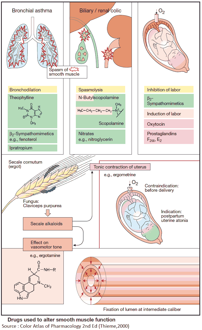
Muscle, Smooth
Bronchiolitis Obliterans
Cryptogenic Organizing Pneumonia
Epithelial Cells
Lung Volume Measurements
Histological Techniques
Immunohistochemistry
Bronchoalveolar Lavage Fluid
Intermediate-Conductance Calcium-Activated Potassium Channels
Aerosols
Tissue Inhibitor of Metalloproteinase-3
Nitrogen Dioxide
1-(5-Isoquinolinesulfonyl)-2-Methylpiperazine
Inhalation Exposure
Maleimides
Endothelium
Cilia
Indoles
Pulmonary Fibrosis
Microscopy, Electron
Phorbol 12,13-Dibutyrate
Serotonin
Isoquinolines
Administration, Inhalation
Pulmonary Artery
Albuterol
Rats, Wistar
Microscopy, Electron, Scanning
In Situ Hybridization
Rats, Inbred F344
Carbachol
Macaca mulatta
Muscle Contraction
Acetylcholine
Dogs
Mice, Inbred C57BL
Sheep
Disease Models, Animal
RNA, Messenger
Keratinocyte growth factor protects against Clara cell injury induced by naphthalene. (1/99)
(+info)Distal airways in mice exposed to cigarette smoke: Nrf2-regulated genes are increased in Clara cells. (2/99)
(+info)A technological advance comparing epithelial lining fluid from different regions of the lung in smokers. (3/99)
(+info)Prospective isolation of bronchiolar stem cells based upon immunophenotypic and autofluorescence characteristics. (4/99)
(+info)A protein kinase Cdelta-dependent protein kinase D pathway modulates ERK1/2 and JNK1/2 phosphorylation and Bim-associated apoptosis by asbestos. (5/99)
(+info)Drosophila convoluted/dALS is an essential gene required for tracheal tube morphogenesis and apical matrix organization. (6/99)
(+info)Type IV collagen alpha chains of the basement membrane in the rat bronchioalveolar transitional segment. (7/99)
In the present study, we have analyzed the alpha(IV) chain distribution in the subepithelial basement membrane (BM) of the rat pulmonary airway from the bronchi to alveoli. We have furthermore analyzed the alpha(IV) chain distribution in the subepithelial BM of the bronchioalveolar duct junction (BADJ) using alpha(IV) chain specific monoclonal antibodies. Our results show that the BM of the bronchial and bronchiolar epithelium contains [alpha1(IV)]2alpha2(IV) and [alpha5(IV)]2alpha6(IV) molecules and confirmed that the alveolar BM consists of [alpha1(IV)]2alpha2(IV) and alpha3(IV) alpha4(IV)alpha5(IV) molecules. There are also small regions in BADJ consisting of only [alpha1(IV)]2alpha2(IV) molecules without alpha3(IV)alpha4(IV)alpha5(IV) and [alpha5(IV)]2alpha6(IV) molecules. Moreover, the bronchioalveolar stem cells (BASCs)-primordial cells for bronchiolar Clara cells and alveolar type II (AT2) cells - lie adjacent to such small regions. These findings suggest that [alpha1(IV)]2 alpha2(IV) may be important for the BASCs to self-renew or to self-maintain themselves and that microenvironments produced by alpha(IV) chains may be important for cell differentiation. (+info)beta-Catenin is not necessary for maintenance or repair of the bronchiolar epithelium. (8/99)
(+info)Bronchioles are the smallest airways in the respiratory system that carry air into the lungs. They are branching tubes within the lungs that further divide and become smaller than bronchi, ending in tiny air sacs called alveoli where the exchange of oxygen and carbon dioxide occurs. Bronchioles do not have cartilage in their walls, unlike larger bronchi, making them more flexible and able to adjust to changes in lung volume during breathing.
"Bronchi" are a pair of airways in the respiratory system that branch off from the trachea (windpipe) and lead to the lungs. They are responsible for delivering oxygen-rich air to the lungs and removing carbon dioxide during exhalation. The right bronchus is slightly larger and more vertical than the left, and they further divide into smaller branches called bronchioles within the lungs. Any abnormalities or diseases affecting the bronchi can impact lung function and overall respiratory health.
A lung is a pair of spongy, elastic organs in the chest that work together to enable breathing. They are responsible for taking in oxygen and expelling carbon dioxide through the process of respiration. The left lung has two lobes, while the right lung has three lobes. The lungs are protected by the ribcage and are covered by a double-layered membrane called the pleura. The trachea divides into two bronchi, which further divide into smaller bronchioles, leading to millions of tiny air sacs called alveoli, where the exchange of gases occurs.
Uteroglobin, also known as blastokinin or Clara cell 10-kDa protein (CC10), is a small molecular weight protein that is abundantly present in the respiratory tract and reproductive system of many mammals. It was first identified in the uterine fluid of pregnant animals, hence its name.
In the human body, uteroglobin is primarily produced by non-ciliated bronchial epithelial cells known as Clara cells, which are located in the respiratory tract. Uteroglobin has been found to have anti-inflammatory and immunomodulatory properties, and it may play a role in protecting the lungs from injury and inflammation.
In the reproductive system, uteroglobin is produced by the endometrial glands of the uterus during pregnancy, and it has been suggested to have a role in maintaining pregnancy and promoting fetal growth. However, its precise functions in both the respiratory and reproductive systems are not fully understood and are still the subject of ongoing research.
Pulmonary alveoli, also known as air sacs, are tiny clusters of air-filled pouches located at the end of the bronchioles in the lungs. They play a crucial role in the process of gas exchange during respiration. The thin walls of the alveoli, called alveolar membranes, allow oxygen from inhaled air to pass into the bloodstream and carbon dioxide from the bloodstream to pass into the alveoli to be exhaled out of the body. This vital function enables the lungs to supply oxygen-rich blood to the rest of the body and remove waste products like carbon dioxide.
APUD cells are a type of neuroendocrine cell that originated from the neural crest and are widely distributed throughout the body. The term "APUD" is an acronym for "Amine Precursor Uptake and Decarboxylation," which describes the ability of these cells to take up and decarboxylate amino acid precursors to produce biologically active amines, such as serotonin, histamine, and catecholamines.
APUD cells are capable of synthesizing, storing, and releasing hormones or neurotransmitters in response to various stimuli. They can be found in several endocrine and neural tissues, including the thyroid gland, adrenal medulla, pituitary gland, pancreas, lungs, and gastrointestinal tract.
In the gastrointestinal tract, APUD cells are often referred to as enterochromaffin cells or Kulchitsky cells. They play a crucial role in regulating gut motility, secretion, and blood flow through the release of hormones such as serotonin, gastrin, and somatostatin.
It's worth noting that the APUD cell concept has been largely replaced by the more comprehensive neuroendocrine system concept, which encompasses a broader range of cells with neurosecretory functions.
Bronchiolitis is a common respiratory infection in infants and young children, typically caused by a viral infection. It is characterized by inflammation and congestion of the bronchioles (the smallest airways in the lungs), which can lead to difficulty breathing and wheezing.
The most common virus that causes bronchiolitis is respiratory syncytial virus (RSV), but other viruses such as rhinovirus, influenza, and parainfluenza can also cause the condition. Symptoms of bronchiolitis may include cough, wheezing, rapid breathing, difficulty feeding, and fatigue.
In severe cases, bronchiolitis can lead to respiratory distress and require hospitalization. Treatment typically involves supportive care, such as providing fluids and oxygen therapy, and in some cases, medications to help open the airways may be used. Prevention measures include good hand hygiene and avoiding close contact with individuals who are sick.
Bronchial diseases refer to medical conditions that affect the bronchi, which are the large airways that lead into the lungs. These diseases can cause inflammation, narrowing, or obstruction of the bronchi, leading to symptoms such as coughing, wheezing, chest tightness, and difficulty breathing.
Some common bronchial diseases include:
1. Asthma: A chronic inflammatory disease of the airways that causes recurring episodes of wheezing, breathlessness, chest tightness, and coughing.
2. Chronic Bronchitis: A long-term inflammation of the bronchi that leads to a persistent cough and excessive mucus production.
3. Bronchiectasis: A condition in which the bronchi become damaged and widened, leading to chronic infection and inflammation.
4. Bronchitis: An inflammation of the bronchi that can cause coughing, wheezing, and chest tightness.
5. Emphysema: A lung condition that causes shortness of breath due to damage to the air sacs in the lungs. While not strictly a bronchial disease, it is often associated with chronic bronchitis and COPD (Chronic Obstructive Pulmonary Disease).
Treatment for bronchial diseases may include medications such as bronchodilators, corticosteroids, or antibiotics, as well as lifestyle changes such as quitting smoking and avoiding irritants. In severe cases, oxygen therapy or surgery may be necessary.
The trachea, also known as the windpipe, is a tube-like structure in the respiratory system that connects the larynx (voice box) to the bronchi (the two branches leading to each lung). It is composed of several incomplete rings of cartilage and smooth muscle, which provide support and flexibility. The trachea plays a crucial role in directing incoming air to the lungs during inspiration and outgoing air to the larynx during expiration.
Bronchoconstriction is a medical term that refers to the narrowing of the airways in the lungs (the bronchi and bronchioles) due to the contraction of the smooth muscles surrounding them. This constriction can cause difficulty breathing, wheezing, coughing, and shortness of breath, which are common symptoms of asthma and other respiratory conditions.
Bronchoconstriction can be triggered by a variety of factors, including allergens, irritants, cold air, exercise, and emotional stress. In some cases, it may also be caused by certain medications, such as beta-blockers or nonsteroidal anti-inflammatory drugs (NSAIDs). Treatment for bronchoconstriction typically involves the use of bronchodilators, which are medications that help to relax the smooth muscles around the airways and widen them, making it easier to breathe.
Epithelium is the tissue that covers the outer surface of the body, lines the internal cavities and organs, and forms various glands. It is composed of one or more layers of tightly packed cells that have a uniform shape and size, and rest on a basement membrane. Epithelial tissues are avascular, meaning they do not contain blood vessels, and are supplied with nutrients by diffusion from the underlying connective tissue.
Epithelial cells perform a variety of functions, including protection, secretion, absorption, excretion, and sensation. They can be classified based on their shape and the number of cell layers they contain. The main types of epithelium are:
1. Squamous epithelium: composed of flat, scalelike cells that fit together like tiles on a roof. It forms the lining of blood vessels, air sacs in the lungs, and the outermost layer of the skin.
2. Cuboidal epithelium: composed of cube-shaped cells with equal height and width. It is found in glands, tubules, and ducts.
3. Columnar epithelium: composed of tall, rectangular cells that are taller than they are wide. It lines the respiratory, digestive, and reproductive tracts.
4. Pseudostratified epithelium: appears stratified or layered but is actually made up of a single layer of cells that vary in height. The nuclei of these cells appear at different levels, giving the tissue a stratified appearance. It lines the respiratory and reproductive tracts.
5. Transitional epithelium: composed of several layers of cells that can stretch and change shape to accommodate changes in volume. It is found in the urinary bladder and ureters.
Epithelial tissue provides a barrier between the internal and external environments, protecting the body from physical, chemical, and biological damage. It also plays a crucial role in maintaining homeostasis by regulating the exchange of substances between the body and its environment.
Respiratory mucosa refers to the mucous membrane that lines the respiratory tract, including the nose, throat, bronchi, and lungs. It is a specialized type of tissue that is composed of epithelial cells, goblet cells, and glands that produce mucus, which helps to trap inhaled particles such as dust, allergens, and pathogens.
The respiratory mucosa also contains cilia, tiny hair-like structures that move rhythmically to help propel the mucus and trapped particles out of the airways and into the upper part of the throat, where they can be swallowed or coughed up. This defense mechanism is known as the mucociliary clearance system.
In addition to its role in protecting the respiratory tract from harmful substances, the respiratory mucosa also plays a crucial role in immune function by containing various types of immune cells that help to detect and respond to pathogens and other threats.
The Respiratory System is a complex network of organs and tissues that work together to facilitate the process of breathing, which involves the intake of oxygen and the elimination of carbon dioxide. This system primarily includes the nose, throat (pharynx), voice box (larynx), windpipe (trachea), bronchi, bronchioles, lungs, and diaphragm.
The nostrils or mouth take in air that travels through the pharynx, larynx, and trachea into the lungs. Within the lungs, the trachea divides into two bronchi, one for each lung, which further divide into smaller tubes called bronchioles. At the end of these bronchioles are tiny air sacs known as alveoli where the exchange of gases occurs. Oxygen from the inhaled air diffuses through the walls of the alveoli into the bloodstream, while carbon dioxide, a waste product, moves from the blood to the alveoli and is exhaled out of the body.
The diaphragm, a large muscle that separates the chest from the abdomen, plays a crucial role in breathing by contracting and relaxing to change the volume of the chest cavity, thereby allowing air to flow in and out of the lungs. Overall, the Respiratory System is essential for maintaining life by providing the body's cells with the oxygen needed for metabolism and removing waste products like carbon dioxide.
"Mannheimia haemolytica" is a gram-negative, rod-shaped bacterium that is commonly found as part of the normal flora in the upper respiratory tract of cattle and other ruminants. However, under certain conditions such as stress, viral infection, or sudden changes in temperature or humidity, the bacteria can multiply rapidly and cause a severe respiratory disease known as shipping fever or pneumonic pasteurellosis.
The bacterium is named "haemolytica" because it produces a toxin that causes hemolysis, or the breakdown of red blood cells, resulting in the characteristic clear zones around colonies grown on blood agar plates. The bacteria can also cause other symptoms such as fever, coughing, difficulty breathing, and depression.
"Mannheimia haemolytica" is a significant pathogen in the cattle industry, causing substantial economic losses due to mortality, reduced growth rates, and decreased milk production. Prevention and control measures include good management practices, vaccination, and prompt treatment of infected animals with antibiotics.
Ozone (O3) is not a substance that is typically considered a component of health or medicine in the context of human body or physiology. It's actually a form of oxygen, but with three atoms instead of two, making it unstable and reactive. Ozone is naturally present in the Earth's atmosphere, where it forms a protective layer in the stratosphere that absorbs harmful ultraviolet (UV) radiation from the sun.
However, ozone can have both beneficial and detrimental effects on human health depending on its location and concentration. At ground level or in indoor environments, ozone is considered an air pollutant that can irritate the respiratory system and aggravate asthma symptoms when inhaled at high concentrations. It's important to note that ozone should not be confused with oxygen (O2), which is essential for human life and breathing.
Bronchography is a medical imaging technique that involves the injection of a contrast material into the airways (bronchi) of the lungs, followed by X-ray imaging to produce detailed pictures of the bronchial tree. This diagnostic procedure was commonly used in the past to identify abnormalities such as narrowing, blockages, or inflammation in the airways, but it has largely been replaced by newer, less invasive techniques like computed tomography (CT) scans and bronchoscopy.
The process of bronchography involves the following steps:
1. The patient is sedated or given a local anesthetic to minimize discomfort during the procedure.
2. A radiopaque contrast material is introduced into the bronchi through a catheter that is inserted into the trachea, either via a nostril or through a small incision in the neck.
3. Once the contrast material has been distributed throughout the bronchial tree, X-ray images are taken from various angles to capture detailed views of the airways.
4. The images are then analyzed by a radiologist to identify any abnormalities or irregularities in the structure and function of the bronchi.
Although bronchography is considered a relatively safe procedure, it does carry some risks, including allergic reactions to the contrast material, infection, and bleeding. Additionally, the use of ionizing radiation during X-ray imaging should be carefully weighed against the potential benefits of the procedure.
Oximes are a class of chemical compounds that contain the functional group =N-O-, where two organic groups are attached to the nitrogen atom. In a clinical context, oximes are used as antidotes for nerve agent and pesticide poisoning. The most commonly used oxime in medicine is pralidoxime (2-PAM), which is used to reactivate acetylcholinesterase that has been inhibited by organophosphorus compounds, such as nerve agents and certain pesticides. These compounds work by forming a bond with the phosphoryl group of the inhibited enzyme, allowing for its reactivation and restoration of normal neuromuscular function.
Pulmonary emphysema is a chronic respiratory disease characterized by abnormal, permanent enlargement of the airspaces distal to the terminal bronchioles, accompanied by destruction of their walls and without obvious fibrosis. This results in loss of elastic recoil, which leads to trappling of air within the lungs and difficulty exhaling. It is often caused by cigarette smoking or long-term exposure to harmful pollutants. The disease is part of a group of conditions known as chronic obstructive pulmonary disease (COPD), which also includes chronic bronchitis.
Smooth muscle, also known as involuntary muscle, is a type of muscle that is controlled by the autonomic nervous system and functions without conscious effort. These muscles are found in the walls of hollow organs such as the stomach, intestines, bladder, and blood vessels, as well as in the eyes, skin, and other areas of the body.
Smooth muscle fibers are shorter and narrower than skeletal muscle fibers and do not have striations or sarcomeres, which give skeletal muscle its striped appearance. Smooth muscle is controlled by the autonomic nervous system through the release of neurotransmitters such as acetylcholine and norepinephrine, which bind to receptors on the smooth muscle cells and cause them to contract or relax.
Smooth muscle plays an important role in many physiological processes, including digestion, circulation, respiration, and elimination. It can also contribute to various medical conditions, such as hypertension, gastrointestinal disorders, and genitourinary dysfunction, when it becomes overactive or underactive.
Bronchiolitis obliterans is a medical condition characterized by the inflammation and scarring (fibrosis) of the bronchioles, which are the smallest airways in the lungs. This results in the narrowing or complete obstruction of the airways, leading to difficulty breathing and reduced lung function.
The condition is often caused by a respiratory infection, such as adenovirus or mycoplasma pneumonia, but it can also be associated with exposure to certain chemicals, drugs, or radiation therapy. In some cases, the cause may be unknown.
Symptoms of bronchiolitis obliterans include cough, shortness of breath, wheezing, and crackles heard on lung examination. Diagnosis typically involves a combination of medical history, physical exam, imaging studies (such as chest X-ray or CT scan), and pulmonary function tests. In some cases, a biopsy may be necessary to confirm the diagnosis.
Treatment for bronchiolitis obliterans is focused on managing symptoms and preventing further lung damage. This may include bronchodilators to help open up the airways, corticosteroids to reduce inflammation, and oxygen therapy to help with breathing. In severe cases, a lung transplant may be necessary.
Cryptogenic organizing pneumonia (COP) is a type of lung disorder that is characterized by the presence of inflammation and scarring in the lungs. The term "cryptogenic" means that the cause of the condition is unknown or unclear.
Organizing pneumonia is a specific pattern of injury to the lungs that can be caused by various factors, including infections, medications, and autoimmune disorders. However, in cases of COP, there is no clear underlying cause that can be identified.
The main symptoms of COP include cough, shortness of breath, fever, and fatigue. The condition can also cause crackles or wheezing sounds when listening to the lungs with a stethoscope. Diagnosis of COP typically involves a combination of imaging studies, such as chest X-rays or CT scans, and lung biopsy.
Treatment for COP usually involves the use of corticosteroids, which can help to reduce inflammation and improve symptoms. In some cases, other medications may also be used to manage the condition. The prognosis for people with COP is generally good, with most individuals responding well to treatment and experiencing improvement in their symptoms over time. However, recurrence of the condition is possible, and long-term monitoring may be necessary.
Epithelial cells are types of cells that cover the outer surfaces of the body, line the inner surfaces of organs and glands, and form the lining of blood vessels and body cavities. They provide a protective barrier against the external environment, regulate the movement of materials between the internal and external environments, and are involved in the sense of touch, temperature, and pain. Epithelial cells can be squamous (flat and thin), cuboidal (square-shaped and of equal height), or columnar (tall and narrow) in shape and are classified based on their location and function.
Lung volume measurements are clinical tests that determine the amount of air inhaled, exhaled, and present in the lungs at different times during the breathing cycle. These measurements include:
1. Tidal Volume (TV): The amount of air inhaled or exhaled during normal breathing, usually around 500 mL in resting adults.
2. Inspiratory Reserve Volume (IRV): The additional air that can be inhaled after a normal inspiration, approximately 3,000 mL in adults.
3. Expiratory Reserve Volume (ERV): The extra air that can be exhaled after a normal expiration, about 1,000-1,200 mL in adults.
4. Residual Volume (RV): The air remaining in the lungs after a maximal exhalation, approximately 1,100-1,500 mL in adults.
5. Total Lung Capacity (TLC): The total amount of air the lungs can hold at full inflation, calculated as TV + IRV + ERV + RV, around 6,000 mL in adults.
6. Functional Residual Capacity (FRC): The volume of air remaining in the lungs after a normal expiration, equal to ERV + RV, about 2,100-2,700 mL in adults.
7. Inspiratory Capacity (IC): The maximum amount of air that can be inhaled after a normal expiration, equal to TV + IRV, around 3,500 mL in adults.
8. Vital Capacity (VC): The total volume of air that can be exhaled after a maximal inspiration, calculated as IC + ERV, approximately 4,200-5,600 mL in adults.
These measurements help assess lung function and identify various respiratory disorders such as chronic obstructive pulmonary disease (COPD), asthma, and restrictive lung diseases.
Histological techniques are a set of laboratory methods and procedures used to study the microscopic structure of tissues, also known as histology. These techniques include:
1. Tissue fixation: The process of preserving tissue specimens to maintain their structural integrity and prevent decomposition. This is typically done using formaldehyde or other chemical fixatives.
2. Tissue processing: The preparation of fixed tissues for embedding by removing water, fat, and other substances that can interfere with sectioning and staining. This is usually accomplished through a series of dehydration, clearing, and infiltration steps.
3. Embedding: The placement of processed tissue specimens into a solid support medium, such as paraffin or plastic, to facilitate sectioning.
4. Sectioning: The cutting of thin slices (usually 4-6 microns thick) from embedded tissue blocks using a microtome.
5. Staining: The application of dyes or stains to tissue sections to highlight specific structures or components. This can be done through a variety of methods, including hematoxylin and eosin (H&E) staining, immunohistochemistry, and special stains for specific cell types or molecules.
6. Mounting: The placement of stained tissue sections onto glass slides and covering them with a mounting medium to protect the tissue from damage and improve microscopic visualization.
7. Microscopy: The examination of stained tissue sections using a light or electron microscope to observe and analyze their structure and composition.
These techniques are essential for the diagnosis and study of various diseases, including cancer, neurological disorders, and infections. They allow pathologists and researchers to visualize and understand the cellular and molecular changes that occur in tissues during disease processes.
Immunohistochemistry (IHC) is a technique used in pathology and laboratory medicine to identify specific proteins or antigens in tissue sections. It combines the principles of immunology and histology to detect the presence and location of these target molecules within cells and tissues. This technique utilizes antibodies that are specific to the protein or antigen of interest, which are then tagged with a detection system such as a chromogen or fluorophore. The stained tissue sections can be examined under a microscope, allowing for the visualization and analysis of the distribution and expression patterns of the target molecule in the context of the tissue architecture. Immunohistochemistry is widely used in diagnostic pathology to help identify various diseases, including cancer, infectious diseases, and immune-mediated disorders.
Airway obstruction is a medical condition that occurs when the normal flow of air into and out of the lungs is partially or completely blocked. This blockage can be caused by a variety of factors, including swelling of the tissues in the airway, the presence of foreign objects or substances, or abnormal growths such as tumors.
When the airway becomes obstructed, it can make it difficult for a person to breathe normally. They may experience symptoms such as shortness of breath, wheezing, coughing, and chest tightness. In severe cases, airway obstruction can lead to respiratory failure and other life-threatening complications.
There are several types of airway obstruction, including:
1. Upper airway obstruction: This occurs when the blockage is located in the upper part of the airway, such as the nose, throat, or voice box.
2. Lower airway obstruction: This occurs when the blockage is located in the lower part of the airway, such as the trachea or bronchi.
3. Partial airway obstruction: This occurs when the airway is partially blocked, allowing some air to flow in and out of the lungs.
4. Complete airway obstruction: This occurs when the airway is completely blocked, preventing any air from flowing into or out of the lungs.
Treatment for airway obstruction depends on the underlying cause of the condition. In some cases, removing the obstruction may be as simple as clearing the airway of foreign objects or mucus. In other cases, more invasive treatments such as surgery may be necessary.
Bronchoalveolar lavage (BAL) fluid is a type of clinical specimen obtained through a procedure called bronchoalveolar lavage. This procedure involves inserting a bronchoscope into the lungs and instilling a small amount of saline solution into a specific area of the lung, then gently aspirating the fluid back out. The fluid that is recovered is called bronchoalveolar lavage fluid.
BAL fluid contains cells and other substances that are present in the lower respiratory tract, including the alveoli (the tiny air sacs where gas exchange occurs). By analyzing BAL fluid, doctors can diagnose various lung conditions, such as pneumonia, interstitial lung disease, and lung cancer. They can also monitor the effectiveness of treatments for these conditions by comparing the composition of BAL fluid before and after treatment.
BAL fluid is typically analyzed for its cellular content, including the number and type of white blood cells present, as well as for the presence of bacteria, viruses, or other microorganisms. The fluid may also be tested for various proteins, enzymes, and other biomarkers that can provide additional information about lung health and disease.
Intermediate-conductance calcium-activated potassium channels (IKCa) are a type of ion channel found in various cell types, including immune cells, endothelial cells, and neurons. These channels are activated by an increase in intracellular calcium ions (Ca2+) and allow the flow of potassium ions (K+) out of the cell.
IKCa channels have a single-channel conductance that is intermediate between small-conductance (SKCa) and large-conductance (BKCa) calcium-activated potassium channels, typically ranging from 20 to 100 picosiemens (pS). They are encoded by the KCNN4 gene in humans.
The activation of IKCa channels plays a crucial role in regulating various cellular processes, such as membrane potential, calcium signaling, and immune response. For example, in activated immune cells, the opening of IKCa channels helps to repolarize the membrane potential and limit further Ca2+ entry into the cell, thereby modulating cytokine production and inflammatory responses. In endothelial cells, IKCa channel activation can regulate vascular tone and blood flow by controlling the diameter of blood vessels.
Aerosols are defined in the medical field as suspensions of fine solid or liquid particles in a gas. In the context of public health and medicine, aerosols often refer to particles that can remain suspended in air for long periods of time and can be inhaled. They can contain various substances, such as viruses, bacteria, fungi, or chemicals, and can play a role in the transmission of respiratory infections or other health effects.
For example, when an infected person coughs or sneezes, they may produce respiratory droplets that can contain viruses like influenza or SARS-CoV-2 (the virus that causes COVID-19). Some of these droplets can evaporate quickly and leave behind smaller particles called aerosols, which can remain suspended in the air for hours and potentially be inhaled by others. This is one way that respiratory viruses can spread between people in close proximity to each other.
Aerosols can also be generated through medical procedures such as bronchoscopy, suctioning, or nebulizer treatments, which can produce aerosols containing bacteria, viruses, or other particles that may pose an infection risk to healthcare workers or other patients. Therefore, appropriate personal protective equipment (PPE) and airborne precautions are often necessary to reduce the risk of transmission in these settings.
Lung diseases refer to a broad category of disorders that affect the lungs and other structures within the respiratory system. These diseases can impair lung function, leading to symptoms such as coughing, shortness of breath, chest pain, and wheezing. They can be categorized into several types based on the underlying cause and nature of the disease process. Some common examples include:
1. Obstructive lung diseases: These are characterized by narrowing or blockage of the airways, making it difficult to breathe out. Examples include chronic obstructive pulmonary disease (COPD), asthma, bronchiectasis, and cystic fibrosis.
2. Restrictive lung diseases: These involve stiffening or scarring of the lungs, which reduces their ability to expand and take in air. Examples include idiopathic pulmonary fibrosis, sarcoidosis, and asbestosis.
3. Infectious lung diseases: These are caused by bacteria, viruses, fungi, or parasites that infect the lungs. Examples include pneumonia, tuberculosis, and influenza.
4. Vascular lung diseases: These affect the blood vessels in the lungs, impairing oxygen exchange. Examples include pulmonary embolism, pulmonary hypertension, and chronic thromboembolic pulmonary hypertension (CTEPH).
5. Neoplastic lung diseases: These involve abnormal growth of cells within the lungs, leading to cancer. Examples include small cell lung cancer, non-small cell lung cancer, and mesothelioma.
6. Other lung diseases: These include interstitial lung diseases, pleural effusions, and rare disorders such as pulmonary alveolar proteinosis and lymphangioleiomyomatosis (LAM).
It is important to note that this list is not exhaustive, and there are many other conditions that can affect the lungs. Proper diagnosis and treatment of lung diseases require consultation with a healthcare professional, such as a pulmonologist or respiratory therapist.
Tissue Inhibitor of Metalloproteinase-3 (TIMP-3) is a member of the tissue inhibitors of metalloproteinases (TIMPs) family, which are natural inhibitors of matrix metalloproteinases (MMPs), a group of enzymes involved in the degradation and remodeling of extracellular matrix components.
TIMP-3 is unique among TIMPs because it can inhibit all known MMPs and also has the ability to inhibit some members of the ADAM (a disintegrin and metalloproteinase) family, which are involved in protein ectodomain shedding and cell adhesion.
TIMP-3 is a secreted glycoprotein that binds to the extracellular matrix and regulates MMP activity locally. It has been shown to play important roles in various biological processes, including tissue remodeling, angiogenesis, inflammation, and apoptosis. Dysregulation of TIMP-3 expression or function has been implicated in several diseases, such as cancer, fibrosis, and neurodegenerative disorders.
Nitrogen dioxide (NO2) is a gaseous air pollutant and respiratory irritant. It is a reddish-brown toxic gas with a pungent, choking odor. NO2 is a major component of smog and is produced from the combustion of fossil fuels in vehicles, power plants, and industrial processes.
Exposure to nitrogen dioxide can cause respiratory symptoms such as coughing, wheezing, and difficulty breathing, especially in people with asthma or other respiratory conditions. Long-term exposure has been linked to the development of chronic lung diseases, including bronchitis and emphysema. NO2 also contributes to the formation of fine particulate matter (PM2.5), which can penetrate deep into the lungs and cause additional health problems.
Inhalation exposure is a term used in occupational and environmental health to describe the situation where an individual breathes in substances present in the air, which could be gases, vapors, fumes, mist, or particulate matter. These substances can originate from various sources, such as industrial processes, chemical reactions, or natural phenomena.
The extent of inhalation exposure is determined by several factors, including:
1. Concentration of the substance in the air
2. Duration of exposure
3. Frequency of exposure
4. The individual's breathing rate
5. The efficiency of the individual's respiratory protection, if any
Inhalation exposure can lead to adverse health effects, depending on the toxicity and concentration of the inhaled substances. Short-term or acute health effects may include irritation of the eyes, nose, throat, or lungs, while long-term or chronic exposure can result in more severe health issues, such as respiratory diseases, neurological disorders, or cancer.
It is essential to monitor and control inhalation exposures in occupational settings to protect workers' health and ensure compliance with regulatory standards. Various methods are employed for exposure assessment, including personal air sampling, area monitoring, and biological monitoring. Based on the results of these assessments, appropriate control measures can be implemented to reduce or eliminate the risks associated with inhalation exposure.
Maleimides are a class of chemical compounds that contain a maleimide functional group, which is characterized by a five-membered ring containing two carbon atoms and three nitrogen atoms. The double bond in the maleimide ring makes it highly reactive towards nucleophiles, particularly thiol groups found in cysteine residues of proteins.
In medical and biological contexts, maleimides are often used as cross-linking agents to modify or label proteins, peptides, and other biomolecules. For example, maleimide-functionalized probes such as fluorescent dyes, biotin, or radioisotopes can be covalently attached to thiol groups in proteins for various applications, including protein detection, purification, and imaging.
However, it is important to note that maleimides can also react with other nucleophiles such as amines, although at a slower rate. Therefore, careful control of reaction conditions is necessary to ensure specificity towards thiol groups.
The endothelium is the thin, delicate tissue that lines the interior surface of blood vessels and lymphatic vessels. It is a single layer of cells called endothelial cells that are in contact with the blood or lymph fluid. The endothelium plays an essential role in maintaining vascular homeostasis by regulating blood flow, coagulation, platelet activation, immune function, and angiogenesis (the formation of new blood vessels). It also acts as a barrier between the vessel wall and the circulating blood or lymph fluid. Dysfunction of the endothelium has been implicated in various cardiovascular diseases, diabetes, inflammation, and cancer.
Cilia are tiny, hair-like structures that protrude from the surface of many types of cells in the body. They are composed of a core bundle of microtubules surrounded by a protein matrix and are covered with a membrane. Cilia are involved in various cellular functions, including movement of fluid or mucus across the cell surface, detection of external stimuli, and regulation of signaling pathways.
There are two types of cilia: motile and non-motile. Motile cilia are able to move in a coordinated manner to propel fluids or particles across a surface, such as those found in the respiratory tract and reproductive organs. Non-motile cilia, also known as primary cilia, are present on most cells in the body and serve as sensory organelles that detect chemical and mechanical signals from the environment.
Defects in cilia structure or function can lead to a variety of diseases, collectively known as ciliopathies. These conditions can affect multiple organs and systems in the body, including the brain, kidneys, liver, and eyes. Examples of ciliopathies include polycystic kidney disease, Bardet-Biedl syndrome, and Meckel-Gruber syndrome.
Pneumonia is an infection or inflammation of the alveoli (tiny air sacs) in one or both lungs. It's often caused by bacteria, viruses, or fungi. Accumulated pus and fluid in these air sacs make it difficult to breathe, which can lead to coughing, chest pain, fever, and difficulty breathing. The severity of symptoms can vary from mild to life-threatening, depending on the underlying cause, the patient's overall health, and age. Pneumonia is typically diagnosed through a combination of physical examination, medical history, and diagnostic tests such as chest X-rays or blood tests. Treatment usually involves antibiotics for bacterial pneumonia, antivirals for viral pneumonia, and supportive care like oxygen therapy, hydration, and rest.
Indole is not strictly a medical term, but it is a chemical compound that can be found in the human body and has relevance to medical and biological research. Indoles are organic compounds that contain a bicyclic structure consisting of a six-membered benzene ring fused to a five-membered pyrrole ring.
In the context of medicine, indoles are particularly relevant due to their presence in certain hormones and other biologically active molecules. For example, the neurotransmitter serotonin contains an indole ring, as does the hormone melatonin. Indoles can also be found in various plant-based foods, such as cruciferous vegetables (e.g., broccoli, kale), and have been studied for their potential health benefits.
Some indoles, like indole-3-carbinol and diindolylmethane, are found in these vegetables and can have anti-cancer properties by modulating estrogen metabolism, reducing inflammation, and promoting cell death (apoptosis) in cancer cells. However, it is essential to note that further research is needed to fully understand the potential health benefits and risks associated with indoles.
Pulmonary fibrosis is a specific type of lung disease that results from the thickening and scarring of the lung tissues, particularly those in the alveoli (air sacs) and interstitium (the space around the air sacs). This scarring makes it harder for the lungs to properly expand and transfer oxygen into the bloodstream, leading to symptoms such as shortness of breath, coughing, fatigue, and eventually respiratory failure. The exact cause of pulmonary fibrosis can vary, with some cases being idiopathic (without a known cause) or related to environmental factors, medications, medical conditions, or genetic predisposition.
Electron microscopy (EM) is a type of microscopy that uses a beam of electrons to create an image of the sample being examined, resulting in much higher magnification and resolution than light microscopy. There are several types of electron microscopy, including transmission electron microscopy (TEM), scanning electron microscopy (SEM), and reflection electron microscopy (REM).
In TEM, a beam of electrons is transmitted through a thin slice of the sample, and the electrons that pass through the sample are focused to form an image. This technique can provide detailed information about the internal structure of cells, viruses, and other biological specimens, as well as the composition and structure of materials at the atomic level.
In SEM, a beam of electrons is scanned across the surface of the sample, and the electrons that are scattered back from the surface are detected to create an image. This technique can provide information about the topography and composition of surfaces, as well as the structure of materials at the microscopic level.
REM is a variation of SEM in which the beam of electrons is reflected off the surface of the sample, rather than scattered back from it. This technique can provide information about the surface chemistry and composition of materials.
Electron microscopy has a wide range of applications in biology, medicine, and materials science, including the study of cellular structure and function, disease diagnosis, and the development of new materials and technologies.
Phorbol 12,13-dibutyrate (PDB) is not a medical term per se, but a chemical compound used in scientific research. It's a type of phorbol ester, which are tumor promoters and active components of croton oil. PDB is often used as a biochemical tool to study cell signaling pathways, particularly those involving protein kinase C (PKC) activation.
Medically, it may be mentioned in research or clinical studies related to cellular processes, cancer, or inflammation. However, it is not something that a patient would typically encounter in a medical setting.
Serotonin, also known as 5-hydroxytryptamine (5-HT), is a monoamine neurotransmitter that is found primarily in the gastrointestinal (GI) tract, blood platelets, and the central nervous system (CNS) of humans and other animals. It is produced by the conversion of the amino acid tryptophan to 5-hydroxytryptophan (5-HTP), and then to serotonin.
In the CNS, serotonin plays a role in regulating mood, appetite, sleep, memory, learning, and behavior, among other functions. It also acts as a vasoconstrictor, helping to regulate blood flow and blood pressure. In the GI tract, it is involved in peristalsis, the contraction and relaxation of muscles that moves food through the digestive system.
Serotonin is synthesized and stored in serotonergic neurons, which are nerve cells that use serotonin as their primary neurotransmitter. These neurons are found throughout the brain and spinal cord, and they communicate with other neurons by releasing serotonin into the synapse, the small gap between two neurons.
Abnormal levels of serotonin have been linked to a variety of disorders, including depression, anxiety, schizophrenia, and migraines. Medications that affect serotonin levels, such as selective serotonin reuptake inhibitors (SSRIs), are commonly used to treat these conditions.
Isoquinolines are not a medical term per se, but a chemical classification. They refer to a class of organic compounds that consist of a benzene ring fused to a piperidine ring. This structure is similar to that of quinoline, but with the nitrogen atom located at a different position in the ring.
Isoquinolines have various biological activities and can be found in some natural products, including certain alkaloids. Some isoquinoline derivatives have been developed as drugs for the treatment of various conditions, such as cardiovascular diseases, neurological disorders, and cancer. However, specific medical definitions related to isoquinolines typically refer to the use or effects of these specific drugs rather than the broader class of compounds.
"Inhalation administration" is a medical term that refers to the method of delivering medications or therapeutic agents directly into the lungs by inhaling them through the airways. This route of administration is commonly used for treating respiratory conditions such as asthma, COPD (chronic obstructive pulmonary disease), and cystic fibrosis.
Inhalation administration can be achieved using various devices, including metered-dose inhalers (MDIs), dry powder inhalers (DPIs), nebulizers, and soft-mist inhalers. Each device has its unique mechanism of delivering the medication into the lungs, but they all aim to provide a high concentration of the drug directly to the site of action while minimizing systemic exposure and side effects.
The advantages of inhalation administration include rapid onset of action, increased local drug concentration, reduced systemic side effects, and improved patient compliance due to the ease of use and non-invasive nature of the delivery method. However, proper technique and device usage are crucial for effective therapy, as incorrect usage may result in suboptimal drug deposition and therapeutic outcomes.
The pulmonary artery is a large blood vessel that carries deoxygenated blood from the right ventricle of the heart to the lungs for oxygenation. It divides into two main branches, the right and left pulmonary arteries, which further divide into smaller vessels called arterioles, and then into a vast network of capillaries in the lungs where gas exchange occurs. The thin walls of these capillaries allow oxygen to diffuse into the blood and carbon dioxide to diffuse out, making the blood oxygen-rich before it is pumped back to the left side of the heart through the pulmonary veins. This process is crucial for maintaining proper oxygenation of the body's tissues and organs.
Albuterol is a medication that is used to treat bronchospasm, or narrowing of the airways in the lungs, in conditions such as asthma and chronic obstructive pulmonary disease (COPD). It is a short-acting beta-2 agonist, which means it works by relaxing the muscles around the airways, making it easier to breathe. Albuterol is available in several forms, including an inhaler, nebulizer solution, and syrup, and it is typically used as needed to relieve symptoms of bronchospasm. It may also be used before exercise to prevent bronchospasm caused by physical activity.
The medical definition of Albuterol is: "A short-acting beta-2 adrenergic agonist used to treat bronchospasm in conditions such as asthma and COPD. It works by relaxing the muscles around the airways, making it easier to breathe."
"Wistar rats" are a strain of albino rats that are widely used in laboratory research. They were developed at the Wistar Institute in Philadelphia, USA, and were first introduced in 1906. Wistar rats are outbred, which means that they are genetically diverse and do not have a fixed set of genetic characteristics like inbred strains.
Wistar rats are commonly used as animal models in biomedical research because of their size, ease of handling, and relatively low cost. They are used in a wide range of research areas, including toxicology, pharmacology, nutrition, cancer, cardiovascular disease, and behavioral studies. Wistar rats are also used in safety testing of drugs, medical devices, and other products.
Wistar rats are typically larger than many other rat strains, with males weighing between 500-700 grams and females weighing between 250-350 grams. They have a lifespan of approximately 2-3 years. Wistar rats are also known for their docile and friendly nature, making them easy to handle and work with in the laboratory setting.
Scanning electron microscopy (SEM) is a type of electron microscopy that uses a focused beam of electrons to scan the surface of a sample and produce a high-resolution image. In SEM, a beam of electrons is scanned across the surface of a specimen, and secondary electrons are emitted from the sample due to interactions between the electrons and the atoms in the sample. These secondary electrons are then detected by a detector and used to create an image of the sample's surface topography. SEM can provide detailed images of the surface of a wide range of materials, including metals, polymers, ceramics, and biological samples. It is commonly used in materials science, biology, and electronics for the examination and analysis of surfaces at the micro- and nanoscale.
In situ hybridization (ISH) is a molecular biology technique used to detect and localize specific nucleic acid sequences, such as DNA or RNA, within cells or tissues. This technique involves the use of a labeled probe that is complementary to the target nucleic acid sequence. The probe can be labeled with various types of markers, including radioisotopes, fluorescent dyes, or enzymes.
During the ISH procedure, the labeled probe is hybridized to the target nucleic acid sequence in situ, meaning that the hybridization occurs within the intact cells or tissues. After washing away unbound probe, the location of the labeled probe can be visualized using various methods depending on the type of label used.
In situ hybridization has a wide range of applications in both research and diagnostic settings, including the detection of gene expression patterns, identification of viral infections, and diagnosis of genetic disorders.
Lung neoplasms refer to abnormal growths or tumors in the lung tissue. These tumors can be benign (non-cancerous) or malignant (cancerous). Malignant lung neoplasms are further classified into two main types: small cell lung carcinoma and non-small cell lung carcinoma. Lung neoplasms can cause symptoms such as cough, chest pain, shortness of breath, and weight loss. They are often caused by smoking or exposure to secondhand smoke, but can also occur due to genetic factors, radiation exposure, and other environmental carcinogens. Early detection and treatment of lung neoplasms is crucial for improving outcomes and survival rates.
"Newborn animals" refers to the very young offspring of animals that have recently been born. In medical terminology, newborns are often referred to as "neonates," and they are classified as such from birth until about 28 days of age. During this time period, newborn animals are particularly vulnerable and require close monitoring and care to ensure their survival and healthy development.
The specific needs of newborn animals can vary widely depending on the species, but generally, they require warmth, nutrition, hydration, and protection from harm. In many cases, newborns are unable to regulate their own body temperature or feed themselves, so they rely heavily on their mothers for care and support.
In medical settings, newborn animals may be examined and treated by veterinarians to ensure that they are healthy and receiving the care they need. This can include providing medical interventions such as feeding tubes, antibiotics, or other treatments as needed to address any health issues that arise. Overall, the care and support of newborn animals is an important aspect of animal medicine and conservation efforts.
F344 is a strain code used to designate an outbred stock of rats that has been inbreeded for over 100 generations. The F344 rats, also known as Fischer 344 rats, were originally developed at the National Institutes of Health (NIH) and are now widely used in biomedical research due to their consistent and reliable genetic background.
Inbred strains, like the F344, are created by mating genetically identical individuals (siblings or parents and offspring) for many generations until a state of complete homozygosity is reached, meaning that all members of the strain have identical genomes. This genetic uniformity makes inbred strains ideal for use in studies where consistent and reproducible results are important.
F344 rats are known for their longevity, with a median lifespan of around 27-31 months, making them useful for aging research. They also have a relatively low incidence of spontaneous tumors compared to other rat strains. However, they may be more susceptible to certain types of cancer and other diseases due to their inbred status.
It's important to note that while F344 rats are often used as a standard laboratory rat strain, there can still be some genetic variation between individual animals within the same strain, particularly if they come from different suppliers or breeding colonies. Therefore, it's always important to consider the source and history of any animal model when designing experiments and interpreting results.
Carbachol is a cholinergic agonist, which means it stimulates the parasympathetic nervous system by mimicking the action of acetylcholine, a neurotransmitter that is involved in transmitting signals between nerves and muscles. Carbachol binds to both muscarinic and nicotinic receptors, but its effects are more pronounced on muscarinic receptors.
Carbachol is used in medical treatments to produce miosis (pupil constriction), lower intraocular pressure, and stimulate gastrointestinal motility. It can also be used as a diagnostic tool to test for certain conditions such as Hirschsprung's disease.
Like any medication, carbachol can have side effects, including sweating, salivation, nausea, vomiting, diarrhea, bradycardia (slow heart rate), and bronchoconstriction (narrowing of the airways in the lungs). It should be used with caution and under the supervision of a healthcare professional.
"Macaca mulatta" is the scientific name for the Rhesus macaque, a species of monkey that is native to South, Central, and Southeast Asia. They are often used in biomedical research due to their genetic similarity to humans.
Muscle contraction is the physiological process in which muscle fibers shorten and generate force, leading to movement or stability of a body part. This process involves the sliding filament theory where thick and thin filaments within the sarcomeres (the functional units of muscles) slide past each other, facilitated by the interaction between myosin heads and actin filaments. The energy required for this action is provided by the hydrolysis of adenosine triphosphate (ATP). Muscle contractions can be voluntary or involuntary, and they play a crucial role in various bodily functions such as locomotion, circulation, respiration, and posture maintenance.
Acetylcholine is a neurotransmitter, a type of chemical messenger that transmits signals across a chemical synapse from one neuron (nerve cell) to another "target" neuron, muscle cell, or gland cell. It is involved in both peripheral and central nervous system functions.
In the peripheral nervous system, acetylcholine acts as a neurotransmitter at the neuromuscular junction, where it transmits signals from motor neurons to activate muscles. Acetylcholine also acts as a neurotransmitter in the autonomic nervous system, where it is involved in both the sympathetic and parasympathetic systems.
In the central nervous system, acetylcholine plays a role in learning, memory, attention, and arousal. Disruptions in cholinergic neurotransmission have been implicated in several neurological disorders, including Alzheimer's disease, Parkinson's disease, and myasthenia gravis.
Acetylcholine is synthesized from choline and acetyl-CoA by the enzyme choline acetyltransferase and is stored in vesicles at the presynaptic terminal of the neuron. When a nerve impulse arrives, the vesicles fuse with the presynaptic membrane, releasing acetylcholine into the synapse. The acetylcholine then binds to receptors on the postsynaptic membrane, triggering a response in the target cell. Acetylcholine is subsequently degraded by the enzyme acetylcholinesterase, which terminates its action and allows for signal transduction to be repeated.
I believe there might be a misunderstanding in your question. "Dogs" is not a medical term or condition. It is the common name for a domesticated carnivore of the family Canidae, specifically the genus Canis, which includes wolves, foxes, and other extant and extinct species of mammals. Dogs are often kept as pets and companions, and they have been bred in a wide variety of forms and sizes for different purposes, such as hunting, herding, guarding, assisting police and military forces, and providing companionship and emotional support.
If you meant to ask about a specific medical condition or term related to dogs, please provide more context so I can give you an accurate answer.
C57BL/6 (C57 Black 6) is an inbred strain of laboratory mouse that is widely used in biomedical research. The term "inbred" refers to a strain of animals where matings have been carried out between siblings or other closely related individuals for many generations, resulting in a population that is highly homozygous at most genetic loci.
The C57BL/6 strain was established in 1920 by crossing a female mouse from the dilute brown (DBA) strain with a male mouse from the black strain. The resulting offspring were then interbred for many generations to create the inbred C57BL/6 strain.
C57BL/6 mice are known for their robust health, longevity, and ease of handling, making them a popular choice for researchers. They have been used in a wide range of biomedical research areas, including studies of cancer, immunology, neuroscience, cardiovascular disease, and metabolism.
One of the most notable features of the C57BL/6 strain is its sensitivity to certain genetic modifications, such as the introduction of mutations that lead to obesity or impaired glucose tolerance. This has made it a valuable tool for studying the genetic basis of complex diseases and traits.
Overall, the C57BL/6 inbred mouse strain is an important model organism in biomedical research, providing a valuable resource for understanding the genetic and molecular mechanisms underlying human health and disease.
I apologize, but "sheep" is not a term that has a medical definition. It is a common animal with the scientific name Ovis aries. If you have any medical or health-related questions, I would be happy to try and help answer those for you.
Animal disease models are specialized animals, typically rodents such as mice or rats, that have been genetically engineered or exposed to certain conditions to develop symptoms and physiological changes similar to those seen in human diseases. These models are used in medical research to study the pathophysiology of diseases, identify potential therapeutic targets, test drug efficacy and safety, and understand disease mechanisms.
The genetic modifications can include knockout or knock-in mutations, transgenic expression of specific genes, or RNA interference techniques. The animals may also be exposed to environmental factors such as chemicals, radiation, or infectious agents to induce the disease state.
Examples of animal disease models include:
1. Mouse models of cancer: Genetically engineered mice that develop various types of tumors, allowing researchers to study cancer initiation, progression, and metastasis.
2. Alzheimer's disease models: Transgenic mice expressing mutant human genes associated with Alzheimer's disease, which exhibit amyloid plaque formation and cognitive decline.
3. Diabetes models: Obese and diabetic mouse strains like the NOD (non-obese diabetic) or db/db mice, used to study the development of type 1 and type 2 diabetes, respectively.
4. Cardiovascular disease models: Atherosclerosis-prone mice, such as ApoE-deficient or LDLR-deficient mice, that develop plaque buildup in their arteries when fed a high-fat diet.
5. Inflammatory bowel disease models: Mice with genetic mutations affecting intestinal barrier function and immune response, such as IL-10 knockout or SAMP1/YitFc mice, which develop colitis.
Animal disease models are essential tools in preclinical research, but it is important to recognize their limitations. Differences between species can affect the translatability of results from animal studies to human patients. Therefore, researchers must carefully consider the choice of model and interpret findings cautiously when applying them to human diseases.
Messenger RNA (mRNA) is a type of RNA (ribonucleic acid) that carries genetic information copied from DNA in the form of a series of three-base code "words," each of which specifies a particular amino acid. This information is used by the cell's machinery to construct proteins, a process known as translation. After being transcribed from DNA, mRNA travels out of the nucleus to the ribosomes in the cytoplasm where protein synthesis occurs. Once the protein has been synthesized, the mRNA may be degraded and recycled. Post-transcriptional modifications can also occur to mRNA, such as alternative splicing and addition of a 5' cap and a poly(A) tail, which can affect its stability, localization, and translation efficiency.
Neutrophils are a type of white blood cell that are part of the immune system's response to infection. They are produced in the bone marrow and released into the bloodstream where they circulate and are able to move quickly to sites of infection or inflammation in the body. Neutrophils are capable of engulfing and destroying bacteria, viruses, and other foreign substances through a process called phagocytosis. They are also involved in the release of inflammatory mediators, which can contribute to tissue damage in some cases. Neutrophils are characterized by the presence of granules in their cytoplasm, which contain enzymes and other proteins that help them carry out their immune functions.
 Bronchiole
Cannon-Bard theory
Gary R. Epler
Lung
Telocyte
Development of the respiratory system
Respiratory system
Diffuse panbronchiolitis
Colistin
Simple cuboidal epithelium
Respiratory disease
Bronchopneumonia
Inhalation exposure
Positive airway pressure
Classification of pneumonia
Pulmonary alveolus
Club cell
Bronchus
Glossary of medicine
Adenocarcinoma in situ of the lung
Collateral ventilation
Bronchoconstriction
Morphogenesis
Respiratory tract infection
Cough reflex
Crackles
Chalicosis
Rabbit
Levosalbutamol
Tracheobronchial injury
Bronchiole - Wikipedia
bronchioles - Tom Monte
 name the allergic disease associated with construction of bronchioles - uushox55
 Bronchioles Dilate 2 | 3Dme Store
 Bronchioles: Anatomy, Function, and Disease
 Your Lungs & Respiratory System (for Kids) - MedStar Health
Your Lungs & Respiratory System (for Kids) - Advocate Aurora Health
 bronchioles-asthma-conditions | Medical-Artist.com
 Respiratory bronchiole 4 | Digital Histology
 How Many Syllables in Bronchiole - SyllableWords.net
![right lung bronchiole [UBERON 0003538] | Virtual Fly Brain](data:image/png;base64,iVBORw0KGgoAAAANSUhEUgAAABAAAAAQCAYAAAAf8/9hAAAA5UlEQVQ4je3Rvy6EQRQF8J8haL9CoxAN7UapnHgCz+AZxJ+CaHRbqNWeQKIR+xUksoloSDSKVYpiotLIUOxsrC/fPgGnmZs75545dw7/mGo2qjp3cIQFnGEJEQ/YSzG8tQpUdQ6lvMH6hAe72MVnikN6KMPLeMIJLicMZ1yhj4uqzvMwUy5PsYqPcrYh4BBrxfk+DkKxHgupg2e8tgjcY8XP2hsQUgwZd2PEOWzifaz3iO2GYH9kC7bwUupuiuEWx2XnHnZSDD2cF861YVK/UpjFIgajH26iqvO0YayDFMNXK+kP4hsvgTh2f9LaWwAAAABJRU5ErkJggg==) right lung bronchiole [UBERON 0003538] | Virtual Fly Brain
right lung bronchiole [UBERON 0003538] | Virtual Fly Brain
 Glossary of COPD Terms
 Histology | MindMeister Mind Map
 Rigid bronchiole compliant alveolus | Interagency Modeling and Analysis Group
 Congenital Lung Malformations: Background, History of the Procedure, Problem
 Expression of profibrotic mediators in small airways versus parenchyma after cigarette smoke exposure
Table - Influenza A and B Virus Attachment to Respiratory Tract in Marine Mammals - Volume 18, Number 5-May 2012 - Emerging...
 Gas exchange - Health Video: MedlinePlus Medical Encyclopedia
 Delhi Sees 70% Rise in Pollution-Related Ailments As Smog Blankets The City. Children, Elderly Worse Affected
 What is a pulmonologist? Everything you need to know
 Lung - Metaplasia, Goblet Cell - Nonneoplastic Lesion Atlas
Nitrogen Oxides | Medical Management Guidelines | Toxic Substance Portal | ATSDR
 Characterization and antiviral susceptibility of SARS-CoV-2 Omicron BA.2 | Nature
 Free Medical Flashcards about LOFM Chapter 12
Anatomy and Physiology: Lungs, Bronchi, Trachea, Nose
 Histology-World! Histology Testbank-Respiratory System 1
 PPT - Endocrine system PowerPoint presentation | free to download - id: 70eaa5-MjIyO
Alveoli22
- They include the terminal bronchioles, and finally the respiratory bronchioles that mark the start of the respiratory zone delivering air to the gas exchanging units of the alveoli. (wikipedia.org)
- Each of the terminal bronchioles divides to form respiratory bronchioles which contain a small number of alveoli. (wikipedia.org)
- Bronchioles are responsible for conducting air from the larger bronchi to the alveoli, which are tiny air sacs where gas exchange occurs. (labtestsguide.com)
- The smallest bronchioles, known as terminal bronchioles, end in clusters of alveoli, which are surrounded by a dense network of blood capillaries. (labtestsguide.com)
- Bronchioles act as a branching network of airways that distribute air from the larger bronchi to the alveoli. (labtestsguide.com)
- This muscle activity enables the bronchioles to regulate the amount of air reaching the alveoli. (labtestsguide.com)
- At the terminal ends of the bronchioles, clusters of alveoli are located. (labtestsguide.com)
- Mucus-producing cells and cilia in the bronchioles help to trap and remove foreign particles and microorganisms, preventing them from reaching the delicate alveoli. (labtestsguide.com)
- Bronchioles end in tiny air sacs called alveoli (say: al-VEE-uh-lie), where the exchange of oxygen and carbon dioxide actually takes place. (kidshealth.org)
- This network of alveoli, bronchioles, and bronchi is known as the bronchial tree. (kidshealth.org)
- A respiratory bronchiole resembles a terminal bronchiole, except its layers are thinner and alveoli form an integral part of its wall. (digitalhistology.org)
- Alveoli from adjacent respiratory bronchioles and alveolar ducts surround this bronchiole. (digitalhistology.org)
- The alveoli are tiny air sacs located at the end of the bronchioles in the lungs. (proprofs.com)
- The correct answer is nose-pharynx-larynx-trachea-bronchi-bronchioles-alveoli. (proprofs.com)
- The bronchiole which is the smallest tube then passes the air into small and elastic air ducts called alveoli. (finchannel.com)
- The bronchioles end in tiny balloon-like air sacs called alveoli. (ceufast.com)
- The major passages and structures of the lower respiratory tract include the windpipe (trachea) and within the lungs, the bronchi, bronchioles, and alveoli. (mountsinai.org)
- The bronchioles end in air sacs called the alveoli. (mountsinai.org)
- Bronchioles end in tiny air sacs called alveoli, where the exchange of oxygen and carbon dioxide actually takes place. (childrensmn.org)
- The bronchi divide into smaller airways, called bronchioles, which end in clusters of tiny air sacs, called alveoli. (ucsfhealth.org)
- Inhalation of some gases and chemicals may also trigger an allergic response that leads to inflammation and, in some cases, scarring in and around the tiny air sacs (alveoli) and bronchioles of the lung. (msdmanuals.com)
- Hypersensitivity Pneumonitis Hypersensitivity pneumonitis is a type of inflammation in and around the tiny air sacs (alveoli) and smallest airways (bronchioles) of the lung caused by a hypersensitivity reaction to inhaled. (msdmanuals.com)
Lungs12
- The respiratory bronchioles are the narrowest airways of the lungs, 0.5 mm across. (wikipedia.org)
- The respiratory bronchioles deliver air to the exchange surfaces of the lungs. (wikipedia.org)
- Bronchioles are small, thin-walled air passages or tubes in the respiratory system that form part of the lower airways in the lungs. (labtestsguide.com)
- The bronchioles further divide and become narrower as they branch deeper into the lungs. (labtestsguide.com)
- The bronchioles, along with other parts of the respiratory system, are equipped with defense mechanisms to protect the lungs from harmful particles and pathogens. (labtestsguide.com)
- Within the lungs, the bronchi branch into smaller bronchi and even smaller tubes called bronchioles (say: BRAHN-kee-olz). (kidshealth.org)
- Bronchiolitis is inflammation of the bronchioles, the smallest air passages of the lungs. (indiatimes.com)
- It passes through several small branches of tubes in your lungs until it reaches the bronchioles. (finchannel.com)
- Bronchiolitis causes the small breathing tubes of the lungs (bronchioles) to swell. (healthychildren.org)
- Deep in the lungs, each bronchus divides into secondary and tertiary bronchi, which continue to branch to smaller airways called the bronchioles. (mountsinai.org)
- Emphysema, the fourth leading cause of death in the United States, affects the walls of the millions of tiny air sacs in the lungs, which become inflamed and lose elasticity, causing the bronchioles to collapse. (ucsfhealth.org)
- In this condition, the smallest airways carrying air through the lungs, the bronchioles, are scarred and constricted. (cdc.gov)
Trachea1
- Bronchioles are a continuation of the bronchi, which are the larger airways that originate from the trachea. (labtestsguide.com)
Ciliated columnar e2
- The epithelium of the bronchioles starts as a simple ciliated columnar epithelium and changes to simple ciliated cuboidal epithelium as the bronchioles decreases in size. (wikipedia.org)
- The respiratory bronchioles are lined by ciliated columnar epithelium along with some non-ciliated cells called club cells. (wikipedia.org)
Lung5
- The pulmonary lobule is the portion of the lung ventilated by one bronchiole. (wikipedia.org)
- A bronchiole that is part of a right lung [Automatically generated definition]. (virtualflybrain.org)
- Lung, Bronchiole - Metaplasia, Goblet cell from a male Sprague-Dawley rat in an acute study. (nih.gov)
- When a person has popcorn lung, their bronchioles become scarred and inflamed. (finchannel.com)
- bronchioles both in the upper lobes and at the lung bases. (copdfoundation.org)
Terminal9
- Bronchioles divide into even smaller bronchioles, called terminal, which are 0.5 mm or less in diameter. (wikipedia.org)
- Terminal bronchioles in turn divide into smaller respiratory bronchioles which divide into alveolar ducts. (wikipedia.org)
- Terminal bronchioles mark the end of the conducting division of air flow in the respiratory system while respiratory bronchioles are the beginning of the respiratory division where gas exchange takes place. (wikipedia.org)
- As the bronchioles get smaller they divide into terminal bronchioles. (wikipedia.org)
- Each bronchiole divides into between 50 and 80 terminal bronchioles. (wikipedia.org)
- The terminal bronchioles are the most distal segment of the conducting zone. (wikipedia.org)
- Terminal bronchioles are lined with simple ciliated cuboidal epithelium containing club cells. (wikipedia.org)
- In the next phase (16-24 weeks' gestation), canaliculi with a wider lumen, more capillaries, and flatter epithelial cells branch out of the terminal bronchioles and form the respiratory parenchyma. (medscape.com)
- No goblet cells have been identified in the segmental bronchi (generations 7-11) or in the terminal bronchioles. (nih.gov)
Alveolar ducts1
- Alveolar ducts are side branches of the respiratory bronchioles. (wikipedia.org)
Bronchus1
- The location of the metaplasia (i.e., bronchus or bronchiole) should be indicated in the diagnosis as a site modifier. (nih.gov)
Surrounding the bronchi1
- A decrease in diameter is called bronchoconstriction, which is the tightening of the smooth muscle surrounding the bronchi and bronchioles due to and stimulated by histamine, parasympathetic nerves, cold air, chemical irritants, excess mucus production, viral infections, and other factors to decrease air flow. (wikipedia.org)
Divide into smaller1
- These further divide into smaller and smaller branches called bronchioles. (medlineplus.gov)
Lower respi1
- The bronchioles or bronchioli (pronounced bron-kee-oh-lee) are the smaller branches of the bronchial airways in the lower respiratory tract. (wikipedia.org)
Dilate1
- During physical activities or in response to certain stimuli, the bronchioles dilate (bronchodilation) to increase airflow, and in some instances, they constrict (bronchoconstriction) to reduce airflow. (labtestsguide.com)
Respiratory system1
- Bronchioles are small, thin-walled air passages that follow the branching pattern of the respiratory system. (labtestsguide.com)
Tubes1
- The main-stem bronchi divide into small bronchi, and even smaller tubes called bronchioles. (ceufast.com)
Airflow1
- The presence of smooth muscle allows bronchioles to contract and relax, regulating the diameter of the airway and thus controlling airflow. (labtestsguide.com)
Inflammation2
- Emphysema may occur with chronic bronchitis , which is inflammation of the bronchioles. (nursetogether.com)
- Bronchocentric granulomatosis often occurs, which is characterized by necrotizing granulomatous inflammation that destroys the walls of small bronchi and bronchioles. (radiopaedia.org)
Cilia1
- A respiratory bronchiole is lined by simple cuboidal epithelium with club cells and scattered cilia. (digitalhistology.org)
Smaller1
- Unlike the rigid scope, it can be moved down into the smaller airways (bronchioles). (rochester.edu)
Branch1
- They branch off the lesser bronchioles. (wikipedia.org)
Spasm1
- Asthma due to spasm of the bronchioles. (dictionary.net)
Segmental1
- The segmentary bronchi subdivide into fourth order, fifth order and sixth order segmental bronchi before dividing into the bronchioles. (wikipedia.org)
Swell1
- Asthma attacks occur when the lining of your bronchioles swell and the muscles around them get tight. (healthline.com)
Epithelial cells2
- The inner surface of bronchioles is lined with a layer of specialized epithelial cells. (labtestsguide.com)
- 1992. Epithelial cells of bronchioles. (nih.gov)
Cartilage5
- The bronchioles no longer contain the cartilage that is found in the bronchi, or glands in their submucosa. (wikipedia.org)
- The bronchioles are histologically distinct from the bronchi in that their walls do not have hyaline cartilage and they have club cells in their epithelial lining. (wikipedia.org)
- As stated, these bronchioles do not have hyaline cartilage to maintain their patency. (wikipedia.org)
- Unlike the bronchi, which have cartilage rings to provide structural support, bronchioles do not contain cartilage. (labtestsguide.com)
- Unlike the bronchi, bronchioles do not contain cartilage rings in their walls. (labtestsguide.com)
Bronchi divide1
- The bronchi divide many times before evolving into the bronchioles. (wikipedia.org)
Tiny1
- The smallest bronchioles end in tiny air sacs. (medlineplus.gov)
Ensures1
- Efficient ventilation of the bronchioles ensures an adequate supply of oxygen to the bloodstream. (labtestsguide.com)
Anatomy1
- Here is a brief overview of the anatomy of bronchioles. (labtestsguide.com)
Definition1
- Definition of Bronchioles. (labtestsguide.com)
Walls2
- Bronchioles are approximately 1 mm or less in diameter and their walls consist of ciliated cuboidal epithelium and a layer of smooth muscle. (wikipedia.org)
- The smooth muscle present in the walls of bronchioles allows them to contract or relax. (labtestsguide.com)
Diameter4
- The diameter of the bronchioles plays an important role in air flow. (wikipedia.org)
- The bronchioles change diameter to either increase or reduce air flow. (wikipedia.org)
- The diameter of the bronchioles is often said to be less than 1 mm, though this value can range from 5 mm to 0.3 mm. (wikipedia.org)
- Bronchospasm, a potentially life-threatening situation, occurs when the smooth muscular tissue of the bronchioles constricts, severely narrowing their diameter. (wikipedia.org)
Smooth muscle1
- The inner lining (lamina propria) of these bronchioles is thin with no glands present, and is surrounded by a layer of smooth muscle. (wikipedia.org)
Important1
- Here are some of the main reasons why bronchioles are important in respiration. (labtestsguide.com)
Surface1
- The secretion, called pulmonary surfactant, reduces surface tension, allowing for bronchioles to expand during inspiration and keeping the bronchioles from collapsing during expiration. (wikipedia.org)