Blood Vessel Prosthesis
Blood Vessels
Blood Vessel Prosthesis Implantation
Prostheses and Implants
Joint Prosthesis
Prosthesis Failure
Penile Prosthesis
Heart Valve Prosthesis
Visual Prosthesis
Prosthesis Fitting
Neural Prostheses
Ossicular Prosthesis
Dental Prosthesis
Dental Prosthesis, Implant-Supported
Heart Valve Prosthesis Implantation
Amputees
Maxillofacial Prosthesis
Larynx, Artificial
Eye, Artificial
Penile Implantation
Prosthesis-Related Infections
Neovascularization, Physiologic
Reoperation
Neovascularization, Pathologic
Bioprosthesis
Dental Prosthesis Design
Endothelium, Vascular
Silicone Elastomers
Bone Cements
Polyethylene Terephthalates
Aortic Valve
Dental Prosthesis Retention
Treatment Outcome
Cementation
Endothelial Cells
Amputation Stumps
Denture, Partial, Fixed
Maxillofacial Prosthesis Implantation
Range of Motion, Articular
Chromium Alloys
Vascular Endothelial Growth Factor A
Postoperative Complications
Follow-Up Studies
Immunohistochemistry
Hip Joint
Osseointegration
Pericytes
Polyethylenes
Femoral Neoplasms
Aortic Valve Stenosis
Stapes Surgery
Biomechanical Phenomena
Antigens, CD31
Equipment Failure Analysis
Alloys
Reconstructive Surgical Procedures
Total Disc Replacement
Dental Abutments
Denture Design
Polytetrafluoroethylene
Polyethylene
Coated Materials, Biocompatible
Denture, Partial, Removable
Titanium
Mandibular Prosthesis
Retrospective Studies
Orbital Implants
Stress, Mechanical
Durapatite
Angiogenesis Inhibitors
Biocompatible Materials
Allantois
Shoulder Joint
Metal-on-Metal Joint Prostheses
Tibia
Disease Models, Animal
Microscopy, Electron, Scanning
Electrodes, Implanted
Vasodilation
Aortic Valve Insufficiency
Materials Testing
Acetabulum
Vasoconstriction
Cells, Cultured
Angiopoietin-1
Receptor, TIE-2
Arterioles
Capillary Permeability
Endothelial Growth Factors
Speech, Esophageal
Mice, Inbred C57BL
Jaw, Edentulous
Heart Valve Diseases
Rabbits
Vascular Endothelial Growth Factor Receptor-2
Models, Cardiovascular
Endothelium
Osteoarthritis
Tomography, X-Ray Computed
Vascular Endothelial Growth Factors
Dental Implants
Surgical Mesh
Venules
Prospective Studies
Aorta, Abdominal
Cell Movement
Humeral Head
Dogs
Skin
Aorta, Thoracic
Corrosion Casting
Mesenteric Arteries
Dental Implantation, Endosseous
Osteoarthritis, Hip
Intervertebral Disc
Debridement
Denture, Complete, Lower
Denture, Complete, Upper
Lymphokines
Recovery of Function
Angiopoietin-2
Retinal Artery
Blood Flow Velocity
Retinal Vein
Surgical Flaps
Esthetics
Polyesters
Models, Animal
Ischemia
Rats, Sprague-Dawley
Vascular Endothelial Growth Factor Receptor-3
Denture, Complete
Biomedical Engineering
Tooth, Artificial
Computer-Aided Design
Tissue Engineering
Voice Quality
Iliac Artery
Silicones
Polypropylenes
Neurofeedback
Methylmethacrylates
Phosphenes
Penis
Cardiac Catheterization
Dental Impression Technique
Chorion
Swine
Zirconium
Otosclerosis
Femoral Neck Fractures
Infrainguinal revascularisation in the era of vein-graft surveillance--do clinical factors influence long-term outcome? (1/2458)
OBJECTIVES: To investigate the variables affecting the long-term outcome of infrainguinal vein bypass grafts that have undergone postoperative surveillance. DESIGN: A retrospective analysis. PATIENTS AND METHODS: Details of 299 consecutive infrainguinal vein grafts performed in 275 patients from a single university hospital were collected and analysed. All grafts underwent postoperative duplex surveillance. Factors affecting patency, limb salvage and survival rates were examined. These factors were gender, diabetes, hypertension, aspirin, warfarin, ischaemic heart disease, run-off, graft type, early thrombectomy, level of anastomoses and indication for surgery. RESULTS: The 6-year primary, primary assisted and secondary patency rates were 23, 47, and 57%, respectively. Six-year limb salvage and patient survival were 68 and 45%, respectively. Primary patency was adversely influenced by the use of composite vein grafts. Early thrombectomy was the only factor that significantly influenced secondary patency. Limb salvage was worse in diabetic limbs, limbs with poor run-off and in grafts that required early thrombectomy. Postoperative survival was better in males, claudicants and in patients who took aspirin. CONCLUSIONS: Although co-morbid factors did not influence graft patency rates, diabetes did adversely effect limb salvage. This study, like others before it, confirms that aspirin significantly reduces long-term mortality in patients undergoing infrainguinal revascularisation. (+info)Isolated femoropopliteal bypass graft for limb salvage after failed tibial reconstruction: a viable alternative to amputation. (2/2458)
PURPOSE: Femoropopliteal bypass grafting procedures performed to isolated popliteal arteries after failure of a previous tibial reconstruction were studied. The results were compared with those of a study of primary isolated femoropopliteal bypass grafts (IFPBs). METHODS: IFPBs were only constructed if the uninvolved or patent popliteal segment measured at least 7 cm in length and had at least one major collateral supplying the calf. When IFPB was performed for ischemic lesions, these lesions were usually limited to the digits or small portions of the foot. Forty-seven polytetrafluoroethylene grafts and three autogenous reversed saphenous vein grafts were used. RESULTS: Ankle brachial pressure index (ABI) increased after bypass grafting by a mean of 0.46. Three-year primary life table patency and limb-salvage rates for primary IFPBs were 73% and 86%, respectively. All eight IFPBs performed after failed tibial bypass grafts remained patent for 2 to 44 months, with patients having viable, healed feet. CONCLUSION: In the presence of a suitable popliteal artery and limited tissue necrosis, IFPB can have acceptable patency and limb-salvage rates, even when a polytetrafluoroethylene graft is used. Secondary IFPB can be used to achieve limb salvage after failed tibial bypass grafting. (+info)Superficial femoral eversion endarterectomy combined with a vein segment as a composite artery-vein bypass graft for infrainguinal arterial reconstruction. (3/2458)
OBJECTIVE: The purpose of this study was to determine the results of composite artery-vein bypass grafting for infrainguinal arterial reconstruction. METHODS: This study was designed as a retrospective case series in two tertiary referral centers. Forty-eight of 51 patients underwent the procedure of interest for the treatment of ischemic skin lesions (n = 42), rest pain (n = 3), disabling claudication (n = 1), and infected prosthesis (n = 2). The intervention used was infrainguinal composite artery-vein bypass grafting to popliteal (n = 18) and infrapopliteal (n = 30) arteries, with an occluded segment of the superficial femoral artery prepared with eversion endarterectomy and an autogenous vein conduit harvested from greater saphenous veins (n = 43), arm veins (n = 3), and lesser saphenous veins (n = 2). The main outcome measures, primary graft patency rates, foot salvage rates, and patient survival rates, were described by means of the life-table method for a mean follow-up time of 15.5 months. RESULTS: The cumulative loss during the follow-up period was 6% and 24% at 6 and 12 months, respectively. The primary graft patency rates, the foot salvage rates, and the patient survival rates for patients with popliteal grafts were 60.0% +/- 9.07%, 75.7% +/- 9.18%, and 93.5% +/- 6.03%, respectively, at 1 month; 53.7% +/- 11.85%, 68.9% +/- 12.47%, and 85. 0% +/- 9.92% at 1 year; and 46.7% +/- 18.19%, 68.9% +/- 20.54%, and 53.1% +/- 17.15% at 5 years. For infrapopliteal grafts, the corresponding estimates were 72.4% +/- 7.06%, 72.9% +/- 6.99%, and 92.7% +/- 4.79% at 1 month; 55.6% +/- 10.70%, 55.4% +/- 10.07%, and 77.9% +/- 9.02% at 1 year; and 33.6% +/- 22.36%, 55.4% +/- 30.20%, and 20.8% +/- 9.89% at 5 years. CONCLUSION: The composite artery-vein bypass graft is a useful autogenous alternative for infrainguinal arterial reconstruction when a vein of the required quality is not available or when the procedure needs to be confined to the affected limb. (+info)The value of late computed tomographic scanning in identification of vascular abnormalities after abdominal aortic aneurysm repair. (4/2458)
PURPOSE: The purpose of this study was to determine the prevalence of late arterial abnormalities after aortic aneurysm repair and thus to suggest a routine for postoperative radiologic follow-up examination and to establish reference criteria for endovascular repair. METHODS: Computed tomographic (CT) scan follow-up examination was obtained at 8 to 9 years after abdominal aortic aneurysm (AAA) repair on a cohort of patients enrolled in the Canadian Aneurysm Study. The original registry consisted of 680 patients who underwent repair of nonruptured AAA. When the request for CT scan follow-up examination was sent in 1994, 251 patients were alive and potentially available for CT scan follow-up examination and 94 patients agreed to undergo abdominal and thoracic CT scanning procedures. Each scan was interpreted independently by two vascular radiologists. RESULTS: For analysis, the aorta was divided into five defined segments and an aneurysm was defined as a more than 50% enlargement from the expected normal value as defined in the reporting standards for aneurysms. With this strict definition, 64.9% of patients had aneurysmal dilatation and the abnormality was considered as a possible indication for surgical repair in 13.8%. Of the 39 patients who underwent initial repair with a tube graft, 12 (30.8%) were found to have an iliac aneurysm and six of these aneurysms (15.4%) were of possible surgical significance. Graft dilatation was observed from the time of operation (median graft size of 18 mm) to a median size of 22 mm as measured by means of CT scanning at follow-up examination. Fluid or thrombus was seen around the graft in 28% of the cases, and bowel was believed to be intimately associated with the graft in 7%. CONCLUSION: Late follow-up CT scans after AAA repair often show vascular abnormalities. Most of these abnormalities are not clinically significant, but, in 13.8% of patients, the thoracic or abdominal aortic segment was aneurysmal and, in 15.4% of patients who underwent tube graft placement, one of the iliac arteries was significantly abnormal to warrant consideration for surgical repair. On the basis of these findings, a routine CT follow-up examination after 5 years is recommended. This study provides a population-based study for comparison with the results of endovascular repair. (+info)Right atrial bypass grafting for central venous obstruction associated with dialysis access: another treatment option. (5/2458)
PURPOSE: Central venous obstruction is a common problem in patients with chronic renal failure who undergo maintenance hemodialysis. We studied the use of right atrial bypass grafting in nine cases of central venous obstruction associated with upper extremity venous hypertension. To better understand the options for managing this condition, we discuss the roles of surgery and percutaneous transluminal angioplasty with stent placement. METHODS: All patients had previously undergone placement of bilateral temporary subclavian vein dialysis catheters. Severe arm swelling, graft thrombosis, or graft malfunction developed because of central venous stenosis or obstruction in the absence of alternative access sites. A large-diameter (10 to 16 mm) externally reinforced polytetrafluoroethylene (GoreTex) graft was used to bypass the obstructed vein and was anastomosed to the right atrial appendage. This technique was used to bypass six lesions in the subclavian vein, two lesions at the innominate vein/superior vena caval junction, and one lesion in the distal axillary vein. RESULTS: All patients except one had significant resolution of symptoms without operative mortality. Bypass grafts remained patent, allowing the arteriovenous grafts to provide functional access for 1.5 to 52 months (mean, 15.4 months) after surgery. CONCLUSION: Because no mortality directly resulted from the procedure and the morbidity rate was acceptable, this bypass grafting technique was adequate in maintaining the dialysis access needed by these patients. Because of the magnitude of the procedure, we recommend it only for the occasional patient in whom all other access sites are exhausted and in whom percutaneous dilation and/or stenting has failed. (+info)Infrarenal endoluminal bifurcated stent graft infected with Listeria monocytogenes. (6/2458)
Prosthetic graft infection as a result of Listeria monocytogenes is an extremely rare event that recently occurred in a 77-year-old man who underwent endoluminal stent grafting for infrarenal abdominal aortic aneurysm. The infected aortic endoluminal prosthesis was removed by means of en bloc resection of the aneurysm and contained endograft with in situ aortoiliac reconstruction. At the 10-month follow-up examination, the patient was well and had no signs of infection. (+info)Endovascular stent graft repair of aortopulmonary fistula. (7/2458)
Two patients who had aortopulmonary fistula of postoperative origin with hemoptysis underwent successful repair by means of an endovascular stent graft procedure. One patient had undergone repeated thoracotomies two times, and the other one time to repair anastomotic aneurysms of the descending aorta after surgery for Takayasu's arteritis. A self-expanding stainless steel stent covered with a Dacron graft was inserted into the lesion through the external iliac or femoral artery. The patients recovered well, with no signs of infection or recurrent hemoptysis 8 months after the procedure. Endovascular stent grafting may be a therapeutic option for treating patients with aortopulmonary fistula. (+info)Frame dislocation of body middle rings in endovascular stent tube grafts. (8/2458)
OBJECTIVES: To understand the cause, and propose a mechanism for frame dislocation in endovascular grafts. MATERIALS AND METHODS: Five tube grafts were explanted due to secondary distal leakage 15-21 months after operation. One bifurcated graft was removed during emergency operation after aortic rupture caused by secondary leakage. A second bifurcated graft was harvested from a patient with thrombotic occlusion of one limb, who died after transurethral prostatic resection. The inside of the grafts were examined endoscopically. The stent was inspected after removal of the fabric, broken ligatures were counted and examined by scanning electron microscopy. The fabric strength was tested by probe puncture. RESULTS: We found 17-44% of the stent ligatures of the body middle rings to be loose. The knots were intact. Degradation of the polyester textile was not observed. CONCLUSIONS: Continuous movements in the grafted aorta and blood pressure impose permanent stress to the stent frame and the polyester fabric resulting in morphological changes in the body middle ring of grafts. The clinical implications of the suture breakages are unknown although they may be related to distal secondary leakage in tube grafts. (+info)A blood vessel prosthesis is a medical device that is used as a substitute for a damaged or diseased natural blood vessel. It is typically made of synthetic materials such as polyester, Dacron, or ePTFE (expanded polytetrafluoroethylene) and is designed to mimic the function of a native blood vessel by allowing the flow of blood through it.
Blood vessel prostheses are used in various surgical procedures, including coronary artery bypass grafting, peripheral arterial reconstruction, and the creation of arteriovenous fistulas for dialysis access. The choice of material and size of the prosthesis depends on several factors, such as the location and diameter of the vessel being replaced, the patient's age and overall health status, and the surgeon's preference.
It is important to note that while blood vessel prostheses can be effective in restoring blood flow, they may also carry risks such as infection, thrombosis (blood clot formation), and graft failure over time. Therefore, careful patient selection, surgical technique, and postoperative management are crucial for the success of these procedures.
Blood vessels are the part of the circulatory system that transport blood throughout the body. They form a network of tubes that carry blood to and from the heart, lungs, and other organs. The main types of blood vessels are arteries, veins, and capillaries. Arteries carry oxygenated blood away from the heart to the rest of the body, while veins return deoxygenated blood back to the heart. Capillaries connect arteries and veins and facilitate the exchange of oxygen, nutrients, and waste materials between the blood and the body's tissues.
Blood vessel prosthesis implantation is a surgical procedure in which an artificial blood vessel, also known as a vascular graft or prosthetic graft, is inserted into the body to replace a damaged or diseased native blood vessel. The prosthetic graft can be made from various materials such as Dacron (polyester), PTFE (polytetrafluoroethylene), or bovine/human tissue.
The implantation of a blood vessel prosthesis is typically performed to treat conditions that cause narrowing or blockage of the blood vessels, such as atherosclerosis, aneurysms, or traumatic injuries. The procedure may be used to bypass blocked arteries in the legs (peripheral artery disease), heart (coronary artery bypass surgery), or neck (carotid endarterectomy). It can also be used to replace damaged veins for hemodialysis access in patients with kidney failure.
The success of blood vessel prosthesis implantation depends on various factors, including the patient's overall health, the location and extent of the vascular disease, and the type of graft material used. Possible complications include infection, bleeding, graft thrombosis (clotting), and graft failure, which may require further surgical intervention or endovascular treatments.
Prosthesis design is a specialized field in medical device technology that involves creating and developing artificial substitutes to replace a missing body part, such as a limb, tooth, eye, or internal organ. The design process typically includes several stages: assessment of the patient's needs, selection of appropriate materials, creation of a prototype, testing and refinement, and final fabrication and fitting of the prosthesis.
The goal of prosthesis design is to create a device that functions as closely as possible to the natural body part it replaces, while also being comfortable, durable, and aesthetically pleasing for the patient. The design process may involve collaboration between medical professionals, engineers, and designers, and may take into account factors such as the patient's age, lifestyle, occupation, and overall health.
Prosthesis design can be highly complex, particularly for advanced devices such as robotic limbs or implantable organs. These devices often require sophisticated sensors, actuators, and control systems to mimic the natural functions of the body part they replace. As a result, prosthesis design is an active area of research and development in the medical field, with ongoing efforts to improve the functionality, comfort, and affordability of these devices for patients.
Prostheses: Artificial substitutes or replacements for missing body parts, such as limbs, eyes, or teeth. They are designed to restore the function, appearance, or mobility of the lost part. Prosthetic devices can be categorized into several types, including:
1. External prostheses: Devices that are attached to the outside of the body, like artificial arms, legs, hands, and feet. These may be further classified into:
a. Cosmetic or aesthetic prostheses: Primarily designed to improve the appearance of the affected area.
b. Functional prostheses: Designed to help restore the functionality and mobility of the lost limb.
2. Internal prostheses: Implanted artificial parts that replace missing internal organs, bones, or tissues, such as heart valves, hip joints, or intraocular lenses.
Implants: Medical devices or substances that are intentionally placed inside the body to replace or support a missing or damaged biological structure, deliver medication, monitor physiological functions, or enhance bodily functions. Examples of implants include:
1. Orthopedic implants: Devices used to replace or reinforce damaged bones, joints, or cartilage, such as knee or hip replacements.
2. Cardiovascular implants: Devices that help support or regulate heart function, like pacemakers, defibrillators, and artificial heart valves.
3. Dental implants: Artificial tooth roots that are placed into the jawbone to support dental prostheses, such as crowns, bridges, or dentures.
4. Neurological implants: Devices used to stimulate nerves, brain structures, or spinal cord tissues to treat various neurological conditions, like deep brain stimulators for Parkinson's disease or cochlear implants for hearing loss.
5. Ophthalmic implants: Artificial lenses that are placed inside the eye to replace a damaged or removed natural lens, such as intraocular lenses used in cataract surgery.
A joint prosthesis, also known as an artificial joint or a replacement joint, is a surgical implant used to replace all or part of a damaged or diseased joint. The most common types of joint prostheses are total hip replacements and total knee replacements. These prostheses typically consist of a combination of metal, plastic, and ceramic components that are designed to replicate the movement and function of a natural joint.
Joint prostheses are usually recommended for patients who have severe joint pain or mobility issues that cannot be adequately managed with other treatments such as physical therapy, medication, or lifestyle changes. The goal of joint replacement surgery is to relieve pain, improve joint function, and enhance the patient's quality of life.
Joint prostheses are typically made from materials such as titanium, cobalt-chrome alloys, stainless steel, polyethylene plastic, and ceramics. The choice of material depends on a variety of factors, including the patient's age, activity level, weight, and overall health.
While joint replacement surgery is generally safe and effective, there are risks associated with any surgical procedure, including infection, blood clots, implant loosening or failure, and nerve damage. Patients who undergo joint replacement surgery typically require several weeks of rehabilitation and physical therapy to regain strength and mobility in the affected joint.
Prosthesis failure is a term used to describe a situation where a prosthetic device, such as an artificial joint or limb, has stopped functioning or failed to meet its intended purpose. This can be due to various reasons, including mechanical failure, infection, loosening of the device, or a reaction to the materials used in the prosthesis.
Mechanical failure can occur due to wear and tear, manufacturing defects, or improper use of the prosthetic device. Infection can also lead to prosthesis failure, particularly in cases where the prosthesis is implanted inside the body. The immune system may react to the presence of the foreign material, leading to inflammation and infection.
Loosening of the prosthesis can also cause it to fail over time, as the device becomes less stable and eventually stops working properly. Additionally, some people may have a reaction to the materials used in the prosthesis, leading to tissue damage or other complications that can result in prosthesis failure.
In general, prosthesis failure can lead to decreased mobility, pain, and the need for additional surgeries or treatments to correct the problem. It is important for individuals with prosthetic devices to follow their healthcare provider's instructions carefully to minimize the risk of prosthesis failure and ensure that the device continues to function properly over time.
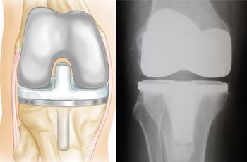
A knee prosthesis, also known as a knee replacement or artificial knee joint, is a medical device used to replace the damaged or diseased weight-bearing surfaces of the knee joint. It typically consists of three components: the femoral component (made of metal) that fits over the end of the thighbone (femur), the tibial component (often made of metal and plastic) that fits into the top of the shinbone (tibia), and a patellar component (usually made of plastic) that replaces the damaged surface of the kneecap.
The primary goal of knee prosthesis is to relieve pain, restore function, and improve quality of life for individuals with advanced knee joint damage due to conditions such as osteoarthritis, rheumatoid arthritis, or traumatic injuries. The procedure to implant a knee prosthesis is called knee replacement surgery or total knee arthroplasty (TKA).
A hip prosthesis, also known as a total hip replacement, is a surgical implant designed to replace the damaged or diseased components of the human hip joint. The procedure involves replacing the femoral head (the ball at the top of the thigh bone) and the acetabulum (the socket in the pelvis) with artificial parts, typically made from materials such as metal, ceramic, or plastic.
The goal of a hip prosthesis is to relieve pain, improve joint mobility, and restore function, allowing patients to return to their normal activities and enjoy an improved quality of life. The procedure is most commonly performed in individuals with advanced osteoarthritis, rheumatoid arthritis, or other degenerative conditions that have caused significant damage to the hip joint.
There are several different types of hip prostheses available, each with its own unique design and set of benefits and risks. The choice of prosthesis will depend on a variety of factors, including the patient's age, activity level, overall health, and specific medical needs. In general, however, all hip prostheses are designed to provide a durable, long-lasting solution for patients suffering from debilitating joint pain and stiffness.
A penile prosthesis is a medical device that is implanted inside the penis to treat erectile dysfunction. It consists of a pair of inflatable or semi-rigid rods, which are surgically placed into the corpora cavernosa (the two sponge-like areas inside the penis that fill with blood to create an erection). The implant allows the person with ED to have a controlled and manual erection suitable for sexual intercourse. This is usually considered as a last resort when other treatments, such as medications or vacuum devices, have failed.
A heart valve prosthesis is a medical device that is implanted in the heart to replace a damaged or malfunctioning heart valve. The prosthetic valve can be made of biological tissue (such as from a pig or cow) or artificial materials (such as carbon or polyester). Its function is to allow for the proper directional flow of blood through the heart, opening and closing with each heartbeat to prevent backflow of blood.
There are several types of heart valve prostheses, including:
1. Mechanical valves: These are made entirely of artificial materials and have a longer lifespan than biological valves. However, they require the patient to take blood-thinning medication for the rest of their life to prevent blood clots from forming on the valve.
2. Bioprosthetic valves: These are made of biological tissue and typically last 10-15 years before needing replacement. They do not require the patient to take blood-thinning medication, but there is a higher risk of reoperation due to degeneration of the tissue over time.
3. Homografts or allografts: These are human heart valves that have been donated and preserved for transplantation. They have similar longevity to bioprosthetic valves and do not require blood-thinning medication.
4. Autografts: In this case, the patient's own pulmonary valve is removed and used to replace the damaged aortic valve. This procedure is called the Ross procedure and has excellent long-term results, but it requires advanced surgical skills and is not widely available.
The choice of heart valve prosthesis depends on various factors, including the patient's age, overall health, lifestyle, and personal preferences.
Artificial limbs, also known as prosthetics, are artificial substitutes that replace a part or all of an absent extremity or limb. They are designed to restore the function, mobility, and appearance of the lost limb as much as possible. Artificial limbs can be made from various materials such as wood, plastic, metal, or carbon fiber, and they can be custom-made to fit the individual's specific needs and measurements.
Prosthetic limbs can be categorized into two main types: cosmetic and functional. Cosmetic prosthetics are designed to look like natural limbs and are primarily used to improve the appearance of the person. Functional prosthetics, on the other hand, are designed to help the individual perform specific tasks and activities. They may include features such as hooks, hands, or specialized feet that can be used for different purposes.
Advances in technology have led to the development of more sophisticated artificial limbs, including those that can be controlled by the user's nervous system, known as bionic prosthetics. These advanced prosthetic devices can provide a greater degree of mobility and control for the user, allowing them to perform complex movements and tasks with ease.
A visual prosthesis, also known as a retinal implant or bionic eye, is a medical device that aims to restore some functional vision in individuals who have severe visual impairment or blindness due to certain eye conditions such as retinitis pigmentosa or age-related macular degeneration.
The prosthesis works by electrically stimulating the remaining viable nerve cells in the retina, which then transmit the signals to the brain via the optic nerve. The device typically consists of a camera that captures visual information, a processor that converts the images into electrical signals, and an electrode array that is implanted onto the surface of the retina.
The electrical stimulation of the retinal cells creates patterns of light in the individual's visual field, allowing them to perceive shapes, edges, and movements. While the level of visual acuity achieved with current visual prostheses is still limited, they can significantly improve the quality of life for some individuals by enabling them to perform tasks such as recognizing objects, navigating their environment, and identifying facial expressions.
Prosthesis implantation is a surgical procedure where an artificial device or component, known as a prosthesis, is placed inside the body to replace a missing or damaged body part. The prosthesis can be made from various materials such as metal, plastic, or ceramic and is designed to perform the same function as the original body part.
The implantation procedure involves making an incision in the skin to create a pocket where the prosthesis will be placed. The prosthesis is then carefully positioned and secured in place using screws, cement, or other fixation methods. In some cases, tissue from the patient's own body may be used to help anchor the prosthesis.
Once the prosthesis is in place, the incision is closed with sutures or staples, and the area is bandaged. The patient will typically need to undergo rehabilitation and physical therapy to learn how to use the new prosthesis and regain mobility and strength.
Prosthesis implantation is commonly performed for a variety of reasons, including joint replacement due to arthritis or injury, dental implants to replace missing teeth, and breast reconstruction after mastectomy. The specific procedure and recovery time will depend on the type and location of the prosthesis being implanted.
Prosthesis fitting is the process of selecting, designing, fabricating, and fitting a prosthetic device to replace a part of an individual's body that is missing due to congenital absence, illness, injury, or amputation. The primary goal of prosthesis fitting is to restore the person's physical function, mobility, and independence, as well as improve their overall quality of life.
The process typically involves several steps:
1. Assessment: A thorough evaluation of the patient's medical history, physical condition, and functional needs is conducted to determine the most appropriate type of prosthesis. This may include measurements, castings, or digital scans of the residual limb.
2. Design: Based on the assessment, a customized design plan is created for the prosthetic device, taking into account factors such as the patient's lifestyle, occupation, and personal preferences.
3. Fabrication: The prosthesis is manufactured using various materials, components, and techniques to meet the specific requirements of the patient. This may involve the use of 3D printing, computer-aided design (CAD), or traditional handcrafting methods.
4. Fitting: Once the prosthesis is fabricated, it is carefully fitted to the patient's residual limb, ensuring optimal comfort, alignment, and stability. Adjustments may be made as needed to achieve the best fit and function.
5. Training: The patient receives training on how to use and care for their new prosthetic device, including exercises to strengthen the residual limb and improve overall mobility. Follow-up appointments are scheduled to monitor progress, make any necessary adjustments, and provide ongoing support.
A neural prosthesis is a type of medical device that is designed to assist or replace the function of impaired nervous system structures. These devices can be used to stimulate nerves and restore sensation, movement, or other functions that have been lost due to injury or disease. They may also be used to monitor neural activity and provide feedback to the user or to a external device.
Neural prostheses can take many forms, depending on the specific function they are intended to restore. For example, a cochlear implant is a type of neural prosthesis that is used to restore hearing in people with severe to profound hearing loss. The device consists of a microphone, a processor, and a array of electrodes that are implanted in the inner ear. Sound is converted into electrical signals by the microphone and processor, and these signals are then used to stimulate the remaining nerve cells in the inner ear, allowing the user to hear sounds.
Other examples of neural prostheses include deep brain stimulation devices, which are used to treat movement disorders such as Parkinson's disease; retinal implants, which are used to restore vision in people with certain types of blindness; and sacral nerve stimulators, which are used to treat urinary incontinence.
It is important to note that neural prostheses are not intended to cure or fully reverse the underlying condition that caused the impairment, but rather to help restore some level of function and improve the user's quality of life.
An ossicular prosthesis is a medical device used to replace one or more of the small bones (ossicles) in the middle ear that are involved in hearing. These bones, known as the malleus, incus, and stapes, form a chain responsible for transmitting sound vibrations from the eardrum to the inner ear.
An ossicular prosthesis is typically made of biocompatible materials such as ceramic, plastic, or metal. The prosthesis is designed to bypass damaged or missing ossicles and reestablish the connection between the eardrum and the inner ear, thereby improving hearing function. Ossicular prostheses are often used in surgeries aimed at reconstructing the middle ear, such as tympanoplasty or stapedectomy, to treat various types of conductive hearing loss.
A dental prosthesis is a device that replaces one or more missing teeth or parts of teeth to correct deficiencies in chewing ability, speech, and aesthetics. It can be removable or fixed (permanent) and can be made from various materials such as acrylic resin, porcelain, metal alloys, or a combination of these. Examples of dental prostheses include dentures, bridges, crowns, and implants.
A dental prosthesis that is supported by dental implants is an artificial replacement for one or more missing teeth. It is a type of dental restoration that is anchored to the jawbone using one or more titanium implant posts, which are surgically placed into the bone. The prosthesis is then attached to the implants, providing a stable and secure fit that closely mimics the function and appearance of natural teeth.
There are several types of implant-supported dental prostheses, including crowns, bridges, and dentures. A single crown may be used to replace a single missing tooth, while a bridge or denture can be used to replace multiple missing teeth. The specific type of prosthesis used will depend on the number and location of the missing teeth, as well as the patient's individual needs and preferences.
Implant-supported dental prostheses offer several advantages over traditional removable dentures, including improved stability, comfort, and functionality. They also help to preserve jawbone density and prevent facial sagging that can occur when teeth are missing. However, they do require a surgical procedure to place the implants, and may not be suitable for all patients due to factors such as bone density or overall health status.
Heart valve prosthesis implantation is a surgical procedure where an artificial heart valve is inserted to replace a damaged or malfunctioning native heart valve. This can be necessary for patients with valvular heart disease, including stenosis (narrowing) or regurgitation (leaking), who do not respond to medical management and are at risk of heart failure or other complications.
There are two main types of artificial heart valves used in prosthesis implantation: mechanical valves and biological valves. Mechanical valves are made of synthetic materials, such as carbon and metal, and can last a long time but require lifelong anticoagulation therapy to prevent blood clots from forming. Biological valves, on the other hand, are made from animal or human tissue and typically do not require anticoagulation therapy but may have a limited lifespan and may need to be replaced in the future.
The decision to undergo heart valve prosthesis implantation is based on several factors, including the patient's age, overall health, type and severity of valvular disease, and personal preferences. The procedure can be performed through traditional open-heart surgery or minimally invasive techniques, such as robotic-assisted surgery or transcatheter aortic valve replacement (TAVR). Recovery time varies depending on the approach used and individual patient factors.
Lymphatic vessels are thin-walled, valved structures that collect and transport lymph, a fluid derived from the interstitial fluid surrounding the cells, throughout the lymphatic system. They play a crucial role in immune function and maintaining fluid balance in the body. The primary function of lymphatic vessels is to return excess interstitial fluid, proteins, waste products, and immune cells to the bloodstream via the subclavian veins near the heart.
There are two types of lymphatic vessels:
1. Lymphatic capillaries: These are the smallest lymphatic vessels, found in most body tissues except for the central nervous system (CNS). They have blind ends and are highly permeable to allow the entry of interstitial fluid, proteins, and other large molecules.
2. Larger lymphatic vessels: These include precollecting vessels, collecting vessels, and lymphatic trunks. Precollecting vessels have valves that prevent backflow of lymph and merge to form larger collecting vessels. Collecting vessels contain smooth muscle in their walls, which helps to propel the lymph forward. They also have valves at regular intervals to ensure unidirectional flow towards the heart. Lymphatic trunks are large vessels that collect lymph from various regions of the body and eventually drain into the two main lymphatic ducts: the thoracic duct and the right lymphatic duct.
Overall, lymphatic vessels play a vital role in maintaining fluid balance, immune surveillance, and waste removal in the human body.
Retinal vessels refer to the blood vessels that are located in the retina, which is the light-sensitive tissue that lines the inner surface of the eye. The retina contains two types of blood vessels: arteries and veins.
The central retinal artery supplies oxygenated blood to the inner layers of the retina, while the central retinal vein drains deoxygenated blood from the retina. These vessels can be visualized during a routine eye examination using an ophthalmoscope, which allows healthcare professionals to assess their health and any potential abnormalities.
Retinal vessels are essential for maintaining the health and function of the retina, and any damage or changes to these vessels can affect vision and lead to various eye conditions such as diabetic retinopathy, retinal vein occlusion, and hypertensive retinopathy.
An amputee is a person who has had a limb or extremity removed by trauma, medical illness, or surgical intervention. Amputation may affect any part of the body, including fingers, toes, hands, feet, arms, and legs. The level of amputation can vary from partial loss to complete removal of the affected limb.
There are several reasons why a person might become an amputee:
- Trauma: Accidents, injuries, or violence can result in amputations due to severe tissue damage or irreparable vascular injury.
- Medical illness: Certain medical conditions such as diabetes, peripheral arterial disease, and cancer may require amputation if the affected limb cannot be saved through other treatments.
- Infection: Severe infections that do not respond to antibiotics or other treatments may necessitate amputation to prevent the spread of infection.
- Congenital defects: Some individuals are born with missing or malformed limbs, making them congenital amputees.
Amputees face various challenges, including physical limitations, emotional distress, and social adjustment. However, advancements in prosthetics and rehabilitation have significantly improved the quality of life for many amputees, enabling them to lead active and fulfilling lives.
A maxillofacial prosthesis is a custom-made device used to replace all or part of a facial feature, such as an eye, ear, nose, or lip, that has been lost due to trauma, cancer surgery, or other causes. It is typically made from materials like silicone, acrylic, or nylon and is designed to mimic the appearance and texture of natural skin and tissues.
Maxillofacial prostheses are created by trained professionals called maxillofacial prosthodontists, who have specialized training in the diagnosis, treatment planning, and rehabilitation of patients with facial defects. The process of creating a maxillofacial prosthesis typically involves taking an impression of the affected area, creating a custom-made mold, and then fabricating the prosthesis to fit precisely over the defect.
Maxillofacial prostheses can help improve patients' appearance, self-confidence, and quality of life by restoring their facial symmetry and functionality. They may also help protect the underlying tissues and structures from injury or infection, and can be used in conjunction with other treatments, such as radiation therapy or chemotherapy, to enhance their effectiveness.
An artificial larynx, also known as a voice prosthesis or speech aid, is a device used to help individuals who have undergone a laryngectomy (surgical removal of the larynx) or have other conditions that prevent them from speaking normally. The device generates sound mechanically, which can then be shaped into speech by the user.
There are two main types of artificial larynx devices:
1. External: This type of device consists of a small electronic unit that produces sound when the user presses a button or activates it with a breath. The sound is then directed through a tube or hose into a face mask or a mouthpiece, where the user can shape it into speech.
2. Internal: An internal artificial larynx, also known as a voice prosthesis, is implanted in the body during surgery. It works by allowing air to flow from the trachea into the esophagus and then through the voice prosthesis, which creates sound that can be used for speech.
Both types of artificial larynx devices require practice and training to use effectively, but they can significantly improve communication and quality of life for individuals who have lost their natural voice due to laryngeal cancer or other conditions.
Arthroplasty, replacement, is a surgical procedure where a damaged or diseased joint surface is removed and replaced with an artificial implant or device. The goal of this surgery is to relieve pain, restore function, and improve the quality of life for patients who have severe joint damage due to arthritis or other conditions.
During the procedure, the surgeon removes the damaged cartilage and bone from the joint and replaces them with a metal, plastic, or ceramic component that replicates the shape and function of the natural joint surface. The most common types of joint replacement surgery are hip replacement, knee replacement, and shoulder replacement.
The success rate of joint replacement surgery is generally high, with many patients experiencing significant pain relief and improved mobility. However, as with any surgical procedure, there are risks involved, including infection, blood clots, implant loosening or failure, and nerve damage. Therefore, it's essential to discuss the potential benefits and risks of joint replacement surgery with a healthcare provider before making a decision.
An artificial eye, also known as a prosthetic eye, is a type of medical device that is used to replace a natural eye that has been removed or is not functional due to injury, disease, or congenital abnormalities. It is typically made of acrylic or glass and is custom-made to match the size, shape, and color of the patient's other eye as closely as possible.
The artificial eye is designed to fit over the eye socket and rest on the eyelids, allowing the person to have a more natural appearance and improve their ability to blink and close their eye. It does not restore vision, but it can help protect the eye socket and improve the patient's self-esteem and quality of life.
The process of fitting an artificial eye typically involves several appointments with an ocularist, who is a healthcare professional trained in the measurement, design, and fabrication of prosthetic eyes. The ocularist will take impressions of the eye socket, create a model, and then use that model to make the artificial eye. Once the artificial eye is made, the ocularist will fit it and make any necessary adjustments to ensure that it is comfortable and looks natural.
Penile implantation, also known as a prosthetic penis or penile prosthesis, is a surgical procedure to place devices into the penis to help a person with erectile dysfunction (ED) achieve an erection. The two main types of penile implants are inflatable and semi-rigid rods.
The inflatable implant consists of a fluid-filled reservoir, a pump, and two or three inflatable cylinders in the penis. The semi-rigid rod implant is a pair of flexible rods that are bent into an erect position for sexual intercourse and can be straightened when not in use.
Penile implantation is typically considered as a last resort treatment option for ED, when other treatments such as medications, vacuum constriction devices, or penile injections have failed or are not suitable. The procedure is typically performed by a urologist under general or spinal anesthesia and requires a hospital stay of one to two days.
It's important to note that like any surgical procedure, penile implantation also has risks such as infection, bleeding, mechanical failure, and device malfunction. It is essential for patients to discuss the potential benefits and risks with their healthcare provider before making a decision about this treatment option.
Prosthesis-related infections, also known as prosthetic joint infections (PJIs), are infections that occur around or within a prosthetic device, such as an artificial joint. These infections can be caused by bacteria, fungi, or other microorganisms and can lead to serious complications if not treated promptly and effectively.
Prosthesis-related infections can occur soon after the implantation of the prosthetic device (early infection) or months or even years later (late infection). Early infections are often caused by bacteria that enter the surgical site during the procedure, while late infections may be caused by hematogenous seeding (i.e., when bacteria from another source spread through the bloodstream and settle in the prosthetic device) or by contamination during a subsequent medical procedure.
Symptoms of prosthesis-related infections can include pain, swelling, redness, warmth, and drainage around the affected area. In some cases, patients may also experience fever, chills, or fatigue. Diagnosis typically involves a combination of clinical evaluation, laboratory tests (such as blood cultures, joint fluid analysis, and tissue biopsy), and imaging studies (such as X-rays, CT scans, or MRI).
Treatment of prosthesis-related infections usually involves a combination of antibiotics and surgical intervention. The specific treatment approach will depend on the type and severity of the infection, as well as the patient's overall health status. In some cases, it may be necessary to remove or replace the affected prosthetic device.
"Prosthesis coloring" is not a recognized medical term or concept in the field of prosthetics. However, I can provide you with some context that might help clarify what you are looking for.
In the context of artificial limbs (prostheses), patients may want their devices to match their skin tone as closely as possible to make them less noticeable and more aesthetically appealing. This process is called "prosthetic covering" or "cosmesis," which involves applying custom-made covers, sleeves, or skins over the prosthesis to mimic the appearance of natural skin color and texture.
Prosthetic covering materials can be painted, printed, or dyed to achieve the desired color match. This process is often referred to as "coloring" or "painting the prosthesis." The coloring technique may involve using various shades, tones, and textures to create a natural-looking appearance that blends well with the user's remaining limb or body.
In summary, while there is no formal medical definition for "prosthesis coloring," it likely refers to the process of applying custom colors, shading, or patterns to an artificial limb (prosthesis) to create a more natural and aesthetically pleasing appearance that matches the user's skin tone.
Physiologic neovascularization is the natural and controlled formation of new blood vessels in the body, which occurs as a part of normal growth and development, as well as in response to tissue repair and wound healing. This process involves the activation of endothelial cells, which line the interior surface of blood vessels, and their migration, proliferation, and tube formation to create new capillaries. Physiologic neovascularization is tightly regulated by a balance of pro-angiogenic and anti-angiogenic factors, ensuring that it occurs only when and where it is needed. It plays crucial roles in various physiological processes, such as embryonic development, tissue regeneration, and wound healing.
A reoperation is a surgical procedure that is performed again on a patient who has already undergone a previous operation for the same or related condition. Reoperations may be required due to various reasons, such as inadequate initial treatment, disease recurrence, infection, or complications from the first surgery. The nature and complexity of a reoperation can vary widely depending on the specific circumstances, but it often carries higher risks and potential complications compared to the original operation.
Pathologic neovascularization is the abnormal growth of new blood vessels in previously avascular tissue or excessive growth within existing vasculature, which occurs as a result of hypoxia, inflammation, or angiogenic stimuli. These newly formed vessels are often disorganized, fragile, and lack proper vessel hierarchy, leading to impaired blood flow and increased vascular permeability. Pathologic neovascularization can be observed in various diseases such as cancer, diabetic retinopathy, age-related macular degeneration, and chronic inflammation. This process contributes to disease progression by promoting tumor growth, metastasis, and edema formation, ultimately leading to tissue damage and organ dysfunction.
A bioprosthesis is a type of medical implant that is made from biological materials, such as heart valves or tendons taken from animals (xenografts) or humans (allografts). These materials are processed and sterilized to be used in surgical procedures to replace damaged or diseased tissues in the body.
Bioprosthetic implants are often used in cardiac surgery, such as heart valve replacement, because they are less likely to cause an immune response than synthetic materials. However, they may have a limited lifespan due to calcification and degeneration of the biological tissue over time. Therefore, bioprosthetic implants may need to be replaced after several years.
Bioprostheses can also be used in other types of surgical procedures, such as ligament or tendon repair, where natural tissue is needed to restore function and mobility. These prostheses are designed to mimic the properties of native tissues and provide a more physiological solution than synthetic materials.
Coronary vessels refer to the network of blood vessels that supply oxygenated blood and nutrients to the heart muscle, also known as the myocardium. The two main coronary arteries are the left main coronary artery and the right coronary artery.
The left main coronary artery branches off into the left anterior descending artery (LAD) and the left circumflex artery (LCx). The LAD supplies blood to the front of the heart, while the LCx supplies blood to the side and back of the heart.
The right coronary artery supplies blood to the right lower part of the heart, including the right atrium and ventricle, as well as the back of the heart.
Coronary vessel disease (CVD) occurs when these vessels become narrowed or blocked due to the buildup of plaque, leading to reduced blood flow to the heart muscle. This can result in chest pain, shortness of breath, or a heart attack.
A dental prosthesis is a device that replaces missing teeth or parts of teeth and restores their function and appearance. The design of a dental prosthesis refers to the plan and specifications used to create it, including the materials, shape, size, and arrangement of the artificial teeth and any supporting structures.
The design of a dental prosthesis is typically based on a variety of factors, including:
* The number and location of missing teeth
* The condition of the remaining teeth and gums
* The patient's bite and jaw alignment
* The patient's aesthetic preferences
* The patient's ability to chew and speak properly
There are several types of dental prostheses, including:
* Dentures: A removable appliance that replaces all or most of the upper or lower teeth.
* Fixed partial denture (FPD): Also known as a bridge, this is a fixed (non-removable) appliance that replaces one or more missing teeth by attaching artificial teeth to the remaining natural teeth on either side of the gap.
* Removable partial denture (RPD): A removable appliance that replaces some but not all of the upper or lower teeth.
* Implant-supported prosthesis: An artificial tooth or set of teeth that is supported by dental implants, which are surgically placed in the jawbone.
The design of a dental prosthesis must be carefully planned and executed to ensure a good fit, proper function, and natural appearance. It may involve several appointments with a dentist or dental specialist, such as a prosthodontist, to take impressions, make measurements, and try in the finished prosthesis.
The endothelium is a thin layer of simple squamous epithelial cells that lines the interior surface of blood vessels, lymphatic vessels, and heart chambers. The vascular endothelium, specifically, refers to the endothelial cells that line the blood vessels. These cells play a crucial role in maintaining vascular homeostasis by regulating vasomotor tone, coagulation, platelet activation, inflammation, and permeability of the vessel wall. They also contribute to the growth and repair of the vascular system and are involved in various pathological processes such as atherosclerosis, hypertension, and diabetes.
Silicone elastomers are a type of synthetic rubber made from silicone, which is a polymer composed primarily of silicon-oxygen bonds. They are known for their durability, flexibility, and resistance to heat, cold, and moisture. Silicone elastomers can be manufactured in various forms, including liquids, gels, and solids, and they are used in a wide range of medical applications such as:
1. Breast implants: Silicone elastomer shells filled with silicone gel are commonly used for breast augmentation and reconstruction.
2. Contact lenses: Some contact lenses are made from silicone elastomers due to their high oxygen permeability, which allows for better eye health.
3. Catheters: Silicone elastomer catheters are flexible and resistant to kinking, making them suitable for long-term use in various medical procedures.
4. Implantable drug delivery systems: Silicone elastomers can be used as a matrix for controlled release of drugs, allowing for sustained and targeted medication administration.
5. Medical adhesives: Silicone elastomer adhesives are biocompatible and can be used to attach medical devices to the skin or other tissues.
6. Sealants and coatings: Silicone elastomers can be used as sealants and coatings in medical devices to prevent leakage, improve durability, and reduce infection risk.
It is important to note that while silicone elastomers are generally considered safe for medical use, there have been concerns about the potential health risks associated with breast implants, such as capsular contracture, breast pain, and immune system reactions. However, these risks vary depending on the individual's health status and the specific type of silicone elastomer used.
Bone cements are medical-grade materials used in orthopedic and trauma surgery to fill gaps between bone surfaces and implants, such as artificial joints or screws. They serve to mechanically stabilize the implant and provide a smooth, load-bearing surface. The two most common types of bone cement are:
1. Polymethylmethacrylate (PMMA) cement: This is a two-component system consisting of powdered PMMA and liquid methyl methacrylate monomer. When mixed together, they form a dough-like consistency that hardens upon exposure to air. PMMA cement has been widely used for decades in joint replacement surgeries, such as hip or knee replacements.
2. Calcium phosphate (CP) cement: This is a two-component system consisting of a powdered CP compound and an aqueous solution. When mixed together, they form a paste that hardens through a chemical reaction at body temperature. CP cement has lower mechanical strength compared to PMMA but demonstrates better biocompatibility, bioactivity, and the ability to resorb over time.
Both types of bone cements have advantages and disadvantages, and their use depends on the specific surgical indication and patient factors.
I'm sorry for any confusion, but "Polyethylene Terephthalates" is not a medical term. It is a type of polymer used in the manufacturing of various products, such as plastic bottles and textile fibers. Medically, you might encounter the abbreviation "PET" or "PET scan," which stands for "Positron Emission Tomography." A PET scan is a type of medical imaging that provides detailed pictures of the body's interior. If you have any medical terms you would like defined, I'd be happy to help!
Hip arthroplasty, also known as hip replacement surgery, is a medical procedure where the damaged or diseased joint surfaces of the hip are removed and replaced with artificial components. These components typically include a metal or ceramic ball that replaces the head of the femur (thigh bone), and a polyethylene or ceramic socket that replaces the acetabulum (hip socket) in the pelvis.
The goal of hip arthroplasty is to relieve pain, improve joint mobility, and restore function to the hip joint. This procedure is commonly performed in patients with advanced osteoarthritis, rheumatoid arthritis, hip fractures, or other conditions that cause significant damage to the hip joint.
There are several types of hip replacement surgeries, including traditional total hip arthroplasty, partial (hemi) hip arthroplasty, and resurfacing hip arthroplasty. The choice of procedure depends on various factors, such as the patient's age, activity level, overall health, and the extent of joint damage.
After surgery, patients typically require rehabilitation to regain strength, mobility, and function in the affected hip. With proper care and follow-up, most patients can expect significant pain relief and improved quality of life following hip arthroplasty.
Prosthesis retention, in the context of medical prosthetics, refers to the secure and stable attachment or fixation of a prosthetic device to the body or the remaining limb (stump) of an amputee. The primary goal of prosthesis retention is to ensure that the artificial limb remains in place during various activities, providing optimal functionality, comfort, and safety for the user.
There are several methods for achieving prosthesis retention, including:
1. Suction sockets: A custom-made socket that creates a seal around the residual limb using a special liner and air pressure to keep the prosthesis in place.
2. Mechanical locks: Devices such as pin locks, lanyard locks, or magnetic couplings that secure the prosthetic limb to the residual limb by engaging with specific components within the socket.
3. Vacuum-assisted suspension: A system that uses vacuum pressure to create a seal between the residual limb and the socket, providing retention and stability.
4. Belt or harness systems: Straps or bands that attach to the prosthesis and wrap around the user's body or sound limb to keep the device in place.
5. Osseointegration: A surgical procedure that involves implanting a metal rod directly into the bone, allowing for a direct connection between the residual limb and the prosthetic device.
Prosthesis retention is crucial for ensuring the successful use of an artificial limb, as it enables users to perform their daily activities with confidence and ease.
The aortic valve is the valve located between the left ventricle (the lower left chamber of the heart) and the aorta (the largest artery in the body, which carries oxygenated blood from the heart to the rest of the body). It is made up of three thin flaps or leaflets that open and close to regulate blood flow. During a heartbeat, the aortic valve opens to allow blood to be pumped out of the left ventricle into the aorta, and then closes to prevent blood from flowing back into the ventricle when it relaxes. Any abnormality or damage to this valve can lead to various cardiovascular conditions such as aortic stenosis, aortic regurgitation, or infective endocarditis.
Dental prosthesis retention refers to the means by which a dental prosthesis, such as a denture, is held in place in the mouth. The retention can be achieved through several methods, including:
1. Suction: This is the most common method of retention for lower dentures, where the shape and fit of the denture base create suction against the gums to hold it in place.
2. Mechanical retention: This involves the use of mechanical components such as clasps or attachments that hook onto remaining natural teeth or dental implants to hold the prosthesis in place.
3. Adhesive retention: Dental adhesives can be used to help secure the denture to the gums, providing additional retention and stability.
4. Implant retention: Dental implants can be used to provide a more secure and stable retention of the dental prosthesis. The implant is surgically placed in the jawbone and acts as an anchor for the prosthesis.
Proper retention of a dental prosthesis is essential for optimal function, comfort, and speech. A well-retained prosthesis can help prevent sore spots, improve chewing efficiency, and enhance overall quality of life.
Arthroplasty, replacement, knee is a surgical procedure where the damaged or diseased joint surface of the knee is removed and replaced with an artificial joint or prosthesis. The procedure involves resurfacing the worn-out ends of the femur (thigh bone) and tibia (shin bone) with metal components, and the back of the kneecap with a plastic button. This surgery is usually performed to relieve pain and restore function in patients with severe knee osteoarthritis, rheumatoid arthritis, or traumatic injuries that have damaged the joint beyond repair. The goal of knee replacement surgery is to improve mobility, reduce pain, and enhance the quality of life for the patient.
Treatment outcome is a term used to describe the result or effect of medical treatment on a patient's health status. It can be measured in various ways, such as through symptoms improvement, disease remission, reduced disability, improved quality of life, or survival rates. The treatment outcome helps healthcare providers evaluate the effectiveness of a particular treatment plan and make informed decisions about future care. It is also used in clinical research to compare the efficacy of different treatments and improve patient care.
In the medical field, cementation refers to the process of using a type of dental cement or bonding agent to attach a dental restoration (such as a crown, bridge, or false tooth) to a natural tooth or implant. The cement helps to create a strong and secure attachment, while also helping to seal the restoration and prevent the entry of bacteria and saliva.
Dental cement can be made from various materials, including glass ionomers, resin-modified glass ionomers, zinc phosphate, and polycarboxylate cements. The choice of cement depends on several factors, such as the type of restoration being attached, the location in the mouth, and the patient's individual needs and preferences.
Cementation is an important step in many dental procedures, as it helps to ensure the longevity and success of the restoration. Proper technique and material selection are crucial for achieving a successful cementation that will last for years to come.
Endothelial cells are the type of cells that line the inner surface of blood vessels, lymphatic vessels, and heart chambers. They play a crucial role in maintaining vascular homeostasis by controlling vasomotor tone, coagulation, platelet activation, and inflammation. Endothelial cells also regulate the transport of molecules between the blood and surrounding tissues, and contribute to the maintenance of the structural integrity of the vasculature. They are flat, elongated cells with a unique morphology that allows them to form a continuous, nonthrombogenic lining inside the vessels. Endothelial cells can be isolated from various tissues and cultured in vitro for research purposes.
Amputation stumps, also known as residual limbs, refer to the remaining part of a limb after it has been amputated. The stump includes the soft tissue and bone that were once part of the amputated limb. Proper care and management of the amputation stump are essential for optimal healing, reducing the risk of complications such as infection or delayed wound healing, and promoting successful prosthetic fitting and use. This may involve various treatments such as wound care, pain management, physical therapy, and the use of specialized medical devices.
A partial denture that is fixed, also known as a fixed partial denture or a dental bridge, is a type of prosthetic device used to replace one or more missing teeth. Unlike removable partial dentures, which can be taken out of the mouth for cleaning and maintenance, fixed partial dentures are permanently attached to the remaining natural teeth or implants surrounding the gap left by the missing tooth or teeth.
A typical fixed partial denture consists of an artificial tooth (or pontic) that is fused to one or two crowns on either side. The crowns are cemented onto the prepared surfaces of the adjacent teeth, providing a stable and secure attachment for the pontic. This creates a natural-looking and functional replacement for the missing tooth or teeth.
Fixed partial dentures offer several advantages over removable options, including improved stability, comfort, and aesthetics. However, they typically require more extensive preparation of the adjacent teeth, which may involve removing some healthy tooth structure to accommodate the crowns. Proper oral hygiene is essential to maintain the health of the supporting teeth and gums, as well as the longevity of the fixed partial denture. Regular dental check-ups and professional cleanings are also necessary to ensure the continued success of this type of restoration.
Maxillofacial prosthesis implantation is a medical procedure that involves the surgical placement of osseointegrated implants (fixtures that are integrated into the bone) to support and retain a custom-made maxillofacial prosthesis. This type of prosthesis is designed to replace all or part of the facial structures, such as the eyes, nose, ears, or jaw, which may be missing due to congenital defects, trauma, or cancer resection.
The implantation procedure typically involves several steps:
1. Pre-surgical planning: This includes taking detailed measurements and creating a custom-made surgical guide based on the patient's anatomy.
2. Surgical placement of implants: The surgeon uses the surgical guide to place the implants in the bone at precise locations and angles.
3. Healing period: After the surgery, the implants are allowed to heal and integrate with the bone for several months.
4. Prosthesis fabrication: Once the implants have integrated, an impression is taken of the implant abutments (the parts that protrude through the gums) and a custom-made prosthesis is created.
5. Delivery of the prosthesis: The prosthesis is attached to the implant abutments using screws or other attachments.
Maxillofacial prosthesis implantation can significantly improve the patient's quality of life by restoring facial function, appearance, and speech. However, it requires careful planning, surgical skill, and close collaboration between the surgeon, prosthodontist, and patient.
Articular Range of Motion (AROM) is a term used in physiotherapy and orthopedics to describe the amount of movement available in a joint, measured in degrees of a circle. It refers to the range through which synovial joints can actively move without causing pain or injury. AROM is assessed by measuring the degree of motion achieved by active muscle contraction, as opposed to passive range of motion (PROM), where the movement is generated by an external force.
Assessment of AROM is important in evaluating a patient's functional ability and progress, planning treatment interventions, and determining return to normal activities or sports participation. It is also used to identify any restrictions in joint mobility that may be due to injury, disease, or surgery, and to monitor the effectiveness of rehabilitation programs.
Chromium alloys are materials made by combining chromium with other metals, such as nickel, cobalt, or iron. The addition of chromium to these alloys enhances their properties, making them resistant to corrosion and high temperatures. These alloys have a wide range of applications in various industries, including automotive, aerospace, and medical devices.
Chromium alloys can be classified into two main categories: stainless steels and superalloys. Stainless steels are alloys that contain at least 10.5% chromium by weight, which forms a passive oxide layer on the surface of the material, protecting it from corrosion. Superalloys, on the other hand, are high-performance alloys designed to operate in extreme environments, such as jet engines and gas turbines. They contain significant amounts of chromium, along with other elements like nickel, cobalt, and molybdenum.
Chromium alloys have several medical applications due to their excellent properties. For instance, they are used in surgical instruments, dental implants, and orthopedic devices because of their resistance to corrosion and biocompatibility. Additionally, some chromium alloys exhibit superelasticity, a property that allows them to return to their original shape after being deformed, making them suitable for use in stents and other medical devices that require flexibility and durability.
A palatal obturator is a type of dental prosthesis that is used to close or block a hole or opening in the roof of the mouth, also known as the hard palate. This condition can occur due to various reasons such as cleft palate, cancer, trauma, or surgery. The obturator is designed to fit securely in the patient's mouth and restore normal speech, swallowing, and chewing functions.
The palatal obturator typically consists of a custom-made plate made of acrylic resin or other materials that are compatible with the oral tissues. The plate has an extension that fills the opening in the palate and creates a barrier between the oral and nasal cavities. This helps to prevent food and liquids from entering the nasal cavity during eating and speaking, which can cause discomfort, irritation, and infection.
Palatal obturators may be temporary or permanent, depending on the patient's needs and condition. They are usually fabricated based on an impression of the patient's mouth and fitted by a dental professional to ensure proper function and comfort. Proper care and maintenance of the obturator, including regular cleaning and adjustments, are essential to maintain its effectiveness and prevent complications.
Vascular Endothelial Growth Factor A (VEGFA) is a specific isoform of the vascular endothelial growth factor (VEGF) family. It is a well-characterized signaling protein that plays a crucial role in angiogenesis, the process of new blood vessel formation from pre-existing vessels. VEGFA stimulates the proliferation and migration of endothelial cells, which line the interior surface of blood vessels, thereby contributing to the growth and development of new vasculature. This protein is essential for physiological processes such as embryonic development and wound healing, but it has also been implicated in various pathological conditions, including cancer, age-related macular degeneration, and diabetic retinopathy. The regulation of VEGFA expression and activity is critical to maintaining proper vascular function and homeostasis.
In the field of medicine, "time factors" refer to the duration of symptoms or time elapsed since the onset of a medical condition, which can have significant implications for diagnosis and treatment. Understanding time factors is crucial in determining the progression of a disease, evaluating the effectiveness of treatments, and making critical decisions regarding patient care.
For example, in stroke management, "time is brain," meaning that rapid intervention within a specific time frame (usually within 4.5 hours) is essential to administering tissue plasminogen activator (tPA), a clot-busting drug that can minimize brain damage and improve patient outcomes. Similarly, in trauma care, the "golden hour" concept emphasizes the importance of providing definitive care within the first 60 minutes after injury to increase survival rates and reduce morbidity.
Time factors also play a role in monitoring the progression of chronic conditions like diabetes or heart disease, where regular follow-ups and assessments help determine appropriate treatment adjustments and prevent complications. In infectious diseases, time factors are crucial for initiating antibiotic therapy and identifying potential outbreaks to control their spread.
Overall, "time factors" encompass the significance of recognizing and acting promptly in various medical scenarios to optimize patient outcomes and provide effective care.
Postoperative complications refer to any unfavorable condition or event that occurs during the recovery period after a surgical procedure. These complications can vary in severity and may include, but are not limited to:
1. Infection: This can occur at the site of the incision or inside the body, such as pneumonia or urinary tract infection.
2. Bleeding: Excessive bleeding (hemorrhage) can lead to a drop in blood pressure and may require further surgical intervention.
3. Blood clots: These can form in the deep veins of the legs (deep vein thrombosis) and can potentially travel to the lungs (pulmonary embolism).
4. Wound dehiscence: This is when the surgical wound opens up, which can lead to infection and further complications.
5. Pulmonary issues: These include atelectasis (collapsed lung), pneumonia, or respiratory failure.
6. Cardiovascular problems: These include abnormal heart rhythms (arrhythmias), heart attack, or stroke.
7. Renal failure: This can occur due to various reasons such as dehydration, blood loss, or the use of certain medications.
8. Pain management issues: Inadequate pain control can lead to increased stress, anxiety, and decreased mobility.
9. Nausea and vomiting: These can be caused by anesthesia, opioid pain medication, or other factors.
10. Delirium: This is a state of confusion and disorientation that can occur in the elderly or those with certain medical conditions.
Prompt identification and management of these complications are crucial to ensure the best possible outcome for the patient.
Arteries are blood vessels that carry oxygenated blood away from the heart to the rest of the body. They have thick, muscular walls that can withstand the high pressure of blood being pumped out of the heart. Arteries branch off into smaller vessels called arterioles, which further divide into a vast network of tiny capillaries where the exchange of oxygen, nutrients, and waste occurs between the blood and the body's cells. After passing through the capillary network, deoxygenated blood collects in venules, then merges into veins, which return the blood back to the heart.
Follow-up studies are a type of longitudinal research that involve repeated observations or measurements of the same variables over a period of time, in order to understand their long-term effects or outcomes. In medical context, follow-up studies are often used to evaluate the safety and efficacy of medical treatments, interventions, or procedures.
In a typical follow-up study, a group of individuals (called a cohort) who have received a particular treatment or intervention are identified and then followed over time through periodic assessments or data collection. The data collected may include information on clinical outcomes, adverse events, changes in symptoms or functional status, and other relevant measures.
The results of follow-up studies can provide important insights into the long-term benefits and risks of medical interventions, as well as help to identify factors that may influence treatment effectiveness or patient outcomes. However, it is important to note that follow-up studies can be subject to various biases and limitations, such as loss to follow-up, recall bias, and changes in clinical practice over time, which must be carefully considered when interpreting the results.
Capillaries are the smallest blood vessels in the body, with diameters that range from 5 to 10 micrometers. They form a network of tiny tubes that connect the arterioles (small branches of arteries) and venules (small branches of veins), allowing for the exchange of oxygen, carbon dioxide, nutrients, and waste products between the blood and the surrounding tissues.
Capillaries are composed of a single layer of endothelial cells that surround a hollow lumen through which blood flows. The walls of capillaries are extremely thin, allowing for easy diffusion of molecules between the blood and the surrounding tissue. This is essential for maintaining the health and function of all body tissues.
Capillaries can be classified into three types based on their structure and function: continuous, fenestrated, and sinusoidal. Continuous capillaries have a continuous layer of endothelial cells with tight junctions that restrict the passage of large molecules. Fenestrated capillaries have small pores or "fenestrae" in the endothelial cell walls that allow for the passage of larger molecules, such as proteins and lipids. Sinusoidal capillaries are found in organs with high metabolic activity, such as the liver and spleen, and have large, irregular spaces between the endothelial cells that allow for the exchange of even larger molecules.
Overall, capillaries play a critical role in maintaining the health and function of all body tissues by allowing for the exchange of nutrients, oxygen, and waste products between the blood and surrounding tissues.
Amputation is defined as the surgical removal of all or part of a limb or extremity such as an arm, leg, foot, hand, toe, or finger. This procedure is typically performed to remove damaged or dead tissue due to various reasons like severe injury, infection, tumors, or chronic conditions that impair circulation, such as diabetes or peripheral arterial disease. The goal of amputation is to alleviate pain, prevent further complications, and improve the patient's quality of life. Following the surgery, patients may require rehabilitation and prosthetic devices to help them adapt to their new physical condition.
Immunohistochemistry (IHC) is a technique used in pathology and laboratory medicine to identify specific proteins or antigens in tissue sections. It combines the principles of immunology and histology to detect the presence and location of these target molecules within cells and tissues. This technique utilizes antibodies that are specific to the protein or antigen of interest, which are then tagged with a detection system such as a chromogen or fluorophore. The stained tissue sections can be examined under a microscope, allowing for the visualization and analysis of the distribution and expression patterns of the target molecule in the context of the tissue architecture. Immunohistochemistry is widely used in diagnostic pathology to help identify various diseases, including cancer, infectious diseases, and immune-mediated disorders.
The hip joint, also known as the coxal joint, is a ball-and-socket type synovial joint that connects the femur (thigh bone) to the pelvis. The "ball" is the head of the femur, while the "socket" is the acetabulum, a concave surface on the pelvic bone.
The hip joint is surrounded by a strong fibrous capsule and is reinforced by several ligaments, including the iliofemoral, ischiofemoral, and pubofemoral ligaments. The joint allows for flexion, extension, abduction, adduction, medial and lateral rotation, and circumduction movements, making it one of the most mobile joints in the body.
The hip joint is also supported by various muscles, including the gluteus maximus, gluteus medius, gluteus minimus, iliopsoas, and other hip flexors and extensors. These muscles provide stability and strength to the joint, allowing for weight-bearing activities such as walking, running, and jumping.
Osseointegration is a direct structural and functional connection between living bone and the surface of an implant. It's a process where the bone grows in and around the implant, which is typically made of titanium or another biocompatible material. This process provides a solid foundation for dental prosthetics, such as crowns, bridges, or dentures, or for orthopedic devices like artificial limbs. The success of osseointegration depends on various factors, including the patient's overall health, the quality and quantity of available bone, and the surgical technique used for implant placement.
Pericytes are specialized cells that surround the endothelial cells which line the blood capillaries. They play an important role in the regulation of capillary diameter, blood flow, and the formation of new blood vessels (angiogenesis). Pericytes also contribute to the maintenance of the blood-brain barrier, immune surveillance, and the clearance of waste products from the brain. They are often referred to as "mural cells" or "rouleaux cells" and can be found in various tissues throughout the body.
I believe there may be some confusion in your question as Polyethylenes are not a medical term, but rather a category of synthetic polymers commonly used in various industrial and medical applications. Here's a brief overview:
Polyethylene (PE) is a type of thermoplastic polymer made from the monomer ethylene. It is a versatile material with numerous applications due to its chemical resistance, durability, and flexibility. There are several types of polyethylenes, including:
1. Low-density polyethylene (LDPE): This type has a lower density and more branching in its molecular structure, which results in less crystallinity. LDPE is known for its flexibility and is often used in packaging films, bags, and containers.
2. High-density polyethylene (HDPE): HDPE has a higher density and less branching, resulting in greater crystallinity. It is more rigid than LDPE and is commonly used in applications such as bottles, pipes, and containers.
3. Linear low-density polyethylene (LLDPE): This type combines the flexibility of LDPE with some of the strength and rigidity of HDPE. LLDPE has fewer branches than LDPE but more than HDPE. It is often used in film applications, such as stretch wrap and agricultural films.
4. Ultra-high molecular weight polyethylene (UHMWPE): UHMWPE has an extremely high molecular weight, resulting in exceptional wear resistance, impact strength, and chemical resistance. It is commonly used in medical applications, such as orthopedic implants and joint replacements, due to its biocompatibility and low friction coefficient.
While polyethylenes are not a medical term per se, they do have significant medical applications, particularly UHMWPE in orthopedic devices.
Arthroplasty is a surgical procedure to restore the integrity and function of a joint. The term is derived from two Greek words: "arthro" meaning joint, and "plasty" meaning to mold or form. There are several types of arthroplasty, but most involve resurfacing the damaged joint cartilage with artificial materials such as metal, plastic, or ceramic.
The goal of arthroplasty is to relieve pain, improve mobility, and restore function in a joint that has been damaged by arthritis, injury, or other conditions. The most common types of arthroplasty are total joint replacement (TJR) and partial joint replacement (PJR).
In TJR, the surgeon removes the damaged ends of the bones in the joint and replaces them with artificial components called prostheses. These prostheses can be made of metal, plastic, or ceramic materials, and are designed to mimic the natural movement and function of the joint.
In PJR, only one side of the joint is resurfaced, typically because the damage is less extensive. This procedure is less invasive than TJR and may be recommended for younger patients who are still active or have a higher risk of complications from a full joint replacement.
Other types of arthroplasty include osteotomy, in which the surgeon cuts and reshapes the bone to realign the joint; arthrodesis, in which the surgeon fuses two bones together to create a stable joint; and resurfacing, in which the damaged cartilage is removed and replaced with a smooth, artificial surface.
Arthroplasty is typically recommended for patients who have tried other treatments, such as physical therapy, medication, or injections, but have not found relief from their symptoms. While arthroplasty can be highly effective in relieving pain and improving mobility, it is not without risks, including infection, blood clots, and implant failure. Patients should discuss the benefits and risks of arthroplasty with their healthcare provider to determine if it is the right treatment option for them.
The femur is the medical term for the thigh bone, which is the longest and strongest bone in the human body. It connects the hip bone to the knee joint and plays a crucial role in supporting the weight of the body and allowing movement during activities such as walking, running, and jumping. The femur is composed of a rounded head, a long shaft, and two condyles at the lower end that articulate with the tibia and patella to form the knee joint.
Femoral neoplasms refer to abnormal growths or tumors that develop in the femur, which is the long thigh bone in the human body. These neoplasms can be benign (non-cancerous) or malignant (cancerous). Benign femoral neoplasms are slow-growing and rarely spread to other parts of the body, while malignant neoplasms are aggressive and can invade nearby tissues and organs, as well as metastasize (spread) to distant sites.
There are various types of femoral neoplasms, including osteochondromas, enchondromas, chondrosarcomas, osteosarcomas, and Ewing sarcomas, among others. The specific type of neoplasm is determined by the cell type from which it arises and its behavior.
Symptoms of femoral neoplasms may include pain, swelling, stiffness, or weakness in the thigh, as well as a palpable mass or limited mobility. Diagnosis typically involves imaging studies such as X-rays, CT scans, or MRI, as well as biopsy to determine the type and grade of the tumor. Treatment options may include surgery, radiation therapy, chemotherapy, or a combination of these approaches, depending on the type, size, location, and stage of the neoplasm.
Aortic valve stenosis is a cardiac condition characterized by the narrowing or stiffening of the aortic valve, which separates the left ventricle (the heart's main pumping chamber) from the aorta (the large artery that carries oxygen-rich blood to the rest of the body). This narrowing or stiffening prevents the aortic valve from opening fully, resulting in reduced blood flow from the left ventricle to the aorta and the rest of the body.
The narrowing can be caused by several factors, including congenital heart defects, calcification (hardening) of the aortic valve due to aging, or scarring of the valve due to rheumatic fever or other inflammatory conditions. As a result, the left ventricle must work harder to pump blood through the narrowed valve, which can lead to thickening and enlargement of the left ventricular muscle (left ventricular hypertrophy).
Symptoms of aortic valve stenosis may include chest pain or tightness, shortness of breath, fatigue, dizziness or fainting, and heart palpitations. Severe aortic valve stenosis can lead to serious complications such as heart failure, arrhythmias, or even sudden cardiac death. Treatment options may include medications to manage symptoms, lifestyle changes, or surgical intervention such as aortic valve replacement.
Stapes surgery, also known as stapedectomy or stapedotomy, is a surgical procedure performed to correct hearing loss caused by otosclerosis. Otosclerosis is a condition in which the stapes bone in the middle ear becomes fixed and unable to vibrate properly, leading to conductive hearing loss.
During stapes surgery, the surgeon makes an incision behind the ear and creates a small opening in the eardrum. The fixed stapes bone is then removed or modified, and a prosthetic device is inserted in its place to allow sound vibrations to be transmitted to the inner ear. In some cases, a piece of tissue or artificial material may be used to fill the space left by the removed bone.
Stapedectomy involves complete removal of the stapes bone, while stapedotomy involves making a small hole in the stapes bone and inserting a prosthetic device through it. Both procedures are typically performed on an outpatient basis and have a high success rate in restoring hearing. However, as with any surgical procedure, there are risks involved, including infection, permanent hearing loss, and balance problems.
A smooth muscle within the vascular system refers to the involuntary, innervated muscle that is found in the walls of blood vessels. These muscles are responsible for controlling the diameter of the blood vessels, which in turn regulates blood flow and blood pressure. They are called "smooth" muscles because their individual muscle cells do not have the striations, or cross-striped patterns, that are observed in skeletal and cardiac muscle cells. Smooth muscle in the vascular system is controlled by the autonomic nervous system and by hormones, and can contract or relax slowly over a period of time.
The mitral valve, also known as the bicuspid valve, is a two-leaflet valve located between the left atrium and left ventricle in the heart. Its function is to ensure unidirectional flow of blood from the left atrium into the left ventricle during the cardiac cycle. The mitral valve consists of two leaflets (anterior and posterior), the chordae tendineae, papillary muscles, and the left atrial and ventricular myocardium. Dysfunction of the mitral valve can lead to various heart conditions such as mitral regurgitation or mitral stenosis.
Biomechanics is the application of mechanical laws to living structures and systems, particularly in the field of medicine and healthcare. A biomechanical phenomenon refers to a observable event or occurrence that involves the interaction of biological tissues or systems with mechanical forces. These phenomena can be studied at various levels, from the molecular and cellular level to the tissue, organ, and whole-body level.
Examples of biomechanical phenomena include:
1. The way that bones and muscles work together to produce movement (known as joint kinematics).
2. The mechanical behavior of biological tissues such as bone, cartilage, tendons, and ligaments under various loads and stresses.
3. The response of cells and tissues to mechanical stimuli, such as the way that bone tissue adapts to changes in loading conditions (known as Wolff's law).
4. The biomechanics of injury and disease processes, such as the mechanisms of joint injury or the development of osteoarthritis.
5. The use of mechanical devices and interventions to treat medical conditions, such as orthopedic implants or assistive devices for mobility impairments.
Understanding biomechanical phenomena is essential for developing effective treatments and prevention strategies for a wide range of medical conditions, from musculoskeletal injuries to neurological disorders.
CD31 (also known as PECAM-1 or Platelet Endothelial Cell Adhesion Molecule-1) is a type of protein that is found on the surface of certain cells in the body, including platelets, endothelial cells (which line the blood vessels), and some immune cells.
CD31 functions as a cell adhesion molecule, meaning it helps cells stick together and interact with each other. It plays important roles in various physiological processes, such as the regulation of leukocyte migration, angiogenesis (the formation of new blood vessels), hemostasis (the process that stops bleeding), and thrombosis (the formation of a blood clot inside a blood vessel).
As an antigen, CD31 is used in immunological techniques to identify and characterize cells expressing this protein. Antigens are substances that can be recognized by the immune system and stimulate an immune response. In the case of CD31, antibodies specific to this protein can be used to detect its presence on the surface of cells, providing valuable information for research and diagnostic purposes.
Equipment Failure Analysis is a process of identifying the cause of failure in medical equipment or devices. This involves a systematic examination and evaluation of the equipment, its components, and operational history to determine why it failed. The analysis may include physical inspection, chemical testing, and review of maintenance records, as well as assessment of design, manufacturing, and usage factors that may have contributed to the failure.
The goal of Equipment Failure Analysis is to identify the root cause of the failure, so that corrective actions can be taken to prevent similar failures in the future. This is important in medical settings to ensure patient safety and maintain the reliability and effectiveness of medical equipment.
The knee joint, also known as the tibiofemoral joint, is the largest and one of the most complex joints in the human body. It is a synovial joint that connects the thighbone (femur) to the shinbone (tibia). The patella (kneecap), which is a sesamoid bone, is located in front of the knee joint and helps in the extension of the leg.
The knee joint is made up of three articulations: the femorotibial joint between the femur and tibia, the femoropatellar joint between the femur and patella, and the tibiofibular joint between the tibia and fibula. These articulations are surrounded by a fibrous capsule that encloses the synovial membrane, which secretes synovial fluid to lubricate the joint.
The knee joint is stabilized by several ligaments, including the medial and lateral collateral ligaments, which provide stability to the sides of the joint, and the anterior and posterior cruciate ligaments, which prevent excessive forward and backward movement of the tibia relative to the femur. The menisci, which are C-shaped fibrocartilaginous structures located between the femoral condyles and tibial plateaus, also help to stabilize the joint by absorbing shock and distributing weight evenly across the articular surfaces.
The knee joint allows for flexion, extension, and a small amount of rotation, making it essential for activities such as walking, running, jumping, and sitting.
"Device Removal" in a medical context generally refers to the surgical or nonsurgical removal of a medical device that has been previously implanted in a patient's body. The purpose of removing the device may vary, depending on the individual case. Some common reasons for device removal include infection, malfunction, rejection, or when the device is no longer needed.
Examples of medical devices that may require removal include pacemakers, implantable cardioverter-defibrillators (ICDs), artificial joints, orthopedic hardware, breast implants, cochlear implants, and intrauterine devices (IUDs). The procedure for device removal will depend on the type of device, its location in the body, and the reason for its removal.
It is important to note that device removal carries certain risks, such as bleeding, infection, damage to surrounding tissues, or complications related to anesthesia. Therefore, the decision to remove a medical device should be made carefully, considering both the potential benefits and risks of the procedure.
'Alloys' is not a medical term. It is a term used in materials science and engineering to describe a mixture or solid solution composed of two or more elements, at least one of which is a metal. The components are typically present in significant amounts (>1% by weight). The properties of alloys, such as their strength, durability, and corrosion resistance, often differ from those of the constituent elements.
While not directly related to medicine, some alloys do have medical applications. For example, certain alloys are used in orthopedic implants, dental restorations, and other medical devices due to their desirable properties such as biocompatibility, strength, and resistance to corrosion.
Reconstructive surgical procedures are a type of surgery aimed at restoring the form and function of body parts that are defective or damaged due to various reasons such as congenital abnormalities, trauma, infection, tumors, or disease. These procedures can involve the transfer of tissue from one part of the body to another, manipulation of bones, muscles, and tendons, or use of prosthetic materials to reconstruct the affected area. The goal is to improve both the physical appearance and functionality of the body part, thereby enhancing the patient's quality of life. Examples include breast reconstruction after mastectomy, cleft lip and palate repair, and treatment of severe burns.
Denture retention, in the field of dentistry, refers to the ability of a dental prosthesis (dentures) to maintain its position and stability within the mouth. It is achieved through various factors including the fit, shape, and design of the denture, as well as the use of dental implants or adhesives. Proper retention helps ensure comfortable and effective chewing, speaking, and smiling for individuals who have lost some or all of their natural teeth.
Total disc replacement (TDR), also known as total disc arthroplasty, is a surgical procedure in which the damaged or degenerated intervertebral disc in the spine is removed and replaced with an artificial device. The primary goal of this procedure is to maintain motion within the spinal segment while alleviating pain and other symptoms caused by the damaged disc.
The artificial disc, typically made from materials such as metal or polymer, is designed to replicate the natural movement and function of a healthy intervertebral disc. The surgery can be performed at various levels of the spine, including cervical (neck) and lumbar (lower back), depending on the location of the damaged disc.
TDR is generally considered for patients with degenerative disc disease who have not responded to non-surgical treatments such as physical therapy or pain management. The potential benefits of TDR over traditional spinal fusion surgery include preserving motion, reducing the risk of adjacent segment degeneration, and potentially faster recovery times. However, as with any surgical procedure, there are risks involved, including infection, implant wear, dislocation, or subsidence (sinking of the implant into the bone). It is essential to discuss these potential risks and benefits with a qualified medical professional before making a decision about undergoing TDR surgery.
The aorta is the largest artery in the human body, which originates from the left ventricle of the heart and carries oxygenated blood to the rest of the body. It can be divided into several parts, including the ascending aorta, aortic arch, and descending aorta. The ascending aorta gives rise to the coronary arteries that supply blood to the heart muscle. The aortic arch gives rise to the brachiocephalic, left common carotid, and left subclavian arteries, which supply blood to the head, neck, and upper extremities. The descending aorta travels through the thorax and abdomen, giving rise to various intercostal, visceral, and renal arteries that supply blood to the chest wall, organs, and kidneys.
A dental abutment is a component of a dental implant restoration that connects the implant to the replacement tooth or teeth. It serves as a support structure and is attached to the implant, which is surgically placed in the jawbone. The abutment provides a stable foundation for the placement of a crown, bridge, or denture, depending on the patient's individual needs.
Dental abutments can be made from various materials such as titanium, zirconia, or other biocompatible materials. They come in different shapes and sizes to accommodate the specific requirements of each implant case. The selection of an appropriate dental abutment is crucial for ensuring a successful and long-lasting dental implant restoration.
A dental prosthesis known as an "overlay denture" is a type of removable restoration that covers and restores only the occlusal (biting) surfaces of the natural teeth, while leaving the remaining tooth structure and surrounding soft tissues intact. This type of denture is typically used when there are still sufficient healthy tooth structures present to provide support and stability for the prosthesis.
Overlay dentures can be made from various materials such as acrylic resin or metal alloys, and they can be fabricated to fit over the natural teeth with precision, ensuring optimal comfort and functionality. These dentures are designed to improve the patient's ability to chew and speak properly, while also enhancing their smile and overall oral esthetics.
It is important to note that proper dental hygiene and regular check-ups with a dental professional are essential for maintaining good oral health and ensuring the longevity of an overlay denture.
Denture design refers to the plan and configuration of a removable dental prosthesis, which is created to replace missing teeth and surrounding tissues in the mouth. The design process involves several factors such as:
1. The number and position of artificial teeth (pontics) used to restore the functional occlusion and aesthetics.
2. The type and arrangement of the denture base material that supports the artificial teeth and conforms to the oral tissues.
3. The selection and placement of various rests, clasps, or attachments to improve retention, stability, and support of the denture.
4. The choice of materials used for the construction of the denture, including the type of acrylic resin, metal alloys, or other components.
5. Consideration of the patient's individual needs, preferences, and oral conditions to ensure optimal fit, comfort, and functionality.
The design process is typically carried out by a dental professional, such as a prosthodontist or denturist, in close collaboration with the patient to achieve a custom-made solution that meets their specific requirements.
Polytetrafluoroethylene (PTFE) is not inherently a medical term, but it is a chemical compound with significant uses in the medical field. Medically, PTFE is often referred to by its brand name, Teflon. It is a synthetic fluoropolymer used in various medical applications due to its unique properties such as high resistance to heat, electrical and chemical interaction, and exceptional non-reactivity with body tissues.
PTFE can be found in medical devices like catheters, where it reduces friction, making insertion easier and minimizing trauma. It is also used in orthopedic and dental implants, drug delivery systems, and sutures due to its biocompatibility and non-adhesive nature.
I'm sorry for any confusion, but "Polyethylene" is not a medical term. It is a type of synthetic plastic that is commonly used in various industrial and consumer products. Medical definitions typically refer to substances or conditions related to human health, treatment, or disease processes. If you have any questions related to medical terminology or concepts, I'd be happy to help!
Biocompatible coated materials refer to surfaces or substances that are treated or engineered with a layer or film designed to interact safely and effectively with living tissues or biological systems, without causing harm or adverse reactions. The coating material is typically composed of biomaterials that can withstand the conditions of the specific application while promoting a positive response from the body.
The purpose of these coatings may vary depending on the medical device or application. For example, they might be used to enhance the lubricity and wear resistance of implantable devices, reduce the risk of infection, promote integration with surrounding tissues, control drug release, or prevent the formation of biofilms.
Biocompatible coated materials must undergo rigorous testing and evaluation to ensure their safety and efficacy in various clinical settings. This includes assessing potential cytotoxicity, genotoxicity, sensitization, hemocompatibility, carcinogenicity, and other factors that could impact the body's response to the material.
Examples of biocompatible coating materials include:
1. Hydrogels: Cross-linked networks of hydrophilic polymers that can be used for drug delivery, tissue engineering, or as lubricious coatings on medical devices.
2. Self-assembling monolayers (SAMs): Organosilane or thiol-based molecules that form a stable, well-ordered film on surfaces, which can be further functionalized to promote specific biological interactions.
3. Poly(ethylene glycol) (PEG): A biocompatible polymer often used as a coating material due to its ability to reduce protein adsorption and cell attachment, making it useful for preventing biofouling or thrombosis on medical devices.
4. Bioactive glass: A type of biomaterial composed of silica-based glasses that can stimulate bone growth and healing when used as a coating material in orthopedic or dental applications.
5. Drug-eluting coatings: Biocompatible polymers impregnated with therapeutic agents, designed to release the drug over time to promote healing, prevent infection, or inhibit restenosis in various medical devices.
A partial denture, removable is a type of dental prosthesis used when one or more natural teeth remain in the upper or lower jaw. It is designed to replace the missing teeth and rest on the remaining teeth and gums for support. This type of denture can be removed by the patient for cleaning and while sleeping. It is typically made of acrylic resin, metal, or a combination of both, and is custom-fabricated to fit the individual's mouth for comfort and functionality.
Titanium is not a medical term, but rather a chemical element (symbol Ti, atomic number 22) that is widely used in the medical field due to its unique properties. Medically, it is often referred to as a biocompatible material used in various medical applications such as:
1. Orthopedic implants: Titanium and its alloys are used for making joint replacements (hips, knees, shoulders), bone plates, screws, and rods due to their high strength-to-weight ratio, excellent corrosion resistance, and biocompatibility.
2. Dental implants: Titanium is also commonly used in dental applications like implants, crowns, and bridges because of its ability to osseointegrate, or fuse directly with bone tissue, providing a stable foundation for replacement teeth.
3. Cardiovascular devices: Titanium alloys are used in the construction of heart valves, pacemakers, and other cardiovascular implants due to their non-magnetic properties, which prevent interference with magnetic resonance imaging (MRI) scans.
4. Medical instruments: Due to its resistance to corrosion and high strength, titanium is used in the manufacturing of various medical instruments such as surgical tools, needles, and catheters.
In summary, Titanium is a chemical element with unique properties that make it an ideal material for various medical applications, including orthopedic and dental implants, cardiovascular devices, and medical instruments.
The lymphatic system is a complex network of organs, tissues, vessels, and cells that work together to defend the body against infectious diseases and also play a crucial role in the immune system. It is made up of:
1. Lymphoid Organs: These include the spleen, thymus, lymph nodes, tonsils, adenoids, and Peyer's patches (in the intestines). They produce and mature immune cells.
2. Lymphatic Vessels: These are thin tubes that carry clear fluid called lymph towards the heart.
3. Lymph: This is a clear-to-white fluid that contains white blood cells, mainly lymphocytes, which help fight infections.
4. Other tissues and cells: These include bone marrow where immune cells are produced, and lymphocytes (T cells and B cells) which are types of white blood cells that help protect the body from infection and disease.
The primary function of the lymphatic system is to transport lymph throughout the body, collecting waste products, bacteria, viruses, and other foreign substances from the tissues, and filtering them out through the lymph nodes. The lymphatic system also helps in the absorption of fats and fat-soluble vitamins from food in the digestive tract.
A mandibular prosthesis is a type of dental prosthesis that replaces all or part of the lower jaw (mandible). It is typically used when teeth are missing in the lower jaw due to injury, decay, or other reasons. The prosthesis can be removable or fixed and is designed to restore function, such as chewing and speaking, as well as aesthetics.
Removable mandibular prostheses are often made of acrylic resin and may include artificial teeth attached to a pink-colored base that resembles gum tissue. The prosthesis is held in place with suction or with the help of dental adhesives.
Fixed mandibular prostheses, on the other hand, are typically used when there is enough bone structure remaining in the jaw to support them. These types of prostheses may be anchored to dental implants, which are small titanium screws that are surgically placed in the jawbone. The implants fuse with the bone over time, providing a stable foundation for the prosthesis.
In addition to restoring function and aesthetics, mandibular prostheses can also help prevent further dental problems, such as jaw misalignment and bite issues, that can occur when teeth are missing in the lower jaw.
Retrospective studies, also known as retrospective research or looking back studies, are a type of observational study that examines data from the past to draw conclusions about possible causal relationships between risk factors and outcomes. In these studies, researchers analyze existing records, medical charts, or previously collected data to test a hypothesis or answer a specific research question.
Retrospective studies can be useful for generating hypotheses and identifying trends, but they have limitations compared to prospective studies, which follow participants forward in time from exposure to outcome. Retrospective studies are subject to biases such as recall bias, selection bias, and information bias, which can affect the validity of the results. Therefore, retrospective studies should be interpreted with caution and used primarily to generate hypotheses for further testing in prospective studies.
Orbital implants are medical devices used in the field of ophthalmology, specifically for orbital fracture repair and enucleation or evisceration procedures. They serve as a replacement for the natural eye structure (the eyeball) when it is removed due to various reasons such as severe trauma, tumors, or painful blind eyes.
Orbital implants are typically made of biocompatible materials like porous polyethylene, hydroxyapatite, or glass. These materials allow for the growth of fibrovascular tissue into the pores, which helps to integrate the implant with the surrounding tissues and minimize movement. The size of the implant is chosen based on the individual patient's needs and may vary from 16mm to 24mm in diameter.
The primary function of orbital implants is to restore the volume and shape of the eye socket, maintain proper eyelid position and function, and provide a foundation for the attachment of an ocular prosthesis (artificial eye) that can be worn over the implant to give a more natural appearance.
Mechanical stress, in the context of physiology and medicine, refers to any type of force that is applied to body tissues or organs, which can cause deformation or displacement of those structures. Mechanical stress can be either external, such as forces exerted on the body during physical activity or trauma, or internal, such as the pressure changes that occur within blood vessels or other hollow organs.
Mechanical stress can have a variety of effects on the body, depending on the type, duration, and magnitude of the force applied. For example, prolonged exposure to mechanical stress can lead to tissue damage, inflammation, and chronic pain. Additionally, abnormal or excessive mechanical stress can contribute to the development of various musculoskeletal disorders, such as tendinitis, osteoarthritis, and herniated discs.
In order to mitigate the negative effects of mechanical stress, the body has a number of adaptive responses that help to distribute forces more evenly across tissues and maintain structural integrity. These responses include changes in muscle tone, joint positioning, and connective tissue stiffness, as well as the remodeling of bone and other tissues over time. However, when these adaptive mechanisms are overwhelmed or impaired, mechanical stress can become a significant factor in the development of various pathological conditions.
Edentulous partially refers to a condition where some teeth are missing in the jaw but not all. In other words, it is a state of having fewer teeth than normal for that particular dental arch. A dental arch can be either the upper or lower jaw.
In medical terms, "edentulous" means lacking teeth. So, when we say "jaw, edentulous, partially," it indicates a jaw that has some missing teeth. This condition is different from being completely edentulous, which refers to having no teeth at all in the dental arch.
Being edentulous or partially edentulous can impact an individual's ability to eat, speak, and affect their overall quality of life. Dental professionals often recommend various treatment options, such as dentures, bridges, or implants, to restore functionality and aesthetics for those who are partially edentulous.
Dura Mater: The tough, outer membrane that covers the brain and spinal cord.
Hydroxyapatite: A naturally occurring mineral form of calcium apatite, also known as dahllite, with the formula Ca5(PO4)3(OH), is the primary mineral component of biological apatites found in bones and teeth.
Therefore, "Durapatite" isn't a recognized medical term, but it seems like it might be a combination of "dura mater" and "hydroxyapatite." If you meant to ask about a material used in medical or dental applications that combines properties of both dura mater and hydroxyapatite, please provide more context.
Angiogenesis inhibitors are a class of drugs that block the growth of new blood vessels (angiogenesis). They work by targeting specific molecules involved in the process of angiogenesis, such as vascular endothelial growth factor (VEGF) and its receptors. By blocking these molecules, angiogenesis inhibitors can prevent the development of new blood vessels that feed tumors, thereby slowing or stopping their growth.
Angiogenesis inhibitors are used in the treatment of various types of cancer, including colon, lung, breast, kidney, and ovarian cancer. They may be given alone or in combination with other cancer treatments, such as chemotherapy or radiation therapy. Some examples of angiogenesis inhibitors include bevacizumab (Avastin), sorafenib (Nexavar), sunitinib (Sutent), and pazopanib (Votrient).
It's important to note that while angiogenesis inhibitors can be effective in treating cancer, they can also have serious side effects, such as high blood pressure, bleeding, and damage to the heart or kidneys. Therefore, it's essential that patients receive careful monitoring and management of these potential side effects while undergoing treatment with angiogenesis inhibitors.
Biocompatible materials are non-toxic and non-reacting substances that can be used in medical devices, tissue engineering, and drug delivery systems without causing harm or adverse reactions to living tissues or organs. These materials are designed to mimic the properties of natural tissues and are able to integrate with biological systems without being rejected by the body's immune system.
Biocompatible materials can be made from a variety of substances, including metals, ceramics, polymers, and composites. The specific properties of these materials, such as their mechanical strength, flexibility, and biodegradability, are carefully selected to meet the requirements of their intended medical application.
Examples of biocompatible materials include titanium used in dental implants and joint replacements, polyethylene used in artificial hips, and hydrogels used in contact lenses and drug delivery systems. The use of biocompatible materials has revolutionized modern medicine by enabling the development of advanced medical technologies that can improve patient outcomes and quality of life.
The allantois is a fetal membranous structure in mammals, including humans, that arises from the posterior end of the embryonic hindgut during early development. It plays an essential role in the exchange of waste products and nutrients between the developing fetus and the mother's uterus.
The allantois serves as a reservoir for urinary waste produced by the fetal kidneys, which are the primitive metanephros at this stage. As the allantois grows, it extends toward the chorion, another fetal membrane lining the uterine wall. The point where these two structures meet forms the allantoic bud, which eventually develops into the umbilical cord.
In some non-mammalian vertebrates, like birds and reptiles, the allantois plays a significant role in gas exchange and calcium transport for eggshell formation. However, in humans and other mammals, its primary function is to form part of the umbilical cord, which connects the developing fetus to the placenta, allowing for nutrient and waste exchange between the mother and the fetus.
After birth, the remnants of the allantois become a small fibrous structure called the urachus or median umbilical ligament, which extends from the bladder to the umbilicus. This structure usually obliterates during infancy but may persist as a variant anatomical feature in some individuals.
A laryngectomy is a surgical procedure that involves the removal of the larynx, also known as the voice box. This is typically performed in cases of advanced laryngeal cancer or other severe diseases of the larynx. After the surgery, the patient will have a permanent stoma (opening) in the neck to allow for breathing. The ability to speak after a total laryngectomy can be restored through various methods such as esophageal speech, tracheoesophageal puncture with a voice prosthesis, or electronic devices.
The shoulder joint, also known as the glenohumeral joint, is the most mobile joint in the human body. It is a ball and socket synovial joint that connects the head of the humerus (upper arm bone) to the glenoid cavity of the scapula (shoulder blade). The shoulder joint allows for a wide range of movements including flexion, extension, abduction, adduction, internal rotation, and external rotation. It is surrounded by a group of muscles and tendons known as the rotator cuff that provide stability and enable smooth movement of the joint.
Lymphangiogenesis is the formation of new lymphatic vessels from pre-existing ones. It is a complex biological process that involves the growth, differentiation, and remodeling of lymphatic endothelial cells, which line the interior surface of lymphatic vessels. Lymphangiogenesis plays crucial roles in various physiological processes, including tissue drainage, immune surveillance, and lipid absorption. However, it can also contribute to pathological conditions such as cancer metastasis, inflammation, and fibrosis when it is dysregulated.
The process of lymphangiogenesis is regulated by a variety of growth factors, receptors, and signaling molecules, including vascular endothelial growth factor (VEGF)-C, VEGF-D, and their receptor VEGFR-3, as well as other factors such as angiopoietins, integrins, and matrix metalloproteinases. Understanding the mechanisms of lymphangiogenesis has important implications for developing novel therapies for a range of diseases associated with abnormal lymphatic vessel growth and function.
A Metal-on-Metal (MoM) joint prosthesis is a type of artificial joint replacement where both the ball and socket components are made of metal materials, typically cobalt-chromium alloys. This design was initially developed to offer increased durability, reduced wear, and improved range of motion compared to other types of joint prostheses. However, recent studies have raised concerns about potential adverse effects such as metallosis (metal debris accumulation in the tissue), local soft-tissue reactions, and elevated metal ion levels in the bloodstream, which may lead to systemic health issues. As a result, the use of MoM joint prostheses has become less common in recent years.
The femoral artery is the major blood vessel that supplies oxygenated blood to the lower extremity of the human body. It is a continuation of the external iliac artery and becomes the popliteal artery as it passes through the adductor hiatus in the adductor magnus muscle of the thigh.
The femoral artery is located in the femoral triangle, which is bound by the sartorius muscle anteriorly, the adductor longus muscle medially, and the biceps femoris muscle posteriorly. It can be easily palpated in the groin region, making it a common site for taking blood samples, measuring blood pressure, and performing surgical procedures such as femoral artery catheterization and bypass grafting.
The femoral artery gives off several branches that supply blood to the lower limb, including the deep femoral artery, the superficial femoral artery, and the profunda femoris artery. These branches provide blood to the muscles, bones, skin, and other tissues of the leg, ankle, and foot.
The tibia, also known as the shin bone, is the larger of the two bones in the lower leg and part of the knee joint. It supports most of the body's weight and is a major insertion point for muscles that flex the foot and bend the leg. The tibia articulates with the femur at the knee joint and with the fibula and talus bone at the ankle joint. Injuries to the tibia, such as fractures, are common in sports and other activities that put stress on the lower leg.
Animal disease models are specialized animals, typically rodents such as mice or rats, that have been genetically engineered or exposed to certain conditions to develop symptoms and physiological changes similar to those seen in human diseases. These models are used in medical research to study the pathophysiology of diseases, identify potential therapeutic targets, test drug efficacy and safety, and understand disease mechanisms.
The genetic modifications can include knockout or knock-in mutations, transgenic expression of specific genes, or RNA interference techniques. The animals may also be exposed to environmental factors such as chemicals, radiation, or infectious agents to induce the disease state.
Examples of animal disease models include:
1. Mouse models of cancer: Genetically engineered mice that develop various types of tumors, allowing researchers to study cancer initiation, progression, and metastasis.
2. Alzheimer's disease models: Transgenic mice expressing mutant human genes associated with Alzheimer's disease, which exhibit amyloid plaque formation and cognitive decline.
3. Diabetes models: Obese and diabetic mouse strains like the NOD (non-obese diabetic) or db/db mice, used to study the development of type 1 and type 2 diabetes, respectively.
4. Cardiovascular disease models: Atherosclerosis-prone mice, such as ApoE-deficient or LDLR-deficient mice, that develop plaque buildup in their arteries when fed a high-fat diet.
5. Inflammatory bowel disease models: Mice with genetic mutations affecting intestinal barrier function and immune response, such as IL-10 knockout or SAMP1/YitFc mice, which develop colitis.
Animal disease models are essential tools in preclinical research, but it is important to recognize their limitations. Differences between species can affect the translatability of results from animal studies to human patients. Therefore, researchers must carefully consider the choice of model and interpret findings cautiously when applying them to human diseases.
Scanning electron microscopy (SEM) is a type of electron microscopy that uses a focused beam of electrons to scan the surface of a sample and produce a high-resolution image. In SEM, a beam of electrons is scanned across the surface of a specimen, and secondary electrons are emitted from the sample due to interactions between the electrons and the atoms in the sample. These secondary electrons are then detected by a detector and used to create an image of the sample's surface topography. SEM can provide detailed images of the surface of a wide range of materials, including metals, polymers, ceramics, and biological samples. It is commonly used in materials science, biology, and electronics for the examination and analysis of surfaces at the micro- and nanoscale.
Implanted electrodes are medical devices that are surgically placed inside the body to interface directly with nerves, neurons, or other electrically excitable tissue for various therapeutic purposes. These electrodes can be used to stimulate or record electrical activity from specific areas of the body, depending on their design and application.
There are several types of implanted electrodes, including:
1. Deep Brain Stimulation (DBS) electrodes: These are placed deep within the brain to treat movement disorders such as Parkinson's disease, essential tremor, and dystonia. DBS electrodes deliver electrical impulses that modulate abnormal neural activity in targeted brain regions.
2. Spinal Cord Stimulation (SCS) electrodes: These are implanted along the spinal cord to treat chronic pain syndromes. SCS electrodes emit low-level electrical pulses that interfere with pain signals traveling to the brain, providing relief for patients.
3. Cochlear Implant electrodes: These are surgically inserted into the cochlea of the inner ear to restore hearing in individuals with severe to profound hearing loss. The electrodes stimulate the auditory nerve directly, bypassing damaged hair cells within the cochlea.
4. Retinal Implant electrodes: These are implanted in the retina to treat certain forms of blindness caused by degenerative eye diseases like retinitis pigmentosa. The electrodes convert visual information from a camera into electrical signals, which stimulate remaining retinal cells and transmit the information to the brain via the optic nerve.
5. Sacral Nerve Stimulation (SNS) electrodes: These are placed near the sacral nerves in the lower back to treat urinary or fecal incontinence and overactive bladder syndrome. SNS electrodes deliver electrical impulses that regulate the function of the affected muscles and nerves.
6. Vagus Nerve Stimulation (VNS) electrodes: These are wrapped around the vagus nerve in the neck to treat epilepsy and depression. VNS electrodes provide intermittent electrical stimulation to the vagus nerve, which has connections to various regions of the brain involved in these conditions.
Overall, implanted electrodes serve as a crucial component in many neuromodulation therapies, offering an effective treatment option for numerous neurological and sensory disorders.
Vasodilation is the widening or increase in diameter of blood vessels, particularly the involuntary relaxation of the smooth muscle in the tunica media (middle layer) of the arteriole walls. This results in an increase in blood flow and a decrease in vascular resistance. Vasodilation can occur due to various physiological and pathophysiological stimuli, such as local metabolic demands, neural signals, or pharmacological agents. It plays a crucial role in regulating blood pressure, tissue perfusion, and thermoregulation.
Aortic valve insufficiency, also known as aortic regurgitation or aortic incompetence, is a cardiac condition in which the aortic valve does not close properly during the contraction phase of the heart cycle. This allows blood to flow back into the left ventricle from the aorta, instead of being pumped out to the rest of the body. As a result, the left ventricle must work harder to maintain adequate cardiac output, which can lead to left ventricular enlargement and heart failure over time if left untreated.
The aortic valve is a trileaflet valve that lies between the left ventricle and the aorta. During systole (the contraction phase of the heart cycle), the aortic valve opens to allow blood to be pumped out of the left ventricle into the aorta and then distributed to the rest of the body. During diastole (the relaxation phase of the heart cycle), the aortic valve closes to prevent blood from flowing back into the left ventricle.
Aortic valve insufficiency can be caused by various conditions, including congenital heart defects, infective endocarditis, rheumatic heart disease, Marfan syndrome, and trauma. Symptoms of aortic valve insufficiency may include shortness of breath, fatigue, chest pain, palpitations, and edema (swelling). Diagnosis is typically made through physical examination, echocardiography, and other imaging studies. Treatment options depend on the severity of the condition and may include medication, surgery to repair or replace the aortic valve, or a combination of both.
I'm happy to help! However, I must clarify that "materials testing" is not a medical term per se. It is a term used in various fields, including engineering and materials science, to describe the process of subjecting materials to different tests to evaluate their properties and performance under various conditions.
In the context of medical devices or implants, materials testing may refer to the evaluation of the physical and mechanical properties of materials used in their construction. These tests can include assessments of strength, durability, biocompatibility, and other factors that are critical to ensuring the safety and efficacy of medical devices.
Medical device manufacturers must comply with regulatory standards for materials testing to ensure that their products meet specific requirements for performance, safety, and quality. These standards may vary depending on the type of device, its intended use, and the country or region in which it will be marketed and sold.
The acetabulum is the cup-shaped cavity in the pelvic bone (specifically, the os coxa) where the head of the femur bone articulates to form the hip joint. It provides a stable and flexible connection between the lower limb and the trunk, allowing for a wide range of movements such as flexion, extension, abduction, adduction, rotation, and circumduction. The acetabulum is lined with articular cartilage, which facilitates smooth and frictionless movement of the hip joint. Its stability is further enhanced by various ligaments, muscles, and the labrum, a fibrocartilaginous rim that deepens the socket and increases its contact area with the femoral head.
Regional blood flow (RBF) refers to the rate at which blood flows through a specific region or organ in the body, typically expressed in milliliters per minute per 100 grams of tissue (ml/min/100g). It is an essential physiological parameter that reflects the delivery of oxygen and nutrients to tissues while removing waste products. RBF can be affected by various factors such as metabolic demands, neural regulation, hormonal influences, and changes in blood pressure or vascular resistance. Measuring RBF is crucial for understanding organ function, diagnosing diseases, and evaluating the effectiveness of treatments.
Vasoconstriction is a medical term that refers to the narrowing of blood vessels due to the contraction of the smooth muscle in their walls. This process decreases the diameter of the lumen (the inner space of the blood vessel) and reduces blood flow through the affected vessels. Vasoconstriction can occur throughout the body, but it is most noticeable in the arterioles and precapillary sphincters, which control the amount of blood that flows into the capillary network.
The autonomic nervous system, specifically the sympathetic division, plays a significant role in regulating vasoconstriction through the release of neurotransmitters like norepinephrine (noradrenaline). Various hormones and chemical mediators, such as angiotensin II, endothelin-1, and serotonin, can also induce vasoconstriction.
Vasoconstriction is a vital physiological response that helps maintain blood pressure and regulate blood flow distribution in the body. However, excessive or prolonged vasoconstriction may contribute to several pathological conditions, including hypertension, stroke, and peripheral vascular diseases.
"Cells, cultured" is a medical term that refers to cells that have been removed from an organism and grown in controlled laboratory conditions outside of the body. This process is called cell culture and it allows scientists to study cells in a more controlled and accessible environment than they would have inside the body. Cultured cells can be derived from a variety of sources, including tissues, organs, or fluids from humans, animals, or cell lines that have been previously established in the laboratory.
Cell culture involves several steps, including isolation of the cells from the tissue, purification and characterization of the cells, and maintenance of the cells in appropriate growth conditions. The cells are typically grown in specialized media that contain nutrients, growth factors, and other components necessary for their survival and proliferation. Cultured cells can be used for a variety of purposes, including basic research, drug development and testing, and production of biological products such as vaccines and gene therapies.
It is important to note that cultured cells may behave differently than they do in the body, and results obtained from cell culture studies may not always translate directly to human physiology or disease. Therefore, it is essential to validate findings from cell culture experiments using additional models and ultimately in clinical trials involving human subjects.
Angiopoietin-1 (ANG-1) is a protein that plays a crucial role in the development and maintenance of blood vessels. It is a member of the angiopoietin family, which includes several growth factors involved in the regulation of angiogenesis, the formation of new blood vessels from pre-existing ones.
ANG-1 primarily binds to the Tie2 receptor, which is predominantly expressed on vascular endothelial cells. The ANG-1/Tie2 signaling pathway promotes vascular stability, integrity, and maturation by enhancing endothelial cell survival, migration, and adhesion. It also inhibits vascular leakage and inflammation, contributing to the overall homeostasis of the vasculature.
In addition to its role in physiological conditions, ANG-1 has been implicated in various pathological processes such as tumor angiogenesis, ischemia, and fibrosis. Modulation of the ANG-1/Tie2 signaling pathway has emerged as a potential therapeutic strategy for treating several diseases associated with abnormal vascular function.
TEC (Tyrosine kinase with Immunoglobulin-like and EGF homology domains-2) or TIE-2 is a type of receptor tyrosine kinase that plays a crucial role in the regulation of angiogenesis, lymphangiogenesis, and vascular maintenance. It is primarily expressed on the surface of endothelial cells, which line the interior surface of blood vessels.
The TIE-2 receptor binds to its ligand, angiopoietin-1 (Ang1), promoting vessel stability and quiescence by reducing endothelial cell permeability and enhancing their survival. Angiopoietin-2 (Ang2) can also bind to the TIE-2 receptor but with lower affinity than Ang1, acting as a context-dependent agonist or antagonist. In the presence of VEGF (Vascular Endothelial Growth Factor), Ang2 functions as an antagonist, inducing vascular instability and increasing endothelial cell permeability, which contributes to angiogenesis during development and in pathological conditions like tumor growth, inflammation, and ischemia.
Abnormal TIE-2 signaling has been implicated in several diseases, including cancer, atherosclerosis, and diabetic retinopathy. Targeting the TIE-2 signaling pathway presents an attractive therapeutic strategy for treating these conditions.
Arterioles are small branches of arteries that play a crucial role in regulating blood flow and blood pressure within the body's circulatory system. They are the smallest type of blood vessels that have muscular walls, which allow them to contract or dilate in response to various physiological signals.
Arterioles receive blood from upstream arteries and deliver it to downstream capillaries, where the exchange of oxygen, nutrients, and waste products occurs between the blood and surrounding tissues. The contraction of arteriolar muscles can reduce the diameter of these vessels, causing increased resistance to blood flow and leading to a rise in blood pressure upstream. Conversely, dilation of arterioles reduces resistance and allows for greater blood flow at a lower pressure.
The regulation of arteriolar tone is primarily controlled by the autonomic nervous system, local metabolic factors, and various hormones. This fine-tuning of arteriolar diameter enables the body to maintain adequate blood perfusion to vital organs while also controlling overall blood pressure and distribution.
Veins are blood vessels that carry deoxygenated blood from the tissues back to the heart. They have a lower pressure than arteries and contain valves to prevent the backflow of blood. Veins have a thin, flexible wall with a larger lumen compared to arteries, allowing them to accommodate more blood volume. The color of veins is often blue or green due to the absorption characteristics of light and the reduced oxygen content in the blood they carry.
Capillary permeability refers to the ability of substances to pass through the walls of capillaries, which are the smallest blood vessels in the body. These tiny vessels connect the arterioles and venules, allowing for the exchange of nutrients, waste products, and gases between the blood and the surrounding tissues.
The capillary wall is composed of a single layer of endothelial cells that are held together by tight junctions. The permeability of these walls varies depending on the size and charge of the molecules attempting to pass through. Small, uncharged molecules such as water, oxygen, and carbon dioxide can easily diffuse through the capillary wall, while larger or charged molecules such as proteins and large ions have more difficulty passing through.
Increased capillary permeability can occur in response to inflammation, infection, or injury, allowing larger molecules and immune cells to enter the surrounding tissues. This can lead to swelling (edema) and tissue damage if not controlled. Decreased capillary permeability, on the other hand, can lead to impaired nutrient exchange and tissue hypoxia.
Overall, the permeability of capillaries is a critical factor in maintaining the health and function of tissues throughout the body.
Endothelial growth factors (ECGFs or EGFs) are a group of signaling proteins that stimulate the growth, proliferation, and survival of endothelial cells, which line the interior surface of blood vessels. These growth factors play crucial roles in various physiological processes, including angiogenesis (the formation of new blood vessels), wound healing, and vascular development during embryogenesis.
One of the most well-studied EGFs is the vascular endothelial growth factor (VEGF) family, which consists of several members like VEGFA, VEGFB, VEGFC, VEGFD, and placental growth factor (PlGF). These factors bind to specific receptors on the surface of endothelial cells, leading to a cascade of intracellular signaling events that ultimately result in cell proliferation, migration, and survival.
Other EGFs include fibroblast growth factors (FGFs), hepatocyte growth factor (HGF), platelet-derived growth factor (PDGF), and transforming growth factor-beta (TGF-β). Dysregulation of endothelial growth factors has been implicated in various pathological conditions, such as cancer, diabetic retinopathy, age-related macular degeneration, and cardiovascular diseases. Therefore, understanding the functions and regulation of EGFs is essential for developing novel therapeutic strategies to treat these disorders.
Esophageal speech is not a type of "speech" in the traditional sense, but rather a method of producing sounds or words using the esophagus after a laryngectomy (surgical removal of the voice box). Here's a medical definition:
Esophageal Speech: A form of alaryngeal speech produced by swallowing air into the esophagus and releasing it through the upper esophageal sphincter, creating vibrations that are shaped into sounds and words. This method is used by individuals who have undergone a laryngectomy, where the vocal cords are removed, making traditional speech impossible. Mastering esophageal speech requires extensive practice and rehabilitation.
C57BL/6 (C57 Black 6) is an inbred strain of laboratory mouse that is widely used in biomedical research. The term "inbred" refers to a strain of animals where matings have been carried out between siblings or other closely related individuals for many generations, resulting in a population that is highly homozygous at most genetic loci.
The C57BL/6 strain was established in 1920 by crossing a female mouse from the dilute brown (DBA) strain with a male mouse from the black strain. The resulting offspring were then interbred for many generations to create the inbred C57BL/6 strain.
C57BL/6 mice are known for their robust health, longevity, and ease of handling, making them a popular choice for researchers. They have been used in a wide range of biomedical research areas, including studies of cancer, immunology, neuroscience, cardiovascular disease, and metabolism.
One of the most notable features of the C57BL/6 strain is its sensitivity to certain genetic modifications, such as the introduction of mutations that lead to obesity or impaired glucose tolerance. This has made it a valuable tool for studying the genetic basis of complex diseases and traits.
Overall, the C57BL/6 inbred mouse strain is an important model organism in biomedical research, providing a valuable resource for understanding the genetic and molecular mechanisms underlying human health and disease.
"Edentulous jaw" is a medical term used to describe a jaw that is missing all of its natural teeth. The term "edentulous" is derived from the Latin word "edentulus," which means "without teeth." This condition can affect either the upper jaw (maxilla) or the lower jaw (mandible), or both, resulting in a significant impact on an individual's ability to eat, speak, and maintain proper facial structure.
Edentulism is often associated with aging, as tooth loss becomes more common in older adults due to factors like gum disease, tooth decay, and injury. However, it can also affect younger individuals who have lost their teeth due to various reasons. Dental professionals typically recommend the use of dentures or dental implants to restore oral function and aesthetics for patients with edentulous jaws.
Heart valve diseases are a group of conditions that affect the function of one or more of the heart's four valves (tricuspid, pulmonic, mitral, and aortic). These valves are responsible for controlling the direction and flow of blood through the heart. Heart valve diseases can cause the valves to become narrowed (stenosis), leaky (regurgitation or insufficiency), or improperly closed (prolapse), leading to disrupted blood flow within the heart and potentially causing symptoms such as shortness of breath, fatigue, chest pain, and irregular heart rhythms. The causes of heart valve diseases can include congenital defects, age-related degenerative changes, infections, rheumatic heart disease, and high blood pressure. Treatment options may include medications, surgical repair or replacement of the affected valve(s), or transcatheter procedures.
I believe there may be some confusion in your question. "Rabbits" is a common name used to refer to the Lagomorpha species, particularly members of the family Leporidae. They are small mammals known for their long ears, strong legs, and quick reproduction.
However, if you're referring to "rabbits" in a medical context, there is a term called "rabbit syndrome," which is a rare movement disorder characterized by repetitive, involuntary movements of the fingers, resembling those of a rabbit chewing. It is also known as "finger-chewing chorea." This condition is usually associated with certain medications, particularly antipsychotics, and typically resolves when the medication is stopped or adjusted.
Vascular Endothelial Growth Factor Receptor-2 (VEGFR-2) is a tyrosine kinase receptor that is primarily expressed on vascular endothelial cells. It is a crucial regulator of angiogenesis, the process of new blood vessel formation from pre-existing vessels. VEGFR-2 is activated by binding to its ligand, Vascular Endothelial Growth Factor-A (VEGF-A), leading to receptor dimerization and autophosphorylation. This activation triggers a cascade of intracellular signaling events that promote endothelial cell proliferation, migration, survival, and vascular permeability, all essential steps in the angiogenic process.
VEGFR-2 plays a significant role in physiological and pathological conditions associated with angiogenesis, such as embryonic development, wound healing, tumor growth, and retinopathies. Inhibition of VEGFR-2 signaling has been an attractive target for anti-angiogenic therapies in various diseases, including cancer and age-related macular degeneration.
Cardiovascular models are simplified representations or simulations of the human cardiovascular system used in medical research, education, and training. These models can be physical, computational, or mathematical and are designed to replicate various aspects of the heart, blood vessels, and blood flow. They can help researchers study the structure and function of the cardiovascular system, test new treatments and interventions, and train healthcare professionals in diagnostic and therapeutic techniques.
Physical cardiovascular models may include artificial hearts, blood vessels, or circulation systems made from materials such as plastic, rubber, or silicone. These models can be used to study the mechanics of heart valves, the effects of different surgical procedures, or the impact of various medical devices on blood flow.
Computational and mathematical cardiovascular models use algorithms and equations to simulate the behavior of the cardiovascular system. These models may range from simple representations of a single heart chamber to complex simulations of the entire circulatory system. They can be used to study the electrical activity of the heart, the biomechanics of blood flow, or the distribution of drugs in the body.
Overall, cardiovascular models play an essential role in advancing our understanding of the human body and improving patient care.
The endothelium is the thin, delicate tissue that lines the interior surface of blood vessels and lymphatic vessels. It is a single layer of cells called endothelial cells that are in contact with the blood or lymph fluid. The endothelium plays an essential role in maintaining vascular homeostasis by regulating blood flow, coagulation, platelet activation, immune function, and angiogenesis (the formation of new blood vessels). It also acts as a barrier between the vessel wall and the circulating blood or lymph fluid. Dysfunction of the endothelium has been implicated in various cardiovascular diseases, diabetes, inflammation, and cancer.
Traumatic amputation is the accidental or spontaneous separation of a limb or body part due to trauma or severe injury. This can occur as a result of motor vehicle accidents, industrial incidents, agricultural mishaps, or military combat, among other causes. The severed portion may or may not be recoverable for reattachment depending on various factors such as the extent of damage, ischemia time, and conditions during transportation. Immediate medical attention is required to control bleeding, manage shock, prevent infection, and initiate appropriate wound care and potential reconstructive surgery.
Osteoarthritis (OA) is a type of joint disease that is characterized by the breakdown and eventual loss of cartilage - the tissue that cushions the ends of bones where they meet in the joints. This breakdown can cause the bones to rub against each other, causing pain, stiffness, and loss of mobility. OA can occur in any joint, but it most commonly affects the hands, knees, hips, and spine. It is often associated with aging and can be caused or worsened by obesity, injury, or overuse.
The medical definition of osteoarthritis is: "a degenerative, non-inflammatory joint disease characterized by the loss of articular cartilage, bone remodeling, and the formation of osteophytes (bone spurs). It is often associated with pain, stiffness, and decreased range of motion in the affected joint."
X-ray computed tomography (CT or CAT scan) is a medical imaging method that uses computer-processed combinations of many X-ray images taken from different angles to produce cross-sectional (tomographic) images (virtual "slices") of the body. These cross-sectional images can then be used to display detailed internal views of organs, bones, and soft tissues in the body.
The term "computed tomography" is used instead of "CT scan" or "CAT scan" because the machines take a series of X-ray measurements from different angles around the body and then use a computer to process these data to create detailed images of internal structures within the body.
CT scanning is a noninvasive, painless medical test that helps physicians diagnose and treat medical conditions. CT imaging provides detailed information about many types of tissue including lung, bone, soft tissue and blood vessels. CT examinations can be performed on every part of the body for a variety of reasons including diagnosis, surgical planning, and monitoring of therapeutic responses.
In computed tomography (CT), an X-ray source and detector rotate around the patient, measuring the X-ray attenuation at many different angles. A computer uses this data to construct a cross-sectional image by the process of reconstruction. This technique is called "tomography". The term "computed" refers to the use of a computer to reconstruct the images.
CT has become an important tool in medical imaging and diagnosis, allowing radiologists and other physicians to view detailed internal images of the body. It can help identify many different medical conditions including cancer, heart disease, lung nodules, liver tumors, and internal injuries from trauma. CT is also commonly used for guiding biopsies and other minimally invasive procedures.
In summary, X-ray computed tomography (CT or CAT scan) is a medical imaging technique that uses computer-processed combinations of many X-ray images taken from different angles to produce cross-sectional images of the body. It provides detailed internal views of organs, bones, and soft tissues in the body, allowing physicians to diagnose and treat medical conditions.
Vascular Endothelial Growth Factors (VEGFs) are a family of signaling proteins that stimulate the growth and development of new blood vessels, a process known as angiogenesis. They play crucial roles in both physiological and pathological conditions, such as embryonic development, wound healing, and tumor growth. Specifically, VEGFs bind to specific receptors on the surface of endothelial cells, which line the interior surface of blood vessels, triggering a cascade of intracellular signaling events that promote cell proliferation, migration, and survival. Dysregulation of VEGF signaling has been implicated in various diseases, including cancer, age-related macular degeneration, and diabetic retinopathy.
Dental implants are artificial tooth roots that are surgically placed into the jawbone to replace missing or extracted teeth. They are typically made of titanium, a biocompatible material that can fuse with the bone over time in a process called osseointegration. Once the implant has integrated with the bone, a dental crown, bridge, or denture can be attached to it to restore function and aesthetics to the mouth.
Dental implants are a popular choice for tooth replacement because they offer several advantages over traditional options like dentures or bridges. They are more stable and comfortable, as they do not rely on adjacent teeth for support and do not slip or move around in the mouth. Additionally, dental implants can help to preserve jawbone density and prevent facial sagging that can occur when teeth are missing.
The process of getting dental implants typically involves several appointments with a dental specialist called a prosthodontist or an oral surgeon. During the first appointment, the implant is placed into the jawbone, and the gum tissue is stitched closed. Over the next few months, the implant will fuse with the bone. Once this process is complete, a second surgery may be necessary to expose the implant and attach an abutment, which connects the implant to the dental restoration. Finally, the crown, bridge, or denture is attached to the implant, providing a natural-looking and functional replacement for the missing tooth.
Surgical mesh is a medical device that is used in various surgical procedures, particularly in reconstructive surgery, to provide additional support to weakened or damaged tissues. It is typically made from synthetic materials such as polypropylene or polyester, or from biological materials such as animal tissue or human cadaveric tissue.
The mesh is designed to be implanted into the body, where it can help to reinforce and repair damaged tissues. For example, it may be used in hernia repairs to support the weakened abdominal wall, or in pelvic floor reconstruction surgery to treat conditions such as pelvic organ prolapse or stress urinary incontinence.
Surgical mesh can come in different forms, including sheets, plugs, and patches, and may be either absorbable or non-absorbable. The choice of mesh material and type will depend on the specific surgical indication and the patient's individual needs. It is important for patients to discuss the risks and benefits of surgical mesh with their healthcare provider before undergoing any surgical procedure that involves its use.
Venules are very small blood vessels that carry oxygen-depleted blood from capillaries to veins. They have a diameter of 8-50 micrometers and are an integral part of the microcirculation system in the body. Venules merge together to form veins, which then transport the low-oxygen blood back to the heart.
Prospective studies, also known as longitudinal studies, are a type of cohort study in which data is collected forward in time, following a group of individuals who share a common characteristic or exposure over a period of time. The researchers clearly define the study population and exposure of interest at the beginning of the study and follow up with the participants to determine the outcomes that develop over time. This type of study design allows for the investigation of causal relationships between exposures and outcomes, as well as the identification of risk factors and the estimation of disease incidence rates. Prospective studies are particularly useful in epidemiology and medical research when studying diseases with long latency periods or rare outcomes.
The abdominal aorta is the portion of the aorta, which is the largest artery in the body, that runs through the abdomen. It originates from the thoracic aorta at the level of the diaphragm and descends through the abdomen, where it branches off into several smaller arteries that supply blood to the pelvis, legs, and various abdominal organs. The abdominal aorta is typically divided into four segments: the suprarenal, infrarenal, visceral, and parietal portions. Disorders of the abdominal aorta can include aneurysms, atherosclerosis, and dissections, which can have serious consequences if left untreated.
Cell movement, also known as cell motility, refers to the ability of cells to move independently and change their location within tissue or inside the body. This process is essential for various biological functions, including embryonic development, wound healing, immune responses, and cancer metastasis.
There are several types of cell movement, including:
1. **Crawling or mesenchymal migration:** Cells move by extending and retracting protrusions called pseudopodia or filopodia, which contain actin filaments. This type of movement is common in fibroblasts, immune cells, and cancer cells during tissue invasion and metastasis.
2. **Amoeboid migration:** Cells move by changing their shape and squeezing through tight spaces without forming protrusions. This type of movement is often observed in white blood cells (leukocytes) as they migrate through the body to fight infections.
3. **Pseudopodial extension:** Cells extend pseudopodia, which are temporary cytoplasmic projections containing actin filaments. These protrusions help the cell explore its environment and move forward.
4. **Bacterial flagellar motion:** Bacteria use a whip-like structure called a flagellum to propel themselves through their environment. The rotation of the flagellum is driven by a molecular motor in the bacterial cell membrane.
5. **Ciliary and ependymal movement:** Ciliated cells, such as those lining the respiratory tract and fallopian tubes, have hair-like structures called cilia that beat in coordinated waves to move fluids or mucus across the cell surface.
Cell movement is regulated by a complex interplay of signaling pathways, cytoskeletal rearrangements, and adhesion molecules, which enable cells to respond to environmental cues and navigate through tissues.
The elbow joint, also known as the cubitus joint, is a hinge joint that connects the humerus bone of the upper arm to the radius and ulna bones of the forearm. It allows for flexion and extension movements of the forearm, as well as some degree of rotation. The main articulation occurs between the trochlea of the humerus and the trochlear notch of the ulna, while the radial head of the radius also contributes to the joint's stability and motion. Ligaments, muscles, and tendons surround and support the elbow joint, providing strength and protection during movement.
The humeral head is the rounded, articular surface at the proximal end of the humerus bone in the human body. It forms the upper part of the shoulder joint and articulates with the glenoid fossa of the scapula to form the glenohumeral joint, allowing for a wide range of motion in the arm. The humeral head is covered with cartilage that helps to provide a smooth, lubricated surface for movement and shock absorption.
I believe there might be a misunderstanding in your question. "Dogs" is not a medical term or condition. It is the common name for a domesticated carnivore of the family Canidae, specifically the genus Canis, which includes wolves, foxes, and other extant and extinct species of mammals. Dogs are often kept as pets and companions, and they have been bred in a wide variety of forms and sizes for different purposes, such as hunting, herding, guarding, assisting police and military forces, and providing companionship and emotional support.
If you meant to ask about a specific medical condition or term related to dogs, please provide more context so I can give you an accurate answer.
In medical terms, the skin is the largest organ of the human body. It consists of two main layers: the epidermis (outer layer) and dermis (inner layer), as well as accessory structures like hair follicles, sweat glands, and oil glands. The skin plays a crucial role in protecting us from external factors such as bacteria, viruses, and environmental hazards, while also regulating body temperature and enabling the sense of touch.
A foreign-body reaction is an immune response that occurs when a non-native substance, or "foreign body," is introduced into the human body. This can include things like splinters, surgical implants, or even injected medications. The immune system recognizes these substances as foreign and mounts a response to try to eliminate them.
The initial response to a foreign body is often an acute inflammatory reaction, characterized by the release of chemical mediators that cause vasodilation, increased blood flow, and the migration of white blood cells to the site. This can result in symptoms such as redness, swelling, warmth, and pain.
If the foreign body is not eliminated, a chronic inflammatory response may develop, which can lead to the formation of granulation tissue, fibrosis, and encapsulation of the foreign body. In some cases, this reaction can cause significant tissue damage or impede proper healing.
It's worth noting that not all foreign bodies necessarily elicit a strong immune response. The nature and size of the foreign body, as well as its location in the body, can all influence the severity of the reaction.
Disarticulation is a medical term that refers to the separation or dislocation of a joint. It can occur as a result of trauma, disease, or surgical intervention. In some cases, disarticulation may be necessary to relieve pain or improve mobility in a damaged joint. In forensic medicine, disarticulation is used to describe the postmortem separation of body parts at the joints, which can occur naturally in advanced decomposition or as a result of scavenging by animals.
The thoracic aorta is the segment of the largest artery in the human body (the aorta) that runs through the chest region (thorax). The thoracic aorta begins at the aortic arch, where it branches off from the ascending aorta, and extends down to the diaphragm, where it becomes the abdominal aorta.
The thoracic aorta is divided into three parts: the ascending aorta, the aortic arch, and the descending aorta. The ascending aorta rises from the left ventricle of the heart and is about 2 inches (5 centimeters) long. The aortic arch curves backward and to the left, giving rise to the brachiocephalic trunk, the left common carotid artery, and the left subclavian artery. The descending thoracic aorta runs downward through the chest, passing through the diaphragm to become the abdominal aorta.
The thoracic aorta supplies oxygenated blood to the upper body, including the head, neck, arms, and chest. It plays a critical role in maintaining blood flow and pressure throughout the body.
Corrosion casting is a specialized technique used in anatomy and pathology to create detailed casts or molds of biological specimens, particularly vascular systems. This method is also known as "acid etching" or "corrosive casting." Here's the medical definition:
Corrosion casting is a process that involves injecting a special resin or plastic material into the vasculature or other hollow structures of a biological specimen, such as an organ or tissue. The injected material thoroughly fills the cavity and then hardens once it has set. After hardening, the surrounding tissues are corroded or dissolved using strong acids or bases, leaving behind only the cast or mold of the internal structures.
This technique results in a detailed three-dimensional representation of the complex internal networks, like blood vessels, which can be used for further study, research, and education. Corrosion casting is particularly useful in visualizing the intricate branching patterns and structural relationships within these systems.
The mesenteric arteries are the arteries that supply oxygenated blood to the intestines. There are three main mesenteric arteries: the superior mesenteric artery, which supplies blood to the small intestine (duodenum to two-thirds of the transverse colon) and large intestine (cecum, ascending colon, and the first part of the transverse colon); the inferior mesenteric artery, which supplies blood to the distal third of the transverse colon, descending colon, sigmoid colon, and rectum; and the middle colic artery, which is a branch of the superior mesenteric artery that supplies blood to the transverse colon. These arteries are important in maintaining adequate blood flow to the intestines to support digestion and absorption of nutrients.
Thrombosis is the formation of a blood clot (thrombus) inside a blood vessel, obstructing the flow of blood through the circulatory system. When a clot forms in an artery, it can cut off the supply of oxygen and nutrients to the tissues served by that artery, leading to damage or tissue death. If a thrombus forms in the heart, it can cause a heart attack. If a thrombus breaks off and travels through the bloodstream, it can lodge in a smaller vessel, causing blockage and potentially leading to damage in the organ that the vessel supplies. This is known as an embolism.
Thrombosis can occur due to various factors such as injury to the blood vessel wall, abnormalities in blood flow, or changes in the composition of the blood. Certain medical conditions, medications, and lifestyle factors can increase the risk of thrombosis. Treatment typically involves anticoagulant or thrombolytic therapy to dissolve or prevent further growth of the clot, as well as addressing any underlying causes.
Endosseous dental implantation is a medical procedure that involves the placement of an artificial tooth root (dental implant) directly into the jawbone. The term "endosseous" refers to the surgical placement of the implant within the bone (endo- meaning "within" and -osseous meaning "bony"). This type of dental implant is the most common and widely used method for replacing missing teeth.
During the procedure, a small incision is made in the gum tissue to expose the jawbone, and a hole is drilled into the bone to receive the implant. The implant is then carefully positioned and secured within the bone. Once the implant has integrated with the bone (a process that can take several months), a dental crown or bridge is attached to the implant to restore function and aesthetics to the mouth.
Endosseous dental implantation is a safe and effective procedure that has a high success rate, making it an excellent option for patients who are missing one or more teeth due to injury, decay, or other causes.
Osteoarthritis (OA) of the hip is a degenerative joint disease that affects the articular cartilage and subchondral bone of the hip joint. It is characterized by the progressive loss of cartilage, remodeling of bone, osteophyte formation (bone spurs), cysts, and mild to moderate inflammation. The degenerative process can lead to pain, stiffness, limited range of motion, and crepitus (grating or crackling sound) during movement.
In the hip joint, OA typically affects the femoral head and acetabulum. As the articular cartilage wears away, the underlying bone becomes exposed and can lead to bone-on-bone contact, which is painful. The body responds by attempting to repair the damage through remodeling of the subchondral bone and formation of osteophytes. However, these changes can further limit joint mobility and exacerbate symptoms.
Risk factors for OA of the hip include age, obesity, genetics, previous joint injury or surgery, and repetitive stress on the joint. Treatment options may include pain management (such as NSAIDs, physical therapy, and injections), lifestyle modifications (such as weight loss and exercise), and, in severe cases, surgical intervention (such as hip replacement).
Equipment design, in the medical context, refers to the process of creating and developing medical equipment and devices, such as surgical instruments, diagnostic machines, or assistive technologies. This process involves several stages, including:
1. Identifying user needs and requirements
2. Concept development and brainstorming
3. Prototyping and testing
4. Design for manufacturing and assembly
5. Safety and regulatory compliance
6. Verification and validation
7. Training and support
The goal of equipment design is to create safe, effective, and efficient medical devices that meet the needs of healthcare providers and patients while complying with relevant regulations and standards. The design process typically involves a multidisciplinary team of engineers, clinicians, designers, and researchers who work together to develop innovative solutions that improve patient care and outcomes.
Cerebral arteries refer to the blood vessels that supply oxygenated blood to the brain. These arteries branch off from the internal carotid arteries and the vertebral arteries, which combine to form the basilar artery. The major cerebral arteries include:
1. Anterior cerebral artery (ACA): This artery supplies blood to the frontal lobes of the brain, including the motor and sensory cortices responsible for movement and sensation in the lower limbs.
2. Middle cerebral artery (MCA): The MCA is the largest of the cerebral arteries and supplies blood to the lateral surface of the brain, including the temporal, parietal, and frontal lobes. It is responsible for providing blood to areas involved in motor function, sensory perception, speech, memory, and vision.
3. Posterior cerebral artery (PCA): The PCA supplies blood to the occipital lobe, which is responsible for visual processing, as well as parts of the temporal and parietal lobes.
4. Anterior communicating artery (ACoA) and posterior communicating arteries (PComAs): These are small arteries that connect the major cerebral arteries, forming an important circulatory network called the Circle of Willis. The ACoA connects the two ACAs, while the PComAs connect the ICA with the PCA and the basilar artery.
These cerebral arteries play a crucial role in maintaining proper brain function by delivering oxygenated blood to various regions of the brain. Any damage or obstruction to these arteries can lead to serious neurological conditions, such as strokes or transient ischemic attacks (TIAs).
An intervertebral disc is a fibrocartilaginous structure found between the vertebrae of the spinal column in humans and other animals. It functions as a shock absorber, distributes mechanical stress during weight-bearing activities, and allows for varying degrees of mobility between adjacent vertebrae.
The disc is composed of two parts: the annulus fibrosus, which forms the tough, outer layer; and the nucleus pulposus, which is a gel-like substance in the center that contains proteoglycans and water. The combination of these components provides the disc with its unique ability to distribute forces and allow for movement.
The intervertebral discs are essential for the normal functioning of the spine, providing stability, flexibility, and protection to the spinal cord and nerves. However, they can also be subject to degeneration and injury, which may result in conditions such as herniated discs or degenerative disc disease.
Debridement is a medical procedure that involves the removal of dead, damaged, or infected tissue to improve the healing process or prevent further infection. This can be done through various methods such as surgical debridement (removal of tissue using scalpel or scissors), mechanical debridement (use of wound irrigation or high-pressure water jet), autolytic debridement (using the body's own enzymes to break down and reabsorb dead tissue), and enzymatic debridement (application of topical enzymes to dissolve necrotic tissue). The goal of debridement is to promote healthy tissue growth, reduce the risk of infection, and improve overall wound healing.
A complete lower denture is a removable dental appliance that replaces all of the natural teeth in the lower jaw. It is typically made of plastic or a combination of plastic and metal, and it rests on the gums and bones of the lower jaw. The denture is designed to look and function like natural teeth, allowing the individual to speak, chew, and smile confidently. Complete lower dentures are custom-made for each patient to ensure a comfortable and secure fit.
A complete upper denture is a removable dental appliance that replaces all of the natural teeth in the upper jaw. It is typically made of acrylic resin and fits over the gums, creating a natural-looking smile and allowing the patient to chew and speak properly. The denture is custom-made to fit the unique contours of the patient's mouth, ensuring a comfortable and secure fit.
Complete upper dentures are designed to replace an entire arch of teeth, providing support for the lips and cheeks and helping to maintain the natural shape of the face. They can be held in place by suction or with the help of dental adhesives, and should be removed and cleaned regularly to ensure good oral hygiene and prevent damage to the gums and underlying bone.
Overall, complete upper dentures are an effective solution for patients who have lost all of their upper teeth due to injury, decay, or other factors. They can help restore function, aesthetics, and confidence, allowing individuals to lead a healthy and fulfilling life.
Lymphokines are a type of cytokines that are produced and released by activated lymphocytes, a type of white blood cell, in response to an antigenic stimulation. They play a crucial role in the regulation of immune responses and inflammation. Lymphokines can mediate various biological activities such as chemotaxis, activation, proliferation, and differentiation of different immune cells including lymphocytes, monocytes, macrophages, and eosinophils. Examples of lymphokines include interleukins (ILs), interferons (IFNs), tumor necrosis factor (TNF), and colony-stimulating factors (CSFs).
"Recovery of function" is a term used in medical rehabilitation to describe the process in which an individual regains the ability to perform activities or tasks that were previously difficult or impossible due to injury, illness, or disability. This can involve both physical and cognitive functions. The goal of recovery of function is to help the person return to their prior level of independence and participation in daily activities, work, and social roles as much as possible.
Recovery of function may be achieved through various interventions such as physical therapy, occupational therapy, speech-language therapy, and other rehabilitation strategies. The specific approach used will depend on the individual's needs and the nature of their impairment. Recovery of function can occur spontaneously as the body heals, or it may require targeted interventions to help facilitate the process.
It is important to note that recovery of function does not always mean a full return to pre-injury or pre-illness levels of ability. Instead, it often refers to the person's ability to adapt and compensate for any remaining impairments, allowing them to achieve their maximum level of functional independence and quality of life.
A cadaver is a deceased body that is used for medical research or education. In the field of medicine, cadavers are often used in anatomy lessons, surgical training, and other forms of medical research. The use of cadavers allows medical professionals to gain a deeper understanding of the human body and its various systems without causing harm to living subjects. Cadavers may be donated to medical schools or obtained through other means, such as through consent of the deceased or their next of kin. It is important to handle and treat cadavers with respect and dignity, as they were once living individuals who deserve to be treated with care even in death.
Denture repair is the process of fixing or mending broken, damaged, or ill-fitting dentures to restore their functionality, comfort, and appearance. This may involve repairing fractured denture bases or teeth, rebasing or relining dentures to ensure a better fit, or adding new teeth to replace those that have been lost due to decay or breakage. Denture repairs are typically performed by dental professionals, such as dentists or prosthodontists, who have the necessary training and expertise to provide high-quality and safe repairs. It is essential to have damaged dentures repaired promptly to prevent further damage and potential harm to the oral tissues.
Angiopoietin-2 (Ang-2) is a protein that is involved in the regulation of blood vessel formation and maintenance. It is a member of the angiopoietin family, which includes Ang-1, Ang-2, Ang-3, and Ang-4. These proteins bind to the Tie receptor tyrosine kinases (Tie1 and Tie2) on the surface of endothelial cells, which line the interior of blood vessels.
Ang-2 is primarily produced by endothelial cells and has context-dependent roles in angiogenesis, which is the growth of new blood vessels from pre-existing ones. In general, Ang-2 is thought to act as an antagonist of Ang-1, which promotes vessel stability and maturation.
Ang-2 can destabilize existing blood vessels by binding to Tie2 receptors and blocking the stabilizing effects of Ang-1. This can lead to increased vascular permeability and inflammation. However, in the presence of pro-angiogenic factors such as VEGF (vascular endothelial growth factor), Ang-2 can also promote the formation of new blood vessels by stimulating endothelial cell migration and proliferation.
Abnormal regulation of Ang-2 has been implicated in various diseases, including cancer, diabetic retinopathy, and age-related macular degeneration. In these conditions, increased levels of Ang-2 can contribute to the development of abnormal blood vessels, which can lead to tissue damage and loss of function.
A retinal artery is a small branch of the ophthalmic artery that supplies oxygenated blood to the inner layers of the retina, which is the light-sensitive tissue located at the back of the eye. There are two main retinal arteries - the central retinal artery and the cilioretinal artery. The central retinal artery enters the eye through the optic nerve and divides into smaller branches to supply blood to the entire retina, while the cilioretinal artery is a smaller artery that supplies blood to a small portion of the retina near the optic nerve. Any damage or blockage to these arteries can lead to serious vision problems, such as retinal artery occlusion or retinal artery embolism.
Blood flow velocity is the speed at which blood travels through a specific part of the vascular system. It is typically measured in units of distance per time, such as centimeters per second (cm/s) or meters per second (m/s). Blood flow velocity can be affected by various factors, including cardiac output, vessel diameter, and viscosity of the blood. Measuring blood flow velocity is important in diagnosing and monitoring various medical conditions, such as heart disease, stroke, and peripheral vascular disease.
A Retinal Vein is a vessel that carries oxygen-depleted blood away from the retina, a light-sensitive layer at the back of the eye. The retinal veins originate from a network of smaller vessels called venules and ultimately merge to form the central retinal vein, which exits the eye through the optic nerve.
Retinal veins are crucial for maintaining the health and function of the retina, as they facilitate the removal of waste products and help regulate the ocular environment. However, they can also be susceptible to various pathological conditions such as retinal vein occlusions, which can lead to vision loss or damage to the eye.
A surgical flap is a specialized type of surgical procedure where a section of living tissue (including skin, fat, muscle, and/or blood vessels) is lifted from its original site and moved to another location, while still maintaining a blood supply through its attached pedicle. This technique allows the surgeon to cover and reconstruct defects or wounds that cannot be closed easily with simple suturing or stapling.
Surgical flaps can be classified based on their vascularity, type of tissue involved, or method of transfer. The choice of using a specific type of surgical flap depends on the location and size of the defect, the patient's overall health, and the surgeon's expertise. Some common types of surgical flaps include:
1. Random-pattern flaps: These flaps are based on random blood vessels within the tissue and are typically used for smaller defects in areas with good vascularity, such as the face or scalp.
2. Axial pattern flaps: These flaps are designed based on a known major blood vessel and its branches, allowing them to cover larger defects or reach distant sites. Examples include the radial forearm flap and the anterolateral thigh flap.
3. Local flaps: These flaps involve tissue adjacent to the wound and can be further classified into advancement, rotation, transposition, and interpolation flaps based on their movement and orientation.
4. Distant flaps: These flaps are harvested from a distant site and then transferred to the defect after being tunneled beneath the skin or through a separate incision. Examples include the groin flap and the latissimus dorsi flap.
5. Free flaps: In these flaps, the tissue is completely detached from its original blood supply and then reattached at the new site using microvascular surgical techniques. This allows for greater flexibility in terms of reach and placement but requires specialized expertise and equipment.
Surgical flaps play a crucial role in reconstructive surgery, helping to restore form and function after trauma, tumor removal, or other conditions that result in tissue loss.
"Esthetics" is a term that refers to the branch of knowledge dealing with the principles of beauty and artistic taste, particularly as they relate to the appreciation of beauty in the visual arts. However, it is important to note that "esthetics" is not typically used as a medical term.
In the context of healthcare and medicine, the term that is more commonly used is "aesthetics," which refers to the study and theory of beauty and taste, but in relation to medical treatments or procedures that aim to improve or restore physical appearance. Aesthetic medicine includes procedures such as cosmetic surgery, dermatology, and other treatments aimed at enhancing or restoring physical appearance for reasons that are not related to medical necessity.
Therefore, the term "esthetics" is more appropriately used in the context of art, beauty, and culture rather than medicine.
The humerus is the long bone in the upper arm that extends from the shoulder joint (glenohumeral joint) to the elbow joint. It articulates with the glenoid cavity of the scapula to form the shoulder joint and with the radius and ulna bones at the elbow joint. The proximal end of the humerus has a rounded head that provides for movement in multiple planes, making it one of the most mobile joints in the body. The greater and lesser tubercles are bony prominences on the humeral head that serve as attachment sites for muscles that move the shoulder and arm. The narrow shaft of the humerus provides stability and strength for weight-bearing activities, while the distal end forms two articulations: one with the ulna (trochlea) and one with the radius (capitulum). Together, these structures allow for a wide range of motion in the shoulder and elbow joints.
I'm sorry for any confusion, but "Polyesters" is not a medical term. It is a term used in materials science and textile industry to describe a type of synthetic fiber made from polymers characterized by the presence of ester groups in their main chain. If you have any questions related to medical terminology or concepts, I'd be happy to help with those instead!
An animal model in medicine refers to the use of non-human animals in experiments to understand, predict, and test responses and effects of various biological and chemical interactions that may also occur in humans. These models are used when studying complex systems or processes that cannot be easily replicated or studied in human subjects, such as genetic manipulation or exposure to harmful substances. The choice of animal model depends on the specific research question being asked and the similarities between the animal's and human's biological and physiological responses. Examples of commonly used animal models include mice, rats, rabbits, guinea pigs, and non-human primates.
Wound healing is a complex and dynamic process that occurs after tissue injury, aiming to restore the integrity and functionality of the damaged tissue. It involves a series of overlapping phases: hemostasis, inflammation, proliferation, and remodeling.
1. Hemostasis: This initial phase begins immediately after injury and involves the activation of the coagulation cascade to form a clot, which stabilizes the wound and prevents excessive blood loss.
2. Inflammation: Activated inflammatory cells, such as neutrophils and monocytes/macrophages, infiltrate the wound site to eliminate pathogens, remove debris, and release growth factors that promote healing. This phase typically lasts for 2-5 days post-injury.
3. Proliferation: In this phase, various cell types, including fibroblasts, endothelial cells, and keratinocytes, proliferate and migrate to the wound site to synthesize extracellular matrix (ECM) components, form new blood vessels (angiogenesis), and re-epithelialize the wounded area. This phase can last up to several weeks depending on the size and severity of the wound.
4. Remodeling: The final phase of wound healing involves the maturation and realignment of collagen fibers, leading to the restoration of tensile strength in the healed tissue. This process can continue for months to years after injury, although the tissue may never fully regain its original structure and function.
It is important to note that wound healing can be compromised by several factors, including age, nutrition, comorbidities (e.g., diabetes, vascular disease), and infection, which can result in delayed healing or non-healing chronic wounds.
Ischemia is the medical term used to describe a lack of blood flow to a part of the body, often due to blocked or narrowed blood vessels. This can lead to a shortage of oxygen and nutrients in the tissues, which can cause them to become damaged or die. Ischemia can affect many different parts of the body, including the heart, brain, legs, and intestines. Symptoms of ischemia depend on the location and severity of the blockage, but they may include pain, cramping, numbness, weakness, or coldness in the affected area. In severe cases, ischemia can lead to tissue death (gangrene) or organ failure. Treatment for ischemia typically involves addressing the underlying cause of the blocked blood flow, such as through medication, surgery, or lifestyle changes.
Sprague-Dawley rats are a strain of albino laboratory rats that are widely used in scientific research. They were first developed by researchers H.H. Sprague and R.C. Dawley in the early 20th century, and have since become one of the most commonly used rat strains in biomedical research due to their relatively large size, ease of handling, and consistent genetic background.
Sprague-Dawley rats are outbred, which means that they are genetically diverse and do not suffer from the same limitations as inbred strains, which can have reduced fertility and increased susceptibility to certain diseases. They are also characterized by their docile nature and low levels of aggression, making them easier to handle and study than some other rat strains.
These rats are used in a wide variety of research areas, including toxicology, pharmacology, nutrition, cancer, and behavioral studies. Because they are genetically diverse, Sprague-Dawley rats can be used to model a range of human diseases and conditions, making them an important tool in the development of new drugs and therapies.
Vascular Endothelial Growth Factor Receptor-3 (VEGFR-3) is a type of receptor tyrosine kinase that is primarily expressed in lymphatic endothelial cells. It is a crucial regulator of lymphangiogenesis, which is the formation of new lymphatic vessels from pre-existing ones. VEGFR-3 binds to its ligands, including VEGF-C and VEGF-D, leading to the activation of downstream signaling pathways that promote cell survival, proliferation, migration, and differentiation of lymphatic endothelial cells.
VEGFR-3 also plays a role in angiogenesis, which is the formation of new blood vessels from pre-existing ones. However, its functions in angiogenesis are less well understood compared to its roles in lymphangiogenesis. Dysregulation of VEGFR-3 signaling has been implicated in various pathological conditions, including cancer, inflammation, and lymphatic disorders.
A complete denture is a removable dental appliance that replaces all of the teeth in an upper or lower arch. It is also commonly referred to as a "full denture." A complete denture is created specifically to fit a patient's mouth and can be made of either acrylic resin (plastic) or metal and acrylic resin.
The upper complete denture covers the palate (roof of the mouth), while the lower complete denture is shaped like a horseshoe to leave room for the tongue. Dentures are held in place by forming a seal with the gums and remaining jawbone structure, and can be secured further with the use of dental adhesives.
Complete dentures not only restore the ability to eat and speak properly but also help support the facial structures, improving the patient's appearance and overall confidence. It is important to maintain regular dental check-ups even if all teeth are missing, as the dentist will monitor the fit and health of the oral tissues and make any necessary adjustments to the denture.
Biomedical engineering is a field that combines engineering principles and design concepts with medical and biological sciences to develop solutions to healthcare challenges. It involves the application of engineering methods to analyze, understand, and solve problems in biology and medicine, with the goal of improving human health and well-being. Biomedical engineers may work on a wide range of projects, including developing new medical devices, designing artificial organs, creating diagnostic tools, simulating biological systems, and optimizing healthcare delivery systems. They often collaborate with other professionals such as doctors, nurses, and scientists to develop innovative solutions that meet the needs of patients and healthcare providers.
Ossicular replacement is a surgical procedure in which the damaged or diseased ossicles (the three smallest bones in the middle ear, namely the malleus, incus, and stapes) are replaced with artificial prostheses. This procedure is often performed to correct hearing loss caused by various conditions such as chronic otitis media, cholesteatoma, or ossicular chain discontinuity.
The artificial prostheses used in ossicular replacement can be made of various materials, including ceramic, plastic, or metal. The choice of material depends on several factors, including the patient's individual needs and preferences, the size and shape of the remaining ossicles, and the surgeon's experience and preference.
The goal of ossicular replacement is to improve hearing by restoring the normal function of the ossicular chain, which is responsible for transmitting sound vibrations from the eardrum to the inner ear. By replacing the damaged or missing ossicles with artificial prostheses, the surgeon can help to restore the patient's ability to hear and communicate effectively.
An artificial tooth, also known as a dental prosthesis or dental restoration, is a device made to replace a missing tooth or teeth. It can be removable, such as a denture, or fixed, such as a bridge or an implant-supported crown. The material used to make artificial teeth can vary and may include porcelain, resin, metal, or a combination of these materials. Its purpose is to restore function, aesthetics, and/or speech, and it is custom-made to fit the individual's mouth for comfort and effectiveness.
Computer-Aided Design (CAD) is the use of computer systems to aid in the creation, modification, analysis, or optimization of a design. CAD software is used to create and manage designs in a variety of fields, such as architecture, engineering, and manufacturing. It allows designers to visualize their ideas in 2D or 3D, simulate how the design will function, and make changes quickly and easily. This can help to improve the efficiency and accuracy of the design process, and can also facilitate collaboration and communication among team members.
Tissue engineering is a branch of biomedical engineering that combines the principles of engineering, materials science, and biological sciences to develop functional substitutes for damaged or diseased tissues and organs. It involves the creation of living, three-dimensional structures that can restore, maintain, or improve tissue function. This is typically accomplished through the use of cells, scaffolds (biodegradable matrices), and biologically active molecules. The goal of tissue engineering is to develop biological substitutes that can ultimately restore normal function and structure in damaged tissues or organs.
Osteolysis is a medical term that refers to the loss or resorption of bone tissue. It's a process where the body's normal bone remodeling cycle is disrupted, leading to an imbalance between bone formation and bone breakdown. This results in the progressive deterioration and destruction of bone.
Osteolysis can occur due to various reasons such as chronic inflammation, mechanical stress, or certain medical conditions like rheumatoid arthritis, Paget's disease, or bone tumors. It can also be a side effect of some medications, such as those used in cancer treatment or for managing osteoporosis.
In severe cases, osteolysis can lead to weakened bones, increased risk of fractures, and deformities. Treatment typically aims to address the underlying cause and may include medication, surgery, or lifestyle changes.
Voice quality, in the context of medicine and particularly in otolaryngology (ear, nose, and throat medicine), refers to the characteristic sound of an individual's voice that can be influenced by various factors. These factors include the vocal fold vibration, respiratory support, articulation, and any underlying medical conditions.
A change in voice quality might indicate a problem with the vocal folds or surrounding structures, neurological issues affecting the nerves that control vocal fold movement, or other medical conditions. Examples of terms used to describe voice quality include breathy, hoarse, rough, strained, or tense. A detailed analysis of voice quality is often part of a speech-language pathologist's assessment and can help in diagnosing and managing various voice disorders.
The iliac arteries are major branches of the abdominal aorta, the large artery that carries oxygen-rich blood from the heart to the rest of the body. The iliac arteries divide into two branches, the common iliac arteries, which further bifurcate into the internal and external iliac arteries.
The internal iliac artery supplies blood to the lower abdomen, pelvis, and the reproductive organs, while the external iliac artery provides blood to the lower extremities, including the legs and feet. Together, the iliac arteries play a crucial role in circulating blood throughout the body, ensuring that all tissues and organs receive the oxygen and nutrients they need to function properly.
Silicones are not a medical term, but they are commonly used in the medical field, particularly in medical devices and healthcare products. Silicones are synthetic polymers made up of repeating units of siloxane, which is a chain of alternating silicon and oxygen atoms. They can exist in various forms such as oils, gels, rubbers, and resins.
In the medical context, silicones are often used for their unique properties, including:
1. Biocompatibility - Silicones have a low risk of causing an adverse reaction when they come into contact with living tissue.
2. Inertness - They do not react chemically with other substances, making them suitable for use in medical devices that need to remain stable over time.
3. Temperature resistance - Silicones can maintain their flexibility and elasticity even under extreme temperature conditions.
4. Gas permeability - Some silicone materials allow gases like oxygen and water vapor to pass through, which is useful in applications where maintaining a moist environment is essential.
5. Durability - Silicones have excellent resistance to aging, weathering, and environmental factors, ensuring long-lasting performance.
Examples of medical applications for silicones include:
1. Breast implants
2. Contact lenses
3. Catheters
4. Artificial joints and tendons
5. Bandages and wound dressings
6. Drug delivery systems
7. Medical adhesives
8. Infant care products (nipples, pacifiers)
I'm sorry for any confusion, but "Polypropylenes" does not have a medical definition as it is not a medical term. Polypropylene is a type of plastic that is used in various medical and non-medical applications. It is a thermoplastic polymer resin that is produced by the polymerization of propylene.
In the medical field, polypropylene is sometimes used to make single-use surgical instruments, sutures, and medical devices due to its resistance to heat, chemicals, and electricity. It is also biocompatible, meaning it can be safely used in the body without causing adverse reactions. However, "Polypropylenes" as a medical term is not recognized or used in the medical community.
Neurofeedback, also known as neurobiofeedback or EEG biofeedback, is a type of biofeedback that involves measuring brain waves and providing that information to the individual in real-time so that they can learn to modify their brain wave activity. It typically involves the use of sensors placed on the scalp that measure electrical activity in the brain, which is displayed to the person in the form of visual or auditory feedback. Through this process, individuals can learn to voluntarily regulate their brain wave activity, with potential applications in the treatment of various neurological and psychiatric conditions such as attention deficit hyperactivity disorder (ADHD), epilepsy, migraines, anxiety disorders, and insomnia.
Bone transplantation, also known as bone grafting, is a surgical procedure in which bone or bone-like material is transferred from one part of the body to another or from one person to another. The graft may be composed of cortical (hard outer portion) bone, cancellous (spongy inner portion) bone, or a combination of both. It can be taken from different sites in the same individual (autograft), from another individual of the same species (allograft), or from an animal source (xenograft). The purpose of bone transplantation is to replace missing bone, provide structural support, and stimulate new bone growth. This procedure is commonly used in orthopedic, dental, and maxillofacial surgeries to repair bone defects caused by trauma, tumors, or congenital conditions.
Methyl Methacrylates (MMA) are a family of synthetic materials that are commonly used in the medical field, particularly in orthopedic and dental applications. Medically, MMA is often used as a bone cement to fix prosthetic implants, such as artificial hips or knees, into place during surgeries.
Methyl methacrylates consist of a type of acrylic resin that hardens when mixed with a liquid catalyst. This property allows it to be easily molded and shaped before it sets, making it ideal for use in surgical procedures where precise positioning is required. Once hardened, MMA forms a strong, stable bond with the bone, helping to secure the implant in place.
It's important to note that while MMA is widely used in medical applications, there have been concerns about its safety in certain situations. For example, some studies have suggested that high levels of methyl methacrylate fumes released during the setting process may be harmful to both patients and surgical staff. Therefore, appropriate precautions should be taken when using MMA-based products in medical settings.
Phosphenes are described as the phenomenon of seeing light without light actually entering the eye. This can occur through various mechanisms such as applying pressure to the eyeball, due to rubbing or closing the eyes tightly, or after exposure to bright lights. Additionally, phosphenes can also be experienced during conditions like migraines or as a result of certain neurological disorders.
In simpler terms, phosphenes are the sensation of seeing flashes of light caused by internal stimuli rather than external light input.
The penis is a part of the male reproductive and urinary systems. It has three parts: the root, the body, and the glans. The root attaches to the pelvic bone and the body makes up the majority of the free-hanging portion. The glans is the cone-shaped end that protects the urethra, the tube inside the penis that carries urine from the bladder and semen from the testicles.
The penis has a dual function - it acts as a conduit for both urine and semen. During sexual arousal, the penis becomes erect when blood fills two chambers inside its shaft. This process is facilitated by the relaxation of the smooth muscles in the arterial walls and the trappping of blood in the corpora cavernosa. The stiffness of the penis enables sexual intercourse. After ejaculation, or when the sexual arousal passes, the muscles contract and the blood flows out of the penis back into the body, causing it to become flaccid again.
The foreskin, a layer of skin that covers the glans, is sometimes removed in a procedure called circumcision. Circumcision is often performed for religious or cultural reasons, or as a matter of family custom. In some countries, it's also done for medical reasons, such as to treat conditions like phimosis (an inability to retract the foreskin) or balanitis (inflammation of the glans).
It's important to note that any changes in appearance, size, or function of the penis should be evaluated by a healthcare professional, as they could indicate an underlying medical condition.
Cardiac catheterization is a medical procedure used to diagnose and treat cardiovascular conditions. In this procedure, a thin, flexible tube called a catheter is inserted into a blood vessel in the arm or leg and threaded up to the heart. The catheter can be used to perform various diagnostic tests, such as measuring the pressure inside the heart chambers and assessing the function of the heart valves.
Cardiac catheterization can also be used to treat certain cardiovascular conditions, such as narrowed or blocked arteries. In these cases, a balloon or stent may be inserted through the catheter to open up the blood vessel and improve blood flow. This procedure is known as angioplasty or percutaneous coronary intervention (PCI).
Cardiac catheterization is typically performed in a hospital cardiac catheterization laboratory by a team of healthcare professionals, including cardiologists, radiologists, and nurses. The procedure may be done under local anesthesia with sedation or general anesthesia, depending on the individual patient's needs and preferences.
Overall, cardiac catheterization is a valuable tool in the diagnosis and treatment of various heart conditions, and it can help improve symptoms, reduce complications, and prolong life for many patients.
A dental impression technique is a method used in dentistry to create a detailed and accurate replica of a patient's teeth and oral structures. This is typically accomplished by using an impression material, which is inserted into a tray and then placed in the patient's mouth. The material sets or hardens, capturing every detail of the teeth, gums, and other oral tissues.
There are several types of dental impression techniques, including:
1. Irreversible Hydrocolloid Impression Material: This is a common type of impression material that is made of alginate powder mixed with water. It is poured into a tray and inserted into the patient's mouth. Once set, it is removed and used to create a cast or model of the teeth.
2. Reversible Hydrocolloid Impression Material: This type of impression material is similar to irreversible hydrocolloid, but it can be reused. It is made of agar and water and is poured into a tray and inserted into the patient's mouth. Once set, it is removed and reheated to be used again.
3. Polyvinyl Siloxane (PVS) Impression Material: This is a two-part impression material that is made of a base and a catalyst. It is poured into a tray and inserted into the patient's mouth. Once set, it is removed and used to create a cast or model of the teeth. PVS is known for its high accuracy and detail.
4. Addition Silicone Impression Material: This is another two-part impression material that is made of a base and a catalyst. It is similar to PVS, but it has a longer working time and sets slower. It is often used for full-arch impressions or when there is a need for a very detailed impression.
5. Elastomeric Impression Material: This is a type of impression material that is made of a rubber-like substance. It is poured into a tray and inserted into the patient's mouth. Once set, it is removed and used to create a cast or model of the teeth. Elastomeric impression materials are known for their high accuracy and detail.
The dental impression technique is an essential part of many dental procedures, including creating crowns, bridges, dentures, and orthodontic appliances. The accuracy and detail of the impression can significantly impact the fit and function of the final restoration or appliance.
The chorion is the outermost fetal membrane that surrounds the developing conceptus (the embryo or fetus and its supporting structures). It forms early in pregnancy as an extraembryonic structure, meaning it arises from cells that will not become part of the actual body of the developing organism. The chorion plays a crucial role in pregnancy by contributing to the formation of the placenta, which provides nutrients and oxygen to the growing embryo/fetus and removes waste products.
One of the most important functions of the chorion is to produce human chorionic gonadotropin (hCG), a hormone that signals the presence of pregnancy and maintains the corpus luteum, a temporary endocrine structure in the ovary that produces progesterone during early pregnancy. Progesterone is essential for preparing the uterus for implantation and maintaining the pregnancy.
The chorion consists of two layers: an inner cytotrophoblast layer and an outer syncytiotrophoblast layer. The cytotrophoblast layer is made up of individual cells, while the syncytiotrophoblast layer is a multinucleated mass of fused cytotrophoblast cells. These layers interact with the maternal endometrium (the lining of the uterus) to form the placenta and facilitate exchange between the mother and the developing fetus.
In summary, the chorion is a vital extraembryonic structure in pregnancy that contributes to the formation of the placenta, produces hCG, and interacts with the maternal endometrium to support fetal development.
"Swine" is a common term used to refer to even-toed ungulates of the family Suidae, including domestic pigs and wild boars. However, in a medical context, "swine" often appears in the phrase "swine flu," which is a strain of influenza virus that typically infects pigs but can also cause illness in humans. The 2009 H1N1 pandemic was caused by a new strain of swine-origin influenza A virus, which was commonly referred to as "swine flu." It's important to note that this virus is not transmitted through eating cooked pork products; it spreads from person to person, mainly through respiratory droplets produced when an infected person coughs or sneezes.
The patella, also known as the kneecap, is a sesamoid bone located at the front of the knee joint. It is embedded in the tendon of the quadriceps muscle and serves to protect the knee joint and increase the leverage of the extensor mechanism, allowing for greater extension force of the lower leg. The patella moves within a groove on the femur called the trochlea during flexion and extension of the knee.
Zirconium is not a medical term, but it is a chemical element with the symbol Zr and atomic number 40. It is a gray-white, strong, corrosion-resistant transition metal that is used primarily in nuclear reactors, as an opacifier in glazes for ceramic cookware, and in surgical implants such as artificial joints due to its biocompatibility.
In the context of medical devices or implants, zirconium alloys may be used for their mechanical properties and resistance to corrosion. For example, zirconia (a form of zirconium dioxide) is a popular material for dental crowns and implants due to its durability, strength, and natural appearance.
However, it's important to note that while zirconium itself is not considered a medical term, there are various medical applications and devices that utilize zirconium-based materials.
Otosclerosis is a medical condition that affects the bones in the middle ear. It is characterized by the abnormal growth and hardening (sclerosis) of the otosclerotic bone near the stapes footplate, one of the tiny bones in the middle ear (ossicles). This abnormal bone growth can cause stiffness or fixation of the stapes bone, preventing it from vibrating properly and leading to conductive hearing loss. In some cases, otosclerosis may also result in sensorineural hearing loss due to involvement of the inner ear structures. The exact cause of otosclerosis is not fully understood, but it is believed to have a genetic component and can sometimes be associated with pregnancy. Treatment options for otosclerosis include hearing aids or surgical procedures like stapedectomy or stapedotomy to bypass or remove the affected bone and improve hearing.
A femoral neck fracture is a type of hip fracture that occurs in the narrow, vertical section of bone just below the ball of the femur (thigh bone) that connects to the hip socket. This area is called the femoral neck. Femoral neck fractures can be categorized into different types based on their location and the direction of the fractured bone.
These fractures are typically caused by high-energy trauma, such as car accidents or falls from significant heights, in younger individuals. However, in older adults, particularly those with osteoporosis, femoral neck fractures can also result from low-energy trauma, like a simple fall from standing height.
Femoral neck fractures are often serious and require prompt medical attention. Treatment usually involves surgery to realign and stabilize the broken bone fragments, followed by rehabilitation to help regain mobility and strength. Potential complications of femoral neck fractures include avascular necrosis (loss of blood flow to the femoral head), nonunion or malunion (improper healing), and osteoarthritis in the hip joint.
In medical terms, the leg refers to the lower portion of the human body that extends from the knee down to the foot. It includes the thigh (femur), lower leg (tibia and fibula), foot, and ankle. The leg is primarily responsible for supporting the body's weight and enabling movements such as standing, walking, running, and jumping.
The leg contains several important structures, including bones, muscles, tendons, ligaments, blood vessels, nerves, and joints. These structures work together to provide stability, support, and mobility to the lower extremity. Common medical conditions that can affect the leg include fractures, sprains, strains, infections, peripheral artery disease, and neurological disorders.
Eye enucleation is a surgical procedure that involves the removal of the entire eyeball, leaving the eye muscles, eyelids, and orbital structures intact. This procedure is typically performed to treat severe eye conditions or injuries, such as uncontrollable pain, blindness, cancer, or trauma. After the eyeball is removed, an implant may be placed in the socket to help maintain its shape and appearance. The optic nerve and other surrounding tissues are cut during the enucleation procedure, which means that vision cannot be restored in the affected eye. However, the remaining eye structures can still function normally, allowing for regular blinking, tear production, and eyelid movement.
Vascular endothelial growth factor (VEGF) receptors are a type of cell surface receptor that play crucial roles in the process of angiogenesis, which is the formation of new blood vessels from pre-existing ones. These receptors bind to VEGF proteins, leading to a cascade of intracellular signaling events that ultimately result in the proliferation, migration, and survival of endothelial cells, which line the interior surface of blood vessels. There are three main types of VEGF receptors: VEGFR-1, VEGFR-2, and VEGFR-3. These receptors have distinct roles in angiogenesis, with VEGFR-2 being the primary mediator of this process. Dysregulation of VEGF signaling has been implicated in various diseases, including cancer, age-related macular degeneration, and diabetic retinopathy, making VEGF receptors important targets for therapeutic intervention.
The chorioallantoic membrane (CAM) is a highly vascularized extraembryonic membrane in birds, such as chickens and quails, that forms during the development of the embryo. It is a fusion of the chorion and allantois, which have important functions in gas exchange and waste removal, respectively. The CAM provides a rich source of blood vessels and serves as a site for nutrient and waste transport between the developing embryo and the external environment.
The CAM has been widely used as a model system in various biological research areas, including angiogenesis, tumor biology, and drug development. Its accessibility, robust vascularization, and immune tolerance make it an attractive platform for studying vasculature-related processes and screening potential therapeutic compounds.
In the context of scientific research, the CAM is often manipulated by creating a window in the eggshell, allowing direct observation and experimental access to the membrane. Researchers can then perform various assays, such as grafting tumor cells or applying test compounds, to investigate angiogenesis, tumor growth, and drug responses.
Ankylosis is a medical term that refers to the abnormal joining or fusion of bones, typically in a joint. This can occur as a result of various conditions such as injury, infection, or inflammatory diseases like rheumatoid arthritis. The fusion of bones can restrict movement and cause stiffness in the affected joint. In some cases, ankylosis can lead to deformity and disability if not treated promptly and effectively.
There are different types of ankylosis depending on the location and extent of bone fusion. For instance, when it affects the spine, it is called "ankylosing spondylitis," which is a chronic inflammatory disease that can cause stiffness and pain in the joints between the vertebrae.
Treatment for ankylosis depends on the underlying cause and severity of the condition. In some cases, physical therapy or surgery may be necessary to restore mobility and function to the affected joint.
Collagen is the most abundant protein in the human body, and it is a major component of connective tissues such as tendons, ligaments, skin, and bones. Collagen provides structure and strength to these tissues and helps them to withstand stretching and tension. It is made up of long chains of amino acids, primarily glycine, proline, and hydroxyproline, which are arranged in a triple helix structure. There are at least 16 different types of collagen found in the body, each with slightly different structures and functions. Collagen is important for maintaining the integrity and health of tissues throughout the body, and it has been studied for its potential therapeutic uses in various medical conditions.
Three-dimensional (3D) imaging in medicine refers to the use of technologies and techniques that generate a 3D representation of internal body structures, organs, or tissues. This is achieved by acquiring and processing data from various imaging modalities such as X-ray computed tomography (CT), magnetic resonance imaging (MRI), ultrasound, or confocal microscopy. The resulting 3D images offer a more detailed visualization of the anatomy and pathology compared to traditional 2D imaging techniques, allowing for improved diagnostic accuracy, surgical planning, and minimally invasive interventions.
In 3D imaging, specialized software is used to reconstruct the acquired data into a volumetric model, which can be manipulated and viewed from different angles and perspectives. This enables healthcare professionals to better understand complex anatomical relationships, detect abnormalities, assess disease progression, and monitor treatment response. Common applications of 3D imaging include neuroimaging, orthopedic surgery planning, cancer staging, dental and maxillofacial reconstruction, and interventional radiology procedures.
"Wistar rats" are a strain of albino rats that are widely used in laboratory research. They were developed at the Wistar Institute in Philadelphia, USA, and were first introduced in 1906. Wistar rats are outbred, which means that they are genetically diverse and do not have a fixed set of genetic characteristics like inbred strains.
Wistar rats are commonly used as animal models in biomedical research because of their size, ease of handling, and relatively low cost. They are used in a wide range of research areas, including toxicology, pharmacology, nutrition, cancer, cardiovascular disease, and behavioral studies. Wistar rats are also used in safety testing of drugs, medical devices, and other products.
Wistar rats are typically larger than many other rat strains, with males weighing between 500-700 grams and females weighing between 250-350 grams. They have a lifespan of approximately 2-3 years. Wistar rats are also known for their docile and friendly nature, making them easy to handle and work with in the laboratory setting.
A dental crown is a type of dental restoration that completely caps or encircles a tooth or dental implant. Crowns are used to restore the strength, functionality, and appearance of teeth that have been damaged or weakened due to various reasons such as decay, fracture, or large fillings. They can be made from various materials including porcelain, ceramic, metal, or a combination of these. The crown is custom-made to fit over the prepared tooth and is cemented into place, becoming a permanent part of the tooth. Crowns are also used for cosmetic purposes to improve the appearance of discolored or misshapen teeth.
The endothelium is a thin layer of cells that lines the interior surface of blood vessels and lymphatic vessels. The lymphatic endothelium, specifically, is the type of endothelial cell that forms the walls of lymphatic vessels. These vessels are an important part of the immune system and play a crucial role in transporting fluid, waste products, and immune cells throughout the body.
The lymphatic endothelium helps to regulate the movement of fluids and cells between the tissues and the bloodstream. It also contains specialized structures called valves that help to ensure the unidirectional flow of lymph fluid towards the heart. Dysfunction of the lymphatic endothelium has been implicated in a variety of diseases, including lymphedema, inflammation, and cancer metastasis.
The retina is the innermost, light-sensitive layer of tissue in the eye of many vertebrates and some cephalopods. It receives light that has been focused by the cornea and lens, converts it into neural signals, and sends these to the brain via the optic nerve. The retina contains several types of photoreceptor cells including rods (which handle vision in low light) and cones (which are active in bright light and are capable of color vision).
In medical terms, any pathological changes or diseases affecting the retinal structure and function can lead to visual impairment or blindness. Examples include age-related macular degeneration, diabetic retinopathy, retinal detachment, and retinitis pigmentosa among others.
Alaryngeal speech refers to the various methods of communicating without the use of the vocal folds (cords) in the larynx, which are typically used for producing sounds during normal speech. This type of communication is necessary for individuals who have lost their larynx or have a non-functioning larynx due to conditions such as cancer, trauma, or surgery.
There are several types of alaryngeal speech, including:
1. Esophageal speech: In this method, air is swallowed into the esophagus and then released in short bursts to produce sounds. This technique requires significant practice and training to master.
2. Tracheoesophageal puncture (TEP) speech: A small opening is created between the trachea and the esophagus, allowing air from the lungs to pass directly into the esophagus. A one-way valve is placed in the opening to prevent food and liquids from entering the trachea. The air passing through the esophagus produces sound, which can be modified with articulation and resonance to produce speech.
3. Electrolarynx: This is a small electronic device that is held against the neck or jaw and produces vibrations that are used to create sound for speech. The user then shapes these sounds into words using their articulatory muscles (lips, tongue, teeth, etc.).
Alaryngeal speech can be challenging to learn and may require extensive therapy and practice to achieve proficiency. However, with proper training and support, many individuals are able to communicate effectively using these methods.
Angiogenesis inducing agents are substances or drugs that stimulate the growth of new blood vessels, a process known as angiogenesis. This process is essential for the growth and development of tissues and organs in the body, including wound healing and the formation of blood vessels in the placenta during pregnancy. However, abnormal angiogenesis can also contribute to various diseases, such as cancer, diabetic retinopathy, and age-related macular degeneration.
Angiogenesis inducing agents are being studied for their potential therapeutic benefits in a variety of medical conditions. For example, they may be used to promote wound healing or tissue repair after injury or surgery. In cancer treatment, angiogenesis inhibitors are often used to block the growth of new blood vessels and prevent tumors from growing and spreading. However, angiogenesis inducing agents can have the opposite effect and may potentially be used to enhance the delivery of drugs to tumors or improve the effectiveness of other cancer treatments.
Examples of angiogenesis inducing agents include certain growth factors, such as vascular endothelial growth factor (VEGF), fibroblast growth factor (FGF), and platelet-derived growth factor (PDGF). These substances can be administered as drugs to stimulate angiogenesis in specific contexts. Other substances, such as hypoxia-inducible factors (HIFs) and prostaglandins, can also induce angiogenesis under certain conditions.
Dental restoration failure refers to the breakdown or loss of functionality of a dental restoration, which is a procedure performed to restore the function, integrity, and morphology of a tooth that has been damaged due to decay, trauma, or wear. The restoration can include fillings, crowns, veneers, bridges, and implants. Failure of dental restorations can occur due to various reasons such as recurrent decay, fracture, poor fit, or material failure, leading to further damage or loss of the tooth.
A stent is a small mesh tube that's used to treat narrow or weak arteries. Arteries are blood vessels that carry blood away from your heart to other parts of your body. A stent is placed in an artery as part of a procedure called angioplasty. Angioplasty restores blood flow through narrowed or blocked arteries by inflating a tiny balloon inside the blocked artery to widen it.
The stent is then inserted into the widened artery to keep it open. The stent is usually made of metal, but some are coated with medication that is slowly and continuously released to help prevent the formation of scar tissue in the artery. This can reduce the chance of the artery narrowing again.
Stents are also used in other parts of the body, such as the neck (carotid artery) and kidneys (renal artery), to help maintain blood flow and prevent blockages. They can also be used in the urinary system to treat conditions like ureteropelvic junction obstruction or narrowing of the urethra.
Messenger RNA (mRNA) is a type of RNA (ribonucleic acid) that carries genetic information copied from DNA in the form of a series of three-base code "words," each of which specifies a particular amino acid. This information is used by the cell's machinery to construct proteins, a process known as translation. After being transcribed from DNA, mRNA travels out of the nucleus to the ribosomes in the cytoplasm where protein synthesis occurs. Once the protein has been synthesized, the mRNA may be degraded and recycled. Post-transcriptional modifications can also occur to mRNA, such as alternative splicing and addition of a 5' cap and a poly(A) tail, which can affect its stability, localization, and translation efficiency.
Thromboembolism is a medical condition that refers to the obstruction of a blood vessel by a thrombus (blood clot) that has formed elsewhere in the body and then been transported by the bloodstream to a narrower vessel, where it becomes lodged. This process can occur in various parts of the body, leading to different types of thromboembolisms:
1. Deep Vein Thrombosis (DVT): A thrombus forms in the deep veins, usually in the legs or pelvis, and then breaks off and travels to the lungs, causing a pulmonary embolism.
2. Pulmonary Embolism (PE): A thrombus formed elsewhere, often in the deep veins of the legs, dislodges and travels to the lungs, blocking one or more pulmonary arteries. This can lead to shortness of breath, chest pain, and potentially life-threatening complications if not treated promptly.
3. Cerebral Embolism: A thrombus formed in another part of the body, such as the heart or carotid artery, dislodges and travels to the brain, causing a stroke or transient ischemic attack (TIA).
4. Arterial Thromboembolism: A thrombus forms in an artery and breaks off, traveling to another part of the body and blocking blood flow to an organ or tissue, leading to potential damage or loss of function. Examples include mesenteric ischemia (intestinal damage due to blocked blood flow) and retinal artery occlusion (vision loss due to blocked blood flow in the eye).
Prevention, early detection, and appropriate treatment are crucial for managing thromboembolism and reducing the risk of severe complications.
Experimental implants refer to medical devices that are not yet approved by regulatory authorities for general use in medical practice. These are typically being tested in clinical trials to evaluate their safety and efficacy. The purpose of experimental implants is to determine whether they can be used as a viable treatment option for various medical conditions. They may include, but are not limited to, devices such as artificial joints, heart valves, or spinal cord stimulators that are still in the developmental or testing stage. Participation in clinical trials involving experimental implants is voluntary and usually requires informed consent from the patient.
Cineradiography is a medical imaging technique that combines fluoroscopy and cinematography to record moving images of the internal structures of a patient's body. It uses a special X-ray machine with a high-speed image intensifier and a movie camera or video recorder to capture real-time, dynamic visualizations of bodily functions such as swallowing, digestion, or muscle movements.
During cineradiography, a continuous X-ray beam is passed through the patient's body while the image intensifier converts the X-rays into visible light, which is then captured by the camera or video recorder. The resulting film or digital recordings can be played back in slow motion or frame by frame to analyze the movement and function of internal organs and structures.
Cineradiography has largely been replaced by newer imaging technologies such as CT and MRI, which offer higher resolution and more detailed images without the use of radiation. However, it is still used in some specialized applications where real-time, dynamic visualization is essential for diagnosis or treatment planning.
Electron microscopy (EM) is a type of microscopy that uses a beam of electrons to create an image of the sample being examined, resulting in much higher magnification and resolution than light microscopy. There are several types of electron microscopy, including transmission electron microscopy (TEM), scanning electron microscopy (SEM), and reflection electron microscopy (REM).
In TEM, a beam of electrons is transmitted through a thin slice of the sample, and the electrons that pass through the sample are focused to form an image. This technique can provide detailed information about the internal structure of cells, viruses, and other biological specimens, as well as the composition and structure of materials at the atomic level.
In SEM, a beam of electrons is scanned across the surface of the sample, and the electrons that are scattered back from the surface are detected to create an image. This technique can provide information about the topography and composition of surfaces, as well as the structure of materials at the microscopic level.
REM is a variation of SEM in which the beam of electrons is reflected off the surface of the sample, rather than scattered back from it. This technique can provide information about the surface chemistry and composition of materials.
Electron microscopy has a wide range of applications in biology, medicine, and materials science, including the study of cellular structure and function, disease diagnosis, and the development of new materials and technologies.
In medical terms, pressure is defined as the force applied per unit area on an object or body surface. It is often measured in millimeters of mercury (mmHg) in clinical settings. For example, blood pressure is the force exerted by circulating blood on the walls of the arteries and is recorded as two numbers: systolic pressure (when the heart beats and pushes blood out) and diastolic pressure (when the heart rests between beats).
Pressure can also refer to the pressure exerted on a wound or incision to help control bleeding, or the pressure inside the skull or spinal canal. High or low pressure in different body systems can indicate various medical conditions and require appropriate treatment.
The saphenous vein is a term used in anatomical description to refer to the great or small saphenous veins, which are superficial veins located in the lower extremities of the human body.
The great saphenous vein (GSV) is the longest vein in the body and originates from the medial aspect of the foot, ascending along the medial side of the leg and thigh, and drains into the femoral vein at the saphenofemoral junction, located in the upper third of the thigh.
The small saphenous vein (SSV) is a shorter vein that originates from the lateral aspect of the foot, ascends along the posterior calf, and drains into the popliteal vein at the saphenopopliteal junction, located in the popliteal fossa.
These veins are often used as conduits for coronary artery bypass grafting (CABG) surgery due to their consistent anatomy and length.
The brain is the central organ of the nervous system, responsible for receiving and processing sensory information, regulating vital functions, and controlling behavior, movement, and cognition. It is divided into several distinct regions, each with specific functions:
1. Cerebrum: The largest part of the brain, responsible for higher cognitive functions such as thinking, learning, memory, language, and perception. It is divided into two hemispheres, each controlling the opposite side of the body.
2. Cerebellum: Located at the back of the brain, it is responsible for coordinating muscle movements, maintaining balance, and fine-tuning motor skills.
3. Brainstem: Connects the cerebrum and cerebellum to the spinal cord, controlling vital functions such as breathing, heart rate, and blood pressure. It also serves as a relay center for sensory information and motor commands between the brain and the rest of the body.
4. Diencephalon: A region that includes the thalamus (a major sensory relay station) and hypothalamus (regulates hormones, temperature, hunger, thirst, and sleep).
5. Limbic system: A group of structures involved in emotional processing, memory formation, and motivation, including the hippocampus, amygdala, and cingulate gyrus.
The brain is composed of billions of interconnected neurons that communicate through electrical and chemical signals. It is protected by the skull and surrounded by three layers of membranes called meninges, as well as cerebrospinal fluid that provides cushioning and nutrients.
Signal transduction is the process by which a cell converts an extracellular signal, such as a hormone or neurotransmitter, into an intracellular response. This involves a series of molecular events that transmit the signal from the cell surface to the interior of the cell, ultimately resulting in changes in gene expression, protein activity, or metabolism.
The process typically begins with the binding of the extracellular signal to a receptor located on the cell membrane. This binding event activates the receptor, which then triggers a cascade of intracellular signaling molecules, such as second messengers, protein kinases, and ion channels. These molecules amplify and propagate the signal, ultimately leading to the activation or inhibition of specific cellular responses.
Signal transduction pathways are highly regulated and can be modulated by various factors, including other signaling molecules, post-translational modifications, and feedback mechanisms. Dysregulation of these pathways has been implicated in a variety of diseases, including cancer, diabetes, and neurological disorders.
Nitric oxide (NO) is a molecule made up of one nitrogen atom and one oxygen atom. In the body, it is a crucial signaling molecule involved in various physiological processes such as vasodilation, immune response, neurotransmission, and inhibition of platelet aggregation. It is produced naturally by the enzyme nitric oxide synthase (NOS) from the amino acid L-arginine. Inhaled nitric oxide is used medically to treat pulmonary hypertension in newborns and adults, as it helps to relax and widen blood vessels, improving oxygenation and blood flow.
Dental veneers, also known as dental porcelain laminates or just veneers, are thin custom-made shells of tooth-colored materials designed to cover the front surface of teeth to improve their appearance. These shells are bonded to the front of the teeth, changing their color, shape, size, or length.
Dental veneers can be made from porcelain or resin composite materials. Porcelain veneers are more stain-resistant and generally last longer than resin veneers. They also better mimic the light-reflecting properties of natural teeth. Resin veneers, on the other hand, are thinner and require less removal of the tooth's surface before placement.
Dental veneers are often used to treat dental conditions like discolored teeth, worn down teeth, chipped or broken teeth, misaligned teeth, irregularly shaped teeth, or gaps between teeth. The procedure usually requires three visits to the dentist: one for consultation and treatment planning, another to prepare the tooth and take an impression for the veneer, and a final visit to bond the veneer to the tooth.
It is important to note that while dental veneers can greatly improve the appearance of your teeth, they are not suitable for everyone. Your dentist will evaluate your oral health and discuss whether dental veneers are the right option for you.
Vasoconstrictor agents are substances that cause the narrowing of blood vessels by constricting the smooth muscle in their walls. This leads to an increase in blood pressure and a decrease in blood flow. They work by activating the sympathetic nervous system, which triggers the release of neurotransmitters such as norepinephrine and epinephrine that bind to alpha-adrenergic receptors on the smooth muscle cells of the blood vessel walls, causing them to contract.
Vasoconstrictor agents are used medically for a variety of purposes, including:
* Treating hypotension (low blood pressure)
* Controlling bleeding during surgery or childbirth
* Relieving symptoms of nasal congestion in conditions such as the common cold or allergies
Examples of vasoconstrictor agents include phenylephrine, oxymetazoline, and epinephrine. It's important to note that prolonged use or excessive doses of vasoconstrictor agents can lead to rebound congestion and other adverse effects, so they should be used with caution and under the guidance of a healthcare professional.
Blood pressure is the force exerted by circulating blood on the walls of the blood vessels. It is measured in millimeters of mercury (mmHg) and is given as two figures:
1. Systolic pressure: This is the pressure when the heart pushes blood out into the arteries.
2. Diastolic pressure: This is the pressure when the heart rests between beats, allowing it to fill with blood.
Normal blood pressure for adults is typically around 120/80 mmHg, although this can vary slightly depending on age, sex, and other factors. High blood pressure (hypertension) is generally considered to be a reading of 130/80 mmHg or higher, while low blood pressure (hypotension) is usually defined as a reading below 90/60 mmHg. It's important to note that blood pressure can fluctuate throughout the day and may be affected by factors such as stress, physical activity, and medication use.
Computer-assisted image processing is a medical term that refers to the use of computer systems and specialized software to improve, analyze, and interpret medical images obtained through various imaging techniques such as X-ray, CT (computed tomography), MRI (magnetic resonance imaging), ultrasound, and others.
The process typically involves several steps, including image acquisition, enhancement, segmentation, restoration, and analysis. Image processing algorithms can be used to enhance the quality of medical images by adjusting contrast, brightness, and sharpness, as well as removing noise and artifacts that may interfere with accurate diagnosis. Segmentation techniques can be used to isolate specific regions or structures of interest within an image, allowing for more detailed analysis.
Computer-assisted image processing has numerous applications in medical imaging, including detection and characterization of lesions, tumors, and other abnormalities; assessment of organ function and morphology; and guidance of interventional procedures such as biopsies and surgeries. By automating and standardizing image analysis tasks, computer-assisted image processing can help to improve diagnostic accuracy, efficiency, and consistency, while reducing the potential for human error.
A shoulder fracture refers to a break in one or more bones that make up the shoulder joint, which includes the humerus (upper arm bone), scapula (shoulder blade), and clavicle (collarbone). These types of fractures can occur due to various reasons such as high-energy trauma, falls, or degenerative conditions. Symptoms may include severe pain, swelling, bruising, limited range of motion, deformity, and in some cases, numbness or tingling sensations. Treatment options depend on the severity and location of the fracture but can include immobilization with a sling or brace, surgery, or physical therapy.
Cerebrovascular circulation refers to the network of blood vessels that supply oxygenated blood and nutrients to the brain tissue, and remove waste products. It includes the internal carotid arteries, vertebral arteries, circle of Willis, and the intracranial arteries that branch off from them.
The internal carotid arteries and vertebral arteries merge to form the circle of Willis, a polygonal network of vessels located at the base of the brain. The anterior cerebral artery, middle cerebral artery, posterior cerebral artery, and communicating arteries are the major vessels that branch off from the circle of Willis and supply blood to different regions of the brain.
Interruptions or abnormalities in the cerebrovascular circulation can lead to various neurological conditions such as stroke, transient ischemic attack (TIA), and vascular dementia.
Crutches are medical devices that provide support and assistance for mobility, typically used by individuals who have difficulty walking or standing due to injury, illness, or disability. They help to reduce weight-bearing stress on the affected limb, improve balance, and increase stability during ambulation. Crutches can be either manually operated or designed with special features such as springs or shock absorbers to enhance comfort and functionality. Proper fit, adjustment, and usage of crutches are crucial for ensuring safety, preventing further injury, and promoting rehabilitation.
Vascular resistance is a measure of the opposition to blood flow within a vessel or a group of vessels, typically expressed in units of mmHg/(mL/min) or sometimes as dynes*sec/cm^5. It is determined by the diameter and length of the vessels, as well as the viscosity of the blood flowing through them. In general, a decrease in vessel diameter, an increase in vessel length, or an increase in blood viscosity will result in an increase in vascular resistance, while an increase in vessel diameter, a decrease in vessel length, or a decrease in blood viscosity will result in a decrease in vascular resistance. Vascular resistance is an important concept in the study of circulation and cardiovascular physiology because it plays a key role in determining blood pressure and blood flow within the body.
"Nude mice" is a term used in the field of laboratory research to describe a strain of mice that have been genetically engineered to lack a functional immune system. Specifically, nude mice lack a thymus gland and have a mutation in the FOXN1 gene, which results in a failure to develop a mature T-cell population. This means that they are unable to mount an effective immune response against foreign substances or organisms.
The name "nude" refers to the fact that these mice also have a lack of functional hair follicles, resulting in a hairless or partially hairless phenotype. This feature is actually a secondary consequence of the same genetic mutation that causes their immune deficiency.
Nude mice are commonly used in research because their weakened immune system makes them an ideal host for transplanted tumors, tissues, and cells from other species, including humans. This allows researchers to study the behavior of these foreign substances in a living organism without the complication of an immune response. However, it's important to note that because nude mice lack a functional immune system, they must be kept in sterile conditions and are more susceptible to infection than normal mice.
Stomatitis, denture is a specific type of stomatitis (inflammation of the mouth) that is caused by ill-fitting or poorly cleaned dentures. It is also known as denture-induced stomatitis. The condition is often characterized by redness and soreness of the oral mucosa, particularly under the denture-bearing area.
The continuous irritation and friction from the denture, combined with the accumulation of microorganisms such as Candida albicans (yeast), can lead to this inflammatory response. Denture wearers, especially those who have been using their dentures for an extended period or those with poor oral hygiene, are at a higher risk of developing denture-induced stomatitis.
To manage this condition, it is essential to maintain good oral hygiene, clean the dentures thoroughly, and ensure a proper fit. In some cases, antifungal medications may be prescribed to treat any underlying Candida infection. Regular dental check-ups are also crucial for early detection and prevention of stomatitis, denture.
Vasodilator agents are pharmacological substances that cause the relaxation or widening of blood vessels by relaxing the smooth muscle in the vessel walls. This results in an increase in the diameter of the blood vessels, which decreases vascular resistance and ultimately reduces blood pressure. Vasodilators can be further classified based on their site of action:
1. Systemic vasodilators: These agents cause a generalized relaxation of the smooth muscle in the walls of both arteries and veins, resulting in a decrease in peripheral vascular resistance and preload (the volume of blood returning to the heart). Examples include nitroglycerin, hydralazine, and calcium channel blockers.
2. Arterial vasodilators: These agents primarily affect the smooth muscle in arterial vessel walls, leading to a reduction in afterload (the pressure against which the heart pumps blood). Examples include angiotensin-converting enzyme (ACE) inhibitors, angiotensin receptor blockers (ARBs), and direct vasodilators like sodium nitroprusside.
3. Venous vasodilators: These agents primarily affect the smooth muscle in venous vessel walls, increasing venous capacitance and reducing preload. Examples include nitroglycerin and other organic nitrates.
Vasodilator agents are used to treat various cardiovascular conditions such as hypertension, heart failure, angina, and pulmonary arterial hypertension. It is essential to monitor their use carefully, as excessive vasodilation can lead to orthostatic hypotension, reflex tachycardia, or fluid retention.
Artificial organs are medical devices that are implanted in the human body to replace the function of a damaged, diseased, or failing organ. These devices can be made from a variety of materials, including metals, plastics, and synthetic biomaterials. They are designed to mimic the structure and function of natural organs as closely as possible, with the goal of improving the patient's quality of life and extending their lifespan.
Some examples of artificial organs include:
1. Artificial heart: A device that is implanted in the chest to replace the function of a failing heart. It can be used as a temporary or permanent solution for patients with end-stage heart failure.
2. Artificial pancreas: A device that is used to treat type 1 diabetes by regulating blood sugar levels. It consists of an insulin pump and a continuous glucose monitor, which work together to deliver insulin automatically based on the patient's needs.
3. Artificial kidney: A device that filters waste products from the blood, similar to a natural kidney. It can be used as a temporary or permanent solution for patients with end-stage renal disease.
4. Artificial lung: A device that helps patients with respiratory failure breathe by exchanging oxygen and carbon dioxide in the blood.
5. Artificial bladder: A device that is implanted in the body to help patients with bladder dysfunction urinate.
6. Artificial eyes: Prosthetic devices that are used to replace a missing or damaged eye, providing cosmetic and sometimes functional benefits.
It's important to note that while artificial organs can significantly improve the quality of life for many patients, they are not without risks. Complications such as infection, rejection, and device failure can occur, and ongoing medical care is necessary to monitor and manage these risks.
Joint instability is a condition characterized by the loss of normal joint function and increased risk of joint injury due to impaired integrity of the supporting structures, such as ligaments, muscles, or cartilage. This can result in excessive movement or laxity within the joint, leading to decreased stability and increased susceptibility to dislocations or subluxations. Joint instability may cause pain, swelling, and limited range of motion, and it can significantly impact a person's mobility and quality of life. It is often caused by trauma, degenerative conditions, or congenital abnormalities and may require medical intervention, such as physical therapy, bracing, or surgery, to restore joint stability.
In the field of medicine, ceramics are commonly referred to as inorganic, non-metallic materials that are made up of compounds such as oxides, carbides, and nitrides. These materials are often used in medical applications due to their biocompatibility, resistance to corrosion, and ability to withstand high temperatures. Some examples of medical ceramics include:
1. Bioceramics: These are ceramic materials that are used in medical devices and implants, such as hip replacements, dental implants, and bone grafts. They are designed to be biocompatible, which means they can be safely implanted into the body without causing an adverse reaction.
2. Ceramic coatings: These are thin layers of ceramic material that are applied to medical devices and implants to improve their performance and durability. For example, ceramic coatings may be used on orthopedic implants to reduce wear and tear, or on cardiovascular implants to prevent blood clots from forming.
3. Ceramic membranes: These are porous ceramic materials that are used in medical filtration systems, such as hemodialysis machines. They are designed to selectively filter out impurities while allowing essential molecules to pass through.
4. Ceramic scaffolds: These are three-dimensional structures made of ceramic material that are used in tissue engineering and regenerative medicine. They provide a framework for cells to grow and multiply, helping to repair or replace damaged tissues.
Overall, medical ceramics play an important role in modern healthcare, providing safe and effective solutions for a wide range of medical applications.
Retinal neovascularization is a medical condition characterized by the growth of new, abnormal blood vessels on the surface of the retina, which is the light-sensitive tissue located at the back of the eye. This condition typically occurs in response to an insufficient supply of oxygen and nutrients to the retina, often due to damage or disease, such as diabetic retinopathy or retinal vein occlusion.
The new blood vessels that form during neovascularization are fragile and prone to leakage, which can cause fluid and protein to accumulate in the retina, leading to distorted vision, hemorrhages, and potentially blindness if left untreated. Retinal neovascularization is a serious eye condition that requires prompt medical attention and management to prevent further vision loss.
Biological models, also known as physiological models or organismal models, are simplified representations of biological systems, processes, or mechanisms that are used to understand and explain the underlying principles and relationships. These models can be theoretical (conceptual or mathematical) or physical (such as anatomical models, cell cultures, or animal models). They are widely used in biomedical research to study various phenomena, including disease pathophysiology, drug action, and therapeutic interventions.
Examples of biological models include:
1. Mathematical models: These use mathematical equations and formulas to describe complex biological systems or processes, such as population dynamics, metabolic pathways, or gene regulation networks. They can help predict the behavior of these systems under different conditions and test hypotheses about their underlying mechanisms.
2. Cell cultures: These are collections of cells grown in a controlled environment, typically in a laboratory dish or flask. They can be used to study cellular processes, such as signal transduction, gene expression, or metabolism, and to test the effects of drugs or other treatments on these processes.
3. Animal models: These are living organisms, usually vertebrates like mice, rats, or non-human primates, that are used to study various aspects of human biology and disease. They can provide valuable insights into the pathophysiology of diseases, the mechanisms of drug action, and the safety and efficacy of new therapies.
4. Anatomical models: These are physical representations of biological structures or systems, such as plastic models of organs or tissues, that can be used for educational purposes or to plan surgical procedures. They can also serve as a basis for developing more sophisticated models, such as computer simulations or 3D-printed replicas.
Overall, biological models play a crucial role in advancing our understanding of biology and medicine, helping to identify new targets for therapeutic intervention, develop novel drugs and treatments, and improve human health.
Microcirculation is the circulation of blood in the smallest blood vessels, including arterioles, venules, and capillaries. It's responsible for the delivery of oxygen and nutrients to the tissues and the removal of waste products. The microcirculation plays a crucial role in maintaining tissue homeostasis and is regulated by various physiological mechanisms such as autonomic nervous system activity, local metabolic factors, and hormones.
Impairment of microcirculation can lead to tissue hypoxia, inflammation, and organ dysfunction, which are common features in several diseases, including diabetes, hypertension, sepsis, and ischemia-reperfusion injury. Therefore, understanding the structure and function of the microcirculation is essential for developing new therapeutic strategies to treat these conditions.
Silver compounds refer to chemical substances that combine silver (Ag) with one or more other elements. In the medical context, silver compounds are known for their antimicrobial properties and have been used in various medical applications such as wound dressings, creams, and coatings on medical devices.
Some examples of silver compounds include:
* Silver sulfadiazine (AgSD): a common topical antibiotic used to prevent and treat bacterial infections in burn wounds.
* Silver nitrate (AgNO3): a strong antiseptic used to treat wounds, skin infections, and eye conditions such as neonatal conjunctivitis.
* Silver chloride (AgCl): a compound used in some wound dressings for its antimicrobial properties.
* Silver proteinate: a silver compound that is often used in dietary supplements and claimed to have immune-boosting and anti-inflammatory effects, although its efficacy is not well established.
It's important to note that while silver compounds can be effective antimicrobial agents, they can also have potential side effects such as skin irritation, discoloration, and in some cases, argyria (a bluish-gray discoloration of the skin caused by excessive accumulation of silver). Therefore, they should be used under the guidance of a healthcare professional.
The carotid arteries are a pair of vital blood vessels in the human body that supply oxygenated blood to the head and neck. Each person has two common carotid arteries, one on each side of the neck, which branch off from the aorta, the largest artery in the body.
The right common carotid artery originates from the brachiocephalic trunk, while the left common carotid artery arises directly from the aortic arch. As they ascend through the neck, they split into two main branches: the internal and external carotid arteries.
The internal carotid artery supplies oxygenated blood to the brain, eyes, and other structures within the skull, while the external carotid artery provides blood to the face, scalp, and various regions of the neck.
Maintaining healthy carotid arteries is crucial for overall cardiovascular health and preventing serious conditions like stroke, which can occur when the arteries become narrowed or blocked due to the buildup of plaque or fatty deposits (atherosclerosis). Regular check-ups with healthcare professionals may include monitoring carotid artery health through ultrasound or other imaging techniques.
The femoral head is the rounded, ball-like top portion of the femur (thigh bone) that fits into the hip socket (acetabulum) to form the hip joint. It has a smooth, articular cartilage surface that allows for smooth and stable articulation with the pelvis. The femoral head is connected to the femoral neck, which is a narrower section of bone that angles downward and leads into the shaft of the femur. Together, the femoral head and neck provide stability and range of motion to the hip joint.
Surface properties in the context of medical science refer to the characteristics and features of the outermost layer or surface of a biological material or structure, such as cells, tissues, organs, or medical devices. These properties can include physical attributes like roughness, smoothness, hydrophobicity or hydrophilicity, and electrical conductivity, as well as chemical properties like charge, reactivity, and composition.
In the field of biomaterials science, understanding surface properties is crucial for designing medical implants, devices, and drug delivery systems that can interact safely and effectively with biological tissues and fluids. Surface modifications, such as coatings or chemical treatments, can be used to alter surface properties and enhance biocompatibility, improve lubricity, reduce fouling, or promote specific cellular responses like adhesion, proliferation, or differentiation.
Similarly, in the field of cell biology, understanding surface properties is essential for studying cell-cell interactions, cell signaling, and cell behavior. Cells can sense and respond to changes in their environment, including variations in surface properties, which can influence cell shape, motility, and function. Therefore, characterizing and manipulating surface properties can provide valuable insights into the mechanisms of cellular processes and offer new strategies for developing therapies and treatments for various diseases.
Foreign-body migration is a medical condition that occurs when a foreign object, such as a surgical implant, tissue graft, or trauma-induced fragment, moves from its original position within the body to a different location. This displacement can cause various complications and symptoms depending on the type of foreign body, the location it migrated to, and the individual's specific physiological response.
Foreign-body migration may result from insufficient fixation or anchoring of the object during implantation, inadequate wound healing, infection, or an inflammatory reaction. Symptoms can include pain, swelling, redness, or infection at the new location, as well as potential damage to surrounding tissues and organs. Diagnosis typically involves imaging techniques like X-rays, CT scans, or MRIs to locate the foreign body, followed by a surgical procedure to remove it and address any resulting complications.
Smooth muscle myocytes are specialized cells that make up the contractile portion of non-striated, or smooth, muscles. These muscles are found in various organs and structures throughout the body, including the walls of blood vessels, the digestive system, the respiratory system, and the reproductive system.
Smooth muscle myocytes are smaller than their striated counterparts (skeletal and cardiac muscle cells) and have a single nucleus. They lack the distinctive banding pattern seen in striated muscles and instead have a uniform appearance of actin and myosin filaments. Smooth muscle myocytes are controlled by the autonomic nervous system, which allows them to contract and relax involuntarily.
These cells play an essential role in many physiological processes, such as regulating blood flow, moving food through the digestive tract, and facilitating childbirth. They can also contribute to various pathological conditions, including hypertension, atherosclerosis, and gastrointestinal disorders.
Neoplasm transplantation is not a recognized or established medical procedure in the field of oncology. The term "neoplasm" refers to an abnormal growth of cells, which can be benign or malignant (cancerous). "Transplantation" typically refers to the surgical transfer of living cells, tissues, or organs from one part of the body to another or between individuals.
The concept of neoplasm transplantation may imply the transfer of cancerous cells or tissues from a donor to a recipient, which is not a standard practice due to ethical considerations and the potential harm it could cause to the recipient. In some rare instances, researchers might use laboratory animals to study the transmission and growth of human cancer cells, but this is done for scientific research purposes only and under strict regulatory guidelines.
In summary, there is no medical definition for 'Neoplasm Transplantation' as it does not represent a standard or ethical medical practice.
Corrosion is a process of deterioration or damage to a material, usually a metal, caused by chemical reactions with its environment. In the medical context, corrosion may refer to the breakdown and destruction of living tissue due to exposure to harsh substances or environmental conditions. This can occur in various parts of the body, such as the skin, mouth, or gastrointestinal tract, and can be caused by factors like acid reflux, infection, or exposure to chemicals.
In the case of medical devices made of metal, corrosion can also refer to the degradation of the device due to chemical reactions with bodily fluids or tissues. This can compromise the function and safety of the device, potentially leading to complications or failure. Therefore, understanding and preventing corrosion is an important consideration in the design and use of medical devices made of metal.
Eye evisceration is a surgical procedure in which the contents of the eye are removed, leaving the sclera (the white part of the eye) and the eyelids intact. This procedure is typically performed to treat severe eye injuries or infections, as well as to alleviate pain in blind eyes. After the eye contents are removed, an orbital implant is placed in the eye socket to restore its shape and volume. The eyelids are then closed over the implant, creating a smooth appearance. It's important to note that although the eye appears to have some cosmetic normality after the procedure, vision cannot be restored.
Confocal microscopy is a powerful imaging technique used in medical and biological research to obtain high-resolution, contrast-rich images of thick samples. This super-resolution technology provides detailed visualization of cellular structures and processes at various depths within a specimen.
In confocal microscopy, a laser beam focused through a pinhole illuminates a small spot within the sample. The emitted fluorescence or reflected light from this spot is then collected by a detector, passing through a second pinhole that ensures only light from the focal plane reaches the detector. This process eliminates out-of-focus light, resulting in sharp images with improved contrast compared to conventional widefield microscopy.
By scanning the laser beam across the sample in a raster pattern and collecting fluorescence at each point, confocal microscopy generates optical sections of the specimen. These sections can be combined to create three-dimensional reconstructions, allowing researchers to study cellular architecture and interactions within complex tissues.
Confocal microscopy has numerous applications in medical research, including studying protein localization, tracking intracellular dynamics, analyzing cell morphology, and investigating disease mechanisms at the cellular level. Additionally, it is widely used in clinical settings for diagnostic purposes, such as analyzing skin lesions or detecting pathogens in patient samples.
Tooth preparation in prosthodontics refers to the process of altering the clinical crown of a tooth or teeth to receive a restoration, such as a crown, veneer, or bridge. This procedure involves removing a portion of the enamel and dentin to create a suitable foundation for the prosthetic device. The preparation aims to achieve proper retention, resistance form, and marginal fit, ensuring the successful integration and longevity of the restoration. The process may also include the management of tooth structure loss due to decay, trauma, or wear, and the establishment of harmonious occlusion with the opposing teeth.
Transgenic mice are genetically modified rodents that have incorporated foreign DNA (exogenous DNA) into their own genome. This is typically done through the use of recombinant DNA technology, where a specific gene or genetic sequence of interest is isolated and then introduced into the mouse embryo. The resulting transgenic mice can then express the protein encoded by the foreign gene, allowing researchers to study its function in a living organism.
The process of creating transgenic mice usually involves microinjecting the exogenous DNA into the pronucleus of a fertilized egg, which is then implanted into a surrogate mother. The offspring that result from this procedure are screened for the presence of the foreign DNA, and those that carry the desired genetic modification are used to establish a transgenic mouse line.
Transgenic mice have been widely used in biomedical research to model human diseases, study gene function, and test new therapies. They provide a valuable tool for understanding complex biological processes and developing new treatments for a variety of medical conditions.
Vascular diseases are medical conditions that affect the circulatory system, specifically the blood vessels (arteries, veins, and capillaries). These diseases can include conditions such as:
1. Atherosclerosis: The buildup of fats, cholesterol, and other substances in and on the walls of the arteries, which can restrict blood flow.
2. Peripheral Artery Disease (PAD): A condition caused by atherosclerosis where there is narrowing or blockage of the peripheral arteries, most commonly in the legs. This can lead to pain, numbness, and cramping.
3. Coronary Artery Disease (CAD): Atherosclerosis of the coronary arteries that supply blood to the heart muscle. This can lead to chest pain, shortness of breath, or a heart attack.
4. Carotid Artery Disease: Atherosclerosis of the carotid arteries in the neck that supply blood to the brain. This can increase the risk of stroke.
5. Cerebrovascular Disease: Conditions that affect blood flow to the brain, including stroke and transient ischemic attack (TIA or "mini-stroke").
6. Aneurysm: A weakened area in the wall of a blood vessel that causes it to bulge outward and potentially rupture.
7. Deep Vein Thrombosis (DVT): A blood clot that forms in the deep veins, usually in the legs, which can cause pain, swelling, and increased risk of pulmonary embolism if the clot travels to the lungs.
8. Varicose Veins: Swollen, twisted, and often painful veins that have filled with an abnormal collection of blood, usually appearing in the legs.
9. Vasculitis: Inflammation of the blood vessels, which can cause damage and narrowing, leading to reduced blood flow.
10. Raynaud's Phenomenon: A condition where the small arteries that supply blood to the skin become narrowed, causing decreased blood flow, typically in response to cold temperatures or stress.
These are just a few examples of vascular conditions that fall under the umbrella term "cerebrovascular disease." Early diagnosis and treatment can significantly improve outcomes for many of these conditions.
Periprosthetic fractures are defined as fractures that occur in close proximity to a prosthetic joint, such as those found in total hip or knee replacements. These types of fractures typically occur as a result of low-energy trauma, and can be caused by a variety of factors including osteoporosis, bone weakness, or loosening of the prosthetic implant.
Periprosthetic fractures are classified based on the location of the fracture in relation to the prosthesis, as well as the stability of the implant. Treatment options for periprosthetic fractures may include non-surgical management, such as immobilization with a brace or cast, or surgical intervention, such as open reduction and internal fixation (ORIF) or revision arthroplasty.
The management of periprosthetic fractures can be complex and requires careful consideration of various factors, including the patient's age, overall health status, bone quality, and functional needs. As such, these types of fractures are typically managed by orthopedic surgeons with experience in joint replacement surgery and fracture care.
Breast implants are medical devices that are inserted into the breast to enhance their size, shape, or fullness. They can also be used for breast reconstruction after a mastectomy or other medical treatments. Breast implants typically consist of a silicone shell filled with either saline (sterile saltwater) or silicone gel.
There are two main types of breast implants:
1. Saline-filled implants: These implants have a silicone outer shell that is filled with sterile saline solution after the implant has been inserted into the breast. This allows for some adjustment in the size and shape of the implant after surgery.
2. Silicone gel-filled implants: These implants have a silicone outer shell that is pre-filled with a cohesive silicone gel. The gel is designed to feel more like natural breast tissue than saline implants.
Breast implants come in various sizes, shapes, and textures, and the choice of implant will depend on several factors, including the patient's body type, desired outcome, and personal preference. It is important for patients considering breast implants to discuss their options with a qualified plastic surgeon who can help them make an informed decision based on their individual needs and goals.
Blood circulation, also known as cardiovascular circulation, refers to the process by which blood is pumped by the heart and circulated throughout the body through a network of blood vessels, including arteries, veins, and capillaries. This process ensures that oxygen and nutrients are delivered to cells and tissues, while waste products and carbon dioxide are removed.
The circulation of blood can be divided into two main parts: the pulmonary circulation and the systemic circulation. The pulmonary circulation involves the movement of blood between the heart and the lungs, where it picks up oxygen and releases carbon dioxide. The systemic circulation refers to the movement of blood between the heart and the rest of the body, delivering oxygen and nutrients to cells and tissues while picking up waste products for removal.
The heart plays a central role in blood circulation, acting as a pump that contracts and relaxes to move blood through the body. The contraction of the heart's left ventricle pushes oxygenated blood into the aorta, which then branches off into smaller arteries that carry blood throughout the body. The blood then flows through capillaries, where it exchanges oxygen and nutrients for waste products and carbon dioxide with surrounding cells and tissues. The deoxygenated blood is then collected in veins, which merge together to form larger vessels that eventually return the blood back to the heart's right atrium. From there, the blood is pumped into the lungs to pick up oxygen and release carbon dioxide, completing the cycle of blood circulation.
A "knockout" mouse is a genetically engineered mouse in which one or more genes have been deleted or "knocked out" using molecular biology techniques. This allows researchers to study the function of specific genes and their role in various biological processes, as well as potential associations with human diseases. The mice are generated by introducing targeted DNA modifications into embryonic stem cells, which are then used to create a live animal. Knockout mice have been widely used in biomedical research to investigate gene function, disease mechanisms, and potential therapeutic targets.
Cell proliferation is the process by which cells increase in number, typically through the process of cell division. In the context of biology and medicine, it refers to the reproduction of cells that makes up living tissue, allowing growth, maintenance, and repair. It involves several stages including the transition from a phase of quiescence (G0 phase) to an active phase (G1 phase), DNA replication in the S phase, and mitosis or M phase, where the cell divides into two daughter cells.
Abnormal or uncontrolled cell proliferation is a characteristic feature of many diseases, including cancer, where deregulated cell cycle control leads to excessive and unregulated growth of cells, forming tumors that can invade surrounding tissues and metastasize to distant sites in the body.
In situ hybridization (ISH) is a molecular biology technique used to detect and localize specific nucleic acid sequences, such as DNA or RNA, within cells or tissues. This technique involves the use of a labeled probe that is complementary to the target nucleic acid sequence. The probe can be labeled with various types of markers, including radioisotopes, fluorescent dyes, or enzymes.
During the ISH procedure, the labeled probe is hybridized to the target nucleic acid sequence in situ, meaning that the hybridization occurs within the intact cells or tissues. After washing away unbound probe, the location of the labeled probe can be visualized using various methods depending on the type of label used.
In situ hybridization has a wide range of applications in both research and diagnostic settings, including the detection of gene expression patterns, identification of viral infections, and diagnosis of genetic disorders.
Human Umbilical Vein Endothelial Cells (HUVECs) are a type of primary cells that are isolated from the umbilical cord vein of human placenta. These cells are naturally equipped with endothelial properties and functions, making them an essential tool in biomedical research. HUVECs line the interior surface of blood vessels and play a crucial role in the regulation of vascular function, including angiogenesis (the formation of new blood vessels), coagulation, and permeability. Due to their accessibility and high proliferation rate, HUVECs are widely used in various research areas such as vascular biology, toxicology, drug development, and gene therapy.
A denture precision attachment is a type of dental prosthesis that uses a precise and secure connection between the denture and the remaining natural teeth or implants. This connection is made using a specially designed male and female attachment system, which allows for easy removal and cleaning of the denture while providing stability and support during use.
The male component of the attachment is typically incorporated into the denture, while the female component is attached to the natural tooth or implant. The two components are designed to fit together precisely, creating a strong and stable connection that helps to improve the function, comfort, and aesthetics of the denture.
Precision attachments are often used in cases where there are insufficient teeth or bone structure to support a traditional denture, or where patients desire a more secure and stable fit than can be achieved with a standard denture adhesive. They may also be used in conjunction with dental implants to provide additional support and retention for the denture.
Overall, precision attachments offer a reliable and effective solution for patients who require replacement of missing teeth, and can help to improve their quality of life by restoring their ability to eat, speak, and smile with confidence.
The umbilical veins are blood vessels in the umbilical cord that carry oxygenated and nutrient-rich blood from the mother to the developing fetus during pregnancy. There are typically two umbilical veins, one of which usually degenerates and becomes obliterated, leaving a single functional vein. This remaining vein is known as the larger umbilical vein or the venous duct. It enters the fetal abdomen through the umbilicus and passes through the liver, where it branches off to form the portal sinus. Ultimately, the blood from the umbilical vein mixes with the blood from the inferior vena cava and is pumped to the heart through the right atrium.
It's important to note that after birth, the umbilical veins are no longer needed and undergo involution, becoming the ligamentum teres in the adult.
Erectile dysfunction (ED) is the inability to achieve or maintain an erection sufficient for satisfactory sexual performance. It can have physical and psychological causes, such as underlying health conditions like diabetes, heart disease, obesity, and mental health issues like stress, anxiety, and depression. ED can also be a side effect of certain medications. Treatment options include lifestyle changes, medication, counseling, and in some cases, surgery.
The vasomotor system is a part of the autonomic nervous system that controls the diameter of blood vessels, particularly the smooth muscle in the walls of arterioles and precapillary sphincters. It regulates blood flow to different parts of the body by constricting or dilating these vessels. The vasomotor center located in the medulla oblongata of the brainstem controls the system, receiving input from various sensory receptors and modulating the sympathetic and parasympathetic nervous systems' activity. Vasoconstriction decreases blood flow, while vasodilation increases it.
Methyl Methacrylate (MMA) is not a medical term itself, but it is a chemical compound that is used in various medical applications. Therefore, I will provide you with a general definition and some of its medical uses.
Methyl methacrylate (C5H8O2) is an organic compound, specifically an ester of methacrylic acid and methanol. It is a colorless liquid at room temperature, with a characteristic sweet odor. MMA is primarily used in the production of polymethyl methacrylate (PMMA), a transparent thermoplastic often referred to as acrylic glass or plexiglass.
In the medical field, PMMA has several applications:
1. Intraocular lenses: PMMA is used to create artificial intraocular lenses (IOLs) that replace natural lenses during cataract surgery. These IOLs are biocompatible and provide excellent optical clarity.
2. Bone cement: MMA is mixed with a powdered polymer to form polymethyl methacrylate bone cement, which is used in orthopedic and trauma surgeries for fixation of prosthetic joint replacements, vertebroplasty, and kyphoplasty.
3. Dental applications: PMMA is used in the fabrication of dental crowns, bridges, and dentures due to its excellent mechanical properties and biocompatibility.
4. Surgical implants: PMMA is also used in various surgical implants, such as cranial plates and reconstructive surgery, because of its transparency and ability to be molded into specific shapes.
Laminin is a family of proteins that are an essential component of the basement membrane, which is a specialized type of extracellular matrix. Laminins are large trimeric molecules composed of three different chains: α, β, and γ. There are five different α chains, three different β chains, and three different γ chains that can combine to form at least 15 different laminin isoforms.
Laminins play a crucial role in maintaining the structure and integrity of basement membranes by interacting with other components of the extracellular matrix, such as collagen IV, and cell surface receptors, such as integrins. They are involved in various biological processes, including cell adhesion, differentiation, migration, and survival.
Laminin dysfunction has been implicated in several human diseases, including cancer, diabetic nephropathy, and muscular dystrophy.
Reproducibility of results in a medical context refers to the ability to obtain consistent and comparable findings when a particular experiment or study is repeated, either by the same researcher or by different researchers, following the same experimental protocol. It is an essential principle in scientific research that helps to ensure the validity and reliability of research findings.
In medical research, reproducibility of results is crucial for establishing the effectiveness and safety of new treatments, interventions, or diagnostic tools. It involves conducting well-designed studies with adequate sample sizes, appropriate statistical analyses, and transparent reporting of methods and findings to allow other researchers to replicate the study and confirm or refute the results.
The lack of reproducibility in medical research has become a significant concern in recent years, as several high-profile studies have failed to produce consistent findings when replicated by other researchers. This has led to increased scrutiny of research practices and a call for greater transparency, rigor, and standardization in the conduct and reporting of medical research.
Vascular Endothelial Growth Factor C (VEGF-C) is a protein that belongs to the family of vascular endothelial growth factors. It plays a crucial role in angiogenesis, which is the formation of new blood vessels from pre-existing ones. Specifically, VEGF-C is a key regulator of lymphangiogenesis, which is the development of new lymphatic vessels.
VEGF-C stimulates the growth and proliferation of lymphatic endothelial cells, leading to an increase in the number and size of lymphatic vessels. This process is important for maintaining fluid balance in tissues and for the immune system's response to infection and inflammation.
Abnormal regulation of VEGF-C has been implicated in various diseases, including cancer, where it can promote tumor growth and metastasis by enhancing the formation of new blood vessels that supply nutrients and oxygen to the tumor. Inhibitors of VEGF-C have been developed as potential therapeutic agents for cancer treatment.
"Weight-bearing" is a term used in the medical field to describe the ability of a body part or limb to support the weight or pressure exerted upon it, typically while standing, walking, or performing other physical activities. In a clinical setting, healthcare professionals often use the term "weight-bearing exercise" to refer to physical activities that involve supporting one's own body weight, such as walking, jogging, or climbing stairs. These exercises can help improve bone density, muscle strength, and overall physical function, particularly in individuals with conditions affecting the bones, joints, or muscles.
In addition, "weight-bearing" is also used to describe the positioning of a body part during medical imaging studies, such as X-rays or MRIs. For example, a weight-bearing X-ray of the foot or ankle involves taking an image while the patient stands on the affected limb, allowing healthcare providers to assess any alignment or stability issues that may not be apparent in a non-weight-bearing position.
A dose-response relationship in the context of drugs refers to the changes in the effects or symptoms that occur as the dose of a drug is increased or decreased. Generally, as the dose of a drug is increased, the severity or intensity of its effects also increases. Conversely, as the dose is decreased, the effects of the drug become less severe or may disappear altogether.
The dose-response relationship is an important concept in pharmacology and toxicology because it helps to establish the safe and effective dosage range for a drug. By understanding how changes in the dose of a drug affect its therapeutic and adverse effects, healthcare providers can optimize treatment plans for their patients while minimizing the risk of harm.
The dose-response relationship is typically depicted as a curve that shows the relationship between the dose of a drug and its effect. The shape of the curve may vary depending on the drug and the specific effect being measured. Some drugs may have a steep dose-response curve, meaning that small changes in the dose can result in large differences in the effect. Other drugs may have a more gradual dose-response curve, where larger changes in the dose are needed to produce significant effects.
In addition to helping establish safe and effective dosages, the dose-response relationship is also used to evaluate the potential therapeutic benefits and risks of new drugs during clinical trials. By systematically testing different doses of a drug in controlled studies, researchers can identify the optimal dosage range for the drug and assess its safety and efficacy.
The ankle joint, also known as the talocrural joint, is the articulation between the bones of the lower leg (tibia and fibula) and the talus bone in the foot. It is a synovial hinge joint that allows for dorsiflexion and plantarflexion movements, which are essential for walking, running, and jumping. The ankle joint is reinforced by strong ligaments on both sides to provide stability during these movements.
Vascular patency is a term used in medicine to describe the state of a blood vessel (such as an artery or vein) being open, unobstructed, and allowing for the normal flow of blood. It is an important concept in the treatment and management of various cardiovascular conditions, such as peripheral artery disease, coronary artery disease, and deep vein thrombosis.
Maintaining vascular patency can help prevent serious complications like tissue damage, organ dysfunction, or even death. This may involve medical interventions such as administering blood-thinning medications to prevent clots, performing procedures to remove blockages, or using devices like stents to keep vessels open. Regular monitoring of vascular patency is also crucial for evaluating the effectiveness of treatments and adjusting care plans accordingly.
A hip dislocation is a medical emergency that occurs when the head of the femur (thighbone) slips out of its socket in the pelvis. This can happen due to high-energy trauma, such as a car accident or a severe fall. Hip dislocations can also occur in people with certain health conditions that make their hips more prone to displacement, such as developmental dysplasia of the hip.
There are two main types of hip dislocations: posterior and anterior. In a posterior dislocation, the femur head moves out of the back of the socket, which is the most common type. In an anterior dislocation, the femur head moves out of the front of the socket. Both types of hip dislocations can cause severe pain, swelling, and difficulty moving the affected leg.
Immediate medical attention is necessary for a hip dislocation to realign the bones and prevent further damage. Treatment typically involves sedation or anesthesia to relax the muscles around the joint, followed by a closed reduction procedure to gently guide the femur head back into the socket. In some cases, surgery may be required to repair any associated injuries, such as fractures or damaged ligaments. After treatment, physical therapy and rehabilitation are usually necessary to restore strength, mobility, and function to the affected hip joint.
A chick embryo refers to the developing organism that arises from a fertilized chicken egg. It is often used as a model system in biological research, particularly during the stages of development when many of its organs and systems are forming and can be easily observed and manipulated. The study of chick embryos has contributed significantly to our understanding of various aspects of developmental biology, including gastrulation, neurulation, organogenesis, and pattern formation. Researchers may use various techniques to observe and manipulate the chick embryo, such as surgical alterations, cell labeling, and exposure to drugs or other agents.
Catheterization is a medical procedure in which a catheter (a flexible tube) is inserted into the body to treat various medical conditions or for diagnostic purposes. The specific definition can vary depending on the area of medicine and the particular procedure being discussed. Here are some common types of catheterization:
1. Urinary catheterization: This involves inserting a catheter through the urethra into the bladder to drain urine. It is often performed to manage urinary retention, monitor urine output in critically ill patients, or assist with surgical procedures.
2. Cardiac catheterization: A procedure where a catheter is inserted into a blood vessel, usually in the groin or arm, and guided to the heart. This allows for various diagnostic tests and treatments, such as measuring pressures within the heart chambers, assessing blood flow, or performing angioplasty and stenting of narrowed coronary arteries.
3. Central venous catheterization: A catheter is inserted into a large vein, typically in the neck, chest, or groin, to administer medications, fluids, or nutrition, or to monitor central venous pressure.
4. Peritoneal dialysis catheterization: A catheter is placed into the abdominal cavity for individuals undergoing peritoneal dialysis, a type of kidney replacement therapy.
5. Neurological catheterization: In some cases, a catheter may be inserted into the cerebrospinal fluid space (lumbar puncture) or the brain's ventricular system (ventriculostomy) to diagnose or treat various neurological conditions.
These are just a few examples of catheterization procedures in medicine. The specific definition and purpose will depend on the medical context and the particular organ or body system involved.
Hemodynamics is the study of how blood flows through the cardiovascular system, including the heart and the vascular network. It examines various factors that affect blood flow, such as blood volume, viscosity, vessel length and diameter, and pressure differences between different parts of the circulatory system. Hemodynamics also considers the impact of various physiological and pathological conditions on these variables, and how they in turn influence the function of vital organs and systems in the body. It is a critical area of study in fields such as cardiology, anesthesiology, and critical care medicine.
Pia Mater is the inner-most layer of the meninges, which are the protective coverings of the brain and spinal cord. It is a very thin and highly vascularized (rich in blood vessels) membrane that closely adheres to the surface of the brain. The name "Pia Mater" comes from Latin, meaning "tender mother." This layer provides nutrition and protection to the brain, and it also allows for the movement and flexibility of the brain within the skull.
The choroid is a layer of the eye that contains blood vessels that supply oxygen and nutrients to the outer layers of the retina. It lies between the sclera (the white, protective coat of the eye) and the retina (the light-sensitive tissue at the back of the eye). The choroid is essential for maintaining the health and function of the retina, particularly the photoreceptor cells that detect light and transmit visual signals to the brain. Damage to the choroid can lead to vision loss or impairment.
Dental porcelain is a type of biocompatible ceramic material that is commonly used in restorative and cosmetic dentistry to create tooth-colored restorations such as crowns, veneers, inlays, onlays, and bridges. It is made from a mixture of powdered porcelain and water, which is heated to high temperatures to form a hard, glass-like substance. Dental porcelain has several desirable properties for dental restorations, including:
1. High strength and durability: Dental porcelain is strong enough to withstand the forces of biting and chewing, making it suitable for use in load-bearing restorations such as crowns and bridges.
2. Natural appearance: Dental porcelain can be matched closely to the color, translucency, and texture of natural teeth, allowing for highly aesthetic restorations that blend seamlessly with the surrounding dentition.
3. Biocompatibility: Dental porcelain is biologically inert and does not cause adverse reactions or toxicity in the body, making it a safe choice for dental restorations.
4. Chemical resistance: Dental porcelain is resistant to staining and chemical attack from substances such as coffee, tea, red wine, and acidic foods and drinks.
5. Low thermal conductivity: Dental porcelain has low thermal conductivity, which means it does not transmit heat or cold readily, reducing the risk of temperature sensitivity in dental restorations.
Overall, dental porcelain is a versatile and reliable material for creating high-quality, natural-looking, and durable dental restorations.
Transesophageal echocardiography (TEE) is a type of echocardiogram, which is a medical test that uses sound waves to create detailed images of the heart. In TEE, a special probe containing a transducer is passed down the esophagus (the tube that connects the mouth to the stomach) to obtain views of the heart from behind. This allows for more detailed images of the heart structures and function compared to a standard echocardiogram, which uses a probe placed on the chest. TEE is often used in patients with poor image quality from a standard echocardiogram or when more detailed images are needed to diagnose or monitor certain heart conditions. It is typically performed by a trained cardiologist or sonographer under the direction of a cardiologist.
Immunoenzyme techniques are a group of laboratory methods used in immunology and clinical chemistry that combine the specificity of antibody-antigen reactions with the sensitivity and amplification capabilities of enzyme reactions. These techniques are primarily used for the detection, quantitation, or identification of various analytes (such as proteins, hormones, drugs, viruses, or bacteria) in biological samples.
In immunoenzyme techniques, an enzyme is linked to an antibody or antigen, creating a conjugate. This conjugate then interacts with the target analyte in the sample, forming an immune complex. The presence and amount of this immune complex can be visualized or measured by detecting the enzymatic activity associated with it.
There are several types of immunoenzyme techniques, including:
1. Enzyme-linked Immunosorbent Assay (ELISA): A widely used method for detecting and quantifying various analytes in a sample. In ELISA, an enzyme is attached to either the capture antibody or the detection antibody. After the immune complex formation, a substrate is added that reacts with the enzyme, producing a colored product that can be measured spectrophotometrically.
2. Immunoblotting (Western blot): A method used for detecting specific proteins in a complex mixture, such as a protein extract from cells or tissues. In this technique, proteins are separated by gel electrophoresis and transferred to a membrane, where they are probed with an enzyme-conjugated antibody directed against the target protein.
3. Immunohistochemistry (IHC): A method used for detecting specific antigens in tissue sections or cells. In IHC, an enzyme-conjugated primary or secondary antibody is applied to the sample, and the presence of the antigen is visualized using a chromogenic substrate that produces a colored product at the site of the antigen-antibody interaction.
4. Immunofluorescence (IF): A method used for detecting specific antigens in cells or tissues by employing fluorophore-conjugated antibodies. The presence of the antigen is visualized using a fluorescence microscope.
5. Enzyme-linked immunosorbent assay (ELISA): A method used for detecting and quantifying specific antigens or antibodies in liquid samples, such as serum or culture supernatants. In ELISA, an enzyme-conjugated detection antibody is added after the immune complex formation, and a substrate is added that reacts with the enzyme to produce a colored product that can be measured spectrophotometrically.
These techniques are widely used in research and diagnostic laboratories for various applications, including protein characterization, disease diagnosis, and monitoring treatment responses.
A femoral fracture is a medical term that refers to a break in the thigh bone, which is the longest and strongest bone in the human body. The femur extends from the hip joint to the knee joint and is responsible for supporting the weight of the upper body and allowing movement of the lower extremity. Femoral fractures can occur due to various reasons such as high-energy trauma, low-energy trauma in individuals with weak bones (osteoporosis), or as a result of a direct blow to the thigh.
Femoral fractures can be classified into different types based on their location, pattern, and severity. Some common types of femoral fractures include:
1. Transverse fracture: A break that occurs straight across the bone.
2. Oblique fracture: A break that occurs at an angle across the bone.
3. Spiral fracture: A break that occurs in a helical pattern around the bone.
4. Comminuted fracture: A break that results in multiple fragments of the bone.
5. Open or compound fracture: A break in which the bone pierces through the skin.
6. Closed or simple fracture: A break in which the bone does not pierce through the skin.
Femoral fractures can cause severe pain, swelling, bruising, and difficulty walking or bearing weight on the affected leg. Diagnosis typically involves a physical examination, medical history, and imaging tests such as X-rays or CT scans. Treatment may involve surgical intervention, including the use of metal rods, plates, or screws to stabilize the bone, followed by rehabilitation and physical therapy to restore mobility and strength.
I'm sorry for any confusion, but "Polyurethanes" are not a medical term. They are a type of polymer that is used in a variety of industrial and consumer products, including foam cushions, insulation, and packaging materials. Polyurethanes are created through a chemical reaction between diisocyanates and polyols. While they have many applications in the medical field, such as in the production of medical devices and equipment, they are not a medical term themselves.
A hindlimb, also known as a posterior limb, is one of the pair of extremities that are located distally to the trunk in tetrapods (four-legged vertebrates) and include mammals, birds, reptiles, and amphibians. In humans and other primates, hindlimbs are equivalent to the lower limbs, which consist of the thigh, leg, foot, and toes.
The primary function of hindlimbs is locomotion, allowing animals to move from one place to another. However, they also play a role in other activities such as balance, support, and communication. In humans, the hindlimbs are responsible for weight-bearing, standing, walking, running, and jumping.
In medical terminology, the term "hindlimb" is not commonly used to describe human anatomy. Instead, healthcare professionals use terms like lower limbs or lower extremities to refer to the same region of the body. However, in comparative anatomy and veterinary medicine, the term hindlimb is still widely used to describe the corresponding structures in non-human animals.
Osteoarthritis (OA) of the knee is a degenerative joint disease that affects the articular cartilage and subchondral bone in the knee joint. It is characterized by the breakdown and eventual loss of the smooth, cushioning cartilage that covers the ends of bones and allows for easy movement within joints. As the cartilage wears away, the bones rub against each other, causing pain, stiffness, and limited mobility. Osteoarthritis of the knee can also lead to the formation of bone spurs (osteophytes) and cysts in the joint. This condition is most commonly found in older adults, but it can also occur in younger people as a result of injury or overuse. Risk factors include obesity, family history, previous joint injuries, and repetitive stress on the knee joint. Treatment options typically include pain management, physical therapy, and in some cases, surgery.
A partial, temporary denture is a removable dental appliance that is used to replace one or more missing teeth on a temporary basis. It is also known as an "interim" or "transitional" partial denture. This type of denture is typically made from acrylic resin and may be held in place with clasps that fit around remaining natural teeth or with the use of dental adhesives.
Partial, temporary dentures are used for a variety of reasons, such as to maintain the position of existing teeth while a patient waits for a permanent restoration, to allow gum tissue to heal after tooth extraction, or to provide an aesthetic solution for missing teeth during the healing process. They may also be used as a long-term solution for individuals who cannot tolerate a full denture or who are not candidates for other types of dental restorations.
It is important to note that while temporary partial dentures can help improve function and aesthetics, they are not meant to be a permanent replacement for missing teeth. A dental professional should be consulted for a comprehensive evaluation and treatment plan to address long-term oral health needs.
The pulmonary artery is a large blood vessel that carries deoxygenated blood from the right ventricle of the heart to the lungs for oxygenation. It divides into two main branches, the right and left pulmonary arteries, which further divide into smaller vessels called arterioles, and then into a vast network of capillaries in the lungs where gas exchange occurs. The thin walls of these capillaries allow oxygen to diffuse into the blood and carbon dioxide to diffuse out, making the blood oxygen-rich before it is pumped back to the left side of the heart through the pulmonary veins. This process is crucial for maintaining proper oxygenation of the body's tissues and organs.
Bone neoplasms are abnormal growths or tumors that develop in the bone. They can be benign (non-cancerous) or malignant (cancerous). Benign bone neoplasms do not spread to other parts of the body and are rarely a threat to life, although they may cause problems if they grow large enough to press on surrounding tissues or cause fractures. Malignant bone neoplasms, on the other hand, can invade and destroy nearby tissue and may spread (metastasize) to other parts of the body.
There are many different types of bone neoplasms, including:
1. Osteochondroma - a benign tumor that develops from cartilage and bone
2. Enchondroma - a benign tumor that forms in the cartilage that lines the inside of the bones
3. Chondrosarcoma - a malignant tumor that develops from cartilage
4. Osteosarcoma - a malignant tumor that develops from bone cells
5. Ewing sarcoma - a malignant tumor that develops in the bones or soft tissues around the bones
6. Giant cell tumor of bone - a benign or occasionally malignant tumor that develops from bone tissue
7. Fibrosarcoma - a malignant tumor that develops from fibrous tissue in the bone
The symptoms of bone neoplasms vary depending on the type, size, and location of the tumor. They may include pain, swelling, stiffness, fractures, or limited mobility. Treatment options depend on the type and stage of the tumor but may include surgery, radiation therapy, chemotherapy, or a combination of these treatments.
A neoplasm of vascular tissue is an abnormal growth or mass of cells in the blood vessels or lymphatic vessels. These growths can be benign (non-cancerous) or malignant (cancerous). Benign neoplasms, such as hemangiomas and lymphangiomas, are typically not harmful and may not require treatment. However, they can cause symptoms if they grow large enough to press on nearby organs or tissues. Malignant neoplasms, such as angiosarcomas, are cancerous and can invade and destroy surrounding tissue, as well as spread (metastasize) to other parts of the body. Treatment for vascular tissue neoplasms depends on the type, size, location, and stage of the growth, and may include surgery, radiation therapy, chemotherapy, or a combination of these.
The mesenteric veins are a set of blood vessels that are responsible for draining deoxygenated blood from the small and large intestines. There are two main mesenteric veins: the superior mesenteric vein and the inferior mesenteric vein. The superior mesenteric vein drains blood from the majority of the small intestine, as well as the ascending colon and proximal two-thirds of the transverse colon. The inferior mesenteric vein drains blood from the distal third of the transverse colon, descending colon, sigmoid colon, and rectum. These veins ultimately drain into the portal vein, which carries the blood to the liver for further processing.
Microvessels are the smallest blood vessels in the body, including capillaries, venules, and arterioles. They form a crucial part of the circulatory system, responsible for delivering oxygen and nutrients to tissues and organs while removing waste products. Capillaries, the tiniest microvessels, facilitate the exchange of substances between blood and tissue cells through their thin walls. Overall, microvessels play a vital role in maintaining proper organ function and overall health.
According to the National Institutes of Health (NIH), stem cells are "initial cells" or "precursor cells" that have the ability to differentiate into many different cell types in the body. They can also divide without limit to replenish other cells for as long as the person or animal is still alive.
There are two main types of stem cells: embryonic stem cells, which come from human embryos, and adult stem cells, which are found in various tissues throughout the body. Embryonic stem cells have the ability to differentiate into all cell types in the body, while adult stem cells have more limited differentiation potential.
Stem cells play an essential role in the development and repair of various tissues and organs in the body. They are currently being studied for their potential use in the treatment of a wide range of diseases and conditions, including cancer, diabetes, heart disease, and neurological disorders. However, more research is needed to fully understand the properties and capabilities of these cells before they can be used safely and effectively in clinical settings.
Penile induration is a medical term used to describe the hardening or thickening of the tissues in the penis. This condition can be caused by various factors, including Peyronie's disease, which is a connective tissue disorder characterized by the formation of scar tissue (plaques) inside the penis. These plaques can cause the penis to curve or bend during an erection, resulting in painful intercourse and difficulty having or maintaining an erection. Other possible causes of penile induration include sexually transmitted infections, trauma, and certain skin conditions. Treatment for penile induration depends on the underlying cause and may include medication, surgery, or other therapies.
In medical terms, the arm refers to the upper limb of the human body, extending from the shoulder to the wrist. It is composed of three major bones: the humerus in the upper arm, and the radius and ulna in the lower arm. The arm contains several joints, including the shoulder joint, elbow joint, and wrist joint, which allow for a wide range of motion. The arm also contains muscles, blood vessels, nerves, and other soft tissues that are essential for normal function.
Longitudinal ligaments, in the context of anatomy, refer to the fibrous bands that run lengthwise along the spine. They are named as such because they extend in the same direction as the long axis of the body. The main function of these ligaments is to provide stability and limit excessive movement in the spinal column.
There are three layers of longitudinal ligaments in the spine:
1. Anterior Longitudinal Ligament (ALL): This ligament runs down the front of the vertebral bodies, attached to their anterior aspects. It helps to prevent hyperextension of the spine.
2. Posterior Longitudinal Ligament (PLL): The PLL is located on the posterior side of the vertebral bodies and extends from the axis (C2) to the sacrum. Its primary function is to limit hyperflexion of the spine.
3. Ligamentum Flavum: Although not strictly a 'longitudinal' ligament, it is often grouped with them due to its longitudinal orientation. The ligamentum flavum is a pair of elastic bands that connect adjacent laminae (posterior bony parts) of the vertebral arch in the spine. Its main function is to maintain tension and stability while allowing slight movement between the vertebrae.
These longitudinal ligaments play an essential role in maintaining spinal alignment, protecting the spinal cord, and facilitating controlled movements within the spine.
A microelectrode is a small electrode with dimensions ranging from several micrometers to a few tens of micrometers in diameter. They are used in various biomedical applications, such as neurophysiological studies, neuromodulation, and brain-computer interfaces. In these applications, microelectrodes serve to record electrical activity from individual or small groups of neurons or deliver electrical stimuli to specific neural structures with high spatial resolution.
Microelectrodes can be fabricated using various materials, including metals (e.g., tungsten, stainless steel, platinum), metal alloys, carbon fibers, and semiconductor materials like silicon. The design of microelectrodes may vary depending on the specific application, with some common types being sharpened metal wires, glass-insulated metal microwires, and silicon-based probes with multiple recording sites.
The development and use of microelectrodes have significantly contributed to our understanding of neural function in health and disease, enabling researchers and clinicians to investigate the underlying mechanisms of neurological disorders and develop novel therapies for conditions such as Parkinson's disease, epilepsy, and hearing loss.
Aluminum oxide is a chemical compound with the formula Al2O3. It is also known as alumina and it is a white solid that is widely used in various industries due to its unique properties. Aluminum oxide is highly resistant to corrosion, has a high melting point, and is an electrical insulator.
In the medical field, aluminum oxide is used in a variety of applications such as:
1. Dental crowns and implants: Aluminum oxide is used in the production of dental crowns and implants due to its strength and durability.
2. Orthopedic implants: Aluminum oxide is used in some types of orthopedic implants, such as knee and hip replacements, because of its biocompatibility and resistance to wear.
3. Medical ceramics: Aluminum oxide is used in the production of medical ceramics, which are used in various medical devices such as pacemakers and hearing aids.
4. Pharmaceuticals: Aluminum oxide is used as an excipient in some pharmaceutical products, such as tablets and capsules, to improve their stability and shelf life.
5. Medical research: Aluminum oxide is used in medical research, for example, as a substrate material for growing cells or as a coating material for medical devices.
It's important to note that while aluminum oxide has many useful applications in the medical field, exposure to high levels of aluminum can be harmful to human health. Therefore, it is important to use aluminum oxide and other aluminum-containing materials safely and according to established guidelines.
Graft occlusion in the context of vascular surgery refers to the complete or partial blockage of a blood vessel that has been surgically replaced or repaired with a graft. The graft can be made from either synthetic materials or autologous tissue (taken from another part of the patient's body).
Graft occlusion can occur due to various reasons, including:
1. Thrombosis: Formation of a blood clot within the graft, which can obstruct blood flow.
2. Intimal hyperplasia: Overgrowth of the inner lining (intima) of the graft or the adjacent native vessel, causing narrowing of the lumen and reducing blood flow.
3. Atherosclerosis: Deposition of cholesterol and other substances in the walls of the graft, leading to hardening and narrowing of the vessel.
4. Infection: Bacterial or fungal infection of the graft can cause inflammation, weakening, and ultimately occlusion of the graft.
5. Mechanical factors: Kinking, twisting, or compression of the graft can lead to obstruction of blood flow.
Graft occlusion is a significant complication following vascular surgery, as it can result in reduced perfusion to downstream tissues and organs, leading to ischemia (lack of oxygen supply) and potential tissue damage or loss.
A zebrafish is a freshwater fish species belonging to the family Cyprinidae and the genus Danio. Its name is derived from its distinctive striped pattern that resembles a zebra's. Zebrafish are often used as model organisms in scientific research, particularly in developmental biology, genetics, and toxicology studies. They have a high fecundity rate, transparent embryos, and a rapid development process, making them an ideal choice for researchers. However, it is important to note that providing a medical definition for zebrafish may not be entirely accurate or relevant since they are primarily used in biological research rather than clinical medicine.
An electronic amplifier is a device that increases the power of an electrical signal. It does this by taking a small input signal and producing a larger output signal while maintaining the same or similar signal shape. Amplifiers are used in various applications, such as audio systems, radio communications, and medical equipment.
In medical terminology, electronic amplifiers can be found in different diagnostic and therapeutic devices. For example, they are used in electrocardiogram (ECG) machines to amplify the small electrical signals generated by the heart, making them strong enough to be recorded and analyzed. Similarly, in electromyography (EMG) tests, electronic amplifiers are used to amplify the weak electrical signals produced by muscles.
In addition, electronic amplifiers play a crucial role in neurostimulation devices such as cochlear implants, which require amplification of electrical signals to stimulate the auditory nerve and restore hearing in individuals with severe hearing loss. Overall, electronic amplifiers are essential components in many medical applications that involve the detection, measurement, or manipulation of weak electrical signals.
Computer-assisted surgery (CAS) refers to the use of computer systems and technologies to assist and enhance surgical procedures. These systems can include a variety of tools such as imaging software, robotic systems, and navigation devices that help surgeons plan, guide, and perform surgeries with greater precision and accuracy.
In CAS, preoperative images such as CT scans or MRI images are used to create a three-dimensional model of the surgical site. This model can be used to plan the surgery, identify potential challenges, and determine the optimal approach. During the surgery, the surgeon can use the computer system to navigate and guide instruments with real-time feedback, allowing for more precise movements and reduced risk of complications.
Robotic systems can also be used in CAS to perform minimally invasive procedures with smaller incisions and faster recovery times. The surgeon controls the robotic arms from a console, allowing for greater range of motion and accuracy than traditional hand-held instruments.
Overall, computer-assisted surgery provides a number of benefits over traditional surgical techniques, including improved precision, reduced risk of complications, and faster recovery times for patients.
Orthopedic equipment refers to devices or appliances used in the practice of orthopedics, which is a branch of medicine focused on the correction, support, and prevention of disorders, injuries, or deformities of the skeletal system, including bones, joints, ligaments, tendons, and muscles. These devices can be categorized into various types based on their function and application:
1. Mobility aids: Equipment that helps individuals with impaired mobility to move around more easily, such as walkers, crutches, canes, wheelchairs, and scooters.
2. Immobilization devices: Used to restrict movement of a specific body part to promote healing, prevent further injury, or provide support during rehabilitation, including casts, braces, splints, slings, and collars.
3. Prosthetics: Artificial limbs that replace missing body parts due to amputation, illness, or congenital defects, enabling individuals to perform daily activities and maintain independence.
4. Orthotics: Custom-made or off-the-shelf devices worn inside shoes or on the body to correct foot alignment issues, provide arch support, or alleviate pain in the lower extremities.
5. Rehabilitation equipment: Devices used during physical therapy sessions to improve strength, flexibility, balance, and coordination, such as resistance bands, exercise balls, balance boards, and weight training machines.
6. Surgical instruments: Specialized tools used by orthopedic surgeons during operations to repair fractures, replace joints, or correct deformities, including saws, drills, retractors, and screwdrivers.
7. Diagnostic equipment: Imaging devices that help healthcare professionals assess musculoskeletal conditions, such as X-ray machines, CT scanners, MRI machines, and ultrasound systems.
These various types of orthopedic equipment play a crucial role in the diagnosis, treatment, rehabilitation, and management of orthopedic disorders and injuries, enhancing patients' quality of life and functional abilities.
The mesentery is a continuous fold of the peritoneum, the double-layered serous membrane that lines the abdominal cavity, which attaches the stomach, small intestine, large intestine (colon), and rectum to the posterior wall of the abdomen. It provides blood vessels, nerves, and lymphatic vessels to these organs.
Traditionally, the mesentery was thought to consist of separate and distinct sections along the length of the intestines. However, recent research has shown that the mesentery is a continuous organ, with a single continuous tethering point to the posterior abdominal wall. This new understanding of the anatomy of the mesentery has implications for the study of various gastrointestinal diseases and disorders.
The ear is the sensory organ responsible for hearing and maintaining balance. It can be divided into three parts: the outer ear, middle ear, and inner ear. The outer ear consists of the pinna (the visible part of the ear) and the external auditory canal, which directs sound waves toward the eardrum. The middle ear contains three small bones called ossicles that transmit sound vibrations from the eardrum to the inner ear. The inner ear contains the cochlea, a spiral-shaped organ responsible for converting sound vibrations into electrical signals that are sent to the brain, and the vestibular system, which is responsible for maintaining balance.
Histochemistry is the branch of pathology that deals with the microscopic localization of cellular or tissue components using specific chemical reactions. It involves the application of chemical techniques to identify and locate specific biomolecules within tissues, cells, and subcellular structures. This is achieved through the use of various staining methods that react with specific antigens or enzymes in the sample, allowing for their visualization under a microscope. Histochemistry is widely used in diagnostic pathology to identify different types of tissues, cells, and structures, as well as in research to study cellular and molecular processes in health and disease.
To my knowledge, there is no widely recognized medical definition for a "TIE-1 receptor" in the context of general medicine or clinical practice. The term "TIE-1" refers to a type of gene and its corresponding protein that are part of the angiopoietin/TIE signaling pathway, which plays crucial roles in blood vessel development and maintenance. However, this is more of a research concept and is not typically mentioned in medical textbooks or clinical practice guidelines.
Therefore, I would recommend consulting relevant scientific literature or consulting with a basic science or molecular biology expert for a more detailed and accurate definition of "TIE-1 receptor" and its functions.
Pregnancy is a physiological state or condition where a fertilized egg (zygote) successfully implants and grows in the uterus of a woman, leading to the development of an embryo and finally a fetus. This process typically spans approximately 40 weeks, divided into three trimesters, and culminates in childbirth. Throughout this period, numerous hormonal and physical changes occur to support the growing offspring, including uterine enlargement, breast development, and various maternal adaptations to ensure the fetus's optimal growth and well-being.
Limb salvage is a medical term used to describe the surgical procedures and treatments aimed at preserving and restoring the functionality of a severely injured or diseased limb, rather than amputating it. The goal of limb salvage is to improve the patient's quality of life by maintaining their mobility, independence, and overall well-being.
Limb salvage may involve various surgical techniques such as vascular reconstruction, bone realignment, muscle flap coverage, and external fixation. These procedures aim to restore blood flow, stabilize bones, cover exposed tissues, and prevent infection. Additionally, adjuvant therapies like hyperbaric oxygen treatment, physical therapy, and pain management may be employed to support the healing process and improve functional outcomes.
Limb salvage is typically considered when a limb is threatened by conditions such as severe trauma, tumors, infections, or peripheral arterial disease. The decision to pursue limb salvage over amputation depends on factors like the patient's overall health, age, and personal preferences, as well as the extent of the injury or disease, potential for recovery, and likelihood of successful rehabilitation.
Angiogenic proteins are a group of molecules that play a crucial role in the formation of new blood vessels, a process known as angiogenesis. These proteins can stimulate the growth, survival, and migration of endothelial cells, which line the interior surface of blood vessels. By promoting the development of new blood vessels, angiogenic proteins help supply oxygen and nutrients to tissues, facilitating wound healing, tissue repair, and regeneration.
However, an imbalance in angiogenic proteins can contribute to various pathological conditions. Overexpression or dysregulation of these proteins has been associated with several diseases, including cancer, diabetic retinopathy, age-related macular degeneration, and rheumatoid arthritis. In contrast, a deficiency in angiogenic proteins can lead to ischemic disorders, such as peripheral artery disease and coronary artery disease.
Some examples of angiogenic proteins are:
1. Vascular Endothelial Growth Factor (VEGF): One of the most potent and well-studied angiogenic factors, VEGF stimulates endothelial cell proliferation, migration, and survival. It is overexpressed in various malignancies, contributing to tumor growth and metastasis.
2. Fibroblast Growth Factor (FGF): A family of growth factors that includes FGF1, FGF2, and others. They promote angiogenesis by stimulating endothelial cell proliferation, migration, and differentiation.
3. Angiopoietins: A group of proteins that include Angiopoietin-1 (Ang-1) and Angiopoietin-2 (Ang-2). Ang-1 primarily acts as a stabilizer of blood vessels by promoting endothelial cell survival and maturation, while Ang-2 can destabilize existing vessels and promote the formation of new ones.
4. Platelet-Derived Growth Factor (PDGF): A protein that plays a role in recruiting pericytes, supporting cells that help maintain the stability of blood vessels. PDGF also contributes to angiogenesis by stimulating endothelial cell proliferation and migration.
5. Hepatocyte Growth Factor (HGF): A pleiotropic factor that promotes angiogenesis by stimulating endothelial cell motility, proliferation, and survival. It also plays a role in the recruitment of endothelial progenitor cells to sites of neovascularization.
6. Transforming Growth Factor-β (TGF-β): A family of cytokines that includes TGF-β1, TGF-β2, and TGF-β3. They regulate various cellular processes, including angiogenesis, by modulating endothelial cell function and extracellular matrix remodeling.
7. Vascular Endothelial Growth Factor (VEGF) receptors: Tyrosine kinase receptors that mediate the effects of VEGF on endothelial cells. They include VEGFR-1, VEGFR-2, and VEGFR-3, which have distinct roles in angiogenesis and lymphangiogenesis.
8. Tie receptors: Receptor tyrosine kinases that bind to Angiopoietins and regulate endothelial cell survival, migration, and vascular remodeling. They include Tie-1 and Tie-2, which have distinct roles in angiogenesis and vascular maturation.
9. Eph receptors: Receptor tyrosine kinases that bind to ephrins and regulate cell-cell interactions, migration, and axonal guidance. They also play a role in angiogenesis by modulating endothelial cell function and vascular patterning.
10. Notch receptors: Transmembrane proteins that mediate cell-cell communication and regulate various developmental processes, including angiogenesis. They include Notch-1, Notch-2, Notch-3, and Notch-4, which have distinct roles in endothelial cell differentiation, migration, and vascular morphogenesis.
These factors and their receptors form complex signaling networks that regulate angiogenesis in a context-dependent manner. Dysregulation of these pathways can lead to aberrant angiogenesis and contribute to the pathogenesis of various diseases, including cancer, diabetic retinopathy, and age-related macular degeneration. Therefore, understanding the molecular mechanisms that control angiogenesis is crucial for developing novel therapeutic strategies to treat these conditions.
Rheumatoid arthritis (RA) is a systemic autoimmune disease that primarily affects the joints. It is characterized by persistent inflammation, synovial hyperplasia, and subsequent damage to the articular cartilage and bone. The immune system mistakenly attacks the body's own tissues, specifically targeting the synovial membrane lining the joint capsule. This results in swelling, pain, warmth, and stiffness in affected joints, often most severely in the hands and feet.
RA can also have extra-articular manifestations, affecting other organs such as the lungs, heart, skin, eyes, and blood vessels. The exact cause of RA remains unknown, but it is believed to involve a complex interplay between genetic susceptibility and environmental triggers. Early diagnosis and treatment are crucial in managing rheumatoid arthritis to prevent joint damage, disability, and systemic complications.
A feasibility study is a preliminary investigation or analysis conducted to determine the viability of a proposed project, program, or product. In the medical field, feasibility studies are often conducted before implementing new treatments, procedures, equipment, or facilities. These studies help to assess the practicality and effectiveness of the proposed intervention, as well as its potential benefits and risks.
Feasibility studies in healthcare typically involve several steps:
1. Problem identification: Clearly define the problem that the proposed project, program, or product aims to address.
2. Objectives setting: Establish specific, measurable, achievable, relevant, and time-bound (SMART) objectives for the study.
3. Literature review: Conduct a thorough review of existing research and best practices related to the proposed intervention.
4. Methodology development: Design a methodology for data collection and analysis that will help answer the research questions and achieve the study's objectives.
5. Resource assessment: Evaluate the availability and adequacy of resources, including personnel, time, and finances, required to carry out the proposed intervention.
6. Risk assessment: Identify potential risks and challenges associated with the implementation of the proposed intervention and develop strategies to mitigate them.
7. Cost-benefit analysis: Estimate the costs and benefits of the proposed intervention, including direct and indirect costs, as well as short-term and long-term benefits.
8. Stakeholder engagement: Engage relevant stakeholders, such as patients, healthcare providers, administrators, and policymakers, to gather their input and support for the proposed intervention.
9. Decision-making: Based on the findings of the feasibility study, make an informed decision about whether or not to proceed with the proposed project, program, or product.
Feasibility studies are essential in healthcare as they help ensure that resources are allocated efficiently and effectively, and that interventions are evidence-based, safe, and beneficial for patients.
"Medical electronics" refers to the field of electronics that is specifically designed for medical applications. This can include a wide range of devices and systems, such as:
1. Medical imaging equipment, such as X-ray machines, CT scanners, MRI machines, and ultrasound machines.
2. Patient monitoring equipment, such as heart rate monitors, blood pressure monitors, and oxygen saturation monitors.
3. Therapeutic devices, such as pacemakers, defibrillators, and deep brain stimulators.
4. Laboratory equipment, such as DNA sequencers, mass spectrometers, and microarray scanners.
5. Wearable health technology, such as fitness trackers, smartwatches, and continuous glucose monitors.
6. Telemedicine systems that enable remote consultations and patient monitoring.
Medical electronics must meet strict regulatory requirements to ensure safety, effectiveness, and reliability. These devices often require specialized electronic components, such as sensors, signal processing circuits, and power management circuits, that are designed to operate in the challenging environments found in medical settings. Medical electronics engineers must have a deep understanding of both electronics and medical applications to design and develop these complex systems.
Blindness is a condition of complete or near-complete vision loss. It can be caused by various factors such as eye diseases, injuries, or birth defects. Total blindness means that a person cannot see anything at all, while near-complete blindness refers to having only light perception or the ability to perceive the direction of light, but not able to discern shapes or forms. Legal blindness is a term used to define a certain level of visual impairment that qualifies an individual for government assistance and benefits; it usually means best corrected visual acuity of 20/200 or worse in the better eye, or a visual field no greater than 20 degrees in diameter.
Norepinephrine, also known as noradrenaline, is a neurotransmitter and a hormone that is primarily produced in the adrenal glands and is released into the bloodstream in response to stress or physical activity. It plays a crucial role in the "fight-or-flight" response by preparing the body for action through increasing heart rate, blood pressure, respiratory rate, and glucose availability.
As a neurotransmitter, norepinephrine is involved in regulating various functions of the nervous system, including attention, perception, motivation, and arousal. It also plays a role in modulating pain perception and responding to stressful or emotional situations.
In medical settings, norepinephrine is used as a vasopressor medication to treat hypotension (low blood pressure) that can occur during septic shock, anesthesia, or other critical illnesses. It works by constricting blood vessels and increasing heart rate, which helps to improve blood pressure and perfusion of vital organs.
Gait is a medical term used to describe the pattern of movement of the limbs during walking or running. It includes the manner or style of walking, including factors such as rhythm, speed, and step length. A person's gait can provide important clues about their physical health and neurological function, and abnormalities in gait may indicate the presence of underlying medical conditions, such as neuromuscular disorders, orthopedic problems, or injuries.
A typical human gait cycle involves two main phases: the stance phase, during which the foot is in contact with the ground, and the swing phase, during which the foot is lifted and moved forward in preparation for the next step. The gait cycle can be further broken down into several sub-phases, including heel strike, foot flat, midstance, heel off, and toe off.
Gait analysis is a specialized field of study that involves observing and measuring a person's gait pattern using various techniques, such as video recordings, force plates, and motion capture systems. This information can be used to diagnose and treat gait abnormalities, improve mobility and function, and prevent injuries.
Tympanoplasty is a surgical procedure performed to reconstruct or repair the tympanic membrane (eardrum) and/or the small bones of the middle ear (ossicles). The primary goal of this surgery is to restore hearing, but it can also help manage chronic middle ear infections, traumatic eardrum perforations, or cholesteatoma (a skin growth in the middle ear).
During the procedure, a surgeon may use various techniques such as grafting tissue from another part of the body to rebuild the eardrum or using prosthetic materials to reconstruct the ossicles. The choice of technique depends on the extent and location of the damage. Tympanoplasty is typically an outpatient procedure, meaning patients can return home on the same day of the surgery.
Electric stimulation, also known as electrical nerve stimulation or neuromuscular electrical stimulation, is a therapeutic treatment that uses low-voltage electrical currents to stimulate nerves and muscles. It is often used to help manage pain, promote healing, and improve muscle strength and mobility. The electrical impulses can be delivered through electrodes placed on the skin or directly implanted into the body.
In a medical context, electric stimulation may be used for various purposes such as:
1. Pain management: Electric stimulation can help to block pain signals from reaching the brain and promote the release of endorphins, which are natural painkillers produced by the body.
2. Muscle rehabilitation: Electric stimulation can help to strengthen muscles that have become weak due to injury, illness, or surgery. It can also help to prevent muscle atrophy and improve range of motion.
3. Wound healing: Electric stimulation can promote tissue growth and help to speed up the healing process in wounds, ulcers, and other types of injuries.
4. Urinary incontinence: Electric stimulation can be used to strengthen the muscles that control urination and reduce symptoms of urinary incontinence.
5. Migraine prevention: Electric stimulation can be used as a preventive treatment for migraines by applying electrical impulses to specific nerves in the head and neck.
It is important to note that electric stimulation should only be administered under the guidance of a qualified healthcare professional, as improper use can cause harm or discomfort.
Neoplasms are abnormal growths of cells or tissues in the body that serve no physiological function. They can be benign (non-cancerous) or malignant (cancerous). Benign neoplasms are typically slow growing and do not spread to other parts of the body, while malignant neoplasms are aggressive, invasive, and can metastasize to distant sites.
Neoplasms occur when there is a dysregulation in the normal process of cell division and differentiation, leading to uncontrolled growth and accumulation of cells. This can result from genetic mutations or other factors such as viral infections, environmental exposures, or hormonal imbalances.
Neoplasms can develop in any organ or tissue of the body and can cause various symptoms depending on their size, location, and type. Treatment options for neoplasms include surgery, radiation therapy, chemotherapy, immunotherapy, and targeted therapy, among others.
Cell adhesion refers to the binding of cells to extracellular matrices or to other cells, a process that is fundamental to the development, function, and maintenance of multicellular organisms. Cell adhesion is mediated by various cell surface receptors, such as integrins, cadherins, and immunoglobulin-like cell adhesion molecules (Ig-CAMs), which interact with specific ligands in the extracellular environment. These interactions lead to the formation of specialized junctions, such as tight junctions, adherens junctions, and desmosomes, that help to maintain tissue architecture and regulate various cellular processes, including proliferation, differentiation, migration, and survival. Disruptions in cell adhesion can contribute to a variety of diseases, including cancer, inflammation, and degenerative disorders.
Tensile strength is a material property that measures the maximum amount of tensile (pulling) stress that a material can withstand before failure, such as breaking or fracturing. It is usually measured in units of force per unit area, such as pounds per square inch (psi) or pascals (Pa). In the context of medical devices or biomaterials, tensile strength may be used to describe the mechanical properties of materials used in implants, surgical tools, or other medical equipment. High tensile strength is often desirable in these applications to ensure that the material can withstand the stresses and forces it will encounter during use.
In the context of medicine, there is no specific medical definition for 'metals.' However, certain metals have significant roles in biological systems and are thus studied in physiology, pathology, and pharmacology. Some metals are essential to life, serving as cofactors for enzymatic reactions, while others are toxic and can cause harm at certain levels.
Examples of essential metals include:
1. Iron (Fe): It is a crucial component of hemoglobin, myoglobin, and various enzymes involved in energy production, DNA synthesis, and electron transport.
2. Zinc (Zn): This metal is vital for immune function, wound healing, protein synthesis, and DNA synthesis. It acts as a cofactor for over 300 enzymes.
3. Copper (Cu): Copper is essential for energy production, iron metabolism, antioxidant defense, and connective tissue formation. It serves as a cofactor for several enzymes.
4. Magnesium (Mg): Magnesium plays a crucial role in many biochemical reactions, including nerve and muscle function, protein synthesis, and blood pressure regulation.
5. Manganese (Mn): This metal is necessary for bone development, protein metabolism, and antioxidant defense. It acts as a cofactor for several enzymes.
6. Molybdenum (Mo): Molybdenum is essential for the function of certain enzymes involved in the metabolism of nucleic acids, proteins, and drugs.
7. Cobalt (Co): Cobalt is a component of vitamin B12, which plays a vital role in DNA synthesis, fatty acid metabolism, and nerve function.
Examples of toxic metals include:
1. Lead (Pb): Exposure to lead can cause neurological damage, anemia, kidney dysfunction, and developmental issues.
2. Mercury (Hg): Mercury is highly toxic and can cause neurological problems, kidney damage, and developmental issues.
3. Arsenic (As): Arsenic exposure can lead to skin lesions, cancer, neurological disorders, and cardiovascular diseases.
4. Cadmium (Cd): Cadmium is toxic and can cause kidney damage, bone demineralization, and lung irritation.
5. Chromium (Cr): Excessive exposure to chromium can lead to skin ulcers, respiratory issues, and kidney and liver damage.
Heterologous transplantation is a type of transplantation where an organ or tissue is transferred from one species to another. This is in contrast to allogeneic transplantation, where the donor and recipient are of the same species, or autologous transplantation, where the donor and recipient are the same individual.
In heterologous transplantation, the immune systems of the donor and recipient are significantly different, which can lead to a strong immune response against the transplanted organ or tissue. This is known as a graft-versus-host disease (GVHD), where the immune cells in the transplanted tissue attack the recipient's body.
Heterologous transplantation is not commonly performed in clinical medicine due to the high risk of rejection and GVHD. However, it may be used in research settings to study the biology of transplantation and to develop new therapies for transplant rejection.
Nitric Oxide Synthase (NOS) is a group of enzymes that catalyze the production of nitric oxide (NO) from L-arginine. There are three distinct isoforms of NOS, each with different expression patterns and functions:
1. Neuronal Nitric Oxide Synthase (nNOS or NOS1): This isoform is primarily expressed in the nervous system and plays a role in neurotransmission, synaptic plasticity, and learning and memory processes.
2. Inducible Nitric Oxide Synthase (iNOS or NOS2): This isoform is induced by various stimuli such as cytokines, lipopolysaccharides, and hypoxia in a variety of cells including immune cells, endothelial cells, and smooth muscle cells. iNOS produces large amounts of NO, which functions as a potent effector molecule in the immune response, particularly in the defense against microbial pathogens.
3. Endothelial Nitric Oxide Synthase (eNOS or NOS3): This isoform is constitutively expressed in endothelial cells and produces low levels of NO that play a crucial role in maintaining vascular homeostasis by regulating vasodilation, inhibiting platelet aggregation, and preventing smooth muscle cell proliferation.
Overall, NOS plays an essential role in various physiological processes, including neurotransmission, immune response, cardiovascular function, and respiratory regulation. Dysregulation of NOS activity has been implicated in several pathological conditions such as hypertension, atherosclerosis, neurodegenerative diseases, and inflammatory disorders.
Collateral circulation refers to the alternate blood supply routes that bypass an obstructed or narrowed vessel and reconnect with the main vascular system. These collateral vessels can develop over time as a result of the body's natural adaptation to chronic ischemia (reduced blood flow) caused by various conditions such as atherosclerosis, thromboembolism, or vasculitis.
The development of collateral circulation helps maintain adequate blood flow and oxygenation to affected tissues, minimizing the risk of tissue damage and necrosis. In some cases, well-developed collateral circulations can help compensate for significant blockages in major vessels, reducing symptoms and potentially preventing the need for invasive interventions like revascularization procedures. However, the extent and effectiveness of collateral circulation vary from person to person and depend on factors such as age, overall health status, and the presence of comorbidities.
I'm sorry for any confusion, but "Finite Element Analysis" (FEA) is not a medical term. It is a computational technique used in engineering and physical sciences. FEA is a computerized method for predicting how a product reacts to real-world forces, vibration, heat, fluid flow, and other physical effects. It's a way that engineers can simulate the performance of a product or system before it is built, which can help reduce costs, improve quality, and shorten the development time.
However, in a medical context, FEA might be used in the field of biomechanical engineering to analyze the mechanical behavior of biological systems, such as bones, joints, or soft tissues, under various loads and conditions. This can help researchers and clinicians better understand the mechanisms of injury, disease, or the effects of treatment, and develop more effective prevention, diagnostic, or therapeutic strategies.
Anatomic models are three-dimensional representations of body structures used for educational, training, or demonstration purposes. They can be made from various materials such as plastic, wax, or rubber and may depict the entire body or specific regions, organs, or systems. These models can be used to provide a visual aid for understanding anatomy, physiology, and pathology, and can be particularly useful in situations where actual human specimens are not available or practical to use. They may also be used for surgical planning and rehearsal, as well as in medical research and product development.
Magnetic Resonance Angiography (MRA) is a non-invasive medical imaging technique that uses magnetic fields and radio waves to create detailed images of the blood vessels or arteries within the body. It is a type of Magnetic Resonance Imaging (MRI) that focuses specifically on the circulatory system.
MRA can be used to diagnose and evaluate various conditions related to the blood vessels, such as aneurysms, stenosis (narrowing of the vessel), or the presence of plaques or tumors. It can also be used to plan for surgeries or other treatments related to the vascular system. The procedure does not use radiation and is generally considered safe, although people with certain implants like pacemakers may not be able to have an MRA due to safety concerns.
Fibroblast Growth Factor 2 (FGF-2), also known as basic fibroblast growth factor, is a protein involved in various biological processes such as cell growth, proliferation, and differentiation. It plays a crucial role in wound healing, embryonic development, and angiogenesis (the formation of new blood vessels). FGF-2 is produced and secreted by various cells, including fibroblasts, and exerts its effects by binding to specific receptors on the cell surface, leading to activation of intracellular signaling pathways. It has been implicated in several diseases, including cancer, where it can contribute to tumor growth and progression.
Vestibular diseases are a group of disorders that affect the vestibular system, which is responsible for maintaining balance and spatial orientation. The vestibular system includes the inner ear and parts of the brain that process sensory information related to movement and position.
These diseases can cause symptoms such as vertigo (a spinning sensation), dizziness, imbalance, nausea, and visual disturbances. Examples of vestibular diseases include:
1. Benign paroxysmal positional vertigo (BPPV): a condition in which small crystals in the inner ear become dislodged and cause brief episodes of vertigo triggered by changes in head position.
2. Labyrinthitis: an inner ear infection that can cause sudden onset of vertigo, hearing loss, and tinnitus (ringing in the ears).
3. Vestibular neuronitis: inflammation of the vestibular nerve that causes severe vertigo, nausea, and imbalance but typically spares hearing.
4. Meniere's disease: a disorder characterized by recurrent episodes of vertigo, tinnitus, hearing loss, and a feeling of fullness in the affected ear.
5. Vestibular migraine: a type of migraine that includes vestibular symptoms such as dizziness, imbalance, and disorientation.
6. Superior canal dehiscence syndrome: a condition in which there is a thinning or absence of bone over the superior semicircular canal in the inner ear, leading to vertigo, sound- or pressure-induced dizziness, and hearing loss.
7. Bilateral vestibular hypofunction: reduced function of both vestibular systems, causing chronic imbalance, unsteadiness, and visual disturbances.
Treatment for vestibular diseases varies depending on the specific diagnosis but may include medication, physical therapy, surgery, or a combination of these approaches.
A dental implant is a surgical component that interfaces with the bone of the jaw or skull to support a dental prosthesis such as a crown, bridge, denture, facial prosthesis or to act as an orthodontic anchor.
A single-tooth dental implant specifically refers to the replacement of a single missing tooth. The process typically involves three stages:
1. Placement: A titanium screw is placed into the jawbone where the missing tooth once was, acting as a root for the new tooth.
2. Osseointegration: Over several months, the jawbone grows around and fuses with the implant, creating a strong and stable foundation for the replacement tooth.
3. Restoration: A custom-made crown is attached to the implant, restoring the natural appearance and function of the missing tooth.
Single-tooth dental implants are a popular choice because they look, feel, and function like natural teeth, and they do not require the alteration of adjacent teeth, as is necessary with traditional bridgework.
Absorbable implants are medical devices that are designed to be placed inside the body during a surgical procedure, where they provide support, stabilization, or other functions, and then gradually break down and are absorbed by the body over time. These implants are typically made from materials such as polymers, proteins, or ceramics that have been engineered to degrade at a controlled rate, allowing them to be resorbed and eliminated from the body without the need for a second surgical procedure to remove them.
Absorbable implants are often used in orthopedic, dental, and plastic surgery applications, where they can help promote healing and support tissue regeneration. For example, absorbable screws or pins may be used to stabilize fractured bones during the healing process, after which they will gradually dissolve and be absorbed by the body. Similarly, absorbable membranes may be used in dental surgery to help guide the growth of new bone and gum tissue around an implant, and then be resorbed over time.
It's important to note that while absorbable implants offer several advantages over non-absorbable materials, such as reduced risk of infection and improved patient comfort, they may also have some limitations. For example, the mechanical properties of absorbable materials may not be as strong as those of non-absorbable materials, which could affect their performance in certain applications. Additionally, the degradation products of absorbable implants may cause local inflammation or other adverse reactions in some patients. As with any medical device, the use of absorbable implants should be carefully considered and discussed with a qualified healthcare professional.
A surgical wound infection, also known as a surgical site infection (SSI), is defined by the Centers for Disease Control and Prevention (CDC) as an infection that occurs within 30 days after surgery (or within one year if an implant is left in place) and involves either:
1. Purulent drainage from the incision;
2. Organisms isolated from an aseptically obtained culture of fluid or tissue from the incision;
3. At least one of the following signs or symptoms of infection: pain or tenderness, localized swelling, redness, or heat; and
4. Diagnosis of surgical site infection by the surgeon or attending physician.
SSIs can be classified as superficial incisional, deep incisional, or organ/space infections, depending on the depth and extent of tissue involvement. They are a common healthcare-associated infection and can lead to increased morbidity, mortality, and healthcare costs.
Patient satisfaction is a concept in healthcare quality measurement that reflects the patient's perspective and evaluates their experience with the healthcare services they have received. It is a multidimensional construct that includes various aspects such as interpersonal mannerisms of healthcare providers, technical competence, accessibility, timeliness, comfort, and communication.
Patient satisfaction is typically measured through standardized surveys or questionnaires that ask patients to rate their experiences on various aspects of care. The results are often used to assess the quality of care provided by healthcare organizations, identify areas for improvement, and inform policy decisions. However, it's important to note that patient satisfaction is just one aspect of healthcare quality and should be considered alongside other measures such as clinical outcomes and patient safety.
Connective tissue is a type of biological tissue that provides support, strength, and protection to various structures in the body. It is composed of cells called fibroblasts, which produce extracellular matrix components such as collagen, elastin, and proteoglycans. These components give connective tissue its unique properties, including tensile strength, elasticity, and resistance to compression.
There are several types of connective tissue in the body, each with its own specific functions and characteristics. Some examples include:
1. Loose or Areolar Connective Tissue: This type of connective tissue is found throughout the body and provides cushioning and support to organs and other structures. It contains a large amount of ground substance, which allows for the movement and gliding of adjacent tissues.
2. Dense Connective Tissue: This type of connective tissue has a higher concentration of collagen fibers than loose connective tissue, making it stronger and less flexible. Dense connective tissue can be further divided into two categories: regular (or parallel) and irregular. Regular dense connective tissue, such as tendons and ligaments, has collagen fibers that run parallel to each other, providing great tensile strength. Irregular dense connective tissue, such as the dermis of the skin, has collagen fibers arranged in a more haphazard pattern, providing support and flexibility.
3. Adipose Tissue: This type of connective tissue is primarily composed of fat cells called adipocytes. Adipose tissue serves as an energy storage reservoir and provides insulation and cushioning to the body.
4. Cartilage: A firm, flexible type of connective tissue that contains chondrocytes within a matrix of collagen and proteoglycans. Cartilage is found in various parts of the body, including the joints, nose, ears, and trachea.
5. Bone: A specialized form of connective tissue that consists of an organic matrix (mainly collagen) and an inorganic mineral component (hydroxyapatite). Bone provides structural support to the body and serves as a reservoir for calcium and phosphate ions.
6. Blood: Although not traditionally considered connective tissue, blood does contain elements of connective tissue, such as plasma proteins and leukocytes (white blood cells). Blood transports nutrients, oxygen, hormones, and waste products throughout the body.
In medicine, elasticity refers to the ability of a tissue or organ to return to its original shape after being stretched or deformed. This property is due to the presence of elastic fibers in the extracellular matrix of the tissue, which can stretch and recoil like rubber bands.
Elasticity is an important characteristic of many tissues, particularly those that are subjected to repeated stretching or compression, such as blood vessels, lungs, and skin. For example, the elasticity of the lungs allows them to expand and contract during breathing, while the elasticity of blood vessels helps maintain normal blood pressure by allowing them to expand and constrict in response to changes in blood flow.
In addition to its role in normal physiology, elasticity is also an important factor in the diagnosis and treatment of various medical conditions. For example, decreased elasticity in the lungs can be a sign of lung disease, while increased elasticity in the skin can be a sign of aging or certain genetic disorders. Medical professionals may use techniques such as pulmonary function tests or skin biopsies to assess elasticity and help diagnose these conditions.
The basement membrane is a thin, specialized layer of extracellular matrix that provides structural support and separates epithelial cells (which line the outer surfaces of organs and blood vessels) from connective tissue. It is composed of two main layers: the basal lamina, which is produced by the epithelial cells, and the reticular lamina, which is produced by the connective tissue. The basement membrane plays important roles in cell adhesion, migration, differentiation, and survival.
The basal lamina is composed mainly of type IV collagen, laminins, nidogens, and proteoglycans, while the reticular lamina contains type III collagen, fibronectin, and other matrix proteins. The basement membrane also contains a variety of growth factors and cytokines that can influence cell behavior.
Defects in the composition or organization of the basement membrane can lead to various diseases, including kidney disease, eye disease, and skin blistering disorders.
"Cattle" is a term used in the agricultural and veterinary fields to refer to domesticated animals of the genus *Bos*, primarily *Bos taurus* (European cattle) and *Bos indicus* (Zebu). These animals are often raised for meat, milk, leather, and labor. They are also known as bovines or cows (for females), bulls (intact males), and steers/bullocks (castrated males). However, in a strict medical definition, "cattle" does not apply to humans or other animals.
Growth factor receptors are a type of cell surface receptor that bind to specific growth factors, which are signaling molecules that play crucial roles in regulating various cellular processes such as growth, differentiation, and survival. These receptors have an extracellular domain that can recognize and bind to the growth factor and an intracellular domain that can transduce the signal into the cell through a series of biochemical reactions.
There are several types of growth factors, including fibroblast growth factors (FGFs), epidermal growth factors (EGFs), vascular endothelial growth factors (VEGFs), and transforming growth factors (TGFs). Each type of growth factor has its own specific receptor or family of receptors.
Once a growth factor binds to its receptor, it triggers a cascade of intracellular signaling events that ultimately lead to changes in gene expression, protein synthesis, and other cellular responses. These responses can include the activation of enzymes, the regulation of ion channels, and the modulation of cytoskeletal dynamics.
Abnormalities in growth factor receptor signaling have been implicated in various diseases, including cancer, developmental disorders, and autoimmune diseases. For example, mutations in growth factor receptors can lead to uncontrolled cell growth and division, which is a hallmark of cancer. Therefore, understanding the structure and function of growth factor receptors has important implications for the development of new therapies for these diseases.
Medical Definition:
Magnetic Resonance Imaging (MRI) is a non-invasive diagnostic imaging technique that uses a strong magnetic field and radio waves to create detailed cross-sectional or three-dimensional images of the internal structures of the body. The patient lies within a large, cylindrical magnet, and the scanner detects changes in the direction of the magnetic field caused by protons in the body. These changes are then converted into detailed images that help medical professionals to diagnose and monitor various medical conditions, such as tumors, injuries, or diseases affecting the brain, spinal cord, heart, blood vessels, joints, and other internal organs. MRI does not use radiation like computed tomography (CT) scans.
Experimental neoplasms refer to abnormal growths or tumors that are induced and studied in a controlled laboratory setting, typically in animals or cell cultures. These studies are conducted to understand the fundamental mechanisms of cancer development, progression, and potential treatment strategies. By manipulating various factors such as genetic mutations, environmental exposures, and pharmacological interventions, researchers can gain valuable insights into the complex processes underlying neoplasm formation and identify novel targets for cancer therapy. It is important to note that experimental neoplasms may not always accurately represent human cancers, and further research is needed to translate these findings into clinically relevant applications.
Analysis of Variance (ANOVA) is a statistical technique used to compare the means of two or more groups and determine whether there are any significant differences between them. It is a way to analyze the variance in a dataset to determine whether the variability between groups is greater than the variability within groups, which can indicate that the groups are significantly different from one another.
ANOVA is based on the concept of partitioning the total variance in a dataset into two components: variance due to differences between group means (also known as "between-group variance") and variance due to differences within each group (also known as "within-group variance"). By comparing these two sources of variance, ANOVA can help researchers determine whether any observed differences between groups are statistically significant, or whether they could have occurred by chance.
ANOVA is a widely used technique in many areas of research, including biology, psychology, engineering, and business. It is often used to compare the means of two or more experimental groups, such as a treatment group and a control group, to determine whether the treatment had a significant effect. ANOVA can also be used to compare the means of different populations or subgroups within a population, to identify any differences that may exist between them.
Cell division is the process by which a single eukaryotic cell (a cell with a true nucleus) divides into two identical daughter cells. This complex process involves several stages, including replication of DNA, separation of chromosomes, and division of the cytoplasm. There are two main types of cell division: mitosis and meiosis.
Mitosis is the type of cell division that results in two genetically identical daughter cells. It is a fundamental process for growth, development, and tissue repair in multicellular organisms. The stages of mitosis include prophase, prometaphase, metaphase, anaphase, and telophase, followed by cytokinesis, which divides the cytoplasm.
Meiosis, on the other hand, is a type of cell division that occurs in the gonads (ovaries and testes) during the production of gametes (sex cells). Meiosis results in four genetically unique daughter cells, each with half the number of chromosomes as the parent cell. This process is essential for sexual reproduction and genetic diversity. The stages of meiosis include meiosis I and meiosis II, which are further divided into prophase, prometaphase, metaphase, anaphase, and telophase.
In summary, cell division is the process by which a single cell divides into two daughter cells, either through mitosis or meiosis. This process is critical for growth, development, tissue repair, and sexual reproduction in multicellular organisms.
Phonocardiography is a non-invasive medical procedure that involves the graphical representation and analysis of sounds produced by the heart. It uses a device called a phonocardiograph to record these sounds, which are then displayed as waveforms on a screen. The procedure is often used in conjunction with other diagnostic techniques, such as electrocardiography (ECG), to help diagnose various heart conditions, including valvular heart disease and heart murmurs.
During the procedure, a specialized microphone called a phonendoscope is placed on the chest wall over the area of the heart. The microphone picks up the sounds generated by the heart's movements, such as the closing and opening of the heart valves, and transmits them to the phonocardiograph. The phonocardiograph then converts these sounds into a visual representation, which can be analyzed for any abnormalities or irregularities in the heart's function.
Phonocardiography is a valuable tool for healthcare professionals, as it can provide important insights into the health and functioning of the heart. By analyzing the waveforms produced during phonocardiography, doctors can identify any potential issues with the heart's valves or other structures, which may require further investigation or treatment. Overall, phonocardiography is an essential component of modern cardiac diagnostics, helping to ensure that patients receive accurate and timely diagnoses for their heart conditions.
Cell differentiation is the process by which a less specialized cell, or stem cell, becomes a more specialized cell type with specific functions and structures. This process involves changes in gene expression, which are regulated by various intracellular signaling pathways and transcription factors. Differentiation results in the development of distinct cell types that make up tissues and organs in multicellular organisms. It is a crucial aspect of embryonic development, tissue repair, and maintenance of homeostasis in the body.
Vascular Endothelial Growth Factor Receptor-1 (VEGFR-1), also known as Flt-1 (Fms-like tyrosine kinase-1), is a receptor tyrosine kinase that plays a crucial role in the regulation of angiogenesis, vasculogenesis, and lymphangiogenesis. It is primarily expressed on vascular endothelial cells, hematopoietic stem cells, and monocytes/macrophages. VEGFR-1 binds to several ligands, including Vascular Endothelial Growth Factor-A (VEGF-A), VEGF-B, and Placental Growth Factor (PlGF). The binding of these ligands to VEGFR-1 triggers intracellular signaling cascades that modulate various cellular responses, such as proliferation, migration, survival, and vascular permeability. While VEGFR-1 is known to have a role in promoting angiogenesis under certain conditions, it primarily acts as a negative regulator of angiogenesis by sequestering VEGF-A, preventing its binding to the more proangiogenic VEGFR-2 receptor. Dysregulation of VEGFR-1 signaling has been implicated in various pathological conditions, including cancer, inflammation, and vascular diseases.
Developmental gene expression regulation refers to the processes that control the activation or repression of specific genes during embryonic and fetal development. These regulatory mechanisms ensure that genes are expressed at the right time, in the right cells, and at appropriate levels to guide proper growth, differentiation, and morphogenesis of an organism.
Developmental gene expression regulation is a complex and dynamic process involving various molecular players, such as transcription factors, chromatin modifiers, non-coding RNAs, and signaling molecules. These regulators can interact with cis-regulatory elements, like enhancers and promoters, to fine-tune the spatiotemporal patterns of gene expression during development.
Dysregulation of developmental gene expression can lead to various congenital disorders and developmental abnormalities. Therefore, understanding the principles and mechanisms governing developmental gene expression regulation is crucial for uncovering the etiology of developmental diseases and devising potential therapeutic strategies.
Nerve fibers are specialized structures that constitute the long, slender processes (axons) of neurons (nerve cells). They are responsible for conducting electrical impulses, known as action potentials, away from the cell body and transmitting them to other neurons or effector organs such as muscles and glands. Nerve fibers are often surrounded by supportive cells called glial cells and are grouped together to form nerve bundles or nerves. These fibers can be myelinated (covered with a fatty insulating sheath called myelin) or unmyelinated, which influences the speed of impulse transmission.
Tunica intima, also known as the intima layer, is the innermost layer of a blood vessel, including arteries and veins. It is in direct contact with the flowing blood and is composed of simple squamous endothelial cells that form a continuous, non-keratinized, stratified epithelium. These cells play a crucial role in maintaining vascular homeostasis by regulating the passage of molecules and immune cells between the blood and the vessel wall, as well as contributing to the maintenance of blood fluidity and preventing coagulation.
The tunica intima is supported by a thin layer of connective tissue called the basement membrane, which provides structural stability and anchorage for the endothelial cells. Beneath the basement membrane lies a loose network of elastic fibers and collagen, known as the internal elastic lamina, that separates the tunica intima from the middle layer, or tunica media.
In summary, the tunica intima is the innermost layer of blood vessels, primarily composed of endothelial cells and a basement membrane, which regulates various functions to maintain vascular homeostasis.
Reverse Transcriptase Polymerase Chain Reaction (RT-PCR) is a laboratory technique used in molecular biology to amplify and detect specific DNA sequences. This technique is particularly useful for the detection and quantification of RNA viruses, as well as for the analysis of gene expression.
The process involves two main steps: reverse transcription and polymerase chain reaction (PCR). In the first step, reverse transcriptase enzyme is used to convert RNA into complementary DNA (cDNA) by reading the template provided by the RNA molecule. This cDNA then serves as a template for the PCR amplification step.
In the second step, the PCR reaction uses two primers that flank the target DNA sequence and a thermostable polymerase enzyme to repeatedly copy the targeted cDNA sequence. The reaction mixture is heated and cooled in cycles, allowing the primers to anneal to the template, and the polymerase to extend the new strand. This results in exponential amplification of the target DNA sequence, making it possible to detect even small amounts of RNA or cDNA.
RT-PCR is a sensitive and specific technique that has many applications in medical research and diagnostics, including the detection of viruses such as HIV, hepatitis C virus, and SARS-CoV-2 (the virus that causes COVID-19). It can also be used to study gene expression, identify genetic mutations, and diagnose genetic disorders.
The placenta is an organ that develops in the uterus during pregnancy and provides oxygen and nutrients to the growing baby through the umbilical cord. It also removes waste products from the baby's blood. The placenta attaches to the wall of the uterus, and the baby's side of the placenta contains many tiny blood vessels that connect to the baby's circulatory system. This allows for the exchange of oxygen, nutrients, and waste between the mother's and baby's blood. After the baby is born, the placenta is usually expelled from the uterus in a process called afterbirth.
The Fluorescent Antibody Technique (FAT) is a type of immunofluorescence assay used in laboratory medicine and pathology for the detection and localization of specific antigens or antibodies in tissues, cells, or microorganisms. In this technique, a fluorescein-labeled antibody is used to selectively bind to the target antigen or antibody, forming an immune complex. When excited by light of a specific wavelength, the fluorescein label emits light at a longer wavelength, typically visualized as green fluorescence under a fluorescence microscope.
The FAT is widely used in diagnostic microbiology for the identification and characterization of various bacteria, viruses, fungi, and parasites. It has also been applied in the diagnosis of autoimmune diseases and certain cancers by detecting specific antibodies or antigens in patient samples. The main advantage of FAT is its high sensitivity and specificity, allowing for accurate detection and differentiation of various pathogens and disease markers. However, it requires specialized equipment and trained personnel to perform and interpret the results.
In the context of medical devices, magnets are objects made of magnetic material that produce a magnetic field. They are used in various medical applications such as in magnetic resonance imaging (MRI) machines to generate detailed images of internal organs and tissues, and in some types of surgical instruments to help guide and control the positioning of implants. Magnets can also be found in some assistive devices for people with disabilities, such as magnetic bracelets or shoe inserts that are claimed to provide therapeutic benefits, although these claims are not always supported by scientific evidence.
A partial denture that is inserted immediately after tooth extraction is known as an "immediate partial denture." It is a type of removable dental prosthesis that is designed to replace one or more missing teeth, while also allowing the gums and underlying bone to heal properly following extractions.
Immediate partial dentures are typically made prior to the extraction procedure, using impressions of the patient's existing teeth and gums. Once the teeth are extracted, the denture is inserted into the mouth immediately, providing the patient with a temporary replacement for their missing teeth while they heal.
It's important to note that immediate partial dentures may require adjustments or relining as the gums heal and the bone remodels, so follow-up appointments with the dental professional are usually necessary to ensure a proper fit and function.
In medical terms, a hand is the part of the human body that is attached to the forearm and consists of the carpus (wrist), metacarpus, and phalanges. It is made up of 27 bones, along with muscles, tendons, ligaments, and other soft tissues. The hand is a highly specialized organ that is capable of performing a wide range of complex movements and functions, including grasping, holding, manipulating objects, and communicating through gestures. It is also richly innervated with sensory receptors that provide information about touch, temperature, pain, and proprioception (the sense of the position and movement of body parts).
Congenital Upper Extremity Deformities refer to physical abnormalities or malformations of the upper limb (arm, elbow, forearm, wrist, and hand) that are present at birth. These deformities can vary greatly in severity, complexity, and impact on function and appearance. They may result from genetic factors, environmental influences, or a combination of both during fetal development. Examples of congenital upper extremity deformities include:
1. Radial club hand: A condition where the radius bone in the forearm is underdeveloped or absent, causing the hand to turn outward and the wrist to bend inward.
2. Club foot of the arm: Also known as congenital vertical talus, this deformity affects the ankle and foot, causing them to point upwards. In the upper extremity, it can lead to limited mobility and function.
3. Polydactyly: The presence of extra fingers or toes, which can be fully formed or rudimentary.
4. Syndactyly: Fusion or webbing of fingers or toes.
5. Radial longitudinal deficiency: A spectrum of radial ray anomalies that includes radial club hand and other associated malformations.
6. Ulnar longitudinal deficiency: Underdevelopment or absence of the ulna bone, which can lead to deformities in the forearm, wrist, and hand.
7. Amniotic band syndrome: A condition where fibrous bands in the amniotic sac entangle and restrict the growth of fetal parts, including the upper limbs.
8. Cleidocranial dysplasia: A genetic disorder characterized by underdeveloped or absent collarbones, delayed closing of the skull bones, and other skeletal abnormalities, including shortened or deformed upper extremities.
9. Arthrogryposis: A group of conditions characterized by joint contractures and stiffness, which can affect any part of the body, including the upper extremities.
Treatment for congenital upper extremity deformities typically involves a combination of surgical interventions, physical therapy, bracing, or prosthetics to improve function, appearance, and quality of life.
Fluorescence microscopy is a type of microscopy that uses fluorescent dyes or proteins to highlight and visualize specific components within a sample. In this technique, the sample is illuminated with high-energy light, typically ultraviolet (UV) or blue light, which excites the fluorescent molecules causing them to emit lower-energy, longer-wavelength light, usually visible light in the form of various colors. This emitted light is then collected by the microscope and detected to produce an image.
Fluorescence microscopy has several advantages over traditional brightfield microscopy, including the ability to visualize specific structures or molecules within a complex sample, increased sensitivity, and the potential for quantitative analysis. It is widely used in various fields of biology and medicine, such as cell biology, neuroscience, and pathology, to study the structure, function, and interactions of cells and proteins.
There are several types of fluorescence microscopy techniques, including widefield fluorescence microscopy, confocal microscopy, two-photon microscopy, and total internal reflection fluorescence (TIRF) microscopy, each with its own strengths and limitations. These techniques can provide valuable insights into the behavior of cells and proteins in health and disease.
In medical terms, the orbit refers to the bony cavity or socket in the skull that contains and protects the eye (eyeball) and its associated structures, including muscles, nerves, blood vessels, fat, and the lacrimal gland. The orbit is made up of several bones: the frontal bone, sphenoid bone, zygomatic bone, maxilla bone, and palatine bone. These bones form a pyramid-like shape that provides protection for the eye while also allowing for a range of movements.
Oxygen is a colorless, odorless, tasteless gas that constitutes about 21% of the earth's atmosphere. It is a crucial element for human and most living organisms as it is vital for respiration. Inhaled oxygen enters the lungs and binds to hemoglobin in red blood cells, which carries it to tissues throughout the body where it is used to convert nutrients into energy and carbon dioxide, a waste product that is exhaled.
Medically, supplemental oxygen therapy may be provided to patients with conditions such as chronic obstructive pulmonary disease (COPD), pneumonia, heart failure, or other medical conditions that impair the body's ability to extract sufficient oxygen from the air. Oxygen can be administered through various devices, including nasal cannulas, face masks, and ventilators.
The extracellular matrix (ECM) is a complex network of biomolecules that provides structural and biochemical support to cells in tissues and organs. It is composed of various proteins, glycoproteins, and polysaccharides, such as collagens, elastin, fibronectin, laminin, and proteoglycans. The ECM plays crucial roles in maintaining tissue architecture, regulating cell behavior, and facilitating communication between cells. It provides a scaffold for cell attachment, migration, and differentiation, and helps to maintain the structural integrity of tissues by resisting mechanical stresses. Additionally, the ECM contains various growth factors, cytokines, and chemokines that can influence cellular processes such as proliferation, survival, and differentiation. Overall, the extracellular matrix is essential for the normal functioning of tissues and organs, and its dysregulation can contribute to various pathological conditions, including fibrosis, cancer, and degenerative diseases.
Anoxia is a medical condition that refers to the absence or complete lack of oxygen supply in the body or a specific organ, tissue, or cell. This can lead to serious health consequences, including damage or death of cells and tissues, due to the vital role that oxygen plays in supporting cellular metabolism and energy production.
Anoxia can occur due to various reasons, such as respiratory failure, cardiac arrest, severe blood loss, carbon monoxide poisoning, or high altitude exposure. Prolonged anoxia can result in hypoxic-ischemic encephalopathy, a serious condition that can cause brain damage and long-term neurological impairments.
Medical professionals use various diagnostic tests, such as blood gas analysis, pulse oximetry, and electroencephalography (EEG), to assess oxygen levels in the body and diagnose anoxia. Treatment for anoxia typically involves addressing the underlying cause, providing supplemental oxygen, and supporting vital functions, such as breathing and circulation, to prevent further damage.
Medical Definition:
"Risk factors" are any attribute, characteristic or exposure of an individual that increases the likelihood of developing a disease or injury. They can be divided into modifiable and non-modifiable risk factors. Modifiable risk factors are those that can be changed through lifestyle choices or medical treatment, while non-modifiable risk factors are inherent traits such as age, gender, or genetic predisposition. Examples of modifiable risk factors include smoking, alcohol consumption, physical inactivity, and unhealthy diet, while non-modifiable risk factors include age, sex, and family history. It is important to note that having a risk factor does not guarantee that a person will develop the disease, but rather indicates an increased susceptibility.
The mammary arteries are a set of blood vessels that supply oxygenated blood to the mammary glands, which are the structures in female breasts responsible for milk production during lactation. The largest mammary artery, also known as the internal thoracic or internal mammary artery, originates from the subclavian artery and descends along the inner side of the chest wall. It then branches into several smaller arteries that supply blood to the breast tissue. These include the anterior and posterior intercostal arteries, lateral thoracic artery, and pectoral branches. The mammary arteries are crucial in maintaining the health and function of the breast tissue, and any damage or blockage to these vessels can lead to various breast-related conditions or diseases.
A lung is a pair of spongy, elastic organs in the chest that work together to enable breathing. They are responsible for taking in oxygen and expelling carbon dioxide through the process of respiration. The left lung has two lobes, while the right lung has three lobes. The lungs are protected by the ribcage and are covered by a double-layered membrane called the pleura. The trachea divides into two bronchi, which further divide into smaller bronchioles, leading to millions of tiny air sacs called alveoli, where the exchange of gases occurs.
Arthritis is a medical condition characterized by inflammation in one or more joints, leading to symptoms such as pain, stiffness, swelling, and reduced range of motion. There are many different types of arthritis, including osteoarthritis, rheumatoid arthritis, psoriatic arthritis, gout, and lupus, among others.
Osteoarthritis is the most common form of arthritis and is caused by wear and tear on the joints over time. Rheumatoid arthritis, on the other hand, is an autoimmune disorder in which the body's immune system mistakenly attacks the joint lining, causing inflammation and damage.
Arthritis can affect people of all ages, including children, although it is more common in older adults. Treatment for arthritis may include medications to manage pain and reduce inflammation, physical therapy, exercise, and in some cases, surgery.
Retinopathy of Prematurity (ROP) is a potentially sight-threatening proliferative retinal vascular disorder that primarily affects prematurely born infants, particularly those with low birth weight and/or young gestational age. It is characterized by the abnormal growth and development of retinal blood vessels due to disturbances in the oxygen supply and metabolic demands during critical phases of fetal development.
The condition can be classified into various stages (1-5) based on its severity, with stages 4 and 5 being more severe forms that may lead to retinal detachment and blindness if left untreated. The pathogenesis of ROP involves an initial phase of vessel loss and regression in the central retina, followed by a secondary phase of abnormal neovascularization, which can cause fibrosis, traction, and ultimately, retinal detachment.
ROP is typically managed with a multidisciplinary approach involving ophthalmologists, neonatologists, and pediatricians. Treatment options include laser photocoagulation, cryotherapy, intravitreal anti-VEGF injections, or even surgical interventions to prevent retinal detachment and preserve vision. Regular screening examinations are crucial for early detection and timely management of ROP in at-risk infants.
Tissue distribution, in the context of pharmacology and toxicology, refers to the way that a drug or xenobiotic (a chemical substance found within an organism that is not naturally produced by or expected to be present within that organism) is distributed throughout the body's tissues after administration. It describes how much of the drug or xenobiotic can be found in various tissues and organs, and is influenced by factors such as blood flow, lipid solubility, protein binding, and the permeability of cell membranes. Understanding tissue distribution is important for predicting the potential effects of a drug or toxin on different parts of the body, and for designing drugs with improved safety and efficacy profiles.
Chromium is an essential trace element that is necessary for human health. It is a key component of the glucose tolerance factor, which helps to enhance the function of insulin in regulating blood sugar levels. Chromium can be found in various foods such as meat, fish, whole grains, and some fruits and vegetables. However, it is also available in dietary supplements for those who may not get adequate amounts through their diet.
The recommended daily intake of chromium varies depending on age and gender. For adults, the adequate intake (AI) is 20-35 micrograms per day for women and 35-50 micrograms per day for men. Chromium deficiency is rare but can lead to impaired glucose tolerance, insulin resistance, and increased risk of developing type 2 diabetes.
It's important to note that while chromium supplements are marketed as a way to improve insulin sensitivity and blood sugar control, there is limited evidence to support these claims. Moreover, excessive intake of chromium can have adverse effects on health, including liver and kidney damage, stomach irritation, and hypoglycemia. Therefore, it's recommended to consult with a healthcare provider before taking any dietary supplements containing chromium.
Gene expression is the process by which the information encoded in a gene is used to synthesize a functional gene product, such as a protein or RNA molecule. This process involves several steps: transcription, RNA processing, and translation. During transcription, the genetic information in DNA is copied into a complementary RNA molecule, known as messenger RNA (mRNA). The mRNA then undergoes RNA processing, which includes adding a cap and tail to the mRNA and splicing out non-coding regions called introns. The resulting mature mRNA is then translated into a protein on ribosomes in the cytoplasm through the process of translation.
The regulation of gene expression is a complex and highly controlled process that allows cells to respond to changes in their environment, such as growth factors, hormones, and stress signals. This regulation can occur at various stages of gene expression, including transcriptional activation or repression, RNA processing, mRNA stability, and translation. Dysregulation of gene expression has been implicated in many diseases, including cancer, genetic disorders, and neurological conditions.
Nitric Oxide Synthase Type III (NOS-III), also known as endothelial Nitric Oxide Synthase (eNOS), is an enzyme responsible for the production of nitric oxide (NO) in the endothelium, the lining of blood vessels. This enzyme catalyzes the conversion of L-arginine to L-citrulline, producing NO as a byproduct. The release of NO from eNOS plays an important role in regulating vascular tone and homeostasis, including the relaxation of smooth muscle cells in the blood vessel walls, inhibition of platelet aggregation, and modulation of immune function. Mutations or dysfunction in NOS-III can contribute to various cardiovascular diseases such as hypertension, atherosclerosis, and erectile dysfunction.
Von Willebrand factor (vWF) is a large multimeric glycoprotein that plays a crucial role in hemostasis, the process which leads to the cessation of bleeding and the formation of a blood clot. It was named after Erik Adolf von Willebrand, a Finnish physician who first described the disorder associated with its deficiency, known as von Willebrand disease (vWD).
The primary functions of vWF include:
1. Platelet adhesion and aggregation: vWF mediates the initial attachment of platelets to damaged blood vessel walls by binding to exposed collagen fibers and then interacting with glycoprotein Ib (GPIb) receptors on the surface of platelets, facilitating platelet adhesion. Subsequently, vWF also promotes platelet-platelet interactions (aggregation) through its interaction with platelet glycoprotein IIb/IIIa (GPIIb/IIIa) receptors under high shear stress conditions found in areas of turbulent blood flow, such as arterioles and the capillary bed.
2. Transport and stabilization of coagulation factor VIII: vWF serves as a carrier protein for coagulation factor VIII (FVIII), protecting it from proteolytic degradation and maintaining its stability in circulation. This interaction between vWF and FVIII is essential for the proper functioning of the coagulation cascade, particularly in the context of vWD, where impaired FVIII function can lead to bleeding disorders.
3. Wound healing: vWF contributes to wound healing by promoting platelet adhesion and aggregation at the site of injury, which facilitates the formation of a provisional fibrin-based clot that serves as a scaffold for tissue repair and regeneration.
In summary, von Willebrand factor is a vital hemostatic protein involved in platelet adhesion, aggregation, coagulation factor VIII stabilization, and wound healing. Deficiencies or dysfunctions in vWF can lead to bleeding disorders such as von Willebrand disease.
The vestibular system is a part of the inner ear that contributes to our sense of balance and spatial orientation. It is made up of two main components: the vestibule and the labyrinth.
The vestibule is a bony chamber in the inner ear that contains two important structures called the utricle and saccule. These structures contain hair cells and fluid-filled sacs that help detect changes in head position and movement, allowing us to maintain our balance and orientation in space.
The labyrinth, on the other hand, is a more complex structure that includes the vestibule as well as three semicircular canals. These canals are also filled with fluid and contain hair cells that detect rotational movements of the head. Together, the vestibule and labyrinth work together to provide us with information about our body's position and movement in space.
Overall, the vestibular system plays a crucial role in maintaining our balance, coordinating our movements, and helping us navigate through our environment.
Diskectomy is a surgical procedure in which all or part of an intervertebral disc (the cushion between two vertebrae) is removed. This procedure is typically performed to alleviate pressure on nerve roots or the spinal cord caused by a herniated or degenerative disc. In a diskectomy, the surgeon accesses the damaged disc through an incision in the back or neck and removes the portion of the disc that is causing the compression. This can help to relieve pain, numbness, tingling, or weakness in the affected limb. Diskectomy may be performed as an open surgery or using minimally invasive techniques, depending on the individual case.
Joint diseases is a broad term that refers to various conditions affecting the joints, including but not limited to:
1. Osteoarthritis (OA): A degenerative joint disease characterized by the breakdown of cartilage and underlying bone, leading to pain, stiffness, and potential loss of function.
2. Rheumatoid Arthritis (RA): An autoimmune disorder causing inflammation in the synovial membrane lining the joints, resulting in swelling, pain, and joint damage if left untreated.
3. Infectious Arthritis: Joint inflammation caused by bacterial, viral, or fungal infections that spread through the bloodstream or directly enter the joint space.
4. Gout: A type of arthritis resulting from the buildup of uric acid crystals in the joints, typically affecting the big toe and characterized by sudden attacks of severe pain, redness, and swelling.
5. Psoriatic Arthritis (PsA): An inflammatory joint disease associated with psoriasis, causing symptoms such as pain, stiffness, and swelling in the joints and surrounding tissues.
6. Juvenile Idiopathic Arthritis (JIA): A group of chronic arthritis conditions affecting children, characterized by joint inflammation, pain, and stiffness.
7. Ankylosing Spondylitis: A form of arthritis primarily affecting the spine, causing inflammation, pain, and potential fusion of spinal vertebrae.
8. Bursitis: Inflammation of the fluid-filled sacs (bursae) that cushion joints, leading to pain and swelling.
9. Tendinitis: Inflammation or degeneration of tendons, which connect muscles to bones, often resulting in pain and stiffness near joints.
These conditions can impact the function and mobility of affected joints, causing discomfort and limiting daily activities. Proper diagnosis and treatment are essential for managing joint diseases and preserving joint health.
Fluorescein angiography is a medical diagnostic procedure used in ophthalmology to examine the blood flow in the retina and choroid, which are the inner layers of the eye. This test involves injecting a fluorescent dye, Fluorescein, into a patient's arm vein. As the dye reaches the blood vessels in the eye, a specialized camera takes rapid sequences of photographs to capture the dye's circulation through the retina and choroid.
The images produced by fluorescein angiography can help doctors identify any damage to the blood vessels, leakage, or abnormal growth of new blood vessels. This information is crucial in diagnosing and managing various eye conditions such as age-related macular degeneration, diabetic retinopathy, retinal vein occlusions, and inflammatory eye diseases.
It's important to note that while fluorescein angiography is a valuable diagnostic tool, it does carry some risks, including temporary side effects like nausea, vomiting, or allergic reactions to the dye. In rare cases, severe adverse reactions can occur, so patients should discuss these potential risks with their healthcare provider before undergoing the procedure.
Arteriosclerosis is a general term that describes the hardening and stiffening of the artery walls. It's a progressive condition that can occur as a result of aging, or it may be associated with certain risk factors such as high blood pressure, high cholesterol, diabetes, smoking, and a sedentary lifestyle.
The process of arteriosclerosis involves the buildup of plaque, made up of fat, cholesterol, calcium, and other substances, in the inner lining of the artery walls. Over time, this buildup can cause the artery walls to thicken and harden, reducing the flow of oxygen-rich blood to the body's organs and tissues.
Arteriosclerosis can affect any of the body's arteries, but it is most commonly found in the coronary arteries that supply blood to the heart, the cerebral arteries that supply blood to the brain, and the peripheral arteries that supply blood to the limbs. When arteriosclerosis affects the coronary arteries, it can lead to heart disease, angina, or heart attack. When it affects the cerebral arteries, it can lead to stroke or transient ischemic attack (TIA). When it affects the peripheral arteries, it can cause pain, numbness, or weakness in the limbs, and in severe cases, gangrene and amputation.
Leukocytes, also known as white blood cells (WBCs), are a crucial component of the human immune system. They are responsible for protecting the body against infections and foreign substances. Leukocytes are produced in the bone marrow and circulate throughout the body in the bloodstream and lymphatic system.
There are several types of leukocytes, including:
1. Neutrophils - These are the most abundant type of leukocyte and are primarily responsible for fighting bacterial infections. They contain enzymes that can destroy bacteria.
2. Lymphocytes - These are responsible for producing antibodies and destroying virus-infected cells, as well as cancer cells. There are two main types of lymphocytes: B-lymphocytes and T-lymphocytes.
3. Monocytes - These are the largest type of leukocyte and help to break down and remove dead or damaged tissues, as well as microorganisms.
4. Eosinophils - These play a role in fighting parasitic infections and are also involved in allergic reactions and inflammation.
5. Basophils - These release histamine and other chemicals that cause inflammation in response to allergens or irritants.
An abnormal increase or decrease in the number of leukocytes can indicate an underlying medical condition, such as an infection, inflammation, or a blood disorder.
Morphogenesis is a term used in developmental biology and refers to the process by which cells give rise to tissues and organs with specific shapes, structures, and patterns during embryonic development. This process involves complex interactions between genes, cells, and the extracellular environment that result in the coordinated movement and differentiation of cells into specialized functional units.
Morphogenesis is a dynamic and highly regulated process that involves several mechanisms, including cell proliferation, death, migration, adhesion, and differentiation. These processes are controlled by genetic programs and signaling pathways that respond to environmental cues and regulate the behavior of individual cells within a developing tissue or organ.
The study of morphogenesis is important for understanding how complex biological structures form during development and how these processes can go awry in disease states such as cancer, birth defects, and degenerative disorders.
The stapes is the smallest bone in the human body, which is a part of the middle ear. It is also known as the "stirrup" because of its U-shaped structure. The stapes connects the inner ear to the middle ear, transmitting sound vibrations from the ear drum to the inner ear. More specifically, it is the third bone in the series of three bones (the ossicles) that conduct sound waves from the air to the fluid-filled inner ear.
A port-wine stain is a type of birthmark that appears at birth or shortly thereafter. It's caused by an abnormal development of blood vessels in the skin, leading to a permanently reddish-purple discoloration. Port-wine stains are generally found on the face but can occur anywhere on the body. They tend to grow as the child grows and may become darker and thicker over time.
Unlike some other types of birthmarks, port-wine stains usually do not fade or go away on their own. In some cases, they can be associated with various syndromes or conditions that affect the development of blood vessels or nerves. Treatment options include laser therapy, which can help to reduce the size and color of the stain, especially when started in early childhood.
"Coturnix" is a genus of birds that includes several species of quails. The most common species is the Common Quail (Coturnix coturnix), which is also known as the European Quail or the Eurasian Quail. This small ground-dwelling bird is found throughout Europe, Asia, and parts of Africa, and it is known for its distinctive call and its migratory habits. Other species in the genus Coturnix include the Rain Quail (Coturnix coromandelica), the Stubble Quail (Coturnix pectoralis), and the Harlequin Quail (Coturnix delegorguei). These birds are all similar in appearance and behavior, with small, round bodies, short wings, and strong legs that are adapted for running and scratching in leaf litter. They are also known for their cryptic coloration, which helps them blend in with their surroundings and avoid predators. Quails are popular game birds and are also kept as pets and for ornamental purposes in some parts of the world.
SCID mice is an acronym for Severe Combined Immunodeficiency mice. These are genetically modified mice that lack a functional immune system due to the mutation or knockout of several key genes required for immunity. This makes them ideal for studying the human immune system, infectious diseases, and cancer, as well as testing new therapies and treatments in a controlled environment without the risk of interference from the mouse's own immune system. SCID mice are often used in xenotransplantation studies, where human cells or tissues are transplanted into the mouse to study their behavior and interactions with the human immune system.
Neuropilin-1 (NRP-1) is a cell surface glycoprotein receptor that has been identified as having roles in both nervous system development and cancer biology. It was initially described as a receptor for semaphorins, which are guidance cues involved in axon pathfinding during neuronal development. However, it is now known to also function as a co-receptor for vascular endothelial growth factor (VEGF), playing critical roles in angiogenesis and lymphangiogenesis.
NRP-1 contains several distinct domains that allow it to interact with various ligands and coreceptors, including a extracellular domain containing two complement-binding protein-like domains, a membrane-proximal MAM (meprin A5, reversion-inducing cysteine-rich protein, and KAZAL) domain, and an intracellular domain.
In cancer biology, NRP-1 has been found to be overexpressed in many tumor types, where it contributes to tumor growth, progression, and metastasis by promoting angiogenesis, lymphangiogenesis, and tumor cell survival, migration, and invasion. Therefore, NRP-1 is considered a promising therapeutic target for cancer treatment.
Equipment safety in a medical context refers to the measures taken to ensure that medical equipment is free from potential harm or risks to patients, healthcare providers, and others who may come into contact with the equipment. This includes:
1. Designing and manufacturing the equipment to meet safety standards and regulations.
2. Properly maintaining and inspecting the equipment to ensure it remains safe over time.
3. Providing proper training for healthcare providers on how to use the equipment safely.
4. Implementing safeguards, such as alarms and warnings, to alert users of potential hazards.
5. Conducting regular risk assessments to identify and address any potential safety concerns.
6. Reporting and investigating any incidents or accidents involving the equipment to determine their cause and prevent future occurrences.
In medical terms, the foot is the part of the lower limb that is distal to the leg and below the ankle, extending from the tarsus to the toes. It is primarily responsible for supporting body weight and facilitating movement through push-off during walking or running. The foot is a complex structure made up of 26 bones, 33 joints, and numerous muscles, tendons, ligaments, and nerves that work together to provide stability, balance, and flexibility. It can be divided into three main parts: the hindfoot, which contains the talus and calcaneus (heel) bones; the midfoot, which includes the navicular, cuboid, and cuneiform bones; and the forefoot, which consists of the metatarsals and phalanges that form the toes.
Acquired nose deformities refer to structural changes or abnormalities in the shape of the nose that occur after birth, as opposed to congenital deformities which are present at birth. These deformities can result from various factors such as trauma, injury, infection, tumors, or surgical procedures. Depending on the severity and cause of the deformity, it may affect both the aesthetic appearance and functionality of the nose, potentially causing difficulty in breathing, sinus problems, or sleep apnea. Treatment options for acquired nose deformities may include minimally invasive procedures, such as fillers or laser surgery, or more extensive surgical interventions, such as rhinoplasty or septoplasty, to restore both form and function to the nose.
In the context of medicine and healthcare, "movement" refers to the act or process of changing physical location or position. It involves the contraction and relaxation of muscles, which allows for the joints to move and the body to be in motion. Movement can also refer to the ability of a patient to move a specific body part or limb, which is assessed during physical examinations. Additionally, "movement" can describe the progression or spread of a disease within the body.
The term "lower extremity" is used in the medical field to refer to the portion of the human body that includes the structures below the hip joint. This includes the thigh, lower leg, ankle, and foot. The lower extremities are responsible for weight-bearing and locomotion, allowing individuals to stand, walk, run, and jump. They contain many important structures such as bones, muscles, tendons, ligaments, nerves, and blood vessels.
Osteosarcoma is defined as a type of cancerous tumor that arises from the cells that form bones (osteoblasts). It's the most common primary bone cancer, and it typically develops in the long bones of the body, such as the arms or legs, near the growth plates. Osteosarcoma can metastasize (spread) to other parts of the body, including the lungs, making it a highly malignant form of cancer. Symptoms may include bone pain, swelling, and fractures. Treatment usually involves a combination of surgery, chemotherapy, and/or radiation therapy.
Sensory aids are devices or equipment that are used to improve or compensate for impaired sensory functions such as hearing, vision, or touch. They are designed to help individuals with disabilities or impairments to better interact with their environment and perform daily activities. Here are some examples:
1. Hearing aids - electronic devices worn in or behind the ear that amplify sounds for people with hearing loss.
2. Cochlear implants - surgically implanted devices that provide sound sensations to individuals with severe to profound hearing loss.
3. Visual aids - devices used to improve vision, such as eyeglasses, contact lenses, magnifiers, or telescopic lenses.
4. Low vision devices - specialized equipment for people with significant visual impairment, including large print books, talking watches, and screen readers.
5. Tactile aids - devices that provide tactile feedback to individuals with visual or hearing impairments, such as Braille displays or vibrating pagers.
Overall, sensory aids play an essential role in enhancing the quality of life for people with sensory impairments by improving their ability to communicate, access information, and navigate their environment.
Suture techniques refer to the various methods used by surgeons to sew or stitch together tissues in the body after an injury, trauma, or surgical incision. The main goal of suturing is to approximate and hold the edges of the wound together, allowing for proper healing and minimizing scar formation.
There are several types of suture techniques, including:
1. Simple Interrupted Suture: This is one of the most basic suture techniques where the needle is passed through the tissue at a right angle, creating a loop that is then tightened to approximate the wound edges. Multiple stitches are placed along the length of the incision or wound.
2. Continuous Locking Suture: In this technique, the needle is passed continuously through the tissue in a zigzag pattern, with each stitch locking into the previous one. This creates a continuous line of sutures that provides strong tension and support to the wound edges.
3. Running Suture: Similar to the continuous locking suture, this technique involves passing the needle continuously through the tissue in a straight line. However, instead of locking each stitch, the needle is simply passed through the previous loop before being tightened. This creates a smooth and uninterrupted line of sutures that can be easily removed after healing.
4. Horizontal Mattress Suture: In this technique, two parallel stitches are placed horizontally across the wound edges, creating a "mattress" effect that provides additional support and tension to the wound. This is particularly useful in deep or irregularly shaped wounds.
5. Vertical Mattress Suture: Similar to the horizontal mattress suture, this technique involves placing two parallel stitches vertically across the wound edges. This creates a more pronounced "mattress" effect that can help reduce tension and minimize scarring.
6. Subcuticular Suture: In this technique, the needle is passed just below the surface of the skin, creating a smooth and barely visible line of sutures. This is particularly useful in cosmetic surgery or areas where minimizing scarring is important.
The choice of suture technique depends on various factors such as the location and size of the wound, the type of tissue involved, and the patient's individual needs and preferences. Proper suture placement and tension are crucial for optimal healing and aesthetic outcomes.
Hyperplasia is a medical term that refers to an abnormal increase in the number of cells in an organ or tissue, leading to an enlargement of the affected area. It's a response to various stimuli such as hormones, chronic irritation, or inflammation. Hyperplasia can be physiological, like the growth of breast tissue during pregnancy, or pathological, like in the case of benign or malignant tumors. The process is generally reversible if the stimulus is removed. It's important to note that hyperplasia itself is not cancerous, but some forms of hyperplasia can increase the risk of developing cancer over time.
Compression bandages are medical devices used to apply pressure on a part of the body, typically on limbs such as arms or legs. They are often used in the treatment of venous disorders, lymphatic disorders, and wounds, including venous ulcers, leg edema, and chronic swelling. The compression helps to promote better blood flow, reduce swelling, and aid in the healing process by helping to prevent fluid buildup in the tissues. They are usually made from elastic materials that allow for adjustable levels of compression and can be wrapped around the affected area in a specific manner to ensure proper fit and effectiveness. It is important to receive proper instruction on the application and removal of compression bandages to ensure they are used safely and effectively.
An algorithm is not a medical term, but rather a concept from computer science and mathematics. In the context of medicine, algorithms are often used to describe step-by-step procedures for diagnosing or managing medical conditions. These procedures typically involve a series of rules or decision points that help healthcare professionals make informed decisions about patient care.
For example, an algorithm for diagnosing a particular type of heart disease might involve taking a patient's medical history, performing a physical exam, ordering certain diagnostic tests, and interpreting the results in a specific way. By following this algorithm, healthcare professionals can ensure that they are using a consistent and evidence-based approach to making a diagnosis.
Algorithms can also be used to guide treatment decisions. For instance, an algorithm for managing diabetes might involve setting target blood sugar levels, recommending certain medications or lifestyle changes based on the patient's individual needs, and monitoring the patient's response to treatment over time.
Overall, algorithms are valuable tools in medicine because they help standardize clinical decision-making and ensure that patients receive high-quality care based on the latest scientific evidence.
A complete immediate denture is a type of dental prosthesis that is used to replace all of the natural teeth in an arch (either upper or lower) and is placed in the mouth immediately after the removal of any remaining teeth. This type of denture is designed to provide patients with a temporary but functional replacement for their missing teeth while they heal from the extraction procedure.
The process of making a complete immediate denture typically involves taking impressions of the patient's oral tissues before the teeth are extracted, and then using those impressions to create a custom-made denture that fits securely in the mouth. Once the natural teeth have been removed, the denture is placed in the mouth, allowing the patient to speak, chew, and function normally while they heal.
It's important to note that because the gums and bones in the mouth change shape during the healing process, complete immediate dentures may require adjustments or relining over time to ensure a proper fit. Additionally, because the denture is placed immediately after tooth extraction, there may be some initial discomfort or difficulty speaking and eating until the patient becomes accustomed to the new appliance.
Muscle contraction is the physiological process in which muscle fibers shorten and generate force, leading to movement or stability of a body part. This process involves the sliding filament theory where thick and thin filaments within the sarcomeres (the functional units of muscles) slide past each other, facilitated by the interaction between myosin heads and actin filaments. The energy required for this action is provided by the hydrolysis of adenosine triphosphate (ATP). Muscle contractions can be voluntary or involuntary, and they play a crucial role in various bodily functions such as locomotion, circulation, respiration, and posture maintenance.
Endocarditis is an inflammation of the inner layer of the heart chambers and heart valves, called the endocardium. This inflammation typically results from a bacterial or, less commonly, fungal infection that travels through the bloodstream and attaches to damaged areas of the heart.
There are two main types of endocarditis:
1. Acute Endocarditis: Develops quickly and can be severe, causing fever, chills, shortness of breath, fatigue, and heart murmurs. It may lead to serious complications like heart failure, embolism (blood clots that travel to other parts of the body), and damage to heart valves.
2. Subacute Endocarditis: Develops more slowly, often causing milder symptoms that can be mistaken for a cold or flu. Symptoms may include fatigue, weakness, fever, night sweats, weight loss, joint pain, and heart murmurs. Subacute endocarditis is more likely to affect people with previously damaged heart valves or congenital heart conditions.
Treatment usually involves several weeks of intravenous antibiotics or antifungal medications, depending on the cause of the infection. In some cases, surgery may be required to repair or replace damaged heart valves. Preventive measures include good oral hygiene and prompt treatment of infections, especially in individuals at a higher risk for endocarditis, such as those with congenital heart defects, artificial heart valves, or previous history of endocarditis.
Bone malalignment is a term used to describe the abnormal alignment or positioning of bones in relation to each other. This condition can occur as a result of injury, deformity, surgery, or disease processes that affect the bones and joints. Bone malalignment can cause pain, stiffness, limited mobility, and an increased risk of further injury. In some cases, bone malalignment may require treatment such as bracing, physical therapy, or surgery to correct the alignment and improve function.
The yolk sac is a structure that forms in the early stages of an embryo's development. It is a extra-embryonic membrane, which means it exists outside of the developing embryo, and it plays a critical role in providing nutrients to the growing embryo during the initial stages of development.
In more detail, the yolk sac is responsible for producing blood cells, contributing to the formation of the early circulatory system, and storing nutrients that are absorbed from the yolk material inside the egg or uterus. The yolk sac also has a role in the development of the gut and the immune system.
As the embryo grows and the placenta develops, the yolk sac's function becomes less critical, and it eventually degenerates. However, remnants of the yolk sac can sometimes persist and may be found in the developing fetus or newborn baby. In some cases, abnormalities in the development or regression of the yolk sac can lead to developmental problems or congenital disorders.
"Fundus Oculi" is a medical term that refers to the back part of the interior of the eye, including the optic disc, macula, fovea, retinal vasculature, and peripheral retina. It is the area where light is focused and then transmitted to the brain via the optic nerve, forming visual images. Examinations of the fundus oculi are crucial for detecting various eye conditions such as diabetic retinopathy, macular degeneration, glaucoma, and other retinal diseases. The examination is typically performed using an ophthalmoscope or a specialized camera called a retinal camera.
Acetylcholine is a neurotransmitter, a type of chemical messenger that transmits signals across a chemical synapse from one neuron (nerve cell) to another "target" neuron, muscle cell, or gland cell. It is involved in both peripheral and central nervous system functions.
In the peripheral nervous system, acetylcholine acts as a neurotransmitter at the neuromuscular junction, where it transmits signals from motor neurons to activate muscles. Acetylcholine also acts as a neurotransmitter in the autonomic nervous system, where it is involved in both the sympathetic and parasympathetic systems.
In the central nervous system, acetylcholine plays a role in learning, memory, attention, and arousal. Disruptions in cholinergic neurotransmission have been implicated in several neurological disorders, including Alzheimer's disease, Parkinson's disease, and myasthenia gravis.
Acetylcholine is synthesized from choline and acetyl-CoA by the enzyme choline acetyltransferase and is stored in vesicles at the presynaptic terminal of the neuron. When a nerve impulse arrives, the vesicles fuse with the presynaptic membrane, releasing acetylcholine into the synapse. The acetylcholine then binds to receptors on the postsynaptic membrane, triggering a response in the target cell. Acetylcholine is subsequently degraded by the enzyme acetylcholinesterase, which terminates its action and allows for signal transduction to be repeated.
A periodontal prosthesis is a removable dental appliance that is used to replace missing teeth and surrounding tissues in patients who have advanced periodontal disease, also known as gum disease. This type of prosthesis is designed to restore both function and aesthetics, helping individuals to chew, speak, and smile with confidence.
Periodontal prostheses are typically made from a combination of materials, including acrylic resins, metals, and sometimes porcelain. They can be used to replace one or more missing teeth, or even an entire arch of teeth. The design of the prosthesis will depend on the individual's specific needs and the extent of their periodontal disease.
There are several types of periodontal prostheses, including:
1. Removable Partial Dentures (RPDs): These are used when some natural teeth remain in the upper or lower jaw. The RPD is designed to fit around the remaining teeth and provide support for the replacement teeth.
2. Overdentures: These are removable dental appliances that fit over a small number of remaining natural teeth or implants, providing additional stability and support.
3. Complete Dentures: When all the teeth in an arch are missing, a complete denture is used to replace them. The denture is held in place by suction, muscle tension, and sometimes dental adhesives.
It's important to note that periodontal prostheses require regular maintenance and professional cleaning to ensure their longevity and effectiveness. Patients should follow their dentist's or dental specialist's instructions for caring for their prosthesis and maintain good oral hygiene to prevent further issues with their gums and remaining teeth.
'Staining and labeling' are techniques commonly used in pathology, histology, cytology, and molecular biology to highlight or identify specific components or structures within tissues, cells, or molecules. These methods enable researchers and medical professionals to visualize and analyze the distribution, localization, and interaction of biological entities, contributing to a better understanding of diseases, cellular processes, and potential therapeutic targets.
Medical definitions for 'staining' and 'labeling' are as follows:
1. Staining: A process that involves applying dyes or stains to tissues, cells, or molecules to enhance their contrast and reveal specific structures or components. Stains can be categorized into basic stains (which highlight acidic structures) and acidic stains (which highlight basic structures). Common staining techniques include Hematoxylin and Eosin (H&E), which differentiates cell nuclei from the surrounding cytoplasm and extracellular matrix; special stains, such as PAS (Periodic Acid-Schiff) for carbohydrates or Masson's trichrome for collagen fibers; and immunostains, which use antibodies to target specific proteins.
2. Labeling: A process that involves attaching a detectable marker or tag to a molecule of interest, allowing its identification, quantification, or tracking within a biological system. Labels can be direct, where the marker is directly conjugated to the targeting molecule, or indirect, where an intermediate linker molecule is used to attach the label to the target. Common labeling techniques include fluorescent labels (such as FITC, TRITC, or Alexa Fluor), enzymatic labels (such as horseradish peroxidase or alkaline phosphatase), and radioactive labels (such as ³²P or ¹⁴C). Labeling is often used in conjunction with staining techniques to enhance the specificity and sensitivity of detection.
Together, staining and labeling provide valuable tools for medical research, diagnostics, and therapeutic development, offering insights into cellular and molecular processes that underlie health and disease.
I'm sorry for any confusion, but "Metal Ceramic Alloys" is not a recognized medical term. It is, however, a term used in the field of materials science and engineering. Metal ceramic alloys, also known as cermets, are materials that combine properties of both metals and ceramics. They are typically composed of a metal or alloy matrix with embedded ceramic particles.
In the context of medical devices or dental restorations, the term you might be looking for is "porcelain-fused-to-metal" (PFM). PFM restorations are a type of dental crown or bridge that combines a metal substructure with a ceramic outer layer. The metal provides strength and durability, while the ceramic layer mimics the appearance of natural teeth.
If you have any questions related to medical terminology or definitions, please don't hesitate to ask!
Monoclonal antibodies are a type of antibody that are identical because they are produced by a single clone of cells. They are laboratory-produced molecules that act like human antibodies in the immune system. They can be designed to attach to specific proteins found on the surface of cancer cells, making them useful for targeting and treating cancer. Monoclonal antibodies can also be used as a therapy for other diseases, such as autoimmune disorders and inflammatory conditions.
Monoclonal antibodies are produced by fusing a single type of immune cell, called a B cell, with a tumor cell to create a hybrid cell, or hybridoma. This hybrid cell is then able to replicate indefinitely, producing a large number of identical copies of the original antibody. These antibodies can be further modified and engineered to enhance their ability to bind to specific targets, increase their stability, and improve their effectiveness as therapeutic agents.
Monoclonal antibodies have several mechanisms of action in cancer therapy. They can directly kill cancer cells by binding to them and triggering an immune response. They can also block the signals that promote cancer growth and survival. Additionally, monoclonal antibodies can be used to deliver drugs or radiation directly to cancer cells, increasing the effectiveness of these treatments while minimizing their side effects on healthy tissues.
Monoclonal antibodies have become an important tool in modern medicine, with several approved for use in cancer therapy and other diseases. They are continuing to be studied and developed as a promising approach to treating a wide range of medical conditions.
The abdominal wall refers to the group of muscles, fascia (sheaths of connective tissue), and skin that make up the front and sides of the abdomen, extending from the thorax (chest) to the pelvis. It provides protection to the abdominal organs, supports the trunk, and allows for movement of the torso.
The main muscles of the anterior abdominal wall include:
1. Rectus sheaths (Rectus Abdominis): paired vertical muscles running from the pubic symphysis to the xiphoid process and costal cartilages of ribs 5-7.
2. External obliques: thin, irregular muscles that lie over the lower part of the abdomen and run diagonally downward and forward from the lower ribs to the iliac crest (pelvic bone) and pubic tubercle.
3. Internal obliques: thicker muscles that lie under the external obliques, running diagonally upward and forward from the iliac crest to the lower ribs.
4. Transverse abdominis: deepest of the abdominal muscles, lying horizontally across the abdomen, attaching from the lower ribs to the pelvis.
These muscles are interconnected by various layers of fascia and aponeuroses (flat, broad tendons), forming a complex structure that allows for both stability and mobility. The linea alba, a fibrous band, runs down the midline of the anterior abdominal wall, connecting the rectus sheaths.
Damage to the abdominal wall can occur due to trauma, surgery, or various medical conditions, which may require surgical intervention for repair.
Receptor Protein-Tyrosine Kinases (RTKs) are a type of transmembrane receptors found on the cell surface that play a crucial role in signal transduction and regulation of various cellular processes, including cell growth, differentiation, metabolism, and survival. They are called "tyrosine kinases" because they possess an intrinsic enzymatic activity that catalyzes the transfer of a phosphate group from ATP to tyrosine residues on target proteins, thereby modulating their function.
RTKs are composed of three main domains: an extracellular domain that binds to specific ligands (growth factors, hormones, or cytokines), a transmembrane domain that spans the cell membrane, and an intracellular domain with tyrosine kinase activity. Upon ligand binding, RTKs undergo conformational changes that lead to their dimerization or oligomerization, which in turn activates their tyrosine kinase activity. Activated RTKs then phosphorylate specific tyrosine residues on downstream signaling proteins, initiating a cascade of intracellular signaling events that ultimately result in the appropriate cellular response.
Dysregulation of RTK signaling has been implicated in various human diseases, including cancer, diabetes, and developmental disorders. As such, RTKs are important targets for therapeutic intervention in these conditions.
"Cat" is a common name that refers to various species of small carnivorous mammals that belong to the family Felidae. The domestic cat, also known as Felis catus or Felis silvestris catus, is a popular pet and companion animal. It is a subspecies of the wildcat, which is found in Europe, Africa, and Asia.
Domestic cats are often kept as pets because of their companionship, playful behavior, and ability to hunt vermin. They are also valued for their ability to provide emotional support and therapy to people. Cats are obligate carnivores, which means that they require a diet that consists mainly of meat to meet their nutritional needs.
Cats are known for their agility, sharp senses, and predatory instincts. They have retractable claws, which they use for hunting and self-defense. Cats also have a keen sense of smell, hearing, and vision, which allow them to detect prey and navigate their environment.
In medical terms, cats can be hosts to various parasites and diseases that can affect humans and other animals. Some common feline diseases include rabies, feline leukemia virus (FeLV), feline immunodeficiency virus (FIV), and toxoplasmosis. It is important for cat owners to keep their pets healthy and up-to-date on vaccinations and preventative treatments to protect both the cats and their human companions.
The pericardium is the double-walled sac that surrounds the heart. It has an outer fibrous layer and an inner serous layer, which further divides into two parts: the parietal layer lining the fibrous pericardium and the visceral layer (epicardium) closely adhering to the heart surface.
The space between these two layers is filled with a small amount of lubricating serous fluid, allowing for smooth movement of the heart within the pericardial cavity. The pericardium provides protection, support, and helps maintain the heart's normal position within the chest while reducing friction during heart contractions.
Necrosis is the premature death of cells or tissues due to damage or injury, such as from infection, trauma, infarction (lack of blood supply), or toxic substances. It's a pathological process that results in the uncontrolled and passive degradation of cellular components, ultimately leading to the release of intracellular contents into the extracellular space. This can cause local inflammation and may lead to further tissue damage if not treated promptly.
There are different types of necrosis, including coagulative, liquefactive, caseous, fat, fibrinoid, and gangrenous necrosis, each with distinct histological features depending on the underlying cause and the affected tissues or organs.
Dental implantation is a surgical procedure in which a titanium post or frame is inserted into the jawbone beneath the gum line to replace the root of a missing tooth. Once the implant has integrated with the bone, a replacement tooth (crown) is attached to the top of the implant, providing a stable and durable restoration that looks, feels, and functions like a natural tooth. Dental implants can also be used to support dental bridges or dentures, providing added stability and comfort for patients who are missing multiple teeth.
Calcium sulfate is an inorganic compound with the chemical formula CaSO4. It is a white, odorless, and tasteless solid that is insoluble in alcohol but soluble in water. Calcium sulfate is commonly found in nature as the mineral gypsum, which is used in various industrial applications such as plaster, wallboard, and cement.
In the medical field, calcium sulfate may be used as a component of some pharmaceutical products or as a surgical material. For example, it can be used as a bone void filler to promote healing after bone fractures or surgeries. Calcium sulfate is also used in some dental materials and medical devices.
It's important to note that while calcium sulfate has various industrial and medical uses, it should not be taken as a dietary supplement or medication without the guidance of a healthcare professional.
Bone conduction is a type of hearing mechanism that involves the transmission of sound vibrations directly to the inner ear through the bones of the skull, bypassing the outer and middle ears. This occurs when sound waves cause the bones in the skull to vibrate, stimulating the cochlea (the spiral cavity of the inner ear) and its hair cells, which convert the mechanical energy of the vibrations into electrical signals that are sent to the brain and interpreted as sound.
Bone conduction is a natural part of the hearing process in humans, but it can also be used artificially through the use of bone-conduction devices, such as hearing aids or headphones, which transmit sound vibrations directly to the skull. This type of transmission can provide improved hearing for individuals with conductive hearing loss, mixed hearing loss, or single-sided deafness, as it bypasses damaged or obstructed outer and middle ears.
The omentum, in anatomical terms, refers to a large apron-like fold of abdominal fatty tissue that hangs down from the stomach and loops over the intestines. It is divided into two portions: the greater omentum, which is larger and hangs down further, and the lesser omentum, which is smaller and connects the stomach to the liver.
The omentum has several functions in the body, including providing protection and cushioning for the abdominal organs, assisting with the immune response by containing a large number of immune cells, and helping to repair damaged tissues. It can also serve as a source of nutrients and energy for the body during times of starvation or other stressors.
In medical contexts, the omentum may be surgically mobilized and used to wrap around injured or inflamed tissues in order to promote healing and reduce the risk of infection. This technique is known as an "omentopexy" or "omentoplasty."
Calcitonin gene-related peptide (CGRP) is a neurotransmitter and vasodilator peptide that is widely distributed in the nervous system. It is encoded by the calcitonin gene, which also encodes calcitonin and catestatin. CGRP is produced and released by sensory nerves and plays important roles in pain transmission, modulation of inflammation, and regulation of blood flow.
CGRP exists as two forms, α-CGRP and β-CGRP, which differ slightly in their amino acid sequences but have similar biological activities. α-CGRP is found primarily in the central and peripheral nervous systems, while β-CGRP is expressed mainly in the gastrointestinal tract.
CGRP exerts its effects by binding to specific G protein-coupled receptors, which are widely distributed in various tissues, including blood vessels, smooth muscles, and sensory neurons. Activation of CGRP receptors leads to increased intracellular cyclic AMP levels, activation of protein kinase A, and subsequent relaxation of vascular smooth muscle, resulting in vasodilation.
CGRP has been implicated in several clinical conditions, including migraine, cluster headache, and inflammatory pain. Inhibition of CGRP signaling has emerged as a promising therapeutic strategy for the treatment of these disorders.
BALB/c is an inbred strain of laboratory mouse that is widely used in biomedical research. The strain was developed at the Institute of Cancer Research in London by Henry Baldwin and his colleagues in the 1920s, and it has since become one of the most commonly used inbred strains in the world.
BALB/c mice are characterized by their black coat color, which is determined by a recessive allele at the tyrosinase locus. They are also known for their docile and friendly temperament, making them easy to handle and work with in the laboratory.
One of the key features of BALB/c mice that makes them useful for research is their susceptibility to certain types of tumors and immune responses. For example, they are highly susceptible to developing mammary tumors, which can be induced by chemical carcinogens or viral infection. They also have a strong Th2-biased immune response, which makes them useful models for studying allergic diseases and asthma.
BALB/c mice are also commonly used in studies of genetics, neuroscience, behavior, and infectious diseases. Because they are an inbred strain, they have a uniform genetic background, which makes it easier to control for genetic factors in experiments. Additionally, because they have been bred in the laboratory for many generations, they are highly standardized and reproducible, making them ideal subjects for scientific research.
Vasculitis is a group of disorders characterized by inflammation of the blood vessels, which can cause changes in the vessel walls including thickening, narrowing, or weakening. These changes can restrict blood flow, leading to organ and tissue damage. The specific symptoms and severity of vasculitis depend on the size and location of the affected blood vessels and the extent of inflammation. Vasculitis can affect any organ system in the body, and its causes can vary, including infections, autoimmune disorders, or exposure to certain medications or chemicals.
Arterial occlusive diseases are medical conditions characterized by the blockage or narrowing of the arteries, which can lead to a reduction in blood flow to various parts of the body. This reduction in blood flow can cause tissue damage and may result in serious complications such as tissue death (gangrene), organ dysfunction, or even death.
The most common cause of arterial occlusive diseases is atherosclerosis, which is the buildup of plaque made up of fat, cholesterol, calcium, and other substances in the inner lining of the artery walls. Over time, this plaque can harden and narrow the arteries, restricting blood flow. Other causes of arterial occlusive diseases include blood clots, emboli (tiny particles that travel through the bloodstream and lodge in smaller vessels), inflammation, trauma, and certain inherited conditions.
Symptoms of arterial occlusive diseases depend on the location and severity of the blockage. Common symptoms include:
* Pain, cramping, or fatigue in the affected limb, often triggered by exercise and relieved by rest (claudication)
* Numbness, tingling, or weakness in the affected limb
* Coldness or discoloration of the skin in the affected area
* Slow-healing sores or wounds on the toes, feet, or legs
* Erectile dysfunction in men
Treatment for arterial occlusive diseases may include lifestyle changes such as quitting smoking, exercising regularly, and eating a healthy diet. Medications to lower cholesterol, control blood pressure, prevent blood clots, or manage pain may also be prescribed. In severe cases, surgical procedures such as angioplasty, stenting, or bypass surgery may be necessary to restore blood flow.
An embolism is a medical condition that occurs when a substance, such as a blood clot or an air bubble, blocks a blood vessel. This can happen in any part of the body, but it is particularly dangerous when it affects the brain (causing a stroke) or the lungs (causing a pulmonary embolism). Embolisms can cause serious harm by preventing oxygen and nutrients from reaching the tissues and organs that need them. They are often the result of underlying medical conditions, such as heart disease or deep vein thrombosis, and may require immediate medical attention to prevent further complications.
Contrast media are substances that are administered to a patient in order to improve the visibility of internal body structures or processes in medical imaging techniques such as X-rays, CT scans, MRI scans, and ultrasounds. These media can be introduced into the body through various routes, including oral, rectal, or intravenous administration.
Contrast media work by altering the appearance of bodily structures in imaging studies. For example, when a patient undergoes an X-ray examination, contrast media can be used to highlight specific organs, tissues, or blood vessels, making them more visible on the resulting images. In CT and MRI scans, contrast media can help to enhance the differences between normal and abnormal tissues, allowing for more accurate diagnosis and treatment planning.
There are several types of contrast media available, each with its own specific properties and uses. Some common examples include barium sulfate, which is used as a contrast medium in X-ray studies of the gastrointestinal tract, and iodinated contrast media, which are commonly used in CT scans to highlight blood vessels and other structures.
While contrast media are generally considered safe, they can sometimes cause adverse reactions, ranging from mild symptoms such as nausea or hives to more serious complications such as anaphylaxis or kidney damage. As a result, it is important for healthcare providers to carefully evaluate each patient's medical history and individual risk factors before administering contrast media.
Sensitivity and specificity are statistical measures used to describe the performance of a diagnostic test or screening tool in identifying true positive and true negative results.
* Sensitivity refers to the proportion of people who have a particular condition (true positives) who are correctly identified by the test. It is also known as the "true positive rate" or "recall." A highly sensitive test will identify most or all of the people with the condition, but may also produce more false positives.
* Specificity refers to the proportion of people who do not have a particular condition (true negatives) who are correctly identified by the test. It is also known as the "true negative rate." A highly specific test will identify most or all of the people without the condition, but may also produce more false negatives.
In medical testing, both sensitivity and specificity are important considerations when evaluating a diagnostic test. High sensitivity is desirable for screening tests that aim to identify as many cases of a condition as possible, while high specificity is desirable for confirmatory tests that aim to rule out the condition in people who do not have it.
It's worth noting that sensitivity and specificity are often influenced by factors such as the prevalence of the condition in the population being tested, the threshold used to define a positive result, and the reliability and validity of the test itself. Therefore, it's important to consider these factors when interpreting the results of a diagnostic test.
Fluoroscopy is a type of medical imaging that uses X-rays to obtain real-time moving images of the internal structures of the body. A continuous X-ray beam is passed through the body part being examined, and the resulting fluoroscopic images are transmitted to a monitor, allowing the medical professional to view the structure and movement of the internal organs and bones in real time.
Fluoroscopy is often used to guide minimally invasive procedures such as catheterization, stent placement, or joint injections. It can also be used to diagnose and monitor a variety of medical conditions, including gastrointestinal disorders, musculoskeletal injuries, and cardiovascular diseases.
It is important to note that fluoroscopy involves exposure to ionizing radiation, and the risks associated with this exposure should be carefully weighed against the benefits of the procedure. Medical professionals are trained to use the lowest possible dose of radiation necessary to obtain the desired diagnostic information.
"Mycoplasma pulmonis" is a species of bacteria that belongs to the genus Mycoplasma, which are characterized as the smallest free-living organisms. "M. pulmonis" is known to primarily infect rodents, particularly mice and rats, causing respiratory diseases. It colonizes the upper and lower respiratory tract, leading to conditions such as murine respiratory mycoplasmosis (MRM).
The bacteria lack a cell wall, which makes them resistant to many antibiotics that target cell wall synthesis. They can cause chronic inflammation and damage to the respiratory system, including airway obstruction, bronchiolitis, and alveolitis. Transmission of "M. pulmonis" typically occurs through direct contact with infected animals or their aerosolized secretions.
It is important to note that "Mycoplasma pulmonis" does not infect humans and is primarily a research model for studying bacterial respiratory infections and host immune responses.
I'm sorry for any confusion, but "Man-Machine Systems" is not a commonly used medical term or concept in the field of medicine or healthcare. It is more commonly associated with fields such as engineering, human factors, and ergonomics.
In those contexts, "Man-Machine Systems" typically refers to systems where humans and machines work together to achieve a common goal. The human operator provides intelligence, decision-making capabilities, and the ability to adapt to new situations, while the machine provides strength, precision, and the ability to process large amounts of data quickly.
Examples of Man-Machine Systems in healthcare might include robotic surgical systems, where a surgeon uses a robot to perform precise movements during surgery, or computerized decision support systems that help healthcare providers make informed decisions based on patient data.
Intercellular signaling peptides and proteins are molecules that mediate communication and interaction between different cells in living organisms. They play crucial roles in various biological processes, including cell growth, differentiation, migration, and apoptosis (programmed cell death). These signals can be released into the extracellular space, where they bind to specific receptors on the target cell's surface, triggering intracellular signaling cascades that ultimately lead to a response.
Peptides are short chains of amino acids, while proteins are larger molecules made up of one or more polypeptide chains. Both can function as intercellular signaling molecules by acting as ligands for cell surface receptors or by being cleaved from larger precursor proteins and released into the extracellular space. Examples of intercellular signaling peptides and proteins include growth factors, cytokines, chemokines, hormones, neurotransmitters, and their respective receptors.
These molecules contribute to maintaining homeostasis within an organism by coordinating cellular activities across tissues and organs. Dysregulation of intercellular signaling pathways has been implicated in various diseases, such as cancer, autoimmune disorders, and neurodegenerative conditions. Therefore, understanding the mechanisms underlying intercellular signaling is essential for developing targeted therapies to treat these disorders.
X-ray microtomography, often referred to as micro-CT, is a non-destructive imaging technique used to visualize and analyze the internal structure of objects with high spatial resolution. It is based on the principles of computed tomography (CT), where multiple X-ray images are acquired at different angles and then reconstructed into cross-sectional slices using specialized software. These slices can be further processed to create 3D visualizations, allowing researchers and clinicians to examine the internal structure and composition of samples in great detail. Micro-CT is widely used in materials science, biology, medicine, and engineering for various applications such as material characterization, bone analysis, and defect inspection.
Aortic diseases refer to conditions that affect the aorta, which is the largest and main artery in the body. The aorta carries oxygenated blood from the heart to the rest of the body. Aortic diseases can weaken or damage the aorta, leading to various complications. Here are some common aortic diseases with their medical definitions:
1. Aortic aneurysm: A localized dilation or bulging of the aortic wall, which can occur in any part of the aorta but is most commonly found in the abdominal aorta (abdominal aortic aneurysm) or the thoracic aorta (thoracic aortic aneurysm). Aneurysms can increase the risk of rupture, leading to life-threatening bleeding.
2. Aortic dissection: A separation of the layers of the aortic wall due to a tear in the inner lining, allowing blood to flow between the layers and potentially cause the aorta to rupture. This is a medical emergency that requires immediate treatment.
3. Aortic stenosis: A narrowing of the aortic valve opening, which restricts blood flow from the heart to the aorta. This can lead to shortness of breath, chest pain, and other symptoms. Severe aortic stenosis may require surgical or transcatheter intervention to replace or repair the aortic valve.
4. Aortic regurgitation: Also known as aortic insufficiency, this condition occurs when the aortic valve does not close properly, allowing blood to leak back into the heart. This can lead to symptoms such as fatigue, shortness of breath, and palpitations. Treatment may include medication or surgical repair or replacement of the aortic valve.
5. Aortitis: Inflammation of the aorta, which can be caused by various conditions such as infections, autoimmune diseases, or vasculitides. Aortitis can lead to aneurysms, dissections, or stenosis and may require medical treatment with immunosuppressive drugs or surgical intervention.
6. Marfan syndrome: A genetic disorder that affects the connective tissue, including the aorta. People with Marfan syndrome are at risk of developing aortic aneurysms and dissections, and may require close monitoring and prophylactic surgery to prevent complications.
Phantom limb is a condition where an individual experiences sensations in a limb or appendage that has been amputated. These sensations can include feelings of pain, warmth, cold, itching, or tingling in the area where the limb used to be. The exact cause of phamtom limb is not fully understood, but it's believed to be related to mixed signals from the brain and nervous system.
Phantom limb sensations are relatively common among amputees, with some studies suggesting that up to 80% of individuals who have undergone an amputation may experience these sensations to some degree. While phantom limb can be a challenging condition to live with, there are various treatments available that can help manage the symptoms and improve quality of life. These may include medications, physical therapy, and alternative therapies such as acupuncture or mirror box therapy.
Coronary angiography is a medical procedure that uses X-ray imaging to visualize the coronary arteries, which supply blood to the heart muscle. During the procedure, a thin, flexible catheter is inserted into an artery in the arm or groin and threaded through the blood vessels to the heart. A contrast dye is then injected through the catheter, and X-ray images are taken as the dye flows through the coronary arteries. These images can help doctors diagnose and treat various heart conditions, such as blockages or narrowing of the arteries, that can lead to chest pain or heart attacks. It is also known as coronary arteriography or cardiac catheterization.
An abdominal aortic aneurysm (AAA) is a localized dilatation or bulging of the abdominal aorta, which is the largest artery in the body that supplies oxygenated blood to the trunk and lower extremities. Normally, the diameter of the abdominal aorta measures about 2 centimeters (cm) in adults. However, when the diameter of the aorta exceeds 3 cm, it is considered an aneurysm.
AAA can occur anywhere along the length of the abdominal aorta, but it most commonly occurs below the renal arteries and above the iliac bifurcation. The exact cause of AAA remains unclear, but several risk factors have been identified, including smoking, hypertension, advanced age, male gender, family history, and certain genetic disorders such as Marfan syndrome and Ehlers-Danlos syndrome.
The main concern with AAA is the risk of rupture, which can lead to life-threatening internal bleeding. The larger the aneurysm, the greater the risk of rupture. Symptoms of AAA may include abdominal or back pain, a pulsating mass in the abdomen, or symptoms related to compression of surrounding structures such as the kidneys, ureters, or nerves. However, many AAAs are asymptomatic and are discovered incidentally during imaging studies performed for other reasons.
Diagnosis of AAA typically involves imaging tests such as ultrasound, computed tomography (CT) scan, or magnetic resonance imaging (MRI). Treatment options depend on the size and location of the aneurysm, as well as the patient's overall health status. Small AAAs that are not causing symptoms may be monitored with regular imaging studies to assess for growth. Larger AAAs or those that are growing rapidly may require surgical repair, either through open surgery or endovascular repair using a stent graft.
Hemorrhage is defined in the medical context as an excessive loss of blood from the circulatory system, which can occur due to various reasons such as injury, surgery, or underlying health conditions that affect blood clotting or the integrity of blood vessels. The bleeding may be internal, external, visible, or concealed, and it can vary in severity from minor to life-threatening, depending on the location and extent of the bleeding. Hemorrhage is a serious medical emergency that requires immediate attention and treatment to prevent further blood loss, organ damage, and potential death.
A mammalian embryo is the developing offspring of a mammal, from the time of implantation of the fertilized egg (blastocyst) in the uterus until the end of the eighth week of gestation. During this period, the embryo undergoes rapid cell division and organ differentiation to form a complex structure with all the major organs and systems in place. This stage is followed by fetal development, which continues until birth. The study of mammalian embryos is important for understanding human development, evolution, and reproductive biology.
Cochlear implants are medical devices that are surgically implanted in the inner ear to help restore hearing in individuals with severe to profound hearing loss. These devices bypass the damaged hair cells in the inner ear and directly stimulate the auditory nerve, allowing the brain to interpret sound signals. Cochlear implants consist of two main components: an external processor that picks up and analyzes sounds from the environment, and an internal receiver/stimulator that receives the processed information and sends electrical impulses to the auditory nerve. The resulting patterns of electrical activity are then perceived as sound by the brain. Cochlear implants can significantly improve communication abilities, language development, and overall quality of life for individuals with profound hearing loss.
Perfusion, in medical terms, refers to the process of circulating blood through the body's organs and tissues to deliver oxygen and nutrients and remove waste products. It is a measure of the delivery of adequate blood flow to specific areas or tissues in the body. Perfusion can be assessed using various methods, including imaging techniques like computed tomography (CT) scans, magnetic resonance imaging (MRI), and perfusion scintigraphy.
Perfusion is critical for maintaining proper organ function and overall health. When perfusion is impaired or inadequate, it can lead to tissue hypoxia, acidosis, and cell death, which can result in organ dysfunction or failure. Conditions that can affect perfusion include cardiovascular disease, shock, trauma, and certain surgical procedures.
A biological marker, often referred to as a biomarker, is a measurable indicator that reflects the presence or severity of a disease state, or a response to a therapeutic intervention. Biomarkers can be found in various materials such as blood, tissues, or bodily fluids, and they can take many forms, including molecular, histologic, radiographic, or physiological measurements.
In the context of medical research and clinical practice, biomarkers are used for a variety of purposes, such as:
1. Diagnosis: Biomarkers can help diagnose a disease by indicating the presence or absence of a particular condition. For example, prostate-specific antigen (PSA) is a biomarker used to detect prostate cancer.
2. Monitoring: Biomarkers can be used to monitor the progression or regression of a disease over time. For instance, hemoglobin A1c (HbA1c) levels are monitored in diabetes patients to assess long-term blood glucose control.
3. Predicting: Biomarkers can help predict the likelihood of developing a particular disease or the risk of a negative outcome. For example, the presence of certain genetic mutations can indicate an increased risk for breast cancer.
4. Response to treatment: Biomarkers can be used to evaluate the effectiveness of a specific treatment by measuring changes in the biomarker levels before and after the intervention. This is particularly useful in personalized medicine, where treatments are tailored to individual patients based on their unique biomarker profiles.
It's important to note that for a biomarker to be considered clinically valid and useful, it must undergo rigorous validation through well-designed studies, including demonstrating sensitivity, specificity, reproducibility, and clinical relevance.
Electric stimulation therapy, also known as neuromuscular electrical stimulation (NMES) or electromyostimulation, is a therapeutic treatment that uses electrical impulses to stimulate muscles and nerves. The electrical signals are delivered through electrodes placed on the skin near the target muscle group or nerve.
The therapy can be used for various purposes, including:
1. Pain management: Electric stimulation can help reduce pain by stimulating the release of endorphins, which are natural painkillers produced by the body. It can also help block the transmission of pain signals to the brain.
2. Muscle rehabilitation: NMES can be used to prevent muscle atrophy and maintain muscle tone in individuals who are unable to move their muscles due to injury or illness, such as spinal cord injuries or stroke.
3. Improving circulation: Electric stimulation can help improve blood flow and reduce swelling by contracting the muscles and promoting the movement of fluids in the body.
4. Wound healing: NMES can be used to promote wound healing by increasing blood flow, reducing swelling, and improving muscle function around the wound site.
5. Muscle strengthening: Electric stimulation can be used to strengthen muscles by causing them to contract and relax repeatedly, which can help improve muscle strength and endurance.
It is important to note that electric stimulation therapy should only be administered under the guidance of a trained healthcare professional, as improper use can cause harm or discomfort.
Elastin is a protein that provides elasticity to tissues and organs, allowing them to resume their shape after stretching or contracting. It is a major component of the extracellular matrix in many tissues, including the skin, lungs, blood vessels, and ligaments. Elastin fibers can stretch up to 1.5 times their original length and then return to their original shape due to the unique properties of this protein. The elastin molecule is made up of cross-linked chains of the protein tropoelastin, which are produced by cells called fibroblasts and then assembled into larger elastin fibers by enzymes called lysyl oxidases. Elastin has a very long half-life, with some estimates suggesting that it can remain in the body for up to 70 years or more.
Acrylic resins are a type of synthetic polymer made from methacrylate monomers. They are widely used in various industrial, commercial, and medical applications due to their unique properties such as transparency, durability, resistance to breakage, and ease of coloring or molding. In the medical field, acrylic resins are often used to make dental restorations like false teeth and fillings, medical devices like intraocular lenses, and surgical instruments. They can also be found in orthopedic implants, bone cement, and other medical-grade plastics. Acrylic resins are biocompatible, meaning they do not typically cause adverse reactions when in contact with living tissue. However, they may release small amounts of potentially toxic chemicals over time, so their long-term safety in certain applications is still a subject of ongoing research.
Heart valves are specialized structures in the heart that ensure unidirectional flow of blood through its chambers during the cardiac cycle. There are four heart valves: the tricuspid valve and the mitral (bicuspid) valve, located between the atria and ventricles, and the pulmonic (pulmonary) valve and aortic valve, located between the ventricles and the major blood vessels leaving the heart.
The heart valves are composed of thin flaps of tissue called leaflets or cusps, which are supported by a fibrous ring. The aortic and pulmonic valves have three cusps each, while the tricuspid and mitral valves have three and two cusps, respectively.
The heart valves open and close in response to pressure differences across them, allowing blood to flow forward into the ventricles during diastole (filling phase) and preventing backflow of blood into the atria during systole (contraction phase). A properly functioning heart valve ensures efficient pumping of blood by the heart and maintains normal blood circulation throughout the body.
The lumbar vertebrae are the five largest and strongest vertebrae in the human spine, located in the lower back region. They are responsible for bearing most of the body's weight and providing stability during movement. The lumbar vertebrae have a characteristic shape, with a large body in the front, which serves as the main weight-bearing structure, and a bony ring in the back, formed by the pedicles, laminae, and processes. This ring encloses and protects the spinal cord and nerves. The lumbar vertebrae are numbered L1 to L5, starting from the uppermost one. They allow for flexion, extension, lateral bending, and rotation movements of the trunk.
A cell line that is derived from tumor cells and has been adapted to grow in culture. These cell lines are often used in research to study the characteristics of cancer cells, including their growth patterns, genetic changes, and responses to various treatments. They can be established from many different types of tumors, such as carcinomas, sarcomas, and leukemias. Once established, these cell lines can be grown and maintained indefinitely in the laboratory, allowing researchers to conduct experiments and studies that would not be feasible using primary tumor cells. It is important to note that tumor cell lines may not always accurately represent the behavior of the original tumor, as they can undergo genetic changes during their time in culture.
Anticoagulants are a class of medications that work to prevent the formation of blood clots in the body. They do this by inhibiting the coagulation cascade, which is a series of chemical reactions that lead to the formation of a clot. Anticoagulants can be given orally, intravenously, or subcutaneously, depending on the specific drug and the individual patient's needs.
There are several different types of anticoagulants, including:
1. Heparin: This is a naturally occurring anticoagulant that is often used in hospitalized patients who require immediate anticoagulation. It works by activating an enzyme called antithrombin III, which inhibits the formation of clots.
2. Low molecular weight heparin (LMWH): LMWH is a form of heparin that has been broken down into smaller molecules. It has a longer half-life than standard heparin and can be given once or twice daily by subcutaneous injection.
3. Direct oral anticoagulants (DOACs): These are newer oral anticoagulants that work by directly inhibiting specific clotting factors in the coagulation cascade. Examples include apixaban, rivaroxaban, and dabigatran.
4. Vitamin K antagonists: These are older oral anticoagulants that work by inhibiting the action of vitamin K, which is necessary for the formation of clotting factors. Warfarin is an example of a vitamin K antagonist.
Anticoagulants are used to prevent and treat a variety of conditions, including deep vein thrombosis (DVT), pulmonary embolism (PE), atrial fibrillation, and prosthetic heart valve thrombosis. It is important to note that anticoagulants can increase the risk of bleeding, so they must be used with caution and regular monitoring of blood clotting times may be required.
Gold alloys are not strictly a medical term, but they are often used in medical applications, particularly in the field of dentistry. Therefore, I will provide both a general definition and a dental-specific definition for clarity.
A gold alloy is a mixture of different metals, where gold is the primary component. The other metals are added to modify the properties of gold, such as its hardness, melting point, or color. These alloys can contain varying amounts of gold, ranging from 30% to 75%, depending on their intended use.
In dentistry, gold alloys refer to a specific type of alloy used for dental restorations like crowns, inlays, and onlays. These alloys typically contain between 60% and 90% gold, along with other metals such as silver, copper, and sometimes palladium or zinc. The high gold content ensures excellent biocompatibility, corrosion resistance, and durability, making these alloys a popular choice for dental applications. Additionally, their malleability allows for precise shaping and adjustment during the fabrication process.
A Severity of Illness Index is a measurement tool used in healthcare to assess the severity of a patient's condition and the risk of mortality or other adverse outcomes. These indices typically take into account various physiological and clinical variables, such as vital signs, laboratory values, and co-morbidities, to generate a score that reflects the patient's overall illness severity.
Examples of Severity of Illness Indices include the Acute Physiology and Chronic Health Evaluation (APACHE) system, the Simplified Acute Physiology Score (SAPS), and the Mortality Probability Model (MPM). These indices are often used in critical care settings to guide clinical decision-making, inform prognosis, and compare outcomes across different patient populations.
It is important to note that while these indices can provide valuable information about a patient's condition, they should not be used as the sole basis for clinical decision-making. Rather, they should be considered in conjunction with other factors, such as the patient's overall clinical presentation, treatment preferences, and goals of care.
Acquired joint deformities refer to structural changes in the alignment and shape of a joint that develop after birth, due to various causes such as injury, disease, or wear and tear. These deformities can affect the function and mobility of the joint, causing pain, stiffness, and limited range of motion. Examples of conditions that can lead to acquired joint deformities include arthritis, infection, trauma, and nerve damage. Treatment may involve medication, physical therapy, or surgery to correct the deformity and alleviate symptoms.
Coloring agents, also known as food dyes or color additives, are substances that are added to foods, medications, and cosmetics to improve their appearance by giving them a specific color. These agents can be made from both synthetic and natural sources. They must be approved by regulatory agencies such as the U.S. Food and Drug Administration (FDA) before they can be used in products intended for human consumption.
Coloring agents are used for various reasons, including:
* To replace color lost during food processing or preparation
* To make foods more visually appealing
* To help consumers easily identify certain types of food
* To indicate the flavor of a product (e.g., fruit-flavored candies)
It's important to note that while coloring agents can enhance the appearance of products, they do not affect their taste or nutritional value. Some people may have allergic reactions to certain coloring agents, so it's essential to check product labels if you have any known allergies. Additionally, excessive consumption of some synthetic coloring agents has been linked to health concerns, so moderation is key.
A fatal outcome is a term used in medical context to describe a situation where a disease, injury, or illness results in the death of an individual. It is the most severe and unfortunate possible outcome of any medical condition, and is often used as a measure of the severity and prognosis of various diseases and injuries. In clinical trials and research, fatal outcome may be used as an endpoint to evaluate the effectiveness and safety of different treatments or interventions.
Inflammation is a complex biological response of tissues to harmful stimuli, such as pathogens, damaged cells, or irritants. It is characterized by the following signs: rubor (redness), tumor (swelling), calor (heat), dolor (pain), and functio laesa (loss of function). The process involves the activation of the immune system, recruitment of white blood cells, and release of inflammatory mediators, which contribute to the elimination of the injurious stimuli and initiation of the healing process. However, uncontrolled or chronic inflammation can also lead to tissue damage and diseases.
Postoperative care refers to the comprehensive medical treatment and nursing attention provided to a patient following a surgical procedure. The goal of postoperative care is to facilitate the patient's recovery, prevent complications, manage pain, ensure proper healing of the incision site, and maintain overall health and well-being until the patient can resume their normal activities.
This type of care includes monitoring vital signs, managing pain through medication or other techniques, ensuring adequate hydration and nutrition, helping the patient with breathing exercises to prevent lung complications, encouraging mobility to prevent blood clots, monitoring for signs of infection or other complications, administering prescribed medications, providing wound care, and educating the patient about postoperative care instructions.
The duration of postoperative care can vary depending on the type and complexity of the surgical procedure, as well as the individual patient's needs and overall health status. It may be provided in a hospital setting, an outpatient surgery center, or in the patient's home, depending on the level of care required.
Priapism is defined as a persistent and painful erection of the penis that lasts for more than four hours and occurs without sexual stimulation. It's a serious medical condition that requires immediate attention, as it can lead to permanent damage to the penis if left untreated.
Priapism can be classified into two types: ischemic (or low-flow) priapism and nonischemic (or high-flow) priapism. Ischemic priapism is the more common form, and it occurs when blood flow to the penis is obstructed, leading to the accumulation of deoxygenated blood in the corpora cavernosa. Nonischemic priapism, on the other hand, is usually caused by unregulated arterial blood flow into the corpora cavernosa, often as a result of trauma or surgery.
The causes of priapism can vary, but some common underlying conditions include sickle cell disease, leukemia, spinal cord injuries, and certain medications such as antidepressants and drugs used to treat erectile dysfunction. Treatment for priapism depends on the type and cause of the condition, and may involve medication, aspiration of blood from the penis, or surgical intervention.
A computer simulation is a process that involves creating a model of a real-world system or phenomenon on a computer and then using that model to run experiments and make predictions about how the system will behave under different conditions. In the medical field, computer simulations are used for a variety of purposes, including:
1. Training and education: Computer simulations can be used to create realistic virtual environments where medical students and professionals can practice their skills and learn new procedures without risk to actual patients. For example, surgeons may use simulation software to practice complex surgical techniques before performing them on real patients.
2. Research and development: Computer simulations can help medical researchers study the behavior of biological systems at a level of detail that would be difficult or impossible to achieve through experimental methods alone. By creating detailed models of cells, tissues, organs, or even entire organisms, researchers can use simulation software to explore how these systems function and how they respond to different stimuli.
3. Drug discovery and development: Computer simulations are an essential tool in modern drug discovery and development. By modeling the behavior of drugs at a molecular level, researchers can predict how they will interact with their targets in the body and identify potential side effects or toxicities. This information can help guide the design of new drugs and reduce the need for expensive and time-consuming clinical trials.
4. Personalized medicine: Computer simulations can be used to create personalized models of individual patients based on their unique genetic, physiological, and environmental characteristics. These models can then be used to predict how a patient will respond to different treatments and identify the most effective therapy for their specific condition.
Overall, computer simulations are a powerful tool in modern medicine, enabling researchers and clinicians to study complex systems and make predictions about how they will behave under a wide range of conditions. By providing insights into the behavior of biological systems at a level of detail that would be difficult or impossible to achieve through experimental methods alone, computer simulations are helping to advance our understanding of human health and disease.
A "laser dye" system, also known as tunable dye laser or organic dye laser, refers to a type of laser that uses an organic dye as the gain medium. The dye is typically dissolved in a liquid solvent and is pumped optically to produce stimulated emission. The wavelength of the output light can be tuned by changing the type of dye or adjusting the cavity length, making these lasers highly versatile in terms of the range of wavelengths they can emit. They are used in a variety of applications, including spectroscopy, laser medicine, and scientific research.
Interventional ultrasonography is a medical procedure that involves the use of real-time ultrasound imaging to guide minimally invasive diagnostic and therapeutic interventions. This technique combines the advantages of ultrasound, such as its non-ionizing nature (no radiation exposure), relatively low cost, and portability, with the ability to perform precise and targeted procedures.
In interventional ultrasonography, a specialized physician called an interventional radiologist or an interventional sonographer uses high-frequency sound waves to create detailed images of internal organs and tissues. These images help guide the placement of needles, catheters, or other instruments used during the procedure. Common interventions include biopsies (tissue sampling), fluid drainage, tumor ablation, and targeted drug delivery.
The real-time visualization provided by ultrasonography allows for increased accuracy and safety during these procedures, minimizing complications and reducing recovery time compared to traditional surgical approaches. Additionally, interventional ultrasonography can be performed on an outpatient basis, further contributing to its appeal as a less invasive alternative in many clinical scenarios.
Mitral valve insufficiency, also known as mitral regurgitation, is a cardiac condition in which the mitral valve located between the left atrium and left ventricle of the heart does not close properly, causing blood to flow backward into the atrium during contraction of the ventricle. This leads to an increased volume load on the left heart chamber and can result in symptoms such as shortness of breath, fatigue, and fluid retention. The condition can be caused by various factors including valve damage due to degenerative changes, infective endocarditis, rheumatic heart disease, or trauma. Treatment options include medication, mitral valve repair, or replacement surgery depending on the severity and underlying cause of the insufficiency.
Western blotting is a laboratory technique used in molecular biology to detect and quantify specific proteins in a mixture of many different proteins. This technique is commonly used to confirm the expression of a protein of interest, determine its size, and investigate its post-translational modifications. The name "Western" blotting distinguishes this technique from Southern blotting (for DNA) and Northern blotting (for RNA).
The Western blotting procedure involves several steps:
1. Protein extraction: The sample containing the proteins of interest is first extracted, often by breaking open cells or tissues and using a buffer to extract the proteins.
2. Separation of proteins by electrophoresis: The extracted proteins are then separated based on their size by loading them onto a polyacrylamide gel and running an electric current through the gel (a process called sodium dodecyl sulfate-polyacrylamide gel electrophoresis or SDS-PAGE). This separates the proteins according to their molecular weight, with smaller proteins migrating faster than larger ones.
3. Transfer of proteins to a membrane: After separation, the proteins are transferred from the gel onto a nitrocellulose or polyvinylidene fluoride (PVDF) membrane using an electric current in a process called blotting. This creates a replica of the protein pattern on the gel but now immobilized on the membrane for further analysis.
4. Blocking: The membrane is then blocked with a blocking agent, such as non-fat dry milk or bovine serum albumin (BSA), to prevent non-specific binding of antibodies in subsequent steps.
5. Primary antibody incubation: A primary antibody that specifically recognizes the protein of interest is added and allowed to bind to its target protein on the membrane. This step may be performed at room temperature or 4°C overnight, depending on the antibody's properties.
6. Washing: The membrane is washed with a buffer to remove unbound primary antibodies.
7. Secondary antibody incubation: A secondary antibody that recognizes the primary antibody (often coupled to an enzyme or fluorophore) is added and allowed to bind to the primary antibody. This step may involve using a horseradish peroxidase (HRP)-conjugated or alkaline phosphatase (AP)-conjugated secondary antibody, depending on the detection method used later.
8. Washing: The membrane is washed again to remove unbound secondary antibodies.
9. Detection: A detection reagent is added to visualize the protein of interest by detecting the signal generated from the enzyme-conjugated or fluorophore-conjugated secondary antibody. This can be done using chemiluminescent, colorimetric, or fluorescent methods.
10. Analysis: The resulting image is analyzed to determine the presence and quantity of the protein of interest in the sample.
Western blotting is a powerful technique for identifying and quantifying specific proteins within complex mixtures. It can be used to study protein expression, post-translational modifications, protein-protein interactions, and more. However, it requires careful optimization and validation to ensure accurate and reproducible results.
A hemangioma is a benign (noncancerous) vascular tumor or growth that originates from blood vessels. It is characterized by an overgrowth of endothelial cells, which line the interior surface of blood vessels. Hemangiomas can occur in various parts of the body, but they are most commonly found on the skin and mucous membranes.
Hemangiomas can be classified into two main types:
1. Capillary hemangioma (also known as strawberry hemangioma): This type is more common and typically appears during the first few weeks of life. It grows rapidly for several months before gradually involuting (or shrinking) on its own, usually within the first 5 years of life. Capillary hemangiomas can be superficial, appearing as a bright red, raised lesion on the skin, or deep, forming a bluish, compressible mass beneath the skin.
2. Cavernous hemangioma: This type is less common and typically appears during infancy or early childhood. It consists of large, dilated blood vessels and can occur in various organs, including the skin, liver, brain, and gastrointestinal tract. Cavernous hemangiomas on the skin appear as a rubbery, bluish mass that does not typically involute like capillary hemangiomas.
Most hemangiomas do not require treatment, especially if they are small and not causing any significant problems. However, in cases where hemangiomas interfere with vital functions, impair vision or hearing, or become infected, various treatments may be considered, such as medication (e.g., corticosteroids, propranolol), laser therapy, surgical excision, or embolization.
In medical terms, the iris refers to the colored portion of the eye that surrounds the pupil. It is a circular structure composed of thin, contractile muscle fibers (radial and circumferential) arranged in a regular pattern. These muscles are controlled by the autonomic nervous system and can adjust the size of the pupil in response to changes in light intensity or emotional arousal. By constricting or dilating the iris, the amount of light entering the eye can be regulated, which helps maintain optimal visual acuity under various lighting conditions.
The color of the iris is determined by the concentration and distribution of melanin pigments within the iris stroma. The iris also contains blood vessels, nerves, and connective tissue that support its structure and function. Anatomically, the iris is continuous with the ciliary body and the choroid, forming part of the uveal tract in the eye.
The basilar artery is a major blood vessel that supplies oxygenated blood to the brainstem and cerebellum. It is formed by the union of two vertebral arteries at the lower part of the brainstem, near the junction of the medulla oblongata and pons.
The basilar artery runs upward through the center of the brainstem and divides into two posterior cerebral arteries at the upper part of the brainstem, near the midbrain. The basilar artery gives off several branches that supply blood to various parts of the brainstem, including the pons, medulla oblongata, and midbrain, as well as to the cerebellum.
The basilar artery is an important part of the circle of Willis, a network of arteries at the base of the brain that ensures continuous blood flow to the brain even if one of the arteries becomes blocked or narrowed.
An elbow prosthesis is a medical device that is used to replace all or part of the elbow joint during a surgical procedure called elbow arthroplasty or elbow replacement. The prosthesis typically consists of a metal component that replaces the humerus (upper arm bone) and another metal or plastic component that replaces the ulna (forearm bone). These components are designed to articulate with each other in a way that replicates the normal movement of the elbow joint, allowing for flexion, extension, and rotation.
Elbow prostheses may be used to treat a variety of conditions, including severe arthritis, fractures, tumors, or other injuries that have damaged the elbow joint beyond repair. The goal of elbow replacement surgery is to relieve pain, restore function, and improve the patient's quality of life.
There are several different types of elbow prostheses available, each with its own unique design features and benefits. Some prostheses are designed to be fixed in place using cement, while others use a press-fit or semi-constrained design that allows for some degree of natural movement between the components. The choice of prosthesis will depend on several factors, including the patient's age, activity level, and overall health.
After surgery, patients typically undergo a period of rehabilitation to help them regain strength and mobility in their elbow. This may involve physical therapy, exercises, and other treatments designed to promote healing and prevent complications. With proper care and follow-up, most patients can expect to enjoy improved function and reduced pain following elbow replacement surgery with an elbow prosthesis.
Muscle tonus, also known as muscle tone, refers to the continuous and passive partial contraction of the muscles, which helps to maintain posture and stability. It is the steady state of slight tension that is present in resting muscles, allowing them to quickly respond to stimuli and move. This natural state of mild contraction is maintained by the involuntary activity of the nervous system and can be affected by factors such as injury, disease, or exercise.
It's important to note that muscle tone should not be confused with muscle "tone" in the context of physical appearance or body sculpting, which refers to the amount of muscle definition and leanness seen in an individual's physique.
"Age factors" refer to the effects, changes, or differences that age can have on various aspects of health, disease, and medical care. These factors can encompass a wide range of issues, including:
1. Physiological changes: As people age, their bodies undergo numerous physical changes that can affect how they respond to medications, illnesses, and medical procedures. For example, older adults may be more sensitive to certain drugs or have weaker immune systems, making them more susceptible to infections.
2. Chronic conditions: Age is a significant risk factor for many chronic diseases, such as heart disease, diabetes, cancer, and arthritis. As a result, age-related medical issues are common and can impact treatment decisions and outcomes.
3. Cognitive decline: Aging can also lead to cognitive changes, including memory loss and decreased decision-making abilities. These changes can affect a person's ability to understand and comply with medical instructions, leading to potential complications in their care.
4. Functional limitations: Older adults may experience physical limitations that impact their mobility, strength, and balance, increasing the risk of falls and other injuries. These limitations can also make it more challenging for them to perform daily activities, such as bathing, dressing, or cooking.
5. Social determinants: Age-related factors, such as social isolation, poverty, and lack of access to transportation, can impact a person's ability to obtain necessary medical care and affect their overall health outcomes.
Understanding age factors is critical for healthcare providers to deliver high-quality, patient-centered care that addresses the unique needs and challenges of older adults. By taking these factors into account, healthcare providers can develop personalized treatment plans that consider a person's age, physical condition, cognitive abilities, and social circumstances.
Cerebral veins are the blood vessels that carry deoxygenated blood from the brain to the dural venous sinuses, which are located between the layers of tissue covering the brain. The largest cerebral vein is the superior sagittal sinus, which runs along the top of the brain. Other major cerebral veins include the straight sinus, transverse sinus, sigmoid sinus, and cavernous sinus. These veins receive blood from smaller veins called venules that drain the surface and deep structures of the brain. The cerebral veins play an important role in maintaining normal circulation and pressure within the brain.
Cell hypoxia, also known as cellular hypoxia or tissue hypoxia, refers to a condition in which the cells or tissues in the body do not receive an adequate supply of oxygen. Oxygen is essential for the production of energy in the form of ATP (adenosine triphosphate) through a process called oxidative phosphorylation. When the cells are deprived of oxygen, they switch to anaerobic metabolism, which produces lactic acid as a byproduct and can lead to acidosis.
Cell hypoxia can result from various conditions, including:
1. Low oxygen levels in the blood (hypoxemia) due to lung diseases such as chronic obstructive pulmonary disease (COPD), pneumonia, or high altitude.
2. Reduced blood flow to tissues due to cardiovascular diseases such as heart failure, peripheral artery disease, or shock.
3. Anemia, which reduces the oxygen-carrying capacity of the blood.
4. Carbon monoxide poisoning, which binds to hemoglobin and prevents it from carrying oxygen.
5. Inadequate ventilation due to trauma, drug overdose, or other causes that can lead to respiratory failure.
Cell hypoxia can cause cell damage, tissue injury, and organ dysfunction, leading to various clinical manifestations depending on the severity and duration of hypoxia. Treatment aims to correct the underlying cause and improve oxygen delivery to the tissues.
Immediate dental implant loading is a dental procedure where a dental implant is placed and a restoration (such as a crown, bridge, or denture) is attached to it during the same appointment or immediately after the implant surgery. Traditionally, dental implants were allowed to heal and integrate with the jawbone for several months before loading (placing the restoration), but recent advances in implant technology and surgical techniques have made immediate loading a viable option in certain cases.
The success of immediate dental implant loading depends on various factors such as the patient's oral health, the quality and quantity of bone, the type and location of the implant, and the expertise of the dental professional. Immediate loading can offer several benefits, including reduced treatment time, fewer surgical procedures, and improved aesthetics and function. However, it is not always suitable for every patient or situation, and a thorough evaluation is necessary to determine if immediate loading is the best option.
The Predictive Value of Tests, specifically the Positive Predictive Value (PPV) and Negative Predictive Value (NPV), are measures used in diagnostic tests to determine the probability that a positive or negative test result is correct.
Positive Predictive Value (PPV) is the proportion of patients with a positive test result who actually have the disease. It is calculated as the number of true positives divided by the total number of positive results (true positives + false positives). A higher PPV indicates that a positive test result is more likely to be a true positive, and therefore the disease is more likely to be present.
Negative Predictive Value (NPV) is the proportion of patients with a negative test result who do not have the disease. It is calculated as the number of true negatives divided by the total number of negative results (true negatives + false negatives). A higher NPV indicates that a negative test result is more likely to be a true negative, and therefore the disease is less likely to be present.
The predictive value of tests depends on the prevalence of the disease in the population being tested, as well as the sensitivity and specificity of the test. A test with high sensitivity and specificity will generally have higher predictive values than a test with low sensitivity and specificity. However, even a highly sensitive and specific test can have low predictive values if the prevalence of the disease is low in the population being tested.
Bacterial endocarditis is a medical condition characterized by the inflammation and infection of the inner layer of the heart, known as the endocardium. This infection typically occurs when bacteria enter the bloodstream and attach themselves to damaged or abnormal heart valves or other parts of the endocardium. The bacteria can then multiply and cause the formation of vegetations, which are clusters of infected tissue that can further damage the heart valves and lead to serious complications such as heart failure, stroke, or even death if left untreated.
Bacterial endocarditis is a relatively uncommon but potentially life-threatening condition that requires prompt medical attention. Risk factors for developing bacterial endocarditis include pre-existing heart conditions such as congenital heart defects, artificial heart valves, previous history of endocarditis, or other conditions that damage the heart valves. Intravenous drug use is also a significant risk factor for this condition.
Symptoms of bacterial endocarditis may include fever, chills, fatigue, muscle and joint pain, shortness of breath, chest pain, and a new or changing heart murmur. Diagnosis typically involves a combination of medical history, physical examination, blood cultures, and imaging tests such as echocardiography. Treatment usually involves several weeks of intravenous antibiotics to eradicate the infection, and in some cases, surgical intervention may be necessary to repair or replace damaged heart valves.
Recurrence, in a medical context, refers to the return of symptoms or signs of a disease after a period of improvement or remission. It indicates that the condition has not been fully eradicated and may require further treatment. Recurrence is often used to describe situations where a disease such as cancer comes back after initial treatment, but it can also apply to other medical conditions. The likelihood of recurrence varies depending on the type of disease and individual patient factors.
Drug delivery systems (DDS) refer to techniques or technologies that are designed to improve the administration of a pharmaceutical compound in terms of its efficiency, safety, and efficacy. A DDS can modify the drug release profile, target the drug to specific cells or tissues, protect the drug from degradation, and reduce side effects.
The goal of a DDS is to optimize the bioavailability of a drug, which is the amount of the drug that reaches the systemic circulation and is available at the site of action. This can be achieved through various approaches, such as encapsulating the drug in a nanoparticle or attaching it to a biomolecule that targets specific cells or tissues.
Some examples of DDS include:
1. Controlled release systems: These systems are designed to release the drug at a controlled rate over an extended period, reducing the frequency of dosing and improving patient compliance.
2. Targeted delivery systems: These systems use biomolecules such as antibodies or ligands to target the drug to specific cells or tissues, increasing its efficacy and reducing side effects.
3. Nanoparticle-based delivery systems: These systems use nanoparticles made of polymers, lipids, or inorganic materials to encapsulate the drug and protect it from degradation, improve its solubility, and target it to specific cells or tissues.
4. Biodegradable implants: These are small devices that can be implanted under the skin or into body cavities to deliver drugs over an extended period. They can be made of biodegradable materials that gradually break down and release the drug.
5. Inhalation delivery systems: These systems use inhalers or nebulizers to deliver drugs directly to the lungs, bypassing the digestive system and improving bioavailability.
Overall, DDS play a critical role in modern pharmaceutical research and development, enabling the creation of new drugs with improved efficacy, safety, and patient compliance.
"Edentulous mouth" is a medical term used to describe a condition where an individual has no remaining natural teeth in either their upper or lower jaw, or both. This situation can occur due to various reasons such as tooth decay, gum disease, trauma, or aging. Dentists often recommend dental prosthetics like dentures to restore oral function and aesthetics for individuals with edentulous mouths.
The Blood-Brain Barrier (BBB) is a highly specialized, selective interface between the central nervous system (CNS) and the circulating blood. It is formed by unique endothelial cells that line the brain's capillaries, along with tight junctions, astrocytic foot processes, and pericytes, which together restrict the passage of substances from the bloodstream into the CNS. This barrier serves to protect the brain from harmful agents and maintain a stable environment for proper neural function. However, it also poses a challenge in delivering therapeutics to the CNS, as most large and hydrophilic molecules cannot cross the BBB.
The pelvic bones, also known as the hip bones, are a set of three irregularly shaped bones that connect to form the pelvic girdle in the lower part of the human body. They play a crucial role in supporting the spine and protecting the abdominal and pelvic organs.
The pelvic bones consist of three bones:
1. The ilium: This is the largest and uppermost bone, forming the majority of the hip bone and the broad, flaring part of the pelvis known as the wing of the ilium or the iliac crest, which can be felt on the side of the body.
2. The ischium: This is the lower and back portion of the pelvic bone that forms part of the sitting surface or the "sit bones."
3. The pubis: This is the front part of the pelvic bone, which connects to the other side at the pubic symphysis in the midline of the body.
The pelvic bones are joined together at the acetabulum, a cup-shaped socket that forms the hip joint and articulates with the head of the femur (thigh bone). The pelvic bones also have several openings for the passage of blood vessels, nerves, and reproductive and excretory organs.
The shape and size of the pelvic bones differ between males and females due to their different roles in childbirth and locomotion. Females typically have a wider and shallower pelvis than males to accommodate childbirth, while males usually have a narrower and deeper pelvis that is better suited for weight-bearing and movement.
The Malleus is one of the three smallest bones in the human body, also known as the hammer. It's part of the ossicles in the middle ear, which are responsible for transmitting sound waves from the air to the fluid-filled inner ear. The malleus connects to the eardrum and its base articulates with the incus (anvil), the second of the three ossicles. Together, these bones help amplify and transfer sound vibrations to the inner ear, where they are converted into electrical signals that can be interpreted by the brain as sound.
The cornea is the clear, dome-shaped surface at the front of the eye. It plays a crucial role in focusing vision. The cornea protects the eye from harmful particles and microorganisms, and it also serves as a barrier against UV light. Its transparency allows light to pass through and get focused onto the retina. The cornea does not contain blood vessels, so it relies on tears and the fluid inside the eye (aqueous humor) for nutrition and oxygen. Any damage or disease that affects its clarity and shape can significantly impact vision and potentially lead to blindness if left untreated.
Angioplasty, balloon refers to a medical procedure used to widen narrowed or obstructed blood vessels, particularly the coronary arteries that supply blood to the heart muscle. This procedure is typically performed using a catheter-based technique, where a thin, flexible tube called a catheter is inserted into an artery, usually through the groin or wrist, and guided to the site of the narrowing or obstruction in the coronary artery.
Once the catheter reaches the affected area, a small balloon attached to the tip of the catheter is inflated, which compresses the plaque against the artery wall and stretches the artery, thereby restoring blood flow. The balloon is then deflated and removed, along with the catheter.
Balloon angioplasty is often combined with the placement of a stent, a small metal mesh tube that helps to keep the artery open and prevent it from narrowing again. This procedure is known as percutaneous coronary intervention (PCI) or coronary angioplasty and stenting.
Overall, balloon angioplasty is a relatively safe and effective treatment for coronary artery disease, although complications such as bleeding, infection, or re-narrowing of the artery can occur in some cases.
Dentures are defined as a removable dental appliance that replaces missing teeth and surrounding tissues. They are made to resemble your natural teeth and may even enhance your smile. There are two types of dentures - complete and partial. Complete dentures are used when all the teeth are missing, while partial dentures are used when some natural teeth remain.
Complete dentures cover the entire upper or lower jaw, while partial dentures replace one or more missing teeth by attaching to the remaining teeth. Dentures improve chewing ability, speech, and support the facial muscles and structure, preventing sagging of the cheeks and jowls that can occur with missing teeth.
The process of getting dentures usually involves several appointments with a dental professional, who will take impressions and measurements of your mouth to ensure a proper fit and comfortable bite. It may take some time to get used to wearing dentures, but they are an effective solution for restoring a natural-looking smile and improving oral function in people who have lost their teeth.
Ephrin-B2 is a type of protein that belongs to the ephrin family and is primarily involved in the development and function of the nervous system. It is a membrane-bound ligand for Eph receptor tyrosine kinases, and their interactions play crucial roles in cell-cell communication during embryogenesis and adult tissue homeostasis.
Ephrin-B2 is specifically a glycosylphosphatidylinositol (GPI)-anchored protein that is expressed on the cell membrane of various cell types, including endothelial cells, neurons, and some immune cells. Its interactions with Eph receptors, which are transmembrane proteins, lead to bidirectional signaling across the contacting cell membranes. This process regulates various aspects of cell behavior, such as adhesion, migration, repulsion, and proliferation.
In the context of the cardiovascular system, ephrin-B2 is essential for the development and maintenance of blood vessels. It is involved in the formation of arterial-venous boundaries, vascular branching, and remodeling. Mutations or dysregulation of ephrin-B2 have been implicated in various diseases, including cancer, where it can contribute to tumor angiogenesis and metastasis.
"Newborn animals" refers to the very young offspring of animals that have recently been born. In medical terminology, newborns are often referred to as "neonates," and they are classified as such from birth until about 28 days of age. During this time period, newborn animals are particularly vulnerable and require close monitoring and care to ensure their survival and healthy development.
The specific needs of newborn animals can vary widely depending on the species, but generally, they require warmth, nutrition, hydration, and protection from harm. In many cases, newborns are unable to regulate their own body temperature or feed themselves, so they rely heavily on their mothers for care and support.
In medical settings, newborn animals may be examined and treated by veterinarians to ensure that they are healthy and receiving the care they need. This can include providing medical interventions such as feeding tubes, antibiotics, or other treatments as needed to address any health issues that arise. Overall, the care and support of newborn animals is an important aspect of animal medicine and conservation efforts.
An oral fistula is an abnormal connection or tunnel that links the oral cavity (the mouth) to another structure, usually the skin of the face or the neck. This condition can occur as a result of various factors such as infection, trauma, surgery, or congenital abnormalities. Oral fistulas may cause symptoms like pain, discomfort, difficulty in swallowing or speaking, and leakage of saliva or food from the opening of the fistula. Treatment typically involves surgical closure of the fistulous tract to restore normal anatomy and function.
I cannot provide a specific medical definition for "Melanoma, Experimental," as it is not a standardized medical term. However, I can give you information about melanoma and experimental treatments related to this disease.
Melanoma is a type of cancer that develops from pigment-producing cells known as melanocytes. It usually occurs in the skin but can rarely occur in other parts of the body, such as the eyes or internal organs. Melanoma is characterized by the uncontrolled growth and multiplication of melanocytes, forming malignant tumors.
Experimental treatments for melanoma refer to novel therapeutic strategies that are currently being researched and tested in clinical trials. These experimental treatments may include:
1. Targeted therapies: Drugs that target specific genetic mutations or molecular pathways involved in melanoma growth and progression. Examples include BRAF and MEK inhibitors, such as vemurafenib, dabrafenib, and trametinib.
2. Immunotherapies: Treatments designed to enhance the immune system's ability to recognize and destroy cancer cells. These may include checkpoint inhibitors (e.g., ipilimumab, nivolumab, pembrolizumab), adoptive cell therapies (e.g., CAR T-cell therapy), and therapeutic vaccines.
3. Oncolytic viruses: Genetically modified viruses that can selectively infect and kill cancer cells while leaving healthy cells unharmed. Talimogene laherparepvec (T-VEC) is an example of an oncolytic virus approved for the treatment of advanced melanoma.
4. Combination therapies: The use of multiple experimental treatments in combination to improve efficacy and reduce the risk of resistance. For instance, combining targeted therapies with immunotherapies or different types of immunotherapies.
5. Personalized medicine approaches: Using genetic testing and biomarker analysis to identify the most effective treatment for an individual patient based on their specific tumor characteristics.
It is essential to consult with healthcare professionals and refer to clinical trial databases, such as ClinicalTrials.gov, for up-to-date information on experimental treatments for melanoma.
Aortography is a medical procedure that involves taking X-ray images of the aorta, which is the largest blood vessel in the body. The procedure is usually performed to diagnose or assess various conditions related to the aorta, such as aneurysms, dissections, or blockages.
To perform an aortography, a contrast dye is injected into the aorta through a catheter that is inserted into an artery, typically in the leg or arm. The contrast dye makes the aorta visible on X-ray images, allowing doctors to see its structure and any abnormalities that may be present.
The procedure is usually performed in a hospital or outpatient setting and may require sedation or anesthesia. While aortography can provide valuable diagnostic information, it also carries some risks, such as allergic reactions to the contrast dye, damage to blood vessels, or infection. Therefore, it is typically reserved for situations where other diagnostic tests have been inconclusive or where more invasive treatment may be required.
A transducer is a device that converts one form of energy into another. In the context of medicine and biology, transducers often refer to devices that convert a physiological parameter (such as blood pressure, temperature, or sound waves) into an electrical signal that can be measured and analyzed. Examples of medical transducers include:
1. Blood pressure transducer: Converts the mechanical force exerted by blood on the walls of an artery into an electrical signal.
2. Temperature transducer: Converts temperature changes into electrical signals.
3. ECG transducer (electrocardiogram): Converts the electrical activity of the heart into a visual representation called an electrocardiogram.
4. Ultrasound transducer: Uses sound waves to create images of internal organs and structures.
5. Piezoelectric transducer: Generates an electric charge when subjected to pressure or vibration, used in various medical devices such as hearing aids, accelerometers, and pressure sensors.
An aortic aneurysm is a medical condition characterized by the abnormal widening or bulging of the wall of the aorta, which is the largest artery in the body. The aorta carries oxygenated blood from the heart to the rest of the body. When the aortic wall weakens, it can stretch and balloon out, forming an aneurysm.
Aortic aneurysms can occur anywhere along the aorta but are most commonly found in the abdominal section (abdominal aortic aneurysm) or the chest area (thoracic aortic aneurysm). The size and location of the aneurysm, as well as the patient's overall health, determine the risk of rupture and associated complications.
Aneurysms often do not cause symptoms until they become large or rupture. Symptoms may include:
* Pain in the chest, back, or abdomen
* Pulsating sensation in the abdomen
* Difficulty breathing
* Hoarseness
* Coughing or vomiting
Risk factors for aortic aneurysms include age, smoking, high blood pressure, family history, and certain genetic conditions. Treatment options depend on the size and location of the aneurysm and may include monitoring, medication, or surgical repair.
Macrophages are a type of white blood cell that are an essential part of the immune system. They are large, specialized cells that engulf and destroy foreign substances, such as bacteria, viruses, parasites, and fungi, as well as damaged or dead cells. Macrophages are found throughout the body, including in the bloodstream, lymph nodes, spleen, liver, lungs, and connective tissues. They play a critical role in inflammation, immune response, and tissue repair and remodeling.
Macrophages originate from monocytes, which are a type of white blood cell produced in the bone marrow. When monocytes enter the tissues, they differentiate into macrophages, which have a larger size and more specialized functions than monocytes. Macrophages can change their shape and move through tissues to reach sites of infection or injury. They also produce cytokines, chemokines, and other signaling molecules that help coordinate the immune response and recruit other immune cells to the site of infection or injury.
Macrophages have a variety of surface receptors that allow them to recognize and respond to different types of foreign substances and signals from other cells. They can engulf and digest foreign particles, bacteria, and viruses through a process called phagocytosis. Macrophages also play a role in presenting antigens to T cells, which are another type of immune cell that helps coordinate the immune response.
Overall, macrophages are crucial for maintaining tissue homeostasis, defending against infection, and promoting wound healing and tissue repair. Dysregulation of macrophage function has been implicated in a variety of diseases, including cancer, autoimmune disorders, and chronic inflammatory conditions.
Tie receptors (also known as Tyrosine Kinase with Immunoglobulin-like and EGF-like domains) are a family of transmembrane receptors that play crucial roles in regulating various cellular processes, including cell survival, proliferation, differentiation, and migration. They are composed of an extracellular domain containing immunoglobulin-like and EGF-like motifs, a single transmembrane region, and an intracellular tyrosine kinase domain. Upon ligand binding, Tie receptors undergo dimerization and autophosphorylation, leading to the activation of downstream signaling pathways that control vascular development, angiogenesis, and maintenance of vascular integrity. There are two main members of this family: Tie1 and Tie2 (also known as Tek). While Tie2 is widely expressed in endothelial cells and has well-established ligands (Angiopoietin-1 and -2), Tie1 is predominantly found in endothelial cells and its function and ligand remain less clear. Dysregulation of Tie receptors has been implicated in various vascular disorders, such as tumor angiogenesis and vascular leakage.
The Kaplan-Meier estimate is a statistical method used to calculate the survival probability over time in a population. It is commonly used in medical research to analyze time-to-event data, such as the time until a patient experiences a specific event like disease progression or death. The Kaplan-Meier estimate takes into account censored data, which occurs when some individuals are lost to follow-up before experiencing the event of interest.
The method involves constructing a survival curve that shows the proportion of subjects still surviving at different time points. At each time point, the survival probability is calculated as the product of the conditional probabilities of surviving from one time point to the next. The Kaplan-Meier estimate provides an unbiased and consistent estimator of the survival function, even when censoring is present.
In summary, the Kaplan-Meier estimate is a crucial tool in medical research for analyzing time-to-event data and estimating survival probabilities over time while accounting for censored observations.
The rotator cuff is a group of four muscles and their tendons that attach to the shoulder blade (scapula) and help stabilize and move the shoulder joint. These muscles are the supraspinatus, infraspinatus, teres minor, and subscapularis. The rotator cuff helps to keep the head of the humerus (upper arm bone) centered in the glenoid fossa (shoulder socket), providing stability during shoulder movements. It also allows for rotation and elevation of the arm. Rotator cuff injuries or conditions, such as tears or tendinitis, can cause pain and limit shoulder function.
Velopharyngeal Insufficiency (VPI) is a medical condition that affects the proper functioning of the velopharyngeal valve, which is responsible for closing off the nasal cavity from the mouth during speech. This valve is made up of the soft palate (the back part of the roof of the mouth), the pharynx (the back of the throat), and the muscles that control their movement.
In VPI, the velopharyngeal valve does not close completely or properly during speech, causing air to escape through the nose and resulting in hypernasality, nasal emission, and/or articulation errors. This can lead to difficulties with speech clarity and understanding, as well as social and emotional challenges.
VPI can be present from birth (congenital) or acquired later in life due to factors such as cleft palate, neurological disorders, trauma, or surgery. Treatment for VPI may include speech therapy, surgical intervention, or a combination of both.
The postoperative period is the time following a surgical procedure during which the patient's response to the surgery and anesthesia is monitored, and any complications or adverse effects are managed. This period can vary in length depending on the type of surgery and the individual patient's needs, but it typically includes the immediate recovery phase in the post-anesthesia care unit (PACU) or recovery room, as well as any additional time spent in the hospital for monitoring and management of pain, wound healing, and other aspects of postoperative care.
The goals of postoperative care are to ensure the patient's safety and comfort, promote optimal healing and rehabilitation, and minimize the risk of complications such as infection, bleeding, or other postoperative issues. The specific interventions and treatments provided during this period will depend on a variety of factors, including the type and extent of surgery performed, the patient's overall health and medical history, and any individualized care plans developed in consultation with the patient and their healthcare team.
The tricuspid valve is the heart valve that separates the right atrium and the right ventricle in the human heart. It is called "tricuspid" because it has three leaflets or cusps, which are also referred to as flaps or segments. These cusps are named anterior, posterior, and septal. The tricuspid valve's function is to prevent the backflow of blood from the ventricle into the atrium during systole, ensuring unidirectional flow of blood through the heart.
The elbow is a joint formed by the articulation between the humerus bone of the upper arm and the radius and ulna bones of the forearm. It allows for flexion, extension, and rotation of the forearm. The medical definition of "elbow" refers to this specific anatomical structure and its associated functions in human anatomy.
Staphylococcal infections are a type of infection caused by Staphylococcus bacteria, which are commonly found on the skin and nose of healthy people. However, if they enter the body through a cut, scratch, or other wound, they can cause an infection.
There are several types of Staphylococcus bacteria, but the most common one that causes infections is Staphylococcus aureus. These infections can range from minor skin infections such as pimples, boils, and impetigo to serious conditions such as pneumonia, bloodstream infections, and toxic shock syndrome.
Symptoms of staphylococcal infections depend on the type and severity of the infection. Treatment typically involves antibiotics, either topical or oral, depending on the severity and location of the infection. In some cases, hospitalization may be necessary for more severe infections. It is important to note that some strains of Staphylococcus aureus have developed resistance to certain antibiotics, making them more difficult to treat.
Arthrodesis is a surgical procedure to fuse together the bones of a joint, in order to restrict its movement and provide stability. This procedure is typically performed when a joint has been severely damaged by injury, arthritis, or other conditions, and non-surgical treatments have failed to relieve symptoms such as pain and instability.
During the surgery, the cartilage that normally cushions the ends of the bones is removed, and the bones are realigned and held in place with hardware such as plates, screws, or rods. Over time, the bones grow together, forming a solid fusion that restricts joint motion.
Arthrodesis can be performed on various joints throughout the body, including the spine, wrist, ankle, and knee. While this procedure can provide significant pain relief and improve function, it does limit the range of motion in the fused joint, which may impact mobility and daily activities. Therefore, arthrodesis is typically considered a last resort when other treatments have failed.
In the context of medicine, particularly in anatomy and physiology, "rotation" refers to the movement of a body part around its own axis or the long axis of another structure. This type of motion is three-dimensional and can occur in various planes. A common example of rotation is the movement of the forearm bones (radius and ulna) around each other during pronation and supination, which allows the hand to be turned palm up or down. Another example is the rotation of the head during mastication (chewing), where the mandible moves in a circular motion around the temporomandibular joint.
Regeneration in a medical context refers to the process of renewal, restoration, and growth that replaces damaged or missing cells, tissues, organs, or even whole limbs in some organisms. This complex biological process involves various cellular and molecular mechanisms, such as cell proliferation, differentiation, and migration, which work together to restore the structural and functional integrity of the affected area.
In human medicine, regeneration has attracted significant interest due to its potential therapeutic applications in treating various conditions, including degenerative diseases, trauma, and congenital disorders. Researchers are actively studying the underlying mechanisms of regeneration in various model organisms to develop novel strategies for promoting tissue repair and regeneration in humans.
Examples of regeneration in human medicine include liver regeneration after partial hepatectomy, where the remaining liver lobes can grow back to their original size within weeks, and skin wound healing, where keratinocytes migrate and proliferate to close the wound and restore the epidermal layer. However, the regenerative capacity of humans is limited compared to some other organisms, such as planarians and axolotls, which can regenerate entire body parts or even their central nervous system.
Risk assessment in the medical context refers to the process of identifying, evaluating, and prioritizing risks to patients, healthcare workers, or the community related to healthcare delivery. It involves determining the likelihood and potential impact of adverse events or hazards, such as infectious diseases, medication errors, or medical devices failures, and implementing measures to mitigate or manage those risks. The goal of risk assessment is to promote safe and high-quality care by identifying areas for improvement and taking action to minimize harm.
Calcinosis is a medical condition characterized by the abnormal deposit of calcium salts in various tissues of the body, commonly under the skin or in the muscles and tendons. These calcium deposits can form hard lumps or nodules that can cause pain, inflammation, and restricted mobility. Calcinosis can occur as a complication of other medical conditions, such as autoimmune disorders, kidney disease, and hypercalcemia (high levels of calcium in the blood). In some cases, the cause of calcinosis may be unknown. Treatment for calcinosis depends on the underlying cause and may include medications to manage calcium levels, physical therapy, and surgical removal of large deposits.
The mandible, also known as the lower jaw, is the largest and strongest bone in the human face. It forms the lower portion of the oral cavity and plays a crucial role in various functions such as mastication (chewing), speaking, and swallowing. The mandible is a U-shaped bone that consists of a horizontal part called the body and two vertical parts called rami.
The mandible articulates with the skull at the temporomandibular joints (TMJs) located in front of each ear, allowing for movements like opening and closing the mouth, protrusion, retraction, and side-to-side movement. The mandible contains the lower teeth sockets called alveolar processes, which hold the lower teeth in place.
In medical terminology, the term "mandible" refers specifically to this bone and its associated structures.
Technetium Tc 99m Medronate is a radiopharmaceutical agent used in nuclear medicine for bone scintigraphy. It is a technetium-labeled bisphosphonate compound, which accumulates in areas of increased bone turnover and metabolism. This makes it useful for detecting and evaluating various bone diseases and conditions, such as fractures, tumors, infections, and arthritis.
The "Tc 99m" refers to the radioisotope technetium-99m, which has a half-life of approximately 6 hours and emits gamma rays that can be detected by a gamma camera. The medronate component is a bisphosphonate molecule that binds to hydroxyapatite crystals in bone tissue, allowing the radiolabeled compound to accumulate in areas of active bone remodeling.
Overall, Technetium Tc 99m Medronate is an important tool in nuclear medicine for diagnosing and managing various musculoskeletal disorders.
Femoral head necrosis, also known as avascular necrosis of the femoral head, is a medical condition that results from the interruption of blood flow to the femoral head, which is the rounded end of the thigh bone that fits into the hip joint. This lack of blood supply can cause the bone tissue to die, leading to the collapse of the femoral head and eventually resulting in hip joint damage or arthritis.
The condition can be caused by a variety of factors, including trauma, alcohol abuse, corticosteroid use, radiation therapy, and certain medical conditions such as sickle cell disease and lupus. Symptoms may include pain in the hip or groin, limited range of motion, and difficulty walking. Treatment options depend on the severity and progression of the necrosis and may include medication, physical therapy, or surgical intervention.
Tissue expansion devices are medical implants used in plastic and reconstructive surgery to enable the body to grow new tissue. These devices consist of a silicone balloon that is inserted under the skin near the area where additional tissue is needed. Over time, the balloon is gradually filled with a sterile saline solution through an integrated valve system, causing the overlying skin to stretch and thicken.
The expansion process can take several weeks or months, depending on the desired amount of tissue growth. Once enough new tissue has been generated, the expander is removed, and the expanded skin is used to reconstruct the defect or deficiency in the adjacent area. Tissue expansion devices are commonly used for breast reconstruction after mastectomy, as well as for repairing burns, wounds, and other soft-tissue defects.
CD (cluster of differentiation) antigens are cell-surface proteins that are expressed on leukocytes (white blood cells) and can be used to identify and distinguish different subsets of these cells. They are important markers in the field of immunology and hematology, and are commonly used to diagnose and monitor various diseases, including cancer, autoimmune disorders, and infectious diseases.
CD antigens are designated by numbers, such as CD4, CD8, CD19, etc., which refer to specific proteins found on the surface of different types of leukocytes. For example, CD4 is a protein found on the surface of helper T cells, while CD8 is found on cytotoxic T cells.
CD antigens can be used as targets for immunotherapy, such as monoclonal antibody therapy, in which antibodies are designed to bind to specific CD antigens and trigger an immune response against cancer cells or infected cells. They can also be used as markers to monitor the effectiveness of treatments and to detect minimal residual disease (MRD) after treatment.
It's important to note that not all CD antigens are exclusive to leukocytes, some can be found on other cell types as well, and their expression can vary depending on the activation state or differentiation stage of the cells.
WKY (Wistar Kyoto) is not a term that refers to "rats, inbred" in a medical definition. Instead, it is a strain of laboratory rat that is widely used in biomedical research. WKY rats are an inbred strain, which means they are the result of many generations of brother-sister matings, resulting in a genetically uniform population.
WKY rats originated from the Wistar Institute in Philadelphia and were established as a normotensive control strain to contrast with other rat strains that exhibit hypertension. They have since been used in various research areas, including cardiovascular, neurological, and behavioral studies. Compared to other commonly used rat strains like the spontaneously hypertensive rat (SHR), WKY rats are known for their lower blood pressure, reduced stress response, and greater emotionality.
In summary, "WKY" is a designation for an inbred strain of laboratory rat that is often used as a control group in biomedical research due to its normotensive characteristics.
Skeletal muscle, also known as striated or voluntary muscle, is a type of muscle that is attached to bones by tendons or aponeuroses and functions to produce movements and support the posture of the body. It is composed of long, multinucleated fibers that are arranged in parallel bundles and are characterized by alternating light and dark bands, giving them a striped appearance under a microscope. Skeletal muscle is under voluntary control, meaning that it is consciously activated through signals from the nervous system. It is responsible for activities such as walking, running, jumping, and lifting objects.
A kidney, in medical terms, is one of two bean-shaped organs located in the lower back region of the body. They are essential for maintaining homeostasis within the body by performing several crucial functions such as:
1. Regulation of water and electrolyte balance: Kidneys help regulate the amount of water and various electrolytes like sodium, potassium, and calcium in the bloodstream to maintain a stable internal environment.
2. Excretion of waste products: They filter waste products from the blood, including urea (a byproduct of protein metabolism), creatinine (a breakdown product of muscle tissue), and other harmful substances that result from normal cellular functions or external sources like medications and toxins.
3. Endocrine function: Kidneys produce several hormones with important roles in the body, such as erythropoietin (stimulates red blood cell production), renin (regulates blood pressure), and calcitriol (activated form of vitamin D that helps regulate calcium homeostasis).
4. pH balance regulation: Kidneys maintain the proper acid-base balance in the body by excreting either hydrogen ions or bicarbonate ions, depending on whether the blood is too acidic or too alkaline.
5. Blood pressure control: The kidneys play a significant role in regulating blood pressure through the renin-angiotensin-aldosterone system (RAAS), which constricts blood vessels and promotes sodium and water retention to increase blood volume and, consequently, blood pressure.
Anatomically, each kidney is approximately 10-12 cm long, 5-7 cm wide, and 3 cm thick, with a weight of about 120-170 grams. They are surrounded by a protective layer of fat and connected to the urinary system through the renal pelvis, ureters, bladder, and urethra.
The cardiovascular system, also known as the circulatory system, is a biological system responsible for pumping and transporting blood throughout the body in animals and humans. It consists of the heart, blood vessels (comprising arteries, veins, and capillaries), and blood. The main function of this system is to transport oxygen, nutrients, hormones, and cellular waste products throughout the body to maintain homeostasis and support organ function.
The heart acts as a muscular pump that contracts and relaxes to circulate blood. It has four chambers: two atria on the top and two ventricles on the bottom. The right side of the heart receives deoxygenated blood from the body, pumps it through the lungs for oxygenation, and then sends it back to the left side of the heart. The left side of the heart then pumps the oxygenated blood through the aorta and into the systemic circulation, reaching all parts of the body via a network of arteries and capillaries. Deoxygenated blood is collected by veins and returned to the right atrium, completing the cycle.
The cardiovascular system plays a crucial role in regulating temperature, pH balance, and fluid balance throughout the body. It also contributes to the immune response and wound healing processes. Dysfunctions or diseases of the cardiovascular system can lead to severe health complications, such as hypertension, coronary artery disease, heart failure, stroke, and peripheral artery disease.
An artificial heart is a mechanical device designed to replace the function of one or both ventricles of the natural human heart. It can be used as a temporary or permanent solution for patients with end-stage heart failure who are not candidates for heart transplantation. There are different types of artificial hearts, such as total artificial hearts and ventricular assist devices (VADs), which can help to pump blood throughout the body. These devices are typically composed of titanium and polyurethane materials and are powered by external electrical systems. They are designed to mimic the natural heart's action, helping to maintain adequate blood flow and oxygenation to vital organs.
The uterus, also known as the womb, is a hollow, muscular organ located in the female pelvic cavity, between the bladder and the rectum. It has a thick, middle layer called the myometrium, which is composed of smooth muscle tissue, and an inner lining called the endometrium, which provides a nurturing environment for the fertilized egg to develop into a fetus during pregnancy.
The uterus is where the baby grows and develops until it is ready for birth through the cervix, which is the lower, narrow part of the uterus that opens into the vagina. The uterus plays a critical role in the menstrual cycle as well, by shedding its lining each month if pregnancy does not occur.
Chondrosarcoma is a type of cancer that develops in the cartilaginous tissue, which is the flexible and smooth connective tissue found in various parts of the body such as the bones, ribs, and nose. It is characterized by the production of malignant cartilage cells that can invade surrounding tissues and spread to other parts of the body (metastasis).
Chondrosarcomas are typically slow-growing tumors but can be aggressive in some cases. They usually occur in adults over the age of 40, and men are more commonly affected than women. The most common sites for chondrosarcoma development include the bones of the pelvis, legs, and arms.
Treatment for chondrosarcoma typically involves surgical removal of the tumor, along with radiation therapy or chemotherapy in some cases. The prognosis for chondrosarcoma depends on several factors, including the size and location of the tumor, the grade of malignancy, and whether it has spread to other parts of the body.
Glial Fibrillary Acidic Protein (GFAP) is a type of intermediate filament protein that is primarily found in astrocytes, which are a type of star-shaped glial cells in the central nervous system (CNS). These proteins play an essential role in maintaining the structural integrity and stability of astrocytes. They also participate in various cellular processes such as responding to injury, providing support to neurons, and regulating the extracellular environment.
GFAP is often used as a marker for astrocytic activation or reactivity, which can occur in response to CNS injuries, neuroinflammation, or neurodegenerative diseases. Elevated GFAP levels in cerebrospinal fluid (CSF) or blood can indicate astrocyte damage or dysfunction and are associated with several neurological conditions, including traumatic brain injury, stroke, multiple sclerosis, Alzheimer's disease, and Alexander's disease.
Extracellular matrix (ECM) proteins are a group of structural and functional molecules that provide support, organization, and regulation to the cells in tissues and organs. The ECM is composed of a complex network of proteins, glycoproteins, and carbohydrates that are secreted by the cells and deposited outside of them.
ECM proteins can be classified into several categories based on their structure and function, including:
1. Collagens: These are the most abundant ECM proteins and provide strength and stability to tissues. They form fibrils that can withstand high tensile forces.
2. Proteoglycans: These are complex molecules made up of a core protein and one or more glycosaminoglycan (GAG) chains. The GAG chains attract water, making proteoglycans important for maintaining tissue hydration and resilience.
3. Elastin: This is an elastic protein that allows tissues to stretch and recoil, such as in the lungs and blood vessels.
4. Fibronectins: These are large glycoproteins that bind to cells and ECM components, providing adhesion, migration, and signaling functions.
5. Laminins: These are large proteins found in basement membranes, which provide structural support for epithelial and endothelial cells.
6. Tenascins: These are large glycoproteins that modulate cell adhesion and migration, and regulate ECM assembly and remodeling.
Together, these ECM proteins create a microenvironment that influences cell behavior, differentiation, and function. Dysregulation of ECM proteins has been implicated in various diseases, including fibrosis, cancer, and degenerative disorders.
Dental soldering is a procedure in which two or more metal components are joined together by melting and flowing a filler metal into the joint, creating a strong metallic bond. In dentistry, this technique is primarily used to repair or construct dental restorations such as crowns, bridges, and orthodontic appliances.
The process typically involves:
1. Cleaning and preparing the surfaces to be soldered by removing any oxides, oils, or contaminants that might interfere with the bond.
2. Applying a flux to the prepared surfaces to prevent further oxidation during heating.
3. Positioning the components accurately so they can be joined correctly.
4. Heating the parts using a soldering torch or other heat source, while simultaneously applying the filler metal (solder) to the joint.
5. Allowing the solder to cool and solidify, creating a strong metallic bond between the components.
6. Finishing and polishing the soldered area for smooth integration with the surrounding dental restoration.
Dental soldering requires precision, skill, and knowledge of various metals and alloys used in dentistry. Proper safety measures, including protective eyewear and a well-ventilated workspace, should be taken during the procedure to minimize potential hazards from heat, flames, or fumes.
Electromyography (EMG) is a medical diagnostic procedure that measures the electrical activity of skeletal muscles during contraction and at rest. It involves inserting a thin needle electrode into the muscle to record the electrical signals generated by the muscle fibers. These signals are then displayed on an oscilloscope and may be heard through a speaker.
EMG can help diagnose various neuromuscular disorders, such as muscle weakness, numbness, or pain, and can distinguish between muscle and nerve disorders. It is often used in conjunction with other diagnostic tests, such as nerve conduction studies, to provide a comprehensive evaluation of the nervous system.
EMG is typically performed by a neurologist or a physiatrist, and the procedure may cause some discomfort or pain, although this is usually minimal. The results of an EMG can help guide treatment decisions and monitor the progression of neuromuscular conditions over time.
Laser-Doppler flowmetry (LDF) is a non-invasive, investigative technique used to measure microcirculatory blood flow in real time. It is based on the principle of the Doppler effect, which describes the change in frequency or wavelength of light or sound waves as they encounter a moving object or reflect off a moving surface.
In LDF, a low-power laser beam is directed at the skin or other transparent tissue. The light penetrates the tissue and scatters off the moving red blood cells within the microvasculature. As the light scatters, it undergoes a slight frequency shift due to the movement of the red blood cells. This frequency shift is then detected by a photodetector, which converts it into an electrical signal. The magnitude of this signal is directly proportional to the speed and concentration of the moving red blood cells, providing a measure of microcirculatory blood flow.
LDF has various clinical applications, including the assessment of skin perfusion in patients with peripheral arterial disease, burn injuries, and flaps used in reconstructive surgery. It can also be used to study the effects of drugs or other interventions on microcirculation in research settings.
Vascular Endothelial Growth Factor D (VEGFD) is a protein that belongs to the family of vascular endothelial growth factors. It plays an essential role in the process of angiogenesis, which is the formation of new blood vessels from pre-existing ones. Specifically, VEGFD stimulates the growth and proliferation of lymphatic endothelial cells, thereby promoting the development and maintenance of the lymphatic system.
VEGFD binds to its specific receptor, VEGFR-3, which is primarily expressed on the surface of lymphatic endothelial cells. This binding triggers a cascade of intracellular signaling events that ultimately lead to the activation of various genes involved in cell proliferation, migration, and survival.
Dysregulation of VEGFD and its receptor has been implicated in several pathological conditions, including lymphatic malformations, cancer, and inflammatory diseases. In these contexts, the overexpression or aberrant activation of VEGFD can contribute to excessive angiogenesis and lymphangiogenesis, leading to tissue edema, tumor growth, and metastasis. Therefore, targeting the VEGFD signaling pathway has emerged as a promising therapeutic strategy for various diseases.
Pulmonary circulation refers to the process of blood flow through the lungs, where blood picks up oxygen and releases carbon dioxide. This is a vital part of the overall circulatory system, which delivers nutrients and oxygen to the body's cells while removing waste products like carbon dioxide.
In pulmonary circulation, deoxygenated blood from the systemic circulation returns to the right atrium of the heart via the superior and inferior vena cava. The blood then moves into the right ventricle through the tricuspid valve and gets pumped into the pulmonary artery when the right ventricle contracts.
The pulmonary artery divides into smaller vessels called arterioles, which further branch into a vast network of tiny capillaries in the lungs. Here, oxygen from the alveoli diffuses into the blood, binding to hemoglobin in red blood cells, while carbon dioxide leaves the blood and is exhaled through the nose or mouth.
The now oxygenated blood collects in venules, which merge to form pulmonary veins. These veins transport the oxygen-rich blood back to the left atrium of the heart, where it enters the systemic circulation once again. This continuous cycle enables the body's cells to receive the necessary oxygen and nutrients for proper functioning while disposing of waste products.
The Fluorescent Antibody Technique (FAT), Indirect is a type of immunofluorescence assay used to detect the presence of specific antigens in a sample. In this method, the sample is first incubated with a primary antibody that binds to the target antigen. After washing to remove unbound primary antibodies, a secondary fluorescently labeled antibody is added, which recognizes and binds to the primary antibody. This indirect labeling approach allows for amplification of the signal, making it more sensitive than direct methods. The sample is then examined under a fluorescence microscope to visualize the location and amount of antigen based on the emitted light from the fluorescent secondary antibody. It's commonly used in diagnostic laboratories for detection of various bacteria, viruses, and other antigens in clinical specimens.
Medical science often defines and describes "walking" as a form of locomotion or mobility where an individual repeatedly lifts and sets down each foot to move forward, usually bearing weight on both legs. It is a complex motor activity that requires the integration and coordination of various systems in the human body, including the musculoskeletal, neurological, and cardiovascular systems.
Walking involves several components such as balance, coordination, strength, and endurance. The ability to walk independently is often used as a measure of functional mobility and overall health status. However, it's important to note that the specific definition of walking may vary depending on the context and the medical or scientific field in question.
Hypertension is a medical term used to describe abnormally high blood pressure in the arteries, often defined as consistently having systolic blood pressure (the top number in a blood pressure reading) over 130 mmHg and/or diastolic blood pressure (the bottom number) over 80 mmHg. It is also commonly referred to as high blood pressure.
Hypertension can be classified into two types: primary or essential hypertension, which has no identifiable cause and accounts for about 95% of cases, and secondary hypertension, which is caused by underlying medical conditions such as kidney disease, hormonal disorders, or use of certain medications.
If left untreated, hypertension can lead to serious health complications such as heart attack, stroke, heart failure, and chronic kidney disease. Therefore, it is important for individuals with hypertension to manage their condition through lifestyle modifications (such as healthy diet, regular exercise, stress management) and medication if necessary, under the guidance of a healthcare professional.
Endostatin is a naturally occurring protein that inhibits the growth of new blood vessels, a process known as angiogenesis. It is derived from collagen type XVIII, which is found in the basement membrane of blood vessels. Endostatin has been studied for its potential use in treating various diseases, including cancer, because tumors need to form new blood vessels to grow and spread. By inhibiting this process, endostatin may be able to slow or stop tumor growth. It has also been investigated for its potential role in the treatment of age-related macular degeneration, a leading cause of blindness, due to its ability to inhibit the growth of new blood vessels in the eye.
In medical terms, the hip is a ball-and-socket joint where the rounded head of the femur (thigh bone) fits into the cup-shaped socket, also known as the acetabulum, of the pelvis. This joint allows for a wide range of movement in the lower extremities and supports the weight of the upper body during activities such as walking, running, and jumping. The hip joint is surrounded by strong ligaments, muscles, and tendons that provide stability and enable proper functioning.
Endothelin is a type of peptide (small protein) that is produced by the endothelial cells, which line the interior surface of blood vessels. Endothelins are known to be potent vasoconstrictors, meaning they cause the narrowing of blood vessels, and thus increase blood pressure. There are three major types of endothelin molecules, known as Endothelin-1, Endothelin-2, and Endothelin-3. These endothelins bind to specific receptors (ETA, ETB) on the surface of smooth muscle cells in the blood vessel walls, leading to contraction and subsequent vasoconstriction. Additionally, endothelins have been implicated in various physiological and pathophysiological processes such as regulation of cell growth, inflammation, and fibrosis.
Surgical anastomosis is a medical procedure that involves the connection of two tubular structures, such as blood vessels or intestines, to create a continuous passage. This technique is commonly used in various types of surgeries, including vascular, gastrointestinal, and orthopedic procedures.
During a surgical anastomosis, the ends of the two tubular structures are carefully prepared by removing any damaged or diseased tissue. The ends are then aligned and joined together using sutures, staples, or other devices. The connection must be secure and leak-free to ensure proper function and healing.
The success of a surgical anastomosis depends on several factors, including the patient's overall health, the location and condition of the structures being joined, and the skill and experience of the surgeon. Complications such as infection, bleeding, or leakage can occur, which may require additional medical intervention or surgery.
Proper postoperative care is also essential to ensure the success of a surgical anastomosis. This may include monitoring for signs of complications, administering medications to prevent infection and promote healing, and providing adequate nutrition and hydration.
Nitroglycerin, also known as glyceryl trinitrate, is a medication used primarily for the treatment of angina pectoris (chest pain due to coronary artery disease) and hypertensive emergencies (severe high blood pressure). It belongs to a class of drugs called nitrates or organic nitrites.
Nitroglycerin works by relaxing and dilating the smooth muscle in blood vessels, which leads to decreased workload on the heart and increased oxygen delivery to the myocardium (heart muscle). This results in reduced symptoms of angina and improved cardiac function during hypertensive emergencies.
The drug is available in various forms, including sublingual tablets, sprays, transdermal patches, ointments, and intravenous solutions. The choice of formulation depends on the specific clinical situation and patient needs. Common side effects of nitroglycerin include headache, dizziness, and hypotension (low blood pressure).
Radiostereometric Analysis (RSA) is a highly accurate and precise method used in medical research and clinical practice to evaluate the motion, migration, or displacement of surgically implanted orthopedic devices such as joint prostheses, screws, or plates. It involves taking simultaneous biplanar X-ray images of the implant from two different angles using a calibration device for size and distance measurements. The resulting stereo images are then digitized and processed to create a 3D model that allows for precise measurement of even small movements of the implant relative to surrounding bone structures. This technique is particularly useful in studying implant stability, wear, and micromotion, which can help optimize surgical techniques and implant designs to improve patient outcomes.
Sonication is a medical and laboratory term that refers to the use of ultrasound waves to agitate particles in a liquid. This process is often used in medical and scientific research to break down or disrupt cells, tissue, or other substances that are being studied. The high-frequency sound waves create standing waves that cause the particles in the liquid to vibrate, which can lead to cavitation (the formation and collapse of bubbles) and ultimately result in the disruption of the cell membranes or other structures. This technique is commonly used in procedures such as sonication of blood cultures to release microorganisms from clots, enhancing their growth in culture media and facilitating their identification.
Astrocytes are a type of star-shaped glial cell found in the central nervous system (CNS), including the brain and spinal cord. They play crucial roles in supporting and maintaining the health and function of neurons, which are the primary cells responsible for transmitting information in the CNS.
Some of the essential functions of astrocytes include:
1. Supporting neuronal structure and function: Astrocytes provide structural support to neurons by ensheathing them and maintaining the integrity of the blood-brain barrier, which helps regulate the entry and exit of substances into the CNS.
2. Regulating neurotransmitter levels: Astrocytes help control the levels of neurotransmitters in the synaptic cleft (the space between two neurons) by taking up excess neurotransmitters and breaking them down, thus preventing excessive or prolonged activation of neuronal receptors.
3. Providing nutrients to neurons: Astrocytes help supply energy metabolites, such as lactate, to neurons, which are essential for their survival and function.
4. Modulating synaptic activity: Through the release of various signaling molecules, astrocytes can modulate synaptic strength and plasticity, contributing to learning and memory processes.
5. Participating in immune responses: Astrocytes can respond to CNS injuries or infections by releasing pro-inflammatory cytokines and chemokines, which help recruit immune cells to the site of injury or infection.
6. Promoting neuronal survival and repair: In response to injury or disease, astrocytes can become reactive and undergo morphological changes that aid in forming a glial scar, which helps contain damage and promote tissue repair. Additionally, they release growth factors and other molecules that support the survival and regeneration of injured neurons.
Dysfunction or damage to astrocytes has been implicated in several neurological disorders, including Alzheimer's disease, Parkinson's disease, amyotrophic lateral sclerosis (ALS), and multiple sclerosis (MS).
I'm sorry for any confusion, but "myography" is not a recognized term in the field of medicine or medical terminology. It may be possible that you have misspelled or misremembered a related term. If you meant "myology," that refers to the study of muscles, their structure, function, and disorders. If you had a different term in mind, please provide it so I can give you a more accurate response.
Ligation, in the context of medical terminology, refers to the process of tying off a part of the body, usually blood vessels or tissue, with a surgical suture or another device. The goal is to stop the flow of fluids such as blood or other substances within the body. It is commonly used during surgeries to control bleeding or to block the passage of fluids, gases, or solids in various parts of the body.
The Scala Tympani is a part of the inner ear's bony labyrinth, specifically within the cochlea. It is one of the two channels (the other being the Scala Vestibuli) that make up the bony duct of the cochlea, through which sound waves are transmitted to the inner ear.
The Scala Tympani starts at the round window, which is a membrane-covered opening located on the cochlea's outer wall. It runs parallel to the Scala Vestibuli and connects with it at the helicotrema, a small opening at the apex or tip of the cochlea.
When sound waves reach the inner ear, they cause vibrations in the fluid-filled Scala Tympani and Scala Vestibuli, which stimulate hair cells within the organ of Corti, leading to the conversion of mechanical energy into electrical signals that are then transmitted to the brain via the auditory nerve.
It's important to note that any damage or dysfunction in the Scala Tympani or other parts of the inner ear can lead to hearing loss or other auditory disorders.
Mitral valve stenosis is a cardiac condition characterized by the narrowing or stiffening of the mitral valve, one of the four heart valves that regulate blood flow through the heart. This narrowing prevents the mitral valve from fully opening during diastole (relaxation phase of the heart cycle), leading to restricted flow of oxygenated blood from the left atrium into the left ventricle.
The narrowing or stiffening of the mitral valve can be caused by various factors, such as rheumatic heart disease, congenital heart defects, aging, or calcium deposits on the valve leaflets. As a result, the left atrium has to work harder to pump blood into the left ventricle, causing increased pressure in the left atrium and pulmonary veins. This can lead to symptoms such as shortness of breath, fatigue, coughing, and heart palpitations.
Mitral valve stenosis is typically diagnosed through a combination of medical history, physical examination, and imaging techniques like echocardiography or cardiac catheterization. Treatment options may include medications to manage symptoms and prevent complications, as well as surgical interventions such as mitral valve repair or replacement to alleviate the stenosis and improve heart function.
Echocardiography is a medical procedure that uses sound waves to produce detailed images of the heart's structure, function, and motion. It is a non-invasive test that can help diagnose various heart conditions, such as valve problems, heart muscle damage, blood clots, and congenital heart defects.
During an echocardiogram, a transducer (a device that sends and receives sound waves) is placed on the chest or passed through the esophagus to obtain images of the heart. The sound waves produced by the transducer bounce off the heart structures and return to the transducer, which then converts them into electrical signals that are processed to create images of the heart.
There are several types of echocardiograms, including:
* Transthoracic echocardiography (TTE): This is the most common type of echocardiogram and involves placing the transducer on the chest.
* Transesophageal echocardiography (TEE): This type of echocardiogram involves passing a specialized transducer through the esophagus to obtain images of the heart from a closer proximity.
* Stress echocardiography: This type of echocardiogram is performed during exercise or medication-induced stress to assess how the heart functions under stress.
* Doppler echocardiography: This type of echocardiogram uses sound waves to measure blood flow and velocity in the heart and blood vessels.
Echocardiography is a valuable tool for diagnosing and managing various heart conditions, as it provides detailed information about the structure and function of the heart. It is generally safe, non-invasive, and painless, making it a popular choice for doctors and patients alike.
A rupture, in medical terms, refers to the breaking or tearing of an organ, tissue, or structure in the body. This can occur due to various reasons such as trauma, injury, increased pressure, or degeneration. A ruptured organ or structure can lead to serious complications, including internal bleeding, infection, and even death, if not treated promptly and appropriately. Examples of ruptures include a ruptured appendix, ruptured eardrum, or a ruptured disc in the spine.
A granuloma is a type of organized immune response that occurs when the body encounters a foreign substance that it cannot eliminate. A "foreign-body" granuloma specifically refers to this reaction in response to an exogenous material, such as a splinter, suture, or other types of medical implants.
Foreign-body granulomas are characterized by the formation of a collection of immune cells, including macrophages and lymphocytes, which surround and attempt to isolate the foreign material. Over time, this collection of immune cells can become walled off and form a well-circumscribed mass or nodule.
Foreign-body granulomas may cause localized symptoms such as pain, swelling, or inflammation, depending on their location and size. In some cases, they may also lead to complications such as infection or tissue damage. Treatment typically involves removing the foreign body, if possible, followed by anti-inflammatory therapy to manage any residual symptoms or complications.
A drug combination refers to the use of two or more drugs in combination for the treatment of a single medical condition or disease. The rationale behind using drug combinations is to achieve a therapeutic effect that is superior to that obtained with any single agent alone, through various mechanisms such as:
* Complementary modes of action: When different drugs target different aspects of the disease process, their combined effects may be greater than either drug used alone.
* Synergistic interactions: In some cases, the combination of two or more drugs can result in a greater-than-additive effect, where the total response is greater than the sum of the individual responses to each drug.
* Antagonism of adverse effects: Sometimes, the use of one drug can mitigate the side effects of another, allowing for higher doses or longer durations of therapy.
Examples of drug combinations include:
* Highly active antiretroviral therapy (HAART) for HIV infection, which typically involves a combination of three or more antiretroviral drugs to suppress viral replication and prevent the development of drug resistance.
* Chemotherapy regimens for cancer treatment, where combinations of cytotoxic agents are used to target different stages of the cell cycle and increase the likelihood of tumor cell death.
* Fixed-dose combination products, such as those used in the treatment of hypertension or type 2 diabetes, which combine two or more active ingredients into a single formulation for ease of administration and improved adherence to therapy.
However, it's important to note that drug combinations can also increase the risk of adverse effects, drug-drug interactions, and medication errors. Therefore, careful consideration should be given to the selection of appropriate drugs, dosing regimens, and monitoring parameters when using drug combinations in clinical practice.
Palatal neoplasms refer to abnormal growths or tumors that occur on the palate, which is the roof of the mouth. These growths can be benign (non-cancerous) or malignant (cancerous). Benign neoplasms are typically slower growing and less likely to spread, while malignant neoplasms are more aggressive and can invade nearby tissues and organs.
Palatal neoplasms can have various causes, including genetic factors, environmental exposures, and viral infections. They may present with symptoms such as mouth pain, difficulty swallowing, swelling or lumps in the mouth, bleeding, or numbness in the mouth or face.
The diagnosis of palatal neoplasms typically involves a thorough clinical examination, imaging studies, and sometimes biopsy to determine the type and extent of the growth. Treatment options depend on the type, size, location, and stage of the neoplasm but may include surgery, radiation therapy, chemotherapy, or a combination of these approaches. Regular follow-up care is essential to monitor for recurrence or spread of the neoplasm.
Histological techniques are a set of laboratory methods and procedures used to study the microscopic structure of tissues, also known as histology. These techniques include:
1. Tissue fixation: The process of preserving tissue specimens to maintain their structural integrity and prevent decomposition. This is typically done using formaldehyde or other chemical fixatives.
2. Tissue processing: The preparation of fixed tissues for embedding by removing water, fat, and other substances that can interfere with sectioning and staining. This is usually accomplished through a series of dehydration, clearing, and infiltration steps.
3. Embedding: The placement of processed tissue specimens into a solid support medium, such as paraffin or plastic, to facilitate sectioning.
4. Sectioning: The cutting of thin slices (usually 4-6 microns thick) from embedded tissue blocks using a microtome.
5. Staining: The application of dyes or stains to tissue sections to highlight specific structures or components. This can be done through a variety of methods, including hematoxylin and eosin (H&E) staining, immunohistochemistry, and special stains for specific cell types or molecules.
6. Mounting: The placement of stained tissue sections onto glass slides and covering them with a mounting medium to protect the tissue from damage and improve microscopic visualization.
7. Microscopy: The examination of stained tissue sections using a light or electron microscope to observe and analyze their structure and composition.
These techniques are essential for the diagnosis and study of various diseases, including cancer, neurological disorders, and infections. They allow pathologists and researchers to visualize and understand the cellular and molecular changes that occur in tissues during disease processes.
The femoral vein is the large vein that runs through the thigh and carries oxygen-depleted blood from the lower limbs back to the heart. It is located in the femoral triangle, along with the femoral artery and nerve. The femoral vein begins at the knee as the popliteal vein, which then joins with the deep vein of the thigh to form the femoral vein. As it moves up the leg, it is joined by several other veins, including the great saphenous vein, before it becomes the external iliac vein at the inguinal ligament in the groin.
Equipment failure is a term used in the medical field to describe the malfunction or breakdown of medical equipment, devices, or systems that are essential for patient care. This can include simple devices like syringes and thermometers, as well as complex machines such as ventilators, infusion pumps, and imaging equipment.
Equipment failure can have serious consequences for patients, including delayed or inappropriate treatment, injury, or even death. It is therefore essential that medical equipment is properly maintained, tested, and repaired to ensure its safe and effective operation.
There are many potential causes of equipment failure, including:
* Wear and tear from frequent use
* Inadequate cleaning or disinfection
* Improper handling or storage
* Power supply issues
* Software glitches or bugs
* Mechanical failures or defects
* Human error or misuse
To prevent equipment failure, healthcare facilities should have established policies and procedures for the acquisition, maintenance, and disposal of medical equipment. Staff should be trained in the proper use and handling of equipment, and regular inspections and testing should be performed to identify and address any potential issues before they lead to failure.
A laser is not a medical term per se, but a physical concept that has important applications in medicine. The term "LASER" stands for "Light Amplification by Stimulated Emission of Radiation." It refers to a device that produces and amplifies light with specific characteristics, such as monochromaticity (single wavelength), coherence (all waves moving in the same direction), and high intensity.
In medicine, lasers are used for various therapeutic and diagnostic purposes, including surgery, dermatology, ophthalmology, and dentistry. They can be used to cut, coagulate, or vaporize tissues with great precision, minimizing damage to surrounding structures. Additionally, lasers can be used to detect and measure physiological parameters, such as blood flow and oxygen saturation.
It's important to note that while lasers are powerful tools in medicine, they must be used by trained professionals to ensure safe and effective treatment.
Angiomyolipoma is a type of benign tumor that occurs most commonly in the kidney. It is composed of blood vessels (angio-), smooth muscle cells (myo-), and fat cells (lipo-). Angiomyolipomas are usually associated with the genetic disorder tuberous sclerosis complex, but they can also occur spontaneously or as a result of other genetic conditions.
These tumors can vary in size and may cause symptoms such as pain, blood in the urine, or a palpable mass in the abdomen if they grow large enough. In some cases, angiomyolipomas may also be at risk for rupture and bleeding, particularly if they are larger than 4 cm in size.
Treatment options for angiomyolipomas include surveillance with imaging tests, medication to reduce the risk of bleeding, or surgical removal of the tumor. The choice of treatment depends on factors such as the size and location of the tumor, the presence of symptoms, and the patient's overall health.
"Venae Cavae" is a term that refers to the two large veins in the human body that return deoxygenated blood from the systemic circulation to the right atrium of the heart.
The "Superior Vena Cava" receives blood from the upper half of the body, including the head, neck, upper limbs, and chest, while the "Inferior Vena Cava" collects blood from the lower half of the body, including the abdomen and lower limbs.
Together, these veins play a crucial role in the circulatory system by ensuring that oxygen-depleted blood is efficiently returned to the heart for reoxygenation in the lungs.
Mandibular prosthesis implantation is a dental surgical procedure that involves the placement of dental implants into the mandible (lower jawbone) to support and retain a prosthetic restoration, such as a denture or fixed bridge. This procedure is typically performed to restore oral function, aesthetics, and quality of life for patients who have lost all or most of their natural lower teeth due to injury, decay, or other reasons.
The implantation process typically involves several steps. First, the dental surgeon will carefully evaluate the patient's jawbone density and overall oral health to determine if they are a good candidate for the procedure. If so, the surgeon will then place one or more titanium implants into the mandible, using specialized surgical techniques to ensure proper placement and alignment.
After the implant(s) have been placed, the patient will typically undergo a healing period of several months, during which time the jawbone will gradually fuse with the implant(s) in a process called osseointegration. Once this process is complete, the surgeon will attach an abutment to each implant, which will serve as a connector between the implant and the prosthetic restoration.
Finally, the dental prosthesis (such as a denture or bridge) will be fabricated and attached to the abutments, providing a stable and secure replacement for the missing teeth. With proper care and maintenance, mandibular prosthesis implantation can provide a long-lasting and effective solution for patients with significant tooth loss.
Enzyme inhibitors are substances that bind to an enzyme and decrease its activity, preventing it from catalyzing a chemical reaction in the body. They can work by several mechanisms, including blocking the active site where the substrate binds, or binding to another site on the enzyme to change its shape and prevent substrate binding. Enzyme inhibitors are often used as drugs to treat various medical conditions, such as high blood pressure, abnormal heart rhythms, and bacterial infections. They can also be found naturally in some foods and plants, and can be used in research to understand enzyme function and regulation.
Coronary artery disease (CAD) is a medical condition in which the coronary arteries, which supply oxygen-rich blood to the heart muscle, become narrowed or blocked due to the buildup of cholesterol, fatty deposits, and other substances, known as plaque. Over time, this buildup can cause the arteries to harden and narrow (a process called atherosclerosis), reducing blood flow to the heart muscle.
The reduction in blood flow can lead to various symptoms and complications, including:
1. Angina (chest pain or discomfort) - This occurs when the heart muscle doesn't receive enough oxygen-rich blood, causing pain, pressure, or discomfort in the chest, arms, neck, jaw, or back.
2. Shortness of breath - When the heart isn't receiving adequate blood flow, it can't pump blood efficiently to meet the body's demands, leading to shortness of breath during physical activities or at rest.
3. Heart attack - If a piece of plaque ruptures or breaks off in a coronary artery, a blood clot can form and block the artery, causing a heart attack (myocardial infarction). This can damage or destroy part of the heart muscle.
4. Heart failure - Chronic reduced blood flow to the heart muscle can weaken it over time, leading to heart failure, a condition in which the heart can't pump blood efficiently to meet the body's needs.
5. Arrhythmias - Reduced blood flow and damage to the heart muscle can lead to abnormal heart rhythms (arrhythmias), which can be life-threatening if not treated promptly.
Coronary artery disease is typically diagnosed through a combination of medical history, physical examination, and diagnostic tests such as electrocardiograms (ECGs), stress testing, cardiac catheterization, and imaging studies like coronary computed tomography angiography (CCTA). Treatment options for CAD include lifestyle modifications, medications, medical procedures, and surgery.
"Plastics" is not a term that has a specific medical definition. However, in a broader context, plastics can refer to a wide range of synthetic or semi-synthetic materials that are used in various medical applications due to their durability, flexibility, and ability to be molded into different shapes. Some examples include:
1. Medical devices such as catheters, implants, and surgical instruments.
2. Packaging for medical supplies and pharmaceuticals.
3. Protective barriers like gloves and gowns used in medical settings.
4. Intraocular lenses and other ophthalmic applications.
It's important to note that the term "plastics" is not a medical term per se, but rather a general category of materials with diverse uses across different industries, including healthcare.
"Evaluation studies" is a broad term that refers to the systematic assessment or examination of a program, project, policy, intervention, or product. The goal of an evaluation study is to determine its merits, worth, and value by measuring its effects, efficiency, and impact. There are different types of evaluation studies, including formative evaluations (conducted during the development or implementation of a program to provide feedback for improvement), summative evaluations (conducted at the end of a program to determine its overall effectiveness), process evaluations (focusing on how a program is implemented and delivered), outcome evaluations (assessing the short-term and intermediate effects of a program), and impact evaluations (measuring the long-term and broad consequences of a program).
In medical contexts, evaluation studies are often used to assess the safety, efficacy, and cost-effectiveness of new treatments, interventions, or technologies. These studies can help healthcare providers make informed decisions about patient care, guide policymakers in developing evidence-based policies, and promote accountability and transparency in healthcare systems. Examples of evaluation studies in medicine include randomized controlled trials (RCTs) that compare the outcomes of a new treatment to those of a standard or placebo treatment, observational studies that examine the real-world effectiveness and safety of interventions, and economic evaluations that assess the costs and benefits of different healthcare options.
Heparin is defined as a highly sulfated glycosaminoglycan (a type of polysaccharide) that is widely present in many tissues, but is most commonly derived from the mucosal tissues of mammalian lungs or intestinal mucosa. It is an anticoagulant that acts as an inhibitor of several enzymes involved in the blood coagulation cascade, primarily by activating antithrombin III which then neutralizes thrombin and other clotting factors.
Heparin is used medically to prevent and treat thromboembolic disorders such as deep vein thrombosis, pulmonary embolism, and certain types of heart attacks. It can also be used during hemodialysis, cardiac bypass surgery, and other medical procedures to prevent the formation of blood clots.
It's important to note that while heparin is a powerful anticoagulant, it does not have any fibrinolytic activity, meaning it cannot dissolve existing blood clots. Instead, it prevents new clots from forming and stops existing clots from growing larger.
"Carcinoma, Lewis lung" is a term used to describe a specific type of lung cancer that was first discovered in strain C57BL/6J mice by Dr. Margaret R. Lewis in 1951. It is a spontaneously occurring undifferentiated carcinoma that originates from the lung epithelium and is highly invasive and metastatic, making it a popular model for studying cancer biology and testing potential therapies.
The Lewis lung carcinoma (LLC) cells are typically characterized by their rapid growth rate, ability to form tumors when implanted into syngeneic mice, and high levels of vascular endothelial growth factor (VEGF), which promotes angiogenesis and tumor growth.
It is important to note that while the LLC model has been useful for studying certain aspects of lung cancer, it may not fully recapitulate the complexity and heterogeneity of human lung cancers. Therefore, findings from LLC studies should be validated in more clinically relevant models before being translated into human therapies.
A phenotype is the physical or biochemical expression of an organism's genes, or the observable traits and characteristics resulting from the interaction of its genetic constitution (genotype) with environmental factors. These characteristics can include appearance, development, behavior, and resistance to disease, among others. Phenotypes can vary widely, even among individuals with identical genotypes, due to differences in environmental influences, gene expression, and genetic interactions.
Dental materials are substances that are used in restorative dentistry, prosthodontics, endodontics, orthodontics, and preventive dentistry to restore or replace missing tooth structure, improve the function and esthetics of teeth, and protect the oral tissues from decay and disease. These materials can be classified into various categories based on their physical and chemical properties, including metals, ceramics, polymers, composites, cements, and alloys.
Some examples of dental materials include:
1. Amalgam: a metal alloy used for dental fillings that contains silver, tin, copper, and mercury. It is strong, durable, and resistant to wear but has been controversial due to concerns about the toxicity of mercury.
2. Composite: a tooth-colored restorative material made of a mixture of glass or ceramic particles and a bonding agent. It is used for fillings, veneers, and other esthetic dental treatments.
3. Glass ionomer cement: a type of cement used for dental restorations that releases fluoride ions and helps prevent tooth decay. It is often used for fillings in children's teeth or as a base under crowns and bridges.
4. Porcelain: a ceramic material used for dental crowns, veneers, and other esthetic restorations. It is strong, durable, and resistant to staining but can be brittle and prone to fracture.
5. Gold alloy: a metal alloy used for dental restorations that contains gold, copper, and other metals. It is highly biocompatible, corrosion-resistant, and malleable but can be expensive and less esthetic than other materials.
6. Acrylic resin: a type of polymer used for dental appliances such as dentures, night guards, and orthodontic retainers. It is lightweight, flexible, and easy to modify but can be less durable than other materials.
The choice of dental material depends on various factors, including the location and extent of the restoration, the patient's oral health status, their esthetic preferences, and their budget. Dental professionals must consider these factors carefully when selecting the appropriate dental material for each individual case.
"Inbred strains of rats" are genetically identical rodents that have been produced through many generations of brother-sister mating. This results in a high degree of homozygosity, where the genes at any particular locus in the genome are identical in all members of the strain.
Inbred strains of rats are widely used in biomedical research because they provide a consistent and reproducible genetic background for studying various biological phenomena, including the effects of drugs, environmental factors, and genetic mutations on health and disease. Additionally, inbred strains can be used to create genetically modified models of human diseases by introducing specific mutations into their genomes.
Some commonly used inbred strains of rats include the Wistar Kyoto (WKY), Sprague-Dawley (SD), and Fischer 344 (F344) rat strains. Each strain has its own unique genetic characteristics, making them suitable for different types of research.
nitroprusside (ni-troe-rus-ide)
A rapid-acting vasodilator used in the management of severe hypertension, acute heart failure, and to reduce afterload in patients undergoing cardiac surgery. It is a potent arterial and venous dilator that decreases preload and afterload, thereby reducing myocardial oxygen demand. Nitroprusside is metabolized to cyanide, which must be monitored closely during therapy to prevent toxicity.
Pharmacologic class: Peripheral vasodilators
Therapeutic class: Antihypertensives, Vasodilators
Medical Categories: Cardiovascular Drugs, Hypertension Agents
A dislocation is a condition in which a bone slips out of its normal position in a joint. This can happen as a result of trauma or injury, such as a fall or direct blow to the body. Dislocations can cause pain, swelling, and limited mobility in the affected area. In some cases, a dislocation may also damage surrounding tissues, such as ligaments, tendons, and nerves.
Dislocations are typically treated by reducing the dislocation, which means putting the bone back into its normal position. This is usually done with the help of medication to relieve pain and relaxation techniques to help the person stay still during the reduction. In some cases, surgery may be necessary to repair damaged tissues or if the dislocation cannot be reduced through other methods. After the dislocation has been reduced, the joint may be immobilized with a splint or sling to allow it to heal properly.
It is important to seek medical attention promptly if you suspect that you have a dislocation. If left untreated, a dislocation can lead to further complications, such as joint instability and chronic pain.
Computer-assisted signal processing is a medical term that refers to the use of computer algorithms and software to analyze, interpret, and extract meaningful information from biological signals. These signals can include physiological data such as electrocardiogram (ECG) waves, electromyography (EMG) signals, electroencephalography (EEG) readings, or medical images.
The goal of computer-assisted signal processing is to automate the analysis of these complex signals and extract relevant features that can be used for diagnostic, monitoring, or therapeutic purposes. This process typically involves several steps, including:
1. Signal acquisition: Collecting raw data from sensors or medical devices.
2. Preprocessing: Cleaning and filtering the data to remove noise and artifacts.
3. Feature extraction: Identifying and quantifying relevant features in the signal, such as peaks, troughs, or patterns.
4. Analysis: Applying statistical or machine learning algorithms to interpret the extracted features and make predictions about the underlying physiological state.
5. Visualization: Presenting the results in a clear and intuitive way for clinicians to review and use.
Computer-assisted signal processing has numerous applications in healthcare, including:
* Diagnosing and monitoring cardiac arrhythmias or other heart conditions using ECG signals.
* Assessing muscle activity and function using EMG signals.
* Monitoring brain activity and diagnosing neurological disorders using EEG readings.
* Analyzing medical images to detect abnormalities, such as tumors or fractures.
Overall, computer-assisted signal processing is a powerful tool for improving the accuracy and efficiency of medical diagnosis and monitoring, enabling clinicians to make more informed decisions about patient care.
I apologize, but "sheep" is not a term that has a medical definition. It is a common animal with the scientific name Ovis aries. If you have any medical or health-related questions, I would be happy to try and help answer those for you.
Nonparametric statistics is a branch of statistics that does not rely on assumptions about the distribution of variables in the population from which the sample is drawn. In contrast to parametric methods, nonparametric techniques make fewer assumptions about the data and are therefore more flexible in their application. Nonparametric tests are often used when the data do not meet the assumptions required for parametric tests, such as normality or equal variances.
Nonparametric statistical methods include tests such as the Wilcoxon rank-sum test (also known as the Mann-Whitney U test) for comparing two independent groups, the Wilcoxon signed-rank test for comparing two related groups, and the Kruskal-Wallis test for comparing more than two independent groups. These tests use the ranks of the data rather than the actual values to make comparisons, which allows them to be used with ordinal or continuous data that do not meet the assumptions of parametric tests.
Overall, nonparametric statistics provide a useful set of tools for analyzing data in situations where the assumptions of parametric methods are not met, and can help researchers draw valid conclusions from their data even when the data are not normally distributed or have other characteristics that violate the assumptions of parametric tests.
EphB4 is a type of receptor tyrosine kinase (RTK) that belongs to the Eph receptor family. These receptors are involved in cell-cell communication during development and tissue homeostasis. Specifically, EphB4 is a membrane-bound protein that interacts with its ligand, ephrin-B2, which is also a transmembrane protein, to mediate bidirectional signaling between neighboring cells.
The binding of ephrin-B2 to EphB4 triggers a variety of intracellular signaling events that regulate various cellular processes, including cell migration, adhesion, and repulsion. In the context of the cardiovascular system, EphB4 plays important roles in vascular development, angiogenesis, and arterial-venous specification.
Mutations or dysregulation of EphB4 have been implicated in various pathological conditions, such as cancer, atherosclerosis, and neurological disorders. Therefore, understanding the function and regulation of EphB4 has important implications for the development of novel therapeutic strategies for these diseases.
Neoplasm invasiveness is a term used in pathology and oncology to describe the aggressive behavior of cancer cells as they invade surrounding tissues and organs. This process involves the loss of cell-to-cell adhesion, increased motility and migration, and the ability of cancer cells to degrade the extracellular matrix (ECM) through the production of enzymes such as matrix metalloproteinases (MMPs).
Invasive neoplasms are cancers that have spread beyond the original site where they first developed and have infiltrated adjacent tissues or structures. This is in contrast to non-invasive or in situ neoplasms, which are confined to the epithelial layer where they originated and have not yet invaded the underlying basement membrane.
The invasiveness of a neoplasm is an important prognostic factor in cancer diagnosis and treatment, as it can indicate the likelihood of metastasis and the potential effectiveness of various therapies. In general, more invasive cancers are associated with worse outcomes and require more aggressive treatment approaches.
The forearm is the region of the upper limb between the elbow and the wrist. It consists of two bones, the radius and ulna, which are located side by side and run parallel to each other. The forearm is responsible for movements such as flexion, extension, supination, and pronation of the hand and wrist.
The renal artery is a pair of blood vessels that originate from the abdominal aorta and supply oxygenated blood to each kidney. These arteries branch into several smaller vessels that provide blood to the various parts of the kidneys, including the renal cortex and medulla. The renal arteries also carry nutrients and other essential components needed for the normal functioning of the kidneys. Any damage or blockage to the renal artery can lead to serious consequences, such as reduced kidney function or even kidney failure.
The term "canes" is a common name for walking sticks that are used as a mobility aid. They are typically made of materials such as wood, metal, or fiberglass and have a handle at the top and a single foot at the bottom to provide support and stability while walking.
However, in medical terminology, "canes" does not have a specific definition. It is simply another name for walking sticks or walking canes. If you are looking for a medical definition related to a specific medical condition or treatment, could you please provide more context?
"Cell count" is a medical term that refers to the process of determining the number of cells present in a given volume or sample of fluid or tissue. This can be done through various laboratory methods, such as counting individual cells under a microscope using a specialized grid called a hemocytometer, or using automated cell counters that use light scattering and electrical impedance techniques to count and classify different types of cells.
Cell counts are used in a variety of medical contexts, including hematology (the study of blood and blood-forming tissues), microbiology (the study of microscopic organisms), and pathology (the study of diseases and their causes). For example, a complete blood count (CBC) is a routine laboratory test that includes a white blood cell (WBC) count, red blood cell (RBC) count, hemoglobin level, hematocrit value, and platelet count. Abnormal cell counts can indicate the presence of various medical conditions, such as infections, anemia, or leukemia.
A partial denture is a type of removable dental prosthesis that replaces one or more missing teeth on a jaw while the remaining natural teeth remain intact. It is designed to fit securely and comfortably among the existing teeth, filling in the gaps created by tooth loss and helping to restore normal biting, chewing, and speaking functions.
Partial dentures typically consist of an acrylic base that resembles the color of gum tissue, with artificial teeth attached to it. The base is often held in place by metal or plastic clasps that hook around the existing teeth for support. In some cases, precision attachments may be used instead of clasps for a more discreet and natural-looking fit.
Partial dentures can help prevent several dental issues associated with tooth loss, such as drifting, tilting, or rotating of adjacent teeth, which can lead to further tooth loss and bite problems over time. They also help maintain the overall shape and structure of the face, preventing sagging or collapsing of facial muscles that may occur due to missing teeth.
Regular dental check-ups are essential for individuals wearing partial dentures to ensure proper fit, function, and oral health. Dentists will often examine the denture, the remaining natural teeth, and the gums to monitor any changes or issues and make necessary adjustments as needed.
I'm sorry for any confusion, but "friction" is not a term that has a specific medical definition. Friction is a physical concept that describes the force that resists the sliding of one surface over another. It is commonly described as the "roughness" between two surfaces. While friction can have effects on various biological processes and medical devices, it does not have a unique medical meaning in and of itself.
Retinal diseases refer to a group of conditions that affect the retina, which is the light-sensitive tissue located at the back of the eye. The retina is responsible for converting light into electrical signals that are sent to the brain and interpreted as visual images. Retinal diseases can cause vision loss or even blindness, depending on their severity and location in the retina.
Some common retinal diseases include:
1. Age-related macular degeneration (AMD): A progressive disease that affects the central part of the retina called the macula, causing blurred or distorted vision.
2. Diabetic retinopathy: A complication of diabetes that can damage the blood vessels in the retina, leading to vision loss.
3. Retinal detachment: A serious condition where the retina becomes separated from its underlying tissue, requiring immediate medical attention.
4. Macular edema: Swelling or thickening of the macula due to fluid accumulation, which can cause blurred vision.
5. Retinitis pigmentosa: A group of inherited eye disorders that affect the retina's ability to respond to light, causing progressive vision loss.
6. Macular hole: A small break in the macula that can cause distorted or blurry vision.
7. Retinal vein occlusion: Blockage of the retinal veins that can lead to bleeding, swelling, and potential vision loss.
Treatment for retinal diseases varies depending on the specific condition and its severity. Some treatments include medication, laser therapy, surgery, or a combination of these options. Regular eye exams are essential for early detection and treatment of retinal diseases.
Hemorheology is the study of the flow properties of blood and its components, including red blood cells, white blood cells, platelets, and plasma. Specifically, it examines how these components interact with each other and with the walls of blood vessels to affect the flow characteristics of blood under different conditions. Hemorheological factors can influence blood viscosity, which is a major determinant of peripheral vascular resistance and cardiac workload. Abnormalities in hemorheology have been implicated in various diseases such as atherosclerosis, hypertension, diabetes, and sickle cell disease.
'Gene expression regulation' refers to the processes that control whether, when, and where a particular gene is expressed, meaning the production of a specific protein or functional RNA encoded by that gene. This complex mechanism can be influenced by various factors such as transcription factors, chromatin remodeling, DNA methylation, non-coding RNAs, and post-transcriptional modifications, among others. Proper regulation of gene expression is crucial for normal cellular function, development, and maintaining homeostasis in living organisms. Dysregulation of gene expression can lead to various diseases, including cancer and genetic disorders.
Malunited fractures refer to a type of fracture where the bones do not heal in their proper alignment or position. This can occur due to various reasons such as inadequate reduction of the fracture fragments during initial treatment, improper casting or immobilization, or failure of the patient to follow proper immobilization instructions. Malunited fractures can result in deformity, limited range of motion, and decreased functionality of the affected limb. Additional treatments such as surgery may be required to correct the malunion and restore normal function.
Coronary circulation refers to the circulation of blood in the coronary vessels, which supply oxygenated blood to the heart muscle (myocardium) and drain deoxygenated blood from it. The coronary circulation system includes two main coronary arteries - the left main coronary artery and the right coronary artery - that branch off from the aorta just above the aortic valve. These arteries further divide into smaller branches, which supply blood to different regions of the heart muscle.
The left main coronary artery divides into two branches: the left anterior descending (LAD) artery and the left circumflex (LCx) artery. The LAD supplies blood to the front and sides of the heart, while the LCx supplies blood to the back and sides of the heart. The right coronary artery supplies blood to the lower part of the heart, including the right ventricle and the bottom portion of the left ventricle.
The veins that drain the heart muscle include the great cardiac vein, the middle cardiac vein, and the small cardiac vein, which merge to form the coronary sinus. The coronary sinus empties into the right atrium, allowing deoxygenated blood to enter the right side of the heart and be pumped to the lungs for oxygenation.
Coronary circulation is essential for maintaining the health and function of the heart muscle, as it provides the necessary oxygen and nutrients required for proper contraction and relaxation of the myocardium. Any disruption or blockage in the coronary circulation system can lead to serious consequences, such as angina, heart attack, or even death.
Actin is a type of protein that forms part of the contractile apparatus in muscle cells, and is also found in various other cell types. It is a globular protein that polymerizes to form long filaments, which are important for many cellular processes such as cell division, cell motility, and the maintenance of cell shape. In muscle cells, actin filaments interact with another type of protein called myosin to enable muscle contraction. Actins can be further divided into different subtypes, including alpha-actin, beta-actin, and gamma-actin, which have distinct functions and expression patterns in the body.
Vitallium is not a medical term per se, but rather a trademarked name for a specific alloy that is often used in the medical field, particularly in orthopedic and dental applications. The term "Vitallium" was first coined by the International Nickel Company in 1932 to describe their cobalt-chromium-molybdenum alloy.
Medical Vitallium is typically composed of approximately 60% cobalt, 25% chromium, and 7.5% molybdenum, with trace amounts of other elements like carbon, manganese, silicon, and iron. This specific combination of metals results in an alloy that has several desirable properties for medical applications:
1. High strength-to-weight ratio: Vitallium is exceptionally strong and durable, making it suitable for load-bearing implants such as artificial hip or knee joints.
2. Corrosion resistance: The alloy exhibits excellent corrosion resistance in the human body, which helps to ensure the longevity of medical devices made from it.
3. Biocompatibility: Vitallium has been shown to be biocompatible, meaning that it does not typically cause adverse reactions or rejection when implanted into the human body.
4. Wear resistance: The alloy's hardness and durability make it resistant to wear, which is particularly important in dental applications where components like crowns and bridges must withstand constant use.
5. Magnetic resonance imaging (MRI) compatibility: Vitallium is generally considered safe for use in MRI scans, as it does not interfere significantly with the magnetic field or radiofrequency pulses used during the procedure.
Overall, while "Vitallium" may not be a medical term itself, it represents an important alloy that has contributed significantly to advancements in orthopedic and dental medicine.
Cerebral amyloid angiopathy (CAA) is a medical condition characterized by the accumulation of beta-amyloid protein in the walls of small to medium-sized blood vessels in the brain. This protein buildup can cause damage to the vessel walls, leading to bleeding (cerebral hemorrhage), cognitive decline, and other neurological symptoms.
CAA is often associated with aging and is a common finding in older adults. It can also be seen in people with Alzheimer's disease and other forms of dementia. The exact cause of CAA is not fully understood, but it is believed to result from the abnormal processing and clearance of beta-amyloid protein in the brain.
The diagnosis of CAA typically involves a combination of clinical evaluation, imaging studies such as MRI or CT scans, and sometimes cerebrospinal fluid analysis. Treatment for CAA is generally supportive and focused on managing symptoms and preventing complications. There are currently no approved disease-modifying treatments for CAA.
Prognosis is a medical term that refers to the prediction of the likely outcome or course of a disease, including the chances of recovery or recurrence, based on the patient's symptoms, medical history, physical examination, and diagnostic tests. It is an important aspect of clinical decision-making and patient communication, as it helps doctors and patients make informed decisions about treatment options, set realistic expectations, and plan for future care.
Prognosis can be expressed in various ways, such as percentages, categories (e.g., good, fair, poor), or survival rates, depending on the nature of the disease and the available evidence. However, it is important to note that prognosis is not an exact science and may vary depending on individual factors, such as age, overall health status, and response to treatment. Therefore, it should be used as a guide rather than a definitive forecast.
Molecular sequence data refers to the specific arrangement of molecules, most commonly nucleotides in DNA or RNA, or amino acids in proteins, that make up a biological macromolecule. This data is generated through laboratory techniques such as sequencing, and provides information about the exact order of the constituent molecules. This data is crucial in various fields of biology, including genetics, evolution, and molecular biology, allowing for comparisons between different organisms, identification of genetic variations, and studies of gene function and regulation.
A Brain-Computer Interface (BCI), also known as a neural-control interface or a brain-machine interface, is a system that enables direct communication and interaction between the human brain and an external electronic device. BCI technology translates brain signals into commands that can control artificial devices, such as computers, prosthetic limbs, or other assistive technologies.
There are primarily two types of BCIs: invasive and non-invasive. Invasive BCIs involve the surgical implantation of electrodes directly onto the surface or within the brain tissue to record neural activity with high resolution. Non-invasive BCIs, on the other hand, utilize external sensors, like electroencephalography (EEG) caps, to measure brain signals through the scalp.
The applications of BCIs are vast and varied, including communication aids for individuals with severe motor disabilities, rehabilitation tools for stroke patients, and assistive devices for people with amputations or spinal cord injuries. Additionally, BCI technology holds potential for enhancing human performance in various fields, such as gaming, education, and military applications. However, it is essential to consider the ethical implications and potential risks associated with BCI use as the technology continues to advance.
Dimethylpolysiloxanes are a type of silicone-based compound that are often used as lubricants, coatings, and fluid ingredients in various industrial and consumer products. In medical terms, they can be found in some pharmaceutical and medical device formulations as inactive ingredients. They are typically included as anti-foaming agents or to improve the texture and consistency of a product.
Dimethylpolysiloxanes are made up of long chains of silicon and oxygen atoms, with methyl groups (CH3) attached to the silicon atoms. This gives them unique properties such as low toxicity, thermal stability, and resistance to oxidation and water absorption. However, some people may have allergic reactions or sensitivities to dimethylpolysiloxanes, so they should be used with caution in medical applications.
In medical terms, the heart is a muscular organ located in the thoracic cavity that functions as a pump to circulate blood throughout the body. It's responsible for delivering oxygen and nutrients to the tissues and removing carbon dioxide and other wastes. The human heart is divided into four chambers: two atria on the top and two ventricles on the bottom. The right side of the heart receives deoxygenated blood from the body and pumps it to the lungs, while the left side receives oxygenated blood from the lungs and pumps it out to the rest of the body. The heart's rhythmic contractions and relaxations are regulated by a complex electrical conduction system.
SHR (Spontaneously Hypertensive Rats) are an inbred strain of rats that were originally developed through selective breeding for high blood pressure. They are widely used as a model to study hypertension and related cardiovascular diseases, as well as neurological disorders such as stroke and dementia.
Inbred strains of animals are created by mating genetically identical individuals (siblings or offspring) for many generations, resulting in a population that is highly homozygous at all genetic loci. This means that the animals within an inbred strain are essentially genetically identical to one another, which makes them useful for studying the effects of specific genes or environmental factors on disease processes.
SHR rats develop high blood pressure spontaneously, without any experimental manipulation, and show many features of human hypertension, such as increased vascular resistance, left ventricular hypertrophy, and renal dysfunction. They also exhibit a number of behavioral abnormalities, including hyperactivity, impulsivity, and cognitive deficits, which make them useful for studying the neurological consequences of hypertension and other cardiovascular diseases.
Overall, inbred SHR rats are an important tool in biomedical research, providing a valuable model for understanding the genetic and environmental factors that contribute to hypertension and related disorders.
The synovial membrane, also known as the synovium, is the soft tissue that lines the inner surface of the capsule of a synovial joint, which is a type of joint that allows for smooth movement between bones. This membrane secretes synovial fluid, a viscous substance that lubricates and nourishes the cartilage and helps to reduce friction within the joint during movement.
The synovial membrane has a highly specialized structure, consisting of two layers: the intima and the subintima. The intima is a thin layer of cells that are in direct contact with the synovial fluid, while the subintima is a more fibrous layer that contains blood vessels and nerves.
The main function of the synovial membrane is to produce and regulate the production of synovial fluid, as well as to provide nutrients to the articular cartilage. It also plays a role in the immune response within the joint, helping to protect against infection and inflammation. However, abnormalities in the synovial membrane can lead to conditions such as rheumatoid arthritis, where the membrane becomes inflamed and produces excess synovial fluid, leading to pain, swelling, and joint damage.
A dental implant-abutment design refers to the specific configuration and components used to connect a dental implant, which serves as an artificial tooth root, to a dental restoration, such as a crown, bridge, or denture. The abutment is a connecting element that attaches to the implant and provides the foundation for the dental restoration.
There are various dental implant-abutment designs available, each with its unique advantages and disadvantages. Some common designs include:
1. External hexagon: This design features an external hexagonal connection between the implant and abutment. It is one of the earliest and most widely used designs but has largely been replaced by internal connection designs due to concerns about screw loosening and microbial leakage.
2. Internal hexagon: In this design, the hexagonal connection is located inside the implant, providing better stability and resistance to rotational forces compared to external hexagon designs.
3. Morse taper: This design uses a tapered connection between the implant and abutment, creating a tight, press-fit interface that resists micromovements and microbial leakage. It is known for its high stability and ease of use.
4. UCLA (University of California, Los Angeles) abutment: This design features a custom-milled abutment specifically designed to fit the individual implant platform. The UCLA abutment provides excellent esthetics and precision but requires additional laboratory fabrication time and costs.
5. Platform switching: This design involves using an abutment with a smaller diameter than the implant platform, creating a gap between the implant and abutment. This gap has been shown to reduce crestal bone loss and improve soft tissue esthetics around dental implants.
6. Cement-retained: In this design, the restoration is cemented onto the abutment using a dental cement. While it provides good esthetics and simplicity, there are concerns about excess cement causing peri-implant inflammation and bone loss.
7. Screw-retained: This design involves securing the restoration to the abutment with a screw. It offers easy retrievability and maintenance but may have compromised esthetics due to the presence of a screw access hole.
Each dental implant system has its unique advantages and disadvantages, and selecting the appropriate one depends on various factors such as the patient's anatomy, oral health status, and desired treatment outcomes. Dentists should consider these factors when choosing the most suitable implant system for their patients.
Proteoglycans are complex, highly negatively charged macromolecules that are composed of a core protein covalently linked to one or more glycosaminoglycan (GAG) chains. They are a major component of the extracellular matrix (ECM) and play crucial roles in various biological processes, including cell signaling, regulation of growth factor activity, and maintenance of tissue structure and function.
The GAG chains, which can vary in length and composition, are long, unbranched polysaccharides that are composed of repeating disaccharide units containing a hexuronic acid (either glucuronic or iduronic acid) and a hexosamine (either N-acetylglucosamine or N-acetylgalactosamine). These GAG chains can be sulfated to varying degrees, which contributes to the negative charge of proteoglycans.
Proteoglycans are classified into four major groups based on their core protein structure and GAG composition: heparan sulfate/heparin proteoglycans, chondroitin/dermatan sulfate proteoglycans, keratan sulfate proteoglycans, and hyaluronan-binding proteoglycans. Each group has distinct functions and is found in specific tissues and cell types.
In summary, proteoglycans are complex macromolecules composed of a core protein and one or more GAG chains that play important roles in the ECM and various biological processes, including cell signaling, growth factor regulation, and tissue structure maintenance.
Robotics, in the medical context, refers to the branch of technology that deals with the design, construction, operation, and application of robots in medical fields. These machines are capable of performing a variety of tasks that can aid or replicate human actions, often with high precision and accuracy. They can be used for various medical applications such as surgery, rehabilitation, prosthetics, patient care, and diagnostics. Surgical robotics, for example, allows surgeons to perform complex procedures with increased dexterity, control, and reduced fatigue, while minimizing invasiveness and improving patient outcomes.
Intervertebral disc displacement, also known as a slipped disc or herniated disc, is a medical condition where the inner, softer material (nucleus pulposus) of the intervertebral disc bulges or ruptures through its outer, tougher ring (annulus fibrosus). This can put pressure on nearby nerves and cause pain, numbness, tingling, or weakness in the affected area, often in the lower back or neck. The displacement may also lead to inflammation and irritation of the surrounding spinal structures, further exacerbating the symptoms. The condition is typically caused by age-related wear and tear (degenerative disc disease) or sudden trauma.
Image enhancement in the medical context refers to the process of improving the quality and clarity of medical images, such as X-rays, CT scans, MRI scans, or ultrasound images, to aid in the diagnosis and treatment of medical conditions. Image enhancement techniques may include adjusting contrast, brightness, or sharpness; removing noise or artifacts; or applying specialized algorithms to highlight specific features or structures within the image.
The goal of image enhancement is to provide clinicians with more accurate and detailed information about a patient's anatomy or physiology, which can help inform medical decision-making and improve patient outcomes.
"Bone" is the hard, dense connective tissue that makes up the skeleton of vertebrate animals. It provides support and protection for the body's internal organs, and serves as a attachment site for muscles, tendons, and ligaments. Bone is composed of cells called osteoblasts and osteoclasts, which are responsible for bone formation and resorption, respectively, and an extracellular matrix made up of collagen fibers and mineral crystals.
Bones can be classified into two main types: compact bone and spongy bone. Compact bone is dense and hard, and makes up the outer layer of all bones and the shafts of long bones. Spongy bone is less dense and contains large spaces, and makes up the ends of long bones and the interior of flat and irregular bones.
The human body has 206 bones in total. They can be further classified into five categories based on their shape: long bones, short bones, flat bones, irregular bones, and sesamoid bones.
The dental casting technique is a method used in dentistry to create accurate replicas or reproductions of teeth and oral structures. This process typically involves the following steps:
1. Making an impression: A dental professional takes an impression of the patient's teeth and oral structures using a special material, such as alginate or polyvinyl siloxane. The impression material captures the precise shape and contours of the teeth and surrounding tissues.
2. Pouring the cast: The impression is then filled with a casting material, such as gypsum-based stone, which hardens to form a positive model or replica of the teeth and oral structures. This model is called a dental cast or die.
3. Examining and modifying the cast: The dental cast can be used for various purposes, such as analyzing the patient's bite, planning treatment, fabricating dental appliances, or creating study models for teaching or research purposes. Dental professionals may also modify the cast to simulate various conditions or treatments.
4. Replicating the process: In some cases, multiple casts may be made from a single impression, allowing dental professionals to create identical replicas of the patient's teeth and oral structures. This can be useful for comparing changes over time, creating duplicate appliances, or sharing information with other dental professionals involved in the patient's care.
The dental casting technique is an essential part of many dental procedures, as it enables dentists to accurately assess, plan, and implement treatments based on the unique characteristics of each patient's oral structures.
Integrin αVβ3 is a type of integrin, which is a heterodimeric transmembrane receptor that mediates cell-cell and cell-extracellular matrix (ECM) interactions. Integrins play crucial roles in various biological processes, including cell adhesion, migration, proliferation, differentiation, and survival.
Integrin αVβ3 is composed of two subunits, αV and β3, which are non-covalently associated to form a functional receptor. This integrin can bind to various ECM proteins containing the arginine-glycine-aspartic acid (RGD) motif, such as vitronectin, fibronectin, fibrinogen, and osteopontin.
Integrin αVβ3 is widely expressed in different cell types, including endothelial cells, smooth muscle cells, macrophages, and various tumor cells. It has been implicated in several physiological and pathological processes, such as angiogenesis, wound healing, bone remodeling, and tumor metastasis.
In the context of cancer, integrin αVβ3 has been shown to promote tumor growth, invasion, and metastasis by enhancing cell migration, survival, and resistance to apoptosis. Therefore, targeting integrin αVβ3 with therapeutic agents has emerged as a promising strategy for cancer treatment.
A ventral hernia is a type of hernia that occurs in the abdominal wall, specifically in the anterior (front) aspect. It can occur due to a weakness or defect in the abdominal wall muscles and fascia, which allows the internal organs or tissues to push through and create a bulge or swelling.
Ventral hernias can be classified into several types based on their location, size, and cause. Some of the common types include:
1. Incisional Hernia - occurs at the site of a previous surgical incision, where the abdominal wall has not healed properly or has become weakened over time.
2. Epigastric Hernia - located in the upper middle part of the abdomen, between the breastbone and the navel.
3. Umbilical Hernia - occurs around the belly button, most commonly seen in infants but can also affect adults.
4. Spigelian Hernia - a rare type of hernia that occurs lateral to the rectus sheath, usually at the level of the semilunar line.
5. Diastasis Recti - a separation of the abdominal muscles in the midline, which can lead to a ventral hernia if not treated.
Symptoms of a ventral hernia may include pain or discomfort, especially when lifting heavy objects, straining, coughing, or during physical activity. In some cases, a hernia may become incarcerated or strangulated, which requires immediate medical attention. Treatment options for ventral hernias typically involve surgical repair, either through open surgery or laparoscopic techniques.
Corneal neovascularization is a medical condition that refers to the growth of new, abnormal blood vessels in the cornea, which is the clear, dome-shaped surface at the front of the eye. The cornea typically receives its nutrients from tears and oxygen in the air, so it does not have its own blood vessels. However, when the cornea is damaged or inflamed, it may trigger the growth of new blood vessels from the surrounding tissue into the cornea to promote healing.
Corneal neovascularization can occur due to various eye conditions such as infection, injury, inflammation, degenerative diseases, or contact lens wear. Excessive growth of blood vessels in the cornea can interfere with vision, cause scarring, and increase the risk of corneal transplant rejection. Treatment for corneal neovascularization depends on the underlying cause and may include topical medications, surgery, or other therapies to reduce inflammation, prevent further growth of blood vessels, and preserve vision.
Embryo loss is a medical term that refers to the miscarriage or spontaneous abortion of an embryo, which is the developing offspring from the time of fertilization until the end of the eighth week of pregnancy. Embryo loss can occur at any point during this period and may be caused by various factors such as chromosomal abnormalities, maternal health issues, infections, environmental factors, or lifestyle habits.
Embryo loss is a common occurrence, with up to 30% of pregnancies ending in miscarriage, many of which happen before the woman even realizes she is pregnant. In most cases, embryo loss is a natural process that occurs when the body detects an abnormality or problem with the developing embryo and terminates the pregnancy to prevent further complications. However, recurrent embryo loss can be a sign of underlying medical issues and may require further evaluation and treatment.
I believe there may be some confusion in your question. "Quail" is typically used to refer to a group of small birds that belong to the family Phasianidae and the subfamily Perdicinae. There is no established medical definition for "quail."
However, if you're referring to the verb "to quail," it means to shrink back, draw back, or cower, often due to fear or intimidation. In a medical context, this term could be used metaphorically to describe a patient's psychological response to a threatening situation, such as receiving a difficult diagnosis. But again, "quail" itself is not a medical term.
Hydroxyapatite is a calcium phosphate mineral that makes up about 70% of the inorganic component of bone and teeth in humans and other animals. It has the chemical formula Ca10(PO4)6(OH)2. Hydroxyapatite is a naturally occurring mineral form of calcium apatite, with the idealized crystal structure consisting of alternating calcium and phosphate layers.
In addition to its natural occurrence in bone and teeth, hydroxyapatite has various medical applications due to its biocompatibility and osteoconductive properties. It is used as a coating on orthopedic implants to promote bone growth and integration with the implant, and it is also used in dental and oral healthcare products for remineralization of tooth enamel. Furthermore, hydroxyapatite has been studied for its potential use in drug delivery systems, tissue engineering, and other biomedical applications.
The Posterior Cruciate Ligament (PCL) is one of the major ligaments in the knee, providing stability to the joint. It is a strong band of tissue located in the back of the knee, connecting the thighbone (femur) to the shinbone (tibia). The PCL limits the backward motion of the tibia relative to the femur and provides resistance to forces that tend to push the tibia backwards. It also assists in maintaining the overall alignment and function of the knee joint during various movements and activities. Injuries to the PCL are less common compared to injuries to the Anterior Cruciate Ligament (ACL) but can still occur due to high-energy trauma, such as motor vehicle accidents or sports incidents involving direct impact to the front of the knee.
Ultrasonography, Doppler, color is a type of diagnostic ultrasound technique that uses the Doppler effect to produce visual images of blood flow in vessels and the heart. The Doppler effect is the change in frequency or wavelength of a wave in relation to an observer who is moving relative to the source of the wave. In this context, it refers to the change in frequency of the ultrasound waves as they reflect off moving red blood cells.
In color Doppler ultrasonography, different colors are used to represent the direction and speed of blood flow. Red typically represents blood flowing toward the transducer (the device that sends and receives sound waves), while blue represents blood flowing away from the transducer. The intensity or brightness of the color is proportional to the velocity of blood flow.
Color Doppler ultrasonography is often used in conjunction with grayscale ultrasound imaging, which provides information about the structure and composition of tissues. Together, these techniques can help diagnose a wide range of conditions, including heart disease, blood clots, and abnormalities in blood flow.
A fetus is the developing offspring in a mammal, from the end of the embryonic period (approximately 8 weeks after fertilization in humans) until birth. In humans, the fetal stage of development starts from the eleventh week of pregnancy and continues until childbirth, which is termed as full-term pregnancy at around 37 to 40 weeks of gestation. During this time, the organ systems become fully developed and the body grows in size. The fetus is surrounded by the amniotic fluid within the amniotic sac and is connected to the placenta via the umbilical cord, through which it receives nutrients and oxygen from the mother. Regular prenatal care is essential during this period to monitor the growth and development of the fetus and ensure a healthy pregnancy and delivery.
The vestibular nerve, also known as the vestibulocochlear nerve or cranial nerve VIII, is a pair of nerves that transmit sensory information from the balance-sensing structures in the inner ear (the utricle, saccule, and semicircular canals) to the brain. This information helps the brain maintain balance and orientation of the head in space. The vestibular nerve also plays a role in hearing by transmitting sound signals from the cochlea to the brain.
Tissue scaffolds, also known as bioactive scaffolds or synthetic extracellular matrices, refer to three-dimensional structures that serve as templates for the growth and organization of cells in tissue engineering and regenerative medicine. These scaffolds are designed to mimic the natural extracellular matrix (ECM) found in biological tissues, providing a supportive environment for cell attachment, proliferation, differentiation, and migration.
Tissue scaffolds can be made from various materials, including naturally derived biopolymers (e.g., collagen, alginate, chitosan, hyaluronic acid), synthetic polymers (e.g., polycaprolactone, polylactic acid, poly(lactic-co-glycolic acid)), or a combination of both. The choice of material depends on the specific application and desired properties, such as biocompatibility, biodegradability, mechanical strength, and porosity.
The primary functions of tissue scaffolds include:
1. Cell attachment: Providing surfaces for cells to adhere, spread, and form stable focal adhesions.
2. Mechanical support: Offering a structural framework that maintains the desired shape and mechanical properties of the engineered tissue.
3. Nutrient diffusion: Ensuring adequate transport of nutrients, oxygen, and waste products throughout the scaffold to support cell survival and function.
4. Guided tissue growth: Directing the organization and differentiation of cells through spatial cues and biochemical signals.
5. Biodegradation: Gradually degrading at a rate that matches tissue regeneration, allowing for the replacement of the scaffold with native ECM produced by the cells.
Tissue scaffolds have been used in various applications, such as wound healing, bone and cartilage repair, cardiovascular tissue engineering, and neural tissue regeneration. The design and fabrication of tissue scaffolds are critical aspects of tissue engineering, aiming to create functional substitutes for damaged or diseased tissues and organs.
Rheology is not a term that is specific to medicine, but rather it is a term used in the field of physics to describe the flow and deformation of matter. It specifically refers to the study of how materials flow or deform under various stresses or strains. This concept can be applied to various medical fields such as studying the flow properties of blood (hematology), understanding the movement of tissues and organs during surgical procedures, or analyzing the mechanical behavior of biological materials like bones and cartilages.
An arteriovenous (AV) anastomosis is a connection or short channel between an artery and a vein that bypasses the capillary bed. In a normal physiological condition, blood flows from the arteries to the capillaries, where oxygen and nutrients are exchanged with the surrounding tissues, and then drains into veins. However, in an AV anastomosis, blood flows directly from the artery to the vein without passing through the capillary network.
AV anastomoses can occur naturally or be created surgically for various medical purposes. For example, they may be created during bypass surgery to reroute blood flow around a blocked or damaged vessel. In some cases, AV anastomoses may also develop as a result of certain medical conditions, such as cirrhosis or arteriovenous malformations (AVMs). AVMs are abnormal connections between arteries and veins that can lead to the formation of an AV anastomosis.
It is important to note that while AV anastomoses can be beneficial in certain medical situations, they can also have negative consequences if they occur inappropriately or become too large. For example, excessive AV anastomoses can lead to high-flow shunts, which can cause tissue damage and other complications.
Thrombospondin-1 (TSP-1) is a multifunctional glycoprotein that is involved in various biological processes, including cell adhesion, migration, proliferation, differentiation, and angiogenesis. It is primarily produced by platelets, endothelial cells, and smooth muscle cells. TSP-1 is a large molecule composed of several domains, including an N-terminal domain that binds to calcium, a region that interacts with various extracellular matrix proteins, and a C-terminal domain that mediates its interaction with cell surface receptors.
TSP-1 plays a critical role in the regulation of coagulation and thrombosis by interacting with components of the coagulation cascade and promoting platelet aggregation. It also has anti-angiogenic properties, as it can inhibit the proliferation and migration of endothelial cells and induce their apoptosis. TSP-1 has been implicated in several pathological conditions, including atherosclerosis, tumor growth and metastasis, and fibrosis.
Organ culture techniques refer to the methods used to maintain or grow intact organs or pieces of organs under controlled conditions in vitro, while preserving their structural and functional characteristics. These techniques are widely used in biomedical research to study organ physiology, pathophysiology, drug development, and toxicity testing.
Organ culture can be performed using a variety of methods, including:
1. Static organ culture: In this method, the organs or tissue pieces are placed on a porous support in a culture dish and maintained in a nutrient-rich medium. The medium is replaced periodically to ensure adequate nutrition and removal of waste products.
2. Perfusion organ culture: This method involves perfusing the organ with nutrient-rich media, allowing for better distribution of nutrients and oxygen throughout the tissue. This technique is particularly useful for studying larger organs such as the liver or kidney.
3. Microfluidic organ culture: In this approach, microfluidic devices are used to create a controlled microenvironment for organ cultures. These devices allow for precise control over the flow of nutrients and waste products, as well as the application of mechanical forces.
Organ culture techniques can be used to study various aspects of organ function, including metabolism, secretion, and response to drugs or toxins. Additionally, these methods can be used to generate three-dimensional tissue models that better recapitulate the structure and function of intact organs compared to traditional two-dimensional cell cultures.
The eye is the organ of sight, primarily responsible for detecting and focusing on visual stimuli. It is a complex structure composed of various parts that work together to enable vision. Here are some of the main components of the eye:
1. Cornea: The clear front part of the eye that refracts light entering the eye and protects the eye from harmful particles and microorganisms.
2. Iris: The colored part of the eye that controls the amount of light reaching the retina by adjusting the size of the pupil.
3. Pupil: The opening in the center of the iris that allows light to enter the eye.
4. Lens: A biconvex structure located behind the iris that further refracts light and focuses it onto the retina.
5. Retina: A layer of light-sensitive cells (rods and cones) at the back of the eye that convert light into electrical signals, which are then transmitted to the brain via the optic nerve.
6. Optic Nerve: The nerve that carries visual information from the retina to the brain.
7. Vitreous: A clear, gel-like substance that fills the space between the lens and the retina, providing structural support to the eye.
8. Conjunctiva: A thin, transparent membrane that covers the front of the eye and the inner surface of the eyelids.
9. Extraocular Muscles: Six muscles that control the movement of the eye, allowing for proper alignment and focus.
The eye is a remarkable organ that allows us to perceive and interact with our surroundings. Various medical specialties, such as ophthalmology and optometry, are dedicated to the diagnosis, treatment, and management of various eye conditions and diseases.
Anti-bacterial agents, also known as antibiotics, are a type of medication used to treat infections caused by bacteria. These agents work by either killing the bacteria or inhibiting their growth and reproduction. There are several different classes of anti-bacterial agents, including penicillins, cephalosporins, fluoroquinolones, macrolides, and tetracyclines, among others. Each class of antibiotic has a specific mechanism of action and is used to treat certain types of bacterial infections. It's important to note that anti-bacterial agents are not effective against viral infections, such as the common cold or flu. Misuse and overuse of antibiotics can lead to antibiotic resistance, which is a significant global health concern.
"Gagging" is a reflexive response to an irritation or stimulation of the back of the throat, which involves involuntary contraction of the muscles at the back of the throat and sometimes accompanied by vomiting. It is a protective mechanism to prevent foreign objects from entering the lungs during swallowing. In a medical context, gagging may also refer to the use of a device or maneuver to temporarily block the upper airway as part of certain medical procedures.
Angiopoietins are a family of growth factors that play crucial roles in the development and maintenance of blood vessels. They bind to the Tie2 receptor tyrosine kinase, which is primarily expressed on vascular endothelial cells. The interaction between angiopoietins and Tie2 regulates various aspects of vascular biology, including vasculogenesis, angiogenesis, and vascular stability.
There are four main members in the angiopoietin family: Ang1, Ang2, Ang3 (also known as Ang4 in humans), and Ang4 (also known as Ang5 in mice). Among these, Ang1 and Ang2 have been studied most extensively.
Ang1 is produced by perivascular cells, such as smooth muscle cells and pericytes, and it acts as a stabilizing factor for blood vessels. It promotes vascular maturation and quiescence by enhancing endothelial cell survival, reducing vascular permeability, and increasing the association between endothelial cells and mural cells (pericytes or smooth muscle cells).
Ang2, on the other hand, is produced mainly by endothelial cells and has context-dependent functions. During embryonic development, Ang2 acts as a pro-angiogenic factor in conjunction with vascular endothelial growth factor (VEGF) to promote the formation of new blood vessels. However, in adult tissues, Ang2 is upregulated during pathological conditions like inflammation and tumor growth, where it destabilizes existing vasculature by antagonizing Ang1's effects on Tie2 signaling. This leads to increased vascular permeability, inflammation, and the initiation of angiogenesis.
In summary, angiopoietins are essential regulators of blood vessel development and homeostasis, with distinct functions for different family members in promoting or inhibiting various aspects of vascular biology.
Apoptosis is a programmed and controlled cell death process that occurs in multicellular organisms. It is a natural process that helps maintain tissue homeostasis by eliminating damaged, infected, or unwanted cells. During apoptosis, the cell undergoes a series of morphological changes, including cell shrinkage, chromatin condensation, and fragmentation into membrane-bound vesicles called apoptotic bodies. These bodies are then recognized and engulfed by neighboring cells or phagocytic cells, preventing an inflammatory response. Apoptosis is regulated by a complex network of intracellular signaling pathways that involve proteins such as caspases, Bcl-2 family members, and inhibitors of apoptosis (IAPs).
Survival analysis is a branch of statistics that deals with the analysis of time to event data. It is used to estimate the time it takes for a certain event of interest to occur, such as death, disease recurrence, or treatment failure. The event of interest is called the "failure" event, and survival analysis estimates the probability of not experiencing the failure event until a certain point in time, also known as the "survival" probability.
Survival analysis can provide important information about the effectiveness of treatments, the prognosis of patients, and the identification of risk factors associated with the event of interest. It can handle censored data, which is common in medical research where some participants may drop out or be lost to follow-up before the event of interest occurs.
Survival analysis typically involves estimating the survival function, which describes the probability of surviving beyond a certain time point, as well as hazard functions, which describe the instantaneous rate of failure at a given time point. Other important concepts in survival analysis include median survival times, restricted mean survival times, and various statistical tests to compare survival curves between groups.
The endometrium is the innermost layer of the uterus, which lines the uterine cavity and has a critical role in the menstrual cycle and pregnancy. It is composed of glands and blood vessels that undergo cyclic changes under the influence of hormones, primarily estrogen and progesterone. During the menstrual cycle, the endometrium thickens in preparation for a potential pregnancy. If fertilization does not occur, it will break down and be shed, resulting in menstruation. In contrast, if implantation takes place, the endometrium provides essential nutrients to support the developing embryo and placenta throughout pregnancy.
Cell surface receptors, also known as membrane receptors, are proteins located on the cell membrane that bind to specific molecules outside the cell, known as ligands. These receptors play a crucial role in signal transduction, which is the process of converting an extracellular signal into an intracellular response.
Cell surface receptors can be classified into several categories based on their structure and mechanism of action, including:
1. Ion channel receptors: These receptors contain a pore that opens to allow ions to flow across the cell membrane when they bind to their ligands. This ion flux can directly activate or inhibit various cellular processes.
2. G protein-coupled receptors (GPCRs): These receptors consist of seven transmembrane domains and are associated with heterotrimeric G proteins that modulate intracellular signaling pathways upon ligand binding.
3. Enzyme-linked receptors: These receptors possess an intrinsic enzymatic activity or are linked to an enzyme, which becomes activated when the receptor binds to its ligand. This activation can lead to the initiation of various signaling cascades within the cell.
4. Receptor tyrosine kinases (RTKs): These receptors contain intracellular tyrosine kinase domains that become activated upon ligand binding, leading to the phosphorylation and activation of downstream signaling molecules.
5. Integrins: These receptors are transmembrane proteins that mediate cell-cell or cell-matrix interactions by binding to extracellular matrix proteins or counter-receptors on adjacent cells. They play essential roles in cell adhesion, migration, and survival.
Cell surface receptors are involved in various physiological processes, including neurotransmission, hormone signaling, immune response, and cell growth and differentiation. Dysregulation of these receptors can contribute to the development of numerous diseases, such as cancer, diabetes, and neurological disorders.
Heart auscultation is a medical procedure in which a healthcare professional uses a stethoscope to listen to the sounds produced by the heart. The process involves placing the stethoscope on various locations of the chest wall to hear different areas of the heart.
The sounds heard during auscultation are typically related to the opening and closing of the heart valves, as well as the turbulence created by blood flow through the heart chambers. These sounds can provide important clues about the structure and function of the heart, allowing healthcare professionals to diagnose various cardiovascular conditions such as heart murmurs, valvular disorders, and abnormal heart rhythms.
Heart auscultation is a key component of a physical examination and requires proper training and experience to interpret the findings accurately.
Substance P is an undecapeptide neurotransmitter and neuromodulator, belonging to the tachykinin family of peptides. It is widely distributed in the central and peripheral nervous systems and is primarily found in sensory neurons. Substance P plays a crucial role in pain transmission, inflammation, and various autonomic functions. It exerts its effects by binding to neurokinin 1 (NK-1) receptors, which are expressed on the surface of target cells. Apart from nociception and inflammation, Substance P is also involved in regulating emotional behaviors, smooth muscle contraction, and fluid balance.
Up-regulation is a term used in molecular biology and medicine to describe an increase in the expression or activity of a gene, protein, or receptor in response to a stimulus. This can occur through various mechanisms such as increased transcription, translation, or reduced degradation of the molecule. Up-regulation can have important functional consequences, for example, enhancing the sensitivity or response of a cell to a hormone, neurotransmitter, or drug. It is a normal physiological process that can also be induced by disease or pharmacological interventions.
I'm sorry for any confusion, but "textiles" is not a term that is typically used in a medical context. In general, textiles refer to the broad category of flexible materials made by weaving, knitting, braiding, or felting fibers together. They include things like clothing, upholstery, and other soft goods.
In a medical setting, terms such as "medical textiles" or "healthcare textiles" might be used to refer to textile-based products that are specifically designed for use in medical applications, such as bandages, wound dressings, sutures, and implantable materials. These products must meet strict regulatory requirements to ensure their safety and effectiveness.
However, it's important to note that while some healthcare professionals may be familiar with the term "textiles" in this context, it is not a standard medical term and would not be used in a formal medical definition.
Osteotomy is a surgical procedure in which a bone is cut to shorten, lengthen, or change its alignment. It is often performed to correct deformities or to realign bones that have been damaged by trauma or disease. The bone may be cut straight across (transverse osteotomy) or at an angle (oblique osteotomy). After the bone is cut, it can be realigned and held in place with pins, plates, or screws until it heals. This procedure is commonly performed on bones in the leg, such as the femur or tibia, but can also be done on other bones in the body.
Intraoperative complications refer to any unforeseen problems or events that occur during the course of a surgical procedure, once it has begun and before it is completed. These complications can range from minor issues, such as bleeding or an adverse reaction to anesthesia, to major complications that can significantly impact the patient's health and prognosis.
Examples of intraoperative complications include:
1. Bleeding (hemorrhage) - This can occur due to various reasons such as injury to blood vessels or organs during surgery.
2. Infection - Surgical site infections can develop if the surgical area becomes contaminated during the procedure.
3. Anesthesia-related complications - These include adverse reactions to anesthesia, difficulty maintaining the patient's airway, or cardiovascular instability.
4. Organ injury - Accidental damage to surrounding organs can occur during surgery, leading to potential long-term consequences.
5. Equipment failure - Malfunctioning surgical equipment can lead to complications and compromise the safety of the procedure.
6. Allergic reactions - Patients may have allergies to certain medications or materials used during surgery, causing an adverse reaction.
7. Prolonged operative time - Complications may arise if a surgical procedure takes longer than expected, leading to increased risk of infection and other issues.
Intraoperative complications require prompt identification and management by the surgical team to minimize their impact on the patient's health and recovery.
Splanchnic circulation refers to the blood flow to the visceral organs, including the gastrointestinal tract, pancreas, spleen, and liver. These organs receive a significant portion of the cardiac output, with approximately 25-30% of the total restingly going to the splanchnic circulation. The splanchnic circulation is regulated by a complex interplay of neural and hormonal mechanisms that help maintain adequate blood flow to these vital organs while also allowing for the distribution of blood to other parts of the body as needed.
The splanchnic circulation is unique in its ability to vasodilate and increase blood flow significantly in response to meals or other stimuli, such as stress or hormonal changes. This increased blood flow helps support the digestive process and absorption of nutrients. At the same time, the body must carefully regulate this blood flow to prevent a significant drop in blood pressure or overloading the heart with too much work.
Overall, the splanchnic circulation plays a critical role in maintaining the health and function of the body's vital organs, and dysregulation of this system can contribute to various diseases, including digestive disorders, liver disease, and cardiovascular disease.
Dental occlusion refers to the alignment and contact between the upper and lower teeth when the jaws are closed. It is the relationship between the maxillary (upper) and mandibular (lower) teeth when they approach each other, as occurs during chewing or biting.
A proper dental occlusion, also known as a balanced occlusion, ensures that the teeth and jaw joints function harmoniously, reducing the risk of tooth wear, damage, and temporomandibular disorders (TMD). Malocclusion, on the other hand, refers to improper alignment or contact between the upper and lower teeth, which may require orthodontic treatment or dental restorations to correct.
Endothelin-1 is a small peptide (21 amino acids) and a potent vasoconstrictor, which means it narrows blood vessels. It is primarily produced by the endothelial cells that line the interior surface of blood vessels. Endothelin-1 plays a crucial role in regulating vascular tone, cell growth, and inflammation. Its dysregulation has been implicated in various cardiovascular diseases, such as hypertension and heart failure. It exerts its effects by binding to specific G protein-coupled receptors (ETA and ETB) on the surface of target cells.
A partial denture that is fixed and bonded with resin is a type of dental restoration used when one or more natural teeth are missing in a jaw. Unlike removable partial dentures, fixed partial dentures, also known as "dental bridges," are permanently attached to the remaining teeth or implants for support.
In this specific type, the false tooth (or pontic) is connected to the adjacent teeth with the help of resin-bonded retainers, which are made from a special dental resin material. The retainers are bonded to the back surfaces of the supporting teeth, providing a secure and stable fit for the replacement tooth.
Resin-bonded fixed partial dentures offer several advantages, including minimally invasive preparation, lower cost compared to other types of bridges, and quicker installation time. However, they may not be suitable for all cases, especially when supporting teeth have large fillings or significant crowning. A dental professional can determine the most appropriate treatment option based on an individual's oral health needs and preferences.
Compressive strength is a measure of the maximum compressive load that a material or structure can withstand before failure or deformation. It is typically expressed in units of pressure, such as pounds per square inch (psi) or megapascals (MPa). Compressive strength is an important property in the design and analysis of structures and materials, as it helps to ensure their safety and durability under compressive loads.
In medical terminology, compressive strength may refer to the ability of biological tissues, such as bone or cartilage, to withstand compressive forces without deforming or failing. For example, osteoporosis is a condition characterized by reduced bone density and compressive strength, which can increase the risk of fractures in affected individuals. Similarly, degenerative changes in articular cartilage can lead to decreased compressive strength and joint pain or stiffness.
Microscopy is a technical field in medicine that involves the use of microscopes to observe structures and phenomena that are too small to be seen by the naked eye. It allows for the examination of samples such as tissues, cells, and microorganisms at high magnifications, enabling the detection and analysis of various medical conditions, including infections, diseases, and cellular abnormalities.
There are several types of microscopy used in medicine, including:
1. Light Microscopy: This is the most common type of microscopy, which uses visible light to illuminate and magnify samples. It can be used to examine a wide range of biological specimens, such as tissue sections, blood smears, and bacteria.
2. Electron Microscopy: This type of microscopy uses a beam of electrons instead of light to produce highly detailed images of samples. It is often used in research settings to study the ultrastructure of cells and tissues.
3. Fluorescence Microscopy: This technique involves labeling specific molecules within a sample with fluorescent dyes, allowing for their visualization under a microscope. It can be used to study protein interactions, gene expression, and cell signaling pathways.
4. Confocal Microscopy: This type of microscopy uses a laser beam to scan a sample point by point, producing high-resolution images with reduced background noise. It is often used in medical research to study the structure and function of cells and tissues.
5. Scanning Probe Microscopy: This technique involves scanning a sample with a physical probe, allowing for the measurement of topography, mechanical properties, and other characteristics at the nanoscale. It can be used in medical research to study the structure and function of individual molecules and cells.
Postoperative hemorrhage is a medical term that refers to bleeding that occurs after a surgical procedure. This condition can range from minor oozing to severe, life-threatening bleeding. Postoperative hemorrhage can occur soon after surgery or even several days later, as the surgical site begins to heal.
The causes of postoperative hemorrhage can vary, but some common factors include:
1. Inadequate hemostasis during surgery: This means that all bleeding was not properly controlled during the procedure, leading to bleeding after surgery.
2. Blood vessel injury: During surgery, blood vessels may be accidentally cut or damaged, causing bleeding after the procedure.
3. Coagulopathy: This is a condition in which the body has difficulty forming blood clots, increasing the risk of postoperative hemorrhage.
4. Use of anticoagulant medications: Medications that prevent blood clots can increase the risk of bleeding after surgery.
5. Infection: An infection at the surgical site can cause inflammation and bleeding.
Symptoms of postoperative hemorrhage may include swelling, pain, warmth, or discoloration around the surgical site, as well as signs of shock such as rapid heartbeat, low blood pressure, and confusion. Treatment for postoperative hemorrhage depends on the severity of the bleeding and may include medications to control bleeding, transfusions of blood products, or additional surgery to stop the bleeding.
Cell survival refers to the ability of a cell to continue living and functioning normally, despite being exposed to potentially harmful conditions or treatments. This can include exposure to toxins, radiation, chemotherapeutic drugs, or other stressors that can damage cells or interfere with their normal processes.
In scientific research, measures of cell survival are often used to evaluate the effectiveness of various therapies or treatments. For example, researchers may expose cells to a particular drug or treatment and then measure the percentage of cells that survive to assess its potential therapeutic value. Similarly, in toxicology studies, measures of cell survival can help to determine the safety of various chemicals or substances.
It's important to note that cell survival is not the same as cell proliferation, which refers to the ability of cells to divide and multiply. While some treatments may promote cell survival, they may also inhibit cell proliferation, making them useful for treating diseases such as cancer. Conversely, other treatments may be designed to specifically target and kill cancer cells, even if it means sacrificing some healthy cells in the process.
In the context of human anatomy, the term "tail" is not used to describe any part of the body. Humans are considered tailless primates, and there is no structure or feature that corresponds directly to the tails found in many other animals.
However, there are some medical terms related to the lower end of the spine that might be confused with a tail:
1. Coccyx (Tailbone): The coccyx is a small triangular bone at the very bottom of the spinal column, formed by the fusion of several rudimentary vertebrae. It's also known as the tailbone because it resembles the end of an animal's tail in its location and appearance.
2. Cauda Equina (Horse's Tail): The cauda equina is a bundle of nerve roots at the lower end of the spinal cord, just above the coccyx. It got its name because it looks like a horse's tail due to the numerous rootlets radiating from the conus medullaris (the tapering end of the spinal cord).
These two structures are not tails in the traditional sense but rather medical terms related to the lower end of the human spine.
The Chi-square distribution is a continuous probability distribution that is often used in statistical hypothesis testing. It is the distribution of a sum of squares of k independent standard normal random variables. The resulting quantity follows a chi-square distribution with k degrees of freedom, denoted as χ²(k).
The probability density function (pdf) of the Chi-square distribution with k degrees of freedom is given by:
f(x; k) = (1/ (2^(k/2) * Γ(k/2))) \* x^((k/2)-1) \* e^(-x/2), for x > 0 and 0, otherwise.
Where Γ(k/2) is the gamma function evaluated at k/2. The mean and variance of a Chi-square distribution with k degrees of freedom are k and 2k, respectively.
The Chi-square distribution has various applications in statistical inference, including testing goodness-of-fit, homogeneity of variances, and independence in contingency tables.
Green Fluorescent Protein (GFP) is not a medical term per se, but a scientific term used in the field of molecular biology. GFP is a protein that exhibits bright green fluorescence when exposed to light, particularly blue or ultraviolet light. It was originally discovered in the jellyfish Aequorea victoria.
In medical and biological research, scientists often use recombinant DNA technology to introduce the gene for GFP into other organisms, including bacteria, plants, and animals, including humans. This allows them to track the expression and localization of specific genes or proteins of interest in living cells, tissues, or even whole organisms.
The ability to visualize specific cellular structures or processes in real-time has proven invaluable for a wide range of research areas, from studying the development and function of organs and organ systems to understanding the mechanisms of diseases and the effects of therapeutic interventions.
Vascular surgical procedures are operations that are performed to treat conditions and diseases related to the vascular system, which includes the arteries, veins, and capillaries. These procedures can be invasive or minimally invasive and are often used to treat conditions such as peripheral artery disease, carotid artery stenosis, aortic aneurysms, and venous insufficiency.
Some examples of vascular surgical procedures include:
* Endarterectomy: a procedure to remove plaque buildup from the inside of an artery
* Bypass surgery: creating a new path for blood to flow around a blocked or narrowed artery
* Angioplasty and stenting: using a balloon to open a narrowed artery and placing a stent to keep it open
* Aneurysm repair: surgically repairing an aneurysm, a weakened area in the wall of an artery that has bulged out and filled with blood
* Embolectomy: removing a blood clot from a blood vessel
* Thrombectomy: removing a blood clot from a vein
These procedures are typically performed by vascular surgeons, who are trained in the diagnosis and treatment of vascular diseases.
Phenylephrine is a medication that belongs to the class of drugs known as sympathomimetic amines. It primarily acts as an alpha-1 adrenergic receptor agonist, which means it stimulates these receptors, leading to vasoconstriction (constriction of blood vessels). This effect can be useful in various medical situations, such as:
1. Nasal decongestion: When applied topically in the nose, phenylephrine causes constriction of the blood vessels in the nasal passages, which helps to relieve congestion and swelling. It is often found in over-the-counter (OTC) cold and allergy products.
2. Ocular circulation: In ophthalmology, phenylephrine is used to dilate the pupils before eye examinations. The increased pressure from vasoconstriction helps to open up the pupil, allowing for a better view of the internal structures of the eye.
3. Hypotension management: In some cases, phenylephrine may be given intravenously to treat low blood pressure (hypotension) during medical procedures like spinal anesthesia or septic shock. The vasoconstriction helps to increase blood pressure and improve perfusion of vital organs.
It is essential to use phenylephrine as directed, as improper usage can lead to adverse effects such as increased heart rate, hypertension, arrhythmias, and rebound congestion (when used as a nasal decongestant). Always consult with a healthcare professional for appropriate guidance on using this medication.
The myocardium is the middle layer of the heart wall, composed of specialized cardiac muscle cells that are responsible for pumping blood throughout the body. It forms the thickest part of the heart wall and is divided into two sections: the left ventricle, which pumps oxygenated blood to the rest of the body, and the right ventricle, which pumps deoxygenated blood to the lungs.
The myocardium contains several types of cells, including cardiac muscle fibers, connective tissue, nerves, and blood vessels. The muscle fibers are arranged in a highly organized pattern that allows them to contract in a coordinated manner, generating the force necessary to pump blood through the heart and circulatory system.
Damage to the myocardium can occur due to various factors such as ischemia (reduced blood flow), infection, inflammation, or genetic disorders. This damage can lead to several cardiac conditions, including heart failure, arrhythmias, and cardiomyopathy.
The popliteal artery is the continuation of the femoral artery that passes through the popliteal fossa, which is the area behind the knee. It is the major blood vessel that supplies oxygenated blood to the lower leg and foot. The popliteal artery divides into the anterior tibial artery and the tibioperoneal trunk at the lower border of the popliteus muscle. Any damage or blockage to this artery can result in serious health complications, including reduced blood flow to the leg and foot, which may lead to pain, cramping, numbness, or even tissue death (gangrene) if left untreated.
Polymethyl methacrylate (PMMA) is a type of synthetic resin that is widely used in the medical field due to its biocompatibility and versatility. It is a transparent, rigid, and lightweight material that can be easily molded into different shapes and forms. Here are some of the medical definitions of PMMA:
1. A biocompatible acrylic resin used in various medical applications such as bone cement, intraocular lenses, dental restorations, and drug delivery systems.
2. A type of synthetic material that is used as a bone cement to fix prosthetic joint replacements and vertebroplasty for the treatment of spinal fractures.
3. A transparent and shatter-resistant material used in the manufacture of medical devices such as intravenous (IV) fluid bags, dialyzer housings, and oxygenators.
4. A drug delivery system that can be used to administer drugs locally or systemically, such as intraocular sustained-release drug implants for the treatment of chronic eye diseases.
5. A component of dental restorations such as fillings, crowns, and bridges due to its excellent mechanical properties and esthetic qualities.
Overall, PMMA is a versatile and valuable material in the medical field, with numerous applications that take advantage of its unique properties.
In the field of medical imaging, "phantoms" refer to physical objects that are specially designed and used for calibration, quality control, and evaluation of imaging systems. These phantoms contain materials with known properties, such as attenuation coefficients or spatial resolution, which allow for standardized measurement and comparison of imaging parameters across different machines and settings.
Imaging phantoms can take various forms depending on the modality of imaging. For example, in computed tomography (CT), a common type of phantom is the "water-equivalent phantom," which contains materials with similar X-ray attenuation properties as water. This allows for consistent measurement of CT dose and image quality. In magnetic resonance imaging (MRI), phantoms may contain materials with specific relaxation times or magnetic susceptibilities, enabling assessment of signal-to-noise ratio, spatial resolution, and other imaging parameters.
By using these standardized objects, healthcare professionals can ensure the accuracy, consistency, and reliability of medical images, ultimately contributing to improved patient care and safety.
Pulsatile flow is a type of fluid flow that occurs in a rhythmic, wave-like pattern, typically seen in the cardiovascular system. It refers to the periodic variation in the volume or velocity of a fluid (such as blood) that is caused by the regular beating of the heart. In pulsatile flow, there are periods of high flow followed by periods of low or no flow, which creates a distinct pattern on a graph or tracing. This type of flow is important for maintaining proper function and health in organs and tissues throughout the body.
A nonmammalian embryo refers to the developing organism in animals other than mammals, from the fertilized egg (zygote) stage until hatching or birth. In nonmammalian species, the developmental stages and terminology differ from those used in mammals. The term "embryo" is generally applied to the developing organism up until a specific stage of development that is characterized by the formation of major organs and structures. After this point, the developing organism is referred to as a "larva," "juvenile," or other species-specific terminology.
The study of nonmammalian embryos has played an important role in our understanding of developmental biology and evolutionary developmental biology (evo-devo). By comparing the developmental processes across different animal groups, researchers can gain insights into the evolutionary origins and diversification of body plans and structures. Additionally, nonmammalian embryos are often used as model systems for studying basic biological processes, such as cell division, gene regulation, and pattern formation.
Oral surgical procedures refer to various types of surgeries performed in the oral cavity and maxillofacial region, which includes the mouth, jaws, face, and skull. These procedures are typically performed by oral and maxillofacial surgeons, who are dental specialists with extensive training in surgical procedures involving the mouth, jaws, and face.
Some common examples of oral surgical procedures include:
1. Tooth extractions: This involves removing a tooth that is damaged beyond repair or causing problems for the surrounding teeth. Wisdom tooth removal is a common type of tooth extraction.
2. Dental implant placement: This procedure involves placing a small titanium post in the jawbone to serve as a replacement root for a missing tooth. A dental crown is then attached to the implant, creating a natural-looking and functional replacement tooth.
3. Jaw surgery: Also known as orthognathic surgery, this procedure involves repositioning the jaws to correct bite problems or facial asymmetry.
4. Biopsy: This procedure involves removing a small sample of tissue from the oral cavity for laboratory analysis, often to diagnose suspicious lesions or growths.
5. Lesion removal: This procedure involves removing benign or malignant growths from the oral cavity, such as tumors or cysts.
6. Temporomandibular joint (TMJ) surgery: This procedure involves treating disorders of the TMJ, which connects the jawbone to the skull and allows for movement when eating, speaking, and yawning.
7. Facial reconstruction: This procedure involves rebuilding or reshaping the facial bones after trauma, cancer surgery, or other conditions that affect the face.
Overall, oral surgical procedures are an important part of dental and medical care, helping to diagnose and treat a wide range of conditions affecting the mouth, jaws, and face.
In the context of medical terminology, "porosity" is not a term that is frequently used to describe human tissues or organs. However, in dermatology and cosmetics, porosity refers to the ability of the skin to absorb and retain moisture or topical treatments.
A skin with high porosity has larger pores and can absorb more products, while a skin with low porosity has smaller pores and may have difficulty absorbing products. It is important to note that this definition of porosity is not a medical one but is instead used in the beauty industry.
Sensory feedback refers to the information that our senses (such as sight, sound, touch, taste, and smell) provide to our nervous system about our body's interaction with its environment. This information is used by our brain and muscles to make adjustments in movement, posture, and other functions to maintain balance, coordination, and stability.
For example, when we walk, our sensory receptors in the skin, muscles, and joints provide feedback to our brain about the position and movement of our limbs. This information is used to adjust our muscle contractions and make small corrections in our gait to maintain balance and avoid falling. Similarly, when we touch a hot object, sensory receptors in our skin send signals to our brain that activate the withdrawal reflex, causing us to quickly pull away our hand.
In summary, sensory feedback is an essential component of our nervous system's ability to monitor and control our body's movements and responses to the environment.
The trachea, also known as the windpipe, is a tube-like structure in the respiratory system that connects the larynx (voice box) to the bronchi (the two branches leading to each lung). It is composed of several incomplete rings of cartilage and smooth muscle, which provide support and flexibility. The trachea plays a crucial role in directing incoming air to the lungs during inspiration and outgoing air to the larynx during expiration.
Cell adhesion molecules (CAMs) are a type of protein found on the surface of cells that mediate the attachment or adhesion of cells to either other cells or to the extracellular matrix (ECM), which is the network of proteins and carbohydrates that provides structural and biochemical support to surrounding cells.
CAMs play crucial roles in various biological processes, including tissue development, differentiation, repair, and maintenance of tissue architecture and function. They are also involved in cell signaling, migration, and regulation of the immune response.
There are several types of CAMs, classified based on their structure and function, such as immunoglobulin-like CAMs (IgCAMs), cadherins, integrins, and selectins. Dysregulation of CAMs has been implicated in various diseases, including cancer, inflammation, and neurological disorders.
Stereognosis is a medical term that refers to the ability to identify and recognize the form, size, and texture of an object by using tactile (touch) sensation without visual or auditory input. It's a component of sensory perception that allows us to understand the three-dimensional shape and nature of objects through touch alone.
Loss or impairment of stereognosis can be a sign of neurological disorders, such as damage to the sensory cortex in the parietal lobe of the brain. Testing stereognosis is often part of a neurological examination to assess the functioning of the nervous system and the sense of touch.
Cobalt is a chemical element with the symbol Co and atomic number 27. It is a hard, silver-white, lustrous, and brittle metal that is found naturally only in chemically combined form, except for small amounts found in meteorites. Cobalt is used primarily in the production of magnetic, wear-resistant, and high-strength alloys, as well as in the manufacture of batteries, magnets, and pigments.
In a medical context, cobalt is sometimes used in the form of cobalt-60, a radioactive isotope, for cancer treatment through radiation therapy. Cobalt-60 emits gamma rays that can be directed at tumors to destroy cancer cells. Additionally, small amounts of cobalt are present in some vitamin B12 supplements and fortified foods, as cobalt is an essential component of vitamin B12. However, exposure to high levels of cobalt can be harmful and may cause health effects such as allergic reactions, lung damage, heart problems, and neurological issues.
CD34 is a type of antigen that is found on the surface of certain cells in the human body. Specifically, CD34 antigens are present on hematopoietic stem cells, which are immature cells that can develop into different types of blood cells. These stem cells are found in the bone marrow and are responsible for producing red blood cells, white blood cells, and platelets.
CD34 antigens are a type of cell surface marker that is used in medical research and clinical settings to identify and isolate hematopoietic stem cells. They are also used in the development of stem cell therapies and transplantation procedures. CD34 antigens can be detected using various laboratory techniques, such as flow cytometry or immunohistochemistry.
It's important to note that while CD34 is a useful marker for identifying hematopoietic stem cells, it is not exclusive to these cells and can also be found on other cell types, such as endothelial cells that line blood vessels. Therefore, additional markers are often used in combination with CD34 to more specifically identify and isolate hematopoietic stem cells.
The term "upper extremity" is used in the medical field to refer to the portion of the upper limb that extends from the shoulder to the hand. This includes the arm, elbow, forearm, wrist, and hand. The upper extremity is responsible for various functions such as reaching, grasping, and manipulating objects, making it an essential part of a person's daily activities.
Microbubbles are tiny gas-filled microspheres, typically made up of a gas core (such as air or perfluorocarbon) encapsulated by a stabilizing shell (often a phospholipid or protein). They range in size from 1 to 10 micrometers in diameter and are used in various medical applications.
In diagnostic imaging, microbubbles serve as contrast agents for ultrasound examinations. When injected into the bloodstream, they enhance the echogenicity of blood, improving visualization of vasculature, tissue perfusion, and detection of abnormalities such as tumors or lesions.
In therapeutic applications, microbubbles can be utilized in targeted drug delivery systems, where they are loaded with drugs or genes and then mechanically destroyed using ultrasound to release their cargo locally at the target site. This approach allows for more precise and controlled drug administration while minimizing systemic side effects.
In the context of medical definitions, polymers are large molecules composed of repeating subunits called monomers. These long chains of monomers can have various structures and properties, depending on the type of monomer units and how they are linked together. In medicine, polymers are used in a wide range of applications, including drug delivery systems, medical devices, and tissue engineering scaffolds. Some examples of polymers used in medicine include polyethylene, polypropylene, polystyrene, polyvinyl chloride (PVC), and biodegradable polymers such as polylactic acid (PLA) and polycaprolactone (PCL).
The cervical vertebrae are the seven vertebrae that make up the upper part of the spine, also known as the neck region. They are labeled C1 to C7, with C1 being closest to the skull and C7 connecting to the thoracic vertebrae in the chest region. The cervical vertebrae have unique structures to allow for a wide range of motion in the neck while also protecting the spinal cord and providing attachment points for muscles and ligaments.
Retinitis pigmentosa (RP) is a group of rare, genetic disorders that involve a breakdown and loss of cells in the retina - a light-sensitive tissue located at the back of the eye. The retina converts light into electrical signals which are then sent to the brain and interpreted as visual images.
In RP, the cells that detect light (rods and cones) degenerate more slowly than other cells in the retina, leading to a progressive loss of vision. Symptoms typically begin in childhood with night blindness (difficulty seeing in low light), followed by a gradual narrowing of the visual field (tunnel vision). Over time, this can lead to significant vision loss and even blindness.
The condition is usually inherited and there are several different genes that have been associated with RP. The diagnosis is typically made based on a combination of genetic testing, family history, and clinical examination. Currently, there is no cure for RP, but researchers are actively working to develop new treatments that may help slow or stop the progression of the disease.
Collagen type XVIII is a type of collagen that is found in the basement membrane, which is a thin layer of extracellular matrix that separates and supports epithelial and endothelial cells. It is a heterotrimeric protein composed of three different chains, alpha1(XVIII), alpha2(XVIII), and alpha3(XVIII). Collagen XVIII is thought to play a role in the maintenance and organization of the basement membrane, as well as in cell adhesion and migration. It also contains a number of distinct domains that are involved in various biological processes, including angiogenesis, tissue repair, and tumor growth. Mutations in the gene that encodes collagen XVIII have been associated with eye diseases such as Knobloch syndrome and familial exudative vitreoretinopathy.
Brain neoplasms, also known as brain tumors, are abnormal growths of cells within the brain. These growths can be benign (non-cancerous) or malignant (cancerous). Benign brain tumors typically grow slowly and do not spread to other parts of the body. However, they can still cause serious problems if they press on sensitive areas of the brain. Malignant brain tumors, on the other hand, are cancerous and can grow quickly, invading surrounding brain tissue and spreading to other parts of the brain or spinal cord.
Brain neoplasms can arise from various types of cells within the brain, including glial cells (which provide support and insulation for nerve cells), neurons (nerve cells that transmit signals in the brain), and meninges (the membranes that cover the brain and spinal cord). They can also result from the spread of cancer cells from other parts of the body, known as metastatic brain tumors.
Symptoms of brain neoplasms may vary depending on their size, location, and growth rate. Common symptoms include headaches, seizures, weakness or paralysis in the limbs, difficulty with balance and coordination, changes in speech or vision, confusion, memory loss, and changes in behavior or personality.
Treatment for brain neoplasms depends on several factors, including the type, size, location, and grade of the tumor, as well as the patient's age and overall health. Treatment options may include surgery, radiation therapy, chemotherapy, targeted therapy, or a combination of these approaches. Regular follow-up care is essential to monitor for recurrence and manage any long-term effects of treatment.
Gene transfer techniques, also known as gene therapy, refer to medical procedures where genetic material is introduced into an individual's cells or tissues to treat or prevent diseases. This can be achieved through various methods:
1. **Viral Vectors**: The most common method uses modified viruses, such as adenoviruses, retroviruses, or lentiviruses, to carry the therapeutic gene into the target cells. The virus infects the cell and inserts the new gene into the cell's DNA.
2. **Non-Viral Vectors**: These include methods like electroporation (using electric fields to create pores in the cell membrane), gene guns (shooting gold particles coated with DNA into cells), or liposomes (tiny fatty bubbles that can enclose DNA).
3. **Direct Injection**: In some cases, the therapeutic gene can be directly injected into a specific tissue or organ.
The goal of gene transfer techniques is to supplement or replace a faulty gene with a healthy one, thereby correcting the genetic disorder. However, these techniques are still largely experimental and have their own set of challenges, including potential immune responses, issues with accurate targeting, and risks of mutations or cancer development.
Lordosis is a term used in the medical field to describe an excessive inward curvature of the spine. It most commonly occurs in the cervical (neck) and lumbar (lower back) regions. When it happens in the lower back, it's often referred to as swayback. While some degree of lordosis is normal and necessary for proper spinal alignment and movement, excessive curvature can lead to pain, discomfort, and difficulty with mobility. It can be caused by a variety of factors, including poor posture, obesity, pregnancy, and certain medical conditions such as kyphosis or spondylolisthesis.
I'm sorry for any confusion, but "pilot projects" is not a medical term per se. Instead, it is a general term used in various fields, including healthcare and medicine, to describe a small-scale initiative that is implemented on a temporary basis to evaluate its feasibility, effectiveness, or impact before deciding whether to expand or continue it.
In the context of healthcare, pilot projects might involve testing new treatment protocols, implementing innovative care models, or introducing technology solutions in a limited setting to assess their potential benefits and drawbacks. The results of these projects can help inform decisions about broader implementation and provide valuable insights for improving the quality and efficiency of healthcare services.
Thoracoplasty is a surgical procedure that involves the removal or collapse of one or more ribs and the attached muscles from the chest wall. This procedure is typically performed to correct a deformity or to remove infected tissue in the chest cavity, such as in cases of chronic empyema (a collection of pus in the pleural space) or tuberculosis.
The removal of ribs can also help to reduce the size of an overexpanded lung, which can occur due to conditions like COPD (chronic obstructive pulmonary disease) or a bronchopleural fistula (an abnormal connection between the airways and the pleural space). Thoracoplasty can also be used for cosmetic purposes, such as in the treatment of pectus excavatum (a deformity where the breastbone is sunken into the chest).
The axillary artery is a major blood vessel in the upper limb. It is the continuation of the subclavian artery and begins at the lateral border of the first rib, where it becomes the brachial artery. The axillary artery supplies oxygenated blood to the upper extremity, chest wall, and breast.
The axillary artery is divided into three parts based on the surrounding structures:
1. First part: From its origin at the lateral border of the first rib to the medial border of the pectoralis minor muscle. It lies deep to the clavicle and is covered by the scalene muscles, the anterior and middle scalene being the most important. The branches arising from this portion are the superior thoracic artery and the thyrocervical trunk.
2. Second part: Behind the pectoralis minor muscle. The branches arising from this portion are the lateral thoracic artery and the subscapular artery.
3. Third part: After leaving the lower border of the pectoralis minor muscle, it becomes the brachial artery. The branches arising from this portion are the anterior circumflex humeral artery and the posterior circumflex humeral artery.
The axillary artery is a common site for surgical interventions such as angioplasty and stenting to treat peripheral arterial disease, as well as for bypass grafting in cases of severe atherosclerosis or occlusion.
Thoracic surgery, also known as cardiothoracic surgery, is a branch of medicine that specializes in the surgical treatment of diseases affecting the organs inside the thorax (chest), specifically the heart, lungs, esophagus, and major blood vessels. This can include procedures such as lung biopsies, lobectomies, pneumonectomies, esophagectomies, heart valve repairs or replacements, coronary artery bypass grafting, and treatment of chest injuries. Thoracic surgeons are medical doctors who have completed extensive training in this field, including a general surgery residency followed by a fellowship in thoracic surgery.
Aging is a complex, progressive and inevitable process of bodily changes over time, characterized by the accumulation of cellular damage and degenerative changes that eventually lead to increased vulnerability to disease and death. It involves various biological, genetic, environmental, and lifestyle factors that contribute to the decline in physical and mental functions. The medical field studies aging through the discipline of gerontology, which aims to understand the underlying mechanisms of aging and develop interventions to promote healthy aging and extend the human healthspan.
Shear strength is a property of a material that describes its ability to withstand forces that cause internal friction and sliding of one portion of the material relative to another. In the context of human tissues, shear strength is an important factor in understanding how tissues respond to various stresses and strains, such as those experienced during physical activities or injuries.
For example, in the case of bones, shear strength is a critical factor in determining their ability to resist fractures under different types of loading conditions. Similarly, in soft tissues like ligaments and tendons, shear strength plays a crucial role in maintaining the integrity of these structures during movement and preventing excessive deformation or injury.
It's worth noting that measuring the shear strength of human tissues can be challenging due to their complex structure and anisotropic properties. As such, researchers often use specialized techniques and equipment to quantify these properties under controlled conditions in the lab.
Treatment failure is a term used in medicine to describe the situation when a prescribed treatment or intervention is not achieving the desired therapeutic goals or objectives. This may occur due to various reasons, such as:
1. Development of drug resistance by the pathogen or disease being treated.
2. Inadequate dosage or frequency of the medication.
3. Poor adherence or compliance to the treatment regimen by the patient.
4. The presence of underlying conditions or comorbidities that may affect the efficacy of the treatment.
5. The severity or progression of the disease despite appropriate treatment.
When treatment failure occurs, healthcare providers may need to reassess the patient's condition and modify the treatment plan accordingly, which may include adjusting the dosage, changing the medication, adding new medications, or considering alternative treatments.
Adrenergic fibers are a type of nerve fiber that releases neurotransmitters known as catecholamines, such as norepinephrine (noradrenaline) and epinephrine (adrenaline). These neurotransmitters bind to adrenergic receptors in various target organs, including the heart, blood vessels, lungs, glands, and other tissues, and mediate the "fight or flight" response to stress.
Adrenergic fibers can be classified into two types based on their neurotransmitter content:
1. Noradrenergic fibers: These fibers release norepinephrine as their primary neurotransmitter and are widely distributed throughout the autonomic nervous system, including the sympathetic and some parasympathetic ganglia. They play a crucial role in regulating cardiovascular function, respiration, metabolism, and other physiological processes.
2. Adrenergic fibers with dual innervation: These fibers contain both norepinephrine and epinephrine as neurotransmitters and are primarily located in the adrenal medulla. They release epinephrine into the bloodstream, which acts on distant target organs to produce a more widespread and intense "fight or flight" response than norepinephrine alone.
Overall, adrenergic fibers play a critical role in maintaining homeostasis and responding to stress by modulating various physiological functions through the release of catecholamines.
Membrane proteins are a type of protein that are embedded in the lipid bilayer of biological membranes, such as the plasma membrane of cells or the inner membrane of mitochondria. These proteins play crucial roles in various cellular processes, including:
1. Cell-cell recognition and signaling
2. Transport of molecules across the membrane (selective permeability)
3. Enzymatic reactions at the membrane surface
4. Energy transduction and conversion
5. Mechanosensation and signal transduction
Membrane proteins can be classified into two main categories: integral membrane proteins, which are permanently associated with the lipid bilayer, and peripheral membrane proteins, which are temporarily or loosely attached to the membrane surface. Integral membrane proteins can further be divided into three subcategories based on their topology:
1. Transmembrane proteins, which span the entire width of the lipid bilayer with one or more alpha-helices or beta-barrels.
2. Lipid-anchored proteins, which are covalently attached to lipids in the membrane via a glycosylphosphatidylinositol (GPI) anchor or other lipid modifications.
3. Monotopic proteins, which are partially embedded in the membrane and have one or more domains exposed to either side of the bilayer.
Membrane proteins are essential for maintaining cellular homeostasis and are targets for various therapeutic interventions, including drug development and gene therapy. However, their structural complexity and hydrophobicity make them challenging to study using traditional biochemical methods, requiring specialized techniques such as X-ray crystallography, nuclear magnetic resonance (NMR) spectroscopy, and single-particle cryo-electron microscopy (cryo-EM).
 "Natural history of gutter-related type Ia endoleaks after snorkel/chimney endovascular aneurysm repair." by Brant W...
 Nationwide volume and mortality after elective surgery in cirrhotic patients
 Nicolai L. Volodos - Wikipedia
 Prioritizing endovascular AAA repair according to the preoperative diameter was a safe strategy during the COVID-19 pandemic |...
 San Francisco Giants outfielder Drew Robinson's remarkable second act - ESPN
 Browse School of Medicine | Stanford Profiles
 Bedsores (pressure ulcers): Treatments, stages, causes, and pictures
 New vision in sight: producing eye implants efficiently
 RePub, Erasmus University Repository:
Patients with large neck diameter have a higher risk of type IA endoleaks and aneurysm...
RePub, Erasmus University Repository:
Patients with large neck diameter have a higher risk of type IA endoleaks and aneurysm... BIOENGINEERING
 FAQs | Healthcare Services Butler, PA
 National trends in repair for type B aortic dissection.
 Florida teen attacked by shark to undergo partial leg amputation, hospital says
 Prosthesis Fitting - Special Subjects - MSD Manual Consumer Version
 3-D Bioprinting Sector Message Board | InvestorsHub
 Fibrosarcoma: Symptoms, Treatment, Risks and More
 Keeping your leg on
 DeCS
DeCS MeSH Browser
 Deakin University / All Locations
 AI continuing to gain ground: Awards presented to winners of the 'Founder Competition - Digital Innovation' at EMO 2019 in...