Blood Coagulation Factor Inhibitors
Blood Coagulation
Blood Coagulation Factors
Factor XIII
Factor IX
Factor X
Factor Xa
Factor VII
Factor VIII
Factor XIa
Thromboplastin
Factor VIIa
Blood Coagulation Disorders
Factor XI
Factor IXa
Factor V
Prothrombin
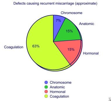
Disseminated Intravascular Coagulation
Partial Thromboplastin Time
Prothrombin Time
Factor XII
Factor Va
Protein C
Factor VIIIa
1-Carboxyglutamic Acid
Hemophilia B
Receptors, Tumor Necrosis Factor
Vitamin K
Arthritis, Rheumatoid
Transglutaminases
Hemophilia A
Tumor Necrosis Factor Decoy Receptors
Antithrombin III
Inhibitor of Differentiation Proteins
Tumor Necrosis Factor-alpha
Arcidae
Fibrinogen
Thrombelastography
Antithrombins
Fibrin
Amino Acid Sequence
Hemostasis
Molecular Sequence Data
Vitamin K Deficiency
Immunoglobulin G
Antibodies, Monoclonal, Humanized
Benzamidines
Binding Sites
Factor XII Deficiency
Immunologic Factors
Factor XIIa
Identification and purification of vitamin K-dependent proteins and peptides with monoclonal antibodies specific for gamma -carboxyglutamyl (Gla) residues. (1/55)
Novel monoclonal antibodies that specifically recognize gamma-carboxyglutamyl (Gla) residues in proteins and peptides have been produced. As demonstrated by Western blot and time-resolved immunofluorescence assays the antibodies are pan-specific for most or all of the Gla-containing proteins tested (factors VII, IX, and X, prothrombin, protein C, protein S, growth arrest-specific protein 6, bone Gla protein, conantokin G from a cone snail, and factor Xa-like proteins from snake venom). Only the Gla-containing light chain of the two-chain proteins was bound. Decarboxylation destroyed the epitope(s) on prothrombin fragment 1, and Ca(2+) strongly inhibited binding to prothrombin. In Western blot, immunofluorescence, and surface plasmon resonance assays the antibodies bound peptides conjugated to bovine serum albumin that contained either a single Gla or a tandem pair of Gla residues. Binding was maintained when the sequence surrounding the Gla residue(s) was altered. Replacement of Gla with glutamic acid resulted in a complete loss of the epitope. The utility of the antibodies was demonstrated in immunochemical methods for detecting Gla-containing proteins and in the immunopurification of a factor Xa-like protein from tiger snake venom. The amino acid sequences of the Gla domain and portions of the heavy chain of the snake protein were determined. (+info)The role of coagulation abnormalities in the development of Perthes' disease. (2/55)
Recent reports have suggested an association between Perthes' disease and an underlying thrombophilic or hypofibrinolytic tendency. In Northern Ireland there is a high incidence of Perthes' disease (11.7 per 100,000 or 1 in 607 children) in a stable paediatric population. We reviewed 139 children with Perthes' disease and compared them with a control group of 220 aged- and gender-matched healthy primary schoolchildren with similar racial and ethnic backgrounds. There were no significant deficiencies of antithrombotic factors protein C, protein S, antithrombin III or resistance to activated protein C. A total of 53 (38.1%) of the children with Perthes' disease had a prolonged activated partial thromboplastin time (>38) compared with 13 (5.9%) of the control group (p < 0.001). Our findings have shown that using standard assays, thrombophilia secondary to antithrombotic factor deficiency or resistance to activated protein does not appear to be an aetiological factor for Perthes' disease. The cause of the prolonged activated partial thromboplastin time, usually associated with a clotting factor deficiency, is under further investigation. (+info)Factor V Leiden mutation, prothrombin gene mutation, and deficiencies in coagulation inhibitors associated with Budd-Chiari syndrome and portal vein thrombosis: results of a case-control study. (3/55)
In a collaborative multicenter case-control study, we investigated the effect of factor V Leiden mutation, prothrombin gene mutation, and inherited deficiencies of protein C, protein S, and antithrombin on the risk of Budd-Chiari syndrome (BCS) and portal vein thrombosis (PVT). We compared 43 BCS patients and 92 PVT patients with 474 population-based controls. The relative risk of BCS was 11.3 (95% CI 4.8-26.5) for individuals with factor V Leiden mutation, 2.1(95% CI 0.4-9.6) for those with prothrombin gene mutation, and 6.8 (95% CI 1.9-24.4) for those with protein C deficiency. The relative risk of PVT was 2.7 (95% CI 1.1-6.9) for individuals with factor V Leiden mutation, 1.4 (95% CI 0.4-5.2) for those with prothrombin gene mutation, and 4.6 (95% CI 1.5-14.1) for those with protein C deficiency. The relative risk of BCS or PVT was not increased in the presence of inherited protein S or antithrombin deficiency. Concurrence of either acquired or inherited thrombotic risk factors was observed in 26% of the BCS patients and 37% of the PVT patients. We conclude that factor V Leiden mutation and hereditary protein C deficiency appear to be important risk factors for BCS and PVT. Although the prevalence of the prothrombin gene mutation was increased, it was not found to be a significant risk factor for BCS and PVT. The coexistence of thrombogenic risk factors in many patients indicates that BCS and PVT can be the result of a combined effect of different pathogenetic mechanisms. (+info)Structural basis for inhibition promiscuity of dual specific thrombin and factor Xa blood coagulation inhibitors. (4/55)
BACKGROUND: A major current focus of pharmaceutical research is the development of selective inhibitors of the blood coagulation enzymes thrombin or factor Xa to be used as orally bioavailable anticoagulant drugs in thromboembolic disorders and in the prevention of venous and arterial thrombosis. Simultaneous direct inhibition of thrombin and factor Xa by synthetic proteinase inhibitors as a novel approach to antithrombotic therapy could result in potent anticoagulants with improved pharmacological properties. RESULTS: The binding mode of such dual specific inhibitors of thrombin and factor Xa was determined for the first time by comparative crystallography using human alpha-thrombin, human des-Gla (1--44) factor Xa and bovine trypsin as the ligand receptors. The benzamidine-based inhibitors utilize two different conformations for the interaction with thrombin and factor Xa/trypsin, which are evoked by the steric requirements of the topologically different S2 subsites of the enzymes. Compared to the unliganded forms of the proteinases, ligand binding induces conformational adjustments of thrombin and factor Xa active site residues indicative of a pronounced induced fit mechanism. CONCLUSION: The structural data reveal the molecular basis for a desired unselective inhibition of the two key components of the blood coagulation cascade. The 4-(1-methyl-benzimidazole-2-yl)-methylamino-benzamidine moieties of the inhibitors are able to fill both the small solvent accessible as well as the larger hydrophobic S2 pockets of factor Xa and thrombin, respectively. Distal fragments of the inhibitors are identified which fit into both the cation hole/aromatic box of factor Xa and the hydrophobic aryl binding site of thrombin. Thus, binding constants in the medium-to-low nanomolar range are obtained against both enzymes. (+info)The dynamics of thrombin formation. (5/55)
The central event of the hemostatic process is the generation of thrombin through the tissue factor pathway. This is a highly regulated, dynamic process in which thrombin itself plays many roles, positively and negatively its production and destruction. The hemostatic process is essential to normal physiology and is also the Achilles heel of our aging population. The inappropriate generation of thrombin may lead to vascular occlusion with the consequence of myocardial infarction, stroke, pulmonary embolism, or venous thrombosis. In this review, we summarize our present views regarding the tissue factor pathway by which thrombin is generated and the roles played by extrinsic and intrinsic factor Xa generating complexes in hemostasis and the roles of the stoichiometric and dynamic inhibitors that regulate thrombin generation. (+info)Thrombotic risk factors and extent of liver fibrosis in chronic viral hepatitis. (6/55)
BACKGROUND AND AIMS: Thrombosis of the small intrahepatic veins has been suggested to trigger liver tissue remodelling. We evaluated the prevalence of multiple thrombotic risk factors and their association with the extent of fibrosis in chronic viral hepatitis. METHODS: Ninety consecutive patients with chronic hepatitis B or C without malignancy, a history of venous thrombosis, or antiviral/immunosuppressive therapy within the last six months were included. Thrombophilic and coagulation factors were evaluated on the liver biopsy day. RESULTS: One or more thrombotic risk factors were found in 68% and > or =2 factors in 37% of patients. Higher necroinflammatory activity was independently associated with higher prothrombin time (p=0.003), alanine aminotransferase level (p=0.011), and histological staging (p=0.018). Patients with staging scores of 4-6 compared with those with scores of 0-3 more frequently had deficiency of protein C (24% v 3%; p=0.007), antithrombin III (28% v 5%; p=0.005), and plasminogen (19% v 2%; p=0.03), and a trend for more frequent activated protein C resistance (8% v 0%; p=0.075). The presence of > or =1 significant thrombotic risk factor was observed in 11/25 (44%) patients with staging scores of 4-6 and in 6/65 (9%) patients with scores of 0-3 (p<0.001), being the only variable independently associated with advanced staging (odds ratio 2.4, p=0.02). CONCLUSIONS: Thrombotic risk factors are frequently detected in patients with chronic viral hepatitis and the presence of > or =1 significant factor is associated with more advanced fibrosis. Whether the association of such thrombophilic conditions with advanced fibrosis is a primary or secondary phenomenon and whether their development in combination with local inflammation accelerate the progression of liver fibrosis need further evaluation. (+info)Mikamo Lecture: a radical view on the 'superfamily' of cardiovascular risk factors. (7/55)
Many risk factors for cardiovascular disease generate superoxide in the blood vessels and thereby impair endothelial function. To emphasize the critical role of oxygen radicals, this is a 'radical view' of those risk factors. It will be useful to organize risk factors into a 'superfamily', with consideration of mediators, mechanisms, and target organs. Studies are summarized which suggest that, in parallel with the impairment of endothelial vasomotor function, the thrombin/thrombomodulin/activated protein C anticoagulant mechanism, which requires endothelial thrombomodulin, is also impaired by atherosclerosis and improves during regression of atherosclerosis. Impairment of the anticoagulant mechanism may contribute to thrombosis in atherosclerotic arteries, and improvement of the anticoagulant mechanism during regression of atherosclerosis may reduce the risk of cardiovascular events. (+info)Time course of coagulation parameters, cytokines and adhesion molecules in Plasmodium falciparum malaria. (8/55)
We studied 38 patients with malaria tropica over a period of 5 days during antiparasitic therapy. Serum or plasma levels of interleukin (IL) 1beta, IL-6, IL-10, tumour necrosis factor-alpha (TNF-alpha), the soluble vascular adhesion molecule (sVCAM) and the soluble intracellular adhesion molecule (sICAM) were determined by enzyme-linked immunosorbent assay. Protein C and antithrombin III activity were analysed by chromogenic tests and protein S activity by a clotting test. Antithrombin III, protein C and protein S activity was significantly lower in patients with severe malaria and displayed a highly significant increase in activity over the time of evaluation. Levels of sVCAM and sICAM were increased for the whole study period, but no significant differences were found between severe and mild malaria cases. Serum IL-1beta, IL-6 and IL-10 levels were significantly higher in patients with severe malaria, whereas no significant differences were found for TNF-alpha. IL-6 and IL-10 decreased significantly over 5 days during schizontocidal therapy. Our data show an impairment of the coagulation system which correlates with pro-inflammatory cytokines and therefore with the severity of the disease. (+info)Blood coagulation factor inhibitors are substances that interfere with the normal blood clotting process by inhibiting the function of coagulation factors. These inhibitors can be either naturally occurring or artificially produced.
Naturally occurring coagulation factor inhibitors include antithrombin, protein C, and tissue factor pathway inhibitor (TFPI). These inhibitors play a crucial role in regulating the coagulation cascade and preventing excessive clot formation.
Artificially produced coagulation factor inhibitors are used as therapeutic agents to treat thrombotic disorders. Examples include direct oral anticoagulants (DOACs) such as apixaban, rivaroxaban, and dabigatran, which selectively inhibit specific coagulation factors (factor Xa or thrombin).
Additionally, there are also antibodies that can act as coagulation factor inhibitors. These include autoantibodies that develop in some individuals and cause bleeding disorders such as acquired hemophilia A or antiphospholipid syndrome.
Blood coagulation, also known as blood clotting, is a complex process that occurs in the body to prevent excessive bleeding when a blood vessel is damaged. This process involves several different proteins and chemical reactions that ultimately lead to the formation of a clot.
The coagulation cascade is initiated when blood comes into contact with tissue factor, which is exposed after damage to the blood vessel wall. This triggers a series of enzymatic reactions that activate clotting factors, leading to the formation of a fibrin clot. Fibrin is a protein that forms a mesh-like structure that traps platelets and red blood cells to form a stable clot.
Once the bleeding has stopped, the coagulation process is regulated and inhibited to prevent excessive clotting. The fibrinolytic system degrades the clot over time, allowing for the restoration of normal blood flow.
Abnormalities in the blood coagulation process can lead to bleeding disorders or thrombotic disorders such as deep vein thrombosis and pulmonary embolism.
Blood coagulation factors, also known as clotting factors, are a group of proteins that play a crucial role in the blood coagulation process. They are essential for maintaining hemostasis, which is the body's ability to stop bleeding after injury.
There are 13 known blood coagulation factors, and they are designated by Roman numerals I through XIII. These factors are produced in the liver and are normally present in an inactive form in the blood. When there is an injury to a blood vessel, the coagulation process is initiated, leading to the activation of these factors in a specific order.
The coagulation cascade involves two pathways: the intrinsic and extrinsic pathways. The intrinsic pathway is activated when there is damage to the blood vessel itself, while the extrinsic pathway is activated by tissue factor released from damaged tissues. Both pathways converge at the common pathway, leading to the formation of a fibrin clot.
Blood coagulation factors work together in a complex series of reactions that involve activation, binding, and proteolysis. When one factor is activated, it activates the next factor in the cascade, and so on. This process continues until a stable fibrin clot is formed.
Deficiencies or abnormalities in blood coagulation factors can lead to bleeding disorders such as hemophilia or thrombosis. Hemophilia is a genetic disorder that affects one or more of the coagulation factors, leading to excessive bleeding and difficulty forming clots. Thrombosis, on the other hand, occurs when there is an abnormal formation of blood clots in the blood vessels, which can lead to serious complications such as stroke or pulmonary embolism.
Factor XIII, also known as fibrin stabilizing factor, is a protein involved in the clotting process of blood. It is a transglutaminase enzyme that cross-links fibrin molecules to form a stable clot. Factor XIII becomes activated during the coagulation cascade, and its activity helps strengthen the clot and protect it from premature degradation by proteolytic enzymes. A deficiency in Factor XIII can lead to a bleeding disorder characterized by prolonged bleeding after injury or surgery.
Factor IX is also known as Christmas factor, which is a protein that plays a crucial role in the coagulation cascade, a series of chemical reactions that leads to the formation of a blood clot. It is one of the essential components required for the proper functioning of the body's natural blood-clotting mechanism.
Factor IX is synthesized in the liver and activated when it comes into contact with an injured blood vessel. Once activated, it collaborates with other factors to convert factor X to its active form, which then converts prothrombin to thrombin. Thrombin is responsible for converting fibrinogen to fibrin, forming a stable fibrin clot that helps stop bleeding and promote healing.
Deficiencies in Factor IX can lead to hemophilia B, a genetic disorder characterized by prolonged bleeding and an increased risk of spontaneous bleeding. Hemophilia B is inherited in an X-linked recessive pattern, meaning it primarily affects males, while females serve as carriers of the disease. Treatment for hemophilia B typically involves replacing the missing or deficient Factor IX through infusions to prevent or manage bleeding episodes.
Factor X is a protein that is essential for blood clotting, also known as coagulation. It is an enzyme that plays a crucial role in the coagulation cascade, which is a series of chemical reactions that lead to the formation of a blood clot. Factor X is activated by one of two pathways: the intrinsic pathway, which is initiated by damage to the blood vessels, or the extrinsic pathway, which is triggered by the release of tissue factor from damaged cells. Once activated, Factor X converts prothrombin to thrombin, which then converts fibrinogen to fibrin to form a stable clot.
Inherited deficiencies in Factor X can lead to bleeding disorders, while increased levels of Factor X have been associated with an increased risk of thrombosis or blood clots. Therefore, maintaining appropriate levels of Factor X is important for the proper balance between bleeding and clotting in the body.
Factor Xa is a serine protease that plays a crucial role in the coagulation cascade, which is a series of reactions that lead to the formation of a blood clot. It is one of the activated forms of Factor X, a pro-protein that is converted to Factor Xa through the action of other enzymes in the coagulation cascade.
Factor Xa functions as a key component of the prothrombinase complex, which also includes calcium ions, phospholipids, and activated Factor V (also known as Activated Protein C or APC). This complex is responsible for converting prothrombin to thrombin, which then converts fibrinogen to fibrin, forming a stable clot.
Inhibitors of Factor Xa are used as anticoagulants in the prevention and treatment of thromboembolic disorders such as deep vein thrombosis and pulmonary embolism. These drugs work by selectively inhibiting Factor Xa, thereby preventing the formation of the prothrombinase complex and reducing the risk of clot formation.
Factor VII, also known as proconvertin, is a protein involved in the coagulation cascade, which is a series of chemical reactions that leads to the formation of a blood clot. Factor VII is synthesized in the liver and is activated when it comes into contact with tissue factor, which is exposed when blood vessels are damaged. Activated Factor VII then activates Factor X, leading to the formation of thrombin and ultimately a fibrin clot.
Inherited deficiencies or dysfunctions of Factor VII can lead to an increased risk of bleeding, while elevated levels of Factor VII have been associated with an increased risk of thrombosis (blood clots).
Factor VIII is a protein in the blood that is essential for normal blood clotting. It is also known as antihemophilic factor (AHF). Deficiency or dysfunction of this protein results in hemophilia A, a genetic disorder characterized by prolonged bleeding and easy bruising. Factor VIII works together with other proteins to help form a clot and stop bleeding at the site of an injury. It acts as a cofactor for another clotting factor, IX, in the so-called intrinsic pathway of blood coagulation. Intravenous infusions of Factor VIII concentrate are used to treat and prevent bleeding episodes in people with hemophilia A.
Factor XIa is a serine protease enzyme that plays a crucial role in blood coagulation. It is formed through the activation of Factor XI, which is one of the key components in the intrinsic pathway of the coagulation cascade. The activation of Factor XI to Factor XIa occurs via either autoactivation or through the action of thrombin. Once activated, Factor XIa can cleave and activate Factor IX, leading to the formation of Factor IXa, which further amplifies the coagulation cascade.
In summary, Factor XIa is a vital enzyme in the blood coagulation process, contributing to the formation of a stable fibrin clot that helps prevent excessive bleeding during injury or trauma.
Blood coagulation tests, also known as coagulation studies or clotting tests, are a series of medical tests used to evaluate the blood's ability to clot. These tests measure the functioning of various clotting factors and regulatory proteins involved in the coagulation cascade, which is a complex process that leads to the formation of a blood clot to prevent excessive bleeding.
The most commonly performed coagulation tests include:
1. Prothrombin Time (PT): Measures the time it takes for a sample of plasma to clot after the addition of calcium and tissue factor, which activates the extrinsic pathway of coagulation. The PT is reported in seconds and can be converted to an International Normalized Ratio (INR) to monitor anticoagulant therapy.
2. Activated Partial Thromboplastin Time (aPTT): Measures the time it takes for a sample of plasma to clot after the addition of calcium, phospholipid, and a contact activator, which activates the intrinsic pathway of coagulation. The aPTT is reported in seconds and is used to monitor heparin therapy.
3. Thrombin Time (TT): Measures the time it takes for a sample of plasma to clot after the addition of thrombin, which directly converts fibrinogen to fibrin. The TT is reported in seconds and can be used to detect the presence of fibrin degradation products or abnormalities in fibrinogen function.
4. Fibrinogen Level: Measures the amount of fibrinogen, a protein involved in clot formation, present in the blood. The level is reported in grams per liter (g/L) and can be used to assess bleeding risk or the effectiveness of fibrinogen replacement therapy.
5. D-dimer Level: Measures the amount of D-dimer, a protein fragment produced during the breakdown of a blood clot, present in the blood. The level is reported in micrograms per milliliter (µg/mL) and can be used to diagnose or exclude venous thromboembolism (VTE), such as deep vein thrombosis (DVT) or pulmonary embolism (PE).
These tests are important for the diagnosis, management, and monitoring of various bleeding and clotting disorders. They can help identify the underlying cause of abnormal bleeding or clotting, guide appropriate treatment decisions, and monitor the effectiveness of therapy. It is essential to interpret these test results in conjunction with a patient's clinical presentation and medical history.
Thromboplastin is a substance that activates the coagulation cascade, leading to the formation of a clot (thrombus). It's primarily found in damaged or injured tissues and blood vessels, as well as in platelets (thrombocytes). There are two types of thromboplastin:
1. Extrinsic thromboplastin (also known as tissue factor): This is a transmembrane glycoprotein that is primarily found in subendothelial cells and released upon injury to the blood vessels. It initiates the extrinsic pathway of coagulation by binding to and activating Factor VII, ultimately leading to the formation of thrombin and fibrin clots.
2. Intrinsic thromboplastin (also known as plasma thromboplastin or factor III): This term is used less frequently and refers to a labile phospholipid component present in platelet membranes, which plays a role in the intrinsic pathway of coagulation.
In clinical settings, the term "thromboplastin" often refers to reagents used in laboratory tests like the prothrombin time (PT) and activated partial thromboplastin time (aPTT). These reagents contain a source of tissue factor and calcium ions to initiate and monitor the coagulation process.
Factor VIIa is a protein involved in the coagulation cascade, which is a series of chemical reactions that leads to the formation of a blood clot. Factor VIIa is the activated form of factor VII, which is normally activated by tissue factor (TF) when there is damage to the blood vessels. Together, TF and Factor VIIa convert Factor X to its active form, Factor Xa, which then converts prothrombin to thrombin, leading to the formation of a fibrin clot.
In summary, Factor VIIa is an important protein in the coagulation cascade that helps to initiate the formation of a blood clot in response to injury.
Antirheumatic agents are a class of drugs used to treat rheumatoid arthritis, other inflammatory types of arthritis, and related conditions. These medications work by reducing inflammation in the body, relieving symptoms such as pain, swelling, and stiffness in the joints. They can also help slow down or prevent joint damage and disability caused by the disease.
There are several types of antirheumatic agents, including:
1. Nonsteroidal anti-inflammatory drugs (NSAIDs): These medications, such as ibuprofen and naproxen, reduce inflammation and relieve pain. They are often used to treat mild to moderate symptoms of arthritis.
2. Corticosteroids: These powerful anti-inflammatory drugs, such as prednisone and cortisone, can quickly reduce inflammation and suppress the immune system. They are usually used for short-term relief of severe symptoms or in combination with other antirheumatic agents.
3. Disease-modifying antirheumatic drugs (DMARDs): These medications, such as methotrexate and hydroxychloroquine, work by slowing down the progression of rheumatoid arthritis and preventing joint damage. They can take several weeks or months to become fully effective.
4. Biologic response modifiers (biologics): These are a newer class of DMARDs that target specific molecules involved in the immune response. They include drugs such as adalimumab, etanercept, and infliximab. Biologics are usually used in combination with other antirheumatic agents for patients who have not responded to traditional DMARD therapy.
5. Janus kinase (JAK) inhibitors: These medications, such as tofacitinib and baricitinib, work by blocking the action of enzymes called JAKs that are involved in the immune response. They are used to treat moderate to severe rheumatoid arthritis and can be used in combination with other antirheumatic agents.
It is important to note that antirheumatic agents can have significant side effects and should only be prescribed by a healthcare provider who is experienced in the management of rheumatoid arthritis. Regular monitoring and follow-up are essential to ensure safe and effective treatment.
Blood coagulation disorders, also known as bleeding disorders or clotting disorders, refer to a group of medical conditions that affect the body's ability to form blood clots properly. Normally, when a blood vessel is injured, the body's coagulation system works to form a clot to stop the bleeding and promote healing.
In blood coagulation disorders, there can be either an increased tendency to bleed due to problems with the formation of clots (hemorrhagic disorder), or an increased tendency for clots to form inappropriately even without injury, leading to blockages in the blood vessels (thrombotic disorder).
Examples of hemorrhagic disorders include:
1. Hemophilia - a genetic disorder that affects the ability to form clots due to deficiencies in clotting factors VIII or IX.
2. Von Willebrand disease - another genetic disorder caused by a deficiency or abnormality of the von Willebrand factor, which helps platelets stick together to form a clot.
3. Liver diseases - can lead to decreased production of coagulation factors, increasing the risk of bleeding.
4. Disseminated intravascular coagulation (DIC) - a serious condition where clotting and bleeding occur simultaneously due to widespread activation of the coagulation system.
Examples of thrombotic disorders include:
1. Factor V Leiden mutation - a genetic disorder that increases the risk of inappropriate blood clot formation.
2. Antithrombin III deficiency - a genetic disorder that impairs the body's ability to break down clots, increasing the risk of thrombosis.
3. Protein C or S deficiencies - genetic disorders that lead to an increased risk of thrombosis due to impaired regulation of the coagulation system.
4. Antiphospholipid syndrome (APS) - an autoimmune disorder where the body produces antibodies against its own clotting factors, increasing the risk of thrombosis.
Treatment for blood coagulation disorders depends on the specific diagnosis and may include medications to manage bleeding or prevent clots, as well as lifestyle changes and monitoring to reduce the risk of complications.
Factor XI, also known as plasma thromboplastin antecedent (PTA) or antihemophilic factor C, is a protein involved in blood coagulation. It is one of the factors in the intrinsic pathway of coagulation, which is activated when blood comes into contact with negatively charged surfaces, such as damaged blood vessels.
When Factor XI is activated (usually by thrombin or activated Factor XII), it activates more Factor XI and also activates Factor IX, leading to the formation of a complex that converts Factor X to its active form, Factor Xa. This ultimately leads to the formation of a fibrin clot and helps to stop bleeding.
Deficiencies in Factor XI can lead to an increased risk of bleeding, although the severity of the bleeding disorder can vary widely among individuals with Factor XI deficiency. Treatment for Factor XI deficiency typically involves replacement therapy with fresh frozen plasma or recombinant Factor XI concentrate.
Factor IXa is a protein that plays a crucial role in the coagulation cascade, which is a series of biochemical reactions involved in blood clotting. It is an activated form of Factor IX, which is one of the coagulation factors that help convert prothrombin to thrombin, leading to the formation of a fibrin clot and stopping bleeding at the site of injury.
Factor IXa works by activating Factor X in the presence of calcium ions, phospholipids, and Factor VIIIa, which is another activated coagulation factor. This complex is called the tenase complex. The activation of Factor X leads to the formation of thrombin, which then converts fibrinogen to fibrin, forming a stable clot.
Deficiencies or dysfunctions in Factor IXa can lead to bleeding disorders such as hemophilia B, also known as Christmas disease, which is characterized by prolonged bleeding times and spontaneous bleeding episodes.
Coagulants are substances that promote the process of coagulation or clotting. They are often used in medical settings to help control bleeding and promote healing. Coagulants work by encouraging the formation of a clot, which helps to stop the flow of blood from a wound or cut.
There are several different types of coagulants that may be used in medical treatments. Some coagulants are naturally occurring substances, such as vitamin K, which is essential for the production of certain clotting factors in the body. Other coagulants may be synthetic or semi-synthetic compounds, such as recombinant activated factor VII (rFVIIa), which is used to treat bleeding disorders and prevent excessive bleeding during surgery.
Coagulants are often administered through injection or infusion, but they can also be applied topically to wounds or cuts. In some cases, coagulants may be used in combination with other treatments, such as compression or cauterization, to help control bleeding and promote healing.
It is important to note that while coagulants can be helpful in controlling bleeding and promoting healing, they can also increase the risk of blood clots and other complications. As a result, they should only be used under the guidance and supervision of a qualified healthcare professional.
Factor V, also known as proaccelerin or labile factor, is a protein involved in the coagulation cascade, which is a series of chemical reactions that leads to the formation of a blood clot. Factor V acts as a cofactor for the activation of Factor X to Factor Xa, which is a critical step in the coagulation cascade.
When blood vessels are damaged, the coagulation cascade is initiated to prevent excessive bleeding. During this process, Factor V is activated by thrombin, another protein involved in coagulation, and then forms a complex with activated Factor X and calcium ions on the surface of platelets or other cells. This complex converts prothrombin to thrombin, which then converts fibrinogen to fibrin to form a stable clot.
Deficiency or dysfunction of Factor V can lead to bleeding disorders such as hemophilia B or factor V deficiency, while mutations in the gene encoding Factor V can increase the risk of thrombosis, as seen in the Factor V Leiden mutation.
Prothrombin is a protein present in blood plasma, and it's also known as coagulation factor II. It plays a crucial role in the coagulation cascade, which is a complex series of reactions that leads to the formation of a blood clot.
When an injury occurs, the coagulation cascade is initiated to prevent excessive blood loss. Prothrombin is converted into its active form, thrombin, by another factor called factor Xa in the presence of calcium ions, phospholipids, and factor Va. Thrombin then catalyzes the conversion of fibrinogen into fibrin, forming a stable clot.
Prothrombin levels can be measured through a blood test, which is often used to diagnose or monitor conditions related to bleeding or coagulation disorders, such as liver disease or vitamin K deficiency.
Disseminated Intravascular Coagulation (DIC) is a complex medical condition characterized by the abnormal activation of the coagulation cascade, leading to the formation of blood clots in small blood vessels throughout the body. This process can result in the consumption of clotting factors and platelets, which can then lead to bleeding complications. DIC can be caused by a variety of underlying conditions, including sepsis, trauma, cancer, and obstetric emergencies.
The term "disseminated" refers to the widespread nature of the clotting activation, while "intravascular" indicates that the clotting is occurring within the blood vessels. The condition can manifest as both bleeding and clotting complications, which can make it challenging to diagnose and manage.
The diagnosis of DIC typically involves laboratory tests that evaluate coagulation factors, platelet count, fibrin degradation products, and other markers of coagulation activation. Treatment is focused on addressing the underlying cause of the condition while also managing any bleeding or clotting complications that may arise.
Partial Thromboplastin Time (PTT) is a medical laboratory test that measures the time it takes for blood to clot. It's more specifically a measure of the intrinsic and common pathways of the coagulation cascade, which are the series of chemical reactions that lead to the formation of a clot.
The test involves adding a partial thromboplastin reagent (an activator of the intrinsic pathway) and calcium to plasma, and then measuring the time it takes for a fibrin clot to form. This is compared to a control sample, and the ratio of the two times is calculated.
The PTT test is often used to help diagnose bleeding disorders or abnormal blood clotting, such as hemophilia or disseminated intravascular coagulation (DIC). It can also be used to monitor the effectiveness of anticoagulant therapy, such as heparin. Prolonged PTT results may indicate a bleeding disorder or an increased risk of bleeding, while shortened PTT results may indicate a hypercoagulable state and an increased risk of thrombosis.
Prothrombin time (PT) is a medical laboratory test that measures the time it takes for blood to clot. It's often used to evaluate the functioning of the extrinsic and common pathways of the coagulation system, which is responsible for blood clotting. Specifically, PT measures how long it takes for prothrombin (a protein produced by the liver) to be converted into thrombin, an enzyme that converts fibrinogen into fibrin and helps form a clot.
Prolonged PT may indicate a bleeding disorder or a deficiency in coagulation factors, such as vitamin K deficiency or the use of anticoagulant medications like warfarin. It's important to note that PT is often reported with an international normalized ratio (INR), which allows for standardization and comparison of results across different laboratories and reagent types.
Thrombin is a serine protease enzyme that plays a crucial role in the coagulation cascade, which is a complex series of biochemical reactions that leads to the formation of a blood clot (thrombus) to prevent excessive bleeding during an injury. Thrombin is formed from its precursor protein, prothrombin, through a process called activation, which involves cleavage by another enzyme called factor Xa.
Once activated, thrombin converts fibrinogen, a soluble plasma protein, into fibrin, an insoluble protein that forms the structural framework of a blood clot. Thrombin also activates other components of the coagulation cascade, such as factor XIII, which crosslinks and stabilizes the fibrin network, and platelets, which contribute to the formation and growth of the clot.
Thrombin has several regulatory mechanisms that control its activity, including feedback inhibition by antithrombin III, a plasma protein that inactivates thrombin and other serine proteases, and tissue factor pathway inhibitor (TFPI), which inhibits the activation of factor Xa, thereby preventing further thrombin formation.
Overall, thrombin is an essential enzyme in hemostasis, the process that maintains the balance between bleeding and clotting in the body. However, excessive or uncontrolled thrombin activity can lead to pathological conditions such as thrombosis, atherosclerosis, and disseminated intravascular coagulation (DIC).
Factor XII, also known as Hageman factor, is a protein that plays a role in the coagulation cascade, which is the series of events that leads to the formation of a blood clot. It is one of the zymogens, or inactive precursor proteins, that becomes activated and helps to trigger the coagulation process.
When Factor XII comes into contact with negatively charged surfaces, such as damaged endothelial cells or artificial surfaces like those found on medical devices, it undergoes a conformational change and becomes activated. Activated Factor XII then activates other proteins in the coagulation cascade, including Factor XI, which ultimately leads to the formation of a fibrin clot.
Deficiencies in Factor XII are generally not associated with bleeding disorders, as the coagulation cascade can still proceed through other pathways. However, excessive activation of Factor XII has been implicated in certain thrombotic disorders, such as deep vein thrombosis and disseminated intravascular coagulation (DIC).
Factor V, also known as proaccelerin or labile factor, is a protein involved in the coagulation cascade, which is a series of chemical reactions that leads to the formation of a blood clot. Factor V acts as a cofactor for the conversion of prothrombin to thrombin, which is a critical step in the coagulation process.
Inherited deficiencies or abnormalities in Factor V can lead to bleeding disorders. For example, Factor V Leiden is a genetic mutation that causes an increased risk of blood clots, while Factor V deficiency can cause a bleeding disorder.
It's worth noting that "Factor Va" is not a standard medical term. Factor V becomes activated and turns into Factor Va during the coagulation cascade. Therefore, it is possible that you are looking for the definition of "Factor Va" in the context of its role as an activated form of Factor V in the coagulation process.
Protein C is a vitamin K-dependent protease that functions as an important regulator of coagulation and inflammation. It is a plasma protein produced in the liver that, when activated, degrades clotting factors Va and VIIIa to limit thrombus formation and prevent excessive blood clotting. Protein C also has anti-inflammatory properties by inhibiting the release of pro-inflammatory cytokines and reducing endothelial cell activation. Inherited or acquired deficiencies in Protein C can lead to an increased risk of thrombosis, a condition characterized by abnormal blood clot formation within blood vessels.
Factor VIIIa is a protein that plays a crucial role in the coagulation cascade, which is the series of biochemical reactions involved in blood clotting. Specifically, Factor VIIIa is an activated form of Factor VIII, which is one of the essential clotting factors required for normal hemostasis (the process that stops bleeding).
Factor VIIIa functions as a cofactor for another protein called Factor IXa, and together they form the "tenase complex." This complex activates Factor X to Factor Xa, which ultimately leads to the formation of a fibrin clot.
Deficiencies or dysfunctions in Factor VIII or Factor VIIIa can result in bleeding disorders such as hemophilia A, a genetic condition characterized by prolonged bleeding and spontaneous hemorrhages.
1-Carboxyglutamic acid, also known as γ-carboxyglutamic acid, is a post-translational modification found on certain blood clotting factors and other calcium-binding proteins. It is formed by the carboxylation of glutamic acid residues in these proteins, which enhances their ability to bind to calcium ions. This modification is essential for the proper functioning of many physiological processes, including blood coagulation, bone metabolism, and wound healing.
Hemophilia B is a genetic disorder that affects the body's ability to control blood clotting, also known as coagulation. This condition is caused by a deficiency or dysfunction in Factor IX, one of the proteins essential for normal blood clotting. As a result, people with Hemophilia B experience prolonged bleeding and bruising after injuries, surgeries, or spontaneously, particularly in joints and muscles.
There are different degrees of severity, depending on how much Factor IX is missing or not functioning properly. Mild cases may only become apparent after significant trauma, surgery, or tooth extraction, while severe cases can lead to spontaneous bleeding into joints and muscles, causing pain, swelling, and potential long-term damage. Hemophilia B primarily affects males, as it is an X-linked recessive disorder, but females can be carriers of the condition and may experience mild symptoms.
Tumor Necrosis Factor (TNF) Receptors are cell surface receptors that bind to tumor necrosis factor cytokines. They play crucial roles in the regulation of a variety of immune cell functions, including inflammation, immunity, and cell survival or death (apoptosis).
There are two major types of TNF receptors: TNFR1 (also known as p55 or CD120a) and TNFR2 (also known as p75 or CD120b). TNFR1 is widely expressed in most tissues, while TNFR2 has a more restricted expression pattern and is mainly found on immune cells.
TNF receptors have an intracellular domain called the death domain, which can trigger signaling pathways leading to apoptosis when activated by TNF ligands. However, they can also activate other signaling pathways that promote cell survival, differentiation, and inflammation. Dysregulation of TNF receptor signaling has been implicated in various diseases, including cancer, autoimmune disorders, and neurodegenerative conditions.
Vitamin K is a fat-soluble vitamin that plays a crucial role in blood clotting and bone metabolism. It is essential for the production of several proteins involved in blood clotting, including factor II (prothrombin), factor VII, factor IX, and factor X. Additionally, Vitamin K is necessary for the synthesis of osteocalcin, a protein that contributes to bone health by regulating the deposition of calcium in bones.
There are two main forms of Vitamin K: Vitamin K1 (phylloquinone), which is found primarily in green leafy vegetables and some vegetable oils, and Vitamin K2 (menaquinones), which is produced by bacteria in the intestines and is also found in some fermented foods.
Vitamin K deficiency can lead to bleeding disorders such as hemorrhage and excessive bruising. While Vitamin K deficiency is rare in adults, it can occur in newborns who have not yet developed sufficient levels of the vitamin. Therefore, newborns are often given a Vitamin K injection shortly after birth to prevent bleeding problems.
Rheumatoid arthritis (RA) is a systemic autoimmune disease that primarily affects the joints. It is characterized by persistent inflammation, synovial hyperplasia, and subsequent damage to the articular cartilage and bone. The immune system mistakenly attacks the body's own tissues, specifically targeting the synovial membrane lining the joint capsule. This results in swelling, pain, warmth, and stiffness in affected joints, often most severely in the hands and feet.
RA can also have extra-articular manifestations, affecting other organs such as the lungs, heart, skin, eyes, and blood vessels. The exact cause of RA remains unknown, but it is believed to involve a complex interplay between genetic susceptibility and environmental triggers. Early diagnosis and treatment are crucial in managing rheumatoid arthritis to prevent joint damage, disability, and systemic complications.
Transglutaminases are a family of enzymes that catalyze the post-translational modification of proteins by forming isopeptide bonds between the carboxamide group of peptide-bound glutamine residues and the ε-amino group of lysine residues. This process is known as transamidation or cross-linking. Transglutaminases play important roles in various biological processes, including cell signaling, differentiation, apoptosis, and tissue repair. There are several types of transglutaminases, such as tissue transglutaminase (TG2), factor XIII, and blood coagulation factor XIIIA. Abnormal activity or expression of these enzymes has been implicated in various diseases, such as celiac disease, neurodegenerative disorders, and cancer.
Hemophilia A is a genetic bleeding disorder caused by a deficiency in clotting factor VIII. This results in impaired blood clotting and prolonged bleeding, particularly after injuries or surgeries. Symptoms can range from mild to severe, with the most severe form resulting in spontaneous bleeding into joints and muscles, leading to pain, swelling, and potential joint damage over time. Hemophilia A primarily affects males, as it is an X-linked recessive disorder, and is usually inherited from a carrier mother. However, about one third of cases result from a spontaneous mutation in the gene for factor VIII. Treatment typically involves replacement therapy with infusions of factor VIII concentrates to prevent or control bleeding episodes.
Tumor Necrosis Factor (TNF) Decoy Receptors are soluble forms of TNF receptors that act as decoy molecules to neutralize the activity of TNF-α, a pro-inflammatory cytokine. They function by binding to TNF-α and preventing it from interacting with its cell surface receptors (TNFR1 and TNFR2), thereby inhibiting the downstream signaling cascades that lead to inflammation and tissue damage.
There are two main types of TNF decoy receptors:
1. TNF Receptor 1 (TNFR1, also known as p55 or p60) - This type of decoy receptor is produced by alternative splicing of the TNFR1 gene and can be found in both membrane-bound and soluble forms. The soluble form of TNFR1 acts as a decoy receptor for TNF-α, preventing it from binding to its cell surface receptors.
2. TNF Receptor 2 (TNFR2, also known as p75 or p80) - This type of decoy receptor is primarily found in the soluble form and is produced by proteolytic cleavage of the membrane-bound TNFR2. Soluble TNFR2 can bind to TNF-α with higher affinity than TNFR1, making it a more effective decoy receptor.
TNF decoy receptors have been implicated in various physiological and pathological processes, including inflammation, immune regulation, and cancer. They are being investigated as potential therapeutic targets for the treatment of various inflammatory diseases, such as rheumatoid arthritis, inflammatory bowel disease, and psoriasis.
Antithrombin III is a protein that inhibits the formation of blood clots (thrombi) in the body. It does this by inactivating several enzymes involved in coagulation, including thrombin and factor Xa. Antithrombin III is produced naturally by the liver and is also available as a medication for the prevention and treatment of thromboembolic disorders, such as deep vein thrombosis and pulmonary embolism. It works by binding to and neutralizing excess clotting factors in the bloodstream, thereby reducing the risk of clot formation.
Inhibitors of Differentiation (ID) proteins are a family of transcriptional regulators that play crucial roles in controlling cell growth, differentiation, and survival. They belong to the basic helix-loop-helix (bHLH) protein family and function as negative regulators of differentiation in various cell types.
ID proteins lack the DNA-binding domain required for specific interactions with DNA, but they contain a highly conserved HLH region that enables them to form heterodimers with other bHLH transcription factors. By doing so, ID proteins prevent these partner bHLH factors from binding to their target DNA sequences and thus inhibit the differentiation programs driven by those factors.
There are four members in the ID protein family: ID1, ID2, ID3, and ID4. These proteins exhibit distinct expression patterns during embryonic development and in adult tissues, reflecting their diverse roles in regulating cell fate decisions and homeostasis. Dysregulation of ID protein function has been implicated in several pathological conditions, including cancer and neurodevelopmental disorders.
Tumor Necrosis Factor-alpha (TNF-α) is a cytokine, a type of small signaling protein involved in immune response and inflammation. It is primarily produced by activated macrophages, although other cell types such as T-cells, natural killer cells, and mast cells can also produce it.
TNF-α plays a crucial role in the body's defense against infection and tissue injury by mediating inflammatory responses, activating immune cells, and inducing apoptosis (programmed cell death) in certain types of cells. It does this by binding to its receptors, TNFR1 and TNFR2, which are found on the surface of many cell types.
In addition to its role in the immune response, TNF-α has been implicated in the pathogenesis of several diseases, including autoimmune disorders such as rheumatoid arthritis, inflammatory bowel disease, and psoriasis, as well as cancer, where it can promote tumor growth and metastasis.
Therapeutic agents that target TNF-α, such as infliximab, adalimumab, and etanercept, have been developed to treat these conditions. However, these drugs can also increase the risk of infections and other side effects, so their use must be carefully monitored.
Arcidae is a family of marine bivalves, commonly known as ark clams or angel wings. These bivalves are characterized by their triangular or elongated shells, which are often sculptured with radial ribs and concentric growth lines. They are filter feeders, living buried in the sand or mud and feeding on plankton and organic matter in the water. Arcidae species can be found in both shallow and deep waters, ranging from tropical to polar regions. Some examples of genera within this family include Barbatia, Arca, and Anadara.
Fibrinogen is a soluble protein present in plasma, synthesized by the liver. It plays an essential role in blood coagulation. When an injury occurs, fibrinogen gets converted into insoluble fibrin by the action of thrombin, forming a fibrin clot that helps to stop bleeding from the injured site. Therefore, fibrinogen is crucial for hemostasis, which is the process of stopping bleeding and starting the healing process after an injury.
Thromboelastography (TEG) is a viscoelastic method used to assess the kinetics of clot formation, clot strength, and fibrinolysis in whole blood. It provides a global assessment of hemostasis by measuring the mechanical properties of a clot as it forms and dissolves over time. The TEG graph displays several parameters that reflect the different stages of clotting, including reaction time (R), clot formation time (K), angle of clot formation (α), maximum amplitude (MA), and percentage lysis at 30 minutes (LY30). These parameters can help guide transfusion therapy and inform decisions regarding the management of coagulopathy in various clinical settings, such as trauma, cardiac surgery, liver transplantation, and obstetrics.
Antithrombins are substances that prevent the formation or promote the dissolution of blood clots (thrombi). They include:
1. Anticoagulants: These are medications that reduce the ability of the blood to clot. Examples include heparin, warfarin, and direct oral anticoagulants (DOACs) such as apixaban, rivaroxaban, and dabigatran.
2. Thrombolytic agents: These are medications that break down existing blood clots. Examples include alteplase, reteplase, and tenecteplase.
3. Fibrinolytics: These are a type of thrombolytic agent that specifically target fibrin, a protein involved in the formation of blood clots.
4. Natural anticoagulants: These are substances produced by the body to regulate blood clotting. Examples include antithrombin III, protein C, and protein S.
Antithrombins are used in the prevention and treatment of various thromboembolic disorders, such as deep vein thrombosis (DVT), pulmonary embolism (PE), stroke, and myocardial infarction (heart attack). It is important to note that while antithrombins can help prevent or dissolve blood clots, they also increase the risk of bleeding, so their use must be carefully monitored.
Fibrin is defined as a protein that is formed from fibrinogen during the clotting of blood. It plays an essential role in the formation of blood clots, also known as a clotting or coagulation cascade. When an injury occurs and bleeding starts, fibrin threads form a net-like structure that entraps platelets and red blood cells to create a stable clot, preventing further loss of blood.
The process of forming fibrin from fibrinogen is initiated by thrombin, another protein involved in the coagulation cascade. Thrombin cleaves fibrinogen into fibrin monomers, which then polymerize to form long strands of fibrin. These strands cross-link with each other through a process catalyzed by factor XIIIa, forming a stable clot that protects the wound and promotes healing.
It is important to note that abnormalities in fibrin formation or breakdown can lead to bleeding disorders or thrombotic conditions, respectively. Proper regulation of fibrin production and degradation is crucial for maintaining healthy hemostasis and preventing excessive clotting or bleeding.
Anticoagulants are a class of medications that work to prevent the formation of blood clots in the body. They do this by inhibiting the coagulation cascade, which is a series of chemical reactions that lead to the formation of a clot. Anticoagulants can be given orally, intravenously, or subcutaneously, depending on the specific drug and the individual patient's needs.
There are several different types of anticoagulants, including:
1. Heparin: This is a naturally occurring anticoagulant that is often used in hospitalized patients who require immediate anticoagulation. It works by activating an enzyme called antithrombin III, which inhibits the formation of clots.
2. Low molecular weight heparin (LMWH): LMWH is a form of heparin that has been broken down into smaller molecules. It has a longer half-life than standard heparin and can be given once or twice daily by subcutaneous injection.
3. Direct oral anticoagulants (DOACs): These are newer oral anticoagulants that work by directly inhibiting specific clotting factors in the coagulation cascade. Examples include apixaban, rivaroxaban, and dabigatran.
4. Vitamin K antagonists: These are older oral anticoagulants that work by inhibiting the action of vitamin K, which is necessary for the formation of clotting factors. Warfarin is an example of a vitamin K antagonist.
Anticoagulants are used to prevent and treat a variety of conditions, including deep vein thrombosis (DVT), pulmonary embolism (PE), atrial fibrillation, and prosthetic heart valve thrombosis. It is important to note that anticoagulants can increase the risk of bleeding, so they must be used with caution and regular monitoring of blood clotting times may be required.
An amino acid sequence is the specific order of amino acids in a protein or peptide molecule, formed by the linking of the amino group (-NH2) of one amino acid to the carboxyl group (-COOH) of another amino acid through a peptide bond. The sequence is determined by the genetic code and is unique to each type of protein or peptide. It plays a crucial role in determining the three-dimensional structure and function of proteins.
Monoclonal antibodies are a type of antibody that are identical because they are produced by a single clone of cells. They are laboratory-produced molecules that act like human antibodies in the immune system. They can be designed to attach to specific proteins found on the surface of cancer cells, making them useful for targeting and treating cancer. Monoclonal antibodies can also be used as a therapy for other diseases, such as autoimmune disorders and inflammatory conditions.
Monoclonal antibodies are produced by fusing a single type of immune cell, called a B cell, with a tumor cell to create a hybrid cell, or hybridoma. This hybrid cell is then able to replicate indefinitely, producing a large number of identical copies of the original antibody. These antibodies can be further modified and engineered to enhance their ability to bind to specific targets, increase their stability, and improve their effectiveness as therapeutic agents.
Monoclonal antibodies have several mechanisms of action in cancer therapy. They can directly kill cancer cells by binding to them and triggering an immune response. They can also block the signals that promote cancer growth and survival. Additionally, monoclonal antibodies can be used to deliver drugs or radiation directly to cancer cells, increasing the effectiveness of these treatments while minimizing their side effects on healthy tissues.
Monoclonal antibodies have become an important tool in modern medicine, with several approved for use in cancer therapy and other diseases. They are continuing to be studied and developed as a promising approach to treating a wide range of medical conditions.
Hemostasis is the physiological process that occurs to stop bleeding (bleeding control) when a blood vessel is damaged. This involves the interaction of platelets, vasoconstriction, and blood clotting factors leading to the formation of a clot. The ultimate goal of hemostasis is to maintain the integrity of the vascular system while preventing excessive blood loss.
Molecular sequence data refers to the specific arrangement of molecules, most commonly nucleotides in DNA or RNA, or amino acids in proteins, that make up a biological macromolecule. This data is generated through laboratory techniques such as sequencing, and provides information about the exact order of the constituent molecules. This data is crucial in various fields of biology, including genetics, evolution, and molecular biology, allowing for comparisons between different organisms, identification of genetic variations, and studies of gene function and regulation.
Vitamin K deficiency is a condition that occurs when the body lacks adequate amounts of Vitamin K, a fat-soluble vitamin essential for blood clotting and bone metabolism. This can lead to an increased risk of excessive bleeding (hemorrhage) and calcification of tissues.
Vitamin K is required for the activation of several proteins involved in blood clotting, known as coagulation factors II, VII, IX, and X. A deficiency in Vitamin K can result in these factors remaining in their inactive forms, leading to impaired blood clotting and an increased risk of bleeding.
Vitamin K deficiency can occur due to several reasons, including malnutrition, malabsorption disorders (such as cystic fibrosis or celiac disease), liver diseases, use of certain medications (such as antibiotics or anticoagulants), and prolonged use of warfarin therapy.
In newborns, Vitamin K deficiency can lead to a serious bleeding disorder known as hemorrhagic disease of the newborn. This is because newborns have low levels of Vitamin K at birth, and their gut bacteria, which are responsible for producing Vitamin K, are not yet fully developed. Therefore, it is recommended that newborns receive a dose of Vitamin K within the first few days of life to prevent this condition.
Symptoms of Vitamin K deficiency can include easy bruising, nosebleeds, bleeding gums, blood in urine or stools, and excessive menstrual bleeding. In severe cases, it can lead to life-threatening hemorrhage. Treatment typically involves administering Vitamin K supplements or injections to replenish the body's levels of this essential nutrient.
Immunoglobulin G (IgG) is a type of antibody, which is a protective protein produced by the immune system in response to foreign substances like bacteria or viruses. IgG is the most abundant type of antibody in human blood, making up about 75-80% of all antibodies. It is found in all body fluids and plays a crucial role in fighting infections caused by bacteria, viruses, and toxins.
IgG has several important functions:
1. Neutralization: IgG can bind to the surface of bacteria or viruses, preventing them from attaching to and infecting human cells.
2. Opsonization: IgG coats the surface of pathogens, making them more recognizable and easier for immune cells like neutrophils and macrophages to phagocytose (engulf and destroy) them.
3. Complement activation: IgG can activate the complement system, a group of proteins that work together to help eliminate pathogens from the body. Activation of the complement system leads to the formation of the membrane attack complex, which creates holes in the cell membranes of bacteria, leading to their lysis (destruction).
4. Antibody-dependent cellular cytotoxicity (ADCC): IgG can bind to immune cells like natural killer (NK) cells and trigger them to release substances that cause target cells (such as virus-infected or cancerous cells) to undergo apoptosis (programmed cell death).
5. Immune complex formation: IgG can form immune complexes with antigens, which can then be removed from the body through various mechanisms, such as phagocytosis by immune cells or excretion in urine.
IgG is a critical component of adaptive immunity and provides long-lasting protection against reinfection with many pathogens. It has four subclasses (IgG1, IgG2, IgG3, and IgG4) that differ in their structure, function, and distribution in the body.
Monoclonal antibodies are laboratory-produced proteins that mimic the immune system's ability to fight off harmful antigens such as viruses and cancer cells. They are created by fusing a single B cell (the type of white blood cell responsible for producing antibodies) with a tumor cell, resulting in a hybrid cell called a hybridoma. This hybridoma can then be cloned to produce a large number of identical cells, all producing the same antibody, hence "monoclonal."
Humanized monoclonal antibodies are a type of monoclonal antibody that have been genetically engineered to include human components. This is done to reduce the risk of an adverse immune response in patients receiving the treatment. In this process, the variable region of the mouse monoclonal antibody, which contains the antigen-binding site, is grafted onto a human constant region. The resulting humanized monoclonal antibody retains the ability to bind to the target antigen while minimizing the immunogenicity associated with murine (mouse) antibodies.
In summary, "antibodies, monoclonal, humanized" refers to a type of laboratory-produced protein that mimics the immune system's ability to fight off harmful antigens, but with reduced immunogenicity due to the inclusion of human components in their structure.
Benzamidines are a group of organic compounds that contain a benzene ring linked to an amidine functional group. They are commonly used as antimicrobial agents, particularly in the treatment of various gram-negative bacterial infections. Benzamidines work by inhibiting the enzyme bacterial dehydrogenases, which are essential for the bacteria's survival.
Some examples of benzamidine derivatives include:
* Tempanamine hydrochloride (Tembaglanil): used to treat urinary tract infections caused by susceptible strains of Escherichia coli and Klebsiella pneumoniae.
* Chlorhexidine: a broad-spectrum antimicrobial agent used as a disinfectant and preservative in various medical and dental applications.
* Prothiobenzamide: an anti-inflammatory and analgesic drug used to treat gout and rheumatoid arthritis.
It is important to note that benzamidines have a narrow therapeutic index, which means that the difference between an effective dose and a toxic dose is small. Therefore, they should be used with caution and under the supervision of a healthcare professional.
In the context of medical and biological sciences, a "binding site" refers to a specific location on a protein, molecule, or cell where another molecule can attach or bind. This binding interaction can lead to various functional changes in the original protein or molecule. The other molecule that binds to the binding site is often referred to as a ligand, which can be a small molecule, ion, or even another protein.
The binding between a ligand and its target binding site can be specific and selective, meaning that only certain ligands can bind to particular binding sites with high affinity. This specificity plays a crucial role in various biological processes, such as signal transduction, enzyme catalysis, or drug action.
In the case of drug development, understanding the location and properties of binding sites on target proteins is essential for designing drugs that can selectively bind to these sites and modulate protein function. This knowledge can help create more effective and safer therapeutic options for various diseases.
Factor XII deficiency, also known as Hageman factor deficiency, is a rare genetic disorder characterized by a lack or dysfunction of coagulation factor XII. This protein is involved in the initiation of the coagulation cascade, which leads to the formation of a blood clot. People with Factor XII deficiency may have an increased risk of bleeding, but it is typically mild and not life-threatening. The diagnosis is usually made through blood tests that measure the level and function of Factor XII. Treatment is generally not necessary unless there is significant bleeding, in which case fresh frozen plasma or cryoprecipitate may be given to provide temporary correction of the deficiency. It's important to note that Factor XII deficiency is not a common cause of bleeding disorders and it doesn't increase the risk of thrombosis.
Fibrinolysis is the natural process in the body that leads to the dissolution of blood clots. It is a vital part of hemostasis, the process that regulates bleeding and wound healing. Fibrinolysis occurs when plasminogen activators convert plasminogen to plasmin, an enzyme that breaks down fibrin, the insoluble protein mesh that forms the structure of a blood clot. This process helps to prevent excessive clotting and maintains the fluidity of the blood. In medical settings, fibrinolysis can also refer to the therapeutic use of drugs that stimulate this process to dissolve unwanted or harmful blood clots, such as those that cause deep vein thrombosis or pulmonary embolism.
Immunologic factors refer to the elements of the immune system that contribute to the body's defense against foreign substances, infectious agents, and cancerous cells. These factors include various types of white blood cells (such as lymphocytes, neutrophils, monocytes, and eosinophils), antibodies, complement proteins, cytokines, and other molecules involved in the immune response.
Immunologic factors can be categorized into two main types: innate immunity and adaptive immunity. Innate immunity is the non-specific defense mechanism that provides immediate protection against pathogens through physical barriers (e.g., skin, mucous membranes), chemical barriers (e.g., stomach acid, enzymes), and inflammatory responses. Adaptive immunity, on the other hand, is a specific defense mechanism that develops over time as the immune system learns to recognize and respond to particular pathogens or antigens.
Abnormalities in immunologic factors can lead to various medical conditions, such as autoimmune disorders, immunodeficiency diseases, and allergies. Therefore, understanding immunologic factors is crucial for diagnosing and treating these conditions.
Factor XIIa is a protease enzyme that plays a role in the coagulation cascade, which is the series of events that leads to blood clotting. It is formed when Factor XII, also known as Hageman factor, is activated by contact with negatively charged surfaces such as damaged endothelial cells or artificial surfaces like medical devices.
Once activated, Factor XIIa can activate other components of the coagulation cascade, including Factor XI, which ultimately leads to the formation of a fibrin clot. While Factor XIIa is an important part of the coagulation system, it is not essential for normal hemostasis (the process that stops bleeding) in humans, as people with deficiencies in Factor XII do not have an increased risk of bleeding. However, excessive activation of Factor XIIa has been implicated in several pathological conditions, including thrombosis and inflammation.
 Alteration of the Substrate and Inhibitor Specificities of Blood Coagulation Factor VIIa: Importance of Amino Acid Residue K192...
 A low molecular weight platelet inhibitor of factor XIa: purification, characterization, and possible role in blood coagulation...
 Discovery and development of direct Xa inhibitors - Wikipedia
 Advanced Search Results - Public Health Image Library(PHIL)
 Consumption Coagulopathy: Practice Essentials, Pathophysiology, Epidemiology
 Apixaban ≥98% (HPLC) | Sigma-Aldrich
 DeCS
DeCS Prothrombin Complex Concentrate for Major Bleeding on Factor Xa Inhibitors: A Prospective Cohort Study
![FDA Approves Prophylactic Treatment with VONVENDI® [von Willebrand Factor (Recombinant)] for Adult Patients Living with Severe...](data:image/png;base64,iVBORw0KGgoAAAANSUhEUgAAABAAAAAQCAMAAAAoLQ9TAAAAJ1BMVEVHcEyzIBezIBezIBezIBezIBezIBezIBezIBezIBezIBezIBezIBdn8zgwAAAADHRSTlMAEYHtuidJmMtbP9G+9Z0OAAAAUUlEQVQYlXWOUQ7AIAhDEQXR9f7nXSZzDhP7915aApFHEsXUtolskRWIGwPqnzsQK/kRZTFjRCcnZ+StAPRXlCn4JL6Jno5ScyHrEamXsX96A6ANA/JUA6pyAAAAAElFTkSuQmCC) FDA Approves Prophylactic Treatment with VONVENDI® [von Willebrand Factor (Recombinant)] for Adult Patients Living with Severe...
FDA Approves Prophylactic Treatment with VONVENDI® [von Willebrand Factor (Recombinant)] for Adult Patients Living with Severe... Vonvendi - Side Effects, Uses, Dosage, Overdose, Pregnancy, Alcohol | RxWiki
 At 5.9% Growth Rate Blood Plasma Derivatives Market Expected to Grow $44,333 million by 2023 - PharmiWeb.com
 Innohep (Tinzaparin): Uses, Dosage, Side Effects, Interactions, Warning
 Human Ebola virus infection in West Africa: a review of available therapeutic agents that target different steps of the life...
 What Is Warfarin?
 Factor VIII assay: MedlinePlus Medical Encyclopedia
 FEIBA 50 U/ml powder and solvent for solution for infusion
 Research Journal of Pharmacutical Sciences : Insilico Drug Designing of Potent Inhibitors for PDE7B, A Therapeutic Target for...
 Secreted Phospholipases A2 - not just Enzymes: Revisited
 BLOOD COAGULATION
 Performance and thermal decomposition analysis of foaming agent NPL-10 for use in heavy oil recovery by steam injection
 Earn CE for your Florida Board of Clinical Laboratory Personnel License Renewal - LabCE
 Staff Listing in the Biomolecular Science and Medicinal Chemistry Division - The University of Nottingham