Bile Ducts
Bile Ducts, Extrahepatic
Common Bile Duct
Bile
Cholestasis, Extrahepatic
Bile Duct Diseases
Bile Acids and Salts
Bile Ducts, Intrahepatic
Cholestasis
Common Bile Duct Diseases
Hepatic Duct, Common
Gallstones
Cholangiography
Common Bile Duct Neoplasms
Cholangiopancreatography, Endoscopic Retrograde
Pancreatic Ducts
Cystic Duct
Liver
Jaundice, Obstructive
Cholangitis
Biliary Atresia
Cholelithiasis
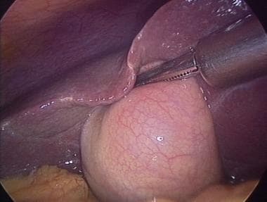
Cholecystectomy, Laparoscopic
Gallbladder
Thoracic Duct
Biliary Tract Diseases
Sphincterotomy, Endoscopic
Biliary Tract Neoplasms
Cholangiopancreatography, Magnetic Resonance
Ampulla of Vater
Cholestasis, Intrahepatic
Choledochal Cyst
Klatskin's Tumor
Salivary Ducts
Bile Pigments
Bile Canaliculi
Choledochostomy
Jaundice
Liver Cirrhosis, Biliary
Biliary Fistula
Wolffian Ducts
Cholangitis, Sclerosing
Jejunostomy
Hypertension, Portal
Hepatic Artery
Sphincter of Oddi
Ursodeoxycholic Acid
Pancreatitis
Mullerian Ducts
Bile Reflux
Taurocholic Acid
Cholagogues and Choleretics
Anastomosis, Roux-en-Y
Portoenterostomy, Hepatic
Lithiasis
Cholecystitis
Portal System
Imino Acids
1-Naphthylisothiocyanate
Cholic Acids
Chenodeoxycholic Acid
Liver Cirrhosis
Retrospective Studies
Portography
Dilatation, Pathologic
Kidney Tubules, Collecting
Treatment Outcome
Iatrogenic Disease
Carcinoma, Hepatocellular
Pancreaticoduodenectomy
Tomography, X-Ray Computed
Postoperative Complications
Liver Function Tests
Constriction, Pathologic
Hepatocytes
Immunohistochemistry
Duodenum
Endoscopy
Anastomosis, Surgical
Liver Cirrhosis, Experimental
Pancreas
Lithotripsy
Cholic Acid
Deoxycholic Acid
Endosonography
Intraoperative Complications
Sphincterotomy, Transhepatic
Lithocholic Acid
Cholesterol
Portasystemic Shunt, Surgical
Technetium Tc 99m Disofenin
Catheterization
Cystadenoma
Jaundice, Neonatal
Endoscopes
Taurodeoxycholic Acid
Taurochenodeoxycholic Acid
Gallbladder Diseases
Keratin-7
Enterohepatic Circulation
Pancreatic Neoplasms
Rats, Sprague-Dawley
Epithelium
Intraoperative Care
Liver Transplantation
Kidney
Calculi
Jejunum
Organic Anion Transporters, Sodium-Dependent
Clonorchiasis
Caroli Disease
Follow-Up Studies
alpha-Fetoproteins
Vitelline Duct
Glycocholic Acid
Adenocarcinoma, Papillary
Cystadenocarcinoma
Cysts
Cholecystostomy
Rats, Inbred Strains
RNA, Messenger
Lacrimal Duct Obstruction
Disease Models, Animal
Endolymphatic Duct
Intestines
Alkaline Phosphatase
Glucuronosyltransferase
Esophageal and Gastric Varices
Splenic Vein
Tissue Distribution
Rats, Inbred F344
Biopsy
Alanine Transaminase
Epithelial Cells
Sphincter of Oddi Dysfunction
Cholecystitis, Acute
Prognosis
Rats, Wistar
Survival Rate
Ultrasonography
Cholesterol 7-alpha-Hydroxylase
Technetium Tc 99m Aggregated Albumin
Duodenal Diseases
Preoperative Care
Microsomes, Liver
Cryoglobulinemia
Prospective Studies
Stents
Cholestanetriol 26-Monooxygenase
Reoperation
Dogs
Fascioliasis
gamma-Glutamyltransferase
Mesenteric Veins
Secretin
Opisthorchis
Clonorchis sinensis
Pancreatitis, Chronic
Cholestyramine Resin
Chemoembolization, Therapeutic
Mice, Inbred C57BL
Laparoscopy
Aspartate Aminotransferases
Cochlear Duct
Fatal Outcome
Sensitivity and Specificity
Adenomyoma
Clinical value of K-ras codon 12 analysis and endobiliary brush cytology for the diagnosis of malignant extrahepatic bile duct stenosis. (1/179)
Extrahepatic biliary stenosis can be caused by benign and malignant disorders. In most cases, a tissue diagnosis is needed for optimal management of patients, but the sensitivity of biliary cytology for the diagnosis of a malignancy is relatively low. The additional diagnostic value of K-ras mutational analysis of endobiliary brush cytology was assessed. Endobiliary brush cytology specimens obtained during endoscopic retrograde cholangiopancreaticography were prospectively collected from 312 consecutive patients with extrahepatic biliary stenosis. The results of conventional light microscopic cytology and K-ras codon 12 mutational analysis were compared and evaluated in view of the final diagnosis made by histological examination of the stenotic lesion and/or patient follow-up. The sensitivities of cytology and mutational analysis to detect malignancy were 36 and 42%, respectively. When both tests were combined, the sensitivity increased to 62%. The specificity of cytology was 98%, and the specificity of the mutational analysis and of both tests combined was 89%. Positive predictive values for cytology, mutational analysis, and both tests combined were 98, 92, and 94%, whereas the corresponding negative predictive values were 34, 34, and 44%, respectively. The sensitivity of K-ras mutational analysis was 63% for pancreatic carcinomas compared to 27% for bile duct, gallbladder, and ampullary carcinomas. K-ras mutational analysis can be considered supplementary to conventional light microscopy of endobiliary brush cytology to diagnose patients with malignant extrahepatic biliary stenosis, particularly in the case of pancreatic cancer. The presence of a K-ras codon 12 mutation in endobiliary brush cytology per se supports a clinical suspicion of malignancy, even when the conventional cytology is negative or equivocal. (+info)Metachronous bile duct cancer in a patient surviving for a decade and undergoing curative surgery twice. (2/179)
We report a 75-year-old woman with metachronous bile duct cancer who underwent curative resection twice and has survived for a decade. In 1989, she was admitted because her serum alkaline phosphatase level was elevated. Computed tomography (CT) showed a low-density mass, 2 cm in diameter, at the left hepatic duct and intrahepatic bile duct dilatation in the left lobe. We diagnosed the lesion as an intrahepatic bile duct cancer and performed extended left hepatic lobectomy with systematic lymph node dissection. The histological diagnosis was a well differentiated cholangiocellular carcinoma with hepatic hilar and celiac lymph node metastases (T1N2M0, Stage IVB). In 1996, she was re-admitted with obstructive jaundice. CT showed a slightly enhanced mass, 4 cm in diameter, in the pancreatic head. After reducing the jaundice by percutaneous transhepatic biliary drainage, pancreatoduodenectomy was performed. The histological diagnosis of this lesion was a moderately differentiated adenocarcinoma originating from the intrapancreatic bile duct. Ten years after the first operation, she is leading a normal daily life with no cancer recurrence. These findings suggest that repeated curative surgery can result in a long-term survival of patients with metachronous bile duct cancer. (+info)Partial hepatectomy and bile duct ligation in rainbow trout (Oncorhynchus mykiss): histologic, immunohistochemical and enzyme histochemical characterization of hepatic regeneration and biliary hyperplasia. (3/179)
Hepatic regeneration following partial hepatectomy (PH) and biliary hyperplasia subsequent to bile duct ligation (BDL) were characterized in rainbow trout (Oncorhynchus mykiss) by light microscopy using routine and special (immunohistochemical and enzyme histochemical) stains. Both PH and BDL involved initial hypertrophy and hyperplasia of bile preductular epithelial cells (BPDECs). BPDECs are small oval cells that form junctional complexes with hepatocytes and bile ductular cells and are commonly found in hepatic tubules of teleost liver. Proliferating BPDECs transitioned through intermediate cell types before final differentiation into large basophilic hepatocytes (following PH) or biliary epithelial cells (after BDL). Normal BPDECs and hepatocytes were both negative for cytokeratin intermediate filaments in control fish when screened with the monoclonal antibody AE1/AE3. In contrast, hyperplastic BPDECs and their progeny (intermediate cells, immature hepatocytes, ductal epithelial cells) were all strongly cytokeratin positive. Cytokeratin expression was transient in newly differentiated hepatocytes (expression decreased as hepatocytes acquired characteristics consistent with full differentiation) but was permanent in biliary epithelial cells (expression was very strong in large mature ducts). BPDECs, intermediate cells, and immature ductal cells were also strongly positive for alkaline phosphatase following BDL. Chronology of histologic events and cytokeratin and enzyme expression all support the hypothesis that BPDECs possess the capacity to differentiate into either hepatocytes or biliary epithelial cells. Thus, BPDECs may be the teleost equivalent of a bipolar hepatic stem cell in mammals. (+info)Postoperative bile duct strictures: management and outcome in the 1990s. (4/179)
OBJECTIVE: To describe the management and outcome after surgical reconstruction of 156 patients with postoperative bile duct strictures managed in the 1990s. SUMMARY BACKGROUND DATA: The management of postoperative bile duct strictures and major bile duct injuries remains a challenge for even the most skilled biliary tract surgeon. The 1990s saw a dramatic increase in the incidence of bile duct strictures and injuries from the introduction and widespread use of laparoscopic cholecystectomy. Although the management of these injuries and short-term outcome have been reported, long-term follow-up is limited. METHODS: Data were collected prospectively on 156 patients treated at the Johns Hopkins Hospital with major bile duct injuries or postoperative bile duct strictures between January 1990 and December 1999. With the exception of bile duct injuries discovered and repaired during surgery, all patients underwent preoperative percutaneous transhepatic cholangiography and placement of transhepatic biliary catheters before surgical repair. Follow-up was conducted by medical record review or telephone interview during January 2000. RESULTS: Of the 156 patients undergoing surgical reconstruction, 142 had completed treatment with a mean follow-up of 57.5 months. Two patients died of reasons unrelated to biliary tract disease before the completion of treatment. Twelve patients (7.9%) had not completed treatment and still had biliary stents in place at the time of this report. Of patients who had completed treatment, 90. 8% were considered to have a successful outcome without the need for follow-up invasive, diagnos tic, or therapeutic interventional procedures. Patients with reconstruction after injury or stricture after laparoscopic cholecystectomy had a better overall outcome than patients whose postoperative stricture developed after other types of surgery. Presenting symptoms, number of stents, interval to referral, prior repair, and length of postoperative stenting were not significant predictors of outcome. Overall, a successful outcome, without the need for biliary stents, was obtained in 98% of patients, including those requiring a secondary procedure for recurrent stricture. CONCLUSIONS: Major bile duct injuries and postoperative bile duct strictures remain a considerable surgical challenge. Management with preoperative cholangiography to delineate the anatomy and placement of percutaneous biliary catheters, followed by surgical reconstruction with a Roux-en-Y hepaticojejunostomy, is associated with a successful outcome in up to 98% of patients. (+info)Genetic alterations and growth pattern in biliary duct carcinomas: loss of heterozygosity at chromosome 5q bears a close relation with polypoid growth. (5/179)
Biliary duct carcinomas (BDCs) are relatively rare and the carcinogenic mechanisms underlying their induction are poorly understood. There are two growth patterns, polypoid and non-polypoid infiltrative type, but little information is available concerning the relation between growth pattern and genetic alterations. A comparative study was therefore conducted to clarify if differences in genetic changes, including loss of heterozygosity (LOH) at 5q, 9p, 17p, and 18q, and K-ras mutations exist between polypoid and non-polypoid infiltrative type BDCs. LOH analysis was performed using microsatellite markers and K-ras point mutations were analysed by dot blot hybridisation. The incidences of changes for polypoid and non-polypoid infiltrative types were 73% and 26% on 5q, 63% and 59% on 9p, 55% and 50% on 17p, and 20% and 18% on 18q, and 25% and 27% for K-ras mutations. Most importantly, we found the frequency of 5qLOH to be significantly higher with polypoid growth than in the non-polypoid infiltrative type (p<0.05), especially in extrahepatic duct carcinomas (p<0.05). The incidences of other genetic alterations (LOH at 9p, 17p, and 18q, and K-ras mutations) showed similar rates with both tumour types. The present data suggest that 5qLOH may have a close relation with polypoid growth in BDCs. (+info)Is preventive resection of the extrahepatic bile duct necessary in cases of pancreaticobiliary maljunction without dilatation of the bile duct? (6/179)
BACKGROUND: No consensus has been reached on whether preventive resection of the extrahepatic bile duct is necessary in cases of pancreaticobiliary maljunction (PBM) without dilatation of the extrahepatic bile duct (undilated type). METHODS: Sixty-eight patients with PBM underwent corrective surgery and several clinical characteristics and pathological findings including K-ras point mutation were evaluated. RESULTS: Unlike dilated bile duct, none of the patients with undilated type duct had clinical symptoms in early childhood. In patients with either cystic or spindle type duct, amylase levels in the bile duct were >10(4) U/l, whereas those in patients with undilated type duct were <10(4) U/l. Postoperative scintigraphy of the biliary system of undilated type revealed no evidence of cholestasis. After surgery, eight patients with undilated type duct, in whom the bile duct had been preserved, had no further clinical symptoms and no evidence of malignancy. Bile duct tissue specimens revealed no hyperplasia, dysplasia or cancerous lesions and they had no K-ras mutation in undilated type. CONCLUSION: The results showed that there was little bile stasis, injury to the mucosa was mild and less genetic changes could be seen in patients with undilated type duct. Therefore, in patients without dilatation of bile duct and advanced cancer, cholecystectomy alone is sufficient. (+info)Restrictive cardiomyopathy in a patient with extrahepatic biliary atresia. (7/179)
The most commonly associated anomalies in patients with extrahepatic biliary atresia are cardiovascular, digestive and splenic defects. Of the cardiovascular anomalies, there are very few reports of biliary atresia with cardiomyopathy. We report the first case of a child with extrahepatic biliary atresia and restrictive cardiomyopathy. The patient was a 13-month-old boy diagnosed with extrahepatic biliary atresia at the age of 2 months, when he underwent laparotomy for definite diagnosis.Hepatic portoenterostomy was performed after confirmative cholangiogram. Recently, he developed severe cough and dyspnea, and his respiratory symptoms worsened. Chest radiograph showed cardiomegaly. Two- dimensional echocardiography showed marked biatrial enlargement. On M- mode echocardiogram, a slight increase in left ventricular dimension was seen in early diastole with a relatively good left ventricular function. Mitral inflow Doppler tracing showed an increased E-velocity (1.1 m/sec) with decreased deceleration time (75 m/sec), and increased E/A ratio (0.33). He was diagnosed as having restrictive cardiomyopathy with characteristic echocardiographic features. (+info)Immunohistochemical and ultrastructural study of ito cells (fat-storing cells) in response to extrahepatic bile duct ligation in broiler chickens. (8/179)
The Ito cell (fat-storing cell) lies in perisinusoidal space of liver and has a variety of functions. We investigated the immunohistochemistry and ultrastructure of Ito cells in normal and cholestatic livers of broiler chickens. Immunohistochemistry demonstrated that Ito cells expressed HHF35 muscle actin, vimentin, desmin, glial fibrillary acidic protein (GFAP), neuron-specific enolase (NSE), chromogranin A and cytokeratins in normal livers. These cells were diffusely scattered throughout the lobules. Livers treated with extrahepatic bile duct ligation (BDL) showed cholestasis, fibrosis, proliferation of biliary ductules and Ito cells. The Ito cells were frequently found in fibrotic areas and were larger in size with more extensive immunoreactivity than those of normal livers. Ultrastructural study demonstrated that Ito cells were closely associated with the production of collagen fibers in BDL livers. These findings suggest that Ito cells actively react against hepatocytic injuries and play a major role in the hepatic fibrogenesis of cholestatic livers of chickens. (+info)Bile ducts are tubular structures that carry bile from the liver to the gallbladder for storage or directly to the small intestine to aid in digestion. There are two types of bile ducts: intrahepatic and extrahepatic. Intrahepatic bile ducts are located within the liver and drain bile from liver cells, while extrahepatic bile ducts are outside the liver and include the common hepatic duct, cystic duct, and common bile duct. These ducts can become obstructed or inflamed, leading to various medical conditions such as cholestasis, cholecystitis, and gallstones.
Extrahepatic bile ducts refer to the portion of the biliary system that lies outside the liver. The biliary system is responsible for producing, storing, and transporting bile, a digestive fluid produced by the liver.
The extrahepatic bile ducts include:
1. The common hepatic duct: This duct is formed by the union of the right and left hepatic ducts, which drain bile from the corresponding lobes of the liver.
2. The cystic duct: This short duct connects the gallbladder to the common hepatic duct, allowing bile to flow into the gallbladder for storage and concentration.
3. The common bile duct: This is the result of the fusion of the common hepatic duct and the cystic duct. It transports bile from the liver and gallbladder to the duodenum, the first part of the small intestine, where it aids in fat digestion.
4. The ampulla of Vater (or hepatopancreatic ampulla): This is a dilated area where the common bile duct and the pancreatic duct join and empty their contents into the duodenum through a shared opening called the major duodenal papilla.
Extrahepatic bile ducts can be affected by various conditions, such as gallstones, inflammation (cholangitis), strictures, or tumors, which may require medical or surgical intervention.
The common bile duct is a duct that results from the union of the cystic duct (which drains bile from the gallbladder) and the common hepatic duct (which drains bile from the liver). The common bile duct transports bile, a digestive enzyme, from the liver and gallbladder to the duodenum, which is the first part of the small intestine.
The common bile duct runs through the head of the pancreas before emptying into the second part of the duodenum, either alone or in conjunction with the pancreatic duct, via a small opening called the ampulla of Vater. The common bile duct plays a crucial role in the digestion of fats by helping to break them down into smaller molecules that can be absorbed by the body.
Bile is a digestive fluid that is produced by the liver and stored in the gallbladder. It plays an essential role in the digestion and absorption of fats and fat-soluble vitamins in the small intestine. Bile consists of bile salts, bilirubin, cholesterol, phospholipids, electrolytes, and water.
Bile salts are amphipathic molecules that help to emulsify fats into smaller droplets, increasing their surface area and allowing for more efficient digestion by enzymes such as lipase. Bilirubin is a breakdown product of hemoglobin from red blood cells and gives bile its characteristic greenish-brown color.
Bile is released into the small intestine in response to food, particularly fats, entering the digestive tract. It helps to break down large fat molecules into smaller ones that can be absorbed through the walls of the intestines and transported to other parts of the body for energy or storage.
Bile duct neoplasms, also known as cholangiocarcinomas, refer to a group of malignancies that arise from the bile ducts. These are the tubes that carry bile from the liver to the gallbladder and small intestine. Bile duct neoplasms can be further classified based on their location as intrahepatic (within the liver), perihilar (at the junction of the left and right hepatic ducts), or distal (in the common bile duct).
These tumors are relatively rare, but their incidence has been increasing in recent years. They can cause a variety of symptoms, including jaundice, abdominal pain, weight loss, and fever. The diagnosis of bile duct neoplasms typically involves imaging studies such as CT or MRI scans, as well as blood tests to assess liver function. In some cases, a biopsy may be necessary to confirm the diagnosis.
Treatment options for bile duct neoplasms depend on several factors, including the location and stage of the tumor, as well as the patient's overall health. Surgical resection is the preferred treatment for early-stage tumors, while chemotherapy and radiation therapy may be used in more advanced cases. For patients who are not candidates for surgery, palliative treatments such as stenting or bypass procedures may be recommended to relieve symptoms and improve quality of life.
Extrahepatic cholestasis is a medical condition characterized by the impaired flow of bile outside of the liver. Bile is a digestive fluid produced by the liver that helps in the absorption and digestion of fats. When the flow of bile is obstructed or blocked, it can lead to an accumulation of bile components, such as bilirubin, in the bloodstream, resulting in jaundice, dark urine, light-colored stools, and itching.
Extrahepatic cholestasis can be caused by various factors, including gallstones, tumors, strictures, or inflammation of the bile ducts. It is essential to diagnose and treat extrahepatic cholestasis promptly to prevent further complications, such as liver damage or infection. Treatment options may include medications, endoscopic procedures, or surgery, depending on the underlying cause of the condition.
Bile duct diseases refer to a group of medical conditions that affect the bile ducts, which are tiny tubes that carry bile from the liver to the gallbladder and small intestine. Bile is a digestive juice produced by the liver that helps break down fats in food.
There are several types of bile duct diseases, including:
1. Choledocholithiasis: This occurs when stones form in the common bile duct, causing blockage and leading to symptoms such as abdominal pain, jaundice, and fever.
2. Cholangitis: This is an infection of the bile ducts that can cause inflammation, pain, and fever. It can occur due to obstruction of the bile ducts or as a complication of other medical procedures.
3. Primary Biliary Cirrhosis (PBC): This is a chronic autoimmune disease that affects the bile ducts in the liver, causing inflammation and scarring that can lead to cirrhosis and liver failure.
4. Primary Sclerosing Cholangitis (PSC): This is another autoimmune disease that causes inflammation and scarring of the bile ducts, leading to liver damage and potential liver failure.
5. Bile Duct Cancer: Also known as cholangiocarcinoma, this is a rare form of cancer that affects the bile ducts and can cause jaundice, abdominal pain, and weight loss.
6. Benign Strictures: These are narrowing of the bile ducts that can occur due to injury, inflammation, or surgery, leading to blockage and potential infection.
Symptoms of bile duct diseases may include jaundice, abdominal pain, fever, itching, dark urine, and light-colored stools. Treatment depends on the specific condition and may involve medication, surgery, or other medical interventions.
Bile acids and salts are naturally occurring steroidal compounds that play a crucial role in the digestion and absorption of lipids (fats) in the body. They are produced in the liver from cholesterol and then conjugated with glycine or taurine to form bile acids, which are subsequently converted into bile salts by the addition of a sodium or potassium ion.
Bile acids and salts are stored in the gallbladder and released into the small intestine during digestion, where they help emulsify fats, allowing them to be broken down into smaller molecules that can be absorbed by the body. They also aid in the elimination of waste products from the liver and help regulate cholesterol metabolism.
Abnormalities in bile acid synthesis or transport can lead to various medical conditions, such as cholestatic liver diseases, gallstones, and diarrhea. Therefore, understanding the role of bile acids and salts in the body is essential for diagnosing and treating these disorders.
Intrahepatic bile ducts are the small tubular structures inside the liver that collect bile from the liver cells (hepatocytes). Bile is a digestive fluid produced by the liver that helps in the absorption of fats and fat-soluble vitamins from food. The intrahepatic bile ducts merge to form larger ducts, which eventually exit the liver and join with the cystic duct from the gallbladder to form the common bile duct. The common bile duct then empties into the duodenum, the first part of the small intestine, where bile aids in digestion. Intrahepatic bile ducts can become obstructed or damaged due to various conditions such as gallstones, tumors, or inflammation, leading to complications like jaundice, liver damage, and infection.
Cholestasis is a medical condition characterized by the interruption or reduction of bile flow from the liver to the small intestine. Bile is a digestive fluid produced by the liver that helps in the breakdown and absorption of fats. When the flow of bile is blocked or reduced, it can lead to an accumulation of bile components, such as bilirubin, in the blood, which can cause jaundice, itching, and other symptoms.
Cholestasis can be caused by various factors, including liver diseases (such as hepatitis, cirrhosis, or cancer), gallstones, alcohol abuse, certain medications, pregnancy, and genetic disorders. Depending on the underlying cause, cholestasis may be acute or chronic, and it can range from mild to severe in its symptoms and consequences. Treatment for cholestasis typically involves addressing the underlying cause and managing the symptoms with supportive care.
Common bile duct diseases refer to conditions that affect the common bile duct, a tube that carries bile from the liver and gallbladder into the small intestine. Some common examples of common bile duct diseases include:
1. Choledocholithiasis: This is the presence of stones (calculi) in the common bile duct, which can cause blockage, inflammation, and infection.
2. Cholangitis: This is an infection or inflammation of the common bile duct, often caused by obstruction due to stones, tumors, or strictures.
3. Common bile duct cancer (cholangiocarcinoma): This is a rare but aggressive cancer that arises from the cells lining the common bile duct.
4. Biliary strictures: These are narrowing or scarring of the common bile duct, which can be caused by injury, inflammation, or surgery.
5. Benign tumors: Non-cancerous growths in the common bile duct can also cause blockage and other symptoms.
Symptoms of common bile duct diseases may include abdominal pain, jaundice (yellowing of the skin and eyes), fever, chills, nausea, vomiting, and dark urine or light-colored stools. Treatment depends on the specific condition and severity but may include medications, endoscopic procedures, surgery, or a combination of these approaches.
The common hepatic duct is a medical term that refers to the duct in the liver responsible for carrying bile from the liver. More specifically, it is the duct that results from the convergence of the right and left hepatic ducts, which themselves carry bile from the right and left lobes of the liver, respectively. The common hepatic duct then joins with the cystic duct from the gallbladder to form the common bile duct, which ultimately drains into the duodenum, a part of the small intestine.
The primary function of the common hepatic duct is to transport bile, a digestive juice produced by the liver, to the small intestine. Bile helps break down fats during the digestion process, making it possible for the body to absorb them properly. Any issues or abnormalities in the common hepatic duct can lead to problems with bile flow and potentially cause health complications such as jaundice, gallstones, or liver damage.
Adenoma of the bile duct is a benign (noncancerous) tumor that develops in the bile ducts, which are tiny tubes that carry bile from the liver to the gallbladder and small intestine. Bile is a digestive fluid produced by the liver.
Bile duct adenomas are rare and usually do not cause any symptoms. However, if they grow large enough, they may obstruct the flow of bile and cause jaundice (yellowing of the skin and whites of the eyes), abdominal pain, or itching. In some cases, bile duct adenomas may become cancerous and develop into bile duct carcinomas.
The exact cause of bile duct adenomas is not known, but they are more common in people with certain genetic disorders, such as Gardner's syndrome and von Hippel-Lindau disease. Treatment for bile duct adenomas typically involves surgical removal of the tumor.
Gallstones are small, hard deposits that form in the gallbladder, a small organ located under the liver. They can range in size from as small as a grain of sand to as large as a golf ball. Gallstones can be made of cholesterol, bile pigments, or calcium salts, or a combination of these substances.
There are two main types of gallstones: cholesterol stones and pigment stones. Cholesterol stones are the most common type and are usually yellow-green in color. They form when there is too much cholesterol in the bile, which causes it to become saturated and form crystals that eventually grow into stones. Pigment stones are smaller and darker in color, ranging from brown to black. They form when there is an excess of bilirubin, a waste product produced by the breakdown of red blood cells, in the bile.
Gallstones can cause symptoms such as abdominal pain, nausea, vomiting, and bloating, especially after eating fatty foods. In some cases, gallstones can lead to serious complications, such as inflammation of the gallbladder (cholecystitis), infection, or blockage of the bile ducts, which can cause jaundice, a yellowing of the skin and eyes.
The exact cause of gallstones is not fully understood, but risk factors include being female, older age, obesity, a family history of gallstones, rapid weight loss, diabetes, and certain medical conditions such as cirrhosis or sickle cell anemia. Treatment for gallstones may involve medication to dissolve the stones, shock wave therapy to break them up, or surgery to remove the gallbladder.
Cholangiography is a medical procedure that involves taking X-ray images of the bile ducts (the tubes that carry bile from the liver to the small intestine). This is typically done by injecting a contrast dye into the bile ducts through an endoscope or a catheter that has been inserted into the body.
There are several types of cholangiography, including:
* Endoscopic retrograde cholangiopancreatography (ERCP): This procedure involves inserting an endoscope through the mouth and down the throat into the small intestine. A dye is then injected into the bile ducts through a small tube that is passed through the endoscope.
* Percutaneous transhepatic cholangiography (PTC): This procedure involves inserting a needle through the skin and into the liver to inject the contrast dye directly into the bile ducts.
* Operative cholangiography: This procedure is performed during surgery to examine the bile ducts for any abnormalities or blockages.
Cholangiography can help diagnose a variety of conditions that affect the bile ducts, such as gallstones, tumors, or inflammation. It can also be used to guide treatment decisions, such as whether surgery is necessary to remove a blockage.
Common bile duct neoplasms refer to abnormal growths that can occur in the common bile duct, which is a tube that carries bile from the liver and gallbladder into the small intestine. These growths can be benign or malignant (cancerous).
Benign neoplasms of the common bile duct include papillomas, adenomas, and leiomyomas. Malignant neoplasms are typically adenocarcinomas, which arise from the glandular cells lining the duct. Other types of malignancies that can affect the common bile duct include cholangiocarcinoma, gallbladder carcinoma, and metastatic cancer from other sites.
Symptoms of common bile duct neoplasms may include jaundice (yellowing of the skin and eyes), abdominal pain, dark urine, and light-colored stools. Diagnosis may involve imaging tests such as CT scans or MRCP (magnetic resonance cholangiopancreatography) and biopsy to confirm the type of neoplasm. Treatment options depend on the type and stage of the neoplasm and may include surgery, radiation therapy, chemotherapy, or a combination of these approaches.
Endoscopic retrograde cholangiopancreatography (ERCP) is a medical procedure that combines upper gastrointestinal (GI) endoscopy and fluoroscopy to diagnose and treat certain problems of the bile ducts and pancreas.
During ERCP, a flexible endoscope (a long, thin, lighted tube with a camera on the end) is passed through the patient's mouth and throat, then through the stomach and into the first part of the small intestine (duodenum). A narrow plastic tube (catheter) is then inserted through the endoscope and into the bile ducts and/or pancreatic duct. Contrast dye is injected through the catheter, and X-rays are taken to visualize the ducts.
ERCP can be used to diagnose a variety of conditions affecting the bile ducts and pancreas, including gallstones, tumors, strictures (narrowing of the ducts), and chronic pancreatitis. It can also be used to treat certain conditions, such as removing gallstones from the bile duct or placing stents to keep the ducts open in cases of stricture.
ERCP is an invasive procedure that carries a risk of complications, including pancreatitis, infection, bleeding, and perforation (a tear in the lining of the GI tract). It should only be performed by experienced medical professionals in a hospital setting.
The pancreatic ducts are a set of tubular structures within the pancreas that play a crucial role in the digestive system. The main pancreatic duct, also known as the duct of Wirsung, is responsible for transporting pancreatic enzymes and bicarbonate-rich fluid from the pancreas to the duodenum, which is the first part of the small intestine.
The exocrine portion of the pancreas contains numerous smaller ducts called interlobular ducts and intralobular ducts that merge and ultimately join the main pancreatic duct. This system ensures that the digestive enzymes and fluids produced by the pancreas are effectively delivered to the small intestine, where they aid in the breakdown and absorption of nutrients from food.
In addition to the main pancreatic duct, there is an accessory pancreatic duct, also known as Santorini's duct, which can sometimes join the common bile duct before emptying into the duodenum through a shared opening called the ampulla of Vater. However, in most individuals, the accessory pancreatic duct usually drains into the main pancreatic duct before entering the duodenum.
Cholangiocarcinoma is a type of cancer that arises from the cells that line the bile ducts, which are small tubes that carry digestive enzymes from the liver to the small intestine. It can occur in different parts of the bile duct system, including the bile ducts inside the liver (intrahepatic), the bile ducts outside the liver (extrahepatic), and the area where the bile ducts join the pancreas and small intestine (ampulla of Vater).
Cholangiocarcinoma is a relatively rare cancer, but its incidence has been increasing in recent years. It can be difficult to diagnose because its symptoms are often nonspecific and similar to those of other conditions, such as gallstones or pancreatitis. Treatment options depend on the location and stage of the cancer, and may include surgery, radiation therapy, chemotherapy, or a combination of these approaches.
The biliary tract is a system of ducts that transport bile from the liver to the gallbladder and then to the small intestine. Bile is a digestive fluid produced by the liver that helps in the breakdown and absorption of fats in the small intestine. The main components of the biliary tract are:
1. Intrahepatic bile ducts: These are the smaller branches of bile ducts located within the liver that collect bile from the liver cells or hepatocytes.
2. Gallbladder: A small pear-shaped organ located beneath the liver, which stores and concentrates bile received from the intrahepatic bile ducts. The gallbladder releases bile into the small intestine when food is ingested, particularly fats, to aid digestion.
3. Common hepatic duct: This is a duct that forms by the union of the right and left hepatic ducts, which carry bile from the right and left lobes of the liver, respectively.
4. Cystic duct: A short duct that connects the gallbladder to the common hepatic duct, forming the beginning of the common bile duct.
5. Common bile duct: This is a larger duct formed by the union of the common hepatic duct and the cystic duct. It carries bile from the liver and gallbladder into the small intestine.
6. Pancreatic duct: A separate duct that originates from the pancreas, a gland located near the liver and stomach. The pancreatic duct joins the common bile duct just before they both enter the duodenum, the first part of the small intestine.
7. Ampulla of Vater: This is the dilated portion where the common bile duct and the pancreatic duct join together and empty their contents into the duodenum through a shared opening called the papilla of Vater.
Disorders related to the biliary tract include gallstones, cholecystitis (inflammation of the gallbladder), bile duct stones, bile duct strictures or obstructions, and primary sclerosing cholangitis, among others.
The cystic duct is a short tube that connects the gallbladder to the common bile duct, which carries bile from the liver and gallbladder into the small intestine. The cystic duct allows bile to flow from the gallbladder into the common bile duct when it is needed for digestion. It is a part of the biliary system and plays an important role in the digestive process.
The liver is a large, solid organ located in the upper right portion of the abdomen, beneath the diaphragm and above the stomach. It plays a vital role in several bodily functions, including:
1. Metabolism: The liver helps to metabolize carbohydrates, fats, and proteins from the food we eat into energy and nutrients that our bodies can use.
2. Detoxification: The liver detoxifies harmful substances in the body by breaking them down into less toxic forms or excreting them through bile.
3. Synthesis: The liver synthesizes important proteins, such as albumin and clotting factors, that are necessary for proper bodily function.
4. Storage: The liver stores glucose, vitamins, and minerals that can be released when the body needs them.
5. Bile production: The liver produces bile, a digestive juice that helps to break down fats in the small intestine.
6. Immune function: The liver plays a role in the immune system by filtering out bacteria and other harmful substances from the blood.
Overall, the liver is an essential organ that plays a critical role in maintaining overall health and well-being.
Biliary tract surgical procedures refer to a range of operations that involve the biliary system, which includes the liver, gallbladder, and bile ducts. These procedures can be performed for various reasons, including the treatment of gallstones, bile duct injuries, tumors, or other conditions affecting the biliary tract. Here are some examples of biliary tract surgical procedures:
1. Cholecystectomy: This is the surgical removal of the gallbladder, which is often performed to treat symptomatic gallstones or chronic cholecystitis (inflammation of the gallbladder). It can be done as an open procedure or laparoscopically.
2. Bile duct exploration: This procedure involves opening the common bile duct to remove stones, strictures, or tumors. It is often performed during a cholecystectomy if there is suspicion of common bile duct involvement.
3. Hepaticojejunostomy: This operation connects the liver's bile ducts directly to a portion of the small intestine called the jejunum, bypassing a damaged or obstructed segment of the biliary tract. It is often performed for benign or malignant conditions affecting the bile ducts.
4. Roux-en-Y hepaticojejunostomy: This procedure involves creating a Y-shaped limb of jejunum and connecting it to the liver's bile ducts, bypassing the common bile duct and duodenum. It is often performed for complex biliary tract injuries or malignancies.
5. Whipple procedure (pancreaticoduodenectomy): This extensive operation involves removing the head of the pancreas, the duodenum, a portion of the jejunum, the gallbladder, and the common bile duct. It is performed for malignancies involving the pancreas, bile duct, or duodenum.
6. Liver resection: This procedure involves removing a portion of the liver to treat primary liver tumors (hepatocellular carcinoma or cholangiocarcinoma) or metastatic cancer from other organs.
7. Biliary stenting or bypass: These minimally invasive procedures involve placing a stent or creating a bypass to relieve bile duct obstructions caused by tumors, strictures, or stones. They can be performed endoscopically (ERCP) or percutaneously (PTC).
8. Cholecystectomy: This procedure involves removing the gallbladder, often for symptomatic cholelithiasis (gallstones) or cholecystitis (inflammation of the gallbladder). It can be performed laparoscopically or open.
9. Biliary drainage: This procedure involves placing a catheter to drain bile from the liver or bile ducts, often for acute or chronic obstructions caused by tumors, strictures, or stones. It can be performed endoscopically (ERCP) or percutaneously (PTC).
10. Bilioenteric anastomosis: This procedure involves connecting the biliary tract to a portion of the small intestine, often for benign or malignant conditions affecting the bile ducts or pancreas. It can be performed open or laparoscopically.
Obstructive Jaundice is a medical condition characterized by the yellowing of the skin, sclera (whites of the eyes), and mucous membranes due to the accumulation of bilirubin in the bloodstream. This occurs when there is an obstruction or blockage in the bile ducts that transport bile from the liver to the small intestine.
Bile, which contains bilirubin, aids in digestion and is usually released from the liver into the small intestine. When the flow of bile is obstructed, bilirubin builds up in the blood, causing jaundice. The obstruction can be caused by various factors, such as gallstones, tumors, or strictures in the bile ducts.
Obstructive jaundice may present with additional symptoms like dark urine, light-colored stools, itching, abdominal pain, and weight loss, depending on the cause and severity of the obstruction. It is essential to seek medical attention if jaundice is observed, as timely diagnosis and management can prevent potential complications, such as liver damage or infection.
Cholangitis is a medical condition characterized by inflammation of the bile ducts, which are the tubes that carry bile from the liver to the small intestine. Bile is a digestive juice produced by the liver that helps break down fats in food.
There are two types of cholangitis: acute and chronic. Acute cholangitis is a sudden and severe infection that can cause symptoms such as abdominal pain, fever, jaundice (yellowing of the skin and eyes), and dark urine. It is usually caused by a bacterial infection that enters the bile ducts through a blockage or obstruction.
Chronic cholangitis, on the other hand, is a long-term inflammation of the bile ducts that can lead to scarring and narrowing of the ducts. This can cause symptoms such as abdominal pain, itching, and jaundice. Chronic cholangitis can be caused by various factors, including primary sclerosing cholangitis (an autoimmune disease), bile duct stones, or tumors in the bile ducts.
Treatment for cholangitis depends on the underlying cause of the condition. Antibiotics may be used to treat bacterial infections, and surgery may be necessary to remove blockages or obstructions in the bile ducts. In some cases, medications may be prescribed to manage symptoms and prevent further complications.
Biliary atresia is a rare, progressive liver disease in infants and children, characterized by the inflammation, fibrosis, and obstruction of the bile ducts. This results in the impaired flow of bile from the liver to the intestine, leading to cholestasis (accumulation of bile in the liver), jaundice (yellowing of the skin and eyes), and eventually liver cirrhosis and failure if left untreated.
The exact cause of biliary atresia is not known, but it is believed to be a combination of genetic and environmental factors. It can occur as an isolated condition or in association with other congenital anomalies. The diagnosis of biliary atresia is typically made through imaging studies, such as ultrasound and cholangiography, and confirmed by liver biopsy.
The standard treatment for biliary atresia is a surgical procedure called the Kasai portoenterostomy, which aims to restore bile flow from the liver to the intestine. In this procedure, the damaged bile ducts are removed and replaced with a loop of intestine that is connected directly to the liver. The success of the Kasai procedure depends on several factors, including the age at diagnosis and surgery, the extent of liver damage, and the skill and experience of the surgeon.
Despite successful Kasai surgery, many children with biliary atresia will eventually develop cirrhosis and require liver transplantation. The prognosis for children with biliary atresia has improved significantly over the past few decades due to earlier diagnosis, advances in surgical techniques, and better postoperative care. However, it remains a challenging condition that requires close monitoring and multidisciplinary management by pediatric hepatologists, surgeons, and other healthcare professionals.
Cholelithiasis is a medical term that refers to the presence of gallstones in the gallbladder. The gallbladder is a small pear-shaped organ located beneath the liver that stores bile, a digestive fluid produced by the liver. Gallstones are hardened deposits that can form in the gallbladder when substances in the bile, such as cholesterol or bilirubin, crystallize.
Gallstones can vary in size and may be as small as a grain of sand or as large as a golf ball. Some people with gallstones may not experience any symptoms, while others may have severe abdominal pain, nausea, vomiting, fever, and jaundice (yellowing of the skin and eyes) if the gallstones block the bile ducts.
Cholelithiasis is a common condition that affects millions of people worldwide, particularly women over the age of 40 and those with certain medical conditions such as obesity, diabetes, and rapid weight loss. If left untreated, gallstones can lead to serious complications such as inflammation of the gallbladder (cholecystitis), infection, or pancreatitis (inflammation of the pancreas). Treatment options for cholelithiasis include medication, shock wave lithotripsy (breaking up the gallstones with sound waves), and surgery to remove the gallbladder (cholecystectomy).
Cholecystectomy is a medical procedure to remove the gallbladder, a small pear-shaped organ located on the right side of the abdomen, just beneath the liver. The primary function of the gallbladder is to store and concentrate bile, a digestive fluid produced by the liver. During a cholecystectomy, the surgeon removes the gallbladder, usually due to the presence of gallstones or inflammation that can cause pain, infection, or other complications.
There are two primary methods for performing a cholecystectomy:
1. Open Cholecystectomy: In this traditional surgical approach, the surgeon makes an incision in the abdomen to access and remove the gallbladder. This method is typically used when there are complications or unique circumstances that make laparoscopic surgery difficult or risky.
2. Laparoscopic Cholecystectomy: This is a minimally invasive surgical procedure where the surgeon makes several small incisions in the abdomen, through which a thin tube with a camera (laparoscope) and specialized surgical instruments are inserted. The surgeon then guides these tools to remove the gallbladder while viewing the internal structures on a video monitor.
After the gallbladder is removed, bile flows directly from the liver into the small intestine through the common bile duct, and the body continues to function normally without any significant issues.
Laparoscopic cholecystectomy is a surgical procedure to remove the gallbladder using a laparoscope, a thin tube with a camera, which allows the surgeon to view the internal structures on a video monitor. The surgery is performed through several small incisions in the abdomen, rather than a single large incision used in open cholecystectomy. This approach results in less postoperative pain, fewer complications, and shorter recovery time compared to open cholecystectomy.
The procedure is typically indicated for symptomatic gallstones or chronic inflammation of the gallbladder (cholecystitis), which can cause severe abdominal pain, nausea, vomiting, and fever. Laparoscopic cholecystectomy has become the standard of care for gallbladder removal due to its minimally invasive nature and excellent outcomes.
The gallbladder is a small, pear-shaped organ located just under the liver in the right upper quadrant of the abdomen. Its primary function is to store and concentrate bile, a digestive enzyme produced by the liver, which helps in the breakdown of fats during the digestion process. When food, particularly fatty foods, enter the stomach and small intestine, the gallbladder contracts and releases bile through the common bile duct into the duodenum, the first part of the small intestine, to aid in fat digestion.
The gallbladder is made up of three main parts: the fundus, body, and neck. It has a muscular wall that allows it to contract and release bile. Gallstones, an inflammation of the gallbladder (cholecystitis), or other gallbladder diseases can cause pain, discomfort, and potentially serious health complications if left untreated.
The thoracic duct is the largest lymphatic vessel in the human body. It is a part of the lymphatic system, which helps to regulate fluid balance and immune function. The thoracic duct originates from the cisterna chyli, a dilated sac located in the abdomen near the aorta.
The thoracic duct collects lymph from the lower extremities, abdomen, pelvis, and left side of the thorax (chest). It ascends through the diaphragm and enters the chest, where it passes through the mediastinum (the central part of the chest between the lungs) and eventually drains into the left subclavian vein.
The thoracic duct plays a crucial role in transporting lymphatic fluid, which contains white blood cells, fats, proteins, and other substances, back into the circulatory system. Any obstruction or damage to the thoracic duct can lead to lymph accumulation in the surrounding tissues, causing swelling and other symptoms.
Ligation, in the context of medical terminology, refers to the process of tying off a part of the body, usually blood vessels or tissue, with a surgical suture or another device. The goal is to stop the flow of fluids such as blood or other substances within the body. It is commonly used during surgeries to control bleeding or to block the passage of fluids, gases, or solids in various parts of the body.
Biliary tract diseases refer to a group of medical conditions that affect the biliary system, which includes the gallbladder, bile ducts, and liver. Bile is a digestive juice produced by the liver, stored in the gallbladder, and released into the small intestine through the bile ducts to help digest fats.
Biliary tract diseases can cause various symptoms such as abdominal pain, jaundice, fever, nausea, vomiting, and changes in stool color. Some of the common biliary tract diseases include:
1. Gallstones: Small, hard deposits that form in the gallbladder or bile ducts made up of cholesterol or bilirubin.
2. Cholecystitis: Inflammation of the gallbladder, often caused by gallstones.
3. Cholangitis: Infection or inflammation of the bile ducts.
4. Biliary dyskinesia: A motility disorder that affects the contraction and relaxation of the muscles in the biliary system.
5. Primary sclerosing cholangitis: A chronic autoimmune disease that causes scarring and narrowing of the bile ducts.
6. Biliary tract cancer: Rare cancers that affect the gallbladder, bile ducts, or liver.
Treatment for biliary tract diseases varies depending on the specific condition and severity but may include medications, surgery, or a combination of both.
Endoscopic sphincterotomy is a medical procedure that involves the use of an endoscope (a flexible tube with a light and camera) to cut the papilla of Vater, which contains the sphincter of Oddi muscle. This procedure is typically performed to treat gallstones or to manage other conditions related to the bile ducts or pancreatic ducts.
The sphincterotomy helps to widen the opening of the papilla, allowing stones or other obstructions to pass through more easily. It may also be used to relieve pressure and pain caused by spasms of the sphincter of Oddi muscle. The procedure is usually done under sedation or anesthesia and carries a risk of complications such as bleeding, infection, perforation, and pancreatitis.
Biliary tract neoplasms refer to abnormal growths or tumors that develop in the biliary system, which includes the gallbladder, bile ducts inside and outside the liver, and the ducts that connect the liver to the small intestine. These neoplasms can be benign (non-cancerous) or malignant (cancerous).
Malignant biliary tract neoplasms are often referred to as cholangiocarcinoma if they originate in the bile ducts, or gallbladder cancer if they arise in the gallbladder. These cancers are relatively rare but can be aggressive and difficult to treat. They can cause symptoms such as jaundice (yellowing of the skin and eyes), abdominal pain, weight loss, and dark urine.
Risk factors for biliary tract neoplasms include chronic inflammation of the biliary system, primary sclerosing cholangitis, liver cirrhosis, hepatitis B or C infection, parasitic infections, and certain genetic conditions. Early detection and treatment can improve outcomes for patients with these neoplasms.
Magnetic resonance cholangiopancreatography (MRCP) is a non-invasive medical imaging technique that uses magnetic resonance imaging (MRI) to visualize the bile ducts and pancreatic duct. This diagnostic test does not use radiation like other imaging techniques such as computed tomography (CT) scans or endoscopic retrograde cholangiopancreatography (ERCP).
During an MRCP, the patient lies on a table that slides into the MRI machine. Contrast agents may be used to enhance the visibility of the ducts. The MRI machine uses a strong magnetic field and radio waves to produce detailed images of the internal structures, allowing radiologists to assess any abnormalities or blockages in the bile and pancreatic ducts.
MRCP is often used to diagnose conditions such as gallstones, tumors, inflammation, or strictures in the bile or pancreatic ducts. It can also be used to monitor the effectiveness of treatments for these conditions. However, it does not allow for therapeutic interventions like ERCP, which can remove stones or place stents.
Choledocholithiasis is a medical condition characterized by the presence of one or more gallstones in the common bile duct, which is the tube that carries bile from the liver and gallbladder to the small intestine. Bile is a digestive fluid produced by the liver that helps break down fats in the small intestine. Gallstones are hardened deposits of digestive fluids that can form in the gallbladder or, less commonly, in the bile ducts.
Choledocholithiasis can cause a variety of symptoms, including abdominal pain, jaundice (yellowing of the skin and eyes), nausea, vomiting, and fever. If left untreated, it can lead to serious complications such as infection or inflammation of the bile ducts or pancreas, which can be life-threatening.
The condition is typically diagnosed through imaging tests such as ultrasound, CT scan, or MRI, and may require endoscopic or surgical intervention to remove the gallstones from the common bile duct.
The ampulla of Vater, also known as hepatopancreatic ampulla, is a dilated portion of the common bile duct where it joins the main pancreatic duct and empties into the second part of the duodenum. It serves as a conduit for both bile from the liver and digestive enzymes from the pancreas to reach the small intestine, facilitating the digestion and absorption of nutrients. The ampulla of Vater is surrounded by a muscular sphincter, the sphincter of Oddi, which controls the flow of these secretions into the duodenum.
Intrahepatic cholestasis is a medical condition characterized by the interruption or reduction of bile flow within the liver. Bile is a digestive fluid produced by the liver that helps in the absorption of fats and fat-soluble vitamins. Intrahepatic cholestasis occurs when there is a problem with the transport of bile components inside the liver cells (hepatocytes). This can lead to an accumulation of bile acids, bilirubin, and other substances in the liver, which can cause damage to liver cells and result in symptoms such as jaundice, itching, and dark urine.
Intrahepatic cholestasis can be caused by various factors, including medications, alcohol abuse, hepatitis viruses, autoimmune disorders, genetic defects, and cancer. Depending on the underlying cause, intrahepatic cholestasis can be acute or chronic, and it can range from mild to severe. Treatment typically involves addressing the underlying cause of the condition, as well as providing supportive care to manage symptoms and prevent complications.
A Choledochal cyst is a congenital dilatation or abnormal enlargement of the bile ducts, which are the tubes that carry bile from the liver to the small intestine. Bile is a digestive juice produced by the liver that helps in the digestion of fats.
Choledochal cysts can be classified into several types based on their location and the anatomy of the biliary tree. The most common type, called Type I, involves dilatation of the common bile duct. Other types include dilatation of the intrahepatic bile ducts (Type II), dilatation of both the intrahepatic and extrahepatic bile ducts (Type III), and multiple cystic dilatations of the bile ducts (Type IV).
Choledochal cysts are more common in females than males, and they can present at any age. Symptoms may include abdominal pain, jaundice, vomiting, and fever. Complications of choledochal cysts can include bile duct stones, infection, and cancer. Treatment typically involves surgical removal of the cyst, followed by reconstruction of the biliary tree.
Gallbladder neoplasms refer to abnormal growths in the tissue of the gallbladder, which can be benign or malignant. Benign neoplasms are non-cancerous and typically do not spread to other parts of the body. Malignant neoplasms, also known as gallbladder cancer, can invade nearby tissues and organs and may metastasize (spread) to distant parts of the body. Gallbladder neoplasms can cause symptoms such as abdominal pain, jaundice, and nausea, but they are often asymptomatic until they have advanced to an advanced stage. The exact causes of gallbladder neoplasms are not fully understood, but risk factors include gallstones, chronic inflammation of the gallbladder, and certain inherited genetic conditions.
The portal vein is the large venous trunk that carries blood from the gastrointestinal tract, spleen, pancreas, and gallbladder to the liver. It is formed by the union of the superior mesenteric vein (draining the small intestine and a portion of the large intestine) and the splenic vein (draining the spleen and pancreas). The portal vein then divides into right and left branches within the liver, where the blood flows through the sinusoids and gets enriched with oxygen and nutrients before being drained by the hepatic veins into the inferior vena cava. This unique arrangement allows the liver to process and detoxify the absorbed nutrients, remove waste products, and regulate metabolic homeostasis.
A Klatskin's tumor, also known as a perihilar cholangiocarcinoma, is a rare and aggressive form of cancer that occurs at the junction where the right and left hepatic ducts come together to form the common hepatic duct, which then becomes the common bile duct. This type of tumor can obstruct the flow of bile from the liver into the small intestine, leading to jaundice, itching, abdominal pain, and other symptoms. Klatskin's tumors are often difficult to diagnose and treat due to their location and tendency to spread quickly. Surgical resection is the preferred treatment option when possible, although chemotherapy and radiation therapy may also be used in some cases.
Salivary ducts are the excretory tubules that transport saliva from the major and minor salivary glands to the oral cavity. The main function of these ducts is to convey the salivary secretions, which contain enzymes and lubricants, into the mouth to aid in digestion, speech, and swallowing.
There are two pairs of major salivary glands: the parotid glands and the submandibular glands. Each pair has its own set of ducts. The parotid gland's saliva is drained through the parotid duct, also known as Stensen's duct, which opens into the oral cavity opposite the upper second molar tooth. The submandibular gland's saliva is transported through the submandibular duct, or Wharton's duct, which empties into the floor of the mouth near the base of the tongue.
Minor salivary glands are scattered throughout the oral cavity and pharynx, and their secretions are drained via small ducts directly into the oral mucosa.
Liver neoplasms refer to abnormal growths in the liver that can be benign or malignant. Benign liver neoplasms are non-cancerous tumors that do not spread to other parts of the body, while malignant liver neoplasms are cancerous tumors that can invade and destroy surrounding tissue and spread to other organs.
Liver neoplasms can be primary, meaning they originate in the liver, or secondary, meaning they have metastasized (spread) to the liver from another part of the body. Primary liver neoplasms can be further classified into different types based on their cell of origin and behavior, including hepatocellular carcinoma, cholangiocarcinoma, and hepatic hemangioma.
The diagnosis of liver neoplasms typically involves a combination of imaging studies, such as ultrasound, CT scan, or MRI, and biopsy to confirm the type and stage of the tumor. Treatment options depend on the type and extent of the neoplasm and may include surgery, radiation therapy, chemotherapy, or liver transplantation.
Hepatectomy is a surgical procedure that involves the removal of part or all of the liver. This procedure can be performed for various reasons, such as removing cancerous or non-cancerous tumors, treating liver trauma, or donating a portion of the liver to another person in need of a transplant (live donor hepatectomy). The extent of the hepatectomy depends on the medical condition and overall health of the patient. It is a complex procedure that requires significant expertise and experience from the surgical team due to the liver's unique anatomy, blood supply, and regenerative capabilities.
Bile pigments are the yellow-brown colored end products of hemoglobin breakdown in the liver. Hemoglobin is a protein found in red blood cells that carries oxygen throughout the body. When these cells are broken down, heme (the non-protein part of hemoglobin) is converted into biliverdin, which is then converted into bilirubin. Bilirubin is further metabolized and excreted by the liver as a component of bile, a digestive fluid that helps break down fats in the small intestine.
Under normal conditions, the liver effectively removes and excretes bilirubin from the body through the bile ducts into the small intestine. However, when there is an overproduction of bilirubin or a problem with its elimination, it can accumulate in the blood, leading to jaundice (yellowing of the skin and eyes) and other symptoms associated with liver dysfunction.
In summary, bile pigments are the waste products formed during the breakdown of hemoglobin, primarily consisting of bilirubin, which is eliminated from the body via the liver and bile ducts.
Bile canaliculi are the smallest bile-transporting structures in the liver. They are formed by the close apposition of hepatocyte (liver cell) plasma membranes, and they are responsible for the majority of bile production. The bile canaliculi merge to form bile ductules, which then merge to form larger bile ducts that transport bile to the gallbladder and small intestine. Bile is a fluid that contains water, electrolytes, bile salts, cholesterol, phospholipids, and bilirubin, which are produced by the liver and play important roles in digestion and elimination of waste products.
Choledochostomy is a surgical procedure that involves creating an opening (stoma) into the common bile duct, which carries bile from the liver and gallbladder to the small intestine. This procedure is typically performed to relieve obstructions or blockages in the bile duct, such as those caused by gallstones, tumors, or scar tissue.
During the choledochostomy procedure, a surgeon makes an incision in the abdomen and exposes the common bile duct. The duct is then cut open, and a small tube (catheter) is inserted into the duct to allow bile to drain out of the body. The catheter may be left in place temporarily or permanently, depending on the underlying condition causing the obstruction.
Choledochostomy is typically performed as an open surgical procedure, but it can also be done using minimally invasive techniques such as laparoscopy or robotic-assisted surgery. As with any surgical procedure, choledochostomy carries risks such as bleeding, infection, and damage to surrounding tissues. However, these risks are generally low in the hands of an experienced surgeon.
Jaundice is a medical condition characterized by the yellowing of the skin, sclera (whites of the eyes), and mucous membranes due to an excess of bilirubin in the bloodstream. Bilirubin is a yellow-orange pigment produced when hemoglobin from red blood cells is broken down. Normally, bilirubin is processed by the liver and excreted through bile into the digestive system. However, if there's an issue with bilirubin metabolism or elimination, it can accumulate in the body, leading to jaundice.
Jaundice can be a symptom of various underlying conditions, such as liver diseases (hepatitis, cirrhosis), gallbladder issues (gallstones, tumors), or blood disorders (hemolysis). It is essential to consult a healthcare professional if jaundice is observed, as it may indicate a severe health problem requiring prompt medical attention.
Drainage, in medical terms, refers to the removal of excess fluid or accumulated collections of fluids from various body parts or spaces. This is typically accomplished through the use of medical devices such as catheters, tubes, or drains. The purpose of drainage can be to prevent the buildup of fluids that may cause discomfort, infection, or other complications, or to treat existing collections of fluid such as abscesses, hematomas, or pleural effusions. Drainage may also be used as a diagnostic tool to analyze the type and composition of the fluid being removed.
Bilirubin is a yellowish pigment that is produced by the liver when it breaks down old red blood cells. It is a normal byproduct of hemoglobin metabolism and is usually conjugated (made water-soluble) in the liver before being excreted through the bile into the digestive system. Elevated levels of bilirubin can cause jaundice, a yellowing of the skin and eyes. Increased bilirubin levels may indicate liver disease or other medical conditions such as gallstones or hemolysis. It is also measured to assess liver function and to help diagnose various liver disorders.
Biliary cirrhosis is a specific type of liver cirrhosis that results from chronic inflammation and scarring of the bile ducts, leading to impaired bile flow, liver damage, and fibrosis. It can be further classified into primary biliary cholangitis (PBC) and secondary biliary cirrhosis. PBC is an autoimmune disease, while secondary biliary cirrhosis is often associated with chronic gallstones, biliary tract obstruction, or recurrent pyogenic cholangitis. Symptoms may include fatigue, itching, jaundice, and abdominal discomfort. Diagnosis typically involves blood tests, imaging studies, and sometimes liver biopsy. Treatment focuses on managing symptoms, slowing disease progression, and preventing complications.
A biliary fistula is an abnormal connection or passage between the biliary system (which includes the gallbladder, bile ducts, and liver) and another organ or structure, usually in the abdominal cavity. This connection allows bile, which is a digestive fluid produced by the liver, to leak out of its normal pathway and into other areas of the body.
Biliary fistulas can occur as a result of trauma, surgery, infection, or inflammation in the biliary system. Symptoms may include abdominal pain, fever, jaundice (yellowing of the skin and eyes), nausea, vomiting, and clay-colored stools. Treatment typically involves addressing the underlying cause of the fistula, such as draining an infection or repairing damaged tissue, and diverting bile flow away from the site of the leak. In some cases, surgery may be necessary to repair the fistula.
The Wolffian ducts, also known as the mesonephric ducts, are a pair of embryological structures present in the developing urinary system of male fetuses. They originate from the intermediate mesoderm and descend towards the posterior end of the developing kidney, or the metanephros.
The Wolffian ducts play a crucial role in the formation of the male reproductive system. In males, these ducts give rise to the vas deferens, seminal vesicles, and ejaculatory ducts. They also contribute to the development of the kidneys, specifically the pronephros and mesonephros, which are transient structures that eventually give way to the permanent kidney, or metanephros.
In females, the Wolffian ducts regress due to the absence of testicular hormones, as they do not contribute to the formation of female reproductive organs. Instead, the paramesonephric ducts, also known as the Mullerian ducts, develop into the female reproductive structures such as the fallopian tubes, uterus, and vagina.
Sclerosing cholangitis is a chronic progressive disease characterized by inflammation and scarring (fibrosis) of the bile ducts, leading to their narrowing or obstruction. This results in impaired bile flow from the liver to the small intestine, which can cause damage to the liver cells and eventually result in cirrhosis and liver failure.
The condition often affects both the intrahepatic (within the liver) and extrahepatic (outside the liver) bile ducts. The exact cause of sclerosing cholangitis is not known, but it is believed to involve an autoimmune response, genetic predisposition, and environmental factors.
Symptoms of sclerosing cholangitis may include jaundice (yellowing of the skin and eyes), itching, abdominal pain, fatigue, weight loss, dark urine, and light-colored stools. The diagnosis is typically made through imaging tests such as magnetic resonance cholangiopancreatography (MRCP) or endoscopic retrograde cholangiopancreatography (ERCP), which can visualize the bile ducts and detect any abnormalities.
Treatment for sclerosing cholangitis is aimed at managing symptoms, preventing complications, and slowing down the progression of the disease. This may include medications to relieve itching, antibiotics to treat infections, and drugs to reduce inflammation and improve bile flow. In severe cases, a liver transplant may be necessary.
A jejunostomy is a surgical procedure where an opening (stoma) is created in the lower part of the small intestine, called the jejunum. This stoma allows for the passage of nutrients and digestive enzymes from the small intestine into a tube or external pouch, bypassing the mouth, esophagus, stomach, and upper small intestine (duodenum).
Jejunostomy is typically performed to provide enteral nutrition support in patients who are unable to consume food or liquids by mouth due to various medical conditions such as dysphagia, gastroparesis, bowel obstruction, or after certain surgical procedures. The jejunostomy tube can be used for short-term or long-term nutritional support, depending on the patient's needs and underlying medical condition.
Liver diseases refer to a wide range of conditions that affect the normal functioning of the liver. The liver is a vital organ responsible for various critical functions such as detoxification, protein synthesis, and production of biochemicals necessary for digestion.
Liver diseases can be categorized into acute and chronic forms. Acute liver disease comes on rapidly and can be caused by factors like viral infections (hepatitis A, B, C, D, E), drug-induced liver injury, or exposure to toxic substances. Chronic liver disease develops slowly over time, often due to long-term exposure to harmful agents or inherent disorders of the liver.
Common examples of liver diseases include hepatitis, cirrhosis (scarring of the liver tissue), fatty liver disease, alcoholic liver disease, autoimmune liver diseases, genetic/hereditary liver disorders (like Wilson's disease and hemochromatosis), and liver cancers. Symptoms may vary widely depending on the type and stage of the disease but could include jaundice, abdominal pain, fatigue, loss of appetite, nausea, and weight loss.
Early diagnosis and treatment are essential to prevent progression and potential complications associated with liver diseases.
Portal hypertension is a medical condition characterized by an increased pressure in the portal vein, which is the large blood vessel that carries blood from the intestines, spleen, and pancreas to the liver. Normal portal venous pressure is approximately 5-10 mmHg. Portal hypertension is defined as a portal venous pressure greater than 10 mmHg.
The most common cause of portal hypertension is cirrhosis of the liver, which leads to scarring and narrowing of the small blood vessels in the liver, resulting in increased resistance to blood flow. Other causes include blood clots in the portal vein, inflammation of the liver or bile ducts, and invasive tumors that block the flow of blood through the liver.
Portal hypertension can lead to a number of complications, including the development of abnormal blood vessels (varices) in the esophagus, stomach, and intestines, which are prone to bleeding. Ascites, or the accumulation of fluid in the abdominal cavity, is another common complication of portal hypertension. Other potential complications include encephalopathy, which is a condition characterized by confusion, disorientation, and other neurological symptoms, and an increased risk of bacterial infections.
Treatment of portal hypertension depends on the underlying cause and the severity of the condition. Medications to reduce pressure in the portal vein, such as beta blockers or nitrates, may be used. Endoscopic procedures to band or inject varices can help prevent bleeding. In severe cases, surgery or liver transplantation may be necessary.
The hepatic artery is a branch of the celiac trunk or abdominal aorta that supplies oxygenated blood to the liver. It typically divides into two main branches, the right and left hepatic arteries, which further divide into smaller vessels to supply different regions of the liver. The hepatic artery also gives off branches to supply other organs such as the gallbladder, pancreas, and duodenum.
It's worth noting that there is significant variability in the anatomy of the hepatic artery, with some individuals having additional branches or variations in the origin of the vessel. This variability can have implications for surgical procedures involving the liver and surrounding organs.
The Sphincter of Oddi is a muscular valve that controls the flow of bile and pancreatic juice from the pancreatic and bile ducts into the duodenum, which is the first part of the small intestine. It is named after Ruggero Oddi, an Italian physiologist who discovered it in 1887. The Sphincter of Oddi has two parts: the sphincter papillae, which surrounds the common opening of the pancreatic and bile ducts into the duodenum, and the sphincter choledochus, which is located more proximally in the bile duct. The contraction and relaxation of these muscles help regulate the release of digestive enzymes from the pancreas and the flow of bile from the liver to aid in digestion.
Ursodeoxycholic acid (UDCA) is a naturally occurring bile acid that is used medically as a therapeutic agent. It is commonly used to treat gallstones, particularly cholesterol gallstones, and other conditions associated with abnormal liver function, such as primary biliary cholangitis (PBC). UDCA works by decreasing the amount of cholesterol in bile and protecting liver cells from damage. It is also known as ursodiol or Ursotan.
Pancreatitis is a medical condition characterized by inflammation of the pancreas, a gland located in the abdomen that plays a crucial role in digestion and regulating blood sugar levels. The inflammation can be acute (sudden and severe) or chronic (persistent and recurring), and it can lead to various complications if left untreated.
Acute pancreatitis often results from gallstones or excessive alcohol consumption, while chronic pancreatitis may be caused by long-term alcohol abuse, genetic factors, autoimmune conditions, or metabolic disorders like high triglyceride levels. Symptoms of acute pancreatitis include severe abdominal pain, nausea, vomiting, fever, and increased heart rate, while chronic pancreatitis may present with ongoing abdominal pain, weight loss, diarrhea, and malabsorption issues due to impaired digestive enzyme production. Treatment typically involves supportive care, such as intravenous fluids, pain management, and addressing the underlying cause. In severe cases, hospitalization and surgery may be necessary.
Müllerian ducts are a pair of embryonic structures found in female mammals, including humans. They give rise to the female reproductive system during fetal development. In females, the Müllerian ducts develop into the fallopian tubes, uterus, cervix, and upper part of the vagina.
In males, the regression of Müllerian ducts is induced by a hormone called anti-Müllerian hormone (AMH), produced by the developing testes. In the absence of AMH or if it fails to function properly, the Müllerian ducts may persist and lead to conditions known as persistent Müllerian duct syndrome (PMDS) or Müllerian remnants in males.
In summary, Müllerian ducts are essential structures for female reproductive system development, and their regression is crucial for male reproductive organ formation.
Bile reflux is a condition in which bile flows backward from the small intestine into the stomach and sometimes into the esophagus, causing symptoms such as heartburn, nausea, vomiting a greenish-yellow fluid (bile), and abdominal pain. Bile is a digestive fluid produced by the liver that helps to break down fats in the small intestine. Normally, a muscle called the sphincter of Oddi prevents bile from flowing backward into the stomach. However, if this muscle becomes weak or damaged, bile reflux can occur.
Bile reflux is different from gastroesophageal reflux disease (GERD), which occurs when stomach acid flows backward into the esophagus. Although both conditions can cause similar symptoms, such as heartburn and regurgitation, they require different treatments. Bile reflux can increase the risk of complications such as inflammation of the stomach lining (gastritis), ulcers, and cancer of the esophagus. If left untreated, bile reflux can lead to serious health problems, so it is important to seek medical attention if you experience symptoms.
Taurocholic acid is a bile salt, which is a type of organic compound that plays a crucial role in the digestion and absorption of fats and fat-soluble vitamins in the small intestine. It is formed in the liver by conjugation of cholic acid with taurine, an amino sulfonic acid.
Taurocholic acid has a detergent-like effect on the lipids in our food, helping to break them down into smaller molecules that can be absorbed through the intestinal wall and transported to other parts of the body for energy production or storage. It also helps to maintain the flow of bile from the liver to the gallbladder and small intestine, where it is stored until needed for digestion.
Abnormal levels of taurocholic acid in the body have been linked to various health conditions, including gallstones, liver disease, and gastrointestinal disorders. Therefore, it is important to maintain a healthy balance of bile salts, including taurocholic acid, for optimal digestive function.
Cholagogues and choleretics are terms used to describe medications or substances that affect bile secretion and flow in the body. Here is a medical definition for each:
1. Cholagogue: A substance that promotes the discharge of bile from the gallbladder into the duodenum, often by stimulating the contraction of the gallbladder muscle. This helps in the digestion and absorption of fats. Examples include chenodeoxycholic acid, ursodeoxycholic acid, and some herbal remedies like dandelion root and milk thistle.
2. Choleretic: A substance that increases the production of bile by the liver or its flow through the biliary system. This can help with the digestion of fats and the elimination of waste products from the body. Examples include certain medications like ursodeoxycholic acid, as well as natural substances such as lemon juice, artichoke extract, and turmeric.
It is important to note that while cholagogues and choleretics can aid in digestion, they should be used under the guidance of a healthcare professional, as improper use or overuse may lead to complications like diarrhea or gallstone formation.
Roux-en-Y anastomosis is a type of surgical connection between two parts of the gastrointestinal tract, typically performed during gastric bypass surgery for weight loss. In this procedure, a small pouch is created from the upper stomach, and the remaining portion of the stomach is bypassed. The Roux limb, a segment of the small intestine, is then connected to both the pouch and the bypassed stomach, creating two separate channels for food and digestive juices to mix. This surgical technique helps to reduce the amount of food that can be consumed and absorbed, leading to weight loss.
A hepatic portoenterostomy, also known as Kasai procedure, is a surgical operation performed on infants with extrahepatic biliary atresia. This condition is characterized by the absence or abnormal formation of the bile ducts that carry bile from the liver to the small intestine, leading to obstruction and damage to the liver.
During a hepatic portoenterostomy, the surgeon creates an anastomosis (connection) between the portal vein, which brings blood to the liver, and a loop of intestine. This connection allows bile to flow directly from the liver into the intestine, bypassing the blocked or absent bile ducts. The goal of the procedure is to restore bile flow and prevent further damage to the liver.
The success of the procedure varies, but it can help improve the child's quality of life and delay or prevent the need for a liver transplant in some cases. However, many children with biliary atresia will eventually require a liver transplant as the disease progresses.
Lithiasis is a medical term that refers to the formation of stones or calculi in various organs of the body. These stones can develop in the kidneys (nephrolithiasis), gallbladder (cholelithiasis), urinary bladder (cystolithiasis), or salivary glands (sialolithiasis). The stones are usually composed of minerals and organic substances, and their formation can be influenced by various factors such as diet, dehydration, genetic predisposition, and chronic inflammation. Lithiasis can cause a range of symptoms depending on the location and size of the stone, including pain, obstruction, infection, and damage to surrounding tissues. Treatment may involve medication, shock wave lithotripsy, or surgical removal of the stones.
Cholecystitis is a medical condition characterized by inflammation of the gallbladder, a small pear-shaped organ located under the liver that stores and concentrates bile produced by the liver. Bile is a digestive fluid that helps break down fats in the small intestine during digestion.
Acute cholecystitis is a sudden inflammation of the gallbladder, often caused by the presence of gallstones that block the cystic duct, the tube that carries bile from the gallbladder to the common bile duct. This blockage can cause bile to build up in the gallbladder, leading to inflammation, swelling, and pain.
Chronic cholecystitis is a long-term inflammation of the gallbladder, often caused by repeated attacks of acute cholecystitis or the presence of gallstones that cause ongoing irritation and damage to the gallbladder wall. Over time, chronic cholecystitis can lead to thickening and scarring of the gallbladder wall, which can reduce its ability to function properly.
Symptoms of cholecystitis may include sudden and severe abdominal pain, often in the upper right or center of the abdomen, that may worsen after eating fatty foods; fever; nausea and vomiting; bloating and gas; and clay-colored stools. Treatment for cholecystitis typically involves antibiotics to treat any infection present, pain relief, and surgery to remove the gallbladder (cholecystectomy). In some cases, a nonsurgical procedure called endoscopic retrograde cholangiopancreatography (ERCP) may be used to remove gallstones from the bile duct.
A portal system in medicine refers to a venous system in which veins from various tissues or organs (known as tributaries) drain into a common large vessel (known as the portal vein), which then carries the blood to a specific organ for filtration and processing before it is returned to the systemic circulation. The most well-known example of a portal system is the hepatic portal system, where veins from the gastrointestinal tract, spleen, pancreas, and stomach merge into the portal vein and then transport blood to the liver for detoxification and nutrient processing. Other examples include the hypophyseal portal system, which connects the hypothalamus to the anterior pituitary gland, and the renal portal system found in some animals.
Imino acids are organic compounds that contain a nitrogen atom as part of an amide-like structure. They are structurally similar to amino acids, which contain a carboxyl group and an amino group, but instead of the amino group, imino acids have a structural unit known as an imine or Schiff base, which is a carbon-nitrogen double bond with a hydrogen atom attached to the nitrogen atom.
One example of an imino acid is proline, which is a cyclic imino acid that plays important roles in protein structure and function. Proline is unique among the 20 standard amino acids because its side chain is linked to the nitrogen atom of the backbone, forming a ring-like structure. This structural feature gives proline unique properties, such as restricted rotation around the bond between the nitrogen and alpha carbon atoms, which can affect protein folding and stability.
Other imino acids may be formed through chemical reactions or enzymatic processes, and they can play important roles in various biological pathways, including the biosynthesis of amino acids, nucleotides, and other biomolecules. However, imino acids are not typically considered to be part of the standard set of 20 amino acids that make up proteins.
1-Naphthylisothiocyanate (also known as 1-NIT or ANS) is a chemical compound that is used in research and scientific studies. It is an isothiocyanate derivative of 1-naphthol, which means it has a molecular structure containing a naphthalene ring with an isothiocyanate functional group attached to it.
In medical and biological research, 1-Naphthylisothiocyanate has been used as a tool for studying various cellular processes, including the regulation of calcium signaling and the activation of certain enzymes. It can also act as an irritant and may cause respiratory and skin irritation in humans.
It is important to note that 1-Naphthylisothiocyanate is not a drug or medication, and it should only be used under controlled laboratory conditions by trained professionals.
Cholic acids are a type of bile acid, which are naturally occurring steroid acids that play a crucial role in the digestion and absorption of fats and fat-soluble vitamins in the body. Cholic acid is the primary bile acid synthesized in the liver from cholesterol. It is then conjugated with glycine or taurine to form conjugated cholic acids, which are stored in the gallbladder and released into the small intestine during digestion to aid in fat emulsification and absorption.
Cholic acid and its derivatives have also been studied for their potential therapeutic benefits in various medical conditions, including liver diseases, gallstones, and bacterial infections. However, more research is needed to fully understand the mechanisms of action and potential side effects of cholic acids and their derivatives before they can be widely used as therapeutic agents.
Chenodeoxycholic acid (CDCA) is a bile acid that is naturally produced in the human body. It is formed in the liver from cholesterol and is then conjugated with glycine or taurine to become a primary bile acid. CDCA is stored in the gallbladder and released into the small intestine during digestion, where it helps to emulsify fats and facilitate their absorption.
CDCA also has important regulatory functions in the body, including acting as a signaling molecule that binds to specific receptors in the liver, intestines, and other tissues. It plays a role in glucose and lipid metabolism, inflammation, and cell growth and differentiation.
In addition to its natural functions, CDCA is also used as a medication for the treatment of certain medical conditions. For example, it is used to dissolve gallstones that are composed of cholesterol, and it is also used to treat a rare genetic disorder called cerebrotendinous xanthomatosis (CTX), which is characterized by the accumulation of CDCA and other bile acids in various tissues.
It's important to note that while CDCA has therapeutic uses, it can also have adverse effects if taken in high doses or for extended periods of time. Therefore, it should only be used under the supervision of a healthcare professional.
Liver cirrhosis is a chronic, progressive disease characterized by the replacement of normal liver tissue with scarred (fibrotic) tissue, leading to loss of function. The scarring is caused by long-term damage from various sources such as hepatitis, alcohol abuse, nonalcoholic fatty liver disease, and other causes. As the disease advances, it can lead to complications like portal hypertension, fluid accumulation in the abdomen (ascites), impaired brain function (hepatic encephalopathy), and increased risk of liver cancer. It is generally irreversible, but early detection and treatment of underlying causes may help slow down its progression.
Retrospective studies, also known as retrospective research or looking back studies, are a type of observational study that examines data from the past to draw conclusions about possible causal relationships between risk factors and outcomes. In these studies, researchers analyze existing records, medical charts, or previously collected data to test a hypothesis or answer a specific research question.
Retrospective studies can be useful for generating hypotheses and identifying trends, but they have limitations compared to prospective studies, which follow participants forward in time from exposure to outcome. Retrospective studies are subject to biases such as recall bias, selection bias, and information bias, which can affect the validity of the results. Therefore, retrospective studies should be interpreted with caution and used primarily to generate hypotheses for further testing in prospective studies.
Portography is a medical term that refers to an X-ray examination of the portal vein, which is the large blood vessel that carries blood from the digestive organs to the liver. In this procedure, a contrast dye is injected into the patient's veins, and then X-rays are taken to visualize the flow of the dye through the portal vein and its branches. This test can help diagnose various conditions that affect the liver and surrounding organs, such as cirrhosis, tumors, or blood clots in the portal vein. It is also known as a portovenogram or hepatic venography.
Pathologic dilatation refers to an abnormal and excessive widening or enlargement of a body cavity or organ, which can result from various medical conditions. This abnormal dilation can occur in different parts of the body, including the blood vessels, digestive tract, airways, or heart chambers.
In the context of the cardiovascular system, pathologic dilatation may indicate a weakening or thinning of the heart muscle, leading to an enlarged chamber that can no longer pump blood efficiently. This condition is often associated with various heart diseases, such as cardiomyopathy, valvular heart disease, or long-standing high blood pressure.
In the gastrointestinal tract, pathologic dilatation may occur due to mechanical obstruction, neuromuscular disorders, or inflammatory conditions that affect the normal motility of the intestines. Examples include megacolon in Hirschsprung's disease, toxic megacolon in ulcerative colitis, or volvulus (twisting) of the bowel.
Pathologic dilatation can lead to various complications, such as reduced organ function, impaired circulation, and increased risk of infection or perforation. Treatment depends on the underlying cause and may involve medications, surgery, or other interventions to address the root problem and prevent further enlargement.
Collecting kidney tubules, also known as collecting ducts, are the final portion of the renal tubule in the nephron of the kidney. They collect filtrate from the distal convoluted tubules and glomeruli and are responsible for the reabsorption of water and electrolytes back into the bloodstream under the influence of antidiuretic hormone (ADH) and aldosterone. The collecting ducts then deliver the remaining filtrate to the ureter, which transports it to the bladder for storage until urination.
Treatment outcome is a term used to describe the result or effect of medical treatment on a patient's health status. It can be measured in various ways, such as through symptoms improvement, disease remission, reduced disability, improved quality of life, or survival rates. The treatment outcome helps healthcare providers evaluate the effectiveness of a particular treatment plan and make informed decisions about future care. It is also used in clinical research to compare the efficacy of different treatments and improve patient care.
Iatrogenic disease refers to any condition or illness that is caused, directly or indirectly, by medical treatment or intervention. This can include adverse reactions to medications, infections acquired during hospitalization, complications from surgical procedures, or injuries caused by medical equipment. It's important to note that iatrogenic diseases are unintended and often preventable with proper care and precautions.
Hepatocellular carcinoma (HCC) is the most common type of primary liver cancer in adults. It originates from the hepatocytes, which are the main functional cells of the liver. This type of cancer is often associated with chronic liver diseases such as cirrhosis caused by hepatitis B or C virus infection, alcohol abuse, non-alcoholic fatty liver disease (NAFLD), and aflatoxin exposure.
The symptoms of HCC can vary but may include unexplained weight loss, lack of appetite, abdominal pain or swelling, jaundice, and fatigue. The diagnosis of HCC typically involves imaging tests such as ultrasound, CT scan, or MRI, as well as blood tests to measure alpha-fetoprotein (AFP) levels. Treatment options for Hepatocellular carcinoma depend on the stage and extent of the cancer, as well as the patient's overall health and liver function. Treatment options may include surgery, radiation therapy, chemotherapy, targeted therapy, or liver transplantation.
Pancreaticoduodenectomy, also known as the Whipple procedure, is a complex surgical operation that involves the removal of the head of the pancreas, the duodenum (the first part of the small intestine), the gallbladder, and the distal common bile duct. In some cases, a portion of the stomach may also be removed. The remaining parts of the pancreas, bile duct, and intestines are then reconnected to allow for the digestion of food and drainage of bile.
This procedure is typically performed as a treatment for various conditions affecting the pancreas, such as tumors (including pancreatic cancer), chronic pancreatitis, or traumatic injuries. It is a major surgical operation that requires significant expertise and experience to perform safely and effectively.
X-ray computed tomography (CT or CAT scan) is a medical imaging method that uses computer-processed combinations of many X-ray images taken from different angles to produce cross-sectional (tomographic) images (virtual "slices") of the body. These cross-sectional images can then be used to display detailed internal views of organs, bones, and soft tissues in the body.
The term "computed tomography" is used instead of "CT scan" or "CAT scan" because the machines take a series of X-ray measurements from different angles around the body and then use a computer to process these data to create detailed images of internal structures within the body.
CT scanning is a noninvasive, painless medical test that helps physicians diagnose and treat medical conditions. CT imaging provides detailed information about many types of tissue including lung, bone, soft tissue and blood vessels. CT examinations can be performed on every part of the body for a variety of reasons including diagnosis, surgical planning, and monitoring of therapeutic responses.
In computed tomography (CT), an X-ray source and detector rotate around the patient, measuring the X-ray attenuation at many different angles. A computer uses this data to construct a cross-sectional image by the process of reconstruction. This technique is called "tomography". The term "computed" refers to the use of a computer to reconstruct the images.
CT has become an important tool in medical imaging and diagnosis, allowing radiologists and other physicians to view detailed internal images of the body. It can help identify many different medical conditions including cancer, heart disease, lung nodules, liver tumors, and internal injuries from trauma. CT is also commonly used for guiding biopsies and other minimally invasive procedures.
In summary, X-ray computed tomography (CT or CAT scan) is a medical imaging technique that uses computer-processed combinations of many X-ray images taken from different angles to produce cross-sectional images of the body. It provides detailed internal views of organs, bones, and soft tissues in the body, allowing physicians to diagnose and treat medical conditions.
Postoperative complications refer to any unfavorable condition or event that occurs during the recovery period after a surgical procedure. These complications can vary in severity and may include, but are not limited to:
1. Infection: This can occur at the site of the incision or inside the body, such as pneumonia or urinary tract infection.
2. Bleeding: Excessive bleeding (hemorrhage) can lead to a drop in blood pressure and may require further surgical intervention.
3. Blood clots: These can form in the deep veins of the legs (deep vein thrombosis) and can potentially travel to the lungs (pulmonary embolism).
4. Wound dehiscence: This is when the surgical wound opens up, which can lead to infection and further complications.
5. Pulmonary issues: These include atelectasis (collapsed lung), pneumonia, or respiratory failure.
6. Cardiovascular problems: These include abnormal heart rhythms (arrhythmias), heart attack, or stroke.
7. Renal failure: This can occur due to various reasons such as dehydration, blood loss, or the use of certain medications.
8. Pain management issues: Inadequate pain control can lead to increased stress, anxiety, and decreased mobility.
9. Nausea and vomiting: These can be caused by anesthesia, opioid pain medication, or other factors.
10. Delirium: This is a state of confusion and disorientation that can occur in the elderly or those with certain medical conditions.
Prompt identification and management of these complications are crucial to ensure the best possible outcome for the patient.
Liver function tests (LFTs) are a group of blood tests that are used to assess the functioning and health of the liver. These tests measure the levels of various enzymes, proteins, and waste products that are produced or metabolized by the liver. Some common LFTs include:
1. Alanine aminotransferase (ALT): An enzyme found primarily in the liver, ALT is released into the bloodstream in response to liver cell damage. Elevated levels of ALT may indicate liver injury or disease.
2. Aspartate aminotransferase (AST): Another enzyme found in various tissues, including the liver, heart, and muscles. Like ALT, AST is released into the bloodstream following tissue damage. High AST levels can be a sign of liver damage or other medical conditions.
3. Alkaline phosphatase (ALP): An enzyme found in several organs, including the liver, bile ducts, and bones. Elevated ALP levels may indicate a blockage in the bile ducts, liver disease, or bone disorders.
4. Gamma-glutamyl transferase (GGT): An enzyme found mainly in the liver, pancreas, and biliary system. Increased GGT levels can suggest liver disease, alcohol consumption, or the use of certain medications.
5. Bilirubin: A yellowish pigment produced when hemoglobin from red blood cells is broken down. Bilirubin is processed by the liver and excreted through bile. High bilirubin levels can indicate liver dysfunction, bile duct obstruction, or certain types of anemia.
6. Albumin: A protein produced by the liver that helps maintain fluid balance in the body and transports various substances in the blood. Low albumin levels may suggest liver damage, malnutrition, or kidney disease.
7. Total protein: A measure of all proteins present in the blood, including albumin and other types of proteins produced by the liver. Decreased total protein levels can indicate liver dysfunction or other medical conditions.
These tests are often ordered together as part of a routine health checkup or when evaluating symptoms related to liver function or disease. The results should be interpreted in conjunction with clinical findings, medical history, and other diagnostic tests.
Pathological constriction refers to an abnormal narrowing or tightening of a body passage or organ, which can interfere with the normal flow of blood, air, or other substances through the area. This constriction can occur due to various reasons such as inflammation, scarring, or abnormal growths, and can affect different parts of the body, including blood vessels, airways, intestines, and ureters. Pathological constriction can lead to a range of symptoms and complications depending on its location and severity, and may require medical intervention to correct.
The nasolacrimal duct is a medical term that refers to the passageway responsible for draining tears from the eye into the nasal cavity. This narrow tube, which is about 12 millimeters long, begins at the inner corner of the eyelid (near the nose) and ends in the inferior meatus of the nasal cavity, close to the inferior turbinate.
The nasolacrimal duct is part of the nasolacrimal system, which includes the puncta (small openings at the inner corner of the eyelids), canaliculi (tiny channels that connect the puncta to the nasolacrimal sac), and the nasolacrimal sac (a small pouch-like structure located between the eye and the nose).
The primary function of the nasolacrimal duct is to help maintain a healthy ocular surface by draining tears, which contain waste products, debris, and pathogens accumulated on the surface of the eye. The continuous flow of tears through the nasolacrimal duct also helps prevent bacterial growth and potential infections.
In some cases, the nasolacrimal duct can become obstructed due to various factors such as age-related changes, inflammation, or congenital abnormalities. This condition, known as nasolacrimal duct obstruction (NLDO), may result in watery eyes, discomfort, and an increased risk of eye infections. In severe cases, medical intervention or surgical procedures might be necessary to restore proper tear drainage.
Hepatocytes are the predominant type of cells in the liver, accounting for about 80% of its cytoplasmic mass. They play a key role in protein synthesis, protein storage, transformation of carbohydrates, synthesis of cholesterol, bile salts and phospholipids, detoxification, modification, and excretion of exogenous and endogenous substances, initiation of formation and secretion of bile, and enzyme production. Hepatocytes are essential for the maintenance of homeostasis in the body.
Immunohistochemistry (IHC) is a technique used in pathology and laboratory medicine to identify specific proteins or antigens in tissue sections. It combines the principles of immunology and histology to detect the presence and location of these target molecules within cells and tissues. This technique utilizes antibodies that are specific to the protein or antigen of interest, which are then tagged with a detection system such as a chromogen or fluorophore. The stained tissue sections can be examined under a microscope, allowing for the visualization and analysis of the distribution and expression patterns of the target molecule in the context of the tissue architecture. Immunohistochemistry is widely used in diagnostic pathology to help identify various diseases, including cancer, infectious diseases, and immune-mediated disorders.
The duodenum is the first part of the small intestine, immediately following the stomach. It is a C-shaped structure that is about 10-12 inches long and is responsible for continuing the digestion process that begins in the stomach. The duodenum receives partially digested food from the stomach through the pyloric valve and mixes it with digestive enzymes and bile produced by the pancreas and liver, respectively. These enzymes help break down proteins, fats, and carbohydrates into smaller molecules, allowing for efficient absorption in the remaining sections of the small intestine.
Endoscopy is a medical procedure that involves the use of an endoscope, which is a flexible tube with a light and camera at the end, to examine the interior of a body cavity or organ. The endoscope is inserted through a natural opening in the body, such as the mouth or anus, or through a small incision. The images captured by the camera are transmitted to a monitor, allowing the physician to visualize the internal structures and detect any abnormalities, such as inflammation, ulcers, or tumors. Endoscopy can also be used for diagnostic purposes, such as taking tissue samples for biopsy, or for therapeutic purposes, such as removing polyps or performing minimally invasive surgeries.
Surgical anastomosis is a medical procedure that involves the connection of two tubular structures, such as blood vessels or intestines, to create a continuous passage. This technique is commonly used in various types of surgeries, including vascular, gastrointestinal, and orthopedic procedures.
During a surgical anastomosis, the ends of the two tubular structures are carefully prepared by removing any damaged or diseased tissue. The ends are then aligned and joined together using sutures, staples, or other devices. The connection must be secure and leak-free to ensure proper function and healing.
The success of a surgical anastomosis depends on several factors, including the patient's overall health, the location and condition of the structures being joined, and the skill and experience of the surgeon. Complications such as infection, bleeding, or leakage can occur, which may require additional medical intervention or surgery.
Proper postoperative care is also essential to ensure the success of a surgical anastomosis. This may include monitoring for signs of complications, administering medications to prevent infection and promote healing, and providing adequate nutrition and hydration.
Experimental liver cirrhosis refers to a controlled research setting where various factors and substances are intentionally introduced to induce liver cirrhosis in animals or cell cultures. The purpose is to study the mechanisms, progression, potential treatments, and prevention strategies for liver cirrhosis. This could involve administering chemicals, drugs, alcohol, viruses, or manipulating genes associated with liver damage and fibrosis. It's important to note that results from experimental models may not directly translate to human conditions, but they can provide valuable insights into disease pathophysiology and therapeutic development.
The ejaculatory ducts are a pair of small tubes in the male reproductive system that transport sperm from the vas deferens to the urethra, which runs through the penis and carries both semen and urine. Each duct is formed by the joining of the vas deferens and the seminal vesicle, and they pass through the prostate gland before opening into the urethra. The ejaculatory ducts are important for the proper functioning of the male reproductive system as they allow sperm to mix with other fluids from the seminal vesicles and prostate gland to create semen, which is necessary for fertilization.
The pancreas is a glandular organ located in the abdomen, posterior to the stomach. It has both exocrine and endocrine functions. The exocrine portion of the pancreas consists of acinar cells that produce and secrete digestive enzymes into the duodenum via the pancreatic duct. These enzymes help in the breakdown of proteins, carbohydrates, and fats in food.
The endocrine portion of the pancreas consists of clusters of cells called islets of Langerhans, which include alpha, beta, delta, and F cells. These cells produce and secrete hormones directly into the bloodstream, including insulin, glucagon, somatostatin, and pancreatic polypeptide. Insulin and glucagon are critical regulators of blood sugar levels, with insulin promoting glucose uptake and storage in tissues and glucagon stimulating glycogenolysis and gluconeogenesis to raise blood glucose when it is low.
Lithotripsy is a medical procedure that uses shock waves or other high-energy sound waves to break down and remove calculi (stones) in the body, particularly in the kidneys, ureters, or gallbladder. The procedure is typically performed on an outpatient basis and does not require any incisions.
During lithotripsy, the patient lies on a cushioned table while a lithotripter, a device that generates shock waves, is positioned around the area of the stone. As the shock waves pass through the body, they break the stone into tiny fragments that can then be easily passed out of the body in urine.
Lithotripsy is generally a safe and effective procedure, but it may not be suitable for everyone. Patients with certain medical conditions, such as bleeding disorders or pregnancy, may not be able to undergo lithotripsy. Additionally, some stones may be too large or too dense to be effectively treated with lithotripsy. In these cases, other treatment options, such as surgery, may be necessary.
Cholic acid is a primary bile acid, which is a type of organic compound that plays a crucial role in the digestion and absorption of fats and fat-soluble vitamins in the body. It is produced in the liver from cholesterol and is then conjugated with glycine or taurine to form conjugated bile acids, which are stored in the gallbladder and released into the small intestine during digestion.
Cholic acid helps to emulsify fats, allowing them to be broken down into smaller droplets that can be absorbed by the body. It also facilitates the absorption of fat-soluble vitamins such as vitamin A, D, E, and K. In addition to its role in digestion, cholic acid is also involved in the regulation of cholesterol metabolism and the excretion of bile acids from the body.
Abnormalities in cholic acid metabolism can lead to various medical conditions, such as cholestatic liver diseases, gallstones, and genetic disorders that affect bile acid synthesis.
Deoxycholic acid is a bile acid, which is a natural molecule produced in the liver and released into the intestine to aid in the digestion of fats. It is also a secondary bile acid, meaning that it is formed from the metabolism of primary bile acids by bacteria in the gut.
Deoxycholic acid has a chemical formula of C~24~H~39~NO~4~ and a molecular weight of 391.57 g/mol. It is a white crystalline powder that is soluble in water and alcohol. In the body, deoxycholic acid acts as a detergent to help break down dietary fats into smaller droplets, which can then be absorbed by the intestines.
In addition to its role in digestion, deoxycholic acid has been investigated for its potential therapeutic uses. For example, it is approved by the US Food and Drug Administration (FDA) as an injectable treatment for reducing fat in the submental area (the region below the chin), under the brand name Kybella. When injected into this area, deoxycholic acid causes the destruction of fat cells, which are then naturally eliminated from the body over time.
It's important to note that while deoxycholic acid is a natural component of the human body, its therapeutic use can have potential side effects and risks, so it should only be used under the supervision of a qualified healthcare professional.
Endosonography, also known as endoscopic ultrasound (EUS), is a medical procedure that combines endoscopy and ultrasound to obtain detailed images and information about the digestive tract and surrounding organs. An endoscope, which is a flexible tube with a light and camera at its tip, is inserted through the mouth or rectum to reach the area of interest. A high-frequency ultrasound transducer at the tip of the endoscope generates sound waves that bounce off body tissues and create echoes, which are then translated into detailed images by a computer.
Endosonography allows doctors to visualize structures such as the esophageal, stomach, and intestinal walls, lymph nodes, blood vessels, and organs like the pancreas, liver, and gallbladder. It can help diagnose conditions such as tumors, inflammation, and infections, and it can also be used to guide biopsies or fine-needle aspirations of suspicious lesions.
Overall, endosonography is a valuable tool for the diagnosis and management of various gastrointestinal and related disorders.
Endoscopy of the digestive system, also known as gastrointestinal (GI) endoscopy, is a medical procedure that allows healthcare professionals to visually examine the inside lining of the digestive tract using a flexible tube with a light and camera attached to it, called an endoscope. This procedure can help diagnose and treat various conditions affecting the digestive system, including gastroesophageal reflux disease (GERD), ulcers, inflammatory bowel disease (IBD), and cancer.
There are several types of endoscopy procedures that focus on different parts of the digestive tract:
1. Esophagogastroduodenoscopy (EGD): This procedure examines the esophagus, stomach, and duodenum (the first part of the small intestine). It is often used to investigate symptoms such as difficulty swallowing, abdominal pain, or bleeding in the upper GI tract.
2. Colonoscopy: This procedure explores the large intestine (colon) and rectum. It is commonly performed to screen for colon cancer, as well as to diagnose and treat conditions like inflammatory bowel disease, diverticulosis, or polyps.
3. Sigmoidoscopy: Similar to a colonoscopy, this procedure examines the lower part of the colon (sigmoid colon) and rectum. It is often used as a screening tool for colon cancer and to investigate symptoms like rectal bleeding or changes in bowel habits.
4. Upper GI endoscopy: This procedure focuses on the esophagus, stomach, and duodenum, using a thin, flexible tube with a light and camera attached to it. It is used to diagnose and treat conditions such as GERD, ulcers, and difficulty swallowing.
5. Capsule endoscopy: This procedure involves swallowing a small capsule containing a camera that captures images of the digestive tract as it passes through. It can help diagnose conditions in the small intestine that may be difficult to reach with traditional endoscopes.
Endoscopy is typically performed under sedation or anesthesia to ensure patient comfort during the procedure. The images captured by the endoscope are displayed on a monitor, allowing the healthcare provider to assess the condition of the digestive tract and make informed treatment decisions.
Intraoperative complications refer to any unforeseen problems or events that occur during the course of a surgical procedure, once it has begun and before it is completed. These complications can range from minor issues, such as bleeding or an adverse reaction to anesthesia, to major complications that can significantly impact the patient's health and prognosis.
Examples of intraoperative complications include:
1. Bleeding (hemorrhage) - This can occur due to various reasons such as injury to blood vessels or organs during surgery.
2. Infection - Surgical site infections can develop if the surgical area becomes contaminated during the procedure.
3. Anesthesia-related complications - These include adverse reactions to anesthesia, difficulty maintaining the patient's airway, or cardiovascular instability.
4. Organ injury - Accidental damage to surrounding organs can occur during surgery, leading to potential long-term consequences.
5. Equipment failure - Malfunctioning surgical equipment can lead to complications and compromise the safety of the procedure.
6. Allergic reactions - Patients may have allergies to certain medications or materials used during surgery, causing an adverse reaction.
7. Prolonged operative time - Complications may arise if a surgical procedure takes longer than expected, leading to increased risk of infection and other issues.
Intraoperative complications require prompt identification and management by the surgical team to minimize their impact on the patient's health and recovery.
Transhepatic sphincterotomy is a medical procedure that involves the incision or cutting of the papilla of Vater, which is a small muscular structure located at the junction of the common bile duct and the main pancreatic duct, with the ampulla of Vater, within the second part of the duodenum. This procedure is performed using a special type of endoscope that is passed through the liver (transhepatically) to access the bile ducts.
The goal of transhepatic sphincterotomy is to relieve obstructions or blockages in the bile ducts, such as gallstones or tumors, that cannot be removed using other endoscopic techniques. This procedure is typically performed by an interventional radiologist or a gastroenterologist with specialized training in endoscopic retrograde cholangiopancreatography (ERCP).
Transhepatic sphincterotomy is considered a higher-risk procedure than traditional ERCP sphincterotomy due to the need for liver puncture and the potential complications associated with this approach, including bleeding, infection, and injury to surrounding organs. However, it may be necessary in certain situations where traditional ERCP is not feasible or has failed.
Lithocholic acid (LCA) is a secondary bile acid that is produced in the liver by bacterial modification of primary bile acids, specifically chenodeoxycholic acid. It is a steroid acid that plays a role in various physiological processes such as cholesterol metabolism, drug absorption, and gut microbiota regulation. However, high levels of LCA can be toxic to the liver and have been linked to several diseases, including colon cancer and cholestatic liver diseases.
Pancreatic diseases refer to a group of medical conditions that affect the structure and function of the pancreas, a vital organ located in the abdomen. The pancreas has two main functions: an exocrine function, which involves the production of digestive enzymes that help break down food in the small intestine, and an endocrine function, which involves the production of hormones such as insulin and glucagon that regulate blood sugar levels.
Pancreatic diseases can be broadly classified into two categories: inflammatory and non-inflammatory. Inflammatory pancreatic diseases include conditions such as acute pancreatitis, which is characterized by sudden inflammation of the pancreas, and chronic pancreatitis, which is a long-term inflammation that can lead to scarring and loss of function.
Non-inflammatory pancreatic diseases include conditions such as pancreatic cancer, which is a malignant tumor that can arise from the cells of the pancreas, and benign tumors such as cysts or adenomas. Other non-inflammatory conditions include pancreatic insufficiency, which can occur when the pancreas does not produce enough digestive enzymes, and diabetes mellitus, which can result from impaired insulin production or action.
Overall, pancreatic diseases can have serious consequences on a person's health and quality of life, and early diagnosis and treatment are essential for optimal outcomes.
Cholesterol is a type of lipid (fat) molecule that is an essential component of cell membranes and is also used to make certain hormones and vitamins in the body. It is produced by the liver and is also obtained from animal-derived foods such as meat, dairy products, and eggs.
Cholesterol does not mix with blood, so it is transported through the bloodstream by lipoproteins, which are particles made up of both lipids and proteins. There are two main types of lipoproteins that carry cholesterol: low-density lipoproteins (LDL), also known as "bad" cholesterol, and high-density lipoproteins (HDL), also known as "good" cholesterol.
High levels of LDL cholesterol in the blood can lead to a buildup of cholesterol in the walls of the arteries, increasing the risk of heart disease and stroke. On the other hand, high levels of HDL cholesterol are associated with a lower risk of these conditions because HDL helps remove LDL cholesterol from the bloodstream and transport it back to the liver for disposal.
It is important to maintain healthy levels of cholesterol through a balanced diet, regular exercise, and sometimes medication if necessary. Regular screening is also recommended to monitor cholesterol levels and prevent health complications.
A portosystemic shunt is a surgical procedure that creates a connection between the portal vein (the blood vessel that carries blood from the digestive organs to the liver) and another systemic vein (a vein that carries blood away from the liver). This procedure is typically performed in animals, particularly dogs, to treat conditions such as portal hypertension or liver disease.
In a surgical portosystemic shunt, the surgeon creates a connection between the portal vein and a systemic vein, allowing blood from the digestive organs to bypass the liver. This can help to reduce the pressure in the portal vein and improve blood flow to the liver. The specific type of shunt created and the surgical approach used may vary depending on the individual patient's needs and the surgeon's preference.
It is important to note that while a surgical portosystemic shunt can be an effective treatment for certain conditions, it is not without risks and potential complications. As with any surgical procedure, there is always a risk of infection, bleeding, or other complications. Additionally, the creation of a portosystemic shunt can have long-term effects on the liver and overall health of the patient. It is important for pet owners to carefully consider the risks and benefits of this procedure and to discuss any questions or concerns they may have with their veterinarian.
Technetium Tc 99m Disofenin is not a medical condition, but rather a radiopharmaceutical used in diagnostic imaging. It is a radioactive tracer used in nuclear medicine scans, specifically for liver and biliary system imaging. The compound consists of the radioisotope Technetium-99m (Tc-99m) bonded to the pharmaceutical Disofenin.
The Tc-99m is a gamma emitter with a half-life of 6 hours, making it ideal for diagnostic imaging. When administered to the patient, the compound is taken up by the liver and excreted into the bile ducts and gallbladder, allowing medical professionals to visualize these structures using a gamma camera. This can help detect various conditions such as tumors, gallstones, or obstructions in the biliary system.
It's important to note that Technetium Tc 99m Disofenin is used diagnostically and not for therapeutic purposes. The radiation exposure from this compound is generally low and considered safe for diagnostic use. However, as with any medical procedure involving radiation, the benefits and risks should be carefully weighed and discussed with a healthcare professional.
Catheterization is a medical procedure in which a catheter (a flexible tube) is inserted into the body to treat various medical conditions or for diagnostic purposes. The specific definition can vary depending on the area of medicine and the particular procedure being discussed. Here are some common types of catheterization:
1. Urinary catheterization: This involves inserting a catheter through the urethra into the bladder to drain urine. It is often performed to manage urinary retention, monitor urine output in critically ill patients, or assist with surgical procedures.
2. Cardiac catheterization: A procedure where a catheter is inserted into a blood vessel, usually in the groin or arm, and guided to the heart. This allows for various diagnostic tests and treatments, such as measuring pressures within the heart chambers, assessing blood flow, or performing angioplasty and stenting of narrowed coronary arteries.
3. Central venous catheterization: A catheter is inserted into a large vein, typically in the neck, chest, or groin, to administer medications, fluids, or nutrition, or to monitor central venous pressure.
4. Peritoneal dialysis catheterization: A catheter is placed into the abdominal cavity for individuals undergoing peritoneal dialysis, a type of kidney replacement therapy.
5. Neurological catheterization: In some cases, a catheter may be inserted into the cerebrospinal fluid space (lumbar puncture) or the brain's ventricular system (ventriculostomy) to diagnose or treat various neurological conditions.
These are just a few examples of catheterization procedures in medicine. The specific definition and purpose will depend on the medical context and the particular organ or body system involved.
Cystadenoma is a type of benign tumor (not cancerous), which arises from glandular epithelial cells and is covered by a thin layer of connective tissue. These tumors can develop in various locations within the body, including the ovaries, pancreas, and other organs that contain glands.
There are two main types of cystadenomas: serous and mucinous. Serous cystadenomas are filled with a clear or watery fluid, while mucinous cystadenomas contain a thick, gelatinous material. Although they are generally not harmful, these tumors can grow quite large and cause discomfort or other symptoms due to their size or location. In some cases, cystadenomas may undergo malignant transformation and develop into cancerous tumors, known as cystadenocarcinomas. Regular medical follow-up and monitoring are essential for individuals diagnosed with cystadenomas to ensure early detection and treatment of any potential complications.
Neonatal jaundice is a medical condition characterized by the yellowing of a newborn baby's skin and eyes due to an excess of bilirubin in the blood. Bilirubin is a yellowish substance produced by the normal breakdown of red blood cells, which are then processed by the liver and excreted through the bile. In neonatal jaundice, the liver is not yet fully developed and cannot process bilirubin quickly enough, leading to its accumulation in the body.
Neonatal jaundice typically appears within the first 2-4 days of life and can range from mild to severe. Mild cases may resolve on their own without treatment, while more severe cases may require medical intervention such as phototherapy or a blood transfusion. Risk factors for neonatal jaundice include prematurity, bruising during birth, blood type incompatibility between mother and baby, and certain genetic disorders.
It is important to monitor newborns closely for signs of jaundice and seek medical attention if concerned, as untreated neonatal jaundice can lead to serious complications such as brain damage or hearing loss.
Liver circulation, also known as hepatic circulation, refers to the blood flow through the liver. The liver receives blood from two sources: the hepatic artery and the portal vein.
The hepatic artery delivers oxygenated blood from the heart to the liver, accounting for about 25% of the liver's blood supply. The remaining 75% comes from the portal vein, which carries nutrient-rich, deoxygenated blood from the gastrointestinal tract, spleen, pancreas, and gallbladder to the liver.
In the liver, these two sources of blood mix in the sinusoids, small vessels with large spaces between the endothelial cells that line them. This allows for efficient exchange of substances between the blood and the hepatocytes (liver cells). The blood then leaves the liver through the hepatic veins, which merge into the inferior vena cava and return the blood to the heart.
The unique dual blood supply and extensive sinusoidal network in the liver enable it to perform various critical functions, such as detoxification, metabolism, synthesis, storage, and secretion of numerous substances, maintaining body homeostasis.
Duodenoscopy is a medical procedure that involves the insertion of a duodenoscope, which is a flexible, lighted tube with a camera and tiny tools on the end, through the mouth and down the throat to examine the upper part of the small intestine (duodenum) and the opening of the bile and pancreatic ducts.
During the procedure, the doctor can take tissue samples for biopsy, remove polyps or other abnormal growths, or perform other interventions as needed. Duodenoscopy is commonly used to diagnose and treat conditions such as gastrointestinal bleeding, inflammation, infection, and cancer.
It's important to note that duodenoscopes have been associated with the spread of antibiotic-resistant bacteria in some cases, so healthcare providers must follow strict cleaning and disinfection protocols to minimize this risk.
An endoscope is a medical device used for examining the interior of a body cavity or organ. It consists of a long, thin, flexible (or rigid) tube with a light and a camera at one end. The other end is connected to a video monitor that displays the images captured by the camera. Endoscopes can be inserted through natural openings in the body, such as the mouth or anus, or through small incisions. They are used for diagnostic purposes, as well as for performing various medical procedures, including biopsies and surgeries. Different types of endoscopes include gastroscopes, colonoscopes, bronchoscopes, and arthroscopes, among others.
Hepatitis is a medical condition characterized by inflammation of the liver, often resulting in damage to liver cells. It can be caused by various factors, including viral infections (such as Hepatitis A, B, C, D, and E), alcohol abuse, toxins, medications, and autoimmune disorders. Symptoms may include jaundice, fatigue, abdominal pain, loss of appetite, nausea, vomiting, and dark urine. The severity of the disease can range from mild illness to severe, life-threatening conditions, such as liver failure or cirrhosis.
Taurodeoxycholic acid (TDCA) is a bile acid, which is a type of organic compound that is produced in the liver and essential for the digestion and absorption of fats. It is a conjugated bile acid, meaning it is formed from the combination of a deoxycholic acid with a taurine molecule.
TDCA helps to emulsify dietary fats, making them easier to absorb in the small intestine. It also plays a role in the elimination of cholesterol from the body by promoting its conversion into bile acids and excretion through the digestive system.
Abnormal levels of TDCA and other bile acids have been associated with various medical conditions, including liver disease, gallstones, and intestinal disorders. Therefore, measuring the levels of TDCA in blood or other bodily fluids can provide valuable diagnostic information for these conditions.
Taurochenodeoxycholic acid (TCDCA) is a bile acid that is conjugated with the amino acid taurine. Bile acids are synthesized from cholesterol in the liver and released into the small intestine to aid in the digestion and absorption of fats and fat-soluble vitamins. TCDCA, along with other bile acids, is reabsorbed in the terminal ileum and transported back to the liver through the enterohepatic circulation. It plays a role in maintaining cholesterol homeostasis and has been studied for its potential therapeutic effects in various medical conditions, including gallstones, cholestatic liver diseases, and neurological disorders.
Gallbladder diseases refer to a range of conditions that affect the function and structure of the gallbladder, a small pear-shaped organ located beneath the liver. The primary role of the gallbladder is to store, concentrate, and release bile into the small intestine to aid in digesting fats. Gallbladder diseases can be chronic or acute and may cause various symptoms, discomfort, or complications if left untreated. Here are some common gallbladder diseases with brief definitions:
1. Cholelithiasis: The presence of gallstones within the gallbladder. Gallstones are small, hard deposits made of cholesterol, bilirubin, or a combination of both, which can vary in size from tiny grains to several centimeters.
2. Cholecystitis: Inflammation of the gallbladder, often caused by obstruction of the cystic duct (the tube connecting the gallbladder and the common bile duct) due to a gallstone. This condition can be acute or chronic and may cause abdominal pain, fever, and tenderness in the right upper quadrant of the abdomen.
3. Choledocholithiasis: The presence of gallstones within the common bile duct, which can lead to obstruction, jaundice, and potential infection of the biliary system (cholangitis).
4. Acalculous gallbladder disease: Gallbladder dysfunction or inflammation without the presence of gallstones. This condition is often seen in critically ill patients and can lead to similar symptoms as cholecystitis.
5. Gallbladder polyps: Small growths attached to the inner wall of the gallbladder. While most polyps are benign, some may have malignant potential, especially if they are larger than 1 cm in size or associated with certain risk factors.
6. Gallbladder cancer: A rare form of cancer that originates in the gallbladder tissue. It is often asymptomatic in its early stages and can be challenging to diagnose. Symptoms may include abdominal pain, jaundice, or a palpable mass in the right upper quadrant of the abdomen.
It is essential to consult with a healthcare professional if experiencing symptoms related to gallbladder disease for proper diagnosis and treatment.
Keratin-7 is not a medical term itself, but it is a specific type of keratin protein that is often used in pathology as a marker for certain types of carcinomas. Keratins are a family of fibrous proteins that make up the structural framework of epithelial cells, which line the surfaces and glands of the body.
Keratin-7 is typically expressed in simple epithelia, such as those found in the gastrointestinal tract, pancreas, bile ducts, and respiratory and genitourinary tracts. It can be used as a marker to help identify carcinomas that arise from these tissues, such as adenocarcinomas of the pancreas or biliary system.
In medical terminology, keratin-7 positivity is often reported in the pathology report of a biopsy or surgical specimen to indicate the presence of this protein in cancer cells. This information can be helpful in determining the origin and behavior of the tumor, as well as guiding treatment decisions.
In the field of medicine, "time factors" refer to the duration of symptoms or time elapsed since the onset of a medical condition, which can have significant implications for diagnosis and treatment. Understanding time factors is crucial in determining the progression of a disease, evaluating the effectiveness of treatments, and making critical decisions regarding patient care.
For example, in stroke management, "time is brain," meaning that rapid intervention within a specific time frame (usually within 4.5 hours) is essential to administering tissue plasminogen activator (tPA), a clot-busting drug that can minimize brain damage and improve patient outcomes. Similarly, in trauma care, the "golden hour" concept emphasizes the importance of providing definitive care within the first 60 minutes after injury to increase survival rates and reduce morbidity.
Time factors also play a role in monitoring the progression of chronic conditions like diabetes or heart disease, where regular follow-ups and assessments help determine appropriate treatment adjustments and prevent complications. In infectious diseases, time factors are crucial for initiating antibiotic therapy and identifying potential outbreaks to control their spread.
Overall, "time factors" encompass the significance of recognizing and acting promptly in various medical scenarios to optimize patient outcomes and provide effective care.
Enterohepatic circulation is the process by which certain substances, such as bile salts, bilirubin, and some drugs, are chemically modified and reabsorbed in the enterohepatic system. This system includes the liver, bile ducts, and small intestine.
In the case of bile salts, they are synthesized in the liver, secreted into the bile, and stored in the gallbladder. After a meal, the gallbladder contracts and releases bile into the small intestine to aid in fat digestion. The bile salts help to emulsify fats, allowing them to be absorbed by the intestines. Once absorbed, they are transported back to the liver through the portal vein, where they can be reused for further bile production.
Similarly, bilirubin, a waste product produced from the breakdown of red blood cells, is also conjugated in the liver and excreted into the bile. In the small intestine, bacteria break down bilirubin into colorless urobilinogen, which can be reabsorbed and transported back to the liver for further processing.
Certain drugs may also undergo enterohepatic circulation, where they are metabolized in the liver, excreted into the bile, and then reabsorbed in the small intestine. This can prolong the duration of drug action and affect its overall effectiveness.
Pancreatic neoplasms refer to abnormal growths in the pancreas that can be benign or malignant. The pancreas is a gland located behind the stomach that produces hormones and digestive enzymes. Pancreatic neoplasms can interfere with the normal functioning of the pancreas, leading to various health complications.
Benign pancreatic neoplasms are non-cancerous growths that do not spread to other parts of the body. They are usually removed through surgery to prevent any potential complications, such as blocking the bile duct or causing pain.
Malignant pancreatic neoplasms, also known as pancreatic cancer, are cancerous growths that can invade and destroy surrounding tissues and organs. They can also spread (metastasize) to other parts of the body, such as the liver, lungs, or bones. Pancreatic cancer is often aggressive and difficult to treat, with a poor prognosis.
There are several types of pancreatic neoplasms, including adenocarcinomas, neuroendocrine tumors, solid pseudopapillary neoplasms, and cystic neoplasms. The specific type of neoplasm is determined through various diagnostic tests, such as imaging studies, biopsies, and blood tests. Treatment options depend on the type, stage, and location of the neoplasm, as well as the patient's overall health and preferences.
Sprague-Dawley rats are a strain of albino laboratory rats that are widely used in scientific research. They were first developed by researchers H.H. Sprague and R.C. Dawley in the early 20th century, and have since become one of the most commonly used rat strains in biomedical research due to their relatively large size, ease of handling, and consistent genetic background.
Sprague-Dawley rats are outbred, which means that they are genetically diverse and do not suffer from the same limitations as inbred strains, which can have reduced fertility and increased susceptibility to certain diseases. They are also characterized by their docile nature and low levels of aggression, making them easier to handle and study than some other rat strains.
These rats are used in a wide variety of research areas, including toxicology, pharmacology, nutrition, cancer, and behavioral studies. Because they are genetically diverse, Sprague-Dawley rats can be used to model a range of human diseases and conditions, making them an important tool in the development of new drugs and therapies.
Epithelium is the tissue that covers the outer surface of the body, lines the internal cavities and organs, and forms various glands. It is composed of one or more layers of tightly packed cells that have a uniform shape and size, and rest on a basement membrane. Epithelial tissues are avascular, meaning they do not contain blood vessels, and are supplied with nutrients by diffusion from the underlying connective tissue.
Epithelial cells perform a variety of functions, including protection, secretion, absorption, excretion, and sensation. They can be classified based on their shape and the number of cell layers they contain. The main types of epithelium are:
1. Squamous epithelium: composed of flat, scalelike cells that fit together like tiles on a roof. It forms the lining of blood vessels, air sacs in the lungs, and the outermost layer of the skin.
2. Cuboidal epithelium: composed of cube-shaped cells with equal height and width. It is found in glands, tubules, and ducts.
3. Columnar epithelium: composed of tall, rectangular cells that are taller than they are wide. It lines the respiratory, digestive, and reproductive tracts.
4. Pseudostratified epithelium: appears stratified or layered but is actually made up of a single layer of cells that vary in height. The nuclei of these cells appear at different levels, giving the tissue a stratified appearance. It lines the respiratory and reproductive tracts.
5. Transitional epithelium: composed of several layers of cells that can stretch and change shape to accommodate changes in volume. It is found in the urinary bladder and ureters.
Epithelial tissue provides a barrier between the internal and external environments, protecting the body from physical, chemical, and biological damage. It also plays a crucial role in maintaining homeostasis by regulating the exchange of substances between the body and its environment.
Intraoperative care refers to the medical care and interventions provided to a patient during a surgical procedure. This care is typically administered by a team of healthcare professionals, including anesthesiologists, surgeons, nurses, and other specialists as needed. The goal of intraoperative care is to maintain the patient's physiological stability throughout the surgery, minimize complications, and ensure the best possible outcome.
Intraoperative care may include:
1. Anesthesia management: Administering and monitoring anesthetic drugs to keep the patient unconscious and free from pain during the surgery.
2. Monitoring vital signs: Continuously tracking the patient's heart rate, blood pressure, oxygen saturation, body temperature, and other key physiological parameters to ensure they remain within normal ranges.
3. Fluid and blood product administration: Maintaining adequate intravascular volume and oxygen-carrying capacity through the infusion of fluids and blood products as needed.
4. Intraoperative imaging: Utilizing real-time imaging techniques, such as X-ray, ultrasound, or CT scans, to guide the surgical procedure and ensure accurate placement of implants or other devices.
5. Neuromonitoring: Using electrophysiological methods to monitor the functional integrity of nerves and neural structures during surgery, particularly in procedures involving the brain, spine, or peripheral nerves.
6. Intraoperative medication management: Administering various medications as needed for pain control, infection prophylaxis, or the treatment of medical conditions that may arise during the surgery.
7. Temperature management: Regulating the patient's body temperature to prevent hypothermia or hyperthermia, which can have adverse effects on surgical outcomes and overall patient health.
8. Communication and coordination: Ensuring effective communication among the members of the surgical team to optimize patient care and safety.
Liver transplantation is a surgical procedure in which a diseased or failing liver is replaced with a healthy one from a deceased donor or, less commonly, a portion of a liver from a living donor. The goal of the procedure is to restore normal liver function and improve the patient's overall health and quality of life.
Liver transplantation may be recommended for individuals with end-stage liver disease, acute liver failure, certain genetic liver disorders, or liver cancers that cannot be treated effectively with other therapies. The procedure involves complex surgery to remove the diseased liver and implant the new one, followed by a period of recovery and close medical monitoring to ensure proper function and minimize the risk of complications.
The success of liver transplantation has improved significantly in recent years due to advances in surgical techniques, immunosuppressive medications, and post-transplant care. However, it remains a major operation with significant risks and challenges, including the need for lifelong immunosuppression to prevent rejection of the new liver, as well as potential complications such as infection, bleeding, and organ failure.
A kidney, in medical terms, is one of two bean-shaped organs located in the lower back region of the body. They are essential for maintaining homeostasis within the body by performing several crucial functions such as:
1. Regulation of water and electrolyte balance: Kidneys help regulate the amount of water and various electrolytes like sodium, potassium, and calcium in the bloodstream to maintain a stable internal environment.
2. Excretion of waste products: They filter waste products from the blood, including urea (a byproduct of protein metabolism), creatinine (a breakdown product of muscle tissue), and other harmful substances that result from normal cellular functions or external sources like medications and toxins.
3. Endocrine function: Kidneys produce several hormones with important roles in the body, such as erythropoietin (stimulates red blood cell production), renin (regulates blood pressure), and calcitriol (activated form of vitamin D that helps regulate calcium homeostasis).
4. pH balance regulation: Kidneys maintain the proper acid-base balance in the body by excreting either hydrogen ions or bicarbonate ions, depending on whether the blood is too acidic or too alkaline.
5. Blood pressure control: The kidneys play a significant role in regulating blood pressure through the renin-angiotensin-aldosterone system (RAAS), which constricts blood vessels and promotes sodium and water retention to increase blood volume and, consequently, blood pressure.
Anatomically, each kidney is approximately 10-12 cm long, 5-7 cm wide, and 3 cm thick, with a weight of about 120-170 grams. They are surrounded by a protective layer of fat and connected to the urinary system through the renal pelvis, ureters, bladder, and urethra.
"Calculi" is a medical term that refers to abnormal concretions or hard masses formed within the body, usually in hollow organs or cavities. These masses are typically composed of minerals such as calcium oxalate, calcium phosphate, or magnesium ammonium phosphate, and can vary in size from tiny granules to large stones. The plural form of the Latin word "calculus" (meaning "pebble"), calculi are commonly known as "stones." They can occur in various locations within the body, including the kidneys, gallbladder, urinary bladder, and prostate gland. The presence of calculi can cause a range of symptoms, such as pain, obstruction, infection, or inflammation, depending on their size, location, and composition.
The jejunum is the middle section of the small intestine, located between the duodenum and the ileum. It is responsible for the majority of nutrient absorption that occurs in the small intestine, particularly carbohydrates, proteins, and some fats. The jejunum is characterized by its smooth muscle structure, which allows it to contract and mix food with digestive enzymes and absorb nutrients through its extensive network of finger-like projections called villi.
The jejunum is also lined with microvilli, which further increase the surface area available for absorption. Additionally, the jejunum contains numerous lymphatic vessels called lacteals, which help to absorb fats and fat-soluble vitamins into the bloodstream. Overall, the jejunum plays a critical role in the digestion and absorption of nutrients from food.
Organic anion transporters (OATs) are membrane transport proteins that facilitate the movement of organic anions across biological membranes. The term "sodium-dependent" refers to a specific type of OAT that requires sodium ions (Na+) as a co-transport substrate to move organic anions across the membrane. These transporters play crucial roles in the elimination and distribution of various endogenous and exogenous organic anions, including drugs, toxins, and metabolites. Sodium-dependent OATs are primarily located in the kidneys and liver, where they help maintain homeostasis by regulating the reabsorption and secretion of these substances.
Clonorchiasis is a parasitic infection caused by the trematode worm Clonorchis sinensis, also known as the Chinese liver fluke. This flatworm infects the bile ducts and liver of humans and other animals, leading to inflammation, obstruction, and potential complications such as cholangitis, cirrhosis, and cholangiocarcinoma (bile duct cancer).
Humans become infected with Clonorchis sinensis by consuming raw or undercooked freshwater fish that contain metacercariae, the infective larval stage of the parasite. The larvae excyst in the small intestine and migrate to the bile ducts, where they mature into adult worms and reproduce. Eggs are released into the stool and can contaminate water sources if proper sanitation is not maintained.
Symptoms of clonorchiasis may include abdominal pain, diarrhea, nausea, vomiting, and liver enlargement. In severe cases, patients may experience jaundice, ascites (fluid accumulation in the abdomen), and weight loss. Diagnosis is typically made by detecting eggs in stool samples or identifying the parasite in biopsied tissue. Treatment involves administering anthelmintic drugs such as praziquantel to eliminate the infection. Preventive measures include avoiding consumption of raw or undercooked fish and maintaining good hygiene practices.
Lithotripsy, laser refers to a medical procedure that uses laser energy to break down and fragment stones located in the urinary tract, such as kidney or ureteral stones. The laser energy is delivered through a flexible fiberoptic endoscope, which is inserted into the urinary tract. Once the stone is targeted, the laser energy is focused on it, causing the stone to fragment into tiny pieces that can then be passed naturally through the urine. This procedure is typically performed under anesthesia and may require hospitalization depending on the size and location of the stone. It is a minimally invasive alternative to traditional surgical methods for treating urinary tract stones.
Caroli disease is a rare genetic disorder that affects the liver and bile ducts. It is characterized by abnormal dilations or sac-like structures in the intrahepatic bile ducts, which are the ducts that carry bile from the liver to the gallbladder and small intestine. These dilations can lead to recurrent cholangitis (inflammation of the bile ducts), stone formation, and liver damage.
Caroli disease is usually diagnosed in childhood or early adulthood, and it can be associated with other congenital anomalies such as polycystic kidney disease. The exact cause of Caroli disease is not fully understood, but it is believed to be inherited in an autosomal recessive manner, meaning that an individual must inherit two copies of the abnormal gene, one from each parent, to develop the condition.
Treatment for Caroli disease may include antibiotics to manage cholangitis, endoscopic procedures to remove stones or dilate strictures, and surgery to bypass or remove affected bile ducts. In severe cases, liver transplantation may be necessary. Regular monitoring of liver function and surveillance for complications are essential in the management of this condition.
Follow-up studies are a type of longitudinal research that involve repeated observations or measurements of the same variables over a period of time, in order to understand their long-term effects or outcomes. In medical context, follow-up studies are often used to evaluate the safety and efficacy of medical treatments, interventions, or procedures.
In a typical follow-up study, a group of individuals (called a cohort) who have received a particular treatment or intervention are identified and then followed over time through periodic assessments or data collection. The data collected may include information on clinical outcomes, adverse events, changes in symptoms or functional status, and other relevant measures.
The results of follow-up studies can provide important insights into the long-term benefits and risks of medical interventions, as well as help to identify factors that may influence treatment effectiveness or patient outcomes. However, it is important to note that follow-up studies can be subject to various biases and limitations, such as loss to follow-up, recall bias, and changes in clinical practice over time, which must be carefully considered when interpreting the results.
Alpha-fetoprotein (AFP) is a protein produced by the yolk sac and the liver during fetal development. In adults, AFP is normally present in very low levels in the blood. However, abnormal production of AFP can occur in certain medical conditions, such as:
* Liver cancer or hepatocellular carcinoma (HCC)
* Germ cell tumors, including non-seminomatous testicular cancer and ovarian cancer
* Hepatitis or liver inflammation
* Certain types of benign liver disease, such as cirrhosis or hepatic adenomas
Elevated levels of AFP in the blood can be detected through a simple blood test. This test is often used as a tumor marker to help diagnose and monitor certain types of cancer, particularly HCC. However, it's important to note that an elevated AFP level alone is not enough to diagnose cancer, and further testing is usually needed to confirm the diagnosis. Additionally, some non-cancerous conditions can also cause elevated AFP levels, so it's important to interpret the test results in the context of the individual's medical history and other diagnostic tests.
The Vitelline Duct, also known as the Yolk Stalk or the Omphalomesenteric Duct, is a vestigial structure in human embryonic development. It is a canal that connects the midgut of the developing fetus to the yolk sac, which provides nutrients during early stages of embryonic growth.
In normal development, this duct usually obliterates or closes off completely by the end of the 8th week of gestation. If it fails to do so, it can result in various congenital abnormalities. These may include Meckel's diverticulum (a pouch protruding from the wall of the intestine), omphalocele (a defect where the intestines and other organs protrude through the belly button), or persistent vitellointestinal duct, which can lead to infections and bowel obstructions.
I'm sorry for any confusion, but "Glycocholic Acid" is not a widely recognized or established medical term. It seems there might be a misunderstanding or a typo in your request.
If you meant "Glycocholic," it's a term that refers to a substance conjugated with glycine, which is an amino acid. This process often occurs in the liver during the metabolism of certain substances, like bile acids.
"Glycocholic" could theoretically refer to a glycine conjugate of a bile acid such as cholic acid, which would make it a derivative called "Glycocholic Acid." However, I couldn't find any specific medical or scientific literature that directly refers to "Glycocholic Acid" as a known compound or concept.
If you could provide more context or clarify your question, I would be happy to help further!
Adenocarcinoma is a type of cancer that arises from glandular epithelial cells. These cells line the inside of many internal organs, including the breasts, prostate, colon, and lungs. Adenocarcinomas can occur in any of these organs, as well as in other locations where glands are present.
The term "adenocarcinoma" is used to describe a cancer that has features of glandular tissue, such as mucus-secreting cells or cells that produce hormones. These cancers often form glandular structures within the tumor mass and may produce mucus or other substances.
Adenocarcinomas are typically slow-growing and tend to spread (metastasize) to other parts of the body through the lymphatic system or bloodstream. They can be treated with surgery, radiation therapy, chemotherapy, targeted therapy, or a combination of these treatments. The prognosis for adenocarcinoma depends on several factors, including the location and stage of the cancer, as well as the patient's overall health and age.
Adenocarcinoma, papillary is a type of cancer that begins in the glandular cells and grows in a finger-like projection (called a papilla). This type of cancer can occur in various organs, including the lungs, pancreas, thyroid, and female reproductive system. The prognosis and treatment options for papillary adenocarcinoma depend on several factors, such as the location and stage of the tumor, as well as the patient's overall health. It is important to consult with a healthcare professional for an accurate diagnosis and personalized treatment plan.
Cystadenocarcinoma is a type of tumor that arises from the epithelial lining of a cyst, and it has the potential to invade surrounding tissues and spread (metastasize) to other parts of the body. It typically affects glandular organs such as the ovaries, pancreas, and salivary glands.
Cystadenocarcinomas can be classified into two types: serous and mucinous. Serous cystadenocarcinomas produce a watery fluid, while mucinous cystadenocarcinomas produce a thick, mucus-like fluid. Both types of tumors can be benign or malignant, but malignant cystadenocarcinomas are more aggressive and have a higher risk of metastasis.
Symptoms of cystadenocarcinoma depend on the location and size of the tumor. In some cases, there may be no symptoms until the tumor has grown large enough to cause pain or other problems. Treatment typically involves surgical removal of the tumor, along with any affected surrounding tissue. Chemotherapy and radiation therapy may also be used in some cases to help prevent recurrence or spread of the cancer.
A cyst is a closed sac, having a distinct membrane and division between the sac and its surrounding tissue, that contains fluid, air, or semisolid material. Cysts can occur in various parts of the body, including the skin, internal organs, and bones. They can be caused by various factors, such as infection, genetic predisposition, or blockage of a duct or gland. Some cysts may cause symptoms, such as pain or discomfort, while others may not cause any symptoms at all. Treatment for cysts depends on the type and location of the cyst, as well as whether it is causing any problems. Some cysts may go away on their own, while others may need to be drained or removed through a surgical procedure.
Cholecystostomy is a medical procedure that involves the creation of an opening or fistula between the gallbladder and the stomach or the skin surface to drain bile from the gallbladder. This procedure is typically performed when there is an obstruction in the cystic duct or common bile duct, leading to inflammation or infection of the gallbladder (cholecystitis).
There are two types of cholecystostomy: percutaneous and open. Percutaneous cholecystostomy is a minimally invasive procedure that involves inserting a small tube through the skin and into the gallbladder under the guidance of imaging techniques such as ultrasound or CT scan. Open cholecystostomy, on the other hand, requires an open surgical incision to access the gallbladder and create the fistula.
Cholecystostomy is usually a temporary measure to manage acute cholecystitis in high-risk patients who are not suitable candidates for surgery or until they become stable enough to undergo a more definitive treatment, such as cholecystectomy (surgical removal of the gallbladder).
"Inbred strains of rats" are genetically identical rodents that have been produced through many generations of brother-sister mating. This results in a high degree of homozygosity, where the genes at any particular locus in the genome are identical in all members of the strain.
Inbred strains of rats are widely used in biomedical research because they provide a consistent and reproducible genetic background for studying various biological phenomena, including the effects of drugs, environmental factors, and genetic mutations on health and disease. Additionally, inbred strains can be used to create genetically modified models of human diseases by introducing specific mutations into their genomes.
Some commonly used inbred strains of rats include the Wistar Kyoto (WKY), Sprague-Dawley (SD), and Fischer 344 (F344) rat strains. Each strain has its own unique genetic characteristics, making them suitable for different types of research.
Messenger RNA (mRNA) is a type of RNA (ribonucleic acid) that carries genetic information copied from DNA in the form of a series of three-base code "words," each of which specifies a particular amino acid. This information is used by the cell's machinery to construct proteins, a process known as translation. After being transcribed from DNA, mRNA travels out of the nucleus to the ribosomes in the cytoplasm where protein synthesis occurs. Once the protein has been synthesized, the mRNA may be degraded and recycled. Post-transcriptional modifications can also occur to mRNA, such as alternative splicing and addition of a 5' cap and a poly(A) tail, which can affect its stability, localization, and translation efficiency.
Lacrimal duct obstruction is a blockage in the lacrimal duct, which is the passageway that drains tears from the eye into the nose. This condition can cause excessive tearing, pain, and swelling in the affected eye. In some cases, it may also lead to recurrent eye infections or inflammation. The obstruction can be caused by various factors such as age-related changes, injury, infection, inflammation, or congenital abnormalities. Treatment options for lacrimal duct obstruction depend on the underlying cause and severity of the condition and may include medications, minor surgical procedures, or more invasive surgeries.
Cholecystography is a medical procedure that involves the use of X-rays to examine the gallbladder and bile ducts. It is also known as an oral cholecystogram (OCG).
The procedure involves administering a contrast agent, typically a iodine-based dye, which is absorbed by the liver and excreted into the bile ducts and gallbladder. The dye makes the bile ducts and gallbladder visible on X-ray images, allowing doctors to diagnose conditions such as gallstones, tumors, or inflammation of the gallbladder.
Cholecystography is not commonly used today due to the development of more advanced imaging techniques, such as ultrasound and computed tomography (CT) scans, which are non-invasive and do not require the use of contrast agents. However, it may still be used in certain cases where other imaging tests are inconclusive or unavailable.
Animal disease models are specialized animals, typically rodents such as mice or rats, that have been genetically engineered or exposed to certain conditions to develop symptoms and physiological changes similar to those seen in human diseases. These models are used in medical research to study the pathophysiology of diseases, identify potential therapeutic targets, test drug efficacy and safety, and understand disease mechanisms.
The genetic modifications can include knockout or knock-in mutations, transgenic expression of specific genes, or RNA interference techniques. The animals may also be exposed to environmental factors such as chemicals, radiation, or infectious agents to induce the disease state.
Examples of animal disease models include:
1. Mouse models of cancer: Genetically engineered mice that develop various types of tumors, allowing researchers to study cancer initiation, progression, and metastasis.
2. Alzheimer's disease models: Transgenic mice expressing mutant human genes associated with Alzheimer's disease, which exhibit amyloid plaque formation and cognitive decline.
3. Diabetes models: Obese and diabetic mouse strains like the NOD (non-obese diabetic) or db/db mice, used to study the development of type 1 and type 2 diabetes, respectively.
4. Cardiovascular disease models: Atherosclerosis-prone mice, such as ApoE-deficient or LDLR-deficient mice, that develop plaque buildup in their arteries when fed a high-fat diet.
5. Inflammatory bowel disease models: Mice with genetic mutations affecting intestinal barrier function and immune response, such as IL-10 knockout or SAMP1/YitFc mice, which develop colitis.
Animal disease models are essential tools in preclinical research, but it is important to recognize their limitations. Differences between species can affect the translatability of results from animal studies to human patients. Therefore, researchers must carefully consider the choice of model and interpret findings cautiously when applying them to human diseases.
The endolymphatic duct is a narrow canal in the inner ear that is part of the membranous labyrinth. It connects the utricle and saccule (two sensory structures in the vestibular system responsible for detecting changes in head position and movement) to the endolymphatic sac (a dilated portion of the duct that helps regulate the volume and pressure of endolymph, a fluid found within the membranous labyrinth).
The endolymphatic duct plays a crucial role in maintaining the balance and homeostasis of the inner ear by allowing the absorption and circulation of endolymph. Disorders or abnormalities in this region can lead to various vestibular and hearing dysfunctions, such as Meniere's disease, endolymphatic hydrops, and other inner ear disorders.
The intestines, also known as the bowel, are a part of the digestive system that extends from the stomach to the anus. They are responsible for the further breakdown and absorption of nutrients from food, as well as the elimination of waste products. The intestines can be divided into two main sections: the small intestine and the large intestine.
The small intestine is a long, coiled tube that measures about 20 feet in length and is lined with tiny finger-like projections called villi, which increase its surface area and enhance nutrient absorption. The small intestine is where most of the digestion and absorption of nutrients takes place.
The large intestine, also known as the colon, is a wider tube that measures about 5 feet in length and is responsible for absorbing water and electrolytes from digested food, forming stool, and eliminating waste products from the body. The large intestine includes several regions, including the cecum, colon, rectum, and anus.
Together, the intestines play a critical role in maintaining overall health and well-being by ensuring that the body receives the nutrients it needs to function properly.
Alkaline phosphatase (ALP) is an enzyme found in various body tissues, including the liver, bile ducts, digestive system, bones, and kidneys. It plays a role in breaking down proteins and minerals, such as phosphate, in the body.
The medical definition of alkaline phosphatase refers to its function as a hydrolase enzyme that removes phosphate groups from molecules at an alkaline pH level. In clinical settings, ALP is often measured through blood tests as a biomarker for various health conditions.
Elevated levels of ALP in the blood may indicate liver or bone diseases, such as hepatitis, cirrhosis, bone fractures, or cancer. Therefore, physicians may order an alkaline phosphatase test to help diagnose and monitor these conditions. However, it is essential to interpret ALP results in conjunction with other diagnostic tests and clinical findings for accurate diagnosis and treatment.
Glucuronosyltransferase (UDP-glucuronosyltransferase) is an enzyme belonging to the family of glycosyltransferases. It plays a crucial role in the process of biotransformation and detoxification of various endogenous and exogenous substances, including drugs, hormones, and environmental toxins, in the liver and other organs.
The enzyme functions by transferring a glucuronic acid moiety from a donor molecule, uridine diphosphate glucuronic acid (UDP-GlcUA), to an acceptor molecule, which can be a variety of hydrophobic compounds. This reaction results in the formation of a more water-soluble glucuronide conjugate, facilitating the excretion of the substrate through urine or bile.
There are multiple isoforms of glucuronosyltransferase, classified into two main families: UGT1 and UGT2. These isoforms exhibit different substrate specificities and tissue distributions, allowing for a wide range of compounds to be metabolized through the glucuronidation pathway.
In summary, Glucuronosyltransferase is an essential enzyme in the detoxification process, facilitating the elimination of various substances from the body by conjugating them with a glucuronic acid moiety.
Esophageal varices and gastric varices are abnormal, enlarged veins in the lower part of the esophagus (the tube that connects the throat to the stomach) and in the stomach lining, respectively. They occur as a result of increased pressure in the portal vein, which is the large blood vessel that carries blood from the digestive organs to the liver. This condition is known as portal hypertension.
Esophageal varices are more common than gastric varices and tend to be more symptomatic. They can cause bleeding, which can be life-threatening if not treated promptly. Gastric varices may also bleed, but they are often asymptomatic until they rupture.
The most common causes of esophageal and gastric varices are cirrhosis (scarring of the liver) and portal hypertension due to other liver diseases such as schistosomiasis or Budd-Chiari syndrome. Treatment options for esophageal and gastric varices include medications to reduce bleeding, endoscopic therapies to treat active bleeding or prevent recurrent bleeding, and surgical procedures to relieve portal hypertension.
The splenic vein is a large, thin-walled vein that carries oxygenated blood from the spleen and pancreas to the liver. It is formed by the union of several smaller veins that drain the upper part of the stomach, the pancreas, and the left side of the colon (splenic flexure). The splenic vein runs along the top border of the pancreas and merges with the superior mesenteric vein to form the portal vein. This venous system allows for the filtration and detoxification of blood by the liver before it is distributed to the rest of the body.
Tissue distribution, in the context of pharmacology and toxicology, refers to the way that a drug or xenobiotic (a chemical substance found within an organism that is not naturally produced by or expected to be present within that organism) is distributed throughout the body's tissues after administration. It describes how much of the drug or xenobiotic can be found in various tissues and organs, and is influenced by factors such as blood flow, lipid solubility, protein binding, and the permeability of cell membranes. Understanding tissue distribution is important for predicting the potential effects of a drug or toxin on different parts of the body, and for designing drugs with improved safety and efficacy profiles.
F344 is a strain code used to designate an outbred stock of rats that has been inbreeded for over 100 generations. The F344 rats, also known as Fischer 344 rats, were originally developed at the National Institutes of Health (NIH) and are now widely used in biomedical research due to their consistent and reliable genetic background.
Inbred strains, like the F344, are created by mating genetically identical individuals (siblings or parents and offspring) for many generations until a state of complete homozygosity is reached, meaning that all members of the strain have identical genomes. This genetic uniformity makes inbred strains ideal for use in studies where consistent and reproducible results are important.
F344 rats are known for their longevity, with a median lifespan of around 27-31 months, making them useful for aging research. They also have a relatively low incidence of spontaneous tumors compared to other rat strains. However, they may be more susceptible to certain types of cancer and other diseases due to their inbred status.
It's important to note that while F344 rats are often used as a standard laboratory rat strain, there can still be some genetic variation between individual animals within the same strain, particularly if they come from different suppliers or breeding colonies. Therefore, it's always important to consider the source and history of any animal model when designing experiments and interpreting results.
A biopsy is a medical procedure in which a small sample of tissue is taken from the body to be examined under a microscope for the presence of disease. This can help doctors diagnose and monitor various medical conditions, such as cancer, infections, or autoimmune disorders. The type of biopsy performed will depend on the location and nature of the suspected condition. Some common types of biopsies include:
1. Incisional biopsy: In this procedure, a surgeon removes a piece of tissue from an abnormal area using a scalpel or other surgical instrument. This type of biopsy is often used when the lesion is too large to be removed entirely during the initial biopsy.
2. Excisional biopsy: An excisional biopsy involves removing the entire abnormal area, along with a margin of healthy tissue surrounding it. This technique is typically employed for smaller lesions or when cancer is suspected.
3. Needle biopsy: A needle biopsy uses a thin, hollow needle to extract cells or fluid from the body. There are two main types of needle biopsies: fine-needle aspiration (FNA) and core needle biopsy. FNA extracts loose cells, while a core needle biopsy removes a small piece of tissue.
4. Punch biopsy: In a punch biopsy, a round, sharp tool is used to remove a small cylindrical sample of skin tissue. This type of biopsy is often used for evaluating rashes or other skin abnormalities.
5. Shave biopsy: During a shave biopsy, a thin slice of tissue is removed from the surface of the skin using a sharp razor-like instrument. This technique is typically used for superficial lesions or growths on the skin.
After the biopsy sample has been collected, it is sent to a laboratory where a pathologist will examine the tissue under a microscope and provide a diagnosis based on their findings. The results of the biopsy can help guide further treatment decisions and determine the best course of action for managing the patient's condition.
Alanine transaminase (ALT) is a type of enzyme found primarily in the cells of the liver and, to a lesser extent, in the cells of other tissues such as the heart, muscles, and kidneys. Its primary function is to catalyze the reversible transfer of an amino group from alanine to another alpha-keto acid, usually pyruvate, to form pyruvate and another amino acid, usually glutamate. This process is known as the transamination reaction.
When liver cells are damaged or destroyed due to various reasons such as hepatitis, alcohol abuse, nonalcoholic fatty liver disease, or drug-induced liver injury, ALT is released into the bloodstream. Therefore, measuring the level of ALT in the blood is a useful diagnostic tool for evaluating liver function and detecting liver damage. Normal ALT levels vary depending on the laboratory, but typically range from 7 to 56 units per liter (U/L) for men and 6 to 45 U/L for women. Elevated ALT levels may indicate liver injury or disease, although other factors such as muscle damage or heart disease can also cause elevations in ALT.
Epithelial cells are types of cells that cover the outer surfaces of the body, line the inner surfaces of organs and glands, and form the lining of blood vessels and body cavities. They provide a protective barrier against the external environment, regulate the movement of materials between the internal and external environments, and are involved in the sense of touch, temperature, and pain. Epithelial cells can be squamous (flat and thin), cuboidal (square-shaped and of equal height), or columnar (tall and narrow) in shape and are classified based on their location and function.
Sphincter of Oddi dysfunction (SOD) is a condition characterized by abnormalities in the functioning of the Sphincter of Oddi, which is a muscular valve that controls the flow of bile and pancreatic juice from the pancreas and gallbladder into the duodenum (the first part of the small intestine).
In SOD, the sphincter may either fail to relax properly or become overactive, leading to a variety of symptoms such as abdominal pain, nausea, vomiting, bloating, and elevated liver enzymes. The condition can be classified into two types: Type I, which is associated with elevated liver enzymes and/or pancreatic enzymes, and Type II, which is characterized by abdominal pain without biochemical abnormalities.
The diagnosis of SOD typically involves a series of tests such as manometry (measuring the pressure inside the sphincter), endoscopic ultrasound, or magnetic resonance cholangiopancreatography (MRCP) to visualize the anatomy and function of the sphincter. Treatment options may include medications to relax the sphincter, endoscopic therapy to cut or stretch the muscle, or surgery in severe cases.
Acute cholecystitis is a medical condition characterized by inflammation of the gallbladder (cholecystitis) that develops suddenly (acute). The gallbladder is a small pear-shaped organ located in the upper right part of the abdomen, beneath the liver. It stores bile, a digestive juice produced by the liver, which helps break down fats in the food we eat.
Acute cholecystitis occurs when the gallbladder becomes inflamed and irritated, often due to the presence of gallstones that block the cystic duct, the tube that carries bile from the gallbladder into the small intestine. When the cystic duct is obstructed, bile builds up in the gallbladder, causing it to become swollen, inflamed, and infected.
Symptoms of acute cholecystitis may include sudden and severe abdominal pain, often located in the upper right or middle part of the abdomen, that may radiate to the back or shoulder blade area. Other symptoms may include fever, nausea, vomiting, loss of appetite, and abdominal tenderness or swelling.
Acute cholecystitis is typically diagnosed through a combination of medical history, physical examination, laboratory tests, and imaging studies such as ultrasound or CT scan. Treatment may involve hospitalization, antibiotics to treat infection, pain relief medications, and surgery to remove the gallbladder (cholecystectomy). In some cases, nonsurgical treatments such as endoscopic sphincterotomy or percutaneous cholecystostomy may be used to relieve obstruction and inflammation.
Dilation, also known as dilatation, refers to the process of expanding or enlarging a body passage or cavity. In medical terms, it typically refers to the widening of a bodily opening or hollow organ, allowing for increased flow or access. This can occur naturally, such as during childbirth when the cervix dilates to allow for the passage of a baby, or it can be induced through medical procedures or interventions.
For example, dilation of the pupils is a natural response to darkness or certain medications, while dilation of blood vessels is a common side effect of some drugs and can also occur in response to changes in temperature or emotional state. Dilation of the stomach or intestines may be necessary for medical procedures such as endoscopies or surgeries.
It's important to note that dilation can also refer to the abnormal enlargement of a body part, such as dilated cardiomyopathy, which refers to an enlarged and weakened heart muscle.
Prognosis is a medical term that refers to the prediction of the likely outcome or course of a disease, including the chances of recovery or recurrence, based on the patient's symptoms, medical history, physical examination, and diagnostic tests. It is an important aspect of clinical decision-making and patient communication, as it helps doctors and patients make informed decisions about treatment options, set realistic expectations, and plan for future care.
Prognosis can be expressed in various ways, such as percentages, categories (e.g., good, fair, poor), or survival rates, depending on the nature of the disease and the available evidence. However, it is important to note that prognosis is not an exact science and may vary depending on individual factors, such as age, overall health status, and response to treatment. Therefore, it should be used as a guide rather than a definitive forecast.
"Wistar rats" are a strain of albino rats that are widely used in laboratory research. They were developed at the Wistar Institute in Philadelphia, USA, and were first introduced in 1906. Wistar rats are outbred, which means that they are genetically diverse and do not have a fixed set of genetic characteristics like inbred strains.
Wistar rats are commonly used as animal models in biomedical research because of their size, ease of handling, and relatively low cost. They are used in a wide range of research areas, including toxicology, pharmacology, nutrition, cancer, cardiovascular disease, and behavioral studies. Wistar rats are also used in safety testing of drugs, medical devices, and other products.
Wistar rats are typically larger than many other rat strains, with males weighing between 500-700 grams and females weighing between 250-350 grams. They have a lifespan of approximately 2-3 years. Wistar rats are also known for their docile and friendly nature, making them easy to handle and work with in the laboratory setting.
Medical survival rate is a statistical measure used to determine the percentage of patients who are still alive for a specific period of time after their diagnosis or treatment for a certain condition or disease. It is often expressed as a five-year survival rate, which refers to the proportion of people who are alive five years after their diagnosis. Survival rates can be affected by many factors, including the stage of the disease at diagnosis, the patient's age and overall health, the effectiveness of treatment, and other health conditions that the patient may have. It is important to note that survival rates are statistical estimates and do not necessarily predict an individual patient's prognosis.
Ultrasonography, also known as sonography, is a diagnostic medical procedure that uses high-frequency sound waves (ultrasound) to produce dynamic images of organs, tissues, or blood flow inside the body. These images are captured in real-time and can be used to assess the size, shape, and structure of various internal structures, as well as detect any abnormalities such as tumors, cysts, or inflammation.
During an ultrasonography procedure, a small handheld device called a transducer is placed on the patient's skin, which emits and receives sound waves. The transducer sends high-frequency sound waves into the body, and these waves bounce back off internal structures and are recorded by the transducer. The recorded data is then processed and transformed into visual images that can be interpreted by a medical professional.
Ultrasonography is a non-invasive, painless, and safe procedure that does not use radiation like other imaging techniques such as CT scans or X-rays. It is commonly used to diagnose and monitor conditions in various parts of the body, including the abdomen, pelvis, heart, blood vessels, and musculoskeletal system.
Cholesterol 7-alpha-hydroxylase (CYP7A1) is an enzyme that plays a crucial role in the regulation of cholesterol homeostasis in the body. It is located in the endoplasmic reticulum of hepatic cells and is responsible for the rate-limiting step in the synthesis of bile acids from cholesterol.
The enzyme catalyzes the conversion of cholesterol to 7α-hydroxycholesterol, which is then further metabolized to form primary bile acids, including cholic acid and chenodeoxycholic acid. These bile acids are essential for the digestion and absorption of fats and fat-soluble vitamins in the small intestine.
Additionally, CYP7A1 is also involved in the regulation of cholesterol levels in the body by providing negative feedback to the synthesis of cholesterol in the liver. When cholesterol levels are high, the activity of CYP7A1 increases, leading to an increase in bile acid synthesis and a decrease in cholesterol levels. Conversely, when cholesterol levels are low, the activity of CYP7A1 decreases, reducing bile acid synthesis and allowing cholesterol levels to rise.
Abnormalities in CYP7A1 function have been implicated in several diseases, including gallstones, liver disease, and cardiovascular disease.
Technetium Tc 99m Aggregated Albumin is a radiopharmaceutical preparation used in diagnostic imaging. It consists of radioactive technetium-99m (^99m^Tc) chemically bonded to human serum albumin, which has been aggregated to increase its size and alter its clearance from the body.
The resulting compound is injected into the patient's bloodstream, where it accumulates in the reticuloendothelial system (RES), including the liver, spleen, and bone marrow. The radioactive emission of technetium-99m can then be detected by a gamma camera, producing images that reflect the distribution and function of the RES.
This imaging technique is used to diagnose and monitor various conditions, such as liver disease, inflammation, or tumors. It provides valuable information about the patient's health status and helps guide medical decision-making.
Duodenal diseases refer to a range of medical conditions that affect the duodenum, which is the first part of the small intestine. Here are some examples of duodenal diseases:
1. Duodenitis: This is inflammation of the duodenum, which can cause symptoms such as abdominal pain, nausea, vomiting, and bloating. Duodenitis can be caused by bacterial or viral infections, excessive use of nonsteroidal anti-inflammatory drugs (NSAIDs), or chronic inflammation due to conditions like Crohn's disease.
2. Peptic ulcers: These are sores that develop in the lining of the duodenum, usually as a result of infection with Helicobacter pylori bacteria or long-term use of NSAIDs. Symptoms can include abdominal pain, bloating, and heartburn.
3. Duodenal cancer: This is a rare type of cancer that affects the duodenum. Symptoms can include abdominal pain, weight loss, and blood in the stool.
4. Celiac disease: This is an autoimmune disorder that causes the immune system to attack the lining of the small intestine in response to gluten, a protein found in wheat, barley, and rye. This can lead to inflammation and damage to the duodenum.
5. Duodenal diverticulosis: This is a condition in which small pouches form in the lining of the duodenum. While many people with duodenal diverticulosis do not experience symptoms, some may develop complications such as inflammation or infection.
6. Duodenal atresia: This is a congenital condition in which the duodenum does not form properly, leading to blockage of the intestine. This can cause symptoms such as vomiting and difficulty feeding in newborns.
In the context of medical research, "methods" refers to the specific procedures or techniques used in conducting a study or experiment. This includes details on how data was collected, what measurements were taken, and what statistical analyses were performed. The methods section of a medical paper allows other researchers to replicate the study if they choose to do so. It is considered one of the key components of a well-written research article, as it provides transparency and helps establish the validity of the findings.
'Digestive System Neoplasms' refer to new and abnormal growths of tissue in the digestive system that can be benign or malignant. These growths are also known as tumors, and they can occur in any part of the digestive system, including the esophagus, stomach, small intestine, large intestine (colon and rectum), liver, bile ducts, pancreas, and gallbladder. Neoplasms in the digestive system can interfere with normal digestion and absorption of nutrients, cause bleeding, obstruct the digestive tract, and spread to other parts of the body (metastasis) if they are malignant.
Benign neoplasms are not cancerous and do not usually spread to other parts of the body. They can often be removed surgically and may not require further treatment. Malignant neoplasms, on the other hand, are cancerous and can invade nearby tissues and organs and spread to other parts of the body. Treatment for malignant neoplasms in the digestive system typically involves a combination of surgery, radiation therapy, and chemotherapy.
The causes of digestive system neoplasms are varied and include genetic factors, environmental exposures, lifestyle factors (such as diet and smoking), and infectious agents. Prevention strategies may include maintaining a healthy diet, avoiding tobacco and excessive alcohol consumption, practicing safe sex, getting vaccinated against certain viral infections, and undergoing regular screenings for certain types of neoplasms (such as colonoscopies for colorectal cancer).
Liver regeneration is the ability of the liver to restore its original mass and function after injury or surgical resection. This complex process involves the proliferation and differentiation of mature hepatocytes, as well as the activation and transdifferentiation of various types of stem and progenitor cells located in the liver. The mechanisms that regulate liver regeneration include a variety of growth factors, hormones, and cytokines, which act in a coordinated manner to ensure the restoration of normal liver architecture and function. Liver regeneration is essential for the survival of individuals who have undergone partial hepatectomy or who have suffered liver damage due to various causes, such as viral hepatitis, alcohol abuse, or drug-induced liver injury.
Preoperative care refers to the series of procedures, interventions, and preparations that are conducted before a surgical operation. The primary goal of preoperative care is to ensure the patient's well-being, optimize their physical condition, reduce potential risks, and prepare them mentally and emotionally for the upcoming surgery.
Preoperative care typically includes:
1. Preoperative assessment: A thorough evaluation of the patient's overall health status, including medical history, physical examination, laboratory tests, and diagnostic imaging, to identify any potential risk factors or comorbidities that may impact the surgical procedure and postoperative recovery.
2. Informed consent: The process of ensuring the patient understands the nature of the surgery, its purpose, associated risks, benefits, and alternative treatment options. The patient signs a consent form indicating they have been informed and voluntarily agree to undergo the surgery.
3. Preoperative instructions: Guidelines provided to the patient regarding their diet, medication use, and other activities in the days leading up to the surgery. These instructions may include fasting guidelines, discontinuing certain medications, or arranging for transportation after the procedure.
4. Anesthesia consultation: A meeting with the anesthesiologist to discuss the type of anesthesia that will be used during the surgery and address any concerns related to anesthesia risks, side effects, or postoperative pain management.
5. Preparation of the surgical site: Cleaning and shaving the area where the incision will be made, as well as administering appropriate antimicrobial agents to minimize the risk of infection.
6. Medical optimization: Addressing any underlying medical conditions or correcting abnormalities that may negatively impact the surgical outcome. This may involve adjusting medications, treating infections, or managing chronic diseases such as diabetes.
7. Emotional and psychological support: Providing counseling, reassurance, and education to help alleviate anxiety, fear, or emotional distress related to the surgery.
8. Preoperative holding area: The patient is transferred to a designated area near the operating room where they are prepared for surgery by changing into a gown, having intravenous (IV) lines inserted, and receiving monitoring equipment.
By following these preoperative care guidelines, healthcare professionals aim to ensure that patients undergo safe and successful surgical procedures with optimal outcomes.
Microsomes, liver refers to a subcellular fraction of liver cells (hepatocytes) that are obtained during tissue homogenization and subsequent centrifugation. These microsomal fractions are rich in membranous structures known as the endoplasmic reticulum (ER), particularly the rough ER. They are involved in various important cellular processes, most notably the metabolism of xenobiotics (foreign substances) including drugs, toxins, and carcinogens.
The liver microsomes contain a variety of enzymes, such as cytochrome P450 monooxygenases, that are crucial for phase I drug metabolism. These enzymes help in the oxidation, reduction, or hydrolysis of xenobiotics, making them more water-soluble and facilitating their excretion from the body. Additionally, liver microsomes also host other enzymes involved in phase II conjugation reactions, where the metabolites from phase I are further modified by adding polar molecules like glucuronic acid, sulfate, or acetyl groups.
In summary, liver microsomes are a subcellular fraction of liver cells that play a significant role in the metabolism and detoxification of xenobiotics, contributing to the overall protection and maintenance of cellular homeostasis within the body.
Cryoglobulinemia is a medical condition characterized by the presence of abnormal proteins called cryoglobulins in the blood. These proteins become insoluble at lower temperatures and can form immune complexes that can cause inflammation and damage to small blood vessels when they precipitate in cooler parts of the body.
Cryoglobulinemia is often associated with underlying conditions such as autoimmune diseases (such as rheumatoid arthritis or lupus), chronic infections (such as hepatitis C), and certain types of cancer (such as lymphoma). Symptoms can vary widely, but may include purpura (purple spots on the skin), joint pain, peripheral neuropathy (nerve damage causing numbness or weakness), fatigue, and kidney problems.
The diagnosis of cryoglobulinemia is typically made by detecting cryoglobulins in the blood through a special test that requires the blood sample to be kept at cold temperatures. Treatment for cryoglobulinemia depends on the underlying cause, but may include medications such as corticosteroids, immunosuppressants, or chemotherapy drugs.
Prospective studies, also known as longitudinal studies, are a type of cohort study in which data is collected forward in time, following a group of individuals who share a common characteristic or exposure over a period of time. The researchers clearly define the study population and exposure of interest at the beginning of the study and follow up with the participants to determine the outcomes that develop over time. This type of study design allows for the investigation of causal relationships between exposures and outcomes, as well as the identification of risk factors and the estimation of disease incidence rates. Prospective studies are particularly useful in epidemiology and medical research when studying diseases with long latency periods or rare outcomes.
A stent is a small mesh tube that's used to treat narrow or weak arteries. Arteries are blood vessels that carry blood away from your heart to other parts of your body. A stent is placed in an artery as part of a procedure called angioplasty. Angioplasty restores blood flow through narrowed or blocked arteries by inflating a tiny balloon inside the blocked artery to widen it.
The stent is then inserted into the widened artery to keep it open. The stent is usually made of metal, but some are coated with medication that is slowly and continuously released to help prevent the formation of scar tissue in the artery. This can reduce the chance of the artery narrowing again.
Stents are also used in other parts of the body, such as the neck (carotid artery) and kidneys (renal artery), to help maintain blood flow and prevent blockages. They can also be used in the urinary system to treat conditions like ureteropelvic junction obstruction or narrowing of the urethra.
Cholestanetriol 26-monooxygenase is an enzyme that is involved in the metabolism of bile acids and steroids in the body. This enzyme is responsible for adding a hydroxyl group (-OH) to the cholestanetriol molecule at position 26, which is a critical step in the conversion of cholestanetriol to bile acids.
The gene that encodes this enzyme is called CYP3A4, which is located on chromosome 7 in humans. Mutations in this gene can lead to various metabolic disorders, including impaired bile acid synthesis and altered steroid hormone metabolism.
Deficiency or dysfunction of cholestanetriol 26-monooxygenase has been associated with several diseases, such as liver disease, cerebrotendinous xanthomatosis, and some forms of cancer. Therefore, understanding the function and regulation of this enzyme is essential for developing new therapies and treatments for these conditions.
A reoperation is a surgical procedure that is performed again on a patient who has already undergone a previous operation for the same or related condition. Reoperations may be required due to various reasons, such as inadequate initial treatment, disease recurrence, infection, or complications from the first surgery. The nature and complexity of a reoperation can vary widely depending on the specific circumstances, but it often carries higher risks and potential complications compared to the original operation.
I believe there might be a misunderstanding in your question. "Dogs" is not a medical term or condition. It is the common name for a domesticated carnivore of the family Canidae, specifically the genus Canis, which includes wolves, foxes, and other extant and extinct species of mammals. Dogs are often kept as pets and companions, and they have been bred in a wide variety of forms and sizes for different purposes, such as hunting, herding, guarding, assisting police and military forces, and providing companionship and emotional support.
If you meant to ask about a specific medical condition or term related to dogs, please provide more context so I can give you an accurate answer.
Cholecystolithiasis is the medical term for the presence of gallstones in the gallbladder. The gallbladder is a small pear-shaped organ located under the liver that stores and concentrates bile, a digestive fluid produced by the liver. Gallstones are hardened deposits that can form in the gallbladder when substances in the bile, such as cholesterol or bilirubin, become concentrated and crystallize.
Gallstones can vary in size, from tiny grains of sand to large stones several centimeters in diameter. Some people may have a single gallstone, while others may have many. Gallstones may cause no symptoms at all, but if they block the flow of bile out of the gallbladder, they can cause pain, inflammation, and infection.
Symptoms of cholecystolithiasis may include abdominal pain, often in the upper right or center of the abdomen, that may be sharp or crampy and may occur after eating fatty foods. Other symptoms may include nausea, vomiting, fever, and chills. If gallstones are left untreated, they can lead to serious complications such as cholecystitis (inflammation of the gallbladder), pancreatitis (inflammation of the pancreas), or cholangitis (infection of the bile ducts). Treatment for cholecystolithiasis may include medication to dissolve the gallstones, shock wave lithotripsy to break up the stones, or surgery to remove the gallbladder.
Fascioliasis is a parasitic infection caused by two species of flatworms (trematodes) called Fasciola hepatica and Fasciola gigantica. These worms are commonly known as liver flukes. The infection occurs when people consume raw or undercooked watercress, watercress salad, or other contaminated vegetables.
The life cycle of these parasites involves a complex series of stages involving snails and aquatic vegetation. When humans ingest the larval stage of the parasite, it migrates through the intestinal wall, enters the abdominal cavity, and eventually reaches the liver. Here, it causes damage to the bile ducts and liver parenchyma, leading to symptoms such as fever, abdominal pain, diarrhea, and jaundice.
Fascioliasis is more common in areas where livestock farming is prevalent, particularly in parts of South America, Africa, and Asia. However, it can also occur in travelers who have consumed contaminated food or water while visiting endemic areas. Treatment typically involves the use of anti-parasitic medications such as triclabendazole or praziquantel.
Gamma-glutamyltransferase (GGT), also known as gamma-glutamyl transpeptidase, is an enzyme found in many tissues, including the liver, bile ducts, and pancreas. GGT is involved in the metabolism of certain amino acids and plays a role in the detoxification of various substances in the body.
GGT is often measured as a part of a panel of tests used to evaluate liver function. Elevated levels of GGT in the blood may indicate liver disease or injury, bile duct obstruction, or alcohol consumption. However, it's important to note that several other factors can also affect GGT levels, so abnormal results should be interpreted in conjunction with other clinical findings and diagnostic tests.
The mesenteric veins are a set of blood vessels that are responsible for draining deoxygenated blood from the small and large intestines. There are two main mesenteric veins: the superior mesenteric vein and the inferior mesenteric vein. The superior mesenteric vein drains blood from the majority of the small intestine, as well as the ascending colon and proximal two-thirds of the transverse colon. The inferior mesenteric vein drains blood from the distal third of the transverse colon, descending colon, sigmoid colon, and rectum. These veins ultimately drain into the portal vein, which carries the blood to the liver for further processing.
Palliative care is a type of medical care that focuses on relieving the pain, symptoms, and stress of serious illnesses. The goal is to improve quality of life for both the patient and their family. It is provided by a team of doctors, nurses, and other specialists who work together to address the physical, emotional, social, and spiritual needs of the patient. Palliative care can be provided at any stage of an illness, alongside curative treatments, and is not dependent on prognosis.
The World Health Organization (WHO) defines palliative care as: "an approach that improves the quality of life of patients and their families facing the problems associated with life-threatening illness, through the prevention and relief of suffering by means of early identification and impeccable assessment and treatment of pain and other problems, physical, psychological and spiritual."
Secretin is a hormone that is produced and released by the S cells in the duodenum, which is the first part of the small intestine. It is released in response to the presence of acidic chyme (partially digested food) entering the duodenum from the stomach. Secretin stimulates the pancreas to produce bicarbonate-rich alkaline secretions, which help neutralize the acidity of the chyme and create an optimal environment for enzymatic digestion in the small intestine.
Additionally, secretin also promotes the production of watery fluids from the liver, which aids in the digestion process. Overall, secretin plays a crucial role in maintaining the pH balance and facilitating proper nutrient absorption in the gastrointestinal tract.
Opisthorchis is a genus of trematode flatworms that are commonly known as liver flukes. These parasites primarily infect the bile ducts and liver of various mammals, including humans. The most common species that infect humans are Opisthorchis viverrini and Opisthorchis felineus.
Humans become infected with these parasites by consuming raw or undercooked fish that contain the larval stage of the fluke (metacercariae). Once ingested, the metacercariae excyst in the small intestine and migrate to the bile ducts, where they mature into adults. Adult Opisthorchis worms are thin and elongated, with a length of 7-15 mm and a width of 1-3 mm. They have a characteristic brownish color due to their diet, which consists mainly of blood and bile.
Infection with Opisthorchis can lead to chronic inflammation of the bile ducts and liver, which may result in symptoms such as abdominal pain, diarrhea, weight loss, and fatigue. Long-term infection has been linked to an increased risk of cholangiocarcinoma, a rare but aggressive form of liver cancer.
Prevention of Opisthorchis infection involves avoiding the consumption of raw or undercooked fish, particularly in areas where the parasite is endemic. Infection can also be treated with anti-parasitic drugs such as praziquantel.
Opisthorchiasis is a parasitic infection caused by the trematode flatworms of the genus Opisthorchiidae, specifically Opisthorchis viverrini and Opisthorchis felineus. These flatworms are transmitted to humans through the consumption of raw or undercooked fish that contain the infective larval stage (metacercariae) of the parasite.
Once ingested, the metacercariae excyst in the small intestine and migrate to the bile ducts of the liver, where they mature into adult worms and reside. The adults can live for several years in the host's body, producing eggs that are released into the bile and then passed through the stool.
The infection can cause a range of symptoms, including abdominal pain, diarrhea, liver enlargement, and bile duct inflammation. Chronic opisthorchiasis can lead to more severe complications such as cholangitis, cholecystitis, gallstones, and liver cirrhosis. In some cases, it may also increase the risk of developing cholangiocarcinoma, a rare but aggressive form of bile duct cancer.
Preventive measures include avoiding the consumption of raw or undercooked fish, particularly in areas where the infection is endemic, and practicing good personal hygiene to prevent fecal-oral transmission. Treatment typically involves the use of anti-parasitic drugs such as praziquantel or albendazole to kill the adult worms and prevent further complications.
Clonorchis sinensis is a tiny parasitic flatworm, also known as the Chinese liver fluke. It belongs to the class Trematoda and the family Opisthorchiidae. This parasite infects the bile ducts of humans and other animals, causing a disease called clonorchiasis.
Humans become infected with C. sinensis by consuming raw or undercooked freshwater fish that carry the parasite's larvae. Once inside the human body, the larvae migrate to the bile ducts and mature into adult flukes, which can live for several years. The presence of these flukes in the bile ducts can cause inflammation, obstruction, and scarring, leading to symptoms such as abdominal pain, diarrhea, jaundice, and liver damage.
Preventing clonorchiasis involves avoiding the consumption of raw or undercooked freshwater fish, particularly in areas where the parasite is endemic. Proper cooking and freezing of fish can kill the larvae and prevent infection. In addition, improving sanitation and hygiene practices can help reduce the spread of the parasite from infected individuals to others.
Chronic pancreatitis is a long-standing inflammation of the pancreas that leads to irreversible structural changes and impaired function of the pancreas. It is characterized by recurrent or persistent abdominal pain, often radiating to the back, and maldigestion with steatorrhea (fatty stools) due to exocrine insufficiency. The pancreatic damage results from repeated episodes of acute pancreatitis, alcohol abuse, genetic predisposition, or autoimmune processes. Over time, the pancreas may lose its ability to produce enough digestive enzymes and hormones like insulin, which can result in diabetes mellitus. Chronic pancreatitis also increases the risk of developing pancreatic cancer.
Cholestyramine resin is a medication used to treat high levels of cholesterol in the blood. It is a type of drug called a bile acid sequestrant, which works by binding to bile acids in the digestive system and preventing them from being reabsorbed into the body. This leads to an increased removal of cholesterol from the body, which can help lower the levels of cholesterol in the blood.
Cholestyramine resin is available as a powder that is mixed with water or other fluids and taken by mouth. It may be used alone or in combination with other medications to treat high cholesterol. In addition to its use for lowering cholesterol, cholestyramine resin may also be used to treat itching associated with partial biliary obstruction (blockage of the bile ducts) and to reduce the absorption of certain drugs, such as digitalis and thyroid hormones.
It is important to follow the instructions of a healthcare provider when taking cholestyramine resin, as the medication can interfere with the absorption of other medications and nutrients. It may also cause gastrointestinal side effects, such as constipation, bloating, and gas.
Chemoembolization, therapeutic is a medical procedure that involves the delivery of chemotherapy drugs directly to a tumor through its blood supply, followed by the blocking of the blood vessel leading to the tumor. This approach allows for a higher concentration of the chemotherapy drug to be delivered directly to the tumor while minimizing exposure to the rest of the body. The embolization component of the procedure involves blocking the blood vessel with various substances such as microspheres, gel foam, or coils, which can help to starve the tumor of oxygen and nutrients.
Therapeutic chemoembolization is typically used in the treatment of liver cancer, including primary liver cancer (hepatocellular carcinoma) and metastatic liver cancer. It may also be used in other types of cancer that have spread to the liver. The procedure can help to reduce the size of the tumor, relieve symptoms, and improve survival rates in some patients. However, like all medical procedures, it carries a risk of complications such as infection, bleeding, and damage to surrounding tissues.
C57BL/6 (C57 Black 6) is an inbred strain of laboratory mouse that is widely used in biomedical research. The term "inbred" refers to a strain of animals where matings have been carried out between siblings or other closely related individuals for many generations, resulting in a population that is highly homozygous at most genetic loci.
The C57BL/6 strain was established in 1920 by crossing a female mouse from the dilute brown (DBA) strain with a male mouse from the black strain. The resulting offspring were then interbred for many generations to create the inbred C57BL/6 strain.
C57BL/6 mice are known for their robust health, longevity, and ease of handling, making them a popular choice for researchers. They have been used in a wide range of biomedical research areas, including studies of cancer, immunology, neuroscience, cardiovascular disease, and metabolism.
One of the most notable features of the C57BL/6 strain is its sensitivity to certain genetic modifications, such as the introduction of mutations that lead to obesity or impaired glucose tolerance. This has made it a valuable tool for studying the genetic basis of complex diseases and traits.
Overall, the C57BL/6 inbred mouse strain is an important model organism in biomedical research, providing a valuable resource for understanding the genetic and molecular mechanisms underlying human health and disease.
Laparoscopy is a surgical procedure that involves the insertion of a laparoscope, which is a thin tube with a light and camera attached to it, through small incisions in the abdomen. This allows the surgeon to view the internal organs without making large incisions. It's commonly used to diagnose and treat various conditions such as endometriosis, ovarian cysts, infertility, and appendicitis. The advantages of laparoscopy over traditional open surgery include smaller incisions, less pain, shorter hospital stays, and quicker recovery times.
Duodenal neoplasms refer to abnormal growths in the duodenum, which is the first part of the small intestine that receives digestive secretions from the pancreas and bile duct. These growths can be benign or malignant (cancerous).
Benign neoplasms include adenomas, leiomyomas, lipomas, and hamartomas. They are usually slow-growing and do not spread to other parts of the body. However, they may cause symptoms such as abdominal pain, bleeding, or obstruction of the intestine.
Malignant neoplasms include adenocarcinomas, neuroendocrine tumors (carcinoids), lymphomas, and sarcomas. They are more aggressive and can invade surrounding tissues and spread to other parts of the body. Symptoms may include abdominal pain, weight loss, jaundice, anemia, or bowel obstruction.
The diagnosis of duodenal neoplasms is usually made through imaging tests such as CT scans, MRI, or endoscopy with biopsy. Treatment depends on the type and stage of the tumor and may include surgery, chemotherapy, radiation therapy, or a combination of these modalities.
Aspartate aminotransferases (ASTs) are a group of enzymes found in various tissues throughout the body, including the heart, liver, and muscles. They play a crucial role in the metabolic process of transferring amino groups between different molecules.
In medical terms, AST is often used as a blood test to measure the level of this enzyme in the serum. Elevated levels of AST can indicate damage or injury to tissues that contain this enzyme, such as the liver or heart. For example, liver disease, including hepatitis and cirrhosis, can cause elevated AST levels due to damage to liver cells. Similarly, heart attacks can also result in increased AST levels due to damage to heart muscle tissue.
It is important to note that an AST test alone cannot diagnose a specific medical condition, but it can provide valuable information when used in conjunction with other diagnostic tests and clinical evaluation.
The cochlear duct, also known as the scala media, is a membranous duct located within the cochlea of the inner ear. It is one of three fluid-filled compartments in the cochlea, along with the vestibular duct (scala vestibuli) and the tympanic duct (scala tympani).
The cochlear duct contains endolymph, a specialized fluid that carries electrical signals to the auditory nerve. The organ of Corti, which is responsible for converting sound vibrations into electrical signals, is located within the cochlear duct.
The cochlear duct runs along the length of the cochlea and is separated from the vestibular duct by Reissner's membrane and from the tympanic duct by the basilar membrane. These membranes help to create a highly sensitive and selective environment for sound perception, allowing us to hear and distinguish different frequencies and intensities of sound.
A fatal outcome is a term used in medical context to describe a situation where a disease, injury, or illness results in the death of an individual. It is the most severe and unfortunate possible outcome of any medical condition, and is often used as a measure of the severity and prognosis of various diseases and injuries. In clinical trials and research, fatal outcome may be used as an endpoint to evaluate the effectiveness and safety of different treatments or interventions.
Sensitivity and specificity are statistical measures used to describe the performance of a diagnostic test or screening tool in identifying true positive and true negative results.
* Sensitivity refers to the proportion of people who have a particular condition (true positives) who are correctly identified by the test. It is also known as the "true positive rate" or "recall." A highly sensitive test will identify most or all of the people with the condition, but may also produce more false positives.
* Specificity refers to the proportion of people who do not have a particular condition (true negatives) who are correctly identified by the test. It is also known as the "true negative rate." A highly specific test will identify most or all of the people without the condition, but may also produce more false negatives.
In medical testing, both sensitivity and specificity are important considerations when evaluating a diagnostic test. High sensitivity is desirable for screening tests that aim to identify as many cases of a condition as possible, while high specificity is desirable for confirmatory tests that aim to rule out the condition in people who do not have it.
It's worth noting that sensitivity and specificity are often influenced by factors such as the prevalence of the condition in the population being tested, the threshold used to define a positive result, and the reliability and validity of the test itself. Therefore, it's important to consider these factors when interpreting the results of a diagnostic test.
Adenomyoma is a benign (non-cancerous) growth that occurs when the glands and muscle tissue from the lining of the uterus (endometrium) become embedded in the muscular wall of the uterus (myometrium). This condition most commonly affects women in their 40s and 50s, and it can cause symptoms such as heavy menstrual bleeding, painful periods, and pelvic pain or discomfort.
The term "adenomyoma" is derived from two words: "adeno," which means gland, and "myoma," which refers to a benign muscle tumor. Therefore, an adenomyoma can be thought of as a benign growth that contains both glandular tissue and muscle tissue.
Adenomyomas are typically found in the lower part of the uterus, near the cervix, and they can vary in size from small nodules to larger masses. In some cases, adenomyomas may cause no symptoms at all, while in other cases, they can lead to significant discomfort and pain.
The exact cause of adenomyoma is not fully understood, but it is thought to be related to hormonal factors, as well as trauma or injury to the uterus. Treatment options for adenomyoma may include medication to manage symptoms, such as pain relievers or hormone therapy, or surgical intervention, such as a hysterectomy (removal of the uterus).
"Mesocricetus" is a genus of rodents, more commonly known as hamsters. It includes several species of hamsters that are native to various parts of Europe and Asia. The best-known member of this genus is the Syrian hamster, also known as the golden hamster or Mesocricetus auratus, which is a popular pet due to its small size and relatively easy care. These hamsters are burrowing animals and are typically solitary in the wild.
Keratins are a type of fibrous structural proteins that constitute the main component of the integumentary system, which includes the hair, nails, and skin of vertebrates. They are also found in other tissues such as horns, hooves, feathers, and reptilian scales. Keratins are insoluble proteins that provide strength, rigidity, and protection to these structures.
Keratins are classified into two types: soft keratins (Type I) and hard keratins (Type II). Soft keratins are found in the skin and simple epithelial tissues, while hard keratins are present in structures like hair, nails, horns, and hooves.
Keratin proteins have a complex structure consisting of several domains, including an alpha-helical domain, beta-pleated sheet domain, and a non-repetitive domain. These domains provide keratin with its unique properties, such as resistance to heat, chemicals, and mechanical stress.
In summary, keratins are fibrous structural proteins that play a crucial role in providing strength, rigidity, and protection to various tissues in the body.
The digestive system is a series of organs that work together to convert food into nutrients and energy. Digestive system surgical procedures involve operations on any part of the digestive system, including the esophagus, stomach, small intestine, large intestine, liver, pancreas, and gallbladder. These procedures can be performed for a variety of reasons, such as to treat diseases, repair damage, or remove cancerous growths.
Some common digestive system surgical procedures include:
1. Gastric bypass surgery: A procedure in which the stomach is divided into two parts and the smaller part is connected directly to the small intestine, bypassing a portion of the stomach and upper small intestine. This procedure is used to treat severe obesity.
2. Colonoscopy: A procedure in which a flexible tube with a camera on the end is inserted into the rectum and colon to examine the lining for polyps, cancer, or other abnormalities.
3. Colectomy: A procedure in which all or part of the colon is removed, often due to cancer, inflammatory bowel disease, or diverticulitis.
4. Gastrostomy: A procedure in which a hole is made through the abdominal wall and into the stomach to create an opening for feeding. This is often done for patients who have difficulty swallowing.
5. Esophagectomy: A procedure in which all or part of the esophagus is removed, often due to cancer. The remaining esophagus is then reconnected to the stomach or small intestine.
6. Liver resection: A procedure in which a portion of the liver is removed, often due to cancer or other diseases.
7. Pancreatectomy: A procedure in which all or part of the pancreas is removed, often due to cancer or chronic pancreatitis.
8. Cholecystectomy: A procedure in which the gallbladder is removed, often due to gallstones or inflammation.
These are just a few examples of digestive system surgical procedures. There are many other types of operations that can be performed on the digestive system depending on the specific needs and condition of each patient.
Therapeutic embolization is a medical procedure that involves intentionally blocking or obstructing blood vessels to stop excessive bleeding or block the flow of blood to a tumor or abnormal tissue. This is typically accomplished by injecting small particles, such as microspheres or coils, into the targeted blood vessel through a catheter, which is inserted into a larger blood vessel and guided to the desired location using imaging techniques like X-ray or CT scanning. The goal of therapeutic embolization is to reduce the size of a tumor, control bleeding, or block off abnormal blood vessels that are causing problems.
Echinococcosis, hepatic is a type of parasitic infection caused by the larval stage of the tapeworm Echinococcus granulosus. The infection typically occurs when a person accidentally ingests microscopic eggs of the tapeworm, which can be present in contaminated food, water, or soil.
Once inside the body, the eggs hatch and release larvae that can migrate to various organs, including the liver. In the liver, the larvae form hydatid cysts, which are fluid-filled sacs that can grow slowly over several years, causing symptoms such as abdominal pain, nausea, vomiting, and jaundice.
Hepatic echinococcosis is a serious condition that can lead to complications such as cyst rupture, infection, or organ damage if left untreated. Treatment options include surgery to remove the cysts, medication to kill the parasites, or a combination of both. Prevention measures include good hygiene practices, avoiding contact with contaminated soil or water, and cooking meat thoroughly before eating it.
Biological transport refers to the movement of molecules, ions, or solutes across biological membranes or through cells in living organisms. This process is essential for maintaining homeostasis, regulating cellular functions, and enabling communication between cells. There are two main types of biological transport: passive transport and active transport.
Passive transport does not require the input of energy and includes:
1. Diffusion: The random movement of molecules from an area of high concentration to an area of low concentration until equilibrium is reached.
2. Osmosis: The diffusion of solvent molecules (usually water) across a semi-permeable membrane from an area of lower solute concentration to an area of higher solute concentration.
3. Facilitated diffusion: The assisted passage of polar or charged substances through protein channels or carriers in the cell membrane, which increases the rate of diffusion without consuming energy.
Active transport requires the input of energy (in the form of ATP) and includes:
1. Primary active transport: The direct use of ATP to move molecules against their concentration gradient, often driven by specific transport proteins called pumps.
2. Secondary active transport: The coupling of the movement of one substance down its electrochemical gradient with the uphill transport of another substance, mediated by a shared transport protein. This process is also known as co-transport or counter-transport.
Hyperbilirubinemia is a medical condition characterized by an excessively high level of bilirubin in the bloodstream. Bilirubin is a yellowish pigment produced by the liver when it breaks down old red blood cells. Normally, bilirubin is conjugated (made water-soluble) in the liver and then excreted through the bile into the digestive system. However, if there is a problem with the liver's ability to process or excrete bilirubin, it can build up in the blood, leading to hyperbilirubinemia.
Hyperbilirubinemia can be classified as either unconjugated or conjugated, depending on whether the bilirubin is in its direct (conjugated) or indirect (unconjugated) form. Unconjugated hyperbilirubinemia can occur due to increased production of bilirubin (such as in hemolytic anemia), decreased uptake of bilirubin by the liver, or impaired conjugation of bilirubin in the liver. Conjugated hyperbilirubinemia, on the other hand, is usually caused by a problem with the excretion of conjugated bilirubin into the bile, such as in cholestatic liver diseases like hepatitis or cirrhosis.
Symptoms of hyperbilirubinemia can include jaundice (yellowing of the skin and eyes), dark urine, light-colored stools, itching, and fatigue. Treatment depends on the underlying cause of the condition and may involve medications, dietary changes, or surgery.
A villous adenoma is a type of polyp (a growth that protrudes from the lining of an organ) found in the colon or rectum. It is named for its appearance under a microscope, which reveals finger-like projections called "villi" on the surface of the polyp.
Villous adenomas are typically larger than other types of polyps and can be several centimeters in size. They are also more likely to be cancerous or precancerous, meaning that they have the potential to develop into colon or rectal cancer over time.
Because of this increased risk, it is important for villous adenomas to be removed surgically if they are found during a colonoscopy or other diagnostic procedure. Regular follow-up colonoscopies may also be recommended to monitor for the development of new polyps or recurrence of previous ones.
Ethiodized oil is a type of poppy seed oil that has been chemically treated with iodine. It is a highly dense form of iodine, which is used as a radiocontrast medium for imaging studies, such as X-rays and CT scans. The iodine in the ethiodized oil absorbs the X-rays and makes certain structures in the body more visible on the images. It is typically used to help diagnose conditions related to the gastrointestinal tract, such as ulcers or tumors.
It's important to note that the use of ethiodized oil as a radiocontrast medium has declined in recent years due to the development of newer, safer contrast agents. Additionally, there are potential risks associated with its use, including allergic reactions and kidney damage, so it is typically used only when other options are not available or have been determined to be inappropriate.
Organ specificity, in the context of immunology and toxicology, refers to the phenomenon where a substance (such as a drug or toxin) or an immune response primarily affects certain organs or tissues in the body. This can occur due to various reasons such as:
1. The presence of specific targets (like antigens in the case of an immune response or receptors in the case of drugs) that are more abundant in these organs.
2. The unique properties of certain cells or tissues that make them more susceptible to damage.
3. The way a substance is metabolized or cleared from the body, which can concentrate it in specific organs.
For example, in autoimmune diseases, organ specificity describes immune responses that are directed against antigens found only in certain organs, such as the thyroid gland in Hashimoto's disease. Similarly, some toxins or drugs may have a particular affinity for liver cells, leading to liver damage or specific drug interactions.
Drug-Induced Liver Injury (DILI) is a medical term that refers to liver damage or injury caused by the use of medications or drugs. This condition can vary in severity, from mild abnormalities in liver function tests to severe liver failure, which may require a liver transplant.
The exact mechanism of DILI can differ depending on the drug involved, but it generally occurs when the liver metabolizes the drug into toxic compounds that damage liver cells. This can happen through various pathways, including direct toxicity to liver cells, immune-mediated reactions, or metabolic idiosyncrasies.
Symptoms of DILI may include jaundice (yellowing of the skin and eyes), fatigue, abdominal pain, nausea, vomiting, loss of appetite, and dark urine. In severe cases, it can lead to complications such as ascites, encephalopathy, and bleeding disorders.
The diagnosis of DILI is often challenging because it requires the exclusion of other potential causes of liver injury. Liver function tests, imaging studies, and sometimes liver biopsies may be necessary to confirm the diagnosis. Treatment typically involves discontinuing the offending drug and providing supportive care until the liver recovers. In some cases, medications that protect the liver or promote its healing may be used.
Biotransformation is the metabolic modification of a chemical compound, typically a xenobiotic (a foreign chemical substance found within an living organism), by a biological system. This process often involves enzymatic conversion of the parent compound to one or more metabolites, which may be more or less active, toxic, or mutagenic than the original substance.
In the context of pharmacology and toxicology, biotransformation is an important aspect of drug metabolism and elimination from the body. The liver is the primary site of biotransformation, but other organs such as the kidneys, lungs, and gastrointestinal tract can also play a role.
Biotransformation can occur in two phases: phase I reactions involve functionalization of the parent compound through oxidation, reduction, or hydrolysis, while phase II reactions involve conjugation of the metabolite with endogenous molecules such as glucuronic acid, sulfate, or acetate to increase its water solubility and facilitate excretion.
Recurrence, in a medical context, refers to the return of symptoms or signs of a disease after a period of improvement or remission. It indicates that the condition has not been fully eradicated and may require further treatment. Recurrence is often used to describe situations where a disease such as cancer comes back after initial treatment, but it can also apply to other medical conditions. The likelihood of recurrence varies depending on the type of disease and individual patient factors.
A "knockout" mouse is a genetically engineered mouse in which one or more genes have been deleted or "knocked out" using molecular biology techniques. This allows researchers to study the function of specific genes and their role in various biological processes, as well as potential associations with human diseases. The mice are generated by introducing targeted DNA modifications into embryonic stem cells, which are then used to create a live animal. Knockout mice have been widely used in biomedical research to investigate gene function, disease mechanisms, and potential therapeutic targets.
A pancreatectomy is a surgical procedure in which all or part of the pancreas is removed. There are several types of pancreatectomies, including:
* **Total pancreatectomy:** Removal of the entire pancreas, as well as the spleen and nearby lymph nodes. This type of pancreatectomy is usually done for patients with cancer that has spread throughout the pancreas or for those who have had multiple surgeries to remove pancreatic tumors.
* **Distal pancreatectomy:** Removal of the body and tail of the pancreas, as well as nearby lymph nodes. This type of pancreatectomy is often done for patients with tumors in the body or tail of the pancreas.
* **Partial (or segmental) pancreatectomy:** Removal of a portion of the head or body of the pancreas, as well as nearby lymph nodes. This type of pancreatectomy is often done for patients with tumors in the head or body of the pancreas that can be removed without removing the entire organ.
* **Pylorus-preserving pancreaticoduodenectomy (PPPD):** A type of surgery used to treat tumors in the head of the pancreas, as well as other conditions such as chronic pancreatitis. In this procedure, the head of the pancreas, duodenum, gallbladder, and bile duct are removed, but the stomach and lower portion of the esophagus (pylorus) are left in place.
After a pancreatectomy, patients may experience problems with digestion and blood sugar regulation, as the pancreas plays an important role in these functions. Patients may need to take enzyme supplements to help with digestion and may require insulin therapy to manage their blood sugar levels.
The Cytochrome P-450 (CYP450) enzyme system is a group of enzymes found primarily in the liver, but also in other organs such as the intestines, lungs, and skin. These enzymes play a crucial role in the metabolism and biotransformation of various substances, including drugs, environmental toxins, and endogenous compounds like hormones and fatty acids.
The name "Cytochrome P-450" refers to the unique property of these enzymes to bind to carbon monoxide (CO) and form a complex that absorbs light at a wavelength of 450 nm, which can be detected spectrophotometrically.
The CYP450 enzyme system is involved in Phase I metabolism of xenobiotics, where it catalyzes oxidation reactions such as hydroxylation, dealkylation, and epoxidation. These reactions introduce functional groups into the substrate molecule, which can then undergo further modifications by other enzymes during Phase II metabolism.
There are several families and subfamilies of CYP450 enzymes, each with distinct substrate specificities and functions. Some of the most important CYP450 enzymes include:
1. CYP3A4: This is the most abundant CYP450 enzyme in the human liver and is involved in the metabolism of approximately 50% of all drugs. It also metabolizes various endogenous compounds like steroids, bile acids, and vitamin D.
2. CYP2D6: This enzyme is responsible for the metabolism of many psychotropic drugs, including antidepressants, antipsychotics, and beta-blockers. It also metabolizes some endogenous compounds like dopamine and serotonin.
3. CYP2C9: This enzyme plays a significant role in the metabolism of warfarin, phenytoin, and nonsteroidal anti-inflammatory drugs (NSAIDs).
4. CYP2C19: This enzyme is involved in the metabolism of proton pump inhibitors, antidepressants, and clopidogrel.
5. CYP2E1: This enzyme metabolizes various xenobiotics like alcohol, acetaminophen, and carbon tetrachloride, as well as some endogenous compounds like fatty acids and prostaglandins.
Genetic polymorphisms in CYP450 enzymes can significantly affect drug metabolism and response, leading to interindividual variability in drug efficacy and toxicity. Understanding the role of CYP450 enzymes in drug metabolism is crucial for optimizing pharmacotherapy and minimizing adverse effects.
Steroid hydroxylases are enzymes that catalyze the addition of a hydroxyl group (-OH) to a steroid molecule. These enzymes are located in the endoplasmic reticulum and play a crucial role in the biosynthesis of various steroid hormones, such as cortisol, aldosterone, and sex hormones. The hydroxylation reaction catalyzed by these enzymes increases the polarity and solubility of steroids, allowing them to be further metabolized and excreted from the body.
The most well-known steroid hydroxylases are part of the cytochrome P450 family, specifically CYP11A1, CYP11B1, CYP11B2, CYP17A1, CYP19A1, and CYP21A2. Each enzyme has a specific function in steroid biosynthesis, such as converting cholesterol to pregnenolone (CYP11A1), hydroxylating the 11-beta position of steroids (CYP11B1 and CYP11B2), or performing multiple hydroxylation reactions in the synthesis of sex hormones (CYP17A1, CYP19A1, and CYP21A2).
Defects in these enzymes can lead to various genetic disorders, such as congenital adrenal hyperplasia, which is characterized by impaired steroid hormone biosynthesis.
Liver failure is a serious condition in which the liver is no longer able to perform its normal functions, such as removing toxins and waste products from the blood, producing bile to help digest food, and regulating blood clotting. This can lead to a buildup of toxins in the body, jaundice (yellowing of the skin and eyes), fluid accumulation in the abdomen, and an increased risk of bleeding. Liver failure can be acute (sudden) or chronic (developing over time). Acute liver failure is often caused by medication toxicity, viral hepatitis, or other sudden illnesses. Chronic liver failure is most commonly caused by long-term damage from conditions such as cirrhosis, hepatitis, alcohol abuse, and non-alcoholic fatty liver disease.
It's important to note that Liver Failure is a life threatening condition and need immediate medical attention.
Gastrointestinal (GI) hemorrhage is a term used to describe any bleeding that occurs in the gastrointestinal tract, which includes the esophagus, stomach, small intestine, large intestine, and rectum. The bleeding can range from mild to severe and can produce symptoms such as vomiting blood, passing black or tarry stools, or having low blood pressure.
GI hemorrhage can be classified as either upper or lower, depending on the location of the bleed. Upper GI hemorrhage refers to bleeding that occurs above the ligament of Treitz, which is a point in the small intestine where it becomes narrower and turns a corner. Common causes of upper GI hemorrhage include gastritis, ulcers, esophageal varices, and Mallory-Weiss tears.
Lower GI hemorrhage refers to bleeding that occurs below the ligament of Treitz. Common causes of lower GI hemorrhage include diverticulosis, colitis, inflammatory bowel disease, and vascular abnormalities such as angiodysplasia.
The diagnosis of GI hemorrhage is often made based on the patient's symptoms, medical history, physical examination, and diagnostic tests such as endoscopy, CT scan, or radionuclide scanning. Treatment depends on the severity and cause of the bleeding and may include medications, endoscopic procedures, surgery, or a combination of these approaches.
Reverse Transcriptase Polymerase Chain Reaction (RT-PCR) is a laboratory technique used in molecular biology to amplify and detect specific DNA sequences. This technique is particularly useful for the detection and quantification of RNA viruses, as well as for the analysis of gene expression.
The process involves two main steps: reverse transcription and polymerase chain reaction (PCR). In the first step, reverse transcriptase enzyme is used to convert RNA into complementary DNA (cDNA) by reading the template provided by the RNA molecule. This cDNA then serves as a template for the PCR amplification step.
In the second step, the PCR reaction uses two primers that flank the target DNA sequence and a thermostable polymerase enzyme to repeatedly copy the targeted cDNA sequence. The reaction mixture is heated and cooled in cycles, allowing the primers to anneal to the template, and the polymerase to extend the new strand. This results in exponential amplification of the target DNA sequence, making it possible to detect even small amounts of RNA or cDNA.
RT-PCR is a sensitive and specific technique that has many applications in medical research and diagnostics, including the detection of viruses such as HIV, hepatitis C virus, and SARS-CoV-2 (the virus that causes COVID-19). It can also be used to study gene expression, identify genetic mutations, and diagnose genetic disorders.
A duodenoscope is a type of endoscope that is used for performing minimally invasive diagnostic and therapeutic procedures in the gastrointestinal tract, specifically in the duodenum, which is the first part of the small intestine. The duodenoscope is a flexible tube with a camera and a light at its tip, allowing physicians to visualize the inside of the duodenum and surrounding organs. It also has channels that can deliver therapies or enable the removal of tissue samples for biopsy. Duodenoscopes are commonly used in procedures such as endoscopic retrograde cholangiopancreatography (ERCP), which involves the examination and treatment of the bile and pancreatic ducts.
The kidney medulla is the inner portion of the renal pyramids in the kidney, consisting of multiple conical structures found within the kidney. It is composed of loops of Henle and collecting ducts responsible for concentrating urine by reabsorbing water and producing a hyperosmotic environment. The kidney medulla has a unique blood supply and is divided into an inner and outer zone, with the inner zone having a higher osmolarity than the outer zone. This region of the kidney helps regulate electrolyte and fluid balance in the body.
Histochemistry is the branch of pathology that deals with the microscopic localization of cellular or tissue components using specific chemical reactions. It involves the application of chemical techniques to identify and locate specific biomolecules within tissues, cells, and subcellular structures. This is achieved through the use of various staining methods that react with specific antigens or enzymes in the sample, allowing for their visualization under a microscope. Histochemistry is widely used in diagnostic pathology to identify different types of tissues, cells, and structures, as well as in research to study cellular and molecular processes in health and disease.
A laparotomy is a surgical procedure that involves making an incision in the abdominal wall to gain access to the abdominal cavity. This procedure is typically performed to diagnose and treat various conditions such as abdominal trauma, tumors, infections, or inflammatory diseases. The size of the incision can vary depending on the reason for the surgery and the extent of the condition being treated. Once the procedure is complete, the incision is closed with sutures or staples.
The term "laparotomy" comes from the Greek words "lapara," which means "flank" or "side," and "tome," which means "to cut." Together, they describe the surgical procedure that involves cutting into the abdomen to examine its contents.
Kupffer cells are specialized macrophages that reside in the liver, particularly in the sinusoids of the liver's blood circulation system. They play a crucial role in the immune system by engulfing and destroying bacteria, microorganisms, and other particles that enter the liver via the portal vein. Kupffer cells also contribute to the clearance of damaged red blood cells, iron metabolism, and the regulation of inflammation in the liver. They are named after the German pathologist Karl Wilhelm von Kupffer who first described them in 1876.
Colic is a term used to describe excessive, frequent crying or fussiness in a healthy infant, often lasting several hours a day and occurring several days a week. Although the exact cause of colic is unknown, it may be related to digestive issues, such as gas or indigestion. The medical community defines colic by the "Rule of Three": crying for more than three hours per day, for more than three days per week, and for longer than three weeks in an infant who is well-fed and otherwise healthy. It typically begins within the first few weeks of life and improves on its own, usually by age 3-4 months. While colic can be distressing for parents and caregivers, it does not cause any long-term harm to the child.
Cholestanols are a type of sterol that is similar in structure to cholesterol. They are found in small amounts in the body and can also be found in some foods. Cholestanols are formed when cholesterol undergoes a chemical reaction called isomerization, which changes its structure.
Cholestanols are important because they can accumulate in the body and contribute to the development of certain medical conditions. For example, elevated levels of cholestanols in the blood have been associated with an increased risk of cardiovascular disease. Additionally, some genetic disorders can cause an accumulation of cholestanols in various tissues, leading to a range of symptoms such as liver damage, neurological problems, and cataracts.
Medically, cholestanols are often used as markers for the diagnosis and monitoring of certain conditions related to cholesterol metabolism.
Perfusion, in medical terms, refers to the process of circulating blood through the body's organs and tissues to deliver oxygen and nutrients and remove waste products. It is a measure of the delivery of adequate blood flow to specific areas or tissues in the body. Perfusion can be assessed using various methods, including imaging techniques like computed tomography (CT) scans, magnetic resonance imaging (MRI), and perfusion scintigraphy.
Perfusion is critical for maintaining proper organ function and overall health. When perfusion is impaired or inadequate, it can lead to tissue hypoxia, acidosis, and cell death, which can result in organ dysfunction or failure. Conditions that can affect perfusion include cardiovascular disease, shock, trauma, and certain surgical procedures.
A chronic disease is a long-term medical condition that often progresses slowly over a period of years and requires ongoing management and care. These diseases are typically not fully curable, but symptoms can be managed to improve quality of life. Common chronic diseases include heart disease, stroke, cancer, diabetes, arthritis, and COPD (chronic obstructive pulmonary disease). They are often associated with advanced age, although they can also affect children and younger adults. Chronic diseases can have significant impacts on individuals' physical, emotional, and social well-being, as well as on healthcare systems and society at large.
The ileum is the third and final segment of the small intestine, located between the jejunum and the cecum (the beginning of the large intestine). It plays a crucial role in nutrient absorption, particularly for vitamin B12 and bile salts. The ileum is characterized by its thin, lined walls and the presence of Peyer's patches, which are part of the immune system and help surveil for pathogens.
The hepatic veins are blood vessels that carry oxygen-depleted blood from the liver back to the heart. There are typically three major hepatic veins - right, middle, and left - that originate from the posterior aspect of the liver and drain into the inferior vena cava just below the diaphragm. These veins are responsible for returning the majority of the blood flow from the gastrointestinal tract and spleen to the heart. It's important to note that the hepatic veins do not have valves, which can make them susceptible to a condition called Budd-Chiari syndrome, where blood clots form in the veins and obstruct the flow of blood from the liver.
Bicarbonates, also known as sodium bicarbonate or baking soda, is a chemical compound with the formula NaHCO3. In the context of medical definitions, bicarbonates refer to the bicarbonate ion (HCO3-), which is an important buffer in the body that helps maintain normal pH levels in blood and other bodily fluids.
The balance of bicarbonate and carbonic acid in the body helps regulate the acidity or alkalinity of the blood, a condition known as pH balance. Bicarbonates are produced by the body and are also found in some foods and drinking water. They work to neutralize excess acid in the body and help maintain the normal pH range of 7.35 to 7.45.
In medical testing, bicarbonate levels may be measured as part of an electrolyte panel or as a component of arterial blood gas (ABG) analysis. Low bicarbonate levels can indicate metabolic acidosis, while high levels can indicate metabolic alkalosis. Both conditions can have serious consequences if not treated promptly and appropriately.
Hyperplasia is a medical term that refers to an abnormal increase in the number of cells in an organ or tissue, leading to an enlargement of the affected area. It's a response to various stimuli such as hormones, chronic irritation, or inflammation. Hyperplasia can be physiological, like the growth of breast tissue during pregnancy, or pathological, like in the case of benign or malignant tumors. The process is generally reversible if the stimulus is removed. It's important to note that hyperplasia itself is not cancerous, but some forms of hyperplasia can increase the risk of developing cancer over time.
A carcinoid tumor is a type of slow-growing neuroendocrine tumor that usually originates in the digestive tract, particularly in the small intestine. These tumors can also arise in other areas such as the lungs, appendix, and rarely in other organs. Carcinoid tumors develop from cells of the diffuse endocrine system (also known as the neuroendocrine system) that are capable of producing hormones or biologically active amines.
Carcinoid tumors can produce and release various hormones and bioactive substances, such as serotonin, histamine, bradykinins, prostaglandins, and tachykinins, which can lead to a variety of symptoms. The most common syndrome associated with carcinoid tumors is the carcinoid syndrome, characterized by flushing, diarrhea, abdominal cramping, and wheezing or difficulty breathing.
Carcinoid tumors are typically classified as functional or nonfunctional based on whether they produce and secrete hormones that cause symptoms. Functional carcinoid tumors account for approximately 30% of cases and can lead to the development of carcinoid syndrome, while nonfunctional tumors do not produce significant amounts of hormones and are often asymptomatic until they grow large enough to cause local or distant complications.
Treatment options for carcinoid tumors depend on the location, size, and extent of the tumor, as well as whether it is functional or nonfunctional. Treatment may include surgery, medications (such as somatostatin analogs, chemotherapy, or targeted therapies), and radiation therapy. Regular follow-up with imaging studies and biochemical tests is essential to monitor for recurrence and assess treatment response.
Dehydrocholic acid is not typically considered a medical term, but it does have relevance to the field of medicine as a gastrointestinal stimulant and choleretic agent. Here's a brief definition:
Dehydrocholic acid (C~24~H~39~NO~5~) is a bile salt that is formed from cholic acid through the introduction of a double bond between carbons 7 and 8. It is used in medical research and practice as a pharmacological agent to stimulate the production and flow of bile from the liver, which can aid in digestion and absorption of fats. Dehydrocholic acid may also be used in diagnostic tests to assess liver function and biliary tract patency.
It is important to note that dehydrocholic acid is not commonly used as a therapeutic agent in clinical practice due to the availability of safer and more effective alternatives for treating gastrointestinal disorders and promoting liver health.
Hemobilia is a medical condition that refers to the presence of blood in the bile ducts, which can lead to the passage of blood in the stool or vomiting of blood (hematemesis). This condition usually results from a traumatic injury, rupture of a blood vessel, or a complication from a medical procedure involving the liver, gallbladder, or bile ducts. In some cases, hemobilia may also be caused by tumors or abnormal blood vessels in the liver. Symptoms of hemobilia can include abdominal pain, jaundice, and gastrointestinal bleeding. Diagnosis typically involves imaging tests such as CT scans or endoscopic retrograde cholangiopancreatography (ERCP) to visualize the bile ducts and identify the source of bleeding. Treatment may involve endovascular procedures, surgery, or other interventions to stop the bleeding and manage any underlying conditions.
A splenorenal shunt is a surgical procedure that creates a connection between the spleen and the left kidney vein (renal vein). This type of shunt is typically performed to reroute the flow of blood from the spleen when there is an obstruction in the portal vein, which carries blood from the gastrointestinal tract, liver, and spleen. The procedure helps to alleviate portal hypertension (high blood pressure in the portal vein) and its complications, such as variceal bleeding (bleeding from enlarged veins in the esophagus or stomach).
During a surgical splenorenal shunt procedure, the surgeon will make an incision in the left flank region to access both the spleen and the left renal vein. The splenic vein is then divided, and one end is connected to the left renal vein using a synthetic graft or a portion of the patient's own blood vessel (autograft). This connection allows the blood from the spleen to bypass the obstructed portal vein and flow directly into the systemic venous circulation.
It is important to note that splenorenal shunts have been largely replaced by transjugular intrahepatic portosystemic shunts (TIPS) as the first-line treatment for managing portal hypertension due to their lower invasiveness and fewer complications. However, surgical splenorenal shunts may still be considered in specific cases where TIPS is not feasible or has failed.
Feces are the solid or semisolid remains of food that could not be digested or absorbed in the small intestine, along with bacteria and other waste products. After being stored in the colon, feces are eliminated from the body through the rectum and anus during defecation. Feces can vary in color, consistency, and odor depending on a person's diet, health status, and other factors.
Chronic hepatitis is a type of liver inflammation that lasts for more than six months and can lead to scarring of the liver (cirrhosis), liver failure, and even liver cancer in some cases. It can be caused by various factors, including viral infections such as Hepatitis B and C, autoimmune disorders, alcohol abuse, and non-alcoholic fatty liver disease. The symptoms of chronic hepatitis may include fatigue, loss of appetite, nausea, vomiting, abdominal pain, joint pain, dark urine, and jaundice (yellowing of the skin and eyes). Treatment for chronic hepatitis depends on the underlying cause and may include antiviral medications, immunosuppressive drugs, or lifestyle changes.
In the context of medicine and pharmacology, "kinetics" refers to the study of how a drug moves throughout the body, including its absorption, distribution, metabolism, and excretion (often abbreviated as ADME). This field is called "pharmacokinetics."
1. Absorption: This is the process of a drug moving from its site of administration into the bloodstream. Factors such as the route of administration (e.g., oral, intravenous, etc.), formulation, and individual physiological differences can affect absorption.
2. Distribution: Once a drug is in the bloodstream, it gets distributed throughout the body to various tissues and organs. This process is influenced by factors like blood flow, protein binding, and lipid solubility of the drug.
3. Metabolism: Drugs are often chemically modified in the body, typically in the liver, through processes known as metabolism. These changes can lead to the formation of active or inactive metabolites, which may then be further distributed, excreted, or undergo additional metabolic transformations.
4. Excretion: This is the process by which drugs and their metabolites are eliminated from the body, primarily through the kidneys (urine) and the liver (bile).
Understanding the kinetics of a drug is crucial for determining its optimal dosing regimen, potential interactions with other medications or foods, and any necessary adjustments for special populations like pediatric or geriatric patients, or those with impaired renal or hepatic function.
Postcholecystectomy Syndrome is a condition that occurs in some patients following the surgical removal of the gallbladder (cholecystectomy). The syndrome encompasses a variety of symptoms such as abdominal pain, bloating, gas, indigestion, and diarrhea, which can be caused by several factors including:
1. Abnormal functioning or motility of the sphincter of Oddi (a muscle that controls the flow of bile and pancreatic juice into the small intestine)
2. Formation of gallstones in the bile ducts (choledocholithiasis)
3. Biliary dyskinesia (impaired functioning of the biliary tract muscles)
4. Persistent or recurrent infection or inflammation of the bile ducts (biliopathy)
5. Formation of abnormal bile-filled pouches (biliolethiasis or bile duct cysts)
6. Changes in bowel habits due to altered enterohepatic circulation of bile acids
The symptoms of Postcholecystectomy Syndrome can vary in severity and frequency, and they may appear soon after the surgery or develop months or even years later. The diagnosis of this condition typically involves a comprehensive medical evaluation, including a detailed history, physical examination, laboratory tests, and imaging studies such as ultrasound, CT scan, MRI, or endoscopic retrograde cholangiopancreatography (ERCP).
Treatment options for Postcholecystectomy Syndrome depend on the underlying cause of the symptoms and may include medications, dietary modifications, endoscopic procedures, or surgery. In some cases, the syndrome may resolve on its own without any specific treatment.
Glucuronates are not a medical term per se, but they refer to salts or esters of glucuronic acid, a organic compound that is a derivative of glucose. In the context of medical and biological sciences, glucuronidation is a common detoxification process in which glucuronic acid is conjugated to a wide variety of molecules, including drugs, hormones, and environmental toxins, to make them more water-soluble and facilitate their excretion from the body through urine or bile.
The process of glucuronidation is catalyzed by enzymes called UDP-glucuronosyltransferases (UGTs), which are found in various tissues, including the liver, intestines, and kidneys. The resulting glucuronides can be excreted directly or further metabolized before excretion.
Therefore, "glucuronates" can refer to the chemical compounds that result from this process of conjugation with glucuronic acid, as well as the therapeutic potential of enhancing or inhibiting glucuronidation for various clinical applications.
Sclerotherapy is a medical procedure used to treat varicose veins and spider veins. It involves the injection of a solution (called a sclerosant) directly into the affected vein, which causes the vein to collapse and eventually fade away. The sclerosant works by irritating the lining of the vein, causing it to swell and stick together, which then leads to clotting and the eventual reabsorption of the vein by the body.
The procedure is typically performed in a doctor's office or outpatient setting and may require multiple sessions depending on the severity and number of veins being treated. Common side effects include bruising, swelling, and discomfort at the injection site, as well as the possibility of developing brownish pigmentation or small ulcers near the treatment area. However, these side effects are usually temporary and resolve on their own within a few weeks.
Sclerotherapy is considered a safe and effective treatment for varicose veins and spider veins, with high success rates and low complication rates. It is important to note that while sclerotherapy can improve the appearance of affected veins, it does not prevent new veins from developing in the future.
Intra-arterial infusion is a medical procedure in which a liquid medication or fluid is delivered directly into an artery. This technique is used to deliver drugs directly to a specific organ or region of the body, bypassing the usual systemic circulation and allowing for higher concentrations of the drug to reach the target area. It is often used in cancer treatment to deliver chemotherapeutic agents directly to tumors, as well as in other conditions such as severe infections or inflammation.
Intra-arterial infusions are typically administered through a catheter that is inserted into an artery, usually under the guidance of imaging techniques such as fluoroscopy, CT, or MRI. The procedure requires careful monitoring and precise control to ensure proper placement of the catheter and accurate delivery of the medication.
It's important to note that intra-arterial infusions are different from intra venous (IV) infusions, where medications are delivered into a vein instead of an artery. The choice between intra-arterial and intra-venous infusion depends on various factors such as the type of medication being used, the location of the target area, and the patient's overall medical condition.
The intraoperative period is the phase of surgical treatment that refers to the time during which the surgery is being performed. It begins when the anesthesia is administered and the patient is prepared for the operation, and it ends when the surgery is completed, the anesthesia is discontinued, and the patient is transferred to the recovery room or intensive care unit (ICU).
During the intraoperative period, the surgical team, including surgeons, anesthesiologists, nurses, and other healthcare professionals, work together to carry out the surgical procedure safely and effectively. The anesthesiologist monitors the patient's vital signs, such as heart rate, blood pressure, oxygen saturation, and body temperature, throughout the surgery to ensure that the patient remains stable and does not experience any complications.
The surgeon performs the operation, using various surgical techniques and instruments to achieve the desired outcome. The surgical team also takes measures to prevent infection, control bleeding, and manage pain during and after the surgery.
Overall, the intraoperative period is a critical phase of surgical treatment that requires close collaboration and communication among members of the healthcare team to ensure the best possible outcomes for the patient.
A hamartoma is a benign tumor-like growth that is composed of an unusual mixture of cells and tissues that are normally found in the affected area. These growths can occur anywhere in the body, but they are most commonly found in the skin, lungs, and brain. Hamartomas are typically slow growing and do not spread to other parts of the body (metastasize). They are usually harmless, but in some cases, they may cause symptoms or complications depending on their size and location. In general, hamartomas do not require treatment unless they are causing problems.
Contrast media are substances that are administered to a patient in order to improve the visibility of internal body structures or processes in medical imaging techniques such as X-rays, CT scans, MRI scans, and ultrasounds. These media can be introduced into the body through various routes, including oral, rectal, or intravenous administration.
Contrast media work by altering the appearance of bodily structures in imaging studies. For example, when a patient undergoes an X-ray examination, contrast media can be used to highlight specific organs, tissues, or blood vessels, making them more visible on the resulting images. In CT and MRI scans, contrast media can help to enhance the differences between normal and abnormal tissues, allowing for more accurate diagnosis and treatment planning.
There are several types of contrast media available, each with its own specific properties and uses. Some common examples include barium sulfate, which is used as a contrast medium in X-ray studies of the gastrointestinal tract, and iodinated contrast media, which are commonly used in CT scans to highlight blood vessels and other structures.
While contrast media are generally considered safe, they can sometimes cause adverse reactions, ranging from mild symptoms such as nausea or hives to more serious complications such as anaphylaxis or kidney damage. As a result, it is important for healthcare providers to carefully evaluate each patient's medical history and individual risk factors before administering contrast media.
A newborn infant is a baby who is within the first 28 days of life. This period is also referred to as the neonatal period. Newborns require specialized care and attention due to their immature bodily systems and increased vulnerability to various health issues. They are closely monitored for signs of well-being, growth, and development during this critical time.
A symporter is a type of transmembrane protein that functions to transport two or more molecules or ions across a biological membrane in the same direction, simultaneously. This process is called co-transport and it is driven by the concentration gradient of one of the substrates, which is usually an ion such as sodium (Na+) or proton (H+).
Symporters are classified based on the type of energy that drives the transport process. Primary active transporters, such as symporters, use the energy from ATP hydrolysis or from the electrochemical gradient of ions to move substrates against their concentration gradient. In contrast, secondary active transporters use the energy stored in an existing electrochemical gradient of one substrate to drive the transport of another substrate against its own concentration gradient.
Symporters play important roles in various physiological processes, including nutrient uptake, neurotransmitter reuptake, and ion homeostasis. For example, the sodium-glucose transporter (SGLT) is a symporter that co-transports glucose and sodium ions across the intestinal epithelium and the renal proximal tubule, contributing to glucose absorption and regulation of blood glucose levels. Similarly, the dopamine transporter (DAT) is a symporter that co-transports dopamine and sodium ions back into presynaptic neurons, terminating the action of dopamine in the synapse.
'Fasciola hepatica' is a medical term that refers to a type of flatworm, specifically a liver fluke, which is a parasitic flatworm that infects the livers of various animals, including sheep, cattle, and humans. The parasite has a complex life cycle involving aquatic snails as an intermediate host and can cause significant damage to the liver and bile ducts in its definitive host. Infection with Fasciola hepatica is known as fascioliasis, which can lead to symptoms such as abdominal pain, fever, and jaundice.
Hydroxysteroid dehydrogenases (HSDs) are a group of enzymes that play a crucial role in steroid hormone metabolism. They catalyze the oxidation and reduction reactions of hydroxyl groups on the steroid molecule, which can lead to the activation or inactivation of steroid hormones. HSDs are involved in the conversion of various steroids, including sex steroids (e.g., androgens, estrogens) and corticosteroids (e.g., cortisol, cortisone). These enzymes can be found in different tissues throughout the body, and their activity is regulated by various factors, such as hormones, growth factors, and cytokines. Dysregulation of HSDs has been implicated in several diseases, including cancer, diabetes, and cardiovascular disease.
A diverticulum is a small sac or pouch that forms as a result of a weakness in the wall of a hollow organ, such as the intestine. These sacs can become inflamed or infected, leading to conditions like diverticulitis. Diverticula are common in the large intestine, particularly in the colon, and are more likely to develop with age. They are usually asymptomatic but can cause symptoms such as abdominal pain, bloating, constipation, or diarrhea if they become inflamed or infected.
Glucuronides are conjugated compounds formed in the liver by the attachment of glucuronic acid to a variety of molecules, including drugs, hormones, and environmental toxins. This process, known as glucuronidation, is catalyzed by enzymes called UDP-glucuronosyltransferases (UGTs) and increases the water solubility of these compounds, allowing them to be more easily excreted from the body through urine or bile.
Glucuronidation plays a crucial role in the detoxification and elimination of many substances, including drugs and toxins. However, in some cases, glucuronides can also be hydrolyzed back into their original forms by enzymes called β-glucuronidases, which can lead to reabsorption of the parent compound and prolong its effects or toxicity.
Overall, understanding the metabolism and disposition of glucuronides is important for predicting drug interactions, pharmacokinetics, and potential adverse effects.
An animal model in medicine refers to the use of non-human animals in experiments to understand, predict, and test responses and effects of various biological and chemical interactions that may also occur in humans. These models are used when studying complex systems or processes that cannot be easily replicated or studied in human subjects, such as genetic manipulation or exposure to harmful substances. The choice of animal model depends on the specific research question being asked and the similarities between the animal's and human's biological and physiological responses. Examples of commonly used animal models include mice, rats, rabbits, guinea pigs, and non-human primates.
Neoplasm staging is a systematic process used in medicine to describe the extent of spread of a cancer, including the size and location of the original (primary) tumor and whether it has metastasized (spread) to other parts of the body. The most widely accepted system for this purpose is the TNM classification system developed by the American Joint Committee on Cancer (AJCC) and the Union for International Cancer Control (UICC).
In this system, T stands for tumor, and it describes the size and extent of the primary tumor. N stands for nodes, and it indicates whether the cancer has spread to nearby lymph nodes. M stands for metastasis, and it shows whether the cancer has spread to distant parts of the body.
Each letter is followed by a number that provides more details about the extent of the disease. For example, a T1N0M0 cancer means that the primary tumor is small and has not spread to nearby lymph nodes or distant sites. The higher the numbers, the more advanced the cancer.
Staging helps doctors determine the most appropriate treatment for each patient and estimate the patient's prognosis. It is an essential tool for communication among members of the healthcare team and for comparing outcomes of treatments in clinical trials.
"Cells, cultured" is a medical term that refers to cells that have been removed from an organism and grown in controlled laboratory conditions outside of the body. This process is called cell culture and it allows scientists to study cells in a more controlled and accessible environment than they would have inside the body. Cultured cells can be derived from a variety of sources, including tissues, organs, or fluids from humans, animals, or cell lines that have been previously established in the laboratory.
Cell culture involves several steps, including isolation of the cells from the tissue, purification and characterization of the cells, and maintenance of the cells in appropriate growth conditions. The cells are typically grown in specialized media that contain nutrients, growth factors, and other components necessary for their survival and proliferation. Cultured cells can be used for a variety of purposes, including basic research, drug development and testing, and production of biological products such as vaccines and gene therapies.
It is important to note that cultured cells may behave differently than they do in the body, and results obtained from cell culture studies may not always translate directly to human physiology or disease. Therefore, it is essential to validate findings from cell culture experiments using additional models and ultimately in clinical trials involving human subjects.
Intestinal absorption refers to the process by which the small intestine absorbs water, nutrients, and electrolytes from food into the bloodstream. This is a critical part of the digestive process, allowing the body to utilize the nutrients it needs and eliminate waste products. The inner wall of the small intestine contains tiny finger-like projections called villi, which increase the surface area for absorption. Nutrients are absorbed into the bloodstream through the walls of the capillaries in these villi, and then transported to other parts of the body for use or storage.
Immunoenzyme techniques are a group of laboratory methods used in immunology and clinical chemistry that combine the specificity of antibody-antigen reactions with the sensitivity and amplification capabilities of enzyme reactions. These techniques are primarily used for the detection, quantitation, or identification of various analytes (such as proteins, hormones, drugs, viruses, or bacteria) in biological samples.
In immunoenzyme techniques, an enzyme is linked to an antibody or antigen, creating a conjugate. This conjugate then interacts with the target analyte in the sample, forming an immune complex. The presence and amount of this immune complex can be visualized or measured by detecting the enzymatic activity associated with it.
There are several types of immunoenzyme techniques, including:
1. Enzyme-linked Immunosorbent Assay (ELISA): A widely used method for detecting and quantifying various analytes in a sample. In ELISA, an enzyme is attached to either the capture antibody or the detection antibody. After the immune complex formation, a substrate is added that reacts with the enzyme, producing a colored product that can be measured spectrophotometrically.
2. Immunoblotting (Western blot): A method used for detecting specific proteins in a complex mixture, such as a protein extract from cells or tissues. In this technique, proteins are separated by gel electrophoresis and transferred to a membrane, where they are probed with an enzyme-conjugated antibody directed against the target protein.
3. Immunohistochemistry (IHC): A method used for detecting specific antigens in tissue sections or cells. In IHC, an enzyme-conjugated primary or secondary antibody is applied to the sample, and the presence of the antigen is visualized using a chromogenic substrate that produces a colored product at the site of the antigen-antibody interaction.
4. Immunofluorescence (IF): A method used for detecting specific antigens in cells or tissues by employing fluorophore-conjugated antibodies. The presence of the antigen is visualized using a fluorescence microscope.
5. Enzyme-linked immunosorbent assay (ELISA): A method used for detecting and quantifying specific antigens or antibodies in liquid samples, such as serum or culture supernatants. In ELISA, an enzyme-conjugated detection antibody is added after the immune complex formation, and a substrate is added that reacts with the enzyme to produce a colored product that can be measured spectrophotometrically.
These techniques are widely used in research and diagnostic laboratories for various applications, including protein characterization, disease diagnosis, and monitoring treatment responses.
Infrared rays are not typically considered in the context of medical definitions. They are a type of electromagnetic radiation with longer wavelengths than those of visible light, ranging from 700 nanometers to 1 millimeter. In the field of medicine, infrared radiation is sometimes used in therapeutic settings for its heat properties, such as in infrared saunas or infrared therapy devices. However, infrared rays themselves are not a medical condition or diagnosis.
Organ size refers to the volume or physical measurement of an organ in the body of an individual. It can be described in terms of length, width, and height or by using specialized techniques such as imaging studies (like CT scans or MRIs) to determine the volume. The size of an organ can vary depending on factors such as age, sex, body size, and overall health status. Changes in organ size may indicate various medical conditions, including growths, inflammation, or atrophy.
The small intestine is the portion of the gastrointestinal tract that extends from the pylorus of the stomach to the beginning of the large intestine (cecum). It plays a crucial role in the digestion and absorption of nutrients from food. The small intestine is divided into three parts: the duodenum, jejunum, and ileum.
1. Duodenum: This is the shortest and widest part of the small intestine, approximately 10 inches long. It receives chyme (partially digested food) from the stomach and begins the process of further digestion with the help of various enzymes and bile from the liver and pancreas.
2. Jejunum: The jejunum is the middle section, which measures about 8 feet in length. It has a large surface area due to the presence of circular folds (plicae circulares), finger-like projections called villi, and microvilli on the surface of the absorptive cells (enterocytes). These structures increase the intestinal surface area for efficient absorption of nutrients, electrolytes, and water.
3. Ileum: The ileum is the longest and final section of the small intestine, spanning about 12 feet. It continues the absorption process, mainly of vitamin B12, bile salts, and any remaining nutrients. At the end of the ileum, there is a valve called the ileocecal valve that prevents backflow of contents from the large intestine into the small intestine.
The primary function of the small intestine is to absorb the majority of nutrients, electrolytes, and water from ingested food. The mucosal lining of the small intestine contains numerous goblet cells that secrete mucus, which protects the epithelial surface and facilitates the movement of chyme through peristalsis. Additionally, the small intestine hosts a diverse community of microbiota, which contributes to various physiological functions, including digestion, immunity, and protection against pathogens.
Survival analysis is a branch of statistics that deals with the analysis of time to event data. It is used to estimate the time it takes for a certain event of interest to occur, such as death, disease recurrence, or treatment failure. The event of interest is called the "failure" event, and survival analysis estimates the probability of not experiencing the failure event until a certain point in time, also known as the "survival" probability.
Survival analysis can provide important information about the effectiveness of treatments, the prognosis of patients, and the identification of risk factors associated with the event of interest. It can handle censored data, which is common in medical research where some participants may drop out or be lost to follow-up before the event of interest occurs.
Survival analysis typically involves estimating the survival function, which describes the probability of surviving beyond a certain time point, as well as hazard functions, which describe the instantaneous rate of failure at a given time point. Other important concepts in survival analysis include median survival times, restricted mean survival times, and various statistical tests to compare survival curves between groups.
Phospholipids are a major class of lipids that consist of a hydrophilic (water-attracting) head and two hydrophobic (water-repelling) tails. The head is composed of a phosphate group, which is often bound to an organic molecule such as choline, ethanolamine, serine or inositol. The tails are made up of two fatty acid chains.
Phospholipids are a key component of cell membranes and play a crucial role in maintaining the structural integrity and function of the cell. They form a lipid bilayer, with the hydrophilic heads facing outwards and the hydrophobic tails facing inwards, creating a barrier that separates the interior of the cell from the outside environment.
Phospholipids are also involved in various cellular processes such as signal transduction, intracellular trafficking, and protein function regulation. Additionally, they serve as emulsifiers in the digestive system, helping to break down fats in the diet.
Alagille syndrome is a genetic disorder that affects the liver, heart, and other parts of the body. It is also known as Arteriohepatic dysplasia or Alagille-Watson syndrome. The main features of this condition include:
1. Liver disease: Most individuals with Alagille syndrome have a liver disorder called bile duct paucity, which means that the small tubes (bile ducts) inside the liver that carry bile to the intestine are narrowed or missing. This can lead to liver scarring and damage over time.
2. Heart defects: About 90% of people with Alagille syndrome have a congenital heart defect, such as pulmonary stenosis (narrowing of the pulmonary valve) or tetralogy of Fallot (a combination of four heart defects).
3. Skeletal abnormalities: Many individuals with Alagille syndrome have distinctive facial features and skeletal changes, such as a broad forehead, wide-set eyes, a pointed chin, and butterfly-shaped vertebrae in the spine.
4. Eye problems: Approximately 90% of people with Alagille syndrome have eye abnormalities, including posterior embryotoxon (a narrowing of the drainage angle of the eye) or retinal changes.
5. Kidney issues: Up to 40% of individuals with Alagille syndrome may experience kidney problems, such as renal dysplasia (abnormal kidney development) or vesicoureteral reflux (backflow of urine from the bladder into the ureters).
6. Other features: Some people with Alagille syndrome may have growth delays, cognitive impairment, or hearing loss.
Alagille syndrome is caused by mutations in one of two genes: JAG1 or NOTCH2. These genes play crucial roles in embryonic development and tissue growth. Inheritance of Alagille syndrome is autosomal dominant, meaning that a person has a 50% chance of inheriting the condition if one parent carries the mutated gene. However, about 30-40% of cases result from new (de novo) mutations and have no family history of the disorder.
Neoplasm metastasis is the spread of cancer cells from the primary site (where the original or primary tumor formed) to other places in the body. This happens when cancer cells break away from the original (primary) tumor and enter the bloodstream or lymphatic system. The cancer cells can then travel to other parts of the body and form new tumors, called secondary tumors or metastases.
Metastasis is a key feature of malignant neoplasms (cancers), and it is one of the main ways that cancer can cause harm in the body. The metastatic tumors may continue to grow and may cause damage to the organs and tissues where they are located. They can also release additional cancer cells into the bloodstream or lymphatic system, leading to further spread of the cancer.
The metastatic tumors are named based on the location where they are found, as well as the type of primary cancer. For example, if a patient has a primary lung cancer that has metastasized to the liver, the metastatic tumor would be called a liver metastasis from lung cancer.
It is important to note that the presence of metastases can significantly affect a person's prognosis and treatment options. In general, metastatic cancer is more difficult to treat than cancer that has not spread beyond its original site. However, there are many factors that can influence a person's prognosis and response to treatment, so it is important for each individual to discuss their specific situation with their healthcare team.
Sclerosing solutions are medications or substances that are used to intentionally cause the scarring and hardening (sclerosis) of tissue, usually in the context of treating various medical conditions. These solutions work by irritating the interior lining of blood vessels or other targeted tissues, leading to the formation of a fibrous scar and the eventual closure of the affected area.
One common use of sclerosing solutions is in the treatment of abnormal veins, such as varicose veins or spider veins. A solution like sodium tetradecyl sulfate or polidocanol is injected directly into the problematic vein, causing inflammation and eventual closure of the vein. The body then gradually absorbs the closed vein, reducing its appearance and associated symptoms.
Other medical applications for sclerosing solutions include the treatment of lymphatic malformations, hydroceles, and certain types of tumors or cysts. It is essential to administer these substances under the supervision of a qualified healthcare professional, as improper use can lead to complications such as infection, tissue damage, or embolism.
Carcinoembryonic antigen (CEA) is a protein that is normally produced in small amounts during fetal development. In adults, low levels of CEA can be found in the blood, but elevated levels are typically associated with various types of cancer, particularly colon, rectal, and breast cancer.
Measurement of CEA levels in the blood is sometimes used as a tumor marker to monitor response to treatment, detect recurrence, or screen for secondary cancers in patients with a history of certain types of cancer. However, it's important to note that CEA is not a specific or sensitive indicator of cancer and can be elevated in various benign conditions such as inflammation, smoking, and some gastrointestinal diseases. Therefore, the test should be interpreted in conjunction with other clinical and diagnostic findings.
A splenectomy is a surgical procedure in which the spleen is removed from the body. The spleen is an organ located in the upper left quadrant of the abdomen, near the stomach and behind the ribs. It plays several important roles in the body, including fighting certain types of infections, removing old or damaged red blood cells from the circulation, and storing platelets and white blood cells.
There are several reasons why a splenectomy may be necessary, including:
* Trauma to the spleen that cannot be repaired
* Certain types of cancer, such as Hodgkin's lymphoma or non-Hodgkin's lymphoma
* Sickle cell disease, which can cause the spleen to enlarge and become damaged
* A ruptured spleen, which can be life-threatening if not treated promptly
* Certain blood disorders, such as idiopathic thrombocytopenic purpura (ITP) or hemolytic anemia
A splenectomy is typically performed under general anesthesia and may be done using open surgery or laparoscopically. After the spleen is removed, the incision(s) are closed with sutures or staples. Recovery time varies depending on the individual and the type of surgery performed, but most people are able to return to their normal activities within a few weeks.
It's important to note that following a splenectomy, individuals may be at increased risk for certain types of infections, so it's recommended that they receive vaccinations to help protect against these infections. They should also seek medical attention promptly if they develop fever, chills, or other signs of infection.
An acute disease is a medical condition that has a rapid onset, develops quickly, and tends to be short in duration. Acute diseases can range from minor illnesses such as a common cold or flu, to more severe conditions such as pneumonia, meningitis, or a heart attack. These types of diseases often have clear symptoms that are easy to identify, and they may require immediate medical attention or treatment.
Acute diseases are typically caused by an external agent or factor, such as a bacterial or viral infection, a toxin, or an injury. They can also be the result of a sudden worsening of an existing chronic condition. In general, acute diseases are distinct from chronic diseases, which are long-term medical conditions that develop slowly over time and may require ongoing management and treatment.
Examples of acute diseases include:
* Acute bronchitis: a sudden inflammation of the airways in the lungs, often caused by a viral infection.
* Appendicitis: an inflammation of the appendix that can cause severe pain and requires surgical removal.
* Gastroenteritis: an inflammation of the stomach and intestines, often caused by a viral or bacterial infection.
* Migraine headaches: intense headaches that can last for hours or days, and are often accompanied by nausea, vomiting, and sensitivity to light and sound.
* Myocardial infarction (heart attack): a sudden blockage of blood flow to the heart muscle, often caused by a buildup of plaque in the coronary arteries.
* Pneumonia: an infection of the lungs that can cause coughing, chest pain, and difficulty breathing.
* Sinusitis: an inflammation of the sinuses, often caused by a viral or bacterial infection.
It's important to note that while some acute diseases may resolve on their own with rest and supportive care, others may require medical intervention or treatment to prevent complications and promote recovery. If you are experiencing symptoms of an acute disease, it is always best to seek medical attention to ensure proper diagnosis and treatment.
Local neoplasm recurrence is the return or regrowth of a tumor in the same location where it was originally removed or treated. This means that cancer cells have survived the initial treatment and started to grow again in the same area. It's essential to monitor and detect any local recurrence as early as possible, as it can affect the prognosis and may require additional treatment.
Isoenzymes, also known as isoforms, are multiple forms of an enzyme that catalyze the same chemical reaction but differ in their amino acid sequence, structure, and/or kinetic properties. They are encoded by different genes or alternative splicing of the same gene. Isoenzymes can be found in various tissues and organs, and they play a crucial role in biological processes such as metabolism, detoxification, and cell signaling. Measurement of isoenzyme levels in body fluids (such as blood) can provide valuable diagnostic information for certain medical conditions, including tissue damage, inflammation, and various diseases.
Lipase is an enzyme that is produced by the pancreas and found in the digestive system of most organisms. Its primary function is to catalyze the hydrolysis of fats (triglycerides) into smaller molecules, such as fatty acids and glycerol, which can then be absorbed by the intestines and utilized for energy or stored for later use.
In medical terms, lipase levels in the blood are often measured to diagnose or monitor conditions that affect the pancreas, such as pancreatitis (inflammation of the pancreas), pancreatic cancer, or cystic fibrosis. Elevated lipase levels may indicate damage to the pancreas and its ability to produce digestive enzymes.
Autoimmune diseases are a group of disorders in which the immune system, which normally protects the body from foreign invaders like bacteria and viruses, mistakenly attacks the body's own cells and tissues. This results in inflammation and damage to various organs and tissues in the body.
In autoimmune diseases, the body produces autoantibodies that target its own proteins or cell receptors, leading to their destruction or malfunction. The exact cause of autoimmune diseases is not fully understood, but it is believed that a combination of genetic and environmental factors contribute to their development.
There are over 80 different types of autoimmune diseases, including rheumatoid arthritis, lupus, multiple sclerosis, type 1 diabetes, Hashimoto's thyroiditis, Graves' disease, psoriasis, and inflammatory bowel disease. Symptoms can vary widely depending on the specific autoimmune disease and the organs or tissues affected. Treatment typically involves managing symptoms and suppressing the immune system to prevent further damage.
Carcinoma is a type of cancer that develops from epithelial cells, which are the cells that line the inner and outer surfaces of the body. These cells cover organs, glands, and other structures within the body. Carcinomas can occur in various parts of the body, including the skin, lungs, breasts, prostate, colon, and pancreas. They are often characterized by the uncontrolled growth and division of abnormal cells that can invade surrounding tissues and spread to other parts of the body through a process called metastasis. Carcinomas can be further classified based on their appearance under a microscope, such as adenocarcinoma, squamous cell carcinoma, and basal cell carcinoma.
 Cholangiocarcinoma
Gallbladder cancer
Biliary atresia
Biloma
Hyperbilirubinemia in adults
Organomegaly
Choledochal cysts
Marginal zone B-cell lymphoma
Primary biliary cholangitis
Cholestasis
Albert Gombault
Hepatic plexus
Cystic artery
Hepatomegaly
Common bile duct
Liver
Passaro's triangle
Liver function tests
Cystic duct
Cholangiocyte
GALNT3
Cholecystography
CYP3A5
List of MeSH codes (A03)
Terminologia Anatomica
List of MeSH codes (C06)
List of cancer types
Vanishing bile duct syndrome
Biliary disease
List of hepato-biliary diseases
 Pathology Outlines - Extrahepatic biliary atresia
β cells occur naturally in extrahepatic bile ducts of mice<...
 Biliary Tract Cancers: Extrahepatic Management - KIPDF.COM
 Choledochal Cysts: Practice Essentials, Background, Pathophysiology
 Cholestasis: MedlinePlus Medical Encyclopedia
Imaging of Liver Transplantation Complications: Practice Essentials, Complications, Postoperative Fluid Collections
Cholangiocarcinoma - Wikipedia
 Total Excision of The Extrahepatic Bile Duct For A Complex Pancreatico-Biliary Maljunction With Pancreas Divisum: Case Report
Centrosome abnormalities in human carcinomas of the gallbladder and intrahepatic and extrahepatic bile ducts<...
 Cancer Protocols
 Article - Billing and Coding: MolDX: Next-Generation Sequencing for Solid Tumors (A57831)
![がん情報サイト | がん情報各論:[医療専門家向け]Bile Duct Cancer (Cholangiocarcinoma) Treatment](data:image/png;base64,iVBORw0KGgoAAAANSUhEUgAAABAAAAAQCAYAAAAf8/9hAAAC0klEQVQ4jcWTzWscZQDGf+/7zuzMZPY72W6baEIlmGD8KkSKVK1FUfxCEKkHMZeCUBAvgr0pFUFPimChqAcpFIuKIHjxJkEvCmporajZbtpkTbr5aJLdnZ2dmfd9PRRs7x58/oAfv4eHB/7vCAC7MQ+N96H+FMQtKEwD+ERXXmZo/+dEjTV+OgGP/yiwqRUjh/4FOPZToPkReHtA+XfgFGeQwTy9xVkGa69hTR2neI77T99HvPQNjte2+g+EmrpuYK29brJ85hmyzpNEzZcQqonuj2D6dXLlayjRxyldoHrwBOVHF9AXrHDuAkByVEDryzKD9QNEjRfRnQCzM0PO1CkWoKgqVMMqo8MrFNrHMT/UEN0bFfjgq8Ok6w8jdo/hk8ctgOehXUWiLD2hGfJCHbjyXiF4AzG+ib16E0D4P5Mftih/HLF9EDfndLk21ehusBWlQJ7by5Nnfa/4ZhwX1wL/d6B6EyDsdAj3r2B7p1ATpwY6mWtuL41cjcOyNsJ6Koy2k8rfteyhrcBZzZFNJCjvxoxrSYOaWEXSA+dueWm3caux3pFuuv5qX+8ERXfP2Xxu9Ot99q8DCl1M4osf9itHcUUesEhho0A6h+SfAwukxtjB5cnS7JkhN/9K4OTPV/zRb8fy078txpeeWx6sDBaRuITerr5C32wiLsffP5aY6Alp3Y83k6WLY94scRbRzdYLFvN23qm9lZro6Yzuu54sPe/mTGCtOVJ0xt8zNi05EvlLZuOTme48W1B7Twaq/JkQIhHSxL1sc0tK7W9HG3dmxGaiMLY30qvvONJ1d/XytECeV3OvH44CVU5SE88Zmz2obTbf6v+6vBovmErulgcMSSvL5EKgStHAdqa6WfsRhA6VVEoK9xNZz81QdW/7IifDc8amtZ2kdTqUtWP3hHM5KUXbke6+kj+cIvR4O2oe7+ktT5M1AznyQtWZ/O4/v/EfNE86A/msCDkAAAAASUVORK5CYII=) がん情報サイト | がん情報各論:[医療専門家向け]Bile Duct Cancer (Cholangiocarcinoma) Treatment
がん情報サイト | がん情報各論:[医療専門家向け]Bile Duct Cancer (Cholangiocarcinoma) Treatment
CT scans demonstrated marked extrahepatic biliary dilation with regular intrahepatic bile ducts no proof biliary blockage by...
 Endocrine and Benign Mesenchymal Tumors of the Extrahepatic Bile Ducts | Tumors of the Gallbladder, Extrahepatic Bile Ducts,...
The Arterial Blood Supply of the Extrahepatic Biliary Tract - Surgical Aspects - Polish Journal of Surgery - Volume 80, Issue 6...
 Efficacy Analysis of Suprapapillary versus Transpapillary Self-Expandable Metal Stents According to the Level of Obstruction in...
 Abdomen and retroperitoneum | 1.2 Gallbladder and bile ducts : Case 1.2.15 Bile duct: Sludge and gallstones in the extrahepatic...
 CT of the extrahepatic bile ducts: Wall thickness and contrast enhancement in normal and abnormal ducts<...
Cirrhosis: Practice Essentials, Overview, Etiology
Javier Herrera, MD | Edmonds, WA
 Federico Aucejo, MD | Cleveland Clinic
 Liver and Other Neoplasms - Treatment Approaches - Medical Clinical Policy Bulletins | Aetna
S100A10 | Cancer Genetics Web
 Cholangiocarcinoma | Bile Duct Cancer, Asbestos Link
 Post-COVID-19 cholangiopathy: Systematic review
 Obesity and liver cancer | Annals of Hepatology
Internet Scientific Publications
Acute calculous cholecystitis: Review of current best practices
Cholangiocarcinoma20
- Cholangiocarcinoma, also known as bile duct cancer, is a type of cancer that forms in the bile ducts. (wikipedia.org)
- Risk factors for cholangiocarcinoma include primary sclerosing cholangitis (an inflammatory disease of the bile ducts), ulcerative colitis, cirrhosis, hepatitis C, hepatitis B, infection with certain liver flukes, and some congenital liver malformations. (wikipedia.org)
- The most common physical indications of cholangiocarcinoma are abnormal liver function tests, jaundice (yellowing of the eyes and skin occurring when bile ducts are blocked by tumor), abdominal pain (30-50%), generalized itching (66%), weight loss (30-50%), fever (up to 20%), and changes in the color of stool or urine. (wikipedia.org)
- To some extent, the symptoms depend upon the location of the tumor: people with cholangiocarcinoma in the extrahepatic bile ducts (outside the liver) are more likely to have jaundice, while those with tumors of the bile ducts within the liver more often have pain without jaundice. (wikipedia.org)
- For inflammatory bowel disease patients with altered DNA repair functions, the progression from PSC to cholangiocarcinoma may be a consequence of DNA damage resulting from biliary inflammation and bile acids. (wikipedia.org)
- Cancer of the bile duct (also called cholangiocarcinoma) is extremely rare. (tri-kobe.org)
- Cholangiocarcinoma is bile duct cancer, a serious and potentially life-threatening type of cancer. (mesothelioma.net)
- Cholangiocarcinoma is a cancer of the bile ducts. (mesothelioma.net)
- When cancer originates in these ducts, it is referred to as cholangiocarcinoma. (mesothelioma.net)
- When the cancer begins in the ducts inside the liver it is called intrahepatic cholangiocarcinoma. (mesothelioma.net)
- Extrahepatic cholangiocarcinoma occurs in ducts outside of the liver, closest to the small intestine. (mesothelioma.net)
- Cancer in bile ducts outside but closer to the liver is called hilar cholangiocarcinoma. (mesothelioma.net)
- Approximately 8,000 people per year receive a diagnosis of cholangiocarcinoma, compared to 3,000 mesothelioma diagnoses in the U.S. Bile duct cancer is more common in southeast Asia due to a parasite. (mesothelioma.net)
- One study that examined thousands of Europeans diagnosed with bile duct cancer discovered that those exposed to asbestos in the workplace were more likely to develop cholangiocarcinoma, specifically intrahepatic bile duct cancer. (mesothelioma.net)
- For information about bile duct cancer, see Bile Duct Cancer (Cholangiocarcinoma) Treatment. (oncolink.org)
- Cholangiocarcinoma (CCA) is an aggressive tumor occurring in bile ducts and associated with dismal outcomes. (lww.com)
- It can be classified according to anatomical location as intrahepatic cholangiocarcinoma (ICC) or extrahepatic cholangiocarcinoma (ECC). (lww.com)
- Neuroendocrine Component in Extrahepatic Cholangiocarcinoma is Associated with Better Survival: Data from the SEER Study. (bvsalud.org)
- This study was performed to evaluate the prognostic value of the neuroendocrine component in patients with extrahepatic cholangiocarcinoma (EHCC). (bvsalud.org)
- Progression obliterates the bile ducts and leads to cirrhosis, liver failure, and sometimes cholangiocarcinoma. (msdmanuals.com)
Obstruction11
- BILIARY DRAINAGE FOR MALIGNANT STRICTURES Biliary obstruction is potentially fatal because of the adverse pathologic effects including depressed immunity, impaired phagocytic activity, reduced Kupffer cell function, and paucity of bile salts reaching the gut, with consequent endotoxemia, septicemia, and renal failure. (kipdf.com)
- Such laboratory findings suggest obstruction of the bile ducts, rather than inflammation or infection of the liver parenchyma, as the primary cause of the jaundice. (wikipedia.org)
- Obstructed biliary system due to an obstruction in the distal CBD [common bile duct]. (hdc.org.nz)
- 1] The ERCP was performed on 21 June 1999 by Dr J. The report concluded that Mrs K had a mildly dilated extra and intrahepatic biliary system, the cause of which was uncertain, and queried whether she had an obstructed cystic duct from a cystic duct stone obstruction or stone in the neck of the gall bladder. (hdc.org.nz)
- Gallbladder and bile duct tumors can cause extrahepatic biliary obstruction. (merckmanuals.com)
- Mechanical bile drainage can often relieve pruritus, recurrent sepsis, and pain due to biliary obstruction. (merckmanuals.com)
- Laboratory and imaging studies gave results that were consistent with a malignant obstruction in the common bile duct. (springer.com)
- Percutaneous transhepatic cholangiography (PTC) may allow access to proximal lesions with obstruction of both right and left hepatic ducts, permitting collection of material for cytologic studies and performance of drainage. (medscape.com)
- Neonatal bile duct explants treated with the toxin showed lumen obstruction with increased subepithelial staining for α-smooth muscle actin and collagen, consistent with fibrosis. (nih.gov)
- Progressive destruction or the absence of all or part of the extrahepatic BILE DUCTS, resulting in the complete obstruction of BILE flow. (bvsalud.org)
- Пухлини жовчного міхура та жовчних проток Gallbladder and bile duct tumors can cause extrahepatic biliary obstruction. (msdmanuals.com)
Pancreatic6
- Other endocrine cell types ( producing somatostatin and pancreatic polypeptide) are also found in close association with the bile-duct-derived β cells, but exocrine pancreatic tissue is not present. (bath.ac.uk)
- After local resection, the bile and pancreatic ducts were reconstructed. (ispub.com)
- This is probably proximal to the level of the pancreatic duct/ampulla since there is no pancreatic duct dilatation. (hdc.org.nz)
- In 80% to 90% of individuals, the common bile duct and main pancreatic duct unite and emerge as a common channel through the ampulla of Vater. (clinicalgate.com)
- However, in a minority of individuals, the common bile duct and pancreatic duct remain separate when they pass through the pancreas and ampulla. (clinicalgate.com)
- The width of the common bile duct (CBD) and the pancreatic duct (PD) was determined. (bmj.com)
Right and left hepatic d5
- Tumors of the intrahepatic bile ducts originate in small intrahepatic ductules or large intrahepatic ducts that are proximal to the bifurcation of the right and left hepatic ducts. (tri-kobe.org)
- They include part of the right and left hepatic ducts that are outside the liver, the common hepatic duct, and the common bile duct. (tri-kobe.org)
- The hilum is the region where the right and left hepatic ducts exit the liver and join to form the common hepatic duct that is proximal to the origin of the cystic duct. (tri-kobe.org)
- The right and left hepatic ducts join outside of the liver substance 95% of the time to form the common hepatic duct. (vesalius.com)
- [ 6 ] ), occur at the bifurcation of right and left hepatic ducts. (medscape.com)
Pancreas8
- Genetic lineage labelling shows that these β cells arise from the liver domain rather than the pancreas and, by histological study, they appear to be formed directly from the bile duct epithelium in late embryogenesis. (bath.ac.uk)
- The majority of strictures of the mid and lower common bile ducts, which are mainly caused by carcinoma of the head of the pancreas, can be drained effectively by the endoscopic approach.5 Many hilar biliary strictures are difficult to treat endoscopically, however, and are best dealt with interventional radiologic techniques.6 The indications for PTBD are summarized in Table 15.1. (kipdf.com)
- The common bile duct moves through the pancreas and into the small intestine. (mesothelioma.net)
- A primary tumor may grow in the intrahepatic ducts, just outside the liver, or the ducts that run through the pancreas and small intestines. (mesothelioma.net)
- This chapter presents an approach to the processing of surgical specimens of the gallbladder, extrahepatic biliary tract, and pancreas. (clinicalgate.com)
- The common bile duct then courses downward about 7 cm, passes behind the superior part of the duodenum, and enters the pancreas. (clinicalgate.com)
- [ 7 ] Distal extrahepatic tumors are located from the upper border of the pancreas to the ampulla. (medscape.com)
- bringing the total number of currently available models to 148 models from 18 different primary sites including brain, skin, colon, pancreas, lung, extrahepatic bile duct, and more. (cancer.gov)
Cystic duct3
- The common bile ducts are where these smaller ducts connect with the cystic duct outside the liver. (mesothelioma.net)
- At the most proximal end of the cystic duct, there is a discrete area of narrowing that helps to regulate the flow of bile into and out of the gallbladder. (clinicalgate.com)
- The cystic duct is approximately 3 to 4 cm long and joins the common hepatic duct to form the common bile duct. (clinicalgate.com)
Cancers6
- Biliary Tract Cancers Biliary Tract Cancers: Extrahepatic Management CHAPTER 15 TARUN SABHARWAL and ANDREAS ADAM INTRODUCTION Interventional radi. (kipdf.com)
- Stents can be placed to open areas of the common bile duct that are narrowed or blocked by cancers. (medlineplus.gov)
- This PDQ cancer information summary for health professionals provides comprehensive, peer-reviewed, evidence-based information about the treatment of bile duct cancers. (tri-kobe.org)
- Many bile duct cancers are multifocal. (tri-kobe.org)
- These findings highlighted a potential carcinogenic impact of As in drinking water on bile duct cancers, paving the way for future studies aiming to replicate this association with individual data as well as its clinical and ecological implications. (lww.com)
- The etiology of most bile duct cancers remains undetermined. (medscape.com)
Extra-hepatic bile ducts1
- Here, we show that β cells are also present in the extra-hepatic bile ducts of mice. (bath.ac.uk)
Tumors of the bile ducts1
- [ 1 ] The classification of bile duct tumors has changed to include intrahepatic tumors of the bile ducts and extrahepatic tumors (perihilar and distal) of the bile ducts. (tri-kobe.org)
Cholestasis3
- Cholestasis is any condition in which the flow of bile from the liver is slowed or blocked. (medlineplus.gov)
- Extrahepatic cholestasis occurs outside the liver. (medlineplus.gov)
- Extrahepatic cholestasis is reflected in elevated levels of conjugated (ie, direct) bilirubin, alkaline phosphatase, and gamma-glutamyltransferase (GGT). (medscape.com)
Primary sclerosing4
- In the Western world, the most common of these is primary sclerosing cholangitis (PSC), an inflammatory disease of the bile ducts which is closely associated with ulcerative colitis (UC). (wikipedia.org)
- Bile duct cancer may occur more frequently in patients with a history of primary sclerosing cholangitis, chronic ulcerative colitis, choledochal cysts, or infections with the liver fluke Clonorchis sinensis . (tri-kobe.org)
- Primary Sclerosing Cholangitis (PSC) Primary sclerosing cholangitis (PSC) is patchy inflammation, fibrosis, and strictures of the bile ducts that has no known cause. (merckmanuals.com)
- Первинний склерозуючий холангіт (ПСХ) Primary sclerosing cholangitis (PSC) is patchy inflammation, fibrosis, and strictures of the bile ducts that has no known cause. (msdmanuals.com)
Cysts1
- Choledochal cysts are congenital bile duct anomalies (see the image below). (medscape.com)
Originate3
- Biliary atresia is now considered to be a secondary phenomenon rather than a failure of development since the liver and ducts originate from the hepatic diverticulum (see biliary embryology ). (vesalius.com)
- Cholangiocarcinomas (CCCs) are malignancies of the biliary duct system that may originate in the liver and extrahepatic bile ducts, which terminate at the ampulla of Vater. (medscape.com)
- Where does abberant posterior right hepatic duct originate? (flashcardmachine.com)
Strictures1
- Sclerosing cholangitis refers to chronic cholestatic syndromes characterized by patchy inflammation, fibrosis, and strictures of the intrahepatic and extrahepatic bile ducts. (msdmanuals.com)
Distal extrahepatic1
- Cholangiocarcinomas occur predominantly in the extrahepatic bile ducts: 60 to 70% in the perihilar region (Klatskin tumors), about 25% in the distal extrahepatic ducts, and the rest in the liver. (merckmanuals.com)
Congenital bile duct1
- There is no known single cause, but risk factors include liver disease and congenital bile duct abnormalities. (mesothelioma.net)
Tumor2
- In about a third of cases involving the common bile duct and less commonly with other locations the tumor can be completely removed by surgery offering a chance of a cure. (wikipedia.org)
- We herein present what we believe is only the 47th reported case of a primary carcinoid tumor occurring in the extrahepatic bile ducts. (springer.com)
Gallbladder cancer1
- [ 1 ] In contrast, bile duct tumors located outside of the liver had been classified with gallbladder cancer as extrahepatic biliary tract tumors. (tri-kobe.org)
Involving the common hepa1
- Computed tomography (CT) scan of a choledochal cyst involving the common hepatic duct. (medscape.com)
Etiology1
- UNLABELLED: Biliary atresia, the most common indication for pediatric liver transplantation, is a fibrotic disease of unknown etiology affecting the extrahepatic bile ducts of newborns. (nih.gov)
Dilatation2
- an ultrasound, which showed an extrahepatic biliary dilatation, and common bile duct measuring 12.6mm. (hdc.org.nz)
- Ultrasound may demonstrate biliary duct dilatation and larger hilar lesions. (medscape.com)
Dilation1
- Computed Tomography (CT) revealed intrahepatic and extrahepatic bile duct dilation. (ispub.com)
Cancer6
- The true incidence of bile duct cancer is unknown, however, because establishing an accurate diagnosis is difficult. (tri-kobe.org)
- Intrahepatic bile duct cancer may be relatively indolent and difficult to clinically differentiate from metastatic adenocarcinoma deposits in the liver. (tri-kobe.org)
- How Is Bile Duct Cancer Diagnosed? (mesothelioma.net)
- If doctors suspect bile duct cancer, they may insert an endoscope through the mouth to the stomach and intestines to provide a closer view. (mesothelioma.net)
- Liver cancer includes two major types: hepatocellular carcinoma (HCC) and intrahepatic bile duct cancer. (oncolink.org)
- and 10) cancer of the gallbladder and extrahepatic ducts. (cdc.gov)
Epithelium2
- Cholangiocarcinomas arise from the intrahepatic or extrahepatic biliary epithelium. (medscape.com)
- active Na resorption (NaH exchanger) -active Cl resorption (ClBicarb exchanger) ==>draws water out of bile and into gb epithelium. (flashcardmachine.com)
Occurs2
- Contrast enhancement of the duct wall occurs in patients without biliary tract disease and alone is not predictive of pathology. (wustl.edu)
- Cystic disease of the intra and extrahepatic bile ducts occurs in various combinations. (vesalius.com)
Cholangiocarcinomas2
- Tumors of this region are also known as extrahepatic cholangiocarcinomas (Figure 2 ). (tri-kobe.org)
- Cholangiocarcinomas tend to grow slowly and to infiltrate the walls of the ducts, dissecting along tissue planes. (medscape.com)
Porta hepatis1
- The common bile duct is the most anterior structure of the three major structures of the porta hepatis: common bile duct, portal vein, and hepatic artery. (clinicalgate.com)
Ultrasound2
Lesions3
- Ablation is an increasingly recognized alternative to surgery for liver tumors in patients with impaired liver function, associated extrahepatic disease, lesions inaccessible to surgical resection, extensive bilobar metastatic disease or concurrent medical conditions precluding an operation 1 . (nature.com)
- Computer-assisted stereotactic navigation is particularly interesting in the setting of very small or invisible "vanishing" lesions (targeting accuracy), very large lesions requiring multiple needle placements in order to achieve complete ablation, or difficult-to-reach or treat lesions (close proximity to major vessels/bile ducts, liver dome, segment I lesions). (nature.com)
- Both primary and secondary sclerosing cholangitis cause similar inflammatory and fibrosing lesions scarring the bile ducts. (msdmanuals.com)
Intrahepatic biliary1
- These cystic dilatations of the biliary tree can involve the extrahepatic biliary radicles, the intrahepatic biliary radicles, or both. (medscape.com)
Fibrosis2
- The toxin biliatresone causes mouse extrahepatic cholangiocyte damage and fibrosis through decreased glutathione and SOX17. (nih.gov)
- The goal of this study was to determine the cellular changes caused by biliatresone in mammalian cells that ultimately lead to biliary atresia and extrahepatic fibrosis. (nih.gov)
Hilar1
- Guni H, Cho A, Tohma T et al: The blood supply of the hilar bile duct and its relationship to the communicating arcade located between the right and left hepatic arteries. (edu.pl)
Hepatic duct1
- Five percent of the time there is a subvesical hepatic duct (segment 5, see below) deep to the gallbladder bed. (vesalius.com)
Magnetic resonance cholangiopancr1
- Magnetic resonance cholangiopancreatography (MRCP) allows accurate duct visualisation. (bmj.com)
Anomalies2
Cause bile1
- It is injury to this duct and not ducts of Luschka that usually cause bile leakage from the gallbladder bed. (vesalius.com)
Salts2
- Bile is isosmotic with plasma and consists primarily of water and electrolytes but also organic compounds: bile salts, phospholipids. (merckmanuals.com)
- where are bile salts reabsorbed? (flashcardmachine.com)
Atresia1
- Generalmente la atresia biliar se encuentra en los lactantes y es la causa de la tercera parte de las ICTERICIAS colestásicas neonatales. (bvsalud.org)
Newborns1
- Osemblak J, Siwek R The arterial supply of the extrahepatic biliary ducts in newborns and infants. (edu.pl)
Mammalian1
- The mammalian bile-duct-derived β cells might therefore represent an extant trace of the evolutionary origin of the vertebrate β cell. (bath.ac.uk)
Endocrine1
- ZS E Rassi RM Mohsine F Berger P Thierry CC-M Partensky (2004) ArticleTitle Endocrine tumors of the extrahepatic bile duct. (springer.com)
Sludge1
- The sludge can clog the Comon bile duct and cause the typical GBA pain. (askmehelpdesk.com)
Anatomy4
- Anatomy of the intrahepatic bile duct. (tri-kobe.org)
- Anatomy of the extrahepatic bile duct. (tri-kobe.org)
- Adams DB The importance of extrahepatic biliary anatomy in preventing complications of laparoscopic cholecystectomy. (edu.pl)
- Benson E, Page RE A practical reappraisal of the anatomy of the extrahepatic bile ducts and arteries. (edu.pl)
Small intestine1
- This region includes the common bile duct and inserts into the small intestine. (tri-kobe.org)
Cholangiography1
- A cholangiography, imaging of the bile ducts, may also be performed. (mesothelioma.net)
Hepatocellular carcinoma1
- Traditionally, bile duct tumors located within the liver had been classified with hepatocellular carcinoma as primary liver tumors. (tri-kobe.org)
Stones3
- Stones in the common bile duct can often be removed. (medlineplus.gov)
- tx of primary duct stones? (flashcardmachine.com)
- the stones can form in the gallbladder or in the ducts themselves. (msdmanuals.com)
Disease1
- Objective Changes of the pancreaticobiliary ducts herald disease. (bmj.com)
Pancreatitis1
- Bile duct stricture w./out hx of iliary manipulation or pancreatitis? (flashcardmachine.com)