Bile Duct Diseases
Common Bile Duct Diseases
Bile Ducts
Common Bile Duct
Bile
Bile Acids and Salts
Bile Ducts, Extrahepatic
Bile Ducts, Intrahepatic
Cholestasis
Pancreatic Ducts
Gallstones
Common Bile Duct Neoplasms
Cholangiopancreatography, Endoscopic Retrograde
Cholangiography
Hepatic Duct, Common
Cholestasis, Extrahepatic
Cystic Duct
Cholelithiasis
Thoracic Duct
Cholecystectomy, Laparoscopic
Jaundice, Obstructive
Cholangitis
Sphincterotomy, Endoscopic
Gallbladder
Biliary Tract Diseases
Salivary Ducts
Liver
Bile Pigments
Ampulla of Vater
Investigation of bile ducts before laparoscopic cholecystectomy. (1/273)
BACKGROUND: Since the advent of laparoscopic cholecystectomy, there has been controversy about the investigation of the bile ducts and the management of common bile duct stones. Routine peroperative cholangiography (POC) in all cases has been recommended. We have adopted a policy of not performing routine POC, and the results of 700 cases are reported. METHODS: Since 1990, all patients have undergone preoperative ultrasound scan. We have performed selective preoperative endoscopic retrograde cholangiopancreatography (ERCP) because of a clinical history of jaundice and/or pancreatitis, abnormal liver function tests and ultrasound evidence of dilated bile ducts (N=78, 11.1%). The remaining 622 patients did not have a routine POC, but selective peroperative cholangiogram (POC) was performed only in 42 patients (6%) because of unsuccessful ERCP or mild alteration in the criteria for the presence of bile duct stones. The remaining 580 patients did not undergo POC. Careful dissection of Calot's triangle was performed in all cases to reduce the risk of bile duct injuries. RESULTS: The overall operative complications, postoperative morbidity and mortality was 1.71%, 2.14% and 0.43%, respectively. Bile duct injuries occurred in two patients (0.26%) and both were recognized during the operation and repaired. There was a single incidence of retained stone in this series of 700 cases (0.14%), which required postoperative ERCP. CONCLUSIONS: This policy of selective preoperative ERCP, and not routine peroperative cholangiogram, is cost effective and not associated with significant incidence of retained stones or bile duct injuries after laparoscopic cholecystectomy. (+info)Acute carbon tetrachloride feeding induces damage of large but not small cholangiocytes from BDL rat liver. (2/273)
Bile duct damage and/or loss is limited to a range of duct sizes in cholangiopathies. We tested the hypothesis that CCl4 damages only large ducts. CCl4 or mineral oil was given to bile duct-ligated (BDL) rats, and 1, 2, and 7 days later small and large cholangiocytes were purified and evaluated for apoptosis, proliferation, and secretion. In situ, we measured apoptosis by morphometric and TUNEL analysis and the number of small and large ducts by morphometry. Two days after CCl4 administration, we found an increased number of small ducts and reduced number of large ducts. In vitro apoptosis was observed only in large cholangiocytes, and this was accompanied by loss of proliferation and secretion in large cholangiocytes and loss of choleretic effect of secretin. Small cholangiocytes de novo express the secretin receptor gene and secretin-induced cAMP response. Consistent with damage of large ducts, we detected cytochrome P-4502E1 (which CCl4 converts to its radicals) only in large cholangiocytes. CCl4 induces selective apoptosis of large ducts associated with loss of large cholangiocyte proliferation and secretion. (+info)Detection of Helicobacter DNA in bile from bile duct diseases. (3/273)
Several species of Helicobacter colonize the hepatobiliary tract of animals and cause hepatobiliary diseases. The aim of this study is to investigate Helicobacter found in the biliary tract diseases of humans. Thirty-two bile samples (15 from bile duct cancer, 6 from pancreatic head cancer, and 11 from intrahepatic duct stone) were obtained by percutaneous transhepatic biliary drainage. Polymerase chain reaction analysis using Helicobacter specific urease A gene and 16S rRNA primers, bile pH measurement, and Helicobacter culture were performed. Helicobacter DNA was detected in 37.5%, and 31.3% by PCR with ureA gene, and 16S rRNA, respectively. The bile pH was not related to the presence of Helicobacter. The cultures were not successful. In conclusion, Helicobacter can be detected in the bile of patients with bile duct diseases. The possibility of pathogenesis of biliary tract diseases in humans by these organisms will be further investigated. (+info)Helical computed tomographic cholangiography versus endosonography for suspected bile duct stones: a prospective blinded study in non-jaundiced patients. (4/273)
BACKGROUND: Helical computed tomography performed after intravenous administration of a cholangiographic contrast material (HCT-cholangiography) may be useful for detecting bile duct stones in non-jaundiced patients. However, this method has never been compared with other non-invasive biliary imaging tests. AIMS: To compare prospectively HCT-cholangiography and endosonography (EUS) in a group of non-jaundiced patients with suspected bile duct stones. METHODS: Fifty two subjects underwent both HCT-cholangiography and EUS. Endoscopic retrograde cholangiography (ERCP), with or without instrumental bile duct exploration, served as a reference method, and was successful in all but two patients. RESULTS: Thirty four patients (68%) were found to have choledocholithiasis at ERCP. The sensitivity for HCT-cholangiography in stone detection was 85%, specificity 88%, and accuracy 86%. For EUS the sensitivity was 91%, specificity 100%, and accuracy 94%. The differences were not significant. No serious complications occurred with either method. CONCLUSIONS: HCT-cholangiography and EUS are safe and comparably accurate methods for detecting bile duct stones in non-jaundiced patients. (+info)Clinical features and management of biliary ascariasis in a non-endemic area. (5/273)
Biliary ascariasis is common in certain geographical areas of the world. In India, it is common in the Kashmir valley and only stray cases have been reported from other parts of the country. Between January 1995 and May 1997, 14 patients with biliary ascariasis were seen at our centre, which is more than 1000 km from the Kashmir valley. The mean (+/- SD) age of the patients was 31.7 (+/- 6.1) years and all were females. None of them had been to a place known to be endemic for biliary ascariasis. Four patients presented with acute cholangitis, eight with acute abdominal pain and vomiting, and the remaining two were diagnosed incidentally during surgery for gallstone disease. Barring these two patients, ultrasound examination of the abdomen diagnosed the condition accurately. In 10 patients, a part of the worm was visible outside the papilla of Vater. The roundworm was caught in a Dormia basket and could be extracted in nine patients. In one patient the worm migrated inside the bile duct while it was being caught in a Dormia basket. In this and two other patients, in whom the worm had migrated completely inside the bile duct, worms were removed with the help of a Dormia basket after endoscopic sphincterotomy. There were no complications of endoscopic therapy. In the two patients in whom biliary ascariasis was detected during surgery, the worms were removed after choledocholithotomy. On a mean follow-up of 13.8 months, only one patient had a recurrence of biliary ascariasis. It is concluded that biliary ascariasis is not an uncommon disease and must be considered as a possibility in patients presenting with acute cholangitis and biliary pain even in a non-endemic area. Ultrasonography is an excellent diagnostic tool and endoscopic management is very effective and safe in the treatment of these patients. (+info)Expression of CD44 on bile ducts in primary sclerosing cholangitis and primary biliary cirrhosis. (6/273)
AIM: To examine expression of CD44, a transmembrane glycoprotein involved in lymphocyte homing and activation, in inflammatory liver diseases. METHODS: Formalin fixed, paraffin embedded tissues were obtained from normal, uninvolved liver from patients undergoing partial hepatectomy for metastatic carcinoma (9) and transplant hepatectomy specimens from patients with primary biliary cirrhosis (12), primary sclerosing cholangitis (8), autoimmune hepatitis (3), hepatitis C (3), and secondary sclerosing cholangitis (1). Expression of CD44 (using antibodies to three core epitopes), HLA-DR, and lymphocyte phenotypic markers was studied by immunohistochemistry. RESULTS: CD44 expression was not detected in either hepatocytes or biliary epithelial cells in normal livers. In sections from all 27 transplant hepatectomy specimens, CD44 was positive in bile duct epithelial cells but not in hepatocytes. The proportion of CD44+ ducts was much higher in biliary disease than in chronic hepatitis. By contrast, expression of HLA-DR was detected in a relatively small percentage of bile ducts. Activated, memory phenotype CD4+ T lymphocytes were increased in the parenchyma of all diseased livers and an infiltrate of activated CD8+ cells within the biliary epithelium was evident in inflammatory biliary disease. CONCLUSIONS: CD44 appears to play an important role in the development of autoimmune biliary disease by promoting lymphoepithelial interactions, whereas HLA-DR may be involved in the subsequent progression of these conditions. (+info)Radiologic findings of Mirizzi syndrome with emphasis on MRI. (7/273)
We have reported a case of Mirizzi syndrome preoperatively diagnosed using MR cholangiopancreatography. MRCP and T2-weighted image using a single-shot fast spin-echo sequence accurately depicted all components of Mirizzi syndrome, including impacted stone in the neck of the gallbladder compressing the common hepatic duct and wall-thickening of the gallbladder without any evidence of malignancy. The combination of MRCP and T2-weighted image can be counted on to replace conventional modalities of diagnosing Mirizzi syndrome without any loss of diagnostic accuracy. (+info)Characterization and isolation of ductular cells coexpressing neural cell adhesion molecule and Bcl-2 from primary cholangiopathies and ductal plate malformations. (8/273)
It has recently been shown that reactive bile ductules display neuroendocrine features, including immunoreactivity for the neural cell adhesion molecule (NCAM). In this study we have compared the immunohistochemical expression of NCAM with that of HEA-125 (biliary specific) and LKM-1 (hepatocyte specific) and other markers relevant to morphogenesis (Bcl-2, EMA) and cell proliferation (Ki-67) in cryostat sections from different chronic liver diseases and from fetal livers at different gestational ages. In parallel, viable NCAM-positive ductular cells were purified from collagenase digests of cirrhotic livers by immunomagnetic separation and characterized by immunocytochemistry and transmission electron microscopy. We demonstrated that reactive ductules with atypical morphology coexpressed NCAM and Bcl-2 and were found mainly in congenital diseases associated with ductal plate malformation and in primary cholangiopathies. On the contrary, reactive ductules with typical morphology were negative for NCAM/Bcl-2 and positive for EMA. Reactive ductules coexpressing NCAM/Bcl-2 were negative for the proliferation marker Ki-67 and appeared to be directly connected with periportal hepatocytes. In fetal livers NCAM/Bcl-2 was transiently expressed during the early developmental stages of ductal plate (10-16 weeks) and started to disappear as the ductal plate began duplicating. NCAM-positive ductal plate cells were Ki-67 negative, becoming positive in duplicated segments. Thus the histogenesis of ductular reactive cells seems to recapitulate the early stages of biliary ontogenesis. In primary cholangiopathies and ductal plate malformations, these cells do not appear to maturate further, and thus abundant ductular structures coexist with vanishing mature ducts. These NCAM-positive ductular cells were immunopurified from patients with chronic cholestatic liver diseases and showed ultrastructural features consistent with a less differentiated phenotype than mature cholangiocytes. These isolated cells represent a useful model for in vitro studies. (+info)Bile duct diseases refer to a group of medical conditions that affect the bile ducts, which are tiny tubes that carry bile from the liver to the gallbladder and small intestine. Bile is a digestive juice produced by the liver that helps break down fats in food.
There are several types of bile duct diseases, including:
1. Choledocholithiasis: This occurs when stones form in the common bile duct, causing blockage and leading to symptoms such as abdominal pain, jaundice, and fever.
2. Cholangitis: This is an infection of the bile ducts that can cause inflammation, pain, and fever. It can occur due to obstruction of the bile ducts or as a complication of other medical procedures.
3. Primary Biliary Cirrhosis (PBC): This is a chronic autoimmune disease that affects the bile ducts in the liver, causing inflammation and scarring that can lead to cirrhosis and liver failure.
4. Primary Sclerosing Cholangitis (PSC): This is another autoimmune disease that causes inflammation and scarring of the bile ducts, leading to liver damage and potential liver failure.
5. Bile Duct Cancer: Also known as cholangiocarcinoma, this is a rare form of cancer that affects the bile ducts and can cause jaundice, abdominal pain, and weight loss.
6. Benign Strictures: These are narrowing of the bile ducts that can occur due to injury, inflammation, or surgery, leading to blockage and potential infection.
Symptoms of bile duct diseases may include jaundice, abdominal pain, fever, itching, dark urine, and light-colored stools. Treatment depends on the specific condition and may involve medication, surgery, or other medical interventions.
Common bile duct diseases refer to conditions that affect the common bile duct, a tube that carries bile from the liver and gallbladder into the small intestine. Some common examples of common bile duct diseases include:
1. Choledocholithiasis: This is the presence of stones (calculi) in the common bile duct, which can cause blockage, inflammation, and infection.
2. Cholangitis: This is an infection or inflammation of the common bile duct, often caused by obstruction due to stones, tumors, or strictures.
3. Common bile duct cancer (cholangiocarcinoma): This is a rare but aggressive cancer that arises from the cells lining the common bile duct.
4. Biliary strictures: These are narrowing or scarring of the common bile duct, which can be caused by injury, inflammation, or surgery.
5. Benign tumors: Non-cancerous growths in the common bile duct can also cause blockage and other symptoms.
Symptoms of common bile duct diseases may include abdominal pain, jaundice (yellowing of the skin and eyes), fever, chills, nausea, vomiting, and dark urine or light-colored stools. Treatment depends on the specific condition and severity but may include medications, endoscopic procedures, surgery, or a combination of these approaches.
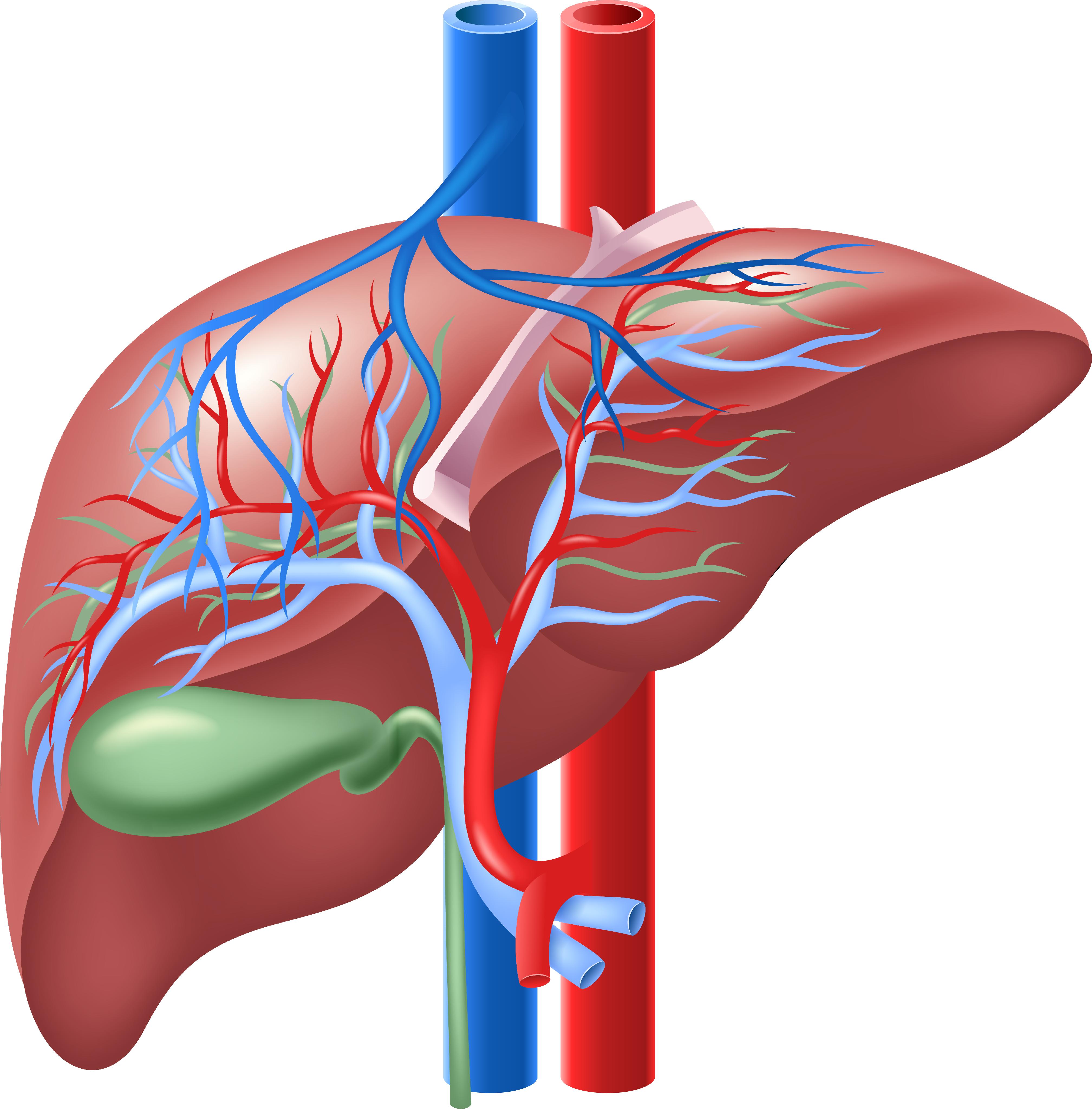
Bile ducts are tubular structures that carry bile from the liver to the gallbladder for storage or directly to the small intestine to aid in digestion. There are two types of bile ducts: intrahepatic and extrahepatic. Intrahepatic bile ducts are located within the liver and drain bile from liver cells, while extrahepatic bile ducts are outside the liver and include the common hepatic duct, cystic duct, and common bile duct. These ducts can become obstructed or inflamed, leading to various medical conditions such as cholestasis, cholecystitis, and gallstones.
The common bile duct is a duct that results from the union of the cystic duct (which drains bile from the gallbladder) and the common hepatic duct (which drains bile from the liver). The common bile duct transports bile, a digestive enzyme, from the liver and gallbladder to the duodenum, which is the first part of the small intestine.
The common bile duct runs through the head of the pancreas before emptying into the second part of the duodenum, either alone or in conjunction with the pancreatic duct, via a small opening called the ampulla of Vater. The common bile duct plays a crucial role in the digestion of fats by helping to break them down into smaller molecules that can be absorbed by the body.
Bile is a digestive fluid that is produced by the liver and stored in the gallbladder. It plays an essential role in the digestion and absorption of fats and fat-soluble vitamins in the small intestine. Bile consists of bile salts, bilirubin, cholesterol, phospholipids, electrolytes, and water.
Bile salts are amphipathic molecules that help to emulsify fats into smaller droplets, increasing their surface area and allowing for more efficient digestion by enzymes such as lipase. Bilirubin is a breakdown product of hemoglobin from red blood cells and gives bile its characteristic greenish-brown color.
Bile is released into the small intestine in response to food, particularly fats, entering the digestive tract. It helps to break down large fat molecules into smaller ones that can be absorbed through the walls of the intestines and transported to other parts of the body for energy or storage.
Bile acids and salts are naturally occurring steroidal compounds that play a crucial role in the digestion and absorption of lipids (fats) in the body. They are produced in the liver from cholesterol and then conjugated with glycine or taurine to form bile acids, which are subsequently converted into bile salts by the addition of a sodium or potassium ion.
Bile acids and salts are stored in the gallbladder and released into the small intestine during digestion, where they help emulsify fats, allowing them to be broken down into smaller molecules that can be absorbed by the body. They also aid in the elimination of waste products from the liver and help regulate cholesterol metabolism.
Abnormalities in bile acid synthesis or transport can lead to various medical conditions, such as cholestatic liver diseases, gallstones, and diarrhea. Therefore, understanding the role of bile acids and salts in the body is essential for diagnosing and treating these disorders.
Extrahepatic bile ducts refer to the portion of the biliary system that lies outside the liver. The biliary system is responsible for producing, storing, and transporting bile, a digestive fluid produced by the liver.
The extrahepatic bile ducts include:
1. The common hepatic duct: This duct is formed by the union of the right and left hepatic ducts, which drain bile from the corresponding lobes of the liver.
2. The cystic duct: This short duct connects the gallbladder to the common hepatic duct, allowing bile to flow into the gallbladder for storage and concentration.
3. The common bile duct: This is the result of the fusion of the common hepatic duct and the cystic duct. It transports bile from the liver and gallbladder to the duodenum, the first part of the small intestine, where it aids in fat digestion.
4. The ampulla of Vater (or hepatopancreatic ampulla): This is a dilated area where the common bile duct and the pancreatic duct join and empty their contents into the duodenum through a shared opening called the major duodenal papilla.
Extrahepatic bile ducts can be affected by various conditions, such as gallstones, inflammation (cholangitis), strictures, or tumors, which may require medical or surgical intervention.
Bile duct neoplasms, also known as cholangiocarcinomas, refer to a group of malignancies that arise from the bile ducts. These are the tubes that carry bile from the liver to the gallbladder and small intestine. Bile duct neoplasms can be further classified based on their location as intrahepatic (within the liver), perihilar (at the junction of the left and right hepatic ducts), or distal (in the common bile duct).
These tumors are relatively rare, but their incidence has been increasing in recent years. They can cause a variety of symptoms, including jaundice, abdominal pain, weight loss, and fever. The diagnosis of bile duct neoplasms typically involves imaging studies such as CT or MRI scans, as well as blood tests to assess liver function. In some cases, a biopsy may be necessary to confirm the diagnosis.
Treatment options for bile duct neoplasms depend on several factors, including the location and stage of the tumor, as well as the patient's overall health. Surgical resection is the preferred treatment for early-stage tumors, while chemotherapy and radiation therapy may be used in more advanced cases. For patients who are not candidates for surgery, palliative treatments such as stenting or bypass procedures may be recommended to relieve symptoms and improve quality of life.
Intrahepatic bile ducts are the small tubular structures inside the liver that collect bile from the liver cells (hepatocytes). Bile is a digestive fluid produced by the liver that helps in the absorption of fats and fat-soluble vitamins from food. The intrahepatic bile ducts merge to form larger ducts, which eventually exit the liver and join with the cystic duct from the gallbladder to form the common bile duct. The common bile duct then empties into the duodenum, the first part of the small intestine, where bile aids in digestion. Intrahepatic bile ducts can become obstructed or damaged due to various conditions such as gallstones, tumors, or inflammation, leading to complications like jaundice, liver damage, and infection.
Cholestasis is a medical condition characterized by the interruption or reduction of bile flow from the liver to the small intestine. Bile is a digestive fluid produced by the liver that helps in the breakdown and absorption of fats. When the flow of bile is blocked or reduced, it can lead to an accumulation of bile components, such as bilirubin, in the blood, which can cause jaundice, itching, and other symptoms.
Cholestasis can be caused by various factors, including liver diseases (such as hepatitis, cirrhosis, or cancer), gallstones, alcohol abuse, certain medications, pregnancy, and genetic disorders. Depending on the underlying cause, cholestasis may be acute or chronic, and it can range from mild to severe in its symptoms and consequences. Treatment for cholestasis typically involves addressing the underlying cause and managing the symptoms with supportive care.
The pancreatic ducts are a set of tubular structures within the pancreas that play a crucial role in the digestive system. The main pancreatic duct, also known as the duct of Wirsung, is responsible for transporting pancreatic enzymes and bicarbonate-rich fluid from the pancreas to the duodenum, which is the first part of the small intestine.
The exocrine portion of the pancreas contains numerous smaller ducts called interlobular ducts and intralobular ducts that merge and ultimately join the main pancreatic duct. This system ensures that the digestive enzymes and fluids produced by the pancreas are effectively delivered to the small intestine, where they aid in the breakdown and absorption of nutrients from food.
In addition to the main pancreatic duct, there is an accessory pancreatic duct, also known as Santorini's duct, which can sometimes join the common bile duct before emptying into the duodenum through a shared opening called the ampulla of Vater. However, in most individuals, the accessory pancreatic duct usually drains into the main pancreatic duct before entering the duodenum.
Gallstones are small, hard deposits that form in the gallbladder, a small organ located under the liver. They can range in size from as small as a grain of sand to as large as a golf ball. Gallstones can be made of cholesterol, bile pigments, or calcium salts, or a combination of these substances.
There are two main types of gallstones: cholesterol stones and pigment stones. Cholesterol stones are the most common type and are usually yellow-green in color. They form when there is too much cholesterol in the bile, which causes it to become saturated and form crystals that eventually grow into stones. Pigment stones are smaller and darker in color, ranging from brown to black. They form when there is an excess of bilirubin, a waste product produced by the breakdown of red blood cells, in the bile.
Gallstones can cause symptoms such as abdominal pain, nausea, vomiting, and bloating, especially after eating fatty foods. In some cases, gallstones can lead to serious complications, such as inflammation of the gallbladder (cholecystitis), infection, or blockage of the bile ducts, which can cause jaundice, a yellowing of the skin and eyes.
The exact cause of gallstones is not fully understood, but risk factors include being female, older age, obesity, a family history of gallstones, rapid weight loss, diabetes, and certain medical conditions such as cirrhosis or sickle cell anemia. Treatment for gallstones may involve medication to dissolve the stones, shock wave therapy to break them up, or surgery to remove the gallbladder.
Adenoma of the bile duct is a benign (noncancerous) tumor that develops in the bile ducts, which are tiny tubes that carry bile from the liver to the gallbladder and small intestine. Bile is a digestive fluid produced by the liver.
Bile duct adenomas are rare and usually do not cause any symptoms. However, if they grow large enough, they may obstruct the flow of bile and cause jaundice (yellowing of the skin and whites of the eyes), abdominal pain, or itching. In some cases, bile duct adenomas may become cancerous and develop into bile duct carcinomas.
The exact cause of bile duct adenomas is not known, but they are more common in people with certain genetic disorders, such as Gardner's syndrome and von Hippel-Lindau disease. Treatment for bile duct adenomas typically involves surgical removal of the tumor.
Common bile duct neoplasms refer to abnormal growths that can occur in the common bile duct, which is a tube that carries bile from the liver and gallbladder into the small intestine. These growths can be benign or malignant (cancerous).
Benign neoplasms of the common bile duct include papillomas, adenomas, and leiomyomas. Malignant neoplasms are typically adenocarcinomas, which arise from the glandular cells lining the duct. Other types of malignancies that can affect the common bile duct include cholangiocarcinoma, gallbladder carcinoma, and metastatic cancer from other sites.
Symptoms of common bile duct neoplasms may include jaundice (yellowing of the skin and eyes), abdominal pain, dark urine, and light-colored stools. Diagnosis may involve imaging tests such as CT scans or MRCP (magnetic resonance cholangiopancreatography) and biopsy to confirm the type of neoplasm. Treatment options depend on the type and stage of the neoplasm and may include surgery, radiation therapy, chemotherapy, or a combination of these approaches.
Endoscopic retrograde cholangiopancreatography (ERCP) is a medical procedure that combines upper gastrointestinal (GI) endoscopy and fluoroscopy to diagnose and treat certain problems of the bile ducts and pancreas.
During ERCP, a flexible endoscope (a long, thin, lighted tube with a camera on the end) is passed through the patient's mouth and throat, then through the stomach and into the first part of the small intestine (duodenum). A narrow plastic tube (catheter) is then inserted through the endoscope and into the bile ducts and/or pancreatic duct. Contrast dye is injected through the catheter, and X-rays are taken to visualize the ducts.
ERCP can be used to diagnose a variety of conditions affecting the bile ducts and pancreas, including gallstones, tumors, strictures (narrowing of the ducts), and chronic pancreatitis. It can also be used to treat certain conditions, such as removing gallstones from the bile duct or placing stents to keep the ducts open in cases of stricture.
ERCP is an invasive procedure that carries a risk of complications, including pancreatitis, infection, bleeding, and perforation (a tear in the lining of the GI tract). It should only be performed by experienced medical professionals in a hospital setting.
Cholangiography is a medical procedure that involves taking X-ray images of the bile ducts (the tubes that carry bile from the liver to the small intestine). This is typically done by injecting a contrast dye into the bile ducts through an endoscope or a catheter that has been inserted into the body.
There are several types of cholangiography, including:
* Endoscopic retrograde cholangiopancreatography (ERCP): This procedure involves inserting an endoscope through the mouth and down the throat into the small intestine. A dye is then injected into the bile ducts through a small tube that is passed through the endoscope.
* Percutaneous transhepatic cholangiography (PTC): This procedure involves inserting a needle through the skin and into the liver to inject the contrast dye directly into the bile ducts.
* Operative cholangiography: This procedure is performed during surgery to examine the bile ducts for any abnormalities or blockages.
Cholangiography can help diagnose a variety of conditions that affect the bile ducts, such as gallstones, tumors, or inflammation. It can also be used to guide treatment decisions, such as whether surgery is necessary to remove a blockage.
The common hepatic duct is a medical term that refers to the duct in the liver responsible for carrying bile from the liver. More specifically, it is the duct that results from the convergence of the right and left hepatic ducts, which themselves carry bile from the right and left lobes of the liver, respectively. The common hepatic duct then joins with the cystic duct from the gallbladder to form the common bile duct, which ultimately drains into the duodenum, a part of the small intestine.
The primary function of the common hepatic duct is to transport bile, a digestive juice produced by the liver, to the small intestine. Bile helps break down fats during the digestion process, making it possible for the body to absorb them properly. Any issues or abnormalities in the common hepatic duct can lead to problems with bile flow and potentially cause health complications such as jaundice, gallstones, or liver damage.
Extrahepatic cholestasis is a medical condition characterized by the impaired flow of bile outside of the liver. Bile is a digestive fluid produced by the liver that helps in the absorption and digestion of fats. When the flow of bile is obstructed or blocked, it can lead to an accumulation of bile components, such as bilirubin, in the bloodstream, resulting in jaundice, dark urine, light-colored stools, and itching.
Extrahepatic cholestasis can be caused by various factors, including gallstones, tumors, strictures, or inflammation of the bile ducts. It is essential to diagnose and treat extrahepatic cholestasis promptly to prevent further complications, such as liver damage or infection. Treatment options may include medications, endoscopic procedures, or surgery, depending on the underlying cause of the condition.
The cystic duct is a short tube that connects the gallbladder to the common bile duct, which carries bile from the liver and gallbladder into the small intestine. The cystic duct allows bile to flow from the gallbladder into the common bile duct when it is needed for digestion. It is a part of the biliary system and plays an important role in the digestive process.
Cholelithiasis is a medical term that refers to the presence of gallstones in the gallbladder. The gallbladder is a small pear-shaped organ located beneath the liver that stores bile, a digestive fluid produced by the liver. Gallstones are hardened deposits that can form in the gallbladder when substances in the bile, such as cholesterol or bilirubin, crystallize.
Gallstones can vary in size and may be as small as a grain of sand or as large as a golf ball. Some people with gallstones may not experience any symptoms, while others may have severe abdominal pain, nausea, vomiting, fever, and jaundice (yellowing of the skin and eyes) if the gallstones block the bile ducts.
Cholelithiasis is a common condition that affects millions of people worldwide, particularly women over the age of 40 and those with certain medical conditions such as obesity, diabetes, and rapid weight loss. If left untreated, gallstones can lead to serious complications such as inflammation of the gallbladder (cholecystitis), infection, or pancreatitis (inflammation of the pancreas). Treatment options for cholelithiasis include medication, shock wave lithotripsy (breaking up the gallstones with sound waves), and surgery to remove the gallbladder (cholecystectomy).
The thoracic duct is the largest lymphatic vessel in the human body. It is a part of the lymphatic system, which helps to regulate fluid balance and immune function. The thoracic duct originates from the cisterna chyli, a dilated sac located in the abdomen near the aorta.
The thoracic duct collects lymph from the lower extremities, abdomen, pelvis, and left side of the thorax (chest). It ascends through the diaphragm and enters the chest, where it passes through the mediastinum (the central part of the chest between the lungs) and eventually drains into the left subclavian vein.
The thoracic duct plays a crucial role in transporting lymphatic fluid, which contains white blood cells, fats, proteins, and other substances, back into the circulatory system. Any obstruction or damage to the thoracic duct can lead to lymph accumulation in the surrounding tissues, causing swelling and other symptoms.
Laparoscopic cholecystectomy is a surgical procedure to remove the gallbladder using a laparoscope, a thin tube with a camera, which allows the surgeon to view the internal structures on a video monitor. The surgery is performed through several small incisions in the abdomen, rather than a single large incision used in open cholecystectomy. This approach results in less postoperative pain, fewer complications, and shorter recovery time compared to open cholecystectomy.
The procedure is typically indicated for symptomatic gallstones or chronic inflammation of the gallbladder (cholecystitis), which can cause severe abdominal pain, nausea, vomiting, and fever. Laparoscopic cholecystectomy has become the standard of care for gallbladder removal due to its minimally invasive nature and excellent outcomes.
The biliary tract is a system of ducts that transport bile from the liver to the gallbladder and then to the small intestine. Bile is a digestive fluid produced by the liver that helps in the breakdown and absorption of fats in the small intestine. The main components of the biliary tract are:
1. Intrahepatic bile ducts: These are the smaller branches of bile ducts located within the liver that collect bile from the liver cells or hepatocytes.
2. Gallbladder: A small pear-shaped organ located beneath the liver, which stores and concentrates bile received from the intrahepatic bile ducts. The gallbladder releases bile into the small intestine when food is ingested, particularly fats, to aid digestion.
3. Common hepatic duct: This is a duct that forms by the union of the right and left hepatic ducts, which carry bile from the right and left lobes of the liver, respectively.
4. Cystic duct: A short duct that connects the gallbladder to the common hepatic duct, forming the beginning of the common bile duct.
5. Common bile duct: This is a larger duct formed by the union of the common hepatic duct and the cystic duct. It carries bile from the liver and gallbladder into the small intestine.
6. Pancreatic duct: A separate duct that originates from the pancreas, a gland located near the liver and stomach. The pancreatic duct joins the common bile duct just before they both enter the duodenum, the first part of the small intestine.
7. Ampulla of Vater: This is the dilated portion where the common bile duct and the pancreatic duct join together and empty their contents into the duodenum through a shared opening called the papilla of Vater.
Disorders related to the biliary tract include gallstones, cholecystitis (inflammation of the gallbladder), bile duct stones, bile duct strictures or obstructions, and primary sclerosing cholangitis, among others.
Obstructive Jaundice is a medical condition characterized by the yellowing of the skin, sclera (whites of the eyes), and mucous membranes due to the accumulation of bilirubin in the bloodstream. This occurs when there is an obstruction or blockage in the bile ducts that transport bile from the liver to the small intestine.
Bile, which contains bilirubin, aids in digestion and is usually released from the liver into the small intestine. When the flow of bile is obstructed, bilirubin builds up in the blood, causing jaundice. The obstruction can be caused by various factors, such as gallstones, tumors, or strictures in the bile ducts.
Obstructive jaundice may present with additional symptoms like dark urine, light-colored stools, itching, abdominal pain, and weight loss, depending on the cause and severity of the obstruction. It is essential to seek medical attention if jaundice is observed, as timely diagnosis and management can prevent potential complications, such as liver damage or infection.
Cholecystectomy is a medical procedure to remove the gallbladder, a small pear-shaped organ located on the right side of the abdomen, just beneath the liver. The primary function of the gallbladder is to store and concentrate bile, a digestive fluid produced by the liver. During a cholecystectomy, the surgeon removes the gallbladder, usually due to the presence of gallstones or inflammation that can cause pain, infection, or other complications.
There are two primary methods for performing a cholecystectomy:
1. Open Cholecystectomy: In this traditional surgical approach, the surgeon makes an incision in the abdomen to access and remove the gallbladder. This method is typically used when there are complications or unique circumstances that make laparoscopic surgery difficult or risky.
2. Laparoscopic Cholecystectomy: This is a minimally invasive surgical procedure where the surgeon makes several small incisions in the abdomen, through which a thin tube with a camera (laparoscope) and specialized surgical instruments are inserted. The surgeon then guides these tools to remove the gallbladder while viewing the internal structures on a video monitor.
After the gallbladder is removed, bile flows directly from the liver into the small intestine through the common bile duct, and the body continues to function normally without any significant issues.
Cholangitis is a medical condition characterized by inflammation of the bile ducts, which are the tubes that carry bile from the liver to the small intestine. Bile is a digestive juice produced by the liver that helps break down fats in food.
There are two types of cholangitis: acute and chronic. Acute cholangitis is a sudden and severe infection that can cause symptoms such as abdominal pain, fever, jaundice (yellowing of the skin and eyes), and dark urine. It is usually caused by a bacterial infection that enters the bile ducts through a blockage or obstruction.
Chronic cholangitis, on the other hand, is a long-term inflammation of the bile ducts that can lead to scarring and narrowing of the ducts. This can cause symptoms such as abdominal pain, itching, and jaundice. Chronic cholangitis can be caused by various factors, including primary sclerosing cholangitis (an autoimmune disease), bile duct stones, or tumors in the bile ducts.
Treatment for cholangitis depends on the underlying cause of the condition. Antibiotics may be used to treat bacterial infections, and surgery may be necessary to remove blockages or obstructions in the bile ducts. In some cases, medications may be prescribed to manage symptoms and prevent further complications.
Biliary tract surgical procedures refer to a range of operations that involve the biliary system, which includes the liver, gallbladder, and bile ducts. These procedures can be performed for various reasons, including the treatment of gallstones, bile duct injuries, tumors, or other conditions affecting the biliary tract. Here are some examples of biliary tract surgical procedures:
1. Cholecystectomy: This is the surgical removal of the gallbladder, which is often performed to treat symptomatic gallstones or chronic cholecystitis (inflammation of the gallbladder). It can be done as an open procedure or laparoscopically.
2. Bile duct exploration: This procedure involves opening the common bile duct to remove stones, strictures, or tumors. It is often performed during a cholecystectomy if there is suspicion of common bile duct involvement.
3. Hepaticojejunostomy: This operation connects the liver's bile ducts directly to a portion of the small intestine called the jejunum, bypassing a damaged or obstructed segment of the biliary tract. It is often performed for benign or malignant conditions affecting the bile ducts.
4. Roux-en-Y hepaticojejunostomy: This procedure involves creating a Y-shaped limb of jejunum and connecting it to the liver's bile ducts, bypassing the common bile duct and duodenum. It is often performed for complex biliary tract injuries or malignancies.
5. Whipple procedure (pancreaticoduodenectomy): This extensive operation involves removing the head of the pancreas, the duodenum, a portion of the jejunum, the gallbladder, and the common bile duct. It is performed for malignancies involving the pancreas, bile duct, or duodenum.
6. Liver resection: This procedure involves removing a portion of the liver to treat primary liver tumors (hepatocellular carcinoma or cholangiocarcinoma) or metastatic cancer from other organs.
7. Biliary stenting or bypass: These minimally invasive procedures involve placing a stent or creating a bypass to relieve bile duct obstructions caused by tumors, strictures, or stones. They can be performed endoscopically (ERCP) or percutaneously (PTC).
8. Cholecystectomy: This procedure involves removing the gallbladder, often for symptomatic cholelithiasis (gallstones) or cholecystitis (inflammation of the gallbladder). It can be performed laparoscopically or open.
9. Biliary drainage: This procedure involves placing a catheter to drain bile from the liver or bile ducts, often for acute or chronic obstructions caused by tumors, strictures, or stones. It can be performed endoscopically (ERCP) or percutaneously (PTC).
10. Bilioenteric anastomosis: This procedure involves connecting the biliary tract to a portion of the small intestine, often for benign or malignant conditions affecting the bile ducts or pancreas. It can be performed open or laparoscopically.
Ligation, in the context of medical terminology, refers to the process of tying off a part of the body, usually blood vessels or tissue, with a surgical suture or another device. The goal is to stop the flow of fluids such as blood or other substances within the body. It is commonly used during surgeries to control bleeding or to block the passage of fluids, gases, or solids in various parts of the body.
Choledocholithiasis is a medical condition characterized by the presence of one or more gallstones in the common bile duct, which is the tube that carries bile from the liver and gallbladder to the small intestine. Bile is a digestive fluid produced by the liver that helps break down fats in the small intestine. Gallstones are hardened deposits of digestive fluids that can form in the gallbladder or, less commonly, in the bile ducts.
Choledocholithiasis can cause a variety of symptoms, including abdominal pain, jaundice (yellowing of the skin and eyes), nausea, vomiting, and fever. If left untreated, it can lead to serious complications such as infection or inflammation of the bile ducts or pancreas, which can be life-threatening.
The condition is typically diagnosed through imaging tests such as ultrasound, CT scan, or MRI, and may require endoscopic or surgical intervention to remove the gallstones from the common bile duct.
Endoscopic sphincterotomy is a medical procedure that involves the use of an endoscope (a flexible tube with a light and camera) to cut the papilla of Vater, which contains the sphincter of Oddi muscle. This procedure is typically performed to treat gallstones or to manage other conditions related to the bile ducts or pancreatic ducts.
The sphincterotomy helps to widen the opening of the papilla, allowing stones or other obstructions to pass through more easily. It may also be used to relieve pressure and pain caused by spasms of the sphincter of Oddi muscle. The procedure is usually done under sedation or anesthesia and carries a risk of complications such as bleeding, infection, perforation, and pancreatitis.
The gallbladder is a small, pear-shaped organ located just under the liver in the right upper quadrant of the abdomen. Its primary function is to store and concentrate bile, a digestive enzyme produced by the liver, which helps in the breakdown of fats during the digestion process. When food, particularly fatty foods, enter the stomach and small intestine, the gallbladder contracts and releases bile through the common bile duct into the duodenum, the first part of the small intestine, to aid in fat digestion.
The gallbladder is made up of three main parts: the fundus, body, and neck. It has a muscular wall that allows it to contract and release bile. Gallstones, an inflammation of the gallbladder (cholecystitis), or other gallbladder diseases can cause pain, discomfort, and potentially serious health complications if left untreated.
Biliary tract diseases refer to a group of medical conditions that affect the biliary system, which includes the gallbladder, bile ducts, and liver. Bile is a digestive juice produced by the liver, stored in the gallbladder, and released into the small intestine through the bile ducts to help digest fats.
Biliary tract diseases can cause various symptoms such as abdominal pain, jaundice, fever, nausea, vomiting, and changes in stool color. Some of the common biliary tract diseases include:
1. Gallstones: Small, hard deposits that form in the gallbladder or bile ducts made up of cholesterol or bilirubin.
2. Cholecystitis: Inflammation of the gallbladder, often caused by gallstones.
3. Cholangitis: Infection or inflammation of the bile ducts.
4. Biliary dyskinesia: A motility disorder that affects the contraction and relaxation of the muscles in the biliary system.
5. Primary sclerosing cholangitis: A chronic autoimmune disease that causes scarring and narrowing of the bile ducts.
6. Biliary tract cancer: Rare cancers that affect the gallbladder, bile ducts, or liver.
Treatment for biliary tract diseases varies depending on the specific condition and severity but may include medications, surgery, or a combination of both.
Salivary ducts are the excretory tubules that transport saliva from the major and minor salivary glands to the oral cavity. The main function of these ducts is to convey the salivary secretions, which contain enzymes and lubricants, into the mouth to aid in digestion, speech, and swallowing.
There are two pairs of major salivary glands: the parotid glands and the submandibular glands. Each pair has its own set of ducts. The parotid gland's saliva is drained through the parotid duct, also known as Stensen's duct, which opens into the oral cavity opposite the upper second molar tooth. The submandibular gland's saliva is transported through the submandibular duct, or Wharton's duct, which empties into the floor of the mouth near the base of the tongue.
Minor salivary glands are scattered throughout the oral cavity and pharynx, and their secretions are drained via small ducts directly into the oral mucosa.
The liver is a large, solid organ located in the upper right portion of the abdomen, beneath the diaphragm and above the stomach. It plays a vital role in several bodily functions, including:
1. Metabolism: The liver helps to metabolize carbohydrates, fats, and proteins from the food we eat into energy and nutrients that our bodies can use.
2. Detoxification: The liver detoxifies harmful substances in the body by breaking them down into less toxic forms or excreting them through bile.
3. Synthesis: The liver synthesizes important proteins, such as albumin and clotting factors, that are necessary for proper bodily function.
4. Storage: The liver stores glucose, vitamins, and minerals that can be released when the body needs them.
5. Bile production: The liver produces bile, a digestive juice that helps to break down fats in the small intestine.
6. Immune function: The liver plays a role in the immune system by filtering out bacteria and other harmful substances from the blood.
Overall, the liver is an essential organ that plays a critical role in maintaining overall health and well-being.
Bile pigments are the yellow-brown colored end products of hemoglobin breakdown in the liver. Hemoglobin is a protein found in red blood cells that carries oxygen throughout the body. When these cells are broken down, heme (the non-protein part of hemoglobin) is converted into biliverdin, which is then converted into bilirubin. Bilirubin is further metabolized and excreted by the liver as a component of bile, a digestive fluid that helps break down fats in the small intestine.
Under normal conditions, the liver effectively removes and excretes bilirubin from the body through the bile ducts into the small intestine. However, when there is an overproduction of bilirubin or a problem with its elimination, it can accumulate in the blood, leading to jaundice (yellowing of the skin and eyes) and other symptoms associated with liver dysfunction.
In summary, bile pigments are the waste products formed during the breakdown of hemoglobin, primarily consisting of bilirubin, which is eliminated from the body via the liver and bile ducts.
The ampulla of Vater, also known as hepatopancreatic ampulla, is a dilated portion of the common bile duct where it joins the main pancreatic duct and empties into the second part of the duodenum. It serves as a conduit for both bile from the liver and digestive enzymes from the pancreas to reach the small intestine, facilitating the digestion and absorption of nutrients. The ampulla of Vater is surrounded by a muscular sphincter, the sphincter of Oddi, which controls the flow of these secretions into the duodenum.
Bile canaliculi are the smallest bile-transporting structures in the liver. They are formed by the close apposition of hepatocyte (liver cell) plasma membranes, and they are responsible for the majority of bile production. The bile canaliculi merge to form bile ductules, which then merge to form larger bile ducts that transport bile to the gallbladder and small intestine. Bile is a fluid that contains water, electrolytes, bile salts, cholesterol, phospholipids, and bilirubin, which are produced by the liver and play important roles in digestion and elimination of waste products.
 Vanishing bile duct syndrome
Cholecystitis
Signs and symptoms
Trepibutone
Abdomen
Ethinylestradiol
Cirrhosis
Ursodeoxycholic acid
Hepatomegaly
Hans Kehr
CA19-9
Hans Huchzermeyer
Caroli disease
Liver disease
Obeticholic acid
Urinalysis
Fasciolosis
Liver support system
Primary sclerosing cholangitis
Common bile duct
Daniel Alagille
List of MeSH codes (C06)
Abdominal ultrasonography
ENPP3
Cholangiocarcinoma
Common bile duct stone
John Bland-Sutton
Caroli
Cholecystography
List of hepato-biliary diseases
 Bile Duct Diseases - Multiple Languages: MedlinePlus
 Bile Duct Diseases Locations NYC | Mount Sinai - New York
 Liver, Bile Duct and Pancreas Disease | Mount Sinai South Nassau
 Artificial bile ducts could help treat liver disease in children | Business Weekly | Technology News | Business news |...
 Bile Duct Diseases | University of Miami Health System
 Bile Duct Cancer
(Cholangiocarcinoma) (15234) ... This is used to detect diseases that affect the bile ducts including bile duct cancer, gallstones stuck in the bile duct, and ... This is used to detect diseases that affect the bile ducts including bile duct cancer, gallstones stuck in the bile duct, and ...
 GastroHERBAL, fatty liver disease and gallstones, bile duct UK
Gallstones - Bile Duct and Gallbladder Diseases - Gastroenterology - Diseases - McMaster Textbook of Internal Medicine
 Bile Duct Cancer (Cholangiocarcinoma) | Johns Hopkins Medicine
Vanishing bile duct syndrome - Wikipedia
 Cholangiocarcinoma | Bile Duct Cancer, Asbestos Link
 Zubsolv: Dosage, side effects, uses, and more
 Other Infections | www.hivandhepatitis.com
 Andrew Smith, MD | Cleveland Clinic
 Xanthoma disseminatum with progressive involvement of the central nervous and hepatobiliary systems
 Duke Gastroenterology Clinic - Clinic 2J | Durham, NC | Duke Health
 Biliary Obstruction: Background, Pathophysiology, Etiology
 CDC - Liver Flukes
 Gastroenterologists Near Me in Chaska, MN - Sharecare
 Alternative Treatments for Intrahepatic Bile Duct Cancer | New Hope Unlimited
 Cirrhosis of the Liver: Symptoms, Causes & Treatments
 Accessory Organs - Digestive Diseases Nutrition | UCLA Health
Advanced Search Results - Public Health Image Library(PHIL)
 IndexCat
The liver: Structure, function, and disease
 CenterWatch
 Cirrhosis - Harvard Health
 Virginia Expert Witnesses For Hire | Round Table Group
 MRCP (MR Cholangiopancreatography)
 10-month-old baby gets life-saving liver transplant from a stranger living 2,000 miles away - CBS News
ERCP10
- Doctors can perform an Endoscopic Retrograde Cholangiopancreatogram (ERCP) to diagnose diseases. (gastrohealthpartners.com)
- In an ERCP, they can inject contrast dye to help image your bile ducts during an x-ray. (gastrohealthpartners.com)
- ERCP, in addition to helping with diagnosis, can help treat disease. (gastrohealthpartners.com)
- Doctors can pass tools through the endoscope during an ERCP and open blocked ducts, remove or break up gallstones, insert stents, and even remove tumors. (gastrohealthpartners.com)
- ERCP - an endoscopic technique used to look at the bile duct to see if it is blocked and inflamed. (charlesimber.co.uk)
- When I had the ERCP, a stent was put in one of the ducts. (cancer.org)
- Multicenter comparative evaluation of endoscopic placement of expandable metal stents for malignant distal common bile duct obstruction by ERCP or EUS-guided approach. (medscape.com)
- After validation in cell line-derived EVs, we applied the technology to human bile samples collected through an endoscopic retrograde cholangiopancreatography (ERCP) procedure. (massgeneral.org)
- The molecular analysis of a bile sample could further improve the diagnostic accuracy of the current gold standard pathology diagnosis through ERCP. (massgeneral.org)
- 1.3.3 If the bile duct cannot be cleared with ERCP, use biliary stenting to achieve biliary drainage only as a temporary measure until definitive endoscopic or surgical clearance. (nice.org.uk)
Common bile4
- Another common bile duct condition is cholangitis, which is inflammation in the bile duct system. (gastrohealthpartners.com)
- 1.3.1 Offer bile duct clearance and laparoscopic cholecystectomy to people with symptomatic or asymptomatic common bile duct stones. (nice.org.uk)
- Diseases in any part of the ductal system of the BILIARY TRACT from the smallest BILE CANALICULI to the largest COMMON BILE DUCT . (bvsalud.org)
- Notably, laparoscopic methods are not recommended when disease of the common bile duct or gallbladder is suspected that may necessitate a decompressive biliary procedure, cholecystectomy, choledochotomy, cholelith removal, or cholecystoenterostomy (ie, cholecystoduodenostomy, cholecystojejunostomy). (merckvetmanual.com)
Requiring endoscopic2
- This effect was restricted to patients with severe disease, as defined by development of dominant stenosis (DS) requiring endoscopic intervention. (nih.gov)
- There is a wide range of patients I treat, from patients with simple bile duct stones to patients with pancreatic cancer requiring endoscopic procedures to relief symptoms such as pain and yellowing of the skin. (dukehealth.org)
Intrahepatic bile duct4
- Mycobacterial diseases, sarcoidosis, fungal diseases, and pneumoconiosis are all possible etiologies for such radiologic findings.The CT scan of the abdomen (Figures 4 and 5) showed marked hepatomegaly, with no evidence of extrahepatic or intrahepatic bile duct dilation. (medscape.com)
- Intrahepatic bile duct atresia (Alagille syndrome) (ALGS2 MIM:610205 and ALGS1 MIM:118450) Extrahepatic bile duct atresia Autosomal recessive polycystic kidney disease Congential hepatic fibrosis Caroli's disease Von Meyenburg complex Trisomy 17, 18 and 21 Cystic fibrosis Alpha 1 antitrypsin deficiency Trihydroxycoprostanic acidemia Byler's disease Bile duct injury and loss can result from autoimmune destruction. (wikipedia.org)
- As proof of principle, we applied DUCT to a mouse model for Alagille syndrome ( Jag1 Ndr/Ndr mice), characterized by intrahepatic bile duct paucity, that can spontaneously generate a biliary system in adulthood. (elifesciences.org)
- The cancer has a 5-year relative survival rate of 25% for localized intrahepatic bile duct cancers and just. (ascopost.com)
Gallstones6
- Gallstones are one common issue for bile ducts. (gastrohealthpartners.com)
- If small gallstones are washed out of the gallbladder and try and exit the bile duct but get stuck, this blocks the duct. (charlesimber.co.uk)
- My ducts were closing because of the cancer and not the gallstones. (cancer.org)
- For instance, ulcerative colitis or stones similar to gallstones can cause inflammation of the bile duct. (cancer.net)
- Helicobacter DNA was detected in the gallbladder tissue and bile of 28% and 18% respectively of the patients, but was not detected in any of the gallstones. (who.int)
- Examples of gastrointestinal (GI) diseases include ulcers, gallstones, and liver diseases. (westhillshospital.com)
Diagnosed with bile duct c3
- Still, these tests can sometimes be useful after a person is diagnosed with bile duct cancer. (cancer.org)
- I was diagnosed with bile duct cancer in July 2009. (cancer.org)
- If you are diagnosed with bile duct cancer, your doctor may check for these tumour markers: CEA (carcinoembryonic antigen) and CA 19-9 (carbohydrate antigen 19-9). (bccancer.bc.ca)
Obstruction3
- I see patients mostly for gallbladder disease, appendicitis, bowel obstruction, hernias and colon disease. (healthtap.com)
- Gastrointestinal (GI) stents are designed for palliative therapy for various diseases causing obstruction in the GI tract. (marketsandmarkets.com)
- The addition of nimotuzumab, an EGFR-targeting monoclonal antibody, to gemcitabine increased overall survival in patients with KRAS wild-type advanced pancreatic cancer, particularly those who did not need surgery for obstruction of a pancreatic bile duct, according to data from the phase III. (ascopost.com)
Jaundice3
- If attacks start to make everyday life difficult, and the patient starts to show jaundice, their gallstone disease has become more serious. (charlesimber.co.uk)
- 1 2 Clinical symptoms of Caroli's disease include right upper quadrant abdominal pain, jaundice, and recurrent cholangitis. (e-cmh.org)
- Physical examination revealed only hepatomegaly, without stigmata of chronic liver disease or jaundice. (medscape.com)
Cholangiocarcinoma5
- Cholangiocarcinoma (CCA), cancer that forms in the bile ducts, is a fatal disease that is often detected after it has spread too far to be surgically removed. (massgeneral.org)
- We wanted to see if molecular analysis of extracellular vesicles (EVs) in human bile samples could accurately detect patients with cholangiocarcinoma from patients with other benign or inflammatory conditions. (massgeneral.org)
- Primary sclerosing cholangitis (PSC) is a cholestatic liver disease that affects the bile ducts, which can have severe consequences including cirrhosis and cholangiocarcinoma. (medpagetoday.com)
- Bile duct adenocarcinoma is also called cholangiocarcinoma. (bccancer.bc.ca)
- Perhaps even more so when the type of cancer is rare, like bile duct cancer, also known as cholangiocarcinoma. (allsup.com)
Cancers8
- Most bile duct cancers aren't found until a person goes to a doctor because they have symptoms. (cancer.org)
- Also, not all bile duct cancers make these tumor markers, so low or normal levels don't always mean cancer is not present. (cancer.org)
- Growth of this market can be attributed to the rising prevalence of GI cancers and other digestive diseases, changing lifestyles, increasing healthcare expenditure on gastrointestinal procedures, and an increasing preference for minimally invasive surgeries. (marketsandmarkets.com)
- Over 90% (90 out of 100) of bile duct cancers are adenocarcinomas. (bccancer.bc.ca)
- These cancers begin in the mucus glands lining the inside of the bile duct. (bccancer.bc.ca)
- Other bile duct cancers include squamous cell carcinoma, lymphoma and sarcoma. (bccancer.bc.ca)
- Only about 10% (10 out of 100) bile duct cancers are intrahepatic. (bccancer.bc.ca)
- The majority of patients with bile duct cancers are diagnosed when the cancer is far too advanced to be removed by surgery. (allsup.com)
Pancreas and bile ducts2
- I am currently specializing in pancreaticobiliary disease, which is a subspecialty within gastroenterology that focuses on diseases of the pancreas and bile ducts. (dukehealth.org)
- Physicians and surgeons are dedicated to treat diseases of the liver, pancreas and bile ducts with high quality standards. (vejthani.com)
Hepatic12
- Caroli's disease belongs to a group of hepatic fibropolycystic diseases. (e-cmh.org)
- Two types have been described: a pure form or Caroli's disease (type 1) and a complex form associated with congenital hepatic fibrosis or Caroli's syndrome (type 2). (e-cmh.org)
- Vanishing bile duct syndrome is a loose collection of diseases which leads to the injury to hepatic bile ducts and eventual ductopenia. (wikipedia.org)
- The intrahepatic bile ducts develop out of sheets of primitive hepatic epithelial cells in a pattern determined by the branching of the portal vein and surrounding mesenchyme. (bmj.com)
- Congenital hepatic fibrosis (CHF) is an autosomal recessive disease that primarily affects the hepatobiliary and renal systems. (medscape.com)
- It is characterized by hepatic fibrosis, portal hypertension, and renal cystic disease. (medscape.com)
- Congenital hepatic fibrosis is one of the fibropolycystic diseases, which also include Caroli disease , autosomal dominant polycystic kidney disease (ADPKD), and autosomal recessive polycystic kidney disease (ARPKD). (medscape.com)
- Congenital hepatic fibrosis is associated with an impairment of renal functions, usually caused by an ARPKD, which is a severe form of polycystic kidney disease . (medscape.com)
- Congenital hepatic fibrosis is a ductal plate malformation of the small interlobular bile ducts, whereas Caroli disease involves the large intrahepatic bile ducts. (medscape.com)
- The hepatic disease progresses to develop portal hypertension associated with splenomegaly and esophageal varices. (medscape.com)
- Perihilar - Cancer starts in the hepatic duct where the bile ducts join just outside the liver. (bccancer.bc.ca)
- The tumour has spread into the portal vein on both sides of the liver, the common hepatic artery, or other bile ducts on one side of the liver and a main blood vessel on the other side of the liver. (bccancer.bc.ca)
Blockage4
- If levels of these substances are higher, it might point to blockage of the bile duct, but they can't show if it's due to cancer or some other reason. (cancer.org)
- If the blockage is not relieved, the inflammation and swelling in the bile duct can spread to the walls of the gallbladder. (charlesimber.co.uk)
- Do not take valsartan if you have liver disease caused by blockage in the bile duct or any other severe liver disease. (mydr.com.au)
- The dye will show a narrowing or blockage of your bile duct. (bccancer.bc.ca)
Cancer51
- How Do You Test for Bile Duct Cancer? (cancer.org)
- If there's reason to suspect that you might have bile duct cancer, your doctor will want to take your complete medical history to check for risk factors and to learn more about your symptoms. (cancer.org)
- A physical exam is done to look for signs of bile duct cancer or other health problems. (cancer.org)
- If bile duct cancer is suspected, the exam will focus mostly on the abdomen (belly) to check for any lumps, tenderness, or build-up of fluid. (cancer.org)
- If symptoms and/or the results of the physical exam suggest you might have bile duct cancer, tests will be done. (cancer.org)
- People with bile duct cancer may have high blood levels of the markers called CEA and CA 19-9 . (cancer.org)
- Cancer can also occur in the bile ducts. (gastrohealthpartners.com)
- Bile duct cancer is rare and aggressive. (gastrohealthpartners.com)
- One other emerging treatment involves using Radiofrequency Ablation for palliative care to treat the symptoms of bile duct cancer. (gastrohealthpartners.com)
- For example, with patients who have bile duct cancer, surgery can help to remove tumors. (gastrohealthpartners.com)
- This is chronic cholecystitis , which is a risk factor for gallbladder cancer and bile duct cancer. (charlesimber.co.uk)
- Chronic complicated gallstone disease can increase the risk of both bile duct cancer and gallbladder cancer. (charlesimber.co.uk)
- Mount Sinai provides personalized care, whatever type of acute or chronic liver disease you may have, including hepatitis B and hepatitis C, liver cancer, or cirrhosis . (mountsinai.org)
- I have been trying to find an oncologist that has dealt with bile duct cancer but am having a hard time getting a reference. (cancer.org)
- Also, if any of you know an oncologist in California that specializes in bile duct cancer. (cancer.org)
- Hi I also have Bile duct cancer that was diagnosed in june 2008 they removed 40% of my liver.They said they got it all but On my last scan it showed 5 little spots they are not sure what it is yet. (cancer.org)
- One that is more up-to-date about bile duct cancer. (cancer.org)
- Rates of Pierre S. Health behaviors and quality of life of cancer sur- chronic disease (eg, heart disease, asthma) and disability vivors in Massachusetts, 2006: data use for comprehensive were higher among cancer survivors. (cdc.gov)
- People who receive livers from domino donors tend to be older adults or adults who have liver disease or liver cancer . (healthline.com)
- In fact, unlike other forms of cancer, Hodgkins lymphoma is known to be curable even if diagnosed in the later stages of the disease. (home-remedies-for-you.com)
- On the other hand the Hodgkins lymphoma prognosis also includes possible complications in the form of heart disease, lung problems, infertility or the inability to have children in future, thyroid problems, diseases of the bone marrow as well as other forms of cancer. (home-remedies-for-you.com)
- In the past few years, there has been a steep rise in morbidity and mortality from chronic diseases such as esophageal cancer, colorectal cancer, and gastrointestinal-related critical disorders. (marketsandmarkets.com)
- You will find out more about the factors that increase the chance of developing bile duct cancer. (cancer.net)
- What are the risk factors for bile duct cancer? (cancer.net)
- The following factors may raise a person's risk of developing bile duct cancer. (cancer.net)
- Previous disease or irritation of the bile duct are a possible risk factor for this cancer. (cancer.net)
- Older adults are more likely to develop bile duct cancer. (cancer.net)
- Dioxins, nitrosamines, and polychlorinated biphenyls (PCBs) may cause bile duct cancer. (cancer.net)
- Are there ways to prevent bile duct cancer? (cancer.net)
- Researchers continue to look into what factors cause bile duct cancer, including ways to prevent it. (cancer.net)
- Although there is no proven way to completely prevent bile duct cancer, you may be able to lower your risk. (cancer.net)
- Thorium dioxide, a chemical once used in x-ray examinations, is associated with a high risk of developing bile duct cancer. (cancer.net)
- However, other hazardous chemicals are still available or found in the environment that can increase the risk of developing bile duct cancer. (cancer.net)
- It explains what changes or medical problems bile duct cancer can cause. (cancer.net)
- 7. Primary cancer of the Bile ducts. (cdc.gov)
- The incidence of cancer of the bile duct is the highest among other cancer types in the world. (vejthani.com)
- What are the signs and symptoms of bile duct cancer? (bccancer.bc.ca)
- How is bile duct cancer diagnosed? (bccancer.bc.ca)
- MRCP (magnetic resonance cholangiopancreatography): if your doctor thinks you have extrahepatic bile duct cancer. (bccancer.bc.ca)
- This test can be used to do a biopsy of bile duct cells, check for bile duct stones or a tumour, and to check if cancer has spread to nearby lymph nodes. (bccancer.bc.ca)
- used to check for extrahepatic bile duct cancer. (bccancer.bc.ca)
- What are the types of bile duct cancer? (bccancer.bc.ca)
- Extrahepatic - Cancer starts in the bile duct outside the liver. (bccancer.bc.ca)
- This is the most common place for bile duct cancer. (bccancer.bc.ca)
- Intrahepatic - Cancer starts in the bile duct inside the liver. (bccancer.bc.ca)
- What are the stages of bile duct cancer? (bccancer.bc.ca)
- The tumour (cancer growth) is only in the innermost layer of the bile duct. (bccancer.bc.ca)
- Though the incidence of bile duct cancer is rare in the U.S. with fewer than 50,000 people diagnosed, the need for continued research and funding is great. (allsup.com)
- Organizations participating in this month-long awareness campaign are committed to helping patients, physicians and caregivers gain the resources and funds to prevent, diagnose and treat bile duct cancer which about 10,000 people in the U.S. develop each year. (allsup.com)
- Bile duct cancer arises from the cells lining the bile duct. (allsup.com)
- Allsup experts help people dealing with rare diseases , like gall bladder and bile duct cancer, navigate the complex SSDI program - checking first to see if the condition is listed as a Compassionate Allowance. (allsup.com)
Cholangitis6
- Primary biliary cirrhosis Primary sclerosing cholangitis Hodgkin's lymphoma Chronic graft-versus-host disease Drugs(chlorpromazine)/Toxins Ischemia Treatment is dependent upon the underlying cause. (wikipedia.org)
- The mission of PSC Partners Seeking a Cure is to drive research to identify treatments and a cure for primary sclerosing cholangitis (PSC), while providing education and support for those impacted by this rare disease. (guidestar.org)
- Primary sclerosing cholangitis (PSC) is a rare, chronic and progressive bile duct disease that damages the bile ducts inside and outside the liver. (guidestar.org)
- As a former NCAA All-American Hockey Player, three-time national champion with the University of Minnesota - Duluth, and member of the U.S. Women's National Team, Julianne Vasichek was at the top of her game when she was unexpectedly diagnosed in 2008 with a rare liver disease called Primary Sclerosing Cholangitis (PSC). (life-source.org)
- Objective Primary sclerosing cholangitis (PSC) is characterised by bile duct strictures and progressive liver disease, eventually requiring liver transplantation. (bmj.com)
- Primary sclerosing cholangitis is a long-term disease that causes swelling, scarring, and narrowing of the bile ducts. (merckmanuals.com)
Carry bile3
- Bile ducts are tubes that primarily carry bile from the liver and gallbladder to the small intestine to help digest fats. (gastrohealthpartners.com)
- Bile ducts carry bile from the liver to the gallbladder and then to the small intestine. (bccancer.bc.ca)
- Your bile ducts are tubes that carry bile from your liver to your intestines. (merckmanuals.com)
Small bile ducts2
- This is an abnormality of the small bile ducts that a person has in the liver from birth. (cancer.net)
- Inside your liver are small bile ducts. (merckmanuals.com)
Cirrhosis2
- Primary biliary cirrhosis of the liver refers to a disease that gradually destroys bile ducts presen. (home-remedies-for-you.com)
- Cirrhosis is liver disease that can cause scarring or long-lasting irritation. (cancer.net)
Vanishing bile duct syndrome1
- Cases with severe cholestasis may be prolonged and can lead to vanishing bile duct syndrome. (nih.gov)
Endoscopic Ultrasound1
- They can also perform an Endoscopic Ultrasound (EUS) to examine your bile ducts and make a diagnosis. (gastrohealthpartners.com)
Chronic4
- Philips CA, Agarwal M, Rajesh S, Ahamed R, Augustine P. A novel homozygous frameshift variant in the ABCC2-gene in Dubin-Johnson syndrome may predispose to chronic liver disease. (medscape.com)
- Prolonged cholestasis and ductopenia resembling primary chronic biliary disease can occur. (bmj.com)
- Withholding statin therapy in patients with chronic liver disease is not recommended. (msdmanuals.com)
- Statin use in patients with chronic liver disease is not different from its use in patients without baseline liver disease. (msdmanuals.com)
Inflammatory bowel d5
- Over 75 percent of PSC patients have inflammatory bowel disease (IBD). (guidestar.org)
- Sulfonamides with 5-aminosalicyclic acid are the structural components of sulfasalazine, which is widely used for long term management of inflammatory bowel disease. (nih.gov)
- PSC affects both children and adults, and 70% to 80% have inflammatory bowel disease (IBD). (medpagetoday.com)
- Overview of Inflammatory Bowel Disease (IBD) In inflammatory bowel diseases, the intestine (bowel) becomes inflamed, often causing recurring abdominal pain and diarrhea. (merckmanuals.com)
- The two primary types of inflammatory bowel disease (IBD) are Crohn. (merckmanuals.com)
Blockages may occur1
- Over time, blockages may occur, and bile becomes trapped and damages the liver. (life-source.org)
Disorders4
- And we say that behavioral disorders are caused by "mental" diseases to distinguish them from "real" diseases-infections, tumors, broken bones, burst blood vessels, polio. (cdc.gov)
- Sometimes we even believe that people with mental diseases and behavioral disorders suffer more from weakness of spirit and flaws of character than from genuine disease. (cdc.gov)
- Over the years, several studies have revealed an important link between thyroid disorders and gallstone disease. (bvsalud.org)
- This association between thyroid hormone disorders and cholesterol gallstone disease is due to the importance of thyroid hormones on cholesterol synthesis, bile functioning and content, and gallbladder motility. (bvsalud.org)
Help diagnose bile1
- They can help diagnose bile duct, gallbladder, or liver disease. (cancer.org)
Ductopenia1
- Consecutive liver biopsies demonstrated focal lobular inflammation, hepatocyte drop-out, and a progressive loss of the small interlobular bile ducts (ductopenia). (nih.gov)
Inflammation2
- They can cause inflammation, increasing pressure in the gallbladder and potentially blocking a bile duct. (gastrohealthpartners.com)
- With PSC, bile ducts are inflamed, and the inflammation leads to scarring and narrowing (sclerosing) of the affected ducts. (guidestar.org)
Dilatation6
- Abdominal CT ( Fig. 1A, B ) performed at the other hospital and abdominal magnetic resonance imaging (MRI) and magnetic resonance cholangiopancreatography (MRCP) ( Fig. 1C, D ) performed at our hospital revealed a localized bile duct dilatation measuring 2.0×0.8 cm in liver segment 5 without a demonstrable intraductal mass lesion and underlying steatosis of the liver. (e-cmh.org)
- Assuming that the lesion of the localized bile duct dilatation in liver segment 5 was due to an IPNB, we decided to perform surgery after consultation with the general surgery department because the possibility of malignancy with IPNB could not be excluded. (e-cmh.org)
- The changes include the proliferation and dilatation of epithelial ducts in these tissues with abnormal deposition of extracellular matrix. (bmj.com)
- They include the proliferation and dilatation of epithelial ducts, and proliferative changes in the extracellular matrix of stromal connective tissue. (bmj.com)
- One of the manifestations that has received intensive study are related to the ductal plate malformation of the liver and comprise proliferation and dilatation of the intrahepatic bile ductules, with a variable degree of fibrosis and cyst formation. (bmj.com)
- The family of fibropolycystic diseases are characterized by varying degrees of persistent bile duct structures, fibrosis, and duct dilatation. (medscape.com)
Liver diseases1
- At the Mount Sinai Health System, our liver specialists or hepatologists have been at the forefront of groundbreaking advances in the detection and treatment of liver diseases for more than half a century. (mountsinai.org)
Diagnose2
- Doctors diagnose bile duct diseases in a few different ways. (gastrohealthpartners.com)
- This team was built in order to diagnose and treat the disease even if the disease has just started. (vejthani.com)
Ultrasound1
- 1.1.1 Offer liver function tests and ultrasound to people with suspected gallstone disease, and to people with abdominal or gastrointestinal symptoms that have been unresponsive to previous management. (nice.org.uk)
Computed tomography3
- A 30-year-old man with no underlying diseases visited our hospital for further evaluation and treatment of a liver mass discovered on abdominal computed tomography (CT) scan at another hospital. (e-cmh.org)
- Three-dimensional cholangiography applying C-arm computed tomography in bile duct carcinoma: a new radiological technique. (medscape.com)
- developed a technique called double resin casting micro computed tomography, or DUCT for short. (elifesciences.org)
Strictures1
- Bile ducts can also have strictures (narrowing). (gastrohealthpartners.com)
Primary2
- The task is further rendered difficult on biopsy, as drugs can mimic all the patterns found in primary liver disease. (bmj.com)
- Drug-related injury can mimic all the patterns observed in primary liver disease, and an unequivocal histological diagnosis is not possible in the majority of cases. (bmj.com)
Diagnosis4
- Our liver disease specialists evaluate the health and function of your liver, ensuring an expert diagnosis and a comprehensive treatment plan that can help stop and even reverse your liver disease. (mountsinai.org)
- What is the differential diagnosis of this patient's liver disease, based on medical history, results of physical examination, and liver chemistries? (medscape.com)
- Stones that are found incidentally, as a result of imaging investigations unrelated to gallstone disease in people who have been completely symptom free for at least 12 months before diagnosis. (nice.org.uk)
- Liver biopsy is the gold standard for diagnosis of liver disease. (merckvetmanual.com)
Biopsy2
- The treatment for Hodgkins lymphoma usually commences after this disease has been confirmed through a bone marrow biopsy or even a lymph node biopsy along with some blood chemistry and CT scans of one's abdomen, pelvis and chest. (home-remedies-for-you.com)
- Liver biopsy should always be done even if an obvious biliary abnormality is the predominant disease process, because underlying histologic liver lesions will guide recommended treatment. (merckvetmanual.com)
Centers for Diseas4
- The opinions expressed by authors contributing to this journal do not necessarily reflect the opinions of the US Department of Health and Human Services, the Public Health Service, the Centers for Disease Control and Prevention, or the authors' affiliated institutions. (cdc.gov)
- Centers for Disease Control and Prevention. (cdc.gov)
- The Centers for Disease Control and Prevention (CDC) cannot attest to the accuracy of a non-federal website. (cdc.gov)
- Inclusion in the update does not necessarily represent the views of the Centers for Disease Control and Prevention nor does it imply endorsement of the article's methods or findings. (cdc.gov)
Tumors1
- Tumors of the Bile Ducts and Gallbladder Tumors, both noncancerous and cancerous, within the bile ducts or gallbladder are rare. (merckmanuals.com)
Symptoms6
- Bile duct diseases tend to have some of a group of symptoms . (gastrohealthpartners.com)
- Depending on the disease, these symptoms can be at differing levels of severity and present in varied ways. (gastrohealthpartners.com)
- The progression the disease also influences the severity and types of symptoms. (gastrohealthpartners.com)
- Uncomplicated gallstone disease produces only infrequent symptoms. (charlesimber.co.uk)
- Although she had been experiencing unexplained symptoms for some time, including fatigue, itching and abdominal pain, she never imagined the cause was a rare and debilitating disease. (life-source.org)
- Sadly, many patients come to consult with the doctor when the symptoms cannot be treated or prevented, then the result of less effectively treating the disease. (vejthani.com)
Surgery3
- Consultation with an expert gastroenterologist with experience in surgery for gallstone disease. (charlesimber.co.uk)
- Dr. Robert Winchell specializes in Surgery, with a special focus on Trauma/Critical Care, Burn Care and Gallbladder & Bile Duct Disease. (castleconnolly.com)
- Our bariatric surgery may also be an option for reducing the health risks of weight-related conditions like Type 2 diabetes, high blood pressure and heart disease, stroke and nonalcoholic fatty liver disease. (westhillshospital.com)
Tubes3
- When you eat, your gallbladder pushes the bile into tubes called bile ducts. (medlineplus.gov)
- Many essential parts of the body contain tubes: the liver for example, contains bile ducts and blood vessels. (elifesciences.org)
- This approach shows the cross-sections of tubes, but not how the ducts connect and interact. (elifesciences.org)
Autoimmune diseases3
- PSC is also associated with other autoimmune diseases. (guidestar.org)
- Furthermore, some researchers have suggested that toxic overload may contribute to autoimmune diseases including inflammatory and rheumatoid arthritis, 6,7 and neurological diseases such as Alzheimer's and Parkinson's. (totalhealthmagazine.com)
- Autoimmune Diseases The immune system is your body's defense system. (merckmanuals.com)
Becomes trapped1
- As the scarring blocks more and more ducts, bile becomes trapped in the liver. (guidestar.org)
Severe1
- The earliest sign that the bile duct has become blocked is a severe attack of biliary colic. (charlesimber.co.uk)
Autosomal2
- Caroli's disease is a rare autosomal-recessive disorder caused by malformation of the ductal plate during embryonic development. (e-cmh.org)
- In this review, we examine the clinical features and differential diagnoses of this group of syndromes, including autosomal recessive polycystic kidney disease (ARPKD), juvenile nephronophthisis (NPHP), Meckel-Gruber syndrome (MKS), Bardet-Biedl syndrome (BBS), and Jeune asphyxiating thoracic dystrophy (JATD). (bmj.com)
Digestive2
- Your liver makes a digestive juice called bile. (medlineplus.gov)
- Gastroenterologists focus on the treatment of diseases of the digestive system, this includes a range of organs starting at the mouth and ending at the rectum. (westhillshospital.com)
Stents1
- Biodegradable stents are made of biodegradable materials such as polyesters, polycarbonates, bacterial-derived polymers, and corrodible metals and are mostly used in coronary artery disease. (marketsandmarkets.com)
Cholestatic liver d1
- [ 1 ] Table 1 details the major diseases in each of these 3 categories of cholestatic liver disease. (medscape.com)
Cholecystectomy1
- DNA was extracted from gallbladder, bile and gallstone samples from 50 patients undergoing cholecystectomy. (who.int)
Hodgkin's1
- 6. Lymphomas (other than Hodgkin's disease). (cdc.gov)
Alagille1
- For example, babies with Alagille syndrome are born with few or no bile ducts, resulting in serious liver disease. (elifesciences.org)
Stent1
- Doctors can also help drain bile during an EUS by inserting a stent to help drain into the small intestine or stomach. (gastrohealthpartners.com)
Cystic2
- It is characterized by multifocal congenital dilatations of the intrahepatic bile ducts, which may be diffuse or limited, presenting in a sack form that produces cystic structures, which communicate with the biliary tree. (e-cmh.org)
- Build‑up of pus in the gallbladder, as a result of a blocked cystic duct. (nice.org.uk)