Helix-Loop-Helix Motifs
Basic Helix-Loop-Helix Transcription Factors
Inhibitor of Differentiation Proteins
Hepatocyte Nuclear Factor 3-beta
Hepatocyte Nuclear Factor 3-gamma
Hepatocyte Nuclear Factor 3-alpha
Forkhead Transcription Factors
Transcription Factors
Winged-Helix Transcription Factors
DNA-Binding Proteins
Gene Expression Regulation, Developmental
Molecular Sequence Data
Transcription, Genetic
Nuclear Proteins
Base Sequence
RNA, Messenger
Cell Differentiation
Mice, Transgenic
Mice, Knockout
Promoter Regions, Genetic
T-Lymphocytes, Regulatory
Helix (Snails)
Amino Acid Sequence
Binding Sites
Sp1 Transcription Factor
Gene Expression Regulation
Protein Binding
Transcriptional Activation
Trans-Activators
Protein Structure, Secondary
Mutation
DNA
Models, Molecular
Protein Structure, Tertiary
Repressor Proteins
Transcription Factor AP-1
Signal Transduction
Homeodomain Proteins
Basic-Leucine Zipper Transcription Factors
Transfection
Cell Nucleus
Transcription Factor AP-2
Transcription Factors, TFII
Kruppel-Like Transcription Factors
HeLa Cells
Genes, Reporter
Sequence Homology, Amino Acid
Cells, Cultured
Nucleic Acid Conformation
Chromatin Immunoprecipitation
Recombinant Fusion Proteins
Cloning, Molecular
YY1 Transcription Factor
STAT3 Transcription Factor
Transcription Factor TFIID
GATA4 Transcription Factor
Plasmids
Mutagenesis, Site-Directed
Zinc Fingers
Activating Transcription Factor 3
Reverse Transcriptase Polymerase Chain Reaction
NFATC Transcription Factors
A conserved motif in goosecoid mediates groucho-dependent repression in Drosophila embryos. (1/5432)
Surprisingly small peptide motifs can confer critical biological functions. One example is the WRPW tetrapeptide present in the Hairy family of transcriptional repressors, which mediates recruitment of the Groucho (Gro) corepressor to target promoters. We recently showed that Engrailed (En) is another repressor that requires association with Gro for its function. En lacks a WRPW motif; instead, it contains another short conserved sequence, the En homology region 1 (eh1)/GEH motif, that is likely to play a role in tethering Gro to the promoter. Here, we characterize a repressor domain from the Goosecoid (Gsc) developmental regulator that includes an eh1/GEH-like motif. We demonstrate that this domain (GscR) mediates efficient repression in Drosophila blastoderm embryos and that repression by GscR requires Gro function. GscR and Gro interact in vitro, and the eh1/GEH motif is necessary and sufficient for the interaction and for in vivo repression. Because WRPW- and eh1/GEH-like motifs are present in different proteins and in many organisms, the results suggest that interactions between short peptides and Gro represent a widespread mechanism of repression. Finally, we investigate whether Gro is part of a stable multiprotein complex in the nucleus. Our results indicate that Gro does not form stable associations with other proteins but that it may be able to assemble into homomultimeric complexes. (+info)Physical interaction of the bHLH LYL1 protein and NF-kappaB1 p105. (2/5432)
The LYL1 gene was first identified upon the molecular characterization of the t(7;9)(q35;p13) translocation associated with some human T-cell acute leukemias (T-ALLs). In adult tissues, LYL1 expression is restricted to hematopoietic cells with the notable exclusion of the T cell lineage. LYL1 encodes a basic helix-loop-helix (bHLH) protein highly related to TAL-1, whose activation is also associated with a high proportion of human T-ALLs. A yeast two-hybrid system was used to identify proteins that specifically interact with LYL1 and might mediate its activities. We found that p105, the precursor of NF-kappaB1 p50, was the major LYL1-interacting protein in this system. The association between LYL1 and p105 was confirmed both in vitro and in vivo in mammalian cells. Biochemical studies indicated that the interaction was mediated by the bHLH motif of LYL1 and the ankyrin-like motifs of p105. Ectopic expression of LYL1 in a human T cell line caused a significant decrease in NF-kappaB-dependent transcription, associated with a reduced level of NF-kappaB1 proteins. (+info)A molecular pathway revealing a genetic basis for human cardiac and craniofacial defects. (3/5432)
Microdeletions of chromosome 22q11 are the most common genetic defects associated with cardiac and craniofacial anomalies in humans. A screen for mouse genes dependent on dHAND, a transcription factor implicated in neural crest development, identified Ufd1, which maps to human 22q11 and encodes a protein involved in degradation of ubiquitinated proteins. Mouse Ufd1 was specifically expressed in most tissues affected in patients with 22q11 deletion syndrome. The human UFD1L gene was deleted in all 182 patients studied with 22q11 deletion, and a smaller deletion of approximately 20 kilobases that removed exons 1 to 3 of UFD1L was found in one individual with features typical of 22q11 deletion syndrome. These data suggest that UFD1L haploinsufficiency contributes to the congenital heart and craniofacial defects seen in 22q11 deletion. (+info)Expression of Mash1 in basal cells of rat circumvallate taste buds is dependent upon gustatory innervation. (4/5432)
Mash1, a mammalian homologue of the Drosophila achaete-scute proneural gene complex, plays an essential role in differentiation of subsets of peripheral neurons. In this study, using RT-PCR and in situ RT-PCR, we investigated if Mash1 gene expression occurs in rat taste buds. Further, we examined dynamics of Mash1 expression in the process of degeneration and regeneration in denervated rat taste buds. In rat tongue epithelium, Mash1 gene expression is confined to circumvallate, foliate, and fungiform papilla epithelia that include taste buds. In taste buds, Mash1-expressing cells are round cells in the basal compartment. In contrast, the mature taste bud cells do not express the Mash1 gene. Denervation and regeneration experiments show that the expression of Mash1 requires gustatory innervation. We conclude that Mash1 is expressed in cells of the taste bud lineage, and that the expression of Mash1 in rat taste buds is dependent upon gustatory innervation. (+info)Dynamic expression of lunatic fringe suggests a link between notch signaling and an autonomous cellular oscillator driving somite segmentation. (5/5432)
The metameric organization of the vertebrate trunk is a characteristic feature of all members of this phylum. The origin of this metamerism can be traced to the division of paraxial mesoderm into individual units, termed somites, during embryonic development. Despite the identification of somites as the first overt sign of segmentation in vertebrates well over 100 years ago, the mechanism(s) underlying somite formation remain poorly understood. Recently, however, several genes have been identified which play prominent roles in orchestrating segmentation, including the novel secreted factor lunatic fringe. To gain further insight into the mechanism by which lunatic fringe controls somite development, we have conducted a thorough analysis of lunatic fringe expression in the unsegmented paraxial mesoderm of chick embryos. Here we report that lunatic fringe is expressed predominantly in somite -II, where somite I corresponds to the most recently formed somite and somite -I corresponds to the group of cells which will form the next somite. In addition, we show that lunatic fringe is expressed in a highly dynamic manner in the chick segmental plate prior to somite formation and that lunatic fringe expression cycles autonomously with a periodicity of somite formation. Moreover, the murine ortholog of lunatic fringe undergoes a similar cycling expression pattern in the presomitic mesoderm of somite stage mouse embryos. The demonstration of a dynamic periodic expression pattern suggests that lunatic fringe may function to integrate notch signaling to a cellular oscillator controlling somite segmentation. (+info)The development and evolution of bristle patterns in Diptera. (6/5432)
The spatial distribution of sensory bristles on the notum of different species of Diptera is compared. Species displaying ancestral features have a simple organization of randomly distributed, but uniformly spaced, bristles, whereas species thought to be more derived bear patterns in which the bristles are aligned into longitudinal rows. The number of rows of large bristles on the scutum was probably restricted to four early on in the evolution of cyclorraphous Brachyceran flies. Most species have stereotyped patterns based on modifications of these four rows. The possible constraints placed upon the patterning mechanisms due to growth and moulting within the Diptera are discussed, as well as within hemimetabolous insects. The holometabolic life cycle and the setting aside of groups of imaginal cells whose function is not required during the growth period, may have provided the freedom necessary for the evolution of elaborate bristle patterns. We briefly review the current state of knowledge concerning the complex genetic pathways regulating achaete-scute gene expression and bristle pattern in Drosophila melanogaster, and consider mechanisms for the genetic regulation of the bristle patterns of other species of Diptera. (+info)Bar homeobox genes are latitudinal prepattern genes in the developing Drosophila notum whose expression is regulated by the concerted functions of decapentaplegic and wingless. (7/5432)
In Drosophila notum, the expression of achaete-scute proneural genes and bristle formation have been shown to be regulated by putative prepattern genes expressed longitudinally. Here, we show that two homeobox genes at the Bar locus (BarH1 and BarH2) may belong to a different class of prepattern genes expressed latitudinally, and suggest that the developing notum consists of checker-square-like subdomains, each governed by a different combination of prepattern genes. BarH1 and BarH2 are coexpressed in the anterior-most notal region and regulate the formation of microchaetae within the region of BarH1/BarH2 expression through activating achaete-scute. Presutural macrochaetae formation also requires Bar homeobox gene activity. Bar homeobox gene expression is restricted dorsally and posteriorly by Decapentaplegic signaling, while the ventral limit of the expression domain of Bar homeobox genes is determined by wingless whose expression is under the control of Decapentaplegic signaling. (+info)Dual role of extramacrochaetae in cell proliferation and cell differentiation during wing morphogenesis in Drosophila. (8/5432)
The Extramacrochaetae (emc) gene encodes a transcription factor with an HLH domain without the basic region involved in interaction with DNA present in other proteins that have this domain. EMC forms heterodimers with bHLH proteins preventing their binding to DNA, acting as a negative regulator. The function of emc is required in many developmental processes during the development of Drosophila, including wing morphogenesis. Mitotic recombination clones of both null and gain-of-function alleles of emc, indicate that during wing morphogenesis, emc participates in cell proliferation within the intervein regions (vein patterning), as well as in vein differentiation. The study of relationships between emc and different genes involved in wing development reveal strong genetic interactions with genes of the Ras signalling pathway (torpedo, vein, veinlet and Gap), blistered, plexus and net, in both adult wing phenotypes and cell behaviour in genetic mosaics. These interactions are also analyzed as variations of emc expression patterns in mutant backgrounds for these genes. In addition, cell proliferation behaviour of emc mutant cells varies depending on the mutant background. The results show that genes of the Ras signalling pathway are co-operatively involved in the activity of emc during cell proliferation, and later antagonistically during cell differentiation, repressing EMC expression. (+info)Helix-loop-helix (HLH) motifs are structural domains found in certain proteins, particularly transcription factors, that play a crucial role in DNA binding and protein-protein interactions. These motifs consist of two amphipathic α-helices connected by a loop region. The first helix is known as the "helix-1" or "recognition helix," while the second one is called the "helix-2" or "dimerization helix."
In many HLH proteins, the helices come together to form a dimer through interactions between their hydrophobic residues located in the core of the helix-2. This dimerization enables DNA binding by positioning the recognition helices in close proximity to each other and allowing them to interact with specific DNA sequences, often referred to as E-box motifs (CANNTG).
HLH motifs can be further classified into basic HLH (bHLH) proteins and HLH-only proteins. bHLH proteins contain a basic region adjacent to the N-terminal end of the first helix, which facilitates DNA binding. In contrast, HLH-only proteins lack this basic region and primarily function as dimerization partners for bHLH proteins or participate in other protein-protein interactions.
These motifs are involved in various cellular processes, including cell fate determination, differentiation, proliferation, and apoptosis. Dysregulation of HLH proteins has been implicated in several diseases, such as cancer and neurodevelopmental disorders.
Basic Helix-Loop-Helix (bHLH) transcription factors are a type of proteins that regulate gene expression through binding to specific DNA sequences. They play crucial roles in various biological processes, including cell growth, differentiation, and apoptosis. The bHLH domain is composed of two amphipathic α-helices separated by a loop region. This structure allows the formation of homodimers or heterodimers, which then bind to the E-box DNA motif (5'-CANNTG-3') to regulate transcription.
The bHLH family can be further divided into several subfamilies based on their sequence similarities and functional characteristics. Some members of this family are involved in the development and function of the nervous system, while others play critical roles in the development of muscle and bone. Dysregulation of bHLH transcription factors has been implicated in various human diseases, including cancer and neurodevelopmental disorders.
Inhibitors of Differentiation (ID) proteins are a family of transcriptional regulators that play crucial roles in controlling cell growth, differentiation, and survival. They belong to the basic helix-loop-helix (bHLH) protein family and function as negative regulators of differentiation in various cell types.
ID proteins lack the DNA-binding domain required for specific interactions with DNA, but they contain a highly conserved HLH region that enables them to form heterodimers with other bHLH transcription factors. By doing so, ID proteins prevent these partner bHLH factors from binding to their target DNA sequences and thus inhibit the differentiation programs driven by those factors.
There are four members in the ID protein family: ID1, ID2, ID3, and ID4. These proteins exhibit distinct expression patterns during embryonic development and in adult tissues, reflecting their diverse roles in regulating cell fate decisions and homeostasis. Dysregulation of ID protein function has been implicated in several pathological conditions, including cancer and neurodevelopmental disorders.
Hepatocyte Nuclear Factor 3-beta (HNF-3β, also known as FOXA3) is a transcription factor that plays crucial roles in the development and function of various organs, including the liver, pancreas, and kidneys. It belongs to the forkhead box (FOX) family of proteins, which are characterized by a conserved DNA-binding domain known as the forkhead box or winged helix domain.
In the liver, HNF-3β is essential for the differentiation and maintenance of hepatocytes, the primary functional cells of the liver. It regulates the expression of several genes involved in liver-specific functions such as glucose metabolism, bile acid synthesis, and detoxification.
HNF-3β also has important roles in the pancreas, where it helps regulate the development and function of insulin-producing beta cells. In the kidneys, HNF-3β is involved in the differentiation and maintenance of the nephron, the functional unit responsible for filtering blood and maintaining water and electrolyte balance.
Mutations in the gene encoding HNF-3β have been associated with several genetic disorders, including maturity-onset diabetes of the young (MODY) and renal cysts and diabetes syndrome (RCAD).
Hepatocyte Nuclear Factor 3-gamma (HNF-3γ, also known as FOXA3) is a member of the forkhead box (FOX) family of transcription factors. It plays crucial roles in the development and function of the liver, pancreas, and other organs. In the liver, HNF-3γ helps regulate the expression of genes involved in glucose and lipid metabolism, bile acid synthesis, and detoxification processes. Mutations in the HNF-3γ gene have been associated with various liver diseases, including monogenic forms of diabetes.
Hepatocyte Nuclear Factor 3-alpha (HNF-3α), also known as FoxA1, is a transcription factor that plays a crucial role in the development and function of the liver. It belongs to the forkhead box (Fox) family of proteins, which are characterized by a conserved DNA-binding domain called the forkhead box or winged helix domain.
HNF-3α is primarily expressed in the liver, pancreas, and intestine, where it regulates the expression of various genes involved in glucose and lipid metabolism, bile acid synthesis, and other liver-specific functions. It acts by binding to specific DNA sequences called FOX or HNF-3 response elements, thereby modulating the transcriptional activity of target genes.
Mutations in the gene encoding HNF-3α have been associated with several human diseases, including maturity-onset diabetes of the young (MODY) and liver dysfunction. In MODY, mutations in HNF-3α impair its ability to regulate glucose metabolism, leading to impaired insulin secretion and hyperglycemia. In the liver, HNF-3α plays a critical role in maintaining the differentiated state of hepatocytes and regulating their response to various hormonal and metabolic signals.
Forkhead transcription factors (FOX) are a family of proteins that play crucial roles in the regulation of gene expression through the process of binding to specific DNA sequences, thereby controlling various biological processes such as cell growth, differentiation, and apoptosis. These proteins are characterized by a conserved DNA-binding domain, known as the forkhead box or FOX domain, which adopts a winged helix structure that recognizes and binds to the consensus sequence 5'-(G/A)(T/C)AA(C/A)A-3'.
The FOX family is further divided into subfamilies based on the structure of their DNA-binding domains, with each subfamily having distinct functions. For example, FOXP proteins are involved in brain development and function, while FOXO proteins play a key role in regulating cellular responses to stress and metabolism. Dysregulation of forkhead transcription factors has been implicated in various diseases, including cancer, diabetes, and neurodegenerative disorders.
Transcription factors are proteins that play a crucial role in regulating gene expression by controlling the transcription of DNA to messenger RNA (mRNA). They function by binding to specific DNA sequences, known as response elements, located in the promoter region or enhancer regions of target genes. This binding can either activate or repress the initiation of transcription, depending on the properties and interactions of the particular transcription factor. Transcription factors often act as part of a complex network of regulatory proteins that determine the precise spatiotemporal patterns of gene expression during development, differentiation, and homeostasis in an organism.
Winged-helix transcription factors are a family of proteins involved in the regulation of gene expression. They are called winged-helix because of their unique structure, which includes a helix-turn-helix motif and two "wing" regions that are involved in DNA binding. These transcription factors play crucial roles in various biological processes such as development, differentiation, and metabolism by regulating the expression of specific genes. Some examples of winged-helix transcription factors include HNF-3 (hepatocyte nuclear factor 3), FOXP3 (forkhead box P3), and NFAT (nuclear factor of activated T-cells). Mutations in these proteins have been associated with various human diseases, including diabetes, cancer, and immunodeficiency.
DNA-binding proteins are a type of protein that have the ability to bind to DNA (deoxyribonucleic acid), the genetic material of organisms. These proteins play crucial roles in various biological processes, such as regulation of gene expression, DNA replication, repair and recombination.
The binding of DNA-binding proteins to specific DNA sequences is mediated by non-covalent interactions, including electrostatic, hydrogen bonding, and van der Waals forces. The specificity of binding is determined by the recognition of particular nucleotide sequences or structural features of the DNA molecule.
DNA-binding proteins can be classified into several categories based on their structure and function, such as transcription factors, histones, and restriction enzymes. Transcription factors are a major class of DNA-binding proteins that regulate gene expression by binding to specific DNA sequences in the promoter region of genes and recruiting other proteins to modulate transcription. Histones are DNA-binding proteins that package DNA into nucleosomes, the basic unit of chromatin structure. Restriction enzymes are DNA-binding proteins that recognize and cleave specific DNA sequences, and are widely used in molecular biology research and biotechnology applications.
Developmental gene expression regulation refers to the processes that control the activation or repression of specific genes during embryonic and fetal development. These regulatory mechanisms ensure that genes are expressed at the right time, in the right cells, and at appropriate levels to guide proper growth, differentiation, and morphogenesis of an organism.
Developmental gene expression regulation is a complex and dynamic process involving various molecular players, such as transcription factors, chromatin modifiers, non-coding RNAs, and signaling molecules. These regulators can interact with cis-regulatory elements, like enhancers and promoters, to fine-tune the spatiotemporal patterns of gene expression during development.
Dysregulation of developmental gene expression can lead to various congenital disorders and developmental abnormalities. Therefore, understanding the principles and mechanisms governing developmental gene expression regulation is crucial for uncovering the etiology of developmental diseases and devising potential therapeutic strategies.
Molecular sequence data refers to the specific arrangement of molecules, most commonly nucleotides in DNA or RNA, or amino acids in proteins, that make up a biological macromolecule. This data is generated through laboratory techniques such as sequencing, and provides information about the exact order of the constituent molecules. This data is crucial in various fields of biology, including genetics, evolution, and molecular biology, allowing for comparisons between different organisms, identification of genetic variations, and studies of gene function and regulation.
Genetic transcription is the process by which the information in a strand of DNA is used to create a complementary RNA molecule. This process is the first step in gene expression, where the genetic code in DNA is converted into a form that can be used to produce proteins or functional RNAs.
During transcription, an enzyme called RNA polymerase binds to the DNA template strand and reads the sequence of nucleotide bases. As it moves along the template, it adds complementary RNA nucleotides to the growing RNA chain, creating a single-stranded RNA molecule that is complementary to the DNA template strand. Once transcription is complete, the RNA molecule may undergo further processing before it can be translated into protein or perform its functional role in the cell.
Transcription can be either "constitutive" or "regulated." Constitutive transcription occurs at a relatively constant rate and produces essential proteins that are required for basic cellular functions. Regulated transcription, on the other hand, is subject to control by various intracellular and extracellular signals, allowing cells to respond to changing environmental conditions or developmental cues.
Nuclear proteins are a category of proteins that are primarily found in the nucleus of a eukaryotic cell. They play crucial roles in various nuclear functions, such as DNA replication, transcription, repair, and RNA processing. This group includes structural proteins like lamins, which form the nuclear lamina, and regulatory proteins, such as histones and transcription factors, that are involved in gene expression. Nuclear localization signals (NLS) often help target these proteins to the nucleus by interacting with importin proteins during active transport across the nuclear membrane.
A base sequence in the context of molecular biology refers to the specific order of nucleotides in a DNA or RNA molecule. In DNA, these nucleotides are adenine (A), guanine (G), cytosine (C), and thymine (T). In RNA, uracil (U) takes the place of thymine. The base sequence contains genetic information that is transcribed into RNA and ultimately translated into proteins. It is the exact order of these bases that determines the genetic code and thus the function of the DNA or RNA molecule.
Messenger RNA (mRNA) is a type of RNA (ribonucleic acid) that carries genetic information copied from DNA in the form of a series of three-base code "words," each of which specifies a particular amino acid. This information is used by the cell's machinery to construct proteins, a process known as translation. After being transcribed from DNA, mRNA travels out of the nucleus to the ribosomes in the cytoplasm where protein synthesis occurs. Once the protein has been synthesized, the mRNA may be degraded and recycled. Post-transcriptional modifications can also occur to mRNA, such as alternative splicing and addition of a 5' cap and a poly(A) tail, which can affect its stability, localization, and translation efficiency.
Cell differentiation is the process by which a less specialized cell, or stem cell, becomes a more specialized cell type with specific functions and structures. This process involves changes in gene expression, which are regulated by various intracellular signaling pathways and transcription factors. Differentiation results in the development of distinct cell types that make up tissues and organs in multicellular organisms. It is a crucial aspect of embryonic development, tissue repair, and maintenance of homeostasis in the body.
Transgenic mice are genetically modified rodents that have incorporated foreign DNA (exogenous DNA) into their own genome. This is typically done through the use of recombinant DNA technology, where a specific gene or genetic sequence of interest is isolated and then introduced into the mouse embryo. The resulting transgenic mice can then express the protein encoded by the foreign gene, allowing researchers to study its function in a living organism.
The process of creating transgenic mice usually involves microinjecting the exogenous DNA into the pronucleus of a fertilized egg, which is then implanted into a surrogate mother. The offspring that result from this procedure are screened for the presence of the foreign DNA, and those that carry the desired genetic modification are used to establish a transgenic mouse line.
Transgenic mice have been widely used in biomedical research to model human diseases, study gene function, and test new therapies. They provide a valuable tool for understanding complex biological processes and developing new treatments for a variety of medical conditions.
A "knockout" mouse is a genetically engineered mouse in which one or more genes have been deleted or "knocked out" using molecular biology techniques. This allows researchers to study the function of specific genes and their role in various biological processes, as well as potential associations with human diseases. The mice are generated by introducing targeted DNA modifications into embryonic stem cells, which are then used to create a live animal. Knockout mice have been widely used in biomedical research to investigate gene function, disease mechanisms, and potential therapeutic targets.
Promoter regions in genetics refer to specific DNA sequences located near the transcription start site of a gene. They serve as binding sites for RNA polymerase and various transcription factors that regulate the initiation of gene transcription. These regulatory elements help control the rate of transcription and, therefore, the level of gene expression. Promoter regions can be composed of different types of sequences, such as the TATA box and CAAT box, and their organization and composition can vary between different genes and species.
Regulatory T-lymphocytes (Tregs), also known as suppressor T cells, are a subpopulation of T-cells that play a critical role in maintaining immune tolerance and preventing autoimmune diseases. They function to suppress the activation and proliferation of other immune cells, thereby regulating the immune response and preventing it from attacking the body's own tissues.
Tregs constitutively express the surface markers CD4 and CD25, as well as the transcription factor Foxp3, which is essential for their development and function. They can be further divided into subsets based on their expression of other markers, such as CD127 and CD45RA.
Tregs are critical for maintaining self-tolerance by suppressing the activation of self-reactive T cells that have escaped negative selection in the thymus. They also play a role in regulating immune responses to foreign antigens, such as those encountered during infection or cancer, and can contribute to the immunosuppressive microenvironment found in tumors.
Dysregulation of Tregs has been implicated in various autoimmune diseases, including type 1 diabetes, rheumatoid arthritis, and multiple sclerosis, as well as in cancer and infectious diseases. Therefore, understanding the mechanisms that regulate Treg function is an important area of research with potential therapeutic implications.
An amino acid sequence is the specific order of amino acids in a protein or peptide molecule, formed by the linking of the amino group (-NH2) of one amino acid to the carboxyl group (-COOH) of another amino acid through a peptide bond. The sequence is determined by the genetic code and is unique to each type of protein or peptide. It plays a crucial role in determining the three-dimensional structure and function of proteins.
In the context of medical and biological sciences, a "binding site" refers to a specific location on a protein, molecule, or cell where another molecule can attach or bind. This binding interaction can lead to various functional changes in the original protein or molecule. The other molecule that binds to the binding site is often referred to as a ligand, which can be a small molecule, ion, or even another protein.
The binding between a ligand and its target binding site can be specific and selective, meaning that only certain ligands can bind to particular binding sites with high affinity. This specificity plays a crucial role in various biological processes, such as signal transduction, enzyme catalysis, or drug action.
In the case of drug development, understanding the location and properties of binding sites on target proteins is essential for designing drugs that can selectively bind to these sites and modulate protein function. This knowledge can help create more effective and safer therapeutic options for various diseases.
Sp1 (Specificity Protein 1) transcription factor is a protein that binds to specific DNA sequences, known as GC boxes, in the promoter regions of many genes. It plays a crucial role in the regulation of gene expression by controlling the initiation of transcription. Sp1 recognizes and binds to the consensus sequence of GGGCGG upstream of the transcription start site, thereby recruiting other co-activators or co-repressors to modulate the rate of transcription. Sp1 is involved in various cellular processes, including cell growth, differentiation, and apoptosis, and its dysregulation has been implicated in several human diseases, such as cancer.
'Gene expression regulation' refers to the processes that control whether, when, and where a particular gene is expressed, meaning the production of a specific protein or functional RNA encoded by that gene. This complex mechanism can be influenced by various factors such as transcription factors, chromatin remodeling, DNA methylation, non-coding RNAs, and post-transcriptional modifications, among others. Proper regulation of gene expression is crucial for normal cellular function, development, and maintaining homeostasis in living organisms. Dysregulation of gene expression can lead to various diseases, including cancer and genetic disorders.
Protein binding, in the context of medical and biological sciences, refers to the interaction between a protein and another molecule (known as the ligand) that results in a stable complex. This process is often reversible and can be influenced by various factors such as pH, temperature, and concentration of the involved molecules.
In clinical chemistry, protein binding is particularly important when it comes to drugs, as many of them bind to proteins (especially albumin) in the bloodstream. The degree of protein binding can affect a drug's distribution, metabolism, and excretion, which in turn influence its therapeutic effectiveness and potential side effects.
Protein-bound drugs may be less available for interaction with their target tissues, as only the unbound or "free" fraction of the drug is active. Therefore, understanding protein binding can help optimize dosing regimens and minimize adverse reactions.
Transcriptional activation is the process by which a cell increases the rate of transcription of specific genes from DNA to RNA. This process is tightly regulated and plays a crucial role in various biological processes, including development, differentiation, and response to environmental stimuli.
Transcriptional activation occurs when transcription factors (proteins that bind to specific DNA sequences) interact with the promoter region of a gene and recruit co-activator proteins. These co-activators help to remodel the chromatin structure around the gene, making it more accessible for the transcription machinery to bind and initiate transcription.
Transcriptional activation can be regulated at multiple levels, including the availability and activity of transcription factors, the modification of histone proteins, and the recruitment of co-activators or co-repressors. Dysregulation of transcriptional activation has been implicated in various diseases, including cancer and genetic disorders.
Trans-activators are proteins that increase the transcriptional activity of a gene or a set of genes. They do this by binding to specific DNA sequences and interacting with the transcription machinery, thereby enhancing the recruitment and assembly of the complexes needed for transcription. In some cases, trans-activators can also modulate the chromatin structure to make the template more accessible to the transcription machinery.
In the context of HIV (Human Immunodeficiency Virus) infection, the term "trans-activator" is often used specifically to refer to the Tat protein. The Tat protein is a viral regulatory protein that plays a critical role in the replication of HIV by activating the transcription of the viral genome. It does this by binding to a specific RNA structure called the Trans-Activation Response Element (TAR) located at the 5' end of all nascent HIV transcripts, and recruiting cellular cofactors that enhance the processivity and efficiency of RNA polymerase II, leading to increased viral gene expression.
Secondary protein structure refers to the local spatial arrangement of amino acid chains in a protein, typically described as regular repeating patterns held together by hydrogen bonds. The two most common types of secondary structures are the alpha-helix (α-helix) and the beta-pleated sheet (β-sheet). In an α-helix, the polypeptide chain twists around itself in a helical shape, with each backbone atom forming a hydrogen bond with the fourth amino acid residue along the chain. This forms a rigid rod-like structure that is resistant to bending or twisting forces. In β-sheets, adjacent segments of the polypeptide chain run parallel or antiparallel to each other and are connected by hydrogen bonds, forming a pleated sheet-like arrangement. These secondary structures provide the foundation for the formation of tertiary and quaternary protein structures, which determine the overall three-dimensional shape and function of the protein.
A mutation is a permanent change in the DNA sequence of an organism's genome. Mutations can occur spontaneously or be caused by environmental factors such as exposure to radiation, chemicals, or viruses. They may have various effects on the organism, ranging from benign to harmful, depending on where they occur and whether they alter the function of essential proteins. In some cases, mutations can increase an individual's susceptibility to certain diseases or disorders, while in others, they may confer a survival advantage. Mutations are the driving force behind evolution, as they introduce new genetic variability into populations, which can then be acted upon by natural selection.
Deoxyribonucleic acid (DNA) is the genetic material present in the cells of organisms where it is responsible for the storage and transmission of hereditary information. DNA is a long molecule that consists of two strands coiled together to form a double helix. Each strand is made up of a series of four nucleotide bases - adenine (A), guanine (G), cytosine (C), and thymine (T) - that are linked together by phosphate and sugar groups. The sequence of these bases along the length of the molecule encodes genetic information, with A always pairing with T and C always pairing with G. This base-pairing allows for the replication and transcription of DNA, which are essential processes in the functioning and reproduction of all living organisms.
A cell line is a culture of cells that are grown in a laboratory for use in research. These cells are usually taken from a single cell or group of cells, and they are able to divide and grow continuously in the lab. Cell lines can come from many different sources, including animals, plants, and humans. They are often used in scientific research to study cellular processes, disease mechanisms, and to test new drugs or treatments. Some common types of human cell lines include HeLa cells (which come from a cancer patient named Henrietta Lacks), HEK293 cells (which come from embryonic kidney cells), and HUVEC cells (which come from umbilical vein endothelial cells). It is important to note that cell lines are not the same as primary cells, which are cells that are taken directly from a living organism and have not been grown in the lab.
Molecular models are three-dimensional representations of molecular structures that are used in the field of molecular biology and chemistry to visualize and understand the spatial arrangement of atoms and bonds within a molecule. These models can be physical or computer-generated and allow researchers to study the shape, size, and behavior of molecules, which is crucial for understanding their function and interactions with other molecules.
Physical molecular models are often made up of balls (representing atoms) connected by rods or sticks (representing bonds). These models can be constructed manually using materials such as plastic or wooden balls and rods, or they can be created using 3D printing technology.
Computer-generated molecular models, on the other hand, are created using specialized software that allows researchers to visualize and manipulate molecular structures in three dimensions. These models can be used to simulate molecular interactions, predict molecular behavior, and design new drugs or chemicals with specific properties. Overall, molecular models play a critical role in advancing our understanding of molecular structures and their functions.
Tertiary protein structure refers to the three-dimensional arrangement of all the elements (polypeptide chains) of a single protein molecule. It is the highest level of structural organization and results from interactions between various side chains (R groups) of the amino acids that make up the protein. These interactions, which include hydrogen bonds, ionic bonds, van der Waals forces, and disulfide bridges, give the protein its unique shape and stability, which in turn determines its function. The tertiary structure of a protein can be stabilized by various factors such as temperature, pH, and the presence of certain ions. Any changes in these factors can lead to denaturation, where the protein loses its tertiary structure and thus its function.
Repressor proteins are a type of regulatory protein in molecular biology that suppress the transcription of specific genes into messenger RNA (mRNA) by binding to DNA. They function as part of gene regulation processes, often working in conjunction with an operator region and a promoter region within the DNA molecule. Repressor proteins can be activated or deactivated by various signals, allowing for precise control over gene expression in response to changing cellular conditions.
There are two main types of repressor proteins:
1. DNA-binding repressors: These directly bind to specific DNA sequences (operator regions) near the target gene and prevent RNA polymerase from transcribing the gene into mRNA.
2. Allosteric repressors: These bind to effector molecules, which then cause a conformational change in the repressor protein, enabling it to bind to DNA and inhibit transcription.
Repressor proteins play crucial roles in various biological processes, such as development, metabolism, and stress response, by controlling gene expression patterns in cells.
Transcription Factor AP-1 (Activator Protein 1) is a heterodimeric transcription factor that belongs to the bZIP (basic region-leucine zipper) family. It is formed by the dimerization of Jun (c-Jun, JunB, JunD) and Fos (c-Fos, FosB, Fra1, Fra2) protein families, or alternatively by homodimers of Jun proteins. AP-1 plays a crucial role in regulating gene expression in various cellular processes such as proliferation, differentiation, and apoptosis. Its activity is tightly controlled through various signaling pathways, including the MAPK (mitogen-activated protein kinase) cascades, which lead to phosphorylation and activation of its components. Once activated, AP-1 binds to specific DNA sequences called TPA response elements (TREs) or AP-1 sites, thereby modulating the transcription of target genes involved in various cellular responses, such as inflammation, immune response, stress response, and oncogenic transformation.
Signal transduction is the process by which a cell converts an extracellular signal, such as a hormone or neurotransmitter, into an intracellular response. This involves a series of molecular events that transmit the signal from the cell surface to the interior of the cell, ultimately resulting in changes in gene expression, protein activity, or metabolism.
The process typically begins with the binding of the extracellular signal to a receptor located on the cell membrane. This binding event activates the receptor, which then triggers a cascade of intracellular signaling molecules, such as second messengers, protein kinases, and ion channels. These molecules amplify and propagate the signal, ultimately leading to the activation or inhibition of specific cellular responses.
Signal transduction pathways are highly regulated and can be modulated by various factors, including other signaling molecules, post-translational modifications, and feedback mechanisms. Dysregulation of these pathways has been implicated in a variety of diseases, including cancer, diabetes, and neurological disorders.
Homeodomain proteins are a group of transcription factors that play crucial roles in the development and differentiation of cells in animals and plants. They are characterized by the presence of a highly conserved DNA-binding domain called the homeodomain, which is typically about 60 amino acids long. The homeodomain consists of three helices, with the third helix responsible for recognizing and binding to specific DNA sequences.
Homeodomain proteins are involved in regulating gene expression during embryonic development, tissue maintenance, and organismal growth. They can act as activators or repressors of transcription, depending on the context and the presence of cofactors. Mutations in homeodomain proteins have been associated with various human diseases, including cancer, congenital abnormalities, and neurological disorders.
Some examples of homeodomain proteins include PAX6, which is essential for eye development, HOX genes, which are involved in body patterning, and NANOG, which plays a role in maintaining pluripotency in stem cells.
Basic-leucine zipper (bZIP) transcription factors are a family of transcriptional regulatory proteins characterized by the presence of a basic region and a leucine zipper motif. The basic region, which is rich in basic amino acids such as lysine and arginine, is responsible for DNA binding, while the leucine zipper motif mediates protein-protein interactions and dimerization.
BZIP transcription factors play important roles in various cellular processes, including gene expression regulation, cell growth, differentiation, and stress response. They bind to specific DNA sequences called AP-1 sites, which are often found in the promoter regions of target genes. BZIP transcription factors can form homodimers or heterodimers with other bZIP proteins, allowing for combinatorial control of gene expression.
Examples of bZIP transcription factors include c-Jun, c-Fos, ATF (activating transcription factor), and CREB (cAMP response element-binding protein). Dysregulation of bZIP transcription factors has been implicated in various diseases, including cancer, inflammation, and neurodegenerative disorders.
Transfection is a term used in molecular biology that refers to the process of deliberately introducing foreign genetic material (DNA, RNA or artificial gene constructs) into cells. This is typically done using chemical or physical methods, such as lipofection or electroporation. Transfection is widely used in research and medical settings for various purposes, including studying gene function, producing proteins, developing gene therapies, and creating genetically modified organisms. It's important to note that transfection is different from transduction, which is the process of introducing genetic material into cells using viruses as vectors.
The cell nucleus is a membrane-bound organelle found in the eukaryotic cells (cells with a true nucleus). It contains most of the cell's genetic material, organized as DNA molecules in complex with proteins, RNA molecules, and histones to form chromosomes.
The primary function of the cell nucleus is to regulate and control the activities of the cell, including growth, metabolism, protein synthesis, and reproduction. It also plays a crucial role in the process of mitosis (cell division) by separating and protecting the genetic material during this process. The nuclear membrane, or nuclear envelope, surrounding the nucleus is composed of two lipid bilayers with numerous pores that allow for the selective transport of molecules between the nucleoplasm (nucleus interior) and the cytoplasm (cell exterior).
The cell nucleus is a vital structure in eukaryotic cells, and its dysfunction can lead to various diseases, including cancer and genetic disorders.
Transcription Factor AP-2 is a specific protein involved in the process of gene transcription. It belongs to a family of transcription factors known as Activating Enhancer-Binding Proteins (AP-2). These proteins regulate gene expression by binding to specific DNA sequences called enhancers, which are located near the genes they control.
AP-2 is composed of four subunits that form a homo- or heterodimer, which then binds to the consensus sequence 5'-GCCNNNGGC-3'. This sequence is typically found in the promoter regions of target genes. Once bound, AP-2 can either activate or repress gene transcription, depending on the context and the presence of cofactors.
AP-2 plays crucial roles during embryonic development, particularly in the formation of the nervous system, limbs, and face. It is also involved in cell cycle regulation, differentiation, and apoptosis (programmed cell death). Dysregulation of AP-2 has been implicated in several diseases, including various types of cancer.
Transcription factors (TFs) are proteins that regulate the transcription of genetic information from DNA to RNA by binding to specific DNA sequences. They play a crucial role in controlling gene expression, which is the process by which information in genes is converted into a functional product, such as a protein.
TFII, on the other hand, refers to a general class of transcription factors that are involved in the initiation of RNA polymerase II-dependent transcription. These proteins are often referred to as "general transcription factors" because they are required for the transcription of most protein-coding genes in eukaryotic cells.
TFII factors help to assemble the preinitiation complex (PIC) at the promoter region of a gene, which is a group of proteins that includes RNA polymerase II and other cofactors necessary for transcription. Once the PIC is assembled, TFII factors help to recruit RNA polymerase II to the promoter and initiate transcription.
Some examples of TFII factors include TFIIA, TFIIB, TFIID, TFIIE, TFIIF, and TFIIH. Each of these factors plays a specific role in the initiation of transcription, such as recognizing and binding to specific DNA sequences or modifying the chromatin structure around the promoter to make it more accessible to RNA polymerase II.
Kruppel-like transcription factors (KLFs) are a family of transcription factors that are characterized by their highly conserved DNA-binding domain, known as the Kruppel-like zinc finger domain. This domain consists of approximately 30 amino acids and is responsible for binding to specific DNA sequences, thereby regulating gene expression.
KLFs play important roles in various biological processes, including cell proliferation, differentiation, apoptosis, and inflammation. They are involved in the development and function of many tissues and organs, such as the hematopoietic system, cardiovascular system, nervous system, and gastrointestinal tract.
There are 17 known members of the KLF family in humans, each with distinct functions and expression patterns. Some KLFs act as transcriptional activators, while others function as repressors. Dysregulation of KLFs has been implicated in various diseases, including cancer, cardiovascular disease, and diabetes.
Overall, Kruppel-like transcription factors are crucial regulators of gene expression that play important roles in normal development and physiology, as well as in the pathogenesis of various diseases.
HeLa cells are a type of immortalized cell line used in scientific research. They are derived from a cancer that developed in the cervical tissue of Henrietta Lacks, an African-American woman, in 1951. After her death, cells taken from her tumor were found to be capable of continuous division and growth in a laboratory setting, making them an invaluable resource for medical research.
HeLa cells have been used in a wide range of scientific studies, including research on cancer, viruses, genetics, and drug development. They were the first human cell line to be successfully cloned and are able to grow rapidly in culture, doubling their population every 20-24 hours. This has made them an essential tool for many areas of biomedical research.
It is important to note that while HeLa cells have been instrumental in numerous scientific breakthroughs, the story of their origin raises ethical questions about informed consent and the use of human tissue in research.
A "reporter gene" is a type of gene that is linked to a gene of interest in order to make the expression or activity of that gene detectable. The reporter gene encodes for a protein that can be easily measured and serves as an indicator of the presence and activity of the gene of interest. Commonly used reporter genes include those that encode for fluorescent proteins, enzymes that catalyze colorimetric reactions, or proteins that bind to specific molecules.
In the context of genetics and genomics research, a reporter gene is often used in studies involving gene expression, regulation, and function. By introducing the reporter gene into an organism or cell, researchers can monitor the activity of the gene of interest in real-time or after various experimental treatments. The information obtained from these studies can help elucidate the role of specific genes in biological processes and diseases, providing valuable insights for basic research and therapeutic development.
Sequence homology, amino acid, refers to the similarity in the order of amino acids in a protein or a portion of a protein between two or more species. This similarity can be used to infer evolutionary relationships and functional similarities between proteins. The higher the degree of sequence homology, the more likely it is that the proteins are related and have similar functions. Sequence homology can be determined through various methods such as pairwise alignment or multiple sequence alignment, which compare the sequences and calculate a score based on the number and type of matching amino acids.
"Cells, cultured" is a medical term that refers to cells that have been removed from an organism and grown in controlled laboratory conditions outside of the body. This process is called cell culture and it allows scientists to study cells in a more controlled and accessible environment than they would have inside the body. Cultured cells can be derived from a variety of sources, including tissues, organs, or fluids from humans, animals, or cell lines that have been previously established in the laboratory.
Cell culture involves several steps, including isolation of the cells from the tissue, purification and characterization of the cells, and maintenance of the cells in appropriate growth conditions. The cells are typically grown in specialized media that contain nutrients, growth factors, and other components necessary for their survival and proliferation. Cultured cells can be used for a variety of purposes, including basic research, drug development and testing, and production of biological products such as vaccines and gene therapies.
It is important to note that cultured cells may behave differently than they do in the body, and results obtained from cell culture studies may not always translate directly to human physiology or disease. Therefore, it is essential to validate findings from cell culture experiments using additional models and ultimately in clinical trials involving human subjects.
Nucleic acid conformation refers to the three-dimensional structure that nucleic acids (DNA and RNA) adopt as a result of the bonding patterns between the atoms within the molecule. The primary structure of nucleic acids is determined by the sequence of nucleotides, while the conformation is influenced by factors such as the sugar-phosphate backbone, base stacking, and hydrogen bonding.
Two common conformations of DNA are the B-form and the A-form. The B-form is a right-handed helix with a diameter of about 20 Å and a pitch of 34 Å, while the A-form has a smaller diameter (about 18 Å) and a shorter pitch (about 25 Å). RNA typically adopts an A-form conformation.
The conformation of nucleic acids can have significant implications for their function, as it can affect their ability to interact with other molecules such as proteins or drugs. Understanding the conformational properties of nucleic acids is therefore an important area of research in molecular biology and medicine.
Chromatin Immunoprecipitation (ChIP) is a molecular biology technique used to analyze the interaction between proteins and DNA in the cell. It is a powerful tool for studying protein-DNA binding, such as transcription factor binding to specific DNA sequences, histone modification, and chromatin structure.
In ChIP assays, cells are first crosslinked with formaldehyde to preserve protein-DNA interactions. The chromatin is then fragmented into small pieces using sonication or other methods. Specific antibodies against the protein of interest are added to precipitate the protein-DNA complexes. After reversing the crosslinking, the DNA associated with the protein is purified and analyzed using PCR, sequencing, or microarray technologies.
ChIP assays can provide valuable information about the regulation of gene expression, epigenetic modifications, and chromatin structure in various biological processes and diseases, including cancer, development, and differentiation.
Recombinant fusion proteins are artificially created biomolecules that combine the functional domains or properties of two or more different proteins into a single protein entity. They are generated through recombinant DNA technology, where the genes encoding the desired protein domains are linked together and expressed as a single, chimeric gene in a host organism, such as bacteria, yeast, or mammalian cells.
The resulting fusion protein retains the functional properties of its individual constituent proteins, allowing for novel applications in research, diagnostics, and therapeutics. For instance, recombinant fusion proteins can be designed to enhance protein stability, solubility, or immunogenicity, making them valuable tools for studying protein-protein interactions, developing targeted therapies, or generating vaccines against infectious diseases or cancer.
Examples of recombinant fusion proteins include:
1. Etaglunatide (ABT-523): A soluble Fc fusion protein that combines the heavy chain fragment crystallizable region (Fc) of an immunoglobulin with the extracellular domain of the human interleukin-6 receptor (IL-6R). This fusion protein functions as a decoy receptor, neutralizing IL-6 and its downstream signaling pathways in rheumatoid arthritis.
2. Etanercept (Enbrel): A soluble TNF receptor p75 Fc fusion protein that binds to tumor necrosis factor-alpha (TNF-α) and inhibits its proinflammatory activity, making it a valuable therapeutic option for treating autoimmune diseases like rheumatoid arthritis, ankylosing spondylitis, and psoriasis.
3. Abatacept (Orencia): A fusion protein consisting of the extracellular domain of cytotoxic T-lymphocyte antigen 4 (CTLA-4) linked to the Fc region of an immunoglobulin, which downregulates T-cell activation and proliferation in autoimmune diseases like rheumatoid arthritis.
4. Belimumab (Benlysta): A monoclonal antibody that targets B-lymphocyte stimulator (BLyS) protein, preventing its interaction with the B-cell surface receptor and inhibiting B-cell activation in systemic lupus erythematosus (SLE).
5. Romiplostim (Nplate): A fusion protein consisting of a thrombopoietin receptor agonist peptide linked to an immunoglobulin Fc region, which stimulates platelet production in patients with chronic immune thrombocytopenia (ITP).
6. Darbepoetin alfa (Aranesp): A hyperglycosylated erythropoiesis-stimulating protein that functions as a longer-acting form of recombinant human erythropoietin, used to treat anemia in patients with chronic kidney disease or cancer.
7. Palivizumab (Synagis): A monoclonal antibody directed against the F protein of respiratory syncytial virus (RSV), which prevents RSV infection and is administered prophylactically to high-risk infants during the RSV season.
8. Ranibizumab (Lucentis): A recombinant humanized monoclonal antibody fragment that binds and inhibits vascular endothelial growth factor A (VEGF-A), used in the treatment of age-related macular degeneration, diabetic retinopathy, and other ocular disorders.
9. Cetuximab (Erbitux): A chimeric monoclonal antibody that binds to epidermal growth factor receptor (EGFR), used in the treatment of colorectal cancer and head and neck squamous cell carcinoma.
10. Adalimumab (Humira): A fully humanized monoclonal antibody that targets tumor necrosis factor-alpha (TNF-α), used in the treatment of various inflammatory diseases, including rheumatoid arthritis, psoriasis, and Crohn's disease.
11. Bevacizumab (Avastin): A recombinant humanized monoclonal antibody that binds to VEGF-A, used in the treatment of various cancers, including colorectal, lung, breast, and kidney cancer.
12. Trastuzumab (Herceptin): A humanized monoclonal antibody that targets HER2/neu receptor, used in the treatment of breast cancer.
13. Rituximab (Rituxan): A chimeric monoclonal antibody that binds to CD20 antigen on B cells, used in the treatment of non-Hodgkin's lymphoma and rheumatoid arthritis.
14. Palivizumab (Synagis): A humanized monoclonal antibody that binds to the F protein of respiratory syncytial virus, used in the prevention of respiratory syncytial virus infection in high-risk infants.
15. Infliximab (Remicade): A chimeric monoclonal antibody that targets TNF-α, used in the treatment of various inflammatory diseases, including Crohn's disease, ulcerative colitis, rheumatoid arthritis, and ankylosing spondylitis.
16. Natalizumab (Tysabri): A humanized monoclonal antibody that binds to α4β1 integrin, used in the treatment of multiple sclerosis and Crohn's disease.
17. Adalimumab (Humira): A fully human monoclonal antibody that targets TNF-α, used in the treatment of various inflammatory diseases, including rheumatoid arthritis, psoriatic arthritis, ankylosing spondylitis, Crohn's disease, and ulcerative colitis.
18. Golimumab (Simponi): A fully human monoclonal antibody that targets TNF-α, used in the treatment of rheumatoid arthritis, psoriatic arthritis, ankylosing spondylitis, and ulcerative colitis.
19. Certolizumab pegol (Cimzia): A PEGylated Fab' fragment of a humanized monoclonal antibody that targets TNF-α, used in the treatment of rheumatoid arthritis, psoriatic arthritis, ankylosing spondylitis, and Crohn's disease.
20. Ustekinumab (Stelara): A fully human monoclonal antibody that targets IL-12 and IL-23, used in the treatment of psoriasis, psoriatic arthritis, and Crohn's disease.
21. Secukinumab (Cosentyx): A fully human monoclonal antibody that targets IL-17A, used in the treatment of psoriasis, psoriatic arthritis, and ankylosing spondylitis.
22. Ixekizumab (Taltz): A fully human monoclonal antibody that targets IL-17A, used in the treatment of psoriasis and psoriatic arthritis.
23. Brodalumab (Siliq): A fully human monoclonal antibody that targets IL-17 receptor A, used in the treatment of psoriasis.
24. Sarilumab (Kevzara): A fully human monoclonal antibody that targets the IL-6 receptor, used in the treatment of rheumatoid arthritis.
25. Tocilizumab (Actemra): A humanized monoclonal antibody that targets the IL-6 receptor, used in the treatment of rheumatoid arthritis, systemic juvenile idiopathic arthritis, polyarticular juvenile idiopathic arthritis, giant cell arteritis, and chimeric antigen receptor T-cell-induced cytokine release syndrome.
26. Siltuximab (Sylvant): A chimeric monoclonal antibody that targets IL-6, used in the treatment of multicentric Castleman disease.
27. Satralizumab (Enspryng): A humanized monoclonal antibody that targets IL-6 receptor alpha, used in the treatment of neuromyelitis optica spectrum disorder.
28. Sirukumab (Plivensia): A human monoclonal antibody that targets IL-6, used in the treatment
Molecular cloning is a laboratory technique used to create multiple copies of a specific DNA sequence. This process involves several steps:
1. Isolation: The first step in molecular cloning is to isolate the DNA sequence of interest from the rest of the genomic DNA. This can be done using various methods such as PCR (polymerase chain reaction), restriction enzymes, or hybridization.
2. Vector construction: Once the DNA sequence of interest has been isolated, it must be inserted into a vector, which is a small circular DNA molecule that can replicate independently in a host cell. Common vectors used in molecular cloning include plasmids and phages.
3. Transformation: The constructed vector is then introduced into a host cell, usually a bacterial or yeast cell, through a process called transformation. This can be done using various methods such as electroporation or chemical transformation.
4. Selection: After transformation, the host cells are grown in selective media that allow only those cells containing the vector to grow. This ensures that the DNA sequence of interest has been successfully cloned into the vector.
5. Amplification: Once the host cells have been selected, they can be grown in large quantities to amplify the number of copies of the cloned DNA sequence.
Molecular cloning is a powerful tool in molecular biology and has numerous applications, including the production of recombinant proteins, gene therapy, functional analysis of genes, and genetic engineering.
The YY1 transcription factor, also known as Yin Yang 1, is a protein that plays a crucial role in the regulation of gene expression. It functions as a transcriptional repressor or activator, depending on the context and target gene. YY1 can bind to DNA at specific sites, known as YY1-binding sites, and it interacts with various other proteins to form complexes that modulate the activity of RNA polymerase II, which is responsible for transcribing protein-coding genes.
YY1 has been implicated in a wide range of biological processes, including embryonic development, cell growth, differentiation, and DNA damage response. Mutations or dysregulation of YY1 have been associated with various human diseases, such as cancer, neurodevelopmental disorders, and heart disease.
STAT3 (Signal Transducer and Activator of Transcription 3) is a transcription factor protein that plays a crucial role in signal transduction and gene regulation. It is activated through phosphorylation by various cytokines and growth factors, which leads to its dimerization, nuclear translocation, and binding to specific DNA sequences. Once bound to the DNA, STAT3 regulates the expression of target genes involved in various cellular processes such as proliferation, differentiation, survival, and angiogenesis. Dysregulation of STAT3 has been implicated in several diseases, including cancer, autoimmune disorders, and inflammatory conditions.
Transcription Factor TFIID is a multi-subunit protein complex that plays a crucial role in the process of transcription, which is the first step in gene expression. In eukaryotic cells, TFIID is responsible for recognizing and binding to the promoter region of genes, specifically to the TATA box, a sequence found in many promoters that acts as a binding site for the general transcription factors.
TFIID is composed of the TATA-box binding protein (TBP) and several TBP-associated factors (TAFs). The TBP subunit initially recognizes and binds to the TATA box, followed by the recruitment of other general transcription factors and RNA polymerase II to form a preinitiation complex. This complex then initiates the transcription of DNA into messenger RNA (mRNA), allowing for the production of proteins and the regulation of gene expression.
Transcription Factor TFIID is essential for accurate and efficient transcription, and its dysfunction can lead to various developmental and physiological abnormalities, including diseases such as cancer.
GATA4 is a transcription factor that belongs to the GATA family of zinc finger proteins, which are characterized by their ability to bind to DNA sequences containing the core motif (A/T)GATA(A/G). GATA4 specifically recognizes and binds to GATA motifs in the promoter and enhancer regions of target genes, where it can modulate their transcription.
GATA4 is widely expressed in various tissues, including the heart, gut, lungs, and gonads. In the heart, GATA4 plays critical roles during cardiac development, such as promoting cardiomyocyte differentiation and regulating heart tube formation. It also continues to be expressed in adult hearts, where it helps maintain cardiac function and can contribute to heart repair after injury.
Mutations in the GATA4 gene have been associated with congenital heart defects, suggesting its essential role in heart development. Additionally, GATA4 has been implicated in cancer progression, particularly in gastrointestinal and lung cancers, where it can act as an oncogene by promoting cell proliferation and survival.
A plasmid is a small, circular, double-stranded DNA molecule that is separate from the chromosomal DNA of a bacterium or other organism. Plasmids are typically not essential for the survival of the organism, but they can confer beneficial traits such as antibiotic resistance or the ability to degrade certain types of pollutants.
Plasmids are capable of replicating independently of the chromosomal DNA and can be transferred between bacteria through a process called conjugation. They often contain genes that provide resistance to antibiotics, heavy metals, and other environmental stressors. Plasmids have also been engineered for use in molecular biology as cloning vectors, allowing scientists to replicate and manipulate specific DNA sequences.
Plasmids are important tools in genetic engineering and biotechnology because they can be easily manipulated and transferred between organisms. They have been used to produce vaccines, diagnostic tests, and genetically modified organisms (GMOs) for various applications, including agriculture, medicine, and industry.
Site-directed mutagenesis is a molecular biology technique used to introduce specific and targeted changes to a specific DNA sequence. This process involves creating a new variant of a gene or a specific region of interest within a DNA molecule by introducing a planned, deliberate change, or mutation, at a predetermined site within the DNA sequence.
The methodology typically involves the use of molecular tools such as PCR (polymerase chain reaction), restriction enzymes, and/or ligases to introduce the desired mutation(s) into a plasmid or other vector containing the target DNA sequence. The resulting modified DNA molecule can then be used to transform host cells, allowing for the production of large quantities of the mutated gene or protein for further study.
Site-directed mutagenesis is a valuable tool in basic research, drug discovery, and biotechnology applications where specific changes to a DNA sequence are required to understand gene function, investigate protein structure/function relationships, or engineer novel biological properties into existing genes or proteins.
Zinc fingers are a type of protein structural motif involved in specific DNA binding and, by extension, in the regulation of gene expression. They are so named because of their characteristic "finger-like" shape that is formed when a zinc ion binds to the amino acids within the protein. This structure allows the protein to interact with and recognize specific DNA sequences, thereby playing a crucial role in various biological processes such as transcription, repair, and recombination of genetic material.
Activating Transcription Factor 3 (ATF3) is a protein involved in the regulation of gene expression. It belongs to the ATF/CREB family of basic region-leucine zipper (bZIP) transcription factors, which bind to specific DNA sequences and regulate the transcription of target genes.
ATF3 is known to be rapidly induced in response to various cellular stresses, such as oxidative stress, DNA damage, and inflammation. It can act as a transcriptional activator or repressor, depending on the context and the presence of other co-factors. ATF3 has been implicated in a variety of biological processes, including cell survival, differentiation, and apoptosis.
In the medical field, abnormal regulation of ATF3 has been linked to several diseases, such as cancer, neurodegenerative disorders, and autoimmune diseases. For example, ATF3 has been shown to play a role in tumorigenesis by regulating the expression of genes involved in cell proliferation, apoptosis, and metastasis. Additionally, ATF3 has been implicated in the pathogenesis of neurodegenerative disorders such as Alzheimer's disease and Parkinson's disease, where it may contribute to neuronal death and inflammation.
Overall, Activating Transcription Factor 3 is an important protein involved in the regulation of gene expression in response to cellular stress, and its dysregulation has been linked to several diseases.
Reverse Transcriptase Polymerase Chain Reaction (RT-PCR) is a laboratory technique used in molecular biology to amplify and detect specific DNA sequences. This technique is particularly useful for the detection and quantification of RNA viruses, as well as for the analysis of gene expression.
The process involves two main steps: reverse transcription and polymerase chain reaction (PCR). In the first step, reverse transcriptase enzyme is used to convert RNA into complementary DNA (cDNA) by reading the template provided by the RNA molecule. This cDNA then serves as a template for the PCR amplification step.
In the second step, the PCR reaction uses two primers that flank the target DNA sequence and a thermostable polymerase enzyme to repeatedly copy the targeted cDNA sequence. The reaction mixture is heated and cooled in cycles, allowing the primers to anneal to the template, and the polymerase to extend the new strand. This results in exponential amplification of the target DNA sequence, making it possible to detect even small amounts of RNA or cDNA.
RT-PCR is a sensitive and specific technique that has many applications in medical research and diagnostics, including the detection of viruses such as HIV, hepatitis C virus, and SARS-CoV-2 (the virus that causes COVID-19). It can also be used to study gene expression, identify genetic mutations, and diagnose genetic disorders.
Nuclear factor of activated T-cells (NFAT) transcription factors are a group of proteins that play a crucial role in the regulation of gene transcription in various cells, including immune cells. They are involved in the activation of genes responsible for immune responses, cell survival, differentiation, and development.
NFAT transcription factors can be divided into five main members: NFATC1 (also known as NFAT2 or NFATp), NFATC2 (or NFAT1), NFATC3 (or NFATc), NFATC4 (or NFAT3), and NFAT5 (or TonEBP). These proteins share a highly conserved DNA-binding domain, known as the Rel homology region, which allows them to bind to specific sequences in the promoter or enhancer regions of target genes.
NFATC transcription factors are primarily located in the cytoplasm in their inactive form, bound to inhibitory proteins. Upon stimulation of the cell, typically through calcium-dependent signaling pathways, NFAT proteins get dephosphorylated by calcineurin phosphatase, leading to their nuclear translocation and activation. Once in the nucleus, NFATC transcription factors can form homodimers or heterodimers with other transcription factors, such as AP-1, to regulate gene expression.
In summary, NFATC transcription factors are a family of proteins involved in the regulation of gene transcription, primarily in immune cells, and play critical roles in various cellular processes, including immune responses, differentiation, and development.
 Basic helix-loop-helix leucine zipper transcription factors
 Frontiers | TcMYC2a, a Basic Helix-Loop-Helix Transcription Factor, Transduces JA-Signals and Regulates Taxol Biosynthesis in...
 Temporal and cell-specific effects of the basic Helix-Loop-Helix Transcription factor Twist1 during breast cancer progression
 The basic helix-loop-helix transcription factor scleraxis regulates cardiac fibroblast collagen gene expression
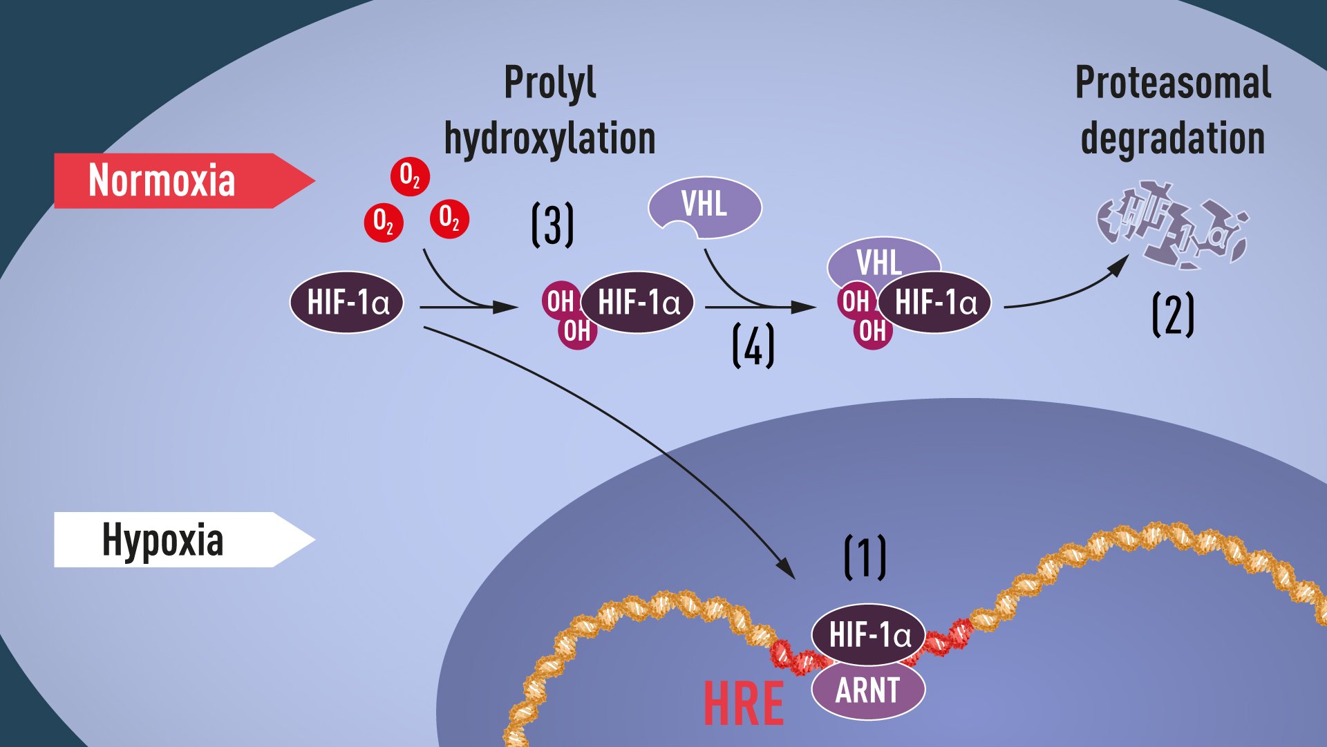
 Research resource: Transcriptional profiling reveals different pseudohypoxic signatures in SDHB and VHL-related...
 MESP2 gene: MedlinePlus Genetics
 REGULATOR: a database of metazoan transcription factors and maternal factors for developmental studies
![TCF4 transcription factor 4 [Homo sapiens (human)] - Gene - NCBI](data:image/png;base64,iVBORw0KGgoAAAANSUhEUgAAABAAAAAQCAYAAAAf8/9hAAAB1ElEQVQ4jaWSPWgTcRjGf/ehuWhobO2JxGJRY3taTTRV2yoqSpW6iIWO4iAoUsRBioNDKUWKLU7i4KA4OfhVREQnETRia03k7IdiS0LaQYKJQg3mLtfc30GySNUDn/V5nx/vy/vAf0pqad3db2xquiBJku93s2Tb2eEHdw1rTcsxol23sObTjN7oIp9KVmaU9kMdTxcLAyiqGtA0bfms+XKQULSdQG2EmnUx0q9ughAA8p/CFW0IN3Sv0vUI5p2zIMpUrd5JeP/Jii//80ZJUlrb9lyV8qn3zI5dB8A4MoBWtcITAKBmZe3eRmPzccYf9uIUsyzx6zQd7fMMAIjFdgxpkuPy4clFANbu6qa6fouybXtznxeAoqoBn0/zz5kvBqVQ5DBasJ5gXaPnDQAWFpwCkiwLZekyAMp2wTPAsqy5d8nEZcIHThPQo7jlIua9854BibdvekqKX8PouARAOn6F+c8pT4Bc7svz6U8f77O1cwDVV439PcPU4yHw8AUhhDPyOn4OfWOMuuZfBZp41INTLACorhC2/Jc2zsxMX8vl8lMcPBUHFL5mnpEZGa748sS42esKYS8WLtl2NjE22s/6fScIhtr48W2S5O0zIFwvp3vST6Z+myCvkaonAAAAAElFTkSuQmCC) TCF4 transcription factor 4 [Homo sapiens (human)] - Gene - NCBI
TCF4 transcription factor 4 [Homo sapiens (human)] - Gene - NCBI The Importance Of Neurogenins - 1402 Words | Bartleby
 Embryology of the Thyroid and Parathyroids: Initial Thyroid Embryology, Descent of the Thyroid Gland, Parafollicular Cells
 LYL1 protein expression summary - The Human Protein Atlas
 Drosophila gene families: The Enhancer of split complex
 IJMS | Free Full-Text | PI3K Signaling in Neurons: A Central Node for the Control of Multiple Functions
 OPUS Würzburg | Biochemische Charakterisierung der basischen Helix-Loop-Helix-Transkriptionsfaktoren Hey1 und Hey2 sowie...
 Alexa Fluor® 647 Mouse anti-NeuroD1
 dr. Laurens Pauwels
 PLAZA 3.0 Dicots : Binding Sites
 Citations: Effect of Posthatch Feed Restriction on Broiler Breast Muscle Development and Muscle Transcriptional Regulatory...
 Publication : USDA ARS
 c-Myc (human) Recombinant monoclonal antibody (9E10) (Rabbit IgGκ) - ENZ-ABS461 - Enzo Life Sciences
 Pharos : Target Details - P17542
 Brain Abnormalities and Glioma-Like Lesions in Mice Overexpressing the Long Isoform of PDGF-A in Astrocytic Cells - D...
 ZFIN Publication: Nelson et al., 2012
 An EBF3-Mediated Transcriptional Program That Induces Cell Cycle Arrest and Apoptosis | Cancer Research | American Association...
 WTS database | WHO FCTC
 Light regulation and functional characterization of Phytochrome Interacting Factor 1 (PIF1) in Arabidopsis
 HES1 (D6P2U) Rabbit mAb | Cell Signaling Technology