Basal Ganglia Hemorrhage
Evidence for apoptosis after intercerebral hemorrhage in rat striatum. (1/26)
The overall hypothesis that cell death after intracerebral hemorrhage is mediated in part by apoptotic mechanisms was tested. Intracerebral hemorrhage was induced in rats using stereotactic infusions of 0.5 U of collagenase (1-microL volume) into the striatum. After 24 hours, large numbers of TUNEL-positive stained cells with morphologies suggestive of apoptosis were present in the center and periphery of the hemorrhage. Double staining with Nissl and immunocytochemical labeling with antibodies against neuronal nuclei and glial fibrillary acidic protein suggested that these TUNEL-positive cells were mostly neurons and astrocytes. Electrophoresis of hemorrhagic brain extracts showed evidence of DNA laddering into approximately 200-bp fragments. Western blots showed cleavage of the cytosolic caspase substrate gelsolin. The density of TUNEL-positive cells at 24 and 48 hours after hemorrhage was significantly reduced by treatment with the broad-spectrum caspase inhibitor zVADfmk. It was unlikely that apoptotic changes were due to neurotoxicity of injected collagenase because TUNEL-positive cells and DNA laddering were also obtained in an alternative model of hemorrhage where autologous blood was infused into the striatum. Furthermore, equivalent doses of collagenase did not induce cell death in primary neuronal cultures. These results provide initial evidence that apoptotic mechanisms may mediate some of the injury in brain after intracerebral hemorrhage. (+info)Striatocapsular haemorrhage. (2/26)
Haemorrhages in the striatocapsular area, or striatocapsular haemorrhages (SCHs), have been regarded as a single entity, although the area is composed of several functionally discrete structures that receive blood supply from different arteries. We analysed the morphological and clinical presentations of 215 cases of SCHs according to a new classification method we have designed on the basis of arterial territories. SCHs were divided into six types: (i) anterior type (Heubner's artery); (ii) middle type (medial lenticulostriate artery); (iii) posteromedial type (anterior choroidal artery); (iv) posterolateral type (posteromedial branches of lateral lenticulostriate artery); (v) lateral type (most lateral branches of lateral lenticulostriate artery); and (vi) massive type. The anterior type (11%) formed small caudate haematomas, always ruptured into the lateral ventricle, causing severe headache, and mild contralateral hemiparesis developed occasionally. The outcome was excellent. The middle type (7%) involved the globus pallidus and medial putamen, frequently causing contralateral hemiparesis and transient conjugate eye deviation to the lesion side. About 50% of the patients recovered to normal. The posteromedial type (4%) formed very small haematomas in the posterior limb of the internal capsule and presented with mild dysarthria, contralateral hemiparesis and sensory deficit, with excellent outcome in general. The posterolateral type (33%) affected the posterior half of the putamen and posterior limb of the internal capsule and presented with impaired consciousness and contralateral hemiparesis with either language dysfunction or contralateral neglect. The outcome was fair to poor but there were no deaths. The lateral type (21%) formed large elliptical haematomas between the putamen and insular cortex. Contralateral hemiparesis with language dysfunction or contralateral neglect developed frequently but resolved over several weeks. The clinical outcome was relatively excellent except when the haematoma size was very large. The massive type (24%) formed huge haematomas affecting the entire striatocapsular area. Marked sensorimotor deficits and impaired consciousness, ocular movement dysfunctions including the 'wrong-way' eyes were observed quite frequently. The outcome was very poor with a case fatality rate of 81%. The clinico-radiological presentations suggested its origin was the same as the posterolateral type. (+info)Relationship between stroke and asymptomatic minute hemorrhages in hypertensive patients. (3/26)
Asymptomatic small hemorrhages were identified in hypertensive patients by T2*-weighted gradient echo magnetic resonance (MR) imaging to investigate the relationship between hypertensive intracerebral hemorrhage and asymptomatic minute hemorrhages. Forty-eight patients with hypertensive intracerebral hemorrhage or cerebral infarction with hypertension (these diseases were defined as stroke) were treated in National Defense Medical College from April 1998 to February 2000. All patients had no past history of stroke or head injury, underwent MR imaging within 6 months of the stroke attack, were aged from 40 to 80 years, and had no diagnosis of aneurysm, angioma, or moyamoya disease. Patients were divided into the infarction group and hemorrhage group. All foci over 2 mm in size appearing as hypointense on T2*-weighted MR imaging and unrelated to stroke areas were defined as minute hemorrhages. There were no significant differences between the two groups with respect to sex, age, and history of diabetes mellitus. The incidence of minute hemorrhages in the hemorrhage group (21/26) was greater than in the infarction group (9/22, p < 0.01). The incidence of minute hemorrhages in the basal ganglia (18/26) was greater in the hemorrhage group than in the infarction group (4/22, p < 0.001). Symptomatic intracerebral hemorrhage may be preceded by asymptomatic minute hemorrhage. (+info)Hypertensive caudate hemorrhage prognostic predictor, outcome, and role of external ventricular drainage. (4/26)
BACKGROUND AND PURPOSE: The purpose of the present study was to analyze the outcome and outcome predictors of caudate hemorrhage and role of external ventricular drainage in acute hydrocephalus. METHODS: Clinical data from 36 consecutive patients with hypertensive caudate hemorrhage was used in the present study. Age, gender, volume of parenchymal hematoma, hematoma in the internal capsule, initial Glasgow Coma Scale (GCS), hydrocephalus, severity of intraventricular hemorrhage, and hemorrhagic dilatation of the fourth ventricle were analyzed for effect on outcome. Effect of external ventricle drainage for hydrocephalus was evaluated by comparing preoperative and postoperative GCS scores. RESULTS: By univariate analyses, poor outcome was associated with a poor initial GCS score (P=0.016), hydrocephalus (P<0.001), intraventricular hemorrhage severity (P<0.01), and hemorrhagic dilatation of the fourth ventricle (P=0.02). By multivariate analysis, stepwise logistic regression revealed that hydrocephalus was the only independent prognostic factor for poor outcome (P<0.001). Postoperative 48-hour GCS score was better than the preoperative score by use of paired-sample t test (P<0.001). CONCLUSIONS: Hydrocephalus is the most important predictor of poor outcome. External ventricular drainage response for hydrocephalus was good in the present study, whereas an early decision should be made regarding preoperative neurological condition. (+info)Longitudinal changes of metabolites in frontal lobes after hemorrhagic stroke of basal ganglia: a proton magnetic resonance spectroscopy study. (5/26)
BACKGROUND AND PURPOSE: We investigated serial metabolic changes in frontal lobes of patients with deep intracerebral hemorrhage (ICH) to examine the correlation between N-acetylaspartate (NAA) and degree of motor impairment or clinical outcome. METHODS: - Twenty patients with deep ICH were examined with proton magnetic resonance spectroscopy with the application of a multivoxel method (1 voxel=10x10x20 mm; 64 voxels). NAA/creatine ratios in the white matter of the primary motor and premotor areas on both sides were measured sequentially: within 48 hours, at 2 weeks, and 1 month after onset. The National Institutes of Health Stroke Scale and Barthel Index for disability were measured for each patient. RESULTS: - In the primary motor area on the affected side, where the hematoma did not extend, the NAA/creatine ratio decreased sequentially. At 48 hours and 2 weeks after onset, a negative correlation was detected between NAA/creatine and hematoma volume, but there was no correlation 1 month later. At 2 weeks, NAA/creatine correlated negatively with motor impairment (r=-0.750), and there was a significant correlation with clinical outcome as early as 2 weeks after onset (r=0.954). These sequential changes of NAA/creatine varied according to patients' long-term clinical outcome. Patients with poor outcome demonstrated notable reduction of NAA/creatine over the bilateral frontal lobes. CONCLUSIONS: - The delayed gradual reduction of NAA/creatine ratio in the frontal lobes correlates with motor deficit and clinical outcome after deep ICH, suggesting that the neural networks in the frontal lobe could be important for recovery. (+info)Stereotactic fibrinolysis of spontaneous intracerebral hematoma using infusion of recombinant tissue plasminogen activator. (6/26)
PURPOSE: The authors present a prospective study on 10 patients with stereotactic infusion of tissue plasminogen activator (rtPA) intraparenchimal hemorrhage. METHODS: Between 1999 and 2000, 10 patients with deep seated hematomas in the basal ganglia were selected for stereotactic infusion of rtPA and spontaneous clot drainage. RESULTS: All cases had about 80% reduction of the hematoma volume in the CT scan at the third day. The intracranial pressure was normalized by the third day too. There were no local or systemic complications with the use of this thrombolytic. The results were shown by the Glasgow Outcome Scale with six patients in V, three in IV and one in III after 3 months. CONCLUSION: Early treatment and drainage with minimally invasive neurosurgery, can make these patients with deep-seated hematomas recover the consciousness and they can be rehabilitated earlier avoiding secondary complications. (+info)Possible acute hemorrhagic leukoencephalitis manifesting as intracerebral hemorrhage on computed tomography--case report. (7/26)
A 15-year-old girl presented with meningeal irritation and bilateral cerebral signs after contracting influenza. A lumbar puncture revealed bloody cerebrospinal fluid and polymorphonuclear predominant pleocytosis with an elevated protein level and normal glucose level. Computed tomography showed a hematoma in the right basal ganglia and lateral ventricles. Symmetrical low density areas were also noted in the bilateral white matter. The preliminary diagnosis was hemorrhagic cerebrovascular disease of unknown cause. However, her neurological condition deteriorated. Magnetic resonance (MR) imaging showed diffuse high intensity signals in the bilateral white matter and small spotty lesions, indicating hemorrhages in various stages. The final diagnosis was acute hemorrhagic leukoencephalitis (AHL). However, high-dose steroid administration and plasmapheresis failed to improve her condition. Hypothermia could not control her intracranial pressure and she died 12 days after admission. The neuroimaging findings indicated the histological characteristics of AHL, but the hematoma formation is rare. AHL is a fulminant form of brain demyelination and can be fatal, so early diagnosis and aggressive treatment are important for successful recovery. Therefore, early investigation by MR imaging is necessary. (+info)Interobserver agreement in the assessment of lobar versus deep location of intracerebral haematomas on CT. (8/26)
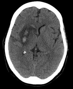
In patients with supratentorial intracerebral haemorrhage (ICH), it is important to discriminate superficial (lobar) and deep (basal ganglia) location, since this has consequences for research and prognosis. Haemorrhages at these sites have different causes and different risk factors. We studied the interobserver variation between three radiologists in classifying fifty large haematomas on CT as deep or lobar. The kappa values were almost perfect, ranging from 0.88 to 0.96. We conclude that the assessment of CT by radiologist is a reliable method to discriminate between lobar versus deep origin even for large intracerebral haematomas. (+info)A basal ganglia hemorrhage is a type of intracranial hemorrhage, which is defined as bleeding within the skull or brain. Specifically, a basal ganglia hemorrhage involves bleeding into the basal ganglia, which are clusters of neurons located deep within the forebrain and are involved in regulating movement, cognition, and emotion.
Basal ganglia hemorrhages can result from various factors, including hypertension (high blood pressure), cerebral amyloid angiopathy, illicit drug use (such as cocaine or amphetamines), and head trauma. Symptoms of a basal ganglia hemorrhage may include sudden onset of severe headache, altered consciousness, weakness or paralysis on one side of the body, difficulty speaking or understanding speech, and visual disturbances.
Diagnosis of a basal ganglia hemorrhage typically involves imaging studies such as computed tomography (CT) or magnetic resonance imaging (MRI). Treatment may include supportive care, medications to control symptoms, and surgical intervention in some cases. The prognosis for individuals with a basal ganglia hemorrhage varies depending on the severity of the bleed, the presence of underlying medical conditions, and the timeliness and effectiveness of treatment.
The basal ganglia are a group of interconnected nuclei, or clusters of neurons, located in the base of the brain. They play a crucial role in regulating motor function, cognition, and emotion. The main components of the basal ganglia include the striatum (made up of the caudate nucleus, putamen, and ventral striatum), globus pallidus (divided into external and internal segments), subthalamic nucleus, and substantia nigra (with its pars compacta and pars reticulata).
The basal ganglia receive input from various regions of the cerebral cortex and other brain areas. They process this information and send output back to the thalamus and cortex, helping to modulate and coordinate movement. The basal ganglia also contribute to higher cognitive functions such as learning, decision-making, and habit formation. Dysfunction in the basal ganglia can lead to neurological disorders like Parkinson's disease, Huntington's disease, and dystonia.
 Intracranial hemorrhage
Cerebral arteriovenous malformation
Hydrostatic shock
Intracerebral hemorrhage
Pallidotomy
Charcot-Bouchard aneurysm
Intraparenchymal hemorrhage
Cerebral amyloid angiopathy
List of MeSH codes (C10)
Abulia
Cerebral contusion
Putamen
Intracranial aneurysm
List of MeSH codes (C14)
List of MeSH codes (C23)
2020 Punjab alcohol poisoning
Transcortical sensory aphasia
Hypertension and the brain
Hemiballismus
Post-stroke depression
Acute disseminated encephalomyelitis
Dyskinetic cerebral palsy
Cerebral venous sinus thrombosis
Nervous system disease
Hypokinesia
Hyperintensity
Posterior reversible encephalopathy syndrome
Preterm birth
Trichotillomania
Spasmodic torticollis
 Basal Ganglia Hemorrhage After Endovascular Thrombectomy | Neurology | JN Learning | AMA Ed Hub
 Basal ganglia hemorrhage | MedLink Neurology
Intracranial hemorrhage - Wikipedia
 Chorea in Children: Overview, Causes of Chorea, Pathophysiology and General Principles in Treatment of Chorea
 Clinical changes of serum melatonin and ICAM-1 levels in patients with basal ganglia hypertensive intracerebral hemorrhage
...
Clinical changes of serum melatonin and ICAM-1 levels in patients with basal ganglia hypertensive intracerebral hemorrhage
...
 A neurocytoma and an associated lenticulostriate artery aneurysm presenting with intraventricular hemorrhage: case report
Meningococcal Meningitis Treatment & Management: Approach Considerations, Pharmacologic Care, Prophylaxis
Plus it
 Charcot-Bouchard aneurysms revisited: clinicopathologic correlations | Modern Pathology
 Erowid.org: Erowid Reference 1911 : Prilog ispitivanju dejstva LSD-25 u eksperimentu na psima : Jovanovic D, Kandic B, Kronja T
 Question 2
 Spectrum of Intracerebral Hemorrhage in Children: A Report from PICU of a Resource Limited Country
 Why Is It Important for Families to Understand the "New Normal" After a Brain Injury? | BrainLine
 Radiology journal announces top 2022 images | AuntMinnie
Eclampsia: Overview, Etiologic and Risk Factors for Preeclampsia/Eclampsia, Multiorgan System Effects
Abnormal findings in the basal ganglia: a diagnostic clue for patients with diabetic striatopathy | BMJ Case Reports
Mechanism and Therapy of Brain Edema after Intracerebral Hemorrhage | Cerebrovascular Diseases | Karger Publishers
 Top Images in Radiology 2022 | RSNA
Cerebral aneurysmal arteriopathy in childhood AIDS | Neurology
 Hypertensive Emergencies: A Review | CE Article | NursingCenter
Diffusion Tensor Imaging in Chronic Subdural Hematoma: Correlation between Clinical Signs and Fractional Anisotropy in the...
CLINICA NEUROL, 55: 823 |827, 2015 | Rinsho Shinkeigaku (Clinical Neurology) | JAPANESE SOCIETY OF NEUROLOGY
 as a previous dial up user--sm
Dyskeratosis Congenita: Background, Pathophysiology, Etiology
 Information for Cerebral haemorrhage
 Marion S. Buckwalter, MD, PhD's Profile | Stanford Profiles
 Cholesterol-Lowering Drugs Linked To Lower Risk Of Bleeding Stroke
 Preeclampsia - Hypertensive States of Pregnancy
Thalamus3
- Magnetic Resonance Imaging was used to assess regional volumes of brain IDs in basal ganglia, brainstem, white matter, thalamus, and cortex/border with the corticomedullary junction, using a fully automatic assessment procedure followed by individual checking/correction where necessary. (springer.com)
- FDG-PET revealed severe hypometabolism in the left cerebral hemisphere, including basal ganglia and thalamus, and hypermetabolism in the right cerebral hemisphere. (koreamed.org)
- The thalamus/basal ganglion regions were involved in 46% of the cases. (elsevierpure.com)
Subarachnoid8
- He described both intracerebral hemorrhage and subarachnoid hemorrhage. (medlink.com)
- Intracranial hemorrhage refers to any bleeding within the cranial vault, including subdural and epidural hematomas and subarachnoid hemorrhage. (medlink.com)
- It can cause epidural hemorrhage, subdural hemorrhage, and subarachnoid hemorrhage. (wikipedia.org)
- However, MRI has higher sensitivity than CT scan for the detection of epidural hemorrhage, subdural hemorrhage, subarachnoid hemorrhage, nonhemorrhagic cortical contusions, hemorrhagic parenchymal contusions, brainstem injuries, and white matter axonal injuries. (wikipedia.org)
- Extra-axial hemorrhage, bleeding that occurs within the skull but outside of the brain tissue, falls into three subtypes: epidural hematoma, subdural hematoma, and subarachnoid hemorrhage. (wikipedia.org)
- Four hours later, she developed a parenchymal hemorrhage in the left basal ganglia without subarachnoid hemorrhage. (surgicalneurologyint.com)
- 4 ] Intracranial ICA dissection typically presents as severe headache, immediately followed by neurological symptoms of cerebral ischemia or subarachnoid hemorrhage. (surgicalneurologyint.com)
- Cervicocephalic artery dissection can result in ischemic stroke or subarachnoid hemorrhage, affecting young or middle-aged adults [ 1 , 2 ]. (neurointervention.org)
Intraventricular6
- This category includes intraparenchymal hemorrhage, or bleeding within the brain tissue, and intraventricular hemorrhage, bleeding within the brain's ventricles (particularly of premature infants). (wikipedia.org)
- We present the first reported case of a central neurocytoma in a patient with intraventricular hemorrhage caused by rupture of an aneurysm on a lenticulostriate artery that supplied the tumor. (nih.gov)
- A 35-year-old man who presented with an intraventricular hemorrhage underwent magnetic resonance imaging and cerebral angiography that disclosed a right lateral intraventricular mass and a 7-mm fusiform aneurysm from a lateral lenticulostriate branch of the right middle cerebral artery. (nih.gov)
- Previous reports have demonstrated that intraventricular neurocytoma may present with tumor hemorrhage. (nih.gov)
- F) Follow-up noncontrast CT image obtained 1 hour after the third phase CT angiogram shows massive enlargement of the hematoma, intraventricular hemorrhage, hydrocephalus, and subfalcine herniation. (auntminnie.com)
- If the hemorrhage ruptures into the ventricular system (intraventricular hemorrhage), blood may cause acute hydrocephalus, which is an independent predictor for a worse outcome after intracerebral hemorrhage. (msdmanuals.com)
Hypertensive intracerebral hemorrhage2
- To investigate the clinical changes of serum melatonin and intercellular adhesion molecule-1 (ICAM-1) levels in patients with basal ganglia hypertensive intracerebral hemorrhage (HICH). (org.pk)
- Hou H, Li X. Clinical changes of serum melatonin and ICAM-1 levels in patients with basal ganglia hypertensive intracerebral hemorrhage. (org.pk)
Left basal ganglia1
- B) Arterial phase CT angiogram obtained 192 seconds later, after administration of contrast material, demonstrates focal enhancement in the left basal ganglia (arrow). (auntminnie.com)
Intracerebral hemorrhages1
- Lobar intracerebral hemorrhages (hematomas in the cerebral lobes, outside the basal ganglia) usually result from angiopathy due to amyloid deposition in cerebral arteries (cerebral amyloid angiopathy), which affects primarily older people. (msdmanuals.com)
Lesions5
- CT scans showed high-intensity lesions in bilateral basal ganglia ( figure 1A ). (bmj.com)
- Basal ganglia lesions on MRI remained 4 weeks after the onset. (bmj.com)
- A) CT scans showed high-intensity lesions in the basal ganglia bilaterally. (bmj.com)
- Brain MRI revealed abnormal lesions in the fornix, corpus callosum, basal ganglia and frontal lobe. (neurology-jp.org)
- A brain magnetic resonance imaging revealed lesions in the upper part of the genu and body in the corpus callosum as well as hemorrhage in the inter-hemispheric fissure. (koreamed.org)
Hypertension9
- The most common cause of basal ganglia is hypertension. (medlink.com)
- Non-traumatic causes of hemorrhage includes: hypertension, cerebral amyloid angiopathy, hemorrhagic conversion of ischemic infarction, cerebral aneurysms, dural arteriovenous fistulae, cerebral venous sinus thrombosis, cerebral vasculitis and mycotic aneurysm. (wikipedia.org)
- More than half of all cases of intracranial hemorrhage are the result of hypertension. (wikipedia.org)
- Hypertension and cerebral amyloid angiopathy (CAA) are the most common causes of primary ICH, but the mechanism of hemorrhage in both conditions is unclear. (nature.com)
- In hypertension, the cause of hemorrhage is thought to be elevated blood pressure-induced degenerative changes in the penetrating arterioles leading to rupture [ 14 ]. (nature.com)
- At least half of all patients who have this kind of hemorrhage have a history of hypertension. (umassmed.edu)
- The most common location for hemorrhage due to hypertension is the putamen. (msdmanuals.com)
- Use of cocaine or, occasionally, other sympathomimetic drugs or medications can cause transient severe hypertension leading to hemorrhage. (msdmanuals.com)
- Chronic arterial hypertension leads to formation of microaneurysms (Charcot-Bouchard aneurysms) in small perforating arteries, which may rupture and cause intracerebral hemorrhage. (msdmanuals.com)
Ischemic2
- A head CT scan was obtained in all patients prior to angiography to exclude hemorrhage and other mimics of ischemic stroke. (ajnr.org)
- Here, we report a case of intracranial ICA dissection with ischemic onset, with a complication of remote parenchymal hemorrhage due to a recanalized dissected perforator following endovascular therapy. (surgicalneurologyint.com)
Corpus callosum1
- Callosal disconnection syndrome in a patient with corpus callosum hemorrhage: a diffusion tensor tractography study. (koreamed.org)
Putamen1
- In this case, we believe that bleeding involved a small penetrating artery vessel supplying the putamen (one of the basal ganglia). (umassmed.edu)
Pons1
- compressing the brain stem and often causing secondary hemorrhages in the midbrain and pons. (msdmanuals.com)
Arterial1
- It is caused by injuries of small arterial or venous vessels, causing hemorrhage within the brain parenchyma, and give rise to hyperdense lesion on CT scan. (wikipedia.org)
Etiology1
- Only one of the 12 subjects with CBAs had a large ICH, and the etiology underlying the hemorrhage was likely multifactorial. (nature.com)
Stroke6
- Through the years, intracerebral hemorrhage has also been termed "cerebral hemorrhage," "intracranial hemorrhage," "hemorrhagic stroke," and "cerebral bleed. (medlink.com)
- Successful reperfusion is associated with a better outcome, and the prevalence of hemorrhage does not exceed that which occurs in the natural history of embolic stroke. (ajnr.org)
- Intracerebral hemorrhage (ICH) in children is a rare but disabling disease that accounts for almost half cases of stroke. (hindawi.com)
- Intracerebral hemorrhage (ICH) is a subtype of stroke with a severe high mortality and disability rate and accounts for about 10-15% of all strokes. (karger.com)
- People who take cholesterol-lowering drugs called statins may have a lower risk of having a type of stroke called an intracerebral hemorrhage, according to a new study published in the December 7, 2022, online issue of Neurology®, the medical journal of the American Academy of Neurology. (worldhealth.net)
- While statins have been shown to reduce the risk of stroke from blood clots, there has been conflicting research on whether statin use increases or decreases the risk of a person having a first intracerebral hemorrhage," said study author David Gaist, MD, Ph.D., of the University of Southern Denmark in Odense and a member of the American Academy of Neurology. (worldhealth.net)
Cortex2
- The memory system of the 'cortex - basal ganglia - thalamic loop' model suggests a need for the participation of the prefrontal lobe (PL) and basal ganglia (BG). (oatext.com)
- Hemorrhage may involve any part of the CEREBRAL CORTEX and the BASAL GANGLIA. (ucdenver.edu)
Bilateral2
- B) MRI showed increased signal intensity on T1-weighted images in the bilateral basal ganglia. (bmj.com)
- of the head shows fronto-temporal atrophy carnitine levels in urine were elevated and and bilateral subdural haemorrhage glutaryl-CoA dehydrogenase activity in cul- tured fibroblasts was low. (who.int)
Pulmonary2
- His autopsy revealed severe pulmonary hemorrhage with alveolar vasculitis and cholesterol crystals in the brain, kidneys, liver, and the other organs. (neurology-jp.org)
- It was possible that cholesterol embolization to multiple organs including the brain induced systemic vasculitis that caused pulmonary hemorrhage and his critical prognosis. (neurology-jp.org)
Rupture1
- They were first described by Charcot and Bouchard in 1868 as a cause of hypertensive hemorrhage when they rupture [ 18 , 19 ]. (nature.com)
Brain parenchyma2
- Intracerebral hemorrhage refers specifically to bleeding within the brain parenchyma. (medlink.com)
- Intracerebral hemorrhage is focal bleeding from a blood vessel in the brain parenchyma. (msdmanuals.com)
Outcome2
- Poor outcome or death is associated with nonrecanalization, older age, hemorrhage, and ICA bifurcation occlusions. (ajnr.org)
- Comparison of TMS and DTT for predicting motor outcome in intracerebral hemorrhage. (koreamed.org)
Aneurysm1
- Hemorrhage associated with central neurocytoma has been described previously, but never in association with an aneurysm originating from a feeding artery. (nih.gov)
Hematoma1
- BACKGROUND: Subcortical injury resulting from conventional surgical management of intracranial hemorrhage may counteract the potential benefts of hematoma evacuation. (elsevierpure.com)
Syndrome1
- Their article sparked controversy in the literature, prompting others to point out that such patients were at risk of hemorrhage and ischemia, and, thus, the syndrome was not necessarily "reversible. (medlink.com)
Severe2
- Basal ganglia hemorrhage is one of the most severe strokes. (medlink.com)
- Most patients could survive the initial injury of smaller hemorrhage, but the secondary injury may result in severe neurological deficits and even death [ 4 ]. (karger.com)
Clinical2
- This update highlights important clinical trial results on the treatment of intracerebral hemorrhage, including blood pressure management and surgery. (medlink.com)
- Hemorrhage occurred in 10 of the 26 patients, with clinical deterioration in three. (ajnr.org)
Calcifications1
- Over 80% of children with AIDS have CNS involvement including acquired microcephaly, diffuse cerebral atrophy, calcifications of the basal ganglia, and HIV-associated encephalitis. (neurology.org)
Infarcts1
- Two CBAs in the basal ganglia demonstrated associated microhemorrhages, while three demonstrated infarcts in the vicinity. (nature.com)
Cognitive1
- We investigated differential characteristic of working memory and learning ability caused by a unilateral basal ganglia lesion (BGL) and a prefrontal lobe lesion (PLL) to provide a strategy for cognitive rehabilitation. (oatext.com)
Subcortical1
- 24 h presented complete effacement of basal cisterns (p = 0.005), sulcular effacement (p = 0.013), loss of cortico-subcortical differentiation (p = 0.0001) and effacement of the suprasellar cistern (p = 0.005). (medintensiva.org)
Disorders1
- Movement disorders (particularly chorea, athetosis, and dystonia) are thought to result from basal ganglia pathology. (medscape.com)
Melanoma1
- The most common causes of metastatic intracerebral hemorrhage include melanoma, renal cell carcinoma, and choriocarcinoma. (msdmanuals.com)
Cistern1
- The inflammatory reaction in the basal cistern causes the development of thick gelatinous exudate, which eventually encases the surrounding cranial nerves. (ina-jns.org)
Surgical1
- Surgical treatment has a limited role in the treatment of intracerebral hemorrhage. (medlink.com)
Focal1
- A) T2-weighted short inversion time inversion recovery MRI scan at 1.5 T in short-axis view shows focal high-signal intensities (arrow) at basal lateral and inferior wall, indicating myocardial edema. (auntminnie.com)
Patients4
- Most patients with basal ganglia hemorrhage have high blood pressure. (medlink.com)
- The average dose of urokinase was higher in the hemorrhage group, and mortality was higher in patients who hemorrhaged. (ajnr.org)
- CT hyperintensity and increased signal intensity on T1WI MRI in basal ganglia are characteristic findings observed in 79% and 95% of patients with DS, respectively. (bmj.com)
- Among them, similar radiological findings on basal ganglia can be observed in patients with Wilson's disease, hepatic encephalopathy and poisoning (manganese and organic mercury). (bmj.com)
Secondary1
- Despite the well-known risk factors, the pathogenesis of ICH is unclear, and the site of bleeding has rarely been demonstrated histologically due to the difficulty in examining tissue destroyed by hemorrhage as well as secondary bleeding caused by the disruption of surrounding arteries [ 12 , 13 ]. (nature.com)
Recurrent1
- Lobar hemorrhages may be multiple and recurrent. (msdmanuals.com)