Bacteriophage M13
Bacteriophage T4
Escherichia coli
Bacteriophage lambda
Bacteriophage T7
Lysogeny
DNA, Single-Stranded
Base Sequence
T-Phages
Bacteriophage mu
Mutation
Bacteriophage phi 6
Molecular Sequence Data
DNA, Circular
DNA Restriction Enzymes
Bacteriophage phi X 174
Phosphorus Isotopes
Bacteriophage P2
Plasmids
Genes
Bacteriophage T3
Amino Acid Sequence
Cloning, Molecular
Bacteriophage Typing
Bacteriophage P1
Salmonella Phages
Siphoviridae
Nucleic Acid Conformation
Oligodeoxyribonucleotides
RNA Phages
Magnetic Resonance Spectroscopy
Bacteriolysis
Bacteriophage PRD1
Pseudomonas Phages
Bacillus Phages
Nucleic Acid Hybridization
Viral Tail Proteins
Levivirus
DNA
Protein Conformation
Adsorption
DNA Packaging
Prophages
Inovirus
Genetics, Microbial
DNA-Directed RNA Polymerases
Attachment Sites, Microbiological
Recombination, Genetic
Viral Plaque Assay
Virus Replication
Transduction, Genetic
Centrifugation, Density Gradient
Microscopy, Electron
Cystoviridae
Bacteriophage Pf1
Chloramphenicol
Temperature
Chromosome Mapping
Caudovirales
Transcription, Genetic
DNA-Directed DNA Polymerase
Genetic Complementation Test
DNA Primase
Tritium
Biological Therapy
Cryoelectron Microscopy
Host Specificity
DNA Nucleotidyltransferases
Templates, Genetic
Viral Regulatory and Accessory Proteins
Viral Structural Proteins
Operon
Chromosomes, Bacterial
Phosphotungstic Acid
Sequence Analysis, DNA
Nucleic Acid Denaturation
Thymine
Open Reading Frames
Ultraviolet Rays
DNA, Recombinant
Restriction Mapping
Mitomycins
Polynucleotide Ligases
Binding Sites
Mycobacteriophages
Endodeoxyribonucleases
Radiation Effects
Integrases
Operator Regions, Genetic
Virus Assembly
Exonucleases
DNA Helicases
Lactococcus lactis
Microviridae
RNA Nucleotidyltransferases
Salmonella typhimurium
Uracil
Models, Molecular
Virion
Identification of residues in beta-lactamase critical for binding beta-lactamase inhibitory protein. (1/356)
beta-Lactamase inhibitory protein (BLIP) is a potent inhibitor of several beta-lactamases including TEM-1 beta-lactamase (Ki = 0.1 nM). The co-crystal structure of TEM-1 beta-lactamase and BLIP has been solved, revealing the contact residues involved in the interface between the enzyme and inhibitor. To determine which residues in TEM-1 beta-lactamase are critical for binding BLIP, the method of monovalent phage display was employed. Random mutants of TEM-1 beta-lactamase in the 99-114 loop-helix and 235-240 B3 beta-strand regions were displayed as fusion proteins on the surface of the M13 bacteriophage. Functional mutants were selected based on the ability to bind BLIP. After three rounds of enrichment, the sequences of a collection of functional beta-lactamase mutants revealed a consensus sequence for the binding of BLIP. Seven loop-helix residues including Asp-101, Leu-102, Val-103, Ser-106, Pro-107, Thr-109, and His-112 and three B3 beta-strand residues including Ser-235, Gly-236, and Gly-238 were found to be critical for tight binding of BLIP. In addition, the selected beta-lactamase mutants A113L/T114R and E240K were found to increase binding of BLIP by over 6- and 11-fold, respectively. Combining these substitutions resulted in 550-fold tighter binding between the enzyme and BLIP with a Ki of 0.40 pM. These results reveal that the binding between TEM-1 beta-lactamase and BLIP can be improved and that there are a large number of sequences consistent with tight binding between BLIP and beta-lactamase. (+info)Gene transfer to mammalian cells using genetically targeted filamentous bacteriophage. (2/356)
We have genetically modified filamentous bacteriophage to deliver genes to mammalian cells. In previous studies we showed that noncovalently attached fibroblast growth factor (FGF2) can target bacteriophage to COS-1 cells, resulting in receptor-mediated transduction with a reporter gene. Thus, bacteriophage, which normally lack tropism for mammalian cells, can be adapted for mammalian cell gene transfer. To determine the potential of using phage-mediated gene transfer as a novel display phage screening strategy, we transfected COS-1 cells with phage that were engineered to display FGF2 on their surface coat as a fusion to the minor coat protein, pIII. Immunoblot and ELISA analysis confirmed the presence of FGF2 on the phage coat. Significant transduction was obtained in COS-1 cells with the targeted FGF2-phage compared with the nontargeted parent phage. Specificity was demonstrated by successful inhibition of transduction in the presence of excess free FGF2. Having demonstrated mammalian cell transduction by phage displaying a known gene targeting ligand, it is now feasible to apply phage-mediated transduction as a screen for discovering novel ligands. (+info)Selection-dominant and nonaccessible epitopes on cell-surface receptors revealed by cell-panning with a large phage antibody library. (3/356)
To generate antibodies to defined cell-surface antigens, we used a large phage antibody fragment library to select on cell transfectants expressing one of three chosen receptors. First, in vitro panning procedures and phage antibody screening ELISAs were developed using whole live cells stably expressing the antigen of interest. When these methodologies were applied to Chinese hamster ovary (CHO) cells expressing one of the receptors for a neuropeptide, somatostatin, using either direct cell panning or a strategy of depletion or ligand-directed elution, many different pan-CHO-cell binders were selected, but none was receptor specific. However, when using direct panning on CHO-cells expressing the human membrane protein CD36, an extraordinary high frequency of antigen-specific phage antibodies was found. Panning on myoblasts expressing the rat homologue of CD36 revealed a similar selection dominance for anti-(CD36). Binding of all selected 20 different anti-(CD36) phage was surprisingly inhibited by one anti-(CD36) mAb CLB-IVC7, which recognizes a functional epitope that is also immunodominant in vivo. Similar inhibition was found for seven anti-(rat) CD36 that cross-reacted with human CD36. Our results show that, although cells can be used as antigen carriers to select and screen phage antibodies, the nature of the antigen target has a profound effect on the outcome of the selection. (+info)Identification of peroxisomal proteins by using M13 phage protein VI phage display: molecular evidence that mammalian peroxisomes contain a 2,4-dienoyl-CoA reductase. (4/356)
To elucidate unknown mammalian peroxisomal enzymes and functions, we subjected M13 phage expressing fusions between the gene encoding protein VI and a rat liver cDNA library to an immunoaffinity selection process in vitro (biopanning) with the use of antibodies raised against peroxisomal subfractions. In an initial series of biopanning experiments, four different cDNA clones were obtained. These cDNA species encoded two previously identified peroxisomal enzymes, catalase and urate oxidase, and two novel proteins that contained a C-terminal peroxisomal targeting signal (PTS1). A primary structure analysis of these novel proteins revealed that one, ending in the tripeptide AKL, is homologous to the yeast peroxisomal 2,4-dienoyl-CoA reductase (EC 1.3.1.34; DCR), an enzyme required for the degradation of unsaturated fatty acids, and that the other, ending in the tripeptide SRL, is a putative member of the short-chain dehydrogenase/reductase (SDR) family, with three isoforms. Green fluorescent protein (GFP) fusions encoding GFP-DCR-AKL, GFP-DCR, GFP-SDR-SRL and GFP-SDR were expressed in mammalian cells. The analysis of the subcellular location of the recombinant fusion proteins confirmed the peroxisomal localization of GFP-DCR-AKL and GFP-SDR-SRL, as well as the functionality of the PTS1. That the AKL protein is indeed an NADPH-dependent DCR was demonstrated by showing DCR activity of the bacterially expressed protein. These results demonstrate at the molecular level that mammalian peroxisomes do indeed contain a DCR. In addition, the results presented here indicate that the protein VI display system is suitable for the isolation of rare cDNA clones from cDNA libraries and that this technology facilitates the identification of novel peroxisomal proteins. (+info)Peptide ligands to human immunodeficiency virus type 1 gp120 identified from phage display libraries. (5/356)
We have used phage-displayed peptide libraries to identify novel ligands to the human immunodeficiency virus type 1 (HIV-1) envelope glycoprotein gp120. Screening of libraries of random 12-mers, 7-mers, and cyclic 9-mers produced two families of gp120 binding peptides. Members of a family with the prototype sequence RINNIPWSEAMM (peptide 12p1) inhibit the interaction between gp120 and both four-domain soluble CD4 (4dCD4) and monoclonal antibody (MAb) 17b, a neutralizing antibody that covers the chemokine receptor binding surface on gp120. Peptide 12p1 inhibits the interaction of 4dCD4 with gp120 from three different HIV strains, implying that it binds to a conserved site on gp120. Members of a second family of peptides, with the prototype sequence TSPYEDWQTYLM (peptide 12p2), bind more weakly to gp120. They do not detectably affect its interaction with 4dCD4, but they enhance its binding to MAb 17b. A common sequence motif in the two peptide families and cross-competition for gp120 binding suggest that they have overlapping contacts. Their divergent effects on the affinity of gp120 for MAb 17b may indicate that their binding stabilizes distinct conformational states of gp120. The functional properties of 12p1 suggest that it might be a useful lead for the development of inhibitors of HIV entry. (+info)Peptide ligands that bind IgM antibodies and block interaction with antigen. (6/356)
We have selected a peptide-display phage library on IgM Abs and identified a panel of phage-expressing peptides that bind to IgM Abs in general, but not to Abs of other classes. A synthetic peptide corresponding to one of the displayed peptide sequences also binds to IgM Abs. The peptides bind to both soluble pentameric Abs and to monomeric cell-surface IgM. The phage-displayed and synthetic peptides inhibit the binding of IgM Abs to Ag. These peptides may create confounding artifacts when IgM Abs are used for epitope mapping studies. Nonetheless, the peptides may have both experimental and therapeutic utility. (+info)Selection of a C5a receptor antagonist from phage libraries attenuating the inflammatory response in immune complex disease and ischemia/reperfusion injury. (7/356)
A C5a-receptor antagonist was selected from human C5a phage display libraries in which the C terminus of des-Arg74-hC5a was mutated. The selected molecule is a competitive C5a receptor antagonist in vitro and in vivo. Signal transduction is interrupted at the level of G-protein activation. In addition, the antagonist does not cause any C5a receptor phosphorylation. Proinflammatory properties such as chemotaxis or lysosomal enzyme release of differentiated U937 cells, as well as C5a-induced changes in intracellular Ca2+ concentration of murine peritoneal macrophages, are inhibited. The in vivo efficacy was evaluated in three different animal models of immune complex diseases in mice, i.e., the reverse passive Arthus reaction in the peritoneum, skin, and lung. The i.v. application of the C5a receptor antagonist abrogated polymorphonuclear neutrophil accumulation in peritoneum and markedly attenuated polymorphonuclear neutrophil migration into the skin and the lung. In a model of intestinal ischemia/reperfusion injury, i.v. administration of the C5a receptor antagonist decreased local and remote tissue injury: bowel wall edema and hemorrhage as well as pulmonary microvascular dysfunction. These data give evidence that C5a is an important mediator triggering the inflammatory sequelae seen in immune complex diseases and ischemia/reperfusion injury. The selected C5a receptor antagonist may prove useful to attenuate the inflammatory response in these disorders. (+info)New inhibitors of Helicobacter pylori urease holoenzyme selected from phage-displayed peptide libraries. (8/356)
Urease is an important virulence factor for Helicobacter pylori and is critical for bacterial colonization of the human gastric mucosa. Specific inhibition of urease activity has been proposed as a possible strategy to fight this bacteria which infects billions of individual throughout the world and can lead to severe pathological conditions in a limited number of cases. We have selected peptides which specifically bind and inhibit H. pylori urease from libraries of random peptides displayed on filamentous phage in the context of pIII coat protein. Screening of a highly diverse 25-mer combinatorial library and two newly constructed random 6-mer peptide libraries on solid phase H. pylori urease holoenzyme allowed the identification of two peptides, 24-mer TFLPQPRCSALLRYLSEDGVIVPS and 6-mer YDFYWW that can bind and inhibit the activity of urease purified from H. pylori. These two peptides were chemically synthesized and their inhibition constants (Ki) were found to be 47 microM for the 24-mer and 30 microM for the 6-mer peptide. Both peptides specifically inhibited the activity of H. pylori urease but not that of Bacillus pasteurii. (+info)Bacteriophage M13 is a type of bacterial virus that infects and replicates within the bacterium Escherichia coli (E. coli). It is a filamentous phage, meaning it has a long, thin, and flexible structure. The M13 phage specifically infects only the F pili of E. coli bacteria, which are hair-like appendages found on the surface of certain strains of E. coli.
Once inside the host cell, the M13 phage uses the bacterial machinery to produce new viral particles, or progeny phages, without killing the host cell. The phage genome is made up of a single-stranded circular DNA molecule that encodes for about 10 genes. These genes are involved in various functions such as replication, packaging, and assembly of the phage particles.
Bacteriophage M13 is widely used in molecular biology research due to its ability to efficiently incorporate foreign DNA sequences into its genome. This property has been exploited for a variety of applications, including DNA sequencing, gene cloning, and protein expression. The M13 phage can display foreign peptides or proteins on the surface of its coat protein, making it useful for screening antibodies or identifying ligands in phage display technology.
Bacteriophages, often simply called phages, are viruses that infect and replicate within bacteria. They consist of a protein coat, called the capsid, that encases the genetic material, which can be either DNA or RNA. Bacteriophages are highly specific, meaning they only infect certain types of bacteria, and they reproduce by hijacking the bacterial cell's machinery to produce more viruses.
Once a phage infects a bacterium, it can either replicate its genetic material and create new phages (lytic cycle), or integrate its genetic material into the bacterial chromosome and replicate along with the bacterium (lysogenic cycle). In the lytic cycle, the newly formed phages are released by lysing, or breaking open, the bacterial cell.
Bacteriophages play a crucial role in shaping microbial communities and have been studied as potential alternatives to antibiotics for treating bacterial infections.
Coliphages are viruses that infect and replicate within certain species of bacteria that belong to the coliform group, particularly Escherichia coli (E. coli). These viruses are commonly found in water and soil environments and are frequently used as indicators of fecal contamination in water quality testing. Coliphages are not harmful to humans or animals, but their presence in water can suggest the potential presence of pathogenic bacteria or other microorganisms that may pose a health risk. There are two main types of coliphages: F-specific RNA coliphages and somatic (or non-F specific) DNA coliphages.
Bacteriophage T4, also known as T4 phage, is a type of virus that infects and replicates within the bacterium Escherichia coli (E. coli). It is one of the most well-studied bacteriophages and has been used as a model organism in molecular biology research for many decades.
T4 phage has a complex structure, with an icosahedral head that contains its genetic material (DNA) and a tail that attaches to the host cell and injects the DNA inside. The T4 phage genome is around 169 kilobases in length and encodes approximately 289 proteins.
Once inside the host cell, the T4 phage DNA takes over the bacterial machinery to produce new viral particles. The host cell eventually lyses (bursts), releasing hundreds of new phages into the environment. T4 phage is a lytic phage, meaning that it only replicates through the lytic cycle and does not integrate its genome into the host's chromosome.
T4 phage has been used in various applications, including bacterial typing, phage therapy, and genetic engineering. Its study has contributed significantly to our understanding of molecular biology, genetics, and virology.
Viral DNA refers to the genetic material present in viruses that consist of DNA as their core component. Deoxyribonucleic acid (DNA) is one of the two types of nucleic acids that are responsible for storing and transmitting genetic information in living organisms. Viruses are infectious agents much smaller than bacteria that can only replicate inside the cells of other organisms, called hosts.
Viral DNA can be double-stranded (dsDNA) or single-stranded (ssDNA), depending on the type of virus. Double-stranded DNA viruses have a genome made up of two complementary strands of DNA, while single-stranded DNA viruses contain only one strand of DNA.
Examples of dsDNA viruses include Adenoviruses, Herpesviruses, and Poxviruses, while ssDNA viruses include Parvoviruses and Circoviruses. Viral DNA plays a crucial role in the replication cycle of the virus, encoding for various proteins necessary for its multiplication and survival within the host cell.
DNA viruses are a type of virus that contain DNA (deoxyribonucleic acid) as their genetic material. These viruses replicate by using the host cell's machinery to synthesize new viral components, which are then assembled into new viruses and released from the host cell.
DNA viruses can be further classified based on the structure of their genomes and the way they replicate. For example, double-stranded DNA (dsDNA) viruses have a genome made up of two strands of DNA, while single-stranded DNA (ssDNA) viruses have a genome made up of a single strand of DNA.
Examples of DNA viruses include herpes simplex virus, varicella-zoster virus, human papillomavirus, and adenoviruses. Some DNA viruses are associated with specific diseases, such as cancer (e.g., human papillomavirus) or neurological disorders (e.g., herpes simplex virus).
It's important to note that while DNA viruses contain DNA as their genetic material, RNA viruses contain RNA (ribonucleic acid) as their genetic material. Both DNA and RNA viruses can cause a wide range of diseases in humans, animals, and plants.
'Escherichia coli' (E. coli) is a type of gram-negative, facultatively anaerobic, rod-shaped bacterium that commonly inhabits the intestinal tract of humans and warm-blooded animals. It is a member of the family Enterobacteriaceae and one of the most well-studied prokaryotic model organisms in molecular biology.
While most E. coli strains are harmless and even beneficial to their hosts, some serotypes can cause various forms of gastrointestinal and extraintestinal illnesses in humans and animals. These pathogenic strains possess virulence factors that enable them to colonize and damage host tissues, leading to diseases such as diarrhea, urinary tract infections, pneumonia, and sepsis.
E. coli is a versatile organism with remarkable genetic diversity, which allows it to adapt to various environmental niches. It can be found in water, soil, food, and various man-made environments, making it an essential indicator of fecal contamination and a common cause of foodborne illnesses. The study of E. coli has contributed significantly to our understanding of fundamental biological processes, including DNA replication, gene regulation, and protein synthesis.
Bacteriophage lambda, often simply referred to as phage lambda, is a type of virus that infects the bacterium Escherichia coli (E. coli). It is a double-stranded DNA virus that integrates its genetic material into the bacterial chromosome as a prophage when it infects the host cell. This allows the phage to replicate along with the bacterium until certain conditions trigger the lytic cycle, during which new virions are produced and released by lysing, or breaking open, the host cell.
Phage lambda is widely studied in molecular biology due to its well-characterized life cycle and genetic structure. It has been instrumental in understanding various fundamental biological processes such as gene regulation, DNA recombination, and lysis-lysogeny decision.
Viral proteins are the proteins that are encoded by the viral genome and are essential for the viral life cycle. These proteins can be structural or non-structural and play various roles in the virus's replication, infection, and assembly process. Structural proteins make up the physical structure of the virus, including the capsid (the protein shell that surrounds the viral genome) and any envelope proteins (that may be present on enveloped viruses). Non-structural proteins are involved in the replication of the viral genome and modulation of the host cell environment to favor viral replication. Overall, a thorough understanding of viral proteins is crucial for developing antiviral therapies and vaccines.
Bacteriophage T7 is a type of virus that infects and replicates within the bacterium Escherichia coli (E. coli). It is a double-stranded DNA virus that specifically recognizes and binds to the outer membrane of E. coli bacteria through its tail fibers. After attachment, the viral genome is injected into the host cell, where it hijacks the bacterial machinery to produce new phage particles. The rapid reproduction of T7 phages within the host cell often results in lysis, or rupture, of the bacterial cell, leading to the release of newly formed phage virions. Bacteriophage T7 is widely studied as a model system for understanding virus-host interactions and molecular biology.
Lysogeny is a process in the life cycle of certain viruses, known as bacteriophages or phages, which can infect bacteria. In lysogeny, the viral DNA integrates into the chromosome of the host bacterium and replicates along with it, remaining dormant and not producing any new virus particles. This state is called lysogeny or the lysogenic cycle.
The integrated viral DNA is known as a prophage. The bacterial cell that contains a prophage is called a lysogen. The lysogen can continue to grow and divide normally, passing the prophage onto its daughter cells during reproduction. This dormant state can last for many generations of the host bacterium.
However, under certain conditions such as DNA damage or exposure to UV radiation, the prophage can be induced to excise itself from the bacterial chromosome and enter the lytic cycle. In the lytic cycle, the viral DNA replicates rapidly, producing many new virus particles, which eventually leads to the lysis (breaking open) of the host cell and the release of the newly formed virions.
Lysogeny is an important mechanism for the spread and survival of bacteriophages in bacterial populations. It also plays a role in horizontal gene transfer between bacteria, as genes carried by prophages can be transferred to other bacteria during transduction.
Single-stranded DNA (ssDNA) is a form of DNA that consists of a single polynucleotide chain. In contrast, double-stranded DNA (dsDNA) consists of two complementary polynucleotide chains that are held together by hydrogen bonds.
In the double-helix structure of dsDNA, each nucleotide base on one strand pairs with a specific base on the other strand through hydrogen bonding: adenine (A) with thymine (T), and guanine (G) with cytosine (C). This base pairing provides stability to the double-stranded structure.
Single-stranded DNA, on the other hand, lacks this complementary base pairing and is therefore less stable than dsDNA. However, ssDNA can still form secondary structures through intrastrand base pairing, such as hairpin loops or cruciform structures.
Single-stranded DNA is found in various biological contexts, including viral genomes, transcription bubbles during gene expression, and in certain types of genetic recombination. It also plays a critical role in some laboratory techniques, such as polymerase chain reaction (PCR) and DNA sequencing.
A base sequence in the context of molecular biology refers to the specific order of nucleotides in a DNA or RNA molecule. In DNA, these nucleotides are adenine (A), guanine (G), cytosine (C), and thymine (T). In RNA, uracil (U) takes the place of thymine. The base sequence contains genetic information that is transcribed into RNA and ultimately translated into proteins. It is the exact order of these bases that determines the genetic code and thus the function of the DNA or RNA molecule.
Viral genes refer to the genetic material present in viruses that contains the information necessary for their replication and the production of viral proteins. In DNA viruses, the genetic material is composed of double-stranded or single-stranded DNA, while in RNA viruses, it is composed of single-stranded or double-stranded RNA.
Viral genes can be classified into three categories: early, late, and structural. Early genes encode proteins involved in the replication of the viral genome, modulation of host cell processes, and regulation of viral gene expression. Late genes encode structural proteins that make up the viral capsid or envelope. Some viruses also have structural genes that are expressed throughout their replication cycle.
Understanding the genetic makeup of viruses is crucial for developing antiviral therapies and vaccines. By targeting specific viral genes, researchers can develop drugs that inhibit viral replication and reduce the severity of viral infections. Additionally, knowledge of viral gene sequences can inform the development of vaccines that stimulate an immune response to specific viral proteins.
I believe there might be a slight confusion in your question. T-phages are not a medical term, but rather a term used in the field of molecular biology and virology. T-phages refer to specific bacteriophages (viruses that infect bacteria) that belong to the family of Podoviridae and have a tail structure with a contractile sheath.
To be more specific, T-even phages are a group of T-phages that include well-studied bacteriophages like T2, T4, and T6. These phages infect Escherichia coli bacteria and have been extensively researched to understand their life cycles, genetic material packaging, and molecular mechanisms of infection.
In summary, T-phages are not a medical term but rather refer to specific bacteriophages used in scientific research.
Bacteriophage mu, also known as Mucoid Bacteriophage or Phage Mu, is a type of bacterial virus that infects and replicates within the genetic material of specific bacteria, primarily belonging to the genus Pseudomonas. This phage is characterized by its unique ability to integrate its genome into the host bacterium's chromosome at random locations, which can result in mutations or alterations in the bacterial genome.
Phage Mu has a relatively large genome and encodes various proteins that facilitate its replication, packaging, and release from the host cell. When Phage Mu infects a bacterium, it injects its genetic material into the host cytoplasm, where it circularizes and then integrates itself into the host's chromosome via a process called transposition. This integration can lead to significant changes in the host bacterium's genome, potentially altering its phenotype or even converting it into a lysogenic state, where the phage remains dormant within the host cell until environmental conditions trigger its replication and release.
Phage Mu is widely used as a tool for genetic research due to its ability to introduce random mutations into bacterial genomes, facilitating the study of gene function and regulation. Additionally, Phage Mu has been explored for potential applications in phage therapy, where it could be used to target and eliminate specific bacterial pathogens without adversely affecting other beneficial microorganisms present in the host organism or environment.
A mutation is a permanent change in the DNA sequence of an organism's genome. Mutations can occur spontaneously or be caused by environmental factors such as exposure to radiation, chemicals, or viruses. They may have various effects on the organism, ranging from benign to harmful, depending on where they occur and whether they alter the function of essential proteins. In some cases, mutations can increase an individual's susceptibility to certain diseases or disorders, while in others, they may confer a survival advantage. Mutations are the driving force behind evolution, as they introduce new genetic variability into populations, which can then be acted upon by natural selection.
DNA replication is the biological process by which DNA makes an identical copy of itself during cell division. It is a fundamental mechanism that allows genetic information to be passed down from one generation of cells to the next. During DNA replication, each strand of the double helix serves as a template for the synthesis of a new complementary strand. This results in the creation of two identical DNA molecules. The enzymes responsible for DNA replication include helicase, which unwinds the double helix, and polymerase, which adds nucleotides to the growing strands.
Bacteriophage phi 6, also known as Phi 6 or Pseudomonas phage Phi 6, is a double-stranded RNA virus that infects and replicates within the bacterium Pseudomonas syringae. It is a member of the family Cystoviridae and has an icosahedral head and a tail structure, which allows it to attach to and inject its genetic material into the host cell. Bacteriophage phi 6 is often used as a model system for studying RNA replication and transcription, as well as for understanding the mechanisms of virus-host interactions. It has also been studied as a potential candidate for use in phage therapy, which is the use of bacteriophages to treat bacterial infections.
Molecular sequence data refers to the specific arrangement of molecules, most commonly nucleotides in DNA or RNA, or amino acids in proteins, that make up a biological macromolecule. This data is generated through laboratory techniques such as sequencing, and provides information about the exact order of the constituent molecules. This data is crucial in various fields of biology, including genetics, evolution, and molecular biology, allowing for comparisons between different organisms, identification of genetic variations, and studies of gene function and regulation.
Circular DNA is a type of DNA molecule that forms a closed loop, rather than the linear double helix structure commonly associated with DNA. This type of DNA is found in some viruses, plasmids (small extrachromosomal DNA molecules found in bacteria), and mitochondria and chloroplasts (organelles found in plant and animal cells).
Circular DNA is characterized by the absence of telomeres, which are the protective caps found on linear chromosomes. Instead, circular DNA has a specific sequence where the two ends join together, known as the origin of replication and the replication terminus. This structure allows for the DNA to be replicated efficiently and compactly within the cell.
Because of its circular nature, circular DNA is more resistant to degradation by enzymes that cut linear DNA, making it more stable in certain environments. Additionally, the ability to easily manipulate and clone circular DNA has made it a valuable tool in molecular biology and genetic engineering.
DNA restriction enzymes, also known as restriction endonucleases, are a type of enzyme that cut double-stranded DNA at specific recognition sites. These enzymes are produced by bacteria and archaea as a defense mechanism against foreign DNA, such as that found in bacteriophages (viruses that infect bacteria).
Restriction enzymes recognize specific sequences of nucleotides (the building blocks of DNA) and cleave the phosphodiester bonds between them. The recognition sites for these enzymes are usually palindromic, meaning that the sequence reads the same in both directions when facing the opposite strands of DNA.
Restriction enzymes are widely used in molecular biology research for various applications such as genetic engineering, genome mapping, and DNA fingerprinting. They allow scientists to cut DNA at specific sites, creating precise fragments that can be manipulated and analyzed. The use of restriction enzymes has been instrumental in the development of recombinant DNA technology and the Human Genome Project.
Bacteriophage phi X 174, also known as Phi X 174 or ΦX174, is a bacterial virus that infects the bacterium Escherichia coli (E. coli). It is a small, icosahedral-shaped virus with a diameter of about 30 nanometers and belongs to the family Podoviridae in the order Caudovirales.
Phi X 174 has a single-stranded DNA genome that is circular and consists of 5,386 base pairs. It is one of the smallest viruses known to infect bacteria, and its simplicity has made it a model system for studying bacteriophage biology and molecular biology.
Phi X 174 was first discovered in 1962 by American scientist S.E. Luria and his colleagues. It is able to infect E. coli cells that lack the F-pilus, a hair-like structure on the surface of the bacterial cell. Once inside the host cell, phi X 174 uses the host's machinery to replicate its DNA and produce new viral particles, which are then released from the host cell by lysis, causing the cell to burst open and release the new viruses.
Phi X 174 has been extensively studied for its unique biological properties, including its small size, simple genome, and ability to infect E. coli cells. It has also been used as a tool in molecular biology research, such as in the development of DNA sequencing techniques and the study of gene regulation.
A capsid is the protein shell that encloses and protects the genetic material of a virus. It is composed of multiple copies of one or more proteins that are arranged in a specific structure, which can vary in shape and symmetry depending on the type of virus. The capsid plays a crucial role in the viral life cycle, including protecting the viral genome from host cell defenses, mediating attachment to and entry into host cells, and assisting with the assembly of new virus particles during replication.
Phosphorus isotopes are different forms of the element phosphorus that have different numbers of neutrons in their atomic nuclei, while the number of protons remains the same. The most common and stable isotope of phosphorus is 31P, which contains 15 protons and 16 neutrons. However, there are also several other isotopes of phosphorus that exist, including 32P and 33P, which are radioactive and have 15 protons and 17 or 18 neutrons, respectively. These radioactive isotopes are often used in medical research and treatment, such as in the form of radiopharmaceuticals to diagnose and treat various diseases.
Bacteriophage P2 is a type of virus that infects and replicates within a specific bacterium, Escherichia coli (E. coli). It's a double-stranded DNA virus that was first isolated in the 1950s. Bacteriophage P2 is known for its ability to integrate its genetic material into the host bacterium's chromosome and establish lysogeny, where it can remain dormant until environmental conditions trigger its replication.
Bacteriophage P2 has been extensively studied as a model system in molecular biology due to its unique life cycle and genetic characteristics. It has contributed significantly to our understanding of various biological processes such as DNA replication, transcription regulation, and lysogeny. However, it's important to note that bacteriophage P2 is not typically used for medical purposes like treating bacterial infections.
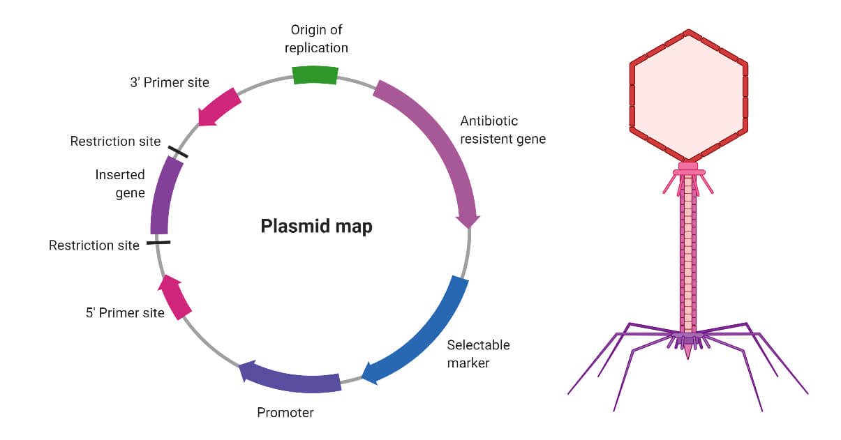
A plasmid is a small, circular, double-stranded DNA molecule that is separate from the chromosomal DNA of a bacterium or other organism. Plasmids are typically not essential for the survival of the organism, but they can confer beneficial traits such as antibiotic resistance or the ability to degrade certain types of pollutants.
Plasmids are capable of replicating independently of the chromosomal DNA and can be transferred between bacteria through a process called conjugation. They often contain genes that provide resistance to antibiotics, heavy metals, and other environmental stressors. Plasmids have also been engineered for use in molecular biology as cloning vectors, allowing scientists to replicate and manipulate specific DNA sequences.
Plasmids are important tools in genetic engineering and biotechnology because they can be easily manipulated and transferred between organisms. They have been used to produce vaccines, diagnostic tests, and genetically modified organisms (GMOs) for various applications, including agriculture, medicine, and industry.
A gene is a specific sequence of nucleotides in DNA that carries genetic information. Genes are the fundamental units of heredity and are responsible for the development and function of all living organisms. They code for proteins or RNA molecules, which carry out various functions within cells and are essential for the structure, function, and regulation of the body's tissues and organs.
Each gene has a specific location on a chromosome, and each person inherits two copies of every gene, one from each parent. Variations in the sequence of nucleotides in a gene can lead to differences in traits between individuals, including physical characteristics, susceptibility to disease, and responses to environmental factors.
Medical genetics is the study of genes and their role in health and disease. It involves understanding how genes contribute to the development and progression of various medical conditions, as well as identifying genetic risk factors and developing strategies for prevention, diagnosis, and treatment.
Capsid proteins are the structural proteins that make up the capsid, which is the protective shell of a virus. The capsid encloses the viral genome and helps to protect it from degradation and detection by the host's immune system. Capsid proteins are typically arranged in a symmetrical pattern and can self-assemble into the capsid structure when exposed to the viral genome.
The specific arrangement and composition of capsid proteins vary between different types of viruses, and they play important roles in the virus's life cycle, including recognition and binding to host cells, entry into the cell, and release of the viral genome into the host cytoplasm. Capsid proteins can also serve as targets for antiviral therapies and vaccines.
Bacteriophage T3 is a type of virus that infects and replicates within specific bacteria, particularly Escherichia coli (E. coli) strains that have the F+ fertility factor. It is a double-stranded DNA bacteriophage with an icosahedral head and a contractile tail. The T3 phage binds to the bacterial host using its tail fibers, injects its genetic material into the cell, and hijacks the host's machinery to produce more viral particles.
After replicating, the new phages are assembled, and the bacterial cell eventually lyses, releasing the progeny phages to infect other susceptible bacteria. Bacteriophage T3 is known for its rapid replication cycle and precise host recognition, making it a valuable tool in molecular biology research.
An amino acid sequence is the specific order of amino acids in a protein or peptide molecule, formed by the linking of the amino group (-NH2) of one amino acid to the carboxyl group (-COOH) of another amino acid through a peptide bond. The sequence is determined by the genetic code and is unique to each type of protein or peptide. It plays a crucial role in determining the three-dimensional structure and function of proteins.
Molecular cloning is a laboratory technique used to create multiple copies of a specific DNA sequence. This process involves several steps:
1. Isolation: The first step in molecular cloning is to isolate the DNA sequence of interest from the rest of the genomic DNA. This can be done using various methods such as PCR (polymerase chain reaction), restriction enzymes, or hybridization.
2. Vector construction: Once the DNA sequence of interest has been isolated, it must be inserted into a vector, which is a small circular DNA molecule that can replicate independently in a host cell. Common vectors used in molecular cloning include plasmids and phages.
3. Transformation: The constructed vector is then introduced into a host cell, usually a bacterial or yeast cell, through a process called transformation. This can be done using various methods such as electroporation or chemical transformation.
4. Selection: After transformation, the host cells are grown in selective media that allow only those cells containing the vector to grow. This ensures that the DNA sequence of interest has been successfully cloned into the vector.
5. Amplification: Once the host cells have been selected, they can be grown in large quantities to amplify the number of copies of the cloned DNA sequence.
Molecular cloning is a powerful tool in molecular biology and has numerous applications, including the production of recombinant proteins, gene therapy, functional analysis of genes, and genetic engineering.
Bacteriophage typing is a laboratory method used to identify and differentiate bacterial strains based on their susceptibility to specific bacteriophages, which are viruses that infect and replicate within bacteria. In this technique, a standard set of bacteriophages with known host ranges are allowed to infect and form plaques on a lawn of bacterial cells grown on a solid medium, such as agar. The pattern and number of plaques formed are then used to identify the specific bacteriophage types that are able to infect the bacterial strain, providing a unique "fingerprint" or profile that can be used for typing and differentiating different bacterial strains.
Bacteriophage typing is particularly useful in epidemiological studies, as it can help track the spread of specific bacterial clones within a population, monitor antibiotic resistance patterns, and provide insights into the evolution and ecology of bacterial pathogens. It has been widely used in the study of various bacterial species, including Staphylococcus aureus, Salmonella enterica, and Mycobacterium tuberculosis, among others.
Bacteriophage P1 is a type of bacterial virus that infects and replicates within a specific host, which is the bacterium Escherichia coli (E. coli). It is a double-stranded DNA virus that can integrate its genetic material into the chromosome of the host bacterium and replicate along with it (lysogenic cycle), or it can choose to reproduce independently by causing the lysis (breaking open) of the host cell (lytic cycle).
Bacteriophage P1 is known for its ability to package its DNA into large, head-full structures, and it has been widely studied as a model system for understanding bacterial genetics, virus-host interactions, and DNA packaging mechanisms. It also serves as a valuable tool in molecular biology for various applications such as cloning, mapping, and manipulating DNA.
Salmonella phages are viruses that infect and replicate within bacteria of the genus Salmonella. These phages, also known as bacteriophages or simply phages, are composed of a protein capsid that encases the genetic material, which can be either DNA or RNA. They specifically target Salmonella bacteria, using the bacteria's resources to replicate and produce new phage particles. This process often leads to the lysis (breaking open) of the bacterial cell, resulting in the release of newly formed phages.
Salmonella phages have been studied as potential alternatives to antibiotics for controlling Salmonella infections, particularly in food production settings. They offer the advantage of being highly specific to their target bacteria, reducing the risk of disrupting beneficial microbiota. However, further research is needed to fully understand their safety and efficacy before they can be widely used as therapeutic or prophylactic agents.
Siphoviridae is a family of tailed bacteriophages, which are viruses that infect and replicate within bacteria. The members of this family are characterized by their long, non-contractile tails, which are typically around 100-1000 nanometers in length. The tail fibers at the end of the tail are used to recognize and attach to specific receptors on the surface of bacterial cells.
The Siphoviridae family includes many well-known bacteriophages, such as the lambda phage that infects Escherichia coli bacteria. The genetic material of Siphoviridae viruses is double-stranded DNA, which is packaged inside an icosahedral capsid (the protein shell of the virus).
It's worth noting that Siphoviridae is one of the five families in the order Caudovirales, which includes all tailed bacteriophages. The other four families are Myoviridae, Podoviridae, Herelleviridae, and Ackermannviridae.
Nucleic acid conformation refers to the three-dimensional structure that nucleic acids (DNA and RNA) adopt as a result of the bonding patterns between the atoms within the molecule. The primary structure of nucleic acids is determined by the sequence of nucleotides, while the conformation is influenced by factors such as the sugar-phosphate backbone, base stacking, and hydrogen bonding.
Two common conformations of DNA are the B-form and the A-form. The B-form is a right-handed helix with a diameter of about 20 Å and a pitch of 34 Å, while the A-form has a smaller diameter (about 18 Å) and a shorter pitch (about 25 Å). RNA typically adopts an A-form conformation.
The conformation of nucleic acids can have significant implications for their function, as it can affect their ability to interact with other molecules such as proteins or drugs. Understanding the conformational properties of nucleic acids is therefore an important area of research in molecular biology and medicine.
Oligodeoxyribonucleotides (ODNs) are relatively short, synthetic single-stranded DNA molecules. They typically contain 15 to 30 nucleotides, but can range from 2 to several hundred nucleotides in length. ODNs are often used as tools in molecular biology research for various applications such as:
1. Nucleic acid detection and quantification (e.g., real-time PCR)
2. Gene regulation (antisense, RNA interference)
3. Gene editing (CRISPR-Cas systems)
4. Vaccine development
5. Diagnostic purposes
Due to their specificity and affinity towards complementary DNA or RNA sequences, ODNs can be designed to target a particular gene or sequence of interest. This makes them valuable tools in understanding gene function, regulation, and interaction with other molecules within the cell.
RNA phages are a type of bacteriophage, which is a virus that infects bacteria. Unlike most other bacteriophages, RNA phages have an RNA genome instead of a DNA genome. These viruses infect and replicate within bacteria that have an RNA genome or those that can incorporate RNA into their replication cycle.
RNA phages are relatively simple in structure, consisting of an icosahedral capsid (protein shell) containing the single-stranded RNA genome. The genome may be either positive-sense (+) or negative-sense (-), depending on whether it can serve directly as messenger RNA (mRNA) for translation or if it must first be transcribed into a complementary RNA strand before translation.
Examples of well-known RNA phages include the MS2, Qβ, and φ6 phages. These viruses have been extensively studied as model systems to understand fundamental principles of RNA biology, virus replication strategies, and host-pathogen interactions. They also have potential applications in biotechnology, such as in the development of RNA-based vaccines and gene therapy vectors.
Magnetic Resonance Spectroscopy (MRS) is a non-invasive diagnostic technique that provides information about the biochemical composition of tissues, including their metabolic state. It is often used in conjunction with Magnetic Resonance Imaging (MRI) to analyze various metabolites within body tissues, such as the brain, heart, liver, and muscles.
During MRS, a strong magnetic field, radio waves, and a computer are used to produce detailed images and data about the concentration of specific metabolites in the targeted tissue or organ. This technique can help detect abnormalities related to energy metabolism, neurotransmitter levels, pH balance, and other biochemical processes, which can be useful for diagnosing and monitoring various medical conditions, including cancer, neurological disorders, and metabolic diseases.
There are different types of MRS, such as Proton (^1^H) MRS, Phosphorus-31 (^31^P) MRS, and Carbon-13 (^13^C) MRS, each focusing on specific elements or metabolites within the body. The choice of MRS technique depends on the clinical question being addressed and the type of information needed for diagnosis or monitoring purposes.
Bacteriolysis is the breaking down or destruction of bacterial cells. This process can occur naturally or as a result of medical treatment, such as when antibiotics target and destroy bacteria by disrupting their cell walls. The term "bacteriolysis" specifically refers to the breakdown of the bacterial cell membrane, which can lead to the release of the contents of the bacterial cell and ultimately result in the death of the organism.
Bacteriophage PRD1 is a type of virus that infects and replicates within certain bacteria. It is a double-stranded DNA virus that belongs to the family *Caudoviricetes* and the order *Corticovirales*. The virion (the complete viral particle) of PRD1 has an icosahedral capsid (the protein shell) and a lipid bilayer membrane enclosing the genomic DNA.
PRD1 is known to infect a limited range of Gram-negative bacteria, including some strains of *Escherichia coli* and *Salmonella enterica*. The virus attaches to the bacterial cell surface and injects its genetic material into the host cell. Once inside the host, the viral DNA is replicated and used to produce new virions.
PRD1 has been extensively studied as a model system for understanding the structure and assembly of complex viruses. Its genome encodes for about 50 proteins, many of which are involved in the construction of the virion. Additionally, PRD1 has been used in various biotechnological applications, such as the development of gene delivery vectors and vaccine candidates.
Pseudomonas phages are viruses that infect and replicate within bacteria of the genus Pseudomonas. These phages are important in the study of Pseudomonas species, which include several significant human pathogens such as P. aeruginosa. Phages can be used for therapeutic purposes to treat bacterial infections, including those caused by Pseudomonas. Additionally, they are also useful tools in molecular biology and genetic research.
It's worth noting that while "Pseudomonas phages" refers specifically to phages that infect Pseudomonas bacteria, the term "phage" on its own is used to describe any virus that infects and replicates within a bacterial host.
Staphylococcus phages do not have a specific medical definition, but I can provide you with some information about both concepts:
1. Staphylococcus: This term refers to a group of bacteria that can cause various infections in humans and animals. The most common species is Staphylococcus aureus, which often colonizes the skin and nasal passages of healthy individuals. However, it can lead to infections when it enters the body through wounds or other breaks in the skin.
2. Phages: These are viruses that infect and kill bacteria. They specifically target and replicate within bacterial cells, using the host's machinery for their reproduction. Once the phage has multiplied sufficiently, it causes the bacterial cell to lyse (burst), releasing new phage particles into the environment. Phages can be specific to certain bacterial species or strains, making them potential alternatives to antibiotics in treating bacterial infections without disrupting the normal microbiota.
When combining these two concepts, Staphylococcus phages refer to viruses that infect and kill Staphylococcus bacteria. These phages can be used as therapeutic agents to treat Staphylococcus infections, particularly those caused by antibiotic-resistant strains like methicillin-resistant Staphylococcus aureus (MRSA). However, it is essential to note that the use of phages as a treatment option is still an experimental approach and requires further research before becoming a widely accepted therapeutic strategy.
Bacillus phages are viruses that infect and replicate within bacteria of the genus Bacillus. These phages, also known as bacteriophages or simply phages, are a type of virus that is specifically adapted to infect and multiply within bacteria. They use the bacterial cell's machinery to produce new copies of themselves, often resulting in the lysis (breakdown) of the bacterial cell. Bacillus phages are widely studied for their potential applications in biotechnology, medicine, and basic research.
Nucleic acid hybridization is a process in molecular biology where two single-stranded nucleic acids (DNA, RNA) with complementary sequences pair together to form a double-stranded molecule through hydrogen bonding. The strands can be from the same type of nucleic acid or different types (i.e., DNA-RNA or DNA-cDNA). This process is commonly used in various laboratory techniques, such as Southern blotting, Northern blotting, polymerase chain reaction (PCR), and microarray analysis, to detect, isolate, and analyze specific nucleic acid sequences. The hybridization temperature and conditions are critical to ensure the specificity of the interaction between the two strands.
Podoviridae is a family of viruses in the order Caudovirales, which are tailed, double-stranded DNA viruses. The members of this family are characterized by their short, noncontractile tails. The virions (virus particles) of Podoviridae are typically icosahedral in shape and measure around 60 nanometers in diameter.
The host organisms of Podoviridae are primarily bacteria, making them bacteriophages or phages. They infect and replicate within the host bacterium, often leading to its lysis (breakdown) and release of new virions. The family Podoviridae is further divided into several genera, including T7-like viruses, N4-like viruses, and P22-like viruses, among others.
It's worth noting that while Podoviridae is a well-established family of bacteriophages, the field of virology is constantly evolving as new research and discoveries are made. Therefore, it's possible that the classification and definition of Podoviridae may change over time.
I'm sorry for any confusion, but "Streptococcus Phages" is not a standard medical term. The term "Streptococcus" refers to a genus of gram-positive bacteria that can cause various infections in humans, such as strep throat and skin infections. A "phage," on the other hand, is a virus that infects and replicates within bacteria.
Therefore, if you are referring to bacteriophages that infect Streptococcus bacteria, then the term would be "Streptococcus phages" or more specifically, the name of the particular phage species that infect Streptococcus bacteria (e.g., Streptococcus phage C1, Streptococcus phage Φ29). However, it's important to note that there are many different types of bacteriophages that can infect various strains of Streptococcus bacteria, so the specific phage would need to be identified.
I'm sorry for any confusion, but "viral tail proteins" is not a widely recognized or established medical term. The term "tail proteins" is used in the context of certain viruses, particularly bacteriophages (viruses that infect bacteria), which have a tail-like structure that helps them attach to and inject their genetic material into host cells.
However, even within this context, there isn't a specific concept known as "viral tail proteins" that has a widely accepted medical definition. The proteins that make up the tail structure of bacteriophages have various functions and are referred to by different names based on their roles. These can include terms like "tail fiber proteins," "tail tube proteins," "tail terminator proteins," etc.
If you're looking for information about a specific protein or group of proteins related to viral tails, I would be happy to help further if you could provide more details.
I'm sorry for any confusion, but "Levivirus" is not a term commonly used in medical definitions. It is actually a type of small, icosahedral, single-stranded RNA virus that infects bacteria. They are also known as "Leviviridae" and are studied in the field of virology, not typically in medical practice. If you have any questions about bacteriophages or other types of viruses that might be more medically relevant, I'd be happy to help with those!
A viral genome is the genetic material (DNA or RNA) that is present in a virus. It contains all the genetic information that a virus needs to replicate itself and infect its host. The size and complexity of viral genomes can vary greatly, ranging from a few thousand bases to hundreds of thousands of bases. Some viruses have linear genomes, while others have circular genomes. The genome of a virus also contains the information necessary for the virus to hijack the host cell's machinery and use it to produce new copies of the virus. Understanding the genetic makeup of viruses is important for developing vaccines and antiviral treatments.
Deoxyribonucleic acid (DNA) is the genetic material present in the cells of organisms where it is responsible for the storage and transmission of hereditary information. DNA is a long molecule that consists of two strands coiled together to form a double helix. Each strand is made up of a series of four nucleotide bases - adenine (A), guanine (G), cytosine (C), and thymine (T) - that are linked together by phosphate and sugar groups. The sequence of these bases along the length of the molecule encodes genetic information, with A always pairing with T and C always pairing with G. This base-pairing allows for the replication and transcription of DNA, which are essential processes in the functioning and reproduction of all living organisms.
Protein conformation refers to the specific three-dimensional shape that a protein molecule assumes due to the spatial arrangement of its constituent amino acid residues and their associated chemical groups. This complex structure is determined by several factors, including covalent bonds (disulfide bridges), hydrogen bonds, van der Waals forces, and ionic bonds, which help stabilize the protein's unique conformation.
Protein conformations can be broadly classified into two categories: primary, secondary, tertiary, and quaternary structures. The primary structure represents the linear sequence of amino acids in a polypeptide chain. The secondary structure arises from local interactions between adjacent amino acid residues, leading to the formation of recurring motifs such as α-helices and β-sheets. Tertiary structure refers to the overall three-dimensional folding pattern of a single polypeptide chain, while quaternary structure describes the spatial arrangement of multiple folded polypeptide chains (subunits) that interact to form a functional protein complex.
Understanding protein conformation is crucial for elucidating protein function, as the specific three-dimensional shape of a protein directly influences its ability to interact with other molecules, such as ligands, nucleic acids, or other proteins. Any alterations in protein conformation due to genetic mutations, environmental factors, or chemical modifications can lead to loss of function, misfolding, aggregation, and disease states like neurodegenerative disorders and cancer.
Adsorption is a process in which atoms, ions, or molecules from a gas, liquid, or dissolved solid accumulate on the surface of a material. This occurs because the particles in the adsorbate (the substance being adsorbed) have forces that attract them to the surface of the adsorbent (the material that the adsorbate is adhering to).
In medical terms, adsorption can refer to the use of materials with adsorptive properties to remove harmful substances from the body. For example, activated charcoal is sometimes used in the treatment of poisoning because it can adsorb a variety of toxic substances and prevent them from being absorbed into the bloodstream.
It's important to note that adsorption is different from absorption, which refers to the process by which a substance is taken up and distributed throughout a material or tissue.
DNA packaging refers to the way in which DNA molecules are compacted and organized within the nucleus of a eukaryotic cell. In order to fit into the nucleus, which is only a small fraction of the size of the cell, the long DNA molecule must be tightly packed. This is accomplished through a process called "supercoiling," in which the DNA double helix twists and coils upon itself, as well as through its association with histone proteins.
Histones are small, positively charged proteins that bind to the negatively charged DNA molecule, forming structures known as nucleosomes. The DNA wraps around the outside of the histone octamer (a complex made up of eight histone proteins) in a repeating pattern, creating a "bead on a string" structure. These nucleosomes are then coiled and compacted further to form higher-order structures, ultimately resulting in the highly condensed chromatin that is found within the cell nucleus.
Proper DNA packaging is essential for the regulation of gene expression, as well as for the protection and maintenance of genetic information. Abnormalities in DNA packaging have been linked to a variety of diseases, including cancer.
A prophage is a bacteriophage (a virus that infects bacteria) genome that is integrated into the chromosome of a bacterium and replicates along with it. The phage genome remains dormant within the bacterial host until an environmental trigger, such as stress or damage to the host cell, induces the prophage to excise itself from the bacterial chromosome and enter a lytic cycle, during which new virions are produced and released by lysing the host cell. This process is known as lysogeny.
Prophages can play important roles in the biology of their bacterial hosts, such as contributing to genetic diversity through horizontal gene transfer, modulating bacterial virulence, and providing resistance to superinfection by other phages. However, they can also have detrimental effects on the host, such as causing lysis or altering bacterial phenotypes in ways that are disadvantageous for survival.
It's worth noting that not all bacteriophages form prophages; some exist exclusively as extrachromosomal elements, while others can integrate into the host genome but do not necessarily become dormant or replicate with the host cell.
I'm sorry for any confusion, but "Inovirus" is not a recognized term in current medical or scientific nomenclature. It seems there might be some mistake, as it is not listed in any major virology or medical databases. Inoviruses are actually a group of filamentous bacteriophages (viruses that infect bacteria) with a unique structure and replication strategy. If you have any more context or details about where you encountered this term, I'd be happy to help further!
Microbial genetics is the study of heredity and variation in microorganisms, including bacteria, viruses, fungi, and parasites. It involves the investigation of their genetic material (DNA and RNA), genes, gene expression, genetic regulation, mutations, genetic recombination, and genome organization. This field is crucial for understanding the mechanisms of microbial pathogenesis, evolution, ecology, and biotechnological applications. Research in microbial genetics has led to significant advancements in areas such as antibiotic resistance, vaccine development, and gene therapy.
DNA-directed RNA polymerases are enzymes that synthesize RNA molecules using a DNA template in a process called transcription. These enzymes read the sequence of nucleotides in a DNA molecule and use it as a blueprint to construct a complementary RNA strand.
The RNA polymerase moves along the DNA template, adding ribonucleotides one by one to the growing RNA chain. The synthesis is directional, starting at the promoter region of the DNA and moving towards the terminator region.
In bacteria, there is a single type of RNA polymerase that is responsible for transcribing all types of RNA (mRNA, tRNA, and rRNA). In eukaryotic cells, however, there are three different types of RNA polymerases: RNA polymerase I, II, and III. Each type is responsible for transcribing specific types of RNA.
RNA polymerases play a crucial role in gene expression, as they link the genetic information encoded in DNA to the production of functional proteins. Inhibition or mutation of these enzymes can have significant consequences for cellular function and survival.
Attachment sites in microbiology refer to specific locations on the surface of a host cell (such as a human or animal cell) where microorganisms such as bacteria, viruses, fungi, or parasites can bind and establish an infection. These sites may be receptors, proteins, or other molecules on the cell surface that the microorganism recognizes and interacts with through its own adhesive structures, such as pili or fimbriae in bacteria, or glycoprotein spikes in viruses. The ability of a microorganism to attach to a host cell is a critical first step in the infection process, and understanding these attachment sites can provide important insights into the pathogenesis of infectious diseases and potential targets for prevention and treatment.
Genetic recombination is the process by which genetic material is exchanged between two similar or identical molecules of DNA during meiosis, resulting in new combinations of genes on each chromosome. This exchange occurs during crossover, where segments of DNA are swapped between non-sister homologous chromatids, creating genetic diversity among the offspring. It is a crucial mechanism for generating genetic variability and facilitating evolutionary change within populations. Additionally, recombination also plays an essential role in DNA repair processes through mechanisms such as homologous recombinational repair (HRR) and non-homologous end joining (NHEJ).
A viral plaque assay is a laboratory technique used to measure the infectivity and concentration of viruses in a sample. This method involves infecting a monolayer of cells (usually in a petri dish or multi-well plate) with a known volume of a virus-containing sample, followed by overlaying the cells with a nutrient-agar medium to restrict viral spread and enable individual plaques to form.
After an incubation period that allows for viral replication and cell death, the cells are stained, and clear areas or "plaques" become visible in the monolayer. Each plaque represents a localized region of infected and lysed cells, caused by the progeny of a single infectious virus particle. The number of plaques is then counted, and the viral titer (infectious units per milliliter or PFU/mL) is calculated based on the dilution factor and volume of the original inoculum.
Viral plaque assays are essential for determining viral titers, assessing virus-host interactions, evaluating antiviral agents, and studying viral pathogenesis.
Virus replication is the process by which a virus produces copies or reproduces itself inside a host cell. This involves several steps:
1. Attachment: The virus attaches to a specific receptor on the surface of the host cell.
2. Penetration: The viral genetic material enters the host cell, either by invagination of the cell membrane or endocytosis.
3. Uncoating: The viral genetic material is released from its protective coat (capsid) inside the host cell.
4. Replication: The viral genetic material uses the host cell's machinery to produce new viral components, such as proteins and nucleic acids.
5. Assembly: The newly synthesized viral components are assembled into new virus particles.
6. Release: The newly formed viruses are released from the host cell, often through lysis (breaking) of the cell membrane or by budding off the cell membrane.
The specific mechanisms and details of virus replication can vary depending on the type of virus. Some viruses, such as DNA viruses, use the host cell's DNA polymerase to replicate their genetic material, while others, such as RNA viruses, use their own RNA-dependent RNA polymerase or reverse transcriptase enzymes. Understanding the process of virus replication is important for developing antiviral therapies and vaccines.
'Bacillus subtilis' is a gram-positive, rod-shaped bacterium that is commonly found in soil and vegetation. It is a facultative anaerobe, meaning it can grow with or without oxygen. This bacterium is known for its ability to form durable endospores during unfavorable conditions, which allows it to survive in harsh environments for long periods of time.
'Bacillus subtilis' has been widely studied as a model organism in microbiology and molecular biology due to its genetic tractability and rapid growth. It is also used in various industrial applications, such as the production of enzymes, antibiotics, and other bioproducts.
Although 'Bacillus subtilis' is generally considered non-pathogenic, there have been rare cases of infection in immunocompromised individuals. It is important to note that this bacterium should not be confused with other pathogenic species within the genus Bacillus, such as B. anthracis (causative agent of anthrax) or B. cereus (a foodborne pathogen).
Genetic transduction is a process in molecular biology that describes the transfer of genetic material from one bacterium to another by a viral vector called a bacteriophage (or phage). In this process, the phage infects one bacterium and incorporates a portion of the bacterial DNA into its own genetic material. When the phage then infects a second bacterium, it can transfer the incorporated bacterial DNA to the new host. This can result in the horizontal gene transfer (HGT) of traits such as antibiotic resistance or virulence factors between bacteria.
There are two main types of transduction: generalized and specialized. In generalized transduction, any portion of the bacterial genome can be packaged into the phage particle, leading to a random assortment of genetic material being transferred. In specialized transduction, only specific genes near the site where the phage integrates into the bacterial chromosome are consistently transferred.
It's important to note that genetic transduction is not to be confused with transformation or conjugation, which are other mechanisms of HGT in bacteria.
Bacterial DNA refers to the genetic material found in bacteria. It is composed of a double-stranded helix containing four nucleotide bases - adenine (A), thymine (T), guanine (G), and cytosine (C) - that are linked together by phosphodiester bonds. The sequence of these bases in the DNA molecule carries the genetic information necessary for the growth, development, and reproduction of bacteria.
Bacterial DNA is circular in most bacterial species, although some have linear chromosomes. In addition to the main chromosome, many bacteria also contain small circular pieces of DNA called plasmids that can carry additional genes and provide resistance to antibiotics or other environmental stressors.
Unlike eukaryotic cells, which have their DNA enclosed within a nucleus, bacterial DNA is present in the cytoplasm of the cell, where it is in direct contact with the cell's metabolic machinery. This allows for rapid gene expression and regulation in response to changing environmental conditions.
Centrifugation, Density Gradient is a medical laboratory technique used to separate and purify different components of a mixture based on their size, density, and shape. This method involves the use of a centrifuge and a density gradient medium, such as sucrose or cesium chloride, to create a stable density gradient within a column or tube.
The sample is carefully layered onto the top of the gradient and then subjected to high-speed centrifugation. During centrifugation, the particles in the sample move through the gradient based on their size, density, and shape, with heavier particles migrating faster and further than lighter ones. This results in the separation of different components of the mixture into distinct bands or zones within the gradient.
This technique is commonly used to purify and concentrate various types of biological materials, such as viruses, organelles, ribosomes, and subcellular fractions, from complex mixtures. It allows for the isolation of pure and intact particles, which can then be collected and analyzed for further study or use in downstream applications.
In summary, Centrifugation, Density Gradient is a medical laboratory technique used to separate and purify different components of a mixture based on their size, density, and shape using a centrifuge and a density gradient medium.
Electron microscopy (EM) is a type of microscopy that uses a beam of electrons to create an image of the sample being examined, resulting in much higher magnification and resolution than light microscopy. There are several types of electron microscopy, including transmission electron microscopy (TEM), scanning electron microscopy (SEM), and reflection electron microscopy (REM).
In TEM, a beam of electrons is transmitted through a thin slice of the sample, and the electrons that pass through the sample are focused to form an image. This technique can provide detailed information about the internal structure of cells, viruses, and other biological specimens, as well as the composition and structure of materials at the atomic level.
In SEM, a beam of electrons is scanned across the surface of the sample, and the electrons that are scattered back from the surface are detected to create an image. This technique can provide information about the topography and composition of surfaces, as well as the structure of materials at the microscopic level.
REM is a variation of SEM in which the beam of electrons is reflected off the surface of the sample, rather than scattered back from it. This technique can provide information about the surface chemistry and composition of materials.
Electron microscopy has a wide range of applications in biology, medicine, and materials science, including the study of cellular structure and function, disease diagnosis, and the development of new materials and technologies.
I'm sorry for any confusion, but "Cystoviridae" is not a medical term or a term used to describe a human medical condition. It is actually the name of a family of bacteriophages, which are viruses that infect and replicate within bacteria. Cystoviridae viruses have a double-stranded RNA genome and are known to infect certain types of Gram-negative bacteria. They are not associated with human diseases or conditions.
Bacteriophage Pf1 is a type of virus that infects and replicates within the bacterium Pseudomonas aeruginosa. It is a filamentous phage, meaning that it has a long, thread-like structure. The genetic material of Pf1 is double-stranded DNA. This bacteriophage is often used in research as a tool to study various aspects of bacterial and viral biology, including the molecular mechanisms of infection, gene regulation, and protein function. It is also being investigated for its potential use in phage therapy, which involves using bacteriophages to treat bacterial infections.
Deoxyribonucleases (DNases) are a group of enzymes that cleave, or cut, the phosphodiester bonds in the backbone of deoxyribonucleic acid (DNA) molecules. DNases are classified based on their mechanism of action into two main categories: double-stranded DNases and single-stranded DNases.
Double-stranded DNases cleave both strands of the DNA duplex, while single-stranded DNases cleave only one strand. These enzymes play important roles in various biological processes, such as DNA replication, repair, recombination, and degradation. They are also used in research and clinical settings for applications such as DNA fragmentation analysis, DNA sequencing, and treatment of cystic fibrosis.
It's worth noting that there are many different types of DNases with varying specificities and activities, and the medical definition may vary depending on the context.
Chloramphenicol is an antibiotic medication that is used to treat a variety of bacterial infections. It works by inhibiting the ability of bacteria to synthesize proteins, which essential for their growth and survival. This helps to stop the spread of the infection and allows the body's immune system to clear the bacteria from the body.
Chloramphenicol is a broad-spectrum antibiotic, which means that it is effective against many different types of bacteria. It is often used to treat serious infections that have not responded to other antibiotics. However, because of its potential for serious side effects, including bone marrow suppression and gray baby syndrome, chloramphenicol is usually reserved for use in cases where other antibiotics are not effective or are contraindicated.
Chloramphenicol can be given by mouth, injection, or applied directly to the skin in the form of an ointment or cream. It is important to take or use chloramphenicol exactly as directed by a healthcare provider, and to complete the full course of treatment even if symptoms improve before all of the medication has been taken. This helps to ensure that the infection is fully treated and reduces the risk of antibiotic resistance.
Temperature, in a medical context, is a measure of the degree of hotness or coldness of a body or environment. It is usually measured using a thermometer and reported in degrees Celsius (°C), degrees Fahrenheit (°F), or kelvin (K). In the human body, normal core temperature ranges from about 36.5-37.5°C (97.7-99.5°F) when measured rectally, and can vary slightly depending on factors such as time of day, physical activity, and menstrual cycle. Elevated body temperature is a common sign of infection or inflammation, while abnormally low body temperature can indicate hypothermia or other medical conditions.
Chromosome mapping, also known as physical mapping, is the process of determining the location and order of specific genes or genetic markers on a chromosome. This is typically done by using various laboratory techniques to identify landmarks along the chromosome, such as restriction enzyme cutting sites or patterns of DNA sequence repeats. The resulting map provides important information about the organization and structure of the genome, and can be used for a variety of purposes, including identifying the location of genes associated with genetic diseases, studying evolutionary relationships between organisms, and developing genetic markers for use in breeding or forensic applications.
Caudovirales is an order of viruses that includes tailed bacteriophages, which are viruses that infect and replicate within bacteria. The name "Caudovirales" is derived from the Latin word "cauda," meaning tail, and refers to the characteristic tail structure present on these viruses.
The members of Caudovirales have a complex virion structure, consisting of an icosahedral capsid that contains the viral genome, and a tail structure that is used for attachment to and infection of the host bacterial cell. The tail structure typically consists of a contractile sheath surrounding a core containing tail fibers or spikes, which recognize and bind to specific receptors on the surface of the host cell.
The genome of Caudovirales members is usually double-stranded DNA (dsDNA), although some members have single-stranded DNA (ssDNA) genomes. The genome size can vary widely, ranging from around 10 to over 200 kilobases in length.
Caudovirales viruses are ubiquitous in the environment and play important roles in shaping bacterial communities and ecology. They have been studied extensively as models for understanding virus-host interactions and have potential applications in biotechnology and medicine, such as phage therapy for treating bacterial infections.
A viral RNA (ribonucleic acid) is the genetic material found in certain types of viruses, as opposed to viruses that contain DNA (deoxyribonucleic acid). These viruses are known as RNA viruses. The RNA can be single-stranded or double-stranded and can exist as several different forms, such as positive-sense, negative-sense, or ambisense RNA. Upon infecting a host cell, the viral RNA uses the host's cellular machinery to translate the genetic information into proteins, leading to the production of new virus particles and the continuation of the viral life cycle. Examples of human diseases caused by RNA viruses include influenza, COVID-19 (SARS-CoV-2), hepatitis C, and polio.
Genetic transcription is the process by which the information in a strand of DNA is used to create a complementary RNA molecule. This process is the first step in gene expression, where the genetic code in DNA is converted into a form that can be used to produce proteins or functional RNAs.
During transcription, an enzyme called RNA polymerase binds to the DNA template strand and reads the sequence of nucleotide bases. As it moves along the template, it adds complementary RNA nucleotides to the growing RNA chain, creating a single-stranded RNA molecule that is complementary to the DNA template strand. Once transcription is complete, the RNA molecule may undergo further processing before it can be translated into protein or perform its functional role in the cell.
Transcription can be either "constitutive" or "regulated." Constitutive transcription occurs at a relatively constant rate and produces essential proteins that are required for basic cellular functions. Regulated transcription, on the other hand, is subject to control by various intracellular and extracellular signals, allowing cells to respond to changing environmental conditions or developmental cues.
Bacterial proteins are a type of protein that are produced by bacteria as part of their structural or functional components. These proteins can be involved in various cellular processes, such as metabolism, DNA replication, transcription, and translation. They can also play a role in bacterial pathogenesis, helping the bacteria to evade the host's immune system, acquire nutrients, and multiply within the host.
Bacterial proteins can be classified into different categories based on their function, such as:
1. Enzymes: Proteins that catalyze chemical reactions in the bacterial cell.
2. Structural proteins: Proteins that provide structural support and maintain the shape of the bacterial cell.
3. Signaling proteins: Proteins that help bacteria to communicate with each other and coordinate their behavior.
4. Transport proteins: Proteins that facilitate the movement of molecules across the bacterial cell membrane.
5. Toxins: Proteins that are produced by pathogenic bacteria to damage host cells and promote infection.
6. Surface proteins: Proteins that are located on the surface of the bacterial cell and interact with the environment or host cells.
Understanding the structure and function of bacterial proteins is important for developing new antibiotics, vaccines, and other therapeutic strategies to combat bacterial infections.
DNA-directed DNA polymerase is a type of enzyme that synthesizes new strands of DNA by adding nucleotides to an existing DNA template in a 5' to 3' direction. These enzymes are essential for DNA replication, repair, and recombination. They require a single-stranded DNA template, a primer with a free 3' hydroxyl group, and the four deoxyribonucleoside triphosphates (dNTPs) as substrates to carry out the polymerization reaction.
DNA polymerases also have proofreading activity, which allows them to correct errors that occur during DNA replication by removing mismatched nucleotides and replacing them with the correct ones. This helps ensure the fidelity of the genetic information passed from one generation to the next.
There are several different types of DNA polymerases, each with specific functions and characteristics. For example, DNA polymerase I is involved in both DNA replication and repair, while DNA polymerase III is the primary enzyme responsible for DNA replication in bacteria. In eukaryotic cells, DNA polymerase alpha, beta, gamma, delta, and epsilon have distinct roles in DNA replication, repair, and maintenance.
A genetic complementation test is a laboratory procedure used in molecular genetics to determine whether two mutated genes can complement each other's function, indicating that they are located at different loci and represent separate alleles. This test involves introducing a normal or wild-type copy of one gene into a cell containing a mutant version of the same gene, and then observing whether the presence of the normal gene restores the normal function of the mutated gene. If the introduction of the normal gene results in the restoration of the normal phenotype, it suggests that the two genes are located at different loci and can complement each other's function. However, if the introduction of the normal gene does not restore the normal phenotype, it suggests that the two genes are located at the same locus and represent different alleles of the same gene. This test is commonly used to map genes and identify genetic interactions in a variety of organisms, including bacteria, yeast, and animals.
DNA primase is a type of enzyme that plays a crucial role in the process of DNA replication. Its primary function is to synthesize short RNA segments, known as primers, that are required for the initiation of DNA synthesis.
In more detail, during DNA replication, an enzyme called helicase unwinds the double-stranded DNA molecule and creates a replication fork, where the two strands are separated. However, before DNA polymerase can add nucleotides to the new strand, it requires a free 3'-OH group to which it can add the next nucleotide. This free 3'-OH group is provided by the RNA primer synthesized by DNA primase.
DNA primase recognizes and binds to single-stranded DNA (ssDNA) at the replication fork, where it initiates the synthesis of an RNA primer. The primer consists of a short stretch of RNA nucleotides, typically around 10 bases long, that are added to the ssDNA template in a specific sequence. Once the RNA primer is in place, DNA polymerase can begin adding DNA nucleotides to the new strand, starting from the 3'-end of the RNA primer.
After DNA replication is complete, another enzyme called DNA polymerase I removes the RNA primers and replaces them with DNA nucleotides. The resulting gaps are then sealed by DNA ligase, which forms a phosphodiester bond between the adjacent nucleotides to create a continuous strand of DNA.
Overall, DNA primase is an essential enzyme that plays a critical role in the initiation and completion of DNA replication, ensuring the accurate duplication of genetic information from one generation to the next.
Tritium is not a medical term, but it is a term used in the field of nuclear physics and chemistry. Tritium (symbol: T or 3H) is a radioactive isotope of hydrogen with two neutrons and one proton in its nucleus. It is also known as heavy hydrogen or superheavy hydrogen.
Tritium has a half-life of about 12.3 years, which means that it decays by emitting a low-energy beta particle (an electron) to become helium-3. Due to its radioactive nature and relatively short half-life, tritium is used in various applications, including nuclear weapons, fusion reactors, luminous paints, and medical research.
In the context of medicine, tritium may be used as a radioactive tracer in some scientific studies or medical research, but it is not a term commonly used to describe a medical condition or treatment.
Biological therapy, also known as biotherapy or immunotherapy, is a type of medical treatment that uses biological agents (such as substances derived from living organisms or laboratory-made versions of these substances) to identify and modify specific targets in the body to treat diseases, including cancer. These therapies can work by boosting the body's natural defenses to fight illness, interfering with the growth and spread of abnormal cells, or replacing absent or faulty proteins in the body. Examples of biological therapies include monoclonal antibodies, cytokines, and vaccines.
Cryo-electron microscopy (Cryo-EM) is a type of electron microscopy where the sample is studied at cryogenic temperatures, typically liquid nitrogen temperatures. This technique is used to investigate the structure and shape of biological molecules and complexes, viruses, and other nanoscale particles.
In Cryo-EM, the sample is rapidly frozen to preserve its natural structure and then imaged using a beam of electrons. The images are collected at different angles and then computationally combined to generate a 3D reconstruction of the sample. This technique allows researchers to visualize biological structures in their native environment with near-atomic resolution, providing valuable insights into their function and behavior.
Cryo-EM has become an increasingly popular tool in structural biology due to its ability to image large and complex structures that are difficult or impossible to crystallize for X-ray crystallography. It has been used to determine the structures of many important biological molecules, including membrane proteins, ribosomes, viruses, and protein complexes involved in various cellular processes.
Host specificity, in the context of medical and infectious diseases, refers to the tendency of a pathogen (such as a virus, bacterium, or parasite) to infect and cause disease only in specific host species or individuals with certain genetic characteristics. This means that the pathogen is not able to establish infection or cause illness in other types of hosts. Host specificity can be determined by various factors such as the ability of the pathogen to attach to and enter host cells, replicate within the host, evade the host's immune response, and obtain necessary nutrients from the host. Understanding host specificity is important for developing effective strategies to prevent and control infectious diseases.
DNA nucleotidyltransferases are a class of enzymes that catalyze the addition of one or more nucleotides to the 3'-hydroxyl end of a DNA molecule. These enzymes play important roles in various biological processes, including DNA repair, recombination, and replication.
The reaction catalyzed by DNA nucleotidyltransferases involves the transfer of a nucleotide triphosphate (NTP) to the 3'-hydroxyl end of a DNA molecule, resulting in the formation of a phosphodiester bond and the release of pyrophosphate. The enzymes can add a single nucleotide or multiple nucleotides, depending on the specific enzyme and its function.
DNA nucleotidyltransferases are classified into several subfamilies based on their sequence similarity and function, including polymerases, terminal transferases, and primases. These enzymes have been extensively studied for their potential applications in biotechnology and medicine, such as in DNA sequencing, diagnostics, and gene therapy.
Sewage is not typically considered a medical term, but it does have relevance to public health and medicine. Sewage is the wastewater that is produced by households and industries, which contains a variety of contaminants including human waste, chemicals, and other pollutants. It can contain various pathogens such as bacteria, viruses, and parasites, which can cause diseases in humans if they come into contact with it or consume contaminated food or water. Therefore, the proper treatment and disposal of sewage is essential to prevent the spread of infectious diseases and protect public health.
Molecular weight, also known as molecular mass, is the mass of a molecule. It is expressed in units of atomic mass units (amu) or daltons (Da). Molecular weight is calculated by adding up the atomic weights of each atom in a molecule. It is a useful property in chemistry and biology, as it can be used to determine the concentration of a substance in a solution, or to calculate the amount of a substance that will react with another in a chemical reaction.
A bacterial gene is a segment of DNA (or RNA in some viruses) that contains the genetic information necessary for the synthesis of a functional bacterial protein or RNA molecule. These genes are responsible for encoding various characteristics and functions of bacteria such as metabolism, reproduction, and resistance to antibiotics. They can be transmitted between bacteria through horizontal gene transfer mechanisms like conjugation, transformation, and transduction. Bacterial genes are often organized into operons, which are clusters of genes that are transcribed together as a single mRNA molecule.
It's important to note that the term "bacterial gene" is used to describe genetic elements found in bacteria, but not all genetic elements in bacteria are considered genes. For example, some DNA sequences may not encode functional products and are therefore not considered genes. Additionally, some bacterial genes may be plasmid-borne or phage-borne, rather than being located on the bacterial chromosome.
A genetic template refers to the sequence of DNA or RNA that contains the instructions for the development and function of an organism or any of its components. These templates provide the code for the synthesis of proteins and other functional molecules, and determine many of the inherited traits and characteristics of an individual. In this sense, genetic templates serve as the blueprint for life and are passed down from one generation to the next through the process of reproduction.
In molecular biology, the term "template" is used to describe the strand of DNA or RNA that serves as a guide or pattern for the synthesis of a complementary strand during processes such as transcription and replication. During transcription, the template strand of DNA is transcribed into a complementary RNA molecule, while during replication, each parental DNA strand serves as a template for the synthesis of a new complementary strand.
In genetic engineering and synthetic biology, genetic templates can be manipulated and modified to introduce new functions or alter existing ones in organisms. This is achieved through techniques such as gene editing, where specific sequences in the genetic template are targeted and altered using tools like CRISPR-Cas9. Overall, genetic templates play a crucial role in shaping the structure, function, and evolution of all living organisms.
Viral regulatory and accessory proteins are a type of viral protein that play a role in the regulation of viral replication, gene expression, and host immune response. These proteins are not directly involved in the structural components of the virus but instead help to modulate the environment inside the host cell to facilitate viral replication and evade the host's immune system.
Regulatory proteins control various stages of the viral life cycle, such as transcription, translation, and genome replication. They may also interact with host cell regulatory proteins to alter their function and promote viral replication. Accessory proteins, on the other hand, are non-essential for viral replication but can enhance viral pathogenesis or modulate the host's immune response.
The specific functions of viral regulatory and accessory proteins vary widely among different viruses. For example, in human immunodeficiency virus (HIV), the Tat protein is a regulatory protein that activates transcription of the viral genome, while the Vpu protein is an accessory protein that downregulates the expression of CD4 receptors on host cells to prevent superinfection.
Understanding the functions of viral regulatory and accessory proteins is important for developing antiviral therapies and vaccines, as these proteins can be potential targets for inhibiting viral replication or modulating the host's immune response.
In the context of medicine and pharmacology, "kinetics" refers to the study of how a drug moves throughout the body, including its absorption, distribution, metabolism, and excretion (often abbreviated as ADME). This field is called "pharmacokinetics."
1. Absorption: This is the process of a drug moving from its site of administration into the bloodstream. Factors such as the route of administration (e.g., oral, intravenous, etc.), formulation, and individual physiological differences can affect absorption.
2. Distribution: Once a drug is in the bloodstream, it gets distributed throughout the body to various tissues and organs. This process is influenced by factors like blood flow, protein binding, and lipid solubility of the drug.
3. Metabolism: Drugs are often chemically modified in the body, typically in the liver, through processes known as metabolism. These changes can lead to the formation of active or inactive metabolites, which may then be further distributed, excreted, or undergo additional metabolic transformations.
4. Excretion: This is the process by which drugs and their metabolites are eliminated from the body, primarily through the kidneys (urine) and the liver (bile).
Understanding the kinetics of a drug is crucial for determining its optimal dosing regimen, potential interactions with other medications or foods, and any necessary adjustments for special populations like pediatric or geriatric patients, or those with impaired renal or hepatic function.
Viral structural proteins are the protein components that make up the viral particle or capsid, providing structure and stability to the virus. These proteins are encoded by the viral genome and are involved in the assembly of new virus particles during the replication cycle. They can be classified into different types based on their location and function, such as capsid proteins, matrix proteins, and envelope proteins. Capsid proteins form the protein shell that encapsulates the viral genome, while matrix proteins are located between the capsid and the envelope, and envelope proteins are embedded in the lipid bilayer membrane that surrounds some viruses.
An operon is a genetic unit in prokaryotic organisms (like bacteria) consisting of a cluster of genes that are transcribed together as a single mRNA molecule, which then undergoes translation to produce multiple proteins. This genetic organization allows for the coordinated regulation of genes that are involved in the same metabolic pathway or functional process. The unit typically includes promoter and operator regions that control the transcription of the operon, as well as structural genes encoding the proteins. Operons were first discovered in bacteria, but similar genetic organizations have been found in some eukaryotic organisms, such as yeast.
Bacterial chromosomes are typically circular, double-stranded DNA molecules that contain the genetic material of bacteria. Unlike eukaryotic cells, which have their DNA housed within a nucleus, bacterial chromosomes are located in the cytoplasm of the cell, often associated with the bacterial nucleoid.
Bacterial chromosomes can vary in size and structure among different species, but they typically contain all of the genetic information necessary for the survival and reproduction of the organism. They may also contain plasmids, which are smaller circular DNA molecules that can carry additional genes and can be transferred between bacteria through a process called conjugation.
One important feature of bacterial chromosomes is their ability to replicate rapidly, allowing bacteria to divide quickly and reproduce in large numbers. The replication of the bacterial chromosome begins at a specific origin point and proceeds in opposite directions until the entire chromosome has been copied. This process is tightly regulated and coordinated with cell division to ensure that each daughter cell receives a complete copy of the genetic material.
Overall, the study of bacterial chromosomes is an important area of research in microbiology, as understanding their structure and function can provide insights into bacterial genetics, evolution, and pathogenesis.
Phosphotungstic acid is not typically defined in a medical context as it is a chemical compound with the formula H3PW12O40. It is a complex polyoxometalate anion consisting of 12 tungsten atoms and one phosphorus atom, all in the +5 or +6 oxidation state, surrounded by 40 oxygen atoms.
In medicine, phosphotungstic acid is sometimes used as a negative stain for electron microscopy to enhance contrast and visualization of biological specimens. However, it is not a medication or a therapeutic agent, so it does not have a medical definition per se.
DNA Sequence Analysis is the systematic determination of the order of nucleotides in a DNA molecule. It is a critical component of modern molecular biology, genetics, and genetic engineering. The process involves determining the exact order of the four nucleotide bases - adenine (A), guanine (G), cytosine (C), and thymine (T) - in a DNA molecule or fragment. This information is used in various applications such as identifying gene mutations, studying evolutionary relationships, developing molecular markers for breeding, and diagnosing genetic diseases.
The process of DNA Sequence Analysis typically involves several steps, including DNA extraction, PCR amplification (if necessary), purification, sequencing reaction, and electrophoresis. The resulting data is then analyzed using specialized software to determine the exact sequence of nucleotides.
In recent years, high-throughput DNA sequencing technologies have revolutionized the field of genomics, enabling the rapid and cost-effective sequencing of entire genomes. This has led to an explosion of genomic data and new insights into the genetic basis of many diseases and traits.
Nucleic acid denaturation is the process of separating the two strands of a double-stranded DNA molecule, or unwinding the helical structure of an RNA molecule, by disrupting the hydrogen bonds that hold the strands together. This process is typically caused by exposure to high temperatures, changes in pH, or the presence of chemicals called denaturants.
Denaturation can also cause changes in the shape and function of nucleic acids. For example, it can disrupt the secondary and tertiary structures of RNA molecules, which can affect their ability to bind to other molecules and carry out their functions within the cell.
In molecular biology, nucleic acid denaturation is often used as a tool for studying the structure and function of nucleic acids. For example, it can be used to separate the two strands of a DNA molecule for sequencing or amplification, or to study the interactions between nucleic acids and other molecules.
It's important to note that denaturation is a reversible process, and under the right conditions, the double-stranded structure of DNA can be restored through a process called renaturation or annealing.
Thymine is a pyrimidine nucleobase that is one of the four nucleobases in the nucleic acid double helix of DNA (the other three being adenine, guanine, and cytosine). It is denoted by the letter T in DNA notation and pairs with adenine via two hydrogen bonds. Thymine is not typically found in RNA, where uracil takes its place pairing with adenine. The structure of thymine consists of a six-membered ring (pyrimidine) fused to a five-membered ring containing two nitrogen atoms and a ketone group.
An open reading frame (ORF) is a continuous stretch of DNA or RNA sequence that has the potential to be translated into a protein. It begins with a start codon (usually "ATG" in DNA, which corresponds to "AUG" in RNA) and ends with a stop codon ("TAA", "TAG", or "TGA" in DNA; "UAA", "UAG", or "UGA" in RNA). The sequence between these two points is called a coding sequence (CDS), which, when transcribed into mRNA and translated into amino acids, forms a polypeptide chain.
In eukaryotic cells, ORFs can be located in either protein-coding genes or non-coding regions of the genome. In prokaryotic cells, multiple ORFs may be present on a single strand of DNA, often organized into operons that are transcribed together as a single mRNA molecule.
It's important to note that not all ORFs necessarily represent functional proteins; some may be pseudogenes or result from errors in genome annotation. Therefore, additional experimental evidence is typically required to confirm the expression and functionality of a given ORF.
According to the medical definition, ultraviolet (UV) rays are invisible radiations that fall in the range of the electromagnetic spectrum between 100-400 nanometers. UV rays are further divided into three categories: UVA (320-400 nm), UVB (280-320 nm), and UVC (100-280 nm).
UV rays have various sources, including the sun and artificial sources like tanning beds. Prolonged exposure to UV rays can cause damage to the skin, leading to premature aging, eye damage, and an increased risk of skin cancer. UVA rays penetrate deeper into the skin and are associated with skin aging, while UVB rays primarily affect the outer layer of the skin and are linked to sunburns and skin cancer. UVC rays are the most harmful but fortunately, they are absorbed by the Earth's atmosphere and do not reach the surface.
Healthcare professionals recommend limiting exposure to UV rays, wearing protective clothing, using broad-spectrum sunscreen with an SPF of at least 30, and avoiding tanning beds to reduce the risk of UV-related health problems.
Recombinant DNA is a term used in molecular biology to describe DNA that has been created by combining genetic material from more than one source. This is typically done through the use of laboratory techniques such as molecular cloning, in which fragments of DNA are inserted into vectors (such as plasmids or viruses) and then introduced into a host organism where they can replicate and produce many copies of the recombinant DNA molecule.
Recombinant DNA technology has numerous applications in research, medicine, and industry, including the production of recombinant proteins for use as therapeutics, the creation of genetically modified organisms (GMOs) for agricultural or industrial purposes, and the development of new tools for genetic analysis and manipulation.
It's important to note that while recombinant DNA technology has many potential benefits, it also raises ethical and safety concerns, and its use is subject to regulation and oversight in many countries.
Restriction mapping is a technique used in molecular biology to identify the location and arrangement of specific restriction endonuclease recognition sites within a DNA molecule. Restriction endonucleases are enzymes that cut double-stranded DNA at specific sequences, producing fragments of various lengths. By digesting the DNA with different combinations of these enzymes and analyzing the resulting fragment sizes through techniques such as agarose gel electrophoresis, researchers can generate a restriction map - a visual representation of the locations and distances between recognition sites on the DNA molecule. This information is crucial for various applications, including cloning, genome analysis, and genetic engineering.
Mitomycin is an antineoplastic antibiotic derived from Streptomyces caespitosus. It is used in cancer chemotherapy, particularly for the treatment of gastrointestinal tumors, head and neck cancers, and sensitive skin cancers like squamous cell carcinoma. Mitomycin works by forming cross-links in DNA, which prevents DNA replication and transcription, ultimately leading to cell death. It is often administered through intravenous injection or topically during surgery for local treatment of certain cancers. Common side effects include nausea, vomiting, diarrhea, and potential myelosuppression (decrease in blood cells).
Polynucleotide ligases are enzymes that catalyze the formation of phosphodiester bonds between the 3'-hydroxyl and 5'-phosphate ends of two adjacent nucleotides in a polynucleotide chain, such as DNA. These enzymes play a crucial role in the repair and replication of DNA, by sealing breaks or gaps in the sugar-phosphate backbone of the DNA molecule. They are essential for maintaining genomic integrity and stability, and have been widely used in molecular biology research and biotechnological applications, including DNA sequencing, cloning, and genetic engineering. Polynucleotide ligases can be found in various organisms, from bacteria to humans, and they typically require ATP or NAD+ as a cofactor for the ligation reaction.
In the context of medical and biological sciences, a "binding site" refers to a specific location on a protein, molecule, or cell where another molecule can attach or bind. This binding interaction can lead to various functional changes in the original protein or molecule. The other molecule that binds to the binding site is often referred to as a ligand, which can be a small molecule, ion, or even another protein.
The binding between a ligand and its target binding site can be specific and selective, meaning that only certain ligands can bind to particular binding sites with high affinity. This specificity plays a crucial role in various biological processes, such as signal transduction, enzyme catalysis, or drug action.
In the case of drug development, understanding the location and properties of binding sites on target proteins is essential for designing drugs that can selectively bind to these sites and modulate protein function. This knowledge can help create more effective and safer therapeutic options for various diseases.
Mycobacteriophages are viruses that infect and replicate within mycobacteria, which include species such as Mycobacterium tuberculosis and Mycobacterium smegmatis. These viruses are important tools in the study of mycobacterial biology, genetics, and evolution. They have also been explored for their potential therapeutic use in treating mycobacterial infections, including tuberculosis.
Mycobacteriophages typically have double-stranded DNA genomes that range in size from around 50 to 170 kilobases. They can be classified into different groups or "clusters" based on genetic similarities and differences. Some mycobacteriophages are temperate, meaning they can either replicate lytically (killing the host cell) or establish a persistent relationship with the host by integrating their genome into the host's chromosome as a prophage. Others are strictly lytic and always kill the host cell upon infection.
Understanding the biology of mycobacteriophages can provide insights into the basic mechanisms of virus-host interactions, DNA replication, gene regulation, and other fundamental processes. Additionally, studying the diversity of mycobacteriophages can shed light on evolutionary relationships among different mycobacterial species and strains.
Endodeoxyribonucleases are a type of enzyme that cleave, or cut, phosphodiester bonds within the backbone of DNA molecules. These enzymes are also known as restriction endonucleases or simply restriction enzymes. They are called "restriction" enzymes because they were first discovered in bacteria, where they function to protect the organism from foreign DNA by cleaving and destroying invading viral DNA.
Endodeoxyribonucleases recognize specific sequences of nucleotides within the DNA molecule, known as recognition sites or restriction sites, and cut the phosphodiester bonds at specific locations within these sites. The cuts made by endodeoxyribonucleases can be either "sticky" or "blunt," depending on whether the enzyme leaves single-stranded overhangs or creates blunt ends at the site of cleavage, respectively.
Endodeoxyribonucleases are widely used in molecular biology research for various applications, including DNA cloning, genome mapping, and genetic engineering. They allow researchers to cut DNA molecules at specific sites, creating defined fragments that can be manipulated and recombined in a variety of ways.
Radiation effects refer to the damages that occur in living tissues when exposed to ionizing radiation. These effects can be categorized into two types: deterministic and stochastic. Deterministic effects have a threshold dose below which the effect does not occur, and above which the severity of the effect increases with the dose. Examples include radiation-induced erythema, epilation, and organ damage. Stochastic effects, on the other hand, do not have a threshold dose, and the probability of the effect occurring increases with the dose. Examples include genetic mutations and cancer induction. The severity of the effect is not related to the dose in this case.
Integrases are enzymes that are responsible for the integration of genetic material into a host's DNA. In particular, integrases play a crucial role in the life cycle of retroviruses, such as HIV (Human Immunodeficiency Virus). These viruses have an RNA genome, which must be reverse-transcribed into DNA before it can be integrated into the host's chromosomal DNA.
The integrase enzyme, encoded by the virus's pol gene, is responsible for this critical step in the retroviral replication cycle. It mediates the cutting and pasting of the viral cDNA into a specific site within the host cell's genome, leading to the formation of a provirus. This provirus can then be transcribed and translated by the host cell's machinery, resulting in the production of new virus particles.
Integrase inhibitors are an important class of antiretroviral drugs used in the treatment of HIV infection. They work by blocking the activity of the integrase enzyme, thereby preventing the integration of viral DNA into the host genome and halting the replication of the virus.
Operator regions in genetics refer to specific DNA sequences that regulate the transcription of nearby genes. These regions are binding sites for proteins called transcription factors, which control the rate at which genetic information is copied into RNA. Operator regions are typically located near the promoter region of a gene and can influence the expression of one or multiple genes in a coordinated manner.
In some cases, operator regions may be shared by several genes that are organized into a single operon, a genetic unit consisting of a cluster of genes that are transcribed together as a single mRNA molecule. Operators play a crucial role in the regulation of gene expression and help to ensure that genes are turned on or off at appropriate times during development and in response to environmental signals.
Virus assembly, also known as virion assembly, is the final stage in the virus life cycle where individual viral components come together to form a complete viral particle or virion. This process typically involves the self-assembly of viral capsid proteins around the viral genome (DNA or RNA) and, in enveloped viruses, the acquisition of a lipid bilayer membrane containing viral glycoproteins. The specific mechanisms and regulation of virus assembly vary among different viral families, but it is often directed by interactions between viral structural proteins and genomic nucleic acid.
Exonucleases are a type of enzyme that cleaves nucleotides from the ends of a DNA or RNA molecule. They differ from endonucleases, which cut internal bonds within the nucleic acid chain. Exonucleases can be further classified based on whether they remove nucleotides from the 5' or 3' end of the molecule.
5' exonucleases remove nucleotides from the 5' end of the molecule, starting at the terminal phosphate group and working their way towards the interior of the molecule. This process releases nucleotide monophosphates (NMPs) as products.
3' exonucleases, on the other hand, remove nucleotides from the 3' end of the molecule, starting at the terminal hydroxyl group and working their way towards the interior of the molecule. This process releases nucleoside diphosphates (NDPs) as products.
Exonucleases play important roles in various biological processes, including DNA replication, repair, and degradation, as well as RNA processing and turnover. They are also used in molecular biology research for a variety of applications, such as DNA sequencing, cloning, and genome engineering.
DNA helicases are a group of enzymes that are responsible for separating the two strands of DNA during processes such as replication and transcription. They do this by unwinding the double helix structure of DNA, using energy from ATP to break the hydrogen bonds between the base pairs. This allows other proteins to access the individual strands of DNA and carry out functions such as copying the genetic code or transcribing it into RNA.
During replication, DNA helicases help to create a replication fork, where the two strands of DNA are separated and new complementary strands are synthesized. In transcription, DNA helicases help to unwind the DNA double helix at the promoter region, allowing the RNA polymerase enzyme to bind and begin transcribing the DNA into RNA.
DNA helicases play a crucial role in maintaining the integrity of the genetic code and are essential for the normal functioning of cells. Defects in DNA helicases have been linked to various diseases, including cancer and neurological disorders.
"Lactococcus lactis" is a species of gram-positive, facultatively anaerobic bacteria that are commonly found in nature, particularly in environments involving plants and dairy products. It is a catalase-negative, non-spore forming coccus that typically occurs in pairs or short chains.
"Lactococcus lactis" has significant industrial importance as it plays a crucial role in the production of fermented foods such as cheese and buttermilk. The bacterium converts lactose into lactic acid, which contributes to the sour taste and preservative qualities of these products.
In addition to its use in food production, "Lactococcus lactis" has been explored for its potential therapeutic applications. It can be used as a vector for delivering therapeutic proteins or vaccines to the gastrointestinal tract due to its ability to survive and colonize there.
It's worth noting that "Lactococcus lactis" is generally considered safe for human consumption, and it's one of the most commonly used probiotics in food and supplements.
Microviridae is a family of small, icosahedral ssDNA viruses that infect various types of bacteria. The genome of these viruses is non-enveloped and consists of a single molecule of circular DNA. Microviridae includes several genera, such as Microvirus, Gokushovirinae, and Alphatetravirinae, which are characterized by different genome organizations and host ranges. These viruses typically have a simple structure, consisting of an icosahedral capsid that encapsidates the genetic material. They are important models for studying the fundamental principles of virus replication and evolution.
RNA nucleotidyltransferases are a class of enzymes that catalyze the template-independent addition of nucleotides to the 3' end of RNA molecules, using nucleoside triphosphates as substrates. These enzymes play crucial roles in various biological processes, including RNA maturation, quality control, and regulation.
The reaction catalyzed by RNA nucleotidyltransferases involves the formation of a phosphodiester bond between the 3'-hydroxyl group of the RNA substrate and the alpha-phosphate group of the incoming nucleoside triphosphate. This results in the elongation of the RNA molecule by one or more nucleotides, depending on the specific enzyme and context.
Examples of RNA nucleotidyltransferases include poly(A) polymerases, which add poly(A) tails to mRNAs during processing, and terminal transferases, which are involved in DNA repair and V(D)J recombination in the immune system. These enzymes have been implicated in various diseases, including cancer and neurological disorders, making them potential targets for therapeutic intervention.
"Salmonella enterica" serovar "Typhimurium" is a subspecies of the bacterial species Salmonella enterica, which is a gram-negative, facultatively anaerobic, rod-shaped bacterium. It is a common cause of foodborne illness in humans and animals worldwide. The bacteria can be found in a variety of sources, including contaminated food and water, raw meat, poultry, eggs, and dairy products.
The infection caused by Salmonella Typhimurium is typically self-limiting and results in gastroenteritis, which is characterized by symptoms such as diarrhea, abdominal cramps, fever, and vomiting. However, in some cases, the infection can spread to other parts of the body and cause more severe illness, particularly in young children, older adults, and people with weakened immune systems.
Salmonella Typhimurium is a major public health concern due to its ability to cause outbreaks of foodborne illness, as well as its potential to develop antibiotic resistance. Proper food handling, preparation, and storage practices can help prevent the spread of Salmonella Typhimurium and other foodborne pathogens.
Uracil is not a medical term, but it is a biological molecule. Medically or biologically, uracil can be defined as one of the four nucleobases in the nucleic acid of RNA (ribonucleic acid) that is linked to a ribose sugar by an N-glycosidic bond. It forms base pairs with adenine in double-stranded RNA and DNA. Uracil is a pyrimidine derivative, similar to thymine found in DNA, but it lacks the methyl group (-CH3) that thymine has at the 5 position of its ring.
Molecular models are three-dimensional representations of molecular structures that are used in the field of molecular biology and chemistry to visualize and understand the spatial arrangement of atoms and bonds within a molecule. These models can be physical or computer-generated and allow researchers to study the shape, size, and behavior of molecules, which is crucial for understanding their function and interactions with other molecules.
Physical molecular models are often made up of balls (representing atoms) connected by rods or sticks (representing bonds). These models can be constructed manually using materials such as plastic or wooden balls and rods, or they can be created using 3D printing technology.
Computer-generated molecular models, on the other hand, are created using specialized software that allows researchers to visualize and manipulate molecular structures in three dimensions. These models can be used to simulate molecular interactions, predict molecular behavior, and design new drugs or chemicals with specific properties. Overall, molecular models play a critical role in advancing our understanding of molecular structures and their functions.
A virion is the complete, infectious form of a virus outside its host cell. It consists of the viral genome (DNA or RNA) enclosed within a protein coat called the capsid, which is often surrounded by a lipid membrane called the envelope. The envelope may contain viral proteins and glycoproteins that aid in attachment to and entry into host cells during infection. The term "virion" emphasizes the infectious nature of the virus particle, as opposed to non-infectious components like individual capsid proteins or naked viral genome.
'Escherichia coli (E. coli) proteins' refer to the various types of proteins that are produced and expressed by the bacterium Escherichia coli. These proteins play a critical role in the growth, development, and survival of the organism. They are involved in various cellular processes such as metabolism, DNA replication, transcription, translation, repair, and regulation.
E. coli is a gram-negative, facultative anaerobe that is commonly found in the intestines of warm-blooded organisms. It is widely used as a model organism in scientific research due to its well-studied genetics, rapid growth, and ability to be easily manipulated in the laboratory. As a result, many E. coli proteins have been identified, characterized, and studied in great detail.
Some examples of E. coli proteins include enzymes involved in carbohydrate metabolism such as lactase, sucrase, and maltose; proteins involved in DNA replication such as the polymerases, single-stranded binding proteins, and helicases; proteins involved in transcription such as RNA polymerase and sigma factors; proteins involved in translation such as ribosomal proteins, tRNAs, and aminoacyl-tRNA synthetases; and regulatory proteins such as global regulators, two-component systems, and transcription factors.
Understanding the structure, function, and regulation of E. coli proteins is essential for understanding the basic biology of this important organism, as well as for developing new strategies for combating bacterial infections and improving industrial processes involving bacteria.
 M13 bacteriophage - Wikipedia
 M13 bacteriophage
 Biology's wires and motors : single-molecule mechanics of M13 bacteriophage and kinesin
Browsing University of Waterloo by Subject "M13 bacteriophage"
Convection-enhanced delivery of M13 bacteriophage to the brain in: Journal of Neurosurgery Volume 117 Issue 2 (2012) Journals
 Publications | NIST
 Anti-M13 + Fd Bacteriophage Coat Proteins 抗体 (ab6188) | アブカム
 Aptamer-Phage Reporters for Ultrasensitive Lateral Flow Assays
 MeSH Browser
 Electricity from viruses | Electronic Design
 Phase Behavior for the Aqueous Two-Phase Systems Containing the Ionic Liquid 1-Butyl-3-methylimidazolium Tetrafluoroborate and...
 Viruses | Free Full-Text | Bacteriophage Mediates Efficient Gene Transfer in Combination with Conventional Transfection Reagents
 Alex BROGAN | Lecturer | PhD | King's College London, London | KCL | Department of Chemistry | Research profile
Protein Technologies Pharmaceutical glossaries & Taxonomies
 Plasmid | PPT
 Scientists Report Rapid Phage-Based Approach to Directed Molecular Evolution
qpcr retrovirus titration kit - Health Executive Summit (France)
The extreme C terminus of herpes simplex virus DNA polymerase is crucial for functional interaction with processivity factor...
 RCSB PDB - 1TOL: FUSION OF N-TERMINAL DOMAIN OF THE MINOR COAT PROTEIN FROM GENE III IN PHAGE M13, AND C-TERMINAL DOMAIN OF E....
NIOSHTIC-2 Search Results - Full View
 Researchers turn viruses into molecular Legos | Research UC Berkeley
 View source - 2011.igem.org
 New insights into Escherichia coli metabolism: carbon scavenging, acetate metabolism and carbon recycling responses during...
Electrochemical Behaviour of Microporous Materials for Water Purification - the University of Bath's research portal
 Find Research outputs
- Houston Methodist Scholars
Find Research outputs
- Houston Methodist Scholars
 MsGC-β3 forms active homodimers and inactive heterodimers with NO-sensitive soluble guanylyl cyclase subunits | Journal of...
 Doctorado
Designing DNA Origami Structures - Emma Benjaminson - Mechanical Engineering Graduate Student
 천양테크
천양테크Filamentous12
- M13 is one of the Ff phages (fd and f1 are others), a member of the family filamentous bacteriophage (inovirus). (wikipedia.org)
- M13 is a filamentous bacteriophage composed of circular single stranded DNA (ssDNA) which is 6407 nucleotides long encapsulated in approximately 2700 copies of the major coat protein P8, and capped with 5 copies of two different minor coat proteins (P9, P6, P3) on the ends. (bionity.com)
- Filamentous phage use a bacterial structure known as the F pilus to infect E. coli , with the M13 p3 tip contacting the TolA protein on the bacterial pilus. (bionity.com)
- This thesis explores both sides of the dichotomy in an effort to reveal the mechanics of two biological systems - M13 filamentous bacteriophage and kinesin motor protein - and the properties of their underlying parts. (mit.edu)
- M13 is a filamentous virus that has elicited the interest of the engineering community for its use in phage display of combinatorial peptide libraries and more recently as a 1D template for organizing and growing inorganic materials. (mit.edu)
- In the last decade, filamentous M13 bacteriophage has emerged into numerous biotechnological applications as a promising nontoxic and self-assembling biomaterial with specific binding properties. (nist.gov)
- The new system, called phage-assisted continuous evolution, or PACE, is based on the continuous culture and selection of the M13 filamentous bacteriophage commonly used in phage display. (genengnews.com)
- The antibody fragments are displayed on the surface of filamentous bacteriophage (M13). (chunyangtech.com)
- Conditional lethal mutants of the small filamentous coliphage M13. (wikidata.org)
- The high-resolution crystal structure of the gene V protein (GVP) from the Ff filamentous phages (M13, fl, fd) has been solved recently for the wild-type and two surface mutant (Y41F and Y41H) proteins, leading to a plausible model for the polymeric GVP-ssDNA complex (Guan Y, Zhang H, Wang AHJ, 1995, Protein Sci 4:187-197). (tmu.edu.tw)
- The bacteriophage M13 is a filamentous phage that displays peptides linked to the minor coat protein III gene. (springeropen.com)
- M13 is a filamentous bacteriophage of E. coli and contains a 7.2kb long single stranded circular DNA. (fullsportpress.net)
Phage M139
- Peptides selected by PD are commonly synthesised and tested, but the use of phage M13 displaying the selected peptides as a direct biding in the intestinal tract has not yet been tested. (springeropen.com)
- This study evaluated whether phage M13 can remain viable in the chicken gastrointestinal tract and whether it causes injury or humoral immune response. (springeropen.com)
- We inoculated phage M13 or E. coli ER2738 (ECR) infected with M13 into birds at different ages. (springeropen.com)
- The presence of phage M13 or ECR did not result in gut injuries and had no impacts on weight gain and bird health. (springeropen.com)
- Phage M13 remains viable in the bird's intestine if inoculated with E. coli ER2738. (springeropen.com)
- Phage M13 remains in the gut without leading to a humoral response up to 42 days. (springeropen.com)
- After the selection by PD, the peptides displayed by phage M13, are sequenced, characterised and synthesised for later use. (springeropen.com)
- A deeper knowledge of this issue can facilitate other studies on phage M13 displaying the peptides. (springeropen.com)
- Being a larger structure than the purified peptide, phage M13 displaying selected peptides may stimulate the local immune response and/or bind to microorganisms more successfully. (springeropen.com)
Peptide4
- M13 phage displaying an in vivo biotinylatable peptide (AviTag) genetically fused to the phage tail protein pIII were used as reporter particle scaffolds, with biotinylated aptamers attached via avidin-biotin linkages, and horseradish peroxidase (HRP) reporter enzymes covalently attached to the pVIII coat protein. (nih.gov)
- Our method utilizes a phage anti-immunocomplex assay (PHAIA) technology in which a short peptide loop displayed on the surface of the M13 bacteriophage binds specifically to the antibody-analyte complex, allowing the noncompetitive detection of small analytes. (cdc.gov)
- Analysis of peptide-binding motifs for two disease associated HLA-DR13 alleles using an M13 phage display library. (ox.ac.uk)
- We have analysed the peptide binding motifs of HLA-DRB1*1301 and *1302 using affinity purified HLA-DR13 molecules to purify sequentially HLA-DR13-binding peptides from a large random library of M13 phage containing nonamer inserts in the pIII coat protein. (ox.ac.uk)
20191
- https://en.wikipedia.org/wiki/M13_bacteriophage Visited 11/18/2019. (github.io)
Proteins3
- Recent studies indicate that M13 bacteriophage, a very large nanoparticle, binds to β-amyloid and α-synuclein proteins, leading to plaque disaggregation in models of Alzheimer and Parkinson disease. (thejns.org)
- Tissue, cells or virus corresponding to M13 + Fd Bacteriophage Coat Proteins. (abcam.co.jp)
- An electrical field applied to a film of M13 viruses cause helical proteins that coat the viruses to twist and turn in response, as a result of the piezoelectric. (electronicdesign.com)
Phages3
- Ff phages are composed of circular single-stranded DNA (ssDNA), which in the case of the m13 phage is 6407 nucleotides long and is encapsulated in approximately 2700 copies of the major coat protein p8, and capped with about 5 copies each of four different minor coat proteins (p3 and p6 at one end and p7 and p9 at the other end). (wikipedia.org)
- Single-molecule assays centered around the "zinc fingered-phages" are being developed to establish M13 as a standalone, single-molecule building block for achieving universal connectivity and for unlocking future studies on the nature of protein-DNA interactions. (mit.edu)
- In this context, we evaluated the viability and innocuity of the wild M13 bacteriophage in the intestine of chickens, with the aim to obtain an alternative for future studies in which these phages are used in therapies or vaccines. (springeropen.com)
Genome1
- To do this we infected cells with bacteriophage M13, which packages its own ssDNA genome identified by length, and so in tandem packaged our vector in single stranded form. (igem.org)
Coli3
- Below are steps involved with replication of M13 in E. coli. (wikipedia.org)
- Temperate bacteriophage of the genus INOVIRUS which infects enterobacteria, especially E. coli. (nih.gov)
- a high amount of the bacteriophage has been replicated in a modified E. coli (Barbas et al. (springeropen.com)
Purification1
- 2010. Purification of bacteriophage M13 by anion exchange chromatography. (bioservuk.com)
Lambda1
- Many phage have proved useful in the study of molecular biology and as vectors for the transfer of genetic information between cells lambda bacteriophage can also undergo a lytic cycle or can enter a lysogenic cycle, in which the page DNA is incorporated into that of the host, awaiting a signal that initiates events leading to replication of the virus and lysis of the host cell. (genomicglossaries.com)
Vectors1
- M13 phage has been variously modified to give rise to a MP13 mp series of cloning vectors which can be used for cloning of a wide variety of DNA fragments DNA cloning is an experimental technique that produces identical copies of DNA genetic code sequences. (fullsportpress.net)
Gene2
- M13 does not have this unique Bam site in gene 3. (wikipedia.org)
- Molecular cloning of the DNA ligase gene from bacteriophage T4. (wikidata.org)
Viruses1
- Woo-Jae Chung and Seung-Wuk Lee prepare solutions containing bacteria-eating M13 viruses. (berkeley.edu)
Mammalian1
- propose an electronic nose based on the programable surface chemistry of M13 bacteriophage, inspired by the neural mechanism of the mammalian olfactory system. (digital-olfaction.com)
Recombinant2
- M13 plasmids are used for many recombinant DNA processes, and the virus has also been used for phage display, directed evolution, nanostructures and nanotechnology applications. (wikipedia.org)
- M13 plasmids are used for many recombinant DNA processes, and the virus has also been studied for its uses in nanostructures and nanotechnology. (bionity.com)
Surface1
- The researchers chose the M13 virus - harmless to humans and a model organism in research labs - because its long, "chopstick-like" shape with a helical groove on its surface closely resembles collagen fibers. (berkeley.edu)
Virus3
- The virus is called M13 bacteriophage. (electronicdesign.com)
- The workhorse of phage display is the M13 bacteriophage virus. (genomicglossaries.com)
- That system was a soup of saline solution containing varying concentrations of a common bacteria-attacking virus, the M13 bacteriophage. (berkeley.edu)
Antibody1
- Can be used as a capture antibody when coated directly on microtiter plates, or as a primary detection antibody for specifically captured M13 or fd phage. (abcam.co.jp)
Coat2
- For a wild type M13 particle, it takes approximately 2700 copies of p8 to make the coat about 900 nm long. (wikipedia.org)
- As such, M13 was further engineered to incorporate variants of the zinc finger, DNA-binding domain of transcription factor Zif268 as minor coat fusions. (mit.edu)
Insertion1
- M13 had to be engineered to have accessible insertion sites, making it limited in its flexibility in handling different sized inserts. (wikipedia.org)
Molecular1
- Whereas M13 mechanics were studied in the context of classical rod or polymer bending, our focus within kinesin mechanics is the molecular aspects of motility. (mit.edu)
Sequence3
- One last note that I think is interesting: we use the M13 sequence bacteriophage because nature can make longer single strands of DNA more easily than we can. (github.io)
- I want to write another post about how we actually make DNA, but for this discussion it's interesting to note that we just extract the M13 sequence from bacteria and use it in our structures. (github.io)
- The sequence is over 7000 base pairs and the bacteriophage can make it repeatably and at high yield. (github.io)
Analysis2
- An MRI-volumetric analysis revealed that the mean volume of distribution (Vd) to volume of infusion (Vi) ratio of M13 bacteriophage was 2.3 ± 0.2 in gray matter and 1.9 ± 0.3 in white matter. (thejns.org)
- What Can We Learn from a Metagenomic Analysis of a Georgian Bacteriophage Cocktail? (mdpi.com)
Materials1
- In this thesis, ionic current rectification phenomena have been studied and investigated for different types of microporous materials such as, bacteriophage M13 (one-dimensional), graphene oxide and titanate nanosheets (two-dimensional), and Nafion and PIM-EA-TB "heterojunction" (three-dimensional). (bath.ac.uk)
Technologies1
- A better understanding of its polymer mechanics is critical to the further development of future M13-based technologies. (mit.edu)
Studies2
- Bacteriophage was coinfused with Gd-DTPA (1 mM), and serial MRI studies were performed during infusion. (thejns.org)
- Real-time MRI studies of coinfused Gd-DTPA (1 mM) can be used for accurate tracking of distribution during infusion of M13 bacteriophage. (thejns.org)
Study1
- According to a previous study, the epitopes expressed in bacteriophages selected by PD can stimulate the immune response (Díaz-Valdés et al. (springeropen.com)