Auditory Cortex
Evoked Potentials, Auditory
Auditory Perception
Auditory Pathways
Pitch Perception
Brain Mapping
Cerebral Cortex
Sound
Prefrontal Cortex
Visual Cortex
Magnetoencephalography
Neurons
Psychoacoustics
Functional Laterality
Somatosensory Cortex
Callithrix
Magnetic Resonance Imaging
Motor Cortex
Neuronal Plasticity
Cats
Ferrets
Inferior Colliculi
Chiroptera
Action Potentials
Tinnitus
Echolocation
Speech Perception
Electrodes, Implanted
Hearing
Models, Neurological
Thalamus
Loudness Perception
Electroencephalography
Pattern Recognition, Physiological
Neuroanatomical Tract-Tracing Techniques
Geniculate Bodies
Wakefulness
Nerve Net
Hearing Loss, Central
Image Processing, Computer-Assisted
Neural Inhibition
Basal Nucleus of Meynert
Photic Stimulation
Electrophysiology
Entorhinal Cortex
Critical Period (Psychology)
Microelectrodes
Temporal Lobe
Sound Spectrography
Wheat Germ Agglutinin-Horseradish Peroxidase Conjugate
Auditory Perceptual Disorders
Signal Processing, Computer-Assisted
Kidney Cortex
Rats, Sprague-Dawley
Neuronal Tract-Tracers
Analysis of Variance
Brain
Contingent Negative Variation
Voice
Gerbillinae
Attention
Cerebellar Cortex
Visual Perception
Psychomotor Performance
Evoked Potentials, Auditory, Brain Stem
Frontal Lobe
Cues
Cochlear Implants
Rats, Long-Evans
Adaptation, Physiological
Sensory Receptor Cells
Phonetics
Macaca fascicularis
Cochlear Nerve
Evoked Potentials
Discrimination Learning
Macaca mulatta
Spectrum Analysis
Parietal Lobe
Pyramidal Cells
Electrophysiological Phenomena
Acoustics
Macaca
Memory
Data Interpretation, Statistical
Adrenal Cortex
Dose-Response Relationship, Radiation
Cochlea
Electrophysiological Processes
Synapses
Stereotaxic Techniques
Ear
Psychophysics
Oxygen
Lipreading
Nonlinear Dynamics
Cochlear Implantation
Functional Neuroimaging
Parvalbumins
Thalamic Nuclei
Visual Pathways
Cochlear Nucleus
Synaptic Transmission
Hallucinations
Differential Threshold
Muscimol
Afferent Pathways
Corticofugal amplification of facilitative auditory responses of subcortical combination-sensitive neurons in the mustached bat. (1/1840)
Recent studies on the bat's auditory system indicate that the corticofugal system mediates a highly focused positive feedback to physiologically "matched" subcortical neurons, and widespread lateral inhibition to physiologically "unmatched" subcortical neurons, to adjust and improve information processing. These findings have solved the controversy in physiological data, accumulated since 1962, of corticofugal effects on subcortical auditory neurons: inhibitory, excitatory, or both (an inhibitory effect is much more frequent than an excitatory effect). In the mustached bat, Pteronotus parnellii parnellii, the inferior colliculus, medial geniculate body, and auditory cortex each have "FM-FM" neurons, which are "combination-sensitive" and are tuned to specific time delays (echo delays) of echo FM components from the FM components of an emitted biosonar pulse. FM-FM neurons are more complex in response properties than cortical neurons which primarily respond to single tones. In the present study, we found that inactivation of the entire FM-FM area in the cortex, including neurons both physiologically matched and unmatched with subcortical FM-FM neurons, on the average reduced the facilitative responses to paired FM sounds by 82% for thalamic FM-FM neurons and by 66% for collicular FM-FM neurons. The corticofugal influence on the facilitative responses of subcortical combination-sensitive neurons is much larger than that on the excitatory responses of subcortical neurons primarily responding to single tones. Therefore we propose the hypothesis that, in general, the processing of complex sounds by combination-sensitive neurons more heavily depends on the corticofugal system than that by single-tone sensitive neurons. (+info)Episodic retrieval activates the precuneus irrespective of the imagery content of word pair associates. A PET study. (2/1840)
The aim of this study was to evaluate further the role of the precuneus in episodic memory retrieval. The specific hypothesis addressed was that the precuneus is involved in episodic memory retrieval irrespective of the imagery content. Two groups of six right-handed normal male volunteers took part in the study. Each subject underwent six [15O]butanol-PET scans. In each of the six trials, the memory task began with the injection of a bolus of 1500 MBq of [15O]butanol. For Group 1, 12 word pair associates were presented visually, for Group 2 auditorily. The subjects of each group had to learn and retrieve two sets of 12 word pairs each. One set consisted of highly imaginable words and another one of abstract words. Words of both sets were not related semantically, representing 'hard' associations. The presentations of nonsense words served as reference conditions. We demonstrate that the precuneus shows consistent activation during episodic memory retrieval. Precuneus activation occurred in visual and auditory presentation modalities and for both highly imaginable and abstract words. The present study therefore provides further evidence that the precuneus has a specific function in episodic memory retrieval as a multimodal association area. (+info)Single cortical neurons serve both echolocation and passive sound localization. (3/1840)
The pallid bat uses passive listening at low frequencies to detect and locate terrestrial prey and reserves its high-frequency echolocation for general orientation. While hunting, this bat must attend to both streams of information. These streams are processed through two parallel, functionally specialized pathways that are segregated at the level of the inferior colliculus. This report describes functionally bimodal neurons in auditory cortex that receive converging input from these two pathways. Each brain stem pathway imposes its own suite of response properties on these cortical neurons. Consequently, the neurons are bimodally tuned to low and high frequencies, and respond selectively to both noise transients used in prey detection, and downward frequency modulation (FM) sweeps used in echolocation. A novel finding is that the monaural and binaural response properties of these neurons can change as a function of the sound presented. The majority of neurons appeared binaurally inhibited when presented with noise but monaural or binaurally facilitated when presented with the echolocation pulse. Consequently, their spatial sensitivity will change, depending on whether the bat is engaged in echolocation or passive listening. These results demonstrate that the response properties of single cortical neurons can change with behavioral context and suggest that they are capable of supporting more than one behavior. (+info)The magnitude and phase of temporal modulation transfer functions in cat auditory cortex. (4/1840)
Temporal modulation transfer functions (tMTFs) in response to periodic click trains are presented for simultaneous recordings from primary auditory cortex, anterior auditory field, and secondary auditory cortex in 21 cats. The multiunit records could be separated in to 215 single-unit spike trains that allowed a reliable estimate of a group delay, which represents the cumulative delay for responses to repetitive stimuli. For approximately two-thirds of the 215 single units the group delay was within 7.5 msec of the response latency to the first clicks in the trains. For the remaining units, the group delay was on average approximately 14 msec higher, and this may result from differences in synaptic properties. These findings were similar in the three cortical areas studied. The findings are modeled based on presynaptic facilitation and depression and pyramidal cell calcium kinetics, and a quantitative description of the magnitude of the tMTF was obtained that resulted in substantially shorter depression time constants (20 msec) than reported for visual cortex (300 msec). A small amount (0-5.5%) of facilitation that decayed with a time constant of 60 msec was obtained. Auditory cortical cells apparently have much faster recovery mechanisms than visual cortical cells. This allows for the ability of the auditory cortex to reliably track the rhythms that occur in natural sounds. (+info)Plasticity of temporal information processing in the primary auditory cortex. (5/1840)
Neurons in the rat primary auditory cortex (A1) generally cannot respond to tone sequences faster than 12 pulses per second (pps). To test whether experience can modify this maximum following rate in adult rats, trains of brief tones with random carrier frequency but fixed repetition rate were paired with electrical stimulation of the nucleus basalis (NB) 300 to 400 times per day for 20-25 days. Pairing NB stimulation with 5-pps stimuli markedly decreased the cortical response to rapidly presented stimuli, whereas pairing with 15-pps stimuli significantly increased the maximum cortical following rate. In contrast, pairing with fixed carrier frequency 15-pps trains did not significantly increase the mean maximum following rate. Thus this protocol elicits extensive cortical remodeling of temporal response properties and demonstrates that simple differences in spectral and temporal features of the sensory input can drive very different cortical reorganizations. (+info)Activation of Heschl's gyrus during auditory hallucinations. (6/1840)
Apart from being a common feature of mental illness, auditory hallucinations provide an intriguing model for the study of internally generated sensory perceptions that are attributed to external sources. Until now, the knowledge about the cortical network that supports such hallucinations has been restricted by methodological limitations. Here, we describe an experiment with paranoid schizophrenic patients whose on- and offset of auditory hallucinations could be monitored within one functional magnetic resonance imaging (fMRI) session. We demonstrate an increase of the blood oxygen level-dependent (BOLD) signal in Heschl's gyrus during the patients' hallucinations. Our results provide direct evidence of the involvement of primary auditory areas in auditory verbal hallucinations and establish novel constraints for psychopathological models. (+info)Sensitivity to simulated directional sound motion in the rat primary auditory cortex. (7/1840)
Sensitivity to simulated directional sound motion in the rat primary auditory cortex. This paper examines neuron responses in rat primary auditory cortex (AI) during sound stimulation of the two ears designed to simulate sound motion in the horizontal plane. The simulated sound motion was synthesized from mathematical equations that generated dynamic changes in interaural phase, intensity, and Doppler shifts at the two ears. The simulated sounds were based on moving sources in the right frontal horizontal quadrant. Stimuli consisted of three circumferential segments between 0 and 30 degrees, 30 and 60 degrees, and 60 and 90 degrees and four radial segments at 0, 30, 60, and 90 degrees. The constant velocity portion of each segment was 0.84 m long. The circumferential segments and center of the radial segments were calculated to simulate a distance of 2 m from the head. Each segment had two trajectories that simulated motion in both directions, and each trajectory was presented at two velocities. Young adult rats were anesthetized, the left primary auditory cortex was exposed, and microelectrode recordings were obtained from sound responsive cells in AI. All testing took place at a tonal frequency that most closely approximated the best frequency of the unit at a level 20 dB above the tuning curve threshold. The results were presented on polar plots that emphasized the two directions of simulated motion for each segment rather than the location of sound in space. The trajectory exhibiting a "maximum motion response" could be identified from these plots. "Neuron discharge profiles" within these trajectories were used to demonstrate neuron activity for the two motion directions. Cells were identified that clearly responded to simulated uni- or multidirectional sound motion (39%), that were sensitive to sound location only (19%), or that were sound driven but insensitive to our location or sound motion stimuli (42%). The results demonstrated the capacity of neurons in rat auditory cortex to selectively process dynamic stimulus conditions representing simulated motion on the horizontal plane. Our data further show that some cells were responsive to location along the horizontal plane but not sensitive to motion. Cells sensitive to motion, however, also responded best to the moving sound at a particular location within the trajectory. It would seem that the mechanisms underlying sensitivity to sound location as well as direction of motion converge on the same cell. (+info)Neural correlates of gap detection in three auditory cortical fields in the Cat. (8/1840)
Neural correlates of gap detection in three auditory cortical fields in the cat. Mimimum detectable gaps in noise in humans are independent of the position of the gap, whereas in cat primary auditory cortex (AI) they are position dependent. The position dependence in other cortical areas is not known and may resolve this contrast. This study presents minimum detectable gap-in-noise values for which single-unit (SU), multiunit (MU) recordings and local field potentials (LFPs) show an onset response to the noise after the gap. The gap, which varied in duration between 5 and 70 ms, was preceded by a noise burst of either 5 ms (early gap) or 500 ms (late gap) duration. In 10 cats, simultaneous recordings were made with one electrode each in AI, anterior auditory field (AAF), and secondary auditory cortex (AII). In nine additional cats, two electrodes were inserted in AI and one in AAF. Minimum detectable gaps based on SU, MU, or LFP data in each cortical area were the same. In addition, very similar minimum early-gap values were found in all three areas (means, 36.1-41.7 ms). The minimum late-gap values were also similar in AI and AII (means, 11.1 and 11.7 ms), whereas AAF showed significantly larger minimum late-gap durations (mean 21.5 ms). For intensities >35 dB SPL, distributions of minimum early-gap durations in AAF and AII had modal values at approximately 45 ms. In AI, the distribution was more uniform. Distributions for minimum late-gap duration were skewed toward low values (mode at 5 ms), but high values (+info)The auditory cortex is the region of the brain that is responsible for processing and analyzing sounds, including speech. It is located in the temporal lobe of the cerebral cortex, specifically within the Heschl's gyrus and the surrounding areas. The auditory cortex receives input from the auditory nerve, which carries sound information from the inner ear to the brain.
The auditory cortex is divided into several subregions that are responsible for different aspects of sound processing, such as pitch, volume, and location. These regions work together to help us recognize and interpret sounds in our environment, allowing us to communicate with others and respond appropriately to our surroundings. Damage to the auditory cortex can result in hearing loss or difficulty understanding speech.
Acoustic stimulation refers to the use of sound waves or vibrations to elicit a response in an individual, typically for the purpose of assessing or treating hearing, balance, or neurological disorders. In a medical context, acoustic stimulation may involve presenting pure tones, speech sounds, or other types of auditory signals through headphones, speakers, or specialized devices such as bone conduction transducers.
The response to acoustic stimulation can be measured using various techniques, including electrophysiological tests like auditory brainstem responses (ABRs) or otoacoustic emissions (OAEs), behavioral observations, or functional imaging methods like fMRI. Acoustic stimulation is also used in therapeutic settings, such as auditory training programs for hearing impairment or vestibular rehabilitation for balance disorders.
It's important to note that acoustic stimulation should be administered under the guidance of a qualified healthcare professional to ensure safety and effectiveness.
Auditory evoked potentials (AEP) are medical tests that measure the electrical activity in the brain in response to sound stimuli. These tests are often used to assess hearing function and neural processing in individuals, particularly those who cannot perform traditional behavioral hearing tests.
There are several types of AEP tests, including:
1. Brainstem Auditory Evoked Response (BAER) or Brainstem Auditory Evoked Potentials (BAEP): This test measures the electrical activity generated by the brainstem in response to a click or tone stimulus. It is often used to assess the integrity of the auditory nerve and brainstem pathways, and can help diagnose conditions such as auditory neuropathy and retrocochlear lesions.
2. Middle Latency Auditory Evoked Potentials (MLAEP): This test measures the electrical activity generated by the cortical auditory areas of the brain in response to a click or tone stimulus. It is often used to assess higher-level auditory processing, and can help diagnose conditions such as auditory processing disorders and central auditory dysfunction.
3. Long Latency Auditory Evoked Potentials (LLAEP): This test measures the electrical activity generated by the cortical auditory areas of the brain in response to a complex stimulus, such as speech. It is often used to assess language processing and cognitive function, and can help diagnose conditions such as learning disabilities and dementia.
Overall, AEP tests are valuable tools for assessing hearing and neural function in individuals who cannot perform traditional behavioral hearing tests or who have complex neurological conditions.
Auditory perception refers to the process by which the brain interprets and makes sense of the sounds we hear. It involves the recognition and interpretation of different frequencies, intensities, and patterns of sound waves that reach our ears through the process of hearing. This allows us to identify and distinguish various sounds such as speech, music, and environmental noises.
The auditory system includes the outer ear, middle ear, inner ear, and the auditory nerve, which transmits electrical signals to the brain's auditory cortex for processing and interpretation. Auditory perception is a complex process that involves multiple areas of the brain working together to identify and make sense of sounds in our environment.
Disorders or impairments in auditory perception can result in difficulties with hearing, understanding speech, and identifying environmental sounds, which can significantly impact communication, learning, and daily functioning.
Auditory pathways refer to the series of structures and nerves in the body that are involved in processing sound and transmitting it to the brain for interpretation. The process begins when sound waves enter the ear and cause vibrations in the eardrum, which then move the bones in the middle ear. These movements stimulate hair cells in the cochlea, a spiral-shaped structure in the inner ear, causing them to release neurotransmitters that activate auditory nerve fibers.
The auditory nerve carries these signals to the brainstem, where they are relayed through several additional structures before reaching the auditory cortex in the temporal lobe of the brain. Here, the signals are processed and interpreted as sounds, allowing us to hear and understand speech, music, and other environmental noises.
Damage or dysfunction at any point along the auditory pathway can lead to hearing loss or impairment.
Pitch perception is the ability to identify and discriminate different frequencies or musical notes. It is the way our auditory system interprets and organizes sounds based on their highness or lowness, which is determined by the frequency of the sound waves. A higher pitch corresponds to a higher frequency, while a lower pitch corresponds to a lower frequency. Pitch perception is an important aspect of hearing and is crucial for understanding speech, enjoying music, and localizing sounds in our environment. It involves complex processing in the inner ear and auditory nervous system.
Brain mapping is a broad term that refers to the techniques used to understand the structure and function of the brain. It involves creating maps of the various cognitive, emotional, and behavioral processes in the brain by correlating these processes with physical locations or activities within the nervous system. Brain mapping can be accomplished through a variety of methods, including functional magnetic resonance imaging (fMRI), positron emission tomography (PET) scans, electroencephalography (EEG), and others. These techniques allow researchers to observe which areas of the brain are active during different tasks or thoughts, helping to shed light on how the brain processes information and contributes to our experiences and behaviors. Brain mapping is an important area of research in neuroscience, with potential applications in the diagnosis and treatment of neurological and psychiatric disorders.
The cerebral cortex is the outermost layer of the brain, characterized by its intricate folded structure and wrinkled appearance. It is a region of great importance as it plays a key role in higher cognitive functions such as perception, consciousness, thought, memory, language, and attention. The cerebral cortex is divided into two hemispheres, each containing four lobes: the frontal, parietal, temporal, and occipital lobes. These areas are responsible for different functions, with some regions specializing in sensory processing while others are involved in motor control or associative functions. The cerebral cortex is composed of gray matter, which contains neuronal cell bodies, and is covered by a layer of white matter that consists mainly of myelinated nerve fibers.
In the context of medicine, particularly in the field of auscultation (the act of listening to the internal sounds of the body), "sound" refers to the noises produced by the functioning of the heart, lungs, and other organs. These sounds are typically categorized into two types:
1. **Bradyacoustic sounds**: These are low-pitched sounds that are heard when there is a turbulent flow of blood or when two body structures rub against each other. An example would be the heart sound known as "S1," which is produced by the closure of the mitral and tricuspid valves at the beginning of systole (contraction of the heart's ventricles).
2. **High-pitched sounds**: These are sharper, higher-frequency sounds that can provide valuable diagnostic information. An example would be lung sounds, which include breath sounds like those heard during inhalation and exhalation, as well as adventitious sounds like crackles, wheezes, and pleural friction rubs.
It's important to note that these medical "sounds" are not the same as the everyday definition of sound, which refers to the sensation produced by stimulation of the auditory system by vibrations.
Sound localization is the ability of the auditory system to identify the location or origin of a sound source in the environment. It is a crucial aspect of hearing and enables us to navigate and interact with our surroundings effectively. The process involves several cues, including time differences in the arrival of sound to each ear (interaural time difference), differences in sound level at each ear (interaural level difference), and spectral information derived from the filtering effects of the head and external ears on incoming sounds. These cues are analyzed by the brain to determine the direction and distance of the sound source, allowing for accurate localization.
The prefrontal cortex is the anterior (frontal) part of the frontal lobe in the brain, involved in higher-order cognitive processes such as planning complex cognitive behavior, personality expression, decision making, and moderating social behavior. It also plays a significant role in working memory and executive functions. The prefrontal cortex is divided into several subregions, each associated with specific cognitive and emotional functions. Damage to the prefrontal cortex can result in various impairments, including difficulties with planning, decision making, and social behavior regulation.
The visual cortex is the part of the brain that processes visual information. It is located in the occipital lobe, which is at the back of the brain. The visual cortex is responsible for receiving and interpreting signals from the retina, which are then transmitted through the optic nerve and optic tract.
The visual cortex contains several areas that are involved in different aspects of visual processing, such as identifying shapes, colors, and movements. These areas work together to help us recognize and understand what we see. Damage to the visual cortex can result in various visual impairments, such as blindness or difficulty with visual perception.
Pitch discrimination, in the context of audiology and neuroscience, refers to the ability to perceive and identify the difference in pitch between two or more sounds. It is the measure of how accurately an individual can distinguish between different frequencies or tones. This ability is crucial for various aspects of hearing, such as understanding speech, appreciating music, and localizing sound sources.
Pitch discrimination is typically measured using psychoacoustic tests, where a listener is presented with two sequential tones and asked to determine whether the second tone is higher or lower in pitch than the first one. The smallest detectable difference between the frequencies of these two tones is referred to as the "just noticeable difference" (JND) or the "difference limen." This value can be used to quantify an individual's pitch discrimination abilities and may vary depending on factors such as frequency, intensity, and age.
Deficits in pitch discrimination can have significant consequences for various aspects of daily life, including communication difficulties and reduced enjoyment of music. These deficits can result from damage to the auditory system due to factors like noise exposure, aging, or certain medical conditions, such as hearing loss or neurological disorders.
Magnetoencephalography (MEG) is a non-invasive functional neuroimaging technique used to measure the magnetic fields produced by electrical activity in the brain. These magnetic fields are detected by very sensitive devices called superconducting quantum interference devices (SQUIDs), which are cooled to extremely low temperatures to enhance their sensitivity. MEG provides direct and real-time measurement of neural electrical activity with high temporal resolution, typically on the order of milliseconds, allowing for the investigation of brain function during various cognitive, sensory, and motor tasks. It is often used in conjunction with other neuroimaging techniques, such as fMRI, to provide complementary information about brain structure and function.
Neurons, also known as nerve cells or neurocytes, are specialized cells that constitute the basic unit of the nervous system. They are responsible for receiving, processing, and transmitting information and signals within the body. Neurons have three main parts: the dendrites, the cell body (soma), and the axon. The dendrites receive signals from other neurons or sensory receptors, while the axon transmits these signals to other neurons, muscles, or glands. The junction between two neurons is called a synapse, where neurotransmitters are released to transmit the signal across the gap (synaptic cleft) to the next neuron. Neurons vary in size, shape, and structure depending on their function and location within the nervous system.
Reaction time, in the context of medicine and physiology, refers to the time period between the presentation of a stimulus and the subsequent initiation of a response. This complex process involves the central nervous system, particularly the brain, which perceives the stimulus, processes it, and then sends signals to the appropriate muscles or glands to react.
There are different types of reaction times, including simple reaction time (responding to a single, expected stimulus) and choice reaction time (choosing an appropriate response from multiple possibilities). These measures can be used in clinical settings to assess various aspects of neurological function, such as cognitive processing speed, motor control, and alertness.
However, it is important to note that reaction times can be influenced by several factors, including age, fatigue, attention, and the use of certain medications or substances.
Psychoacoustics is a branch of psychophysics that deals with the study of the psychological and physiological responses to sound. It involves understanding how people perceive, interpret, and react to different sounds, including speech, music, and environmental noises. This field combines knowledge from various areas such as psychology, acoustics, physics, and engineering to investigate the relationship between physical sound characteristics and human perception. Research in psychoacoustics has applications in fields like hearing aid design, noise control, music perception, and communication systems.
Functional laterality, in a medical context, refers to the preferential use or performance of one side of the body over the other for specific functions. This is often demonstrated in hand dominance, where an individual may be right-handed or left-handed, meaning they primarily use their right or left hand for tasks such as writing, eating, or throwing.
However, functional laterality can also apply to other bodily functions and structures, including the eyes (ocular dominance), ears (auditory dominance), or legs. It's important to note that functional laterality is not a strict binary concept; some individuals may exhibit mixed dominance or no strong preference for one side over the other.
In clinical settings, assessing functional laterality can be useful in diagnosing and treating various neurological conditions, such as stroke or traumatic brain injury, where understanding any resulting lateralized impairments can inform rehabilitation strategies.
The somatosensory cortex is a part of the brain located in the postcentral gyrus of the parietal lobe, which is responsible for processing sensory information from the body. It receives and integrates tactile, proprioceptive, and thermoception inputs from the skin, muscles, joints, and internal organs, allowing us to perceive and interpret touch, pressure, pain, temperature, vibration, position, and movement of our body parts. The somatosensory cortex is organized in a map-like manner, known as the sensory homunculus, where each body part is represented according to its relative sensitivity and density of innervation. This organization allows for precise localization and discrimination of tactile stimuli across the body surface.
The auditory threshold is the minimum sound intensity or loudness level that a person can detect 50% of the time, for a given tone frequency. It is typically measured in decibels (dB) and represents the quietest sound that a person can hear. The auditory threshold can be affected by various factors such as age, exposure to noise, and certain medical conditions. Hearing tests, such as pure-tone audiometry, are used to measure an individual's auditory thresholds for different frequencies.
Callithrix is a genus of New World monkeys, also known as marmosets. They are small, active primates found in the forests of South and Central America. The term "Callithrix" itself is derived from the Greek words "kallis" meaning beautiful and "thrix" meaning hair, referring to their thick, vibrantly colored fur.
Marmosets in the genus Callithrix are characterized by their slender bodies, long, bushy tails, and specialized dental structures that allow them to gouge tree bark to extract sap and exudates, which form a significant part of their diet. They also consume fruits, insects, and small vertebrates.
Some well-known species in this genus include the common marmoset (Callithrix jacchus), the white-headed marmoset (Callithrix geoffroyi), and the buffy-tufted-ear marmoset (Callithrix aurita). Marmosets are popular subjects of research due to their small size, short gestation period, and ease of breeding in captivity.
Medical Definition:
Magnetic Resonance Imaging (MRI) is a non-invasive diagnostic imaging technique that uses a strong magnetic field and radio waves to create detailed cross-sectional or three-dimensional images of the internal structures of the body. The patient lies within a large, cylindrical magnet, and the scanner detects changes in the direction of the magnetic field caused by protons in the body. These changes are then converted into detailed images that help medical professionals to diagnose and monitor various medical conditions, such as tumors, injuries, or diseases affecting the brain, spinal cord, heart, blood vessels, joints, and other internal organs. MRI does not use radiation like computed tomography (CT) scans.
The motor cortex is a region in the frontal lobe of the brain that is responsible for controlling voluntary movements. It is involved in planning, initiating, and executing movements of the limbs, body, and face. The motor cortex contains neurons called Betz cells, which have large cell bodies and are responsible for transmitting signals to the spinal cord to activate muscles. Damage to the motor cortex can result in various movement disorders such as hemiplegia or paralysis on one side of the body.
Neuronal plasticity, also known as neuroplasticity or neural plasticity, refers to the ability of the brain and nervous system to change and adapt as a result of experience, learning, injury, or disease. This can involve changes in the structure, organization, and function of neurons (nerve cells) and their connections (synapses) in the central and peripheral nervous systems.
Neuronal plasticity can take many forms, including:
* Synaptic plasticity: Changes in the strength or efficiency of synaptic connections between neurons. This can involve the formation, elimination, or modification of synapses.
* Neural circuit plasticity: Changes in the organization and connectivity of neural circuits, which are networks of interconnected neurons that process information.
* Structural plasticity: Changes in the physical structure of neurons, such as the growth or retraction of dendrites (branches that receive input from other neurons) or axons (projections that transmit signals to other neurons).
* Functional plasticity: Changes in the physiological properties of neurons, such as their excitability, responsiveness, or sensitivity to stimuli.
Neuronal plasticity is a fundamental property of the nervous system and plays a crucial role in many aspects of brain function, including learning, memory, perception, and cognition. It also contributes to the brain's ability to recover from injury or disease, such as stroke or traumatic brain injury.
"Cat" is a common name that refers to various species of small carnivorous mammals that belong to the family Felidae. The domestic cat, also known as Felis catus or Felis silvestris catus, is a popular pet and companion animal. It is a subspecies of the wildcat, which is found in Europe, Africa, and Asia.
Domestic cats are often kept as pets because of their companionship, playful behavior, and ability to hunt vermin. They are also valued for their ability to provide emotional support and therapy to people. Cats are obligate carnivores, which means that they require a diet that consists mainly of meat to meet their nutritional needs.
Cats are known for their agility, sharp senses, and predatory instincts. They have retractable claws, which they use for hunting and self-defense. Cats also have a keen sense of smell, hearing, and vision, which allow them to detect prey and navigate their environment.
In medical terms, cats can be hosts to various parasites and diseases that can affect humans and other animals. Some common feline diseases include rabies, feline leukemia virus (FeLV), feline immunodeficiency virus (FIV), and toxoplasmosis. It is important for cat owners to keep their pets healthy and up-to-date on vaccinations and preventative treatments to protect both the cats and their human companions.
In the context of medicine, particularly in audiology and otolaryngology (ear, nose, and throat specialty), "noise" is defined as unwanted or disturbing sound in the environment that can interfere with communication, rest, sleep, or cognitive tasks. It can also refer to sounds that are harmful to hearing, such as loud machinery noises or music, which can cause noise-induced hearing loss if exposure is prolonged or at high enough levels.
In some medical contexts, "noise" may also refer to non-specific signals or interfering factors in diagnostic tests and measurements that can make it difficult to interpret results accurately.
A ferret is a domesticated mammal that belongs to the weasel family, Mustelidae. The scientific name for the common ferret is Mustela putorius furo. Ferrets are native to Europe and have been kept as pets for thousands of years due to their playful and curious nature. They are small animals, typically measuring between 13-20 inches in length, including their tail, and weighing between 1.5-4 pounds.
Ferrets have a slender body with short legs, a long neck, and a pointed snout. They have a thick coat of fur that can vary in color from white to black, with many different patterns in between. Ferrets are known for their high level of activity and intelligence, and they require regular exercise and mental stimulation to stay healthy and happy.
Ferrets are obligate carnivores, which means that they require a diet that is high in protein and low in carbohydrates. They have a unique digestive system that allows them to absorb nutrients efficiently from their food, but it also means that they are prone to certain health problems if they do not receive proper nutrition.
Ferrets are social animals and typically live in groups. They communicate with each other using a variety of vocalizations, including barks, chirps, and purrs. Ferrets can be trained to use a litter box and can learn to perform simple tricks. With proper care and attention, ferrets can make loving and entertaining pets.
The inferior colliculi are a pair of rounded eminences located in the midbrain, specifically in the tectum of the mesencephalon. They play a crucial role in auditory processing and integration. The inferior colliculi receive inputs from various sources, including the cochlear nuclei, superior olivary complex, and cortical areas. They then send their outputs to the medial geniculate body, which is a part of the thalamus that relays auditory information to the auditory cortex.
In summary, the inferior colliculi are important structures in the auditory pathway that help process and integrate auditory information before it reaches the cerebral cortex for further analysis and perception.
Chiroptera is the scientific order that includes all bat species. Bats are the only mammals capable of sustained flight, and they are distributed worldwide with the exception of extremely cold environments. They vary greatly in size, from the bumblebee bat, which weighs less than a penny, to the giant golden-crowned flying fox, which has a wingspan of up to 6 feet.
Bats play a crucial role in many ecosystems as pollinators and seed dispersers for plants, and they also help control insect populations. Some bat species are nocturnal and use echolocation to navigate and find food, while others are diurnal and rely on their vision. Their diet mainly consists of insects, fruits, nectar, and pollen, although a few species feed on blood or small vertebrates.
Unfortunately, many bat populations face significant threats due to habitat loss, disease, and wind turbine collisions, leading to declining numbers and increased conservation efforts.
An action potential is a brief electrical signal that travels along the membrane of a nerve cell (neuron) or muscle cell. It is initiated by a rapid, localized change in the permeability of the cell membrane to specific ions, such as sodium and potassium, resulting in a rapid influx of sodium ions and a subsequent efflux of potassium ions. This ion movement causes a brief reversal of the electrical potential across the membrane, which is known as depolarization. The action potential then propagates along the cell membrane as a wave, allowing the electrical signal to be transmitted over long distances within the body. Action potentials play a crucial role in the communication and functioning of the nervous system and muscle tissue.
Tinnitus is the perception of ringing or other sounds in the ears or head when no external sound is present. It can be described as a sensation of hearing sound even when no actual noise is present. The sounds perceived can vary widely, from a whistling, buzzing, hissing, swooshing, to a pulsating sound, and can be soft or loud.
Tinnitus is not a disease itself but a symptom that can result from a wide range of underlying causes, such as hearing loss, exposure to loud noises, ear infections, earwax blockage, head or neck injuries, circulatory system disorders, certain medications, and age-related hearing loss.
Tinnitus can be temporary or chronic, and it may affect one or both ears. While tinnitus is not usually a sign of a serious medical condition, it can significantly impact quality of life and interfere with daily activities, sleep, and concentration.
Echolocation is a biological sonar system used by certain animals to navigate and locate objects in their environment. It is most commonly associated with bats and dolphins, although some other species such as shrews and cave-dwelling birds also use this method.
In echolocation, the animal emits a series of sounds, often in the form of clicks or chirps, which travel through the air or water until they hit an object. The sound then reflects off the object and returns to the animal, providing information about the distance, size, shape, and location of the object.
By analyzing the time delay between the emission of the sound and the reception of the echo, as well as the frequency changes in the echo caused by the movement of the object or the animal itself, the animal can create a mental image of its surroundings and navigate through it with great precision.
Speech perception is the process by which the brain interprets and understands spoken language. It involves recognizing and discriminating speech sounds (phonemes), organizing them into words, and attaching meaning to those words in order to comprehend spoken language. This process requires the integration of auditory information with prior knowledge and context. Factors such as hearing ability, cognitive function, and language experience can all impact speech perception.
Implanted electrodes are medical devices that are surgically placed inside the body to interface directly with nerves, neurons, or other electrically excitable tissue for various therapeutic purposes. These electrodes can be used to stimulate or record electrical activity from specific areas of the body, depending on their design and application.
There are several types of implanted electrodes, including:
1. Deep Brain Stimulation (DBS) electrodes: These are placed deep within the brain to treat movement disorders such as Parkinson's disease, essential tremor, and dystonia. DBS electrodes deliver electrical impulses that modulate abnormal neural activity in targeted brain regions.
2. Spinal Cord Stimulation (SCS) electrodes: These are implanted along the spinal cord to treat chronic pain syndromes. SCS electrodes emit low-level electrical pulses that interfere with pain signals traveling to the brain, providing relief for patients.
3. Cochlear Implant electrodes: These are surgically inserted into the cochlea of the inner ear to restore hearing in individuals with severe to profound hearing loss. The electrodes stimulate the auditory nerve directly, bypassing damaged hair cells within the cochlea.
4. Retinal Implant electrodes: These are implanted in the retina to treat certain forms of blindness caused by degenerative eye diseases like retinitis pigmentosa. The electrodes convert visual information from a camera into electrical signals, which stimulate remaining retinal cells and transmit the information to the brain via the optic nerve.
5. Sacral Nerve Stimulation (SNS) electrodes: These are placed near the sacral nerves in the lower back to treat urinary or fecal incontinence and overactive bladder syndrome. SNS electrodes deliver electrical impulses that regulate the function of the affected muscles and nerves.
6. Vagus Nerve Stimulation (VNS) electrodes: These are wrapped around the vagus nerve in the neck to treat epilepsy and depression. VNS electrodes provide intermittent electrical stimulation to the vagus nerve, which has connections to various regions of the brain involved in these conditions.
Overall, implanted electrodes serve as a crucial component in many neuromodulation therapies, offering an effective treatment option for numerous neurological and sensory disorders.
Hearing is the ability to perceive sounds by detecting vibrations in the air or other mediums and translating them into nerve impulses that are sent to the brain for interpretation. In medical terms, hearing is defined as the sense of sound perception, which is mediated by the ear and interpreted by the brain. It involves a complex series of processes, including the conduction of sound waves through the outer ear to the eardrum, the vibration of the middle ear bones, and the movement of fluid in the inner ear, which stimulates hair cells to send electrical signals to the auditory nerve and ultimately to the brain. Hearing allows us to communicate with others, appreciate music and sounds, and detect danger or important events in our environment.
Neurological models are simplified representations or simulations of various aspects of the nervous system, including its structure, function, and processes. These models can be theoretical, computational, or physical and are used to understand, explain, and predict neurological phenomena. They may focus on specific neurological diseases, disorders, or functions, such as memory, learning, or movement. The goal of these models is to provide insights into the complex workings of the nervous system that cannot be easily observed or understood through direct examination alone.
The thalamus is a large, paired structure in the brain that serves as a relay station for sensory and motor signals to the cerebral cortex. It is located in the dorsal part of the diencephalon and is made up of two symmetrical halves, each connected to the corresponding cerebral hemisphere.
The thalamus receives inputs from almost all senses, except for the olfactory system, and processes them before sending them to specific areas in the cortex. It also plays a role in regulating consciousness, sleep, and alertness. Additionally, the thalamus is involved in motor control by relaying information between the cerebellum and the motor cortex.
The thalamus is divided into several nuclei, each with distinct connections and functions. Some of these nuclei are involved in sensory processing, while others are involved in motor function or regulation of emotions and cognition. Overall, the thalamus plays a critical role in integrating information from various brain regions and modulating cognitive and emotional processes.
Loudness perception refers to the subjective experience of the intensity or volume of a sound, which is a psychological response to the physical property of sound pressure level. It is a measure of how loud or soft a sound seems to an individual, and it can be influenced by various factors such as frequency, duration, and the context in which the sound is heard.
The perception of loudness is closely related to the concept of sound intensity, which is typically measured in decibels (dB). However, while sound intensity is an objective physical measurement, loudness is a subjective experience that can vary between individuals and even for the same individual under different listening conditions.
Loudness perception is a complex process that involves several stages of auditory processing, including mechanical transduction of sound waves by the ear, neural encoding of sound information in the auditory nerve, and higher-level cognitive processes that interpret and modulate the perceived loudness of sounds. Understanding the mechanisms underlying loudness perception is important for developing hearing aids, cochlear implants, and other assistive listening devices, as well as for diagnosing and treating various hearing disorders.
Electroencephalography (EEG) is a medical procedure that records electrical activity in the brain. It uses small, metal discs called electrodes, which are attached to the scalp with paste or a specialized cap. These electrodes detect tiny electrical charges that result from the activity of brain cells, and the EEG machine then amplifies and records these signals.
EEG is used to diagnose various conditions related to the brain, such as seizures, sleep disorders, head injuries, infections, and degenerative diseases like Alzheimer's or Parkinson's. It can also be used during surgery to monitor brain activity and ensure that surgical procedures do not interfere with vital functions.
EEG is a safe and non-invasive procedure that typically takes about 30 minutes to an hour to complete, although longer recordings may be necessary in some cases. Patients are usually asked to relax and remain still during the test, as movement can affect the quality of the recording.
Pattern recognition in the context of physiology refers to the ability to identify and interpret specific patterns or combinations of physiological variables or signals that are characteristic of certain physiological states, conditions, or functions. This process involves analyzing data from various sources such as vital signs, biomarkers, medical images, or electrophysiological recordings to detect meaningful patterns that can provide insights into the underlying physiology or pathophysiology of a given condition.
Physiological pattern recognition is an essential component of clinical decision-making and diagnosis, as it allows healthcare professionals to identify subtle changes in physiological function that may indicate the presence of a disease or disorder. It can also be used to monitor the effectiveness of treatments and interventions, as well as to guide the development of new therapies and medical technologies.
Pattern recognition algorithms and techniques are often used in physiological signal processing and analysis to automate the identification and interpretation of patterns in large datasets. These methods can help to improve the accuracy and efficiency of physiological pattern recognition, enabling more personalized and precise approaches to healthcare.
Neural pathways, also known as nerve tracts or fasciculi, refer to the highly organized and specialized routes through which nerve impulses travel within the nervous system. These pathways are formed by groups of neurons (nerve cells) that are connected in a series, creating a continuous communication network for electrical signals to transmit information between different regions of the brain, spinal cord, and peripheral nerves.
Neural pathways can be classified into two main types: sensory (afferent) and motor (efferent). Sensory neural pathways carry sensory information from various receptors in the body (such as those for touch, temperature, pain, and vision) to the brain for processing. Motor neural pathways, on the other hand, transmit signals from the brain to the muscles and glands, controlling movements and other effector functions.
The formation of these neural pathways is crucial for normal nervous system function, as it enables efficient communication between different parts of the body and allows for complex behaviors, cognitive processes, and adaptive responses to internal and external stimuli.
Neuroanatomical tract-tracing techniques are a set of neuroanatomical methods used to map the connections and pathways between different neurons, neural nuclei, or brain regions. These techniques involve introducing a tracer substance into a specific population of neurons, which is then transported through the axons and dendrites to other connected cells. The distribution of the tracer can be visualized and analyzed to determine the pattern of connectivity between different brain areas.
There are two main types of neuroanatomical tract-tracing techniques: anterograde and retrograde. Anterograde tracing involves introducing a tracer into the cell body or dendrites of a neuron, which is then transported to the axon terminals in target areas. Retrograde tracing, on the other hand, involves introducing a tracer into the axon terminals of a neuron, which is then transported back to the cell body and dendrites.
Examples of neuroanatomical tract-tracing techniques include the use of horseradish peroxidase (HRP), fluorescent tracers, radioactive tracers, and viral vectors. These techniques have been instrumental in advancing our understanding of brain circuitry and function, and continue to be an important tool in neuroscience research.
The geniculate bodies are part of the auditory pathway in the brainstem. They are two small, rounded eminences located on the lateral side of the upper pons, near the junction with the midbrain. The geniculate bodies are divided into an anterior and a posterior portion, known as the anterior and posterior geniculate bodies, respectively.
The anterior geniculate body receives inputs from the contralateral cochlear nucleus via the trapezoid body, and it is involved in the processing of sound localization. The posterior geniculate body receives inputs from the inferior colliculus via the lateral lemniscus and is involved in the processing of auditory information for conscious perception.
Overall, the geniculate bodies play a critical role in the processing and transmission of auditory information to higher brain areas for further analysis and interpretation.
Animal vocalization refers to the production of sound by animals through the use of the vocal organs, such as the larynx in mammals or the syrinx in birds. These sounds can serve various purposes, including communication, expressing emotions, attracting mates, warning others of danger, and establishing territory. The complexity and diversity of animal vocalizations are vast, with some species capable of producing intricate songs or using specific calls to convey different messages. In a broader sense, animal vocalizations can also include sounds produced through other means, such as stridulation in insects.
I'm sorry for any confusion, but "music" is not a term that has a medical definition. Music is a form of art that uses sound organized in time. It may include elements such as melody, harmony, rhythm, and dynamics. While music can have various psychological and physiological effects on individuals, it is not considered a medical term with a specific diagnosis or treatment application. If you have any questions related to medicine or health, I'd be happy to try to help answer those for you!
Wakefulness is a state of consciousness in which an individual is alert and aware of their surroundings. It is characterized by the ability to perceive, process, and respond to stimuli in a purposeful manner. In a medical context, wakefulness is often assessed using measures such as the electroencephalogram (EEG) to evaluate brain activity patterns associated with consciousness.
Wakefulness is regulated by several interconnected neural networks that promote arousal and attention. These networks include the ascending reticular activating system (ARAS), which consists of a group of neurons located in the brainstem that project to the thalamus and cerebral cortex, as well as other regions involved in regulating arousal and attention, such as the basal forebrain and hypothalamus.
Disorders of wakefulness can result from various underlying conditions, including neurological disorders, sleep disorders, medication side effects, or other medical conditions that affect brain function. Examples of such disorders include narcolepsy, insomnia, hypersomnia, and various forms of encephalopathy or brain injury.
Speech is the vocalized form of communication using sounds and words to express thoughts, ideas, and feelings. It involves the articulation of sounds through the movement of muscles in the mouth, tongue, and throat, which are controlled by nerves. Speech also requires respiratory support, phonation (vocal cord vibration), and prosody (rhythm, stress, and intonation).
Speech is a complex process that develops over time in children, typically beginning with cooing and babbling sounds in infancy and progressing to the use of words and sentences by around 18-24 months. Speech disorders can affect any aspect of this process, including articulation, fluency, voice, and language.
In a medical context, speech is often evaluated and treated by speech-language pathologists who specialize in diagnosing and managing communication disorders.
Deafness is a hearing loss that is so severe that it results in significant difficulty in understanding or comprehending speech, even when using hearing aids. It can be congenital (present at birth) or acquired later in life due to various causes such as disease, injury, infection, exposure to loud noises, or aging. Deafness can range from mild to profound and may affect one ear (unilateral) or both ears (bilateral). In some cases, deafness may be accompanied by tinnitus, which is the perception of ringing or other sounds in the ears.
Deaf individuals often use American Sign Language (ASL) or other forms of sign language to communicate. Some people with less severe hearing loss may benefit from hearing aids, cochlear implants, or other assistive listening devices. Deafness can have significant social, educational, and vocational implications, and early intervention and appropriate support services are critical for optimal development and outcomes.
A nerve net, also known as a neural net or neuronal network, is not a medical term per se, but rather a concept in neuroscience and artificial intelligence (AI). It refers to a complex network of interconnected neurons that process and transmit information. In the context of the human body, the nervous system can be thought of as a type of nerve net, with the brain and spinal cord serving as the central processing unit and peripheral nerves carrying signals to and from various parts of the body.
In the field of AI, artificial neural networks are computational models inspired by the structure and function of biological nerve nets. These models consist of interconnected nodes or "neurons" that process information and learn patterns through a process of training and adaptation. They have been used in a variety of applications, including image recognition, natural language processing, and machine learning.
Central hearing loss is a type of hearing disorder that occurs due to damage or dysfunction in the central auditory pathways of the brain, rather than in the ear itself. This condition can result from various causes, such as stroke, tumors, trauma, infection, or degenerative diseases affecting the brain.
In central hearing loss, the person may have difficulty understanding and processing speech, even when they can hear sounds at normal levels. They might experience problems with sound localization, discriminating between similar sounds, and comprehending complex auditory signals. This type of hearing loss is different from sensorineural or conductive hearing loss, which are related to issues in the outer, middle, or inner ear.
Computer-assisted image processing is a medical term that refers to the use of computer systems and specialized software to improve, analyze, and interpret medical images obtained through various imaging techniques such as X-ray, CT (computed tomography), MRI (magnetic resonance imaging), ultrasound, and others.
The process typically involves several steps, including image acquisition, enhancement, segmentation, restoration, and analysis. Image processing algorithms can be used to enhance the quality of medical images by adjusting contrast, brightness, and sharpness, as well as removing noise and artifacts that may interfere with accurate diagnosis. Segmentation techniques can be used to isolate specific regions or structures of interest within an image, allowing for more detailed analysis.
Computer-assisted image processing has numerous applications in medical imaging, including detection and characterization of lesions, tumors, and other abnormalities; assessment of organ function and morphology; and guidance of interventional procedures such as biopsies and surgeries. By automating and standardizing image analysis tasks, computer-assisted image processing can help to improve diagnostic accuracy, efficiency, and consistency, while reducing the potential for human error.
Neural inhibition is a process in the nervous system that decreases or prevents the activity of neurons (nerve cells) in order to regulate and control communication within the nervous system. It is a fundamental mechanism that allows for the balance of excitation and inhibition necessary for normal neural function. Inhibitory neurotransmitters, such as GABA (gamma-aminobutyric acid) and glycine, are released from the presynaptic neuron and bind to receptors on the postsynaptic neuron, reducing its likelihood of firing an action potential. This results in a decrease in neural activity and can have various effects depending on the specific neurons and brain regions involved. Neural inhibition is crucial for many functions including motor control, sensory processing, attention, memory, and emotional regulation.
Electric stimulation, also known as electrical nerve stimulation or neuromuscular electrical stimulation, is a therapeutic treatment that uses low-voltage electrical currents to stimulate nerves and muscles. It is often used to help manage pain, promote healing, and improve muscle strength and mobility. The electrical impulses can be delivered through electrodes placed on the skin or directly implanted into the body.
In a medical context, electric stimulation may be used for various purposes such as:
1. Pain management: Electric stimulation can help to block pain signals from reaching the brain and promote the release of endorphins, which are natural painkillers produced by the body.
2. Muscle rehabilitation: Electric stimulation can help to strengthen muscles that have become weak due to injury, illness, or surgery. It can also help to prevent muscle atrophy and improve range of motion.
3. Wound healing: Electric stimulation can promote tissue growth and help to speed up the healing process in wounds, ulcers, and other types of injuries.
4. Urinary incontinence: Electric stimulation can be used to strengthen the muscles that control urination and reduce symptoms of urinary incontinence.
5. Migraine prevention: Electric stimulation can be used as a preventive treatment for migraines by applying electrical impulses to specific nerves in the head and neck.
It is important to note that electric stimulation should only be administered under the guidance of a qualified healthcare professional, as improper use can cause harm or discomfort.
The basal nucleus of Meynert is a collection of neurons located in the substantia innominata, which is a part of the forebrain. These neurons are primarily cholinergic, meaning they release the neurotransmitter acetylcholine. The basal nucleus of Meynert projects to various regions of the cerebral cortex and plays an important role in modulating cognitive functions such as attention, memory, and arousal. Degeneration of these neurons has been implicated in several neurological disorders, including Alzheimer's disease and Parkinson's disease dementia.
Photic stimulation is a medical term that refers to the exposure of the eyes to light, specifically repetitive pulses of light, which is used as a method in various research and clinical settings. In neuroscience, it's often used in studies related to vision, circadian rhythms, and brain function.
In a clinical context, photic stimulation is sometimes used in the diagnosis of certain medical conditions such as seizure disorders (like epilepsy). By observing the response of the brain to this light stimulus, doctors can gain valuable insights into the functioning of the brain and the presence of any neurological disorders.
However, it's important to note that photic stimulation should be conducted under the supervision of a trained healthcare professional, as improper use can potentially trigger seizures in individuals who are susceptible to them.
Electrophysiology is a branch of medicine that deals with the electrical activities of the body, particularly the heart. In a medical context, electrophysiology studies (EPS) are performed to assess abnormal heart rhythms (arrhythmias) and to evaluate the effectiveness of certain treatments, such as medication or pacemakers.
During an EPS, electrode catheters are inserted into the heart through blood vessels in the groin or neck. These catheters can record the electrical activity of the heart and stimulate it to help identify the source of the arrhythmia. The information gathered during the study can help doctors determine the best course of treatment for each patient.
In addition to cardiac electrophysiology, there are also other subspecialties within electrophysiology, such as neuromuscular electrophysiology, which deals with the electrical activity of the nervous system and muscles.
In the field of medicine, "time factors" refer to the duration of symptoms or time elapsed since the onset of a medical condition, which can have significant implications for diagnosis and treatment. Understanding time factors is crucial in determining the progression of a disease, evaluating the effectiveness of treatments, and making critical decisions regarding patient care.
For example, in stroke management, "time is brain," meaning that rapid intervention within a specific time frame (usually within 4.5 hours) is essential to administering tissue plasminogen activator (tPA), a clot-busting drug that can minimize brain damage and improve patient outcomes. Similarly, in trauma care, the "golden hour" concept emphasizes the importance of providing definitive care within the first 60 minutes after injury to increase survival rates and reduce morbidity.
Time factors also play a role in monitoring the progression of chronic conditions like diabetes or heart disease, where regular follow-ups and assessments help determine appropriate treatment adjustments and prevent complications. In infectious diseases, time factors are crucial for initiating antibiotic therapy and identifying potential outbreaks to control their spread.
Overall, "time factors" encompass the significance of recognizing and acting promptly in various medical scenarios to optimize patient outcomes and provide effective care.
The entorhinal cortex is a region in the brain that is located in the medial temporal lobe and is part of the limbic system. It plays a crucial role in memory, navigation, and the processing of sensory information. The entorhinal cortex is closely connected to the hippocampus, which is another important structure for memory and spatial cognition.
The entorhinal cortex can be divided into several subregions, including the lateral, medial, and posterior sections. These subregions have distinct connectivity patterns and may contribute differently to various cognitive functions. One of the most well-known features of the entorhinal cortex is the presence of "grid cells," which are neurons that fire in response to specific spatial locations and help to form a cognitive map of the environment.
Damage to the entorhinal cortex has been linked to several neurological and psychiatric conditions, including Alzheimer's disease, epilepsy, and schizophrenia.
A microelectrode is a small electrode with dimensions ranging from several micrometers to a few tens of micrometers in diameter. They are used in various biomedical applications, such as neurophysiological studies, neuromodulation, and brain-computer interfaces. In these applications, microelectrodes serve to record electrical activity from individual or small groups of neurons or deliver electrical stimuli to specific neural structures with high spatial resolution.
Microelectrodes can be fabricated using various materials, including metals (e.g., tungsten, stainless steel, platinum), metal alloys, carbon fibers, and semiconductor materials like silicon. The design of microelectrodes may vary depending on the specific application, with some common types being sharpened metal wires, glass-insulated metal microwires, and silicon-based probes with multiple recording sites.
The development and use of microelectrodes have significantly contributed to our understanding of neural function in health and disease, enabling researchers and clinicians to investigate the underlying mechanisms of neurological disorders and develop novel therapies for conditions such as Parkinson's disease, epilepsy, and hearing loss.
The temporal lobe is one of the four main lobes of the cerebral cortex in the brain, located on each side of the head roughly level with the ears. It plays a major role in auditory processing, memory, and emotion. The temporal lobe contains several key structures including the primary auditory cortex, which is responsible for analyzing sounds, and the hippocampus, which is crucial for forming new memories. Damage to the temporal lobe can result in various neurological symptoms such as hearing loss, memory impairment, and changes in emotional behavior.
Sound spectrography, also known as voice spectrography, is a diagnostic procedure in which a person's speech sounds are analyzed and displayed as a visual pattern called a spectrogram. This test is used to evaluate voice disorders, speech disorders, and hearing problems. It can help identify patterns of sound production and reveal any abnormalities in the vocal tract or hearing mechanism.
During the test, a person is asked to produce specific sounds or sentences, which are then recorded and analyzed by a computer program. The program breaks down the sound waves into their individual frequencies and amplitudes, and displays them as a series of horizontal lines on a graph. The resulting spectrogram shows how the frequencies and amplitudes change over time, providing valuable information about the person's speech patterns and any underlying problems.
Sound spectrography is a useful tool for diagnosing and treating voice and speech disorders, as well as for researching the acoustic properties of human speech. It can also be used to evaluate hearing aids and other assistive listening devices, and to assess the effectiveness of various treatments for hearing loss and other auditory disorders.
Wheat Germ Agglutinin (WGA) is a lectin protein found in wheat germ, which binds specifically to certain sugars on the surface of cells. Horseradish Peroxidase (HRP) is an enzyme derived from horseradish that catalyzes the conversion of certain substrates, producing a chemiluminescent or colorimetric signal.
A WGA-HRP conjugate refers to the formation of a covalent bond between WGA and HRP, creating an immunoconjugate. This complex is often used as a detection tool in various assays, such as ELISA (Enzyme-Linked Immunosorbent Assay) or Western blotting, where it can bind to specific carbohydrates on the target molecule and catalyze a colorimetric or chemiluminescent reaction, allowing for the visualization of the target.
Time perception, in the context of medicine and neuroscience, refers to the subjective experience and cognitive representation of time intervals. It is a complex process that involves the integration of various sensory, attentional, and emotional factors.
Disorders or injuries to certain brain regions, such as the basal ganglia, thalamus, or cerebellum, can affect time perception, leading to symptoms such as time distortion, where time may seem to pass more slowly or quickly than usual. Additionally, some neurological and psychiatric conditions, such as Parkinson's disease, attention deficit hyperactivity disorder (ADHD), and depression, have been associated with altered time perception.
Assessment of time perception is often used in neuropsychological evaluations to help diagnose and monitor the progression of certain neurological disorders. Various tests exist to measure time perception, such as the temporal order judgment task, where individuals are asked to judge which of two stimuli occurred first, or the duration estimation task, where individuals are asked to estimate the duration of a given stimulus.
Auditory perceptual disorders, also known as auditory processing disorders (APD), refer to a group of hearing-related problems in which the ears are able to hear sounds normally, but the brain has difficulty interpreting or making sense of those sounds. This means that individuals with APD have difficulty recognizing and discriminating speech sounds, especially in noisy environments. They may also have trouble identifying where sounds are coming from, distinguishing between similar sounds, and understanding spoken language when it is rapid or complex.
APD can lead to difficulties in academic performance, communication, and social interactions. It is important to note that APD is not a hearing loss, but rather a problem with how the brain processes auditory information. Diagnosis of APD typically involves a series of tests administered by an audiologist, and treatment may include specialized therapy and/or assistive listening devices.
Computer-assisted signal processing is a medical term that refers to the use of computer algorithms and software to analyze, interpret, and extract meaningful information from biological signals. These signals can include physiological data such as electrocardiogram (ECG) waves, electromyography (EMG) signals, electroencephalography (EEG) readings, or medical images.
The goal of computer-assisted signal processing is to automate the analysis of these complex signals and extract relevant features that can be used for diagnostic, monitoring, or therapeutic purposes. This process typically involves several steps, including:
1. Signal acquisition: Collecting raw data from sensors or medical devices.
2. Preprocessing: Cleaning and filtering the data to remove noise and artifacts.
3. Feature extraction: Identifying and quantifying relevant features in the signal, such as peaks, troughs, or patterns.
4. Analysis: Applying statistical or machine learning algorithms to interpret the extracted features and make predictions about the underlying physiological state.
5. Visualization: Presenting the results in a clear and intuitive way for clinicians to review and use.
Computer-assisted signal processing has numerous applications in healthcare, including:
* Diagnosing and monitoring cardiac arrhythmias or other heart conditions using ECG signals.
* Assessing muscle activity and function using EMG signals.
* Monitoring brain activity and diagnosing neurological disorders using EEG readings.
* Analyzing medical images to detect abnormalities, such as tumors or fractures.
Overall, computer-assisted signal processing is a powerful tool for improving the accuracy and efficiency of medical diagnosis and monitoring, enabling clinicians to make more informed decisions about patient care.
The kidney cortex is the outer region of the kidney where most of the functional units called nephrons are located. It plays a crucial role in filtering blood and regulating water, electrolyte, and acid-base balance in the body. The kidney cortex contains the glomeruli, proximal tubules, loop of Henle, and distal tubules, which work together to reabsorb necessary substances and excrete waste products into the urine.
Sprague-Dawley rats are a strain of albino laboratory rats that are widely used in scientific research. They were first developed by researchers H.H. Sprague and R.C. Dawley in the early 20th century, and have since become one of the most commonly used rat strains in biomedical research due to their relatively large size, ease of handling, and consistent genetic background.
Sprague-Dawley rats are outbred, which means that they are genetically diverse and do not suffer from the same limitations as inbred strains, which can have reduced fertility and increased susceptibility to certain diseases. They are also characterized by their docile nature and low levels of aggression, making them easier to handle and study than some other rat strains.
These rats are used in a wide variety of research areas, including toxicology, pharmacology, nutrition, cancer, and behavioral studies. Because they are genetically diverse, Sprague-Dawley rats can be used to model a range of human diseases and conditions, making them an important tool in the development of new drugs and therapies.
Cerebral dominance is a concept in neuropsychology that refers to the specialization of one hemisphere of the brain over the other for certain cognitive functions. In most people, the left hemisphere is dominant for language functions such as speaking and understanding spoken or written language, while the right hemisphere is dominant for non-verbal functions such as spatial ability, face recognition, and artistic ability.
Cerebral dominance does not mean that the non-dominant hemisphere is incapable of performing the functions of the dominant hemisphere, but rather that it is less efficient or specialized in those areas. The concept of cerebral dominance has been used to explain individual differences in cognitive abilities and learning styles, as well as the laterality of brain damage and its effects on cognition and behavior.
It's important to note that cerebral dominance is a complex phenomenon that can vary between individuals and can be influenced by various factors such as genetics, environment, and experience. Additionally, recent research has challenged the strict lateralization of functions and suggested that there is more functional overlap and interaction between the two hemispheres than previously thought.
Brain waves, also known as electroencephalography (EEG) waves, are the rhythmic electrical activity produced by the brain's neurons. These waves are detected by placing electrodes on the scalp and can be visualized using an EEG machine. Brain waves are typically categorized into different frequency bands, including:
1. Delta waves (0.5-4 Hz): Slow waves that are typically seen during deep sleep or in pathological states such as coma.
2. Theta waves (4-8 Hz): Slower waves that are associated with drowsiness, meditation, and creative thinking.
3. Alpha waves (8-13 Hz): These waves are present during relaxed wakefulness and can be seen during eyes-closed rest.
4. Beta waves (13-30 Hz): Faster waves that are associated with active thinking, focus, and alertness.
5. Gamma waves (30-100 Hz): The fastest waves, which are associated with higher cognitive functions such as attention, perception, and problem-solving.
Abnormalities in brain wave patterns can be indicative of various neurological conditions, including epilepsy, sleep disorders, brain injuries, and neurodegenerative diseases.
Neuronal tract-tracers are specialized tools used in neuroscience to map the connections and pathways between neurons (nerve cells) in the brain or other parts of the nervous system. These tracers are typically injected into a specific region of the brain, where they are taken up by nearby nerve terminals. The tracers then travel along the length of the neuron's axon, allowing researchers to visualize and track the connections between different brain regions.
There are several types of tract-tracers available, including radioactive tracers, fluorescent tracers, and biotinylated tracers. Each type has its own advantages and limitations, depending on the specific research question being addressed. For example, radioactive tracers can provide high-resolution images of neuronal connections, but they require specialized equipment to detect and may have safety concerns due to their radioactivity. Fluorescent tracers, on the other hand, are safer and easier to use, but they may not provide as high a resolution as radioactive tracers.
Tract-tracing is an important tool in neuroscience research, as it allows researchers to understand the complex circuitry of the brain and how different regions communicate with each other. This knowledge can help shed light on the neural basis of various cognitive processes, emotions, and behaviors, as well as neurological disorders such as Parkinson's disease, Alzheimer's disease, and stroke.
Analysis of Variance (ANOVA) is a statistical technique used to compare the means of two or more groups and determine whether there are any significant differences between them. It is a way to analyze the variance in a dataset to determine whether the variability between groups is greater than the variability within groups, which can indicate that the groups are significantly different from one another.
ANOVA is based on the concept of partitioning the total variance in a dataset into two components: variance due to differences between group means (also known as "between-group variance") and variance due to differences within each group (also known as "within-group variance"). By comparing these two sources of variance, ANOVA can help researchers determine whether any observed differences between groups are statistically significant, or whether they could have occurred by chance.
ANOVA is a widely used technique in many areas of research, including biology, psychology, engineering, and business. It is often used to compare the means of two or more experimental groups, such as a treatment group and a control group, to determine whether the treatment had a significant effect. ANOVA can also be used to compare the means of different populations or subgroups within a population, to identify any differences that may exist between them.
An illusion is a perception in the brain that does not match the actual stimulus in the environment. It is often described as a false or misinterpreted sensory experience, where the senses perceive something that is different from the reality. Illusions can occur in any of the senses, including vision, hearing, touch, taste, and smell.
In medical terms, illusions are sometimes associated with certain neurological conditions, such as migraines, brain injuries, or mental health disorders like schizophrenia. They can also be a side effect of certain medications or substances. In these cases, the illusions may be a symptom of an underlying medical condition and should be evaluated by a healthcare professional.
It's important to note that while illusions are often used in the context of entertainment and art, they can also have serious implications for individuals who experience them frequently or as part of a medical condition.
The brain is the central organ of the nervous system, responsible for receiving and processing sensory information, regulating vital functions, and controlling behavior, movement, and cognition. It is divided into several distinct regions, each with specific functions:
1. Cerebrum: The largest part of the brain, responsible for higher cognitive functions such as thinking, learning, memory, language, and perception. It is divided into two hemispheres, each controlling the opposite side of the body.
2. Cerebellum: Located at the back of the brain, it is responsible for coordinating muscle movements, maintaining balance, and fine-tuning motor skills.
3. Brainstem: Connects the cerebrum and cerebellum to the spinal cord, controlling vital functions such as breathing, heart rate, and blood pressure. It also serves as a relay center for sensory information and motor commands between the brain and the rest of the body.
4. Diencephalon: A region that includes the thalamus (a major sensory relay station) and hypothalamus (regulates hormones, temperature, hunger, thirst, and sleep).
5. Limbic system: A group of structures involved in emotional processing, memory formation, and motivation, including the hippocampus, amygdala, and cingulate gyrus.
The brain is composed of billions of interconnected neurons that communicate through electrical and chemical signals. It is protected by the skull and surrounded by three layers of membranes called meninges, as well as cerebrospinal fluid that provides cushioning and nutrients.
Contingent Negative Variation (CNV) is a slow negative shift in brain potentials that occurs between the presentation of a warning stimulus and an imperative stimulus, which requires a response from the subject. It is typically recorded over the frontal-central region of the scalp and reflects anticipatory attention and preparation for action. The amplitude of the CNV has been found to be related to various factors such as the difficulty or uncertainty of the upcoming task, motivation, and emotional arousal. It is often used in research on cognitive processes, motor control, and neuropsychiatric disorders.
In medical terms, the term "voice" refers to the sound produced by vibration of the vocal cords caused by air passing out from the lungs during speech, singing, or breathing. It is a complex process that involves coordination between respiratory, phonatory, and articulatory systems. Any damage or disorder in these systems can affect the quality, pitch, loudness, and flexibility of the voice.
The medical field dealing with voice disorders is called Phoniatrics or Voice Medicine. Voice disorders can present as hoarseness, breathiness, roughness, strain, weakness, or a complete loss of voice, which can significantly impact communication, social interaction, and quality of life.
Gerbillinae is a subfamily of rodents that includes gerbils, jirds, and sand rats. These small mammals are primarily found in arid regions of Africa and Asia. They are characterized by their long hind legs, which they use for hopping, and their long, thin tails. Some species have adapted to desert environments by developing specialized kidneys that allow them to survive on minimal water intake.
In a medical or psychological context, attention is the cognitive process of selectively concentrating on certain aspects of the environment while ignoring other things. It involves focusing mental resources on specific stimuli, sensory inputs, or internal thoughts while blocking out irrelevant distractions. Attention can be divided into different types, including:
1. Sustained attention: The ability to maintain focus on a task or stimulus over time.
2. Selective attention: The ability to concentrate on relevant stimuli while ignoring irrelevant ones.
3. Divided attention: The capacity to pay attention to multiple tasks or stimuli simultaneously.
4. Alternating attention: The skill of shifting focus between different tasks or stimuli as needed.
Deficits in attention are common symptoms of various neurological and psychiatric conditions, such as ADHD, dementia, depression, and anxiety disorders. Assessment of attention is an essential part of neuropsychological evaluations and can be measured using various tests and tasks.
The cerebellar cortex is the outer layer of the cerebellum, which is a part of the brain that plays a crucial role in motor control, balance, and coordination of muscle movements. The cerebellar cortex contains numerous small neurons called granule cells, as well as other types of neurons such as Purkinje cells, basket cells, and stellate cells. These neurons are organized into distinct layers and microcircuits that process information related to motor function and possibly other functions such as cognition and emotion. The cerebellar cortex receives input from various sources, including the spinal cord, vestibular system, and cerebral cortex, and sends output to brainstem nuclei and thalamus, which in turn project to the cerebral cortex. Damage to the cerebellar cortex can result in ataxia, dysmetria, dysdiadochokinesia, and other motor symptoms.
Visual perception refers to the ability to interpret and organize information that comes from our eyes to recognize and understand what we are seeing. It involves several cognitive processes such as pattern recognition, size estimation, movement detection, and depth perception. Visual perception allows us to identify objects, navigate through space, and interact with our environment. Deficits in visual perception can lead to learning difficulties and disabilities.
Psychomotor performance refers to the integration and coordination of mental processes (cognitive functions) with physical movements. It involves the ability to perform complex tasks that require both cognitive skills, such as thinking, remembering, and perceiving, and motor skills, such as gross and fine motor movements. Examples of psychomotor performances include driving a car, playing a musical instrument, or performing surgical procedures.
In a medical context, psychomotor performance is often used to assess an individual's ability to perform activities of daily living (ADLs) and instrumental activities of daily living (IADLs), such as bathing, dressing, cooking, cleaning, and managing medications. Deficits in psychomotor performance can be a sign of neurological or psychiatric disorders, such as dementia, Parkinson's disease, or depression.
Assessment of psychomotor performance may involve tests that measure reaction time, coordination, speed, precision, and accuracy of movements, as well as cognitive functions such as attention, memory, and problem-solving skills. These assessments can help healthcare professionals develop appropriate treatment plans and monitor the progression of diseases or the effectiveness of interventions.
Auditory brainstem evoked potentials (ABEPs or BAEPs) are medical tests that measure the electrical activity in the auditory pathway of the brain in response to sound stimulation. The test involves placing electrodes on the scalp and recording the tiny electrical signals generated by the nerve cells in the brainstem as they respond to clicks or tone bursts presented through earphones.
The resulting waveform is analyzed for latency (the time it takes for the signal to travel from the ear to the brain) and amplitude (the strength of the signal). Abnormalities in the waveform can indicate damage to the auditory nerve or brainstem, and are often used in the diagnosis of various neurological conditions such as multiple sclerosis, acoustic neuroma, and brainstem tumors.
The test is non-invasive, painless, and takes only a few minutes to perform. It provides valuable information about the functioning of the auditory pathway and can help guide treatment decisions for patients with hearing or balance disorders.
The frontal lobe is the largest lobes of the human brain, located at the front part of each cerebral hemisphere and situated in front of the parietal and temporal lobes. It plays a crucial role in higher cognitive functions such as decision making, problem solving, planning, parts of social behavior, emotional expressions, physical reactions, and motor function. The frontal lobe is also responsible for what's known as "executive functions," which include the ability to focus attention, understand rules, switch focus, plan actions, and inhibit inappropriate behaviors. It is divided into five areas, each with its own specific functions: the primary motor cortex, premotor cortex, Broca's area, prefrontal cortex, and orbitofrontal cortex. Damage to the frontal lobe can result in a wide range of impairments, depending on the location and extent of the injury.
In the context of medicine, "cues" generally refer to specific pieces of information or signals that can help healthcare professionals recognize and respond to a particular situation or condition. These cues can come in various forms, such as:
1. Physical examination findings: For example, a patient's abnormal heart rate or blood pressure reading during a physical exam may serve as a cue for the healthcare professional to investigate further.
2. Patient symptoms: A patient reporting chest pain, shortness of breath, or other concerning symptoms can act as a cue for a healthcare provider to consider potential diagnoses and develop an appropriate treatment plan.
3. Laboratory test results: Abnormal findings on laboratory tests, such as elevated blood glucose levels or abnormal liver function tests, may serve as cues for further evaluation and diagnosis.
4. Medical history information: A patient's medical history can provide valuable cues for healthcare professionals when assessing their current health status. For example, a history of smoking may increase the suspicion for chronic obstructive pulmonary disease (COPD) in a patient presenting with respiratory symptoms.
5. Behavioral or environmental cues: In some cases, behavioral or environmental factors can serve as cues for healthcare professionals to consider potential health risks. For instance, exposure to secondhand smoke or living in an area with high air pollution levels may increase the risk of developing respiratory conditions.
Overall, "cues" in a medical context are essential pieces of information that help healthcare professionals make informed decisions about patient care and treatment.
Cochlear implants are medical devices that are surgically implanted in the inner ear to help restore hearing in individuals with severe to profound hearing loss. These devices bypass the damaged hair cells in the inner ear and directly stimulate the auditory nerve, allowing the brain to interpret sound signals. Cochlear implants consist of two main components: an external processor that picks up and analyzes sounds from the environment, and an internal receiver/stimulator that receives the processed information and sends electrical impulses to the auditory nerve. The resulting patterns of electrical activity are then perceived as sound by the brain. Cochlear implants can significantly improve communication abilities, language development, and overall quality of life for individuals with profound hearing loss.
"Long-Evans" is a strain of laboratory rats commonly used in scientific research. They are named after their developers, the scientists Long and Evans. This strain is albino, with a brownish-black hood over their eyes and ears, and they have an agouti (salt-and-pepper) color on their backs. They are often used as a model organism due to their size, ease of handling, and genetic similarity to humans. However, I couldn't find any specific medical definition related to "Long-Evans rats" as they are not a medical condition or disease.
Physiological adaptation refers to the changes or modifications that occur in an organism's biological functions or structures as a result of environmental pressures or changes. These adaptations enable the organism to survive and reproduce more successfully in its environment. They can be short-term, such as the constriction of blood vessels in response to cold temperatures, or long-term, such as the evolution of longer limbs in animals that live in open environments.
In the context of human physiology, examples of physiological adaptation include:
1. Acclimatization: The process by which the body adjusts to changes in environmental conditions, such as altitude or temperature. For example, when a person moves to a high-altitude location, their body may produce more red blood cells to compensate for the lower oxygen levels, leading to improved oxygen delivery to tissues.
2. Exercise adaptation: Regular physical activity can lead to various physiological adaptations, such as increased muscle strength and endurance, enhanced cardiovascular function, and improved insulin sensitivity.
3. Hormonal adaptation: The body can adjust hormone levels in response to changes in the environment or internal conditions. For instance, during prolonged fasting, the body releases stress hormones like cortisol and adrenaline to help maintain energy levels and prevent muscle wasting.
4. Sensory adaptation: Our senses can adapt to different stimuli over time. For example, when we enter a dark room after being in bright sunlight, it takes some time for our eyes to adjust to the new light level. This process is known as dark adaptation.
5. Aging-related adaptations: As we age, various physiological changes occur that help us adapt to the changing environment and maintain homeostasis. These include changes in body composition, immune function, and cognitive abilities.
Sensory receptor cells are specialized structures that convert physical stimuli from our environment into electrical signals, which are then transmitted to the brain for interpretation. These receptors can be found in various tissues throughout the body and are responsible for detecting sensations such as touch, pressure, temperature, taste, and smell. They can be classified into two main types: exteroceptors, which respond to stimuli from the external environment, and interoceptors, which react to internal conditions within the body. Examples of sensory receptor cells include hair cells in the inner ear, photoreceptors in the eye, and taste buds on the tongue.
Cortical synchronization refers to the phenomenon of coordinated neural activity in the cerebral cortex, the brain region responsible for higher cognitive functions. It is characterized by the synchronized firing of neurons in various cortical areas, leading to the generation of rhythmic electrical patterns. These rhythms can be observed using electroencephalography (EEG) and other neuroimaging techniques.
Cortical synchronization plays a crucial role in various cognitive processes, such as attention, perception, memory, and consciousness. It is also involved in the pathophysiology of several neurological and psychiatric disorders, including epilepsy, schizophrenia, and Parkinson's disease.
The degree of cortical synchronization can be modulated by various factors, such as sensory stimulation, attention, arousal, and cognitive load. The precise mechanisms underlying cortical synchronization are still not fully understood but are thought to involve complex interactions between excitatory and inhibitory neurons, as well as the modulation of synaptic strength and connectivity.
Phonetics is not typically considered a medical term, but rather a branch of linguistics that deals with the sounds of human speech. It involves the study of how these sounds are produced, transmitted, and received, as well as how they are used to convey meaning in different languages. However, there can be some overlap between phonetics and certain areas of medical research, such as speech-language pathology or audiology, which may study the production, perception, and disorders of speech sounds for diagnostic or therapeutic purposes.
"Macaca fascicularis" is the scientific name for the crab-eating macaque, also known as the long-tailed macaque. It's a species of monkey that is native to Southeast Asia. They are called "crab-eating" macaques because they are known to eat crabs and other crustaceans. These monkeys are omnivorous and their diet also includes fruits, seeds, insects, and occasionally smaller vertebrates.
Crab-eating macaques are highly adaptable and can be found in a wide range of habitats, including forests, grasslands, and wetlands. They are also known to live in close proximity to human settlements and are often considered pests due to their tendency to raid crops and steal food from humans.
These monkeys are social animals and live in large groups called troops. They have a complex social structure with a clear hierarchy and dominant males. Crab-eating macaques are also known for their intelligence and problem-solving abilities.
In medical research, crab-eating macaques are often used as animal models due to their close genetic relationship to humans. They are used in studies related to infectious diseases, neuroscience, and reproductive biology, among others.
The cochlear nerve, also known as the auditory nerve, is the sensory nerve that transmits sound signals from the inner ear to the brain. It consists of two parts: the outer spiral ganglion and the inner vestibular portion. The spiral ganglion contains the cell bodies of the bipolar neurons that receive input from hair cells in the cochlea, which is the snail-shaped organ in the inner ear responsible for hearing. These neurons then send their axons to form the cochlear nerve, which travels through the internal auditory meatus and synapses with neurons in the cochlear nuclei located in the brainstem.
Damage to the cochlear nerve can result in hearing loss or deafness, depending on the severity of the injury. Common causes of cochlear nerve damage include acoustic trauma, such as exposure to loud noises, viral infections, meningitis, and tumors affecting the nerve or surrounding structures. In some cases, cochlear nerve damage may be treated with hearing aids, cochlear implants, or other assistive devices to help restore or improve hearing function.
Evoked potentials (EPs) are medical tests that measure the electrical activity in the brain or spinal cord in response to specific sensory stimuli, such as sight, sound, or touch. These tests are often used to help diagnose and monitor conditions that affect the nervous system, such as multiple sclerosis, brainstem tumors, and spinal cord injuries.
There are several types of EPs, including:
1. Visual Evoked Potentials (VEPs): These are used to assess the function of the visual pathway from the eyes to the back of the brain. A patient is typically asked to look at a patterned image or flashing light while electrodes placed on the scalp record the electrical responses.
2. Brainstem Auditory Evoked Potentials (BAEPs): These are used to evaluate the function of the auditory nerve and brainstem. Clicking sounds are presented to one or both ears, and electrodes placed on the scalp measure the response.
3. Somatosensory Evoked Potentials (SSEPs): These are used to assess the function of the peripheral nerves and spinal cord. Small electrical shocks are applied to a nerve at the wrist or ankle, and electrodes placed on the scalp record the response as it travels up the spinal cord to the brain.
4. Motor Evoked Potentials (MEPs): These are used to assess the function of the motor pathways in the brain and spinal cord. A magnetic or electrical stimulus is applied to the brain or spinal cord, and electrodes placed on a muscle measure the response as it travels down the motor pathway.
EPs can help identify abnormalities in the nervous system that may not be apparent through other diagnostic tests, such as imaging studies or clinical examinations. They are generally safe, non-invasive procedures with few risks or side effects.
Discrimination learning is a type of learning in which an individual learns to distinguish between two or more stimuli and respond differently to each. It involves the ability to recognize the differences between similar stimuli and to respond appropriately based on the specific characteristics of each stimulus. This type of learning is important for many aspects of cognition, including perception, language, and problem-solving.
In discrimination learning, an individual may be presented with two or more stimuli and reinforced for responding differently to each. For example, a person might be trained to press a button in response to the color red and to do nothing in response to the color green. Through this process of differential reinforcement, the individual learns to discriminate between the two colors and to respond appropriately to each.
Discrimination learning is often studied in animals as well as humans, and it is thought to involve a range of cognitive processes, including attention, memory, and perception. It is an important aspect of many forms of learning and plays a role in a wide variety of behaviors.
"Macaca mulatta" is the scientific name for the Rhesus macaque, a species of monkey that is native to South, Central, and Southeast Asia. They are often used in biomedical research due to their genetic similarity to humans.
I'm sorry for any confusion, but "spectrum analysis" is not a commonly used medical term. Spectrum analysis is a term that is more frequently used in the fields of physics, mathematics, and engineering to describe the process of breaking down a signal or a wave into its different frequencies and amplitudes, creating a visual representation called a spectrum.
If you have any concerns about a medical issue, I would recommend consulting with a healthcare professional for accurate information and guidance.
The parietal lobe is a region of the brain that is located in the posterior part of the cerebral cortex, covering the upper and rear portions of the brain. It is involved in processing sensory information from the body, such as touch, temperature, and pain, as well as spatial awareness and perception, visual-spatial cognition, and the integration of different senses.
The parietal lobe can be divided into several functional areas, including the primary somatosensory cortex (which receives tactile information from the body), the secondary somatosensory cortex (which processes more complex tactile information), and the posterior parietal cortex (which is involved in spatial attention, perception, and motor planning).
Damage to the parietal lobe can result in various neurological symptoms, such as neglect of one side of the body, difficulty with spatial orientation, problems with hand-eye coordination, and impaired mathematical and language abilities.
Pyramidal cells, also known as pyramidal neurons, are a type of multipolar neuron found in the cerebral cortex and hippocampus of the brain. They have a characteristic triangular or pyramid-like shape with a single apical dendrite that extends from the apex of the cell body towards the pial surface, and multiple basal dendrites that branch out from the base of the cell body.
Pyramidal cells are excitatory neurons that play a crucial role in information processing and transmission within the brain. They receive inputs from various sources, including other neurons and sensory receptors, and generate action potentials that are transmitted to other neurons through their axons. The apical dendrite of pyramidal cells receives inputs from distant cortical areas, while the basal dendrites receive inputs from local circuits.
Pyramidal cells are named after their pyramid-like shape and are among the largest neurons in the brain. They are involved in various cognitive functions, including learning, memory, attention, and perception. Dysfunction of pyramidal cells has been implicated in several neurological disorders, such as Alzheimer's disease, epilepsy, and schizophrenia.
Electrophysiological phenomena refer to the electrical properties and activities of biological tissues, cells, or organ systems, particularly in relation to nerve and muscle function. These phenomena can be studied using various techniques such as electrocardiography (ECG), electromyography (EMG), and electroencephalography (EEG).
In the context of cardiology, electrophysiological phenomena are often used to describe the electrical activity of the heart. The ECG is a non-invasive test that measures the electrical activity of the heart as it contracts and relaxes. By analyzing the patterns of electrical activity, doctors can diagnose various heart conditions such as arrhythmias, myocardial infarction, and electrolyte imbalances.
In neurology, electrophysiological phenomena are used to study the electrical activity of the brain. The EEG is a non-invasive test that measures the electrical activity of the brain through sensors placed on the scalp. By analyzing the patterns of electrical activity, doctors can diagnose various neurological conditions such as epilepsy, sleep disorders, and brain injuries.
Overall, electrophysiological phenomena are an important tool in medical diagnostics and research, providing valuable insights into the function of various organ systems.
Acoustics is a branch of physics that deals with the study of sound, its production, transmission, and effects. In a medical context, acoustics may refer to the use of sound waves in medical procedures such as:
1. Diagnostic ultrasound: This technique uses high-frequency sound waves to create images of internal organs and tissues. It is commonly used during pregnancy to monitor fetal development, but it can also be used to diagnose a variety of medical conditions, including heart disease, cancer, and musculoskeletal injuries.
2. Therapeutic ultrasound: This technique uses low-frequency sound waves to promote healing and reduce pain and inflammation in muscles, tendons, and ligaments. It is often used to treat soft tissue injuries, arthritis, and other musculoskeletal conditions.
3. Otology: Acoustics also plays a crucial role in the field of otology, which deals with the study and treatment of hearing and balance disorders. The shape, size, and movement of the outer ear, middle ear, and inner ear all affect how sound waves are transmitted and perceived. Abnormalities in any of these structures can lead to hearing loss, tinnitus, or balance problems.
In summary, acoustics is an important field of study in medicine that has applications in diagnosis, therapy, and the understanding of various medical conditions related to sound and hearing.
I'm sorry for any confusion, but "Macaca" is not a medical term. It is the name of a genus that includes several species of monkeys, commonly known as macaques. These primates are often used in biomedical research due to their similarities with humans in terms of genetics and physiology. If you have any questions related to medicine or health, I would be happy to try to help answer them.
In the context of medical and clinical neuroscience, memory is defined as the brain's ability to encode, store, retain, and recall information or experiences. Memory is a complex cognitive process that involves several interconnected regions of the brain and can be categorized into different types based on various factors such as duration and the nature of the information being remembered.
The major types of memory include:
1. Sensory memory: The shortest form of memory, responsible for holding incoming sensory information for a brief period (less than a second to several seconds) before it is either transferred to short-term memory or discarded.
2. Short-term memory (also called working memory): A temporary storage system that allows the brain to hold and manipulate information for approximately 20-30 seconds, although this duration can be extended through rehearsal strategies. Short-term memory has a limited capacity, typically thought to be around 7±2 items.
3. Long-term memory: The memory system responsible for storing large amounts of information over extended periods, ranging from minutes to a lifetime. Long-term memory has a much larger capacity compared to short-term memory and is divided into two main categories: explicit (declarative) memory and implicit (non-declarative) memory.
Explicit (declarative) memory can be further divided into episodic memory, which involves the recollection of specific events or episodes, including their temporal and spatial contexts, and semantic memory, which refers to the storage and retrieval of general knowledge, facts, concepts, and vocabulary, independent of personal experience or context.
Implicit (non-declarative) memory encompasses various forms of learning that do not require conscious awareness or intention, such as procedural memory (skills and habits), priming (facilitated processing of related stimuli), classical conditioning (associative learning), and habituation (reduced responsiveness to repeated stimuli).
Memory is a crucial aspect of human cognition and plays a significant role in various aspects of daily life, including learning, problem-solving, decision-making, social interactions, and personal identity. Memory dysfunction can result from various neurological and psychiatric conditions, such as dementia, Alzheimer's disease, stroke, traumatic brain injury, and depression.
Statistical data interpretation involves analyzing and interpreting numerical data in order to identify trends, patterns, and relationships. This process often involves the use of statistical methods and tools to organize, summarize, and draw conclusions from the data. The goal is to extract meaningful insights that can inform decision-making, hypothesis testing, or further research.
In medical contexts, statistical data interpretation is used to analyze and make sense of large sets of clinical data, such as patient outcomes, treatment effectiveness, or disease prevalence. This information can help healthcare professionals and researchers better understand the relationships between various factors that impact health outcomes, develop more effective treatments, and identify areas for further study.
Some common statistical methods used in data interpretation include descriptive statistics (e.g., mean, median, mode), inferential statistics (e.g., hypothesis testing, confidence intervals), and regression analysis (e.g., linear, logistic). These methods can help medical professionals identify patterns and trends in the data, assess the significance of their findings, and make evidence-based recommendations for patient care or public health policy.
The adrenal cortex is the outer portion of the adrenal gland, which is located on top of the kidneys. It plays a crucial role in producing hormones that are essential for various bodily functions. The adrenal cortex is divided into three zones:
1. Zona glomerulosa: This outermost zone produces mineralocorticoids, primarily aldosterone. Aldosterone helps regulate sodium and potassium balance and thus influences blood pressure by controlling the amount of fluid in the body.
2. Zona fasciculata: The middle layer is responsible for producing glucocorticoids, with cortisol being the most important one. Cortisol regulates metabolism, helps manage stress responses, and has anti-inflammatory properties. It also plays a role in blood sugar regulation and maintaining the body's response to injury and illness.
3. Zona reticularis: The innermost zone produces androgens, primarily dehydroepiandrosterone (DHEA) and its sulfate form (DHEAS). These androgens are weak compared to those produced by the gonads (ovaries or testes), but they can be converted into more potent androgens or estrogens in peripheral tissues.
Disorders related to the adrenal cortex can lead to hormonal imbalances, affecting various bodily functions. Examples include Addison's disease (insufficient adrenal cortical hormone production) and Cushing's syndrome (excessive glucocorticoid levels).
A dose-response relationship in radiation refers to the correlation between the amount of radiation exposure (dose) and the biological response or adverse health effects observed in exposed individuals. As the level of radiation dose increases, the severity and frequency of the adverse health effects also tend to increase. This relationship is crucial in understanding the risks associated with various levels of radiation exposure and helps inform radiation protection standards and guidelines.
The effects of ionizing radiation can be categorized into two types: deterministic and stochastic. Deterministic effects have a threshold dose below which no effect is observed, and above this threshold, the severity of the effect increases with higher doses. Examples include radiation-induced cataracts or radiation dermatitis. Stochastic effects, on the other hand, do not have a clear threshold and are based on probability; as the dose increases, so does the likelihood of the adverse health effect occurring, such as an increased risk of cancer.
Understanding the dose-response relationship in radiation exposure is essential for setting limits on occupational and public exposure to ionizing radiation, optimizing radiation protection practices, and developing effective medical countermeasures in case of radiation emergencies.
The cochlea is a part of the inner ear that is responsible for hearing. It is a spiral-shaped structure that looks like a snail shell and is filled with fluid. The cochlea contains hair cells, which are specialized sensory cells that convert sound vibrations into electrical signals that are sent to the brain.
The cochlea has three main parts: the vestibular canal, the tympanic canal, and the cochlear duct. Sound waves enter the inner ear and cause the fluid in the cochlea to move, which in turn causes the hair cells to bend. This bending motion stimulates the hair cells to generate electrical signals that are sent to the brain via the auditory nerve.
The brain then interprets these signals as sound, allowing us to hear and understand speech, music, and other sounds in our environment. Damage to the hair cells or other structures in the cochlea can lead to hearing loss or deafness.
Electrophysiological processes refer to the electrical activities that occur within biological cells or organ systems, particularly in nerve and muscle tissues. These processes involve the generation, transmission, and reception of electrical signals that are essential for various physiological functions, such as nerve impulse transmission, muscle contraction, and hormonal regulation.
At the cellular level, electrophysiological processes are mediated by the flow of ions across the cell membrane through specialized protein channels. This ion movement generates a voltage difference across the membrane, leading to the development of action potentials, which are rapid changes in electrical potential that travel along the cell membrane and transmit signals between cells.
In clinical medicine, electrophysiological studies (EPS) are often used to diagnose and manage various cardiac arrhythmias and neurological disorders. These studies involve the recording of electrical activity from the heart or brain using specialized equipment, such as an electrocardiogram (ECG) or an electroencephalogram (EEG). By analyzing these recordings, physicians can identify abnormalities in the electrical activity of these organs and develop appropriate treatment plans.
A synapse is a structure in the nervous system that allows for the transmission of signals from one neuron (nerve cell) to another. It is the point where the axon terminal of one neuron meets the dendrite or cell body of another, and it is here that neurotransmitters are released and received. The synapse includes both the presynaptic and postsynaptic elements, as well as the cleft between them.
At the presynaptic side, an action potential travels down the axon and triggers the release of neurotransmitters into the synaptic cleft through exocytosis. These neurotransmitters then bind to receptors on the postsynaptic side, which can either excite or inhibit the receiving neuron. The strength of the signal between two neurons is determined by the number and efficiency of these synapses.
Synapses play a crucial role in the functioning of the nervous system, allowing for the integration and processing of information from various sources. They are also dynamic structures that can undergo changes in response to experience or injury, which has important implications for learning, memory, and recovery from neurological disorders.
Stereotaxic techniques are minimally invasive surgical procedures used in neuroscience and neurology that allow for precise targeting and manipulation of structures within the brain. These methods use a stereotactic frame, which is attached to the skull and provides a three-dimensional coordinate system to guide the placement of instruments such as electrodes, cannulas, or radiation sources. The main goal is to reach specific brain areas with high precision and accuracy, minimizing damage to surrounding tissues. Stereotaxic techniques are widely used in research, diagnosis, and treatment of various neurological disorders, including movement disorders, pain management, epilepsy, and psychiatric conditions.
The ear is the sensory organ responsible for hearing and maintaining balance. It can be divided into three parts: the outer ear, middle ear, and inner ear. The outer ear consists of the pinna (the visible part of the ear) and the external auditory canal, which directs sound waves toward the eardrum. The middle ear contains three small bones called ossicles that transmit sound vibrations from the eardrum to the inner ear. The inner ear contains the cochlea, a spiral-shaped organ responsible for converting sound vibrations into electrical signals that are sent to the brain, and the vestibular system, which is responsible for maintaining balance.
Psychophysics is not a medical term per se, but rather a subfield of psychology and neuroscience that studies the relationship between physical stimuli and the sensations and perceptions they produce. It involves the quantitative investigation of psychological functions, such as how brightness or loudness is perceived relative to the physical intensity of light or sound.
In medical contexts, psychophysical methods may be used in research or clinical settings to understand how patients with neurological conditions or sensory impairments perceive and respond to different stimuli. This information can inform diagnostic assessments, treatment planning, and rehabilitation strategies.
Oxygen is a colorless, odorless, tasteless gas that constitutes about 21% of the earth's atmosphere. It is a crucial element for human and most living organisms as it is vital for respiration. Inhaled oxygen enters the lungs and binds to hemoglobin in red blood cells, which carries it to tissues throughout the body where it is used to convert nutrients into energy and carbon dioxide, a waste product that is exhaled.
Medically, supplemental oxygen therapy may be provided to patients with conditions such as chronic obstructive pulmonary disease (COPD), pneumonia, heart failure, or other medical conditions that impair the body's ability to extract sufficient oxygen from the air. Oxygen can be administered through various devices, including nasal cannulas, face masks, and ventilators.
Lipreading, also known as speechreading, is not a medical term per se, but it is a communication strategy often used by individuals with hearing loss. It involves paying close attention to the movements of the lips, facial expressions, and body language of the person who is speaking to help understand spoken words.
While lipreading can be helpful, it should be noted that it is not an entirely accurate way to comprehend speech, as many sounds look similar on the lips, and factors such as lighting and the speaker's articulation can affect its effectiveness. Therefore, lipreading is often used in conjunction with other communication strategies, such as hearing aids, cochlear implants, or American Sign Language (ASL).
"Nonlinear dynamics is a branch of mathematics and physics that deals with the study of systems that exhibit nonlinear behavior, where the output is not directly proportional to the input. In the context of medicine, nonlinear dynamics can be used to model complex biological systems such as the human cardiovascular system or the brain, where the interactions between different components can lead to emergent properties and behaviors that are difficult to predict using traditional linear methods. Nonlinear dynamic models can help to understand the underlying mechanisms of these systems, make predictions about their behavior, and develop interventions to improve health outcomes."
Space perception, in the context of neuroscience and psychology, refers to the ability to perceive and understand the spatial arrangement of objects and their relationship to oneself. It involves integrating various sensory inputs such as visual, auditory, tactile, and proprioceptive information to create a coherent three-dimensional representation of our environment.
This cognitive process enables us to judge distances, sizes, shapes, and movements of objects around us. It also helps us navigate through space, reach for objects, avoid obstacles, and maintain balance. Disorders in space perception can lead to difficulties in performing everyday activities and may be associated with neurological conditions such as stroke, brain injury, or neurodevelopmental disorders like autism.
Cochlear implantation is a surgical procedure in which a device called a cochlear implant is inserted into the inner ear (cochlea) of a person with severe to profound hearing loss. The implant consists of an external component, which includes a microphone, processor, and transmitter, and an internal component, which includes a receiver and electrode array.
The microphone picks up sounds from the environment and sends them to the processor, which analyzes and converts the sounds into electrical signals. These signals are then transmitted to the receiver, which stimulates the electrode array in the cochlea. The electrodes directly stimulate the auditory nerve fibers, bypassing the damaged hair cells in the inner ear that are responsible for normal hearing.
The brain interprets these electrical signals as sound, allowing the person to perceive and understand speech and other sounds. Cochlear implantation is typically recommended for people who do not benefit from traditional hearing aids and can significantly improve communication, quality of life, and social integration for those with severe to profound hearing loss.
Unilateral hearing loss is a type of hearing impairment that affects only one ear. This condition can be either sensorineural or conductive in nature. Sensorineural hearing loss results from damage to the inner ear or nerve pathways leading to the brain, while conductive hearing loss occurs when sound waves are not properly transmitted through the outer or middle ear. Unilateral hearing loss can result in difficulty hearing and understanding speech, particularly in noisy environments, and can also impact communication and quality of life. The cause of unilateral hearing loss can vary and may include factors such as infection, trauma, genetics, or exposure to loud noise. Treatment options depend on the underlying cause and severity of the hearing loss and may include hearing aids, cochlear implants, or surgical intervention.
Functional neuroimaging is a branch of medical imaging that involves the use of various techniques to measure and visualize the metabolic activity or blood flow in different regions of the brain. These measurements can be used to infer the level of neural activation in specific brain areas, allowing researchers and clinicians to study the functioning of the brain in various states, such as during rest, cognitive tasks, or disease processes.
Some common functional neuroimaging techniques include:
1. Functional Magnetic Resonance Imaging (fMRI): This technique uses magnetic fields and radio waves to measure changes in blood flow and oxygenation levels in the brain, which are associated with neural activity.
2. Positron Emission Tomography (PET): This technique involves the injection of a small amount of radioactive tracer into the body, which is taken up by active brain cells. The resulting gamma rays are then detected and used to create images of brain activity.
3. Single-Photon Emission Computed Tomography (SPECT): Similar to PET, SPECT uses a radioactive tracer to measure blood flow in the brain, but with lower resolution and sensitivity.
4. Functional Near-Infrared Spectroscopy (fNIRS): This technique uses near-infrared light to measure changes in oxygenation levels in the brain, providing a non-invasive and relatively inexpensive method for studying brain function.
Functional neuroimaging has numerous applications in both research and clinical settings, including the study of cognitive processes, the diagnosis and monitoring of neurological and psychiatric disorders, and the development of new treatments and interventions.
Parvalbumins are a group of calcium-binding proteins that are primarily found in muscle and nerve tissues. They belong to the EF-hand superfamily, which is characterized by a specific structure containing helix-loop-helix motifs that bind calcium ions. Parvalbumins have a high affinity for calcium and play an essential role in regulating intracellular calcium concentrations during muscle contraction and nerve impulse transmission.
In muscle tissue, parvalbumins are found in fast-twitch fibers and help to facilitate rapid relaxation after muscle contraction by binding calcium ions and removing them from the cytoplasm. In nerve tissue, parvalbumins are expressed in inhibitory interneurons and modulate neuronal excitability by regulating intracellular calcium concentrations during synaptic transmission.
Parvalbumins have also been identified as potential allergens in certain foods, such as fish and shellfish, and may cause allergic reactions in sensitive individuals.
Thalamic nuclei refer to specific groupings of neurons within the thalamus, a key relay station in the brain that receives sensory information from various parts of the body and transmits it to the cerebral cortex for processing. The thalamus is divided into several distinct nuclei, each with its own unique functions and connections. These nuclei can be broadly categorized into three groups:
1. Sensory relay nuclei: These nuclei receive sensory information from different modalities such as vision, audition, touch, and taste, and project this information to specific areas of the cerebral cortex for further processing. Examples include the lateral geniculate nucleus (vision), medial geniculate nucleus (audition), and ventral posterior nucleus (touch and taste).
2. Association nuclei: These nuclei are involved in higher-order cognitive functions, such as attention, memory, and executive control. They receive inputs from various cortical areas and project back to those same areas, forming closed loops that facilitate information processing and integration. Examples include the mediodorsal nucleus and pulvinar.
3. Motor relay nuclei: These nuclei are involved in motor control and coordination. They receive inputs from the cerebral cortex and basal ganglia and project to the brainstem and spinal cord, helping to regulate movement and posture. Examples include the ventral anterior and ventral lateral nuclei.
Overall, thalamic nuclei play a crucial role in integrating sensory, motor, and cognitive information, allowing for adaptive behavior and conscious experience.
Synaptic potentials refer to the electrical signals generated at the synapse, which is the junction where two neurons (or a neuron and another type of cell) meet and communicate with each other. These electrical signals are responsible for transmitting information from one neuron to another and play a crucial role in neural communication and information processing in the nervous system.
There are two main types of synaptic potentials: excitatory postsynaptic potentials (EPSPs) and inhibitory postsynaptic potentials (IPSPs). EPSPs are generated when the neurotransmitter released from the presynaptic neuron binds to receptors on the postsynaptic neuron, causing an influx of positively charged ions (such as sodium) into the cell. This results in a depolarization of the membrane potential and makes it more likely that the postsynaptic neuron will generate an action potential.
In contrast, IPSPs are generated when the neurotransmitter binds to receptors that cause an influx of negatively charged ions (such as chloride) into the cell or an efflux of positively charged ions (such as potassium) out of the cell. This results in a hyperpolarization of the membrane potential and makes it less likely that the postsynaptic neuron will generate an action potential.
The summation of multiple synaptic potentials can lead to the generation of an action potential, which is then transmitted down the axon to other neurons or target cells. The strength and duration of synaptic potentials can be modulated by various factors, including the amount and type of neurotransmitter released, the number and location of receptors on the postsynaptic membrane, and the presence of modulatory molecules such as neuromodulators and second messengers.
Visual pathways, also known as the visual system or the optic pathway, refer to the series of specialized neurons in the nervous system that transmit visual information from the eyes to the brain. This complex network includes the retina, optic nerve, optic chiasma, optic tract, lateral geniculate nucleus, pulvinar, and the primary and secondary visual cortices located in the occipital lobe of the brain.
The process begins when light enters the eye and strikes the photoreceptor cells (rods and cones) in the retina, converting the light energy into electrical signals. These signals are then transmitted to bipolar cells and subsequently to ganglion cells, whose axons form the optic nerve. The fibers from each eye's nasal hemiretina cross at the optic chiasma, while those from the temporal hemiretina continue without crossing. This results in the formation of the optic tract, which carries visual information from both eyes to the opposite side of the brain.
The majority of fibers in the optic tract synapse with neurons in the lateral geniculate nucleus (LGN), a part of the thalamus. The LGN sends this information to the primary visual cortex, also known as V1 or Brodmann area 17, located in the occipital lobe. Here, simple features like lines and edges are initially processed. Further processing occurs in secondary (V2) and tertiary (V3-V5) visual cortices, where more complex features such as shape, motion, and depth are analyzed. Ultimately, this information is integrated to form our perception of the visual world.
The cochlear nucleus is the first relay station in the auditory pathway within the central nervous system. It is a structure located in the lower pons region of the brainstem and receives sensory information from the cochlea, which is the spiral-shaped organ of hearing in the inner ear.
The cochlear nucleus consists of several subdivisions, each with distinct neuronal populations that process different aspects of auditory information. These subdivisions include the anteroventral cochlear nucleus (AVCN), posteroventral cochlear nucleus (PVCN), dorsal cochlear nucleus (DCN), and the granule cell domain.
Neurons in these subdivisions perform various computations on the incoming auditory signals, such as frequency analysis, intensity coding, and sound localization. The output of the cochlear nucleus is then sent via several pathways to higher brain regions for further processing and interpretation, including the inferior colliculus, medial geniculate body, and eventually the auditory cortex.
Damage or dysfunction in the cochlear nucleus can lead to hearing impairments and other auditory processing disorders.
Synaptic transmission is the process by which a neuron communicates with another cell, such as another neuron or a muscle cell, across a junction called a synapse. It involves the release of neurotransmitters from the presynaptic terminal of the neuron, which then cross the synaptic cleft and bind to receptors on the postsynaptic cell, leading to changes in the electrical or chemical properties of the target cell. This process is critical for the transmission of signals within the nervous system and for controlling various physiological functions in the body.
A hallucination is a perception in the absence of external stimuli. They are sensory experiences that feel real, but are generated from inside the mind rather than by external reality. Hallucinations can occur in any of the senses, causing individuals to hear sounds, see visions, or smell odors that aren't actually present. They can range from relatively simple experiences, such as seeing flashes of light, to complex experiences like seeing and interacting with people or objects that aren't there. Hallucinations are often associated with certain medical conditions, mental health disorders, or the use of certain substances.
The Differential Threshold, also known as the Just Noticeable Difference (JND), is the minimum change in a stimulus that can be detected or perceived as different from another stimulus by an average human observer. It is a fundamental concept in psychophysics, which deals with the relationship between physical stimuli and the sensations and perceptions they produce.
The differential threshold is typically measured using methods such as the method of limits or the method of constant stimuli, in which the intensity of a stimulus is gradually increased or decreased until the observer can reliably detect a difference. The difference between the original stimulus and the barely detectable difference is then taken as the differential threshold.
The differential threshold can vary depending on a number of factors, including the type of stimulus (e.g., visual, auditory, tactile), the intensity of the original stimulus, the observer's attention and expectations, and individual differences in sensory sensitivity. Understanding the differential threshold is important for many applications, such as designing sensory aids for people with hearing or vision impairments, optimizing the design of multimedia systems, and developing more effective methods for detecting subtle changes in physiological signals.
Muscimol is defined as a cyclic psychoactive ingredient found in certain mushrooms, including Amanita muscaria and Amanita pantherina. It acts as a potent agonist at GABA-A receptors, which are involved in inhibitory neurotransmission in the central nervous system. Muscimol can cause symptoms such as altered consciousness, delirium, hallucinations, and seizures. It is used in research but has no medical applications.
Afferent pathways, also known as sensory pathways, refer to the neural connections that transmit sensory information from the peripheral nervous system to the central nervous system (CNS), specifically to the brain and spinal cord. These pathways are responsible for carrying various types of sensory information, such as touch, temperature, pain, pressure, vibration, hearing, vision, and taste, to the CNS for processing and interpretation.
The afferent pathways begin with sensory receptors located throughout the body, which detect changes in the environment and convert them into electrical signals. These signals are then transmitted via afferent neurons, also known as sensory neurons, to the spinal cord or brainstem. Within the CNS, the information is further processed and integrated with other neural inputs before being relayed to higher cognitive centers for conscious awareness and response.
Understanding the anatomy and physiology of afferent pathways is essential for diagnosing and treating various neurological conditions that affect sensory function, such as neuropathies, spinal cord injuries, and brain disorders.
I'm not a doctor, but I can tell you that singing is typically not considered a medical term. However, it does have relevance to various medical fields such as otolaryngology (ear, nose, and throat medicine) and speech-language pathology. In these contexts, singing can be described as the controlled production of vocal sound through the vibration of the vocal folds (vocal cords) in the larynx (voice box), combined with the manipulation of pitch, volume, and duration using the respiratory system, articulatory structures (tongue, lips, jaw, etc.), and phonatory control.
It's important to note that singing can also be used as a therapeutic tool in various medical settings, such as voice therapy for individuals with voice disorders or as a form of music therapy for patients with neurological conditions or mental health disorders.
 Auditory cortex
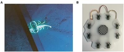


![APA format] Physiology References for the Native American Flute](https://flutopedia.com/img/JoinCVFluteNewsletter_150.jpg)