Joints
Joint Diseases
Finger Joint
Magnetic Resonance Imaging
Ankle Joint
Hip Joint
Tarsal Joints
Brain Mapping
Wrist Joint
Joint Capsule
Brain
Photic Stimulation
Visual Cortex
Electroencephalography
Image Processing, Computer-Assisted
Epilepsies, Partial
Joint Instability
Temporomandibular Joint Disorders
Parietal Lobe
Metatarsophalangeal Joint
Encephalocele
Hemianopsia
Foot Joints
Evoked Potentials, Visual
Visual Perception
Cerebral Cortex
Functional Laterality
Shoulder Joint
Spinal Nerves
Joint Prosthesis
Temporal Lobe
Blindness
Temporomandibular Joint Disc
Acromioclavicular Joint
Skull Fractures
Alpha Rhythm
Osteoarthritis
Headache
Meningocele
Pattern Recognition, Visual
Cranial Sinuses
Frontal Lobe
Arthritis, Rheumatoid
Dandy-Walker Syndrome
Cluster Headache
Visual Pathways
Sternoclavicular Joint
Cartilage, Articular
Synovial Fluid
Epilepsy, Reflex
Face
Biomechanical Phenomena
Phosphenes
Attention
Cranial Fossa, Posterior
Synovitis
Agnosia
Tomography, Emission-Computed
Axis
Synovial Membrane
Tomography, X-Ray Computed
Range of Motion, Articular
Psychomotor Performance
Visual Fields
Scalp
Magnetoencephalography
Parietal Bone
Arthritis, Experimental
Arthrography
Nerve Net
Arthritis, Infectious
Posterior Cerebral Artery
Skull Base
Analysis of Variance
Nerve Block
Osteoarthritis, Knee
Neuropsychological Tests
Carpal Joints
Hallucinations
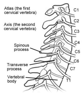
Cervical Vertebrae
Tectum Mesencephali
Dislocations
Movement
Oxygen
Headache Disorders
Electric Stimulation Therapy
Reading
Migraine Disorders
Epilepsy
Treatment Outcome
Tomography, Emission-Computed, Single-Photon
Head
Arnold-Chiari Malformation
Patellofemoral Joint
Evoked Potentials
Touch
Models, Anatomic
Reference Values
Intracranial Arteriovenous Malformations
Weight-Bearing
Pulsed Radiofrequency Treatment
Transcranial Magnetic Stimulation
Human Body
Reproducibility of Results
Brain Diseases
Severity of Illness Index
Imaging, Three-Dimensional
Skull Fracture, Basilar
Diffusion Magnetic Resonance Imaging
Nerve Fibers, Myelinated
Dura Mater
Menkes Kinky Hair Syndrome
Thalamus
Image Interpretation, Computer-Assisted
Parahippocampal Gyrus
Cerebral Infarction
Delta Rhythm
Ligaments, Articular
Atrophy
Cues
Cerebellum
Stifle
Alexia, Pure
Neck Muscles
Case-Control Studies
Temporomandibular Joint Dysfunction Syndrome
Recognition (Psychology)
Follow-Up Studies
Infarction, Posterior Cerebral Artery
Retrospective Studies
Contracture
Myoclonic Epilepsy, Juvenile
Positron-Emission Tomography
Dyslexia, Acquired
Electrodes, Implanted
Pain
Vision Disorders
Functional Neuroimaging
Touch Perception
Memory
Joint Commission on Accreditation of Healthcare Organizations
Radiopharmaceuticals
Perceptual Disorders
Electrooculography
Prosopagnosia
Stereotaxic Techniques
Technetium Tc 99m Exametazime
Neuroimaging
Iofetamine
Diffusion Tensor Imaging
Tibia
Posterior Leukoencephalopathy Syndrome
Hyperglycemic Hyperosmolar Nonketotic Coma
Alzheimer Disease
Statistics as Topic
Signal Processing, Computer-Assisted
Anisotropy
Hydrocephalus
Cerebral Ventricles
Neuralgia
Magnetic Resonance Spectroscopy
Transoral decompression for craniovertebral osseous anomalies: perioperative management dilemmas. (1/92)
The surgical outcome of 74 patients, who underwent transoral decompression (TOD) for ventral irreducible craniovertebral junction anomalies between January 1989 to September 1997, was studied to evaluate the perioperative complications and problems encountered. The indications for TOD included irreducible atlantoaxial dislocation (n=24), basilar invagination (n=16), and a combination of both (n=35). Following TOD, occipitocervical stabilization using Jain's technique was carried out in 50 (67.5%) and atlantoaxial fusion using Brooks' construct in 18 (24.3%) patients. The pre- and postoperative radiology was compared to assess the adequacy of decompression and stability. The major morbidity included pharyngeal wound sepsis leading to dehiscence (20.3%) and haemorrhage (4%), valopharyngeal insufficiency (8.1%), CSF leak (6.7%) and inadequate decompression (6.7%). Neurological deterioration occurred transiently in 17 (22.9%) and was sustained in 7 (9.4%) patients. The mortality in six cases was due to operative trauma, exanguination from pharyngeal wound (one each), postoperative instability and inability to be weaned off from the ventilator (two each). Of the 47 (63.5%) patients available at follow up ranging from 3 months to 2 years, 26 (55.3%) showed improvement from their preoperative status while 14 (29.8%) demonstrated stabilization of their neurological deficits. Seven (14.9%) of them deteriorated. Though TOD is logical and effective in relieving ventral compression due to craniovertebral junction anomalies, it carries the formidable risks of instability, incomplete decompression, neurological deterioration, CSF leak, infection and palatopharyngeal dysfunction. (+info)Bow hunter's stroke associated with atlantooccipital assimilation--case report. (2/92)
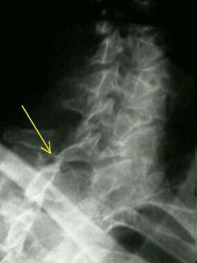
A 39-year-old male presented with bow hunter's stroke manifesting as repeated vertebrobasilar ischemic attacks induced by head rotation 45 degrees to the left. Three-dimensional computed tomography angiography clearly showed the occluded right vertebral artery (VA) between the axis and atlas. Single photon emission computed tomography study showed diffuse hypoperfusion of the brain stem and bilateral cerebellar hemispheres, suggesting hemodynamic compromise of these regions. He refused surgery and was treated conservatively. The most likely mechanism is that the affected VA was fixed by the ossification of the atlantooccipital membrane, vascular groove, and transverse foramen of the atlas, and therefore became elongated and compressed by head-turning. (+info)Surgical treatment of nonunited fractures of the odontoid process, with special reference to occipitocervical fusion for unreducible atlantoaxial subluxation or instability. (3/92)
Fifty-seven consecutive patients treated surgically for nonunited fractures of the odontoid process were reviewed. All patients presented late, exhibiting neurological deficits subsequent to nonunion. Delay in presentation was between 6 and 120 months (mean 32 months) after the original injury, due to missed diagnosis or inappropriate management. Seven patients who were reduced in traction underwent a Gallie atlantoaxial fusion. In the remaining 50 patients who were unreducible, an occipitocervical arthrodesis was performed. They were followed up for a minimum of 2 years, except one who died from postoperative respiratory failure. All patients obtained a solid bony union, including two in whom nonunion occurred following atlantoaxial fusion, and occipitocervical fusion was added as a rescue. Thirty-eight patients achieved excellent neurological recovery, nine still had some disability, five retained their neurological deficits and two reported a deterioration. In two patients, a recurrence in a traumatic episode was experienced long after a resolution. Our findings demonstrate that occipitocervical arthrodesis is preferable for unreducible subluxation or instability of atlantoaxial articulation in nonunion of odontoid fractures. (+info)Bilateral type 1 proatlantal arteries with absence of vertebral arteries. (4/92)
The persistent proatlantal artery is a well-described communication between the carotid and vertebrobasilar system. However, persistence of bilateral proatlantal arteries is exceptionally rare. Although usually noted as an incidental finding, the presence of a proatlantal artery, particularly when bilateral, may result in unusual symptoms or may have implications for therapy. We report a case of bilateral proatlantal arteries, describe their embryology, and consider potential clinical implications of this finding. (+info)Hindbrain stroke in children caused by extracranial vertebral artery trauma. (5/92)
Hindbrain transient ischemic attacks (TIAs) culminating in posterior circulation stroke are described in five children. Atlanto-axial subluxation and angiographical documentation of C1 to C2 level arterial pathology are documented in one patient. Four additional patients with nearly identical clinical presentations, posterior fossa TIAs, stroke and basilar angiographical pathology are reviewed. A mechanical traumatic etiology is suggested. Unexplained transient repeated brain stem and/or cerebellar sympotomatology may be due to extracranial vetebral artery stenosis or occlusion by atlanto-axial instability. After appropriate documentation, stabilization may prevent further TIAs or strokes. (+info)Recognition and management of atlanto-occipital dislocation: improving survival from an often fatal condition. (6/92)
OBJECTIVE: To provide an overview of atlanto-occipital dislocation and associated occipital condyle fracturcs so as to alert physicians to this rare injury and potentially improve patient outcome. The pertinent anatomy, mechanism of injury, clinical and radiologic evaluation and the management of these rare injuries are discussed in an attempt to alert physicians to this type of injury and to improve outcome. DATA SOURCES: The data were obtained from a MEDLINE search of the English literature from 1966 to 1999 and the experience of 4 spine surgeons at a quaternary care acute spinal cord injury unit. STUDY SELECTION: Detailed anatomic and epidemiologically sound radiology studies were identified and analyzed. Only small retrospective studies or case series were available in the literature. DATA EXTRACTION: Valid anatomic, biomechanical and radiologic evaluation was extracted from studies. Clinical data came from limited studies and expert opinion. DATA SYNTHESIS: Early diagnosis is essential and is facilitated by a detailed clinical examination and strict adherence to an imaging algorithm that includes CT and MRI scanning. When the dislocation is identified, timely gentle reduction and prompt stabilization throuigh nonoperative or operative means is found to optimize patient outcome. CONCLUSIONS: Atlanto-occipital dislocation should be suspected in any patient involved in a high speed motor vehicle or pedestrian collision. Once suspected, proper imaging and appropriate management of these once fatal injuries can improve survival and neurologic outcome. (+info)Skeletal aspects of the atlanto-occipital fusion in a Japanese brown calf. (7/92)
Atlanto-occipital fusion in a Japanese Brown calf was examined morphologically, paying special attention to skeletal changes. At the craniovertebral junction, the basal occipital bone fused to the cranial extremity of the ventral arch of the atlas with the rudiment of the atlantal centrum. The dens was not formed at the axis. These changes suggest that a hypocentrum and a centrum of the atlas derived from the first cervical sclerotome had failed to separate the occipital base from the proatlantal sclerotome including the apical element of the dens. Although a developmental disturbance at the cervical and thoracic vertebrae was also associated, critical neurological signs such as ataxia and paralysis were absent. (+info)Traumatic posterior atlantooccipital dislocation with Jefferson fracture and fracture-dislocation of C6-C7: a case report with survival. (8/92)
Atlantooccipital dislocation (AOD) is a rare and usually fatal injury. In the current study, the authors reported an extremely rare case of posterior AOD with Jefferson fracture and fracture-dislocation of C6-C7. The patient survived the injury and had only incomplete quadriplegia below the C7 segment with anterior cord syndrome. He was successfully managed with in situ occipitocervical fusion using the Cotrel-Dubousset rod system, corpectomy of C6, and anterior interbody fusion of C5-C7 with plating. To our knowledge, this is the first report of posterior AOD with two other non-contiguous cervical spine injuries. A high index of suspicion and careful examination of the upper cervical spine should be considered as the key to the diagnosis of AOD in cases that involve multiple or lower cervical spine injuries. (+info)The occipital bone is the single, posterior cranial bone that forms the base of the skull and encloses the brain. It articulates with the parietal bones anteriorly and the temporal bones laterally. The occipital bone also contains several important structures such as the foramen magnum, through which the spinal cord connects to the brain, and the external and internal occipital protuberances, which serve as attachment points for neck muscles.
A joint is the location at which two or more bones make contact. They are constructed to allow movement and provide support and stability to the body during motion. Joints can be classified in several ways, including structure, function, and the type of tissue that forms them. The three main types of joints based on structure are fibrous (or fixed), cartilaginous, and synovial (or diarthrosis). Fibrous joints do not have a cavity and have limited movement, while cartilaginous joints allow for some movement and are connected by cartilage. Synovial joints, the most common and most movable type, have a space between the articular surfaces containing synovial fluid, which reduces friction and wear. Examples of synovial joints include hinge, pivot, ball-and-socket, saddle, and condyloid joints.
The knee joint, also known as the tibiofemoral joint, is the largest and one of the most complex joints in the human body. It is a synovial joint that connects the thighbone (femur) to the shinbone (tibia). The patella (kneecap), which is a sesamoid bone, is located in front of the knee joint and helps in the extension of the leg.
The knee joint is made up of three articulations: the femorotibial joint between the femur and tibia, the femoropatellar joint between the femur and patella, and the tibiofibular joint between the tibia and fibula. These articulations are surrounded by a fibrous capsule that encloses the synovial membrane, which secretes synovial fluid to lubricate the joint.
The knee joint is stabilized by several ligaments, including the medial and lateral collateral ligaments, which provide stability to the sides of the joint, and the anterior and posterior cruciate ligaments, which prevent excessive forward and backward movement of the tibia relative to the femur. The menisci, which are C-shaped fibrocartilaginous structures located between the femoral condyles and tibial plateaus, also help to stabilize the joint by absorbing shock and distributing weight evenly across the articular surfaces.
The knee joint allows for flexion, extension, and a small amount of rotation, making it essential for activities such as walking, running, jumping, and sitting.
Joint diseases is a broad term that refers to various conditions affecting the joints, including but not limited to:
1. Osteoarthritis (OA): A degenerative joint disease characterized by the breakdown of cartilage and underlying bone, leading to pain, stiffness, and potential loss of function.
2. Rheumatoid Arthritis (RA): An autoimmune disorder causing inflammation in the synovial membrane lining the joints, resulting in swelling, pain, and joint damage if left untreated.
3. Infectious Arthritis: Joint inflammation caused by bacterial, viral, or fungal infections that spread through the bloodstream or directly enter the joint space.
4. Gout: A type of arthritis resulting from the buildup of uric acid crystals in the joints, typically affecting the big toe and characterized by sudden attacks of severe pain, redness, and swelling.
5. Psoriatic Arthritis (PsA): An inflammatory joint disease associated with psoriasis, causing symptoms such as pain, stiffness, and swelling in the joints and surrounding tissues.
6. Juvenile Idiopathic Arthritis (JIA): A group of chronic arthritis conditions affecting children, characterized by joint inflammation, pain, and stiffness.
7. Ankylosing Spondylitis: A form of arthritis primarily affecting the spine, causing inflammation, pain, and potential fusion of spinal vertebrae.
8. Bursitis: Inflammation of the fluid-filled sacs (bursae) that cushion joints, leading to pain and swelling.
9. Tendinitis: Inflammation or degeneration of tendons, which connect muscles to bones, often resulting in pain and stiffness near joints.
These conditions can impact the function and mobility of affected joints, causing discomfort and limiting daily activities. Proper diagnosis and treatment are essential for managing joint diseases and preserving joint health.
A finger joint, also known as an articulation, is the point where two bones in a finger connect and allow for movement. The majority of finger joints are classified as hinge joints, permitting flexion and extension movements. These joints consist of several components:
1. Articular cartilage: Smooth tissue that covers the ends of the bones, enabling smooth movement and protecting the bones from friction.
2. Joint capsule: A fibrous sac enclosing the joint, providing stability and producing synovial fluid for lubrication.
3. Synovial membrane: Lines the inner surface of the joint capsule and produces synovial fluid to lubricate the joint.
4. Volar plate (palmar ligament): A strong band of tissue located on the palm side of the joint, preventing excessive extension and maintaining alignment.
5. Collateral ligaments: Two bands of tissue located on each side of the joint, providing lateral stability and limiting radial and ulnar deviation.
6. Flexor tendons: Tendons that attach to the bones on the palmar side of the finger joints, facilitating flexion movements.
7. Extensor tendons: Tendons that attach to the bones on the dorsal side of the finger joints, enabling extension movements.
Finger joints are essential for hand function and enable activities such as grasping, holding, writing, and manipulating objects.
Medical Definition:
Magnetic Resonance Imaging (MRI) is a non-invasive diagnostic imaging technique that uses a strong magnetic field and radio waves to create detailed cross-sectional or three-dimensional images of the internal structures of the body. The patient lies within a large, cylindrical magnet, and the scanner detects changes in the direction of the magnetic field caused by protons in the body. These changes are then converted into detailed images that help medical professionals to diagnose and monitor various medical conditions, such as tumors, injuries, or diseases affecting the brain, spinal cord, heart, blood vessels, joints, and other internal organs. MRI does not use radiation like computed tomography (CT) scans.
The ankle joint, also known as the talocrural joint, is the articulation between the bones of the lower leg (tibia and fibula) and the talus bone in the foot. It is a synovial hinge joint that allows for dorsiflexion and plantarflexion movements, which are essential for walking, running, and jumping. The ankle joint is reinforced by strong ligaments on both sides to provide stability during these movements.
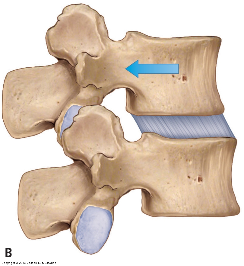
The Atlanto-Occipital Joint, also known as the AO joint or the craniocervical joint, is the articulation between the occiput (the base of the skull) and the atlas (the first cervical vertebra). This joint allows for movements such as nodding your head "yes" and tilting your head from side to side. It is a crucial joint in maintaining the alignment and stability of the head and neck.
The hip joint, also known as the coxal joint, is a ball-and-socket type synovial joint that connects the femur (thigh bone) to the pelvis. The "ball" is the head of the femur, while the "socket" is the acetabulum, a concave surface on the pelvic bone.
The hip joint is surrounded by a strong fibrous capsule and is reinforced by several ligaments, including the iliofemoral, ischiofemoral, and pubofemoral ligaments. The joint allows for flexion, extension, abduction, adduction, medial and lateral rotation, and circumduction movements, making it one of the most mobile joints in the body.
The hip joint is also supported by various muscles, including the gluteus maximus, gluteus medius, gluteus minimus, iliopsoas, and other hip flexors and extensors. These muscles provide stability and strength to the joint, allowing for weight-bearing activities such as walking, running, and jumping.
The tarsal joints are a series of articulations in the foot that involve the bones of the hindfoot and midfoot. There are three main tarsal joints:
1. Talocrural joint (also known as the ankle joint): This is the joint between the talus bone of the lower leg and the tibia and fibula bones of the lower leg, as well as the calcaneus bone of the foot. It allows for dorsiflexion and plantarflexion movements of the foot.
2. Subtalar joint: This is the joint between the talus bone and the calcaneus bone. It allows for inversion and eversion movements of the foot.
3. Tarsometatarsal joints (also known as the Lisfranc joint): These are the joints between the tarsal bones of the midfoot and the metatarsal bones of the forefoot. They allow for flexion, extension, abduction, and adduction movements of the foot.
These joints play an important role in the stability and mobility of the foot, allowing for various movements during activities such as walking, running, and jumping.
Brain mapping is a broad term that refers to the techniques used to understand the structure and function of the brain. It involves creating maps of the various cognitive, emotional, and behavioral processes in the brain by correlating these processes with physical locations or activities within the nervous system. Brain mapping can be accomplished through a variety of methods, including functional magnetic resonance imaging (fMRI), positron emission tomography (PET) scans, electroencephalography (EEG), and others. These techniques allow researchers to observe which areas of the brain are active during different tasks or thoughts, helping to shed light on how the brain processes information and contributes to our experiences and behaviors. Brain mapping is an important area of research in neuroscience, with potential applications in the diagnosis and treatment of neurological and psychiatric disorders.
The wrist joint, also known as the radiocarpal joint, is a condyloid joint that connects the distal end of the radius bone in the forearm to the proximal row of carpal bones in the hand (scaphoid, lunate, and triquetral bones). It allows for flexion, extension, radial deviation, and ulnar deviation movements of the hand. The wrist joint is surrounded by a capsule and reinforced by several ligaments that provide stability and strength to the joint.
A joint capsule is the fibrous sac that encloses a synovial joint, which is a type of joint characterized by the presence of a cavity filled with synovial fluid. The joint capsule provides stability and strength to the joint, while also allowing for a range of motion. It consists of two layers: an outer fibrous layer and an inner synovial membrane. The fibrous layer is made up of dense connective tissue that helps to stabilize the joint, while the synovial membrane produces synovial fluid, which lubricates the joint and reduces friction during movement.
The sacroiliac (SI) joint is the joint that connects the iliac bone (part of the pelvis) and the sacrum (the triangular bone at the base of the spine). There are two sacroiliac joints, one on each side of the spine. The primary function of these joints is to absorb shock between the upper body and lower body and distribute the weight of the upper body to the lower body. They also provide a small amount of movement to allow for flexibility when walking or running. The SI joints are supported and stabilized by strong ligaments, muscles, and bones.
The brain is the central organ of the nervous system, responsible for receiving and processing sensory information, regulating vital functions, and controlling behavior, movement, and cognition. It is divided into several distinct regions, each with specific functions:
1. Cerebrum: The largest part of the brain, responsible for higher cognitive functions such as thinking, learning, memory, language, and perception. It is divided into two hemispheres, each controlling the opposite side of the body.
2. Cerebellum: Located at the back of the brain, it is responsible for coordinating muscle movements, maintaining balance, and fine-tuning motor skills.
3. Brainstem: Connects the cerebrum and cerebellum to the spinal cord, controlling vital functions such as breathing, heart rate, and blood pressure. It also serves as a relay center for sensory information and motor commands between the brain and the rest of the body.
4. Diencephalon: A region that includes the thalamus (a major sensory relay station) and hypothalamus (regulates hormones, temperature, hunger, thirst, and sleep).
5. Limbic system: A group of structures involved in emotional processing, memory formation, and motivation, including the hippocampus, amygdala, and cingulate gyrus.
The brain is composed of billions of interconnected neurons that communicate through electrical and chemical signals. It is protected by the skull and surrounded by three layers of membranes called meninges, as well as cerebrospinal fluid that provides cushioning and nutrients.
Photic stimulation is a medical term that refers to the exposure of the eyes to light, specifically repetitive pulses of light, which is used as a method in various research and clinical settings. In neuroscience, it's often used in studies related to vision, circadian rhythms, and brain function.
In a clinical context, photic stimulation is sometimes used in the diagnosis of certain medical conditions such as seizure disorders (like epilepsy). By observing the response of the brain to this light stimulus, doctors can gain valuable insights into the functioning of the brain and the presence of any neurological disorders.
However, it's important to note that photic stimulation should be conducted under the supervision of a trained healthcare professional, as improper use can potentially trigger seizures in individuals who are susceptible to them.
The visual cortex is the part of the brain that processes visual information. It is located in the occipital lobe, which is at the back of the brain. The visual cortex is responsible for receiving and interpreting signals from the retina, which are then transmitted through the optic nerve and optic tract.
The visual cortex contains several areas that are involved in different aspects of visual processing, such as identifying shapes, colors, and movements. These areas work together to help us recognize and understand what we see. Damage to the visual cortex can result in various visual impairments, such as blindness or difficulty with visual perception.
Electroencephalography (EEG) is a medical procedure that records electrical activity in the brain. It uses small, metal discs called electrodes, which are attached to the scalp with paste or a specialized cap. These electrodes detect tiny electrical charges that result from the activity of brain cells, and the EEG machine then amplifies and records these signals.
EEG is used to diagnose various conditions related to the brain, such as seizures, sleep disorders, head injuries, infections, and degenerative diseases like Alzheimer's or Parkinson's. It can also be used during surgery to monitor brain activity and ensure that surgical procedures do not interfere with vital functions.
EEG is a safe and non-invasive procedure that typically takes about 30 minutes to an hour to complete, although longer recordings may be necessary in some cases. Patients are usually asked to relax and remain still during the test, as movement can affect the quality of the recording.
Computer-assisted image processing is a medical term that refers to the use of computer systems and specialized software to improve, analyze, and interpret medical images obtained through various imaging techniques such as X-ray, CT (computed tomography), MRI (magnetic resonance imaging), ultrasound, and others.
The process typically involves several steps, including image acquisition, enhancement, segmentation, restoration, and analysis. Image processing algorithms can be used to enhance the quality of medical images by adjusting contrast, brightness, and sharpness, as well as removing noise and artifacts that may interfere with accurate diagnosis. Segmentation techniques can be used to isolate specific regions or structures of interest within an image, allowing for more detailed analysis.
Computer-assisted image processing has numerous applications in medical imaging, including detection and characterization of lesions, tumors, and other abnormalities; assessment of organ function and morphology; and guidance of interventional procedures such as biopsies and surgeries. By automating and standardizing image analysis tasks, computer-assisted image processing can help to improve diagnostic accuracy, efficiency, and consistency, while reducing the potential for human error.
Epilepsy, partial is a type of epilepsy characterized by recurrent, unprovoked seizures that originate in a specific, localized area of the brain. These seizures are also known as focal seizures and can vary in severity and symptoms depending on the location of the abnormal electrical activity in the brain.
Partial epilepsies can be further classified into two main categories: simple partial seizures and complex partial seizures. Simple partial seizures do not involve a loss of consciousness, while complex partial seizures are associated with impaired awareness or responsiveness during the seizure.
The causes of partial epilepsies can include brain injury, infection, stroke, tumors, genetic factors, or an unknown cause. Treatment typically involves anti-seizure medications, and in some cases, surgery may be recommended to remove the specific area of the brain responsible for the seizures.
Joint instability is a condition characterized by the loss of normal joint function and increased risk of joint injury due to impaired integrity of the supporting structures, such as ligaments, muscles, or cartilage. This can result in excessive movement or laxity within the joint, leading to decreased stability and increased susceptibility to dislocations or subluxations. Joint instability may cause pain, swelling, and limited range of motion, and it can significantly impact a person's mobility and quality of life. It is often caused by trauma, degenerative conditions, or congenital abnormalities and may require medical intervention, such as physical therapy, bracing, or surgery, to restore joint stability.
Temporomandibular Joint Disorders (TMD) refer to a group of conditions that cause pain and dysfunction in the temporomandibular joint (TMJ) and the muscles that control jaw movement. The TMJ is the hinge joint that connects the lower jaw (mandible) to the skull (temporal bone) in front of the ear. It allows for movements required for activities such as eating, speaking, and yawning.
TMD can result from various causes, including:
1. Muscle tension or spasm due to clenching or grinding teeth (bruxism), stress, or jaw misalignment
2. Dislocation or injury of the TMJ disc, which is a small piece of cartilage that acts as a cushion between the bones in the joint
3. Arthritis or other degenerative conditions affecting the TMJ
4. Bite problems (malocclusion) leading to abnormal stress on the TMJ and its surrounding muscles
5. Stress, which can exacerbate existing TMD symptoms by causing muscle tension
Symptoms of Temporomandibular Joint Disorders may include:
- Pain or tenderness in the jaw, face, neck, or shoulders
- Limited jaw movement or locking of the jaw
- Clicking, popping, or grating sounds when moving the jaw
- Headaches, earaches, or dizziness
- Difficulty chewing or biting
- Swelling on the side of the face
Treatment for TMD varies depending on the severity and cause of the condition. It may include self-care measures (like eating soft foods, avoiding extreme jaw movements, and applying heat or cold packs), physical therapy, medications (such as muscle relaxants, pain relievers, or anti-inflammatory drugs), dental work (including bite adjustments or orthodontic treatment), or even surgery in severe cases.
The parietal lobe is a region of the brain that is located in the posterior part of the cerebral cortex, covering the upper and rear portions of the brain. It is involved in processing sensory information from the body, such as touch, temperature, and pain, as well as spatial awareness and perception, visual-spatial cognition, and the integration of different senses.
The parietal lobe can be divided into several functional areas, including the primary somatosensory cortex (which receives tactile information from the body), the secondary somatosensory cortex (which processes more complex tactile information), and the posterior parietal cortex (which is involved in spatial attention, perception, and motor planning).
Damage to the parietal lobe can result in various neurological symptoms, such as neglect of one side of the body, difficulty with spatial orientation, problems with hand-eye coordination, and impaired mathematical and language abilities.
The metatarsophalangeal (MTP) joint is the joint in the foot where the metatarsal bones of the foot (the long bones behind the toes) connect with the proximal phalanges of the toes. It's a synovial joint, which means it's surrounded by a capsule containing synovial fluid to allow for smooth movement. The MTP joint is responsible for allowing the flexion and extension movements of the toes, and is important for maintaining balance and pushing off during walking and running. Issues with the MTP joint can lead to conditions such as hallux valgus (bunions) or hammertoe.
An Encephalocele is a type of neural tube defect that occurs when the bones of the skull do not close completely during fetal development. This results in a sac-like protrusion of the brain and the membranes that cover it through an opening in the skull. The sac may be visible on the scalp, forehead, or back of the head, and can vary in size. Encephaloceles can cause a range of symptoms, including developmental delays, intellectual disabilities, vision problems, and seizures, depending on the severity and location of the defect. Treatment typically involves surgical repair of the encephalocele soon after birth to prevent further damage to the brain and improve outcomes.
Hemianopsia is a medical term that refers to a loss of vision in half of the visual field in one or both eyes. It can be either homonymous (the same side in both eyes) or heteronymous (different sides in each eye). Hemianopsia usually results from damage to the optic radiations or occipital cortex in the brain, often due to stroke, trauma, tumor, or other neurological conditions. It can significantly impact a person's daily functioning and may require visual rehabilitation to help compensate for the vision loss.
"Foot joints" is a general term that refers to the various articulations or connections between the bones in the foot. There are several joints in the foot, including:
1. The ankle joint (tibiotalar joint): This is the joint between the tibia and fibula bones of the lower leg and the talus bone of the foot.
2. The subtalar joint (talocalcaneal joint): This is the joint between the talus bone and the calcaneus (heel) bone.
3. The calcaneocuboid joint: This is the joint between the calcaneus bone and the cuboid bone, which is one of the bones in the midfoot.
4. The tarsometatarsal joints (Lisfranc joint): These are the joints that connect the tarsal bones in the midfoot to the metatarsal bones in the forefoot.
5. The metatarsophalangeal joints: These are the joints between the metatarsal bones and the phalanges (toes) in the forefoot.
6. The interphalangeal joints: These are the joints between the phalanges within each toe.
Each of these foot joints plays a specific role in supporting the foot, absorbing shock, and allowing for movement and flexibility during walking and other activities.
Evoked potentials, visual, also known as visually evoked potentials (VEPs), are electrical responses recorded from the brain following the presentation of a visual stimulus. These responses are typically measured using electroencephalography (EEG) and can provide information about the functioning of the visual pathways in the brain.
There are several types of VEPs, including pattern-reversal VEPs and flash VEPs. Pattern-reversal VEPs are elicited by presenting alternating checkerboard patterns, while flash VEPs are elicited by flashing a light. The responses are typically analyzed in terms of their latency (the time it takes for the response to occur) and amplitude (the size of the response).
VEPs are often used in clinical settings to help diagnose and monitor conditions that affect the visual system, such as multiple sclerosis, optic neuritis, and brainstem tumors. They can also be used in research to study the neural mechanisms underlying visual perception.
Visual perception refers to the ability to interpret and organize information that comes from our eyes to recognize and understand what we are seeing. It involves several cognitive processes such as pattern recognition, size estimation, movement detection, and depth perception. Visual perception allows us to identify objects, navigate through space, and interact with our environment. Deficits in visual perception can lead to learning difficulties and disabilities.
The cerebral cortex is the outermost layer of the brain, characterized by its intricate folded structure and wrinkled appearance. It is a region of great importance as it plays a key role in higher cognitive functions such as perception, consciousness, thought, memory, language, and attention. The cerebral cortex is divided into two hemispheres, each containing four lobes: the frontal, parietal, temporal, and occipital lobes. These areas are responsible for different functions, with some regions specializing in sensory processing while others are involved in motor control or associative functions. The cerebral cortex is composed of gray matter, which contains neuronal cell bodies, and is covered by a layer of white matter that consists mainly of myelinated nerve fibers.
Functional laterality, in a medical context, refers to the preferential use or performance of one side of the body over the other for specific functions. This is often demonstrated in hand dominance, where an individual may be right-handed or left-handed, meaning they primarily use their right or left hand for tasks such as writing, eating, or throwing.
However, functional laterality can also apply to other bodily functions and structures, including the eyes (ocular dominance), ears (auditory dominance), or legs. It's important to note that functional laterality is not a strict binary concept; some individuals may exhibit mixed dominance or no strong preference for one side over the other.
In clinical settings, assessing functional laterality can be useful in diagnosing and treating various neurological conditions, such as stroke or traumatic brain injury, where understanding any resulting lateralized impairments can inform rehabilitation strategies.
The shoulder joint, also known as the glenohumeral joint, is the most mobile joint in the human body. It is a ball and socket synovial joint that connects the head of the humerus (upper arm bone) to the glenoid cavity of the scapula (shoulder blade). The shoulder joint allows for a wide range of movements including flexion, extension, abduction, adduction, internal rotation, and external rotation. It is surrounded by a group of muscles and tendons known as the rotator cuff that provide stability and enable smooth movement of the joint.
Spinal nerves are the bundles of nerve fibers that transmit signals between the spinal cord and the rest of the body. There are 31 pairs of spinal nerves in the human body, which can be divided into five regions: 8 cervical, 12 thoracic, 5 lumbar, 5 sacral, and 1 coccygeal. Each spinal nerve carries both sensory information (such as touch, temperature, and pain) from the periphery to the spinal cord, and motor information (such as muscle control) from the spinal cord to the muscles and other structures in the body. Spinal nerves also contain autonomic fibers that regulate involuntary functions such as heart rate, digestion, and blood pressure.
The foramen magnum is the largest opening in the human skull, located at the base of the skull, through which the spinal cord connects to the brain. It is a crucial structure for the transmission of nerve impulses between the brain and the rest of the body. The foramen magnum also provides passage for blood vessels that supply the brainstem and upper spinal cord.
A joint prosthesis, also known as an artificial joint or a replacement joint, is a surgical implant used to replace all or part of a damaged or diseased joint. The most common types of joint prostheses are total hip replacements and total knee replacements. These prostheses typically consist of a combination of metal, plastic, and ceramic components that are designed to replicate the movement and function of a natural joint.
Joint prostheses are usually recommended for patients who have severe joint pain or mobility issues that cannot be adequately managed with other treatments such as physical therapy, medication, or lifestyle changes. The goal of joint replacement surgery is to relieve pain, improve joint function, and enhance the patient's quality of life.
Joint prostheses are typically made from materials such as titanium, cobalt-chrome alloys, stainless steel, polyethylene plastic, and ceramics. The choice of material depends on a variety of factors, including the patient's age, activity level, weight, and overall health.
While joint replacement surgery is generally safe and effective, there are risks associated with any surgical procedure, including infection, blood clots, implant loosening or failure, and nerve damage. Patients who undergo joint replacement surgery typically require several weeks of rehabilitation and physical therapy to regain strength and mobility in the affected joint.
The temporal lobe is one of the four main lobes of the cerebral cortex in the brain, located on each side of the head roughly level with the ears. It plays a major role in auditory processing, memory, and emotion. The temporal lobe contains several key structures including the primary auditory cortex, which is responsible for analyzing sounds, and the hippocampus, which is crucial for forming new memories. Damage to the temporal lobe can result in various neurological symptoms such as hearing loss, memory impairment, and changes in emotional behavior.
Blindness is a condition of complete or near-complete vision loss. It can be caused by various factors such as eye diseases, injuries, or birth defects. Total blindness means that a person cannot see anything at all, while near-complete blindness refers to having only light perception or the ability to perceive the direction of light, but not able to discern shapes or forms. Legal blindness is a term used to define a certain level of visual impairment that qualifies an individual for government assistance and benefits; it usually means best corrected visual acuity of 20/200 or worse in the better eye, or a visual field no greater than 20 degrees in diameter.
The temporomandibular joint (TMJ) disc is a small, thin piece of fibrocartilaginous tissue located within the TMJ, which is the joint that connects the mandible (jawbone) to the temporal bone of the skull. The disc acts as a cushion and allows for smooth movement of the jaw during activities such as eating, speaking, and yawning. It divides the joint into two compartments: the upper and lower compartments.
The TMJ disc is composed of several types of tissue, including collagen fibers, elastin fibers, and a small number of cells called fibroblasts. The disc's unique structure allows it to withstand the forces generated during jaw movement and helps to distribute these forces evenly across the joint.
The TMJ disc can become damaged or displaced due to various factors such as trauma, teeth grinding (bruxism), or degenerative joint diseases like osteoarthritis. This can lead to temporomandibular disorders (TMDs) characterized by pain, stiffness, and limited jaw movement.
The acromioclavicular (AC) joint is the joint located between the acromion process of the scapula (shoulder blade) and the clavicle (collarbone). It allows for a small amount of movement between these two bones and participates in shoulder motion. Injuries to this joint, such as AC joint separations or sprains, are common and can occur due to falls, direct blows, or repetitive motions that cause the ligaments that support the AC joint to become stretched or torn.
A skull fracture is a break in one or more of the bones that form the skull. It can occur from a direct blow to the head, penetrating injuries like gunshot wounds, or from strong rotational forces during an accident. There are several types of skull fractures, including:
1. Linear Skull Fracture: This is the most common type, where there's a simple break in the bone without any splintering, depression, or displacement. It often doesn't require treatment unless it's near a sensitive area like an eye or ear.
2. Depressed Skull Fracture: In this type, a piece of the skull is pushed inward toward the brain. Surgery may be needed to relieve pressure on the brain and repair the fracture.
3. Diastatic Skull Fracture: This occurs along the suture lines (the fibrous joints between the skull bones) that haven't fused yet, often seen in infants and young children.
4. Basilar Skull Fracture: This involves fractures at the base of the skull. It can be serious due to potential injury to the cranial nerves and blood vessels located in this area.
5. Comminuted Skull Fracture: In this severe type, the bone is shattered into many pieces. These fractures usually require extensive surgical repair.
Symptoms of a skull fracture can include pain, swelling, bruising, bleeding (if there's an open wound), and in some cases, clear fluid draining from the ears or nose (cerebrospinal fluid leak). Severe fractures may cause brain injury, leading to symptoms like confusion, loss of consciousness, seizures, or neurological deficits. Immediate medical attention is necessary for any suspected skull fracture.
Alpha rhythm is a type of brain wave that is typically observed in the electroencephalogram (EEG) of normal, awake individuals when they have their eyes closed. It is characterized by sinusoidal waves with a frequency range of 8-13 Hz and is most prominent over the occipital region of the head, which is located at the back of the skull above the brain's visual cortex.
Alpha rhythm is typically associated with relaxed wakefulness, and its presence may indicate that an individual is awake but not engaged in any mentally demanding tasks. It can be blocked or suppressed by various stimuli, such as opening one's eyes, hearing a loud noise, or engaging in mental activity.
Disruptions in alpha rhythm have been observed in various neurological and psychiatric conditions, including epilepsy, dementia, depression, and anxiety disorders. However, more research is needed to fully understand the clinical significance of these abnormalities.
Osteoarthritis (OA) is a type of joint disease that is characterized by the breakdown and eventual loss of cartilage - the tissue that cushions the ends of bones where they meet in the joints. This breakdown can cause the bones to rub against each other, causing pain, stiffness, and loss of mobility. OA can occur in any joint, but it most commonly affects the hands, knees, hips, and spine. It is often associated with aging and can be caused or worsened by obesity, injury, or overuse.
The medical definition of osteoarthritis is: "a degenerative, non-inflammatory joint disease characterized by the loss of articular cartilage, bone remodeling, and the formation of osteophytes (bone spurs). It is often associated with pain, stiffness, and decreased range of motion in the affected joint."
A headache is defined as pain or discomfort in the head, scalp, or neck. It can be a symptom of various underlying conditions such as stress, sinus congestion, migraine, or more serious issues like meningitis or concussion. Headaches can vary in intensity, ranging from mild to severe, and may be accompanied by other symptoms such as nausea, vomiting, or sensitivity to light and sound. There are over 150 different types of headaches, including tension headaches, cluster headaches, and sinus headaches, each with their own specific characteristics and causes.
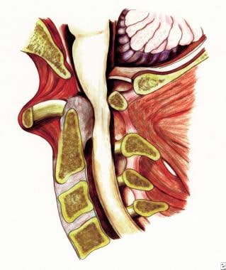
The Cervical Atlas, also known as C1 or the atlas vertebra, is the uppermost and most superior of the seven cervical vertebrae in the human spine. It plays a crucial role in supporting and facilitating the movement of the head, as it articulates with both the occipital bone (forming the joint called the atlanto-occipital joint) and the axis (or C2) vertebra (forming the atlantoaxial joint). The unique structure of the cervical atlas lacks a body, instead having an anterior and posterior arch with two lateral masses that form the facet joints for articulation with the axis. This arrangement allows for a wide range of motion in the neck, including flexion, extension, lateral bending, and rotation.
A meningocele is a type of neural tube defect that results in the herniation of the meninges (the protective membranes covering the brain and spinal cord) through a defect in the vertebral column. The meninges protrude as a sac-like structure, which may be covered by skin or a thin layer of tissue. Meningoceles usually do not contain neural tissue, but cerebrospinal fluid is present within the sac. They are typically asymptomatic unless there is compression of surrounding structures or infection. Treatment generally involves surgical repair to prevent potential complications such as meningitis or neurological damage.
Visual pattern recognition is the ability to identify and interpret patterns in visual information. In a medical context, it often refers to the process by which healthcare professionals recognize and diagnose medical conditions based on visible signs or symptoms. This can involve recognizing the characteristic appearance of a rash, wound, or other physical feature associated with a particular disease or condition. It may also involve recognizing patterns in medical images such as X-rays, CT scans, or MRIs.
In the field of radiology, for example, visual pattern recognition is a critical skill. Radiologists are trained to recognize the typical appearances of various diseases and conditions in medical images. This allows them to make accurate diagnoses based on the patterns they see. Similarly, dermatologists use visual pattern recognition to identify skin abnormalities and diseases based on the appearance of rashes, lesions, or other skin changes.
Overall, visual pattern recognition is an essential skill in many areas of medicine, allowing healthcare professionals to quickly and accurately diagnose medical conditions based on visible signs and symptoms.
Skull neoplasms refer to abnormal growths or tumors that develop within the skull. These growths can be benign (non-cancerous) or malignant (cancerous). They can originate from various types of cells, such as bone cells, nerve cells, or soft tissues. Skull neoplasms can cause various symptoms depending on their size and location, including headaches, seizures, vision problems, hearing loss, and neurological deficits. Treatment options include surgery, radiation therapy, and chemotherapy. It is important to note that a neoplasm in the skull can also refer to metastatic cancer, which has spread from another part of the body to the skull.
Cranial sinuses are a part of the venous system in the human head. They are air-filled spaces located within the skull and are named according to their location. The cranial sinuses include:
1. Superior sagittal sinus: It runs along the top of the brain, inside the skull, and drains blood from the scalp and the veins of the brain.
2. Inferior sagittal sinus: It runs along the bottom of the brain and drains into the straight sinus.
3. Straight sinus: It is located at the back of the brain and receives blood from the inferior sagittal sinus and great cerebral vein.
4. Occipital sinuses: They are located at the back of the head and drain blood from the scalp and skull.
5. Cavernous sinuses: They are located on each side of the brain, near the temple, and receive blood from the eye and surrounding areas.
6. Sphenoparietal sinus: It is a small sinus that drains blood from the front part of the brain into the cavernous sinus.
7. Petrosquamosal sinuses: They are located near the ear and drain blood from the scalp and skull.
The cranial sinuses play an essential role in draining blood from the brain and protecting it from injury.
The frontal lobe is the largest lobes of the human brain, located at the front part of each cerebral hemisphere and situated in front of the parietal and temporal lobes. It plays a crucial role in higher cognitive functions such as decision making, problem solving, planning, parts of social behavior, emotional expressions, physical reactions, and motor function. The frontal lobe is also responsible for what's known as "executive functions," which include the ability to focus attention, understand rules, switch focus, plan actions, and inhibit inappropriate behaviors. It is divided into five areas, each with its own specific functions: the primary motor cortex, premotor cortex, Broca's area, prefrontal cortex, and orbitofrontal cortex. Damage to the frontal lobe can result in a wide range of impairments, depending on the location and extent of the injury.
Rheumatoid arthritis (RA) is a systemic autoimmune disease that primarily affects the joints. It is characterized by persistent inflammation, synovial hyperplasia, and subsequent damage to the articular cartilage and bone. The immune system mistakenly attacks the body's own tissues, specifically targeting the synovial membrane lining the joint capsule. This results in swelling, pain, warmth, and stiffness in affected joints, often most severely in the hands and feet.
RA can also have extra-articular manifestations, affecting other organs such as the lungs, heart, skin, eyes, and blood vessels. The exact cause of RA remains unknown, but it is believed to involve a complex interplay between genetic susceptibility and environmental triggers. Early diagnosis and treatment are crucial in managing rheumatoid arthritis to prevent joint damage, disability, and systemic complications.
The skull is the bony structure that encloses and protects the brain, the eyes, and the ears. It is composed of two main parts: the cranium, which contains the brain, and the facial bones. The cranium is made up of several fused flat bones, while the facial bones include the upper jaw (maxilla), lower jaw (mandible), cheekbones, nose bones, and eye sockets (orbits).
The skull also provides attachment points for various muscles that control chewing, moving the head, and facial expressions. Additionally, it contains openings for blood vessels, nerves, and the spinal cord to pass through. The skull's primary function is to protect the delicate and vital structures within it from injury and trauma.
Dandy-Walker Syndrome is a congenital brain malformation characterized by the absence or underdevelopment of the cerebellar vermis (the part of the brain that helps coordinate movement) and an enlarged fluid-filled space (fourth ventricle) surrounding it. This condition can also be associated with an upward bulging of the back of the skull (occipital bone), and in some cases, hydrocephalus (excessive accumulation of cerebrospinal fluid in the brain). The syndrome can vary in severity, and symptoms may include problems with balance, coordination, developmental delays, and increased intracranial pressure. It is usually diagnosed through imaging tests such as ultrasound, CT scan, or MRI. Treatment typically involves managing symptoms and addressing complications, which may include surgical procedures to relieve hydrocephalus if present.
A cluster headache is a type of primary headache disorder characterized by severe, one-sided headaches that occur in clusters, meaning they happen several times a day for several weeks or months and then go into remission for a period of time. The pain of a cluster headache is typically intense and often described as a sharp, stabbing, or burning sensation around the eye or temple on one side of the head.
Cluster headaches are relatively rare, affecting fewer than 1 in 1000 people. They tend to affect men more often than women and usually start between the ages of 20 and 50. The exact cause of cluster headaches is not fully understood, but they are thought to be related to abnormalities in the hypothalamus, a part of the brain that regulates various bodily functions, including hormone production and sleep-wake cycles.
Cluster headache attacks can last from 15 minutes to several hours and may be accompanied by other symptoms such as redness or tearing of the eye, runny nose, sweating, or swelling on the affected side of the face. During a cluster period, headaches typically occur at the same time each day, often at night or in the early morning.
Cluster headaches can be treated with various medications, including triptans, oxygen therapy, and local anesthetics. Preventive treatments such as verapamil, lithium, or corticosteroids may also be used to reduce the frequency and severity of cluster headache attacks during a cluster period.
Visual pathways, also known as the visual system or the optic pathway, refer to the series of specialized neurons in the nervous system that transmit visual information from the eyes to the brain. This complex network includes the retina, optic nerve, optic chiasma, optic tract, lateral geniculate nucleus, pulvinar, and the primary and secondary visual cortices located in the occipital lobe of the brain.
The process begins when light enters the eye and strikes the photoreceptor cells (rods and cones) in the retina, converting the light energy into electrical signals. These signals are then transmitted to bipolar cells and subsequently to ganglion cells, whose axons form the optic nerve. The fibers from each eye's nasal hemiretina cross at the optic chiasma, while those from the temporal hemiretina continue without crossing. This results in the formation of the optic tract, which carries visual information from both eyes to the opposite side of the brain.
The majority of fibers in the optic tract synapse with neurons in the lateral geniculate nucleus (LGN), a part of the thalamus. The LGN sends this information to the primary visual cortex, also known as V1 or Brodmann area 17, located in the occipital lobe. Here, simple features like lines and edges are initially processed. Further processing occurs in secondary (V2) and tertiary (V3-V5) visual cortices, where more complex features such as shape, motion, and depth are analyzed. Ultimately, this information is integrated to form our perception of the visual world.
The sternoclavicular joint is the joint where the clavicle (collarbone) meets the sternum (breastbone). It is the only joint that connects the upper limb to the trunk of the body. This joint allows for movement in multiple directions, including elevation and depression of the shoulder, as well as some degree of protraction and retraction. The sternoclavicular joint is supported by several ligaments, which provide stability and strength to the joint.
Articular cartilage is the smooth, white tissue that covers the ends of bones where they come together to form joints. It provides a cushion between bones and allows for smooth movement by reducing friction. Articular cartilage also absorbs shock and distributes loads evenly across the joint, protecting the bones from damage. It is avascular, meaning it does not have its own blood supply, and relies on the surrounding synovial fluid for nutrients. Over time, articular cartilage can wear down or become damaged due to injury or disease, leading to conditions such as osteoarthritis.
Synovial fluid is a viscous, clear, and straw-colored fluid found in the cavities of synovial joints, bursae, and tendon sheaths. It is produced by the synovial membrane, which lines the inner surface of the capsule surrounding these structures.
The primary function of synovial fluid is to reduce friction between articulating surfaces, providing lubrication for smooth and painless movement. It also acts as a shock absorber, protecting the joints from external forces during physical activities. Synovial fluid contains nutrients that nourish the articular cartilage, hyaluronic acid, which provides its viscoelastic properties, and lubricin, a protein responsible for boundary lubrication.
Abnormalities in synovial fluid composition or volume can indicate joint-related disorders, such as osteoarthritis, rheumatoid arthritis, gout, infection, or trauma. Analysis of synovial fluid is often used diagnostically to determine the underlying cause of joint pain, inflammation, or dysfunction.
Reflex epilepsy is a type of epilepsy in which seizures are consistently triggered by specific, recurring sensory stimuli. These triggers can vary widely and may include visual patterns, flashes of light, touch, sound, or even emotional experiences. When the brain receives input from these triggers, it responds with an abnormal electrical discharge that can lead to a seizure.
Reflex epilepsy is relatively rare, accounting for only about 5-10% of all epilepsy cases. It's important to note that not everyone who experiences seizures in response to these triggers has reflex epilepsy; the defining characteristic of this condition is the consistent and reproducible nature of the seizure response to a specific stimulus.
There are several different types of reflex epilepsy, each characterized by its own unique set of triggers. For example, some people with this condition may experience seizures in response to visual patterns or flashes of light (known as photosensitive epilepsy), while others may have seizures triggered by certain sounds or tactile sensations.
Treatment for reflex epilepsy typically involves identifying and avoiding triggers whenever possible, as well as using medication to control seizures. In some cases, surgery may be recommended to remove the specific area of the brain that is responsible for the abnormal electrical activity. With proper treatment and management, many people with reflex epilepsy are able to lead full and active lives.
In medical terms, the face refers to the front part of the head that is distinguished by the presence of the eyes, nose, and mouth. It includes the bones of the skull (frontal bone, maxilla, zygoma, nasal bones, lacrimal bones, palatine bones, inferior nasal conchae, and mandible), muscles, nerves, blood vessels, skin, and other soft tissues. The face plays a crucial role in various functions such as breathing, eating, drinking, speaking, seeing, smelling, and expressing emotions. It also serves as an important identifier for individuals, allowing them to be recognized by others.
Intra-articular injections refer to the administration of medication directly into a joint space. This route of administration is used for treating various joint conditions such as inflammation, pain, and arthritis. Commonly injected medications include corticosteroids, local anesthetics, and viscosupplementation agents. The procedure is usually performed using imaging guidance, like ultrasound or fluoroscopy, to ensure accurate placement of the medication within the joint.
Biomechanics is the application of mechanical laws to living structures and systems, particularly in the field of medicine and healthcare. A biomechanical phenomenon refers to a observable event or occurrence that involves the interaction of biological tissues or systems with mechanical forces. These phenomena can be studied at various levels, from the molecular and cellular level to the tissue, organ, and whole-body level.
Examples of biomechanical phenomena include:
1. The way that bones and muscles work together to produce movement (known as joint kinematics).
2. The mechanical behavior of biological tissues such as bone, cartilage, tendons, and ligaments under various loads and stresses.
3. The response of cells and tissues to mechanical stimuli, such as the way that bone tissue adapts to changes in loading conditions (known as Wolff's law).
4. The biomechanics of injury and disease processes, such as the mechanisms of joint injury or the development of osteoarthritis.
5. The use of mechanical devices and interventions to treat medical conditions, such as orthopedic implants or assistive devices for mobility impairments.
Understanding biomechanical phenomena is essential for developing effective treatments and prevention strategies for a wide range of medical conditions, from musculoskeletal injuries to neurological disorders.
Cerebral dominance is a concept in neuropsychology that refers to the specialization of one hemisphere of the brain over the other for certain cognitive functions. In most people, the left hemisphere is dominant for language functions such as speaking and understanding spoken or written language, while the right hemisphere is dominant for non-verbal functions such as spatial ability, face recognition, and artistic ability.
Cerebral dominance does not mean that the non-dominant hemisphere is incapable of performing the functions of the dominant hemisphere, but rather that it is less efficient or specialized in those areas. The concept of cerebral dominance has been used to explain individual differences in cognitive abilities and learning styles, as well as the laterality of brain damage and its effects on cognition and behavior.
It's important to note that cerebral dominance is a complex phenomenon that can vary between individuals and can be influenced by various factors such as genetics, environment, and experience. Additionally, recent research has challenged the strict lateralization of functions and suggested that there is more functional overlap and interaction between the two hemispheres than previously thought.
Phosphenes are described as the phenomenon of seeing light without light actually entering the eye. This can occur through various mechanisms such as applying pressure to the eyeball, due to rubbing or closing the eyes tightly, or after exposure to bright lights. Additionally, phosphenes can also be experienced during conditions like migraines or as a result of certain neurological disorders.
In simpler terms, phosphenes are the sensation of seeing flashes of light caused by internal stimuli rather than external light input.
In a medical or psychological context, attention is the cognitive process of selectively concentrating on certain aspects of the environment while ignoring other things. It involves focusing mental resources on specific stimuli, sensory inputs, or internal thoughts while blocking out irrelevant distractions. Attention can be divided into different types, including:
1. Sustained attention: The ability to maintain focus on a task or stimulus over time.
2. Selective attention: The ability to concentrate on relevant stimuli while ignoring irrelevant ones.
3. Divided attention: The capacity to pay attention to multiple tasks or stimuli simultaneously.
4. Alternating attention: The skill of shifting focus between different tasks or stimuli as needed.
Deficits in attention are common symptoms of various neurological and psychiatric conditions, such as ADHD, dementia, depression, and anxiety disorders. Assessment of attention is an essential part of neuropsychological evaluations and can be measured using various tests and tasks.
The carpometacarpal (CMC) joints are the articulations between the carpal bones of the wrist and the metacarpal bones of the hand. There are five CMC joints in total, with one located at the base of each finger and thumb. The CMC joint of the thumb, also known as the first CMC joint or trapeziometacarpal joint, is the most commonly affected by osteoarthritis. These joints play a crucial role in hand function and movement, allowing for various grips and grasping motions.
The posterior cranial fossa is a term used in anatomy to refer to the portion of the skull that forms the lower, back part of the cranial cavity. It is located between the occipital bone and the temporal bones, and it contains several important structures including the cerebellum, pons, medulla oblongata, and the lower cranial nerves (IX-XII). The posterior fossa also contains the foramen magnum, which is a large opening through which the spinal cord connects to the brainstem. This region of the skull is protected by the occipital bone, which forms the base of the skull and provides attachment for several neck muscles.
Synovitis is a medical condition characterized by inflammation of the synovial membrane, which is the soft tissue that lines the inner surface of joint capsules and tendon sheaths. The synovial membrane produces synovial fluid, which lubricates the joint and allows for smooth movement.
Inflammation of the synovial membrane can cause it to thicken, redden, and become painful and swollen. This can lead to stiffness, limited mobility, and discomfort in the affected joint or tendon sheath. Synovitis may occur as a result of injury, overuse, infection, or autoimmune diseases such as rheumatoid arthritis.
If left untreated, synovitis can cause irreversible damage to the joint and surrounding tissues, including cartilage loss and bone erosion. Treatment typically involves a combination of medications, physical therapy, and lifestyle modifications to reduce inflammation and manage pain.
Agnosia is a medical term that refers to the inability to recognize or comprehend the meaning or significance of sensory stimuli, even though the specific senses themselves are intact. It is a higher-level cognitive disorder, caused by damage to certain areas of the brain that are responsible for processing and interpreting information from our senses.
There are different types of agnosia, depending on which sense is affected:
* Visual agnosia: The inability to recognize or identify objects, faces, or shapes based on visual input.
* Auditory agnosia: The inability to understand spoken language or recognize sounds, even though hearing is intact.
* Tactile agnosia: The inability to recognize objects by touch, despite normal tactile sensation.
* Olfactory and gustatory agnosia: The inability to identify smells or tastes, respectively, even though the senses of smell and taste are functioning normally.
Agnosia can result from various causes, including stroke, brain injury, infection, degenerative diseases, or tumors that damage specific areas of the brain involved in sensory processing and interpretation. Treatment for agnosia typically focuses on rehabilitation and compensation strategies to help individuals adapt to their deficits and improve their quality of life.
Emission computed tomography (ECT) is a type of tomographic imaging technique in which an emission signal from within the body is detected to create cross-sectional images of that signal's distribution. In Emission-Computed Tomography (ECT), a radionuclide is introduced into the body, usually through injection, inhalation or ingestion. The radionuclide emits gamma rays that are then detected by external gamma cameras.
The data collected from these cameras is then used to create cross-sectional images of the distribution of the radiopharmaceutical within the body. This allows for the identification and quantification of functional information about specific organs or systems within the body, such as blood flow, metabolic activity, or receptor density.
One common type of Emission-Computed Tomography is Single Photon Emission Computed Tomography (SPECT), which uses a single gamma camera that rotates around the patient to collect data from multiple angles. Another type is Positron Emission Tomography (PET), which uses positron-emitting radionuclides and detects the coincident gamma rays emitted by the annihilation of positrons and electrons.
Overall, ECT is a valuable tool in medical imaging for diagnosing and monitoring various diseases, including cancer, heart disease, and neurological disorders.
Reaction time, in the context of medicine and physiology, refers to the time period between the presentation of a stimulus and the subsequent initiation of a response. This complex process involves the central nervous system, particularly the brain, which perceives the stimulus, processes it, and then sends signals to the appropriate muscles or glands to react.
There are different types of reaction times, including simple reaction time (responding to a single, expected stimulus) and choice reaction time (choosing an appropriate response from multiple possibilities). These measures can be used in clinical settings to assess various aspects of neurological function, such as cognitive processing speed, motor control, and alertness.
However, it is important to note that reaction times can be influenced by several factors, including age, fatigue, attention, and the use of certain medications or substances.
In medical terms, "axis" is used to describe a line or lines along which a structure or body part can move or around which it is oriented. It is often used in anatomical context to refer to specific axes of movement or alignment for various parts of the body. For example:
* The axial skeleton, also known as the upright skeleton, includes the skull, vertebral column, and chest cage.
* In neurology, the term "axis" is used to describe the second cervical vertebra (C2), which is also called the axis because it serves as a pivot point for head movement.
* The term "longitudinal axis" is used to describe an imaginary line that runs from the head to the foot, passing through the center of the body.
* In imaging studies such as X-rays or MRIs, the term "axis" may be used to describe a specific orientation or alignment for the image.
Overall, the term "axis" is used in medicine to describe lines or planes that serve as reference points for movement, alignment, or orientation of various body structures and parts.
The synovial membrane, also known as the synovium, is the soft tissue that lines the inner surface of the capsule of a synovial joint, which is a type of joint that allows for smooth movement between bones. This membrane secretes synovial fluid, a viscous substance that lubricates and nourishes the cartilage and helps to reduce friction within the joint during movement.
The synovial membrane has a highly specialized structure, consisting of two layers: the intima and the subintima. The intima is a thin layer of cells that are in direct contact with the synovial fluid, while the subintima is a more fibrous layer that contains blood vessels and nerves.
The main function of the synovial membrane is to produce and regulate the production of synovial fluid, as well as to provide nutrients to the articular cartilage. It also plays a role in the immune response within the joint, helping to protect against infection and inflammation. However, abnormalities in the synovial membrane can lead to conditions such as rheumatoid arthritis, where the membrane becomes inflamed and produces excess synovial fluid, leading to pain, swelling, and joint damage.
X-ray computed tomography (CT or CAT scan) is a medical imaging method that uses computer-processed combinations of many X-ray images taken from different angles to produce cross-sectional (tomographic) images (virtual "slices") of the body. These cross-sectional images can then be used to display detailed internal views of organs, bones, and soft tissues in the body.
The term "computed tomography" is used instead of "CT scan" or "CAT scan" because the machines take a series of X-ray measurements from different angles around the body and then use a computer to process these data to create detailed images of internal structures within the body.
CT scanning is a noninvasive, painless medical test that helps physicians diagnose and treat medical conditions. CT imaging provides detailed information about many types of tissue including lung, bone, soft tissue and blood vessels. CT examinations can be performed on every part of the body for a variety of reasons including diagnosis, surgical planning, and monitoring of therapeutic responses.
In computed tomography (CT), an X-ray source and detector rotate around the patient, measuring the X-ray attenuation at many different angles. A computer uses this data to construct a cross-sectional image by the process of reconstruction. This technique is called "tomography". The term "computed" refers to the use of a computer to reconstruct the images.
CT has become an important tool in medical imaging and diagnosis, allowing radiologists and other physicians to view detailed internal images of the body. It can help identify many different medical conditions including cancer, heart disease, lung nodules, liver tumors, and internal injuries from trauma. CT is also commonly used for guiding biopsies and other minimally invasive procedures.
In summary, X-ray computed tomography (CT or CAT scan) is a medical imaging technique that uses computer-processed combinations of many X-ray images taken from different angles to produce cross-sectional images of the body. It provides detailed internal views of organs, bones, and soft tissues in the body, allowing physicians to diagnose and treat medical conditions.
Articular Range of Motion (AROM) is a term used in physiotherapy and orthopedics to describe the amount of movement available in a joint, measured in degrees of a circle. It refers to the range through which synovial joints can actively move without causing pain or injury. AROM is assessed by measuring the degree of motion achieved by active muscle contraction, as opposed to passive range of motion (PROM), where the movement is generated by an external force.
Assessment of AROM is important in evaluating a patient's functional ability and progress, planning treatment interventions, and determining return to normal activities or sports participation. It is also used to identify any restrictions in joint mobility that may be due to injury, disease, or surgery, and to monitor the effectiveness of rehabilitation programs.
Psychomotor performance refers to the integration and coordination of mental processes (cognitive functions) with physical movements. It involves the ability to perform complex tasks that require both cognitive skills, such as thinking, remembering, and perceiving, and motor skills, such as gross and fine motor movements. Examples of psychomotor performances include driving a car, playing a musical instrument, or performing surgical procedures.
In a medical context, psychomotor performance is often used to assess an individual's ability to perform activities of daily living (ADLs) and instrumental activities of daily living (IADLs), such as bathing, dressing, cooking, cleaning, and managing medications. Deficits in psychomotor performance can be a sign of neurological or psychiatric disorders, such as dementia, Parkinson's disease, or depression.
Assessment of psychomotor performance may involve tests that measure reaction time, coordination, speed, precision, and accuracy of movements, as well as cognitive functions such as attention, memory, and problem-solving skills. These assessments can help healthcare professionals develop appropriate treatment plans and monitor the progression of diseases or the effectiveness of interventions.
Visual fields refer to the total area in which objects can be seen while keeping the eyes focused on a central point. It is the entire area that can be observed using peripheral (side) vision while the eye gazes at a fixed point. A visual field test is used to detect blind spots or gaps (scotomas) in a person's vision, which could indicate various medical conditions such as glaucoma, retinal damage, optic nerve disease, brain tumors, or strokes. The test measures both the central and peripheral vision and maps the entire area that can be seen when focusing on a single point.
The scalp is the anatomical region located at the upper part of the human head, covering the skull except for the face and the ears. It is made up of several layers: the skin, the connective tissue, the galea aponeurotica (a strong, flat, tendinous sheet), loose areolar tissue, and the periosteum (the highly vascularized innermost layer that attaches directly to the skull bones). The scalp has a rich blood supply and is home to numerous sensory receptors, including those for touch, pain, and temperature. It also contains hair follicles, sebaceous glands, and sweat glands.
Magnetoencephalography (MEG) is a non-invasive functional neuroimaging technique used to measure the magnetic fields produced by electrical activity in the brain. These magnetic fields are detected by very sensitive devices called superconducting quantum interference devices (SQUIDs), which are cooled to extremely low temperatures to enhance their sensitivity. MEG provides direct and real-time measurement of neural electrical activity with high temporal resolution, typically on the order of milliseconds, allowing for the investigation of brain function during various cognitive, sensory, and motor tasks. It is often used in conjunction with other neuroimaging techniques, such as fMRI, to provide complementary information about brain structure and function.
The parietal bone is one of the four flat bones that form the skull's cranial vault, which protects the brain. There are two parietal bones in the skull, one on each side, located posterior to the frontal bone and temporal bone, and anterior to the occipital bone. Each parietal bone has a squamous part, which forms the roof and sides of the skull, and a smaller, wing-like portion called the mastoid process. The parietal bones contribute to the formation of the coronal and lambdoid sutures, which are fibrous joints that connect the bones in the skull.
Neurosurgical procedures are operations that are performed on the brain, spinal cord, and peripheral nerves. These procedures are typically carried out by neurosurgeons, who are medical doctors with specialized training in the diagnosis and treatment of disorders of the nervous system. Neurosurgical procedures can be used to treat a wide range of conditions, including traumatic injuries, tumors, aneurysms, vascular malformations, infections, degenerative diseases, and congenital abnormalities.
Some common types of neurosurgical procedures include:
* Craniotomy: A procedure in which a bone flap is temporarily removed from the skull to gain access to the brain. This type of procedure may be performed to remove a tumor, repair a blood vessel, or relieve pressure on the brain.
* Spinal fusion: A procedure in which two or more vertebrae in the spine are fused together using bone grafts and metal hardware. This is often done to stabilize the spine and alleviate pain caused by degenerative conditions or spinal deformities.
* Microvascular decompression: A procedure in which a blood vessel that is causing pressure on a nerve is repositioned or removed. This type of procedure is often used to treat trigeminal neuralgia, a condition that causes severe facial pain.
* Deep brain stimulation: A procedure in which electrodes are implanted in specific areas of the brain and connected to a battery-operated device called a neurostimulator. The neurostimulator sends electrical impulses to the brain to help alleviate symptoms of movement disorders such as Parkinson's disease or dystonia.
* Stereotactic radiosurgery: A non-invasive procedure that uses focused beams of radiation to treat tumors, vascular malformations, and other abnormalities in the brain or spine. This type of procedure is often used for patients who are not good candidates for traditional surgery due to age, health status, or location of the lesion.
Neurosurgical procedures can be complex and require a high degree of skill and expertise. Patients considering neurosurgical treatment should consult with a qualified neurosurgeon to discuss their options and determine the best course of action for their individual situation.
Experimental arthritis refers to the induction of joint inflammation in animal models for the purpose of studying the disease process and testing potential treatments. This is typically achieved through the use of various methods such as injecting certain chemicals or proteins into the joints, genetically modifying animals to develop arthritis-like symptoms, or immunizing animals to induce an autoimmune response against their own joint tissues. These models are crucial for advancing our understanding of the underlying mechanisms of arthritis and for developing new therapies to treat this debilitating disease.
Arthrography is a medical imaging technique used to diagnose problems within joints. It involves the injection of a contrast agent, such as a radiopaque dye or air, into the joint space, followed by the use of fluoroscopy or X-ray imaging to visualize the internal structures of the joint. This can help to identify injuries, tears, or other abnormalities in the cartilage, ligaments, tendons, or bones within the joint.
The procedure is typically performed on an outpatient basis and may be used to diagnose conditions such as shoulder dislocations, rotator cuff tears, meniscal tears in the knee, or hip labral injuries. It is a relatively safe and minimally invasive procedure, although there may be some temporary discomfort or swelling at the injection site. Patients are usually advised to avoid strenuous activity for a day or two following the procedure to allow the contrast agent to fully dissipate from the joint.
A nerve net, also known as a neural net or neuronal network, is not a medical term per se, but rather a concept in neuroscience and artificial intelligence (AI). It refers to a complex network of interconnected neurons that process and transmit information. In the context of the human body, the nervous system can be thought of as a type of nerve net, with the brain and spinal cord serving as the central processing unit and peripheral nerves carrying signals to and from various parts of the body.
In the field of AI, artificial neural networks are computational models inspired by the structure and function of biological nerve nets. These models consist of interconnected nodes or "neurons" that process information and learn patterns through a process of training and adaptation. They have been used in a variety of applications, including image recognition, natural language processing, and machine learning.
Infectious arthritis, also known as septic arthritis, is a type of joint inflammation that is caused by a bacterial or fungal infection. The infection can enter the joint through the bloodstream or directly into the synovial fluid of the joint, often as a result of a traumatic injury, surgery, or an underlying condition such as diabetes or a weakened immune system.
The most common symptoms of infectious arthritis include sudden onset of severe pain and swelling in the affected joint, fever, chills, and difficulty moving the joint. If left untreated, infectious arthritis can lead to serious complications such as joint damage or destruction, sepsis, and even death. Treatment typically involves antibiotics or antifungal medications to eliminate the infection, along with rest, immobilization, and sometimes surgery to drain the infected synovial fluid.
It is important to seek medical attention promptly if you experience symptoms of infectious arthritis, as early diagnosis and treatment can help prevent long-term complications and improve outcomes.
The Posterior Cerebral Artery (PCA) is one of the major arteries that supplies blood to the brain. It is a branch of the basilar artery, which is formed by the union of the two vertebral arteries. The PCA supplies oxygenated blood to the occipital lobe (responsible for visual processing), the temporal lobe (involved in auditory and memory functions), and the thalamus and midbrain (relay station for sensory and motor signals).
The PCA has two segments: the precommunicating segment (P1) and the postcommunicating segment (P2). The P1 segment runs posteriorly along the cerebral peduncle, while the P2 segment courses around the midbrain to reach the occipital lobe.
Atherosclerosis, embolism, or other vascular conditions can affect the PCA and lead to a variety of neurological symptoms, including visual loss, memory impairment, and difficulty with language processing.
Cerebrovascular circulation refers to the network of blood vessels that supply oxygenated blood and nutrients to the brain tissue, and remove waste products. It includes the internal carotid arteries, vertebral arteries, circle of Willis, and the intracranial arteries that branch off from them.
The internal carotid arteries and vertebral arteries merge to form the circle of Willis, a polygonal network of vessels located at the base of the brain. The anterior cerebral artery, middle cerebral artery, posterior cerebral artery, and communicating arteries are the major vessels that branch off from the circle of Willis and supply blood to different regions of the brain.
Interruptions or abnormalities in the cerebrovascular circulation can lead to various neurological conditions such as stroke, transient ischemic attack (TIA), and vascular dementia.
A cadaver is a deceased body that is used for medical research or education. In the field of medicine, cadavers are often used in anatomy lessons, surgical training, and other forms of medical research. The use of cadavers allows medical professionals to gain a deeper understanding of the human body and its various systems without causing harm to living subjects. Cadavers may be donated to medical schools or obtained through other means, such as through consent of the deceased or their next of kin. It is important to handle and treat cadavers with respect and dignity, as they were once living individuals who deserve to be treated with care even in death.
The skull base is the lower part of the skull that forms the floor of the cranial cavity and the roof of the facial skeleton. It is a complex anatomical region composed of several bones, including the frontal, sphenoid, temporal, occipital, and ethmoid bones. The skull base supports the brain and contains openings for blood vessels and nerves that travel between the brain and the face or neck. The skull base can be divided into three regions: the anterior cranial fossa, middle cranial fossa, and posterior cranial fossa, which house different parts of the brain.
Analysis of Variance (ANOVA) is a statistical technique used to compare the means of two or more groups and determine whether there are any significant differences between them. It is a way to analyze the variance in a dataset to determine whether the variability between groups is greater than the variability within groups, which can indicate that the groups are significantly different from one another.
ANOVA is based on the concept of partitioning the total variance in a dataset into two components: variance due to differences between group means (also known as "between-group variance") and variance due to differences within each group (also known as "within-group variance"). By comparing these two sources of variance, ANOVA can help researchers determine whether any observed differences between groups are statistically significant, or whether they could have occurred by chance.
ANOVA is a widely used technique in many areas of research, including biology, psychology, engineering, and business. It is often used to compare the means of two or more experimental groups, such as a treatment group and a control group, to determine whether the treatment had a significant effect. ANOVA can also be used to compare the means of different populations or subgroups within a population, to identify any differences that may exist between them.
A nerve block is a medical procedure in which an anesthetic or neurolytic agent is injected near a specific nerve or bundle of nerves to block the transmission of pain signals from that area to the brain. This technique can be used for both diagnostic and therapeutic purposes, such as identifying the source of pain, providing temporary or prolonged relief, or facilitating surgical procedures in the affected region.
The injection typically contains a local anesthetic like lidocaine or bupivacaine, which numbs the nerve, preventing it from transmitting pain signals. In some cases, steroids may also be added to reduce inflammation and provide longer-lasting relief. Depending on the type of nerve block and its intended use, the injection might be administered close to the spine (neuraxial blocks), at peripheral nerves (peripheral nerve blocks), or around the sympathetic nervous system (sympathetic nerve blocks).
While nerve blocks are generally safe, they can have side effects such as infection, bleeding, nerve damage, or in rare cases, systemic toxicity from the anesthetic agent. It is essential to consult with a qualified medical professional before undergoing this procedure to ensure proper evaluation, technique, and post-procedure care.
Form perception, also known as shape perception, is not a term that has a specific medical definition. However, in the field of neuropsychology and sensory perception, form perception refers to the ability to recognize and interpret different shapes and forms of objects through visual processing. This ability is largely dependent on the integrity of the visual cortex and its ability to process and interpret information received from the retina.
Damage to certain areas of the brain, particularly in the occipital and parietal lobes, can result in deficits in form perception, leading to difficulties in recognizing and identifying objects based on their shape or form. This condition is known as visual agnosia and can be a symptom of various neurological disorders such as stroke, brain injury, or degenerative diseases like Alzheimer's disease.
Osteoarthritis (OA) of the knee is a degenerative joint disease that affects the articular cartilage and subchondral bone in the knee joint. It is characterized by the breakdown and eventual loss of the smooth, cushioning cartilage that covers the ends of bones and allows for easy movement within joints. As the cartilage wears away, the bones rub against each other, causing pain, stiffness, and limited mobility. Osteoarthritis of the knee can also lead to the formation of bone spurs (osteophytes) and cysts in the joint. This condition is most commonly found in older adults, but it can also occur in younger people as a result of injury or overuse. Risk factors include obesity, family history, previous joint injuries, and repetitive stress on the knee joint. Treatment options typically include pain management, physical therapy, and in some cases, surgery.
Arthralgia is a medical term that refers to pain in the joints. It does not involve inflammation, which would be referred to as arthritis. The pain can range from mild to severe and may occur in one or multiple joints. Arthralgia can have various causes, including injuries, infections, degenerative conditions, or systemic diseases. In some cases, the underlying cause of arthralgia remains unknown. Treatment typically focuses on managing the pain and addressing the underlying condition if it can be identified.
Neuropsychological tests are a type of psychological assessment that measures cognitive functions, such as attention, memory, language, problem-solving, and perception. These tests are used to help diagnose and understand the cognitive impact of neurological conditions, including dementia, traumatic brain injury, stroke, Parkinson's disease, and other disorders that affect the brain.
The tests are typically administered by a trained neuropsychologist and can take several hours to complete. They may involve paper-and-pencil tasks, computerized tasks, or interactive activities. The results of the tests are compared to normative data to help identify any areas of cognitive weakness or strength.
Neuropsychological testing can provide valuable information for treatment planning, rehabilitation, and assessing response to treatment. It can also be used in research to better understand the neural basis of cognition and the impact of neurological conditions on cognitive function.
The carpal joints are a group of articulations in the wrist region of the human body. They consist of eight bones, which are arranged in two rows. The proximal row includes the scaphoid, lunate, triquetral, and pisiform bones, while the distal row includes the trapezium, trapezoid, capitate, and hamate bones.
The carpal joints can be further divided into several smaller joints, including:
1. The midcarpal joint: This joint is located between the proximal and distal rows of carpal bones and allows for flexion, extension, and circumduction movements of the wrist.
2. The radiocarpal joint: This joint is located between the distal end of the radius bone and the scaphoid and lunate bones in the proximal row. It allows for flexion, extension, radial deviation, and ulnar deviation movements of the wrist.
3. The intercarpal joints: These are the joints located between the individual carpal bones within each row. They allow for small gliding movements between the bones.
The carpal joints are surrounded by a fibrous capsule, ligaments, and muscles that provide stability and support to the wrist. The smooth articular cartilage covering the surfaces of the bones allows for smooth movement and reduces friction during articulation.
A hallucination is a perception in the absence of external stimuli. They are sensory experiences that feel real, but are generated from inside the mind rather than by external reality. Hallucinations can occur in any of the senses, causing individuals to hear sounds, see visions, or smell odors that aren't actually present. They can range from relatively simple experiences, such as seeing flashes of light, to complex experiences like seeing and interacting with people or objects that aren't there. Hallucinations are often associated with certain medical conditions, mental health disorders, or the use of certain substances.
Space perception, in the context of neuroscience and psychology, refers to the ability to perceive and understand the spatial arrangement of objects and their relationship to oneself. It involves integrating various sensory inputs such as visual, auditory, tactile, and proprioceptive information to create a coherent three-dimensional representation of our environment.
This cognitive process enables us to judge distances, sizes, shapes, and movements of objects around us. It also helps us navigate through space, reach for objects, avoid obstacles, and maintain balance. Disorders in space perception can lead to difficulties in performing everyday activities and may be associated with neurological conditions such as stroke, brain injury, or neurodevelopmental disorders like autism.
The cervical vertebrae are the seven vertebrae that make up the upper part of the spine, also known as the neck region. They are labeled C1 to C7, with C1 being closest to the skull and C7 connecting to the thoracic vertebrae in the chest region. The cervical vertebrae have unique structures to allow for a wide range of motion in the neck while also protecting the spinal cord and providing attachment points for muscles and ligaments.
The "tectum mesencephali" is a term used in anatomy to refer to the roof or dorsal portion of the midbrain, which is a part of the brainstem. It plays a crucial role in visual and auditory processing, as well as motor coordination. The tectum mesencephali contains several important structures, including the superior colliculi and the inferior colliculi, which are involved in the reflexive responses to visual and auditory stimuli, respectively. Additionally, the tectum mesencephali is connected to various other regions of the brain, allowing for the integration of sensory information and the coordination of motor responses.
A dislocation is a condition in which a bone slips out of its normal position in a joint. This can happen as a result of trauma or injury, such as a fall or direct blow to the body. Dislocations can cause pain, swelling, and limited mobility in the affected area. In some cases, a dislocation may also damage surrounding tissues, such as ligaments, tendons, and nerves.
Dislocations are typically treated by reducing the dislocation, which means putting the bone back into its normal position. This is usually done with the help of medication to relieve pain and relaxation techniques to help the person stay still during the reduction. In some cases, surgery may be necessary to repair damaged tissues or if the dislocation cannot be reduced through other methods. After the dislocation has been reduced, the joint may be immobilized with a splint or sling to allow it to heal properly.
It is important to seek medical attention promptly if you suspect that you have a dislocation. If left untreated, a dislocation can lead to further complications, such as joint instability and chronic pain.
In the context of medicine and healthcare, "movement" refers to the act or process of changing physical location or position. It involves the contraction and relaxation of muscles, which allows for the joints to move and the body to be in motion. Movement can also refer to the ability of a patient to move a specific body part or limb, which is assessed during physical examinations. Additionally, "movement" can describe the progression or spread of a disease within the body.
Oxygen is a colorless, odorless, tasteless gas that constitutes about 21% of the earth's atmosphere. It is a crucial element for human and most living organisms as it is vital for respiration. Inhaled oxygen enters the lungs and binds to hemoglobin in red blood cells, which carries it to tissues throughout the body where it is used to convert nutrients into energy and carbon dioxide, a waste product that is exhaled.
Medically, supplemental oxygen therapy may be provided to patients with conditions such as chronic obstructive pulmonary disease (COPD), pneumonia, heart failure, or other medical conditions that impair the body's ability to extract sufficient oxygen from the air. Oxygen can be administered through various devices, including nasal cannulas, face masks, and ventilators.
Neural pathways, also known as nerve tracts or fasciculi, refer to the highly organized and specialized routes through which nerve impulses travel within the nervous system. These pathways are formed by groups of neurons (nerve cells) that are connected in a series, creating a continuous communication network for electrical signals to transmit information between different regions of the brain, spinal cord, and peripheral nerves.
Neural pathways can be classified into two main types: sensory (afferent) and motor (efferent). Sensory neural pathways carry sensory information from various receptors in the body (such as those for touch, temperature, pain, and vision) to the brain for processing. Motor neural pathways, on the other hand, transmit signals from the brain to the muscles and glands, controlling movements and other effector functions.
The formation of these neural pathways is crucial for normal nervous system function, as it enables efficient communication between different parts of the body and allows for complex behaviors, cognitive processes, and adaptive responses to internal and external stimuli.
Sensory aids are devices or equipment that are used to improve or compensate for impaired sensory functions such as hearing, vision, or touch. They are designed to help individuals with disabilities or impairments to better interact with their environment and perform daily activities. Here are some examples:
1. Hearing aids - electronic devices worn in or behind the ear that amplify sounds for people with hearing loss.
2. Cochlear implants - surgically implanted devices that provide sound sensations to individuals with severe to profound hearing loss.
3. Visual aids - devices used to improve vision, such as eyeglasses, contact lenses, magnifiers, or telescopic lenses.
4. Low vision devices - specialized equipment for people with significant visual impairment, including large print books, talking watches, and screen readers.
5. Tactile aids - devices that provide tactile feedback to individuals with visual or hearing impairments, such as Braille displays or vibrating pagers.
Overall, sensory aids play an essential role in enhancing the quality of life for people with sensory impairments by improving their ability to communicate, access information, and navigate their environment.
Headache disorders refer to a group of conditions characterized by recurrent headaches that cause significant distress and impairment in daily functioning. The most common types of headache disorders are tension-type headaches, migraines, and cluster headaches.
Tension-type headaches are typically described as a dull, aching sensation around the head and neck, often accompanied by tightness or pressure. Migraines, on the other hand, are usually characterized by moderate to severe throbbing pain on one or both sides of the head, often accompanied by nausea, vomiting, sensitivity to light and sound, and visual disturbances.
Cluster headaches are relatively rare but extremely painful, with attacks lasting from 15 minutes to three hours and occurring several times a day for weeks or months. They typically affect one side of the head and are often accompanied by symptoms such as redness and tearing of the eye, nasal congestion, and sweating on the affected side of the face.
Headache disorders can have a significant impact on quality of life, and effective treatment often requires a multidisciplinary approach that may include medication, lifestyle changes, and behavioral therapies.
Electric stimulation therapy, also known as neuromuscular electrical stimulation (NMES) or electromyostimulation, is a therapeutic treatment that uses electrical impulses to stimulate muscles and nerves. The electrical signals are delivered through electrodes placed on the skin near the target muscle group or nerve.
The therapy can be used for various purposes, including:
1. Pain management: Electric stimulation can help reduce pain by stimulating the release of endorphins, which are natural painkillers produced by the body. It can also help block the transmission of pain signals to the brain.
2. Muscle rehabilitation: NMES can be used to prevent muscle atrophy and maintain muscle tone in individuals who are unable to move their muscles due to injury or illness, such as spinal cord injuries or stroke.
3. Improving circulation: Electric stimulation can help improve blood flow and reduce swelling by contracting the muscles and promoting the movement of fluids in the body.
4. Wound healing: NMES can be used to promote wound healing by increasing blood flow, reducing swelling, and improving muscle function around the wound site.
5. Muscle strengthening: Electric stimulation can be used to strengthen muscles by causing them to contract and relax repeatedly, which can help improve muscle strength and endurance.
It is important to note that electric stimulation therapy should only be administered under the guidance of a trained healthcare professional, as improper use can cause harm or discomfort.
I'm sorry for any confusion, but "reading" is not a term that has a specific medical definition. Reading is the activity or process of deciphering and understanding written words or text. It is a fundamental skill in language acquisition and communication, and is not typically used in a medical context unless there is a concern related to reading difficulties or disorders, such as dyslexia. If you have any questions related to medical terminology or health concerns, I'd be happy to try to help answer those for you!
In the field of medicine, "time factors" refer to the duration of symptoms or time elapsed since the onset of a medical condition, which can have significant implications for diagnosis and treatment. Understanding time factors is crucial in determining the progression of a disease, evaluating the effectiveness of treatments, and making critical decisions regarding patient care.
For example, in stroke management, "time is brain," meaning that rapid intervention within a specific time frame (usually within 4.5 hours) is essential to administering tissue plasminogen activator (tPA), a clot-busting drug that can minimize brain damage and improve patient outcomes. Similarly, in trauma care, the "golden hour" concept emphasizes the importance of providing definitive care within the first 60 minutes after injury to increase survival rates and reduce morbidity.
Time factors also play a role in monitoring the progression of chronic conditions like diabetes or heart disease, where regular follow-ups and assessments help determine appropriate treatment adjustments and prevent complications. In infectious diseases, time factors are crucial for initiating antibiotic therapy and identifying potential outbreaks to control their spread.
Overall, "time factors" encompass the significance of recognizing and acting promptly in various medical scenarios to optimize patient outcomes and provide effective care.
A migraine disorder is a neurological condition characterized by recurrent headaches that often involve one side of the head and are accompanied by various symptoms such as nausea, vomiting, sensitivity to light and sound, and visual disturbances. Migraines can last from several hours to days and can be severely debilitating. The exact cause of migraines is not fully understood, but they are believed to result from a combination of genetic and environmental factors that affect the brain and blood vessels. There are different types of migraines, including migraine without aura, migraine with aura, chronic migraine, and others, each with its own specific set of symptoms and diagnostic criteria. Treatment typically involves a combination of lifestyle changes, medications, and behavioral therapies to manage symptoms and prevent future attacks.
Epilepsy is a chronic neurological disorder characterized by recurrent, unprovoked seizures. These seizures are caused by abnormal electrical activity in the brain, which can result in a wide range of symptoms, including convulsions, loss of consciousness, and altered sensations or behaviors. Epilepsy can have many different causes, including genetic factors, brain injury, infection, or stroke. In some cases, the cause may be unknown.
There are many different types of seizures that can occur in people with epilepsy, and the specific type of seizure will depend on the location and extent of the abnormal electrical activity in the brain. Some people may experience only one type of seizure, while others may have several different types. Seizures can vary in frequency, from a few per year to dozens or even hundreds per day.
Epilepsy is typically diagnosed based on the patient's history of recurrent seizures and the results of an electroencephalogram (EEG), which measures the electrical activity in the brain. Imaging tests such as MRI or CT scans may also be used to help identify any structural abnormalities in the brain that may be contributing to the seizures.
While there is no cure for epilepsy, it can often be effectively managed with medication. In some cases, surgery may be recommended to remove the area of the brain responsible for the seizures. With proper treatment and management, many people with epilepsy are able to lead normal, productive lives.
Treatment outcome is a term used to describe the result or effect of medical treatment on a patient's health status. It can be measured in various ways, such as through symptoms improvement, disease remission, reduced disability, improved quality of life, or survival rates. The treatment outcome helps healthcare providers evaluate the effectiveness of a particular treatment plan and make informed decisions about future care. It is also used in clinical research to compare the efficacy of different treatments and improve patient care.
Emission-Computed Tomography, Single-Photon (SPECT) is a type of nuclear medicine imaging procedure that generates detailed, three-dimensional images of the distribution of radioactive pharmaceuticals within the body. It uses gamma rays emitted by a radiopharmaceutical that is introduced into the patient's body, and a specialized gamma camera to detect these gamma rays and create tomographic images. The data obtained from the SPECT imaging can be used to diagnose various medical conditions, evaluate organ function, and guide treatment decisions. It is commonly used to image the heart, brain, and bones, among other organs and systems.
In medical terms, the "head" is the uppermost part of the human body that contains the brain, skull, face, eyes, nose, mouth, and ears. It is connected to the rest of the body by the neck and is responsible for many vital functions such as sight, hearing, smell, taste, touch, and thought processing. The head also plays a crucial role in maintaining balance, speech, and eating.
Arnold-Chiari malformation is a structural abnormality of the brain and skull base, specifically the cerebellum and brainstem. It is characterized by the descent of the cerebellar tonsils and sometimes parts of the brainstem through the foramen magnum (the opening at the base of the skull) into the upper spinal canal. This can cause pressure on the brainstem and cerebellum, potentially leading to a range of symptoms such as headaches, neck pain, unsteady gait, swallowing difficulties, hearing or balance problems, and in severe cases, neurological deficits. There are four types of Arnold-Chiari malformations, with type I being the most common and least severe form. Types II, III, and IV are progressively more severe and involve varying degrees of hindbrain herniation and associated neural tissue damage. Surgical intervention is often required to alleviate symptoms and prevent further neurological deterioration.
The patellofemoral joint is the articulation between the patella (kneecap) and the femur (thigh bone). It is a synovial joint, which means it is surrounded by a joint capsule containing synovial fluid to lubricate the joint. This joint is responsible for providing stability to the knee extensor mechanism and allows for smooth movement of the patella during activities like walking, running, and jumping. Pain or dysfunction in this joint can result in various conditions such as patellofemoral pain syndrome, chondromalacia patella, or patellar dislocation.
Evoked potentials (EPs) are medical tests that measure the electrical activity in the brain or spinal cord in response to specific sensory stimuli, such as sight, sound, or touch. These tests are often used to help diagnose and monitor conditions that affect the nervous system, such as multiple sclerosis, brainstem tumors, and spinal cord injuries.
There are several types of EPs, including:
1. Visual Evoked Potentials (VEPs): These are used to assess the function of the visual pathway from the eyes to the back of the brain. A patient is typically asked to look at a patterned image or flashing light while electrodes placed on the scalp record the electrical responses.
2. Brainstem Auditory Evoked Potentials (BAEPs): These are used to evaluate the function of the auditory nerve and brainstem. Clicking sounds are presented to one or both ears, and electrodes placed on the scalp measure the response.
3. Somatosensory Evoked Potentials (SSEPs): These are used to assess the function of the peripheral nerves and spinal cord. Small electrical shocks are applied to a nerve at the wrist or ankle, and electrodes placed on the scalp record the response as it travels up the spinal cord to the brain.
4. Motor Evoked Potentials (MEPs): These are used to assess the function of the motor pathways in the brain and spinal cord. A magnetic or electrical stimulus is applied to the brain or spinal cord, and electrodes placed on a muscle measure the response as it travels down the motor pathway.
EPs can help identify abnormalities in the nervous system that may not be apparent through other diagnostic tests, such as imaging studies or clinical examinations. They are generally safe, non-invasive procedures with few risks or side effects.
I'm sorry for any confusion, but "touch" is not a term that has a specific medical definition in the context you've provided. In a general sense, touch refers to the ability to perceive things through physically contacting them, which is a function of our nervous system. However, it's not a term used to describe a specific medical condition, diagnosis, treatment, or procedure. If you have any more specific context or question in mind, I'd be happy to try and help further!
Anatomic models are three-dimensional representations of body structures used for educational, training, or demonstration purposes. They can be made from various materials such as plastic, wax, or rubber and may depict the entire body or specific regions, organs, or systems. These models can be used to provide a visual aid for understanding anatomy, physiology, and pathology, and can be particularly useful in situations where actual human specimens are not available or practical to use. They may also be used for surgical planning and rehearsal, as well as in medical research and product development.
Reference values, also known as reference ranges or reference intervals, are the set of values that are considered normal or typical for a particular population or group of people. These values are often used in laboratory tests to help interpret test results and determine whether a patient's value falls within the expected range.
The process of establishing reference values typically involves measuring a particular biomarker or parameter in a large, healthy population and then calculating the mean and standard deviation of the measurements. Based on these statistics, a range is established that includes a certain percentage of the population (often 95%) and excludes extreme outliers.
It's important to note that reference values can vary depending on factors such as age, sex, race, and other demographic characteristics. Therefore, it's essential to use reference values that are specific to the relevant population when interpreting laboratory test results. Additionally, reference values may change over time due to advances in measurement technology or changes in the population being studied.
Intracranial arteriovenous malformations (AVMs) are abnormal, tangled connections between the arteries and veins in the brain. These connections bypass the capillary system, which can lead to high-flow shunting and potential complications such as hemorrhage, stroke, or neurological deficits. AVMs are congenital conditions, meaning they are present at birth, although symptoms may not appear until later in life. They are relatively rare, affecting approximately 0.1% of the population. Treatment options for AVMs include surgery, radiation therapy, and endovascular embolization, depending on the size, location, and specific characteristics of the malformation.
"Weight-bearing" is a term used in the medical field to describe the ability of a body part or limb to support the weight or pressure exerted upon it, typically while standing, walking, or performing other physical activities. In a clinical setting, healthcare professionals often use the term "weight-bearing exercise" to refer to physical activities that involve supporting one's own body weight, such as walking, jogging, or climbing stairs. These exercises can help improve bone density, muscle strength, and overall physical function, particularly in individuals with conditions affecting the bones, joints, or muscles.
In addition, "weight-bearing" is also used to describe the positioning of a body part during medical imaging studies, such as X-rays or MRIs. For example, a weight-bearing X-ray of the foot or ankle involves taking an image while the patient stands on the affected limb, allowing healthcare providers to assess any alignment or stability issues that may not be apparent in a non-weight-bearing position.
Pulsed radiofrequency (PRF) treatment is a minimally invasive therapeutic procedure used in pain management and interventional medicine. It involves the use of electrical pulses, delivered via a specialized needle-like probe, to target specific nerves or nerve roots. These electrical pulses are delivered in a controlled and precise manner, at a frequency that does not cause heat damage to the surrounding tissues.
The goal of PRF treatment is to modulate the transmission of pain signals from the affected area to the brain, thereby reducing the perception of pain. The exact mechanism by which PRF works is not fully understood, but it is thought to involve changes in the electrical properties of nerve cells and the release of various chemical mediators that influence pain processing.
PRF treatment is typically performed under local anesthesia or conscious sedation, depending on the patient's preference and the specific procedure being performed. It is generally considered a safe and well-tolerated procedure, with few reported side effects. However, as with any medical intervention, there are potential risks and benefits that should be discussed with a qualified healthcare provider before undergoing treatment.
Transcranial Magnetic Stimulation (TMS) is a non-invasive form of brain stimulation where a magnetic field is generated via an electromagnetic coil placed on the scalp. This magnetic field induces an electric current in the underlying brain tissue, which can lead to neuronal activation or inhibition, depending on the frequency and intensity of the stimulation. TMS has been used as a therapeutic intervention for various neurological and psychiatric conditions, such as depression, migraine, and tinnitus, among others. It is also used in research settings to investigate brain function and connectivity.
The atlanto-axial joint is the joint between the first and second cervical vertebrae, also known as C1 (atlas) and C2 (axis). It consists of two separate joints: the median atlanto-axial joint, which is a pivot joint that allows for rotation of the head, and the paired lateral atlanto-axial joints, which are plane joints that allow for limited gliding movements.
The atlanto-axial joint is surrounded by several ligaments that provide stability and limit excessive movement. The transverse ligament, located on the anterior aspect of the joint, is particularly important as it prevents excessive movement of the atlas on the axis and helps to protect the spinal cord.
Abnormalities or injuries to the atlanto-axial joint can result in instability and potentially serious neurological complications.
A human body is a complex structure made up of many different types of cells, tissues, and organs that work together to maintain life. It is composed of head, neck, trunk, arms and legs. The human body is divided into several organ systems that perform specific functions necessary for survival. These include the cardiovascular system (heart and blood vessels), respiratory system (lungs and airways), digestive system (stomach, intestines, liver, pancreas), nervous system (brain, spinal cord, nerves), endocrine system (glands and hormones), musculoskeletal system (bones, muscles, joints), immune system (white blood cells, lymphatic system) and reproductive system. Each of these systems is made up of specialized cells and tissues that work together to perform specific functions that are essential for the overall health and well-being of the individual.
Reproducibility of results in a medical context refers to the ability to obtain consistent and comparable findings when a particular experiment or study is repeated, either by the same researcher or by different researchers, following the same experimental protocol. It is an essential principle in scientific research that helps to ensure the validity and reliability of research findings.
In medical research, reproducibility of results is crucial for establishing the effectiveness and safety of new treatments, interventions, or diagnostic tools. It involves conducting well-designed studies with adequate sample sizes, appropriate statistical analyses, and transparent reporting of methods and findings to allow other researchers to replicate the study and confirm or refute the results.
The lack of reproducibility in medical research has become a significant concern in recent years, as several high-profile studies have failed to produce consistent findings when replicated by other researchers. This has led to increased scrutiny of research practices and a call for greater transparency, rigor, and standardization in the conduct and reporting of medical research.
Brain diseases, also known as neurological disorders, refer to a wide range of conditions that affect the brain and nervous system. These diseases can be caused by various factors such as genetics, infections, injuries, degeneration, or structural abnormalities. They can affect different parts of the brain, leading to a variety of symptoms and complications.
Some examples of brain diseases include:
1. Alzheimer's disease - a progressive degenerative disorder that affects memory and cognitive function.
2. Parkinson's disease - a movement disorder characterized by tremors, stiffness, and difficulty with coordination and balance.
3. Multiple sclerosis - a chronic autoimmune disease that affects the nervous system and can cause a range of symptoms such as vision loss, muscle weakness, and cognitive impairment.
4. Epilepsy - a neurological disorder characterized by recurrent seizures.
5. Brain tumors - abnormal growths in the brain that can be benign or malignant.
6. Stroke - a sudden interruption of blood flow to the brain, which can cause paralysis, speech difficulties, and other neurological symptoms.
7. Meningitis - an infection of the membranes surrounding the brain and spinal cord.
8. Encephalitis - an inflammation of the brain that can be caused by viruses, bacteria, or autoimmune disorders.
9. Huntington's disease - a genetic disorder that affects muscle coordination, cognitive function, and mental health.
10. Migraine - a neurological condition characterized by severe headaches, often accompanied by nausea, vomiting, and sensitivity to light and sound.
Brain diseases can range from mild to severe and may be treatable or incurable. They can affect people of all ages and backgrounds, and early diagnosis and treatment are essential for improving outcomes and quality of life.
A Severity of Illness Index is a measurement tool used in healthcare to assess the severity of a patient's condition and the risk of mortality or other adverse outcomes. These indices typically take into account various physiological and clinical variables, such as vital signs, laboratory values, and co-morbidities, to generate a score that reflects the patient's overall illness severity.
Examples of Severity of Illness Indices include the Acute Physiology and Chronic Health Evaluation (APACHE) system, the Simplified Acute Physiology Score (SAPS), and the Mortality Probability Model (MPM). These indices are often used in critical care settings to guide clinical decision-making, inform prognosis, and compare outcomes across different patient populations.
It is important to note that while these indices can provide valuable information about a patient's condition, they should not be used as the sole basis for clinical decision-making. Rather, they should be considered in conjunction with other factors, such as the patient's overall clinical presentation, treatment preferences, and goals of care.
A syndrome, in medical terms, is a set of symptoms that collectively indicate or characterize a disease, disorder, or underlying pathological process. It's essentially a collection of signs and/or symptoms that frequently occur together and can suggest a particular cause or condition, even though the exact physiological mechanisms might not be fully understood.
For example, Down syndrome is characterized by specific physical features, cognitive delays, and other developmental issues resulting from an extra copy of chromosome 21. Similarly, metabolic syndromes like diabetes mellitus type 2 involve a group of risk factors such as obesity, high blood pressure, high blood sugar, and abnormal cholesterol or triglyceride levels that collectively increase the risk of heart disease, stroke, and diabetes.
It's important to note that a syndrome is not a specific diagnosis; rather, it's a pattern of symptoms that can help guide further diagnostic evaluation and management.
Three-dimensional (3D) imaging in medicine refers to the use of technologies and techniques that generate a 3D representation of internal body structures, organs, or tissues. This is achieved by acquiring and processing data from various imaging modalities such as X-ray computed tomography (CT), magnetic resonance imaging (MRI), ultrasound, or confocal microscopy. The resulting 3D images offer a more detailed visualization of the anatomy and pathology compared to traditional 2D imaging techniques, allowing for improved diagnostic accuracy, surgical planning, and minimally invasive interventions.
In 3D imaging, specialized software is used to reconstruct the acquired data into a volumetric model, which can be manipulated and viewed from different angles and perspectives. This enables healthcare professionals to better understand complex anatomical relationships, detect abnormalities, assess disease progression, and monitor treatment response. Common applications of 3D imaging include neuroimaging, orthopedic surgery planning, cancer staging, dental and maxillofacial reconstruction, and interventional radiology procedures.
Arthroplasty, replacement, is a surgical procedure where a damaged or diseased joint surface is removed and replaced with an artificial implant or device. The goal of this surgery is to relieve pain, restore function, and improve the quality of life for patients who have severe joint damage due to arthritis or other conditions.
During the procedure, the surgeon removes the damaged cartilage and bone from the joint and replaces them with a metal, plastic, or ceramic component that replicates the shape and function of the natural joint surface. The most common types of joint replacement surgery are hip replacement, knee replacement, and shoulder replacement.
The success rate of joint replacement surgery is generally high, with many patients experiencing significant pain relief and improved mobility. However, as with any surgical procedure, there are risks involved, including infection, blood clots, implant loosening or failure, and nerve damage. Therefore, it's essential to discuss the potential benefits and risks of joint replacement surgery with a healthcare provider before making a decision.
A basilar skull fracture is a type of skull fracture that involves the base of the skull. It is a serious and potentially life-threatening injury, as it can cause damage to the brainstem and cranial nerves. A basilar skull fracture may occur as a result of a severe head trauma, such as from a fall, car accident, or violent assault.
In a basilar skull fracture, the bones that form the base of the skull (the occipital bone, sphenoid bone, and temporal bones) are broken. This type of fracture can be difficult to diagnose on a routine skull X-ray, and may require further imaging studies such as a CT scan or MRI to confirm the diagnosis.
Symptoms of a basilar skull fracture may include:
* Battle's sign: a bruise behind the ear
* Raccoon eyes: bruising around the eyes
* Clear fluid leaking from the nose or ears (cerebrospinal fluid)
* Hearing loss
* Facial paralysis
* Difficulty swallowing
* Changes in level of consciousness
If you suspect that someone has a basilar skull fracture, it is important to seek medical attention immediately. This type of injury requires prompt treatment and close monitoring to prevent complications such as infection or brain swelling.
Cerebral veins are the blood vessels that carry deoxygenated blood from the brain to the dural venous sinuses, which are located between the layers of tissue covering the brain. The largest cerebral vein is the superior sagittal sinus, which runs along the top of the brain. Other major cerebral veins include the straight sinus, transverse sinus, sigmoid sinus, and cavernous sinus. These veins receive blood from smaller veins called venules that drain the surface and deep structures of the brain. The cerebral veins play an important role in maintaining normal circulation and pressure within the brain.
Diffusion Magnetic Resonance Imaging (MRI) is a non-invasive medical imaging technique that uses magnetic fields and radio waves to produce detailed images of the body's internal structures, particularly the brain and nervous system. In diffusion MRI, the movement of water molecules in biological tissues is measured and analyzed to generate contrast in the images based on the microstructural properties of the tissue.
Diffusion MRI is unique because it allows for the measurement of water diffusion in various directions, which can reveal important information about the organization and integrity of nerve fibers in the brain. This technique has been widely used in research and clinical settings to study a variety of neurological conditions, including stroke, traumatic brain injury, multiple sclerosis, and neurodegenerative diseases such as Alzheimer's disease.
In summary, diffusion MRI is a specialized type of MRI that measures the movement of water molecules in biological tissues to generate detailed images of the body's internal structures, particularly the brain and nervous system. It provides valuable information about the microstructural properties of tissues and has important applications in both research and clinical settings.
Myelinated nerve fibers are neuronal processes that are surrounded by a myelin sheath, a fatty insulating substance that is produced by Schwann cells in the peripheral nervous system and oligodendrocytes in the central nervous system. This myelin sheath helps to increase the speed of electrical impulse transmission, also known as action potentials, along the nerve fiber. The myelin sheath has gaps called nodes of Ranvier where the electrical impulses can jump from one node to the next, which also contributes to the rapid conduction of signals. Myelinated nerve fibers are typically found in the peripheral nerves and the optic nerve, but not in the central nervous system (CNS) tracts that are located within the brain and spinal cord.
Dura Mater is the thickest and outermost of the three membranes (meninges) that cover the brain and spinal cord. It provides protection and support to these delicate structures. The other two layers are called the Arachnoid Mater and the Pia Mater, which are thinner and more delicate than the Dura Mater. Together, these three layers form a protective barrier around the central nervous system.
Menkes kinky hair syndrome, also known as Menkes disease or Steely hair syndrome, is a rare X-linked recessive genetic disorder caused by mutations in the ATP7A gene. This gene provides instructions for making a protein that plays an essential role in the body's ability to absorb and utilize copper, which is necessary for various enzymes involved in vital functions such as energy production, antioxidant activity, connective tissue synthesis, and neurotransmitter synthesis.
The main features of Menkes kinky hair syndrome include:
1. Kinky or steely hypopigmented hair: The hair is often sparse, brittle, and has a characteristic steel wool appearance due to abnormal keratin formation caused by copper deficiency.
2. Neurological symptoms: These may include developmental delays, seizures, hypotonia (low muscle tone), and progressive neurodegeneration leading to severe intellectual disability.
3. Connective tissue abnormalities: Loose skin, joint laxity, hernias, and fragile blood vessels are common features of the condition.
4. Growth failure: Affected individuals often have poor growth and weight gain.
5. Other symptoms: Menkes kinky hair syndrome can also cause gastrointestinal problems, cardiovascular issues, and temperature regulation difficulties.
The onset of symptoms typically occurs within the first few months of life, with most affected children not surviving beyond early childhood due to the severity of their neurological impairments. However, some milder forms of the disorder have been reported, which may allow for a longer lifespan and less severe symptoms.
'Behavior' is a term used in the medical and scientific community to describe the actions or reactions of an individual in response to internal or external stimuli. It can be observed and measured, and it involves all the responses of a person, including motor responses, emotional responses, and cognitive responses. Behaviors can be voluntary or involuntary, adaptive or maladaptive, and normal or abnormal. They can also be influenced by genetic, physiological, environmental, and social factors. In a medical context, the study of behavior is often relevant to understanding and treating various mental health conditions, such as anxiety disorders, mood disorders, and personality disorders.
Acoustic stimulation refers to the use of sound waves or vibrations to elicit a response in an individual, typically for the purpose of assessing or treating hearing, balance, or neurological disorders. In a medical context, acoustic stimulation may involve presenting pure tones, speech sounds, or other types of auditory signals through headphones, speakers, or specialized devices such as bone conduction transducers.
The response to acoustic stimulation can be measured using various techniques, including electrophysiological tests like auditory brainstem responses (ABRs) or otoacoustic emissions (OAEs), behavioral observations, or functional imaging methods like fMRI. Acoustic stimulation is also used in therapeutic settings, such as auditory training programs for hearing impairment or vestibular rehabilitation for balance disorders.
It's important to note that acoustic stimulation should be administered under the guidance of a qualified healthcare professional to ensure safety and effectiveness.
Motion perception is the ability to interpret and understand the movement of objects in our environment. It is a complex process that involves multiple areas of the brain and the visual system. In medical terms, motion perception refers to the specific function of the visual system to detect and analyze the movement of visual stimuli. This allows us to perceive and respond to moving objects in our environment, which is crucial for activities such as driving, sports, and even maintaining balance. Disorders in motion perception can lead to conditions like motion sickness or difficulty with depth perception.
The thalamus is a large, paired structure in the brain that serves as a relay station for sensory and motor signals to the cerebral cortex. It is located in the dorsal part of the diencephalon and is made up of two symmetrical halves, each connected to the corresponding cerebral hemisphere.
The thalamus receives inputs from almost all senses, except for the olfactory system, and processes them before sending them to specific areas in the cortex. It also plays a role in regulating consciousness, sleep, and alertness. Additionally, the thalamus is involved in motor control by relaying information between the cerebellum and the motor cortex.
The thalamus is divided into several nuclei, each with distinct connections and functions. Some of these nuclei are involved in sensory processing, while others are involved in motor function or regulation of emotions and cognition. Overall, the thalamus plays a critical role in integrating information from various brain regions and modulating cognitive and emotional processes.
Computer-assisted image interpretation is the use of computer algorithms and software to assist healthcare professionals in analyzing and interpreting medical images. These systems use various techniques such as pattern recognition, machine learning, and artificial intelligence to help identify and highlight abnormalities or patterns within imaging data, such as X-rays, CT scans, MRI, and ultrasound images. The goal is to increase the accuracy, consistency, and efficiency of image interpretation, while also reducing the potential for human error. It's important to note that these systems are intended to assist healthcare professionals in their decision making process and not to replace them.
The parahippocampal gyrus is a region within the brain's temporal lobe that plays a significant role in memory encoding and retrieval, as well as in the processing of spatial navigation and visual perception. It is located next to the hippocampus, which is another crucial structure for long-term memory formation. The parahippocampal gyrus contains several subregions, including the entorhinal cortex, perirhinal cortex, and the posterior cingulate cortex, all of which contribute to various aspects of learning and memory. Damage to this area can lead to memory impairments, particularly in the context of recognizing places or objects (source: Nieuwenhuis & De Dreu, 2016).
In medical terms, the "neck" is defined as the portion of the body that extends from the skull/head to the thorax or chest region. It contains 7 cervical vertebrae, muscles, nerves, blood vessels, lymphatic vessels, and glands (such as the thyroid gland). The neck is responsible for supporting the head, allowing its movement in various directions, and housing vital structures that enable functions like respiration and circulation.
Post-traumatic headache (PTH) is a secondary headache disorder that occurs following a traumatic injury to the head or neck. According to the International Classification of Headache Disorders (ICHD-3), PTH can be classified into two types: acute and chronic.
Acute post-traumatic headache develops within seven days after the trauma, while chronic post-traumatic headache persists for more than three months after the injury. The headaches can have various characteristics, such as being tension-type or migraine-like, and may be accompanied by other symptoms like memory problems, difficulty concentrating, and mood changes.
The exact pathophysiology of PTH is not fully understood, but it is thought to involve a complex interplay between mechanical, vascular, and neuroinflammatory factors that result from the traumatic injury. Treatment for PTH typically involves a multidisciplinary approach, including pharmacologic therapy, physical therapy, behavioral interventions, and lifestyle modifications.
Cerebral infarction, also known as a "stroke" or "brain attack," is the sudden death of brain cells caused by the interruption of their blood supply. It is most commonly caused by a blockage in one of the blood vessels supplying the brain (an ischemic stroke), but can also result from a hemorrhage in or around the brain (a hemorrhagic stroke).
Ischemic strokes occur when a blood clot or other particle blocks a cerebral artery, cutting off blood flow to a part of the brain. The lack of oxygen and nutrients causes nearby brain cells to die. Hemorrhagic strokes occur when a weakened blood vessel ruptures, causing bleeding within or around the brain. This bleeding can put pressure on surrounding brain tissues, leading to cell death.
Symptoms of cerebral infarction depend on the location and extent of the affected brain tissue but may include sudden weakness or numbness in the face, arm, or leg; difficulty speaking or understanding speech; vision problems; loss of balance or coordination; and severe headache with no known cause. Immediate medical attention is crucial for proper diagnosis and treatment to minimize potential long-term damage or disability.
A "delta rhythm" is a term used in electroencephalography (EEG) to describe a pattern of brain waves that are typically seen in the delta frequency range (0.5-4 Hz) and are maximal over the posterior regions of the head. This rhythm is often observed during deep sleep stages, specifically stage 3 and stage 4 of non-rapid eye movement (NREM) sleep, also known as slow-wave sleep.
Delta waves are characterized by their high amplitude and slow frequency, making them easily distinguishable from other brain wave patterns. The presence of a robust delta rhythm during sleep is thought to reflect the restorative processes that occur during this stage of sleep, including memory consolidation and physical restoration.
However, it's important to note that abnormal delta rhythms can also be observed in certain neurological conditions, such as epilepsy or encephalopathy, where they may indicate underlying brain dysfunction or injury. In these cases, the presence of delta rhythm may have different clinical implications and require further evaluation by a medical professional.
Articular ligaments, also known as fibrous ligaments, are bands of dense, fibrous connective tissue that connect and stabilize bones to each other at joints. They help to limit the range of motion of a joint and provide support, preventing excessive movement that could cause injury. Articular ligaments are composed mainly of collagen fibers arranged in a parallel pattern, making them strong and flexible. They have limited blood supply and few nerve endings, which makes them less prone to injury but also slower to heal if damaged. Examples of articular ligaments include the anterior cruciate ligament (ACL) and posterior cruciate ligament (PCL) in the knee joint, and the medial collateral ligament (MCL) and lateral collateral ligament (LCL) in the elbow joint.
Atrophy is a medical term that refers to the decrease in size and wasting of an organ or tissue due to the disappearance of cells, shrinkage of cells, or decreased number of cells. This process can be caused by various factors such as disuse, aging, degeneration, injury, or disease.
For example, if a muscle is immobilized for an extended period, it may undergo atrophy due to lack of use. Similarly, certain medical conditions like diabetes, cancer, and heart failure can lead to the wasting away of various tissues and organs in the body.
Atrophy can also occur as a result of natural aging processes, leading to decreased muscle mass and strength in older adults. In general, atrophy is characterized by a decrease in the volume or weight of an organ or tissue, which can have significant impacts on its function and overall health.
In the context of medicine, "cues" generally refer to specific pieces of information or signals that can help healthcare professionals recognize and respond to a particular situation or condition. These cues can come in various forms, such as:
1. Physical examination findings: For example, a patient's abnormal heart rate or blood pressure reading during a physical exam may serve as a cue for the healthcare professional to investigate further.
2. Patient symptoms: A patient reporting chest pain, shortness of breath, or other concerning symptoms can act as a cue for a healthcare provider to consider potential diagnoses and develop an appropriate treatment plan.
3. Laboratory test results: Abnormal findings on laboratory tests, such as elevated blood glucose levels or abnormal liver function tests, may serve as cues for further evaluation and diagnosis.
4. Medical history information: A patient's medical history can provide valuable cues for healthcare professionals when assessing their current health status. For example, a history of smoking may increase the suspicion for chronic obstructive pulmonary disease (COPD) in a patient presenting with respiratory symptoms.
5. Behavioral or environmental cues: In some cases, behavioral or environmental factors can serve as cues for healthcare professionals to consider potential health risks. For instance, exposure to secondhand smoke or living in an area with high air pollution levels may increase the risk of developing respiratory conditions.
Overall, "cues" in a medical context are essential pieces of information that help healthcare professionals make informed decisions about patient care and treatment.
The cerebellum is a part of the brain that lies behind the brainstem and is involved in the regulation of motor movements, balance, and coordination. It contains two hemispheres and a central portion called the vermis. The cerebellum receives input from sensory systems and other areas of the brain and spinal cord and sends output to motor areas of the brain. Damage to the cerebellum can result in problems with movement, balance, and coordination.
The term "stifle" is commonly used in veterinary medicine to refer to the joint in the leg of animals, specifically the knee joint in quadrupeds such as dogs and horses. In human anatomy, this joint is called the patellofemoral joint or knee joint. The stifle is a complex joint made up of several bones, including the femur, tibia, and patella (kneecap), as well as various ligaments, tendons, and cartilage that provide stability and support. Injuries or diseases affecting the stifle can cause lameness, pain, and decreased mobility in animals.
Alexia, also known as word blindness or pure alexia, is a type of acquired reading disorder that affects the ability to recognize and understand written words, despite having intact intelligence, speech, and language comprehension. It is typically caused by damage to specific areas in the left occipitotemporal cortex of the brain, often as a result of stroke or head injury.
People with pure alexia may have difficulty reading individual words, even those they know well, but can still write and speak normally. They may use strategies such as guessing words based on their first letter or shape, or by using context clues to help them understand written text. Treatment for pure alexia typically involves various forms of rehabilitation and compensatory strategies to help individuals regain or improve their reading abilities.
Short-term memory, also known as primary or active memory, is the system responsible for holding and processing limited amounts of information for brief periods of time, typically on the order of seconds to minutes. It has a capacity of around 7±2 items, as suggested by George Miller's "magic number" theory. Short-term memory allows us to retain and manipulate information temporarily while we are using it, such as remembering a phone number while dialing or following a set of instructions. Information in short-term memory can be maintained through rehearsal, which is the conscious repetition of the information. Over time, if the information is not transferred to long-term memory through consolidation processes, it will be forgotten.
Neck muscles, also known as cervical muscles, are a group of muscles that provide movement, support, and stability to the neck region. They are responsible for various functions such as flexion, extension, rotation, and lateral bending of the head and neck. The main neck muscles include:
1. Sternocleidomastoid: This muscle is located on either side of the neck and is responsible for rotating and flexing the head. It also helps in tilting the head to the same side.
2. Trapezius: This large, flat muscle covers the back of the neck, shoulders, and upper back. It is involved in movements like shrugging the shoulders, rotating and extending the head, and stabilizing the scapula (shoulder blade).
3. Scalenes: These three pairs of muscles are located on the side of the neck and assist in flexing, rotating, and laterally bending the neck. They also help with breathing by elevating the first two ribs during inspiration.
4. Suboccipitals: These four small muscles are located at the base of the skull and are responsible for fine movements of the head, such as tilting and rotating.
5. Longus Colli and Longus Capitis: These muscles are deep neck flexors that help with flexing the head and neck forward.
6. Splenius Capitis and Splenius Cervicis: These muscles are located at the back of the neck and assist in extending, rotating, and laterally bending the head and neck.
7. Levator Scapulae: This muscle is located at the side and back of the neck, connecting the cervical vertebrae to the scapula. It helps with rotation, extension, and elevation of the head and scapula.
A facial expression is a result of the contraction or relaxation of muscles in the face that change the physical appearance of an individual's face to convey various emotions, intentions, or physical sensations. Facial expressions can be voluntary or involuntary and are a form of non-verbal communication that plays a crucial role in social interaction and conveying a person's state of mind.
The seven basic facial expressions of emotion, as proposed by Paul Ekman, include happiness, sadness, fear, disgust, surprise, anger, and contempt. These facial expressions are universally recognized across cultures and can be detected through the interpretation of specific muscle movements in the face, known as action units, which are measured and analyzed in fields such as psychology, neurology, and computer vision.
A case-control study is an observational research design used to identify risk factors or causes of a disease or health outcome. In this type of study, individuals with the disease or condition (cases) are compared with similar individuals who do not have the disease or condition (controls). The exposure history or other characteristics of interest are then compared between the two groups to determine if there is an association between the exposure and the disease.
Case-control studies are often used when it is not feasible or ethical to conduct a randomized controlled trial, as they can provide valuable insights into potential causes of diseases or health outcomes in a relatively short period of time and at a lower cost than other study designs. However, because case-control studies rely on retrospective data collection, they are subject to biases such as recall bias and selection bias, which can affect the validity of the results. Therefore, it is important to carefully design and conduct case-control studies to minimize these potential sources of bias.
Temporomandibular Joint Dysfunction Syndrome, often abbreviated as TMJD or TMD, is a group of conditions that cause pain and dysfunction in the temporomandibular joint (TMJ) - the joint that connects the jawbone to the skull. Here's a more detailed medical definition:
Temporomandibular Joint Dysfunction Syndrome is a complex disorder characterized by pain, clicking, popping, or grating sounds in the TMJ; limited movement or locking of the jaw; and/or painful chewing movements. The condition may be caused by a variety of factors, including muscle tension, joint inflammation, structural problems with the joint itself, or injury to the head, neck, or jaw.
Symptoms of TMJD can include:
- Pain or tenderness in the face, jaw joint area, neck, and/or shoulders
- Limited ability to open the mouth wide
- Jaw locking, making it difficult to close or open the mouth
- Clicking, popping, or grating sounds in the TMJ when opening or closing the mouth
- A significant change in the way the upper and lower teeth fit together
- Headaches, earaches, dizziness, and hearing problems
Treatment for TMJD can vary depending on the severity of the condition and its underlying cause. It may include self-care practices such as eating soft foods, avoiding extreme jaw movements, and practicing relaxation techniques; physical therapy; medication to reduce pain and inflammation; dental treatments such as mouthguards or bite adjustments; and, in rare cases, surgery.
Follow-up studies are a type of longitudinal research that involve repeated observations or measurements of the same variables over a period of time, in order to understand their long-term effects or outcomes. In medical context, follow-up studies are often used to evaluate the safety and efficacy of medical treatments, interventions, or procedures.
In a typical follow-up study, a group of individuals (called a cohort) who have received a particular treatment or intervention are identified and then followed over time through periodic assessments or data collection. The data collected may include information on clinical outcomes, adverse events, changes in symptoms or functional status, and other relevant measures.
The results of follow-up studies can provide important insights into the long-term benefits and risks of medical interventions, as well as help to identify factors that may influence treatment effectiveness or patient outcomes. However, it is important to note that follow-up studies can be subject to various biases and limitations, such as loss to follow-up, recall bias, and changes in clinical practice over time, which must be carefully considered when interpreting the results.
Posterior cerebral artery (PCA) infarction refers to the death of brain tissue in the region of the brain supplied by the posterior cerebral artery due to insufficient blood supply. The PCA supplies blood to the occipital lobe (responsible for vision), parts of the temporal lobe, and other structures in the brain.
PCA infarction can result from various conditions that cause a blockage or reduction of blood flow in the PCA, such as embolism (a clot or debris traveling from another part of the body), thrombosis (a blood clot forming within the artery), or dissection (tearing of the artery wall). Symptoms of PCA infarction may include visual loss or disturbances, memory problems, language impairment, and other neurological deficits, depending on the extent and location of the infarction.
Retrospective studies, also known as retrospective research or looking back studies, are a type of observational study that examines data from the past to draw conclusions about possible causal relationships between risk factors and outcomes. In these studies, researchers analyze existing records, medical charts, or previously collected data to test a hypothesis or answer a specific research question.
Retrospective studies can be useful for generating hypotheses and identifying trends, but they have limitations compared to prospective studies, which follow participants forward in time from exposure to outcome. Retrospective studies are subject to biases such as recall bias, selection bias, and information bias, which can affect the validity of the results. Therefore, retrospective studies should be interpreted with caution and used primarily to generate hypotheses for further testing in prospective studies.
A contracture, in a medical context, refers to the abnormal shortening and hardening of muscles, tendons, or other tissue, which can result in limited mobility and deformity of joints. This condition can occur due to various reasons such as injury, prolonged immobilization, scarring, neurological disorders, or genetic conditions.
Contractures can cause significant impairment in daily activities and quality of life, making it difficult for individuals to perform routine tasks like dressing, bathing, or walking. Treatment options may include physical therapy, splinting, casting, medications, surgery, or a combination of these approaches, depending on the severity and underlying cause of the contracture.
Ankylosis is a medical term that refers to the abnormal joining or fusion of bones, typically in a joint. This can occur as a result of various conditions such as injury, infection, or inflammatory diseases like rheumatoid arthritis. The fusion of bones can restrict movement and cause stiffness in the affected joint. In some cases, ankylosis can lead to deformity and disability if not treated promptly and effectively.
There are different types of ankylosis depending on the location and extent of bone fusion. For instance, when it affects the spine, it is called "ankylosing spondylitis," which is a chronic inflammatory disease that can cause stiffness and pain in the joints between the vertebrae.
Treatment for ankylosis depends on the underlying cause and severity of the condition. In some cases, physical therapy or surgery may be necessary to restore mobility and function to the affected joint.
Juvenile Myoclonic Epilepsy (JME) is a genetic condition that is characterized by the occurrence of myoclonic seizures, which are sudden, brief, shock-like jerks of muscles typically occurring in the arms and legs. These seizures usually begin in adolescence or early adulthood, between 12 to 18 years of age.
JME is a type of generalized epilepsy, meaning that it involves abnormal electrical activity throughout the brain rather than just one area. In addition to myoclonic seizures, individuals with JME may also experience absence seizures (brief periods of staring and unresponsiveness) and/or tonic-clonic seizures (generalized convulsions).
The condition is often inherited in an autosomal dominant manner, meaning that a child has a 50% chance of inheriting the gene mutation from a parent with JME. However, not all cases are familial, and some may result from new genetic changes (mutations) that occur spontaneously.
JME is typically treated with anticonvulsant medications such as valproate or lamotrigine to control seizures. Lifestyle modifications, including avoiding sleep deprivation, stress, and excessive alcohol consumption, may also help reduce the frequency of seizures. With appropriate treatment, most individuals with JME can lead normal or near-normal lives.
Positron-Emission Tomography (PET) is a type of nuclear medicine imaging that uses small amounts of radioactive material, called a radiotracer, to produce detailed, three-dimensional images. This technique measures metabolic activity within the body, such as sugar metabolism, to help distinguish between healthy and diseased tissue, identify cancerous cells, or examine the function of organs.
During a PET scan, the patient is injected with a radiotracer, typically a sugar-based compound labeled with a positron-emitting radioisotope, such as fluorine-18 (^18^F). The radiotracer accumulates in cells that are metabolically active, like cancer cells. As the radiotracer decays, it emits positrons, which then collide with electrons in nearby tissue, producing gamma rays. A special camera, called a PET scanner, detects these gamma rays and uses this information to create detailed images of the body's internal structures and processes.
PET is often used in conjunction with computed tomography (CT) or magnetic resonance imaging (MRI) to provide both functional and anatomical information, allowing for more accurate diagnosis and treatment planning. Common applications include detecting cancer recurrence, staging and monitoring cancer, evaluating heart function, and assessing brain function in conditions like dementia and epilepsy.
Acquired dyslexia, also known as "alexia" or "word blindness," is a medical condition that affects an individual's ability to understand written language despite having normal intelligence and education. It is characterized by a sudden loss of the ability to read, following damage to specific areas of the brain responsible for visual processing and language comprehension, such as the left occipitotemporal cortex.
Unlike developmental dyslexia, which is present from birth or early childhood, acquired dyslexia occurs later in life due to brain injury, stroke, infection, tumor, or other neurological conditions that damage the language and visual processing areas of the brain. Individuals with acquired dyslexia may have difficulty recognizing words, letters, or symbols, despite having intact hearing and speaking abilities.
Treatment for acquired dyslexia typically involves rehabilitation and compensation strategies to help the individual regain their reading skills or develop alternative ways to process written language. Speech-language therapy, occupational therapy, and assistive technology may be used as part of a comprehensive treatment plan.
Implanted electrodes are medical devices that are surgically placed inside the body to interface directly with nerves, neurons, or other electrically excitable tissue for various therapeutic purposes. These electrodes can be used to stimulate or record electrical activity from specific areas of the body, depending on their design and application.
There are several types of implanted electrodes, including:
1. Deep Brain Stimulation (DBS) electrodes: These are placed deep within the brain to treat movement disorders such as Parkinson's disease, essential tremor, and dystonia. DBS electrodes deliver electrical impulses that modulate abnormal neural activity in targeted brain regions.
2. Spinal Cord Stimulation (SCS) electrodes: These are implanted along the spinal cord to treat chronic pain syndromes. SCS electrodes emit low-level electrical pulses that interfere with pain signals traveling to the brain, providing relief for patients.
3. Cochlear Implant electrodes: These are surgically inserted into the cochlea of the inner ear to restore hearing in individuals with severe to profound hearing loss. The electrodes stimulate the auditory nerve directly, bypassing damaged hair cells within the cochlea.
4. Retinal Implant electrodes: These are implanted in the retina to treat certain forms of blindness caused by degenerative eye diseases like retinitis pigmentosa. The electrodes convert visual information from a camera into electrical signals, which stimulate remaining retinal cells and transmit the information to the brain via the optic nerve.
5. Sacral Nerve Stimulation (SNS) electrodes: These are placed near the sacral nerves in the lower back to treat urinary or fecal incontinence and overactive bladder syndrome. SNS electrodes deliver electrical impulses that regulate the function of the affected muscles and nerves.
6. Vagus Nerve Stimulation (VNS) electrodes: These are wrapped around the vagus nerve in the neck to treat epilepsy and depression. VNS electrodes provide intermittent electrical stimulation to the vagus nerve, which has connections to various regions of the brain involved in these conditions.
Overall, implanted electrodes serve as a crucial component in many neuromodulation therapies, offering an effective treatment option for numerous neurological and sensory disorders.
Pain is an unpleasant sensory and emotional experience associated with actual or potential tissue damage, or described in terms of such damage. It is a complex phenomenon that can result from various stimuli, such as thermal, mechanical, or chemical irritation, and it can be acute or chronic. The perception of pain involves the activation of specialized nerve cells called nociceptors, which transmit signals to the brain via the spinal cord. These signals are then processed in different regions of the brain, leading to the conscious experience of pain. It's important to note that pain is a highly individual and subjective experience, and its perception can vary widely among individuals.
A craniotomy is a surgical procedure where a bone flap is temporarily removed from the skull to access the brain. This procedure is typically performed to treat various neurological conditions, such as brain tumors, aneurysms, arteriovenous malformations, or traumatic brain injuries. After the underlying brain condition is addressed, the bone flap is usually replaced and secured back in place with plates and screws. The purpose of a craniotomy is to provide access to the brain for diagnostic or therapeutic interventions while minimizing potential damage to surrounding tissues.
Vision disorders refer to a wide range of conditions that affect the visual system and result in various symptoms, such as blurry vision, double vision, distorted vision, impaired depth perception, and difficulty with visual tracking or focusing. These disorders can be categorized into several types, including:
1. Refractive errors: These occur when the shape of the eye prevents light from focusing directly on the retina, resulting in blurry vision. Examples include myopia (nearsightedness), hyperopia (farsightedness), astigmatism, and presbyopia (age-related loss of near vision).
2. Strabismus: Also known as crossed eyes or walleye, strabismus is a misalignment of the eyes where they point in different directions, which can lead to double vision or loss of depth perception.
3. Amblyopia: Often called lazy eye, amblyopia is a condition where one eye has reduced vision due to lack of proper visual development during childhood. It may be caused by strabismus, refractive errors, or other factors that interfere with normal visual development.
4. Accommodative disorders: These involve problems with the focusing ability of the eyes, such as convergence insufficiency (difficulty focusing on close objects) and accommodative dysfunction (inability to maintain clear vision at different distances).
5. Binocular vision disorders: These affect how the eyes work together as a team, leading to issues like poor depth perception, eye strain, and headaches. Examples include convergence insufficiency, divergence excess, and suppression.
6. Ocular motility disorders: These involve problems with eye movement, such as nystagmus (involuntary eye movements), strabismus, or restricted extraocular muscle function.
7. Visual processing disorders: These affect the brain's ability to interpret and make sense of visual information, even when the eyes themselves are healthy. Symptoms may include difficulty with reading, recognizing shapes and objects, and understanding spatial relationships.
8. Low vision: This term refers to significant visual impairment that cannot be fully corrected with glasses, contact lenses, medication, or surgery. It includes conditions like macular degeneration, diabetic retinopathy, glaucoma, and cataracts.
9. Blindness: Complete loss of sight in both eyes, which can be caused by various factors such as injury, disease, or genetic conditions.
Functional neuroimaging is a branch of medical imaging that involves the use of various techniques to measure and visualize the metabolic activity or blood flow in different regions of the brain. These measurements can be used to infer the level of neural activation in specific brain areas, allowing researchers and clinicians to study the functioning of the brain in various states, such as during rest, cognitive tasks, or disease processes.
Some common functional neuroimaging techniques include:
1. Functional Magnetic Resonance Imaging (fMRI): This technique uses magnetic fields and radio waves to measure changes in blood flow and oxygenation levels in the brain, which are associated with neural activity.
2. Positron Emission Tomography (PET): This technique involves the injection of a small amount of radioactive tracer into the body, which is taken up by active brain cells. The resulting gamma rays are then detected and used to create images of brain activity.
3. Single-Photon Emission Computed Tomography (SPECT): Similar to PET, SPECT uses a radioactive tracer to measure blood flow in the brain, but with lower resolution and sensitivity.
4. Functional Near-Infrared Spectroscopy (fNIRS): This technique uses near-infrared light to measure changes in oxygenation levels in the brain, providing a non-invasive and relatively inexpensive method for studying brain function.
Functional neuroimaging has numerous applications in both research and clinical settings, including the study of cognitive processes, the diagnosis and monitoring of neurological and psychiatric disorders, and the development of new treatments and interventions.
Touch perception, also known as tactile perception, refers to the ability to perceive and interpret sensations resulting from mechanical stimulation of the skin and other tissues. This sense is mediated by various receptors in the skin, such as Meissner's corpuscles, Pacinian corpuscles, Merkel's disks, and Ruffini endings, which detect different types of stimuli like pressure, vibration, and texture.
The information gathered by these receptors is transmitted to the brain through sensory neurons, where it is processed and integrated with other sensory information to create a coherent perception of the environment. Touch perception plays a crucial role in many aspects of daily life, including object manipulation, social interaction, and the appreciation of various forms of sensory pleasure.
Eye movements, also known as ocular motility, refer to the voluntary or involuntary motion of the eyes that allows for visual exploration of our environment. There are several types of eye movements, including:
1. Saccades: rapid, ballistic movements that quickly shift the gaze from one point to another.
2. Pursuits: smooth, slow movements that allow the eyes to follow a moving object.
3. Vergences: coordinated movements of both eyes in opposite directions, usually in response to a three-dimensional stimulus.
4. Vestibulo-ocular reflex (VOR): automatic eye movements that help stabilize the gaze during head movement.
5. Optokinetic nystagmus (OKN): rhythmic eye movements that occur in response to large moving visual patterns, such as when looking out of a moving vehicle.
Abnormalities in eye movements can indicate neurological or ophthalmological disorders and are often assessed during clinical examinations.
Antirheumatic agents are a class of drugs used to treat rheumatoid arthritis, other inflammatory types of arthritis, and related conditions. These medications work by reducing inflammation in the body, relieving symptoms such as pain, swelling, and stiffness in the joints. They can also help slow down or prevent joint damage and disability caused by the disease.
There are several types of antirheumatic agents, including:
1. Nonsteroidal anti-inflammatory drugs (NSAIDs): These medications, such as ibuprofen and naproxen, reduce inflammation and relieve pain. They are often used to treat mild to moderate symptoms of arthritis.
2. Corticosteroids: These powerful anti-inflammatory drugs, such as prednisone and cortisone, can quickly reduce inflammation and suppress the immune system. They are usually used for short-term relief of severe symptoms or in combination with other antirheumatic agents.
3. Disease-modifying antirheumatic drugs (DMARDs): These medications, such as methotrexate and hydroxychloroquine, work by slowing down the progression of rheumatoid arthritis and preventing joint damage. They can take several weeks or months to become fully effective.
4. Biologic response modifiers (biologics): These are a newer class of DMARDs that target specific molecules involved in the immune response. They include drugs such as adalimumab, etanercept, and infliximab. Biologics are usually used in combination with other antirheumatic agents for patients who have not responded to traditional DMARD therapy.
5. Janus kinase (JAK) inhibitors: These medications, such as tofacitinib and baricitinib, work by blocking the action of enzymes called JAKs that are involved in the immune response. They are used to treat moderate to severe rheumatoid arthritis and can be used in combination with other antirheumatic agents.
It is important to note that antirheumatic agents can have significant side effects and should only be prescribed by a healthcare provider who is experienced in the management of rheumatoid arthritis. Regular monitoring and follow-up are essential to ensure safe and effective treatment.
In the context of medical and clinical neuroscience, memory is defined as the brain's ability to encode, store, retain, and recall information or experiences. Memory is a complex cognitive process that involves several interconnected regions of the brain and can be categorized into different types based on various factors such as duration and the nature of the information being remembered.
The major types of memory include:
1. Sensory memory: The shortest form of memory, responsible for holding incoming sensory information for a brief period (less than a second to several seconds) before it is either transferred to short-term memory or discarded.
2. Short-term memory (also called working memory): A temporary storage system that allows the brain to hold and manipulate information for approximately 20-30 seconds, although this duration can be extended through rehearsal strategies. Short-term memory has a limited capacity, typically thought to be around 7±2 items.
3. Long-term memory: The memory system responsible for storing large amounts of information over extended periods, ranging from minutes to a lifetime. Long-term memory has a much larger capacity compared to short-term memory and is divided into two main categories: explicit (declarative) memory and implicit (non-declarative) memory.
Explicit (declarative) memory can be further divided into episodic memory, which involves the recollection of specific events or episodes, including their temporal and spatial contexts, and semantic memory, which refers to the storage and retrieval of general knowledge, facts, concepts, and vocabulary, independent of personal experience or context.
Implicit (non-declarative) memory encompasses various forms of learning that do not require conscious awareness or intention, such as procedural memory (skills and habits), priming (facilitated processing of related stimuli), classical conditioning (associative learning), and habituation (reduced responsiveness to repeated stimuli).
Memory is a crucial aspect of human cognition and plays a significant role in various aspects of daily life, including learning, problem-solving, decision-making, social interactions, and personal identity. Memory dysfunction can result from various neurological and psychiatric conditions, such as dementia, Alzheimer's disease, stroke, traumatic brain injury, and depression.
The Joint Commission on Accreditation of Healthcare Organizations (JCAHO) is a non-profit organization in the United States that evaluates and accredits healthcare services and organizations. It was originally established in 1951 as the Joint Commission on Accreditation of Hospitals (JCAH), and changed its name to JCAHO in 1987 to reflect its expansion beyond hospital accreditation to include other types of healthcare organizations. In 2007, the organization became known simply as "The Joint Commission."
The Joint Commission's mission is to continuously improve the safety and quality of care provided by healthcare organizations through evaluation, accreditation, and education. It accomplishes this by setting standards for healthcare services and facilities, and then conducting unannounced surveys to assess whether these standards are being met. The survey process includes an on-site review of the organization's policies, procedures, and practices, as well as interviews with staff, patients, and their families.
Healthcare organizations that meet or exceed The Joint Commission's standards can earn accreditation, which is recognized as a mark of quality by consumers, insurers, and regulatory bodies. Accreditation is voluntary, but many healthcare organizations choose to participate because it demonstrates their commitment to excellence and helps them identify areas for improvement.
In addition to hospital accreditation, The Joint Commission also offers accreditation programs for ambulatory care facilities, behavioral health care organizations, home health agencies, laboratories, long-term care facilities, and office-based surgery practices.
Gait is a medical term used to describe the pattern of movement of the limbs during walking or running. It includes the manner or style of walking, including factors such as rhythm, speed, and step length. A person's gait can provide important clues about their physical health and neurological function, and abnormalities in gait may indicate the presence of underlying medical conditions, such as neuromuscular disorders, orthopedic problems, or injuries.
A typical human gait cycle involves two main phases: the stance phase, during which the foot is in contact with the ground, and the swing phase, during which the foot is lifted and moved forward in preparation for the next step. The gait cycle can be further broken down into several sub-phases, including heel strike, foot flat, midstance, heel off, and toe off.
Gait analysis is a specialized field of study that involves observing and measuring a person's gait pattern using various techniques, such as video recordings, force plates, and motion capture systems. This information can be used to diagnose and treat gait abnormalities, improve mobility and function, and prevent injuries.
Radiopharmaceuticals are defined as pharmaceutical preparations that contain radioactive isotopes and are used for diagnosis or therapy in nuclear medicine. These compounds are designed to interact specifically with certain biological targets, such as cells, tissues, or organs, and emit radiation that can be detected and measured to provide diagnostic information or used to destroy abnormal cells or tissue in therapeutic applications.
The radioactive isotopes used in radiopharmaceuticals have carefully controlled half-lives, which determine how long they remain radioactive and how long the pharmaceutical preparation remains effective. The choice of radioisotope depends on the intended use of the radiopharmaceutical, as well as factors such as its energy, range of emission, and chemical properties.
Radiopharmaceuticals are used in a wide range of medical applications, including imaging, cancer therapy, and treatment of other diseases and conditions. Examples of radiopharmaceuticals include technetium-99m for imaging the heart, lungs, and bones; iodine-131 for treating thyroid cancer; and samarium-153 for palliative treatment of bone metastases.
The use of radiopharmaceuticals requires specialized training and expertise in nuclear medicine, as well as strict adherence to safety protocols to minimize radiation exposure to patients and healthcare workers.
Perceptual disorders are conditions that affect the way a person perceives or interprets sensory information from their environment. These disorders can involve any of the senses, including sight, sound, touch, taste, and smell. They can cause a person to have difficulty recognizing, interpreting, or responding appropriately to sensory stimuli.
Perceptual disorders can result from damage to the brain or nervous system, such as from a head injury, stroke, or degenerative neurological condition. They can also be caused by certain mental health conditions, such as schizophrenia or severe depression.
Symptoms of perceptual disorders may include:
* Misinterpretations of sensory information, such as seeing things that are not there or hearing voices that are not present
* Difficulty recognizing familiar objects or people
* Problems with depth perception or spatial awareness
* Difficulty judging the size, shape, or distance of objects
* Trouble distinguishing between similar sounds or colors
* Impaired sense of smell or taste
Perceptual disorders can have a significant impact on a person's daily life and functioning. Treatment may involve medication, therapy, or rehabilitation to help the person better cope with their symptoms and improve their ability to interact with their environment.
Tropane alkaloids are a class of naturally occurring compounds that contain a tropane ring in their chemical structure. This ring is composed of a seven-membered ring with two nitrogen atoms, one of which is part of a piperidine ring. Tropane alkaloids are found in various plants, particularly those in the Solanaceae family, which includes nightshade, belladonna, and datura. Some well-known tropane alkaloids include atropine, scopolamine, and cocaine. These compounds have diverse pharmacological activities, such as anticholinergic, local anesthetic, and central nervous system stimulant effects.
Electrooculography (EOG) is a technique for measuring the resting potential of the eye and the changes in this potential that occur with eye movements. It involves placing electrodes near the eyes to detect the small electric fields generated by the movement of the eyeball within the surrounding socket. This technique is used in research and clinical settings to study eye movements and their control, as well as in certain diagnostic applications such as assessing the function of the oculomotor system in patients with neurological disorders.
Prosopagnosia is a neurological disorder characterized by the inability to recognize or remember faces, even those of familiar people such as family members and friends. This condition often results from brain damage, particularly to the fusiform gyrus area located in the temporal lobe, which is responsible for facial recognition and memory.
Individuals with prosopagnosia may have difficulty distinguishing between faces, sometimes even mistaking their own reflection or confusing family members with strangers. However, they can still recognize people through other means, such as voice, hairstyle, clothing, or gait. Prosopagnosia can be congenital (present at birth) or acquired due to brain injury or disease.
There are two main types of prosopagnosia: developmental (or congenital) and acquired. Developmental prosopagnosia is present from birth and tends to run in families, suggesting a genetic component. Acquired prosopagnosia occurs after brain damage due to stroke, trauma, or degenerative diseases like dementia.
Prosopagnosia can significantly impact social interactions and relationships, causing distress and isolation for those affected. Currently, there is no cure for this condition; however, various strategies and techniques can help individuals with prosopagnosia cope and improve their face recognition abilities.
Stereotaxic techniques are minimally invasive surgical procedures used in neuroscience and neurology that allow for precise targeting and manipulation of structures within the brain. These methods use a stereotactic frame, which is attached to the skull and provides a three-dimensional coordinate system to guide the placement of instruments such as electrodes, cannulas, or radiation sources. The main goal is to reach specific brain areas with high precision and accuracy, minimizing damage to surrounding tissues. Stereotaxic techniques are widely used in research, diagnosis, and treatment of various neurological disorders, including movement disorders, pain management, epilepsy, and psychiatric conditions.
Technetium Tc 99m Exametazime is a radiopharmaceutical agent used in nuclear medicine imaging procedures. The compound consists of the radioisotope Technetium-99m (^99m^Tc) bonded to Exametazime, also known as HMPAO (hexamethylpropyleneamine oxime).
Once injected into the patient's bloodstream, Technetium Tc 99m Exametazime distributes evenly throughout the brain, crossing the blood-brain barrier and entering cells. The radioactive decay of Technetium-99m emits gamma rays that can be detected by a gamma camera, creating images of the brain's blood flow and distribution of the tracer.
This imaging technique is often used in cerebral perfusion studies to assess conditions such as stroke, epilepsy, or dementia, providing valuable information about regional cerebral blood flow and potential areas of injury or abnormality.
Neuroimaging is a medical term that refers to the use of various techniques to either directly or indirectly image the structure, function, or pharmacology of the nervous system. It includes techniques such as computed tomography (CT), magnetic resonance imaging (MRI), functional MRI (fMRI), positron emission tomography (PET), single-photon emission computed tomography (SPECT), and diffusion tensor imaging (DTI). These techniques are used to diagnose and monitor various neurological and psychiatric conditions, as well as to understand the underlying mechanisms of brain function in health and disease.
Iofetamine is a radiopharmaceutical agent used in myocardial perfusion imaging, a type of nuclear stress test. It is a derivative of the amphetamine family and functions as a vasoconstrictor when administered. Iofetamine is labeled with technetium-99m (^99mTc) before use, which allows for the detection and imaging of the heart's blood flow and function during rest and stress conditions. This information helps physicians diagnose and assess coronary artery disease and evaluate the effectiveness of treatments.
The medical definition of Iofetamine is:
A radiopharmaceutical agent, (^99mTc)Tc-sestamibi or (^99mTc)Tc-MIBI, used in myocardial perfusion imaging for the assessment of coronary artery disease. Iofetamine is a lipophilic cation that accumulates in myocardial cells in proportion to regional blood flow. The technetium-99m label enables gamma camera detection and imaging, providing information about the heart's blood flow and function during rest and stress conditions.
Diffusion Tensor Imaging (DTI) is a type of magnetic resonance imaging (MRI) technique that allows for the measurement and visualization of water diffusion in biological tissues, particularly in the brain. DTI provides information about the microstructural organization and integrity of nerve fibers within the brain by measuring the directionality of water diffusion in the brain's white matter tracts.
In DTI, a tensor is used to describe the three-dimensional diffusion properties of water molecules in each voxel (three-dimensional pixel) of an MRI image. The tensor provides information about the magnitude and direction of water diffusion, which can be used to calculate various diffusion metrics such as fractional anisotropy (FA), mean diffusivity (MD), axial diffusivity (AD), and radial diffusivity (RD). These metrics provide insights into the structural properties of nerve fibers, including their orientation, density, and integrity.
DTI has numerous clinical applications, such as in the diagnosis and monitoring of neurological disorders like multiple sclerosis, traumatic brain injury, and neurodegenerative diseases. It can also be used for presurgical planning to identify critical white matter tracts that need to be preserved during surgery.
An illusion is a perception in the brain that does not match the actual stimulus in the environment. It is often described as a false or misinterpreted sensory experience, where the senses perceive something that is different from the reality. Illusions can occur in any of the senses, including vision, hearing, touch, taste, and smell.
In medical terms, illusions are sometimes associated with certain neurological conditions, such as migraines, brain injuries, or mental health disorders like schizophrenia. They can also be a side effect of certain medications or substances. In these cases, the illusions may be a symptom of an underlying medical condition and should be evaluated by a healthcare professional.
It's important to note that while illusions are often used in the context of entertainment and art, they can also have serious implications for individuals who experience them frequently or as part of a medical condition.
The tibia, also known as the shin bone, is the larger of the two bones in the lower leg and part of the knee joint. It supports most of the body's weight and is a major insertion point for muscles that flex the foot and bend the leg. The tibia articulates with the femur at the knee joint and with the fibula and talus bone at the ankle joint. Injuries to the tibia, such as fractures, are common in sports and other activities that put stress on the lower leg.
Posterior Leukoencephalopathy Syndrome (PLS) is a neurological disorder characterized by the presence of vasogenic edema (swelling due to leakage of fluid from blood vessels) in the white matter (part of the brain that contains nerve fibers) of the posterior regions (occipital and parietal lobes) of the brain.
The symptoms of PLS can vary but often include headache, altered mental status, seizures, visual disturbances, and hypertension (high blood pressure). The exact cause of PLS is not fully understood, but it has been associated with certain conditions such as eclampsia, preeclampsia, kidney failure, autoimmune disorders, and the use of certain medications.
PLS is typically diagnosed based on clinical symptoms and imaging studies such as MRI or CT scans. Treatment usually involves addressing the underlying cause of PLS, controlling hypertension if present, and managing seizures if they occur. With prompt and appropriate treatment, most patients with PLS have a good prognosis and recover completely. However, in severe cases, PLS can lead to permanent neurological damage or even death.
Hyperglycemic Hyperosmolar Nonketotic Coma (HHNC) is a serious complication of diabetes, specifically type 2, that occurs when blood glucose levels rise to extremely high levels, typically above 600 mg/dL. This condition is often accompanied by severe dehydration due to excessive urination and an inability to consume adequate fluids.
The term "hyperosmolar" refers to the high concentration of glucose in the blood, which increases the osmolality (or osmotic pressure) of the blood. This can lead to water moving out of cells and into the bloodstream to try to balance out the concentration, causing severe dehydration.
The term "nonketotic" means that there is no significant production of ketone bodies, which are produced when the body breaks down fat for energy in the absence of sufficient insulin. This differentiates HHNC from diabetic ketoacidosis (DKA), another serious complication of diabetes.
The "coma" part of the term refers to the fact that HHNC can cause altered mental status, ranging from confusion and disorientation to coma, due to the effects of dehydration and high blood glucose levels on the brain.
HHNC is a medical emergency that requires immediate treatment in a hospital setting. Treatment typically involves administering fluids to rehydrate the body, insulin to lower blood glucose levels, and addressing any other underlying conditions or complications. If left untreated, HHNC can be life-threatening.
Alzheimer's disease is a progressive disorder that causes brain cells to waste away (degenerate) and die. It's the most common cause of dementia — a continuous decline in thinking, behavioral and social skills that disrupts a person's ability to function independently.
The early signs of the disease include forgetting recent events or conversations. As the disease progresses, a person with Alzheimer's disease will develop severe memory impairment and lose the ability to carry out everyday tasks.
Currently, there's no cure for Alzheimer's disease. However, treatments can temporarily slow the worsening of dementia symptoms and improve quality of life.
Statistics, as a topic in the context of medicine and healthcare, refers to the scientific discipline that involves the collection, analysis, interpretation, and presentation of numerical data or quantifiable data in a meaningful and organized manner. It employs mathematical theories and models to draw conclusions, make predictions, and support evidence-based decision-making in various areas of medical research and practice.
Some key concepts and methods in medical statistics include:
1. Descriptive Statistics: Summarizing and visualizing data through measures of central tendency (mean, median, mode) and dispersion (range, variance, standard deviation).
2. Inferential Statistics: Drawing conclusions about a population based on a sample using hypothesis testing, confidence intervals, and statistical modeling.
3. Probability Theory: Quantifying the likelihood of events or outcomes in medical scenarios, such as diagnostic tests' sensitivity and specificity.
4. Study Designs: Planning and implementing various research study designs, including randomized controlled trials (RCTs), cohort studies, case-control studies, and cross-sectional surveys.
5. Sampling Methods: Selecting a representative sample from a population to ensure the validity and generalizability of research findings.
6. Multivariate Analysis: Examining the relationships between multiple variables simultaneously using techniques like regression analysis, factor analysis, or cluster analysis.
7. Survival Analysis: Analyzing time-to-event data, such as survival rates in clinical trials or disease progression.
8. Meta-Analysis: Systematically synthesizing and summarizing the results of multiple studies to provide a comprehensive understanding of a research question.
9. Biostatistics: A subfield of statistics that focuses on applying statistical methods to biological data, including medical research.
10. Epidemiology: The study of disease patterns in populations, which often relies on statistical methods for data analysis and interpretation.
Medical statistics is essential for evidence-based medicine, clinical decision-making, public health policy, and healthcare management. It helps researchers and practitioners evaluate the effectiveness and safety of medical interventions, assess risk factors and outcomes associated with diseases or treatments, and monitor trends in population health.
Computer-assisted signal processing is a medical term that refers to the use of computer algorithms and software to analyze, interpret, and extract meaningful information from biological signals. These signals can include physiological data such as electrocardiogram (ECG) waves, electromyography (EMG) signals, electroencephalography (EEG) readings, or medical images.
The goal of computer-assisted signal processing is to automate the analysis of these complex signals and extract relevant features that can be used for diagnostic, monitoring, or therapeutic purposes. This process typically involves several steps, including:
1. Signal acquisition: Collecting raw data from sensors or medical devices.
2. Preprocessing: Cleaning and filtering the data to remove noise and artifacts.
3. Feature extraction: Identifying and quantifying relevant features in the signal, such as peaks, troughs, or patterns.
4. Analysis: Applying statistical or machine learning algorithms to interpret the extracted features and make predictions about the underlying physiological state.
5. Visualization: Presenting the results in a clear and intuitive way for clinicians to review and use.
Computer-assisted signal processing has numerous applications in healthcare, including:
* Diagnosing and monitoring cardiac arrhythmias or other heart conditions using ECG signals.
* Assessing muscle activity and function using EMG signals.
* Monitoring brain activity and diagnosing neurological disorders using EEG readings.
* Analyzing medical images to detect abnormalities, such as tumors or fractures.
Overall, computer-assisted signal processing is a powerful tool for improving the accuracy and efficiency of medical diagnosis and monitoring, enabling clinicians to make more informed decisions about patient care.
Anisotropy is a medical term that refers to the property of being directionally dependent, meaning that its properties or characteristics vary depending on the direction in which they are measured. In the context of medicine and biology, anisotropy can refer to various biological structures, tissues, or materials that exhibit different physical or chemical properties along different axes.
For example, certain types of collagen fibers in tendons and ligaments exhibit anisotropic behavior because they are stronger and stiffer when loaded along their long axis compared to being loaded perpendicular to it. Similarly, some brain tissues may show anisotropy due to the presence of nerve fibers that are organized in specific directions, leading to differences in electrical conductivity or diffusion properties depending on the orientation of the measurement.
Anisotropy is an important concept in various medical fields, including radiology, neurology, and materials science, as it can provide valuable information about the structure and function of biological tissues and help guide diagnostic and therapeutic interventions.
Hydrocephalus is a medical condition characterized by an abnormal accumulation of cerebrospinal fluid (CSF) within the brain, leading to an increase in intracranial pressure and potentially causing damage to the brain tissues. This excessive buildup of CSF can result from either overproduction or impaired absorption of the fluid, which typically causes the ventricles (fluid-filled spaces) inside the brain to expand and put pressure on surrounding brain structures.
The condition can be congenital, present at birth due to genetic factors or abnormalities during fetal development, or acquired later in life as a result of injuries, infections, tumors, or other disorders affecting the brain's ability to regulate CSF flow and absorption. Symptoms may vary depending on age, severity, and duration but often include headaches, vomiting, balance problems, vision issues, cognitive impairment, and changes in behavior or personality.
Treatment for hydrocephalus typically involves surgically implanting a shunt system that diverts the excess CSF from the brain to another part of the body where it can be absorbed, such as the abdominal cavity. In some cases, endoscopic third ventriculostomy (ETV) might be an alternative treatment option, creating a new pathway for CSF flow within the brain. Regular follow-ups with neurosurgeons and other healthcare professionals are essential to monitor the condition and make any necessary adjustments to the treatment plan.
The cerebral ventricles are a system of interconnected fluid-filled cavities within the brain. They are located in the center of the brain and are filled with cerebrospinal fluid (CSF), which provides protection to the brain by cushioning it from impacts and helping to maintain its stability within the skull.
There are four ventricles in total: two lateral ventricles, one third ventricle, and one fourth ventricle. The lateral ventricles are located in each cerebral hemisphere, while the third ventricle is located between the thalami of the two hemispheres. The fourth ventricle is located at the base of the brain, above the spinal cord.
CSF flows from the lateral ventricles into the third ventricle through narrow passageways called the interventricular foramen. From there, it flows into the fourth ventricle through another narrow passageway called the cerebral aqueduct. CSF then leaves the fourth ventricle and enters the subarachnoid space surrounding the brain and spinal cord, where it can be absorbed into the bloodstream.
Abnormalities in the size or shape of the cerebral ventricles can indicate underlying neurological conditions, such as hydrocephalus (excessive accumulation of CSF) or atrophy (shrinkage) of brain tissue. Imaging techniques, such as computed tomography (CT) or magnetic resonance imaging (MRI), are often used to assess the size and shape of the cerebral ventricles in clinical settings.
Neuralgia is a type of pain that occurs along the pathway of a nerve, often caused by damage or irritation to the nerve. It is typically described as a sharp, stabbing, burning, or electric-shock like pain that can be severe and debilitating. Neuralgia can affect any nerve in the body, but it most commonly occurs in the facial area (trigeminal neuralgia) or in the nerves related to the spine (postherpetic neuralgia). The pain associated with neuralgia can be intermittent or constant and may be worsened by certain triggers such as touch, temperature changes, or movement. Treatment for neuralgia typically involves medications to manage pain, as well as other therapies such as nerve blocks, surgery, or lifestyle modifications.
Magnetic Resonance Spectroscopy (MRS) is a non-invasive diagnostic technique that provides information about the biochemical composition of tissues, including their metabolic state. It is often used in conjunction with Magnetic Resonance Imaging (MRI) to analyze various metabolites within body tissues, such as the brain, heart, liver, and muscles.
During MRS, a strong magnetic field, radio waves, and a computer are used to produce detailed images and data about the concentration of specific metabolites in the targeted tissue or organ. This technique can help detect abnormalities related to energy metabolism, neurotransmitter levels, pH balance, and other biochemical processes, which can be useful for diagnosing and monitoring various medical conditions, including cancer, neurological disorders, and metabolic diseases.
There are different types of MRS, such as Proton (^1^H) MRS, Phosphorus-31 (^31^P) MRS, and Carbon-13 (^13^C) MRS, each focusing on specific elements or metabolites within the body. The choice of MRS technique depends on the clinical question being addressed and the type of information needed for diagnosis or monitoring purposes.
Aging is a complex, progressive and inevitable process of bodily changes over time, characterized by the accumulation of cellular damage and degenerative changes that eventually lead to increased vulnerability to disease and death. It involves various biological, genetic, environmental, and lifestyle factors that contribute to the decline in physical and mental functions. The medical field studies aging through the discipline of gerontology, which aims to understand the underlying mechanisms of aging and develop interventions to promote healthy aging and extend the human healthspan.
Cognition refers to the mental processes involved in acquiring, processing, and utilizing information. These processes include perception, attention, memory, language, problem-solving, and decision-making. Cognitive functions allow us to interact with our environment, understand and respond to stimuli, learn new skills, and remember experiences.
In a medical context, cognitive function is often assessed as part of a neurological or psychiatric evaluation. Impairments in cognition can be caused by various factors, such as brain injury, neurodegenerative diseases (e.g., Alzheimer's disease), infections, toxins, and mental health conditions. Assessing cognitive function helps healthcare professionals diagnose conditions, monitor disease progression, and develop treatment plans.
Lewy body disease, also known as dementia with Lewy bodies, is a type of progressive degenerative dementia that affects thinking, behavior, and movement. It's named after Dr. Friedrich Lewy, the scientist who discovered the abnormal protein deposits, called Lewy bodies, that are characteristic of this disease.
Lewy bodies are made up of a protein called alpha-synuclein and are found in the brain cells of individuals with Lewy body disease. These abnormal protein deposits are also found in people with Parkinson's disease, but they are more widespread in Lewy body disease, affecting multiple areas of the brain.
The symptoms of Lewy body disease can vary from person to person, but they often include:
* Cognitive decline, such as memory loss, confusion, and difficulty with problem-solving
* Visual hallucinations and delusions
* Parkinsonian symptoms, such as stiffness, tremors, and difficulty walking or moving
* Fluctuations in alertness and attention
* REM sleep behavior disorder, where a person acts out their dreams during sleep
Lewy body disease is a progressive condition, which means that the symptoms get worse over time. Currently, there is no cure for Lewy body disease, but medications can help manage some of the symptoms.
The gyrus cinguli, also known as the cingulate gyrus, is a structure located in the brain. It forms part of the limbic system and plays a role in various functions such as emotion, memory, and perception of pain. The gyrus cinguli is situated in the medial aspect of the cerebral hemisphere, adjacent to the corpus callosum, and curves around the frontal portion of the corpus callosum, forming a C-shaped structure. It has been implicated in several neurological and psychiatric conditions, including depression, anxiety disorders, and chronic pain syndromes.
Cerebral angiography is a medical procedure that involves taking X-ray images of the blood vessels in the brain after injecting a contrast dye into them. This procedure helps doctors to diagnose and treat various conditions affecting the blood vessels in the brain, such as aneurysms, arteriovenous malformations, and stenosis (narrowing of the blood vessels).
During the procedure, a catheter is inserted into an artery in the leg and threaded through the body to the blood vessels in the neck or brain. The contrast dye is then injected through the catheter, and X-ray images are taken to visualize the blood flow through the brain's blood vessels.
Cerebral angiography provides detailed images of the blood vessels in the brain, allowing doctors to identify any abnormalities or blockages that may be causing symptoms or increasing the risk of stroke. Based on the results of the cerebral angiography, doctors can develop a treatment plan to address these issues and prevent further complications.
Cognitive disorders are a category of mental health disorders that primarily affect cognitive abilities including learning, memory, perception, and problem-solving. These disorders can be caused by various factors such as brain injury, degenerative diseases, infection, substance abuse, or developmental disabilities. Examples of cognitive disorders include dementia, amnesia, delirium, and intellectual disability. It's important to note that the specific definition and diagnostic criteria for cognitive disorders may vary depending on the medical source or classification system being used.
Prosthesis-related infections, also known as prosthetic joint infections (PJIs), are infections that occur around or within a prosthetic device, such as an artificial joint. These infections can be caused by bacteria, fungi, or other microorganisms and can lead to serious complications if not treated promptly and effectively.
Prosthesis-related infections can occur soon after the implantation of the prosthetic device (early infection) or months or even years later (late infection). Early infections are often caused by bacteria that enter the surgical site during the procedure, while late infections may be caused by hematogenous seeding (i.e., when bacteria from another source spread through the bloodstream and settle in the prosthetic device) or by contamination during a subsequent medical procedure.
Symptoms of prosthesis-related infections can include pain, swelling, redness, warmth, and drainage around the affected area. In some cases, patients may also experience fever, chills, or fatigue. Diagnosis typically involves a combination of clinical evaluation, laboratory tests (such as blood cultures, joint fluid analysis, and tissue biopsy), and imaging studies (such as X-rays, CT scans, or MRI).
Treatment of prosthesis-related infections usually involves a combination of antibiotics and surgical intervention. The specific treatment approach will depend on the type and severity of the infection, as well as the patient's overall health status. In some cases, it may be necessary to remove or replace the affected prosthetic device.
Auditory perception refers to the process by which the brain interprets and makes sense of the sounds we hear. It involves the recognition and interpretation of different frequencies, intensities, and patterns of sound waves that reach our ears through the process of hearing. This allows us to identify and distinguish various sounds such as speech, music, and environmental noises.
The auditory system includes the outer ear, middle ear, inner ear, and the auditory nerve, which transmits electrical signals to the brain's auditory cortex for processing and interpretation. Auditory perception is a complex process that involves multiple areas of the brain working together to identify and make sense of sounds in our environment.
Disorders or impairments in auditory perception can result in difficulties with hearing, understanding speech, and identifying environmental sounds, which can significantly impact communication, learning, and daily functioning.
A neuroectodermal tumor, melanotic, also known as a melanotic neuroectodermal tumor of infancy or MNTI, is a rare, typically benign but locally aggressive tumor that originates from the neural crest cells, a type of stem cell that gives rise to various tissues in the body, including nerve cells and pigment-producing cells called melanocytes.
MNTIs are usually found in the head and neck region of infants under one year of age, although they can occur in older children and adults as well. These tumors are characterized by the presence of melanin, a dark pigment that gives them a black or brown color.
MNTIs typically grow rapidly and can cause symptoms such as swelling, pain, or difficulty breathing if they are located in the head or neck region. Treatment usually involves surgical removal of the tumor, and the prognosis is generally good, especially if the tumor is completely removed. However, there is a risk of recurrence, so close follow-up with a healthcare provider is necessary.
In the context of medicine, particularly in anatomy and physiology, "rotation" refers to the movement of a body part around its own axis or the long axis of another structure. This type of motion is three-dimensional and can occur in various planes. A common example of rotation is the movement of the forearm bones (radius and ulna) around each other during pronation and supination, which allows the hand to be turned palm up or down. Another example is the rotation of the head during mastication (chewing), where the mandible moves in a circular motion around the temporomandibular joint.
"Torque" is not a term that has a specific medical definition. It is a physical concept used in the fields of physics and engineering, referring to a twisting force that causes rotation around an axis. However, in certain medical contexts, such as in discussions of spinal or joint biomechanics, the term "torque" may be used to describe a rotational force applied to a body part. But generally speaking, "torque" is not a term commonly used in medical terminology.
A Trochlear nerve injury, also known as Fourth cranial nerve palsy, refers to damage or dysfunction of the fourth cranial nerve (trochlear nerve). This nerve is responsible for controlling the movement of the eye's superior oblique muscle, which helps in downward and outward movement of the eye.
Trochlear nerve injuries can result in vertical diplopia (double vision), where images appear double when looking downwards or to the side. The diplopia may be worse when looking down and out, such as when walking down stairs or reading.
The injury can be caused by various factors including head trauma, increased intracranial pressure, tumors, aneurysms, or other neurological conditions. Treatment options depend on the severity and cause of the injury and may include eye patches, prism lenses, or surgical intervention in some cases.
Arthroplasty, replacement, knee is a surgical procedure where the damaged or diseased joint surface of the knee is removed and replaced with an artificial joint or prosthesis. The procedure involves resurfacing the worn-out ends of the femur (thigh bone) and tibia (shin bone) with metal components, and the back of the kneecap with a plastic button. This surgery is usually performed to relieve pain and restore function in patients with severe knee osteoarthritis, rheumatoid arthritis, or traumatic injuries that have damaged the joint beyond repair. The goal of knee replacement surgery is to improve mobility, reduce pain, and enhance the quality of life for the patient.
Hemarthrosis is a medical term that refers to the presence of blood in a joint space. This condition usually occurs as a result of trauma or injury that causes bleeding into the joint, such as a fracture or dislocation. Certain medical conditions like hemophilia and other bleeding disorders can also make a person more prone to hemarthrosis.
The accumulation of blood in the joint space can cause pain, swelling, warmth, and stiffness, making it difficult for the individual to move the affected joint. In some cases, hemarthrosis may require medical intervention, such as draining the excess blood from the joint or administering clotting factors to help stop the bleeding. If left untreated, hemarthrosis can lead to complications like joint damage and chronic pain.
Emotions are complex psychological states that involve three distinct components: a subjective experience, a physiological response, and a behavioral or expressive response. Emotions can be short-lived, such as a flash of anger, or more long-lasting, such as enduring sadness. They can also vary in intensity, from mild irritation to intense joy or fear.
Emotions are often distinguished from other psychological states, such as moods and temperament, which may be less specific and more enduring. Emotions are typically thought to have a clear cause or object, such as feeling happy when you receive good news or feeling anxious before a job interview.
There are many different emotions that people can experience, including happiness, sadness, anger, fear, surprise, disgust, and shame. These emotions are often thought to serve important adaptive functions, helping individuals respond to challenges and opportunities in their environment.
In medical contexts, emotions may be relevant to the diagnosis and treatment of various mental health conditions, such as depression, anxiety disorders, and bipolar disorder. Abnormalities in emotional processing and regulation have been implicated in many psychiatric illnesses, and therapies that target these processes may be effective in treating these conditions.
Arthroscopy is a minimally invasive surgical procedure where an orthopedic surgeon uses an arthroscope (a thin tube with a light and camera on the end) to diagnose and treat problems inside a joint. The surgeon makes a small incision, inserts the arthroscope into the joint, and then uses the attached camera to view the inside of the joint on a monitor. They can then insert other small instruments through additional incisions to repair or remove damaged tissue.
Arthroscopy is most commonly used for joints such as the knee, shoulder, hip, ankle, and wrist. It offers several advantages over traditional open surgery, including smaller incisions, less pain and bleeding, faster recovery time, and reduced risk of infection. The procedure can be used to diagnose and treat a wide range of conditions, including torn ligaments or cartilage, inflamed synovial tissue, loose bone or cartilage fragments, and joint damage caused by arthritis.
An osteophyte, also known as a bone spur, is a bony projection that forms along the margins of joints, often as a result of degenerative changes in the cartilage and underlying bone. These changes are most commonly seen in conditions such as osteoarthritis, where the protective cartilage that cushions the ends of bones breaks down, leading to inflammation, pain, and reduced mobility.
Osteophytes can develop in any joint in the body, but they are most commonly found in the spine, hips, knees, and hands. They may vary in size from small bumps to large, irregular growths that can restrict joint movement and cause discomfort or pain. In some cases, osteophytes may also compress nearby nerves, leading to symptoms such as numbness, tingling, or weakness in the affected limb.
While osteophytes are often considered a sign of aging or joint degeneration, they can also be caused by other conditions that put excessive stress on the joints, such as injury, infection, or inflammatory arthritis. Treatment for osteophytes typically involves addressing the underlying cause of joint damage, along with pain management strategies such as physical therapy, medication, or in some cases, surgery.
Prospective studies, also known as longitudinal studies, are a type of cohort study in which data is collected forward in time, following a group of individuals who share a common characteristic or exposure over a period of time. The researchers clearly define the study population and exposure of interest at the beginning of the study and follow up with the participants to determine the outcomes that develop over time. This type of study design allows for the investigation of causal relationships between exposures and outcomes, as well as the identification of risk factors and the estimation of disease incidence rates. Prospective studies are particularly useful in epidemiology and medical research when studying diseases with long latency periods or rare outcomes.
A hindlimb, also known as a posterior limb, is one of the pair of extremities that are located distally to the trunk in tetrapods (four-legged vertebrates) and include mammals, birds, reptiles, and amphibians. In humans and other primates, hindlimbs are equivalent to the lower limbs, which consist of the thigh, leg, foot, and toes.
The primary function of hindlimbs is locomotion, allowing animals to move from one place to another. However, they also play a role in other activities such as balance, support, and communication. In humans, the hindlimbs are responsible for weight-bearing, standing, walking, running, and jumping.
In medical terminology, the term "hindlimb" is not commonly used to describe human anatomy. Instead, healthcare professionals use terms like lower limbs or lower extremities to refer to the same region of the body. However, in comparative anatomy and veterinary medicine, the term hindlimb is still widely used to describe the corresponding structures in non-human animals.
The femur is the medical term for the thigh bone, which is the longest and strongest bone in the human body. It connects the hip bone to the knee joint and plays a crucial role in supporting the weight of the body and allowing movement during activities such as walking, running, and jumping. The femur is composed of a rounded head, a long shaft, and two condyles at the lower end that articulate with the tibia and patella to form the knee joint.
Psoriatic arthritis is a form of inflammatory arthritis that occurs in some people with psoriasis, a skin condition characterized by scaly, red, and itchy patches. The Arthritis Foundation defines psoriatic arthritis as "a chronic disease characterized by swelling, pain, and stiffness in and around the joints. It usually affects the fingers and toes but can also affect the lower back, knees, ankles, and spine."
Psoriatic arthritis can cause a variety of symptoms, including:
* Joint pain, swelling, and stiffness
* Swollen fingers or toes (dactylitis)
* Tenderness, pain, and swelling where tendons and ligaments attach to bones (enthesitis)
* Changes in nail growth, such as pitting, ridging, or separation from the nail bed
* Fatigue and weakness
* Reduced range of motion and mobility
The exact cause of psoriatic arthritis is not fully understood, but it is believed to involve a combination of genetic, environmental, and immune system factors. Treatment typically involves a combination of medications, lifestyle changes, and physical therapy to manage symptoms and prevent joint damage.
Ocular vision refers to the ability to process and interpret visual information that is received by the eyes. This includes the ability to see clearly and make sense of the shapes, colors, and movements of objects in the environment. The ocular system, which includes the eye and related structures such as the optic nerve and visual cortex of the brain, works together to enable vision.
There are several components of ocular vision, including:
* Visual acuity: the clarity or sharpness of vision
* Field of vision: the extent of the visual world that is visible at any given moment
* Color vision: the ability to distinguish different colors
* Depth perception: the ability to judge the distance of objects in three-dimensional space
* Contrast sensitivity: the ability to distinguish an object from its background based on differences in contrast
Disorders of ocular vision can include refractive errors such as nearsightedness or farsightedness, as well as more serious conditions such as cataracts, glaucoma, and macular degeneration. These conditions can affect one or more aspects of ocular vision and may require medical treatment to prevent further vision loss.
Cranial nerve diseases refer to conditions that affect the cranial nerves, which are a set of 12 pairs of nerves that originate from the brainstem and control various functions in the head and neck. These functions include vision, hearing, taste, smell, movement of the eyes and face, and sensation in the face.
Diseases of the cranial nerves can result from a variety of causes, including injury, infection, inflammation, tumors, or degenerative conditions. The specific symptoms that a person experiences will depend on which cranial nerve is affected and how severely it is damaged.
For example, damage to the optic nerve (cranial nerve II) can cause vision loss or visual disturbances, while damage to the facial nerve (cranial nerve VII) can result in weakness or paralysis of the face. Other common symptoms of cranial nerve diseases include pain, numbness, tingling, and hearing loss.
Treatment for cranial nerve diseases varies depending on the underlying cause and severity of the condition. In some cases, medication or surgery may be necessary to treat the underlying cause and relieve symptoms. Physical therapy or rehabilitation may also be recommended to help individuals regain function and improve their quality of life.
Neurological models are simplified representations or simulations of various aspects of the nervous system, including its structure, function, and processes. These models can be theoretical, computational, or physical and are used to understand, explain, and predict neurological phenomena. They may focus on specific neurological diseases, disorders, or functions, such as memory, learning, or movement. The goal of these models is to provide insights into the complex workings of the nervous system that cannot be easily observed or understood through direct examination alone.
The prefrontal cortex is the anterior (frontal) part of the frontal lobe in the brain, involved in higher-order cognitive processes such as planning complex cognitive behavior, personality expression, decision making, and moderating social behavior. It also plays a significant role in working memory and executive functions. The prefrontal cortex is divided into several subregions, each associated with specific cognitive and emotional functions. Damage to the prefrontal cortex can result in various impairments, including difficulties with planning, decision making, and social behavior regulation.
Vertebral artery dissection is a medical condition that involves a tear in the inner lining (the tunica intima) of the vertebral artery, one of the major blood vessels supplying oxygenated blood to the brain. This tear allows blood to enter the vessel wall, creating a false lumen and leading to narrowing or blockage of the true lumen. The dissection can occur spontaneously or following trauma to the neck, and it can result in decreased blood flow to the brainstem and cerebellum, potentially causing symptoms such as headache, neck pain, dizziness, vertigo, double vision, difficulty swallowing, slurred speech, and weakness or numbness on one side of the body. Vertebral artery dissection is a serious condition that requires prompt medical attention and management to prevent potential complications such as stroke.
Electromyography (EMG) is a medical diagnostic procedure that measures the electrical activity of skeletal muscles during contraction and at rest. It involves inserting a thin needle electrode into the muscle to record the electrical signals generated by the muscle fibers. These signals are then displayed on an oscilloscope and may be heard through a speaker.
EMG can help diagnose various neuromuscular disorders, such as muscle weakness, numbness, or pain, and can distinguish between muscle and nerve disorders. It is often used in conjunction with other diagnostic tests, such as nerve conduction studies, to provide a comprehensive evaluation of the nervous system.
EMG is typically performed by a neurologist or a physiatrist, and the procedure may cause some discomfort or pain, although this is usually minimal. The results of an EMG can help guide treatment decisions and monitor the progression of neuromuscular conditions over time.
Ocular motility disorders refer to a group of conditions that affect the movement of the eyes. These disorders can result from nerve damage, muscle dysfunction, or brain injuries. They can cause abnormal eye alignment, limited range of motion, and difficulty coordinating eye movements. Common symptoms include double vision, blurry vision, strabismus (crossed eyes), nystagmus (involuntary eye movement), and difficulty tracking moving objects. Ocular motility disorders can be congenital or acquired and may require medical intervention to correct or manage the condition.
Cartilage is a type of connective tissue that is found throughout the body in various forms. It is made up of specialized cells called chondrocytes, which are embedded in a firm, flexible matrix composed of collagen fibers and proteoglycans. This unique structure gives cartilage its characteristic properties of being both strong and flexible.
There are three main types of cartilage in the human body: hyaline cartilage, elastic cartilage, and fibrocartilage.
1. Hyaline cartilage is the most common type and is found in areas such as the articular surfaces of bones (where they meet to form joints), the nose, trachea, and larynx. It has a smooth, glassy appearance and provides a smooth, lubricated surface for joint movement.
2. Elastic cartilage contains more elastin fibers than hyaline cartilage, which gives it greater flexibility and resilience. It is found in structures such as the external ear and parts of the larynx and epiglottis.
3. Fibrocartilage has a higher proportion of collagen fibers and fewer chondrocytes than hyaline or elastic cartilage. It is found in areas that require high tensile strength, such as the intervertebral discs, menisci (found in joints like the knee), and the pubic symphysis.
Cartilage plays a crucial role in supporting and protecting various structures within the body, allowing for smooth movement and providing a cushion between bones to absorb shock and prevent wear and tear. However, cartilage has limited capacity for self-repair and regeneration, making damage or degeneration of cartilage tissue a significant concern in conditions such as osteoarthritis.
'Task Performance and Analysis' is not a commonly used medical term, but it can be found in the field of rehabilitation medicine and ergonomics. It refers to the process of evaluating and understanding how a specific task is performed, in order to identify any physical or cognitive demands placed on an individual during the performance of that task. This information can then be used to inform the design of interventions, such as workplace modifications or rehabilitation programs, aimed at improving task performance or reducing the risk of injury.
In a medical context, task performance and analysis may be used in the assessment and treatment of individuals with disabilities or injuries, to help them return to work or other activities of daily living. The analysis involves breaking down the task into its component parts, observing and measuring the physical and cognitive demands of each part, and evaluating the individual's ability to perform those demands. Based on this analysis, recommendations may be made for modifications to the task or the environment, training or education, or assistive devices that can help the individual perform the task more safely and efficiently.
Overall, task performance and analysis is a valuable tool in promoting safe and effective task performance, reducing the risk of injury, and improving functional outcomes for individuals with disabilities or injuries.
Arthroplasty is a surgical procedure to restore the integrity and function of a joint. The term is derived from two Greek words: "arthro" meaning joint, and "plasty" meaning to mold or form. There are several types of arthroplasty, but most involve resurfacing the damaged joint cartilage with artificial materials such as metal, plastic, or ceramic.
The goal of arthroplasty is to relieve pain, improve mobility, and restore function in a joint that has been damaged by arthritis, injury, or other conditions. The most common types of arthroplasty are total joint replacement (TJR) and partial joint replacement (PJR).
In TJR, the surgeon removes the damaged ends of the bones in the joint and replaces them with artificial components called prostheses. These prostheses can be made of metal, plastic, or ceramic materials, and are designed to mimic the natural movement and function of the joint.
In PJR, only one side of the joint is resurfaced, typically because the damage is less extensive. This procedure is less invasive than TJR and may be recommended for younger patients who are still active or have a higher risk of complications from a full joint replacement.
Other types of arthroplasty include osteotomy, in which the surgeon cuts and reshapes the bone to realign the joint; arthrodesis, in which the surgeon fuses two bones together to create a stable joint; and resurfacing, in which the damaged cartilage is removed and replaced with a smooth, artificial surface.
Arthroplasty is typically recommended for patients who have tried other treatments, such as physical therapy, medication, or injections, but have not found relief from their symptoms. While arthroplasty can be highly effective in relieving pain and improving mobility, it is not without risks, including infection, blood clots, and implant failure. Patients should discuss the benefits and risks of arthroplasty with their healthcare provider to determine if it is the right treatment option for them.
Spina Bifida Occulta is a type of spinal dysraphism, which is a birth defect involving incomplete closure of the spine. In Spina Bifida Occulta, the spinal bones (vertebrae) do not fully form and close around the spinal cord during fetal development, leaving a small gap or split in the lower back region. However, the spinal cord and nerves usually develop normally and are not exposed or damaged, unlike in more severe forms of spina bifida.
In many cases, individuals with Spina Bifida Occulta do not experience any symptoms and may not even know they have the condition unless it is discovered during an imaging test for another reason. In some instances, people with this condition might develop late-onset neurological symptoms or complications such as back pain, muscle weakness, or changes in bladder or bowel function.
It's essential to note that while Spina Bifida Occulta is generally less severe than other forms of spina bifida, it can still pose risks and may require medical evaluation and monitoring to ensure proper development and address any potential issues.
An electrode is a medical device that can conduct electrical currents and is used to transmit or receive electrical signals, often in the context of medical procedures or treatments. In a medical setting, electrodes may be used for a variety of purposes, such as:
1. Recording electrical activity in the body: Electrodes can be attached to the skin or inserted into body tissues to measure electrical signals produced by the heart, brain, muscles, or nerves. This information can be used to diagnose medical conditions, monitor the effectiveness of treatments, or guide medical procedures.
2. Stimulating nerve or muscle activity: Electrodes can be used to deliver electrical impulses to nerves or muscles, which can help to restore function or alleviate symptoms in people with certain medical conditions. For example, electrodes may be used to stimulate the nerves that control bladder function in people with spinal cord injuries, or to stimulate muscles in people with muscle weakness or paralysis.
3. Administering treatments: Electrodes can also be used to deliver therapeutic treatments, such as transcranial magnetic stimulation (TMS) for depression or deep brain stimulation (DBS) for movement disorders like Parkinson's disease. In these procedures, electrodes are implanted in specific areas of the brain and connected to a device that generates electrical impulses, which can help to regulate abnormal brain activity and improve symptoms.
Overall, electrodes play an important role in many medical procedures and treatments, allowing healthcare professionals to diagnose and treat a wide range of conditions that affect the body's electrical systems.
Rolandic epilepsy, also known as benign focal epilepsy of childhood with centrotemporal spikes (BFEC), is a type of epilepsy that primarily affects children. It is called "Rolandic" because the seizures often originate in or near the Rolandic area of the brain, which is involved in speech and motor function.
The hallmark feature of Rolandic epilepsy is focal seizures that typically involve tingling or numbness sensations on one side of the face, tongue, or mouth, followed by speech difficulties and sometimes weakness or jerking movements on one side of the body. These seizures usually occur during sleep or drowsiness and can cause awakening from sleep.
Rolandic epilepsy is typically outgrown by adolescence, and many children with this condition do not require long-term treatment. However, some children may experience cognitive or behavioral difficulties that warrant evaluation and management.
It's important to note that while Rolandic epilepsy is considered benign, it can still have a significant impact on a child's quality of life and daily functioning. Proper diagnosis and management are essential to ensure the best possible outcomes for children with this condition.
Sensitivity and specificity are statistical measures used to describe the performance of a diagnostic test or screening tool in identifying true positive and true negative results.
* Sensitivity refers to the proportion of people who have a particular condition (true positives) who are correctly identified by the test. It is also known as the "true positive rate" or "recall." A highly sensitive test will identify most or all of the people with the condition, but may also produce more false positives.
* Specificity refers to the proportion of people who do not have a particular condition (true negatives) who are correctly identified by the test. It is also known as the "true negative rate." A highly specific test will identify most or all of the people without the condition, but may also produce more false negatives.
In medical testing, both sensitivity and specificity are important considerations when evaluating a diagnostic test. High sensitivity is desirable for screening tests that aim to identify as many cases of a condition as possible, while high specificity is desirable for confirmatory tests that aim to rule out the condition in people who do not have it.
It's worth noting that sensitivity and specificity are often influenced by factors such as the prevalence of the condition in the population being tested, the threshold used to define a positive result, and the reliability and validity of the test itself. Therefore, it's important to consider these factors when interpreting the results of a diagnostic test.
Disease progression is the worsening or advancement of a medical condition over time. It refers to the natural course of a disease, including its development, the severity of symptoms and complications, and the impact on the patient's overall health and quality of life. Understanding disease progression is important for developing appropriate treatment plans, monitoring response to therapy, and predicting outcomes.
The rate of disease progression can vary widely depending on the type of medical condition, individual patient factors, and the effectiveness of treatment. Some diseases may progress rapidly over a short period of time, while others may progress more slowly over many years. In some cases, disease progression may be slowed or even halted with appropriate medical interventions, while in other cases, the progression may be inevitable and irreversible.
In clinical practice, healthcare providers closely monitor disease progression through regular assessments, imaging studies, and laboratory tests. This information is used to guide treatment decisions and adjust care plans as needed to optimize patient outcomes and improve quality of life.
Cartilage diseases refer to conditions that affect the cartilaginous tissues in the body. Cartilage is a firm, flexible connective tissue found in many areas of the body, including the joints, ribcage, ears, and nose. It provides structure and support, allows for smooth movement between bones, and protects the ends of bones from friction.
There are several types of cartilage diseases, including:
1. Osteoarthritis (OA): This is a degenerative joint disease that occurs when the protective cartilage that cushions the ends of your bones wears down over time. It can cause pain, stiffness, and loss of mobility in the affected joints.
2. Rheumatoid arthritis (RA): This is an autoimmune disorder that causes inflammation in the lining of the joints, leading to cartilage damage and bone erosion.
3. Traumatic arthritis: This occurs when a joint is injured, causing damage to the cartilage and resulting in pain, stiffness, and loss of mobility.
4. Infectious arthritis: This occurs when a joint becomes infected, leading to inflammation and potential damage to the cartilage.
5. Chondromalacia patellae: This is a condition that affects the cartilage on the back of the kneecap, causing pain and stiffness in the knee.
6. Costochondritis: This is an inflammation of the cartilage in the ribcage, causing chest pain and discomfort.
7. Nasal septal deviation: This is a condition where the cartilage that separates the nostrils is crooked or off-center, causing difficulty breathing through the nose.
8. Osteochondritis dissecans (OCD): This is a joint condition that occurs when a piece of cartilage and bone in a joint becomes detached, causing pain and stiffness.
9. Synovial chondromatosis: This is a rare condition where nodules made up of cartilage form in the lining of a joint, causing pain, swelling, and limited mobility.
Treatment for cartilage diseases varies depending on the specific condition and severity, but may include medication, physical therapy, surgery, or a combination of these.
Echo-Planar Imaging (EPI) is a type of magnetic resonance imaging (MRI) technique that uses rapidly alternating magnetic field gradients and radiofrequency pulses to acquire multiple images in a very short period of time. This technique allows for the rapid acquisition of images, making it useful for functional MRI (fMRI) studies, diffusion-weighted imaging, and other applications where motion artifacts can be a problem.
In EPI, a single excitation pulse is followed by a series of gradient echoes that are acquired in a rapid succession, with each echo providing information about a different slice or plane of the object being imaged. The resulting images can then be combined to create a 3D representation of the object.
One of the key advantages of EPI is its speed, as it can acquire an entire brain volume in as little as 50 milliseconds. This makes it possible to capture rapid changes in the brain, such as those that occur during cognitive tasks or in response to neural activation. However, the technique can be susceptible to distortions and artifacts, particularly at higher field strengths, which can affect image quality and accuracy.
Posture is the position or alignment of body parts supported by the muscles, especially the spine and head in relation to the vertebral column. It can be described as static (related to a stationary position) or dynamic (related to movement). Good posture involves training your body to stand, walk, sit, and lie in positions where the least strain is placed on supporting muscles and ligaments during movement or weight-bearing activities. Poor posture can lead to various health issues such as back pain, neck pain, headaches, and respiratory problems.
Chondrocytes are the specialized cells that produce and maintain the extracellular matrix of cartilage tissue. They are responsible for synthesizing and secreting the collagen fibers, proteoglycans, and other components that give cartilage its unique properties, such as elasticity, resiliency, and resistance to compression. Chondrocytes are located within lacunae, or small cavities, in the cartilage matrix, and they receive nutrients and oxygen through diffusion from the surrounding tissue fluid. They are capable of adapting to changes in mechanical stress by modulating the production and organization of the extracellular matrix, which allows cartilage to withstand various loads and maintain its structural integrity. Chondrocytes play a crucial role in the development, maintenance, and repair of cartilaginous tissues throughout the body, including articular cartilage, costal cartilage, and growth plate cartilage.
A meningioma is a type of slow-growing tumor that forms on the membranes (meninges) surrounding the brain and spinal cord. It's usually benign, meaning it doesn't spread to other parts of the body, but it can still cause serious problems if it grows and presses on nearby tissues.
Meningiomas most commonly occur in adults, and are more common in women than men. They can cause various symptoms depending on their location and size, including headaches, seizures, vision or hearing problems, memory loss, and changes in personality or behavior. In some cases, they may not cause any symptoms at all and are discovered only during imaging tests for other conditions.
Treatment options for meningiomas include monitoring with regular imaging scans, surgery to remove the tumor, and radiation therapy to shrink or kill the tumor cells. The best treatment approach depends on factors such as the size and location of the tumor, the patient's age and overall health, and their personal preferences.
Unconsciousness is a state of complete awareness where a person is not responsive to stimuli and cannot be awakened. It is often caused by severe trauma, illness, or lack of oxygen supply to the brain. In medical terms, it is defined as a lack of response to verbal commands, pain, or other stimuli, indicating that the person's brain is not functioning at a level necessary to maintain wakefulness and awareness.
Unconsciousness can be described as having different levels, ranging from drowsiness to deep coma. The causes of unconsciousness can vary widely, including head injury, seizure, stroke, infection, drug overdose, or lack of oxygen supply to the brain. Depending on the cause and severity, unconsciousness may last for a few seconds or continue for an extended period, requiring medical intervention and treatment.
Complex partial epilepsy, also known as temporal lobe epilepsy or focal impaired awareness epilepsy, is a type of epilepsy characterized by recurrent, unprovoked seizures that originate in the temporal lobe or other localized areas of the brain. These seizures typically involve alterations in consciousness or awareness, and may include automatisms (involuntary, repetitive movements), such as lip smacking, fidgeting, or picking at clothes. Complex partial seizures can last from a few seconds to several minutes and may be followed by a post-ictal period of confusion or fatigue.
Complex partial epilepsy is often associated with structural abnormalities in the brain, such as hippocampal sclerosis, tumors, or malformations. It can also be caused by infectious or inflammatory processes, vascular disorders, or genetic factors. The diagnosis of complex partial epilepsy typically involves a thorough neurological evaluation, including a detailed history of seizure symptoms, neuroimaging studies (such as MRI or CT scans), and electroencephalography (EEG) to record brain activity during and between seizures.
Treatment for complex partial epilepsy usually involves medication therapy with antiepileptic drugs (AEDs). In some cases, surgery may be recommended if medications are not effective in controlling seizures or if there is a structural lesion that can be safely removed. Other treatment options may include dietary modifications, such as the ketogenic diet, or vagus nerve stimulation.
In medical terms, lubrication refers to the application of a slippery substance or fluid to reduce friction and facilitate smooth movement between two surfaces. This is particularly relevant in the context of human anatomy, where lubrication plays a crucial role in various bodily functions. For instance, the mucous membranes that line body cavities such as the mouth, vagina, and rectum secrete fluids to provide lubrication for easy movement of tissues and foreign substances (like food or during sexual intercourse). Similarly, synovial fluid, a viscous substance found in joints, provides lubrication that enables smooth articulation between bones. Artificial lubricants may also be used in medical procedures to facilitate the insertion and movement of medical devices such as catheters or endoscopes.
In medical terms, a hand is the part of the human body that is attached to the forearm and consists of the carpus (wrist), metacarpus, and phalanges. It is made up of 27 bones, along with muscles, tendons, ligaments, and other soft tissues. The hand is a highly specialized organ that is capable of performing a wide range of complex movements and functions, including grasping, holding, manipulating objects, and communicating through gestures. It is also richly innervated with sensory receptors that provide information about touch, temperature, pain, and proprioception (the sense of the position and movement of body parts).
Collagen Type II is a specific type of collagen that is a major component of the extracellular matrix in articular cartilage, which is the connective tissue that covers and protects the ends of bones in joints. It is also found in other tissues such as the vitreous humor of the eye and the inner ear.
Collagen Type II is a triple helix molecule composed of three polypeptide chains that contain a high proportion of the amino acids proline and hydroxyproline. This type of collagen provides structural support and elasticity to tissues, and it also plays a role in the regulation of cell behavior and signaling.
Collagen Type II is a target for autoimmune responses in conditions such as rheumatoid arthritis, where the immune system mistakenly attacks the body's own collagen, leading to joint inflammation and damage. It is also a common component of various dietary supplements and therapies used to support joint health and treat osteoarthritis.
Acquired joint deformities refer to structural changes in the alignment and shape of a joint that develop after birth, due to various causes such as injury, disease, or wear and tear. These deformities can affect the function and mobility of the joint, causing pain, stiffness, and limited range of motion. Examples of conditions that can lead to acquired joint deformities include arthritis, infection, trauma, and nerve damage. Treatment may involve medication, physical therapy, or surgery to correct the deformity and alleviate symptoms.
The menisci are crescent-shaped fibrocartilaginous structures located in the knee joint. There are two menisci in each knee: the medial meniscus and the lateral meniscus. The tibial menisci, also known as the medial and lateral menisci, are named according to their location in the knee joint. They lie on the top surface of the tibia (shin bone) and provide shock absorption, stability, and lubrication to the knee joint.
The tibial menisci have a complex shape, with a wider outer portion called the peripheral rim and a narrower inner portion called the central portion or root attachment. The menisci are attached to the bones of the knee joint by ligaments and have a rich blood supply in their outer portions, which helps in healing after injury. However, the inner two-thirds of the menisci have a poor blood supply, making them more prone to degeneration and less likely to heal after injury.
Damage to the tibial menisci can occur due to trauma or degenerative changes, leading to symptoms such as pain, swelling, stiffness, and limited mobility of the knee joint. Treatment for meniscal injuries may include physical therapy, bracing, or surgery, depending on the severity and location of the injury.
The sternocostal joints are the articulations between the sternum (breastbone) and the costal cartilages of the true ribs (the first seven pairs of ribs). Specifically, the manubrium (the superior portion of the sternum) articulates with the second to seventh costal cartilages, while the body of the sternum articulates with the lower costal cartilages of the fifth to seventh ribs. These joints are synovial joints and allow for some degree of movement during respiration, contributing to the expansion and contraction of the thoracic cavity. The primary motion at these joints is a gliding or sliding motion.
The patella, also known as the kneecap, is a sesamoid bone located at the front of the knee joint. It is embedded in the tendon of the quadriceps muscle and serves to protect the knee joint and increase the leverage of the extensor mechanism, allowing for greater extension force of the lower leg. The patella moves within a groove on the femur called the trochlea during flexion and extension of the knee.
In a medical context, "orientation" typically refers to an individual's awareness and understanding of their personal identity, place, time, and situation. It is a critical component of cognitive functioning and mental status. Healthcare professionals often assess a person's orientation during clinical evaluations, using tests that inquire about their name, location, the current date, and the circumstances of their hospitalization or visit.
There are different levels of orientation:
1. Person (or self): The individual knows their own identity, including their name, age, and other personal details.
2. Place: The individual is aware of where they are, such as the name of the city, hospital, or healthcare facility.
3. Time: The individual can accurately state the current date, day of the week, month, and year.
4. Situation or event: The individual understands why they are in the healthcare setting, what happened leading to their hospitalization or visit, and the nature of any treatments or procedures they are undergoing.
Impairments in orientation can be indicative of various neurological or psychiatric conditions, such as delirium, dementia, or substance intoxication or withdrawal. It is essential for healthcare providers to monitor and address orientation issues to ensure appropriate diagnosis, treatment, and patient safety.
A forelimb is a term used in animal anatomy to refer to the upper limbs located in the front of the body, primarily involved in movement and manipulation of the environment. In humans, this would be equivalent to the arms, while in quadrupedal animals (those that move on four legs), it includes the structures that are comparable to both the arms and legs of humans, such as the front legs of dogs or the forepaws of cats. The bones that make up a typical forelimb include the humerus, radius, ulna, carpals, metacarpals, and phalanges.
Absence epilepsy is a type of epilepsy characterized by recurrent brief episodes of "absences," or staring spells, that can last from a few seconds to several minutes. These episodes are often accompanied by subtle body movements such as lip smacking or eyelid flutters. Absence epilepsy is most commonly diagnosed in children and adolescents, and it is more common in girls than boys.
The seizures in absence epilepsy are caused by abnormal electrical activity in the brain, specifically in a part of the brain called the cortex. These abnormal electrical discharges occur in a pattern that involves both sides of the brain simultaneously. This differs from other types of epilepsy, which may involve only one side of the brain or specific areas within a single hemisphere.
Absence seizures are typically brief and do not cause confusion or disorientation after they end. However, if they occur frequently, they can interfere with learning and social development. In some cases, absence epilepsy may be associated with other types of seizures, such as generalized tonic-clonic (grand mal) seizures or myoclonic jerks.
The diagnosis of absence epilepsy is usually made based on the characteristic symptoms and the results of an electroencephalogram (EEG), which can detect the abnormal electrical activity in the brain during a seizure. Treatment typically involves medication to control the seizures, such as ethosuximide or valproic acid. In some cases, a ketogenic diet may also be recommended as an alternative treatment option.
Fluorodeoxyglucose F18 (FDG-18) is not a medical condition, but a radiopharmaceutical used in medical imaging. It is a type of glucose (a simple sugar) that has been chemically combined with a small amount of a radioactive isotope called fluorine-18.
FDG-18 is used in positron emission tomography (PET) scans to help identify areas of the body where cells are using more energy than normal, such as cancerous tumors. The FDG-18 is injected into the patient's vein and travels throughout the body. Because cancer cells often use more glucose than normal cells, they tend to absorb more FDG-18.
Once inside the body, the FDG-18 emits positrons, which interact with electrons in nearby tissue, producing gamma rays that can be detected by a PET scanner. The resulting images can help doctors locate and assess the size and activity of cancerous tumors, as well as monitor the effectiveness of treatment.
The corpus callosum is the largest collection of white matter in the brain, consisting of approximately 200 million nerve fibers. It is a broad, flat band of tissue that connects the two hemispheres of the brain, allowing them to communicate and coordinate information processing. The corpus callosum plays a crucial role in integrating sensory, motor, and cognitive functions between the two sides of the brain. Damage to the corpus callosum can result in various neurological symptoms, including difficulties with movement, speech, memory, and social behavior.
Psychosurgery is a surgical intervention aimed at modifying or altering brain functions to treat severe and disabling mental disorders. It involves the deliberate destruction or disconnection of specific areas of the brain, typically through procedures such as lobotomy or stereotactic neurosurgery. These interventions are usually considered a last resort when other treatments have failed, and they are reserved for individuals with extreme cases of mental illness, such as intractable depression, obsessive-compulsive disorder, or severe anxiety disorders.
It's important to note that psychosurgery is a highly controversial and stigmatized field, and its use has declined significantly since the mid-20th century due to concerns about its effectiveness, ethics, and potential for harm. Today, psychosurgery is tightly regulated and subject to strict ethical guidelines in most countries.
An Arachnoid cyst is a type of abnormal fluid-filled sac that develops between the brain or spinal cord and the arachnoid membrane, which is one of the three layers that cover and protect the central nervous system. These cysts are filled with cerebrospinal fluid (CSF), which is the same fluid that surrounds and cushions the brain and spinal cord.
Arachnoid cysts can vary in size and may be present at birth or develop later in life due to trauma, infection, or other factors. While many arachnoid cysts are asymptomatic and do not cause any problems, larger cysts or those that grow or shift over time can put pressure on the brain or spinal cord, leading to a range of neurological symptoms such as headaches, seizures, hearing or vision changes, balance or coordination difficulties, and cognitive impairments.
Treatment for arachnoid cysts depends on their size, location, and associated symptoms. In some cases, observation and monitoring may be sufficient, while in others, surgical intervention may be necessary to drain the cyst or create a connection between it and the surrounding CSF space to relieve pressure.
An algorithm is not a medical term, but rather a concept from computer science and mathematics. In the context of medicine, algorithms are often used to describe step-by-step procedures for diagnosing or managing medical conditions. These procedures typically involve a series of rules or decision points that help healthcare professionals make informed decisions about patient care.
For example, an algorithm for diagnosing a particular type of heart disease might involve taking a patient's medical history, performing a physical exam, ordering certain diagnostic tests, and interpreting the results in a specific way. By following this algorithm, healthcare professionals can ensure that they are using a consistent and evidence-based approach to making a diagnosis.
Algorithms can also be used to guide treatment decisions. For instance, an algorithm for managing diabetes might involve setting target blood sugar levels, recommending certain medications or lifestyle changes based on the patient's individual needs, and monitoring the patient's response to treatment over time.
Overall, algorithms are valuable tools in medicine because they help standardize clinical decision-making and ensure that patients receive high-quality care based on the latest scientific evidence.
Size perception in a medical context typically refers to the way an individual's brain interprets and perceives the size or volume of various stimuli. This can include visual stimuli, such as objects or distances, as well as tactile stimuli, like the size of an object being held or touched.
Disorders in size perception can occur due to neurological conditions, brain injuries, or certain developmental disorders. For example, individuals with visual agnosia may have difficulty recognizing or perceiving the size of objects they see, even though their eyes are functioning normally. Similarly, those with somatoparaphrenia may not recognize the size of their own limbs due to damage in specific areas of the brain.
It's important to note that while 'size perception' is not a medical term per se, it can still be used in a medical or clinical context to describe these types of symptoms and conditions.
Creatine is a organic acid that is produced naturally in the liver, kidneys and pancreas. It is also found in small amounts in certain foods such as meat and fish. The chemical formula for creatine is C4H9N3O2. In the body, creatine is converted into creatine phosphate, which is used to help produce energy during high-intensity exercise, such as weightlifting or sprinting.
Creatine can also be taken as a dietary supplement, in the form of creatine monohydrate, with the goal of increasing muscle creatine and phosphocreatine levels, which may improve athletic performance and help with muscle growth. However, it is important to note that while some studies have found that creatine supplementation can improve exercise performance and muscle mass in certain populations, others have not found significant benefits.
Creatine supplements are generally considered safe when used as directed, but they can cause side effects such as weight gain, stomach discomfort, and muscle cramps in some people. It is always recommended to consult a healthcare professional before starting any new supplement regimen.
Physical stimulation, in a medical context, refers to the application of external forces or agents to the body or its tissues to elicit a response. This can include various forms of touch, pressure, temperature, vibration, or electrical currents. The purpose of physical stimulation may be therapeutic, as in the case of massage or physical therapy, or diagnostic, as in the use of reflex tests. It is also used in research settings to study physiological responses and mechanisms.
In a broader sense, physical stimulation can also refer to the body's exposure to physical activity or exercise, which can have numerous health benefits, including improving cardiovascular function, increasing muscle strength and flexibility, and reducing the risk of chronic diseases.
Knee injuries refer to damages or harm caused to the structures surrounding or within the knee joint, which may include the bones (femur, tibia, and patella), cartilage (meniscus and articular cartilage), ligaments (ACL, PCL, MCL, and LCL), tendons (patellar and quadriceps), muscles, bursae, and other soft tissues. These injuries can result from various causes, such as trauma, overuse, degeneration, or sports-related activities. Symptoms may include pain, swelling, stiffness, instability, reduced range of motion, and difficulty walking or bearing weight on the affected knee. Common knee injuries include fractures, dislocations, meniscal tears, ligament sprains or ruptures, and tendonitis. Proper diagnosis and treatment are crucial to ensure optimal recovery and prevent long-term complications.
The Anterior Cruciate Ligament (ACL) is a major stabilizing ligament in the knee. It is one of the four strong bands of tissue that connect the bones of the knee joint together. The ACL runs diagonally through the middle of the knee and helps to control the back and forth motion of the knee, as well as provide stability to the knee joint. Injuries to the ACL often occur during sports or physical activities that involve sudden stops, changes in direction, or awkward landings.
Proprioception is the unconscious perception of movement and spatial orientation arising from stimuli within the body itself. It is sometimes described as the "sixth sense" and it's all about knowing where your body parts are, how they are moving, and the effort being used to move them. This information is crucial for motor control, balance, and coordination.
The proprioceptive system includes sensory receptors called proprioreceptors located in muscles, tendons, and joints that send messages to the brain through nerves regarding body position and movement. These messages are then integrated with information from other senses, such as vision and vestibular sense (related to balance), to create a complete understanding of the body's position and motion in space.
Deficits in proprioception can lead to problems with coordination, balance, and fine motor skills.
Organotechnetium compounds are chemical substances that contain carbon-technetium bonds, where technetium is an element with the symbol Tc and atomic number 43. These types of compounds are primarily used in medical imaging as radioactive tracers due to the ability of technetium-99m to emit gamma rays. The organotechnetium compounds help in localizing specific organs, tissues, or functions within the body, making them useful for diagnostic purposes in nuclear medicine.
It is important to note that most organotechnetium compounds are synthesized from technetium-99m, which is generated from the decay of molybdenum-99. The use of these compounds requires proper handling and administration by trained medical professionals due to their radioactive nature.
The ulna is one of the two long bones in the forearm, the other being the radius. It runs from the elbow to the wrist and is located on the medial side of the forearm, next to the bone called the humerus in the upper arm. The ulna plays a crucial role in the movement of the forearm and also serves as an attachment site for various muscles.
The carpus is the region of the forelimb in animals that corresponds to the wrist in humans. It is located between the radius and ulna bones of the forearm and the metacarpal bones of the paw. The carpus is made up of several small bones called carpals, which provide flexibility and support for movement of the limb. The number and arrangement of these bones can vary among different animal species.
Color perception refers to the ability to detect, recognize, and differentiate various colors and color patterns in the visual field. This complex process involves the functioning of both the eyes and the brain.
The eye's retina contains two types of photoreceptor cells called rods and cones. Rods are more sensitive to light and dark changes and help us see in low-light conditions, but they do not contribute much to color vision. Cones, on the other hand, are responsible for color perception and function best in well-lit conditions.
There are three types of cone cells, each sensitive to a particular range of wavelengths corresponding to blue, green, and red colors. The combination of signals from these three types of cones allows us to perceive a wide spectrum of colors.
The brain then interprets these signals and translates them into the perception of different colors and hues. It is important to note that color perception can be influenced by various factors, including cultural background, personal experiences, and even language. Some individuals may also have deficiencies in color perception due to genetic or acquired conditions, such as color blindness or cataracts.
'Labor presentation' is a term used in obstetrics to describe the part of the fetus that enters the mother's pelvis first during labor. This positioning determines the route the baby will take through the birth canal. The most common and uncomplicated presentation is vertex or cephalic presentation, where the baby's head is the presenting part. Other possible presentations include breech (buttocks or feet first), face, brow, and shoulder presentations, which can potentially lead to complications during delivery if not managed appropriately.
A tendon is the strong, flexible band of tissue that connects muscle to bone. It helps transfer the force produced by the muscle to allow various movements of our body parts. Tendons are made up of collagen fibers arranged in parallel bundles and have a poor blood supply, making them prone to injuries and slow to heal. Examples include the Achilles tendon, which connects the calf muscle to the heel bone, and the patellar tendon, which connects the kneecap to the shinbone.
"Medical electronics" refers to the field of electronics that is specifically designed for medical applications. This can include a wide range of devices and systems, such as:
1. Medical imaging equipment, such as X-ray machines, CT scanners, MRI machines, and ultrasound machines.
2. Patient monitoring equipment, such as heart rate monitors, blood pressure monitors, and oxygen saturation monitors.
3. Therapeutic devices, such as pacemakers, defibrillators, and deep brain stimulators.
4. Laboratory equipment, such as DNA sequencers, mass spectrometers, and microarray scanners.
5. Wearable health technology, such as fitness trackers, smartwatches, and continuous glucose monitors.
6. Telemedicine systems that enable remote consultations and patient monitoring.
Medical electronics must meet strict regulatory requirements to ensure safety, effectiveness, and reliability. These devices often require specialized electronic components, such as sensors, signal processing circuits, and power management circuits, that are designed to operate in the challenging environments found in medical settings. Medical electronics engineers must have a deep understanding of both electronics and medical applications to design and develop these complex systems.
A ventriculostomy is a medical procedure in which an opening is made into one of the cerebral ventricles, the fluid-filled spaces within the brain, to relieve pressure or to obtain cerebrospinal fluid (CSF) for diagnostic testing. This is typically performed using a catheter known as an external ventricular drain (EVD). The EVD is inserted through a burr hole in the skull and into the ventricle, allowing CSF to drain out and be measured or tested. Ventriculostomy may be necessary in the management of various conditions that can cause increased intracranial pressure, such as hydrocephalus, brain tumors, or traumatic brain injuries.
Physical anthropology is a subfield of anthropology that focuses on the study of human biological variation and evolution, both in the past and in the present. It draws upon various scientific disciplines such as genetics, anatomy, physiology, and paleontology to understand the biological origins and development of our species, Homo sapiens.
Physical anthropologists study a wide range of topics, including human and primate evolution, population genetics, skeletal biology, forensic anthropology, and bioarchaeology. They often work with fossil remains, archaeological sites, and living populations to investigate questions related to human adaptation, health, migration, and diversity.
By examining the biological aspects of human existence, physical anthropologists aim to contribute to a more comprehensive understanding of what it means to be human, both in terms of our shared characteristics as a species and the unique variations that make each individual and population distinct.
The frontal bone is the bone that forms the forehead and the upper part of the eye sockets (orbits) in the skull. It is a single, flat bone that has a prominent ridge in the middle called the superior sagittal sinus, which contains venous blood. The frontal bone articulates with several other bones, including the parietal bones at the sides and back, the nasal bones in the center of the face, and the zygomatic (cheek) bones at the lower sides of the orbits.
Ocular fixation is a term used in ophthalmology and optometry to refer to the ability of the eyes to maintain steady gaze or visual focus on an object. It involves the coordinated movement of the extraocular muscles that control eye movements, allowing for clear and stable vision.
In medical terminology, fixation specifically refers to the state in which the eyes are aligned and focused on a single point in space. This is important for maintaining visual perception and preventing blurring or double vision. Ocular fixation can be affected by various factors such as muscle weakness, nerve damage, or visual processing disorders.
Assessment of ocular fixation is often used in eye examinations to evaluate visual acuity, eye alignment, and muscle function. Abnormalities in fixation may indicate the presence of underlying eye conditions or developmental delays that require further investigation and treatment.
Depth perception is the ability to accurately judge the distance or separation of an object in three-dimensional space. It is a complex visual process that allows us to perceive the world in three dimensions and to understand the spatial relationships between objects.
Depth perception is achieved through a combination of monocular cues, which are visual cues that can be perceived with one eye, and binocular cues, which require input from both eyes. Monocular cues include perspective (the relative size of objects), texture gradients (finer details become smaller as distance increases), and atmospheric perspective (colors become less saturated and lighter in value as distance increases). Binocular cues include convergence (the degree to which the eyes must turn inward to focus on an object) and retinal disparity (the slight difference in the images projected onto the two retinas due to the slightly different positions of the eyes).
Deficits in depth perception can occur due to a variety of factors, including eye disorders, brain injuries, or developmental delays. These deficits can result in difficulties with tasks such as driving, sports, or navigating complex environments. Treatment for depth perception deficits may include vision therapy, corrective lenses, or surgery.
Schizophrenia is a severe mental disorder characterized by disturbances in thought, perception, emotion, and behavior. It often includes hallucinations (usually hearing voices), delusions, paranoia, and disorganized speech and behavior. The onset of symptoms typically occurs in late adolescence or early adulthood. Schizophrenia is a complex, chronic condition that requires ongoing treatment and management. It significantly impairs social and occupational functioning, and it's often associated with reduced life expectancy due to comorbid medical conditions. The exact causes of schizophrenia are not fully understood, but research suggests that genetic, environmental, and neurodevelopmental factors play a role in its development.
In the context of medicine, particularly in neurolinguistics and speech-language pathology, language is defined as a complex system of communication that involves the use of symbols (such as words, signs, or gestures) to express and exchange information. It includes various components such as phonology (sound systems), morphology (word structures), syntax (sentence structure), semantics (meaning), and pragmatics (social rules of use). Language allows individuals to convey their thoughts, feelings, and intentions, and to understand the communication of others. Disorders of language can result from damage to specific areas of the brain, leading to impairments in comprehension, production, or both.
Brain waves, also known as electroencephalography (EEG) waves, are the rhythmic electrical activity produced by the brain's neurons. These waves are detected by placing electrodes on the scalp and can be visualized using an EEG machine. Brain waves are typically categorized into different frequency bands, including:
1. Delta waves (0.5-4 Hz): Slow waves that are typically seen during deep sleep or in pathological states such as coma.
2. Theta waves (4-8 Hz): Slower waves that are associated with drowsiness, meditation, and creative thinking.
3. Alpha waves (8-13 Hz): These waves are present during relaxed wakefulness and can be seen during eyes-closed rest.
4. Beta waves (13-30 Hz): Faster waves that are associated with active thinking, focus, and alertness.
5. Gamma waves (30-100 Hz): The fastest waves, which are associated with higher cognitive functions such as attention, perception, and problem-solving.
Abnormalities in brain wave patterns can be indicative of various neurological conditions, including epilepsy, sleep disorders, brain injuries, and neurodegenerative diseases.
Corrosion casting is a specialized technique used in anatomy and pathology to create detailed casts or molds of biological specimens, particularly vascular systems. This method is also known as "acid etching" or "corrosive casting." Here's the medical definition:
Corrosion casting is a process that involves injecting a special resin or plastic material into the vasculature or other hollow structures of a biological specimen, such as an organ or tissue. The injected material thoroughly fills the cavity and then hardens once it has set. After hardening, the surrounding tissues are corroded or dissolved using strong acids or bases, leaving behind only the cast or mold of the internal structures.
This technique results in a detailed three-dimensional representation of the complex internal networks, like blood vessels, which can be used for further study, research, and education. Corrosion casting is particularly useful in visualizing the intricate branching patterns and structural relationships within these systems.
Arthrometry is a measurement technique used in the field of orthopedics and rheumatology to assess the integrity and mobility of joints. When qualified with the term "articular," it specifically refers to the measurement of articular motion or range of motion (ROM) within a synovial joint.
Articular arthrometry involves using specialized instruments, such as goniometers, inclinometers, or digital devices like smartphone applications and wearable sensors, to quantify the degree of flexion, extension, abduction, adduction, rotation, or other movements in a joint. This information can help medical professionals evaluate joint function, diagnose injuries or conditions affecting joint mobility, monitor disease progression, and assess treatment outcomes.
In summary, articular arthrometry is the measurement of articular motion within synovial joints to evaluate joint health and function.
Growth Differentiation Factor 5 (GDF5) is a member of the transforming growth factor-beta (TGF-β) superfamily of proteins, which are involved in various developmental processes such as cell growth, differentiation, and migration. GDF5 plays crucial roles in skeletal development, joint formation, and cartilage maintenance. It is a secreted signaling molecule that binds to specific receptors on the cell surface, activating intracellular signaling pathways that regulate gene expression and ultimately influence cell behavior.
GDF5 has been associated with several genetic disorders affecting the musculoskeletal system, such as brachydactyly type C (shortened fingers or toes), Grebe's recessive chondrodysplasia (disproportionate short stature and joint deformities), and Hunter-Thompson syndrome (a rare skeletal disorder characterized by abnormal bone growth, joint laxity, and other features). Additionally, GDF5 has been implicated in osteoarthritis, a degenerative joint disease, due to its role in maintaining cartilage homeostasis.
A closed head injury is a type of traumatic brain injury (TBI) that occurs when there is no penetration or breakage of the skull. The brain is encased in the skull and protected by cerebrospinal fluid, but when the head experiences a sudden impact or jolt, the brain can move back and forth within the skull, causing it to bruise, tear blood vessels, or even cause nerve damage. This type of injury can result from various incidents such as car accidents, sports injuries, falls, or any other event that causes the head to suddenly stop or change direction quickly.
Closed head injuries can range from mild (concussion) to severe (diffuse axonal injury, epidural hematoma, subdural hematoma), and symptoms may not always be immediately apparent. They can include headache, dizziness, nausea, vomiting, confusion, memory loss, difficulty concentrating, mood changes, sleep disturbances, and in severe cases, loss of consciousness, seizures, or even coma. It is essential to seek medical attention immediately if you suspect a closed head injury, as prompt diagnosis and treatment can significantly improve the outcome.
Statistical data interpretation involves analyzing and interpreting numerical data in order to identify trends, patterns, and relationships. This process often involves the use of statistical methods and tools to organize, summarize, and draw conclusions from the data. The goal is to extract meaningful insights that can inform decision-making, hypothesis testing, or further research.
In medical contexts, statistical data interpretation is used to analyze and make sense of large sets of clinical data, such as patient outcomes, treatment effectiveness, or disease prevalence. This information can help healthcare professionals and researchers better understand the relationships between various factors that impact health outcomes, develop more effective treatments, and identify areas for further study.
Some common statistical methods used in data interpretation include descriptive statistics (e.g., mean, median, mode), inferential statistics (e.g., hypothesis testing, confidence intervals), and regression analysis (e.g., linear, logistic). These methods can help medical professionals identify patterns and trends in the data, assess the significance of their findings, and make evidence-based recommendations for patient care or public health policy.
Magnetic Resonance Angiography (MRA) is a non-invasive medical imaging technique that uses magnetic fields and radio waves to create detailed images of the blood vessels or arteries within the body. It is a type of Magnetic Resonance Imaging (MRI) that focuses specifically on the circulatory system.
MRA can be used to diagnose and evaluate various conditions related to the blood vessels, such as aneurysms, stenosis (narrowing of the vessel), or the presence of plaques or tumors. It can also be used to plan for surgeries or other treatments related to the vascular system. The procedure does not use radiation and is generally considered safe, although people with certain implants like pacemakers may not be able to have an MRA due to safety concerns.
Aspartic acid is an α-amino acid with the chemical formula HO2CCH(NH2)CO2H. It is one of the twenty standard amino acids, and it is a polar, negatively charged, and hydrophilic amino acid. In proteins, aspartic acid usually occurs in its ionized form, aspartate, which has a single negative charge.
Aspartic acid plays important roles in various biological processes, including metabolism, neurotransmitter synthesis, and energy production. It is also a key component of many enzymes and proteins, where it often contributes to the formation of ionic bonds and helps stabilize protein structure.
In addition to its role as a building block of proteins, aspartic acid is also used in the synthesis of other important biological molecules, such as nucleotides, which are the building blocks of DNA and RNA. It is also a component of the dipeptide aspartame, an artificial sweetener that is widely used in food and beverages.
Like other amino acids, aspartic acid is essential for human health, but it cannot be synthesized by the body and must be obtained through the diet. Foods that are rich in aspartic acid include meat, poultry, fish, dairy products, eggs, legumes, and some fruits and vegetables.
Oximes are a class of chemical compounds that contain the functional group =N-O-, where two organic groups are attached to the nitrogen atom. In a clinical context, oximes are used as antidotes for nerve agent and pesticide poisoning. The most commonly used oxime in medicine is pralidoxime (2-PAM), which is used to reactivate acetylcholinesterase that has been inhibited by organophosphorus compounds, such as nerve agents and certain pesticides. These compounds work by forming a bond with the phosphoryl group of the inhibited enzyme, allowing for its reactivation and restoration of normal neuromuscular function.
A knee prosthesis, also known as a knee replacement or artificial knee joint, is a medical device used to replace the damaged or diseased weight-bearing surfaces of the knee joint. It typically consists of three components: the femoral component (made of metal) that fits over the end of the thighbone (femur), the tibial component (often made of metal and plastic) that fits into the top of the shinbone (tibia), and a patellar component (usually made of plastic) that replaces the damaged surface of the kneecap.
The primary goal of knee prosthesis is to relieve pain, restore function, and improve quality of life for individuals with advanced knee joint damage due to conditions such as osteoarthritis, rheumatoid arthritis, or traumatic injuries. The procedure to implant a knee prosthesis is called knee replacement surgery or total knee arthroplasty (TKA).
Anomia is a language disorder that affects a person's ability to name objects, places, or people. It is often caused by damage to the brain, such as from a stroke, brain injury, or neurological condition. In anomia, a person has difficulty retrieving words from their memory, and may substitute similar-sounding words, describe the object instead of naming it, or be unable to come up with a name at all. Anomia can range from mild to severe and can significantly impact a person's ability to communicate effectively.
The Circle of Willis is a circulatory arrangement in the brain where the major arteries that supply blood to the brain converge to form an almost circular structure. It is named after Thomas Willis, an English physician who first described it in 1664.
This circle is formed by the joining of the two internal carotid arteries, which divide into the anterior cerebral and middle cerebral arteries, with the basilar artery, which arises from the vertebral arteries. These vessels anastomose, or connect, to form a polygon-like structure at the base of the brain.
The Circle of Willis plays a crucial role in maintaining adequate blood flow to the brain, as it allows for collateral circulation. If one of the arteries that make up the circle becomes blocked or narrowed, blood can still reach the affected area through the other vessels in the circle. This helps to minimize the risk of stroke and other neurological disorders.
Catatonia is a state of neurogenic motor immobility and behavioral abnormality manifested by stupor, mutism, negativism, rigidity, posturing, stereotypy, agitation, or Grimmacing. It can be a symptom associated with various neurological and mental disorders, such as schizophrenia, bipolar disorder, depression, or brain injury. Catatonic symptoms can also occur as a side effect of certain medications.
The diagnosis of catatonia is typically made based on the observation of characteristic clinical symptoms and the exclusion of other potential causes through medical evaluation. Treatment for catatonia may include medication, such as benzodiazepines or electroconvulsive therapy (ECT), depending on the underlying cause and severity of the symptoms.
Syringomyelia is a medical condition characterized by the formation of a fluid-filled cavity or cavities (syrinx) within the spinal cord. This syrinx can lead to various symptoms depending on its size and location, which may include pain, muscle weakness, numbness, and stiffness in the neck, back, shoulders, arms, or legs. In some cases, it may also affect bladder and bowel function, sexual performance, and the ability to maintain normal body temperature. Syringomyelia is often associated with Chiari malformation, a condition where the lower part of the brain extends into the spinal canal. However, other conditions such as spinal cord injuries, tumors, or infections may also cause syringomyelia.
Psychophysics is not a medical term per se, but rather a subfield of psychology and neuroscience that studies the relationship between physical stimuli and the sensations and perceptions they produce. It involves the quantitative investigation of psychological functions, such as how brightness or loudness is perceived relative to the physical intensity of light or sound.
In medical contexts, psychophysical methods may be used in research or clinical settings to understand how patients with neurological conditions or sensory impairments perceive and respond to different stimuli. This information can inform diagnostic assessments, treatment planning, and rehabilitation strategies.
A neurological examination is a series of tests used to evaluate the functioning of the nervous system, including both the central nervous system (the brain and spinal cord) and peripheral nervous system (the nerves that extend from the brain and spinal cord to the rest of the body). It is typically performed by a healthcare professional such as a neurologist or a primary care physician with specialized training in neurology.
During a neurological examination, the healthcare provider will assess various aspects of neurological function, including:
1. Mental status: This involves evaluating a person's level of consciousness, orientation, memory, and cognitive abilities.
2. Cranial nerves: There are 12 cranial nerves that control functions such as vision, hearing, smell, taste, and movement of the face and neck. The healthcare provider will test each of these nerves to ensure they are functioning properly.
3. Motor function: This involves assessing muscle strength, tone, coordination, and reflexes. The healthcare provider may ask the person to perform certain movements or tasks to evaluate these functions.
4. Sensory function: The healthcare provider will test a person's ability to feel different types of sensations, such as touch, pain, temperature, vibration, and proprioception (the sense of where your body is in space).
5. Coordination and balance: The healthcare provider may assess a person's ability to perform coordinated movements, such as touching their finger to their nose or walking heel-to-toe.
6. Reflexes: The healthcare provider will test various reflexes throughout the body using a reflex hammer.
The results of a neurological examination can help healthcare providers diagnose and monitor conditions that affect the nervous system, such as stroke, multiple sclerosis, Parkinson's disease, or peripheral neuropathy.
Chondrocalcinosis is a medical condition characterized by the deposition of calcium pyrophosphate dihydrate crystals in the fibrous cartilage (also known as chondral or articular cartilage) and/or the joint cavity (synovial fluid). This cartilage is present in various parts of the body, including the ears, nose, respiratory tract, and connective tissues such as those found in joints.
Calcium pyrophosphate dihydrate crystals are normally present in small amounts within the body; however, an overabundance of these crystals can lead to chondrocalcinosis. The condition is often associated with osteoarthritis and can affect people of all ages but is more common in older adults.
Chondrocalcinosis may not always cause symptoms, but when it does, they can include joint pain, stiffness, swelling, and warmth. These symptoms are similar to those seen in other forms of arthritis, making chondrocalcinosis difficult to diagnose based on symptoms alone. Diagnosis typically involves imaging techniques such as X-rays or ultrasounds, as well as joint fluid analysis to identify the presence of calcium pyrophosphate dihydrate crystals.
Treatment for chondrocalcinosis is generally focused on managing symptoms and addressing any underlying conditions that may contribute to the development or progression of the disease. This can include medications such as nonsteroidal anti-inflammatory drugs (NSAIDs) to reduce pain and inflammation, joint aspiration to remove excess fluid and crystals from the affected area, and physical therapy to maintain joint mobility and strength. In some cases, surgery may be necessary to repair or replace damaged joints.
Peripheral nerves are nerve fibers that transmit signals between the central nervous system (CNS, consisting of the brain and spinal cord) and the rest of the body. These nerves convey motor, sensory, and autonomic information, enabling us to move, feel, and respond to changes in our environment. They form a complex network that extends from the CNS to muscles, glands, skin, and internal organs, allowing for coordinated responses and functions throughout the body. Damage or injury to peripheral nerves can result in various neurological symptoms, such as numbness, weakness, or pain, depending on the type and severity of the damage.
Optical illusions are visual phenomena that occur when the brain perceives an image or scene differently from the actual physical properties of that image or scene. They often result from the brain's attempt to interpret and make sense of ambiguous, contradictory, or incomplete information provided by the eyes. This can lead to visually perceived images that are different from the objective reality. Optical illusions can be categorized into different types such as literal illusions, physiological illusions, and cognitive illusions, based on the nature of the illusion and the underlying cause.
Neck pain is discomfort or soreness in the neck region, which can extend from the base of the skull to the upper part of the shoulder blades, caused by injury, irritation, or inflammation of the muscles, ligaments, or nerves in the cervical spine. The pain may worsen with movement and can be accompanied by stiffness, numbness, tingling, or weakness in the neck, arms, or hands. In some cases, headaches can also occur as a result of neck pain.
In medical terms, dissection refers to the separation of the layers of a biological tissue or structure by cutting or splitting. It is often used to describe the process of surgically cutting through tissues, such as during an operation to separate organs or examine their internal structures.
However, "dissection" can also refer to a pathological condition in which there is a separation of the layers of a blood vessel wall by blood, creating a false lumen or aneurysm. This type of dissection is most commonly seen in the aorta and can be life-threatening if not promptly diagnosed and treated.
In summary, "dissection" has both surgical and pathological meanings related to the separation of tissue layers, and it's essential to consider the context in which the term is used.
Consciousness disorders, also known as altered consciousness, refer to conditions that affect a person's awareness or perception of their surroundings, themselves, or their current state. These disorders can range from mild to severe and can be caused by various factors such as brain injury, illness, or the use of certain medications.
There are several types of consciousness disorders, including:
1. Coma: A state of deep unconsciousness in which a person is unable to respond to stimuli or communicate.
2. Vegetative State: A condition in which a person may have sleep-wake cycles and some automatic responses, but lacks awareness or the ability to interact with their environment.
3. Minimally Conscious State: A condition in which a person has some degree of awareness and may be able to respond to stimuli, but is unable to communicate or consistently interact with their environment.
4. Delirium: A state of confusion and altered consciousness that can occur suddenly and fluctuate over time. It is often caused by an underlying medical condition or the use of certain medications.
5. Locked-in Syndrome: A rare condition in which a person is fully conscious but unable to move or communicate due to complete paralysis of all voluntary muscles except for those that control eye movement.
Treatment for consciousness disorders depends on the underlying cause and may include medication, therapy, or surgery. In some cases, recovery may be possible with appropriate treatment and rehabilitation. However, in other cases, the disorder may be permanent or result in long-term disability.
An epidural cranial hematoma is a specific type of hematoma, which is defined as an abnormal accumulation of blood in a restricted space, occurring between the dura mater (the outermost layer of the meninges that covers the brain and spinal cord) and the skull in the cranial region. This condition is often caused by trauma or head injury, which results in the rupture of blood vessels, allowing blood to collect in the epidural space. The accumulation of blood can compress the brain tissue and cause various neurological symptoms, potentially leading to serious complications if not promptly diagnosed and treated.
The basal ganglia are a group of interconnected nuclei, or clusters of neurons, located in the base of the brain. They play a crucial role in regulating motor function, cognition, and emotion. The main components of the basal ganglia include the striatum (made up of the caudate nucleus, putamen, and ventral striatum), globus pallidus (divided into external and internal segments), subthalamic nucleus, and substantia nigra (with its pars compacta and pars reticulata).
The basal ganglia receive input from various regions of the cerebral cortex and other brain areas. They process this information and send output back to the thalamus and cortex, helping to modulate and coordinate movement. The basal ganglia also contribute to higher cognitive functions such as learning, decision-making, and habit formation. Dysfunction in the basal ganglia can lead to neurological disorders like Parkinson's disease, Huntington's disease, and dystonia.
Diffuse cerebral sclerosis of Schilder, also known as Schilder's disease, is a rare inflammatory demyelinating disorder of the central nervous system. It primarily affects children and young adults, but can occur at any age. The condition is characterized by widespread destruction of the myelin sheath, which surrounds and protects nerve fibers in the brain.
The hallmark feature of Schilder's disease is the presence of multiple, large, symmetrical lesions in the white matter of both cerebral hemispheres. These lesions are typically located in the parieto-occipital regions of the brain and can extend to involve other areas as well.
The symptoms of Schilder's disease vary depending on the location and extent of the lesions, but may include:
* Progressive intellectual decline
* Seizures
* Visual disturbances
* Weakness or paralysis on one side of the body (hemiparesis)
* Loss of sensation in various parts of the body
* Speech difficulties
* Behavioral changes, such as irritability, mood swings, and depression
The exact cause of Schilder's disease is not known, but it is believed to be an autoimmune disorder, in which the body's own immune system mistakenly attacks the myelin sheath. There is no cure for Schilder's disease, and treatment typically involves corticosteroids or other immunosuppressive therapies to reduce inflammation and slow the progression of the disease. Despite treatment, many patients with Schilder's disease experience significant disability and may require long-term care.
Vertebrobasilar insufficiency (VBI) is a medical condition characterized by inadequate blood flow to the vertebral and basilar arteries, which supply oxygenated blood to the brainstem and cerebellum. These arteries arise from the subclavian arteries and merge to form the basilar artery, which supplies critical structures in the posterior circulation of the brain.
VBI is often caused by atherosclerosis, or the buildup of plaque in the arterial walls, leading to narrowing (stenosis) or occlusion of these vessels. Other causes include embolism, arterial dissection, and vasculitis. The decreased blood flow can result in various neurological symptoms, such as dizziness, vertigo, imbalance, difficulty swallowing, slurred speech, visual disturbances, and even transient ischemic attacks (TIAs) or strokes.
Diagnosis of VBI typically involves a combination of clinical evaluation, imaging studies like MRA or CTA, and sometimes cerebral angiography to assess the extent and location of vascular narrowing or occlusion. Treatment options may include lifestyle modifications, medications to manage risk factors (such as hypertension, diabetes, or high cholesterol), antiplatelet therapy, or surgical interventions like endarterectomy or stenting in severe cases.
Trigeminal neuralgia is a chronic pain condition that affects the trigeminal nerve, which is one of the largest nerves in the head. It carries sensations from the face to the brain.
Medically, trigeminal neuralgia is defined as a neuropathic disorder characterized by episodes of intense, stabbing, electric shock-like pain in the areas of the face supplied by the trigeminal nerve (the ophthalmic, maxillary, and mandibular divisions). The pain can be triggered by simple activities such as talking, eating, brushing teeth, or even touching the face lightly.
The condition is more common in women over 50, but it can occur at any age and in either gender. While the exact cause of trigeminal neuralgia is not always known, it can sometimes be related to pressure on the trigeminal nerve from a nearby blood vessel or other causes such as multiple sclerosis. Treatment typically involves medications, surgery, or a combination of both.
Central nervous system (CNS) vascular malformations are abnormal tangles or masses of blood vessels in the brain or spinal cord. These malformations can be congenital (present at birth) or acquired (develop later in life). They can vary in size, location, and symptoms, which may include headaches, seizures, weakness, numbness, difficulty speaking or understanding speech, and vision problems.
There are several types of CNS vascular malformations, including:
1. Arteriovenous malformations (AVMs): These are tangles of arteries and veins with a direct connection between them, bypassing the capillary network. AVMs can cause bleeding in the brain or spinal cord, leading to stroke or neurological deficits.
2. Cavernous malformations: These are clusters of dilated, thin-walled blood vessels that form a sac-like structure. They can rupture and bleed, causing symptoms such as seizures, headaches, or neurological deficits.
3. Developmental venous anomalies (DVAs): These are benign vascular malformations characterized by an abnormal pattern of veins that drain blood from the brain. DVAs are usually asymptomatic but can be associated with other vascular malformations.
4. Capillary telangiectasias: These are small clusters of dilated capillaries in the brain or spinal cord. They are usually asymptomatic and found incidentally during imaging studies.
5. Moyamoya disease: This is a rare, progressive cerebrovascular disorder characterized by the narrowing or blockage of the internal carotid arteries and their branches. This can lead to decreased blood flow to the brain, causing symptoms such as headaches, seizures, and strokes.
The diagnosis of CNS vascular malformations typically involves imaging studies such as MRI or CT scans, and sometimes angiography. Treatment options may include observation, medication, surgery, or endovascular procedures, depending on the type, location, and severity of the malformation.
Rhizotomy is a surgical procedure where the root(s) of a nerve are cut. It is often used to treat chronic pain, spasticity, or other neurological symptoms that have not responded to other treatments. In some cases, only a portion of the nerve root may be severed (selective rhizotomy), while in others the entire root may be cut (root transaction). The specific nerves targeted during a rhizotomy depend on the individual patient's condition and symptoms.
This procedure is typically performed by a neurosurgeon, and it can be done through an open surgical approach or using minimally invasive techniques such as endoscopic or percutaneous approaches. After the surgery, patients may require physical therapy to help regain strength and mobility in the affected area. Potential risks of rhizotomy include numbness, weakness, and loss of reflexes in the areas served by the severed nerves.
"Age factors" refer to the effects, changes, or differences that age can have on various aspects of health, disease, and medical care. These factors can encompass a wide range of issues, including:
1. Physiological changes: As people age, their bodies undergo numerous physical changes that can affect how they respond to medications, illnesses, and medical procedures. For example, older adults may be more sensitive to certain drugs or have weaker immune systems, making them more susceptible to infections.
2. Chronic conditions: Age is a significant risk factor for many chronic diseases, such as heart disease, diabetes, cancer, and arthritis. As a result, age-related medical issues are common and can impact treatment decisions and outcomes.
3. Cognitive decline: Aging can also lead to cognitive changes, including memory loss and decreased decision-making abilities. These changes can affect a person's ability to understand and comply with medical instructions, leading to potential complications in their care.
4. Functional limitations: Older adults may experience physical limitations that impact their mobility, strength, and balance, increasing the risk of falls and other injuries. These limitations can also make it more challenging for them to perform daily activities, such as bathing, dressing, or cooking.
5. Social determinants: Age-related factors, such as social isolation, poverty, and lack of access to transportation, can impact a person's ability to obtain necessary medical care and affect their overall health outcomes.
Understanding age factors is critical for healthcare providers to deliver high-quality, patient-centered care that addresses the unique needs and challenges of older adults. By taking these factors into account, healthcare providers can develop personalized treatment plans that consider a person's age, physical condition, cognitive abilities, and social circumstances.
Carpal bones are the eight small bones that make up the wrist joint in humans and other primates. These bones are arranged in two rows, with four bones in each row. The proximal row includes the scaphoid, lunate, triquetral, and pisiform bones, while the distal row includes the trapezium, trapezoid, capitate, and hamate bones.
The carpal bones play an essential role in the function of the wrist joint by providing stability, support, and mobility. They allow for a wide range of movements, including flexion, extension, radial deviation, ulnar deviation, and circumduction. The complex structure of the carpal bones also helps to absorb shock and distribute forces evenly across the wrist during activities such as gripping or lifting objects.
Injuries to the carpal bones, such as fractures or dislocations, can be painful and may require medical treatment to ensure proper healing and prevent long-term complications. Additionally, degenerative conditions such as arthritis can affect the carpal bones, leading to pain, stiffness, and decreased mobility in the wrist joint.
Kinesthesia, also known as proprioception, refers to the perception or awareness of the position and movement of the body parts in space. It is a type of sensory information that comes from receptors located in muscles, tendons, ligaments, and joints, which detect changes in tension, length, and pressure of these tissues during movement. This information is then sent to the brain, where it is integrated with visual and vestibular (inner ear) inputs to create a sense of body position and movement.
Kinesthesia allows us to perform complex movements and maintain balance without having to consciously think about each movement. It helps us to coordinate our movements, adjust our posture, and navigate through our environment with ease. Deficits in kinesthetic perception can lead to difficulties with motor coordination, balance, and mobility.
A fatal outcome is a term used in medical context to describe a situation where a disease, injury, or illness results in the death of an individual. It is the most severe and unfortunate possible outcome of any medical condition, and is often used as a measure of the severity and prognosis of various diseases and injuries. In clinical trials and research, fatal outcome may be used as an endpoint to evaluate the effectiveness and safety of different treatments or interventions.
Nonparametric statistics is a branch of statistics that does not rely on assumptions about the distribution of variables in the population from which the sample is drawn. In contrast to parametric methods, nonparametric techniques make fewer assumptions about the data and are therefore more flexible in their application. Nonparametric tests are often used when the data do not meet the assumptions required for parametric tests, such as normality or equal variances.
Nonparametric statistical methods include tests such as the Wilcoxon rank-sum test (also known as the Mann-Whitney U test) for comparing two independent groups, the Wilcoxon signed-rank test for comparing two related groups, and the Kruskal-Wallis test for comparing more than two independent groups. These tests use the ranks of the data rather than the actual values to make comparisons, which allows them to be used with ordinal or continuous data that do not meet the assumptions of parametric tests.
Overall, nonparametric statistics provide a useful set of tools for analyzing data in situations where the assumptions of parametric methods are not met, and can help researchers draw valid conclusions from their data even when the data are not normally distributed or have other characteristics that violate the assumptions of parametric tests.
Cranial sutures are the fibrous joints that connect and hold together the bones of the skull (cranium) in humans and other animals. These sutures provide flexibility for the skull during childbirth and growth, allowing the skull to expand as the brain grows in size, especially during infancy and early childhood.
There are several cranial sutures in the human skull, including:
1. The sagittal suture, which runs along the midline of the skull, connecting the two parietal bones.
2. The coronal suture, which connects the frontal bone to the two parietal bones.
3. The lambdoid suture, which connects the occipital bone to the two parietal bones.
4. The squamosal suture, which connects the temporal bone to the parietal bone.
5. The frontosphenoidal and sphenoethmoidal sutures, which connect the frontal bone, sphenoid bone, and ethmoid bone in the anterior cranial fossa.
These sutures are typically made up of a specialized type of connective tissue called Sharpey's fibers, which interdigitate with each other to form a strong yet flexible joint. Over time, as the skull bones fully fuse together, these sutures become less prominent and eventually ossify (turn into bone). In some cases, abnormalities in cranial suture development or fusion can lead to medical conditions such as craniosynostosis.
Medical Definition of Rest:
1. A state of motionless, inactivity, or repose of the body.
2. A period during which such a state is experienced, usually as a result of sleep or relaxation.
3. The cessation of mental or physical activity; a pause or interval of rest is a period of time in which one does not engage in work or exertion.
4. In medical contexts, rest may also refer to the treatment or management strategy that involves limiting physical activity or exertion in order to allow an injury or illness to heal, reduce pain or prevent further harm. This can include bed rest, where a person is advised to stay in bed for a certain period of time.
5. In physiology, rest refers to the state of the body when it is not engaged in physical activity and the muscles are at their resting length and tension. During rest, the body's systems have an opportunity to recover from the demands placed on them during activity, allowing for optimal functioning and overall health.
Spinal fusion is a surgical procedure where two or more vertebrae in the spine are fused together to create a solid bone. The purpose of this procedure is to restrict movement between the fused vertebrae, which can help reduce pain and stabilize the spine. This is typically done using bone grafts or bone graft substitutes, along with hardware such as rods, screws, or cages to hold the vertebrae in place while they heal together. The procedure may be recommended for various spinal conditions, including degenerative disc disease, spinal stenosis, spondylolisthesis, scoliosis, or fractures.
Mechanical stress, in the context of physiology and medicine, refers to any type of force that is applied to body tissues or organs, which can cause deformation or displacement of those structures. Mechanical stress can be either external, such as forces exerted on the body during physical activity or trauma, or internal, such as the pressure changes that occur within blood vessels or other hollow organs.
Mechanical stress can have a variety of effects on the body, depending on the type, duration, and magnitude of the force applied. For example, prolonged exposure to mechanical stress can lead to tissue damage, inflammation, and chronic pain. Additionally, abnormal or excessive mechanical stress can contribute to the development of various musculoskeletal disorders, such as tendinitis, osteoarthritis, and herniated discs.
In order to mitigate the negative effects of mechanical stress, the body has a number of adaptive responses that help to distribute forces more evenly across tissues and maintain structural integrity. These responses include changes in muscle tone, joint positioning, and connective tissue stiffness, as well as the remodeling of bone and other tissues over time. However, when these adaptive mechanisms are overwhelmed or impaired, mechanical stress can become a significant factor in the development of various pathological conditions.
Cephalometry is a medical term that refers to the measurement and analysis of the skull, particularly the head face relations. It is commonly used in orthodontics and maxillofacial surgery to assess and plan treatment for abnormalities related to the teeth, jaws, and facial structures. The process typically involves taking X-ray images called cephalograms, which provide a lateral view of the head, and then using various landmarks and reference lines to make measurements and evaluate skeletal and dental relationships. This information can help clinicians diagnose problems, plan treatment, and assess treatment outcomes.
Eclampsia is a serious pregnancy complication characterized by the onset of seizures or convulsions in a woman who has already developed preeclampsia, which is a condition marked by high blood pressure and damage to organs such as the liver and kidneys. Eclampsia can occur before, during, or after delivery and is considered a medical emergency that requires immediate treatment. It can pose significant risks to both the mother and the baby, including premature birth, fetal growth restriction, and even maternal and fetal death.
The exact causes of eclampsia are not fully understood, but it is thought to be related to problems with the placenta and abnormal blood vessel development in the uterus. Risk factors for developing eclampsia include preexisting medical conditions such as chronic hypertension or diabetes, a history of preeclampsia or eclampsia in previous pregnancies, multiple gestation (carrying more than one baby), and certain genetic factors.
Treatment for eclampsia typically involves delivering the baby as soon as possible to prevent further complications. In some cases, medication may be given to manage seizures and prevent their recurrence. Close monitoring of both the mother and the baby is essential to ensure the best possible outcomes.
Longitudinal studies are a type of research design where data is collected from the same subjects repeatedly over a period of time, often years or even decades. These studies are used to establish patterns of changes and events over time, and can help researchers identify causal relationships between variables. They are particularly useful in fields such as epidemiology, psychology, and sociology, where the focus is on understanding developmental trends and the long-term effects of various factors on health and behavior.
In medical research, longitudinal studies can be used to track the progression of diseases over time, identify risk factors for certain conditions, and evaluate the effectiveness of treatments or interventions. For example, a longitudinal study might follow a group of individuals over several decades to assess their exposure to certain environmental factors and their subsequent development of chronic diseases such as cancer or heart disease. By comparing data collected at multiple time points, researchers can identify trends and correlations that may not be apparent in shorter-term studies.
Longitudinal studies have several advantages over other research designs, including their ability to establish temporal relationships between variables, track changes over time, and reduce the impact of confounding factors. However, they also have some limitations, such as the potential for attrition (loss of participants over time), which can introduce bias and affect the validity of the results. Additionally, longitudinal studies can be expensive and time-consuming to conduct, requiring significant resources and a long-term commitment from both researchers and study participants.
Perceptual masking, also known as sensory masking or just masking, is a concept in sensory perception that refers to the interference in the ability to detect or recognize a stimulus (the target) due to the presence of another stimulus (the mask). This phenomenon can occur across different senses, including audition and vision.
In the context of hearing, perceptual masking occurs when one sound (the masker) makes it difficult to hear another sound (the target) because the two sounds are presented simultaneously or in close proximity to each other. The masker can make the target sound less detectable, harder to identify, or even completely inaudible.
There are different types of perceptual masking, including:
1. Simultaneous Masking: When the masker and target sounds occur at the same time.
2. Temporal Masking: When the masker sound precedes or follows the target sound by a short period. This type of masking can be further divided into forward masking (when the masker comes before the target) and backward masking (when the masker comes after the target).
3. Informational Masking: A more complex form of masking that occurs when the listener's cognitive processes, such as attention or memory, are affected by the presence of the masker sound. This type of masking can make it difficult to understand speech in noisy environments, even if the signal-to-noise ratio is favorable.
Perceptual masking has important implications for understanding and addressing hearing difficulties, particularly in situations with background noise or multiple sounds occurring simultaneously.
Skull base neoplasms refer to abnormal growths or tumors located in the skull base, which is the region where the skull meets the spine and where the brain connects with the blood vessels and nerves that supply the head and neck. These neoplasms can be benign (non-cancerous) or malignant (cancerous), and they can arise from various types of cells in this area, including bone, nerve, glandular, and vascular tissue.
Skull base neoplasms can cause a range of symptoms depending on their size, location, and growth rate. Some common symptoms include headaches, vision changes, hearing loss, facial numbness or weakness, difficulty swallowing, and balance problems. Treatment options for skull base neoplasms may include surgery, radiation therapy, chemotherapy, or a combination of these approaches. The specific treatment plan will depend on the type, size, location, and stage of the tumor, as well as the patient's overall health and medical history.
A seizure is an uncontrolled, abnormal firing of neurons (brain cells) that can cause various symptoms such as convulsions, loss of consciousness, altered awareness, or changes in behavior. Seizures can be caused by a variety of factors including epilepsy, brain injury, infection, toxic substances, or genetic disorders. They can also occur without any identifiable cause, known as idiopathic seizures. Seizures are a medical emergency and require immediate attention.
A saccade is a quick, rapid, and ballistic conjugate eye movement that shifts the point of fixation from one target to another. It helps in rapidly repositioning the fovea (the central part of the retina with the highest visual acuity) to focus on different targets of interest in the visual scene. Saccades are essential for efficient scanning and exploration of our environment, allowing us to direct our high-resolution vision towards various points of interest. They typically take only about 20-200 milliseconds to complete and can reach peak velocities of up to 500 degrees per second or more, depending on the amplitude of the movement. Saccades are a critical component of normal visual function and are often studied in fields such as ophthalmology, neurology, and neuroscience.
"Recovery of function" is a term used in medical rehabilitation to describe the process in which an individual regains the ability to perform activities or tasks that were previously difficult or impossible due to injury, illness, or disability. This can involve both physical and cognitive functions. The goal of recovery of function is to help the person return to their prior level of independence and participation in daily activities, work, and social roles as much as possible.
Recovery of function may be achieved through various interventions such as physical therapy, occupational therapy, speech-language therapy, and other rehabilitation strategies. The specific approach used will depend on the individual's needs and the nature of their impairment. Recovery of function can occur spontaneously as the body heals, or it may require targeted interventions to help facilitate the process.
It is important to note that recovery of function does not always mean a full return to pre-injury or pre-illness levels of ability. Instead, it often refers to the person's ability to adapt and compensate for any remaining impairments, allowing them to achieve their maximum level of functional independence and quality of life.
Regression analysis is a statistical technique used in medicine, as well as in other fields, to examine the relationship between one or more independent variables (predictors) and a dependent variable (outcome). It allows for the estimation of the average change in the outcome variable associated with a one-unit change in an independent variable, while controlling for the effects of other independent variables. This technique is often used to identify risk factors for diseases or to evaluate the effectiveness of medical interventions. In medical research, regression analysis can be used to adjust for potential confounding variables and to quantify the relationship between exposures and health outcomes. It can also be used in predictive modeling to estimate the probability of a particular outcome based on multiple predictors.
Brain neoplasms, also known as brain tumors, are abnormal growths of cells within the brain. These growths can be benign (non-cancerous) or malignant (cancerous). Benign brain tumors typically grow slowly and do not spread to other parts of the body. However, they can still cause serious problems if they press on sensitive areas of the brain. Malignant brain tumors, on the other hand, are cancerous and can grow quickly, invading surrounding brain tissue and spreading to other parts of the brain or spinal cord.
Brain neoplasms can arise from various types of cells within the brain, including glial cells (which provide support and insulation for nerve cells), neurons (nerve cells that transmit signals in the brain), and meninges (the membranes that cover the brain and spinal cord). They can also result from the spread of cancer cells from other parts of the body, known as metastatic brain tumors.
Symptoms of brain neoplasms may vary depending on their size, location, and growth rate. Common symptoms include headaches, seizures, weakness or paralysis in the limbs, difficulty with balance and coordination, changes in speech or vision, confusion, memory loss, and changes in behavior or personality.
Treatment for brain neoplasms depends on several factors, including the type, size, location, and grade of the tumor, as well as the patient's age and overall health. Treatment options may include surgery, radiation therapy, chemotherapy, targeted therapy, or a combination of these approaches. Regular follow-up care is essential to monitor for recurrence and manage any long-term effects of treatment.
A newborn infant is a baby who is within the first 28 days of life. This period is also referred to as the neonatal period. Newborns require specialized care and attention due to their immature bodily systems and increased vulnerability to various health issues. They are closely monitored for signs of well-being, growth, and development during this critical time.
Sleep stages are distinct patterns of brain activity that occur during sleep, as measured by an electroencephalogram (EEG). They are part of the sleep cycle and are used to describe the different types of sleep that humans go through during a normal night's rest. The sleep cycle includes several repeating stages:
1. Stage 1 (N1): This is the lightest stage of sleep, where you transition from wakefulness to sleep. During this stage, muscle activity and brain waves begin to slow down.
2. Stage 2 (N2): In this stage, your heart rate slows, body temperature decreases, and eye movements stop. Brain wave activity becomes slower, with occasional bursts of electrical activity called sleep spindles.
3. Stage 3 (N3): Also known as deep non-REM sleep, this stage is characterized by slow delta waves. It is during this stage that the body undergoes restorative processes such as tissue repair, growth, and immune function enhancement.
4. REM (Rapid Eye Movement) sleep: This is the stage where dreaming typically occurs. Your eyes move rapidly beneath closed eyelids, heart rate and respiration become irregular, and brain wave activity increases to levels similar to wakefulness. REM sleep is important for memory consolidation and learning.
The sleep cycle progresses through these stages multiple times during the night, with REM sleep periods becoming longer towards morning. Understanding sleep stages is crucial in diagnosing and treating various sleep disorders.
In animal anatomy, the tarsus is the section of the lower limb that is equivalent to the human ankle and rearfoot. It is the part of the leg between the metatarsus, which contains the bones of the toes, and the crus (the lower leg), which contains the tibia and fibula bones. The tarsus is made up of several bones, including the talus, calcaneus, cuboid, navicular, and three cuneiform bones in humans. In animals, these bones may be fused or partially fused, depending on the species. The tarsus plays a crucial role in weight-bearing and movement, providing stability and support for the animal's body.
Animal disease models are specialized animals, typically rodents such as mice or rats, that have been genetically engineered or exposed to certain conditions to develop symptoms and physiological changes similar to those seen in human diseases. These models are used in medical research to study the pathophysiology of diseases, identify potential therapeutic targets, test drug efficacy and safety, and understand disease mechanisms.
The genetic modifications can include knockout or knock-in mutations, transgenic expression of specific genes, or RNA interference techniques. The animals may also be exposed to environmental factors such as chemicals, radiation, or infectious agents to induce the disease state.
Examples of animal disease models include:
1. Mouse models of cancer: Genetically engineered mice that develop various types of tumors, allowing researchers to study cancer initiation, progression, and metastasis.
2. Alzheimer's disease models: Transgenic mice expressing mutant human genes associated with Alzheimer's disease, which exhibit amyloid plaque formation and cognitive decline.
3. Diabetes models: Obese and diabetic mouse strains like the NOD (non-obese diabetic) or db/db mice, used to study the development of type 1 and type 2 diabetes, respectively.
4. Cardiovascular disease models: Atherosclerosis-prone mice, such as ApoE-deficient or LDLR-deficient mice, that develop plaque buildup in their arteries when fed a high-fat diet.
5. Inflammatory bowel disease models: Mice with genetic mutations affecting intestinal barrier function and immune response, such as IL-10 knockout or SAMP1/YitFc mice, which develop colitis.
Animal disease models are essential tools in preclinical research, but it is important to recognize their limitations. Differences between species can affect the translatability of results from animal studies to human patients. Therefore, researchers must carefully consider the choice of model and interpret findings cautiously when applying them to human diseases.
In medical terms, the foot is the part of the lower limb that is distal to the leg and below the ankle, extending from the tarsus to the toes. It is primarily responsible for supporting body weight and facilitating movement through push-off during walking or running. The foot is a complex structure made up of 26 bones, 33 joints, and numerous muscles, tendons, ligaments, and nerves that work together to provide stability, balance, and flexibility. It can be divided into three main parts: the hindfoot, which contains the talus and calcaneus (heel) bones; the midfoot, which includes the navicular, cuboid, and cuneiform bones; and the forefoot, which consists of the metatarsals and phalanges that form the toes.
I'm sorry for any confusion, but "Linear Models" is actually a term from the field of statistics and machine learning, rather than medicine. A linear model is a type of statistical model that is used to analyze the relationship between two or more variables. In a linear model, the relationship between the dependent variable (the outcome or result) and the independent variable(s) (the factors being studied) is assumed to be linear, meaning that it can be described by a straight line on a graph.
The equation for a simple linear model with one independent variable (x) and one dependent variable (y) looks like this:
y = β0 + β1*x + ε
In this equation, β0 is the y-intercept or the value of y when x equals zero, β1 is the slope or the change in y for each unit increase in x, and ε is the error term or the difference between the actual values of y and the predicted values of y based on the linear model.
Linear models are widely used in medical research to study the relationship between various factors (such as exposure to a risk factor or treatment) and health outcomes (such as disease incidence or mortality). They can also be used to adjust for confounding variables, which are factors that may influence both the independent variable and the dependent variable, and thus affect the observed relationship between them.
Intracranial hemorrhage (ICH) is a type of stroke caused by bleeding within the brain or its surrounding tissues. It's a serious medical emergency that requires immediate attention and treatment. The bleeding can occur in various locations:
1. Epidural hematoma: Bleeding between the dura mater (the outermost protective covering of the brain) and the skull. This is often caused by trauma, such as a head injury.
2. Subdural hematoma: Bleeding between the dura mater and the brain's surface, which can also be caused by trauma.
3. Subarachnoid hemorrhage: Bleeding in the subarachnoid space, which is filled with cerebrospinal fluid (CSF) and surrounds the brain. This type of ICH is commonly caused by the rupture of an intracranial aneurysm or arteriovenous malformation.
4. Intraparenchymal hemorrhage: Bleeding within the brain tissue itself, which can be caused by hypertension (high blood pressure), amyloid angiopathy, or trauma.
5. Intraventricular hemorrhage: Bleeding into the brain's ventricular system, which contains CSF and communicates with the subarachnoid space. This type of ICH is often seen in premature infants but can also be caused by head trauma or aneurysm rupture in adults.
Symptoms of intracranial hemorrhage may include sudden severe headache, vomiting, altered consciousness, confusion, seizures, weakness, numbness, or paralysis on one side of the body, vision changes, or difficulty speaking or understanding speech. Rapid diagnosis and treatment are crucial to prevent further brain damage and potential long-term disabilities or death.
 Atlanto-occipital joint
Anterior atlantooccipital membrane
Posterior atlantooccipital membrane
Rectus capitis posterior minor muscle
Rectus capitis posterior major muscle
Atlas (anatomy)
Odobenocetops
Condyloid joint
Obliquus capitis superior muscle
Virtopsy
Atlas joint
Cervical vertebrae
Vertebra
Spinal manipulation
Occipital condyles
Atlanto-axial joint
Tectorial membrane of atlanto-axial joint
Temnospondyli
Index of anatomy articles
Albanerpetontidae
List of flexors of the human body
Craniocervical instability
List of MeSH codes (A02)
Plagiosauridae
Airway management
Crocodilia
Thalassocnus
Rectus capitis lateralis muscle
Tracheal intubation
Huskerpeton
Atlanto-occipital joint - Wikipedia
 The clinical biomechanics of the occipitoatlantoaxial complex
 Atlantoaxial Instability in Down Syndrome: Overview, Etiology and Pathophysiology, Clinical Evaluation
2012 ICD-9-CM Diagnosis Code 847.0 : Sprain of neck
 Bassett Collection - Lane Medical Library - Stanford University School of Medicine
Cervical Facet Syndrome Clinical Presentation: History, Physical Examination
 CID 7483 Common Anatomic Regions for Animal
 Head and Neck Surgeries - NYSORA | NYSORA
 Thieme E-Journals - manuelletherapie / Abstract
 Neck Related Headaches - Cervicogenic Headaches
Brucella ceti Infection in Harbor Porpoise (Phocoena phocoena) - Volume 16, Number 12-December 2010 - Emerging Infectious...
 Atlantooccipital disassociation : Wheeless' Textbook of Orthopaedics
7 Euthanasia | Recognition and Alleviation of Pain and Distress in Laboratory Animals | The National Academies Press
 junct-, jug-, join- - Word Information
 Flashcards - Human Anatomy
 The Role of Physical Medicine and Rehabilitation in Shoulder Disorders | IntechOpen
 SciELO - Revista de Odontologia da UNESP, Volume: 52, Published: 2023
 Vaccination against influenza in rheumatoid arthritis: the effect of disease modifying drugs, including TNFα blockers | Annals...
Contralateral far-lateral approach for clipping of a ruptured vertebral artery-posterior inferior cerebellar artery aneurysm in...
 Adelaide Research & Scholarship: Search
 Cunningham's textbook of anatomy - Daniel John Cunningham - Google Livres
Adelaide Research & Scholarship: Search
Adelaide Research & Scholarship: Search
 whole body issues Archives - MaryAnn Reynolds, MS, LMT, BCTMB
Cunningham's textbook of anatomy - Daniel John Cunningham - Google Livres
Neck Headaches
 Vertebral column
 Christopher Howson, MD | Pain & Rehab Medicine | BayCare Clinic
 Autopsy 2020-3700 Floyd.pdf | PDF Host
 PRIME PubMed | Associations between craniofacial morphology, head posture, and cervical vertebral body fusions in men with...
Condyles7
- The ligaments connecting the bones are: Two articular capsules Posterior atlanto-occipital membrane Anterior atlanto-occipital membrane The capsules of the atlantooccipital articulation surround the condyles of the occipital bone, and connect them with the articular processes of the atlas: they are thin and loose. (wikipedia.org)
- A condylar (rounded articular surface at the extremity of a bone) synovial joint (joint in which the opposing bony surfaces are covered with a layer of hyaline cartilage or fibrocartilage) between the superior articular facets of the atlas and the condyles (rounded articular surface at the extremity of a bone) of the occipital bone (bone forming the rear and rear bottom of the skull). (wordinfo.info)
- These superior facets articulate with the occipital condyles, which face downward and outward. (medscape.com)
- It consists of the condyles of the occipital bone and the upper joint surfaces of the 1st cervical vertebra. (trinitymedcenter.com)
- Lateral parts lying horizontally, one on each side of foramen magnum bearing the occipital condyles. (worldofmedicalsaviours.com)
- Occipital condyles face downwards. (worldofmedicalsaviours.com)
- The superior articular facets articulate with the occipital condyles and are kidney-shaped, concave, projecting upward and inward, while the inferior articular facets are comparatively flat projecting downward and inward and articulate with the superior facets of the axis. (michaelmurraymd.com)
Occiput3
- The atlas articulates superiorly with the occiput (the atlanto-occipital joint) and inferiorly with the axis (the atlantoaxial joint). (medscape.com)
- Occipital bone forms the region of the occiput(back of the head) and is trapezoid in shape. (worldofmedicalsaviours.com)
- In this blog, we will introduce the occiput and C1 fixation at the atlanto-occipital (AO) joint. (sportsmedicineacupuncture.com)
Posterior10
- The measurement of the movement of C1 over C2 is mainly measured by the gap between the anterior wall of the dens the posterior wall of the anterior rim of C1 (atlanto-dental interval, ADI). (medscape.com)
- For example, we may instance the expansion of the tendon of the semimembranosus muscle to the oblique ligament of the knee-joint, and the offshoots from the tendon of the tibialis posterior muscle to the plantar aspects of various tarsal bones, as illustrations of structures which play an important part as ligaments, but are not indelibly incorporated with the joint capsule. (co.ma)
- Posterior view of coronal section showing the ligaments supporting the atlanto-occipital and atlanto-axial joints. (osmosis.org)
- A. Posterior view and B. Anterior view (coronal section) showing craniovertebral joints. (osmosis.org)
- The odontoid process is held in tight proximity to the posterior aspect of the anterior arch of the atlas by the transverse ligament, which stabilizes the atlantoaxial joint. (medscape.com)
- Middle third of the Line forms the apex of the posterior triangle and is related to the occipital artery. (worldofmedicalsaviours.com)
- In the elbow joint, the triceps brachii muscle attaches to the olecranon process of the ulna posterior to the elbow joint. (deliconstruct.be)
- It also gives communicating branches to the posterior ramus of C2 the greater and lesser occipital nerves and the accessory nerve. (eltecle.com)
- The posterior ramus also provide sensory innervation from the meninges and the atlantooccipital joint. (eltecle.com)
- The transverse ligament firmly opposes the odontoid process to the posterior portion of the anterior arch of the atlas and provides stability to the atlantoaxial joint. (michaelmurraymd.com)
Axis3
- The movements permitted in this joint are: (a) flexion and extension around the mediolateral axis, which give rise to the ordinary forward and backward nodding of the head. (wikipedia.org)
- The atlas is the first cervical vertebra, and along with the second vertebra, the axis, forms the joint connecting the skull and the spine. (atlasprofilax.ch)
- AtlasPROfilax® is a neuromuscular massage technique that focuses on the short muscles of the neck - the suboccipital muscles - that surround and stabilize the head joints (base of the skull, atlas and axis vertebras). (atlasprofilax.ch)
Lateral6
- Dreyfuss P, Michaelsen M, Fletcher D. Atlanto-occipital and lateral atlanto-axial joint pain patterns. (medscape.com)
- 3. The joint that is formed by the inferior articular and malleolar articular surfaces of the tibia, the malleolar articular surface of the fibula, and the medial malleolar, lateral malleolar, and superior surfaces of the talus. (wordinfo.info)
- C. Inferior view of atlas highlighting the articular surfaces of the lateral atlanto-axial joints. (osmosis.org)
- On each lateral mass is a superior and inferior facet (zygapophyseal) joint. (medscape.com)
- its medial part gives origin to the highest fibres of the Trapezius and its lateral part to the Occipital belly of the Occipito-frontalis, below which are insertions of sternocleidomastoid and Splentus capilis. (worldofmedicalsaviours.com)
- A zygapophyseal joint is present on the superior and inferior aspect of the lateral mass. (michaelmurraymd.com)
Dislocation1
- The injury is officially called "a bilateral atlanto occipital joint dislocation," but effectively means a decapitation. (analyzingamerica.org)
Inferior2
- The cavities of the superior and inferior (12) intervertebral joints of the third cervical vertebra have been opened. (stanford.edu)
- D. Inferior view of occipital bone with ghosted atlas. (osmosis.org)
Anatomy1
- Anatomy of the atlanto-axial joints. (osmosis.org)
Spine6
- C2/3 cervical spine joints. (premierphysiotherapy.ie)
- Joints of the vertebral column lie between individual vertebrae that make up the spine. (osmosis.org)
- The connection between the head and the spine is the atlanto-occipital joint. (trinitymedcenter.com)
- This joint connects your skull to your spine at the first vertebra, called the atlas. (thinkup.nl)
- Facet joints are synovial joints that give the spine it's flexibility by sliding on the articular processes of the vertebra below. (kalyanspine.com)
- Compared to other parts of the spine, the joint capsules are quite loose in the cervical region to facilitate smooth movement. (kalyanspine.com)
Nerves2
- The joints contain cartilage and a rich supply of nerves, and they can wear down, become inflamed, and start hurting similar to other joints of the body. (necksolutions.com)
- The most likely sources of your neck headache is dysfunction of either your upper neck joints, neck muscles or nerves, which trigger pain signals that travel to your trigeminal nucleus in your brainstem, where you interpret the pain signals as a neck headache . (premierphysiotherapy.ie)
Articulates1
- C1 serves as a ring or washer that the skull rests upon and articulates in a pivot joint with the dens or odontoid process of C2. (medscape.com)
Neck18
- Patients with cervical facet joint syndrome often present with complaints of neck pain, headaches, and limited range of motion (ROM). (medscape.com)
- Bogduk N, Marsland A. The cervical zygapophysial joints as a source of neck pain. (medscape.com)
- Bogduk N, Aprill C. On the nature of neck pain, discography and cervical zygapophysial joint blocks. (medscape.com)
- The joints, discs, muscles, and ligaments of the neck all contain nerve endings which are sensitive to neck injury or strains that can result in headaches. (necksolutions.com)
- most commonly problems with the discs or facet joints of the upper and middle part neck are often involved with injury, arthritis and postural stress, while muscle strain may add to the overall pain. (necksolutions.com)
- Where the skull and neck meet at the top of the neck is a joint called the atlanto occiptial joint (A-O joint). (necksolutions.com)
- Chronic neck pain and headaches from these joints may respond well posture modifications and retraction neck exercises . (necksolutions.com)
- Pressure on neck muscles can cause pain, however, this also puts pressure on the facet joints lying under the muscle, so it is not always possible to identify the direct cause of the tenderness. (necksolutions.com)
- In other words, your headache is caused by a neck joint problem. (premierphysiotherapy.ie)
- including the upper three neck joints, C2/3 disc, spinal cord coverings and neck muscles. (premierphysiotherapy.ie)
- In simple terms, your neck joints can cause neck headache or pain if they are either too stiff or move too much (eg wobbly and unsupported by weak muscles) or are locked in an abnormal joint position eg. (premierphysiotherapy.ie)
- Once your neck joint becomes stressed and painful, the pain signals are referred to the trigeminocervical nucleus in your brainstem… and you start to feel a neck headache or, in some cases, face pain! (premierphysiotherapy.ie)
- Your physiotherapist is an expert in the assessment and correction of neck joint dysfunctions that result in neck headache. (premierphysiotherapy.ie)
- Some of your neck muscles overwork when protecting injured neck joints. (premierphysiotherapy.ie)
- Adjacent vertebrae articulate via intervertebral discs and facet joints , and there are specialized joints in the upper cervical region, the thoracic region, and between the sacrum and pelvis for articulation with the head and neck, the ribs , and the hip respectively. (amboss.com)
- Insertion: Neck of the mandible, capsule and disc of the temporomandibular joint (TMJ). (goconqr.com)
- The most important joint in your head-neck-back is the atlanto-occipital joint. (thinkup.nl)
- During the day it is very helpful to have the tools to briefly release tension in this area of the neck, around this very important atlanto-occipital joint. (thinkup.nl)
Vertebrae1
- The little joints that link the vertebrae together are known as facet joints. (trinitymedcenter.com)
Synovial joint2
- It is a synovial joint. (wikipedia.org)
- 1. A hinge synovial joint between the tibia and fibula above and the talus below. (wordinfo.info)
Ligaments6
- In pathologic conditions (eg, abnormalities of the odontoid bone or in the ligaments that keep these joints together), this displacement increases, and bone structures can pressure the spinal cord, producing clinical symptoms. (medscape.com)
- From what has been said in connexion with the development of joints, it will be evident that ligaments are essentially products derived from the cellular articular disc. (co.ma)
- Nevertheless, in relation to the fully formed joint, many structures are described as ligaments which do not take origin in the manner just indicated. (co.ma)
- others become thoroughly incorporated with the articular capsules and cannot be separated therefrom, while yet others may be found situated within the capsule of a joint, and thus play the part of interarticular ligaments. (co.ma)
- Two illustrations may be given of structures playing the part of ligaments within the capsule of a joint, although in the first instance they are not developed as ligaments. (co.ma)
- laciniatum of the ankle-joint), although called ligaments, have no direct developmental association with articular ligaments. (co.ma)
Bone10
- The atlanto-occipital joint (Capsula articularis atlantooccipitalis) is an articulation between the atlas bone and the occipital bone. (wikipedia.org)
- The atlanto-occipital joint is an articulation between the atlas bone and the occipital bone. (wikipedia.org)
- Surgery may be used to fix the joint and any associated bone fractures. (wikipedia.org)
- Occipital bone. (wikipedia.org)
- The Journal of Bone and Joint Surgery. (wikipedia.org)
- The Bone & Joint Journal. (wikipedia.org)
- J Bone Joint Surg Am . 1963 Sep. (medscape.com)
- Cartilaginous joints exist where the ribs connect to the sternum or breast bone and these joints allow movements during breathing. (wordinfo.info)
- The occipital bone has an opening at the base of the skull for the spinal cord's attachment to the brain stem. (lawyers24-7.com)
- This bone is part of the atlanto-occipital joint which allows the head to move back and forth. (lawyers24-7.com)
Atlas1
- As part of the atlanto-occipital joint, the best protected joint in your body, the atlas influences your well-being. (atlasprofilax.ch)
Neuralgia1
- These typically presents with headache, particularly occipital neuralgia. (eltecle.com)
Sinus2
- the straight sinus and the occipital sinus also end at this site. (worldofmedicalsaviours.com)
- occipital sinus lies in the attached margin of the flax cerebelli. (worldofmedicalsaviours.com)
External occipital pr3
- a ridge running in the median plane from the external occipital protuberance to foramen magnum, gives attachment to the ligamentum nuchae. (worldofmedicalsaviours.com)
- medial 1/3rd of superior nuchal line and external occipital protuberance. (worldofmedicalsaviours.com)
- an irregular elevation in the centre opposite the external occipital protuberance. (worldofmedicalsaviours.com)
Facet joint5
- Prevalence of facet joint pain in chronic spinal pain of cervical, thoracic, and lumbar regions. (medscape.com)
- Assessment of Prevalence of Cervical Facet Joint Pain with Diagnostic Cervical Medial Branch Blocks: Analysis Based on Chronic Pain Model. (medscape.com)
- locked facet joint or poor posture. (premierphysiotherapy.ie)
- BACKGROUND Septic arthritis of the facet joint (SAFJ) has been considered a rare type of spinal infection. (bvsalud.org)
- We report the clinical progress of a rare case of septic arthritis of the cervical facet joint (SACFJ) with some imaging considerations, and we compare our findings with existing reports of SACFJ. (bvsalud.org)
Facets2
- These joints do not have discs or facets, but either can become inflamed and cause pain and both joints are vulnerable to whiplash injury. (necksolutions.com)
- The front border of the spinal canal is built by the vertebral bodies and intervertebral discs, the side by the intervertebral joints (facets) and back by the ligamentum flavum (yellow band) and vertebral arches. (trinitymedcenter.com)
Muscles3
- Clinical features that are often, but not always, associated with cervical facet pain include tenderness to palpation over the facet joints or paraspinal muscles, pain with cervical extension or rotation, and absent neurologic abnormalities. (medscape.com)
- The spinal cord and nerve roots conduct electric-like signals from the skin and joints to the brain, and process of movement is initiated from the brain to the muscles. (trinitymedcenter.com)
- Introduction to Birth Defects of the Face, Bones, Joints, and Muscles Birth defects, also called congenital anomalies, are physical abnormalities that occur before a baby is born. (msdmanuals.com)
Flexion3
- The atlantooccipital joints allow movement in extension and flexion. (medscape.com)
- the atlanto-occipital joint is responsible for 50% of flexion and extension. (medscape.com)
- Atlantooccipital instability should be ruled out with flexion/extension Radiographs. (trinitymedcenter.com)
Median1
- A. Superior view of the median atlanto-axial joint. (osmosis.org)
Pain patterns1
- Dwyer A, Aprill C, Bogduk N. Cervical zygapophyseal joint pain patterns. (medscape.com)
Vertebral column3
- The joints of the vertebral column include the joints of the vertebral bodies, the joints of the vertebral arches, the craniovertebral joints , costovertebral joints , and the sacroiliac joints. (osmosis.org)
- Let's start with the joints of the vertebral bodies, which are symphyses or secondary cartilaginous joints - that aid in weight-bearing and provide strength to the vertebral column . (osmosis.org)
- Joints of the vertebral column are craniovertebral joints , joints between the vertebral bodies, joints of the vertebral arches (also known as zygapophyseal joints , costovertebral joints , and the sacroiliac joints. (osmosis.org)
Surfaces1
- As the epiphyses adjoining articular cavities are produced in the joint-units, the attachments of the capsule should be found upon, and restricted to, the non-articular surfaces of the articular epiphyses. (co.ma)
Spinal1
- For this review, we are specifically concerned with high-velocity low-amplitude (HVLA) SMT, which involves a rapid, controlled manual thrust targeting specific spinal joints [ 6 ]. (biomedcentral.com)
Rotation1
- Pour extraire amrita , le nectar d'immortalité, démons et dieux unissent leurs forces et tirent sur chaque extrémité du serpent Vâsuki pour mettre la montagne en rotation et faire tourner la mer. (clairelalande.com)
Total joint arthroplasty2
- Dr. Wheeless enjoys and performs all types of orthopaedic surgery but is renowned for his expertise in total joint arthroplasty (Hip and Knee replacement) as well as complex joint infections. (wheelessonline.com)
- A systematic review of the literature regarding the MAC infection following total joint arthroplasty including hip and knee joint was performed. (bvsalud.org)
Capsules1
- It is not necessary to suppose that the synovial stratum has disappeared from these articular cartilages as the result of friction, because, notwithstanding constant friction, such parts as the interior of articular capsules or the menisci of the knee-joint have not been denuded of their synovial covering. (co.ma)
Consists1
- It consists of a pair of condyloid joints. (wikipedia.org)
Odontoid1
- A negative correlation exists between atlanto-odontoid distance and age. (medscape.com)
Tendon1
- The tibial collateral ligament of the same joint is regarded as a detached portion of the tendon belonging to that part of the adductor magnus muscle which takes origin from the ischium, while the fibular collateral ligament of the knee is considered by some to be the primitive femoral origin of the peronaeus longus muscle. (co.ma)
Zygapophyseal-joint2
- Aprill C, Bogduk N. The prevalence of cervical zygapophyseal joint pain. (medscape.com)
- Lord SM, Barnsley L, Wallis BJ, McDonald GJ, Bogduk N. Percutaneous radio-frequency neurotomy for chronic cervical zygapophyseal-joint pain. (medscape.com)
Capsule1
- Of structures which have become indelibly incorporated with the primitive capsule, we may instance the broad tendinous expansions of the quadriceps extensor muscle around the knee-joint. (co.ma)
Accidents1
- The atlanto-occipital joint may be dislocated, especially from violent accidents such as traffic collisions. (wikipedia.org)
Lumbar1
- Orientation of the zygapophyseal (facet) joints in the A. Cervical B. Thoracic C. Lumbar regions. (osmosis.org)
Downward1
- These joints bear the body weight when sitting or standing, and give us the flexibility to do the downward dog in yoga class. (osmosis.org)
Bones1
- A type of synarthrosis in which the bones are united by cartilage consisting of fibrous joints and the cartilaginous joints in which the bones are held together by cartilage. (wordinfo.info)
Meninges1
- triangular in shape: lodge occipital lobes of the cerebrum with meninges. (worldofmedicalsaviours.com)
Elbow Joint1
- In a prior version of this table, the code SRT: T-D8300 was used for (16953009, SCT, "Elbow Joint") . (nema.org)