Aphakia
Lens Implantation, Intraocular
Cataract
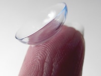
Contact Lenses, Hydrophilic
Visual Acuity
Blindness
Persistent Hyperplastic Primary Vitreous
Refractive Errors
Aniseikonia
Eyeglasses
Artificial Lens Implant Migration
Encyclopedias as Topic
Randomised clinical trial of lensectomy versus lens aspiration and primary capsulotomy for children with bilateral cataract in south India. (1/85)
AIMS: The primary objective was to determine which surgical technique gave the best long term visual outcome for infants and young children with bilateral symmetrical cataract in south India. Secondary objectives were to assess complications and the need for further surgical intervention. METHODS: A randomised controlled clinical trial was undertaken. 65 children under 10 years old with bilateral cataract had one eye treated by lensectomy and the other by aspiration with primary capsulotomy. RESULTS: 56 children (86%) with a mean age at surgery of 53 months were reviewed 3 years after surgery. The overall binocular acuity was 6/18 or better in 57.1% and 6/60 or better in 94.6%. There was no difference in visual acuity between the matched pairs of eyes undergoing aspiration or lensectomy at the third year of follow up (p=0.57). Aspiration eyes were more likely to require a secondary procedure to restore vision than lensectomy eyes (66.1% v 1.8%). CONCLUSION: Aspiration with primary capsulotomy gives an acceptable visual outcome in this part of India providing that there is good follow up to manage capsule opacification. If secondary intervention is not possible owing to poor compliance with follow up, then lensectomy is likely to give better long term visual rehabilitation providing there is good maintenance and technical support for the lensectomy equipment. (+info)Diabetic versus non-diabetic colour vision after cataract surgery. (2/85)
AIMS: To examine whether the colour vision abnormalities found in phakic patients with diabetes mellitus is preserved after removal of the lens by cataract surgery. METHODS: 21 diabetic (16 IDDM and five NIDDM) and 19 non-diabetic patients of comparable age, postoperative visual acuity, and sex distribution, all aphakic or pseudophakic following cataract surgery, had their monocular colour vision examined using the Farnsworth-Munsell 100 hue test. The fundus status of the diabetic patients ranged from no retinopathy to photocoagulation treated proliferative diabetic retinopathy. Patients with macular oedema were specifically excluded from the study. RESULTS: The error scores of both the diabetic (mean 146 (SD 94)) and the non-diabetic patients (83 (79)) did not deviate significantly from the age related normal range. The error score in the diabetic group was significantly higher than in the non-diabetic group (p=0.02) but the amplitude of the difference was small in comparison with previous studies of phakic subjects. The error scores in the diabetic group were not correlated with the degree of retinopathy (p>0.2). CONCLUSION: After cataract surgery only a minor difference exists between the colour vision scores of diabetic and non-diabetic patients. This indicates that accelerated yellowing of the lens in diabetes is the predominant cause of the colour vision anomaly found in phakic diabetic patients. (+info)Deletion in the promoter region and altered expression of Pitx3 homeobox gene in aphakia mice. (3/85)
Mouse aphakia (ak) is a recessive phenotype that spontaneously occurs in the 129/Sv-SlJ strain and is characterized by small eyes that lack a lens. We have recently identified a homeobox-containing gene, Pitx3, and have shown that it is expressed in the developing lens and maps to chromosome 19 close to ak in mouse. Human PITX3 gene was found to underlie anterior segment dysgenesis and cataracts. We have now obtained the entire sequence of the mouse Pitx3 gene including 10 kb of the 5' region and 5 kb of the 3' region. Of several microsatellite repeat regions identified within the Pitx3 sequence, one was informative for linkage analysis. No recombination was observed between ak and the Pitx3 marker, indicating that these two loci are closely linked (0.2 +/- 0.2 cM). Additionally, Pitx3 transcripts were not detected in the ak/ak mice either in the lens placode or at later developmental stages of the lens by in situ hybridization. Since no differences were previously found between ak/ak and wild-type sequences in the Pitx3 coding region, we hypothesized that an etiologic mutation is located in the promoter or other regulatory regions. To test this hypothesis we studied the 5' flanking region of the Pitx3 gene. This analysis revealed a deletion of 652 bp located 2.5 kb upstream from the start point of the Pitx3 5' UTR sequence in ak/ak mice. The deletion co-segregated with the ak mutation and was not detected in 16 samples from 10 different mouse strains including the founder strains. Analysis of the 652 bp region identified sequences similar to consensus binding sites for transcription factors AP-2 and Maf that were shown to play a critical role in lens determination. These lines of evidence suggest that the abnormal ocular development in the aphakia mouse is due to the deletion upstream of the Pitx3 gene. (+info)Pitx3 is required for development of substantia nigra dopaminergic neurons. (4/85)
Dopaminergic (DA) neurons of substantia nigra in the midbrain control voluntary movement, and their degeneration is the cause of Parkinson's disease. The complete set of genes required to specifically determine the development of midbrain DA subgroups is not known yet. We report here that mice lacking the bicoid-related homeoprotein Pitx3 fail to develop DA neurons of the substantia nigra. Other mesencephalic DA neurons of the ventral tegmental area and retrorubral field are unaltered in their dopamine expression and histological organization. These data suggest that Pitx3-dependent gene expression is specifically required for the differentiation of DA progenitors within the mesencephalic DA system. (+info)Pitx3 is required for motor activity and for survival of a subset of midbrain dopaminergic neurons. (5/85)
Mesencephalic dopaminergic (MesDA) neurons play crucial roles in motor and behavioral processes; their loss in Parkinson's disease (PD) results in striatal dopamine (DA) deficiency and hypokinetic movement disorder. The Pitx3 homeobox gene is expressed in the MesDA system. We now show that only a subset of MesDA neurons express Pitx3 and that in Pitx3-deficient aphakia mice, this subset is progressively lost by apoptosis during fetal (substantia nigra, SN) and postnatal (ventral tegmental area) development, resulting in very low striatal DA and akinesia. Similar to human PD, dorsal SN neurons (which are Pitx3 negative) are spared in mutant mice. Thus, Pitx3 defines a pathway for survival of neurons that are implicated in PD and that are required for spontaneous locomotor activity. (+info)Early developmental failure of substantia nigra dopamine neurons in mice lacking the homeodomain gene Pitx3. (6/85)
The mesencephalic dopamine (mesDA) system is involved in the control of movement and behavior. The expression of Pitx3 in the brain is restricted to the mesDA system and the gene is induced relatively late, at E11.5, a time when tyrosine hydroxylase (Th) gene expression is initiated. We show here that, in the Pitx3-deficient aphakia (ak) mouse mutant, the mesDA system is malformed. Owing to the developmental failure of mesDA neurons in the lateral field of the midbrain, mesDA neurons are not found in the SNc and the projections to the caudate putamen are selectively lost. However, Pitx3 is expressed in all mesDA neurons in control animals. Therefore, mesDA neurons react specifically to the loss of Pitx3. Defects of motor control where not seen in the ak mice, suggesting that other neuronal systems compensate for the absence of the nigrostriatal pathway. However, an overall lower activity was observed. The results suggest that Pitx3 is specifically required for the formation of the SNc subfield at the onset of dopaminergic neuron differentiation. (+info)Cataract surgical coverage and outcome in the Tibet Autonomous Region of China. (7/85)
BACKGROUND: A recently published, population based survey of the Tibet Autonomous Region (TAR) of China reported on low vision, blindness, and blinding conditions. This paper presents detailed findings from that survey regarding cataract, including prevalence, cataract surgical coverage, surgical outcome, and barriers to use of services. METHODS: The Tibet Eye Care Assessment (TECA) was a prevalence survey of people from randomly selected households from three of the seven provinces of the TAR (Lhoka, Nakchu, and Lingzhr), representing its three main environmental regions. The survey, conducted in 1999 and 2000, assessed visual acuity, cause of vision loss, and eye care services. RESULTS: Among the 15,900 people enumerated, 12,644 were examined (79.6%). Cataract prevalence was 5.2% and 13.8%, for the total population, and those over age 50, respectively. Cataract surgical coverage (vision <6/60) for people age 50 and older (85-90% of cataract blind) was 56% overall, 70% for men and 47% for women. The most common barriers to use of cataract surgical services were distance and cost. In the 216 eyes with cataract surgery, 60% were aphakic and 40% were pseudophakic. Pseudophakic surgery left 19% of eyes blind (<6/60) and an additional 20% of eyes with poor vision (6/24-6/60). Aphakic surgery left 24% of eyes blind and an additional 21% of eyes with poor vision. Even though more women remained blind than men, 28% versus 18% respectively, the different was not statistically significant (p = 0.25). CONCLUSIONS: Cataract surgical coverage was remarkably high despite the difficulty of providing services to such an isolated and sparse population. Cataract surgical outcome was poor for both aphakic and pseudophakic surgery. Two main priorities are improving cataract surgical quality and cataract surgical coverage, particularly for women. (+info)Neonatal aphakia retards ocular growth and alters scleral gene expression in rhesus monkeys. (8/85)
PURPOSE: We hypothesize that remodeling of the scleral extracellular matrix, involving collagen and proteoglycan synthesis and turnover, is a key process involved in ocular growth. Decreased axial elongation is observed following neonatal removal of the crystalline lens in a rhesus monkey model of congenital cataract. We wanted to determine changes in gene expression in the operated and companion eye following lensectomy, especially for extracellular matrix in the sclera. METHODS: Between 4 and 7 days of age, infant monkeys underwent surgical removal of the lens from the right eye. Axial lengths of the operated and unmanipulated fellow eyes were measured and when interocular differences of >0.4 mm were achieved, monkeys were sacrificed and RNA was isolated from sclera. In order to determine changes in scleral gene expression in aphakic versus control eyes, we used Clontech's Atlas Gene Array (Human Cancer Array version 1.2) hybridized with total RNA from three monkeys. RESULTS: Atlas Gene Array analysis demonstrated differential expression of several genes in the operated versus the unmanipulated eye. Most notably, there was a statistically significant increase in expression of several extracellular matrix (ECM) genes including: aggrecan, decorin, biglycan, several collagens, and tenascin in the RNA from the sclera of the aphakic eyes when compared to the unmanipulated eyes. Genes for several matrix metalloproteinases (MMPs) showed no significant change following lens removal although there was a trend towards decreased expression. There were also statistically significant changes in the pattern of gene expression in the operated eye relative to the unmanipulated eye for cell adhesion, cell cycle, apoptosis, and cytoskeleton transcripts. CONCLUSIONS: Our results suggest that removal of the crystalline lens alters gene expression in the sclera with a prominent upregulation of ECM transcripts. These data support recent evidence that remodeling of the ECM composition of the sclera may be an important regulator of ocular growth. (+info)Aphakia is a medical condition that refers to the absence of the lens in the eye. This can occur naturally, but it's most commonly the result of surgery to remove a cataract, a cloudy lens that can cause vision loss. In some cases, the lens may not be successfully removed or may be accidentally lost during surgery, leading to aphakia. People with aphakia typically have significant vision problems and may require corrective measures such as glasses, contact lenses, or an intraocular lens implant to improve their vision.
Contact lenses are thin, curved plastic or silicone hydrogel devices that are placed on the eye to correct vision, replace a missing or damaged cornea, or for cosmetic purposes. They rest on the surface of the eye, called the cornea, and conform to its shape. Contact lenses are designed to float on a thin layer of tears and move with each blink.
There are two main types of contact lenses: soft and rigid gas permeable (RGP). Soft contact lenses are made of flexible hydrophilic (water-absorbing) materials that allow oxygen to pass through the lens to the cornea. RGP lenses are made of harder, more oxygen-permeable materials.
Contact lenses can be used to correct various vision problems, including nearsightedness, farsightedness, astigmatism, and presbyopia. They come in different shapes, sizes, and powers to suit individual needs and preferences. Proper care, handling, and regular check-ups with an eye care professional are essential for maintaining good eye health and preventing complications associated with contact lens wear.
Pseudophakia is a medical term that refers to the condition where a person's natural lens in the eye has been replaced with an artificial one. This procedure is typically performed during cataract surgery, where the cloudy, natural lens is removed and replaced with a clear, artificial lens to improve vision. The prefix "pseudo" means false or fake, and "phakia" refers to the natural lens of the eye, hence the term "Pseudophakia" implies a false or artificial lens.
Cataract extraction is a surgical procedure that involves removing the cloudy lens (cataract) from the eye. This procedure is typically performed to restore vision impairment caused by cataracts and improve overall quality of life. There are two primary methods for cataract extraction:
1. Phacoemulsification: This is the most common method used today. It involves making a small incision in the front part of the eye (cornea), inserting an ultrasonic probe to break up the cloudy lens into tiny pieces, and then removing those pieces with suction. After removing the cataract, an artificial intraocular lens (IOL) is inserted to replace the natural lens and help focus light onto the retina.
2. Extracapsular Cataract Extraction: In this method, a larger incision is made on the side of the cornea, allowing the surgeon to remove the cloudy lens in one piece without breaking it up. The back part of the lens capsule is left intact to support the IOL. This technique is less common and typically reserved for more advanced cataracts or when phacoemulsification cannot be performed.
Recovery from cataract extraction usually involves using eye drops to prevent infection and inflammation, as well as protecting the eye with a shield or glasses during sleep for a few weeks after surgery. Most people experience improved vision within a few days to a week following the procedure.
Intraocular lenses (IOLs) are artificial lens implants that are placed inside the eye during ophthalmic surgery, such as cataract removal. These lenses are designed to replace the natural lens of the eye that has become clouded or damaged, thereby restoring vision impairment caused by cataracts or other conditions.
There are several types of intraocular lenses available, including monofocal, multifocal, toric, and accommodative lenses. Monofocal IOLs provide clear vision at a single fixed distance, while multifocal IOLs offer clear vision at multiple distances. Toric IOLs are designed to correct astigmatism, and accommodative IOLs can change shape and position within the eye to allow for a range of vision.
The selection of the appropriate type of intraocular lens depends on various factors, including the patient's individual visual needs, lifestyle, and ocular health. The implantation procedure is typically performed on an outpatient basis and involves minimal discomfort or recovery time. Overall, intraocular lenses have become a safe and effective treatment option for patients with vision impairment due to cataracts or other eye conditions.
Intraocular lens (IOL) implantation is a surgical procedure that involves placing a small artificial lens inside the eye to replace the natural lens that has been removed. This procedure is typically performed during cataract surgery, where the cloudy natural lens is removed and replaced with an IOL to restore clear vision.
During the procedure, a small incision is made in the eye, and the cloudy lens is broken up and removed using ultrasound waves or laser energy. Then, the folded IOL is inserted through the same incision and positioned in the correct place inside the eye. Once in place, the IOL unfolds and is secured into position.
There are several types of IOLs available, including monofocal, multifocal, toric, and accommodating lenses. Monofocal lenses provide clear vision at one distance, while multifocal lenses offer clear vision at multiple distances. Toric lenses correct astigmatism, and accommodating lenses can change shape to focus on objects at different distances.
Overall, intraocular lens implantation is a safe and effective procedure that can help restore clear vision in patients with cataracts or other eye conditions that require the removal of the natural lens.
A cataract is a clouding of the natural lens in the eye that affects vision. This clouding can cause vision to become blurry, faded, or dim, making it difficult to see clearly. Cataracts are a common age-related condition, but they can also be caused by injury, disease, or medication use. In most cases, cataracts develop gradually over time and can be treated with surgery to remove the cloudy lens and replace it with an artificial one.
Hydrophilic contact lenses are a type of contact lens that is designed to absorb and retain water. These lenses are made from materials that have an affinity for water, which helps them to remain moist and comfortable on the eye. The water content of hydrophilic contact lenses can vary, but typically ranges from 30-80% by weight.
Hydrophilic contact lenses are often used to correct refractive errors such as myopia (nearsightedness), hyperopia (farsightedness), and astigmatism. They can be made in a variety of materials, including soft hydrogel and silicone hydrogel.
One advantage of hydrophilic contact lenses is that they tend to be more comfortable to wear than other types of contacts, as they retain moisture and conform closely to the shape of the eye. However, they may also be more prone to deposits and buildup, which can lead to protein accumulation and discomfort over time. Proper care and cleaning are essential to maintain the health of the eyes when wearing hydrophilic contact lenses.
Visual acuity is a measure of the sharpness or clarity of vision. It is usually tested by reading an eye chart from a specific distance, such as 20 feet (6 meters). The standard eye chart used for this purpose is called the Snellen chart, which contains rows of letters that decrease in size as you read down the chart.
Visual acuity is typically expressed as a fraction, with the numerator representing the testing distance and the denominator indicating the smallest line of type that can be read clearly. For example, if a person can read the line on the eye chart that corresponds to a visual acuity of 20/20, it means they have normal vision at 20 feet. If their visual acuity is 20/40, it means they must be as close as 20 feet to see what someone with normal vision can see at 40 feet.
It's important to note that visual acuity is just one aspect of overall vision and does not necessarily reflect other important factors such as peripheral vision, depth perception, color vision, or contrast sensitivity.
Blindness is a condition of complete or near-complete vision loss. It can be caused by various factors such as eye diseases, injuries, or birth defects. Total blindness means that a person cannot see anything at all, while near-complete blindness refers to having only light perception or the ability to perceive the direction of light, but not able to discern shapes or forms. Legal blindness is a term used to define a certain level of visual impairment that qualifies an individual for government assistance and benefits; it usually means best corrected visual acuity of 20/200 or worse in the better eye, or a visual field no greater than 20 degrees in diameter.
Microphthalmos is a medical condition where one or both eyes are abnormally small due to developmental anomalies in the eye. The size of the eye may vary from slightly smaller than normal to barely visible. This condition can occur in isolation or as part of a syndrome with other congenital abnormalities. It can also be associated with other ocular conditions such as cataracts, retinal disorders, and orbital defects. Depending on the severity, microphthalmos may lead to visual impairment or blindness.
Hyphema is defined as the presence of blood in the anterior chamber of the eye, which is the space between the cornea and the iris. This condition usually results from trauma or injury to the eye, but it can also occur due to various medical conditions such as severe eye inflammation, retinal surgery, or blood disorders that affect clotting.
The blood in the anterior chamber can vary in amount, ranging from a few drops to a complete fill, which is called an "eight-ball hyphema." Hyphema can be painful and cause sensitivity to light (photophobia), blurred vision, or even loss of vision if not treated promptly.
Immediate medical attention is necessary for hyphema to prevent complications such as increased intraocular pressure, corneal blood staining, glaucoma, or cataracts. Treatment options may include bed rest, eye drops to reduce inflammation and control intraocular pressure, and sometimes surgery to remove the blood from the anterior chamber.
Persistent Hyperplastic Primary Vitreous (PHPV) is a rare congenital eye condition that occurs during fetal development. It is characterized by the failure of the primary vitreous, a gel-like substance in the eye, to completely regress or disappear. Instead, the primary vitreous persists and undergoes hyperplasia, leading to the formation of abnormal tissue within the eye.
In PHPV, this persistent tissue can cause various problems, including a small pupil, a cloudy area in the center of the lens (cataract), a white mass behind the lens, and abnormal blood vessels growing from the retina towards the center of the eye. These abnormalities can lead to visual impairment or even blindness, depending on the severity of the condition.
PHPV is typically diagnosed during infancy or early childhood, through a comprehensive eye examination that includes a detailed view of the internal structures of the eye using a specialized lens (slit lamp) and other diagnostic tests. Treatment options may include surgery to remove the abnormal tissue and improve vision, but the success of treatment depends on the extent and location of the PHPV.
Refractive errors are a group of vision conditions that include nearsightedness (myopia), farsightedness (hyperopia), astigmatism, and presbyopia. These conditions occur when the shape of the eye prevents light from focusing directly on the retina, causing blurred or distorted vision.
Myopia is a condition where distant objects appear blurry while close-up objects are clear. This occurs when the eye is too long or the cornea is too curved, causing light to focus in front of the retina instead of directly on it.
Hyperopia, on the other hand, is a condition where close-up objects appear blurry while distant objects are clear. This happens when the eye is too short or the cornea is not curved enough, causing light to focus behind the retina.
Astigmatism is a condition that causes blurred vision at all distances due to an irregularly shaped cornea or lens.
Presbyopia is a natural aging process that affects everyone as they get older, usually around the age of 40. It causes difficulty focusing on close-up objects and can be corrected with reading glasses, bifocals, or progressive lenses.
Refractive errors can be diagnosed through a comprehensive eye exam and are typically corrected with eyeglasses, contact lenses, or refractive surgery such as LASIK.
Aniseikonia is a medical term that refers to a condition where there is a significant difference in the size or shape of the images perceived by each eye. This occurs when there is a disproportionate amount of magnification or minification between the two eyes, leading to a mismatch in the visual perception of objects' size and shape.
Aniseikonia can result from various factors, including anisometropia (a significant difference in the refractive power between the two eyes), cataract surgery, corneal irregularities, or retinal diseases. It can cause symptoms such as eyestrain, headaches, and difficulty with depth perception, reading, and overall visual comfort.
Treatment for aniseikonia typically involves correcting the underlying refractive error with prescription lenses, prisms, or contact lenses. In some cases, surgical intervention may be necessary to address any structural issues causing the condition.
Eyeglasses are a medical device used to correct vision problems. Also known as spectacles, they consist of frames that hold one or more lenses through which a person looks to see clearly. The lenses may be made of glass or plastic and are designed to compensate for various visual impairments such as nearsightedness, farsightedness, astigmatism, or presbyopia. Eyeglasses can be custom-made to fit an individual's face and prescription, and they come in a variety of styles, colors, and materials. Some people wear eyeglasses all the time, while others may only need to wear them for certain activities such as reading or driving.
Artificial lens implant migration refers to the movement or displacement of an intraocular lens (IOL) from its intended position within the eye. This can occur after cataract surgery, during which the cloudy natural lens is removed and replaced with an artificial one. The IOL is typically secured in place with special anchors or loops, but in some cases, it may become dislodged and move within the eye.
There are several possible causes of artificial lens implant migration, including surgical complications, trauma to the eye, or weakness in the capsular bag that holds the lens in place. Symptoms of this condition can include blurry vision, double vision, or seeing halos around lights. If left untreated, lens implant migration can lead to more serious complications, such as retinal detachment or glaucoma. Treatment options may include repositioning the lens or replacing it with a new one.
An encyclopedia is a comprehensive reference work containing articles on various topics, usually arranged in alphabetical order. In the context of medicine, a medical encyclopedia is a collection of articles that provide information about a wide range of medical topics, including diseases and conditions, treatments, tests, procedures, and anatomy and physiology. Medical encyclopedias may be published in print or electronic formats and are often used as a starting point for researching medical topics. They can provide reliable and accurate information on medical subjects, making them useful resources for healthcare professionals, students, and patients alike. Some well-known examples of medical encyclopedias include the Merck Manual and the Stedman's Medical Dictionary.
A pupil, in medical terms, refers to the circular opening in the center of the iris (the colored part of the eye) that allows light to enter and reach the retina. The size of the pupil can change involuntarily in response to light intensity and emotional state, as well as voluntarily through certain eye exercises or with the use of eye drops. Pupillary reactions are important in clinical examinations as they can provide valuable information about the nervous system's functioning, particularly the brainstem and cranial nerves II and III.
 Aphakia
Himalayan Cataract Project
Retinal detachment
List of OMIM disorder codes
Iris-fixated intraocular lens
Iridodonesis
Anil Kumar Mandal
Intraocular lens
Lens (vertebrate anatomy)
Far-sightedness
Glued intraocular lens
Aniseikonia
Irvin Borish
Intraocular lens scaffold
Amblyopia
Tadini (ophthalmologist)
FOXE3
Secondary glaucoma
Cataract surgery
Cone cell
Epikeratophakia
Pre-Descemet's endothelial keratoplasty
Bubbles (Trailer Park Boys)
Eye surgery
Aspheric lens
Ultraviolet
Photophobia
List of ICD-9 codes 320-389: diseases of the nervous system and sense organs
Proliferative vitreoretinopathy
Rocky Mountain Horse
Aphakia - Wikipedia
 The association between myopic shift and visual acuity outcome in pediatric aphakia
 Artisan Aphakia
Intraoperative aberrometry-based aphakia refraction in patients with cataract: status and options | British Journal of...
Secondary iris-claw anterior chamber lens implantation in patients with aphakia without capsular support | British Journal of...
 Outcomes of Bilateral Cataract Surgery in Infants 7 to 24 Months of Age Using the Toddler Aphakia and Pseudophakia Treatment...
 2o IOL implantation in an 11y/o girl with traumatic corneal perforation & aphakia - International Society of Refractive Surgery
 Contact Lenses for Aphakia
 Aphakia Symptoms Archives - Helthy Leaf
 Paediatric Aphakia - Ezekiel Eyes | Contact Lenses Perth
 Sutureless dislocated IOLexchange with ARTISAN aphakia
 Condition Congenital Aphakia-iris Hypoplasia-microphthalmia-microcornea Syndrome
 What is Aphakia? Causes | Symptoms | Diagnosis | Treatment | Complications - GASTRO Health
 Aphakia - the absence of the crystalline lens of the eye | Feel Good Contacts
Page 1 | Search Results | Ophthalmologica | Karger Publishers
 38 CFR Appendix C to Part 4 - Appendix C to Part 4-Alphabetical Index of Disabilities | Electronic Code of Federal Regulations ...
 Pharmaceutics | Free Full-Text | Review of Intraocular Pharmacokinetics of Anti-Infectives Commonly Used in the Treatment of...
Contact Lens Correction of Aphakia in Children: A Report by the American Academy of Ophthalmology<...
 Postoperative Retinal Detachment: Background, Pathophysiology, Epidemiology
 Paper: Outcomes of Glued IOL Scaffold for Soemmerring Ring Removal in Aphakia With Posterior Capsule Rupture (2015 ASCRS ASOA...
 Molecular Vision: Yzer, Mol Vis 2012; 18:412-425. Table 2
 Dr. Joshua Powell, MD - Ophthalmology Specialist in Norman, OK | Healthgrades
 Opthamology Data (1971-75)
 Michael Joel Elman, MD| Comprehensive Ophthalmology | MedStar Health
 XELPROS - Renal and Urology News
Daniel W Wang, MD| Comprehensive Ophthalmology | MedStar Health
Eye Surgery and Treatment | TRICARE
 Outcomes of cataract surgery in a rural and urban south Indi... : Indian Journal of Ophthalmology
 Dr. Thomas Repko, MD, Ophthalmology Specialist - Akron, OH | Sharecare
Visual acuity2
Corneal3
- Astigmatism: With-the-rule astigmatism due to corneal wound healing may occur in surgical aphakia, mainly after intracapsular cataract extraction or extracapsular cataract extraction. (wikipedia.org)
- Retinal detachment Aphakic bullous keratopathy Aphakia can be corrected by wearing glasses or contact lenses, by artificial lens implantation, or by refractive corneal surgeries. (wikipedia.org)
- Dr. Shorter fits both gas permeable and soft contact lenses for patients with a history of keratoconus, corneal surgery and aphakia. (uic.edu)
Artisan Aphakia8
- Artisan Aphakia is de eerste keus back-uplens bij gecompliceerde cataractgevallen. (ophtec.com)
- Uit de lange ervaring met irisfixatie blijkt dat de Artisan Aphakia-IOL een voorspelbaar, veilig en zeer precies implantaat is. (ophtec.com)
- Uit de lange ervaring met irisfixatie blijkt dat de Artisan Aphakia-IOL een voorspelbaar, veilig en zeer precies implantaat is dat afakie corrigeert wanneer correctie op een andere manier niet mogelijk is. (ophtec.com)
- Met Artisan Aphakia-IOL's is opereren eenvoudiger. (ophtec.com)
- Dr. Adrián Hernández Martínez talks about why the Artisan aphakia IOL is his standard solution for cases without capsular support and provides us with some usefull tips. (ophtec.com)
- Dr. Adrián Hernández Martínez implants the Artisan Aphakia IOL in a patient with post-surgical aphakia without capsular support. (ophtec.com)
- Dr. Ramon C. Ghanem shares challenging cases and explains why he prefers the Artisan Aphakia IOL to solve these cases. (ophtec.com)
- Dr. C. Peckar implants the Artisan Aphakia using the enclavation forceps. (ophtec.com)
Pseudophakia4
- Scholars@Duke publication: Outcomes of Bilateral Cataract Surgery in Infants 7 to 24 Months of Age Using the Toddler Aphakia and Pseudophakia Treatment Study Registry. (duke.edu)
- PURPOSE: To evaluate outcomes of bilateral cataract surgery in children aged 7 to 24 months and compare rates of adverse events (AEs) with other Toddler Aphakia and Pseudophakia Study (TAPS) registry outcomes. (duke.edu)
- Aphakia and pseudophakia, especially after YAG capsulotomy, predispose to PVD. (medscape.com)
- The incidence of PVD is increased in persons with intraocular inflammation, aphakia or pseudophakia, trauma, myopia, or vitreoretinal diseases. (aao.org)
Contact lenses5
- It is believed that the use of contact lenses in paediatric Aphakia is medically essential. (ezekieleyes.com)
- Yes, aphakia can be treated through contact lenses , glasses and surgery. (feelgoodcontacts.ie)
- Purpose: To review the published literature to assess the visual outcomes and adverse events associated with the 2 most commonly used contact lenses for treating aphakia in children: silicone elastomer (SE) and rigid gas permeable (RGP). (elsevierpure.com)
- Silicone elastomer and RGP contact lenses were found to be effective for treating aphakia in children. (elsevierpure.com)
- SilSoft Super Plus contact lenses are designed for children who have had cataract surgery where an intraocular lens has not been implanted (aphakia). (yourlens.com)
Intraocular lens1
- Aphakia is treated using Pseudophakic lens (an artificial lens on the eye) or IOL (intraocular lens) through surgery. (feelgoodcontacts.ie)
Absence4
- Aphakia is the absence of the lens of the eye, due to surgical removal, such as in cataract surgery, a perforating wound or ulcer, or congenital anomaly. (wikipedia.org)
- Congenital aphakia-iris hypoplasia-microphthalmia-microcornea syndrome is a rare genetic disorder characterized by the absence of the lens of the eye (aphakia), underdeveloped iris (iris hypoplasia), abnormally small eye (microphthalmia), and abnormally small cornea (microcornea). (rarediseaseshealthcenter.com)
- This gene is responsible for the development of the eyes, and mutations in this gene can lead to a variety of eye abnormalities, including aphakia (absence of the lens of the eye), iris hypoplasia (underdevelopment of the colored part of the eye), microphthalmia (abnormally small eyes), and microcornea (abnormally small cornea). (rarediseaseshealthcenter.com)
- The absence of the lens of one or both of our eyes is called as aphakia. (gaspaininchest.com)
Disorders1
- Several diseases and disorders are related to the eyes, but Aphakia is fatal. (helthyleaf.com)
Complications1
- Main complications of surgical aphakia include: Spectacle intolerance: Due to image magnification (up to 30%), optical aberration, prismatic effect and roving ring scotoma, spectacles are not well tolerated by aphakic patients. (wikipedia.org)
Posterior1
- To assess the clinical outcomes of glued intraocular (IOL) scaffold procedure for the removal of Soemmering ring in aphakia with associated posterior capsular defect. (confex.com)
Congenital6
- Paediatric Aphakia is a condition in which a child has no crystalline lens in his or her eye, due to congenital or traumatic cataracts. (ezekieleyes.com)
- What are the symptoms of Congenital aphakia-iris hypoplasia-microphthalmia-microcornea syndrome? (rarediseaseshealthcenter.com)
- Congenital aphakia-iris hypoplasia-microphthalmia-microcornea syndrome is a rare genetic disorder caused by mutations in the PAX6 gene. (rarediseaseshealthcenter.com)
- The treatments for Congenital aphakia-iris hypoplasia-microphthalmia-microcornea syndrome vary depending on the severity of the condition. (rarediseaseshealthcenter.com)
- Is there a cure/medications for Congenital aphakia-iris hypoplasia-microphthalmia-microcornea syndrome? (rarediseaseshealthcenter.com)
- However, due to a rare condition called congenital aphakia, some people are born without lenses in one or both eyes. (pocahontastimes.com)
Cataract removal1
- Aphakia is mainly caused by cataract removal and replacement IOL. (feelgoodcontacts.ie)
Eyes1
- Due to unequal refractive power between the eyes, wearing spectacles with single-eye aphakia may cause double vision. (wikipedia.org)
Lens of1
- Aphakia Meaning is the lack of the lens of the eye. (helthyleaf.com)
Aniseikonia1
- Further, spectacles are not possible in the case of unilateral (one eye) Aphakia because of the image size difference (Aniseikonia) induced. (ezekieleyes.com)
Clinical1
- DESIGN: Retrospective clinical study at 10 Infant Aphakia Treatment Study (IATS) sites. (duke.edu)
Lenses1
- Babies with aphakia are born without lenses due to development issues or genetics. (feelgoodcontacts.ie)
Babies1
- Babies are rarely born with aphakia. (wikipedia.org)
Injuries1
- Injuries, cataract surgery, or genetics can play a major part in aphakia. (feelgoodcontacts.ie)
Vision4
- Loss of accommodation: Since the lens and its zonules are responsible for adjusting the focus of vision to different lengths, patients with aphakia will have a total loss of accommodation. (wikipedia.org)
- Aphakia is a condition where you are missing a lens in your eye, causing a loss of focus in your vision. (feelgoodcontacts.ie)
- People with aphakia have blurry/shaky vision. (feelgoodcontacts.ie)
- arpt alt setting-29.98.Aircraft alt set-30.02.Pilot issued waiver for aphakia of l eye w 20/200 vision. (aviation-safety.net)
People2
- People with aphakia have relatively small pupils and their pupils dilate to a lesser degree. (wikipedia.org)
- Aphakia mostly occurs in adults or older people who have cataracts (it is a clouding of your internal eye lens and your looks like milky). (gaspaininchest.com)
Patients1
- Patients who are 70 and above tend to suffer from aphakia the most. (feelgoodcontacts.ie)
Standard2
- A standard ophthalmic exam is performed to diagnosis your aphakia . (gaspaininchest.com)
- Aphakia is diagnosed through a standard eye test. (feelgoodcontacts.ie)
Infant Aphakia Treatment Study Group1
- Infant Aphakia Treatment Study Group. (medlineplus.gov)
Lens12
- Aphakia is the absence of the lens of the eye, due to surgical removal, such as in cataract surgery, a perforating wound or ulcer, or congenital anomaly. (wikipedia.org)
- Surgical removal of a lens, mainly in cataract surgery, is the most common cause of aphakia. (wikipedia.org)
- Loss of accommodation: Since the lens and its zonules are responsible for adjusting the focus of vision to different lengths, patients with aphakia will have a total loss of accommodation. (wikipedia.org)
- Retinal detachment Aphakic bullous keratopathy Aphakia can be corrected by wearing glasses or contact lenses, by artificial lens implantation, or by refractive corneal surgeries. (wikipedia.org)
- Our Infant Aphakia Clinical Database was queried to identify patients who underwent surgery for unilateral congenital cataract and were treated with a high-plus contact lens placed on the sound eye after a period of failure with adhesive patches. (medscape.com)
- Aphakia is mainly congenital or as result of LENS DISLOCATION AND SUBLUXATION. (ouhsc.edu)
- Our Eye 4 Aphakia is mimicking the anterior segment of the eye with an artificial lens (IOL). (eyecre.at)
- In the operating room, the infants were randomly assigned to receive an artificial lens implant or go without a lens, a condition called aphakia. (nih.gov)
- Congenital cataract includes diagnosis codes indicating congenital cataract, congenital aphakia, coloboma of lens, spherophakia, congenital lens malformations, or infantile or juvenile cataracts of any type. (cdc.gov)
- Bilateral Artisan lens for aphakia and megalocornea: Long-term follow-up. (medscape.com)
- As the magistrate noted, a childhood injury involving a BB pellet left Cogan without a lens in his right eye, a condition referred to as aphakia. (findlaw.com)
- Conclusions: Sutureless SFIOL could be considered as a long-term option for the management of aphakia, dislocated IOL, and lens with poor capsular support. (mehdijournal.com)
Astigmatism1
- Astigmatism: With-the-rule astigmatism due to corneal wound healing may occur in surgical aphakia, mainly after intracapsular cataract extraction or extracapsular cataract extraction. (wikipedia.org)
Primary aphakia1
- To describe the natural history, management, and visual outcome in children with congenital primary aphakia (CPA). (nih.gov)
IATS1
- DESIGN: Retrospective clinical study at 10 Infant Aphakia Treatment Study (IATS) sites. (duke.edu)
20211
- 2021. https://nursing.unboundmedicine.com/nursingcentral/view/Tabers-Dictionary/747239/all/aphakia__aphacia. (unboundmedicine.com)
MeSH1
- Aphakia" is a descriptor in the National Library of Medicine's controlled vocabulary thesaurus, MeSH (Medical Subject Headings) . (ouhsc.edu)
Capsule1
- Course will cover various techniques for secondary IOL placement in the presence of IOL exchange, poor capsule support, or aphakia. (ascrs.org)
Anterior1
- Assalamualaikum sahabat DD Medika, beberapa hari yang lalu @rsmataachmadwardi mengadakan Seminar Aphakia Problems & Management dan Wetlab Course: Iris Claw, Yamane Technique, Anterior Vitrectomy for Anterior Segment Surgeon l yang diselenggarakan di Hotel Horison Ultima Ratu Serang, Banten. (ddmedika.com)
Surgical2
- Iridectomy mark may be seen in surgical aphakia. (wikipedia.org)
- Main complications of surgical aphakia include: Spectacle intolerance: Due to image magnification (up to 30%), optical aberration, prismatic effect and roving ring scotoma, spectacles are not well tolerated by aphakic patients. (wikipedia.org)
Management1
- Present state of management of aphakia. (wikipedia.org)
Total1
- This graph shows the total number of publications written about "Aphakia" by people in this website by year, and whether "Aphakia" was a major or minor topic of these publications. (ouhsc.edu)