Aortic Valve Insufficiency
Aortic Valve
Mitral Valve Insufficiency
Tricuspid Valve Insufficiency
Pulmonary Valve Insufficiency
Aortic Valve Stenosis
Venous Valves
Heart Valve Diseases
Heart Valve Prosthesis Implantation
Heart Valve Prosthesis
Bioprosthesis
Treatment Outcome
Adrenal Insufficiency
Tricuspid Valve
Renal Insufficiency
Venous Insufficiency
Cardiac Catheterization
Exocrine Pancreatic Insufficiency
Aortic Valve Prolapse
Endocarditis, Bacterial
Mitral Valve Prolapse
Placental Insufficiency
Mitral Valve Stenosis
Echocardiography, Transesophageal
Cardiac Valve Annuloplasty
Echocardiography
Frequency and long term follow up of valvar insufficiency caused by retrograde aortic radiofrequency catheter ablation procedures. (1/941)
OBJECTIVE: To assess the frequency of valvar complications caused by left sided radiofrequency catheter ablation using the retrograde aortic technique. METHODS: 179 patients (118 male) with a mean (SD) age of 43 (17) years underwent 216 procedures at one centre. The target of the ablation was an accessory atrioventricular pathway in 144 patients, the atrioventricular junction in 29 patients, and a ventricular tachycardia in six patients. In 25 patients structural heart disease was identified before the procedure (ischaemic heart disease 10, cardiomyopathy nine, valvar three, other three). Echo/Doppler examinations were performed the day before the procedure and within 24 hours postablation; the investigations were all reviewed by the same investigator. Patients with identified valvar injury caused by the procedure were followed for 42 (7) months. RESULTS: Valvar injury caused by the ablation procedure was identified in four young (age 30 (8) years), otherwise healthy patients with left lateral atrioventricular accessory pathways. Mild mitral insufficiency with a central regurgitation jet was detected in two patients and remained unchanged at follow up. Mild aortic insufficiency was detected in another two patients. In one of these the regurgitation jet was central and remained unchanged at follow up. In one patient the regurgitation jet was located between the non-coronary and left cusps in relation to a loosely attached structure. Both the structure and the valvar regurgitation disappeared during follow up. No clinical complications occurred in any of the patients during follow up. CONCLUSION: In this study, the frequency of valvar complications after left sided radiofrequency catheter ablation using the retrograde aortic technique was 1.9%. (+info)Impact of vascular adaptation to chronic aortic regurgitation on left ventricular performance. (2/941)
BACKGROUND: This investigation was designed to test the hypothesis that vascular adaptation occurs in patients with chronic aortic regurgitation to maintain left ventricular (LV) performance. METHODS AND RESULTS: Forty-five patients with chronic aortic regurgitation (mean age 50+/-14 years) were studied using a micromanometer LV catheter to obtain LV pressures and radionuclide ventriculography to obtain LV volumes during multiple loading conditions and right atrial pacing. These 45 patients were subgrouped according to their LV contractility (Ees) and ejection fraction values. Group I consisted of 24 patients with a normal Ees. Group IIa consisted of 10 patients with impaired Ees values (Ees <1.00 mm Hg/mL) but normal LV ejection fractions; Group IIb consisted of 11 patients with impaired contractility and reduced LV ejection fractions. The left ventricular-arterial coupling ratio, Ees/Ea, where Ea was calculated by dividing the LV end-systolic pressure by LV stroke volume, averaged 1.60+/-0.91 in Group I. It decreased to 0.91+/-0.27 in Group IIa (P<0.05 versus Group I), and it decreased further in Group IIb to 0.43+/-0.24 (P<0.001 versus Groups I and IIa). The LV ejection fractions were inversely related to the Ea values in both the normal and impaired contractility groups (r=-0.48, P<0.05 and r=-0.56, P<0.01, respectively), although the slopes of these relationships differed (P<0.05). The average LV work was maximal in Group IIa when the left ventricular-arterial coupling ratio was near 1.0 because of a significant decrease in total arterial elastance (P<0.01 versus Group I). In contrast, the decrease in the left ventricular-arterial coupling ratio in Group IIb was caused by an increase in total arterial elastance, effectively double loading the LV, contributing to a decrease in LV pump efficiency (P<0.01 versus Group IIa and P<0.001 versus Group I). CONCLUSIONS: Vascular adaptation may be heterogeneous in patients with chronic aortic regurgitation. In some, total arterial elastance decreases to maximize LV work and maintain LV performance, whereas in others, it increases, thereby double loading the LV, contributing to afterload excess and a deterioration in LV performance that is most prominent in those with impaired contractility. (+info)Bileaflet mechanical prostheses for aortic valve replacement in patients younger than 65 years and 65 years of age or older: major thromboembolic and hemorrhagic complications. (3/941)
OBJECTIVE: To determine major thromboembolic and hemorrhagic complications and predictive risk factors associated with aortic valve replacement (AVR), using bileaflet mechanical prostheses (CarboMedics and St. Jude Medical). DESIGN: A case series. SETTING: Cardiac surgical services at the teaching institutions of the University of British Columbia. PATIENTS AND METHODS: Patients 2 age groups who had undergone AVR between 1989 and 1994 were studied. Group 1 comprised 384 patients younger than 65 years. Group 2 comprised 215 patients 65 years of age and older. RESULTS: The linearized rates of major thromboembolism (TE) occurring after AVR were 1.54%/patient-year for group 1 and 3.32%/patient-year for group 2; the rates for major TE occurring more than 30 days after AVR were 1.13%/patient-year for group 1 and 1.55%/patient-year for group 2. The crude rates for major TE occurring within 30 days of AVR were 1.04% for group 1 and 3.72% for group 2. The death rate from major TE in group 1 was 0.31%/patient-year and in group 2 was 0.88%/patient-year. Of the major TE events occurring within 30 days, 100% of patients in both age groups were inadequately anticoagulated at the time of the event, and for events occurring more than 30 days after AVR, 45% in group 1 and 57% in group 2 were inadequately anticoagulated (INR less than 2.0). The overall linearized rates of major hemorrhage were 1.54%/patient-year for group 1 and 2.21%/patient-year for group 2. There were no cases of prosthesis thrombosis in either group. The mean (and standard error) overall freedom from major TE for group 1 patients at 5 years was 95.6% (1.4%) and with exclusion of early events was 96.7% (1.3%); for group 2 patients the rates were 90.0% (3.2%) and 93.7% (3.0%), respectively. The mean (and SE) overall freedom from major and fatal TE and hemorrhage for group 1 patients was 90.1% (2.3%) and with exclusion of early events was 91.2% (2.3%); for group 2 patients the rates were 87.9% (3.1%) and 92.5% (2.9%), respectively. The 5-year rate for freedom from valve-related death for group 1 patients was 96.3% (2.1%) and for group 2 patients was 97.2% (1.2%). CONCLUSION: The thromboembolic and hemorrhagic complications after AVR with bileaflet mechanical prostheses occur more frequently and result in more deaths in patients 65 years of age and older than in patients years younger than 65 years. (+info)Minimally invasive aortic valve replacement through a transverse sternotomy: a word of caution. (4/941)
OBJECTIVES: To compare aortic valve replacement (AVR) using a minimally invasive approach through a transverse sternotomy with the established approach of median sternotomy. DESIGN: Retrospective, case-control study. PATIENTS: Fourteen high risk patients (median age 78, Parsonnet score of 18%) who underwent AVR performed through a minimally invasive transverse sternotomy were compared with a historical group of patients matched for age, sex, and Parsonnet score who underwent AVR performed through a median sternotomy by the same surgeon. OUTCOME MEASURES: Cross clamp time, total bypass time, intensive care stay, postoperative in-hospital stay, morbidity, and mortality. RESULTS: There were two deaths in the minimally invasive group and none in the control group (NS). The cross clamp and total bypass times were longer in the minimally invasive group (67 and 92 minutes v 46 and 66 minutes, p < 0.001). There was a higher incidence of re-exploration for bleeding (14% v 0%) and paravalvar leaks (21% v 0%) in the minimally invasive group but these differences were not significant. The minimally invasive group had a longer postoperative in-hospital stay (p = 0.025). The incidence of mortality or major morbidity was 43% (six of 14) in the minimally invasive group and 7% (one of 14) in the matched pairs (p = 0.013). CONCLUSIONS: AVR can be performed through a transverse sternotomy but the operation takes longer and there is an unacceptably high incidence of morbidity and mortality. (+info)Non-invasive assessment of left ventricular function after correction of severe aortic regurgitation. (5/941)
Twenty patients were studied with simultaneous left ventricular cavity echocardiograms and apex cardiograms during the first two weeks after correction of severe aortic regurgitation. Endocardial echoes and apex cardiograms were digitized, so that left ventricular dimensions, their rates of change, and echo dimension-apex cardiogram relations could be studied. After aortic valve replacement, there was an early reduction in end-diastolic dimension, within 2 days, from 7-0 +/- 0-8 cm to 5-7 +/- 1-0 cm (P less than 0-001), while peak normalized shortening rate (peak Vcf) dropped from 1-9 +/- 0-6 to 1-4 +/- 0-6 S-1 (P less than 0-01), and remained unchanged for the remainder of the study. Immediately after operation, striking abnormalities of isovolumic contraction and, to a lesser extent, of early relaxation, could be seen, which regressed over 4 to 7 days, except in 2 patients who developed a low output state. These changes in left ventricular dimension, Vcf, and isovolumic contraction could not have been described by an single "measure" of left ventricular function. (+info)Syphilitic aortic regurgitation. An appraisal of surgical treatment. (6/941)
During the 10 years from 1964 to 1973, fifteen patients with severe syphilitic aortic regurgitation were treated surgically at the National Heart Hospital. In thirteen the valve was replaced and in two it was repaired. In addition four had replacement of an aneurysmal ascending aorta with a Dacron graft and seven some form of plastic repair to the coronary ostia. Three patients died within 1 month of surgery and a further six during the follow-up period which varied from 1 to 55 months (mean 25-5). The six survivors have been followed-up for an average of 33 months. Factors contributing to this high mortality were analysed and it was found that the mean duration of effort dyspnoea was 22 months in the survivors compared with 48 months in those who had died. Similarly the average duration of nocturnal dyspnoea was 4 months in the survivors compared with a mean of 8 months in those who had died. Only six out of the fifteen patients had angina; this was present in two of the survivors and in four of the fatalities. The pulse pressure, heart size, and haemodynamic findings were similar in the two groups. The prognostic value of an elevated erythocyte sedimentation rate was also examined. It was concluded that preoperative investigations should include aortography, coronary arteriography, an assessment of left ventricular function, and whenever possible myocardial biopsy. These data were interpreted as suggesting that patients should be referred for surgery at an earlier stage in the disease--certainly before the onset of cardiac failure and--and that if this more aggresive attitude was adopted, as it has been in non-syphilitic cases of aortic valve disease, the present high mortality in this group would be reduced. (+info)Three-dimensional reconstruction of the color Doppler-imaged vena contracta for quantifying aortic regurgitation: studies in a chronic animal model. (7/941)
BACKGROUND: The purpose of this study was to investigate the use of 3-dimensional (3D) reconstruction of color Doppler flow maps to image and extract the vena contracta cross-sectional area to determine the severity of aortic regurgitation (AR) in an animal model. Evaluation of the vena contracta with 2-dimensional imaging systems may not be sufficiently robust to fully characterize this region, which may be asymmetrically shaped. METHODS AND RESULTS: In 6 sheep with surgically induced chronic AR, 18 hemodynamically different states were studied. Instantaneous regurgitant flow rates were obtained by aortic and pulmonary electromagnetic flowmeters (EMFs) as reference standards, and aortic regurgitant effective orifice areas (EOAs) were determined from EMF regurgitant flow rates divided by continuous-wave (CW) Doppler velocities. Composite video data for color Doppler imaging of the aortic regurgitant flows were transferred into a TomTec computer after computer-controlled 180 degrees rotational acquisition. After the 3D data transverse to the flow jet were sectioned, the smallest proximal jet cross section was identified for direct measurement of the vena contracta area. Peak regurgitant flow rates and regurgitant stroke volumes were calculated as the product of these areas and the CW Doppler peak velocities and velocity-time integrals, respectively. There was an excellent correlation between the 3D-derived vena contracta areas and reference EOAs (r=0.99, SEE=0.01 cm2) and between 3D and reference peak regurgitant flow rates and regurgitant stroke volumes (r=0.99, difference=0.11 L/min; r=0.99, difference=1.5 mL/beat, respectively). CONCLUSIONS: 3D-based determination of the vena contracta cross-sectional area can provide accurate quantification of the severity of AR. (+info)Mortality and morbidity of aortic regurgitation in clinical practice. A long-term follow-up study. (8/941)
BACKGROUND: The outcome of aortic regurgitation conservatively followed in clinical practice is poorly defined. METHODS AND RESULTS: Long-term outcome of 246 patients with severe or moderately severe aortic regurgitation diagnosed by color Doppler echocardiography was analyzed. With conservative management, mortality rate was higher than expected (at 10 years, 34+/-5%, P<0. 001) and morbidity was high (10-year rates of 47+/-6% for heart failure and 62+/-4% for aortic valve surgery). At 10 years, 75+/-3% of patients had died or had surgery and 83+/-3% had had cardiovascular events. In multivariate analysis, predictors of survival were age (P<0.001), functional class (P<0.001), comorbidity index (P=0.033), atrial fibrillation (P=0.002), and left ventricular end-systolic diameter corrected for body surface area (P=0.025). Ejection fraction was also an independent predictor of overall survival, including postoperative follow-up of surgically treated patients (P<0.001). High risk during conservative treatment, with mortality rate in excess of that expected, was noted among patients with severe, even transient, symptoms (24.6% yearly, P<0.001) but also in those with mild (class II) symptoms (6.3% yearly, P=0.02) and in asymptomatic patients with left ventricular ejection fraction <55% (5.8% yearly, P=0.03) or with end-systolic diameter normalized to body surface area >/=25 mm/m2 (7.8% yearly, P=0.004). Surgery performed during follow-up was independently associated with reduced cardiovascular mortality (adjusted hazard ratio, 0.54; P=0.048). CONCLUSIONS: Patients diagnosed with severe aortic regurgitation in clinical practice incur excess mortality and high morbidity, underscoring the serious prognosis of the disease. Surgery, which reduces cardiac mortality rates, should be considered promptly in high-risk patients. (+info)Aortic valve insufficiency, also known as aortic regurgitation or aortic incompetence, is a cardiac condition in which the aortic valve does not close properly during the contraction phase of the heart cycle. This allows blood to flow back into the left ventricle from the aorta, instead of being pumped out to the rest of the body. As a result, the left ventricle must work harder to maintain adequate cardiac output, which can lead to left ventricular enlargement and heart failure over time if left untreated.
The aortic valve is a trileaflet valve that lies between the left ventricle and the aorta. During systole (the contraction phase of the heart cycle), the aortic valve opens to allow blood to be pumped out of the left ventricle into the aorta and then distributed to the rest of the body. During diastole (the relaxation phase of the heart cycle), the aortic valve closes to prevent blood from flowing back into the left ventricle.
Aortic valve insufficiency can be caused by various conditions, including congenital heart defects, infective endocarditis, rheumatic heart disease, Marfan syndrome, and trauma. Symptoms of aortic valve insufficiency may include shortness of breath, fatigue, chest pain, palpitations, and edema (swelling). Diagnosis is typically made through physical examination, echocardiography, and other imaging studies. Treatment options depend on the severity of the condition and may include medication, surgery to repair or replace the aortic valve, or a combination of both.
The aortic valve is the valve located between the left ventricle (the lower left chamber of the heart) and the aorta (the largest artery in the body, which carries oxygenated blood from the heart to the rest of the body). It is made up of three thin flaps or leaflets that open and close to regulate blood flow. During a heartbeat, the aortic valve opens to allow blood to be pumped out of the left ventricle into the aorta, and then closes to prevent blood from flowing back into the ventricle when it relaxes. Any abnormality or damage to this valve can lead to various cardiovascular conditions such as aortic stenosis, aortic regurgitation, or infective endocarditis.
Mitral valve insufficiency, also known as mitral regurgitation, is a cardiac condition in which the mitral valve located between the left atrium and left ventricle of the heart does not close properly, causing blood to flow backward into the atrium during contraction of the ventricle. This leads to an increased volume load on the left heart chamber and can result in symptoms such as shortness of breath, fatigue, and fluid retention. The condition can be caused by various factors including valve damage due to degenerative changes, infective endocarditis, rheumatic heart disease, or trauma. Treatment options include medication, mitral valve repair, or replacement surgery depending on the severity and underlying cause of the insufficiency.
Tricuspid valve insufficiency, also known as tricuspid regurgitation, is a cardiac condition in which the tricuspid valve located between the right atrium and right ventricle of the heart does not close properly, allowing blood to flow back into the right atrium during contraction of the right ventricle. This results in a portion of the blood being pumped inefficiently, which can lead to volume overload of the right side of the heart and potentially result in symptoms such as fatigue, weakness, shortness of breath, and fluid retention. The condition can be congenital or acquired, with common causes including dilated cardiomyopathy, infective endocarditis, rheumatic heart disease, and trauma.
Pulmonary Valve Insufficiency, also known as Pulmonary Regurgitation, is a cardiac condition in which the pulmonary valve located between the right ventricle and the pulmonary artery does not close properly. This leads to the backward leakage or regurgitation of blood from the pulmonary artery into the right ventricle during diastole, causing an increased volume load on the right ventricle.
The severity of Pulmonary Valve Insufficiency can vary from mild to severe and may be caused by congenital heart defects, infective endocarditis, Marfan syndrome, rheumatic heart disease, or as a result of aging, or following certain cardiac procedures such as pulmonary valvotomy or ventriculostomy.
Mild Pulmonary Valve Insufficiency may not cause any symptoms and may only require periodic monitoring. However, severe Pulmonary Valve Insufficiency can lead to right-sided heart failure, arrhythmias, and other complications if left untreated. Treatment options for Pulmonary Valve Insufficiency include medication, surgical repair or replacement of the pulmonary valve, or a combination of these approaches.
Aortic valve stenosis is a cardiac condition characterized by the narrowing or stiffening of the aortic valve, which separates the left ventricle (the heart's main pumping chamber) from the aorta (the large artery that carries oxygen-rich blood to the rest of the body). This narrowing or stiffening prevents the aortic valve from opening fully, resulting in reduced blood flow from the left ventricle to the aorta and the rest of the body.
The narrowing can be caused by several factors, including congenital heart defects, calcification (hardening) of the aortic valve due to aging, or scarring of the valve due to rheumatic fever or other inflammatory conditions. As a result, the left ventricle must work harder to pump blood through the narrowed valve, which can lead to thickening and enlargement of the left ventricular muscle (left ventricular hypertrophy).
Symptoms of aortic valve stenosis may include chest pain or tightness, shortness of breath, fatigue, dizziness or fainting, and heart palpitations. Severe aortic valve stenosis can lead to serious complications such as heart failure, arrhythmias, or even sudden cardiac death. Treatment options may include medications to manage symptoms, lifestyle changes, or surgical intervention such as aortic valve replacement.
Venous valves are one-way flaps made of thin, flexible tissue that lie inside your veins. They allow blood to flow towards the heart but prevent it from flowing backward. These valves are especially important in the veins of the legs, where they help to counteract the force of gravity and ensure that blood flows back up to the heart. When venous valves become damaged or weakened, blood can pool in the veins, leading to conditions such as varicose veins or chronic venous insufficiency.
Heart valve diseases are a group of conditions that affect the function of one or more of the heart's four valves (tricuspid, pulmonic, mitral, and aortic). These valves are responsible for controlling the direction and flow of blood through the heart. Heart valve diseases can cause the valves to become narrowed (stenosis), leaky (regurgitation or insufficiency), or improperly closed (prolapse), leading to disrupted blood flow within the heart and potentially causing symptoms such as shortness of breath, fatigue, chest pain, and irregular heart rhythms. The causes of heart valve diseases can include congenital defects, age-related degenerative changes, infections, rheumatic heart disease, and high blood pressure. Treatment options may include medications, surgical repair or replacement of the affected valve(s), or transcatheter procedures.
Heart valve prosthesis implantation is a surgical procedure where an artificial heart valve is inserted to replace a damaged or malfunctioning native heart valve. This can be necessary for patients with valvular heart disease, including stenosis (narrowing) or regurgitation (leaking), who do not respond to medical management and are at risk of heart failure or other complications.
There are two main types of artificial heart valves used in prosthesis implantation: mechanical valves and biological valves. Mechanical valves are made of synthetic materials, such as carbon and metal, and can last a long time but require lifelong anticoagulation therapy to prevent blood clots from forming. Biological valves, on the other hand, are made from animal or human tissue and typically do not require anticoagulation therapy but may have a limited lifespan and may need to be replaced in the future.
The decision to undergo heart valve prosthesis implantation is based on several factors, including the patient's age, overall health, type and severity of valvular disease, and personal preferences. The procedure can be performed through traditional open-heart surgery or minimally invasive techniques, such as robotic-assisted surgery or transcatheter aortic valve replacement (TAVR). Recovery time varies depending on the approach used and individual patient factors.
The mitral valve, also known as the bicuspid valve, is a two-leaflet valve located between the left atrium and left ventricle in the heart. Its function is to ensure unidirectional flow of blood from the left atrium into the left ventricle during the cardiac cycle. The mitral valve consists of two leaflets (anterior and posterior), the chordae tendineae, papillary muscles, and the left atrial and ventricular myocardium. Dysfunction of the mitral valve can lead to various heart conditions such as mitral regurgitation or mitral stenosis.
A heart valve prosthesis is a medical device that is implanted in the heart to replace a damaged or malfunctioning heart valve. The prosthetic valve can be made of biological tissue (such as from a pig or cow) or artificial materials (such as carbon or polyester). Its function is to allow for the proper directional flow of blood through the heart, opening and closing with each heartbeat to prevent backflow of blood.
There are several types of heart valve prostheses, including:
1. Mechanical valves: These are made entirely of artificial materials and have a longer lifespan than biological valves. However, they require the patient to take blood-thinning medication for the rest of their life to prevent blood clots from forming on the valve.
2. Bioprosthetic valves: These are made of biological tissue and typically last 10-15 years before needing replacement. They do not require the patient to take blood-thinning medication, but there is a higher risk of reoperation due to degeneration of the tissue over time.
3. Homografts or allografts: These are human heart valves that have been donated and preserved for transplantation. They have similar longevity to bioprosthetic valves and do not require blood-thinning medication.
4. Autografts: In this case, the patient's own pulmonary valve is removed and used to replace the damaged aortic valve. This procedure is called the Ross procedure and has excellent long-term results, but it requires advanced surgical skills and is not widely available.
The choice of heart valve prosthesis depends on various factors, including the patient's age, overall health, lifestyle, and personal preferences.
Cardiac surgical procedures are operations that are performed on the heart or great vessels (the aorta and vena cava) by cardiothoracic surgeons. These surgeries are often complex and require a high level of skill and expertise. Some common reasons for cardiac surgical procedures include:
1. Coronary artery bypass grafting (CABG): This is a surgery to improve blood flow to the heart in patients with coronary artery disease. During the procedure, a healthy blood vessel from another part of the body is used to create a detour around the blocked or narrowed portion of the coronary artery.
2. Valve repair or replacement: The heart has four valves that control blood flow through and out of the heart. If one or more of these valves become damaged or diseased, they may need to be repaired or replaced. This can be done using artificial valves or valves from animal or human donors.
3. Aneurysm repair: An aneurysm is a weakened area in the wall of an artery that can bulge out and potentially rupture. If an aneurysm occurs in the aorta, it may require surgical repair to prevent rupture.
4. Heart transplantation: In some cases, heart failure may be so severe that a heart transplant is necessary. This involves removing the diseased heart and replacing it with a healthy donor heart.
5. Arrhythmia surgery: Certain types of abnormal heart rhythms (arrhythmias) may require surgical treatment. One such procedure is called the Maze procedure, which involves creating a pattern of scar tissue in the heart to disrupt the abnormal electrical signals that cause the arrhythmia.
6. Congenital heart defect repair: Some people are born with structural problems in their hearts that require surgical correction. These may include holes between the chambers of the heart or abnormal blood vessels.
Cardiac surgical procedures carry risks, including bleeding, infection, stroke, and death. However, for many patients, these surgeries can significantly improve their quality of life and longevity.
A bioprosthesis is a type of medical implant that is made from biological materials, such as heart valves or tendons taken from animals (xenografts) or humans (allografts). These materials are processed and sterilized to be used in surgical procedures to replace damaged or diseased tissues in the body.
Bioprosthetic implants are often used in cardiac surgery, such as heart valve replacement, because they are less likely to cause an immune response than synthetic materials. However, they may have a limited lifespan due to calcification and degeneration of the biological tissue over time. Therefore, bioprosthetic implants may need to be replaced after several years.
Bioprostheses can also be used in other types of surgical procedures, such as ligament or tendon repair, where natural tissue is needed to restore function and mobility. These prostheses are designed to mimic the properties of native tissues and provide a more physiological solution than synthetic materials.
Treatment outcome is a term used to describe the result or effect of medical treatment on a patient's health status. It can be measured in various ways, such as through symptoms improvement, disease remission, reduced disability, improved quality of life, or survival rates. The treatment outcome helps healthcare providers evaluate the effectiveness of a particular treatment plan and make informed decisions about future care. It is also used in clinical research to compare the efficacy of different treatments and improve patient care.
Adrenal insufficiency is a condition in which the adrenal glands do not produce adequate amounts of certain hormones, primarily cortisol and aldosterone. Cortisol helps regulate metabolism, respond to stress, and suppress inflammation, while aldosterone helps regulate sodium and potassium levels in the body to maintain blood pressure.
Primary adrenal insufficiency, also known as Addison's disease, occurs when there is damage to the adrenal glands themselves, often due to autoimmune disorders, infections, or certain medications. Secondary adrenal insufficiency occurs when the pituitary gland fails to produce enough adrenocorticotropic hormone (ACTH), which stimulates the adrenal glands to produce cortisol.
Symptoms of adrenal insufficiency may include fatigue, weakness, weight loss, decreased appetite, nausea, vomiting, diarrhea, abdominal pain, low blood pressure, dizziness, and darkening of the skin. Treatment typically involves replacing the missing hormones with medications taken orally or by injection.
The pulmonary valve, also known as the pulmonic valve, is a semilunar valve located at the exit of the right ventricle of the heart and the beginning of the pulmonary artery. It has three cusps or leaflets that prevent the backflow of blood from the pulmonary artery into the right ventricle during ventricular diastole, ensuring unidirectional flow of blood towards the lungs for oxygenation.
The tricuspid valve is the heart valve that separates the right atrium and the right ventricle in the human heart. It is called "tricuspid" because it has three leaflets or cusps, which are also referred to as flaps or segments. These cusps are named anterior, posterior, and septal. The tricuspid valve's function is to prevent the backflow of blood from the ventricle into the atrium during systole, ensuring unidirectional flow of blood through the heart.
Renal insufficiency, also known as kidney failure, is a medical condition in which the kidneys are unable to properly filter waste products and excess fluids from the blood. This results in a buildup of these substances in the body, which can cause a variety of symptoms such as weakness, shortness of breath, and fluid retention. Renal insufficiency can be acute, meaning it comes on suddenly, or chronic, meaning it develops over time. It is typically diagnosed through blood tests, urine tests, and imaging studies. Treatment may include medications to control symptoms, dietary changes, and in severe cases, dialysis or a kidney transplant.
Venous insufficiency is a medical condition that occurs when the veins, particularly in the legs, have difficulty returning blood back to the heart due to impaired valve function or obstruction in the vein. This results in blood pooling in the veins, leading to symptoms such as varicose veins, swelling, skin changes, and ulcers. Prolonged venous insufficiency can cause chronic pain and affect the quality of life if left untreated.
Cardiac catheterization is a medical procedure used to diagnose and treat cardiovascular conditions. In this procedure, a thin, flexible tube called a catheter is inserted into a blood vessel in the arm or leg and threaded up to the heart. The catheter can be used to perform various diagnostic tests, such as measuring the pressure inside the heart chambers and assessing the function of the heart valves.
Cardiac catheterization can also be used to treat certain cardiovascular conditions, such as narrowed or blocked arteries. In these cases, a balloon or stent may be inserted through the catheter to open up the blood vessel and improve blood flow. This procedure is known as angioplasty or percutaneous coronary intervention (PCI).
Cardiac catheterization is typically performed in a hospital cardiac catheterization laboratory by a team of healthcare professionals, including cardiologists, radiologists, and nurses. The procedure may be done under local anesthesia with sedation or general anesthesia, depending on the individual patient's needs and preferences.
Overall, cardiac catheterization is a valuable tool in the diagnosis and treatment of various heart conditions, and it can help improve symptoms, reduce complications, and prolong life for many patients.
Exocrine pancreatic insufficiency (EPI) is a condition characterized by the reduced ability to digest and absorb nutrients due to a lack of digestive enzymes produced by the exocrine glands in the pancreas. These enzymes, including lipases, amylases, and proteases, are necessary for breaking down fats, carbohydrates, and proteins in food during the digestion process.
When EPI occurs, undigested food passes through the gastrointestinal tract, leading to malabsorption of nutrients, which can result in various symptoms such as abdominal pain, bloating, diarrhea, weight loss, and steatorrhea (fatty stools). EPI is often associated with chronic pancreatitis, cystic fibrosis, pancreatic cancer, or other conditions that damage the exocrine glands in the pancreas.
EPI can be diagnosed through various tests, including fecal elastase testing, fecal fat quantification, and imaging studies to assess the structure and function of the pancreas. Treatment typically involves replacing the missing enzymes with oral supplements taken with meals and snacks to improve digestion and absorption of nutrients. In addition, dietary modifications and management of underlying conditions are essential for optimal outcomes.
Calcinosis is a medical condition characterized by the abnormal deposit of calcium salts in various tissues of the body, commonly under the skin or in the muscles and tendons. These calcium deposits can form hard lumps or nodules that can cause pain, inflammation, and restricted mobility. Calcinosis can occur as a complication of other medical conditions, such as autoimmune disorders, kidney disease, and hypercalcemia (high levels of calcium in the blood). In some cases, the cause of calcinosis may be unknown. Treatment for calcinosis depends on the underlying cause and may include medications to manage calcium levels, physical therapy, and surgical removal of large deposits.
Aortic valve prolapse is a cardiac condition in which the aortic valve leaflets bulge or billow into the left ventricle during systole, the phase of the heart cycle when the ventricles contract to pump blood out of the heart. The aortic valve typically has three leaflets that open and close to regulate the flow of blood between the left ventricle and the aorta. In aortic valve prolapse, one or more of these leaflets become floppy, allowing blood to leak back into the left ventricle, a condition known as aortic regurgitation.
Aortic valve prolapse can be congenital or acquired. Some people are born with abnormalities in the aortic valve that make it more prone to prolapse, while others may develop the condition due to degenerative changes in the valve tissue over time. Certain factors, such as Marfan syndrome, bicuspid aortic valve, and infective endocarditis, can increase the risk of aortic valve prolapse.
The symptoms of aortic valve prolapse can vary depending on the severity of the condition. Mild cases may not cause any noticeable symptoms, while more severe cases can lead to shortness of breath, fatigue, chest pain, and irregular heart rhythms. Treatment for aortic valve prolapse may include monitoring, medication, or surgical repair or replacement of the aortic valve.
Bacterial endocarditis is a medical condition characterized by the inflammation and infection of the inner layer of the heart, known as the endocardium. This infection typically occurs when bacteria enter the bloodstream and attach themselves to damaged or abnormal heart valves or other parts of the endocardium. The bacteria can then multiply and cause the formation of vegetations, which are clusters of infected tissue that can further damage the heart valves and lead to serious complications such as heart failure, stroke, or even death if left untreated.
Bacterial endocarditis is a relatively uncommon but potentially life-threatening condition that requires prompt medical attention. Risk factors for developing bacterial endocarditis include pre-existing heart conditions such as congenital heart defects, artificial heart valves, previous history of endocarditis, or other conditions that damage the heart valves. Intravenous drug use is also a significant risk factor for this condition.
Symptoms of bacterial endocarditis may include fever, chills, fatigue, muscle and joint pain, shortness of breath, chest pain, and a new or changing heart murmur. Diagnosis typically involves a combination of medical history, physical examination, blood cultures, and imaging tests such as echocardiography. Treatment usually involves several weeks of intravenous antibiotics to eradicate the infection, and in some cases, surgical intervention may be necessary to repair or replace damaged heart valves.
Prosthesis design is a specialized field in medical device technology that involves creating and developing artificial substitutes to replace a missing body part, such as a limb, tooth, eye, or internal organ. The design process typically includes several stages: assessment of the patient's needs, selection of appropriate materials, creation of a prototype, testing and refinement, and final fabrication and fitting of the prosthesis.
The goal of prosthesis design is to create a device that functions as closely as possible to the natural body part it replaces, while also being comfortable, durable, and aesthetically pleasing for the patient. The design process may involve collaboration between medical professionals, engineers, and designers, and may take into account factors such as the patient's age, lifestyle, occupation, and overall health.
Prosthesis design can be highly complex, particularly for advanced devices such as robotic limbs or implantable organs. These devices often require sophisticated sensors, actuators, and control systems to mimic the natural functions of the body part they replace. As a result, prosthesis design is an active area of research and development in the medical field, with ongoing efforts to improve the functionality, comfort, and affordability of these devices for patients.
Mitral valve prolapse (MVP) is a heart condition where the mitral valve, which separates the left atrium and left ventricle in the heart, doesn't function properly. In MVP, one or both of the mitral valve flaps (known as leaflets) bulge or billow into the left atrium during the contraction of the left ventricle. This prolapse can cause a leakage of blood back into the atrium, known as mitral regurgitation. In many cases, MVP is asymptomatic and doesn't require treatment, but in some instances, it may lead to complications such as infective endocarditis or arrhythmias. The exact causes of MVP are not fully understood, but it can be associated with certain genetic factors, connective tissue disorders, and mitral valve abnormalities present at birth.
Placental insufficiency is a condition in which the placenta does not provide adequate nutrients and oxygen to the developing fetus. This can occur due to various reasons, such as poor placental development, damage to the placenta, or problems with the blood flow to the placenta. As a result, the fetus may receive less oxygen and nutrients than it needs for proper growth and development, which can lead to a range of complications, including low birth weight, preterm birth, and developmental delays.
The medical definition of placental insufficiency is: "a condition in which the placenta fails to provide adequate support to the developing fetus, resulting in impaired fetal growth and development." This condition can be diagnosed through various tests, such as ultrasound, fetal monitoring, and blood tests, and may require close monitoring and management throughout pregnancy to ensure the best possible outcomes for both the mother and the baby.
Mitral valve stenosis is a cardiac condition characterized by the narrowing or stiffening of the mitral valve, one of the four heart valves that regulate blood flow through the heart. This narrowing prevents the mitral valve from fully opening during diastole (relaxation phase of the heart cycle), leading to restricted flow of oxygenated blood from the left atrium into the left ventricle.
The narrowing or stiffening of the mitral valve can be caused by various factors, such as rheumatic heart disease, congenital heart defects, aging, or calcium deposits on the valve leaflets. As a result, the left atrium has to work harder to pump blood into the left ventricle, causing increased pressure in the left atrium and pulmonary veins. This can lead to symptoms such as shortness of breath, fatigue, coughing, and heart palpitations.
Mitral valve stenosis is typically diagnosed through a combination of medical history, physical examination, and imaging techniques like echocardiography or cardiac catheterization. Treatment options may include medications to manage symptoms and prevent complications, as well as surgical interventions such as mitral valve repair or replacement to alleviate the stenosis and improve heart function.
Transesophageal echocardiography (TEE) is a type of echocardiogram, which is a medical test that uses sound waves to create detailed images of the heart. In TEE, a special probe containing a transducer is passed down the esophagus (the tube that connects the mouth to the stomach) to obtain views of the heart from behind. This allows for more detailed images of the heart structures and function compared to a standard echocardiogram, which uses a probe placed on the chest. TEE is often used in patients with poor image quality from a standard echocardiogram or when more detailed images are needed to diagnose or monitor certain heart conditions. It is typically performed by a trained cardiologist or sonographer under the direction of a cardiologist.
Cardiac valve annuloplasty is a surgical procedure that involves repairing and reinforcing the ring-like structure (annulus) surrounding the heart valves, primarily the mitral or tricuspid valves. This procedure is often performed to correct valve leaks or regurgitation caused by various conditions such as valve disease or dilated cardiomyopathy.
During the annuloplasty procedure, the surgeon typically uses an artificial ring-like device (annuloplasty ring) made of fabric, metal, or a combination of both to reshape and stabilize the damaged annulus. The ring is sewn in place, reducing the size of the valve opening and helping the valve leaflets to coapt properly, thereby preventing valve leaks and improving heart function.
Annuloplasty can be performed as a standalone procedure or in combination with other cardiac surgeries such as valve replacement or repair. The specific technique and approach may vary depending on the individual patient's needs and the surgeon's preference.
Echocardiography is a medical procedure that uses sound waves to produce detailed images of the heart's structure, function, and motion. It is a non-invasive test that can help diagnose various heart conditions, such as valve problems, heart muscle damage, blood clots, and congenital heart defects.
During an echocardiogram, a transducer (a device that sends and receives sound waves) is placed on the chest or passed through the esophagus to obtain images of the heart. The sound waves produced by the transducer bounce off the heart structures and return to the transducer, which then converts them into electrical signals that are processed to create images of the heart.
There are several types of echocardiograms, including:
* Transthoracic echocardiography (TTE): This is the most common type of echocardiogram and involves placing the transducer on the chest.
* Transesophageal echocardiography (TEE): This type of echocardiogram involves passing a specialized transducer through the esophagus to obtain images of the heart from a closer proximity.
* Stress echocardiography: This type of echocardiogram is performed during exercise or medication-induced stress to assess how the heart functions under stress.
* Doppler echocardiography: This type of echocardiogram uses sound waves to measure blood flow and velocity in the heart and blood vessels.
Echocardiography is a valuable tool for diagnosing and managing various heart conditions, as it provides detailed information about the structure and function of the heart. It is generally safe, non-invasive, and painless, making it a popular choice for doctors and patients alike.
An encyclopedia is a comprehensive reference work containing articles on various topics, usually arranged in alphabetical order. In the context of medicine, a medical encyclopedia is a collection of articles that provide information about a wide range of medical topics, including diseases and conditions, treatments, tests, procedures, and anatomy and physiology. Medical encyclopedias may be published in print or electronic formats and are often used as a starting point for researching medical topics. They can provide reliable and accurate information on medical subjects, making them useful resources for healthcare professionals, students, and patients alike. Some well-known examples of medical encyclopedias include the Merck Manual and the Stedman's Medical Dictionary.
 Quadricuspid aortic valve
Exercise intolerance
Dominic Corrigan
Cor bovinum
Aortic valve repair
ENOX2
Ankylosing spondylitis
Pressure-volume loop analysis in cardiology
Mitral regurgitation
Aortic valve replacement
List of ICD-9 codes 740-759: congenital anomalies
TBX2
Intra-aortic balloon pump
List of MeSH codes (C14)
Regurgitation (circulation)
Aortic dissection
List of circulatory system conditions
Heart valve
Mitral valve repair
Aortic valve
Giuseppe Di Benedetto
List of ICD-9 codes 390-459: diseases of the circulatory system
MYL4
Gerald Ford
Apicoaortic conduit
Aortic regurgitation
Insufficiency
Kawasaki disease
Valvular heart disease
Afterload
 Pediatric Aortic Valve Insufficiency: Background, Pathophysiology, Epidemiology
 Will growing improve trace aortic insufficiency & tricuspid valve?
Pediatric Aortic Valve Insufficiency: Background, Pathophysiology, Epidemiology
En</span><i class="cpel-switcher icon fas fa-angle-down" aria-hidden=...
 A method for the measurement of left ventricular overload for aortic valve insufficiency - ViVitro Labs
 Prevalence of aortic valve abnormalities in the elderly: an echocardiographic study of a random population sample
TPLC - Total Product Life Cycle
 Impact of Paravalvular Aortic Insufficiency on Left Ventricular Remodeling and Mortality after Transcatheter Aortic Valve...
5-year outcomes of transcatheter aortic valve replacement or surgical aortic valve replacement for high surgical risk patients...
 Apex of the heart: What it is, function, and medical conditions
Quadricuspid aortic valve - Wikipedia
Perioperative care differences of surgical aortic valve replacement between North America and Europe | Heart
Transmission of Streptococcus equi Subspecies zooepidemicus Infection from Horses to Humans - Volume 19, Number 7-July 2013 -...
Prognosis
 Tufts Medical Center | Cardiology 12863
 Cardiac Closure Devices for ASD & PFO
 Only Spondylodiscitis? A Clinical Case of Multiple Septic Embolization
Oct 27 2023 This Week in Cardiology
 Searching for "aortic valve" - SMART Imagebase
The Journal of Cardiovascular Surgery 2017 June;58(3) - Minerva Medica - Riviste
 Italy politics: Silvio Berlusconi to have heart surgery - BBC News
 Técnica de controle automático da rotação de bombas de assitência ventricular
 Clinical Approach to the Cardiopulmonary Patient - WSAVA2006 - VIN
 Ventricular septal defect (patient information) - wikidoc
 BTA :: Prof. Dr Ivan Mitev, who Made the Bulgarian Scientific Discovery of the 20th Century: the Sixth Heart Tone
BTA :: Prof. Dr Ivan Mitev, who Made the Bulgarian Scientific Discovery of the 20th Century: the Sixth Heart Tone
 Axial spondyloarthritis | Annals of the Rheumatic Diseases
2023 Publications | Institut de cardiologie de l'Université d'Ottawa
Regurgitation32
- In fact, more than half of patients who present with pure aortic regurgitation (AR) without any associated cardiac anomalies have aortic valve insufficiency caused by aortic root disease. (medscape.com)
- Aortic regurgitation. (medscape.com)
- In long-standing aortic regurgitation, this compensatory mechanism begins to deteriorate. (medscape.com)
- When LV function cannot continue to compensate for volume overload, the LV dilates, and LV end-diastolic volume increases, even without further increase in aortic regurgitation volume. (medscape.com)
- I had trace aortic regurgitation 2 years ago and the echo i recently took said its mild regurgitaion. (healthtap.com)
- Valve regurgitation and cusp calcification were assessed visually. (nih.gov)
- Aortic regurgitation, mostly mild, was found in 29% of the entire study cohort. (nih.gov)
- BACKGROUND: Paravalvular aortic regurgitation (PAR) remains a common complication following transcatheter aortic valve replacement (TAVR), and has been associated with increased mortality. (cornell.edu)
- A transthoracic echocardiogram (TTE) indicates if there is an aortic regurgitation, but a 3-D transesophageal echocardiogram can give a better view of the aortic valve. (wikipedia.org)
- Congenital quadricuspid aortic valve associated with aortic insufficiency and mitral regurgitation. (wikipedia.org)
- Quadricuspid aortic valve: a rare etiology of aortic regurgitation. (wikipedia.org)
- Methods Patients with moderate or greater aortic stenosis or regurgitation requiring SAVR were enrolled in a prospective observational cohort evaluating the safety and efficacy of a new stented bioprosthesis at 25 centres in North America (Canada and the USA) and 13 centres in Europe (Germany, the Netherlands, France, the UK, Switzerland and Italy). (bmj.com)
- The patient had a past history of coronary heart disease and aortic regurgitation and type 2 diabetes. (scirp.org)
- Aortic regurgitation is a heart valve disease in which the aortic valve does not close tightly. (medlineplus.gov)
- In the past, rheumatic fever was the main cause of aortic regurgitation. (medlineplus.gov)
- Therefore, aortic regurgitation is more commonly due to other causes. (medlineplus.gov)
- If your blood pressure is high, you may need to take blood pressure medicines to help slow the worsening of aortic regurgitation. (medlineplus.gov)
- Surgery to repair or replace the aortic valve corrects aortic regurgitation. (medlineplus.gov)
- People with angina or congestive heart failure due to aortic regurgitation do poorly without treatment. (medlineplus.gov)
- You have symptoms of aortic regurgitation. (medlineplus.gov)
- Blood pressure control is very important if you are at risk for aortic regurgitation. (medlineplus.gov)
- Review of the intraoperative management and hemodynamic goals for patients with aortic stenosis, mitral stenosis, aortic insufficiency, and mitral valve regurgitation. (asda.org)
- These articles and videos discuss the advantages and newest innovations in aortic valve repair surgery for patients with aortic stenosis and aortic regurgitation. (biostable-s-e.com)
- Aortic dilatation was present in 29% of the patients, and 26% had moderate or severe aortic valve regurgitation at the index diagnosis. (elsevierpure.com)
- During a mean±SD follow-up of 4.8±5.6 years, 8 patients underwent aortic valve surgery, with severe aortic valve regurgitation being the surgical indication in 7 patients. (elsevierpure.com)
- One patient with mild to moderate aortic valve regurgitation underwent aortic valve repair for obstruction of the left coronary ostium by the accessory cusp of QAV. (elsevierpure.com)
- Aortic valve regurgitation was the predominant hemodynamic abnormality and the indication for aortic valve surgery in most patients who received surgery. (elsevierpure.com)
- 0.35 mm/year) was associated with male sex, hypertension, presence of raphe and aortic regurgitation. (gencat.cat)
- Variables associated with aortic stenosis and regurgitation progression, adjusted by follow-up time, were presence of raphe, hypertension and dyslipidemia and basal valvular dysfunction, respectively. (gencat.cat)
- A 67-year-old man with a dilated cardiomyopathy and severe aortic regurgitation (AR) secondary to a traumatic cusp lesion was referred to our institution because of progressive worsening of dyspnea. (hunimed.eu)
- There is no aortic regurgitation signal, and there are a lot of valve clicks, which I want you to look at closely for a second, because we'll come back to those. (medscape.com)
- Minimal degrees of regurgitation (i.e., trace or mild mitral regurgitation {MR} or trace aortic regurgitation {AR}) are relatively common in the general population and are not generally considered abnormal. (cdc.gov)
Placement of Aortic Transcatheter Valves2
- The aim of the present study was to examine the association between PAR, LV remodeling and mortality following TAVR in a non-PARTNER (Placement of Aortic Transcatheter Valves) trial population. (cornell.edu)
- The Placement of Aortic Transcatheter Valves (PARTNER) trial showed that mortality at 1 year, 2 years, and 3 years is much the same with transcatheter aortic valve replacement (TAVR) or surgical aortic valve replacement (SAVR) for high-risk patients with aortic stenosis. (nih.gov)
Bicuspid3
- Patients in Europe were older, had a lower body mass index, less bicuspid disease and worse degree of aortic stenosis at baseline. (bmj.com)
- The bicuspid native aortic valve was resected the same day, and several bacterial patches were observed. (cdc.gov)
- Bicuspid aortic valve (BAV) patients are at high risk of developing progressive aortic valve dysfunction and ascending aorta dilation. (gencat.cat)
Endocarditis8
- Causes of acquired aortic valve insufficiency include endocarditis, trauma, systemic diseases, and connective tissue syndromes. (medscape.com)
- In all patients with spondylodiscitis, infective endocarditis should be considered, particularly in patients with heart valve disease history, since spondylodiscitis may be the presenting sign of an infective endocarditis. (scirp.org)
- The present case illustrates the pathogenic potential of group G streptococci in spondylodiscitis and native valve endocarditis. (scirp.org)
- Infective endocarditis (IE) occurs in older group who has prosthetic valves or structural heart diseases. (scirp.org)
- Staphylococcus aureus and viridans streptococci are the most common causes of native valve infective endocarditis. (scirp.org)
- Until this report, there has been no previous report of spondylodiscitis and native valve endocarditis due to G group Streptococcus in the literature. (scirp.org)
- Today, aortic insufficiency usually is caused by conditions such as high blood pressure, endocarditis (inflammation of the inner layer of the heart), syphilis, lupus, congenital (present at birth) heart valve defects, and other conditions. (uabmedicine.org)
- No infective endocarditis or aortic dissection was found. (elsevierpure.com)
Valvular5
- Recently, the percentage of individuals with aortic valve insufficiency caused by aortic root disease has been steadily increasing compared with the percentage of those with valvular disease. (medscape.com)
- Quadricuspid aortic valves are very rare cardiac valvular anomalies with a prevalence of 0.013% to 0.043% of cardiac cases and a prevalence of 1 in 6000 patients that undertake aortic valve surgery. (wikipedia.org)
- Doppler Evaluation of Aortic Valve Heart Disease Training Video is designed to provide an introduction to the Doppler evaluation of commonly seen mitral and aortic valvular heart disease. (gcus.com)
- Although valvular lesions were observed on both sides of the heart, a left-sided valve was affected in all cases. (cdc.gov)
- Because symptoms frequently occur relatively late during the course of valvular incompetence, the prevalence of valve lesions was assessed for patients who were exposed to these drugs but who had no obvious history of cardiac disease or cardiac symptoms. (cdc.gov)
Critical aortic valve st1
- The prevalence of critical aortic valve stenosis was 2.9% (95% confidence interval 1.4% to 5.1%) in the group 75 to 86 years of age. (nih.gov)
Chronic2
- Amet suffers from chronic rheumatic heart disease, aortic insufficiency, coronary artery disease and third level mitral valve prolapse, and urgently needs a heart valve transplant. (khpg.org)
- The role of radionuclide ventriculograms in determining the indications for valve replacement in chronic aortic valve insufficiency]. (bvsalud.org)
Aneurysm1
- An belly aortic aneurysm is an enlargement of the decrease a part of the aorta that extends via the belly space (at instances, the upper portion of the aorta within the chest can be enlarged) treatment xanax withdrawal [url=http://www.cimarronbsa.org/order-cheap/Albenza/]albenza 400 mg generic amex[/url]. (ehd.org)
Stenosis and insufficiency2
- Perform routine Doppler measurements and apply the Doppler criteria for grading aortic valve stenosis and insufficiency. (gcus.com)
- combined vitium (stenosis and insufficiency) in one patient, # combined mitral tricuspid insufficiency in one patient. (biomedcentral.com)
Cardiac7
- Echo found trace aortic insufficiency & mild tr in cardiac screen of fit 14 y/o boy. (healthtap.com)
- Comprehensive assessment of a quadricuspid aortic valve and coronary arteries by multidetector cardiac CT. (wikipedia.org)
- Small VSDs (defined as VSD dimension less than half the size of the aortic annulus diameter) are usually isolated defects with otherwise normal cardiac anatomy and function. (medscape.com)
- Cardiac auscultation revealed diastolic murmur in the aortic area and systolic murmur in the left sternal border and apex. (scirp.org)
- Quadricuspid aortic valve (QAV) is a rare congenital cardiac defect. (elsevierpure.com)
- Other findings, including abnormalities of other cardiac valves, septal defects, persistent left superior vena cava, and patent ductus arteriosus, were present in 32% of patients. (elsevierpure.com)
- Conclusions-Aortic dilatation and other structural cardiac abnormalities were relatively common among patients with QAV. (elsevierpure.com)
Left ventricle7
- Aortic valve insufficiency results from leakage and backflow of blood that is ejected from the left ventricle (LV) into the ascending aorta back into the left ventricle. (medscape.com)
- Perimembranous ventricular septal defects (VSDs) are located in the left ventricle outflow tract beneath the aortic valve. (medscape.com)
- The aortic valve connects the left ventricle to the aorta. (pediatricheartspecialists.com)
- This increase in pressure causes the aortic valve to open, allowing blood to pass from the left ventricle into the aorta. (pediatricheartspecialists.com)
- Aortic valve insufficiency refers to leakage of blood backwards from the aorta into left ventricle because of inadequate or incomplete closure of the aortic valve. (pediatricheartspecialists.com)
- The mitral valve keeps blood flowing in one direction from the left atrium to the left ventricle. (healthtap.com)
- Aortic insufficiency is a heart valve disease in which the aortic valve does not close tightly, allowing a small amount of blood to flow in the wrong direction from the aorta (the largest blood vessel) into the left ventricle (a chamber of the heart). (uabmedicine.org)
Aorta9
- The aortic valve is composed of 3 thin leaflets (ie, cusps) that project from the wall of the proximal ascending aorta. (medscape.com)
- In the embryonic stage, the truncus arteriosus connects to the dorsal aspect of the aorta via 6 pairs of aortic arches. (medscape.com)
- Regardless of etiology, aortic valve insufficiency results in volume overload on the LV because the LV is forced to pump the entire diastolic volume received from the left atrium and the regurgitant volume from the aorta through an incompetent aortic valve. (medscape.com)
- Hello, I have marfan Syndrom and Already did 2 Heartoperations ,So I have an artificial valve and Aorta. (healthtap.com)
- I have marfan syndrom and Did 2 heart OP .So I have an artificial Aorta und an artificial Valve .I check my heart yearly everything is good since my last OP. I am 27 years old and I do sports and go ot gym (whey and creatine). (healthtap.com)
- The main focuses of clinical practice include coronary artery bypass grafting, modern procedures for heart valve reconstruction and replacement, treatment of diseases of the thoracic aorta, as well as treatment of heart failure by implanting an artificial heart. (bookinghealth.com)
- We aimed to assess mid-long-term aorta dilation and valve dysfunction progression and their predictors. (gencat.cat)
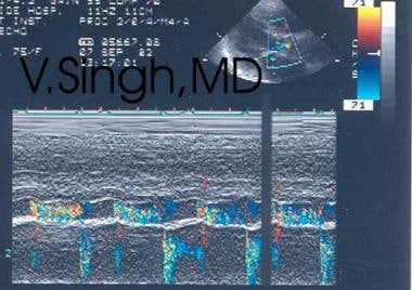
- We had the M-modes to the aortic valve, and you notice that in the M-mode of the aortic valve, the aorta was going up and down a lot. (medscape.com)
- Then you have the isovolumic contraction period, and then the aortic valve click demonstrating the aortic valve opening, followed by the ejection period, during which you have the continuous-wave Doppler signal through the left ventricular outflow tract (LVOT) and the aorta. (medscape.com)
Defective Aortic Valve1
- The 79-year-old will undergo surgery by the middle of next week to replace a defective aortic valve, Alberto Zangrillo told reporters in Rome. (bbc.com)
Quadricuspid aortic valve7
- A quadricuspid aortic valve (QAV) is a rare congenital heart defect characterized by the presence of four cusps, instead of the usual three found normally in the aortic valve. (wikipedia.org)
- If an "X" shape is seen, then the patient can be diagnosed with having a quadricuspid aortic valve. (wikipedia.org)
- There have been seven described variations of the quadricuspid aortic valve. (wikipedia.org)
- The quadricuspid aortic valve: a comprehensive review. (wikipedia.org)
- Quadricuspid aortic valve: a report of 12 cases and a review of the literature. (wikipedia.org)
- Quadricuspid Aortic Valve Revealed by Real-Time, 3-Dimensional Transesophageal Echocardiography. (wikipedia.org)
- Mid-term results in patients having tricuspidization of the quadricuspid aortic valve. (wikipedia.org)
Transcatheter aortic valve4
- Impact of Paravalvular Aortic Insufficiency on Left Ventricular Remodeling and Mortality after Transcatheter Aortic Valve Replacement. (cornell.edu)
- However, another option is transcatheter aortic valve implantation (TAVI), a less invasive procedure. (medicalnewstoday.com)
- In 2012, we performed the first transcatheter aortic valve replacement (TAVR) in Alabama, and UAB has performed more TAVR procedures than any other hospital in the state. (uabmedicine.org)
- After formal discussion in the heart team, the patient was scheduled for TAVI (transcatheter aortic valve implantation). (hunimed.eu)
Severe3
- We used a computer-generated randomisation sequence to randomly assign high-risk patients with severe aortic stenosis to either SAVR or TAVR with a balloon-expandable bovine pericardial tissue valve by either a transfemoral or transapical approach. (nih.gov)
- The infection led to severe aortic valve insufficiency and rapid death before the patient could be subjected to surgery. (lu.se)
- This demonstrates that IE caused by A. sanguinicola can be severe and cause valve destruction. (lu.se)
Dissection1
- Sex Differences in Thoracic Aortic Disease and Dissection: JACC Review Topic of the Week. (ottawaheart.ca)
Pathology3
- We have identified in an exact and certain way a pathology of aortic valve that is called aortic insufficiency,' the doctor said. (bbc.com)
- Identify common pathology for aortic valve disease. (gcus.com)
- Recognize cardiology pathology and correlate the two dimensional and Doppler echocardiography findings associated with various types of aortic valve disease. (gcus.com)
Echocardiography1
- Intraoperative transesophageal echocardiography accurately predicts mitral valve anatomy and suitability for repair. (apil.ca)
Tricuspid valve insuffi1
- Radionuclide ventriculography--a noninvasive method of diagnosis and quantification of tricuspid valve insufficiency]. (bvsalud.org)
Replacement15
- We recorded no structural valve deterioration requiring surgical valve replacement in either group. (nih.gov)
- The typical method of treatment is through surgery such as aortic valve reconstruction surgery (AVRS) and aortic valve replacement, usually with a synthetic valve. (wikipedia.org)
- Objective To describe differences between North America and Europe in the perioperative management of patients undergoing surgical aortic valve replacement (SAVR). (bmj.com)
- Most valve replacement surgeries are successful. (medicalnewstoday.com)
- Heart valve replacement surgery carries some risks, such as infection and bleeding. (medicalnewstoday.com)
- Keep reading to learn more about heart valve replacement surgery, including when it may be necessary, how much it may cost, what to expect, and what risks come with it. (medicalnewstoday.com)
- A heart valve replacement may be necessary if a person's heart valves are not working properly and are too damaged for a repair to be successful. (medicalnewstoday.com)
- Sometimes, people may need a replacement for more than one valve. (medicalnewstoday.com)
- According to a 2020 study , the average cost of aortic valve replacement surgery is about $59,000. (medicalnewstoday.com)
- Preparation for a valve replacement depends on the type of procedure a person is having. (medicalnewstoday.com)
- This type of replacement procedure involves opening up the chest to replace a damaged valve. (medicalnewstoday.com)
- The decision to have aortic valve replacement depends on your symptoms and the condition and function of your heart. (medlineplus.gov)
- There is increasing interest in a minimally invasive procedure in which a replacement valve is implanted via catheter. (medlineplus.gov)
- There was a way for Jack to get his valve replaced and maintain his physical independence through a fully robotic aortic valve replacement. (uchicagomedicine.org)
- Depending on the symptoms and severity, treatments may include blood pressure medication, angiotensin-converting enzyme ( ACE ) inhibitors (drugs that helps relax blood vessels), limits on activity, or aortic valve replacement surgery. (uabmedicine.org)
Defects1
- Congenital heart valve defects where the AORTIC VALVE has two instead of normal three cusps. (bvsalud.org)
Dilatation2
- Large VSDs (defined as defect size equal to or greater than the diameter of the aortic annulus) typically have left heart dilatation and pulmonary artery hypertension with normal left ventricular systolic function. (medscape.com)
- The HAART Aortic Annuloplasty Device reduces annular dilatation and recreates normal leaflet coaptation geometry through internal geometric ring annuloplasty. (biostable-s-e.com)
Prosthesis7
- Two persons (0.4%) had an aortic valve prosthesis. (nih.gov)
- The TAVI procedure was performed with the implantation of a fully retrievable and repositionable aortic valve prosthesis (Direct Flow 29 mm, Direct Flow Medical, Santa Rosa, California) with an excellent result and no paravalvular leak. (hunimed.eu)
- We believe that treatment of selected cases of pure AR with the Direct Flow valve is feasible and takes advantage of the retrievability of the prosthesis. (hunimed.eu)
- You can see the mechanical and a prosthesis of the mitral and the aortic positions. (medscape.com)
- You can see the peak velocity here through the aortic-valve prosthesis is about 2 m/s. (medscape.com)
- You can see the opening and closing of the mechanical aortic and the mechanical mitral valve because of the location of the continuous-wave Doppler going through both the aortic prosthesis and the "anterior" prosthesis of the mitral valve. (medscape.com)
- The velocity through the aortic valve prosthesis of about 2 m/s is normal. (medscape.com)
Congenital2
- Aortic valve insufficiency can be due to, or associated with, congenital heart disease. (medscape.com)
- Additional congenital heart lesions (eg, muscular right ventricular outflow tract obstruction, pulmonary valve stenosis, pulmonary venous obstruction, persistent elevation of PVR, mitral stenosis) can restrict shunting, possibly leading to right-to-left shunting at the VSD, depending on the ultimate resistance balance between the systemic and the total right-sided resistances. (medscape.com)
Annulus2
- The fourth dysplastic cusp is incapable of fully closing the aortic annulus, which causes a backflow of blood through the aortic valve. (wikipedia.org)
- Cardiol Young " Echocardiographic versus Angiographic measurement of the Aortic Valve Annulus in children undergoing balloon Aortic Valvuloplasty: method affects outcomes . (bcm.edu)
Diastolic1
- Supply, which is always abnormally tenuous because of the lower-than-normal coronary driving pressure (difference in aortic diastolic pressure and ventricular diastolic pressure), cannot keep up with the increased demand. (medscape.com)
Doppler4
- I am showing you a continuous-wave Doppler in the short axis around the aortic-valve level. (medscape.com)
- In the next slide, there is a continuous-wave Doppler through the aortic valve. (medscape.com)
- The continuous-wave Doppler for the mitral valve did not demonstrate any mitral gurgitation signal. (medscape.com)
- Well, let's take another look at a continuous-wave Doppler that I already showed you through the aortic valve. (medscape.com)
Abnormalities3
- These include abnormalities of the aortic valve leaflets and pathologies of the proximal aortic root. (medscape.com)
- This article primarily focuses on aortic valve insufficiency caused by abnormalities in the aortic valve leaflets. (medscape.com)
- This study was undertaken to elucidate the prevalence of aortic valve abnormalities in the elderly. (nih.gov)
Disease5
- More information is needed about the prevalence of aortic valve disease in old age. (nih.gov)
- The Journal of Heart Valve Disease, 13(4), 534-537. (wikipedia.org)
- Few of them have underlying rheumatic valve disease. (scirp.org)
- It provides ongoing care - sometimes for life - to patients who have or are at risk for structural heart and valve disease. (uabmedicine.org)
- It aims to improve quality of life and survival for people with heart valve disease through timely diagnosis and appropriate intervention. (bvsalud.org)
Systolic1
- The systolic aortic valve area was calculated by the continuity equation. (nih.gov)
Implantation1
- The TAVI devices designed for the treatment of calcific aortic stenosis have numerous limitations for the treatment of pure AR such as the risk of residual AR, the lack of repositionability and retrievability, and the need for valve- in-valve implantation. (hunimed.eu)
Rheumatic1
- In the past, rheumatic fever was the main cause of aortic insufficiency, but the use of antibiotics to treat infections has made rheumatic fever less common. (uabmedicine.org)
Mild1
- Stenosis affected only 8% of the valves and was mild. (elsevierpure.com)
Pulmonary1
- This procedure involves swapping the person's damaged aortic valve with their pulmonary valve and replacing the pulmonary valve with a donor valve. (medicalnewstoday.com)
Patient2
- [1] J.C.P. Williams observed in four patients an association between supravalvular aortic stenosis and the common physical and mental characteristics of this patient population and stated that it "may constitute a previously unrecognized syndrome" [1] . (physio-pedia.com)
- For over a decade, he's replaced aortic valves by making a six centimeter incision, lending to a faster recovery for the patient. (uchicagomedicine.org)
Outflow1
- The velocity ratio (peak velocity in the left ventricular outflow tract/peak velocity across the aortic valve) was a supplementary criterion for aortic stenosis. (nih.gov)
Calcific1
- Calcific aortic valve stenosis constitutes a significant health problem in the elderly. (nih.gov)
Surgery7
- Only a minority of those with potentially operable aortic valve stenosis undergo surgery. (nih.gov)
- Replacing a heart valve often involves open-heart surgery. (medicalnewstoday.com)
- Surgery can cure aortic insufficiency and relieve symptoms, unless you develop heart failure or other complications. (medlineplus.gov)
- The therapeutic options include aortic surgery, coronary artery bypass grafting, transplantation surgery, surgical treatment of heart rhythm disorders (arrhythmias), minimally invasive surgery, surgical treatment of the heart valves, including reconstructive interventions. (bookinghealth.com)
- Aortic valve repair for tri-leaflet aortic insufficiency associated with asymmetric aortic root aneurysms Article from Annals of Cardiothoracic Surgery (Submitted Jan 26, 2019. (biostable-s-e.com)
- Our experienced surgeons and cardiologists take a comprehensive approach to diagnosing and treating this condition, and their expertise ranges from traditional open-heart surgery to robotic-assisted valve repair and the latest in minimally invasive surgical techniques, which require only small incisions (cuts). (uabmedicine.org)
- As an added service, patients who have been told by non-UAB doctors that they need valve surgery can speak to a UAB structural heart and valve surgeon for a second opinion. (uabmedicine.org)
Pathologies1
- This new capability greatly expands the variety of treatable pathologies that cause aortic insufficiency. (biostable-s-e.com)
Patients1
- Afin de préciser le statut en sélénium à Cotonou les auteurs ont dosé le sélénium plasmatique chez 10 béninoises(âge moyen = 27,1 ans) atteintes de CMPP, chez 18 patients/es atteints/es de CMD (11 femmes,7 hommes, âge moyen = 38,9 ans) et chez 46 béninoises « témoins » en bonne santé ayant accouché récemment (âge moyen = 29,8 ans). (bvsalud.org)
Cusps1
- There is no correlation between the anatomy and functional status of the aortic cusps. (wikipedia.org)