Aorta, Thoracic
Aorta, Abdominal
Aortic Coarctation
Aortography
Aortic Aneurysm, Thoracic
Arteriosclerosis
Aneurysm, Dissecting
Endothelium, Vascular
Blood Vessel Prosthesis Implantation
Rabbits
Aortic Rupture
Aortic Aneurysm, Abdominal
Blood Vessel Prosthesis
Vasodilation
Vasoconstriction
Phenylephrine
Aneurysm, Infected
Tomography, X-Ray Computed
Elastin
Atherosclerosis
Polyethylene Terephthalates
Rats, Wistar
Muscle Contraction
Dilatation, Pathologic
Marfan Syndrome
Acetylcholine
Nitric Oxide
Rats, Inbred WKY
Rats, Sprague-Dawley
Aneurysm, False
Iliac Artery
Rats, Inbred SHR
Hypertension
Aortic Valve
Dose-Response Relationship, Drug
Norepinephrine
Apolipoproteins E
Anastomosis, Surgical
Nitroprusside
Disease Models, Animal
Nitric Oxide Synthase Type III
Endothelium
Catheterization
Echocardiography, Transesophageal
Stents
Takayasu Arteritis
Nitric Oxide Synthase
Pulmonary Artery
Subclavian Artery
Angiotensin II
Potassium Chloride
Cyclic GMP
Mesenteric Arteries
Elastic Tissue
Brachiocephalic Trunk
Treatment Outcome
Mice, Inbred C57BL
NG-Nitroarginine Methyl Ester
Aortic Valve Insufficiency
Cholesterol
Blood Vessels
Cells, Cultured
RNA, Messenger
Aortic Arch Syndromes
Methylene Blue
Mice, Knockout
Carotid Arteries
Blood Flow Velocity
Models, Cardiovascular
Hemodynamics
Rats, Inbred Strains
Hypercholesterolemia
Tunica Intima
15-Hydroxy-11 alpha,9 alpha-(epoxymethano)prosta-5,13-dienoic Acid
Calcium
Aortic Valve Stenosis
Heart Defects, Congenital
Celiac Artery
Myocytes, Smooth Muscle
Indomethacin
Swine
Ulcer
Paraplegia
Pulsatile Flow
Circulatory Arrest, Deep Hypothermia Induced
Endovascular Procedures
Reoperation
Spinal Cord Ischemia
Desmosine
Dogs
Stress, Mechanical
Enzyme Inhibitors
Receptors, Thromboxane
Magnetic Resonance Angiography
Prazosin
Tunica Media
Cardiopulmonary Bypass
Arterial Occlusive Diseases
Mesenteric Artery, Superior
Receptors, Adrenergic, alpha-1
Desoxycorticosterone
Adrenergic alpha-Antagonists
Immunohistochemistry
Epoprostenol
Vena Cava, Inferior
Nitroglycerin
Postoperative Complications
Nitroarginine
A molecular pathway revealing a genetic basis for human cardiac and craniofacial defects. (1/4396)
Microdeletions of chromosome 22q11 are the most common genetic defects associated with cardiac and craniofacial anomalies in humans. A screen for mouse genes dependent on dHAND, a transcription factor implicated in neural crest development, identified Ufd1, which maps to human 22q11 and encodes a protein involved in degradation of ubiquitinated proteins. Mouse Ufd1 was specifically expressed in most tissues affected in patients with 22q11 deletion syndrome. The human UFD1L gene was deleted in all 182 patients studied with 22q11 deletion, and a smaller deletion of approximately 20 kilobases that removed exons 1 to 3 of UFD1L was found in one individual with features typical of 22q11 deletion syndrome. These data suggest that UFD1L haploinsufficiency contributes to the congenital heart and craniofacial defects seen in 22q11 deletion. (+info)The cyclo-oxygenase-dependent regulation of rabbit vein contraction: evidence for a prostaglandin E2-mediated relaxation. (2/4396)
1. Arachidonic acid (0.01-1 microM) induced relaxation of precontracted rings of rabbit saphenous vein, which was counteracted by contraction at concentrations higher than 1 microM. Concentrations higher than 1 microM were required to induce dose-dependent contraction of vena cava and thoracic aorta from the same animals. 2. Pretreatment with a TP receptor antagonist (GR32191B or SQ29548, 3 microM) potentiated the relaxant effect in the saphenous vein, revealed a vasorelaxant component in the vena cava response and did not affect the response of the aorta. 3. Removal of the endothelium from the venous rings, caused a 10 fold rightward shift in the concentration-relaxation curves to arachidonic acid. Whether or not the endothelium was present, the arachidonic acid-induced relaxations were prevented by indomethacin (10 microM) pretreatment. 4. In the saphenous vein, PGE2 was respectively a 50 and 100 fold more potent relaxant prostaglandin than PGI2 and PGD2. Pretreatment with the EP4 receptor antagonist, AH23848B, shifted the concentration-relaxation curves of this tissue to arachidonic acid in a dose-dependent manner. 5. In the presence of 1 microM arachidonic acid, venous rings produced 8-10 fold more PGE2 than did aorta whereas 6keto-PGF1alpha and TXB2 productions remained comparable. 6. Intact rings of saphenous vein relaxed in response to A23187. Pretreatment with L-NAME (100 microM) or indomethacin (10 microM) reduced this response by 50% whereas concomitant pretreatment totally suppressed it. After endothelium removal, the remaining relaxing response to A23187 was prevented by indomethacin but not affected by L-NAME. 7. We conclude that stimulation of the cyclo-oxygenase pathway by arachidonic acid induced endothelium-dependent, PGE2/EP4 mediated relaxation of the rabbit saphenous vein. This process might participate in the A23187-induced relaxation of the saphenous vein and account for a relaxing component in the response of the vena cava to arachidonic acid. It was not observed in thoracic aorta because of the lack of a vasodilatory receptor and/or the poorer ability of this tissue than veins to produce PGE2. (+info)Enantioselective inhibition of the biotransformation and pharmacological actions of isoidide dinitrate by diphenyleneiodonium sulphate. (3/4396)
1. We have shown previously that the D- and L- enantiomers of isoidide dinitrate (D-IIDN and L-IIDN) exhibit a potency difference for relaxation and cyclic GMP accumulation in isolated rat aorta and that this is related to preferential biotransformation of the more potent enantiomer (D-IIDN). The objective of the current study was to examine the effect of the flavoprotein inhibitor, diphenyleneiodonium sulphate (DPI), on the enantioselectivity of IIDN action. 2. In isolated rat aortic strip preparations, exposure to 0.3 microM DPI resulted in a 3.6 fold increase in the EC50 value for D-IIDN-induced relaxation, but had no effect on L-IIDN-induced relaxation. 3. Incubation of aortic strips with 2 microM D- or L-IIDN for 5 min resulted in significantly more D-isoidide mononitrate formed (5.0 +/- 1.5 pmol mg protein(-1)) than L-isoidide mononitrate (2.1 +/- 0.7 pmol mg protein(-1)) and this difference was abolished by pretreatment of tissues with 0.3 microM DPI. DPI had no effect on glutathione S-transferase (GST) activity or GSH-dependent biotransformation of D- or L-IIDN in the 105,000 x g supernatant fraction of rat aorta. 4. Consistent with both the relaxation and biotransformation data, treatment of tissues with 0.3 microM DPI significantly inhibited D-IIDN-induced cyclic GMP accumulation, but had no effect on L-IIDN-induced cyclic GMP accumulation. 5. In the intact animal, 2 mg kg(-1) DPI significantly inhibited the pharmacokinetic and haemodynamic properties of D-IIDN, but had no effect L-IIDN. 6. These data suggest that the basis for the potency difference for relaxation by the two enantiomers is preferential biotransformation of D-IIDN to NO, by an enzyme that is inhibited by DPI. Given that DPI binds to and inhibits NADPH-cytochrome P450 reductase, the data are consistent with a role for the cytochromes P450-NADPH-cytochrome P450 reductase system in this enantioselective biotransformation process. (+info)Effect of acute and long-term treatment with 17-beta-estradiol on the vasomotor responses in the rat aorta. (4/4396)
1. This study sought to evaluate whether the effects of acute and long-term treatment with 17-beta-estradiol on the vasomotor responses in rat aortic rings are mediated through the same mechanism. 2. Ovariectomized rats were treated daily with either 17-beta-estradiol-3-benzoate (100 microg kg(-1)) or vehicle for 1 week. 3. The effect of long-term 17-beta-estradiol treatment on the responses to cumulative doses of phenylephrine, 5-HT, calcium, potassium and 17-beta-estradiol was determined in aortic rings. In the same rings, the effect of acute exposure to 17-beta-estradiol (5 and 10 microM) on the dose response curves for phenylephrine, 5-HT, calcium, potassium and acetylcholine were estimated. The measurements were made in rings with and without intact endothelium. The tone-related basal release of nitric oxide (NO) was measured in rings with intact endothelium. 4. Long-term 17-beta-estradiol treatment reduced the maximum developed contraction to all contracting agents studied. This effect was abolished in endothelium denuded vessels. Acute 17-beta-estradiol treatment also reduced maximal contraction. This effect, however, was independent of the endothelium. 5. Long-term 17-beta-estradiol treatment significantly increased the ability of the rings to dilate in response to acetylcholine whereas acute exposure to 17-beta-estradiol had no effect. The tone-related release of NO was significantly increased after long-term exposure to 17-beta-estradiol. 6. In conclusion, this study indicate that the acute and long-term effects of 17-beta-estradiol in the rat aorta are mediated through different mechanisms. The long-term effect is mediated through the endothelium most likely by increasing NO release. In contrast, the acute effect of 17-beta-estradiol seems to be through an effect on the vascular smooth muscle cells. (+info)Studies of the role of endothelium-dependent nitric oxide release in the sustained vasodilator effects of corticotrophin releasing factor and sauvagine. (5/4396)
1. The mechanisms of the sustained vasodilator actions of corticotrophin-releasing factor (CRF) and sauvagine (SVG) were studied using rings of endothelium de-nuded rat thoracic aorta (RTA) and the isolated perfused rat superior mesenteric arterial vasculature (SMA). 2. SVG was approximately 50 fold more potent than CRF on RTA (EC40: 0.9 +/- 0.2 and 44 +/- 9 nM respectively, P < 0.05), and approximately 10 fold more active in the perfused SMA (ED40: 0.05 +/- 0.02 and 0.6 +/- 0.1 nmol respectively, P < 0.05). Single bolus injections of CRF (100 pmol) or SVG (15 pmol) in the perfused SMA caused reductions in perfusion pressure of 23 +/- 1 and 24 +/- 2% that lasted more than 20 min. 3. Removal of the endothelium in the perfused SMA with deoxycholic acid attenuated the vasodilatation and revealed two phases to the response; a short lasting direct action, and a sustained phase which was fully inhibited. 4. Inhibition of nitric oxide synthase with L-NAME (100 microM) L-NMMA (100 microM) or 2-ethyl-2-thiopseudourea (ETPU, 100 microM) had similar effects on the vasodilator responses to CRF as removal of the endothelium, suggesting a pivotal role for nitric oxide. However the selective guanylate cyclase inhibitor 1H-[l,2,4]oxadiazolo[4,3-alpha]quinoxalin-1-one (ODQ, 10 microM) did not affect the response to CRF. 5. High potassium (60 mM) completely inhibited the vasodilator response to CRF in the perfused SMA, indicating a role for K channels in this response. 6. Compared to other vasodilator agents acting via the release of NO, the actions of CRF and SVG are strikingly long-lasting, suggesting a novel mechanism of prolonged activation of nitric oxide synthase. (+info)Protective effect of dietary tomato against endothelial dysfunction in hypercholesterolemic mice. (6/4396)
The effects of dietary ingestion of tomato were studied in mice that had been made hypercholesterolemic by feeding atherogenic diets. Mice which had been fed on the atherogenic diet without tomato for 4 months had significantly increased plasma lipid peroxide, and the vaso-relaxing activity in the aorta induced by acetylcholine (ACh) was harmed when compared with mice fed on a common commercial diet. On the other hand, mice which had been fed on the atherogenic diet containing 20% (w/w) lyophilized powder of tomato showed less increase in the plasma lipid peroxide level, and ACh-induced vaso-relaxation was maintained at the same level as that in normal mice. These results indicate that tomato has a preventive effect on atherosclerosis by protecting plasma lipids from oxidation. (+info)Effects of docosahexaenoic and eicosapentaenoic acid on lipid metabolism, eicosanoid production, platelet aggregation and atherosclerosis in hypercholesterolemic rats. (7/4396)
Exogenously hypercholesterolemic (ExHC) rats were fed on an atherogenic diet supplemented with 1% each of either ethyl ester docosahexaenoic acid [EE-DHA, 22:6(n-3)], ethyl ester eicosapentaenoic acid [EE-EPA, 20:5(n-3)] or safflower oil (SO) for 6 months. The rats fed on the diets containing EE-EPA or EE-DHA, compared with those fed on SO, had lower serum cholesterol and triacylglycerol levels, less aggregation of platelets and slower progress of intimal thickening in the ascending aorta. Relative to the SO-fed rats, both of the (n-3) fatty acid-fed rats had a significantly reduced proportion of arachidonic acid in the platelet and aortic phospholipids, and lower production of thromboxane A2 by platelets and of prostacyclin by the aorta. These results suggest that EPA and DHA are similarly involved in preventing atherosclerosis development by reducing hypercholesterolemia and modifying the platelet functions. (+info)Modulation of temperature-induced tone by vasoconstrictor agents. (8/4396)
One of the primary cardiovascular adjustments to hyperthermia is a sympathetically mediated increase in vascular resistance in the viscera. Nonneural factors such as a change in vascular tone or reactivity may also contribute to this response. Therefore, the aim of this study was to determine whether vascular smooth muscle tone is altered during heating to physiologically relevant temperatures >37 degrees C. Gradually increasing bath temperature from 37 degrees C (normothermia) to 43 degrees C (severe hyperthermia) produced graded contractions in vascular ring segments from rat mesenteric arteries and thoracic aortae. In untreated rings these contractions were relatively small, whereas hyperthermia elicited near-maximal increases in tension when rings were constricted with phenylephrine or KCl before heating. In phenylephrine-treated mesenteric arterial rings, the contractile responses to heating were markedly attenuated by the Ca2+ channel antagonists nifedipine and diltiazem. Diltiazem also blocked the contractile responses to heating in thoracic aortic rings. These results demonstrate that hyperthermia has a limited effect on tension generation in rat vascular smooth muscle in the absence of vascular tone. However, in the presence of agonist-induced tone, tension generation during heating is markedly enhanced and dependent on extracellular Ca2+. In conclusion, these data suggest that local regulation of vascular tone can contribute to the hemodynamic adjustments to hyperthermia. (+info)The aorta is the largest artery in the human body, which originates from the left ventricle of the heart and carries oxygenated blood to the rest of the body. It can be divided into several parts, including the ascending aorta, aortic arch, and descending aorta. The ascending aorta gives rise to the coronary arteries that supply blood to the heart muscle. The aortic arch gives rise to the brachiocephalic, left common carotid, and left subclavian arteries, which supply blood to the head, neck, and upper extremities. The descending aorta travels through the thorax and abdomen, giving rise to various intercostal, visceral, and renal arteries that supply blood to the chest wall, organs, and kidneys.
The thoracic aorta is the segment of the largest artery in the human body (the aorta) that runs through the chest region (thorax). The thoracic aorta begins at the aortic arch, where it branches off from the ascending aorta, and extends down to the diaphragm, where it becomes the abdominal aorta.
The thoracic aorta is divided into three parts: the ascending aorta, the aortic arch, and the descending aorta. The ascending aorta rises from the left ventricle of the heart and is about 2 inches (5 centimeters) long. The aortic arch curves backward and to the left, giving rise to the brachiocephalic trunk, the left common carotid artery, and the left subclavian artery. The descending thoracic aorta runs downward through the chest, passing through the diaphragm to become the abdominal aorta.
The thoracic aorta supplies oxygenated blood to the upper body, including the head, neck, arms, and chest. It plays a critical role in maintaining blood flow and pressure throughout the body.
The abdominal aorta is the portion of the aorta, which is the largest artery in the body, that runs through the abdomen. It originates from the thoracic aorta at the level of the diaphragm and descends through the abdomen, where it branches off into several smaller arteries that supply blood to the pelvis, legs, and various abdominal organs. The abdominal aorta is typically divided into four segments: the suprarenal, infrarenal, visceral, and parietal portions. Disorders of the abdominal aorta can include aneurysms, atherosclerosis, and dissections, which can have serious consequences if left untreated.
Aortic diseases refer to conditions that affect the aorta, which is the largest and main artery in the body. The aorta carries oxygenated blood from the heart to the rest of the body. Aortic diseases can weaken or damage the aorta, leading to various complications. Here are some common aortic diseases with their medical definitions:
1. Aortic aneurysm: A localized dilation or bulging of the aortic wall, which can occur in any part of the aorta but is most commonly found in the abdominal aorta (abdominal aortic aneurysm) or the thoracic aorta (thoracic aortic aneurysm). Aneurysms can increase the risk of rupture, leading to life-threatening bleeding.
2. Aortic dissection: A separation of the layers of the aortic wall due to a tear in the inner lining, allowing blood to flow between the layers and potentially cause the aorta to rupture. This is a medical emergency that requires immediate treatment.
3. Aortic stenosis: A narrowing of the aortic valve opening, which restricts blood flow from the heart to the aorta. This can lead to shortness of breath, chest pain, and other symptoms. Severe aortic stenosis may require surgical or transcatheter intervention to replace or repair the aortic valve.
4. Aortic regurgitation: Also known as aortic insufficiency, this condition occurs when the aortic valve does not close properly, allowing blood to leak back into the heart. This can lead to symptoms such as fatigue, shortness of breath, and palpitations. Treatment may include medication or surgical repair or replacement of the aortic valve.
5. Aortitis: Inflammation of the aorta, which can be caused by various conditions such as infections, autoimmune diseases, or vasculitides. Aortitis can lead to aneurysms, dissections, or stenosis and may require medical treatment with immunosuppressive drugs or surgical intervention.
6. Marfan syndrome: A genetic disorder that affects the connective tissue, including the aorta. People with Marfan syndrome are at risk of developing aortic aneurysms and dissections, and may require close monitoring and prophylactic surgery to prevent complications.
Aortic coarctation is a narrowing of the aorta, the largest blood vessel in the body that carries oxygen-rich blood from the heart to the rest of the body. This condition usually occurs in the part of the aorta that is just beyond where it arises from the left ventricle and before it divides into the iliac arteries.
In aortic coarctation, the narrowing can vary from mild to severe, and it can cause a variety of symptoms depending on the severity of the narrowing and the age of the individual. In newborns and infants with severe coarctation, symptoms may include difficulty breathing, poor feeding, and weak or absent femoral pulses (located in the groin area). Older children and adults with mild to moderate coarctation may not experience any symptoms until later in life, when high blood pressure, headaches, nosebleeds, leg cramps, or heart failure develop.
Aortic coarctation is typically diagnosed through physical examination, imaging tests such as echocardiography, CT angiography, or MRI, and sometimes cardiac catheterization. Treatment options include surgical repair or balloon dilation (also known as balloon angioplasty) to open the narrowed section of the aorta. If left untreated, aortic coarctation can lead to serious complications such as high blood pressure, heart failure, stroke, and rupture or dissection of the aorta.
An aortic aneurysm is a medical condition characterized by the abnormal widening or bulging of the wall of the aorta, which is the largest artery in the body. The aorta carries oxygenated blood from the heart to the rest of the body. When the aortic wall weakens, it can stretch and balloon out, forming an aneurysm.
Aortic aneurysms can occur anywhere along the aorta but are most commonly found in the abdominal section (abdominal aortic aneurysm) or the chest area (thoracic aortic aneurysm). The size and location of the aneurysm, as well as the patient's overall health, determine the risk of rupture and associated complications.
Aneurysms often do not cause symptoms until they become large or rupture. Symptoms may include:
* Pain in the chest, back, or abdomen
* Pulsating sensation in the abdomen
* Difficulty breathing
* Hoarseness
* Coughing or vomiting
Risk factors for aortic aneurysms include age, smoking, high blood pressure, family history, and certain genetic conditions. Treatment options depend on the size and location of the aneurysm and may include monitoring, medication, or surgical repair.
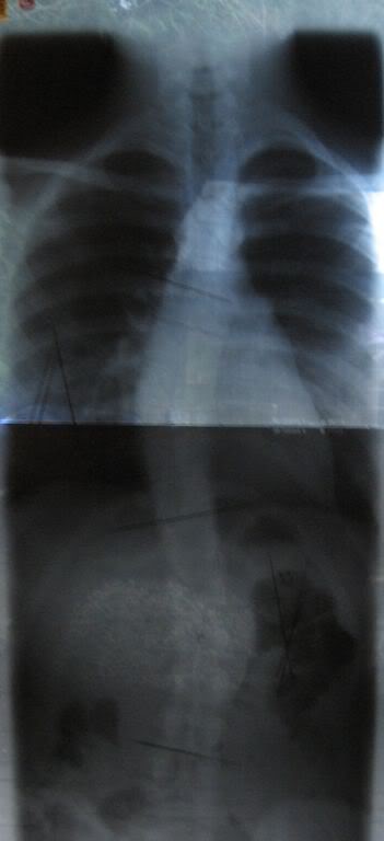
Aortography is a medical procedure that involves taking X-ray images of the aorta, which is the largest blood vessel in the body. The procedure is usually performed to diagnose or assess various conditions related to the aorta, such as aneurysms, dissections, or blockages.
To perform an aortography, a contrast dye is injected into the aorta through a catheter that is inserted into an artery, typically in the leg or arm. The contrast dye makes the aorta visible on X-ray images, allowing doctors to see its structure and any abnormalities that may be present.
The procedure is usually performed in a hospital or outpatient setting and may require sedation or anesthesia. While aortography can provide valuable diagnostic information, it also carries some risks, such as allergic reactions to the contrast dye, damage to blood vessels, or infection. Therefore, it is typically reserved for situations where other diagnostic tests have been inconclusive or where more invasive treatment may be required.
A smooth muscle within the vascular system refers to the involuntary, innervated muscle that is found in the walls of blood vessels. These muscles are responsible for controlling the diameter of the blood vessels, which in turn regulates blood flow and blood pressure. They are called "smooth" muscles because their individual muscle cells do not have the striations, or cross-striped patterns, that are observed in skeletal and cardiac muscle cells. Smooth muscle in the vascular system is controlled by the autonomic nervous system and by hormones, and can contract or relax slowly over a period of time.
A thoracic aortic aneurysm is a localized dilatation or bulging of the thoracic aorta, which is the part of the aorta that runs through the chest cavity. The aorta is the largest artery in the body, and it carries oxygenated blood from the heart to the rest of the body.
Thoracic aortic aneurysms can occur anywhere along the thoracic aorta, but they are most commonly found in the aortic arch or the descending thoracic aorta. These aneurysms can vary in size, and they are considered significant when they are 50% larger than the expected normal diameter of the aorta.
The exact cause of thoracic aortic aneurysms is not fully understood, but several factors can contribute to their development, including:
* Atherosclerosis (hardening and narrowing of the arteries)
* High blood pressure
* Genetic disorders such as Marfan syndrome or Ehlers-Danlos syndrome
* Infections or inflammation of the aorta
* Trauma to the chest
Thoracic aortic aneurysms can be asymptomatic and found incidentally on imaging studies, or they may present with symptoms such as chest pain, cough, difficulty swallowing, or hoarseness. If left untreated, thoracic aortic aneurysms can lead to serious complications, including aortic dissection (tearing of the inner layer of the aorta) or rupture, which can be life-threatening.
Treatment options for thoracic aortic aneurysms include medical management with blood pressure control and cholesterol-lowering medications, as well as surgical repair or endovascular stenting, depending on the size, location, and growth rate of the aneurysm. Regular follow-up imaging is necessary to monitor the size and progression of the aneurysm over time.
Arteriosclerosis is a general term that describes the hardening and stiffening of the artery walls. It's a progressive condition that can occur as a result of aging, or it may be associated with certain risk factors such as high blood pressure, high cholesterol, diabetes, smoking, and a sedentary lifestyle.
The process of arteriosclerosis involves the buildup of plaque, made up of fat, cholesterol, calcium, and other substances, in the inner lining of the artery walls. Over time, this buildup can cause the artery walls to thicken and harden, reducing the flow of oxygen-rich blood to the body's organs and tissues.
Arteriosclerosis can affect any of the body's arteries, but it is most commonly found in the coronary arteries that supply blood to the heart, the cerebral arteries that supply blood to the brain, and the peripheral arteries that supply blood to the limbs. When arteriosclerosis affects the coronary arteries, it can lead to heart disease, angina, or heart attack. When it affects the cerebral arteries, it can lead to stroke or transient ischemic attack (TIA). When it affects the peripheral arteries, it can cause pain, numbness, or weakness in the limbs, and in severe cases, gangrene and amputation.
A dissecting aneurysm is a serious and potentially life-threatening condition that occurs when there is a tear in the inner layer of the artery wall, allowing blood to flow between the layers of the artery wall. This can cause the artery to bulge or balloon out, leading to a dissection aneurysm.
Dissecting aneurysms can occur in any artery, but they are most commonly found in the aorta, which is the largest artery in the body. When a dissecting aneurysm occurs in the aorta, it is often referred to as a "dissecting aortic aneurysm."
Dissecting aneurysms can be caused by various factors, including high blood pressure, atherosclerosis (hardening and narrowing of the arteries), genetic disorders that affect the connective tissue, trauma, or illegal drug use (such as cocaine).
Symptoms of a dissecting aneurysm may include sudden severe chest or back pain, which can feel like ripping or tearing, shortness of breath, sweating, lightheadedness, or loss of consciousness. If left untreated, a dissecting aneurysm can lead to serious complications, such as rupture of the artery, stroke, or even death.
Treatment for a dissecting aneurysm typically involves surgery or endovascular repair to prevent further damage and reduce the risk of rupture. The specific treatment approach will depend on various factors, including the location and size of the aneurysm, the patient's overall health, and their medical history.
The endothelium is a thin layer of simple squamous epithelial cells that lines the interior surface of blood vessels, lymphatic vessels, and heart chambers. The vascular endothelium, specifically, refers to the endothelial cells that line the blood vessels. These cells play a crucial role in maintaining vascular homeostasis by regulating vasomotor tone, coagulation, platelet activation, inflammation, and permeability of the vessel wall. They also contribute to the growth and repair of the vascular system and are involved in various pathological processes such as atherosclerosis, hypertension, and diabetes.
Blood vessel prosthesis implantation is a surgical procedure in which an artificial blood vessel, also known as a vascular graft or prosthetic graft, is inserted into the body to replace a damaged or diseased native blood vessel. The prosthetic graft can be made from various materials such as Dacron (polyester), PTFE (polytetrafluoroethylene), or bovine/human tissue.
The implantation of a blood vessel prosthesis is typically performed to treat conditions that cause narrowing or blockage of the blood vessels, such as atherosclerosis, aneurysms, or traumatic injuries. The procedure may be used to bypass blocked arteries in the legs (peripheral artery disease), heart (coronary artery bypass surgery), or neck (carotid endarterectomy). It can also be used to replace damaged veins for hemodialysis access in patients with kidney failure.
The success of blood vessel prosthesis implantation depends on various factors, including the patient's overall health, the location and extent of the vascular disease, and the type of graft material used. Possible complications include infection, bleeding, graft thrombosis (clotting), and graft failure, which may require further surgical intervention or endovascular treatments.
I believe there may be some confusion in your question. "Rabbits" is a common name used to refer to the Lagomorpha species, particularly members of the family Leporidae. They are small mammals known for their long ears, strong legs, and quick reproduction.
However, if you're referring to "rabbits" in a medical context, there is a term called "rabbit syndrome," which is a rare movement disorder characterized by repetitive, involuntary movements of the fingers, resembling those of a rabbit chewing. It is also known as "finger-chewing chorea." This condition is usually associated with certain medications, particularly antipsychotics, and typically resolves when the medication is stopped or adjusted.
Aortic rupture is a medical emergency that refers to the tearing or splitting of the aorta, which is the largest and main artery in the body. The aorta carries oxygenated blood from the heart to the rest of the body. An aortic rupture can lead to life-threatening internal bleeding and requires immediate medical attention.
There are two types of aortic ruptures:
1. Aortic dissection: This occurs when there is a tear in the inner lining of the aorta, allowing blood to flow between the layers of the aortic wall. This can cause the aorta to bulge or split, leading to a rupture.
2. Thoracic aortic aneurysm rupture: An aneurysm is a weakened and bulging area in the aortic wall. When an aneurysm in the thoracic aorta (the part of the aorta that runs through the chest) ruptures, it can cause severe bleeding and other complications.
Risk factors for aortic rupture include high blood pressure, smoking, aging, family history of aortic disease, and certain genetic conditions such as Marfan syndrome or Ehlers-Danlos syndrome. Symptoms of an aortic rupture may include sudden severe chest or back pain, difficulty breathing, weakness, sweating, and loss of consciousness. Treatment typically involves emergency surgery to repair the aorta and control bleeding.
An abdominal aortic aneurysm (AAA) is a localized dilatation or bulging of the abdominal aorta, which is the largest artery in the body that supplies oxygenated blood to the trunk and lower extremities. Normally, the diameter of the abdominal aorta measures about 2 centimeters (cm) in adults. However, when the diameter of the aorta exceeds 3 cm, it is considered an aneurysm.
AAA can occur anywhere along the length of the abdominal aorta, but it most commonly occurs below the renal arteries and above the iliac bifurcation. The exact cause of AAA remains unclear, but several risk factors have been identified, including smoking, hypertension, advanced age, male gender, family history, and certain genetic disorders such as Marfan syndrome and Ehlers-Danlos syndrome.
The main concern with AAA is the risk of rupture, which can lead to life-threatening internal bleeding. The larger the aneurysm, the greater the risk of rupture. Symptoms of AAA may include abdominal or back pain, a pulsating mass in the abdomen, or symptoms related to compression of surrounding structures such as the kidneys, ureters, or nerves. However, many AAAs are asymptomatic and are discovered incidentally during imaging studies performed for other reasons.
Diagnosis of AAA typically involves imaging tests such as ultrasound, computed tomography (CT) scan, or magnetic resonance imaging (MRI). Treatment options depend on the size and location of the aneurysm, as well as the patient's overall health status. Small AAAs that are not causing symptoms may be monitored with regular imaging studies to assess for growth. Larger AAAs or those that are growing rapidly may require surgical repair, either through open surgery or endovascular repair using a stent graft.
A blood vessel prosthesis is a medical device that is used as a substitute for a damaged or diseased natural blood vessel. It is typically made of synthetic materials such as polyester, Dacron, or ePTFE (expanded polytetrafluoroethylene) and is designed to mimic the function of a native blood vessel by allowing the flow of blood through it.
Blood vessel prostheses are used in various surgical procedures, including coronary artery bypass grafting, peripheral arterial reconstruction, and the creation of arteriovenous fistulas for dialysis access. The choice of material and size of the prosthesis depends on several factors, such as the location and diameter of the vessel being replaced, the patient's age and overall health status, and the surgeon's preference.
It is important to note that while blood vessel prostheses can be effective in restoring blood flow, they may also carry risks such as infection, thrombosis (blood clot formation), and graft failure over time. Therefore, careful patient selection, surgical technique, and postoperative management are crucial for the success of these procedures.
Vasodilation is the widening or increase in diameter of blood vessels, particularly the involuntary relaxation of the smooth muscle in the tunica media (middle layer) of the arteriole walls. This results in an increase in blood flow and a decrease in vascular resistance. Vasodilation can occur due to various physiological and pathophysiological stimuli, such as local metabolic demands, neural signals, or pharmacological agents. It plays a crucial role in regulating blood pressure, tissue perfusion, and thermoregulation.
Vasoconstriction is a medical term that refers to the narrowing of blood vessels due to the contraction of the smooth muscle in their walls. This process decreases the diameter of the lumen (the inner space of the blood vessel) and reduces blood flow through the affected vessels. Vasoconstriction can occur throughout the body, but it is most noticeable in the arterioles and precapillary sphincters, which control the amount of blood that flows into the capillary network.
The autonomic nervous system, specifically the sympathetic division, plays a significant role in regulating vasoconstriction through the release of neurotransmitters like norepinephrine (noradrenaline). Various hormones and chemical mediators, such as angiotensin II, endothelin-1, and serotonin, can also induce vasoconstriction.
Vasoconstriction is a vital physiological response that helps maintain blood pressure and regulate blood flow distribution in the body. However, excessive or prolonged vasoconstriction may contribute to several pathological conditions, including hypertension, stroke, and peripheral vascular diseases.
Phenylephrine is a medication that belongs to the class of drugs known as sympathomimetic amines. It primarily acts as an alpha-1 adrenergic receptor agonist, which means it stimulates these receptors, leading to vasoconstriction (constriction of blood vessels). This effect can be useful in various medical situations, such as:
1. Nasal decongestion: When applied topically in the nose, phenylephrine causes constriction of the blood vessels in the nasal passages, which helps to relieve congestion and swelling. It is often found in over-the-counter (OTC) cold and allergy products.
2. Ocular circulation: In ophthalmology, phenylephrine is used to dilate the pupils before eye examinations. The increased pressure from vasoconstriction helps to open up the pupil, allowing for a better view of the internal structures of the eye.
3. Hypotension management: In some cases, phenylephrine may be given intravenously to treat low blood pressure (hypotension) during medical procedures like spinal anesthesia or septic shock. The vasoconstriction helps to increase blood pressure and improve perfusion of vital organs.
It is essential to use phenylephrine as directed, as improper usage can lead to adverse effects such as increased heart rate, hypertension, arrhythmias, and rebound congestion (when used as a nasal decongestant). Always consult with a healthcare professional for appropriate guidance on using this medication.
Vasoconstrictor agents are substances that cause the narrowing of blood vessels by constricting the smooth muscle in their walls. This leads to an increase in blood pressure and a decrease in blood flow. They work by activating the sympathetic nervous system, which triggers the release of neurotransmitters such as norepinephrine and epinephrine that bind to alpha-adrenergic receptors on the smooth muscle cells of the blood vessel walls, causing them to contract.
Vasoconstrictor agents are used medically for a variety of purposes, including:
* Treating hypotension (low blood pressure)
* Controlling bleeding during surgery or childbirth
* Relieving symptoms of nasal congestion in conditions such as the common cold or allergies
Examples of vasoconstrictor agents include phenylephrine, oxymetazoline, and epinephrine. It's important to note that prolonged use or excessive doses of vasoconstrictor agents can lead to rebound congestion and other adverse effects, so they should be used with caution and under the guidance of a healthcare professional.
Aortitis is a medical condition characterized by inflammation of the aorta, which is the largest artery in the body that carries oxygenated blood from the heart to the rest of the body. The inflammation can cause damage to the aortic wall, leading to weakening, bulging (aneurysm), or tearing (dissection) of the aorta. Aortitis can be caused by various conditions, including infections, autoimmune diseases, and certain medications. It is essential to diagnose and treat aortitis promptly to prevent serious complications.
An infected aneurysm, also known as a mycotic aneurysm, is a localized dilation or bulging of the wall of a blood vessel that has been invaded and damaged by infectious organisms. This type of aneurysm can occur in any blood vessel, but they are most commonly found in the aorta and cerebral arteries.
Infected aneurysms are usually caused by bacterial or fungal infections that spread through the bloodstream from another part of the body, such as endocarditis (infection of the heart valves), pneumonia, or skin infections. The infection weakens the vessel wall, causing it to bulge and potentially rupture, which can lead to serious complications such as hemorrhage, stroke, or even death.
Symptoms of infected aneurysm may include fever, chills, fatigue, weakness, weight loss, and localized pain or tenderness in the area of the aneurysm. Diagnosis is typically made through imaging tests such as CT angiography, MRI, or ultrasound, along with blood cultures to identify the causative organism. Treatment usually involves a combination of antibiotics to eliminate the infection and surgical intervention to repair or remove the aneurysm.
X-ray computed tomography (CT or CAT scan) is a medical imaging method that uses computer-processed combinations of many X-ray images taken from different angles to produce cross-sectional (tomographic) images (virtual "slices") of the body. These cross-sectional images can then be used to display detailed internal views of organs, bones, and soft tissues in the body.
The term "computed tomography" is used instead of "CT scan" or "CAT scan" because the machines take a series of X-ray measurements from different angles around the body and then use a computer to process these data to create detailed images of internal structures within the body.
CT scanning is a noninvasive, painless medical test that helps physicians diagnose and treat medical conditions. CT imaging provides detailed information about many types of tissue including lung, bone, soft tissue and blood vessels. CT examinations can be performed on every part of the body for a variety of reasons including diagnosis, surgical planning, and monitoring of therapeutic responses.
In computed tomography (CT), an X-ray source and detector rotate around the patient, measuring the X-ray attenuation at many different angles. A computer uses this data to construct a cross-sectional image by the process of reconstruction. This technique is called "tomography". The term "computed" refers to the use of a computer to reconstruct the images.
CT has become an important tool in medical imaging and diagnosis, allowing radiologists and other physicians to view detailed internal images of the body. It can help identify many different medical conditions including cancer, heart disease, lung nodules, liver tumors, and internal injuries from trauma. CT is also commonly used for guiding biopsies and other minimally invasive procedures.
In summary, X-ray computed tomography (CT or CAT scan) is a medical imaging technique that uses computer-processed combinations of many X-ray images taken from different angles to produce cross-sectional images of the body. It provides detailed internal views of organs, bones, and soft tissues in the body, allowing physicians to diagnose and treat medical conditions.
Vasodilator agents are pharmacological substances that cause the relaxation or widening of blood vessels by relaxing the smooth muscle in the vessel walls. This results in an increase in the diameter of the blood vessels, which decreases vascular resistance and ultimately reduces blood pressure. Vasodilators can be further classified based on their site of action:
1. Systemic vasodilators: These agents cause a generalized relaxation of the smooth muscle in the walls of both arteries and veins, resulting in a decrease in peripheral vascular resistance and preload (the volume of blood returning to the heart). Examples include nitroglycerin, hydralazine, and calcium channel blockers.
2. Arterial vasodilators: These agents primarily affect the smooth muscle in arterial vessel walls, leading to a reduction in afterload (the pressure against which the heart pumps blood). Examples include angiotensin-converting enzyme (ACE) inhibitors, angiotensin receptor blockers (ARBs), and direct vasodilators like sodium nitroprusside.
3. Venous vasodilators: These agents primarily affect the smooth muscle in venous vessel walls, increasing venous capacitance and reducing preload. Examples include nitroglycerin and other organic nitrates.
Vasodilator agents are used to treat various cardiovascular conditions such as hypertension, heart failure, angina, and pulmonary arterial hypertension. It is essential to monitor their use carefully, as excessive vasodilation can lead to orthostatic hypotension, reflex tachycardia, or fluid retention.
Elastin is a protein that provides elasticity to tissues and organs, allowing them to resume their shape after stretching or contracting. It is a major component of the extracellular matrix in many tissues, including the skin, lungs, blood vessels, and ligaments. Elastin fibers can stretch up to 1.5 times their original length and then return to their original shape due to the unique properties of this protein. The elastin molecule is made up of cross-linked chains of the protein tropoelastin, which are produced by cells called fibroblasts and then assembled into larger elastin fibers by enzymes called lysyl oxidases. Elastin has a very long half-life, with some estimates suggesting that it can remain in the body for up to 70 years or more.
Atherosclerosis is a medical condition characterized by the buildup of plaques, made up of fat, cholesterol, calcium, and other substances found in the blood, on the inner walls of the arteries. This process gradually narrows and hardens the arteries, reducing the flow of oxygen-rich blood to various parts of the body. Atherosclerosis can affect any artery in the body, including those that supply blood to the heart (coronary arteries), brain, limbs, and other organs. The progressive narrowing and hardening of the arteries can lead to serious complications such as coronary artery disease, carotid artery disease, peripheral artery disease, and aneurysms, which can result in heart attacks, strokes, or even death if left untreated.
The exact cause of atherosclerosis is not fully understood, but it is believed to be associated with several risk factors, including high blood pressure, high cholesterol levels, smoking, diabetes, obesity, physical inactivity, and a family history of the condition. Atherosclerosis can often progress without any symptoms for many years, but as the disease advances, it can lead to various signs and symptoms depending on which arteries are affected. Treatment typically involves lifestyle changes, medications, and, in some cases, surgical procedures to restore blood flow.
I'm sorry for any confusion, but "Polyethylene Terephthalates" is not a medical term. It is a type of polymer used in the manufacturing of various products, such as plastic bottles and textile fibers. Medically, you might encounter the abbreviation "PET" or "PET scan," which stands for "Positron Emission Tomography." A PET scan is a type of medical imaging that provides detailed pictures of the body's interior. If you have any medical terms you would like defined, I'd be happy to help!
Arteries are blood vessels that carry oxygenated blood away from the heart to the rest of the body. They have thick, muscular walls that can withstand the high pressure of blood being pumped out of the heart. Arteries branch off into smaller vessels called arterioles, which further divide into a vast network of tiny capillaries where the exchange of oxygen, nutrients, and waste occurs between the blood and the body's cells. After passing through the capillary network, deoxygenated blood collects in venules, then merges into veins, which return the blood back to the heart.
"Wistar rats" are a strain of albino rats that are widely used in laboratory research. They were developed at the Wistar Institute in Philadelphia, USA, and were first introduced in 1906. Wistar rats are outbred, which means that they are genetically diverse and do not have a fixed set of genetic characteristics like inbred strains.
Wistar rats are commonly used as animal models in biomedical research because of their size, ease of handling, and relatively low cost. They are used in a wide range of research areas, including toxicology, pharmacology, nutrition, cancer, cardiovascular disease, and behavioral studies. Wistar rats are also used in safety testing of drugs, medical devices, and other products.
Wistar rats are typically larger than many other rat strains, with males weighing between 500-700 grams and females weighing between 250-350 grams. They have a lifespan of approximately 2-3 years. Wistar rats are also known for their docile and friendly nature, making them easy to handle and work with in the laboratory setting.
Muscle relaxation, in a medical context, refers to the process of reducing tension and promoting relaxation in the skeletal muscles. This can be achieved through various techniques, including progressive muscle relaxation (PMR), where individuals consciously tense and then release specific muscle groups in a systematic manner.
PMR has been shown to help reduce anxiety, stress, and muscle tightness, and improve overall well-being. It is often used as a complementary therapy in conjunction with other treatments for conditions such as chronic pain, headaches, and insomnia.
Additionally, muscle relaxation can also be facilitated through pharmacological interventions, such as the use of muscle relaxant medications. These drugs work by inhibiting the transmission of signals between nerves and muscles, leading to a reduction in muscle tone and spasticity. They are commonly used to treat conditions such as multiple sclerosis, cerebral palsy, and spinal cord injuries.
Muscle contraction is the physiological process in which muscle fibers shorten and generate force, leading to movement or stability of a body part. This process involves the sliding filament theory where thick and thin filaments within the sarcomeres (the functional units of muscles) slide past each other, facilitated by the interaction between myosin heads and actin filaments. The energy required for this action is provided by the hydrolysis of adenosine triphosphate (ATP). Muscle contractions can be voluntary or involuntary, and they play a crucial role in various bodily functions such as locomotion, circulation, respiration, and posture maintenance.
Blood pressure is the force exerted by circulating blood on the walls of the blood vessels. It is measured in millimeters of mercury (mmHg) and is given as two figures:
1. Systolic pressure: This is the pressure when the heart pushes blood out into the arteries.
2. Diastolic pressure: This is the pressure when the heart rests between beats, allowing it to fill with blood.
Normal blood pressure for adults is typically around 120/80 mmHg, although this can vary slightly depending on age, sex, and other factors. High blood pressure (hypertension) is generally considered to be a reading of 130/80 mmHg or higher, while low blood pressure (hypotension) is usually defined as a reading below 90/60 mmHg. It's important to note that blood pressure can fluctuate throughout the day and may be affected by factors such as stress, physical activity, and medication use.
Pathologic dilatation refers to an abnormal and excessive widening or enlargement of a body cavity or organ, which can result from various medical conditions. This abnormal dilation can occur in different parts of the body, including the blood vessels, digestive tract, airways, or heart chambers.
In the context of the cardiovascular system, pathologic dilatation may indicate a weakening or thinning of the heart muscle, leading to an enlarged chamber that can no longer pump blood efficiently. This condition is often associated with various heart diseases, such as cardiomyopathy, valvular heart disease, or long-standing high blood pressure.
In the gastrointestinal tract, pathologic dilatation may occur due to mechanical obstruction, neuromuscular disorders, or inflammatory conditions that affect the normal motility of the intestines. Examples include megacolon in Hirschsprung's disease, toxic megacolon in ulcerative colitis, or volvulus (twisting) of the bowel.
Pathologic dilatation can lead to various complications, such as reduced organ function, impaired circulation, and increased risk of infection or perforation. Treatment depends on the underlying cause and may involve medications, surgery, or other interventions to address the root problem and prevent further enlargement.
Marfan syndrome is a genetic disorder that affects the body's connective tissue. Connective tissue helps to strengthen and support various structures in the body, including the skin, ligaments, blood vessels, and heart. In Marfan syndrome, the body produces an abnormal amount of a protein called fibrillin-1, which is a key component of connective tissue. This leads to problems with the formation and function of connective tissue throughout the body.
The most serious complications of Marfan syndrome typically involve the heart and blood vessels. The aorta, which is the large artery that carries blood away from the heart, can become weakened and stretched, leading to an increased risk of aortic dissection or rupture. Other common features of Marfan syndrome include long, thin fingers and toes; tall stature; a curved spine; and eye problems such as nearsightedness and lens dislocation.
Marfan syndrome is usually inherited in an autosomal dominant pattern, which means that a child has a 50% chance of inheriting the gene mutation from a parent who has the condition. However, about 25% of cases are the result of a new mutation and occur in people with no family history of the disorder. There is no cure for Marfan syndrome, but treatment can help to manage the symptoms and reduce the risk of complications.
An atherogenic diet is a type of eating pattern that can contribute to the development and progression of atherosclerosis, which is the hardening and narrowing of the arteries due to the buildup of fats, cholesterol, and other substances in the inner lining of the artery walls.
An atherogenic diet is typically high in saturated and trans fats, cholesterol, refined carbohydrates, and salt, and low in fiber, fruits, vegetables, and unsaturated fats. This type of diet can increase the levels of LDL (low-density lipoprotein) or "bad" cholesterol in the blood, which can lead to the formation of plaques in the arteries and increase the risk of cardiovascular disease, including heart attack and stroke.
Therefore, it is recommended to follow a heart-healthy diet that emphasizes fruits, vegetables, whole grains, lean proteins, and healthy fats to reduce the risk of atherosclerosis and other chronic diseases.
Acetylcholine is a neurotransmitter, a type of chemical messenger that transmits signals across a chemical synapse from one neuron (nerve cell) to another "target" neuron, muscle cell, or gland cell. It is involved in both peripheral and central nervous system functions.
In the peripheral nervous system, acetylcholine acts as a neurotransmitter at the neuromuscular junction, where it transmits signals from motor neurons to activate muscles. Acetylcholine also acts as a neurotransmitter in the autonomic nervous system, where it is involved in both the sympathetic and parasympathetic systems.
In the central nervous system, acetylcholine plays a role in learning, memory, attention, and arousal. Disruptions in cholinergic neurotransmission have been implicated in several neurological disorders, including Alzheimer's disease, Parkinson's disease, and myasthenia gravis.
Acetylcholine is synthesized from choline and acetyl-CoA by the enzyme choline acetyltransferase and is stored in vesicles at the presynaptic terminal of the neuron. When a nerve impulse arrives, the vesicles fuse with the presynaptic membrane, releasing acetylcholine into the synapse. The acetylcholine then binds to receptors on the postsynaptic membrane, triggering a response in the target cell. Acetylcholine is subsequently degraded by the enzyme acetylcholinesterase, which terminates its action and allows for signal transduction to be repeated.
"Venae Cavae" is a term that refers to the two large veins in the human body that return deoxygenated blood from the systemic circulation to the right atrium of the heart.
The "Superior Vena Cava" receives blood from the upper half of the body, including the head, neck, upper limbs, and chest, while the "Inferior Vena Cava" collects blood from the lower half of the body, including the abdomen and lower limbs.
Together, these veins play a crucial role in the circulatory system by ensuring that oxygen-depleted blood is efficiently returned to the heart for reoxygenation in the lungs.
Nitric oxide (NO) is a molecule made up of one nitrogen atom and one oxygen atom. In the body, it is a crucial signaling molecule involved in various physiological processes such as vasodilation, immune response, neurotransmission, and inhibition of platelet aggregation. It is produced naturally by the enzyme nitric oxide synthase (NOS) from the amino acid L-arginine. Inhaled nitric oxide is used medically to treat pulmonary hypertension in newborns and adults, as it helps to relax and widen blood vessels, improving oxygenation and blood flow.
WKY (Wistar Kyoto) is not a term that refers to "rats, inbred" in a medical definition. Instead, it is a strain of laboratory rat that is widely used in biomedical research. WKY rats are an inbred strain, which means they are the result of many generations of brother-sister matings, resulting in a genetically uniform population.
WKY rats originated from the Wistar Institute in Philadelphia and were established as a normotensive control strain to contrast with other rat strains that exhibit hypertension. They have since been used in various research areas, including cardiovascular, neurological, and behavioral studies. Compared to other commonly used rat strains like the spontaneously hypertensive rat (SHR), WKY rats are known for their lower blood pressure, reduced stress response, and greater emotionality.
In summary, "WKY" is a designation for an inbred strain of laboratory rat that is often used as a control group in biomedical research due to its normotensive characteristics.
Sprague-Dawley rats are a strain of albino laboratory rats that are widely used in scientific research. They were first developed by researchers H.H. Sprague and R.C. Dawley in the early 20th century, and have since become one of the most commonly used rat strains in biomedical research due to their relatively large size, ease of handling, and consistent genetic background.
Sprague-Dawley rats are outbred, which means that they are genetically diverse and do not suffer from the same limitations as inbred strains, which can have reduced fertility and increased susceptibility to certain diseases. They are also characterized by their docile nature and low levels of aggression, making them easier to handle and study than some other rat strains.
These rats are used in a wide variety of research areas, including toxicology, pharmacology, nutrition, cancer, and behavioral studies. Because they are genetically diverse, Sprague-Dawley rats can be used to model a range of human diseases and conditions, making them an important tool in the development of new drugs and therapies.
A false aneurysm, also known as a pseudoaneurysm, is a type of aneurysm that occurs when there is a leakage or rupture of blood from a blood vessel into the surrounding tissues, creating a pulsating hematoma or collection of blood. Unlike true aneurysms, which involve a localized dilation or bulging of the blood vessel wall, false aneurysms do not have a complete covering of all three layers of the arterial wall (intima, media, and adventitia). Instead, they are typically covered by only one or two layers, such as the intima and adventitia, or by surrounding tissues like connective tissue or fascia.
False aneurysms can result from various factors, including trauma, infection, iatrogenic causes (such as medical procedures), or degenerative changes in the blood vessel wall. They are more common in arteries than veins and can occur in any part of the body. If left untreated, false aneurysms can lead to serious complications such as rupture, thrombosis, distal embolization, or infection. Treatment options for false aneurysms include surgical repair, endovascular procedures, or observation with regular follow-up imaging.
The iliac arteries are major branches of the abdominal aorta, the large artery that carries oxygen-rich blood from the heart to the rest of the body. The iliac arteries divide into two branches, the common iliac arteries, which further bifurcate into the internal and external iliac arteries.
The internal iliac artery supplies blood to the lower abdomen, pelvis, and the reproductive organs, while the external iliac artery provides blood to the lower extremities, including the legs and feet. Together, the iliac arteries play a crucial role in circulating blood throughout the body, ensuring that all tissues and organs receive the oxygen and nutrients they need to function properly.
SHR (Spontaneously Hypertensive Rats) are an inbred strain of rats that were originally developed through selective breeding for high blood pressure. They are widely used as a model to study hypertension and related cardiovascular diseases, as well as neurological disorders such as stroke and dementia.
Inbred strains of animals are created by mating genetically identical individuals (siblings or offspring) for many generations, resulting in a population that is highly homozygous at all genetic loci. This means that the animals within an inbred strain are essentially genetically identical to one another, which makes them useful for studying the effects of specific genes or environmental factors on disease processes.
SHR rats develop high blood pressure spontaneously, without any experimental manipulation, and show many features of human hypertension, such as increased vascular resistance, left ventricular hypertrophy, and renal dysfunction. They also exhibit a number of behavioral abnormalities, including hyperactivity, impulsivity, and cognitive deficits, which make them useful for studying the neurological consequences of hypertension and other cardiovascular diseases.
Overall, inbred SHR rats are an important tool in biomedical research, providing a valuable model for understanding the genetic and environmental factors that contribute to hypertension and related disorders.
Hypertension is a medical term used to describe abnormally high blood pressure in the arteries, often defined as consistently having systolic blood pressure (the top number in a blood pressure reading) over 130 mmHg and/or diastolic blood pressure (the bottom number) over 80 mmHg. It is also commonly referred to as high blood pressure.
Hypertension can be classified into two types: primary or essential hypertension, which has no identifiable cause and accounts for about 95% of cases, and secondary hypertension, which is caused by underlying medical conditions such as kidney disease, hormonal disorders, or use of certain medications.
If left untreated, hypertension can lead to serious health complications such as heart attack, stroke, heart failure, and chronic kidney disease. Therefore, it is important for individuals with hypertension to manage their condition through lifestyle modifications (such as healthy diet, regular exercise, stress management) and medication if necessary, under the guidance of a healthcare professional.
In the field of medicine, "time factors" refer to the duration of symptoms or time elapsed since the onset of a medical condition, which can have significant implications for diagnosis and treatment. Understanding time factors is crucial in determining the progression of a disease, evaluating the effectiveness of treatments, and making critical decisions regarding patient care.
For example, in stroke management, "time is brain," meaning that rapid intervention within a specific time frame (usually within 4.5 hours) is essential to administering tissue plasminogen activator (tPA), a clot-busting drug that can minimize brain damage and improve patient outcomes. Similarly, in trauma care, the "golden hour" concept emphasizes the importance of providing definitive care within the first 60 minutes after injury to increase survival rates and reduce morbidity.
Time factors also play a role in monitoring the progression of chronic conditions like diabetes or heart disease, where regular follow-ups and assessments help determine appropriate treatment adjustments and prevent complications. In infectious diseases, time factors are crucial for initiating antibiotic therapy and identifying potential outbreaks to control their spread.
Overall, "time factors" encompass the significance of recognizing and acting promptly in various medical scenarios to optimize patient outcomes and provide effective care.
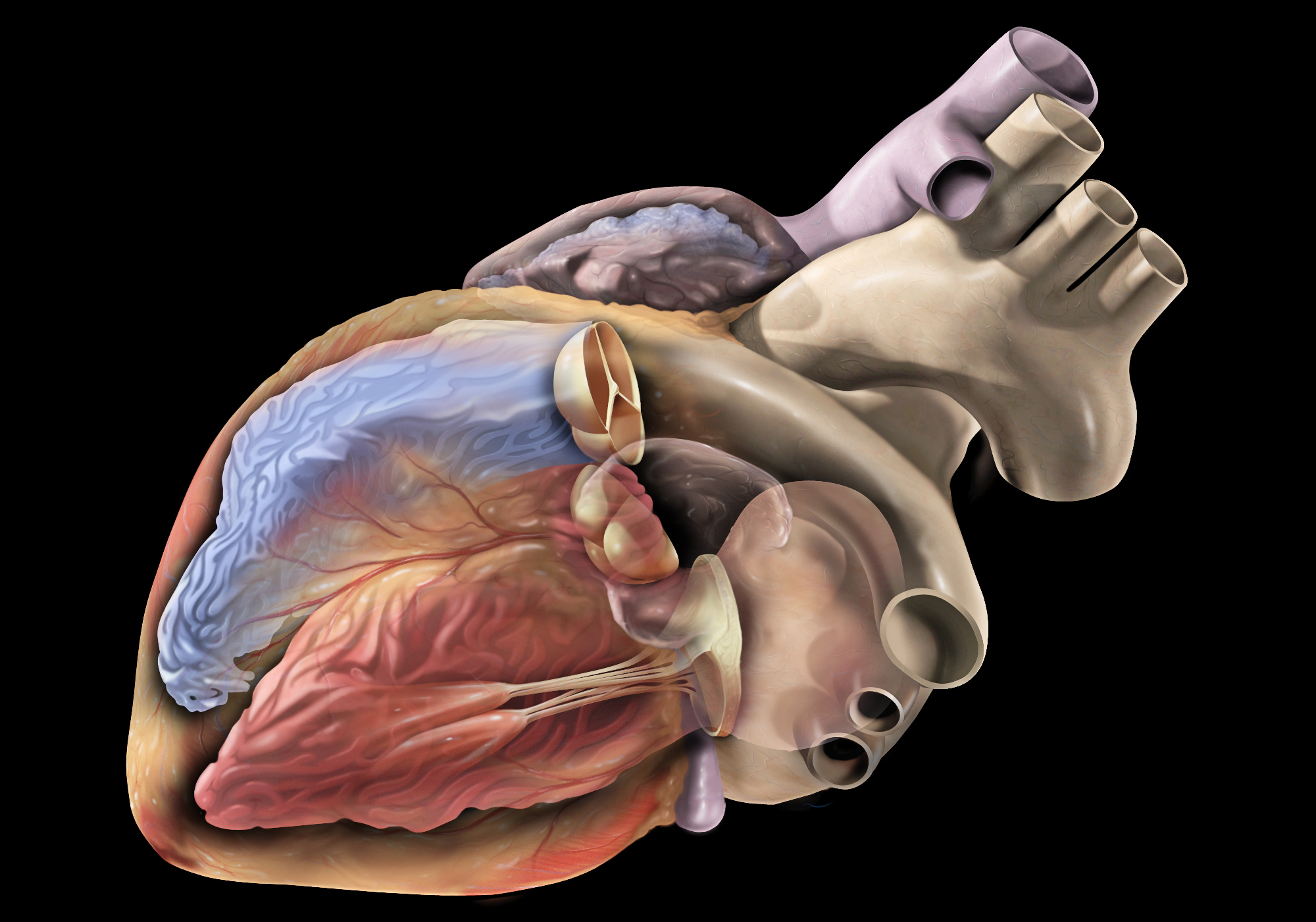
The aortic valve is the valve located between the left ventricle (the lower left chamber of the heart) and the aorta (the largest artery in the body, which carries oxygenated blood from the heart to the rest of the body). It is made up of three thin flaps or leaflets that open and close to regulate blood flow. During a heartbeat, the aortic valve opens to allow blood to be pumped out of the left ventricle into the aorta, and then closes to prevent blood from flowing back into the ventricle when it relaxes. Any abnormality or damage to this valve can lead to various cardiovascular conditions such as aortic stenosis, aortic regurgitation, or infective endocarditis.
Dietary cholesterol is a type of cholesterol that comes from the foods we eat. It is present in animal-derived products such as meat, poultry, dairy products, and eggs. While dietary cholesterol can contribute to an increase in blood cholesterol levels for some people, it's important to note that saturated and trans fats have a more significant impact on blood cholesterol levels than dietary cholesterol itself.
The American Heart Association recommends limiting dietary cholesterol intake to less than 300 milligrams per day for most people, and less than 200 milligrams per day for those with a history of heart disease or high cholesterol levels. However, individual responses to dietary cholesterol can vary, so it's essential to monitor blood cholesterol levels and adjust dietary habits accordingly.
A dose-response relationship in the context of drugs refers to the changes in the effects or symptoms that occur as the dose of a drug is increased or decreased. Generally, as the dose of a drug is increased, the severity or intensity of its effects also increases. Conversely, as the dose is decreased, the effects of the drug become less severe or may disappear altogether.
The dose-response relationship is an important concept in pharmacology and toxicology because it helps to establish the safe and effective dosage range for a drug. By understanding how changes in the dose of a drug affect its therapeutic and adverse effects, healthcare providers can optimize treatment plans for their patients while minimizing the risk of harm.
The dose-response relationship is typically depicted as a curve that shows the relationship between the dose of a drug and its effect. The shape of the curve may vary depending on the drug and the specific effect being measured. Some drugs may have a steep dose-response curve, meaning that small changes in the dose can result in large differences in the effect. Other drugs may have a more gradual dose-response curve, where larger changes in the dose are needed to produce significant effects.
In addition to helping establish safe and effective dosages, the dose-response relationship is also used to evaluate the potential therapeutic benefits and risks of new drugs during clinical trials. By systematically testing different doses of a drug in controlled studies, researchers can identify the optimal dosage range for the drug and assess its safety and efficacy.
Norepinephrine, also known as noradrenaline, is a neurotransmitter and a hormone that is primarily produced in the adrenal glands and is released into the bloodstream in response to stress or physical activity. It plays a crucial role in the "fight-or-flight" response by preparing the body for action through increasing heart rate, blood pressure, respiratory rate, and glucose availability.
As a neurotransmitter, norepinephrine is involved in regulating various functions of the nervous system, including attention, perception, motivation, and arousal. It also plays a role in modulating pain perception and responding to stressful or emotional situations.
In medical settings, norepinephrine is used as a vasopressor medication to treat hypotension (low blood pressure) that can occur during septic shock, anesthesia, or other critical illnesses. It works by constricting blood vessels and increasing heart rate, which helps to improve blood pressure and perfusion of vital organs.
Apolipoprotein E (ApoE) is a protein involved in the metabolism of lipids, particularly cholesterol. It is produced primarily by the liver and is a component of several types of lipoproteins, including very low-density lipoproteins (VLDL) and high-density lipoproteins (HDL).
ApoE plays a crucial role in the transport and uptake of lipids in the body. It binds to specific receptors on cell surfaces, facilitating the delivery of lipids to cells for energy metabolism or storage. ApoE also helps to clear cholesterol from the bloodstream and is involved in the repair and maintenance of tissues.
There are three major isoforms of ApoE, designated ApoE2, ApoE3, and ApoE4, which differ from each other by only a few amino acids. These genetic variations can have significant effects on an individual's risk for developing certain diseases, particularly cardiovascular disease and Alzheimer's disease. For example, individuals who inherit the ApoE4 allele have an increased risk of developing Alzheimer's disease, while those with the ApoE2 allele may have a reduced risk.
In summary, Apolipoprotein E is a protein involved in lipid metabolism and transport, and genetic variations in this protein can influence an individual's risk for certain diseases.
Surgical anastomosis is a medical procedure that involves the connection of two tubular structures, such as blood vessels or intestines, to create a continuous passage. This technique is commonly used in various types of surgeries, including vascular, gastrointestinal, and orthopedic procedures.
During a surgical anastomosis, the ends of the two tubular structures are carefully prepared by removing any damaged or diseased tissue. The ends are then aligned and joined together using sutures, staples, or other devices. The connection must be secure and leak-free to ensure proper function and healing.
The success of a surgical anastomosis depends on several factors, including the patient's overall health, the location and condition of the structures being joined, and the skill and experience of the surgeon. Complications such as infection, bleeding, or leakage can occur, which may require additional medical intervention or surgery.
Proper postoperative care is also essential to ensure the success of a surgical anastomosis. This may include monitoring for signs of complications, administering medications to prevent infection and promote healing, and providing adequate nutrition and hydration.
nitroprusside (ni-troe-rus-ide)
A rapid-acting vasodilator used in the management of severe hypertension, acute heart failure, and to reduce afterload in patients undergoing cardiac surgery. It is a potent arterial and venous dilator that decreases preload and afterload, thereby reducing myocardial oxygen demand. Nitroprusside is metabolized to cyanide, which must be monitored closely during therapy to prevent toxicity.
Pharmacologic class: Peripheral vasodilators
Therapeutic class: Antihypertensives, Vasodilators
Medical Categories: Cardiovascular Drugs, Hypertension Agents
Animal disease models are specialized animals, typically rodents such as mice or rats, that have been genetically engineered or exposed to certain conditions to develop symptoms and physiological changes similar to those seen in human diseases. These models are used in medical research to study the pathophysiology of diseases, identify potential therapeutic targets, test drug efficacy and safety, and understand disease mechanisms.
The genetic modifications can include knockout or knock-in mutations, transgenic expression of specific genes, or RNA interference techniques. The animals may also be exposed to environmental factors such as chemicals, radiation, or infectious agents to induce the disease state.
Examples of animal disease models include:
1. Mouse models of cancer: Genetically engineered mice that develop various types of tumors, allowing researchers to study cancer initiation, progression, and metastasis.
2. Alzheimer's disease models: Transgenic mice expressing mutant human genes associated with Alzheimer's disease, which exhibit amyloid plaque formation and cognitive decline.
3. Diabetes models: Obese and diabetic mouse strains like the NOD (non-obese diabetic) or db/db mice, used to study the development of type 1 and type 2 diabetes, respectively.
4. Cardiovascular disease models: Atherosclerosis-prone mice, such as ApoE-deficient or LDLR-deficient mice, that develop plaque buildup in their arteries when fed a high-fat diet.
5. Inflammatory bowel disease models: Mice with genetic mutations affecting intestinal barrier function and immune response, such as IL-10 knockout or SAMP1/YitFc mice, which develop colitis.
Animal disease models are essential tools in preclinical research, but it is important to recognize their limitations. Differences between species can affect the translatability of results from animal studies to human patients. Therefore, researchers must carefully consider the choice of model and interpret findings cautiously when applying them to human diseases.
Nitric Oxide Synthase Type III (NOS-III), also known as endothelial Nitric Oxide Synthase (eNOS), is an enzyme responsible for the production of nitric oxide (NO) in the endothelium, the lining of blood vessels. This enzyme catalyzes the conversion of L-arginine to L-citrulline, producing NO as a byproduct. The release of NO from eNOS plays an important role in regulating vascular tone and homeostasis, including the relaxation of smooth muscle cells in the blood vessel walls, inhibition of platelet aggregation, and modulation of immune function. Mutations or dysfunction in NOS-III can contribute to various cardiovascular diseases such as hypertension, atherosclerosis, and erectile dysfunction.
The endothelium is the thin, delicate tissue that lines the interior surface of blood vessels and lymphatic vessels. It is a single layer of cells called endothelial cells that are in contact with the blood or lymph fluid. The endothelium plays an essential role in maintaining vascular homeostasis by regulating blood flow, coagulation, platelet activation, immune function, and angiogenesis (the formation of new blood vessels). It also acts as a barrier between the vessel wall and the circulating blood or lymph fluid. Dysfunction of the endothelium has been implicated in various cardiovascular diseases, diabetes, inflammation, and cancer.
Catheterization is a medical procedure in which a catheter (a flexible tube) is inserted into the body to treat various medical conditions or for diagnostic purposes. The specific definition can vary depending on the area of medicine and the particular procedure being discussed. Here are some common types of catheterization:
1. Urinary catheterization: This involves inserting a catheter through the urethra into the bladder to drain urine. It is often performed to manage urinary retention, monitor urine output in critically ill patients, or assist with surgical procedures.
2. Cardiac catheterization: A procedure where a catheter is inserted into a blood vessel, usually in the groin or arm, and guided to the heart. This allows for various diagnostic tests and treatments, such as measuring pressures within the heart chambers, assessing blood flow, or performing angioplasty and stenting of narrowed coronary arteries.
3. Central venous catheterization: A catheter is inserted into a large vein, typically in the neck, chest, or groin, to administer medications, fluids, or nutrition, or to monitor central venous pressure.
4. Peritoneal dialysis catheterization: A catheter is placed into the abdominal cavity for individuals undergoing peritoneal dialysis, a type of kidney replacement therapy.
5. Neurological catheterization: In some cases, a catheter may be inserted into the cerebrospinal fluid space (lumbar puncture) or the brain's ventricular system (ventriculostomy) to diagnose or treat various neurological conditions.
These are just a few examples of catheterization procedures in medicine. The specific definition and purpose will depend on the medical context and the particular organ or body system involved.
Transesophageal echocardiography (TEE) is a type of echocardiogram, which is a medical test that uses sound waves to create detailed images of the heart. In TEE, a special probe containing a transducer is passed down the esophagus (the tube that connects the mouth to the stomach) to obtain views of the heart from behind. This allows for more detailed images of the heart structures and function compared to a standard echocardiogram, which uses a probe placed on the chest. TEE is often used in patients with poor image quality from a standard echocardiogram or when more detailed images are needed to diagnose or monitor certain heart conditions. It is typically performed by a trained cardiologist or sonographer under the direction of a cardiologist.
A stent is a small mesh tube that's used to treat narrow or weak arteries. Arteries are blood vessels that carry blood away from your heart to other parts of your body. A stent is placed in an artery as part of a procedure called angioplasty. Angioplasty restores blood flow through narrowed or blocked arteries by inflating a tiny balloon inside the blocked artery to widen it.
The stent is then inserted into the widened artery to keep it open. The stent is usually made of metal, but some are coated with medication that is slowly and continuously released to help prevent the formation of scar tissue in the artery. This can reduce the chance of the artery narrowing again.
Stents are also used in other parts of the body, such as the neck (carotid artery) and kidneys (renal artery), to help maintain blood flow and prevent blockages. They can also be used in the urinary system to treat conditions like ureteropelvic junction obstruction or narrowing of the urethra.
Takayasu arteritis is a rare inflammatory disease that affects the large blood vessels in the body, most commonly the aorta and its main branches. It's also known as pulseless disease or aortic arch syndrome. The condition primarily affects young to middle-aged women, although it can occur in anyone at any age.
The inflammation caused by Takayasu arteritis can lead to narrowing, thickening, and weakening of the affected blood vessels' walls, which can result in reduced blood flow to various organs and tissues. This can cause a variety of symptoms depending on the severity and location of the vessel involvement.
Common symptoms include:
* Weak or absent pulses in the arms and/or legs
* High blood pressure (hypertension)
* Dizziness, lightheadedness, or fainting spells due to reduced blood flow to the brain
* Headaches
* Visual disturbances
* Fatigue
* Weight loss
* Night sweats
* Fever
Diagnosis of Takayasu arteritis typically involves a combination of medical history, physical examination, laboratory tests, and imaging studies. Treatment usually includes corticosteroids or other immunosuppressive medications to control inflammation and maintain remission. Regular follow-up with a healthcare provider is essential to monitor disease activity and adjust treatment as necessary.
Nitric Oxide Synthase (NOS) is a group of enzymes that catalyze the production of nitric oxide (NO) from L-arginine. There are three distinct isoforms of NOS, each with different expression patterns and functions:
1. Neuronal Nitric Oxide Synthase (nNOS or NOS1): This isoform is primarily expressed in the nervous system and plays a role in neurotransmission, synaptic plasticity, and learning and memory processes.
2. Inducible Nitric Oxide Synthase (iNOS or NOS2): This isoform is induced by various stimuli such as cytokines, lipopolysaccharides, and hypoxia in a variety of cells including immune cells, endothelial cells, and smooth muscle cells. iNOS produces large amounts of NO, which functions as a potent effector molecule in the immune response, particularly in the defense against microbial pathogens.
3. Endothelial Nitric Oxide Synthase (eNOS or NOS3): This isoform is constitutively expressed in endothelial cells and produces low levels of NO that play a crucial role in maintaining vascular homeostasis by regulating vasodilation, inhibiting platelet aggregation, and preventing smooth muscle cell proliferation.
Overall, NOS plays an essential role in various physiological processes, including neurotransmission, immune response, cardiovascular function, and respiratory regulation. Dysregulation of NOS activity has been implicated in several pathological conditions such as hypertension, atherosclerosis, neurodegenerative diseases, and inflammatory disorders.
The pulmonary artery is a large blood vessel that carries deoxygenated blood from the right ventricle of the heart to the lungs for oxygenation. It divides into two main branches, the right and left pulmonary arteries, which further divide into smaller vessels called arterioles, and then into a vast network of capillaries in the lungs where gas exchange occurs. The thin walls of these capillaries allow oxygen to diffuse into the blood and carbon dioxide to diffuse out, making the blood oxygen-rich before it is pumped back to the left side of the heart through the pulmonary veins. This process is crucial for maintaining proper oxygenation of the body's tissues and organs.
In medicine, elasticity refers to the ability of a tissue or organ to return to its original shape after being stretched or deformed. This property is due to the presence of elastic fibers in the extracellular matrix of the tissue, which can stretch and recoil like rubber bands.
Elasticity is an important characteristic of many tissues, particularly those that are subjected to repeated stretching or compression, such as blood vessels, lungs, and skin. For example, the elasticity of the lungs allows them to expand and contract during breathing, while the elasticity of blood vessels helps maintain normal blood pressure by allowing them to expand and constrict in response to changes in blood flow.
In addition to its role in normal physiology, elasticity is also an important factor in the diagnosis and treatment of various medical conditions. For example, decreased elasticity in the lungs can be a sign of lung disease, while increased elasticity in the skin can be a sign of aging or certain genetic disorders. Medical professionals may use techniques such as pulmonary function tests or skin biopsies to assess elasticity and help diagnose these conditions.
The subclavian artery is a major blood vessel that supplies the upper limb and important structures in the neck and head. It arises from the brachiocephalic trunk (in the case of the right subclavian artery) or directly from the aortic arch (in the case of the left subclavian artery).
The subclavian artery has several branches, including:
1. The vertebral artery, which supplies blood to the brainstem and cerebellum.
2. The internal thoracic artery (also known as the mammary artery), which supplies blood to the chest wall, breast, and anterior mediastinum.
3. The thyrocervical trunk, which gives rise to several branches that supply the neck, including the inferior thyroid artery, the suprascapular artery, and the transverse cervical artery.
4. The costocervical trunk, which supplies blood to the neck and upper back, including the posterior chest wall and the lower neck muscles.
The subclavian artery is a critical vessel in maintaining adequate blood flow to the upper limb, and any blockage or damage to this vessel can lead to significant morbidity, including arm pain, numbness, weakness, or even loss of function.
Angiotensin II is a potent vasoactive peptide hormone that plays a critical role in the renin-angiotensin-aldosterone system (RAAS), which is a crucial regulator of blood pressure and fluid balance in the body. It is formed from angiotensin I through the action of an enzyme called angiotensin-converting enzyme (ACE).
Angiotensin II has several physiological effects on various organs, including:
1. Vasoconstriction: Angiotensin II causes contraction of vascular smooth muscle, leading to an increase in peripheral vascular resistance and blood pressure.
2. Aldosterone release: Angiotensin II stimulates the adrenal glands to release aldosterone, a hormone that promotes sodium reabsorption and potassium excretion in the kidneys, thereby increasing water retention and blood volume.
3. Sympathetic nervous system activation: Angiotensin II activates the sympathetic nervous system, leading to increased heart rate and contractility, further contributing to an increase in blood pressure.
4. Thirst regulation: Angiotensin II stimulates the hypothalamus to increase thirst, promoting water intake and helping to maintain intravascular volume.
5. Cell growth and fibrosis: Angiotensin II has been implicated in various pathological processes, such as cell growth, proliferation, and fibrosis, which can contribute to the development of cardiovascular and renal diseases.
Angiotensin-converting enzyme inhibitors (ACEIs) and angiotensin receptor blockers (ARBs) are two classes of medications commonly used in clinical practice to target the RAAS by blocking the formation or action of angiotensin II, respectively. These drugs have been shown to be effective in managing hypertension, heart failure, and chronic kidney disease.
In medical terms, constriction refers to the narrowing or tightening of a body part or passageway. This can occur due to various reasons such as spasms of muscles, inflammation, or abnormal growths. It can lead to symptoms like difficulty in breathing, swallowing, or blood flow, depending on where it occurs. For example, constriction of the airways in asthma, constriction of blood vessels in hypertension, or constriction of the esophagus in certain digestive disorders.
Potassium chloride is an essential electrolyte that is often used in medical settings as a medication. It's a white, crystalline salt that is highly soluble in water and has a salty taste. In the body, potassium chloride plays a crucial role in maintaining fluid and electrolyte balance, nerve function, and muscle contraction.
Medically, potassium chloride is commonly used to treat or prevent low potassium levels (hypokalemia) in the blood. Hypokalemia can occur due to various reasons such as certain medications, kidney diseases, vomiting, diarrhea, or excessive sweating. Potassium chloride is available in various forms, including tablets, capsules, and liquids, and it's usually taken by mouth.
It's important to note that potassium chloride should be used with caution and under the supervision of a healthcare provider, as high levels of potassium (hyperkalemia) can be harmful and even life-threatening. Hyperkalemia can cause symptoms such as muscle weakness, irregular heartbeat, and cardiac arrest.
Cyclic guanosine monophosphate (cGMP) is a important second messenger molecule that plays a crucial role in various biological processes within the human body. It is synthesized from guanosine triphosphate (GTP) by the enzyme guanylyl cyclase.
Cyclic GMP is involved in regulating diverse physiological functions, such as smooth muscle relaxation, cardiovascular function, and neurotransmission. It also plays a role in modulating immune responses and cellular growth and differentiation.
In the medical field, changes in cGMP levels or dysregulation of cGMP-dependent pathways have been implicated in various disease states, including pulmonary hypertension, heart failure, erectile dysfunction, and glaucoma. Therefore, pharmacological agents that target cGMP signaling are being developed as potential therapeutic options for these conditions.
The mesenteric arteries are the arteries that supply oxygenated blood to the intestines. There are three main mesenteric arteries: the superior mesenteric artery, which supplies blood to the small intestine (duodenum to two-thirds of the transverse colon) and large intestine (cecum, ascending colon, and the first part of the transverse colon); the inferior mesenteric artery, which supplies blood to the distal third of the transverse colon, descending colon, sigmoid colon, and rectum; and the middle colic artery, which is a branch of the superior mesenteric artery that supplies blood to the transverse colon. These arteries are important in maintaining adequate blood flow to the intestines to support digestion and absorption of nutrients.
Elastic tissue is a type of connective tissue found in the body that is capable of returning to its original shape after being stretched or deformed. It is composed mainly of elastin fibers, which are protein molecules with a unique structure that allows them to stretch and recoil. Elastic tissue is found in many areas of the body, including the lungs, blood vessels, and skin, where it provides flexibility and resilience.
The elastin fibers in elastic tissue are intertwined with other types of connective tissue fibers, such as collagen, which provide strength and support. The combination of these fibers allows elastic tissue to stretch and recoil efficiently, enabling organs and tissues to function properly. For example, the elasticity of lung tissue allows the lungs to expand and contract during breathing, while the elasticity of blood vessels helps maintain blood flow and pressure.
Elastic tissue can become less flexible and resilient with age or due to certain medical conditions, such as emphysema or Marfan syndrome. This can lead to a variety of health problems, including respiratory difficulties, cardiovascular disease, and skin sagging.
The brachiocephalic trunk, also known as the brachiocephalic artery or innominate artery, is a large vessel that branches off the aorta and divides into the right common carotid artery and the right subclavian artery. It supplies blood to the head, neck, and arms on the right side of the body.
Treatment outcome is a term used to describe the result or effect of medical treatment on a patient's health status. It can be measured in various ways, such as through symptoms improvement, disease remission, reduced disability, improved quality of life, or survival rates. The treatment outcome helps healthcare providers evaluate the effectiveness of a particular treatment plan and make informed decisions about future care. It is also used in clinical research to compare the efficacy of different treatments and improve patient care.
C57BL/6 (C57 Black 6) is an inbred strain of laboratory mouse that is widely used in biomedical research. The term "inbred" refers to a strain of animals where matings have been carried out between siblings or other closely related individuals for many generations, resulting in a population that is highly homozygous at most genetic loci.
The C57BL/6 strain was established in 1920 by crossing a female mouse from the dilute brown (DBA) strain with a male mouse from the black strain. The resulting offspring were then interbred for many generations to create the inbred C57BL/6 strain.
C57BL/6 mice are known for their robust health, longevity, and ease of handling, making them a popular choice for researchers. They have been used in a wide range of biomedical research areas, including studies of cancer, immunology, neuroscience, cardiovascular disease, and metabolism.
One of the most notable features of the C57BL/6 strain is its sensitivity to certain genetic modifications, such as the introduction of mutations that lead to obesity or impaired glucose tolerance. This has made it a valuable tool for studying the genetic basis of complex diseases and traits.
Overall, the C57BL/6 inbred mouse strain is an important model organism in biomedical research, providing a valuable resource for understanding the genetic and molecular mechanisms underlying human health and disease.
Calcinosis is a medical condition characterized by the abnormal deposit of calcium salts in various tissues of the body, commonly under the skin or in the muscles and tendons. These calcium deposits can form hard lumps or nodules that can cause pain, inflammation, and restricted mobility. Calcinosis can occur as a complication of other medical conditions, such as autoimmune disorders, kidney disease, and hypercalcemia (high levels of calcium in the blood). In some cases, the cause of calcinosis may be unknown. Treatment for calcinosis depends on the underlying cause and may include medications to manage calcium levels, physical therapy, and surgical removal of large deposits.
The femoral artery is the major blood vessel that supplies oxygenated blood to the lower extremity of the human body. It is a continuation of the external iliac artery and becomes the popliteal artery as it passes through the adductor hiatus in the adductor magnus muscle of the thigh.
The femoral artery is located in the femoral triangle, which is bound by the sartorius muscle anteriorly, the adductor longus muscle medially, and the biceps femoris muscle posteriorly. It can be easily palpated in the groin region, making it a common site for taking blood samples, measuring blood pressure, and performing surgical procedures such as femoral artery catheterization and bypass grafting.
The femoral artery gives off several branches that supply blood to the lower limb, including the deep femoral artery, the superficial femoral artery, and the profunda femoris artery. These branches provide blood to the muscles, bones, skin, and other tissues of the leg, ankle, and foot.
NG-Nitroarginine Methyl Ester (L-NAME) is not a medication, but rather a research chemical used in scientific studies. It is an inhibitor of nitric oxide synthase, an enzyme that synthesizes nitric oxide, a molecule involved in the relaxation of blood vessels.
Therefore, L-NAME is often used in experiments to investigate the role of nitric oxide in various physiological and pathophysiological processes. It is important to note that the use of L-NAME in humans is not approved for therapeutic purposes due to its potential side effects, which can include hypertension, decreased renal function, and decreased cerebral blood flow.
Prosthesis design is a specialized field in medical device technology that involves creating and developing artificial substitutes to replace a missing body part, such as a limb, tooth, eye, or internal organ. The design process typically includes several stages: assessment of the patient's needs, selection of appropriate materials, creation of a prototype, testing and refinement, and final fabrication and fitting of the prosthesis.
The goal of prosthesis design is to create a device that functions as closely as possible to the natural body part it replaces, while also being comfortable, durable, and aesthetically pleasing for the patient. The design process may involve collaboration between medical professionals, engineers, and designers, and may take into account factors such as the patient's age, lifestyle, occupation, and overall health.
Prosthesis design can be highly complex, particularly for advanced devices such as robotic limbs or implantable organs. These devices often require sophisticated sensors, actuators, and control systems to mimic the natural functions of the body part they replace. As a result, prosthesis design is an active area of research and development in the medical field, with ongoing efforts to improve the functionality, comfort, and affordability of these devices for patients.
Vascular surgical procedures are operations that are performed to treat conditions and diseases related to the vascular system, which includes the arteries, veins, and capillaries. These procedures can be invasive or minimally invasive and are often used to treat conditions such as peripheral artery disease, carotid artery stenosis, aortic aneurysms, and venous insufficiency.
Some examples of vascular surgical procedures include:
* Endarterectomy: a procedure to remove plaque buildup from the inside of an artery
* Bypass surgery: creating a new path for blood to flow around a blocked or narrowed artery
* Angioplasty and stenting: using a balloon to open a narrowed artery and placing a stent to keep it open
* Aneurysm repair: surgically repairing an aneurysm, a weakened area in the wall of an artery that has bulged out and filled with blood
* Embolectomy: removing a blood clot from a blood vessel
* Thrombectomy: removing a blood clot from a vein
These procedures are typically performed by vascular surgeons, who are trained in the diagnosis and treatment of vascular diseases.
Aortic valve insufficiency, also known as aortic regurgitation or aortic incompetence, is a cardiac condition in which the aortic valve does not close properly during the contraction phase of the heart cycle. This allows blood to flow back into the left ventricle from the aorta, instead of being pumped out to the rest of the body. As a result, the left ventricle must work harder to maintain adequate cardiac output, which can lead to left ventricular enlargement and heart failure over time if left untreated.
The aortic valve is a trileaflet valve that lies between the left ventricle and the aorta. During systole (the contraction phase of the heart cycle), the aortic valve opens to allow blood to be pumped out of the left ventricle into the aorta and then distributed to the rest of the body. During diastole (the relaxation phase of the heart cycle), the aortic valve closes to prevent blood from flowing back into the left ventricle.
Aortic valve insufficiency can be caused by various conditions, including congenital heart defects, infective endocarditis, rheumatic heart disease, Marfan syndrome, and trauma. Symptoms of aortic valve insufficiency may include shortness of breath, fatigue, chest pain, palpitations, and edema (swelling). Diagnosis is typically made through physical examination, echocardiography, and other imaging studies. Treatment options depend on the severity of the condition and may include medication, surgery to repair or replace the aortic valve, or a combination of both.
Cholesterol is a type of lipid (fat) molecule that is an essential component of cell membranes and is also used to make certain hormones and vitamins in the body. It is produced by the liver and is also obtained from animal-derived foods such as meat, dairy products, and eggs.
Cholesterol does not mix with blood, so it is transported through the bloodstream by lipoproteins, which are particles made up of both lipids and proteins. There are two main types of lipoproteins that carry cholesterol: low-density lipoproteins (LDL), also known as "bad" cholesterol, and high-density lipoproteins (HDL), also known as "good" cholesterol.
High levels of LDL cholesterol in the blood can lead to a buildup of cholesterol in the walls of the arteries, increasing the risk of heart disease and stroke. On the other hand, high levels of HDL cholesterol are associated with a lower risk of these conditions because HDL helps remove LDL cholesterol from the bloodstream and transport it back to the liver for disposal.
It is important to maintain healthy levels of cholesterol through a balanced diet, regular exercise, and sometimes medication if necessary. Regular screening is also recommended to monitor cholesterol levels and prevent health complications.
Blood vessels are the part of the circulatory system that transport blood throughout the body. They form a network of tubes that carry blood to and from the heart, lungs, and other organs. The main types of blood vessels are arteries, veins, and capillaries. Arteries carry oxygenated blood away from the heart to the rest of the body, while veins return deoxygenated blood back to the heart. Capillaries connect arteries and veins and facilitate the exchange of oxygen, nutrients, and waste materials between the blood and the body's tissues.
"Cells, cultured" is a medical term that refers to cells that have been removed from an organism and grown in controlled laboratory conditions outside of the body. This process is called cell culture and it allows scientists to study cells in a more controlled and accessible environment than they would have inside the body. Cultured cells can be derived from a variety of sources, including tissues, organs, or fluids from humans, animals, or cell lines that have been previously established in the laboratory.
Cell culture involves several steps, including isolation of the cells from the tissue, purification and characterization of the cells, and maintenance of the cells in appropriate growth conditions. The cells are typically grown in specialized media that contain nutrients, growth factors, and other components necessary for their survival and proliferation. Cultured cells can be used for a variety of purposes, including basic research, drug development and testing, and production of biological products such as vaccines and gene therapies.
It is important to note that cultured cells may behave differently than they do in the body, and results obtained from cell culture studies may not always translate directly to human physiology or disease. Therefore, it is essential to validate findings from cell culture experiments using additional models and ultimately in clinical trials involving human subjects.
Messenger RNA (mRNA) is a type of RNA (ribonucleic acid) that carries genetic information copied from DNA in the form of a series of three-base code "words," each of which specifies a particular amino acid. This information is used by the cell's machinery to construct proteins, a process known as translation. After being transcribed from DNA, mRNA travels out of the nucleus to the ribosomes in the cytoplasm where protein synthesis occurs. Once the protein has been synthesized, the mRNA may be degraded and recycled. Post-transcriptional modifications can also occur to mRNA, such as alternative splicing and addition of a 5' cap and a poly(A) tail, which can affect its stability, localization, and translation efficiency.
Aortic arch syndromes are a group of conditions that affect the aortic arch, which is the curved portion of the aorta that arises from the left ventricle of the heart and gives rise to the major branches of the arterial system. These syndromes are typically caused by congenital abnormalities or degenerative changes in the aorta and can result in various complications, such as obstruction of blood flow, aneurysm formation, and dissection.
There are several types of aortic arch syndromes, including:
1. Coarctation of the Aorta: This is a narrowing of the aorta at the point where it leaves the heart, just distal to the origin of the left subclavian artery. It can cause hypertension in the upper extremities and reduced blood flow to the lower extremities.
2. Aortic Arch Aneurysm: This is a localized dilation or bulging of the aorta in the region of the aortic arch. It can lead to dissection, rupture, or embolism.
3. Aortic Arch Dissection: This is a separation of the layers of the aortic wall, which can result from hypertension, trauma, or genetic disorders such as Marfan syndrome. It can cause severe chest pain, shortness of breath, and shock.
4. Kommerell's Diverticulum: This is an outpouching or bulge in the aorta at the origin of the ligamentum arteriosum, which is a remnant of the ductus arteriosus. It can cause compression of the airways or esophagus and increase the risk of dissection or rupture.
5. Abernethy Malformation: This is a rare congenital anomaly in which there is an abnormal connection between the portal vein and systemic venous circulation, leading to the bypass of the liver. It can cause various complications such as hepatic encephalopathy, pulmonary hypertension, and liver tumors.
The diagnosis and management of aortic arch syndromes require a multidisciplinary approach involving cardiologists, radiologists, surgeons, and other specialists. Treatment options may include medications, endovascular procedures, or surgical interventions depending on the severity and location of the lesion.
Methylene Blue is a heterocyclic aromatic organic compound with the molecular formula C16H18ClN3S. It is primarily used as a medication, but can also be used as a dye or as a chemical reagent. As a medication, it is used in the treatment of methemoglobinemia (a condition where an abnormal amount of methemoglobin is present in the blood), as well as in some forms of poisoning and infections. It works by acting as a reducing agent, converting methemoglobin back to hemoglobin, which is the form of the protein that is responsible for carrying oxygen in the blood. Methylene Blue has also been used off-label for other conditions, such as vasculitis and Alzheimer's disease, although its effectiveness for these uses is not well established.
It is important to note that Methylene Blue should be used with caution, as it can cause serious side effects in some people, particularly those with kidney or liver problems, or those who are taking certain medications. It is also important to follow the instructions of a healthcare provider when using this medication, as improper use can lead to toxicity.
A "knockout" mouse is a genetically engineered mouse in which one or more genes have been deleted or "knocked out" using molecular biology techniques. This allows researchers to study the function of specific genes and their role in various biological processes, as well as potential associations with human diseases. The mice are generated by introducing targeted DNA modifications into embryonic stem cells, which are then used to create a live animal. Knockout mice have been widely used in biomedical research to investigate gene function, disease mechanisms, and potential therapeutic targets.
The carotid arteries are a pair of vital blood vessels in the human body that supply oxygenated blood to the head and neck. Each person has two common carotid arteries, one on each side of the neck, which branch off from the aorta, the largest artery in the body.
The right common carotid artery originates from the brachiocephalic trunk, while the left common carotid artery arises directly from the aortic arch. As they ascend through the neck, they split into two main branches: the internal and external carotid arteries.
The internal carotid artery supplies oxygenated blood to the brain, eyes, and other structures within the skull, while the external carotid artery provides blood to the face, scalp, and various regions of the neck.
Maintaining healthy carotid arteries is crucial for overall cardiovascular health and preventing serious conditions like stroke, which can occur when the arteries become narrowed or blocked due to the buildup of plaque or fatty deposits (atherosclerosis). Regular check-ups with healthcare professionals may include monitoring carotid artery health through ultrasound or other imaging techniques.
The renal artery is a pair of blood vessels that originate from the abdominal aorta and supply oxygenated blood to each kidney. These arteries branch into several smaller vessels that provide blood to the various parts of the kidneys, including the renal cortex and medulla. The renal arteries also carry nutrients and other essential components needed for the normal functioning of the kidneys. Any damage or blockage to the renal artery can lead to serious consequences, such as reduced kidney function or even kidney failure.
Blood flow velocity is the speed at which blood travels through a specific part of the vascular system. It is typically measured in units of distance per time, such as centimeters per second (cm/s) or meters per second (m/s). Blood flow velocity can be affected by various factors, including cardiac output, vessel diameter, and viscosity of the blood. Measuring blood flow velocity is important in diagnosing and monitoring various medical conditions, such as heart disease, stroke, and peripheral vascular disease.
Cardiovascular models are simplified representations or simulations of the human cardiovascular system used in medical research, education, and training. These models can be physical, computational, or mathematical and are designed to replicate various aspects of the heart, blood vessels, and blood flow. They can help researchers study the structure and function of the cardiovascular system, test new treatments and interventions, and train healthcare professionals in diagnostic and therapeutic techniques.
Physical cardiovascular models may include artificial hearts, blood vessels, or circulation systems made from materials such as plastic, rubber, or silicone. These models can be used to study the mechanics of heart valves, the effects of different surgical procedures, or the impact of various medical devices on blood flow.
Computational and mathematical cardiovascular models use algorithms and equations to simulate the behavior of the cardiovascular system. These models may range from simple representations of a single heart chamber to complex simulations of the entire circulatory system. They can be used to study the electrical activity of the heart, the biomechanics of blood flow, or the distribution of drugs in the body.
Overall, cardiovascular models play an essential role in advancing our understanding of the human body and improving patient care.
Hemodynamics is the study of how blood flows through the cardiovascular system, including the heart and the vascular network. It examines various factors that affect blood flow, such as blood volume, viscosity, vessel length and diameter, and pressure differences between different parts of the circulatory system. Hemodynamics also considers the impact of various physiological and pathological conditions on these variables, and how they in turn influence the function of vital organs and systems in the body. It is a critical area of study in fields such as cardiology, anesthesiology, and critical care medicine.
"Inbred strains of rats" are genetically identical rodents that have been produced through many generations of brother-sister mating. This results in a high degree of homozygosity, where the genes at any particular locus in the genome are identical in all members of the strain.
Inbred strains of rats are widely used in biomedical research because they provide a consistent and reproducible genetic background for studying various biological phenomena, including the effects of drugs, environmental factors, and genetic mutations on health and disease. Additionally, inbred strains can be used to create genetically modified models of human diseases by introducing specific mutations into their genomes.
Some commonly used inbred strains of rats include the Wistar Kyoto (WKY), Sprague-Dawley (SD), and Fischer 344 (F344) rat strains. Each strain has its own unique genetic characteristics, making them suitable for different types of research.
Hypercholesterolemia is a medical term that describes a condition characterized by high levels of cholesterol in the blood. Specifically, it refers to an abnormally elevated level of low-density lipoprotein (LDL) cholesterol, also known as "bad" cholesterol, which can contribute to the development of fatty deposits in the arteries called plaques. Over time, these plaques can narrow and harden the arteries, leading to atherosclerosis, a condition that increases the risk of heart disease, stroke, and other cardiovascular complications.
Hypercholesterolemia can be caused by various factors, including genetics, lifestyle choices, and underlying medical conditions. In some cases, it may not cause any symptoms until serious complications arise. Therefore, regular cholesterol screening is essential for early detection and management of hypercholesterolemia. Treatment typically involves lifestyle modifications, such as a healthy diet, regular exercise, and weight management, along with medication if necessary.
Tunica intima, also known as the intima layer, is the innermost layer of a blood vessel, including arteries and veins. It is in direct contact with the flowing blood and is composed of simple squamous endothelial cells that form a continuous, non-keratinized, stratified epithelium. These cells play a crucial role in maintaining vascular homeostasis by regulating the passage of molecules and immune cells between the blood and the vessel wall, as well as contributing to the maintenance of blood fluidity and preventing coagulation.
The tunica intima is supported by a thin layer of connective tissue called the basement membrane, which provides structural stability and anchorage for the endothelial cells. Beneath the basement membrane lies a loose network of elastic fibers and collagen, known as the internal elastic lamina, that separates the tunica intima from the middle layer, or tunica media.
In summary, the tunica intima is the innermost layer of blood vessels, primarily composed of endothelial cells and a basement membrane, which regulates various functions to maintain vascular homeostasis.
Calcium is an essential mineral that is vital for various physiological processes in the human body. The medical definition of calcium is as follows:
Calcium (Ca2+) is a crucial cation and the most abundant mineral in the human body, with approximately 99% of it found in bones and teeth. It plays a vital role in maintaining structural integrity, nerve impulse transmission, muscle contraction, hormonal secretion, blood coagulation, and enzyme activation.
Calcium homeostasis is tightly regulated through the interplay of several hormones, including parathyroid hormone (PTH), calcitonin, and vitamin D. Dietary calcium intake, absorption, and excretion are also critical factors in maintaining optimal calcium levels in the body.
Hypocalcemia refers to low serum calcium levels, while hypercalcemia indicates high serum calcium levels. Both conditions can have detrimental effects on various organ systems and require medical intervention to correct.
Aortic valve stenosis is a cardiac condition characterized by the narrowing or stiffening of the aortic valve, which separates the left ventricle (the heart's main pumping chamber) from the aorta (the large artery that carries oxygen-rich blood to the rest of the body). This narrowing or stiffening prevents the aortic valve from opening fully, resulting in reduced blood flow from the left ventricle to the aorta and the rest of the body.
The narrowing can be caused by several factors, including congenital heart defects, calcification (hardening) of the aortic valve due to aging, or scarring of the valve due to rheumatic fever or other inflammatory conditions. As a result, the left ventricle must work harder to pump blood through the narrowed valve, which can lead to thickening and enlargement of the left ventricular muscle (left ventricular hypertrophy).
Symptoms of aortic valve stenosis may include chest pain or tightness, shortness of breath, fatigue, dizziness or fainting, and heart palpitations. Severe aortic valve stenosis can lead to serious complications such as heart failure, arrhythmias, or even sudden cardiac death. Treatment options may include medications to manage symptoms, lifestyle changes, or surgical intervention such as aortic valve replacement.
Congenital heart defects (CHDs) are structural abnormalities in the heart that are present at birth. They can affect any part of the heart's structure, including the walls of the heart, the valves inside the heart, and the major blood vessels that lead to and from the heart.
Congenital heart defects can range from mild to severe and can cause various symptoms depending on the type and severity of the defect. Some common symptoms of CHDs include cyanosis (a bluish tint to the skin, lips, and fingernails), shortness of breath, fatigue, poor feeding, and slow growth in infants and children.
There are many different types of congenital heart defects, including:
1. Septal defects: These are holes in the walls that separate the four chambers of the heart. The two most common septal defects are atrial septal defect (ASD) and ventricular septal defect (VSD).
2. Valve abnormalities: These include narrowed or leaky valves, which can affect blood flow through the heart.
3. Obstruction defects: These occur when blood flow is blocked or restricted due to narrowing or absence of a part of the heart's structure. Examples include pulmonary stenosis and coarctation of the aorta.
4. Cyanotic heart defects: These cause a lack of oxygen in the blood, leading to cyanosis. Examples include tetralogy of Fallot and transposition of the great arteries.
The causes of congenital heart defects are not fully understood, but genetic factors and environmental influences during pregnancy may play a role. Some CHDs can be detected before birth through prenatal testing, while others may not be diagnosed until after birth or later in childhood. Treatment for CHDs may include medication, surgery, or other interventions to improve blood flow and oxygenation of the body's tissues.
The celiac artery, also known as the anterior abdominal aortic trunk, is a major artery that originates from the abdominal aorta and supplies oxygenated blood to the foregut, which includes the stomach, liver, spleen, pancreas, and upper part of the duodenum. It branches into three main branches: the left gastric artery, the splenic artery, and the common hepatic artery. The celiac artery plays a crucial role in providing blood to these vital organs, and any disruption or damage to it can lead to serious health consequences.
Smooth muscle myocytes are specialized cells that make up the contractile portion of non-striated, or smooth, muscles. These muscles are found in various organs and structures throughout the body, including the walls of blood vessels, the digestive system, the respiratory system, and the reproductive system.
Smooth muscle myocytes are smaller than their striated counterparts (skeletal and cardiac muscle cells) and have a single nucleus. They lack the distinctive banding pattern seen in striated muscles and instead have a uniform appearance of actin and myosin filaments. Smooth muscle myocytes are controlled by the autonomic nervous system, which allows them to contract and relax involuntarily.
These cells play an essential role in many physiological processes, such as regulating blood flow, moving food through the digestive tract, and facilitating childbirth. They can also contribute to various pathological conditions, including hypertension, atherosclerosis, and gastrointestinal disorders.
Indomethacin is a non-steroidal anti-inflammatory drug (NSAID) that is commonly used to reduce pain, inflammation, and fever. It works by inhibiting the activity of certain enzymes in the body, including cyclooxygenase (COX), which plays a role in producing prostaglandins, chemicals involved in the inflammatory response.
Indomethacin is available in various forms, such as capsules, suppositories, and injectable solutions, and is used to treat a wide range of conditions, including rheumatoid arthritis, osteoarthritis, ankylosing spondylitis, gout, and bursitis. It may also be used to relieve pain and reduce fever in other conditions, such as dental procedures or after surgery.
Like all NSAIDs, indomethacin can have side effects, including stomach ulcers, bleeding, and kidney damage, especially when taken at high doses or for long periods of time. It may also increase the risk of heart attack and stroke. Therefore, it is important to use indomethacin only as directed by a healthcare provider and to report any unusual symptoms or side effects promptly.
"Swine" is a common term used to refer to even-toed ungulates of the family Suidae, including domestic pigs and wild boars. However, in a medical context, "swine" often appears in the phrase "swine flu," which is a strain of influenza virus that typically infects pigs but can also cause illness in humans. The 2009 H1N1 pandemic was caused by a new strain of swine-origin influenza A virus, which was commonly referred to as "swine flu." It's important to note that this virus is not transmitted through eating cooked pork products; it spreads from person to person, mainly through respiratory droplets produced when an infected person coughs or sneezes.
Angiography is a medical procedure in which an x-ray image is taken to visualize the internal structure of blood vessels, arteries, or veins. This is done by injecting a radiopaque contrast agent (dye) into the blood vessel using a thin, flexible catheter. The dye makes the blood vessels visible on an x-ray image, allowing doctors to diagnose and treat various medical conditions such as blockages, narrowing, or malformations of the blood vessels.
There are several types of angiography, including:
* Cardiac angiography (also called coronary angiography) - used to examine the blood vessels of the heart
* Cerebral angiography - used to examine the blood vessels of the brain
* Peripheral angiography - used to examine the blood vessels in the limbs or other parts of the body.
Angiography is typically performed by a radiologist, cardiologist, or vascular surgeon in a hospital setting. It can help diagnose conditions such as coronary artery disease, aneurysms, and peripheral arterial disease, among others.
A medical definition of an ulcer is:
A lesion on the skin or mucous membrane characterized by disintegration of surface epithelium, inflammation, and is associated with the loss of substance below the normal lining. Gastric ulcers and duodenal ulcers are types of peptic ulcers that occur in the gastrointestinal tract.
Another type of ulcer is a venous ulcer, which occurs when there is reduced blood flow from vein insufficiency, usually in the lower leg. This can cause skin damage and lead to an open sore or ulcer.
There are other types of ulcers as well, including decubitus ulcers (also known as pressure sores or bedsores), which are caused by prolonged pressure on the skin.
Paraplegia is a medical condition characterized by partial or complete loss of motor function and sensation in the lower extremities, typically affecting both legs. This results from damage to the spinal cord, often due to trauma such as accidents, falls, or gunshot wounds, or from diseases like spina bifida, polio, or tumors. The specific area and extent of the injury on the spinal cord determine the severity and location of paralysis. Individuals with paraplegia may require assistive devices for mobility, such as wheelchairs, and may face various health challenges, including pressure sores, urinary tract infections, and chronic pain.
Pulsatile flow is a type of fluid flow that occurs in a rhythmic, wave-like pattern, typically seen in the cardiovascular system. It refers to the periodic variation in the volume or velocity of a fluid (such as blood) that is caused by the regular beating of the heart. In pulsatile flow, there are periods of high flow followed by periods of low or no flow, which creates a distinct pattern on a graph or tracing. This type of flow is important for maintaining proper function and health in organs and tissues throughout the body.
The Ductus Arteriosus is a fetal blood vessel that connects the pulmonary trunk (the artery that carries blood from the heart to the lungs) and the aorta (the largest artery in the body, which carries oxygenated blood from the heart to the rest of the body). This vessel allows most of the blood from the right ventricle of the fetal heart to bypass the lungs, as the fetus receives oxygen through the placenta rather than breathing air.
After birth, with the first breaths, the blood oxygen level increases and the pressure in the lungs rises. As a result, the circulation in the newborn's body changes, and the Ductus Arteriosus is no longer needed. Within the first few days or weeks of life, this vessel usually closes spontaneously, turning into a fibrous cord called the Ligamentum Arteriosum.
Persistent Patency of the Ductus Arteriosus (PDA) occurs when the Ductus Arteriosus does not close after birth, which can lead to various complications such as heart failure and pulmonary hypertension. This condition is often seen in premature infants and may require medical intervention or surgical closure of the vessel.
Deep hypothermic circulatory arrest (DHCA) is a medical procedure in which the body temperature is lowered to around 15-20°C (59-68°F), and the circulation of blood is temporarily stopped. This technique is often used during complex cardiac surgeries, such as aortic arch reconstruction or repair of congenital heart defects, to reduce the body's metabolic demand for oxygen and allow surgeons to operate in a still and bloodless field.
During DHCA, the patient is connected to a heart-lung machine that takes over the function of pumping blood and oxygenating it. The blood is then cooled down using a cooling device before being returned to the body. Once the body temperature reaches the desired level, the circulation is stopped for a short period, usually no more than 30 minutes, during which time the surgeon can work on the heart or great vessels.
After the surgical procedure is complete, the patient is gradually rewarmed, and the circulation is restarted. DHCA carries some risks, including neurological complications such as stroke, cognitive impairment, or delirium, but it remains an important tool in complex cardiac surgery.
Endovascular procedures are minimally invasive medical treatments that involve accessing and repairing blood vessels or other interior parts of the body through small incisions or punctures. These procedures typically use specialized catheters, wires, and other tools that are inserted into the body through an artery or vein, usually in the leg or arm.
Endovascular procedures can be used to treat a wide range of conditions, including aneurysms, atherosclerosis, peripheral artery disease, carotid artery stenosis, and other vascular disorders. Some common endovascular procedures include angioplasty, stenting, embolization, and thrombectomy.
The benefits of endovascular procedures over traditional open surgery include smaller incisions, reduced trauma to surrounding tissues, faster recovery times, and lower risks of complications such as infection and bleeding. However, endovascular procedures may not be appropriate for all patients or conditions, and careful evaluation and consideration are necessary to determine the best treatment approach.
A reoperation is a surgical procedure that is performed again on a patient who has already undergone a previous operation for the same or related condition. Reoperations may be required due to various reasons, such as inadequate initial treatment, disease recurrence, infection, or complications from the first surgery. The nature and complexity of a reoperation can vary widely depending on the specific circumstances, but it often carries higher risks and potential complications compared to the original operation.
Spinal cord ischemia refers to a reduction or interruption of blood flow to the spinal cord, leading to insufficient oxygen and nutrient supply. This condition can cause damage to the spinal cord tissue, potentially resulting in neurological deficits, such as muscle weakness, sensory loss, or autonomic dysfunction. Spinal cord ischemia may be caused by various factors, including atherosclerosis, embolism, spinal artery stenosis, or complications during surgery. The severity and extent of the neurological impairment depend on the duration and location of the ischemic event in the spinal cord.
Thoracotomy is a surgical procedure that involves making an incision on the chest wall to gain access to the thoracic cavity, which contains the lungs, heart, esophagus, trachea, and other vital organs. The incision can be made on the side (lateral thoracotomy), back (posterolateral thoracotomy), or front (median sternotomy) of the chest wall, depending on the specific surgical indication.
Thoracotomy is performed for various indications, including lung biopsy, lung resection, esophagectomy, heart surgery, and mediastinal mass removal. The procedure allows the surgeon to directly visualize and access the organs within the thoracic cavity, perform necessary procedures, and control bleeding if needed.
After the procedure, the incision is typically closed with sutures or staples, and a chest tube may be placed to drain any accumulated fluid or air from the pleural space around the lungs. The patient will require postoperative care and monitoring in a hospital setting until their condition stabilizes.
Desmosine is a unique amino acid that is not found in proteins, but instead is formed through the cross-linking of lysine residues in collagen and elastin fibers. These fibers are important components of the extracellular matrix, providing strength and elasticity to tissues such as skin, lungs, and blood vessels.
Desmosine is formed through a series of chemical reactions involving the oxidation of lysine residues and their subsequent condensation with other amino acids. This process creates cross-links between collagen and elastin fibers, which helps to stabilize their structure and enhance their mechanical properties.
Abnormalities in desmosine levels have been associated with various diseases, including emphysema, Marfan syndrome, and Ehlers-Danlos syndrome. Measuring desmosine levels in urine or tissue samples can provide valuable insights into the health of collagen and elastin fibers and help diagnose and monitor these conditions.
I believe there might be a misunderstanding in your question. "Dogs" is not a medical term or condition. It is the common name for a domesticated carnivore of the family Canidae, specifically the genus Canis, which includes wolves, foxes, and other extant and extinct species of mammals. Dogs are often kept as pets and companions, and they have been bred in a wide variety of forms and sizes for different purposes, such as hunting, herding, guarding, assisting police and military forces, and providing companionship and emotional support.
If you meant to ask about a specific medical condition or term related to dogs, please provide more context so I can give you an accurate answer.
Mechanical stress, in the context of physiology and medicine, refers to any type of force that is applied to body tissues or organs, which can cause deformation or displacement of those structures. Mechanical stress can be either external, such as forces exerted on the body during physical activity or trauma, or internal, such as the pressure changes that occur within blood vessels or other hollow organs.
Mechanical stress can have a variety of effects on the body, depending on the type, duration, and magnitude of the force applied. For example, prolonged exposure to mechanical stress can lead to tissue damage, inflammation, and chronic pain. Additionally, abnormal or excessive mechanical stress can contribute to the development of various musculoskeletal disorders, such as tendinitis, osteoarthritis, and herniated discs.
In order to mitigate the negative effects of mechanical stress, the body has a number of adaptive responses that help to distribute forces more evenly across tissues and maintain structural integrity. These responses include changes in muscle tone, joint positioning, and connective tissue stiffness, as well as the remodeling of bone and other tissues over time. However, when these adaptive mechanisms are overwhelmed or impaired, mechanical stress can become a significant factor in the development of various pathological conditions.
Enzyme inhibitors are substances that bind to an enzyme and decrease its activity, preventing it from catalyzing a chemical reaction in the body. They can work by several mechanisms, including blocking the active site where the substrate binds, or binding to another site on the enzyme to change its shape and prevent substrate binding. Enzyme inhibitors are often used as drugs to treat various medical conditions, such as high blood pressure, abnormal heart rhythms, and bacterial infections. They can also be found naturally in some foods and plants, and can be used in research to understand enzyme function and regulation.
Thromboxane receptors are a type of G protein-coupled receptor that binds thromboxane A2 (TXA2), a powerful inflammatory mediator and vasoconstrictor synthesized in the body from arachidonic acid. These receptors play a crucial role in various physiological processes, including platelet aggregation, smooth muscle contraction, and modulation of immune responses.
There are two main types of thromboxane receptors: TPα and TPβ. The TPα receptor is primarily found on platelets and vascular smooth muscle cells, while the TPβ receptor is expressed in various tissues such as the kidney, lung, and brain. Activation of these receptors by thromboxane A2 leads to a variety of cellular responses, including platelet activation and aggregation, vasoconstriction, and inflammation.
Abnormalities in thromboxane receptor function have been implicated in several pathological conditions, such as cardiovascular diseases, asthma, and cancer. Therefore, thromboxane receptors are an important target for the development of therapeutic agents to treat these disorders.
Magnetic Resonance Angiography (MRA) is a non-invasive medical imaging technique that uses magnetic fields and radio waves to create detailed images of the blood vessels or arteries within the body. It is a type of Magnetic Resonance Imaging (MRI) that focuses specifically on the circulatory system.
MRA can be used to diagnose and evaluate various conditions related to the blood vessels, such as aneurysms, stenosis (narrowing of the vessel), or the presence of plaques or tumors. It can also be used to plan for surgeries or other treatments related to the vascular system. The procedure does not use radiation and is generally considered safe, although people with certain implants like pacemakers may not be able to have an MRA due to safety concerns.
Coronary vessels refer to the network of blood vessels that supply oxygenated blood and nutrients to the heart muscle, also known as the myocardium. The two main coronary arteries are the left main coronary artery and the right coronary artery.
The left main coronary artery branches off into the left anterior descending artery (LAD) and the left circumflex artery (LCx). The LAD supplies blood to the front of the heart, while the LCx supplies blood to the side and back of the heart.
The right coronary artery supplies blood to the right lower part of the heart, including the right atrium and ventricle, as well as the back of the heart.
Coronary vessel disease (CVD) occurs when these vessels become narrowed or blocked due to the buildup of plaque, leading to reduced blood flow to the heart muscle. This can result in chest pain, shortness of breath, or a heart attack.
**Prazosin** is an antihypertensive drug, which belongs to the class of medications called alpha-blockers. It works by relaxing the muscles in the blood vessels, which helps to lower blood pressure and improve blood flow. Prazosin is primarily used to treat high blood pressure (hypertension), but it may also be used for the management of symptoms related to enlarged prostate (benign prostatic hyperplasia).
In a medical definition context:
Prazosin: A selective α1-adrenergic receptor antagonist, used in the treatment of hypertension and benign prostatic hyperplasia. It acts by blocking the action of norepinephrine on the smooth muscle of blood vessels, resulting in vasodilation and decreased peripheral vascular resistance. This leads to a reduction in blood pressure and an improvement in urinary symptoms associated with an enlarged prostate.
The tunica media is the middle layer of the wall of a blood vessel or hollow organ in the body. It is primarily composed of smooth muscle cells and elastic fibers, which allow the vessel or organ to expand and contract. This layer helps regulate the diameter of the lumen (the inner space) of the vessel or organ, thereby controlling the flow of fluids such as blood or lymph through it. The tunica media plays a crucial role in maintaining proper organ function and blood pressure regulation.
The Sinus of Valsalva are three pouch-like dilations or outpouchings located at the upper part (root) of the aorta, just above the aortic valve. They are named after Antonio Maria Valsalva, an Italian anatomist and physician. These sinuses are divided into three parts:
1. Right Sinus of Valsalva: It is located to the right of the ascending aorta and usually gives rise to the right coronary artery.
2. Left Sinus of Valsalva: It is situated to the left of the ascending aorta and typically gives rise to the left coronary artery.
3. Non-coronary Sinus of Valsalva: This sinus is located in between the right and left coronary sinuses, and it does not give rise to any coronary arteries.
These sinuses play a crucial role during the cardiac cycle, particularly during ventricular contraction (systole). The pressure difference between the aorta and the ventricles causes the aortic valve cusps to be pushed into these sinuses, preventing the backflow of blood from the aorta into the ventricles.
Anatomical variations in the size and shape of the Sinuses of Valsalva can occur, and certain conditions like congenital heart diseases (e.g., aortic valve stenosis or bicuspid aortic valve) may affect their structure and function. Additionally, aneurysms or ruptures of the sinuses can lead to severe complications, such as cardiac tamponade, endocarditis, or stroke.
Cardiopulmonary bypass (CPB) is a medical procedure that temporarily takes over the functions of the heart and lungs during major heart surgery. It allows the surgeon to operate on a still, bloodless heart.
During CPB, the patient's blood is circulated outside the body with the help of a heart-lung machine. The machine pumps the blood through a oxygenator, where it is oxygenated and then returned to the body. This bypasses the heart and lungs, hence the name "cardiopulmonary bypass."
CPB involves several components, including a pump, oxygenator, heat exchanger, and tubing. The patient's blood is drained from the heart through cannulas (tubes) and passed through the oxygenator, where it is oxygenated and carbon dioxide is removed. The oxygenated blood is then warmed to body temperature in a heat exchanger before being pumped back into the body.
While on CPB, the patient's heart is stopped with the help of cardioplegia solution, which is infused directly into the coronary arteries. This helps to protect the heart muscle during surgery. The surgeon can then operate on a still and bloodless heart, allowing for more precise surgical repair.
After the surgery is complete, the patient is gradually weaned off CPB, and the heart is restarted with the help of electrical stimulation or medication. The patient's condition is closely monitored during this time to ensure that their heart and lungs are functioning properly.
While CPB has revolutionized heart surgery and allowed for more complex procedures to be performed, it is not without risks. These include bleeding, infection, stroke, kidney damage, and inflammation. However, with advances in technology and technique, the risks associated with CPB have been significantly reduced over time.
Arterial occlusive diseases are medical conditions characterized by the blockage or narrowing of the arteries, which can lead to a reduction in blood flow to various parts of the body. This reduction in blood flow can cause tissue damage and may result in serious complications such as tissue death (gangrene), organ dysfunction, or even death.
The most common cause of arterial occlusive diseases is atherosclerosis, which is the buildup of plaque made up of fat, cholesterol, calcium, and other substances in the inner lining of the artery walls. Over time, this plaque can harden and narrow the arteries, restricting blood flow. Other causes of arterial occlusive diseases include blood clots, emboli (tiny particles that travel through the bloodstream and lodge in smaller vessels), inflammation, trauma, and certain inherited conditions.
Symptoms of arterial occlusive diseases depend on the location and severity of the blockage. Common symptoms include:
* Pain, cramping, or fatigue in the affected limb, often triggered by exercise and relieved by rest (claudication)
* Numbness, tingling, or weakness in the affected limb
* Coldness or discoloration of the skin in the affected area
* Slow-healing sores or wounds on the toes, feet, or legs
* Erectile dysfunction in men
Treatment for arterial occlusive diseases may include lifestyle changes such as quitting smoking, exercising regularly, and eating a healthy diet. Medications to lower cholesterol, control blood pressure, prevent blood clots, or manage pain may also be prescribed. In severe cases, surgical procedures such as angioplasty, stenting, or bypass surgery may be necessary to restore blood flow.
The superior mesenteric artery (SMA) is a major artery that supplies oxygenated blood to the intestines, specifically the lower part of the duodenum, jejunum, ileum, cecum, ascending colon, and the first and second parts of the transverse colon. It originates from the abdominal aorta, located just inferior to the pancreas, and passes behind the neck of the pancreas before dividing into several branches to supply the intestines. The SMA is an essential vessel in the digestive system, providing blood flow for nutrient absorption and overall gut function.
Regional blood flow (RBF) refers to the rate at which blood flows through a specific region or organ in the body, typically expressed in milliliters per minute per 100 grams of tissue (ml/min/100g). It is an essential physiological parameter that reflects the delivery of oxygen and nutrients to tissues while removing waste products. RBF can be affected by various factors such as metabolic demands, neural regulation, hormonal influences, and changes in blood pressure or vascular resistance. Measuring RBF is crucial for understanding organ function, diagnosing diseases, and evaluating the effectiveness of treatments.
Alpha-1 adrenergic receptors (also known as α1-adrenoreceptors) are a type of G protein-coupled receptor that binds catecholamines, such as norepinephrine and epinephrine. These receptors are primarily found in the smooth muscle of various organs, including the vasculature, heart, liver, kidneys, gastrointestinal tract, and genitourinary system.
When an alpha-1 adrenergic receptor is activated by a catecholamine, it triggers a signaling cascade that leads to the activation of phospholipase C, which in turn activates protein kinase C and increases intracellular calcium levels. This ultimately results in smooth muscle contraction, increased heart rate and force of contraction, and vasoconstriction.
Alpha-1 adrenergic receptors are also found in the central nervous system, where they play a role in regulating wakefulness, attention, and anxiety. There are three subtypes of alpha-1 adrenergic receptors (α1A, α1B, and α1D), each with distinct physiological roles and pharmacological properties.
In summary, alpha-1 adrenergic receptors are a type of G protein-coupled receptor that binds catecholamines and mediates various physiological responses, including smooth muscle contraction, increased heart rate and force of contraction, vasoconstriction, and regulation of wakefulness and anxiety.
Desoxycorticosterone (also known as desoxycorticosterone or DCZ) is a natural steroid hormone produced by the adrenal gland. It is a weak glucocorticoid and mineralocorticoid, which means it has some effects on blood sugar metabolism and regulates electrolyte and fluid balance in the body.
Desoxycorticosterone is used as a medication in the form of its synthetic acetate ester, desoxycorticosterone acetate (DCA), to treat Addison's disease, a condition in which the adrenal glands do not produce enough steroid hormones. DCA helps to replace the missing mineralocorticoid activity and prevent the symptoms of low blood pressure, dehydration, and electrolyte imbalances associated with Addison's disease.
It is important to note that desoxycorticosterone should only be used under the supervision of a healthcare provider, as it can have significant side effects if not properly monitored.
Adrenergic alpha-antagonists, also known as alpha-blockers, are a class of medications that block the effects of adrenaline and noradrenaline at alpha-adrenergic receptors. These receptors are found in various tissues throughout the body, including the smooth muscle of blood vessels, the heart, the genitourinary system, and the eyes.
When alpha-blockers bind to these receptors, they prevent the activation of the sympathetic nervous system, which is responsible for the "fight or flight" response. This results in a relaxation of the smooth muscle, leading to vasodilation (widening of blood vessels), decreased blood pressure, and increased blood flow.
Alpha-blockers are used to treat various medical conditions, such as hypertension (high blood pressure), benign prostatic hyperplasia (enlarged prostate), pheochromocytoma (a rare tumor of the adrenal gland), and certain types of glaucoma.
Examples of alpha-blockers include doxazosin, prazosin, terazosin, and tamsulosin. Side effects of alpha-blockers may include dizziness, lightheadedness, headache, weakness, and orthostatic hypotension (a sudden drop in blood pressure upon standing).
Immunohistochemistry (IHC) is a technique used in pathology and laboratory medicine to identify specific proteins or antigens in tissue sections. It combines the principles of immunology and histology to detect the presence and location of these target molecules within cells and tissues. This technique utilizes antibodies that are specific to the protein or antigen of interest, which are then tagged with a detection system such as a chromogen or fluorophore. The stained tissue sections can be examined under a microscope, allowing for the visualization and analysis of the distribution and expression patterns of the target molecule in the context of the tissue architecture. Immunohistochemistry is widely used in diagnostic pathology to help identify various diseases, including cancer, infectious diseases, and immune-mediated disorders.
Epoprostenol is a medication that belongs to a class of drugs called prostaglandins. It is a synthetic analog of a natural substance in the body called prostacyclin, which widens blood vessels and has anti-platelet effects. Epoprostenol is used to treat pulmonary arterial hypertension (PAH), a condition characterized by high blood pressure in the arteries that supply blood to the lungs.
Epoprostenol works by relaxing the smooth muscle in the walls of the pulmonary arteries, which reduces the resistance to blood flow and lowers the pressure within these vessels. This helps improve symptoms such as shortness of breath, fatigue, and chest pain, and can also prolong survival in people with PAH.
Epoprostenol is administered continuously through a small pump that delivers the medication directly into the bloodstream. It is a potent vasodilator, which means it can cause a sudden drop in blood pressure if not given carefully. Therefore, it is usually started in a hospital setting under close medical supervision.
Common side effects of epoprostenol include headache, flushing, jaw pain, nausea, vomiting, diarrhea, and muscle or joint pain. More serious side effects can include bleeding, infection at the site of the catheter, and an allergic reaction to the medication.
The inferior vena cava (IVC) is the largest vein in the human body that carries deoxygenated blood from the lower extremities, pelvis, and abdomen to the right atrium of the heart. It is formed by the union of the left and right common iliac veins at the level of the fifth lumbar vertebra. The inferior vena cava is a retroperitoneal structure, meaning it lies behind the peritoneum, the lining that covers the abdominal cavity. It ascends through the posterior abdominal wall and passes through the central tendon of the diaphragm to enter the thoracic cavity.
The inferior vena cava is composed of three parts:
1. The infrarenal portion, which lies below the renal veins
2. The renal portion, which receives blood from the renal veins
3. The suprahepatic portion, which lies above the liver and receives blood from the hepatic veins before draining into the right atrium of the heart.
The inferior vena cava plays a crucial role in maintaining venous return to the heart and contributing to cardiovascular function.
Nitroglycerin, also known as glyceryl trinitrate, is a medication used primarily for the treatment of angina pectoris (chest pain due to coronary artery disease) and hypertensive emergencies (severe high blood pressure). It belongs to a class of drugs called nitrates or organic nitrites.
Nitroglycerin works by relaxing and dilating the smooth muscle in blood vessels, which leads to decreased workload on the heart and increased oxygen delivery to the myocardium (heart muscle). This results in reduced symptoms of angina and improved cardiac function during hypertensive emergencies.
The drug is available in various forms, including sublingual tablets, sprays, transdermal patches, ointments, and intravenous solutions. The choice of formulation depends on the specific clinical situation and patient needs. Common side effects of nitroglycerin include headache, dizziness, and hypotension (low blood pressure).
Postoperative complications refer to any unfavorable condition or event that occurs during the recovery period after a surgical procedure. These complications can vary in severity and may include, but are not limited to:
1. Infection: This can occur at the site of the incision or inside the body, such as pneumonia or urinary tract infection.
2. Bleeding: Excessive bleeding (hemorrhage) can lead to a drop in blood pressure and may require further surgical intervention.
3. Blood clots: These can form in the deep veins of the legs (deep vein thrombosis) and can potentially travel to the lungs (pulmonary embolism).
4. Wound dehiscence: This is when the surgical wound opens up, which can lead to infection and further complications.
5. Pulmonary issues: These include atelectasis (collapsed lung), pneumonia, or respiratory failure.
6. Cardiovascular problems: These include abnormal heart rhythms (arrhythmias), heart attack, or stroke.
7. Renal failure: This can occur due to various reasons such as dehydration, blood loss, or the use of certain medications.
8. Pain management issues: Inadequate pain control can lead to increased stress, anxiety, and decreased mobility.
9. Nausea and vomiting: These can be caused by anesthesia, opioid pain medication, or other factors.
10. Delirium: This is a state of confusion and disorientation that can occur in the elderly or those with certain medical conditions.
Prompt identification and management of these complications are crucial to ensure the best possible outcome for the patient.
Nitro-L-arginine or Nitroarginine is not a medical term per se, but it is a chemical compound that is sometimes used in medical research and experiments. It is a salt of nitric acid and L-arginine, an amino acid that is important for the functioning of the body.
Nitroarginine is known to inhibit the production of nitric oxide, a molecule that plays a role in various physiological processes such as blood flow regulation, immune response, and neurotransmission. As a result, nitroarginine has been used in research to study the effects of reduced nitric oxide levels on different systems in the body.
It's worth noting that nitroarginine is not approved for use as a medication in humans, and its use is generally limited to laboratory settings.
Czechoslovakia was a sovereign state in Central Europe that existed from October 28, 1918, when it declared its independence from the Austro-Hungarian Empire, until January 1, 1993. On that date, Czechoslovakia underwent a "velvet divorce" into two separate countries, the Czech Republic and Slovakia.
The medical definition of 'Czechoslovakia' is not applicable as it was a country and not a medical term or condition.
 Thoracic aorta
Thoracic aorta injury
Mediastinal branches of thoracic part of aorta
Esophageal branches of thoracic part of aorta
Ascending aorta
Cardiothoracic anesthesiology
Earl Wood
Hazim J. Safi
Aneurysm
Syphilitic aortitis
Chlorzoxazone
Thoracic duct
Thoracic ganglia
Alain Carpentier
Pulse pressure
Nicolai L. Volodos
Pulmonary vein
Interventional radiology
Chest tube
William F. Bernhard
Deep hypothermic circulatory arrest
Calciseptine
Joseph Halla
Pulmonary sequestration
Interrupted aortic arch
Open aortic surgery
Arthur Ernest Sansom
Panangipalli Venugopal
Thoracic diaphragm
Venkatesh Ramaiah
Thoracic aorta - Wikipedia
 "Thoracic Aorta and Supra-Aortic Arch Branches" by B....
 LOINC 24544-9 CT Thoracic Aorta
 Medical Science Monitor | Endovascular Repair of Thoracic Aorta Injury: 17 Years of Single-Center Experience - Article abstract...
 Embryonic Rat Thoracic Aorta Medial Layer Myoblast Cells (A-10 Line) | Olympus LS
Short description of cell lines. Tissue/organ: aorta thoracic, embryonic
Descending thoracic aorta
 thoracic aorta anatomy ct
ConceptNet 5: thoracic aorta
Thoracic Aorta Research Lab
 Intracranial Hypertension Following Cross-clamping of the Thoracic Aorta | Anesthesiology | American Society of...
 CardioGraft® Thoracic Aorta | LifeNet Health
 Atheromatous Disease of Thoracic Aorta - Dr. Sircus
Thoracic aorta STL file - Aorta - embodi3D.com
137 Intramural Hematoma Thoracic Aorta | Radiology Key
 Aortic Arch & Thoracic descending Aorta Aneurysm - Treatment abroad
 Talk:Congenital Malformation of the Thoracic Aorta - WikiLectures
 thoracic aorta integrity trait - Ontology Report - Rat Genome Database
thoracic aorta molecular composition trait - Ontology Report - Rat Genome Database
Functional chiral asymmetry in the descending thoracic aorta<...
Surgical resection for lung cancer with infiltration of the thoracic aorta<...
Vasorelaxation Effect of Metformin in Rat Thoracic Aorta | Journal of Basic and Applied Pharmacology
 Vascular Rings: Practice Essentials, Anatomy, Pathophysiology
 Frontiers | ADAMTS-5 Decreases in Aortas and Plasma From Aortic Dissection Patients and Alleviates Angiotensin II-Induced...
Anesthetic considerations for surgical repair of the thoracic aorta<...
 Familial thoracic aortic aneurysm and dissection: MedlinePlus Genetics
Orphanet: Search for expert centres
Table 1 - Determining Infected Aortic Aneurysm Treatment Using Focused Detection of Helicobacter cinaedi - Volume 28, Number 7...
Chest
 Thoracic aorta pseudoaneurysm with hemopericardium: unusual presentation of warfarin overdose | Journal of Occupational...
Aneurysm26
- Aortic dissection Thoracic aortic aneurysm Transverse section of thorax, showing relations of pulmonary artery. (wikipedia.org)
- Histopathological image of dissecting aneurysm of descending thoracic aorta in a patient without evidence of Marfan's trait. (iiab.me)
- Clinically, we aim to i mprove the surgical outcome of various procedures targeting thoracic aortic aneurysm and dissection. (umich.edu)
- An aortic aneurysm is a permanent localized dilatation of the aorta, of a diameter 50% or greater than normal, having all the layers of the normal aortic wall. (gsdinternational.com)
- Aneurysm is the most common condition of the thoracic aorta that requires surgical treatment. (gsdinternational.com)
- Aortic arch repair may be considered even in patients with aortic arch aneurysm who already have an indication for surgery of an adjacent aneurysm located in the ascending or descending aorta. (gsdinternational.com)
- Symptoms relating to the aneurysm usually develop later in the course of enlargement of the aorta and result from impingement of the aneurysm on adjacent structures. (gsdinternational.com)
- Findings on the chest radiograph may be rarely diagnostic of a ascending aortic aneurysm or a thoracic aortic aneurysm in general. (gsdinternational.com)
- When the aneurysm involves more the just the very proximal descending thoracic aorta, a two-stage procedure may be considered. (gsdinternational.com)
- Preparing for urgent or emergent thoracic aortic surgery In the clinical presentation of thoracic aortic dissection, leaking aneurysm, or contained traumatic transection, a minimum of two large-bore intravenous (IV) catheters must be placed to provide massive volume resuscitation as needed. (elsevierpure.com)
- In familial TAAD, the aorta can become weakened and stretched (aortic dilatation), which can lead to a bulge in the blood vessel wall (an aneurysm). (medlineplus.gov)
- Thoracic endovascular aneurysm repair (TEVAR) was successfully performed by a cardiovascular surgeon. (biomedcentral.com)
- mean age 63.85 years) underwent descending thoracic aortic aneurysm repair. (nih.gov)
- Aortic aneurysm (thoracic or abdominal) is a lethal injury that requires surgery when its diameter is 5 cm. (panafrican-med-journal.com)
- D) transverse abdominal 2D echographic image through the abdominal aorta below the renal arteries showing aneurysm at 6.3 cm diameter anteroposterior strict without inclination, with thrombus 3.5 / 2 cm. (panafrican-med-journal.com)
- The study included the patients with aneurysm and dissection of thoracic aorta. (scardio.ru)
- A aneurysm can affect any artery but tends to occur more frequently in the aorta or cerebral artery. (healthhype.com)
- The aorta , the largest artery in the body that arises from the left ventricle of the heart is a common site of an aneurysm - aortic aneurysm . (healthhype.com)
- An aortic aneurysm (AA) may only affect the thoracic aorta (TA), abdominal aorta (AAA) or coexist in both segments of the aorta (TAA / thoracoabdominal aneurysm). (healthhype.com)
- Patients with thoracic aortic aneurysm (TAA) may be asymptomatic. (medscape.com)
- Ruptured thoracic aneurysm may cause hypotension, tachycardia, and shock. (medscape.com)
- Although atherosclerotic disease is often present in patients with thoracic aortic aneurysm (TAA), it may only play a minor causal role in the pathogenesis of aneurysm development. (medscape.com)
- Syphilitic aortitis is an increasingly uncommon cause of thoracic aneurysm. (medscape.com)
- Ince H, Nienaber CA. Etiology, pathogenesis and management of thoracic aortic aneurysm. (medscape.com)
- Aneurysm of the thoracic aorta. (medscape.com)
- Thoracic endovascular aneurysm repa. (bvsalud.org)
Diagnosing thoracic aortic1
- CT scan with the use of contrast is the most widely used non invasive technique for diagnosing thoracic aortic disease. (gsdinternational.com)
Aneurysms13
- The risk factors are a previous acute aortic dissection who underwent ascending aorta replacement, a family history of aortic aneurysms, bicuspid valve or connective tissue disorder, smoking, dislipidemia and high blood pressure. (gsdinternational.com)
- Many patients with thoracic aortic aneurysms are asymptomatic at presentation and the aneurysms are detected during testing fot other disorders or in course of follow-up for previous aortic surgery. (gsdinternational.com)
- Aneurysms of the thoracoabdominal aorta may be associated with back pain, abdominal pain and pain in the left shoulder due to irritation of the left hemidiafragm. (gsdinternational.com)
- Aortic aneurysms and dissections may involve the ascending, arch, or descending segments of the thoracic aorta.Surgical repair of the thoracic aorta involves a variety of perioperative approaches and considerations related to the type, location, and extent of thoracic aortic pathology. (elsevierpure.com)
- Occasionally, people with familial TAAD develop aneurysms in the brain or in the section of the aorta located in the abdomen ( abdominal aorta ). (medlineplus.gov)
- Profound hypothermia and circulatory arrest were necessary in 19 patients (9.6%) with extensive aneurysms that involved the arch and ascending aorta (mean circulatory arrest time 46 minutes). (nih.gov)
- CT scans can focus on the thoracic or abdominal aorta to locate aneurysms and other possible aortic diseases. (encyclopedia.com)
- Most aortic aneurysms develop in the abdominal aorta and are known as abdominal aortic aneurysms (AAA). (healthhype.com)
- Thoracic and abdominal aortic aneurysms. (medscape.com)
- Improved prognosis of thoracic aortic aneurysms: a population-based study. (medscape.com)
- Developing surgical intervention criteria for thoracic aortic aneurysms. (medscape.com)
- Natural history, pathogenesis, and etiology of thoracic aortic aneurysms and dissections. (medscape.com)
- The natural history of thoracic aortic aneurysms. (medscape.com)
Endovascular Repair1
- Stanford type A dissections involve the ascending aorta and are treated via emergency surgical repair, whereas type B dissections only involve the descending aorta and usually require endovascular repair and/or medical therapy. (frontiersin.org)
Pulmonary3
- The aorta then arches back over the right pulmonary artery. (wikipedia.org)
- Conclusions: Although pulmonary resection with the involved aorta might cause high surgical morbidity and mortality rates, encouraging long-term survivals were obtained in patients without mediastinal nodal involvement. (elsevierpure.com)
- The procedures for spirometric assessment of pulmonary function will be based on the most current standards of the American Thoracic Society (ATS). (cdc.gov)
Relaxation in rat thoracic aorta2
- Extracellular alkalinization induces endothelium-derived nitric oxide dependent relaxation in rat thoracic aorta. (bvsalud.org)
- To investigate the mechanism through which the extracellular alkalinization promotes relaxation in rat thoracic aorta . (bvsalud.org)
Arteries9
- In descending order, these include the Bronchial arteries Mediastinal arteries Esophageal arteries Pericardial arteries Superior phrenic arteries Note: The posterior intercostal arteries are branches that originate throughout the length of the posterior aspect of the descending thoracic aorta. (wikipedia.org)
- Note: The posterior intercostal arteries are branches that originate throughout the length of the posterior aspect of the descending thoracic aorta. (iiab.me)
- Layers of these cells are found in the walls of the aorta and other arteries. (medlineplus.gov)
- The coronary arteries, both the right and left coronary artery , originate from the aorta just as it leaves the heart (ascending portion). (healthhype.com)
- The aorta runs downwards and divides into the common iliac arteries which continue through to the lower limbs. (healthhype.com)
- The wall the aorta, like many other arteries, is perpetually under strain due to the high pressure of the blood traveling within it. (healthhype.com)
- The distribution of the systemic arteries is like a ramified tree, the common trunk of which, formed by the aorta, commences at the left ventricle, while the smallest ramifications extend to the peripheral parts of the body and the contained organs (see the image below). (medscape.com)
- The brachiocephalic trunk is the largest branch of the arch of the aorta and divides into the right common carotid and right subclavian arteries. (medscape.com)
- Usually, 9 pairs of posterior intercostal arteries arise from the aorta. (medscape.com)
Proximal2
- CardioGraft Thoracic Aorta Conduit is a human proximal section of the descending aorta. (lifenethealth.org)
- Acute injuries of the ascending aorta and transverse arch usually require this technique, but HCA also offers a safe way to manage repair of the descending thoracic aorta when proximal aortic control is compromised. (elsevierpure.com)
Journal of Thoracic and Cardiovascular Surgery1
- Journal of Thoracic and Cardiovascular Surgery , 129 (4), 804-808. (elsevierpure.com)
Surgery4
- 2016 The American Association for Thoracic Surgery (AATS) consensus guidelines: Surgical treatment of infective endocarditis. (lifenethealth.org)
- Jon G. Quatromoni, MD, MSTR, is a staff physician in the Department of Vascular Surgery, Sydell and Arnold Miller Family Heart, Vascular & Thoracic Institute. (clevelandclinic.org)
- OLV improves surgical exposure and operative conditions during a variety of procedures in the thorax, including lung resections, esophageal surgery, and procedures involving access to the thoracic aorta and the sympathetic chain. (medscape.com)
- The current emphasis on minimally invasive surgical approaches, including a shift from open thoracic surgery to video-assisted thoracoscopic surgery (VATS) and the advent of minimally invasive cardiac surgery, has led to an increased reliance on OLV for adequate surgical exposure. (medscape.com)
Vascular4
- At the end of fourth week of high fat diet feeding, thoracic aortae were removed, and cut into helical strips for vascular reactivity studies. (biomedcentral.com)
- The results of surgical reconstruction of acute and chronic traumatic thoracic vascular injuries under these circumstances are not well described. (elsevierpure.com)
- Background: The aim of the study was to demonstrate the safety and effectiveness of a suture-mediated vascular closure device to perform hemostasis after an axillary artery access during endovascular procedures on the aortic valve, the aorta and its side branches. (lu.se)
- a greater differential suggests a vascular abnormality (eg, dissecting thoracic aorta) or a peripheral vascular disorder. (msdmanuals.com)
Front of the aorta2
- Lower, the esophagus passes in front of the aorta, and ultimately is situated on the left. (wikipedia.org)
- The celiac trunk is a short thick trunk that arises from the front of the aorta, just below the aortic hiatus of the diaphragm. (medscape.com)
Dilatation1
- This dilatation is termed the bulb of the aorta. (medscape.com)
Dissection of the aorta2
- Often, the posterior arch is visible only after circumferential dissection of the aorta at the level of its junction with the anterior arch. (medscape.com)
- This may cause the discomfort which warns the surgeon about the danger of rupture or retrograde dissection of the aorta. (scardio.ru)
Artery12
- The aorta is an artery that conveys oxygenated blood from the heart to other parts of the body. (wikipedia.org)
- That is, Stanford type A IMH involves the ascending aorta, with or without involvement of the descending aorta, and Stanford type B IMH involves the descending thoracic aorta, distal to the left subclavian artery origin. (radiologykey.com)
- Visualization through a left thoracotomy shows a normally positioned left (anterior) arch exiting the pericardium and joining the left-side descending thoracic aorta after giving off the left subclavian artery. (medscape.com)
- Through a left thoracotomy, the structures visible in normal position are the descending thoracic aorta and the distal portion of the left subclavian artery. (medscape.com)
- These structures can be traced proximally to identify the site where the left subclavian artery exits the right arch as it joins with the descending aorta. (medscape.com)
- AAD events are initiated by a circumferential or transverse tear of the intima, followed by rapid leakage of blood into the artery wall, which undergoes thrombosis and rupture of the aorta ( 1 , 2 ). (frontiersin.org)
- Comparing coronary artery calcium and thoracic aorta calcium for prediction of all-cause mortality and cardiovascular events on low-dose non-gated computed tomography in a high-risk population of heavy smokers. (ru.nl)
- BACKGROUND: Coronary artery calcium (CAC) and thoracic aorta calcium (TAC) can be detected simultaneously on low-dose, non-gated computed tomography (CT) scans. (ru.nl)
- Left atrium (LA) to femoral artery (FA) bypass using systemic heparinization was used in all cases and bypass instituted before the aorta was prepared for clamping. (elsevierpure.com)
- The aorta is the artery that carries oxygen-rich blood out of the left ventricle of the heart. (healthhype.com)
- Any pathology that affects the aorta can have serious effects or even be potentially fatal due to the nature of this artery. (healthhype.com)
- Three branches are given off from the arch of the aorta: the brachiocephalic trunk, the left common carotid artery, and the left subclavian artery. (medscape.com)
Rupture4
- These aortic abnormalities are potentially life-threatening because they can decrease blood flow to other parts of the body such as the brain or other vital organs, or cause the aorta to break open (rupture). (medlineplus.gov)
- Early surgical repair in traumatic rupture of the thoracic aorta. (elsevierpure.com)
- Dive into the research topics of 'Early surgical repair in traumatic rupture of the thoracic aorta. (elsevierpure.com)
- Rupture where there is a tear or rupture of the aorta, often from trauma. (healthhype.com)
Arch6
- The arch of the aorta, and its branches. (wikipedia.org)
- The posterior (right) arch joins the descending thoracic aorta at the same level as the anterior arch but reaches that point from an extreme posterior course behind the esophagus. (medscape.com)
- The arch travels to the right and behind the esophagus, joining the left-side descending aorta. (medscape.com)
- Typically the aorta starts from the top of the heart (ascending aorta), curves (arch of the aorta) and continues downwards (descending aorta). (healthhype.com)
- It includes aortic root dissection, ascending aortic dissection, aortic arch dissection and descending thoracic aortic dissection. (bvsalud.org)
- At the union of the ascending aorta with the aortic arch, the caliber of the vessel is increased. (medscape.com)
Underwent thoracic1
- Methods: Sixteen patients underwent thoracic aorta resection along with a left pneumonectomy (n = 6), left upper lobectomy (n = 9), or partial lung resection (n = 1), of whom 10 also received preoperative induction therapy. (elsevierpure.com)
Chest2
- This part of the aorta is called the thoracic aorta because it is located in the chest (thorax). (medlineplus.gov)
- The part of the descending aorta that runs through the chest (thoracic cavity) is known as the thoracic aorta while the part that runs below the diaphragm, through the abdomen is known as the abdominal aorta. (healthhype.com)
Thoracoabdominal aorta2
- Single-center, Double-blind, Randomized, Placebo-controlled Trial to Explore the Efficacy and Safety of Lymphoblock for Prevention of Lymphorrhea in Patients With Thoracoabdominal Aorta Repair. (who.int)
- Efficacy and Safety of Lymphoblock in the Prevention of Postoperative Lymphorrhea After Surgical Treatment of the Thoracoabdominal Aorta: a Single-center, Randomized, Placebo-controlled, Double-blind Study. (who.int)
Branches2
- The thoracic aorta has a curved shape that faces forward, and has small branches. (wikipedia.org)
- The aorta gives off several paired branches as it descends. (wikipedia.org)
Endothelial cells2
- These results suggest that, in rat thoracic aorta , that extracellular alkalinization with NaOH activates the NCX reverse mode of endothelial cells in rat thoracic aorta , thereby the intracellular Ca(2+) concentration and activating the Ca(2+)/ calmodulin -dependent NOS. (bvsalud.org)
- Serum bioactivity (with and without exosomal fractions) was assessed via 1) serum cumulative inflammatory potential (SCIP) assay on mouse brain endothelial cells (MBEC) and 2) myography using naïve thoracic aorta from male C57BL6 mice incubated with 1% serum from exposed mice to evaluate vasodilatory changes. (cdc.gov)
Vertebral column1
- Behind the descending thoracic aorta is the vertebral column and the hemiazygos vein. (wikipedia.org)
Adult2
- This study was to investigate the role of endothelium inmetformin-induced vasorelaxation, using rat thoracic ao1ta isolated from adult male wistarrats ( 250-300 g). (tci-thaijo.org)
- The present study is a longitudinal step-serial section examination of the entire descending thoracic aorta from 12 adult non-human primates of varying size and species and with varying degrees of DIT as determined previously by more limited cross-section techniques. (utmb.edu)
Lung3
- To the right is the azygos veins and thoracic duct, and to the left is the left pleura and lung. (wikipedia.org)
- In front of the thoracic aorta lies the root of the left lung, the pericardium, the esophagus, and the diaphragm. (wikipedia.org)
- Objective: The purpose of this study was to evaluate the results of a combined resection of the thoracic aorta and primary lung cancer. (elsevierpure.com)
Reconstruction1
- The study is aimed to compare the open and puncture access in endovascular reconstruction of thoracic aorta. (scardio.ru)
Posterior2
- It is a continuation of the descending aorta and contained in the posterior mediastinal cavity. (iiab.me)
- The descending thoracic aorta is contained in the posterior mediastinum. (medscape.com)
Diaphragm5
- The descending thoracic aorta begins at the lower border of the fourth thoracic vertebra and ends in front of the lower border of the twelfth thoracic vertebra, at the aortic hiatus in the diaphragm where it becomes the abdominal aorta. (wikipedia.org)
- The thoracic aorta is a continuation of the descending aorta and becomes the abdominal aorta when it passes through the diaphragm. (wikipedia.org)
- Any measurable or observable characteristic related to the structural soundness of the part of the descending aorta superior to the aortic hiatus of the diaphragm. (mcw.edu)
- Any measurable or observable characteristic related to the proportions of proteins, minerals, nutrients, gases, or other substances in the part of the descending aorta superior to the aortic hiatus of the diaphragm. (mcw.edu)
- The abdominal aorta begins at the aortic hiatus of the diaphragm, in front of the lower border of the body of the last thoracic vertebra. (medscape.com)
Cavity1
- This may involve the thoracic aorta , the part of the aorta which arises from the heart, curves and run downwards in the thoracic cavity, or the abdominal aorta , the part of the aorta which runs through the abdomen. (healthhype.com)
Intimal1
- AADs can be classified according to origin of the intimal tear and/or involvement of the ascending aorta. (frontiersin.org)
Esophagus1
- The esophagus, which is covered by a nerve plexus lies to the right of the descending thoracic aorta. (wikipedia.org)
Coarctation1
- Coarctation where there is narrowing of a portion of the aorta. (healthhype.com)
Surgical repair1
- Gelfand, BJ & Fox, AA 2011, Anesthetic considerations for surgical repair of the thoracic aorta . (elsevierpure.com)
Left ventricle3
- The initial part of the aorta, the ascending aorta, rises out of the left ventricle, from which it is separated by the aortic valve. (wikipedia.org)
- The aortic valve allows blood to flow from the left ventricle to the aorta. (cdc.gov)
- The aorta commences at the upper part of the left ventricle. (medscape.com)
Repair1
- Expedient diagnosis and prompt repair by clamping and replacing the affected segment of aorta (often with left-heart bypass) can salvage many patients. (elsevierpure.com)
Radiology3
- understand standard patient positioning in thoracic radiology. (utmb.edu)
- A. Knowledge-based: At the end of the first thoracic radiology rotation, the resident will demonstrate learning of at least one-third of the knowledge-based objectives (see Addendum). (utmb.edu)
- demonstrate an understanding of ACR Appropriateness Criteria and ACR Practice Standards and Technical Guidelines for thoracic radiology. (utmb.edu)