Lupus Coagulation Inhibitor
Warfarin
Blood Coagulation
Prothrombin Time
Protein C
Partial Thromboplastin Time
Acenocoumarol
Heparin
Factor Xa
Phenindione
4-Hydroxycoumarins
Antithrombins
Thrombin Time
International Normalized Ratio
Thromboembolism
Protein S
Blood Coagulation Factors
Heparin, Low-Molecular-Weight
Thrombomodulin
beta-Alanine
Prothrombin
Antiphospholipid Syndrome
Vitamin K
Antithrombin III
Antibodies, Antiphospholipid
Antibodies, Anticardiolipin
Thrombophilia
Blood Coagulation Disorders
Venous Thromboembolism
Thromboplastin
Factor X
Hirudins
Factor V
beta 2-Glycoprotein I
Dicumarol
Activated Protein C Resistance
Atrial Fibrillation
Factor Va
Hemostasis
Platelet Aggregation Inhibitors
Rodent Control
Pipecolic Acids
Thiophenes
Enoxaparin
Vitamin K Epoxide Reductases
Protein C Deficiency
Aspirin
Dermatan Sulfate
Heparin Cofactor II
Protein S Deficiency
Cardiolipins
Antithrombin III Deficiency
Hematoma
Drug Monitoring
Dalteparin
Lupus Erythematosus, Systemic
Coumarins
Polysaccharides
Stroke
Arthropod Proteins
Antithrombin Proteins
Factor VIIa
Hemorrhagic Disorders
Seaweed
Factor VIIIa
Citric Acid
Factor VII
Coagulation Protein Disorders
Fibrin
Treatment Outcome
Embolism
Fibrin Fibrinogen Degradation Products
Risk Factors
Morpholines
Blood Preservation
Heparinoids
Citrates
Blood Specimen Collection
Heparitin Sulfate
Postoperative Hemorrhage
Phospholipids
Cerebral Hemorrhage
Hematoma, Subdural
Ancrod
Phaeophyta
Intracranial Embolism and Thrombosis
Pregnancy Complications, Hematologic
Chromogenic Compounds
Heart Valve Prosthesis
Disseminated Intravascular Coagulation
Ancylostoma
Protein C Inhibitor
Glycoproteins
Azetidines
Edetic Acid
Blood Platelets
Fibrinogen
Bleeding Time
Salivary Proteins and Peptides
Dose-Response Relationship, Drug
Prospective Studies
Factor IX
Heparin Lyase
Benzylamines
Factor IXa
Strongylida
Retrospective Studies
Receptors, Thrombin
Annexin A5
Hemostatics
Follow-Up Studies
Cobra Venoms
Viper Venoms
Factor VIII
Protamines
Blood Coagulation Factor Inhibitors
Hirudin Therapy
Drug Interactions
Vena Cava Filters
Galactans
Platelet Activation
Glycosaminoglycans
Annexins
Crotalid Venoms
Cerebrovascular Disorders
Complement Inactivator Proteins
Autoantibodies
Plasma
Sinus Thrombosis, Intracranial
Randomized Controlled Trials as Topic
Oral Surgical Procedures
Risk Assessment
Hexadimethrine Bromide
Nadroparin
Intracranial Hemorrhages
Chondroitin Sulfates
Platelet Aggregation
Pregnancy Complications, Cardiovascular
1-Carboxyglutamic Acid
Protein Binding
Hematology
Molecular Sequence Data
Platelet Factor 4
Clinical Trials as Topic
Structure-Activity Relationship
Drug Therapy, Combination
Receptor, PAR-1
Vitamin K 1
Agkistrodon
Thrombolytic Therapy
Elapidae
Thienopyridines
Fibrinopeptide A
Sulfotransferases
Phlebography
Drugs, Investigational
Drug Substitution
Drug Administration Schedule
Ischemic Attack, Transient
Thrombelastography
Pregnancy
Amino Acid Sequence
Intracranial Hemorrhage, Traumatic
Aptamers, Nucleotide
Pyrazoles
Antifibrinolytic Agents
Heart Diseases
Factor V Deficiency
Mixed Function Oxygenases
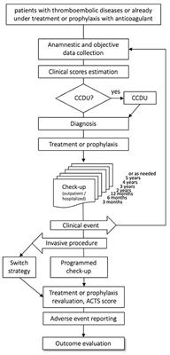
Inherited prothrombotic risk factors and cerebral venous thrombosis. (1/5820)
Fifteen patients with cerebral venous thrombosis were ascertained retrospectively. Their case notes were reviewed, and stored or new blood was assayed for factor V Leiden (FVL) mutation, prothrombin gene mutation 20201A, and 5,10 methylene tetrahydrofolate reductase (MTHFR) C677T mutation. A clinical risk factor was identified in 13 patients--the oral contraceptive pill (5), puerperium (1), HRT (1), mastoiditis (1), dehydration (1), lumbar puncture and myelography (1), carcinoma (1), lupus anticoagulant (2). In addition, two patients had the FVL mutation and five (one of whom also had the FVL mutation) were homozygous for the MTHFR mutation. The latter showed a higher than expected frequency compared to 300 healthy controls from South Wales (OR 3.15.95% Cl 1.01-9.83). No patient had the prothrombin 20201A mutation. Two patients died and three had a monocular visual deficit following anticoagulation (13) or thrombolytic (2) treatment, but there was no association between the presence of a primary prothrombotic risk factor and outcome. These results confirm the importance of investigating patients for both clinical predisposing factors and primary prothrombotic states. (+info)Anticoagulant heparan sulfate precursor structures in F9 embryonal carcinoma cells. (2/5820)
To understand the mechanisms that control anticoagulant heparan sulfate (HSact) biosynthesis, we previously showed that HSact production in the F9 system is determined by the abundance of 3-O-sulfotransferase-1 as well as the size of the HSact precursor pool. In this study, HSact precursor structures have been studied by characterizing [6-3H]GlcN metabolically labeled F9 HS tagged with 3-O-sulfates in vitro by 3'-phosphoadenosine 5'-phospho-35S and purified 3-O-sulfotransferase-1. This later in vitro labeling allows the regions of HS destined to become the antithrombin (AT)-binding sites to be tagged for subsequent structural studies. It was shown that six 3-O-sulfation sites exist per HSact precursor chain. At least five out of six 3-O-sulfate-tagged oligosaccharides in HSact precursors bind AT, whereas none of 3-O-sulfate-tagged oligosaccharides from HSinact precursors bind AT. When treated with low pH nitrous or heparitinase, 3-O-sulfate-tagged HSact and HSinact precursors exhibit clearly different structural features. 3-O-Sulfate-tagged HSact hexasaccharides were AT affinity purified and sequenced by chemical and enzymatic degradations. The 3-O-sulfate-tagged HSact hexasaccharides exhibited the following structures, DeltaUA-[6-3H]GlcNAc6S-GlcUA-[6-3H]GlcNS3(35)S+/-6S-++ +IdceA2S-[6-3H]Glc NS6S. The underlined 6- and 3-O-sulfates constitute the most critical groups for AT binding in view of the fact that the precursor hexasaccharides possess all the elements for AT binding except for the 3-O-sulfate moiety. The presence of five potential AT-binding precursor hexasaccharides in all HSact precursor chains demonstrates for the first time the processive assembly of specific sequence in HS. The difference in structures around potential 3-O-sulfate acceptor sites in HSact and HSinact precursors suggests that these precursors might be generated by different concerted assembly mechanisms in the same cell. This study permits us to understand better the nature of the HS biosynthetic pathway that leads to the generation of specific saccharide sequences. (+info)Warfarin therapy: evolving strategies in anticoagulation. (3/5820)
Warfarin is the oral anticoagulant most frequently used to control and prevent thromboembolic disorders. Prescribing the dose that both avoids hemorrhagic complications and achieves sufficient suppression of thrombosis requires a thorough understanding of the drug's unique pharmacology. Warfarin has a complex dose-response relationship that makes safe and effective use a challenge. For most indications, the dose is adjusted to maintain the patient's International Normalized Ratio (INR) at 2 to 3. Because of the delay in factor II (prothrombin) suppression, heparin is administered concurrently for four to five days to prevent thrombus propagation. Loading doses of warfarin are not warranted and may result in bleeding complications. Interactions with other drugs must be considered, and therapy in elderly patients requires careful management. Current dosing recommendations are reviewed, and practical guidelines for the optimal use of warfarin are provided. (+info)Exosites 1 and 2 are essential for protection of fibrin-bound thrombin from heparin-catalyzed inhibition by antithrombin and heparin cofactor II. (4/5820)
Assembly of ternary thrombin-heparin-fibrin complexes, formed when fibrin binds to exosite 1 on thrombin and fibrin-bound heparin binds to exosite 2, produces a 58- and 247-fold reduction in the heparin-catalyzed rate of thrombin inhibition by antithrombin and heparin cofactor II, respectively. The greater reduction for heparin cofactor II reflects its requirement for access to exosite 1 during the inhibitory process. Protection from inhibition by antithrombin and heparin cofactor II requires ligation of both exosites 1 and 2 because minimal protection is seen when exosite 1 variants (gamma-thrombin and thrombin Quick 1) or an exosite 2 variant (Arg93 --> Ala, Arg97 --> Ala, and Arg101 --> Ala thrombin) is substituted for thrombin. Likewise, the rate of thrombin inhibition by the heparin-independent inhibitor, alpha1-antitrypsin Met358 --> Arg, is decreased less than 2-fold in the presence of soluble fibrin and heparin. In contrast, thrombin is protected from inhibition by a covalent antithrombin-heparin complex, suggesting that access of heparin to exosite 2 of thrombin is hampered when ternary complex formation occurs. These results reveal the importance of exosites 1 and 2 of thrombin in assembly of the ternary complex and the subsequent protection of thrombin from inhibition by heparin-catalyzed inhibitors. (+info)Sperm chemotaxis. (5/5820)
Communication between spermatozoa and egg before contact by chemotaxis appears to be prevalent throughout the animal kingdom. In non-mammalian species, sperm chemotaxis to factors secreted from the egg is well documented. In mammals, sperm chemotaxis to follicular factors in vitro has been established in humans and mice. The attractants of female origin in non-mammalian species are heat-stable peptides or proteins of various sizes, or other small molecules, depending on the species. Species specificity of the attractants in non-mammalian species may vary from high species specificity, through specificity to families with no specificity within a family, to absence of specificity. The mammalian sperm attractants have not been identified but they appear to be heat-stable peptides. The claim that progesterone is the attractant for human spermatozoa has failed to be substantiated, neither have claims for other mammalian sperm attractants been verified. The molecular mechanism of sperm chemotaxis is not known. Models involving modulation of the intracellular Ca2+ concentration have been proposed for both mammalian and non-mammalian sperm chemotaxis. The physiological role of sperm chemotaxis in non-mammalian species appears to differ from that in mammals. In non-mammalian species, sperm chemotaxis strives to bring as many spermatozoa as possible to the egg. However, in mammals, the role appears to be recruitment of a selective population of capacitated ('ripe') spermatozoa to fertilize the egg. (+info)Nonanticoagulant heparin prevents coronary endothelial dysfunction after brief ischemia-reperfusion injury in the dog. (6/5820)
BACKGROUND: Coronary endothelial dysfunction after brief ischemia-reperfusion (IR) remains a clinical problem. We investigated the role of heparin and N-acetylheparin, a nonanticoagulant heparin derivative, in modulating coronary endothelial function after IR injury, with an emphasis on defining the role of the nitric oxide (NO)-cGMP pathway in the heparin-mediated effect. METHODS AND RESULTS: Male mongrel dogs were surgically instrumented, and the effects of both bovine heparin and N-acetylheparin on coronary endothelial vasomotor function, expressed as percent change from baseline flow after acetylcholine challenge, were studied after 15 minutes of regional ischemia of the left anterior descending artery (LAD) followed by 120 minutes of reperfusion. In dogs treated with placebo (saline), coronary vasomotor function was significantly (P+info)Ex vivo evaluation of a Taylor-Couette flow, immobilized heparinase I device for clinical application. (7/5820)
Efficient and safe heparin anticoagulation has remained a problem for continuous renal replacement therapies and intermittent hemodialysis for patients with acute renal failure. To make heparin therapy safer for the patient with acute renal failure at high risk of bleeding, we have proposed regional heparinization of the circuit via an immobilized heparinase I filter. This study tested a device based on Taylor-Couette flow and simultaneous separation/reaction for efficacy and safety of heparin removal in a sheep model. Heparinase I was immobilized onto agarose beads via cyanogen bromide activation. The device, referred to as a vortex flow plasmapheretic reactor, consisted of two concentric cylinders, a priming volume of 45 ml, a microporous membrane for plasma separation, and an outer compartment where the immobilized heparinase I was fluidized separately from the blood cells. Manual white cell and platelet counts, hematocrit, total protein, and fibrinogen assays were performed. Heparin levels were indirectly measured via whole-blood recalcification times (WBRTs). The vortex flow plasmapheretic reactor maintained significantly higher heparin levels in the extracorporeal circuit than in the sheep (device inlet WBRTs were 1. 5 times the device outlet WBRTs) with no hemolysis. The reactor treatment did not effect any physiologically significant changes in complete blood cell counts, platelets, and protein levels for up to 2 hr of operation. Furthermore, gross necropsy and histopathology did not show any significant abnormalities in the kidney, liver, heart, brain, and spleen. (+info)Randomized, placebo-controlled trial of anticoagulant treatment with low-molecular-weight heparin for cerebral sinus thrombosis. (8/5820)
BACKGROUND AND PURPOSE: Treatment of cerebral sinus thrombosis with heparin is controversial. We conducted a double-blind, placebo-controlled multicenter trial to examine whether anticoagulant treatment improves outcome in patients with sinus thrombosis. METHODS: Patients were randomized between body weight-adjusted subcutaneous nadroparin (180 anti-factor Xa units/kg per 24 hours) and matching placebo for 3 weeks (double-blind part of trial), followed by 3 months of oral anticoagulants for patients allocated nadroparin (open part). Patients with cerebral hemorrhage caused by sinus thrombosis were also included. RESULTS: Sixty patients were enrolled, and none were lost to follow-up. In 1 patient the diagnosis proved wrong after randomization. After 3 weeks, 6 of 30 patients (20%) in the nadroparin group and 7 of 29 patients (24%) in the placebo group had a poor outcome, defined as death or Barthel Index score of <15 (risk difference, -4%; 95% CI, -25 to 17%; NS). After 12 weeks, 4 of 30 patients (13%) in the nadroparin group and 6 of 29 (21%) in the placebo group had a poor outcome, defined as death or Oxford Handicap Score of >/=3 (risk difference, -7%; 95% CI, -26% to 12%; NS). There were no new symptomatic cerebral hemorrhages. One patient in the nadroparin group had a major gastrointestinal hemorrhage, and 1 patient in the placebo group died from clinically suspected pulmonary embolism. CONCLUSIONS: Patients with cerebral sinus thrombosis treated with anticoagulants (low-molecular-weight heparin followed by oral anticoagulation) had a favorable outcome more often than controls, but the difference was not statistically significant. Anticoagulation proved to be safe, even in patients with cerebral hemorrhage. (+info)Anticoagulants are a class of medications that work to prevent the formation of blood clots in the body. They do this by inhibiting the coagulation cascade, which is a series of chemical reactions that lead to the formation of a clot. Anticoagulants can be given orally, intravenously, or subcutaneously, depending on the specific drug and the individual patient's needs.
There are several different types of anticoagulants, including:
1. Heparin: This is a naturally occurring anticoagulant that is often used in hospitalized patients who require immediate anticoagulation. It works by activating an enzyme called antithrombin III, which inhibits the formation of clots.
2. Low molecular weight heparin (LMWH): LMWH is a form of heparin that has been broken down into smaller molecules. It has a longer half-life than standard heparin and can be given once or twice daily by subcutaneous injection.
3. Direct oral anticoagulants (DOACs): These are newer oral anticoagulants that work by directly inhibiting specific clotting factors in the coagulation cascade. Examples include apixaban, rivaroxaban, and dabigatran.
4. Vitamin K antagonists: These are older oral anticoagulants that work by inhibiting the action of vitamin K, which is necessary for the formation of clotting factors. Warfarin is an example of a vitamin K antagonist.
Anticoagulants are used to prevent and treat a variety of conditions, including deep vein thrombosis (DVT), pulmonary embolism (PE), atrial fibrillation, and prosthetic heart valve thrombosis. It is important to note that anticoagulants can increase the risk of bleeding, so they must be used with caution and regular monitoring of blood clotting times may be required.
I'm sorry for any confusion, but "Lupus Coagulation Inhibitor" is not a recognized medical term or concept in hematology or immunology.
Systemic Lupus Erythematosus (SLE), often referred to simply as lupus, is an autoimmune disease where the body's immune system mistakenly attacks healthy tissue. However, this does not result in a specific coagulation inhibitor.
If you're asking about lupus anticoagulants, these are antibodies that can interfere with clotting tests but paradoxically increase the risk of blood clots in vivo. They are sometimes seen in patients with SLE and other autoimmune diseases.
Please provide more context if you meant something else, so I can give a more accurate response.
Warfarin is a anticoagulant medication that works by inhibiting the vitamin K-dependent activation of several coagulation factors (factors II, VII, IX, and X). This results in prolonged clotting times and reduced thrombus formation. It is commonly used to prevent and treat blood clots in conditions such as atrial fibrillation, deep vein thrombosis, and pulmonary embolism. Warfarin is also known by its brand names Coumadin and Jantoven.
It's important to note that warfarin has a narrow therapeutic index, meaning that the difference between an effective dose and a toxic one is small. Therefore, it requires careful monitoring of the patient's coagulation status through regular blood tests (INR) to ensure that the dosage is appropriate and to minimize the risk of bleeding complications.
Blood coagulation, also known as blood clotting, is a complex process that occurs in the body to prevent excessive bleeding when a blood vessel is damaged. This process involves several different proteins and chemical reactions that ultimately lead to the formation of a clot.
The coagulation cascade is initiated when blood comes into contact with tissue factor, which is exposed after damage to the blood vessel wall. This triggers a series of enzymatic reactions that activate clotting factors, leading to the formation of a fibrin clot. Fibrin is a protein that forms a mesh-like structure that traps platelets and red blood cells to form a stable clot.
Once the bleeding has stopped, the coagulation process is regulated and inhibited to prevent excessive clotting. The fibrinolytic system degrades the clot over time, allowing for the restoration of normal blood flow.
Abnormalities in the blood coagulation process can lead to bleeding disorders or thrombotic disorders such as deep vein thrombosis and pulmonary embolism.
Prothrombin time (PT) is a medical laboratory test that measures the time it takes for blood to clot. It's often used to evaluate the functioning of the extrinsic and common pathways of the coagulation system, which is responsible for blood clotting. Specifically, PT measures how long it takes for prothrombin (a protein produced by the liver) to be converted into thrombin, an enzyme that converts fibrinogen into fibrin and helps form a clot.
Prolonged PT may indicate a bleeding disorder or a deficiency in coagulation factors, such as vitamin K deficiency or the use of anticoagulant medications like warfarin. It's important to note that PT is often reported with an international normalized ratio (INR), which allows for standardization and comparison of results across different laboratories and reagent types.
Protein C is a vitamin K-dependent protease that functions as an important regulator of coagulation and inflammation. It is a plasma protein produced in the liver that, when activated, degrades clotting factors Va and VIIIa to limit thrombus formation and prevent excessive blood clotting. Protein C also has anti-inflammatory properties by inhibiting the release of pro-inflammatory cytokines and reducing endothelial cell activation. Inherited or acquired deficiencies in Protein C can lead to an increased risk of thrombosis, a condition characterized by abnormal blood clot formation within blood vessels.
Partial Thromboplastin Time (PTT) is a medical laboratory test that measures the time it takes for blood to clot. It's more specifically a measure of the intrinsic and common pathways of the coagulation cascade, which are the series of chemical reactions that lead to the formation of a clot.
The test involves adding a partial thromboplastin reagent (an activator of the intrinsic pathway) and calcium to plasma, and then measuring the time it takes for a fibrin clot to form. This is compared to a control sample, and the ratio of the two times is calculated.
The PTT test is often used to help diagnose bleeding disorders or abnormal blood clotting, such as hemophilia or disseminated intravascular coagulation (DIC). It can also be used to monitor the effectiveness of anticoagulant therapy, such as heparin. Prolonged PTT results may indicate a bleeding disorder or an increased risk of bleeding, while shortened PTT results may indicate a hypercoagulable state and an increased risk of thrombosis.
Rodenticides are a type of pesticide that are specifically designed to control or kill rodents, such as rats and mice. They contain chemicals that can interfere with the normal physiology of rodents, leading to their death. Rodenticides can come in various forms, including powders, pellets, and liquids, and they can be placed in bait stations or used in conjunction with other pest control methods.
It is important to use rodenticides carefully and only as directed, as they can also pose a risk to non-target animals, including pets and wildlife, if not used properly. Additionally, some rodenticides contain chemicals that can accumulate in the body over time and cause harm to humans if they are exposed to them repeatedly or in large quantities. As such, it is important to follow all safety guidelines when using rodenticides and to store them out of reach of children and pets.
Acenocoumarol is an anticoagulant medication that is used to prevent and treat blood clots. It works by inhibiting the formation of vitamin K-dependent clotting factors, which are necessary for normal blood coagulation. This results in a prolonged bleeding time and reduced risk of blood clots.
Acenocoumarol is a coumarin derivative and is available under various brand names, including Sintrom and Nicoumalone. It is typically administered orally in the form of tablets and its effects are monitored through regular blood tests to ensure that the dosage is appropriate and that the risk of bleeding complications is minimized.
Common side effects of acenocoumarol include easy bruising, nosebleeds, and skin rashes. It may also interact with a variety of other medications, including antibiotics, antifungals, and certain herbal supplements, so it is important to inform your healthcare provider of all medications and supplements you are taking before starting acenocoumarol therapy.
It is important to note that acenocoumarol has a narrow therapeutic index, meaning that the difference between an effective dose and a toxic dose is relatively small. Therefore, it is essential to follow your healthcare provider's instructions carefully when taking this medication and to have regular blood tests to monitor its effects on your coagulation status.
Heparin is defined as a highly sulfated glycosaminoglycan (a type of polysaccharide) that is widely present in many tissues, but is most commonly derived from the mucosal tissues of mammalian lungs or intestinal mucosa. It is an anticoagulant that acts as an inhibitor of several enzymes involved in the blood coagulation cascade, primarily by activating antithrombin III which then neutralizes thrombin and other clotting factors.
Heparin is used medically to prevent and treat thromboembolic disorders such as deep vein thrombosis, pulmonary embolism, and certain types of heart attacks. It can also be used during hemodialysis, cardiac bypass surgery, and other medical procedures to prevent the formation of blood clots.
It's important to note that while heparin is a powerful anticoagulant, it does not have any fibrinolytic activity, meaning it cannot dissolve existing blood clots. Instead, it prevents new clots from forming and stops existing clots from growing larger.
Factor Xa is a serine protease that plays a crucial role in the coagulation cascade, which is a series of reactions that lead to the formation of a blood clot. It is one of the activated forms of Factor X, a pro-protein that is converted to Factor Xa through the action of other enzymes in the coagulation cascade.
Factor Xa functions as a key component of the prothrombinase complex, which also includes calcium ions, phospholipids, and activated Factor V (also known as Activated Protein C or APC). This complex is responsible for converting prothrombin to thrombin, which then converts fibrinogen to fibrin, forming a stable clot.
Inhibitors of Factor Xa are used as anticoagulants in the prevention and treatment of thromboembolic disorders such as deep vein thrombosis and pulmonary embolism. These drugs work by selectively inhibiting Factor Xa, thereby preventing the formation of the prothrombinase complex and reducing the risk of clot formation.
Phenindione is an anticoagulant medication, which is primarily used in the prevention and treatment of thromboembolic disorders such as deep vein thrombosis (DVT), pulmonary embolism (PE), and certain types of strokes. It works by inhibiting the formation of blood clots in the body.
Phenindione is a derivative of indandione, and it functions by blocking the activity of vitamin K-dependent coagulation factors II, VII, IX, and X, thereby prolonging the clotting time of the blood. It is available in oral form as tablets or capsules.
It's important to note that phenindione has largely been replaced by other anticoagulants such as warfarin due to its narrow therapeutic index, higher risk of adverse effects, and interactions with other medications. Therefore, it is not commonly used in clinical practice today.
4-Hydroxycoumarins are a type of chemical compound that contains a hydroxy group (-OH) attached to the 4th carbon atom of the coumarin structure. Coumarins themselves are aromatic organic compounds, characterized by a benzene ring fused to a pyrone ring.
4-Hydroxycoumarins have gained attention in medical research due to their potential biological activities. For instance, some 4-hydroxycoumarins exhibit anticoagulant properties and are used as oral anticoagulant drugs, such as warfarin. These compounds work by inhibiting the vitamin K epoxide reductase enzyme, thereby interfering with the blood clotting process.
Additionally, 4-hydroxycoumarins have been investigated for their potential anticancer, anti-inflammatory, and antibacterial activities. However, more research is needed to fully understand their therapeutic potential and safety profiles.
Antithrombins are substances that prevent the formation or promote the dissolution of blood clots (thrombi). They include:
1. Anticoagulants: These are medications that reduce the ability of the blood to clot. Examples include heparin, warfarin, and direct oral anticoagulants (DOACs) such as apixaban, rivaroxaban, and dabigatran.
2. Thrombolytic agents: These are medications that break down existing blood clots. Examples include alteplase, reteplase, and tenecteplase.
3. Fibrinolytics: These are a type of thrombolytic agent that specifically target fibrin, a protein involved in the formation of blood clots.
4. Natural anticoagulants: These are substances produced by the body to regulate blood clotting. Examples include antithrombin III, protein C, and protein S.
Antithrombins are used in the prevention and treatment of various thromboembolic disorders, such as deep vein thrombosis (DVT), pulmonary embolism (PE), stroke, and myocardial infarction (heart attack). It is important to note that while antithrombins can help prevent or dissolve blood clots, they also increase the risk of bleeding, so their use must be carefully monitored.
Blood coagulation tests, also known as coagulation studies or clotting tests, are a series of medical tests used to evaluate the blood's ability to clot. These tests measure the functioning of various clotting factors and regulatory proteins involved in the coagulation cascade, which is a complex process that leads to the formation of a blood clot to prevent excessive bleeding.
The most commonly performed coagulation tests include:
1. Prothrombin Time (PT): Measures the time it takes for a sample of plasma to clot after the addition of calcium and tissue factor, which activates the extrinsic pathway of coagulation. The PT is reported in seconds and can be converted to an International Normalized Ratio (INR) to monitor anticoagulant therapy.
2. Activated Partial Thromboplastin Time (aPTT): Measures the time it takes for a sample of plasma to clot after the addition of calcium, phospholipid, and a contact activator, which activates the intrinsic pathway of coagulation. The aPTT is reported in seconds and is used to monitor heparin therapy.
3. Thrombin Time (TT): Measures the time it takes for a sample of plasma to clot after the addition of thrombin, which directly converts fibrinogen to fibrin. The TT is reported in seconds and can be used to detect the presence of fibrin degradation products or abnormalities in fibrinogen function.
4. Fibrinogen Level: Measures the amount of fibrinogen, a protein involved in clot formation, present in the blood. The level is reported in grams per liter (g/L) and can be used to assess bleeding risk or the effectiveness of fibrinogen replacement therapy.
5. D-dimer Level: Measures the amount of D-dimer, a protein fragment produced during the breakdown of a blood clot, present in the blood. The level is reported in micrograms per milliliter (µg/mL) and can be used to diagnose or exclude venous thromboembolism (VTE), such as deep vein thrombosis (DVT) or pulmonary embolism (PE).
These tests are important for the diagnosis, management, and monitoring of various bleeding and clotting disorders. They can help identify the underlying cause of abnormal bleeding or clotting, guide appropriate treatment decisions, and monitor the effectiveness of therapy. It is essential to interpret these test results in conjunction with a patient's clinical presentation and medical history.
Thrombin time (TT) is a medical laboratory test that measures the time it takes for a clot to form after thrombin, an enzyme that converts fibrinogen to fibrin in the final step of the coagulation cascade, is added to a plasma sample. This test is used to evaluate the efficiency of the conversion of fibrinogen to fibrin and can be used to detect the presence of abnormalities in the coagulation system, such as the presence of heparin or dysfibrinogenemia. Increased thrombin time may indicate the presence of a systemic anticoagulant or a deficiency in fibrinogen.
The International Normalized Ratio (INR) is a standardized measurement of the prothrombin time (PT), which is the time it takes for blood to clot. The INR is used to monitor and regulate the effects of anticoagulant medications, such as warfarin, that affect the blood's ability to clot.
The INR is calculated by dividing the patient's PT by a control value (the PT of normal, healthy blood), raised to the power of a sensitivity factor called the International Sensitivity Index (ISI). The ISI is specific to the thromboplastin reagent used in the PT assay.
The INR provides a consistent and comparable way to monitor anticoagulation therapy across different laboratories, regardless of the thromboplastin reagent used. This helps ensure that patients receive appropriate doses of anticoagulant medications and reduces the risk of bleeding or clotting complications.
In general, an INR range of 2.0 to 3.0 is recommended for most people taking anticoagulants for conditions such as atrial fibrillation, deep vein thrombosis, or pulmonary embolism. However, the target INR range may vary depending on individual patient factors and medical indications.
Thromboembolism is a medical condition that refers to the obstruction of a blood vessel by a thrombus (blood clot) that has formed elsewhere in the body and then been transported by the bloodstream to a narrower vessel, where it becomes lodged. This process can occur in various parts of the body, leading to different types of thromboembolisms:
1. Deep Vein Thrombosis (DVT): A thrombus forms in the deep veins, usually in the legs or pelvis, and then breaks off and travels to the lungs, causing a pulmonary embolism.
2. Pulmonary Embolism (PE): A thrombus formed elsewhere, often in the deep veins of the legs, dislodges and travels to the lungs, blocking one or more pulmonary arteries. This can lead to shortness of breath, chest pain, and potentially life-threatening complications if not treated promptly.
3. Cerebral Embolism: A thrombus formed in another part of the body, such as the heart or carotid artery, dislodges and travels to the brain, causing a stroke or transient ischemic attack (TIA).
4. Arterial Thromboembolism: A thrombus forms in an artery and breaks off, traveling to another part of the body and blocking blood flow to an organ or tissue, leading to potential damage or loss of function. Examples include mesenteric ischemia (intestinal damage due to blocked blood flow) and retinal artery occlusion (vision loss due to blocked blood flow in the eye).
Prevention, early detection, and appropriate treatment are crucial for managing thromboembolism and reducing the risk of severe complications.
Protein S is a vitamin K-dependent protein found in the blood that functions as a natural anticoagulant. It plays a crucial role in regulating the body's clotting system by inhibiting the activation of coagulation factors, thereby preventing excessive blood clotting. Protein S also acts as a cofactor for activated protein C, which is another important anticoagulant protein.
Protein S exists in two forms: free and bound to a protein called C4b-binding protein (C4BP). Only the free form of Protein S has biological activity in inhibiting coagulation. Inherited or acquired deficiencies in Protein S can lead to an increased risk of thrombosis, or abnormal blood clot formation, which can cause various medical conditions such as deep vein thrombosis (DVT) and pulmonary embolism (PE). Regular monitoring of Protein S levels is essential for patients with a history of thrombotic events or those who have a family history of thrombophilia.
Blood coagulation factors, also known as clotting factors, are a group of proteins that play a crucial role in the blood coagulation process. They are essential for maintaining hemostasis, which is the body's ability to stop bleeding after injury.
There are 13 known blood coagulation factors, and they are designated by Roman numerals I through XIII. These factors are produced in the liver and are normally present in an inactive form in the blood. When there is an injury to a blood vessel, the coagulation process is initiated, leading to the activation of these factors in a specific order.
The coagulation cascade involves two pathways: the intrinsic and extrinsic pathways. The intrinsic pathway is activated when there is damage to the blood vessel itself, while the extrinsic pathway is activated by tissue factor released from damaged tissues. Both pathways converge at the common pathway, leading to the formation of a fibrin clot.
Blood coagulation factors work together in a complex series of reactions that involve activation, binding, and proteolysis. When one factor is activated, it activates the next factor in the cascade, and so on. This process continues until a stable fibrin clot is formed.
Deficiencies or abnormalities in blood coagulation factors can lead to bleeding disorders such as hemophilia or thrombosis. Hemophilia is a genetic disorder that affects one or more of the coagulation factors, leading to excessive bleeding and difficulty forming clots. Thrombosis, on the other hand, occurs when there is an abnormal formation of blood clots in the blood vessels, which can lead to serious complications such as stroke or pulmonary embolism.
Hemorrhage is defined in the medical context as an excessive loss of blood from the circulatory system, which can occur due to various reasons such as injury, surgery, or underlying health conditions that affect blood clotting or the integrity of blood vessels. The bleeding may be internal, external, visible, or concealed, and it can vary in severity from minor to life-threatening, depending on the location and extent of the bleeding. Hemorrhage is a serious medical emergency that requires immediate attention and treatment to prevent further blood loss, organ damage, and potential death.
Low-molecular-weight heparin (LMWH) is a type of heparin used as an anticoagulant, which refers to a group of medications that prevent the formation of blood clots. Heparin is a naturally occurring substance in the body, and low-molecular-weight heparins are obtained through the depolymerization of standard heparin.
LMWH has a lower molecular weight than standard heparin, which results in several pharmacological differences. LMWHs have a more predictable dose response, longer half-life, and higher bioavailability when administered subcutaneously compared to standard heparin. They also exhibit greater anti-factor Xa activity relative to their antithrombin (anti-IIa) activity, which contributes to their anticoagulant effects.
LMWHs are used for the prevention and treatment of deep vein thrombosis (DVT), pulmonary embolism (PE), and other thromboembolic disorders. Common LMWHs include enoxaparin, dalteparin, tinzaparin, and nadroparin.
It is essential to monitor the patient's kidney function when using LMWH since they are primarily cleared by the kidneys. In patients with renal impairment, dose adjustments or alternative anticoagulants may be necessary to reduce the risk of bleeding complications.
Thrombomodulin is a protein that is found on the surface of endothelial cells, which line the interior surface of blood vessels. It plays an important role in the regulation of blood coagulation (clotting) and the activation of natural anticoagulant pathways. Thrombomodulin binds to thrombin, a protein involved in blood clotting, and changes its function from promoting coagulation to inhibiting it. This interaction also activates protein C, an important anticoagulant protein, which helps to prevent the excessive formation of blood clots. Thrombomodulin also has anti-inflammatory properties and is involved in the maintenance of the integrity of the endothelial cell lining.
Beta-alanine is a non-essential amino acid, which means that it is not required in the diet because the body can produce it from other amino acids. It is produced in the liver and is also found in some foods such as meat, poultry, and fish.
Beta-alanine plays a role in the production of carnosine, a dipeptide molecule that helps to regulate muscle pH and improve muscle function during high-intensity exercise. When muscles contract during intense exercise, they produce hydrogen ions, which can cause the muscle pH to decrease (become more acidic), leading to fatigue and reduced muscle function. Carnosine acts as a buffer against this acidity, helping to maintain optimal muscle pH levels and improve performance during high-intensity exercise.
Beta-alanine supplements have been shown to increase carnosine levels in muscles, which may lead to improved athletic performance, particularly in activities that require short bursts of intense effort, such as weightlifting or sprinting. However, more research is needed to fully understand the effects and potential benefits of beta-alanine supplementation.
It's important to note that while beta-alanine supplements are generally considered safe for most people, they can cause a tingling sensation in the skin (paresthesia) when taken in high doses. This is a harmless side effect and typically subsides within an hour or so of taking the supplement.
Thrombosis is the formation of a blood clot (thrombus) inside a blood vessel, obstructing the flow of blood through the circulatory system. When a clot forms in an artery, it can cut off the supply of oxygen and nutrients to the tissues served by that artery, leading to damage or tissue death. If a thrombus forms in the heart, it can cause a heart attack. If a thrombus breaks off and travels through the bloodstream, it can lodge in a smaller vessel, causing blockage and potentially leading to damage in the organ that the vessel supplies. This is known as an embolism.
Thrombosis can occur due to various factors such as injury to the blood vessel wall, abnormalities in blood flow, or changes in the composition of the blood. Certain medical conditions, medications, and lifestyle factors can increase the risk of thrombosis. Treatment typically involves anticoagulant or thrombolytic therapy to dissolve or prevent further growth of the clot, as well as addressing any underlying causes.
Prothrombin is a protein present in blood plasma, and it's also known as coagulation factor II. It plays a crucial role in the coagulation cascade, which is a complex series of reactions that leads to the formation of a blood clot.
When an injury occurs, the coagulation cascade is initiated to prevent excessive blood loss. Prothrombin is converted into its active form, thrombin, by another factor called factor Xa in the presence of calcium ions, phospholipids, and factor Va. Thrombin then catalyzes the conversion of fibrinogen into fibrin, forming a stable clot.
Prothrombin levels can be measured through a blood test, which is often used to diagnose or monitor conditions related to bleeding or coagulation disorders, such as liver disease or vitamin K deficiency.
Thrombin is a serine protease enzyme that plays a crucial role in the coagulation cascade, which is a complex series of biochemical reactions that leads to the formation of a blood clot (thrombus) to prevent excessive bleeding during an injury. Thrombin is formed from its precursor protein, prothrombin, through a process called activation, which involves cleavage by another enzyme called factor Xa.
Once activated, thrombin converts fibrinogen, a soluble plasma protein, into fibrin, an insoluble protein that forms the structural framework of a blood clot. Thrombin also activates other components of the coagulation cascade, such as factor XIII, which crosslinks and stabilizes the fibrin network, and platelets, which contribute to the formation and growth of the clot.
Thrombin has several regulatory mechanisms that control its activity, including feedback inhibition by antithrombin III, a plasma protein that inactivates thrombin and other serine proteases, and tissue factor pathway inhibitor (TFPI), which inhibits the activation of factor Xa, thereby preventing further thrombin formation.
Overall, thrombin is an essential enzyme in hemostasis, the process that maintains the balance between bleeding and clotting in the body. However, excessive or uncontrolled thrombin activity can lead to pathological conditions such as thrombosis, atherosclerosis, and disseminated intravascular coagulation (DIC).
Antiphospholipid syndrome (APS) is an autoimmune disorder characterized by the presence of antiphospholipid antibodies in the blood. These antibodies are directed against phospholipids, a type of fat molecule found in cell membranes and plasma lipoproteins. The presence of these antibodies can lead to abnormal blood clotting, which can cause serious complications such as stroke, heart attack, deep vein thrombosis, and pulmonary embolism.
APS can occur either on its own (primary APS) or in conjunction with other autoimmune disorders, such as systemic lupus erythematosus (secondary APS). The exact cause of APS is not fully understood, but it is believed to involve a combination of genetic and environmental factors.
Symptoms of APS can vary widely depending on the location and severity of the blood clots. They may include:
* Recurrent miscarriages or stillbirths
* Blood clots in the legs, lungs, or other parts of the body
* Skin ulcers or lesions
* Headaches, seizures, or stroke-like symptoms
* Kidney problems
* Heart valve abnormalities
Diagnosis of APS typically involves blood tests to detect the presence of antiphospholipid antibodies. Treatment may include medications to prevent blood clots, such as anticoagulants and antiplatelet agents, as well as management of any underlying autoimmune disorders.
Vitamin K is a fat-soluble vitamin that plays a crucial role in blood clotting and bone metabolism. It is essential for the production of several proteins involved in blood clotting, including factor II (prothrombin), factor VII, factor IX, and factor X. Additionally, Vitamin K is necessary for the synthesis of osteocalcin, a protein that contributes to bone health by regulating the deposition of calcium in bones.
There are two main forms of Vitamin K: Vitamin K1 (phylloquinone), which is found primarily in green leafy vegetables and some vegetable oils, and Vitamin K2 (menaquinones), which is produced by bacteria in the intestines and is also found in some fermented foods.
Vitamin K deficiency can lead to bleeding disorders such as hemorrhage and excessive bruising. While Vitamin K deficiency is rare in adults, it can occur in newborns who have not yet developed sufficient levels of the vitamin. Therefore, newborns are often given a Vitamin K injection shortly after birth to prevent bleeding problems.
Thrombophlebitis is a medical condition characterized by the inflammation and clotting of blood in a vein, usually in the legs. The term thrombophlebitis comes from two words: "thrombo" which means blood clot, and "phlebitis" which refers to inflammation of the vein.
The condition can occur in superficial or deep veins. Superficial thrombophlebitis affects the veins just below the skin's surface, while deep vein thrombophlebitis (DVT) occurs in the deeper veins. DVT is a more serious condition as it can lead to complications such as pulmonary embolism if the blood clot breaks off and travels to the lungs.
Symptoms of thrombophlebitis may include redness, warmth, pain, swelling, or discomfort in the affected area. In some cases, there may be visible surface veins that are hard, tender, or ropy to touch. If left untreated, thrombophlebitis can lead to chronic venous insufficiency and other long-term complications. Treatment typically involves medications such as anticoagulants, antiplatelet agents, or thrombolytics, along with compression stockings and other supportive measures.
Phenprocoumon is a vitamin K antagonist, which is a type of anticoagulant medication. It works by inhibiting the activity of certain enzymes in the liver that are necessary for the formation of blood clots. This results in an increased time for blood to clot, reducing the risk of blood clots forming and causing complications such as deep vein thrombosis or pulmonary embolism.
Phenprocoumon is primarily used to prevent and treat blood clots in the veins, arteries, and heart. It is also used to prevent stroke in people with atrial fibrillation or other heart rhythm disorders.
Like other vitamin K antagonists, phenprocoumon has a narrow therapeutic index, meaning that there is only a small range between the effective dose and the toxic dose. Therefore, it requires careful monitoring of blood clotting times (INR) to ensure that the drug is working effectively without causing excessive bleeding.
Phenprocoumon is available in oral form and is typically taken once daily. It may take several days for the full anticoagulant effect of phenprocoumon to be achieved, so it is important to carefully follow dosing instructions and monitoring schedules.
Venous thrombosis is a medical condition characterized by the formation of a blood clot (thrombus) in the deep veins, often in the legs (deep vein thrombosis or DVT), but it can also occur in other parts of the body such as the arms, pelvis, or lungs (pulmonary embolism).
The formation of a venous thrombus can be caused by various factors, including injury to the blood vessel wall, changes in blood flow, and alterations in the composition of the blood. These factors can lead to the activation of clotting factors and platelets, which can result in the formation of a clot that blocks the vein.
Symptoms of venous thrombosis may include swelling, pain, warmth, and redness in the affected area. In some cases, the clot can dislodge and travel to other parts of the body, causing potentially life-threatening complications such as pulmonary embolism.
Risk factors for venous thrombosis include advanced age, obesity, smoking, pregnancy, use of hormonal contraceptives or hormone replacement therapy, cancer, recent surgery or trauma, prolonged immobility, and a history of previous venous thromboembolism. Treatment typically involves the use of anticoagulant medications to prevent further clotting and dissolve existing clots.
Antithrombin III is a protein that inhibits the formation of blood clots (thrombi) in the body. It does this by inactivating several enzymes involved in coagulation, including thrombin and factor Xa. Antithrombin III is produced naturally by the liver and is also available as a medication for the prevention and treatment of thromboembolic disorders, such as deep vein thrombosis and pulmonary embolism. It works by binding to and neutralizing excess clotting factors in the bloodstream, thereby reducing the risk of clot formation.
Antiphospholipid antibodies are a type of autoantibody that targets and binds to certain proteins found in the blood that attach to phospholipids (a type of fat molecule). These antibodies are associated with an increased risk of developing antiphospholipid syndrome, a disorder characterized by abnormal blood clotting.
There are several types of antiphospholipid antibodies, including:
1. Lupus anticoagulant: This type of antiphospholipid antibody can interfere with blood clotting tests and may increase the risk of thrombosis (blood clots) in both arteries and veins.
2. Anticardiolipin antibodies: These antibodies target a specific phospholipid called cardiolipin, which is found in the inner membrane of mitochondria. High levels of anticardiolipin antibodies are associated with an increased risk of thrombosis and pregnancy complications such as recurrent miscarriage.
3. Anti-β2 glycoprotein I antibodies: These antibodies target a protein called β2 glycoprotein I, which binds to negatively charged phospholipids on the surface of cells. High levels of anti-β2 glycoprotein I antibodies are associated with an increased risk of thrombosis and pregnancy complications.
The exact mechanism by which antiphospholipid antibodies cause blood clotting is not fully understood, but it is thought to involve the activation of platelets, the inhibition of natural anticoagulants, and the promotion of inflammation. Antiphospholipid syndrome can be treated with medications that thin the blood or prevent clots from forming, such as aspirin, warfarin, or heparin.
Anticardiolipin antibodies are a type of autoantibody that targets and binds to cardiolipin, a phospholipid component found in the inner mitochondrial membrane of cells. These antibodies are clinically significant because they have been associated with a variety of autoimmune disorders, including antiphospholipid syndrome (APS).
APS is a condition characterized by recurrent blood clots, pregnancy losses, and thrombocytopenia (low platelet count). Anticardiolipin antibodies are one of the three main types of autoantibodies found in APS, along with lupus anticoagulant and anti-β2 glycoprotein I antibodies.
The presence of high levels of anticardiolipin antibodies in the blood can lead to abnormal blood clotting, which can cause serious complications such as deep vein thrombosis, pulmonary embolism, and stroke. Anticardiolipin antibodies can also contribute to pregnancy losses by causing placental insufficiency or abnormal blood clotting in the placenta.
Anticardiolipin antibodies are typically detected through a blood test that measures their levels in the serum. A positive result is usually confirmed with a second test performed at least 12 weeks later to establish persistence. Treatment for anticardiolipin antibody-related disorders typically involves anticoagulation therapy to prevent blood clots and other complications.
Thrombophilia is a medical condition characterized by an increased tendency to form blood clots (thrombi) due to various genetic or acquired abnormalities in the coagulation system. These abnormalities can lead to a hypercoagulable state, which can cause thrombosis in both veins and arteries. Commonly identified thrombophilias include factor V Leiden mutation, prothrombin G20210A mutation, antithrombin deficiency, protein C deficiency, and protein S deficiency.
Acquired thrombophilias can be caused by various factors such as antiphospholipid antibody syndrome (APS), malignancies, pregnancy, oral contraceptive use, hormone replacement therapy, and certain medical conditions like inflammatory bowel disease or nephrotic syndrome.
It is essential to diagnose thrombophilia accurately, as it may influence the management of venous thromboembolism (VTE) events and guide decisions regarding prophylactic anticoagulation in high-risk situations.
Blood coagulation disorders, also known as bleeding disorders or clotting disorders, refer to a group of medical conditions that affect the body's ability to form blood clots properly. Normally, when a blood vessel is injured, the body's coagulation system works to form a clot to stop the bleeding and promote healing.
In blood coagulation disorders, there can be either an increased tendency to bleed due to problems with the formation of clots (hemorrhagic disorder), or an increased tendency for clots to form inappropriately even without injury, leading to blockages in the blood vessels (thrombotic disorder).
Examples of hemorrhagic disorders include:
1. Hemophilia - a genetic disorder that affects the ability to form clots due to deficiencies in clotting factors VIII or IX.
2. Von Willebrand disease - another genetic disorder caused by a deficiency or abnormality of the von Willebrand factor, which helps platelets stick together to form a clot.
3. Liver diseases - can lead to decreased production of coagulation factors, increasing the risk of bleeding.
4. Disseminated intravascular coagulation (DIC) - a serious condition where clotting and bleeding occur simultaneously due to widespread activation of the coagulation system.
Examples of thrombotic disorders include:
1. Factor V Leiden mutation - a genetic disorder that increases the risk of inappropriate blood clot formation.
2. Antithrombin III deficiency - a genetic disorder that impairs the body's ability to break down clots, increasing the risk of thrombosis.
3. Protein C or S deficiencies - genetic disorders that lead to an increased risk of thrombosis due to impaired regulation of the coagulation system.
4. Antiphospholipid syndrome (APS) - an autoimmune disorder where the body produces antibodies against its own clotting factors, increasing the risk of thrombosis.
Treatment for blood coagulation disorders depends on the specific diagnosis and may include medications to manage bleeding or prevent clots, as well as lifestyle changes and monitoring to reduce the risk of complications.
Venous Thromboembolism (VTE) is a medical condition that includes both deep vein thrombosis (DVT) and pulmonary embolism (PE). DVT is a blood clot that forms in the deep veins, usually in the legs, while PE occurs when a clot breaks off and travels to the lungs, blocking a pulmonary artery or one of its branches. This condition can be life-threatening if not diagnosed and treated promptly.
The medical definition of Venous Thromboembolism is:
"The formation of a blood clot (thrombus) in a deep vein, most commonly in the legs, which can then dislodge and travel to the lungs, causing a potentially life-threatening blockage of the pulmonary artery or one of its branches (pulmonary embolism). VTE is a complex disorder resulting from an interplay of genetic and environmental factors that affect the balance between thrombosis and fibrinolysis."
Some common risk factors for VTE include immobility, surgery, trauma, cancer, hormonal therapy, pregnancy, advanced age, and inherited or acquired thrombophilia. Symptoms of DVT may include swelling, pain, warmth, and redness in the affected limb, while symptoms of PE can range from shortness of breath and chest pain to coughing up blood or even sudden death. Diagnosis typically involves a combination of clinical assessment, imaging studies (such as ultrasound, CT scan, or MRI), and laboratory tests (such as D-dimer). Treatment usually includes anticoagulation therapy to prevent further clot formation and reduce the risk of recurrence.
Oral administration is a route of giving medications or other substances by mouth. This can be in the form of tablets, capsules, liquids, pastes, or other forms that can be swallowed. Once ingested, the substance is absorbed through the gastrointestinal tract and enters the bloodstream to reach its intended target site in the body. Oral administration is a common and convenient route of medication delivery, but it may not be appropriate for all substances or in certain situations, such as when rapid onset of action is required or when the patient has difficulty swallowing.
A pulmonary embolism (PE) is a medical condition that occurs when a blood clot, often formed in the deep veins of the legs (deep vein thrombosis), breaks off and travels to the lungs, blocking one or more pulmonary arteries. This blockage can lead to various symptoms such as shortness of breath, chest pain, rapid heart rate, and coughing up blood. In severe cases, it can cause life-threatening complications like low oxygen levels, hypotension, and even death if not promptly diagnosed and treated with anticoagulant medications or thrombolytic therapy to dissolve the clot.
Heparin antagonists, also known as heparin neutralizers or reversal agents, are medications used to reverse the anticoagulant effects of heparin, a type of blood thinner. Heparin works by activating antithrombin III, which inactivates clotting factors IIa and Xa. Heparin antagonists, such as protamine sulfate, work by binding to heparin, forming a stable complex that is unable to bind to and activate antithrombin III, thereby neutralizing its anticoagulant effect.
Protamine sulfate is the most commonly used heparin antagonist. It is a highly basic protein derived from fish sperm that can neutralize the anticoagulant effects of heparin by forming a stable complex with it. The dose of protamine required to reverse the effects of heparin depends on the amount and type of heparin administered, as well as the timing of administration.
It is important to note that while heparin antagonists can reverse the anticoagulant effects of heparin, they do not reverse the underlying coagulation disorder or prevent further clot formation. Therefore, additional treatments may be necessary to manage the underlying condition and prevent recurrent thrombosis.
Thromboplastin is a substance that activates the coagulation cascade, leading to the formation of a clot (thrombus). It's primarily found in damaged or injured tissues and blood vessels, as well as in platelets (thrombocytes). There are two types of thromboplastin:
1. Extrinsic thromboplastin (also known as tissue factor): This is a transmembrane glycoprotein that is primarily found in subendothelial cells and released upon injury to the blood vessels. It initiates the extrinsic pathway of coagulation by binding to and activating Factor VII, ultimately leading to the formation of thrombin and fibrin clots.
2. Intrinsic thromboplastin (also known as plasma thromboplastin or factor III): This term is used less frequently and refers to a labile phospholipid component present in platelet membranes, which plays a role in the intrinsic pathway of coagulation.
In clinical settings, the term "thromboplastin" often refers to reagents used in laboratory tests like the prothrombin time (PT) and activated partial thromboplastin time (aPTT). These reagents contain a source of tissue factor and calcium ions to initiate and monitor the coagulation process.
Factor X is a protein that is essential for blood clotting, also known as coagulation. It is an enzyme that plays a crucial role in the coagulation cascade, which is a series of chemical reactions that lead to the formation of a blood clot. Factor X is activated by one of two pathways: the intrinsic pathway, which is initiated by damage to the blood vessels, or the extrinsic pathway, which is triggered by the release of tissue factor from damaged cells. Once activated, Factor X converts prothrombin to thrombin, which then converts fibrinogen to fibrin to form a stable clot.
Inherited deficiencies in Factor X can lead to bleeding disorders, while increased levels of Factor X have been associated with an increased risk of thrombosis or blood clots. Therefore, maintaining appropriate levels of Factor X is important for the proper balance between bleeding and clotting in the body.
Hirudin is not a medical term itself, but it is a specific substance with medical relevance. Hirudin is a naturally occurring anticoagulant that is found in the saliva of certain species of leeches (such as Hirudo medicinalis). This compound works by inhibiting the activity of thrombin, a key enzyme in the coagulation cascade, which ultimately results in preventing blood clot formation.
Medically, hirudin has been used in some research and therapeutic settings for its anticoagulant properties. For instance, recombinant hirudin (also known as lepirudin) is available for clinical use as an injectable anticoagulant to treat or prevent blood clots in specific medical conditions, such as heparin-induced thrombocytopenia (HIT).
In summary, Hirudins are a group of anticoagulant substances, primarily derived from leeches, that inhibit the activity of thrombin and have potential medical applications in preventing or treating blood clots.
Factor V, also known as proaccelerin or labile factor, is a protein involved in the coagulation cascade, which is a series of chemical reactions that leads to the formation of a blood clot. Factor V acts as a cofactor for the activation of Factor X to Factor Xa, which is a critical step in the coagulation cascade.
When blood vessels are damaged, the coagulation cascade is initiated to prevent excessive bleeding. During this process, Factor V is activated by thrombin, another protein involved in coagulation, and then forms a complex with activated Factor X and calcium ions on the surface of platelets or other cells. This complex converts prothrombin to thrombin, which then converts fibrinogen to fibrin to form a stable clot.
Deficiency or dysfunction of Factor V can lead to bleeding disorders such as hemophilia B or factor V deficiency, while mutations in the gene encoding Factor V can increase the risk of thrombosis, as seen in the Factor V Leiden mutation.
Coagulants are substances that promote the process of coagulation or clotting. They are often used in medical settings to help control bleeding and promote healing. Coagulants work by encouraging the formation of a clot, which helps to stop the flow of blood from a wound or cut.
There are several different types of coagulants that may be used in medical treatments. Some coagulants are naturally occurring substances, such as vitamin K, which is essential for the production of certain clotting factors in the body. Other coagulants may be synthetic or semi-synthetic compounds, such as recombinant activated factor VII (rFVIIa), which is used to treat bleeding disorders and prevent excessive bleeding during surgery.
Coagulants are often administered through injection or infusion, but they can also be applied topically to wounds or cuts. In some cases, coagulants may be used in combination with other treatments, such as compression or cauterization, to help control bleeding and promote healing.
It is important to note that while coagulants can be helpful in controlling bleeding and promoting healing, they can also increase the risk of blood clots and other complications. As a result, they should only be used under the guidance and supervision of a qualified healthcare professional.
Beta 2-glycoprotein I, also known as apolipoprotein H, is a plasma protein that belongs to the family of proteins called immunoglobulin-binding proteins. It has a molecular weight of approximately 44 kDa and is composed of five domains with similar structures.
Beta 2-glycoprotein I is primarily produced in the liver and circulates in the bloodstream, where it plays a role in several physiological processes, including coagulation, complement activation, and lipid metabolism. It has been identified as an autoantigen in certain autoimmune disorders, such as antiphospholipid syndrome (APS), where autoantibodies against beta 2-glycoprotein I can cause blood clots, miscarriages, and other complications.
In medical terminology, the definition of "beta 2-glycoprotein I" is as follows:
A plasma protein that belongs to the family of immunoglobulin-binding proteins and has a molecular weight of approximately 44 kDa. It is primarily produced in the liver and circulates in the bloodstream, where it plays a role in several physiological processes, including coagulation, complement activation, and lipid metabolism. Autoantibodies against beta 2-glycoprotein I are associated with certain autoimmune disorders, such as antiphospholipid syndrome (APS), where they can cause blood clots, miscarriages, and other complications.
Dicumarol is an anticoagulant medication that belongs to a class of compounds known as coumarins. It works by inhibiting the action of vitamin K, which is necessary for the production of certain clotting factors in the liver. This results in a decrease in blood clotting ability and helps prevent the formation of harmful blood clots.
Dicumarol is primarily used to treat and prevent deep vein thrombosis (DVT), pulmonary embolism, and other conditions that may require anticoagulation therapy. It is also used in the management of atrial fibrillation, valvular heart disease, and certain types of heart attacks.
It's important to note that dicumarol has a narrow therapeutic index, meaning that the difference between an effective dose and a toxic dose is relatively small. Therefore, it requires careful monitoring of blood clotting times (INR) to ensure that the drug is working effectively without causing excessive bleeding.
Dicumarol is available in oral form and is typically taken once or twice daily. Common side effects include nausea, vomiting, diarrhea, skin rash, and abnormal liver function tests. Rare but serious side effects include severe bleeding, necrosis of the skin and other tissues, and allergic reactions.
Dicumarol is a prescription medication that should only be used under the guidance of a healthcare professional. It interacts with many other medications and foods, so it's important to inform your doctor about all the drugs you are taking and any dietary changes you may make while on this medication.
Activated Protein C (APC) resistance is a condition in which the body's natural anticoagulant system is impaired, leading to an increased risk of thrombosis or blood clot formation. APC is an enzyme that plays a crucial role in regulating blood coagulation by inactivating clotting factors Va and VIIIa.
APC resistance is most commonly caused by a genetic mutation in the Factor V gene, known as Factor V Leiden. This mutation results in the production of a variant form of Factor V called Factor V Leiden, which is resistant to APC-mediated inactivation. As a result, the body's ability to regulate blood clotting is impaired, leading to an increased risk of thrombosis.
APC resistance can be measured by performing a functional assay that compares the activity of APC in normal plasma versus plasma from a patient with suspected APC resistance. The assay measures the rate of inactivation of Factor Va by APC, and a reduced rate of inactivation indicates APC resistance.
It is important to note that not all individuals with APC resistance will develop thrombosis, and other factors such as age, obesity, pregnancy, oral contraceptive use, and smoking can increase the risk of thrombosis in individuals with APC resistance.
Atrial fibrillation (A-tre-al fi-bru-la'shun) is a type of abnormal heart rhythm characterized by rapid and irregular beating of the atria, the upper chambers of the heart. In this condition, the electrical signals that coordinate heartbeats don't function properly, causing the atria to quiver instead of contracting effectively. As a result, blood may not be pumped efficiently into the ventricles, which can lead to blood clots, stroke, and other complications. Atrial fibrillation is a common type of arrhythmia and can cause symptoms such as palpitations, shortness of breath, fatigue, and dizziness. It can be caused by various factors, including heart disease, high blood pressure, age, and genetics. Treatment options include medications, electrical cardioversion, and surgical procedures to restore normal heart rhythm.
Factor V, also known as proaccelerin or labile factor, is a protein involved in the coagulation cascade, which is a series of chemical reactions that leads to the formation of a blood clot. Factor V acts as a cofactor for the conversion of prothrombin to thrombin, which is a critical step in the coagulation process.
Inherited deficiencies or abnormalities in Factor V can lead to bleeding disorders. For example, Factor V Leiden is a genetic mutation that causes an increased risk of blood clots, while Factor V deficiency can cause a bleeding disorder.
It's worth noting that "Factor Va" is not a standard medical term. Factor V becomes activated and turns into Factor Va during the coagulation cascade. Therefore, it is possible that you are looking for the definition of "Factor Va" in the context of its role as an activated form of Factor V in the coagulation process.
Hemostasis is the physiological process that occurs to stop bleeding (bleeding control) when a blood vessel is damaged. This involves the interaction of platelets, vasoconstriction, and blood clotting factors leading to the formation of a clot. The ultimate goal of hemostasis is to maintain the integrity of the vascular system while preventing excessive blood loss.
Fibrinolytic agents are medications that dissolve or break down blood clots by activating plasminogen, which is converted into plasmin. Plasmin is a proteolytic enzyme that degrades fibrin, the structural protein in blood clots. Fibrinolytic agents are used medically to treat conditions such as acute ischemic stroke, deep vein thrombosis, pulmonary embolism, and myocardial infarction (heart attack) by restoring blood flow in occluded vessels. Examples of fibrinolytic agents include alteplase, reteplase, and tenecteplase. It is important to note that these medications carry a risk of bleeding complications and should be administered with caution.
Platelet aggregation inhibitors are a class of medications that prevent platelets (small blood cells involved in clotting) from sticking together and forming a clot. These drugs work by interfering with the ability of platelets to adhere to each other and to the damaged vessel wall, thereby reducing the risk of thrombosis (blood clot formation).
Platelet aggregation inhibitors are often prescribed for people who have an increased risk of developing blood clots due to various medical conditions such as atrial fibrillation, coronary artery disease, peripheral artery disease, stroke, or a history of heart attack. They may also be used in patients undergoing certain medical procedures, such as angioplasty and stenting, to prevent blood clot formation in the stents.
Examples of platelet aggregation inhibitors include:
1. Aspirin: A nonsteroidal anti-inflammatory drug (NSAID) that irreversibly inhibits the enzyme cyclooxygenase, which is involved in platelet activation and aggregation.
2. Clopidogrel (Plavix): A P2Y12 receptor antagonist that selectively blocks ADP-induced platelet activation and aggregation.
3. Prasugrel (Effient): A third-generation thienopyridine P2Y12 receptor antagonist, similar to clopidogrel but with faster onset and greater potency.
4. Ticagrelor (Brilinta): A direct-acting P2Y12 receptor antagonist that does not require metabolic activation and has a reversible binding profile.
5. Dipyridamole (Persantine): An antiplatelet agent that inhibits platelet aggregation by increasing cyclic adenosine monophosphate (cAMP) levels in platelets, which leads to decreased platelet reactivity.
6. Iloprost (Ventavis): A prostacyclin analogue that inhibits platelet aggregation and causes vasodilation, often used in the treatment of pulmonary arterial hypertension.
7. Cilostazol (Pletal): A phosphodiesterase III inhibitor that increases cAMP levels in platelets, leading to decreased platelet activation and aggregation, as well as vasodilation.
8. Ticlopidine (Ticlid): An older P2Y12 receptor antagonist with a slower onset of action and more frequent side effects compared to clopidogrel or prasugrel.
I'm sorry for any confusion, but "Rodent Control" is not a medical term. It is a pest control practice related to the management and reduction of rodent populations, particularly rats and mice, to prevent damage to structures, ensure food safety, and prevent the spread of diseases.
In a medical context, you might be looking for information on "Rat-Bite Fever" or "Leptospirosis," which are two diseases that can be transmitted to humans through contact with rodents or their urine and feces. Here are brief definitions:
* Rat-Bite Fever: A bacterial infection caused by Streptobacillus moniliformis (in the United States) or Spirillum minus (in Asia). It is typically transmitted through bites, scratches, or contact with urine or feces of infected rats. Symptoms include fever, chills, headache, muscle pain, and vomiting.
* Leptospirosis: A bacterial infection caused by the pathogen Leptospira interrogans. It is often transmitted through exposure to contaminated water, soil, or food that has come into contact with infected animal urine, especially rats. Symptoms can range from mild flu-like illness to severe cases with kidney and liver failure, meningitis, or respiratory distress.
If you have any concerns about rodent-related diseases or require information on pest control practices, it is best to consult a healthcare professional or a licensed pest management expert.
Pipicolic acid is not a term that refers to a specific medical condition or disease. Instead, it is a metabolite that is involved in the body's metabolic processes.
Pipicolic acid is a type of organic compound called a cyclic amino acid, which is derived from the amino acid lysine. It is produced in the liver and is excreted in urine. Pipicolic acid has been found to have various functions in the body, including regulating the metabolism of lipids and bile acids.
Abnormal levels of pipicolic acid in the body may be associated with certain medical conditions, such as liver disease or genetic disorders that affect amino acid metabolism. However, pipicolic acid is not typically used as a diagnostic marker for these conditions.
In summary, pipicolic acid is a cyclic amino acid produced in the liver and involved in various metabolic processes in the body. Abnormal levels of pipicolic acid may be associated with certain medical conditions but are not typically used as diagnostic markers.
Thiophenes are organic compounds that contain a heterocyclic ring made up of four carbon atoms and one sulfur atom. The structure of thiophene is similar to benzene, with the benzene ring being replaced by a thiophene ring. Thiophenes are aromatic compounds, which means they have a stable, planar ring structure and delocalized electrons.
Thiophenes can be found in various natural sources such as coal tar, crude oil, and some foods like onions and garlic. They also occur in certain medications, dyes, and pesticides. Some thiophene derivatives have been synthesized and studied for their potential therapeutic uses, including anti-inflammatory, antiviral, and antitumor activities.
In the medical field, thiophenes are used in some pharmaceuticals as building blocks to create drugs with various therapeutic effects. For example, tipepidine, a cough suppressant, contains a thiophene ring. Additionally, some anesthetics and antipsychotic medications also contain thiophene moieties.
It is important to note that while thiophenes themselves are not typically considered medical terms, they play a role in the chemistry of various pharmaceuticals and other medical-related compounds.
Enoxaparin is a low molecular weight heparin (LMWH) medication that is used as an anticoagulant to prevent and treat blood clots. It works by binding to and inhibiting the activity of factor Xa, a clotting factor in the blood. This helps to reduce the risk of clot formation and can help to prevent conditions such as deep vein thrombosis (DVT) and pulmonary embolism (PE). Enoxaparin is typically given by injection under the skin (subcutaneously) and is available under the brand names Lovenox and Clexane, among others. It is important to follow the instructions of a healthcare professional when using enoxaparin, as it can increase the risk of bleeding.
Vitamin K epoxide reductases (VKORs) are enzymes that play a crucial role in the vitamin K cycle, which is essential for the post-translational modification of certain proteins involved in blood coagulation and bone metabolism. Specifically, VKORs reduce vitamin K epoxide back to its active form, vitamin K hydroquinone, allowing it to participate in the carboxylation of these proteins.
The most well-known member of this enzyme family is VKORC1 (Vitamin K Epoxide Reductase Complex Subunit 1), which is the target of the anticoagulant drug warfarin. Warfarin inhibits VKORC1, preventing the reduction of vitamin K epoxide and thereby interfering with the carboxylation of coagulation factors II, VII, IX, and X, as well as proteins C and S. This leads to the production of functionally inactive forms of these proteins and results in the anticoagulant effect of warfarin.
Protein C deficiency is a genetic disorder that affects the body's ability to control blood clotting. Protein C is a protein in the blood that helps regulate the formation of blood clots. When blood clots form too easily or do not dissolve properly, they can block blood vessels and lead to serious medical conditions such as deep vein thrombosis (DVT) or pulmonary embolism (PE).
People with protein C deficiency have lower than normal levels of this protein in their blood, which can increase their risk of developing abnormal blood clots. The condition is usually inherited and present from birth, but it may not cause any symptoms until later in life, such as during pregnancy, after surgery, or due to other factors that increase the risk of blood clots.
Protein C deficiency can be classified into two types: type I and type II. Type I deficiency is characterized by lower than normal levels of both functional and immunoreactive protein C in the blood. Type II deficiency is characterized by normal or near-normal levels of immunoreactive protein C, but reduced functional activity.
Protein C deficiency can be diagnosed through blood tests that measure the level and function of protein C in the blood. Treatment may include anticoagulant medications to prevent blood clots from forming or dissolve existing ones. Regular monitoring of protein C levels and careful management of risk factors for blood clots are also important parts of managing this condition.
Aspirin is the common name for acetylsalicylic acid, which is a medication used to relieve pain, reduce inflammation, and lower fever. It works by inhibiting the activity of an enzyme called cyclooxygenase (COX), which is involved in the production of prostaglandins, hormone-like substances that cause inflammation and pain. Aspirin also has an antiplatelet effect, which means it can help prevent blood clots from forming. This makes it useful for preventing heart attacks and strokes.
Aspirin is available over-the-counter in various forms, including tablets, capsules, and chewable tablets. It is also available in prescription strengths for certain medical conditions. As with any medication, aspirin should be taken as directed by a healthcare provider, and its use should be avoided in children and teenagers with viral infections due to the risk of Reye's syndrome, a rare but serious condition that can affect the liver and brain.
Dermatan sulfate is a type of glycosaminoglycan, which is a long, unbranched sugar chain found on the proteoglycan core protein in the extracellular matrix of animal tissues. It is composed of repeating disaccharide units of iduronic acid and N-acetylgalactosamine, with alternating sulfation at the 4-position of the iduronic acid and the 6-position of the galactosamine.
Dermatan sulfate is found in various tissues, including skin, heart valves, and blood vessels, where it plays important roles in regulating cell behavior, tissue development, and homeostasis. It also binds to a variety of growth factors, cytokines, and enzymes, modulating their activities and contributing to the regulation of various biological processes.
Abnormalities in dermatan sulfate metabolism can lead to several genetic disorders, such as Hunter syndrome and Hurler-Scheie syndrome, which are characterized by skeletal abnormalities, cardiac defects, and neurological impairment.
Heparin Cofactor II (HCII), also known as serine protease inhibitor E2 or labile factor, is a member of the serpin family of proteins. It is primarily produced in the liver and secreted into the bloodstream. HCII functions as a anticoagulant protein by inhibiting certain serine proteases involved in the coagulation cascade, particularly thrombin and factor Xa. The inhibitory activity of HCII is greatly enhanced in the presence of heparin or other glycosaminoglycans, hence its name.
HCII plays a crucial role in regulating blood clotting by controlling the levels of active thrombin and factor Xa in the circulation, thereby preventing excessive clot formation and maintaining normal hemostasis. Deficiencies or dysfunctions in HCII have been associated with an increased risk of thrombosis and other coagulation-related disorders.
Benzimidazoles are a class of heterocyclic compounds containing a benzene fused to a imidazole ring. They have a wide range of pharmacological activities and are used in the treatment of various diseases. Some of the benzimidazoles are used as antiparasitics, such as albendazole and mebendazole, which are effective against a variety of worm infestations. Other benzimidazoles have antifungal properties, such as thiabendazole and fuberidazole, and are used to treat fungal infections. Additionally, some benzimidazoles have been found to have anti-cancer properties and are being investigated for their potential use in cancer therapy.
Pyridones are a class of organic compounds that contain a pyridone ring, which is a heterocyclic ring consisting of a six-membered ring with five carbon atoms and one nitrogen atom, with one oxygen atom attached to the nitrogen atom by a double bond. Pyridones can be found in various natural sources, including plants and microorganisms, and they also have important applications in the pharmaceutical industry as building blocks for drug design and synthesis. Some drugs that contain pyridone rings include antihistamines, anti-inflammatory agents, and antiviral agents.
Protein S deficiency is a genetic disorder that affects the body's ability to coagulate blood properly. Protein S is a naturally occurring protein in the blood that helps regulate the clotting process by deactivating clotting factors when they are no longer needed. When Protein S levels are too low, it can lead to an increased risk of abnormal blood clots forming within blood vessels, a condition known as thrombophilia.
There are three types of Protein S deficiency: Type I (quantitative deficiency), Type II (qualitative deficiency), and Type III (dysfunctional protein). These types refer to the amount or function of Protein S in the blood. In Type I, there is a decrease in both free and total Protein S levels. In Type II, there is a decrease in functional Protein S despite normal total Protein S levels. In Type III, there is a decrease in free Protein S with normal total Protein S levels.
Protein S deficiency can be inherited or acquired. Inherited forms of the disorder are caused by genetic mutations and are usually present from birth. Acquired forms of Protein S deficiency can develop later in life due to certain medical conditions, such as liver disease, vitamin K deficiency, or the use of certain medications that affect blood clotting.
Symptoms of Protein S deficiency may include recurrent blood clots, usually in the legs (deep vein thrombosis) or lungs (pulmonary embolism), skin discoloration, pain, and swelling in the affected area. In severe cases, it can lead to complications such as chronic leg ulcers, pulmonary hypertension, or damage to the heart or lungs.
Diagnosis of Protein S deficiency typically involves blood tests to measure Protein S levels and function. Treatment may include anticoagulant medications to prevent blood clots from forming or growing larger. Lifestyle modifications such as regular exercise, maintaining a healthy weight, and avoiding smoking can also help reduce the risk of blood clots in people with Protein S deficiency.
Cardiolipins are a type of phospholipid that are primarily found in the inner mitochondrial membrane of cells. They play a crucial role in several important cellular processes, including energy production, apoptosis (programmed cell death), and maintenance of the structural integrity of the mitochondria.
Cardiolipins are unique because they contain four fatty acid chains, whereas most other phospholipids contain only two. This gives cardiolipins a distinctive conical shape that is important for their function in maintaining the curvature and stability of the inner mitochondrial membrane.
Cardiolipins have also been implicated in various diseases, including neurodegenerative disorders, cancer, and bacterial infections. For example, changes in cardiolipin composition or distribution have been linked to mitochondrial dysfunction in Parkinson's disease and other neurological conditions. Additionally, certain bacteria, such as Neisseria gonorrhoeae and Chlamydia trachomatis, can manipulate host cell cardiolipins to facilitate their own survival and replication.
In summary, cardiolipins are essential phospholipids found in the inner mitochondrial membrane that play a critical role in several cellular processes, and have been implicated in various diseases.
Antithrombin III (ATIII) deficiency is a genetic disorder that affects the body's ability to regulate blood clotting. ATIII is a protein produced in the liver that inhibits the activity of thrombin and other coagulation factors, preventing excessive clot formation.
People with ATIII deficiency have lower than normal levels of this protein, which can lead to an increased risk of developing abnormal blood clots (thrombosis) in veins, particularly deep vein thrombosis (DVT) and pulmonary embolism (PE). These clots can cause serious complications, including damage to the affected veins, organ damage, and even death.
ATIII deficiency can be classified into two types: type I and type II. Type I is characterized by a quantitative decrease in ATIII levels, while type II is characterized by a qualitative defect that results in reduced functional activity of the protein.
The condition is usually inherited in an autosomal dominant manner, meaning that a person has a 50% chance of inheriting the gene mutation from an affected parent. However, some cases may occur spontaneously due to new mutations in the ATIII gene. Treatment for ATIII deficiency typically involves anticoagulation therapy with medications such as heparin or warfarin to prevent blood clots from forming.
A hematoma is defined as a localized accumulation of blood in a tissue, organ, or body space caused by a break in the wall of a blood vessel. This can result from various causes such as trauma, surgery, or certain medical conditions that affect coagulation. The severity and size of a hematoma may vary depending on the location and extent of the bleeding. Symptoms can include swelling, pain, bruising, and decreased mobility in the affected area. Treatment options depend on the size and location of the hematoma but may include observation, compression, ice, elevation, or in some cases, surgical intervention.
Drug monitoring, also known as therapeutic drug monitoring (TDM), is a medical practice that involves testing blood or other bodily fluids to determine the concentration of a particular medication. This information is used to ensure that the patient is receiving an appropriate dosage and to help guide adjustments in medication therapy. It can be especially important for medications with a narrow therapeutic index, meaning that there is a small range between the effective dose and a toxic dose.
The goal of drug monitoring is to optimize medication effectiveness while minimizing potential side effects. This may involve measuring the concentration of a drug at various times after dosing to determine how quickly it is being metabolized or eliminated from the body, as well as to assess compliance with the prescribed treatment regimen.
Drug monitoring can be performed using a variety of methods, including immunoassays, chromatography, and mass spectrometry. The specific method used will depend on the drug being monitored and the level of sensitivity required. Results from drug monitoring tests are typically interpreted in conjunction with other clinical information, such as the patient's age, weight, renal function, liver function, and overall health status.
Dalteparin is a low molecular weight heparin (LMWH) medication that is used as an anticoagulant to prevent and treat blood clots. It works by binding to an enzyme called antithrombin III and enhancing its ability to inhibit clotting factors in the blood.
Dalteparin is available under the brand name Fragmin and is administered subcutaneously (under the skin) once or twice a day, depending on the indication and dosage prescribed by a healthcare professional. Common side effects of dalteparin include bleeding, bruising, pain at the injection site, and elevated liver enzymes.
As with all medications, it is important to use dalteparin only under the supervision of a healthcare provider and to follow their instructions carefully.
I'm sorry for any confusion, but the term "Sea Cucumbers" is not typically used in medical definitions. It is a common name given to marine animals belonging to the class Holothuroidea in the phylum Echinodermata. These are sausage-shaped, bottom-dwelling creatures found on the sea floor worldwide. They have a leathery skin and a set of tube feet used for locomotion. While they have some cultural and commercial importance in parts of the world, they do not have direct relevance to medical definitions.
Systemic Lupus Erythematosus (SLE) is a complex autoimmune disease that can affect almost any organ or system in the body. In SLE, the immune system produces an exaggerated response, leading to the production of autoantibodies that attack the body's own cells and tissues, causing inflammation and damage. The symptoms and severity of SLE can vary widely from person to person, but common features include fatigue, joint pain, skin rashes (particularly a "butterfly" rash across the nose and cheeks), fever, hair loss, and sensitivity to sunlight.
Systemic lupus erythematosus can also affect the kidneys, heart, lungs, brain, blood vessels, and other organs, leading to a wide range of symptoms such as kidney dysfunction, chest pain, shortness of breath, seizures, and anemia. The exact cause of SLE is not fully understood, but it is believed to involve a combination of genetic, environmental, and hormonal factors. Treatment typically involves medications to suppress the immune system and manage symptoms, and may require long-term management by a team of healthcare professionals.
Coumarins are a class of organic compounds that occur naturally in certain plants, such as sweet clover and tonka beans. They have a characteristic aroma and are often used as fragrances in perfumes and flavorings in food products. In addition to their use in consumer goods, coumarins also have important medical applications.
One of the most well-known coumarins is warfarin, which is a commonly prescribed anticoagulant medication used to prevent blood clots from forming or growing larger. Warfarin works by inhibiting the activity of vitamin K-dependent clotting factors in the liver, which helps to prolong the time it takes for blood to clot.
Other medical uses of coumarins include their use as anti-inflammatory agents and antimicrobial agents. Some coumarins have also been shown to have potential cancer-fighting properties, although more research is needed in this area.
It's important to note that while coumarins have many medical uses, they can also be toxic in high doses. Therefore, it's essential to use them only under the guidance of a healthcare professional.
Polysaccharides are complex carbohydrates consisting of long chains of monosaccharide units (simple sugars) bonded together by glycosidic linkages. They can be classified based on the type of monosaccharides and the nature of the bonds that connect them.
Polysaccharides have various functions in living organisms. For example, starch and glycogen serve as energy storage molecules in plants and animals, respectively. Cellulose provides structural support in plants, while chitin is a key component of fungal cell walls and arthropod exoskeletons.
Some polysaccharides also have important roles in the human body, such as being part of the extracellular matrix (e.g., hyaluronic acid) or acting as blood group antigens (e.g., ABO blood group substances).
A stroke, also known as cerebrovascular accident (CVA), is a serious medical condition that occurs when the blood supply to part of the brain is interrupted or reduced, leading to deprivation of oxygen and nutrients to brain cells. This can result in the death of brain tissue and cause permanent damage or temporary impairment to cognitive functions, speech, memory, movement, and other body functions controlled by the affected area of the brain.
Strokes can be caused by either a blockage in an artery that supplies blood to the brain (ischemic stroke) or the rupture of a blood vessel in the brain (hemorrhagic stroke). A transient ischemic attack (TIA), also known as a "mini-stroke," is a temporary disruption of blood flow to the brain that lasts only a few minutes and does not cause permanent damage.
Symptoms of a stroke may include sudden weakness or numbness in the face, arm, or leg; difficulty speaking or understanding speech; vision problems; loss of balance or coordination; severe headache with no known cause; and confusion or disorientation. Immediate medical attention is crucial for stroke patients to receive appropriate treatment and prevent long-term complications.
Arthropods are a phylum of animals that includes insects, spiders, crustaceans, and other creatures with jointed appendages. Arthropod proteins, therefore, refer to the proteins that are found in these organisms. These proteins play various roles in the structure, function, and regulation of arthropod cells, tissues, and organs.
Arthropod proteins can be classified into several categories based on their functions, such as structural proteins, enzymes, signaling proteins, and defense proteins. Structural proteins provide support and protection to the arthropod exoskeleton, which is composed mainly of chitin and proteins. Enzymes are proteins that catalyze chemical reactions in arthropod metabolism, while signaling proteins regulate various physiological processes, including growth, development, and reproduction. Defense proteins protect arthropods from pathogens, parasites, and environmental stressors.
Arthropod proteins have attracted significant interest in biomedical research due to their potential applications in drug discovery, vaccine development, and diagnostic tools. For example, some arthropod proteins have been identified as promising targets for the development of new insecticides and antiparasitic drugs. Additionally, arthropod-derived proteins have been used in the production of recombinant vaccines against infectious diseases such as Lyme disease and malaria.
Understanding the structure and function of arthropod proteins is essential for advancing our knowledge of arthropod biology, evolution, and ecology. It also has important implications for human health, agriculture, and environmental conservation.
Hypoprothrombinemia is a medical condition characterized by a decreased level of prothrombin (coagulation factor II) in the blood, which can lead to an increased bleeding tendency. Prothrombin is a protein involved in the coagulation cascade that helps to form blood clots and stop bleeding.
Hypoprothrombinemia can be caused by various factors, including vitamin K deficiency, liver disease, inherited or acquired disorders of prothrombin synthesis, or the use of certain medications such as warfarin. Symptoms may include easy bruising, prolonged bleeding from cuts or injuries, nosebleeds, and in severe cases, internal bleeding. Treatment typically involves addressing the underlying cause and may include vitamin K supplementation, fresh frozen plasma transfusions, or other specific therapies depending on the etiology of the condition.
Antithrombin proteins are a type of protein found in the blood that inhibit the formation of blood clots. They work by binding to and neutralizing thrombin and other coagulation factors, such as factor Xa, that are involved in the coagulation cascade. Antithrombin proteins are an important part of the body's natural anticoagulant system, which helps to prevent excessive clotting and maintain proper blood flow.
Antithrombin proteins can be increased through the use of medications such as heparin, which binds to and enhances the activity of antithrombin. This is why heparin is often used as a treatment for conditions associated with abnormal blood clotting, such as deep vein thrombosis or pulmonary embolism.
It's worth noting that while antithrombin proteins are important for preventing excessive clotting, having too few of these proteins can also be a problem, as it can increase the risk of abnormal bleeding.
Factor VIIa is a protein involved in the coagulation cascade, which is a series of chemical reactions that leads to the formation of a blood clot. Factor VIIa is the activated form of factor VII, which is normally activated by tissue factor (TF) when there is damage to the blood vessels. Together, TF and Factor VIIa convert Factor X to its active form, Factor Xa, which then converts prothrombin to thrombin, leading to the formation of a fibrin clot.
In summary, Factor VIIa is an important protein in the coagulation cascade that helps to initiate the formation of a blood clot in response to injury.
Hemorrhagic disorders are medical conditions characterized by abnormal bleeding due to impaired blood clotting. This can result from deficiencies in coagulation factors, platelet dysfunction, or the use of medications that interfere with normal clotting processes. Examples include hemophilia, von Willebrand disease, and disseminated intravascular coagulation (DIC). Treatment often involves replacing the missing clotting factor or administering medications to help control bleeding.
I'm sorry for any confusion, but seaweed is not typically referred to in medical definitions. Seaweed is a common name for various marine macroalgae, which are multicellular aquatic plants found in the ocean. They are often consumed as food and have been used in traditional medicine in some cultures. However, there is no widely accepted medical definition for seaweed. If you're looking for information about a specific compound or substance derived from seaweed that might be relevant to medical definitions, I would be happy to help if you could provide more details.
Factor VIIIa is a protein that plays a crucial role in the coagulation cascade, which is the series of biochemical reactions involved in blood clotting. Specifically, Factor VIIIa is an activated form of Factor VIII, which is one of the essential clotting factors required for normal hemostasis (the process that stops bleeding).
Factor VIIIa functions as a cofactor for another protein called Factor IXa, and together they form the "tenase complex." This complex activates Factor X to Factor Xa, which ultimately leads to the formation of a fibrin clot.
Deficiencies or dysfunctions in Factor VIII or Factor VIIIa can result in bleeding disorders such as hemophilia A, a genetic condition characterized by prolonged bleeding and spontaneous hemorrhages.
Citric acid is a weak organic acid that is widely found in nature, particularly in citrus fruits such as lemons and oranges. Its chemical formula is C6H8O7, and it exists in a form known as a tribasic acid, which means it can donate three protons in chemical reactions.
In the context of medical definitions, citric acid may be mentioned in relation to various physiological processes, such as its role in the Krebs cycle (also known as the citric acid cycle), which is a key metabolic pathway involved in energy production within cells. Additionally, citric acid may be used in certain medical treatments or therapies, such as in the form of citrate salts to help prevent the formation of kidney stones. It may also be used as a flavoring agent or preservative in various pharmaceutical preparations.
Factor VII, also known as proconvertin, is a protein involved in the coagulation cascade, which is a series of chemical reactions that leads to the formation of a blood clot. Factor VII is synthesized in the liver and is activated when it comes into contact with tissue factor, which is exposed when blood vessels are damaged. Activated Factor VII then activates Factor X, leading to the formation of thrombin and ultimately a fibrin clot.
Inherited deficiencies or dysfunctions of Factor VII can lead to an increased risk of bleeding, while elevated levels of Factor VII have been associated with an increased risk of thrombosis (blood clots).
Coagulation protein disorders are a group of medical conditions that affect the body's ability to form blood clots properly. These disorders can be caused by genetic defects or acquired factors, such as liver disease or vitamin K deficiency.
The coagulation system is a complex process that involves various proteins called clotting factors. When there is an injury to a blood vessel, these clotting factors work together in a specific order to form a clot and prevent excessive bleeding. In coagulation protein disorders, one or more of these clotting factors are missing or not functioning properly, leading to abnormal bleeding or clotting.
There are several types of coagulation protein disorders, including:
1. Hemophilia: This is a genetic disorder that affects the clotting factor VIII or IX. People with hemophilia may experience prolonged bleeding after injuries, surgery, or dental work.
2. Von Willebrand disease: This is another genetic disorder that affects the von Willebrand factor, a protein that helps platelets stick together and form a clot. People with this condition may have nosebleeds, easy bruising, and excessive bleeding during menstruation or after surgery.
3. Factor XI deficiency: This is a rare genetic disorder that affects the clotting factor XI. People with this condition may experience prolonged bleeding after surgery or trauma.
4. Factor VII deficiency: This is a rare genetic disorder that affects the clotting factor VII. People with this condition may have nosebleeds, easy bruising, and excessive bleeding during menstruation or after surgery.
5. Acquired coagulation protein disorders: These are conditions that develop due to other medical factors, such as liver disease, vitamin K deficiency, or the use of certain medications. These disorders can affect one or more clotting factors and may cause abnormal bleeding or clotting.
Treatment for coagulation protein disorders depends on the specific condition and severity of symptoms. In some cases, replacement therapy with the missing clotting factor may be necessary to prevent excessive bleeding. Other treatments may include medications to control bleeding, such as desmopressin or antifibrinolytic agents, and lifestyle changes to reduce the risk of injury and bleeding.
Fibrin is defined as a protein that is formed from fibrinogen during the clotting of blood. It plays an essential role in the formation of blood clots, also known as a clotting or coagulation cascade. When an injury occurs and bleeding starts, fibrin threads form a net-like structure that entraps platelets and red blood cells to create a stable clot, preventing further loss of blood.
The process of forming fibrin from fibrinogen is initiated by thrombin, another protein involved in the coagulation cascade. Thrombin cleaves fibrinogen into fibrin monomers, which then polymerize to form long strands of fibrin. These strands cross-link with each other through a process catalyzed by factor XIIIa, forming a stable clot that protects the wound and promotes healing.
It is important to note that abnormalities in fibrin formation or breakdown can lead to bleeding disorders or thrombotic conditions, respectively. Proper regulation of fibrin production and degradation is crucial for maintaining healthy hemostasis and preventing excessive clotting or bleeding.
Treatment outcome is a term used to describe the result or effect of medical treatment on a patient's health status. It can be measured in various ways, such as through symptoms improvement, disease remission, reduced disability, improved quality of life, or survival rates. The treatment outcome helps healthcare providers evaluate the effectiveness of a particular treatment plan and make informed decisions about future care. It is also used in clinical research to compare the efficacy of different treatments and improve patient care.
Fibrinolysis is the natural process in the body that leads to the dissolution of blood clots. It is a vital part of hemostasis, the process that regulates bleeding and wound healing. Fibrinolysis occurs when plasminogen activators convert plasminogen to plasmin, an enzyme that breaks down fibrin, the insoluble protein mesh that forms the structure of a blood clot. This process helps to prevent excessive clotting and maintains the fluidity of the blood. In medical settings, fibrinolysis can also refer to the therapeutic use of drugs that stimulate this process to dissolve unwanted or harmful blood clots, such as those that cause deep vein thrombosis or pulmonary embolism.
An embolism is a medical condition that occurs when a substance, such as a blood clot or an air bubble, blocks a blood vessel. This can happen in any part of the body, but it is particularly dangerous when it affects the brain (causing a stroke) or the lungs (causing a pulmonary embolism). Embolisms can cause serious harm by preventing oxygen and nutrients from reaching the tissues and organs that need them. They are often the result of underlying medical conditions, such as heart disease or deep vein thrombosis, and may require immediate medical attention to prevent further complications.
In the field of medicine, "time factors" refer to the duration of symptoms or time elapsed since the onset of a medical condition, which can have significant implications for diagnosis and treatment. Understanding time factors is crucial in determining the progression of a disease, evaluating the effectiveness of treatments, and making critical decisions regarding patient care.
For example, in stroke management, "time is brain," meaning that rapid intervention within a specific time frame (usually within 4.5 hours) is essential to administering tissue plasminogen activator (tPA), a clot-busting drug that can minimize brain damage and improve patient outcomes. Similarly, in trauma care, the "golden hour" concept emphasizes the importance of providing definitive care within the first 60 minutes after injury to increase survival rates and reduce morbidity.
Time factors also play a role in monitoring the progression of chronic conditions like diabetes or heart disease, where regular follow-ups and assessments help determine appropriate treatment adjustments and prevent complications. In infectious diseases, time factors are crucial for initiating antibiotic therapy and identifying potential outbreaks to control their spread.
Overall, "time factors" encompass the significance of recognizing and acting promptly in various medical scenarios to optimize patient outcomes and provide effective care.
Fibrin(ogen) degradation products (FDPs) are a group of proteins that result from the breakdown of fibrinogen and fibrin, which are key components of blood clots. This process occurs during the normal physiological process of fibrinolysis, where clots are dissolved to maintain blood flow.
FDPs can be measured in the blood as a marker for the activation of the coagulation and fibrinolytic systems. Elevated levels of FDPs may indicate the presence of a disorder that causes abnormal clotting or bleeding, such as disseminated intravascular coagulation (DIC), deep vein thrombosis (DVT), pulmonary embolism (PE), or certain types of cancer.
It is important to note that FDPs are not specific to any particular disorder and their measurement should be interpreted in conjunction with other clinical and laboratory findings.
Medical Definition:
"Risk factors" are any attribute, characteristic or exposure of an individual that increases the likelihood of developing a disease or injury. They can be divided into modifiable and non-modifiable risk factors. Modifiable risk factors are those that can be changed through lifestyle choices or medical treatment, while non-modifiable risk factors are inherent traits such as age, gender, or genetic predisposition. Examples of modifiable risk factors include smoking, alcohol consumption, physical inactivity, and unhealthy diet, while non-modifiable risk factors include age, sex, and family history. It is important to note that having a risk factor does not guarantee that a person will develop the disease, but rather indicates an increased susceptibility.
I'm sorry for any confusion, but "Morpholines" is not a medical term. It is a chemical term that refers to a class of heterocyclic organic compounds containing one nitrogen atom and one oxygen atom in the ring. They are widely used as intermediates in the synthesis of various pharmaceuticals, agrochemicals, and dyes. If you have any questions about a medical issue or term, I'd be happy to try to help answer those for you!
Whole Blood Coagulation Time (WBCT) is not a standard term used in medical literature. However, I believe you may be referring to "bleeding time" or "coagulation time" which are tests used to evaluate the function of the blood's clotting system.
Bleeding time is a measure of how long it takes for bleeding to stop after a small cut is made in the skin. It helps assess the function of the platelets and the smaller blood vessels.
Coagulation time, on the other hand, measures the time it takes for a larger clot to form in whole blood. This test is not commonly used in clinical practice.
It's important to note that these tests have largely been replaced by more specific coagulation tests, such as prothrombin time (PT) and activated partial thromboplastin time (aPTT), which provide more detailed information about the different components of the clotting system.
Blood preservation refers to the process of keeping blood viable and functional outside of the body for transfusion purposes. This is typically achieved through the addition of various chemical additives, such as anticoagulants and nutrients, to a storage solution in which the blood is contained. The preserved blood is then refrigerated or frozen until it is needed for transfusion.
The goal of blood preservation is to maintain the structural integrity and functional capacity of the red blood cells, white blood cells, and platelets, as well as the coagulation factors, in order to ensure that the transfused blood is safe and effective. Different storage conditions and additives are used for the preservation of different components of blood, depending on their specific requirements.
It's important to note that while blood preservation extends the shelf life of donated blood, it does not last indefinitely. The length of time that blood can be stored depends on several factors, including the type of blood component and the storage conditions. Regular testing is performed to ensure that the preserved blood remains safe and effective for transfusion.
Recurrence, in a medical context, refers to the return of symptoms or signs of a disease after a period of improvement or remission. It indicates that the condition has not been fully eradicated and may require further treatment. Recurrence is often used to describe situations where a disease such as cancer comes back after initial treatment, but it can also apply to other medical conditions. The likelihood of recurrence varies depending on the type of disease and individual patient factors.
Heparinoids are a group of substances that have similar properties to heparin, a highly sulfated glycosaminoglycan found in mast cells and basophils. Heparin is a powerful anticoagulant that works by accelerating the action of an enzyme called antithrombin III, which inhibits the formation of blood clots.
Heparinoids are often used as alternative anticoagulants to heparin in clinical settings. They have similar mechanisms of action and can also inhibit the coagulation cascade, preventing the formation of blood clots. However, heparinoids have a lower anticoagulant activity than heparin and may have different side effect profiles.
Examples of heparinoids include low molecular weight heparins (LMWHs), fondaparinux, and danaparoid. LMWHs are derived from standard heparin by chemical or enzymatic depolymerization and have a lower molecular weight than heparin. They have a more predictable anticoagulant response and longer half-life than standard heparin, making them useful for outpatient treatment of deep vein thrombosis and pulmonary embolism.
Fondaparinux is a synthetic pentasaccharide that selectively binds to antithrombin III and enhances its inhibitory activity against factor Xa, a key enzyme in the coagulation cascade. It has a long half-life and predictable pharmacokinetics, making it useful for the prevention and treatment of venous thromboembolism.
Danaparoid is a mixture of heparan sulfate, dermatan sulfate, and chondroitin sulfate derived from pig intestinal mucosa. It has a lower anticoagulant activity than heparin but a longer half-life and less frequent dosing requirements. Danaparoid is used for the prevention and treatment of venous thromboembolism, as well as for the management of heparin-induced thrombocytopenia (HIT), a rare but serious complication of heparin therapy.
Citrates are the salts or esters of citric acid, a weak organic acid that is naturally found in many fruits and vegetables. In a medical context, citrates are often used as a buffering agent in intravenous fluids to help maintain the pH balance of blood and other bodily fluids. They are also used in various medical tests and treatments, such as in urine alkalinization and as an anticoagulant in kidney dialysis solutions. Additionally, citrate is a component of some dietary supplements and medications.
Blood specimen collection is the process of obtaining a sample of blood from a patient for laboratory testing and analysis. This procedure is performed by trained healthcare professionals, such as nurses or phlebotomists, using sterile equipment to minimize the risk of infection and ensure accurate test results. The collected blood sample may be used to diagnose and monitor various medical conditions, assess overall health and organ function, and check for the presence of drugs, alcohol, or other substances. Proper handling, storage, and transportation of the specimen are crucial to maintain its integrity and prevent contamination.
Heparin sulfate is not exactly referred to as "heparitin sulfate" in medical terminology. The correct term is heparan sulfate, which is a type of glycosaminoglycan (GAG), a long unbranched chain of repeating disaccharide units composed of a hexuronic acid and a hexosamine.
Heparan sulfate is found on the cell surface and in the extracellular matrix, where it plays crucial roles in various biological processes, including cell signaling, regulation of growth factor activity, and control of blood coagulation. It is also an important component of the proteoglycans, which are complex molecules that help to maintain the structural integrity and function of tissues and organs.
Like heparin, heparan sulfate has a high negative charge due to the presence of sulfate groups, which allows it to bind to and interact with various proteins and growth factors. However, heparan sulfate has a more diverse structure than heparin, with variations in the pattern of sulfation along the chain, which leads to specificity in its interactions with different proteins.
Defects in heparan sulfate biosynthesis or function have been implicated in various human diseases, including certain forms of cancer, developmental disorders, and infectious diseases.
Postoperative hemorrhage is a medical term that refers to bleeding that occurs after a surgical procedure. This condition can range from minor oozing to severe, life-threatening bleeding. Postoperative hemorrhage can occur soon after surgery or even several days later, as the surgical site begins to heal.
The causes of postoperative hemorrhage can vary, but some common factors include:
1. Inadequate hemostasis during surgery: This means that all bleeding was not properly controlled during the procedure, leading to bleeding after surgery.
2. Blood vessel injury: During surgery, blood vessels may be accidentally cut or damaged, causing bleeding after the procedure.
3. Coagulopathy: This is a condition in which the body has difficulty forming blood clots, increasing the risk of postoperative hemorrhage.
4. Use of anticoagulant medications: Medications that prevent blood clots can increase the risk of bleeding after surgery.
5. Infection: An infection at the surgical site can cause inflammation and bleeding.
Symptoms of postoperative hemorrhage may include swelling, pain, warmth, or discoloration around the surgical site, as well as signs of shock such as rapid heartbeat, low blood pressure, and confusion. Treatment for postoperative hemorrhage depends on the severity of the bleeding and may include medications to control bleeding, transfusions of blood products, or additional surgery to stop the bleeding.
Phospholipids are a major class of lipids that consist of a hydrophilic (water-attracting) head and two hydrophobic (water-repelling) tails. The head is composed of a phosphate group, which is often bound to an organic molecule such as choline, ethanolamine, serine or inositol. The tails are made up of two fatty acid chains.
Phospholipids are a key component of cell membranes and play a crucial role in maintaining the structural integrity and function of the cell. They form a lipid bilayer, with the hydrophilic heads facing outwards and the hydrophobic tails facing inwards, creating a barrier that separates the interior of the cell from the outside environment.
Phospholipids are also involved in various cellular processes such as signal transduction, intracellular trafficking, and protein function regulation. Additionally, they serve as emulsifiers in the digestive system, helping to break down fats in the diet.
A cerebral hemorrhage, also known as an intracranial hemorrhage or intracerebral hemorrhage, is a type of stroke that results from bleeding within the brain tissue. It occurs when a weakened blood vessel bursts and causes localized bleeding in the brain. This bleeding can increase pressure in the skull, damage nearby brain cells, and release toxic substances that further harm brain tissues.
Cerebral hemorrhages are often caused by chronic conditions like hypertension (high blood pressure) or cerebral amyloid angiopathy, which weakens the walls of blood vessels over time. Other potential causes include trauma, aneurysms, arteriovenous malformations, illicit drug use, and brain tumors. Symptoms may include sudden headache, weakness, numbness, difficulty speaking or understanding speech, vision problems, loss of balance, and altered level of consciousness. Immediate medical attention is required to diagnose and manage cerebral hemorrhage through imaging techniques, supportive care, and possible surgical interventions.
A subdural hematoma is a type of hematoma (a collection of blood) that occurs between the dura mater, which is the outermost protective covering of the brain, and the brain itself. It is usually caused by bleeding from the veins located in this potential space, often as a result of a head injury or trauma.
Subdural hematomas can be classified as acute, subacute, or chronic based on their rate of symptom progression and the time course of their appearance on imaging studies. Acute subdural hematomas typically develop and cause symptoms rapidly, often within hours of the head injury. Subacute subdural hematomas have a more gradual onset of symptoms, which can occur over several days to a week after the trauma. Chronic subdural hematomas may take weeks to months to develop and are often seen in older adults or individuals with chronic alcohol abuse, even after minor head injuries.
Symptoms of a subdural hematoma can vary widely depending on the size and location of the hematoma, as well as the patient's age and overall health. Common symptoms include headache, altered mental status, confusion, memory loss, weakness or numbness, seizures, and in severe cases, coma or even death. Treatment typically involves surgical evacuation of the hematoma, along with management of any underlying conditions that may have contributed to its development.
Ancrod is a thrombin-like enzyme that is derived from the venom of the Malayan pit viper (Calloselasma rhodostoma). It has been used in clinical settings as an anticoagulant and for the treatment of cerebral thrombosis, although its use is not widespread due to the availability of other effective treatments and potential side effects.
Ancrod works by selectively cleaving fibrinogen, a protein involved in blood clotting, into fibrin degradation products. This action reduces the formation of blood clots and increases the bleeding time, making it useful as an anticoagulant. However, ancrod also has potential side effects such as bleeding complications, allergic reactions, and anaphylaxis, which limit its use in clinical practice.
It is important to note that the use of ancrod and other snake venom-derived enzymes for medical purposes should only be done under the supervision of a qualified healthcare professional, and with careful monitoring of potential side effects.
Phaeophyta is a taxonomic division that refers to a group of complex, multicellular brown algae found in marine environments. These algae are characterized by their pigmentation, which includes fucoxanthin, chlorophyll-a, and chlorophyll-c, giving them a brown color. They have diverse morphology, ranging from simple thread-like forms to large seaweeds.
Phaeophyta species are primarily found in cold, nutrient-rich waters and play an essential role in marine ecosystems as primary producers and habitats for various marine organisms. Some examples of Phaeophyta include kelps, rockweed, and bladderwrack. It's worth noting that the classification and nomenclature of algae are continually evolving, so different sources might use slightly different terminology or categorization.
1. Intracranial Embolism: This is a medical condition that occurs when a blood clot or other particle (embolus) formed elsewhere in the body, travels through the bloodstream and lodges itself in the intracranial blood vessels, blocking the flow of blood to a part of the brain. This can lead to various neurological symptoms such as weakness, numbness, speech difficulties, or even loss of consciousness, depending on the severity and location of the blockage.
2. Intracranial Thrombosis: This is a medical condition that occurs when a blood clot (thrombus) forms within the intracranial blood vessels. The clot can partially or completely obstruct the flow of blood, leading to various symptoms such as headache, confusion, seizures, or neurological deficits, depending on the severity and location of the thrombosis. Intracranial thrombosis can occur due to various factors including atherosclerosis, hypertension, diabetes, and other medical conditions that increase the risk of blood clot formation.
Hematologic pregnancy complications refer to disorders related to the blood and blood-forming tissues that occur during pregnancy. These complications can have serious consequences for both the mother and the fetus if not properly managed. Some common hematologic pregnancy complications include:
1. Anemia: A condition characterized by a decrease in the number of red blood cells or hemoglobin in the blood, which can lead to fatigue, weakness, and shortness of breath. Iron-deficiency anemia is the most common type of anemia during pregnancy.
2. Thrombocytopenia: A condition characterized by a decrease in the number of platelets (cells that help blood clot) in the blood. Mild thrombocytopenia is relatively common during pregnancy, but severe thrombocytopenia can increase the risk of bleeding during delivery.
3. Gestational thrombotic thrombocytopenic purpura (GTTP): A rare but serious disorder that can cause blood clots to form in small blood vessels throughout the body, leading to a decrease in the number of platelets and red blood cells. GTTP can cause serious complications such as stroke, kidney failure, and even death if not promptly diagnosed and treated.
4. Disseminated intravascular coagulation (DIC): A condition characterized by abnormal clotting and bleeding throughout the body. DIC can be triggered by various conditions such as severe infections, pregnancy complications, or cancer.
5. Hemolysis, elevated liver enzymes, and low platelets (HELLP) syndrome: A serious complication of pregnancy that can cause damage to the liver and lead to bleeding. HELLP syndrome is often associated with preeclampsia, a condition characterized by high blood pressure and damage to organs such as the liver and kidneys.
It's important for pregnant women to receive regular prenatal care to monitor for these and other potential complications, and to seek prompt medical attention if any concerning symptoms arise.
Chromogenic compounds are substances that can be converted into a colored product through a chemical reaction. These compounds are often used in various diagnostic tests, including microbiological assays and immunoassays, to detect the presence or absence of a specific analyte (such as a particular bacterium, enzyme, or antigen).
In these tests, a chromogenic substrate is added to the sample, and if the target analyte is present, it will react with the substrate and produce a colored product. The intensity of the color can often be correlated with the amount of analyte present in the sample, allowing for quantitative analysis.
Chromogenic compounds are widely used in clinical laboratories because they offer several advantages over other types of diagnostic tests. They are typically easy to use and interpret, and they can provide rapid results with high sensitivity and specificity. Additionally, chromogenic assays can be automated, which can help increase throughput and reduce the potential for human error.
A heart valve prosthesis is a medical device that is implanted in the heart to replace a damaged or malfunctioning heart valve. The prosthetic valve can be made of biological tissue (such as from a pig or cow) or artificial materials (such as carbon or polyester). Its function is to allow for the proper directional flow of blood through the heart, opening and closing with each heartbeat to prevent backflow of blood.
There are several types of heart valve prostheses, including:
1. Mechanical valves: These are made entirely of artificial materials and have a longer lifespan than biological valves. However, they require the patient to take blood-thinning medication for the rest of their life to prevent blood clots from forming on the valve.
2. Bioprosthetic valves: These are made of biological tissue and typically last 10-15 years before needing replacement. They do not require the patient to take blood-thinning medication, but there is a higher risk of reoperation due to degeneration of the tissue over time.
3. Homografts or allografts: These are human heart valves that have been donated and preserved for transplantation. They have similar longevity to bioprosthetic valves and do not require blood-thinning medication.
4. Autografts: In this case, the patient's own pulmonary valve is removed and used to replace the damaged aortic valve. This procedure is called the Ross procedure and has excellent long-term results, but it requires advanced surgical skills and is not widely available.
The choice of heart valve prosthesis depends on various factors, including the patient's age, overall health, lifestyle, and personal preferences.
Disseminated Intravascular Coagulation (DIC) is a complex medical condition characterized by the abnormal activation of the coagulation cascade, leading to the formation of blood clots in small blood vessels throughout the body. This process can result in the consumption of clotting factors and platelets, which can then lead to bleeding complications. DIC can be caused by a variety of underlying conditions, including sepsis, trauma, cancer, and obstetric emergencies.
The term "disseminated" refers to the widespread nature of the clotting activation, while "intravascular" indicates that the clotting is occurring within the blood vessels. The condition can manifest as both bleeding and clotting complications, which can make it challenging to diagnose and manage.
The diagnosis of DIC typically involves laboratory tests that evaluate coagulation factors, platelet count, fibrin degradation products, and other markers of coagulation activation. Treatment is focused on addressing the underlying cause of the condition while also managing any bleeding or clotting complications that may arise.
Ancylostoma is a genus of parasitic roundworms that are commonly known as hookworms. These intestinal parasites infect humans and other animals through contact with contaminated soil, often via the skin or mouth. Two species of Ancylostoma that commonly infect humans are Ancylostoma duodenale and Ancylostoma ceylanicum.
Ancylostoma duodenale is found primarily in tropical and subtropical regions, including parts of the Mediterranean, Africa, Asia, and southern Europe. It can cause a disease called ancylostomiasis or hookworm infection, which can lead to symptoms such as abdominal pain, diarrhea, anemia, and impaired growth in children.
Ancylostoma ceylanicum is found mainly in Southeast Asia, southern China, and some parts of Australia. It can also cause ancylostomiasis, with symptoms similar to those caused by Ancylostoma duodenale. However, Ancylostoma ceylanicum infections are often less severe than those caused by Ancylostoma duodenale.
Preventive measures for hookworm infection include wearing shoes in areas where the soil may be contaminated with feces, washing hands thoroughly after using the toilet or handling soil, and avoiding ingestion of contaminated soil or water. Treatment for hookworm infection typically involves administration of anthelmintic drugs to eliminate the parasites from the body.
Protein C inhibitor is a natural anticoagulant protein found in the blood. It plays a crucial role in regulating the coagulation system by controlling the activity of activated protein C, which is a key enzyme that helps to break down clots and prevent excessive bleeding. Protein C inhibitor works by binding to and inhibiting the activity of activated protein C, thereby ensuring that the coagulation process is balanced and that clots are formed only when necessary.
Inherited or acquired deficiencies in protein C inhibitor can lead to an increased risk of thrombosis or abnormal blood clotting, which can cause serious health complications such as deep vein thrombosis (DVT), pulmonary embolism (PE), and disseminated intravascular coagulation (DIC). Therefore, protein C inhibitor is an essential component of the coagulation system and its activity is tightly regulated to maintain normal hemostasis.
Serine proteinase inhibitors, also known as serine protease inhibitors or serpins, are a group of proteins that inhibit serine proteases, which are enzymes that cut other proteins in a process called proteolysis. Serine proteinases are important in many biological processes such as blood coagulation, fibrinolysis, inflammation and cell death. The inhibition of these enzymes by serpin proteins is an essential regulatory mechanism to maintain the balance and prevent uncontrolled proteolytic activity that can lead to diseases.
Serpins work by forming a covalent complex with their target serine proteinases, irreversibly inactivating them. The active site of serpins contains a reactive center loop (RCL) that mimics the protease's target protein sequence and acts as a bait for the enzyme. When the protease cleaves the RCL, it gets trapped within the serpin structure, leading to its inactivation.
Serpin proteinase inhibitors play crucial roles in various physiological processes, including:
1. Blood coagulation and fibrinolysis regulation: Serpins such as antithrombin, heparin cofactor II, and protease nexin-2 control the activity of enzymes involved in blood clotting and dissolution to prevent excessive or insufficient clot formation.
2. Inflammation modulation: Serpins like α1-antitrypsin, α2-macroglobulin, and C1 inhibitor regulate the activity of proteases released during inflammation, protecting tissues from damage.
3. Cell death regulation: Some serpins, such as PI-9/SERPINB9, control apoptosis (programmed cell death) by inhibiting granzyme B, a protease involved in this process.
4. Embryonic development and tissue remodeling: Serpins like plasminogen activator inhibitor-1 (PAI-1) and PAI-2 regulate the activity of enzymes involved in extracellular matrix degradation during embryonic development and tissue remodeling.
5. Neuroprotection: Serpins such as neuroserpin protect neurons from damage by inhibiting proteases released during neuroinflammation or neurodegenerative diseases.
Dysregulation of serpins has been implicated in various pathological conditions, including thrombosis, emphysema, Alzheimer's disease, and cancer. Understanding the roles of serpins in these processes may provide insights into potential therapeutic strategies for treating these diseases.
Perioperative care is a multidisciplinary approach to the management of patients before, during, and after surgery with the goal of optimizing outcomes and minimizing complications. It encompasses various aspects such as preoperative evaluation and preparation, intraoperative monitoring and management, and postoperative recovery and rehabilitation. The perioperative period begins when a decision is made to pursue surgical intervention and ends when the patient has fully recovered from the procedure. This care is typically provided by a team of healthcare professionals including anesthesiologists, surgeons, nurses, physical therapists, and other specialists as needed.
Recombinant proteins are artificially created proteins produced through the use of recombinant DNA technology. This process involves combining DNA molecules from different sources to create a new set of genes that encode for a specific protein. The resulting recombinant protein can then be expressed, purified, and used for various applications in research, medicine, and industry.
Recombinant proteins are widely used in biomedical research to study protein function, structure, and interactions. They are also used in the development of diagnostic tests, vaccines, and therapeutic drugs. For example, recombinant insulin is a common treatment for diabetes, while recombinant human growth hormone is used to treat growth disorders.
The production of recombinant proteins typically involves the use of host cells, such as bacteria, yeast, or mammalian cells, which are engineered to express the desired protein. The host cells are transformed with a plasmid vector containing the gene of interest, along with regulatory elements that control its expression. Once the host cells are cultured and the protein is expressed, it can be purified using various chromatography techniques.
Overall, recombinant proteins have revolutionized many areas of biology and medicine, enabling researchers to study and manipulate proteins in ways that were previously impossible.
Glycoproteins are complex proteins that contain oligosaccharide chains (glycans) covalently attached to their polypeptide backbone. These glycans are linked to the protein through asparagine residues (N-linked) or serine/threonine residues (O-linked). Glycoproteins play crucial roles in various biological processes, including cell recognition, cell-cell interactions, cell adhesion, and signal transduction. They are widely distributed in nature and can be found on the outer surface of cell membranes, in extracellular fluids, and as components of the extracellular matrix. The structure and composition of glycoproteins can vary significantly depending on their function and location within an organism.
Azetidines are a class of organic compounds that contain a 4-membered saturated ring with two carbon atoms and two nitrogen atoms. The general structure of an azetidine is R-CH2-CH2-N-R', where R and R' can be hydrogen atoms or any other organic substituents.
Azetidines are relatively rare in nature, but they have attracted significant interest in the field of medicinal chemistry due to their unique structure and potential as building blocks for drug design. Some azetidine-containing compounds have been developed as drugs for various therapeutic indications, such as antibiotics, antivirals, and anti-inflammatory agents.
It's worth noting that the term 'azetidines' can also refer to the class of pharmaceutical compounds that contain an azetidine ring in their structure.
Edetic acid, also known as ethylenediaminetetraacetic acid (EDTA), is not a medical term per se, but a chemical compound with various applications in medicine. EDTA is a synthetic amino acid that acts as a chelating agent, which means it can bind to metallic ions and form stable complexes.
In medicine, EDTA is primarily used in the treatment of heavy metal poisoning, such as lead or mercury toxicity. It works by binding to the toxic metal ions in the body, forming a stable compound that can be excreted through urine. This helps reduce the levels of harmful metals in the body and alleviate their toxic effects.
EDTA is also used in some diagnostic tests, such as the determination of calcium levels in blood. Additionally, it has been explored as a potential therapy for conditions like atherosclerosis and Alzheimer's disease, although its efficacy in these areas remains controversial and unproven.
It is important to note that EDTA should only be administered under medical supervision due to its potential side effects and the need for careful monitoring of its use.
Blood platelets, also known as thrombocytes, are small, colorless cell fragments in our blood that play an essential role in normal blood clotting. They are formed in the bone marrow from large cells called megakaryocytes and circulate in the blood in an inactive state until they are needed to help stop bleeding. When a blood vessel is damaged, platelets become activated and change shape, releasing chemicals that attract more platelets to the site of injury. These activated platelets then stick together to form a plug, or clot, that seals the wound and prevents further blood loss. In addition to their role in clotting, platelets also help to promote healing by releasing growth factors that stimulate the growth of new tissue.
Fibrinogen is a soluble protein present in plasma, synthesized by the liver. It plays an essential role in blood coagulation. When an injury occurs, fibrinogen gets converted into insoluble fibrin by the action of thrombin, forming a fibrin clot that helps to stop bleeding from the injured site. Therefore, fibrinogen is crucial for hemostasis, which is the process of stopping bleeding and starting the healing process after an injury.
Bleeding time is a medical test that measures the time it takes for a small blood vessel to stop bleeding after being cut. It's used to evaluate platelet function and the effectiveness of blood clotting. The most common method used to measure bleeding time is the Ivy method, which involves making a standardized incision on the forearm and measuring the time it takes for the bleeding to stop. A normal bleeding time ranges from 2 to 9 minutes, but this can vary depending on the specific method used. Prolonged bleeding time may indicate an impairment in platelet function or clotting factor deficiency.
Salivary proteins and peptides refer to the diverse group of molecules that are present in saliva, which is the clear, slightly alkaline fluid produced by the salivary glands in the mouth. These proteins and peptides play a crucial role in maintaining oral health and contributing to various physiological functions.
Some common types of salivary proteins and peptides include:
1. **Mucins**: These are large, heavily glycosylated proteins that give saliva its viscous quality. They help to lubricate the oral cavity, protect the mucosal surfaces, and aid in food bolus formation.
2. **Amylases**: These enzymes break down carbohydrates into simpler sugars, initiating the digestive process even before food reaches the stomach.
3. **Proline-rich proteins (PRPs)**: PRPs contribute to the buffering capacity of saliva and help protect against tooth erosion by forming a protective layer on tooth enamel.
4. **Histatins**: These are small cationic peptides with antimicrobial properties, playing a significant role in maintaining oral microbial homeostasis and preventing dental caries.
5. **Lactoferrin**: An iron-binding protein that exhibits antibacterial, antifungal, and anti-inflammatory activities, contributing to the overall oral health.
6. **Statherin and Cystatins**: These proteins regulate calcium phosphate precipitation, preventing dental calculus formation and maintaining tooth mineral homeostasis.
Salivary proteins and peptides have attracted significant interest in recent years due to their potential diagnostic and therapeutic applications. Alterations in the composition of these molecules can provide valuable insights into various oral and systemic diseases, making them promising biomarkers for disease detection and monitoring.
A dose-response relationship in the context of drugs refers to the changes in the effects or symptoms that occur as the dose of a drug is increased or decreased. Generally, as the dose of a drug is increased, the severity or intensity of its effects also increases. Conversely, as the dose is decreased, the effects of the drug become less severe or may disappear altogether.
The dose-response relationship is an important concept in pharmacology and toxicology because it helps to establish the safe and effective dosage range for a drug. By understanding how changes in the dose of a drug affect its therapeutic and adverse effects, healthcare providers can optimize treatment plans for their patients while minimizing the risk of harm.
The dose-response relationship is typically depicted as a curve that shows the relationship between the dose of a drug and its effect. The shape of the curve may vary depending on the drug and the specific effect being measured. Some drugs may have a steep dose-response curve, meaning that small changes in the dose can result in large differences in the effect. Other drugs may have a more gradual dose-response curve, where larger changes in the dose are needed to produce significant effects.
In addition to helping establish safe and effective dosages, the dose-response relationship is also used to evaluate the potential therapeutic benefits and risks of new drugs during clinical trials. By systematically testing different doses of a drug in controlled studies, researchers can identify the optimal dosage range for the drug and assess its safety and efficacy.
Prospective studies, also known as longitudinal studies, are a type of cohort study in which data is collected forward in time, following a group of individuals who share a common characteristic or exposure over a period of time. The researchers clearly define the study population and exposure of interest at the beginning of the study and follow up with the participants to determine the outcomes that develop over time. This type of study design allows for the investigation of causal relationships between exposures and outcomes, as well as the identification of risk factors and the estimation of disease incidence rates. Prospective studies are particularly useful in epidemiology and medical research when studying diseases with long latency periods or rare outcomes.
Factor IX is also known as Christmas factor, which is a protein that plays a crucial role in the coagulation cascade, a series of chemical reactions that leads to the formation of a blood clot. It is one of the essential components required for the proper functioning of the body's natural blood-clotting mechanism.
Factor IX is synthesized in the liver and activated when it comes into contact with an injured blood vessel. Once activated, it collaborates with other factors to convert factor X to its active form, which then converts prothrombin to thrombin. Thrombin is responsible for converting fibrinogen to fibrin, forming a stable fibrin clot that helps stop bleeding and promote healing.
Deficiencies in Factor IX can lead to hemophilia B, a genetic disorder characterized by prolonged bleeding and an increased risk of spontaneous bleeding. Hemophilia B is inherited in an X-linked recessive pattern, meaning it primarily affects males, while females serve as carriers of the disease. Treatment for hemophilia B typically involves replacing the missing or deficient Factor IX through infusions to prevent or manage bleeding episodes.
Heparin Lyase, also known as Heparan Sulfate Lyase or Heparanase, is an enzyme that cleaves heparan sulfate proteoglycans (HSPGs), which are complex sugar-protein molecules found on the surface of many cells and in the extracellular matrix. These molecules play important roles in various biological processes such as cell growth, differentiation, and migration.
Heparin Lyase specifically cleaves heparan sulfate chains at a specific site, forming two unsaturated sugar residues. This enzyme is involved in the degradation of HSPGs during physiological processes like tissue remodeling and pathological conditions such as cancer metastasis, inflammation, and diabetic complications.
It's important to note that there are two main types of heparin lyases (heparin lyase I, II, and III) that differ in their substrate specificity and tissue distribution. Heparin Lyase I primarily acts on highly sulfated regions of heparan sulfate chains, while Heparin Lyase III prefers less sulfated domains. Heparin Lyase II has intermediate properties between the other two isoforms.
Benzylamines are a class of organic compounds that consist of a benzene ring attached to an amine group. The amine group (-NH2) can be primary, secondary, or tertiary, depending on the number of hydrogen atoms bonded to the nitrogen atom. Benzylamines are used in the synthesis of various pharmaceuticals, agrochemicals, and other organic compounds. They have a variety of biological activities and can act as central nervous system depressants, local anesthetics, and muscle relaxants. However, some benzylamines can also be toxic or carcinogenic, so they must be handled with care.
Factor IXa is a protein that plays a crucial role in the coagulation cascade, which is a series of biochemical reactions involved in blood clotting. It is an activated form of Factor IX, which is one of the coagulation factors that help convert prothrombin to thrombin, leading to the formation of a fibrin clot and stopping bleeding at the site of injury.
Factor IXa works by activating Factor X in the presence of calcium ions, phospholipids, and Factor VIIIa, which is another activated coagulation factor. This complex is called the tenase complex. The activation of Factor X leads to the formation of thrombin, which then converts fibrinogen to fibrin, forming a stable clot.
Deficiencies or dysfunctions in Factor IXa can lead to bleeding disorders such as hemophilia B, also known as Christmas disease, which is characterized by prolonged bleeding times and spontaneous bleeding episodes.
Strongylida is an order of parasitic roundworms, also known as nematodes. These parasites are primarily found in the gastrointestinal tracts of various hosts, including mammals, birds, and reptiles. Strongylida species have a complex life cycle that involves both free-living and parasitic stages. They are known for their strong epidemiological significance, as they can cause significant disease burden and production losses in livestock industries worldwide.
Some well-known Strongylida genera include:
* Strongyloides (threadworms)
* Ancylostoma (hookworms)
* Necator (hookworms)
* Ostertagia (brown stomach worms)
* Haemonchus (barber's pole worms)
These parasites can cause a range of clinical signs, depending on the species and intensity of infection. Common symptoms include diarrhea, anemia, weight loss, and decreased productivity in affected animals. In humans, Strongyloides species can cause strongyloidiasis, which may present as cutaneous larva migrans or intestinal infection, with potential dissemination to various organs in severe cases.
Retrospective studies, also known as retrospective research or looking back studies, are a type of observational study that examines data from the past to draw conclusions about possible causal relationships between risk factors and outcomes. In these studies, researchers analyze existing records, medical charts, or previously collected data to test a hypothesis or answer a specific research question.
Retrospective studies can be useful for generating hypotheses and identifying trends, but they have limitations compared to prospective studies, which follow participants forward in time from exposure to outcome. Retrospective studies are subject to biases such as recall bias, selection bias, and information bias, which can affect the validity of the results. Therefore, retrospective studies should be interpreted with caution and used primarily to generate hypotheses for further testing in prospective studies.
Thrombin receptors are a type of G protein-coupled receptor (GPCR) that play a crucial role in hemostasis and thrombosis. They are activated by the protease thrombin, which is generated during the coagulation cascade. There are two main types of thrombin receptors: protease-activated receptor 1 (PAR-1) and PAR-4.
PAR-1 is expressed on various cell types including platelets, endothelial cells, and smooth muscle cells, while PAR-4 is primarily expressed on platelets. Activation of these receptors triggers a variety of intracellular signaling pathways that lead to diverse cellular responses such as platelet activation, aggregation, and secretion; vasoconstriction; and inflammation.
Dysregulation of thrombin receptor signaling has been implicated in several pathological conditions, including arterial and venous thrombosis, atherosclerosis, and cancer. Therefore, thrombin receptors are considered important therapeutic targets for the treatment of these disorders.
Annexin A5 is a protein that belongs to the annexin family, which are calcium-dependent phospholipid-binding proteins. Annexin A5 has high affinity for phosphatidylserine, a type of phospholipid that is usually located on the inner leaflet of the plasma membrane in healthy cells. However, when cells undergo apoptosis (programmed cell death), phosphatidylserine is exposed on the outer leaflet of the plasma membrane.
Annexin A5 can bind to exposed phosphatidylserine on the surface of apoptotic cells and is commonly used as a marker for detecting apoptosis in various experimental settings, including flow cytometry, immunohistochemistry, and imaging techniques. Annexin A5-based assays are widely used in research and clinical settings to study the mechanisms of apoptosis and to develop diagnostic tools for various diseases, such as cancer, neurodegenerative disorders, and cardiovascular diseases.
Hemostatics are substances or agents that promote bleeding cessation or prevent the spread of bleeding. They can act in various ways, such as by stimulating the body's natural clotting mechanisms, constricting blood vessels to reduce blood flow, or forming a physical barrier to block the bleeding site.
Hemostatics are often used in medical settings to manage wounds, injuries, and surgical procedures. They can be applied directly to the wound as a powder, paste, or gauze, or they can be administered systemically through intravenous injection. Examples of hemostatic agents include fibrin sealants, collagen-based products, thrombin, and oxidized regenerated cellulose.
It's important to note that while hemostatics can be effective in controlling bleeding, they should be used with caution and only under the guidance of a healthcare professional. Inappropriate use or overuse of hemostatic agents can lead to complications such as excessive clotting, thrombosis, or tissue damage.
Follow-up studies are a type of longitudinal research that involve repeated observations or measurements of the same variables over a period of time, in order to understand their long-term effects or outcomes. In medical context, follow-up studies are often used to evaluate the safety and efficacy of medical treatments, interventions, or procedures.
In a typical follow-up study, a group of individuals (called a cohort) who have received a particular treatment or intervention are identified and then followed over time through periodic assessments or data collection. The data collected may include information on clinical outcomes, adverse events, changes in symptoms or functional status, and other relevant measures.
The results of follow-up studies can provide important insights into the long-term benefits and risks of medical interventions, as well as help to identify factors that may influence treatment effectiveness or patient outcomes. However, it is important to note that follow-up studies can be subject to various biases and limitations, such as loss to follow-up, recall bias, and changes in clinical practice over time, which must be carefully considered when interpreting the results.
Cobra venoms are a type of snake venom that is produced by cobras, which are members of the genus Naja in the family Elapidae. These venoms are complex mixtures of proteins and other molecules that have evolved to help the snake immobilize and digest its prey.
Cobra venoms typically contain a variety of toxic components, including neurotoxins, hemotoxins, and cytotoxins. Neurotoxins target the nervous system and can cause paralysis and respiratory failure. Hemotoxins damage blood vessels and tissues, leading to internal bleeding and organ damage. Cytotoxins destroy cells and can cause tissue necrosis.
The specific composition of cobra venoms can vary widely between different species of cobras, as well as between individual snakes of the same species. Some cobras have venoms that are primarily neurotoxic, while others have venoms that are more hemotoxic or cytotoxic. The potency and effects of cobra venoms can also be influenced by factors such as the age and size of the snake, as well as the temperature and pH of the environment.
Cobra bites can be extremely dangerous and even fatal to humans, depending on the species of cobra, the amount of venom injected, and the location of the bite. Immediate medical attention is required in the event of a cobra bite, including the administration of antivenom therapy to neutralize the effects of the venom.
"Viper venoms" refer to the toxic secretions produced by members of the Viperidae family of snakes, which include pit vipers (such as rattlesnakes, copperheads, and cottonmouths) and true vipers (like adders, vipers, and gaboon vipers). These venoms are complex mixtures of proteins, enzymes, and other bioactive molecules that can cause a wide range of symptoms in prey or predators, including local tissue damage, pain, swelling, bleeding, and potentially life-threatening systemic effects such as coagulopathy, cardiovascular shock, and respiratory failure.
The composition of viper venoms varies widely between different species and even among individuals within the same species. However, many viper venoms contain a variety of enzymes (such as phospholipases A2, metalloproteinases, and serine proteases) that can cause tissue damage and disrupt vital physiological processes in the victim. Additionally, some viper venoms contain neurotoxins that can affect the nervous system and cause paralysis or other neurological symptoms.
Understanding the composition and mechanisms of action of viper venoms is important for developing effective treatments for venomous snakebites, as well as for gaining insights into the evolution and ecology of these fascinating and diverse creatures.
An antidote is a substance that can counteract the effects of a poison or toxin. It works by neutralizing, reducing, or eliminating the harmful effects of the toxic substance. Antidotes can be administered in various forms such as medications, vaccines, or treatments. They are often used in emergency situations to save lives and prevent serious complications from poisoning.
The effectiveness of an antidote depends on several factors, including the type and amount of toxin involved, the timing of administration, and the individual's response to treatment. In some cases, multiple antidotes may be required to treat a single poisoning incident. It is important to note that not all poisons have specific antidotes, and in such cases, supportive care and symptomatic treatment may be necessary.
Examples of common antidotes include:
* Naloxone for opioid overdose
* Activated charcoal for certain types of poisoning
* Digoxin-specific antibodies for digoxin toxicity
* Fomepizole for methanol or ethylene glycol poisoning
* Dimercaprol for heavy metal poisoning.
The medical definition of "Habitual Abortion" refers to a woman who has three or more consecutive pregnancies that end in spontaneous miscarriages before 20 weeks of gestation. The cause of habitual abortions can be difficult to determine and may involve genetic, anatomical, hormonal, or immune system factors. Treatment is often aimed at addressing any underlying issues that may be contributing to the recurrent miscarriages. It's important to note that the terminology has changed over time and the term "recurrent pregnancy loss" is now more commonly used in place of "habitual abortion".
Factor VIII is a protein in the blood that is essential for normal blood clotting. It is also known as antihemophilic factor (AHF). Deficiency or dysfunction of this protein results in hemophilia A, a genetic disorder characterized by prolonged bleeding and easy bruising. Factor VIII works together with other proteins to help form a clot and stop bleeding at the site of an injury. It acts as a cofactor for another clotting factor, IX, in the so-called intrinsic pathway of blood coagulation. Intravenous infusions of Factor VIII concentrate are used to treat and prevent bleeding episodes in people with hemophilia A.
Thrombocytopenia is a medical condition characterized by an abnormally low platelet count (thrombocytes) in the blood. Platelets are small cell fragments that play a crucial role in blood clotting, helping to stop bleeding when a blood vessel is damaged. A healthy adult typically has a platelet count between 150,000 and 450,000 platelets per microliter of blood. Thrombocytopenia is usually diagnosed when the platelet count falls below 150,000 platelets/µL.
Thrombocytopenia can be classified into three main categories based on its underlying cause:
1. Immune thrombocytopenia (ITP): An autoimmune disorder where the immune system mistakenly attacks and destroys its own platelets, leading to a decreased platelet count. ITP can be further divided into primary or secondary forms, depending on whether it occurs alone or as a result of another medical condition or medication.
2. Decreased production: Thrombocytopenia can occur when there is insufficient production of platelets in the bone marrow due to various causes, such as viral infections, chemotherapy, radiation therapy, leukemia, aplastic anemia, or vitamin B12 or folate deficiency.
3. Increased destruction or consumption: Thrombocytopenia can also result from increased platelet destruction or consumption due to conditions like disseminated intravascular coagulation (DIC), thrombotic thrombocytopenic purpura (TTP), hemolytic uremic syndrome (HUS), or severe bacterial infections.
Symptoms of thrombocytopenia may include easy bruising, prolonged bleeding from cuts, spontaneous nosebleeds, bleeding gums, blood in urine or stools, and skin rashes like petechiae (small red or purple spots) or purpura (larger patches). The severity of symptoms can vary depending on the degree of thrombocytopenia and the presence of any underlying conditions. Treatment for thrombocytopenia depends on the cause and may include medications, transfusions, or addressing the underlying condition.
Protamines are small, arginine-rich proteins that are found in the sperm cells of many organisms. They play a crucial role in the process of sperm maturation, also known as spermiogenesis. During this process, the DNA in the sperm cell is tightly packed and compacted by the protamines, which helps to protect the genetic material during its journey to fertilize an egg.
Protamines are typically composed of around 50-100 amino acids and have a high proportion of positively charged arginine residues, which allow them to interact strongly with the negatively charged DNA molecule. This interaction results in the formation of highly condensed chromatin structures that are resistant to enzymatic digestion and other forms of damage.
In addition to their role in sperm maturation, protamines have also been studied for their potential use in drug delivery and gene therapy applications. Their ability to bind strongly to DNA makes them attractive candidates for delivering drugs or genetic material directly to the nucleus of a cell. However, more research is needed to fully understand the potential benefits and risks associated with these applications.
Blood coagulation factor inhibitors are substances that interfere with the normal blood clotting process by inhibiting the function of coagulation factors. These inhibitors can be either naturally occurring or artificially produced.
Naturally occurring coagulation factor inhibitors include antithrombin, protein C, and tissue factor pathway inhibitor (TFPI). These inhibitors play a crucial role in regulating the coagulation cascade and preventing excessive clot formation.
Artificially produced coagulation factor inhibitors are used as therapeutic agents to treat thrombotic disorders. Examples include direct oral anticoagulants (DOACs) such as apixaban, rivaroxaban, and dabigatran, which selectively inhibit specific coagulation factors (factor Xa or thrombin).
Additionally, there are also antibodies that can act as coagulation factor inhibitors. These include autoantibodies that develop in some individuals and cause bleeding disorders such as acquired hemophilia A or antiphospholipid syndrome.
Hirudin therapy, also known as leech therapy, is a type of treatment that uses the saliva of medicinal leeches (Hirudo medicinalis) to alleviate symptoms and promote healing. The saliva of these leeches contains various bioactive compounds, including hirudin, which is a potent anticoagulant that prevents blood clotting.
In hirudin therapy, leeches are applied to specific areas of the body, usually on congested tissues or sites of stasis, where they feed on the patient's blood and release their saliva into the bite site. The hirudin in the saliva helps to dissolve blood clots, improve circulation, reduce swelling, and relieve pain.
Hirudin therapy is used in various medical conditions, such as arterial and venous insufficiency, skin ulcers, joint diseases, and post-surgical recovery, particularly after reconstructive surgery or organ transplantation. It can also be used to treat thrombophlebitis, varicose veins, and other circulatory disorders.
It is essential to note that hirudin therapy should only be performed by trained medical professionals in a controlled environment due to the potential risks associated with infection transmission and bleeding complications.
A drug interaction is the effect of combining two or more drugs, or a drug and another substance (such as food or alcohol), which can alter the effectiveness or side effects of one or both of the substances. These interactions can be categorized as follows:
1. Pharmacodynamic interactions: These occur when two or more drugs act on the same target organ or receptor, leading to an additive, synergistic, or antagonistic effect. For example, taking a sedative and an antihistamine together can result in increased drowsiness due to their combined depressant effects on the central nervous system.
2. Pharmacokinetic interactions: These occur when one drug affects the absorption, distribution, metabolism, or excretion of another drug. For example, taking certain antibiotics with grapefruit juice can increase the concentration of the antibiotic in the bloodstream, leading to potential toxicity.
3. Food-drug interactions: Some drugs may interact with specific foods, affecting their absorption, metabolism, or excretion. An example is the interaction between warfarin (a blood thinner) and green leafy vegetables, which can increase the risk of bleeding due to enhanced vitamin K absorption from the vegetables.
4. Drug-herb interactions: Some herbal supplements may interact with medications, leading to altered drug levels or increased side effects. For instance, St. John's Wort can decrease the effectiveness of certain antidepressants and oral contraceptives by inducing their metabolism.
5. Drug-alcohol interactions: Alcohol can interact with various medications, causing additive sedative effects, impaired judgment, or increased risk of liver damage. For example, combining alcohol with benzodiazepines or opioids can lead to dangerous levels of sedation and respiratory depression.
It is essential for healthcare providers and patients to be aware of potential drug interactions to minimize adverse effects and optimize treatment outcomes.
Vena cava filters are medical devices that are implanted into the inferior vena cava, which is the largest vein in the body that returns blood from the lower half of the body to the heart. These filters are designed to trap blood clots that form in the deep veins of the legs (deep vein thrombosis or DVT) and prevent them from traveling to the lungs (pulmonary embolism or PE), which can be a life-threatening condition.
The filter is typically implanted using a catheter-based procedure, and it has legs or arms that extend out to trap the blood clots as they flow through the vein. Over time, the trapped clots may dissolve on their own or become organized and incorporated into the wall of the vein.
Vena cava filters are typically used in patients who are at high risk for PE but cannot take anticoagulation medication or have failed anticoagulation therapy. However, there is some controversy surrounding the use of these devices due to concerns about their long-term safety and effectiveness.
Galactans are a type of complex carbohydrates known as oligosaccharides that are composed of galactose molecules. They can be found in certain plants, including beans, lentils, and some fruits and vegetables. In the human body, galactans are not digestible and can reach the colon intact, where they may serve as a substrate for fermentation by gut bacteria. This can lead to the production of short-chain fatty acids, which have been shown to have various health benefits. However, in some individuals with irritable bowel syndrome or other functional gastrointestinal disorders, consumption of galactans may cause digestive symptoms such as bloating, gas, and diarrhea.
Platelet activation is the process by which platelets (also known as thrombocytes) become biologically active and change from their inactive discoid shape to a spherical shape with pseudopodia, resulting in the release of chemical mediators that are involved in hemostasis and thrombosis. This process is initiated by various stimuli such as exposure to subendothelial collagen, von Willebrand factor, or thrombin during vascular injury, leading to platelet aggregation and the formation of a platelet plug to stop bleeding. Platelet activation also plays a role in inflammation, immune response, and wound healing.
Glycosaminoglycans (GAGs) are long, unbranched polysaccharides composed of repeating disaccharide units. They are a major component of the extracellular matrix and connective tissues in the body. GAGs are negatively charged due to the presence of sulfate and carboxyl groups, which allows them to attract positively charged ions and water molecules, contributing to their ability to retain moisture and maintain tissue hydration and elasticity.
GAGs can be categorized into four main groups: heparin/heparan sulfate, chondroitin sulfate/dermatan sulfate, keratan sulfate, and hyaluronic acid. These different types of GAGs have varying structures and functions in the body, including roles in cell signaling, inflammation, and protection against enzymatic degradation.
Heparin is a highly sulfated form of heparan sulfate that is found in mast cells and has anticoagulant properties. Chondroitin sulfate and dermatan sulfate are commonly found in cartilage and contribute to its resiliency and ability to withstand compressive forces. Keratan sulfate is found in corneas, cartilage, and bone, where it plays a role in maintaining the structure and function of these tissues. Hyaluronic acid is a large, nonsulfated GAG that is widely distributed throughout the body, including in synovial fluid, where it provides lubrication and shock absorption for joints.
Annexins are a family of calcium-dependent phospholipid-binding proteins that are found in various organisms, including humans. They are involved in several cellular processes, such as membrane organization, signal transduction, and regulation of ion channels. Some annexins also have roles in inflammation, blood coagulation, and apoptosis (programmed cell death).
Annexins have a conserved structure, consisting of a core domain that binds to calcium ions and a variable number of domains that bind to phospholipids. This allows annexins to interact with membranes in a calcium-dependent manner, which is important for their functions.
There are several different annexin proteins, each with its own specific functions and expression patterns. For example, annexin A1 is involved in the regulation of inflammation and has been studied as a potential target for anti-inflammatory therapies. Annexin A2 is involved in the regulation of coagulation and has been studied as a potential target for anticoagulant therapies. Other annexins have roles in cell division, differentiation, and survival.
Overall, annexins are important regulators of various cellular processes and have potential as targets for therapeutic intervention in a variety of diseases.
Crotalid venoms are the toxic secretions produced by the members of the Crotalinae subfamily, also known as pit vipers. This group includes rattlesnakes, cottonmouths (or water moccasins), and copperheads, which are native to the Americas, as well as Old World vipers found in Asia and Europe, such as gaboon vipers and saw-scaled vipers.
Crotalid venoms are complex mixtures of various bioactive molecules, including enzymes, proteins, peptides, and other low molecular weight components. They typically contain a variety of pharmacologically active components, such as hemotoxic and neurotoxic agents, which can cause extensive local tissue damage, coagulopathy, cardiovascular dysfunction, and neuromuscular disorders in the victim.
The composition of crotalid venoms can vary significantly between different species and even among individual specimens within the same species. This variability is influenced by factors such as geographic location, age, sex, diet, and environmental conditions. As a result, the clinical manifestations of crotalid envenomation can be highly variable, ranging from mild local reactions to severe systemic effects that may require intensive medical treatment and supportive care.
Crotalid venoms have been the subject of extensive research in recent years due to their potential therapeutic applications. For example, certain components of crotalid venoms have shown promise as drugs for treating various medical conditions, such as cardiovascular diseases, pain, and inflammation. However, further studies are needed to fully understand the mechanisms of action of these venom components and to develop safe and effective therapies based on them.
Cerebrovascular disorders are a group of medical conditions that affect the blood vessels of the brain. These disorders can be caused by narrowing, blockage, or rupture of the blood vessels, leading to decreased blood flow and oxygen supply to the brain. The most common types of cerebrovascular disorders include:
1. Stroke: A stroke occurs when a blood vessel in the brain becomes blocked or bursts, causing a lack of oxygen and nutrients to reach brain cells. This can lead to permanent damage or death of brain tissue.
2. Transient ischemic attack (TIA): Also known as a "mini-stroke," a TIA occurs when blood flow to the brain is temporarily blocked, often by a blood clot. Symptoms may last only a few minutes to a few hours and typically resolve on their own. However, a TIA is a serious warning sign that a full-blown stroke may occur in the future.
3. Aneurysm: An aneurysm is a weakened or bulging area in the wall of a blood vessel. If left untreated, an aneurysm can rupture and cause bleeding in the brain.
4. Arteriovenous malformation (AVM): An AVM is a tangled mass of abnormal blood vessels that connect arteries and veins. This can lead to bleeding in the brain or stroke.
5. Carotid stenosis: Carotid stenosis occurs when the carotid arteries, which supply blood to the brain, become narrowed or blocked due to plaque buildup. This can increase the risk of stroke.
6. Vertebrobasilar insufficiency: This condition occurs when the vertebral and basilar arteries, which supply blood to the back of the brain, become narrowed or blocked. This can lead to symptoms such as dizziness, vertigo, and difficulty swallowing.
Cerebrovascular disorders are a leading cause of disability and death worldwide. Risk factors for these conditions include age, high blood pressure, smoking, diabetes, high cholesterol, and family history. Treatment may involve medications, surgery, or lifestyle changes to reduce the risk of further complications.
Complement inactivator proteins are a group of regulatory proteins that help to control and limit the activation of the complement system, which is a part of the immune system. The complement system is a complex series of biochemical reactions that help to eliminate pathogens and damaged cells from the body. However, if not properly regulated, the complement system can also cause damage to healthy tissues and contribute to the development of various diseases.
Complement inactivator proteins work by inhibiting specific components of the complement system, preventing them from activating and causing an immune response. Some examples of complement inactivator proteins include:
1. C1 inhibitor (C1INH): This protein regulates the activation of the classical pathway of the complement system by inhibiting the C1 complex, which is a group of proteins that initiate this pathway.
2. Decay-accelerating factor (DAF or CD55): This protein regulates the activation of both the classical and alternative pathways of the complement system by accelerating the decay of the C3/C5 convertases, which are enzymes that activate the complement components C3 and C5.
3. Membrane cofactor protein (MCP or CD46): This protein regulates the activation of the alternative pathway of the complement system by serving as a cofactor for the cleavage and inactivation of C3b, a component of the C3 convertase.
4. Factor H: This protein also regulates the activation of the alternative pathway of the complement system by acting as a cofactor for the cleavage and inactivation of C3b, and by preventing the formation of the C3 convertase.
Deficiencies or dysfunction of complement inactivator proteins can lead to various diseases, including hereditary angioedema (C1INH deficiency), atypical hemolytic uremic syndrome (factor H deficiency or dysfunction), and age-related macular degeneration (complement component overactivation).
Autoantibodies are defined as antibodies that are produced by the immune system and target the body's own cells, tissues, or organs. These antibodies mistakenly identify certain proteins or molecules in the body as foreign invaders and attack them, leading to an autoimmune response. Autoantibodies can be found in various autoimmune diseases such as rheumatoid arthritis, lupus, and thyroiditis. The presence of autoantibodies can also be used as a diagnostic marker for certain conditions.
In the context of medicine, plasma refers to the clear, yellowish fluid that is the liquid component of blood. It's composed of water, enzymes, hormones, antibodies, clotting factors, and other proteins. Plasma serves as a transport medium for cells, nutrients, waste products, gases, and other substances throughout the body. Additionally, it plays a crucial role in the immune response and helps regulate various bodily functions.
Plasma can be collected from blood donors and processed into various therapeutic products, such as clotting factors for people with hemophilia or immunoglobulins for patients with immune deficiencies. This process is called plasma fractionation.
Intracranial sinus thrombosis is a medical condition characterized by the formation of a blood clot (thrombus) within the intracranial venous sinuses, which are responsible for draining blood from the brain. The condition can lead to various neurological symptoms and complications, such as increased intracranial pressure, headaches, seizures, visual disturbances, and altered consciousness. Intracranial sinus thrombosis may result from various factors, including hypercoagulable states, infections, trauma, and malignancies. Immediate medical attention is necessary for proper diagnosis and treatment to prevent potential long-term neurological damage or even death.
A randomized controlled trial (RCT) is a type of clinical study in which participants are randomly assigned to receive either the experimental intervention or the control condition, which may be a standard of care, placebo, or no treatment. The goal of an RCT is to minimize bias and ensure that the results are due to the intervention being tested rather than other factors. This design allows for a comparison between the two groups to determine if there is a significant difference in outcomes. RCTs are often considered the gold standard for evaluating the safety and efficacy of medical interventions, as they provide a high level of evidence for causal relationships between the intervention and health outcomes.
Sulfuric acid esters, also known as sulfate esters, are chemical compounds formed when sulfuric acid reacts with alcohols or phenols. These esters consist of a organic group linked to a sulfate group (SO4). They are widely used in industry, for example, as detergents, emulsifiers, and solvents. In the body, they can be found as part of various biomolecules, such as glycosaminoglycans and steroid sulfates. However, excessive exposure to sulfuric acid esters can cause irritation and damage to tissues.
Oral surgical procedures refer to various types of surgeries performed in the oral cavity and maxillofacial region, which includes the mouth, jaws, face, and skull. These procedures are typically performed by oral and maxillofacial surgeons, who are dental specialists with extensive training in surgical procedures involving the mouth, jaws, and face.
Some common examples of oral surgical procedures include:
1. Tooth extractions: This involves removing a tooth that is damaged beyond repair or causing problems for the surrounding teeth. Wisdom tooth removal is a common type of tooth extraction.
2. Dental implant placement: This procedure involves placing a small titanium post in the jawbone to serve as a replacement root for a missing tooth. A dental crown is then attached to the implant, creating a natural-looking and functional replacement tooth.
3. Jaw surgery: Also known as orthognathic surgery, this procedure involves repositioning the jaws to correct bite problems or facial asymmetry.
4. Biopsy: This procedure involves removing a small sample of tissue from the oral cavity for laboratory analysis, often to diagnose suspicious lesions or growths.
5. Lesion removal: This procedure involves removing benign or malignant growths from the oral cavity, such as tumors or cysts.
6. Temporomandibular joint (TMJ) surgery: This procedure involves treating disorders of the TMJ, which connects the jawbone to the skull and allows for movement when eating, speaking, and yawning.
7. Facial reconstruction: This procedure involves rebuilding or reshaping the facial bones after trauma, cancer surgery, or other conditions that affect the face.
Overall, oral surgical procedures are an important part of dental and medical care, helping to diagnose and treat a wide range of conditions affecting the mouth, jaws, and face.
Risk assessment in the medical context refers to the process of identifying, evaluating, and prioritizing risks to patients, healthcare workers, or the community related to healthcare delivery. It involves determining the likelihood and potential impact of adverse events or hazards, such as infectious diseases, medication errors, or medical devices failures, and implementing measures to mitigate or manage those risks. The goal of risk assessment is to promote safe and high-quality care by identifying areas for improvement and taking action to minimize harm.
Hexadimethrine bromide is not typically considered a medical term, but it is a chemical compound that has been used in some medical contexts.
In chemistry, hexadimethrine bromide is a polycationic compound with the formula C6H17N3Br3. It is a white crystalline powder that is soluble in water and alcohol. In medicine, it has been used as an antiseptic and disinfectant, although its use in this context is not widespread or commonly encountered.
It is important to note that the use of hexadimethrine bromide in medical settings may be limited due to potential toxicity and other safety concerns. Therefore, it is typically used only under the supervision of a healthcare professional and in accordance with established medical guidelines.
Nadroparin is defined as a low molecular weight heparin (LMWH) drug, which is used as an anticoagulant. It is derived from unfractionated heparin and works by inhibiting the activity of coagulation factor Xa and to a lesser extent, thrombin. Nadroparin is commonly used for the prevention and treatment of deep vein thrombosis (DVT) and pulmonary embolism (PE), as well as for the management of unstable angina and non-Q wave myocardial infarction.
The drug is administered subcutaneously, and its anticoagulant effect is monitored by measuring the activated partial thromboplastin time (APTT) or anti-Xa activity. The half-life of nadroparin is approximately 4 hours, and it has a lower risk of heparin-induced thrombocytopenia (HIT) compared to unfractionated heparin.
It's important to note that the use of nadroparin or any other anticoagulant medication should be under the supervision of a healthcare professional, and patients should be closely monitored for bleeding risks and other potential adverse effects.
Intracranial hemorrhage (ICH) is a type of stroke caused by bleeding within the brain or its surrounding tissues. It's a serious medical emergency that requires immediate attention and treatment. The bleeding can occur in various locations:
1. Epidural hematoma: Bleeding between the dura mater (the outermost protective covering of the brain) and the skull. This is often caused by trauma, such as a head injury.
2. Subdural hematoma: Bleeding between the dura mater and the brain's surface, which can also be caused by trauma.
3. Subarachnoid hemorrhage: Bleeding in the subarachnoid space, which is filled with cerebrospinal fluid (CSF) and surrounds the brain. This type of ICH is commonly caused by the rupture of an intracranial aneurysm or arteriovenous malformation.
4. Intraparenchymal hemorrhage: Bleeding within the brain tissue itself, which can be caused by hypertension (high blood pressure), amyloid angiopathy, or trauma.
5. Intraventricular hemorrhage: Bleeding into the brain's ventricular system, which contains CSF and communicates with the subarachnoid space. This type of ICH is often seen in premature infants but can also be caused by head trauma or aneurysm rupture in adults.
Symptoms of intracranial hemorrhage may include sudden severe headache, vomiting, altered consciousness, confusion, seizures, weakness, numbness, or paralysis on one side of the body, vision changes, or difficulty speaking or understanding speech. Rapid diagnosis and treatment are crucial to prevent further brain damage and potential long-term disabilities or death.
A platelet count is a laboratory test that measures the number of platelets, also known as thrombocytes, in a sample of blood. Platelets are small, colorless cell fragments that circulate in the blood and play a crucial role in blood clotting. They help to stop bleeding by sticking together to form a plug at the site of an injured blood vessel.
A normal platelet count ranges from 150,000 to 450,000 platelets per microliter (µL) of blood. A lower than normal platelet count is called thrombocytopenia, while a higher than normal platelet count is known as thrombocytosis.
Abnormal platelet counts can be a sign of various medical conditions, including bleeding disorders, infections, certain medications, and some types of cancer. It is important to consult with a healthcare provider if you have any concerns about your platelet count or if you experience symptoms such as easy bruising, prolonged bleeding, or excessive menstrual flow.
Chondroitin sulfates are a type of complex carbohydrate molecules known as glycosaminoglycans (GAGs). They are a major component of cartilage, the tissue that cushions and protects the ends of bones in joints. Chondroitin sulfates are composed of repeating disaccharide units made up of glucuronic acid and N-acetylgalactosamine, which can be sulfated at various positions.
Chondroitin sulfates play a crucial role in the biomechanical properties of cartilage by attracting water and maintaining the resiliency and elasticity of the tissue. They also interact with other molecules in the extracellular matrix, such as collagen and proteoglycans, to form a complex network that provides structural support and regulates cell behavior.
Chondroitin sulfates have been studied for their potential therapeutic benefits in osteoarthritis, a degenerative joint disease characterized by the breakdown of cartilage. Supplementation with chondroitin sulfate has been shown to reduce pain and improve joint function in some studies, although the evidence is not consistent across all trials. The mechanism of action is thought to involve inhibition of enzymes that break down cartilage, as well as stimulation of cartilage repair and synthesis.
Platelet aggregation is the clumping together of platelets (thrombocytes) in the blood, which is an essential step in the process of hemostasis (the stopping of bleeding) after injury to a blood vessel. When the inner lining of a blood vessel is damaged, exposure of subendothelial collagen and tissue factor triggers platelet activation. Activated platelets change shape, become sticky, and release the contents of their granules, which include ADP (adenosine diphosphate).
ADP then acts as a chemical mediator to attract and bind additional platelets to the site of injury, leading to platelet aggregation. This forms a plug that seals the damaged vessel and prevents further blood loss. Platelet aggregation is also a crucial component in the formation of blood clots (thrombosis) within blood vessels, which can have pathological consequences such as heart attacks and strokes if they obstruct blood flow to vital organs.
Cardiovascular complications in pregnancy refer to conditions that affect the heart and blood vessels, which can arise during pregnancy, childbirth, or after delivery. These complications can be pre-existing or new-onset and can range from mild to severe, potentially threatening the life of both the mother and the fetus. Some examples of cardiovascular complications in pregnancy include:
1. Hypertension disorders: This includes chronic hypertension (high blood pressure before pregnancy), gestational hypertension (high blood pressure that develops after 20 weeks of pregnancy), and preeclampsia/eclampsia (a pregnancy-specific disorder characterized by high blood pressure, proteinuria, and potential organ damage).
2. Cardiomyopathy: A condition in which the heart muscle becomes weakened, leading to an enlarged heart and reduced pumping efficiency. Peripartum cardiomyopathy is a specific type that occurs during pregnancy or in the months following delivery.
3. Arrhythmias: Irregularities in the heart's rhythm, such as tachycardia (rapid heartbeat) or bradycardia (slow heartbeat), can occur during pregnancy and may require medical intervention.
4. Valvular heart disease: Pre-existing valve disorders, like mitral stenosis or aortic insufficiency, can worsen during pregnancy due to increased blood volume and cardiac output. Additionally, new valve issues might develop during pregnancy.
5. Venous thromboembolism (VTE): Pregnancy increases the risk of developing blood clots in the veins, particularly deep vein thrombosis (DVT) or pulmonary embolism (PE).
6. Ischemic heart disease: Although rare, coronary artery disease and acute coronary syndrome can occur during pregnancy, especially in women with risk factors such as obesity, diabetes, or smoking history.
7. Heart failure: Severe cardiac dysfunction leading to fluid accumulation, shortness of breath, and reduced exercise tolerance may develop due to any of the above conditions or other underlying heart diseases.
Early recognition, monitoring, and appropriate management of these cardiovascular complications in pregnancy are crucial for maternal and fetal well-being.
1-Carboxyglutamic acid, also known as γ-carboxyglutamic acid, is a post-translational modification found on certain blood clotting factors and other calcium-binding proteins. It is formed by the carboxylation of glutamic acid residues in these proteins, which enhances their ability to bind to calcium ions. This modification is essential for the proper functioning of many physiological processes, including blood coagulation, bone metabolism, and wound healing.
Protein binding, in the context of medical and biological sciences, refers to the interaction between a protein and another molecule (known as the ligand) that results in a stable complex. This process is often reversible and can be influenced by various factors such as pH, temperature, and concentration of the involved molecules.
In clinical chemistry, protein binding is particularly important when it comes to drugs, as many of them bind to proteins (especially albumin) in the bloodstream. The degree of protein binding can affect a drug's distribution, metabolism, and excretion, which in turn influence its therapeutic effectiveness and potential side effects.
Protein-bound drugs may be less available for interaction with their target tissues, as only the unbound or "free" fraction of the drug is active. Therefore, understanding protein binding can help optimize dosing regimens and minimize adverse reactions.
In the context of medicine and biology, sulfates are ions or compounds that contain the sulfate group (SO4−2). Sulfate is a polyatomic anion with the structure of a sphere. It consists of a central sulfur atom surrounded by four oxygen atoms in a tetrahedral arrangement.
Sulfates can be found in various biological molecules, such as glycosaminoglycans and proteoglycans, which are important components of connective tissue and the extracellular matrix. Sulfate groups play a crucial role in these molecules by providing negative charges that help maintain the structural integrity and hydration of tissues.
In addition to their biological roles, sulfates can also be found in various medications and pharmaceutical compounds. For example, some laxatives contain sulfate salts, such as magnesium sulfate (Epsom salt) or sodium sulfate, which work by increasing the water content in the intestines and promoting bowel movements.
It is important to note that exposure to high levels of sulfates can be harmful to human health, particularly in the form of sulfur dioxide (SO2), a common air pollutant produced by burning fossil fuels. Prolonged exposure to SO2 can cause respiratory problems and exacerbate existing lung conditions.
Hematology is a branch of medicine that deals with the study of blood, its physiology, and pathophysiology. It involves the diagnosis, treatment, and prevention of diseases related to the blood and blood-forming organs such as the bone marrow, spleen, and lymphatic system. This includes disorders of red and white blood cells, platelets, hemoglobin, blood vessels, and coagulation (blood clotting). Some common hematological diseases include anemia, leukemia, lymphoma, sickle cell disease, and bleeding disorders like hemophilia.
Molecular sequence data refers to the specific arrangement of molecules, most commonly nucleotides in DNA or RNA, or amino acids in proteins, that make up a biological macromolecule. This data is generated through laboratory techniques such as sequencing, and provides information about the exact order of the constituent molecules. This data is crucial in various fields of biology, including genetics, evolution, and molecular biology, allowing for comparisons between different organisms, identification of genetic variations, and studies of gene function and regulation.
Platelet Factor 4 (PF4), also known as CXCL4, is a chemokine that is primarily secreted by activated platelets and involved in hemostasis and inflammation. It is a small protein with a molecular weight of approximately 8 kDa and is stored in the alpha granules of resting platelets. Upon activation, platelets release PF4 into the bloodstream, where it plays a role in attracting immune cells to sites of injury or infection.
PF4 can bind to various negatively charged molecules, including heparin, DNA, and RNA, which can lead to the formation of immune complexes. In some cases, these immune complexes can trigger an abnormal immune response, resulting in conditions such as heparin-induced thrombocytopenia (HIT) or vaccine-induced immune thrombotic thrombocytopenia (VITT).
In summary, Platelet Factor 4 is a chemokine released by activated platelets that plays a role in hemostasis and inflammation but can also contribute to the development of certain immune-related disorders.
Clinical trials are research studies that involve human participants and are designed to evaluate the safety and efficacy of new medical treatments, drugs, devices, or behavioral interventions. The purpose of clinical trials is to determine whether a new intervention is safe, effective, and beneficial for patients, as well as to compare it with currently available treatments. Clinical trials follow a series of phases, each with specific goals and criteria, before a new intervention can be approved by regulatory authorities for widespread use.
Clinical trials are conducted according to a protocol, which is a detailed plan that outlines the study's objectives, design, methodology, statistical analysis, and ethical considerations. The protocol is developed and reviewed by a team of medical experts, statisticians, and ethicists, and it must be approved by an institutional review board (IRB) before the trial can begin.
Participation in clinical trials is voluntary, and participants must provide informed consent before enrolling in the study. Informed consent involves providing potential participants with detailed information about the study's purpose, procedures, risks, benefits, and alternatives, as well as their rights as research subjects. Participants can withdraw from the study at any time without penalty or loss of benefits to which they are entitled.
Clinical trials are essential for advancing medical knowledge and improving patient care. They help researchers identify new treatments, diagnostic tools, and prevention strategies that can benefit patients and improve public health. However, clinical trials also pose potential risks to participants, including adverse effects from experimental interventions, time commitment, and inconvenience. Therefore, it is important for researchers to carefully design and conduct clinical trials to minimize risks and ensure that the benefits outweigh the risks.
A Structure-Activity Relationship (SAR) in the context of medicinal chemistry and pharmacology refers to the relationship between the chemical structure of a drug or molecule and its biological activity or effect on a target protein, cell, or organism. SAR studies aim to identify patterns and correlations between structural features of a compound and its ability to interact with a specific biological target, leading to a desired therapeutic response or undesired side effects.
By analyzing the SAR, researchers can optimize the chemical structure of lead compounds to enhance their potency, selectivity, safety, and pharmacokinetic properties, ultimately guiding the design and development of novel drugs with improved efficacy and reduced toxicity.
Combination drug therapy is a treatment approach that involves the use of multiple medications with different mechanisms of action to achieve better therapeutic outcomes. This approach is often used in the management of complex medical conditions such as cancer, HIV/AIDS, and cardiovascular diseases. The goal of combination drug therapy is to improve efficacy, reduce the risk of drug resistance, decrease the likelihood of adverse effects, and enhance the overall quality of life for patients.
In combining drugs, healthcare providers aim to target various pathways involved in the disease process, which may help to:
1. Increase the effectiveness of treatment by attacking the disease from multiple angles.
2. Decrease the dosage of individual medications, reducing the risk and severity of side effects.
3. Slow down or prevent the development of drug resistance, a common problem in chronic diseases like HIV/AIDS and cancer.
4. Improve patient compliance by simplifying dosing schedules and reducing pill burden.
Examples of combination drug therapy include:
1. Antiretroviral therapy (ART) for HIV treatment, which typically involves three or more drugs from different classes to suppress viral replication and prevent the development of drug resistance.
2. Chemotherapy regimens for cancer treatment, where multiple cytotoxic agents are used to target various stages of the cell cycle and reduce the likelihood of tumor cells developing resistance.
3. Cardiovascular disease management, which may involve combining medications such as angiotensin-converting enzyme (ACE) inhibitors, beta-blockers, diuretics, and statins to control blood pressure, heart rate, fluid balance, and cholesterol levels.
4. Treatment of tuberculosis, which often involves a combination of several antibiotics to target different aspects of the bacterial life cycle and prevent the development of drug-resistant strains.
When prescribing combination drug therapy, healthcare providers must carefully consider factors such as potential drug interactions, dosing schedules, adverse effects, and contraindications to ensure safe and effective treatment. Regular monitoring of patients is essential to assess treatment response, manage side effects, and adjust the treatment plan as needed.
In the context of medicine and pharmacology, "kinetics" refers to the study of how a drug moves throughout the body, including its absorption, distribution, metabolism, and excretion (often abbreviated as ADME). This field is called "pharmacokinetics."
1. Absorption: This is the process of a drug moving from its site of administration into the bloodstream. Factors such as the route of administration (e.g., oral, intravenous, etc.), formulation, and individual physiological differences can affect absorption.
2. Distribution: Once a drug is in the bloodstream, it gets distributed throughout the body to various tissues and organs. This process is influenced by factors like blood flow, protein binding, and lipid solubility of the drug.
3. Metabolism: Drugs are often chemically modified in the body, typically in the liver, through processes known as metabolism. These changes can lead to the formation of active or inactive metabolites, which may then be further distributed, excreted, or undergo additional metabolic transformations.
4. Excretion: This is the process by which drugs and their metabolites are eliminated from the body, primarily through the kidneys (urine) and the liver (bile).
Understanding the kinetics of a drug is crucial for determining its optimal dosing regimen, potential interactions with other medications or foods, and any necessary adjustments for special populations like pediatric or geriatric patients, or those with impaired renal or hepatic function.
Protease-activated receptor 1 (PAR-1) is a type of G protein-coupled receptor that is activated by proteolytic cleavage rather than by binding to a ligand in the traditional sense. PAR-1 is expressed on the surface of various cell types, including endothelial cells, smooth muscle cells, and platelets.
When activated by proteases such as thrombin or trypsin, PAR-1 undergoes a conformational change that allows it to interact with G proteins and initiate intracellular signaling pathways. These pathways can lead to a variety of cellular responses, including platelet activation, smooth muscle contraction, and inflammation.
PAR-1 has been implicated in several physiological processes, including hemostasis, thrombosis, and vascular remodeling, as well as in the pathophysiology of various diseases, such as atherosclerosis, cancer, and Alzheimer's disease. Therefore, PAR-1 is an important target for the development of therapeutic agents for these conditions.
Vitamin K1, also known as phylloquinone, is a type of fat-soluble vitamin K. It is the primary form of Vitamin K found in plants, particularly in green leafy vegetables such as kale, spinach, and collard greens. Vitamin K1 plays a crucial role in blood clotting and helps to prevent excessive bleeding by assisting in the production of several proteins involved in this process. It is also essential for maintaining healthy bones by aiding in the regulation of calcium deposition in bone tissue. A deficiency in Vitamin K1 can lead to bleeding disorders and, in some cases, osteoporosis.
'Agkistrodon' is a genus of venomous snakes commonly known as pit vipers, found predominantly in North America and parts of Asia. This genus includes several species, among them the copperhead (A. contortrix), cottonmouth or water moccasin (A. piscivorus), and the cantil (A. bilineatus). These snakes are characterized by their triangular heads, heat-sensing pits between the eyes and nostrils, and elliptical pupils. They deliver venom through hollow fangs and can cause significant harm to humans if they bite.
It is important to note that 'Agkistrodon' species are often misidentified due to their similarities with other pit vipers. Accurate identification of a snakebite victim is crucial for proper medical treatment, so seeking professional help from herpetologists or medical professionals is highly recommended in such situations.
Thrombolytic therapy, also known as thrombolysis, is a medical treatment that uses medications called thrombolytics or fibrinolytics to dissolve or break down blood clots (thrombi) in blood vessels. These clots can obstruct the flow of blood to vital organs such as the heart, lungs, or brain, leading to serious conditions like myocardial infarction (heart attack), pulmonary embolism, or ischemic stroke.
The goal of thrombolytic therapy is to restore blood flow as quickly and efficiently as possible to prevent further damage to the affected organ and potentially save lives. Commonly used thrombolytic drugs include alteplase (tPA), reteplase, and tenecteplase. It's essential to administer these medications as soon as possible after the onset of symptoms for optimal treatment outcomes. However, there are risks associated with thrombolytic therapy, such as an increased chance of bleeding complications, which must be carefully weighed against its benefits in each individual case.
Elapidae is a family of venomous snakes, also known as elapids. This family includes many well-known species such as cobras, mambas, death adders, and sea snakes. Elapids are characterized by their fixed fangs, which are located at the front of the upper jaw and deliver venom through a hollow canal. The venom of these snakes is typically neurotoxic, causing paralysis and respiratory failure in prey or attackers.
Elapids are found throughout the world, with the greatest diversity occurring in tropical regions. They vary widely in size, from small species like the death adders that measure only a few inches long, to large species like the king cobra, which can reach lengths of up to 18 feet (5.5 meters).
Elapids are generally shy and avoid confrontations with humans whenever possible. However, they will defend themselves aggressively if threatened or cornered. Bites from elapid snakes can be medically significant and may require antivenom treatment.
Thienopyridines are a class of antiplatelet medications that work by irreversibly inhibiting the ADP (adenosine diphosphate) receptor on platelets, thereby preventing platelet activation and aggregation. This class includes drugs such as clopidogrel (Plavix), prasugrel (Effient), and ticlopidine (Ticlid). They are commonly used in the prevention of arterial thrombosis, including the treatment of acute coronary syndrome and peripheral artery disease.
Fibrinopeptide A is a small protein molecule that is cleaved and released from the larger fibrinogen protein during the blood clotting process. Specifically, it is removed by the enzyme thrombin as part of the conversion of fibrinogen to fibrin, which is the main structural component of a blood clot. The measurement of Fibrinopeptide A in the blood can be used as a marker for ongoing thrombin activation and fibrin formation, which are key events in coagulation and hemostasis. Increased levels of Fibrinopeptide A may indicate abnormal or excessive blood clotting, such as in disseminated intravascular coagulation (DIC) or deep vein thrombosis (DVT).
Sulfotransferases (STs) are a group of enzymes that play a crucial role in the process of sulfoconjugation, which is the transfer of a sulfo group (-SO3H) from a donor molecule to an acceptor molecule. These enzymes are widely distributed in nature and are found in various organisms, including humans.
In humans, STs are involved in the metabolism and detoxification of numerous xenobiotics, such as drugs, food additives, and environmental pollutants, as well as endogenous compounds, such as hormones, neurotransmitters, and lipids. The sulfoconjugation reaction catalyzed by STs can increase the water solubility of these compounds, facilitating their excretion from the body.
STs can be classified into several families based on their sequence similarity and cofactor specificity. The largest family of STs is the cytosolic sulfotransferases, which use 3'-phosphoadenosine 5'-phosphosulfate (PAPS) as a cofactor to transfer the sulfo group to various acceptor molecules, including phenols, alcohols, amines, and steroids.
Abnormalities in ST activity have been implicated in several diseases, such as cancer, cardiovascular disease, and neurological disorders. Therefore, understanding the function and regulation of STs is essential for developing new therapeutic strategies to treat these conditions.
Phlebography is a medical imaging technique used to visualize and assess the veins, particularly in the legs. It involves the injection of a contrast agent into the veins, followed by X-ray imaging to capture the flow of the contrast material through the veins. This allows doctors to identify any abnormalities such as blood clots, blockages, or malformations in the venous system.
There are different types of phlebography, including ascending phlebography (where the contrast agent is injected into a foot vein and travels up the leg) and descending phlebography (where the contrast agent is injected into a vein in the groin or neck and travels down the leg).
Phlebography is an invasive procedure that requires careful preparation and monitoring, and it is typically performed by radiologists or vascular specialists. It has largely been replaced by non-invasive imaging techniques such as ultrasound and CT angiography in many clinical settings.
Investigational drugs, also known as experimental or trial drugs, refer to medications that are currently being tested in clinical trials to evaluate their safety and efficacy for the treatment of various medical conditions. These drugs have not yet been approved by regulatory agencies such as the US Food and Drug Administration (FDA) for general use.
Before entering clinical trials, investigational drugs must undergo extensive preclinical testing in the lab and on animals to assess their safety and potential therapeutic benefits. Clinical trials are conducted in phases, starting with small groups of healthy volunteers to assess safety, followed by larger groups of patients to evaluate efficacy and side effects.
Participation in clinical trials is voluntary, and participants must meet certain eligibility criteria to ensure their safety and the validity of the trial results. Investigational drugs may ultimately be approved for general use if they are found to be safe and effective in clinical trials.
Drug substitution, also known as medication substitution, refers to the practice of replacing a prescribed medication with a different one that is therapeutically equivalent or similar. This may be done for various reasons such as:
* Cost: The substitute drug may be less expensive than the original medication.
* Availability: The substitute drug may be more readily available than the original medication.
* Adverse effects: The substitute drug may have fewer or less severe side effects than the original medication.
* Drug interactions: The substitute drug may have fewer or no interactions with other medications that the patient is taking.
* Efficacy: The substitute drug may be equally or more effective than the original medication.
It's important to note that any changes to a patient's medication regimen should be made in consultation with their healthcare provider, as substituting medications can have potential risks and benefits. Additionally, some states have laws and regulations that govern when and how drug substitution can be done.
A "Drug Administration Schedule" refers to the plan for when and how a medication should be given to a patient. It includes details such as the dose, frequency (how often it should be taken), route (how it should be administered, such as orally, intravenously, etc.), and duration (how long it should be taken) of the medication. This schedule is often created and prescribed by healthcare professionals, such as doctors or pharmacists, to ensure that the medication is taken safely and effectively. It may also include instructions for missed doses or changes in the dosage.
A Transient Ischemic Attack (TIA), also known as a "mini-stroke," is a temporary period of symptoms similar to those you'd get if you were having a stroke. A TIA doesn't cause permanent damage and is often caused by a temporary decrease in blood supply to part of your brain, which may last as little as five minutes.
Like an ischemic stroke, a TIA occurs when a clot or debris blocks blood flow to part of your nervous system. However, unlike a stroke, a TIA doesn't leave lasting damage because the blockage is temporary.
Symptoms of a TIA can include sudden onset of weakness, numbness or paralysis in your face, arm or leg, typically on one side of your body. You could also experience slurred or garbled speech, or difficulty understanding others. Other symptoms can include blindness in one or both eyes, dizziness, or a severe headache with no known cause.
Even though TIAs usually last only a few minutes, they are a serious condition and should not be ignored. If you suspect you or someone else is experiencing a TIA, seek immediate medical attention. TIAs can be a warning sign that a full-blown stroke is imminent.
Thromboelastography (TEG) is a viscoelastic method used to assess the kinetics of clot formation, clot strength, and fibrinolysis in whole blood. It provides a global assessment of hemostasis by measuring the mechanical properties of a clot as it forms and dissolves over time. The TEG graph displays several parameters that reflect the different stages of clotting, including reaction time (R), clot formation time (K), angle of clot formation (α), maximum amplitude (MA), and percentage lysis at 30 minutes (LY30). These parameters can help guide transfusion therapy and inform decisions regarding the management of coagulopathy in various clinical settings, such as trauma, cardiac surgery, liver transplantation, and obstetrics.
Pregnancy is a physiological state or condition where a fertilized egg (zygote) successfully implants and grows in the uterus of a woman, leading to the development of an embryo and finally a fetus. This process typically spans approximately 40 weeks, divided into three trimesters, and culminates in childbirth. Throughout this period, numerous hormonal and physical changes occur to support the growing offspring, including uterine enlargement, breast development, and various maternal adaptations to ensure the fetus's optimal growth and well-being.
An amino acid sequence is the specific order of amino acids in a protein or peptide molecule, formed by the linking of the amino group (-NH2) of one amino acid to the carboxyl group (-COOH) of another amino acid through a peptide bond. The sequence is determined by the genetic code and is unique to each type of protein or peptide. It plays a crucial role in determining the three-dimensional structure and function of proteins.
Traumatic Intracranial Hemorrhage (TIH) is a type of bleeding that occurs within the skull or inside the brain parenchyma as a result of traumatic injury. It can be further classified based on the location and type of bleeding, which includes:
1. Epidural hematoma (EDH): Bleeding between the dura mater and the inner table of the skull, usually caused by arterial bleeding from the middle meningeal artery after a temporal bone fracture.
2. Subdural hematoma (SDH): Bleeding in the potential space between the dura mater and the arachnoid membrane, often due to venous sinus or bridging vein injury. SDHs can be acute, subacute, or chronic based on their age and clinical presentation.
3. Subarachnoid hemorrhage (SAH): Bleeding into the subarachnoid space, which is filled with cerebrospinal fluid (CSF). SAH is commonly caused by trauma but can also be secondary to aneurysmal rupture or arteriovenous malformations.
4. Intraparenchymal hemorrhage (IPH): Bleeding directly into the brain parenchyma, which can result from contusions, lacerations, or shearing forces during traumatic events.
5. Intraventricular hemorrhage (IVH): Bleeding into the cerebral ventricles, often as a complication of IPH, SAH, or EDH. IVH can lead to obstructive hydrocephalus and increased intracranial pressure (ICP).
TIHs are medical emergencies requiring prompt diagnosis and management to prevent secondary brain injury and reduce morbidity and mortality. Imaging modalities such as computed tomography (CT) or magnetic resonance imaging (MRI) are used for the detection and characterization of TIHs, while neurosurgical intervention may be necessary in specific cases.
Aptamers are short, single-stranded oligonucleotides (DNA or RNA) that bind to specific target molecules with high affinity and specificity. They are generated through an iterative process called Systematic Evolution of Ligands by EXponential enrichment (SELEX), where large libraries of randomized oligonucleotides are subjected to repeated rounds of selection and amplification until sequences with the desired binding properties are identified. Nucleotide aptamers have potential applications in various fields, including diagnostics, therapeutics, and research tools.
The term "nucleotide" refers to the basic building blocks of nucleic acids (DNA and RNA). A nucleotide consists of a pentose sugar (ribose for RNA and deoxyribose for DNA), a phosphate group, and a nitrogenous base. The nitrogenous bases in nucleotides are adenine, guanine, cytosine, thymine (in DNA) or uracil (in RNA). In aptamers, the nucleotide sequences form specific three-dimensional structures that enable them to recognize and bind to their target molecules.
Pyrazoles are heterocyclic aromatic organic compounds that contain a six-membered ring with two nitrogen atoms at positions 1 and 2. The chemical structure of pyrazoles consists of a pair of nitrogen atoms adjacent to each other in the ring, which makes them unique from other azole heterocycles such as imidazoles or triazoles.
Pyrazoles have significant biological activities and are found in various pharmaceuticals, agrochemicals, and natural products. Some pyrazole derivatives exhibit anti-inflammatory, analgesic, antipyretic, antimicrobial, antiviral, antifungal, and anticancer properties.
In the medical field, pyrazoles are used in various drugs to treat different conditions. For example, celecoxib (Celebrex) is a selective COX-2 inhibitor used for pain relief and inflammation reduction in arthritis patients. It contains a pyrazole ring as its core structure. Similarly, febuxostat (Uloric) is a medication used to treat gout, which also has a pyrazole moiety.
Overall, pyrazoles are essential compounds with significant medical applications and potential for further development in drug discovery and design.
Antifibrinolytic agents are a class of medications that inhibit the breakdown of blood clots. They work by blocking the action of enzymes called plasminogen activators, which convert plasminogen to plasmin, the main enzyme responsible for breaking down fibrin, a protein that forms the framework of a blood clot.
By preventing the conversion of plasminogen to plasmin, antifibrinolytic agents help to stabilize existing blood clots and prevent their premature dissolution. These medications are often used in clinical settings where excessive bleeding is a concern, such as during or after surgery, childbirth, or trauma.
Examples of antifibrinolytic agents include tranexamic acid, aminocaproic acid, and epsilon-aminocaproic acid. While these medications can be effective in reducing bleeding, they also carry the risk of thromboembolic events, such as deep vein thrombosis or pulmonary embolism, due to their pro-coagulant effects. Therefore, they should be used with caution and only under the close supervision of a healthcare provider.
Heart disease is a broad term for a class of diseases that involve the heart or blood vessels. It's often used to refer to conditions that include:
1. Coronary artery disease (CAD): This is the most common type of heart disease. It occurs when the arteries that supply blood to the heart become hardened and narrowed due to the buildup of cholesterol and other substances, which can lead to chest pain (angina), shortness of breath, or a heart attack.
2. Heart failure: This condition occurs when the heart is unable to pump blood efficiently to meet the body's needs. It can be caused by various conditions, including coronary artery disease, high blood pressure, and cardiomyopathy.
3. Arrhythmias: These are abnormal heart rhythms, which can be too fast, too slow, or irregular. They can lead to symptoms such as palpitations, dizziness, and fainting.
4. Valvular heart disease: This involves damage to one or more of the heart's four valves, which control blood flow through the heart. Damage can be caused by various conditions, including infection, rheumatic fever, and aging.
5. Cardiomyopathy: This is a disease of the heart muscle that makes it harder for the heart to pump blood efficiently. It can be caused by various factors, including genetics, viral infections, and drug abuse.
6. Pericardial disease: This involves inflammation or other problems with the sac surrounding the heart (pericardium). It can cause chest pain and other symptoms.
7. Congenital heart defects: These are heart conditions that are present at birth, such as a hole in the heart or abnormal blood vessels. They can range from mild to severe and may require medical intervention.
8. Heart infections: The heart can become infected by bacteria, viruses, or parasites, leading to various symptoms and complications.
It's important to note that many factors can contribute to the development of heart disease, including genetics, lifestyle choices, and certain medical conditions. Regular check-ups and a healthy lifestyle can help reduce the risk of developing heart disease.
Helminth proteins refer to the proteins that are produced and expressed by helminths, which are parasitic worms that cause diseases in humans and animals. These proteins can be found on the surface or inside the helminths and play various roles in their biology, such as in development, reproduction, and immune evasion. Some helminth proteins have been identified as potential targets for vaccines or drug development, as blocking their function may help to control or eliminate helminth infections. Examples of helminth proteins that have been studied include the antigen Bm86 from the cattle tick Boophilus microplus, and the tetraspanin protein Sm22.6 from the blood fluke Schistosoma mansoni.
Factor V deficiency is a rare bleeding disorder that is caused by a mutation in the gene that produces coagulation factor V, a protein involved in the clotting process. This condition can lead to excessive bleeding following injury or surgery, and may also cause menorrhagia (heavy menstrual periods) in women.
Factor V deficiency is inherited in an autosomal recessive manner, meaning that an individual must inherit two copies of the mutated gene (one from each parent) in order to develop the condition. People who inherit only one copy of the mutated gene are carriers and may have a milder form of the disorder or no symptoms at all.
Treatment for factor V deficiency typically involves replacement therapy with fresh frozen plasma or clotting factor concentrates, which can help to reduce bleeding episodes and prevent complications. In some cases, medications such as desmopressin or antifibrinolytics may also be used to manage the condition.
Mixed Function Oxygenases (MFOs) are a type of enzyme that catalyze the addition of one atom each from molecular oxygen (O2) to a substrate, while reducing the other oxygen atom to water. These enzymes play a crucial role in the metabolism of various endogenous and exogenous compounds, including drugs, carcinogens, and environmental pollutants.
MFOs are primarily located in the endoplasmic reticulum of cells and consist of two subunits: a flavoprotein component that contains FAD or FMN as a cofactor, and an iron-containing heme protein. The most well-known example of MFO is cytochrome P450, which is involved in the oxidation of xenobiotics and endogenous compounds such as steroids, fatty acids, and vitamins.
MFOs can catalyze a variety of reactions, including hydroxylation, epoxidation, dealkylation, and deamination, among others. These reactions often lead to the activation or detoxification of xenobiotics, making MFOs an important component of the body's defense system against foreign substances. However, in some cases, these reactions can also produce reactive intermediates that may cause toxicity or contribute to the development of diseases such as cancer.
Disaccharides are a type of carbohydrate that is made up of two monosaccharide units bonded together. Monosaccharides are simple sugars, such as glucose, fructose, or galactose. When two monosaccharides are joined together through a condensation reaction, they form a disaccharide.
The most common disaccharides include:
* Sucrose (table sugar), which is composed of one glucose molecule and one fructose molecule.
* Lactose (milk sugar), which is composed of one glucose molecule and one galactose molecule.
* Maltose (malt sugar), which is composed of two glucose molecules.
Disaccharides are broken down into their component monosaccharides during digestion by enzymes called disaccharidases, which are located in the brush border of the small intestine. These enzymes catalyze the hydrolysis of the glycosidic bond that links the two monosaccharides together, releasing them to be absorbed into the bloodstream and used for energy.
Disorders of disaccharide digestion and absorption can lead to various symptoms, such as bloating, diarrhea, and abdominal pain. For example, lactose intolerance is a common condition in which individuals lack sufficient levels of the enzyme lactase, leading to an inability to properly digest lactose and resulting in gastrointestinal symptoms.
I'm sorry for any confusion, but the term "Reptilian Proteins" does not have a specific or established medical or scientific meaning. The term "reptilian" generally refers to characteristics of reptiles, and proteins are complex molecules that perform various functions within all living organisms, including reptiles. However, there isn't a recognized category or classification called "Reptilian Proteins" in the field of medicine or biology. If you have any questions about specific reptile-related proteins or reptile physiology, I would be happy to try and help with those!
Postoperative complications refer to any unfavorable condition or event that occurs during the recovery period after a surgical procedure. These complications can vary in severity and may include, but are not limited to:
1. Infection: This can occur at the site of the incision or inside the body, such as pneumonia or urinary tract infection.
2. Bleeding: Excessive bleeding (hemorrhage) can lead to a drop in blood pressure and may require further surgical intervention.
3. Blood clots: These can form in the deep veins of the legs (deep vein thrombosis) and can potentially travel to the lungs (pulmonary embolism).
4. Wound dehiscence: This is when the surgical wound opens up, which can lead to infection and further complications.
5. Pulmonary issues: These include atelectasis (collapsed lung), pneumonia, or respiratory failure.
6. Cardiovascular problems: These include abnormal heart rhythms (arrhythmias), heart attack, or stroke.
7. Renal failure: This can occur due to various reasons such as dehydration, blood loss, or the use of certain medications.
8. Pain management issues: Inadequate pain control can lead to increased stress, anxiety, and decreased mobility.
9. Nausea and vomiting: These can be caused by anesthesia, opioid pain medication, or other factors.
10. Delirium: This is a state of confusion and disorientation that can occur in the elderly or those with certain medical conditions.
Prompt identification and management of these complications are crucial to ensure the best possible outcome for the patient.
Elapid venoms are the toxic secretions produced by elapid snakes, a family of venomous snakes that includes cobras, mambas, kraits, and coral snakes. These venoms are primarily composed of neurotoxins, which can cause paralysis and respiratory failure in prey or predators.
Elapid venoms work by targeting the nervous system, disrupting communication between the brain and muscles. This results in muscle weakness, paralysis, and eventually respiratory failure if left untreated. Some elapid venoms also contain hemotoxins, which can cause tissue damage, bleeding, and other systemic effects.
The severity of envenomation by an elapid snake depends on several factors, including the species of snake, the amount of venom injected, the location of the bite, and the size and health of the victim. Prompt medical treatment is essential in cases of elapid envenomation, as the effects of the venom can progress rapidly and lead to serious complications or death if left untreated.
X-ray computed tomography (CT or CAT scan) is a medical imaging method that uses computer-processed combinations of many X-ray images taken from different angles to produce cross-sectional (tomographic) images (virtual "slices") of the body. These cross-sectional images can then be used to display detailed internal views of organs, bones, and soft tissues in the body.
The term "computed tomography" is used instead of "CT scan" or "CAT scan" because the machines take a series of X-ray measurements from different angles around the body and then use a computer to process these data to create detailed images of internal structures within the body.
CT scanning is a noninvasive, painless medical test that helps physicians diagnose and treat medical conditions. CT imaging provides detailed information about many types of tissue including lung, bone, soft tissue and blood vessels. CT examinations can be performed on every part of the body for a variety of reasons including diagnosis, surgical planning, and monitoring of therapeutic responses.
In computed tomography (CT), an X-ray source and detector rotate around the patient, measuring the X-ray attenuation at many different angles. A computer uses this data to construct a cross-sectional image by the process of reconstruction. This technique is called "tomography". The term "computed" refers to the use of a computer to reconstruct the images.
CT has become an important tool in medical imaging and diagnosis, allowing radiologists and other physicians to view detailed internal images of the body. It can help identify many different medical conditions including cancer, heart disease, lung nodules, liver tumors, and internal injuries from trauma. CT is also commonly used for guiding biopsies and other minimally invasive procedures.
In summary, X-ray computed tomography (CT or CAT scan) is a medical imaging technique that uses computer-processed combinations of many X-ray images taken from different angles to produce cross-sectional images of the body. It provides detailed internal views of organs, bones, and soft tissues in the body, allowing physicians to diagnose and treat medical conditions.
Oligosaccharides are complex carbohydrates composed of relatively small numbers (3-10) of monosaccharide units joined together by glycosidic linkages. They occur naturally in foods such as milk, fruits, vegetables, and legumes. In the body, oligosaccharides play important roles in various biological processes, including cell recognition, signaling, and protection against pathogens.
There are several types of oligosaccharides, classified based on their structures and functions. Some common examples include:
1. Disaccharides: These consist of two monosaccharide units, such as sucrose (glucose + fructose), lactose (glucose + galactose), and maltose (glucose + glucose).
2. Trisaccharides: These contain three monosaccharide units, like maltotriose (glucose + glucose + glucose) and raffinose (galactose + glucose + fructose).
3. Oligosaccharides found in human milk: Human milk contains unique oligosaccharides that serve as prebiotics, promoting the growth of beneficial bacteria in the gut. These oligosaccharides also help protect infants from pathogens by acting as decoy receptors and inhibiting bacterial adhesion to intestinal cells.
4. N-linked and O-linked glycans: These are oligosaccharides attached to proteins in the body, playing crucial roles in protein folding, stability, and function.
5. Plant-derived oligosaccharides: Fructooligosaccharides (FOS) and galactooligosaccharides (GOS) are examples of plant-derived oligosaccharides that serve as prebiotics, promoting the growth of beneficial gut bacteria.
Overall, oligosaccharides have significant impacts on human health and disease, particularly in relation to gastrointestinal function, immunity, and inflammation.
A peptide fragment is a short chain of amino acids that is derived from a larger peptide or protein through various biological or chemical processes. These fragments can result from the natural breakdown of proteins in the body during regular physiological processes, such as digestion, or they can be produced experimentally in a laboratory setting for research or therapeutic purposes.
Peptide fragments are often used in research to map the structure and function of larger peptides and proteins, as well as to study their interactions with other molecules. In some cases, peptide fragments may also have biological activity of their own and can be developed into drugs or diagnostic tools. For example, certain peptide fragments derived from hormones or neurotransmitters may bind to receptors in the body and mimic or block the effects of the full-length molecule.
Immunoglobulin G (IgG) is a type of antibody, which is a protective protein produced by the immune system in response to foreign substances like bacteria or viruses. IgG is the most abundant type of antibody in human blood, making up about 75-80% of all antibodies. It is found in all body fluids and plays a crucial role in fighting infections caused by bacteria, viruses, and toxins.
IgG has several important functions:
1. Neutralization: IgG can bind to the surface of bacteria or viruses, preventing them from attaching to and infecting human cells.
2. Opsonization: IgG coats the surface of pathogens, making them more recognizable and easier for immune cells like neutrophils and macrophages to phagocytose (engulf and destroy) them.
3. Complement activation: IgG can activate the complement system, a group of proteins that work together to help eliminate pathogens from the body. Activation of the complement system leads to the formation of the membrane attack complex, which creates holes in the cell membranes of bacteria, leading to their lysis (destruction).
4. Antibody-dependent cellular cytotoxicity (ADCC): IgG can bind to immune cells like natural killer (NK) cells and trigger them to release substances that cause target cells (such as virus-infected or cancerous cells) to undergo apoptosis (programmed cell death).
5. Immune complex formation: IgG can form immune complexes with antigens, which can then be removed from the body through various mechanisms, such as phagocytosis by immune cells or excretion in urine.
IgG is a critical component of adaptive immunity and provides long-lasting protection against reinfection with many pathogens. It has four subclasses (IgG1, IgG2, IgG3, and IgG4) that differ in their structure, function, and distribution in the body.
Invertebrate hormones refer to the chemical messengers that regulate various physiological processes in invertebrate animals, which include insects, mollusks, worms, and other animals without a backbone. These hormones are produced by specialized endocrine cells or glands and released into the bloodstream to target organs, where they elicit specific responses that help control growth, development, reproduction, metabolism, and behavior.
Examples of invertebrate hormones include:
1. Ecdysteroids: These are steroid hormones found in arthropods such as insects and crustaceans. They regulate molting (ecdysis) and metamorphosis by stimulating the growth and differentiation of new cuticle layers.
2. Juvenile hormone (JH): This is a sesquiterpenoid hormone produced by the corpora allata glands in insects. JH plays a crucial role in maintaining the juvenile stage, regulating reproduction, and controlling diapause (a period of suspended development during unfavorable conditions).
3. Neuropeptides: These are short chains of amino acids that act as hormones or neurotransmitters in invertebrates. They regulate various functions such as feeding behavior, growth, reproduction, and circadian rhythms. Examples include the neuropeptide F (NPF), which controls food intake and energy balance, and the insulin-like peptides (ILPs) that modulate metabolism and growth.
4. Molluscan cardioactive peptides: These are neuropeptides found in mollusks that regulate heart function by controlling heart rate and contractility. An example is FMRFamide, which has been identified in various mollusk species and influences several physiological processes, including feeding behavior, muscle contraction, and reproduction.
5. Vertebrate-like hormones: Some invertebrates produce hormones that are structurally and functionally similar to those found in vertebrates. For example, some annelids (segmented worms) and cephalopods (squid and octopus) have insulin-like peptides that regulate metabolism and growth, while certain echinoderms (starfish and sea urchins) produce steroid hormones that control reproduction.
In summary, invertebrates utilize various types of hormones to regulate their physiological functions, including neuropeptides, cardioactive peptides, insulin-like peptides, and vertebrate-like hormones. These hormones play crucial roles in controlling growth, development, reproduction, feeding behavior, and other essential processes that maintain homeostasis and ensure survival. Understanding the mechanisms of hormone action in invertebrates can provide valuable insights into the evolution of hormonal systems and their functions across different animal taxa.
Subcutaneous injection is a route of administration where a medication or vaccine is delivered into the subcutaneous tissue, which lies between the skin and the muscle. This layer contains small blood vessels, nerves, and connective tissues that help to absorb the medication slowly and steadily over a period of time. Subcutaneous injections are typically administered using a short needle, at an angle of 45-90 degrees, and the dose is injected slowly to minimize discomfort and ensure proper absorption. Common sites for subcutaneous injections include the abdomen, thigh, or upper arm. Examples of medications that may be given via subcutaneous injection include insulin, heparin, and some vaccines.
The endothelium is a thin layer of simple squamous epithelial cells that lines the interior surface of blood vessels, lymphatic vessels, and heart chambers. The vascular endothelium, specifically, refers to the endothelial cells that line the blood vessels. These cells play a crucial role in maintaining vascular homeostasis by regulating vasomotor tone, coagulation, platelet activation, inflammation, and permeability of the vessel wall. They also contribute to the growth and repair of the vascular system and are involved in various pathological processes such as atherosclerosis, hypertension, and diabetes.
Cell surface receptors, also known as membrane receptors, are proteins located on the cell membrane that bind to specific molecules outside the cell, known as ligands. These receptors play a crucial role in signal transduction, which is the process of converting an extracellular signal into an intracellular response.
Cell surface receptors can be classified into several categories based on their structure and mechanism of action, including:
1. Ion channel receptors: These receptors contain a pore that opens to allow ions to flow across the cell membrane when they bind to their ligands. This ion flux can directly activate or inhibit various cellular processes.
2. G protein-coupled receptors (GPCRs): These receptors consist of seven transmembrane domains and are associated with heterotrimeric G proteins that modulate intracellular signaling pathways upon ligand binding.
3. Enzyme-linked receptors: These receptors possess an intrinsic enzymatic activity or are linked to an enzyme, which becomes activated when the receptor binds to its ligand. This activation can lead to the initiation of various signaling cascades within the cell.
4. Receptor tyrosine kinases (RTKs): These receptors contain intracellular tyrosine kinase domains that become activated upon ligand binding, leading to the phosphorylation and activation of downstream signaling molecules.
5. Integrins: These receptors are transmembrane proteins that mediate cell-cell or cell-matrix interactions by binding to extracellular matrix proteins or counter-receptors on adjacent cells. They play essential roles in cell adhesion, migration, and survival.
Cell surface receptors are involved in various physiological processes, including neurotransmission, hormone signaling, immune response, and cell growth and differentiation. Dysregulation of these receptors can contribute to the development of numerous diseases, such as cancer, diabetes, and neurological disorders.
Phosphatidylserines are a type of phospholipids that are essential components of the cell membrane, particularly in the brain. They play a crucial role in maintaining the fluidity and permeability of the cell membrane, and are involved in various cellular processes such as signal transduction, protein anchorage, and apoptosis (programmed cell death). Phosphatidylserines contain a polar head group made up of serine amino acids and two non-polar fatty acid tails. They are abundant in the inner layer of the cell membrane but can be externalized to the outer layer during apoptosis, where they serve as signals for recognition and removal of dying cells by the immune system. Phosphatidylserines have been studied for their potential benefits in various medical conditions, including cognitive decline, Alzheimer's disease, and depression.
 Anticoagulant
Lupus anticoagulant
Arachnids in medicine
Polyestriol phosphate
Lofepramine
Septic pelvic thrombophlebitis
White-winged vampire bat
Omeprazole
Hepatotoxicity
Direct factor Xa inhibitors
CYP3A4
Priapism
Multisystem inflammatory syndrome in children
Betrixaban
Methylprednisolone
Semuloparin sodium
Cerebral angiography
Antiplatelet drug
Synovial fluid
Cardiomegaly
Drugs in pregnancy
Management of atrial fibrillation
Long-eared owl
Factor X
Deep vein thrombosis
Ruth R. Wexler
REG1
Discovery and development of direct thrombin inhibitors
Discovery and development of direct Xa inhibitors
Confluence of sinuses
Anticoagulant - Wikipedia
 Anticoagulants for Strokes | The BMJ
 Peripheral Vascular Disease Medication: Anticoagulants
Rebound Hypercoagulability After Ending Anticoagulants for Thromboembolism
 Hartmann2016 - Blood coagulation model for simulating anticoagulants | BioModels
Anticoagulants Linked to Reduced Dementia Risk in AF
Dissection Syndromes Medication: Anticoagulants, Antiplatelet agents
 Stroke Medication: Anticoagulants, tPA, Statins, and More
 Blood Thinners | Anticoagulants | MedlinePlus
Factor XI Inhibitors: Promise of a Safe Anticoagulant?
Anticoagulants in pregnancy. | Heart
 Novel Oral Anticoagulants for Atrial Fibrillation
 Nonvitamin K Anticoagulants Promise Better Kidney Protection to Warfarin
 Adverse outcome pathway and risks of anticoagulant rodenticides to predatory wildlife
 Treatment With Direct Oral Anticoagulants for Nonvalvular Atrial Fibrillation
 anticoagulants Archives - Blood Clots
 Anticoagulants: Issues for Aviators
 DailyMed - CPDA-1- anticoagulant citrate phosphate dextrose adenine solution
Circadian changes in anticoagulant effect of heparin infused at a constant rate. | The BMJ
 Anticoagulants For Mechanical Heart Valve Replacements
Direct Oral Anticoagulants (DOACs)
 Anticoagulant | Difference Between
 Frontiers | MAC Project-Monitoring Anticoagulant Therapy Observational Study: Rationale and Protocol
 Diabetic microangiopathy: lupus anticoagulant dependent thrombotic tendency in type 1 (insulin-dependent) diabetes mellitus
The nontoxic mushroom Auricularia auricula contains a polysaccharide with anticoagulant activity mediated by antithrombin
 Atrial Fibrillation: Which Anticoagulant Should I Take to Prevent Stroke? | HealthLink BC
 Reversal of Anticoagulants - Transfusion Ontario
JCI - Lupus anticoagulant activity of autoimmune antiphospholipid antibodies is dependent upon beta 2-glycoprotein I.
 AstraZeneca (AZN) Anticoagulant Drug Study Meets Efficacy Goal - June 5, 2023 - Zacks.com
 Blog Post: Management of Anti-coagulants ("Blood Thinners") Around the Time of Surgery | RCSEd
Warfarin26
- One of the first anticoagulants, warfarin, was initially approved as a rodenticide. (wikipedia.org)
- Common anticoagulants include warfarin and heparin. (wikipedia.org)
- Newer non-vitamin K antagonist oral anticoagulants appear to have fewer life-threatening bleeding events compared to warfarin. (wikipedia.org)
- Warfarin is the anticoagulant of choice for venous thromboembolism (VTE) treatment, although its suppression of the endogenous clot-dissolution complex APC:PS may ultimately lead to longer time-to-clot dissolution profiles, resulting in increased risk of re-thrombosis. (ebi.ac.uk)
- The anticoagulant warfarin (Coumadin, Jantoven) is used to prevent blood clots from forming or to prevent existing clots from getting larger. (healthline.com)
- Anticoagulants, such as heparin or warfarin (also called Coumadin), slow down your body's process of making clots. (medlineplus.gov)
- For many years, the only available oral anticoagulant was warfarin , a vitamin K antagonist that was very effective at preventing strokes but that comes with a high risk for bleeding, including intracranial hemorrhage and fatal bleeding. (medscape.com)
- The introduction of the direct-acting anticoagulants (DOACs) a few years ago was a step forward in that these drugs have been shown to be as effective as warfarin but are associated with a lower risk of bleeding, particularly of intracranial hemorrhage (ICH) and fatal bleeding. (medscape.com)
- John Alexander, MD, professor of medicine at Duke University Medical Center, Durham, North Carolina, a researcher active in this area, notes that "while the DOACs cause less bleeding than warfarin, they still cause two or three times more bleeding than placebo, and there is a huge, unmet need for safer anticoagulants that don't cause as much bleeding. (medscape.com)
- Non-vitamin K anticoagulants can be associated with lower kidney damage when compared to warfarin in atrial fibrillation patients, found a new study published in Journal of the American College journal. (medindia.net)
- Their study, published online today in the Journal of the American College of Cardiology, is the most recent in a series of studies seeking to determine the safety and efficacy of non-vitamin K antagonist oral anticoagulants versus the long-standard warfarin. (medindia.net)
- However, our findings indicate that the non-vitamin K antagonist oral anticoagulants as a group are associated with less injury to kidneys than warfarin. (medindia.net)
- These patients had atrial fibrillation and started taking oral anticoagulants -- apixaban, dabigatran, rivaroxaban or warfarin -- sometime between Oct. 1, 2010, and April 30, 2016. (medindia.net)
- However, the researchers took it a step further and separated out the patients taking non-vitamin K antagonist oral anticoagulants from those taking warfarin. (medindia.net)
- Since non-vitamin K antagonist oral anticoagulants have a different drug mechanism than warfarin, researchers have hypothesized that non-vitamin K antagonist oral anticoagulants may be related to better renal outcomes. (medindia.net)
- Patients with nonvalvular atrial fibrillation can certainly be switched from warfarin therapy to a NOAC or a DOAC (direct oral anticoagulant). (ajmc.com)
- Is there a difference in efficacy and safety among new oral anticoagulants compared to warfarin in subgroups of patients with atrial fibrillation (Afib)? (the-hospitalist.org)
- Studies of new oral anticoagulants have demonstrated that these agents are at least as safe and effective as warfarin for prevention of stroke and systemic embolism in Afib. (the-hospitalist.org)
- The analysis included four trials of Afib patients randomized to receive warfarin or a novel oral anticoagulant (NOAC), including dabigatran, rivaroxaban, apixaban, and edoxaban. (the-hospitalist.org)
- In relation to stroke, systemic embolic events, and all-cause mortality, new oral anticoagulants showed a favorable efficacy and safety profile as compared to warfarin in Afib patients. (the-hospitalist.org)
- Comparison of the efficacy and safety of new oral anticoagulants with warfarin in patients with atrial fibrillation: a meta-analysis of randomised trials. (the-hospitalist.org)
- Novel oral anticoagulants (NOACs) lead to a one-fifth reduction in the risk of intraocular bleeding, compared with warfarin in patients with atrial fibrillation (AF) or venous thromboembolism (VT), according to a new Australian study. (empr.com)
- conducted a systematic review and meta-analysis of 12 Phase III randomized clinical trials (N=102,627 patients) that enrolled patients with AF or VT, compared a novel oral anticoagulant ( dabigatran , rivaroxaban , apixaban , or edoxaban ) with warfarin , and recorded event data on intraocular bleeding. (empr.com)
- Risk of Intraocular Bleeding With Novel Oral Anticoagulants Compared With Warfarin: A Systematic Review and Meta-analysis. (empr.com)
- 3 Direct oral anticoagulants (DOACs), e.g. dabigatran or rivaroxaban, are generally favoured over vitamin K analogues, e.g. warfarin. (bpac.org.nz)
- Patients on warfarin might need to reduce their anticoagulant dose or monitor their prothrombin time more closely while taking atovaquone-proguanil, although coadministration of these drugs is not contraindicated. (cdc.gov)
DOACs8
- Several direct oral anticoagulants (DOACs) have been approved by the United States Food & Drug Administration (FDA) since 2010. (stoptheclot.org)
- In recent years, a new class of anticoagulant drugs targeting the coagulation factors Xa and IIa, the so-called "direct" oral anticoagulants (DOACs), were developed and marketed. (frontiersin.org)
- To our knowledge, only one prospective observational registry (START2 Registry) recruiting unselected patients with VTE ( 14 , 15 ) treated with anticoagulants, irrespective of the clinical indication, and of the class of anticoagulants (parenteral, VKAs, DOACs), is currently ongoing in Italy. (frontiersin.org)
- Specifically, we will gather information on clinical outcomes and on adverse events occurring to unselected patients with VTE treated with anticoagulants, with a specific focus on DOACs. (frontiersin.org)
- Newer anticoagulants, called direct oral anticoagulants (DOACs), also lower the risk of stroke. (healthlinkbc.ca)
- Direct oral anticoagulants (DOACs) are increasingly replacing vitamin K antagonists for prevention of stroke in patients with atrial fibrillation, partly owing to the lack of a need for routine monitoring. (physiciansweekly.com)
- Direct oral anticoagulants (DOACs): PCC administration is not suggested. (medscape.com)
- The aim of this prospective comparative clinical study was to evaluate the effect of oral anticoagulants on peri- and post-operative bleeding during simple single tooth extractions , comparing patients in treatment with vitamin K antagonists (VKAs) and patients assuming direct oral anticoagulants (DOACs). (bvsalud.org)
Rivaroxaban5
- This detrimental effect might not occur during VTE treatment using other anticoagulants, such as rivaroxaban or enoxaparin, given their different mechanisms of action within the coagulation network. (ebi.ac.uk)
- Ideally, all patients who can benefit from using an anticoagulant, i.e. with a CHA 2 DS 2 -VASc score ≥ 2 for females or ≥ 1 for males, should either be prescribed a DOAC (i.e. dabigatran or rivaroxaban) or have a documented reason why not. (bpac.org.nz)
- A retrospective analysis of Medicare patients who started an anticoagulant and then amiodarone found that they had a 44% increased risk for bleeding-related hospitalization compared to those on other antiarrhythmics and that this risk was higher with rivaroxaban than apixaban. (acpinternist.org)
- They noted that this risk was most pronounced in patients who were receiving rivaroxaban or had known risk factors for hemorrhagic complications of anticoagulant treatment. (acpinternist.org)
- The use of novel oral anticoagulants, including dabigatran, rivaroxaban, and apixaban, is not expected to cause significant interactions, and their use has been suggested as an alternative for patients in need of anticoagulation. (cdc.gov)
Lupus anticoagulant6
- Lupus anticoagulant (LA), an immunoglobulin which interferes with endothelial cell function, is frequently associated with a high risk of thromboembolic events. (nih.gov)
- Lupus anticoagulant activity of autoimmune antiphospholipid antibodies is dependent upon beta 2-glycoprotein I. (jci.org)
- In light of the fact that lupus anticoagulants, a type of antiphospholipid antibody, have similar anticoagulant properties, the relationship of beta 2GPI to lupus anticoagulant activity was investigated. (jci.org)
- IgG from patients with autoimmune diseases or syphilis were tested for anticardiolipin reactivity and lupus anticoagulant activity in the presence and absence of beta 2GPI. (jci.org)
- These data demonstrate that antiphospholipid autoantibodies exert lupus anticoagulant activity via an interaction with beta 2GPI. (jci.org)
- In general, healthy subjects do not have a prolonged aPTT and do not have lupus anticoagulant (LA) activity. (medscape.com)
Heparin6
- Circadian changes in anticoagulant effect of heparin infused at a constant rate. (bmj.com)
- Its anticoagulant activity was due to catalysis of thrombin inhibition by antithrombin but not by heparin cofactor II. (nih.gov)
- The novel pyrrole derivatives were screened for anticoagulant activity against Activated Partial Thromboplastin Time (APTT) and Prothrombin Time (PT) coagulation assays and compound (3) is highly response for anticoagulant activity compared with compounds (1) and (2) and standard Heparin at concentration (60 μg mL -1 ) against APTT assay. (scialert.net)
- a low molecular weight heparin is an alternative anticoagulant option during pregnancy). (bpac.org.nz)
- Anticoagulant activity of heparin in intravenous fluids. (bmj.com)
- Anticoagulant inpatient medications should include heparin or an LMWH, followed by the initiation of an oral coumarin derivative. (medscape.com)
Thinners3
- Anticoagulants, commonly known as blood thinners, are chemical substances that prevent or reduce coagulation of blood, prolonging the clotting time. (wikipedia.org)
- The main side effect of anticoagulants or blood thinners is bleeding. (medindia.net)
- As with all blood thinners, patients should avoid aspirin and other NSAIDS and wear or carry identification stating that they are taking anticoagulants. (stoptheclot.org)
Prescribing new oral anticoagulants1
- In their current advice, the MHRA has said that prescribers should take 'special care' when prescribing new oral anticoagulants to patients with other conditions, and should pay particular attention to renal function. (nursinginpractice.com)
Venous Thromboembolism3
- In vitro studies and anecdotal clinical reports have suggested that clinically significant rebound hypercoagulability may occur after discontinuation of oral anticoagulants (OACs), such as vitamin K antagonists and ximelagatran, for venous thromboembolism (VTE). (medscape.com)
- The aim of the MAC project is to collect real-life clinical information in unselected patients given oral anticoagulants for venous thromboembolism, during a 5-year follow-up period. (frontiersin.org)
- We discuss in-hospital and post-discharge venous thromboembolism (VTE) prevention, treatment of suspected but unconfirmed VTE, laboratory monitoring of COVID-19, associated anticoagulant therapies, and essential elements for optimized transitions of care specific to patients with COVID-19. (springer.com)
Oral anticoagulant drugs2
- Welcome to the page that accompanies the EHRA Practical Guide on the use of novel oral anticoagulant drugs (NOACs) in patients with non-valvular atrial fibrillation (AF). (escardio.org)
- Kidney function decline in patients taking oral anticoagulant drugs is an important topic that has been overlooked in previous clinical trials," says lead author Xiaoxi Yao, Ph.D. "Even our past work at Mayo Clinic has been primarily focused on risks for stroke or bleeding. (medindia.net)
Adding anticoagulant therapy1
- There is no evidence to indicate that adding anticoagulant therapy to standard treatment has a benefit for people with cerebral small vessel disease but not dementia and there is an increased risk of a person with this disease experiencing a bleed with this approach. (wikipedia.org)
Take any anticoagulant2
- If you take any anticoagulant, you need to be careful to avoid serious bleeding by preventing falls and injuries. (healthlinkbc.ca)
- So when you take any anticoagulant, you need to take extra care to prevent bleeding, such as by preventing falls and injuries. (healthlinkbc.ca)
Class of anticoagulant1
- Risk of bleeding is dependent on the class of anticoagulant agent used, patient's age, and pre-existing health conditions. (wikipedia.org)
Effects of anticoagulants1
- The model may be used as a tool during clinical practice to predict effects of anticoagulants on individual clotting factor time courses and optimize antithrombotic therapy. (ebi.ac.uk)
Rodenticides5
- Despite a long history of successful use, routine application of some anticoagulant rodenticides (ARs) may be at a crossroad due to new regulatory guidelines intended to mitigate risk. (usgs.gov)
- Commercial exterminators may still employ the anticoagulant rodenticides discussed here. (marvistavet.com)
- The traditional products are called "anticoagulant rodenticides" and are discussed here. (marvistavet.com)
- Anticoagulant rodenticides cause internal bleeding. (marvistavet.com)
- Title : Laboratory studies of five anticoagulant rodenticides Personal Author(s) : Hayes, Wayland J.;Gaines, Thomas B. (cdc.gov)
Antagonist oral1
- We found that non-vitamin K antagonist oral anticoagulants, as a group, were associated with reduced risk of adverse kidney outcomes," says Dr. Noseworthy. (medindia.net)
Circulating Anticoagulants3
- For example, the immune system may produce an autoantibody against clotting factor VIII or factor V. These antibodies are called circulating anticoagulants because they travel (circulate) in the bloodstream. (msdmanuals.com)
- Circulating anticoagulants usually cause excessive bleeding. (msdmanuals.com)
- However, some people with certain types of circulating anticoagulants develop blood clots within an artery or vein instead of excessive bleeding. (msdmanuals.com)
Vitamin2
- Further, unlike most poisons, where treatment involves managing symptoms until the poison is out of the system, there is a true antidote for anticoagulant rat poison and it is actually a vitamin. (marvistavet.com)
- Should we fear direct oral anticoagulants more than vitamin K antagonists in simple single tooth extraction? (bvsalud.org)
Thrombotic2
- As a class of medications, anticoagulants are used in therapy for thrombotic disorders. (wikipedia.org)
- In patients prone to pathologic clotting, such as those with AF, the balance of these two processes has shifted toward thrombosis, so anticoagulants are used to reduce the thrombotic risks. (medscape.com)
Antiplatelet4
- Anticoagulants are closely related to antiplatelet drugs and thrombolytic drugs by manipulating the various pathways of blood coagulation. (wikipedia.org)
- Specifically, antiplatelet drugs inhibit platelet aggregation (clumping together), whereas anticoagulants inhibit specific pathways of the coagulation cascade, which happens after the initial platelet aggregation but before the formation of fibrin and stable aggregated platelet products. (wikipedia.org)
- What Are Anticoagulants and Antiplatelet Agents? (medlineplus.gov)
- such patients are unlikely to benefit from anticoagulant (or antiplatelet) use and be exposed to unnecessary risks. (bpac.org.nz)
Medications3
- As such, providers should employ a methodical and thoughtful approach to the use of high-risk anticoagulant medications for both prophylactic and therapeutic purposes. (springer.com)
- The document also addresses key strategies to minimize COVID-19 exposure risk for patients taking chronic anticoagulant medications. (springer.com)
- Anticoagulant medications prevent further clot deposition. (medscape.com)
Risks3
- The use of anticoagulants is a decision based upon the risks and benefits of anticoagulation. (wikipedia.org)
- Can you help me better understand the risks of anticoagulants relative to valve function and patient lifestyle? (heart-valve-surgery.com)
- The Medicines and Healthcare Products Regulatory Agency has said it will review the use of anticoagulants, following a new study that showed increased risks for atrial fibrillation patients with comorbid chronic kidney disease patients. (nursinginpractice.com)
Thrombin2
- Anticoagulants reduce thrombin generation and fibrin formation and minimize clot propagation. (medscape.com)
- e.g., thrombin when bound to thrombomodulin activates protein C. Another example is factor V (FV), which in addition to being a procofactor to FVa has emerged as an anticoagulant. (lu.se)
Coagulation3
- The hope that factor XI inhibitors will prevent pathologic thrombosis with a lower bleeding risk compared to other anticoagulants comes down to the role of factor XI in the coagulation cascade. (medscape.com)
- The complex reactions of blood coagulation are balanced by several natural anticoagulants resulting in tuned hemostasis. (lu.se)
- The anticoagulant properties of FV were discovered during investigations of individual patients and follow the great tradition of bed-to-bench and bench-to-bed research in the coagulation field. (lu.se)
OACs1
- Oral anticoagulants (OACs) are taken by many people in pill or tablet form, and various intravenous anticoagulant dosage forms are used in hospitals. (wikipedia.org)
Parenteral1
- It is indicated for the treatment of DVT and PE in patients who have been treated with a parenteral anticoagulant for 5-10 days. (medscape.com)
Rodenticide1
- Long-acting anticoagulant rodenticide poisoning: an evidence-based consensus guideline for out-of-hospital management. (cdc.gov)
Anticoagulation1
- The goal of this document is to provide guidance from the Anticoagulation Forum, a North American organization of anticoagulation providers, regarding use of anticoagulant therapies in patients with COVID-19. (springer.com)
DOAC1
- Take a direct oral anticoagulant (DOAC) to prevent stroke. (healthlinkbc.ca)
Clinical2
- Cite this: Anticoagulants and Antiplatelets During Acute GI Bleeding Clinical Practice Guidelines (ACG/CAG, 2022) - Medscape - Apr 29, 2022. (medscape.com)
- After a substantial ingestion of a long-acting anticoagulant, clinical signs of coagulopathy typically occur within 24-72 hours. (cdc.gov)
Strokes2
- The quest to find an anticoagulant that can prevent strokes, cardiovascular events, and venous thrombosis without significantly increasing risk of bleeding is something of a holy grail in cardiovascular medicine. (medscape.com)
- We are currently updating our guideline on the management of AF and will consider the study and any resulting action from MHRA in relation to anticoagulants to prevent strokes in people with AF as part of that process. (nursinginpractice.com)
Therapy4
- Some indications for anticoagulant therapy that are known to have benefit from therapy include: Atrial fibrillation - commonly forms an atrial appendage clot Coronary artery disease Deep vein thrombosis - can lead to pulmonary embolism Ischemic stroke Hypercoagulable states (e.g. (wikipedia.org)
- There are pros and cons to both artificial valves (requiring anticoagulant therapy) and biological valves ( pig valves , cow valves , human donor valves). (heart-valve-surgery.com)
- They add that "future studies are required to better characterize the optimal management of patients with both ophthalmic disease and cardiovascular comorbidities requiring therapy with anticoagulants. (empr.com)
- The recently introduced new oral anticoagulants (nOAC) carry a higher gastrointestinal bleeding risk compared to traditional antithrombotic therapy. (eur.nl)
Drugs6
- Older adults with atrial fibrillation (AF) experience faster global cognitive decline and have an increased risk for dementia compared with people of similar age who do not have AF, although use of anticoagulant drugs may reduce this risk, new research suggests. (medscape.com)
- Participants who took anticoagulant drugs had a 60% decreased risk for dementia. (medscape.com)
- however, taking anticoagulant drugs may decrease the risk of dementia due to AF," he said. (medscape.com)
- Anticoagulants are drugs that help keep your blood from clotting easily. (healthline.com)
- The company is aiming for the research and development of drugs that include anticoagulants as a prime pipeline candidate. (medgadget.com)
- 52.5% women) beginning anticoagulants and antiarrhythmic drugs, 54,977 took amiodarone and 36,613 took flecainide or sotalol. (acpinternist.org)
Inhibitors3
- Factor XI Inhibitors: Promise of a Safe Anticoagulant? (medscape.com)
- Factor XI Inhibitors: The Promise of a Truly Safe Anticoagulant? (medscape.com)
- We are hopeful that factor XI inhibitors might be those anticoagulants. (medscape.com)
Prevent3
- The problem, therefore, with anticoagulants used to date that block one of these factors is that they prevent clotting but also cause bleeding. (medscape.com)
- Anticoagulants prevent the clotting of blood in our body. (medindia.net)
- Atrial Fibrillation: Which Anticoagulant Should I Take to Prevent Stroke? (healthlinkbc.ca)
Direct oral2
- Because of the delayed registration of direct oral anticoagulants in Italy, scarce real-life data on such treatments is available for the Italian population. (frontiersin.org)
- Serum or Plasma for Quantification of Direct Oral Anticoagulants? (physiciansweekly.com)
Blood5
- Some anticoagulants are used in medical equipment, such as sample tubes, blood transfusion bags, heart-lung machines, and dialysis equipment. (wikipedia.org)
- Anticoagulants work by increasing the time it takes for a blood clot to form, so they increase your risk of problems from bleeding. (healthlinkbc.ca)
- Anticoagulants make your blood clot slower than normal. (healthlinkbc.ca)
- 7. Mix blood and anticoagulant in the primary container immediately, at several intervals during collection, and immediately after collection. (nih.gov)
- It was established that after intranasal introduction of these peptides to a rat, anticoagulant and fibrinolytic activity of enzymatic and nonenzymatic nature increased in the rat blood. (unboundmedicine.com)
Risk7
- The most serious and common adverse side effect associated with anticoagulant are increased risk of bleeding, both nonmajor and major bleeding events. (wikipedia.org)
- Taking an anticoagulant lowers that risk. (healthlinkbc.ca)
- How do anticoagulants lower your risk of stroke? (healthlinkbc.ca)
- If you are taking an anticoagulant, read the tips below to help reduce your risk of bleeding. (nps.org.au)
- The use of anticoagulants in people with AF significantly reduces the risk of stroke ( Figure 1 ) as well as mortality, with greater benefits expected in people at higher risk. (bpac.org.nz)
- Patients with the lowest CHA 2 DS 2 -VASc risk scores for their sex (zero for males, one for females) should not use an anticoagulant. (bpac.org.nz)
- This audit identifies patients with AF in order to assess whether their use of anticoagulants is appropriate for their current stroke risk. (bpac.org.nz)
Onset1
- Additionally, the potentially protective effect of anticoagulants for cognitive function and in delaying the onset of dementia has been insufficiently explored, they note. (medscape.com)
Bleeding1
- More recently, a natural anticoagulant FV splice isoform (FV-Short) was discovered when investigating the East Texas bleeding disorder. (lu.se)
Serum1
- Detection of a long-acting anticoagulant (e.g., brodifacoum) in serum, plasma, or urine, as determined by commercial laboratory tests. (cdc.gov)
Pregnancy1
- Anticoagulants in pregnancy. (bmj.com)