Anterior Eye Segment
Iris
Ciliary Body
Posterior Eye Segment
Anterior Chamber
Eye
Ocular Physiological Phenomena
Cornea
Trabecular Meshwork
Hyaluronan synthase expression in bovine eyes. (1/421)
PURPOSE: Hyaluronan (HA), a high-molecular-weight linear glycosaminoglycan, is a component of the extracellular matrix (ECM). It is expressed in eyes and plays important roles in many biologic processes, including cell migration, proliferation, and differentiation. Hyaluronan is produced by HA synthase (HAS), which has three isoforms: HAS1, HAS2, and HAS3. In this study, the HAS expression in the anterior segment of bovine eyes was investigated to determine the significance of HA in eyes. METHODS: To obtain bovine HAS probes, degenerate oligonucleotide primers, based on well-conserved amino acid sequences including the catalytic region of each HAS isoform, were used for reverse transcription-polymerase chain reaction to amplify mRNA from bovine corneal endothelial cells (BCECs). Hyaluronan synthase-1 expression in the anterior segment of bovine eyes at the protein level was investigated by immunohistochemistry. RESULTS: All three HAS isoforms were expressed in BCECs at the mRNA level. Amplified cDNA fragments of HAS1, HAS2, and HAS3 from BCECs can be aligned to human counterparts, showing similarities of 100%, 97.3%, and 100%, respectively, at the amino acid level. Hyaluronan synthase 1 was expressed at the protein level in corneal epithelium, keratocyte, corneal endothelium, conjunctival epithelium, ciliary epithelium, capillary endothelium, and trabecular meshwork. CONCLUSIONS: Hyaluronan synthase isoforms were expressed in the ocular anterior segment and are speculated to be involved in HA production in situ. (+info)Anterior polar cataracts in CS rats: a predictor of mature cataract formation. (2/421)
PURPOSE: The objective of this study was to characterize the morphology of the anterior opacities formed during recovery from posterior subcapsular cataract (PSC) in Royal College of Surgeons (RCS) rats. METHODS: Lenses from RCS rats at 8 and 12 weeks postnatal (n = 14 and 12, respectively) were examined under a dissecting microscope for the presence of anterior opacities. Lenses with anterior opacities were fixed, embedded in epoxy resin, and sectioned along the optic axis for light microscopy (LM) and transmission electron microscopy (TEM). RESULTS: At eight weeks postnatal, 21.5% of animals (3/14) had anterior cataracts. Light microscopy of 1- to 2-microm-thick sections revealed an anomalous layer of material located at the epithelium-fiber interface, which was identified as a zone of liquefaction by TEM. Epithelial cells had minor structural defects but were not necrotic. Anterior portions of elongating and cortical fibers under the zone of liquefaction were undisrupted, whereas their posterior portions had numerous vacuoles. The anterior opacities were classified as anterior polar cataracts (APCs) based on the location and type of morphologic damage in the affected lenses. At twelve weeks postnatal, 25% of animals (3/12) had APCs that involved prominent vesiculation of the anterior cortex. Ultrastructural examination showed that large vesicles were located between and inside anterior fibers and that most extracellular spaces were abnormally widened. Posteriorly, internalization of the PSC by new fiber growth was disordered and displayed vesiculation and density variations. In the bow region, LM revealed minor structural irregularities that were identified as groups of apparently degenerating fibers by TEM. CONCLUSIONS: APCs in RCS rats are caused by degeneration of elongating fibers in the bow region and subsequent damage in the superficial anterior cortex. The percentage of animals with APCs (25%) was consistent with the percentage of animals in which mature cataracts eventually develop. The morphologic changes, time of onset, and percentage of animals affected suggest that APC is the initial manifestation of mature cataract formation in RCS rats. (+info)Normal development of refractive state and ocular component dimensions in the marmoset (Callithrix jacchus). (3/421)
Refractive state and ocular dimensions were studied longitudinally in nine normal marmosets. Animals were anaesthetised and examined (with some exceptions) at 4, 6, 7, 8, 10, 15, 24 and 39 weeks of age. Cycloplegic retinoscopy showed that hyperopia early in life rapidly diminished. Refraction corrected for the artefact of retinoscopy stabilised by 8 weeks of age, but at a slightly myopic value, rather than at emmetropia. The ocular components continued to change throughout the period studied. Corneal radius, measured by photokeratometry, increased slightly during development. Anterior segment depth and vitreous chamber depth (VCD), measured by A-scan ultrasonography, increased throughout development while lens thickness initially increased and then decreased. Data from the eyes of these normal animals were compared with that from the contralateral eyes of animals which received short periods of monocular deprivation early in life (Troilo, D., & Judge S.J. (1993). Ocular development and visual deprivation myopia in the common marmoset (Callithrix jacchus jacchus). Vision Research, 33, 1311-24); eyes which viewed through no lens or a plano lens (Graham, B. & Judge, S.J. (1999)). The effects of spectacle wear in infancy on eye growth and refractive error in the marmoset (Callithrix jacchus). Vision Research, 39, 189-206), and eyes of normal animals in another colony. There were no significant differences between the first two groups and the normal animals in our colony while age-matched animals from the other colony were slightly but significantly less myopic than our animals. (+info)High-resolution ultrasonic imaging of blood flow in the anterior segment of the eye. (4/421)
PURPOSE: To develop a noninvasive technique to visualize and measure blood flow in the iris and ciliary body. METHODS: Echo data from 50-MHz ultrasound scans of the iris and ciliary body of rabbits were digitized using a new "swept scan" modality. The method makes use of spatial oversampling to identify regions with scatterers whose range changes with time. The data allowed construction of high-resolution B-mode images with embedded flow information. Pulsatility over the cardiac cycle was evaluated by sending a series of pulses along a single line of sight containing a vessel of interest. Local blood flow and changes over the cardiac cycle before and after application of atropine were quantified. RESULTS: Flow was identified in the radial vessels and major arterial circle of the iris. Vessels with lumens as small as 40 microm in diameter and flow velocities as low as 0.6 mm/sec were measured. Change in blood velocity over the cardiac cycle was determined to be approximately 27%. Peak systolic velocity after administration of topical atropine increased by 72%. CONCLUSIONS: This technique allowed visualization of flow using the same type of very-high-frequency transducer now widely used for imaging the anterior segment. The technique can also be used at lower frequencies for more posterior tissues with similar improvement of resolution over Doppler. The ability to examine flow in the anterior segment of the eye offers a new tool for study of glaucoma, hypotony, tumors, and other disorders. (+info)Effects of UV-A radiation on lens epithelial NaK-ATPase in organ culture. (5/421)
PURPOSE: To investigate the mechanisms involved in the damage caused by UV-A irradiation at 365 nm on the eye lens. METHODS: Bovine lenses obtained from animals 1 to 5 years of age were placed in specially designed organ culture chambers for preincubation. Twenty-four hours later, the lenses were irradiated by 33 J/cm2 UV-A at 365 nm. During irradiation, the lenses were oriented in the culture so that the anterior surface faced the incident UV-A radiation source. After irradiation, lens optical quality was monitored throughout the 8 days of the culture period, and lens samples were taken for analysis of NaK-ATPase activity. RESULTS: Lens optics and NaK-ATPase activity were affected by irradiation of 33 J/cm2. The effects on lens epithelial NaK-ATPase activity were stronger at the equators than at the center. The damage to the activity at the center was reversible, as the lens optically recovered from the LW-A damage. CONCLUSIONS: Lens NaK-ATPase activity can recover from damage caused by UV-A at 365 nm. When the lenses received irradiation of 33 J/cm2, NaK-ATPase activity recovered from the damage during the culture period only at the center and not at the equators of the epithelium. (+info)Hydraulic pressure stimulates adenosine 3',5'-cyclic monophosphate accumulation in endothelial cells from Schlemm's canal. (6/421)
PURPOSE: Fluid flow across various endothelia results in a variety of intracellular and extracellular adaptations. In the living eye, aqueous humor flows across the surface of endothelial cells on trabecular meshwork (TM) beams and in the juxtacanalicular tissue and through or between a continuous monolayer of endothelial cells that line Schlemm's canal (SC). This study was undertaken to test the hypothesis that fluid flow induces biochemical changes in the endothelial cells of the outflow pathway that may modify outflow resistance. METHODS: Trabecular meshwork and SC cells isolated from the outflow pathway of human cadaveric eyes were seeded onto porous filters, placed in Ussing-type chambers, and subjected to fluid flow driven by a pressure head of 15 mm Hg on their apical surface. Cell lysates were prepared and analyzed for adenosine 3',5'-cyclic monophosphate (cAMP) accumulation. Barrier function of cell monolayers was examined using transendothelial electrical resistance measurements. RESULTS: Three different SC cell strains in 14 independent experiments responded with at least a threefold increase in cAMP that was both time and pressure dependent. Conversely, flow-treated TM cells failed to respond in six independent experiments in which five different TM cell strains were used. Electrical resistance across cell monolayers positively correlated with cAMP accumulation and was calcium sensitive. CONCLUSIONS: cAMP signaling is affected by pressure differentials across SC cell monolayers and provides evidence for the participation of SC cells in the regulation of aqueous outflow. (+info)Effects of experimental ocular inflammation on ocular immune privilege. (7/421)
PURPOSE: To determine whether the inflammation of endotoxin-induced uveitis (EIU) and experimental autoimmune uveoretinitis (EAU) alters key in vivo and in vitro parameters of ocular immune privilege. METHODS: For EIU induction, C3H/HeN mice received 200 microg lipopolysaccharide (LPS). For EAU induction, B10.A mice were immunized with 50 microg interphotoreceptor retinoid-binding protein (IRBP) mixed with complete Freund's adjuvant. Aqueous humor (AqH) was collected at periodic intervals and assayed for leukocyte content and the ability to suppress or enhance T-cell proliferation. Eyes with EAU were assessed for the capacity to support anterior chamber (AC)-associated immune deviation (ACAID) induction after injection of ovalbumin (OVA). RESULTS: Inflammation within the anterior segment in EIU peaked at 12 to 24 hours and was detected from 10 days onward in EAU. In AqH of EIU, protein content rose within 4 hours, followed by infiltrating leukocytes. EIU AqH promptly lost its capacity to suppress T-cell proliferation and became mitogenic for T cells. In AqH of EAU, protein and leukocyte content rose at 11 days and continued to remain elevated thereafter. Whereas 11-day EAU AqH failed to suppress T-cell proliferation, AqH at later time points reacquired immunosuppressive properties. Injection of OVA into the AC of eyes of mice with EAU failed to induce ACAID. CONCLUSIONS: The intraocular inflammation of EIU and EAU disrupted important parameters of immune privilege, ranging from breakdown of the blood- ocular barrier, to loss of an immunosuppressive microenvironment, to abrogation of ACAID. Because AqH from inflamed EAU reacquired the ability to suppress T-cell proliferation, the authors conclude that the capacity to regulate immune expression and inflammation can be a property even of inflamed eyes. (+info)Expression of the Mf1 gene in developing mouse hearts: implication in the development of human congenital heart defects. (8/421)
The transcription factor FKHL7 gene has recently been associated with the anterior segment dysgenesis disorder of the eye known as Axenfeld-Rieger anomaly (ARA). A growing body of evidence indicates that mutations in FKHL7 cause not only defects in the anterior segment of the eye but defects in the heart valves and septa as well. In order to evaluate its contribution to normal heart septation and valve formation, expression of the mouse homologue Mf1 in embryonic hearts was analyzed by in situ hybridization. A weak but significant level of Mf1 expression could be detected in the endocardium of mouse embryos as early as day 8.5 post-conception (p.c.). Mf1 expression was undetectable in the hearts of day 9.5 p.c. embryos, but by day 10.5-11 p.c., Mf1 transcripts could be found again in the endocardium of both the atrium and ventricle and a relatively strong signal was observed in the dorsal portion of the septum primum, in what appeared to be the spinal vestibule. At day 13 p.c. when aortic and pulmonary trunks are separated, relatively more Mf1 transcripts were detected in the leaflets of aortic, pulmonary, and venous valves, the ventral portion of the septum primum, as well as in the single layer of cells on the edges of the atrioventricular cushion tissues. Surprisingly, there was no signal detected in the developing interventricular septum. At day 15 p.c., overall Mf1 signals were greatly decreased. However, significant levels of expression could still be observed in the atrial septum, the tricuspid valve, the mitral valve, and in the venous valve but not in the interventricular septum. The temporal and spatial expression patterns of the Mf1 gene in developing mouse hearts suggest that Mf1 may play a critical role in the formation of valves and septa with the exception of the interventricular septum. This is further supported by our studies showing that mutations in the FKHL7 gene were associated with defects in the anterior segment of the eye as well as atrial septal defects or mitral valve defects. Dev Dyn 1999;216:16-27. (+info)The anterior eye segment refers to the front portion of the eye, which includes the cornea, iris, ciliary body, and lens. The cornea is the clear, dome-shaped surface at the front of the eye that refracts light entering the eye and provides protection. The iris is the colored part of the eye that controls the amount of light reaching the retina by adjusting the size of the pupil. The ciliary body is a muscle that changes the shape of the lens to focus on objects at different distances. The lens is a transparent structure located behind the iris that further refracts light to provide a clear image. Together, these structures work to focus light onto the retina and enable vision.
In medical terms, the iris refers to the colored portion of the eye that surrounds the pupil. It is a circular structure composed of thin, contractile muscle fibers (radial and circumferential) arranged in a regular pattern. These muscles are controlled by the autonomic nervous system and can adjust the size of the pupil in response to changes in light intensity or emotional arousal. By constricting or dilating the iris, the amount of light entering the eye can be regulated, which helps maintain optimal visual acuity under various lighting conditions.
The color of the iris is determined by the concentration and distribution of melanin pigments within the iris stroma. The iris also contains blood vessels, nerves, and connective tissue that support its structure and function. Anatomically, the iris is continuous with the ciliary body and the choroid, forming part of the uveal tract in the eye.
The ciliary body is a part of the eye's internal structure that is located between the choroid and the iris. It is composed of muscle tissue and is responsible for adjusting the shape of the lens through a process called accommodation, which allows the eye to focus on objects at varying distances. Additionally, the ciliary body produces aqueous humor, the clear fluid that fills the anterior chamber of the eye and helps to nourish the eye's internal structures. The ciliary body is also responsible for maintaining the shape and position of the lens within the eye.
The posterior segment of the eye refers to the back portion of the interior of the eye, including the vitreous, retina, choroid, and optic nerve. This region is responsible for processing visual information and transmitting it to the brain. The retina contains photoreceptor cells that convert light into electrical signals, which are then sent through the optic nerve to the brain for interpretation as images. Disorders of the posterior eye segment can lead to vision loss or blindness.
The anterior chamber is the front portion of the eye, located between the cornea (the clear front "window" of the eye) and the iris (the colored part of the eye). It is filled with a clear fluid called aqueous humor that provides nutrients to the structures inside the eye and helps maintain its shape. The anterior chamber plays an important role in maintaining the overall health and function of the eye.
The eye is the organ of sight, primarily responsible for detecting and focusing on visual stimuli. It is a complex structure composed of various parts that work together to enable vision. Here are some of the main components of the eye:
1. Cornea: The clear front part of the eye that refracts light entering the eye and protects the eye from harmful particles and microorganisms.
2. Iris: The colored part of the eye that controls the amount of light reaching the retina by adjusting the size of the pupil.
3. Pupil: The opening in the center of the iris that allows light to enter the eye.
4. Lens: A biconvex structure located behind the iris that further refracts light and focuses it onto the retina.
5. Retina: A layer of light-sensitive cells (rods and cones) at the back of the eye that convert light into electrical signals, which are then transmitted to the brain via the optic nerve.
6. Optic Nerve: The nerve that carries visual information from the retina to the brain.
7. Vitreous: A clear, gel-like substance that fills the space between the lens and the retina, providing structural support to the eye.
8. Conjunctiva: A thin, transparent membrane that covers the front of the eye and the inner surface of the eyelids.
9. Extraocular Muscles: Six muscles that control the movement of the eye, allowing for proper alignment and focus.
The eye is a remarkable organ that allows us to perceive and interact with our surroundings. Various medical specialties, such as ophthalmology and optometry, are dedicated to the diagnosis, treatment, and management of various eye conditions and diseases.
"Ocular physiological phenomena" is not a standardized medical term with a specific definition. However, I can provide some examples of ocular physiological phenomena, which refer to various normal functions and processes that occur in the eye. Here are a few examples:
1. Accommodation: The ability of the eye to change optical power to maintain a clear image or focus on an object as its distance varies. This is primarily achieved by changing the curvature of the lens through the action of the ciliary muscles.
2. Pupillary reflex: The automatic adjustment of the pupil's size in response to changes in light intensity. In bright light, the pupil constricts (miosis), while in dim light, it dilates (mydriasis). This reflex helps regulate the amount of light that enters the eye.
3. Tear production: The continuous secretion of tears by the lacrimal glands to keep the eyes moist and protected from dust, microorganisms, and other foreign particles.
4. Extraocular muscle function: The coordinated movement of the six extraocular muscles that control eyeball rotation and enable various gaze directions.
5. Color vision: The ability to perceive and distinguish different colors based on the sensitivity of photoreceptor cells (cones) in the retina to specific wavelengths of light.
6. Dark adaptation: The process by which the eyes adjust to low-light conditions, improving visual sensitivity primarily through changes in the rod photoreceptors' sensitivity and pupil dilation.
7. Light adaptation: The ability of the eye to adjust to different levels of illumination, mainly through alterations in pupil size and photoreceptor cell response.
These are just a few examples of ocular physiological phenomena. There are many more processes and functions that occur within the eye, contributing to our visual perception and overall eye health.
The cornea is the clear, dome-shaped surface at the front of the eye. It plays a crucial role in focusing vision. The cornea protects the eye from harmful particles and microorganisms, and it also serves as a barrier against UV light. Its transparency allows light to pass through and get focused onto the retina. The cornea does not contain blood vessels, so it relies on tears and the fluid inside the eye (aqueous humor) for nutrition and oxygen. Any damage or disease that affects its clarity and shape can significantly impact vision and potentially lead to blindness if left untreated.
The trabecular meshwork is a specialized tissue located in the anterior chamber angle of the eye, near the iris and cornea. It is composed of a network of interconnected beams or trabeculae that provide support and structure to the eye. The primary function of the trabecular meshwork is to regulate the outflow of aqueous humor, the fluid that fills the anterior chamber of the eye, and maintain intraocular pressure within normal ranges.
The aqueous humor flows from the ciliary processes in the posterior chamber of the eye through the pupil and into the anterior chamber. From there, it drains out of the eye through the trabecular meshwork and into the canal of Schlemm, which leads to the venous system. Any obstruction or damage to the trabecular meshwork can lead to an increase in intraocular pressure and potentially contribute to the development of glaucoma, a leading cause of irreversible blindness worldwide.
The crystalline lens is a biconvex transparent structure in the eye that helps to refract (bend) light rays and focus them onto the retina. It is located behind the iris and pupil and is suspended by small fibers called zonules that connect it to the ciliary body. The lens can change its shape to accommodate and focus on objects at different distances, a process known as accommodation. With age, the lens may become cloudy or opaque, leading to cataracts.
 Anterior segment mesenchymal dysgenesis
Anterior segment of eyeball
Pars plicata
Spionida
Eupeodes perplexus
Amar Agarwal
Onychophora
Xibalbanus tulumensis
Roberto Zaldívar
Hamid Mahmood Butt
Accommodation (vertebrate eye)
Glossary of entomology terms
Jean Hissette
Aqueous humour
Amblyoponinae
Leiobunum
Mammalian eye
Alitta succinea
Eodiscina
Cryphalogenes
Human eye
Scanning laser polarimetry
Hippodamia variegata
Halonoproctidae
Oedancala dorsalis
Psila fimetaria
Vetulicolia
Liphistiidae
Orestes botot
Ophryotrocha craigsmithi
Toxic anterior segment syndrome
Peter Hersh
Progress in Anterior Eye Segment: Research and Practice | British Journal of Ophthalmology
Optimal number and orientation of anterior segment OCT images to measure ocular biometric parameters in angle closure eyes: the...
 Ophthalmologic Manifestations of Sickle Cell Disease (SCD): Overview, Posterior Segment Abnormalities, Anterior Segment...
Anterior Eye Segment | Colorado PROFILES
 EYE ANTERIOR SEGMENT AND ANNEXES SURGERY
 John A. Irvine, MD - Anterior Segment Diseases | UCLA Health
Subspecialty - Anterior Segment and Glaucoma - Oxford Eye Surgery
Anterior segment mesenchymal dysgenesis - Wikipedia
 Toxic anterior segment syndrome after uncomplicated cataract surgery p | OPTH
 Molecular Vision: A new recessively inherited disorder composed of foveal hypoplasia, optic nerve decussation defects and...
 Morphogenesis of the anterior segment in the zebrafish eye | BMC Developmental Biology | Full Text
topic:"Anterior Eye Segment - surgery" found 1 record - Provincial Hospitals Library Catalogue - Nova Scotia Health Authority
Fast segmentation of anterior segment optical coherence tomography images using graph cut | Eye and Vision | Full Text
 Ophthalmic Alterations in the Sturge-Weber Syndrome, Klippel-Trenaunay Syndrome, and the Phakomatosis Pigmentovascularis: An...
 Histopathology of anterior lens capsules in vitrectomized eyes with tamponade by silicone oil
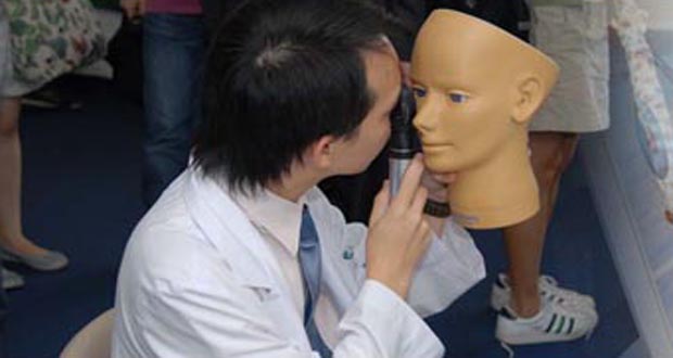
Teleophthalmology: A Model for Eye Care Delivery in Rural and Underserved Areas of India
 Optometry | FHNW
 Protecting Sight #322: Shah Rukh Khan's thoughts on humanity, fame and love. Wills Eye anterior segment surgery & bilateral...
 Casey Eye Institute | OHSU
 The Case of the Pregnant Woman With a Visual Disturbance - American Academy of Ophthalmology
 Douglas Donald Koch, M.D. | BCM
 DailyMed - PREDNISONE tablet
 Peters plus syndrome: MedlinePlus Genetics
DailyMed - PREDNISONE tablet
 Children's Eyes Examination Checklist: What All Parents Should Know | Bangkok Hospital
A Case of Shock-Induced Vision Loss
 Disease Models - Faculty - People - Biomedical Genetics and Genomics Graduate Program - Doctor of Philosophy(Ph.D.) - Graduate...
Final MRCOphth: Candidate 122 UK Final MRCOphth
Corneal13
- It leads to anomalies in the structure of the mature anterior segment, associated with an increased risk of glaucoma and corneal opacity. (wikipedia.org)
- Peters' (frequently misspelled as Peter's) anomaly is a specific type of mesenchymal anterior segment dysgenesis, in which there is central corneal leukoma, adhesions of the iris and cornea and abnormalities of the posterior corneal stroma, Descemet's membrane, corneal endothelium, lens and anterior chamber. (wikipedia.org)
- Abnormal migration, proliferation, differentiation, or survival of these cells contribute to diseases of the anterior segment such as corneal dystrophy, lens cataract, and glaucoma. (biomedcentral.com)
- Hamed AM, Wang L, Misra M, Koch DD " A comparative analysis of five methods of determining corneal refractive power in eyes that have undergone myopic laser in situ keratomileusis. . (bcm.edu)
- The severity of corneal clouding and other eye problems can vary between individuals with Peters plus syndrome, even among members of the same family. (medlineplus.gov)
- The left eye was microphthalmic, with a corneal diameter of 9 mm (right, 10 mm). (ajnr.org)
- secondary outcomes included evaluation of changes in Standard Patient Evaluation of Eye Dryness (SPEED) questionnaire, tear film break-up time (TBUT), corneal and conjunctival staining, tear osmolarity, matrix metalloproteinase-9 (MMP-9), artificial tear use and safety assessments. (springer.com)
- Eagle Eye Centre integrates ophthalmic and optometric eye care under a multisubspecialty practice, and offers medical and surgical services including: refractive surgery, presbyopia treatment, cataract surgery, retina services, corneal transplants, glaucoma and diabetic eye treatments, management of ocular inflammations, myopia screening and myopia control, and neuro-ophthalmology. (eagleeyecentre.com.sg)
- Our doctors treat corneal abrasions, dry eye, and many other corneal disorders and diseases including keratoconus and pterygium . (eagleeyecentre.com.sg)
- EEC is one of the first eye clinic to integrate ophthalmic and optometric eye care under one roof, and offers a comprehensive range of services including: refractive surgery, presbyopia treatment, cataract surgery, retina services, corneal transplants, glaucoma and diabetic eye treatments, management of ocular inflammations, myopia screening and myopia control. (eagleeyecentre.com.sg)
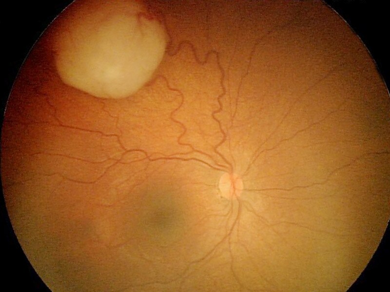
- Owing to co-morbidities like cata- on the disc, overpass phenomenon, nicking racts and corneal pathologies in the elderly of blood vessels or nerve fibre layer defect population in developing countries, viewing was noted, the eye was declared to have fundus details is also a challenge. (who.int)
- [ 1 ] Mpox virus may enter the eye via autoinoculation [ 2 ] and cause a range of problems from mild to severe, including conjunctivitis, blepharitis, keratitis, corneal ulcer, corneal scarring, and rarely loss of vision. (cdc.gov)
- Clinical judgement should be used in assessing the stability of underlying eye structures, and caution should be taken with obtaining swabs if corneal ulcers or severely painful lesions are present. (cdc.gov)
Cornea11
- After joining Doheny Eye Institute in 1987, Dr. Irvine has concentrated his clinical activities on consultations for anterior segment surgery, complicated cataract situations, and cornea/external disease including ocular surface tumors. (uclahealth.org)
- The anterior segment of the vertebrate eye is comprised of the cornea, lens, iris, ciliary body, and highly specialized tissue at the iridocorneal angle. (biomedcentral.com)
- For mammals and other higher vertebrates, refraction of light entering the eye is accomplished by both the transparent cornea and lens. (biomedcentral.com)
- In order to produce patient specific models, geometry of the eyes in particular the cornea is required. (biomedcentral.com)
- Anterior segment OCT (AS-OCT) allows the resolution of anterior and posterior surfaces of the entire cornea. (biomedcentral.com)
- This allows accurate measurement of the thickness and volume of the entire cornea, as well as the anterior chamber biometry such as its angle and depth. (biomedcentral.com)
- Other devices are either limited to only imaging a small section of the cornea or are unable to automatically segment the entire anterior segment like the Visante OCT system (Zeiss Meditec Inc., Dublin, California) [ 12 ]. (biomedcentral.com)
- The anterior segment consists of structures including the lens, the colored part of the eye (iris), and the clear covering of the eye (cornea). (medlineplus.gov)
- Peters anomaly involves abnormal development of the anterior segment, which results in a cornea that is cloudy (opaque) and causes blurred vision. (medlineplus.gov)
- I have sub-specialised in treatments of the cornea and refractive surgery, with a fellowship at the Singapore National Eye Centre and by carrying out a PhD on diagnostic applications of optical coherence tomography imaging of the cornea. (spirehealthcare.com)
- Slit lamp examination and dilated funduscopic examination can be helpful for determining whether anterior segment structures (conjunctiva, cornea, iris) or posterior segment structures (retina, nerve, choroid) are involved. (cdc.gov)
Dysgenesis8
- in mouse cranial neural crest results in anterior segment dysgenesis and early-onset glaucoma. (ucdenver.edu)
- Anterior segment mesenchymal dysgenesis, or simply anterior segment dysgenesis (ASD), is a failure of the normal development of the tissues of the anterior segment of the eye. (wikipedia.org)
- The first family (F1) presented with foveal hypoplasia and anterior segment dysgenesis, and the second family (F2) presented with foveal hypoplasia and chiasmal misrouting in the absence of albinism. (molvis.org)
- We report a new recessively inherited syndrome consisting of f oveal h ypoplasia, o ptic n erve d ecussation defects and a nterior segment dysgenesis, which we have abbreviated to FHONDA syndrome. (molvis.org)
- In this study, we extend the clinical phenotypes observed in each family and show that they have the same disorder, which we have termed f oveal h ypoplasia, o ptic n erve d ecussation defects and a nterior segment dysgenesis (FHONDA syndrome). (molvis.org)
- Primarily, several forms of glaucoma are associated with anterior segment dysgenesis and genes which are essential for formation of this part of the eye can promote glaucoma [ 9 , 10 ]. (biomedcentral.com)
- Mutation analysis of the genes associated with anterior segment dysgenesis, microcornea and microphthalmia in 257 patients with glaucoma. (cdc.gov)
- Compound heterozygous mutations in SMO associated with anterior segment dysgenesis and morning glory syndrome. (cdc.gov)
Ophthalmology9
- Her research is focused on topical glaucoma treatments and their effects on the ocular surface, micro-invasive glaucoma surgery, endocyclophotocoagulation in glaucoma management, developing strategies for managing chronic eye conditions in primary care and providing ophthalmology support for clinical trials in other sub specialties. (oxfordeyesurgery.com)
- He is also Vice President of the Imaging and Perimetry Society, Chair of the Membership Committee of the Glaucoma Research Society, and Workstream Lead for the design of the new Moorfields Eye Hospital and UCL Institute of Ophthalmology centre. (oxfordeyesurgery.com)
- Ophthalmology and Eye Diseases, 5, 1-3. (wikipedia.org)
- We treat eye conditions from the most straightforward to the most complex, and offer expert care in all ophthalmology specialties. (ohsu.edu)
- We offer world-class ophthalmology education opportunities at OHSU Casey Eye Institute. (ohsu.edu)
- Through our mobile clinic and preschool vision screening programs in Oregon and our international ophthalmology partnership in Myanmar, we are making a difference in eye health by bringing care to those who need it. (ohsu.edu)
- At Mercy Clinic Eye Specialists - Ophthalmology, our focus is to help preserve your most precious sense: your sight. (mercy.net)
- Around the Eye in 365 Days by Dr. Gary Schwartz is a quick look into the fascinating world of ophthalmology. (slackbooks.com)
- Benefiting from blended learning opportunities, you will learn about paediatric ophthalmology, strabismus (ocular misalignment), ocular motility disorders, and other eye and vision health issues and diseases. (ucl.ac.uk)
Glaucoma10
- One group studying acute angle-closure glaucoma (AACG) made measurements using OCT images and then used these to create a finite element model of the anterior chamber [ 10 ]. (biomedcentral.com)
- Glaucoma can be due to malformations of the anterior chamber or high episcleral venous pressure and in phakomatosis pigmentovascularis it can also be associated with angle hyperpigmentation. (hindawi.com)
- In 1879, Sturge reported on a case with bilateral facial nevus, vascular deformity, and congenital glaucoma in the right eye and spasms affecting the patient's left side of the body [ 9 ]. (hindawi.com)
- Larger than normal pupils, sensitivity to light and teary eyes may point to congenital glaucoma. (bangkokhospital.com)
- Eye pressure test for glaucoma. (bangkokhospital.com)
- ABSTRACT We carried out a validity assessment study for glaucoma screening procedures used dur- ing the survey conducted in Oman in 2005 on 6644 eyes in 3324 people 30 years. (who.int)
- Ocular pressure and fundus changes were the screening parameters used: glaucoma was found in 433 eyes. (who.int)
- An eye may, thus, be declared as not having glaucoma, but cannot be labelled as having glaucoma, using these parameters. (who.int)
- these can also of the eye with the help of torchlight and be used for early detection of glaucoma [ 4 ]. (who.int)
- neovascular membrane growth in the anterior chamber angle of the eye at the peripheral margin of the iris can occur, and this growth leads to neovascular glaucoma. (msdmanuals.com)
ISCHEMIA AFTER STRABISMUS SURGERY1
- Hopefully, adequate monitoring techniques during surgery could lead to the prediction and elimination of anterior segment ischemia after strabismus surgery. (lu.se)
Abnormalities4
- The identification of chiasmal misrouting in family F1 and anterior segment abnormalities in family F2 suggested that the families have the same clinical phenotype. (molvis.org)
- Ophthalmic examination revealed no abnormalities in the right eye. (ajnr.org)
- The anterior chamber and lens showed no abnormalities. (ajnr.org)
- The anterior eye segment showed no abnormalities. (ajnr.org)
Ocular anterior segment2
- The ocular anterior segment is critical for focusing incoming light onto the neural retina and for regulating intraocular pressure. (biomedcentral.com)
- Two main functions are ascribed to the ocular anterior segment. (biomedcentral.com)
Diseases7
- In addition, an understanding of ontogeny of the anterior segment has significance to several human diseases. (biomedcentral.com)
- This community outreach programme using telemedicine facilities has increased awareness of eye diseases, improved access to specialized health care, helped in local community empowerment, and provided employment opportunities. (hindawi.com)
- Screening is carried out in Chunampet district for diabetes and its complications especially diabetic eye diseases by using a mobile telemedicine van with satellite connectivity. (hindawi.com)
- In addition to a general outpatient clinic, the University Eye Hospital Tübingen has thirteen additional special outpatient clinics and consultations, as well as fourteen private consultations for various ophthalmological diseases. (uni-tuebingen.de)
- Our Neuro-ophthalmologist manages and treats diseases affecting the brain and nerves that are closely related to the eyes such as hemifacial spasms , double vision , myasthenia gravis and strokes affecting the visual pathway. (eagleeyecentre.com.sg)
- An increasing incidence of eye diseases has been registered in the last decades in developed countries due to the ageing of population, changes in lifestyle, environmental factors, and the presence of concomitant medical conditions. (mdpi.com)
- Pathogenic variants of AIPL1, MERTK, GUCY2D, and FOXE3 in Pakistani families with clinically heterogeneous eye diseases. (cdc.gov)
Moorfields Eye Hosp3
- A key benefit of studying this programme is that you will be taught by world-leading experts from UCL and Moorfields Eye Hospital. (ucl.ac.uk)
- You will be taught by Consultant Ophthalmologists and Orthoptists based at Moorfields Eye Hospital, ranked as the world's best eye hospital (SCImago Institutions Rankings 2023). (ucl.ac.uk)
- Our longstanding partnership with Moorfields Eye Hospital represents the largest co-located site for eye research, education and care in the world. (ucl.ac.uk)
Chamber2
- All agents injected into the anterior chamber can cause TASS. (dovepress.com)
- Five weeks later, enucleation was required due to enlargement of the mass with an increase of intraocular pressure, shallowness of the anterior chamber, and progression toward a phthisis bulbi. (ajnr.org)
Retinal9
- The telemedicine van is equipped with a digital retinal camera by which retinal imaging is performed by eye technicians who are unemployed youth recruited from the local area and trained at our centre. (hindawi.com)
- Macula, retinal vessels, and periphery appeared normal in both eyes. (aao.org)
- Dilated fundus exam demon-strated diffuse retinal pigment epithelium atrophy with pigment deposition and a bull's-eye pattern. (aao.org)
- We report a 6-month-old boy who presented with unilateral leukocoria, retinal detachment, and a retrolental mass in a microphthalmic eye based on retinal dysplasia with concurrent optic nerve aplasia. (ajnr.org)
- Fundus photograph of the left eye shows a retrolental mass (D, arrows ) in the inferomedial quadrant of the vitreous (V) with large irregular feeder vessels, focal hemorrhages, and retinal detachment (R, arrowheads ). (ajnr.org)
- Axial T1WI ( A ) image shows a hyperintense mass in the anterior part of the vitreous (V), adjacent to the ciliary body (CB) on either side of the lens, combined with a tent-shaped retinal detachment with hyperintense subretinal fluid (SF). (ajnr.org)
- Our lab develops and uses adaptive optics, eye movement correction and optical microscopy technologies to improve the non-invasive visualization of the retina to the point that individual retinal structure and function and can be visualized at the cellular and even sub-cellular scale. (stanford.edu)
- Invited Session V: The eye as a window to systemic and neurodegenerative health: Seeking Answers through a keyhole: Harnessing the Synergy of Dynamic OCT/OCT Angiography and Adaptive Optics SLO for Retinal Assessment of Systemic Disease. (stanford.edu)
- En face OCT reflectance images which accompany OCTA studies offer a glimpse of the macrophage-like cellular activity above the retinal surface which responds to systemically instigated vascular events below. (stanford.edu)
Toxic anterior3
- To report toxic anterior segment syndrome (TASS) after cataract surgery possibly associated with intracameral use of cefuroxime. (dovepress.com)
- Toxic anterior segment syndrome (TASS) is postoperative anterior segment inflammation. (dovepress.com)
- Toxic Anterior Segment Syndrome. (slackbooks.com)
Intraocular3
- He has been involved in multiple clinical trials which include studies on dry eye treatment, infectious keratitis, and intraocular lenses. (uclahealth.org)
- Intraocular pressure was 15 mm Hg in both eyes. (aao.org)
- Intraocular pressure was 12 mm Hg in each eye. (medscape.com)
Lens4
- Development of the anterior segment initiates with the invagination of the lens from surface ectoderm. (biomedcentral.com)
- With establishment of the lens vesicle, head mesoderm and neural crest cells migrate into a periocular location and eventually move into the anterior segment of the rudimentary eye between the surface ectoderm and the neural retina and lens. (biomedcentral.com)
- Histopathologic examination and immunohistochemistry for collagen type V were performed on residual anterior lens capsules of vitrectomized eyes (2 patients) that had been tamponaded with silicone oil. (nih.gov)
- Anterior segment exam revealed mild nuclear sclerosis of the lens. (aao.org)
SURGERY17
- It usually develops after uncomplicated anterior segment surgery. (dovepress.com)
- Did you mean topic:"anterior eye segments - surgery" ? (nshealth.ca)
- Trocar surgery has the potential to revolutionize anterior segment surgery, demonstrated here as an easy-to-use technique for conquering the pars plana region. (nshealth.ca)
- Equipment for Surgery of Anterior Segment -- 3. (nshealth.ca)
- Trocar Surgery of Posterior Segment -- 18. (nshealth.ca)
- Introduction and Possible Indications for Trocar Surgery of Posterior Segment -- 19. (nshealth.ca)
- Equipment for Trocar Surgery of Posterior Segment -- 20. (nshealth.ca)
- Wills Eye anterior segment surgery & bilateral fourth nerve palsy. (protectingsight.com)
- 2. Wills Eye Anterior Segment Surgery Update - One of the best Saturday morning courses I've attended. (protectingsight.com)
- I am a consultant eye surgeon (ophthalmologist) with an interest in the anterior segment of the eye and cataract surgery, working at University Hospital Southampton NHS Trust since 2015. (spirehealthcare.com)
- I specialise in treatments and surgery of the anterior segment of the eye. (spirehealthcare.com)
- Eyes are accessible, enabling minimally invasive surgery and precise drug delivery. (nih.gov)
- This project focuses on evaluating novel imaging techniques for measurement of perfusion and oxygenation in the anterior segment of the eye and the eye muscles during strabismus surgery. (lu.se)
- Anterior segment ischemia is a rare but severe complication to strabismus surgery. (lu.se)
- It is generally believed that to reduce the risk of anterior segment ischemia, only two muscles should be operated on during strabismus surgery and a third muscle can only be operated on given that 6 months healing time has passed. (lu.se)
- Knowledge of the effect of strabismus surgery on perfusion to the anterior segments of the eye is virtually non-existent. (lu.se)
- Furthermore, patients that are considered to have high risk of developing anterior segment ischemia are often not offered surgery at all or operated on with a suboptimal surgical method when a perfusion examination might show that standard procedures could be performed. (lu.se)
Inflammation1
- Dry eye syndrome (DES) is a multifactorial disease of the ocular surface associated with reduced tear secretion, increased osmolarity of the tears and inflammation that may lead to ocular surface damage. (springer.com)
Posterior segments2
- The increase of public awareness on ocular conditions leads to an early diagnosis and treatment, as well as an increased demand for more effective and minimally invasive solutions for the treatment of both the anterior and posterior segments of the eye. (mdpi.com)
- Anterior and posterior segments, together with neuro-ophthalmic disorders were found among stroke patients in this study. (who.int)
Examination12
- Children's eye examination can be divided according to 3 main age groups. (bangkokhospital.com)
- Moreover, pediatrician usually sends children with developmental delay or Down's Syndrome to undergo eye examination for cataract and other vision problems. (bangkokhospital.com)
- If there is still any doubt, the doctor may order dilating eye drops for detailed examination. (bangkokhospital.com)
- Slit-lamp examination of the anterior segment was normal. (medscape.com)
- A smear of a blister adjacent to the eye showing the Romaña sign yielded T. cruzi on direct examination. (cdc.gov)
- A general or comprehensive medical eye examination is designed to reveal both existing and potential eye problems, even in the absence of specific symptoms. (bcm.edu)
- The examination consists of a patient history, examination and testing to assess the functional behavior, anatomic status of the eye and the related structures. (bcm.edu)
- PPE used by healthcare personnel who enter the patient's room or perform any eye examination should include gown, gloves, eye protection (i.e., goggles or a face shield that covers the front and sides of the face), and a NIOSH-approved particulate respirator equipped with N95 filters or higher. (cdc.gov)
- On ocular examination, the patient was blinking to light with both eyes. (medscape.com)
- Examination of the anterior segment was unremarkable. (medscape.com)
- EQUALITY study participants received a comprehensive eye exam by an optometrist including medical history, visual acuity with walk-in and best correction, refraction, color vision, applanation tonometry, pachymetry, undilated slit lamp anterior segment examination, undilated gonioscopy, and dilated fundus examination. (cdc.gov)
- Examination of the eyes by an ophthalmologist is essential to assess for papilledema, which indicates elevated intracranial pressure. (medscape.com)
Exam4
- Anterior segment exam was normal. (aao.org)
- On dilated fundus exam, she had very subtle, deep hyperpigmented spots near the fovea in her left eye. (aao.org)
- Will I receive a prescription for contact lenses or glasses with my general eye exam? (bcm.edu)
- If you know in advance that you are interested in contact lenses, please inform the patient care representative so that an appointment will be made for you to be seen when you are here for your general eye exam. (bcm.edu)
Structures of the anterior segment3
- The structures of the anterior segment arise from diverse embryonic lineages and there is exquisite coordination among the different compartments during development. (biomedcentral.com)
- However, the relevant cellular interactions between various structures of the anterior segment and the molecular basis of development is just beginning to be understood. (biomedcentral.com)
- However, their underlying program for the extraction of the structures of the anterior segment is not evaluated for accuracy. (biomedcentral.com)
Development of the anterior segment1
- A detailed understanding of the mechanisms of development of the anterior segment can provide general insights into questions such as tissue induction, cell type fate determination, and the regulation of cellular morphogenesis. (biomedcentral.com)
Developmental1
- Orthoptists are Allied Health Professionals who are experts in the diagnosis and management of developmental eye conditions, defects in eye movement and binocular vision. (ucl.ac.uk)
Ophthalmic2
- Each of the Alkek Eye Center physicians is qualified to treat a broad range of ophthalmic conditions. (bcm.edu)
- Despite being the most common route of ophthalmic drug administration, eye drops are associated with compliance issues, drug wastage by lacrimation, and low bioavailability due to the ocular barriers. (mdpi.com)
Alkek Eye Center4
- Alkek Eye Center physicians and technicians maintain a full patient care schedule. (bcm.edu)
- Alkek Eye Center offers a full-service optical shop, BCM Optical , as a part of our facility. (bcm.edu)
- Does Alkek Eye Center have a program to assist low-income patients? (bcm.edu)
- Alkek Eye Center physicians are available to address your eye care needs 24-hours a day, seven days a week. (bcm.edu)
Tissues2
- These same genes then regulate mesenchymal cell differentiation to give rise to distinct anterior segment tissues. (wikipedia.org)
- Interplay between PITX2 and FOXC1 in the development of different anterior segment tissues may partly explain the phenotypic variability and the genetic heterogeneity characteristic of ASD. (wikipedia.org)
Pathologies1
- Smoking and Eye Pathologies. (benthamscience.com)
Patient's1
- If ocular involvement of mpox virus is suspected, then ophthalmologic consultation should be strongly considered for a thorough evaluation and continued monitoring of the patient's condition and extent of disease, especially in cases of vision changes, eye pain, or increasing redness. (cdc.gov)
Ophthalmologists3
- While our ophthalmologists specialize in medical eye exams, our optometrists at Mercy Eye Care focus on routine eye exams for glasses and contact lenses. (mercy.net)
- The one page a day format plus wide ranging topics, makes Around the Eye in 365 Days a fun and interesting read for all in the field from general ophthalmologists to optometrists to residents to students to office staff to industry sales forces. (slackbooks.com)
- In a field situation, however, even skilled segment of the eye was examined using ophthalmologists find it difficult to diag- direct ophthalmoscope. (who.int)
Malformations1
- Expanding the spectrum of FOXC1 and PITX2 mutations and copy number changes in patients with anterior segment malformations. (cdc.gov)
Retina2
- All quadrants of the retina are imaged by the eye technician. (hindawi.com)
- The optics of the eye can be thought of as an imperfect microscope objective through which the retina can be observed. (stanford.edu)
Disorders2
- They should be assessed for eye disorders and lazy eye condition. (bangkokhospital.com)
- We deliver diagnosis and treatment services for a variety of eye disorders. (mercy.net)
20201
- Experimental eye research 2020 6 197 108118. (cdc.gov)
Ophthalmologist1
- Parents should take heed and have their children's eyes examined annually, and follow the advice of their pediatric ophthalmologist accordingly. (bangkokhospital.com)
Ciliary5
- Ciliary body melanoma is a subtype of uveal melanoma, the most common primary malignant tumor of the eye. (medscape.com)
- They can be classified as anterior uveal melanomas when the tumor arises in the iris and as posterior uveal melanomas when it arises in either the choroid or the ciliary body. (medscape.com)
- Left: Illustration of an eye and the four rectus muscles with the anterior ciliary arteries. (lu.se)
- Right: One of our first LSCI images showing the eye and the perfusion araising from the anterior ciliary arteries. (lu.se)
- The condition occurs due to damage of the anterior ciliary arteries that course along the rectus muscles and therefore gets damaged during surgical manipulation. (lu.se)
Fellowship1
- Our team is comprised of fellowship-trained eye specialists and all providers are welcoming new patients and accept most insurance plans. (mercy.net)
Bilateral1
- Peters anomaly is usually bilateral, which means that it affects both eyes. (medlineplus.gov)
Iris1
- A transparent, biconvex structure of the EYE, enclosed in a capsule and situated behind the IRIS and in front of the vitreous humor (VITREOUS BODY). (harvard.edu)
Fundus1
- Fundus autofluo-rescence showed an array of lobules and specks of hypoautofluorescence arranged in concentric rings in the macula of both eyes (as shown in Fig. 2 of the right eye). (aao.org)
Examine4
- 8 year old girl with hyperopic correction, attending with mom.Asked to examine eye movements.Duane s type 1 with small left esophoria for near without specs.Orthophoric with specs.Asked management. (mrcophth.com)
- Asked to examine orbits,DO NOT examine eye movements. (mrcophth.com)
- Examine posterior segment of right eye. (mrcophth.com)
- Examine Posterior segment of BE. (mrcophth.com)
Differentiation1
- Similarly, the relative timing of tissue differentiation in the anterior segment is also conserved with other vertebrates. (biomedcentral.com)
PITX21
- In this review, the role of the ASD genes, PITX2 and FOXC1, is considered in relation to the embryology of the anterior segment, the biochemical function of these proteins, and their role in development and disease aetiology. (wikipedia.org)
Anomaly1
- An eye problem called Peters anomaly is the most common anterior segment abnormality seen in Peters plus syndrome. (medlineplus.gov)
Macula1
- The macula in the left eye also showed a white intraretinal lesion, with a small hemorrhage above the fovea (Figure 3). (medscape.com)
Optic nerve1
- ION is of two types: anterior (AION) and posterior (PION), the first involving the anterior part of the optic nerve (also called the optic nerve head, ONH) and the second, the rest of the optic nerve. (intechopen.com)
MeSH1
- Anterior Eye Segment" is a descriptor in the National Library of Medicine's controlled vocabulary thesaurus, MeSH (Medical Subject Headings) . (ucdenver.edu)
Syndrome4
- Drooping upper eyelid on either side or both may have been caused by lazy eye syndrome. (bangkokhospital.com)
- There are many techniques to assess lazy eye syndrome. (bangkokhospital.com)
- The objective of this study was to evaluate the efficacy and safety of our multi-ingredient supplement in subjects with dry eye syndrome (DES). (springer.com)
- This study was conducted to evaluate efficacy of a multi-ingredient supplement containing lutein, zeaxanthin, curcumin and vitamin D3 in subjects with dry eye syndrome (DES). (springer.com)
Symptoms4
- Artificial tears and topical corticosteroids used as first-line treatments usually improve the symptoms of dry eyes but are associated with adverse effects with long-term use. (springer.com)
- Overall, we have demonstrated statistically significant clinical improvements in dry eye symptoms as compared to the placebo in subjects validated through well-established subjective questionnaire-based approaches such as OSDI and SPEED. (springer.com)
- Respiratory irritation (37%) and eye irritation (23%) were the most commonly reported symptoms. (medscape.com)
- A history of self-reported dry eye symptoms for two months prior to study enrollment, - Diagnosis of MGD Low delivery type, with mechanism obstructive and non- cicatricial with scores as follows: Ocular Surface Disease Index (OSDI) questionnaire = 13, Non-Invasive Tear break-up time (NIBUT) lower than 10 seconds (The Sirius anterior segment analyzer (CSO, Florence, Italy). (who.int)
Fibrils4
- In this study, a quadrant of the anterior segment from a normal human donor eye was dynamically pressurized in the SC lumen, and imaged using a customized optical coherence tomography (OCT). The TM/JCT/SC complex finite element (FE) with embedded collagen fibrils was reconstructed based on the segmented boundary nodes in the OCT images. (lu.se)
- Fibrillenstruktur (or fast-twitch) muscle fibrils generate fast eye movements and are composed of well-defined myofibrils with well-developed sarcomeres. (medscape.com)
- Felderstruktur muscle fibrils generate slow or tonic eye movements and are composed of poorly defined myofibrils with poorly developed sarcomeres. (medscape.com)
- The innervation to fibrillenstruktur fibrils is thick and heavily myelinated, with a single (en plaque) neuromuscular junction, whereas the innervation to felderstruktur fibrils is thin, with multiple grapelike clusters of neuromuscular junctions. (medscape.com)
Binocular vision1
- Video eye-tracking systems for investigation of the processes involved in binocular coordination and the importance of binocular vision. (fhnw.ch)
Patients6
- Seventeen eyes of 17 patients who were diagnosed with TASS were enrolled in this study. (dovepress.com)
- Prior to the operation, tropicamide 1% and phenylephrine hydrochloride 2.5% were applied to patients' eyes for pupil dilation. (dovepress.com)
- When carrying out biomechanical modelling of the eye, one of the aims is to be able to produce patient specific models so that the eyes of individual patients can be simulated providing important information towards personalised medicine. (biomedcentral.com)
- Retrospective, consecutive study of 282 eyes (282 patients) who underwent a secondary IOL implantation using the Carlevale IOL (Soleko IOL Division, Italy) with two anchor haptics for intrascleral implantation with either 23- or 27-gauge (G) port. (karger.com)
- Our experienced providers and caring staff are committed to providing the best eye care experience possible for our patients. (mercy.net)
- Editas Medicine, a biotech company building on work by NEI researchers is conducting the first-in-human CRISPR trial to help patients with vision loss caused by a mutation in CEP290, another crucial gene in the eye, which was also discovered by NEI scientists. (nih.gov)
Vision14
- At OHSU Casey Eye Institute we offer world-class specialists, state-of-the-art technologies and truly personalized care to promote healthy vision. (ohsu.edu)
- Since 1945, OHSU Casey Eye Institute doctors and vision scientists have worked side by side to discover the causes of eye disease and find new treatments. (ohsu.edu)
- At 30 weeks of pregnancy, she noticed that vision in her left eye became blurry. (aao.org)
- While in the ER, she complained of seeing blurry spots near the center of her vision in the left eye. (aao.org)
- Her vision was 20/20 in both eyes. (aao.org)
- So, it is important for the children to have their eyes examined periodically to ensure the health and proper care for their vision. (bangkokhospital.com)
- This is the time when parents need to be alert for vision problems that include crossed eyes or lazy eye. (bangkokhospital.com)
- A 60-year-old man with anemia secondary to a duodenal ulcer was referred for evaluation of a 2-week history of painless, reduced vision in both eyes. (medscape.com)
- Color vision with Ishihara pseudoisochromatic plates was 10/10 in both eyes. (medscape.com)
- As you know from your regular visits to the eye doctor, the goal is to maintain 20/20 vision. (nih.gov)
- Investments in vision research accelerate biomedical progress because eyes possess unique features. (nih.gov)
- NEI recently funded a clinical study that tested NGoggle, an easy-to-wear device that can assess vision loss by analyzing signals between the brain and eyes. (nih.gov)
- Involvement of the eyes can be a vision-threatening condition and should be treated urgently. (cdc.gov)
- In children, strabismus can cause severe permanent vision impairment but in adults the main problem is diplopia or social difficulties such as avoiding eye contact, which can be very disabling. (lu.se)
Angle3
- They travel anteriorly and laterally at an angle of 23º with the visual axis of the eye in primary position. (medscape.com)
- The tendon of the superior oblique muscle passes through the trochlea (which is located nasally at the superior orbital rim) and is reflected inferiorly, posteriorly, and laterally at an angle of 51º to the visual axis with the eye in primary position. (medscape.com)
- It passes posteriorly and laterally in the orbit, forming an angle of 51º with the visual axis of the eye in primary position, before passing beneath the inferior rectus muscle and inserting posterior to the equator on the inferior and lateral aspect of the globe. (medscape.com)
Contact lenses1
- For routine eye exams for glasses and contact lenses visit Mercy Eye Care - Optometry - Medical Tower A Suite 140A . (mercy.net)
Graph1
- This graph shows the total number of publications written about "Anterior Eye Segment" by people in this website by year, and whether "Anterior Eye Segment" was a major or minor topic of these publications. (ucdenver.edu)
Right eye4
- She underwent Humphrey visual field testing, as an inpatient, which showed a full field in the right eye and a small paracentral scotoma in the left. (aao.org)
- After considering her normal visual field in the right eye, paracentral scotoma in the left eye, and lack of a relative afferent pupillary defect, we believed a maculopathy was more likely. (aao.org)
- OCT showed normal architecture in the right eye and attenuation of the ellipsoid layer in the left. (aao.org)
- Automated perimetry of the right eye showing superior altitudinal defect. (medscape.com)
Silicone1
- Silicone oil filled eye, no indentation. (mrcophth.com)