Anterior Cerebral Artery
Infarction, Anterior Cerebral Artery
Cerebral Angiography
Intracranial Aneurysm
Middle Cerebral Artery
Circle of Willis
Cerebral Arterial Diseases
Cerebral Infarction
Infarction, Middle Cerebral Artery
Subarachnoid Hemorrhage
Aneurysm, Ruptured
Tomography, X-Ray Computed
Carotid Artery, Internal
Ultrasonography, Doppler, Transcranial
Basilar Artery
Magnetic Resonance Angiography
Brain Ischemia
Blood Flow Velocity
Cerebral Revascularization
Echoencephalography
Intracranial Embolism and Thrombosis
Vasospasm, Intracranial
Ischemic Attack, Transient
Carotid Arteries
Brain
Angiography, Digital Subtraction
Arterial Occlusive Diseases
Intracranial Arteriovenous Malformations
Brain Hemorrhage, Traumatic
Embolization, Therapeutic
Vertebral Artery
Posterior Cerebral Artery
Akinetic Mutism
Intracranial Arteriosclerosis
Cerebrovascular Disorders
Moyamoya Disease
Cerebral Hemorrhage
Magnetic Resonance Imaging
Aneurysm, Dissecting
Surgical Instruments
Ultrasonography, Doppler
Collateral Circulation
Stroke
Treatment Outcome
Hematoma
Carotid Stenosis
Pulmonary Artery
Ultrasonography
Frontal Lobe
Cerebral Palsy
Carotid Artery Diseases
Mesenteric Arteries
Retrospective Studies
Hematoma, Subdural
Postoperative Complications
Intracranial Embolism
Temporal Arteries
Follow-Up Studies
Brain Edema
Infarction, Posterior Cerebral Artery
Larger anastomoses in angiotensinogen-knockout mice attenuate early metabolic disturbances after middle cerebral artery occlusion. (1/108)
Abnormalities in the homeostasis of the renin-angiotensin system have been implicated in the pathogenesis of vascular disorders, including stroke. The authors investigated whether angiotensinogen (AGN) knockout mice exhibit differences in brain susceptibility to focal ischemia, and whether such differences can be related to special features of the collateral circulation. Wild-type and AGN-knockout mice were submitted to permanent suture occlusion of the middle cerebral artery (MCA). The collateral vascular system was visualized by systemic latex infusion, and the ischemic lesions were identified by cresyl-violet staining. The core and penumbra of the evolving infarct were differentiated by bioluminescence and autoradiographic imaging of ATP and protein biosynthesis, respectively. In wild-type mice, mean arterial blood pressure was 95.0 +/- 8.6 mm Hg, and the diameter of fully relaxed anastomotic vessels between the peripheral branches of the anterior and middle cerebral arteries 26.6 +/- 4.0 microm. In AGN knockouts, mean arterial blood pressure was significantly lower, 71.5 +/- 8.5 mm Hg (P < .01), and the anastomotic vessels were significantly larger, 29.4 +/- 4.6 microm (P < .01). One hour after MCA occlusion, AGN-knockout mice exhibited a smaller ischemic core (defined as the region of ATP depletion) but a larger penumbra (the area of disturbed protein synthesis with preserved ATP). At 24 hours after MCA occlusion, this difference disappeared, and histologically visible lesions were of similar size in both strains. The observations show that in AGN-knockout mice the more efficient collateral blood supply delays ischemic injury despite the lower blood pressure. Pharmacologic suppression of angiotensin formation may prolong the therapeutic window for treatment of infarcts. (+info)Flow dynamics in a lethal anterior communicating artery aneurysm. (2/108)
We describe and analyze the flow dynamics in replicas of a human anterior communicating artery aneurysm. The replicas were placed in a circuit of pulsating non-Newtonian fluid, and flows were adjusted to replicate human physiologic parameters. Individual slipstreams were opacified with isobaric dyes, and images were recorded on film and by CT/MR angiography. When flow in the afferent (internal carotid) and efferent (anterior and middle cerebral) arteries was bilaterally equal, slipstreams rarely entered the aneurysm. When flow in either the afferent or efferent vessels was not symmetrical, however, slipstreams entered the aneurysm neck, impinged upon the aneurysm dome, and swirled within the aneurysm. Unequal flow in carotid or cerebral systems may be necessary to direct pathologic, fluid slipstreams into an aneurysm. (+info)Hemodynamic changes around cerebral arteriovenous malformation before and after embolization measured with PET. (3/108)
To estimate the changes in regional cerebral blood flow (rCBF) around cerebral arteriovenous malformation (AVM) before and after embolization, 6 patients with AVM were sequentially examined with positron emission tomography (PET). PET depicted the remodeling of rCBF in the ipsilateral hemisphere of AVM after embolization. Decrease of rCBF in the ipsilateral hemisphere was also detected in patients with focal symptoms before embolization, and improvement of clinical symptoms after embolization corresponded to disappearance of rCBF decrease. PET can detect hemodynamic changes after embolization, and has a possibility to estimate the effect of embolization in patients with AVM. (+info)Neuropsychological changes after surgery for anterior communicating artery aneurysm. (4/108)
Neuropsychological disturbances following surgery for anterior communicating artery aneurysms were analyzed in 26 patients (11 males, 15 females) using the Hasegawa dementia scale-revised (HDS-R) over a 3-year period. The patients were aged from 34 to 76 years (mean 54.1 years). Lesions in the frontal lobe were evaluated using computed tomography (CT). Twenty-three patients had symptoms over the course. Four patients had basal forebrain lesion, five had ventral frontal lesion, and 12 had no lesion. Patients with basal forebrain lesion and no lesion tended to show disorientation. The mean HDS-R score was 10.2 points in the patients with ventral frontal lesion, and 13.5 points in the patients with no lesion. These scores are within the range for dementia. The mean HDS-R score in patients with basal forebrain and striate lesions was over 25 points and beyond the range for dementia. Significant differences were observed in the HDS-R score between patients with ventral frontal lesion and basal forebrain lesion, and between patients with no lesion and basal forebrain lesion (p < 0.05). Recovery from neuropsychological disturbances was poorer in patients with ventral frontal lesion and no lesion compared to those with basal forebrain and striate lesions, and their symptoms tended to persist. (+info)Prolapsing gyrus rectus as a cause of progressive optic neuropathy. (5/108)
The pathogenesis of optic neuropathy caused by neurovascular compression or by similar mechanisms is unclear. Thin-slice magnetic resonance (MR) imaging was performed in 69 patients with optic neuropathy without demonstrable ophthalmological lesions (57.0 +/- 17.1 years of age) and 102 normal subjects (57.7 +/- 13.9 years of age). The MR imaging features were classified into "no compression" by the internal carotid artery (ICA), "compression" by the ICA, "no contact" with the anterior cerebral artery (ACA) or the gyrus rectus, "contact" with either or both, "compression" by the ACA, and "compression" by the gyrus rectus. The Spearman correlation coefficients were calculated between patients or controls, the MR classification, and the age, and the number of patients in each MR classification were evaluated by the chi 2 test. Five of the 69 patients with rapidly progressive symptoms were operated on via the frontotemporal approach. The MR imaging feature of "compression" by the gyrus rectus was the best predictor of optic neuropathy (Spearman correlation coefficients rho = -0.23646, p < 0.0018). This MR imaging feature was observed in 38 of 69 patients and in 32 of 102 controls (p = 0.002). Compression of the nerve by the gyrus rectus or the ACA was confirmed in all five operated cases. Decompression of the nerve was fully achieved in four of the five patients, and their symptoms have not progressed since then. Optic neuropathies due to compression by the prolapsing gyrus rectus are not well understood. Such neuropathies may be detected by MR imaging. (+info)Ruptured aneurysm at the origin of the median artery of the corpus callosum associated with accessory middle cerebral artery--case report. (6/108)
A 62-year-old male presented with ruptured anterior communicating artery (ACoA) aneurysm manifesting as severe headache associated with the rare combination of median artery of the corpus callosum (MACC) and accessory middle cerebral artery (MCA). Computed tomography demonstrated diffuse subarachnoid hemorrhage. Left carotid angiography demonstrated an anomalous vessel originating from the ACoA complex and passing forward in the interhemispheric fissure between the two companion A2 segments. This vessel was identified as the MACC. Another anomalous vessel originated from the left A1-A2 segment and passed into the sylvian fissure. This vessel was identified as the accessory MCA. Left frontotemporal craniotomy was performed to clip the neck of the aneurysm. After identifying both A1 and A2 segments, accessory MCA, and the MACC, the aneurysm neck was occluded successfully. The ACoA complex is one of the most frequent sites of vascular anomalies. Preoperative and intraoperative care is required to identify the presence of anomalies of the ACoA complex prior to clip placement, to avoid accidental damage or clipping, which may result in severe neurological deficits. (+info)Histomorphometrical study of the elastic fiber system in the anterior cerebral artery of man. (7/108)
The aim of the present study was to quantify the distribution of the elastic fiber system within the wall of the anterior cerebral artery. The study is based on the works of Glynn (1940) and Stehbens (1989) concerning the incidence and origin of brain aneurysms and recent studies of the elastic fibers. The anterior cerebral artery was divided into three segments, S1, S2 and S3: S1 corresponds to the origin of the anterior cerebral artery, S2 is located at the junction of the anterior cerebral artery with the anterior communicating artery, and S3 at the junction of the rostrum and genu of the corpus callosum,which were submitted to routine histological procedures. A histomorphometrical study was undertaken using an estimation of the linear density (Ld) of the components of the fibrous elastic system which evaluates their full length in each segment. Data were analyzed using first order linear regression methods. The results show a decreasing quantity of elastic fibers in the three segments (S1>S2>S3). Study of the elastic fiber system may originate new concepts regarding the genesis of cerebral artery aneurysm. (+info)A modified transorbital baboon model of reperfused stroke. (8/108)
BACKGROUND AND PURPOSE: Although pathophysiological studies of focal cerebral ischemia in nonhuman primates can provide important information not obtainable in rodent models, primate experimentation is limited by considerations of cost, availability, effort, and ethics. A reproducible and quantitative model that minimizes the number of animals necessary to detect differences between treatment groups is therefore crucial. METHODS: Eight male baboons (weight, 22+/-2 kg) underwent left transorbital craniectomy followed by 1 hour of temporary ipsilateral internal carotid artery occlusion at the level of the anterior choroidal artery together with bilateral temporary occlusion of both anterior cerebral arteries (A1) proximal to the anterior communicating artery. A tightly controlled nitrous oxide-narcotic anesthetic allowed for intraoperative motor evoked potential confirmation of middle cerebral artery (MCA) territory ischemia. Animals survived to 72 hours or 10 days if successfully self-caring. Outcomes were assessed with a 100-point neurological grading system, and infarct volume was quantified by planimetric analysis of both MRI and triphenyltetrazolium chloride-stained sections. RESULTS: Infarction volumes (on T2-weighted images) were 32+/-7% (mean+/-SEM) of the ipsilateral hemisphere, and neurological scores averaged 29+/-9. All animals demonstrated evidence of hemispheric infarction, with damage evident in both cortical and subcortical regions in the MCA vascular territory. Histologically determined infarction volumes differed by <3% and correlated with absolute neurological scores (r=0.9, P:=0.003). CONCLUSIONS: Transorbital temporary occlusion of the entire anterior cerebral circulation with strict control of physiological parameters can reliably produce reperfused MCA territory infarction. The magnitude of the resultant infarct with little interanimal variability diminishes the potential number of animals required to distinguish between 2 treatment regimens. The anatomic distribution of the infarct and associated functional deficits offer comparability to human hemispheric strokes. (+info)The Anterior Cerebral Artery (ACA) is a paired set of arteries that originate from the internal carotid artery or its branch, the posterior communicating artery. They supply oxygenated blood to the frontal lobes and parts of the parietal lobes of the brain.
The ACA runs along the medial side of each hemisphere, anterior to the corpus callosum, which is the largest bundle of nerve fibers connecting the two hemispheres of the brain. It gives off branches that supply the motor and sensory areas of the lower extremities, as well as the areas responsible for higher cognitive functions such as language, memory, and emotion.
The ACA is divided into several segments: A1, A2, A3, and A4. The A1 segment runs from its origin at the internal carotid artery to the anterior communicating artery, which connects the two ACAs. The A2 segment extends from the anterior communicating artery to the bifurcation of the ACA into its terminal branches. The A3 and A4 segments are the distal branches that supply the frontal and parietal lobes.
Interruptions or blockages in the flow of blood through the ACA can lead to various neurological deficits, including weakness or paralysis of the lower extremities, language impairment, and changes in cognitive function.
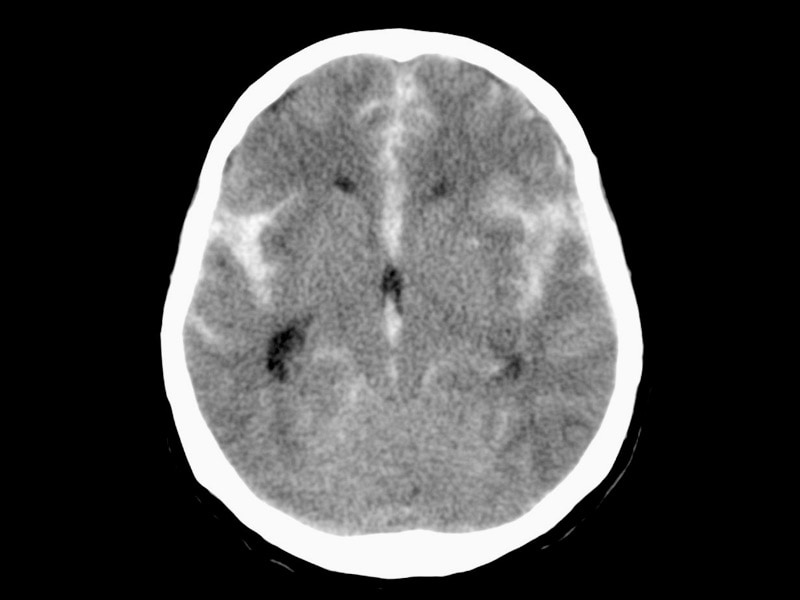
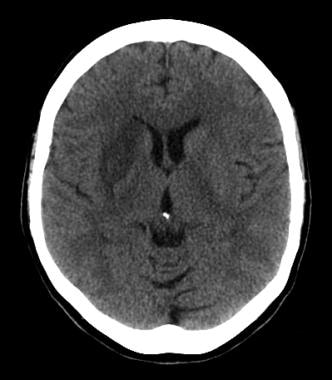
Anterior cerebral artery infarction refers to the death of brain tissue (also known as an infarct) in the territory supplied by the anterior cerebral artery (ACA) due to insufficient blood flow. The ACA supplies oxygenated blood to the frontal lobes of the brain, which are responsible for higher cognitive functions such as reasoning, problem-solving, and decision-making, as well as motor control of the lower extremities.
An infarction in this territory can result from various causes, including atherosclerosis, embolism, thrombosis, or vasospasm. Symptoms of an ACA infarction may include weakness or paralysis on one side of the body (usually the lower extremities), difficulty with coordination and balance, urinary incontinence, changes in personality or behavior, and impaired cognitive function. The severity of symptoms depends on the extent and location of the infarct. Immediate medical attention is necessary to prevent further damage and improve the chances of recovery.
Cerebral arteries refer to the blood vessels that supply oxygenated blood to the brain. These arteries branch off from the internal carotid arteries and the vertebral arteries, which combine to form the basilar artery. The major cerebral arteries include:
1. Anterior cerebral artery (ACA): This artery supplies blood to the frontal lobes of the brain, including the motor and sensory cortices responsible for movement and sensation in the lower limbs.
2. Middle cerebral artery (MCA): The MCA is the largest of the cerebral arteries and supplies blood to the lateral surface of the brain, including the temporal, parietal, and frontal lobes. It is responsible for providing blood to areas involved in motor function, sensory perception, speech, memory, and vision.
3. Posterior cerebral artery (PCA): The PCA supplies blood to the occipital lobe, which is responsible for visual processing, as well as parts of the temporal and parietal lobes.
4. Anterior communicating artery (ACoA) and posterior communicating arteries (PComAs): These are small arteries that connect the major cerebral arteries, forming an important circulatory network called the Circle of Willis. The ACoA connects the two ACAs, while the PComAs connect the ICA with the PCA and the basilar artery.
These cerebral arteries play a crucial role in maintaining proper brain function by delivering oxygenated blood to various regions of the brain. Any damage or obstruction to these arteries can lead to serious neurological conditions, such as strokes or transient ischemic attacks (TIAs).
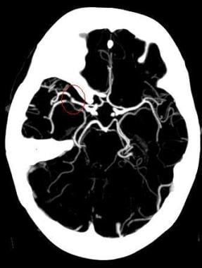
Cerebral angiography is a medical procedure that involves taking X-ray images of the blood vessels in the brain after injecting a contrast dye into them. This procedure helps doctors to diagnose and treat various conditions affecting the blood vessels in the brain, such as aneurysms, arteriovenous malformations, and stenosis (narrowing of the blood vessels).
During the procedure, a catheter is inserted into an artery in the leg and threaded through the body to the blood vessels in the neck or brain. The contrast dye is then injected through the catheter, and X-ray images are taken to visualize the blood flow through the brain's blood vessels.
Cerebral angiography provides detailed images of the blood vessels in the brain, allowing doctors to identify any abnormalities or blockages that may be causing symptoms or increasing the risk of stroke. Based on the results of the cerebral angiography, doctors can develop a treatment plan to address these issues and prevent further complications.
An intracranial aneurysm is a localized, blood-filled dilation or bulging in the wall of a cerebral artery within the skull (intracranial). These aneurysms typically occur at weak points in the arterial walls, often at branching points where the vessel divides into smaller branches. Over time, the repeated pressure from blood flow can cause the vessel wall to weaken and balloon out, forming a sac-like structure. Intracranial aneurysms can vary in size, ranging from a few millimeters to several centimeters in diameter.
There are three main types of intracranial aneurysms:
1. Saccular (berry) aneurysm: This is the most common type, characterized by a round or oval shape with a narrow neck and a bulging sac. They usually develop at branching points in the arteries due to congenital weaknesses in the vessel wall.
2. Fusiform aneurysm: These aneurysms have a dilated segment along the length of the artery, forming a cigar-shaped or spindle-like structure. They are often caused by atherosclerosis and can affect any part of the cerebral arteries.
3. Dissecting aneurysm: This type occurs when there is a tear in the inner lining (intima) of the artery, allowing blood to flow between the layers of the vessel wall. It can lead to narrowing or complete blockage of the affected artery and may cause subarachnoid hemorrhage if it ruptures.
Intracranial aneurysms can be asymptomatic and discovered incidentally during imaging studies for other conditions. However, when they grow larger or rupture, they can lead to severe complications such as subarachnoid hemorrhage, stroke, or even death. Treatment options include surgical clipping, endovascular coiling, or flow diversion techniques to prevent further growth and potential rupture of the aneurysm.
The Middle Cerebral Artery (MCA) is one of the main blood vessels that supplies oxygenated blood to the brain. It arises from the internal carotid artery and divides into several branches, which supply the lateral surface of the cerebral hemisphere, including the frontal, parietal, and temporal lobes.
The MCA is responsible for providing blood flow to critical areas of the brain, such as the primary motor and sensory cortices, Broca's area (associated with speech production), Wernicke's area (associated with language comprehension), and the visual association cortex.
Damage to the MCA or its branches can result in a variety of neurological deficits, depending on the specific location and extent of the injury. These may include weakness or paralysis on one side of the body, sensory loss, language impairment, and visual field cuts.
The Circle of Willis is a circulatory arrangement in the brain where the major arteries that supply blood to the brain converge to form an almost circular structure. It is named after Thomas Willis, an English physician who first described it in 1664.
This circle is formed by the joining of the two internal carotid arteries, which divide into the anterior cerebral and middle cerebral arteries, with the basilar artery, which arises from the vertebral arteries. These vessels anastomose, or connect, to form a polygon-like structure at the base of the brain.
The Circle of Willis plays a crucial role in maintaining adequate blood flow to the brain, as it allows for collateral circulation. If one of the arteries that make up the circle becomes blocked or narrowed, blood can still reach the affected area through the other vessels in the circle. This helps to minimize the risk of stroke and other neurological disorders.
Cerebral arterial diseases refer to conditions that affect the blood vessels supplying the brain. These diseases can result in reduced blood flow, blockages, or bleeding in the brain. The most common cerebral arterial diseases include:
1. Atherosclerosis: A buildup of plaque made up of fat, cholesterol, and other substances in the inner lining of an artery, which can lead to narrowing or blockage of the artery.
2. Embolism: A blood clot or other particle that forms elsewhere in the body and travels to the brain, where it blocks a cerebral artery.
3. Thrombosis: The formation of a blood clot within a cerebral artery.
4. Aneurysm: A weakened area in the wall of an artery that bulges out and can rupture, causing bleeding in the brain.
5. Arteriovenous malformation (AVM): An abnormal tangle of blood vessels in the brain that can cause bleeding or reduced blood flow to surrounding tissue.
6. Vasculitis: Inflammation of the blood vessels in the brain, which can lead to narrowing, blockage, or weakening of the vessel walls.
These conditions can lead to serious complications such as stroke, transient ischemic attack (TIA), or vascular dementia. Treatment options include medications, surgery, and lifestyle changes to manage risk factors.
Cerebral infarction, also known as a "stroke" or "brain attack," is the sudden death of brain cells caused by the interruption of their blood supply. It is most commonly caused by a blockage in one of the blood vessels supplying the brain (an ischemic stroke), but can also result from a hemorrhage in or around the brain (a hemorrhagic stroke).
Ischemic strokes occur when a blood clot or other particle blocks a cerebral artery, cutting off blood flow to a part of the brain. The lack of oxygen and nutrients causes nearby brain cells to die. Hemorrhagic strokes occur when a weakened blood vessel ruptures, causing bleeding within or around the brain. This bleeding can put pressure on surrounding brain tissues, leading to cell death.
Symptoms of cerebral infarction depend on the location and extent of the affected brain tissue but may include sudden weakness or numbness in the face, arm, or leg; difficulty speaking or understanding speech; vision problems; loss of balance or coordination; and severe headache with no known cause. Immediate medical attention is crucial for proper diagnosis and treatment to minimize potential long-term damage or disability.
Middle Cerebral Artery (MCA) infarction is a type of ischemic stroke that occurs when there is an obstruction in the blood supply to the middle cerebral artery, which is one of the major blood vessels that supplies oxygenated blood to the brain. The MCA supplies blood to a large portion of the brain, including the motor and sensory cortex, parts of the temporal and parietal lobes, and the basal ganglia.
An infarction is the death of tissue due to the lack of blood supply, which can lead to damage or loss of function in the affected areas of the brain. Symptoms of MCA infarction may include weakness or numbness on one side of the body, difficulty speaking or understanding speech, vision problems, and altered levels of consciousness.
MCA infarctions can be caused by various factors, including embolism (a blood clot that travels to the brain from another part of the body), thrombosis (a blood clot that forms in the MCA itself), or stenosis (narrowing of the artery due to atherosclerosis or other conditions). Treatment for MCA infarction may include medications to dissolve blood clots, surgery to remove the obstruction, or rehabilitation to help regain lost function.
A subarachnoid hemorrhage is a type of stroke that results from bleeding into the space surrounding the brain, specifically within the subarachnoid space which contains cerebrospinal fluid (CSF). This space is located between the arachnoid membrane and the pia mater, two of the three layers that make up the meninges, the protective covering of the brain and spinal cord.
The bleeding typically originates from a ruptured aneurysm, a weakened area in the wall of a cerebral artery, or less commonly from arteriovenous malformations (AVMs) or head trauma. The sudden influx of blood into the CSF-filled space can cause increased intracranial pressure, irritation to the brain, and vasospasms, leading to further ischemia and potential additional neurological damage.
Symptoms of a subarachnoid hemorrhage may include sudden onset of severe headache (often described as "the worst headache of my life"), neck stiffness, altered mental status, nausea, vomiting, photophobia, and focal neurological deficits. Rapid diagnosis and treatment are crucial to prevent further complications and improve the chances of recovery.
A ruptured aneurysm is a serious medical condition that occurs when the wall of an artery or a blood vessel weakens and bulges out, forming an aneurysm, which then bursts, causing bleeding into the surrounding tissue. This can lead to internal hemorrhage, organ damage, and even death, depending on the location and severity of the rupture.
Ruptured aneurysms are often caused by factors such as high blood pressure, smoking, aging, and genetic predisposition. They can occur in any part of the body but are most common in the aorta (the largest artery in the body) and the cerebral arteries (in the brain).
Symptoms of a ruptured aneurysm may include sudden and severe pain, weakness or paralysis, difficulty breathing, confusion, loss of consciousness, and shock. Immediate medical attention is required to prevent further complications and increase the chances of survival. Treatment options for a ruptured aneurysm may include surgery, endovascular repair, or medication to manage symptoms and prevent further bleeding.
Cerebrovascular circulation refers to the network of blood vessels that supply oxygenated blood and nutrients to the brain tissue, and remove waste products. It includes the internal carotid arteries, vertebral arteries, circle of Willis, and the intracranial arteries that branch off from them.
The internal carotid arteries and vertebral arteries merge to form the circle of Willis, a polygonal network of vessels located at the base of the brain. The anterior cerebral artery, middle cerebral artery, posterior cerebral artery, and communicating arteries are the major vessels that branch off from the circle of Willis and supply blood to different regions of the brain.
Interruptions or abnormalities in the cerebrovascular circulation can lead to various neurological conditions such as stroke, transient ischemic attack (TIA), and vascular dementia.
X-ray computed tomography (CT or CAT scan) is a medical imaging method that uses computer-processed combinations of many X-ray images taken from different angles to produce cross-sectional (tomographic) images (virtual "slices") of the body. These cross-sectional images can then be used to display detailed internal views of organs, bones, and soft tissues in the body.
The term "computed tomography" is used instead of "CT scan" or "CAT scan" because the machines take a series of X-ray measurements from different angles around the body and then use a computer to process these data to create detailed images of internal structures within the body.
CT scanning is a noninvasive, painless medical test that helps physicians diagnose and treat medical conditions. CT imaging provides detailed information about many types of tissue including lung, bone, soft tissue and blood vessels. CT examinations can be performed on every part of the body for a variety of reasons including diagnosis, surgical planning, and monitoring of therapeutic responses.
In computed tomography (CT), an X-ray source and detector rotate around the patient, measuring the X-ray attenuation at many different angles. A computer uses this data to construct a cross-sectional image by the process of reconstruction. This technique is called "tomography". The term "computed" refers to the use of a computer to reconstruct the images.
CT has become an important tool in medical imaging and diagnosis, allowing radiologists and other physicians to view detailed internal images of the body. It can help identify many different medical conditions including cancer, heart disease, lung nodules, liver tumors, and internal injuries from trauma. CT is also commonly used for guiding biopsies and other minimally invasive procedures.
In summary, X-ray computed tomography (CT or CAT scan) is a medical imaging technique that uses computer-processed combinations of many X-ray images taken from different angles to produce cross-sectional images of the body. It provides detailed internal views of organs, bones, and soft tissues in the body, allowing physicians to diagnose and treat medical conditions.
The internal carotid artery is a major blood vessel that supplies oxygenated blood to the brain. It originates from the common carotid artery and passes through the neck, entering the skull via the carotid canal in the temporal bone. Once inside the skull, it branches into several smaller vessels that supply different parts of the brain with blood.
The internal carotid artery is divided into several segments: cervical, petrous, cavernous, clinoid, and supraclinoid. Each segment has distinct clinical significance in terms of potential injury or disease. The most common conditions affecting the internal carotid artery include atherosclerosis, which can lead to stroke or transient ischemic attack (TIA), and dissection, which can cause severe headache, neck pain, and neurological symptoms.
It's important to note that any blockage or damage to the internal carotid artery can have serious consequences, as it can significantly reduce blood flow to the brain and lead to permanent neurological damage or even death. Therefore, regular check-ups and screening tests are recommended for individuals at high risk of developing vascular diseases.
Transcranial Doppler ultrasonography is a non-invasive diagnostic technique that uses high-frequency sound waves to visualize and measure the velocity of blood flow in the cerebral arteries located in the skull. This imaging modality employs the Doppler effect, which describes the change in frequency of sound waves as they reflect off moving red blood cells. By measuring the frequency shift of the reflected ultrasound waves, the velocity and direction of blood flow can be determined.
Transcranial Doppler ultrasonography is primarily used to assess cerebrovascular circulation and detect abnormalities such as stenosis (narrowing), occlusion (blockage), or embolism (obstruction) in the intracranial arteries. It can also help monitor patients with conditions like sickle cell disease, vasospasm following subarachnoid hemorrhage, and evaluate the effectiveness of treatments such as thrombolysis or angioplasty. The procedure is typically performed by placing a transducer on the patient's skull after applying a coupling gel, and it does not involve radiation exposure or contrast agents.
The basilar artery is a major blood vessel that supplies oxygenated blood to the brainstem and cerebellum. It is formed by the union of two vertebral arteries at the lower part of the brainstem, near the junction of the medulla oblongata and pons.
The basilar artery runs upward through the center of the brainstem and divides into two posterior cerebral arteries at the upper part of the brainstem, near the midbrain. The basilar artery gives off several branches that supply blood to various parts of the brainstem, including the pons, medulla oblongata, and midbrain, as well as to the cerebellum.
The basilar artery is an important part of the circle of Willis, a network of arteries at the base of the brain that ensures continuous blood flow to the brain even if one of the arteries becomes blocked or narrowed.
Magnetic Resonance Angiography (MRA) is a non-invasive medical imaging technique that uses magnetic fields and radio waves to create detailed images of the blood vessels or arteries within the body. It is a type of Magnetic Resonance Imaging (MRI) that focuses specifically on the circulatory system.
MRA can be used to diagnose and evaluate various conditions related to the blood vessels, such as aneurysms, stenosis (narrowing of the vessel), or the presence of plaques or tumors. It can also be used to plan for surgeries or other treatments related to the vascular system. The procedure does not use radiation and is generally considered safe, although people with certain implants like pacemakers may not be able to have an MRA due to safety concerns.
Brain ischemia is the medical term used to describe a reduction or interruption of blood flow to the brain, leading to a lack of oxygen and glucose delivery to brain tissue. This can result in brain damage or death of brain cells, known as infarction. Brain ischemia can be caused by various conditions such as thrombosis (blood clot formation), embolism (obstruction of a blood vessel by a foreign material), or hypoperfusion (reduced blood flow). The severity and duration of the ischemia determine the extent of brain damage. Symptoms can range from mild, such as transient ischemic attacks (TIAs or "mini-strokes"), to severe, including paralysis, speech difficulties, loss of consciousness, and even death. Immediate medical attention is required for proper diagnosis and treatment to prevent further damage and potential long-term complications.
Blood flow velocity is the speed at which blood travels through a specific part of the vascular system. It is typically measured in units of distance per time, such as centimeters per second (cm/s) or meters per second (m/s). Blood flow velocity can be affected by various factors, including cardiac output, vessel diameter, and viscosity of the blood. Measuring blood flow velocity is important in diagnosing and monitoring various medical conditions, such as heart disease, stroke, and peripheral vascular disease.
Cerebral revascularization is a surgical procedure aimed at restoring blood flow to the brain. This is often performed in cases where there is narrowing or blockage of the cerebral arteries, a condition known as cerebrovascular disease. The most common type of cerebral revascularization is called carotid endarterectomy, which involves removing plaque buildup from the carotid artery in the neck to improve blood flow to the brain. Another type is extracranial-intracranial bypass, where a new connection is created between an external carotid artery and an intracranial artery to bypass a blockage.
Echoencephalography (EEG) is a type of neurosonology technique that uses ultrasound to assess the structures of the brain and detect any abnormalities. It is also known as brain ultrasound or transcranial Doppler ultrasound. This non-invasive procedure involves placing a small ultrasound probe on the skull, which emits sound waves that travel through the skull and bounce back (echo) when they reach the brain tissue. The resulting echoes are then analyzed to create images of the brain's structures, including the ventricles, cerebral arteries, and other blood vessels.
EEG is often used in infants and young children, as their skulls are still thin enough to allow for clear ultrasound imaging. It can help diagnose conditions such as hydrocephalus (fluid buildup in the brain), intracranial hemorrhage (bleeding in the brain), stroke, and other neurological disorders. EEG is a safe and painless procedure that does not require any radiation or contrast agents, making it an attractive alternative to other imaging techniques such as CT or MRI scans. However, its use is limited in older children and adults due to the thickening of the skull bones, which can make it difficult to obtain clear images.
1. Intracranial Embolism: This is a medical condition that occurs when a blood clot or other particle (embolus) formed elsewhere in the body, travels through the bloodstream and lodges itself in the intracranial blood vessels, blocking the flow of blood to a part of the brain. This can lead to various neurological symptoms such as weakness, numbness, speech difficulties, or even loss of consciousness, depending on the severity and location of the blockage.
2. Intracranial Thrombosis: This is a medical condition that occurs when a blood clot (thrombus) forms within the intracranial blood vessels. The clot can partially or completely obstruct the flow of blood, leading to various symptoms such as headache, confusion, seizures, or neurological deficits, depending on the severity and location of the thrombosis. Intracranial thrombosis can occur due to various factors including atherosclerosis, hypertension, diabetes, and other medical conditions that increase the risk of blood clot formation.
Arteries are blood vessels that carry oxygenated blood away from the heart to the rest of the body. They have thick, muscular walls that can withstand the high pressure of blood being pumped out of the heart. Arteries branch off into smaller vessels called arterioles, which further divide into a vast network of tiny capillaries where the exchange of oxygen, nutrients, and waste occurs between the blood and the body's cells. After passing through the capillary network, deoxygenated blood collects in venules, then merges into veins, which return the blood back to the heart.
Intracranial vasospasm is a medical condition characterized by the narrowing or constriction of the intracranial arteries, which are the blood vessels that supply blood to the brain. This narrowing is usually caused by the contraction or spasming of the smooth muscle in the walls of the arteries, leading to reduced blood flow and oxygen delivery to the brain tissue.
Intracranial vasospasm is often associated with subarachnoid hemorrhage (SAH), a type of stroke caused by bleeding in the space surrounding the brain. SAH can cause the release of blood components, such as hemoglobin and iron, which can irritate and damage the walls of the arteries. This irritation can trigger an inflammatory response that leads to the contraction of the smooth muscle in the artery walls, causing vasospasm.
Vasospasm can cause further ischemia (reduced blood flow) or infarction (tissue death) in the brain, leading to serious neurological deficits or even death. Therefore, prompt diagnosis and treatment of intracranial vasospasm are crucial for improving patient outcomes. Treatment options may include medications to dilate the blood vessels, angioplasty (balloon dilation) or stenting procedures to mechanically open up the arteries, or surgical intervention to relieve pressure on the brain.
A Transient Ischemic Attack (TIA), also known as a "mini-stroke," is a temporary period of symptoms similar to those you'd get if you were having a stroke. A TIA doesn't cause permanent damage and is often caused by a temporary decrease in blood supply to part of your brain, which may last as little as five minutes.
Like an ischemic stroke, a TIA occurs when a clot or debris blocks blood flow to part of your nervous system. However, unlike a stroke, a TIA doesn't leave lasting damage because the blockage is temporary.
Symptoms of a TIA can include sudden onset of weakness, numbness or paralysis in your face, arm or leg, typically on one side of your body. You could also experience slurred or garbled speech, or difficulty understanding others. Other symptoms can include blindness in one or both eyes, dizziness, or a severe headache with no known cause.
Even though TIAs usually last only a few minutes, they are a serious condition and should not be ignored. If you suspect you or someone else is experiencing a TIA, seek immediate medical attention. TIAs can be a warning sign that a full-blown stroke is imminent.
The carotid arteries are a pair of vital blood vessels in the human body that supply oxygenated blood to the head and neck. Each person has two common carotid arteries, one on each side of the neck, which branch off from the aorta, the largest artery in the body.
The right common carotid artery originates from the brachiocephalic trunk, while the left common carotid artery arises directly from the aortic arch. As they ascend through the neck, they split into two main branches: the internal and external carotid arteries.
The internal carotid artery supplies oxygenated blood to the brain, eyes, and other structures within the skull, while the external carotid artery provides blood to the face, scalp, and various regions of the neck.
Maintaining healthy carotid arteries is crucial for overall cardiovascular health and preventing serious conditions like stroke, which can occur when the arteries become narrowed or blocked due to the buildup of plaque or fatty deposits (atherosclerosis). Regular check-ups with healthcare professionals may include monitoring carotid artery health through ultrasound or other imaging techniques.
The brain is the central organ of the nervous system, responsible for receiving and processing sensory information, regulating vital functions, and controlling behavior, movement, and cognition. It is divided into several distinct regions, each with specific functions:
1. Cerebrum: The largest part of the brain, responsible for higher cognitive functions such as thinking, learning, memory, language, and perception. It is divided into two hemispheres, each controlling the opposite side of the body.
2. Cerebellum: Located at the back of the brain, it is responsible for coordinating muscle movements, maintaining balance, and fine-tuning motor skills.
3. Brainstem: Connects the cerebrum and cerebellum to the spinal cord, controlling vital functions such as breathing, heart rate, and blood pressure. It also serves as a relay center for sensory information and motor commands between the brain and the rest of the body.
4. Diencephalon: A region that includes the thalamus (a major sensory relay station) and hypothalamus (regulates hormones, temperature, hunger, thirst, and sleep).
5. Limbic system: A group of structures involved in emotional processing, memory formation, and motivation, including the hippocampus, amygdala, and cingulate gyrus.
The brain is composed of billions of interconnected neurons that communicate through electrical and chemical signals. It is protected by the skull and surrounded by three layers of membranes called meninges, as well as cerebrospinal fluid that provides cushioning and nutrients.
Digital subtraction angiography (DSA) is a medical imaging technique used to visualize the blood vessels and blood flow within the body. It combines the use of X-ray technology with digital image processing to produce detailed images of the vascular system.
In DSA, a contrast agent is injected into the patient's bloodstream through a catheter, which is typically inserted into an artery in the leg and guided to the area of interest using fluoroscopy. As the contrast agent flows through the blood vessels, X-ray images are taken at multiple time points.
The digital subtraction process involves taking a baseline image without contrast and then subtracting it from subsequent images taken with contrast. This allows for the removal of background structures and noise, resulting in clearer images of the blood vessels. DSA can be used to diagnose and evaluate various vascular conditions, such as aneurysms, stenosis, and tumors, and can also guide interventional procedures such as angioplasty and stenting.
Neurosurgical procedures are operations that are performed on the brain, spinal cord, and peripheral nerves. These procedures are typically carried out by neurosurgeons, who are medical doctors with specialized training in the diagnosis and treatment of disorders of the nervous system. Neurosurgical procedures can be used to treat a wide range of conditions, including traumatic injuries, tumors, aneurysms, vascular malformations, infections, degenerative diseases, and congenital abnormalities.
Some common types of neurosurgical procedures include:
* Craniotomy: A procedure in which a bone flap is temporarily removed from the skull to gain access to the brain. This type of procedure may be performed to remove a tumor, repair a blood vessel, or relieve pressure on the brain.
* Spinal fusion: A procedure in which two or more vertebrae in the spine are fused together using bone grafts and metal hardware. This is often done to stabilize the spine and alleviate pain caused by degenerative conditions or spinal deformities.
* Microvascular decompression: A procedure in which a blood vessel that is causing pressure on a nerve is repositioned or removed. This type of procedure is often used to treat trigeminal neuralgia, a condition that causes severe facial pain.
* Deep brain stimulation: A procedure in which electrodes are implanted in specific areas of the brain and connected to a battery-operated device called a neurostimulator. The neurostimulator sends electrical impulses to the brain to help alleviate symptoms of movement disorders such as Parkinson's disease or dystonia.
* Stereotactic radiosurgery: A non-invasive procedure that uses focused beams of radiation to treat tumors, vascular malformations, and other abnormalities in the brain or spine. This type of procedure is often used for patients who are not good candidates for traditional surgery due to age, health status, or location of the lesion.
Neurosurgical procedures can be complex and require a high degree of skill and expertise. Patients considering neurosurgical treatment should consult with a qualified neurosurgeon to discuss their options and determine the best course of action for their individual situation.
Arterial occlusive diseases are medical conditions characterized by the blockage or narrowing of the arteries, which can lead to a reduction in blood flow to various parts of the body. This reduction in blood flow can cause tissue damage and may result in serious complications such as tissue death (gangrene), organ dysfunction, or even death.
The most common cause of arterial occlusive diseases is atherosclerosis, which is the buildup of plaque made up of fat, cholesterol, calcium, and other substances in the inner lining of the artery walls. Over time, this plaque can harden and narrow the arteries, restricting blood flow. Other causes of arterial occlusive diseases include blood clots, emboli (tiny particles that travel through the bloodstream and lodge in smaller vessels), inflammation, trauma, and certain inherited conditions.
Symptoms of arterial occlusive diseases depend on the location and severity of the blockage. Common symptoms include:
* Pain, cramping, or fatigue in the affected limb, often triggered by exercise and relieved by rest (claudication)
* Numbness, tingling, or weakness in the affected limb
* Coldness or discoloration of the skin in the affected area
* Slow-healing sores or wounds on the toes, feet, or legs
* Erectile dysfunction in men
Treatment for arterial occlusive diseases may include lifestyle changes such as quitting smoking, exercising regularly, and eating a healthy diet. Medications to lower cholesterol, control blood pressure, prevent blood clots, or manage pain may also be prescribed. In severe cases, surgical procedures such as angioplasty, stenting, or bypass surgery may be necessary to restore blood flow.
Intracranial arteriovenous malformations (AVMs) are abnormal, tangled connections between the arteries and veins in the brain. These connections bypass the capillary system, which can lead to high-flow shunting and potential complications such as hemorrhage, stroke, or neurological deficits. AVMs are congenital conditions, meaning they are present at birth, although symptoms may not appear until later in life. They are relatively rare, affecting approximately 0.1% of the population. Treatment options for AVMs include surgery, radiation therapy, and endovascular embolization, depending on the size, location, and specific characteristics of the malformation.
A traumatic brain hemorrhage is a type of bleeding that occurs within the brain or in the spaces surrounding the brain as a result of trauma or injury. This condition can range from mild to severe, and it is often a medical emergency.
Trauma can cause blood vessels in the brain to rupture, leading to the leakage of blood into the brain tissue or the spaces surrounding the brain. The buildup of blood puts pressure on the delicate tissues of the brain, which can cause damage and result in various symptoms.
There are several types of traumatic brain hemorrhages, including:
1. Epidural hematoma: This occurs when blood accumulates between the skull and the dura mater, the tough outer covering of the brain. It is often caused by a skull fracture that damages an artery or vein.
2. Subdural hematoma: In this type, bleeding occurs between the dura mater and the next inner covering of the brain, called the arachnoid membrane. Subdural hematomas are usually caused by venous injuries but can also result from arterial damage.
3. Intraparenchymal hemorrhage: This refers to bleeding within the brain tissue itself, often due to the rupture of small blood vessels.
4. Subarachnoid hemorrhage: Bleeding occurs in the space between the arachnoid membrane and the innermost covering of the brain, called the pia mater. This type of hemorrhage is commonly caused by an aneurysm or a head injury.
Symptoms of a traumatic brain hemorrhage may include:
* Sudden severe headache
* Nausea and vomiting
* Confusion or disorientation
* Vision changes, such as double vision or blurred vision
* Balance problems or difficulty walking
* Slurred speech or difficulty communicating
* Seizures
* Loss of consciousness
* Weakness or numbness in the face, arms, or legs
Immediate medical attention is necessary if a traumatic brain hemorrhage is suspected. Treatment may involve surgery to relieve pressure on the brain and stop the bleeding, as well as medications to manage symptoms and prevent complications. The prognosis for a traumatic brain hemorrhage depends on various factors, including the location and severity of the bleed, the patient's age and overall health, and the promptness and effectiveness of treatment.
Therapeutic embolization is a medical procedure that involves intentionally blocking or obstructing blood vessels to stop excessive bleeding or block the flow of blood to a tumor or abnormal tissue. This is typically accomplished by injecting small particles, such as microspheres or coils, into the targeted blood vessel through a catheter, which is inserted into a larger blood vessel and guided to the desired location using imaging techniques like X-ray or CT scanning. The goal of therapeutic embolization is to reduce the size of a tumor, control bleeding, or block off abnormal blood vessels that are causing problems.
The vertebral artery is a major blood vessel that supplies oxygenated blood to the brain and upper spinal cord. It arises from the subclavian artery, then ascends through the transverse processes of several cervical vertebrae before entering the skull through the foramen magnum. Inside the skull, it joins with the opposite vertebral artery to form the basilar artery, which supplies blood to the brainstem and cerebellum. The vertebral artery also gives off several important branches that supply blood to various regions of the brainstem and upper spinal cord.
The Posterior Cerebral Artery (PCA) is one of the major arteries that supplies blood to the brain. It is a branch of the basilar artery, which is formed by the union of the two vertebral arteries. The PCA supplies oxygenated blood to the occipital lobe (responsible for visual processing), the temporal lobe (involved in auditory and memory functions), and the thalamus and midbrain (relay station for sensory and motor signals).
The PCA has two segments: the precommunicating segment (P1) and the postcommunicating segment (P2). The P1 segment runs posteriorly along the cerebral peduncle, while the P2 segment courses around the midbrain to reach the occipital lobe.
Atherosclerosis, embolism, or other vascular conditions can affect the PCA and lead to a variety of neurological symptoms, including visual loss, memory impairment, and difficulty with language processing.
Cerebral dominance is a concept in neuropsychology that refers to the specialization of one hemisphere of the brain over the other for certain cognitive functions. In most people, the left hemisphere is dominant for language functions such as speaking and understanding spoken or written language, while the right hemisphere is dominant for non-verbal functions such as spatial ability, face recognition, and artistic ability.
Cerebral dominance does not mean that the non-dominant hemisphere is incapable of performing the functions of the dominant hemisphere, but rather that it is less efficient or specialized in those areas. The concept of cerebral dominance has been used to explain individual differences in cognitive abilities and learning styles, as well as the laterality of brain damage and its effects on cognition and behavior.
It's important to note that cerebral dominance is a complex phenomenon that can vary between individuals and can be influenced by various factors such as genetics, environment, and experience. Additionally, recent research has challenged the strict lateralization of functions and suggested that there is more functional overlap and interaction between the two hemispheres than previously thought.
A craniotomy is a surgical procedure where a bone flap is temporarily removed from the skull to access the brain. This procedure is typically performed to treat various neurological conditions, such as brain tumors, aneurysms, arteriovenous malformations, or traumatic brain injuries. After the underlying brain condition is addressed, the bone flap is usually replaced and secured back in place with plates and screws. The purpose of a craniotomy is to provide access to the brain for diagnostic or therapeutic interventions while minimizing potential damage to surrounding tissues.
Akinetic mutism is a neurological condition characterized by a severe decrease in initiating and sustaining voluntary movements and speech, along with a decreased level of responsiveness to the environment. It is often caused by damage to the frontal lobe of the brain, particularly to the anterior cingulate cortex and its connections to other parts of the brain.
People with akinetic mutism may appear awake and have their eyes open, but they are generally unresponsive to external stimuli and do not initiate voluntary movements or speech on their own. They may occasionally respond to direct questions or commands, but their responses are often limited and delayed. The condition can be caused by various factors, including brain injury, stroke, tumors, infections, or degenerative diseases such as Parkinson's disease.
Akinetic mutism is distinct from a vegetative state, which is characterized by the absence of both awareness and sleep-wake cycles. In contrast, people with akinetic mutism may retain some degree of awareness and have sleep-wake cycles, although their level of responsiveness is significantly reduced.
Intracranial arteriosclerosis is a medical condition characterized by the thickening and hardening of the walls of the intracranial arteries, which are the blood vessels that supply blood to the brain. This process is caused by the buildup of plaque, made up of fat, cholesterol, and other substances, within the walls of the arteries.
Intracranial arteriosclerosis can lead to a narrowing or blockage of the affected arteries, reducing blood flow to the brain. This can result in various neurological symptoms, such as headaches, dizziness, seizures, and transient ischemic attacks (TIAs) or strokes.
The condition is more common in older adults, particularly those with a history of hypertension, diabetes, smoking, and high cholesterol levels. Intracranial arteriosclerosis can be diagnosed through imaging tests such as magnetic resonance angiography (MRA) or computed tomographic angiography (CTA). Treatment typically involves managing risk factors and may include medications to control blood pressure, cholesterol levels, and prevent blood clots. In severe cases, surgical procedures such as angioplasty and stenting may be necessary to open up the affected arteries.
Cerebrovascular disorders are a group of medical conditions that affect the blood vessels of the brain. These disorders can be caused by narrowing, blockage, or rupture of the blood vessels, leading to decreased blood flow and oxygen supply to the brain. The most common types of cerebrovascular disorders include:
1. Stroke: A stroke occurs when a blood vessel in the brain becomes blocked or bursts, causing a lack of oxygen and nutrients to reach brain cells. This can lead to permanent damage or death of brain tissue.
2. Transient ischemic attack (TIA): Also known as a "mini-stroke," a TIA occurs when blood flow to the brain is temporarily blocked, often by a blood clot. Symptoms may last only a few minutes to a few hours and typically resolve on their own. However, a TIA is a serious warning sign that a full-blown stroke may occur in the future.
3. Aneurysm: An aneurysm is a weakened or bulging area in the wall of a blood vessel. If left untreated, an aneurysm can rupture and cause bleeding in the brain.
4. Arteriovenous malformation (AVM): An AVM is a tangled mass of abnormal blood vessels that connect arteries and veins. This can lead to bleeding in the brain or stroke.
5. Carotid stenosis: Carotid stenosis occurs when the carotid arteries, which supply blood to the brain, become narrowed or blocked due to plaque buildup. This can increase the risk of stroke.
6. Vertebrobasilar insufficiency: This condition occurs when the vertebral and basilar arteries, which supply blood to the back of the brain, become narrowed or blocked. This can lead to symptoms such as dizziness, vertigo, and difficulty swallowing.
Cerebrovascular disorders are a leading cause of disability and death worldwide. Risk factors for these conditions include age, high blood pressure, smoking, diabetes, high cholesterol, and family history. Treatment may involve medications, surgery, or lifestyle changes to reduce the risk of further complications.
Moyamoya Disease is a rare, progressive cerebrovascular disorder characterized by the narrowing or occlusion (blockage) of the internal carotid artery and its main branches. The name "moyamoya" means "puff of smoke" in Japanese and describes the look of the tangle of tiny vessels formed to compensate for the blockage. Over time, these fragile vessels can become less effective or rupture, leading to transient ischemic attacks (mini-strokes), strokes, bleeding in the brain, or cognitive decline. The exact cause of moyamoya disease is unknown, but it may be associated with genetic factors and certain medical conditions such as Down syndrome, neurofibromatosis type 1, and sickle cell anemia. Treatment options include surgical procedures to improve blood flow to the brain.
A cerebral hemorrhage, also known as an intracranial hemorrhage or intracerebral hemorrhage, is a type of stroke that results from bleeding within the brain tissue. It occurs when a weakened blood vessel bursts and causes localized bleeding in the brain. This bleeding can increase pressure in the skull, damage nearby brain cells, and release toxic substances that further harm brain tissues.
Cerebral hemorrhages are often caused by chronic conditions like hypertension (high blood pressure) or cerebral amyloid angiopathy, which weakens the walls of blood vessels over time. Other potential causes include trauma, aneurysms, arteriovenous malformations, illicit drug use, and brain tumors. Symptoms may include sudden headache, weakness, numbness, difficulty speaking or understanding speech, vision problems, loss of balance, and altered level of consciousness. Immediate medical attention is required to diagnose and manage cerebral hemorrhage through imaging techniques, supportive care, and possible surgical interventions.
Medical Definition:
Magnetic Resonance Imaging (MRI) is a non-invasive diagnostic imaging technique that uses a strong magnetic field and radio waves to create detailed cross-sectional or three-dimensional images of the internal structures of the body. The patient lies within a large, cylindrical magnet, and the scanner detects changes in the direction of the magnetic field caused by protons in the body. These changes are then converted into detailed images that help medical professionals to diagnose and monitor various medical conditions, such as tumors, injuries, or diseases affecting the brain, spinal cord, heart, blood vessels, joints, and other internal organs. MRI does not use radiation like computed tomography (CT) scans.
A dissecting aneurysm is a serious and potentially life-threatening condition that occurs when there is a tear in the inner layer of the artery wall, allowing blood to flow between the layers of the artery wall. This can cause the artery to bulge or balloon out, leading to a dissection aneurysm.
Dissecting aneurysms can occur in any artery, but they are most commonly found in the aorta, which is the largest artery in the body. When a dissecting aneurysm occurs in the aorta, it is often referred to as a "dissecting aortic aneurysm."
Dissecting aneurysms can be caused by various factors, including high blood pressure, atherosclerosis (hardening and narrowing of the arteries), genetic disorders that affect the connective tissue, trauma, or illegal drug use (such as cocaine).
Symptoms of a dissecting aneurysm may include sudden severe chest or back pain, which can feel like ripping or tearing, shortness of breath, sweating, lightheadedness, or loss of consciousness. If left untreated, a dissecting aneurysm can lead to serious complications, such as rupture of the artery, stroke, or even death.
Treatment for a dissecting aneurysm typically involves surgery or endovascular repair to prevent further damage and reduce the risk of rupture. The specific treatment approach will depend on various factors, including the location and size of the aneurysm, the patient's overall health, and their medical history.
Surgical instruments are specialized tools or devices that are used by medical professionals during surgical procedures to assist in various tasks such as cutting, dissecting, grasping, holding, retracting, clamping, and suturing body tissues. These instruments are designed to be safe, precise, and effective, with a variety of shapes, sizes, and materials used depending on the specific surgical application. Some common examples of surgical instruments include scalpels, forceps, scissors, hemostats, retractors, and needle holders. Proper sterilization and maintenance of these instruments are crucial to ensure patient safety and prevent infection.
Ultrasonography, Doppler refers to a non-invasive diagnostic medical procedure that uses high-frequency sound waves to create real-time images of the movement of blood flow through vessels, tissues, or heart valves. The Doppler effect is used to measure the frequency shift of the ultrasound waves as they bounce off moving red blood cells, which allows for the calculation of the speed and direction of blood flow. This technique is commonly used to diagnose and monitor various conditions such as deep vein thrombosis, carotid artery stenosis, heart valve abnormalities, and fetal heart development during pregnancy. It does not use radiation or contrast agents and is considered safe with minimal risks.
Collateral circulation refers to the alternate blood supply routes that bypass an obstructed or narrowed vessel and reconnect with the main vascular system. These collateral vessels can develop over time as a result of the body's natural adaptation to chronic ischemia (reduced blood flow) caused by various conditions such as atherosclerosis, thromboembolism, or vasculitis.
The development of collateral circulation helps maintain adequate blood flow and oxygenation to affected tissues, minimizing the risk of tissue damage and necrosis. In some cases, well-developed collateral circulations can help compensate for significant blockages in major vessels, reducing symptoms and potentially preventing the need for invasive interventions like revascularization procedures. However, the extent and effectiveness of collateral circulation vary from person to person and depend on factors such as age, overall health status, and the presence of comorbidities.
A stroke, also known as cerebrovascular accident (CVA), is a serious medical condition that occurs when the blood supply to part of the brain is interrupted or reduced, leading to deprivation of oxygen and nutrients to brain cells. This can result in the death of brain tissue and cause permanent damage or temporary impairment to cognitive functions, speech, memory, movement, and other body functions controlled by the affected area of the brain.
Strokes can be caused by either a blockage in an artery that supplies blood to the brain (ischemic stroke) or the rupture of a blood vessel in the brain (hemorrhagic stroke). A transient ischemic attack (TIA), also known as a "mini-stroke," is a temporary disruption of blood flow to the brain that lasts only a few minutes and does not cause permanent damage.
Symptoms of a stroke may include sudden weakness or numbness in the face, arm, or leg; difficulty speaking or understanding speech; vision problems; loss of balance or coordination; severe headache with no known cause; and confusion or disorientation. Immediate medical attention is crucial for stroke patients to receive appropriate treatment and prevent long-term complications.
Treatment outcome is a term used to describe the result or effect of medical treatment on a patient's health status. It can be measured in various ways, such as through symptoms improvement, disease remission, reduced disability, improved quality of life, or survival rates. The treatment outcome helps healthcare providers evaluate the effectiveness of a particular treatment plan and make informed decisions about future care. It is also used in clinical research to compare the efficacy of different treatments and improve patient care.
Regional blood flow (RBF) refers to the rate at which blood flows through a specific region or organ in the body, typically expressed in milliliters per minute per 100 grams of tissue (ml/min/100g). It is an essential physiological parameter that reflects the delivery of oxygen and nutrients to tissues while removing waste products. RBF can be affected by various factors such as metabolic demands, neural regulation, hormonal influences, and changes in blood pressure or vascular resistance. Measuring RBF is crucial for understanding organ function, diagnosing diseases, and evaluating the effectiveness of treatments.
A hematoma is defined as a localized accumulation of blood in a tissue, organ, or body space caused by a break in the wall of a blood vessel. This can result from various causes such as trauma, surgery, or certain medical conditions that affect coagulation. The severity and size of a hematoma may vary depending on the location and extent of the bleeding. Symptoms can include swelling, pain, bruising, and decreased mobility in the affected area. Treatment options depend on the size and location of the hematoma but may include observation, compression, ice, elevation, or in some cases, surgical intervention.
Carotid stenosis is a medical condition that refers to the narrowing or constriction of the lumen (inner space) of the carotid artery. The carotid arteries are major blood vessels that supply oxygenated blood to the head and neck. Carotid stenosis usually results from the buildup of plaque, made up of fat, cholesterol, calcium, and other substances, on the inner walls of the artery. This process is called atherosclerosis.
As the plaque accumulates, it causes the artery to narrow, reducing blood flow to the brain. Severe carotid stenosis can increase the risk of stroke, as a clot or debris from the plaque can break off and travel to the brain, blocking a smaller blood vessel and causing tissue damage or death.
Carotid stenosis is typically diagnosed through imaging tests such as ultrasound, CT angiography, or MRI angiography. Treatment options may include lifestyle modifications (such as quitting smoking, controlling blood pressure, and managing cholesterol levels), medications to reduce the risk of clots, or surgical procedures like endarterectomy or stenting to remove or bypass the blockage.
The pulmonary artery is a large blood vessel that carries deoxygenated blood from the right ventricle of the heart to the lungs for oxygenation. It divides into two main branches, the right and left pulmonary arteries, which further divide into smaller vessels called arterioles, and then into a vast network of capillaries in the lungs where gas exchange occurs. The thin walls of these capillaries allow oxygen to diffuse into the blood and carbon dioxide to diffuse out, making the blood oxygen-rich before it is pumped back to the left side of the heart through the pulmonary veins. This process is crucial for maintaining proper oxygenation of the body's tissues and organs.
Ultrasonography, also known as sonography, is a diagnostic medical procedure that uses high-frequency sound waves (ultrasound) to produce dynamic images of organs, tissues, or blood flow inside the body. These images are captured in real-time and can be used to assess the size, shape, and structure of various internal structures, as well as detect any abnormalities such as tumors, cysts, or inflammation.
During an ultrasonography procedure, a small handheld device called a transducer is placed on the patient's skin, which emits and receives sound waves. The transducer sends high-frequency sound waves into the body, and these waves bounce back off internal structures and are recorded by the transducer. The recorded data is then processed and transformed into visual images that can be interpreted by a medical professional.
Ultrasonography is a non-invasive, painless, and safe procedure that does not use radiation like other imaging techniques such as CT scans or X-rays. It is commonly used to diagnose and monitor conditions in various parts of the body, including the abdomen, pelvis, heart, blood vessels, and musculoskeletal system.
The frontal lobe is the largest lobes of the human brain, located at the front part of each cerebral hemisphere and situated in front of the parietal and temporal lobes. It plays a crucial role in higher cognitive functions such as decision making, problem solving, planning, parts of social behavior, emotional expressions, physical reactions, and motor function. The frontal lobe is also responsible for what's known as "executive functions," which include the ability to focus attention, understand rules, switch focus, plan actions, and inhibit inappropriate behaviors. It is divided into five areas, each with its own specific functions: the primary motor cortex, premotor cortex, Broca's area, prefrontal cortex, and orbitofrontal cortex. Damage to the frontal lobe can result in a wide range of impairments, depending on the location and extent of the injury.
Cerebral palsy (CP) is a group of disorders that affect a person's ability to move and maintain balance and posture. According to the Mayo Clinic, CP is caused by abnormal brain development or damage to the developing brain that affects a child's ability to control movement.
The symptoms of cerebral palsy can vary in severity and may include:
* Spasticity (stiff or tight muscles)
* Rigidity (resistance to passive movement)
* Poor coordination and balance
* Weakness or paralysis
* Tremors or involuntary movements
* Abnormal gait or difficulty walking
* Difficulty with fine motor skills, such as writing or using utensils
* Speech and language difficulties
* Vision, hearing, or swallowing problems
It's important to note that cerebral palsy is not a progressive condition, meaning that it does not worsen over time. However, the symptoms may change over time, and some individuals with CP may experience additional medical conditions as they age.
Cerebral palsy is usually caused by brain damage that occurs before or during birth, but it can also be caused by brain injuries that occur in the first few years of life. Some possible causes of cerebral palsy include:
* Infections during pregnancy
* Lack of oxygen to the brain during delivery
* Traumatic head injury during birth
* Brain bleeding or stroke in the newborn period
* Genetic disorders
* Maternal illness or infection during pregnancy
There is no cure for cerebral palsy, but early intervention and treatment can help improve outcomes and quality of life. Treatment may include physical therapy, occupational therapy, speech therapy, medications to manage symptoms, surgery, and assistive devices such as braces or wheelchairs.
Carotid artery diseases refer to conditions that affect the carotid arteries, which are the major blood vessels that supply oxygen-rich blood to the head and neck. The most common type of carotid artery disease is atherosclerosis, which occurs when fatty deposits called plaques build up in the inner lining of the arteries.
These plaques can cause the arteries to narrow or become blocked, reducing blood flow to the brain and increasing the risk of stroke. Other carotid artery diseases include carotid artery dissection, which occurs when there is a tear in the inner lining of the artery, and fibromuscular dysplasia, which is a condition that affects the muscle and tissue in the walls of the artery.
Symptoms of carotid artery disease may include neck pain or pulsations, transient ischemic attacks (TIAs) or "mini-strokes," and strokes. Treatment options for carotid artery disease depend on the severity and type of the condition but may include lifestyle changes, medications, endarterectomy (a surgical procedure to remove plaque from the artery), or angioplasty and stenting (procedures to open blocked arteries using a balloon and stent).
The mesenteric arteries are the arteries that supply oxygenated blood to the intestines. There are three main mesenteric arteries: the superior mesenteric artery, which supplies blood to the small intestine (duodenum to two-thirds of the transverse colon) and large intestine (cecum, ascending colon, and the first part of the transverse colon); the inferior mesenteric artery, which supplies blood to the distal third of the transverse colon, descending colon, sigmoid colon, and rectum; and the middle colic artery, which is a branch of the superior mesenteric artery that supplies blood to the transverse colon. These arteries are important in maintaining adequate blood flow to the intestines to support digestion and absorption of nutrients.
The renal artery is a pair of blood vessels that originate from the abdominal aorta and supply oxygenated blood to each kidney. These arteries branch into several smaller vessels that provide blood to the various parts of the kidneys, including the renal cortex and medulla. The renal arteries also carry nutrients and other essential components needed for the normal functioning of the kidneys. Any damage or blockage to the renal artery can lead to serious consequences, such as reduced kidney function or even kidney failure.
The femoral artery is the major blood vessel that supplies oxygenated blood to the lower extremity of the human body. It is a continuation of the external iliac artery and becomes the popliteal artery as it passes through the adductor hiatus in the adductor magnus muscle of the thigh.
The femoral artery is located in the femoral triangle, which is bound by the sartorius muscle anteriorly, the adductor longus muscle medially, and the biceps femoris muscle posteriorly. It can be easily palpated in the groin region, making it a common site for taking blood samples, measuring blood pressure, and performing surgical procedures such as femoral artery catheterization and bypass grafting.
The femoral artery gives off several branches that supply blood to the lower limb, including the deep femoral artery, the superficial femoral artery, and the profunda femoris artery. These branches provide blood to the muscles, bones, skin, and other tissues of the leg, ankle, and foot.
Retrospective studies, also known as retrospective research or looking back studies, are a type of observational study that examines data from the past to draw conclusions about possible causal relationships between risk factors and outcomes. In these studies, researchers analyze existing records, medical charts, or previously collected data to test a hypothesis or answer a specific research question.
Retrospective studies can be useful for generating hypotheses and identifying trends, but they have limitations compared to prospective studies, which follow participants forward in time from exposure to outcome. Retrospective studies are subject to biases such as recall bias, selection bias, and information bias, which can affect the validity of the results. Therefore, retrospective studies should be interpreted with caution and used primarily to generate hypotheses for further testing in prospective studies.
A subdural hematoma is a type of hematoma (a collection of blood) that occurs between the dura mater, which is the outermost protective covering of the brain, and the brain itself. It is usually caused by bleeding from the veins located in this potential space, often as a result of a head injury or trauma.
Subdural hematomas can be classified as acute, subacute, or chronic based on their rate of symptom progression and the time course of their appearance on imaging studies. Acute subdural hematomas typically develop and cause symptoms rapidly, often within hours of the head injury. Subacute subdural hematomas have a more gradual onset of symptoms, which can occur over several days to a week after the trauma. Chronic subdural hematomas may take weeks to months to develop and are often seen in older adults or individuals with chronic alcohol abuse, even after minor head injuries.
Symptoms of a subdural hematoma can vary widely depending on the size and location of the hematoma, as well as the patient's age and overall health. Common symptoms include headache, altered mental status, confusion, memory loss, weakness or numbness, seizures, and in severe cases, coma or even death. Treatment typically involves surgical evacuation of the hematoma, along with management of any underlying conditions that may have contributed to its development.
Postoperative complications refer to any unfavorable condition or event that occurs during the recovery period after a surgical procedure. These complications can vary in severity and may include, but are not limited to:
1. Infection: This can occur at the site of the incision or inside the body, such as pneumonia or urinary tract infection.
2. Bleeding: Excessive bleeding (hemorrhage) can lead to a drop in blood pressure and may require further surgical intervention.
3. Blood clots: These can form in the deep veins of the legs (deep vein thrombosis) and can potentially travel to the lungs (pulmonary embolism).
4. Wound dehiscence: This is when the surgical wound opens up, which can lead to infection and further complications.
5. Pulmonary issues: These include atelectasis (collapsed lung), pneumonia, or respiratory failure.
6. Cardiovascular problems: These include abnormal heart rhythms (arrhythmias), heart attack, or stroke.
7. Renal failure: This can occur due to various reasons such as dehydration, blood loss, or the use of certain medications.
8. Pain management issues: Inadequate pain control can lead to increased stress, anxiety, and decreased mobility.
9. Nausea and vomiting: These can be caused by anesthesia, opioid pain medication, or other factors.
10. Delirium: This is a state of confusion and disorientation that can occur in the elderly or those with certain medical conditions.
Prompt identification and management of these complications are crucial to ensure the best possible outcome for the patient.
An intracranial embolism is a medical condition that occurs when a blood clot or other foreign material (embolus) forms elsewhere in the body and travels to the blood vessels within the brain. This embolus then blocks the flow of blood in the cerebral arteries, leading to potential damage or death of brain tissue. Common sources of intracranial emboli include heart conditions such as atrial fibrillation, valvular heart disease, or following a heart attack; or from large-vessel atherosclerosis in the carotid arteries. Symptoms can vary depending on the location and size of the obstruction, but may include sudden weakness or numbness, confusion, difficulty speaking, vision loss, severe headache, or even loss of consciousness. Immediate medical attention is required to diagnose and treat intracranial embolism, often involving anticoagulation therapy, endovascular procedures, or surgery.
Temporal arteries are the paired set of arteries that run along the temples on either side of the head. They are branches of the external carotid artery and play a crucial role in supplying oxygenated blood to the scalp and surrounding muscles. One of the most common conditions associated with temporal arteries is Temporal Arteritis (also known as Giant Cell Arteritis), which is an inflammation of these arteries that can lead to serious complications like vision loss if not promptly diagnosed and treated.
Follow-up studies are a type of longitudinal research that involve repeated observations or measurements of the same variables over a period of time, in order to understand their long-term effects or outcomes. In medical context, follow-up studies are often used to evaluate the safety and efficacy of medical treatments, interventions, or procedures.
In a typical follow-up study, a group of individuals (called a cohort) who have received a particular treatment or intervention are identified and then followed over time through periodic assessments or data collection. The data collected may include information on clinical outcomes, adverse events, changes in symptoms or functional status, and other relevant measures.
The results of follow-up studies can provide important insights into the long-term benefits and risks of medical interventions, as well as help to identify factors that may influence treatment effectiveness or patient outcomes. However, it is important to note that follow-up studies can be subject to various biases and limitations, such as loss to follow-up, recall bias, and changes in clinical practice over time, which must be carefully considered when interpreting the results.
Brain edema is a medical condition characterized by the abnormal accumulation of fluid in the brain, leading to an increase in intracranial pressure. This can result from various causes, such as traumatic brain injury, stroke, infection, brain tumors, or inflammation. The swelling of the brain can compress vital structures, impair blood flow, and cause neurological symptoms, which may range from mild headaches to severe cognitive impairment, seizures, coma, or even death if not treated promptly and effectively.
In the field of medicine, "time factors" refer to the duration of symptoms or time elapsed since the onset of a medical condition, which can have significant implications for diagnosis and treatment. Understanding time factors is crucial in determining the progression of a disease, evaluating the effectiveness of treatments, and making critical decisions regarding patient care.
For example, in stroke management, "time is brain," meaning that rapid intervention within a specific time frame (usually within 4.5 hours) is essential to administering tissue plasminogen activator (tPA), a clot-busting drug that can minimize brain damage and improve patient outcomes. Similarly, in trauma care, the "golden hour" concept emphasizes the importance of providing definitive care within the first 60 minutes after injury to increase survival rates and reduce morbidity.
Time factors also play a role in monitoring the progression of chronic conditions like diabetes or heart disease, where regular follow-ups and assessments help determine appropriate treatment adjustments and prevent complications. In infectious diseases, time factors are crucial for initiating antibiotic therapy and identifying potential outbreaks to control their spread.
Overall, "time factors" encompass the significance of recognizing and acting promptly in various medical scenarios to optimize patient outcomes and provide effective care.
Cerebral veins are the blood vessels that carry deoxygenated blood from the brain to the dural venous sinuses, which are located between the layers of tissue covering the brain. The largest cerebral vein is the superior sagittal sinus, which runs along the top of the brain. Other major cerebral veins include the straight sinus, transverse sinus, sigmoid sinus, and cavernous sinus. These veins receive blood from smaller veins called venules that drain the surface and deep structures of the brain. The cerebral veins play an important role in maintaining normal circulation and pressure within the brain.
Posterior cerebral artery (PCA) infarction refers to the death of brain tissue in the region of the brain supplied by the posterior cerebral artery due to insufficient blood supply. The PCA supplies blood to the occipital lobe (responsible for vision), parts of the temporal lobe, and other structures in the brain.
PCA infarction can result from various conditions that cause a blockage or reduction of blood flow in the PCA, such as embolism (a clot or debris traveling from another part of the body), thrombosis (a blood clot forming within the artery), or dissection (tearing of the artery wall). Symptoms of PCA infarction may include visual loss or disturbances, memory problems, language impairment, and other neurological deficits, depending on the extent and location of the infarction.
A newborn infant is a baby who is within the first 28 days of life. This period is also referred to as the neonatal period. Newborns require specialized care and attention due to their immature bodily systems and increased vulnerability to various health issues. They are closely monitored for signs of well-being, growth, and development during this critical time.
Vasoconstriction is a medical term that refers to the narrowing of blood vessels due to the contraction of the smooth muscle in their walls. This process decreases the diameter of the lumen (the inner space of the blood vessel) and reduces blood flow through the affected vessels. Vasoconstriction can occur throughout the body, but it is most noticeable in the arterioles and precapillary sphincters, which control the amount of blood that flows into the capillary network.
The autonomic nervous system, specifically the sympathetic division, plays a significant role in regulating vasoconstriction through the release of neurotransmitters like norepinephrine (noradrenaline). Various hormones and chemical mediators, such as angiotensin II, endothelin-1, and serotonin, can also induce vasoconstriction.
Vasoconstriction is a vital physiological response that helps maintain blood pressure and regulate blood flow distribution in the body. However, excessive or prolonged vasoconstriction may contribute to several pathological conditions, including hypertension, stroke, and peripheral vascular diseases.
Basilar6
- Posterior cerebral arteries are arteries which are formed by a division of the basilar artery. (psychology-lexicon.com)
- If bilateral, the basilar artery is significantly smaller than normal. (radiopaedia.org)
- After passing through the skull, the right and left vertebral arteries join together to form a single basilar artery. (onteenstoday.com)
- The vertebral and basilar arteries supply the brain stem, cerebellum, posterior cerebral cortex, and medial temporal lobe. (msdmanuals.com)
- The posterior cerebral arteries bifurcate from the basilar artery to supply the medial temporal (including the hippocampus) and occipital lobes, thalamus, and mammillary and geniculate bodies. (msdmanuals.com)
- Results: The average inner diameter of the arteries of the CW were: internal carotid artery: 4.24 mm, anterior cerebral artery: 2.09 mm, posterior communicating artery: 1.32 mm, posterior cerebral artery: 1.94 mm, diameter of the basilar artery: 3.22 mm. (onteenstoday.com)
Aneurysm8
- We sought to determine whether the triple-A2 variant is specifically associated with anterior communicating artery (ACoA) aneurysm. (utmb.edu)
- 9. Endovascular treatment of ruptured distal posterior inferior cerebellar artery aneurysm. (nih.gov)
- We present a case of a man in his 80s with an incidental posterior cerebral artery aneurysm encased within a lipoma. (bmj.com)
- We present the first report in English literature of a patient with a posterior cerebral artery aneurysm encased by a lipoma within the interpeduncular fossa. (bmj.com)
- A time-of-flight MR angiogram of the brain ( figure 1 ) and a subsequent CT angiogram ( figure 2 ) performed 2 weeks later found a saccular proximal left posterior cerebral artery aneurysm measuring 7×4 mm in diameter within the interpeduncular fossa, partially encased within a T1 hyperintense lipoma 13 mm in diameter. (bmj.com)
- MRI showing a lipoma(blue arrow) within the interpeduncular fossa, hyperintense on T1 imaging (A) and time-of-flight angiography (B) showing a left posterior cerebral artery aneurysm 7×4 mm in diameter, partially encased by the lipoma(blue arrow). (bmj.com)
- The short circuit resulted either from a direct junction of the arteries in the aneurysm, or by way of a pre-existing angioma. (thieme-connect.com)
- When the origin of the fetal posterior cerebral artery (PCA) is at the saccular neck of an internal carotid artery (ICA) - posterior communicating artery (PcomA) aneurysm (IC-PC aneurysms), treatment by surgical and endovascular means poses challenges. (surgicalneurologyint.com)
Infarction5
- Fetal-Type Variants of the Posterior Cerebral Artery and Concurrent Infarction in the Major Arterial Territories of the Cerebral Hemisphere. (radiopaedia.org)
- A stroke due to a blood clot in a cerebral vein is a venous infarction (see VENOUS INFARCTION, BRAIN). (nih.gov)
- Unusual bifrontal cerebral infarction related to pituitary apoplexy. (surgicalneurologyint.com)
- PA leading to cerebral infarction is a rare condition that presents high morbidity and mortality levels. (surgicalneurologyint.com)
- No article was found for Infarction, Anterior Cerebral Artery and MTHFR[original query] . (cdc.gov)
Branches14
- Its smaller branches: the callosal (supracallosal) arteries are considered to be the A4 and A5 segments. (wikipedia.org)
- Two branches arise from this segment: Orbitofrontal artery (medial frontal basal): Arises a small distance away from the AComm Frontopolar artery (polar frontal): Arises after the orbitofrontal, close to the curvature of A2 over the corpus callosum. (wikipedia.org)
- A3, also termed the pericallosal artery, is one of the (or the only) main terminal branches of the ACA, which extends posteriorly in the pericallosal sulcus to form the internal parietal arteries (superior, inferior) and the precuneal artery. (wikipedia.org)
- This artery in turn branches into the medial frontal arteries (anterior, intermediate, posterior), and the paracentral artery, with the cingulate branches arising throughout its length. (wikipedia.org)
- In the latter case, the branches mentioned will originate from the pericallosal artery. (wikipedia.org)
- E - EACA: The ACA is one of the two branches of the Internal Carotid Artery (ICA) and is also known as the EACA (Embryonic Anterior Cerebral Artery). (medicalsupernotes.com)
- B - Branches: The ACA gives rise to various branches, including the Callosomarginal Artery, Pericallosal Artery, and Frontopolar Artery. (medicalsupernotes.com)
- The cavernous segment averages 39 mm in length and gives rise to far more branches, including the meningohypophyseal trunk, the anterior meningeal artery, the artery to the inferior portion of the cavernous sinus, and the ophthalmic artery. (medscape.com)
- Branches of the posterior cerebral artery enter the posterior perforated substance in the interpeduncular fossa and course upward through the medial thalamic region. (stanford.edu)
- Three branches are given off from the arch of the aorta: the brachiocephalic trunk, the left common carotid artery, and the left subclavian artery. (medscape.com)
- and stem and proximal branches of the anterior cerebral artery, MCA, and posterior cerebral arteries. (williams-syndrome.org)
- The common carotid arteries itself do not supply blood to the body, but its branches supply the head and neck regions from the external and internal carotid arteries. (onteenstoday.com)
- Branches of the anterior and middle cerebral arteries (lenticulostriate arteries) supply the basal ganglia and anterior limb of the internal capsule. (msdmanuals.com)
- The aorta branches into a network of smaller arteries that extend throughout the body. (onteenstoday.com)
Choroidal artery2
- This anterior division, which appears at the twenty-eighth day of development, also forms the middle cerebral artery and the anterior choroidal artery. (wikipedia.org)
- This gives rise to the superior hypophyseal perforators to the anterior pituitary and stalk, posterior communicating artery (PCoA), and anterior choroidal artery (AChA) before bifurcating into the ACA and MCA (see the image below). (medscape.com)
Arterial5
- The arterial circle and arteries of the brain (inferior view). (wikipedia.org)
- The EndoCAMeleon was able to enhance both the visibility of the anterior cerebral artery complex and the accessibility of the A1 and A2 arterial walls to a range of approximately 270 degrees. (neurosurgery-blog.com)
- Arteries are the large vessels that carry oxygenated blood away from the heart (except for the pulmonary circuit, in which the arterial blood is deoxygenated). (medscape.com)
- Arterial supply, anterior view. (medscape.com)
- Vessel baseline diameter is inversely proportional to the level of vasodilation obtained after reactive hyperemia stimulus (major arteries dilate less that smaller arteries) and there is variability in arterial diameter from individual to individual. (onteenstoday.com)
Left common1
- Lateral projection of a left common carotid artery injection that displays the order of branching in the intracranial carotid, including 1: ophthalmic, 2: posterior communicating, 3: anterior choroidal, and 4: anterior cerebral arteries. (medscape.com)
Posterior cerebral7
- This artery may form an anastomosis with the posterior cerebral artery. (wikipedia.org)
- The PCoA extends posteriorly to connect with the primary segment of the posterior cerebral artery (PCA), allowing collateral flow to pass between the anterior and posterior circulations. (medscape.com)
- A fetal (origin of the) posterior cerebral artery is a common variant in the posterior cerebral circulation , estimated to occur in 20-30% of individuals 2 . (radiopaedia.org)
- The posterior communicating artery (PCom) is larger than the P1 segment of the posterior cerebral artery (PCA) and supplies the bulk of the blood to the PCA 4 . (radiopaedia.org)
- 1. Yamamoto Y, Georgiadis A, Chang H, Caplan L. Posterior Cerebral Artery Territory Infarcts in the New England Medical Center Posterior Circulation Registry. (radiopaedia.org)
- In all observed cases the posterior cerebral artery was involved on one or both sides, while the anterior cerebral artery was not always involved in the pathological circulation. (thieme-connect.com)
- The treatment of internal carotid artery (ICA) - posterior communicating artery aneurysms (ICPC aneurysms) is challenging when a fetal posterior cerebral artery (PCA) arises from the saccular neck. (surgicalneurologyint.com)
Fetal Posterior Commun1
- 5. Mohamed Micdhadhu M, Kho K, Murad M, Looi I. Fetal Posterior Communicating Artery as a Conduit for Concurrent Anterior and Posterior Circulation Infarct: A Case Report. (radiopaedia.org)
External caroti3
- What does external carotid artery supply? (onteenstoday.com)
- The external carotid artery supplies blood to the face and neck. (onteenstoday.com)
- Which is not a branch of external carotid artery? (onteenstoday.com)
Communicating artery aneurysms2
Segment8
- The anteromedial central (medial lenticulostriate) arteries arise from this segment as well as the AComm, which irrigates the caudate nucleus and the anterior limb of the internal capsule A2 extends from the AComm to the bifurcation forming the pericallosal and callosomarginal arteries. (wikipedia.org)
- The recurrent artery of Heubner (distal medial striate artery), which irrigates the internal capsule, usually arises at the beginning of this segment near the AComm. (wikipedia.org)
- cerebral artery anomaly: Azygous ACA with fenestrated right A1 segment. (pacs.de)
- Objective: Triplicate A2 segment of the anterior cerebral artery is a rare anatomical variant (1%-3% prevalence) that which is thought to result mainly from persistence of the embryonic median artery of the corpus callosum. (utmb.edu)
- On occasion, the persistent stapedial branch of the petrous segment traverses a bony canal and continues as the middle meningeal artery. (medscape.com)
- The cavernous portion of the intracranial ICA segment crosses the membranes of the cavernous sinus, winding anteriorly and superomedially, then ascends vertically in a groove along the sphenoid bone, and then passing along the medial aspect of the anterior clinoid process. (medscape.com)
- The postcommunicating segment of the anterior cerebral artery was longer in the Williams syndrome group. (williams-syndrome.org)
- The internal carotid artery enters the posterior inferior aspect of the sinus and bends upon itself as the carotid siphon ( cavernous segment - C4 ). (radiopaedia.org)
Circulation8
- Strokes that occur in a part of the artery prior to the anterior communicating usually do not produce many symptoms because of collateral circulation. (wikipedia.org)
- The surgical instruments involved include test and demonstrate the trafficability of the devices in anterior cerebral circulation vessels. (preclinic-sim.com)
- Intracranial atherosclerosis of the anterior circulation (anterior ICAS) and intracranial atherosclerosis of the posterior circulation (posterior ICAS) are thought to involve different pathogeneses and risk factors. (nih.gov)
- The significance is in the stroke pattern, as the PCA is practically a part of the anterior circulation. (radiopaedia.org)
- 6. Stent, balloon-assisted coiling and double microcatheter for treating wide-neck aneurysms in anterior cerebral circulation. (nih.gov)
- Chestnut Medical Technologies, Menlo Park, California) in 66 patients with 47 anterior circulation aneurysms. (ajnr.org)
- Anterior circulation and posterior circulation communicate in the circle of Willis via the posterior communicating artery. (msdmanuals.com)
- Thrombectomy of tandem anterior circulation occlusion may be the first therapeutic option. (nih.gov)
Caudate nucleus2
- It also supplies the front four-fifths of the corpus callosum, and provides blood to deep structures such as the anterior limb of the internal capsule, part of the caudate nucleus, and the anterior part of the globus pallidus. (wikipedia.org)
- R - Recurrent Artery: The ACA gives off a recurrent artery known as the Recurrent Artery of Heubner, which supplies the head of the caudate nucleus and parts of the internal capsule. (medicalsupernotes.com)
Occlusion7
- 8. Treatment of giant/large internal carotid aneurysms: parent artery occlusion or stent-assisted coiling. (nih.gov)
- MMD is a type of cerebrovascular disease characterized by chronic progressive steno-occlusion of the cerebral vessels. (hindawi.com)
- The relationship between SUA and TGs may be able to help predict the progressive steno-occlusion of the cerebral vessels that is associated with MMD and may be involved in the development of MMD. (hindawi.com)
- There was severe coronary atherosclerosis with near total occlusion of the left anterior descending artery. (cdc.gov)
- To assess the efficacy and safety profile of stent-retriever thrombectomy (SRT) in acute anterior ischemic stroke patients with tandem occlusion. (nih.gov)
- The literature search identified 11 previous studies involving a total of 237 subjects out of whom 193 (81.4 %) were treated with acute stent placement for the extracranial internal carotid artery occlusion. (nih.gov)
- SRT with emergency carotid stenting is associated with acceptable safety and efficacy in acute anterior stroke patients with tandem occlusion compared to natural history. (nih.gov)
Angiography2
- One reason why Moniz's operations gained better acceptance than the earlier trials mentioned above was evidently the fact that he was internationally respected for having developed cerebral angiography . (nobelprize.org)
- The major characteristic of MMD is a steno-occlusive change at the end of the internal carotid artery (ICA), middle cerebral artery (MCA), and/or proximal anterior cerebral artery (ACA) that is accompanied by the formation of smoke-like abnormal blood vessels at the base of the skull, as shown by digital subtraction angiography (DSA) [ 1 , 2 ]. (hindawi.com)
Vertebral arteries2
- The internal carotid arteries supply the anterior (front) areas and the vertebral arteries supply the posterior (back) areas of the brain. (onteenstoday.com)
- The brain receives blood from two sources: the internal carotid arteries, which arise at the point in the neck where the common carotid arteries bifurcate, and the vertebral arteries (Figure 1.20). (onteenstoday.com)
Vascular2
- For this reason, lipomas are often associated with vascular malformations and may develop in conjunction with vascular malformations such as cerebral aneurysms. (bmj.com)
- Cerebral vascular insufficiency (not enough blood flow to the brain) increases the risk of stroke and is a major cause of neurologic death and disability worldwide. (onteenstoday.com)
Bifurcation3
- This artery arises from the common carotid artery in the neck, entering the head at skull base via the carotid canal, and terminates at the bifurcation into the anterior cerebral artery (ACA) and middle cerebral artery (MCA). (medscape.com)
- Artery formed by the bifurcation of the internal carotid artery (CAROTID ARTERY, INTERNAL). (bvsalud.org)
- Any of the arteries that arise from a bifurcation of the aorta supplying the pelvic or abdomen. (mcw.edu)
Proximal2
- Although the extracranial carotid artery has been studied in a sample of patients with Williams syndrome, proximal intracranial arteries have not. (williams-syndrome.org)
- CONCLUSIONS: Despite the elastin haploinsufficiency, the proximal intracranial arteries in Williams syndrome preserve normal patency. (williams-syndrome.org)
Hemispheres3
- In a study of 76 hemispheres, the artery was present in only 60% of the cases. (wikipedia.org)
- C - Course: The Anterior Cerebral Artery (ACA) courses along the medial aspect of the cerebral hemispheres. (medicalsupernotes.com)
- E - Extends to Midline: The ACA primarily supplies the superior and medial aspects of the cerebral hemispheres, including the superior frontal gyrus and the paracentral lobule, and extends toward the midline. (medicalsupernotes.com)
Middle6
- L - Lesser Hemisphere Involvement: The ACA is responsible for the blood supply to a smaller portion of the cerebral hemisphere compared to the Middle Cerebral Artery (MCA), which supplies a larger area. (medicalsupernotes.com)
- 7. Coil embolization of unruptured middle cerebral artery aneurysms. (nih.gov)
- These occlusions mainly affect anterior and middle cerebral arteries causing multi-cystic encephalomalacia, microcephaly, porencephaly, ventriculomegaly and cerebral palsy5. (ispub.com)
- The internal carotid arteries branch to form two major cerebral arteries, the anterior and middle cerebral arteries. (onteenstoday.com)
- The middle cerebral artery supplies large portions of the lateral surfaces of frontal, parietal, and temporal lobes. (msdmanuals.com)
- If you have a more severe stroke or brain bleed that involves a middle cerebral or anterior cerebral artery, guidance to medical examiners suggests waiting five years. (overdriveonline.com)
Internal carotid10
- The two anterior cerebral arteries arise from the internal carotid artery and are part of the circle of Willis. (wikipedia.org)
- A1 originates from the internal carotid artery and extends to the anterior communicating artery (AComm). (wikipedia.org)
- The anterior cerebral artery develops from a primitive anterior division of the internal carotid artery that initially supplies the optic and olfactory regions. (wikipedia.org)
- The anterior cerebral arteries (top of figure) arise from the trifurcations of the internal carotid arteries. (wikipedia.org)
- Anterior cerebral artery results from half of the division of an internal carotid artery, it supplies the anterior medial portion of its corresponding cerebral hemisphere . (psychology-lexicon.com)
- The internal carotid artery (ICA) embryologically develops from the third primitive aortic arch. (medscape.com)
- 17. Clinical and Radiological Outcomes After Treatment of Unruptured Paraophthalmic Internal Carotid Artery Aneurysms: a Comparative and Pooled Analysis of Single-Center Experiences. (nih.gov)
- The internal carotid arteries do not supply any structures in the neck, entering the cranial cavity via the carotid canal in the petrous part of the temporal bone. (onteenstoday.com)
- Within the cranial cavity, the internal carotid artery supplies: The brain. (onteenstoday.com)
- That sharp, stabbing pain in your forehead you feel when you take too big a sip of your milkshake is called sphenopalatine ganglioneuralgia , and it's caused when two blood vessels that bring blood to your brain (the internal carotid artery and anterior cerebral artery) expand. (mentalfloss.com)
Branch5
- Callosal marginal artery: A commonly present terminal branch of the ACA, which bifurcates from the pericallosal artery. (wikipedia.org)
- This portion gives rise to the caroticotympanic artery, supplying the tympanic cavity, and the pterygoid or vidian branch passing through the pterygoid canal. (medscape.com)
- The brachiocephalic trunk is the largest branch of the arch of the aorta and divides into the right common carotid and right subclavian arteries. (medscape.com)
- The arteries branch into smaller and smaller vessels, eventually becoming very small vessels called arterioles. (onteenstoday.com)
- As they move further from the heart, arteries branch off and become increasingly smaller. (onteenstoday.com)
Intracranial artery3
- This product is mainly made of high transparent soft silica gel, which is used for surgical training related to intracranial artery interventional diagnosis and treatment, performance test and function demonstration of surgical instruments. (preclinic-sim.com)
- A recent study confirmed that SUA is a risk factor for intracranial artery stenosis [ 13 ]. (hindawi.com)
- In addition, data from several studies suggest that triglycerides (TGs) are a risk factor for cerebrovascular diseases, including carotid stenosis and intracranial artery stenosis [ 14 , 15 ]. (hindawi.com)
Atherosclerosis3
- A type of ischemic stroke resulting from obstruction due to a BLOOD CLOT formed within in a CEREBRAL ARTERY often associated with ATHEROSCLEROSIS. (nih.gov)
- It is typically caused by atherosclerosis (buildup of fats, cholesterol and other substances that clog arteries that supply blood to the brain). (onteenstoday.com)
- Other significant conditions included mild aortic atherosclerosis and mild cerebral atherosclerosis. (cdc.gov)
Vessels3
- When lipomas neighbour cerebral vasculature, careful evaluation of the adjacent vessels should be conducted. (bmj.com)
- The arteries, in their distribution, communicate with one another (forming what are called anastomoses) and end in minute vessels, called arterioles, which in their turn open into a close-meshed network of microscopic vessels, termed capillaries, the true deliverers of oxygen and nutrients to the cells. (medscape.com)
- The carotid arteries are major blood vessels in the neck that supply blood to the brain, neck, and face. (onteenstoday.com)
Laterally1
- The supraclinoid or cerebral ICA bends posteriorly and laterally between the oculomotor (III) and optic (II) nerves. (medscape.com)
Pulmonary Artery1
- The pulmonary artery carries deoxygenated blood from the heart, to the lungs, for purification while the systemic arteries form a network of arteries that carry oxygenated blood from the heart to other parts of the body. (onteenstoday.com)
Azygos1
- An azygos anterior cerebral artery is an uncommon to rare variant of the circle of Willis where the two A1 segments of the anterior cerebral artery (ACA) join to form a single trunk. (pacs.de)
Parent artery3
- 11. A single-centre experience and follow-up of patients with endovascular coiling of large and giant intracranial aneurysms with parent artery preservation. (nih.gov)
- 13. Wingspan stent-assisted coiling of intracranial aneurysms with symptomatic parent artery stenosis: experience in 35 patients with mid-term follow-up results. (nih.gov)
- Although endovascular coil embolization of giant thrombosed aneurysms yielded unsatisfactory periprocedural and long-term results,[ 3 , 7 ] recent reports suggested that giant thrombosed intracranial aneurysms were safely and effectively treated by the endovascular reconstruction of the parent artery with flow diverters. (surgicalneurologyint.com)
Systemic arteries2
- The distribution of the systemic arteries is like a ramified tree, the common trunk of which, formed by the aorta, commences at the left ventricle, while the smallest ramifications extend to the peripheral parts of the body and the contained organs (see the image below). (medscape.com)
- Systemic arteries deliver blood to the arterioles, and then to the capillaries, where nutrients and gases are exchanged. (onteenstoday.com)
Parietal3
- The anterior cerebral artery (ACA) is one of a pair of cerebral arteries that supplies oxygenated blood to most midline portions of the frontal lobes and superior medial parietal lobes of the brain. (wikipedia.org)
- A - Anterior Region: It mainly supplies the anterior and superior parts of the brain, including the superior and medial aspects of the frontal and parietal lobes. (medicalsupernotes.com)
- The anterior cerebral artery supplies the medial portions of the frontal and parietal lobes and corpus callosum. (msdmanuals.com)
Brain5
- Arteries to the brain on magnetic resonance angiogram (MRA). (medscape.com)
- Intracranial lipomas are rare and usually asymptomatic congenital malformations 1 commonly associated with brain malformations, as well as cerebral artery abnormalities. (bmj.com)
- Esquenazi Y, Lo V, Lee K. Critical care management of patients with cerebral edema in brain tumors. (uth.edu)
- What is the major artery that supplies blood to the head and brain? (onteenstoday.com)
- The central arteries of the brain were never involved. (thieme-connect.com)
Corpus1
- We provide examples of the use of DTI in the study of the corpus callosum, the anterior white commissure, the corticospinal tract, the limbic system, the long association fibers, the cerebellar peduncles, and the optic tract . (brainmaps.org)
Midline1
- It descends in front of the vertebral column and ends on the body of the fourth lumbar vertebra, commonly a little to the left of the midline, by dividing into the 2 common iliac arteries. (medscape.com)
ACoA1
- The 2 ACAs connect through the anterior communicating artery (ACoA), thus joining the left and right carotid circulations. (medscape.com)
Frontal1
- The anterior cerebral artery supplies a part of the frontal lobe, specifically its medial surface and the upper border. (wikipedia.org)
Stroke4
- Anterior cerebral artery syndrome refers to symptoms that follow a stroke occurring in the area normally supplied by one of the arteries. (wikipedia.org)
- When obstruction is due to a BLOOD CLOT formed within in a cerebral blood vessel it is a thrombotic stroke . (nih.gov)
- When obstruction is formed elsewhere and moved to block a cerebral blood vessel (see CEREBRAL EMBOLISM) it is referred to as embolic stroke . (nih.gov)
- Inside these manuscripts, only five patients with both anterior cerebral arteries stroke. (surgicalneurologyint.com)
Arteria1
- In allen beobachteten Fällen war die Arteria cerebri posterior ein- oder doppelseitig beteiligt, während die Arteria cerebri anterior nicht immer in den pathologischen Kreislauf einbezogen war. (thieme-connect.com)
Aorta3
- Usually, 9 pairs of posterior intercostal arteries arise from the aorta. (medscape.com)
- The aorta is the largest artery in the body. (onteenstoday.com)
- The largest artery of the body is the aorta, which begins at the heart. (onteenstoday.com)
Stent3
- 12. Tiny Cerebral Aneurysms Can Be Treated Safely and Effectively with Low-Profile Visualized Intraluminal Support Stent-Assisted Coiling or Coiling Alone. (nih.gov)
- 15. Using a covered stent for large cerebral aneurysms treated with stent-assisted coiling. (nih.gov)
- 18. A multicenter study of stent-assisted coiling of cerebral aneurysms with a Y configuration. (nih.gov)