Angiotensin II
Angiotensin II Type 1 Receptor Blockers
Receptor, Angiotensin, Type 1
Receptors, Angiotensin
Receptor, Angiotensin, Type 2
Tetrazoles
Angiotensin Receptor Antagonists
Losartan
Angiotensin I
Angiotensin-Converting Enzyme Inhibitors
Imidazoles
Biphenyl Compounds
Renin-Angiotensin System
Antihypertensive Agents
Peptidyl-Dipeptidase A
Hypertension
Renin
Valine
Angiotensin III
Benzoates
Angiotensins
Rats, Sprague-Dawley
1-Sarcosine-8-Isoleucine Angiotensin II
Angiotensinogen
Pyridines
Kidney
Saralasin
Calcium Channel Blockers
Rats, Inbred SHR
Aldosterone
Thiazepines
Enalapril
Rats, Inbred WKY
Dose-Response Relationship, Drug
Cells, Cultured
Rats, Wistar
RNA, Messenger
Myocardium
Disease Models, Animal
Cardiomegaly
Hypertension, Renal
Captopril
Amlodipine
Vasoconstriction
Bradykinin
Lisinopril
Fibrosis
Chymases
NADPH Oxidase
Signal Transduction
Mineralocorticoid Receptor Antagonists
Hemodynamics
Ramipril
Receptor, Bradykinin B2
Ventricular Remodeling
Spironolactone
Norepinephrine
Mice, Inbred C57BL
Heart Failure
Drug Therapy, Combination
Mice, Knockout
Oxidative Stress
Nitric Oxide
Adrenergic beta-Antagonists
Hypertrophy
Diabetic Nephropathies
Enalaprilat
Receptors, Bradykinin
Sympathetic Nervous System
NG-Nitroarginine Methyl Ester
Kidney Glomerulus
Rats, Inbred Dahl
Enzyme Inhibitors
Calcium
Endothelin-1
Vascular Resistance
Atrial Natriuretic Factor
Acrylates
Kidney Tubules, Proximal
Vasodilation
Endothelium, Vascular
Aldosterone Synthase
Infusion Pumps, Implantable
Arrestins
Sodium
Aorta, Thoracic
Gene Expression Regulation
Teprotide
Hydrochlorothiazide
Analysis of Variance
Superoxides
Kinins
Adrenal Glands
Rabbits
Gene Expression
Glomerular Filtration Rate
Diet, Sodium-Restricted
Thiophenes
Hypertrophy, Left Ventricular
Dihydropyridines
Immunohistochemistry
Blotting, Western
Body Weight
Reactive Oxygen Species
Myocardial Infarction
Up-Regulation
Nitric Oxide Synthase Type III
Nitric Oxide Synthase
Mesenteric Arteries
Dogs
Reverse Transcriptase Polymerase Chain Reaction
Phosphorylation
Angiotensin Amide
Potassium Channel Blockers
Myocytes, Cardiac
Heart Ventricles
Kidney Cortex
Azetidinecarboxylic Acid
Subfornical Organ
Enzyme Activation
Podocytes
Perindopril
Myocytes, Smooth Muscle
Endomyocardial Fibrosis
Diabetes Mellitus, Experimental
Receptors, G-Protein-Coupled
Protein Kinase C
Collagen
Collagen Type III
Transforming Growth Factor beta1
Nephrosclerosis
Polymorphism, Genetic
Treatment Outcome
Rats, Inbred Strains
Medulla Oblongata
Random Allocation
Vasopressins
Drug Interactions
Arterioles
Receptors, Endothelin
Chemokine CCL2
Receptors, Mineralocorticoid
Peptide Fragments
Transforming Growth Factor beta
Furosemide
Double-Blind Method
Diabetes Mellitus, Type 2
Phenylephrine
Receptor, Endothelin A
Mitogen-Activated Protein Kinases
Potassium
Nifedipine
Pre-Eclampsia
Arginine Vasopressin
Anilides
Down-Regulation
Rats, Inbred OLETF
Amides
Baroreflex
Models, Animal
Blood Vessels
PPAR gamma
GTP-Binding Protein alpha Subunits, Gq-G11
Kidney Tubules
Acetophenones
Indoles
Angiotensin receptor subtype 1 mediates angiotensin II enhancement of isoproterenol-induced cyclic AMP production in preglomerular microvascular smooth muscle cells. (1/1828)
In a previous study, we found that angiotensin (Ang) II enhances beta-adrenoceptor-induced cAMP production in cultured preglomerular microvascular smooth muscle cells (PMVSMCs) obtained from spontaneously hypertensive rats. The purpose of the present investigation was to identify the Ang receptor subtypes that mediate this effect. In our first study, we compared the ability of Ang II, Ang III, Ang (3-8), and Ang (1-7) to increase cAMP production in isoproterenol (1 microM)-treated PMVSMCs. Each peptide was tested at 0.1, 1, 10, 100, and 1000 nM. Both Ang II and Ang III increased intracellular (EC50s, 1 and 11 nM, respectively) and extracellular (EC50s, 2 and 14 nM, respectively) cAMP levels in a concentration-dependent fashion. In contrast, Ang (3-8) and Ang (1-7) did not enhance either intracellular or extracellular cAMP levels at any concentration tested. In our second study, we examined the ability of L 158809 [a selective Ang receptor subtype 1 (AT1) receptor antagonist] to inhibit Ang II (100 nM) and Ang III (100 nM) enhancement of isoproterenol (1 microM)-induced cAMP production in PMVSMCs. L 158809 (10 nM) abolished or nearly abolished (p <.001) Ang II and Ang III enhancement of isoproterenol-induced intracellular and extracellular cAMP levels. In contrast, PD 123319 (300 nM; a selective AT2 receptor antagonist) did not significantly alter Ang II enhancement of isoproterenol-induced intracellular or extracellular cAMP levels. We conclude that AT1 receptors, but not AT2, Ang (3-8), nor Ang (1-7) receptors mediate Ang II and Ang III enhancement of beta-adrenoceptor-induced cAMP production in cultured PMVSMCs. (+info)Angiotensin II antagonist prevents electrical remodeling in atrial fibrillation. (2/1828)
BACKGROUND: The blockade of angiotensin II (Ang II) formation has protective effects on cardiovascular tissue; however, the role of Ang II in atrial electrical remodeling is unknown. The purpose of this study was to investigate the effects of candesartan and captopril on atrial electrical remodeling. METHODS AND RESULTS: In 24 dogs, the atrial effective refractory period (AERP) was measured before, during, and after rapid atrial pacing. Rapid atrial pacing at 800 bpm was maintained for 180 minutes. The infusion of saline (n=8), candesartan (n=5), captopril (n=6), or Ang II (n=5) was initiated 30 minutes before rapid pacing and continued throughout the study. In the saline group, AERP was significantly shortened during rapid atrial pacing (from 149+/-11 to 132+/-16 ms, P<0.01). There was no significant difference in AERP shortening between the saline group and the Ang II group. However, in the candesartan and captopril groups, shortening of the AERP after rapid pacing was completely inhibited (from 142+/-9 to 147+/-12 ms with candesartan, from 153+/-15 to 153+/-14 ms with captopril, P=NS). Although rate adaptation of the AERP was lost in the saline group, this phenomenon was preserved in the candesartan and captopril groups. CONCLUSIONS: The inhibition of endogenous Ang II prevented AERP shortening during rapid atrial pacing. These results indicate for the first time that Ang II may be involved in the mechanism of atrial electrical remodeling and that the blockade of Ang II may lead to the better therapeutic management of human atrial fibrillation. (+info)Angiotensin II inhibits rat arterial KATP channels by inhibiting steady-state protein kinase A activity and activating protein kinase Ce. (3/1828)
We used whole-cell patch clamp to investigate steady-state activation of ATP-sensitive K+ channels (KATP) of rat arterial smooth muscle by protein kinase A (PKA) and the pathway by which angiotensin II (Ang II) inhibits these channels. Rp-cAMPS, an inhibitor of PKA, did not affect KATP currents activated by pinacidil when the intracellular solution contained 0.1 mM ATP. However, when ATP was increased to 1.0 mM, inhibition of PKA reduced KATP current, while the phosphatase inhibitor calyculin A caused a small increase in current. Ang II (100 nM) inhibited KATP current activated by the K+ channel opener pinacidil. The degree of inhibition was greater with 1.0 mM than with 0.1 mM intracellular ATP. The effect of Ang II was abolished by the AT1 receptor antagonist losartan. The inhibition of KATP currents by Ang II was abolished by a combination of PKA inhibitor peptide 5-24 (5 microM) and PKC inhibitor peptide 19-27 (100 microM), while either alone caused only partial block of the effect. In the presence of PKA inhibitor peptide, the inhibitory effect of Ang II was unaffected by the PKC inhibitor Go 6976, which is selective for Ca2+-dependent isoforms of PKC, but was abolished by a selective peptide inhibitor of the translocation of the epsilon isoform of PKC. Our results indicate that KATP channels are activated by steady-state phosphorylation by PKA at normal intracellular ATP levels, and that Ang II inhibits the channels both through activation of PKCepsilon and inhibition of PKA. (+info)Reactive oxygen species-mediated homologous downregulation of angiotensin II type 1 receptor mRNA by angiotensin II. (4/1828)
Recent studies suggest a crucial role of reactive oxygen species (ROS) for the signaling of angiotensin (Ang) II through Ang II type 1 receptor (AT(1)-R). However, the role of ROS in the regulation of AT(1)-R expression has not been explored. In this study, we examined the effect of an antioxidant on the homologous downregulation of AT(1)-R by Ang II. Ang II (10(-6) mol/L) decreased AT(1)-R mRNA with a peak suppression at 6 hours of stimulation in rat aortic vascular smooth muscle cells. Preincubation of vascular smooth muscle cells with N:-acetylcysteine (NAC), a potent antioxidant, almost completely inhibited the Ang II-induced downregulation of AT(1)-R mRNA. The effect of NAC was due to stabilization of the AT(1)-R mRNA that was destabilized by Ang II. The Ang II-induced AT(1)-R mRNA downregulation was also blocked by PD98059, an extracellular signal-regulated protein kinase (ERK) kinase inhibitor. Ang II-induced ERK activation was inhibited by NAC as well as by PD98059. Exogenous H(2)O(2) also suppressed AT(1)-R mRNA. These results suggest that the production of ROS and the activation of ERK are critical for the downregulation of AT(1)-R mRNA. The generation of ROS through stimulation of AT(1)-R not only mediates signaling of Ang II but also may play a crucial role in the adaptation process of AT(1)-R to the sustained stimulation of Ang II. (+info)Use of positron emission tomography to study AT1 receptor regulation in vivo. (5/1828)
Increased sodium intake and enhanced sodium sensitivity are implicated in the pathogenesis of hypertension and in the control of a major regulator of BP, the type 1 angiotensin receptor (AT(1) receptor). An in vivo technique to study changes of renal AT(1) receptors by dietary sodium was developed that uses positron emission tomography (PET). PET revealed that renal cortical AT(1) receptor binding was increased in sodium-loaded compared with sodium-deprived dogs, which correlated with ex vivo estimations of AT(1) receptor numbers. Plasma renin activity, angiotensin II, and aldosterone were inversely related to changes in AT(1) receptor binding. These results demonstrate, for the first time in vivo, that the renal AT(1) receptor is inversely related to the activity of the renin angiotensin system, which may provide a compensatory mechanism to prevent inappropriate fluctuations in arterial BP. The ability to measure AT(1) receptor binding in vivo has potential significance for clinical studies of AT(1) receptors, because PET is a noninvasive imaging technique that is readily applicable in humans. (+info)Angiotensin II type 1 and 2 receptors in conduit arteries of normal developing microswine. (6/1828)
OBJECTIVE: To identify vascular cells capable of responding to angiotensin II (Ang II) generated in conduit arteries, we examined the Ang II type 1 receptor (AT1R) and Ang II type 2 receptor (AT2R) in the thoracic aorta (TA) and abdominal aorta (AA) and branches in 90-day fetal, 3-week postnatal, and 6-month adult microswine. METHODS AND RESULTS: By autoradiography ((125)I-[Sar(1)Ile(8)]-Ang II with or without AT1R- or AT2R-selective analogues or (125)I-CGP 42112), there were striking rostrocaudal differences in (1) AT2R binding at all ages (prominent in AA wall and branches, sparse in TA wall and branches) and (2) a non-AT2R binding site for CGP 42112 (consistently evident in postnatal TA and branches but absent in AA and branches). Furthermore, patterns of AT2R distribution in infradiaphragmatic arteries were developmentally distinct. In fetal AAs, high-density AT2Rs occupied the inner 60% of the medial-endothelial wall. In postnatal AAs, AT2Rs were sparse in the medial-endothelial wall but prominent in a circumferential smooth muscle alpha-actin-negative cell layer at the medial-adventitial border, occupying approximately 20% to 25% of the AA cross-sectional area. AT1R density in the TA and AA medial-endothelial wall increased with age, whereas AT2R density decreased after birth. CONCLUSIONS: A novel AT2R-positive cell layer confined to postnatal infradiaphragmatic arteries physically links adventitial and medial layers, appears optimally positioned to transduce AT2R-dependent functions of local Ang II, and suggests that adventitial Ang II may elicit regionally distinct vascular responses. (+info)Angiotensin II type 1 receptor blockade to control blood pressure in postmenopausal women: influence of hormone replacement therapy. (7/1828)
BACKGROUND: Hypertension is twice as common in postmenopausal than in premenopausal women. This study evaluated the effectiveness of a blockade of the renin-angiotensin-aldosterone system (RAAS) with candesartan cilexetil (CC) to control blood pressure (BP) in hypertensive menopausal women, and the influence of hormone replacement therapy (HRT). METHODS: This was designed as a prospective, open-label and non-comparative study. Included were 618 hypertensive menopausal women grade I/II according to the Sixth Report of the Joint National Committee (VI-JNC), with an average age 52+/-4.7 years (95% CI 52.3-53.0) and with a last menstrual period (LMP) at least one year before. BP was determined by measurement in four visits during six months of follow-up, according to the recommendations of the OMS/SIH. Optimal control of BP was considered as BP <140/90 mm Hg. RESULTS: A statistically significant decrease in systolic (SBP; 19.9+/-11.2) and diastolic (DBP; 11.5+/-7.3) blood pressure mm Hg values was observed (P<0.01). The control of BP increased significantly over time to 61.2% (P<0.01). In multivariate analysis, only age was associated with control of BP (beta= -0.062; P=0.004). Of the women not controlled in the second visit, 12.5 mg of hydrochlorothiazide (HCTZ) were added to 31.5% (N=122), with 80% more BP control achieved in visit 3 than in the non-supplement group (OR=1.8; 95% CI 1.04-3.05; P<0.03). One hundred and three (16.7%) patients were receiving HRT for 2.01+/-2.23 years (95% CI 1.55-2.46). HRT did not affect the control of BP. No severe adverse reactions were reported. CONCLUSIONS: Candesartan cilexetil significantly reduced SBP and DBP and increased control (61.2%) of BP in hypertensive menopausal women. Only age had an inverse association with control of BP. In this study, HRT did not affect the control of BP. (+info)Effects of dual blockade of the renin-angiotensin system in primary proteinuric nephropathies. (8/1828)
BACKGROUND: Blockade of the renin-angiotensin system (RAS) with angiotensin converting enzyme (ACE) inhibitors or with angiotensin II type 1 (AT1) receptor blockers has been shown to reduce proteinuria and to slow down the progression of renal disease in diabetic and non-diabetic primary proteinuric nephropathies. Additionally, this beneficial effect is not dependent on blood pressure control. METHODS: To assess and compare the effects of lisinopril (up to 40 mg/day), candesartan (up to 32 mg/day) and combination therapy (lisinopril up to 20 mg/day plus candesartan up to 16 mg/day) on urinary protein excretion, 45 patients with primary proteinuric nephropathies (urinary protein/creatinine ratio 3.8+/-2.4 g/g) and normal or slightly reduced renal function (CCr 95+/-33 mL/min) were enrolled in a six month multicenter, prospective, open, randomized, active-controlled and parallel-group trial with 1:1:1 allocation. Blood pressure goal was set at or below 125/75 mm Hg for all patients, with additional antihypertensive medication prescribed if required. RESULTS: Renal function, estimated by creatinine clearance, remained stable throughout the study. Hyperkalemia (K>5.5 mmol/L) was detected in 3.1% of all measurements in follow-up, and was more frequent in patients treated with lisinopril alone or lisinopril plus candesartan (P<0.001) than in those on candesartan alone. No other relevant adverse event was recorded. The blood pressure goal (<125/75 mm Hg) was achieved by week 4 in all treatment groups (P<0.005 when compared to baseline), and afterwards the mean systolic and diastolic blood pressure remained below these values until the end of the trial with no statistically significant differences between groups. Urinary protein/creatinine ratio (percentage reduction 95% confidence intervals CI) decreased in patients treated with lisinopril alone to -33% (CI -12-56) to -31% (CI 0-68) and to -50% (CI -9-90), in patients treated with candesartan to -28% (CI -12-45), to -41% (CI -30-52) and to -48% (CI -32-63), in patients treated with the combination of both to -60% (CI -44-77) to -54% (CI -38-69) and to -70% (CI -57-83) at two, three, and six months, respectively. All comparisons with baseline achieved statistical significance and treatment with combination therapy was statistically more effective in proteinuria reduction than treatment with candesartan alone at two and six months (P=0.004 and P=0.023, respectively) and than treatment with lisinopril only at two months (P=0.03). CONCLUSION: Dual blockade of the renin-angiotensin system with ACE inhibitors and AT1 receptor blockers produces a beneficial antiproteinuric effect that could not be explained only by the systemic blood pressure reduction. All treatments were well tolerated. (+info)Angiotensin II is a potent vasoactive peptide hormone that plays a critical role in the renin-angiotensin-aldosterone system (RAAS), which is a crucial regulator of blood pressure and fluid balance in the body. It is formed from angiotensin I through the action of an enzyme called angiotensin-converting enzyme (ACE).
Angiotensin II has several physiological effects on various organs, including:
1. Vasoconstriction: Angiotensin II causes contraction of vascular smooth muscle, leading to an increase in peripheral vascular resistance and blood pressure.
2. Aldosterone release: Angiotensin II stimulates the adrenal glands to release aldosterone, a hormone that promotes sodium reabsorption and potassium excretion in the kidneys, thereby increasing water retention and blood volume.
3. Sympathetic nervous system activation: Angiotensin II activates the sympathetic nervous system, leading to increased heart rate and contractility, further contributing to an increase in blood pressure.
4. Thirst regulation: Angiotensin II stimulates the hypothalamus to increase thirst, promoting water intake and helping to maintain intravascular volume.
5. Cell growth and fibrosis: Angiotensin II has been implicated in various pathological processes, such as cell growth, proliferation, and fibrosis, which can contribute to the development of cardiovascular and renal diseases.
Angiotensin-converting enzyme inhibitors (ACEIs) and angiotensin receptor blockers (ARBs) are two classes of medications commonly used in clinical practice to target the RAAS by blocking the formation or action of angiotensin II, respectively. These drugs have been shown to be effective in managing hypertension, heart failure, and chronic kidney disease.
Angiotensin II Type 1 Receptor Blockers (ARBs) are a class of medications used to treat hypertension, heart failure, and protect against kidney damage in patients with diabetes. They work by blocking the action of angiotensin II, a hormone that causes blood vessels to constrict and blood pressure to increase, at its type 1 receptor. By blocking this effect, ARBs cause blood vessels to dilate, reducing blood pressure and decreasing the workload on the heart. Examples of ARBs include losartan, valsartan, irbesartan, and candesartan.
The Angiotensin II Receptor Type 1 (AT1 receptor) is a type of G protein-coupled receptor that binds and responds to the hormone angiotensin II, which plays a crucial role in the renin-angiotensin-aldosterone system (RAAS). The RAAS is a vital physiological mechanism that regulates blood pressure, fluid, and electrolyte balance.
The AT1 receptor is found in various tissues throughout the body, including the vascular smooth muscle cells, cardiac myocytes, adrenal glands, kidneys, and brain. When angiotensin II binds to the AT1 receptor, it activates a series of intracellular signaling pathways that lead to vasoconstriction, increased sodium and water reabsorption in the kidneys, and stimulation of aldosterone release from the adrenal glands. These effects ultimately result in an increase in blood pressure and fluid volume.
AT1 receptor antagonists, also known as angiotensin II receptor blockers (ARBs), are a class of drugs used to treat hypertension, heart failure, and other cardiovascular conditions. By blocking the AT1 receptor, these medications prevent angiotensin II from exerting its effects on the cardiovascular system, leading to vasodilation, decreased sodium and water reabsorption in the kidneys, and reduced aldosterone release. These actions ultimately result in a decrease in blood pressure and fluid volume.
Angiotensin receptors are a type of G protein-coupled receptor that binds the angiotensin peptides, which are important components of the renin-angiotensin-aldosterone system (RAAS). The RAAS is a hormonal system that regulates blood pressure and fluid balance.
There are two main types of angiotensin receptors: AT1 and AT2. Activation of AT1 receptors leads to vasoconstriction, increased sodium and water reabsorption in the kidneys, and cell growth and proliferation. On the other hand, activation of AT2 receptors has opposite effects, such as vasodilation, natriuresis (increased excretion of sodium in urine), and anti-proliferative actions.
Angiotensin II is a potent activator of AT1 receptors, while angiotensin IV has high affinity for AT2 receptors. Angiotensin-converting enzyme (ACE) inhibitors and angiotensin receptor blockers (ARBs) are two classes of drugs that target the RAAS by blocking the formation or action of angiotensin II, leading to decreased activation of AT1 receptors and improved cardiovascular outcomes.
The Angiotensin II Receptor Type 2 (AT2R) is a type of G protein-coupled receptor that binds to the hormone angiotensin II, which plays a crucial role in the renin-angiotensin system (RAS), a vital component in regulating blood pressure and fluid balance.
The AT2R is expressed in various tissues, including the heart, blood vessels, kidneys, brain, and reproductive organs. When angiotensin II binds to the AT2R, it initiates several signaling pathways that can lead to vasodilation, anti-proliferation, anti-inflammation, and neuroprotection.
In contrast to the Angiotensin II Receptor Type 1 (AT1R), which is primarily associated with vasoconstriction, sodium retention, and fibrosis, AT2R activation has been shown to have protective effects in several pathological conditions, including hypertension, heart failure, atherosclerosis, and kidney disease.
However, the precise functions of AT2R are still being investigated, and its role in various physiological and pathophysiological processes may vary depending on the tissue type and context.
Tetrazoles are a class of heterocyclic aromatic organic compounds that contain a five-membered ring with four nitrogen atoms and one carbon atom. They have the chemical formula of C2H2N4. Tetrazoles are stable under normal conditions, but can decompose explosively when heated or subjected to strong shock.
In the context of medicinal chemistry, tetrazoles are sometimes used as bioisosteres for carboxylic acids, as they can mimic some of their chemical and biological properties. This has led to the development of several drugs that contain tetrazole rings, such as the antiviral drug tenofovir and the anti-inflammatory drug celecoxib.
However, it's important to note that 'tetrazoles' is not a medical term per se, but rather a chemical term that can be used in the context of medicinal chemistry or pharmacology.
Angiotensin receptor antagonists (ARAs), also known as angiotensin II receptor blockers (ARBs), are a class of medications used to treat hypertension, heart failure, and protect against kidney damage in patients with diabetes. They work by blocking the action of angiotensin II, a potent vasoconstrictor and hormone that increases blood pressure and promotes tissue fibrosis. By blocking the binding of angiotensin II to its receptors, ARAs cause relaxation of blood vessels, decreased sodium and water retention, and reduced cardiac remodeling, ultimately leading to improved cardiovascular function and reduced risk of organ damage. Examples of ARAs include losartan, valsartan, irbesartan, and candesartan.
Angiotensin II Type 2 Receptor Blockers (AT2RBs) are a class of drugs that selectively block the activation of Angiotensin II Type 2 receptors (AT2R). These receptors are found in various tissues throughout the body and play a role in regulating blood pressure, inflammation, and cell growth.
Angiotensin II is a hormone that constricts blood vessels and increases blood pressure. It binds to both AT1R and AT2R, but its effects are mainly mediated through AT1R. AT2RBs work by blocking the action of Angiotensin II at the AT2R, which can help lower blood pressure and reduce inflammation.
AT2RBs have been shown to have potential benefits in various clinical settings, including heart failure, diabetes, and kidney disease. However, their use is not as widespread as angiotensin-converting enzyme (ACE) inhibitors or angiotensin receptor blockers (ARBs), which primarily target the AT1R.
Some examples of AT2RBs include EMA401, PD123319, and TRV120027.
Losartan is an angiotensin II receptor blocker (ARB) medication that is primarily used to treat hypertension (high blood pressure), but can also be used to manage chronic heart failure and protect against kidney damage in patients with type 2 diabetes. It works by blocking the action of angiotensin II, a hormone that causes blood vessels to narrow and blood pressure to rise. By blocking this hormone's effects, losartan helps relax and widen blood vessels, making it easier for the heart to pump blood and reducing the workload on the cardiovascular system.
The medical definition of losartan is: "A synthetic angiotensin II receptor antagonist used in the treatment of hypertension, chronic heart failure, and diabetic nephropathy. It selectively blocks the binding of angiotensin II to the AT1 receptor, leading to vasodilation, decreased aldosterone secretion, and increased renin activity."
Benzimidazoles are a class of heterocyclic compounds containing a benzene fused to a imidazole ring. They have a wide range of pharmacological activities and are used in the treatment of various diseases. Some of the benzimidazoles are used as antiparasitics, such as albendazole and mebendazole, which are effective against a variety of worm infestations. Other benzimidazoles have antifungal properties, such as thiabendazole and fuberidazole, and are used to treat fungal infections. Additionally, some benzimidazoles have been found to have anti-cancer properties and are being investigated for their potential use in cancer therapy.
Angiotensin I is a decapeptide (a peptide consisting of ten amino acids) that is generated by the action of an enzyme called renin on a protein called angiotensinogen. Renin cleaves angiotensinogen to produce angiotensin I, which is then converted to angiotensin II by the action of an enzyme called angiotensin-converting enzyme (ACE).
Angiotensin II is a potent vasoconstrictor, meaning it causes blood vessels to narrow and blood pressure to increase. It also stimulates the release of aldosterone from the adrenal glands, which leads to increased sodium and water reabsorption in the kidneys, further increasing blood volume and blood pressure.
Angiotensin I itself has little biological activity, but it is an important precursor to angiotensin II, which plays a key role in regulating blood pressure and fluid balance in the body.
Angiotensin-Converting Enzyme (ACE) inhibitors are a class of medications that are commonly used to treat various cardiovascular conditions, such as hypertension (high blood pressure), heart failure, and diabetic nephropathy (kidney damage in people with diabetes).
ACE inhibitors work by blocking the action of angiotensin-converting enzyme, an enzyme that converts the hormone angiotensin I to angiotensin II. Angiotensin II is a potent vasoconstrictor, meaning it narrows blood vessels and increases blood pressure. By inhibiting the conversion of angiotensin I to angiotensin II, ACE inhibitors cause blood vessels to relax and widen, which lowers blood pressure and reduces the workload on the heart.
Some examples of ACE inhibitors include captopril, enalapril, lisinopril, ramipril, and fosinopril. These medications are generally well-tolerated, but they can cause side effects such as cough, dizziness, headache, and elevated potassium levels in the blood. It is important for patients to follow their healthcare provider's instructions carefully when taking ACE inhibitors and to report any unusual symptoms or side effects promptly.
Imidazoles are a class of heterocyclic organic compounds that contain a double-bonded nitrogen atom and two additional nitrogen atoms in the ring. They have the chemical formula C3H4N2. In a medical context, imidazoles are commonly used as antifungal agents. Some examples of imidazole-derived antifungals include clotrimazole, miconazole, and ketoconazole. These medications work by inhibiting the synthesis of ergosterol, a key component of fungal cell membranes, leading to increased permeability and death of the fungal cells. Imidazoles may also have anti-inflammatory, antibacterial, and anticancer properties.
Biphenyl compounds, also known as diphenyls, are a class of organic compounds consisting of two benzene rings linked by a single carbon-carbon bond. The chemical structure of biphenyl compounds can be represented as C6H5-C6H5. These compounds are widely used in the industrial sector, including as intermediates in the synthesis of other chemicals, as solvents, and in the production of plastics and dyes. Some biphenyl compounds also have biological activity and can be found in natural products. For example, some plant-derived compounds that belong to this class have been shown to have anti-inflammatory, antioxidant, and anticancer properties.
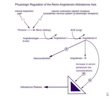
The Renin-Angiotensin System (RAS) is a complex hormonal system that regulates blood pressure, fluid and electrolyte balance, and vascular resistance. It plays a crucial role in the pathophysiology of hypertension, heart failure, and kidney diseases.
Here's a brief overview of how it works:
1. Renin is an enzyme that is released by the juxtaglomerular cells in the kidneys in response to decreased blood pressure or reduced salt delivery to the distal tubules.
2. Renin acts on a protein called angiotensinogen, which is produced by the liver, converting it into angiotensin I.
3. Angiotensin-converting enzyme (ACE), found in the lungs and other tissues, then converts angiotensin I into angiotensin II, a potent vasoconstrictor that narrows blood vessels and increases blood pressure.
4. Angiotensin II also stimulates the release of aldosterone from the adrenal glands, which promotes sodium and water reabsorption in the kidneys, further increasing blood volume and blood pressure.
5. Additionally, angiotensin II has direct effects on the heart, promoting hypertrophy and remodeling, which can contribute to heart failure.
6. The RAS can be modulated by various medications, such as ACE inhibitors, angiotensin receptor blockers (ARBs), and aldosterone antagonists, which are commonly used to treat hypertension, heart failure, and kidney diseases.
Antihypertensive agents are a class of medications used to treat high blood pressure (hypertension). They work by reducing the force and rate of heart contractions, dilating blood vessels, or altering neurohormonal activation to lower blood pressure. Examples include diuretics, beta blockers, ACE inhibitors, ARBs, calcium channel blockers, and direct vasodilators. These medications may be used alone or in combination to achieve optimal blood pressure control.
Peptidyl-dipeptidase A is more commonly known as angiotensin-converting enzyme (ACE). It is a key enzyme in the renin-angiotensin-aldosterone system (RAAS), which regulates blood pressure and fluid balance.
ACE is a membrane-bound enzyme found primarily in the lungs, but also in other tissues such as the heart, kidneys, and blood vessels. It plays a crucial role in converting the inactive decapeptide angiotensin I into the potent vasoconstrictor octapeptide angiotensin II, which constricts blood vessels and increases blood pressure.
ACE also degrades the peptide bradykinin, which is involved in the regulation of blood flow and vascular permeability. By breaking down bradykinin, ACE helps to counteract its vasodilatory effects, thereby maintaining blood pressure homeostasis.
Inhibitors of ACE are widely used as medications for the treatment of hypertension, heart failure, and diabetic kidney disease, among other conditions. These drugs work by blocking the action of ACE, leading to decreased levels of angiotensin II and increased levels of bradykinin, which results in vasodilation, reduced blood pressure, and improved cardiovascular function.
Blood pressure is the force exerted by circulating blood on the walls of the blood vessels. It is measured in millimeters of mercury (mmHg) and is given as two figures:
1. Systolic pressure: This is the pressure when the heart pushes blood out into the arteries.
2. Diastolic pressure: This is the pressure when the heart rests between beats, allowing it to fill with blood.
Normal blood pressure for adults is typically around 120/80 mmHg, although this can vary slightly depending on age, sex, and other factors. High blood pressure (hypertension) is generally considered to be a reading of 130/80 mmHg or higher, while low blood pressure (hypotension) is usually defined as a reading below 90/60 mmHg. It's important to note that blood pressure can fluctuate throughout the day and may be affected by factors such as stress, physical activity, and medication use.
Hypertension is a medical term used to describe abnormally high blood pressure in the arteries, often defined as consistently having systolic blood pressure (the top number in a blood pressure reading) over 130 mmHg and/or diastolic blood pressure (the bottom number) over 80 mmHg. It is also commonly referred to as high blood pressure.
Hypertension can be classified into two types: primary or essential hypertension, which has no identifiable cause and accounts for about 95% of cases, and secondary hypertension, which is caused by underlying medical conditions such as kidney disease, hormonal disorders, or use of certain medications.
If left untreated, hypertension can lead to serious health complications such as heart attack, stroke, heart failure, and chronic kidney disease. Therefore, it is important for individuals with hypertension to manage their condition through lifestyle modifications (such as healthy diet, regular exercise, stress management) and medication if necessary, under the guidance of a healthcare professional.
Renin is a medically recognized term and it is defined as:
"A protein (enzyme) that is produced and released by specialized cells (juxtaglomerular cells) in the kidney. Renin is a key component of the renin-angiotensin-aldosterone system (RAAS), which helps regulate blood pressure and fluid balance in the body.
When the kidney detects a decrease in blood pressure or a reduction in sodium levels, it releases renin into the bloodstream. Renin then acts on a protein called angiotensinogen, converting it to angiotensin I. Angiotensin-converting enzyme (ACE) subsequently converts angiotensin I to angiotensin II, which is a potent vasoconstrictor that narrows blood vessels and increases blood pressure.
Additionally, angiotensin II stimulates the adrenal glands to release aldosterone, a hormone that promotes sodium reabsorption in the kidneys and increases water retention, further raising blood pressure.
Therefore, renin plays a critical role in maintaining proper blood pressure and electrolyte balance in the body."
Valine is an essential amino acid, meaning it cannot be produced by the human body and must be obtained through diet. It is a hydrophobic amino acid, with a branched side chain, and is necessary for the growth, repair, and maintenance of tissues in the body. Valine is also important for muscle metabolism, and is often used by athletes as a supplement to enhance physical performance. Like other essential amino acids, valine must be obtained through foods such as meat, fish, dairy products, and legumes.
Angiotensin III is a hormone that is involved in the regulation of blood pressure and fluid balance in the body. It is formed by the enzymatic breakdown of angiotensin II, another hormone in the renin-angiotensin system (RAS). Angiotensin III has similar physiological effects as angiotensin II, including vasoconstriction (narrowing of blood vessels), stimulation of aldosterone release from the adrenal glands (which leads to sodium and water retention), and stimulation of thirst.
Angiotensin III is a peptide consisting of three amino acids, namely arginine-valine-tyrosine (Arg-Val-Tyr). It binds to and activates the angiotensin II receptor type 1 (AT1) and type 2 (AT2), which are found in various tissues throughout the body. The activation of these receptors leads to a range of physiological responses, including increased blood pressure, heart rate, and fluid volume.
Angiotensin III is less potent than angiotensin II in its ability to cause vasoconstriction and aldosterone release, but it has been shown to have important roles in the regulation of cardiovascular function, particularly during conditions of reduced renal perfusion or low blood pressure. It may also contribute to the development of certain diseases, such as hypertension, heart failure, and kidney disease.
Benzoates are the salts and esters of benzoic acid. They are widely used as preservatives in foods, cosmetics, and pharmaceuticals to prevent the growth of microorganisms. The chemical formula for benzoic acid is C6H5COOH, and when it is combined with a base (like sodium or potassium), it forms a benzoate salt (e.g., sodium benzoate or potassium benzoate). When benzoic acid reacts with an alcohol, it forms a benzoate ester (e.g., methyl benzoate or ethyl benzoate).
Benzoates are generally considered safe for use in food and cosmetics in small quantities. However, some people may have allergies or sensitivities to benzoates, which can cause reactions such as hives, itching, or asthma symptoms. In addition, there is ongoing research into the potential health effects of consuming high levels of benzoates over time, particularly in relation to gut health and the development of certain diseases.
In a medical context, benzoates may also be used as a treatment for certain conditions. For example, sodium benzoate is sometimes given to people with elevated levels of ammonia in their blood (hyperammonemia) to help reduce those levels and prevent brain damage. This is because benzoates can bind with excess ammonia in the body and convert it into a form that can be excreted in urine.
Vasoconstrictor agents are substances that cause the narrowing of blood vessels by constricting the smooth muscle in their walls. This leads to an increase in blood pressure and a decrease in blood flow. They work by activating the sympathetic nervous system, which triggers the release of neurotransmitters such as norepinephrine and epinephrine that bind to alpha-adrenergic receptors on the smooth muscle cells of the blood vessel walls, causing them to contract.
Vasoconstrictor agents are used medically for a variety of purposes, including:
* Treating hypotension (low blood pressure)
* Controlling bleeding during surgery or childbirth
* Relieving symptoms of nasal congestion in conditions such as the common cold or allergies
Examples of vasoconstrictor agents include phenylephrine, oxymetazoline, and epinephrine. It's important to note that prolonged use or excessive doses of vasoconstrictor agents can lead to rebound congestion and other adverse effects, so they should be used with caution and under the guidance of a healthcare professional.
Angiotensins are a group of hormones that play a crucial role in the body's cardiovascular system, particularly in regulating blood pressure and fluid balance. The most well-known angiotensins are Angiotensin I, Angiotensin II, and Angiotensin-(1-7).
Angiotensinogen is a protein produced mainly by the liver. When the body requires an increase in blood pressure, renin (an enzyme produced by the kidneys) cleaves angiotensinogen to form Angiotensin I. Then, another enzyme called angiotensin-converting enzyme (ACE), primarily found in the lungs, converts Angiotensin I into Angiotensin II.
Angiotensin II is a potent vasoconstrictor, causing blood vessels to narrow and increase blood pressure. It also stimulates the release of aldosterone from the adrenal glands, which leads to increased sodium reabsorption in the kidneys, further raising blood pressure and promoting fluid retention.
Angiotensin-(1-7) is a more recently discovered member of the angiotensin family. It has opposing effects to Angiotensin II, acting as a vasodilator and counterbalancing some of the negative consequences of Angiotensin II's actions.
Medications called ACE inhibitors and ARBs (angiotensin receptor blockers) are commonly used in clinical practice to target the renin-angiotensin system, lowering blood pressure and protecting against organ damage in various cardiovascular conditions.
Sprague-Dawley rats are a strain of albino laboratory rats that are widely used in scientific research. They were first developed by researchers H.H. Sprague and R.C. Dawley in the early 20th century, and have since become one of the most commonly used rat strains in biomedical research due to their relatively large size, ease of handling, and consistent genetic background.
Sprague-Dawley rats are outbred, which means that they are genetically diverse and do not suffer from the same limitations as inbred strains, which can have reduced fertility and increased susceptibility to certain diseases. They are also characterized by their docile nature and low levels of aggression, making them easier to handle and study than some other rat strains.
These rats are used in a wide variety of research areas, including toxicology, pharmacology, nutrition, cancer, and behavioral studies. Because they are genetically diverse, Sprague-Dawley rats can be used to model a range of human diseases and conditions, making them an important tool in the development of new drugs and therapies.
I am not aware of a specific medical definition for "1-Sarcosine-8-Isoleucine Angiotensin II." It is possible that this term is being used to describe an altered or modified form of the peptide hormone angiotensin II.
Angiotensin II is a powerful vasoconstrictor and plays a central role in the regulation of blood pressure and fluid balance. Its octapeptide structure consists of eight amino acids, with the sequence Asp-Arg-Val-Tyr-Ile-His-Pro-Phe.
Modifying this sequence by replacing one or more amino acids can result in altered biological activity. In this case, "1-Sarcosine-8-Isoleucine" suggests that the first amino acid (Aspartic Acid) has been replaced with Sarcosine (N-methylglycine), and the eighth amino acid (Phenylalanine) has been replaced with Isoleucine.
However, without further context or research, it is difficult to provide a precise medical definition for this term. If you are seeking information on a specific scientific study or application, please provide more details so that I can give a more informed response.
Angiotensinogen is a protein that is produced mainly by the liver. It is the precursor to angiotensin I, which is a molecule that begins the process of constriction (narrowing) of blood vessels, leading to an increase in blood pressure. When angiotensinogen comes into contact with an enzyme called renin, it is cleaved into angiotensin I. Angiotensin-converting enzyme (ACE) then converts angiotensin I into angiotensin II, which is a potent vasoconstrictor and a key player in the body's regulation of blood pressure and fluid balance.
Angiotensinogen is an important component of the renin-angiotensin-aldosterone system (RAAS), which helps to regulate blood pressure and fluid balance by controlling the volume and flow of fluids in the body. Disorders of the RAAS can lead to high blood pressure, kidney disease, and other health problems.
I'm sorry for any confusion, but "Pyridines" is not a medical term. It is a chemical term that refers to a class of organic compounds with the chemical structure of a six-membered ring containing one nitrogen atom and five carbon atoms (heterocyclic aromatic compound).
In a biological or medical context, pyridine derivatives can be found in various natural and synthetic substances. For example, some medications contain pyridine rings as part of their chemical structure. However, "Pyridines" itself is not a medical term or condition.
A kidney, in medical terms, is one of two bean-shaped organs located in the lower back region of the body. They are essential for maintaining homeostasis within the body by performing several crucial functions such as:
1. Regulation of water and electrolyte balance: Kidneys help regulate the amount of water and various electrolytes like sodium, potassium, and calcium in the bloodstream to maintain a stable internal environment.
2. Excretion of waste products: They filter waste products from the blood, including urea (a byproduct of protein metabolism), creatinine (a breakdown product of muscle tissue), and other harmful substances that result from normal cellular functions or external sources like medications and toxins.
3. Endocrine function: Kidneys produce several hormones with important roles in the body, such as erythropoietin (stimulates red blood cell production), renin (regulates blood pressure), and calcitriol (activated form of vitamin D that helps regulate calcium homeostasis).
4. pH balance regulation: Kidneys maintain the proper acid-base balance in the body by excreting either hydrogen ions or bicarbonate ions, depending on whether the blood is too acidic or too alkaline.
5. Blood pressure control: The kidneys play a significant role in regulating blood pressure through the renin-angiotensin-aldosterone system (RAAS), which constricts blood vessels and promotes sodium and water retention to increase blood volume and, consequently, blood pressure.
Anatomically, each kidney is approximately 10-12 cm long, 5-7 cm wide, and 3 cm thick, with a weight of about 120-170 grams. They are surrounded by a protective layer of fat and connected to the urinary system through the renal pelvis, ureters, bladder, and urethra.
Saralasin is a synthetic analog of the natural hormone angiotensin II, which is used in research and medicine. It acts as an antagonist of the angiotensin II receptor, blocking its effects. Saralasin is primarily used in research to study the role of the renin-angiotensin system in various physiological processes. In clinical medicine, it has been used in the diagnosis and treatment of conditions such as hypertension and pheochromocytoma, although its use is not widespread due to the availability of more effective and selective drugs.
Calcium channel blockers (CCBs) are a class of medications that work by inhibiting the influx of calcium ions into cardiac and smooth muscle cells. This action leads to relaxation of the muscles, particularly in the blood vessels, resulting in decreased peripheral resistance and reduced blood pressure. Calcium channel blockers also have anti-arrhythmic effects and are used in the management of various cardiovascular conditions such as hypertension, angina, and certain types of arrhythmias.
Calcium channel blockers can be further classified into two main categories based on their chemical structure: dihydropyridines (e.g., nifedipine, amlodipine) and non-dihydropyridines (e.g., verapamil, diltiazem). Dihydropyridines are more selective for vascular smooth muscle and have a greater effect on blood pressure than heart rate or conduction. Non-dihydropyridines have a more significant impact on cardiac conduction and contractility, in addition to their vasodilatory effects.
It is important to note that calcium channel blockers may interact with other medications and should be used under the guidance of a healthcare professional. Potential side effects include dizziness, headache, constipation, and peripheral edema.
SHR (Spontaneously Hypertensive Rats) are an inbred strain of rats that were originally developed through selective breeding for high blood pressure. They are widely used as a model to study hypertension and related cardiovascular diseases, as well as neurological disorders such as stroke and dementia.
Inbred strains of animals are created by mating genetically identical individuals (siblings or offspring) for many generations, resulting in a population that is highly homozygous at all genetic loci. This means that the animals within an inbred strain are essentially genetically identical to one another, which makes them useful for studying the effects of specific genes or environmental factors on disease processes.
SHR rats develop high blood pressure spontaneously, without any experimental manipulation, and show many features of human hypertension, such as increased vascular resistance, left ventricular hypertrophy, and renal dysfunction. They also exhibit a number of behavioral abnormalities, including hyperactivity, impulsivity, and cognitive deficits, which make them useful for studying the neurological consequences of hypertension and other cardiovascular diseases.
Overall, inbred SHR rats are an important tool in biomedical research, providing a valuable model for understanding the genetic and environmental factors that contribute to hypertension and related disorders.
Aldosterone is a hormone produced by the adrenal gland. It plays a key role in regulating sodium and potassium balance and maintaining blood pressure through its effects on the kidneys. Aldosterone promotes the reabsorption of sodium ions and the excretion of potassium ions in the distal tubules and collecting ducts of the nephrons in the kidneys. This increases the osmotic pressure in the blood, which in turn leads to water retention and an increase in blood volume and blood pressure.
Aldosterone is released from the adrenal gland in response to a variety of stimuli, including angiotensin II (a peptide hormone produced as part of the renin-angiotensin-aldosterone system), potassium ions, and adrenocorticotropic hormone (ACTH) from the pituitary gland. The production of aldosterone is regulated by a negative feedback mechanism involving sodium levels in the blood. High sodium levels inhibit the release of aldosterone, while low sodium levels stimulate its release.
In addition to its role in maintaining fluid and electrolyte balance and blood pressure, aldosterone has been implicated in various pathological conditions, including hypertension, heart failure, and primary hyperaldosteronism (a condition characterized by excessive production of aldosterone).
Thiazepines are not a recognized term in medical terminology or pharmacology. It appears that you may have misspelled "thiazepines," which also does not have a specific medical meaning. However, "thiazepine" is a chemical compound with a specific structure, and it is the core structure of some drugs such as thiazepine derivatives. These derivatives are often used for their sedative, hypnotic, anticonvulsant, and muscle relaxant properties.
If you meant to ask about "thiazide" or "thiazide diuretics," I would be happy to provide a definition:
Thiazides are a class of diuretic medications that act on the distal convoluted tubule in the kidney, promoting sodium and chloride excretion. This also leads to increased water excretion (diuresis) and decreased extracellular fluid volume. Thiazide diuretics are primarily used to treat hypertension and edema associated with heart failure or liver cirrhosis. Common thiazide diuretics include hydrochlorothiazide, chlorthalidone, and indapamide.
Enalapril is a medication that belongs to a class of drugs called angiotensin-converting enzyme (ACE) inhibitors. It works by blocking the action of a hormone in the body called angiotensin II, which causes blood vessels to narrow and tighten. By blocking this hormone, Enalapril helps relax and widen blood vessels, making it easier for the heart to pump blood and reducing the workload on the heart.
Enalapril is commonly used to treat high blood pressure (hypertension), congestive heart failure, and to improve survival after a heart attack. It may also be used to treat other conditions as determined by your doctor.
The medication comes in the form of tablets or capsules that are taken orally, usually once or twice a day with or without food. The dosage will depend on various factors such as the patient's age, weight, and medical condition. It is important to follow the instructions of your healthcare provider when taking Enalapril.
Like all medications, Enalapril can cause side effects, including dry cough, dizziness, headache, fatigue, and nausea. More serious side effects may include allergic reactions, kidney problems, and low blood pressure. If you experience any concerning symptoms while taking Enalapril, it is important to contact your healthcare provider right away.
WKY (Wistar Kyoto) is not a term that refers to "rats, inbred" in a medical definition. Instead, it is a strain of laboratory rat that is widely used in biomedical research. WKY rats are an inbred strain, which means they are the result of many generations of brother-sister matings, resulting in a genetically uniform population.
WKY rats originated from the Wistar Institute in Philadelphia and were established as a normotensive control strain to contrast with other rat strains that exhibit hypertension. They have since been used in various research areas, including cardiovascular, neurological, and behavioral studies. Compared to other commonly used rat strains like the spontaneously hypertensive rat (SHR), WKY rats are known for their lower blood pressure, reduced stress response, and greater emotionality.
In summary, "WKY" is a designation for an inbred strain of laboratory rat that is often used as a control group in biomedical research due to its normotensive characteristics.
A dose-response relationship in the context of drugs refers to the changes in the effects or symptoms that occur as the dose of a drug is increased or decreased. Generally, as the dose of a drug is increased, the severity or intensity of its effects also increases. Conversely, as the dose is decreased, the effects of the drug become less severe or may disappear altogether.
The dose-response relationship is an important concept in pharmacology and toxicology because it helps to establish the safe and effective dosage range for a drug. By understanding how changes in the dose of a drug affect its therapeutic and adverse effects, healthcare providers can optimize treatment plans for their patients while minimizing the risk of harm.
The dose-response relationship is typically depicted as a curve that shows the relationship between the dose of a drug and its effect. The shape of the curve may vary depending on the drug and the specific effect being measured. Some drugs may have a steep dose-response curve, meaning that small changes in the dose can result in large differences in the effect. Other drugs may have a more gradual dose-response curve, where larger changes in the dose are needed to produce significant effects.
In addition to helping establish safe and effective dosages, the dose-response relationship is also used to evaluate the potential therapeutic benefits and risks of new drugs during clinical trials. By systematically testing different doses of a drug in controlled studies, researchers can identify the optimal dosage range for the drug and assess its safety and efficacy.
"Cells, cultured" is a medical term that refers to cells that have been removed from an organism and grown in controlled laboratory conditions outside of the body. This process is called cell culture and it allows scientists to study cells in a more controlled and accessible environment than they would have inside the body. Cultured cells can be derived from a variety of sources, including tissues, organs, or fluids from humans, animals, or cell lines that have been previously established in the laboratory.
Cell culture involves several steps, including isolation of the cells from the tissue, purification and characterization of the cells, and maintenance of the cells in appropriate growth conditions. The cells are typically grown in specialized media that contain nutrients, growth factors, and other components necessary for their survival and proliferation. Cultured cells can be used for a variety of purposes, including basic research, drug development and testing, and production of biological products such as vaccines and gene therapies.
It is important to note that cultured cells may behave differently than they do in the body, and results obtained from cell culture studies may not always translate directly to human physiology or disease. Therefore, it is essential to validate findings from cell culture experiments using additional models and ultimately in clinical trials involving human subjects.
A smooth muscle within the vascular system refers to the involuntary, innervated muscle that is found in the walls of blood vessels. These muscles are responsible for controlling the diameter of the blood vessels, which in turn regulates blood flow and blood pressure. They are called "smooth" muscles because their individual muscle cells do not have the striations, or cross-striped patterns, that are observed in skeletal and cardiac muscle cells. Smooth muscle in the vascular system is controlled by the autonomic nervous system and by hormones, and can contract or relax slowly over a period of time.
"Wistar rats" are a strain of albino rats that are widely used in laboratory research. They were developed at the Wistar Institute in Philadelphia, USA, and were first introduced in 1906. Wistar rats are outbred, which means that they are genetically diverse and do not have a fixed set of genetic characteristics like inbred strains.
Wistar rats are commonly used as animal models in biomedical research because of their size, ease of handling, and relatively low cost. They are used in a wide range of research areas, including toxicology, pharmacology, nutrition, cancer, cardiovascular disease, and behavioral studies. Wistar rats are also used in safety testing of drugs, medical devices, and other products.
Wistar rats are typically larger than many other rat strains, with males weighing between 500-700 grams and females weighing between 250-350 grams. They have a lifespan of approximately 2-3 years. Wistar rats are also known for their docile and friendly nature, making them easy to handle and work with in the laboratory setting.
Messenger RNA (mRNA) is a type of RNA (ribonucleic acid) that carries genetic information copied from DNA in the form of a series of three-base code "words," each of which specifies a particular amino acid. This information is used by the cell's machinery to construct proteins, a process known as translation. After being transcribed from DNA, mRNA travels out of the nucleus to the ribosomes in the cytoplasm where protein synthesis occurs. Once the protein has been synthesized, the mRNA may be degraded and recycled. Post-transcriptional modifications can also occur to mRNA, such as alternative splicing and addition of a 5' cap and a poly(A) tail, which can affect its stability, localization, and translation efficiency.
The myocardium is the middle layer of the heart wall, composed of specialized cardiac muscle cells that are responsible for pumping blood throughout the body. It forms the thickest part of the heart wall and is divided into two sections: the left ventricle, which pumps oxygenated blood to the rest of the body, and the right ventricle, which pumps deoxygenated blood to the lungs.
The myocardium contains several types of cells, including cardiac muscle fibers, connective tissue, nerves, and blood vessels. The muscle fibers are arranged in a highly organized pattern that allows them to contract in a coordinated manner, generating the force necessary to pump blood through the heart and circulatory system.
Damage to the myocardium can occur due to various factors such as ischemia (reduced blood flow), infection, inflammation, or genetic disorders. This damage can lead to several cardiac conditions, including heart failure, arrhythmias, and cardiomyopathy.
Animal disease models are specialized animals, typically rodents such as mice or rats, that have been genetically engineered or exposed to certain conditions to develop symptoms and physiological changes similar to those seen in human diseases. These models are used in medical research to study the pathophysiology of diseases, identify potential therapeutic targets, test drug efficacy and safety, and understand disease mechanisms.
The genetic modifications can include knockout or knock-in mutations, transgenic expression of specific genes, or RNA interference techniques. The animals may also be exposed to environmental factors such as chemicals, radiation, or infectious agents to induce the disease state.
Examples of animal disease models include:
1. Mouse models of cancer: Genetically engineered mice that develop various types of tumors, allowing researchers to study cancer initiation, progression, and metastasis.
2. Alzheimer's disease models: Transgenic mice expressing mutant human genes associated with Alzheimer's disease, which exhibit amyloid plaque formation and cognitive decline.
3. Diabetes models: Obese and diabetic mouse strains like the NOD (non-obese diabetic) or db/db mice, used to study the development of type 1 and type 2 diabetes, respectively.
4. Cardiovascular disease models: Atherosclerosis-prone mice, such as ApoE-deficient or LDLR-deficient mice, that develop plaque buildup in their arteries when fed a high-fat diet.
5. Inflammatory bowel disease models: Mice with genetic mutations affecting intestinal barrier function and immune response, such as IL-10 knockout or SAMP1/YitFc mice, which develop colitis.
Animal disease models are essential tools in preclinical research, but it is important to recognize their limitations. Differences between species can affect the translatability of results from animal studies to human patients. Therefore, researchers must carefully consider the choice of model and interpret findings cautiously when applying them to human diseases.
Cardiomegaly is a medical term that refers to an enlarged heart. It can be caused by various conditions such as high blood pressure, heart valve problems, cardiomyopathy, or fluid accumulation around the heart (pericardial effusion). Cardiomegaly can be detected through imaging tests like chest X-rays or echocardiograms. Depending on the underlying cause, treatment options may include medications, lifestyle changes, or in some cases, surgery. It is important to consult with a healthcare professional for proper diagnosis and treatment.
Renal hypertension, also known as renovascular hypertension, is a type of secondary hypertension (high blood pressure) that is caused by narrowing or obstruction of the renal arteries or veins, which supply blood to the kidneys. This can lead to decreased blood flow and oxygen delivery to the kidney tissue, activating the renin-angiotensin-aldosterone system (RAAS) and resulting in increased peripheral vascular resistance, sodium retention, and extracellular fluid volume, ultimately causing hypertension.
Renal hypertension can be classified into two types:
1. Renin-dependent renal hypertension: This is caused by a decrease in blood flow to the kidneys, leading to increased renin release from the juxtaglomerular cells of the kidney. Renin converts angiotensinogen to angiotensin I, which is then converted to angiotensin II by angiotensin-converting enzyme (ACE). Angiotensin II is a potent vasoconstrictor that causes an increase in peripheral vascular resistance and blood pressure.
2. Renin-independent renal hypertension: This is caused by increased sodium retention and extracellular fluid volume, leading to an increase in blood pressure. This can be due to various factors such as obstructive sleep apnea, primary aldosteronism, or pheochromocytoma.
Renal hypertension is often asymptomatic but can lead to serious complications such as kidney damage, heart failure, and stroke if left untreated. Diagnosis of renal hypertension involves imaging studies such as renal artery duplex ultrasound, CT angiography, or magnetic resonance angiography (MRA) to identify any narrowing or obstruction in the renal arteries or veins. Treatment options include medications such as ACE inhibitors, angiotensin receptor blockers (ARBs), calcium channel blockers, and diuretics, as well as interventions such as angioplasty and stenting to improve blood flow to the kidneys.
Captopril is a medication that belongs to a class of drugs called ACE (angiotensin-converting enzyme) inhibitors. It works by blocking the action of a chemical in the body called angiotensin II, which causes blood vessels to narrow and release hormones that can increase blood pressure. By blocking the action of angiotensin II, captopril helps relax and widen blood vessels, which lowers blood pressure and improves blood flow.
Captopril is used to treat high blood pressure (hypertension), congestive heart failure, and to improve survival after a heart attack. It may also be used to protect the kidneys from damage due to diabetes or high blood pressure. The medication comes in the form of tablets that are taken by mouth, usually two to three times per day.
Common side effects of captopril include cough, dizziness, headache, and skin rash. More serious side effects may include allergic reactions, kidney problems, and changes in blood cell counts. It is important for patients taking captopril to follow their doctor's instructions carefully and report any unusual symptoms or side effects promptly.
Amlodipine is a type of medication known as a calcium channel blocker, which is primarily used to treat high blood pressure and angina (chest pain caused by reduced blood flow to the heart). It works by relaxing the muscles around the blood vessels, which causes them to widen and improves blood flow. This helps to lower blood pressure and reduce the workload on the heart, making it easier for the heart to pump blood effectively.
Amlodipine is available in various strengths as a tablet or an extended-release tablet, and it is typically taken once daily. The medication may take several weeks to reach its full effect, so it is important to continue taking it even if you do not notice any immediate improvement in your symptoms.
As with any medication, amlodipine can cause side effects, including headache, dizziness, fatigue, and swelling of the ankles or feet. In rare cases, it may also cause more serious side effects such as allergic reactions, irregular heartbeat, or liver damage. If you experience any unusual symptoms while taking amlodipine, it is important to contact your healthcare provider right away.
It is important to follow your healthcare provider's instructions carefully when taking amlodipine, and to inform them of any other medications or supplements that you are taking, as well as any medical conditions that you have. This will help ensure that the medication is safe and effective for you to use.
Hydralazine is an antihypertensive medication, which means it is used to treat high blood pressure. It works by relaxing and widening the blood vessels, making it easier for the heart to pump blood through the body. This can help reduce the workload on the heart and lower blood pressure. Hydralazine is available in oral tablet form and is typically prescribed to be taken several times a day.
Hydralazine belongs to a class of medications called vasodilators, which work by relaxing the muscle in the walls of the blood vessels, causing them to widen. This increases the amount of blood that can flow through the blood vessels and reduces the pressure within them. Hydralazine is often used in combination with other medications to treat high blood pressure.
It's important to note that hydralazine should be used under the close supervision of a healthcare provider, as it can cause side effects such as headache, dizziness, and rapid heartbeat. It may also interact with certain other medications, so it is important to inform your doctor of all medications you are taking before starting hydralazine.
Renal circulation refers to the blood flow specifically dedicated to the kidneys. The main function of the kidneys is to filter waste and excess fluids from the blood, which then get excreted as urine. To perform this function efficiently, the kidneys receive a substantial amount of the body's total blood supply - about 20-25% in a resting state.
The renal circulation process begins when deoxygenated blood from the rest of the body returns to the right side of the heart and is pumped into the lungs for oxygenation. Oxygen-rich blood then leaves the left side of the heart through the aorta, the largest artery in the body.
A portion of this oxygen-rich blood moves into the renal arteries, which branch directly from the aorta and supply each kidney with blood. Within the kidneys, these arteries divide further into smaller vessels called afferent arterioles, which feed into a network of tiny capillaries called the glomerulus within each nephron (the functional unit of the kidney).
The filtration process occurs in the glomeruli, where waste materials and excess fluids are separated from the blood. The resulting filtrate then moves through another set of capillaries, the peritubular capillaries, which surround the renal tubules (the part of the nephron that reabsorbs necessary substances back into the bloodstream).
The now-deoxygenated blood from the kidneys' capillary network coalesces into venules and then merges into the renal veins, which ultimately drain into the inferior vena cava and return the blood to the right side of the heart. This highly specialized circulation system allows the kidneys to efficiently filter waste while maintaining appropriate blood volume and composition.
Vasoconstriction is a medical term that refers to the narrowing of blood vessels due to the contraction of the smooth muscle in their walls. This process decreases the diameter of the lumen (the inner space of the blood vessel) and reduces blood flow through the affected vessels. Vasoconstriction can occur throughout the body, but it is most noticeable in the arterioles and precapillary sphincters, which control the amount of blood that flows into the capillary network.
The autonomic nervous system, specifically the sympathetic division, plays a significant role in regulating vasoconstriction through the release of neurotransmitters like norepinephrine (noradrenaline). Various hormones and chemical mediators, such as angiotensin II, endothelin-1, and serotonin, can also induce vasoconstriction.
Vasoconstriction is a vital physiological response that helps maintain blood pressure and regulate blood flow distribution in the body. However, excessive or prolonged vasoconstriction may contribute to several pathological conditions, including hypertension, stroke, and peripheral vascular diseases.
Bradykinin is a naturally occurring peptide in the human body, consisting of nine amino acids. It is a potent vasodilator and increases the permeability of blood vessels, causing a local inflammatory response. Bradykinin is formed from the breakdown of certain proteins, such as kininogen, by enzymes called kininases or proteases, including kallikrein. It plays a role in several physiological processes, including pain transmission, blood pressure regulation, and the immune response. In some pathological conditions, such as hereditary angioedema, bradykinin levels can increase excessively, leading to symptoms like swelling, redness, and pain.
Lisinopril is an angiotensin-converting enzyme (ACE) inhibitor, which is a type of medication used to treat various cardiovascular conditions. It works by blocking the conversion of angiotensin I to angiotensin II, a potent vasoconstrictor, resulting in relaxation and widening of blood vessels, decreased blood pressure, and increased blood flow.
Lisinopril is primarily used to treat hypertension (high blood pressure), congestive heart failure, and to improve survival after a heart attack. It may also be used to protect the kidneys from damage due to diabetes or high blood pressure. Additionally, it has been shown to reduce proteinuria (excess protein in urine) in patients with diabetic nephropathy.
Common side effects of Lisinopril include dizziness, headache, fatigue, and cough. More serious side effects may include angioedema (rapid swelling of the face, lips, tongue, or throat), hyperkalemia (elevated potassium levels), and impaired kidney function.
It is important to follow the prescribing physician's instructions carefully when taking Lisinopril and to report any unusual symptoms promptly. Regular monitoring of blood pressure, kidney function, and electrolyte levels may be necessary during treatment with this medication.
Proteinuria is a medical term that refers to the presence of excess proteins, particularly albumin, in the urine. Under normal circumstances, only small amounts of proteins should be found in the urine because the majority of proteins are too large to pass through the glomeruli, which are the filtering units of the kidneys.
However, when the glomeruli become damaged or diseased, they may allow larger molecules such as proteins to leak into the urine. Persistent proteinuria is often a sign of kidney disease and can indicate damage to the glomeruli. It is usually detected through a routine urinalysis and may be confirmed with further testing.
The severity of proteinuria can vary, and it can be a symptom of various underlying conditions such as diabetes, hypertension, glomerulonephritis, and other kidney diseases. Treatment for proteinuria depends on the underlying cause and may include medications to control blood pressure, manage diabetes, or reduce protein loss in the urine.
Fibrosis is a pathological process characterized by the excessive accumulation and/or altered deposition of extracellular matrix components, particularly collagen, in various tissues and organs. This results in the formation of fibrous scar tissue that can impair organ function and structure. Fibrosis can occur as a result of chronic inflammation, tissue injury, or abnormal repair mechanisms, and it is a common feature of many diseases, including liver cirrhosis, lung fibrosis, heart failure, and kidney disease.
In medical terms, fibrosis is defined as:
"The process of producing scar tissue (consisting of collagen) in response to injury or chronic inflammation in normal connective tissue. This can lead to the thickening and stiffening of affected tissues and organs, impairing their function."
Chymases are a type of enzyme that belong to the family of serine proteases. They are found in various tissues and organs, including the heart, lungs, and immune cells called mast cells. Chymases play a role in several physiological and pathological processes, such as inflammation, tissue remodeling, and blood pressure regulation.
One of the most well-known chymases is found in the mast cells and is often referred to as "mast cell chymase." This enzyme can cleave and activate various proteins, including angiotensin I to angiotensin II, a potent vasoconstrictor that increases blood pressure. Chymases have also been implicated in the development of cardiovascular diseases, such as hypertension and heart failure, as well as respiratory diseases like asthma and chronic obstructive pulmonary disease (COPD).
In summary, chymases are a group of serine protease enzymes that play important roles in various physiological and pathological processes, particularly in inflammation, tissue remodeling, and blood pressure regulation.
The aorta is the largest artery in the human body, which originates from the left ventricle of the heart and carries oxygenated blood to the rest of the body. It can be divided into several parts, including the ascending aorta, aortic arch, and descending aorta. The ascending aorta gives rise to the coronary arteries that supply blood to the heart muscle. The aortic arch gives rise to the brachiocephalic, left common carotid, and left subclavian arteries, which supply blood to the head, neck, and upper extremities. The descending aorta travels through the thorax and abdomen, giving rise to various intercostal, visceral, and renal arteries that supply blood to the chest wall, organs, and kidneys.
NADPH oxidase is an enzyme complex that plays a crucial role in the production of reactive oxygen species (ROS) in various cell types. The primary function of NADPH oxidase is to catalyze the transfer of electrons from NADPH to molecular oxygen, resulting in the formation of superoxide radicals. This enzyme complex consists of several subunits, including two membrane-bound components (gp91phox and p22phox) and several cytosolic components (p47phox, p67phox, p40phox, and rac1 or rac2). Upon activation, these subunits assemble to form a functional enzyme complex that generates ROS, which serve as important signaling molecules in various cellular processes. However, excessive or uncontrolled production of ROS by NADPH oxidase has been implicated in the pathogenesis of several diseases, such as cardiovascular disorders, neurodegenerative diseases, and cancer.
Signal transduction is the process by which a cell converts an extracellular signal, such as a hormone or neurotransmitter, into an intracellular response. This involves a series of molecular events that transmit the signal from the cell surface to the interior of the cell, ultimately resulting in changes in gene expression, protein activity, or metabolism.
The process typically begins with the binding of the extracellular signal to a receptor located on the cell membrane. This binding event activates the receptor, which then triggers a cascade of intracellular signaling molecules, such as second messengers, protein kinases, and ion channels. These molecules amplify and propagate the signal, ultimately leading to the activation or inhibition of specific cellular responses.
Signal transduction pathways are highly regulated and can be modulated by various factors, including other signaling molecules, post-translational modifications, and feedback mechanisms. Dysregulation of these pathways has been implicated in a variety of diseases, including cancer, diabetes, and neurological disorders.
Mineralocorticoid receptor antagonists (MRAs) are a class of medications that block the action of aldosterone, a hormone produced by the adrenal glands. Aldosterone helps regulate sodium and potassium balance and blood pressure by binding to mineralocorticoid receptors in the kidneys, heart, blood vessels, and brain.
When aldosterone binds to these receptors, it promotes sodium retention and potassium excretion, which can lead to an increase in blood volume and blood pressure. MRAs work by blocking the binding of aldosterone to its receptors, thereby preventing these effects.
MRAs are primarily used to treat heart failure, hypertension, and kidney disease. By reducing sodium retention and increasing potassium excretion, MRAs can help lower blood pressure, reduce fluid buildup in the body, and improve heart function. Examples of MRAs include spironolactone and eplerenone.
Hemodynamics is the study of how blood flows through the cardiovascular system, including the heart and the vascular network. It examines various factors that affect blood flow, such as blood volume, viscosity, vessel length and diameter, and pressure differences between different parts of the circulatory system. Hemodynamics also considers the impact of various physiological and pathological conditions on these variables, and how they in turn influence the function of vital organs and systems in the body. It is a critical area of study in fields such as cardiology, anesthesiology, and critical care medicine.
Ramipril is an angiotensin-converting enzyme (ACE) inhibitor, which is a type of medication used to treat various cardiovascular conditions. It works by blocking the conversion of angiotensin I to angiotensin II, a potent vasoconstrictor, thereby causing relaxation and widening of blood vessels, decreasing blood pressure, and increasing blood flow.
Ramipril is primarily used for the treatment of hypertension (high blood pressure), heart failure, and the prevention of major cardiovascular events such as myocardial infarction (heart attack) and stroke in high-risk patients. It may also be used to improve survival after a heart attack.
The medication is available in oral tablet form and is typically taken once or twice daily, depending on the prescribed dosage. Side effects of ramipril can include cough, dizziness, headache, fatigue, nausea, and taste changes. Serious side effects are rare but may include kidney problems, hyperkalemia (high potassium levels), and angioedema (swelling of the face, lips, tongue, or throat).
It is important to follow the prescribing physician's instructions carefully when taking ramipril and to report any unusual symptoms or side effects promptly. Regular monitoring of blood pressure, kidney function, and potassium levels may be necessary during treatment with this medication.
The Bradykinin B2 receptor (B2R) is a type of G protein-coupled receptor that binds to and is activated by the peptide hormone bradykinin. Upon activation, it triggers a variety of intracellular signaling pathways leading to diverse physiological responses such as vasodilation, increased vascular permeability, pain, and inflammation.
B2Rs are widely distributed in various tissues, including the cardiovascular, respiratory, gastrointestinal, and nervous systems. They play a crucial role in several pathophysiological conditions such as hypertension, heart failure, ischemia-reperfusion injury, pain, and inflammatory diseases.
B2Rs are also the target of clinically used drugs, including angiotensin-converting enzyme (ACE) inhibitors and angiotensin receptor blockers (ARBs), which increase bradykinin levels and enhance its effects on B2Rs, leading to vasodilation and reduced blood pressure.
Ventricular remodeling is a structural adaptation process of the heart in response to stress or injury, such as myocardial infarction (heart attack) or pressure overload. This process involves changes in size, shape, and function of the ventricles (the lower chambers of the heart).
In ventricular remodeling, the heart muscle may thicken, enlarge, or become more stiff, leading to alterations in the pumping ability of the heart. These changes can ultimately result in cardiac dysfunction, heart failure, and an increased risk of arrhythmias (irregular heart rhythms).
Ventricular remodeling is often classified into two types:
1. Concentric remodeling: This occurs when the ventricular wall thickens (hypertrophy) without a significant increase in chamber size, leading to a decrease in the cavity volume and an increase in the thickness of the ventricular wall.
2. Eccentric remodeling: This involves an increase in both the ventricular chamber size and wall thickness due to the addition of new muscle cells (hyperplasia) or enlargement of existing muscle cells (hypertrophy). As a result, the overall shape of the ventricle becomes more spherical and less elliptical.
Both types of remodeling can negatively impact heart function and contribute to the development of heart failure. Close monitoring and appropriate treatment are essential for managing ventricular remodeling and preventing further complications.
Spironolactone is a prescription medication that belongs to a class of drugs known as potassium-sparing diuretics. It works by blocking the action of aldosterone, a hormone that helps regulate sodium and potassium balance in your body. This results in increased urine production (diuresis) and decreased salt and fluid retention.
Spironolactone is primarily used to treat edema (fluid buildup) associated with heart failure, liver cirrhosis, or kidney disease. It's also prescribed for the treatment of high blood pressure and primary hyperaldosteronism, a condition where the adrenal glands produce too much aldosterone.
Furthermore, spironolactone is used off-label to treat conditions such as acne, hirsutism (excessive hair growth in women), and hormone-sensitive breast cancer in postmenopausal women.
It's important to note that spironolactone can cause increased potassium levels in the blood (hyperkalemia) and should be used with caution in patients with kidney impairment or those taking other medications that affect potassium balance. Regular monitoring of electrolyte levels, including potassium and sodium, is essential during spironolactone therapy.
Norepinephrine, also known as noradrenaline, is a neurotransmitter and a hormone that is primarily produced in the adrenal glands and is released into the bloodstream in response to stress or physical activity. It plays a crucial role in the "fight-or-flight" response by preparing the body for action through increasing heart rate, blood pressure, respiratory rate, and glucose availability.
As a neurotransmitter, norepinephrine is involved in regulating various functions of the nervous system, including attention, perception, motivation, and arousal. It also plays a role in modulating pain perception and responding to stressful or emotional situations.
In medical settings, norepinephrine is used as a vasopressor medication to treat hypotension (low blood pressure) that can occur during septic shock, anesthesia, or other critical illnesses. It works by constricting blood vessels and increasing heart rate, which helps to improve blood pressure and perfusion of vital organs.
Diuretics are a type of medication that increase the production of urine and help the body eliminate excess fluid and salt. They work by interfering with the reabsorption of sodium in the kidney tubules, which in turn causes more water to be excreted from the body. Diuretics are commonly used to treat conditions such as high blood pressure, heart failure, liver cirrhosis, and kidney disease. There are several types of diuretics, including loop diuretics, thiazide diuretics, potassium-sparing diuretics, and osmotic diuretics, each with its own mechanism of action and potential side effects. It is important to use diuretics under the guidance of a healthcare professional, as they can interact with other medications and have an impact on electrolyte balance in the body.
C57BL/6 (C57 Black 6) is an inbred strain of laboratory mouse that is widely used in biomedical research. The term "inbred" refers to a strain of animals where matings have been carried out between siblings or other closely related individuals for many generations, resulting in a population that is highly homozygous at most genetic loci.
The C57BL/6 strain was established in 1920 by crossing a female mouse from the dilute brown (DBA) strain with a male mouse from the black strain. The resulting offspring were then interbred for many generations to create the inbred C57BL/6 strain.
C57BL/6 mice are known for their robust health, longevity, and ease of handling, making them a popular choice for researchers. They have been used in a wide range of biomedical research areas, including studies of cancer, immunology, neuroscience, cardiovascular disease, and metabolism.
One of the most notable features of the C57BL/6 strain is its sensitivity to certain genetic modifications, such as the introduction of mutations that lead to obesity or impaired glucose tolerance. This has made it a valuable tool for studying the genetic basis of complex diseases and traits.
Overall, the C57BL/6 inbred mouse strain is an important model organism in biomedical research, providing a valuable resource for understanding the genetic and molecular mechanisms underlying human health and disease.
Heart rate is the number of heartbeats per unit of time, often expressed as beats per minute (bpm). It can vary significantly depending on factors such as age, physical fitness, emotions, and overall health status. A resting heart rate between 60-100 bpm is generally considered normal for adults, but athletes and individuals with high levels of physical fitness may have a resting heart rate below 60 bpm due to their enhanced cardiovascular efficiency. Monitoring heart rate can provide valuable insights into an individual's health status, exercise intensity, and response to various treatments or interventions.
Heart failure is a pathophysiological state in which the heart is unable to pump sufficient blood to meet the metabolic demands of the body or do so only at the expense of elevated filling pressures. It can be caused by various cardiac disorders, including coronary artery disease, hypertension, valvular heart disease, cardiomyopathy, and arrhythmias. Symptoms may include shortness of breath, fatigue, and fluid retention. Heart failure is often classified based on the ejection fraction (EF), which is the percentage of blood that is pumped out of the left ventricle during each contraction. A reduced EF (less than 40%) is indicative of heart failure with reduced ejection fraction (HFrEF), while a preserved EF (greater than or equal to 50%) is indicative of heart failure with preserved ejection fraction (HFpEF). There is also a category of heart failure with mid-range ejection fraction (HFmrEF) for those with an EF between 40-49%.
In the field of medicine, "time factors" refer to the duration of symptoms or time elapsed since the onset of a medical condition, which can have significant implications for diagnosis and treatment. Understanding time factors is crucial in determining the progression of a disease, evaluating the effectiveness of treatments, and making critical decisions regarding patient care.
For example, in stroke management, "time is brain," meaning that rapid intervention within a specific time frame (usually within 4.5 hours) is essential to administering tissue plasminogen activator (tPA), a clot-busting drug that can minimize brain damage and improve patient outcomes. Similarly, in trauma care, the "golden hour" concept emphasizes the importance of providing definitive care within the first 60 minutes after injury to increase survival rates and reduce morbidity.
Time factors also play a role in monitoring the progression of chronic conditions like diabetes or heart disease, where regular follow-ups and assessments help determine appropriate treatment adjustments and prevent complications. In infectious diseases, time factors are crucial for initiating antibiotic therapy and identifying potential outbreaks to control their spread.
Overall, "time factors" encompass the significance of recognizing and acting promptly in various medical scenarios to optimize patient outcomes and provide effective care.
Combination drug therapy is a treatment approach that involves the use of multiple medications with different mechanisms of action to achieve better therapeutic outcomes. This approach is often used in the management of complex medical conditions such as cancer, HIV/AIDS, and cardiovascular diseases. The goal of combination drug therapy is to improve efficacy, reduce the risk of drug resistance, decrease the likelihood of adverse effects, and enhance the overall quality of life for patients.
In combining drugs, healthcare providers aim to target various pathways involved in the disease process, which may help to:
1. Increase the effectiveness of treatment by attacking the disease from multiple angles.
2. Decrease the dosage of individual medications, reducing the risk and severity of side effects.
3. Slow down or prevent the development of drug resistance, a common problem in chronic diseases like HIV/AIDS and cancer.
4. Improve patient compliance by simplifying dosing schedules and reducing pill burden.
Examples of combination drug therapy include:
1. Antiretroviral therapy (ART) for HIV treatment, which typically involves three or more drugs from different classes to suppress viral replication and prevent the development of drug resistance.
2. Chemotherapy regimens for cancer treatment, where multiple cytotoxic agents are used to target various stages of the cell cycle and reduce the likelihood of tumor cells developing resistance.
3. Cardiovascular disease management, which may involve combining medications such as angiotensin-converting enzyme (ACE) inhibitors, beta-blockers, diuretics, and statins to control blood pressure, heart rate, fluid balance, and cholesterol levels.
4. Treatment of tuberculosis, which often involves a combination of several antibiotics to target different aspects of the bacterial life cycle and prevent the development of drug-resistant strains.
When prescribing combination drug therapy, healthcare providers must carefully consider factors such as potential drug interactions, dosing schedules, adverse effects, and contraindications to ensure safe and effective treatment. Regular monitoring of patients is essential to assess treatment response, manage side effects, and adjust the treatment plan as needed.
Sodium chloride, commonly known as salt, is an essential electrolyte in dietary intake. It is a chemical compound made up of sodium (Na+) and chloride (Cl-) ions. In a medical context, particularly in nutrition and dietetics, "sodium chloride, dietary" refers to the consumption of this compound in food sources.
Sodium plays a crucial role in various bodily functions such as maintaining fluid balance, assisting nerve impulse transmission, and contributing to muscle contraction. The Dietary Guidelines for Americans recommend limiting sodium intake to less than 2,300 milligrams (mg) per day and further suggest an ideal limit of no more than 1,500 mg per day for most adults, especially those with high blood pressure. However, the average American consumes more than twice the recommended amount, primarily from processed and prepared foods. Excessive sodium intake can lead to high blood pressure and increase the risk of heart disease and stroke.
A "knockout" mouse is a genetically engineered mouse in which one or more genes have been deleted or "knocked out" using molecular biology techniques. This allows researchers to study the function of specific genes and their role in various biological processes, as well as potential associations with human diseases. The mice are generated by introducing targeted DNA modifications into embryonic stem cells, which are then used to create a live animal. Knockout mice have been widely used in biomedical research to investigate gene function, disease mechanisms, and potential therapeutic targets.
Oxidative stress is defined as an imbalance between the production of reactive oxygen species (free radicals) and the body's ability to detoxify them or repair the damage they cause. This imbalance can lead to cellular damage, oxidation of proteins, lipids, and DNA, disruption of cellular functions, and activation of inflammatory responses. Prolonged or excessive oxidative stress has been linked to various health conditions, including cancer, cardiovascular diseases, neurodegenerative disorders, and aging-related diseases.
Nitric oxide (NO) is a molecule made up of one nitrogen atom and one oxygen atom. In the body, it is a crucial signaling molecule involved in various physiological processes such as vasodilation, immune response, neurotransmission, and inhibition of platelet aggregation. It is produced naturally by the enzyme nitric oxide synthase (NOS) from the amino acid L-arginine. Inhaled nitric oxide is used medically to treat pulmonary hypertension in newborns and adults, as it helps to relax and widen blood vessels, improving oxygenation and blood flow.
Adrenergic beta-antagonists, also known as beta blockers, are a class of medications that block the effects of adrenaline and noradrenaline (also known as epinephrine and norepinephrine) on beta-adrenergic receptors. These receptors are found in various tissues throughout the body, including the heart, lungs, and blood vessels.
Beta blockers work by binding to these receptors and preventing the activation of certain signaling pathways that lead to increased heart rate, force of heart contractions, and relaxation of blood vessels. As a result, beta blockers can lower blood pressure, reduce heart rate, and decrease the workload on the heart.
Beta blockers are used to treat a variety of medical conditions, including hypertension (high blood pressure), angina (chest pain), heart failure, irregular heart rhythms, migraines, and certain anxiety disorders. Some common examples of beta blockers include metoprolol, atenolol, propranolol, and bisoprolol.
It is important to note that while beta blockers can have many benefits, they can also cause side effects such as fatigue, dizziness, and shortness of breath. Additionally, sudden discontinuation of beta blocker therapy can lead to rebound hypertension or worsening chest pain. Therefore, it is important to follow the dosing instructions provided by a healthcare provider carefully when taking these medications.
Hypertrophy, in the context of physiology and pathology, refers to an increase in the size of an organ or tissue due to an enlargement of its constituent cells. It is often used to describe the growth of muscle cells (myocytes) in response to increased workload or hormonal stimulation, resulting in an increase in muscle mass. However, hypertrophy can also occur in other organs such as the heart (cardiac hypertrophy) in response to high blood pressure or valvular heart disease.
It is important to note that while hypertrophy involves an increase in cell size, hyperplasia refers to an increase in cell number. In some cases, both hypertrophy and hyperplasia can occur together, leading to a significant increase in the overall size and function of the organ or tissue.
Diabetic nephropathy is a kidney disease that occurs as a complication of diabetes. It is also known as diabetic kidney disease (DKD). This condition affects the ability of the kidneys to filter waste and excess fluids from the blood, leading to their accumulation in the body.
Diabetic nephropathy is caused by damage to the small blood vessels in the kidneys, which can occur over time due to high levels of glucose in the blood. This damage can lead to scarring and thickening of the kidney's filtering membranes, reducing their ability to function properly.
Symptoms of diabetic nephropathy may include proteinuria (the presence of protein in the urine), edema (swelling in the legs, ankles, or feet due to fluid retention), and hypertension (high blood pressure). Over time, if left untreated, diabetic nephropathy can progress to end-stage kidney disease, which requires dialysis or a kidney transplant.
Preventing or delaying the onset of diabetic nephropathy involves maintaining good control of blood sugar levels, keeping blood pressure under control, and making lifestyle changes such as quitting smoking, eating a healthy diet, and getting regular exercise. Regular monitoring of kidney function through urine tests and blood tests is also important for early detection and treatment of this condition.
Enalaprilat is a medication that belongs to a class of drugs called ACE (angiotensin-converting enzyme) inhibitors. It is the active metabolite of Enalapril. Enalaprilat works by blocking the action of angiotensin-converting enzyme, which helps to relax and widen blood vessels, thereby reducing blood pressure and increasing blood flow.
Enalaprilat is primarily used to treat hypertension (high blood pressure), heart failure, and to improve survival after a heart attack. It is administered intravenously in a hospital setting, and its effects are usually seen within 15 minutes of administration. Common side effects of Enalaprilat include hypotension (low blood pressure), dizziness, headache, and nausea.
Bradykinin receptors are a type of G protein-coupled receptor (GPCR) that binds to and is activated by the peptide hormone bradykinin. There are two main types of bradykinin receptors, B1 and B2, which are distinguished by their pharmacological properties, distribution, and function.
Bradykinin Receptor B1 (B1R) is upregulated during tissue injury and inflammation, and it mediates pain, hyperalgesia, and vasodilation. The activation of B1R also promotes the production of pro-inflammatory cytokines and chemokines, contributing to the development of chronic inflammation.
Bradykinin Receptor B2 (B2R) is constitutively expressed in various tissues, including the vascular endothelium, smooth muscle, and nervous system. It mediates many of the physiological effects of bradykinin, such as vasodilation, increased vascular permeability, and pain sensation. B2R also plays a role in the regulation of blood pressure, fluid balance, and tissue repair.
Both B1R and B2R are involved in the pathogenesis of several diseases, including inflammatory disorders, cardiovascular diseases, and chronic pain conditions. Therefore, targeting these receptors with specific drugs has emerged as a promising therapeutic strategy for treating various medical conditions.
The sympathetic nervous system (SNS) is a part of the autonomic nervous system that operates largely below the level of consciousness, and it functions to produce appropriate physiological responses to perceived danger. It's often associated with the "fight or flight" response. The SNS uses nerve impulses to stimulate target organs, causing them to speed up (e.g., increased heart rate), prepare for action, or otherwise respond to stressful situations.
The sympathetic nervous system is activated due to stressful emotional or physical situations and it prepares the body for immediate actions. It dilates the pupils, increases heart rate and blood pressure, accelerates breathing, and slows down digestion. The primary neurotransmitter involved in this system is norepinephrine (also known as noradrenaline).
NG-Nitroarginine Methyl Ester (L-NAME) is not a medication, but rather a research chemical used in scientific studies. It is an inhibitor of nitric oxide synthase, an enzyme that synthesizes nitric oxide, a molecule involved in the relaxation of blood vessels.
Therefore, L-NAME is often used in experiments to investigate the role of nitric oxide in various physiological and pathophysiological processes. It is important to note that the use of L-NAME in humans is not approved for therapeutic purposes due to its potential side effects, which can include hypertension, decreased renal function, and decreased cerebral blood flow.
In medical terms, the heart is a muscular organ located in the thoracic cavity that functions as a pump to circulate blood throughout the body. It's responsible for delivering oxygen and nutrients to the tissues and removing carbon dioxide and other wastes. The human heart is divided into four chambers: two atria on the top and two ventricles on the bottom. The right side of the heart receives deoxygenated blood from the body and pumps it to the lungs, while the left side receives oxygenated blood from the lungs and pumps it out to the rest of the body. The heart's rhythmic contractions and relaxations are regulated by a complex electrical conduction system.
Oligopeptides are defined in medicine and biochemistry as short chains of amino acids, typically containing fewer than 20 amino acid residues. These small peptides are important components in various biological processes, such as serving as signaling molecules, enzyme inhibitors, or structural elements in some proteins. They can be found naturally in foods and may also be synthesized for use in medical research and therapeutic applications.
A kidney glomerulus is a functional unit in the nephron of the kidney. It is a tuft of capillaries enclosed within a structure called Bowman's capsule, which filters waste and excess fluids from the blood. The glomerulus receives blood from an afferent arteriole and drains into an efferent arteriole.
The process of filtration in the glomerulus is called ultrafiltration, where the pressure within the glomerular capillaries drives plasma fluid and small molecules (such as ions, glucose, amino acids, and waste products) through the filtration membrane into the Bowman's space. Larger molecules, like proteins and blood cells, are retained in the blood due to their larger size. The filtrate then continues down the nephron for further processing, eventually forming urine.
'Rats, Inbred Dahl' are a strain of laboratory rats that have been selectively bred for research purposes. They were first developed by Dr. Lewis L. Dahl in the 1960s at the University of Colorado School of Medicine. These rats are known for their susceptibility to develop high blood pressure (hypertension) and related cardiovascular diseases, making them a valuable model for studying hypertension and its complications.
Inbred Dahl rats are typically divided into two main strains: the Dahl Salt-Sensitive (SS/JrHsdMcwi or SS) rat and the Dahl Salt-Resistant (SR/JrHsdMcwi or SR) rat. When fed a high-salt diet, the SS rats develop severe hypertension, kidney damage, and cardiac hypertrophy, while the SR rats are relatively resistant to these effects.
The Inbred Dahl rats have contributed significantly to our understanding of the genetic and environmental factors that contribute to the development of hypertension and related disorders. They continue to be widely used in biomedical research today.
Renovascular hypertension is a type of secondary hypertension (high blood pressure) that is caused by renal artery stenosis or narrowing. This condition reduces blood flow to the kidneys, leading to the activation of the renin-angiotensin-aldosterone system (RAAS), which causes an increase in peripheral vascular resistance and blood volume, resulting in hypertension.
Renovascular hypertension is often seen in people with atherosclerosis or fibromuscular dysplasia, which are the most common causes of renal artery stenosis. Other conditions that can lead to renovascular hypertension include vasculitis, blood clots, and compression of the renal artery by nearby structures.
Diagnosis of renovascular hypertension typically involves imaging studies such as duplex ultrasound, CT angiography, or magnetic resonance angiography to visualize the renal arteries and assess for stenosis. Treatment may involve medications to control blood pressure, lifestyle modifications, and procedures such as angioplasty and stenting to open up the narrowed renal artery. In some cases, surgery may be necessary to restore blood flow to the kidney.
Enzyme inhibitors are substances that bind to an enzyme and decrease its activity, preventing it from catalyzing a chemical reaction in the body. They can work by several mechanisms, including blocking the active site where the substrate binds, or binding to another site on the enzyme to change its shape and prevent substrate binding. Enzyme inhibitors are often used as drugs to treat various medical conditions, such as high blood pressure, abnormal heart rhythms, and bacterial infections. They can also be found naturally in some foods and plants, and can be used in research to understand enzyme function and regulation.
Calcium is an essential mineral that is vital for various physiological processes in the human body. The medical definition of calcium is as follows:
Calcium (Ca2+) is a crucial cation and the most abundant mineral in the human body, with approximately 99% of it found in bones and teeth. It plays a vital role in maintaining structural integrity, nerve impulse transmission, muscle contraction, hormonal secretion, blood coagulation, and enzyme activation.
Calcium homeostasis is tightly regulated through the interplay of several hormones, including parathyroid hormone (PTH), calcitonin, and vitamin D. Dietary calcium intake, absorption, and excretion are also critical factors in maintaining optimal calcium levels in the body.
Hypocalcemia refers to low serum calcium levels, while hypercalcemia indicates high serum calcium levels. Both conditions can have detrimental effects on various organ systems and require medical intervention to correct.
Endothelin-1 is a small peptide (21 amino acids) and a potent vasoconstrictor, which means it narrows blood vessels. It is primarily produced by the endothelial cells that line the interior surface of blood vessels. Endothelin-1 plays a crucial role in regulating vascular tone, cell growth, and inflammation. Its dysregulation has been implicated in various cardiovascular diseases, such as hypertension and heart failure. It exerts its effects by binding to specific G protein-coupled receptors (ETA and ETB) on the surface of target cells.
Vascular resistance is a measure of the opposition to blood flow within a vessel or a group of vessels, typically expressed in units of mmHg/(mL/min) or sometimes as dynes*sec/cm^5. It is determined by the diameter and length of the vessels, as well as the viscosity of the blood flowing through them. In general, a decrease in vessel diameter, an increase in vessel length, or an increase in blood viscosity will result in an increase in vascular resistance, while an increase in vessel diameter, a decrease in vessel length, or a decrease in blood viscosity will result in a decrease in vascular resistance. Vascular resistance is an important concept in the study of circulation and cardiovascular physiology because it plays a key role in determining blood pressure and blood flow within the body.
Atrial natriuretic factor (ANF), also known as atrial natriuretic peptide (ANP), is a hormone that is primarily produced and secreted by the atria of the heart in response to stretching of the cardiac muscle cells due to increased blood volume. ANF plays a crucial role in regulating body fluid homeostasis, blood pressure, and cardiovascular function.
The main physiological action of ANF is to promote sodium and water excretion by the kidneys, which helps lower blood volume and reduce blood pressure. ANF also relaxes vascular smooth muscle, dilates blood vessels, and inhibits the renin-angiotensin-aldosterone system (RAAS), further contributing to its blood pressure-lowering effects.
Defects in ANF production or action have been implicated in several cardiovascular disorders, including heart failure, hypertension, and kidney disease. Therefore, ANF and its analogs are being investigated as potential therapeutic agents for the treatment of these conditions.
Acrylates are a group of chemical compounds that are derived from acrylic acid. They are commonly used in various industrial and commercial applications, including the production of plastics, resins, paints, and adhesives. In the medical field, acrylates are sometimes used in the formation of dental restorations, such as fillings and dentures, due to their strong bonding properties and durability.
However, it is important to note that some people may have allergic reactions or sensitivities to acrylates, which can cause skin irritation, allergic contact dermatitis, or other adverse effects. Therefore, medical professionals must use caution when working with these materials and ensure that patients are informed of any potential risks associated with their use.
The proximal kidney tubule is the initial portion of the renal tubule in the nephron of the kidney. It is located in the renal cortex and is called "proximal" because it is closer to the glomerulus, compared to the distal tubule. The proximal tubule plays a crucial role in the reabsorption of water, electrolytes, and nutrients from the filtrate that has been formed by the glomerulus. It also helps in the secretion of waste products and other substances into the urine.
The proximal tubule is divided into two segments: the pars convoluta and the pars recta. The pars convoluta is the curved portion that receives filtrate from the Bowman's capsule, while the pars recta is the straight portion that extends deeper into the renal cortex.
The proximal tubule is lined with a simple cuboidal epithelium, and its cells are characterized by numerous mitochondria, which provide energy for active transport processes. The apical surface of the proximal tubular cells has numerous microvilli, forming a brush border that increases the surface area for reabsorption.
In summary, the proximal kidney tubule is a critical site for the reabsorption of water, electrolytes, and nutrients from the glomerular filtrate, contributing to the maintenance of fluid and electrolyte balance in the body.
Vasodilation is the widening or increase in diameter of blood vessels, particularly the involuntary relaxation of the smooth muscle in the tunica media (middle layer) of the arteriole walls. This results in an increase in blood flow and a decrease in vascular resistance. Vasodilation can occur due to various physiological and pathophysiological stimuli, such as local metabolic demands, neural signals, or pharmacological agents. It plays a crucial role in regulating blood pressure, tissue perfusion, and thermoregulation.
The endothelium is a thin layer of simple squamous epithelial cells that lines the interior surface of blood vessels, lymphatic vessels, and heart chambers. The vascular endothelium, specifically, refers to the endothelial cells that line the blood vessels. These cells play a crucial role in maintaining vascular homeostasis by regulating vasomotor tone, coagulation, platelet activation, inflammation, and permeability of the vessel wall. They also contribute to the growth and repair of the vascular system and are involved in various pathological processes such as atherosclerosis, hypertension, and diabetes.
Organ size refers to the volume or physical measurement of an organ in the body of an individual. It can be described in terms of length, width, and height or by using specialized techniques such as imaging studies (like CT scans or MRIs) to determine the volume. The size of an organ can vary depending on factors such as age, sex, body size, and overall health status. Changes in organ size may indicate various medical conditions, including growths, inflammation, or atrophy.
Aldosterone synthase is a steroidogenic enzyme that is primarily responsible for the production of the hormone aldosterone in the adrenal gland. It is encoded by the CYP11B2 gene and is located within the mitochondria of the zona glomerulosa cells in the adrenal cortex.
Aldosterone synthase catalyzes two key reactions in the biosynthesis of aldosterone: the conversion of corticosterone to 18-hydroxycorticosterone and the subsequent conversion of 18-hydroxycorticosterone to aldosterone. These reactions involve the sequential addition of hydroxyl groups at the C18 position of the steroid molecule, which is a critical step in the synthesis of aldosterone.
Aldosterone plays an important role in regulating blood pressure and electrolyte balance by increasing the reabsorption of sodium and water in the distal nephron of the kidney, while promoting the excretion of potassium. Disorders of aldosterone synthase can lead to conditions such as primary hyperaldosteronism, which is characterized by excessive production of aldosterone and can result in hypertension and hypokalemia.
An implantable infusion pump is a small, programmable medical device that is surgically placed under the skin to deliver precise amounts of medication directly into the body over an extended period. These pumps are often used for long-term therapies, such as managing chronic pain, delivering chemotherapy drugs, or administering hormones for conditions like diabetes or growth hormone deficiency.
The implantable infusion pump consists of a reservoir to hold the medication and a mechanism to control the rate and timing of its delivery. The device can be refilled periodically through a small incision in the skin. Implantable infusion pumps are designed to provide consistent, controlled dosing with minimal side effects and improved quality of life compared to traditional methods like injections or oral medications.
It is important to note that implantable infusion pumps should only be used under the guidance and care of a healthcare professional, as they require careful programming and monitoring to ensure safe and effective use.
Arrestins are a family of proteins that play a crucial role in regulating G protein-coupled receptor (GPCR) signaling. There are four main types of arrestins: visual arrestin (also known as arr1 or S-arrestin), β-arrestin1 (also known as arr2 or Kon/Vec), β-arrestin2 (also known as arr3 or hTHT), and arrestin-domain containing protein 1 (ARRDC1).
Arrestins bind to the intracellular domains of activated GPCRs, which leads to several outcomes:
1. They prevent further activation of G proteins by the receptor, effectively "arresting" the signal transduction process.
2. They promote the internalization (endocytosis) of the receptor from the cell membrane into endosomes, where it can be either degraded or recycled back to the cell surface.
3. They act as scaffolds for various signaling complexes and mediate interactions between GPCRs and other intracellular signaling proteins, leading to the activation of different signaling pathways.
Overall, arrestins play a critical role in fine-tuning GPCR signaling, ensuring appropriate cellular responses to hormones, neurotransmitters, and other extracellular signals.
Sodium is an essential mineral and electrolyte that is necessary for human health. In a medical context, sodium is often discussed in terms of its concentration in the blood, as measured by serum sodium levels. The normal range for serum sodium is typically between 135 and 145 milliequivalents per liter (mEq/L).
Sodium plays a number of important roles in the body, including:
* Regulating fluid balance: Sodium helps to regulate the amount of water in and around your cells, which is important for maintaining normal blood pressure and preventing dehydration.
* Facilitating nerve impulse transmission: Sodium is involved in the generation and transmission of electrical signals in the nervous system, which is necessary for proper muscle function and coordination.
* Assisting with muscle contraction: Sodium helps to regulate muscle contractions by interacting with other minerals such as calcium and potassium.
Low sodium levels (hyponatremia) can cause symptoms such as confusion, seizures, and coma, while high sodium levels (hypernatremia) can lead to symptoms such as weakness, muscle cramps, and seizures. Both conditions require medical treatment to correct.
The thoracic aorta is the segment of the largest artery in the human body (the aorta) that runs through the chest region (thorax). The thoracic aorta begins at the aortic arch, where it branches off from the ascending aorta, and extends down to the diaphragm, where it becomes the abdominal aorta.
The thoracic aorta is divided into three parts: the ascending aorta, the aortic arch, and the descending aorta. The ascending aorta rises from the left ventricle of the heart and is about 2 inches (5 centimeters) long. The aortic arch curves backward and to the left, giving rise to the brachiocephalic trunk, the left common carotid artery, and the left subclavian artery. The descending thoracic aorta runs downward through the chest, passing through the diaphragm to become the abdominal aorta.
The thoracic aorta supplies oxygenated blood to the upper body, including the head, neck, arms, and chest. It plays a critical role in maintaining blood flow and pressure throughout the body.
'Gene expression regulation' refers to the processes that control whether, when, and where a particular gene is expressed, meaning the production of a specific protein or functional RNA encoded by that gene. This complex mechanism can be influenced by various factors such as transcription factors, chromatin remodeling, DNA methylation, non-coding RNAs, and post-transcriptional modifications, among others. Proper regulation of gene expression is crucial for normal cellular function, development, and maintaining homeostasis in living organisms. Dysregulation of gene expression can lead to various diseases, including cancer and genetic disorders.
Teprotide is not a medical condition but rather a medication. It's a synthetic peptide that acts as an inhibitor of the enzyme angiotensin-converting enzyme (ACE). ACE plays a crucial role in regulating blood pressure and fluid balance by converting angiotensin I to angiotensin II, which is a potent vasoconstrictor. By blocking this conversion, teprotide can help lower blood pressure and reduce the workload on the heart.
Teprotide was initially used in clinical trials for the treatment of hypertension and heart failure but has since been largely replaced by other ACE inhibitors with more favorable pharmacokinetic properties. It is still occasionally used in research settings to study the renin-angiotensin system's role in various physiological processes.
Hydrochlorothiazide is a diuretic drug, which means it helps the body get rid of extra salt and water by increasing the amount of urine that is produced. The medical definition of Hydrochlorothiazide is:
A thiazide diuretic drug used to treat hypertension and edema associated with heart failure, liver cirrhosis, and kidney disorders. It works by inhibiting the reabsorption of sodium and chloride ions in the distal convoluted tubule of the nephron, which increases water excretion and decreases blood volume and pressure. Hydrochlorothiazide may be used alone or in combination with other antihypertensive agents. It is also used to treat conditions such as diabetes insipidus, renal tubular acidosis, and hypercalcemia.
The usual starting dose of hydrochlorothiazide for adults is 25 mg to 50 mg once a day, which may be increased gradually depending on the patient's response. The maximum recommended daily dose is 100 mg. Common side effects of hydrochlorothiazide include increased urination, headache, dizziness, and muscle cramps.
Analysis of Variance (ANOVA) is a statistical technique used to compare the means of two or more groups and determine whether there are any significant differences between them. It is a way to analyze the variance in a dataset to determine whether the variability between groups is greater than the variability within groups, which can indicate that the groups are significantly different from one another.
ANOVA is based on the concept of partitioning the total variance in a dataset into two components: variance due to differences between group means (also known as "between-group variance") and variance due to differences within each group (also known as "within-group variance"). By comparing these two sources of variance, ANOVA can help researchers determine whether any observed differences between groups are statistically significant, or whether they could have occurred by chance.
ANOVA is a widely used technique in many areas of research, including biology, psychology, engineering, and business. It is often used to compare the means of two or more experimental groups, such as a treatment group and a control group, to determine whether the treatment had a significant effect. ANOVA can also be used to compare the means of different populations or subgroups within a population, to identify any differences that may exist between them.
Superoxides are partially reduced derivatives of oxygen that contain one extra electron, giving them an overall charge of -1. They are highly reactive and unstable, with the most common superoxide being the hydroxyl radical (•OH-) and the superoxide anion (O2-). Superoxides are produced naturally in the body during metabolic processes, particularly within the mitochondria during cellular respiration. They play a role in various physiological processes, but when produced in excess or not properly neutralized, they can contribute to oxidative stress and damage to cells and tissues, potentially leading to the development of various diseases such as cancer, atherosclerosis, and neurodegenerative disorders.
Kinins are a group of endogenous inflammatory mediators that are involved in the body's response to injury or infection. They are derived from the decapeptide bradykinin and its related peptides, which are formed by the enzymatic cleavage of precursor proteins called kininogens.
Kinins exert their effects through the activation of specific G protein-coupled receptors, known as B1 and B2 receptors. These receptors are widely distributed throughout the body, including in the cardiovascular, respiratory, gastrointestinal, and nervous systems.
Activation of kinin receptors leads to a range of physiological responses, including vasodilation, increased vascular permeability, pain, and smooth muscle contraction. Kinins are also known to interact with other inflammatory mediators, such as prostaglandins and leukotrienes, to amplify the inflammatory response.
In addition to their role in inflammation, kinins have been implicated in a number of pathological conditions, including hypertension, asthma, arthritis, and pain. As such, kinin-targeted therapies are being explored as potential treatments for these and other diseases.
The adrenal glands are a pair of endocrine glands that are located on top of the kidneys. Each gland has two parts: the outer cortex and the inner medulla. The adrenal cortex produces hormones such as cortisol, aldosterone, and androgens, which regulate metabolism, blood pressure, and other vital functions. The adrenal medulla produces catecholamines, including epinephrine (adrenaline) and norepinephrine (noradrenaline), which help the body respond to stress by increasing heart rate, blood pressure, and alertness.
I believe there may be some confusion in your question. "Rabbits" is a common name used to refer to the Lagomorpha species, particularly members of the family Leporidae. They are small mammals known for their long ears, strong legs, and quick reproduction.
However, if you're referring to "rabbits" in a medical context, there is a term called "rabbit syndrome," which is a rare movement disorder characterized by repetitive, involuntary movements of the fingers, resembling those of a rabbit chewing. It is also known as "finger-chewing chorea." This condition is usually associated with certain medications, particularly antipsychotics, and typically resolves when the medication is stopped or adjusted.
Gene expression is the process by which the information encoded in a gene is used to synthesize a functional gene product, such as a protein or RNA molecule. This process involves several steps: transcription, RNA processing, and translation. During transcription, the genetic information in DNA is copied into a complementary RNA molecule, known as messenger RNA (mRNA). The mRNA then undergoes RNA processing, which includes adding a cap and tail to the mRNA and splicing out non-coding regions called introns. The resulting mature mRNA is then translated into a protein on ribosomes in the cytoplasm through the process of translation.
The regulation of gene expression is a complex and highly controlled process that allows cells to respond to changes in their environment, such as growth factors, hormones, and stress signals. This regulation can occur at various stages of gene expression, including transcriptional activation or repression, RNA processing, mRNA stability, and translation. Dysregulation of gene expression has been implicated in many diseases, including cancer, genetic disorders, and neurological conditions.
Albuminuria is a medical condition that refers to the presence of albumin in the urine. Albumin is a type of protein normally found in the blood, but not in the urine. When the kidneys are functioning properly, they prevent large proteins like albumin from passing through into the urine. However, when the kidneys are damaged or not working correctly, such as in nephrotic syndrome or other kidney diseases, small amounts of albumin can leak into the urine.
The amount of albumin in the urine is often measured in milligrams per liter (mg/L) or in a spot urine sample, as the albumin-to-creatinine ratio (ACR). A small amount of albumin in the urine is called microalbuminuria, while a larger amount is called macroalbuminuria or proteinuria. The presence of albuminuria can indicate kidney damage and may be a sign of underlying medical conditions such as diabetes or high blood pressure. It is important to monitor and manage albuminuria to prevent further kidney damage and potential complications.
Glomerular filtration rate (GFR) is a test used to check how well the kidneys are working. Specifically, it estimates how much blood passes through the glomeruli each minute. The glomeruli are the tiny fibers in the kidneys that filter waste from the blood. A lower GFR number means that the kidneys aren't working properly and may indicate kidney disease.
The GFR is typically calculated using a formula that takes into account the patient's serum creatinine level, age, sex, and race. The most commonly used formula is the CKD-EPI (Chronic Kidney Disease Epidemiology Collaboration) equation. A normal GFR is usually above 90 mL/min/1.73m2, but this can vary depending on the individual's age and other factors.
A sodium-restricted diet is a meal plan designed to limit the amount of sodium (salt) intake. The recommended daily sodium intake for adults is less than 2,300 milligrams (mg), but for those with certain medical conditions such as high blood pressure, heart failure, or chronic kidney disease, a lower daily sodium limit of 1,500 to 2,000 mg may be recommended.
A sodium-restricted diet typically involves avoiding processed and packaged foods, which are often high in sodium, and limiting the use of salt when cooking or at the table. Fresh fruits, vegetables, lean proteins, and whole grains are encouraged as they are naturally low in sodium. It is important to read food labels carefully, as some foods may contain hidden sources of sodium.
Adhering to a sodium-restricted diet can help manage blood pressure, reduce fluid retention, and decrease the risk of heart disease and stroke. However, it is important to consult with a healthcare provider or a registered dietitian before starting any new diet plan to ensure that it meets individual nutritional needs and medical conditions.
Thiophenes are organic compounds that contain a heterocyclic ring made up of four carbon atoms and one sulfur atom. The structure of thiophene is similar to benzene, with the benzene ring being replaced by a thiophene ring. Thiophenes are aromatic compounds, which means they have a stable, planar ring structure and delocalized electrons.
Thiophenes can be found in various natural sources such as coal tar, crude oil, and some foods like onions and garlic. They also occur in certain medications, dyes, and pesticides. Some thiophene derivatives have been synthesized and studied for their potential therapeutic uses, including anti-inflammatory, antiviral, and antitumor activities.
In the medical field, thiophenes are used in some pharmaceuticals as building blocks to create drugs with various therapeutic effects. For example, tipepidine, a cough suppressant, contains a thiophene ring. Additionally, some anesthetics and antipsychotic medications also contain thiophene moieties.
It is important to note that while thiophenes themselves are not typically considered medical terms, they play a role in the chemistry of various pharmaceuticals and other medical-related compounds.
Cyclic N-oxides are a class of organic compounds that contain a cyclic structure with a nitrogen atom bonded to an oxygen atom as an N-oxide. An N-oxide is a compound in which the nitrogen atom has a positive charge and the oxygen atom has a negative charge, forming a polar covalent bond. In cyclic N-oxides, this N-O group is part of a ring structure, which can be composed of various combinations of carbon, nitrogen, and other atoms. These compounds have been studied for their potential use in pharmaceuticals, agrochemicals, and materials science.
Left ventricular hypertrophy (LVH) is a medical condition in which the left ventricle of the heart undergoes an enlargement or thickening of its muscle wall. The left ventricle is the main pumping chamber of the heart that supplies oxygenated blood to the rest of the body.
In response to increased workload, such as hypertension (high blood pressure), aortic valve stenosis, or athletic training, the left ventricular muscle may thicken and enlarge. This process is called "hypertrophy." While some degree of hypertrophy can be adaptive in athletes, significant or excessive hypertrophy can lead to impaired relaxation and filling of the left ventricle during diastole, reduced pumping capacity, and decreased compliance of the chamber.
Left ventricular hypertrophy is often asymptomatic initially but can increase the risk of various cardiovascular complications such as heart failure, arrhythmias, myocardial infarction (heart attack), and sudden cardiac death over time. It is typically diagnosed through imaging techniques like echocardiography or cardiac MRI and confirmed by measuring the thickness of the left ventricular wall.
Dihydropyridines are a class of compounds that contain a core structure of two fused rings, each containing six carbon atoms, with a hydrogen atom attached to each of the two central carbon atoms. They are commonly used in pharmaceuticals, particularly as calcium channel blockers in the treatment of cardiovascular diseases.
Calcium channel blockers, including dihydropyridines, work by blocking the influx of calcium ions into cardiac and vascular smooth muscle cells. This leads to relaxation of the muscles, resulting in decreased peripheral resistance and reduced blood pressure. Dihydropyridines are known for their potent vasodilatory effects and include medications such as nifedipine, amlodipine, and felodipine.
It is important to note that while dihydropyridines can be effective in treating hypertension and angina, they may also have side effects such as headache, dizziness, and peripheral edema. Additionally, they may interact with other medications, so it is essential to consult a healthcare provider before starting or changing any medication regimen.
Immunohistochemistry (IHC) is a technique used in pathology and laboratory medicine to identify specific proteins or antigens in tissue sections. It combines the principles of immunology and histology to detect the presence and location of these target molecules within cells and tissues. This technique utilizes antibodies that are specific to the protein or antigen of interest, which are then tagged with a detection system such as a chromogen or fluorophore. The stained tissue sections can be examined under a microscope, allowing for the visualization and analysis of the distribution and expression patterns of the target molecule in the context of the tissue architecture. Immunohistochemistry is widely used in diagnostic pathology to help identify various diseases, including cancer, infectious diseases, and immune-mediated disorders.
Western blotting is a laboratory technique used in molecular biology to detect and quantify specific proteins in a mixture of many different proteins. This technique is commonly used to confirm the expression of a protein of interest, determine its size, and investigate its post-translational modifications. The name "Western" blotting distinguishes this technique from Southern blotting (for DNA) and Northern blotting (for RNA).
The Western blotting procedure involves several steps:
1. Protein extraction: The sample containing the proteins of interest is first extracted, often by breaking open cells or tissues and using a buffer to extract the proteins.
2. Separation of proteins by electrophoresis: The extracted proteins are then separated based on their size by loading them onto a polyacrylamide gel and running an electric current through the gel (a process called sodium dodecyl sulfate-polyacrylamide gel electrophoresis or SDS-PAGE). This separates the proteins according to their molecular weight, with smaller proteins migrating faster than larger ones.
3. Transfer of proteins to a membrane: After separation, the proteins are transferred from the gel onto a nitrocellulose or polyvinylidene fluoride (PVDF) membrane using an electric current in a process called blotting. This creates a replica of the protein pattern on the gel but now immobilized on the membrane for further analysis.
4. Blocking: The membrane is then blocked with a blocking agent, such as non-fat dry milk or bovine serum albumin (BSA), to prevent non-specific binding of antibodies in subsequent steps.
5. Primary antibody incubation: A primary antibody that specifically recognizes the protein of interest is added and allowed to bind to its target protein on the membrane. This step may be performed at room temperature or 4°C overnight, depending on the antibody's properties.
6. Washing: The membrane is washed with a buffer to remove unbound primary antibodies.
7. Secondary antibody incubation: A secondary antibody that recognizes the primary antibody (often coupled to an enzyme or fluorophore) is added and allowed to bind to the primary antibody. This step may involve using a horseradish peroxidase (HRP)-conjugated or alkaline phosphatase (AP)-conjugated secondary antibody, depending on the detection method used later.
8. Washing: The membrane is washed again to remove unbound secondary antibodies.
9. Detection: A detection reagent is added to visualize the protein of interest by detecting the signal generated from the enzyme-conjugated or fluorophore-conjugated secondary antibody. This can be done using chemiluminescent, colorimetric, or fluorescent methods.
10. Analysis: The resulting image is analyzed to determine the presence and quantity of the protein of interest in the sample.
Western blotting is a powerful technique for identifying and quantifying specific proteins within complex mixtures. It can be used to study protein expression, post-translational modifications, protein-protein interactions, and more. However, it requires careful optimization and validation to ensure accurate and reproducible results.
Body weight is the measure of the force exerted on a scale or balance by an object's mass, most commonly expressed in units such as pounds (lb) or kilograms (kg). In the context of medical definitions, body weight typically refers to an individual's total weight, which includes their skeletal muscle, fat, organs, and bodily fluids.
Healthcare professionals often use body weight as a basic indicator of overall health status, as it can provide insights into various aspects of a person's health, such as nutritional status, metabolic function, and risk factors for certain diseases. For example, being significantly underweight or overweight can increase the risk of developing conditions like malnutrition, diabetes, heart disease, and certain types of cancer.
It is important to note that body weight alone may not provide a complete picture of an individual's health, as it does not account for factors such as muscle mass, bone density, or body composition. Therefore, healthcare professionals often use additional measures, such as body mass index (BMI), waist circumference, and blood tests, to assess overall health status more comprehensively.
Fumarates are the salts or esters of fumaric acid, a naturally occurring organic compound with the formula HO2C-CH=CH-CO2H. In the context of medical therapy, fumarates are used as medications for the treatment of psoriasis and multiple sclerosis.
One such medication is dimethyl fumarate (DMF), which is a stable salt of fumaric acid. DMF has anti-inflammatory and immunomodulatory properties, and it's used to treat relapsing forms of multiple sclerosis (MS) and moderate-to-severe plaque psoriasis.
The exact mechanism of action of fumarates in these conditions is not fully understood, but they are thought to modulate the immune system and have antioxidant effects. Common side effects of fumarate therapy include gastrointestinal symptoms such as diarrhea, nausea, and abdominal pain, as well as flushing and skin reactions.
Reactive Oxygen Species (ROS) are highly reactive molecules containing oxygen, including peroxides, superoxide, hydroxyl radical, and singlet oxygen. They are naturally produced as byproducts of normal cellular metabolism in the mitochondria, and can also be generated by external sources such as ionizing radiation, tobacco smoke, and air pollutants. At low or moderate concentrations, ROS play important roles in cell signaling and homeostasis, but at high concentrations, they can cause significant damage to cell structures, including lipids, proteins, and DNA, leading to oxidative stress and potential cell death.
Myocardial infarction (MI), also known as a heart attack, is a medical condition characterized by the death of a segment of heart muscle (myocardium) due to the interruption of its blood supply. This interruption is most commonly caused by the blockage of a coronary artery by a blood clot formed on the top of an atherosclerotic plaque, which is a buildup of cholesterol and other substances in the inner lining of the artery.
The lack of oxygen and nutrients supply to the heart muscle tissue results in damage or death of the cardiac cells, causing the affected area to become necrotic. The extent and severity of the MI depend on the size of the affected area, the duration of the occlusion, and the presence of collateral circulation.
Symptoms of a myocardial infarction may include chest pain or discomfort, shortness of breath, nausea, lightheadedness, and sweating. Immediate medical attention is necessary to restore blood flow to the affected area and prevent further damage to the heart muscle. Treatment options for MI include medications, such as thrombolytics, antiplatelet agents, and pain relievers, as well as procedures such as percutaneous coronary intervention (PCI) or coronary artery bypass grafting (CABG).
Kidney disease, also known as nephropathy or renal disease, refers to any functional or structural damage to the kidneys that impairs their ability to filter blood, regulate electrolytes, produce hormones, and maintain fluid balance. This damage can result from a wide range of causes, including diabetes, hypertension, glomerulonephritis, polycystic kidney disease, lupus, infections, drugs, toxins, and congenital or inherited disorders.
Depending on the severity and progression of the kidney damage, kidney diseases can be classified into two main categories: acute kidney injury (AKI) and chronic kidney disease (CKD). AKI is a sudden and often reversible loss of kidney function that occurs over hours to days, while CKD is a progressive and irreversible decline in kidney function that develops over months or years.
Symptoms of kidney diseases may include edema, proteinuria, hematuria, hypertension, electrolyte imbalances, metabolic acidosis, anemia, and decreased urine output. Treatment options depend on the underlying cause and severity of the disease and may include medications, dietary modifications, dialysis, or kidney transplantation.
Up-regulation is a term used in molecular biology and medicine to describe an increase in the expression or activity of a gene, protein, or receptor in response to a stimulus. This can occur through various mechanisms such as increased transcription, translation, or reduced degradation of the molecule. Up-regulation can have important functional consequences, for example, enhancing the sensitivity or response of a cell to a hormone, neurotransmitter, or drug. It is a normal physiological process that can also be induced by disease or pharmacological interventions.
Nitric Oxide Synthase Type III (NOS-III), also known as endothelial Nitric Oxide Synthase (eNOS), is an enzyme responsible for the production of nitric oxide (NO) in the endothelium, the lining of blood vessels. This enzyme catalyzes the conversion of L-arginine to L-citrulline, producing NO as a byproduct. The release of NO from eNOS plays an important role in regulating vascular tone and homeostasis, including the relaxation of smooth muscle cells in the blood vessel walls, inhibition of platelet aggregation, and modulation of immune function. Mutations or dysfunction in NOS-III can contribute to various cardiovascular diseases such as hypertension, atherosclerosis, and erectile dysfunction.
Nitric Oxide Synthase (NOS) is a group of enzymes that catalyze the production of nitric oxide (NO) from L-arginine. There are three distinct isoforms of NOS, each with different expression patterns and functions:
1. Neuronal Nitric Oxide Synthase (nNOS or NOS1): This isoform is primarily expressed in the nervous system and plays a role in neurotransmission, synaptic plasticity, and learning and memory processes.
2. Inducible Nitric Oxide Synthase (iNOS or NOS2): This isoform is induced by various stimuli such as cytokines, lipopolysaccharides, and hypoxia in a variety of cells including immune cells, endothelial cells, and smooth muscle cells. iNOS produces large amounts of NO, which functions as a potent effector molecule in the immune response, particularly in the defense against microbial pathogens.
3. Endothelial Nitric Oxide Synthase (eNOS or NOS3): This isoform is constitutively expressed in endothelial cells and produces low levels of NO that play a crucial role in maintaining vascular homeostasis by regulating vasodilation, inhibiting platelet aggregation, and preventing smooth muscle cell proliferation.
Overall, NOS plays an essential role in various physiological processes, including neurotransmission, immune response, cardiovascular function, and respiratory regulation. Dysregulation of NOS activity has been implicated in several pathological conditions such as hypertension, atherosclerosis, neurodegenerative diseases, and inflammatory disorders.
The mesenteric arteries are the arteries that supply oxygenated blood to the intestines. There are three main mesenteric arteries: the superior mesenteric artery, which supplies blood to the small intestine (duodenum to two-thirds of the transverse colon) and large intestine (cecum, ascending colon, and the first part of the transverse colon); the inferior mesenteric artery, which supplies blood to the distal third of the transverse colon, descending colon, sigmoid colon, and rectum; and the middle colic artery, which is a branch of the superior mesenteric artery that supplies blood to the transverse colon. These arteries are important in maintaining adequate blood flow to the intestines to support digestion and absorption of nutrients.
I believe there might be a misunderstanding in your question. "Dogs" is not a medical term or condition. It is the common name for a domesticated carnivore of the family Canidae, specifically the genus Canis, which includes wolves, foxes, and other extant and extinct species of mammals. Dogs are often kept as pets and companions, and they have been bred in a wide variety of forms and sizes for different purposes, such as hunting, herding, guarding, assisting police and military forces, and providing companionship and emotional support.
If you meant to ask about a specific medical condition or term related to dogs, please provide more context so I can give you an accurate answer.
Intraventricular injections are a type of medical procedure where medication is administered directly into the cerebral ventricles of the brain. The cerebral ventricles are fluid-filled spaces within the brain that contain cerebrospinal fluid (CSF). This procedure is typically used to deliver drugs that target conditions affecting the central nervous system, such as infections or tumors.
Intraventricular injections are usually performed using a thin, hollow needle that is inserted through a small hole drilled into the skull. The medication is then injected directly into the ventricles, allowing it to circulate throughout the CSF and reach the brain tissue more efficiently than other routes of administration.
This type of injection is typically reserved for situations where other methods of drug delivery are not effective or feasible. It carries a higher risk of complications, such as bleeding, infection, or damage to surrounding tissues, compared to other routes of administration. Therefore, it is usually performed by trained medical professionals in a controlled clinical setting.
Reverse Transcriptase Polymerase Chain Reaction (RT-PCR) is a laboratory technique used in molecular biology to amplify and detect specific DNA sequences. This technique is particularly useful for the detection and quantification of RNA viruses, as well as for the analysis of gene expression.
The process involves two main steps: reverse transcription and polymerase chain reaction (PCR). In the first step, reverse transcriptase enzyme is used to convert RNA into complementary DNA (cDNA) by reading the template provided by the RNA molecule. This cDNA then serves as a template for the PCR amplification step.
In the second step, the PCR reaction uses two primers that flank the target DNA sequence and a thermostable polymerase enzyme to repeatedly copy the targeted cDNA sequence. The reaction mixture is heated and cooled in cycles, allowing the primers to anneal to the template, and the polymerase to extend the new strand. This results in exponential amplification of the target DNA sequence, making it possible to detect even small amounts of RNA or cDNA.
RT-PCR is a sensitive and specific technique that has many applications in medical research and diagnostics, including the detection of viruses such as HIV, hepatitis C virus, and SARS-CoV-2 (the virus that causes COVID-19). It can also be used to study gene expression, identify genetic mutations, and diagnose genetic disorders.
Phosphorylation is the process of adding a phosphate group (a molecule consisting of one phosphorus atom and four oxygen atoms) to a protein or other organic molecule, which is usually done by enzymes called kinases. This post-translational modification can change the function, localization, or activity of the target molecule, playing a crucial role in various cellular processes such as signal transduction, metabolism, and regulation of gene expression. Phosphorylation is reversible, and the removal of the phosphate group is facilitated by enzymes called phosphatases.
Angiotensin amide is not a medical term itself, but it refers to a form of angiotensin II, which is a potent vasoconstrictor (a substance that narrows blood vessels) in the body. Angiotensin II is formed from angiotensin I through the action of an enzyme called angiotensin-converting enzyme (ACE).
Angiotensin II amide, also known as angiotensin II-amide or angiotensin II-(1-8), refers to the biologically active form of angiotensin II. It is an octapeptide with the amino acid sequence Asp-Arg-Val-Tyr-Ile-His-Pro-Phe, and its carboxy terminus is amidated (has an amide group instead of a free carboxyl group). This amide form of angiotensin II is more stable than the non-amidated form and has a longer half-life in circulation.
Angiotensin II amide plays a crucial role in regulating blood pressure and fluid balance by causing vasoconstriction, stimulating aldosterone release from the adrenal glands (which leads to sodium and water retention), and promoting thirst. Drugs that inhibit ACE or block angiotensin II receptors are commonly used in the treatment of hypertension and heart failure.
Potassium channel blockers are a class of medications that work by blocking potassium channels, which are proteins in the cell membrane that control the movement of potassium ions into and out of cells. By blocking these channels, potassium channel blockers can help to regulate electrical activity in the heart, making them useful for treating certain types of cardiac arrhythmias (irregular heart rhythms).
There are several different types of potassium channel blockers, including:
1. Class III antiarrhythmic drugs: These medications, such as amiodarone and sotalol, are used to treat and prevent serious ventricular arrhythmias (irregular heart rhythms that originate in the lower chambers of the heart).
2. Calcium channel blockers: While not strictly potassium channel blockers, some calcium channel blockers also have effects on potassium channels. These medications, such as diltiazem and verapamil, are used to treat hypertension (high blood pressure), angina (chest pain), and certain types of arrhythmias.
3. Non-selective potassium channel blockers: These medications, such as 4-aminopyridine and tetraethylammonium, have a broader effect on potassium channels and are used primarily in research settings to study the electrical properties of cells.
It's important to note that potassium channel blockers can have serious side effects, particularly when used in high doses or in combination with other medications that affect heart rhythms. They should only be prescribed by a healthcare provider who is familiar with their use and potential risks.
Cardiac myocytes are the muscle cells that make up the heart muscle, also known as the myocardium. These specialized cells are responsible for contracting and relaxing in a coordinated manner to pump blood throughout the body. They differ from skeletal muscle cells in several ways, including their ability to generate their own electrical impulses, which allows the heart to function as an independent rhythmical pump. Cardiac myocytes contain sarcomeres, the contractile units of the muscle, and are connected to each other by intercalated discs that help coordinate contraction and ensure the synchronous beating of the heart.
The heart ventricles are the two lower chambers of the heart that receive blood from the atria and pump it to the lungs or the rest of the body. The right ventricle pumps deoxygenated blood to the lungs, while the left ventricle pumps oxygenated blood to the rest of the body. Both ventricles have thick, muscular walls to generate the pressure necessary to pump blood through the circulatory system.
The kidney cortex is the outer region of the kidney where most of the functional units called nephrons are located. It plays a crucial role in filtering blood and regulating water, electrolyte, and acid-base balance in the body. The kidney cortex contains the glomeruli, proximal tubules, loop of Henle, and distal tubules, which work together to reabsorb necessary substances and excrete waste products into the urine.
Azetidinecarboxylic acid is a chemical compound with the formula (CH2)3NCOOH. It is a white crystalline solid that is soluble in water and polar organic solvents. Azetidinecarboxylic acid is a cyclic amino acid, containing a four-membered ring consisting of two carbon atoms and two nitrogen atoms.
It is not known to have any physiological role in humans or other organisms, and it is not a naturally occurring compound. It is used primarily as a building block in the synthesis of other chemical compounds, including pharmaceuticals and agrochemicals.
In medical terms, azetidinecarboxylic acid itself does not have any specific diagnostic, therapeutic, or preventive applications. However, it may be used in laboratory research to study the properties and reactions of cyclic amino acids and their derivatives.
The subfornical organ is a circumventricular organ located in the rostral part of the anterior wall of the third ventricle, above the fornix and posterior to the anterior commissure. It is one of the key structures involved in the regulation of fluid balance and cardiovascular function.
The subfornical organ contains specialized neurons that are sensitive to angiotensin II, a hormone that regulates blood pressure and fluid balance by stimulating thirst and vasopressin release. These neurons are not protected by the blood-brain barrier, allowing them to directly detect changes in circulating levels of angiotensin II and other substances.
The subfornical organ also contains receptors for other hormones and neurotransmitters that regulate fluid balance and cardiovascular function, such as atrial natriuretic peptide (ANP) and nitric oxide. These receptors allow the subfornical organ to integrate information from multiple sources and modulate its responses accordingly.
Overall, the subfornical organ plays a critical role in maintaining fluid balance and cardiovascular homeostasis by detecting changes in circulating hormones and neurotransmitters and initiating appropriate physiological responses.
Enzyme activation refers to the process by which an enzyme becomes biologically active and capable of carrying out its specific chemical or biological reaction. This is often achieved through various post-translational modifications, such as proteolytic cleavage, phosphorylation, or addition of cofactors or prosthetic groups to the enzyme molecule. These modifications can change the conformation or structure of the enzyme, exposing or creating a binding site for the substrate and allowing the enzymatic reaction to occur.
For example, in the case of proteolytic cleavage, an inactive precursor enzyme, known as a zymogen, is cleaved into its active form by a specific protease. This is seen in enzymes such as trypsin and chymotrypsin, which are initially produced in the pancreas as inactive precursors called trypsinogen and chymotrypsinogen, respectively. Once they reach the small intestine, they are activated by enteropeptidase, a protease that cleaves a specific peptide bond, releasing the active enzyme.
Phosphorylation is another common mechanism of enzyme activation, where a phosphate group is added to a specific serine, threonine, or tyrosine residue on the enzyme by a protein kinase. This modification can alter the conformation of the enzyme and create a binding site for the substrate, allowing the enzymatic reaction to occur.
Enzyme activation is a crucial process in many biological pathways, as it allows for precise control over when and where specific reactions take place. It also provides a mechanism for regulating enzyme activity in response to various signals and stimuli, such as hormones, neurotransmitters, or changes in the intracellular environment.
Podocytes are specialized cells that make up the visceral epithelial layer of the glomerular basement membrane in the kidney. They have long, interdigitating foot processes that wrap around the capillaries of the glomerulus and play a crucial role in maintaining the filtration barrier of the kidney. The slit diaphragms between the foot processes allow for the passage of small molecules while retaining larger proteins in the bloodstream. Podocytes also contribute to the maintenance and regulation of the glomerular filtration rate, making them essential for normal renal function. Damage or loss of podocytes can lead to proteinuria and kidney disease.
Nephrectomy is a surgical procedure in which all or part of a kidney is removed. It may be performed due to various reasons such as severe kidney damage, kidney cancer, or living donor transplantation. The type of nephrectomy depends on the reason for the surgery - a simple nephrectomy involves removing only the affected portion of the kidney, while a radical nephrectomy includes removal of the whole kidney along with its surrounding tissues like the adrenal gland and lymph nodes.
Perindopril is an angiotensin-converting enzyme (ACE) inhibitor used in the treatment of hypertension, heart failure, and previous myocardial infarction (heart attack). It works by blocking the conversion of angiotensin I to angiotensin II, a potent vasoconstrictor, which leads to decreased peripheral vascular resistance and reduced blood pressure. Additionally, perindopril inhibits the breakdown of bradykinin, a vasodilator, further contributing to its hypotensive effects.
Vasodilator agents are pharmacological substances that cause the relaxation or widening of blood vessels by relaxing the smooth muscle in the vessel walls. This results in an increase in the diameter of the blood vessels, which decreases vascular resistance and ultimately reduces blood pressure. Vasodilators can be further classified based on their site of action:
1. Systemic vasodilators: These agents cause a generalized relaxation of the smooth muscle in the walls of both arteries and veins, resulting in a decrease in peripheral vascular resistance and preload (the volume of blood returning to the heart). Examples include nitroglycerin, hydralazine, and calcium channel blockers.
2. Arterial vasodilators: These agents primarily affect the smooth muscle in arterial vessel walls, leading to a reduction in afterload (the pressure against which the heart pumps blood). Examples include angiotensin-converting enzyme (ACE) inhibitors, angiotensin receptor blockers (ARBs), and direct vasodilators like sodium nitroprusside.
3. Venous vasodilators: These agents primarily affect the smooth muscle in venous vessel walls, increasing venous capacitance and reducing preload. Examples include nitroglycerin and other organic nitrates.
Vasodilator agents are used to treat various cardiovascular conditions such as hypertension, heart failure, angina, and pulmonary arterial hypertension. It is essential to monitor their use carefully, as excessive vasodilation can lead to orthostatic hypotension, reflex tachycardia, or fluid retention.
Arteries are blood vessels that carry oxygenated blood away from the heart to the rest of the body. They have thick, muscular walls that can withstand the high pressure of blood being pumped out of the heart. Arteries branch off into smaller vessels called arterioles, which further divide into a vast network of tiny capillaries where the exchange of oxygen, nutrients, and waste occurs between the blood and the body's cells. After passing through the capillary network, deoxygenated blood collects in venules, then merges into veins, which return the blood back to the heart.
Smooth muscle myocytes are specialized cells that make up the contractile portion of non-striated, or smooth, muscles. These muscles are found in various organs and structures throughout the body, including the walls of blood vessels, the digestive system, the respiratory system, and the reproductive system.
Smooth muscle myocytes are smaller than their striated counterparts (skeletal and cardiac muscle cells) and have a single nucleus. They lack the distinctive banding pattern seen in striated muscles and instead have a uniform appearance of actin and myosin filaments. Smooth muscle myocytes are controlled by the autonomic nervous system, which allows them to contract and relax involuntarily.
These cells play an essential role in many physiological processes, such as regulating blood flow, moving food through the digestive tract, and facilitating childbirth. They can also contribute to various pathological conditions, including hypertension, atherosclerosis, and gastrointestinal disorders.
Imidazolidines are a class of heterocyclic organic compounds that contain a four-membered ring with two nitrogen atoms and two carbon atoms. The nitrogen atoms are adjacent to each other in the ring structure. These compounds have various applications, including as building blocks for pharmaceuticals and other organic materials. However, I couldn't find a specific medical definition related to disease or pathology for "imidazolidines." If you have any further questions or need information about a specific imidazolidine derivative with medicinal properties, please let me know!
Endomyocardial fibrosis is a rare heart condition characterized by the thickening and scarring (fibrosis) of the inner layer of the heart muscle (endocardium) and the muscular walls of the lower chambers of the heart (ventricles). This process can restrict the heart's ability to fill properly with blood, leading to symptoms such as shortness of breath, fatigue, and fluid retention. The exact cause of endomyocardial fibrosis is not fully understood, but it is believed to involve an abnormal immune response or inflammation. It is more commonly found in tropical regions of Africa and Asia. Treatment typically involves medications to manage symptoms and improve heart function, as well as potentially surgical interventions to remove the scar tissue and restore normal heart function.
I couldn't find a medical definition specifically for "Diabetes Mellitus, Experimental." However, I can provide you with information about experimental diabetes research and its relevance to understanding the disease.
Experimental diabetes models are used in biomedical research to study the pathophysiology of diabetes mellitus and to test potential therapies or treatments. These models can be broadly categorized into two types: chemically-induced and genetically modified.
1. Chemically-induced diabetes models: These involve administering chemicals, such as alloxan or streptozotocin, to animals (commonly mice or rats) to destroy their pancreatic β-cells, which produce insulin. This results in hyperglycemia and symptoms similar to those seen in type 1 diabetes in humans.
2. Genetically modified diabetes models: These involve altering the genes of animals (commonly mice) to create a diabetes phenotype. Examples include non-obese diabetic (NOD) mice, which develop an autoimmune form of diabetes similar to human type 1 diabetes, and various strains of obese mice with insulin resistance, such as ob/ob or db/db mice, which model aspects of type 2 diabetes.
These experimental models help researchers better understand the mechanisms behind diabetes development and progression, identify new therapeutic targets, and test potential treatments before moving on to human clinical trials. However, it's essential to recognize that these models may not fully replicate all aspects of human diabetes, so findings from animal studies should be interpreted with caution.
G-protein-coupled receptors (GPCRs) are a family of membrane receptors that play an essential role in cellular signaling and communication. These receptors possess seven transmembrane domains, forming a structure that spans the lipid bilayer of the cell membrane. They are called "G-protein-coupled" because they interact with heterotrimeric G proteins upon activation, which in turn modulate various downstream signaling pathways.
When an extracellular ligand binds to a GPCR, it causes a conformational change in the receptor's structure, leading to the exchange of guanosine diphosphate (GDP) for guanosine triphosphate (GTP) on the associated G protein's α subunit. This exchange triggers the dissociation of the G protein into its α and βγ subunits, which then interact with various effector proteins to elicit cellular responses.
There are four main families of GPCRs, classified based on their sequence similarities and downstream signaling pathways:
1. Gq-coupled receptors: These receptors activate phospholipase C (PLC), which leads to the production of inositol trisphosphate (IP3) and diacylglycerol (DAG). IP3 induces calcium release from intracellular stores, while DAG activates protein kinase C (PKC).
2. Gs-coupled receptors: These receptors activate adenylyl cyclase, which increases the production of cyclic adenosine monophosphate (cAMP) and subsequently activates protein kinase A (PKA).
3. Gi/o-coupled receptors: These receptors inhibit adenylyl cyclase, reducing cAMP levels and modulating PKA activity. Additionally, they can activate ion channels or regulate other signaling pathways through the βγ subunits.
4. G12/13-coupled receptors: These receptors primarily activate RhoGEFs, which in turn activate RhoA and modulate cytoskeletal organization and cellular motility.
GPCRs are involved in various physiological processes, including neurotransmission, hormone signaling, immune response, and sensory perception. Dysregulation of GPCR function has been implicated in numerous diseases, making them attractive targets for drug development.
Protein Kinase C (PKC) is a family of serine-threonine kinases that play crucial roles in various cellular signaling pathways. These enzymes are activated by second messengers such as diacylglycerol (DAG) and calcium ions (Ca2+), which result from the activation of cell surface receptors like G protein-coupled receptors (GPCRs) and receptor tyrosine kinases (RTKs).
Once activated, PKC proteins phosphorylate downstream target proteins, thereby modulating their activities. This regulation is involved in numerous cellular processes, including cell growth, differentiation, apoptosis, and membrane trafficking. There are at least 10 isoforms of PKC, classified into three subfamilies based on their second messenger requirements and structural features: conventional (cPKC; α, βI, βII, and γ), novel (nPKC; δ, ε, η, and θ), and atypical (aPKC; ζ and ι/λ). Dysregulation of PKC signaling has been implicated in several diseases, such as cancer, diabetes, and neurological disorders.
Collagen is the most abundant protein in the human body, and it is a major component of connective tissues such as tendons, ligaments, skin, and bones. Collagen provides structure and strength to these tissues and helps them to withstand stretching and tension. It is made up of long chains of amino acids, primarily glycine, proline, and hydroxyproline, which are arranged in a triple helix structure. There are at least 16 different types of collagen found in the body, each with slightly different structures and functions. Collagen is important for maintaining the integrity and health of tissues throughout the body, and it has been studied for its potential therapeutic uses in various medical conditions.
Collagen Type III, also known as Collagen III Alpha 1 (COL3A1), is a type of collagen that is found in various connective tissues throughout the body. It is a fibrillar collagen that is produced by fibroblasts and is a major component of reticular fibers, which provide structural support to organs such as the liver, spleen, and lymph nodes. Collagen Type III is also found in the walls of blood vessels, the skin, and the intestinal tract.
Mutations in the COL3A1 gene can lead to a rare genetic disorder called Ehlers-Danlos syndrome type IV, which is characterized by fragile and elastic skin, easy bruising, and spontaneous rupture of blood vessels. Collagen Type III has been studied for its potential role in various other medical conditions, including fibrosis, cancer, and cardiovascular disease.
Transforming Growth Factor-beta 1 (TGF-β1) is a cytokine that belongs to the TGF-β superfamily. It is a multifunctional protein involved in various cellular processes, including cell growth, differentiation, apoptosis, and extracellular matrix production. TGF-β1 plays crucial roles in embryonic development, tissue homeostasis, and repair, as well as in pathological conditions such as fibrosis and cancer. It signals through a heteromeric complex of type I and type II serine/threonine kinase receptors, leading to the activation of intracellular signaling pathways, primarily the Smad-dependent pathway. TGF-β1 has context-dependent functions, acting as a tumor suppressor in normal and early-stage cancer cells but promoting tumor progression and metastasis in advanced cancers.
Sodium Chloride is defined as the inorganic compound with the chemical formula NaCl, representing a 1:1 ratio of sodium and chloride ions. It is commonly known as table salt or halite, and it is used extensively in food seasoning and preservation due to its ability to enhance flavor and inhibit bacterial growth. In medicine, sodium chloride is used as a balanced electrolyte solution for rehydration and as a topical wound irrigant and antiseptic. It is also an essential component of the human body's fluid balance and nerve impulse transmission.
Nephrosclerosis is a medical term that refers to the thickening and scarring (fibrosis) of the small arteries and arterioles in the kidneys, resulting in reduced blood flow and damage to the kidney tissue. This process can lead to decreased kidney function and ultimately result in chronic kidney disease or end-stage renal failure.
The two main types of nephrosclerosis are:
1. Hypertensive nephrosclerosis: This type is caused by long-term high blood pressure (hypertension), which damages the small blood vessels in the kidneys over time, leading to scarring and thickening of the arterial walls.
2. Ischemic nephrosclerosis: This type results from reduced blood flow to the kidneys due to atherosclerosis or other vascular diseases that cause narrowing or blockage of the renal arteries.
Nephrosclerosis is often asymptomatic in its early stages, but as the condition progresses, it may lead to symptoms such as proteinuria (protein in the urine), hematuria (blood in the urine), edema (swelling), and hypertension. Diagnosis typically involves a combination of medical history, physical examination, laboratory tests, and imaging studies. Treatment focuses on managing underlying conditions such as high blood pressure and diabetes, which can help slow or prevent further kidney damage.
Sulfonamides are a group of synthetic antibacterial drugs that contain the sulfonamide group (SO2NH2) in their chemical structure. They are bacteriostatic agents, meaning they inhibit bacterial growth rather than killing them outright. Sulfonamides work by preventing the bacteria from synthesizing folic acid, which is essential for their survival.
The first sulfonamide drug was introduced in the 1930s and since then, many different sulfonamides have been developed with varying chemical structures and pharmacological properties. They are used to treat a wide range of bacterial infections, including urinary tract infections, respiratory tract infections, skin and soft tissue infections, and ear infections.
Some common sulfonamide drugs include sulfisoxazole, sulfamethoxazole, and trimethoprim-sulfamethoxazole (a combination of a sulfonamide and another antibiotic called trimethoprim). While sulfonamides are generally safe and effective when used as directed, they can cause side effects such as rash, nausea, and allergic reactions. It is important to follow the prescribing physician's instructions carefully and to report any unusual symptoms or side effects promptly.
Genetic polymorphism refers to the occurrence of multiple forms (called alleles) of a particular gene within a population. These variations in the DNA sequence do not generally affect the function or survival of the organism, but they can contribute to differences in traits among individuals. Genetic polymorphisms can be caused by single nucleotide changes (SNPs), insertions or deletions of DNA segments, or other types of genetic rearrangements. They are important for understanding genetic diversity and evolution, as well as for identifying genetic factors that may contribute to disease susceptibility in humans.
Treatment outcome is a term used to describe the result or effect of medical treatment on a patient's health status. It can be measured in various ways, such as through symptoms improvement, disease remission, reduced disability, improved quality of life, or survival rates. The treatment outcome helps healthcare providers evaluate the effectiveness of a particular treatment plan and make informed decisions about future care. It is also used in clinical research to compare the efficacy of different treatments and improve patient care.
"Inbred strains of rats" are genetically identical rodents that have been produced through many generations of brother-sister mating. This results in a high degree of homozygosity, where the genes at any particular locus in the genome are identical in all members of the strain.
Inbred strains of rats are widely used in biomedical research because they provide a consistent and reproducible genetic background for studying various biological phenomena, including the effects of drugs, environmental factors, and genetic mutations on health and disease. Additionally, inbred strains can be used to create genetically modified models of human diseases by introducing specific mutations into their genomes.
Some commonly used inbred strains of rats include the Wistar Kyoto (WKY), Sprague-Dawley (SD), and Fischer 344 (F344) rat strains. Each strain has its own unique genetic characteristics, making them suitable for different types of research.
The medulla oblongata is a part of the brainstem that is located in the posterior portion of the brainstem and continues with the spinal cord. It plays a vital role in controlling several critical bodily functions, such as breathing, heart rate, and blood pressure. The medulla oblongata also contains nerve pathways that transmit sensory information from the body to the brain and motor commands from the brain to the muscles. Additionally, it is responsible for reflexes such as vomiting, swallowing, coughing, and sneezing.
"Random allocation," also known as "random assignment" or "randomization," is a process used in clinical trials and other research studies to distribute participants into different intervention groups (such as experimental group vs. control group) in a way that minimizes selection bias and ensures the groups are comparable at the start of the study.
In random allocation, each participant has an equal chance of being assigned to any group, and the assignment is typically made using a computer-generated randomization schedule or other objective methods. This process helps to ensure that any differences between the groups are due to the intervention being tested rather than pre-existing differences in the participants' characteristics.
Vasopressin, also known as antidiuretic hormone (ADH), is a hormone that helps regulate water balance in the body. It is produced by the hypothalamus and stored in the posterior pituitary gland. When the body is dehydrated or experiencing low blood pressure, vasopressin is released into the bloodstream, where it causes the kidneys to decrease the amount of urine they produce and helps to constrict blood vessels, thereby increasing blood pressure. This helps to maintain adequate fluid volume in the body and ensure that vital organs receive an adequate supply of oxygen-rich blood. In addition to its role in water balance and blood pressure regulation, vasopressin also plays a role in social behaviors such as pair bonding and trust.
A drug interaction is the effect of combining two or more drugs, or a drug and another substance (such as food or alcohol), which can alter the effectiveness or side effects of one or both of the substances. These interactions can be categorized as follows:
1. Pharmacodynamic interactions: These occur when two or more drugs act on the same target organ or receptor, leading to an additive, synergistic, or antagonistic effect. For example, taking a sedative and an antihistamine together can result in increased drowsiness due to their combined depressant effects on the central nervous system.
2. Pharmacokinetic interactions: These occur when one drug affects the absorption, distribution, metabolism, or excretion of another drug. For example, taking certain antibiotics with grapefruit juice can increase the concentration of the antibiotic in the bloodstream, leading to potential toxicity.
3. Food-drug interactions: Some drugs may interact with specific foods, affecting their absorption, metabolism, or excretion. An example is the interaction between warfarin (a blood thinner) and green leafy vegetables, which can increase the risk of bleeding due to enhanced vitamin K absorption from the vegetables.
4. Drug-herb interactions: Some herbal supplements may interact with medications, leading to altered drug levels or increased side effects. For instance, St. John's Wort can decrease the effectiveness of certain antidepressants and oral contraceptives by inducing their metabolism.
5. Drug-alcohol interactions: Alcohol can interact with various medications, causing additive sedative effects, impaired judgment, or increased risk of liver damage. For example, combining alcohol with benzodiazepines or opioids can lead to dangerous levels of sedation and respiratory depression.
It is essential for healthcare providers and patients to be aware of potential drug interactions to minimize adverse effects and optimize treatment outcomes.
Regional blood flow (RBF) refers to the rate at which blood flows through a specific region or organ in the body, typically expressed in milliliters per minute per 100 grams of tissue (ml/min/100g). It is an essential physiological parameter that reflects the delivery of oxygen and nutrients to tissues while removing waste products. RBF can be affected by various factors such as metabolic demands, neural regulation, hormonal influences, and changes in blood pressure or vascular resistance. Measuring RBF is crucial for understanding organ function, diagnosing diseases, and evaluating the effectiveness of treatments.
The term "drinking" is commonly used to refer to the consumption of beverages, but in a medical context, it usually refers to the consumption of alcoholic drinks. According to the Merriam-Webster Medical Dictionary, "drinking" is defined as:
1. The act or habit of swallowing liquid (such as water, juice, or alcohol)
2. The ingestion of alcoholic beverages
It's important to note that while moderate drinking may not pose significant health risks for some individuals, excessive or binge drinking can lead to a range of negative health consequences, including addiction, liver disease, heart disease, and increased risk of injury or violence.
Arterioles are small branches of arteries that play a crucial role in regulating blood flow and blood pressure within the body's circulatory system. They are the smallest type of blood vessels that have muscular walls, which allow them to contract or dilate in response to various physiological signals.
Arterioles receive blood from upstream arteries and deliver it to downstream capillaries, where the exchange of oxygen, nutrients, and waste products occurs between the blood and surrounding tissues. The contraction of arteriolar muscles can reduce the diameter of these vessels, causing increased resistance to blood flow and leading to a rise in blood pressure upstream. Conversely, dilation of arterioles reduces resistance and allows for greater blood flow at a lower pressure.
The regulation of arteriolar tone is primarily controlled by the autonomic nervous system, local metabolic factors, and various hormones. This fine-tuning of arteriolar diameter enables the body to maintain adequate blood perfusion to vital organs while also controlling overall blood pressure and distribution.
Natriuresis is the process or condition of excreting an excessive amount of sodium (salt) through urine. It is a physiological response to high sodium levels in the body, which can be caused by various factors such as certain medical conditions (e.g., kidney disease, heart failure), medications, or dietary habits. The increased excretion of sodium helps regulate the body's water balance and maintain normal blood pressure. However, persistent natriuresis may indicate underlying health issues that require medical attention.
Endothelin receptors are a type of G protein-coupled receptor that bind to endothelin, a potent vasoconstrictor peptide. There are two main types of endothelin receptors: ETA and ETB. ETA receptors are found in vascular smooth muscle cells and activate phospholipase C, leading to an increase in intracellular calcium and subsequent contraction of the smooth muscle. ETB receptors are found in both endothelial cells and vascular smooth muscle cells. In endothelial cells, ETB receptor activation leads to the release of nitric oxide and prostacyclin, which cause vasodilation. In vascular smooth muscle cells, ETB receptor activation causes vasoconstriction through a mechanism that is not fully understood.
Endothelin receptors play important roles in regulating blood flow, vascular remodeling, and the development of cardiovascular diseases such as hypertension and heart failure. They are also involved in the regulation of cell growth, differentiation, and apoptosis in various tissues.
Chemokine (C-C motif) ligand 2, also known as monocyte chemoattractant protein-1 (MCP-1), is a small signaling protein that belongs to the chemokine family. Chemokines are a group of cytokines, or regulatory proteins, that play important roles in immune responses and inflammation by recruiting various immune cells to sites of infection or injury.
CCL2 specifically acts as a chemoattractant for monocytes, memory T cells, and dendritic cells, guiding them to migrate towards the source of infection or tissue damage. It does this by binding to its receptor, CCR2, which is expressed on the surface of these immune cells.
CCL2 has been implicated in several pathological conditions, including atherosclerosis, rheumatoid arthritis, and various cancers, where it contributes to the recruitment of immune cells that can exacerbate tissue damage or promote tumor growth and metastasis. Therefore, targeting CCL2 or its signaling pathways has emerged as a potential therapeutic strategy for these diseases.
Medical Definition:
Mineralocorticoid Receptors (MRs) are a type of nuclear receptor protein that are activated by the binding of mineralocorticoid hormones, such as aldosterone. These receptors are expressed in various tissues and cells, including the kidneys, heart, blood vessels, and brain.
When activated, MRs regulate gene expression related to sodium and potassium homeostasis, water balance, and electrolyte transport. This is primarily achieved through the regulation of ion channels and transporters in the distal nephron of the kidney, leading to increased sodium reabsorption and potassium excretion.
Abnormalities in mineralocorticoid receptor function have been implicated in several diseases, including hypertension, heart failure, and primary aldosteronism.
A peptide fragment is a short chain of amino acids that is derived from a larger peptide or protein through various biological or chemical processes. These fragments can result from the natural breakdown of proteins in the body during regular physiological processes, such as digestion, or they can be produced experimentally in a laboratory setting for research or therapeutic purposes.
Peptide fragments are often used in research to map the structure and function of larger peptides and proteins, as well as to study their interactions with other molecules. In some cases, peptide fragments may also have biological activity of their own and can be developed into drugs or diagnostic tools. For example, certain peptide fragments derived from hormones or neurotransmitters may bind to receptors in the body and mimic or block the effects of the full-length molecule.
Transforming Growth Factor-beta (TGF-β) is a type of cytokine, which is a cell signaling protein involved in the regulation of various cellular processes, including cell growth, differentiation, and apoptosis (programmed cell death). TGF-β plays a critical role in embryonic development, tissue homeostasis, and wound healing. It also has been implicated in several pathological conditions such as fibrosis, cancer, and autoimmune diseases.
TGF-β exists in multiple isoforms (TGF-β1, TGF-β2, and TGF-β3) that are produced by many different cell types, including immune cells, epithelial cells, and fibroblasts. The protein is synthesized as a precursor molecule, which is cleaved to release the active TGF-β peptide. Once activated, TGF-β binds to its receptors on the cell surface, leading to the activation of intracellular signaling pathways that regulate gene expression and cell behavior.
In summary, Transforming Growth Factor-beta (TGF-β) is a multifunctional cytokine involved in various cellular processes, including cell growth, differentiation, apoptosis, embryonic development, tissue homeostasis, and wound healing. It has been implicated in several pathological conditions such as fibrosis, cancer, and autoimmune diseases.
Furosemide is a loop diuretic medication that is primarily used to treat edema (fluid retention) associated with various medical conditions such as heart failure, liver cirrhosis, and kidney disease. It works by inhibiting the sodium-potassium-chloride cotransporter in the ascending loop of Henle in the kidneys, thereby promoting the excretion of water, sodium, and chloride ions. This increased urine output helps reduce fluid accumulation in the body and lower blood pressure.
Furosemide is also known by its brand names Lasix and Frusid. It can be administered orally or intravenously, depending on the patient's condition and the desired rate of diuresis. Common side effects include dehydration, electrolyte imbalances, hearing loss (in high doses), and increased blood sugar levels.
It is essential to monitor kidney function, electrolyte levels, and fluid balance while using furosemide to minimize potential adverse effects and ensure appropriate treatment.
The double-blind method is a study design commonly used in research, including clinical trials, to minimize bias and ensure the objectivity of results. In this approach, both the participants and the researchers are unaware of which group the participants are assigned to, whether it be the experimental group or the control group. This means that neither the participants nor the researchers know who is receiving a particular treatment or placebo, thus reducing the potential for bias in the evaluation of outcomes. The assignment of participants to groups is typically done by a third party not involved in the study, and the codes are only revealed after all data have been collected and analyzed.
Diabetes Mellitus, Type 2 is a metabolic disorder characterized by high blood glucose (or sugar) levels resulting from the body's inability to produce sufficient amounts of insulin or effectively use the insulin it produces. This form of diabetes usually develops gradually over several years and is often associated with older age, obesity, physical inactivity, family history of diabetes, and certain ethnicities.
In Type 2 diabetes, the body's cells become resistant to insulin, meaning they don't respond properly to the hormone. As a result, the pancreas produces more insulin to help glucose enter the cells. Over time, the pancreas can't keep up with the increased demand, leading to high blood glucose levels and diabetes.
Type 2 diabetes is managed through lifestyle modifications such as weight loss, regular exercise, and a healthy diet. Medications, including insulin therapy, may also be necessary to control blood glucose levels and prevent long-term complications associated with the disease, such as heart disease, nerve damage, kidney damage, and vision loss.
Phenylephrine is a medication that belongs to the class of drugs known as sympathomimetic amines. It primarily acts as an alpha-1 adrenergic receptor agonist, which means it stimulates these receptors, leading to vasoconstriction (constriction of blood vessels). This effect can be useful in various medical situations, such as:
1. Nasal decongestion: When applied topically in the nose, phenylephrine causes constriction of the blood vessels in the nasal passages, which helps to relieve congestion and swelling. It is often found in over-the-counter (OTC) cold and allergy products.
2. Ocular circulation: In ophthalmology, phenylephrine is used to dilate the pupils before eye examinations. The increased pressure from vasoconstriction helps to open up the pupil, allowing for a better view of the internal structures of the eye.
3. Hypotension management: In some cases, phenylephrine may be given intravenously to treat low blood pressure (hypotension) during medical procedures like spinal anesthesia or septic shock. The vasoconstriction helps to increase blood pressure and improve perfusion of vital organs.
It is essential to use phenylephrine as directed, as improper usage can lead to adverse effects such as increased heart rate, hypertension, arrhythmias, and rebound congestion (when used as a nasal decongestant). Always consult with a healthcare professional for appropriate guidance on using this medication.
Endothelin A (ETA) receptor is a type of G protein-coupled receptor that is activated by the peptide hormone endothelin-1, endothelin-2, and endothelin-3. It is widely expressed in various tissues and organs, including vascular smooth muscle cells, cardiac myocytes, fibroblasts, and kidney cells. Activation of ETA receptor leads to vasoconstriction, increased cell proliferation, and fibrosis, which contribute to the development of hypertension, heart failure, and chronic kidney disease. Therefore, ETA receptor antagonists have been developed as potential therapeutic agents for these conditions.
"Newborn animals" refers to the very young offspring of animals that have recently been born. In medical terminology, newborns are often referred to as "neonates," and they are classified as such from birth until about 28 days of age. During this time period, newborn animals are particularly vulnerable and require close monitoring and care to ensure their survival and healthy development.
The specific needs of newborn animals can vary widely depending on the species, but generally, they require warmth, nutrition, hydration, and protection from harm. In many cases, newborns are unable to regulate their own body temperature or feed themselves, so they rely heavily on their mothers for care and support.
In medical settings, newborn animals may be examined and treated by veterinarians to ensure that they are healthy and receiving the care they need. This can include providing medical interventions such as feeding tubes, antibiotics, or other treatments as needed to address any health issues that arise. Overall, the care and support of newborn animals is an important aspect of animal medicine and conservation efforts.
Drug synergism is a pharmacological concept that refers to the interaction between two or more drugs, where the combined effect of the drugs is greater than the sum of their individual effects. This means that when these drugs are administered together, they produce an enhanced therapeutic response compared to when they are given separately.
Drug synergism can occur through various mechanisms, such as:
1. Pharmacodynamic synergism - When two or more drugs interact with the same target site in the body and enhance each other's effects.
2. Pharmacokinetic synergism - When one drug affects the metabolism, absorption, distribution, or excretion of another drug, leading to an increased concentration of the second drug in the body and enhanced therapeutic effect.
3. Physiochemical synergism - When two drugs interact physically, such as when one drug enhances the solubility or permeability of another drug, leading to improved absorption and bioavailability.
It is important to note that while drug synergism can result in enhanced therapeutic effects, it can also increase the risk of adverse reactions and toxicity. Therefore, healthcare providers must carefully consider the potential benefits and risks when prescribing combinations of drugs with known or potential synergistic effects.
Mitogen-Activated Protein Kinases (MAPKs) are a family of serine/threonine protein kinases that play crucial roles in various cellular processes, including proliferation, differentiation, transformation, and apoptosis, in response to diverse stimuli such as mitogens, growth factors, hormones, cytokines, and environmental stresses. They are highly conserved across eukaryotes and consist of a three-tiered kinase module composed of MAPK kinase kinases (MAP3Ks), MAPK kinases (MKKs or MAP2Ks), and MAPKs.
Activation of MAPKs occurs through a sequential phosphorylation and activation cascade, where MAP3Ks phosphorylate and activate MKKs, which in turn phosphorylate and activate MAPKs at specific residues (Thr-X-Tyr or Ser-Pro motifs). Once activated, MAPKs can further phosphorylate and regulate various downstream targets, including transcription factors and other protein kinases.
There are four major groups of MAPKs in mammals: extracellular signal-regulated kinases (ERK1/2), c-Jun N-terminal kinases (JNK1/2/3), p38 MAPKs (p38α/β/γ/δ), and ERK5/BMK1. Each group of MAPKs has distinct upstream activators, downstream targets, and cellular functions, allowing for a high degree of specificity in signal transduction and cellular responses. Dysregulation of MAPK signaling pathways has been implicated in various human diseases, including cancer, diabetes, neurodegenerative disorders, and inflammatory diseases.
Potassium is a essential mineral and an important electrolyte that is widely distributed in the human body. The majority of potassium in the body (approximately 98%) is found within cells, with the remaining 2% present in blood serum and other bodily fluids. Potassium plays a crucial role in various physiological processes, including:
1. Regulation of fluid balance and maintenance of normal blood pressure through its effects on vascular tone and sodium excretion.
2. Facilitation of nerve impulse transmission and muscle contraction by participating in the generation and propagation of action potentials.
3. Protein synthesis, enzyme activation, and glycogen metabolism.
4. Regulation of acid-base balance through its role in buffering systems.
The normal serum potassium concentration ranges from 3.5 to 5.0 mEq/L (milliequivalents per liter) or mmol/L (millimoles per liter). Potassium levels outside this range can have significant clinical consequences, with both hypokalemia (low potassium levels) and hyperkalemia (high potassium levels) potentially leading to serious complications such as cardiac arrhythmias, muscle weakness, and respiratory failure.
Potassium is primarily obtained through the diet, with rich sources including fruits (e.g., bananas, oranges, and apricots), vegetables (e.g., leafy greens, potatoes, and tomatoes), legumes, nuts, dairy products, and meat. In cases of deficiency or increased needs, potassium supplements may be recommended under the guidance of a healthcare professional.
Nifedipine is an antihypertensive and calcium channel blocker medication. It works by relaxing the muscles of the blood vessels, which helps to lower blood pressure and improve the supply of oxygen and nutrients to the heart. Nifedipine is used to treat high blood pressure (hypertension), angina (chest pain), and certain types of heart rhythm disorders.
In medical terms, nifedipine can be defined as: "A dihydropyridine calcium channel blocker that is used in the treatment of hypertension, angina pectoris, and Raynaud's phenomenon. It works by inhibiting the influx of calcium ions into vascular smooth muscle and cardiac muscle, which results in relaxation of the vascular smooth muscle and decreased workload on the heart."
Pre-eclampsia is a pregnancy-related disorder, typically characterized by the onset of high blood pressure (hypertension) and damage to organs, such as the kidneys, after the 20th week of pregnancy. It is often accompanied by proteinuria, which is the presence of excess protein in the urine. Pre-eclampsia can lead to serious complications for both the mother and the baby if left untreated or unmanaged.
The exact causes of pre-eclampsia are not fully understood, but it is believed that placental issues, genetic factors, and immune system problems may contribute to its development. Risk factors include first-time pregnancies, history of pre-eclampsia in previous pregnancies, chronic hypertension, obesity, older age (35 or older), and assisted reproductive technology (ART) pregnancies.
Pre-eclampsia can progress to a more severe form called eclampsia, which is characterized by the onset of seizures. HELLP syndrome, another severe complication, involves hemolysis (breaking down of red blood cells), elevated liver enzymes, and low platelet count.
Early detection and management of pre-eclampsia are crucial to prevent severe complications. Regular prenatal care, including frequent blood pressure checks and urine tests, can help identify early signs of the condition. Treatment typically involves close monitoring, medication to lower blood pressure, corticosteroids to promote fetal lung maturity, and, in some cases, delivery of the baby if the mother's or baby's health is at risk.
Arginine vasopressin (AVP), also known as antidiuretic hormone (ADH), is a hormone produced in the hypothalamus and stored in the posterior pituitary gland. It plays a crucial role in regulating water balance and blood pressure in the body.
AVP acts on the kidneys to promote water reabsorption, which helps maintain adequate fluid volume and osmotic balance in the body. It also constricts blood vessels, increasing peripheral vascular resistance and thereby helping to maintain blood pressure. Additionally, AVP has been shown to have effects on cognitive function, mood regulation, and pain perception.
Deficiencies or excesses of AVP can lead to a range of medical conditions, including diabetes insipidus (characterized by excessive thirst and urination), hyponatremia (low sodium levels in the blood), and syndrome of inappropriate antidiuretic hormone secretion (SIADH).
Anilides are chemical compounds that result from the reaction between aniline (a organic compound with the formula C6H5NH2) and a carboxylic acid or its derivative. The resulting compound has the general structure R-CO-NH-C6H5, where R represents the rest of the carboxylic acid molecule.
Anilides are widely used in the pharmaceutical industry to produce various drugs, such as analgesics, anti-inflammatory agents, and antifungal agents. Some examples of anilide-based drugs include acetaminophen (also known as paracetamol), fenacetin, and flufenamic acid.
It's worth noting that some anilides have been found to have toxic effects on the liver and kidneys, so they must be used with caution and under medical supervision.
Down-regulation is a process that occurs in response to various stimuli, where the number or sensitivity of cell surface receptors or the expression of specific genes is decreased. This process helps maintain homeostasis within cells and tissues by reducing the ability of cells to respond to certain signals or molecules.
In the context of cell surface receptors, down-regulation can occur through several mechanisms:
1. Receptor internalization: After binding to their ligands, receptors can be internalized into the cell through endocytosis. Once inside the cell, these receptors may be degraded or recycled back to the cell surface in smaller numbers.
2. Reduced receptor synthesis: Down-regulation can also occur at the transcriptional level, where the expression of genes encoding for specific receptors is decreased, leading to fewer receptors being produced.
3. Receptor desensitization: Prolonged exposure to a ligand can lead to a decrease in receptor sensitivity or affinity, making it more difficult for the cell to respond to the signal.
In the context of gene expression, down-regulation refers to the decreased transcription and/or stability of specific mRNAs, leading to reduced protein levels. This process can be induced by various factors, including microRNA (miRNA)-mediated regulation, histone modification, or DNA methylation.
Down-regulation is an essential mechanism in many physiological processes and can also contribute to the development of several diseases, such as cancer and neurodegenerative disorders.
"Rats, Inbred OLETF" is a specific strain of laboratory rats used in medical research. "OLETF" stands for "Otsuka Long-Evans Tokushima Fatty," which refers to the fact that this strain was developed by crossbreeding and inbreeding Long-Evans rats from the University of Kyoto with local wild rats in Tokushima, Japan, by Otsuka Pharmaceutical Co.
The OLETF rat is a model for studying obesity, type 2 diabetes, and related metabolic disorders. These rats have a genetic mutation that impairs the function of their cholecystokinin-1 (CCK-1) receptors, which are involved in regulating satiety and insulin secretion. As a result, OLETF rats become obese and develop type 2 diabetes as they age.
Inbred strains like the OLETF rat are useful for medical research because they have a consistent genetic background, making it easier to control variables and study the effects of specific genes or interventions. However, it's important to note that results from animal studies may not always translate directly to humans, so further research is needed to confirm any findings.
An amide is a functional group or a compound that contains a carbonyl group (a double-bonded carbon atom) and a nitrogen atom. The nitrogen atom is connected to the carbonyl carbon atom by a single bond, and it also has a lone pair of electrons. Amides are commonly found in proteins and peptides, where they form amide bonds (also known as peptide bonds) between individual amino acids.
The general structure of an amide is R-CO-NHR', where R and R' can be alkyl or aryl groups. Amides can be classified into several types based on the nature of R and R' substituents:
* Primary amides: R-CO-NH2
* Secondary amides: R-CO-NHR'
* Tertiary amides: R-CO-NR''R'''
Amides have several important chemical properties. They are generally stable and resistant to hydrolysis under neutral or basic conditions, but they can be hydrolyzed under acidic conditions or with strong bases. Amides also exhibit a characteristic infrared absorption band around 1650 cm-1 due to the carbonyl stretching vibration.
In addition to their prevalence in proteins and peptides, amides are also found in many natural and synthetic compounds, including pharmaceuticals, dyes, and polymers. They have a wide range of applications in chemistry, biology, and materials science.
The baroreflex is a physiological mechanism that helps regulate blood pressure and heart rate in response to changes in stretch of the arterial walls. It is mediated by baroreceptors, which are specialized sensory nerve endings located in the carotid sinus and aortic arch. These receptors detect changes in blood pressure and send signals to the brainstem via the glossopharyngeal (cranial nerve IX) and vagus nerves (cranial nerve X), respectively.
In response to an increase in arterial pressure, the baroreceptors are stimulated, leading to increased firing of afferent neurons that signal the brainstem. This results in a reflexive decrease in heart rate and cardiac output, as well as vasodilation of peripheral blood vessels, which collectively work to reduce blood pressure back towards its normal level. Conversely, if arterial pressure decreases, the baroreceptors are less stimulated, leading to an increase in heart rate and cardiac output, as well as vasoconstriction of peripheral blood vessels, which helps restore blood pressure.
Overall, the baroreflex is a crucial homeostatic mechanism that helps maintain stable blood pressure and ensure adequate perfusion of vital organs.
The renal artery is a pair of blood vessels that originate from the abdominal aorta and supply oxygenated blood to each kidney. These arteries branch into several smaller vessels that provide blood to the various parts of the kidneys, including the renal cortex and medulla. The renal arteries also carry nutrients and other essential components needed for the normal functioning of the kidneys. Any damage or blockage to the renal artery can lead to serious consequences, such as reduced kidney function or even kidney failure.
An animal model in medicine refers to the use of non-human animals in experiments to understand, predict, and test responses and effects of various biological and chemical interactions that may also occur in humans. These models are used when studying complex systems or processes that cannot be easily replicated or studied in human subjects, such as genetic manipulation or exposure to harmful substances. The choice of animal model depends on the specific research question being asked and the similarities between the animal's and human's biological and physiological responses. Examples of commonly used animal models include mice, rats, rabbits, guinea pigs, and non-human primates.
Blood vessels are the part of the circulatory system that transport blood throughout the body. They form a network of tubes that carry blood to and from the heart, lungs, and other organs. The main types of blood vessels are arteries, veins, and capillaries. Arteries carry oxygenated blood away from the heart to the rest of the body, while veins return deoxygenated blood back to the heart. Capillaries connect arteries and veins and facilitate the exchange of oxygen, nutrients, and waste materials between the blood and the body's tissues.
PPAR gamma, or Peroxisome Proliferator-Activated Receptor gamma, is a nuclear receptor protein that functions as a transcription factor. It plays a crucial role in the regulation of genes involved in adipogenesis (the process of forming mature fat cells), lipid metabolism, insulin sensitivity, and glucose homeostasis. PPAR gamma is primarily expressed in adipose tissue but can also be found in other tissues such as the immune system, large intestine, and brain.
PPAR gamma forms a heterodimer with another nuclear receptor protein, RXR (Retinoid X Receptor), and binds to specific DNA sequences called PPREs (Peroxisome Proliferator Response Elements) in the promoter regions of target genes. Upon binding, PPAR gamma modulates the transcription of these genes, either activating or repressing their expression.
Agonists of PPAR gamma, such as thiazolidinediones (TZDs), are used clinically to treat type 2 diabetes due to their insulin-sensitizing effects. These drugs work by binding to and activating PPAR gamma, which in turn leads to the upregulation of genes involved in glucose uptake and metabolism in adipose tissue and skeletal muscle.
In summary, PPAR gamma is a nuclear receptor protein that regulates gene expression related to adipogenesis, lipid metabolism, insulin sensitivity, and glucose homeostasis. Its activation has therapeutic implications for the treatment of type 2 diabetes and other metabolic disorders.
GTP-binding protein alpha subunits, Gq-G11, are a family of heterotrimeric G proteins that play a crucial role in intracellular signaling transduction pathways. They are composed of three subunits: alpha, beta, and gamma. The alpha subunit of this family is referred to as Gαq, Gα11, Gα14, or Gα15/16, depending on the specific type.
These G proteins are activated by G protein-coupled receptors (GPCRs) upon binding of an agonist to the receptor. The activation leads to the exchange of GDP for GTP on the alpha subunit, causing it to dissociate from the beta and gamma subunits and further interact with downstream effector proteins. This interaction ultimately results in the activation of various signaling cascades, including the phospholipase C beta (PLCβ) pathway, which leads to the production of second messengers such as inositol trisphosphate (IP3) and diacylglycerol (DAG), and subsequently calcium mobilization.
Defects or mutations in GTP-binding protein alpha subunits, Gq-G11, have been implicated in several diseases, such as cancer, cardiovascular disorders, and neurological conditions.
Kidney tubules are the structural and functional units of the kidney responsible for reabsorption, secretion, and excretion of various substances. They are part of the nephron, which is the basic unit of the kidney's filtration and reabsorption process.
There are three main types of kidney tubules:
1. Proximal tubule: This is the initial segment of the kidney tubule that receives the filtrate from the glomerulus. It is responsible for reabsorbing approximately 65% of the filtrate, including water, glucose, amino acids, and electrolytes.
2. Loop of Henle: This U-shaped segment of the tubule consists of a thin descending limb, a thin ascending limb, and a thick ascending limb. The loop of Henle helps to concentrate urine by creating an osmotic gradient that allows water to be reabsorbed in the collecting ducts.
3. Distal tubule: This is the final segment of the kidney tubule before it empties into the collecting duct. It is responsible for fine-tuning the concentration of electrolytes and pH balance in the urine by selectively reabsorbing or secreting substances such as sodium, potassium, chloride, and hydrogen ions.
Overall, kidney tubules play a critical role in maintaining fluid and electrolyte balance, regulating acid-base balance, and removing waste products from the body.
A cell line is a culture of cells that are grown in a laboratory for use in research. These cells are usually taken from a single cell or group of cells, and they are able to divide and grow continuously in the lab. Cell lines can come from many different sources, including animals, plants, and humans. They are often used in scientific research to study cellular processes, disease mechanisms, and to test new drugs or treatments. Some common types of human cell lines include HeLa cells (which come from a cancer patient named Henrietta Lacks), HEK293 cells (which come from embryonic kidney cells), and HUVEC cells (which come from umbilical vein endothelial cells). It is important to note that cell lines are not the same as primary cells, which are cells that are taken directly from a living organism and have not been grown in the lab.
Acetophenones are organic compounds that consist of a phenyl group (a benzene ring with a hydroxyl group replaced by a hydrogen atom) bonded to an acetyl group (a carbonyl group bonded to a methyl group). The chemical structure can be represented as CH3COC6H5.
Acetophenones are aromatic ketones and can be found in essential oils of various plants, as well as in some synthetic fragrances. They have a characteristic sweet, fruity odor and are used in the perfume industry. In addition to their use as fragrances, acetophenones have been studied for their potential medicinal properties, including anti-inflammatory, antimicrobial, and analgesic effects. However, more research is needed before they can be considered safe and effective for medical use.
Indole is not strictly a medical term, but it is a chemical compound that can be found in the human body and has relevance to medical and biological research. Indoles are organic compounds that contain a bicyclic structure consisting of a six-membered benzene ring fused to a five-membered pyrrole ring.
In the context of medicine, indoles are particularly relevant due to their presence in certain hormones and other biologically active molecules. For example, the neurotransmitter serotonin contains an indole ring, as does the hormone melatonin. Indoles can also be found in various plant-based foods, such as cruciferous vegetables (e.g., broccoli, kale), and have been studied for their potential health benefits.
Some indoles, like indole-3-carbinol and diindolylmethane, are found in these vegetables and can have anti-cancer properties by modulating estrogen metabolism, reducing inflammation, and promoting cell death (apoptosis) in cancer cells. However, it is essential to note that further research is needed to fully understand the potential health benefits and risks associated with indoles.
Mitogen-Activated Protein Kinase 3 (MAPK3), also known as extracellular signal-regulated kinase 1 (ERK1), is a serine/threonine protein kinase that plays a crucial role in intracellular signal transduction pathways. It is involved in the regulation of various cellular processes, including proliferation, differentiation, and survival, in response to extracellular stimuli such as growth factors, hormones, and stress.
MAPK3 is activated through a phosphorylation cascade that involves the activation of upstream MAPK kinases (MKK or MEK). Once activated, MAPK3 can phosphorylate and activate various downstream targets, including transcription factors, to regulate gene expression. Dysregulation of MAPK3 signaling has been implicated in several diseases, including cancer and neurological disorders.
Zona glomerulosa is a region of the adrenal gland, specifically the outer portion of the adrenal cortex. It is responsible for producing mineralocorticoids, with the principal one being aldosterone. Aldosterone helps regulate electrolyte and fluid balance in the body by increasing the reabsorption of sodium ions and water in the distal nephron of the kidney while promoting the excretion of potassium ions. This process assists in maintaining blood pressure and volume within normal ranges. The zona glomerulosa's function is primarily under the control of the renin-angiotensin-aldosterone system (RAAS).
The Paraventricular Hypothalamic Nucleus (PVN) is a nucleus in the hypothalamus, which is a part of the brain that regulates various autonomic functions and homeostatic processes. The PVN plays a crucial role in the regulation of neuroendocrine and autonomic responses to stress, as well as the control of fluid and electrolyte balance, cardiovascular function, and energy balance.
The PVN is composed of several subdivisions, including the magnocellular and parvocellular divisions. The magnocellular neurons produce and release two neuropeptides, oxytocin and vasopressin (also known as antidiuretic hormone), into the circulation via the posterior pituitary gland. These neuropeptides play important roles in social behavior, reproduction, and fluid balance.
The parvocellular neurons, on the other hand, project to various brain regions and the pituitary gland, where they release neurotransmitters and neuropeptides that regulate the hypothalamic-pituitary-adrenal (HPA) axis, which is responsible for the stress response. The PVN also contains neurons that produce corticotropin-releasing hormone (CRH), a key neurotransmitter involved in the regulation of the HPA axis and the stress response.
Overall, the Paraventricular Hypothalamic Nucleus is an essential component of the brain's regulatory systems that help maintain homeostasis and respond to stressors. Dysfunction of the PVN has been implicated in various pathological conditions, including hypertension, obesity, and mood disorders.
Systole is the phase of the cardiac cycle during which the heart muscle contracts to pump blood out of the heart. Specifically, it refers to the contraction of the ventricles, the lower chambers of the heart. This is driven by the action of the electrical conduction system of the heart, starting with the sinoatrial node and passing through the atrioventricular node and bundle branches to the Purkinje fibers.
During systole, the pressure within the ventricles increases as they contract, causing the aortic and pulmonary valves to open and allowing blood to be ejected into the systemic and pulmonary circulations, respectively. The duration of systole is typically shorter than that of diastole, the phase during which the heart muscle relaxes and the chambers fill with blood.
In clinical settings, the terms "systolic" and "diastolic" are often used to describe blood pressure measurements, with the former referring to the pressure exerted on the artery walls when the ventricles contract and eject blood, and the latter referring to the pressure when the ventricles are relaxed and filling with blood.
The Juxtaglomerular Apparatus (JGA) is a specialized region located at the junction between the afferent arteriole and the distal convoluted tubule in the nephron of the kidney. It plays a crucial role in regulating blood pressure and fluid balance within the body through the renin-angiotensin-aldosterone system (RAAS).
The JGA consists of three main components:
1. Juxtaglomerular Cells: These are specialized smooth muscle cells found in the media layer of the afferent arteriole. They have the ability to synthesize and release renin, an enzyme that initiates the RAAS cascade. When blood pressure decreases or when sodium levels in the distal convoluted tubule are low, these cells are stimulated to release renin.
2. Macula Densa: This is a group of specialized epithelial cells located within the wall of the distal convoluted tubule at its point of contact with the afferent arteriole. These cells monitor the concentration and flow rate of filtrate in the tubule and provide feedback to the juxtaglomerular cells regarding sodium levels and pressure changes in the nephron.
3. Lacis Cells: Also known as extraglomerular mesangial cells, lacis cells are located within the connective tissue surrounding the JGA. They help regulate blood flow to the glomerulus by contracting or relaxing in response to various stimuli.
In summary, the Juxtaglomerular Apparatus is a critical structure involved in maintaining homeostasis through its role in regulating blood pressure and fluid balance via the renin-angiotensin-aldosterone system.
The kidney medulla is the inner portion of the renal pyramids in the kidney, consisting of multiple conical structures found within the kidney. It is composed of loops of Henle and collecting ducts responsible for concentrating urine by reabsorbing water and producing a hyperosmotic environment. The kidney medulla has a unique blood supply and is divided into an inner and outer zone, with the inner zone having a higher osmolarity than the outer zone. This region of the kidney helps regulate electrolyte and fluid balance in the body.
Endothelin is a type of peptide (small protein) that is produced by the endothelial cells, which line the interior surface of blood vessels. Endothelins are known to be potent vasoconstrictors, meaning they cause the narrowing of blood vessels, and thus increase blood pressure. There are three major types of endothelin molecules, known as Endothelin-1, Endothelin-2, and Endothelin-3. These endothelins bind to specific receptors (ETA, ETB) on the surface of smooth muscle cells in the blood vessel walls, leading to contraction and subsequent vasoconstriction. Additionally, endothelins have been implicated in various physiological and pathophysiological processes such as regulation of cell growth, inflammation, and fibrosis.
Coronary vessels refer to the network of blood vessels that supply oxygenated blood and nutrients to the heart muscle, also known as the myocardium. The two main coronary arteries are the left main coronary artery and the right coronary artery.
The left main coronary artery branches off into the left anterior descending artery (LAD) and the left circumflex artery (LCx). The LAD supplies blood to the front of the heart, while the LCx supplies blood to the side and back of the heart.
The right coronary artery supplies blood to the right lower part of the heart, including the right atrium and ventricle, as well as the back of the heart.
Coronary vessel disease (CVD) occurs when these vessels become narrowed or blocked due to the buildup of plaque, leading to reduced blood flow to the heart muscle. This can result in chest pain, shortness of breath, or a heart attack.
A radioligand assay is a type of in vitro binding assay used in molecular biology and pharmacology to measure the affinity and quantity of a ligand (such as a drug or hormone) to its specific receptor. In this technique, a small amount of a radioactively labeled ligand, also known as a radioligand, is introduced to a sample containing the receptor of interest. The radioligand binds competitively with other unlabeled ligands present in the sample for the same binding site on the receptor. After allowing sufficient time for binding, the reaction is stopped, and the amount of bound radioligand is measured using a technique such as scintillation counting. The data obtained from this assay can be used to determine the dissociation constant (Kd) and maximum binding capacity (Bmax) of the receptor-ligand interaction, which are important parameters in understanding the pharmacological properties of drugs and other ligands.
In the context of medicine and pharmacology, "kinetics" refers to the study of how a drug moves throughout the body, including its absorption, distribution, metabolism, and excretion (often abbreviated as ADME). This field is called "pharmacokinetics."
1. Absorption: This is the process of a drug moving from its site of administration into the bloodstream. Factors such as the route of administration (e.g., oral, intravenous, etc.), formulation, and individual physiological differences can affect absorption.
2. Distribution: Once a drug is in the bloodstream, it gets distributed throughout the body to various tissues and organs. This process is influenced by factors like blood flow, protein binding, and lipid solubility of the drug.
3. Metabolism: Drugs are often chemically modified in the body, typically in the liver, through processes known as metabolism. These changes can lead to the formation of active or inactive metabolites, which may then be further distributed, excreted, or undergo additional metabolic transformations.
4. Excretion: This is the process by which drugs and their metabolites are eliminated from the body, primarily through the kidneys (urine) and the liver (bile).
Understanding the kinetics of a drug is crucial for determining its optimal dosing regimen, potential interactions with other medications or foods, and any necessary adjustments for special populations like pediatric or geriatric patients, or those with impaired renal or hepatic function.
Hydroxymethylglutaryl-CoA (HMG-CoA) reductase inhibitors, also known as statins, are a class of cholesterol-lowering medications. They work by inhibiting the enzyme HMG-CoA reductase, which plays a central role in the production of cholesterol in the liver. By blocking this enzyme, the liver is stimulated to take up more low-density lipoprotein (LDL) cholesterol from the bloodstream, leading to a decrease in LDL cholesterol levels and a reduced risk of cardiovascular disease.
Examples of HMG-CoA reductase inhibitors include atorvastatin, simvastatin, pravastatin, rosuvastatin, and fluvastatin. These medications are commonly prescribed to individuals with high cholesterol levels, particularly those who are at risk for or have established cardiovascular disease.
It's important to note that while HMG-CoA reductase inhibitors can be effective in reducing LDL cholesterol levels and the risk of cardiovascular events, they should be used as part of a comprehensive approach to managing high cholesterol, which may also include lifestyle modifications such as dietary changes, exercise, and weight management.
Inositol phosphates are a family of molecules that consist of an inositol ring, which is a six-carbon heterocyclic compound, linked to one or more phosphate groups. These molecules play important roles as intracellular signaling intermediates and are involved in various cellular processes such as cell growth, differentiation, and metabolism.
Inositol hexakisphosphate (IP6), also known as phytic acid, is a form of inositol phosphate that is found in plant-based foods. IP6 has the ability to bind to minerals such as calcium, magnesium, and iron, which can reduce their bioavailability in the body.
Inositol phosphates have been implicated in several diseases, including cancer, diabetes, and neurodegenerative disorders. For example, altered levels of certain inositol phosphates have been observed in cancer cells, suggesting that they may play a role in tumor growth and progression. Additionally, mutations in enzymes involved in the metabolism of inositol phosphates have been associated with several genetic diseases.
Sodium channel blockers are a class of medications that work by blocking sodium channels in the heart, which prevents the rapid influx of sodium ions into the cells during depolarization. This action slows down the rate of impulse generation and propagation in the heart, which in turn decreases the heart rate and prolongs the refractory period.
Sodium channel blockers are primarily used to treat cardiac arrhythmias, including atrial fibrillation, atrial flutter, and ventricular tachycardia. They may also be used to treat certain types of neuropathic pain. Examples of sodium channel blockers include Class I antiarrhythmics such as flecainide, propafenone, lidocaine, and mexiletine.
It's important to note that sodium channel blockers can have potential side effects, including proarrhythmia (i.e., the development of new arrhythmias or worsening of existing ones), negative inotropy (decreased contractility of the heart muscle), and cardiac conduction abnormalities. Therefore, these medications should be used with caution and under the close supervision of a healthcare provider.
Molecular sequence data refers to the specific arrangement of molecules, most commonly nucleotides in DNA or RNA, or amino acids in proteins, that make up a biological macromolecule. This data is generated through laboratory techniques such as sequencing, and provides information about the exact order of the constituent molecules. This data is crucial in various fields of biology, including genetics, evolution, and molecular biology, allowing for comparisons between different organisms, identification of genetic variations, and studies of gene function and regulation.
A chronic disease is a long-term medical condition that often progresses slowly over a period of years and requires ongoing management and care. These diseases are typically not fully curable, but symptoms can be managed to improve quality of life. Common chronic diseases include heart disease, stroke, cancer, diabetes, arthritis, and COPD (chronic obstructive pulmonary disease). They are often associated with advanced age, although they can also affect children and younger adults. Chronic diseases can have significant impacts on individuals' physical, emotional, and social well-being, as well as on healthcare systems and society at large.
Tetrahydroisoquinolines (TIQs) are not a medical condition, but rather a class of organic compounds that have been studied in the field of medicine and neuroscience. TIQs are naturally occurring substances found in various foods, beverages, and plants, as well as produced endogenously in the human body. They have been shown to have various pharmacological activities, including acting as weak psychoactive agents, antioxidants, and inhibitors of certain enzymes. Some TIQs have also been implicated in the pathophysiology of certain neurological disorders such as Parkinson's disease. However, more research is needed to fully understand their roles and potential therapeutic applications.
A cross-over study is a type of experimental design in which participants receive two or more interventions in a specific order. After a washout period, each participant receives the opposite intervention(s). The primary advantage of this design is that it controls for individual variability by allowing each participant to act as their own control.
In medical research, cross-over studies are often used to compare the efficacy or safety of two treatments. For example, a researcher might conduct a cross-over study to compare the effectiveness of two different medications for treating high blood pressure. Half of the participants would be randomly assigned to receive one medication first and then switch to the other medication after a washout period. The other half of the participants would receive the opposite order of treatments.
Cross-over studies can provide valuable insights into the relative merits of different interventions, but they also have some limitations. For example, they may not be suitable for studying conditions that are chronic or irreversible, as it may not be possible to completely reverse the effects of the first intervention before administering the second one. Additionally, carryover effects from the first intervention can confound the results if they persist into the second treatment period.
Overall, cross-over studies are a useful tool in medical research when used appropriately and with careful consideration of their limitations.
Left ventricular function refers to the ability of the left ventricle (the heart's lower-left chamber) to contract and relax, thereby filling with and ejecting blood. The left ventricle is responsible for pumping oxygenated blood to the rest of the body. Its function is evaluated by measuring several parameters, including:
1. Ejection fraction (EF): This is the percentage of blood that is pumped out of the left ventricle with each heartbeat. A normal ejection fraction ranges from 55% to 70%.
2. Stroke volume (SV): The amount of blood pumped by the left ventricle in one contraction. A typical SV is about 70 mL/beat.
3. Cardiac output (CO): The total volume of blood that the left ventricle pumps per minute, calculated as the product of stroke volume and heart rate. Normal CO ranges from 4 to 8 L/minute.
Assessment of left ventricular function is crucial in diagnosing and monitoring various cardiovascular conditions such as heart failure, coronary artery disease, valvular heart diseases, and cardiomyopathies.
Mitogen-Activated Protein Kinase 1 (MAPK1), also known as Extracellular Signal-Regulated Kinase 2 (ERK2), is a protein kinase that plays a crucial role in intracellular signal transduction pathways. It is a member of the MAPK family, which regulates various cellular processes such as proliferation, differentiation, apoptosis, and stress response.
MAPK1 is activated by a cascade of phosphorylation events initiated by upstream activators like MAPKK (Mitogen-Activated Protein Kinase Kinase) in response to various extracellular signals such as growth factors, hormones, and mitogens. Once activated, MAPK1 phosphorylates downstream targets, including transcription factors and other protein kinases, thereby modulating their activities and ultimately influencing gene expression and cellular responses.
MAPK1 is widely expressed in various tissues and cells, and its dysregulation has been implicated in several pathological conditions, including cancer, inflammation, and neurodegenerative diseases. Therefore, understanding the regulation and function of MAPK1 signaling pathways has important implications for developing therapeutic strategies to treat these disorders.
Genotype, in genetics, refers to the complete heritable genetic makeup of an individual organism, including all of its genes. It is the set of instructions contained in an organism's DNA for the development and function of that organism. The genotype is the basis for an individual's inherited traits, and it can be contrasted with an individual's phenotype, which refers to the observable physical or biochemical characteristics of an organism that result from the expression of its genes in combination with environmental influences.
It is important to note that an individual's genotype is not necessarily identical to their genetic sequence. Some genes have multiple forms called alleles, and an individual may inherit different alleles for a given gene from each parent. The combination of alleles that an individual inherits for a particular gene is known as their genotype for that gene.
Understanding an individual's genotype can provide important information about their susceptibility to certain diseases, their response to drugs and other treatments, and their risk of passing on inherited genetic disorders to their offspring.
"Spin labels" are a term used in the field of magnetic resonance, including nuclear magnetic resonance (NMR) and electron paramagnetic resonance (EPR). They refer to molecules or atoms that have been chemically attached to a system of interest and possess a stable, unpaired electron. This unpaired electron behaves like a tiny magnet and can be manipulated using magnetic fields and radiofrequency pulses in EPR experiments. The resulting changes in the electron's spin state can provide information about the local environment, dynamics, and structure of the system to which it is attached. Spin labels are often used in biochemistry and materials science to study complex biological systems or materials at the molecular level.
Cardiovascular agents are a class of medications that are used to treat various conditions related to the cardiovascular system, which includes the heart and blood vessels. These agents can be further divided into several subcategories based on their specific mechanisms of action and therapeutic effects. Here are some examples:
1. Antiarrhythmics: These drugs are used to treat abnormal heart rhythms or arrhythmias. They work by stabilizing the electrical activity of the heart and preventing irregular impulses from spreading through the heart muscle.
2. Antihypertensives: These medications are used to lower high blood pressure, also known as hypertension. There are several classes of antihypertensive drugs, including diuretics, beta-blockers, calcium channel blockers, and angiotensin-converting enzyme (ACE) inhibitors.
3. Anticoagulants: These drugs are used to prevent blood clots from forming or growing larger. They work by interfering with the coagulation cascade, which is a series of chemical reactions that lead to the formation of a blood clot.
4. Antiplatelet agents: These medications are used to prevent platelets in the blood from sticking together and forming clots. They work by inhibiting the aggregation of platelets, which are small cells in the blood that help form clots.
5. Lipid-lowering agents: These drugs are used to lower cholesterol and other fats in the blood. They work by reducing the production or absorption of cholesterol in the body or increasing the removal of cholesterol from the bloodstream. Examples include statins, bile acid sequestrants, and PCSK9 inhibitors.
6. Vasodilators: These medications are used to widen blood vessels and improve blood flow. They work by relaxing the smooth muscle in the walls of blood vessels, causing them to dilate or widen. Examples include nitrates, calcium channel blockers, and ACE inhibitors.
7. Inotropes: These drugs are used to increase the force of heart contractions. They work by increasing the sensitivity of heart muscle cells to calcium ions, which are necessary for muscle contraction.
These are just a few examples of cardiovascular medications that are used to treat various conditions related to the heart and blood vessels. It is important to note that these medications can have side effects and should be taken under the guidance of a healthcare provider.
Reference values, also known as reference ranges or reference intervals, are the set of values that are considered normal or typical for a particular population or group of people. These values are often used in laboratory tests to help interpret test results and determine whether a patient's value falls within the expected range.
The process of establishing reference values typically involves measuring a particular biomarker or parameter in a large, healthy population and then calculating the mean and standard deviation of the measurements. Based on these statistics, a range is established that includes a certain percentage of the population (often 95%) and excludes extreme outliers.
It's important to note that reference values can vary depending on factors such as age, sex, race, and other demographic characteristics. Therefore, it's essential to use reference values that are specific to the relevant population when interpreting laboratory test results. Additionally, reference values may change over time due to advances in measurement technology or changes in the population being studied.
Transgenic mice are genetically modified rodents that have incorporated foreign DNA (exogenous DNA) into their own genome. This is typically done through the use of recombinant DNA technology, where a specific gene or genetic sequence of interest is isolated and then introduced into the mouse embryo. The resulting transgenic mice can then express the protein encoded by the foreign gene, allowing researchers to study its function in a living organism.
The process of creating transgenic mice usually involves microinjecting the exogenous DNA into the pronucleus of a fertilized egg, which is then implanted into a surrogate mother. The offspring that result from this procedure are screened for the presence of the foreign DNA, and those that carry the desired genetic modification are used to establish a transgenic mouse line.
Transgenic mice have been widely used in biomedical research to model human diseases, study gene function, and test new therapies. They provide a valuable tool for understanding complex biological processes and developing new treatments for a variety of medical conditions.
Creatinine is a waste product that's produced by your muscles and removed from your body by your kidneys. Creatinine is a breakdown product of creatine, a compound found in meat and fish, as well as in the muscles of vertebrates, including humans.
In healthy individuals, the kidneys filter out most of the creatinine and eliminate it through urine. However, when the kidneys are not functioning properly, creatinine levels in the blood can rise. Therefore, measuring the amount of creatinine in the blood or urine is a common way to test how well the kidneys are working. High creatinine levels in the blood may indicate kidney damage or kidney disease.
Cyclic guanosine monophosphate (cGMP) is a important second messenger molecule that plays a crucial role in various biological processes within the human body. It is synthesized from guanosine triphosphate (GTP) by the enzyme guanylyl cyclase.
Cyclic GMP is involved in regulating diverse physiological functions, such as smooth muscle relaxation, cardiovascular function, and neurotransmission. It also plays a role in modulating immune responses and cellular growth and differentiation.
In the medical field, changes in cGMP levels or dysregulation of cGMP-dependent pathways have been implicated in various disease states, including pulmonary hypertension, heart failure, erectile dysfunction, and glaucoma. Therefore, pharmacological agents that target cGMP signaling are being developed as potential therapeutic options for these conditions.
Glutamyl Aminopeptidase (GAP, or sometimes also abbreviated as GP) is an enzyme that is found in many tissues throughout the body, including the kidneys and the intestines. Its primary function is to help break down proteins into smaller peptides and individual amino acids by removing certain types of amino acids from the ends of these protein chains.
GAP is a type of exopeptidase enzyme, which means that it works on the outside edges of proteins rather than in the middle. Specifically, GAP removes the amino acid glutamic acid (or its amide form, glutamine) from the N-terminus (the beginning end) of peptides and proteins.
In clinical settings, GAP is often measured in blood or urine samples as a biomarker for various medical conditions. For example, elevated levels of GAP in the blood may indicate liver disease or kidney damage, while decreased levels may be associated with certain types of cancer or gastrointestinal disorders. However, it's important to note that GAP is just one of many factors that doctors may consider when diagnosing and treating these conditions.
I'm sorry for any confusion, but "Rats, Zucker" is not a standard medical term or abbreviation in human medicine. It seems to be an incorrect combination of two terms from the field of laboratory animal science.
1. "Rats" are commonly used laboratory animals.
2. "Zucker" is a surname and also refers to a strain of laboratory rats, specifically the Zucker Diabetic Fatty (ZDF) rat, which is a model for studying type 2 diabetes mellitus.
If you have any questions related to human medicine or healthcare, I would be happy to help clarify those for you.
Dietary sodium is a mineral that is primarily found in table salt (sodium chloride) and many processed foods. It is an essential nutrient for human health, playing a crucial role in maintaining fluid balance, transmitting nerve impulses, and regulating muscle contractions. However, consuming too much dietary sodium can increase blood pressure and contribute to the development of hypertension, heart disease, stroke, and kidney problems.
The recommended daily intake of dietary sodium is less than 2,300 milligrams (mg) per day for most adults, but the American Heart Association recommends no more than 1,500 mg per day for optimal heart health. It's important to note that many processed and restaurant foods contain high levels of sodium, so it's essential to read food labels and choose fresh, whole foods whenever possible to help limit dietary sodium intake.
Hyperkalemia is a medical condition characterized by an elevated level of potassium (K+) in the blood serum, specifically when the concentration exceeds 5.0-5.5 mEq/L (milliequivalents per liter). Potassium is a crucial intracellular ion that plays a significant role in various physiological processes, including nerve impulse transmission, muscle contraction, and heart rhythm regulation.
Mild to moderate hyperkalemia might not cause noticeable symptoms but can still have harmful effects on the body, particularly on the cardiovascular system. Severe cases of hyperkalemia (potassium levels > 6.5 mEq/L) can lead to potentially life-threatening arrhythmias and heart failure.
Hyperkalemia may result from various factors, such as kidney dysfunction, hormonal imbalances, medication side effects, trauma, or excessive potassium intake. Prompt identification and management of hyperkalemia are essential to prevent severe complications and ensure proper treatment.
Microcirculation is the circulation of blood in the smallest blood vessels, including arterioles, venules, and capillaries. It's responsible for the delivery of oxygen and nutrients to the tissues and the removal of waste products. The microcirculation plays a crucial role in maintaining tissue homeostasis and is regulated by various physiological mechanisms such as autonomic nervous system activity, local metabolic factors, and hormones.
Impairment of microcirculation can lead to tissue hypoxia, inflammation, and organ dysfunction, which are common features in several diseases, including diabetes, hypertension, sepsis, and ischemia-reperfusion injury. Therefore, understanding the structure and function of the microcirculation is essential for developing new therapeutic strategies to treat these conditions.
Ureteral obstruction is a medical condition characterized by the partial or complete blockage of the ureter, which is the tube that carries urine from the kidney to the bladder. This blockage can be caused by various factors such as kidney stones, tumors, blood clots, or scar tissue, leading to a backup of urine in the kidney (hydronephrosis). Ureteral obstruction can cause pain, infection, and potential kidney damage if not treated promptly.
Chronic kidney failure, also known as chronic kidney disease (CKD) stage 5 or end-stage renal disease (ESRD), is a permanent loss of kidney function that occurs gradually over a period of months to years. It is defined as a glomerular filtration rate (GFR) of less than 15 ml/min, which means the kidneys are filtering waste and excess fluids at less than 15% of their normal capacity.
CKD can be caused by various underlying conditions such as diabetes, hypertension, glomerulonephritis, polycystic kidney disease, and recurrent kidney infections. Over time, the damage to the kidneys can lead to a buildup of waste products and fluids in the body, which can cause a range of symptoms including fatigue, weakness, shortness of breath, nausea, vomiting, and confusion.
Treatment for chronic kidney failure typically involves managing the underlying condition, making lifestyle changes such as following a healthy diet, and receiving supportive care such as dialysis or a kidney transplant to replace lost kidney function.
Perfusion, in medical terms, refers to the process of circulating blood through the body's organs and tissues to deliver oxygen and nutrients and remove waste products. It is a measure of the delivery of adequate blood flow to specific areas or tissues in the body. Perfusion can be assessed using various methods, including imaging techniques like computed tomography (CT) scans, magnetic resonance imaging (MRI), and perfusion scintigraphy.
Perfusion is critical for maintaining proper organ function and overall health. When perfusion is impaired or inadequate, it can lead to tissue hypoxia, acidosis, and cell death, which can result in organ dysfunction or failure. Conditions that can affect perfusion include cardiovascular disease, shock, trauma, and certain surgical procedures.
Transfection is a term used in molecular biology that refers to the process of deliberately introducing foreign genetic material (DNA, RNA or artificial gene constructs) into cells. This is typically done using chemical or physical methods, such as lipofection or electroporation. Transfection is widely used in research and medical settings for various purposes, including studying gene function, producing proteins, developing gene therapies, and creating genetically modified organisms. It's important to note that transfection is different from transduction, which is the process of introducing genetic material into cells using viruses as vectors.
Inflammation is a complex biological response of tissues to harmful stimuli, such as pathogens, damaged cells, or irritants. It is characterized by the following signs: rubor (redness), tumor (swelling), calor (heat), dolor (pain), and functio laesa (loss of function). The process involves the activation of the immune system, recruitment of white blood cells, and release of inflammatory mediators, which contribute to the elimination of the injurious stimuli and initiation of the healing process. However, uncontrolled or chronic inflammation can also lead to tissue damage and diseases.
Cilazapril is an oral antihypertensive drug, which belongs to the class of medications known as ACE (angiotensin-converting enzyme) inhibitors. It works by blocking the action of a chemical in the body called angiotensin II that causes blood vessels to narrow, thereby helping to relax and widen blood vessels, and lower blood pressure.
Cilazapril is primarily used to treat hypertension (high blood pressure), and can also be used to improve survival after a heart attack and to manage chronic heart failure. It is available under the brand name Inhibace and in generic form as well. As with any medication, it should be taken under the supervision of a healthcare provider, who will consider the individual's medical history, current medications, and other factors before prescribing it.
Neurons, also known as nerve cells or neurocytes, are specialized cells that constitute the basic unit of the nervous system. They are responsible for receiving, processing, and transmitting information and signals within the body. Neurons have three main parts: the dendrites, the cell body (soma), and the axon. The dendrites receive signals from other neurons or sensory receptors, while the axon transmits these signals to other neurons, muscles, or glands. The junction between two neurons is called a synapse, where neurotransmitters are released to transmit the signal across the gap (synaptic cleft) to the next neuron. Neurons vary in size, shape, and structure depending on their function and location within the nervous system.
Adrenergic alpha-antagonists, also known as alpha-blockers, are a class of medications that block the effects of adrenaline and noradrenaline at alpha-adrenergic receptors. These receptors are found in various tissues throughout the body, including the smooth muscle of blood vessels, the heart, the genitourinary system, and the eyes.
When alpha-blockers bind to these receptors, they prevent the activation of the sympathetic nervous system, which is responsible for the "fight or flight" response. This results in a relaxation of the smooth muscle, leading to vasodilation (widening of blood vessels), decreased blood pressure, and increased blood flow.
Alpha-blockers are used to treat various medical conditions, such as hypertension (high blood pressure), benign prostatic hyperplasia (enlarged prostate), pheochromocytoma (a rare tumor of the adrenal gland), and certain types of glaucoma.
Examples of alpha-blockers include doxazosin, prazosin, terazosin, and tamsulosin. Side effects of alpha-blockers may include dizziness, lightheadedness, headache, weakness, and orthostatic hypotension (a sudden drop in blood pressure upon standing).
COS cells are a type of cell line that are commonly used in molecular biology and genetic research. The name "COS" is an acronym for "CV-1 in Origin," as these cells were originally derived from the African green monkey kidney cell line CV-1. COS cells have been modified through genetic engineering to express high levels of a protein called SV40 large T antigen, which allows them to efficiently take up and replicate exogenous DNA.
There are several different types of COS cells that are commonly used in research, including COS-1, COS-3, and COS-7 cells. These cells are widely used for the production of recombinant proteins, as well as for studies of gene expression, protein localization, and signal transduction.
It is important to note that while COS cells have been a valuable tool in scientific research, they are not without their limitations. For example, because they are derived from monkey kidney cells, there may be differences in the way that human genes are expressed or regulated in these cells compared to human cells. Additionally, because COS cells express SV40 large T antigen, they may have altered cell cycle regulation and other phenotypic changes that could affect experimental results. Therefore, it is important to carefully consider the choice of cell line when designing experiments and interpreting results.
The Kallikrein-Kinin system is a complex network of blood proteins and enzymes that plays a significant role in the regulation of blood pressure, inflammation, and pain perception. This system involves the activation of several components, including prekallikrein, kininogen, and kallikrein, which work together to release vasoactive peptides called bradykinins.
Bradykinins are potent vasodilators that increase blood flow and lower blood pressure by promoting the dilation of blood vessels. They also stimulate pain receptors, causing localized pain and inflammation in response to tissue damage or injury. The Kallikrein-Kinin system is activated during various physiological and pathological conditions, such as inflammation, trauma, and certain kidney diseases, contributing to the regulation of these processes.
In summary, the Kallikrein-Kinin system is a crucial component of the body's homeostatic mechanisms that helps maintain blood pressure, modulate inflammatory responses, and regulate pain perception through the release of vasoactive peptides called bradykinins.
Benzazepines are a class of heterocyclic compounds that contain a benzene fused to a diazepine ring. In the context of pharmaceuticals, benzazepines refer to a group of drugs with various therapeutic uses, such as antipsychotics and antidepressants. Some examples of benzazepine-derived drugs include clozapine, olanzapine, and loxoprofen. These drugs have complex mechanisms of action, often involving multiple receptor systems in the brain.
Pregnancy is a physiological state or condition where a fertilized egg (zygote) successfully implants and grows in the uterus of a woman, leading to the development of an embryo and finally a fetus. This process typically spans approximately 40 weeks, divided into three trimesters, and culminates in childbirth. Throughout this period, numerous hormonal and physical changes occur to support the growing offspring, including uterine enlargement, breast development, and various maternal adaptations to ensure the fetus's optimal growth and well-being.
The adrenal cortex is the outer portion of the adrenal gland, which is located on top of the kidneys. It plays a crucial role in producing hormones that are essential for various bodily functions. The adrenal cortex is divided into three zones:
1. Zona glomerulosa: This outermost zone produces mineralocorticoids, primarily aldosterone. Aldosterone helps regulate sodium and potassium balance and thus influences blood pressure by controlling the amount of fluid in the body.
2. Zona fasciculata: The middle layer is responsible for producing glucocorticoids, with cortisol being the most important one. Cortisol regulates metabolism, helps manage stress responses, and has anti-inflammatory properties. It also plays a role in blood sugar regulation and maintaining the body's response to injury and illness.
3. Zona reticularis: The innermost zone produces androgens, primarily dehydroepiandrosterone (DHEA) and its sulfate form (DHEAS). These androgens are weak compared to those produced by the gonads (ovaries or testes), but they can be converted into more potent androgens or estrogens in peripheral tissues.
Disorders related to the adrenal cortex can lead to hormonal imbalances, affecting various bodily functions. Examples include Addison's disease (insufficient adrenal cortical hormone production) and Cushing's syndrome (excessive glucocorticoid levels).
Left ventricular dysfunction (LVD) is a condition characterized by the impaired ability of the left ventricle of the heart to pump blood efficiently during contraction. The left ventricle is one of the four chambers of the heart and is responsible for pumping oxygenated blood to the rest of the body.
LVD can be caused by various underlying conditions, such as coronary artery disease, cardiomyopathy, valvular heart disease, or hypertension. These conditions can lead to structural changes in the left ventricle, including remodeling, hypertrophy, and dilation, which ultimately impair its contractile function.
The severity of LVD is often assessed by measuring the ejection fraction (EF), which is the percentage of blood that is pumped out of the left ventricle during each contraction. A normal EF ranges from 55% to 70%, while an EF below 40% is indicative of LVD.
LVD can lead to various symptoms, such as shortness of breath, fatigue, fluid retention, and decreased exercise tolerance. It can also increase the risk of complications, such as heart failure, arrhythmias, and cardiac arrest. Treatment for LVD typically involves managing the underlying cause, along with medications to improve contractility, reduce fluid buildup, and control heart rate. In severe cases, devices such as implantable cardioverter-defibrillators (ICDs) or left ventricular assist devices (LVADs) may be required.
Disease progression is the worsening or advancement of a medical condition over time. It refers to the natural course of a disease, including its development, the severity of symptoms and complications, and the impact on the patient's overall health and quality of life. Understanding disease progression is important for developing appropriate treatment plans, monitoring response to therapy, and predicting outcomes.
The rate of disease progression can vary widely depending on the type of medical condition, individual patient factors, and the effectiveness of treatment. Some diseases may progress rapidly over a short period of time, while others may progress more slowly over many years. In some cases, disease progression may be slowed or even halted with appropriate medical interventions, while in other cases, the progression may be inevitable and irreversible.
In clinical practice, healthcare providers closely monitor disease progression through regular assessments, imaging studies, and laboratory tests. This information is used to guide treatment decisions and adjust care plans as needed to optimize patient outcomes and improve quality of life.
Cell division is the process by which a single eukaryotic cell (a cell with a true nucleus) divides into two identical daughter cells. This complex process involves several stages, including replication of DNA, separation of chromosomes, and division of the cytoplasm. There are two main types of cell division: mitosis and meiosis.
Mitosis is the type of cell division that results in two genetically identical daughter cells. It is a fundamental process for growth, development, and tissue repair in multicellular organisms. The stages of mitosis include prophase, prometaphase, metaphase, anaphase, and telophase, followed by cytokinesis, which divides the cytoplasm.
Meiosis, on the other hand, is a type of cell division that occurs in the gonads (ovaries and testes) during the production of gametes (sex cells). Meiosis results in four genetically unique daughter cells, each with half the number of chromosomes as the parent cell. This process is essential for sexual reproduction and genetic diversity. The stages of meiosis include meiosis I and meiosis II, which are further divided into prophase, prometaphase, metaphase, anaphase, and telophase.
In summary, cell division is the process by which a single cell divides into two daughter cells, either through mitosis or meiosis. This process is critical for growth, development, tissue repair, and sexual reproduction in multicellular organisms.
Extracellular signal-regulated mitogen-activated protein kinases (ERKs or Extracellular signal-regulated kinases) are a subfamily of the MAPK (mitogen-activated protein kinase) family, which are serine/threonine protein kinases that regulate various cellular processes such as proliferation, differentiation, migration, and survival in response to extracellular signals.
ERKs are activated by a cascade of phosphorylation events initiated by the binding of growth factors, hormones, or other extracellular molecules to their respective receptors. This activation results in the formation of a complex signaling pathway that involves the sequential activation of several protein kinases, including Ras, Raf, MEK (MAPK/ERK kinase), and ERK.
Once activated, ERKs translocate to the nucleus where they phosphorylate and activate various transcription factors, leading to changes in gene expression that ultimately result in the appropriate cellular response. Dysregulation of the ERK signaling pathway has been implicated in a variety of diseases, including cancer, diabetes, and neurological disorders.
Adrenergic antagonists, also known as beta blockers or sympatholytic drugs, are a class of medications that block the effects of adrenaline and noradrenaline (also known as epinephrine and norepinephrine) on the body. These neurotransmitters are part of the sympathetic nervous system and play a role in the "fight or flight" response, increasing heart rate, blood pressure, and respiratory rate.
Adrenergic antagonists work by binding to beta-adrenergic receptors in the body, preventing the neurotransmitters from activating them. This results in a decrease in heart rate, blood pressure, and respiratory rate. These medications are used to treat various conditions such as hypertension, angina, heart failure, arrhythmias, glaucoma, and anxiety disorders.
There are two types of adrenergic antagonists: beta blockers and alpha blockers. Beta blockers selectively bind to beta-adrenergic receptors, while alpha blockers bind to alpha-adrenergic receptors. Some medications, such as labetalol, have both beta and alpha blocking properties.
It is important to note that adrenergic antagonists can interact with other medications and may cause side effects, so it is essential to use them under the guidance of a healthcare professional.
Brain Natriuretic Peptide (BNP) is a type of natriuretic peptide that is primarily produced in the heart, particularly in the ventricles. Although it was initially identified in the brain, hence its name, it is now known that the cardiac ventricles are the main source of BNP in the body.
BNP is released into the bloodstream in response to increased stretching or distension of the heart muscle cells due to conditions such as heart failure, hypertension, and myocardial infarction (heart attack). Once released, BNP binds to specific receptors in the kidneys, causing an increase in urine production and excretion of sodium, which helps reduce fluid volume and decrease the workload on the heart.
BNP also acts as a hormone that regulates various physiological functions, including blood pressure, cardiac remodeling, and inflammation. Measuring BNP levels in the blood is a useful diagnostic tool for detecting and monitoring heart failure, as higher levels of BNP are associated with more severe heart dysfunction.
Mesangial cells are specialized cells that are found in the mesangium, which is the middle layer of the glomerulus in the kidney. The glomerulus is a network of capillaries where blood filtration occurs. Mesangial cells play an important role in maintaining the structure and function of the glomerulus. They help regulate the size of the filtration slits between the capillary endothelial cells and the podocytes (specialized epithelial cells) by contracting and relaxing, similar to smooth muscle cells. Additionally, mesangial cells can phagocytize immune complexes and other debris in the glomerulus, contributing to the body's immune response. They also produce extracellular matrix components that provide structural support for the glomerulus. Mesangial cell dysfunction or injury can contribute to kidney diseases such as glomerulonephritis and diabetic nephropathy.
Oral administration is a route of giving medications or other substances by mouth. This can be in the form of tablets, capsules, liquids, pastes, or other forms that can be swallowed. Once ingested, the substance is absorbed through the gastrointestinal tract and enters the bloodstream to reach its intended target site in the body. Oral administration is a common and convenient route of medication delivery, but it may not be appropriate for all substances or in certain situations, such as when rapid onset of action is required or when the patient has difficulty swallowing.
Tunica intima, also known as the intima layer, is the innermost layer of a blood vessel, including arteries and veins. It is in direct contact with the flowing blood and is composed of simple squamous endothelial cells that form a continuous, non-keratinized, stratified epithelium. These cells play a crucial role in maintaining vascular homeostasis by regulating the passage of molecules and immune cells between the blood and the vessel wall, as well as contributing to the maintenance of blood fluidity and preventing coagulation.
The tunica intima is supported by a thin layer of connective tissue called the basement membrane, which provides structural stability and anchorage for the endothelial cells. Beneath the basement membrane lies a loose network of elastic fibers and collagen, known as the internal elastic lamina, that separates the tunica intima from the middle layer, or tunica media.
In summary, the tunica intima is the innermost layer of blood vessels, primarily composed of endothelial cells and a basement membrane, which regulates various functions to maintain vascular homeostasis.
Echocardiography is a medical procedure that uses sound waves to produce detailed images of the heart's structure, function, and motion. It is a non-invasive test that can help diagnose various heart conditions, such as valve problems, heart muscle damage, blood clots, and congenital heart defects.
During an echocardiogram, a transducer (a device that sends and receives sound waves) is placed on the chest or passed through the esophagus to obtain images of the heart. The sound waves produced by the transducer bounce off the heart structures and return to the transducer, which then converts them into electrical signals that are processed to create images of the heart.
There are several types of echocardiograms, including:
* Transthoracic echocardiography (TTE): This is the most common type of echocardiogram and involves placing the transducer on the chest.
* Transesophageal echocardiography (TEE): This type of echocardiogram involves passing a specialized transducer through the esophagus to obtain images of the heart from a closer proximity.
* Stress echocardiography: This type of echocardiogram is performed during exercise or medication-induced stress to assess how the heart functions under stress.
* Doppler echocardiography: This type of echocardiogram uses sound waves to measure blood flow and velocity in the heart and blood vessels.
Echocardiography is a valuable tool for diagnosing and managing various heart conditions, as it provides detailed information about the structure and function of the heart. It is generally safe, non-invasive, and painless, making it a popular choice for doctors and patients alike.
Apoptosis is a programmed and controlled cell death process that occurs in multicellular organisms. It is a natural process that helps maintain tissue homeostasis by eliminating damaged, infected, or unwanted cells. During apoptosis, the cell undergoes a series of morphological changes, including cell shrinkage, chromatin condensation, and fragmentation into membrane-bound vesicles called apoptotic bodies. These bodies are then recognized and engulfed by neighboring cells or phagocytic cells, preventing an inflammatory response. Apoptosis is regulated by a complex network of intracellular signaling pathways that involve proteins such as caspases, Bcl-2 family members, and inhibitors of apoptosis (IAPs).
Myocardial contraction refers to the rhythmic and forceful shortening of heart muscle cells (myocytes) in the myocardium, which is the muscular wall of the heart. This process is initiated by electrical signals generated by the sinoatrial node, causing a wave of depolarization that spreads throughout the heart.
During myocardial contraction, calcium ions flow into the myocytes, triggering the interaction between actin and myosin filaments, which are the contractile proteins in the muscle cells. This interaction causes the myofilaments to slide past each other, resulting in the shortening of the sarcomeres (the functional units of muscle contraction) and ultimately leading to the contraction of the heart muscle.
Myocardial contraction is essential for pumping blood throughout the body and maintaining adequate circulation to vital organs. Any impairment in myocardial contractility can lead to various cardiac disorders, such as heart failure, cardiomyopathy, and arrhythmias.
Collagen Type I is the most abundant form of collagen in the human body, found in various connective tissues such as tendons, ligaments, skin, and bones. It is a structural protein that provides strength and integrity to these tissues. Collagen Type I is composed of three alpha chains, two alpha-1(I) chains, and one alpha-2(I) chain, arranged in a triple helix structure. This type of collagen is often used in medical research and clinical applications, such as tissue engineering and regenerative medicine, due to its excellent mechanical properties and biocompatibility.
GTP-binding proteins, also known as G proteins, are a family of molecular switches present in many organisms, including humans. They play a crucial role in signal transduction pathways, particularly those involved in cellular responses to external stimuli such as hormones, neurotransmitters, and sensory signals like light and odorants.
G proteins are composed of three subunits: α, β, and γ. The α-subunit binds GTP (guanosine triphosphate) and acts as the active component of the complex. When a G protein-coupled receptor (GPCR) is activated by an external signal, it triggers a conformational change in the associated G protein, allowing the α-subunit to exchange GDP (guanosine diphosphate) for GTP. This activation leads to dissociation of the G protein complex into the GTP-bound α-subunit and the βγ-subunit pair. Both the α-GTP and βγ subunits can then interact with downstream effectors, such as enzymes or ion channels, to propagate and amplify the signal within the cell.
The intrinsic GTPase activity of the α-subunit eventually hydrolyzes the bound GTP to GDP, which leads to re-association of the α and βγ subunits and termination of the signal. This cycle of activation and inactivation makes G proteins versatile signaling elements that can respond quickly and precisely to changing environmental conditions.
Defects in G protein-mediated signaling pathways have been implicated in various diseases, including cancer, neurological disorders, and cardiovascular diseases. Therefore, understanding the function and regulation of GTP-binding proteins is essential for developing targeted therapeutic strategies.
Endothelial cells are the type of cells that line the inner surface of blood vessels, lymphatic vessels, and heart chambers. They play a crucial role in maintaining vascular homeostasis by controlling vasomotor tone, coagulation, platelet activation, and inflammation. Endothelial cells also regulate the transport of molecules between the blood and surrounding tissues, and contribute to the maintenance of the structural integrity of the vasculature. They are flat, elongated cells with a unique morphology that allows them to form a continuous, nonthrombogenic lining inside the vessels. Endothelial cells can be isolated from various tissues and cultured in vitro for research purposes.
Propranolol is a medication that belongs to a class of drugs called beta blockers. Medically, it is defined as a non-selective beta blocker, which means it blocks the effects of both epinephrine (adrenaline) and norepinephrine (noradrenaline) on the heart and other organs. These effects include reducing heart rate, contractility, and conduction velocity, leading to decreased oxygen demand by the myocardium. Propranolol is used in the management of various conditions such as hypertension, angina pectoris, arrhythmias, essential tremor, anxiety disorders, and infants with congenital heart defects. It may also be used to prevent migraines and reduce the risk of future heart attacks. As with any medication, it should be taken under the supervision of a healthcare provider due to potential side effects and contraindications.
Transgenic rats are genetically modified rats that have incorporated foreign DNA (transgene) into their own genome. This is typically done through the use of recombinant DNA techniques in the laboratory. The transgene can come from any species, including other mammals, plants, or even bacteria. Once the transgene is introduced into the rat's embryonic cells, it becomes a permanent part of the rat's genetic makeup and is passed on to its offspring.
Transgenic rats are used in biomedical research as models for studying human diseases, developing new therapies, and testing the safety and efficacy of drugs. They offer several advantages over traditional laboratory rats, including the ability to manipulate specific genes, study gene function and regulation, and investigate the underlying mechanisms of disease.
Some common applications of transgenic rats in research include:
1. Modeling human diseases: Transgenic rats can be engineered to develop symptoms and characteristics of human diseases, such as cancer, diabetes, Alzheimer's, and Parkinson's. This allows researchers to study the disease progression, test new treatments, and evaluate their effectiveness.
2. Gene function and regulation: By introducing specific genes into rats, scientists can investigate their role in various biological processes, such as development, aging, and metabolism. They can also study how genes are regulated and how they interact with each other.
3. Drug development and testing: Transgenic rats can be used to test the safety and efficacy of new drugs before they are tested in humans. By studying the effects of drugs on transgenic rats, researchers can gain insights into their potential benefits and risks.
4. Toxicology studies: Transgenic rats can be used to study the toxicity of chemicals, pollutants, and other substances. This helps ensure that new products and treatments are safe for human use.
In summary, transgenic rats are genetically modified rats that have incorporated foreign DNA into their own genome. They are widely used in biomedical research to model human diseases, study gene function and regulation, develop new therapies, and test the safety and efficacy of drugs.
Antioxidants are substances that can prevent or slow damage to cells caused by free radicals, which are unstable molecules that the body produces as a reaction to environmental and other pressures. Antioxidants are able to neutralize free radicals by donating an electron to them, thus stabilizing them and preventing them from causing further damage to the cells.
Antioxidants can be found in a variety of foods, including fruits, vegetables, nuts, and grains. Some common antioxidants include vitamins C and E, beta-carotene, and selenium. Antioxidants are also available as dietary supplements.
In addition to their role in protecting cells from damage, antioxidants have been studied for their potential to prevent or treat a number of health conditions, including cancer, heart disease, and age-related macular degeneration. However, more research is needed to fully understand the potential benefits and risks of using antioxidant supplements.
RAB1 GTP-binding proteins are a subfamily of the RAS superfamily of small GTPases, which function as molecular switches in intracellular vesicle trafficking. RAB1 proteins exist in two forms, RAB1A and RAB1B, that bind to guanosine triphosphate (GTP) and guanosine diphosphate (GDP).
In their GTP-bound form, RAB1 proteins interact with effector molecules to regulate the formation of transport vesicles at the endoplasmic reticulum (ER) and their subsequent fusion with the cis-Golgi apparatus. This process is critical for the proper sorting and transport of proteins and lipids between the ER, Golgi, and other cellular membranes.
RAB1 proteins play a crucial role in maintaining the integrity of the early secretory pathway and have been implicated in various cellular processes, including autophagy, mitochondrial dynamics, and cytokinesis. Dysregulation of RAB1 GTP-binding proteins has been linked to several human diseases, such as cancer, neurodegenerative disorders, and infectious diseases.
Myocardial ischemia is a condition in which the blood supply to the heart muscle (myocardium) is reduced or blocked, leading to insufficient oxygen delivery and potential damage to the heart tissue. This reduction in blood flow typically results from the buildup of fatty deposits, called plaques, in the coronary arteries that supply the heart with oxygen-rich blood. The plaques can rupture or become unstable, causing the formation of blood clots that obstruct the artery and limit blood flow.
Myocardial ischemia may manifest as chest pain (angina pectoris), shortness of breath, fatigue, or irregular heartbeats (arrhythmias). In severe cases, it can lead to myocardial infarction (heart attack) if the oxygen supply is significantly reduced or cut off completely, causing permanent damage or death of the heart muscle. Early diagnosis and treatment of myocardial ischemia are crucial for preventing further complications and improving patient outcomes.
Atherosclerosis is a medical condition characterized by the buildup of plaques, made up of fat, cholesterol, calcium, and other substances found in the blood, on the inner walls of the arteries. This process gradually narrows and hardens the arteries, reducing the flow of oxygen-rich blood to various parts of the body. Atherosclerosis can affect any artery in the body, including those that supply blood to the heart (coronary arteries), brain, limbs, and other organs. The progressive narrowing and hardening of the arteries can lead to serious complications such as coronary artery disease, carotid artery disease, peripheral artery disease, and aneurysms, which can result in heart attacks, strokes, or even death if left untreated.
The exact cause of atherosclerosis is not fully understood, but it is believed to be associated with several risk factors, including high blood pressure, high cholesterol levels, smoking, diabetes, obesity, physical inactivity, and a family history of the condition. Atherosclerosis can often progress without any symptoms for many years, but as the disease advances, it can lead to various signs and symptoms depending on which arteries are affected. Treatment typically involves lifestyle changes, medications, and, in some cases, surgical procedures to restore blood flow.
Autoradiography is a medical imaging technique used to visualize and localize the distribution of radioactively labeled compounds within tissues or organisms. In this process, the subject is first exposed to a radioactive tracer that binds to specific molecules or structures of interest. The tissue is then placed in close contact with a radiation-sensitive film or detector, such as X-ray film or an imaging plate.
As the radioactive atoms decay, they emit particles (such as beta particles) that interact with the film or detector, causing chemical changes and leaving behind a visible image of the distribution of the labeled compound. The resulting autoradiogram provides information about the location, quantity, and sometimes even the identity of the molecules or structures that have taken up the radioactive tracer.
Autoradiography has been widely used in various fields of biology and medical research, including pharmacology, neuroscience, genetics, and cell biology, to study processes such as protein-DNA interactions, gene expression, drug metabolism, and neuronal connectivity. However, due to the use of radioactive materials and potential hazards associated with them, this technique has been gradually replaced by non-radioactive alternatives like fluorescence in situ hybridization (FISH) or immunofluorescence techniques.
Indomethacin is a non-steroidal anti-inflammatory drug (NSAID) that is commonly used to reduce pain, inflammation, and fever. It works by inhibiting the activity of certain enzymes in the body, including cyclooxygenase (COX), which plays a role in producing prostaglandins, chemicals involved in the inflammatory response.
Indomethacin is available in various forms, such as capsules, suppositories, and injectable solutions, and is used to treat a wide range of conditions, including rheumatoid arthritis, osteoarthritis, ankylosing spondylitis, gout, and bursitis. It may also be used to relieve pain and reduce fever in other conditions, such as dental procedures or after surgery.
Like all NSAIDs, indomethacin can have side effects, including stomach ulcers, bleeding, and kidney damage, especially when taken at high doses or for long periods of time. It may also increase the risk of heart attack and stroke. Therefore, it is important to use indomethacin only as directed by a healthcare provider and to report any unusual symptoms or side effects promptly.
Hydroxyproline is not a medical term per se, but it is a significant component in the medical field, particularly in the study of connective tissues and collagen. Here's a scientific definition:
Hydroxyproline is a modified amino acid that is formed by the post-translational modification of the amino acid proline in collagen and some other proteins. This process involves the addition of a hydroxyl group (-OH) to the proline residue, which alters its chemical properties and contributes to the stability and structure of collagen fibers. Collagen is the most abundant protein in the human body and is a crucial component of connective tissues such as tendons, ligaments, skin, and bones. The presence and quantity of hydroxyproline can serve as a marker for collagen turnover and degradation, making it relevant to various medical and research contexts, including the study of diseases affecting connective tissues like osteoarthritis, rheumatoid arthritis, and Ehlers-Danlos syndrome.
The brain is the central organ of the nervous system, responsible for receiving and processing sensory information, regulating vital functions, and controlling behavior, movement, and cognition. It is divided into several distinct regions, each with specific functions:
1. Cerebrum: The largest part of the brain, responsible for higher cognitive functions such as thinking, learning, memory, language, and perception. It is divided into two hemispheres, each controlling the opposite side of the body.
2. Cerebellum: Located at the back of the brain, it is responsible for coordinating muscle movements, maintaining balance, and fine-tuning motor skills.
3. Brainstem: Connects the cerebrum and cerebellum to the spinal cord, controlling vital functions such as breathing, heart rate, and blood pressure. It also serves as a relay center for sensory information and motor commands between the brain and the rest of the body.
4. Diencephalon: A region that includes the thalamus (a major sensory relay station) and hypothalamus (regulates hormones, temperature, hunger, thirst, and sleep).
5. Limbic system: A group of structures involved in emotional processing, memory formation, and motivation, including the hippocampus, amygdala, and cingulate gyrus.
The brain is composed of billions of interconnected neurons that communicate through electrical and chemical signals. It is protected by the skull and surrounded by three layers of membranes called meninges, as well as cerebrospinal fluid that provides cushioning and nutrients.
The glomerular mesangium is a part of the nephron in the kidney. It is the region located in the middle of the glomerular tuft, where the capillary loops of the glomerulus are surrounded by a network of extracellular matrix and mesangial cells. These cells and matrix play an important role in maintaining the structure and function of the filtration barrier in the glomerulus, which helps to filter waste products from the blood.
The mesangial cells have contractile properties and can regulate the flow of blood through the capillaries by constricting or dilating the diameter of the glomerular capillary loops. They also play a role in immune responses, as they can phagocytize immune complexes and release cytokines and growth factors that modulate inflammation and tissue repair.
Abnormalities in the mesangium can lead to various kidney diseases, such as glomerulonephritis, mesangial proliferative glomerulonephritis, and diabetic nephropathy.
Heart function tests are a group of diagnostic exams that are used to evaluate the structure and functioning of the heart. These tests help doctors assess the pumping efficiency of the heart, the flow of blood through the heart, the presence of any heart damage, and the overall effectiveness of the heart in delivering oxygenated blood to the rest of the body.
Some common heart function tests include:
1. Echocardiogram (Echo): This test uses sound waves to create detailed images of the heart's structure and functioning. It can help detect any damage to the heart muscle, valves, or sac surrounding the heart.
2. Nuclear Stress Test: This test involves injecting a small amount of radioactive substance into the patient's bloodstream and taking images of the heart while it is at rest and during exercise. The test helps evaluate blood flow to the heart and detect any areas of reduced blood flow, which could indicate coronary artery disease.
3. Cardiac Magnetic Resonance Imaging (MRI): This test uses magnetic fields and radio waves to create detailed images of the heart's structure and function. It can help detect any damage to the heart muscle, valves, or other structures of the heart.
4. Electrocardiogram (ECG): This test measures the electrical activity of the heart and helps detect any abnormalities in the heart's rhythm or conduction system.
5. Exercise Stress Test: This test involves walking on a treadmill or riding a stationary bike while being monitored for changes in heart rate, blood pressure, and ECG readings. It helps evaluate exercise capacity and detect any signs of coronary artery disease.
6. Cardiac Catheterization: This is an invasive procedure that involves inserting a catheter into the heart to measure pressures and take samples of blood from different parts of the heart. It can help diagnose various heart conditions, including heart valve problems, congenital heart defects, and coronary artery disease.
Overall, heart function tests play an essential role in diagnosing and managing various heart conditions, helping doctors provide appropriate treatment and improve patient outcomes.
An infusion pump is a medical device used to deliver fluids, such as medications, nutrients, or supplements, into a patient's body in a controlled and precise manner. These pumps can be programmed to deliver specific amounts of fluid over set periods, allowing for accurate and consistent administration. They are often used in hospitals, clinics, and home care settings to administer various types of therapies, including pain management, chemotherapy, antibiotic treatment, and parenteral nutrition.
Infusion pumps come in different sizes and configurations, with some being portable and battery-operated for use outside of a medical facility. They typically consist of a reservoir for the fluid, a pumping mechanism to move the fluid through tubing and into the patient's body, and a control system that allows healthcare professionals to program the desired flow rate and volume. Some advanced infusion pumps also include safety features such as alarms to alert healthcare providers if there are any issues with the pump's operation or if the patient's condition changes unexpectedly.
Methylergonovine is a medication that belongs to a class of drugs called ergot alkaloids. It is primarily used to prevent and treat uterine bleeding after childbirth. Medically, it is defined as a semi-synthetic ergopeptide analog with oxytocic properties, which stimulates myometrial contractions and reduces postpartum hemorrhage.
Methylergonovine works by stimulating the smooth muscle of the uterus, causing it to contract. This helps to return the uterus to its pre-pregnancy size and also helps to control bleeding after childbirth. It is important to note that methylergonovine should only be used under the supervision of a healthcare provider, as it can have serious side effects if not used properly.
Fibroblasts are specialized cells that play a critical role in the body's immune response and wound healing process. They are responsible for producing and maintaining the extracellular matrix (ECM), which is the non-cellular component present within all tissues and organs, providing structural support and biochemical signals for surrounding cells.
Fibroblasts produce various ECM proteins such as collagens, elastin, fibronectin, and laminins, forming a complex network of fibers that give tissues their strength and flexibility. They also help in the regulation of tissue homeostasis by controlling the turnover of ECM components through the process of remodeling.
In response to injury or infection, fibroblasts become activated and start to proliferate rapidly, migrating towards the site of damage. Here, they participate in the inflammatory response, releasing cytokines and chemokines that attract immune cells to the area. Additionally, they deposit new ECM components to help repair the damaged tissue and restore its functionality.
Dysregulation of fibroblast activity has been implicated in several pathological conditions, including fibrosis (excessive scarring), cancer (where they can contribute to tumor growth and progression), and autoimmune diseases (such as rheumatoid arthritis).
A base sequence in the context of molecular biology refers to the specific order of nucleotides in a DNA or RNA molecule. In DNA, these nucleotides are adenine (A), guanine (G), cytosine (C), and thymine (T). In RNA, uracil (U) takes the place of thymine. The base sequence contains genetic information that is transcribed into RNA and ultimately translated into proteins. It is the exact order of these bases that determines the genetic code and thus the function of the DNA or RNA molecule.
Capillaries are the smallest blood vessels in the body, with diameters that range from 5 to 10 micrometers. They form a network of tiny tubes that connect the arterioles (small branches of arteries) and venules (small branches of veins), allowing for the exchange of oxygen, carbon dioxide, nutrients, and waste products between the blood and the surrounding tissues.
Capillaries are composed of a single layer of endothelial cells that surround a hollow lumen through which blood flows. The walls of capillaries are extremely thin, allowing for easy diffusion of molecules between the blood and the surrounding tissue. This is essential for maintaining the health and function of all body tissues.
Capillaries can be classified into three types based on their structure and function: continuous, fenestrated, and sinusoidal. Continuous capillaries have a continuous layer of endothelial cells with tight junctions that restrict the passage of large molecules. Fenestrated capillaries have small pores or "fenestrae" in the endothelial cell walls that allow for the passage of larger molecules, such as proteins and lipids. Sinusoidal capillaries are found in organs with high metabolic activity, such as the liver and spleen, and have large, irregular spaces between the endothelial cells that allow for the exchange of even larger molecules.
Overall, capillaries play a critical role in maintaining the health and function of all body tissues by allowing for the exchange of nutrients, oxygen, and waste products between the blood and surrounding tissues.
Atenolol is a beta-blocker medication that is primarily used to treat hypertension (high blood pressure), angina (chest pain), and certain types of heart rhythm disorders. It works by blocking the action of certain hormones in the body, such as adrenaline, on the heart and blood vessels. This helps to reduce the heart's workload, lower its rate and force of contractions, and improve blood flow.
Beta-blockers like atenolol are also sometimes used to prevent migraines or to treat symptoms of anxiety, such as rapid heartbeat or tremors. Atenolol is available in immediate-release and extended-release forms, and it is typically taken orally once or twice a day. As with any medication, atenolol can have side effects, including dizziness, fatigue, and gastrointestinal symptoms, and it may interact with other medications or medical conditions. It is important to use atenolol only under the supervision of a healthcare provider.
Acetylcholine is a neurotransmitter, a type of chemical messenger that transmits signals across a chemical synapse from one neuron (nerve cell) to another "target" neuron, muscle cell, or gland cell. It is involved in both peripheral and central nervous system functions.
In the peripheral nervous system, acetylcholine acts as a neurotransmitter at the neuromuscular junction, where it transmits signals from motor neurons to activate muscles. Acetylcholine also acts as a neurotransmitter in the autonomic nervous system, where it is involved in both the sympathetic and parasympathetic systems.
In the central nervous system, acetylcholine plays a role in learning, memory, attention, and arousal. Disruptions in cholinergic neurotransmission have been implicated in several neurological disorders, including Alzheimer's disease, Parkinson's disease, and myasthenia gravis.
Acetylcholine is synthesized from choline and acetyl-CoA by the enzyme choline acetyltransferase and is stored in vesicles at the presynaptic terminal of the neuron. When a nerve impulse arrives, the vesicles fuse with the presynaptic membrane, releasing acetylcholine into the synapse. The acetylcholine then binds to receptors on the postsynaptic membrane, triggering a response in the target cell. Acetylcholine is subsequently degraded by the enzyme acetylcholinesterase, which terminates its action and allows for signal transduction to be repeated.
Desoxycorticosterone (also known as desoxycorticosterone or DCZ) is a natural steroid hormone produced by the adrenal gland. It is a weak glucocorticoid and mineralocorticoid, which means it has some effects on blood sugar metabolism and regulates electrolyte and fluid balance in the body.
Desoxycorticosterone is used as a medication in the form of its synthetic acetate ester, desoxycorticosterone acetate (DCA), to treat Addison's disease, a condition in which the adrenal glands do not produce enough steroid hormones. DCA helps to replace the missing mineralocorticoid activity and prevent the symptoms of low blood pressure, dehydration, and electrolyte imbalances associated with Addison's disease.
It is important to note that desoxycorticosterone should only be used under the supervision of a healthcare provider, as it can have significant side effects if not properly monitored.
Genetically modified animals (GMAs) are those whose genetic makeup has been altered using biotechnological techniques. This is typically done by introducing one or more genes from another species into the animal's genome, resulting in a new trait or characteristic that does not naturally occur in that species. The introduced gene is often referred to as a transgene.
The process of creating GMAs involves several steps:
1. Isolation: The desired gene is isolated from the DNA of another organism.
2. Transfer: The isolated gene is transferred into the target animal's cells, usually using a vector such as a virus or bacterium.
3. Integration: The transgene integrates into the animal's chromosome, becoming a permanent part of its genetic makeup.
4. Selection: The modified cells are allowed to multiply, and those that contain the transgene are selected for further growth and development.
5. Breeding: The genetically modified individuals are bred to produce offspring that carry the desired trait.
GMAs have various applications in research, agriculture, and medicine. In research, they can serve as models for studying human diseases or testing new therapies. In agriculture, GMAs can be developed to exhibit enhanced growth rates, improved disease resistance, or increased nutritional value. In medicine, GMAs may be used to produce pharmaceuticals or other therapeutic agents within their bodies.
Examples of genetically modified animals include mice with added genes for specific proteins that make them useful models for studying human diseases, goats that produce a human protein in their milk to treat hemophilia, and pigs with enhanced resistance to certain viruses that could potentially be used as organ donors for humans.
It is important to note that the use of genetically modified animals raises ethical concerns related to animal welfare, environmental impact, and potential risks to human health. These issues must be carefully considered and addressed when developing and implementing GMA technologies.
The abdominal aorta is the portion of the aorta, which is the largest artery in the body, that runs through the abdomen. It originates from the thoracic aorta at the level of the diaphragm and descends through the abdomen, where it branches off into several smaller arteries that supply blood to the pelvis, legs, and various abdominal organs. The abdominal aorta is typically divided into four segments: the suprarenal, infrarenal, visceral, and parietal portions. Disorders of the abdominal aorta can include aneurysms, atherosclerosis, and dissections, which can have serious consequences if left untreated.
"Competitive binding" is a term used in pharmacology and biochemistry to describe the behavior of two or more molecules (ligands) competing for the same binding site on a target protein or receptor. In this context, "binding" refers to the physical interaction between a ligand and its target.
When a ligand binds to a receptor, it can alter the receptor's function, either activating or inhibiting it. If multiple ligands compete for the same binding site, they will compete to bind to the receptor. The ability of each ligand to bind to the receptor is influenced by its affinity for the receptor, which is a measure of how strongly and specifically the ligand binds to the receptor.
In competitive binding, if one ligand is present in high concentrations, it can prevent other ligands with lower affinity from binding to the receptor. This is because the higher-affinity ligand will have a greater probability of occupying the binding site and blocking access to the other ligands. The competition between ligands can be described mathematically using equations such as the Langmuir isotherm, which describes the relationship between the concentration of ligand and the fraction of receptors that are occupied by the ligand.
Competitive binding is an important concept in drug development, as it can be used to predict how different drugs will interact with their targets and how they may affect each other's activity. By understanding the competitive binding properties of a drug, researchers can optimize its dosage and delivery to maximize its therapeutic effect while minimizing unwanted side effects.
Medical Definition:
"Risk factors" are any attribute, characteristic or exposure of an individual that increases the likelihood of developing a disease or injury. They can be divided into modifiable and non-modifiable risk factors. Modifiable risk factors are those that can be changed through lifestyle choices or medical treatment, while non-modifiable risk factors are inherent traits such as age, gender, or genetic predisposition. Examples of modifiable risk factors include smoking, alcohol consumption, physical inactivity, and unhealthy diet, while non-modifiable risk factors include age, sex, and family history. It is important to note that having a risk factor does not guarantee that a person will develop the disease, but rather indicates an increased susceptibility.
Patch-clamp techniques are a group of electrophysiological methods used to study ion channels and other electrical properties of cells. These techniques were developed by Erwin Neher and Bert Sakmann, who were awarded the Nobel Prize in Physiology or Medicine in 1991 for their work. The basic principle of patch-clamp techniques involves creating a high resistance seal between a glass micropipette and the cell membrane, allowing for the measurement of current flowing through individual ion channels or groups of channels.
There are several different configurations of patch-clamp techniques, including:
1. Cell-attached configuration: In this configuration, the micropipette is attached to the outer surface of the cell membrane, and the current flowing across a single ion channel can be measured. This configuration allows for the study of the properties of individual channels in their native environment.
2. Whole-cell configuration: Here, the micropipette breaks through the cell membrane, creating a low resistance electrical connection between the pipette and the inside of the cell. This configuration allows for the measurement of the total current flowing across all ion channels in the cell membrane.
3. Inside-out configuration: In this configuration, the micropipette is pulled away from the cell after establishing a seal, resulting in the exposure of the inner surface of the cell membrane to the solution in the pipette. This configuration allows for the study of the properties of ion channels in isolation from other cellular components.
4. Outside-out configuration: Here, the micropipette is pulled away from the cell after establishing a seal, resulting in the exposure of the outer surface of the cell membrane to the solution in the pipette. This configuration allows for the study of the properties of ion channels in their native environment, but with the ability to control the composition of the extracellular solution.
Patch-clamp techniques have been instrumental in advancing our understanding of ion channel function and have contributed to numerous breakthroughs in neuroscience, pharmacology, and physiology.
Insulin is a hormone produced by the beta cells of the pancreatic islets, primarily in response to elevated levels of glucose in the circulating blood. It plays a crucial role in regulating blood glucose levels and facilitating the uptake and utilization of glucose by peripheral tissues, such as muscle and adipose tissue, for energy production and storage. Insulin also inhibits glucose production in the liver and promotes the storage of excess glucose as glycogen or triglycerides.
Deficiency in insulin secretion or action leads to impaired glucose regulation and can result in conditions such as diabetes mellitus, characterized by chronic hyperglycemia and associated complications. Exogenous insulin is used as a replacement therapy in individuals with diabetes to help manage their blood glucose levels and prevent long-term complications.
Diuresis is a medical term that refers to an increased production of urine by the kidneys. It can occur as a result of various factors, including certain medications, medical conditions, or as a response to a physiological need, such as in the case of dehydration. Diuretics are a class of drugs that promote diuresis and are often used to treat conditions such as high blood pressure, heart failure, and edema.
Diuresis can be classified into several types based on its underlying cause or mechanism, including:
1. Osmotic diuresis: This occurs when the kidneys excrete large amounts of urine in response to a high concentration of solutes (such as glucose) in the tubular fluid. The high osmolarity of the tubular fluid causes water to be drawn out of the bloodstream and into the urine, leading to an increase in urine output.
2. Forced diuresis: This is a medical procedure in which large amounts of intravenous fluids are administered to promote diuresis. It is used in certain clinical situations, such as to enhance the excretion of toxic substances or to prevent kidney damage.
3. Natriuretic diuresis: This occurs when the kidneys excrete large amounts of sodium and water in response to the release of natriuretic peptides, which are hormones that regulate sodium balance and blood pressure.
4. Aquaresis: This is a type of diuresis that occurs in response to the ingestion of large amounts of water, leading to dilute urine production.
5. Pathological diuresis: This refers to increased urine production due to underlying medical conditions such as diabetes insipidus or pyelonephritis.
It is important to note that excessive diuresis can lead to dehydration and electrolyte imbalances, so it should be monitored carefully in clinical settings.
Pheniramine is an antihistamine drug that works by blocking the action of histamine, a substance in the body that causes allergic symptoms. It is used to relieve or prevent symptoms of hay fever and other allergies such as rash, itching, watery eyes, and runny nose. Pheniramine may also be used to treat motion sickness and to help with sleep before surgery.
It's important to note that pheniramine can cause drowsiness, so it should not be taken with alcohol or other drugs that may also cause drowsiness. It is also recommended to consult a healthcare professional before taking this medication, especially for children under 2 years old and people with certain medical conditions such as glaucoma, enlarged prostate, and difficulty urinating.
"Swine" is a common term used to refer to even-toed ungulates of the family Suidae, including domestic pigs and wild boars. However, in a medical context, "swine" often appears in the phrase "swine flu," which is a strain of influenza virus that typically infects pigs but can also cause illness in humans. The 2009 H1N1 pandemic was caused by a new strain of swine-origin influenza A virus, which was commonly referred to as "swine flu." It's important to note that this virus is not transmitted through eating cooked pork products; it spreads from person to person, mainly through respiratory droplets produced when an infected person coughs or sneezes.
Cricetinae is a subfamily of rodents that includes hamsters, gerbils, and relatives. These small mammals are characterized by having short limbs, compact bodies, and cheek pouches for storing food. They are native to various parts of the world, particularly in Europe, Asia, and Africa. Some species are popular pets due to their small size, easy care, and friendly nature. In a medical context, understanding the biology and behavior of Cricetinae species can be important for individuals who keep them as pets or for researchers studying their physiology.
Immunoblotting, also known as western blotting, is a laboratory technique used in molecular biology and immunogenetics to detect and quantify specific proteins in a complex mixture. This technique combines the electrophoretic separation of proteins by gel electrophoresis with their detection using antibodies that recognize specific epitopes (protein fragments) on the target protein.
The process involves several steps: first, the protein sample is separated based on size through sodium dodecyl sulfate-polyacrylamide gel electrophoresis (SDS-PAGE). Next, the separated proteins are transferred onto a nitrocellulose or polyvinylidene fluoride (PVDF) membrane using an electric field. The membrane is then blocked with a blocking agent to prevent non-specific binding of antibodies.
After blocking, the membrane is incubated with a primary antibody that specifically recognizes the target protein. Following this, the membrane is washed to remove unbound primary antibodies and then incubated with a secondary antibody conjugated to an enzyme such as horseradish peroxidase (HRP) or alkaline phosphatase (AP). The enzyme catalyzes a colorimetric or chemiluminescent reaction that allows for the detection of the target protein.
Immunoblotting is widely used in research and clinical settings to study protein expression, post-translational modifications, protein-protein interactions, and disease biomarkers. It provides high specificity and sensitivity, making it a valuable tool for identifying and quantifying proteins in various biological samples.
Adrenergic beta-1 receptor antagonists, also known as beta blockers, are a class of medications that block the effects of adrenaline and noradrenaline (also known as epinephrine and norepinephrine) on beta-1 receptors. These receptors are found primarily in the heart and kidneys, where they mediate various physiological responses such as increased heart rate, contractility, and conduction velocity, as well as renin release from the kidneys.
By blocking the action of adrenaline and noradrenaline on these receptors, beta blockers can help to reduce heart rate, lower blood pressure, decrease the force of heart contractions, and improve symptoms of angina (chest pain). They are commonly used to treat a variety of conditions, including hypertension, heart failure, arrhythmias, and certain types of tremors. Examples of beta blockers include metoprolol, atenolol, and propranolol.
A biological marker, often referred to as a biomarker, is a measurable indicator that reflects the presence or severity of a disease state, or a response to a therapeutic intervention. Biomarkers can be found in various materials such as blood, tissues, or bodily fluids, and they can take many forms, including molecular, histologic, radiographic, or physiological measurements.
In the context of medical research and clinical practice, biomarkers are used for a variety of purposes, such as:
1. Diagnosis: Biomarkers can help diagnose a disease by indicating the presence or absence of a particular condition. For example, prostate-specific antigen (PSA) is a biomarker used to detect prostate cancer.
2. Monitoring: Biomarkers can be used to monitor the progression or regression of a disease over time. For instance, hemoglobin A1c (HbA1c) levels are monitored in diabetes patients to assess long-term blood glucose control.
3. Predicting: Biomarkers can help predict the likelihood of developing a particular disease or the risk of a negative outcome. For example, the presence of certain genetic mutations can indicate an increased risk for breast cancer.
4. Response to treatment: Biomarkers can be used to evaluate the effectiveness of a specific treatment by measuring changes in the biomarker levels before and after the intervention. This is particularly useful in personalized medicine, where treatments are tailored to individual patients based on their unique biomarker profiles.
It's important to note that for a biomarker to be considered clinically valid and useful, it must undergo rigorous validation through well-designed studies, including demonstrating sensitivity, specificity, reproducibility, and clinical relevance.
Pressoreceptors are specialized sensory nerve endings found in the walls of blood vessels, particularly in the carotid sinus and aortic arch. They respond to changes in blood pressure by converting the mechanical stimulus into electrical signals that are transmitted to the brain. This information helps regulate cardiovascular function and maintain blood pressure homeostasis.
Cardiotonic agents are a type of medication that have a positive inotropic effect on the heart, meaning they help to improve the contractility and strength of heart muscle contractions. These medications are often used to treat heart failure, as they can help to improve the efficiency of the heart's pumping ability and increase cardiac output.
Cardiotonic agents work by increasing the levels of calcium ions inside heart muscle cells during each heartbeat, which in turn enhances the force of contraction. Some common examples of cardiotonic agents include digitalis glycosides (such as digoxin), which are derived from the foxglove plant, and synthetic medications such as dobutamine and milrinone.
While cardiotonic agents can be effective in improving heart function, they can also have potentially serious side effects, including arrhythmias, electrolyte imbalances, and digestive symptoms. As a result, they are typically used under close medical supervision and their dosages may need to be carefully monitored to minimize the risk of adverse effects.
Polymerase Chain Reaction (PCR) is a laboratory technique used to amplify specific regions of DNA. It enables the production of thousands to millions of copies of a particular DNA sequence in a rapid and efficient manner, making it an essential tool in various fields such as molecular biology, medical diagnostics, forensic science, and research.
The PCR process involves repeated cycles of heating and cooling to separate the DNA strands, allow primers (short sequences of single-stranded DNA) to attach to the target regions, and extend these primers using an enzyme called Taq polymerase, resulting in the exponential amplification of the desired DNA segment.
In a medical context, PCR is often used for detecting and quantifying specific pathogens (viruses, bacteria, fungi, or parasites) in clinical samples, identifying genetic mutations or polymorphisms associated with diseases, monitoring disease progression, and evaluating treatment effectiveness.
Thirst, also known as dry mouth or polydipsia, is a physiological need or desire to drink fluids to maintain fluid balance and hydration in the body. It is primarily regulated by the hypothalamus in response to changes in osmolality and volume of bodily fluids, particularly blood. Thirst can be triggered by various factors such as dehydration, excessive sweating, diarrhea, vomiting, fever, burns, certain medications, and medical conditions affecting the kidneys, adrenal glands, or other organs. It is a vital homeostatic mechanism to ensure adequate hydration and proper functioning of various bodily systems.
The carotid arteries are a pair of vital blood vessels in the human body that supply oxygenated blood to the head and neck. Each person has two common carotid arteries, one on each side of the neck, which branch off from the aorta, the largest artery in the body.
The right common carotid artery originates from the brachiocephalic trunk, while the left common carotid artery arises directly from the aortic arch. As they ascend through the neck, they split into two main branches: the internal and external carotid arteries.
The internal carotid artery supplies oxygenated blood to the brain, eyes, and other structures within the skull, while the external carotid artery provides blood to the face, scalp, and various regions of the neck.
Maintaining healthy carotid arteries is crucial for overall cardiovascular health and preventing serious conditions like stroke, which can occur when the arteries become narrowed or blocked due to the buildup of plaque or fatty deposits (atherosclerosis). Regular check-ups with healthcare professionals may include monitoring carotid artery health through ultrasound or other imaging techniques.
A cell membrane, also known as the plasma membrane, is a thin semi-permeable phospholipid bilayer that surrounds all cells in animals, plants, and microorganisms. It functions as a barrier to control the movement of substances in and out of the cell, allowing necessary molecules such as nutrients, oxygen, and signaling molecules to enter while keeping out harmful substances and waste products. The cell membrane is composed mainly of phospholipids, which have hydrophilic (water-loving) heads and hydrophobic (water-fearing) tails. This unique structure allows the membrane to be flexible and fluid, yet selectively permeable. Additionally, various proteins are embedded in the membrane that serve as channels, pumps, receptors, and enzymes, contributing to the cell's overall functionality and communication with its environment.
Northern blotting is a laboratory technique used in molecular biology to detect and analyze specific RNA molecules (such as mRNA) in a mixture of total RNA extracted from cells or tissues. This technique is called "Northern" blotting because it is analogous to the Southern blotting method, which is used for DNA detection.
The Northern blotting procedure involves several steps:
1. Electrophoresis: The total RNA mixture is first separated based on size by running it through an agarose gel using electrical current. This separates the RNA molecules according to their length, with smaller RNA fragments migrating faster than larger ones.
2. Transfer: After electrophoresis, the RNA bands are denatured (made single-stranded) and transferred from the gel onto a nitrocellulose or nylon membrane using a technique called capillary transfer or vacuum blotting. This step ensures that the order and relative positions of the RNA fragments are preserved on the membrane, similar to how they appear in the gel.
3. Cross-linking: The RNA is then chemically cross-linked to the membrane using UV light or heat treatment, which helps to immobilize the RNA onto the membrane and prevent it from washing off during subsequent steps.
4. Prehybridization: Before adding the labeled probe, the membrane is prehybridized in a solution containing blocking agents (such as salmon sperm DNA or yeast tRNA) to minimize non-specific binding of the probe to the membrane.
5. Hybridization: A labeled nucleic acid probe, specific to the RNA of interest, is added to the prehybridization solution and allowed to hybridize (form base pairs) with its complementary RNA sequence on the membrane. The probe can be either a DNA or an RNA molecule, and it is typically labeled with a radioactive isotope (such as ³²P) or a non-radioactive label (such as digoxigenin).
6. Washing: After hybridization, the membrane is washed to remove unbound probe and reduce background noise. The washing conditions (temperature, salt concentration, and detergent concentration) are optimized based on the stringency required for specific hybridization.
7. Detection: The presence of the labeled probe is then detected using an appropriate method, depending on the type of label used. For radioactive probes, this typically involves exposing the membrane to X-ray film or a phosphorimager screen and analyzing the resulting image. For non-radioactive probes, detection can be performed using colorimetric, chemiluminescent, or fluorescent methods.
8. Data analysis: The intensity of the signal is quantified and compared to controls (such as housekeeping genes) to determine the relative expression level of the RNA of interest. This information can be used for various purposes, such as identifying differentially expressed genes in response to a specific treatment or comparing gene expression levels across different samples or conditions.
nitroprusside (ni-troe-rus-ide)
A rapid-acting vasodilator used in the management of severe hypertension, acute heart failure, and to reduce afterload in patients undergoing cardiac surgery. It is a potent arterial and venous dilator that decreases preload and afterload, thereby reducing myocardial oxygen demand. Nitroprusside is metabolized to cyanide, which must be monitored closely during therapy to prevent toxicity.
Pharmacologic class: Peripheral vasodilators
Therapeutic class: Antihypertensives, Vasodilators
Medical Categories: Cardiovascular Drugs, Hypertension Agents
Cell surface receptors, also known as membrane receptors, are proteins located on the cell membrane that bind to specific molecules outside the cell, known as ligands. These receptors play a crucial role in signal transduction, which is the process of converting an extracellular signal into an intracellular response.
Cell surface receptors can be classified into several categories based on their structure and mechanism of action, including:
1. Ion channel receptors: These receptors contain a pore that opens to allow ions to flow across the cell membrane when they bind to their ligands. This ion flux can directly activate or inhibit various cellular processes.
2. G protein-coupled receptors (GPCRs): These receptors consist of seven transmembrane domains and are associated with heterotrimeric G proteins that modulate intracellular signaling pathways upon ligand binding.
3. Enzyme-linked receptors: These receptors possess an intrinsic enzymatic activity or are linked to an enzyme, which becomes activated when the receptor binds to its ligand. This activation can lead to the initiation of various signaling cascades within the cell.
4. Receptor tyrosine kinases (RTKs): These receptors contain intracellular tyrosine kinase domains that become activated upon ligand binding, leading to the phosphorylation and activation of downstream signaling molecules.
5. Integrins: These receptors are transmembrane proteins that mediate cell-cell or cell-matrix interactions by binding to extracellular matrix proteins or counter-receptors on adjacent cells. They play essential roles in cell adhesion, migration, and survival.
Cell surface receptors are involved in various physiological processes, including neurotransmission, hormone signaling, immune response, and cell growth and differentiation. Dysregulation of these receptors can contribute to the development of numerous diseases, such as cancer, diabetes, and neurological disorders.
Drinking behavior refers to the patterns and habits related to alcohol consumption. This can include the frequency, quantity, and context in which an individual chooses to drink alcohol. Drinking behaviors can vary widely among individuals and can be influenced by a variety of factors, including cultural norms, personal beliefs, mental health status, and genetic predisposition.
Problematic drinking behaviors can include heavy drinking, binge drinking, and alcohol use disorder (AUD), which is characterized by a pattern of alcohol use that involves problems controlling intake, being preoccupied with alcohol, continuing to use alcohol even when it causes problems, having to drink more to get the same effect, or having withdrawal symptoms when rapidly decreasing or stopping alcohol.
It's important to note that drinking behaviors can have significant impacts on an individual's health and well-being, as well as their relationships, work, and other aspects of their life. If you are concerned about your own drinking behavior or that of someone else, it is recommended to seek professional help from a healthcare provider or addiction specialist.
The cardiovascular system, also known as the circulatory system, is a biological system responsible for pumping and transporting blood throughout the body in animals and humans. It consists of the heart, blood vessels (comprising arteries, veins, and capillaries), and blood. The main function of this system is to transport oxygen, nutrients, hormones, and cellular waste products throughout the body to maintain homeostasis and support organ function.
The heart acts as a muscular pump that contracts and relaxes to circulate blood. It has four chambers: two atria on the top and two ventricles on the bottom. The right side of the heart receives deoxygenated blood from the body, pumps it through the lungs for oxygenation, and then sends it back to the left side of the heart. The left side of the heart then pumps the oxygenated blood through the aorta and into the systemic circulation, reaching all parts of the body via a network of arteries and capillaries. Deoxygenated blood is collected by veins and returned to the right atrium, completing the cycle.
The cardiovascular system plays a crucial role in regulating temperature, pH balance, and fluid balance throughout the body. It also contributes to the immune response and wound healing processes. Dysfunctions or diseases of the cardiovascular system can lead to severe health complications, such as hypertension, coronary artery disease, heart failure, stroke, and peripheral artery disease.
Cardiovascular diseases (CVDs) are a class of diseases that affect the heart and blood vessels. They are the leading cause of death globally, according to the World Health Organization (WHO). The term "cardiovascular disease" refers to a group of conditions that include:
1. Coronary artery disease (CAD): This is the most common type of heart disease and occurs when the arteries that supply blood to the heart become narrowed or blocked due to the buildup of cholesterol, fat, and other substances in the walls of the arteries. This can lead to chest pain, shortness of breath, or a heart attack.
2. Heart failure: This occurs when the heart is unable to pump blood efficiently to meet the body's needs. It can be caused by various conditions, including coronary artery disease, high blood pressure, and cardiomyopathy.
3. Stroke: A stroke occurs when the blood supply to a part of the brain is interrupted or reduced, often due to a clot or a ruptured blood vessel. This can cause brain damage or death.
4. Peripheral artery disease (PAD): This occurs when the arteries that supply blood to the limbs become narrowed or blocked, leading to pain, numbness, or weakness in the legs or arms.
5. Rheumatic heart disease: This is a complication of untreated strep throat and can cause damage to the heart valves, leading to heart failure or other complications.
6. Congenital heart defects: These are structural problems with the heart that are present at birth. They can range from mild to severe and may require medical intervention.
7. Cardiomyopathy: This is a disease of the heart muscle that makes it harder for the heart to pump blood efficiently. It can be caused by various factors, including genetics, infections, and certain medications.
8. Heart arrhythmias: These are abnormal heart rhythms that can cause the heart to beat too fast, too slow, or irregularly. They can lead to symptoms such as palpitations, dizziness, or fainting.
9. Valvular heart disease: This occurs when one or more of the heart valves become damaged or diseased, leading to problems with blood flow through the heart.
10. Aortic aneurysm and dissection: These are conditions that affect the aorta, the largest artery in the body. An aneurysm is a bulge in the aorta, while a dissection is a tear in the inner layer of the aorta. Both can be life-threatening if not treated promptly.
It's important to note that many of these conditions can be managed or treated with medical interventions such as medications, surgery, or lifestyle changes. If you have any concerns about your heart health, it's important to speak with a healthcare provider.
Membrane potential is the electrical potential difference across a cell membrane, typically for excitable cells such as nerve and muscle cells. It is the difference in electric charge between the inside and outside of a cell, created by the selective permeability of the cell membrane to different ions. The resting membrane potential of a typical animal cell is around -70 mV, with the interior being negative relative to the exterior. This potential is generated and maintained by the active transport of ions across the membrane, primarily through the action of the sodium-potassium pump. Membrane potentials play a crucial role in many physiological processes, including the transmission of nerve impulses and the contraction of muscle cells.
Vascular Endothelial Growth Factor A (VEGFA) is a specific isoform of the vascular endothelial growth factor (VEGF) family. It is a well-characterized signaling protein that plays a crucial role in angiogenesis, the process of new blood vessel formation from pre-existing vessels. VEGFA stimulates the proliferation and migration of endothelial cells, which line the interior surface of blood vessels, thereby contributing to the growth and development of new vasculature. This protein is essential for physiological processes such as embryonic development and wound healing, but it has also been implicated in various pathological conditions, including cancer, age-related macular degeneration, and diabetic retinopathy. The regulation of VEGFA expression and activity is critical to maintaining proper vascular function and homeostasis.
Electric stimulation, also known as electrical nerve stimulation or neuromuscular electrical stimulation, is a therapeutic treatment that uses low-voltage electrical currents to stimulate nerves and muscles. It is often used to help manage pain, promote healing, and improve muscle strength and mobility. The electrical impulses can be delivered through electrodes placed on the skin or directly implanted into the body.
In a medical context, electric stimulation may be used for various purposes such as:
1. Pain management: Electric stimulation can help to block pain signals from reaching the brain and promote the release of endorphins, which are natural painkillers produced by the body.
2. Muscle rehabilitation: Electric stimulation can help to strengthen muscles that have become weak due to injury, illness, or surgery. It can also help to prevent muscle atrophy and improve range of motion.
3. Wound healing: Electric stimulation can promote tissue growth and help to speed up the healing process in wounds, ulcers, and other types of injuries.
4. Urinary incontinence: Electric stimulation can be used to strengthen the muscles that control urination and reduce symptoms of urinary incontinence.
5. Migraine prevention: Electric stimulation can be used as a preventive treatment for migraines by applying electrical impulses to specific nerves in the head and neck.
It is important to note that electric stimulation should only be administered under the guidance of a qualified healthcare professional, as improper use can cause harm or discomfort.
A randomized controlled trial (RCT) is a type of clinical study in which participants are randomly assigned to receive either the experimental intervention or the control condition, which may be a standard of care, placebo, or no treatment. The goal of an RCT is to minimize bias and ensure that the results are due to the intervention being tested rather than other factors. This design allows for a comparison between the two groups to determine if there is a significant difference in outcomes. RCTs are often considered the gold standard for evaluating the safety and efficacy of medical interventions, as they provide a high level of evidence for causal relationships between the intervention and health outcomes.
An amino acid sequence is the specific order of amino acids in a protein or peptide molecule, formed by the linking of the amino group (-NH2) of one amino acid to the carboxyl group (-COOH) of another amino acid through a peptide bond. The sequence is determined by the genetic code and is unique to each type of protein or peptide. It plays a crucial role in determining the three-dimensional structure and function of proteins.
Protein binding, in the context of medical and biological sciences, refers to the interaction between a protein and another molecule (known as the ligand) that results in a stable complex. This process is often reversible and can be influenced by various factors such as pH, temperature, and concentration of the involved molecules.
In clinical chemistry, protein binding is particularly important when it comes to drugs, as many of them bind to proteins (especially albumin) in the bloodstream. The degree of protein binding can affect a drug's distribution, metabolism, and excretion, which in turn influence its therapeutic effectiveness and potential side effects.
Protein-bound drugs may be less available for interaction with their target tissues, as only the unbound or "free" fraction of the drug is active. Therefore, understanding protein binding can help optimize dosing regimens and minimize adverse reactions.
Sodium chloride symporter inhibitors are a class of pharmaceutical agents that block the function of the sodium chloride symporter (NCC), which is a protein found in the kidney's distal convoluted tubule. The NCC is responsible for reabsorbing sodium and chloride ions from the filtrate back into the bloodstream, helping to regulate electrolyte balance and blood pressure.
Sodium chloride symporter inhibitors work by selectively binding to and blocking the NCC, preventing it from transporting sodium and chloride ions across the cell membrane. This leads to increased excretion of sodium and chloride in the urine, which can help lower blood pressure in patients with hypertension.
Examples of sodium chloride symporter inhibitors include thiazide diuretics such as hydrochlorothiazide and chlorthalidone, which have been used for many years to treat hypertension and edema associated with heart failure and liver cirrhosis. These medications work by reducing the amount of sodium and fluid in the body, which helps lower blood pressure and reduce swelling.
It's worth noting that while sodium chloride symporter inhibitors can be effective at treating hypertension, they can also cause side effects such as electrolyte imbalances, dehydration, and increased urination. As with any medication, it's important to use them under the guidance of a healthcare provider and to follow dosing instructions carefully.
Ganglionic blockers are a type of medication that blocks the activity of the ganglia, which are clusters of nerve cells located outside the central nervous system. These medications work by blocking the transmission of nerve impulses between the ganglia and the effector organs they innervate, such as muscles or glands.
Ganglionic blockers were once used in the treatment of various conditions, including hypertension (high blood pressure), peptic ulcers, and certain types of pain. However, their use has largely been abandoned due to their significant side effects, which can include dry mouth, blurred vision, constipation, difficulty urinating, and dizziness or lightheadedness upon standing.
There are two main types of ganglionic blockers: nicotinic and muscarinic. Nicotinic ganglionic blockers block the action of acetylcholine at nicotinic receptors in the ganglia, while muscarinic ganglionic blockers block the action of acetylcholine at muscarinic receptors in the ganglia.
Examples of ganglionic blockers include trimethaphan, hexamethonium, and pentolinium. These medications are typically administered intravenously in a hospital setting due to their short duration of action and potential for serious side effects.
Proto-oncogene proteins, such as c-Fos, are normal cellular proteins that play crucial roles in various biological processes including cell growth, differentiation, and survival. They can be activated or overexpressed due to genetic alterations, leading to the formation of cancerous cells. The c-Fos protein is a nuclear phosphoprotein involved in signal transduction pathways and forms a heterodimer with c-Jun to create the activator protein-1 (AP-1) transcription factor complex. This complex binds to specific DNA sequences, thereby regulating the expression of target genes that contribute to various cellular responses, including proliferation, differentiation, and apoptosis. Dysregulation of c-Fos can result in uncontrolled cell growth and malignant transformation, contributing to tumor development and progression.
Tissue distribution, in the context of pharmacology and toxicology, refers to the way that a drug or xenobiotic (a chemical substance found within an organism that is not naturally produced by or expected to be present within that organism) is distributed throughout the body's tissues after administration. It describes how much of the drug or xenobiotic can be found in various tissues and organs, and is influenced by factors such as blood flow, lipid solubility, protein binding, and the permeability of cell membranes. Understanding tissue distribution is important for predicting the potential effects of a drug or toxin on different parts of the body, and for designing drugs with improved safety and efficacy profiles.
I'm happy to help! However, I believe there might be a slight error in your question. The correct term is "FOS" (for immediate-early gene Fos), not "fos genes." Here's the definition of the FOS protein and its associated gene:
FOS is a transcription factor that forms a complex with proteins JUN and JUND, forming the AP-1 (Activator Protein 1) transcription factor complex. The FOS protein plays a crucial role in regulating cell growth, differentiation, and transformation. It binds to specific DNA sequences in the promoter regions of target genes and modulates their expression.
The FOS gene is located on human chromosome 14 (14q21-31) and encodes the FOS protein. The FOS gene belongs to a family of immediate-early genes, which are rapidly activated in response to various extracellular signals such as growth factors, cytokines, and stress. Once activated, these genes regulate the expression of downstream target genes involved in various cellular processes, including proliferation, differentiation, and survival.
I hope this clarifies your question! If you have any more questions or need further information, please don't hesitate to ask.
A chemical stimulation in a medical context refers to the process of activating or enhancing physiological or psychological responses in the body using chemical substances. These chemicals can interact with receptors on cells to trigger specific reactions, such as neurotransmitters and hormones that transmit signals within the nervous system and endocrine system.
Examples of chemical stimulation include the use of medications, drugs, or supplements that affect mood, alertness, pain perception, or other bodily functions. For instance, caffeine can chemically stimulate the central nervous system to increase alertness and decrease feelings of fatigue. Similarly, certain painkillers can chemically stimulate opioid receptors in the brain to reduce the perception of pain.
It's important to note that while chemical stimulation can have therapeutic benefits, it can also have adverse effects if used improperly or in excessive amounts. Therefore, it's essential to follow proper dosing instructions and consult with a healthcare provider before using any chemical substances for stimulation purposes.
Coronary circulation refers to the circulation of blood in the coronary vessels, which supply oxygenated blood to the heart muscle (myocardium) and drain deoxygenated blood from it. The coronary circulation system includes two main coronary arteries - the left main coronary artery and the right coronary artery - that branch off from the aorta just above the aortic valve. These arteries further divide into smaller branches, which supply blood to different regions of the heart muscle.
The left main coronary artery divides into two branches: the left anterior descending (LAD) artery and the left circumflex (LCx) artery. The LAD supplies blood to the front and sides of the heart, while the LCx supplies blood to the back and sides of the heart. The right coronary artery supplies blood to the lower part of the heart, including the right ventricle and the bottom portion of the left ventricle.
The veins that drain the heart muscle include the great cardiac vein, the middle cardiac vein, and the small cardiac vein, which merge to form the coronary sinus. The coronary sinus empties into the right atrium, allowing deoxygenated blood to enter the right side of the heart and be pumped to the lungs for oxygenation.
Coronary circulation is essential for maintaining the health and function of the heart muscle, as it provides the necessary oxygen and nutrients required for proper contraction and relaxation of the myocardium. Any disruption or blockage in the coronary circulation system can lead to serious consequences, such as angina, heart attack, or even death.
In the context of medical and biological sciences, a "binding site" refers to a specific location on a protein, molecule, or cell where another molecule can attach or bind. This binding interaction can lead to various functional changes in the original protein or molecule. The other molecule that binds to the binding site is often referred to as a ligand, which can be a small molecule, ion, or even another protein.
The binding between a ligand and its target binding site can be specific and selective, meaning that only certain ligands can bind to particular binding sites with high affinity. This specificity plays a crucial role in various biological processes, such as signal transduction, enzyme catalysis, or drug action.
In the case of drug development, understanding the location and properties of binding sites on target proteins is essential for designing drugs that can selectively bind to these sites and modulate protein function. This knowledge can help create more effective and safer therapeutic options for various diseases.
Myocardial reperfusion injury is a pathological process that occurs when blood flow is restored to the heart muscle (myocardium) after a period of ischemia or reduced oxygen supply, such as during a myocardial infarction (heart attack). The restoration of blood flow, although necessary to salvage the dying tissue, can itself cause further damage to the heart muscle. This paradoxical phenomenon is known as myocardial reperfusion injury.
The mechanisms behind myocardial reperfusion injury are complex and involve several processes, including:
1. Oxidative stress: The sudden influx of oxygen into the previously ischemic tissue leads to an overproduction of reactive oxygen species (ROS), which can damage cellular structures, such as proteins, lipids, and DNA.
2. Calcium overload: During reperfusion, there is an increase in calcium influx into the cardiomyocytes (heart muscle cells). This elevated intracellular calcium level can disrupt normal cellular functions, leading to further damage.
3. Inflammation: Reperfusion triggers an immune response, with the recruitment of inflammatory cells, such as neutrophils and monocytes, to the site of injury. These cells release cytokines and other mediators that can exacerbate tissue damage.
4. Mitochondrial dysfunction: The restoration of blood flow can cause mitochondria, the powerhouses of the cell, to malfunction, leading to the release of pro-apoptotic factors and contributing to cell death.
5. Vasoconstriction and microvascular obstruction: During reperfusion, there may be vasoconstriction of the small blood vessels (microvasculature) in the heart, which can further limit blood flow and contribute to tissue damage.
Myocardial reperfusion injury is a significant concern because it can negate some of the benefits of early reperfusion therapy, such as thrombolysis or primary percutaneous coronary intervention (PCI), used to treat acute myocardial infarction. Strategies to minimize myocardial reperfusion injury are an area of active research and include pharmacological interventions, ischemic preconditioning, and remote ischemic conditioning.
CHO cells, or Chinese Hamster Ovary cells, are a type of immortalized cell line that are commonly used in scientific research and biotechnology. They were originally derived from the ovaries of a female Chinese hamster (Cricetulus griseus) in the 1950s.
CHO cells have several characteristics that make them useful for laboratory experiments. They can grow and divide indefinitely under appropriate conditions, which allows researchers to culture large quantities of them for study. Additionally, CHO cells are capable of expressing high levels of recombinant proteins, making them a popular choice for the production of therapeutic drugs, vaccines, and other biologics.
In particular, CHO cells have become a workhorse in the field of biotherapeutics, with many approved monoclonal antibody-based therapies being produced using these cells. The ability to genetically modify CHO cells through various methods has further expanded their utility in research and industrial applications.
It is important to note that while CHO cells are widely used in scientific research, they may not always accurately represent human cell behavior or respond to drugs and other compounds in the same way as human cells do. Therefore, results obtained using CHO cells should be validated in more relevant systems when possible.
Apolipoprotein E (ApoE) is a protein involved in the metabolism of lipids, particularly cholesterol. It is produced primarily by the liver and is a component of several types of lipoproteins, including very low-density lipoproteins (VLDL) and high-density lipoproteins (HDL).
ApoE plays a crucial role in the transport and uptake of lipids in the body. It binds to specific receptors on cell surfaces, facilitating the delivery of lipids to cells for energy metabolism or storage. ApoE also helps to clear cholesterol from the bloodstream and is involved in the repair and maintenance of tissues.
There are three major isoforms of ApoE, designated ApoE2, ApoE3, and ApoE4, which differ from each other by only a few amino acids. These genetic variations can have significant effects on an individual's risk for developing certain diseases, particularly cardiovascular disease and Alzheimer's disease. For example, individuals who inherit the ApoE4 allele have an increased risk of developing Alzheimer's disease, while those with the ApoE2 allele may have a reduced risk.
In summary, Apolipoprotein E is a protein involved in lipid metabolism and transport, and genetic variations in this protein can influence an individual's risk for certain diseases.
Cyclic adenosine monophosphate (cAMP) is a key secondary messenger in many biological processes, including the regulation of metabolism, gene expression, and cellular excitability. It is synthesized from adenosine triphosphate (ATP) by the enzyme adenylyl cyclase and is degraded by the enzyme phosphodiesterase.
In the body, cAMP plays a crucial role in mediating the effects of hormones and neurotransmitters on target cells. For example, when a hormone binds to its receptor on the surface of a cell, it can activate a G protein, which in turn activates adenylyl cyclase to produce cAMP. The increased levels of cAMP then activate various effector proteins, such as protein kinases, which go on to regulate various cellular processes.
Overall, the regulation of cAMP levels is critical for maintaining proper cellular function and homeostasis, and abnormalities in cAMP signaling have been implicated in a variety of diseases, including cancer, diabetes, and neurological disorders.
Ligation, in the context of medical terminology, refers to the process of tying off a part of the body, usually blood vessels or tissue, with a surgical suture or another device. The goal is to stop the flow of fluids such as blood or other substances within the body. It is commonly used during surgeries to control bleeding or to block the passage of fluids, gases, or solids in various parts of the body.
Insulin resistance is a condition in which the body's cells become less responsive to insulin, a hormone produced by the pancreas that regulates blood sugar levels. In response to this decreased sensitivity, the pancreas produces more insulin to help glucose enter the cells. However, over time, the pancreas may not be able to keep up with the increased demand for insulin, leading to high levels of glucose in the blood and potentially resulting in type 2 diabetes, prediabetes, or other health issues such as metabolic syndrome, cardiovascular disease, and non-alcoholic fatty liver disease. Insulin resistance is often associated with obesity, physical inactivity, and genetic factors.
Arginine is an α-amino acid that is classified as a semi-essential or conditionally essential amino acid, depending on the developmental stage and health status of the individual. The adult human body can normally synthesize sufficient amounts of arginine to meet its needs, but there are certain circumstances, such as periods of rapid growth or injury, where the dietary intake of arginine may become necessary.
The chemical formula for arginine is C6H14N4O2. It has a molecular weight of 174.20 g/mol and a pKa value of 12.48. Arginine is a basic amino acid, which means that it contains a side chain with a positive charge at physiological pH levels. The side chain of arginine is composed of a guanidino group, which is a functional group consisting of a nitrogen atom bonded to three methyl groups.
In the body, arginine plays several important roles. It is a precursor for the synthesis of nitric oxide, a molecule that helps regulate blood flow and immune function. Arginine is also involved in the detoxification of ammonia, a waste product produced by the breakdown of proteins. Additionally, arginine can be converted into other amino acids, such as ornithine and citrulline, which are involved in various metabolic processes.
Foods that are good sources of arginine include meat, poultry, fish, dairy products, nuts, seeds, and legumes. Arginine supplements are available and may be used for a variety of purposes, such as improving exercise performance, enhancing wound healing, and boosting immune function. However, it is important to consult with a healthcare provider before taking arginine supplements, as they can interact with certain medications and have potential side effects.
Genetic transcription is the process by which the information in a strand of DNA is used to create a complementary RNA molecule. This process is the first step in gene expression, where the genetic code in DNA is converted into a form that can be used to produce proteins or functional RNAs.
During transcription, an enzyme called RNA polymerase binds to the DNA template strand and reads the sequence of nucleotide bases. As it moves along the template, it adds complementary RNA nucleotides to the growing RNA chain, creating a single-stranded RNA molecule that is complementary to the DNA template strand. Once transcription is complete, the RNA molecule may undergo further processing before it can be translated into protein or perform its functional role in the cell.
Transcription can be either "constitutive" or "regulated." Constitutive transcription occurs at a relatively constant rate and produces essential proteins that are required for basic cellular functions. Regulated transcription, on the other hand, is subject to control by various intracellular and extracellular signals, allowing cells to respond to changing environmental conditions or developmental cues.
Pregnancy-induced hypertension (PIH), also known as gestational hypertension, is a condition characterized by the new onset of high blood pressure (≥140 mm Hg systolic or ≥90 mm Hg diastolic) after 20 weeks of pregnancy in a woman who was normotensive before. It can sometimes progress to more severe conditions like preeclampsia and eclampsia, which are associated with damage to other organ systems such as the liver and kidneys.
PIH is typically classified into two types:
1. Gestational hypertension: This is when a woman develops high blood pressure after 20 weeks of pregnancy without any protein in the urine or evidence of damage to other organ systems. Women with gestational hypertension are at increased risk for preeclampsia and may require closer monitoring.
2. Preeclampsia: This is a more severe form of PIH, characterized by high blood pressure and proteinuria (≥0.3 g in a 24-hour urine collection) after 20 weeks of pregnancy. Preeclampsia can also involve damage to other organ systems, such as the liver, kidneys, or brain, and may progress to eclampsia, a life-threatening condition characterized by seizures.
The exact causes of PIH are not fully understood, but it is thought to be related to problems with the development and function of the blood vessels that supply the placenta. Risk factors for developing PIH include first-time pregnancies, obesity, older age, a history of chronic hypertension or kidney disease, and carrying multiples (twins, triplets, etc.).
Treatment for PIH depends on the severity of the condition and the gestational age of the pregnancy. In mild cases, close monitoring of blood pressure, urine protein levels, and fetal growth may be sufficient. More severe cases may require medication to lower blood pressure, corticosteroids to promote fetal lung maturity, or early delivery of the baby to prevent further complications.
PC12 cells are a type of rat pheochromocytoma cell line, which are commonly used in scientific research. Pheochromocytomas are tumors that develop from the chromaffin cells of the adrenal gland, and PC12 cells are a subtype of these cells.
PC12 cells have several characteristics that make them useful for research purposes. They can be grown in culture and can be differentiated into a neuron-like phenotype when treated with nerve growth factor (NGF). This makes them a popular choice for studies involving neuroscience, neurotoxicity, and neurodegenerative disorders.
PC12 cells are also known to express various neurotransmitter receptors, ion channels, and other proteins that are relevant to neuronal function, making them useful for studying the mechanisms of drug action and toxicity. Additionally, PC12 cells can be used to study the regulation of cell growth and differentiation, as well as the molecular basis of cancer.
Isoenzymes, also known as isoforms, are multiple forms of an enzyme that catalyze the same chemical reaction but differ in their amino acid sequence, structure, and/or kinetic properties. They are encoded by different genes or alternative splicing of the same gene. Isoenzymes can be found in various tissues and organs, and they play a crucial role in biological processes such as metabolism, detoxification, and cell signaling. Measurement of isoenzyme levels in body fluids (such as blood) can provide valuable diagnostic information for certain medical conditions, including tissue damage, inflammation, and various diseases.
Promoter regions in genetics refer to specific DNA sequences located near the transcription start site of a gene. They serve as binding sites for RNA polymerase and various transcription factors that regulate the initiation of gene transcription. These regulatory elements help control the rate of transcription and, therefore, the level of gene expression. Promoter regions can be composed of different types of sequences, such as the TATA box and CAAT box, and their organization and composition can vary between different genes and species.
Confocal microscopy is a powerful imaging technique used in medical and biological research to obtain high-resolution, contrast-rich images of thick samples. This super-resolution technology provides detailed visualization of cellular structures and processes at various depths within a specimen.
In confocal microscopy, a laser beam focused through a pinhole illuminates a small spot within the sample. The emitted fluorescence or reflected light from this spot is then collected by a detector, passing through a second pinhole that ensures only light from the focal plane reaches the detector. This process eliminates out-of-focus light, resulting in sharp images with improved contrast compared to conventional widefield microscopy.
By scanning the laser beam across the sample in a raster pattern and collecting fluorescence at each point, confocal microscopy generates optical sections of the specimen. These sections can be combined to create three-dimensional reconstructions, allowing researchers to study cellular architecture and interactions within complex tissues.
Confocal microscopy has numerous applications in medical research, including studying protein localization, tracking intracellular dynamics, analyzing cell morphology, and investigating disease mechanisms at the cellular level. Additionally, it is widely used in clinical settings for diagnostic purposes, such as analyzing skin lesions or detecting pathogens in patient samples.
Muscle contraction is the physiological process in which muscle fibers shorten and generate force, leading to movement or stability of a body part. This process involves the sliding filament theory where thick and thin filaments within the sarcomeres (the functional units of muscles) slide past each other, facilitated by the interaction between myosin heads and actin filaments. The energy required for this action is provided by the hydrolysis of adenosine triphosphate (ATP). Muscle contractions can be voluntary or involuntary, and they play a crucial role in various bodily functions such as locomotion, circulation, respiration, and posture maintenance.
Parenteral infusions refer to the administration of fluids or medications directly into a patient's vein or subcutaneous tissue using a needle or catheter. This route bypasses the gastrointestinal tract and allows for rapid absorption and onset of action. Parenteral infusions can be used to correct fluid and electrolyte imbalances, administer medications that cannot be given orally, provide nutritional support, and deliver blood products. Common types of parenteral infusions include intravenous (IV) drips, IV push, and subcutaneous infusions. It is important that parenteral infusions are administered using aseptic technique to reduce the risk of infection.
Blood glucose, also known as blood sugar, is the concentration of glucose in the blood. Glucose is a simple sugar that serves as the main source of energy for the body's cells. It is carried to each cell through the bloodstream and is absorbed into the cells with the help of insulin, a hormone produced by the pancreas.
The normal range for blood glucose levels in humans is typically between 70 and 130 milligrams per deciliter (mg/dL) when fasting, and less than 180 mg/dL after meals. Levels that are consistently higher than this may indicate diabetes or other metabolic disorders.
Blood glucose levels can be measured through a variety of methods, including fingerstick blood tests, continuous glucose monitoring systems, and laboratory tests. Regular monitoring of blood glucose levels is important for people with diabetes to help manage their condition and prevent complications.
A drug combination refers to the use of two or more drugs in combination for the treatment of a single medical condition or disease. The rationale behind using drug combinations is to achieve a therapeutic effect that is superior to that obtained with any single agent alone, through various mechanisms such as:
* Complementary modes of action: When different drugs target different aspects of the disease process, their combined effects may be greater than either drug used alone.
* Synergistic interactions: In some cases, the combination of two or more drugs can result in a greater-than-additive effect, where the total response is greater than the sum of the individual responses to each drug.
* Antagonism of adverse effects: Sometimes, the use of one drug can mitigate the side effects of another, allowing for higher doses or longer durations of therapy.
Examples of drug combinations include:
* Highly active antiretroviral therapy (HAART) for HIV infection, which typically involves a combination of three or more antiretroviral drugs to suppress viral replication and prevent the development of drug resistance.
* Chemotherapy regimens for cancer treatment, where combinations of cytotoxic agents are used to target different stages of the cell cycle and increase the likelihood of tumor cell death.
* Fixed-dose combination products, such as those used in the treatment of hypertension or type 2 diabetes, which combine two or more active ingredients into a single formulation for ease of administration and improved adherence to therapy.
However, it's important to note that drug combinations can also increase the risk of adverse effects, drug-drug interactions, and medication errors. Therefore, careful consideration should be given to the selection of appropriate drugs, dosing regimens, and monitoring parameters when using drug combinations in clinical practice.
NF-κB (Nuclear Factor kappa-light-chain-enhancer of activated B cells) is a protein complex that plays a crucial role in regulating the immune response to infection and inflammation, as well as in cell survival, differentiation, and proliferation. It is composed of several subunits, including p50, p52, p65 (RelA), c-Rel, and RelB, which can form homodimers or heterodimers that bind to specific DNA sequences called κB sites in the promoter regions of target genes.
Under normal conditions, NF-κB is sequestered in the cytoplasm by inhibitory proteins known as IκBs (inhibitors of κB). However, upon stimulation by various signals such as cytokines, bacterial or viral products, and stress, IκBs are phosphorylated, ubiquitinated, and degraded, leading to the release and activation of NF-κB. Activated NF-κB then translocates to the nucleus, where it binds to κB sites and regulates the expression of target genes involved in inflammation, immunity, cell survival, and proliferation.
Dysregulation of NF-κB signaling has been implicated in various pathological conditions such as cancer, chronic inflammation, autoimmune diseases, and neurodegenerative disorders. Therefore, targeting NF-κB signaling has emerged as a potential therapeutic strategy for the treatment of these diseases.
Intravenous (IV) infusion is a medical procedure in which liquids, such as medications, nutrients, or fluids, are delivered directly into a patient's vein through a needle or a catheter. This route of administration allows for rapid absorption and distribution of the infused substance throughout the body. IV infusions can be used for various purposes, including resuscitation, hydration, nutrition support, medication delivery, and blood product transfusion. The rate and volume of the infusion are carefully controlled to ensure patient safety and efficacy of treatment.
Vascular Endothelial Growth Factor Receptor-1 (VEGFR-1), also known as Flt-1 (Fms-like tyrosine kinase-1), is a receptor tyrosine kinase that plays a crucial role in the regulation of angiogenesis, vasculogenesis, and lymphangiogenesis. It is primarily expressed on vascular endothelial cells, hematopoietic stem cells, and monocytes/macrophages. VEGFR-1 binds to several ligands, including Vascular Endothelial Growth Factor-A (VEGF-A), VEGF-B, and Placental Growth Factor (PlGF). The binding of these ligands to VEGFR-1 triggers intracellular signaling cascades that modulate various cellular responses, such as proliferation, migration, survival, and vascular permeability. While VEGFR-1 is known to have a role in promoting angiogenesis under certain conditions, it primarily acts as a negative regulator of angiogenesis by sequestering VEGF-A, preventing its binding to the more proangiogenic VEGFR-2 receptor. Dysregulation of VEGFR-1 signaling has been implicated in various pathological conditions, including cancer, inflammation, and vascular diseases.
Marfan syndrome is a genetic disorder that affects the body's connective tissue. Connective tissue helps to strengthen and support various structures in the body, including the skin, ligaments, blood vessels, and heart. In Marfan syndrome, the body produces an abnormal amount of a protein called fibrillin-1, which is a key component of connective tissue. This leads to problems with the formation and function of connective tissue throughout the body.
The most serious complications of Marfan syndrome typically involve the heart and blood vessels. The aorta, which is the large artery that carries blood away from the heart, can become weakened and stretched, leading to an increased risk of aortic dissection or rupture. Other common features of Marfan syndrome include long, thin fingers and toes; tall stature; a curved spine; and eye problems such as nearsightedness and lens dislocation.
Marfan syndrome is usually inherited in an autosomal dominant pattern, which means that a child has a 50% chance of inheriting the gene mutation from a parent who has the condition. However, about 25% of cases are the result of a new mutation and occur in people with no family history of the disorder. There is no cure for Marfan syndrome, but treatment can help to manage the symptoms and reduce the risk of complications.
Mitogen-activated protein kinase (MAPK) signaling system is a crucial pathway for the transmission and regulation of various cellular responses in eukaryotic cells. It plays a significant role in several biological processes, including proliferation, differentiation, apoptosis, inflammation, and stress response. The MAPK cascade consists of three main components: MAP kinase kinase kinase (MAP3K or MEKK), MAP kinase kinase (MAP2K or MEK), and MAP kinase (MAPK).
The signaling system is activated by various extracellular stimuli, such as growth factors, cytokines, hormones, and stress signals. These stimuli initiate a phosphorylation cascade that ultimately leads to the activation of MAPKs. The activated MAPKs then translocate into the nucleus and regulate gene expression by phosphorylating various transcription factors and other regulatory proteins.
There are four major MAPK families: extracellular signal-regulated kinases (ERK1/2), c-Jun N-terminal kinases (JNK1/2/3), p38 MAPKs (p38α/β/γ/δ), and ERK5. Each family has distinct functions, substrates, and upstream activators. Dysregulation of the MAPK signaling system can lead to various diseases, including cancer, diabetes, cardiovascular diseases, and neurological disorders. Therefore, understanding the molecular mechanisms underlying this pathway is crucial for developing novel therapeutic strategies.
DNA primers are short single-stranded DNA molecules that serve as a starting point for DNA synthesis. They are typically used in laboratory techniques such as the polymerase chain reaction (PCR) and DNA sequencing. The primer binds to a complementary sequence on the DNA template through base pairing, providing a free 3'-hydroxyl group for the DNA polymerase enzyme to add nucleotides and synthesize a new strand of DNA. This allows for specific and targeted amplification or analysis of a particular region of interest within a larger DNA molecule.
Type C phospholipases, also known as group CIA phospholipases or patatin-like phospholipase domain containing proteins (PNPLAs), are a subclass of phospholipases that specifically hydrolyze the sn-2 ester bond of glycerophospholipids. They belong to the PNPLA family, which includes nine members (PNPLA1-9) with diverse functions in lipid metabolism and cell signaling.
Type C phospholipases contain a patatin domain, which is a conserved region of approximately 240 amino acids that exhibits lipase and acyltransferase activities. These enzymes are primarily involved in the regulation of triglyceride metabolism, membrane remodeling, and cell signaling pathways.
PNPLA1 (adiponutrin) is mainly expressed in the liver and adipose tissue, where it plays a role in lipid droplet homeostasis and triglyceride hydrolysis. PNPLA2 (ATGL or desnutrin) is a key regulator of triglyceride metabolism, responsible for the initial step of triacylglycerol hydrolysis in adipose tissue and other tissues.
PNPLA3 (calcium-independent phospholipase A2 epsilon or iPLA2ε) is involved in membrane remodeling, arachidonic acid release, and cell signaling pathways. Mutations in PNPLA3 have been associated with an increased risk of developing nonalcoholic fatty liver disease (NAFLD), alcoholic liver disease, and hepatic steatosis.
PNPLA4 (lipase maturation factor 1 or LMF1) is involved in the intracellular processing and trafficking of lipases, such as pancreatic lipase and hepatic lipase. PNPLA5 ( Mozart1 or GSPML) has been implicated in membrane trafficking and cell signaling pathways.
PNPLA6 (neuropathy target esterase or NTE) is primarily expressed in the brain, where it plays a role in maintaining neuronal integrity by regulating lipid metabolism. Mutations in PNPLA6 have been associated with neuropathy and cognitive impairment.
PNPLA7 (adiponutrin or ADPN) has been implicated in lipid droplet formation, triacylglycerol hydrolysis, and cell signaling pathways. Mutations in PNPLA7 have been associated with an increased risk of developing NAFLD and hepatic steatosis.
PNPLA8 (diglyceride lipase or DGLα) is involved in the regulation of intracellular triacylglycerol metabolism, particularly in adipocytes and muscle cells. PNPLA9 (calcium-independent phospholipase A2 gamma or iPLA2γ) has been implicated in membrane remodeling, arachidonic acid release, and cell signaling pathways.
PNPLA10 (calcium-independent phospholipase A2 delta or iPLA2δ) is involved in the regulation of intracellular triacylglycerol metabolism, particularly in adipocytes and muscle cells. PNPLA11 (calcium-independent phospholipase A2 epsilon or iPLA2ε) has been implicated in membrane remodeling, arachidonic acid release, and cell signaling pathways.
PNPLA12 (calcium-independent phospholipase A2 zeta or iPLA2ζ) is involved in the regulation of intracellular triacylglycerol metabolism, particularly in adipocytes and muscle cells. PNPLA13 (calcium-independent phospholipase A2 eta or iPLA2η) has been implicated in membrane remodeling, arachidonic acid release, and cell signaling pathways.
PNPLA14 (calcium-independent phospholipase A2 theta or iPLA2θ) is involved in the regulation of intracellular triacylglycerol metabolism, particularly in adipocytes and muscle cells. PNPLA15 (calcium-independent phospholipase A2 iota or iPLA2ι) has been implicated in membrane remodeling, arachidonic acid release, and cell signaling pathways.
PNPLA16 (calcium-independent phospholipase A2 kappa or iPLA2κ) is involved in the regulation of intracellular triacylglycerol metabolism, particularly in adipocytes and muscle cells. PNPLA17 (calcium-independent phospholipase A2 lambda or iPLA2λ) has been implicated in membrane remodeling, arachidonic acid release, and cell signaling pathways.
PNPLA18 (calcium-independent phospholipase A2 mu or iPLA2μ) is involved in the regulation of intracellular triacylglycerol metabolism, particularly in adipocytes and muscle cells. PNPLA19 (calcium-independent phospholipase A2 nu or iPLA2ν) has been implicated in membrane remodeling, arachidonic acid release, and cell signaling pathways.
PNPLA20 (calcium-independent phospholipase A2 xi or iPLA2ξ) is involved in the regulation of intracellular triacylglycerol metabolism, particularly in adipocytes and muscle cells. PNPLA21 (calcium-independent phospholipase A2 omicron or iPLA2ο) has been implicated in membrane remodeling, arachidonic acid release, and cell signaling pathways.
PNPLA22 (calcium-independent phospholipase A2 pi or iPLA2π) is involved in the regulation of intracellular triacylglycerol metabolism, particularly in adipocytes and muscle cells. PNPLA23 (calcium-independent phospholipase A2 rho or iPLA2ρ) has been implicated in membrane remodeling, arachidonic acid release, and cell signaling pathways.
PNPLA24 (calcium-independent phospholipase A2 sigma or iPLA2σ) is involved in the regulation of intracellular triacylglycerol metabolism, particularly in adipocytes and muscle cells. PNPLA25 (calcium-independent phospholipase A2 tau or iPLA2τ) has been implicated in membrane remodeling, arachidonic acid release, and cell signaling pathways.
PNPLA26 (calcium-independent phospholipase A2 upsilon or iPLA2υ) is involved in the regulation of intracellular triacylglycerol metabolism, particularly in adipocytes and muscle cells. PNPLA27 (calcium-independent phospholipase A2 phi or iPLA2φ) has been implicated in membrane remodeling, arachidonic acid release, and cell signaling pathways.
PNPLA28 (calcium-independent phospholipase A2 chi or iPLA2χ) is involved in the regulation of intracellular triacylglycerol metabolism, particularly in adipocytes and muscle cells. PNPLA29 (calcium-independent phospholipase A2 psi or iPLA2ψ) has been implicated in membrane remodeling, arachidonic acid release, and cell signaling pathways.
PNPLA30 (calcium-independent phospholipase A2 omega or iPLA2ω) is involved in the regulation of intracellular triacylglycerol metabolism, particularly in adipocytes and muscle cells. PNPLA31 (calcium-independent phospholipase A2 pi or iPLA2π) has been implicated in membrane remodeling, arachidonic acid release, and cell signaling pathways.
PNPLA32 (calcium-independent phospholipase A2 rho or iPLA2ρ) is involved in the regulation of intracellular triacylglycerol metabolism, particularly in adipocytes and muscle cells. PNPLA33 (calcium-independent phospholipase A2 sigma or iPLA2σ) has been implicated in membrane remodeling, ar
Cell proliferation is the process by which cells increase in number, typically through the process of cell division. In the context of biology and medicine, it refers to the reproduction of cells that makes up living tissue, allowing growth, maintenance, and repair. It involves several stages including the transition from a phase of quiescence (G0 phase) to an active phase (G1 phase), DNA replication in the S phase, and mitosis or M phase, where the cell divides into two daughter cells.
Abnormal or uncontrolled cell proliferation is a characteristic feature of many diseases, including cancer, where deregulated cell cycle control leads to excessive and unregulated growth of cells, forming tumors that can invade surrounding tissues and metastasize to distant sites in the body.
Niacin, also known as nicotinic acid, is a form of vitamin B3 (B-complex vitamin) that is used by the body to turn food into energy. It is found in various foods including meat, fish, milk, eggs, green vegetables, and cereal grains. Niacin is also available as a dietary supplement and prescription medication.
As a medication, niacin is primarily used to treat high cholesterol levels. It works by reducing the production of LDL (bad) cholesterol in the body and increasing the levels of HDL (good) cholesterol. Niacin can also help lower triglycerides, another type of fat found in the blood.
Niacin is available in immediate-release, sustained-release, and extended-release forms. The immediate-release form can cause flushing of the skin, itching, tingling, and headaches, which can be uncomfortable but are not usually serious. The sustained-release and extended-release forms may have fewer side effects, but they can also increase the risk of liver damage and other serious side effects.
It is important to note that niacin should only be taken under the supervision of a healthcare provider, as it can interact with other medications and have potentially serious side effects.
Macrophages are a type of white blood cell that are an essential part of the immune system. They are large, specialized cells that engulf and destroy foreign substances, such as bacteria, viruses, parasites, and fungi, as well as damaged or dead cells. Macrophages are found throughout the body, including in the bloodstream, lymph nodes, spleen, liver, lungs, and connective tissues. They play a critical role in inflammation, immune response, and tissue repair and remodeling.
Macrophages originate from monocytes, which are a type of white blood cell produced in the bone marrow. When monocytes enter the tissues, they differentiate into macrophages, which have a larger size and more specialized functions than monocytes. Macrophages can change their shape and move through tissues to reach sites of infection or injury. They also produce cytokines, chemokines, and other signaling molecules that help coordinate the immune response and recruit other immune cells to the site of infection or injury.
Macrophages have a variety of surface receptors that allow them to recognize and respond to different types of foreign substances and signals from other cells. They can engulf and digest foreign particles, bacteria, and viruses through a process called phagocytosis. Macrophages also play a role in presenting antigens to T cells, which are another type of immune cell that helps coordinate the immune response.
Overall, macrophages are crucial for maintaining tissue homeostasis, defending against infection, and promoting wound healing and tissue repair. Dysregulation of macrophage function has been implicated in a variety of diseases, including cancer, autoimmune disorders, and chronic inflammatory conditions.
Phosphoproteins are proteins that have been post-translationally modified by the addition of a phosphate group (-PO3H2) onto specific amino acid residues, most commonly serine, threonine, or tyrosine. This process is known as phosphorylation and is mediated by enzymes called kinases. Phosphoproteins play crucial roles in various cellular processes such as signal transduction, cell cycle regulation, metabolism, and gene expression. The addition or removal of a phosphate group can activate or inhibit the function of a protein, thereby serving as a switch to control its activity. Phosphoproteins can be detected and quantified using techniques such as Western blotting, mass spectrometry, and immunofluorescence.
Adrenocorticotropic Hormone (ACTH) is a hormone produced and released by the anterior pituitary gland, a small endocrine gland located at the base of the brain. ACTH plays a crucial role in the regulation of the body's stress response and has significant effects on various physiological processes.
The primary function of ACTH is to stimulate the adrenal glands, which are triangular-shaped glands situated on top of the kidneys. The adrenal glands consist of two parts: the outer cortex and the inner medulla. ACTH specifically targets the adrenal cortex, where it binds to specific receptors and initiates a series of biochemical reactions leading to the production and release of steroid hormones, primarily cortisol (a glucocorticoid) and aldosterone (a mineralocorticoid).
Cortisol is involved in various metabolic processes, such as regulating blood sugar levels, modulating the immune response, and helping the body respond to stress. Aldosterone plays a vital role in maintaining electrolyte and fluid balance by promoting sodium reabsorption and potassium excretion in the kidneys.
ACTH release is controlled by the hypothalamus, another part of the brain, which produces corticotropin-releasing hormone (CRH). CRH stimulates the anterior pituitary gland to secrete ACTH, which in turn triggers cortisol production in the adrenal glands. This complex feedback system helps maintain homeostasis and ensures that appropriate amounts of cortisol are released in response to various physiological and psychological stressors.
Disorders related to ACTH can lead to hormonal imbalances, resulting in conditions such as Cushing's syndrome (excessive cortisol production) or Addison's disease (insufficient cortisol production). Proper diagnosis and management of these disorders typically involve assessing the function of the hypothalamic-pituitary-adrenal axis and addressing any underlying issues affecting ACTH secretion.
"Cattle" is a term used in the agricultural and veterinary fields to refer to domesticated animals of the genus *Bos*, primarily *Bos taurus* (European cattle) and *Bos indicus* (Zebu). These animals are often raised for meat, milk, leather, and labor. They are also known as bovines or cows (for females), bulls (intact males), and steers/bullocks (castrated males). However, in a strict medical definition, "cattle" does not apply to humans or other animals.
Glucose is a simple monosaccharide (or single sugar) that serves as the primary source of energy for living organisms. It's a fundamental molecule in biology, often referred to as "dextrose" or "grape sugar." Glucose has the molecular formula C6H12O6 and is vital to the functioning of cells, especially those in the brain and nervous system.
In the body, glucose is derived from the digestion of carbohydrates in food, and it's transported around the body via the bloodstream to cells where it can be used for energy. Cells convert glucose into a usable form through a process called cellular respiration, which involves a series of metabolic reactions that generate adenosine triphosphate (ATP)—the main currency of energy in cells.
Glucose is also stored in the liver and muscles as glycogen, a polysaccharide (multiple sugar) that can be broken down back into glucose when needed for energy between meals or during physical activity. Maintaining appropriate blood glucose levels is crucial for overall health, and imbalances can lead to conditions such as diabetes mellitus.
Propanolamines are a class of pharmaceutical compounds that contain a propan-2-olamine functional group, which is a secondary amine formed by the replacement of one hydrogen atom in an ammonia molecule with a propan-2-ol group. They are commonly used as decongestants and bronchodilators in medical treatments.
Examples of propanolamines include:
* Phenylephrine: a decongestant used to relieve nasal congestion.
* Pseudoephedrine: a decongestant and stimulant used to treat nasal congestion and sinus pressure.
* Ephedrine: a bronchodilator, decongestant, and stimulant used to treat asthma, nasal congestion, and low blood pressure.
It is important to note that propanolamines can have side effects such as increased heart rate, elevated blood pressure, and insomnia, so they should be used with caution and under the supervision of a healthcare professional.
Metoprolol is a type of medication known as a beta blocker. According to the US National Library of Medicine's MedlinePlus, metoprolol is used to treat high blood pressure, angina (chest pain), and heart conditions that may occur after a heart attack. It works by blocking the action of certain natural chemicals in your body, such as epinephrine, on the heart and blood vessels. This helps to reduce the heart's workload, lower its blood pressure, and regulate its rhythm.
Metoprolol is available under various brand names, including Lopressor and Toprol-XL. It can be taken orally as a tablet or an extended-release capsule. As with any medication, metoprolol should be used under the supervision of a healthcare provider, who can monitor its effectiveness and potential side effects.
It is important to note that this definition is intended to provide a general overview of the medical use of metoprolol and should not be considered a substitute for professional medical advice.
Nitrendipine is an antihypertensive drug, which belongs to the class of calcium channel blockers. It works by relaxing and widening the blood vessels, making it easier for the heart to pump blood and reducing the workload on the cardiovascular system. This helps to lower high blood pressure (hypertension) and improve overall cardiovascular health. Nitrendipine is available in oral tablet form and is typically prescribed by a healthcare professional for the treatment of hypertension.
It's important to note that this definition is intended to be a general overview of the medical use and properties of Nitrendipine, and it should not be used as a substitute for professional medical advice or treatment. Always consult with a qualified healthcare provider for information regarding any specific medical condition or treatment.
Catecholamines are a group of hormones and neurotransmitters that are derived from the amino acid tyrosine. The most well-known catecholamines are dopamine, norepinephrine (also known as noradrenaline), and epinephrine (also known as adrenaline). These hormones are produced by the adrenal glands and are released into the bloodstream in response to stress. They play important roles in the "fight or flight" response, increasing heart rate, blood pressure, and alertness. In addition to their role as hormones, catecholamines also function as neurotransmitters, transmitting signals in the nervous system. Disorders of catecholamine regulation can lead to a variety of medical conditions, including hypertension, mood disorders, and neurological disorders.
Verapamil is a calcium channel blocker medication that is primarily used to treat hypertension (high blood pressure), angina (chest pain), and certain types of cardiac arrhythmias (irregular heart rhyats). It works by relaxing the smooth muscle cells in the walls of blood vessels, which causes them to dilate or widen, reducing the resistance to blood flow and thereby lowering blood pressure. Verapamil also slows down the conduction of electrical signals within the heart, which can help to regulate the heart rate and rhythm.
In addition to its cardiovascular effects, verapamil is sometimes used off-label for the treatment of other conditions such as migraine headaches, Raynaud's phenomenon, and certain types of tremors. It is available in various forms, including immediate-release tablets, extended-release capsules, and intravenous (IV) injection.
It is important to note that verapamil can interact with other medications, so it is essential to inform your healthcare provider about all the drugs you are taking before starting this medication. Additionally, verapamil should be used with caution in people with certain medical conditions, such as heart failure, liver disease, and low blood pressure.
A lung is a pair of spongy, elastic organs in the chest that work together to enable breathing. They are responsible for taking in oxygen and expelling carbon dioxide through the process of respiration. The left lung has two lobes, while the right lung has three lobes. The lungs are protected by the ribcage and are covered by a double-layered membrane called the pleura. The trachea divides into two bronchi, which further divide into smaller bronchioles, leading to millions of tiny air sacs called alveoli, where the exchange of gases occurs.
Electrophysiology is a branch of medicine that deals with the electrical activities of the body, particularly the heart. In a medical context, electrophysiology studies (EPS) are performed to assess abnormal heart rhythms (arrhythmias) and to evaluate the effectiveness of certain treatments, such as medication or pacemakers.
During an EPS, electrode catheters are inserted into the heart through blood vessels in the groin or neck. These catheters can record the electrical activity of the heart and stimulate it to help identify the source of the arrhythmia. The information gathered during the study can help doctors determine the best course of treatment for each patient.
In addition to cardiac electrophysiology, there are also other subspecialties within electrophysiology, such as neuromuscular electrophysiology, which deals with the electrical activity of the nervous system and muscles.
Guanabenz is not a medical condition, it's a medication. Here's the definition:
Guanabenz (brand name Wytensin) is a centrally acting antihypertensive agent, primarily used for the treatment of hypertension. It belongs to the class of drugs known as "central alpha-2 adrenergic agonists." Guanabenz works by mimicking the effects of natural neurotransmitters in your body to reduce nerve impulses that cause blood vessels to constrict, thereby promoting vasodilation and lowering blood pressure.
Please consult a healthcare professional or refer to medical resources for more detailed information about specific medications and their uses, side effects, and interactions.
Calcium channels are specialized proteins that span the membrane of cells and allow calcium ions (Ca²+) to flow in and out of the cell. They are crucial for many physiological processes, including muscle contraction, neurotransmitter release, hormone secretion, and gene expression.
There are several types of calcium channels, classified based on their biophysical and pharmacological properties. The most well-known are:
1. Voltage-gated calcium channels (VGCCs): These channels are activated by changes in the membrane potential. They are further divided into several subtypes, including L-type, P/Q-type, N-type, R-type, and T-type. VGCCs play a critical role in excitation-contraction coupling in muscle cells and neurotransmitter release in neurons.
2. Receptor-operated calcium channels (ROCCs): These channels are activated by the binding of an extracellular ligand, such as a hormone or neurotransmitter, to a specific receptor on the cell surface. ROCCs are involved in various physiological processes, including smooth muscle contraction and platelet activation.
3. Store-operated calcium channels (SOCCs): These channels are activated by the depletion of intracellular calcium stores, such as those found in the endoplasmic reticulum. SOCCs play a critical role in maintaining calcium homeostasis and signaling within cells.
Dysregulation of calcium channel function has been implicated in various diseases, including hypertension, arrhythmias, migraine, epilepsy, and neurodegenerative disorders. Therefore, calcium channels are an important target for drug development and therapy.
The placenta is an organ that develops in the uterus during pregnancy and provides oxygen and nutrients to the growing baby through the umbilical cord. It also removes waste products from the baby's blood. The placenta attaches to the wall of the uterus, and the baby's side of the placenta contains many tiny blood vessels that connect to the baby's circulatory system. This allows for the exchange of oxygen, nutrients, and waste between the mother's and baby's blood. After the baby is born, the placenta is usually expelled from the uterus in a process called afterbirth.
Potassium channels are membrane proteins that play a crucial role in regulating the electrical excitability of cells, including cardiac, neuronal, and muscle cells. These channels facilitate the selective passage of potassium ions (K+) across the cell membrane, maintaining the resting membrane potential and shaping action potentials. They are composed of four or six subunits that assemble to form a central pore through which potassium ions move down their electrochemical gradient. Potassium channels can be modulated by various factors such as voltage, ligands, mechanical stimuli, or temperature, allowing cells to fine-tune their electrical properties and respond to different physiological demands. Dysfunction of potassium channels has been implicated in several diseases, including cardiac arrhythmias, epilepsy, and neurodegenerative disorders.
Autoantibodies are defined as antibodies that are produced by the immune system and target the body's own cells, tissues, or organs. These antibodies mistakenly identify certain proteins or molecules in the body as foreign invaders and attack them, leading to an autoimmune response. Autoantibodies can be found in various autoimmune diseases such as rheumatoid arthritis, lupus, and thyroiditis. The presence of autoantibodies can also be used as a diagnostic marker for certain conditions.
Tumor Necrosis Factor-alpha (TNF-α) is a cytokine, a type of small signaling protein involved in immune response and inflammation. It is primarily produced by activated macrophages, although other cell types such as T-cells, natural killer cells, and mast cells can also produce it.
TNF-α plays a crucial role in the body's defense against infection and tissue injury by mediating inflammatory responses, activating immune cells, and inducing apoptosis (programmed cell death) in certain types of cells. It does this by binding to its receptors, TNFR1 and TNFR2, which are found on the surface of many cell types.
In addition to its role in the immune response, TNF-α has been implicated in the pathogenesis of several diseases, including autoimmune disorders such as rheumatoid arthritis, inflammatory bowel disease, and psoriasis, as well as cancer, where it can promote tumor growth and metastasis.
Therapeutic agents that target TNF-α, such as infliximab, adalimumab, and etanercept, have been developed to treat these conditions. However, these drugs can also increase the risk of infections and other side effects, so their use must be carefully monitored.
Diltiazem is a calcium channel blocker medication that is used to treat hypertension (high blood pressure), angina (chest pain), and certain heart rhythm disorders. It works by relaxing the muscles of the blood vessels, which lowers blood pressure and improves blood flow to the heart. Diltiazem may also be used to reduce the risk of heart attack in patients with coronary artery disease.
The medication is available in various forms, including immediate-release tablets, extended-release tablets, and extended-release capsules. It is usually taken orally, one to three times a day, depending on the formulation and the individual patient's needs. Diltiazem may cause side effects such as dizziness, headache, nausea, and constipation.
It is important to follow the dosage instructions provided by your healthcare provider and to inform them of any other medications you are taking, as well as any medical conditions you have, before starting diltiazem.
Macrocyclic compounds are organic compounds containing a large ring structure, typically consisting of 12 or more atoms in the ring. These molecules can be found naturally occurring in some organisms, such as certain antibiotics and toxins, or they can be synthesized in the laboratory for various applications, including pharmaceuticals, catalysts, and materials science.
The term "macrocyclic" is used to distinguish these compounds from smaller ring structures, known as "cyclic" or "small-ring" compounds, which typically contain 5-7 atoms in the ring. Macrocyclic compounds can have a wide range of shapes and sizes, including crown ethers, cyclodextrins, calixarenes, and porphyrins, among others.
The unique structure of macrocyclic compounds often imparts special properties to them, such as the ability to bind selectively to specific ions or molecules, form stable complexes with metals, or act as catalysts for chemical reactions. These properties make macrocyclic compounds useful in a variety of applications, including drug delivery, chemical sensors, and environmental remediation.
Calcium signaling is the process by which cells regulate various functions through changes in intracellular calcium ion concentrations. Calcium ions (Ca^2+^) are crucial second messengers that play a critical role in many cellular processes, including muscle contraction, neurotransmitter release, gene expression, and programmed cell death (apoptosis).
Intracellular calcium levels are tightly regulated by a complex network of channels, pumps, and exchangers located on the plasma membrane and intracellular organelles such as the endoplasmic reticulum (ER) and mitochondria. These proteins control the influx, efflux, and storage of calcium ions within the cell.
Calcium signaling is initiated when an external signal, such as a hormone or neurotransmitter, binds to a specific receptor on the plasma membrane. This interaction triggers the opening of ion channels, allowing extracellular Ca^2+^ to flow into the cytoplasm. In some cases, this influx of calcium ions is sufficient to activate downstream targets directly. However, in most instances, the increase in intracellular Ca^2+^ serves as a trigger for the release of additional calcium from internal stores, such as the ER.
The release of calcium from the ER is mediated by ryanodine receptors (RyRs) and inositol trisphosphate receptors (IP3Rs), which are activated by specific second messengers generated in response to the initial external signal. The activation of these channels leads to a rapid increase in cytoplasmic Ca^2+^, creating a transient intracellular calcium signal known as a "calcium spark" or "calcium puff."
These localized increases in calcium concentration can then propagate throughout the cell as waves of elevated calcium, allowing for the spatial and temporal coordination of various cellular responses. The duration and amplitude of these calcium signals are finely tuned by the interplay between calcium-binding proteins, pumps, and exchangers, ensuring that appropriate responses are elicited in a controlled manner.
Dysregulation of intracellular calcium signaling has been implicated in numerous pathological conditions, including neurodegenerative diseases, cardiovascular disorders, and cancer. Therefore, understanding the molecular mechanisms governing calcium homeostasis and signaling is crucial for the development of novel therapeutic strategies targeting these diseases.
Prostaglandins are naturally occurring, lipid-derived hormones that play various important roles in the human body. They are produced in nearly every tissue in response to injury or infection, and they have diverse effects depending on the site of release and the type of prostaglandin. Some of their functions include:
1. Regulation of inflammation: Prostaglandins contribute to the inflammatory response by increasing vasodilation, promoting fluid accumulation, and sensitizing pain receptors, which can lead to symptoms such as redness, heat, swelling, and pain.
2. Modulation of gastrointestinal functions: Prostaglandins protect the stomach lining from acid secretion and promote mucus production, maintaining the integrity of the gastric mucosa. They also regulate intestinal motility and secretion.
3. Control of renal function: Prostaglandins help regulate blood flow to the kidneys, maintain sodium balance, and control renin release, which affects blood pressure and fluid balance.
4. Regulation of smooth muscle contraction: Prostaglandins can cause both relaxation and contraction of smooth muscles in various tissues, such as the uterus, bronchioles, and vascular system.
5. Modulation of platelet aggregation: Some prostaglandins inhibit platelet aggregation, preventing blood clots from forming too quickly or becoming too large.
6. Reproductive system regulation: Prostaglandins are involved in the menstrual cycle, ovulation, and labor induction by promoting uterine contractions.
7. Neurotransmission: Prostaglandins can modulate neurotransmitter release and neuronal excitability, affecting pain perception, mood, and cognition.
Prostaglandins exert their effects through specific G protein-coupled receptors (GPCRs) found on the surface of target cells. There are several distinct types of prostaglandins (PGs), including PGD2, PGE2, PGF2α, PGI2 (prostacyclin), and thromboxane A2 (TXA2). Each type has unique functions and acts through specific receptors. Prostaglandins are synthesized from arachidonic acid, a polyunsaturated fatty acid derived from membrane phospholipids, by the action of cyclooxygenase (COX) enzymes. Nonsteroidal anti-inflammatory drugs (NSAIDs), such as aspirin and ibuprofen, inhibit COX activity, reducing prostaglandin synthesis and providing analgesic, anti-inflammatory, and antipyretic effects.
The femoral artery is the major blood vessel that supplies oxygenated blood to the lower extremity of the human body. It is a continuation of the external iliac artery and becomes the popliteal artery as it passes through the adductor hiatus in the adductor magnus muscle of the thigh.
The femoral artery is located in the femoral triangle, which is bound by the sartorius muscle anteriorly, the adductor longus muscle medially, and the biceps femoris muscle posteriorly. It can be easily palpated in the groin region, making it a common site for taking blood samples, measuring blood pressure, and performing surgical procedures such as femoral artery catheterization and bypass grafting.
The femoral artery gives off several branches that supply blood to the lower limb, including the deep femoral artery, the superficial femoral artery, and the profunda femoris artery. These branches provide blood to the muscles, bones, skin, and other tissues of the leg, ankle, and foot.
Hexamethonium is defined as a ganglionic blocker, which is a type of medication that blocks the activity at the junction between two nerve cells (neurons) called the neurotransmitter receptor site. It is a non-depolarizing neuromuscular blocking agent, which means it works by binding to and inhibiting the action of the nicotinic acetylcholine receptors at the motor endplate, where the nerve meets the muscle.
Hexamethonium was historically used in anesthesia practice as a adjunct to provide muscle relaxation during surgical procedures. However, its use has largely been replaced by other neuromuscular blocking agents that have a faster onset and shorter duration of action. It is still used in research settings to study the autonomic nervous system and for the treatment of hypertensive emergencies in some cases.
It's important to note that the use of Hexamethonium requires careful monitoring and management, as it can have significant effects on cardiovascular function and other body systems.
I must clarify that the term "Guinea Pigs" is not typically used in medical definitions. However, in colloquial or informal language, it may refer to people who are used as the first to try out a new medical treatment or drug. This is known as being a "test subject" or "in a clinical trial."
In the field of scientific research, particularly in studies involving animals, guinea pigs are small rodents that are often used as experimental subjects due to their size, cost-effectiveness, and ease of handling. They are not actually pigs from Guinea, despite their name's origins being unclear. However, they do not exactly fit the description of being used in human medical experiments.
Aging is a complex, progressive and inevitable process of bodily changes over time, characterized by the accumulation of cellular damage and degenerative changes that eventually lead to increased vulnerability to disease and death. It involves various biological, genetic, environmental, and lifestyle factors that contribute to the decline in physical and mental functions. The medical field studies aging through the discipline of gerontology, which aims to understand the underlying mechanisms of aging and develop interventions to promote healthy aging and extend the human healthspan.
Hydronephrosis is a medical condition characterized by the swelling of one or both kidneys due to the accumulation of urine. This occurs when the flow of urine from the kidney to the bladder is obstructed, causing urine to back up into the kidney. The obstruction can be caused by various factors such as kidney stones, tumors, or congenital abnormalities. If left untreated, hydronephrosis can lead to serious complications including kidney damage and infection. It is typically diagnosed through imaging tests such as ultrasound, CT scan, or MRI.
Cyclooxygenase (COX) inhibitors are a class of drugs that work by blocking the activity of cyclooxygenase enzymes, which are involved in the production of prostaglandins. Prostaglandins are hormone-like substances that play a role in inflammation, pain, and fever.
There are two main types of COX enzymes: COX-1 and COX-2. COX-1 is produced continuously in various tissues throughout the body and helps maintain the normal function of the stomach and kidneys, among other things. COX-2, on the other hand, is produced in response to inflammation and is involved in the production of prostaglandins that contribute to pain, fever, and inflammation.
COX inhibitors can be non-selective, meaning they block both COX-1 and COX-2, or selective, meaning they primarily block COX-2. Non-selective COX inhibitors include drugs such as aspirin, ibuprofen, and naproxen, while selective COX inhibitors are often referred to as coxibs and include celecoxib (Celebrex) and rofecoxib (Vioxx).
COX inhibitors are commonly used to treat pain, inflammation, and fever. However, long-term use of non-selective COX inhibitors can increase the risk of gastrointestinal side effects such as ulcers and bleeding, while selective COX inhibitors may be associated with an increased risk of cardiovascular events such as heart attack and stroke. It is important to talk to a healthcare provider about the potential risks and benefits of COX inhibitors before using them.
A ligand, in the context of biochemistry and medicine, is a molecule that binds to a specific site on a protein or a larger biomolecule, such as an enzyme or a receptor. This binding interaction can modify the function or activity of the target protein, either activating it or inhibiting it. Ligands can be small molecules, like hormones or neurotransmitters, or larger structures, like antibodies. The study of ligand-protein interactions is crucial for understanding cellular processes and developing drugs, as many therapeutic compounds function by binding to specific targets within the body.
Oxadiazoles are heterocyclic compounds containing a five-membered ring consisting of two carbon atoms, one nitrogen atom, and two oxygen atoms in an alternating sequence. There are three possible isomers of oxadiazole, depending on the position of the nitrogen atom: 1,2,3-oxadiazole, 1,2,4-oxadiazole, and 1,3,4-oxadiazole. These compounds have significant interest in medicinal chemistry due to their diverse biological activities, including anti-inflammatory, antiviral, antibacterial, antifungal, and anticancer properties. Some oxadiazoles also exhibit potential as contrast agents for medical imaging techniques such as magnetic resonance imaging (MRI) and computed tomography (CT).
Tachyphylaxis is a medical term that refers to the rapid and temporary loss of response to a drug after its repeated administration, especially when administered in quick succession. This occurs due to the decreased sensitivity or responsiveness of the body's receptors to the drug, resulting in a reduced therapeutic effect over time.
In simpler terms, tachyphylaxis is when the body becomes quickly desensitized to a medication after taking it multiple times in a short period, causing the drug to become less effective or ineffective at achieving the desired outcome. This phenomenon can occur with various medications, including those used for treating pain, allergies, and psychiatric conditions.
It's important to note that tachyphylaxis should not be confused with tolerance, which is a similar but distinct concept where the body gradually becomes less responsive to a drug after prolonged use over time.
Water-electrolyte balance refers to the regulation of water and electrolytes (sodium, potassium, chloride, bicarbonate) in the body to maintain homeostasis. This is crucial for various bodily functions such as nerve impulse transmission, muscle contraction, fluid balance, and pH regulation. The body maintains this balance through mechanisms that control water intake, excretion, and electrolyte concentration in various body fluids like blood and extracellular fluid. Disruptions in water-electrolyte balance can lead to dehydration or overhydration, and imbalances in electrolytes can cause conditions such as hyponatremia (low sodium levels) or hyperkalemia (high potassium levels).
Proto-oncogene proteins are normal cellular proteins that play crucial roles in various cellular processes, such as signal transduction, cell cycle regulation, and apoptosis (programmed cell death). They are involved in the regulation of cell growth, differentiation, and survival under physiological conditions.
When proto-oncogene proteins undergo mutations or aberrations in their expression levels, they can transform into oncogenic forms, leading to uncontrolled cell growth and division. These altered proteins are then referred to as oncogene products or oncoproteins. Oncogenic mutations can occur due to various factors, including genetic predisposition, environmental exposures, and aging.
Examples of proto-oncogene proteins include:
1. Ras proteins: Involved in signal transduction pathways that regulate cell growth and differentiation. Activating mutations in Ras genes are found in various human cancers.
2. Myc proteins: Regulate gene expression related to cell cycle progression, apoptosis, and metabolism. Overexpression of Myc proteins is associated with several types of cancer.
3. EGFR (Epidermal Growth Factor Receptor): A transmembrane receptor tyrosine kinase that regulates cell proliferation, survival, and differentiation. Mutations or overexpression of EGFR are linked to various malignancies, such as lung cancer and glioblastoma.
4. Src family kinases: Intracellular tyrosine kinases that regulate signal transduction pathways involved in cell proliferation, survival, and migration. Dysregulation of Src family kinases is implicated in several types of cancer.
5. Abl kinases: Cytoplasmic tyrosine kinases that regulate various cellular processes, including cell growth, differentiation, and stress responses. Aberrant activation of Abl kinases, as seen in chronic myelogenous leukemia (CML), leads to uncontrolled cell proliferation.
Understanding the roles of proto-oncogene proteins and their dysregulation in cancer development is essential for developing targeted cancer therapies that aim to inhibit or modulate these aberrant signaling pathways.
Intraventricular infusion is a medical procedure where medication or fluid is delivered directly into the cerebral ventricles of the brain through a catheter. The cerebral ventricles are spaces in the brain that contain cerebrospinal fluid (CSF). This method is often used to administer drugs that need to bypass the blood-brain barrier, which can be difficult for certain medications to cross on their own. It is commonly used in the treatment of conditions such as meningitis, encephalitis, and brain tumors.
The process involves surgically implanting a catheter into one of the ventricles, which is then connected to an external or internal pump that delivers the medication or fluid. The infusion can be done continuously over a period of time or intermittently as needed. This method allows for precise control over the amount and rate of drug delivery to the brain, reducing the risk of systemic side effects and increasing the effectiveness of treatment.
However, it's important to note that intraventricular infusions carry risks such as infection, bleeding, and damage to surrounding tissues. Therefore, they are typically reserved for situations where other treatment options have been exhausted or are not effective.
Small interfering RNA (siRNA) is a type of short, double-stranded RNA molecule that plays a role in the RNA interference (RNAi) pathway. The RNAi pathway is a natural cellular process that regulates gene expression by targeting and destroying specific messenger RNA (mRNA) molecules, thereby preventing the translation of those mRNAs into proteins.
SiRNAs are typically 20-25 base pairs in length and are generated from longer double-stranded RNA precursors called hairpin RNAs or dsRNAs by an enzyme called Dicer. Once generated, siRNAs associate with a protein complex called the RNA-induced silencing complex (RISC), which uses one strand of the siRNA (the guide strand) to recognize and bind to complementary sequences in the target mRNA. The RISC then cleaves the target mRNA, leading to its degradation and the inhibition of protein synthesis.
SiRNAs have emerged as a powerful tool for studying gene function and have shown promise as therapeutic agents for a variety of diseases, including viral infections, cancer, and genetic disorders. However, their use as therapeutics is still in the early stages of development, and there are challenges associated with delivering siRNAs to specific cells and tissues in the body.
p38 Mitogen-Activated Protein Kinases (p38 MAPKs) are a family of conserved serine-threonine protein kinases that play crucial roles in various cellular processes, including inflammation, immune response, differentiation, apoptosis, and stress responses. They are activated by diverse stimuli such as cytokines, ultraviolet radiation, heat shock, osmotic stress, and lipopolysaccharides (LPS).
Once activated, p38 MAPKs phosphorylate and regulate several downstream targets, including transcription factors and other protein kinases. This regulation leads to the expression of genes involved in inflammation, cell cycle arrest, and apoptosis. Dysregulation of p38 MAPK signaling has been implicated in various diseases, such as cancer, neurodegenerative disorders, and autoimmune diseases. Therefore, p38 MAPKs are considered promising targets for developing new therapeutic strategies to treat these conditions.
A Sodium-Hydrogen Antiporter (NHA) is a type of membrane transport protein that exchanges sodium ions (Na+) and protons (H+) across a biological membrane. It is also known as a Na+/H+ antiporter or exchanger. This exchange mechanism plays a crucial role in regulating pH, cell volume, and intracellular sodium concentration within various cells and organelles, including the kidney, brain, heart, and mitochondria.
In general, NHA transporters utilize the energy generated by the electrochemical gradient of sodium ions across a membrane to drive the uphill transport of protons from inside to outside the cell or organelle. This process helps maintain an optimal intracellular pH and volume, which is essential for proper cellular function and homeostasis.
There are several isoforms of Sodium-Hydrogen Antiporters found in different tissues and organelles, each with distinct physiological roles and regulatory mechanisms. Dysfunction or alterations in NHA activity have been implicated in various pathophysiological conditions, such as hypertension, heart failure, neurological disorders, and cancer.
Cardiac output is a measure of the amount of blood that the heart pumps in one minute. It is calculated by multiplying the stroke volume (the amount of blood pumped by the left ventricle during each contraction) by the heart rate (the number of times the heart beats per minute). Low cardiac output refers to a condition in which the heart is not able to pump enough blood to meet the body's needs. This can occur due to various reasons such as heart failure, heart attack, or any other conditions that weaken the heart muscle. Symptoms of low cardiac output may include fatigue, shortness of breath, and decreased mental status. Treatment for low cardiac output depends on the underlying cause and may include medications, surgery, or medical devices to help support heart function.
Prospective studies, also known as longitudinal studies, are a type of cohort study in which data is collected forward in time, following a group of individuals who share a common characteristic or exposure over a period of time. The researchers clearly define the study population and exposure of interest at the beginning of the study and follow up with the participants to determine the outcomes that develop over time. This type of study design allows for the investigation of causal relationships between exposures and outcomes, as well as the identification of risk factors and the estimation of disease incidence rates. Prospective studies are particularly useful in epidemiology and medical research when studying diseases with long latency periods or rare outcomes.
'Cercopithecus aethiops' is the scientific name for the monkey species more commonly known as the green monkey. It belongs to the family Cercopithecidae and is native to western Africa. The green monkey is omnivorous, with a diet that includes fruits, nuts, seeds, insects, and small vertebrates. They are known for their distinctive greenish-brown fur and long tail. Green monkeys are also important animal models in biomedical research due to their susceptibility to certain diseases, such as SIV (simian immunodeficiency virus), which is closely related to HIV.
A stroke, also known as cerebrovascular accident (CVA), is a serious medical condition that occurs when the blood supply to part of the brain is interrupted or reduced, leading to deprivation of oxygen and nutrients to brain cells. This can result in the death of brain tissue and cause permanent damage or temporary impairment to cognitive functions, speech, memory, movement, and other body functions controlled by the affected area of the brain.
Strokes can be caused by either a blockage in an artery that supplies blood to the brain (ischemic stroke) or the rupture of a blood vessel in the brain (hemorrhagic stroke). A transient ischemic attack (TIA), also known as a "mini-stroke," is a temporary disruption of blood flow to the brain that lasts only a few minutes and does not cause permanent damage.
Symptoms of a stroke may include sudden weakness or numbness in the face, arm, or leg; difficulty speaking or understanding speech; vision problems; loss of balance or coordination; severe headache with no known cause; and confusion or disorientation. Immediate medical attention is crucial for stroke patients to receive appropriate treatment and prevent long-term complications.
Protein transport, in the context of cellular biology, refers to the process by which proteins are actively moved from one location to another within or between cells. This is a crucial mechanism for maintaining proper cell function and regulation.
Intracellular protein transport involves the movement of proteins within a single cell. Proteins can be transported across membranes (such as the nuclear envelope, endoplasmic reticulum, Golgi apparatus, or plasma membrane) via specialized transport systems like vesicles and transport channels.
Intercellular protein transport refers to the movement of proteins from one cell to another, often facilitated by exocytosis (release of proteins in vesicles) and endocytosis (uptake of extracellular substances via membrane-bound vesicles). This is essential for communication between cells, immune response, and other physiological processes.
It's important to note that any disruption in protein transport can lead to various diseases, including neurological disorders, cancer, and metabolic conditions.
**Prazosin** is an antihypertensive drug, which belongs to the class of medications called alpha-blockers. It works by relaxing the muscles in the blood vessels, which helps to lower blood pressure and improve blood flow. Prazosin is primarily used to treat high blood pressure (hypertension), but it may also be used for the management of symptoms related to enlarged prostate (benign prostatic hyperplasia).
In a medical definition context:
Prazosin: A selective α1-adrenergic receptor antagonist, used in the treatment of hypertension and benign prostatic hyperplasia. It acts by blocking the action of norepinephrine on the smooth muscle of blood vessels, resulting in vasodilation and decreased peripheral vascular resistance. This leads to a reduction in blood pressure and an improvement in urinary symptoms associated with an enlarged prostate.
The liver is a large, solid organ located in the upper right portion of the abdomen, beneath the diaphragm and above the stomach. It plays a vital role in several bodily functions, including:
1. Metabolism: The liver helps to metabolize carbohydrates, fats, and proteins from the food we eat into energy and nutrients that our bodies can use.
2. Detoxification: The liver detoxifies harmful substances in the body by breaking them down into less toxic forms or excreting them through bile.
3. Synthesis: The liver synthesizes important proteins, such as albumin and clotting factors, that are necessary for proper bodily function.
4. Storage: The liver stores glucose, vitamins, and minerals that can be released when the body needs them.
5. Bile production: The liver produces bile, a digestive juice that helps to break down fats in the small intestine.
6. Immune function: The liver plays a role in the immune system by filtering out bacteria and other harmful substances from the blood.
Overall, the liver is an essential organ that plays a critical role in maintaining overall health and well-being.
Kidney transplantation is a surgical procedure where a healthy kidney from a deceased or living donor is implanted into a patient with end-stage renal disease (ESRD) or permanent kidney failure. The new kidney takes over the functions of filtering waste and excess fluids from the blood, producing urine, and maintaining the body's electrolyte balance.
The transplanted kidney is typically placed in the lower abdomen, with its blood vessels connected to the recipient's iliac artery and vein. The ureter of the new kidney is then attached to the recipient's bladder to ensure proper urine flow. Following the surgery, the patient will require lifelong immunosuppressive therapy to prevent rejection of the transplanted organ by their immune system.
An action potential is a brief electrical signal that travels along the membrane of a nerve cell (neuron) or muscle cell. It is initiated by a rapid, localized change in the permeability of the cell membrane to specific ions, such as sodium and potassium, resulting in a rapid influx of sodium ions and a subsequent efflux of potassium ions. This ion movement causes a brief reversal of the electrical potential across the membrane, which is known as depolarization. The action potential then propagates along the cell membrane as a wave, allowing the electrical signal to be transmitted over long distances within the body. Action potentials play a crucial role in the communication and functioning of the nervous system and muscle tissue.
Kidney function tests (KFTs) are a group of diagnostic tests that evaluate how well your kidneys are functioning by measuring the levels of various substances in the blood and urine. The tests typically assess the glomerular filtration rate (GFR), which is an indicator of how efficiently the kidneys filter waste from the blood, as well as the levels of electrolytes, waste products, and proteins in the body.
Some common KFTs include:
1. Serum creatinine: A waste product that's produced by normal muscle breakdown and is excreted by the kidneys. Elevated levels may indicate reduced kidney function.
2. Blood urea nitrogen (BUN): Another waste product that's produced when protein is broken down and excreted by the kidneys. Increased BUN levels can suggest impaired kidney function.
3. Estimated glomerular filtration rate (eGFR): A calculation based on serum creatinine, age, sex, and race that estimates the GFR and provides a more precise assessment of kidney function than creatinine alone.
4. Urinalysis: An examination of a urine sample to detect abnormalities such as protein, blood, or bacteria that may indicate kidney disease.
5. Electrolyte levels: Measurement of sodium, potassium, chloride, and bicarbonate in the blood to ensure they're properly balanced, which is essential for normal kidney function.
KFTs are often ordered as part of a routine check-up or when kidney disease is suspected based on symptoms or other diagnostic tests. Regular monitoring of kidney function can help detect and manage kidney disease early, potentially preventing or slowing down its progression.
Protein-Tyrosine Kinases (PTKs) are a type of enzyme that plays a crucial role in various cellular functions, including signal transduction, cell growth, differentiation, and metabolism. They catalyze the transfer of a phosphate group from ATP to the tyrosine residues of proteins, thereby modifying their activity, localization, or interaction with other molecules.
PTKs can be divided into two main categories: receptor tyrosine kinases (RTKs) and non-receptor tyrosine kinases (NRTKs). RTKs are transmembrane proteins that become activated upon binding to specific ligands, such as growth factors or hormones. NRTKs, on the other hand, are intracellular enzymes that can be activated by various signals, including receptor-mediated signaling and intracellular messengers.
Dysregulation of PTK activity has been implicated in several diseases, such as cancer, diabetes, and inflammatory disorders. Therefore, PTKs are important targets for drug development and therapy.
Protein-kinase B, also known as AKT, is a group of intracellular proteins that play a crucial role in various cellular processes such as glucose metabolism, apoptosis, cell proliferation, transcription, and cell migration. The AKT family includes three isoforms: AKT1, AKT2, and AKT3, which are encoded by the genes PKBalpha, PKBbeta, and PKBgamma, respectively.
Proto-oncogene proteins c-AKT refer to the normal, non-mutated forms of these proteins that are involved in the regulation of cell growth and survival under physiological conditions. However, when these genes are mutated or overexpressed, they can become oncogenes, leading to uncontrolled cell growth and cancer development.
Activation of c-AKT occurs through a signaling cascade that begins with the binding of extracellular ligands such as insulin-like growth factor 1 (IGF-1) or epidermal growth factor (EGF) to their respective receptors on the cell surface. This triggers a series of phosphorylation events that ultimately lead to the activation of c-AKT, which then phosphorylates downstream targets involved in various cellular processes.
In summary, proto-oncogene proteins c-AKT are normal intracellular proteins that play essential roles in regulating cell growth and survival under physiological conditions. However, their dysregulation can contribute to cancer development and progression.
Radioimmunoassay (RIA) is a highly sensitive analytical technique used in clinical and research laboratories to measure concentrations of various substances, such as hormones, vitamins, drugs, or tumor markers, in biological samples like blood, urine, or tissues. The method relies on the specific interaction between an antibody and its corresponding antigen, combined with the use of radioisotopes to quantify the amount of bound antigen.
In a typical RIA procedure, a known quantity of a radiolabeled antigen (also called tracer) is added to a sample containing an unknown concentration of the same unlabeled antigen. The mixture is then incubated with a specific antibody that binds to the antigen. During the incubation period, the antibody forms complexes with both the radiolabeled and unlabeled antigens.
After the incubation, the unbound (free) radiolabeled antigen is separated from the antibody-antigen complexes, usually through a precipitation or separation step involving centrifugation, filtration, or chromatography. The amount of radioactivity in the pellet (containing the antibody-antigen complexes) is then measured using a gamma counter or other suitable radiation detection device.
The concentration of the unlabeled antigen in the sample can be determined by comparing the ratio of bound to free radiolabeled antigen in the sample to a standard curve generated from known concentrations of unlabeled antigen and their corresponding bound/free ratios. The higher the concentration of unlabeled antigen in the sample, the lower the amount of radiolabeled antigen that will bind to the antibody, resulting in a lower bound/free ratio.
Radioimmunoassays offer high sensitivity, specificity, and accuracy, making them valuable tools for detecting and quantifying low levels of various substances in biological samples. However, due to concerns about radiation safety and waste disposal, alternative non-isotopic immunoassay techniques like enzyme-linked immunosorbent assays (ELISAs) have become more popular in recent years.
In the context of medicine and healthcare, 'probability' does not have a specific medical definition. However, in general terms, probability is a branch of mathematics that deals with the study of numerical quantities called probabilities, which are assigned to events or sets of events. Probability is a measure of the likelihood that an event will occur. It is usually expressed as a number between 0 and 1, where 0 indicates that the event is impossible and 1 indicates that the event is certain to occur.
In medical research and statistics, probability is often used to quantify the uncertainty associated with statistical estimates or hypotheses. For example, a p-value is a probability that measures the strength of evidence against a hypothesis. A small p-value (typically less than 0.05) suggests that the observed data are unlikely under the assumption of the null hypothesis, and therefore provides evidence in favor of an alternative hypothesis.
Probability theory is also used to model complex systems and processes in medicine, such as disease transmission dynamics or the effectiveness of medical interventions. By quantifying the uncertainty associated with these models, researchers can make more informed decisions about healthcare policies and practices.
Thromboxane A2 (TXA2) is a potent prostanoid, a type of lipid compound derived from arachidonic acid. It is primarily produced and released by platelets upon activation during the process of hemostasis (the body's response to stop bleeding). TXA2 acts as a powerful vasoconstrictor, causing blood vessels to narrow, which helps limit blood loss at the site of injury. Additionally, it promotes platelet aggregation, contributing to the formation of a stable clot and preventing further bleeding. However, uncontrolled or excessive production of TXA2 can lead to thrombotic events such as heart attacks and strokes. Its effects are balanced by prostacyclin (PGI2), which is produced by endothelial cells and has opposing actions, acting as a vasodilator and inhibiting platelet aggregation. The balance between TXA2 and PGI2 helps maintain vascular homeostasis.
Adaptor proteins are a type of protein that play a crucial role in intracellular signaling pathways by serving as a link between different components of the signaling complex. Specifically, "signal transducing adaptor proteins" refer to those adaptor proteins that are involved in signal transduction processes, where they help to transmit signals from the cell surface receptors to various intracellular effectors. These proteins typically contain modular domains that allow them to interact with multiple partners, thereby facilitating the formation of large signaling complexes and enabling the integration of signals from different pathways.
Signal transducing adaptor proteins can be classified into several families based on their structural features, including the Src homology 2 (SH2) domain, the Src homology 3 (SH3) domain, and the phosphotyrosine-binding (PTB) domain. These domains enable the adaptor proteins to recognize and bind to specific motifs on other signaling molecules, such as receptor tyrosine kinases, G protein-coupled receptors, and cytokine receptors.
One well-known example of a signal transducing adaptor protein is the growth factor receptor-bound protein 2 (Grb2), which contains an SH2 domain that binds to phosphotyrosine residues on activated receptor tyrosine kinases. Grb2 also contains an SH3 domain that interacts with proline-rich motifs on other signaling proteins, such as the guanine nucleotide exchange factor SOS. This interaction facilitates the activation of the Ras small GTPase and downstream signaling pathways involved in cell growth, differentiation, and survival.
Overall, signal transducing adaptor proteins play a critical role in regulating various cellular processes by modulating intracellular signaling pathways in response to extracellular stimuli. Dysregulation of these proteins has been implicated in various diseases, including cancer and inflammatory disorders.
Cyclic peptides are a type of peptides in which the N-terminus and C-terminus of the peptide chain are linked to form a circular structure. This is in contrast to linear peptides, which have a straight peptide backbone with a free N-terminus and C-terminus. The cyclization of peptides can occur through various mechanisms, including the formation of an amide bond between the N-terminal amino group and the C-terminal carboxylic acid group (head-to-tail cyclization), or through the formation of a bond between side chain functional groups.
Cyclic peptides have unique structural and chemical properties that make them valuable in medical and therapeutic applications. For example, they are more resistant to degradation by enzymes compared to linear peptides, which can increase their stability and half-life in the body. Additionally, the cyclic structure allows for greater conformational rigidity, which can enhance their binding affinity and specificity to target molecules.
Cyclic peptides have been explored as potential therapeutics for a variety of diseases, including cancer, infectious diseases, and neurological disorders. They have also been used as tools in basic research to study protein-protein interactions and cell signaling pathways.
Histamine is defined as a biogenic amine that is widely distributed throughout the body and is involved in various physiological functions. It is derived primarily from the amino acid histidine by the action of histidine decarboxylase. Histamine is stored in granules (along with heparin and proteases) within mast cells and basophils, and is released upon stimulation or degranulation of these cells.
Once released into the tissues and circulation, histamine exerts a wide range of pharmacological actions through its interaction with four types of G protein-coupled receptors (H1, H2, H3, and H4 receptors). Histamine's effects are diverse and include modulation of immune responses, contraction and relaxation of smooth muscle, increased vascular permeability, stimulation of gastric acid secretion, and regulation of neurotransmission.
Histamine is also a potent mediator of allergic reactions and inflammation, causing symptoms such as itching, sneezing, runny nose, and wheezing. Antihistamines are commonly used to block the actions of histamine at H1 receptors, providing relief from these symptoms.
Phenylpropionates are a group of organic compounds that contain a phenyl group and a propionate group. In the context of pharmaceuticals, phenylpropionates often refer to a specific type of esterified hormone, such as testosterone phenylpropionate or nandrolone phenylpropionate. These esters are used in some forms of anabolic-androgenic steroids and are created by attaching a phenylpropionate group to the parent hormone molecule. This modification allows for a slower release and longer duration of action when administered intramuscularly.
It is important to note that these substances have medical uses, but they also carry risks and potential side effects, especially when used inappropriately or without medical supervision. They are controlled substances in many countries due to their potential for misuse and abuse.
Phentolamine is a non-selective alpha-blocker drug, which means it blocks both alpha-1 and alpha-2 receptors. It works by relaxing the muscle around blood vessels, which increases blood flow and lowers blood pressure. Phentolamine is used medically for various purposes, including the treatment of high blood pressure, the diagnosis and treatment of pheochromocytoma (a tumor that releases hormones causing high blood pressure), and as an antidote to prevent severe hypertension caused by certain medications or substances. It may also be used in diagnostic tests to determine if a patient's blood pressure is reactive to drugs, and it can be used during some surgical procedures to help lower the risk of hypertensive crises.
Phentolamine is available in two forms: an injectable solution and oral tablets. The injectable form is typically administered by healthcare professionals in a clinical setting, while the oral tablets are less commonly used due to their short duration of action and potential for causing severe drops in blood pressure. As with any medication, phentolamine should be taken under the supervision of a healthcare provider, and patients should follow their doctor's instructions carefully to minimize the risk of side effects and ensure the drug's effectiveness.
Membrane proteins are a type of protein that are embedded in the lipid bilayer of biological membranes, such as the plasma membrane of cells or the inner membrane of mitochondria. These proteins play crucial roles in various cellular processes, including:
1. Cell-cell recognition and signaling
2. Transport of molecules across the membrane (selective permeability)
3. Enzymatic reactions at the membrane surface
4. Energy transduction and conversion
5. Mechanosensation and signal transduction
Membrane proteins can be classified into two main categories: integral membrane proteins, which are permanently associated with the lipid bilayer, and peripheral membrane proteins, which are temporarily or loosely attached to the membrane surface. Integral membrane proteins can further be divided into three subcategories based on their topology:
1. Transmembrane proteins, which span the entire width of the lipid bilayer with one or more alpha-helices or beta-barrels.
2. Lipid-anchored proteins, which are covalently attached to lipids in the membrane via a glycosylphosphatidylinositol (GPI) anchor or other lipid modifications.
3. Monotopic proteins, which are partially embedded in the membrane and have one or more domains exposed to either side of the bilayer.
Membrane proteins are essential for maintaining cellular homeostasis and are targets for various therapeutic interventions, including drug development and gene therapy. However, their structural complexity and hydrophobicity make them challenging to study using traditional biochemical methods, requiring specialized techniques such as X-ray crystallography, nuclear magnetic resonance (NMR) spectroscopy, and single-particle cryo-electron microscopy (cryo-EM).
A nephron is the basic structural and functional unit of the kidney. It is responsible for filtering blood, reabsorbing necessary substances, and excreting waste products into the urine. Each human kidney contains approximately one million nephrons.
The structure of a nephron includes a glomerulus, which is a tuft of capillaries surrounded by Bowman's capsule. The glomerulus filters blood, allowing small molecules like water and solutes to pass through while keeping larger molecules like proteins and blood cells within the capillaries.
The filtrate then passes through the tubular portion of the nephron, which includes the proximal convoluted tubule, loop of Henle, distal convoluted tubule, and collecting duct. The tubular portion reabsorbs necessary substances like water, glucose, amino acids, and electrolytes back into the bloodstream while excreting waste products like urea and creatinine into the urine.
Overall, nephrons play a critical role in maintaining fluid and electrolyte balance, regulating blood pressure, and removing waste products from the body.
Isoproterenol is a medication that belongs to a class of drugs called beta-adrenergic agonists. Medically, it is defined as a synthetic catecholamine with both alpha and beta adrenergic receptor stimulating properties. It is primarily used as a bronchodilator to treat conditions such as asthma and chronic obstructive pulmonary disease (COPD) by relaxing the smooth muscles in the airways, thereby improving breathing.
Isoproterenol can also be used in the treatment of bradycardia (abnormally slow heart rate), cardiac arrest, and heart blocks by increasing the heart rate and contractility. However, due to its non-selective beta-agonist activity, it may cause various side effects such as tremors, palpitations, and increased blood pressure. Its use is now limited due to the availability of more selective and safer medications.
Nitric Oxide Synthase Type II (NOS2), also known as Inducible Nitric Oxide Synthase (iNOS), is an enzyme that catalyzes the production of nitric oxide (NO) from L-arginine. Unlike other isoforms of NOS, NOS2 is not constitutively expressed and its expression can be induced by various stimuli such as cytokines, lipopolysaccharides, and bacterial products. Once induced, NOS2 produces large amounts of NO, which plays a crucial role in the immune response against invading pathogens. However, excessive or prolonged production of NO by NOS2 has been implicated in various pathological conditions such as inflammation, septic shock, and neurodegenerative disorders.
Calcium-calmodulin-dependent protein kinases (CAMKs) are a family of enzymes that play a crucial role in intracellular signaling pathways. They are activated by the binding of calcium ions and calmodulin, a ubiquitous calcium-binding protein, to their regulatory domain.
Once activated, CAMKs phosphorylate specific serine or threonine residues on target proteins, thereby modulating their activity, localization, or stability. This post-translational modification is essential for various cellular processes, including synaptic plasticity, gene expression, metabolism, and cell cycle regulation.
There are several subfamilies of CAMKs, including CaMKI, CaMKII, CaMKIII (also known as CaMKIV), and CaMK kinase (CaMKK). Each subfamily has distinct structural features, substrate specificity, and regulatory mechanisms. Dysregulation of CAMK signaling has been implicated in various pathological conditions, such as neurodegenerative diseases, cancer, and cardiovascular disorders.
Pyrrolidines are not a medical term per se, but they are a chemical compound that can be encountered in the field of medicine and pharmacology. Pyrrolidine is an organic compound with the molecular formula (CH2)4NH. It is a cyclic secondary amine, which means it contains a nitrogen atom surrounded by four carbon atoms in a ring structure.
Pyrrolidines can be found in certain natural substances and are also synthesized for use in pharmaceuticals and research. They have been used as building blocks in the synthesis of various drugs, including some muscle relaxants, antipsychotics, and antihistamines. Additionally, pyrrolidine derivatives can be found in certain plants and fungi, where they may contribute to biological activity or toxicity.
It is important to note that while pyrrolidines themselves are not a medical condition or diagnosis, understanding their chemical properties and uses can be relevant to the study and development of medications.
Smooth muscle, also known as involuntary muscle, is a type of muscle that is controlled by the autonomic nervous system and functions without conscious effort. These muscles are found in the walls of hollow organs such as the stomach, intestines, bladder, and blood vessels, as well as in the eyes, skin, and other areas of the body.
Smooth muscle fibers are shorter and narrower than skeletal muscle fibers and do not have striations or sarcomeres, which give skeletal muscle its striped appearance. Smooth muscle is controlled by the autonomic nervous system through the release of neurotransmitters such as acetylcholine and norepinephrine, which bind to receptors on the smooth muscle cells and cause them to contract or relax.
Smooth muscle plays an important role in many physiological processes, including digestion, circulation, respiration, and elimination. It can also contribute to various medical conditions, such as hypertension, gastrointestinal disorders, and genitourinary dysfunction, when it becomes overactive or underactive.
Thromboxane receptors are a type of G protein-coupled receptor that binds thromboxane A2 (TXA2), a powerful inflammatory mediator and vasoconstrictor synthesized in the body from arachidonic acid. These receptors play a crucial role in various physiological processes, including platelet aggregation, smooth muscle contraction, and modulation of immune responses.
There are two main types of thromboxane receptors: TPα and TPβ. The TPα receptor is primarily found on platelets and vascular smooth muscle cells, while the TPβ receptor is expressed in various tissues such as the kidney, lung, and brain. Activation of these receptors by thromboxane A2 leads to a variety of cellular responses, including platelet activation and aggregation, vasoconstriction, and inflammation.
Abnormalities in thromboxane receptor function have been implicated in several pathological conditions, such as cardiovascular diseases, asthma, and cancer. Therefore, thromboxane receptors are an important target for the development of therapeutic agents to treat these disorders.
Peptides are short chains of amino acid residues linked by covalent bonds, known as peptide bonds. They are formed when two or more amino acids are joined together through a condensation reaction, which results in the elimination of a water molecule and the formation of an amide bond between the carboxyl group of one amino acid and the amino group of another.
Peptides can vary in length from two to about fifty amino acids, and they are often classified based on their size. For example, dipeptides contain two amino acids, tripeptides contain three, and so on. Oligopeptides typically contain up to ten amino acids, while polypeptides can contain dozens or even hundreds of amino acids.
Peptides play many important roles in the body, including serving as hormones, neurotransmitters, enzymes, and antibiotics. They are also used in medical research and therapeutic applications, such as drug delivery and tissue engineering.
Excitatory amino acid antagonists are a class of drugs that block the action of excitatory neurotransmitters, particularly glutamate and aspartate, in the brain. These drugs work by binding to and blocking the receptors for these neurotransmitters, thereby reducing their ability to stimulate neurons and produce an excitatory response.
Excitatory amino acid antagonists have been studied for their potential therapeutic benefits in a variety of neurological conditions, including stroke, epilepsy, traumatic brain injury, and neurodegenerative disorders such as Alzheimer's disease and Parkinson's disease. However, their use is limited by the fact that blocking excitatory neurotransmission can also have negative effects on cognitive function and memory.
There are several types of excitatory amino acid receptors, including N-methyl-D-aspartate (NMDA), alpha-amino-3-hydroxy-5-methyl-4-isoxazolepropionic acid (AMPA), and kainite receptors. Different excitatory amino acid antagonists may target one or more of these receptor subtypes, depending on their specific mechanism of action.
Examples of excitatory amino acid antagonists include ketamine, memantine, and dextromethorphan. These drugs have been used in clinical practice for various indications, such as anesthesia, sedation, and treatment of neurological disorders. However, their use must be carefully monitored due to potential side effects and risks associated with blocking excitatory neurotransmission.
Ischemia is the medical term used to describe a lack of blood flow to a part of the body, often due to blocked or narrowed blood vessels. This can lead to a shortage of oxygen and nutrients in the tissues, which can cause them to become damaged or die. Ischemia can affect many different parts of the body, including the heart, brain, legs, and intestines. Symptoms of ischemia depend on the location and severity of the blockage, but they may include pain, cramping, numbness, weakness, or coldness in the affected area. In severe cases, ischemia can lead to tissue death (gangrene) or organ failure. Treatment for ischemia typically involves addressing the underlying cause of the blocked blood flow, such as through medication, surgery, or lifestyle changes.
Adrenergic alpha-agonists are a type of medication that binds to and activates adrenergic alpha receptors, which are found in the nervous system and other tissues throughout the body. These receptors are activated naturally by chemicals called catecholamines, such as norepinephrine and epinephrine (also known as adrenaline), that are released in response to stress or excitement.
When adrenergic alpha-agonists bind to these receptors, they mimic the effects of catecholamines and cause various physiological responses, such as vasoconstriction (constriction of blood vessels), increased heart rate and force of heart contractions, and relaxation of smooth muscle in the airways.
Adrenergic alpha-agonists are used to treat a variety of medical conditions, including hypertension (high blood pressure), glaucoma, nasal congestion, and attention deficit hyperactivity disorder (ADHD). Examples of adrenergic alpha-agonists include phenylephrine, clonidine, and guanfacine.
It's important to note that adrenergic alpha-agonists can have both beneficial and harmful effects, depending on the specific medication, dosage, and individual patient factors. Therefore, they should only be used under the guidance of a healthcare professional.
 Angiotensin II receptor blocker
Pathophysiology of heart failure
Discovery and development of angiotensin receptor blockers
Valsartan
Hypertension and the brain
List of MeSH codes (D27)
Angiotensin II receptor type 1
Forasartan
AT1
Antagonism (chemistry)
Corneal neovascularization
Polycystic kidney disease
Renin-angiotensin system
AA amyloidosis
Arteriosclerosis
Candesartan
Type 2 diabetes
Fostamatinib
Amiloride
Hyperkalemia
Brain natriuretic peptide 32
Pemphigoid
Malouf syndrome
Pancreatic stellate cell
Fenoldopam
Olmesartan
Amlodipine
Atenolol
Angioedema
Pneumonia
Fimasartan
Telmisartan
Tachycardia-induced cardiomyopathy
Alström syndrome
Losartan
Antihypertensive drug
Magnesium deficiency
Diabetes
Angiotensin II receptor blocker - Wikipedia
 Effects of angiotensin II type 1 receptor blocker on bones in mice with type 1 diabetes induced by streptozotocin<...
The Impact of ACE Inhibitors or Angiotensin II Type 1 Receptor Blockers on the Development of New-Onset Type 2 Diabetes -...
Angiotensin II type-1 receptor blocker valsartan enhances insulin sensitivity in skeletal muscles of diabetic mice. | Palmetto...
 Candesartan use while Breastfeeding | Drugs.com
 Hyperaldosteronism: Practice Essentials, Pathophysiology, Etiology
Fatty Liver: Overview, Etiology, Epidemiology
 Cancers | Free Full-Text | Cyclic Hypoxia: An Update on Its Characteristics, Methods to Measure It and Biological Implications...
Rareshare
 Diabetic retinopathy
 Treatment of Spontaneously Hypertensive Rats With Rosiglitazone and/or Enalapril Restores Balance Between Vasodilator and...
 Angiotensin II promotes endometrial cancer cell survival
Prevalence and Significance of Alterations in Cardiac Structure and Function in Patients With Heart Failure and a Preserved...
Angiotensin II and the ERK pathway mediate the induction of myocardin by hypoxia in cultured rat neonatal cardiomyocytes - 指紋 ...
Angiotensin II type 1 receptor antagonist candesartan as an angiogenic inhibitor in a xenograft model of bladder cancer -...
Andy MacKenzie - Fingerprint - The UWS Academic Portal
Angiotensin II and the ERK pathway mediate the induction of myocardin by hypoxia in cultured rat neonatal cardiomyocytes -...
Guggino, W. B.<...
Pediatric Type 2 Diabetes Mellitus Treatment & Management: Approach Considerations, Pharmacologic Therapy, Proposed Management...
Differential regulation of angiotensin II-induced extracellular signal regulated kinase-1/2 and -5 in progressive...
金子 周一 - 金沢大学研究者情報
 Búsqueda | BVS Bolivia
NIPH Clinical Trials Search
 Carolina Garcia de Alba Rivas, M.D., Ph.D. | Harvard Catalyst Profiles | Harvard Catalyst
Scientific Journals
 Angiotensin II Blockade and Renal Protection
Candesartan Attenuates Diabetic Retinal Vascular Pathology by Restoring Glyoxalase-I Function | Diabetes | American Diabetes...
 Перспективы применения блокаторов рецепторов ангиотензина II | Фомин | Системные гипертензии
 Prevalence of drug-drug interactions in older people before and after hospital admission: analysis from the OPERAM trial | BMC...
ARBs14
- Angiotensin II receptor blockers (ARBs), formally angiotensin II receptor type 1 (AT1) antagonists, also known as angiotensin receptor blockers, angiotensin II receptor antagonists, or AT1 receptor antagonists, are a group of pharmaceuticals that bind to and inhibit the angiotensin II receptor type 1 (AT1) and thereby block the arteriolar contraction and sodium retention effects of renin-angiotensin system. (wikipedia.org)
- Angiotensin II, through AT1 receptor stimulation, is a major stress hormone and, because (ARBs) block these receptors, in addition to their eliciting anti-hypertensive effects, may be considered for the treatment of stress-related disorders. (wikipedia.org)
- Those patients taking angiotensin receptor blockers (ARBs) were 35 to 40% less likely to develop AD than those using other antihypertensives. (wikipedia.org)
- CONCLUSIONS -ACEIs or ARBs may decrease patients' odds of developing new-onset type 2 diabetes but does not reduce the odds of mortality, cardiovascular, or cerebrovascular outcomes over the study follow-up periods among patients with hypertension. (thaiendocrine.org)
- Current national guidelines have recommended the use of renin-angiotensin system inhibitors, including angiotensin II type 1 receptor blockers (ARBs), in preference to other antihypertensive agents for treating hypertensive patients with chronic kidney disease. (current-pharmaceutical-design.com)
- In addition to blocking angiotensin II-induced renal cell and tissue injuries, ARBs can decrease intrarenal angiotensin II levels by reducing proximal tubular angiotensinogen and production of collecting duct renin, as well as angiotensin II accumulation in the kidney. (current-pharmaceutical-design.com)
- We will also discuss the possible mechanisms responsible for the renoprotective effects of ARBs on type 2 diabetic nephropathy. (current-pharmaceutical-design.com)
- The renal protecting ramifications of angiotensin II type 1 (AT1) receptor blockers (ARBs) have already been demonstrated in pet types of diabetes, including type 1 and 2 diabetic rats (8,9). (healthandwellnesssource.org)
- The current standard of care for renal fibrosis involves administration of angiotensin-converting enzyme inhibitors (ACEI) or type 1 angiotensin II receptor blockers (ARBs) that can slow the decline in kidney function but do not revert the morphological damage done to the kidneys [ 1 ]. (springeropen.com)
- Blockade of the angiotensin (ANG) II receptor type 1 (AT(1)R) with angiotensin receptor blockers (ARBs) is widely used in the treatment of hypertension. (nova.edu)
- However, ARBs are variably effective in reducing blood pressure, likely due, in part, to polymorphisms in the ARB binding pocket of the AT(1)R. Therefore, we need a better understanding of variations/polymorphisms that alter binding of ARBs in heterogeneous patient populations. (nova.edu)
- It is a member of the group of pharmaceuticals known as angiotensin II receptor blockers, or ARBs for short. (365chemists.com)
- 6.5 %) who are having proteinuria of more than 150mg / 24 hours already on maximum tolerated dose of Angiotensin Converting Enzyme Inhibitors (ACEi) or Angiotensin receptor Blockers (ARBs). (who.int)
- Angiotensin II receptor blockers (also called ARBs ) reduce the action of angiotensin II in your body. (medlineplus.gov)
Inhibitors12
- They selectively block the activation of the AT1 receptor, preventing the binding of angiotensin II compared to ACE inhibitors. (wikipedia.org)
- BACKGROUND: Hypertension treatment with renin-angiotensin system inhibitors (RASi) presents contradictions about the recovery of damage in cardiovascular autonomic modulation characterized by reduced heart rate variability (HRV) and increased blood pressure variability (BPV). (bvsalud.org)
- Other studies found an interaction between ACE inhibitors and the ACE insertion/deletion (I/D) polymorphism, which resulted in differences in AT(1) receptor mRNA expression, left ventricular hypertrophy and arterial stiffness between different genetic variants. (ox.ac.uk)
- Evelien her research focuses on angiotensin-converting enzyme inhibitors and angiotensin II type 1 receptor blockers with emphasis on their efficacy and safety as antihypertensive therapy in children. (safepedrug.eu)
- Infection in Neuronal Cells SK N MC neuroblastoma till cells were grown to 1 105 con fluency in T25 flasks for the Inhibitors,Modulators,Libraries capase assay or in 4 well chamber slides for the immunocytochemis try analysis. (pdgfreceptor.com)
- To verify Inhibitors,Modulators,Libraries neuronal phenotype, cells were incubated with primary antibodies specific for III tubulin diluted in PBS for 1 hr at 37 C. The cells were then rinsed with PBS prior to incubating the cells for 1 hr at 37 C with goat anti mouse secondary anti body at 1 2000. (pdgfreceptor.com)
- Drugs segment is further segmented into angiotensin-ii receptor blockers, ace inhibitors, beta-blockers, calcium channel blockers, and erythropoiesis-stimulating agents. (medgadget.com)
- 2. Pregnancy (SGLT2 inhibitors not licensed) and 3. (who.int)
- Angiotensin-converting enzyme inhibitors (also called ACE inhibitors ) reduce the production of angiotensin II in your body. (medlineplus.gov)
- Renin inhibitors act by reducing the amount of angiotensin precursors thereby relaxing your blood vessels. (medlineplus.gov)
- Control of hypertension and the use of angiotensin converting enzyme inhibitors (ACEI) and/or angiotensin II receptor blockers especially in those with proteinuria have been shown to protect against chronic kidney disease and delay its progression to kidney failure. (bvsalud.org)
- Blood pressure control using angiotensin converting enzyme inhibitors and angioten- sin receptor blockers as the first line agents because these drugs have beneficial effects on glomerular haemodynamics and proteinuria in addition to their antihypertensive actions b. (bvsalud.org)
Enzyme1
- All components of the RAS (angiotensinogen, angiotensin converting enzyme, angiotensin receptors) are expressed in many types of tumours, including endometrial cancer ( 2 , 4 ). (spandidos-publications.com)
Losartan3
- Irbesartan and losartan have trial data showing benefit in hypertensive patients with type 2 diabetes,[citation needed] and may delay the progression of diabetic nephropathy. (wikipedia.org)
- Introduction: This study was performed to address the pathological roles of the skeletal renin-angiotensin system (RAS)in type 1 diabetes-induced osteoporosis and the effects of the angiotensin II type 1 receptor blocker losartan on bonesin diabetic mice.Materials and methods: Bone histomorphology was detected by staining, Safranin O staining and X-ray radiography.Micro-CT was performed for the analysis of bone parameters. (edu.hk)
- Gene and protein expression were determined byRT-PCR and immunoblotting.Results: Type 1 diabetic mice displayed osteopenia phenotype, and losartan treatment had no osteoprotective effectson diabetic mice as shown by the reduction of bone mineral density and microarchitectural parameters at the proximalmetaphysis of the tibia. (edu.hk)
Hypertension6
- Angiotensin II receptor blockers are used primarily for the treatment of hypertension where the patient is intolerant of ACE inhibitor therapy primarily because of persistent and/or dry cough. (wikipedia.org)
- For many decades Ang II was mainly perceived as a regulator of the cardiovascular system, an important factor in the pathogenesis of hypertension, atherosclerosis, and angiogenesis. (spandidos-publications.com)
- Stroke prevention with the angiotensin II type 1-receptor blocker candesartan in elderly patients with isolated systolic hypertension: the Study on Cognition and Prognosis in the Elderly (SCOPE). (syst-hypertension.ru)
- Dialysis patients frequently have hypertension because the kidneys are less efficient in maintaining blood pressure (BP) homeostasis [ 1 ]. (panafrican-med-journal.com)
- Hypertension happens in ~50% of type II diabetes individuals and can be a major element resulting in arterial harm. (healthandwellnesssource.org)
- The renin-angiotensin-aldosterone program (RAAS) is vital in the control of blood circulation pressure (BP) as well as the pathogenesis of hypertension (7). (healthandwellnesssource.org)
Beta-blockers1
- One category of blood pressure medicines that has been commonly used but is now usually only used if the drugs above are not adequate or cannot be used is beta-blockers. (medlineplus.gov)
Renal1
- The major type of Renal Diseases includes the following. (acko.com)
Candesartan5
- Three women taking candesartan for at least 2 weeks donated several milk samples, either as fore-and hindmilk samples or as a total milk collection at each time, over a 24-hour period after taking a dose of the drug. (drugs.com)
- Two infants aged 8 and 13 months were breasted (extent not stated) by mothers taking candesartan 8 mg daily. (drugs.com)
- The angiotensin type 1 receptor blocker, candesartan, rectifies this imbalance and protects against retinal vasculopathy. (diabetesjournals.org)
- Cultured bovine retinal endothelial cells (BREC) and bovine retinal pericytes (BRP) were incubated with Ang II (100 nmol/l) or Ang II+candesartan (1 μmol/l). (diabetesjournals.org)
- Transgenic Ren-2 rats that overexpress the RAS were randomized to be nondiabetic, diabetic, or diabetic+candesartan (5 mg/kg/day) and studied over 20 weeks. (diabetesjournals.org)
Inhibitor4
- Patients who utilize either an ACE inhibitor (ACEI) or angiotensin receptor blocker (ARB) may experience a decreased incidence of new-onset type 2 diabetes. (thaiendocrine.org)
- Interestingly, while the ERK1/2 inhibitor PD98059 abolished the elevations in MCP-1 expression and cell proliferation, the ERK5 inhibitor BIX02189 abrogated the elevation in collagen type 1 expression. (tokushima-u.ac.jp)
- The SARS-CoV-2 receptor, ACE-2, is expressed on many different cell types: implications for ACE-inhibitor- and angiotensin II receptor blocker-based cardiovascular therapies. (cdc.gov)
- Tell me, why did we think a sodium-glucose cotransporter 2 (SGLT2) [inhibitor] would do anything in acute heart failure? (medscape.com)
ACEI2
- RESEARCH DESIGN AND METHODS -Three reviewers conducted a systematic literature search of Medline, EMBASE, CINAHL, and the Cochrane Library (1966 to present) to extract a consensus of trial data involving an ACEI or ARB with an end point of new-onset type 2 diabetes. (thaiendocrine.org)
- The influence of either an ACEI (six trials) or an ARB (five trials) alone on new-onset type 2 diabetes was similar (0.79 [0.71-0.89] and 0.76 [0.70-0.82], respectively). (thaiendocrine.org)
Diabetes mellitus2
- In June 2019, the US Food and Drug Administration (FDA) approved liraglutide for children aged 10 years or older with type 2 diabetes mellitus. (medscape.com)
- Iranian population of patients with type 2 diabetes mellitus. (who.int)
Renin5
- JRAAS - Journal of the Renin-Angiotensin-Aldosterone System , 15 (3), 218-227. (edu.hk)
- The principal regulators of aldosterone synthesis and secretion are the renin-angiotensin system and the potassium ion concentration. (medscape.com)
- Angiotensin II (Ang II) is the major octapeptide of the renin-angiotensin system (RAS). (spandidos-publications.com)
- however, the mechanistic links between those kinase and renin-angiotensin system (RAS) activation in glomerulonephritis (GN) have not been fully elucidated. (tokushima-u.ac.jp)
- Advanced glycation end products (AGEs) and the renin-angiotensin system (RAS) are both implicated in the development of diabetic retinopathy. (diabetesjournals.org)
Genes4
- After Ang II treatment, poorly differentiated endometrial cancer cell line acquired a mesenchymal phenotype, which was characterized by induced expression of EMT-related genes (VIM, CD44, SNAI1, ZEB1 and ZEB2). (spandidos-publications.com)
- Recently, a number of studies have shown that Ang II plays an important role in proliferation, invasiveness and migration of tumour cells, alteration of expression of cancer-related genes, as well as in physiological tissue remodelling. (spandidos-publications.com)
- Moreover, we examined alterations in the expression of genes related to cancer cell behaviour after Ang II treatment. (spandidos-publications.com)
- A subtype of angiotensin receptor found primarily in rodent species that have two type 1 angiotensin receptor genes. (bvsalud.org)
Medoxomil2
- solid course="kwd-title" Keywords: renoprotective results, olmesartan medoxomil, diabetic nephropathy Intro It was approximated that this MP470 prevalence of diabetes among all age ranges world-wide was 2.8% in 2000 and may very well be 4.4% in 2030 (1). (healthandwellnesssource.org)
- Azilsartan medoxomil, a new angiotensin receptor blocker that has been recently introduced in the clinical arena, represents the eighth angiotensin receptor blocker currently available for BP control. (ox.ac.uk)
Apoptosis1
- Our study revealed that Ang II influences EC cells in terms of cancer-related processes, and is responsible for increased proliferation, reduction in apoptosis, increased mobility and modulation of adhesion potential. (spandidos-publications.com)
Valsartan2
- In a study comparing beta-blocker carvedilol with valsartan, the angiotensin II receptor blocker not only had no deleterious effect on sexual function, but actually improved it. (wikipedia.org)
- Angiotensin II type-1 receptor blocker valsartan enhances insulin sensitivity in skeletal muscles of diabetic mice. (musc.edu)
Blood Vessels3
- Olmesartan helps relax and widen the blood vessels by inhibiting the effects of angiotensin II. (365chemists.com)
- Calcium channel blockers relax blood vessels by reducing calcium entering cells in the wall of the blood vessels. (medlineplus.gov)
- Alpha-blockers help relax your blood vessels, which lowers your blood pressure. (medlineplus.gov)
20161
- All OPERAM patients (≥70 years, with multimorbidity and polypharmacy, hospitalized in four centers in Bern, Brussels, Cork and Utrecht between December 2016 and October 2018, followed over 1 year) who were alive at hospital discharge and had full medication data during the index hospitalization (at baseline i.e., enrolment at admission, and at discharge) were included. (biomedcentral.com)
MRNA1
- We demonstrate that OKP cells express AT(1)R mRNA, bind (125)I-labeled ANG II, and exhibit ANG II-induced phosphorylation of Jak2. (nova.edu)
Vasoconstriction1
- Activation of the type 1 angiotensin receptor causes VASOCONSTRICTION and sodium retention. (bvsalud.org)
Blockade1
- Angiotensin type 1 receptor resistance to blockade in the opossum prox" by Ravi Nistala, Bradley T. Andresen et al. (nova.edu)
Kidney3
- By the segment of indication, the chronic kidney disease market has included Type 1 or Type 2 diabetes, polycystic kidney disease, high blood pressure, and others. (medgadget.com)
- An angiotensin receptor subtype that is expressed at high levels in a variety of adult tissues including the CARDIOVASCULAR SYSTEM, the KIDNEY, the ENDOCRINE SYSTEM and the NERVOUS SYSTEM. (bvsalud.org)
- Background: Type 2 diabetes (T2D) is a major cause of chronic kidney disease. (bvsalud.org)
Endothelium1
- Vascular endothelium contributes importantly to regulation of cardiovascular and metabolic homeostasis ( 1 , 2 ). (diabetesjournals.org)
Cell proliferation1
- Therefore, various biologically active peptides such as angiotensin, affecting cell proliferation, have become a new area of study in endometrial cancer research. (spandidos-publications.com)
Insulin10
- OBJECTIVE -Angiotensin II has been shown to increase hepatic glucose production and decrease insulin sensitivity. (thaiendocrine.org)
- When compared with WKY control rats, 12-week-old vehicle-treated SHRs were hypertensive, overweight, and insulin resistant, with elevated fasting levels of insulin and ET-1 and reduced serum adiponectin levels. (diabetesjournals.org)
- Three-week treatment of SHRs with rosiglitazone and/or enalapril significantly reduced blood pressure, insulin resistance, fasting insulin, and ET-1 levels and increased adiponectin levels to values comparable with those observed in vehicle-treated WKY controls. (diabetesjournals.org)
- The guidelines recommend insulin treatment in all patients who present with ketosis or extremely high blood glucose levels because it may not be clear initially whether these patients have type 2 or type 1 diabetes. (medscape.com)
- Type 1 diabetes, once known as juvenile diabetes or insulin-dependent diabetes, is a chronic condition in which the pancreas produces little or no insulin. (diabeteshealthmatters.com)
- In type 1 diabetes, there's no insulin to let glucose into the cells, so sugar builds up in your bloodstream. (diabeteshealthmatters.com)
- Anyone who has type 1 diabetes needs lifelong insulin therapy. (diabeteshealthmatters.com)
- If you choose injections, you'll likely need a mixture of insulin types to use throughout the day and night. (diabeteshealthmatters.com)
- Multiple daily injections that include a combination of a long-acting insulin combined with a rapid-acting insulin more closely mimic the body's normal use of insulin than do older insulin regimens that only required one or two shots a day. (diabeteshealthmatters.com)
- Terms that describe the age of onset (juvenile or adult) or type of treatment ( insulin - or non- insulin -dependent) are no longer used because of overlap in age groups and treatments between disease types. (msdmanuals.com)
Myocardial infarction2
- The issue of whether angiotensin II receptor antagonists slightly increase the risk of myocardial infarction (MI or heart attack) is currently being investigated. (wikipedia.org)
- Page 2 fibrillation or flutter, previous myocardial infarction, rheumatic valvular heart disease and Author Manuscript prosthetic heart valve) and psychosocial stress/depression [3]. (cdc.gov)
Diuretics1
- With regard to blood pressure response, interactions were found between genetic polymorphisms for endothelial nitric oxide synthase and diuretics, the alpha-adducin gene and diuretics, the alpha-subunit of G protein and beta-adrenoceptor antagonists, and the ACE gene and angiotensin II type 1 (AT(1)) receptor antagonists. (ox.ac.uk)
Antihypertensive medications2
- A retrospective analysis of five million patient records with the US Department of Veterans Affairs system found different types of commonly used antihypertensive medications had very different AD outcomes. (wikipedia.org)
- The most frequent antihypertensive medications prescribed were calcium channel blockers (73.2%, n= 230). (panafrican-med-journal.com)
Patients with type 2 diabet3
- Nearly all patients with type 1 diabetes (younger-onset patients) and more than 60% of patients with type 2 diabetes (older-onset patients) develop retinopathy during the first two decades of disease, and approximately 4% and 2% of these patients respectively, become legally blind (defined as visual acuity of 1/20). (institut-vision.org)
- Retinopathy is already present at the time of diagnosis in 20% of patients with type 2 diabetes. (institut-vision.org)
- The high prevalence of CVD in Iranian patients with type 2 diabetes underscores the importance of better detection and treatment of metabolic risk factors of CVD in these patients. (who.int)
Outcomes1
- Most endometrial cancer cases are diagnosed in the early stages and therefore result in favourable clinical outcomes ( 2 ). (spandidos-publications.com)
Streptozotocin1
- To the very best of our understanding, the power of OM to regulate DN in pet types of streptozotocin (STZ)-induced diabetes is not looked into, although OM once was proven to retard the development of DN (10). (healthandwellnesssource.org)
Exclusion1
- Exclusion criteria: 1. (who.int)
Proteinuria1
- Evaluating the effect of Dapagliflozin on Proteinuria in Type 2 Diabetic Patients. (who.int)
Medication1
- Olmesartan is a type of medication known as an angiotensin II receptor blocker (ARB). (365chemists.com)
Glomerulonephritis1
- There are two types of C3G due to the two types of damage that can be done to the kidneys: dense deposit disease (DDD and C3 glomerulonephritis (C3GN). (rareshare.org)
Imbalance2
- [ 1 ] Tripodi et al reported that in nonalcoholic fatty liver disease (NAFLD), a procoagulant imbalance progresses from steatosis to metabolic cirrhosis, which may be caused by an increase in factor VIII and a reduction of protein C. (medscape.com)
- [ 2 ] The investigators speculated that this imbalance could play a role in the risk for cardiovascular disease and liver fibrosis, conditions commonly associated with NAFLD. (medscape.com)
Treatment4
- Once a diagnosis of type 2 diabetes is confirmed, lifestyle modification and metformin treatment should be initiated. (medscape.com)
- Unless an acute complication (eg, recurrent hypoglycemia, persistent ketosis, hyperglycemic hyperosmolar state) occurs or there is poor patient compliance with treatment, type 2 diabetes is usually managed in an outpatient setting. (medscape.com)
- A study by Loimaala et al study showed that long-term endurance and strength training resulted in improved metabolic control of type 2 diabetes compared with standard treatment. (medscape.com)
- Analyses were stratified by antihypertensive class, beneficiaries' state and county of residence, type of prescription drug plan, and treatment and demographic characteristics. (cdc.gov)
Regulation1
- Advances in the understanding of the pathogenesis of alcoholic steatosis have provided some useful insights, including the role of peroxisome proliferator-activated receptor alpha, which is crucial for the regulation of hepatic fatty acid metabolism. (medscape.com)
Olmesartan2
- Additionally, nonradioactive ANG II competes (125)I-ANG II efficiently, whereas a 10-fold molar excess of olmesartan and the ANG II receptor type 2 blocker PD-123319 is unable to block (125)I-ANG II binding. (nova.edu)
- In contrast, ANG II binding to OKP cells stably expressing rat AT(1A)Rs, which have a conserved AT(1)R-binding pocket with human AT(1)R, is efficiently inhibited by olmesartan. (nova.edu)
Growth2
- The role of the early growth response-1 (EGr-1) transcription factor is thought to be essential for ethanol-induced fatty liver injury in mice. (medscape.com)
- The different P. parvum strains were grown in batch cultures in 2 trials under different experimental conditions to test the development of intra- and extracellular toxicity during growth. (rafinhibitors.com)
MeSH1
- MeSH type of terms is very useful to the purpose of indexing a journal, cataloging, and searching of bi. (researchgate.net)
Fibrosis1
- Conclusion: Altogether, these data suggest that ERK1/2 regulates acute inflammatory reactions, while ERK5 promotes the development of RAS-induced chronic glomerular fibrosis activation in GN. (tokushima-u.ac.jp)
Activation1
- In this study, we sought to clarify the potential roles of ERK1/2 and ERK5 via RAS activation in the pathogenesis of GN. (tokushima-u.ac.jp)
Kidneys1
- Based on the characteristics of their disease and the damage caused to their kidneys, MPGN 1 and 3 patients would now be diagnosed with C3GN, and MPGN 2 patients would now be diagnosed with DDD. (rareshare.org)
Drugs3
- Newer drugs, such as eplerenone, that possess greater specificity for the mineralocorticoid receptor than spironolactone does are becoming available. (medscape.com)
- Drug-drug interactions (DDIs) occur when two or more drugs interact on a pharmacokinetic and/or a pharmacodynamic level, with the risk of increasing the toxicity or reducing the intended effect of one or more of the involved drugs [ 1 , 2 ]. (biomedcentral.com)
- Two drugs may be started if you have stage 2 high blood pressure. (medlineplus.gov)
Represents1
- [ 1 , 2 ] Hyperaldosteronism represents part of a larger entity of hypermineralocorticoidism that may be caused by aldosterone, its mineralocorticoid precursors, or defects that modulate aldosterone effects on its target tissues. (medscape.com)
Protein4
- Here, I investigate the role of one intrinsic factor (receptor-type protein tyrosine phosphatase alpha) and one extrinsic factor (ionizing radiation) in facilitating cancer cell invasion. (ubc.ca)
- Receptor-type protein tyrosine phosphatase alpha (PTPα is a widely expressed transmembrane-bound protein that has been implicated in integrin signaling, focal adhesion formation, and normal cell migration. (ubc.ca)
- Membrane-bound form of monocyte chemoattractant protein-1 enhances antitumor effects of suicide gene therapy in a model of hepatocellular carcinoma. (kanazawa-u.ac.jp)
- n\nResults: Production IWR-1-endo of recombinant protein was confirmed by SDS-PAGE. (rafinhibitors.com)
Hospitalization2
- The point-prevalence of DDIs was evaluated at baseline, discharge, and at 2, 6 and 12 months after hospitalization. (biomedcentral.com)
- However, what we didn't know was that when starting it acutely on the first day of heart failure hospitalization, (1) does it provide any acute efficacy, and (2) is it safe ? (medscape.com)