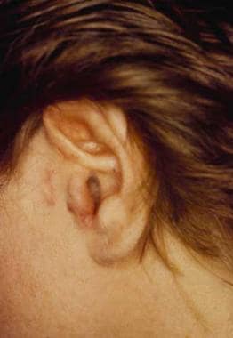
Angiolymphoid Hyperplasia with Eosinophilia
Mucinosis, Follicular
Skin Diseases
Dermatology
Sex Factors
Sex Distribution
A case of eosinophilic myocarditis complicated by Kimura's disease (eosinophilic hyperplastic lymphogranuloma) and erythroderma. (1/45)
This report describes a patient with eosinophilic myocarditis complicated by Kimura's disease (eosinophilic hyperplastic lymphogranuloma) and erythroderma. A 50-year-old man presented with a complaint of precordial pain. However, the only abnormal finding on examinatioin was eosinophilia (1617 eosinophils/microl). Three years later, the patient developed chronic eczema, and was diagnosed with erythroderma posteczematosa. One year later, a tumor was detected in the right auricule, and a diagnosis of Kimura's disease was made, based on the biopsy findings. The patient developed progressive dyspnea 6 months later and was found to have cardiomegaly and a depressed left ventricular ejection fraction (17%). A diagnosis of eosinophilic myocarditis was made based on the results of a right ventricular endomyocardial biopsy. The eosinophilic myocarditis and erythrodrema were treated with steroids with improvement of both the eosinophilia and left ventricular function. (+info)Kimura's disease with bilateral auricular masses. (2/45)
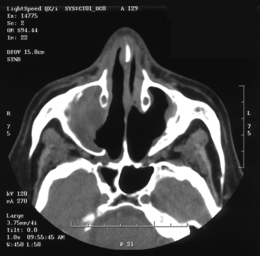
We report an unusual case of Kimura's disease. An 81-year-old Japanese woman was shown to have bilateral auricular masses that had begun to enlarge 6 years before. On CT scans, slightly high-density masses with faint contrast enhancement were seen. The masses were heterogeneous and hypointense on T1-weighted MR images, were slightly hyperintense on T2-weighted MR images, and showed heterogeneous enhancement after the administration of contrast material. Kimura's disease should be included in the differential diagnosis of bilateral auricular tumors. (+info)Life threatening coronary artery spasm in childhood Kimura's disease. (3/45)
A 13 year old boy is described with hypereosinophilia associated with Kimura's disease, who showed repeated life threatening syncopal attacks during daily activities or at rest. Coronary arteriography demonstrated small aneurysms with irregular vessel walls of both coronary arteries, and the absence of organic stenotic lesions. Infusion of a minimal dose of ergonovine into the right coronary artery induced severe spasm of the vessel. Ventricular fibrillation recurred even after administration of nifedipine and isosorbide was started, but was completely inhibited by prednisolone. (+info)Gray scale and power Doppler sonography in cases of Kimura disease. (4/45)
SUMMARY: Kimura disease is a rare chronic inflammatory disorder mimicking malignancy. Nodes are present in the submental and submandibular regions, within the parotid gland, and in the upper cervical chain. On gray scale sonograms, they are hypoechoic and round, with normal hilar architecture and homogeneous internal echoes. On power Doppler sonograms, the nodes show prominent intranodal vessels with a hilar pattern and low intranodal resistance. The soft tissue and parotid lesions also show low-resistance vascularity within. (+info)Bilateral orbital Kimura's disease in a young Asian man. (5/45)
A 16-year-old Asian man adolescent presented with bilateral eyelid swelling with multiple palpable mass lesions, which waned after treatment with corticosteroids but waxed after medications were discontinued, for about 1 year. He was otherwise healthy except that bilateral postauricular lymphadenopathy had developed for about 9 years. Laboratory study revealed peripheral eosinophilia with an elevated IgE level. The tumor masses of the left orbit were completely excised through an incision similar to that used in blepharoplasty, which gave good cosmetic results. Postoperative computed tomography scan showed no residual tumor mass in the left orbit but contralateral homogeneous soft-tissue mass lesions around the lacrimal gland and extending deep into the orbit, between the superior and lateral rectus muscles. Pathology reported numerous lymphoid follicles with active germinal centers and extensive lymphocyte and eosinophil infiltration, which characterize Kimura's disease (KD). The tumor mass in the right orbit was also excised during a second elective surgery 10 days later. No evidence of recurrence was noted after follow-up for 7 months. KD of the orbit is rare and usually occurs in middle-aged to elderly Asian men but can also be present in young adolescent. Complete excision with simultaneous blepharoplasty gives satisfactory cosmetic results. (+info)Bilateral Kimura's disease of the eyelids. (6/45)
A case of Kimura's disease affecting the eyelids bilaterally is reported in a 5-year-old boy of Afro-Caribbean extraction who has been followed for 12 years with repeat biopsies. He initially presented at 5 years of age with swelling of the left upper eyelid, left cervical lymphadenopathy, and eosinophilia. One year later he developed swelling of the right upper eyelid. There has been no change in the clinical appearance over the next 12 years. Repeated biopsies of the eyelids showed a diffuse inflammatory infiltrate with many eosinophils and lymphocytes. A lymph node biopsy showed reactive lymphoid hyperplasia. Immunohistochemistry using lymphoid markers showed a polyclonal pattern. Kimura's disease is a rare cause of eyelid swelling, particularly at such a young age and with bilateral involvement. This case demonstrates that bilateral orbital lymphoid lesions with cervical node involvement do not always imply lymphoma, but may have a benign pathogenesis. The unusually long follow up in this case confirms an excellent prognosis for Kimura's disease with conservative management. Accurate diagnosis in small orbital biopsies may spare the patient unnecessary radical surgery. (+info)Kimura's disease: a diagnostic and therapeutic challenge. (7/45)
INTRODUCTION: Kimura's disease (KD) is a rare, benign, chronic inflammatory disease with unknown aetiology. Its manifestation is protean. KD has a predilection for the head and neck area, and typically presents as tumour-like lesions that could be easily misdiagnosed. We review our experience with four recent cases. METHODS: Over a four-year period, all patients admitted to Singapore General Hospital with KD of the head and neck region were retrospectively reviewed. Biodata, presenting symptoms and clinical parameters, especially serum eosinophil levels, preoperative investigations, type of surgical procedures and outcome were documented. RESULTS: Four patients presented with KD of the head and neck and displayed varied manifestations of the disease. All the patients had raised serum eosinophil levels. None of them had renal involvement. Preoperative computed tomography were performed in two of the patients and showed features suggestive of KD. Fine-needle aspiration cytology that was performed in two patients was not useful in the diagnosis. All the patients underwent surgical excision of the lesions. Only one patient had multiple recurrence, both at the original and remote sites in the head and neck. CONCLUSION: The clinical presentation and behaviour of KD is very variable. Preoperative imaging is useful in the diagnosis of the disease but the final diagnosis is histological. Surgical excision is the current treatment of choice but recurrence is common. A high index of suspicion and awareness is vital in the early diagnosis and management of KD. (+info)Angiolymphoid hyperplasia with eosinophilia (epithelioid hemangioma) of the lung: a clinicopathologic and immunohistochemical study of two cases. (8/45)
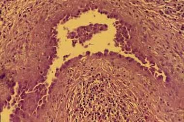
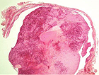
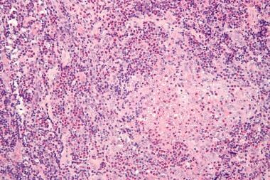
Two cases of primary angiolymphoid hyperplasia with eosinophilia (epithelioid hemangioma) (ALHE/EH) of the lung are described. Both patients are white, a 60-year-old man and a 27-year-old woman. One patient had a long-standing history of asthma, and the other had symptomatology related to the pulmonary mass. Wedge resections were performed in both cases, and both lesions shared similar histopathologic changes, mainly the presence of a tumor mass with a marked presence of eosinophils in the background, lymphoid hyperplasia, and marked proliferations of small-caliber vessels. Immunohistochemical studies using leukocyte common antigen, L-26, and UCHL-1 nicely stained the lymphoid component of the lesion, and CD31 clearly outlined the vascular component of the process. Clinical follow-up demonstrated that the woman died of status asthmaticus, and the man was alive and well 1 year after surgical resection of the lesion. Both cases highlight the ubiquitous distribution of ALHE/EH and underscore the importance of keeping these lesions in the differential diagnosis of vascular and lymphoid lesions of the lung. (+info)Angiolymphoid hyperplasia with eosinophilia (ALHE) is a rare benign vascular lesion that typically presents as one or multiple papules or nodules, often on the head and neck region. The exact cause of ALHE is unknown, but it has been associated with chronic inflammation and immune dysfunction.
Histologically, ALHE is characterized by the proliferation of blood vessels and lymphoid tissue, with a prominent infiltration of eosinophils. The lesions may also contain other inflammatory cells such as plasma cells, histiocytes, and T-lymphocytes.
Clinically, ALHE presents as red to brownish papules or nodules that can be tender or pruritic (itchy). Lesions typically occur on the head and neck region, particularly around the ears, eyes, and nose. In some cases, lesions may also appear on the trunk, arms, or legs.
While ALHE is a benign condition, it can cause significant cosmetic concerns due to its location. Treatment options include surgical excision, laser therapy, and intralesional corticosteroid injections. Recurrence after treatment is not uncommon. It is important to note that while ALHE may resemble other more serious conditions such as cutaneous lymphoma or angiosarcoma, it has a much more favorable prognosis.
Follicular mucinosis is a cutaneous condition characterized by the accumulation of mucin in the hair follicles. Mucin is a complex sugar-protein substance that provides cushioning and lubrication in various tissues throughout the body. In follicular mucinosis, there is an overproduction of mucin within the hair follicles, leading to visible bumps or papules on the skin.
Follicular mucinosis can be classified into three types: primary, secondary, and variant. The primary type is further divided into two subcategories: classic and atypical. The classic form of primary follicular mucinosis typically affects middle-aged adults and presents with localized or generalized patches of skin that are scaly, itchy, and have a smooth, shiny appearance (known as "alopetic pseudopelade"). In contrast, the atypical form is often associated with lymphoma.
Secondary follicular mucinosis can occur in association with various inflammatory skin conditions, such as eczema, psoriasis, and discoid lupus erythematosus. The variant type of follicular mucinosis is a rare condition that primarily affects children and adolescents, presenting with localized areas of thickened, rough skin (known as "hyperkeratotic").
The exact cause of primary follicular mucinosis remains unclear, but it is thought to involve an abnormal immune response. Secondary follicular mucinosis, on the other hand, is a reactive process triggered by underlying inflammatory skin conditions. Treatment for follicular mucinosis depends on the type and severity of the condition, ranging from topical corticosteroids to systemic immunosuppressive therapy in more severe cases.
Eosinophilia is a medical condition characterized by an abnormally high concentration of eosinophils in the circulating blood. Eosinophils are a type of white blood cell that play an important role in the immune system, particularly in fighting off parasitic infections and regulating allergic reactions. However, when their numbers become excessively high, they can contribute to tissue damage and inflammation.
Eosinophilia is typically defined as a count of more than 500 eosinophils per microliter of blood. Mild eosinophilia (up to 1,500 cells/μL) may not cause any symptoms and may be discovered during routine blood tests. However, higher levels of eosinophilia can lead to various symptoms such as coughing, wheezing, skin rashes, and organ damage, depending on the underlying cause.
The causes of eosinophilia are varied and can include allergic reactions, parasitic infections, autoimmune disorders, certain medications, and some types of cancer. Accurate diagnosis and treatment of eosinophilia require identification and management of the underlying cause.
Eye diseases are a range of conditions that affect the eye or visual system, causing damage to vision and, in some cases, leading to blindness. These diseases can be categorized into various types, including:
1. Refractive errors: These include myopia (nearsightedness), hyperopia (farsightedness), astigmatism, and presbyopia, which affect the way light is focused on the retina and can usually be corrected with glasses or contact lenses.
2. Cataracts: A clouding of the lens inside the eye that leads to blurry vision, glare, and decreased contrast sensitivity. Cataract surgery is the most common treatment for this condition.
3. Glaucoma: A group of diseases characterized by increased pressure in the eye, leading to damage to the optic nerve and potential blindness if left untreated. Treatment includes medications, laser therapy, or surgery.
4. Age-related macular degeneration (AMD): A progressive condition that affects the central part of the retina called the macula, causing blurry vision and, in advanced stages, loss of central vision. Treatment may include anti-VEGF injections, laser therapy, or nutritional supplements.
5. Diabetic retinopathy: A complication of diabetes that affects the blood vessels in the retina, leading to bleeding, leakage, and potential blindness if left untreated. Treatment includes laser therapy, anti-VEGF injections, or surgery.
6. Retinal detachment: A separation of the retina from its underlying tissue, which can lead to vision loss if not treated promptly with surgery.
7. Amblyopia (lazy eye): A condition where one eye does not develop normal vision, often due to a misalignment or refractive error in childhood. Treatment includes correcting the underlying problem and encouraging the use of the weaker eye through patching or other methods.
8. Strabismus (crossed eyes): A misalignment of the eyes that can lead to amblyopia if not treated promptly with surgery, glasses, or other methods.
9. Corneal diseases: Conditions that affect the transparent outer layer of the eye, such as keratoconus, Fuchs' dystrophy, and infectious keratitis, which can lead to vision loss if not treated promptly.
10. Uveitis: Inflammation of the middle layer of the eye, which can cause vision loss if not treated promptly with anti-inflammatory medications or surgery.
Skin diseases, also known as dermatological conditions, refer to any medical condition that affects the skin, which is the largest organ of the human body. These diseases can affect the skin's function, appearance, or overall health. They can be caused by various factors, including genetics, infections, allergies, environmental factors, and aging.
Skin diseases can present in many different forms, such as rashes, blisters, sores, discolorations, growths, or changes in texture. Some common examples of skin diseases include acne, eczema, psoriasis, dermatitis, fungal infections, viral infections, bacterial infections, and skin cancer.
The symptoms and severity of skin diseases can vary widely depending on the specific condition and individual factors. Some skin diseases are mild and can be treated with over-the-counter medications or topical creams, while others may require more intensive treatments such as prescription medications, light therapy, or even surgery.
It is important to seek medical attention if you experience any unusual or persistent changes in your skin, as some skin diseases can be serious or indicative of other underlying health conditions. A dermatologist is a medical doctor who specializes in the diagnosis and treatment of skin diseases.
Dermatology is a medical specialty that focuses on the diagnosis, treatment, and prevention of diseases and conditions related to the skin, hair, nails, and mucous membranes. A dermatologist is a medical doctor who has completed specialized training in this field. They are qualified to treat a wide range of skin conditions, including acne, eczema, psoriasis, skin cancer, and many others. Dermatologists may also perform cosmetic procedures to improve the appearance of the skin or to treat signs of aging.
Dermatologic agents are medications, chemicals, or other substances that are applied to the skin (dermis) for therapeutic or cosmetic purposes. They can be used to treat various skin conditions such as acne, eczema, psoriasis, fungal infections, and wounds. Dermatologic agents include topical corticosteroids, antibiotics, antifungals, retinoids, benzoyl peroxide, salicylic acid, and many others. They can come in various forms such as creams, ointments, gels, lotions, solutions, and patches. It is important to follow the instructions for use carefully to ensure safety and effectiveness.
"Sex factors" is a term used in medicine and epidemiology to refer to the differences in disease incidence, prevalence, or response to treatment that are observed between males and females. These differences can be attributed to biological differences such as genetics, hormones, and anatomy, as well as social and cultural factors related to gender.
For example, some conditions such as autoimmune diseases, depression, and osteoporosis are more common in women, while others such as cardiovascular disease and certain types of cancer are more prevalent in men. Additionally, sex differences have been observed in the effectiveness and side effects of various medications and treatments.
It is important to consider sex factors in medical research and clinical practice to ensure that patients receive appropriate and effective care.
"Sex distribution" is a term used to describe the number of males and females in a study population or sample. It can be presented as a simple count, a percentage, or a ratio. This information is often used in research to identify any differences in health outcomes, disease prevalence, or response to treatment between males and females. Additionally, understanding sex distribution can help researchers ensure that their studies are representative of the general population and can inform the design of future studies.
Mikulicz disease is a rare condition characterized by the symmetrical enlargement of the salivary and lacrimal glands. It is named after Jan Mikulicz-Radecki, a Polish surgeon who first described it in 1892. The enlarged glands are typically painless, and the condition can be associated with other systemic diseases such as Sjogren's syndrome, sarcoidosis, lymphoma, and tuberculosis.
In Mikulicz disease, there is a benign infiltration of the salivary and lacrimal glands with immune cells, particularly lymphocytes, which can lead to their enlargement. The exact cause of the condition is not known, but it is thought to be related to an autoimmune response.
Mikulicz disease is often treated with medications that suppress the immune system, such as corticosteroids or immunosuppressive drugs. In some cases, surgical removal of the affected glands may be necessary. The prognosis for Mikulicz disease is generally good, but it can vary depending on the underlying cause and any associated medical conditions.
 Angiolymphoid hyperplasia with eosinophilia
 Angiolymphoid Hyperplasia With Eosinophilia: Background, Pathophysiology, Etiology
 Angiolymphoid hyperplasia with eosinophilia associated with hepatitis C antibodies - Indian Journal of Dermatology, Venereology...
 Angiolymphoid hyperplasia with eosinophilia and entrapment of the ulnar nerve
 Indian Journal of Dermatology: Table of Contents
 CT angiography and MRI of hand vascular lesions: technical considerations and spectrum of imaging findings | Insights into...
 A dermatologic riddle: How is IgG4-related ophthalmic disease parliamentary?
 DeCS
DeCS Vol. 7 No. 5 (2019): Mar 15 (OAMJMS)
| Open Access Macedonian Journal of Medical Sciences
Vol. 7 No. 5 (2019): Mar 15 (OAMJMS)
| Open Access Macedonian Journal of Medical Sciences
 Histological and immunological studies on eosinophilic granuloma of soft tissue, so?called Kimura's disease
 Hyperplasia. Medical search
 by Institution Author "İnce, Songül"
 Vol. 7 No. 3 (2021): March 2021
| International Journal of Otorhinolaryngology and Head and Neck Surgery
Vol. 7 No. 3 (2021): March 2021
| International Journal of Otorhinolaryngology and Head and Neck Surgery
 The Ewha Medical Journal
 Biomedica ::..
 Reduction of Inter-Rater and Intra-Rater Variability in Psoriasis Area and Severity Index Assessment by Photographic Training...
 Epithelioid hemangioma of the penis: case report and review of literature | Journal of Medical Case Reports | Full Text
 Búsqueda | Portal Regional de la BVS
 IgG4-related disease - WikiProjectMed
 Journal of Dermatology Research and Therapy Archives | Clinmed International Library
 Juvenile Temporal Arteritis Clinically Masquerading as Temporal Artery Pseudoaneurysm | Marcus, MD | North American Journal of...
 Photosensitivity Disorders | Harvard Catalyst Profiles | Harvard Catalyst