Angiography, Digital Subtraction
Subtraction Technique
Magnetic Resonance Angiography
Cerebral Angiography
Intracranial Aneurysm
Coronary Angiography
Radiographic Image Enhancement
Imaging, Three-Dimensional
Intracranial Arteriovenous Malformations
Tomography, X-Ray Computed
Embolization, Therapeutic
Sensitivity and Specificity
Carotid Artery, Internal
Image Processing, Computer-Assisted
Aneurysm, Ruptured
Central Nervous System Vascular Malformations
Arterial Occlusive Diseases
Carotid Stenosis
Subarachnoid Hemorrhage
Tomography, Spiral Computed
Iohexol
Salivary Gland Calculi
Reproducibility of Results
Fluorescein Angiography
Iopamidol
Ultrasonography, Doppler, Duplex
Observer Variation
Constriction, Pathologic
Ultrasonography, Doppler, Color
Radiographic Image Interpretation, Computer-Assisted
Radiography, Dental, Digital
Arteriovenous Malformations
Vertebrobasilar Insufficiency
X-Ray Intensifying Screens
Artifacts
Carotid Artery Diseases
Predictive Value of Tests
Gadolinium DTPA
Anterior Cerebral Artery
Magnetic Resonance Imaging
Carotid-Cavernous Sinus Fistula
Vertebral Artery
Tibial Arteries
Retrospective Studies
Surgical Instruments
Prospective Studies
Cranial Sinuses
Vasospasm, Intracranial
Cerebrovascular Disorders
Image Enhancement
Basilar Artery
Radiography, Interventional
Ultrasonography, Doppler, Transcranial
Arteriovenous Fistula
Aneurysm
Treatment Outcome
Myelography
Circle of Willis
Cerebral Revascularization
Iothalamate Meglumine
Carotid Arteries
Intracranial Arteriosclerosis
Salivary Gland Diseases
Radiation Dosage
Feasibility Studies
Blood Flow Velocity
Radiography, Bitewing
Carotid Artery, External
Follow-Up Studies
Moyamoya Disease
Radionuclide Angiography
Phantoms, Imaging
Stents
Collateral Circulation
Aortography
Ultrasonography
Angioplasty, Balloon
Gadolinium
Sialadenitis
Immediate Dental Implant Loading
Ischemic Attack, Transient
Ultrasonography, Doppler
Popliteal Artery
Cerebrospinal Fluid Rhinorrhea
Endarterectomy
Brain Ischemia
Severity of Illness Index
Celiac Artery
Preoperative Care
Renal Artery Obstruction
Coronary Artery Disease
Cavernous Sinus
Organometallic Compounds
Radiography, Dual-Energy Scanned Projection
Ischemia
Iliac Artery
False Negative Reactions
Algorithms
Cerebral Infarction
False Positive Reactions
Peripheral Vascular Diseases
Single-Blind Method
Middle Cerebral Artery
Aneurysm, False
Libraries, Digital
ROC Curve
Catheterization
Endarterectomy, Carotid
Equipment Failure Analysis
Image Interpretation, Computer-Assisted
Models, Anatomic
Vascular Diseases
Blood Vessel Prosthesis
Technetium
Rotation
Indocyanine Green
Cerebral Hemorrhage
Postoperative Complications
Tomography, Emission-Computed, Single-Photon
Coronary Disease
Photography
Brain
Radiology Information Systems
Multidetector Computed Tomography
Radionuclide Imaging
Lower Extremity
Stroke
Sodium Pertechnetate Tc 99m
Gene Library
Hemodynamics
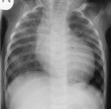
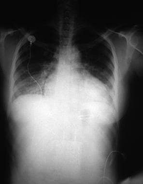
Radiography, Thoracic
Pulmonary Artery
Signal Processing, Computer-Assisted
Carotid Artery, Common
Risk Factors
Arteriosclerosis
Fundus Oculi
Diagnostic Imaging
Ultrasonography, Interventional
Combined carotid endarterectomy and coronary artery bypass graft. (1/1139)
Atherosclerosis is a generalized disease which afflicts a considerable number of patients in both the carotid and coronary arteries. Although the risk of stroke or death use to combined carotid endarterectomy (CEA) and coronary artery bypass graft (CABG) is thought to be higher than that of each individual operation, the combined procedure is generally preferred over staged operations to treat such patients. We performed the combined procedure safely with the aid of intraoperative portable digital subtraction angiography (DSA). This report describes our experience with the operative strategy of simultaneous CEA and CABG. Ninety CEA and 404 CABG were carried out between January 1989 and December 1997. A total of six patients received the combined procedure with the aid of intraoperative DSA; they were studied retrospectively. Postoperative mortality and morbidity after the combined procedure was 0%. In the combined procedure, neurological complications are difficult to detect after CEA because the patient must be maintained under general anesthesia and extracorporeal circulation during the subsequent CABG. However, intraoperative DSA can confirm patency of the internal carotid artery and absence of flap formation after CEA, and the CABG can be performed safely. Intraoperative portable DSA between CEA and CABG is helpful in preventing perioperative stroke in the combined procedure. (+info)Popliteal artery occlusion as a late complication of liquid acrylate embolization for cerebral vascular malformation. (2/1139)
Occlusion of arteriovenous malformations of the brain (BAVMs) by means of an endovascular approach with liquid acrylate glue is an established treatment modality. The specific hazards of this procedure are related to the central nervous system. In the case of unexpectedly rapid polymerization of the cyanoacrylate glue and adhesion of the delivering microcatheter to the BAVM, severing the catheter at the site of vascular access is considered an acceptable and safe management. We present a unique complication related to this technique that has not been described yet. Fragmentation and migration of the microcatheter, originally left in place, had caused popliteal artery occlusion, which required saphenous vein interposition, in a 25-year-old man. Suggestions for avoiding this complication are discussed. (+info)Factors influencing the development of vein-graft stenosis and their significance for clinical management. (3/1139)
OBJECTIVES: To assess the influence of clinical and graft factors on the development of stenotic lesions. In addition the implications of any significant correlation for duplex surveillance schedules or surgical bypass techniques was examined. PATIENTS AND METHODS: In a prospective three centre study, preoperative and peroperative data on 300 infrainguinal autologous vein grafts was analysed. All grafts were monitored by a strict duplex surveillance program and all received an angiogram in the first postoperative year. A revision was only performed if there was evidence of a stenosis of 70% diameter reduction or greater on the angiogram. RESULTS: The minimum graft diameter was the only factor correlated significantly with the development of a significant graft stenosis (PSV-ratio > or = 2.5) during follow-up (p = 0.002). Factors that correlated with the development of event-causing graft stenosis, associated with revision or occlusion, were minimal graft diameter (p = 0.001), the use of a venovenous anastomosis (p = 0.005) and length of the graft (p = 0.025). Multivariate regression analysis revealed that the minimal graft diameter was the only independent factor that significantly correlated with an event-causing graft stenosis (p = 0.009). The stenosis-free rates for grafts with a minimal diameter < 3.5 mm, between 3.5-4.5 and > or = 4.5 mm were 40%, 58% and 75%, respectively (p = < 0.05). Composite vein and arm-vein grafts with minimal diameters > or = 3.5 mm were compared with grafts which consisted of a single uninterrupted greater saphenous vein with a minimal diameter of < 3.5 mm. One-year secondary patency rates in these categories were of 94% and 76%, respectively (p = 0.03). CONCLUSIONS: A minimal graft diameter < 3.5 mm was the only factor that significantly correlated with the development of a graft-stenosis. However, veins with larger diameters may still develop stenotic lesions. Composite vein and arm-vein grafts should be used rather than uninterrupted small caliber saphenous veins. (+info)Bilateral vertebral artery occlusion following cervical spine trauma--case report. (4/1139)
A 41-year-old female presented with a rare case of bilateral vertebral artery occlusion following C5-6 cervical spine subluxation after a fall of 30 feet. Digital subtraction angiography showed occlusion of the bilateral vertebral arteries. Unlocking of the facet joint, posterior wiring with iliac crest grafting, and anterior fusion were performed. The patient died on the 3rd day after the operation. This type of injury has a grim prognosis with less than a third of the patients achieving a good outcome. (+info)Evaluation of cerebral aneurysms with high-resolution MR angiography using a section-interpolation technique: correlation with digital subtraction angiography. (5/1139)
BACKGROUND AND PURPOSE: The objective was to evaluate the results of high-resolution, fast-speed, section-interpolation MR angiography and digital subtraction angiography (DSA), thereby examining the potential use of a primary noninvasive screening test for intracranial aneurysms. METHODS: The images were obtained in 39 cerebral aneurysmal lesions from 30 patients with a time-of-flight MR angiographic technique using a 1.5-T superconducting MR system. The total image volume was divided into four slabs, with 48 partitions each. To save time, only 24 phase-encoded steps were measured and interpolated to 48. The parameters used included 30/6.4 (TR/TE), a flip angle of 25 degrees , a 160x512 matrix, a field of view of 150x200, 7 minutes 42 seconds of scan time, an effective thickness of 0.7 mm, and an entire thickness of 102.2 mm. Maximum intensity projection was used for the image analysis, and a multiplanar reconstruction technique was used for patients with intracranial aneurysms. RESULTS: Among 39 intracranial aneurysmal lesions in 30 patients, 21 were ruptured and 18 were unruptured. Twelve lesions were less than 2 mm in size, 12 were 3 to 5 mm, 12 were 6 to 9 mm, and three were larger than 10 mm. At initial examinations, 38 of 39 aneurysmal lesions were detected by both MR angiography and DSA, with 97% sensitivity. In confirming aneurysms in neck and parent vessels, multiplanar reconstruction was successful in detecting all 39 aneurysms, whereas MR angiography was successful in detecting 27 (69%) and DSA was successful in detecting 32 (82%) of the lesions. CONCLUSION: High-resolution MR angiography with a section-interpolation technique showed equal results to those of DSA for the detection of intracranial aneurysms and may be used as a primary noninvasive screening test. In the evaluation of aneurysms in neck and parent vessels, the concurrent use of MR angiography and multiplanar reconstruction was far superior to the use of either MR angiography or DSA alone. (+info)Twinkling artifact on intracerebral color Doppler sonography. (6/1139)
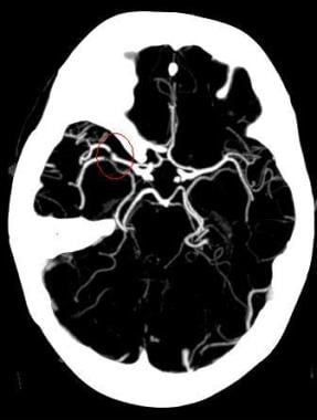
Transcranial Doppler sonography shows potential as a noninvasive technique for long-term follow-up of treated intracranial saccular aneurysms. This technical note describes a color Doppler artifact related to microcoil architecture that might represent a potential pitfall in transcranial Doppler sonographic evaluation of aneurysmal cavity thrombosis, since it may be wrongly interpreted as residual flow or aneurysmal cavity recanalization. (+info)Cerebral veins: comparative study of CT venography with intraarterial digital subtraction angiography. (7/1139)
BACKGROUND AND PURPOSE: Our objective was to compare the reliability of CT venography with intraarterial digital subtraction angiography (DSA) in imaging cerebral venous anatomy and pathology. METHODS: In 25 consecutive patients, 426 venous structures were determined as present, partially present, or absent by three observers evaluating CT multiplanar reformatted (MPR) and maximum intensity projection (MIP) images. These results were compared with the results from intraarterial DSA and, in a second step, with the results of an intraobserver consensus. In addition, pathologic conditions were described. RESULTS: Using DSA as the standard of reference, MPR images had an overall sensitivity of 95% (specificity, 19%) and MIP images a sensitivity of 80% (specificity, 44%) in depicting the cerebral venous anatomy. On the basis of an intraobserver consensus including DSA, MPR, and MIP images (415 vessels present), the sensitivity/specificity was 95%/91% for MPR, 90%/100% for DSA, and 79%/91% for MIP images. MPR images were superior to DSA images in showing the cavernous sinus, the inferior sagittal sinus, and the basal vein of Rosenthal. Venous occlusive diseases were correctly recognized on both MPR and MIP images. Only DSA images provided reliable information of invasion of a sinus by an adjacent meningioma. CONCLUSION: CT venography proved to be a reliable method to depict the cerebral venous structures. MPR images were superior to MIP images. (+info)A persistent pharyngohyostapedial artery: embryologic implications. (8/1139)
A 3-year-old child was examined because of otorrhagia. CT scans showed an unusual vessel, confirmed by angiography, related to a persistent pharyngohyostapedial artery. This embryonic persistent artery associated with the normal internal carotid artery would explain the "duplication" aspect of the internal carotid artery. (+info)Digital subtraction angiography (DSA) is a medical imaging technique used to visualize the blood vessels and blood flow within the body. It combines the use of X-ray technology with digital image processing to produce detailed images of the vascular system.
In DSA, a contrast agent is injected into the patient's bloodstream through a catheter, which is typically inserted into an artery in the leg and guided to the area of interest using fluoroscopy. As the contrast agent flows through the blood vessels, X-ray images are taken at multiple time points.
The digital subtraction process involves taking a baseline image without contrast and then subtracting it from subsequent images taken with contrast. This allows for the removal of background structures and noise, resulting in clearer images of the blood vessels. DSA can be used to diagnose and evaluate various vascular conditions, such as aneurysms, stenosis, and tumors, and can also guide interventional procedures such as angioplasty and stenting.
The "subtraction technique" is not a widely recognized or established term in medical terminology. It may refer to various methods used in different medical contexts that involve subtracting or comparing measurements, values, or observations to diagnose, monitor, or treat medical conditions. However, without more specific context, it's difficult to provide an accurate medical definition of the term.
In radiology, for example, the subtraction technique is a method used in imaging to enhance the visibility of certain structures by digitally subtracting one image from another. This technique is often used in angiography to visualize blood vessels more clearly.
Therefore, it's essential to provide more context or specify the medical field when using the term "subtraction technique" to ensure accurate communication and understanding.
Magnetic Resonance Angiography (MRA) is a non-invasive medical imaging technique that uses magnetic fields and radio waves to create detailed images of the blood vessels or arteries within the body. It is a type of Magnetic Resonance Imaging (MRI) that focuses specifically on the circulatory system.
MRA can be used to diagnose and evaluate various conditions related to the blood vessels, such as aneurysms, stenosis (narrowing of the vessel), or the presence of plaques or tumors. It can also be used to plan for surgeries or other treatments related to the vascular system. The procedure does not use radiation and is generally considered safe, although people with certain implants like pacemakers may not be able to have an MRA due to safety concerns.
Cerebral angiography is a medical procedure that involves taking X-ray images of the blood vessels in the brain after injecting a contrast dye into them. This procedure helps doctors to diagnose and treat various conditions affecting the blood vessels in the brain, such as aneurysms, arteriovenous malformations, and stenosis (narrowing of the blood vessels).
During the procedure, a catheter is inserted into an artery in the leg and threaded through the body to the blood vessels in the neck or brain. The contrast dye is then injected through the catheter, and X-ray images are taken to visualize the blood flow through the brain's blood vessels.
Cerebral angiography provides detailed images of the blood vessels in the brain, allowing doctors to identify any abnormalities or blockages that may be causing symptoms or increasing the risk of stroke. Based on the results of the cerebral angiography, doctors can develop a treatment plan to address these issues and prevent further complications.
Angiography is a medical procedure in which an x-ray image is taken to visualize the internal structure of blood vessels, arteries, or veins. This is done by injecting a radiopaque contrast agent (dye) into the blood vessel using a thin, flexible catheter. The dye makes the blood vessels visible on an x-ray image, allowing doctors to diagnose and treat various medical conditions such as blockages, narrowing, or malformations of the blood vessels.
There are several types of angiography, including:
* Cardiac angiography (also called coronary angiography) - used to examine the blood vessels of the heart
* Cerebral angiography - used to examine the blood vessels of the brain
* Peripheral angiography - used to examine the blood vessels in the limbs or other parts of the body.
Angiography is typically performed by a radiologist, cardiologist, or vascular surgeon in a hospital setting. It can help diagnose conditions such as coronary artery disease, aneurysms, and peripheral arterial disease, among others.
An intracranial aneurysm is a localized, blood-filled dilation or bulging in the wall of a cerebral artery within the skull (intracranial). These aneurysms typically occur at weak points in the arterial walls, often at branching points where the vessel divides into smaller branches. Over time, the repeated pressure from blood flow can cause the vessel wall to weaken and balloon out, forming a sac-like structure. Intracranial aneurysms can vary in size, ranging from a few millimeters to several centimeters in diameter.
There are three main types of intracranial aneurysms:
1. Saccular (berry) aneurysm: This is the most common type, characterized by a round or oval shape with a narrow neck and a bulging sac. They usually develop at branching points in the arteries due to congenital weaknesses in the vessel wall.
2. Fusiform aneurysm: These aneurysms have a dilated segment along the length of the artery, forming a cigar-shaped or spindle-like structure. They are often caused by atherosclerosis and can affect any part of the cerebral arteries.
3. Dissecting aneurysm: This type occurs when there is a tear in the inner lining (intima) of the artery, allowing blood to flow between the layers of the vessel wall. It can lead to narrowing or complete blockage of the affected artery and may cause subarachnoid hemorrhage if it ruptures.
Intracranial aneurysms can be asymptomatic and discovered incidentally during imaging studies for other conditions. However, when they grow larger or rupture, they can lead to severe complications such as subarachnoid hemorrhage, stroke, or even death. Treatment options include surgical clipping, endovascular coiling, or flow diversion techniques to prevent further growth and potential rupture of the aneurysm.
Coronary angiography is a medical procedure that uses X-ray imaging to visualize the coronary arteries, which supply blood to the heart muscle. During the procedure, a thin, flexible catheter is inserted into an artery in the arm or groin and threaded through the blood vessels to the heart. A contrast dye is then injected through the catheter, and X-ray images are taken as the dye flows through the coronary arteries. These images can help doctors diagnose and treat various heart conditions, such as blockages or narrowing of the arteries, that can lead to chest pain or heart attacks. It is also known as coronary arteriography or cardiac catheterization.
Radiographic image enhancement refers to the process of improving the quality and clarity of radiographic images, such as X-rays, CT scans, or MRI images, through various digital techniques. These techniques may include adjusting contrast, brightness, and sharpness, as well as removing noise and artifacts that can interfere with image interpretation.
The goal of radiographic image enhancement is to provide medical professionals with clearer and more detailed images, which can help in the diagnosis and treatment of medical conditions. This process may be performed using specialized software or hardware tools, and it requires a strong understanding of imaging techniques and the specific needs of medical professionals.
Three-dimensional (3D) imaging in medicine refers to the use of technologies and techniques that generate a 3D representation of internal body structures, organs, or tissues. This is achieved by acquiring and processing data from various imaging modalities such as X-ray computed tomography (CT), magnetic resonance imaging (MRI), ultrasound, or confocal microscopy. The resulting 3D images offer a more detailed visualization of the anatomy and pathology compared to traditional 2D imaging techniques, allowing for improved diagnostic accuracy, surgical planning, and minimally invasive interventions.
In 3D imaging, specialized software is used to reconstruct the acquired data into a volumetric model, which can be manipulated and viewed from different angles and perspectives. This enables healthcare professionals to better understand complex anatomical relationships, detect abnormalities, assess disease progression, and monitor treatment response. Common applications of 3D imaging include neuroimaging, orthopedic surgery planning, cancer staging, dental and maxillofacial reconstruction, and interventional radiology procedures.
Contrast media are substances that are administered to a patient in order to improve the visibility of internal body structures or processes in medical imaging techniques such as X-rays, CT scans, MRI scans, and ultrasounds. These media can be introduced into the body through various routes, including oral, rectal, or intravenous administration.
Contrast media work by altering the appearance of bodily structures in imaging studies. For example, when a patient undergoes an X-ray examination, contrast media can be used to highlight specific organs, tissues, or blood vessels, making them more visible on the resulting images. In CT and MRI scans, contrast media can help to enhance the differences between normal and abnormal tissues, allowing for more accurate diagnosis and treatment planning.
There are several types of contrast media available, each with its own specific properties and uses. Some common examples include barium sulfate, which is used as a contrast medium in X-ray studies of the gastrointestinal tract, and iodinated contrast media, which are commonly used in CT scans to highlight blood vessels and other structures.
While contrast media are generally considered safe, they can sometimes cause adverse reactions, ranging from mild symptoms such as nausea or hives to more serious complications such as anaphylaxis or kidney damage. As a result, it is important for healthcare providers to carefully evaluate each patient's medical history and individual risk factors before administering contrast media.
Intracranial arteriovenous malformations (AVMs) are abnormal, tangled connections between the arteries and veins in the brain. These connections bypass the capillary system, which can lead to high-flow shunting and potential complications such as hemorrhage, stroke, or neurological deficits. AVMs are congenital conditions, meaning they are present at birth, although symptoms may not appear until later in life. They are relatively rare, affecting approximately 0.1% of the population. Treatment options for AVMs include surgery, radiation therapy, and endovascular embolization, depending on the size, location, and specific characteristics of the malformation.
X-ray computed tomography (CT or CAT scan) is a medical imaging method that uses computer-processed combinations of many X-ray images taken from different angles to produce cross-sectional (tomographic) images (virtual "slices") of the body. These cross-sectional images can then be used to display detailed internal views of organs, bones, and soft tissues in the body.
The term "computed tomography" is used instead of "CT scan" or "CAT scan" because the machines take a series of X-ray measurements from different angles around the body and then use a computer to process these data to create detailed images of internal structures within the body.
CT scanning is a noninvasive, painless medical test that helps physicians diagnose and treat medical conditions. CT imaging provides detailed information about many types of tissue including lung, bone, soft tissue and blood vessels. CT examinations can be performed on every part of the body for a variety of reasons including diagnosis, surgical planning, and monitoring of therapeutic responses.
In computed tomography (CT), an X-ray source and detector rotate around the patient, measuring the X-ray attenuation at many different angles. A computer uses this data to construct a cross-sectional image by the process of reconstruction. This technique is called "tomography". The term "computed" refers to the use of a computer to reconstruct the images.
CT has become an important tool in medical imaging and diagnosis, allowing radiologists and other physicians to view detailed internal images of the body. It can help identify many different medical conditions including cancer, heart disease, lung nodules, liver tumors, and internal injuries from trauma. CT is also commonly used for guiding biopsies and other minimally invasive procedures.
In summary, X-ray computed tomography (CT or CAT scan) is a medical imaging technique that uses computer-processed combinations of many X-ray images taken from different angles to produce cross-sectional images of the body. It provides detailed internal views of organs, bones, and soft tissues in the body, allowing physicians to diagnose and treat medical conditions.
Therapeutic embolization is a medical procedure that involves intentionally blocking or obstructing blood vessels to stop excessive bleeding or block the flow of blood to a tumor or abnormal tissue. This is typically accomplished by injecting small particles, such as microspheres or coils, into the targeted blood vessel through a catheter, which is inserted into a larger blood vessel and guided to the desired location using imaging techniques like X-ray or CT scanning. The goal of therapeutic embolization is to reduce the size of a tumor, control bleeding, or block off abnormal blood vessels that are causing problems.
Sensitivity and specificity are statistical measures used to describe the performance of a diagnostic test or screening tool in identifying true positive and true negative results.
* Sensitivity refers to the proportion of people who have a particular condition (true positives) who are correctly identified by the test. It is also known as the "true positive rate" or "recall." A highly sensitive test will identify most or all of the people with the condition, but may also produce more false positives.
* Specificity refers to the proportion of people who do not have a particular condition (true negatives) who are correctly identified by the test. It is also known as the "true negative rate." A highly specific test will identify most or all of the people without the condition, but may also produce more false negatives.
In medical testing, both sensitivity and specificity are important considerations when evaluating a diagnostic test. High sensitivity is desirable for screening tests that aim to identify as many cases of a condition as possible, while high specificity is desirable for confirmatory tests that aim to rule out the condition in people who do not have it.
It's worth noting that sensitivity and specificity are often influenced by factors such as the prevalence of the condition in the population being tested, the threshold used to define a positive result, and the reliability and validity of the test itself. Therefore, it's important to consider these factors when interpreting the results of a diagnostic test.
The internal carotid artery is a major blood vessel that supplies oxygenated blood to the brain. It originates from the common carotid artery and passes through the neck, entering the skull via the carotid canal in the temporal bone. Once inside the skull, it branches into several smaller vessels that supply different parts of the brain with blood.
The internal carotid artery is divided into several segments: cervical, petrous, cavernous, clinoid, and supraclinoid. Each segment has distinct clinical significance in terms of potential injury or disease. The most common conditions affecting the internal carotid artery include atherosclerosis, which can lead to stroke or transient ischemic attack (TIA), and dissection, which can cause severe headache, neck pain, and neurological symptoms.
It's important to note that any blockage or damage to the internal carotid artery can have serious consequences, as it can significantly reduce blood flow to the brain and lead to permanent neurological damage or even death. Therefore, regular check-ups and screening tests are recommended for individuals at high risk of developing vascular diseases.
Computer-assisted image processing is a medical term that refers to the use of computer systems and specialized software to improve, analyze, and interpret medical images obtained through various imaging techniques such as X-ray, CT (computed tomography), MRI (magnetic resonance imaging), ultrasound, and others.
The process typically involves several steps, including image acquisition, enhancement, segmentation, restoration, and analysis. Image processing algorithms can be used to enhance the quality of medical images by adjusting contrast, brightness, and sharpness, as well as removing noise and artifacts that may interfere with accurate diagnosis. Segmentation techniques can be used to isolate specific regions or structures of interest within an image, allowing for more detailed analysis.
Computer-assisted image processing has numerous applications in medical imaging, including detection and characterization of lesions, tumors, and other abnormalities; assessment of organ function and morphology; and guidance of interventional procedures such as biopsies and surgeries. By automating and standardizing image analysis tasks, computer-assisted image processing can help to improve diagnostic accuracy, efficiency, and consistency, while reducing the potential for human error.
A ruptured aneurysm is a serious medical condition that occurs when the wall of an artery or a blood vessel weakens and bulges out, forming an aneurysm, which then bursts, causing bleeding into the surrounding tissue. This can lead to internal hemorrhage, organ damage, and even death, depending on the location and severity of the rupture.
Ruptured aneurysms are often caused by factors such as high blood pressure, smoking, aging, and genetic predisposition. They can occur in any part of the body but are most common in the aorta (the largest artery in the body) and the cerebral arteries (in the brain).
Symptoms of a ruptured aneurysm may include sudden and severe pain, weakness or paralysis, difficulty breathing, confusion, loss of consciousness, and shock. Immediate medical attention is required to prevent further complications and increase the chances of survival. Treatment options for a ruptured aneurysm may include surgery, endovascular repair, or medication to manage symptoms and prevent further bleeding.
Central nervous system (CNS) vascular malformations are abnormal tangles or masses of blood vessels in the brain or spinal cord. These malformations can be congenital (present at birth) or acquired (develop later in life). They can vary in size, location, and symptoms, which may include headaches, seizures, weakness, numbness, difficulty speaking or understanding speech, and vision problems.
There are several types of CNS vascular malformations, including:
1. Arteriovenous malformations (AVMs): These are tangles of arteries and veins with a direct connection between them, bypassing the capillary network. AVMs can cause bleeding in the brain or spinal cord, leading to stroke or neurological deficits.
2. Cavernous malformations: These are clusters of dilated, thin-walled blood vessels that form a sac-like structure. They can rupture and bleed, causing symptoms such as seizures, headaches, or neurological deficits.
3. Developmental venous anomalies (DVAs): These are benign vascular malformations characterized by an abnormal pattern of veins that drain blood from the brain. DVAs are usually asymptomatic but can be associated with other vascular malformations.
4. Capillary telangiectasias: These are small clusters of dilated capillaries in the brain or spinal cord. They are usually asymptomatic and found incidentally during imaging studies.
5. Moyamoya disease: This is a rare, progressive cerebrovascular disorder characterized by the narrowing or blockage of the internal carotid arteries and their branches. This can lead to decreased blood flow to the brain, causing symptoms such as headaches, seizures, and strokes.
The diagnosis of CNS vascular malformations typically involves imaging studies such as MRI or CT scans, and sometimes angiography. Treatment options may include observation, medication, surgery, or endovascular procedures, depending on the type, location, and severity of the malformation.
Arterial occlusive diseases are medical conditions characterized by the blockage or narrowing of the arteries, which can lead to a reduction in blood flow to various parts of the body. This reduction in blood flow can cause tissue damage and may result in serious complications such as tissue death (gangrene), organ dysfunction, or even death.
The most common cause of arterial occlusive diseases is atherosclerosis, which is the buildup of plaque made up of fat, cholesterol, calcium, and other substances in the inner lining of the artery walls. Over time, this plaque can harden and narrow the arteries, restricting blood flow. Other causes of arterial occlusive diseases include blood clots, emboli (tiny particles that travel through the bloodstream and lodge in smaller vessels), inflammation, trauma, and certain inherited conditions.
Symptoms of arterial occlusive diseases depend on the location and severity of the blockage. Common symptoms include:
* Pain, cramping, or fatigue in the affected limb, often triggered by exercise and relieved by rest (claudication)
* Numbness, tingling, or weakness in the affected limb
* Coldness or discoloration of the skin in the affected area
* Slow-healing sores or wounds on the toes, feet, or legs
* Erectile dysfunction in men
Treatment for arterial occlusive diseases may include lifestyle changes such as quitting smoking, exercising regularly, and eating a healthy diet. Medications to lower cholesterol, control blood pressure, prevent blood clots, or manage pain may also be prescribed. In severe cases, surgical procedures such as angioplasty, stenting, or bypass surgery may be necessary to restore blood flow.
Carotid stenosis is a medical condition that refers to the narrowing or constriction of the lumen (inner space) of the carotid artery. The carotid arteries are major blood vessels that supply oxygenated blood to the head and neck. Carotid stenosis usually results from the buildup of plaque, made up of fat, cholesterol, calcium, and other substances, on the inner walls of the artery. This process is called atherosclerosis.
As the plaque accumulates, it causes the artery to narrow, reducing blood flow to the brain. Severe carotid stenosis can increase the risk of stroke, as a clot or debris from the plaque can break off and travel to the brain, blocking a smaller blood vessel and causing tissue damage or death.
Carotid stenosis is typically diagnosed through imaging tests such as ultrasound, CT angiography, or MRI angiography. Treatment options may include lifestyle modifications (such as quitting smoking, controlling blood pressure, and managing cholesterol levels), medications to reduce the risk of clots, or surgical procedures like endarterectomy or stenting to remove or bypass the blockage.
A subarachnoid hemorrhage is a type of stroke that results from bleeding into the space surrounding the brain, specifically within the subarachnoid space which contains cerebrospinal fluid (CSF). This space is located between the arachnoid membrane and the pia mater, two of the three layers that make up the meninges, the protective covering of the brain and spinal cord.
The bleeding typically originates from a ruptured aneurysm, a weakened area in the wall of a cerebral artery, or less commonly from arteriovenous malformations (AVMs) or head trauma. The sudden influx of blood into the CSF-filled space can cause increased intracranial pressure, irritation to the brain, and vasospasms, leading to further ischemia and potential additional neurological damage.
Symptoms of a subarachnoid hemorrhage may include sudden onset of severe headache (often described as "the worst headache of my life"), neck stiffness, altered mental status, nausea, vomiting, photophobia, and focal neurological deficits. Rapid diagnosis and treatment are crucial to prevent further complications and improve the chances of recovery.
Spiral Computed Tomography (CT), also known as Helical CT, is a type of computed tomography scan in which the X-ray tube and detector rotate around the patient in a spiral path, capturing data as the table moves the patient through the scanner. This continuous spiral motion allows for faster and more detailed volumetric imaging of internal organs and structures, reducing the need for multiple slices and providing improved image reconstruction. It is commonly used to diagnose and monitor various medical conditions, including cancer, heart disease, and trauma injuries.
Cerebral veins are the blood vessels that carry deoxygenated blood from the brain to the dural venous sinuses, which are located between the layers of tissue covering the brain. The largest cerebral vein is the superior sagittal sinus, which runs along the top of the brain. Other major cerebral veins include the straight sinus, transverse sinus, sigmoid sinus, and cavernous sinus. These veins receive blood from smaller veins called venules that drain the surface and deep structures of the brain. The cerebral veins play an important role in maintaining normal circulation and pressure within the brain.
Iohexol is a non-ionic, water-soluble contrast medium primarily used in radiographic imaging procedures such as computed tomography (CT) scans and angiography. It belongs to a class of medications known as radiocontrast agents. Iohexol works by increasing the X-ray absorption of body tissues, making them more visible on X-ray images. This helps healthcare professionals to better diagnose and assess various medical conditions, including injuries, tumors, and vascular diseases.
The chemical structure of iohexol consists of an iodine atom surrounded by organic molecules, which makes it safe for intravenous administration. It is eliminatted from the body primarily through urinary excretion. Iohexol has a low risk of allergic reactions compared to ionic contrast media and is generally well-tolerated in patients with normal renal function. However, its use should be avoided or closely monitored in individuals with impaired kidney function, as it may increase the risk of nephrotoxicity.
Salivary gland calculi, also known as salivary duct stones or sialoliths, are small, hard deposits that form in the salivary glands or their ducts. These calculi typically consist of calcium salts and other minerals, and can vary in size from a few millimeters to over a centimeter in diameter.
Salivary gland calculi can cause a range of symptoms, including pain, swelling, and difficulty swallowing, particularly during meals. The obstruction of the salivary duct by the calculus can lead to infection or inflammation of the salivary gland (sialadenitis).
The most common location for salivary gland calculi is in the submandibular gland and its duct, followed by the parotid gland and then the sublingual gland. Treatment options for salivary gland calculi include conservative management with hydration, massage, and warm compresses, as well as more invasive procedures such as extracorporeal shock wave lithotripsy, sialendoscopy, or surgical removal of the calculus.
Reproducibility of results in a medical context refers to the ability to obtain consistent and comparable findings when a particular experiment or study is repeated, either by the same researcher or by different researchers, following the same experimental protocol. It is an essential principle in scientific research that helps to ensure the validity and reliability of research findings.
In medical research, reproducibility of results is crucial for establishing the effectiveness and safety of new treatments, interventions, or diagnostic tools. It involves conducting well-designed studies with adequate sample sizes, appropriate statistical analyses, and transparent reporting of methods and findings to allow other researchers to replicate the study and confirm or refute the results.
The lack of reproducibility in medical research has become a significant concern in recent years, as several high-profile studies have failed to produce consistent findings when replicated by other researchers. This has led to increased scrutiny of research practices and a call for greater transparency, rigor, and standardization in the conduct and reporting of medical research.
Fluorescein angiography is a medical diagnostic procedure used in ophthalmology to examine the blood flow in the retina and choroid, which are the inner layers of the eye. This test involves injecting a fluorescent dye, Fluorescein, into a patient's arm vein. As the dye reaches the blood vessels in the eye, a specialized camera takes rapid sequences of photographs to capture the dye's circulation through the retina and choroid.
The images produced by fluorescein angiography can help doctors identify any damage to the blood vessels, leakage, or abnormal growth of new blood vessels. This information is crucial in diagnosing and managing various eye conditions such as age-related macular degeneration, diabetic retinopathy, retinal vein occlusions, and inflammatory eye diseases.
It's important to note that while fluorescein angiography is a valuable diagnostic tool, it does carry some risks, including temporary side effects like nausea, vomiting, or allergic reactions to the dye. In rare cases, severe adverse reactions can occur, so patients should discuss these potential risks with their healthcare provider before undergoing the procedure.
Iopamidol is a non-ionic, low-osmolar contrast media (LOCM) used in diagnostic imaging procedures such as X-rays, CT scans, and angiography. It is a type of radiocontrast agent that contains iodine atoms, which absorb X-rays and make the internal structures of the body visible on X-ray images. Iopamidol has a low osmolarity, which means it has fewer particles per unit volume compared to high-osmolar contrast media (HOCM). This makes it safer and more comfortable for patients as it reduces the risk of adverse reactions such as pain, vasodilation, and kidney damage. Iopamidol is elimated from the body primarily through the kidneys and excreted in the urine.
Ultrasonography, Doppler, and Duplex are diagnostic medical techniques that use sound waves to create images of internal body structures and assess their function. Here are the definitions for each:
1. Ultrasonography: Also known as ultrasound, this is a non-invasive imaging technique that uses high-frequency sound waves to produce images of internal organs and tissues. A small handheld device called a transducer is placed on the skin surface, which emits and receives sound waves. The returning echoes are then processed to create real-time visual images of the internal structures.
2. Doppler: This is a type of ultrasound that measures the velocity and direction of blood flow in the body by analyzing the frequency shift of the reflected sound waves. It can be used to assess blood flow in various parts of the body, such as the heart, arteries, and veins.
3. Duplex: Duplex ultrasonography is a combination of both gray-scale ultrasound and Doppler ultrasound. It provides detailed images of internal structures, as well as information about blood flow velocity and direction. This technique is often used to evaluate conditions such as deep vein thrombosis, carotid artery stenosis, and peripheral arterial disease.
In summary, ultrasonography is a diagnostic imaging technique that uses sound waves to create images of internal structures, Doppler is a type of ultrasound that measures blood flow velocity and direction, and duplex is a combination of both techniques that provides detailed images and information about blood flow.
Observer variation, also known as inter-observer variability or measurement agreement, refers to the difference in observations or measurements made by different observers or raters when evaluating the same subject or phenomenon. It is a common issue in various fields such as medicine, research, and quality control, where subjective assessments are involved.
In medical terms, observer variation can occur in various contexts, including:
1. Diagnostic tests: Different radiologists may interpret the same X-ray or MRI scan differently, leading to variations in diagnosis.
2. Clinical trials: Different researchers may have different interpretations of clinical outcomes or adverse events, affecting the consistency and reliability of trial results.
3. Medical records: Different healthcare providers may document medical histories, physical examinations, or treatment plans differently, leading to inconsistencies in patient care.
4. Pathology: Different pathologists may have varying interpretations of tissue samples or laboratory tests, affecting diagnostic accuracy.
Observer variation can be minimized through various methods, such as standardized assessment tools, training and calibration of observers, and statistical analysis of inter-rater reliability.
Pathological constriction refers to an abnormal narrowing or tightening of a body passage or organ, which can interfere with the normal flow of blood, air, or other substances through the area. This constriction can occur due to various reasons such as inflammation, scarring, or abnormal growths, and can affect different parts of the body, including blood vessels, airways, intestines, and ureters. Pathological constriction can lead to a range of symptoms and complications depending on its location and severity, and may require medical intervention to correct.
Ultrasonography, Doppler, color is a type of diagnostic ultrasound technique that uses the Doppler effect to produce visual images of blood flow in vessels and the heart. The Doppler effect is the change in frequency or wavelength of a wave in relation to an observer who is moving relative to the source of the wave. In this context, it refers to the change in frequency of the ultrasound waves as they reflect off moving red blood cells.
In color Doppler ultrasonography, different colors are used to represent the direction and speed of blood flow. Red typically represents blood flowing toward the transducer (the device that sends and receives sound waves), while blue represents blood flowing away from the transducer. The intensity or brightness of the color is proportional to the velocity of blood flow.
Color Doppler ultrasonography is often used in conjunction with grayscale ultrasound imaging, which provides information about the structure and composition of tissues. Together, these techniques can help diagnose a wide range of conditions, including heart disease, blood clots, and abnormalities in blood flow.
Computer-assisted radiographic image interpretation is the use of computer algorithms and software to assist and enhance the interpretation and analysis of medical images produced by radiography, such as X-rays, CT scans, and MRI scans. The computer-assisted system can help identify and highlight certain features or anomalies in the image, such as tumors, fractures, or other abnormalities, which may be difficult for the human eye to detect. This technology can improve the accuracy and speed of diagnosis, and may also reduce the risk of human error. It's important to note that the final interpretation and diagnosis is always made by a qualified healthcare professional, such as a radiologist, who takes into account the computer-assisted analysis in conjunction with their clinical expertise and knowledge.
Dental digital radiography is a type of medical imaging that uses digital sensors instead of traditional X-ray film to produce highly detailed images of the teeth, gums, and surrounding structures. This technology offers several advantages over conventional dental radiography, including:
1. Lower radiation exposure: Digital sensors require less radiation to produce an image compared to traditional film, making it a safer option for patients.
2. Instant results: The images captured by digital sensors are immediately displayed on a computer screen, allowing dentists to quickly assess the patient's oral health and discuss any findings with them during the appointment.
3. Improved image quality: Digital radiography produces clearer and more precise images compared to traditional film, enabling dentists to better detect issues such as cavities, fractures, or tumors.
4. Enhanced communication: The ability to easily manipulate and enhance digital images allows for better communication between dental professionals and improved patient education.
5. Environmentally friendly: Digital radiography eliminates the need for chemical processing and disposal of used film, making it a more environmentally conscious choice.
6. Easy storage and retrieval: Digital images can be stored electronically and accessed easily for future reference or consultation with other dental professionals.
7. Remote consultations: Digital images can be shared remotely with specialists or insurance companies, facilitating faster diagnoses and treatment planning.
Arteriovenous malformations (AVMs) are abnormal tangles of blood vessels that directly connect arteries and veins, bypassing the capillary system. This results in a high-flow and high-pressure circulation in the affected area. AVMs can occur anywhere in the body but are most common in the brain and spine. They can vary in size and may cause symptoms such as headaches, seizures, or bleeding in the brain. In some cases, AVMs may not cause any symptoms and may only be discovered during imaging tests for other conditions. Treatment options include surgery, radiation therapy, or embolization to reduce the flow of blood through the malformation and prevent complications.
Cerebral arteries refer to the blood vessels that supply oxygenated blood to the brain. These arteries branch off from the internal carotid arteries and the vertebral arteries, which combine to form the basilar artery. The major cerebral arteries include:
1. Anterior cerebral artery (ACA): This artery supplies blood to the frontal lobes of the brain, including the motor and sensory cortices responsible for movement and sensation in the lower limbs.
2. Middle cerebral artery (MCA): The MCA is the largest of the cerebral arteries and supplies blood to the lateral surface of the brain, including the temporal, parietal, and frontal lobes. It is responsible for providing blood to areas involved in motor function, sensory perception, speech, memory, and vision.
3. Posterior cerebral artery (PCA): The PCA supplies blood to the occipital lobe, which is responsible for visual processing, as well as parts of the temporal and parietal lobes.
4. Anterior communicating artery (ACoA) and posterior communicating arteries (PComAs): These are small arteries that connect the major cerebral arteries, forming an important circulatory network called the Circle of Willis. The ACoA connects the two ACAs, while the PComAs connect the ICA with the PCA and the basilar artery.
These cerebral arteries play a crucial role in maintaining proper brain function by delivering oxygenated blood to various regions of the brain. Any damage or obstruction to these arteries can lead to serious neurological conditions, such as strokes or transient ischemic attacks (TIAs).
Vertebrobasilar insufficiency (VBI) is a medical condition characterized by inadequate blood flow to the vertebral and basilar arteries, which supply oxygenated blood to the brainstem and cerebellum. These arteries arise from the subclavian arteries and merge to form the basilar artery, which supplies critical structures in the posterior circulation of the brain.
VBI is often caused by atherosclerosis, or the buildup of plaque in the arterial walls, leading to narrowing (stenosis) or occlusion of these vessels. Other causes include embolism, arterial dissection, and vasculitis. The decreased blood flow can result in various neurological symptoms, such as dizziness, vertigo, imbalance, difficulty swallowing, slurred speech, visual disturbances, and even transient ischemic attacks (TIAs) or strokes.
Diagnosis of VBI typically involves a combination of clinical evaluation, imaging studies like MRA or CTA, and sometimes cerebral angiography to assess the extent and location of vascular narrowing or occlusion. Treatment options may include lifestyle modifications, medications to manage risk factors (such as hypertension, diabetes, or high cholesterol), antiplatelet therapy, or surgical interventions like endarterectomy or stenting in severe cases.
X-ray intensifying screens are medical imaging devices that contain phosphorescent materials, which emit light in response to the absorption of X-ray radiation. They are used in conjunction with X-ray film to enhance the visualization of radiographic images by converting X-rays into visible light. The screens are placed inside a cassette, along with the X-ray film, and exposed to X-rays during medical imaging procedures such as radiography or fluoroscopy.
The phosphorescent materials in the intensifying screens absorb most of the X-ray energy and re-emit it as visible light, which then exposes the X-ray film. This process increases the efficiency of the X-ray exposure, reducing the amount of radiation required to produce a diagnostic image. The use of intensifying screens can significantly improve the quality and detail of radiographic images while minimizing patient exposure to ionizing radiation.
An artifact, in the context of medical terminology, refers to something that is created or introduced during a scientific procedure or examination that does not naturally occur in the patient or specimen being studied. Artifacts can take many forms and can be caused by various factors, including contamination, damage, degradation, or interference from equipment or external sources.
In medical imaging, for example, an artifact might appear as a distortion or anomaly on an X-ray, MRI, or CT scan that is not actually present in the patient's body. This can be caused by factors such as patient movement during the scan, metal implants or other foreign objects in the body, or issues with the imaging equipment itself.
Similarly, in laboratory testing, an artifact might refer to a substance or characteristic that is introduced into a sample during collection, storage, or analysis that can interfere with accurate results. This could include things like contamination from other samples, degradation of the sample over time, or interference from chemicals used in the testing process.
In general, artifacts are considered to be sources of error or uncertainty in medical research and diagnosis, and it is important to identify and account for them in order to ensure accurate and reliable results.
Carotid artery diseases refer to conditions that affect the carotid arteries, which are the major blood vessels that supply oxygen-rich blood to the head and neck. The most common type of carotid artery disease is atherosclerosis, which occurs when fatty deposits called plaques build up in the inner lining of the arteries.
These plaques can cause the arteries to narrow or become blocked, reducing blood flow to the brain and increasing the risk of stroke. Other carotid artery diseases include carotid artery dissection, which occurs when there is a tear in the inner lining of the artery, and fibromuscular dysplasia, which is a condition that affects the muscle and tissue in the walls of the artery.
Symptoms of carotid artery disease may include neck pain or pulsations, transient ischemic attacks (TIAs) or "mini-strokes," and strokes. Treatment options for carotid artery disease depend on the severity and type of the condition but may include lifestyle changes, medications, endarterectomy (a surgical procedure to remove plaque from the artery), or angioplasty and stenting (procedures to open blocked arteries using a balloon and stent).
The Predictive Value of Tests, specifically the Positive Predictive Value (PPV) and Negative Predictive Value (NPV), are measures used in diagnostic tests to determine the probability that a positive or negative test result is correct.
Positive Predictive Value (PPV) is the proportion of patients with a positive test result who actually have the disease. It is calculated as the number of true positives divided by the total number of positive results (true positives + false positives). A higher PPV indicates that a positive test result is more likely to be a true positive, and therefore the disease is more likely to be present.
Negative Predictive Value (NPV) is the proportion of patients with a negative test result who do not have the disease. It is calculated as the number of true negatives divided by the total number of negative results (true negatives + false negatives). A higher NPV indicates that a negative test result is more likely to be a true negative, and therefore the disease is less likely to be present.
The predictive value of tests depends on the prevalence of the disease in the population being tested, as well as the sensitivity and specificity of the test. A test with high sensitivity and specificity will generally have higher predictive values than a test with low sensitivity and specificity. However, even a highly sensitive and specific test can have low predictive values if the prevalence of the disease is low in the population being tested.
Gadolinium DTPA (Diethylenetriaminepentaacetic acid) is a type of gadolinium-based contrast agent (GBCA) used in medical imaging, particularly magnetic resonance imaging (MRI) and magnetic resonance angiography (MRA). It functions as a paramagnetic substance that enhances the visibility of internal body structures during these imaging techniques.
The compound Gadolinium DTPA is formed when gadolinium ions are bound to diethylenetriaminepentaacetic acid, a chelating agent. This binding helps to make the gadolinium ion safer for use in medical imaging by reducing its toxicity and improving its stability in the body.
Gadolinium DTPA is eliminated from the body primarily through the kidneys, making it important to monitor renal function before administering this contrast agent. In some cases, Gadolinium DTPA may cause adverse reactions, including allergic-like responses and nephrogenic systemic fibrosis (NSF) in patients with impaired kidney function.
The Anterior Cerebral Artery (ACA) is a paired set of arteries that originate from the internal carotid artery or its branch, the posterior communicating artery. They supply oxygenated blood to the frontal lobes and parts of the parietal lobes of the brain.
The ACA runs along the medial side of each hemisphere, anterior to the corpus callosum, which is the largest bundle of nerve fibers connecting the two hemispheres of the brain. It gives off branches that supply the motor and sensory areas of the lower extremities, as well as the areas responsible for higher cognitive functions such as language, memory, and emotion.
The ACA is divided into several segments: A1, A2, A3, and A4. The A1 segment runs from its origin at the internal carotid artery to the anterior communicating artery, which connects the two ACAs. The A2 segment extends from the anterior communicating artery to the bifurcation of the ACA into its terminal branches. The A3 and A4 segments are the distal branches that supply the frontal and parietal lobes.
Interruptions or blockages in the flow of blood through the ACA can lead to various neurological deficits, including weakness or paralysis of the lower extremities, language impairment, and changes in cognitive function.
Medical Definition:
Magnetic Resonance Imaging (MRI) is a non-invasive diagnostic imaging technique that uses a strong magnetic field and radio waves to create detailed cross-sectional or three-dimensional images of the internal structures of the body. The patient lies within a large, cylindrical magnet, and the scanner detects changes in the direction of the magnetic field caused by protons in the body. These changes are then converted into detailed images that help medical professionals to diagnose and monitor various medical conditions, such as tumors, injuries, or diseases affecting the brain, spinal cord, heart, blood vessels, joints, and other internal organs. MRI does not use radiation like computed tomography (CT) scans.
Triiodobenzoic acids are a group of organic compounds that contain a benzene ring substituted with three iodine atoms and a carboxyl group. They have the general formula C6H3I3CO2H. These compounds do not have a specific medical definition, but they may be used in medical or pharmaceutical applications due to their chemical properties. For instance, some triiodobenzoic acids can act as radioactive tracers in medical imaging or as precursors in the synthesis of certain drugs. However, direct exposure to these compounds should be avoided as they can be harmful if swallowed, inhaled, or absorbed through the skin.
A Carotid-Cavernous Sinus Fistula (CCSF) is an abnormal connection between the carotid artery and the cavernous sinus, a venous structure in the skull. This connection can be either direct or indirect. Direct CCSFs are caused by trauma or rupture of an aneurysm, while indirect CCSFs are usually spontaneous and associated with conditions such as hypertension, atherosclerosis, or connective tissue disorders.
Symptoms of a CCSF may include headache, eye redness, protrusion of the eyeball, double vision, hearing disturbances, and pulsatile tinnitus (a rhythmic sound in the ear). The severity of symptoms can vary depending on the size of the fistula and the pressure within the cavernous sinus.
Treatment options for CCSF include endovascular repair with stenting or coiling, surgical closure, or observation, depending on the type and size of the fistula and the presence of symptoms.
The vertebral artery is a major blood vessel that supplies oxygenated blood to the brain and upper spinal cord. It arises from the subclavian artery, then ascends through the transverse processes of several cervical vertebrae before entering the skull through the foramen magnum. Inside the skull, it joins with the opposite vertebral artery to form the basilar artery, which supplies blood to the brainstem and cerebellum. The vertebral artery also gives off several important branches that supply blood to various regions of the brainstem and upper spinal cord.
The tibial arteries are three major arteries that supply blood to the lower leg and foot. They are branches of the popliteal artery, which is a continuation of the femoral artery. The three tibial arteries are:
1. Anterior tibial artery: This artery runs down the front of the leg and supplies blood to the muscles in the anterior compartment of the leg, as well as to the foot. It becomes the dorsalis pedis artery as it approaches the ankle.
2. Posterior tibial artery: This artery runs down the back of the leg and supplies blood to the muscles in the posterior compartment of the leg. It then branches into the fibular (peroneal) artery and the medial and lateral plantar arteries, which supply blood to the foot.
3. Fibular (peroneal) artery: This artery runs down the outside of the leg and supplies blood to the muscles in the lateral compartment of the leg. It also provides branches that anastomose with the anterior and posterior tibial arteries, forming a network of vessels that helps ensure adequate blood flow to the foot.
Together, these arteries play a critical role in providing oxygenated blood and nutrients to the lower leg and foot, helping to maintain their health and function.
Retrospective studies, also known as retrospective research or looking back studies, are a type of observational study that examines data from the past to draw conclusions about possible causal relationships between risk factors and outcomes. In these studies, researchers analyze existing records, medical charts, or previously collected data to test a hypothesis or answer a specific research question.
Retrospective studies can be useful for generating hypotheses and identifying trends, but they have limitations compared to prospective studies, which follow participants forward in time from exposure to outcome. Retrospective studies are subject to biases such as recall bias, selection bias, and information bias, which can affect the validity of the results. Therefore, retrospective studies should be interpreted with caution and used primarily to generate hypotheses for further testing in prospective studies.
Surgical instruments are specialized tools or devices that are used by medical professionals during surgical procedures to assist in various tasks such as cutting, dissecting, grasping, holding, retracting, clamping, and suturing body tissues. These instruments are designed to be safe, precise, and effective, with a variety of shapes, sizes, and materials used depending on the specific surgical application. Some common examples of surgical instruments include scalpels, forceps, scissors, hemostats, retractors, and needle holders. Proper sterilization and maintenance of these instruments are crucial to ensure patient safety and prevent infection.
Prospective studies, also known as longitudinal studies, are a type of cohort study in which data is collected forward in time, following a group of individuals who share a common characteristic or exposure over a period of time. The researchers clearly define the study population and exposure of interest at the beginning of the study and follow up with the participants to determine the outcomes that develop over time. This type of study design allows for the investigation of causal relationships between exposures and outcomes, as well as the identification of risk factors and the estimation of disease incidence rates. Prospective studies are particularly useful in epidemiology and medical research when studying diseases with long latency periods or rare outcomes.
Cranial sinuses are a part of the venous system in the human head. They are air-filled spaces located within the skull and are named according to their location. The cranial sinuses include:
1. Superior sagittal sinus: It runs along the top of the brain, inside the skull, and drains blood from the scalp and the veins of the brain.
2. Inferior sagittal sinus: It runs along the bottom of the brain and drains into the straight sinus.
3. Straight sinus: It is located at the back of the brain and receives blood from the inferior sagittal sinus and great cerebral vein.
4. Occipital sinuses: They are located at the back of the head and drain blood from the scalp and skull.
5. Cavernous sinuses: They are located on each side of the brain, near the temple, and receive blood from the eye and surrounding areas.
6. Sphenoparietal sinus: It is a small sinus that drains blood from the front part of the brain into the cavernous sinus.
7. Petrosquamosal sinuses: They are located near the ear and drain blood from the scalp and skull.
The cranial sinuses play an essential role in draining blood from the brain and protecting it from injury.
Intracranial vasospasm is a medical condition characterized by the narrowing or constriction of the intracranial arteries, which are the blood vessels that supply blood to the brain. This narrowing is usually caused by the contraction or spasming of the smooth muscle in the walls of the arteries, leading to reduced blood flow and oxygen delivery to the brain tissue.
Intracranial vasospasm is often associated with subarachnoid hemorrhage (SAH), a type of stroke caused by bleeding in the space surrounding the brain. SAH can cause the release of blood components, such as hemoglobin and iron, which can irritate and damage the walls of the arteries. This irritation can trigger an inflammatory response that leads to the contraction of the smooth muscle in the artery walls, causing vasospasm.
Vasospasm can cause further ischemia (reduced blood flow) or infarction (tissue death) in the brain, leading to serious neurological deficits or even death. Therefore, prompt diagnosis and treatment of intracranial vasospasm are crucial for improving patient outcomes. Treatment options may include medications to dilate the blood vessels, angioplasty (balloon dilation) or stenting procedures to mechanically open up the arteries, or surgical intervention to relieve pressure on the brain.
Cerebrovascular disorders are a group of medical conditions that affect the blood vessels of the brain. These disorders can be caused by narrowing, blockage, or rupture of the blood vessels, leading to decreased blood flow and oxygen supply to the brain. The most common types of cerebrovascular disorders include:
1. Stroke: A stroke occurs when a blood vessel in the brain becomes blocked or bursts, causing a lack of oxygen and nutrients to reach brain cells. This can lead to permanent damage or death of brain tissue.
2. Transient ischemic attack (TIA): Also known as a "mini-stroke," a TIA occurs when blood flow to the brain is temporarily blocked, often by a blood clot. Symptoms may last only a few minutes to a few hours and typically resolve on their own. However, a TIA is a serious warning sign that a full-blown stroke may occur in the future.
3. Aneurysm: An aneurysm is a weakened or bulging area in the wall of a blood vessel. If left untreated, an aneurysm can rupture and cause bleeding in the brain.
4. Arteriovenous malformation (AVM): An AVM is a tangled mass of abnormal blood vessels that connect arteries and veins. This can lead to bleeding in the brain or stroke.
5. Carotid stenosis: Carotid stenosis occurs when the carotid arteries, which supply blood to the brain, become narrowed or blocked due to plaque buildup. This can increase the risk of stroke.
6. Vertebrobasilar insufficiency: This condition occurs when the vertebral and basilar arteries, which supply blood to the back of the brain, become narrowed or blocked. This can lead to symptoms such as dizziness, vertigo, and difficulty swallowing.
Cerebrovascular disorders are a leading cause of disability and death worldwide. Risk factors for these conditions include age, high blood pressure, smoking, diabetes, high cholesterol, and family history. Treatment may involve medications, surgery, or lifestyle changes to reduce the risk of further complications.
Image enhancement in the medical context refers to the process of improving the quality and clarity of medical images, such as X-rays, CT scans, MRI scans, or ultrasound images, to aid in the diagnosis and treatment of medical conditions. Image enhancement techniques may include adjusting contrast, brightness, or sharpness; removing noise or artifacts; or applying specialized algorithms to highlight specific features or structures within the image.
The goal of image enhancement is to provide clinicians with more accurate and detailed information about a patient's anatomy or physiology, which can help inform medical decision-making and improve patient outcomes.
The basilar artery is a major blood vessel that supplies oxygenated blood to the brainstem and cerebellum. It is formed by the union of two vertebral arteries at the lower part of the brainstem, near the junction of the medulla oblongata and pons.
The basilar artery runs upward through the center of the brainstem and divides into two posterior cerebral arteries at the upper part of the brainstem, near the midbrain. The basilar artery gives off several branches that supply blood to various parts of the brainstem, including the pons, medulla oblongata, and midbrain, as well as to the cerebellum.
The basilar artery is an important part of the circle of Willis, a network of arteries at the base of the brain that ensures continuous blood flow to the brain even if one of the arteries becomes blocked or narrowed.
Interventional radiography is a subspecialty of radiology that uses imaging guidance (such as X-ray fluoroscopy, ultrasound, CT, or MRI) to perform minimally invasive diagnostic and therapeutic procedures. These procedures typically involve the insertion of needles, catheters, or other small instruments through the skin or a natural body opening, allowing for targeted treatment with reduced risk, trauma, and recovery time compared to traditional open surgeries.
Examples of interventional radiography procedures include:
1. Angiography: Imaging of blood vessels to diagnose and treat conditions like blockages, narrowing, or aneurysms.
2. Biopsy: The removal of tissue samples for diagnostic purposes.
3. Drainage: The removal of fluid accumulations (e.g., abscesses, cysts) or the placement of catheters to drain fluids continuously.
4. Embolization: The blocking of blood vessels to control bleeding, tumor growth, or reduce the size of an aneurysm.
5. Stenting and angioplasty: The widening of narrowed or blocked vessels using stents (small mesh tubes) or balloon catheters.
6. Radiofrequency ablation: The use of heat to destroy tumors or abnormal tissues.
7. Cryoablation: The use of extreme cold to destroy tumors or abnormal tissues.
Interventional radiologists are medical doctors who have completed specialized training in both diagnostic imaging and interventional procedures, allowing them to provide comprehensive care for patients requiring image-guided treatments.
Transcranial Doppler ultrasonography is a non-invasive diagnostic technique that uses high-frequency sound waves to visualize and measure the velocity of blood flow in the cerebral arteries located in the skull. This imaging modality employs the Doppler effect, which describes the change in frequency of sound waves as they reflect off moving red blood cells. By measuring the frequency shift of the reflected ultrasound waves, the velocity and direction of blood flow can be determined.
Transcranial Doppler ultrasonography is primarily used to assess cerebrovascular circulation and detect abnormalities such as stenosis (narrowing), occlusion (blockage), or embolism (obstruction) in the intracranial arteries. It can also help monitor patients with conditions like sickle cell disease, vasospasm following subarachnoid hemorrhage, and evaluate the effectiveness of treatments such as thrombolysis or angioplasty. The procedure is typically performed by placing a transducer on the patient's skull after applying a coupling gel, and it does not involve radiation exposure or contrast agents.
Intra-arterial injection is a type of medical procedure where a medication or contrast agent is delivered directly into an artery. This technique is used for various therapeutic and diagnostic purposes.
For instance, intra-arterial chemotherapy may be used to deliver cancer drugs directly to the site of a tumor, while intra-arterial thrombolysis involves the administration of clot-busting medications to treat arterial blockages caused by blood clots. Intra-arterial injections are also used in diagnostic imaging procedures such as angiography, where a contrast agent is injected into an artery to visualize the blood vessels and identify any abnormalities.
It's important to note that intra-arterial injections require precise placement of the needle or catheter into the artery, and are typically performed by trained medical professionals using specialized equipment.
An arteriovenous fistula is an abnormal connection or passageway between an artery and a vein. This connection causes blood to flow directly from the artery into the vein, bypassing the capillary network that would normally distribute the oxygen-rich blood to the surrounding tissues.
Arteriovenous fistulas can occur as a result of trauma, disease, or as a planned surgical procedure for patients who require hemodialysis, a treatment for advanced kidney failure. In hemodialysis, the arteriovenous fistula serves as a site for repeated access to the bloodstream, allowing for efficient removal of waste products and excess fluids.
The medical definition of an arteriovenous fistula is:
"An abnormal communication between an artery and a vein, usually created by surgical means for hemodialysis access or occurring as a result of trauma, congenital defects, or disease processes such as vasculitis or neoplasm."
An aneurysm is a localized, balloon-like bulge in the wall of a blood vessel. It occurs when the pressure inside the vessel causes a weakened area to swell and become enlarged. Aneurysms can develop in any blood vessel, but they are most common in arteries at the base of the brain (cerebral aneurysm) and the main artery carrying blood from the heart to the rest of the body (aortic aneurysm).
Aneurysms can be classified as saccular or fusiform, depending on their shape. A saccular aneurysm is a round or oval bulge that projects from the side of a blood vessel, while a fusiform aneurysm is a dilated segment of a blood vessel that is uniform in width and involves all three layers of the arterial wall.
The size and location of an aneurysm can affect its risk of rupture. Generally, larger aneurysms are more likely to rupture than smaller ones. Aneurysms located in areas with high blood pressure or where the vessel branches are also at higher risk of rupture.
Ruptured aneurysms can cause life-threatening bleeding and require immediate medical attention. Symptoms of a ruptured aneurysm may include sudden severe headache, neck stiffness, nausea, vomiting, blurred vision, or loss of consciousness. Unruptured aneurysms may not cause any symptoms and are often discovered during routine imaging tests for other conditions.
Treatment options for aneurysms depend on their size, location, and risk of rupture. Small, unruptured aneurysms may be monitored with regular imaging tests to check for growth or changes. Larger or symptomatic aneurysms may require surgical intervention, such as clipping or coiling, to prevent rupture and reduce the risk of complications.
Sialography is a medical imaging technique used to examine the ducts (salivary glands) that carry saliva from the salivary glands to the mouth. In this procedure, a radiopaque contrast material is injected into the salivary gland, and then X-rays or other forms of imaging are taken to visualize the shape and any abnormalities in the ducts.
The contrast material outlines the ducts on the images, allowing healthcare professionals to identify any blockages, narrowing, dilations, stones, or other abnormalities that may be present in the salivary glands. Sialography is typically used to diagnose and manage conditions such as salivary gland inflammation, obstruction, or infection.
It's worth noting that sialography has been largely replaced by newer imaging techniques, such as ultrasound, CT scans, and MRI, which do not require the injection of a contrast material and are generally considered safer and more comfortable for patients. However, sialography may still be used in certain cases where these other methods are not sufficient to make an accurate diagnosis.
Treatment outcome is a term used to describe the result or effect of medical treatment on a patient's health status. It can be measured in various ways, such as through symptoms improvement, disease remission, reduced disability, improved quality of life, or survival rates. The treatment outcome helps healthcare providers evaluate the effectiveness of a particular treatment plan and make informed decisions about future care. It is also used in clinical research to compare the efficacy of different treatments and improve patient care.
Myelography is a medical imaging technique used to examine the spinal cord and surrounding structures, such as the spinal nerves, intervertebral discs, and the spinal column. This procedure involves the injection of a contrast dye into the subarachnoid space, which is the area surrounding the spinal cord filled with cerebrospinal fluid (CSF). The dye outlines the spinal structures, making them visible on X-ray or CT scan images.
The primary purpose of myelography is to diagnose various spinal conditions, including herniated discs, spinal stenosis, tumors, infection, and traumatic injuries. It can help identify any compression or irritation of the spinal cord or nerves that may be causing pain, numbness, weakness, or other neurological symptoms.
The procedure typically requires the patient to lie flat on their stomach or side while the radiologist inserts a thin needle into the subarachnoid space, usually at the lower lumbar level. Once the contrast dye is injected, the patient will be repositioned for various X-ray views or undergo a CT scan to capture detailed images of the spine. After the procedure, patients may experience headaches, nausea, or discomfort at the injection site, but these symptoms usually resolve within a few days.
Cerebrovascular circulation refers to the network of blood vessels that supply oxygenated blood and nutrients to the brain tissue, and remove waste products. It includes the internal carotid arteries, vertebral arteries, circle of Willis, and the intracranial arteries that branch off from them.
The internal carotid arteries and vertebral arteries merge to form the circle of Willis, a polygonal network of vessels located at the base of the brain. The anterior cerebral artery, middle cerebral artery, posterior cerebral artery, and communicating arteries are the major vessels that branch off from the circle of Willis and supply blood to different regions of the brain.
Interruptions or abnormalities in the cerebrovascular circulation can lead to various neurological conditions such as stroke, transient ischemic attack (TIA), and vascular dementia.
The Circle of Willis is a circulatory arrangement in the brain where the major arteries that supply blood to the brain converge to form an almost circular structure. It is named after Thomas Willis, an English physician who first described it in 1664.
This circle is formed by the joining of the two internal carotid arteries, which divide into the anterior cerebral and middle cerebral arteries, with the basilar artery, which arises from the vertebral arteries. These vessels anastomose, or connect, to form a polygon-like structure at the base of the brain.
The Circle of Willis plays a crucial role in maintaining adequate blood flow to the brain, as it allows for collateral circulation. If one of the arteries that make up the circle becomes blocked or narrowed, blood can still reach the affected area through the other vessels in the circle. This helps to minimize the risk of stroke and other neurological disorders.
Cerebral revascularization is a surgical procedure aimed at restoring blood flow to the brain. This is often performed in cases where there is narrowing or blockage of the cerebral arteries, a condition known as cerebrovascular disease. The most common type of cerebral revascularization is called carotid endarterectomy, which involves removing plaque buildup from the carotid artery in the neck to improve blood flow to the brain. Another type is extracranial-intracranial bypass, where a new connection is created between an external carotid artery and an intracranial artery to bypass a blockage.
The renal artery is a pair of blood vessels that originate from the abdominal aorta and supply oxygenated blood to each kidney. These arteries branch into several smaller vessels that provide blood to the various parts of the kidneys, including the renal cortex and medulla. The renal arteries also carry nutrients and other essential components needed for the normal functioning of the kidneys. Any damage or blockage to the renal artery can lead to serious consequences, such as reduced kidney function or even kidney failure.
Iothalamate Meglumine is not a medical condition, but rather a diagnostic contrast agent used in various imaging studies such as computed tomography (CT) scans and magnetic resonance imaging (MRI) exams. Iothalamate Meglumine is a type of radiocontrast medium that contains iodine atoms which help to enhance the visibility of internal structures during these imaging tests.
The medical definition of Iothalamate Meglumine is:
A radiocontrast agent used in diagnostic imaging, specifically in CT scans and MR urography exams. It contains iodine atoms that help to improve the contrast and visibility of internal structures such as the urinary tract. Iothalamate Meglumine is typically administered intravenously or instilled directly into the bladder.
It's important to note that while Iothalamate Meglumine is generally considered safe, it can cause allergic reactions or kidney damage in some individuals, particularly those with pre-existing kidney disease or diabetes. Therefore, it's essential to inform your healthcare provider of any medical conditions or allergies before undergoing an imaging exam that involves the use of this contrast agent.
The carotid arteries are a pair of vital blood vessels in the human body that supply oxygenated blood to the head and neck. Each person has two common carotid arteries, one on each side of the neck, which branch off from the aorta, the largest artery in the body.
The right common carotid artery originates from the brachiocephalic trunk, while the left common carotid artery arises directly from the aortic arch. As they ascend through the neck, they split into two main branches: the internal and external carotid arteries.
The internal carotid artery supplies oxygenated blood to the brain, eyes, and other structures within the skull, while the external carotid artery provides blood to the face, scalp, and various regions of the neck.
Maintaining healthy carotid arteries is crucial for overall cardiovascular health and preventing serious conditions like stroke, which can occur when the arteries become narrowed or blocked due to the buildup of plaque or fatty deposits (atherosclerosis). Regular check-ups with healthcare professionals may include monitoring carotid artery health through ultrasound or other imaging techniques.
Intracranial arteriosclerosis is a medical condition characterized by the thickening and hardening of the walls of the intracranial arteries, which are the blood vessels that supply blood to the brain. This process is caused by the buildup of plaque, made up of fat, cholesterol, and other substances, within the walls of the arteries.
Intracranial arteriosclerosis can lead to a narrowing or blockage of the affected arteries, reducing blood flow to the brain. This can result in various neurological symptoms, such as headaches, dizziness, seizures, and transient ischemic attacks (TIAs) or strokes.
The condition is more common in older adults, particularly those with a history of hypertension, diabetes, smoking, and high cholesterol levels. Intracranial arteriosclerosis can be diagnosed through imaging tests such as magnetic resonance angiography (MRA) or computed tomographic angiography (CTA). Treatment typically involves managing risk factors and may include medications to control blood pressure, cholesterol levels, and prevent blood clots. In severe cases, surgical procedures such as angioplasty and stenting may be necessary to open up the affected arteries.
Neurosurgical procedures are operations that are performed on the brain, spinal cord, and peripheral nerves. These procedures are typically carried out by neurosurgeons, who are medical doctors with specialized training in the diagnosis and treatment of disorders of the nervous system. Neurosurgical procedures can be used to treat a wide range of conditions, including traumatic injuries, tumors, aneurysms, vascular malformations, infections, degenerative diseases, and congenital abnormalities.
Some common types of neurosurgical procedures include:
* Craniotomy: A procedure in which a bone flap is temporarily removed from the skull to gain access to the brain. This type of procedure may be performed to remove a tumor, repair a blood vessel, or relieve pressure on the brain.
* Spinal fusion: A procedure in which two or more vertebrae in the spine are fused together using bone grafts and metal hardware. This is often done to stabilize the spine and alleviate pain caused by degenerative conditions or spinal deformities.
* Microvascular decompression: A procedure in which a blood vessel that is causing pressure on a nerve is repositioned or removed. This type of procedure is often used to treat trigeminal neuralgia, a condition that causes severe facial pain.
* Deep brain stimulation: A procedure in which electrodes are implanted in specific areas of the brain and connected to a battery-operated device called a neurostimulator. The neurostimulator sends electrical impulses to the brain to help alleviate symptoms of movement disorders such as Parkinson's disease or dystonia.
* Stereotactic radiosurgery: A non-invasive procedure that uses focused beams of radiation to treat tumors, vascular malformations, and other abnormalities in the brain or spine. This type of procedure is often used for patients who are not good candidates for traditional surgery due to age, health status, or location of the lesion.
Neurosurgical procedures can be complex and require a high degree of skill and expertise. Patients considering neurosurgical treatment should consult with a qualified neurosurgeon to discuss their options and determine the best course of action for their individual situation.
Salivary gland diseases refer to a group of conditions that affect the function and structure of the salivary glands. These glands are responsible for producing saliva, which helps in digestion, lubrication, and protection of the mouth and throat. The major salivary glands include the parotid, submandibular, and sublingual glands.
There are several types of salivary gland diseases, including:
1. Salivary Gland Infections: These are usually caused by bacteria or viruses that infect the gland, ducts, or surrounding tissues. The most common infection is called sialadenitis, which can cause pain, swelling, redness, and difficulty swallowing.
2. Salivary Gland Stones (Sialolithiasis): These are small, hard deposits that form in the ducts of the salivary glands, causing blockages and leading to swelling, pain, and infection.
3. Salivary Gland Tumors: Both benign and malignant tumors can develop in the salivary glands. Benign tumors are usually slow-growing and cause localized swelling, while malignant tumors may be more aggressive and spread to other parts of the body.
4. Salivary Gland Dysfunction: This refers to conditions that affect the production or flow of saliva, such as Sjogren's syndrome, radiation therapy, dehydration, or certain medications.
5. Autoimmune Disorders: Conditions like Sjogren's syndrome, lupus, and rheumatoid arthritis can affect the salivary glands and cause inflammation, dry mouth, and other symptoms.
6. Salivary Gland Trauma: Injuries to the face or neck can damage the salivary glands and lead to swelling, bleeding, or decreased function.
Proper diagnosis and treatment of salivary gland diseases require a thorough evaluation by a healthcare professional, often involving imaging studies, laboratory tests, and biopsies. Treatment options may include antibiotics, surgery, radiation therapy, or changes in medication or lifestyle.
Radiation dosage, in the context of medical physics, refers to the amount of radiation energy that is absorbed by a material or tissue, usually measured in units of Gray (Gy), where 1 Gy equals an absorption of 1 Joule of radiation energy per kilogram of matter. In the clinical setting, radiation dosage is used to plan and assess the amount of radiation delivered to a patient during treatments such as radiotherapy. It's important to note that the biological impact of radiation also depends on other factors, including the type and energy level of the radiation, as well as the sensitivity of the irradiated tissues or organs.
A feasibility study is a preliminary investigation or analysis conducted to determine the viability of a proposed project, program, or product. In the medical field, feasibility studies are often conducted before implementing new treatments, procedures, equipment, or facilities. These studies help to assess the practicality and effectiveness of the proposed intervention, as well as its potential benefits and risks.
Feasibility studies in healthcare typically involve several steps:
1. Problem identification: Clearly define the problem that the proposed project, program, or product aims to address.
2. Objectives setting: Establish specific, measurable, achievable, relevant, and time-bound (SMART) objectives for the study.
3. Literature review: Conduct a thorough review of existing research and best practices related to the proposed intervention.
4. Methodology development: Design a methodology for data collection and analysis that will help answer the research questions and achieve the study's objectives.
5. Resource assessment: Evaluate the availability and adequacy of resources, including personnel, time, and finances, required to carry out the proposed intervention.
6. Risk assessment: Identify potential risks and challenges associated with the implementation of the proposed intervention and develop strategies to mitigate them.
7. Cost-benefit analysis: Estimate the costs and benefits of the proposed intervention, including direct and indirect costs, as well as short-term and long-term benefits.
8. Stakeholder engagement: Engage relevant stakeholders, such as patients, healthcare providers, administrators, and policymakers, to gather their input and support for the proposed intervention.
9. Decision-making: Based on the findings of the feasibility study, make an informed decision about whether or not to proceed with the proposed project, program, or product.
Feasibility studies are essential in healthcare as they help ensure that resources are allocated efficiently and effectively, and that interventions are evidence-based, safe, and beneficial for patients.
Blood flow velocity is the speed at which blood travels through a specific part of the vascular system. It is typically measured in units of distance per time, such as centimeters per second (cm/s) or meters per second (m/s). Blood flow velocity can be affected by various factors, including cardiac output, vessel diameter, and viscosity of the blood. Measuring blood flow velocity is important in diagnosing and monitoring various medical conditions, such as heart disease, stroke, and peripheral vascular disease.
Bitewing radiography is a type of dental x-ray examination that involves taking multiple images of the teeth while they are bite together. These x-rays primarily provide a detailed view of the crowns of the upper and lower teeth in a single view, allowing dentists to diagnose and monitor interdental decay (decay between teeth), dental caries, and any bone loss around fillings or near the gum line. Bitewing radiographs are essential for detecting dental problems at an early stage, which can help prevent further damage and costly treatments in the future. They are typically taken annually or biennially during routine dental checkups.
The external carotid artery is a major blood vessel in the neck that supplies oxygenated blood to the structures of the head and neck, excluding the brain. It originates from the common carotid artery at the level of the upper border of the thyroid cartilage, then divides into several branches that supply various regions of the head and neck, including the face, scalp, ears, and neck muscles.
The external carotid artery has eight branches:
1. Superior thyroid artery: Supplies blood to the thyroid gland, larynx, and surrounding muscles.
2. Ascending pharyngeal artery: Supplies blood to the pharynx, palate, and meninges of the brain.
3. Lingual artery: Supplies blood to the tongue and floor of the mouth.
4. Facial artery: Supplies blood to the face, nose, lips, and palate.
5. Occipital artery: Supplies blood to the scalp and muscles of the neck.
6. Posterior auricular artery: Supplies blood to the ear and surrounding muscles.
7. Maxillary artery: Supplies blood to the lower face, nasal cavity, palate, and meninges of the brain.
8. Superficial temporal artery: Supplies blood to the scalp, face, and temporomandibular joint.
The external carotid artery is an essential structure for maintaining adequate blood flow to the head and neck, and any damage or blockage can lead to serious medical conditions such as stroke or tissue necrosis.
Follow-up studies are a type of longitudinal research that involve repeated observations or measurements of the same variables over a period of time, in order to understand their long-term effects or outcomes. In medical context, follow-up studies are often used to evaluate the safety and efficacy of medical treatments, interventions, or procedures.
In a typical follow-up study, a group of individuals (called a cohort) who have received a particular treatment or intervention are identified and then followed over time through periodic assessments or data collection. The data collected may include information on clinical outcomes, adverse events, changes in symptoms or functional status, and other relevant measures.
The results of follow-up studies can provide important insights into the long-term benefits and risks of medical interventions, as well as help to identify factors that may influence treatment effectiveness or patient outcomes. However, it is important to note that follow-up studies can be subject to various biases and limitations, such as loss to follow-up, recall bias, and changes in clinical practice over time, which must be carefully considered when interpreting the results.
Moyamoya Disease is a rare, progressive cerebrovascular disorder characterized by the narrowing or occlusion (blockage) of the internal carotid artery and its main branches. The name "moyamoya" means "puff of smoke" in Japanese and describes the look of the tangle of tiny vessels formed to compensate for the blockage. Over time, these fragile vessels can become less effective or rupture, leading to transient ischemic attacks (mini-strokes), strokes, bleeding in the brain, or cognitive decline. The exact cause of moyamoya disease is unknown, but it may be associated with genetic factors and certain medical conditions such as Down syndrome, neurofibromatosis type 1, and sickle cell anemia. Treatment options include surgical procedures to improve blood flow to the brain.
Radionuclide angiography (RNA) is a type of nuclear medicine imaging procedure used to evaluate the heart's function, specifically the pumping ability of the lower chambers of the heart (the ventricles). It involves the use of radioactive material (radionuclide or radiopharmaceutical) that is injected into the patient's bloodstream. A special camera then captures images of the distribution and accumulation of this radioactive material within the heart, providing information about blood flow, ventricular function, and any potential abnormalities in the heart muscle.
During a RNA procedure, the radiopharmaceutical is usually injected into a vein in the patient's arm. As the tracer circulates through the bloodstream, it accumulates in the heart tissue. The gamma camera captures images of the distribution and accumulation of the radionuclide within the heart at different time points. These images are then used to assess various aspects of heart function, such as ejection fraction (the percentage of blood that is pumped out of the ventricles with each beat), wall motion abnormalities, and any potential areas of reduced blood flow or damage in the heart muscle.
Radionuclide angiography can be used to diagnose and monitor various cardiac conditions, including coronary artery disease, heart failure, cardiomyopathy, and valvular heart disease. It is a non-invasive procedure that does not require catheterization or the use of contrast agents, making it a safer alternative for patients with kidney problems or allergies to contrast materials. However, as with any medical procedure involving radiation exposure, the benefits of RNA must be weighed against the potential risks.
Vascular patency is a term used in medicine to describe the state of a blood vessel (such as an artery or vein) being open, unobstructed, and allowing for the normal flow of blood. It is an important concept in the treatment and management of various cardiovascular conditions, such as peripheral artery disease, coronary artery disease, and deep vein thrombosis.
Maintaining vascular patency can help prevent serious complications like tissue damage, organ dysfunction, or even death. This may involve medical interventions such as administering blood-thinning medications to prevent clots, performing procedures to remove blockages, or using devices like stents to keep vessels open. Regular monitoring of vascular patency is also crucial for evaluating the effectiveness of treatments and adjusting care plans accordingly.
In the field of medical imaging, "phantoms" refer to physical objects that are specially designed and used for calibration, quality control, and evaluation of imaging systems. These phantoms contain materials with known properties, such as attenuation coefficients or spatial resolution, which allow for standardized measurement and comparison of imaging parameters across different machines and settings.
Imaging phantoms can take various forms depending on the modality of imaging. For example, in computed tomography (CT), a common type of phantom is the "water-equivalent phantom," which contains materials with similar X-ray attenuation properties as water. This allows for consistent measurement of CT dose and image quality. In magnetic resonance imaging (MRI), phantoms may contain materials with specific relaxation times or magnetic susceptibilities, enabling assessment of signal-to-noise ratio, spatial resolution, and other imaging parameters.
By using these standardized objects, healthcare professionals can ensure the accuracy, consistency, and reliability of medical images, ultimately contributing to improved patient care and safety.
A stent is a small mesh tube that's used to treat narrow or weak arteries. Arteries are blood vessels that carry blood away from your heart to other parts of your body. A stent is placed in an artery as part of a procedure called angioplasty. Angioplasty restores blood flow through narrowed or blocked arteries by inflating a tiny balloon inside the blocked artery to widen it.
The stent is then inserted into the widened artery to keep it open. The stent is usually made of metal, but some are coated with medication that is slowly and continuously released to help prevent the formation of scar tissue in the artery. This can reduce the chance of the artery narrowing again.
Stents are also used in other parts of the body, such as the neck (carotid artery) and kidneys (renal artery), to help maintain blood flow and prevent blockages. They can also be used in the urinary system to treat conditions like ureteropelvic junction obstruction or narrowing of the urethra.
Collateral circulation refers to the alternate blood supply routes that bypass an obstructed or narrowed vessel and reconnect with the main vascular system. These collateral vessels can develop over time as a result of the body's natural adaptation to chronic ischemia (reduced blood flow) caused by various conditions such as atherosclerosis, thromboembolism, or vasculitis.
The development of collateral circulation helps maintain adequate blood flow and oxygenation to affected tissues, minimizing the risk of tissue damage and necrosis. In some cases, well-developed collateral circulations can help compensate for significant blockages in major vessels, reducing symptoms and potentially preventing the need for invasive interventions like revascularization procedures. However, the extent and effectiveness of collateral circulation vary from person to person and depend on factors such as age, overall health status, and the presence of comorbidities.
Aortography is a medical procedure that involves taking X-ray images of the aorta, which is the largest blood vessel in the body. The procedure is usually performed to diagnose or assess various conditions related to the aorta, such as aneurysms, dissections, or blockages.
To perform an aortography, a contrast dye is injected into the aorta through a catheter that is inserted into an artery, typically in the leg or arm. The contrast dye makes the aorta visible on X-ray images, allowing doctors to see its structure and any abnormalities that may be present.
The procedure is usually performed in a hospital or outpatient setting and may require sedation or anesthesia. While aortography can provide valuable diagnostic information, it also carries some risks, such as allergic reactions to the contrast dye, damage to blood vessels, or infection. Therefore, it is typically reserved for situations where other diagnostic tests have been inconclusive or where more invasive treatment may be required.
Ultrasonography, also known as sonography, is a diagnostic medical procedure that uses high-frequency sound waves (ultrasound) to produce dynamic images of organs, tissues, or blood flow inside the body. These images are captured in real-time and can be used to assess the size, shape, and structure of various internal structures, as well as detect any abnormalities such as tumors, cysts, or inflammation.
During an ultrasonography procedure, a small handheld device called a transducer is placed on the patient's skin, which emits and receives sound waves. The transducer sends high-frequency sound waves into the body, and these waves bounce back off internal structures and are recorded by the transducer. The recorded data is then processed and transformed into visual images that can be interpreted by a medical professional.
Ultrasonography is a non-invasive, painless, and safe procedure that does not use radiation like other imaging techniques such as CT scans or X-rays. It is commonly used to diagnose and monitor conditions in various parts of the body, including the abdomen, pelvis, heart, blood vessels, and musculoskeletal system.
Angioplasty, balloon refers to a medical procedure used to widen narrowed or obstructed blood vessels, particularly the coronary arteries that supply blood to the heart muscle. This procedure is typically performed using a catheter-based technique, where a thin, flexible tube called a catheter is inserted into an artery, usually through the groin or wrist, and guided to the site of the narrowing or obstruction in the coronary artery.
Once the catheter reaches the affected area, a small balloon attached to the tip of the catheter is inflated, which compresses the plaque against the artery wall and stretches the artery, thereby restoring blood flow. The balloon is then deflated and removed, along with the catheter.
Balloon angioplasty is often combined with the placement of a stent, a small metal mesh tube that helps to keep the artery open and prevent it from narrowing again. This procedure is known as percutaneous coronary intervention (PCI) or coronary angioplasty and stenting.
Overall, balloon angioplasty is a relatively safe and effective treatment for coronary artery disease, although complications such as bleeding, infection, or re-narrowing of the artery can occur in some cases.
Gadolinium is a rare earth metal that is used as a contrast agent in medical imaging techniques such as Magnetic Resonance Imaging (MRI) and Magnetic Resonance Angiography (MRA). It works by shortening the relaxation time of protons in tissues, which enhances the visibility of internal body structures on the images. Gadolinium-based contrast agents are injected into the patient's bloodstream during the imaging procedure.
It is important to note that in some individuals, gadolinium-based contrast agents can cause a condition called nephrogenic systemic fibrosis (NSF), which is a rare but serious disorder that affects people with severe kidney disease. NSF causes thickening and hardening of the skin, joints, eyes, and internal organs. Therefore, it is essential to evaluate a patient's renal function before administering gadolinium-based contrast agents.
Sialadenitis is a medical condition characterized by inflammation of the salivary gland. It can occur in any of the major salivary glands, including the parotid, submandibular, and sublingual glands. The inflammation may result from bacterial or viral infections, autoimmune disorders, or obstruction of the salivary ducts.
Acute sialadenitis is often caused by bacterial infections and can lead to symptoms such as pain, swelling, redness, and difficulty swallowing. Chronic sialadenitis, on the other hand, may be caused by recurrent infections, autoimmune disorders like Sjogren's syndrome, or stones in the salivary ducts. Symptoms of chronic sialadenitis can include intermittent swelling, pain, and dry mouth.
Treatment for sialadenitis depends on the underlying cause but may include antibiotics, anti-inflammatory medications, hydration, and massage of the salivary glands. In some cases, surgery may be necessary to remove obstructions or damaged tissue in the salivary gland.
Immediate dental implant loading is a dental procedure where a dental implant is placed and a restoration (such as a crown, bridge, or denture) is attached to it during the same appointment or immediately after the implant surgery. Traditionally, dental implants were allowed to heal and integrate with the jawbone for several months before loading (placing the restoration), but recent advances in implant technology and surgical techniques have made immediate loading a viable option in certain cases.
The success of immediate dental implant loading depends on various factors such as the patient's oral health, the quality and quantity of bone, the type and location of the implant, and the expertise of the dental professional. Immediate loading can offer several benefits, including reduced treatment time, fewer surgical procedures, and improved aesthetics and function. However, it is not always suitable for every patient or situation, and a thorough evaluation is necessary to determine if immediate loading is the best option.
A Transient Ischemic Attack (TIA), also known as a "mini-stroke," is a temporary period of symptoms similar to those you'd get if you were having a stroke. A TIA doesn't cause permanent damage and is often caused by a temporary decrease in blood supply to part of your brain, which may last as little as five minutes.
Like an ischemic stroke, a TIA occurs when a clot or debris blocks blood flow to part of your nervous system. However, unlike a stroke, a TIA doesn't leave lasting damage because the blockage is temporary.
Symptoms of a TIA can include sudden onset of weakness, numbness or paralysis in your face, arm or leg, typically on one side of your body. You could also experience slurred or garbled speech, or difficulty understanding others. Other symptoms can include blindness in one or both eyes, dizziness, or a severe headache with no known cause.
Even though TIAs usually last only a few minutes, they are a serious condition and should not be ignored. If you suspect you or someone else is experiencing a TIA, seek immediate medical attention. TIAs can be a warning sign that a full-blown stroke is imminent.
Ultrasonography, Doppler refers to a non-invasive diagnostic medical procedure that uses high-frequency sound waves to create real-time images of the movement of blood flow through vessels, tissues, or heart valves. The Doppler effect is used to measure the frequency shift of the ultrasound waves as they bounce off moving red blood cells, which allows for the calculation of the speed and direction of blood flow. This technique is commonly used to diagnose and monitor various conditions such as deep vein thrombosis, carotid artery stenosis, heart valve abnormalities, and fetal heart development during pregnancy. It does not use radiation or contrast agents and is considered safe with minimal risks.
In the field of medicine, "time factors" refer to the duration of symptoms or time elapsed since the onset of a medical condition, which can have significant implications for diagnosis and treatment. Understanding time factors is crucial in determining the progression of a disease, evaluating the effectiveness of treatments, and making critical decisions regarding patient care.
For example, in stroke management, "time is brain," meaning that rapid intervention within a specific time frame (usually within 4.5 hours) is essential to administering tissue plasminogen activator (tPA), a clot-busting drug that can minimize brain damage and improve patient outcomes. Similarly, in trauma care, the "golden hour" concept emphasizes the importance of providing definitive care within the first 60 minutes after injury to increase survival rates and reduce morbidity.
Time factors also play a role in monitoring the progression of chronic conditions like diabetes or heart disease, where regular follow-ups and assessments help determine appropriate treatment adjustments and prevent complications. In infectious diseases, time factors are crucial for initiating antibiotic therapy and identifying potential outbreaks to control their spread.
Overall, "time factors" encompass the significance of recognizing and acting promptly in various medical scenarios to optimize patient outcomes and provide effective care.
The popliteal artery is the continuation of the femoral artery that passes through the popliteal fossa, which is the area behind the knee. It is the major blood vessel that supplies oxygenated blood to the lower leg and foot. The popliteal artery divides into the anterior tibial artery and the tibioperoneal trunk at the lower border of the popliteus muscle. Any damage or blockage to this artery can result in serious health complications, including reduced blood flow to the leg and foot, which may lead to pain, cramping, numbness, or even tissue death (gangrene) if left untreated.
Cerebrospinal fluid (CSF) rhinorrhea is a condition where the cerebrospinal fluid, which surrounds and protects the brain and spinal cord, leaks through the nasal cavity. This occurs due to a defect or opening in the skull base or the thin bone that separates the brain from the nasal cavity, known as the cribriform plate.
CSF rhinorrhea can result from trauma, surgery, or spontaneously due to increased pressure in the brain. It is important to diagnose and treat this condition promptly because it increases the risk of meningitis, an infection of the membranes covering the brain and spinal cord. Treatment options include bed rest, hydration, stool softeners, and sometimes surgical repair of the defect.
Endarterectomy is a surgical procedure in which the inner lining of an artery (the endothelium) that has become thickened, damaged, or narrowed due to the buildup of fatty deposits, called plaques, is removed. This process helps restore normal blood flow through the artery and reduces the risk of serious complications such as stroke or limb loss.
The procedure typically involves making an incision in the affected artery, carefully removing the plaque and inner lining, and then closing the artery with sutures or a patch graft. Endarterectomy is most commonly performed on the carotid arteries in the neck, but it can also be done on other arteries throughout the body, including the femoral artery in the leg and the iliac artery in the pelvis.
Endarterectomy is usually recommended for patients with significant narrowing of their arteries who are experiencing symptoms such as pain, numbness, or weakness in their limbs, or who have a high risk of stroke due to carotid artery disease. The procedure is generally safe and effective, but like any surgery, it carries risks such as bleeding, infection, and damage to nearby nerves or tissues.
Brain ischemia is the medical term used to describe a reduction or interruption of blood flow to the brain, leading to a lack of oxygen and glucose delivery to brain tissue. This can result in brain damage or death of brain cells, known as infarction. Brain ischemia can be caused by various conditions such as thrombosis (blood clot formation), embolism (obstruction of a blood vessel by a foreign material), or hypoperfusion (reduced blood flow). The severity and duration of the ischemia determine the extent of brain damage. Symptoms can range from mild, such as transient ischemic attacks (TIAs or "mini-strokes"), to severe, including paralysis, speech difficulties, loss of consciousness, and even death. Immediate medical attention is required for proper diagnosis and treatment to prevent further damage and potential long-term complications.
A Severity of Illness Index is a measurement tool used in healthcare to assess the severity of a patient's condition and the risk of mortality or other adverse outcomes. These indices typically take into account various physiological and clinical variables, such as vital signs, laboratory values, and co-morbidities, to generate a score that reflects the patient's overall illness severity.
Examples of Severity of Illness Indices include the Acute Physiology and Chronic Health Evaluation (APACHE) system, the Simplified Acute Physiology Score (SAPS), and the Mortality Probability Model (MPM). These indices are often used in critical care settings to guide clinical decision-making, inform prognosis, and compare outcomes across different patient populations.
It is important to note that while these indices can provide valuable information about a patient's condition, they should not be used as the sole basis for clinical decision-making. Rather, they should be considered in conjunction with other factors, such as the patient's overall clinical presentation, treatment preferences, and goals of care.
The celiac artery, also known as the anterior abdominal aortic trunk, is a major artery that originates from the abdominal aorta and supplies oxygenated blood to the foregut, which includes the stomach, liver, spleen, pancreas, and upper part of the duodenum. It branches into three main branches: the left gastric artery, the splenic artery, and the common hepatic artery. The celiac artery plays a crucial role in providing blood to these vital organs, and any disruption or damage to it can lead to serious health consequences.
Preoperative care refers to the series of procedures, interventions, and preparations that are conducted before a surgical operation. The primary goal of preoperative care is to ensure the patient's well-being, optimize their physical condition, reduce potential risks, and prepare them mentally and emotionally for the upcoming surgery.
Preoperative care typically includes:
1. Preoperative assessment: A thorough evaluation of the patient's overall health status, including medical history, physical examination, laboratory tests, and diagnostic imaging, to identify any potential risk factors or comorbidities that may impact the surgical procedure and postoperative recovery.
2. Informed consent: The process of ensuring the patient understands the nature of the surgery, its purpose, associated risks, benefits, and alternative treatment options. The patient signs a consent form indicating they have been informed and voluntarily agree to undergo the surgery.
3. Preoperative instructions: Guidelines provided to the patient regarding their diet, medication use, and other activities in the days leading up to the surgery. These instructions may include fasting guidelines, discontinuing certain medications, or arranging for transportation after the procedure.
4. Anesthesia consultation: A meeting with the anesthesiologist to discuss the type of anesthesia that will be used during the surgery and address any concerns related to anesthesia risks, side effects, or postoperative pain management.
5. Preparation of the surgical site: Cleaning and shaving the area where the incision will be made, as well as administering appropriate antimicrobial agents to minimize the risk of infection.
6. Medical optimization: Addressing any underlying medical conditions or correcting abnormalities that may negatively impact the surgical outcome. This may involve adjusting medications, treating infections, or managing chronic diseases such as diabetes.
7. Emotional and psychological support: Providing counseling, reassurance, and education to help alleviate anxiety, fear, or emotional distress related to the surgery.
8. Preoperative holding area: The patient is transferred to a designated area near the operating room where they are prepared for surgery by changing into a gown, having intravenous (IV) lines inserted, and receiving monitoring equipment.
By following these preoperative care guidelines, healthcare professionals aim to ensure that patients undergo safe and successful surgical procedures with optimal outcomes.
Renal artery obstruction is a medical condition that refers to the blockage or restriction of blood flow in the renal artery, which is the main vessel that supplies oxygenated and nutrient-rich blood to the kidneys. This obstruction can be caused by various factors, such as blood clots, atherosclerosis (the buildup of fats, cholesterol, and other substances in and on the artery walls), emboli (tiny particles or air bubbles that travel through the bloodstream and lodge in smaller vessels), or compressive masses like tumors.
The obstruction can lead to reduced kidney function, hypertension, and even kidney failure in severe cases. Symptoms may include high blood pressure, proteinuria (the presence of protein in the urine), hematuria (blood in the urine), and a decrease in kidney function as measured by serum creatinine levels. Diagnosis typically involves imaging studies like Doppler ultrasound, CT angiography, or magnetic resonance angiography to visualize the renal artery and assess the extent of the obstruction. Treatment options may include medications to control blood pressure and reduce kidney damage, as well as invasive procedures like angioplasty and stenting or surgical intervention to remove the obstruction and restore normal blood flow to the kidneys.
Coronary artery disease (CAD) is a medical condition in which the coronary arteries, which supply oxygen-rich blood to the heart muscle, become narrowed or blocked due to the buildup of cholesterol, fatty deposits, and other substances, known as plaque. Over time, this buildup can cause the arteries to harden and narrow (a process called atherosclerosis), reducing blood flow to the heart muscle.
The reduction in blood flow can lead to various symptoms and complications, including:
1. Angina (chest pain or discomfort) - This occurs when the heart muscle doesn't receive enough oxygen-rich blood, causing pain, pressure, or discomfort in the chest, arms, neck, jaw, or back.
2. Shortness of breath - When the heart isn't receiving adequate blood flow, it can't pump blood efficiently to meet the body's demands, leading to shortness of breath during physical activities or at rest.
3. Heart attack - If a piece of plaque ruptures or breaks off in a coronary artery, a blood clot can form and block the artery, causing a heart attack (myocardial infarction). This can damage or destroy part of the heart muscle.
4. Heart failure - Chronic reduced blood flow to the heart muscle can weaken it over time, leading to heart failure, a condition in which the heart can't pump blood efficiently to meet the body's needs.
5. Arrhythmias - Reduced blood flow and damage to the heart muscle can lead to abnormal heart rhythms (arrhythmias), which can be life-threatening if not treated promptly.
Coronary artery disease is typically diagnosed through a combination of medical history, physical examination, and diagnostic tests such as electrocardiograms (ECGs), stress testing, cardiac catheterization, and imaging studies like coronary computed tomography angiography (CCTA). Treatment options for CAD include lifestyle modifications, medications, medical procedures, and surgery.
The cavernous sinus is a venous structure located in the middle cranial fossa, which is a depression in the skull that houses several important nerves and blood vessels. The cavernous sinus is situated on either side of the sphenoid bone, near the base of the skull, and it contains several important structures:
* The internal carotid artery, which supplies oxygenated blood to the brain
* The abducens nerve (cranial nerve VI), which controls lateral movement of the eye
* The oculomotor nerve (cranial nerve III), which controls most of the muscles that move the eye
* The trochlear nerve (cranial nerve IV), which controls one of the muscles that moves the eye
* The ophthalmic and maxillary divisions of the trigeminal nerve (cranial nerve V), which transmit sensory information from the face and head
The cavernous sinus is an important structure because it serves as a conduit for several critical nerves and blood vessels. However, it is also vulnerable to various pathological conditions such as thrombosis (blood clots), infection, tumors, or aneurysms, which can lead to serious neurological deficits or even death.
Fluoroscopy is a type of medical imaging that uses X-rays to obtain real-time moving images of the internal structures of the body. A continuous X-ray beam is passed through the body part being examined, and the resulting fluoroscopic images are transmitted to a monitor, allowing the medical professional to view the structure and movement of the internal organs and bones in real time.
Fluoroscopy is often used to guide minimally invasive procedures such as catheterization, stent placement, or joint injections. It can also be used to diagnose and monitor a variety of medical conditions, including gastrointestinal disorders, musculoskeletal injuries, and cardiovascular diseases.
It is important to note that fluoroscopy involves exposure to ionizing radiation, and the risks associated with this exposure should be carefully weighed against the benefits of the procedure. Medical professionals are trained to use the lowest possible dose of radiation necessary to obtain the desired diagnostic information.
Organometallic compounds are a type of chemical compound that contain at least one metal-carbon bond. This means that the metal is directly attached to carbon atom(s) from an organic molecule. These compounds can be synthesized through various methods, and they have found widespread use in industrial and medicinal applications, including catalysis, polymerization, and pharmaceuticals.
It's worth noting that while organometallic compounds contain metal-carbon bonds, not all compounds with metal-carbon bonds are considered organometallic. For example, in classical inorganic chemistry, simple salts of metal carbonyls (M(CO)n) are not typically classified as organometallic, but rather as metal carbonyl complexes. The distinction between these classes of compounds can sometimes be subtle and is a matter of ongoing debate among chemists.
Equipment design, in the medical context, refers to the process of creating and developing medical equipment and devices, such as surgical instruments, diagnostic machines, or assistive technologies. This process involves several stages, including:
1. Identifying user needs and requirements
2. Concept development and brainstorming
3. Prototyping and testing
4. Design for manufacturing and assembly
5. Safety and regulatory compliance
6. Verification and validation
7. Training and support
The goal of equipment design is to create safe, effective, and efficient medical devices that meet the needs of healthcare providers and patients while complying with relevant regulations and standards. The design process typically involves a multidisciplinary team of engineers, clinicians, designers, and researchers who work together to develop innovative solutions that improve patient care and outcomes.
The femoral artery is the major blood vessel that supplies oxygenated blood to the lower extremity of the human body. It is a continuation of the external iliac artery and becomes the popliteal artery as it passes through the adductor hiatus in the adductor magnus muscle of the thigh.
The femoral artery is located in the femoral triangle, which is bound by the sartorius muscle anteriorly, the adductor longus muscle medially, and the biceps femoris muscle posteriorly. It can be easily palpated in the groin region, making it a common site for taking blood samples, measuring blood pressure, and performing surgical procedures such as femoral artery catheterization and bypass grafting.
The femoral artery gives off several branches that supply blood to the lower limb, including the deep femoral artery, the superficial femoral artery, and the profunda femoris artery. These branches provide blood to the muscles, bones, skin, and other tissues of the leg, ankle, and foot.
Dual-energy scanned projection radiography is a specific type of medical imaging technique that uses two different X-ray energy spectra to create images of a patient's internal structures. This method allows for improved material decomposition and discrimination, making it particularly useful in identifying and distinguishing materials with similar attenuation properties, such as bone and calcium deposits.
In this technique, the X-ray tube emits two distinct energy peaks, which are absorbed differently by various tissues and materials within the body. The resulting images provide information about the composition and density of the imaged structures, enabling more accurate diagnosis and treatment planning. This method is often used in specialized applications like detecting uric acid deposits in gout or distinguishing between bone and metal implants.
Coronary stenosis is a medical condition that refers to the narrowing of the coronary arteries, which supply oxygen-rich blood to the heart muscle. This narrowing is typically caused by the buildup of plaque, made up of fat, cholesterol, and other substances, on the inner walls of the arteries. Over time, as the plaque hardens and calcifies, it can cause the artery to become narrowed or blocked, reducing blood flow to the heart muscle.
Coronary stenosis can lead to various symptoms and complications, including chest pain (angina), shortness of breath, irregular heart rhythms (arrhythmias), and heart attacks. Treatment options for coronary stenosis may include lifestyle changes, medications, medical procedures such as angioplasty or bypass surgery, or a combination of these approaches. Regular check-ups and diagnostic tests, such as stress testing or coronary angiography, can help detect and monitor coronary stenosis over time.
Ischemia is the medical term used to describe a lack of blood flow to a part of the body, often due to blocked or narrowed blood vessels. This can lead to a shortage of oxygen and nutrients in the tissues, which can cause them to become damaged or die. Ischemia can affect many different parts of the body, including the heart, brain, legs, and intestines. Symptoms of ischemia depend on the location and severity of the blockage, but they may include pain, cramping, numbness, weakness, or coldness in the affected area. In severe cases, ischemia can lead to tissue death (gangrene) or organ failure. Treatment for ischemia typically involves addressing the underlying cause of the blocked blood flow, such as through medication, surgery, or lifestyle changes.
The iliac arteries are major branches of the abdominal aorta, the large artery that carries oxygen-rich blood from the heart to the rest of the body. The iliac arteries divide into two branches, the common iliac arteries, which further bifurcate into the internal and external iliac arteries.
The internal iliac artery supplies blood to the lower abdomen, pelvis, and the reproductive organs, while the external iliac artery provides blood to the lower extremities, including the legs and feet. Together, the iliac arteries play a crucial role in circulating blood throughout the body, ensuring that all tissues and organs receive the oxygen and nutrients they need to function properly.
A "false negative" reaction in medical testing refers to a situation where a diagnostic test incorrectly indicates the absence of a specific condition or disease, when in fact it is present. This can occur due to various reasons such as issues with the sensitivity of the test, improper sample collection, or specimen handling and storage.
False negative results can have serious consequences, as they may lead to delayed treatment, misdiagnosis, or a false sense of security for the patient. Therefore, it is essential to interpret medical test results in conjunction with other clinical findings, patient history, and physical examination. In some cases, repeating the test or using a different diagnostic method may be necessary to confirm the initial result.
An algorithm is not a medical term, but rather a concept from computer science and mathematics. In the context of medicine, algorithms are often used to describe step-by-step procedures for diagnosing or managing medical conditions. These procedures typically involve a series of rules or decision points that help healthcare professionals make informed decisions about patient care.
For example, an algorithm for diagnosing a particular type of heart disease might involve taking a patient's medical history, performing a physical exam, ordering certain diagnostic tests, and interpreting the results in a specific way. By following this algorithm, healthcare professionals can ensure that they are using a consistent and evidence-based approach to making a diagnosis.
Algorithms can also be used to guide treatment decisions. For instance, an algorithm for managing diabetes might involve setting target blood sugar levels, recommending certain medications or lifestyle changes based on the patient's individual needs, and monitoring the patient's response to treatment over time.
Overall, algorithms are valuable tools in medicine because they help standardize clinical decision-making and ensure that patients receive high-quality care based on the latest scientific evidence.
Cerebral infarction, also known as a "stroke" or "brain attack," is the sudden death of brain cells caused by the interruption of their blood supply. It is most commonly caused by a blockage in one of the blood vessels supplying the brain (an ischemic stroke), but can also result from a hemorrhage in or around the brain (a hemorrhagic stroke).
Ischemic strokes occur when a blood clot or other particle blocks a cerebral artery, cutting off blood flow to a part of the brain. The lack of oxygen and nutrients causes nearby brain cells to die. Hemorrhagic strokes occur when a weakened blood vessel ruptures, causing bleeding within or around the brain. This bleeding can put pressure on surrounding brain tissues, leading to cell death.
Symptoms of cerebral infarction depend on the location and extent of the affected brain tissue but may include sudden weakness or numbness in the face, arm, or leg; difficulty speaking or understanding speech; vision problems; loss of balance or coordination; and severe headache with no known cause. Immediate medical attention is crucial for proper diagnosis and treatment to minimize potential long-term damage or disability.
Arteries are blood vessels that carry oxygenated blood away from the heart to the rest of the body. They have thick, muscular walls that can withstand the high pressure of blood being pumped out of the heart. Arteries branch off into smaller vessels called arterioles, which further divide into a vast network of tiny capillaries where the exchange of oxygen, nutrients, and waste occurs between the blood and the body's cells. After passing through the capillary network, deoxygenated blood collects in venules, then merges into veins, which return the blood back to the heart.
A "false positive reaction" in medical testing refers to a situation where a diagnostic test incorrectly indicates the presence of a specific condition or disease in an individual who does not actually have it. This occurs when the test results give a positive outcome, while the true health status of the person is negative or free from the condition being tested for.
False positive reactions can be caused by various factors including:
1. Presence of unrelated substances that interfere with the test result (e.g., cross-reactivity between similar molecules).
2. Low specificity of the test, which means it may detect other conditions or irrelevant factors as positive.
3. Contamination during sample collection, storage, or analysis.
4. Human errors in performing or interpreting the test results.
False positive reactions can have significant consequences, such as unnecessary treatments, anxiety, and increased healthcare costs. Therefore, it is essential to confirm any positive test result with additional tests or clinical evaluations before making a definitive diagnosis.
Peripheral Vascular Diseases (PVD) refer to a group of medical conditions that affect the blood vessels outside of the heart and brain. These diseases are characterized by a narrowing or blockage of the peripheral arteries, which can lead to reduced blood flow to the limbs, particularly the legs.
The primary cause of PVD is atherosclerosis, a buildup of fats, cholesterol, and other substances in and on the walls of the arteries, forming plaques that restrict blood flow. Other risk factors include smoking, diabetes, hypertension, high cholesterol levels, and a family history of vascular disease.
Symptoms of PVD can vary depending on the severity of the condition but may include leg pain or cramping during exercise (claudication), numbness or tingling in the legs, coldness or discoloration of the feet, sores or wounds that heal slowly or not at all, and in severe cases, gangrene.
PVD can increase the risk of heart attack and stroke, so it is essential to diagnose and treat the condition as early as possible. Treatment options include lifestyle changes such as quitting smoking, exercising regularly, and maintaining a healthy diet, medications to control symptoms and reduce the risk of complications, and surgical procedures such as angioplasty or bypass surgery to restore blood flow.
A single-blind method in medical research is a study design where the participants are unaware of the group or intervention they have been assigned to, but the researchers conducting the study know which participant belongs to which group. This is done to prevent bias from the participants' expectations or knowledge of their assignment, while still allowing the researchers to control the study conditions and collect data.
In a single-blind trial, the participants do not know whether they are receiving the active treatment or a placebo (a sham treatment that looks like the real thing but has no therapeutic effect), whereas the researcher knows which participant is receiving which intervention. This design helps to ensure that the participants' responses and outcomes are not influenced by their knowledge of the treatment assignment, while still allowing the researchers to assess the effectiveness or safety of the intervention being studied.
Single-blind methods are commonly used in clinical trials and other medical research studies where it is important to minimize bias and control for confounding variables that could affect the study results.
The Middle Cerebral Artery (MCA) is one of the main blood vessels that supplies oxygenated blood to the brain. It arises from the internal carotid artery and divides into several branches, which supply the lateral surface of the cerebral hemisphere, including the frontal, parietal, and temporal lobes.
The MCA is responsible for providing blood flow to critical areas of the brain, such as the primary motor and sensory cortices, Broca's area (associated with speech production), Wernicke's area (associated with language comprehension), and the visual association cortex.
Damage to the MCA or its branches can result in a variety of neurological deficits, depending on the specific location and extent of the injury. These may include weakness or paralysis on one side of the body, sensory loss, language impairment, and visual field cuts.
Vascular surgical procedures are operations that are performed to treat conditions and diseases related to the vascular system, which includes the arteries, veins, and capillaries. These procedures can be invasive or minimally invasive and are often used to treat conditions such as peripheral artery disease, carotid artery stenosis, aortic aneurysms, and venous insufficiency.
Some examples of vascular surgical procedures include:
* Endarterectomy: a procedure to remove plaque buildup from the inside of an artery
* Bypass surgery: creating a new path for blood to flow around a blocked or narrowed artery
* Angioplasty and stenting: using a balloon to open a narrowed artery and placing a stent to keep it open
* Aneurysm repair: surgically repairing an aneurysm, a weakened area in the wall of an artery that has bulged out and filled with blood
* Embolectomy: removing a blood clot from a blood vessel
* Thrombectomy: removing a blood clot from a vein
These procedures are typically performed by vascular surgeons, who are trained in the diagnosis and treatment of vascular diseases.
A false aneurysm, also known as a pseudoaneurysm, is a type of aneurysm that occurs when there is a leakage or rupture of blood from a blood vessel into the surrounding tissues, creating a pulsating hematoma or collection of blood. Unlike true aneurysms, which involve a localized dilation or bulging of the blood vessel wall, false aneurysms do not have a complete covering of all three layers of the arterial wall (intima, media, and adventitia). Instead, they are typically covered by only one or two layers, such as the intima and adventitia, or by surrounding tissues like connective tissue or fascia.
False aneurysms can result from various factors, including trauma, infection, iatrogenic causes (such as medical procedures), or degenerative changes in the blood vessel wall. They are more common in arteries than veins and can occur in any part of the body. If left untreated, false aneurysms can lead to serious complications such as rupture, thrombosis, distal embolization, or infection. Treatment options for false aneurysms include surgical repair, endovascular procedures, or observation with regular follow-up imaging.
Regional blood flow (RBF) refers to the rate at which blood flows through a specific region or organ in the body, typically expressed in milliliters per minute per 100 grams of tissue (ml/min/100g). It is an essential physiological parameter that reflects the delivery of oxygen and nutrients to tissues while removing waste products. RBF can be affected by various factors such as metabolic demands, neural regulation, hormonal influences, and changes in blood pressure or vascular resistance. Measuring RBF is crucial for understanding organ function, diagnosing diseases, and evaluating the effectiveness of treatments.
A digital library is a collection of digital objects, including text, images, audio, and video, that are stored, managed, and accessed electronically. These libraries can include a variety of resources such as e-books, journal articles, databases, multimedia materials, and other digital assets. They often provide features such as search and retrieval capabilities, as well as tools for organizing, preserving, and protecting the digital content. Digital libraries may be standalone institutions or part of larger organizations, such as universities, hospitals, or research centers. They can serve a variety of purposes, including education, research, and cultural preservation. Access to digital libraries may be open to the public or restricted to authorized users.
A Receiver Operating Characteristic (ROC) curve is a graphical representation used in medical decision-making and statistical analysis to illustrate the performance of a binary classifier system, such as a diagnostic test or a machine learning algorithm. It's a plot that shows the tradeoff between the true positive rate (sensitivity) and the false positive rate (1 - specificity) for different threshold settings.
The x-axis of an ROC curve represents the false positive rate (the proportion of negative cases incorrectly classified as positive), while the y-axis represents the true positive rate (the proportion of positive cases correctly classified as positive). Each point on the curve corresponds to a specific decision threshold, with higher points indicating better performance.
The area under the ROC curve (AUC) is a commonly used summary measure that reflects the overall performance of the classifier. An AUC value of 1 indicates perfect discrimination between positive and negative cases, while an AUC value of 0.5 suggests that the classifier performs no better than chance.
ROC curves are widely used in healthcare to evaluate diagnostic tests, predictive models, and screening tools for various medical conditions, helping clinicians make informed decisions about patient care based on the balance between sensitivity and specificity.
Catheterization is a medical procedure in which a catheter (a flexible tube) is inserted into the body to treat various medical conditions or for diagnostic purposes. The specific definition can vary depending on the area of medicine and the particular procedure being discussed. Here are some common types of catheterization:
1. Urinary catheterization: This involves inserting a catheter through the urethra into the bladder to drain urine. It is often performed to manage urinary retention, monitor urine output in critically ill patients, or assist with surgical procedures.
2. Cardiac catheterization: A procedure where a catheter is inserted into a blood vessel, usually in the groin or arm, and guided to the heart. This allows for various diagnostic tests and treatments, such as measuring pressures within the heart chambers, assessing blood flow, or performing angioplasty and stenting of narrowed coronary arteries.
3. Central venous catheterization: A catheter is inserted into a large vein, typically in the neck, chest, or groin, to administer medications, fluids, or nutrition, or to monitor central venous pressure.
4. Peritoneal dialysis catheterization: A catheter is placed into the abdominal cavity for individuals undergoing peritoneal dialysis, a type of kidney replacement therapy.
5. Neurological catheterization: In some cases, a catheter may be inserted into the cerebrospinal fluid space (lumbar puncture) or the brain's ventricular system (ventriculostomy) to diagnose or treat various neurological conditions.
These are just a few examples of catheterization procedures in medicine. The specific definition and purpose will depend on the medical context and the particular organ or body system involved.
Carotid endarterectomy is a surgical procedure to remove plaque buildup (atherosclerosis) from the carotid arteries, which are the major blood vessels that supply oxygen-rich blood to the brain. The surgery involves making an incision in the neck, opening the carotid artery, and removing the plaque from the inside of the artery wall. The goal of the procedure is to restore normal blood flow to the brain and reduce the risk of stroke caused by the narrowing or blockage of the carotid arteries.
Equipment Failure Analysis is a process of identifying the cause of failure in medical equipment or devices. This involves a systematic examination and evaluation of the equipment, its components, and operational history to determine why it failed. The analysis may include physical inspection, chemical testing, and review of maintenance records, as well as assessment of design, manufacturing, and usage factors that may have contributed to the failure.
The goal of Equipment Failure Analysis is to identify the root cause of the failure, so that corrective actions can be taken to prevent similar failures in the future. This is important in medical settings to ensure patient safety and maintain the reliability and effectiveness of medical equipment.
Computer-assisted image interpretation is the use of computer algorithms and software to assist healthcare professionals in analyzing and interpreting medical images. These systems use various techniques such as pattern recognition, machine learning, and artificial intelligence to help identify and highlight abnormalities or patterns within imaging data, such as X-rays, CT scans, MRI, and ultrasound images. The goal is to increase the accuracy, consistency, and efficiency of image interpretation, while also reducing the potential for human error. It's important to note that these systems are intended to assist healthcare professionals in their decision making process and not to replace them.
Anatomic models are three-dimensional representations of body structures used for educational, training, or demonstration purposes. They can be made from various materials such as plastic, wax, or rubber and may depict the entire body or specific regions, organs, or systems. These models can be used to provide a visual aid for understanding anatomy, physiology, and pathology, and can be particularly useful in situations where actual human specimens are not available or practical to use. They may also be used for surgical planning and rehearsal, as well as in medical research and product development.
Vascular diseases are medical conditions that affect the circulatory system, specifically the blood vessels (arteries, veins, and capillaries). These diseases can include conditions such as:
1. Atherosclerosis: The buildup of fats, cholesterol, and other substances in and on the walls of the arteries, which can restrict blood flow.
2. Peripheral Artery Disease (PAD): A condition caused by atherosclerosis where there is narrowing or blockage of the peripheral arteries, most commonly in the legs. This can lead to pain, numbness, and cramping.
3. Coronary Artery Disease (CAD): Atherosclerosis of the coronary arteries that supply blood to the heart muscle. This can lead to chest pain, shortness of breath, or a heart attack.
4. Carotid Artery Disease: Atherosclerosis of the carotid arteries in the neck that supply blood to the brain. This can increase the risk of stroke.
5. Cerebrovascular Disease: Conditions that affect blood flow to the brain, including stroke and transient ischemic attack (TIA or "mini-stroke").
6. Aneurysm: A weakened area in the wall of a blood vessel that causes it to bulge outward and potentially rupture.
7. Deep Vein Thrombosis (DVT): A blood clot that forms in the deep veins, usually in the legs, which can cause pain, swelling, and increased risk of pulmonary embolism if the clot travels to the lungs.
8. Varicose Veins: Swollen, twisted, and often painful veins that have filled with an abnormal collection of blood, usually appearing in the legs.
9. Vasculitis: Inflammation of the blood vessels, which can cause damage and narrowing, leading to reduced blood flow.
10. Raynaud's Phenomenon: A condition where the small arteries that supply blood to the skin become narrowed, causing decreased blood flow, typically in response to cold temperatures or stress.
These are just a few examples of vascular conditions that fall under the umbrella term "cerebrovascular disease." Early diagnosis and treatment can significantly improve outcomes for many of these conditions.
Recurrence, in a medical context, refers to the return of symptoms or signs of a disease after a period of improvement or remission. It indicates that the condition has not been fully eradicated and may require further treatment. Recurrence is often used to describe situations where a disease such as cancer comes back after initial treatment, but it can also apply to other medical conditions. The likelihood of recurrence varies depending on the type of disease and individual patient factors.
A blood vessel prosthesis is a medical device that is used as a substitute for a damaged or diseased natural blood vessel. It is typically made of synthetic materials such as polyester, Dacron, or ePTFE (expanded polytetrafluoroethylene) and is designed to mimic the function of a native blood vessel by allowing the flow of blood through it.
Blood vessel prostheses are used in various surgical procedures, including coronary artery bypass grafting, peripheral arterial reconstruction, and the creation of arteriovenous fistulas for dialysis access. The choice of material and size of the prosthesis depends on several factors, such as the location and diameter of the vessel being replaced, the patient's age and overall health status, and the surgeon's preference.
It is important to note that while blood vessel prostheses can be effective in restoring blood flow, they may also carry risks such as infection, thrombosis (blood clot formation), and graft failure over time. Therefore, careful patient selection, surgical technique, and postoperative management are crucial for the success of these procedures.
Technetium is not a medical term itself, but it is a chemical element with the symbol Tc and atomic number 43. However, in the field of nuclear medicine, which is a branch of medicine that uses small amounts of radioactive material to diagnose or treat diseases, Technetium-99m (a radioisotope of technetium) is commonly used for various diagnostic procedures.
Technetium-99m is a metastable nuclear isomer of technetium-99, and it emits gamma rays that can be detected outside the body to create images of internal organs or tissues. It has a short half-life of about 6 hours, which makes it ideal for diagnostic imaging since it decays quickly and reduces the patient's exposure to radiation.
Technetium-99m is used in a variety of medical procedures, such as bone scans, lung scans, heart scans, liver-spleen scans, brain scans, and kidney scans, among others. It can be attached to different pharmaceuticals or molecules that target specific organs or tissues, allowing healthcare professionals to assess their function or identify any abnormalities.
In medical terms, the leg refers to the lower portion of the human body that extends from the knee down to the foot. It includes the thigh (femur), lower leg (tibia and fibula), foot, and ankle. The leg is primarily responsible for supporting the body's weight and enabling movements such as standing, walking, running, and jumping.
The leg contains several important structures, including bones, muscles, tendons, ligaments, blood vessels, nerves, and joints. These structures work together to provide stability, support, and mobility to the lower extremity. Common medical conditions that can affect the leg include fractures, sprains, strains, infections, peripheral artery disease, and neurological disorders.
In the context of medicine, particularly in anatomy and physiology, "rotation" refers to the movement of a body part around its own axis or the long axis of another structure. This type of motion is three-dimensional and can occur in various planes. A common example of rotation is the movement of the forearm bones (radius and ulna) around each other during pronation and supination, which allows the hand to be turned palm up or down. Another example is the rotation of the head during mastication (chewing), where the mandible moves in a circular motion around the temporomandibular joint.
Indocyanine green (ICG) is a sterile, water-soluble, tricarbocyanine dye that is used as a diagnostic agent in medical imaging. It is primarily used in ophthalmology for fluorescein angiography to examine blood flow in the retina and choroid, and in cardiac surgery to assess cardiac output and perfusion. When injected into the body, ICG binds to plasma proteins and fluoresces when exposed to near-infrared light, allowing for visualization of various tissues and structures. It is excreted primarily by the liver and has a half-life of approximately 3-4 minutes in the bloodstream.
A cerebral hemorrhage, also known as an intracranial hemorrhage or intracerebral hemorrhage, is a type of stroke that results from bleeding within the brain tissue. It occurs when a weakened blood vessel bursts and causes localized bleeding in the brain. This bleeding can increase pressure in the skull, damage nearby brain cells, and release toxic substances that further harm brain tissues.
Cerebral hemorrhages are often caused by chronic conditions like hypertension (high blood pressure) or cerebral amyloid angiopathy, which weakens the walls of blood vessels over time. Other potential causes include trauma, aneurysms, arteriovenous malformations, illicit drug use, and brain tumors. Symptoms may include sudden headache, weakness, numbness, difficulty speaking or understanding speech, vision problems, loss of balance, and altered level of consciousness. Immediate medical attention is required to diagnose and manage cerebral hemorrhage through imaging techniques, supportive care, and possible surgical interventions.
Postoperative complications refer to any unfavorable condition or event that occurs during the recovery period after a surgical procedure. These complications can vary in severity and may include, but are not limited to:
1. Infection: This can occur at the site of the incision or inside the body, such as pneumonia or urinary tract infection.
2. Bleeding: Excessive bleeding (hemorrhage) can lead to a drop in blood pressure and may require further surgical intervention.
3. Blood clots: These can form in the deep veins of the legs (deep vein thrombosis) and can potentially travel to the lungs (pulmonary embolism).
4. Wound dehiscence: This is when the surgical wound opens up, which can lead to infection and further complications.
5. Pulmonary issues: These include atelectasis (collapsed lung), pneumonia, or respiratory failure.
6. Cardiovascular problems: These include abnormal heart rhythms (arrhythmias), heart attack, or stroke.
7. Renal failure: This can occur due to various reasons such as dehydration, blood loss, or the use of certain medications.
8. Pain management issues: Inadequate pain control can lead to increased stress, anxiety, and decreased mobility.
9. Nausea and vomiting: These can be caused by anesthesia, opioid pain medication, or other factors.
10. Delirium: This is a state of confusion and disorientation that can occur in the elderly or those with certain medical conditions.
Prompt identification and management of these complications are crucial to ensure the best possible outcome for the patient.
Emission-Computed Tomography, Single-Photon (SPECT) is a type of nuclear medicine imaging procedure that generates detailed, three-dimensional images of the distribution of radioactive pharmaceuticals within the body. It uses gamma rays emitted by a radiopharmaceutical that is introduced into the patient's body, and a specialized gamma camera to detect these gamma rays and create tomographic images. The data obtained from the SPECT imaging can be used to diagnose various medical conditions, evaluate organ function, and guide treatment decisions. It is commonly used to image the heart, brain, and bones, among other organs and systems.
Coronary artery disease, often simply referred to as coronary disease, is a condition in which the blood vessels that supply oxygen-rich blood to the heart become narrowed or blocked due to the buildup of fatty deposits called plaques. This can lead to chest pain (angina), shortness of breath, or in severe cases, a heart attack.
The medical definition of coronary artery disease is:
A condition characterized by the accumulation of atheromatous plaques in the walls of the coronary arteries, leading to decreased blood flow and oxygen supply to the myocardium (heart muscle). This can result in symptoms such as angina pectoris, shortness of breath, or arrhythmias, and may ultimately lead to myocardial infarction (heart attack) or heart failure.
Risk factors for coronary artery disease include age, smoking, high blood pressure, high cholesterol, diabetes, obesity, physical inactivity, and a family history of the condition. Lifestyle changes such as quitting smoking, exercising regularly, eating a healthy diet, and managing stress can help reduce the risk of developing coronary artery disease. Medical treatments may include medications to control blood pressure, cholesterol levels, or irregular heart rhythms, as well as procedures such as angioplasty or bypass surgery to improve blood flow to the heart.
I'm sorry for any confusion, but "photography" is not a term typically used in medical definitions. Photography refers to the art, application, or process of creating images by recording light or other electromagnetic radiation, either electronically by means of an image sensor, or chemically by means of a light-sensitive material such as photographic film.
If you're looking for a medical term related to imaging, there are several terms that might be relevant, such as:
1. Radiography: This is a technique using X-rays to visualize the internal structures of the body.
2. Ultrasonography: Also known as ultrasound, this is a diagnostic imaging technique using high-frequency sound waves to create images of the inside of the body.
3. Computed Tomography (CT): A type of imaging that uses X-rays to create detailed cross-sectional images of the body.
4. Magnetic Resonance Imaging (MRI): A type of imaging that uses magnetic fields and radio waves to create detailed images of the organs and tissues within the body.
5. Nuclear Medicine: This is a branch of medical imaging that uses small amounts of radioactive material to diagnose and treat diseases.
If you have any questions related to medical definitions or topics, feel free to ask!
The brain is the central organ of the nervous system, responsible for receiving and processing sensory information, regulating vital functions, and controlling behavior, movement, and cognition. It is divided into several distinct regions, each with specific functions:
1. Cerebrum: The largest part of the brain, responsible for higher cognitive functions such as thinking, learning, memory, language, and perception. It is divided into two hemispheres, each controlling the opposite side of the body.
2. Cerebellum: Located at the back of the brain, it is responsible for coordinating muscle movements, maintaining balance, and fine-tuning motor skills.
3. Brainstem: Connects the cerebrum and cerebellum to the spinal cord, controlling vital functions such as breathing, heart rate, and blood pressure. It also serves as a relay center for sensory information and motor commands between the brain and the rest of the body.
4. Diencephalon: A region that includes the thalamus (a major sensory relay station) and hypothalamus (regulates hormones, temperature, hunger, thirst, and sleep).
5. Limbic system: A group of structures involved in emotional processing, memory formation, and motivation, including the hippocampus, amygdala, and cingulate gyrus.
The brain is composed of billions of interconnected neurons that communicate through electrical and chemical signals. It is protected by the skull and surrounded by three layers of membranes called meninges, as well as cerebrospinal fluid that provides cushioning and nutrients.
A Radiology Information System (RIS) is a type of healthcare software specifically designed to manage medical imaging data and related patient information. It serves as a centralized database and communication platform for radiology departments, allowing the integration, storage, retrieval, and sharing of patient records, orders, reports, images, and other relevant documents.
The primary functions of a RIS typically include:
1. Scheduling and tracking: Managing appointments, scheduling resources, and monitoring workflow within the radiology department.
2. Order management: Tracking and processing requests for imaging exams from referring physicians or other healthcare providers.
3. Image tracking: Monitoring the movement of images throughout the entire imaging process, from acquisition to reporting and storage.
4. Report generation: Assisting radiologists in creating structured, standardized reports based on the interpreted imaging studies.
5. Results communication: Sending finalized reports back to the referring physicians or other healthcare providers, often through integration with electronic health records (EHRs) or hospital information systems (HIS).
6. Data analytics: Providing tools for analyzing and reporting departmental performance metrics, such as turnaround times, equipment utilization, and patient satisfaction.
7. Compliance and security: Ensuring adherence to regulatory requirements related to data privacy, protection, and storage, while maintaining secure access controls for authorized users.
By streamlining these processes, a RIS helps improve efficiency, reduce errors, enhance communication, and support better patient care within radiology departments.
Multidetector computed tomography (MDCT) is a type of computed tomography (CT) scan that uses multiple rows of detectors to acquire several slices of images simultaneously, thereby reducing the total time required for the scan and improving the spatial resolution. This technology allows for faster scanning of moving organs, such as the heart, and provides high-resolution images with detailed information about various body structures, including bones, soft tissues, and blood vessels. MDCT has numerous applications in diagnostic imaging, interventional procedures, and cancer staging and treatment follow-up.
Radionuclide imaging, also known as nuclear medicine, is a medical imaging technique that uses small amounts of radioactive material, called radionuclides or radiopharmaceuticals, to diagnose and treat various diseases and conditions. The radionuclides are introduced into the body through injection, inhalation, or ingestion and accumulate in specific organs or tissues. A special camera then detects the gamma rays emitted by these radionuclides and converts them into images that provide information about the structure and function of the organ or tissue being studied.
Radionuclide imaging can be used to evaluate a wide range of medical conditions, including heart disease, cancer, neurological disorders, gastrointestinal disorders, and bone diseases. The technique is non-invasive and generally safe, with minimal exposure to radiation. However, it should only be performed by qualified healthcare professionals in accordance with established guidelines and regulations.
The term "lower extremity" is used in the medical field to refer to the portion of the human body that includes the structures below the hip joint. This includes the thigh, lower leg, ankle, and foot. The lower extremities are responsible for weight-bearing and locomotion, allowing individuals to stand, walk, run, and jump. They contain many important structures such as bones, muscles, tendons, ligaments, nerves, and blood vessels.
Coronary vessels refer to the network of blood vessels that supply oxygenated blood and nutrients to the heart muscle, also known as the myocardium. The two main coronary arteries are the left main coronary artery and the right coronary artery.
The left main coronary artery branches off into the left anterior descending artery (LAD) and the left circumflex artery (LCx). The LAD supplies blood to the front of the heart, while the LCx supplies blood to the side and back of the heart.
The right coronary artery supplies blood to the right lower part of the heart, including the right atrium and ventricle, as well as the back of the heart.
Coronary vessel disease (CVD) occurs when these vessels become narrowed or blocked due to the buildup of plaque, leading to reduced blood flow to the heart muscle. This can result in chest pain, shortness of breath, or a heart attack.
A stroke, also known as cerebrovascular accident (CVA), is a serious medical condition that occurs when the blood supply to part of the brain is interrupted or reduced, leading to deprivation of oxygen and nutrients to brain cells. This can result in the death of brain tissue and cause permanent damage or temporary impairment to cognitive functions, speech, memory, movement, and other body functions controlled by the affected area of the brain.
Strokes can be caused by either a blockage in an artery that supplies blood to the brain (ischemic stroke) or the rupture of a blood vessel in the brain (hemorrhagic stroke). A transient ischemic attack (TIA), also known as a "mini-stroke," is a temporary disruption of blood flow to the brain that lasts only a few minutes and does not cause permanent damage.
Symptoms of a stroke may include sudden weakness or numbness in the face, arm, or leg; difficulty speaking or understanding speech; vision problems; loss of balance or coordination; severe headache with no known cause; and confusion or disorientation. Immediate medical attention is crucial for stroke patients to receive appropriate treatment and prevent long-term complications.
Sodium Pertechnetate Tc 99m is a radioactive pharmaceutical preparation used in medical diagnostic imaging. It is a technetium-99m radiopharmaceutical, where technetium-99m is a metastable nuclear isomer of technetium-99, which emits gamma rays and has a half-life of 6 hours. Sodium Pertechnetate Tc 99m is used as a contrast agent in various diagnostic procedures, such as imaging of the thyroid, salivary glands, or the brain, to evaluate conditions like inflammation, tumors, or abnormalities in blood flow. It is typically administered intravenously, and its short half-life ensures that the radiation exposure is limited.
A "gene library" is not a recognized term in medical genetics or molecular biology. However, the closest concept that might be referred to by this term is a "genomic library," which is a collection of DNA clones that represent the entire genetic material of an organism. These libraries are used for various research purposes, such as identifying and studying specific genes or gene functions.
Hemodynamics is the study of how blood flows through the cardiovascular system, including the heart and the vascular network. It examines various factors that affect blood flow, such as blood volume, viscosity, vessel length and diameter, and pressure differences between different parts of the circulatory system. Hemodynamics also considers the impact of various physiological and pathological conditions on these variables, and how they in turn influence the function of vital organs and systems in the body. It is a critical area of study in fields such as cardiology, anesthesiology, and critical care medicine.
Thoracic radiography is a type of diagnostic imaging that involves using X-rays to produce images of the chest, including the lungs, heart, bronchi, great vessels, and the bones of the spine and chest wall. It is a commonly used tool in the diagnosis and management of various respiratory, cardiovascular, and thoracic disorders such as pneumonia, lung cancer, heart failure, and rib fractures.
During the procedure, the patient is positioned between an X-ray machine and a cassette containing a film or digital detector. The X-ray beam is directed at the chest, and the resulting image is captured on the film or detector. The images produced can help identify any abnormalities in the structure or function of the organs within the chest.
Thoracic radiography may be performed as a routine screening test for certain conditions, such as lung cancer, or it may be ordered when a patient presents with symptoms suggestive of a respiratory or cardiovascular disorder. It is a safe and non-invasive procedure that can provide valuable information to help guide clinical decision making and improve patient outcomes.
The pulmonary artery is a large blood vessel that carries deoxygenated blood from the right ventricle of the heart to the lungs for oxygenation. It divides into two main branches, the right and left pulmonary arteries, which further divide into smaller vessels called arterioles, and then into a vast network of capillaries in the lungs where gas exchange occurs. The thin walls of these capillaries allow oxygen to diffuse into the blood and carbon dioxide to diffuse out, making the blood oxygen-rich before it is pumped back to the left side of the heart through the pulmonary veins. This process is crucial for maintaining proper oxygenation of the body's tissues and organs.
Computer-assisted signal processing is a medical term that refers to the use of computer algorithms and software to analyze, interpret, and extract meaningful information from biological signals. These signals can include physiological data such as electrocardiogram (ECG) waves, electromyography (EMG) signals, electroencephalography (EEG) readings, or medical images.
The goal of computer-assisted signal processing is to automate the analysis of these complex signals and extract relevant features that can be used for diagnostic, monitoring, or therapeutic purposes. This process typically involves several steps, including:
1. Signal acquisition: Collecting raw data from sensors or medical devices.
2. Preprocessing: Cleaning and filtering the data to remove noise and artifacts.
3. Feature extraction: Identifying and quantifying relevant features in the signal, such as peaks, troughs, or patterns.
4. Analysis: Applying statistical or machine learning algorithms to interpret the extracted features and make predictions about the underlying physiological state.
5. Visualization: Presenting the results in a clear and intuitive way for clinicians to review and use.
Computer-assisted signal processing has numerous applications in healthcare, including:
* Diagnosing and monitoring cardiac arrhythmias or other heart conditions using ECG signals.
* Assessing muscle activity and function using EMG signals.
* Monitoring brain activity and diagnosing neurological disorders using EEG readings.
* Analyzing medical images to detect abnormalities, such as tumors or fractures.
Overall, computer-assisted signal processing is a powerful tool for improving the accuracy and efficiency of medical diagnosis and monitoring, enabling clinicians to make more informed decisions about patient care.
The common carotid artery is a major blood vessel in the neck that supplies oxygenated blood to the head and neck. It originates from the brachiocephalic trunk or the aortic arch and divides into the internal and external carotid arteries at the level of the upper border of the thyroid cartilage. The common carotid artery is an important structure in the circulatory system, and any damage or blockage to it can have serious consequences, including stroke.
An X-ray film, also known as radiograph, is a medical imaging tool that uses X-rays to create images of the body's internal structures. The film itself is a light-sensitive material that reacts to the X-rays passing through the body and records the resulting shadows and patterns on its surface.
The process involves exposing the patient to a controlled amount of X-ray radiation, which passes through the body and is absorbed differently by various tissues and structures. Denser materials such as bone absorb more X-rays and appear white or light gray on the film, while less dense materials such as soft tissues absorb fewer X-rays and appear darker.
Once the X-ray exposure is complete, the film is developed using a chemical process that produces a visible image of the internal structures. This image can then be analyzed by medical professionals to diagnose injuries, diseases, or other conditions affecting the body's internal structures.
It's worth noting that in modern medical imaging, digital X-ray sensors have largely replaced traditional X-ray film, offering several advantages such as reduced radiation exposure, faster image processing, and easier storage and retrieval of images.
Medical Definition:
"Risk factors" are any attribute, characteristic or exposure of an individual that increases the likelihood of developing a disease or injury. They can be divided into modifiable and non-modifiable risk factors. Modifiable risk factors are those that can be changed through lifestyle choices or medical treatment, while non-modifiable risk factors are inherent traits such as age, gender, or genetic predisposition. Examples of modifiable risk factors include smoking, alcohol consumption, physical inactivity, and unhealthy diet, while non-modifiable risk factors include age, sex, and family history. It is important to note that having a risk factor does not guarantee that a person will develop the disease, but rather indicates an increased susceptibility.
Thallium is a chemical element with the symbol Tl and atomic number 81. It is a soft, malleable, silver-like metal that is highly toxic. In the context of medicine, thallium may be used as a component in medical imaging tests, such as thallium stress tests, which are used to evaluate blood flow to the heart and detect coronary artery disease. Thallium-201 is a radioactive isotope of thallium that is used as a radiopharmaceutical in these tests. When administered to a patient, it is taken up by heart muscle tissue in proportion to its blood flow, allowing doctors to identify areas of the heart that may not be receiving enough oxygen-rich blood. However, due to concerns about its potential toxicity and the availability of safer alternatives, thallium stress tests are less commonly used today than they were in the past.
Arteriosclerosis is a general term that describes the hardening and stiffening of the artery walls. It's a progressive condition that can occur as a result of aging, or it may be associated with certain risk factors such as high blood pressure, high cholesterol, diabetes, smoking, and a sedentary lifestyle.
The process of arteriosclerosis involves the buildup of plaque, made up of fat, cholesterol, calcium, and other substances, in the inner lining of the artery walls. Over time, this buildup can cause the artery walls to thicken and harden, reducing the flow of oxygen-rich blood to the body's organs and tissues.
Arteriosclerosis can affect any of the body's arteries, but it is most commonly found in the coronary arteries that supply blood to the heart, the cerebral arteries that supply blood to the brain, and the peripheral arteries that supply blood to the limbs. When arteriosclerosis affects the coronary arteries, it can lead to heart disease, angina, or heart attack. When it affects the cerebral arteries, it can lead to stroke or transient ischemic attack (TIA). When it affects the peripheral arteries, it can cause pain, numbness, or weakness in the limbs, and in severe cases, gangrene and amputation.
"Fundus Oculi" is a medical term that refers to the back part of the interior of the eye, including the optic disc, macula, fovea, retinal vasculature, and peripheral retina. It is the area where light is focused and then transmitted to the brain via the optic nerve, forming visual images. Examinations of the fundus oculi are crucial for detecting various eye conditions such as diabetic retinopathy, macular degeneration, glaucoma, and other retinal diseases. The examination is typically performed using an ophthalmoscope or a specialized camera called a retinal camera.
An acute disease is a medical condition that has a rapid onset, develops quickly, and tends to be short in duration. Acute diseases can range from minor illnesses such as a common cold or flu, to more severe conditions such as pneumonia, meningitis, or a heart attack. These types of diseases often have clear symptoms that are easy to identify, and they may require immediate medical attention or treatment.
Acute diseases are typically caused by an external agent or factor, such as a bacterial or viral infection, a toxin, or an injury. They can also be the result of a sudden worsening of an existing chronic condition. In general, acute diseases are distinct from chronic diseases, which are long-term medical conditions that develop slowly over time and may require ongoing management and treatment.
Examples of acute diseases include:
* Acute bronchitis: a sudden inflammation of the airways in the lungs, often caused by a viral infection.
* Appendicitis: an inflammation of the appendix that can cause severe pain and requires surgical removal.
* Gastroenteritis: an inflammation of the stomach and intestines, often caused by a viral or bacterial infection.
* Migraine headaches: intense headaches that can last for hours or days, and are often accompanied by nausea, vomiting, and sensitivity to light and sound.
* Myocardial infarction (heart attack): a sudden blockage of blood flow to the heart muscle, often caused by a buildup of plaque in the coronary arteries.
* Pneumonia: an infection of the lungs that can cause coughing, chest pain, and difficulty breathing.
* Sinusitis: an inflammation of the sinuses, often caused by a viral or bacterial infection.
It's important to note that while some acute diseases may resolve on their own with rest and supportive care, others may require medical intervention or treatment to prevent complications and promote recovery. If you are experiencing symptoms of an acute disease, it is always best to seek medical attention to ensure proper diagnosis and treatment.
Diagnostic imaging is a medical specialty that uses various technologies to produce visual representations of the internal structures and functioning of the body. These images are used to diagnose injury, disease, or other abnormalities and to monitor the effectiveness of treatment. Common modalities of diagnostic imaging include:
1. Radiography (X-ray): Uses ionizing radiation to produce detailed images of bones, teeth, and some organs.
2. Computed Tomography (CT) Scan: Combines X-ray technology with computer processing to create cross-sectional images of the body.
3. Magnetic Resonance Imaging (MRI): Uses a strong magnetic field and radio waves to generate detailed images of soft tissues, organs, and bones.
4. Ultrasound: Employs high-frequency sound waves to produce real-time images of internal structures, often used for obstetrics and gynecology.
5. Nuclear Medicine: Involves the administration of radioactive tracers to assess organ function or detect abnormalities within the body.
6. Positron Emission Tomography (PET) Scan: Uses a small amount of radioactive material to produce detailed images of metabolic activity in the body, often used for cancer detection and monitoring treatment response.
7. Fluoroscopy: Utilizes continuous X-ray imaging to observe moving structures or processes within the body, such as swallowing studies or angiography.
Diagnostic imaging plays a crucial role in modern medicine, allowing healthcare providers to make informed decisions about patient care and treatment plans.
Interventional ultrasonography is a medical procedure that involves the use of real-time ultrasound imaging to guide minimally invasive diagnostic and therapeutic interventions. This technique combines the advantages of ultrasound, such as its non-ionizing nature (no radiation exposure), relatively low cost, and portability, with the ability to perform precise and targeted procedures.
In interventional ultrasonography, a specialized physician called an interventional radiologist or an interventional sonographer uses high-frequency sound waves to create detailed images of internal organs and tissues. These images help guide the placement of needles, catheters, or other instruments used during the procedure. Common interventions include biopsies (tissue sampling), fluid drainage, tumor ablation, and targeted drug delivery.
The real-time visualization provided by ultrasonography allows for increased accuracy and safety during these procedures, minimizing complications and reducing recovery time compared to traditional surgical approaches. Additionally, interventional ultrasonography can be performed on an outpatient basis, further contributing to its appeal as a less invasive alternative in many clinical scenarios.
Telepathology is the practice of pathology at a distance. It involves the use of telecommunication and digital imaging technologies to transmit pathological information, such as images of microscopic slides or gross specimens, from one location to another for the purpose of diagnosis, consultation, or education. This allows pathologists to provide expert opinions and diagnoses without the need for physical transportation of specimens, enabling more timely and efficient patient care.
There are several types of telepathology, including:
1. Static telepathology: This involves the transmission of still images, such as digital photographs or scanned slides, from one location to another. It is often used for second opinions or consultations on specific cases.
2. Real-time telepathology: Also known as dynamic telepathology, this method allows for the remote control of a robotic microscope, enabling the pathologist at the receiving end to view and navigate through the slide in real time. This is particularly useful for frozen section diagnoses during surgery.
3. Whole-slide imaging (WSI): This technique involves digitizing entire glass slides at high resolution, creating a digital file that can be viewed, analyzed, and shared remotely. WSI allows for remote consultation, education, and research, as well as archiving of pathological specimens.
Telepathology has numerous applications in various settings, including hospitals, laboratories, academic institutions, and private practices. It facilitates collaboration among pathologists, enables access to subspecialty expertise, and supports remote learning and continuing education. Additionally, telepathology can help improve patient outcomes by providing faster diagnoses, reducing turnaround times, and minimizing the need for patients to travel for specialized care.
 Digital subtraction angiography
Angiography
Cerebral angiography
Microangiography
Erectile dysfunction
Digital variance angiography
Kinetic imaging
Carotid-cavernous fistula
Vertebral artery dissection
Technicare
Northern Westchester Hospital
Artery of Adamkiewicz
Venography
Intracranial aneurysm
Anatomy
Chronic thromboembolic pulmonary hypertension
Aortoiliac occlusive disease
Embolization
Claudio J. Schonholz
Cerebral vasculitis
Hybrid operating room
Dr. Ziauddin Hospitals
Investigative Radiology
Carotid ultrasonography
Cholangiocarcinoma
City International Hospital
Carbon dioxide angiography
Radiographer
Cerebral edema
Interventional radiology
Digital subtraction angiography - Wikipedia
 Pulmonary Angiography: Practice Essentials, Lung Scintigraphy, Digital Subtraction Pulmonary Angiography
 Is Distortion Correction Necessary for Digital Subtraction Angiography in the Gamma Knife Treatment of Intra-Cranial...
Evaluation of the Pontine Perforators of the Basilar Artery Using Digital Subtraction Angiography in High Resolution and 3D...
 Digital Subtraction Angiography | Philips
Angiography, Digital Subtraction | Profiles RNS
Intra-arterial digital subtraction angiography of the spinal cord<...
 Frontiers | Venous stroke-a stroke subtype that should not be ignored
 QUART DSA Digital Subtraction Angiography QA Test Phantom | Rothband
Bone-Subtraction CT Angiography: Evaluation of Two Different Fully Automated Image-Registration Procedures for Interscan Motion...
 Best Digital Subtraction Angiography (DSA) Doctors In India - HBG Medical Assistance
Anatomical study of the thoracolumbar radiculomedullary arteries, including the Adamkiewicz artery and supporting...
Low-dose, high-frame-rate versus regular-dose, low-frame-rate digital subtraction angiography<...
 Microsurgical clipping as a retreatment strategy for previously ruptured aneurysms treated with the Woven EndoBridge (WEB)...
 Cerebral angiography: MedlinePlus Medical Encyclopedia
 Pipeline embolization device (PED) for neurovascular reconstruction: initial experience in the treatment of 101 intracranial...
Intracranial vascular stenosis and occlusive disease: Evaluation with CT angiography, MR angiography, and digital subtraction...
Three-dimensional fusion digital subtraction angiography: New reconstruction algorithm for simultaneous three-dimensional...
 Volume 13(8) 2007 (17)
 Free Medical Flashcards about AL 101-Chapter 10
Stagnant venous outflow in ruptured arteriovenous malformations revealed by delayed quantitative digital subtraction...
 Visipaque (Iodixanol) - Side Effects, Interactions, Uses, Dosage, Warnings
 The Role of 3 Tesla MRA in the Detection of Intracranial Aneurysms
Severe Infective Endocarditis Caused by Bartonella rochalimae - Volume 30, Number 2-February 2024 - Emerging Infectious...
Imaging of Liver Transplantation Complications: Practice Essentials, Complications, Postoperative Fluid Collections
10137100 | Yearbooks 2020 | University of Pretoria
 AAPM Reports
Fata P[au] - Search Results - PubMed
 DC Bead LUMI™ - Boston Scientific
Computed Tomography6
- It is being replaced by computed tomography angiography (CTA), which can produce 3D images through a test which is less invasive and stressful for the patient, and magnetic resonance angiography (MRA), which avoids X-rays and nephrotoxic contrast agents. (wikipedia.org)
- Angiography Computed tomography angiography (CTA) Contrast medium Peripheral artery disease X-ray image intensifier Digital variance angiography (DVA) Glick, Yair. (wikipedia.org)
- Radiologic modalities used for assessment include lung scintigraphy, digital subtraction pulmonary angiography (DSPA), pulmonary computed tomography angiography (PCTA), and pulmonary magnetic resonance angiography (PMRA). (medscape.com)
- Various imaging modalities may be employed to diagnose and define a source of bleeding, including colonoscopy, nuclear scintigraphy with technetium‐99m‐labeled red blood cell or 99mTc sulfur colloid, computed tomography, and transcatheter digital subtraction angiography. (medscape.com)
- CT angiography In computed tomography (CT), which used to be called computed axial tomography (CAT), an x-ray source and x-ray detector rotate around a person. (msdmanuals.com)
- HlthRes-DB also contains indicators on the availability of the following medical equipment: computed tomography (CT) scanners, magnetic resonance imaging (MRI) units, positron emission tomography (PET) scanners, gamma cameras, digital subtraction angiography units, mammographs, radiation therapy equipment and lithotriptors. (who.int)
Radiography1
- During the 1970s, screen film X-rays were the standard for radiography and angiography. (nxtbook.com)
Interventional2
- Digital subtraction angiography (DSA) is a fluoroscopy technique used in interventional radiology to clearly visualize blood vessels in a bony or dense soft tissue environment. (wikipedia.org)
- Interventional digital subtraction angiography for small bowel gastrointestinal stromal tumors with bleeding. (wjgnet.com)
Intravenous2
- Intravenous digital subtraction angiography (IV-DSA) is a form of angiography which was first developed in the late 1970s. (wikipedia.org)
- Using intravenous digital subtraction angiography for carotid, cerebral, and renal arteries, a comparison was made between a single pulsed image and an integrated image composed of several pulsed images. (elsevierpure.com)
Scintigraphy2
- Ultrasonography (US), CT, MRI, cholangiography, angiography and scintigraphy are the most common radiological modalities used to evaluate the hepatic graft. (medscape.com)
- Radiographic interventions (tagged red blood cell scintigraphy, computed tomographic angiography, and angiography) should be considered in high-risk patients with ongoing bleeding who do not respond adequately to resuscitation and who are unlikely to tolerate bowel preparation and colonoscopy. (medscape.com)
Rotational angiography4
- Therefore, we retrospectively analyzed DSA images and 3D rotational angiography with a review of literature for an imaging definition of microvascular anatomy of the brain stem. (ajnr.org)
- image matrix 2480 × 1920 pixels) and 3D rotational angiography reconstructions (5 second DSA, subtraction technique) obtained on Axiom Artis zee biplane neuroradiologic angiography equipment using standard protocol. (ajnr.org)
- This report describes three-dimensional (3D) fusion digital subtraction angiography (FDSA), a new algorithm for rotational angiography that combines reconstructions of the blood vessels and the osseous frame in a single 3D representation. (johnshopkins.edu)
- Gailloud, P , Oishi, S & Murphy, K 2005, ' Three-dimensional fusion digital subtraction angiography: New reconstruction algorithm for simultaneous three-dimensional rendering of osseous and vascular information obtained during rotational angiography ', American Journal of Neuroradiology , vol. 26, no. 4, pp. 908-911. (johnshopkins.edu)
Magnetic4
- This is because MRA ( magnetic resonance angiography ) and CT angiography give clearer images. (medlineplus.gov)
- Computed tomographic angiography (CTA) and magnetic resonance angiography (MRA) are routinely utilized nowadays in clinical practice. (hindawi.com)
- Magnetic resonance angiography (MRA) In magnetic resonance imaging (MRI), a strong magnetic field and very high frequency radio waves are used to produce highly detailed images. (msdmanuals.com)
- [ 1 , 2 ] yet these modern vascular imaging techniques including transcranial Doppler (TCD) ultrasonography, computed tomographic angiography (CTA), and magnetic resonance angiography (MRA) are not as accurate as the gold standard of conventional or digital subtraction angiography (DSA) (see the image below). (medscape.com)
Stenosis1
- Our purpose was to evaluate the ability of helical CT angiography (CTA) to help detect and quantify intracranial stenosis and occlusion compared with DSA and MR angiography (MRA). (arizona.edu)
Blood vessels5
- In traditional angiography, images are acquired by exposing an area of interest with time-controlled x-rays while injecting a contrast medium into the blood vessels. (wikipedia.org)
- Cerebral angiography is most often used to identify or confirm problems with the blood vessels in or around the brain. (medlineplus.gov)
- In angiography, x-rays are used to produce detailed images of blood vessels. (msdmanuals.com)
- During angiography, doctors can also treat disorders of blood vessels. (msdmanuals.com)
- Coronary angiography Cardiac catheterization and coronary angiography are minimally invasive methods of studying the heart and the blood vessels that supply the heart (coronary arteries) without doing surgery. (msdmanuals.com)
Catheter4
- Angiography with a catheter is used less often now. (medlineplus.gov)
- Next, the Rim catheter was placed in the descending abdominal aorta just above the origin of the iliac arteries and 8-10 mL of contrast was used for digital subtraction. (aapc.com)
- Cerebral Angiography: Catheter. (msdmanuals.com)
- The gold standard for intracerebral vascular imaging remains catheter angiography (Fig 2-11). (aao.org)
Invasive3
- 1 after initial resuscitation and/or active bleeding is suspected, CT angiography provides the fastest and least invasive means to localize the site of blood loss before planning endoscopic or radiological therapy. (medscape.com)
- Angiography, although invasive, is relatively safe. (msdmanuals.com)
- Digital subtraction angiography (DSA), although invasive, is the criterion standard. (medscape.com)
Abstract1
- Abstract Digital subtraction angiography (DSA) is often used to evaluate the morphological and pathological changes of cerebral arteries in clinical practice. (techscience.com)
Cerebral3
- Cerebral angiography is a procedure that uses a special dye (contrast material) and x-rays to see how blood flows through the brain. (medlineplus.gov)
- Cerebral angiography is done in the hospital or radiology center. (medlineplus.gov)
- C-arm CT angiography is an established modality for cerebral imaging as it provides very high spatial resolution. (uni-erlangen.de)
Patients underwent1
- Fifty-four patients underwent bone-subtraction CTA (BSCTA) for the evaluation of the neck vessels with 64-section CT. (ajnr.org)
Vessel1
- In the future, further refinement of imaging techniques is necessary to increase the reliability of small vessel angiography to use this data for risk assessment before stent placement and aneurysm treatment. (ajnr.org)
Occlusion1
- Subsequent angiography confirmed occlusion at the hepatic arterial anastomosis. (medscape.com)
Visualization4
- 1 , 2 Because of the small diameter of these side branches of 383 μm, visualization by different means of angiographic imaging like CT- or MR-based angiography is difficult and uncertain. (ajnr.org)
- To eliminate voxels representing bone or calcification in the final image and prevent them from possibly obscuring the visualization of vessels, one may apply bone-subtraction techniques. (ajnr.org)
- BACKGROUND AND PURPOSE: Although digital subtraction angiography (DSA) provides excellent visualization of the intracranial vasculature, it has several limitations. (arizona.edu)
- He went on to refine and optimize DSA to provide virtually real-time visualization of vascular structures without obstructions, revolutionizing angiography with a safer and more effective technology that is now found in practically every medical center. (nxtbook.com)
Pulmonary1
- Digital subtraction pulmonary angiogram shows splaying of the arterial branches of the right upper lobe and relative oligemia, which are consistent with emphysema (same patient as in the previous image). (medscape.com)
Artery1
- A-5: Digital subtraction angiogram obtained during balloon angioplasty of a stenotic artery. (medscape.com)
Technique4
- Subtraction angiography was first described in 1935 and in English sources in 1962 as a manual technique. (wikipedia.org)
- This subtraction technique allows extraction of a high-intensity signal from the superimposed background information. (ctsicn.org)
- Incorporating a hand-made, custom-designed digital image processor, Mistretta introduced his DSA technique. (nxtbook.com)
- Digital subtraction angiography (DSA) is a technique that reduces artifacts by subtracting densities created by the overlying bony skull. (aao.org)
Examination3
- With Philips Digital Subtraction Angiography (DSA) automatic examination pre-sets support a smooth and efficient workflow in angiography examinations. (philips.co.in)
- Angiography may take less than an hour or several hours, depending on the area of the body being evaluated and the type of the examination or procedures being done. (msdmanuals.com)
- Digital subtraction angiography, examination of upper limb or limbs-1 to 3 data acquisition runs (R) (Anaes. (health.gov.au)
Conventional1
- CT angiography (CTA) has been demonstrated to be an accurate and cost-effective alternative to conventional angiography and MR angiography in a variety of clinical settings. (ajnr.org)
Diagnostic1
- The pioneering accomplishments and vision of Charles A. Mistretta in developing digital subtraction angiography (DSA), timeresolved magnet resonance angiography (MRA), and accelerated imaging algorithms have transformed diagnostic radiology. (nxtbook.com)
Arterial1
- Digital subtraction angiography (DSA) of the spinal cord was performed in 6 patients using selective intra-arterial injections of contrast material. (mssm.edu)
Descriptor1
- Angiography, Digital Subtraction" is a descriptor in the National Library of Medicine's controlled vocabulary thesaurus, MeSH (Medical Subject Headings) . (ctsicn.org)
Scan2
- Bone-subtraction CTA (BSCTA), using a (low-dose) nonenhanced scan to create a bone model, which then is subtracted from the CTA data, 10 - 13 has proved to be a robust method, which can be fully automated, requiring no user interaction. (ajnr.org)
- Acquisition of two scans, one with injected contrast agent and a mask scan without contrast agent, 3D digital subtraction angiography images (3D DSA) can be computed. (uni-erlangen.de)
Algorithms1
- Bone-subtraction algorithms with motion compensation provided higher image-quality scores than pure rigid registration methods, especially in cases with complex motion. (ajnr.org)
Vascular structures1
- The use of digital subtraction technology has enhanced the ability to visualize vascular structures with smaller amounts of contrast dye. (aao.org)
Angiogram1
- Left lower extremity angiogram was then performed using digital subtraction followed by bolus chase. (aapc.com)
Assessment1
- Subtraction quality for the assessment of different vascular segments was evaluated by 2 examiners in a blinded fashion. (ajnr.org)
Methods1
- Digital subtraction angiography (DSA) is still considered the gold standard among the currently used imaging methods for the diagnosis of an intracranial aneurysm. (hindawi.com)
Procedure2
- Before the angiography procedure, people are usually asked to refrain from eating and drinking for 12 hours. (msdmanuals.com)
- Before angiography, people are often given a sedative intravenously to help them relax and remain calm, but they remain conscious during the procedure. (msdmanuals.com)
Diagnosis2
- The cost-effectiveness of digital subtraction angiography in the diagnosis of cerebrovascular disease (PDF). (wikipedia.org)
- Mistretta For contributions to network multi-user information theory and for wide-ranging impact on programmable circuit architectures For the development of imaging instrumentation and techniques that have transformed the diagnosis and treatment of vascular disease Abbas El Gamal's lasting contributions to information theory, wireless networks, field programmable gate arrays (FPGAs), and digital imaging have immensely impacted a wide variety of information technology applications critical in today's society. (nxtbook.com)
Aneurysm1
- The aneurysms size, site of location and aneurysm multiplicity was assessed by three-dimensional rotational digital subtraction angiography (DSA). (banglajol.info)
Images2
- Angiography can provide still images or motion pictures (called cineangiography). (msdmanuals.com)
- Mistretta recognized the importance that electronic subtraction in imaging could play in providing vascular images that were free of obscuring anatomy. (nxtbook.com)
Interpretation1
- Bone-subtraction techniques have been shown to enhance CT angiography (CTA) interpretation, but motion can lead to incomplete bone removal. (ajnr.org)
Imaging1
- Digital subtraction angiography is considered the imaging method of choice. (hindawi.com)
Equipment1
- The QUART DSA phantom is designed for maximum precision in QA/QC of Digital Subtraction Angiography equipment. (rothband.com)
Abdominal1
- He had right upper quadrant abdominal tenderness and digital clubbing. (cdc.gov)
Treatment1
- Is Distortion Correction Necessary for Digital Subtraction Angiography in the Gamma Knife Treatment of Intra-Cranial Arteriovenous Malformations? (karger.com)
Term1
- Hence the term "digital subtraction angiography. (wikipedia.org)
Data2
- The two independent 3D data sets (3D-bone and 3D-digital subtraction angiography [DSA]) are fused in a single 3D representation. (johnshopkins.edu)
- I mangel på vitenskapelige data antok man tidligere at det i hovedsak var nyrearteriene som var affisert, og hos en liten andel pasienter hals- og vertebralarteriene. (tidsskriftet.no)
Accurate1
- The published literature maintains that distortion correction of digital subtraction angiography is essential for accurate definition of the arteriovenous malformation (AVM) nidus when performing Gamma Knife radiosurgery. (karger.com)
Shows1
- This graph shows the total number of publications written about "Angiography, Digital Subtraction" by people in this website by year, and whether "Angiography, Digital Subtraction" was a major or minor topic of these publications. (ctsicn.org)
Technology1
- Digital technology made DSA practical starting in the 1970s. (wikipedia.org)
Patient3
- A limitation of this method is that complex patient motion between the 2 scans can confound the subtraction process. (ajnr.org)
- The patient population was composed of 2 groups: 1) patients with symptomatic atherosclerotic disease who underwent an additional low-dose nonenhanced CT scanning (NECT) for subtraction purposes and 2) patients with cancer of the oral cavity/oropharynx who required CTA for surgical planning of reconstructive surgery. (ajnr.org)
- The patient underwent angiography. (medscape.com)
People1
- Below are the most recent publications written about "Angiography, Digital Subtraction" by people in Profiles. (ctsicn.org)