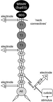
Neural Conduction
Anesthesia, General
Anesthesia
Heart Conduction System
Anesthesia, Local
Anesthesia, Inhalation
Anesthesia, Conduction
Anesthesia, Intravenous
Anesthesia, Obstetrical
Anesthesia Recovery Period
Atrioventricular Node
Anesthetics, Inhalation
Anesthetics, Intravenous
Propofol
Adjuvants, Anesthesia
Anesthetics, Local
Isoflurane
Heart Block
Lidocaine
Anesthetics, Combined
Anesthetics
Monitoring, Intraoperative
Halothane
Nitrous Oxide
Electrocardiography
Action Potentials
Nerve Block
Bone Conduction
Median Nerve
Anesthesia, Closed-Circuit
Arrhythmias, Cardiac
Cardiac Pacing, Artificial
Ulnar Nerve
Fentanyl
Anesthetics, General
Ketamine
Thiopental
Refractory Period, Electrophysiological
Preanesthetic Medication
Ambulatory Surgical Procedures
Peripheral Nerves
Enflurane
Electrophysiology
Dogs
Anesthetics, Dissociative
Pentobarbital
Electrophysiologic Techniques, Cardiac
Tibial Nerve
Atrioventricular Block
Xylazine
Intubation, Intratracheal
Anesthesia Department, Hospital
Electrodiagnosis
Sural Nerve
Conscious Sedation
Aphasia, Conduction
Atrial Flutter
Sciatic Nerve
Anesthesia, Intratracheal
Intraoperative Complications
Catheter Ablation
Purkinje Fibers
Sinoatrial Node
Peripheral Nervous System Diseases
Wolff-Parkinson-White Syndrome
Electromyography
Electroencephalography
Anesthesia and Analgesia
Peroneal Nerve
Prilocaine
Diabetic Neuropathies
NAV1.5 Voltage-Gated Sodium Channel
Prospective Studies
Evoked Potentials, Somatosensory
Methohexital
Ranvier's Nodes
Alfentanil
Hemodynamics
Atrial Fibrillation
Carpal Tunnel Syndrome
Connexin 43
Mepivacaine
Tachycardia, Atrioventricular Nodal Reentry
Sodium Channels
Heart Ventricles
Anti-Arrhythmia Agents
Body Surface Potential Mapping
Chloralose
Bundle-Branch Block
Ethers
Hypnotics and Sedatives
Bradycardia
Tachycardia
Models, Cardiovascular
Midazolam
Surgical Procedures, Minor
Postoperative Complications
Nerve Fibers, Myelinated
Xenon
Connexins
Neuromuscular Nondepolarizing Agents
Tachycardia, Supraventricular
Nerve Fibers
Double-Blind Method
Polyneuropathies
Spinal Nerve Roots
Ether
Cats
Neuromuscular Blocking Agents
Surgical Procedures, Operative
Gap Junctions
Pain Measurement
Brachial Plexus
Sensation
Dose-Response Relationship, Drug
Analgesics, Opioid
Sufentanil
Consciousness Monitors
Laryngeal Masks
Evoked Potentials
Myelin Sheath
Medetomidine
Hypotension, Controlled
Treatment Outcome
Flecainide
Intraoperative Awareness
Myocardium
Etomidate
Succinylcholine
Neuromuscular Blockade
Intraoperative Care
Laryngoscopy
Androstanols
Rats, Sprague-Dawley
Respiration
Demyelinating Diseases
Membrane Potentials
Carbon Dioxide
Deep Sedation
Voltage-Sensitive Dye Imaging
Cardiac Complexes, Premature
Electrophysiological Phenomena
Urethane
Pericardium
Ephedrine
Mandibular Nerve
Procaine
Oxygen
Rabbits
Tachycardia, Ventricular
Sick Sinus Syndrome
Hypotension
Autonomic Nerve Block
Cardiac Electrophysiology
Paresthesia
Shivering
Hernia, Inguinal
Spinal Cord
Amides
Signal Processing, Computer-Assisted
Electrodes
Brugada Syndrome
Droperidol
Command-related distribution of regional cerebral blood flow during attempted handgrip. (1/262)
To localize a central nervous feed-forward mechanism involved in cardiovascular regulation during exercise, brain activation patterns were measured in eight subjects by employing positron emission tomography and oxygen-15-labeled water. Scans were performed at rest and during rhythmic handgrip before and after axillary blockade with bupivacaine. After the blockade, handgrip strength was reduced to 25% (range 0-50%) of control values, whereas handgrip-induced heart rate and blood pressure increases were unaffected (13 +/- 3 beats/min and 12 +/- 5 mmHg, respectively; means +/- SE). Before regional anesthesia, handgrip caused increased activation in the contralateral sensory motor area, the supplementary motor area, and the ipsilateral cerebellum. We found no evidence for changes in the activation pattern due to an interaction between handgrip and regional anesthesia. This was true for both the blocked and unblocked arm. It remains unclear whether the activated areas are responsible for the increase in cardiovascular variables, but neural feedback from the contracting muscles was not necessary for the activation in the mentioned areas during rhythmic handgrip. (+info)Carotid endarterectomy under regional anesthesia. (2/262)
Regional anesthesia for carotid endarterectomy is a simple, reliable, and virtually complication-free technique. We began to perform a series of carotid endarterectomy under regional anesthesia at our institution in May 1990. This report describes our experience with 180 operated patients from May 1990 till December 1995, with regional anesthesia. All patients were operated with microsurgery and we utilized the deeply cervical plexus block at the C-4 level associated with superficial block, along the posterior border of the externocleidomastoid muscle. The main advantage of this technique of anesthesia is that it is the only exact method of assessing the need of a shunt by using the neurological status of the awaken patient during trial carotid cross-clamping. The regional anesthesia allows carotid endarterectomy to be safely performed on patients with advanced cardiac disease or severe chronic obstructive pulmonary disease who were not good candidates for general anesthesia. In this 180 patients we performed 198 consecutive endarterectomies (10% bilateral) with a total morbidity-mortality rate of 2.0%. (+info)Intravenous regional anesthesia (Bier block) in a dog. (3/262)
Intravenous regional anesthesia was used in an adult dog as part of a balanced approach to general anesthesia for amputation of the 4th digit of its right hind limb. It allowed the concentration of isoflurane to be reduced to 0.5%. (+info)Effects of general and locoregional anesthesia on reproductive outcome for in vitro fertilization: a meta-analysis. (4/262)
The objective of this meta-analysis was to evaluate prospective trials of general or locoregional anesthesia on reproductive outcomes (cleavage and pregnancy rate) for in vitro fertilization (IVF). Of 115 published studies retrieved from a search of articles indexed on MEDLINE from 1966 to February 1999, four studies with distinct general and locoregional anesthesia were deemed eligible for meta- analysis. The pooled relative risk and odds ratios were calculated. A test for homogeneity was also performed. The pooled log odds ratio was 1.03 (95% CI 0.90-1.18) in cleavage rate and 0.71 (95% CI 0.47-1.08) in pregnancy rate. Heterogeneity was negative. Cleavage and pregnancy rates were not significantly different in both the general anesthesia and locoregional anesthesia groups. Both anesthetic techniques were favorable to IVF procedure by available published evidence when anesthesia was needed. (+info)Severe vasovagal attack during regional anaesthesia for caesarean section. (5/262)
A patient experienced a severe vasovagal attack during regional anaesthesia for elective Caesarean section. The combination of vagal over-activity and sympathetic block produced profound hypotension that threatened the life of the mother and infant. The vasovagal syndrome is described, and its prevention and management discussed. (+info)The effect of anesthetic technique on postoperative outcomes in hip fracture repair. (6/262)
BACKGROUND: The impact of anesthetic choice on postoperative mortality and morbidity has not been determined with certainty. METHODS: The authors evaluated the effect of type of anesthesia on postoperative mortality and morbidity in a retrospective cohort study of consecutive hip fracture patients, aged 60 yr or older, who underwent surgical repair at 20 US hospitals between 1983 and 1993. The primary outcome was defined as death within 30 days of the operative procedure. The secondary outcomes were postoperative 7-day mortality, postoperative myocardial infarction, postoperative pneumonia, postoperative congestive heart failure, and postoperative change in mental status. Numerous comorbid conditions were controlled for individually and by several comorbidity indices using logistic regression. RESULTS: General anesthesia was used in 6,206 patients (65.8%) and regional anesthesia in 3,219 patients (3,078 spinal anesthesia and 141 epidural anesthesia). The 30-day mortality rate in the general anesthesia group was 4.4%, compared with 5.4% in the regional anesthesia group (unadjusted odds ratio = 0.80; 95% confidence interval = 0.66-0.97). However, the adjusted odds ratio for general anesthesia increased to 1.08 (0.84-1.38). The adjusted odds ratios for general anesthesia versus regional anesthesia for the 7-day mortality was 0.90 (0.59-1.39) and for postoperative morbidity outcomes were as follows: myocardial infarction: adjusted odds ratio = 1.17 (0.80-1.70); congestive heart failure: adjusted odds ratio = 1.04 (0.80-1.36); pneumonia: adjusted odds ratio = 1.21 (0.87-1.68); postoperative change in mental status: adjusted odds ratio = 1.08 (0.95-1.22). CONCLUSIONS: The authors were unable to demonstrate that regional anesthesia was associated with better outcome than was general anesthesia in this large observational study of elderly patients with hip fracture. These results suggest that the type of anesthesia used should depend on factors other than any associated risks of mortality or morbidity. (+info)Ophthalmic regional anesthesia: medial canthus episcleral (sub-tenon) anesthesia is more efficient than peribulbar anesthesia: A double-blind randomized study. (7/262)
BACKGROUND: Regional anesthesia and especially peribulbar anesthesia commonly is used for cataract surgery. Failure rates and need for reinjection remains high, however, with peribulbar anesthesia. Single-injection high-volume medial canthus episcleral (sub-Tenon's) anesthesia has proven to be an efficient and safe alternative to peribulbar anesthesia. METHODS: The authors, in a blind study, compared the effectiveness of both techniques in 66 patients randomly assigned to episcleral anesthesia or single-injection peribulbar anesthesia. Motor blockade (akinesia) was used as the main index of anesthesia effectiveness. It was assessed using an 18-point scale (0-3 for each of the four directions of the gaze, lid opening, and lid closing, the total being from 0 = normal mobility to 18 = no movement at all). This score was compared between the groups 1, 5, 10, and 15 min after injection and at the end of the surgical procedures. Time to onset of the blockade also was compared between the two groups, as was the incidence of incomplete blockade with a need for supplemental injection and the satisfaction of the surgeon, patient, and anesthesiologist. RESULTS: Episcleral anesthesia provided a quicker onset of anesthesia, a better akinesia score, and a lower rate of incomplete blockade necessitating reinjection (0 vs. 39%; P < 0.0001) than peribulbar anesthesia. Even after supplemental injection, peribulbar anesthesia had a lower akinesia score than did episcleral anesthesia. Peribulbar anesthesia began to wear off during surgery, whereas episcleral anesthesia did not. CONCLUSION: Medial canthus single-injection episcleral anesthesia is a suitable alternative to peribulbar anesthesia. It provides better akinesia, with a quicker onset and more constancy in effectiveness. (+info)General versus regional anaesthesia for hip fracture surgery: a meta-analysis of randomized trials. (8/262)
Hip fracture surgery is common and the population at risk is generally elderly. There is no consensus of opinion regarding the safest form of anaesthesia for these patients. We performed a meta-analysis of 15 randomized trials that compare morbidity and mortality associated with general or regional anaesthesia for hip fracture patients. There was a reduced 1-month mortality and incidence of deep vein thrombosis in the regional anaesthesia group. Operations performed under general anaesthesia had a reduction in operation time. No other outcome measures reached a statistically significant difference. There was a tendency towards a lower incidence of myocardial infarction, confusion and postoperative hypoxia in the regional anaesthetic group, and cerebrovascular accident and intra-operative hypotension in the general anaesthetic group. We conclude that there are marginal advantages for regional anaesthesia compared to general anaesthesia for hip fracture patients in terms of early mortality and risk of deep vein thrombosis. (+info)Neural conduction is the process by which electrical signals, known as action potentials, are transmitted along the axon of a neuron (nerve cell) to transmit information between different parts of the nervous system. This electrical impulse is generated by the movement of ions across the neuronal membrane, and it propagates down the length of the axon until it reaches the synapse, where it can then stimulate the release of neurotransmitters to communicate with other neurons or target cells. The speed of neural conduction can vary depending on factors such as the diameter of the axon, the presence of myelin sheaths (which act as insulation and allow for faster conduction), and the temperature of the environment.
General anesthesia is a state of controlled unconsciousness, induced by administering various medications, that eliminates awareness, movement, and pain sensation during medical procedures. It involves the use of a combination of intravenous and inhaled drugs to produce a reversible loss of consciousness, allowing patients to undergo surgical or diagnostic interventions safely and comfortably. The depth and duration of anesthesia are carefully monitored and adjusted throughout the procedure by an anesthesiologist or certified registered nurse anesthetist (CRNA) to ensure patient safety and optimize recovery. General anesthesia is typically used for more extensive surgical procedures, such as open-heart surgery, major orthopedic surgeries, and neurosurgery.
Anesthesia is a medical term that refers to the loss of sensation or awareness, usually induced by the administration of various drugs. It is commonly used during surgical procedures to prevent pain and discomfort. There are several types of anesthesia, including:
1. General anesthesia: This type of anesthesia causes a complete loss of consciousness and is typically used for major surgeries.
2. Regional anesthesia: This type of anesthesia numbs a specific area of the body, such as an arm or leg, while the patient remains conscious.
3. Local anesthesia: This type of anesthesia numbs a small area of the body, such as a cut or wound, and is typically used for minor procedures.
Anesthesia can be administered through various routes, including injection, inhalation, or topical application. The choice of anesthesia depends on several factors, including the type and duration of the procedure, the patient's medical history, and their overall health. Anesthesiologists are medical professionals who specialize in administering anesthesia and monitoring patients during surgical procedures to ensure their safety and comfort.
The heart conduction system is a group of specialized cardiac muscle cells that generate and conduct electrical impulses to coordinate the contraction of the heart chambers. The main components of the heart conduction system include:
1. Sinoatrial (SA) node: Also known as the sinus node, it is located in the right atrium near the entrance of the superior vena cava and functions as the primary pacemaker of the heart. It sets the heart rate by generating electrical impulses at regular intervals.
2. Atrioventricular (AV) node: Located in the interatrial septum, near the opening of the coronary sinus, it serves as a relay station for electrical signals between the atria and ventricles. The AV node delays the transmission of impulses to allow the atria to contract before the ventricles.
3. Bundle of His: A bundle of specialized cardiac muscle fibers that conducts electrical impulses from the AV node to the ventricles. It divides into two main branches, the right and left bundle branches, which further divide into smaller Purkinje fibers.
4. Right and left bundle branches: These are extensions of the Bundle of His that transmit electrical impulses to the respective right and left ventricular myocardium. They consist of specialized conducting tissue with large diameters and minimal resistance, allowing for rapid conduction of electrical signals.
5. Purkinje fibers: Fine, branching fibers that arise from the bundle branches and spread throughout the ventricular myocardium. They are responsible for transmitting electrical impulses to the working cardiac muscle cells, triggering coordinated ventricular contraction.
In summary, the heart conduction system is a complex network of specialized muscle cells responsible for generating and conducting electrical signals that coordinate the contraction of the atria and ventricles, ensuring efficient blood flow throughout the body.
Local anesthesia is a type of anesthesia that numbs a specific area of the body, blocking pain signals from that particular region while allowing the person to remain conscious and alert. It is typically achieved through the injection or application of a local anesthetic drug, which works by temporarily inhibiting the function of nerve fibers carrying pain sensations. Common examples of local anesthetics include lidocaine, prilocaine, and bupivacaine.
Local anesthesia is commonly used for minor surgical procedures, dental work, or other medical interventions where only a small area needs to be numbed. It can also be employed as part of a combined anesthetic technique, such as in conjunction with sedation or regional anesthesia, to provide additional pain relief and increase patient comfort during more extensive surgeries.
The duration of local anesthesia varies depending on the type and dosage of the anesthetic agent used; some last for just a few hours, while others may provide numbness for up to several days. Overall, local anesthesia is considered a safe and effective method for managing pain during various medical procedures.
Spinal anesthesia is a type of regional anesthesia that involves injecting local anesthetic medication into the cerebrospinal fluid in the subarachnoid space, which is the space surrounding the spinal cord. This procedure is typically performed by introducing a needle into the lower back, between the vertebrae, to reach the subarachnoid space.
Once the local anesthetic is introduced into this space, it spreads to block nerve impulses from the corresponding levels of the spine, resulting in numbness and loss of sensation in specific areas of the body below the injection site. The extent and level of anesthesia depend on the amount and type of medication used, as well as the patient's individual response.
Spinal anesthesia is often used for surgeries involving the lower abdomen, pelvis, or lower extremities, such as cesarean sections, hernia repairs, hip replacements, and knee arthroscopies. It can also be utilized for procedures like epidural steroid injections to manage chronic pain conditions affecting the spine and lower limbs.
While spinal anesthesia provides effective pain relief during and after surgery, it may cause side effects such as low blood pressure, headache, or difficulty urinating. These potential complications should be discussed with the healthcare provider before deciding on this type of anesthesia.
Epidural anesthesia is a type of regional anesthesia that involves the injection of local anesthetic medication into the epidural space in the spine, which is the space surrounding the dura mater, a membrane that covers the spinal cord. The injection is typically administered through a catheter placed in the lower back using a needle.
The local anesthetic drug blocks nerve impulses from the affected area, numbing it and relieving pain. Epidural anesthesia can be used for various surgical procedures, such as cesarean sections, knee or hip replacements, and hernia repairs. It is also commonly used during childbirth to provide pain relief during labor and delivery.
The effects of epidural anesthesia can vary depending on the dose and type of medication used, as well as the individual's response to the drug. The anesthetic may take several minutes to start working, and its duration of action can range from a few hours to a day or more. Epidural anesthesia is generally considered safe when administered by trained medical professionals, but like any medical procedure, it carries some risks, including infection, bleeding, nerve damage, and respiratory depression.
Inhalational anesthesia is a type of general anesthesia that is induced by the inhalation of gases or vapors. It is administered through a breathing system, which delivers the anesthetic agents to the patient via a face mask, laryngeal mask airway, or endotracheal tube.
The most commonly used inhalational anesthetics include nitrous oxide, sevoflurane, isoflurane, and desflurane. These agents work by depressing the central nervous system, causing a reversible loss of consciousness, amnesia, analgesia, and muscle relaxation.
The depth of anesthesia can be easily adjusted during the procedure by changing the concentration of the anesthetic agent. Once the procedure is complete, the anesthetic agents are eliminated from the body through exhalation, allowing for a rapid recovery.
Inhalational anesthesia is commonly used in a wide range of surgical procedures due to its ease of administration, quick onset and offset of action, and ability to rapidly adjust the depth of anesthesia. However, it requires careful monitoring and management by trained anesthesia providers to ensure patient safety and optimize outcomes.
Conduction anesthesia is a type of local anesthesia in which an anesthetic agent is administered near a peripheral nerve to block the transmission of painful stimuli. It is called "conduction" anesthesia because it works by blocking the conduction of nerve impulses along the nerve fibers.
There are several types of conduction anesthesia, including:
1. Infiltration anesthesia: In this technique, the anesthetic agent is injected directly into the tissue where the surgical procedure will be performed. This type of anesthesia can be used for minor surgeries such as wound closure or repair of simple lacerations.
2. Nerve block anesthesia: In this technique, the anesthetic agent is injected near a specific nerve or bundle of nerves to block sensation in a larger area of the body. For example, a brachial plexus block can be used to numb the arm and hand for procedures such as shoulder surgery or fracture reduction.
3. Field block anesthesia: In this technique, the anesthetic agent is injected around the periphery of the surgical site to create a "field" of anesthesia that blocks sensation in the area. This type of anesthesia is often used for procedures such as hernia repair or circumcision.
Conduction anesthesia has several advantages over general anesthesia, including reduced risk of complications, faster recovery time, and lower cost. However, it may not be appropriate for all types of surgical procedures or patients, and its effectiveness can vary depending on the skill of the practitioner and the individual patient's response to the anesthetic agent.
Intravenous anesthesia, also known as IV anesthesia, is a type of anesthesia that involves the administration of one or more drugs into a patient's vein to achieve a state of unconsciousness and analgesia (pain relief) during medical procedures. The drugs used in intravenous anesthesia can include sedatives, hypnotics, analgesics, and muscle relaxants, which are carefully selected and dosed based on the patient's medical history, physical status, and the type and duration of the procedure.
The administration of IV anesthesia is typically performed by a trained anesthesiologist or nurse anesthetist, who monitors the patient's vital signs and adjusts the dosage of the drugs as needed to ensure the patient's safety and comfort throughout the procedure. The onset of action for IV anesthesia is relatively rapid, usually within minutes, and the depth and duration of anesthesia can be easily titrated to meet the needs of the individual patient.
Compared to general anesthesia, which involves the administration of inhaled gases or vapors to achieve a state of unconsciousness, intravenous anesthesia is associated with fewer adverse effects on respiratory and cardiovascular function, and may be preferred for certain types of procedures or patients. However, like all forms of anesthesia, IV anesthesia carries risks and potential complications, including allergic reactions, infection, bleeding, and respiratory depression, and requires careful monitoring and management by trained medical professionals.
Obstetrical anesthesia refers to the use of anesthetic techniques and medications during childbirth or obstetrical procedures. The goal is to provide pain relief and comfort to the birthing person while ensuring the safety of both the mother and the baby. There are different types of obstetrical anesthesia, including:
1. Local anesthesia: Injection of a local anesthetic agent to numb a specific area, such as the perineum (the area between the vagina and the anus) during childbirth.
2. Regional anesthesia: Numbing a larger region of the body using techniques like spinal or epidural anesthesia. These methods involve injecting local anesthetic agents near the spinal cord to block nerve impulses, providing pain relief in the lower half of the body.
3. General anesthesia: Using inhaled gases or intravenous medications to render the birthing person unconscious during cesarean sections (C-sections) or other surgical procedures related to childbirth.
The choice of anesthetic technique depends on various factors, including the type of delivery, the mother's medical history, and the preferences of both the mother and the healthcare team. Obstetrical anesthesia requires specialized training and expertise to ensure safe and effective pain management during labor and delivery.
The anesthesia recovery period, also known as the post-anesthetic care unit (PACU) or recovery room stay, is the time immediately following anesthesia and surgery during which a patient's vital signs are closely monitored as they emerge from the effects of anesthesia.
During this period, the patient is typically observed for adequate ventilation, oxygenation, circulation, level of consciousness, pain control, and any potential complications. The length of stay in the recovery room can vary depending on the type of surgery, the anesthetic used, and the individual patient's needs.
The anesthesia recovery period is a critical time for ensuring patient safety and comfort as they transition from the surgical setting to full recovery. Nurses and other healthcare providers in the recovery room are specially trained to monitor and manage patients during this vulnerable period.
Dental anesthesia is a type of local or regional anesthesia that is specifically used in dental procedures to block the transmission of pain impulses from the teeth and surrounding tissues to the brain. The most common types of dental anesthesia include:
1. Local anesthesia: This involves the injection of a local anesthetic drug, such as lidocaine or prilocaine, into the gum tissue near the tooth that is being treated. This numbs the area and prevents the patient from feeling pain during the procedure.
2. Conscious sedation: This is a type of minimal sedation that is used to help patients relax during dental procedures. The patient remains conscious and can communicate with the dentist, but may not remember the details of the procedure. Common methods of conscious sedation include nitrous oxide (laughing gas) or oral sedatives.
3. Deep sedation or general anesthesia: This is rarely used in dental procedures, but may be necessary for patients who are extremely anxious or have special needs. It involves the administration of drugs that cause a state of unconsciousness and prevent the patient from feeling pain during the procedure.
Dental anesthesia is generally safe when administered by a qualified dentist or oral surgeon. However, as with any medical procedure, there are risks involved, including allergic reactions to the anesthetic drugs, nerve damage, and infection. Patients should discuss any concerns they have with their dentist before undergoing dental anesthesia.
The atrioventricular (AV) node is a critical part of the electrical conduction system of the heart. It is a small cluster of specialized cardiac muscle cells located in the lower interatrial septum, near the opening of the coronary sinus. The AV node receives electrical impulses from the sinoatrial node (the heart's natural pacemaker) via the internodal pathways and delays their transmission for a brief period before transmitting them to the bundle of His and then to the ventricles. This delay allows the atria to contract and empty their contents into the ventricles before the ventricles themselves contract, ensuring efficient pumping of blood throughout the body.
The AV node plays an essential role in maintaining a normal heart rhythm, as it can also function as a backup pacemaker if the sinoatrial node fails to generate impulses. However, certain heart conditions or medications can affect the AV node's function and lead to abnormal heart rhythms, such as atrioventricular block or atrial tachycardia.
Inhalational anesthetics are a type of general anesthetic that is administered through the person's respiratory system. They are typically delivered in the form of vapor or gas, which is inhaled through a mask or breathing tube. Commonly used inhalational anesthetics include sevoflurane, desflurane, isoflurane, and nitrous oxide. These agents work by depressing the central nervous system, leading to a loss of consciousness and an inability to feel pain. They are often used for their rapid onset and offset of action, making them useful for both induction and maintenance of anesthesia during surgical procedures.
Intravenous anesthetics are a type of medication that is administered directly into a vein to cause a loss of consciousness and provide analgesia (pain relief) during medical procedures. They work by depressing the central nervous system, inhibiting nerve impulse transmission and ultimately preventing the patient from feeling pain or discomfort during surgery or other invasive procedures.
There are several different types of intravenous anesthetics, each with its own specific properties and uses. Some common examples include propofol, etomidate, ketamine, and barbiturates. These drugs may be used alone or in combination with other medications to provide a safe and effective level of anesthesia for the patient.
The choice of intravenous anesthetic depends on several factors, including the patient's medical history, the type and duration of the procedure, and the desired depth and duration of anesthesia. Anesthesiologists must carefully consider these factors when selecting an appropriate medication regimen for each individual patient.
While intravenous anesthetics are generally safe and effective, they can have side effects and risks, such as respiratory depression, hypotension, and allergic reactions. Anesthesia providers must closely monitor patients during and after the administration of these medications to ensure their safety and well-being.
Propofol is a short-acting medication that is primarily used for the induction and maintenance of general anesthesia during procedures such as surgery. It belongs to a class of drugs called hypnotics or sedatives, which work by depressing the central nervous system to produce a calming effect. Propofol can also be used for sedation in mechanically ventilated patients in intensive care units and for procedural sedation in various diagnostic and therapeutic procedures outside the operating room.
The medical definition of Propofol is:
A rapid-onset, short-duration intravenous anesthetic agent that produces a hypnotic effect and is used for induction and maintenance of general anesthesia, sedation in mechanically ventilated patients, and procedural sedation. It acts by enhancing the inhibitory effects of gamma-aminobutyric acid (GABA) in the brain, leading to a decrease in neuronal activity and a reduction in consciousness. Propofol has a rapid clearance and distribution, allowing for quick recovery after discontinuation of its administration.
An adjuvant in anesthesia refers to a substance or drug that is added to an anesthetic medication to enhance its effects, make it last longer, or improve the overall quality of anesthesia. Adjuvants do not produce analgesia or anesthesia on their own but work synergistically with other anesthetics to achieve better clinical outcomes.
There are several types of adjuvants used in anesthesia, including:
1. Opioids: These are commonly used adjuvants that enhance the analgesic effect of anesthetic drugs. Examples include fentanyl, sufentanil, and remifentanil.
2. Alpha-2 agonists: Drugs like clonidine and dexmedetomidine are used as adjuvants to provide sedation, analgesia, and anxiolysis. They also help reduce the requirement for other anesthetic drugs, thus minimizing side effects.
3. Ketamine: This NMDA receptor antagonist is used as an adjuvant to provide analgesia and amnesia. It can be used in subanesthetic doses to improve the quality of analgesia during general anesthesia or as a sole anesthetic for procedural sedation.
4. Local anesthetics: When used as an adjuvant, local anesthetics can prolong the duration of postoperative analgesia and reduce the requirement for opioids. Examples include bupivacaine, ropivacaine, and lidocaine.
5. Neostigmine: This cholinesterase inhibitor is used as an adjuvant to reverse the neuromuscular blockade produced by non-depolarizing muscle relaxants at the end of surgery.
6. Dexamethasone: A corticosteroid used as an adjuvant to reduce postoperative nausea and vomiting, inflammation, and pain.
7. Magnesium sulfate: This non-competitive NMDA receptor antagonist is used as an adjuvant to provide analgesia, reduce opioid consumption, and provide neuroprotection in certain surgical settings.
The choice of adjuvants depends on the type of surgery, patient factors, and the desired clinical effects.
Local anesthetics are a type of medication that is used to block the sensation of pain in a specific area of the body. They work by temporarily numbing the nerves in that area, preventing them from transmitting pain signals to the brain. Local anesthetics can be administered through various routes, including topical application (such as creams or gels), injection (such as into the skin or tissues), or regional nerve blocks (such as epidural or spinal anesthesia).
Some common examples of local anesthetics include lidocaine, prilocaine, bupivacaine, and ropivacaine. These medications can be used for a variety of medical procedures, ranging from minor surgeries (such as dental work or skin biopsies) to more major surgeries (such as joint replacements or hernia repairs).
Local anesthetics are generally considered safe when used appropriately, but they can have side effects and potential complications. These may include allergic reactions, toxicity (if too much is administered), and nerve damage (if the medication is injected into a nerve). It's important to follow your healthcare provider's instructions carefully when using local anesthetics, and to report any unusual symptoms or side effects promptly.
Isoflurane is a volatile halogenated ether used for induction and maintenance of general anesthesia. It is a colorless liquid with a pungent, sweet odor. Isoflurane is an agonist at the gamma-aminobutyric acid type A (GABAA) receptor and inhibits excitatory neurotransmission in the brain, leading to unconsciousness and immobility. It has a rapid onset and offset of action due to its low blood solubility, allowing for quick adjustments in anesthetic depth during surgery. Isoflurane is also known for its bronchodilator effects, making it useful in patients with reactive airway disease. However, it can cause dose-dependent decreases in heart rate and blood pressure, so careful hemodynamic monitoring is required during its use.
Methyl ethers are a type of organic compound where a methyl group (CH3-) is attached to an oxygen atom, which in turn is connected to another carbon atom. They are formed by the process of methylation, where a methyl group replaces a hydrogen atom in another molecule.
Methyl ethers can be found in various natural and synthetic substances. For example, dimethyl ether (CH3-O-CH3) is a common fuel used in refrigeration systems and as a propellant in aerosol sprays. Anisole (CH3-O-C6H5), another methyl ether, is found in anise oil and is used as a flavoring agent and solvent.
It's worth noting that some methyl ethers have been associated with potential health risks, particularly when they are volatile and can be inhaled or ingested. For example, exposure to high levels of dimethyl ether can cause respiratory irritation, headaches, and dizziness. Therefore, it's important to handle these substances with care and follow appropriate safety guidelines.
Heart block is a cardiac condition characterized by the interruption of electrical impulse transmission from the atria (the upper chambers of the heart) to the ventricles (the lower chambers of the heart). This disruption can lead to abnormal heart rhythms, including bradycardia (a slower-than-normal heart rate), and in severe cases, can cause the heart to stop beating altogether. Heart block is typically caused by damage to the heart's electrical conduction system due to various factors such as aging, heart disease, or certain medications.
There are three types of heart block: first-degree, second-degree, and third-degree (also known as complete heart block). Each type has distinct electrocardiogram (ECG) findings and symptoms. Treatment for heart block depends on the severity of the condition and may include monitoring, medication, or implantation of a pacemaker to regulate the heart's electrical activity.
Anesthesiology is a medical specialty concerned with providing anesthesia, which is the loss of sensation or awareness, to patients undergoing surgical, diagnostic, or therapeutic procedures. Anesthesiologists are responsible for administering various types of anesthetics, monitoring the patient's vital signs during the procedure, and managing any complications that may arise. They also play a critical role in pain management before, during, and after surgery, as well as in the treatment of chronic pain conditions.
Anesthesiologists work closely with other medical professionals, including surgeons, anesthetists, nurses, and respiratory therapists, to ensure that patients receive the best possible care. They must have a thorough understanding of human physiology, pharmacology, and anatomy, as well as excellent communication skills and the ability to make quick decisions under high pressure.
The primary goal of anesthesiology is to provide safe and effective anesthesia that minimizes pain and discomfort while maximizing patient safety and comfort. This requires a deep understanding of the risks and benefits associated with different types of anesthetics, as well as the ability to tailor the anesthetic plan to each individual patient's needs and medical history.
In summary, anesthesiology is a critical medical specialty focused on providing safe and effective anesthesia and pain management for patients undergoing surgical or other medical procedures.
Lidocaine is a type of local anesthetic that numbs painful areas and is used to prevent pain during certain medical procedures. It works by blocking the nerves that transmit pain signals to the brain. In addition to its use as an anesthetic, lidocaine can also be used to treat irregular heart rates and relieve itching caused by allergic reactions or skin conditions such as eczema.
Lidocaine is available in various forms, including creams, gels, ointments, sprays, solutions, and injectable preparations. It can be applied directly to the skin or mucous membranes, or it can be administered by injection into a muscle or vein. The specific dosage and method of administration will depend on the reason for its use and the individual patient's medical history and current health status.
Like all medications, lidocaine can have side effects, including allergic reactions, numbness that lasts too long, and in rare cases, heart problems or seizures. It is important to follow the instructions of a healthcare provider carefully when using lidocaine to minimize the risk of adverse effects.
Combined anesthetics refer to the use of two or more types of anesthetic agents together during a medical procedure to produce a desired level of sedation, amnesia, analgesia, and muscle relaxation. This approach can allow for lower doses of individual anesthetic drugs, which may reduce the risk of adverse effects associated with each drug. Common combinations include using a general anesthetic in combination with a regional or local anesthetic technique. The specific choice of combined anesthetics depends on various factors such as the type and duration of the procedure, patient characteristics, and the desired outcomes.
Anesthetics are medications that are used to block or reduce feelings of pain and sensation, either locally in a specific area of the body or generally throughout the body. They work by depressing the nervous system, interrupting the communication between nerves and the brain. Anesthetics can be administered through various routes such as injection, inhalation, or topical application, depending on the type and the desired effect. There are several classes of anesthetics, including:
1. Local anesthetics: These numb a specific area of the body and are commonly used during minor surgical procedures, dental work, or to relieve pain from injuries. Examples include lidocaine, prilocaine, and bupivacaine.
2. Regional anesthetics: These block nerve impulses in a larger area of the body, such as an arm or leg, and can be used for more extensive surgical procedures. They are often administered through a catheter to provide continuous pain relief over a longer period. Examples include spinal anesthesia, epidural anesthesia, and peripheral nerve blocks.
3. General anesthetics: These cause a state of unconsciousness and are used for major surgical procedures or when the patient needs to be completely immobile during a procedure. They can be administered through inhalation or injection and affect the entire body. Examples include propofol, sevoflurane, and isoflurane.
Anesthetics are typically safe when used appropriately and under medical supervision. However, they can have side effects such as drowsiness, nausea, and respiratory depression. Proper dosing and monitoring by a healthcare professional are essential to minimize the risks associated with anesthesia.
Intraoperative monitoring (IOM) is the practice of using specialized techniques to monitor physiological functions or neural structures in real-time during surgical procedures. The primary goal of IOM is to provide continuous information about the patient's status and the effects of surgery on neurological function, allowing surgeons to make informed decisions and minimize potential risks.
IOM can involve various methods such as:
1. Electrophysiological monitoring: This includes techniques like somatosensory evoked potentials (SSEP), motor evoked potentials (MEP), and electroencephalography (EEG) to assess the integrity of neural pathways and brain function during surgery.
2. Neuromonitoring: Direct electrical stimulation of nerves or spinal cord structures can help identify critical neuroanatomical structures, evaluate their functional status, and guide surgical interventions.
3. Hemodynamic monitoring: Measuring blood pressure, heart rate, cardiac output, and oxygen saturation helps assess the patient's overall physiological status during surgery.
4. Imaging modalities: Intraoperative imaging techniques like ultrasound, computed tomography (CT), or magnetic resonance imaging (MRI) can provide real-time visualization of anatomical structures and surgical progress.
The specific IOM methods employed depend on the type of surgery, patient characteristics, and potential risks involved. Intraoperative monitoring is particularly crucial in procedures where there is a risk of neurological injury, such as spinal cord or brain surgeries, vascular interventions, or tumor resections near critical neural structures.
Halothane is a general anesthetic agent, which is a volatile liquid that evaporates easily and can be inhaled. It is used to produce and maintain general anesthesia (a state of unconsciousness) during surgical procedures. Halothane is known for its rapid onset and offset of action, making it useful for both induction and maintenance of anesthesia.
The medical definition of Halothane is:
Halothane (2-bromo-2-chloro-1,1,1-trifluoroethane) is a volatile liquid general anesthetic agent with a mild, sweet odor. It is primarily used for the induction and maintenance of general anesthesia in surgical procedures due to its rapid onset and offset of action. Halothane is administered via inhalation and acts by depressing the central nervous system, leading to a reversible loss of consciousness and analgesia.
It's important to note that Halothane has been associated with rare cases of severe liver injury (hepatotoxicity) and anaphylaxis (a severe, life-threatening allergic reaction). These risks have led to the development and use of alternative general anesthetic agents with better safety profiles.
Nitrous oxide, also known as laughing gas, is a colorless and non-flammable gas with a slightly sweet odor and taste. In medicine, it's commonly used for its anesthetic and pain reducing effects. It is often used in dental procedures, surgery, and childbirth to help reduce anxiety and provide mild sedation. Nitrous oxide works by binding to the hemoglobin in red blood cells, which reduces the oxygen-carrying capacity of the blood, but this effect is usually not significant at the low concentrations used for analgesia and anxiolysis. It's also considered relatively safe when administered by a trained medical professional because it does not cause depression of the respiratory system or cardiovascular function.
Electrocardiography (ECG or EKG) is a medical procedure that records the electrical activity of the heart. It provides a graphic representation of the electrical changes that occur during each heartbeat. The resulting tracing, called an electrocardiogram, can reveal information about the heart's rate and rhythm, as well as any damage to its cells or abnormalities in its conduction system.
During an ECG, small electrodes are placed on the skin of the chest, arms, and legs. These electrodes detect the electrical signals produced by the heart and transmit them to a machine that amplifies and records them. The procedure is non-invasive, painless, and quick, usually taking only a few minutes.
ECGs are commonly used to diagnose and monitor various heart conditions, including arrhythmias, coronary artery disease, heart attacks, and electrolyte imbalances. They can also be used to evaluate the effectiveness of certain medications or treatments.
An action potential is a brief electrical signal that travels along the membrane of a nerve cell (neuron) or muscle cell. It is initiated by a rapid, localized change in the permeability of the cell membrane to specific ions, such as sodium and potassium, resulting in a rapid influx of sodium ions and a subsequent efflux of potassium ions. This ion movement causes a brief reversal of the electrical potential across the membrane, which is known as depolarization. The action potential then propagates along the cell membrane as a wave, allowing the electrical signal to be transmitted over long distances within the body. Action potentials play a crucial role in the communication and functioning of the nervous system and muscle tissue.
A nerve block is a medical procedure in which an anesthetic or neurolytic agent is injected near a specific nerve or bundle of nerves to block the transmission of pain signals from that area to the brain. This technique can be used for both diagnostic and therapeutic purposes, such as identifying the source of pain, providing temporary or prolonged relief, or facilitating surgical procedures in the affected region.
The injection typically contains a local anesthetic like lidocaine or bupivacaine, which numbs the nerve, preventing it from transmitting pain signals. In some cases, steroids may also be added to reduce inflammation and provide longer-lasting relief. Depending on the type of nerve block and its intended use, the injection might be administered close to the spine (neuraxial blocks), at peripheral nerves (peripheral nerve blocks), or around the sympathetic nervous system (sympathetic nerve blocks).
While nerve blocks are generally safe, they can have side effects such as infection, bleeding, nerve damage, or in rare cases, systemic toxicity from the anesthetic agent. It is essential to consult with a qualified medical professional before undergoing this procedure to ensure proper evaluation, technique, and post-procedure care.
Bone conduction is a type of hearing mechanism that involves the transmission of sound vibrations directly to the inner ear through the bones of the skull, bypassing the outer and middle ears. This occurs when sound waves cause the bones in the skull to vibrate, stimulating the cochlea (the spiral cavity of the inner ear) and its hair cells, which convert the mechanical energy of the vibrations into electrical signals that are sent to the brain and interpreted as sound.
Bone conduction is a natural part of the hearing process in humans, but it can also be used artificially through the use of bone-conduction devices, such as hearing aids or headphones, which transmit sound vibrations directly to the skull. This type of transmission can provide improved hearing for individuals with conductive hearing loss, mixed hearing loss, or single-sided deafness, as it bypasses damaged or obstructed outer and middle ears.
The median nerve is one of the major nerves in the human body, providing sensation and motor function to parts of the arm and hand. It originates from the brachial plexus, a network of nerves that arise from the spinal cord in the neck. The median nerve travels down the arm, passing through the cubital tunnel at the elbow, and continues into the forearm and hand.
In the hand, the median nerve supplies sensation to the palm side of the thumb, index finger, middle finger, and half of the ring finger. It also provides motor function to some of the muscles that control finger movements, allowing for flexion of the fingers and opposition of the thumb.
Damage to the median nerve can result in a condition called carpal tunnel syndrome, which is characterized by numbness, tingling, and weakness in the hand and fingers.
Bupivacaine is a long-acting local anesthetic drug, which is used to cause numbness or loss of feeling in a specific area of the body during certain medical procedures such as surgery, dental work, or childbirth. It works by blocking the nerves that transmit pain signals to the brain.
Bupivacaine is available as a solution for injection and is usually administered directly into the tissue surrounding the nerve to be blocked (nerve block) or into the spinal fluid (epidural). The onset of action of bupivacaine is relatively slow, but its duration of action is long, making it suitable for procedures that require prolonged pain relief.
Like all local anesthetics, bupivacaine carries a risk of side effects such as allergic reactions, nerve damage, and systemic toxicity if accidentally injected into a blood vessel or given in excessive doses. It should be used with caution in patients with certain medical conditions, including heart disease, liver disease, and neurological disorders.
Closed-circuit anesthesia is a type of anesthesia delivery system in which the exhaled gases from the patient are rebreathed after being scrubbed of carbon dioxide and reoxygenated. This is different from open-circuit anesthesia, where the exhaled gases are vented out of the system and fresh gas is continuously supplied to the patient.
In a closed-circuit anesthesia system, the amount of anesthetic agent used can be more precisely controlled, which can lead to a reduction in overall drug usage and potentially fewer side effects for the patient. Additionally, because the exhaled gases are reused, there is less waste and a smaller environmental impact.
Closed-circuit anesthesia systems typically consist of a breathing system, an anesthetic vaporizer, a soda lime canister to remove carbon dioxide, a ventilator to assist with breathing if necessary, and monitors to track the patient's vital signs. These systems are commonly used in veterinary medicine and in human surgery where long-term anesthesia is required.
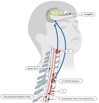
Cardiac arrhythmias are abnormal heart rhythms that result from disturbances in the electrical conduction system of the heart. The heart's normal rhythm is controlled by an electrical signal that originates in the sinoatrial (SA) node, located in the right atrium. This signal travels through the atrioventricular (AV) node and into the ventricles, causing them to contract and pump blood throughout the body.
An arrhythmia occurs when there is a disruption in this electrical pathway or when the heart's natural pacemaker produces an abnormal rhythm. This can cause the heart to beat too fast (tachycardia), too slow (bradycardia), or irregularly.
There are several types of cardiac arrhythmias, including:
1. Atrial fibrillation: A rapid and irregular heartbeat that starts in the atria (the upper chambers of the heart).
2. Atrial flutter: A rapid but regular heartbeat that starts in the atria.
3. Supraventricular tachycardia (SVT): A rapid heartbeat that starts above the ventricles, usually in the atria or AV node.
4. Ventricular tachycardia: A rapid and potentially life-threatening heart rhythm that originates in the ventricles.
5. Ventricular fibrillation: A chaotic and disorganized electrical activity in the ventricles, which can be fatal if not treated immediately.
6. Heart block: A delay or interruption in the conduction of electrical signals from the atria to the ventricles.
Cardiac arrhythmias can cause various symptoms, such as palpitations, dizziness, shortness of breath, chest pain, and fatigue. In some cases, they may not cause any symptoms and go unnoticed. However, if left untreated, certain types of arrhythmias can lead to serious complications, including stroke, heart failure, or even sudden cardiac death.
Treatment for cardiac arrhythmias depends on the type, severity, and underlying causes. Options may include lifestyle changes, medications, cardioversion (electrical shock therapy), catheter ablation, implantable devices such as pacemakers or defibrillators, and surgery. It is essential to consult a healthcare professional for proper evaluation and management of cardiac arrhythmias.
Artificial cardiac pacing is a medical procedure that involves the use of an artificial device to regulate and stimulate the contraction of the heart muscle. This is often necessary when the heart's natural pacemaker, the sinoatrial node, is not functioning properly and the heart is beating too slowly or irregularly.
The artificial pacemaker consists of a small generator that produces electrical impulses and leads that are positioned in the heart to transmit the impulses. The generator is typically implanted just under the skin in the chest, while the leads are inserted into the heart through a vein.
There are different types of artificial cardiac pacing systems, including single-chamber pacemakers, which stimulate either the right atrium or right ventricle, and dual-chamber pacemakers, which stimulate both chambers of the heart. Some pacemakers also have additional features that allow them to respond to changes in the body's needs, such as during exercise or sleep.
Artificial cardiac pacing is a safe and effective treatment for many people with abnormal heart rhythms, and it can significantly improve their quality of life and longevity.
The Ulnar nerve is one of the major nerves in the forearm and hand, which provides motor function to the majority of the intrinsic muscles of the hand (except for those innervated by the median nerve) and sensory innervation to the little finger and half of the ring finger. It originates from the brachial plexus, passes through the cubital tunnel at the elbow, and continues down the forearm, where it runs close to the ulna bone. The ulnar nerve then passes through the Guyon's canal in the wrist before branching out to innervate the hand muscles and provide sensation to the skin on the little finger and half of the ring finger.
Fentanyl is a potent synthetic opioid analgesic, which is similar to morphine but is 50 to 100 times more potent. It is a schedule II prescription drug, typically used to treat patients with severe pain or to manage pain after surgery. It works by binding to the body's opioid receptors, which are found in the brain, spinal cord, and other areas of the body.
Fentanyl can be administered in several forms, including transdermal patches, lozenges, injectable solutions, and tablets that dissolve in the mouth. Illegally manufactured and distributed fentanyl has also become a major public health concern, as it is often mixed with other drugs such as heroin, cocaine, and counterfeit pills, leading to an increase in overdose deaths.
Like all opioids, fentanyl carries a risk of dependence, addiction, and overdose, especially when used outside of medical supervision or in combination with other central nervous system depressants such as alcohol or benzodiazepines. It is important to use fentanyl only as directed by a healthcare provider and to be aware of the potential risks associated with its use.
General anesthetics are a type of medication used to render a person unconscious and insensible to pain during surgical procedures. They work by depressing the central nervous system, affecting the brain's ability to process information and transmit signals, including those related to pain and muscle movement. General anesthesia involves a combination of intravenous (IV) drugs and inhaled gases that produce a state of controlled unconsciousness, amnesia, analgesia, and immobility.
General anesthetics can be classified into several categories based on their chemical structure and mechanism of action, including:
1. Inhalation anesthetics: These are volatile liquids or gases that are vaporized and inhaled through a breathing circuit. Examples include sevoflurane, desflurane, isoflurane, and nitrous oxide.
2. Intravenous anesthetics: These are drugs that are administered directly into the bloodstream through an IV line. Examples include propofol, etomidate, and ketamine.
3. Dissociative anesthetics: These are drugs that produce a state of dissociation between the thalamus and the cerebral cortex, resulting in altered consciousness, analgesia, and amnesia. Ketamine is a commonly used example.
4. Neurodegenerative anesthetics: These are drugs that cause degeneration of neurons in specific areas of the brain, leading to loss of consciousness. Examples include barbiturates such as thiopental and methohexital.
The choice of general anesthetic depends on several factors, including the patient's medical history, the type and duration of surgery, and the anesthesiologist's preference. The administration of general anesthetics requires careful monitoring and management by a trained anesthesia provider to ensure the patient's safety and comfort throughout the procedure.
**Ketamine** is a dissociative anesthetic medication primarily used for starting and maintaining anesthesia. It can lead to a state of altered perception, hallucinations, sedation, and memory loss. Ketamine is also used as a pain reliever in patients with chronic pain conditions and during certain medical procedures due to its strong analgesic properties.
It is available as a generic drug and is also sold under various brand names, such as Ketalar, Ketanest, and Ketamine HCl. It can be administered intravenously, intramuscularly, orally, or as a nasal spray.
In addition to its medical uses, ketamine has been increasingly used off-label for the treatment of mood disorders like depression, anxiety, and post-traumatic stress disorder (PTSD), owing to its rapid antidepressant effects. However, more research is needed to fully understand its long-term benefits and risks in these applications.
It's important to note that ketamine can be abused recreationally due to its dissociative and hallucinogenic effects, which may lead to addiction and severe psychological distress. Therefore, it should only be used under the supervision of a medical professional.
Thiopental, also known as Thiopentone, is a rapid-onset, ultrashort-acting barbiturate derivative. It is primarily used for the induction of anesthesia due to its ability to cause unconsciousness quickly and its short duration of action. Thiopental can also be used for sedation in critically ill patients, though this use has become less common due to the development of safer alternatives.
The drug works by enhancing the inhibitory effects of gamma-aminobutyric acid (GABA), a neurotransmitter in the brain that produces a calming effect. This results in the depression of the central nervous system, leading to sedation, hypnosis, and ultimately, anesthesia.
It is worth noting that Thiopental has been largely replaced by newer drugs in many clinical settings due to its potential for serious adverse effects, such as cardiovascular and respiratory depression, as well as the risk of anaphylaxis. Additionally, it has been used in controversial procedures like capital punishment in some jurisdictions.
The refractory period, electrophysiological, refers to the time interval during which a cardiac or neural cell is unable to respond to a new stimulus immediately after an action potential has been generated. This period is divided into two phases: the absolute refractory period and the relative refractory period.
During the absolute refractory period, the cell cannot be re-stimulated, regardless of the strength of the stimulus, due to the rapid inactivation of voltage-gated sodium channels that are responsible for the rapid depolarization during an action potential. This phase is crucial for maintaining the unidirectional conduction of electrical impulses and preventing the occurrence of re-entry circuits, which can lead to life-threatening arrhythmias in the heart or hyperexcitability in neural tissue.
The relative refractory period follows the absolute refractory period and is characterized by a reduced excitability of the cell. During this phase, a stronger than normal stimulus is required to elicit an action potential due to the slower recovery of voltage-gated sodium channels and the partial activation of potassium channels, which promote repolarization. The duration of both the absolute and relative refractory periods varies depending on the cell type, its physiological state, and other factors such as temperature and pH.
In summary, the electrophysiological refractory period is a fundamental property of excitable cells that ensures proper electrical signaling and prevents uncontrolled excitation or re-entry circuits.
Preanesthetic medication, also known as premedication, refers to the administration of medications before anesthesia to help prepare the patient for the upcoming procedure. These medications can serve various purposes, such as:
1. Anxiolysis: Reducing anxiety and promoting relaxation in patients before surgery.
2. Amnesia: Causing temporary memory loss to help patients forget the events leading up to the surgery.
3. Analgesia: Providing pain relief to minimize discomfort during and after the procedure.
4. Antisialagogue: Decreasing saliva production to reduce the risk of aspiration during intubation.
5. Bronchodilation: Relaxing bronchial smooth muscles, which can help improve respiratory function in patients with obstructive lung diseases.
6. Antiemetic: Preventing or reducing the likelihood of postoperative nausea and vomiting.
7. Sedation: Inducing a state of calmness and drowsiness to facilitate a smooth induction of anesthesia.
Common preanesthetic medications include benzodiazepines (e.g., midazolam), opioids (e.g., fentanyl), anticholinergics (e.g., glycopyrrolate), and H1-antihistamines (e.g., diphenhydramine). The choice of preanesthetic medication depends on the patient's medical history, comorbidities, and the type of anesthesia to be administered.
Ambulatory surgical procedures, also known as outpatient or same-day surgery, refer to medical operations that do not require an overnight hospital stay. These procedures are typically performed in a specialized ambulatory surgery center (ASC) or in a hospital-based outpatient department. Patients undergoing ambulatory surgical procedures receive anesthesia, undergo the operation, and recover enough to be discharged home on the same day of the procedure.
Examples of common ambulatory surgical procedures include:
1. Arthroscopy (joint scope examination and repair)
2. Cataract surgery
3. Colonoscopy and upper endoscopy
4. Dental surgery, such as wisdom tooth extraction
5. Gallbladder removal (cholecystectomy)
6. Hernia repair
7. Hysteroscopy (examination of the uterus)
8. Minor skin procedures, like biopsies and lesion removals
9. Orthopedic procedures, such as carpal tunnel release or joint injections
10. Pain management procedures, including epidural steroid injections and nerve blocks
11. Podiatric (foot and ankle) surgery
12. Tonsillectomy and adenoidectomy
Advancements in medical technology, minimally invasive surgical techniques, and improved anesthesia methods have contributed to the growth of ambulatory surgical procedures, offering patients a more convenient and cost-effective alternative to traditional inpatient surgeries.
Caudal anesthesia is a type of regional anesthesia that involves injecting a local anesthetic into the caudal canal, which is the lower end of the spinal canal where it meets the tailbone or coccyx. This region contains nerve roots that provide sensation to the perineum, buttocks, and lower extremities.
Caudal anesthesia is typically administered through a single injection into the caudal space using a needle inserted through the sacrococcygeal ligament, which is a tough band of tissue that connects the sacrum (the triangular bone at the base of the spine) to the coccyx. Once the needle is in place, the anesthetic solution is injected into the caudal space, where it spreads to surround and numb the nearby nerve roots.
This type of anesthesia is often used for surgeries or procedures involving the lower abdomen, pelvis, or lower extremities, such as hernia repairs, hemorrhoidectomies, or hip replacements. It can also be used to provide postoperative pain relief or to manage chronic pain conditions affecting the lower body.
As with any medical procedure, caudal anesthesia carries some risks and potential complications, including infection, bleeding, nerve damage, and accidental injection of the anesthetic into a blood vessel. However, these complications are rare when the procedure is performed by a trained and experienced anesthesiologist.
Peripheral nerves are nerve fibers that transmit signals between the central nervous system (CNS, consisting of the brain and spinal cord) and the rest of the body. These nerves convey motor, sensory, and autonomic information, enabling us to move, feel, and respond to changes in our environment. They form a complex network that extends from the CNS to muscles, glands, skin, and internal organs, allowing for coordinated responses and functions throughout the body. Damage or injury to peripheral nerves can result in various neurological symptoms, such as numbness, weakness, or pain, depending on the type and severity of the damage.
Enflurane is a volatile halogenated ether that was commonly used as an inhalational general anesthetic agent. Its chemical formula is C3H2ClF5O. It has been largely replaced by newer and safer anesthetics, but it is still occasionally used in certain clinical situations due to its favorable properties such as rapid onset and offset of action, stable hemodynamics, and low blood solubility. However, it can cause adverse effects such as respiratory depression, arrhythmias, and neurotoxicity, particularly with prolonged use or high doses. Therefore, its use requires careful monitoring and management by anesthesia professionals.
The heart atria are the upper chambers of the heart that receive blood from the veins and deliver it to the lower chambers, or ventricles. There are two atria in the heart: the right atrium receives oxygen-poor blood from the body and pumps it into the right ventricle, which then sends it to the lungs to be oxygenated; and the left atrium receives oxygen-rich blood from the lungs and pumps it into the left ventricle, which then sends it out to the rest of the body. The atria contract before the ventricles during each heartbeat, helping to fill the ventricles with blood and prepare them for contraction.
Electrophysiology is a branch of medicine that deals with the electrical activities of the body, particularly the heart. In a medical context, electrophysiology studies (EPS) are performed to assess abnormal heart rhythms (arrhythmias) and to evaluate the effectiveness of certain treatments, such as medication or pacemakers.
During an EPS, electrode catheters are inserted into the heart through blood vessels in the groin or neck. These catheters can record the electrical activity of the heart and stimulate it to help identify the source of the arrhythmia. The information gathered during the study can help doctors determine the best course of treatment for each patient.
In addition to cardiac electrophysiology, there are also other subspecialties within electrophysiology, such as neuromuscular electrophysiology, which deals with the electrical activity of the nervous system and muscles.
I believe there might be a misunderstanding in your question. "Dogs" is not a medical term or condition. It is the common name for a domesticated carnivore of the family Canidae, specifically the genus Canis, which includes wolves, foxes, and other extant and extinct species of mammals. Dogs are often kept as pets and companions, and they have been bred in a wide variety of forms and sizes for different purposes, such as hunting, herding, guarding, assisting police and military forces, and providing companionship and emotional support.
If you meant to ask about a specific medical condition or term related to dogs, please provide more context so I can give you an accurate answer.
Dissociative anesthetics are a class of drugs that produce a state of altered consciousness, characterized by a sense of detachment or dissociation from the environment and oneself. These drugs work by disrupting the normal communication between the brain's thalamus and cortex, which can lead to changes in perception, thinking, and emotion.
Some examples of dissociative anesthetics include ketamine, phencyclidine (PCP), and dextromethorphan (DXM). These drugs can produce a range of effects, including sedation, analgesia, amnesia, and hallucinations. At high doses, they can cause profound dissociative states, in which individuals may feel as though they are outside their own bodies or that the world around them is not real.
Dissociative anesthetics are used medically for a variety of purposes, including as general anesthetics during surgery, as sedatives for diagnostic procedures, and as treatments for chronic pain and depression. However, they also have a high potential for abuse and can produce significant negative health effects when taken recreationally.
In the field of medicine, "time factors" refer to the duration of symptoms or time elapsed since the onset of a medical condition, which can have significant implications for diagnosis and treatment. Understanding time factors is crucial in determining the progression of a disease, evaluating the effectiveness of treatments, and making critical decisions regarding patient care.
For example, in stroke management, "time is brain," meaning that rapid intervention within a specific time frame (usually within 4.5 hours) is essential to administering tissue plasminogen activator (tPA), a clot-busting drug that can minimize brain damage and improve patient outcomes. Similarly, in trauma care, the "golden hour" concept emphasizes the importance of providing definitive care within the first 60 minutes after injury to increase survival rates and reduce morbidity.
Time factors also play a role in monitoring the progression of chronic conditions like diabetes or heart disease, where regular follow-ups and assessments help determine appropriate treatment adjustments and prevent complications. In infectious diseases, time factors are crucial for initiating antibiotic therapy and identifying potential outbreaks to control their spread.
Overall, "time factors" encompass the significance of recognizing and acting promptly in various medical scenarios to optimize patient outcomes and provide effective care.
Pentobarbital is a barbiturate medication that is primarily used for its sedative and hypnotic effects in the treatment of insomnia, seizure disorders, and occasionally to treat severe agitation or delirium. It works by decreasing the activity of nerves in the brain, which produces a calming effect.
In addition to its medical uses, pentobarbital has been used for non-therapeutic purposes such as euthanasia and capital punishment due to its ability to cause respiratory depression and death when given in high doses. It is important to note that the use of pentobarbital for these purposes is highly regulated and restricted to licensed medical professionals in specific circumstances.
Like all barbiturates, pentobarbital has a high potential for abuse and addiction, and its use should be closely monitored by a healthcare provider. It can also cause serious side effects such as respiratory depression, decreased heart rate, and low blood pressure, especially when used in large doses or combined with other central nervous system depressants.
Electrophysiologic techniques, cardiac, refer to medical procedures used to study the electrical activities and conduction systems of the heart. These techniques involve the insertion of electrode catheters into the heart through blood vessels under fluoroscopic guidance to record and stimulate electrical signals. The information obtained from these studies can help diagnose and evaluate various cardiac arrhythmias, determine the optimal treatment strategy, and assess the effectiveness of therapies such as ablation or implantable devices.
The electrophysiologic study (EPS) is a type of cardiac electrophysiologic technique that involves the measurement of electrical signals from different regions of the heart to evaluate its conduction system's function. The procedure can help identify the location of abnormal electrical pathways responsible for arrhythmias and determine the optimal treatment strategy, such as catheter ablation or medication therapy.
Cardiac electrophysiologic techniques are also used in device implantation procedures, such as pacemaker or defibrillator implantation, to ensure proper placement and function of the devices. These techniques can help program and test the devices to optimize their settings for each patient's needs.
In summary, cardiac electrophysiologic techniques are medical procedures used to study and manipulate the electrical activities of the heart, helping diagnose and treat various arrhythmias and other cardiac conditions.
Electric stimulation, also known as electrical nerve stimulation or neuromuscular electrical stimulation, is a therapeutic treatment that uses low-voltage electrical currents to stimulate nerves and muscles. It is often used to help manage pain, promote healing, and improve muscle strength and mobility. The electrical impulses can be delivered through electrodes placed on the skin or directly implanted into the body.
In a medical context, electric stimulation may be used for various purposes such as:
1. Pain management: Electric stimulation can help to block pain signals from reaching the brain and promote the release of endorphins, which are natural painkillers produced by the body.
2. Muscle rehabilitation: Electric stimulation can help to strengthen muscles that have become weak due to injury, illness, or surgery. It can also help to prevent muscle atrophy and improve range of motion.
3. Wound healing: Electric stimulation can promote tissue growth and help to speed up the healing process in wounds, ulcers, and other types of injuries.
4. Urinary incontinence: Electric stimulation can be used to strengthen the muscles that control urination and reduce symptoms of urinary incontinence.
5. Migraine prevention: Electric stimulation can be used as a preventive treatment for migraines by applying electrical impulses to specific nerves in the head and neck.
It is important to note that electric stimulation should only be administered under the guidance of a qualified healthcare professional, as improper use can cause harm or discomfort.
The Tibial nerve is a major branch of the sciatic nerve that originates in the lower back and runs through the buttock and leg. It provides motor (nerve impulses that control muscle movement) and sensory (nerve impulses that convey information about touch, temperature, and pain) innervation to several muscles and skin regions in the lower limb.
More specifically, the Tibial nerve supplies the following structures:
1. Motor Innervation: The Tibial nerve provides motor innervation to the muscles in the back of the leg (posterior compartment), including the calf muscles (gastrocnemius and soleus) and the small muscles in the foot (intrinsic muscles). These muscles are responsible for plantarflexion (pointing the foot downward) and inversion (turning the foot inward) of the foot.
2. Sensory Innervation: The Tibial nerve provides sensory innervation to the skin on the sole of the foot, as well as the heel and some parts of the lower leg.
The Tibial nerve travels down the leg, passing behind the knee and through the calf, where it eventually joins with the common fibular (peroneal) nerve to form the tibial-fibular trunk. This trunk then divides into several smaller nerves that innervate the foot's intrinsic muscles and skin.
Damage or injury to the Tibial nerve can result in various symptoms, such as weakness or paralysis of the calf and foot muscles, numbness or tingling sensations in the sole of the foot, and difficulty walking or standing on tiptoes.
Atrioventricular (AV) block is a disorder of the electrical conduction system of the heart that causes a delay or interruption in the transmission of electrical signals from the atria (the upper chambers of the heart) to the ventricles (the lower chambers of the heart). This results in an abnormal heart rhythm, also known as an arrhythmia.
There are three degrees of AV block:
1. First-degree AV block: In this type of AV block, there is a delay in the conduction of electrical signals from the atria to the ventricles, but all signals are eventually conducted. This condition may not cause any symptoms and is often discovered during a routine electrocardiogram (ECG).
2. Second-degree AV block: In this type of AV block, some electrical signals from the atria are not conducted to the ventricles. There are two types of second-degree AV block: Mobitz type I and Mobitz type II. Mobitz type I is characterized by a progressive prolongation of the PR interval (the time between the electrical activation of the atria and ventricles) until a QRS complex (which represents the electrical activation of the ventricles) is dropped. Mobitz type II is characterized by a constant PR interval with occasional non-conducted P waves.
3. Third-degree AV block: In this type of AV block, no electrical signals are conducted from the atria to the ventricles. The atria and ventricles beat independently of each other, resulting in a slow heart rate (bradycardia) and an irregular rhythm. This condition can be life-threatening if not treated promptly.
The causes of AV block include aging, heart disease, medications, and certain medical conditions such as hypothyroidism and Lyme disease. Treatment depends on the severity of the condition and may include medication, a pacemaker, or surgery.
Xylazine is a central alpha-2 adrenergic agonist, often used in veterinary medicine as a sedative and analgesic. It can produce profound sedation, muscle relaxation, and analgesia. Xylazine is not approved for use in humans in many countries, including the United States, due to its potential for severe side effects such as respiratory depression, bradycardia (slow heart rate), and hypotension (low blood pressure).
Intubation, intratracheal is a medical procedure in which a flexible plastic or rubber tube called an endotracheal tube (ETT) is inserted through the mouth or nose, passing through the vocal cords and into the trachea (windpipe). This procedure is performed to establish and maintain a patent airway, allowing for the delivery of oxygen and the removal of carbon dioxide during mechanical ventilation in various clinical scenarios, such as:
1. Respiratory failure or arrest
2. Procedural sedation
3. Surgery under general anesthesia
4. Neuromuscular disorders
5. Ingestion of toxic substances
6. Head and neck trauma
7. Critical illness or injury affecting the airway
The process of intubation is typically performed by trained medical professionals, such as anesthesiologists, emergency medicine physicians, or critical care specialists, using direct laryngoscopy or video laryngoscopy to visualize the vocal cords and guide the ETT into the correct position. Once placed, the ETT is secured to prevent dislodgement, and the patient's respiratory status is continuously monitored to ensure proper ventilation and oxygenation.
The Anesthesia Department in a hospital is a specialized medical unit responsible for providing anesthetic care to patients undergoing surgical and diagnostic procedures. The department is typically staffed by trained medical professionals known as anesthesiologists, who are medical doctors specializing in anesthesia, as well as nurse anesthetists and anesthesia assistants.
The primary role of the Anesthesia Department is to ensure the safety and comfort of patients during medical procedures that require anesthesia. This may involve administering general anesthesia, which renders the patient unconscious, or regional anesthesia, which numbs a specific area of the body. The anesthesiologist will monitor the patient's vital signs throughout the procedure and adjust the anesthesia as necessary to ensure the patient's safety and comfort.
The Anesthesia Department is also responsible for preoperative assessment and evaluation of patients, including medical history review, physical examination, and laboratory testing. This helps to identify any potential risks or complications associated with anesthesia and allows the anesthesiologist to develop an appropriate anesthetic plan for each patient.
In addition to providing anesthesia care during surgical procedures, the Anesthesia Department may also be involved in managing pain in other settings, such as critical care units, emergency departments, and pain clinics. They may use a variety of techniques, including medications, nerve blocks, and other interventional procedures, to help relieve pain and improve patients' quality of life.
Heart rate is the number of heartbeats per unit of time, often expressed as beats per minute (bpm). It can vary significantly depending on factors such as age, physical fitness, emotions, and overall health status. A resting heart rate between 60-100 bpm is generally considered normal for adults, but athletes and individuals with high levels of physical fitness may have a resting heart rate below 60 bpm due to their enhanced cardiovascular efficiency. Monitoring heart rate can provide valuable insights into an individual's health status, exercise intensity, and response to various treatments or interventions.
Electrodiagnosis, also known as electromyography (EMG), is a medical diagnostic procedure that evaluates the health and function of muscles and nerves. It measures the electrical activity of skeletal muscles at rest and during contraction, as well as the conduction of electrical signals along nerves.
The test involves inserting a thin needle electrode into the muscle to record its electrical activity. The physician will ask the patient to contract and relax the muscle while the electrical activity is recorded. The resulting data can help diagnose various neuromuscular disorders, such as nerve damage or muscle diseases, by identifying abnormalities in the electrical signals.
Electrodiagnosis can be used to diagnose conditions such as carpal tunnel syndrome, peripheral neuropathy, muscular dystrophy, and amyotrophic lateral sclerosis (ALS), among others. It is a valuable tool in the diagnosis and management of neuromuscular disorders, helping physicians to develop appropriate treatment plans for their patients.
The sural nerve is a purely sensory peripheral nerve in the lower leg and foot. It provides sensation to the outer ( lateral) aspect of the little toe and the adjacent side of the fourth toe, as well as a small portion of the skin on the back of the leg between the ankle and knee joints.
The sural nerve is formed by the union of branches from the tibial and common fibular nerves (branches of the sciatic nerve) in the lower leg. It runs down the calf, behind the lateral malleolus (the bony prominence on the outside of the ankle), and into the foot.
The sural nerve is often used as a donor nerve during nerve grafting procedures due to its consistent anatomy and relatively low risk for morbidity at the donor site.
An axon is a long, slender extension of a neuron (a type of nerve cell) that conducts electrical impulses (nerve impulses) away from the cell body to target cells, such as other neurons or muscle cells. Axons can vary in length from a few micrometers to over a meter long and are typically surrounded by a myelin sheath, which helps to insulate and protect the axon and allows for faster transmission of nerve impulses.
Axons play a critical role in the functioning of the nervous system, as they provide the means by which neurons communicate with one another and with other cells in the body. Damage to axons can result in serious neurological problems, such as those seen in spinal cord injuries or neurodegenerative diseases like multiple sclerosis.
Conscious sedation, also known as procedural sedation and analgesia, is a minimally depressed level of consciousness that retains the patient's ability to maintain airway spontaneously and respond appropriately to physical stimulation and verbal commands. It is typically achieved through the administration of sedative and/or analgesic medications and is commonly used in medical procedures that do not require general anesthesia. The goal of conscious sedation is to provide a comfortable and anxiety-free experience for the patient while ensuring their safety throughout the procedure.
Conduction aphasia is a type of aphasia that is characterized by an impairment in the ability to repeat spoken or written words, despite having intact comprehension and production abilities. It is caused by damage to specific areas of the brain, typically in the left hemisphere, that are involved in language repetition and transmission.
Individuals with conduction aphasia may have difficulty repeating sentences or phrases, but they can usually understand spoken and written language and produce speech relatively well. They may also make phonological errors (substituting, adding, or omitting sounds) when speaking, particularly in more complex words or sentences.
Conduction aphasia is often caused by stroke or other types of brain injury, and it can range from mild to severe in terms of its impact on communication abilities. Treatment typically involves speech-language therapy to help individuals improve their language skills and compensate for any remaining deficits.
Atrial flutter is a type of abnormal heart rhythm or arrhythmia that originates in the atria - the upper chambers of the heart. In atrial flutter, the atria beat too quickly, usually between 250 and 350 beats per minute, which is much faster than the normal resting rate of 60 to 100 beats per minute.
This rapid beating causes the atria to quiver or "flutter" instead of contracting effectively. As a result, blood may not be pumped efficiently into the ventricles - the lower chambers of the heart - which can lead to reduced cardiac output and symptoms such as palpitations, shortness of breath, fatigue, dizziness, or chest discomfort.
Atrial flutter is often caused by underlying heart conditions, such as coronary artery disease, hypertension, valvular heart disease, or congenital heart defects. It can also be a complication of cardiac surgery or other medical procedures. In some cases, atrial flutter may occur without any apparent underlying cause, which is known as lone atrial flutter.
Treatment for atrial flutter typically involves medications to control the heart rate and rhythm, electrical cardioversion to restore a normal heart rhythm, or catheter ablation to destroy the abnormal electrical pathways in the heart that are causing the arrhythmia. In some cases, surgical intervention may be necessary to treat atrial flutter.
The sciatic nerve is the largest and longest nerve in the human body, running from the lower back through the buttocks and down the legs to the feet. It is formed by the union of the ventral rami (branches) of the L4 to S3 spinal nerves. The sciatic nerve provides motor and sensory innervation to various muscles and skin areas in the lower limbs, including the hamstrings, calf muscles, and the sole of the foot. Sciatic nerve disorders or injuries can result in symptoms such as pain, numbness, tingling, or weakness in the lower back, hips, legs, and feet, known as sciatica.
Intratracheal anesthesia refers to the administration of anesthetic agents directly into the trachea. This type of anesthesia is typically used in specific medical procedures, such as bronchoscopy or airway surgery, where it is necessary to achieve adequate anesthesia and analgesia of the airways while avoiding systemic effects.
Intratracheal anesthesia is usually delivered through a specialized device called a laryngoscope, which is used to visualize the vocal cords and introduce a narrow tube (endotracheal tube) into the trachea. Once the endotracheal tube is in place, anesthetic gases or liquids can be administered directly into the airways, providing rapid onset of action and minimal systemic absorption.
It's important to note that intratracheal anesthesia should only be performed by trained medical professionals, as there are potential risks associated with this procedure, including damage to the airway, respiratory compromise, and other complications.
Intraoperative complications refer to any unforeseen problems or events that occur during the course of a surgical procedure, once it has begun and before it is completed. These complications can range from minor issues, such as bleeding or an adverse reaction to anesthesia, to major complications that can significantly impact the patient's health and prognosis.
Examples of intraoperative complications include:
1. Bleeding (hemorrhage) - This can occur due to various reasons such as injury to blood vessels or organs during surgery.
2. Infection - Surgical site infections can develop if the surgical area becomes contaminated during the procedure.
3. Anesthesia-related complications - These include adverse reactions to anesthesia, difficulty maintaining the patient's airway, or cardiovascular instability.
4. Organ injury - Accidental damage to surrounding organs can occur during surgery, leading to potential long-term consequences.
5. Equipment failure - Malfunctioning surgical equipment can lead to complications and compromise the safety of the procedure.
6. Allergic reactions - Patients may have allergies to certain medications or materials used during surgery, causing an adverse reaction.
7. Prolonged operative time - Complications may arise if a surgical procedure takes longer than expected, leading to increased risk of infection and other issues.
Intraoperative complications require prompt identification and management by the surgical team to minimize their impact on the patient's health and recovery.
Catheter ablation is a medical procedure in which specific areas of heart tissue that are causing arrhythmias (irregular heartbeats) are destroyed or ablated using heat energy (radiofrequency ablation), cold energy (cryoablation), or other methods. The procedure involves threading one or more catheters through the blood vessels to the heart, where the tip of the catheter can be used to selectively destroy the problematic tissue. Catheter ablation is often used to treat atrial fibrillation, atrial flutter, and other types of arrhythmias that originate in the heart's upper chambers (atria). It may also be used to treat certain types of arrhythmias that originate in the heart's lower chambers (ventricles), such as ventricular tachycardia.
The goal of catheter ablation is to eliminate or reduce the frequency and severity of arrhythmias, thereby improving symptoms and quality of life. In some cases, it may also help to reduce the risk of stroke and other complications associated with arrhythmias. Catheter ablation is typically performed by a specialist in heart rhythm disorders (electrophysiologist) in a hospital or outpatient setting under local anesthesia and sedation. The procedure can take several hours to complete, depending on the complexity of the arrhythmia being treated.
It's important to note that while catheter ablation is generally safe and effective, it does carry some risks, such as bleeding, infection, damage to nearby structures, and the possibility of recurrent arrhythmias. Patients should discuss the potential benefits and risks of the procedure with their healthcare provider before making a decision about treatment.
Purkinje fibers are specialized cardiac muscle fibers that are located in the subendocardial region of the inner ventricular walls of the heart. They play a crucial role in the electrical conduction system of the heart, transmitting electrical impulses from the bundle branches to the ventricular myocardium, which enables the coordinated contraction of the ventricles during each heartbeat.
These fibers have a unique structure that allows for rapid and efficient conduction of electrical signals. They are larger in diameter than regular cardiac muscle fibers, have fewer branching points, and possess more numerous mitochondria and a richer blood supply. These features enable Purkinje fibers to conduct electrical impulses at faster speeds, ensuring that the ventricles contract simultaneously and forcefully, promoting efficient pumping of blood throughout the body.
The sinoatrial (SA) node, also known as the sinus node, is the primary pacemaker of the heart. It is a small bundle of specialized cardiac conduction tissue located in the upper part of the right atrium, near the entrance of the superior vena cava. The SA node generates electrical impulses that initiate each heartbeat, causing the atria to contract and pump blood into the ventricles. This process is called sinus rhythm.
The SA node's electrical activity is regulated by the autonomic nervous system, which can adjust the heart rate in response to changes in the body's needs, such as during exercise or rest. The SA node's rate of firing determines the heart rate, with a normal resting heart rate ranging from 60 to 100 beats per minute.
If the SA node fails to function properly or its electrical impulses are blocked, other secondary pacemakers in the heart may take over, resulting in abnormal heart rhythms called arrhythmias.
The intraoperative period is the phase of surgical treatment that refers to the time during which the surgery is being performed. It begins when the anesthesia is administered and the patient is prepared for the operation, and it ends when the surgery is completed, the anesthesia is discontinued, and the patient is transferred to the recovery room or intensive care unit (ICU).
During the intraoperative period, the surgical team, including surgeons, anesthesiologists, nurses, and other healthcare professionals, work together to carry out the surgical procedure safely and effectively. The anesthesiologist monitors the patient's vital signs, such as heart rate, blood pressure, oxygen saturation, and body temperature, throughout the surgery to ensure that the patient remains stable and does not experience any complications.
The surgeon performs the operation, using various surgical techniques and instruments to achieve the desired outcome. The surgical team also takes measures to prevent infection, control bleeding, and manage pain during and after the surgery.
Overall, the intraoperative period is a critical phase of surgical treatment that requires close collaboration and communication among members of the healthcare team to ensure the best possible outcomes for the patient.
Peripheral Nervous System (PNS) diseases, also known as Peripheral Neuropathies, refer to conditions that affect the functioning of the peripheral nervous system, which includes all the nerves outside the brain and spinal cord. These nerves transmit signals between the central nervous system (CNS) and the rest of the body, controlling sensations, movements, and automatic functions such as heart rate and digestion.
PNS diseases can be caused by various factors, including genetics, infections, toxins, metabolic disorders, trauma, or autoimmune conditions. The symptoms of PNS diseases depend on the type and extent of nerve damage but often include:
1. Numbness, tingling, or pain in the hands and feet
2. Muscle weakness or cramps
3. Loss of reflexes
4. Decreased sensation to touch, temperature, or vibration
5. Coordination problems and difficulty with balance
6. Sexual dysfunction
7. Digestive issues, such as constipation or diarrhea
8. Dizziness or fainting due to changes in blood pressure
Examples of PNS diseases include Guillain-Barre syndrome, Charcot-Marie-Tooth disease, diabetic neuropathy, and peripheral nerve injuries. Treatment for these conditions varies depending on the underlying cause but may involve medications, physical therapy, lifestyle changes, or surgery.
Postoperative pain is defined as the pain or discomfort experienced by patients following a surgical procedure. It can vary in intensity and duration depending on the type of surgery performed, individual pain tolerance, and other factors. The pain may be caused by tissue trauma, inflammation, or nerve damage resulting from the surgical intervention. Proper assessment and management of postoperative pain is essential to promote recovery, prevent complications, and improve patient satisfaction.
Wolff-Parkinson-White (WPW) Syndrome is a heart condition characterized by the presence of an accessory pathway or abnormal electrical connection between the atria (the upper chambers of the heart) and ventricles (the lower chambers of the heart). This accessory pathway allows electrical impulses to bypass the normal conduction system, leading to a shorter PR interval and a "delta wave" on the electrocardiogram (ECG), which is the hallmark of WPW Syndrome.
Individuals with WPW Syndrome may experience no symptoms or may have palpitations, rapid heartbeat (tachycardia), or episodes of atrial fibrillation. In some cases, WPW Syndrome can lead to more serious heart rhythm disturbances and may require treatment, such as medication, catheter ablation, or in rare cases, surgery.
It is important to note that not all individuals with WPW Syndrome will experience symptoms or complications, and many people with this condition can lead normal, active lives with appropriate monitoring and management.
Electromyography (EMG) is a medical diagnostic procedure that measures the electrical activity of skeletal muscles during contraction and at rest. It involves inserting a thin needle electrode into the muscle to record the electrical signals generated by the muscle fibers. These signals are then displayed on an oscilloscope and may be heard through a speaker.
EMG can help diagnose various neuromuscular disorders, such as muscle weakness, numbness, or pain, and can distinguish between muscle and nerve disorders. It is often used in conjunction with other diagnostic tests, such as nerve conduction studies, to provide a comprehensive evaluation of the nervous system.
EMG is typically performed by a neurologist or a physiatrist, and the procedure may cause some discomfort or pain, although this is usually minimal. The results of an EMG can help guide treatment decisions and monitor the progression of neuromuscular conditions over time.
Motor neurons are specialized nerve cells in the brain and spinal cord that play a crucial role in controlling voluntary muscle movements. They transmit electrical signals from the brain to the muscles, enabling us to perform actions such as walking, talking, and swallowing. There are two types of motor neurons: upper motor neurons, which originate in the brain's motor cortex and travel down to the brainstem and spinal cord; and lower motor neurons, which extend from the brainstem and spinal cord to the muscles. Damage or degeneration of these motor neurons can lead to various neurological disorders, such as amyotrophic lateral sclerosis (ALS) and spinal muscular atrophy (SMA).
Electroencephalography (EEG) is a medical procedure that records electrical activity in the brain. It uses small, metal discs called electrodes, which are attached to the scalp with paste or a specialized cap. These electrodes detect tiny electrical charges that result from the activity of brain cells, and the EEG machine then amplifies and records these signals.
EEG is used to diagnose various conditions related to the brain, such as seizures, sleep disorders, head injuries, infections, and degenerative diseases like Alzheimer's or Parkinson's. It can also be used during surgery to monitor brain activity and ensure that surgical procedures do not interfere with vital functions.
EEG is a safe and non-invasive procedure that typically takes about 30 minutes to an hour to complete, although longer recordings may be necessary in some cases. Patients are usually asked to relax and remain still during the test, as movement can affect the quality of the recording.
Anesthesia: This is a medically induced reversible state that causes loss of sensation, including pain, and may also involve loss of consciousness. Anesthesia can be categorized into two main types: general anesthesia and regional or local anesthesia. General anesthesia involves the administration of drugs that result in a loss of consciousness and lack of sensation throughout the entire body. Regional or local anesthesia, on the other hand, involves the injection of an anesthetic agent near a specific nerve or bundle of nerves to block pain signals from a particular region of the body while the patient remains conscious.
Analgesia: This refers to the reduction or elimination of pain without loss of consciousness. Analgesia can be achieved through various methods, including the administration of analgesic drugs such as opioids, non-opioid analgesics, and local anesthetics. Analgesia is often used to manage acute pain associated with surgical procedures, injuries, or medical conditions, as well as chronic pain resulting from long-term medical conditions such as arthritis or cancer.
Atrial function in a medical context refers to the role and performance of the two upper chambers of the heart, known as the atria. The main functions of the atria are to receive blood from the veins and help pump it into the ventricles, which are the lower pumping chambers of the heart.
The atria contract in response to electrical signals generated by the sinoatrial node, which is the heart's natural pacemaker. This contraction helps to fill the ventricles with blood before they contract and pump blood out to the rest of the body. Atrial function can be assessed through various diagnostic tests, such as echocardiograms or electrocardiograms (ECGs), which can help identify any abnormalities in atrial structure or electrical activity that may affect heart function.
The Peroneal nerve, also known as the common fibular nerve, is a branch of the sciatic nerve that supplies the muscles of the lower leg and provides sensation to the skin on the outer part of the lower leg and the top of the foot. It winds around the neck of the fibula (calf bone) and can be vulnerable to injury in this area, leading to symptoms such as weakness or numbness in the foot and leg.
A Cesarean section, often referred to as a C-section, is a surgical procedure used to deliver a baby. It involves making an incision through the mother's abdomen and uterus to remove the baby. This procedure may be necessary when a vaginal delivery would put the mother or the baby at risk.
There are several reasons why a C-section might be recommended, including:
* The baby is in a breech position (feet first) or a transverse position (sideways) and cannot be turned to a normal head-down position.
* The baby is too large to safely pass through the mother's birth canal.
* The mother has a medical condition, such as heart disease or high blood pressure, that could make vaginal delivery risky.
* The mother has an infection, such as HIV or herpes, that could be passed to the baby during a vaginal delivery.
* The labor is not progressing and there are concerns about the health of the mother or the baby.
C-sections are generally safe for both the mother and the baby, but like any surgery, they do carry some risks. These can include infection, bleeding, blood clots, and injury to nearby organs. In addition, women who have a C-section are more likely to experience complications in future pregnancies, such as placenta previa or uterine rupture.
If you have questions about whether a C-section is necessary for your delivery, it's important to discuss your options with your healthcare provider.
Prilocaine is an amide local anesthetic that is often used in topical, injectable, and regional anesthesia. It is commonly combined with lidocaine to reduce the risk of methhemoglobinemia, a rare but potentially serious side effect that can occur with prilocaine use.
Prilocaine works by blocking sodium channels in nerve cell membranes, which prevents the transmission of nerve impulses and results in local anesthesia. It has a rapid onset of action and a relatively short duration of effect.
In addition to its use as a local anesthetic, prilocaine is also used in some dental procedures and for the treatment of premature ejaculation. As with any medication, prilocaine can have side effects, including allergic reactions, numbness, tingling, and pain at the injection site. It should be used with caution in patients with certain medical conditions, such as heart disease, liver or kidney dysfunction, and in pregnant or breastfeeding women.
Diabetic neuropathies refer to a group of nerve disorders that are caused by diabetes. High blood sugar levels can injure nerves throughout the body, but diabetic neuropathies most commonly affect the nerves in the legs and feet.
There are four main types of diabetic neuropathies:
1. Peripheral neuropathy: This is the most common type of diabetic neuropathy. It affects the nerves in the legs and feet, causing symptoms such as numbness, tingling, burning, or shooting pain.
2. Autonomic neuropathy: This type of neuropathy affects the autonomic nerves, which control involuntary functions such as heart rate, blood pressure, digestion, and bladder function. Symptoms may include dizziness, fainting, digestive problems, sexual dysfunction, and difficulty regulating body temperature.
3. Proximal neuropathy: Also known as diabetic amyotrophy, this type of neuropathy affects the nerves in the hips, thighs, or buttocks, causing weakness, pain, and difficulty walking.
4. Focal neuropathy: This type of neuropathy affects a single nerve or group of nerves, causing symptoms such as weakness, numbness, or pain in the affected area. Focal neuropathies can occur anywhere in the body, but they are most common in the head, torso, and legs.
The risk of developing diabetic neuropathies increases with the duration of diabetes and poor blood sugar control. Other factors that may contribute to the development of diabetic neuropathies include genetics, age, smoking, and alcohol consumption.
NAV1.5, also known as SCN5A, is a specific type of voltage-gated sodium channel found in the heart muscle cells (cardiomyocytes). These channels play a crucial role in the generation and transmission of electrical signals that coordinate the contraction of the heart.
More specifically, NAV1.5 channels are responsible for the rapid influx of sodium ions into cardiomyocytes during the initial phase of the action potential, which is the electrical excitation of the cell. This rapid influx of sodium ions helps to initiate and propagate the action potential throughout the heart muscle, allowing for coordinated contraction and proper heart function.
Mutations in the SCN5A gene, which encodes the NAV1.5 channel, have been associated with various cardiac arrhythmias, including long QT syndrome, Brugada syndrome, and familial atrial fibrillation, among others. These genetic disorders can lead to abnormal heart rhythms, syncope, and in some cases, sudden cardiac death.
Prospective studies, also known as longitudinal studies, are a type of cohort study in which data is collected forward in time, following a group of individuals who share a common characteristic or exposure over a period of time. The researchers clearly define the study population and exposure of interest at the beginning of the study and follow up with the participants to determine the outcomes that develop over time. This type of study design allows for the investigation of causal relationships between exposures and outcomes, as well as the identification of risk factors and the estimation of disease incidence rates. Prospective studies are particularly useful in epidemiology and medical research when studying diseases with long latency periods or rare outcomes.
Somatosensory evoked potentials (SEPs) are electrical signals generated in the brain and spinal cord in response to the stimulation of peripheral nerves. These responses are recorded and measured to assess the functioning of the somatosensory system, which is responsible for processing sensations such as touch, temperature, vibration, and proprioception (the sense of the position and movement of body parts).
SEPs are typically elicited by applying electrical stimuli to peripheral nerves in the arms or legs. The resulting neural responses are then recorded using electrodes placed on the scalp or other locations on the body. These recordings can provide valuable information about the integrity and function of the nervous system, and are often used in clinical settings to diagnose and monitor conditions such as nerve damage, spinal cord injury, multiple sclerosis, and other neurological disorders.
SEPs can be further categorized based on the specific type of stimulus used and the location of the recording electrodes. For example, short-latency SEPs (SLSEPs) are those that occur within the first 50 milliseconds after stimulation, and are typically recorded from the scalp over the primary sensory cortex. These responses reflect the earliest stages of sensory processing and can be used to assess the integrity of the peripheral nerves and the ascending sensory pathways in the spinal cord.
In contrast, long-latency SEPs (LLSEPs) occur after 50 milliseconds and are typically recorded from more posterior regions of the scalp over the parietal cortex. These responses reflect later stages of sensory processing and can be used to assess higher-level cognitive functions such as attention, memory, and perception.
Overall, SEPs provide a valuable tool for clinicians and researchers seeking to understand the functioning of the somatosensory system and diagnose or monitor neurological disorders.
Methohexital is a rapidly acting barbiturate sedative-hypnotic agent primarily used for the induction of anesthesia. It is a short-acting drug, with an onset of action of approximately one minute and a duration of action of about 5 to 10 minutes. Methohexital is highly lipid soluble, which allows it to rapidly cross the blood-brain barrier and produce a rapid and profound sedative effect.
Methohexital is administered intravenously and works by depressing the central nervous system (CNS), producing a range of effects from mild sedation to general anesthesia. At lower doses, it can cause drowsiness, confusion, and amnesia, while at higher doses, it can lead to unconsciousness and suppression of respiratory function.
Methohexital is also used for diagnostic procedures that require sedation, such as electroconvulsive therapy (ECT) and cerebral angiography. It is not commonly used outside of hospital or clinical settings due to its potential for serious adverse effects, including respiratory depression, cardiovascular instability, and anaphylaxis.
It's important to note that Methohexital should only be administered by trained medical professionals under close supervision, as it requires careful titration to achieve the desired level of sedation while minimizing the risk of adverse effects.
Ranvier's nodes, also known as nodes of Ranvier, are specialized structures in the nervous system. They are gaps in the myelin sheath, a fatty insulating substance that surrounds the axons of many neurons, leaving them exposed. These nodes play a crucial role in the rapid transmission of electrical signals along the neuron. The unmyelinated sections of the axon at the nodes have a higher concentration of voltage-gated sodium channels, which generate the action potential that propagates along the neuron. The myelinated segments between the nodes, called internodes, help to speed up this process by allowing the action potential to "jump" from node to node, a mechanism known as saltatory conduction. This process significantly increases the speed of neural impulse transmission, making it more efficient. Ranvier's nodes are named after Louis-Antoine Ranvier, a French histologist and physiologist who first described them in the late 19th century.
Alfentanil is a synthetic opioid analgesic drug that is chemically related to fentanyl. It is used for the provision of sedation and pain relief, particularly in critical care settings and during surgical procedures.
The medical definition of Alfentanil is as follows:
Alfentanil is a potent, short-acting opioid analgesic with a rapid onset of action. It is approximately 10 times more potent than morphine and has a rapid clearance rate due to its short elimination half-life of 1-2 hours. Alfentanil is used for the induction and maintenance of anesthesia, as well as for sedation and pain relief in critically ill patients. It works by binding to opioid receptors in the brain and spinal cord, which inhibits the transmission of pain signals and produces analgesia, sedation, and respiratory depression.
Like all opioids, Alfentanil carries a risk of dependence, tolerance, and respiratory depression, and should be used with caution in patients with respiratory or cardiovascular disease. It is typically administered by healthcare professionals in a controlled setting due to its potency and potential for adverse effects.
Hemodynamics is the study of how blood flows through the cardiovascular system, including the heart and the vascular network. It examines various factors that affect blood flow, such as blood volume, viscosity, vessel length and diameter, and pressure differences between different parts of the circulatory system. Hemodynamics also considers the impact of various physiological and pathological conditions on these variables, and how they in turn influence the function of vital organs and systems in the body. It is a critical area of study in fields such as cardiology, anesthesiology, and critical care medicine.
Atrial fibrillation (A-tre-al fi-bru-la'shun) is a type of abnormal heart rhythm characterized by rapid and irregular beating of the atria, the upper chambers of the heart. In this condition, the electrical signals that coordinate heartbeats don't function properly, causing the atria to quiver instead of contracting effectively. As a result, blood may not be pumped efficiently into the ventricles, which can lead to blood clots, stroke, and other complications. Atrial fibrillation is a common type of arrhythmia and can cause symptoms such as palpitations, shortness of breath, fatigue, and dizziness. It can be caused by various factors, including heart disease, high blood pressure, age, and genetics. Treatment options include medications, electrical cardioversion, and surgical procedures to restore normal heart rhythm.
Carpal Tunnel Syndrome (CTS) is a common peripheral nerve disorder that affects the median nerve, which runs from the forearm into the hand through a narrow tunnel-like structure in the wrist called the carpal tunnel. The condition is caused by compression or pinching of the median nerve as it passes through this tunnel, leading to various symptoms such as numbness, tingling, and weakness in the hand and fingers.
The median nerve provides sensation to the thumb, index finger, middle finger, and half of the ring finger. It also controls some small muscles in the hand that allow for fine motor movements. When the median nerve is compressed or damaged due to CTS, it can result in a range of symptoms including:
1. Numbness, tingling, or burning sensations in the fingers (especially the thumb, index finger, middle finger, and half of the ring finger)
2. Pain or discomfort in the hand, wrist, or forearm
3. Weakness in the hand, leading to difficulty gripping objects or making a fist
4. A sensation of swelling or inflammation in the fingers, even if there is no visible swelling present
5. Nighttime symptoms that may disrupt sleep patterns
The exact cause of Carpal Tunnel Syndrome can vary from person to person, but some common risk factors include:
1. Repetitive hand and wrist motions (such as typing, writing, or using tools)
2. Prolonged exposure to vibrations (from machinery or power tools)
3. Wrist trauma or fractures
4. Pregnancy and hormonal changes
5. Certain medical conditions like diabetes, rheumatoid arthritis, and thyroid disorders
6. Obesity
7. Smoking
Diagnosis of Carpal Tunnel Syndrome typically involves a physical examination, medical history review, and sometimes specialized tests like nerve conduction studies or electromyography to confirm the diagnosis and assess the severity of the condition. Treatment options may include splinting, medication, corticosteroid injections, and in severe cases, surgery to relieve pressure on the median nerve.
Consciousness is a complex and multifaceted concept that is difficult to define succinctly, but in a medical or neurological context, it generally refers to an individual's state of awareness and responsiveness to their surroundings. Consciousness involves a range of cognitive processes, including perception, thinking, memory, and attention, and it requires the integration of sensory information, language, and higher-order cognitive functions.
In medical terms, consciousness is often assessed using measures such as the Glasgow Coma Scale, which evaluates an individual's ability to open their eyes, speak, and move in response to stimuli. A coma is a state of deep unconsciousness where an individual is unable to respond to stimuli or communicate, while a vegetative state is a condition where an individual may have sleep-wake cycles and some automatic responses but lacks any meaningful awareness or cognitive function.
Disorders of consciousness can result from brain injury, trauma, infection, or other medical conditions that affect the functioning of the brainstem or cerebral cortex. The study of consciousness is a rapidly evolving field that involves researchers from various disciplines, including neuroscience, psychology, philosophy, and artificial intelligence.
Connexin 43 is a protein that forms gap junctions, which are specialized channels that allow for the direct communication and transport of small molecules between adjacent cells. Connexin 43 is widely expressed in many tissues, including the heart, brain, and various types of epithelial and connective tissues. In the heart, connexin 43 plays a crucial role in electrical conduction and coordination of contraction between cardiac muscle cells. Mutations in the gene that encodes connexin 43 have been associated with several human diseases, including certain types of cardiac arrhythmias and skin disorders.
Mepivacaine is a local anesthetic drug, which is used to cause numbness or loss of feeling before and during surgical procedures. It works by blocking the nerve signals in your body. Mepivacaine has a faster onset of action compared to bupivacaine but has a shorter duration of action. It can be used for infiltration, peripheral nerve block, and epidural anesthesia.
The medical definition of Mepivacaine is:
A amide-type local anesthetic with fast onset and moderate duration of action. Its molar potency is similar to that of procaine, but its duration of action is approximately 50% longer. It has been used for infiltration anesthesia, peripheral nerve block, and epidural anesthesia. Mepivacaine is metabolized in the liver by hydrolysis.
It's important to note that mepivacaine, like any other medication, can have side effects and should be used under the supervision of a healthcare professional.
In medical terms, the heart is a muscular organ located in the thoracic cavity that functions as a pump to circulate blood throughout the body. It's responsible for delivering oxygen and nutrients to the tissues and removing carbon dioxide and other wastes. The human heart is divided into four chambers: two atria on the top and two ventricles on the bottom. The right side of the heart receives deoxygenated blood from the body and pumps it to the lungs, while the left side receives oxygenated blood from the lungs and pumps it out to the rest of the body. The heart's rhythmic contractions and relaxations are regulated by a complex electrical conduction system.
Atrioventricular (AV) nodal reentrant tachycardia (AVNRT) is a type of supraventricular tachycardia (SVT), which is a rapid heart rhythm originating at or above the atrioventricular node. In AVNRT, an abnormal electrical circuit in or near the AV node creates a reentry pathway that allows for rapid heart rates, typically greater than 150-250 beats per minute.
In normal conduction, the electrical impulse travels from the atria to the ventricles through the AV node and then continues down the bundle branches to the Purkinje fibers, resulting in a coordinated contraction of the heart. In AVNRT, an extra electrical pathway exists that allows for the reentry of the electrical impulse back into the atria, creating a rapid and abnormal circuit.
AVNRT is classified based on the direction of the reentry circuit:
1. Typical or common AVNRT: The most common form, accounting for 90% of cases. In this type, the reentry circuit involves an "anterior" and a "posterior" loop in or near the AV node. The anterior loop has slower conduction velocity than the posterior loop, creating a "short" reentry circuit that is responsible for the rapid heart rate.
2. Atypical AVNRT: Less common, accounting for 10% of cases. In this type, the reentry circuit involves an "outer" and an "inner" loop around the AV node. The outer loop has slower conduction velocity than the inner loop, creating a "long" reentry circuit that is responsible for the rapid heart rate.
AVNRT can present with symptoms such as palpitations, dizziness, lightheadedness, shortness of breath, chest discomfort, or syncope (fainting). Treatment options include observation, vagal maneuvers, medications, and catheter ablation. Catheter ablation is a curative treatment that involves the destruction of the abnormal electrical pathway using radiofrequency energy or cryotherapy.
Sodium channels are specialized protein structures that are embedded in the membranes of excitable cells, such as nerve and muscle cells. They play a crucial role in the generation and transmission of electrical signals in these cells. Sodium channels are responsible for the rapid influx of sodium ions into the cell during the initial phase of an action potential, which is the electrical signal that travels along the membrane of a neuron or muscle fiber. This sudden influx of sodium ions causes the membrane potential to rapidly reverse, leading to the depolarization of the cell. After the action potential, the sodium channels close and become inactivated, preventing further entry of sodium ions and helping to restore the resting membrane potential.
Sodium channels are composed of a large alpha subunit and one or two smaller beta subunits. The alpha subunit forms the ion-conducting pore, while the beta subunits play a role in modulating the function and stability of the channel. Mutations in sodium channel genes have been associated with various inherited diseases, including certain forms of epilepsy, cardiac arrhythmias, and muscle disorders.
The heart ventricles are the two lower chambers of the heart that receive blood from the atria and pump it to the lungs or the rest of the body. The right ventricle pumps deoxygenated blood to the lungs, while the left ventricle pumps oxygenated blood to the rest of the body. Both ventricles have thick, muscular walls to generate the pressure necessary to pump blood through the circulatory system.
Anti-arrhythmia agents are a class of medications used to treat abnormal heart rhythms or arrhythmias. These drugs work by modifying the electrical activity of the heart to restore and maintain a normal heart rhythm. There are several types of anti-arrhythmia agents, including:
1. Sodium channel blockers: These drugs slow down the conduction of electrical signals in the heart, which helps to reduce rapid or irregular heartbeats. Examples include flecainide, propafenone, and quinidine.
2. Beta-blockers: These medications work by blocking the effects of adrenaline on the heart, which helps to slow down the heart rate and reduce the force of heart contractions. Examples include metoprolol, atenolol, and esmolol.
3. Calcium channel blockers: These drugs block the entry of calcium into heart muscle cells, which helps to slow down the heart rate and reduce the force of heart contractions. Examples include verapamil and diltiazem.
4. Potassium channel blockers: These medications work by prolonging the duration of the heart's electrical cycle, which helps to prevent abnormal rhythms. Examples include amiodarone and sotalol.
5. Digoxin: This drug increases the force of heart contractions and slows down the heart rate, which can help to restore a normal rhythm in certain types of arrhythmias.
It's important to note that anti-arrhythmia agents can have significant side effects and should only be prescribed by a healthcare professional who has experience in managing arrhythmias. Close monitoring is necessary to ensure the medication is working effectively and not causing any adverse effects.
Body Surface Potential Mapping (BSPM) is a non-invasive medical technique used to record and analyze the electrical activity of the heart from the surface of the body. It involves placing multiple electrodes on the skin of the chest, back, and limbs to measure the potential differences between these points during each heartbeat. This information is then used to create a detailed, visual representation of the electrical activation pattern of the heart, which can help in the diagnosis and evaluation of various cardiac disorders such as arrhythmias, myocardial infarction, and ventricular hypertrophy.
The BSPM technique provides high-resolution spatial and temporal information about the cardiac electrical activity, making it a valuable tool for both clinical and research purposes. It can help identify the origin and spread of abnormal electrical signals in the heart, which is crucial for determining appropriate treatment strategies. Overall, Body Surface Potential Mapping is an important diagnostic modality that offers unique insights into the electrical functioning of the heart.
Chloralose is not a medical term commonly used in modern medicine. However, historically, it is a chemical compound that has been used in research and veterinary medicine as an sedative and hypnotic agent. It is a combination of chloral hydrate and sodium pentobarbital.
Chloralose has been used in research to study the effects of sedation on various physiological processes, such as respiration and circulation. In veterinary medicine, it has been used as an anesthetic for small animals during surgical procedures. However, due to its potential for serious side effects, including respiratory depression and cardiac arrest, chloralose is not commonly used in clinical practice today.
Bundle-branch block (BBB) is a type of conduction delay or block in the heart's electrical system that affects the way electrical impulses travel through the ventricles (the lower chambers of the heart). In BBB, one of the two main bundle branches that conduct electrical impulses to the ventricles is partially or completely blocked, causing a delay in the contraction of one of the ventricles.
There are two types of bundle-branch block: right bundle-branch block (RBBB) and left bundle-branch block (LBBB). In RBBB, the right bundle branch is affected, while in LBBB, the left bundle branch is affected. The symptoms and severity of BBB can vary depending on the underlying cause and the presence of other heart conditions.
In some cases, BBB may not cause any noticeable symptoms and may only be detected during a routine electrocardiogram (ECG). However, if BBB occurs along with other heart conditions such as coronary artery disease, heart failure, or cardiomyopathy, it can increase the risk of serious complications such as arrhythmias, syncope, and even sudden cardiac death.
Treatment for bundle-branch block depends on the underlying cause and the severity of the condition. In some cases, no treatment may be necessary, while in others, medications, pacemakers, or other treatments may be recommended to manage symptoms and prevent complications.
In medical or clinical terms, "ethers" do not have a specific relevance as a single medical condition or diagnosis. However, in a broader chemical context, ethers are a class of organic compounds characterized by an oxygen atom connected to two alkyl or aryl groups. Ethers are not typically used as therapeutic agents but can be found in certain medications as solvents or as part of the drug's chemical structure.
An example of a medication with an ether group is the antihistamine diphenhydramine (Benadryl), which has a phenyl ether moiety in its chemical structure. Another example is the anesthetic sevoflurane, which is a fluorinated methyl isopropyl ether used for inducing and maintaining general anesthesia during surgeries.
It's important to note that 'ethers' as a term primarily belongs to the field of chemistry rather than medicine.
Hypnotics and sedatives are classes of medications that have depressant effects on the central nervous system, leading to sedation (calming or inducing sleep), reduction in anxiety, and in some cases, decreased awareness or memory. These agents work by affecting the neurotransmitter GABA (gamma-aminobutyric acid) in the brain, which results in inhibitory effects on neuronal activity.
Hypnotics are primarily used for the treatment of insomnia and other sleep disorders, while sedatives are often prescribed to manage anxiety or to produce a calming effect before medical procedures. Some medications can function as both hypnotics and sedatives, depending on the dosage and specific formulation. Common examples of these medications include benzodiazepines (such as diazepam and lorazepam), non-benzodiazepine hypnotics (such as zolpidem and eszopiclone), barbiturates, and certain antihistamines.
It is essential to use these medications under the guidance of a healthcare professional, as they can have potential side effects, such as drowsiness, dizziness, confusion, and impaired coordination. Additionally, long-term use or high doses may lead to tolerance, dependence, and withdrawal symptoms upon discontinuation.
Bradycardia is a medical term that refers to an abnormally slow heart rate, typically defined as a resting heart rate of less than 60 beats per minute in adults. While some people, particularly well-trained athletes, may have a naturally low resting heart rate, bradycardia can also be a sign of an underlying health problem.
There are several potential causes of bradycardia, including:
* Damage to the heart's electrical conduction system, such as from heart disease or aging
* Certain medications, including beta blockers, calcium channel blockers, and digoxin
* Hypothyroidism (underactive thyroid gland)
* Sleep apnea
* Infection of the heart (endocarditis or myocarditis)
* Infiltrative diseases such as amyloidosis or sarcoidosis
Symptoms of bradycardia can vary depending on the severity and underlying cause. Some people with bradycardia may not experience any symptoms, while others may feel weak, fatigued, dizzy, or short of breath. In severe cases, bradycardia can lead to fainting, confusion, or even cardiac arrest.
Treatment for bradycardia depends on the underlying cause. If a medication is causing the slow heart rate, adjusting the dosage or switching to a different medication may help. In other cases, a pacemaker may be necessary to regulate the heart's rhythm. It is important to seek medical attention if you experience symptoms of bradycardia, as it can be a sign of a serious underlying condition.
Tachycardia is a medical term that refers to an abnormally rapid heart rate, often defined as a heart rate greater than 100 beats per minute in adults. It can occur in either the atria (upper chambers) or ventricles (lower chambers) of the heart. Different types of tachycardia include supraventricular tachycardia (SVT), atrial fibrillation, atrial flutter, and ventricular tachycardia.
Tachycardia can cause various symptoms such as palpitations, shortness of breath, dizziness, lightheadedness, chest discomfort, or syncope (fainting). In some cases, tachycardia may not cause any symptoms and may only be detected during a routine physical examination or medical test.
The underlying causes of tachycardia can vary widely, including heart disease, electrolyte imbalances, medications, illicit drug use, alcohol abuse, smoking, stress, anxiety, and other medical conditions. In some cases, the cause may be unknown. Treatment for tachycardia depends on the underlying cause, type, severity, and duration of the arrhythmia.
Cardiovascular models are simplified representations or simulations of the human cardiovascular system used in medical research, education, and training. These models can be physical, computational, or mathematical and are designed to replicate various aspects of the heart, blood vessels, and blood flow. They can help researchers study the structure and function of the cardiovascular system, test new treatments and interventions, and train healthcare professionals in diagnostic and therapeutic techniques.
Physical cardiovascular models may include artificial hearts, blood vessels, or circulation systems made from materials such as plastic, rubber, or silicone. These models can be used to study the mechanics of heart valves, the effects of different surgical procedures, or the impact of various medical devices on blood flow.
Computational and mathematical cardiovascular models use algorithms and equations to simulate the behavior of the cardiovascular system. These models may range from simple representations of a single heart chamber to complex simulations of the entire circulatory system. They can be used to study the electrical activity of the heart, the biomechanics of blood flow, or the distribution of drugs in the body.
Overall, cardiovascular models play an essential role in advancing our understanding of the human body and improving patient care.
Midazolam is a medication from the class of drugs known as benzodiazepines. It works by enhancing the effect of a neurotransmitter called gamma-aminobutyric acid (GABA), which has a calming effect on the brain and nervous system. Midazolam is often used for its sedative, hypnotic, anxiolytic, anticonvulsant, and muscle relaxant properties.
Medically, midazolam is used for various purposes, including:
1. Preoperative medication (sedation before surgery)
2. Procedural sedation (for minor surgical or diagnostic procedures)
3. Treatment of seizures (status epilepticus)
4. Sedation in critically ill patients
5. As an adjunct to anesthesia during surgeries
6. Treatment of alcohol withdrawal symptoms
7. To induce amnesia for certain medical or dental procedures
Midazolam is available in various forms, such as tablets, intravenous (IV) solutions, and intranasal sprays. It has a rapid onset of action and a short duration, making it suitable for brief, intermittent procedures. However, midazolam can cause side effects like drowsiness, confusion, respiratory depression, and memory impairment. Therefore, its use should be carefully monitored by healthcare professionals.
Minor surgical procedures are defined as surgical interventions that are relatively simple, performed using local anesthesia or conscious sedation, and have minimal impact on the patient's overall health. These procedures typically involve a small incision, excision, or removal of tissue, and may be performed in a variety of settings, including physician offices, clinics, or ambulatory surgery centers. Examples of minor surgical procedures include:
1. Excision of skin lesions (e.g., moles, cysts, lipomas)
2. Incision and drainage of abscesses
3. Removal of foreign bodies from the skin or soft tissues
4. Repair of simple lacerations or wounds
5. Insertion of ear tubes for recurrent otitis media (ear infections)
6. Biopsy of superficial tissue or organs
7. Cauterization of bleeding vessels
8. Cryotherapy for the removal of warts or other benign growths
9. Injection of therapeutic agents into joints or soft tissues
10. Placement of peripheral intravenous catheters or central lines in certain cases.
While these procedures are considered minor, they still require careful planning, sterile technique, and postoperative care to minimize complications and ensure optimal outcomes for patients.
Postoperative complications refer to any unfavorable condition or event that occurs during the recovery period after a surgical procedure. These complications can vary in severity and may include, but are not limited to:
1. Infection: This can occur at the site of the incision or inside the body, such as pneumonia or urinary tract infection.
2. Bleeding: Excessive bleeding (hemorrhage) can lead to a drop in blood pressure and may require further surgical intervention.
3. Blood clots: These can form in the deep veins of the legs (deep vein thrombosis) and can potentially travel to the lungs (pulmonary embolism).
4. Wound dehiscence: This is when the surgical wound opens up, which can lead to infection and further complications.
5. Pulmonary issues: These include atelectasis (collapsed lung), pneumonia, or respiratory failure.
6. Cardiovascular problems: These include abnormal heart rhythms (arrhythmias), heart attack, or stroke.
7. Renal failure: This can occur due to various reasons such as dehydration, blood loss, or the use of certain medications.
8. Pain management issues: Inadequate pain control can lead to increased stress, anxiety, and decreased mobility.
9. Nausea and vomiting: These can be caused by anesthesia, opioid pain medication, or other factors.
10. Delirium: This is a state of confusion and disorientation that can occur in the elderly or those with certain medical conditions.
Prompt identification and management of these complications are crucial to ensure the best possible outcome for the patient.
Afferent neurons, also known as sensory neurons, are a type of nerve cell that conducts impulses or signals from peripheral receptors towards the central nervous system (CNS), which includes the brain and spinal cord. These neurons are responsible for transmitting sensory information such as touch, temperature, pain, sound, and light to the CNS for processing and interpretation. Afferent neurons have specialized receptor endings that detect changes in the environment and convert them into electrical signals, which are then transmitted to the CNS via synapses with other neurons. Once the signals reach the CNS, they are processed and integrated with other information to produce a response or reaction to the stimulus.
Myelinated nerve fibers are neuronal processes that are surrounded by a myelin sheath, a fatty insulating substance that is produced by Schwann cells in the peripheral nervous system and oligodendrocytes in the central nervous system. This myelin sheath helps to increase the speed of electrical impulse transmission, also known as action potentials, along the nerve fiber. The myelin sheath has gaps called nodes of Ranvier where the electrical impulses can jump from one node to the next, which also contributes to the rapid conduction of signals. Myelinated nerve fibers are typically found in the peripheral nerves and the optic nerve, but not in the central nervous system (CNS) tracts that are located within the brain and spinal cord.
Xenon is a noble gas with symbol Xe and atomic number 54. It's a colorless, heavy, odorless, and chemically inert gas. In the field of medicine, xenon has been used as a general anesthetic due to its ability to produce unconsciousness while preserving physiological reflexes and cardiovascular stability. Its use is limited due to high cost compared to other anesthetics.
Postoperative nausea and vomiting (PONV) are common complications following surgical procedures. It is defined as nausea, vomiting, or both that occurs within the first 24 hours after surgery. PONV can lead to dehydration, electrolyte imbalances, wound dehiscence, and impaired patient satisfaction. Risk factors for PONV include female gender, non-smoking status, history of motion sickness or PONV, use of opioids, and longer duration of surgery. Preventive measures and treatments include antiemetic medications, fluid therapy, and acupuncture or acupressure.
Connexins are a family of proteins that form the structural units of gap junctions, which are specialized channels that allow for the direct exchange of small molecules and ions between adjacent cells. These channels play crucial roles in maintaining tissue homeostasis, coordinating cellular activities, and enabling communication between cells. In humans, there are 21 different connexin genes that encode for these proteins, with each isoform having unique properties and distributions within the body. Mutations in connexin genes have been linked to a variety of human diseases, including hearing loss, skin disorders, and heart conditions.
Neuromuscular non-depolarizing agents are a type of muscle relaxant medication used in anesthesia and critical care settings to facilitate endotracheal intubation, mechanical ventilation, and to prevent muscle contractions during surgery. These agents work by competitively binding to the acetylcholine receptors at the neuromuscular junction, without activating them, thereby preventing the initiation of muscle contraction.
Examples of non-depolarizing neuromuscular blocking agents include:
* Vecuronium
* Rocuronium
* Pancuronium
* Atracurium
* Cisatracurium
* Mivacurium
These medications have a reversible effect and their duration of action can be prolonged in patients with impaired renal or hepatic function, acid-base imbalances, electrolyte abnormalities, or in those who are taking other medications that interact with these agents. Therefore, it is important to monitor the patient's neuromuscular function during and after the administration of non-depolarizing neuromuscular blocking agents.
Supraventricular tachycardia (SVT) is a rapid heart rhythm that originates above the ventricles (the lower chambers of the heart). This type of tachycardia includes atrial tachycardia, atrioventricular nodal reentrant tachycardia (AVNRT), and atrioventricular reentrant tachycardia (AVRT). SVT usually causes a rapid heartbeat that starts and stops suddenly, and may not cause any other symptoms. However, some people may experience palpitations, shortness of breath, chest discomfort, dizziness, or fainting. SVT is typically diagnosed through an electrocardiogram (ECG) or Holter monitor, and can be treated with medications, cardioversion, or catheter ablation.
Nerve fibers are specialized structures that constitute the long, slender processes (axons) of neurons (nerve cells). They are responsible for conducting electrical impulses, known as action potentials, away from the cell body and transmitting them to other neurons or effector organs such as muscles and glands. Nerve fibers are often surrounded by supportive cells called glial cells and are grouped together to form nerve bundles or nerves. These fibers can be myelinated (covered with a fatty insulating sheath called myelin) or unmyelinated, which influences the speed of impulse transmission.
Blood pressure is the force exerted by circulating blood on the walls of the blood vessels. It is measured in millimeters of mercury (mmHg) and is given as two figures:
1. Systolic pressure: This is the pressure when the heart pushes blood out into the arteries.
2. Diastolic pressure: This is the pressure when the heart rests between beats, allowing it to fill with blood.
Normal blood pressure for adults is typically around 120/80 mmHg, although this can vary slightly depending on age, sex, and other factors. High blood pressure (hypertension) is generally considered to be a reading of 130/80 mmHg or higher, while low blood pressure (hypotension) is usually defined as a reading below 90/60 mmHg. It's important to note that blood pressure can fluctuate throughout the day and may be affected by factors such as stress, physical activity, and medication use.
The double-blind method is a study design commonly used in research, including clinical trials, to minimize bias and ensure the objectivity of results. In this approach, both the participants and the researchers are unaware of which group the participants are assigned to, whether it be the experimental group or the control group. This means that neither the participants nor the researchers know who is receiving a particular treatment or placebo, thus reducing the potential for bias in the evaluation of outcomes. The assignment of participants to groups is typically done by a third party not involved in the study, and the codes are only revealed after all data have been collected and analyzed.
Polyneuropathy is a medical condition that refers to the damage or dysfunction of peripheral nerves (nerves outside the brain and spinal cord) in multiple areas of the body. These nerves are responsible for transmitting sensory, motor, and autonomic signals between the central nervous system and the rest of the body.
In polyneuropathies, this communication is disrupted, leading to various symptoms depending on the type and extent of nerve damage. Commonly reported symptoms include:
1. Numbness or tingling in the hands and feet
2. Muscle weakness and cramps
3. Loss of reflexes
4. Burning or stabbing pain
5. Balance and coordination issues
6. Increased sensitivity to touch
7. Autonomic dysfunction, such as bowel, bladder, or digestive problems, and changes in blood pressure
Polyneuropathies can be caused by various factors, including diabetes, alcohol abuse, nutritional deficiencies, autoimmune disorders, infections, toxins, inherited genetic conditions, or idiopathic (unknown) causes. The treatment for polyneuropathy depends on the underlying cause and may involve managing underlying medical conditions, physical therapy, pain management, and lifestyle modifications.
Spinal nerve roots are the initial parts of spinal nerves that emerge from the spinal cord through the intervertebral foramen, which are small openings between each vertebra in the spine. These nerve roots carry motor, sensory, and autonomic fibers to and from specific regions of the body. There are 31 pairs of spinal nerve roots in total, with 8 cervical, 12 thoracic, 5 lumbar, 5 sacral, and 1 coccygeal pair. Each root has a dorsal (posterior) and ventral (anterior) ramus that branch off to form the peripheral nervous system. Irritation or compression of these nerve roots can result in pain, numbness, weakness, or loss of reflexes in the affected area.
Electric conductivity, also known as electrical conductance, is a measure of a material's ability to allow the flow of electric current through it. It is usually measured in units of Siemens per meter (S/m) or ohm-meters (Ω-m).
In medical terms, electric conductivity can refer to the body's ability to conduct electrical signals, which is important for various physiological processes such as nerve impulse transmission and muscle contraction. Abnormalities in electrical conductivity can be associated with various medical conditions, including neurological disorders and heart diseases.
For example, in electrocardiography (ECG), the electric conductivity of the heart is measured to assess its electrical activity and identify any abnormalities that may indicate heart disease. Similarly, in electromyography (EMG), the electric conductivity of muscles is measured to diagnose neuromuscular disorders.
In medical terms, "ether" is an outdated term that was used to refer to a group of compounds known as diethyl ethers. The most common member of this group, and the one most frequently referred to as "ether," is diethyl ether, also known as sulfuric ether or simply ether.
Diethyl ether is a highly volatile, flammable liquid that was once widely used as an anesthetic agent in surgical procedures. It has a characteristic odor and produces a state of unconsciousness when inhaled, allowing patients to undergo surgery without experiencing pain. However, due to its numerous side effects, such as nausea, vomiting, and respiratory depression, as well as the risk of explosion or fire during use, it has largely been replaced by safer and more effective anesthetic agents.
It's worth noting that "ether" also has other meanings in different contexts, including a term used to describe a substance that produces a feeling of detachment from reality or a sense of unreality, as well as a class of organic compounds characterized by the presence of an ether group (-O-, a functional group consisting of an oxygen atom bonded to two alkyl or aryl groups).
"Cat" is a common name that refers to various species of small carnivorous mammals that belong to the family Felidae. The domestic cat, also known as Felis catus or Felis silvestris catus, is a popular pet and companion animal. It is a subspecies of the wildcat, which is found in Europe, Africa, and Asia.
Domestic cats are often kept as pets because of their companionship, playful behavior, and ability to hunt vermin. They are also valued for their ability to provide emotional support and therapy to people. Cats are obligate carnivores, which means that they require a diet that consists mainly of meat to meet their nutritional needs.
Cats are known for their agility, sharp senses, and predatory instincts. They have retractable claws, which they use for hunting and self-defense. Cats also have a keen sense of smell, hearing, and vision, which allow them to detect prey and navigate their environment.
In medical terms, cats can be hosts to various parasites and diseases that can affect humans and other animals. Some common feline diseases include rabies, feline leukemia virus (FeLV), feline immunodeficiency virus (FIV), and toxoplasmosis. It is important for cat owners to keep their pets healthy and up-to-date on vaccinations and preventative treatments to protect both the cats and their human companions.
Neuromuscular blocking agents (NMBAs) are a class of drugs that act on the neuromuscular junction, the site where nerve impulses transmit signals to muscles to cause contraction. NMBAs prevent the transmission of these signals, leading to muscle paralysis. They are used in medical settings during surgical procedures and mechanical ventilation to facilitate intubation, control ventilation, and prevent patient movement. It is important to note that NMBAs do not have any effect on consciousness or pain perception; therefore, they are always used in conjunction with anesthetics and analgesics.
NMBAs can be classified into two main categories based on their mechanism of action:
1. Depolarizing Neuromuscular Blocking Agents: These drugs, such as succinylcholine, cause muscle fasciculations (brief, involuntary contractions) before inducing paralysis. They work by binding to the acetylcholine receptors at the neuromuscular junction and depolarizing the membrane, which results in muscle paralysis. However, the continuous depolarization also causes desensitization of the receptors, leading to a loss of effectiveness over time. Depolarizing NMBAs have a relatively short duration of action.
2. Non-depolarizing Neuromuscular Blocking Agents: These drugs, such as rocuronium, vecuronium, and pancuronium, do not cause muscle fasciculations. They work by binding to the acetylcholine receptors at the neuromuscular junction without depolarizing the membrane, which prevents the transmission of nerve impulses to muscles and leads to paralysis. Non-depolarizing NMBAs have a longer duration of action compared to depolarizing NMBAs.
Close monitoring of neuromuscular function is essential when using NMBAs to ensure adequate reversal of their effects before the patient regains consciousness. This can be achieved through the use of nerve stimulators, which assess the degree of blockade and help guide the administration of reversal agents when necessary.
Operative surgical procedures refer to medical interventions that involve manual manipulation of tissues, structures, or organs in the body, typically performed in an operating room setting under sterile conditions. These procedures are carried out with the use of specialized instruments, such as scalpels, forceps, and scissors, and may require regional or general anesthesia to ensure patient comfort and safety.
Operative surgical procedures can range from relatively minor interventions, such as a biopsy or the removal of a small lesion, to more complex and extensive surgeries, such as open heart surgery or total joint replacement. The specific goals of operative surgical procedures may include the diagnosis and treatment of medical conditions, the repair or reconstruction of damaged tissues or organs, or the prevention of further disease progression.
Regardless of the type or complexity of the procedure, all operative surgical procedures require careful planning, execution, and postoperative management to ensure the best possible outcomes for patients.
Gap junctions are specialized intercellular connections that allow for the direct exchange of ions, small molecules, and electrical signals between adjacent cells. They are composed of arrays of channels called connexons, which penetrate the cell membranes of two neighboring cells and create a continuous pathway for the passage of materials from one cytoplasm to the other. Each connexon is formed by the assembly of six proteins called connexins, which are encoded by different genes and vary in their biophysical properties. Gap junctions play crucial roles in many physiological processes, including the coordination of electrical activity in excitable tissues, the regulation of cell growth and differentiation, and the maintenance of tissue homeostasis. Mutations or dysfunctions in gap junction channels have been implicated in various human diseases, such as cardiovascular disorders, neurological disorders, skin disorders, and cancer.
Pain measurement, in a medical context, refers to the quantification or evaluation of the intensity and/or unpleasantness of a patient's subjective pain experience. This is typically accomplished through the use of standardized self-report measures such as numerical rating scales (NRS), visual analog scales (VAS), or categorical scales (mild, moderate, severe). In some cases, physiological measures like heart rate, blood pressure, and facial expressions may also be used to supplement self-reported pain ratings. The goal of pain measurement is to help healthcare providers better understand the nature and severity of a patient's pain in order to develop an effective treatment plan.
The brachial plexus is a network of nerves that originates from the spinal cord in the neck region and supplies motor and sensory innervation to the upper limb. It is formed by the ventral rami (branches) of the lower four cervical nerves (C5-C8) and the first thoracic nerve (T1). In some cases, contributions from C4 and T2 may also be included.
The brachial plexus nerves exit the intervertebral foramen, pass through the neck, and travel down the upper chest before branching out to form major peripheral nerves of the upper limb. These include the axillary, radial, musculocutaneous, median, and ulnar nerves, which further innervate specific muscles and sensory areas in the arm, forearm, and hand.
Damage to the brachial plexus can result in various neurological deficits, such as weakness or paralysis of the upper limb, numbness, or loss of sensation in the affected area, depending on the severity and location of the injury.
In medical terms, sensation refers to the ability to perceive and interpret various stimuli from our environment through specialized receptor cells located throughout the body. These receptors convert physical stimuli such as light, sound, temperature, pressure, and chemicals into electrical signals that are transmitted to the brain via nerves. The brain then interprets these signals, allowing us to experience sensations like sight, hearing, touch, taste, and smell.
There are two main types of sensations: exteroceptive and interoceptive. Exteroceptive sensations involve stimuli from outside the body, such as light, sound, and touch. Interoceptive sensations, on the other hand, refer to the perception of internal bodily sensations, such as hunger, thirst, heartbeat, or emotions.
Disorders in sensation can result from damage to the nervous system, including peripheral nerves, spinal cord, or brain. Examples include numbness, tingling, pain, or loss of sensation in specific body parts, which can significantly impact a person's quality of life and ability to perform daily activities.
A dose-response relationship in the context of drugs refers to the changes in the effects or symptoms that occur as the dose of a drug is increased or decreased. Generally, as the dose of a drug is increased, the severity or intensity of its effects also increases. Conversely, as the dose is decreased, the effects of the drug become less severe or may disappear altogether.
The dose-response relationship is an important concept in pharmacology and toxicology because it helps to establish the safe and effective dosage range for a drug. By understanding how changes in the dose of a drug affect its therapeutic and adverse effects, healthcare providers can optimize treatment plans for their patients while minimizing the risk of harm.
The dose-response relationship is typically depicted as a curve that shows the relationship between the dose of a drug and its effect. The shape of the curve may vary depending on the drug and the specific effect being measured. Some drugs may have a steep dose-response curve, meaning that small changes in the dose can result in large differences in the effect. Other drugs may have a more gradual dose-response curve, where larger changes in the dose are needed to produce significant effects.
In addition to helping establish safe and effective dosages, the dose-response relationship is also used to evaluate the potential therapeutic benefits and risks of new drugs during clinical trials. By systematically testing different doses of a drug in controlled studies, researchers can identify the optimal dosage range for the drug and assess its safety and efficacy.
Reaction time, in the context of medicine and physiology, refers to the time period between the presentation of a stimulus and the subsequent initiation of a response. This complex process involves the central nervous system, particularly the brain, which perceives the stimulus, processes it, and then sends signals to the appropriate muscles or glands to react.
There are different types of reaction times, including simple reaction time (responding to a single, expected stimulus) and choice reaction time (choosing an appropriate response from multiple possibilities). These measures can be used in clinical settings to assess various aspects of neurological function, such as cognitive processing speed, motor control, and alertness.
However, it is important to note that reaction times can be influenced by several factors, including age, fatigue, attention, and the use of certain medications or substances.
Piperidines are not a medical term per se, but they are a class of organic compounds that have important applications in the pharmaceutical industry. Medically relevant piperidines include various drugs such as some antihistamines, antidepressants, and muscle relaxants.
A piperidine is a heterocyclic amine with a six-membered ring containing five carbon atoms and one nitrogen atom. The structure can be described as a cyclic secondary amine. Piperidines are found in some natural alkaloids, such as those derived from the pepper plant (Piper nigrum), which gives piperidines their name.
In a medical context, it is more common to encounter specific drugs that belong to the class of piperidines rather than the term itself.
Analgesics, opioid are a class of drugs used for the treatment of pain. They work by binding to specific receptors in the brain and spinal cord, blocking the transmission of pain signals to the brain. Opioids can be synthetic or natural, and include drugs such as morphine, codeine, oxycodone, hydrocodone, hydromorphone, fentanyl, and methadone. They are often used for moderate to severe pain, such as that resulting from injury, surgery, or chronic conditions like cancer. However, opioids can also produce euphoria, physical dependence, and addiction, so they are tightly regulated and carry a risk of misuse.
Sufentanil is a potent, synthetic opioid analgesic that is approximately 5-10 times more potent than fentanyl and 1000 times more potent than morphine. It is primarily used for the treatment of moderate to severe pain in surgical settings, as an adjunct to anesthesia, or for obstetrical analgesia during labor and delivery.
Sufentanil works by binding to opioid receptors in the brain and spinal cord, which inhibits the transmission of pain signals to the brain. It has a rapid onset of action and a short duration of effect, making it useful for procedures that require intense analgesia for brief periods.
Like other opioids, sufentanil can cause respiratory depression, sedation, nausea, vomiting, and constipation. It should be used with caution in patients with compromised respiratory function or those who are taking other central nervous system depressants.
I'm not aware of a specific medical definition for "consciousness monitors." The term "consciousness" generally refers to an individual's state of being awake and aware of their surroundings and experiences. In a medical context, healthcare professionals may monitor a person's level of consciousness as part of their overall assessment of the patient's neurological status.
There are several tools and scales that healthcare providers use to assess a person's level of consciousness, including:
1. The Glasgow Coma Scale (GCS): This is a widely used tool for assessing level of consciousness in patients with traumatic brain injury or other conditions that may affect consciousness. The GCS evaluates a patient's ability to open their eyes, speak, and move in response to stimuli.
2. The Alert, Voice, Pain, Unresponsive (AVPU) scale: This is another tool used to assess level of consciousness. It evaluates whether a patient is alert, responds to voice, responds to pain, or is unresponsive.
3. Pupillary response: Healthcare providers may also monitor the size and reactivity of a person's pupils as an indicator of their level of consciousness. Changes in pupil size or reactivity can be a sign of brainstem dysfunction or increased intracranial pressure.
It's important to note that while healthcare professionals may monitor a patient's level of consciousness, there is no single device or tool that can directly measure "consciousness" itself. Instead, these tools and assessments provide valuable information about a person's neurological status and help healthcare providers make informed decisions about their care.
A laryngeal mask is a type of supraglottic airway device that is used in anesthesia and critical care to secure the airway during procedures or respiratory support. It consists of an inflatable cuff that is inserted into the hypopharynx, behind the tongue, and above the laryngeal opening. The cuff forms a low-pressure seal around the laryngeal inlet, allowing for the delivery of ventilated gases to the lungs while minimizing the risk of aspiration.
Laryngeal masks are often used as an alternative to endotracheal intubation, especially in cases where intubation is difficult or contraindicated. They are also used in emergency situations for airway management and during resuscitation efforts. Laryngeal masks come in various sizes and designs, with some models allowing for the placement of a gastric tube to decompress the stomach and reduce the risk of regurgitation and aspiration.
Overall, laryngeal masks provide a safe and effective means of securing the airway while minimizing trauma and discomfort to the patient.
Evoked potentials (EPs) are medical tests that measure the electrical activity in the brain or spinal cord in response to specific sensory stimuli, such as sight, sound, or touch. These tests are often used to help diagnose and monitor conditions that affect the nervous system, such as multiple sclerosis, brainstem tumors, and spinal cord injuries.
There are several types of EPs, including:
1. Visual Evoked Potentials (VEPs): These are used to assess the function of the visual pathway from the eyes to the back of the brain. A patient is typically asked to look at a patterned image or flashing light while electrodes placed on the scalp record the electrical responses.
2. Brainstem Auditory Evoked Potentials (BAEPs): These are used to evaluate the function of the auditory nerve and brainstem. Clicking sounds are presented to one or both ears, and electrodes placed on the scalp measure the response.
3. Somatosensory Evoked Potentials (SSEPs): These are used to assess the function of the peripheral nerves and spinal cord. Small electrical shocks are applied to a nerve at the wrist or ankle, and electrodes placed on the scalp record the response as it travels up the spinal cord to the brain.
4. Motor Evoked Potentials (MEPs): These are used to assess the function of the motor pathways in the brain and spinal cord. A magnetic or electrical stimulus is applied to the brain or spinal cord, and electrodes placed on a muscle measure the response as it travels down the motor pathway.
EPs can help identify abnormalities in the nervous system that may not be apparent through other diagnostic tests, such as imaging studies or clinical examinations. They are generally safe, non-invasive procedures with few risks or side effects.
The myelin sheath is a multilayered, fatty substance that surrounds and insulates many nerve fibers in the nervous system. It is essential for the rapid transmission of electrical signals, or nerve impulses, along these nerve fibers, allowing for efficient communication between different parts of the body. The myelin sheath is produced by specialized cells called oligodendrocytes in the central nervous system (CNS) and Schwann cells in the peripheral nervous system (PNS). Damage to the myelin sheath, as seen in conditions like multiple sclerosis, can significantly impair nerve function and result in various neurological symptoms.
Paroxysmal Tachycardia is a type of arrhythmia (abnormal heart rhythm) characterized by rapid and abrupt onset and offset of episodes of tachycardia, which are faster than normal heart rates. The term "paroxysmal" refers to the sudden and recurring nature of these episodes.
Paroxysmal Tachycardia can occur in various parts of the heart, including the atria (small upper chambers) or ventricles (larger lower chambers). The two most common types are Atrial Paroxysmal Tachycardia (APT) and Ventricular Paroxysmal Tachycardia (VPT).
APT is more common and typically results in a rapid heart rate of 100-250 beats per minute. It usually begins and ends suddenly, lasting for seconds to hours. APT can cause symptoms such as palpitations, lightheadedness, shortness of breath, chest discomfort, or anxiety.
VPT is less common but more serious because it involves the ventricles, which are responsible for pumping blood to the rest of the body. VPT can lead to decreased cardiac output and potentially life-threatening conditions such as syncope (fainting) or even cardiac arrest.
Treatment options for Paroxysmal Tachycardia depend on the underlying cause, severity, and frequency of symptoms. These may include lifestyle modifications, medications, cardioversion (electrical shock to restore normal rhythm), catheter ablation (destroying problematic heart tissue), or implantable devices such as pacemakers or defibrillators.
Medetomidine is a potent alpha-2 adrenergic agonist used primarily in veterinary medicine as an sedative, analgesic (pain reliever), and sympatholytic (reduces the sympathetic nervous system's activity). It is used for chemical restraint, procedural sedation, and analgesia during surgery or other medical procedures in various animals.
In humans, medetomidine is not approved by the FDA for use but may be used off-label in certain situations, such as sedation during diagnostic procedures. It can cause a decrease in heart rate and blood pressure, so it must be administered carefully and with close monitoring of the patient's vital signs.
Medetomidine is available under various brand names, including Domitor (for veterinary use) and Sedator (for human use in some countries). It can also be found as a combination product with ketamine, such as Dexdomitor/Domitor + Ketamine or Ketamine + Medetomidine.
Controlled hypotension is a medical procedure in which the healthcare provider intentionally lowers the patient's blood pressure during surgery. This is done to reduce bleeding and improve surgical conditions. The goal is to maintain the patient's blood pressure at a level that is lower than their normal resting blood pressure, but high enough to ensure adequate blood flow to vital organs such as the heart and brain. Controlled hypotension is closely monitored and managed throughout the surgery to minimize risks and ensure the best possible outcomes for the patient.
Treatment outcome is a term used to describe the result or effect of medical treatment on a patient's health status. It can be measured in various ways, such as through symptoms improvement, disease remission, reduced disability, improved quality of life, or survival rates. The treatment outcome helps healthcare providers evaluate the effectiveness of a particular treatment plan and make informed decisions about future care. It is also used in clinical research to compare the efficacy of different treatments and improve patient care.
I apologize for any confusion, but "Carticaine" is not a recognized medical term or a commonly used medication in the field of medicine. It's possible that there may be some misunderstanding or misspelling in the term. If you have more context or information about where this term came from, I would be happy to help you further clarify or research the correct term.
However, if you are referring to "Articaine," it is a type of local anesthetic that is used in dental and medical procedures to numb specific areas of the body. Articaine works by blocking nerve signals in the area where it is administered, which helps to reduce pain and discomfort during various procedures.
If you have any questions about "Articaine" or other local anesthetics, I would be happy to help answer them for you.
Flecainide is an antiarrhythmic medication used to regularize abnormal heart rhythms, specifically certain types of irregular heartbeats called ventricular arrhythmias and paroxysmal atrial tachycardia/atrial fibrillation. It works by blocking sodium channels in the heart, which helps to slow down the conduction of electrical signals and reduces the likelihood of erratic heart rhythms.
Flecainide is available in oral forms such as tablets or capsules and is typically prescribed under the supervision of a healthcare professional experienced in managing heart rhythm disorders. It's important to note that flecainide can have serious side effects, including increasing the risk of dangerous arrhythmias in some patients, so it should only be used under close medical monitoring.
This definition is for informational purposes only and should not be considered a substitute for professional medical advice, diagnosis, or treatment. If you have any questions about your medications or health conditions, please consult with your healthcare provider.
Intraoperative awareness is a situation in which a patient under general anesthesia experiences some or all aspects of surgical manipulations, consciousness, and/or awareness of the surrounding environment, despite being administered anesthetic drugs to produce unconsciousness. It is also known as unintended intraoperative awareness or accidental awareness during general anesthesia. This rare but potentially distressing complication can lead to psychological disturbances such as post-traumatic stress disorder (PTSD), anxiety, and sleep disorders. Careful monitoring of the depth of anesthesia and effective communication between the anesthesiologist, surgeon, and patient help reduce the incidence of intraoperative awareness.
The myocardium is the middle layer of the heart wall, composed of specialized cardiac muscle cells that are responsible for pumping blood throughout the body. It forms the thickest part of the heart wall and is divided into two sections: the left ventricle, which pumps oxygenated blood to the rest of the body, and the right ventricle, which pumps deoxygenated blood to the lungs.
The myocardium contains several types of cells, including cardiac muscle fibers, connective tissue, nerves, and blood vessels. The muscle fibers are arranged in a highly organized pattern that allows them to contract in a coordinated manner, generating the force necessary to pump blood through the heart and circulatory system.
Damage to the myocardium can occur due to various factors such as ischemia (reduced blood flow), infection, inflammation, or genetic disorders. This damage can lead to several cardiac conditions, including heart failure, arrhythmias, and cardiomyopathy.
Analgesia is defined as the absence or relief of pain in a patient, achieved through various medical means. It is derived from the Greek word "an-" meaning without and "algein" meaning to feel pain. Analgesics are medications that are used to reduce pain without causing loss of consciousness, and they work by blocking the transmission of pain signals to the brain.
Examples of analgesics include over-the-counter medications such as acetaminophen (Tylenol) and nonsteroidal anti-inflammatory drugs (NSAIDs) like ibuprofen (Advil, Motrin) and naproxen (Aleve). Prescription opioid painkillers, such as oxycodone (OxyContin, Percocet) and hydrocodone (Vicodin), are also used for pain relief but carry a higher risk of addiction and abuse.
Analgesia can also be achieved through non-pharmacological means, such as through nerve blocks, spinal cord stimulation, acupuncture, and other complementary therapies. The choice of analgesic therapy depends on the type and severity of pain, as well as the patient's medical history and individual needs.
Etomidate is a intravenous anesthetic medication used for the induction of general anesthesia. It provides a rapid and smooth induction with minimal cardiovascular effects, making it a popular choice in patients with hemodynamic instability. Etomidate also has antiseizure properties. However, its use is associated with adrenal suppression, which can lead to complications such as hypotension and impaired stress response. Therefore, its use is generally avoided in critically ill or septic patients.
The medical definition of 'Etomidate' is:
A carboxylated imidazole derivative that is used as an intravenous anesthetic for the induction of general anesthesia. It has a rapid onset of action and minimal cardiovascular effects, making it useful in patients with hemodynamic instability. Etomidate also has antiseizure properties. However, its use is associated with adrenal suppression, which can lead to complications such as hypotension and impaired stress response. Therefore, its use is generally avoided in critically ill or septic patients.
Ventricular function, in the context of cardiac medicine, refers to the ability of the heart's ventricles (the lower chambers) to fill with blood during the diastole phase and eject blood during the systole phase. The ventricles are primarily responsible for pumping oxygenated blood out to the body (left ventricle) and deoxygenated blood to the lungs (right ventricle).
There are several ways to assess ventricular function, including:
1. Ejection Fraction (EF): This is the most commonly used measure of ventricular function. It represents the percentage of blood that is ejected from the ventricle during each heartbeat. A normal left ventricular ejection fraction is typically between 55% and 70%.
2. Fractional Shortening (FS): This is another measure of ventricular function, which calculates the change in size of the ventricle during contraction as a percentage of the original size. A normal FS for the left ventricle is typically between 25% and 45%.
3. Stroke Volume (SV): This refers to the amount of blood that is pumped out of the ventricle with each heartbeat. SV is calculated by multiplying the ejection fraction by the end-diastolic volume (the amount of blood in the ventricle at the end of diastole).
4. Cardiac Output (CO): This is the total amount of blood that the heart pumps in one minute. It is calculated by multiplying the stroke volume by the heart rate.
Impaired ventricular function can lead to various cardiovascular conditions, such as heart failure, cardiomyopathy, and valvular heart disease. Assessing ventricular function is crucial for diagnosing these conditions, monitoring treatment response, and guiding clinical decision-making.
Succinylcholine is a neuromuscular blocking agent, a type of muscle relaxant used in anesthesia during surgical procedures. It works by inhibiting the transmission of nerve impulses at the neuromuscular junction, leading to temporary paralysis of skeletal muscles. This facilitates endotracheal intubation and mechanical ventilation during surgery. Succinylcholine has a rapid onset of action and is metabolized quickly, making it useful for short surgical procedures. However, its use may be associated with certain adverse effects, such as increased heart rate, muscle fasciculations, and potentially life-threatening hyperkalemia in susceptible individuals.
Neuromuscular blockade (NMB) is a pharmacological state in which the communication between nerves and muscles is interrupted by blocking the neuromuscular junction, thereby preventing muscle contraction. This condition can be achieved through the use of certain medications called neuromuscular blocking agents (NMBAs). These drugs are commonly used during surgical procedures to facilitate endotracheal intubation, mechanical ventilation, and to prevent patient movement and minimize potential injury during surgery. NMBs are classified into two main categories based on their mechanism of action: depolarizing and non-depolarizing agents.
Depolarizing neuromuscular blocking agents, such as succinylcholine, work by activating the nicotinic acetylcholine receptors at the neuromuscular junction, causing a sustained depolarization and muscle contraction followed by flaccid paralysis. Non-depolarizing neuromuscular blocking agents, such as rocuronium, vecuronium, pancuronium, and atracurium, bind to the receptors without activating them, thereby preventing acetylcholine from binding and transmitting the signal for muscle contraction.
Clinical monitoring of neuromuscular blockade is essential to ensure proper dosing and avoid complications such as residual curarization, which can lead to respiratory compromise in the postoperative period. Monitoring techniques include peripheral nerve stimulation and train-of-four (TOF) assessment to evaluate the depth of neuromuscular blockade and guide the administration of reversal agents when appropriate.
Intraoperative care refers to the medical care and interventions provided to a patient during a surgical procedure. This care is typically administered by a team of healthcare professionals, including anesthesiologists, surgeons, nurses, and other specialists as needed. The goal of intraoperative care is to maintain the patient's physiological stability throughout the surgery, minimize complications, and ensure the best possible outcome.
Intraoperative care may include:
1. Anesthesia management: Administering and monitoring anesthetic drugs to keep the patient unconscious and free from pain during the surgery.
2. Monitoring vital signs: Continuously tracking the patient's heart rate, blood pressure, oxygen saturation, body temperature, and other key physiological parameters to ensure they remain within normal ranges.
3. Fluid and blood product administration: Maintaining adequate intravascular volume and oxygen-carrying capacity through the infusion of fluids and blood products as needed.
4. Intraoperative imaging: Utilizing real-time imaging techniques, such as X-ray, ultrasound, or CT scans, to guide the surgical procedure and ensure accurate placement of implants or other devices.
5. Neuromonitoring: Using electrophysiological methods to monitor the functional integrity of nerves and neural structures during surgery, particularly in procedures involving the brain, spine, or peripheral nerves.
6. Intraoperative medication management: Administering various medications as needed for pain control, infection prophylaxis, or the treatment of medical conditions that may arise during the surgery.
7. Temperature management: Regulating the patient's body temperature to prevent hypothermia or hyperthermia, which can have adverse effects on surgical outcomes and overall patient health.
8. Communication and coordination: Ensuring effective communication among the members of the surgical team to optimize patient care and safety.
I'm sorry for any confusion, but "Manuals as Topic" is not a recognized medical term or concept. A manual typically refers to a book or guide that provides instructions or information about a particular subject or task. In a medical context, manuals may include clinical practice guidelines, procedural manuals, policy manuals, or training manuals that provide guidance for healthcare professionals in diagnosing, treating, and managing various medical conditions or situations. However, "Manuals as Topic" is too broad and does not refer to a specific medical concept. If you have a more specific question about a particular type of medical manual, I'd be happy to help!
Laryngoscopy is a medical procedure that involves the examination of the larynx, which is the upper part of the windpipe (trachea), and the vocal cords using a specialized instrument called a laryngoscope. The laryngoscope is inserted through the mouth or nose to provide a clear view of the larynx and surrounding structures. This procedure can be performed for diagnostic purposes, such as identifying abnormalities like growths, inflammation, or injuries, or for therapeutic reasons, such as removing foreign objects or taking tissue samples for biopsy. There are different types of laryngoscopes and techniques used depending on the reason for the examination and the patient's specific needs.
Androstanols are a class of steroid compounds that contain a skeleton of 17 carbon atoms arranged in a particular structure. They are derived from androstane, which is a reduced form of testosterone, a male sex hormone. Androstanols have a variety of biological activities and can be found in various tissues and bodily fluids, including sweat, urine, and blood.
In the context of medical research and diagnostics, androstanols are sometimes used as biomarkers to study various physiological processes and diseases. For example, some studies have investigated the use of androstanol metabolites in urine as markers for prostate cancer. However, more research is needed to establish their clinical utility.
It's worth noting that while androstanols are related to steroid hormones, they do not have the same hormonal activity as testosterone or other sex hormones. Instead, they may play a role in cell signaling and other regulatory functions within the body.
Sprague-Dawley rats are a strain of albino laboratory rats that are widely used in scientific research. They were first developed by researchers H.H. Sprague and R.C. Dawley in the early 20th century, and have since become one of the most commonly used rat strains in biomedical research due to their relatively large size, ease of handling, and consistent genetic background.
Sprague-Dawley rats are outbred, which means that they are genetically diverse and do not suffer from the same limitations as inbred strains, which can have reduced fertility and increased susceptibility to certain diseases. They are also characterized by their docile nature and low levels of aggression, making them easier to handle and study than some other rat strains.
These rats are used in a wide variety of research areas, including toxicology, pharmacology, nutrition, cancer, and behavioral studies. Because they are genetically diverse, Sprague-Dawley rats can be used to model a range of human diseases and conditions, making them an important tool in the development of new drugs and therapies.
Medical Definition of Respiration:
Respiration, in physiology, is the process by which an organism takes in oxygen and gives out carbon dioxide. It's also known as breathing. This process is essential for most forms of life because it provides the necessary oxygen for cellular respiration, where the cells convert biochemical energy from nutrients into adenosine triphosphate (ATP), and releases waste products, primarily carbon dioxide.
In humans and other mammals, respiration is a two-stage process:
1. Breathing (or external respiration): This involves the exchange of gases with the environment. Air enters the lungs through the mouth or nose, then passes through the pharynx, larynx, trachea, and bronchi, finally reaching the alveoli where the actual gas exchange occurs. Oxygen from the inhaled air diffuses into the blood, while carbon dioxide, a waste product of metabolism, diffuses from the blood into the alveoli to be exhaled.
2. Cellular respiration (or internal respiration): This is the process by which cells convert glucose and other nutrients into ATP, water, and carbon dioxide in the presence of oxygen. The carbon dioxide produced during this process then diffuses out of the cells and into the bloodstream to be exhaled during breathing.
In summary, respiration is a vital physiological function that enables organisms to obtain the necessary oxygen for cellular metabolism while eliminating waste products like carbon dioxide.
Procainamide is an antiarrhythmic medication used to treat various types of irregular heart rhythms (arrhythmias), such as atrial fibrillation, atrial flutter, and ventricular tachycardia. It works by prolonging the duration of the cardiac action potential and decreasing the slope of the phase 0 depolarization, which helps to stabilize the heart's electrical activity and restore a normal rhythm.
Procainamide is classified as a Class Ia antiarrhythmic drug, according to the Vaughan Williams classification system. It primarily affects the fast sodium channels in the heart muscle cells, reducing their availability during depolarization. This results in a decreased rate of impulse generation and conduction velocity, which can help to suppress abnormal rhythms.
The medication is available as an oral formulation (procainamide hydrochloride) and as an injectable solution for intravenous use. Common side effects of procainamide include nausea, vomiting, diarrhea, headache, and dizziness. Procainamide can also cause a lupus-like syndrome, characterized by joint pain, skin rashes, and other autoimmune symptoms, in some patients who take the medication for an extended period.
It is essential to monitor procainamide levels in the blood during treatment to ensure that the drug is within the therapeutic range and to minimize the risk of adverse effects. Healthcare providers should also regularly assess patients' renal function, as procainamide and its active metabolite, N-acetylprocainamide (NAPA), are primarily excreted by the kidneys.
Skin temperature is the measure of heat emitted by the skin, which can be an indicator of the body's core temperature. It is typically lower than the body's internal temperature and varies depending on factors such as environmental temperature, blood flow, and physical activity. Skin temperature is often used as a vital sign in medical settings and can be measured using various methods, including thermal scanners, digital thermometers, or mercury thermometers. Changes in skin temperature may also be associated with certain medical conditions, such as inflammation, infection, or nerve damage.
Demyelinating diseases are a group of disorders that are characterized by damage to the myelin sheath, which is the protective covering surrounding nerve fibers in the brain, optic nerves, and spinal cord. Myelin is essential for the rapid transmission of nerve impulses, and its damage results in disrupted communication between the brain and other parts of the body.
The most common demyelinating disease is multiple sclerosis (MS), where the immune system mistakenly attacks the myelin sheath. Other demyelinating diseases include:
1. Acute Disseminated Encephalomyelitis (ADEM): An autoimmune disorder that typically follows a viral infection or vaccination, causing widespread inflammation and demyelination in the brain and spinal cord.
2. Neuromyelitis Optica (NMO) or Devic's Disease: A rare autoimmune disorder that primarily affects the optic nerves and spinal cord, leading to severe vision loss and motor disability.
3. Transverse Myelitis: Inflammation of the spinal cord causing damage to both sides of one level (segment) of the spinal cord, resulting in various neurological symptoms such as muscle weakness, numbness, or pain, depending on which part of the spinal cord is affected.
4. Guillain-Barré Syndrome: An autoimmune disorder that causes rapid-onset muscle weakness, often beginning in the legs and spreading to the upper body, including the face and breathing muscles. It occurs when the immune system attacks the peripheral nerves' myelin sheath.
5. Central Pontine Myelinolysis (CPM): A rare neurological disorder caused by rapid shifts in sodium levels in the blood, leading to damage to the myelin sheath in a specific area of the brainstem called the pons.
These diseases can result in various symptoms, such as muscle weakness, numbness, vision loss, difficulty with balance and coordination, and cognitive impairment, depending on the location and extent of the demyelination. Treatment typically focuses on managing symptoms, modifying the immune system's response, and promoting nerve regeneration and remyelination when possible.
Membrane potential is the electrical potential difference across a cell membrane, typically for excitable cells such as nerve and muscle cells. It is the difference in electric charge between the inside and outside of a cell, created by the selective permeability of the cell membrane to different ions. The resting membrane potential of a typical animal cell is around -70 mV, with the interior being negative relative to the exterior. This potential is generated and maintained by the active transport of ions across the membrane, primarily through the action of the sodium-potassium pump. Membrane potentials play a crucial role in many physiological processes, including the transmission of nerve impulses and the contraction of muscle cells.
Carbon dioxide (CO2) is a colorless, odorless gas that is naturally present in the Earth's atmosphere. It is a normal byproduct of cellular respiration in humans, animals, and plants, and is also produced through the combustion of fossil fuels such as coal, oil, and natural gas.
In medical terms, carbon dioxide is often used as a respiratory stimulant and to maintain the pH balance of blood. It is also used during certain medical procedures, such as laparoscopic surgery, to insufflate (inflate) the abdominal cavity and create a working space for the surgeon.
Elevated levels of carbon dioxide in the body can lead to respiratory acidosis, a condition characterized by an increased concentration of carbon dioxide in the blood and a decrease in pH. This can occur in conditions such as chronic obstructive pulmonary disease (COPD), asthma, or other lung diseases that impair breathing and gas exchange. Symptoms of respiratory acidosis may include shortness of breath, confusion, headache, and in severe cases, coma or death.
Deep sedation, also known as general anesthesia, is a drug-induced depression of consciousness during which patients cannot be easily aroused but respond purposefully following repeated or painful stimulation. It is characterized by the loss of protective reflexes such as cough and gag, and the ability to ventilate spontaneously may be impaired. Patients may require assistance in maintaining a patent airway, and positive pressure ventilation may be required.
Deep sedation/general anesthesia is typically used for surgical procedures or other medical interventions that require patients to be completely unaware and immobile, and it is administered by trained anesthesia professionals who monitor and manage the patient's vital signs and level of consciousness throughout the procedure.
An operating room, also known as an operating theatre or surgery suite, is a specially equipped and staffed hospital department where surgical procedures are performed. It is a sterile environment with controlled temperature, humidity, and air quality to minimize the risk of infection during surgeries. The room is typically equipped with medical equipment such as an operating table, surgical lights, anesthesia machines, monitoring equipment, and various surgical instruments. Access to the operating room is usually restricted to trained medical personnel to maintain a sterile environment and ensure patient safety.
The postoperative period is the time following a surgical procedure during which the patient's response to the surgery and anesthesia is monitored, and any complications or adverse effects are managed. This period can vary in length depending on the type of surgery and the individual patient's needs, but it typically includes the immediate recovery phase in the post-anesthesia care unit (PACU) or recovery room, as well as any additional time spent in the hospital for monitoring and management of pain, wound healing, and other aspects of postoperative care.
The goals of postoperative care are to ensure the patient's safety and comfort, promote optimal healing and rehabilitation, and minimize the risk of complications such as infection, bleeding, or other postoperative issues. The specific interventions and treatments provided during this period will depend on a variety of factors, including the type and extent of surgery performed, the patient's overall health and medical history, and any individualized care plans developed in consultation with the patient and their healthcare team.
Voltage-sensitive dye imaging (VSDI) is not a medical definition itself, but it is a technique used in the field of physiology and neuroscience to measure the electrical activity of cells, particularly excitable cells such as neurons and cardiac myocytes. Here's a brief explanation:
Voltage-sensitive dyes are fluorescent or luminescent molecules that change their optical properties in response to changes in membrane potential. When these dyes bind to the cell membrane, they can report on the electrical activity of the cell by changing their emission intensity, polarization, or lifetime depending on the voltage across the membrane.
VSDI is a technique that uses these voltage-sensitive dyes to measure changes in membrane potential in a population of cells or even in an entire organ. By illuminating the sample with light and measuring the emitted fluorescence or luminescence, researchers can visualize and quantify the electrical activity of cells in real-time.
VSDI has many applications in basic research, including studying the electrical properties of neurons, mapping neural circuits, investigating the mechanisms of excitation-contraction coupling in cardiac myocytes, and developing new drugs that target ion channels. However, it is not a commonly used clinical technique due to its limitations, such as the need for specialized equipment, the potential for phototoxicity, and the difficulty of interpreting signals from complex tissues.
Premature cardiac complexes, also known as premature heartbeats or premature ventricular contractions (PVCs), refer to extra or early heartbeats that originate in the lower chambers of the heart (the ventricles). These extra beats disrupt the normal rhythm and sequence of heartbeats, causing the heart to beat earlier than expected.
Premature cardiac complexes can occur in healthy individuals as well as those with heart disease. They are usually harmless and do not cause any symptoms, but in some cases, they may cause palpitations, skipped beats, or a fluttering sensation in the chest. In rare cases, frequent premature cardiac complexes can lead to more serious heart rhythm disorders or decreased heart function.
The diagnosis of premature cardiac complexes is usually made through an electrocardiogram (ECG) or Holter monitoring, which records the electrical activity of the heart over a period of time. Treatment is typically not necessary unless the premature complexes are frequent, symptomatic, or associated with underlying heart disease. In such cases, medications, cardioversion, or catheter ablation may be recommended.
Ophthalmologic surgical procedures refer to various types of surgeries performed on the eye and its surrounding structures by trained medical professionals called ophthalmologists. These procedures aim to correct or improve vision, diagnose and treat eye diseases or injuries, and enhance the overall health and functionality of the eye. Some common examples of ophthalmologic surgical procedures include:
1. Cataract Surgery: This procedure involves removing a cloudy lens (cataract) from the eye and replacing it with an artificial intraocular lens (IOL).
2. LASIK (Laser-Assisted In Situ Keratomileusis): A type of refractive surgery that uses a laser to reshape the cornea, correcting nearsightedness, farsightedness, and astigmatism.
3. Glaucoma Surgery: Several surgical options are available for treating glaucoma, including laser trabeculoplasty, traditional trabeculectomy, and various drainage device implantations. These procedures aim to reduce intraocular pressure (IOP) and prevent further optic nerve damage.
4. Corneal Transplant: This procedure involves replacing a damaged or diseased cornea with a healthy donor cornea to restore vision and improve the eye's appearance.
5. Vitreoretinal Surgery: These procedures focus on treating issues within the vitreous humor (gel-like substance filling the eye) and the retina, such as retinal detachment, macular holes, or diabetic retinopathy.
6. Strabismus Surgery: This procedure aims to correct misalignment of the eyes (strabismus) by adjusting the muscles responsible for eye movement.
7. Oculoplastic Surgery: These procedures involve reconstructive, cosmetic, and functional surgeries around the eye, such as eyelid repair, removal of tumors, or orbital fracture repairs.
8. Pediatric Ophthalmologic Procedures: Various surgical interventions are performed on children to treat conditions like congenital cataracts, amblyopia (lazy eye), or blocked tear ducts.
These are just a few examples of ophthalmic surgical procedures. The specific treatment plan will depend on the individual's condition and overall health.
Electrophysiological phenomena refer to the electrical properties and activities of biological tissues, cells, or organ systems, particularly in relation to nerve and muscle function. These phenomena can be studied using various techniques such as electrocardiography (ECG), electromyography (EMG), and electroencephalography (EEG).
In the context of cardiology, electrophysiological phenomena are often used to describe the electrical activity of the heart. The ECG is a non-invasive test that measures the electrical activity of the heart as it contracts and relaxes. By analyzing the patterns of electrical activity, doctors can diagnose various heart conditions such as arrhythmias, myocardial infarction, and electrolyte imbalances.
In neurology, electrophysiological phenomena are used to study the electrical activity of the brain. The EEG is a non-invasive test that measures the electrical activity of the brain through sensors placed on the scalp. By analyzing the patterns of electrical activity, doctors can diagnose various neurological conditions such as epilepsy, sleep disorders, and brain injuries.
Overall, electrophysiological phenomena are an important tool in medical diagnostics and research, providing valuable insights into the function of various organ systems.
Urethane is not a term typically used in medical definitions. However, in the field of chemistry and pharmacology, urethane is an ethyl carbamate ester which has been used as a general anesthetic. It is rarely used today due to its potential carcinogenic properties and the availability of safer alternatives.
In the context of materials science, polyurethanes are a class of polymers that contain urethane linkages (-NH-CO-O-) in their main chain. They are widely used in various applications such as foam insulation, coatings, adhesives, and medical devices due to their versatile properties like flexibility, durability, and resistance to abrasion.
The pericardium is the double-walled sac that surrounds the heart. It has an outer fibrous layer and an inner serous layer, which further divides into two parts: the parietal layer lining the fibrous pericardium and the visceral layer (epicardium) closely adhering to the heart surface.
The space between these two layers is filled with a small amount of lubricating serous fluid, allowing for smooth movement of the heart within the pericardial cavity. The pericardium provides protection, support, and helps maintain the heart's normal position within the chest while reducing friction during heart contractions.
Ephedrine is a medication that stimulates the nervous system and is used to treat low blood pressure, asthma, and nasal congestion. It works by narrowing the blood vessels and increasing heart rate, which can help to increase blood pressure and open up the airways in the lungs. Ephedrine may also be used as a bronchodilator to treat COPD (chronic obstructive pulmonary disease).
Ephedrine is available in various forms, including tablets, capsules, and solutions for injection. It is important to follow the instructions of a healthcare provider when taking ephedrine, as it can have side effects such as rapid heart rate, anxiety, headache, and dizziness. Ephedrine should not be used by people with certain medical conditions, such as heart disease, high blood pressure, or narrow-angle glaucoma, and it should not be taken during pregnancy or breastfeeding without consulting a healthcare provider.
In addition to its medical uses, ephedrine has been used as a performance-enhancing drug and is banned by many sports organizations. It can also be found in some over-the-counter cold and allergy medications, although these products are required to carry warnings about the potential for misuse and addiction.
The mandibular nerve is a branch of the trigeminal nerve (the fifth cranial nerve), which is responsible for sensations in the face and motor functions such as biting and chewing. The mandibular nerve provides both sensory and motor innervation to the lower third of the face, below the eye and nose down to the chin.
More specifically, it carries sensory information from the lower teeth, lower lip, and parts of the oral cavity, as well as the skin over the jaw and chin. It also provides motor innervation to the muscles of mastication (chewing), which include the masseter, temporalis, medial pterygoid, and lateral pterygoid muscles.
Damage to the mandibular nerve can result in numbness or loss of sensation in the lower face and mouth, as well as weakness or difficulty with chewing and biting.
Procaine is a local anesthetic drug that is used to reduce the feeling of pain in a specific area of the body. It works by blocking the nerves from transmitting painful sensations to the brain. Procaine is often used during minor surgical procedures, dental work, or when a patient needs to have a wound cleaned or stitched up. It can also be used as a diagnostic tool to help determine the source of pain.
Procaine is administered via injection directly into the area that requires anesthesia. The effects of procaine are relatively short-lived, typically lasting between 30 minutes and two hours, depending on the dose and the individual's metabolism. Procaine may also cause a brief period of heightened sensory perception or euphoria following injection, known as "procaine rush."
It is important to note that procaine should only be administered by trained medical professionals, as improper use can lead to serious complications such as allergic reactions, respiratory depression, and even death.
Oxygen is a colorless, odorless, tasteless gas that constitutes about 21% of the earth's atmosphere. It is a crucial element for human and most living organisms as it is vital for respiration. Inhaled oxygen enters the lungs and binds to hemoglobin in red blood cells, which carries it to tissues throughout the body where it is used to convert nutrients into energy and carbon dioxide, a waste product that is exhaled.
Medically, supplemental oxygen therapy may be provided to patients with conditions such as chronic obstructive pulmonary disease (COPD), pneumonia, heart failure, or other medical conditions that impair the body's ability to extract sufficient oxygen from the air. Oxygen can be administered through various devices, including nasal cannulas, face masks, and ventilators.
I believe there may be some confusion in your question. "Rabbits" is a common name used to refer to the Lagomorpha species, particularly members of the family Leporidae. They are small mammals known for their long ears, strong legs, and quick reproduction.
However, if you're referring to "rabbits" in a medical context, there is a term called "rabbit syndrome," which is a rare movement disorder characterized by repetitive, involuntary movements of the fingers, resembling those of a rabbit chewing. It is also known as "finger-chewing chorea." This condition is usually associated with certain medications, particularly antipsychotics, and typically resolves when the medication is stopped or adjusted.
Ventricular Tachycardia (VT) is a rapid heart rhythm that originates from the ventricles, the lower chambers of the heart. It is defined as three or more consecutive ventricular beats at a rate of 120 beats per minute or greater in a resting adult. This abnormal heart rhythm can cause the heart to pump less effectively, leading to inadequate blood flow to the body and potentially life-threatening conditions such as hypotension, shock, or cardiac arrest.
VT can be classified into three types based on its duration, hemodynamic stability, and response to treatment:
1. Non-sustained VT (NSVT): It lasts for less than 30 seconds and is usually well tolerated without causing significant symptoms or hemodynamic instability.
2. Sustained VT (SVT): It lasts for more than 30 seconds, causes symptoms such as palpitations, dizziness, shortness of breath, or chest pain, and may lead to hemodynamic instability.
3. Pulseless VT: It is a type of sustained VT that does not produce a pulse, blood pressure, or adequate cardiac output, requiring immediate electrical cardioversion or defibrillation to restore a normal heart rhythm.
VT can occur in people with various underlying heart conditions such as coronary artery disease, cardiomyopathy, valvular heart disease, congenital heart defects, and electrolyte imbalances. It can also be triggered by certain medications, substance abuse, or electrical abnormalities in the heart. Prompt diagnosis and treatment of VT are crucial to prevent complications and improve outcomes.
Tetracaine is a local anesthetic commonly used for surface anesthesia of the eye, ear, and mucous membranes. It functions by blocking the nerve impulses in the area where it's applied, thereby numbing the area and relieving pain. It's available in various forms such as solutions, ointments, and sprays. Please note that all medical procedures and treatments should be conducted under the supervision of a healthcare professional.
Sick Sinus Syndrome (SSS) is a term used to describe a group of abnormal heart rhythm disturbances that originates in the sinoatrial node (the natural pacemaker of the heart). This syndrome is characterized by impaired functioning of the sinoatrial node, resulting in various abnormalities such as sinus bradycardia (abnormally slow heart rate), sinus arrest (complete cessation of sinus node activity), and/or sinoatrial exit block (failure of the electrical impulse to leave the sinus node and spread to the atria).
People with SSS may experience symptoms such as palpitations, dizziness, fatigue, shortness of breath, or syncope (fainting) due to inadequate blood supply to the brain caused by slow heart rate. The diagnosis of SSS is typically made based on the patient's symptoms and the results of an electrocardiogram (ECG), Holter monitoring, or event recorder that shows evidence of abnormal sinus node function. Treatment options for SSS may include lifestyle modifications, medications, or implantation of a pacemaker to regulate the heart rate.
Hypotension is a medical term that refers to abnormally low blood pressure, usually defined as a systolic blood pressure less than 90 millimeters of mercury (mm Hg) or a diastolic blood pressure less than 60 mm Hg. Blood pressure is the force exerted by the blood against the walls of the blood vessels as the heart pumps blood.
Hypotension can cause symptoms such as dizziness, lightheadedness, weakness, and fainting, especially when standing up suddenly. In severe cases, hypotension can lead to shock, which is a life-threatening condition characterized by multiple organ failure due to inadequate blood flow.
Hypotension can be caused by various factors, including certain medications, medical conditions such as heart disease, endocrine disorders, and dehydration. It is important to seek medical attention if you experience symptoms of hypotension, as it can indicate an underlying health issue that requires treatment.
An autonomic nerve block is a medical procedure that involves injecting a local anesthetic or other medication into or near the nerves that make up the autonomic nervous system. This type of nerve block is used to diagnose and treat certain medical conditions that affect the autonomic nervous system, such as neuropathy or complex regional pain syndrome (CRPS).
The autonomic nervous system is responsible for controlling many involuntary bodily functions, such as heart rate, blood pressure, digestion, and body temperature. It is made up of two parts: the sympathetic nervous system and the parasympathetic nervous system. The sympathetic nervous system is responsible for preparing the body for "fight or flight" responses, while the parasympathetic nervous system helps the body relax and rest.
An autonomic nerve block can be used to diagnose a problem with the autonomic nervous system by temporarily blocking the nerves' signals and observing how this affects the body's functions. It can also be used to treat pain or other symptoms caused by damage to the autonomic nerves. The injection is usually given in the area near the spine, and the specific location will depend on the nerves being targeted.
It is important to note that an autonomic nerve block is a medical procedure that should only be performed by a qualified healthcare professional. As with any medical procedure, there are risks and benefits associated with an autonomic nerve block, and it is important for patients to discuss these with their doctor before deciding whether this treatment is right for them.
The endocardium is the innermost layer of tissue that lines the chambers of the heart and the valves between them. It is a thin, smooth membrane that is in contact with the blood within the heart. This layer helps to maintain the heart's internal environment, facilitates the smooth movement of blood through the heart, and provides a protective barrier against infection and other harmful substances. The endocardium is composed of simple squamous epithelial cells called endothelial cells, which are supported by a thin layer of connective tissue.
Blood gas analysis is a medical test that measures the levels of oxygen and carbon dioxide in the blood, as well as the pH level, which indicates the acidity or alkalinity of the blood. This test is often used to evaluate lung function, respiratory disorders, and acid-base balance in the body. It can also be used to monitor the effectiveness of treatments for conditions such as chronic obstructive pulmonary disease (COPD), asthma, and other respiratory illnesses. The analysis is typically performed on a sample of arterial blood, although venous blood may also be used in some cases.
Body temperature is the measure of heat produced by the body. In humans, the normal body temperature range is typically between 97.8°F (36.5°C) and 99°F (37.2°C), with an average oral temperature of 98.6°F (37°C). Body temperature can be measured in various ways, including orally, rectally, axillary (under the arm), and temporally (on the forehead).
Maintaining a stable body temperature is crucial for proper bodily functions, as enzymes and other biological processes depend on specific temperature ranges. The hypothalamus region of the brain regulates body temperature through feedback mechanisms that involve shivering to produce heat and sweating to release heat. Fever is a common medical sign characterized by an elevated body temperature above the normal range, often as a response to infection or inflammation.
Cardiac electrophysiology is a branch of medicine that deals with the study and understanding of the electrical activities of the heart. It involves the diagnosis and treatment of various heart rhythm disorders (arrhythmias) such as bradycardia (slow heart rate), tachycardia (fast heart rate), atrial fibrillation, atrial flutter, ventricular fibrillation, and other rhythm abnormalities.
Cardiac electrophysiologists use various diagnostic tests, including electrocardiograms (ECGs), Holter monitors, event monitors, and invasive procedures such as electrophysiology studies (EPS) and catheter ablation to evaluate and treat heart rhythm disorders. The goal of treatment is to restore a normal heart rhythm and prevent complications associated with arrhythmias, such as stroke or heart failure.
Paresthesia is a medical term that describes an abnormal sensation such as tingling, numbness, prickling, or burning, usually in the hands, feet, arms, or legs. These sensations can occur without any obvious cause, often described as "pins and needles" or falling asleep in a limb. However, persistent paresthesia can be a sign of an underlying medical condition, such as nerve damage, diabetes, multiple sclerosis, or a vitamin deficiency. It is important to consult with a healthcare professional if experiencing persistent paresthesia to determine the cause and appropriate treatment.
Shivering is a physical response to cold temperature or emotional stress, characterized by involuntary muscle contractions and relaxations. It's a part of the body's thermoregulation process, which helps to generate heat and maintain a normal body temperature. During shivering, the muscles rapidly contract and relax, producing kinetic energy that is released as heat. This can be observed as visible shaking or trembling, often most noticeable in the arms, legs, and jaw. In some cases, prolonged or intense shivering may also be associated with fever or other medical conditions.
Inguinal hernia, also known as an inguinal rupture or groin hernia, is a protrusion of abdominal-cavity contents through the inguinal canal. The inguinal canal is a passage in the lower abdominal wall that carries the spermatic cord in males and a round ligament in females. Inguinal hernias are more common in men than women.
There are two types of inguinal hernias: direct and indirect. Direct inguinal hernias occur when the abdominal lining and/or fat push through a weakened area in the lower abdominal wall, while indirect inguinal hernias result from a congenital condition where the abdominal lining and/or fat protrude through the internal inguinal ring, a normal opening in the abdominal wall.
Inguinal hernias can cause discomfort or pain, especially during physical activities, coughing, sneezing, or straining. In some cases, incarceration or strangulation of the hernia may occur, leading to serious complications such as bowel obstruction or tissue necrosis, which require immediate medical attention.
Surgical repair is the standard treatment for inguinal hernias, and it can be performed through open or laparoscopic techniques. The goal of surgery is to return the protruding tissues to their proper position and strengthen the weakened abdominal wall with sutures or mesh reinforcement.
The spinal cord is a major part of the nervous system, extending from the brainstem and continuing down to the lower back. It is a slender, tubular bundle of nerve fibers (axons) and support cells (glial cells) that carries signals between the brain and the rest of the body. The spinal cord primarily serves as a conduit for motor information, which travels from the brain to the muscles, and sensory information, which travels from the body to the brain. It also contains neurons that can independently process and respond to information within the spinal cord without direct input from the brain.
The spinal cord is protected by the bony vertebral column (spine) and is divided into 31 segments: 8 cervical, 12 thoracic, 5 lumbar, 5 sacral, and 1 coccygeal. Each segment corresponds to a specific region of the body and gives rise to pairs of spinal nerves that exit through the intervertebral foramina at each level.
The spinal cord is responsible for several vital functions, including:
1. Reflexes: Simple reflex actions, such as the withdrawal reflex when touching a hot surface, are mediated by the spinal cord without involving the brain.
2. Muscle control: The spinal cord carries motor signals from the brain to the muscles, enabling voluntary movement and muscle tone regulation.
3. Sensory perception: The spinal cord transmits sensory information, such as touch, temperature, pain, and vibration, from the body to the brain for processing and awareness.
4. Autonomic functions: The sympathetic and parasympathetic divisions of the autonomic nervous system originate in the thoracolumbar and sacral regions of the spinal cord, respectively, controlling involuntary physiological responses like heart rate, blood pressure, digestion, and respiration.
Damage to the spinal cord can result in various degrees of paralysis or loss of sensation below the level of injury, depending on the severity and location of the damage.
An amide is a functional group or a compound that contains a carbonyl group (a double-bonded carbon atom) and a nitrogen atom. The nitrogen atom is connected to the carbonyl carbon atom by a single bond, and it also has a lone pair of electrons. Amides are commonly found in proteins and peptides, where they form amide bonds (also known as peptide bonds) between individual amino acids.
The general structure of an amide is R-CO-NHR', where R and R' can be alkyl or aryl groups. Amides can be classified into several types based on the nature of R and R' substituents:
* Primary amides: R-CO-NH2
* Secondary amides: R-CO-NHR'
* Tertiary amides: R-CO-NR''R'''
Amides have several important chemical properties. They are generally stable and resistant to hydrolysis under neutral or basic conditions, but they can be hydrolyzed under acidic conditions or with strong bases. Amides also exhibit a characteristic infrared absorption band around 1650 cm-1 due to the carbonyl stretching vibration.
In addition to their prevalence in proteins and peptides, amides are also found in many natural and synthetic compounds, including pharmaceuticals, dyes, and polymers. They have a wide range of applications in chemistry, biology, and materials science.
Computer-assisted signal processing is a medical term that refers to the use of computer algorithms and software to analyze, interpret, and extract meaningful information from biological signals. These signals can include physiological data such as electrocardiogram (ECG) waves, electromyography (EMG) signals, electroencephalography (EEG) readings, or medical images.
The goal of computer-assisted signal processing is to automate the analysis of these complex signals and extract relevant features that can be used for diagnostic, monitoring, or therapeutic purposes. This process typically involves several steps, including:
1. Signal acquisition: Collecting raw data from sensors or medical devices.
2. Preprocessing: Cleaning and filtering the data to remove noise and artifacts.
3. Feature extraction: Identifying and quantifying relevant features in the signal, such as peaks, troughs, or patterns.
4. Analysis: Applying statistical or machine learning algorithms to interpret the extracted features and make predictions about the underlying physiological state.
5. Visualization: Presenting the results in a clear and intuitive way for clinicians to review and use.
Computer-assisted signal processing has numerous applications in healthcare, including:
* Diagnosing and monitoring cardiac arrhythmias or other heart conditions using ECG signals.
* Assessing muscle activity and function using EMG signals.
* Monitoring brain activity and diagnosing neurological disorders using EEG readings.
* Analyzing medical images to detect abnormalities, such as tumors or fractures.
Overall, computer-assisted signal processing is a powerful tool for improving the accuracy and efficiency of medical diagnosis and monitoring, enabling clinicians to make more informed decisions about patient care.
An electrode is a medical device that can conduct electrical currents and is used to transmit or receive electrical signals, often in the context of medical procedures or treatments. In a medical setting, electrodes may be used for a variety of purposes, such as:
1. Recording electrical activity in the body: Electrodes can be attached to the skin or inserted into body tissues to measure electrical signals produced by the heart, brain, muscles, or nerves. This information can be used to diagnose medical conditions, monitor the effectiveness of treatments, or guide medical procedures.
2. Stimulating nerve or muscle activity: Electrodes can be used to deliver electrical impulses to nerves or muscles, which can help to restore function or alleviate symptoms in people with certain medical conditions. For example, electrodes may be used to stimulate the nerves that control bladder function in people with spinal cord injuries, or to stimulate muscles in people with muscle weakness or paralysis.
3. Administering treatments: Electrodes can also be used to deliver therapeutic treatments, such as transcranial magnetic stimulation (TMS) for depression or deep brain stimulation (DBS) for movement disorders like Parkinson's disease. In these procedures, electrodes are implanted in specific areas of the brain and connected to a device that generates electrical impulses, which can help to regulate abnormal brain activity and improve symptoms.
Overall, electrodes play an important role in many medical procedures and treatments, allowing healthcare professionals to diagnose and treat a wide range of conditions that affect the body's electrical systems.
Brugada Syndrome is a genetic disorder characterized by abnormal electrocardiogram (ECG) findings and an increased risk of sudden cardiac death. It is typically caused by a mutation in the SCN5A gene, which encodes for a sodium channel protein in the heart. This mutation can lead to abnormal ion transport in the heart cells, causing changes in the electrical activity of the heart that can trigger dangerous arrhythmias.
The ECG findings associated with Brugada Syndrome include a distinct pattern of ST-segment elevation in the right precordial leads (V1-V3), which can appear spontaneously or be induced by certain medications. The syndrome is often classified into two types based on the presence or absence of symptoms:
* Type 1 Brugada Syndrome: This type is characterized by a coved-type ST-segment elevation of at least 2 mm in height in at least one right precordial lead, with a negative T wave. This pattern must be present to make the diagnosis, and it should not be transient or induced by any medication or condition. Type 1 Brugada Syndrome is associated with a higher risk of sudden cardiac death.
* Type 2 Brugada Syndrome: This type is characterized by a saddleback-type ST-segment elevation of at least 2 mm in height in at least one right precordial lead, with a positive or biphasic T wave. The ST segment should return to the baseline level or below within 0.08 seconds after the J point (the junction between the QRS complex and the ST segment). Type 2 Brugada Syndrome is associated with a lower risk of sudden cardiac death compared to Type 1, but it can still pose a significant risk in some individuals.
Brugada Syndrome can affect people of any age, gender, or ethnicity, although it is more commonly diagnosed in middle-aged men of Asian descent. The syndrome can be inherited in an autosomal dominant manner, meaning that a child has a 50% chance of inheriting the mutation from a parent who carries the gene. However, not all individuals with the genetic mutation will develop symptoms or have abnormal ECG findings.
Treatment for Brugada Syndrome typically involves implanting a cardioverter-defibrillator (ICD) to prevent sudden cardiac death. Medications such as quinidine or isoproterenol may also be used to reduce the risk of arrhythmias. Lifestyle modifications, such as avoiding alcohol and certain medications that can trigger arrhythmias, may also be recommended.
Droperidol is a butyrophenone neuroleptic medication that is primarily used for its antiemetic (anti-nausea and vomiting) properties. It works by blocking dopamine receptors in the brain, which can help to reduce feelings of nausea and vomiting caused by various factors such as chemotherapy, surgery, or motion sickness.
Droperidol is also known for its sedative and anxiolytic (anxiety-reducing) effects, and has been used in the past as a premedication before surgery to help reduce anxiety and produce sedation. However, due to concerns about rare but serious side effects such as QT prolongation (a heart rhythm disorder), droperidol is now less commonly used for this purpose.
Droperidol is available in injectable form and is typically administered by healthcare professionals in a hospital or clinical setting. It should be used with caution and only under the close supervision of a healthcare provider, as it can cause a range of side effects including dizziness, drowsiness, dry mouth, and restlessness. More serious side effects such as seizures, irregular heartbeat, and neuroleptic malignant syndrome (a rare but potentially life-threatening condition characterized by muscle rigidity, fever, and autonomic instability) have also been reported with droperidol use.
The vagus nerve, also known as the 10th cranial nerve (CN X), is the longest of the cranial nerves and extends from the brainstem to the abdomen. It has both sensory and motor functions and plays a crucial role in regulating various bodily functions such as heart rate, digestion, respiratory rate, speech, and sweating, among others.
The vagus nerve is responsible for carrying sensory information from the internal organs to the brain, and it also sends motor signals from the brain to the muscles of the throat and voice box, as well as to the heart, lungs, and digestive tract. The vagus nerve helps regulate the body's involuntary responses, such as controlling heart rate and blood pressure, promoting relaxation, and reducing inflammation.
Dysfunction in the vagus nerve can lead to various medical conditions, including gastroparesis, chronic pain, and autonomic nervous system disorders. Vagus nerve stimulation (VNS) is a therapeutic intervention that involves delivering electrical impulses to the vagus nerve to treat conditions such as epilepsy, depression, and migraine headaches.
Cardiac myocytes are the muscle cells that make up the heart muscle, also known as the myocardium. These specialized cells are responsible for contracting and relaxing in a coordinated manner to pump blood throughout the body. They differ from skeletal muscle cells in several ways, including their ability to generate their own electrical impulses, which allows the heart to function as an independent rhythmical pump. Cardiac myocytes contain sarcomeres, the contractile units of the muscle, and are connected to each other by intercalated discs that help coordinate contraction and ensure the synchronous beating of the heart.
Disopyramide is an antiarrhythmic medication that is primarily used to treat certain types of irregular heart rhythms (arrhythmias), such as ventricular tachycardia and atrial fibrillation. It works by blocking the activity of sodium channels in the heart, which helps to slow down and regulate the heart rate.
Disopyramide is available in immediate-release and extended-release forms, and it may be taken orally as a tablet or capsule. Common side effects of this medication include dry mouth, blurred vision, constipation, and difficulty urinating. More serious side effects can include dizziness, fainting, irregular heartbeat, and allergic reactions.
It is important to take disopyramide exactly as directed by a healthcare provider, as improper use or dosing can lead to serious complications. Additionally, individuals with certain medical conditions, such as heart failure, kidney disease, or myasthenia gravis, may not be able to safely take this medication.
Physiological monitoring is the continuous or intermittent observation and measurement of various body functions or parameters in a patient, with the aim of evaluating their health status, identifying any abnormalities or changes, and guiding clinical decision-making and treatment. This may involve the use of specialized medical equipment, such as cardiac monitors, pulse oximeters, blood pressure monitors, and capnographs, among others. The data collected through physiological monitoring can help healthcare professionals assess the effectiveness of treatments, detect complications early, and make timely adjustments to patient care plans.
The Radial nerve is a major peripheral nerve in the human body that originates from the brachial plexus, which is a network of nerves formed by the union of the ventral rami (anterior divisions) of spinal nerves C5-T1. The radial nerve provides motor function to extensor muscles of the upper limb and sensation to parts of the skin on the back of the arm, forearm, and hand.
More specifically, the radial nerve supplies motor innervation to:
* Extensor muscles of the shoulder (e.g., teres minor, infraspinatus)
* Rotator cuff muscles
* Elbow joint stabilizers (e.g., lateral head of the triceps)
* Extensors of the wrist, fingers, and thumb
The radial nerve also provides sensory innervation to:
* Posterior aspect of the upper arm (from the lower third of the humerus to the elbow)
* Lateral forearm (from the lateral epicondyle of the humerus to the wrist)
* Dorsum of the hand (skin over the radial side of the dorsum, including the first web space)
Damage or injury to the radial nerve may result in various symptoms, such as weakness or paralysis of the extensor muscles, numbness or tingling sensations in the affected areas, and difficulty with extension movements of the wrist, fingers, and thumb. Common causes of radial nerve injuries include fractures of the humerus bone, compression during sleep or prolonged pressure on the nerve (e.g., from crutches), and entrapment syndromes like radial tunnel syndrome.
Epinephrine, also known as adrenaline, is a hormone and a neurotransmitter that is produced in the body. It is released by the adrenal glands in response to stress or excitement, and it prepares the body for the "fight or flight" response. Epinephrine works by binding to specific receptors in the body, which causes a variety of physiological effects, including increased heart rate and blood pressure, improved muscle strength and alertness, and narrowing of the blood vessels in the skin and intestines. It is also used as a medication to treat various medical conditions, such as anaphylaxis (a severe allergic reaction), cardiac arrest, and low blood pressure.
Nerve compression syndromes refer to a group of conditions characterized by the pressure or irritation of a peripheral nerve, causing various symptoms such as pain, numbness, tingling, and weakness in the affected area. This compression can occur due to several reasons, including injury, repetitive motion, bone spurs, tumors, or swelling. Common examples of nerve compression syndromes include carpal tunnel syndrome, cubital tunnel syndrome, radial nerve compression, and ulnar nerve entrapment at the wrist or elbow. Treatment options may include physical therapy, splinting, medications, injections, or surgery, depending on the severity and underlying cause of the condition.
Laryngospasm, often mistakenly referred to as "laryngismus," is a medical condition characterized by an involuntary and sustained closure of the vocal cords (the structures that form the larynx or voice box). This spasm can occur in response to various stimuli, such as irritation, aspiration, or emotional distress, leading to difficulty breathing, coughing, and stridor (a high-pitched sound during inspiration).
The term "laryngismus" is not a widely accepted medical term; however, it may be used informally to refer to any condition affecting the larynx. The correct term for a prolonged or chronic issue with the larynx would be "laryngeal dyskinesia."
The tricuspid valve is the heart valve that separates the right atrium and the right ventricle in the human heart. It is called "tricuspid" because it has three leaflets or cusps, which are also referred to as flaps or segments. These cusps are named anterior, posterior, and septal. The tricuspid valve's function is to prevent the backflow of blood from the ventricle into the atrium during systole, ensuring unidirectional flow of blood through the heart.
Pre-excitation syndromes are a group of cardiac conditions characterized by the presence of an accessory electrical pathway between the atria and ventricles of the heart. This pathway allows electrical impulses to bypass the normal conduction system, leading to early activation (pre-excitation) of a portion of the ventricular muscle. The most common pre-excitation syndrome is Wolff-Parkinson-White (WPW) syndrome, but other types include Lown-Ganong-Levine syndrome and Mahaim syndrome. These conditions can potentially lead to tachyarrhythmias or abnormally fast heart rhythms, which in some cases can be life-threatening if not properly managed.
Right atrial function refers to the role and performance of the right atrium in the heart. The right atrium is one of the four chambers of the heart and is responsible for receiving deoxygenated blood from the body via the superior and inferior vena cava. It then contracts to help pump the blood into the right ventricle, which subsequently sends it to the lungs for oxygenation.
Right atrial function can be assessed through various methods, including echocardiography, cardiac magnetic resonance imaging (MRI), and electrocardiogram (ECG). Abnormalities in right atrial function may indicate underlying heart conditions such as right-sided heart failure, atrial fibrillation, or other cardiovascular diseases. Proper evaluation and monitoring of right atrial function are essential for effective diagnosis, treatment, and management of these conditions.
A reflex is an automatic, involuntary and rapid response to a stimulus that occurs without conscious intention. In the context of physiology and neurology, it's a basic mechanism that involves the transmission of nerve impulses between neurons, resulting in a muscle contraction or glandular secretion.
Reflexes are important for maintaining homeostasis, protecting the body from harm, and coordinating movements. They can be tested clinically to assess the integrity of the nervous system, such as the knee-j jerk reflex, which tests the function of the L3-L4 spinal nerve roots and the sensitivity of the stretch reflex arc.
A Nurse Anesthetist, also known as a Certified Registered Nurse Anesthetist (CRNA), is an advanced practice registered nurse who provides anesthesia and related care before and after surgical, therapeutic, diagnostic, and obstetrical procedures. They hold at least a master's degree in nursing from an accredited program and have passed a national certification exam.
Their responsibilities typically include conducting pre-anesthesia assessments, developing and implementing an anesthetic plan, administering anesthesia, monitoring the patient during the procedure, managing any emergencies that may arise, and providing post-anesthesia care. They work in a variety of settings including hospitals, ambulatory surgery centers, and physician offices.
Vecuronium Bromide is a neuromuscular blocking agent, which is a type of medication that acts on the muscles to cause paralysis. It is used in anesthesia during surgery to provide skeletal muscle relaxation and to facilitate endotracheal intubation and mechanical ventilation. Vecuronium Bromide works by blocking the transmission of nerve impulses at the neuromuscular junction, the site where nerves meet muscles. This results in temporary paralysis of the muscles, allowing for controlled muscle relaxation during surgical procedures. It is a non-depolarizing muscle relaxant and is considered to have a intermediate duration of action.
Ventricular Fibrillation (VF) is a type of cardiac arrhythmia, which is an abnormal heart rhythm. In VF, the ventricles, which are the lower chambers of the heart, beat in a rapid and unorganized manner. This results in the heart being unable to pump blood effectively to the rest of the body, leading to immediate circulatory collapse and cardiac arrest if not treated promptly. It is often caused by underlying heart conditions such as coronary artery disease, structural heart problems, or electrolyte imbalances. VF is a medical emergency that requires immediate defibrillation to restore a normal heart rhythm.
A computer simulation is a process that involves creating a model of a real-world system or phenomenon on a computer and then using that model to run experiments and make predictions about how the system will behave under different conditions. In the medical field, computer simulations are used for a variety of purposes, including:
1. Training and education: Computer simulations can be used to create realistic virtual environments where medical students and professionals can practice their skills and learn new procedures without risk to actual patients. For example, surgeons may use simulation software to practice complex surgical techniques before performing them on real patients.
2. Research and development: Computer simulations can help medical researchers study the behavior of biological systems at a level of detail that would be difficult or impossible to achieve through experimental methods alone. By creating detailed models of cells, tissues, organs, or even entire organisms, researchers can use simulation software to explore how these systems function and how they respond to different stimuli.
3. Drug discovery and development: Computer simulations are an essential tool in modern drug discovery and development. By modeling the behavior of drugs at a molecular level, researchers can predict how they will interact with their targets in the body and identify potential side effects or toxicities. This information can help guide the design of new drugs and reduce the need for expensive and time-consuming clinical trials.
4. Personalized medicine: Computer simulations can be used to create personalized models of individual patients based on their unique genetic, physiological, and environmental characteristics. These models can then be used to predict how a patient will respond to different treatments and identify the most effective therapy for their specific condition.
Overall, computer simulations are a powerful tool in modern medicine, enabling researchers and clinicians to study complex systems and make predictions about how they will behave under a wide range of conditions. By providing insights into the behavior of biological systems at a level of detail that would be difficult or impossible to achieve through experimental methods alone, computer simulations are helping to advance our understanding of human health and disease.
Unmyelinated nerve fibers, also known as unmyelinated axons or non-myelinated fibers, are nerve cells that lack a myelin sheath. Myelin is a fatty, insulating substance that surrounds the axon of many nerve cells and helps to increase the speed of electrical impulses traveling along the nerve fiber.
In unmyelinated nerve fibers, the axons are surrounded by a thin layer of Schwann cell processes called the endoneurium, but there is no continuous myelin sheath. Instead, the axons are packed closely together in bundles, with several axons lying within the same Schwann cell.
Unmyelinated nerve fibers tend to be smaller in diameter than myelinated fibers and conduct electrical impulses more slowly. They are commonly found in the autonomic nervous system, which controls involuntary functions such as heart rate, blood pressure, and digestion, as well as in sensory nerves that transmit pain and temperature signals.
Pancuronium is defined as a non-depolarizing neuromuscular blocking agent, which is used in anesthesia practice to provide skeletal muscle relaxation during surgery. It works by competitively inhibiting the binding of acetylcholine to nicotinic receptors at the motor endplate, thereby preventing muscle contraction. Pancuronium has a intermediate duration of action and is often used for routine surgical procedures requiring muscle relaxation. It is administered intravenously and is typically reversed with an anticholinesterase agent such as neostigmine at the conclusion of surgery.
Acepromazine is a medication that belongs to a class of drugs called phenothiazine derivatives. It acts as a tranquilizer and is commonly used in veterinary medicine to control anxiety, aggression, and excitable behavior in animals. It also has antiemetic properties and is sometimes used to prevent vomiting. In addition, it can be used as a pre-anesthetic medication to help calm and relax animals before surgery.
Acepromazine works by blocking the action of dopamine, a neurotransmitter in the brain that helps regulate movement, emotion, and cognition. This leads to sedation, muscle relaxation, and reduced anxiety. It is available in various forms, including tablets, injectable solutions, and transdermal gels, and is typically given to dogs, cats, and horses.
As with any medication, acepromazine can have side effects, including drowsiness, low blood pressure, decreased heart rate, and respiratory depression. It should be used with caution in animals with certain medical conditions, such as heart disease or liver disease, and should not be given to animals that are pregnant or lactating. It is important to follow the dosing instructions provided by a veterinarian carefully and to monitor the animal for any signs of adverse reactions.
Animal disease models are specialized animals, typically rodents such as mice or rats, that have been genetically engineered or exposed to certain conditions to develop symptoms and physiological changes similar to those seen in human diseases. These models are used in medical research to study the pathophysiology of diseases, identify potential therapeutic targets, test drug efficacy and safety, and understand disease mechanisms.
The genetic modifications can include knockout or knock-in mutations, transgenic expression of specific genes, or RNA interference techniques. The animals may also be exposed to environmental factors such as chemicals, radiation, or infectious agents to induce the disease state.
Examples of animal disease models include:
1. Mouse models of cancer: Genetically engineered mice that develop various types of tumors, allowing researchers to study cancer initiation, progression, and metastasis.
2. Alzheimer's disease models: Transgenic mice expressing mutant human genes associated with Alzheimer's disease, which exhibit amyloid plaque formation and cognitive decline.
3. Diabetes models: Obese and diabetic mouse strains like the NOD (non-obese diabetic) or db/db mice, used to study the development of type 1 and type 2 diabetes, respectively.
4. Cardiovascular disease models: Atherosclerosis-prone mice, such as ApoE-deficient or LDLR-deficient mice, that develop plaque buildup in their arteries when fed a high-fat diet.
5. Inflammatory bowel disease models: Mice with genetic mutations affecting intestinal barrier function and immune response, such as IL-10 knockout or SAMP1/YitFc mice, which develop colitis.
Animal disease models are essential tools in preclinical research, but it is important to recognize their limitations. Differences between species can affect the translatability of results from animal studies to human patients. Therefore, researchers must carefully consider the choice of model and interpret findings cautiously when applying them to human diseases.
Hypothermia is a medically defined condition where the core body temperature drops below 35°C (95°F). It is often associated with exposure to cold environments, but can also occur in cases of severe illness, injury, or immersion in cold water. Symptoms may include shivering, confusion, slowed heart rate and breathing, and if not treated promptly, can lead to unconsciousness, cardiac arrest, and even death.
The maxillary nerve, also known as the second division of the trigeminal nerve (cranial nerve V2), is a primary sensory nerve that provides innervation to the skin of the lower eyelid, side of the nose, part of the cheek, upper lip, and roof of the mouth. It also supplies sensory fibers to the mucous membranes of the nasal cavity, maxillary sinus, palate, and upper teeth. Furthermore, it contributes motor innervation to the muscles involved in chewing (muscles of mastication), specifically the tensor veli palatini and tensor tympani. The maxillary nerve originates from the trigeminal ganglion and passes through the foramen rotundum in the skull before reaching its target areas.
Pregnancy is a physiological state or condition where a fertilized egg (zygote) successfully implants and grows in the uterus of a woman, leading to the development of an embryo and finally a fetus. This process typically spans approximately 40 weeks, divided into three trimesters, and culminates in childbirth. Throughout this period, numerous hormonal and physical changes occur to support the growing offspring, including uterine enlargement, breast development, and various maternal adaptations to ensure the fetus's optimal growth and well-being.
A tourniquet is a device or material used to apply pressure around an extremity, typically an arm or leg, with the goal of controlling severe bleeding (hemorrhage) by compressing blood vessels and limiting arterial flow. Tourniquets are usually applied as a last resort when direct pressure and elevation have failed to stop life-threatening bleeding. They should be used cautiously because they can cause tissue damage, nerve injury, or even amputation if left on for too long. In a medical setting, tourniquets are often applied by healthcare professionals in emergency situations; however, there are also specialized tourniquets available for use by trained individuals in the military, first responder communities, and civilians who have undergone proper training.
Mechanoreceptors are specialized sensory receptor cells that convert mechanical stimuli such as pressure, tension, or deformation into electrical signals that can be processed and interpreted by the nervous system. They are found in various tissues throughout the body, including the skin, muscles, tendons, joints, and internal organs. Mechanoreceptors can detect different types of mechanical stimuli depending on their specific structure and location. For example, Pacinian corpuscles in the skin respond to vibrations, while Ruffini endings in the joints detect changes in joint angle and pressure. Overall, mechanoreceptors play a crucial role in our ability to perceive and interact with our environment through touch, proprioception (the sense of the position and movement of body parts), and visceral sensation (awareness of internal organ activity).
A muscle is a soft tissue in our body that contracts to produce force and motion. It is composed mainly of specialized cells called muscle fibers, which are bound together by connective tissue. There are three types of muscles: skeletal (voluntary), smooth (involuntary), and cardiac. Skeletal muscles attach to bones and help in movement, while smooth muscles are found within the walls of organs and blood vessels, helping with functions like digestion and circulation. Cardiac muscle is the specific type that makes up the heart, allowing it to pump blood throughout the body.
Elective surgical procedures are operations that are scheduled in advance because they do not involve a medical emergency. These surgeries are chosen or "elective" based on the patient's and doctor's decision to improve the patient's quality of life or to treat a non-life-threatening condition. Examples include but are not limited to:
1. Aesthetic or cosmetic surgery such as breast augmentation, rhinoplasty, etc.
2. Orthopedic surgeries like knee or hip replacements
3. Cataract surgery
4. Some types of cancer surgeries where the tumor is not spreading or causing severe symptoms
5. Gastric bypass for weight loss
It's important to note that while these procedures are planned, they still require thorough preoperative evaluation and preparation, and carry risks and benefits that need to be carefully considered by both the patient and the healthcare provider.
Pulmonary veins are blood vessels that carry oxygenated blood from the lungs to the left atrium of the heart. There are four pulmonary veins in total, two from each lung, and they are the only veins in the body that carry oxygen-rich blood. The oxygenated blood from the pulmonary veins is then pumped by the left ventricle to the rest of the body through the aorta. Any blockage or damage to the pulmonary veins can lead to various cardiopulmonary conditions, such as pulmonary hypertension and congestive heart failure.
Dexmedetomidine is a medication that belongs to a class of drugs called alpha-2 adrenergic agonists. It is used for sedation and analgesia (pain relief) in critically ill patients, as well as for procedural sedation in adults and children. Dexmedetomidine works by mimicking the effects of natural chemicals in the body that help to regulate sleep, wakefulness, and pain perception.
The medical definition of dexmedetomidine is: "A selective alpha-2 adrenergic agonist used for sedation and analgesia in critically ill patients, as well as for procedural sedation in adults and children. Dexmedetomidine has sedative, anxiolytic, analgesic, and sympatholytic properties, and its effects are mediated by activation of alpha-2 adrenergic receptors in the central nervous system."
It is important to note that dexmedetomidine should only be administered under the close supervision of a healthcare professional, as it can have significant effects on heart rate, blood pressure, and respiratory function.
The cervical plexus is a network of nerves that arises from the ventral rami (anterior divisions) of the first four cervical spinal nerves (C1-C4) and a portion of C5. These nerves form a series of loops and anastomoses (connections) that give rise to several major and minor branches.
The main functions of the cervical plexus include providing sensory innervation to the skin on the neck, shoulder, and back of the head, as well as supplying motor innervation to some of the muscles in the neck and shoulders, such as the sternocleidomastoid and trapezius.
Some of the major branches of the cervical plexus include:
* The lesser occipital nerve (C2), which provides sensory innervation to the skin over the back of the head and neck.
* The great auricular nerve (C2-C3), which provides sensory innervation to the skin over the ear and lower part of the face.
* The transverse cervical nerve (C2-C3), which provides sensory innervation to the skin over the anterior and lateral neck.
* The supraclavicular nerves (C3-C4), which provide sensory innervation to the skin over the shoulder and upper chest.
* The phrenic nerve (C3-C5), which supplies motor innervation to the diaphragm, the major muscle of respiration.
Overall, the cervical plexus plays a crucial role in providing sensory and motor innervation to the neck, head, and shoulders, allowing for normal movement and sensation in these areas.
Barbiturates are a class of drugs that act as central nervous system depressants, which means they slow down the activity of the brain and nerves. They were commonly used in the past to treat conditions such as anxiety, insomnia, and seizures, but their use has declined due to the risk of addiction, abuse, and serious side effects. Barbiturates can also be used for surgical anesthesia and as a treatment for barbiturate or pentobarbital overdose.
Barbiturates work by enhancing the activity of the neurotransmitter gamma-aminobutyric acid (GABA) in the brain, which results in sedation, hypnosis, and anticonvulsant effects. However, at higher doses, barbiturates can cause respiratory depression, coma, and even death.
Some examples of barbiturates include pentobarbital, phenobarbital, secobarbital, and amobarbital. These drugs are usually available in the form of tablets, capsules, or injectable solutions. It is important to note that barbiturates should only be used under the supervision of a healthcare professional, as they carry a high risk of dependence and abuse.
Atracurium is a non-depolarizing neuromuscular blocking agent (NMBDA) that is used in anesthesia practice to provide skeletal muscle relaxation during surgery. It works by competitively inhibiting the binding of acetylcholine to nicotinic receptors at the motor endplate, thereby preventing muscle contraction.
Atracurium has a rapid onset and intermediate duration of action, making it useful for a variety of surgical procedures. It is also known for its unique property of being broken down by Hofmann elimination, a non-enzymatic degradation process that occurs at physiological pH and temperature, which makes it independent of hepatic or renal function. This makes atracurium a useful option in patients with compromised liver or kidney function.
However, atracurium can cause histamine release, which may lead to hypotension, tachycardia, and bronchospasm, especially with rapid bolus administration. Therefore, it is usually administered by continuous infusion or intermittent boluses, titrated to the desired level of muscle relaxation.
It's important to note that atracurium should only be administered under the supervision of anesthesia professionals and used in accordance with the recommended dosages and monitoring guidelines to ensure patient safety.
Retrospective studies, also known as retrospective research or looking back studies, are a type of observational study that examines data from the past to draw conclusions about possible causal relationships between risk factors and outcomes. In these studies, researchers analyze existing records, medical charts, or previously collected data to test a hypothesis or answer a specific research question.
Retrospective studies can be useful for generating hypotheses and identifying trends, but they have limitations compared to prospective studies, which follow participants forward in time from exposure to outcome. Retrospective studies are subject to biases such as recall bias, selection bias, and information bias, which can affect the validity of the results. Therefore, retrospective studies should be interpreted with caution and used primarily to generate hypotheses for further testing in prospective studies.
Follow-up studies are a type of longitudinal research that involve repeated observations or measurements of the same variables over a period of time, in order to understand their long-term effects or outcomes. In medical context, follow-up studies are often used to evaluate the safety and efficacy of medical treatments, interventions, or procedures.
In a typical follow-up study, a group of individuals (called a cohort) who have received a particular treatment or intervention are identified and then followed over time through periodic assessments or data collection. The data collected may include information on clinical outcomes, adverse events, changes in symptoms or functional status, and other relevant measures.
The results of follow-up studies can provide important insights into the long-term benefits and risks of medical interventions, as well as help to identify factors that may influence treatment effectiveness or patient outcomes. However, it is important to note that follow-up studies can be subject to various biases and limitations, such as loss to follow-up, recall bias, and changes in clinical practice over time, which must be carefully considered when interpreting the results.
Motor Neuron Disease (MND) is a progressive neurodegenerative disorder that affects the motor neurons, which are nerve cells in the brain and spinal cord responsible for controlling voluntary muscles involved in movement, speaking, breathing, and swallowing. As the motor neurons degenerate and die, they stop sending signals to the muscles, causing them to weaken, waste away (atrophy), and eventually lead to paralysis.
There are several types of MND, including:
1. Amyotrophic Lateral Sclerosis (ALS): Also known as Lou Gehrig's disease, this is the most common form of MND. It affects both upper and lower motor neurons, causing muscle weakness, stiffness, twitching, and atrophy throughout the body.
2. Progressive Bulbar Palsy (PBP): This type primarily affects the bulbar muscles in the brainstem, which control speech, swallowing, and chewing. Patients with PBP experience difficulties with speaking, slurred speech, and problems swallowing and may also have weak facial muscles and limb weakness.
3. Primary Lateral Sclerosis (PLS): This form of MND affects only the upper motor neurons, causing muscle stiffness, spasticity, and weakness, primarily in the legs. PLS progresses more slowly than ALS, and patients usually maintain their ability to speak and swallow for a longer period.
4. Progressive Muscular Atrophy (PMA): This type of MND affects only the lower motor neurons, causing muscle wasting, weakness, and fasciculations (muscle twitches). PMA progresses more slowly than ALS but can still be severely disabling over time.
5. Spinal Muscular Atrophy (SMA): This is a genetic form of MND that typically presents in infancy or childhood, although adult-onset forms exist. SMA affects the lower motor neurons in the spinal cord, causing muscle weakness and atrophy, primarily in the legs and trunk.
The exact cause of Motor Neuron Disease is not fully understood, but it is believed to involve a combination of genetic, environmental, and lifestyle factors. There is currently no cure for MND, and treatment focuses on managing symptoms, maintaining quality of life, and slowing disease progression through various therapies and medications.
"Wistar rats" are a strain of albino rats that are widely used in laboratory research. They were developed at the Wistar Institute in Philadelphia, USA, and were first introduced in 1906. Wistar rats are outbred, which means that they are genetically diverse and do not have a fixed set of genetic characteristics like inbred strains.
Wistar rats are commonly used as animal models in biomedical research because of their size, ease of handling, and relatively low cost. They are used in a wide range of research areas, including toxicology, pharmacology, nutrition, cancer, cardiovascular disease, and behavioral studies. Wistar rats are also used in safety testing of drugs, medical devices, and other products.
Wistar rats are typically larger than many other rat strains, with males weighing between 500-700 grams and females weighing between 250-350 grams. They have a lifespan of approximately 2-3 years. Wistar rats are also known for their docile and friendly nature, making them easy to handle and work with in the laboratory setting.
Intravenous injections are a type of medical procedure where medication or fluids are administered directly into a vein using a needle and syringe. This route of administration is also known as an IV injection. The solution injected enters the patient's bloodstream immediately, allowing for rapid absorption and onset of action. Intravenous injections are commonly used to provide quick relief from symptoms, deliver medications that are not easily absorbed by other routes, or administer fluids and electrolytes in cases of dehydration or severe illness. It is important that intravenous injections are performed using aseptic technique to minimize the risk of infection.
An injection is a medical procedure in which a medication, vaccine, or other substance is introduced into the body using a needle and syringe. The substance can be delivered into various parts of the body, including into a vein (intravenous), muscle (intramuscular), under the skin (subcutaneous), or into the spinal canal (intrathecal or spinal).
Injections are commonly used to administer medications that cannot be taken orally, have poor oral bioavailability, need to reach the site of action quickly, or require direct delivery to a specific organ or tissue. They can also be used for diagnostic purposes, such as drawing blood samples (venipuncture) or injecting contrast agents for imaging studies.
Proper technique and sterile conditions are essential when administering injections to prevent infection, pain, and other complications. The choice of injection site depends on the type and volume of the substance being administered, as well as the patient's age, health status, and personal preferences.
Pain is an unpleasant sensory and emotional experience associated with actual or potential tissue damage, or described in terms of such damage. It is a complex phenomenon that can result from various stimuli, such as thermal, mechanical, or chemical irritation, and it can be acute or chronic. The perception of pain involves the activation of specialized nerve cells called nociceptors, which transmit signals to the brain via the spinal cord. These signals are then processed in different regions of the brain, leading to the conscious experience of pain. It's important to note that pain is a highly individual and subjective experience, and its perception can vary widely among individuals.
Tooth extraction is a dental procedure in which a tooth that is damaged or poses a threat to oral health is removed from its socket in the jawbone. This may be necessary due to various reasons such as severe tooth decay, gum disease, fractured teeth, crowded teeth, or for orthodontic treatment purposes. The procedure is performed by a dentist or an oral surgeon, under local anesthesia to numb the area around the tooth, ensuring minimal discomfort during the extraction process.
Premedication is the administration of medication before a medical procedure or surgery to prevent or manage pain, reduce anxiety, minimize side effects of anesthesia, or treat existing medical conditions. The goal of premedication is to improve the safety and outcomes of the medical procedure by preparing the patient's body in advance. Common examples of premedication include administering antibiotics before surgery to prevent infection, giving sedatives to help patients relax before a procedure, or providing medication to control acid reflux during surgery.
In a medical context, awareness generally refers to the state of being conscious or cognizant of something. This can include being aware of one's own thoughts, feelings, and experiences, as well as being aware of external events or sensations.
For example, a person who is awake and alert is said to have full awareness, while someone who is in a coma or under general anesthesia may be described as having reduced or absent awareness. Similarly, a person with dementia or Alzheimer's disease may have impaired awareness of their surroundings or of their own memory and cognitive abilities.
In some cases, awareness may also refer to the process of becoming informed or educated about a particular health condition or medical treatment. For example, a patient may be encouraged to increase their awareness of heart disease risk factors or of the potential side effects of a medication. Overall, awareness involves a deep understanding and perception of oneself and one's environment.
I must clarify that the term "Guinea Pigs" is not typically used in medical definitions. However, in colloquial or informal language, it may refer to people who are used as the first to try out a new medical treatment or drug. This is known as being a "test subject" or "in a clinical trial."
In the field of scientific research, particularly in studies involving animals, guinea pigs are small rodents that are often used as experimental subjects due to their size, cost-effectiveness, and ease of handling. They are not actually pigs from Guinea, despite their name's origins being unclear. However, they do not exactly fit the description of being used in human medical experiments.
Potassium is a essential mineral and an important electrolyte that is widely distributed in the human body. The majority of potassium in the body (approximately 98%) is found within cells, with the remaining 2% present in blood serum and other bodily fluids. Potassium plays a crucial role in various physiological processes, including:
1. Regulation of fluid balance and maintenance of normal blood pressure through its effects on vascular tone and sodium excretion.
2. Facilitation of nerve impulse transmission and muscle contraction by participating in the generation and propagation of action potentials.
3. Protein synthesis, enzyme activation, and glycogen metabolism.
4. Regulation of acid-base balance through its role in buffering systems.
The normal serum potassium concentration ranges from 3.5 to 5.0 mEq/L (milliequivalents per liter) or mmol/L (millimoles per liter). Potassium levels outside this range can have significant clinical consequences, with both hypokalemia (low potassium levels) and hyperkalemia (high potassium levels) potentially leading to serious complications such as cardiac arrhythmias, muscle weakness, and respiratory failure.
Potassium is primarily obtained through the diet, with rich sources including fruits (e.g., bananas, oranges, and apricots), vegetables (e.g., leafy greens, potatoes, and tomatoes), legumes, nuts, dairy products, and meat. In cases of deficiency or increased needs, potassium supplements may be recommended under the guidance of a healthcare professional.
Dermatologic surgical procedures refer to various types of surgeries performed by dermatologists, which are aimed at treating and managing conditions related to the skin, hair, nails, and mucous membranes. These procedures can be divided into several categories, including:
1. Excisional surgery: This involves removing a lesion or growth by cutting it out with a scalpel. The resulting wound is then closed with stitches, sutures, or left to heal on its own.
2. Incisional biopsy: This is a type of excisional surgery where only a portion of the lesion is removed for diagnostic purposes.
3. Cryosurgery: This involves using extreme cold (usually liquid nitrogen) to destroy abnormal tissue, such as warts or precancerous growths.
4. Electrosurgical procedures: These use heat generated by an electric current to remove or destroy skin lesions. Examples include electrodessication and curettage (ED&C), which involves scraping away the affected tissue with a sharp instrument and then applying heat to seal the wound.
5. Laser surgery: Dermatologic surgeons use various types of lasers to treat a wide range of conditions, such as removing tattoos, reducing wrinkles, or treating vascular lesions.
6. Mohs micrographic surgery: This is a specialized surgical technique used to treat certain types of skin cancer, particularly basal cell carcinomas and squamous cell carcinomas. It involves removing the tumor in thin layers and examining each layer under a microscope until no cancer cells remain.
7. Scar revision surgery: Dermatologic surgeons can perform procedures to improve the appearance of scars, such as excising the scar and reclosing the wound or using laser therapy to minimize redness and thickness.
8. Hair transplantation: This involves removing hair follicles from one area of the body (usually the back of the head) and transplanting them to another area where hair is thinning or absent, such as the scalp or eyebrows.
9. Flap surgery: In this procedure, a piece of tissue with its own blood supply is moved from one part of the body to another and then reattached. This can be used for reconstructive purposes after skin cancer removal or trauma.
10. Liposuction: Dermatologic surgeons may perform liposuction to remove excess fat from various areas of the body, such as the abdomen, thighs, or chin.
Charcot-Marie-Tooth disease (CMT) is a group of inherited disorders that cause nerve damage, primarily affecting the peripheral nerves. These are the nerves that transmit signals between the brain and spinal cord to the rest of the body. CMT affects both motor and sensory nerves, leading to muscle weakness and atrophy, as well as numbness or tingling in the hands and feet.
The disease is named after the three physicians who first described it: Jean-Martin Charcot, Pierre Marie, and Howard Henry Tooth. CMT is characterized by its progressive nature, meaning symptoms typically worsen over time, although the rate of progression can vary significantly among individuals.
There are several types of CMT, classified based on their genetic causes and patterns of inheritance. The two most common forms are CMT1 and CMT2:
1. CMT1: This form is caused by mutations in the genes responsible for the myelin sheath, which insulates peripheral nerves and allows for efficient signal transmission. As a result, demyelination occurs, slowing down nerve impulses and causing muscle weakness, particularly in the lower limbs. Symptoms usually begin in childhood or adolescence and include foot drop, high arches, and hammertoes.
2. CMT2: This form is caused by mutations in the genes responsible for the axons, the nerve fibers that transmit signals within peripheral nerves. As a result, axonal degeneration occurs, leading to muscle weakness and atrophy. Symptoms usually begin in early adulthood and progress more slowly than CMT1. They primarily affect the lower limbs but can also involve the hands and arms.
Diagnosis of CMT typically involves a combination of clinical evaluation, family history, nerve conduction studies, and genetic testing. While there is no cure for CMT, treatment focuses on managing symptoms and maintaining mobility and function through physical therapy, bracing, orthopedic surgery, and pain management.
Fiber optic technology in the medical context refers to the use of thin, flexible strands of glass or plastic fibers that are designed to transmit light and images along their length. These fibers are used to create bundles, known as fiber optic cables, which can be used for various medical applications such as:
1. Illumination: Fiber optics can be used to deliver light to hard-to-reach areas during surgical procedures or diagnostic examinations.
2. Imaging: Fiber optics can transmit images from inside the body, enabling doctors to visualize internal structures and tissues. This is commonly used in medical imaging techniques such as endoscopy, colonoscopy, and laparoscopy.
3. Sensing: Fiber optic sensors can be used to measure various physiological parameters such as temperature, pressure, and strain within the body. These sensors can provide real-time data during surgical procedures or for monitoring patients' health status.
Fiber optic technology offers several advantages over traditional medical imaging techniques, including high resolution, flexibility, small diameter, and the ability to bend around corners without significant loss of image quality. Additionally, fiber optics are non-magnetic and can be used in MRI environments without causing interference.
Ethyl ether, also known as diethyl ether or simply ether, is a type of organic compound that is classified as a simple ether. It is a colorless and highly volatile liquid with a characteristic odor that is often described as sweet or fruity. In medical contexts, ethyl ether has been historically used as an anesthetic agent due to its ability to produce unconsciousness and insensitivity to pain when inhaled. However, its use as an anesthetic has largely been replaced by safer and more effective alternatives due to its flammability, explosiveness, and potential for causing serious adverse effects such as heart problems and liver damage.
Ethyl ether is a simple ether consisting of two ethyl groups (-C2H5) linked to an oxygen atom (O), with the molecular formula C4H10O. It is produced by the reaction of ethanol with sulfuric acid, followed by distillation to separate the resulting ethyl ether from other products.
In addition to its historical use as an anesthetic, ethyl ether has been used in various industrial and laboratory applications, such as a solvent for fats, oils, resins, and waxes, and as a starting material for the synthesis of other chemicals. However, due to its flammability and potential for causing harm, it is important to handle ethyl ether with care and follow appropriate safety precautions when using it.
Tiletamine is a veterinary medication that belongs to the class of drugs known as dissociative anesthetics. It is often used in combination with zolazepam, and the combination is sold under the brand name Telazol. This drug combination is primarily used for the induction and maintenance of anesthesia in various animal species.
Tiletamine works by blocking the action of N-methyl-D-aspartate (NMDA) receptors, which are involved in pain perception, learning, and memory. By doing so, it produces a state of dissociation, where animals may appear to be conscious but are not aware of their surroundings or the procedures being performed on them.
It is important to note that tiletamine should only be used under the direction of a licensed veterinarian, as its use requires proper training and experience to ensure safe and effective administration.
Preoperative care refers to the series of procedures, interventions, and preparations that are conducted before a surgical operation. The primary goal of preoperative care is to ensure the patient's well-being, optimize their physical condition, reduce potential risks, and prepare them mentally and emotionally for the upcoming surgery.
Preoperative care typically includes:
1. Preoperative assessment: A thorough evaluation of the patient's overall health status, including medical history, physical examination, laboratory tests, and diagnostic imaging, to identify any potential risk factors or comorbidities that may impact the surgical procedure and postoperative recovery.
2. Informed consent: The process of ensuring the patient understands the nature of the surgery, its purpose, associated risks, benefits, and alternative treatment options. The patient signs a consent form indicating they have been informed and voluntarily agree to undergo the surgery.
3. Preoperative instructions: Guidelines provided to the patient regarding their diet, medication use, and other activities in the days leading up to the surgery. These instructions may include fasting guidelines, discontinuing certain medications, or arranging for transportation after the procedure.
4. Anesthesia consultation: A meeting with the anesthesiologist to discuss the type of anesthesia that will be used during the surgery and address any concerns related to anesthesia risks, side effects, or postoperative pain management.
5. Preparation of the surgical site: Cleaning and shaving the area where the incision will be made, as well as administering appropriate antimicrobial agents to minimize the risk of infection.
6. Medical optimization: Addressing any underlying medical conditions or correcting abnormalities that may negatively impact the surgical outcome. This may involve adjusting medications, treating infections, or managing chronic diseases such as diabetes.
7. Emotional and psychological support: Providing counseling, reassurance, and education to help alleviate anxiety, fear, or emotional distress related to the surgery.
8. Preoperative holding area: The patient is transferred to a designated area near the operating room where they are prepared for surgery by changing into a gown, having intravenous (IV) lines inserted, and receiving monitoring equipment.
By following these preoperative care guidelines, healthcare professionals aim to ensure that patients undergo safe and successful surgical procedures with optimal outcomes.
In the context of medicine, "needles" are thin, sharp, and typically hollow instruments used in various medical procedures to introduce or remove fluids from the body, administer medications, or perform diagnostic tests. They consist of a small-gauge metal tube with a sharp point on one end and a hub on the other, where a syringe is attached.
There are different types of needles, including:
1. Hypodermic needles: These are used for injections, such as intramuscular (IM), subcutaneous (SC), or intravenous (IV) injections, to deliver medications directly into the body. They come in various sizes and lengths depending on the type of injection and the patient's age and weight.
2. Blood collection needles: These are used for drawing blood samples for diagnostic tests. They have a special vacuum-assisted design that allows them to easily penetrate veins and collect the required amount of blood.
3. Surgical needles: These are used in surgeries for suturing (stitching) wounds or tissues together. They are typically curved and made from stainless steel, with a triangular or reverse cutting point to facilitate easy penetration through tissues.
4. Acupuncture needles: These are thin, solid needles used in traditional Chinese medicine for acupuncture therapy. They are inserted into specific points on the body to stimulate energy flow and promote healing.
It is essential to follow proper infection control procedures when handling and disposing of needles to prevent the spread of bloodborne pathogens and infectious diseases.
I couldn't find a medical definition for "Depression, Chemical" as it is not a recognized medical term. However, I can provide you with information about chemical imbalances in the brain that are associated with depression.
Major depressive disorder (MDD), commonly referred to as depression, is believed to be caused by a combination of genetic, environmental, and physiological factors. While there is no definitive evidence that depression is solely caused by a "chemical imbalance," neurotransmitter irregularities in the brain are associated with depressive symptoms. Neurotransmitters are chemical messengers that transmit signals in the brain and other parts of the body. Some of the primary neurotransmitters involved in mood regulation include serotonin, norepinephrine, and dopamine.
In depression, it is thought that there may be alterations in the functioning of these neurotransmitter systems, leading to an imbalance. For example:
1. Serotonin: Low levels of serotonin are associated with depressive symptoms. Selective serotonin reuptake inhibitors (SSRIs), a common class of antidepressants, work by increasing the availability of serotonin in the synapse (the space between neurons) to improve communication between brain cells.
2. Norepinephrine: Imbalances in norepinephrine levels can contribute to depressive symptoms and anxiety. Norepinephrine reuptake inhibitors (NRIs), tricyclic antidepressants (TCAs), and monoamine oxidase inhibitors (MAOIs) are medications that target norepinephrine to help alleviate depression.
3. Dopamine: Deficiencies in dopamine can lead to depressive symptoms, anhedonia (the inability to feel pleasure), and motivation loss. Some antidepressants, like bupropion, work by increasing dopamine levels in the brain.
In summary, while "Chemical Depression" is not a recognized medical term, chemical imbalances in neurotransmitter systems are associated with depressive symptoms. However, depression is a complex disorder that cannot be solely attributed to a single cause or a simple chemical imbalance. It is essential to consider multiple factors when diagnosing and treating depression.
Gynecologic surgical procedures refer to the operations that are performed on the female reproductive system and related organs. These surgeries can be either minimally invasive or open procedures, depending on the condition and the patient's health status.
The indications for gynecologic surgical procedures may include but are not limited to:
1. Diagnosis and treatment of various benign and malignant conditions such as uterine fibroids, ovarian cysts, endometriosis, and cancers of the reproductive organs.
2. Management of abnormal uterine bleeding, pelvic pain, and infertility.
3. Treatment of ectopic pregnancies and miscarriages.
4. Pelvic organ prolapse repair.
5. Sterilization procedures such as tubal ligation.
6. Investigation and treatment of suspicious lesions or abnormal Pap smears.
Some common gynecologic surgical procedures include hysterectomy (removal of the uterus), oophorectomy (removal of the ovary), salpingectomy (removal of the fallopian tube), cystectomy (removal of a cyst), myomectomy (removal of fibroids while preserving the uterus), and endometrial ablation (destruction of the lining of the uterus).
Minimally invasive surgical techniques such as laparoscopy and hysteroscopy have gained popularity in recent years due to their advantages over traditional open surgeries, including smaller incisions, less postoperative pain, quicker recovery times, and reduced risk of complications.
Physical stimulation, in a medical context, refers to the application of external forces or agents to the body or its tissues to elicit a response. This can include various forms of touch, pressure, temperature, vibration, or electrical currents. The purpose of physical stimulation may be therapeutic, as in the case of massage or physical therapy, or diagnostic, as in the use of reflex tests. It is also used in research settings to study physiological responses and mechanisms.
In a broader sense, physical stimulation can also refer to the body's exposure to physical activity or exercise, which can have numerous health benefits, including improving cardiovascular function, increasing muscle strength and flexibility, and reducing the risk of chronic diseases.
The Alfaxalone Alfadolone Mixture is a veterinary anesthetic agent, which contains two active ingredients: alfaxalone and alfadolone. Both are neuroactive steroids that depress the central nervous system, leading to sedation, muscle relaxation, and eventually anesthesia.
The mixture is used for induction and maintenance of anesthesia in various animal species, including dogs, cats, and horses. It provides smooth induction and rapid recovery from anesthesia, making it a popular choice among veterinarians. However, as with any anesthetic agent, there are potential risks and side effects associated with its use, such as respiratory depression, cardiovascular depression, and apnea. Proper dosing, monitoring, and management are essential to ensure the safety and efficacy of this anesthetic agent in veterinary medicine.
Epicardial mapping is a medical procedure used to create a detailed map of the electrical activity on the surface of the heart (epicardium). This technique is often used during electrophysiology studies to help diagnose and locate the source of abnormal heart rhythms, such as ventricular tachycardia or atrial fibrillation.
During epicardial mapping, a specialist (usually an electrophysiologist) will introduce a catheter through a vein or artery, which is then guided to the heart. Once in position, electrodes on the tip of the catheter record electrical signals from the heart's surface. These signals are used to create a detailed map of the heart's electrical activity, allowing the specialist to identify areas with abnormal electrical patterns.
This information can be crucial for determining the best course of treatment, such as targeted ablation therapy to eliminate the source of the arrhythmia. Epicardial mapping is typically performed in an electrophysiology lab or cardiac catheterization laboratory under fluoroscopy guidance, and it requires expertise in both cardiovascular medicine and interventional techniques.
In medical terms, pressure is defined as the force applied per unit area on an object or body surface. It is often measured in millimeters of mercury (mmHg) in clinical settings. For example, blood pressure is the force exerted by circulating blood on the walls of the arteries and is recorded as two numbers: systolic pressure (when the heart beats and pushes blood out) and diastolic pressure (when the heart rests between beats).
Pressure can also refer to the pressure exerted on a wound or incision to help control bleeding, or the pressure inside the skull or spinal canal. High or low pressure in different body systems can indicate various medical conditions and require appropriate treatment.
Dental care for disabled refers to the specialized oral health services and treatments provided to individuals with physical, cognitive, or developmental disabilities. This type of dental care aims to prevent and manage dental diseases and conditions that can be more prevalent and challenging to treat in this population due to factors such as limited mobility, difficulty communicating, behavioral challenges, and the need for specialized equipment and techniques. Dental care for disabled may include routine cleanings, fillings, extractions, and other procedures, as well as education and counseling on oral hygiene and dietary habits. It may also involve collaboration with other healthcare providers to manage overall health and well-being.
Epidural analgesia is a type of regional anesthesia used to manage pain, most commonly during childbirth and after surgery. The term "epidural" refers to the location of the injection, which is in the epidural space of the spinal column.
In this procedure, a small amount of local anesthetic or narcotic medication is injected into the epidural space using a thin catheter. This medication blocks nerve impulses from the lower body, reducing or eliminating pain sensations without causing complete loss of feeling or muscle movement.
Epidural analgesia can be used for both short-term and long-term pain management. It is often preferred in situations where patients require prolonged pain relief, such as during labor and delivery or after major surgery. The medication can be administered continuously or intermittently, depending on the patient's needs and the type of procedure being performed.
While epidural analgesia is generally safe and effective, it can have side effects, including low blood pressure, headache, and difficulty urinating. In rare cases, it may also cause nerve damage or infection. Patients should discuss the risks and benefits of this procedure with their healthcare provider before deciding whether to undergo epidural analgesia.
Sensory receptor cells are specialized structures that convert physical stimuli from our environment into electrical signals, which are then transmitted to the brain for interpretation. These receptors can be found in various tissues throughout the body and are responsible for detecting sensations such as touch, pressure, temperature, taste, and smell. They can be classified into two main types: exteroceptors, which respond to stimuli from the external environment, and interoceptors, which react to internal conditions within the body. Examples of sensory receptor cells include hair cells in the inner ear, photoreceptors in the eye, and taste buds on the tongue.
Heptanol is not a medical term, but a chemical compound. It is more accurately referred to as n-heptanol or normal heptanol in chemistry. It is a primary alcohol with the molecular formula C7H16O. Heptanol is a colorless liquid that is used in the production of perfumes and flavors due to its mild, floral scent.
In medical contexts, heptanol might be encountered as a component of certain laboratory reagents or research tools, but it does not have specific medical applications or implications for human health.
A medical definition of the wrist is the complex joint that connects the forearm to the hand, composed of eight carpal bones arranged in two rows. The wrist allows for movement and flexibility in the hand, enabling us to perform various activities such as grasping, writing, and typing. It also provides stability and support for the hand during these movements. Additionally, numerous ligaments, tendons, and nerves pass through or near the wrist, making it susceptible to injuries and conditions like carpal tunnel syndrome.
Perioperative care is a multidisciplinary approach to the management of patients before, during, and after surgery with the goal of optimizing outcomes and minimizing complications. It encompasses various aspects such as preoperative evaluation and preparation, intraoperative monitoring and management, and postoperative recovery and rehabilitation. The perioperative period begins when a decision is made to pursue surgical intervention and ends when the patient has fully recovered from the procedure. This care is typically provided by a team of healthcare professionals including anesthesiologists, surgeons, nurses, physical therapists, and other specialists as needed.
Electrophysiological processes refer to the electrical activities that occur within biological cells or organ systems, particularly in nerve and muscle tissues. These processes involve the generation, transmission, and reception of electrical signals that are essential for various physiological functions, such as nerve impulse transmission, muscle contraction, and hormonal regulation.
At the cellular level, electrophysiological processes are mediated by the flow of ions across the cell membrane through specialized protein channels. This ion movement generates a voltage difference across the membrane, leading to the development of action potentials, which are rapid changes in electrical potential that travel along the cell membrane and transmit signals between cells.
In clinical medicine, electrophysiological studies (EPS) are often used to diagnose and manage various cardiac arrhythmias and neurological disorders. These studies involve the recording of electrical activity from the heart or brain using specialized equipment, such as an electrocardiogram (ECG) or an electroencephalogram (EEG). By analyzing these recordings, physicians can identify abnormalities in the electrical activity of these organs and develop appropriate treatment plans.
Body temperature regulation, also known as thermoregulation, is the process by which the body maintains its core internal temperature within a narrow range, despite varying external temperatures. This is primarily controlled by the hypothalamus in the brain, which acts as a thermostat and receives input from temperature receptors throughout the body. When the body's temperature rises above or falls below the set point, the hypothalamus initiates responses to bring the temperature back into balance. These responses can include shivering to generate heat, sweating to cool down, vasodilation or vasoconstriction of blood vessels to regulate heat loss, and changes in metabolic rate. Effective body temperature regulation is crucial for maintaining optimal physiological function and overall health.
Myocardial contraction refers to the rhythmic and forceful shortening of heart muscle cells (myocytes) in the myocardium, which is the muscular wall of the heart. This process is initiated by electrical signals generated by the sinoatrial node, causing a wave of depolarization that spreads throughout the heart.
During myocardial contraction, calcium ions flow into the myocytes, triggering the interaction between actin and myosin filaments, which are the contractile proteins in the muscle cells. This interaction causes the myofilaments to slide past each other, resulting in the shortening of the sarcomeres (the functional units of muscle contraction) and ultimately leading to the contraction of the heart muscle.
Myocardial contraction is essential for pumping blood throughout the body and maintaining adequate circulation to vital organs. Any impairment in myocardial contractility can lead to various cardiac disorders, such as heart failure, cardiomyopathy, and arrhythmias.
Analysis of Variance (ANOVA) is a statistical technique used to compare the means of two or more groups and determine whether there are any significant differences between them. It is a way to analyze the variance in a dataset to determine whether the variability between groups is greater than the variability within groups, which can indicate that the groups are significantly different from one another.
ANOVA is based on the concept of partitioning the total variance in a dataset into two components: variance due to differences between group means (also known as "between-group variance") and variance due to differences within each group (also known as "within-group variance"). By comparing these two sources of variance, ANOVA can help researchers determine whether any observed differences between groups are statistically significant, or whether they could have occurred by chance.
ANOVA is a widely used technique in many areas of research, including biology, psychology, engineering, and business. It is often used to compare the means of two or more experimental groups, such as a treatment group and a control group, to determine whether the treatment had a significant effect. ANOVA can also be used to compare the means of different populations or subgroups within a population, to identify any differences that may exist between them.
Morphine is a potent opioid analgesic (pain reliever) derived from the opium poppy. It works by binding to opioid receptors in the brain and spinal cord, blocking the transmission of pain signals and reducing the perception of pain. Morphine is used to treat moderate to severe pain, including pain associated with cancer, myocardial infarction, and other conditions. It can also be used as a sedative and cough suppressant.
Morphine has a high potential for abuse and dependence, and its use should be closely monitored by healthcare professionals. Common side effects of morphine include drowsiness, respiratory depression, constipation, nausea, and vomiting. Overdose can result in respiratory failure, coma, and death.
Paralysis is a loss of muscle function in part or all of your body. It can be localized, affecting only one specific area, or generalized, impacting multiple areas or even the entire body. Paralysis often occurs when something goes wrong with the way messages pass between your brain and muscles. In most cases, paralysis is caused by damage to the nervous system, especially the spinal cord. Other causes include stroke, trauma, infections, and various neurological disorders.
It's important to note that paralysis doesn't always mean a total loss of movement or feeling. Sometimes, it may just cause weakness or numbness in the affected area. The severity and extent of paralysis depend on the underlying cause and the location of the damage in the nervous system.
Intravenous (IV) infusion is a medical procedure in which liquids, such as medications, nutrients, or fluids, are delivered directly into a patient's vein through a needle or a catheter. This route of administration allows for rapid absorption and distribution of the infused substance throughout the body. IV infusions can be used for various purposes, including resuscitation, hydration, nutrition support, medication delivery, and blood product transfusion. The rate and volume of the infusion are carefully controlled to ensure patient safety and efficacy of treatment.
The abdomen refers to the portion of the body that lies between the thorax (chest) and the pelvis. It is a musculo-fascial cavity containing the digestive, urinary, and reproductive organs. The abdominal cavity is divided into several regions and quadrants for medical description and examination purposes. These include the upper and lower abdomen, as well as nine quadrants formed by the intersection of the midline and a horizontal line drawn at the level of the umbilicus (navel).
The major organs located within the abdominal cavity include:
1. Stomach - muscular organ responsible for initial digestion of food
2. Small intestine - long, coiled tube where most nutrient absorption occurs
3. Large intestine - consists of the colon and rectum; absorbs water and stores waste products
4. Liver - largest internal organ, involved in protein synthesis, detoxification, and metabolism
5. Pancreas - secretes digestive enzymes and hormones such as insulin
6. Spleen - filters blood and removes old red blood cells
7. Kidneys - pair of organs responsible for filtering waste products from the blood and producing urine
8. Adrenal glands - sit atop each kidney, produce hormones that regulate metabolism, immune response, and stress response
The abdomen is an essential part of the human body, playing a crucial role in digestion, absorption, and elimination of food and waste materials, as well as various metabolic processes.
Apnea is a medical condition defined as the cessation of breathing for 10 seconds or more. It can occur during sleep (sleep apnea) or while awake (wakeful apnea). There are different types of sleep apnea, including obstructive sleep apnea, central sleep apnea, and complex sleep apnea syndrome. Obstructive sleep apnea occurs when the airway becomes blocked during sleep, while central sleep apnea occurs when the brain fails to signal the muscles to breathe. Complex sleep apnea syndrome, also known as treatment-emergent central sleep apnea, is a combination of obstructive and central sleep apneas. Sleep apnea can lead to various complications, such as fatigue, difficulty concentrating, high blood pressure, heart disease, and stroke.
Pulmonary atelectasis is a medical condition characterized by the collapse or closure of the alveoli (tiny air sacs) in the lungs, leading to reduced or absent gas exchange in the affected area. This results in decreased lung volume and can cause hypoxemia (low oxygen levels in the blood). Atelectasis can be caused by various factors such as obstruction of the airways, surfactant deficiency, pneumothorax, or compression from outside the lung. It can also occur after surgical procedures, particularly when the patient is lying in one position for a long time. Symptoms may include shortness of breath, cough, and chest discomfort, but sometimes it may not cause any symptoms, especially if only a small area of the lung is affected. Treatment depends on the underlying cause and may include bronchodilators, chest physiotherapy, or even surgery in severe cases.
Electrocoagulation is a medical procedure that uses heat generated from an electrical current to cause coagulation (clotting) of tissue. This procedure is often used to treat a variety of medical conditions, such as:
* Gastrointestinal bleeding: Electrocoagulation can be used to control bleeding in the stomach or intestines by applying an electrical current to the affected blood vessels, causing them to shrink and clot.
* Skin lesions: Electrocoagulation can be used to remove benign or malignant skin lesions, such as warts, moles, or skin tags, by applying an electrical current to the growth, which causes it to dehydrate and eventually fall off.
* Vascular malformations: Electrocoagulation can be used to treat vascular malformations (abnormal blood vessels) by applying an electrical current to the affected area, causing the abnormal vessels to shrink and clot.
The procedure is typically performed using a specialized device that delivers an electrical current through a needle or probe. The intensity and duration of the electrical current can be adjusted to achieve the desired effect. Electrocoagulation may be used alone or in combination with other treatments, such as surgery or medication.
It's important to note that electrocoagulation is not without risks, including burns, infection, and scarring. It should only be performed by a qualified medical professional who has experience with the procedure.
Brachial plexus neuritis, also known as Parsonage-Turner syndrome or neuralgic amyotrophy, is a medical condition characterized by inflammation and damage to the brachial plexus. The brachial plexus is a network of nerves that originates from the spinal cord in the neck and travels down the arm, controlling movement and sensation in the shoulder, arm, and hand.
In Brachial plexus neuritis, the insulating covering of the nerves (myelin sheath) is damaged or destroyed, leading to impaired nerve function. The exact cause of this condition is not fully understood, but it can be associated with viral infections, trauma, surgery, or immunological disorders.
Symptoms of Brachial plexus neuritis may include sudden onset of severe pain in the shoulder and arm, followed by weakness or paralysis of the affected muscles. There may also be numbness, tingling, or loss of sensation in the affected areas. In some cases, recovery can occur spontaneously within a few months, while others may experience persistent weakness or disability. Treatment typically involves pain management, physical therapy, and in some cases, corticosteroids or other medications to reduce inflammation.
Afferent pathways, also known as sensory pathways, refer to the neural connections that transmit sensory information from the peripheral nervous system to the central nervous system (CNS), specifically to the brain and spinal cord. These pathways are responsible for carrying various types of sensory information, such as touch, temperature, pain, pressure, vibration, hearing, vision, and taste, to the CNS for processing and interpretation.
The afferent pathways begin with sensory receptors located throughout the body, which detect changes in the environment and convert them into electrical signals. These signals are then transmitted via afferent neurons, also known as sensory neurons, to the spinal cord or brainstem. Within the CNS, the information is further processed and integrated with other neural inputs before being relayed to higher cognitive centers for conscious awareness and response.
Understanding the anatomy and physiology of afferent pathways is essential for diagnosing and treating various neurological conditions that affect sensory function, such as neuropathies, spinal cord injuries, and brain disorders.
A drug interaction is the effect of combining two or more drugs, or a drug and another substance (such as food or alcohol), which can alter the effectiveness or side effects of one or both of the substances. These interactions can be categorized as follows:
1. Pharmacodynamic interactions: These occur when two or more drugs act on the same target organ or receptor, leading to an additive, synergistic, or antagonistic effect. For example, taking a sedative and an antihistamine together can result in increased drowsiness due to their combined depressant effects on the central nervous system.
2. Pharmacokinetic interactions: These occur when one drug affects the absorption, distribution, metabolism, or excretion of another drug. For example, taking certain antibiotics with grapefruit juice can increase the concentration of the antibiotic in the bloodstream, leading to potential toxicity.
3. Food-drug interactions: Some drugs may interact with specific foods, affecting their absorption, metabolism, or excretion. An example is the interaction between warfarin (a blood thinner) and green leafy vegetables, which can increase the risk of bleeding due to enhanced vitamin K absorption from the vegetables.
4. Drug-herb interactions: Some herbal supplements may interact with medications, leading to altered drug levels or increased side effects. For instance, St. John's Wort can decrease the effectiveness of certain antidepressants and oral contraceptives by inducing their metabolism.
5. Drug-alcohol interactions: Alcohol can interact with various medications, causing additive sedative effects, impaired judgment, or increased risk of liver damage. For example, combining alcohol with benzodiazepines or opioids can lead to dangerous levels of sedation and respiratory depression.
It is essential for healthcare providers and patients to be aware of potential drug interactions to minimize adverse effects and optimize treatment outcomes.
A microelectrode is a small electrode with dimensions ranging from several micrometers to a few tens of micrometers in diameter. They are used in various biomedical applications, such as neurophysiological studies, neuromodulation, and brain-computer interfaces. In these applications, microelectrodes serve to record electrical activity from individual or small groups of neurons or deliver electrical stimuli to specific neural structures with high spatial resolution.
Microelectrodes can be fabricated using various materials, including metals (e.g., tungsten, stainless steel, platinum), metal alloys, carbon fibers, and semiconductor materials like silicon. The design of microelectrodes may vary depending on the specific application, with some common types being sharpened metal wires, glass-insulated metal microwires, and silicon-based probes with multiple recording sites.
The development and use of microelectrodes have significantly contributed to our understanding of neural function in health and disease, enabling researchers and clinicians to investigate the underlying mechanisms of neurological disorders and develop novel therapies for conditions such as Parkinson's disease, epilepsy, and hearing loss.
In the context of medicine, and specifically in physiology and respiratory therapy, partial pressure (P or p) is a measure of the pressure exerted by an individual gas in a mixture of gases. It's commonly used to describe the concentrations of gases in the body, such as oxygen (PO2), carbon dioxide (PCO2), and nitrogen (PN2).
The partial pressure of a specific gas is calculated as the fraction of that gas in the total mixture multiplied by the total pressure of the mixture. This concept is based on Dalton's law, which states that the total pressure exerted by a mixture of gases is equal to the sum of the pressures exerted by each individual gas.
For example, in room air at sea level, the partial pressure of oxygen (PO2) is approximately 160 mmHg (mm of mercury), which represents about 21% of the total barometric pressure (760 mmHg). This concept is crucial for understanding gas exchange in the lungs and how gases move across membranes, such as from alveoli to blood and vice versa.
"Random allocation," also known as "random assignment" or "randomization," is a process used in clinical trials and other research studies to distribute participants into different intervention groups (such as experimental group vs. control group) in a way that minimizes selection bias and ensures the groups are comparable at the start of the study.
In random allocation, each participant has an equal chance of being assigned to any group, and the assignment is typically made using a computer-generated randomization schedule or other objective methods. This process helps to ensure that any differences between the groups are due to the intervention being tested rather than pre-existing differences in the participants' characteristics.
Neuromuscular depolarizing agents are a type of muscle relaxant used in anesthesia and critical care medicine. These drugs work by causing depolarization of the post-synaptic membrane at the neuromuscular junction, which is the site where nerve impulses are transmitted to muscles. This results in the binding of the drug to the receptor and the activation of ion channels, leading to muscle contraction.
The most commonly used depolarizing agent is suxamethonium (also known as succinylcholine), which has a rapid onset and short duration of action. It is often used during rapid sequence intubation, where there is a need for immediate muscle relaxation to facilitate endotracheal intubation.
However, the use of depolarizing agents can also lead to several side effects, including increased potassium levels in the blood (hyperkalemia), muscle fasciculations, and an increase in intracranial and intraocular pressure. Therefore, these drugs should be used with caution and only under the close supervision of a trained healthcare provider.
Tetrodotoxin (TTX) is a potent neurotoxin that is primarily found in certain species of pufferfish, blue-ringed octopuses, and other marine animals. It blocks voltage-gated sodium channels in nerve cell membranes, leading to muscle paralysis and potentially respiratory failure. TTX has no known antidote, and medical treatment focuses on supportive care for symptoms. Exposure can occur through ingestion, inhalation, or skin absorption, depending on the route of toxicity.
The heart septum is the thick, muscular wall that divides the right and left sides of the heart. It consists of two main parts: the atrial septum, which separates the right and left atria (the upper chambers of the heart), and the ventricular septum, which separates the right and left ventricles (the lower chambers of the heart). A normal heart septum ensures that oxygen-rich blood from the lungs does not mix with oxygen-poor blood from the body. Any defect or abnormality in the heart septum is called a septal defect, which can lead to various congenital heart diseases.
Auditory evoked potentials (AEP) are medical tests that measure the electrical activity in the brain in response to sound stimuli. These tests are often used to assess hearing function and neural processing in individuals, particularly those who cannot perform traditional behavioral hearing tests.
There are several types of AEP tests, including:
1. Brainstem Auditory Evoked Response (BAER) or Brainstem Auditory Evoked Potentials (BAEP): This test measures the electrical activity generated by the brainstem in response to a click or tone stimulus. It is often used to assess the integrity of the auditory nerve and brainstem pathways, and can help diagnose conditions such as auditory neuropathy and retrocochlear lesions.
2. Middle Latency Auditory Evoked Potentials (MLAEP): This test measures the electrical activity generated by the cortical auditory areas of the brain in response to a click or tone stimulus. It is often used to assess higher-level auditory processing, and can help diagnose conditions such as auditory processing disorders and central auditory dysfunction.
3. Long Latency Auditory Evoked Potentials (LLAEP): This test measures the electrical activity generated by the cortical auditory areas of the brain in response to a complex stimulus, such as speech. It is often used to assess language processing and cognitive function, and can help diagnose conditions such as learning disabilities and dementia.
Overall, AEP tests are valuable tools for assessing hearing and neural function in individuals who cannot perform traditional behavioral hearing tests or who have complex neurological conditions.
A single-blind method in medical research is a study design where the participants are unaware of the group or intervention they have been assigned to, but the researchers conducting the study know which participant belongs to which group. This is done to prevent bias from the participants' expectations or knowledge of their assignment, while still allowing the researchers to control the study conditions and collect data.
In a single-blind trial, the participants do not know whether they are receiving the active treatment or a placebo (a sham treatment that looks like the real thing but has no therapeutic effect), whereas the researcher knows which participant is receiving which intervention. This design helps to ensure that the participants' responses and outcomes are not influenced by their knowledge of the treatment assignment, while still allowing the researchers to assess the effectiveness or safety of the intervention being studied.
Single-blind methods are commonly used in clinical trials and other medical research studies where it is important to minimize bias and control for confounding variables that could affect the study results.
An abnormal reflex in a medical context refers to an involuntary and exaggerated response or lack of response to a stimulus that is not expected in the normal physiological range. These responses can be indicative of underlying neurological disorders or damage to the nervous system. Examples include hyperreflexia (overactive reflexes) and hyporeflexia (underactive reflexes). The assessment of reflexes is an important part of a physical examination, as it can provide valuable information about the functioning of the nervous system.
The brain is the central organ of the nervous system, responsible for receiving and processing sensory information, regulating vital functions, and controlling behavior, movement, and cognition. It is divided into several distinct regions, each with specific functions:
1. Cerebrum: The largest part of the brain, responsible for higher cognitive functions such as thinking, learning, memory, language, and perception. It is divided into two hemispheres, each controlling the opposite side of the body.
2. Cerebellum: Located at the back of the brain, it is responsible for coordinating muscle movements, maintaining balance, and fine-tuning motor skills.
3. Brainstem: Connects the cerebrum and cerebellum to the spinal cord, controlling vital functions such as breathing, heart rate, and blood pressure. It also serves as a relay center for sensory information and motor commands between the brain and the rest of the body.
4. Diencephalon: A region that includes the thalamus (a major sensory relay station) and hypothalamus (regulates hormones, temperature, hunger, thirst, and sleep).
5. Limbic system: A group of structures involved in emotional processing, memory formation, and motivation, including the hippocampus, amygdala, and cingulate gyrus.
The brain is composed of billions of interconnected neurons that communicate through electrical and chemical signals. It is protected by the skull and surrounded by three layers of membranes called meninges, as well as cerebrospinal fluid that provides cushioning and nutrients.
Chloral hydrate is a sedative and hypnotic medication, which means it can help to promote sleep and reduce anxiety. It is a type of compound called a chloral derivative and works by increasing the activity of a neurotransmitter in the brain called gamma-aminobutyric acid (GABA), which has a calming effect on the nervous system.
Chloral hydrate is available in various forms, including tablets, capsules, and liquid solutions. It is typically used for short-term treatment of insomnia or anxiety, but it may also be used for other purposes as determined by a healthcare provider.
Like all medications, chloral hydrate can have side effects, which can include dizziness, headache, stomach upset, and changes in behavior or mood. It is important to use this medication only as directed by a healthcare provider and to report any unusual symptoms or concerns promptly.
Nociceptors are specialized peripheral sensory neurons that detect and transmit signals indicating potentially harmful stimuli in the form of pain. They are activated by various noxious stimuli such as extreme temperatures, intense pressure, or chemical irritants. Once activated, nociceptors transmit these signals to the central nervous system (spinal cord and brain) where they are interpreted as painful sensations, leading to protective responses like withdrawing from the harmful stimulus or seeking medical attention. Nociceptors play a crucial role in our perception of pain and help protect the body from further harm.
Methoxyflurane is a sweet-smelling, volatile liquid that is used as an inhalational general anesthetic agent. It is chemically described as 2,2-dichloro-1,1-difluoro-1-methoxyethane. Methoxyflurane is a fluorinated hydrocarbon with low blood/gas solubility, which allows for rapid induction and emergence from anesthesia. It has been used for the induction and maintenance of anesthesia in both adults and children. However, its use has been limited due to concerns about nephrotoxicity associated with high concentrations or prolonged exposure.
Ion channel gating refers to the process by which ion channels in cell membranes open and close in response to various stimuli, allowing ions such as sodium, potassium, and calcium to flow into or out of the cell. This movement of ions is crucial for many physiological processes, including the generation and transmission of electrical signals in nerve cells, muscle contraction, and the regulation of hormone secretion.
Ion channel gating can be regulated by various factors, including voltage changes across the membrane (voltage-gated channels), ligand binding (ligand-gated channels), mechanical stress (mechanosensitive channels), or other intracellular signals (second messenger-gated channels). The opening and closing of ion channels are highly regulated and coordinated processes that play a critical role in maintaining the proper functioning of cells and organ systems.
A dental pulp test is a medical procedure used to determine if the pulp of a tooth is alive or dead. The pulp is the soft tissue inside the tooth that contains nerves, blood vessels, and connective tissue. There are several types of dental pulp tests, including:
1. Cold Test: This involves applying a cold stimulus to the tooth using a substance such as ice or a cold spray. A healthy pulp will respond to the cold by causing a brief, sharp pain. If the pulp is dead or damaged, there will be no response to the cold.
2. Heat Test: This involves applying a heat stimulus to the tooth using a hot substance such as gutta-percha or a hot water bath. A healthy pulp will respond to the heat by causing a brief, sharp pain. If the pulp is dead or damaged, there will be no response to the heat.
3. Electric Pulp Test: This involves applying a low-level electrical current to the tooth. A healthy pulp will respond to the electrical current by causing a tingling or buzzing sensation. If the pulp is dead or damaged, there will be no response to the electrical current.
The results of these tests can help dental professionals determine if a tooth needs root canal treatment or if it can be saved with other treatments.
Postoperative care refers to the comprehensive medical treatment and nursing attention provided to a patient following a surgical procedure. The goal of postoperative care is to facilitate the patient's recovery, prevent complications, manage pain, ensure proper healing of the incision site, and maintain overall health and well-being until the patient can resume their normal activities.
This type of care includes monitoring vital signs, managing pain through medication or other techniques, ensuring adequate hydration and nutrition, helping the patient with breathing exercises to prevent lung complications, encouraging mobility to prevent blood clots, monitoring for signs of infection or other complications, administering prescribed medications, providing wound care, and educating the patient about postoperative care instructions.
The duration of postoperative care can vary depending on the type and complexity of the surgical procedure, as well as the individual patient's needs and overall health status. It may be provided in a hospital setting, an outpatient surgery center, or in the patient's home, depending on the level of care required.
"Newborn animals" refers to the very young offspring of animals that have recently been born. In medical terminology, newborns are often referred to as "neonates," and they are classified as such from birth until about 28 days of age. During this time period, newborn animals are particularly vulnerable and require close monitoring and care to ensure their survival and healthy development.
The specific needs of newborn animals can vary widely depending on the species, but generally, they require warmth, nutrition, hydration, and protection from harm. In many cases, newborns are unable to regulate their own body temperature or feed themselves, so they rely heavily on their mothers for care and support.
In medical settings, newborn animals may be examined and treated by veterinarians to ensure that they are healthy and receiving the care they need. This can include providing medical interventions such as feeding tubes, antibiotics, or other treatments as needed to address any health issues that arise. Overall, the care and support of newborn animals is an important aspect of animal medicine and conservation efforts.
Skeletal muscle, also known as striated or voluntary muscle, is a type of muscle that is attached to bones by tendons or aponeuroses and functions to produce movements and support the posture of the body. It is composed of long, multinucleated fibers that are arranged in parallel bundles and are characterized by alternating light and dark bands, giving them a striped appearance under a microscope. Skeletal muscle is under voluntary control, meaning that it is consciously activated through signals from the nervous system. It is responsible for activities such as walking, running, jumping, and lifting objects.
Oral surgical procedures refer to various types of surgeries performed in the oral cavity and maxillofacial region, which includes the mouth, jaws, face, and skull. These procedures are typically performed by oral and maxillofacial surgeons, who are dental specialists with extensive training in surgical procedures involving the mouth, jaws, and face.
Some common examples of oral surgical procedures include:
1. Tooth extractions: This involves removing a tooth that is damaged beyond repair or causing problems for the surrounding teeth. Wisdom tooth removal is a common type of tooth extraction.
2. Dental implant placement: This procedure involves placing a small titanium post in the jawbone to serve as a replacement root for a missing tooth. A dental crown is then attached to the implant, creating a natural-looking and functional replacement tooth.
3. Jaw surgery: Also known as orthognathic surgery, this procedure involves repositioning the jaws to correct bite problems or facial asymmetry.
4. Biopsy: This procedure involves removing a small sample of tissue from the oral cavity for laboratory analysis, often to diagnose suspicious lesions or growths.
5. Lesion removal: This procedure involves removing benign or malignant growths from the oral cavity, such as tumors or cysts.
6. Temporomandibular joint (TMJ) surgery: This procedure involves treating disorders of the TMJ, which connects the jawbone to the skull and allows for movement when eating, speaking, and yawning.
7. Facial reconstruction: This procedure involves rebuilding or reshaping the facial bones after trauma, cancer surgery, or other conditions that affect the face.
Overall, oral surgical procedures are an important part of dental and medical care, helping to diagnose and treat a wide range of conditions affecting the mouth, jaws, and face.
Orthopedic procedures are surgical or nonsurgical methods used to treat musculoskeletal conditions, including injuries, deformities, or diseases of the bones, joints, muscles, ligaments, and tendons. These procedures can range from simple splinting or casting to complex surgeries such as joint replacements, spinal fusions, or osteotomies (cutting and repositioning bones). The primary goal of orthopedic procedures is to restore function, reduce pain, and improve the quality of life for patients.
Equipment design, in the medical context, refers to the process of creating and developing medical equipment and devices, such as surgical instruments, diagnostic machines, or assistive technologies. This process involves several stages, including:
1. Identifying user needs and requirements
2. Concept development and brainstorming
3. Prototyping and testing
4. Design for manufacturing and assembly
5. Safety and regulatory compliance
6. Verification and validation
7. Training and support
The goal of equipment design is to create safe, effective, and efficient medical devices that meet the needs of healthcare providers and patients while complying with relevant regulations and standards. The design process typically involves a multidisciplinary team of engineers, clinicians, designers, and researchers who work together to develop innovative solutions that improve patient care and outcomes.
Guaifenesin is a medication that belongs to the class of expectorants. According to the Medical Dictionary by Farlex, guaifenesin is defined as:
"A salicylate-free agent with expectorant properties; it increases respiratory secretions and decreases their viscosity, making coughs more productive. It is used as an antitussive in bronchitis and other respiratory tract infections."
Guaifenesin works by helping to thin and loosen mucus in the airways, making it easier to cough up and clear the airways of bothersome mucus and phlegm. It is commonly available as an over-the-counter medication for relieving symptoms associated with a common cold, flu, or other respiratory infections.
Guaifenesin can be found in various forms, such as tablets, capsules, liquid, or extended-release products. Common brand names of guaifenesin include Mucinex and Robitussin. It is important to follow the recommended dosage on the product label and consult a healthcare professional if you have any questions about its use or if your symptoms persist for more than one week.
Zolazepam is a veterinary medication that belongs to a class of drugs called benzodiazepines. It is used in the induction and maintenance of anesthesia in animals, often in combination with other medications. Zolazepam works by depressing the central nervous system, producing sedation, muscle relaxation, and amnesia.
In veterinary medicine, zolazepam is commonly combined with tiletamine, another dissociative anesthetic, to form a drug called Telazol. This combination provides balanced anesthesia with minimal cardiovascular and respiratory depression.
It's important to note that zolazepam is not approved for use in humans and should only be administered by trained veterinary professionals under strict supervision.
Muscle contraction is the physiological process in which muscle fibers shorten and generate force, leading to movement or stability of a body part. This process involves the sliding filament theory where thick and thin filaments within the sarcomeres (the functional units of muscles) slide past each other, facilitated by the interaction between myosin heads and actin filaments. The energy required for this action is provided by the hydrolysis of adenosine triphosphate (ATP). Muscle contractions can be voluntary or involuntary, and they play a crucial role in various bodily functions such as locomotion, circulation, respiration, and posture maintenance.
Atropine is an anticholinergic drug that blocks the action of the neurotransmitter acetylcholine in the central and peripheral nervous system. It is derived from the belladonna alkaloids, which are found in plants such as deadly nightshade (Atropa belladonna), Jimson weed (Datura stramonium), and Duboisia spp.
In clinical medicine, atropine is used to reduce secretions, increase heart rate, and dilate the pupils. It is often used before surgery to dry up secretions in the mouth, throat, and lungs, and to reduce salivation during the procedure. Atropine is also used to treat certain types of nerve agent and pesticide poisoning, as well as to manage bradycardia (slow heart rate) and hypotension (low blood pressure) caused by beta-blockers or calcium channel blockers.
Atropine can have several side effects, including dry mouth, blurred vision, dizziness, confusion, and difficulty urinating. In high doses, it can cause delirium, hallucinations, and seizures. Atropine should be used with caution in patients with glaucoma, prostatic hypertrophy, or other conditions that may be exacerbated by its anticholinergic effects.
Cataract extraction is a surgical procedure that involves removing the cloudy lens (cataract) from the eye. This procedure is typically performed to restore vision impairment caused by cataracts and improve overall quality of life. There are two primary methods for cataract extraction:
1. Phacoemulsification: This is the most common method used today. It involves making a small incision in the front part of the eye (cornea), inserting an ultrasonic probe to break up the cloudy lens into tiny pieces, and then removing those pieces with suction. After removing the cataract, an artificial intraocular lens (IOL) is inserted to replace the natural lens and help focus light onto the retina.
2. Extracapsular Cataract Extraction: In this method, a larger incision is made on the side of the cornea, allowing the surgeon to remove the cloudy lens in one piece without breaking it up. The back part of the lens capsule is left intact to support the IOL. This technique is less common and typically reserved for more advanced cataracts or when phacoemulsification cannot be performed.
Recovery from cataract extraction usually involves using eye drops to prevent infection and inflammation, as well as protecting the eye with a shield or glasses during sleep for a few weeks after surgery. Most people experience improved vision within a few days to a week following the procedure.
Meperidine is a synthetic opioid analgesic (pain reliever) that works by binding to opioid receptors in the brain and spinal cord, blocking the transmission of pain signals. It is also known by its brand name Demerol and is used to treat moderate to severe pain. Meperidine has a rapid onset of action and its effects typically last for 2-4 hours.
Meperidine can cause various side effects such as dizziness, sedation, nausea, vomiting, sweating, and respiratory depression (slowed breathing). It also has a risk of abuse and physical dependence, so it is classified as a Schedule II controlled substance in the United States.
Meperidine should be used with caution and under the supervision of a healthcare provider due to its potential for serious side effects and addiction. It may not be suitable for people with certain medical conditions or those who are taking other medications that can interact with meperidine.
Artificial respiration is an emergency procedure that can be used to provide oxygen to a person who is not breathing or is breathing inadequately. It involves manually forcing air into the lungs, either by compressing the chest or using a device to deliver breaths. The goal of artificial respiration is to maintain adequate oxygenation of the body's tissues and organs until the person can breathe on their own or until advanced medical care arrives. Artificial respiration may be used in conjunction with cardiopulmonary resuscitation (CPR) in cases of cardiac arrest.
Sinoatrial block is a type of heart conduction disorder that affects the sinoatrial node, which is the natural pacemaker of the heart. In a sinoatrial block, the electrical impulses that originate in the sinoatrial node are delayed or blocked, resulting in a slower than normal heart rate or pauses between heartbeats.
A sinoatrial block can be classified as first-, second-, or third-degree, depending on the severity of the block. In a first-degree sinoatrial block, the electrical impulses are slowed but still conducted through to the atria. In a second-degree sinoatrial block, some of the electrical impulses are blocked, resulting in dropped beats or an irregular heart rhythm. In a third-degree sinoatrial block, also known as sinus node arrest, there is a complete failure of the sinoatrial node to generate impulses, resulting in a prolonged pause followed by a ventricular escape rhythm.
Sinoatrial blocks can be caused by various factors, including aging, heart disease, medication side effects, and electrolyte imbalances. In some cases, a sinoatrial block may not cause any symptoms and may only be detected during a routine electrocardiogram (ECG). However, in more severe cases, a sinoatrial block can lead to symptoms such as palpitations, dizziness, syncope (fainting), or shortness of breath. Treatment for a sinoatrial block depends on the underlying cause and may include medication adjustments, pacemaker implantation, or other interventions.
Polyradiculoneuropathy is a medical term that refers to a condition affecting multiple nerve roots and peripheral nerves. It's a type of neuropathy, which is damage or disease affecting the peripheral nerves, and it involves damage to the nerve roots as they exit the spinal cord.
The term "poly" means many, "radiculo" refers to the nerve root, and "neuropathy" indicates a disorder of the nerves. Therefore, polyradiculoneuropathy implies that multiple nerve roots and peripheral nerves are affected.
This condition can result from various causes, such as infections (like Guillain-Barre syndrome), autoimmune disorders (such as lupus or rheumatoid arthritis), diabetes, cancer, or exposure to toxins. Symptoms may include weakness, numbness, tingling, or pain in the limbs, which can progress and become severe over time. Proper diagnosis and management are crucial for improving outcomes and preventing further nerve damage.
Bronchial spasm refers to a sudden constriction or tightening of the muscles in the bronchial tubes, which are the airways that lead to the lungs. This constriction can cause symptoms such as coughing, wheezing, and difficulty breathing. Bronchial spasm is often associated with respiratory conditions such as asthma, chronic obstructive pulmonary disease (COPD), and bronchitis. In these conditions, the airways are sensitive to various triggers such as allergens, irritants, or infections, which can cause the muscles in the airways to contract and narrow. This can make it difficult for air to flow in and out of the lungs, leading to symptoms such as shortness of breath, wheezing, and coughing. Bronchial spasm can be treated with medications that help to relax the muscles in the airways and open up the airways, such as bronchodilators and anti-inflammatory drugs.
Cardiac output is a measure of the amount of blood that is pumped by the heart in one minute. It is defined as the product of stroke volume (the amount of blood pumped by the left ventricle during each contraction) and heart rate (the number of contractions per minute). Normal cardiac output at rest for an average-sized adult is about 5 to 6 liters per minute. Cardiac output can be increased during exercise or other conditions that require more blood flow, such as during illness or injury. It can be measured noninvasively using techniques such as echocardiography or invasively through a catheter placed in the heart.
Nervous system diseases, also known as neurological disorders, refer to a group of conditions that affect the nervous system, which includes the brain, spinal cord, nerves, and muscles. These diseases can affect various functions of the body, such as movement, sensation, cognition, and behavior. They can be caused by genetics, infections, injuries, degeneration, or tumors. Examples of nervous system diseases include Alzheimer's disease, Parkinson's disease, multiple sclerosis, epilepsy, migraine, stroke, and neuroinfections like meningitis and encephalitis. The symptoms and severity of these disorders can vary widely, ranging from mild to severe and debilitating.
Regional blood flow (RBF) refers to the rate at which blood flows through a specific region or organ in the body, typically expressed in milliliters per minute per 100 grams of tissue (ml/min/100g). It is an essential physiological parameter that reflects the delivery of oxygen and nutrients to tissues while removing waste products. RBF can be affected by various factors such as metabolic demands, neural regulation, hormonal influences, and changes in blood pressure or vascular resistance. Measuring RBF is crucial for understanding organ function, diagnosing diseases, and evaluating the effectiveness of treatments.
The femoral nerve is a major nerve in the thigh region of the human body. It originates from the lumbar plexus, specifically from the ventral rami (anterior divisions) of the second, third, and fourth lumbar nerves (L2-L4). The femoral nerve provides motor and sensory innervation to various muscles and areas in the lower limb.
Motor Innervation:
The femoral nerve is responsible for providing motor innervation to several muscles in the anterior compartment of the thigh, including:
1. Iliacus muscle
2. Psoas major muscle
3. Quadriceps femoris muscle (consisting of four heads: rectus femoris, vastus lateralis, vastus medialis, and vastus intermedius)
These muscles are involved in hip flexion, knee extension, and stabilization of the hip joint.
Sensory Innervation:
The sensory distribution of the femoral nerve includes:
1. Anterior and medial aspects of the thigh
2. Skin over the anterior aspect of the knee and lower leg (via the saphenous nerve, a branch of the femoral nerve)
The saphenous nerve provides sensation to the skin on the inner side of the leg and foot, as well as the medial malleolus (the bony bump on the inside of the ankle).
In summary, the femoral nerve is a crucial component of the lumbar plexus that controls motor functions in the anterior thigh muscles and provides sensory innervation to the anterior and medial aspects of the thigh and lower leg.
Evoked potentials, motor, are a category of tests used in clinical neurophysiology to measure the electrical activity generated by the nervous system in response to a stimulus that specifically activates the motor pathways. These tests can help assess the integrity and function of the motor neurons, which are responsible for controlling voluntary muscle movements.
During a motor evoked potentials test, electrodes are placed on the scalp or directly on the surface of the brain or spinal cord. A stimulus is then applied to the motor cortex or peripheral nerves, causing the muscles to contract. The resulting electrical signals are recorded and analyzed to evaluate the conduction velocity, amplitude, and latency of the motor responses.
Motor evoked potentials tests can be useful in diagnosing various neurological conditions, such as multiple sclerosis, spinal cord injuries, and motor neuron diseases. They can also help monitor the progression of these conditions and assess the effectiveness of treatments.
Sodium channel blockers are a class of medications that work by blocking sodium channels in the heart, which prevents the rapid influx of sodium ions into the cells during depolarization. This action slows down the rate of impulse generation and propagation in the heart, which in turn decreases the heart rate and prolongs the refractory period.
Sodium channel blockers are primarily used to treat cardiac arrhythmias, including atrial fibrillation, atrial flutter, and ventricular tachycardia. They may also be used to treat certain types of neuropathic pain. Examples of sodium channel blockers include Class I antiarrhythmics such as flecainide, propafenone, lidocaine, and mexiletine.
It's important to note that sodium channel blockers can have potential side effects, including proarrhythmia (i.e., the development of new arrhythmias or worsening of existing ones), negative inotropy (decreased contractility of the heart muscle), and cardiac conduction abnormalities. Therefore, these medications should be used with caution and under the close supervision of a healthcare provider.
In medical terms, a hand is the part of the human body that is attached to the forearm and consists of the carpus (wrist), metacarpus, and phalanges. It is made up of 27 bones, along with muscles, tendons, ligaments, and other soft tissues. The hand is a highly specialized organ that is capable of performing a wide range of complex movements and functions, including grasping, holding, manipulating objects, and communicating through gestures. It is also richly innervated with sensory receptors that provide information about touch, temperature, pain, and proprioception (the sense of the position and movement of body parts).
Ion channels are specialized transmembrane proteins that form hydrophilic pores or gaps in the lipid bilayer of cell membranes. They regulate the movement of ions (such as sodium, potassium, calcium, and chloride) across the cell membrane by allowing these charged particles to pass through selectively in response to various stimuli, including voltage changes, ligand binding, mechanical stress, or temperature changes. This ion movement is essential for many physiological processes, including electrical signaling, neurotransmission, muscle contraction, and maintenance of resting membrane potential. Ion channels can be categorized based on their activation mechanisms, ion selectivity, and structural features. Dysfunction of ion channels can lead to various diseases, making them important targets for drug development.
Intercostal nerves are the bundles of nerve fibers that originate from the thoracic spinal cord (T1 to T11) and provide sensory and motor innervation to the thorax, abdomen, and walls of the chest. They run between the ribs (intercostal spaces), hence the name intercostal nerves.
Each intercostal nerve has two components:
1. The lateral cutaneous branch: This branch provides sensory innervation to the skin on the side of the chest wall and abdomen.
2. The anterior cutaneous branch: This branch provides sensory innervation to the skin on the front of the chest and abdomen.
Additionally, each intercostal nerve also gives off a muscular branch that supplies motor innervation to the intercostal muscles (the muscles between the ribs) and the upper abdominal wall muscles. The lowest intercostal nerve (T11) also provides sensory innervation to a small area of skin over the buttock.
Intercostal nerves are important in clinical practice, as they can be affected by various conditions such as herpes zoster (shingles), rib fractures, or thoracic outlet syndrome, leading to pain and sensory changes in the chest wall.
An animal model in medicine refers to the use of non-human animals in experiments to understand, predict, and test responses and effects of various biological and chemical interactions that may also occur in humans. These models are used when studying complex systems or processes that cannot be easily replicated or studied in human subjects, such as genetic manipulation or exposure to harmful substances. The choice of animal model depends on the specific research question being asked and the similarities between the animal's and human's biological and physiological responses. Examples of commonly used animal models include mice, rats, rabbits, guinea pigs, and non-human primates.
Neurons, also known as nerve cells or neurocytes, are specialized cells that constitute the basic unit of the nervous system. They are responsible for receiving, processing, and transmitting information and signals within the body. Neurons have three main parts: the dendrites, the cell body (soma), and the axon. The dendrites receive signals from other neurons or sensory receptors, while the axon transmits these signals to other neurons, muscles, or glands. The junction between two neurons is called a synapse, where neurotransmitters are released to transmit the signal across the gap (synaptic cleft) to the next neuron. Neurons vary in size, shape, and structure depending on their function and location within the nervous system.
Cardiac catheterization is a medical procedure used to diagnose and treat cardiovascular conditions. In this procedure, a thin, flexible tube called a catheter is inserted into a blood vessel in the arm or leg and threaded up to the heart. The catheter can be used to perform various diagnostic tests, such as measuring the pressure inside the heart chambers and assessing the function of the heart valves.
Cardiac catheterization can also be used to treat certain cardiovascular conditions, such as narrowed or blocked arteries. In these cases, a balloon or stent may be inserted through the catheter to open up the blood vessel and improve blood flow. This procedure is known as angioplasty or percutaneous coronary intervention (PCI).
Cardiac catheterization is typically performed in a hospital cardiac catheterization laboratory by a team of healthcare professionals, including cardiologists, radiologists, and nurses. The procedure may be done under local anesthesia with sedation or general anesthesia, depending on the individual patient's needs and preferences.
Overall, cardiac catheterization is a valuable tool in the diagnosis and treatment of various heart conditions, and it can help improve symptoms, reduce complications, and prolong life for many patients.
Dental care for chronically ill refers to the oral health management and treatment provided to individuals who have chronic medical conditions. These patients often require specialized dental care due to their increased risk of developing oral health problems as a result of their underlying medical condition or its treatment. The goal of dental care for the chronically ill is to prevent and manage dental diseases, such as tooth decay and gum disease, in order to maintain overall health and quality of life. This may involve close collaboration between dental professionals, physicians, and other healthcare providers to ensure that the patient's oral health needs are being met in a comprehensive and coordinated manner.
Neuromuscular diseases are a group of disorders that involve the peripheral nervous system, which includes the nerves and muscles outside of the brain and spinal cord. These conditions can affect both children and adults, and they can be inherited or acquired. Neuromuscular diseases can cause a wide range of symptoms, including muscle weakness, numbness, tingling, pain, cramping, and twitching. Some common examples of neuromuscular diseases include muscular dystrophy, amyotrophic lateral sclerosis (ALS), peripheral neuropathy, and myasthenia gravis. The specific symptoms and severity of these conditions can vary widely depending on the underlying cause and the specific muscles and nerves that are affected. Treatment for neuromuscular diseases may include medications, physical therapy, assistive devices, or surgery, depending on the individual case.
The neuromuscular junction (NMJ) is the specialized synapse or chemical communication point, where the motor neuron's nerve terminal (presynaptic element) meets the muscle fiber's motor end plate (postsynaptic element). This junction plays a crucial role in controlling muscle contraction and relaxation.
At the NMJ, the neurotransmitter acetylcholine is released from the presynaptic nerve terminal into the synaptic cleft, following an action potential. Acetylcholine then binds to nicotinic acetylcholine receptors on the postsynaptic membrane of the muscle fiber, leading to the generation of an end-plate potential. If sufficient end-plate potentials are generated and summate, they will trigger an action potential in the muscle fiber, ultimately causing muscle contraction.
Dysfunction at the neuromuscular junction can result in various neuromuscular disorders, such as myasthenia gravis, where autoantibodies attack acetylcholine receptors, leading to muscle weakness and fatigue.
Synaptic transmission is the process by which a neuron communicates with another cell, such as another neuron or a muscle cell, across a junction called a synapse. It involves the release of neurotransmitters from the presynaptic terminal of the neuron, which then cross the synaptic cleft and bind to receptors on the postsynaptic cell, leading to changes in the electrical or chemical properties of the target cell. This process is critical for the transmission of signals within the nervous system and for controlling various physiological functions in the body.
Thoracic nerves are the 12 paired nerves that originate from the thoracic segment (T1-T12) of the spinal cord. These nerves provide motor and sensory innervation to the trunk and abdomen, specifically to the muscles of the chest wall, the skin over the back and chest, and some parts of the abdomen. They also contribute to the formation of the sympathetic trunk, which is a part of the autonomic nervous system that regulates unconscious bodily functions such as heart rate and digestion. Each thoracic nerve emerges from the intervertebral foramen, a small opening between each vertebra, and splits into anterior and posterior branches to innervate the corresponding dermatomes and myotomes.
Ulnar neuropathies refer to conditions that cause damage or dysfunction to the ulnar nerve, which is one of the major nerves in the arm. The ulnar nerve runs down the forearm and through the wrist to the hand, where it provides sensation to the pinky finger and half of the ring finger, as well as motor function to the muscles that control finger movements.
Ulnar neuropathies can result from various causes, including trauma, compression, entrapment, or inflammation. Common symptoms include numbness, tingling, or weakness in the hand and fingers, particularly in the pinky and ring fingers. In more severe cases, muscle wasting and loss of dexterity may occur.
There are several types of ulnar neuropathies, depending on the location and cause of the nerve damage. For example, cubital tunnel syndrome is a type of ulnar neuropathy that results from compression of the ulnar nerve at the elbow, while ulnar nerve entrapment at the wrist (also known as Guyon's canal syndrome) can also cause ulnar neuropathies. Treatment options for ulnar neuropathies may include physical therapy, medication, or surgery, depending on the severity and underlying cause of the condition.
Aspiration pneumonia is a type of pneumonia that occurs when foreign materials such as food, liquid, or vomit enter the lungs, resulting in inflammation or infection. It typically happens when a person inhales these materials involuntarily due to impaired swallowing mechanisms, which can be caused by various conditions such as stroke, dementia, Parkinson's disease, or general anesthesia. The inhalation of foreign materials can cause bacterial growth in the lungs, leading to symptoms like cough, chest pain, fever, and difficulty breathing. Aspiration pneumonia can be a serious medical condition, particularly in older adults or individuals with weakened immune systems, and may require hospitalization and antibiotic treatment.
Biological models, also known as physiological models or organismal models, are simplified representations of biological systems, processes, or mechanisms that are used to understand and explain the underlying principles and relationships. These models can be theoretical (conceptual or mathematical) or physical (such as anatomical models, cell cultures, or animal models). They are widely used in biomedical research to study various phenomena, including disease pathophysiology, drug action, and therapeutic interventions.
Examples of biological models include:
1. Mathematical models: These use mathematical equations and formulas to describe complex biological systems or processes, such as population dynamics, metabolic pathways, or gene regulation networks. They can help predict the behavior of these systems under different conditions and test hypotheses about their underlying mechanisms.
2. Cell cultures: These are collections of cells grown in a controlled environment, typically in a laboratory dish or flask. They can be used to study cellular processes, such as signal transduction, gene expression, or metabolism, and to test the effects of drugs or other treatments on these processes.
3. Animal models: These are living organisms, usually vertebrates like mice, rats, or non-human primates, that are used to study various aspects of human biology and disease. They can provide valuable insights into the pathophysiology of diseases, the mechanisms of drug action, and the safety and efficacy of new therapies.
4. Anatomical models: These are physical representations of biological structures or systems, such as plastic models of organs or tissues, that can be used for educational purposes or to plan surgical procedures. They can also serve as a basis for developing more sophisticated models, such as computer simulations or 3D-printed replicas.
Overall, biological models play a crucial role in advancing our understanding of biology and medicine, helping to identify new targets for therapeutic intervention, develop novel drugs and treatments, and improve human health.
Risk management in the medical context refers to the systematic process of identifying, assessing, and prioritizing risks to patients, staff, or healthcare organizations, followed by the development, implementation, and monitoring of strategies to manage those risks. The goal is to minimize potential harm and optimize patient safety, quality of care, and operational efficiency.
This process typically involves:
1. Identifying potential hazards and risks in the healthcare environment, procedures, or systems.
2. Assessing the likelihood and potential impact of each identified risk.
3. Prioritizing risks based on their severity and probability.
4. Developing strategies to mitigate, eliminate, transfer, or accept the prioritized risks.
5. Implementing the risk management strategies and monitoring their effectiveness.
6. Continuously reviewing and updating the risk management process to adapt to changing circumstances or new information.
Effective risk management in healthcare helps organizations provide safer care, reduce adverse events, and promote a culture of safety and continuous improvement.
In medical terms, the skin is the largest organ of the human body. It consists of two main layers: the epidermis (outer layer) and dermis (inner layer), as well as accessory structures like hair follicles, sweat glands, and oil glands. The skin plays a crucial role in protecting us from external factors such as bacteria, viruses, and environmental hazards, while also regulating body temperature and enabling the sense of touch.
The thorax is the central part of the human body, located between the neck and the abdomen. In medical terms, it refers to the portion of the body that contains the heart, lungs, and associated structures within a protective cage made up of the sternum (breastbone), ribs, and thoracic vertebrae. The thorax is enclosed by muscles and protected by the ribcage, which helps to maintain its structural integrity and protect the vital organs contained within it.
The thorax plays a crucial role in respiration, as it allows for the expansion and contraction of the lungs during breathing. This movement is facilitated by the flexible nature of the ribcage, which expands and contracts with each breath, allowing air to enter and exit the lungs. Additionally, the thorax serves as a conduit for major blood vessels, such as the aorta and vena cava, which carry blood to and from the heart and the rest of the body.
Understanding the anatomy and function of the thorax is essential for medical professionals, as many conditions and diseases can affect this region of the body. These may include respiratory disorders such as pneumonia or chronic obstructive pulmonary disease (COPD), cardiovascular conditions like heart attacks or aortic aneurysms, and musculoskeletal issues involving the ribs, spine, or surrounding muscles.
A neurological examination is a series of tests used to evaluate the functioning of the nervous system, including both the central nervous system (the brain and spinal cord) and peripheral nervous system (the nerves that extend from the brain and spinal cord to the rest of the body). It is typically performed by a healthcare professional such as a neurologist or a primary care physician with specialized training in neurology.
During a neurological examination, the healthcare provider will assess various aspects of neurological function, including:
1. Mental status: This involves evaluating a person's level of consciousness, orientation, memory, and cognitive abilities.
2. Cranial nerves: There are 12 cranial nerves that control functions such as vision, hearing, smell, taste, and movement of the face and neck. The healthcare provider will test each of these nerves to ensure they are functioning properly.
3. Motor function: This involves assessing muscle strength, tone, coordination, and reflexes. The healthcare provider may ask the person to perform certain movements or tasks to evaluate these functions.
4. Sensory function: The healthcare provider will test a person's ability to feel different types of sensations, such as touch, pain, temperature, vibration, and proprioception (the sense of where your body is in space).
5. Coordination and balance: The healthcare provider may assess a person's ability to perform coordinated movements, such as touching their finger to their nose or walking heel-to-toe.
6. Reflexes: The healthcare provider will test various reflexes throughout the body using a reflex hammer.
The results of a neurological examination can help healthcare providers diagnose and monitor conditions that affect the nervous system, such as stroke, multiple sclerosis, Parkinson's disease, or peripheral neuropathy.
Potassium channels are membrane proteins that play a crucial role in regulating the electrical excitability of cells, including cardiac, neuronal, and muscle cells. These channels facilitate the selective passage of potassium ions (K+) across the cell membrane, maintaining the resting membrane potential and shaping action potentials. They are composed of four or six subunits that assemble to form a central pore through which potassium ions move down their electrochemical gradient. Potassium channels can be modulated by various factors such as voltage, ligands, mechanical stimuli, or temperature, allowing cells to fine-tune their electrical properties and respond to different physiological demands. Dysfunction of potassium channels has been implicated in several diseases, including cardiac arrhythmias, epilepsy, and neurodegenerative disorders.
Heart disease is a broad term for a class of diseases that involve the heart or blood vessels. It's often used to refer to conditions that include:
1. Coronary artery disease (CAD): This is the most common type of heart disease. It occurs when the arteries that supply blood to the heart become hardened and narrowed due to the buildup of cholesterol and other substances, which can lead to chest pain (angina), shortness of breath, or a heart attack.
2. Heart failure: This condition occurs when the heart is unable to pump blood efficiently to meet the body's needs. It can be caused by various conditions, including coronary artery disease, high blood pressure, and cardiomyopathy.
3. Arrhythmias: These are abnormal heart rhythms, which can be too fast, too slow, or irregular. They can lead to symptoms such as palpitations, dizziness, and fainting.
4. Valvular heart disease: This involves damage to one or more of the heart's four valves, which control blood flow through the heart. Damage can be caused by various conditions, including infection, rheumatic fever, and aging.
5. Cardiomyopathy: This is a disease of the heart muscle that makes it harder for the heart to pump blood efficiently. It can be caused by various factors, including genetics, viral infections, and drug abuse.
6. Pericardial disease: This involves inflammation or other problems with the sac surrounding the heart (pericardium). It can cause chest pain and other symptoms.
7. Congenital heart defects: These are heart conditions that are present at birth, such as a hole in the heart or abnormal blood vessels. They can range from mild to severe and may require medical intervention.
8. Heart infections: The heart can become infected by bacteria, viruses, or parasites, leading to various symptoms and complications.
It's important to note that many factors can contribute to the development of heart disease, including genetics, lifestyle choices, and certain medical conditions. Regular check-ups and a healthy lifestyle can help reduce the risk of developing heart disease.
Airway management is a set of procedures and techniques used to maintain or restore the flow of air into and out of the lungs, ensuring adequate ventilation and oxygenation of the body. This is critical in medical emergencies such as respiratory arrest, cardiac arrest, trauma, and other situations where a patient may have difficulty breathing on their own.
Airway management includes various interventions, such as:
1. Basic airway maneuvers: These include chin lift, jaw thrust, and suctioning to clear the airway of obstructions.
2. Use of adjuncts: Devices like oropharyngeal (OPA) and nasopharyngeal airways (NPA) can be used to maintain an open airway.
3. Bag-valve-mask (BVM) ventilation: This is a technique where a mask is placed over the patient's face, and positive pressure is applied to the bag to help move air in and out of the lungs.
4. Endotracheal intubation: A flexible plastic tube is inserted through the mouth or nose and advanced into the trachea (windpipe) to secure the airway and allow for mechanical ventilation.
5. Supraglottic airway devices (SADs): These are alternatives to endotracheal intubation, such as laryngeal mask airways (LMAs), that provide a temporary seal over the upper airway to facilitate ventilation.
6. Surgical airway: In rare cases, when other methods fail or are not possible, a surgical airway may be established by creating an opening through the neck (cricothyrotomy or tracheostomy) to access the trachea directly.
Proper airway management requires knowledge of anatomy, understanding of various techniques and devices, and the ability to quickly assess and respond to changing clinical situations. Healthcare professionals, such as physicians, nurses, respiratory therapists, and paramedics, receive extensive training in airway management to ensure competency in managing this critical aspect of patient care.
Isotonic solutions are defined in the context of medical and physiological sciences as solutions that contain the same concentration of solutes (dissolved particles) as another solution, usually the bodily fluids like blood. This means that if you compare the concentration of solute particles in two isotonic solutions, they will be equal.
A common example is a 0.9% sodium chloride (NaCl) solution, also known as normal saline. The concentration of NaCl in this solution is approximately equal to the concentration found in the fluid portion of human blood, making it isotonic with blood.
Isotonic solutions are crucial in medical settings for various purposes, such as intravenous (IV) fluids replacement, wound care, and irrigation solutions. They help maintain fluid balance, prevent excessive water movement across cell membranes, and reduce the risk of damaging cells due to osmotic pressure differences between the solution and bodily fluids.
Propoxycaine is a local anesthetic that was previously used in medical and dental procedures for its numbing effect. It works by blocking the nerve impulses in the area where it is administered, thus reducing the sensation of pain. However, its use has become less common due to the development of safer and more effective alternatives.
The chemical name for Propoxycaine is 2-diethylamino-N-(1-methoxyprop-2-yl)butanamide. It is a derivative of procaine, another local anesthetic, with an added methoxy group to the propanolamine side chain. This modification was intended to increase its potency and duration of action compared to procaine.
Propoxycaine can be administered through various routes, including topical application, injection, or as a suppository. Its effects typically begin within a few minutes after administration and last for up to an hour. Common side effects may include localized pain, redness, or swelling at the site of injection, as well as more systemic effects such as dizziness, headache, or heart palpitations.
It is important to note that Propoxycaine is no longer widely used in clinical practice due to its association with rare but serious side effects, including allergic reactions, seizures, and cardiac arrhythmias. Therefore, its use is generally restricted to specific indications and under the close supervision of a healthcare professional.
In the context of medicine and physiology, vibration refers to the mechanical oscillation of a physical body or substance with a periodic back-and-forth motion around an equilibrium point. This motion can be produced by external forces or internal processes within the body.
Vibration is often measured in terms of frequency (the number of cycles per second) and amplitude (the maximum displacement from the equilibrium position). In clinical settings, vibration perception tests are used to assess peripheral nerve function and diagnose conditions such as neuropathy.
Prolonged exposure to whole-body vibration or hand-transmitted vibration in certain occupational settings can also have adverse health effects, including hearing loss, musculoskeletal disorders, and vascular damage.
Verapamil is a calcium channel blocker medication that is primarily used to treat hypertension (high blood pressure), angina (chest pain), and certain types of cardiac arrhythmias (irregular heart rhyats). It works by relaxing the smooth muscle cells in the walls of blood vessels, which causes them to dilate or widen, reducing the resistance to blood flow and thereby lowering blood pressure. Verapamil also slows down the conduction of electrical signals within the heart, which can help to regulate the heart rate and rhythm.
In addition to its cardiovascular effects, verapamil is sometimes used off-label for the treatment of other conditions such as migraine headaches, Raynaud's phenomenon, and certain types of tremors. It is available in various forms, including immediate-release tablets, extended-release capsules, and intravenous (IV) injection.
It is important to note that verapamil can interact with other medications, so it is essential to inform your healthcare provider about all the drugs you are taking before starting this medication. Additionally, verapamil should be used with caution in people with certain medical conditions, such as heart failure, liver disease, and low blood pressure.
Analgesics are a class of drugs that are used to relieve pain. They work by blocking the transmission of pain signals in the nervous system, allowing individuals to manage their pain levels more effectively. There are many different types of analgesics available, including both prescription and over-the-counter options. Some common examples include acetaminophen (Tylenol), ibuprofen (Advil or Motrin), and opioids such as morphine or oxycodone.
The choice of analgesic will depend on several factors, including the type and severity of pain being experienced, any underlying medical conditions, potential drug interactions, and individual patient preferences. It is important to use these medications as directed by a healthcare provider, as misuse or overuse can lead to serious side effects and potential addiction.
In addition to their pain-relieving properties, some analgesics may also have additional benefits such as reducing inflammation (like in the case of nonsteroidal anti-inflammatory drugs or NSAIDs) or causing sedation (as with certain opioids). However, it is essential to weigh these potential benefits against the risks and side effects associated with each medication.
When used appropriately, analgesics can significantly improve a person's quality of life by helping them manage their pain effectively and allowing them to engage in daily activities more comfortably.
Gamma-cyclodextrins (γ-CDs) are cyclic oligosaccharides composed of seven α-D-glucopyranose units joined by α-1,4 glycosidic bonds. They have a cone-like structure with a hydrophilic outer surface and a hydrophobic central cavity that can form inclusion complexes with various hydrophobic molecules, making them useful as drug delivery agents or in the removal of toxic substances from the body.
Compared to other cyclodextrins such as α-CDs and β-CDs, γ-CDs have a larger cavity size and can form more stable complexes with larger guest molecules. However, they are less commonly used due to their lower water solubility and higher production cost.
It is important to note that the medical use of cyclodextrins, including γ-CDs, may require approval from regulatory agencies such as the U.S. Food and Drug Administration (FDA) for specific indications and formulations.
Airway obstruction is a medical condition that occurs when the normal flow of air into and out of the lungs is partially or completely blocked. This blockage can be caused by a variety of factors, including swelling of the tissues in the airway, the presence of foreign objects or substances, or abnormal growths such as tumors.
When the airway becomes obstructed, it can make it difficult for a person to breathe normally. They may experience symptoms such as shortness of breath, wheezing, coughing, and chest tightness. In severe cases, airway obstruction can lead to respiratory failure and other life-threatening complications.
There are several types of airway obstruction, including:
1. Upper airway obstruction: This occurs when the blockage is located in the upper part of the airway, such as the nose, throat, or voice box.
2. Lower airway obstruction: This occurs when the blockage is located in the lower part of the airway, such as the trachea or bronchi.
3. Partial airway obstruction: This occurs when the airway is partially blocked, allowing some air to flow in and out of the lungs.
4. Complete airway obstruction: This occurs when the airway is completely blocked, preventing any air from flowing into or out of the lungs.
Treatment for airway obstruction depends on the underlying cause of the condition. In some cases, removing the obstruction may be as simple as clearing the airway of foreign objects or mucus. In other cases, more invasive treatments such as surgery may be necessary.
Guillain-Barré syndrome (GBS) is a rare autoimmune disorder in which the body's immune system mistakenly attacks the peripheral nervous system, leading to muscle weakness, tingling sensations, and sometimes paralysis. The peripheral nervous system includes the nerves that control our movements and transmit signals from our skin, muscles, and joints to our brain.
The onset of GBS usually occurs after a viral or bacterial infection, such as respiratory or gastrointestinal infections, or following surgery, vaccinations, or other immune system triggers. The exact cause of the immune response that leads to GBS is not fully understood.
GBS typically progresses rapidly over days or weeks, with symptoms reaching their peak within 2-4 weeks after onset. Most people with GBS experience muscle weakness that starts in the lower limbs and spreads upward to the upper body, arms, and face. In severe cases, the diaphragm and chest muscles may become weakened, leading to difficulty breathing and requiring mechanical ventilation.
The diagnosis of GBS is based on clinical symptoms, nerve conduction studies, and sometimes cerebrospinal fluid analysis. Treatment typically involves supportive care, such as pain management, physical therapy, and respiratory support if necessary. In addition, plasma exchange (plasmapheresis) or intravenous immunoglobulin (IVIG) may be used to reduce the severity of symptoms and speed up recovery.
While most people with GBS recover completely or with minimal residual symptoms, some may experience long-term disability or require ongoing medical care. The prognosis for GBS varies depending on the severity of the illness and the individual's age and overall health.
Tachycardia is a heart rate that is faster than normal when resting. In adults, a normal resting heart rate is typically between 60 and 100 beats per minute (bpm). Tachycardia is generally considered to be a heart rate of more than 100 bpm.
Ectopic atrial tachycardia (EAT) is a type of supraventricular tachycardia (SVT), which means that the abnormal rapid heartbeats originate in the atria, the upper chambers of the heart. EAT is caused by an ectopic focus, or an abnormal electrical focus outside of the sinoatrial node (the heart's natural pacemaker). This ectopic focus can be located in one of the pulmonary veins or in other atrial tissue.
EAT may present with symptoms such as palpitations, lightheadedness, shortness of breath, chest discomfort, or syncope (fainting). In some cases, EAT may not cause any symptoms and can be an incidental finding on an electrocardiogram (ECG) or Holter monitor.
The diagnosis of EAT is typically made based on the ECG findings, which show a regular narrow QRS complex tachycardia with P waves that are inverted in the inferior leads and often dissociated from the QRS complexes. Treatment options for EAT include observation, pharmacologic therapy, cardioversion, or catheter ablation.
The Cauda Equina refers to a bundle of nerves at the lower end of the spinal cord within the vertebral column. It originates from the lumbar (L1-L5) and sacral (S1-S5) regions and looks like a horse's tail, hence the name "Cauda Equina" in Latin. These nerves are responsible for providing motor and sensory innervation to the lower extremities, bladder, bowel, and sexual organs. Any damage or compression to this region can lead to serious neurological deficits, such as bowel and bladder incontinence, sexual dysfunction, and lower limb weakness or paralysis.
Neuritis is a general term that refers to inflammation of a nerve or nerves, often causing pain, loss of function, and/or sensory changes. It can affect any part of the nervous system, including the peripheral nerves (those outside the brain and spinal cord) or the cranial nerves (those that serve the head and neck). Neuritis may result from various causes, such as infections, autoimmune disorders, trauma, toxins, or metabolic conditions. The specific symptoms and treatment depend on the underlying cause and the affected nerve(s).
Quinidine is a Class IA antiarrhythmic medication that is primarily used to treat and prevent various types of cardiac arrhythmias (abnormal heart rhythms). It works by blocking the rapid sodium channels in the heart, which helps to slow down the conduction of electrical signals within the heart and stabilize its rhythm.
Quinidine is derived from the bark of the Cinchona tree and has been used for centuries as a treatment for malaria. However, its antiarrhythmic properties were discovered later, and it became an important medication in cardiology.
In addition to its use in treating arrhythmias, quinidine may also be used off-label for other indications such as the treatment of nocturnal leg cramps or myasthenia gravis. It is available in various forms, including tablets and injectable solutions.
It's important to note that quinidine has a narrow therapeutic index, meaning that there is only a small difference between an effective dose and a toxic one. Therefore, it must be carefully monitored to ensure that the patient is receiving a safe and effective dose. Common side effects of quinidine include gastrointestinal symptoms such as nausea, vomiting, and diarrhea, as well as visual disturbances, headache, and dizziness. More serious side effects can include QT prolongation, which can lead to dangerous arrhythmias, and hypersensitivity reactions.
An emergency is a sudden, unexpected situation that requires immediate medical attention to prevent serious harm, permanent disability, or death. Emergencies can include severe injuries, trauma, cardiac arrest, stroke, difficulty breathing, severe allergic reactions, and other life-threatening conditions. In such situations, prompt medical intervention is necessary to stabilize the patient's condition, diagnose the underlying problem, and provide appropriate treatment.
Emergency medical services (EMS) are responsible for providing emergency care to patients outside of a hospital setting, such as in the home, workplace, or public place. EMS personnel include emergency medical technicians (EMTs), paramedics, and other first responders who are trained to assess a patient's condition, provide basic life support, and transport the patient to a hospital for further treatment.
In a hospital setting, an emergency department (ED) is a specialized unit that provides immediate care to patients with acute illnesses or injuries. ED staff includes physicians, nurses, and other healthcare professionals who are trained to handle a wide range of medical emergencies. The ED is equipped with advanced medical technology and resources to provide prompt diagnosis and treatment for critically ill or injured patients.
Overall, the goal of emergency medical care is to stabilize the patient's condition, prevent further harm, and provide timely and effective treatment to improve outcomes and save lives.
Sinus arrhythmia is a type of heart rhythm disorder (arrhythmia) where the normal rhythm generated by the sinus node in the heart varies in rate or pattern. The sinus node is the natural pacemaker of the heart and usually sets a steady pace for heartbeats. However, in sinus arrhythmia, the heart rate may speed up or slow down abnormally during breathing in (inspiration) or breathing out (expiration).
When the heart rate increases during inspiration, it is called "inspiratory sinus arrhythmia," and when the heart rate decreases during expiration, it is called "expiratory sinus arrhythmia." Most people experience a mild form of inspiratory sinus arrhythmia, which is considered normal, especially in children and young adults.
However, if the variation in heart rate is significant or accompanied by symptoms such as palpitations, dizziness, shortness of breath, or chest discomfort, it may require medical evaluation and treatment. Sinus arrhythmia can be caused by various factors, including lung disease, heart disease, electrolyte imbalances, or the use of certain medications.
The accessory atrioventricular (AV) bundle, also known as the bundle of Kent, is an abnormal electrical connection between the atria and ventricles of the heart. It is a type of accessory pathway that bypasses the normal AV node conduction system, allowing electrical impulses to travel directly from the atria to the ventricles.
This abnormal connection can lead to a type of arrhythmia called Wolff-Parkinson-White (WPW) syndrome, which is characterized by a short PR interval and a wide QRS complex on an electrocardiogram (ECG). WPW syndrome can cause palpitations, rapid heartbeat, and in some cases, may lead to more serious arrhythmias such as atrial fibrillation or atrial flutter.
Accessory AV bundles are typically congenital, meaning they are present from birth, but may not cause any symptoms until later in life. Treatment for WPW syndrome may include medication, catheter ablation to destroy the accessory pathway, or in some cases, surgery.
Sensory thresholds are the minimum levels of stimulation that are required to produce a sensation in an individual, as determined through psychophysical testing. These tests measure the point at which a person can just barely detect the presence of a stimulus, such as a sound, light, touch, or smell.
There are two types of sensory thresholds: absolute and difference. Absolute threshold is the minimum level of intensity required to detect a stimulus 50% of the time. Difference threshold, also known as just noticeable difference (JND), is the smallest change in intensity that can be detected between two stimuli.
Sensory thresholds can vary between individuals and are influenced by factors such as age, attention, motivation, and expectations. They are often used in clinical settings to assess sensory function and diagnose conditions such as hearing or vision loss.
A recovery room, also known as a post-anesthesia care unit (PACU), is a specialized area in a hospital or surgical center where patients are taken after a surgery or procedure to recover from the effects of anesthesia. In this room, patients receive continuous monitoring and care until they are stable enough to be discharged to their regular hospital room or to go home.
The recovery room is staffed with trained healthcare professionals, such as nurses, who have expertise in post-anesthesia care. They monitor the patient's vital signs, including heart rate, blood pressure, respiratory rate, and oxygen saturation, and assess their level of consciousness, pain, and comfort.
Patients in the recovery room may receive oxygen therapy, intravenous fluids, medications to manage pain or nausea, and other treatments as needed. The length of stay in the recovery room varies depending on the type of procedure, the patient's overall health, and their response to anesthesia.
Overall, the primary goal of a recovery room is to ensure that patients receive safe and effective care during the critical period after a surgical or procedural intervention.
Oral surgery is a specialized branch of dentistry that focuses on the diagnosis and surgical treatment of various conditions related to the mouth, teeth, jaws, and facial structures. Some of the common procedures performed by oral surgeons include:
1. Tooth extractions: Removal of severely decayed, damaged, or impacted teeth, such as wisdom teeth.
2. Dental implant placement: Surgical insertion of titanium posts that serve as artificial tooth roots to support dental restorations like crowns, bridges, or dentures.
3. Jaw surgery (orthognathic surgery): Corrective procedures for misaligned jaws, uneven bite, or sleep apnea caused by structural jaw abnormalities.
4. Oral pathology: Diagnosis and treatment of benign and malignant growths or lesions in the oral cavity, including biopsies and removal of tumors.
5. Temporomandibular joint (TMJ) disorders: Surgical intervention for issues related to the joint that connects the jawbone to the skull, such as arthroscopy, open joint surgery, or total joint replacement.
6. Facial trauma reconstruction: Repair of fractured facial bones, soft tissue injuries, and lacerations resulting from accidents, sports injuries, or interpersonal violence.
7. Cleft lip and palate repair: Surgical correction of congenital deformities affecting the upper lip and hard/soft palate.
8. Sleep apnea treatment: Surgical reduction or removal of excess tissue in the throat to alleviate airway obstruction and improve breathing during sleep.
9. Cosmetic procedures: Enhancement of facial aesthetics through various techniques, such as chin or cheekbone augmentation, lip reshaping, or scar revision.
Oral surgeons typically complete a four-year dental school program followed by an additional four to six years of specialized surgical training in a hospital-based residency program. They are qualified to administer general anesthesia and often perform procedures in a hospital setting or outpatient surgical center.
Unconsciousness is a state of complete awareness where a person is not responsive to stimuli and cannot be awakened. It is often caused by severe trauma, illness, or lack of oxygen supply to the brain. In medical terms, it is defined as a lack of response to verbal commands, pain, or other stimuli, indicating that the person's brain is not functioning at a level necessary to maintain wakefulness and awareness.
Unconsciousness can be described as having different levels, ranging from drowsiness to deep coma. The causes of unconsciousness can vary widely, including head injury, seizure, stroke, infection, drug overdose, or lack of oxygen supply to the brain. Depending on the cause and severity, unconsciousness may last for a few seconds or continue for an extended period, requiring medical intervention and treatment.
In medical terms, the leg refers to the lower portion of the human body that extends from the knee down to the foot. It includes the thigh (femur), lower leg (tibia and fibula), foot, and ankle. The leg is primarily responsible for supporting the body's weight and enabling movements such as standing, walking, running, and jumping.
The leg contains several important structures, including bones, muscles, tendons, ligaments, blood vessels, nerves, and joints. These structures work together to provide stability, support, and mobility to the lower extremity. Common medical conditions that can affect the leg include fractures, sprains, strains, infections, peripheral artery disease, and neurological disorders.
Magnetocardiography (MCG) is a non-invasive diagnostic technique that measures the magnetic fields produced by the electrical activity of the heart. It uses highly sensitive devices called magnetometers to detect and record these magnetic signals, which are then processed and analyzed to provide information about the heart's electrical function and structure.
MCG can be used to detect and monitor various cardiac conditions, including arrhythmias, ischemia (reduced blood flow to the heart), and myocardial infarction (heart attack). It can also help in identifying abnormalities in the heart's conduction system and assessing the effectiveness of treatments such as pacemakers.
One advantage of MCG over other diagnostic techniques like electrocardiography (ECG) is that it is not affected by the conductive properties of body tissues, which can distort ECG signals. This makes MCG a more accurate tool for measuring the heart's magnetic fields and can provide additional information about the underlying electrical activity. However, MCG requires specialized equipment and shielding to reduce interference from external magnetic sources, making it less widely available than ECG.
Cardiomyopathies are a group of diseases that affect the heart muscle, leading to mechanical and/or electrical dysfunction. The American Heart Association (AHA) defines cardiomyopathies as "a heterogeneous group of diseases of the myocardium associated with mechanical and/or electrical dysfunction that usually (but not always) exhibit inappropriate ventricular hypertrophy or dilatation and frequently lead to heart failure."
There are several types of cardiomyopathies, including:
1. Dilated cardiomyopathy (DCM): This is the most common type of cardiomyopathy, characterized by an enlarged left ventricle and impaired systolic function, leading to heart failure.
2. Hypertrophic cardiomyopathy (HCM): In this type, there is abnormal thickening of the heart muscle, particularly in the septum between the two ventricles, which can obstruct blood flow and increase the risk of arrhythmias.
3. Restrictive cardiomyopathy (RCM): This is a rare form of cardiomyopathy characterized by stiffness of the heart muscle, impaired relaxation, and diastolic dysfunction, leading to reduced filling of the ventricles and heart failure.
4. Arrhythmogenic right ventricular cardiomyopathy (ARVC): In this type, there is replacement of the normal heart muscle with fatty or fibrous tissue, primarily affecting the right ventricle, which can lead to arrhythmias and sudden cardiac death.
5. Unclassified cardiomyopathies: These are conditions that do not fit into any of the above categories but still significantly affect the heart muscle and function.
Cardiomyopathies can be caused by genetic factors, acquired conditions (e.g., infections, toxins, or autoimmune disorders), or a combination of both. The diagnosis typically involves a comprehensive evaluation, including medical history, physical examination, electrocardiogram (ECG), echocardiography, cardiac magnetic resonance imaging (MRI), and sometimes genetic testing. Treatment depends on the type and severity of the condition but may include medications, lifestyle modifications, implantable devices, or even heart transplantation in severe cases.
The Kv1.2 potassium channel is a type of voltage-gated potassium channel that is widely expressed in the nervous system and other tissues. It is composed of four pore-forming α subunits, each of which contains six transmembrane domains and a voltage-sensing domain. These channels play important roles in regulating neuronal excitability, repolarization of action potentials, and controlling neurotransmitter release.
Kv1.2 channels are activated by membrane depolarization and mediate the rapid efflux of potassium ions from cells, which helps to restore the resting membrane potential. They can also be modulated by various intracellular signaling pathways and pharmacological agents, making them targets for therapeutic intervention in a variety of neurological disorders.
Mutations in the KCNA2 gene, which encodes the Kv1.2 channel, have been associated with several human diseases, including episodic ataxia type 1, familial hemiplegic migraine, and spinocerebellar ataxia type 13. These mutations can alter channel function and lead to abnormal neuronal excitability, which may contribute to the symptoms of these disorders.
Hyperesthesia is a medical term that refers to an increased sensitivity to sensory stimuli, including touch, pain, or temperature. It can affect various parts of the body and can be caused by different conditions, such as nerve damage, multiple sclerosis, or complex regional pain syndrome. Hyperesthesia can manifest as a heightened awareness of sensations, which can be painful or uncomfortable, and may interfere with daily activities. It is essential to consult a healthcare professional for an accurate diagnosis and appropriate treatment if experiencing symptoms of hyperesthesia.
Catheterization is a medical procedure in which a catheter (a flexible tube) is inserted into the body to treat various medical conditions or for diagnostic purposes. The specific definition can vary depending on the area of medicine and the particular procedure being discussed. Here are some common types of catheterization:
1. Urinary catheterization: This involves inserting a catheter through the urethra into the bladder to drain urine. It is often performed to manage urinary retention, monitor urine output in critically ill patients, or assist with surgical procedures.
2. Cardiac catheterization: A procedure where a catheter is inserted into a blood vessel, usually in the groin or arm, and guided to the heart. This allows for various diagnostic tests and treatments, such as measuring pressures within the heart chambers, assessing blood flow, or performing angioplasty and stenting of narrowed coronary arteries.
3. Central venous catheterization: A catheter is inserted into a large vein, typically in the neck, chest, or groin, to administer medications, fluids, or nutrition, or to monitor central venous pressure.
4. Peritoneal dialysis catheterization: A catheter is placed into the abdominal cavity for individuals undergoing peritoneal dialysis, a type of kidney replacement therapy.
5. Neurological catheterization: In some cases, a catheter may be inserted into the cerebrospinal fluid space (lumbar puncture) or the brain's ventricular system (ventriculostomy) to diagnose or treat various neurological conditions.
These are just a few examples of catheterization procedures in medicine. The specific definition and purpose will depend on the medical context and the particular organ or body system involved.
Spinal injections, also known as epidural injections or intrathecal injections, are medical procedures involving the injection of medications directly into the spinal canal. The medication is usually delivered into the space surrounding the spinal cord (the epidural space) or into the cerebrospinal fluid that surrounds and protects the spinal cord (the subarachnoid space).
The medications used in spinal injections can include local anesthetics, steroids, opioids, or a combination of these. The purpose of spinal injections is to provide diagnostic information, therapeutic relief, or both. They are commonly used to treat various conditions affecting the spine, such as radicular pain (pain that radiates down the arms or legs), disc herniation, spinal stenosis, and degenerative disc disease.
Spinal injections can be administered using different techniques, including fluoroscopy-guided injections, computed tomography (CT) scan-guided injections, or with the help of a nerve stimulator. These techniques ensure accurate placement of the medication and minimize the risk of complications.
It is essential to consult a healthcare professional for specific information regarding spinal injections and their potential benefits and risks.
A newborn infant is a baby who is within the first 28 days of life. This period is also referred to as the neonatal period. Newborns require specialized care and attention due to their immature bodily systems and increased vulnerability to various health issues. They are closely monitored for signs of well-being, growth, and development during this critical time.
Adrenergic alpha-agonists are a type of medication that binds to and activates adrenergic alpha receptors, which are found in the nervous system and other tissues throughout the body. These receptors are activated naturally by chemicals called catecholamines, such as norepinephrine and epinephrine (also known as adrenaline), that are released in response to stress or excitement.
When adrenergic alpha-agonists bind to these receptors, they mimic the effects of catecholamines and cause various physiological responses, such as vasoconstriction (constriction of blood vessels), increased heart rate and force of heart contractions, and relaxation of smooth muscle in the airways.
Adrenergic alpha-agonists are used to treat a variety of medical conditions, including hypertension (high blood pressure), glaucoma, nasal congestion, and attention deficit hyperactivity disorder (ADHD). Examples of adrenergic alpha-agonists include phenylephrine, clonidine, and guanfacine.
It's important to note that adrenergic alpha-agonists can have both beneficial and harmful effects, depending on the specific medication, dosage, and individual patient factors. Therefore, they should only be used under the guidance of a healthcare professional.
Myotonic dystrophy is a genetic disorder characterized by progressive muscle weakness, myotonia (delayed relaxation of muscles after contraction), and other symptoms. It is caused by an expansion of repetitive DNA sequences in the DMPK gene on chromosome 19 (type 1) or the ZNF9 gene on chromosome 3 (type 2). These expansions result in abnormal protein production and accumulation, which disrupt muscle function and can also affect other organs such as the heart, eyes, and endocrine system. Myotonic dystrophy is a progressive disease, meaning that symptoms tend to worsen over time. It is typically divided into two types: myotonic dystrophy type 1 (DM1), which is more common and severe, and myotonic dystrophy type 2 (DM2), which tends to be milder with a later onset of symptoms.
Implanted electrodes are medical devices that are surgically placed inside the body to interface directly with nerves, neurons, or other electrically excitable tissue for various therapeutic purposes. These electrodes can be used to stimulate or record electrical activity from specific areas of the body, depending on their design and application.
There are several types of implanted electrodes, including:
1. Deep Brain Stimulation (DBS) electrodes: These are placed deep within the brain to treat movement disorders such as Parkinson's disease, essential tremor, and dystonia. DBS electrodes deliver electrical impulses that modulate abnormal neural activity in targeted brain regions.
2. Spinal Cord Stimulation (SCS) electrodes: These are implanted along the spinal cord to treat chronic pain syndromes. SCS electrodes emit low-level electrical pulses that interfere with pain signals traveling to the brain, providing relief for patients.
3. Cochlear Implant electrodes: These are surgically inserted into the cochlea of the inner ear to restore hearing in individuals with severe to profound hearing loss. The electrodes stimulate the auditory nerve directly, bypassing damaged hair cells within the cochlea.
4. Retinal Implant electrodes: These are implanted in the retina to treat certain forms of blindness caused by degenerative eye diseases like retinitis pigmentosa. The electrodes convert visual information from a camera into electrical signals, which stimulate remaining retinal cells and transmit the information to the brain via the optic nerve.
5. Sacral Nerve Stimulation (SNS) electrodes: These are placed near the sacral nerves in the lower back to treat urinary or fecal incontinence and overactive bladder syndrome. SNS electrodes deliver electrical impulses that regulate the function of the affected muscles and nerves.
6. Vagus Nerve Stimulation (VNS) electrodes: These are wrapped around the vagus nerve in the neck to treat epilepsy and depression. VNS electrodes provide intermittent electrical stimulation to the vagus nerve, which has connections to various regions of the brain involved in these conditions.
Overall, implanted electrodes serve as a crucial component in many neuromodulation therapies, offering an effective treatment option for numerous neurological and sensory disorders.
The Recurrent Laryngeal Nerve (RLN) is a branch of the vagus nerve (cranial nerve X), which is a mixed sensory, motor, and autonomic nerve. The RLN has important functions in providing motor innervation to the intrinsic muscles of the larynx, except for the cricothyroid muscle, which is supplied by the external branch of the superior laryngeal nerve.
The recurrent laryngeal nerve supplies all the muscles that are responsible for adduction (bringing together) of the vocal cords, including the vocalis muscle, lateral cricoarytenoid, thyroarytenoid, and interarytenoid muscles. These muscles play a crucial role in voice production, coughing, and swallowing.
The right recurrent laryngeal nerve has a longer course than the left one. It loops around the subclavian artery in the chest before ascending to the larynx, while the left RLN hooks around the arch of the aorta. This anatomical course makes them vulnerable to injury during various surgical procedures, such as thyroidectomy and neck dissection, leading to potential voice impairment or vocal cord paralysis.
Neural pathways, also known as nerve tracts or fasciculi, refer to the highly organized and specialized routes through which nerve impulses travel within the nervous system. These pathways are formed by groups of neurons (nerve cells) that are connected in a series, creating a continuous communication network for electrical signals to transmit information between different regions of the brain, spinal cord, and peripheral nerves.
Neural pathways can be classified into two main types: sensory (afferent) and motor (efferent). Sensory neural pathways carry sensory information from various receptors in the body (such as those for touch, temperature, pain, and vision) to the brain for processing. Motor neural pathways, on the other hand, transmit signals from the brain to the muscles and glands, controlling movements and other effector functions.
The formation of these neural pathways is crucial for normal nervous system function, as it enables efficient communication between different parts of the body and allows for complex behaviors, cognitive processes, and adaptive responses to internal and external stimuli.
Patient-controlled analgesia (PCA) is a method of pain management that allows patients to self-administer doses of analgesic medication through a controlled pump system. With PCA, the patient can press a button to deliver a predetermined dose of pain medication, usually an opioid, directly into their intravenous (IV) line.
The dosage and frequency of the medication are set by the healthcare provider based on the patient's individual needs and medical condition. The PCA pump is designed to prevent overinfusion by limiting the amount of medication that can be delivered within a specific time frame.
PCA provides several benefits, including improved pain control, increased patient satisfaction, and reduced sedation compared to traditional methods of opioid administration. It also allows patients to take an active role in managing their pain and provides them with a sense of control during their hospital stay. However, it is essential to monitor patients closely while using PCA to ensure safe and effective use.
Adams-Stokes syndrome is a medical condition characterized by a series of episodes of sudden loss of consciousness (syncope) due to a significant decrease or temporary cessation of heart function (cardiac standstill or severe bradycardia). These episodes are often associated with convulsive movements and may be triggered by certain positions or activities that increase the obstruction to blood flow from the heart.
The syndrome is named after English physicians Robert Adams and William Stokes, who independently described the condition in the 19th century. It is most commonly caused by heart block or sick sinus syndrome, which are electrical conduction disorders of the heart. Other causes may include structural heart diseases, such as hypertrophic cardiomyopathy or coronary artery disease.
Treatment for Adams-Stokes syndrome typically involves addressing the underlying cause. In some cases, a pacemaker may be required to regulate the heart's electrical activity and prevent further episodes of syncope.
Perfusion, in medical terms, refers to the process of circulating blood through the body's organs and tissues to deliver oxygen and nutrients and remove waste products. It is a measure of the delivery of adequate blood flow to specific areas or tissues in the body. Perfusion can be assessed using various methods, including imaging techniques like computed tomography (CT) scans, magnetic resonance imaging (MRI), and perfusion scintigraphy.
Perfusion is critical for maintaining proper organ function and overall health. When perfusion is impaired or inadequate, it can lead to tissue hypoxia, acidosis, and cell death, which can result in organ dysfunction or failure. Conditions that can affect perfusion include cardiovascular disease, shock, trauma, and certain surgical procedures.
The sympathetic nervous system (SNS) is a part of the autonomic nervous system that operates largely below the level of consciousness, and it functions to produce appropriate physiological responses to perceived danger. It's often associated with the "fight or flight" response. The SNS uses nerve impulses to stimulate target organs, causing them to speed up (e.g., increased heart rate), prepare for action, or otherwise respond to stressful situations.
The sympathetic nervous system is activated due to stressful emotional or physical situations and it prepares the body for immediate actions. It dilates the pupils, increases heart rate and blood pressure, accelerates breathing, and slows down digestion. The primary neurotransmitter involved in this system is norepinephrine (also known as noradrenaline).
The Peripheral Nervous System (PNS) is that part of the nervous system which lies outside of the brain and spinal cord. It includes all the nerves and ganglia ( clusters of neurons) outside of the central nervous system (CNS). The PNS is divided into two components: the somatic nervous system and the autonomic nervous system.
The somatic nervous system is responsible for transmitting sensory information from the skin, muscles, and joints to the CNS, and for controlling voluntary movements of the skeletal muscles.
The autonomic nervous system, on the other hand, controls involuntary actions, such as heart rate, digestion, respiratory rate, salivation, perspiration, pupillary dilation, and sexual arousal. It is further divided into the sympathetic and parasympathetic systems, which generally have opposing effects and maintain homeostasis in the body.
Damage to the peripheral nervous system can result in various medical conditions such as neuropathies, neuritis, plexopathies, and radiculopathies, leading to symptoms like numbness, tingling, pain, weakness, or loss of reflexes in the affected area.
Antiemetics are a class of medications that are used to prevent and treat nausea and vomiting. They work by blocking or reducing the activity of dopamine, serotonin, and other neurotransmitters in the brain that can trigger these symptoms. Antiemetics can be prescribed for a variety of conditions, including motion sickness, chemotherapy-induced nausea and vomiting, postoperative nausea and vomiting, and pregnancy-related morning sickness. Some common examples of antiemetic medications include ondansetron (Zofran), promethazine (Phenergan), and metoclopramide (Reglan).
Papillary muscles are specialized muscle structures located in the heart, specifically in the ventricles (the lower chambers of the heart). They are attached to the tricuspid and mitral valves' leaflets via tendinous cords, also known as chordae tendineae. The main function of papillary muscles is to prevent the backflow of blood during contraction by providing tension to the valve leaflets through these tendinous cords.
There are two sets of papillary muscles in the heart:
1. Anterior and posterior papillary muscles in the left ventricle, which are attached to the mitral (bicuspid) valve.
2. Three smaller papillary muscles in the right ventricle, which are attached to the tricuspid valve.
These muscle structures play a crucial role in maintaining proper blood flow through the heart and ensuring efficient cardiac function.
Central muscle relaxants are a class of pharmaceutical agents that act on the central nervous system (CNS) to reduce skeletal muscle tone and spasticity. These medications do not directly act on the muscles themselves but rather work by altering the messages sent between the brain and the muscles, thereby reducing excessive muscle contraction and promoting relaxation.
Central muscle relaxants are often prescribed for the management of various neuromuscular disorders, such as multiple sclerosis, spinal cord injuries, cerebral palsy, and stroke-induced spasticity. They may also be used to treat acute musculoskeletal conditions like strains, sprains, or other muscle injuries.
Examples of central muscle relaxants include baclofen, tizanidine, cyclobenzaprine, methocarbamol, and diazepam. It is important to note that these medications can have side effects such as drowsiness, dizziness, and impaired cognitive function, so they should be used with caution and under the guidance of a healthcare professional.
Cardiac surgical procedures are operations that are performed on the heart or great vessels (the aorta and vena cava) by cardiothoracic surgeons. These surgeries are often complex and require a high level of skill and expertise. Some common reasons for cardiac surgical procedures include:
1. Coronary artery bypass grafting (CABG): This is a surgery to improve blood flow to the heart in patients with coronary artery disease. During the procedure, a healthy blood vessel from another part of the body is used to create a detour around the blocked or narrowed portion of the coronary artery.
2. Valve repair or replacement: The heart has four valves that control blood flow through and out of the heart. If one or more of these valves become damaged or diseased, they may need to be repaired or replaced. This can be done using artificial valves or valves from animal or human donors.
3. Aneurysm repair: An aneurysm is a weakened area in the wall of an artery that can bulge out and potentially rupture. If an aneurysm occurs in the aorta, it may require surgical repair to prevent rupture.
4. Heart transplantation: In some cases, heart failure may be so severe that a heart transplant is necessary. This involves removing the diseased heart and replacing it with a healthy donor heart.
5. Arrhythmia surgery: Certain types of abnormal heart rhythms (arrhythmias) may require surgical treatment. One such procedure is called the Maze procedure, which involves creating a pattern of scar tissue in the heart to disrupt the abnormal electrical signals that cause the arrhythmia.
6. Congenital heart defect repair: Some people are born with structural problems in their hearts that require surgical correction. These may include holes between the chambers of the heart or abnormal blood vessels.
Cardiac surgical procedures carry risks, including bleeding, infection, stroke, and death. However, for many patients, these surgeries can significantly improve their quality of life and longevity.
Patient satisfaction is a concept in healthcare quality measurement that reflects the patient's perspective and evaluates their experience with the healthcare services they have received. It is a multidimensional construct that includes various aspects such as interpersonal mannerisms of healthcare providers, technical competence, accessibility, timeliness, comfort, and communication.
Patient satisfaction is typically measured through standardized surveys or questionnaires that ask patients to rate their experiences on various aspects of care. The results are often used to assess the quality of care provided by healthcare organizations, identify areas for improvement, and inform policy decisions. However, it's important to note that patient satisfaction is just one aspect of healthcare quality and should be considered alongside other measures such as clinical outcomes and patient safety.
The coronary sinus is a large vein that receives blood from the heart's muscle tissue. It is located on the posterior side of the heart and is a part of the cardiovascular system. The coronary sinus collects oxygen-depleted blood from the myocardium (the heart muscle) and drains it into the right atrium, where it will then be pumped to the lungs for oxygenation.
The coronary sinus is an essential structure in medical procedures such as cardiac catheterization and electrophysiological studies. It is also a common site for the implantation of pacemakers and other cardiac devices.
Dilated cardiomyopathy (DCM) is a type of cardiomyopathy characterized by the enlargement and weakened contraction of the heart's main pumping chamber (the left ventricle). This enlargement and weakness can lead to symptoms such as shortness of breath, fatigue, and fluid retention. DCM can be caused by various factors including genetics, viral infections, alcohol and drug abuse, and other medical conditions like high blood pressure and diabetes. It is important to note that this condition can lead to heart failure if left untreated.
Magnesium Sulfate is an inorganic salt with the chemical formula MgSO4. It is often encountered as the heptahydrate sulfate mineral epsomite (MgSO4·7H2O), commonly called Epsom salts. Magnesium sulfate is used medically as a vasodilator, to treat constipation, and as an antidote for magnesium overdose or poisoning. It is also used in the preparation of skin for esthetic procedures and in the treatment of eclampsia, a serious complication of pregnancy characterized by seizures.
Hyperkalemia is a medical condition characterized by an elevated level of potassium (K+) in the blood serum, specifically when the concentration exceeds 5.0-5.5 mEq/L (milliequivalents per liter). Potassium is a crucial intracellular ion that plays a significant role in various physiological processes, including nerve impulse transmission, muscle contraction, and heart rhythm regulation.
Mild to moderate hyperkalemia might not cause noticeable symptoms but can still have harmful effects on the body, particularly on the cardiovascular system. Severe cases of hyperkalemia (potassium levels > 6.5 mEq/L) can lead to potentially life-threatening arrhythmias and heart failure.
Hyperkalemia may result from various factors, such as kidney dysfunction, hormonal imbalances, medication side effects, trauma, or excessive potassium intake. Prompt identification and management of hyperkalemia are essential to prevent severe complications and ensure proper treatment.
Induced hypothermia is a medically controlled lowering of the core body temperature to around 89.6-93.2°F (32-34°C) for therapeutic purposes. It is intentionally induced to reduce the metabolic rate and oxygen demand of organs, thereby offering protection during periods of low blood flow or inadequate oxygenation, such as during cardiac bypass surgery, severe trauma, or after a cardiac arrest. The deliberate induction and maintenance of hypothermia can help minimize tissue damage and improve outcomes in specific clinical scenarios. Once the risk has passed, the body temperature is gradually rewarmed to normal levels under controlled conditions.
Neurological models are simplified representations or simulations of various aspects of the nervous system, including its structure, function, and processes. These models can be theoretical, computational, or physical and are used to understand, explain, and predict neurological phenomena. They may focus on specific neurological diseases, disorders, or functions, such as memory, learning, or movement. The goal of these models is to provide insights into the complex workings of the nervous system that cannot be easily observed or understood through direct examination alone.
Tidal volume (Vt) is the amount of air that moves into or out of the lungs during normal, resting breathing. It is the difference between the volume of air in the lungs at the end of a normal expiration and the volume at the end of a normal inspiration. In other words, it's the volume of each breath you take when you are not making any effort to breathe more deeply.
The average tidal volume for an adult human is around 500 milliliters (ml) per breath, but this can vary depending on factors such as age, sex, size, and fitness level. During exercise or other activities that require increased oxygen intake, tidal volume may increase to meet the body's demands for more oxygen.
Tidal volume is an important concept in respiratory physiology and clinical medicine, as it can be used to assess lung function and diagnose respiratory disorders such as chronic obstructive pulmonary disease (COPD) or asthma.
Vectorcardiography (VCG) is a type of graphical recording that depicts the vector magnitude and direction of the electrical activity of the heart over time. It provides a three-dimensional view of the electrical activation pattern of the heart, as opposed to the one-dimensional view offered by a standard electrocardiogram (ECG).
In VCG, the electrical potentials are recorded using a special array of electrodes placed on the body surface. These potentials are then mathematically converted into vectors and plotted on a vector loop or a series of loops that represent different planes of the heart's electrical activity. The resulting tracing provides information about the magnitude, direction, and timing of the electrical activation of the heart, which can be helpful in diagnosing various cardiac arrhythmias, ischemic heart disease, and other cardiac conditions.
Overall, vectorcardiography offers a more detailed and comprehensive view of the heart's electrical activity than traditional ECG, making it a valuable tool in clinical cardiology.
Phenylephrine is a medication that belongs to the class of drugs known as sympathomimetic amines. It primarily acts as an alpha-1 adrenergic receptor agonist, which means it stimulates these receptors, leading to vasoconstriction (constriction of blood vessels). This effect can be useful in various medical situations, such as:
1. Nasal decongestion: When applied topically in the nose, phenylephrine causes constriction of the blood vessels in the nasal passages, which helps to relieve congestion and swelling. It is often found in over-the-counter (OTC) cold and allergy products.
2. Ocular circulation: In ophthalmology, phenylephrine is used to dilate the pupils before eye examinations. The increased pressure from vasoconstriction helps to open up the pupil, allowing for a better view of the internal structures of the eye.
3. Hypotension management: In some cases, phenylephrine may be given intravenously to treat low blood pressure (hypotension) during medical procedures like spinal anesthesia or septic shock. The vasoconstriction helps to increase blood pressure and improve perfusion of vital organs.
It is essential to use phenylephrine as directed, as improper usage can lead to adverse effects such as increased heart rate, hypertension, arrhythmias, and rebound congestion (when used as a nasal decongestant). Always consult with a healthcare professional for appropriate guidance on using this medication.
Nonparametric statistics is a branch of statistics that does not rely on assumptions about the distribution of variables in the population from which the sample is drawn. In contrast to parametric methods, nonparametric techniques make fewer assumptions about the data and are therefore more flexible in their application. Nonparametric tests are often used when the data do not meet the assumptions required for parametric tests, such as normality or equal variances.
Nonparametric statistical methods include tests such as the Wilcoxon rank-sum test (also known as the Mann-Whitney U test) for comparing two independent groups, the Wilcoxon signed-rank test for comparing two related groups, and the Kruskal-Wallis test for comparing more than two independent groups. These tests use the ranks of the data rather than the actual values to make comparisons, which allows them to be used with ordinal or continuous data that do not meet the assumptions of parametric tests.
Overall, nonparametric statistics provide a useful set of tools for analyzing data in situations where the assumptions of parametric methods are not met, and can help researchers draw valid conclusions from their data even when the data are not normally distributed or have other characteristics that violate the assumptions of parametric tests.
The Differential Threshold, also known as the Just Noticeable Difference (JND), is the minimum change in a stimulus that can be detected or perceived as different from another stimulus by an average human observer. It is a fundamental concept in psychophysics, which deals with the relationship between physical stimuli and the sensations and perceptions they produce.
The differential threshold is typically measured using methods such as the method of limits or the method of constant stimuli, in which the intensity of a stimulus is gradually increased or decreased until the observer can reliably detect a difference. The difference between the original stimulus and the barely detectable difference is then taken as the differential threshold.
The differential threshold can vary depending on a number of factors, including the type of stimulus (e.g., visual, auditory, tactile), the intensity of the original stimulus, the observer's attention and expectations, and individual differences in sensory sensitivity. Understanding the differential threshold is important for many applications, such as designing sensory aids for people with hearing or vision impairments, optimizing the design of multimedia systems, and developing more effective methods for detecting subtle changes in physiological signals.
Wakefulness is a state of consciousness in which an individual is alert and aware of their surroundings. It is characterized by the ability to perceive, process, and respond to stimuli in a purposeful manner. In a medical context, wakefulness is often assessed using measures such as the electroencephalogram (EEG) to evaluate brain activity patterns associated with consciousness.
Wakefulness is regulated by several interconnected neural networks that promote arousal and attention. These networks include the ascending reticular activating system (ARAS), which consists of a group of neurons located in the brainstem that project to the thalamus and cerebral cortex, as well as other regions involved in regulating arousal and attention, such as the basal forebrain and hypothalamus.
Disorders of wakefulness can result from various underlying conditions, including neurological disorders, sleep disorders, medication side effects, or other medical conditions that affect brain function. Examples of such disorders include narcolepsy, insomnia, hypersomnia, and various forms of encephalopathy or brain injury.
Etidocaine is a local anesthetic drug, which is used to numb a specific area of the body before certain medical procedures. It works by blocking the nerve signals in the affected area, thereby reducing the sensation of pain. Etidocaine is more potent and has a longer duration of action compared to other local anesthetics like lidocaine.
Etidocaine is available as a topical cream or gel, as well as an injectable solution for local anesthesia. It may be used in various medical procedures such as skin grafting, wound debridement, and certain types of surgeries. However, due to its potential cardiovascular side effects, it is usually avoided in patients with heart disease or other serious medical conditions.
Like all medications, etidocaine can have side effects, including allergic reactions, numbness that lasts too long, and changes in heart rate or blood pressure. It should be used only under the supervision of a healthcare professional who is familiar with its potential risks and benefits.
The pyramidal tracts, also known as the corticospinal tracts, are bundles of nerve fibers that run through the brainstem and spinal cord, originating from the cerebral cortex. These tracts are responsible for transmitting motor signals from the brain to the muscles, enabling voluntary movement and control of the body.
The pyramidal tracts originate from the primary motor cortex in the frontal lobe of the brain and decussate (cross over) in the lower medulla oblongata before continuing down the spinal cord. The left pyramidal tract controls muscles on the right side of the body, while the right pyramidal tract controls muscles on the left side of the body.
Damage to the pyramidal tracts can result in various motor impairments, such as weakness or paralysis, spasticity, and loss of fine motor control, depending on the location and extent of the damage.
Thermal conductivity is not a term that has a specific medical definition. It is a physical property of materials that refers to their ability to conduct heat. However, in the context of medicine, thermal conductivity may be relevant when discussing certain medical treatments or devices that involve heating or cooling tissues. For example, some ablation techniques used to destroy cancerous tissue use probes with high thermal conductivity to deliver radiofrequency energy and generate heat.
Here is a general definition of thermal conductivity:
Thermal conductivity (k) is the measure of a material's ability to transfer heat energy conducted through it due to a temperature difference. It is expressed as the amount of heat energy (in watts, W) transferred per unit of time (second, s) through a unit area (square meter, m²) with a given temperature difference (kelvin, K) between the two faces. The formula for thermal conductivity is:
k = Q x L / (A x ΔT)
Where:
* k is the thermal conductivity (in W/mK)
* Q is the heat transfer rate (in watts, W)
* L is the length of the material through which the heat is transferred (in meters, m)
* A is the cross-sectional area of the material perpendicular to the heat flow (in square meters, m²)
* ΔT is the temperature difference between the two faces of the material (in kelvin, K)
Cerebrovascular circulation refers to the network of blood vessels that supply oxygenated blood and nutrients to the brain tissue, and remove waste products. It includes the internal carotid arteries, vertebral arteries, circle of Willis, and the intracranial arteries that branch off from them.
The internal carotid arteries and vertebral arteries merge to form the circle of Willis, a polygonal network of vessels located at the base of the brain. The anterior cerebral artery, middle cerebral artery, posterior cerebral artery, and communicating arteries are the major vessels that branch off from the circle of Willis and supply blood to different regions of the brain.
Interruptions or abnormalities in the cerebrovascular circulation can lead to various neurological conditions such as stroke, transient ischemic attack (TIA), and vascular dementia.
Diazepam is a medication from the benzodiazepine class, which typically has calming, sedative, muscle relaxant, and anticonvulsant properties. Its medical uses include the treatment of anxiety disorders, alcohol withdrawal syndrome, end-of-life sedation, seizures, muscle spasms, and as a premedication for medical procedures. Diazepam is available in various forms, such as tablets, oral solution, rectal gel, and injectable solutions. It works by enhancing the effects of a neurotransmitter called gamma-aminobutyric acid (GABA) in the brain, which results in the modulation of nerve impulses in the brain, producing a sedative effect.
It is important to note that diazepam can be habit-forming and has several potential side effects, including drowsiness, dizziness, weakness, and impaired coordination. It should only be used under the supervision of a healthcare professional and according to the prescribed dosage to minimize the risk of adverse effects and dependence.
Efferent neurons are specialized nerve cells that transmit signals from the central nervous system (CNS), which includes the brain and spinal cord, to effector organs such as muscles or glands. These signals typically result in a response or action, hence the term "efferent," derived from the Latin word "efferre" meaning "to carry away."
Efferent neurons are part of the motor pathway and can be further classified into two types:
1. Somatic efferent neurons: These neurons transmit signals to skeletal muscles, enabling voluntary movements and posture maintenance. They have their cell bodies located in the ventral horn of the spinal cord and send their axons through the ventral roots to innervate specific muscle fibers.
2. Autonomic efferent neurons: These neurons are responsible for controlling involuntary functions, such as heart rate, digestion, respiration, and pupil dilation. They have a two-neuron chain arrangement, with the preganglionic neuron having its cell body in the CNS (brainstem or spinal cord) and synapsing with the postganglionic neuron in an autonomic ganglion near the effector organ. Autonomic efferent neurons can be further divided into sympathetic, parasympathetic, and enteric subdivisions based on their functions and innervation patterns.
In summary, efferent neurons are a critical component of the nervous system, responsible for transmitting signals from the CNS to various effector organs, ultimately controlling and coordinating numerous bodily functions and responses.
In medical terms, toes are the digits located at the end of the foot. Humans typically have five toes on each foot, consisting of the big toe (hallux), second toe, third toe, fourth toe, and little toe (fifth toe). The bones of the toes are called phalanges, with the exception of the big toe, which has a different bone structure and is composed of a proximal phalanx, distal phalanx, and sometimes a sesamoid bone.
Toes play an essential role in maintaining balance and assisting in locomotion by helping to push off the ground during walking or running. They also contribute to the overall stability and posture of the body. Various medical conditions can affect toes, such as ingrown toenails, bunions, hammertoes, and neuromas, which may require specific treatments or interventions to alleviate pain, restore function, or improve appearance.
"Swine" is a common term used to refer to even-toed ungulates of the family Suidae, including domestic pigs and wild boars. However, in a medical context, "swine" often appears in the phrase "swine flu," which is a strain of influenza virus that typically infects pigs but can also cause illness in humans. The 2009 H1N1 pandemic was caused by a new strain of swine-origin influenza A virus, which was commonly referred to as "swine flu." It's important to note that this virus is not transmitted through eating cooked pork products; it spreads from person to person, mainly through respiratory droplets produced when an infected person coughs or sneezes.
A hindlimb, also known as a posterior limb, is one of the pair of extremities that are located distally to the trunk in tetrapods (four-legged vertebrates) and include mammals, birds, reptiles, and amphibians. In humans and other primates, hindlimbs are equivalent to the lower limbs, which consist of the thigh, leg, foot, and toes.
The primary function of hindlimbs is locomotion, allowing animals to move from one place to another. However, they also play a role in other activities such as balance, support, and communication. In humans, the hindlimbs are responsible for weight-bearing, standing, walking, running, and jumping.
In medical terminology, the term "hindlimb" is not commonly used to describe human anatomy. Instead, healthcare professionals use terms like lower limbs or lower extremities to refer to the same region of the body. However, in comparative anatomy and veterinary medicine, the term hindlimb is still widely used to describe the corresponding structures in non-human animals.
The closing volume is a term used in pulmonary function testing to describe the volume of air that remains in the lungs after a forced exhalation. It is the sum of the residual volume (the amount of air remaining in the lungs after a maximal expiration) and the expiratory reserve volume (the additional amount of air that can be exhaled from the lungs after a normal tidal expiration).
A high closing volume may indicate restrictive lung disease, which is characterized by reduced lung compliance and decreased ability to expand the lungs. This can occur in conditions such as pulmonary fibrosis, pneumonia, or pleural effusion. A low closing volume may suggest obstructive lung disease, such as chronic bronchitis or emphysema, where there is increased airway resistance and difficulty exhaling air from the lungs.
It's important to note that the closing volume is not a routine measurement in pulmonary function testing, but it can be calculated from other measured volumes, such as the forced vital capacity (FVC) and the residual volume (RV).
A gas scavenger system is a type of medical device that is used to capture and dispose of waste anesthetic gases that are exhaled by a patient during surgery. These systems typically consist of a hose or tube that is connected to the anesthesia machine, which captures the waste gases as they exit the breathing circuit. The gases are then filtered through activated carbon or other materials to remove the anesthetic agents and odors before being vented outside of the healthcare facility.
The purpose of a gas scavenger system is to protect operating room staff from exposure to potentially harmful anesthetic gases, which can cause respiratory irritation, headaches, nausea, and other symptoms. In addition, some anesthetic gases have been classified as greenhouse gases and can contribute to climate change, so scavenging systems also help to reduce the environmental impact of anesthesia.
It's important to note that gas scavenger systems are not a substitute for proper ventilation and air exchange in the operating room. They should be used in conjunction with other measures to ensure a safe and healthy work environment for healthcare professionals.
In medical terms, the foot is the part of the lower limb that is distal to the leg and below the ankle, extending from the tarsus to the toes. It is primarily responsible for supporting body weight and facilitating movement through push-off during walking or running. The foot is a complex structure made up of 26 bones, 33 joints, and numerous muscles, tendons, ligaments, and nerves that work together to provide stability, balance, and flexibility. It can be divided into three main parts: the hindfoot, which contains the talus and calcaneus (heel) bones; the midfoot, which includes the navicular, cuboid, and cuneiform bones; and the forefoot, which consists of the metatarsals and phalanges that form the toes.
A craniotomy is a surgical procedure where a bone flap is temporarily removed from the skull to access the brain. This procedure is typically performed to treat various neurological conditions, such as brain tumors, aneurysms, arteriovenous malformations, or traumatic brain injuries. After the underlying brain condition is addressed, the bone flap is usually replaced and secured back in place with plates and screws. The purpose of a craniotomy is to provide access to the brain for diagnostic or therapeutic interventions while minimizing potential damage to surrounding tissues.
Sodium is an essential mineral and electrolyte that is necessary for human health. In a medical context, sodium is often discussed in terms of its concentration in the blood, as measured by serum sodium levels. The normal range for serum sodium is typically between 135 and 145 milliequivalents per liter (mEq/L).
Sodium plays a number of important roles in the body, including:
* Regulating fluid balance: Sodium helps to regulate the amount of water in and around your cells, which is important for maintaining normal blood pressure and preventing dehydration.
* Facilitating nerve impulse transmission: Sodium is involved in the generation and transmission of electrical signals in the nervous system, which is necessary for proper muscle function and coordination.
* Assisting with muscle contraction: Sodium helps to regulate muscle contractions by interacting with other minerals such as calcium and potassium.
Low sodium levels (hyponatremia) can cause symptoms such as confusion, seizures, and coma, while high sodium levels (hypernatremia) can lead to symptoms such as weakness, muscle cramps, and seizures. Both conditions require medical treatment to correct.
An algorithm is not a medical term, but rather a concept from computer science and mathematics. In the context of medicine, algorithms are often used to describe step-by-step procedures for diagnosing or managing medical conditions. These procedures typically involve a series of rules or decision points that help healthcare professionals make informed decisions about patient care.
For example, an algorithm for diagnosing a particular type of heart disease might involve taking a patient's medical history, performing a physical exam, ordering certain diagnostic tests, and interpreting the results in a specific way. By following this algorithm, healthcare professionals can ensure that they are using a consistent and evidence-based approach to making a diagnosis.
Algorithms can also be used to guide treatment decisions. For instance, an algorithm for managing diabetes might involve setting target blood sugar levels, recommending certain medications or lifestyle changes based on the patient's individual needs, and monitoring the patient's response to treatment over time.
Overall, algorithms are valuable tools in medicine because they help standardize clinical decision-making and ensure that patients receive high-quality care based on the latest scientific evidence.
Tachycardia is a heart rate that is faster than normal. In sinoatrial nodal reentry tachycardia (SANRT), the abnormally fast heart rhythm originates in the sinoatrial node, which is the natural pacemaker of the heart. This type of tachycardia occurs due to a reentry circuit within the sinoatrial node, where an electrical impulse travels in a circular pattern and repeatedly stimulates the node to fire off abnormal rapid heartbeats. SANRT is typically characterized by a heart rate of over 100 beats per minute, palpitations, lightheadedness, or occasionally chest discomfort. It is usually a benign condition but can cause symptoms that affect quality of life. In some cases, treatment may be required to prevent recurrences and manage symptoms.
The lumbosacral region is the lower part of the back where the lumbar spine (five vertebrae in the lower back) connects with the sacrum (a triangular bone at the base of the spine). This region is subject to various conditions such as sprains, strains, herniated discs, and degenerative disorders that can cause pain and discomfort. It's also a common site for surgical intervention when non-surgical treatments fail to provide relief.
Patch-clamp techniques are a group of electrophysiological methods used to study ion channels and other electrical properties of cells. These techniques were developed by Erwin Neher and Bert Sakmann, who were awarded the Nobel Prize in Physiology or Medicine in 1991 for their work. The basic principle of patch-clamp techniques involves creating a high resistance seal between a glass micropipette and the cell membrane, allowing for the measurement of current flowing through individual ion channels or groups of channels.
There are several different configurations of patch-clamp techniques, including:
1. Cell-attached configuration: In this configuration, the micropipette is attached to the outer surface of the cell membrane, and the current flowing across a single ion channel can be measured. This configuration allows for the study of the properties of individual channels in their native environment.
2. Whole-cell configuration: Here, the micropipette breaks through the cell membrane, creating a low resistance electrical connection between the pipette and the inside of the cell. This configuration allows for the measurement of the total current flowing across all ion channels in the cell membrane.
3. Inside-out configuration: In this configuration, the micropipette is pulled away from the cell after establishing a seal, resulting in the exposure of the inner surface of the cell membrane to the solution in the pipette. This configuration allows for the study of the properties of ion channels in isolation from other cellular components.
4. Outside-out configuration: Here, the micropipette is pulled away from the cell after establishing a seal, resulting in the exposure of the outer surface of the cell membrane to the solution in the pipette. This configuration allows for the study of the properties of ion channels in their native environment, but with the ability to control the composition of the extracellular solution.
Patch-clamp techniques have been instrumental in advancing our understanding of ion channel function and have contributed to numerous breakthroughs in neuroscience, pharmacology, and physiology.
In the context of medicine, particularly in relation to cancer treatment, protons refer to positively charged subatomic particles found in the nucleus of an atom. Proton therapy, a type of radiation therapy, uses a beam of protons to target and destroy cancer cells with high precision, minimizing damage to surrounding healthy tissue. The concentrated dose of radiation is delivered directly to the tumor site, reducing side effects and improving quality of life during treatment.
Neuroleptanalgesia is a clinical state produced by the combined use of a neuroleptic (a drug that dampens down the activity of the brain, leading to decreased awareness of one's surroundings and reduced ability to initiate movements) and an analgesic (a pain-relieving drug). This combination results in a state of dissociative analgesia, where the patient remains conscious but detached from their environment, with reduced perception of pain. It has been used in certain medical procedures as an alternative to general anesthesia.
The term 'neurolept' refers to drugs that have a pronounced effect on the nervous system, reducing psychomotor agitation and emotional reactivity. Examples of neuroleptic drugs include phenothiazines (such as chlorpromazine), butyrophenones (such as haloperidol), and diphenylbutylpiperidines (such as pimozide).
Analgesics, on the other hand, are medications that primarily target pain perception pathways in the nervous system. Common examples include opioids (such as morphine or fentanyl) and non-opioid analgesics (such as acetaminophen or ibuprofen).
The combination of neuroleptic and analgesic drugs is used to achieve a balance between pain relief, sedation, and preservation of the patient's ability to communicate and cooperate during medical procedures. However, due to potential side effects such as respiratory depression, neuroleptanalgesia requires careful monitoring and management by anesthesiologists or other trained medical professionals.
Tramadol is a centrally acting synthetic opioid analgesic, chemically unrelated to other opioids but with actions similar to those of morphine. It is used to manage moderate to moderately severe pain and is available in immediate-release and extended-release formulations. Tramadol has multiple mechanisms of action including binding to mu-opioid receptors, inhibiting the reuptake of norepinephrine and serotonin, and weakly inhibiting monoamine oxidase A and B. Common side effects include dizziness, headache, nausea, vomiting, and somnolence. Respiratory depression is less frequent compared to other opioids, but caution should still be exercised in patients at risk for respiratory compromise. Tramadol has a lower potential for abuse than traditional opioids, but it can still produce physical dependence and withdrawal symptoms upon discontinuation.
Artificial pneumoperitoneum is a medical condition that refers to the presence of air or gas in the peritoneal cavity, which is the space between the lining of the abdominal wall and the organs within the abdomen. This condition is typically created intentionally during surgical procedures, such as laparoscopy, to provide a working space for the surgeon to perform the operation.
During laparoscopic surgery, a thin tube called a trocar is inserted through a small incision in the abdominal wall, and carbon dioxide gas is pumped into the peritoneal cavity to create a pneumoperitoneum. This allows the surgeon to insert specialized instruments through other small incisions and perform the surgery while visualizing the operative field with a camera.
While artificial pneumoperitoneum is generally safe, there are potential complications that can arise, such as injury to surrounding organs or blood vessels during trocar insertion, subcutaneous emphysema (air trapped under the skin), or gas embolism (gas in the bloodstream). These risks are typically minimized through careful technique and monitoring during the procedure.
Digitalis glycosides are a type of cardiac glycoside that are derived from the foxglove plant (Digitalis purpurea) and related species. These compounds have a steroidal structure with a lactone ring attached to the molecule, which is responsible for their positive inotropic effects on the heart.
The two main digitalis glycosides used clinically are digoxin and digitoxin. They work by inhibiting the sodium-potassium pump in cardiac muscle cells, leading to an increase in intracellular calcium levels and a subsequent enhancement of myocardial contractility. This makes them useful in the treatment of heart failure and atrial arrhythmias such as atrial fibrillation.
However, digitalis glycosides have a narrow therapeutic index, meaning that there is only a small difference between their therapeutic and toxic doses. Therefore, they must be administered with caution and patients should be closely monitored for signs of toxicity such as nausea, vomiting, visual disturbances, and cardiac arrhythmias.
Respiratory mechanics refers to the biomechanical properties and processes that involve the movement of air through the respiratory system during breathing. It encompasses the mechanical behavior of the lungs, chest wall, and the muscles of respiration, including the diaphragm and intercostal muscles.
Respiratory mechanics includes several key components:
1. **Compliance**: The ability of the lungs and chest wall to expand and recoil during breathing. High compliance means that the structures can easily expand and recoil, while low compliance indicates greater resistance to expansion and recoil.
2. **Resistance**: The opposition to airflow within the respiratory system, primarily due to the friction between the air and the airway walls. Airway resistance is influenced by factors such as airway diameter, length, and the viscosity of the air.
3. **Lung volumes and capacities**: These are the amounts of air present in the lungs during different phases of the breathing cycle. They include tidal volume (the amount of air inspired or expired during normal breathing), inspiratory reserve volume (additional air that can be inspired beyond the tidal volume), expiratory reserve volume (additional air that can be exhaled beyond the tidal volume), and residual volume (the air remaining in the lungs after a forced maximum exhalation).
4. **Work of breathing**: The energy required to overcome the resistance and elastic forces during breathing. This work is primarily performed by the respiratory muscles, which contract to generate negative intrathoracic pressure and expand the chest wall, allowing air to flow into the lungs.
5. **Pressure-volume relationships**: These describe how changes in lung volume are associated with changes in pressure within the respiratory system. Important pressure components include alveolar pressure (the pressure inside the alveoli), pleural pressure (the pressure between the lungs and the chest wall), and transpulmonary pressure (the difference between alveolar and pleural pressures).
Understanding respiratory mechanics is crucial for diagnosing and managing various respiratory disorders, such as chronic obstructive pulmonary disease (COPD), asthma, and restrictive lung diseases.
Anisotropy is a medical term that refers to the property of being directionally dependent, meaning that its properties or characteristics vary depending on the direction in which they are measured. In the context of medicine and biology, anisotropy can refer to various biological structures, tissues, or materials that exhibit different physical or chemical properties along different axes.
For example, certain types of collagen fibers in tendons and ligaments exhibit anisotropic behavior because they are stronger and stiffer when loaded along their long axis compared to being loaded perpendicular to it. Similarly, some brain tissues may show anisotropy due to the presence of nerve fibers that are organized in specific directions, leading to differences in electrical conductivity or diffusion properties depending on the orientation of the measurement.
Anisotropy is an important concept in various medical fields, including radiology, neurology, and materials science, as it can provide valuable information about the structure and function of biological tissues and help guide diagnostic and therapeutic interventions.
The subarachnoid space is the area between the arachnoid mater and pia mater, which are two of the three membranes covering the brain and spinal cord (the third one being the dura mater). This space is filled with cerebrospinal fluid (CSF), which provides protection and cushioning to the central nervous system. The subarachnoid space also contains blood vessels that supply the brain and spinal cord with oxygen and nutrients. It's important to note that subarachnoid hemorrhage, a type of stroke, can occur when there is bleeding into this space.
The lingual nerve is a branch of the mandibular division of the trigeminal nerve (cranial nerve V). It provides general sensory innervation to the anterior two-thirds of the tongue, including taste sensation from the same region. It also supplies sensory innervation to the floor of the mouth and the lingual gingiva (gum tissue). The lingual nerve is closely associated with the submandibular and sublingual salivary glands and their ducts.
The atrial appendage, also known as the left atrial appendage (LAA), is a small, ear-shaped structure that is located on the upper left chamber of the heart (left atrium). It has a unique muscular structure and plays a role in the normal functioning of the heart. However, it is best known for its association with atrial fibrillation, a common type of irregular heart rhythm. In people with atrial fibrillation, blood clots can form in the LAA, which can then travel to other parts of the body and cause strokes. For this reason, one treatment option for atrial fibrillation is to close off or remove the LAA to reduce the risk of stroke.
"Pre-excitation, Mahaim-type" is a medical term used to describe a specific electrical conduction pattern in the heart that can lead to an abnormal heart rhythm (arrhythmia). This condition involves an accessory pathway, also known as a "Mahaim fiber," which connects the atria (the upper chambers of the heart) to the ventricles (the lower chambers) in a way that bypasses the normal conduction system.
In this type of pre-excitation, the electrical impulses travel through the accessory pathway and reach the ventricles earlier than they would via the normal conduction system, resulting in a characteristic pattern on an electrocardiogram (ECG) known as a "delta wave." This pre-excitation can lead to tachyarrhythmias such as atrioventricular reentrant tachycardia (AVRT), which can cause symptoms like palpitations, dizziness, or even syncope (fainting).
It's important to note that not all individuals with Mahaim-type pre-excitation will develop arrhythmias, but some may require treatment if they experience symptoms or have a high risk of complications. Treatment options include medications, catheter ablation, or surgical intervention.
I couldn't find a medical definition specifically for "Diabetes Mellitus, Experimental." However, I can provide you with information about experimental diabetes research and its relevance to understanding the disease.
Experimental diabetes models are used in biomedical research to study the pathophysiology of diabetes mellitus and to test potential therapies or treatments. These models can be broadly categorized into two types: chemically-induced and genetically modified.
1. Chemically-induced diabetes models: These involve administering chemicals, such as alloxan or streptozotocin, to animals (commonly mice or rats) to destroy their pancreatic β-cells, which produce insulin. This results in hyperglycemia and symptoms similar to those seen in type 1 diabetes in humans.
2. Genetically modified diabetes models: These involve altering the genes of animals (commonly mice) to create a diabetes phenotype. Examples include non-obese diabetic (NOD) mice, which develop an autoimmune form of diabetes similar to human type 1 diabetes, and various strains of obese mice with insulin resistance, such as ob/ob or db/db mice, which model aspects of type 2 diabetes.
These experimental models help researchers better understand the mechanisms behind diabetes development and progression, identify new therapeutic targets, and test potential treatments before moving on to human clinical trials. However, it's essential to recognize that these models may not fully replicate all aspects of human diabetes, so findings from animal studies should be interpreted with caution.
Sodium hydroxide, also known as caustic soda or lye, is a highly basic anhydrous metal hydroxide with the chemical formula NaOH. It is a white solid that is available in pellets, flakes, granules, or as a 50% saturated solution. Sodium hydroxide is produced in large quantities, primarily for the manufacture of pulp and paper, alcohols, textiles, soaps, detergents, and drain cleaners. It is used in many chemical reactions to neutralize acids and it is a strong bases that can cause severe burns and eye damage.
The facial nerve, also known as the seventh cranial nerve (CN VII), is a mixed nerve that carries both sensory and motor fibers. Its functions include controlling the muscles involved in facial expressions, taste sensation from the anterior two-thirds of the tongue, and secretomotor function to the lacrimal and salivary glands.
The facial nerve originates from the brainstem and exits the skull through the internal acoustic meatus. It then passes through the facial canal in the temporal bone before branching out to innervate various structures of the face. The main branches of the facial nerve include:
1. Temporal branch: Innervates the frontalis, corrugator supercilii, and orbicularis oculi muscles responsible for eyebrow movements and eyelid closure.
2. Zygomatic branch: Supplies the muscles that elevate the upper lip and wrinkle the nose.
3. Buccal branch: Innervates the muscles of the cheek and lips, allowing for facial expressions such as smiling and puckering.
4. Mandibular branch: Controls the muscles responsible for lower lip movement and depressing the angle of the mouth.
5. Cervical branch: Innervates the platysma muscle in the neck, which helps to depress the lower jaw and wrinkle the skin of the neck.
Damage to the facial nerve can result in various symptoms, such as facial weakness or paralysis, loss of taste sensation, and dry eyes or mouth due to impaired secretion.
Dental care for children, also known as pediatric dentistry, is a branch of dentistry that focuses on the oral health of children from infancy through adolescence. The medical definition of dental care for children includes:
1. Preventive Dentistry: This involves regular dental check-ups, professional cleaning, fluoride treatments, and sealants to prevent tooth decay and other dental diseases. Parents are also educated on proper oral hygiene practices for their children, including brushing, flossing, and dietary habits.
2. Restorative Dentistry: If a child develops cavities or other dental problems, restorative treatments such as fillings, crowns, or pulpotomies (baby root canals) may be necessary to restore the health and function of their teeth.
3. Orthodontic Treatment: Many children require orthodontic treatment to correct misaligned teeth or jaws. Early intervention can help guide proper jaw development and prevent more severe issues from developing later on.
4. Habit Counseling: Dental care for children may also involve habit counseling, such as helping a child stop thumb sucking or pacifier use, which can negatively impact their oral health.
5. Sedation and Anesthesia: For children who are anxious about dental procedures or have special needs, sedation or anesthesia may be used to ensure their comfort and safety during treatment.
6. Emergency Care: Dental care for children also includes emergency care for injuries such as knocked-out teeth, broken teeth, or severe toothaches. Prompt attention is necessary to prevent further damage and alleviate pain.
7. Education and Prevention: Finally, dental care for children involves educating parents and children about the importance of good oral hygiene practices and regular dental check-ups to maintain optimal oral health throughout their lives.
Fluorinated hydrocarbons are organic compounds that contain fluorine and carbon atoms. These compounds can be classified into two main groups: fluorocarbons (which consist only of fluorine and carbon) and fluorinated aliphatic or aromatic hydrocarbons (which contain hydrogen in addition to fluorine and carbon).
Fluorocarbons are further divided into three categories: fully fluorinated compounds (perfluorocarbons, PFCs), partially fluorinated compounds (hydrochlorofluorocarbons, HCFCs, and hydrofluorocarbons, HFCs), and chlorofluorocarbons (CFCs). These compounds have been widely used as refrigerants, aerosol propellants, fire extinguishing agents, and cleaning solvents due to their chemical stability, low toxicity, and non-flammability.
Fluorinated aliphatic or aromatic hydrocarbons are organic compounds that contain fluorine, carbon, and hydrogen atoms. Examples include fluorinated alcohols, ethers, amines, and halogenated compounds. These compounds have a wide range of applications in industry, medicine, and research due to their unique chemical properties.
It is important to note that some fluorinated hydrocarbons can contribute to the depletion of the ozone layer and global warming, making it essential to regulate their use and production.
A "Drug Administration Schedule" refers to the plan for when and how a medication should be given to a patient. It includes details such as the dose, frequency (how often it should be taken), route (how it should be administered, such as orally, intravenously, etc.), and duration (how long it should be taken) of the medication. This schedule is often created and prescribed by healthcare professionals, such as doctors or pharmacists, to ensure that the medication is taken safely and effectively. It may also include instructions for missed doses or changes in the dosage.
Vasoconstrictor agents are substances that cause the narrowing of blood vessels by constricting the smooth muscle in their walls. This leads to an increase in blood pressure and a decrease in blood flow. They work by activating the sympathetic nervous system, which triggers the release of neurotransmitters such as norepinephrine and epinephrine that bind to alpha-adrenergic receptors on the smooth muscle cells of the blood vessel walls, causing them to contract.
Vasoconstrictor agents are used medically for a variety of purposes, including:
* Treating hypotension (low blood pressure)
* Controlling bleeding during surgery or childbirth
* Relieving symptoms of nasal congestion in conditions such as the common cold or allergies
Examples of vasoconstrictor agents include phenylephrine, oxymetazoline, and epinephrine. It's important to note that prolonged use or excessive doses of vasoconstrictor agents can lead to rebound congestion and other adverse effects, so they should be used with caution and under the guidance of a healthcare professional.
Auditory brainstem evoked potentials (ABEPs or BAEPs) are medical tests that measure the electrical activity in the auditory pathway of the brain in response to sound stimulation. The test involves placing electrodes on the scalp and recording the tiny electrical signals generated by the nerve cells in the brainstem as they respond to clicks or tone bursts presented through earphones.
The resulting waveform is analyzed for latency (the time it takes for the signal to travel from the ear to the brain) and amplitude (the strength of the signal). Abnormalities in the waveform can indicate damage to the auditory nerve or brainstem, and are often used in the diagnosis of various neurological conditions such as multiple sclerosis, acoustic neuroma, and brainstem tumors.
The test is non-invasive, painless, and takes only a few minutes to perform. It provides valuable information about the functioning of the auditory pathway and can help guide treatment decisions for patients with hearing or balance disorders.
Barium compounds are inorganic substances that contain the metallic element barium (Ba) combined with one or more other elements. Barium is an alkaline earth metal that is highly reactive and toxic in its pure form. However, when bound with other elements to form barium compounds, it can be used safely for various medical and industrial purposes.
In medicine, barium compounds are commonly used as a contrast material for X-ray examinations of the digestive system. When a patient swallows a preparation containing barium sulfate, the dense compound coats the lining of the esophagus, stomach, and intestines, making them visible on an X-ray image. This allows doctors to diagnose conditions such as ulcers, tumors, or blockages in the digestive tract.
Other barium compounds include barium carbonate, barium chloride, and barium hydroxide, which are used in various industrial applications such as drilling muds, flame retardants, and pigments for paints and plastics. However, these compounds can be toxic if ingested or inhaled, so they must be handled with care.
Oxygen consumption, also known as oxygen uptake, is the amount of oxygen that is consumed or utilized by the body during a specific period of time, usually measured in liters per minute (L/min). It is a common measurement used in exercise physiology and critical care medicine to assess an individual's aerobic metabolism and overall health status.
In clinical settings, oxygen consumption is often measured during cardiopulmonary exercise testing (CPET) to evaluate cardiovascular function, pulmonary function, and exercise capacity in patients with various medical conditions such as heart failure, chronic obstructive pulmonary disease (COPD), and other respiratory or cardiac disorders.
During exercise, oxygen is consumed by the muscles to generate energy through a process called oxidative phosphorylation. The amount of oxygen consumed during exercise can provide important information about an individual's fitness level, exercise capacity, and overall health status. Additionally, measuring oxygen consumption can help healthcare providers assess the effectiveness of treatments and rehabilitation programs in patients with various medical conditions.
The phrenic nerve is a motor nerve that originates from the cervical spine (C3-C5) and descends through the neck to reach the diaphragm, which is the primary muscle used for breathing. The main function of the phrenic nerve is to innervate the diaphragm and control its contraction and relaxation, thereby enabling respiration.
Damage or injury to the phrenic nerve can result in paralysis of the diaphragm, leading to difficulty breathing and potentially causing respiratory failure. Certain medical conditions, such as neuromuscular disorders, spinal cord injuries, and tumors, can affect the phrenic nerve and impair its function.
Sotalol is a non-selective beta blocker and class III antiarrhythmic drug. It works by blocking the action of certain natural substances in your body, such as adrenaline, on the heart. This helps to decrease the heart's workload, slow the heart rate, and regulate certain types of irregular heartbeats (such as atrial fibrillation).
Sotalol is used to treat various types of irregular heartbeats (atrial fibrillation/flutter, ventricular tachycardia) and may also be used to help maintain a normal heart rhythm after a heart attack. It is important to note that Sotalol should only be prescribed by a healthcare professional who has experience in treating heart rhythm disorders.
This medical definition is based on the information provided by the National Library of Medicine (NLM).
A stretch reflex, also known as myotatic reflex, is a rapid muscle contraction in response to stretching within the muscle itself. It is a type of reflex that helps to maintain muscle tone, protect muscles and tendons from injury, and assists in coordinating movements.
The stretch reflex is mediated by the stretch (or length) receptors called muscle spindles, which are located within the muscle fibers. When a muscle is stretched suddenly or rapidly, the muscle spindles detect the change in muscle length and activate a rapid motor neuron response, leading to muscle contraction. This reflex helps to stabilize the joint and prevent further stretching or injury.
The most common example of a stretch reflex is the knee-jerk reflex (also known as the patellar reflex), which is elicited by tapping the patellar tendon just below the knee, causing the quadriceps muscle to stretch and contract. This results in a quick extension of the lower leg. Other examples of stretch reflexes include the ankle jerk reflex (Achilles reflex) and the biceps reflex.
A feasibility study is a preliminary investigation or analysis conducted to determine the viability of a proposed project, program, or product. In the medical field, feasibility studies are often conducted before implementing new treatments, procedures, equipment, or facilities. These studies help to assess the practicality and effectiveness of the proposed intervention, as well as its potential benefits and risks.
Feasibility studies in healthcare typically involve several steps:
1. Problem identification: Clearly define the problem that the proposed project, program, or product aims to address.
2. Objectives setting: Establish specific, measurable, achievable, relevant, and time-bound (SMART) objectives for the study.
3. Literature review: Conduct a thorough review of existing research and best practices related to the proposed intervention.
4. Methodology development: Design a methodology for data collection and analysis that will help answer the research questions and achieve the study's objectives.
5. Resource assessment: Evaluate the availability and adequacy of resources, including personnel, time, and finances, required to carry out the proposed intervention.
6. Risk assessment: Identify potential risks and challenges associated with the implementation of the proposed intervention and develop strategies to mitigate them.
7. Cost-benefit analysis: Estimate the costs and benefits of the proposed intervention, including direct and indirect costs, as well as short-term and long-term benefits.
8. Stakeholder engagement: Engage relevant stakeholders, such as patients, healthcare providers, administrators, and policymakers, to gather their input and support for the proposed intervention.
9. Decision-making: Based on the findings of the feasibility study, make an informed decision about whether or not to proceed with the proposed project, program, or product.
Feasibility studies are essential in healthcare as they help ensure that resources are allocated efficiently and effectively, and that interventions are evidence-based, safe, and beneficial for patients.
"Age factors" refer to the effects, changes, or differences that age can have on various aspects of health, disease, and medical care. These factors can encompass a wide range of issues, including:
1. Physiological changes: As people age, their bodies undergo numerous physical changes that can affect how they respond to medications, illnesses, and medical procedures. For example, older adults may be more sensitive to certain drugs or have weaker immune systems, making them more susceptible to infections.
2. Chronic conditions: Age is a significant risk factor for many chronic diseases, such as heart disease, diabetes, cancer, and arthritis. As a result, age-related medical issues are common and can impact treatment decisions and outcomes.
3. Cognitive decline: Aging can also lead to cognitive changes, including memory loss and decreased decision-making abilities. These changes can affect a person's ability to understand and comply with medical instructions, leading to potential complications in their care.
4. Functional limitations: Older adults may experience physical limitations that impact their mobility, strength, and balance, increasing the risk of falls and other injuries. These limitations can also make it more challenging for them to perform daily activities, such as bathing, dressing, or cooking.
5. Social determinants: Age-related factors, such as social isolation, poverty, and lack of access to transportation, can impact a person's ability to obtain necessary medical care and affect their overall health outcomes.
Understanding age factors is critical for healthcare providers to deliver high-quality, patient-centered care that addresses the unique needs and challenges of older adults. By taking these factors into account, healthcare providers can develop personalized treatment plans that consider a person's age, physical condition, cognitive abilities, and social circumstances.
In medical terms, fingers are not specifically defined as they are common anatomical structures. However, I can provide you with a general anatomy definition:
Fingers are the terminal parts of the upper limb in primates, including humans, consisting of four digits (thumb, index, middle, and ring fingers) and one opposable thumb. They contain bones called phalanges, connected by joints that allow for movement and flexibility. Each finger has a nail, nerve endings for sensation, and blood vessels to supply nutrients and oxygen. Fingers are crucial for various activities such as grasping, manipulating objects, and tactile exploration of the environment.
A hysterectomy is a surgical procedure that involves the removal of the uterus (womb). Depending on the specific medical condition and necessity, a hysterectomy may also include the removal of the ovaries, fallopian tubes, and surrounding tissues. There are different types of hysterectomies, including:
1. Total hysterectomy: The uterus and cervix are removed.
2. Supracervical (or subtotal) hysterectomy: Only the upper part of the uterus is removed, leaving the cervix intact.
3. Radical hysterectomy: This procedure involves removing the uterus, cervix, surrounding tissues, and the upper part of the vagina. It is typically performed in cases of cervical cancer.
4. Oophorectomy: The removal of one or both ovaries can be performed along with a hysterectomy depending on the patient's medical condition and age.
5. Salpingectomy: The removal of one or both fallopian tubes can also be performed along with a hysterectomy if needed.
The reasons for performing a hysterectomy may include but are not limited to: uterine fibroids, heavy menstrual bleeding, endometriosis, adenomyosis, pelvic prolapse, cervical or uterine cancer, and chronic pelvic pain. The choice of the type of hysterectomy depends on the patient's medical condition, age, and personal preferences.
Phacoemulsification is a surgical procedure used in cataract removal. It involves using an ultrasonic device to emulsify (break up) the cloudy lens (cataract) into small pieces, which are then aspirated or sucked out through a small incision. This procedure allows for smaller incisions and faster recovery times compared to traditional cataract surgery methods. After the cataract is removed, an artificial intraocular lens (IOL) is typically implanted to replace the natural lens and restore vision.
"Cold temperature" is a relative term and its definition can vary depending on the context. In general, it refers to temperatures that are lower than those normally experienced or preferred by humans and other warm-blooded animals. In a medical context, cold temperature is often defined as an environmental temperature that is below 16°C (60.8°F).
Exposure to cold temperatures can have various physiological effects on the human body, such as vasoconstriction of blood vessels near the skin surface, increased heart rate and metabolic rate, and shivering, which helps to generate heat and maintain body temperature. Prolonged exposure to extreme cold temperatures can lead to hypothermia, a potentially life-threatening condition characterized by a drop in core body temperature below 35°C (95°F).
It's worth noting that some people may have different sensitivities to cold temperatures due to factors such as age, health status, and certain medical conditions. For example, older adults, young children, and individuals with circulatory or neurological disorders may be more susceptible to the effects of cold temperatures.
Pulse oximetry is a noninvasive method for monitoring a person's oxygen saturation (SO2) and pulse rate. It uses a device called a pulse oximeter, which measures the amount of oxygen-carrying hemoglobin in the blood compared to the amount of hemoglobin that is not carrying oxygen. This measurement is expressed as a percentage, known as oxygen saturation (SpO2). Normal oxygen saturation levels are generally 95% or above at sea level. Lower levels may indicate hypoxemia, a condition where there is not enough oxygen in the blood to meet the body's needs. Pulse oximetry is commonly used in hospitals and other healthcare settings to monitor patients during surgery, in intensive care units, and in sleep studies to detect conditions such as sleep apnea. It can also be used by individuals with certain medical conditions, such as chronic obstructive pulmonary disease (COPD), to monitor their oxygen levels at home.
Postanesthesia nursing, also known as Recovery Room or PACU (Post-Anesthesia Care Unit) nursing, is a specialized area of nursing practice that focuses on the care and recovery of patients who have undergone anesthesia and surgical procedures. The primary goal of postanesthesia nursing is to monitor, evaluate, and manage the patient's airway, breathing, circulation, and level of consciousness while ensuring their comfort, safety, and optimal recovery.
Postanesthesia nurses assess patients for any potential complications related to anesthesia, such as respiratory depression, hypotension, nausea, vomiting, or pain. They closely monitor vital signs, oxygenation, and neurological status, providing interventions as needed to maintain physiological stability. Additionally, they collaborate with the interdisciplinary healthcare team, including anesthesiologists, surgeons, and other medical professionals, to ensure seamless communication and coordinated care throughout the patient's recovery process.
Postanesthesia nursing requires a strong understanding of anatomy, physiology, pharmacology, and pathophysiology, as well as excellent assessment, critical thinking, and communication skills. Nurses in this specialty must be able to adapt quickly to changing patient conditions and respond appropriately to emergencies, ensuring that patients receive the highest quality of care during their postoperative recovery.
Entropy, in the context of thermodynamics, is a measure of the number of specific ways in which a system may be arranged, often taken to be a measure of disorder or randomness. The entropy of a system increases as the number of possible arrangements of its particles increases. It is usually denoted by the letter S and measured in units of joules per kelvin (J/K).
However, I must clarify that 'Entropy' is not a medical term. It is a concept from the field of thermodynamics, which is a branch of physics. Entropy has been applied to various fields including information theory and statistical mechanics but it does not have a specific medical definition.
Nerve regeneration is the process of regrowth and restoration of functional nerve connections following damage or injury to the nervous system. This complex process involves various cellular and molecular events, such as the activation of support cells called glia, the sprouting of surviving nerve fibers (axons), and the reformation of neural circuits. The goal of nerve regeneration is to enable the restoration of normal sensory, motor, and autonomic functions impaired due to nerve damage or injury.
In the context of medicine and healthcare, "movement" refers to the act or process of changing physical location or position. It involves the contraction and relaxation of muscles, which allows for the joints to move and the body to be in motion. Movement can also refer to the ability of a patient to move a specific body part or limb, which is assessed during physical examinations. Additionally, "movement" can describe the progression or spread of a disease within the body.
A monosynaptic reflex is a type of reflex response that involves only one synapse, or connection, between the sensory neuron and the motor neuron. In this type of reflex, when a stimulus activates a sensory receptor, it sends a signal directly to a single interneuron in the spinal cord, which then transmits the signal to the appropriate motor neuron. This results in a rapid and automatic response, such as the knee-jerk reflex (also known as the patellar reflex) that occurs when the patellar tendon is tapped, causing the lower leg to extend. Monosynaptic reflexes are important for maintaining muscle tone and protecting the body from injury.
Recurrence, in a medical context, refers to the return of symptoms or signs of a disease after a period of improvement or remission. It indicates that the condition has not been fully eradicated and may require further treatment. Recurrence is often used to describe situations where a disease such as cancer comes back after initial treatment, but it can also apply to other medical conditions. The likelihood of recurrence varies depending on the type of disease and individual patient factors.
Neurological diagnostic techniques are medical tests and examinations used to identify and diagnose conditions related to the nervous system, which includes the brain, spinal cord, nerves, and muscles. These techniques can be divided into several categories:
1. Clinical Examination: A thorough physical examination, including a neurological evaluation, is often the first step in diagnosing neurological conditions. This may involve assessing a person's mental status, muscle strength, coordination, reflexes, sensation, and gait.
2. Imaging Techniques: These are used to produce detailed images of the brain and nervous system. Common imaging techniques include:
- Computed Tomography (CT): This uses X-rays to create cross-sectional images of the brain and other parts of the body.
- Magnetic Resonance Imaging (MRI): This uses a strong magnetic field and radio waves to produce detailed images of the brain and other internal structures.
- Functional MRI (fMRI): This is a type of MRI that measures brain activity by detecting changes in blood flow.
- Positron Emission Tomography (PET): This uses small amounts of radioactive material to produce detailed images of brain function.
- Single Photon Emission Computed Tomography (SPECT): This is a type of nuclear medicine imaging that uses a gamma camera and a computer to produce detailed images of brain function.
3. Electrophysiological Tests: These are used to measure the electrical activity of the brain and nervous system. Common electrophysiological tests include:
- Electroencephalography (EEG): This measures the electrical activity of the brain.
- Evoked Potentials (EPs): These measure the electrical response of the brain and nervous system to sensory stimuli, such as sound or light.
- Nerve Conduction Studies (NCS): These measure the speed and strength of nerve impulses.
- Electromyography (EMG): This measures the electrical activity of muscles.
4. Laboratory Tests: These are used to analyze blood, cerebrospinal fluid, and other bodily fluids for signs of neurological conditions. Common laboratory tests include:
- Complete Blood Count (CBC): This measures the number and type of white and red blood cells in the body.
- Blood Chemistry Tests: These measure the levels of various chemicals in the blood.
- Lumbar Puncture (Spinal Tap): This is used to collect cerebrospinal fluid for analysis.
- Genetic Testing: This is used to identify genetic mutations associated with neurological conditions.
5. Imaging Studies: These are used to produce detailed images of the brain and nervous system. Common imaging studies include:
- Magnetic Resonance Imaging (MRI): This uses a strong magnetic field and radio waves to produce detailed images of the brain and nervous system.
- Computed Tomography (CT): This uses X-rays to produce detailed images of the brain and nervous system.
- Functional MRI (fMRI): This measures changes in blood flow in the brain during cognitive tasks.
- Diffusion Tensor Imaging (DTI): This is used to assess white matter integrity in the brain.
- Magnetic Resonance Spectroscopy (MRS): This is used to measure chemical levels in the brain.
Neostigmine is a medication that belongs to a class of drugs called cholinesterase inhibitors. It works by blocking the breakdown of acetylcholine, a neurotransmitter in the body, leading to an increase in its levels at the neuromuscular junction. This helps to improve muscle strength and tone by enhancing the transmission of nerve impulses to muscles.
Neostigmine is primarily used in the treatment of myasthenia gravis, a neurological disorder characterized by muscle weakness and fatigue. It can also be used to reverse the effects of non-depolarizing muscle relaxants administered during surgery. Additionally, neostigmine may be used to diagnose and manage certain conditions that cause decreased gut motility or urinary retention.
It is important to note that neostigmine should be used under the close supervision of a healthcare professional due to its potential side effects, which can include nausea, vomiting, diarrhea, increased salivation, sweating, and muscle cramps. In some cases, it may also cause respiratory distress or cardiac arrhythmias.
Sudden cardiac death (SCD) is a sudden, unexpected natural death caused by the cessation of cardiac activity. It is often caused by cardiac arrhythmias, particularly ventricular fibrillation, and is often associated with underlying heart disease, although it can occur in people with no known heart condition. SCD is typically defined as a natural death due to cardiac causes that occurs within one hour of the onset of symptoms, or if the individual was last seen alive in a normal state of health, it can be defined as occurring within 24 hours.
It's important to note that sudden cardiac arrest (SCA) is different from SCD, although they are related. SCA refers to the sudden cessation of cardiac activity, which if not treated immediately can lead to SCD.
The Autonomic Nervous System (ANS) is a part of the peripheral nervous system that operates largely below the level of consciousness and controls visceral functions. It is divided into two main subdivisions: the sympathetic and parasympathetic nervous systems, which generally have opposing effects and maintain homeostasis in the body.
The Sympathetic Nervous System (SNS) prepares the body for stressful or emergency situations, often referred to as the "fight or flight" response. It increases heart rate, blood pressure, respiratory rate, and metabolic rate, while also decreasing digestive activity. This response helps the body respond quickly to perceived threats.
The Parasympathetic Nervous System (PNS), on the other hand, promotes the "rest and digest" state, allowing the body to conserve energy and restore itself after the stress response has subsided. It decreases heart rate, blood pressure, and respiratory rate, while increasing digestive activity and promoting relaxation.
These two systems work together to maintain balance in the body by adjusting various functions based on internal and external demands. Disorders of the Autonomic Nervous System can lead to a variety of symptoms, such as orthostatic hypotension, gastroparesis, and cardiac arrhythmias, among others.
The Chi-square distribution is a continuous probability distribution that is often used in statistical hypothesis testing. It is the distribution of a sum of squares of k independent standard normal random variables. The resulting quantity follows a chi-square distribution with k degrees of freedom, denoted as χ²(k).
The probability density function (pdf) of the Chi-square distribution with k degrees of freedom is given by:
f(x; k) = (1/ (2^(k/2) * Γ(k/2))) \* x^((k/2)-1) \* e^(-x/2), for x > 0 and 0, otherwise.
Where Γ(k/2) is the gamma function evaluated at k/2. The mean and variance of a Chi-square distribution with k degrees of freedom are k and 2k, respectively.
The Chi-square distribution has various applications in statistical inference, including testing goodness-of-fit, homogeneity of variances, and independence in contingency tables.
Amiodarone is a Class III antiarrhythmic medication used to treat and prevent various types of irregular heart rhythms (arrhythmias). It works by stabilizing the electrical activity of the heart and slowing down the nerve impulses in the heart tissue. Amiodarone is available in oral tablet and injection forms.
The medical definition of 'Amiodarone' is:
A benzofuran derivative with Class III antiarrhythmic properties, used for the treatment of ventricular arrhythmias. It has a relatively slow onset of action and is therefore not useful in acute situations. Additionally, it has negative inotropic effects and may exacerbate heart failure. The most serious adverse effect is pulmonary fibrosis, which occurs in approximately 1-2% of patients. Other important side effects include corneal microdeposits, hepatotoxicity, thyroid dysfunction, and photosensitivity. Amiodarone has a very long half-life (approximately 50 days) due to its extensive tissue distribution. It is metabolized by the liver and excreted in bile and urine.
Sources:
1. UpToDate - Amiodarone use in adults: Indications, dosing, and adverse effects.
2. Micromedex - Amiodarone.
3. Drugs.com - Amiodarone.
Benzocaine is a local anesthetic agent that works by numbing the skin or mucous membranes to block pain signals from reaching the brain. It is commonly used as a topical medication in the form of creams, gels, sprays, lozenges, and ointments to relieve pain associated with minor cuts, burns, sunburn, sore throat, mouth ulcers, and other conditions that cause discomfort or irritation.
Benzocaine works by temporarily reducing the sensitivity of nerve endings in the affected area, which helps to alleviate pain and provide a soothing effect. It is generally considered safe when used as directed, but it can have some side effects such as skin irritation, stinging, burning, or allergic reactions.
It's important to note that benzocaine products should not be used on deep wounds, puncture injuries, or serious burns, and they should not be applied to large areas of the body or used for prolonged periods without medical supervision. Overuse or misuse of benzocaine can lead to rare but serious side effects such as methemoglobinemia, a condition that affects the oxygen-carrying capacity of the blood.
Peripheral nerve injuries refer to damage or trauma to the peripheral nerves, which are the nerves outside the brain and spinal cord. These nerves transmit information between the central nervous system (CNS) and the rest of the body, including sensory, motor, and autonomic functions. Peripheral nerve injuries can result in various symptoms, depending on the type and severity of the injury, such as numbness, tingling, weakness, or paralysis in the affected area.
Peripheral nerve injuries are classified into three main categories based on the degree of damage:
1. Neuropraxia: This is the mildest form of nerve injury, where the nerve remains intact but its function is disrupted due to a local conduction block. The nerve fiber is damaged, but the supporting structures remain intact. Recovery usually occurs within 6-12 weeks without any residual deficits.
2. Axonotmesis: In this type of injury, there is damage to both the axons and the supporting structures (endoneurium, perineurium). The nerve fibers are disrupted, but the connective tissue sheaths remain intact. Recovery can take several months or even up to a year, and it may be incomplete, with some residual deficits possible.
3. Neurotmesis: This is the most severe form of nerve injury, where there is complete disruption of the nerve fibers and supporting structures (endoneurium, perineurium, epineurium). Recovery is unlikely without surgical intervention, which may involve nerve grafting or repair.
Peripheral nerve injuries can be caused by various factors, including trauma, compression, stretching, lacerations, or chemical exposure. Treatment options depend on the type and severity of the injury and may include conservative management, such as physical therapy and pain management, or surgical intervention for more severe cases.
Hereditary Sensory and Motor Neuropathy (HSMN) is a group of inherited disorders that affect the peripheral nerves, which are the nerves outside the brain and spinal cord. These nerves transmit information between the brain and muscles, as well as sensations such as touch, pain, heat, and cold.
HSMN is characterized by progressive degeneration of these peripheral nerves, leading to muscle weakness, numbness, and tingling sensations, particularly in the hands and feet. The condition can also affect the autonomic nervous system, which controls involuntary functions such as heart rate, blood pressure, and digestion.
HSMN is caused by genetic mutations that are inherited from one or both parents. There are several types of HSMN, each with its own specific symptoms, severity, and pattern of inheritance. The most common form is Charcot-Marie-Tooth disease (CMT), which affects both motor and sensory nerves.
Treatment for HSMN typically focuses on managing the symptoms and preventing complications. This may include physical therapy, bracing or orthopedic surgery to support weakened muscles, pain management, and lifestyle modifications such as avoiding activities that aggravate symptoms. There is currently no cure for HSMN, but ongoing research is aimed at developing new treatments and therapies to slow or halt the progression of the disease.
Vascular resistance is a measure of the opposition to blood flow within a vessel or a group of vessels, typically expressed in units of mmHg/(mL/min) or sometimes as dynes*sec/cm^5. It is determined by the diameter and length of the vessels, as well as the viscosity of the blood flowing through them. In general, a decrease in vessel diameter, an increase in vessel length, or an increase in blood viscosity will result in an increase in vascular resistance, while an increase in vessel diameter, a decrease in vessel length, or a decrease in blood viscosity will result in a decrease in vascular resistance. Vascular resistance is an important concept in the study of circulation and cardiovascular physiology because it plays a key role in determining blood pressure and blood flow within the body.
A diaphragm is a thin, dome-shaped muscle that separates the chest cavity from the abdominal cavity. It plays a vital role in the process of breathing as it contracts and flattens to draw air into the lungs (inhalation) and relaxes and returns to its domed shape to expel air out of the lungs (exhalation).
In addition, a diaphragm is also a type of barrier method of birth control. It is a flexible dome-shaped device made of silicone that fits over the cervix inside the vagina. When used correctly and consistently, it prevents sperm from entering the uterus and fertilizing an egg, thereby preventing pregnancy.
Patient positioning in a medical context refers to the arrangement and placement of a patient's body in a specific posture or alignment on a hospital bed, examination table, or other medical device during medical procedures, surgeries, or diagnostic imaging examinations. The purpose of patient positioning is to optimize the patient's comfort, ensure their safety, facilitate access to the surgical site or area being examined, enhance the effectiveness of medical interventions, and improve the quality of medical images in diagnostic tests.
Proper patient positioning can help prevent complications such as pressure ulcers, nerve injuries, and respiratory difficulties. It may involve adjusting the height and angle of the bed, using pillows, blankets, or straps to support various parts of the body, and communicating with the patient to ensure they are comfortable and aware of what to expect during the procedure.
In surgical settings, patient positioning is carefully planned and executed by a team of healthcare professionals, including surgeons, anesthesiologists, nurses, and surgical technicians, to optimize surgical outcomes and minimize risks. In diagnostic imaging examinations, such as X-rays, CT scans, or MRIs, patient positioning is critical for obtaining high-quality images that can aid in accurate diagnosis and treatment planning.
The inferior vena cava (IVC) is the largest vein in the human body that carries deoxygenated blood from the lower extremities, pelvis, and abdomen to the right atrium of the heart. It is formed by the union of the left and right common iliac veins at the level of the fifth lumbar vertebra. The inferior vena cava is a retroperitoneal structure, meaning it lies behind the peritoneum, the lining that covers the abdominal cavity. It ascends through the posterior abdominal wall and passes through the central tendon of the diaphragm to enter the thoracic cavity.
The inferior vena cava is composed of three parts:
1. The infrarenal portion, which lies below the renal veins
2. The renal portion, which receives blood from the renal veins
3. The suprahepatic portion, which lies above the liver and receives blood from the hepatic veins before draining into the right atrium of the heart.
The inferior vena cava plays a crucial role in maintaining venous return to the heart and contributing to cardiovascular function.
The Apgar score is a quick assessment of the physical condition of a newborn infant, assessed by measuring heart rate, respiratory effort, muscle tone, reflex irritability, and skin color. It is named after Virginia Apgar, an American anesthesiologist who developed it in 1952. The score is usually given at one minute and five minutes after birth, with a possible range of 0 to 10. Scores of 7 and above are considered normal, while scores of 4-6 indicate moderate distress, and scores below 4 indicate severe distress. The Apgar score can provide important information for making decisions about the need for resuscitation or other medical interventions after birth.
Pericarditis is a medical condition characterized by inflammation of the pericardium, which is the thin sac-like membrane that surrounds the heart and contains serous fluid to reduce friction during heartbeats. The inflammation can cause symptoms such as chest pain, shortness of breath, and sometimes fever.
The pericardium has two layers: the visceral pericardium, which is tightly adhered to the heart's surface, and the parietal pericardium, which lines the inner surface of the chest cavity. Normally, there is a small amount of fluid between these two layers, allowing for smooth movement of the heart within the chest cavity.
In pericarditis, the inflammation causes the pericardial layers to become irritated and swollen, leading to an accumulation of excess fluid in the pericardial space. This can result in a condition called pericardial effusion, which can further complicate the situation by putting pressure on the heart and impairing its function.
Pericarditis may be caused by various factors, including viral or bacterial infections, autoimmune disorders, heart attacks, trauma, or cancer. Treatment typically involves addressing the underlying cause, managing symptoms, and reducing inflammation with medications such as nonsteroidal anti-inflammatory drugs (NSAIDs), colchicine, or corticosteroids. In severe cases, pericardiocentesis (removal of excess fluid from the pericardial space) or surgical intervention may be necessary.
Clonidine is an medication that belongs to a class of drugs called centrally acting alpha-agonist hypotensives. It works by stimulating certain receptors in the brain and lowering the heart rate, which results in decreased blood pressure. Clonidine is commonly used to treat hypertension (high blood pressure), but it can also be used for other purposes such as managing withdrawal symptoms from opioids or alcohol, treating attention deficit hyperactivity disorder (ADHD), and preventing migraines. It can be taken orally in the form of tablets or transdermally through a patch applied to the skin. As with any medication, clonidine should be used under the guidance and supervision of a healthcare provider.
Radio waves are not a medical term, but rather a type of electromagnetic radiation with frequencies ranging from about 30 kilohertz (kHz) to 300 gigahertz (GHz). They have longer wavelengths and lower frequencies than other types of electromagnetic radiation such as microwaves, infrared light, visible light, ultraviolet light, X-rays, and gamma rays.
In the medical field, radio waves are used in various diagnostic and therapeutic applications, including:
* Diagnostic imaging: Magnetic resonance imaging (MRI) uses radio waves in combination with a strong magnetic field to generate detailed images of internal organs and tissues.
* Radiation therapy: High-energy radio waves are used to destroy cancer cells or shrink tumors in radiation therapy.
* Cardiac ablation: Radiofrequency ablation is a medical procedure that uses radio waves to destroy small areas of heart tissue that cause abnormal heart rhythms.
It's important to note that while radio waves have many medical applications, they are not themselves a medical term or condition.
Echocardiography is a medical procedure that uses sound waves to produce detailed images of the heart's structure, function, and motion. It is a non-invasive test that can help diagnose various heart conditions, such as valve problems, heart muscle damage, blood clots, and congenital heart defects.
During an echocardiogram, a transducer (a device that sends and receives sound waves) is placed on the chest or passed through the esophagus to obtain images of the heart. The sound waves produced by the transducer bounce off the heart structures and return to the transducer, which then converts them into electrical signals that are processed to create images of the heart.
There are several types of echocardiograms, including:
* Transthoracic echocardiography (TTE): This is the most common type of echocardiogram and involves placing the transducer on the chest.
* Transesophageal echocardiography (TEE): This type of echocardiogram involves passing a specialized transducer through the esophagus to obtain images of the heart from a closer proximity.
* Stress echocardiography: This type of echocardiogram is performed during exercise or medication-induced stress to assess how the heart functions under stress.
* Doppler echocardiography: This type of echocardiogram uses sound waves to measure blood flow and velocity in the heart and blood vessels.
Echocardiography is a valuable tool for diagnosing and managing various heart conditions, as it provides detailed information about the structure and function of the heart. It is generally safe, non-invasive, and painless, making it a popular choice for doctors and patients alike.
A syndrome, in medical terms, is a set of symptoms that collectively indicate or characterize a disease, disorder, or underlying pathological process. It's essentially a collection of signs and/or symptoms that frequently occur together and can suggest a particular cause or condition, even though the exact physiological mechanisms might not be fully understood.
For example, Down syndrome is characterized by specific physical features, cognitive delays, and other developmental issues resulting from an extra copy of chromosome 21. Similarly, metabolic syndromes like diabetes mellitus type 2 involve a group of risk factors such as obesity, high blood pressure, high blood sugar, and abnormal cholesterol or triglyceride levels that collectively increase the risk of heart disease, stroke, and diabetes.
It's important to note that a syndrome is not a specific diagnosis; rather, it's a pattern of symptoms that can help guide further diagnostic evaluation and management.
Propofol, also known as Propanidid among other names, is a short-acting medication that belongs to a class of drugs called general anesthetics. It is used during induction and maintenance of general anesthesia, sedation for mechanically ventilated adults, and procedural sedation.
Propofol works by depressing the central nervous system and producing a state of decreased consciousness, amnesia, and muscle relaxation. It is administered intravenously and its effects begin to be felt within 30 seconds to 1 minute after injection, with an average duration of action of about 4-6 minutes.
Like all general anesthetics, propofol carries a risk of serious side effects, including respiratory depression, low blood pressure, and allergic reactions. It should only be administered by trained medical professionals in a controlled clinical setting.
In medical terms, the thumb is referred to as "pollex" and it's the first digit of the hand, located laterally to the index finger. It's opposable, meaning it can move opposite to the other fingers, allowing for powerful gripping and precise manipulation. The thumb contains two phalanges bones - the distal and proximal - and is connected to the hand by the carpometacarpal joint, which provides a wide range of motion.
Peroneal neuropathies refer to conditions that cause damage or dysfunction to the peroneal nerve, which is a branch of the sciatic nerve. The peroneal nerve runs down the back of the leg and wraps around the fibula bone (the smaller of the two bones in the lower leg) before dividing into two branches that innervate the muscles and skin on the front and side of the lower leg and foot.
Peroneal neuropathies can cause various symptoms, including weakness or paralysis of the ankle and toe muscles, numbness or tingling in the top of the foot and along the outside of the lower leg, and difficulty lifting the foot (known as "foot drop"). These conditions can result from trauma, compression, diabetes, or other underlying medical conditions. Treatment for peroneal neuropathies may include physical therapy, bracing, medications to manage pain, and in some cases, surgery.
Reproducibility of results in a medical context refers to the ability to obtain consistent and comparable findings when a particular experiment or study is repeated, either by the same researcher or by different researchers, following the same experimental protocol. It is an essential principle in scientific research that helps to ensure the validity and reliability of research findings.
In medical research, reproducibility of results is crucial for establishing the effectiveness and safety of new treatments, interventions, or diagnostic tools. It involves conducting well-designed studies with adequate sample sizes, appropriate statistical analyses, and transparent reporting of methods and findings to allow other researchers to replicate the study and confirm or refute the results.
The lack of reproducibility in medical research has become a significant concern in recent years, as several high-profile studies have failed to produce consistent findings when replicated by other researchers. This has led to increased scrutiny of research practices and a call for greater transparency, rigor, and standardization in the conduct and reporting of medical research.
Medical Definition:
"Risk factors" are any attribute, characteristic or exposure of an individual that increases the likelihood of developing a disease or injury. They can be divided into modifiable and non-modifiable risk factors. Modifiable risk factors are those that can be changed through lifestyle choices or medical treatment, while non-modifiable risk factors are inherent traits such as age, gender, or genetic predisposition. Examples of modifiable risk factors include smoking, alcohol consumption, physical inactivity, and unhealthy diet, while non-modifiable risk factors include age, sex, and family history. It is important to note that having a risk factor does not guarantee that a person will develop the disease, but rather indicates an increased susceptibility.
Calcium compounds are chemical substances that contain calcium ions (Ca2+) bonded to various anions. Calcium is an essential mineral for human health, and calcium compounds have numerous biological and industrial applications. Here are some examples of calcium compounds with their medical definitions:
1. Calcium carbonate (CaCO3): A common mineral found in rocks and sediments, calcium carbonate is also a major component of shells, pearls, and bones. It is used as a dietary supplement to prevent or treat calcium deficiency and as an antacid to neutralize stomach acid.
2. Calcium citrate (C6H8CaO7): A calcium salt of citric acid, calcium citrate is often used as a dietary supplement to prevent or treat calcium deficiency. It is more soluble in water and gastric juice than calcium carbonate, making it easier to absorb, especially for people with low stomach acid.
3. Calcium gluconate (C12H22CaO14): A calcium salt of gluconic acid, calcium gluconate is used as a medication to treat or prevent hypocalcemia (low blood calcium levels) and hyperkalemia (high blood potassium levels). It can be given intravenously, orally, or topically.
4. Calcium chloride (CaCl2): A white, deliquescent salt, calcium chloride is used as a de-icing agent, a food additive, and a desiccant. In medical settings, it can be used to treat hypocalcemia or hyperkalemia, or as an antidote for magnesium overdose.
5. Calcium lactate (C6H10CaO6): A calcium salt of lactic acid, calcium lactate is used as a dietary supplement to prevent or treat calcium deficiency. It is less commonly used than calcium carbonate or calcium citrate but may be better tolerated by some people.
6. Calcium phosphate (Ca3(PO4)2): A mineral found in rocks and bones, calcium phosphate is used as a dietary supplement to prevent or treat calcium deficiency. It can also be used as a food additive or a pharmaceutical excipient.
7. Calcium sulfate (CaSO4): A white, insoluble powder, calcium sulfate is used as a desiccant, a plaster, and a fertilizer. In medical settings, it can be used to treat hypocalcemia or as an antidote for magnesium overdose.
8. Calcium hydroxide (Ca(OH)2): A white, alkaline powder, calcium hydroxide is used as a disinfectant, a flocculant, and a building material. In medical settings, it can be used to treat hyperkalemia or as an antidote for aluminum overdose.
9. Calcium acetate (Ca(C2H3O2)2): A white, crystalline powder, calcium acetate is used as a food additive and a medication. It can be used to treat hyperphosphatemia (high blood phosphate levels) in patients with kidney disease.
10. Calcium carbonate (CaCO3): A white, chalky powder, calcium carbonate is used as a dietary supplement, a food additive, and a pharmaceutical excipient. It can also be used as a building material and a mineral supplement.
Norepinephrine, also known as noradrenaline, is a neurotransmitter and a hormone that is primarily produced in the adrenal glands and is released into the bloodstream in response to stress or physical activity. It plays a crucial role in the "fight-or-flight" response by preparing the body for action through increasing heart rate, blood pressure, respiratory rate, and glucose availability.
As a neurotransmitter, norepinephrine is involved in regulating various functions of the nervous system, including attention, perception, motivation, and arousal. It also plays a role in modulating pain perception and responding to stressful or emotional situations.
In medical settings, norepinephrine is used as a vasopressor medication to treat hypotension (low blood pressure) that can occur during septic shock, anesthesia, or other critical illnesses. It works by constricting blood vessels and increasing heart rate, which helps to improve blood pressure and perfusion of vital organs.
Left ventricular function refers to the ability of the left ventricle (the heart's lower-left chamber) to contract and relax, thereby filling with and ejecting blood. The left ventricle is responsible for pumping oxygenated blood to the rest of the body. Its function is evaluated by measuring several parameters, including:
1. Ejection fraction (EF): This is the percentage of blood that is pumped out of the left ventricle with each heartbeat. A normal ejection fraction ranges from 55% to 70%.
2. Stroke volume (SV): The amount of blood pumped by the left ventricle in one contraction. A typical SV is about 70 mL/beat.
3. Cardiac output (CO): The total volume of blood that the left ventricle pumps per minute, calculated as the product of stroke volume and heart rate. Normal CO ranges from 4 to 8 L/minute.
Assessment of left ventricular function is crucial in diagnosing and monitoring various cardiovascular conditions such as heart failure, coronary artery disease, valvular heart diseases, and cardiomyopathies.
Medical malpractice is a legal term that refers to the breach of the duty of care by a healthcare provider, such as a doctor, nurse, or hospital, resulting in harm to the patient. This breach could be due to negligence, misconduct, or a failure to provide appropriate treatment. The standard of care expected from healthcare providers is based on established medical practices and standards within the relevant medical community.
To prove medical malpractice, four key elements must typically be demonstrated:
1. Duty of Care: A healthcare provider-patient relationship must exist, establishing a duty of care.
2. Breach of Duty: The healthcare provider must have failed to meet the standard of care expected in their field or specialty.
3. Causation: The breach of duty must be directly linked to the patient's injury or harm.
4. Damages: The patient must have suffered harm, such as physical injury, emotional distress, financial loss, or other negative consequences due to the healthcare provider's actions or inactions.
Medical malpractice cases can result in significant financial compensation for the victim and may also lead to changes in medical practices and policies to prevent similar incidents from happening in the future.
Aging is a complex, progressive and inevitable process of bodily changes over time, characterized by the accumulation of cellular damage and degenerative changes that eventually lead to increased vulnerability to disease and death. It involves various biological, genetic, environmental, and lifestyle factors that contribute to the decline in physical and mental functions. The medical field studies aging through the discipline of gerontology, which aims to understand the underlying mechanisms of aging and develop interventions to promote healthy aging and extend the human healthspan.
The larynx, also known as the voice box, is a complex structure in the neck that plays a crucial role in protection of the lower respiratory tract and in phonation. It is composed of cartilaginous, muscular, and soft tissue structures. The primary functions of the larynx include:
1. Airway protection: During swallowing, the larynx moves upward and forward to close the opening of the trachea (the glottis) and prevent food or liquids from entering the lungs. This action is known as the swallowing reflex.
2. Phonation: The vocal cords within the larynx vibrate when air passes through them, producing sound that forms the basis of human speech and voice production.
3. Respiration: The larynx serves as a conduit for airflow between the upper and lower respiratory tracts during breathing.
The larynx is located at the level of the C3-C6 vertebrae in the neck, just above the trachea. It consists of several important structures:
1. Cartilages: The laryngeal cartilages include the thyroid, cricoid, and arytenoid cartilages, as well as the corniculate and cuneiform cartilages. These form a framework for the larynx and provide attachment points for various muscles.
2. Vocal cords: The vocal cords are thin bands of mucous membrane that stretch across the glottis (the opening between the arytenoid cartilages). They vibrate when air passes through them, producing sound.
3. Muscles: There are several intrinsic and extrinsic muscles associated with the larynx. The intrinsic muscles control the tension and position of the vocal cords, while the extrinsic muscles adjust the position and movement of the larynx within the neck.
4. Nerves: The larynx is innervated by both sensory and motor nerves. The recurrent laryngeal nerve provides motor innervation to all intrinsic laryngeal muscles, except for one muscle called the cricothyroid, which is innervated by the external branch of the superior laryngeal nerve. Sensory innervation is provided by the internal branch of the superior laryngeal nerve and the recurrent laryngeal nerve.
The larynx plays a crucial role in several essential functions, including breathing, speaking, and protecting the airway during swallowing. Dysfunction or damage to the larynx can result in various symptoms, such as hoarseness, difficulty swallowing, shortness of breath, or stridor (a high-pitched sound heard during inspiration).
Balanced anesthesia is a type of general anesthesia that involves the use of a combination of medications to produce unconsciousness, amnesia, analgesia, and muscle relaxation. The goal of balanced anesthesia is to provide optimal conditions for surgery while minimizing the risks and side effects associated with the use of any single anesthetic agent.
In balanced anesthesia, a variety of drugs are used in carefully titrated doses to achieve the desired effects. These may include:
1. Intravenous (IV) anesthetics: These medications, such as propofol or etomidate, are used to induce unconsciousness and maintain sedation during surgery.
2. Inhalational anesthetics: These gases, such as sevoflurane or desflurane, are delivered through a breathing circuit and help to maintain unconsciousness and provide some degree of pain relief.
3. Opioids: These powerful painkillers, such as fentanyl or morphine, are used to provide analgesia and blunt the body's stress response to surgery.
4. Muscle relaxants: These medications, such as rocuronium or vecuronium, are used to facilitate endotracheal intubation and provide muscle relaxation during surgery.
5. Sedatives: These drugs, such as midazolam or diazepam, may be used to reduce anxiety and promote amnesia.
The specific combination and doses of medications used in balanced anesthesia will vary depending on the patient's medical history, the type and duration of surgery, and the anesthesiologist's preference. The goal is to provide a safe and effective anesthetic that minimizes the risk of adverse effects such as respiratory depression, cardiovascular instability, and emergence delirium.
A decerebrate state is a medical condition that results from severe damage to the brainstem, specifically to the midbrain and above. This type of injury can cause motor responses characterized by rigid extension of the arms and legs, with the arms rotated outward and the wrists and fingers extended. The legs are also extended and the toes pointed downward. These postures are often referred to as "decerebrate rigidity" or "posturing."
The decerebrate state is typically seen in patients who have experienced severe trauma, such as a car accident or gunshot wound, or who have suffered from a large stroke or other type of brain hemorrhage. It can also occur in some cases of severe hypoxia (lack of oxygen) to the brain, such as during cardiac arrest or drowning.
The decerebrate state is a serious medical emergency that requires immediate treatment. If left untreated, it can lead to further brain damage and even death. Treatment typically involves providing supportive care, such as mechanical ventilation to help with breathing, medications to control blood pressure and prevent seizures, and surgery to repair any underlying injuries or bleeding. In some cases, patients may require long-term rehabilitation to regain lost function and improve their quality of life.
Cryosurgery is a medical procedure that uses extreme cold, such as liquid nitrogen or argon gas, to destroy abnormal or unwanted tissue. The intense cold causes the water inside the cells to freeze and form ice crystals, which can rupture the cell membrane and cause the cells to die. Cryosurgery is often used to treat a variety of conditions including skin growths such as warts and tumors, precancerous lesions, and some types of cancer. The procedure is typically performed in a doctor's office or outpatient setting and may require local anesthesia.
Muscular diseases, also known as myopathies, refer to a group of conditions that affect the functionality and health of muscle tissue. These diseases can be inherited or acquired and may result from inflammation, infection, injury, or degenerative processes. They can cause symptoms such as weakness, stiffness, cramping, spasms, wasting, and loss of muscle function.
Examples of muscular diseases include:
1. Duchenne Muscular Dystrophy (DMD): A genetic disorder that results in progressive muscle weakness and degeneration due to a lack of dystrophin protein.
2. Myasthenia Gravis: An autoimmune disease that causes muscle weakness and fatigue, typically affecting the eyes and face, throat, and limbs.
3. Inclusion Body Myositis (IBM): A progressive muscle disorder characterized by muscle inflammation and wasting, typically affecting older adults.
4. Polymyositis: An inflammatory myopathy that causes muscle weakness and inflammation throughout the body.
5. Metabolic Myopathies: A group of inherited disorders that affect muscle metabolism, leading to exercise intolerance, muscle weakness, and other symptoms.
6. Muscular Dystonias: Involuntary muscle contractions and spasms that can cause abnormal postures or movements.
It is important to note that muscular diseases can have a significant impact on an individual's quality of life, mobility, and overall health. Proper diagnosis and treatment are crucial for managing symptoms and improving outcomes.
Right Ventricular Function refers to the ability of the right ventricle (RV) of the heart to receive and eject blood during the cardiac cycle. The right ventricle is one of the four chambers of the heart and is responsible for pumping deoxygenated blood from the body to the lungs for re-oxygenation.
Right ventricular function can be assessed by measuring various parameters such as:
1. Right Ventricular Ejection Fraction (RVEF): It is the percentage of blood that is ejected from the right ventricle during each heartbeat. A normal RVEF ranges from 45-75%.
2. Right Ventricular Systolic Function: It refers to the ability of the right ventricle to contract and eject blood during systole (contraction phase). This can be assessed by measuring the tricuspid annular plane systolic excursion (TAPSE) or tissue Doppler imaging.
3. Right Ventricular Diastolic Function: It refers to the ability of the right ventricle to relax and fill with blood during diastole (relaxation phase). This can be assessed by measuring the right ventricular inflow pattern, tricuspid valve E/A ratio, or deceleration time.
4. Right Ventricular Afterload: It refers to the pressure that the right ventricle must overcome to eject blood into the pulmonary artery. Increased afterload can impair right ventricular function.
Abnormalities in right ventricular function can lead to various cardiovascular conditions such as pulmonary hypertension, heart failure, and arrhythmias.
Peripheral catheterization is a medical procedure that involves the insertion of a thin, flexible tube (catheter) into a peripheral vein, which is a blood vessel located outside of the chest and abdomen. This type of catheterization is typically performed to administer medications, fluids, or nutritional support, or to monitor various physiological parameters such as central venous pressure.
Peripheral catheters are usually inserted into veins in the hands or arms, although they can also be placed in other peripheral veins. The procedure is typically performed using aseptic technique to minimize the risk of infection. Once the catheter is in place, it may be secured with a dressing or suture to prevent movement and dislodgement.
Peripheral catheterization is a relatively safe and common procedure that is routinely performed in hospitals, clinics, and other healthcare settings. However, like any medical procedure, it carries a small risk of complications such as infection, bleeding, or damage to the vein or surrounding tissues.
Carotid endarterectomy is a surgical procedure to remove plaque buildup (atherosclerosis) from the carotid arteries, which are the major blood vessels that supply oxygen-rich blood to the brain. The surgery involves making an incision in the neck, opening the carotid artery, and removing the plaque from the inside of the artery wall. The goal of the procedure is to restore normal blood flow to the brain and reduce the risk of stroke caused by the narrowing or blockage of the carotid arteries.
Reference values, also known as reference ranges or reference intervals, are the set of values that are considered normal or typical for a particular population or group of people. These values are often used in laboratory tests to help interpret test results and determine whether a patient's value falls within the expected range.
The process of establishing reference values typically involves measuring a particular biomarker or parameter in a large, healthy population and then calculating the mean and standard deviation of the measurements. Based on these statistics, a range is established that includes a certain percentage of the population (often 95%) and excludes extreme outliers.
It's important to note that reference values can vary depending on factors such as age, sex, race, and other demographic characteristics. Therefore, it's essential to use reference values that are specific to the relevant population when interpreting laboratory test results. Additionally, reference values may change over time due to advances in measurement technology or changes in the population being studied.
A third molar is the most posterior of the three molars present in an adult human dental arch. They are also commonly known as wisdom teeth, due to their late eruption period which usually occurs between the ages of 17-25, a time traditionally associated with gaining maturity and wisdom.
Anatomically, third molars have four cusps, making them the largest of all the teeth. However, not everyone develops third molars; some people may have one, two, three or no third molars at all. In many cases, third molars do not have enough space to fully erupt and align properly with the rest of the teeth, leading to impaction, infection, or other dental health issues. As a result, third molars are often extracted if they cause problems or if there is a risk they will cause problems in the future.
Urologic surgical procedures refer to various types of surgeries that are performed on the urinary system and male reproductive system. These surgeries can be invasive (requiring an incision) or minimally invasive (using small incisions or scopes). They may be performed to treat a range of conditions, including but not limited to:
1. Kidney stones: Procedures such as shock wave lithotripsy, ureteroscopy, and percutaneous nephrolithotomy are used to remove or break up kidney stones.
2. Urinary tract obstructions: Surgeries like pyeloplasty and urethral dilation can be done to correct blockages in the urinary tract.
3. Prostate gland issues: Transurethral resection of the prostate (TURP), simple prostatectomy, and robotic-assisted laparoscopic radical prostatectomy are some procedures used for benign prostatic hyperplasia (BPH) or prostate cancer.
4. Bladder problems: Procedures such as cystectomy (removal of the bladder), bladder augmentation, and implantation of an artificial urinary sphincter can be done for conditions like bladder cancer or incontinence.
5. Kidney diseases: Nephrectomy (removal of a kidney) may be necessary for severe kidney damage or cancer.
6. Testicular issues: Orchiectomy (removal of one or both testicles) can be performed for testicular cancer.
7. Pelvic organ prolapse: Surgeries like sacrocolpopexy and vaginal vault suspension can help correct this condition in women.
These are just a few examples; there are many other urologic surgical procedures available to treat various conditions affecting the urinary and reproductive systems.
In medical terms, the arm refers to the upper limb of the human body, extending from the shoulder to the wrist. It is composed of three major bones: the humerus in the upper arm, and the radius and ulna in the lower arm. The arm contains several joints, including the shoulder joint, elbow joint, and wrist joint, which allow for a wide range of motion. The arm also contains muscles, blood vessels, nerves, and other soft tissues that are essential for normal function.
Disposable equipment in a medical context refers to items that are designed to be used once and then discarded. These items are often patient-care products that come into contact with patients or bodily fluids, and are meant to help reduce the risk of infection transmission. Examples of disposable medical equipment include gloves, gowns, face masks, syringes, and bandages.
Disposable equipment is intended for single use only and should not be reused or cleaned for reuse. This helps ensure that the equipment remains sterile and free from potential contaminants that could cause harm to patients or healthcare workers. Proper disposal of these items is also important to prevent the spread of infection and maintain a safe and clean environment.
Hydrogen-ion concentration, also known as pH, is a measure of the acidity or basicity of a solution. It is defined as the negative logarithm (to the base 10) of the hydrogen ion activity in a solution. The standard unit of measurement is the pH unit. A pH of 7 is neutral, less than 7 is acidic, and greater than 7 is basic.
In medical terms, hydrogen-ion concentration is important for maintaining homeostasis within the body. For example, in the stomach, a high hydrogen-ion concentration (low pH) is necessary for the digestion of food. However, in other parts of the body such as blood, a high hydrogen-ion concentration can be harmful and lead to acidosis. Conversely, a low hydrogen-ion concentration (high pH) in the blood can lead to alkalosis. Both acidosis and alkalosis can have serious consequences on various organ systems if not corrected.
Acetylcholine is a neurotransmitter, a type of chemical messenger that transmits signals across a chemical synapse from one neuron (nerve cell) to another "target" neuron, muscle cell, or gland cell. It is involved in both peripheral and central nervous system functions.
In the peripheral nervous system, acetylcholine acts as a neurotransmitter at the neuromuscular junction, where it transmits signals from motor neurons to activate muscles. Acetylcholine also acts as a neurotransmitter in the autonomic nervous system, where it is involved in both the sympathetic and parasympathetic systems.
In the central nervous system, acetylcholine plays a role in learning, memory, attention, and arousal. Disruptions in cholinergic neurotransmission have been implicated in several neurological disorders, including Alzheimer's disease, Parkinson's disease, and myasthenia gravis.
Acetylcholine is synthesized from choline and acetyl-CoA by the enzyme choline acetyltransferase and is stored in vesicles at the presynaptic terminal of the neuron. When a nerve impulse arrives, the vesicles fuse with the presynaptic membrane, releasing acetylcholine into the synapse. The acetylcholine then binds to receptors on the postsynaptic membrane, triggering a response in the target cell. Acetylcholine is subsequently degraded by the enzyme acetylcholinesterase, which terminates its action and allows for signal transduction to be repeated.
The cerebral cortex is the outermost layer of the brain, characterized by its intricate folded structure and wrinkled appearance. It is a region of great importance as it plays a key role in higher cognitive functions such as perception, consciousness, thought, memory, language, and attention. The cerebral cortex is divided into two hemispheres, each containing four lobes: the frontal, parietal, temporal, and occipital lobes. These areas are responsible for different functions, with some regions specializing in sensory processing while others are involved in motor control or associative functions. The cerebral cortex is composed of gray matter, which contains neuronal cell bodies, and is covered by a layer of white matter that consists mainly of myelinated nerve fibers.
Vomiting is defined in medical terms as the forceful expulsion of stomach contents through the mouth. It is a violent, involuntary act that is usually accompanied by strong contractions of the abdominal muscles and retching. The body's vomiting reflex is typically triggered when the brain receives signals from the digestive system that something is amiss.
There are many potential causes of vomiting, including gastrointestinal infections, food poisoning, motion sickness, pregnancy, alcohol consumption, and certain medications or medical conditions. In some cases, vomiting can be a symptom of a more serious underlying condition, such as a brain injury, concussion, or chemical imbalance in the body.
Vomiting is generally not considered a serious medical emergency on its own, but it can lead to dehydration and other complications if left untreated. If vomiting persists for an extended period of time, or if it is accompanied by other concerning symptoms such as severe abdominal pain, fever, or difficulty breathing, it is important to seek medical attention promptly.
Spinal nerves are the bundles of nerve fibers that transmit signals between the spinal cord and the rest of the body. There are 31 pairs of spinal nerves in the human body, which can be divided into five regions: 8 cervical, 12 thoracic, 5 lumbar, 5 sacral, and 1 coccygeal. Each spinal nerve carries both sensory information (such as touch, temperature, and pain) from the periphery to the spinal cord, and motor information (such as muscle control) from the spinal cord to the muscles and other structures in the body. Spinal nerves also contain autonomic fibers that regulate involuntary functions such as heart rate, digestion, and blood pressure.
Malignant hyperthermia (MH) is a rare, but potentially life-threatening genetic disorder that can occur in susceptible individuals as a reaction to certain anesthetic drugs or other triggers. The condition is characterized by a rapid and uncontrolled increase in body temperature (hyperthermia), muscle rigidity, and metabolic rate due to abnormal skeletal muscle calcium regulation.
MH can develop quickly during or after surgery, usually within the first hour of exposure to triggering anesthetics such as succinylcholine or volatile inhalational agents (e.g., halothane, sevoflurane, desflurane). The increased metabolic rate and muscle activity lead to excessive production of heat, carbon dioxide, lactic acid, and potassium, which can cause severe complications such as heart rhythm abnormalities, kidney failure, or multi-organ dysfunction if not promptly recognized and treated.
The primary treatment for MH involves discontinuing triggering anesthetics, providing supportive care (e.g., oxygen, fluid replacement), and administering medications to reduce body temperature, muscle rigidity, and metabolic rate. Dantrolene sodium is the specific antidote for MH, which works by inhibiting calcium release from the sarcoplasmic reticulum in skeletal muscle cells, thereby reducing muscle contractility and metabolism.
Individuals with a family history of MH or who have experienced an episode should undergo genetic testing and counseling to determine their susceptibility and take appropriate precautions when receiving anesthesia.
Medical Definition:
Magnetic Resonance Imaging (MRI) is a non-invasive diagnostic imaging technique that uses a strong magnetic field and radio waves to create detailed cross-sectional or three-dimensional images of the internal structures of the body. The patient lies within a large, cylindrical magnet, and the scanner detects changes in the direction of the magnetic field caused by protons in the body. These changes are then converted into detailed images that help medical professionals to diagnose and monitor various medical conditions, such as tumors, injuries, or diseases affecting the brain, spinal cord, heart, blood vessels, joints, and other internal organs. MRI does not use radiation like computed tomography (CT) scans.
Arthroscopy is a minimally invasive surgical procedure where an orthopedic surgeon uses an arthroscope (a thin tube with a light and camera on the end) to diagnose and treat problems inside a joint. The surgeon makes a small incision, inserts the arthroscope into the joint, and then uses the attached camera to view the inside of the joint on a monitor. They can then insert other small instruments through additional incisions to repair or remove damaged tissue.
Arthroscopy is most commonly used for joints such as the knee, shoulder, hip, ankle, and wrist. It offers several advantages over traditional open surgery, including smaller incisions, less pain and bleeding, faster recovery time, and reduced risk of infection. The procedure can be used to diagnose and treat a wide range of conditions, including torn ligaments or cartilage, inflamed synovial tissue, loose bone or cartilage fragments, and joint damage caused by arthritis.
A mutation is a permanent change in the DNA sequence of an organism's genome. Mutations can occur spontaneously or be caused by environmental factors such as exposure to radiation, chemicals, or viruses. They may have various effects on the organism, ranging from benign to harmful, depending on where they occur and whether they alter the function of essential proteins. In some cases, mutations can increase an individual's susceptibility to certain diseases or disorders, while in others, they may confer a survival advantage. Mutations are the driving force behind evolution, as they introduce new genetic variability into populations, which can then be acted upon by natural selection.
Muscle spindles are specialized sensory organs found within the muscle belly, which primarily function as proprioceptors, providing information about the length and rate of change in muscle length. They consist of small, encapsulated bundles of intrafusal muscle fibers that are interspersed among the extrafusal muscle fibers (the ones responsible for force generation).
Muscle spindles have two types of sensory receptors called primary and secondary endings. Primary endings are located near the equatorial region of the intrafusal fiber, while secondary endings are situated more distally. These endings detect changes in muscle length and transmit this information to the central nervous system (CNS) through afferent nerve fibers.
The activation of muscle spindles plays a crucial role in reflexive responses, such as the stretch reflex (myotatic reflex), which helps maintain muscle tone and joint stability. Additionally, they contribute to our sense of body position and movement awareness, known as kinesthesia.
Sorbitol is a type of sugar alcohol used as a sweetener in food and drinks, with about half the calories of table sugar. In a medical context, sorbitol is often used as a laxative to treat constipation, or as a sugar substitute for people with diabetes. It's also used as a bulk sweetener and humectant (a substance that helps retain moisture) in various pharmaceutical and cosmetic products.
When consumed in large amounts, sorbitol can have a laxative effect because it's not fully absorbed by the body and draws water into the intestines, which can lead to diarrhea. It's important for people with certain digestive disorders, such as irritable bowel syndrome or fructose intolerance, to avoid sorbitol and other sugar alcohols, as they can cause gastrointestinal symptoms like bloating, gas, and diarrhea.
Plastic surgery is a medical specialty that involves the restoration, reconstruction, or alteration of the human body. It can be divided into two main categories: reconstructive surgery and cosmetic surgery.
Reconstructive surgery is performed to correct functional impairments caused by burns, trauma, birth defects, or disease. The goal is to improve function, but may also involve improving appearance.
Cosmetic (or aesthetic) surgery is performed to reshape normal structures of the body in order to improve the patient's appearance and self-esteem. This includes procedures such as breast augmentation, rhinoplasty, facelifts, and tummy tucks.
Plastic surgeons use a variety of techniques, including skin grafts, tissue expansion, flap surgery, and fat grafting, to achieve their goals. They must have a thorough understanding of anatomy, as well as excellent surgical skills and aesthetic judgment.
The lumbosacral plexus is a complex network of nerves that arises from the lower part of the spinal cord, specifically the lumbar (L1-L5) and sacral (S1-S4) roots. This plexus is responsible for providing innervation to the lower extremities, including the legs, feet, and some parts of the abdomen and pelvis.
The lumbosacral plexus can be divided into several major branches:
1. The femoral nerve: It arises from the L2-L4 roots and supplies motor innervation to the muscles in the anterior compartment of the thigh, as well as sensation to the anterior and medial aspects of the leg and thigh.
2. The obturator nerve: It originates from the L2-L4 roots and provides motor innervation to the adductor muscles of the thigh and sensation to the inner aspect of the thigh.
3. The sciatic nerve: This is the largest nerve in the body, formed by the union of the tibial and common fibular (peroneal) nerves. It arises from the L4-S3 roots and supplies motor innervation to the muscles of the lower leg and foot, as well as sensation to the posterior aspect of the leg and foot.
4. The pudendal nerve: It originates from the S2-S4 roots and is responsible for providing motor innervation to the pelvic floor muscles and sensory innervation to the genital region.
5. Other smaller nerves, such as the ilioinguinal, iliohypogastric, and genitofemoral nerves, also arise from the lumbosacral plexus and supply sensation to various regions in the lower abdomen and pelvis.
Damage or injury to the lumbosacral plexus can result in significant neurological deficits, including muscle weakness, numbness, and pain in the lower extremities.
Surgical blood loss is the amount of blood that is lost during a surgical procedure. It can occur through various routes such as incisions, punctures or during the removal of organs or tissues. The amount of blood loss can vary widely depending on the type and complexity of the surgery being performed.
Surgical blood loss can be classified into three categories:
1. Insensible losses: These are small amounts of blood that are lost through the skin, respiratory tract, or gastrointestinal tract during surgery. They are not usually significant enough to cause any clinical effects.
2. Visible losses: These are larger amounts of blood that can be seen and measured directly during surgery. They may require transfusion or other interventions to prevent hypovolemia (low blood volume) and its complications.
3. Hidden losses: These are internal bleeding that cannot be easily seen or measured during surgery. They can occur in the abdominal cavity, retroperitoneal space, or other areas of the body. They may require further exploration or imaging studies to diagnose and manage.
Surgical blood loss can lead to several complications such as hypovolemia, anemia, coagulopathy (disorders of blood clotting), and organ dysfunction. Therefore, it is essential to monitor and manage surgical blood loss effectively to ensure optimal patient outcomes.
In medical terms, the orbit refers to the bony cavity or socket in the skull that contains and protects the eye (eyeball) and its associated structures, including muscles, nerves, blood vessels, fat, and the lacrimal gland. The orbit is made up of several bones: the frontal bone, sphenoid bone, zygomatic bone, maxilla bone, and palatine bone. These bones form a pyramid-like shape that provides protection for the eye while also allowing for a range of movements.
The laryngeal nerves are a pair of nerves that originate from the vagus nerve (cranial nerve X) and provide motor and sensory innervation to the larynx. There are two branches of the laryngeal nerves: the superior laryngeal nerve and the recurrent laryngeal nerve.
The superior laryngeal nerve has two branches: the external branch, which provides motor innervation to the cricothyroid muscle and sensation to the mucous membrane of the laryngeal vestibule; and the internal branch, which provides sensory innervation to the mucous membrane of the laryngeal vestibule.
The recurrent laryngeal nerve provides motor innervation to all the intrinsic muscles of the larynx, except for the cricothyroid muscle, and sensation to the mucous membrane below the vocal folds. The right recurrent laryngeal nerve has a longer course than the left one, as it hooks around the subclavian artery before ascending to the larynx.
Damage to the laryngeal nerves can result in voice changes, difficulty swallowing, and respiratory distress.
Pulmonary gas exchange is the process by which oxygen (O2) from inhaled air is transferred to the blood, and carbon dioxide (CO2), a waste product of metabolism, is removed from the blood and exhaled. This process occurs in the lungs, primarily in the alveoli, where the thin walls of the alveoli and capillaries allow for the rapid diffusion of gases between them. The partial pressure gradient between the alveolar air and the blood in the pulmonary capillaries drives this diffusion process. Oxygen-rich blood is then transported to the body's tissues, while CO2-rich blood returns to the lungs to be exhaled.
Thiamylal is a fast-acting, ultra-short-acting barbiturate drug that is primarily used for the induction of anesthesia before surgical procedures. It works by depressing the central nervous system, producing sedation, relaxation, and hypnosis. Thiamylal has a rapid onset of action and its effects last only a short time, making it useful for quickly achieving a desired level of anesthesia while minimizing the risk of prolonged sedation or respiratory depression.
It is important to note that thiamylal should be administered under the close supervision of trained medical personnel, as its use carries certain risks and potential complications, such as cardiovascular and respiratory depression. Additionally, patients with a history of drug allergies, liver or kidney disease, or other medical conditions may require special precautions before receiving thiamylal.
Biophysics is a interdisciplinary field that combines the principles and methods of physics with those of biology to study biological systems and phenomena. It involves the use of physical theories, models, and techniques to understand and explain the properties, functions, and behaviors of living organisms and their constituents, such as cells, proteins, and DNA.
Biophysics can be applied to various areas of biology, including molecular biology, cell biology, neuroscience, and physiology. It can help elucidate the mechanisms of biological processes at the molecular and cellular levels, such as protein folding, ion transport, enzyme kinetics, gene expression, and signal transduction. Biophysical methods can also be used to develop diagnostic and therapeutic tools for medical applications, such as medical imaging, drug delivery, and gene therapy.
Examples of biophysical techniques include X-ray crystallography, nuclear magnetic resonance (NMR) spectroscopy, electron microscopy, fluorescence microscopy, atomic force microscopy, and computational modeling. These methods allow researchers to probe the structure, dynamics, and interactions of biological molecules and systems with high precision and resolution, providing insights into their functions and behaviors.
Transgenic mice are genetically modified rodents that have incorporated foreign DNA (exogenous DNA) into their own genome. This is typically done through the use of recombinant DNA technology, where a specific gene or genetic sequence of interest is isolated and then introduced into the mouse embryo. The resulting transgenic mice can then express the protein encoded by the foreign gene, allowing researchers to study its function in a living organism.
The process of creating transgenic mice usually involves microinjecting the exogenous DNA into the pronucleus of a fertilized egg, which is then implanted into a surrogate mother. The offspring that result from this procedure are screened for the presence of the foreign DNA, and those that carry the desired genetic modification are used to establish a transgenic mouse line.
Transgenic mice have been widely used in biomedical research to model human diseases, study gene function, and test new therapies. They provide a valuable tool for understanding complex biological processes and developing new treatments for a variety of medical conditions.
The reticular formation is not a single structure but rather a complex network of interconnected neurons located in the brainstem, extending from the medulla oblongata through the pons and mesencephalon (midbrain) up to the diencephalon (thalamus and hypothalamus). It forms part of the reticular activating system, which is involved in regulating arousal, awareness, and sleep-wake cycles.
The reticular formation plays a crucial role in various functions such as:
1. Modulation of sensory input: The neurons in the reticular formation receive inputs from all senses (visual, auditory, tactile, etc.) and help filter and prioritize this information before it reaches higher cognitive areas.
2. Control of motor function: The reticular formation contributes to the regulation of muscle tone, posture, and locomotion by modulating the activity of motor neurons in the spinal cord.
3. Regulation of autonomic functions: The reticular formation is involved in controlling heart rate, blood pressure, respiration, and other visceral functions through its connections with the autonomic nervous system.
4. Consciousness and arousal: The ascending reticular activating system (ARAS) originates from the reticular formation and projects to the thalamus and cerebral cortex, where it helps maintain wakefulness and arousal. Damage to the ARAS can lead to coma or other states of altered consciousness.
5. Sleep-wake cycle regulation: The reticular formation contains cells that release neurotransmitters like histamine, serotonin, and orexin/hypocretin, which are essential for sleep-wake regulation. Dysfunction in these circuits has been implicated in various sleep disorders, such as narcolepsy and insomnia.
Arterioles are small branches of arteries that play a crucial role in regulating blood flow and blood pressure within the body's circulatory system. They are the smallest type of blood vessels that have muscular walls, which allow them to contract or dilate in response to various physiological signals.
Arterioles receive blood from upstream arteries and deliver it to downstream capillaries, where the exchange of oxygen, nutrients, and waste products occurs between the blood and surrounding tissues. The contraction of arteriolar muscles can reduce the diameter of these vessels, causing increased resistance to blood flow and leading to a rise in blood pressure upstream. Conversely, dilation of arterioles reduces resistance and allows for greater blood flow at a lower pressure.
The regulation of arteriolar tone is primarily controlled by the autonomic nervous system, local metabolic factors, and various hormones. This fine-tuning of arteriolar diameter enables the body to maintain adequate blood perfusion to vital organs while also controlling overall blood pressure and distribution.
Schwann cells, also known as neurolemmocytes, are a type of glial cell that form the myelin sheath around peripheral nervous system (PNS) axons, allowing for the rapid and efficient transmission of nerve impulses. These cells play a crucial role in the maintenance and function of the PNS.
Schwann cells originate from the neural crest during embryonic development and migrate to the developing nerves. They wrap around the axons in a spiral fashion, forming multiple layers of myelin, which insulates the nerve fibers and increases the speed of electrical impulse transmission. Each Schwann cell is responsible for myelinating a single segment of an axon, with the gaps between these segments called nodes of Ranvier.
Schwann cells also provide structural support to the neurons and contribute to the regeneration of injured peripheral nerves by helping to guide the regrowth of axons to their targets. Additionally, Schwann cells can participate in immune responses within the PNS, such as releasing cytokines and chemokines to recruit immune cells during injury or infection.
I believe there may be a slight confusion in your question as hypnosis and anesthesia are two different concepts in the field of medicine. Here are separate definitions for each:
1. Hypnosis: This is a state of highly focused attention or concentration, often associated with relaxation, and heightened suggestibility. During hypnosis, a person may become more open to suggestions and their perception of reality may change. It's important to note that hypnosis is not a form of unconsciousness or sleep, and the person can usually hear and remember what happens during the session. Hypnosis is sometimes used in medical and psychological settings to help manage pain, anxiety, or symptoms of various conditions.
2. Anesthetic: An anesthetic is a drug that's used to block sensation in certain areas of the body or to induce sleep and reduce pain during surgical procedures. There are two main types of anesthetics: local and general. Local anesthetics numb a specific area of the body, while general anesthetics cause a state of unconsciousness and amnesia, so the person is unaware of the procedure taking place. Anesthetics work by depressing the function of the central nervous system, which includes the brain and spinal cord.
I hope this clarifies any confusion! If you have any further questions or need more information, please don't hesitate to ask.
C57BL/6 (C57 Black 6) is an inbred strain of laboratory mouse that is widely used in biomedical research. The term "inbred" refers to a strain of animals where matings have been carried out between siblings or other closely related individuals for many generations, resulting in a population that is highly homozygous at most genetic loci.
The C57BL/6 strain was established in 1920 by crossing a female mouse from the dilute brown (DBA) strain with a male mouse from the black strain. The resulting offspring were then interbred for many generations to create the inbred C57BL/6 strain.
C57BL/6 mice are known for their robust health, longevity, and ease of handling, making them a popular choice for researchers. They have been used in a wide range of biomedical research areas, including studies of cancer, immunology, neuroscience, cardiovascular disease, and metabolism.
One of the most notable features of the C57BL/6 strain is its sensitivity to certain genetic modifications, such as the introduction of mutations that lead to obesity or impaired glucose tolerance. This has made it a valuable tool for studying the genetic basis of complex diseases and traits.
Overall, the C57BL/6 inbred mouse strain is an important model organism in biomedical research, providing a valuable resource for understanding the genetic and molecular mechanisms underlying human health and disease.
Spinal ganglia, also known as dorsal root ganglia, are clusters of nerve cell bodies located in the peripheral nervous system. They are situated along the length of the spinal cord and are responsible for transmitting sensory information from the body to the brain. Each spinal ganglion contains numerous neurons, or nerve cells, with long processes called axons that extend into the periphery and innervate various tissues and organs. The cell bodies within the spinal ganglia receive sensory input from these axons and transmit this information to the central nervous system via the dorsal roots of the spinal nerves. This allows the brain to interpret and respond to a wide range of sensory stimuli, including touch, temperature, pain, and proprioception (the sense of the position and movement of one's body).
Ketorolac is a non-steroidal anti-inflammatory drug (NSAID) that is used to treat moderate to severe pain. It works by reducing the levels of prostaglandins, chemicals in the body that cause inflammation and trigger pain signals in the brain. By blocking the production of prostaglandins, ketorolac helps to reduce pain, swelling, and fever.
Ketorolac is available in several forms, including tablets, injection solutions, and suppositories. It is typically used for short-term pain relief, as it can increase the risk of serious side effects such as stomach ulcers, bleeding, and kidney problems with long-term use.
Like other NSAIDs, ketorolac may also increase the risk of heart attack and stroke, especially in people who already have cardiovascular disease or risk factors for it. It should be used with caution and only under the supervision of a healthcare provider.
Dreams are a series of thoughts, images, and sensations occurring in a person's mind during sleep. They can be vivid or vague, positive or negative, and may involve memories, emotions, and fears. The scientific study of dreams is called oneirology. While the exact purpose and function of dreams remain a topic of debate among researchers, some theories suggest that dreaming may help with memory consolidation, problem-solving, emotional processing, and learning.
Dreams usually occur during the rapid eye movement (REM) stage of sleep, although they can also happen in non-REM stages. They are typically associated with complex brain activities, involving areas such as the amygdala, hippocampus, and the neocortex. The content of dreams can be influenced by various factors, including a person's thoughts, experiences, emotions, physical state, and environmental conditions.
It is important to note that dreaming is a natural and universal human experience, and understanding dreams can provide insights into our cognitive processes, emotional well-being, and mental health.
I apologize, but "sheep" is not a term that has a medical definition. It is a common animal with the scientific name Ovis aries. If you have any medical or health-related questions, I would be happy to try and help answer those for you.
Plasma substitutes are fluids that are used to replace the plasma volume in conditions such as hypovolemia (low blood volume) or plasma loss, for example due to severe burns, trauma, or major surgery. They do not contain cells or clotting factors, but they help to maintain intravascular volume and tissue perfusion. Plasma substitutes can be divided into two main categories: crystalloids and colloids.
Crystalloid solutions contain small molecules that can easily move between intracellular and extracellular spaces. Examples include normal saline (0.9% sodium chloride) and lactated Ringer's solution. They are less expensive and have a lower risk of allergic reactions compared to colloids, but they may require larger volumes to achieve the same effect due to their rapid distribution in the body.
Colloid solutions contain larger molecules that tend to stay within the intravascular space for longer periods, thus increasing the oncotic pressure and helping to maintain fluid balance. Examples include albumin, fresh frozen plasma, and synthetic colloids such as hydroxyethyl starch (HES) and gelatin. Colloids may be more effective in restoring intravascular volume, but they carry a higher risk of allergic reactions and anaphylaxis, and some types have been associated with adverse effects such as kidney injury and coagulopathy.
The choice of plasma substitute depends on various factors, including the patient's clinical condition, the underlying cause of plasma loss, and any contraindications or potential side effects of the available products. It is important to monitor the patient's hemodynamic status, electrolyte balance, and coagulation profile during and after the administration of plasma substitutes to ensure appropriate resuscitation and avoid complications.
Specific gravity is a term used in medicine, particularly in the context of urinalysis and other bodily fluid analysis. It refers to the ratio of the density (mass of a substance per unit volume) of a sample to the density of a reference substance, usually water. At body temperature, this is expressed as:
Specific gravity = Density of sample / Density of water at 37 degrees Celsius
In urinalysis, specific gravity is used to help evaluate renal function and hydration status. It can indicate whether the kidneys are adequately concentrating or diluting the urine. A lower specific gravity (closer to 1) may suggest overhydration or dilute urine, while a higher specific gravity (greater than 1) could indicate dehydration or concentrated urine. However, specific gravity should be interpreted in conjunction with other urinalysis findings and clinical context for accurate assessment.
The cardiovascular system, also known as the circulatory system, is a biological system responsible for pumping and transporting blood throughout the body in animals and humans. It consists of the heart, blood vessels (comprising arteries, veins, and capillaries), and blood. The main function of this system is to transport oxygen, nutrients, hormones, and cellular waste products throughout the body to maintain homeostasis and support organ function.
The heart acts as a muscular pump that contracts and relaxes to circulate blood. It has four chambers: two atria on the top and two ventricles on the bottom. The right side of the heart receives deoxygenated blood from the body, pumps it through the lungs for oxygenation, and then sends it back to the left side of the heart. The left side of the heart then pumps the oxygenated blood through the aorta and into the systemic circulation, reaching all parts of the body via a network of arteries and capillaries. Deoxygenated blood is collected by veins and returned to the right atrium, completing the cycle.
The cardiovascular system plays a crucial role in regulating temperature, pH balance, and fluid balance throughout the body. It also contributes to the immune response and wound healing processes. Dysfunctions or diseases of the cardiovascular system can lead to severe health complications, such as hypertension, coronary artery disease, heart failure, stroke, and peripheral artery disease.
Temperature, in a medical context, is a measure of the degree of hotness or coldness of a body or environment. It is usually measured using a thermometer and reported in degrees Celsius (°C), degrees Fahrenheit (°F), or kelvin (K). In the human body, normal core temperature ranges from about 36.5-37.5°C (97.7-99.5°F) when measured rectally, and can vary slightly depending on factors such as time of day, physical activity, and menstrual cycle. Elevated body temperature is a common sign of infection or inflammation, while abnormally low body temperature can indicate hypothermia or other medical conditions.
Functional Residual Capacity (FRC) is the volume of air that remains in the lungs after normal expiration during quiet breathing. It represents the sum of the residual volume (RV) and the expiratory reserve volume (ERV). The FRC is approximately 2.5-3.5 liters in a healthy adult. This volume of air serves to keep the alveoli open and maintain oxygenation during periods of quiet breathing, as well as providing a reservoir for additional ventilation during increased activity or exercise.
Denervation is a medical term that refers to the loss or removal of nerve supply to an organ or body part. This can occur as a result of surgical intervention, injury, or disease processes that damage the nerves leading to the affected area. The consequences of denervation depend on the specific organ or tissue involved, but generally, it can lead to changes in function, sensation, and muscle tone. For example, denervation of a skeletal muscle can cause weakness, atrophy, and altered reflexes. Similarly, denervation of an organ such as the heart can lead to abnormalities in heart rate and rhythm. In some cases, denervation may be intentional, such as during surgical procedures aimed at treating chronic pain or spasticity.
Median neuropathy, also known as Carpal Tunnel Syndrome, is a common entrapment neuropathy caused by compression of the median nerve at the wrist level. The median nerve provides sensation to the palm side of the thumb, index finger, middle finger, and half of the ring finger. It also innervates some of the muscles that control movement of the fingers and thumb.
In median neuropathy, the compression of the median nerve can cause symptoms such as numbness, tingling, and weakness in the affected hand and fingers. These symptoms may be worse at night or upon waking up in the morning, and can be exacerbated by activities that involve repetitive motion of the wrist, such as typing or using tools. If left untreated, median neuropathy can lead to permanent nerve damage and muscle wasting in the hand.
Long QT syndrome (LQTS) is a cardiac electrical disorder characterized by a prolonged QT interval on the electrocardiogram (ECG), which can potentially trigger rapid, chaotic heartbeats known as ventricular tachyarrhythmias, such as torsades de pointes. These arrhythmias can be life-threatening and lead to syncope (fainting) or sudden cardiac death. LQTS is often congenital but may also be acquired due to certain medications, medical conditions, or electrolyte imbalances. It's essential to identify and manage LQTS promptly to reduce the risk of severe complications.
Propafenone is an antiarrhythmic medication used to treat certain types of irregular heartbeats (such as atrial fibrillation, paroxysmal supraventricular tachycardia). It works by blocking certain electrical signals in the heart to help it beat regularly. Propafenone belongs to a class of drugs known as Class IC antiarrhythmics.
It is important to note that this definition provides an overview of what propafenone is and how it is used, but it does not cover all possible uses, precautions, side effects, and interactions related to the drug. For more detailed information about propafenone, including its specific indications, contraindications, and potential adverse effects, consult a reliable medical reference or speak with a healthcare professional.
Analgesics, non-narcotic are a class of medications used to relieve pain that do not contain narcotics or opioids. They work by blocking the transmission of pain signals in the nervous system or by reducing inflammation and swelling. Examples of non-narcotic analgesics include acetaminophen (Tylenol), ibuprofen (Advil, Motrin), naproxen (Aleve), and aspirin. These medications are often used to treat mild to moderate pain, such as headaches, menstrual cramps, muscle aches, and arthritis symptoms. They can be obtained over-the-counter or by prescription, depending on the dosage and formulation. It is important to follow the recommended dosages and usage instructions carefully to avoid adverse effects.
Obstetrical analgesia refers to the use of medications or techniques to relieve pain during childbirth. The goal of obstetrical analgesia is to provide comfort and relaxation for the mother during labor and delivery while minimizing risks to both the mother and the baby. There are several methods of obstetrical analgesia, including:
1. Systemic opioids: These medications, such as morphine or fentanyl, can be given intravenously to help reduce the pain of contractions. However, they can cause side effects such as drowsiness, nausea, and respiratory depression in the mother and may also affect the baby's breathing and alertness at birth.
2. Regional anesthesia: This involves numbing a specific area of the body using local anesthetics. The two most common types of regional anesthesia used during childbirth are epidural and spinal anesthesia.
a. Epidural anesthesia: A catheter is inserted into the lower back, near the spinal cord, to deliver a continuous infusion of local anesthetic and sometimes opioids. This numbs the lower half of the body, reducing the pain of contractions and allowing for a more comfortable delivery. Epidural anesthesia can also be used for cesarean sections.
b. Spinal anesthesia: A single injection of local anesthetic is given into the spinal fluid, numbing the lower half of the body. This type of anesthesia is often used for cesarean sections and can also be used for vaginal deliveries in some cases.
3. Nitrous oxide: Also known as laughing gas, this colorless, odorless gas can be inhaled through a mask to help reduce anxiety and provide some pain relief during labor. It is not commonly used in the United States but is more popular in other countries.
When choosing an obstetrical analgesia method, it's essential to consider the potential benefits and risks for both the mother and the baby. Factors such as the mother's health, the progression of labor, and personal preferences should all be taken into account when making this decision. It is crucial to discuss these options with a healthcare provider to determine the most appropriate choice for each individual situation.
Isoproterenol is a medication that belongs to a class of drugs called beta-adrenergic agonists. Medically, it is defined as a synthetic catecholamine with both alpha and beta adrenergic receptor stimulating properties. It is primarily used as a bronchodilator to treat conditions such as asthma and chronic obstructive pulmonary disease (COPD) by relaxing the smooth muscles in the airways, thereby improving breathing.
Isoproterenol can also be used in the treatment of bradycardia (abnormally slow heart rate), cardiac arrest, and heart blocks by increasing the heart rate and contractility. However, due to its non-selective beta-agonist activity, it may cause various side effects such as tremors, palpitations, and increased blood pressure. Its use is now limited due to the availability of more selective and safer medications.
Air pressure, also known as atmospheric pressure, is the force exerted by the weight of air in the atmosphere on a surface. It is measured in units such as pounds per square inch (psi), hectopascals (hPa), or inches of mercury (inHg). The standard atmospheric pressure at sea level is defined as 101,325 Pa (14.7 psi/1013 hPa/29.92 inHg). Changes in air pressure can be used to predict weather patterns and are an important factor in the study of aerodynamics and respiratory physiology.
I'm sorry for any confusion, but "Pirinitramide" doesn't seem to be a recognized term in medical literature or pharmacology. It's possible that there may be a spelling error or it could be a term specific to certain contexts or regions. If you have more information or if there's another term you'd like me to look up, please let me know!
Ajmaline is a type of medication known as a Class I antiarrhythmic agent, which is used to treat certain types of abnormal heart rhythms. It works by blocking the sodium channels in the heart muscle, which helps to slow down the conduction of electrical signals within the heart and can help to restore a normal heart rhythm.
Ajmaline is typically administered intravenously (through a vein) in a hospital setting, as it acts quickly and its effects can be closely monitored by healthcare professionals. It may be used to diagnose certain types of heart rhythm disturbances or to treat acute episodes of arrhythmias that are not responding to other treatments.
Like all medications, ajmaline can have side effects, including dizziness, headache, nausea, and chest pain. It is important for patients to be closely monitored while taking this medication and to report any unusual symptoms to their healthcare provider. Ajmaline should only be used under the close supervision of a qualified healthcare professional.
Respiratory paralysis is a condition characterized by the inability to breathe effectively due to the failure or weakness of the muscles involved in respiration. This can include the diaphragm, intercostal muscles, and other accessory muscles.
In medical terms, it's often associated with conditions that affect the neuromuscular junction, such as botulism, myasthenia gravis, or spinal cord injuries. It can also occur as a complication of general anesthesia, sedative drugs, or certain types of poisoning.
Respiratory paralysis is a serious condition that requires immediate medical attention, as it can lead to lack of oxygen (hypoxia) and buildup of carbon dioxide (hypercapnia) in the body, which can be life-threatening if not treated promptly.
A spinal puncture, also known as a lumbar puncture or a spinal tap, is a medical procedure in which a thin, hollow needle is inserted between two vertebrae in the lower back to extract cerebrospinal fluid (CSF) from the subarachnoid space. This procedure is typically performed to diagnose conditions affecting the central nervous system, such as meningitis, encephalitis, or subarachnoid hemorrhage, by analyzing the CSF for cells, chemicals, bacteria, or viruses. Additionally, spinal punctures can be used to administer medications or anesthetics directly into the CSF space, such as in the case of epidural anesthesia during childbirth.
The medical definition of a spinal puncture is: "A diagnostic and therapeutic procedure that involves introducing a thin needle into the subarachnoid space, typically at the lumbar level, to collect cerebrospinal fluid or administer medications."
The extracellular space is the region outside of cells within a tissue or organ, where various biological molecules and ions exist in a fluid medium. This space is filled with extracellular matrix (ECM), which includes proteins like collagen and elastin, glycoproteins, and proteoglycans that provide structural support and biochemical cues to surrounding cells. The ECM also contains various ions, nutrients, waste products, signaling molecules, and growth factors that play crucial roles in cell-cell communication, tissue homeostasis, and regulation of cell behavior. Additionally, the extracellular space includes the interstitial fluid, which is the fluid component of the ECM, and the lymphatic and vascular systems, through which cells exchange nutrients, waste products, and signaling molecules with the rest of the body. Overall, the extracellular space is a complex and dynamic microenvironment that plays essential roles in maintaining tissue structure, function, and homeostasis.
Carbenoxolone is a synthetic derivative of glycyrrhizin, which is found in the root of the licorice plant. It has been used in the treatment of gastric and duodenal ulcers due to its ability to increase the mucosal resistance and promote healing. Carbenoxolone works by inhibiting the enzyme 11-beta-hydroxysteroid dehydrogenase, which leads to an increase in the levels of cortisol and other steroids in the body. This can have various effects on the body, including anti-inflammatory and immunosuppressive actions.
However, long-term use of carbenoxolone has been associated with serious side effects such as hypertension, hypokalemia (low potassium levels), and edema (fluid retention). Therefore, its use is generally limited to short-term treatment of gastric and duodenal ulcers.
Medical Definition: Carbenoxolone
A synthetic derivative of glycyrrhizin, used in the treatment of gastric and duodenal ulcers due to its ability to increase mucosal resistance and promote healing. It is an inhibitor of 11-beta-hydroxysteroid dehydrogenase, leading to increased levels of cortisol and other steroids in the body, with potential anti-inflammatory and immunosuppressive effects. However, long-term use is associated with serious side effects such as hypertension, hypokalemia, and edema.
Muscle cells, also known as muscle fibers, are specialized cells that have the ability to contract and generate force, allowing for movement of the body and various internal organ functions. There are three main types of muscle tissue: skeletal, cardiac, and smooth.
Skeletal muscle cells are voluntary striated muscles attached to bones, enabling body movements and posture. They are multinucleated, with numerous nuclei located at the periphery of the cell. These cells are often called muscle fibers and can be quite large, extending the entire length of the muscle.
Cardiac muscle cells form the contractile tissue of the heart. They are also striated but have a single nucleus per cell and are interconnected by specialized junctions called intercalated discs, which help coordinate contraction throughout the heart.
Smooth muscle cells are found in various internal organs such as the digestive, respiratory, and urinary tracts, blood vessels, and the reproductive system. They are involuntary, non-striated muscles that control the internal organ functions. Smooth muscle cells have a single nucleus per cell and can either be spindle-shaped or stellate (star-shaped).
In summary, muscle cells are specialized contractile cells responsible for movement and various internal organ functions in the human body. They can be categorized into three types: skeletal, cardiac, and smooth, based on their structure, location, and function.
Medical definitions of water generally describe it as a colorless, odorless, tasteless liquid that is essential for all forms of life. It is a universal solvent, making it an excellent medium for transporting nutrients and waste products within the body. Water constitutes about 50-70% of an individual's body weight, depending on factors such as age, sex, and muscle mass.
In medical terms, water has several important functions in the human body:
1. Regulation of body temperature through perspiration and respiration.
2. Acting as a lubricant for joints and tissues.
3. Facilitating digestion by helping to break down food particles.
4. Transporting nutrients, oxygen, and waste products throughout the body.
5. Helping to maintain healthy skin and mucous membranes.
6. Assisting in the regulation of various bodily functions, such as blood pressure and heart rate.
Dehydration can occur when an individual does not consume enough water or loses too much fluid due to illness, exercise, or other factors. This can lead to a variety of symptoms, including dry mouth, fatigue, dizziness, and confusion. Severe dehydration can be life-threatening if left untreated.
Acetanilides are a group of chemical compounds that consist of an acetic acid molecule (CH3COO-) linked to aniline (C6H5NH2) through an amide bond (-CONH-). The most well-known member of this class is acetanilide itself (N-phenylacetamide, C8H9NO), which has been used historically as a pain reliever and fever reducer. However, its use in medicine has largely been abandoned due to the discovery of serious side effects, including the potential for causing methemoglobinemia, a condition that can lead to tissue hypoxia and even death.
Acetanilides have also been used as intermediates in the synthesis of other chemical compounds, such as dyes and pharmaceuticals. Some derivatives of acetanilide continue to be used in medicine today, including certain antipyretic and analgesic agents. However, these drugs are carefully designed and tested to minimize the risk of adverse effects associated with acetanilide itself.
Heart failure is a pathophysiological state in which the heart is unable to pump sufficient blood to meet the metabolic demands of the body or do so only at the expense of elevated filling pressures. It can be caused by various cardiac disorders, including coronary artery disease, hypertension, valvular heart disease, cardiomyopathy, and arrhythmias. Symptoms may include shortness of breath, fatigue, and fluid retention. Heart failure is often classified based on the ejection fraction (EF), which is the percentage of blood that is pumped out of the left ventricle during each contraction. A reduced EF (less than 40%) is indicative of heart failure with reduced ejection fraction (HFrEF), while a preserved EF (greater than or equal to 50%) is indicative of heart failure with preserved ejection fraction (HFpEF). There is also a category of heart failure with mid-range ejection fraction (HFmrEF) for those with an EF between 40-49%.
Hydroxyethyl starch derivatives are modified starches that are used as plasma expanders in medicine. They are created by chemically treating corn, potato, or wheat starch with hydroxylethyl groups, which makes the starch more soluble and less likely to be broken down by enzymes in the body. This results in a large molecule that can remain in the bloodstream for an extended period, increasing intravascular volume and improving circulation.
These derivatives are available in different molecular weights and substitution patterns, which affect their pharmacokinetics and pharmacodynamics. They are used to treat or prevent hypovolemia (low blood volume) due to various causes such as bleeding, burns, or dehydration. Common brand names include Hetastarch, Pentastarch, and Voluven.
It's important to note that the use of hydroxyethyl starch derivatives has been associated with adverse effects, including kidney injury, coagulopathy, and pruritus (severe itching). Therefore, their use should be carefully monitored and restricted to specific clinical situations.
Anoxia is a medical condition that refers to the absence or complete lack of oxygen supply in the body or a specific organ, tissue, or cell. This can lead to serious health consequences, including damage or death of cells and tissues, due to the vital role that oxygen plays in supporting cellular metabolism and energy production.
Anoxia can occur due to various reasons, such as respiratory failure, cardiac arrest, severe blood loss, carbon monoxide poisoning, or high altitude exposure. Prolonged anoxia can result in hypoxic-ischemic encephalopathy, a serious condition that can cause brain damage and long-term neurological impairments.
Medical professionals use various diagnostic tests, such as blood gas analysis, pulse oximetry, and electroencephalography (EEG), to assess oxygen levels in the body and diagnose anoxia. Treatment for anoxia typically involves addressing the underlying cause, providing supplemental oxygen, and supporting vital functions, such as breathing and circulation, to prevent further damage.
Cardiac arrest, also known as heart arrest, is a medical condition where the heart suddenly stops beating or functioning properly. This results in the cessation of blood flow to the rest of the body, including the brain, leading to loss of consciousness and pulse. Cardiac arrest is often caused by electrical disturbances in the heart that disrupt its normal rhythm, known as arrhythmias. If not treated immediately with cardiopulmonary resuscitation (CPR) and defibrillation, it can lead to death or permanent brain damage due to lack of oxygen supply. It's important to note that a heart attack is different from cardiac arrest; a heart attack occurs when blood flow to a part of the heart is blocked, often by a clot, causing damage to the heart muscle, but the heart continues to beat. However, a heart attack can sometimes trigger a cardiac arrest.
Cnidaria is a phylum of aquatic animals that includes jellyfish, sea anemones, hydra, and corals. They are characterized by the presence of specialized stinging cells called cnidocytes, which they use for defense and capturing prey. Cnidarians have a simple body organization with two basic forms: polyps, which are typically cylindrical and attached to a substrate; and medusae, which are free-swimming and bell-shaped. Some species can exist in both forms during their life cycle.
Cnidarians have no true organs or organ systems, but they do have a unique tissue arrangement with two main layers: an outer epidermis and an inner gastrodermis, separated by a jelly-like mesoglea. They have a digestive cavity called the coelenteron, where they absorb nutrients after capturing and digesting prey. Cnidarians reproduce both sexually and asexually, with some species exhibiting complex life cycles involving multiple forms and reproductive strategies.
Cardiopulmonary bypass (CPB) is a medical procedure that temporarily takes over the functions of the heart and lungs during major heart surgery. It allows the surgeon to operate on a still, bloodless heart.
During CPB, the patient's blood is circulated outside the body with the help of a heart-lung machine. The machine pumps the blood through a oxygenator, where it is oxygenated and then returned to the body. This bypasses the heart and lungs, hence the name "cardiopulmonary bypass."
CPB involves several components, including a pump, oxygenator, heat exchanger, and tubing. The patient's blood is drained from the heart through cannulas (tubes) and passed through the oxygenator, where it is oxygenated and carbon dioxide is removed. The oxygenated blood is then warmed to body temperature in a heat exchanger before being pumped back into the body.
While on CPB, the patient's heart is stopped with the help of cardioplegia solution, which is infused directly into the coronary arteries. This helps to protect the heart muscle during surgery. The surgeon can then operate on a still and bloodless heart, allowing for more precise surgical repair.
After the surgery is complete, the patient is gradually weaned off CPB, and the heart is restarted with the help of electrical stimulation or medication. The patient's condition is closely monitored during this time to ensure that their heart and lungs are functioning properly.
While CPB has revolutionized heart surgery and allowed for more complex procedures to be performed, it is not without risks. These include bleeding, infection, stroke, kidney damage, and inflammation. However, with advances in technology and technique, the risks associated with CPB have been significantly reduced over time.
The trigeminal nerve, also known as the fifth cranial nerve or CNV, is a paired nerve that carries both sensory and motor information. It has three major branches: ophthalmic (V1), maxillary (V2), and mandibular (V3). The ophthalmic branch provides sensation to the forehead, eyes, and upper portion of the nose; the maxillary branch supplies sensation to the lower eyelid, cheek, nasal cavity, and upper lip; and the mandibular branch is responsible for sensation in the lower lip, chin, and parts of the oral cavity, as well as motor function to the muscles involved in chewing. The trigeminal nerve plays a crucial role in sensations of touch, pain, temperature, and pressure in the face and mouth, and it also contributes to biting, chewing, and swallowing functions.
Surgical equipment refers to the specialized tools and instruments used by medical professionals during surgical procedures. These devices are designed to assist in various aspects of surgery, such as cutting, grasping, retraction, clamping, and suturing. Surgical equipment can be categorized into several types based on their function and use:
1. Cutting instruments: These include scalpels, scissors, and surgical blades designed to cut through tissues with precision and minimal trauma.
2. Grasping forceps: Forceps are used to hold, manipulate, or retrieve tissue, organs, or other surgical tools. Examples include Babcock forceps, Kelly forceps, and Allis tissue forceps.
3. Retractors: These devices help to expose deeper structures by holding open body cavities or tissues during surgery. Common retractors include Weitlaner retractors, Army-Navy retractors, and self-retaining retractors like the Bookwalter system.
4. Clamps: Used for occluding blood vessels, controlling bleeding, or approximating tissue edges before suturing. Examples of clamps are hemostats, bulldog clips, and Satinsky clamps.
5. Suction devices: These tools help remove fluids, debris, and smoke from the surgical site, improving visibility for the surgeon. Examples include Yankauer suctions and Frazier tip suctions.
6. Needle holders: Specialized forceps designed to hold suture needles securely during the process of suturing or approximating tissue edges.
7. Surgical staplers: Devices that place linear staple lines in tissues, used for quick and efficient closure of surgical incisions or anastomoses (joining two structures together).
8. Cautery devices: Electrosurgical units that use heat generated by electrical current to cut tissue and coagulate bleeding vessels.
9. Implants and prosthetics: Devices used to replace or reinforce damaged body parts, such as artificial joints, heart valves, or orthopedic implants.
10. Monitoring and navigation equipment: Advanced tools that provide real-time feedback on patient physiology, surgical site anatomy, or instrument positioning during minimally invasive procedures.
These are just a few examples of the diverse range of instruments and devices used in modern surgery. The choice of tools depends on various factors, including the type of procedure, patient characteristics, and surgeon preference.
Neurofilament proteins (NFs) are type IV intermediate filament proteins that are specific to neurons. They are the major structural components of the neuronal cytoskeleton and play crucial roles in maintaining the structural integrity, stability, and diameter of axons. Neurofilaments are composed of three subunits: light (NFL), medium (NFM), and heavy (NFH) neurofilament proteins, which differ in their molecular weights. These subunits assemble into heteropolymers to form the neurofilament core, while the C-terminal tails of NFH and NFM extend outward from the core, interacting with other cellular components and participating in various neuronal functions. Increased levels of neurofilament proteins, particularly NFL, in cerebrospinal fluid (CSF) and blood are considered biomarkers for axonal damage and neurodegeneration in several neurological disorders, such as Alzheimer's disease, Parkinson's disease, amyotrophic lateral sclerosis (ALS), and multiple sclerosis (MS).
An ion is an atom or molecule that has gained or lost one or more electrons, resulting in a net electric charge. Cations are positively charged ions, which have lost electrons, while anions are negatively charged ions, which have gained electrons. Ions can play a significant role in various physiological processes within the human body, including enzyme function, nerve impulse transmission, and maintenance of acid-base balance. They also contribute to the formation of salts and buffer systems that help regulate fluid composition and pH levels in different bodily fluids.
Mexiletine is defined as an antiarrhythmic agent, classified as a Class IB medication. It works by blocking sodium channels in the heart, which helps to stabilize cardiac membranes and reduces the rate of firing of cardiac cells. This makes it useful for treating certain types of irregular heart rhythms (ventricular arrhythmias).
Mexiletine is also known to have analgesic properties and is sometimes used off-label for the treatment of neuropathic pain. It is available in oral form, and its use should be under the close supervision of a healthcare provider due to its potential side effects, which can include gastrointestinal symptoms, dizziness, tremors, and cardiac arrhythmias.
The atrial septum is the wall of tissue that divides the right and left atria, which are the upper chambers of the heart. This septum ensures that oxygen-rich blood in the left atrium is kept separate from oxygen-poor blood in the right atrium. Defects or abnormalities in the atrial septum, such as a hole or a gap, can result in various heart conditions, including septal defects and congenital heart diseases.
In the context of medicine and pharmacology, "kinetics" refers to the study of how a drug moves throughout the body, including its absorption, distribution, metabolism, and excretion (often abbreviated as ADME). This field is called "pharmacokinetics."
1. Absorption: This is the process of a drug moving from its site of administration into the bloodstream. Factors such as the route of administration (e.g., oral, intravenous, etc.), formulation, and individual physiological differences can affect absorption.
2. Distribution: Once a drug is in the bloodstream, it gets distributed throughout the body to various tissues and organs. This process is influenced by factors like blood flow, protein binding, and lipid solubility of the drug.
3. Metabolism: Drugs are often chemically modified in the body, typically in the liver, through processes known as metabolism. These changes can lead to the formation of active or inactive metabolites, which may then be further distributed, excreted, or undergo additional metabolic transformations.
4. Excretion: This is the process by which drugs and their metabolites are eliminated from the body, primarily through the kidneys (urine) and the liver (bile).
Understanding the kinetics of a drug is crucial for determining its optimal dosing regimen, potential interactions with other medications or foods, and any necessary adjustments for special populations like pediatric or geriatric patients, or those with impaired renal or hepatic function.
Calcium is an essential mineral that is vital for various physiological processes in the human body. The medical definition of calcium is as follows:
Calcium (Ca2+) is a crucial cation and the most abundant mineral in the human body, with approximately 99% of it found in bones and teeth. It plays a vital role in maintaining structural integrity, nerve impulse transmission, muscle contraction, hormonal secretion, blood coagulation, and enzyme activation.
Calcium homeostasis is tightly regulated through the interplay of several hormones, including parathyroid hormone (PTH), calcitonin, and vitamin D. Dietary calcium intake, absorption, and excretion are also critical factors in maintaining optimal calcium levels in the body.
Hypocalcemia refers to low serum calcium levels, while hypercalcemia indicates high serum calcium levels. Both conditions can have detrimental effects on various organ systems and require medical intervention to correct.
The Predictive Value of Tests, specifically the Positive Predictive Value (PPV) and Negative Predictive Value (NPV), are measures used in diagnostic tests to determine the probability that a positive or negative test result is correct.
Positive Predictive Value (PPV) is the proportion of patients with a positive test result who actually have the disease. It is calculated as the number of true positives divided by the total number of positive results (true positives + false positives). A higher PPV indicates that a positive test result is more likely to be a true positive, and therefore the disease is more likely to be present.
Negative Predictive Value (NPV) is the proportion of patients with a negative test result who do not have the disease. It is calculated as the number of true negatives divided by the total number of negative results (true negatives + false negatives). A higher NPV indicates that a negative test result is more likely to be a true negative, and therefore the disease is less likely to be present.
The predictive value of tests depends on the prevalence of the disease in the population being tested, as well as the sensitivity and specificity of the test. A test with high sensitivity and specificity will generally have higher predictive values than a test with low sensitivity and specificity. However, even a highly sensitive and specific test can have low predictive values if the prevalence of the disease is low in the population being tested.
Potassium channel blockers are a class of medications that work by blocking potassium channels, which are proteins in the cell membrane that control the movement of potassium ions into and out of cells. By blocking these channels, potassium channel blockers can help to regulate electrical activity in the heart, making them useful for treating certain types of cardiac arrhythmias (irregular heart rhythms).
There are several different types of potassium channel blockers, including:
1. Class III antiarrhythmic drugs: These medications, such as amiodarone and sotalol, are used to treat and prevent serious ventricular arrhythmias (irregular heart rhythms that originate in the lower chambers of the heart).
2. Calcium channel blockers: While not strictly potassium channel blockers, some calcium channel blockers also have effects on potassium channels. These medications, such as diltiazem and verapamil, are used to treat hypertension (high blood pressure), angina (chest pain), and certain types of arrhythmias.
3. Non-selective potassium channel blockers: These medications, such as 4-aminopyridine and tetraethylammonium, have a broader effect on potassium channels and are used primarily in research settings to study the electrical properties of cells.
It's important to note that potassium channel blockers can have serious side effects, particularly when used in high doses or in combination with other medications that affect heart rhythms. They should only be prescribed by a healthcare provider who is familiar with their use and potential risks.
An infusion pump is a medical device used to deliver fluids, such as medications, nutrients, or supplements, into a patient's body in a controlled and precise manner. These pumps can be programmed to deliver specific amounts of fluid over set periods, allowing for accurate and consistent administration. They are often used in hospitals, clinics, and home care settings to administer various types of therapies, including pain management, chemotherapy, antibiotic treatment, and parenteral nutrition.
Infusion pumps come in different sizes and configurations, with some being portable and battery-operated for use outside of a medical facility. They typically consist of a reservoir for the fluid, a pumping mechanism to move the fluid through tubing and into the patient's body, and a control system that allows healthcare professionals to program the desired flow rate and volume. Some advanced infusion pumps also include safety features such as alarms to alert healthcare providers if there are any issues with the pump's operation or if the patient's condition changes unexpectedly.
A computer is a programmable electronic device that can store, retrieve, and process data. It is composed of several components including:
1. Hardware: The physical components of a computer such as the central processing unit (CPU), memory (RAM), storage devices (hard drive or solid-state drive), and input/output devices (monitor, keyboard, and mouse).
2. Software: The programs and instructions that are used to perform specific tasks on a computer. This includes operating systems, applications, and utilities.
3. Input: Devices or methods used to enter data into a computer, such as a keyboard, mouse, scanner, or digital camera.
4. Processing: The function of the CPU in executing instructions and performing calculations on data.
5. Output: The results of processing, which can be displayed on a monitor, printed on paper, or saved to a storage device.
Computers come in various forms and sizes, including desktop computers, laptops, tablets, and smartphones. They are used in a wide range of applications, from personal use for communication, entertainment, and productivity, to professional use in fields such as medicine, engineering, finance, and education.
The accessory nerve, also known as the eleventh cranial nerve (XI), has both a cranial and spinal component. It primarily controls the function of certain muscles in the back of the neck and shoulder.
The cranial part arises from nuclei in the brainstem and innervates some of the muscles that help with head rotation, including the sternocleidomastoid muscle. The spinal root originates from nerve roots in the upper spinal cord (C1-C5), exits the spine, and joins the cranial part to form a single trunk. This trunk then innervates the trapezius muscle, which helps with shoulder movement and stability.
Damage to the accessory nerve can result in weakness or paralysis of the affected muscles, causing symptoms such as difficulty turning the head, weak shoulder shrugging, or winged scapula (a condition where the shoulder blade protrudes from the back).
Nerve degeneration, also known as neurodegeneration, is the progressive loss of structure and function of neurons, which can lead to cognitive decline, motor impairment, and various other symptoms. This process occurs due to a variety of factors, including genetics, environmental influences, and aging. It is a key feature in several neurological disorders such as Alzheimer's disease, Parkinson's disease, Huntington's disease, and multiple sclerosis. The degeneration can affect any part of the nervous system, leading to different symptoms depending on the location and extent of the damage.
In medical terms, the "neck" is defined as the portion of the body that extends from the skull/head to the thorax or chest region. It contains 7 cervical vertebrae, muscles, nerves, blood vessels, lymphatic vessels, and glands (such as the thyroid gland). The neck is responsible for supporting the head, allowing its movement in various directions, and housing vital structures that enable functions like respiration and circulation.
A "knockout" mouse is a genetically engineered mouse in which one or more genes have been deleted or "knocked out" using molecular biology techniques. This allows researchers to study the function of specific genes and their role in various biological processes, as well as potential associations with human diseases. The mice are generated by introducing targeted DNA modifications into embryonic stem cells, which are then used to create a live animal. Knockout mice have been widely used in biomedical research to investigate gene function, disease mechanisms, and potential therapeutic targets.
Laparoscopic cholecystectomy is a surgical procedure to remove the gallbladder using a laparoscope, a thin tube with a camera, which allows the surgeon to view the internal structures on a video monitor. The surgery is performed through several small incisions in the abdomen, rather than a single large incision used in open cholecystectomy. This approach results in less postoperative pain, fewer complications, and shorter recovery time compared to open cholecystectomy.
The procedure is typically indicated for symptomatic gallstones or chronic inflammation of the gallbladder (cholecystitis), which can cause severe abdominal pain, nausea, vomiting, and fever. Laparoscopic cholecystectomy has become the standard of care for gallbladder removal due to its minimally invasive nature and excellent outcomes.
A "cheek" is the fleshy, muscular area of the face that forms the side of the face below the eye and above the jaw. It contains the buccinator muscle, which helps with chewing by moving food to the back teeth for grinding and also assists in speaking and forming facial expressions. The cheek also contains several sensory receptors that allow us to perceive touch, temperature, and pain in this area of the face. Additionally, there is a mucous membrane lining inside the mouth cavity called the buccal mucosa which covers the inner surface of the cheek.
Laparoscopy is a surgical procedure that involves the insertion of a laparoscope, which is a thin tube with a light and camera attached to it, through small incisions in the abdomen. This allows the surgeon to view the internal organs without making large incisions. It's commonly used to diagnose and treat various conditions such as endometriosis, ovarian cysts, infertility, and appendicitis. The advantages of laparoscopy over traditional open surgery include smaller incisions, less pain, shorter hospital stays, and quicker recovery times.
Phenytoin is an anticonvulsant drug, primarily used in the treatment of seizures and prevention of seizure recurrence. It works by reducing the spread of seizure activity in the brain and stabilizing the electrical activity of neurons. Phenytoin is also known to have anti-arrhythmic properties and is occasionally used in the management of certain cardiac arrhythmias.
The drug is available in various forms, including immediate-release tablets, extended-release capsules, and a liquid formulation. Common side effects of phenytoin include dizziness, drowsiness, headache, nausea, vomiting, and unsteady gait. Regular monitoring of blood levels is necessary to ensure that the drug remains within the therapeutic range, as both low and high levels can lead to adverse effects.
It's important to note that phenytoin has several potential drug-drug interactions, particularly with other anticonvulsant medications, certain antibiotics, and oral contraceptives. Therefore, it is crucial to inform healthcare providers about all the medications being taken to minimize the risk of interactions and optimize treatment outcomes.
Aminophylline is a medication that is used to treat and prevent respiratory symptoms such as bronchospasm, wheezing, and shortness of breath. It is a combination of theophylline and ethylenediamine, and it works by relaxing muscles in the airways and increasing the efficiency of the diaphragm, which makes breathing easier.
Aminophylline is classified as a xanthine derivative and a methylxanthine bronchodilator. It is available in various forms, including tablets, capsules, and liquid solutions, and it is typically taken by mouth two to three times a day. The medication may also be given intravenously in hospital settings for the treatment of acute respiratory distress.
Common side effects of aminophylline include nausea, vomiting, headache, and insomnia. More serious side effects can occur at higher doses and may include irregular heartbeat, seizures, and potentially life-threatening allergic reactions. It is important to follow the dosage instructions carefully and to monitor for any signs of adverse reactions while taking this medication.
Intubation is a medical procedure in which a flexible plastic tube called an endotracheal tube (ETT) is inserted into the patient's windpipe (trachea) through the mouth or nose. This procedure is performed to maintain an open airway and ensure adequate ventilation and oxygenation of the lungs during surgery, critical illness, or trauma.
The ETT is connected to a breathing circuit and a ventilator, which delivers breaths and removes carbon dioxide from the lungs. Intubation allows healthcare professionals to manage the patient's airway, control their breathing, and administer anesthesia during surgical procedures. It is typically performed by trained medical personnel such as anesthesiologists, emergency medicine physicians, or critical care specialists.
There are two main types of intubation: oral and nasal. Oral intubation involves inserting the ETT through the patient's mouth, while nasal intubation involves passing the tube through the nostril and into the trachea. The choice of technique depends on various factors, including the patient's medical condition, anatomy, and the reason for intubation.
Mouth rehabilitation, also known as oral rehabilitation or dental rehabilitation, is a process aimed at restoring the functionality, health, and aesthetics of the oral cavity. It involves various procedures such as fillings, extractions, root canal treatments, periodontal therapy, prosthodontic treatments (dentures, crowns, bridges, implants), orthodontic treatments, or a combination thereof. The primary goal is to improve mastication (chewing), speech, and oral hygiene while also enhancing the patient's smile and self-confidence. This process often requires a multidisciplinary team of dental professionals including general dentists, endodontists, periodontists, oral surgeons, orthodontists, and prosthodontists.
Propranolol is a medication that belongs to a class of drugs called beta blockers. Medically, it is defined as a non-selective beta blocker, which means it blocks the effects of both epinephrine (adrenaline) and norepinephrine (noradrenaline) on the heart and other organs. These effects include reducing heart rate, contractility, and conduction velocity, leading to decreased oxygen demand by the myocardium. Propranolol is used in the management of various conditions such as hypertension, angina pectoris, arrhythmias, essential tremor, anxiety disorders, and infants with congenital heart defects. It may also be used to prevent migraines and reduce the risk of future heart attacks. As with any medication, it should be taken under the supervision of a healthcare provider due to potential side effects and contraindications.
"Inbred strains of rats" are genetically identical rodents that have been produced through many generations of brother-sister mating. This results in a high degree of homozygosity, where the genes at any particular locus in the genome are identical in all members of the strain.
Inbred strains of rats are widely used in biomedical research because they provide a consistent and reproducible genetic background for studying various biological phenomena, including the effects of drugs, environmental factors, and genetic mutations on health and disease. Additionally, inbred strains can be used to create genetically modified models of human diseases by introducing specific mutations into their genomes.
Some commonly used inbred strains of rats include the Wistar Kyoto (WKY), Sprague-Dawley (SD), and Fischer 344 (F344) rat strains. Each strain has its own unique genetic characteristics, making them suitable for different types of research.
Efferent pathways refer to the neural connections that carry signals from the central nervous system (CNS), which includes the brain and spinal cord, to the peripheral effectors such as muscles and glands. These pathways are responsible for the initiation and control of motor responses, as well as regulating various autonomic functions.
Efferent pathways can be divided into two main types:
1. Somatic efferent pathways: These pathways carry signals from the CNS to the skeletal muscles, enabling voluntary movements and postural control. The final common pathway for somatic motor innervation is the alpha-motor neuron, which synapses directly onto skeletal muscle fibers.
2. Autonomic efferent pathways: These pathways regulate the function of internal organs, smooth muscles, and glands. They are further divided into two subtypes: sympathetic and parasympathetic. The sympathetic system is responsible for the 'fight or flight' response, while the parasympathetic system promotes rest and digestion. Both systems use a two-neuron chain to transmit signals from the CNS to the effector organs. The preganglionic neuron has its cell body in the CNS and synapses with the postganglionic neuron in an autonomic ganglion located near the effector organ. The postganglionic neuron then innervates the target organ or tissue.
In summary, efferent pathways are the neural connections that carry signals from the CNS to peripheral effectors, enabling motor responses and regulating various autonomic functions. They can be divided into somatic and autonomic efferent pathways, with further subdivisions within the autonomic system.
Anaphylaxis is a severe, life-threatening systemic allergic reaction that occurs suddenly after exposure to an allergen (a substance that triggers an allergic reaction) to which the person has previously been sensitized. The symptoms of anaphylaxis include rapid onset of symptoms such as itching, hives, swelling of the throat and tongue, difficulty breathing, wheezing, cough, chest tightness, rapid heartbeat, hypotension (low blood pressure), shock, and in severe cases, loss of consciousness and death. Anaphylaxis is a medical emergency that requires immediate treatment with epinephrine (adrenaline) and other supportive measures to stabilize the patient's condition.
Myocardial ischemia is a condition in which the blood supply to the heart muscle (myocardium) is reduced or blocked, leading to insufficient oxygen delivery and potential damage to the heart tissue. This reduction in blood flow typically results from the buildup of fatty deposits, called plaques, in the coronary arteries that supply the heart with oxygen-rich blood. The plaques can rupture or become unstable, causing the formation of blood clots that obstruct the artery and limit blood flow.
Myocardial ischemia may manifest as chest pain (angina pectoris), shortness of breath, fatigue, or irregular heartbeats (arrhythmias). In severe cases, it can lead to myocardial infarction (heart attack) if the oxygen supply is significantly reduced or cut off completely, causing permanent damage or death of the heart muscle. Early diagnosis and treatment of myocardial ischemia are crucial for preventing further complications and improving patient outcomes.
The Central Nervous System (CNS) is the part of the nervous system that consists of the brain and spinal cord. It is called the "central" system because it receives information from, and sends information to, the rest of the body through peripheral nerves, which make up the Peripheral Nervous System (PNS).
The CNS is responsible for processing sensory information, controlling motor functions, and regulating various autonomic processes like heart rate, respiration, and digestion. The brain, as the command center of the CNS, interprets sensory stimuli, formulates thoughts, and initiates actions. The spinal cord serves as a conduit for nerve impulses traveling to and from the brain and the rest of the body.
The CNS is protected by several structures, including the skull (which houses the brain) and the vertebral column (which surrounds and protects the spinal cord). Despite these protective measures, the CNS remains vulnerable to injury and disease, which can have severe consequences due to its crucial role in controlling essential bodily functions.
Syncope is a medical term defined as a transient, temporary loss of consciousness and postural tone due to reduced blood flow to the brain. It's often caused by a drop in blood pressure, which can be brought on by various factors such as dehydration, emotional stress, prolonged standing, or certain medical conditions like heart diseases, arrhythmias, or neurological disorders.
During a syncope episode, an individual may experience warning signs such as lightheadedness, dizziness, blurred vision, or nausea before losing consciousness. These episodes usually last only a few minutes and are followed by a rapid, full recovery. However, if left untreated or undiagnosed, recurrent syncope can lead to severe injuries from falls or even life-threatening conditions related to the underlying cause.
Acid-base equilibrium refers to the balance between the concentration of acids and bases in a solution, which determines its pH level. In a healthy human body, maintaining acid-base equilibrium is crucial for proper cellular function and homeostasis.
The balance is maintained by several buffering systems in the body, including the bicarbonate buffer system, which helps to regulate the pH of blood. This system involves the reaction between carbonic acid (a weak acid) and bicarbonate ions (a base) to form water and carbon dioxide.
The balance between acids and bases is carefully regulated by the body's respiratory and renal systems. The lungs control the elimination of carbon dioxide, a weak acid, through exhalation, while the kidneys regulate the excretion of hydrogen ions and the reabsorption of bicarbonate ions.
When the balance between acids and bases is disrupted, it can lead to acid-base disorders such as acidosis (excessive acidity) or alkalosis (excessive basicity). These conditions can have serious consequences on various organ systems if left untreated.
Euthanasia, when used in the context of animals, refers to the act of intentionally causing the death of an animal in a humane and peaceful manner to alleviate suffering from incurable illness or injury. It is also commonly referred to as "putting an animal to sleep" or "mercy killing." The goal of euthanasia in animals is to minimize pain and distress, and it is typically carried out by a veterinarian using approved medications and techniques. Euthanasia may be considered when an animal's quality of life has become significantly compromised and there are no reasonable treatment options available to alleviate its suffering.
Diltiazem is a calcium channel blocker medication that is used to treat hypertension (high blood pressure), angina (chest pain), and certain heart rhythm disorders. It works by relaxing the muscles of the blood vessels, which lowers blood pressure and improves blood flow to the heart. Diltiazem may also be used to reduce the risk of heart attack in patients with coronary artery disease.
The medication is available in various forms, including immediate-release tablets, extended-release tablets, and extended-release capsules. It is usually taken orally, one to three times a day, depending on the formulation and the individual patient's needs. Diltiazem may cause side effects such as dizziness, headache, nausea, and constipation.
It is important to follow the dosage instructions provided by your healthcare provider and to inform them of any other medications you are taking, as well as any medical conditions you have, before starting diltiazem.
Thermosensing refers to the ability of living organisms to detect and respond to changes in temperature. This is achieved through specialized proteins called thermosensors, which are capable of converting thermal energy into chemical or electrical signals that can be interpreted by the organism's nervous system. Thermosensing plays a critical role in regulating various physiological processes, such as body temperature, metabolism, and development. In medicine, understanding thermosensing mechanisms can provide insights into the treatment of conditions associated with impaired temperature regulation, such as fever or hypothermia.
Spinal cord injuries (SCI) refer to damage to the spinal cord that results in a loss of function, such as mobility or feeling. This injury can be caused by direct trauma to the spine or by indirect damage resulting from disease or degeneration of surrounding bones, tissues, or blood vessels. The location and severity of the injury on the spinal cord will determine which parts of the body are affected and to what extent.
The effects of SCI can range from mild sensory changes to severe paralysis, including loss of motor function, autonomic dysfunction, and possible changes in sensation, strength, and reflexes below the level of injury. These injuries are typically classified as complete or incomplete, depending on whether there is any remaining function below the level of injury.
Immediate medical attention is crucial for spinal cord injuries to prevent further damage and improve the chances of recovery. Treatment usually involves immobilization of the spine, medications to reduce swelling and pressure, surgery to stabilize the spine, and rehabilitation to help regain lost function. Despite advances in treatment, SCI can have a significant impact on a person's quality of life and ability to perform daily activities.
The perioperative period is a term used to describe the time frame surrounding a surgical procedure, encompassing the preoperative (before surgery), intraoperative (during surgery), and postoperative (after surgery) phases. This period begins with the initial decision for surgery, continues through the surgical intervention itself, and extends until the patient has fully recovered from the effects of the surgery and anesthesia. The perioperative period involves a multidisciplinary approach to patient care, involving surgeons, anesthesiologists, nurses, and other healthcare professionals working together to optimize patient outcomes, minimize complications, and ensure a smooth transition back to normal daily activities.
"Recovery of function" is a term used in medical rehabilitation to describe the process in which an individual regains the ability to perform activities or tasks that were previously difficult or impossible due to injury, illness, or disability. This can involve both physical and cognitive functions. The goal of recovery of function is to help the person return to their prior level of independence and participation in daily activities, work, and social roles as much as possible.
Recovery of function may be achieved through various interventions such as physical therapy, occupational therapy, speech-language therapy, and other rehabilitation strategies. The specific approach used will depend on the individual's needs and the nature of their impairment. Recovery of function can occur spontaneously as the body heals, or it may require targeted interventions to help facilitate the process.
It is important to note that recovery of function does not always mean a full return to pre-injury or pre-illness levels of ability. Instead, it often refers to the person's ability to adapt and compensate for any remaining impairments, allowing them to achieve their maximum level of functional independence and quality of life.
"Biological clocks" refer to the internal time-keeping systems in living organisms that regulate the timing of various physiological processes and behaviors according to a daily (circadian) rhythm. These rhythms are driven by genetic mechanisms and can be influenced by environmental factors such as light and temperature.
In humans, biological clocks help regulate functions such as sleep-wake cycles, hormone release, body temperature, and metabolism. Disruptions to these internal timekeeping systems have been linked to various health problems, including sleep disorders, mood disorders, and cognitive impairment.
Cell communication, also known as cell signaling, is the process by which cells exchange and transmit signals between each other and their environment. This complex system allows cells to coordinate their functions and maintain tissue homeostasis. Cell communication can occur through various mechanisms including:
1. Autocrine signaling: When a cell releases a signal that binds to receptors on the same cell, leading to changes in its behavior or function.
2. Paracrine signaling: When a cell releases a signal that binds to receptors on nearby cells, influencing their behavior or function.
3. Endocrine signaling: When a cell releases a hormone into the bloodstream, which then travels to distant target cells and binds to specific receptors, triggering a response.
4. Synaptic signaling: In neurons, communication occurs through the release of neurotransmitters that cross the synapse and bind to receptors on the postsynaptic cell, transmitting electrical or chemical signals.
5. Contact-dependent signaling: When cells physically interact with each other, allowing for the direct exchange of signals and information.
Cell communication is essential for various physiological processes such as growth, development, differentiation, metabolism, immune response, and tissue repair. Dysregulation in cell communication can contribute to diseases, including cancer, diabetes, and neurological disorders.
I'm sorry for any confusion, but "pilot projects" is not a medical term per se. Instead, it is a general term used in various fields, including healthcare and medicine, to describe a small-scale initiative that is implemented on a temporary basis to evaluate its feasibility, effectiveness, or impact before deciding whether to expand or continue it.
In the context of healthcare, pilot projects might involve testing new treatment protocols, implementing innovative care models, or introducing technology solutions in a limited setting to assess their potential benefits and drawbacks. The results of these projects can help inform decisions about broader implementation and provide valuable insights for improving the quality and efficiency of healthcare services.
Endoscopy is a medical procedure that involves the use of an endoscope, which is a flexible tube with a light and camera at the end, to examine the interior of a body cavity or organ. The endoscope is inserted through a natural opening in the body, such as the mouth or anus, or through a small incision. The images captured by the camera are transmitted to a monitor, allowing the physician to visualize the internal structures and detect any abnormalities, such as inflammation, ulcers, or tumors. Endoscopy can also be used for diagnostic purposes, such as taking tissue samples for biopsy, or for therapeutic purposes, such as removing polyps or performing minimally invasive surgeries.
Electric countershock, also known as defibrillation, is a medical procedure that uses an electric current to restore normal heart rhythm in certain types of cardiac arrhythmias, such as ventricular fibrillation or pulseless ventricular tachycardia. The procedure involves delivering a therapeutic dose of electrical energy to the heart through electrodes placed on the chest wall or directly on the heart. This electric current helps to depolarize a large number of cardiac cells simultaneously, which can help to interrupt the abnormal electrical activity in the heart and allow the normal conduction system to regain control and restore a normal rhythm. Electric countershock is typically delivered using an automated external defibrillator (AED) or a manual defibrillator, and it is a critical component of advanced cardiac life support (ACLS).
Ambulatory electrocardiography, also known as ambulatory ECG or Holter monitoring, is a non-invasive method of recording the electrical activity of the heart over an extended period of time (typically 24 hours or more) while the patient goes about their daily activities. The device used to record the ECG is called a Holter monitor, which consists of a small, portable recorder that is attached to the patient's chest with electrodes.
The recorded data provides information on any abnormalities in the heart's rhythm or electrical activity during different stages of activity and rest, allowing healthcare providers to diagnose and evaluate various cardiac conditions such as arrhythmias, ischemia, and infarction. The ability to monitor the heart's activity over an extended period while the patient performs their normal activities provides valuable information that may not be captured during a standard ECG, which only records the heart's electrical activity for a few seconds.
In summary, ambulatory electrocardiography is a diagnostic tool used to evaluate the electrical activity of the heart over an extended period, allowing healthcare providers to diagnose and manage various cardiac conditions.
Conductive hearing loss is a type of hearing loss that occurs when there is a problem with the outer or middle ear. Sound waves are not able to transmit efficiently through the ear canal to the eardrum and the small bones in the middle ear, resulting in a reduction of sound that reaches the inner ear. Causes of conductive hearing loss may include earwax buildup, fluid in the middle ear, a middle ear infection, a hole in the eardrum, or problems with the tiny bones in the middle ear. This type of hearing loss can often be treated through medical intervention or surgery.
Pain threshold is a term used in medicine and research to describe the point at which a stimulus begins to be perceived as painful. It is an individual's subjective response and can vary from person to person based on factors such as their pain tolerance, mood, expectations, and cultural background.
The pain threshold is typically determined through a series of tests where gradually increasing levels of stimuli are applied until the individual reports feeling pain. This is often used in research settings to study pain perception and analgesic efficacy. However, it's important to note that the pain threshold should not be confused with pain tolerance, which refers to the maximum level of pain a person can endure.
'Nervous system physiological phenomena' refer to the functions, activities, and processes that occur within the nervous system in a healthy or normal state. This includes:
1. Neuronal Activity: The transmission of electrical signals (action potentials) along neurons, which allows for communication between different cells and parts of the nervous system.
2. Neurotransmission: The release and binding of neurotransmitters to receptors on neighboring cells, enabling the transfer of information across the synapse or junction between two neurons.
3. Sensory Processing: The conversion of external stimuli into electrical signals by sensory receptors, followed by the transmission and interpretation of these signals within the central nervous system (brain and spinal cord).
4. Motor Function: The generation and execution of motor commands, allowing for voluntary movement and control of muscles and glands.
5. Autonomic Function: The regulation of internal organs and glands through the sympathetic and parasympathetic divisions of the autonomic nervous system, maintaining homeostasis within the body.
6. Cognitive Processes: Higher brain functions such as perception, attention, memory, language, learning, and emotion, which are supported by complex neural networks and interactions.
7. Sleep-Wake Cycle: The regulation of sleep and wakefulness through interactions between the brainstem, thalamus, hypothalamus, and basal forebrain, ensuring proper rest and recovery.
8. Development and Plasticity: The growth, maturation, and adaptation of the nervous system throughout life, including processes such as neuronal migration, synaptogenesis, and neural plasticity.
9. Endocrine Regulation: The interaction between the nervous system and endocrine system, with the hypothalamus playing a key role in controlling hormone release and maintaining homeostasis.
10. Immune Function: The communication between the nervous system and immune system, allowing for the coordination of responses to infection, injury, or stress.
A Severity of Illness Index is a measurement tool used in healthcare to assess the severity of a patient's condition and the risk of mortality or other adverse outcomes. These indices typically take into account various physiological and clinical variables, such as vital signs, laboratory values, and co-morbidities, to generate a score that reflects the patient's overall illness severity.
Examples of Severity of Illness Indices include the Acute Physiology and Chronic Health Evaluation (APACHE) system, the Simplified Acute Physiology Score (SAPS), and the Mortality Probability Model (MPM). These indices are often used in critical care settings to guide clinical decision-making, inform prognosis, and compare outcomes across different patient populations.
It is important to note that while these indices can provide valuable information about a patient's condition, they should not be used as the sole basis for clinical decision-making. Rather, they should be considered in conjunction with other factors, such as the patient's overall clinical presentation, treatment preferences, and goals of care.
Ion transport refers to the active or passive movement of ions, such as sodium (Na+), potassium (K+), chloride (Cl-), and calcium (Ca2+) ions, across cell membranes. This process is essential for various physiological functions, including nerve impulse transmission, muscle contraction, and maintenance of resting membrane potential.
Ion transport can occur through several mechanisms, including:
1. Diffusion: the passive movement of ions down their concentration gradient, from an area of high concentration to an area of low concentration.
2. Facilitated diffusion: the passive movement of ions through specialized channels or transporters in the cell membrane.
3. Active transport: the energy-dependent movement of ions against their concentration gradient, requiring the use of ATP. This process is often mediated by ion pumps, such as the sodium-potassium pump (Na+/K+-ATPase).
4. Co-transport or symport: the coupled transport of two or more different ions or molecules in the same direction, often driven by an electrochemical gradient.
5. Counter-transport or antiport: the coupled transport of two or more different ions or molecules in opposite directions, also often driven by an electrochemical gradient.
Abnormalities in ion transport can lead to various medical conditions, such as cystic fibrosis (which involves defective chloride channel function), hypertension (which may be related to altered sodium transport), and certain forms of heart disease (which can result from abnormal calcium handling).
"Mental recall," also known as "memory recall," refers to the ability to retrieve or bring information from your memory storage into your conscious mind, so you can think about, use, or apply it. This process involves accessing and retrieving stored memories in response to certain cues or prompts. It is a fundamental cognitive function that allows individuals to remember and recognize people, places, events, facts, and experiences.
In the context of medical terminology, mental recall may be used to assess an individual's cognitive abilities, particularly in relation to memory function. Impairments in memory recall can be indicative of various neurological or psychological conditions, such as dementia, Alzheimer's disease, or amnesia.
Fluorides are ionic compounds that contain the fluoride anion (F-). In the context of dental and public health, fluorides are commonly used in preventive measures to help reduce tooth decay. They can be found in various forms such as sodium fluoride, stannous fluoride, and calcium fluoride. When these compounds come into contact with saliva, they release fluoride ions that can be absorbed by tooth enamel. This process helps to strengthen the enamel and make it more resistant to acid attacks caused by bacteria in the mouth, which can lead to dental caries or cavities. Fluorides can be topically applied through products like toothpaste, mouth rinses, and fluoride varnishes, or systemically ingested through fluoridated water, salt, or supplements.
Telemetry is the automated measurement and wireless transmission of data from remote or inaccessible sources to receiving stations for monitoring and analysis. In a medical context, telemetry is often used to monitor patients' vital signs such as heart rate, blood pressure, oxygen levels, and other important physiological parameters continuously and remotely. This technology allows healthcare providers to track patients' conditions over time, detect any abnormalities or trends, and make informed decisions about their care, even when they are not physically present with the patient. Telemetry is commonly used in hospitals, clinics, and research settings to monitor patients during procedures, after surgery, or during extended stays in intensive care units.
Fluid therapy, in a medical context, refers to the administration of fluids into a patient's circulatory system for various therapeutic purposes. This can be done intravenously (through a vein), intraosseously (through a bone), or subcutaneously (under the skin). The goal of fluid therapy is to correct or prevent imbalances in the body's fluids and electrolytes, maintain or restore blood volume, and support organ function.
The types of fluids used in fluid therapy can include crystalloids (which contain electrolytes and water) and colloids (which contain larger molecules like proteins). The choice of fluid depends on the patient's specific needs and condition. Fluid therapy is commonly used in the treatment of dehydration, shock, sepsis, trauma, surgery, and other medical conditions that can affect the body's fluid balance.
Proper administration of fluid therapy requires careful monitoring of the patient's vital signs, urine output, electrolyte levels, and overall clinical status to ensure that the therapy is effective and safe.
Posture is the position or alignment of body parts supported by the muscles, especially the spine and head in relation to the vertebral column. It can be described as static (related to a stationary position) or dynamic (related to movement). Good posture involves training your body to stand, walk, sit, and lie in positions where the least strain is placed on supporting muscles and ligaments during movement or weight-bearing activities. Poor posture can lead to various health issues such as back pain, neck pain, headaches, and respiratory problems.
The H-reflex, or Hoffmann reflex, is a monosynaptic reflex that tests the integrity of the Ia afferent nerve fibers and the corresponding alpha motor neurons in the spinal cord. It's often used in clinical and research settings to assess the function of the lower motor neuron and the sensitivity of the stretch reflex.
The H-reflex is elicited by applying an electrical stimulus to a sensory nerve, typically the tibial nerve at the popliteal fossa or the median nerve at the wrist. This stimulation activates Ia afferent fibers, which then synapse directly onto alpha motor neurons in the spinal cord, causing a muscle contraction in the corresponding agonist muscle (e.g., soleus or flexor carpi radialis). The latency of the H-reflex provides information about the conduction velocity of Ia afferent fibers and the excitability of alpha motor neurons.
It's important to note that the H-reflex is influenced by various factors, such as muscle length, contraction state, and the overall excitability of the nervous system. Therefore, interpreting H-reflex results requires a thorough understanding of these influencing factors and careful consideration of the clinical context.
A phenotype is the physical or biochemical expression of an organism's genes, or the observable traits and characteristics resulting from the interaction of its genetic constitution (genotype) with environmental factors. These characteristics can include appearance, development, behavior, and resistance to disease, among others. Phenotypes can vary widely, even among individuals with identical genotypes, due to differences in environmental influences, gene expression, and genetic interactions.
Myocardial infarction (MI), also known as a heart attack, is a medical condition characterized by the death of a segment of heart muscle (myocardium) due to the interruption of its blood supply. This interruption is most commonly caused by the blockage of a coronary artery by a blood clot formed on the top of an atherosclerotic plaque, which is a buildup of cholesterol and other substances in the inner lining of the artery.
The lack of oxygen and nutrients supply to the heart muscle tissue results in damage or death of the cardiac cells, causing the affected area to become necrotic. The extent and severity of the MI depend on the size of the affected area, the duration of the occlusion, and the presence of collateral circulation.
Symptoms of a myocardial infarction may include chest pain or discomfort, shortness of breath, nausea, lightheadedness, and sweating. Immediate medical attention is necessary to restore blood flow to the affected area and prevent further damage to the heart muscle. Treatment options for MI include medications, such as thrombolytics, antiplatelet agents, and pain relievers, as well as procedures such as percutaneous coronary intervention (PCI) or coronary artery bypass grafting (CABG).
Butorphanol is a synthetic opioid analgesic (pain reliever) used to treat moderate to severe pain. It works by binding to the opiate receptors in the brain, which reduces the perception of pain. Butorphanol is available as an injectable solution and a nasal spray.
The medical definition of 'Butorphanol' is:
A synthetic opioid analgesic with agonist-antagonist properties. It is used in the management of moderate to severe pain, as a veterinary analgesic, and for obstetrical analgesia. Butorphanol has a high affinity for the kappa-opioid receptor, a lower affinity for the mu-opioid receptor, and little or no affinity for the delta-opioid receptor. Its actions at the mu-opioid receptor are antagonistic to those of morphine and other mu-opioid agonists, while its actions at the kappa-opioid receptor are similar to those of other opioids.
Butorphanol has a rapid onset of action and a relatively short duration of effect. It may cause respiratory depression, sedation, nausea, vomiting, and other side effects common to opioid analgesics. Butorphanol is classified as a Schedule IV controlled substance in the United States due to its potential for abuse and dependence.
I'm sorry for any confusion, but "magnetics" is not a term that is commonly used in medical definitions. It is a term more frequently used in physics and engineering to refer to things related to magnets or magnetic fields. If you have any questions about medical terminology or concepts, I would be happy to try to help with those!
The Noble gases are a group of elements in the periodic table, specifically helium (He), neon (Ne), argon (Ar), krypton (Kr), xenon (Xe), and radon (Rn). They are called "noble" because they are very unreactive due to having a full complement of electrons in their outer atomic shell, which makes them stable and non-reactive with other elements. This property also means that they do not form compounds under normal conditions. Noble gases are colorless, odorless, tasteless, and nontoxic gases. They are used in various applications such as lighting, medical imaging, and scientific research.
Equipment failure is a term used in the medical field to describe the malfunction or breakdown of medical equipment, devices, or systems that are essential for patient care. This can include simple devices like syringes and thermometers, as well as complex machines such as ventilators, infusion pumps, and imaging equipment.
Equipment failure can have serious consequences for patients, including delayed or inappropriate treatment, injury, or even death. It is therefore essential that medical equipment is properly maintained, tested, and repaired to ensure its safe and effective operation.
There are many potential causes of equipment failure, including:
* Wear and tear from frequent use
* Inadequate cleaning or disinfection
* Improper handling or storage
* Power supply issues
* Software glitches or bugs
* Mechanical failures or defects
* Human error or misuse
To prevent equipment failure, healthcare facilities should have established policies and procedures for the acquisition, maintenance, and disposal of medical equipment. Staff should be trained in the proper use and handling of equipment, and regular inspections and testing should be performed to identify and address any potential issues before they lead to failure.
Interventional ultrasonography is a medical procedure that involves the use of real-time ultrasound imaging to guide minimally invasive diagnostic and therapeutic interventions. This technique combines the advantages of ultrasound, such as its non-ionizing nature (no radiation exposure), relatively low cost, and portability, with the ability to perform precise and targeted procedures.
In interventional ultrasonography, a specialized physician called an interventional radiologist or an interventional sonographer uses high-frequency sound waves to create detailed images of internal organs and tissues. These images help guide the placement of needles, catheters, or other instruments used during the procedure. Common interventions include biopsies (tissue sampling), fluid drainage, tumor ablation, and targeted drug delivery.
The real-time visualization provided by ultrasonography allows for increased accuracy and safety during these procedures, minimizing complications and reducing recovery time compared to traditional surgical approaches. Additionally, interventional ultrasonography can be performed on an outpatient basis, further contributing to its appeal as a less invasive alternative in many clinical scenarios.
In the context of medical research, "methods" refers to the specific procedures or techniques used in conducting a study or experiment. This includes details on how data was collected, what measurements were taken, and what statistical analyses were performed. The methods section of a medical paper allows other researchers to replicate the study if they choose to do so. It is considered one of the key components of a well-written research article, as it provides transparency and helps establish the validity of the findings.
A vagotomy is a surgical procedure that involves cutting or blocking the vagus nerve, which is a parasympathetic nerve that runs from the brainstem to the abdomen and helps regulate many bodily functions such as heart rate, gastrointestinal motility, and digestion. In particular, vagotomy is often performed as a treatment for peptic ulcers, as it can help reduce gastric acid secretion.
There are several types of vagotomy procedures, including:
1. Truncal vagotomy: This involves cutting the main trunks of the vagus nerve as they enter the abdomen. It is a more extensive procedure that reduces gastric acid secretion significantly but can also lead to side effects such as delayed gastric emptying and diarrhea.
2. Selective vagotomy: This involves cutting only the branches of the vagus nerve that supply the stomach, leaving the rest of the nerve intact. It is a less extensive procedure that reduces gastric acid secretion while minimizing side effects.
3. Highly selective vagotomy (HSV): Also known as parietal cell vagotomy, this involves cutting only the branches of the vagus nerve that supply the acid-secreting cells in the stomach. It is a highly targeted procedure that reduces gastric acid secretion while minimizing side effects such as delayed gastric emptying and diarrhea.
Vagotomy is typically performed using laparoscopic or open surgical techniques, depending on the patient's individual needs and the surgeon's preference. While vagotomy can be effective in treating peptic ulcers, it is not commonly performed today due to the development of less invasive treatments such as proton pump inhibitors (PPIs) that reduce gastric acid secretion without surgery.
Intracranial pressure (ICP) is the pressure inside the skull and is typically measured in millimeters of mercury (mmHg). It's the measurement of the pressure exerted by the cerebrospinal fluid (CSF), blood, and brain tissue within the confined space of the skull.
Normal ICP ranges from 5 to 15 mmHg in adults when lying down. Intracranial pressure may increase due to various reasons such as bleeding in the brain, swelling of the brain, increased production or decreased absorption of CSF, and brain tumors. Elevated ICP is a serious medical emergency that can lead to brain damage or even death if not promptly treated. Symptoms of high ICP may include severe headache, vomiting, altered consciousness, and visual changes.
'Animal behavior' refers to the actions or responses of animals to various stimuli, including their interactions with the environment and other individuals. It is the study of the actions of animals, whether they are instinctual, learned, or a combination of both. Animal behavior includes communication, mating, foraging, predator avoidance, and social organization, among other things. The scientific study of animal behavior is called ethology. This field seeks to understand the evolutionary basis for behaviors as well as their physiological and psychological mechanisms.
Acetylthiocholine is a synthetic chemical compound that is widely used in scientific research, particularly in the field of neuroscience. It is the acetylated form of thiocholine, which is a choline ester. Acetylthiocholine is often used as a substrate for enzymes called cholinesterases, including acetylcholinesterase (AChE) and butyrylcholinesterase (BChE).
When Acetylthiocholine is hydrolyzed by AChE, it produces choline and thioacetic acid. This reaction is important because it terminates the signal transduction of the neurotransmitter acetylcholine at the synapse between neurons. Inhibition of AChE can lead to an accumulation of Acetylthiocholine and acetylcholine, which can have various effects on the nervous system, depending on the dose and duration of inhibition.
Acetylthiocholine is also used as a reagent in the Ellman's assay, a colorimetric method for measuring AChE activity. In this assay, Acetylthiocholine is hydrolyzed by AChE, releasing thiocholine, which then reacts with dithiobisnitrobenzoic acid (DTNB) to produce a yellow color. The intensity of the color is proportional to the amount of thiocholine produced and can be used to quantify AChE activity.
The epigastric arteries are a pair of blood vessels that supply the anterior abdominal wall. The inferior epigastric artery originates from the external iliac artery and ascends toward the umbilicus (navel), passing along the posterior surface of the rectus sheath. It anastomoses (joins) with the superior epigastric artery, which is a branch of the internal thoracic artery. Together, these arteries supply blood to the muscles and skin of the anterior abdominal wall.
A tonsillectomy is a surgical procedure in which the tonsils, two masses of lymphoid tissue located on both sides of the back of the throat, are removed. This procedure is typically performed to treat recurrent or severe cases of tonsillitis (inflammation of the tonsils), sleep-disordered breathing such as obstructive sleep apnea, and other conditions where the tonsils are causing problems or complications. The surgery can be done under general anesthesia, and there are various methods for removing the tonsils, including traditional scalpel excision, electrocautery, and laser surgery. After a tonsillectomy, patients may experience pain, swelling, and difficulty swallowing, but these symptoms typically improve within 1-2 weeks post-surgery.
The epidural space is the potential space located outside the dura mater, which is the outermost of the three membranes covering the brain and spinal cord (the meninges). This space runs the entire length of the spinal canal and contains fatty tissue, blood vessels, and nerve roots. It is often used as a route for administering anesthesia during childbirth or surgery, as well as for pain management in certain medical conditions. The injection of medications into this space is called an epidural block.
Topical administration refers to a route of administering a medication or treatment directly to a specific area of the body, such as the skin, mucous membranes, or eyes. This method allows the drug to be applied directly to the site where it is needed, which can increase its effectiveness and reduce potential side effects compared to systemic administration (taking the medication by mouth or injecting it into a vein or muscle).
Topical medications come in various forms, including creams, ointments, gels, lotions, solutions, sprays, and patches. They may be used to treat localized conditions such as skin infections, rashes, inflammation, or pain, or to deliver medication to the eyes or mucous membranes for local or systemic effects.
When applying topical medications, it is important to follow the instructions carefully to ensure proper absorption and avoid irritation or other adverse reactions. This may include cleaning the area before application, covering the treated area with a dressing, or avoiding exposure to sunlight or water after application, depending on the specific medication and its intended use.
Biophysical phenomena refer to the observable events and processes that occur in living organisms, which can be explained and studied using the principles and methods of physics. These phenomena can include a wide range of biological processes at various levels of organization, from molecular interactions to whole-organism behaviors. Examples of biophysical phenomena include the mechanics of muscle contraction, the electrical activity of neurons, the transport of molecules across cell membranes, and the optical properties of biological tissues. By applying physical theories and techniques to the study of living systems, biophysicists seek to better understand the fundamental principles that govern life and to develop new approaches for diagnosing and treating diseases.
Urination, also known as micturition, is the physiological process of excreting urine from the urinary bladder through the urethra. It is a complex process that involves several systems in the body, including the urinary system, nervous system, and muscular system.
In medical terms, urination is defined as the voluntary or involuntary discharge of urine from the urethra, which is the final pathway for the elimination of waste products from the body. The process is regulated by a complex interplay between the detrusor muscle of the bladder, the internal and external sphincters of the urethra, and the nervous system.
During urination, the detrusor muscle contracts, causing the bladder to empty, while the sphincters relax to allow the urine to flow through the urethra and out of the body. The nervous system plays a crucial role in coordinating these actions, with sensory receptors in the bladder sending signals to the brain when it is time to urinate.
Urination is essential for maintaining the balance of fluids and electrolytes in the body, as well as eliminating waste products such as urea, creatinine, and other metabolic byproducts. Abnormalities in urination can indicate underlying medical conditions, such as urinary tract infections, bladder dysfunction, or neurological disorders.
Immunohistochemistry (IHC) is a technique used in pathology and laboratory medicine to identify specific proteins or antigens in tissue sections. It combines the principles of immunology and histology to detect the presence and location of these target molecules within cells and tissues. This technique utilizes antibodies that are specific to the protein or antigen of interest, which are then tagged with a detection system such as a chromogen or fluorophore. The stained tissue sections can be examined under a microscope, allowing for the visualization and analysis of the distribution and expression patterns of the target molecule in the context of the tissue architecture. Immunohistochemistry is widely used in diagnostic pathology to help identify various diseases, including cancer, infectious diseases, and immune-mediated disorders.
Ventricular Premature Complexes (VPCs), also known as Ventricular Extrasystoles or Premature Ventricular Contractions (PVCs), are extra heartbeats that originate in the ventricles, the lower chambers of the heart. These premature beats disrupt the normal sequence of electrical impulses in the heart and cause the ventricles to contract earlier than they should.
VPCs can result in a noticeable "skipped" or "extra" beat sensation, often followed by a stronger beat as the heart returns to its regular rhythm. They may occur occasionally in healthy individuals with no underlying heart condition, but frequent VPCs could indicate an underlying issue such as heart disease, electrolyte imbalance, or digitalis toxicity. In some cases, VPCs can be harmless and require no treatment; however, if they are frequent or associated with structural heart problems, further evaluation and management may be necessary to prevent potential complications like reduced cardiac output or heart failure.
I apologize, but "video recording" is not a term that has a specific medical definition. It generally refers to the process of capturing and storing moving visual images on electronic media, which is used in various medical fields for different purposes such as surgical training, telemedicine consultations, or monitoring patient conditions. However, there is no unique medical meaning associated with this term.
Sinus arrest, cardiac is a medical condition where the sinus node, which is the natural pacemaker of the heart, fails to generate an electrical impulse for a brief period. This results in a pause in the heart's rhythm before the next impulse is generated. If the pause is long enough, it can cause symptoms such as palpitations, dizziness, or even loss of consciousness.
Sinus arrest is usually detected on an electrocardiogram (ECG) and can be caused by various factors, including certain medications, electrolyte imbalances, heart disease, or damage to the sinus node. In some cases, it may be a normal variant, particularly in athletes and individuals who are physically fit.
If the sinus arrest is frequent, persistent, or associated with symptoms, further evaluation and treatment may be necessary. Treatment options may include medication adjustments, correction of electrolyte imbalances, or implantation of a pacemaker to regulate the heart's rhythm.
The superior vena cava is a large vein that carries deoxygenated blood from the upper half of the body to the right atrium of the heart. It is formed by the union of the left and right brachiocephalic veins (also known as the internal jugular and subclavian veins) near the base of the neck. The superior vena cava runs posteriorly to the sternum and enters the upper right portion of the right atrium, just posterior to the opening of the inferior vena cava. It plays a crucial role in the circulatory system by allowing blood returning from the head, neck, upper limbs, and thorax to bypass the liver before entering the heart.
"Foreign bodies" refer to any object or substance that is not normally present in a particular location within the body. These can range from relatively harmless items such as splinters or pieces of food in the skin or gastrointestinal tract, to more serious objects like bullets or sharp instruments that can cause significant damage and infection.
Foreign bodies can enter the body through various routes, including ingestion, inhalation, injection, or penetrating trauma. The location of the foreign body will determine the potential for harm and the necessary treatment. Some foreign bodies may pass through the body without causing harm, while others may require medical intervention such as removal or surgical extraction.
It is important to seek medical attention if a foreign body is suspected, as untreated foreign bodies can lead to complications such as infection, inflammation, and tissue damage.
"Venae Cavae" is a term that refers to the two large veins in the human body that return deoxygenated blood from the systemic circulation to the right atrium of the heart.
The "Superior Vena Cava" receives blood from the upper half of the body, including the head, neck, upper limbs, and chest, while the "Inferior Vena Cava" collects blood from the lower half of the body, including the abdomen and lower limbs.
Together, these veins play a crucial role in the circulatory system by ensuring that oxygen-depleted blood is efficiently returned to the heart for reoxygenation in the lungs.
"Length of Stay" (LOS) is a term commonly used in healthcare to refer to the amount of time a patient spends receiving care in a hospital, clinic, or other healthcare facility. It is typically measured in hours, days, or weeks and can be used as a metric for various purposes such as resource planning, quality assessment, and reimbursement. The length of stay can vary depending on the type of illness or injury, the severity of the condition, the patient's response to treatment, and other factors. It is an important consideration in healthcare management and can have significant implications for both patients and providers.
The hypogastric plexus is a complex network of nerves located in the lower abdomen, near the aortic bifurcation. It plays a crucial role in the autonomic nervous system, primarily controlling the parasympathetic and sympathetic innervation to the pelvic viscera, including the descending colon, rectum, bladder, and reproductive organs. The hypogastric plexus is formed by the fusion of the superior and inferior hypogastric nerves, which originate from the lumbar and sacral spinal cord levels, respectively. Damage to this plexus can lead to various pelvic autonomic dysfunctions, such as urinary and fecal incontinence or sexual impairment.
A tremor is an involuntary, rhythmic muscle contraction and relaxation that causes a shaking movement. It's a type of motion disorder that can affect any part of your body, but it most often occurs in your hands. Tremors can be harmless, but they can also be a symptom of a more serious neurological disorder. The cause of tremors isn't always known, but they can be the result of damage to the brain from a stroke, multiple sclerosis, or trauma. Certain medications, alcohol abuse, and drug withdrawal can also cause tremors. In some cases, tremors may be inherited and run in families.
Tremors can be classified based on their cause, appearance, and the situation in which they occur. The two most common types of tremors are:
* Resting tremors, which occur when your muscles are relaxed, such as when your hands are resting on your lap. Parkinson's disease is a common cause of this type of tremor.
* Action tremors, which occur with purposeful movement, such as when you're trying to hold something or when you're using a utensil. Essential tremor, the most common type of tremor, is an action tremor.
Tremors can also be classified based on their frequency (how often they occur) and amplitude (the size of the movement). High-frequency tremors are faster and smaller in amplitude, while low-frequency tremors are slower and larger in amplitude.
In general, tremors are not a life-threatening condition, but they can be embarrassing or make it difficult to perform daily activities. In some cases, tremors may indicate a more serious underlying condition that requires treatment. If you're concerned about tremors or have any questions about your symptoms, it's important to speak with a healthcare provider for an accurate diagnosis and appropriate treatment.
Tubocurarine is a type of neuromuscular blocking agent, specifically a non-depolarizing skeletal muscle relaxant. It works by competitively binding to the nicotinic acetylcholine receptors at the motor endplate, thereby preventing the binding of acetylcholine and inhibiting muscle contraction. Tubocurarine is derived from the South American curare plant and has been used in anesthesia to facilitate intubation and mechanical ventilation during surgery. However, its use has largely been replaced by newer, more selective agents due to its potential for histamine release and cardiovascular effects.
Liability insurance in a medical context refers to a type of insurance that covers the cost of legal claims made against healthcare professionals or facilities for damages or injuries caused to patients during the course of medical treatment. This can include incidents such as malpractice, errors or omissions in diagnosis or treatment, and failure to provide appropriate care. Liability insurance typically covers legal fees, settlements, and judgments awarded to the plaintiff in a lawsuit. It is intended to protect healthcare providers from financial ruin due to lawsuits and help ensure that patients have access to compensation for harm caused by medical negligence.
Coloring agents, also known as food dyes or color additives, are substances that are added to foods, medications, and cosmetics to improve their appearance by giving them a specific color. These agents can be made from both synthetic and natural sources. They must be approved by regulatory agencies such as the U.S. Food and Drug Administration (FDA) before they can be used in products intended for human consumption.
Coloring agents are used for various reasons, including:
* To replace color lost during food processing or preparation
* To make foods more visually appealing
* To help consumers easily identify certain types of food
* To indicate the flavor of a product (e.g., fruit-flavored candies)
It's important to note that while coloring agents can enhance the appearance of products, they do not affect their taste or nutritional value. Some people may have allergic reactions to certain coloring agents, so it's essential to check product labels if you have any known allergies. Additionally, excessive consumption of some synthetic coloring agents has been linked to health concerns, so moderation is key.
Buccal administration refers to the route of delivering a medication or drug through the buccal mucosa, which is the lining of the inner cheek in the mouth. This route allows for the medication to be absorbed directly into the bloodstream, bypassing the gastrointestinal tract and liver metabolism, which can result in faster onset of action and potentially higher bioavailability.
Buccal administration can be achieved through various forms of dosage forms such as lozenges, tablets, films, or sprays that are placed in contact with the buccal mucosa for a certain period of time until they dissolve or disintegrate and release the active ingredient. This route is commonly used for medications that require a rapid onset of action, have poor oral bioavailability, or are irritating to the gastrointestinal tract.
It's important to note that buccal administration may not be appropriate for all medications, as some drugs may be inactivated by saliva or may cause local irritation or discomfort. Therefore, it's essential to consult with a healthcare professional before using any medication through this route.
Thoracic surgical procedures refer to the operations that are performed on the thorax, which is the part of the body that lies between the neck and the abdomen and includes the chest cage, lungs, heart, great blood vessels, esophagus, diaphragm, and other organs in the chest cavity. These surgical procedures can be either open or minimally invasive (using small incisions and specialized instruments) and are performed to diagnose, treat, or manage various medical conditions affecting the thoracic organs, such as:
1. Lung cancer: Thoracic surgeons perform lung resections (lobectomy, segmentectomy, wedge resection) to remove cancerous lung tissue. They may also perform mediastinal lymph node dissection to assess the spread of the disease.
2. Esophageal surgery: Surgeries like esophagectomy are performed to treat esophageal cancer or other conditions affecting the esophagus, such as severe GERD (gastroesophageal reflux disease).
3. Chest wall surgery: This includes procedures to repair or replace damaged ribs, sternum, or chest wall muscles and treat conditions like pectus excavatum or tumors in the chest wall.
4. Heart surgery: Thoracic surgeons collaborate with cardiac surgeons to perform surgeries on the heart, such as coronary artery bypass grafting (CABG), valve repair/replacement, and procedures for treating aneurysms or dissections of the aorta.
5. Diaphragm surgery: Procedures like diaphragm plication are performed to treat paralysis or weakness of the diaphragm that can lead to respiratory insufficiency.
6. Mediastinal surgery: This involves operating on the mediastinum, the area between the lungs, to remove tumors, cysts, or other abnormal growths.
7. Pleural surgery: Procedures like pleurodesis or decortication are performed to manage conditions affecting the pleura (the membrane surrounding the lungs), such as pleural effusions, pneumothorax, or empyema.
8. Lung surgery: Thoracic surgeons perform procedures on the lungs, including lobectomy, segmentectomy, or pneumonectomy to treat lung cancer, benign tumors, or other lung diseases.
9. Tracheal surgery: This includes procedures to repair or reconstruct damaged trachea or remove tumors and growths in the airway.
10. Esophageal surgery: Collaborating with general surgeons, thoracic surgeons perform esophagectomy and other procedures to treat esophageal cancer, benign tumors, or other conditions affecting the esophagus.
In medical terms, 'air' is defined as the mixture of gases that make up the Earth's atmosphere. It primarily consists of nitrogen (78%), oxygen (21%), and small amounts of other gases such as argon, carbon dioxide, and trace amounts of neon, helium, and methane.
Air is essential for human life, as it provides the oxygen that our bodies need to produce energy through respiration. We inhale air into our lungs, where oxygen is absorbed into the bloodstream and transported to cells throughout the body. At the same time, carbon dioxide, a waste product of cellular metabolism, is exhaled out of the body through the lungs and back into the atmosphere.
In addition to its role in respiration, air also plays a critical role in regulating the Earth's climate and weather patterns, as well as serving as a medium for sound waves and other forms of energy transfer.
I'm sorry for any confusion, but "touch" is not a term that has a specific medical definition in the context you've provided. In a general sense, touch refers to the ability to perceive things through physically contacting them, which is a function of our nervous system. However, it's not a term used to describe a specific medical condition, diagnosis, treatment, or procedure. If you have any more specific context or question in mind, I'd be happy to try and help further!
Isoquinolines are not a medical term per se, but a chemical classification. They refer to a class of organic compounds that consist of a benzene ring fused to a piperidine ring. This structure is similar to that of quinoline, but with the nitrogen atom located at a different position in the ring.
Isoquinolines have various biological activities and can be found in some natural products, including certain alkaloids. Some isoquinoline derivatives have been developed as drugs for the treatment of various conditions, such as cardiovascular diseases, neurological disorders, and cancer. However, specific medical definitions related to isoquinolines typically refer to the use or effects of these specific drugs rather than the broader class of compounds.
Intravenous Immunoglobulins (IVIG) are a preparation of antibodies, specifically immunoglobulins, that are derived from the plasma of healthy donors. They are administered intravenously to provide passive immunity and help boost the immune system's response in individuals with weakened or compromised immune systems. IVIG can be used for various medical conditions such as primary immunodeficiency disorders, secondary immunodeficiencies, autoimmune diseases, and some infectious diseases. The administration of IVIG can help prevent infections, reduce the severity and frequency of infections, and manage the symptoms of certain autoimmune disorders. It is important to note that while IVIG provides temporary immunity, it does not replace a person's own immune system.
A cross-over study is a type of experimental design in which participants receive two or more interventions in a specific order. After a washout period, each participant receives the opposite intervention(s). The primary advantage of this design is that it controls for individual variability by allowing each participant to act as their own control.
In medical research, cross-over studies are often used to compare the efficacy or safety of two treatments. For example, a researcher might conduct a cross-over study to compare the effectiveness of two different medications for treating high blood pressure. Half of the participants would be randomly assigned to receive one medication first and then switch to the other medication after a washout period. The other half of the participants would receive the opposite order of treatments.
Cross-over studies can provide valuable insights into the relative merits of different interventions, but they also have some limitations. For example, they may not be suitable for studying conditions that are chronic or irreversible, as it may not be possible to completely reverse the effects of the first intervention before administering the second one. Additionally, carryover effects from the first intervention can confound the results if they persist into the second treatment period.
Overall, cross-over studies are a useful tool in medical research when used appropriately and with careful consideration of their limitations.
Drug-induced akathisia is a type of movement disorder that is a side effect of certain medications. The term "akathisia" comes from the Greek words "a-," meaning "without," and "kathisia," meaning "sitting." It is characterized by a subjective feeling of restlessness and an uncontrollable urge to be in constant motion, accompanied by objective motor symptoms such as fidgeting, rocking, or pacing.
Drug-induced akathisia is most commonly associated with the use of antipsychotic medications, particularly those that block dopamine receptors in the brain. Other drugs that have been linked to akathisia include certain antidepressants, anti-nausea medications, and some beta blockers used to treat heart conditions.
The symptoms of drug-induced akathisia can range from mild to severe and may include:
* A subjective feeling of inner restlessness or anxiety
* An uncontrollable urge to move, such as fidgeting, rocking, or pacing
* Difficulty sitting still or lying down
* Agitation and irritability
* Sleep disturbances
* Depression or dysphoria
* Suicidal thoughts or behaviors (in severe cases)
The symptoms of drug-induced akathisia can be distressing and may contribute to noncompliance with medication treatment. In some cases, the symptoms may resolve on their own after a period of time, but in other cases, they may persist or worsen, requiring a change in medication or the addition of other medications to manage the symptoms. It is important for individuals who are taking medications that have been associated with akathisia to report any new or worsening symptoms to their healthcare provider as soon as possible.
Congenital heart defects (CHDs) are structural abnormalities in the heart that are present at birth. They can affect any part of the heart's structure, including the walls of the heart, the valves inside the heart, and the major blood vessels that lead to and from the heart.
Congenital heart defects can range from mild to severe and can cause various symptoms depending on the type and severity of the defect. Some common symptoms of CHDs include cyanosis (a bluish tint to the skin, lips, and fingernails), shortness of breath, fatigue, poor feeding, and slow growth in infants and children.
There are many different types of congenital heart defects, including:
1. Septal defects: These are holes in the walls that separate the four chambers of the heart. The two most common septal defects are atrial septal defect (ASD) and ventricular septal defect (VSD).
2. Valve abnormalities: These include narrowed or leaky valves, which can affect blood flow through the heart.
3. Obstruction defects: These occur when blood flow is blocked or restricted due to narrowing or absence of a part of the heart's structure. Examples include pulmonary stenosis and coarctation of the aorta.
4. Cyanotic heart defects: These cause a lack of oxygen in the blood, leading to cyanosis. Examples include tetralogy of Fallot and transposition of the great arteries.
The causes of congenital heart defects are not fully understood, but genetic factors and environmental influences during pregnancy may play a role. Some CHDs can be detected before birth through prenatal testing, while others may not be diagnosed until after birth or later in childhood. Treatment for CHDs may include medication, surgery, or other interventions to improve blood flow and oxygenation of the body's tissues.
Cardiovascular complications in pregnancy refer to conditions that affect the heart and blood vessels, which can arise during pregnancy, childbirth, or after delivery. These complications can be pre-existing or new-onset and can range from mild to severe, potentially threatening the life of both the mother and the fetus. Some examples of cardiovascular complications in pregnancy include:
1. Hypertension disorders: This includes chronic hypertension (high blood pressure before pregnancy), gestational hypertension (high blood pressure that develops after 20 weeks of pregnancy), and preeclampsia/eclampsia (a pregnancy-specific disorder characterized by high blood pressure, proteinuria, and potential organ damage).
2. Cardiomyopathy: A condition in which the heart muscle becomes weakened, leading to an enlarged heart and reduced pumping efficiency. Peripartum cardiomyopathy is a specific type that occurs during pregnancy or in the months following delivery.
3. Arrhythmias: Irregularities in the heart's rhythm, such as tachycardia (rapid heartbeat) or bradycardia (slow heartbeat), can occur during pregnancy and may require medical intervention.
4. Valvular heart disease: Pre-existing valve disorders, like mitral stenosis or aortic insufficiency, can worsen during pregnancy due to increased blood volume and cardiac output. Additionally, new valve issues might develop during pregnancy.
5. Venous thromboembolism (VTE): Pregnancy increases the risk of developing blood clots in the veins, particularly deep vein thrombosis (DVT) or pulmonary embolism (PE).
6. Ischemic heart disease: Although rare, coronary artery disease and acute coronary syndrome can occur during pregnancy, especially in women with risk factors such as obesity, diabetes, or smoking history.
7. Heart failure: Severe cardiac dysfunction leading to fluid accumulation, shortness of breath, and reduced exercise tolerance may develop due to any of the above conditions or other underlying heart diseases.
Early recognition, monitoring, and appropriate management of these cardiovascular complications in pregnancy are crucial for maternal and fetal well-being.
Psychomotor agitation is a state of increased physical activity and purposeless or semi-purposeful voluntary movements, usually associated with restlessness, irritability, and cognitive impairment. It can be a manifestation of various medical and neurological conditions such as delirium, dementia, bipolar disorder, schizophrenia, and substance withdrawal. Psychomotor agitation may also increase the risk of aggressive behavior and physical harm to oneself or others. Appropriate evaluation and management are necessary to address the underlying cause and alleviate symptoms.
Coronary circulation refers to the circulation of blood in the coronary vessels, which supply oxygenated blood to the heart muscle (myocardium) and drain deoxygenated blood from it. The coronary circulation system includes two main coronary arteries - the left main coronary artery and the right coronary artery - that branch off from the aorta just above the aortic valve. These arteries further divide into smaller branches, which supply blood to different regions of the heart muscle.
The left main coronary artery divides into two branches: the left anterior descending (LAD) artery and the left circumflex (LCx) artery. The LAD supplies blood to the front and sides of the heart, while the LCx supplies blood to the back and sides of the heart. The right coronary artery supplies blood to the lower part of the heart, including the right ventricle and the bottom portion of the left ventricle.
The veins that drain the heart muscle include the great cardiac vein, the middle cardiac vein, and the small cardiac vein, which merge to form the coronary sinus. The coronary sinus empties into the right atrium, allowing deoxygenated blood to enter the right side of the heart and be pumped to the lungs for oxygenation.
Coronary circulation is essential for maintaining the health and function of the heart muscle, as it provides the necessary oxygen and nutrients required for proper contraction and relaxation of the myocardium. Any disruption or blockage in the coronary circulation system can lead to serious consequences, such as angina, heart attack, or even death.
'Task Performance and Analysis' is not a commonly used medical term, but it can be found in the field of rehabilitation medicine and ergonomics. It refers to the process of evaluating and understanding how a specific task is performed, in order to identify any physical or cognitive demands placed on an individual during the performance of that task. This information can then be used to inform the design of interventions, such as workplace modifications or rehabilitation programs, aimed at improving task performance or reducing the risk of injury.
In a medical context, task performance and analysis may be used in the assessment and treatment of individuals with disabilities or injuries, to help them return to work or other activities of daily living. The analysis involves breaking down the task into its component parts, observing and measuring the physical and cognitive demands of each part, and evaluating the individual's ability to perform those demands. Based on this analysis, recommendations may be made for modifications to the task or the environment, training or education, or assistive devices that can help the individual perform the task more safely and efficiently.
Overall, task performance and analysis is a valuable tool in promoting safe and effective task performance, reducing the risk of injury, and improving functional outcomes for individuals with disabilities or injuries.
Etilefrine is a synthetic, sympathomimetic amine drug that acts as a direct-acting adrenergic agonist. It primarily stimulates alpha-1 and beta-1 adrenergic receptors, leading to increased heart rate, cardiac contractility, and blood pressure. Etilefrine is used clinically as a vasopressor agent to treat hypotension (low blood pressure) in certain conditions, such as shock or during surgical procedures. It should be administered under the supervision of a healthcare professional due to its potential serious side effects, including cardiac arrhythmias and hypertension.
Minimally invasive surgical procedures are a type of surgery that is performed with the assistance of specialized equipment and techniques to minimize trauma to the patient's body. This approach aims to reduce blood loss, pain, and recovery time as compared to traditional open surgeries. The most common minimally invasive surgical procedure is laparoscopy, which involves making small incisions (usually 0.5-1 cm) in the abdomen or chest and inserting a thin tube with a camera (laparoscope) to visualize the internal organs.
The surgeon then uses long, slender instruments inserted through separate incisions to perform the necessary surgical procedures, such as cutting, coagulation, or suturing. Other types of minimally invasive surgical procedures include arthroscopy (for joint surgery), thoracoscopy (for chest surgery), and hysteroscopy (for uterine surgery). The benefits of minimally invasive surgical procedures include reduced postoperative pain, shorter hospital stays, quicker return to normal activities, and improved cosmetic results. However, not all surgeries can be performed using minimally invasive techniques, and the suitability of a particular procedure depends on various factors, including the patient's overall health, the nature and extent of the surgical problem, and the surgeon's expertise.
Vocal cord paralysis is a medical condition characterized by the inability of one or both vocal cords to move or function properly due to nerve damage or disruption. The vocal cords are two bands of muscle located in the larynx (voice box) that vibrate to produce sound during speech, singing, and breathing. When the nerves that control the vocal cord movements are damaged or not functioning correctly, the vocal cords may become paralyzed or weakened, leading to voice changes, breathing difficulties, and other symptoms.
The causes of vocal cord paralysis can vary, including neurological disorders, trauma, tumors, surgery, or infections. The diagnosis typically involves a physical examination, including a laryngoscopy, to assess the movement and function of the vocal cords. Treatment options may include voice therapy, surgical procedures, or other interventions to improve voice quality and breathing functions.
Physiological stress is a response of the body to a demand or threat that disrupts homeostasis and activates the autonomic nervous system and hypothalamic-pituitary-adrenal (HPA) axis. This results in the release of stress hormones such as adrenaline, cortisol, and noradrenaline, which prepare the body for a "fight or flight" response. Increased heart rate, rapid breathing, heightened sensory perception, and increased alertness are some of the physiological changes that occur during this response. Chronic stress can have negative effects on various bodily functions, including the immune, cardiovascular, and nervous systems.
Aldehyde reductase is an enzyme that belongs to the family of alcohol dehydrogenases. Its primary function is to catalyze the reduction of a wide variety of aldehydes into their corresponding alcohols, using NADPH as a cofactor. This enzyme plays a crucial role in the detoxification of aldehydes generated from various metabolic processes, such as lipid peroxidation and alcohol metabolism. It is widely distributed in different tissues, including the liver, kidney, and brain. In addition to its detoxifying function, aldehyde reductase has been implicated in several physiological and pathophysiological processes, such as neuroprotection, cancer, and diabetes.
Hyperalgesia is a medical term that describes an increased sensitivity to pain. It occurs when the nervous system, specifically the nociceptors (pain receptors), become excessively sensitive to stimuli. This means that a person experiences pain from a stimulus that normally wouldn't cause pain or experiences pain that is more intense than usual. Hyperalgesia can be a result of various conditions such as nerve damage, inflammation, or certain medications. It's an important symptom to monitor in patients with chronic pain conditions, as it may indicate the development of tolerance or addiction to pain medication.
Clinical competence is the ability of a healthcare professional to provide safe and effective patient care, demonstrating the knowledge, skills, and attitudes required for the job. It involves the integration of theoretical knowledge with practical skills, judgment, and decision-making abilities in real-world clinical situations. Clinical competence is typically evaluated through various methods such as direct observation, case studies, simulations, and feedback from peers and supervisors.
A clinically competent healthcare professional should be able to:
1. Demonstrate a solid understanding of the relevant medical knowledge and its application in clinical practice.
2. Perform essential clinical skills proficiently and safely.
3. Communicate effectively with patients, families, and other healthcare professionals.
4. Make informed decisions based on critical thinking and problem-solving abilities.
5. Exhibit professionalism, ethical behavior, and cultural sensitivity in patient care.
6. Continuously evaluate and improve their performance through self-reflection and ongoing learning.
Maintaining clinical competence is essential for healthcare professionals to ensure the best possible outcomes for their patients and stay current with advances in medical science and technology.
Ketorolac tromethamine is a non-steroidal anti-inflammatory drug (NSAID) used to treat pain and inflammation in various clinical settings. It is a salt of ketorolac, which is a racemic mixture of R-(+)- and S-(-)-enantiomers.
Ketorolac tromethamine works by inhibiting the activity of cyclooxygenase (COX) enzymes, which are responsible for the production of prostaglandins, inflammatory mediators involved in pain and inflammation. By blocking the action of COX enzymes, ketorolac tromethamine reduces the production of prostaglandins, thereby alleviating pain and inflammation.
This medication is available as an injectable solution for intravenous (IV) or intramuscular (IM) administration, as well as in oral formulations. It is commonly used for short-term management of moderate to severe pain following surgery or trauma, as well as for the treatment of acute migraines and other painful conditions.
It's important to note that ketorolac tromethamine has a boxed warning from the U.S. Food and Drug Administration (FDA) due to its potential to increase the risk of serious gastrointestinal (GI) adverse events, such as bleeding, ulcers, and perforations, particularly when used for longer than recommended or at higher doses. Additionally, it may also increase the risk of cardiovascular events, renal toxicity, and anaphylaxis in some individuals. Therefore, its use should be closely monitored and managed by healthcare professionals to minimize potential risks.
Herniorrhaphy is a surgical procedure where the herniated tissue or organ is placed back into its original position, and the weakened or damaged muscle wall is repaired. This is typically done to correct a hernia, which is a protrusion of an organ or tissue through a weakened area in the abdominal wall. The surgical incision may be closed with sutures or staples, and sometimes a mesh patch is used to reinforce the repair.
T-box domain proteins are a family of transcription factors that share a highly conserved DNA-binding domain, known as the T-box. The T-box domain is a DNA-binding motif that specifically recognizes and binds to T-box binding elements (TBEs) in the regulatory regions of target genes. These proteins play crucial roles during embryonic development, particularly in the formation of specific tissues and organs, such as the heart, limbs, and brain. Mutations in T-box domain proteins can lead to various congenital defects and developmental disorders. Some examples of T-box domain proteins include TBX1, TBX5, and TBX20.
The cricoid cartilage is a ring-like piece of cartilage that forms the lower part of the larynx, or voice box. It is located in the front portion of the neck, and lies just below the thyroid cartilage, which is the largest cartilage in the larynx and forms the Adam's apple.
The cricoid cartilage serves as a attachment site for several important structures in the neck, including the vocal cords and the trachea (windpipe). It plays an important role in protecting the airway during swallowing by providing a stable platform against which the food pipe (esophagus) can open and close.
In medical procedures such as rapid sequence intubation, the cricoid cartilage may be pressed downward to compress the esophagus and help prevent stomach contents from entering the airway during intubation. This maneuver is known as the "cricoid pressure" or "Sellick's maneuver."
Blood circulation, also known as cardiovascular circulation, refers to the process by which blood is pumped by the heart and circulated throughout the body through a network of blood vessels, including arteries, veins, and capillaries. This process ensures that oxygen and nutrients are delivered to cells and tissues, while waste products and carbon dioxide are removed.
The circulation of blood can be divided into two main parts: the pulmonary circulation and the systemic circulation. The pulmonary circulation involves the movement of blood between the heart and the lungs, where it picks up oxygen and releases carbon dioxide. The systemic circulation refers to the movement of blood between the heart and the rest of the body, delivering oxygen and nutrients to cells and tissues while picking up waste products for removal.
The heart plays a central role in blood circulation, acting as a pump that contracts and relaxes to move blood through the body. The contraction of the heart's left ventricle pushes oxygenated blood into the aorta, which then branches off into smaller arteries that carry blood throughout the body. The blood then flows through capillaries, where it exchanges oxygen and nutrients for waste products and carbon dioxide with surrounding cells and tissues. The deoxygenated blood is then collected in veins, which merge together to form larger vessels that eventually return the blood back to the heart's right atrium. From there, the blood is pumped into the lungs to pick up oxygen and release carbon dioxide, completing the cycle of blood circulation.
The autonomic nervous system (ANS) is a component of the peripheral nervous system that regulates involuntary physiological functions, such as heart rate, digestion, respiratory rate, pupillary response, urination, and sexual arousal. The autonomic pathways refer to the neural connections and signaling processes that allow the ANS to carry out these functions.
The autonomic pathways consist of two main subdivisions: the sympathetic nervous system (SNS) and the parasympathetic nervous system (PNS). These systems have opposing effects on many organs, with the SNS generally stimulating activity and the PNS inhibiting it. The enteric nervous system, which controls gut function, is sometimes considered a third subdivision of the ANS.
The sympathetic pathway originates in the thoracic and lumbar regions of the spinal cord, with preganglionic neurons synapsing on postganglionic neurons in paravertebral ganglia or prevertebral ganglia. The parasympathetic pathway originates in the brainstem (cranial nerves III, VII, IX, and X) and the sacral region of the spinal cord (S2-S4), with preganglionic neurons synapsing on postganglionic neurons near or within the target organ.
Acetylcholine is the primary neurotransmitter used in both the sympathetic and parasympathetic pathways, although norepinephrine (noradrenaline) is also released by some postganglionic sympathetic neurons. The specific pattern of neural activation and inhibition within the autonomic pathways helps maintain homeostasis and allows for adaptive responses to changes in the internal and external environment.
The hypopharynx is the lower part of the pharynx, which is the muscular tube that extends from the back of the nasal cavity and mouth to the esophagus and trachea. The hypopharynx lies posterior to the larynx and is divided into three regions: the pyriform (or piriform) sinuses, the postcricoid area, and the posterior pharyngeal wall. It serves as a passageway for both food and air, and any abnormalities or diseases in this region can lead to swallowing difficulties, aspiration, and other serious medical conditions.
Post-dural puncture headache (PDPH) is a type of headache that can occur following a procedure where the dura mater, the outer layer of the meninges that surrounds the brain and spinal cord, is punctured. This most commonly occurs during lumbar punctures (spinal taps), epidural anesthesia or central line placements.
The headache is typically described as a positional headache, meaning it worsens with sitting upright or standing and improves with lying down. The exact cause of PDPH is not fully understood, but it's thought to be due to the loss of cerebrospinal fluid (CSF) that cushions the brain and spinal cord. This leads to traction on pain-sensitive structures in the head and neck.
PDPH usually begins within 48 hours of the procedure, but can sometimes occur up to five days later. In addition to positional headache, symptoms may include nausea, vomiting, neck stiffness, photophobia (light sensitivity), tinnitus (ringing in the ears), and hearing loss. The headache usually resolves on its own within a few days or weeks, but in some cases, it can last for months or even become chronic. Treatment options include hydration, caffeine, analgesics, and in some refractory cases, an epidural blood patch.
Bronchoscopy is a medical procedure that involves the examination of the inside of the airways and lungs with a flexible or rigid tube called a bronchoscope. This procedure allows healthcare professionals to directly visualize the airways, take tissue samples for biopsy, and remove foreign objects or secretions. Bronchoscopy can be used to diagnose and manage various respiratory conditions such as lung infections, inflammation, cancer, and bleeding. It is usually performed under local or general anesthesia to minimize discomfort and risks associated with the procedure.
Congenital pain insensitivity, also known as congenital analgesia, is a rare genetic disorder characterized by the absence of ability to feel pain due to the malfunction or lack of functioning nociceptors - the nerve cells that transmit painful stimuli to the brain. It is typically caused by mutations in the SCN9A gene, which encodes a sodium channel necessary for the function of nociceptors.
Individuals with congenital pain insensitivity may not feel any pain from injuries or other sources of harm, and as a result, they are at risk for serious injury or even death due to lack of protective responses to painful stimuli. They may also have an increased risk of developing recurrent infections and self-mutilation behaviors.
It is important to note that while these individuals do not feel pain, they can still experience other sensory inputs such as touch, temperature, and pressure. Congenital pain insensitivity is a complex medical condition that requires careful management and monitoring by healthcare professionals.
Electromagnetic phenomena refer to the interactions and effects that occur due to the combination of electrically charged particles and magnetic fields. These phenomena are described by the principles of electromagnetism, a branch of physics that deals with the fundamental forces between charged particles and their interaction with electromagnetic fields.
Electromagnetic phenomena can be observed in various forms, including:
1. Electric fields: The force that exists between charged particles at rest or in motion. Positive charges create an electric field that points away from them, while negative charges create an electric field that points towards them.
2. Magnetic fields: The force that exists around moving charges or current-carrying wires. Magnets and moving charges produce magnetic fields that exert forces on other moving charges or current-carrying wires.
3. Electromagnetic waves: Self-propagating disturbances in electric and magnetic fields, which can travel through space at the speed of light. Examples include visible light, radio waves, microwaves, and X-rays.
4. Electromagnetic induction: The process by which a changing magnetic field generates an electromotive force (EMF) in a conductor, leading to the flow of electric current.
5. Faraday's law of induction: A fundamental principle that relates the rate of change of magnetic flux through a closed loop to the induced EMF in the loop.
6. Lenz's law: A consequence of conservation of energy, which states that the direction of an induced current is such that it opposes the change in magnetic flux causing it.
7. Electromagnetic radiation: The emission and absorption of electromagnetic waves by charged particles undergoing acceleration or deceleration.
8. Maxwell's equations: A set of four fundamental equations that describe how electric and magnetic fields interact, giving rise to electromagnetic phenomena.
In a medical context, electromagnetic phenomena can be harnessed for various diagnostic and therapeutic applications, such as magnetic resonance imaging (MRI), electrocardiography (ECG), electromyography (EMG), and transcranial magnetic stimulation (TMS).
Blood flow velocity is the speed at which blood travels through a specific part of the vascular system. It is typically measured in units of distance per time, such as centimeters per second (cm/s) or meters per second (m/s). Blood flow velocity can be affected by various factors, including cardiac output, vessel diameter, and viscosity of the blood. Measuring blood flow velocity is important in diagnosing and monitoring various medical conditions, such as heart disease, stroke, and peripheral vascular disease.
Urinary retention is a medical condition in which the bladder cannot empty completely or at all, resulting in the accumulation of urine in the bladder. This can lead to discomfort, pain, and difficulty in passing urine. Urinary retention can be acute (sudden onset) or chronic (long-term). Acute urinary retention is a medical emergency that requires immediate attention, while chronic urinary retention may be managed with medications or surgery. The causes of urinary retention include nerve damage, bladder muscle weakness, prostate gland enlargement, and side effects of certain medications.
Left atrial function refers to the role and performance of the left atrium in the heart. The left atrium is the upper chamber on the left side of the heart that receives oxygenated blood from the lungs via the pulmonary veins and then contracts to help pump it into the left ventricle, which is the lower chamber that pumps blood out to the rest of the body.
The main functions of the left atrium include:
1. Receiving oxygen-rich blood from the lungs: The left atrium receives oxygenated blood from the pulmonary veins and acts as a reservoir for this blood before it is pumped into the left ventricle.
2. Contracting to help pump blood into the left ventricle: During atrial contraction, also known as atrial kick, the left atrium contracts and helps push blood into the left ventricle, increasing the amount of blood that can be ejected with each heartbeat.
3. Relaxing to receive more blood: Between heartbeats, the left atrium relaxes and fills up with more oxygenated blood from the lungs.
4. Contributing to heart rate regulation: The left atrium contains specialized cells called pacemaker cells that can help regulate the heart rate by initiating electrical impulses that trigger heart contractions.
Left atrial function is crucial for maintaining efficient cardiac output and overall cardiovascular health. Various conditions, such as heart failure, atrial fibrillation, and hypertension, can negatively impact left atrial function and contribute to the development of complications like stroke and reduced exercise tolerance.
Fibrosis is a pathological process characterized by the excessive accumulation and/or altered deposition of extracellular matrix components, particularly collagen, in various tissues and organs. This results in the formation of fibrous scar tissue that can impair organ function and structure. Fibrosis can occur as a result of chronic inflammation, tissue injury, or abnormal repair mechanisms, and it is a common feature of many diseases, including liver cirrhosis, lung fibrosis, heart failure, and kidney disease.
In medical terms, fibrosis is defined as:
"The process of producing scar tissue (consisting of collagen) in response to injury or chronic inflammation in normal connective tissue. This can lead to the thickening and stiffening of affected tissues and organs, impairing their function."
Lamin Type A, also known as LMNA, is a gene that provides instructions for making proteins called lamins. These proteins are part of the nuclear lamina, a network of fibers that lies just inside the nuclear envelope, which is the membrane that surrounds the cell's nucleus. The nuclear lamina helps maintain the shape and stability of the nucleus and plays a role in regulating gene expression and DNA replication.
Mutations in the LMNA gene can lead to various diseases collectively known as laminopathies, which affect different tissues and organs in the body. These conditions include Emery-Dreifuss muscular dystrophy, limb-girdle muscular dystrophy, dilated cardiomyopathy with conduction system disease, and a type of premature aging disorder called Hutchinson-Gilford progeria syndrome. The specific symptoms and severity of these disorders depend on the particular LMNA mutation and the tissues affected.
The brainstem is the lower part of the brain that connects to the spinal cord. It consists of the midbrain, pons, and medulla oblongata. The brainstem controls many vital functions such as heart rate, breathing, and blood pressure. It also serves as a relay center for sensory and motor information between the cerebral cortex and the rest of the body. Additionally, several cranial nerves originate from the brainstem, including those that control eye movements, facial movements, and hearing.
Octanols are a type of chemical compound known as alcohols, specifically they are fatty alcohols with a chain of 8 carbon atoms. The most common octanol is called 1-octanol, which has the chemical formula CH3(CH2)7OH. It is a colorless oily liquid that is used in the synthesis of other chemicals and as a solvent. Octanols are often used as standards for measuring the partition coefficient between octanol and water, which is a measure of a compound's hydrophobicity or lipophilicity. This property is important in understanding how a compound may be absorbed, distributed, metabolized, and excreted in the body.
Pneumoperitoneum is a medical condition characterized by the presence of free air or gas within the peritoneal cavity, which is the space between the lining of the abdominal wall and the internal organs. This accumulation of air can occur due to various reasons such as perforation of an organ (e.g., stomach, intestine, or esophagus), recent surgery, or medical procedures involving the introduction of air into the abdomen.
The presence of pneumoperitoneum is often diagnosed through imaging techniques like X-rays or computed tomography (CT) scans, which can reveal the presence of free gas in the peritoneal cavity. The condition may require prompt medical attention, depending on the underlying cause and the patient's symptoms. Treatment typically involves addressing the underlying cause, such as repairing a perforation or managing an infection.
Tolmetin is a non-steroidal anti-inflammatory drug (NSAID) that is used to relieve pain, inflammation, and fever. It works by inhibiting the production of prostaglandins, which are hormone-like substances that cause pain and inflammation in the body. Tolmetin is available in immediate-release and sustained-release forms, and it is typically prescribed to treat conditions such as osteoarthritis, rheumatoid arthritis, and juvenile rheumatoid arthritis.
The medical definition of Tolmetin can be found in various pharmaceutical and medical references, including the Merck Manual, the American Hospital Formulary Service (AHFS) Drug Information, and the National Library of Medicine's MedlinePlus. According to these sources, the chemical name for Tolmetin is (3R,5S)-3-(4-methylbenzoyl)-5-(3-methoxy-4-hydroxyphenyl)-1H-indole-2-one, and its molecular formula is C19H16NO3.
Tolmetin has a number of potential side effects, including stomach pain, nausea, vomiting, diarrhea, gas, dizziness, and headache. It can also increase the risk of serious gastrointestinal side effects, such as bleeding, ulcers, and perforations in the stomach or intestines, especially in people who are over the age of 65 or have a history of stomach ulcers or other gastrointestinal problems. Tolmetin can also increase the risk of heart attack, stroke, and other cardiovascular events, particularly in people who take it for a long time or at high doses.
Tolmetin is available only by prescription, and it should be taken exactly as directed by a healthcare provider. It is important to follow the instructions on the label carefully and to talk to a doctor or pharmacist if there are any questions about how to take Tolmetin or what the potential side effects may be.
Ischemia is the medical term used to describe a lack of blood flow to a part of the body, often due to blocked or narrowed blood vessels. This can lead to a shortage of oxygen and nutrients in the tissues, which can cause them to become damaged or die. Ischemia can affect many different parts of the body, including the heart, brain, legs, and intestines. Symptoms of ischemia depend on the location and severity of the blockage, but they may include pain, cramping, numbness, weakness, or coldness in the affected area. In severe cases, ischemia can lead to tissue death (gangrene) or organ failure. Treatment for ischemia typically involves addressing the underlying cause of the blocked blood flow, such as through medication, surgery, or lifestyle changes.
An oculocardiac reflex is a medical term that refers to a reflexive response that involves the eye and the heart. This reflex is elicited when there is pressure or traction applied to the eye or its surrounding structures, which can result in a decrease in heart rate.
The oculocardiac reflex is mediated by the ophthalmic division of the trigeminal nerve (cranial nerve V) and the vagus nerve (cranial nerve X). When the eye or its surrounding structures are stimulated, the impulses travel through the ophthalmic branch of the trigeminal nerve to the brainstem, where they synapse with neurons in the vagus nerve. The vagus nerve then carries these impulses to the sinoatrial node of the heart, which results in a decrease in heart rate.
The oculocardiac reflex is commonly seen during ophthalmic surgical procedures, particularly those that involve manipulation of the eye or its surrounding structures. It can also occur in response to other forms of stimulation, such as coughing, sneezing, or vomiting. In some cases, the oculocardiac reflex can lead to a significant decrease in heart rate, which may require medical intervention to prevent serious complications.
Skeletal muscle fibers, also known as striated muscle fibers, are the type of muscle cells that make up skeletal muscles, which are responsible for voluntary movements of the body. These muscle fibers are long, cylindrical, and multinucleated, meaning they contain multiple nuclei. They are surrounded by a connective tissue layer called the endomysium, and many fibers are bundled together into fascicles, which are then surrounded by another layer of connective tissue called the perimysium.
Skeletal muscle fibers are composed of myofibrils, which are long, thread-like structures that run the length of the fiber. Myofibrils contain repeating units called sarcomeres, which are responsible for the striated appearance of skeletal muscle fibers. Sarcomeres are composed of thick and thin filaments, which slide past each other during muscle contraction to shorten the sarcomere and generate force.
Skeletal muscle fibers can be further classified into two main types based on their contractile properties: slow-twitch (type I) and fast-twitch (type II). Slow-twitch fibers have a high endurance capacity and are used for sustained, low-intensity activities such as maintaining posture. Fast-twitch fibers, on the other hand, have a higher contractile speed and force generation capacity but fatigue more quickly and are used for powerful, explosive movements.
In the context of medical terminology, "heating" generally refers to the application of heat to an area of the body for therapeutic purposes. This can be done using various methods such as hot packs, heating pads, warm compresses, or even heated wax. The goal of applying heat is to increase blood flow, reduce pain and muscle spasms, and promote healing in the affected area. It's important to note that excessive heating or application of heat to sensitive areas should be avoided, as it can lead to burns or other injuries.
"Cells, cultured" is a medical term that refers to cells that have been removed from an organism and grown in controlled laboratory conditions outside of the body. This process is called cell culture and it allows scientists to study cells in a more controlled and accessible environment than they would have inside the body. Cultured cells can be derived from a variety of sources, including tissues, organs, or fluids from humans, animals, or cell lines that have been previously established in the laboratory.
Cell culture involves several steps, including isolation of the cells from the tissue, purification and characterization of the cells, and maintenance of the cells in appropriate growth conditions. The cells are typically grown in specialized media that contain nutrients, growth factors, and other components necessary for their survival and proliferation. Cultured cells can be used for a variety of purposes, including basic research, drug development and testing, and production of biological products such as vaccines and gene therapies.
It is important to note that cultured cells may behave differently than they do in the body, and results obtained from cell culture studies may not always translate directly to human physiology or disease. Therefore, it is essential to validate findings from cell culture experiments using additional models and ultimately in clinical trials involving human subjects.
Ondansetron is a medication that is primarily used to prevent nausea and vomiting caused by chemotherapy, radiation therapy, or surgery. It is a selective antagonist of 5-HT3 receptors, which are found in the brain and gut and play a role in triggering the vomiting reflex. By blocking these receptors, ondansetron helps to reduce the frequency and severity of nausea and vomiting.
The drug is available in various forms, including tablets, oral solution, and injection, and is typically administered 30 minutes before chemotherapy or surgery, and then every 8 to 12 hours as needed. Common side effects of ondansetron include headache, constipation, and diarrhea.
It's important to note that ondansetron should be used under the supervision of a healthcare provider, and its use may be contraindicated in certain individuals, such as those with a history of allergic reactions to the drug or who have certain heart conditions.
Muscle proteins are a type of protein that are found in muscle tissue and are responsible for providing structure, strength, and functionality to muscles. The two major types of muscle proteins are:
1. Contractile proteins: These include actin and myosin, which are responsible for the contraction and relaxation of muscles. They work together to cause muscle movement by sliding along each other and shortening the muscle fibers.
2. Structural proteins: These include titin, nebulin, and desmin, which provide structural support and stability to muscle fibers. Titin is the largest protein in the human body and acts as a molecular spring that helps maintain the integrity of the sarcomere (the basic unit of muscle contraction). Nebulin helps regulate the length of the sarcomere, while desmin forms a network of filaments that connects adjacent muscle fibers together.
Overall, muscle proteins play a critical role in maintaining muscle health and function, and their dysregulation can lead to various muscle-related disorders such as muscular dystrophy, myopathies, and sarcopenia.
Impedance plethysmography is a non-invasive method used to measure changes in blood volume or flow in a particular area of the body. It works by passing a small electrical current through the tissue and measuring the opposition (impedance) to that current, which varies with the amount of blood present in the area.
In impedance cardiography, this technique is used to estimate cardiac output, stroke volume, and other hemodynamic parameters. The changes in impedance are measured across the chest wall, which correlate with the ventricular ejection of blood during each heartbeat. This allows for the calculation of various cardiovascular variables, such as the amount of blood pumped by the heart per minute (cardiac output) and the resistance to blood flow in the systemic circulation (systemic vascular resistance).
Impedance plethysmography is a safe and reliable method for assessing cardiovascular function, and it has been widely used in clinical settings to evaluate patients with various cardiovascular disorders, including heart failure, hypertension, and peripheral arterial disease.
A "Dental Service, Hospital" is a specialized department or unit within a hospital that provides comprehensive dental care services to patients. This type of service is typically equipped with advanced dental technology and staffed by oral health professionals such as dentists, oral surgeons, orthodontists, endodontists, periodontists, and dental hygienists.
The dental services offered in a hospital setting may include preventive care, restorative treatments, oral surgery, prosthodontics (dentures and implants), periodontal therapy, endodontic treatment (root canals), orthodontic treatment, and specialized care for patients with medical conditions that affect their oral health.
Hospital dental services often provide care to patients who require complex or extensive dental treatments, have medical conditions that make it difficult to receive dental care in a traditional dental office setting, or those who are recovering from surgery or other medical procedures. They may also provide emergency dental care for patients with severe dental pain, infection, or trauma.
In summary, a "Dental Service, Hospital" is a specialized unit within a hospital that provides comprehensive dental care services to patients, typically offering advanced technology and staffed by oral health professionals.
Electric stimulation therapy, also known as neuromuscular electrical stimulation (NMES) or electromyostimulation, is a therapeutic treatment that uses electrical impulses to stimulate muscles and nerves. The electrical signals are delivered through electrodes placed on the skin near the target muscle group or nerve.
The therapy can be used for various purposes, including:
1. Pain management: Electric stimulation can help reduce pain by stimulating the release of endorphins, which are natural painkillers produced by the body. It can also help block the transmission of pain signals to the brain.
2. Muscle rehabilitation: NMES can be used to prevent muscle atrophy and maintain muscle tone in individuals who are unable to move their muscles due to injury or illness, such as spinal cord injuries or stroke.
3. Improving circulation: Electric stimulation can help improve blood flow and reduce swelling by contracting the muscles and promoting the movement of fluids in the body.
4. Wound healing: NMES can be used to promote wound healing by increasing blood flow, reducing swelling, and improving muscle function around the wound site.
5. Muscle strengthening: Electric stimulation can be used to strengthen muscles by causing them to contract and relax repeatedly, which can help improve muscle strength and endurance.
It is important to note that electric stimulation therapy should only be administered under the guidance of a trained healthcare professional, as improper use can cause harm or discomfort.
I must clarify that the term "pedigree" is not typically used in medical definitions. Instead, it is often employed in genetics and breeding, where it refers to the recorded ancestry of an individual or a family, tracing the inheritance of specific traits or diseases. In human genetics, a pedigree can help illustrate the pattern of genetic inheritance in families over multiple generations. However, it is not a medical term with a specific clinical definition.
Tachycardia refers to a rapid heart rate, typically defined as over 100 beats per minute in adults. Ectopic junctional tachycardia (EJT) is a specific type of abnormal heart rhythm that originates from the junction between the atria (the upper chambers of the heart) and ventricles (the lower chambers).
In EJT, the electrical impulse arises from an ectopic focus (an area outside of the normal conduction system) located in or near the atrioventricular (AV) node. This results in a rapid heart rate that can range from 100 to 250 beats per minute.
EJT is often seen in patients after cardiac surgery, and it can also occur in other conditions such as myocarditis, digoxin toxicity, or following congenital heart disease repair. It may cause symptoms such as palpitations, shortness of breath, chest discomfort, or dizziness. Treatment options for EJT include medications, cardioversion, or ablation therapy, depending on the underlying cause and severity of symptoms.
Atrial premature complexes (APCs or APCTs) are extra heartbeats that originate in the atria, which are the upper chambers of the heart. These early beats disrupt the normal rhythm and cause a premature contraction before the next scheduled beat. APCs can sometimes be felt as a "skipped" beat or palpitation. They are usually benign and do not require treatment unless they occur frequently or are associated with underlying heart disease.
I'm sorry for any confusion, but "horses" is not a medical term. It is the common name given to the species *Equus ferus caballus*, which are large domesticated mammals used for transportation, work, and recreation. If you have any questions about horses or a related topic that you would like a medical perspective on, please let me know and I'd be happy to help!
Carbon disulfide is a colorless, volatile, and flammable liquid with the chemical formula CS2. It has a unique odor that is often described as being similar to that of rotten eggs or garlic. In industry, carbon disulfide is primarily used as a solvent in the production of rayon and cellophane.
In medicine, exposure to carbon disulfide has been linked to various health problems, including neurological disorders, cardiovascular disease, and reproductive issues. Long-term exposure can lead to symptoms such as headaches, dizziness, memory loss, and peripheral neuropathy. It is also considered a potential occupational carcinogen, meaning that it may increase the risk of cancer with prolonged exposure.
It's important for individuals who work in industries where carbon disulfide is used to follow proper safety protocols, including using appropriate personal protective equipment and monitoring air quality to minimize exposure.
In a medical context, "hot temperature" is not a standard medical term with a specific definition. However, it is often used in relation to fever, which is a common symptom of illness. A fever is typically defined as a body temperature that is higher than normal, usually above 38°C (100.4°F) for adults and above 37.5-38°C (99.5-101.3°F) for children, depending on the source.
Therefore, when a medical professional talks about "hot temperature," they may be referring to a body temperature that is higher than normal due to fever or other causes. It's important to note that a high environmental temperature can also contribute to an elevated body temperature, so it's essential to consider both the body temperature and the environmental temperature when assessing a patient's condition.
Neural inhibition is a process in the nervous system that decreases or prevents the activity of neurons (nerve cells) in order to regulate and control communication within the nervous system. It is a fundamental mechanism that allows for the balance of excitation and inhibition necessary for normal neural function. Inhibitory neurotransmitters, such as GABA (gamma-aminobutyric acid) and glycine, are released from the presynaptic neuron and bind to receptors on the postsynaptic neuron, reducing its likelihood of firing an action potential. This results in a decrease in neural activity and can have various effects depending on the specific neurons and brain regions involved. Neural inhibition is crucial for many functions including motor control, sensory processing, attention, memory, and emotional regulation.
Insufflation is a medical term that refers to the act of introducing a gas or vapor into a body cavity or passage, typically through a tube or surgical instrument. This procedure is often used in medical and surgical settings for various purposes, such as:
* To administer anesthesia during surgery (e.g., introducing nitrous oxide or other gases into the lungs)
* To introduce medication or other substances into the body (e.g., insufflating steroids into a joint)
* To perform diagnostic procedures (e.g., insufflating air or a contrast agent into the gastrointestinal tract to visualize it with X-rays)
* To clean out a body cavity (e.g., irrigating and insufflating the bladder during urological procedures).
It's important to note that insufflation should be performed under controlled conditions, as there are potential risks associated with introducing gases or vapors into the body, such as barotrauma (damage caused by changes in pressure) and infection.
A chronic disease is a long-term medical condition that often progresses slowly over a period of years and requires ongoing management and care. These diseases are typically not fully curable, but symptoms can be managed to improve quality of life. Common chronic diseases include heart disease, stroke, cancer, diabetes, arthritis, and COPD (chronic obstructive pulmonary disease). They are often associated with advanced age, although they can also affect children and younger adults. Chronic diseases can have significant impacts on individuals' physical, emotional, and social well-being, as well as on healthcare systems and society at large.
Spinal cord diseases refer to a group of conditions that affect the spinal cord, which is a part of the central nervous system responsible for transmitting messages between the brain and the rest of the body. These diseases can cause damage to the spinal cord, leading to various symptoms such as muscle weakness, numbness, pain, bladder and bowel dysfunction, and difficulty with movement and coordination.
Spinal cord diseases can be congenital or acquired, and they can result from a variety of causes, including infections, injuries, tumors, degenerative conditions, autoimmune disorders, and genetic factors. Some examples of spinal cord diseases include multiple sclerosis, spina bifida, spinal cord injury, herniated discs, spinal stenosis, and motor neuron diseases such as amyotrophic lateral sclerosis (ALS).
The treatment for spinal cord diseases varies depending on the underlying cause and severity of the condition. Treatment options may include medication, physical therapy, surgery, and rehabilitation. In some cases, the damage to the spinal cord may be irreversible, leading to permanent disability or paralysis.
NAV1.8 (SCN10A) voltage-gated sodium channel is a type of ion channel found in excitable cells such as neurons and some types of immune cells. These channels play a crucial role in the generation and transmission of electrical signals in the form of action potentials. The NAV1.8 subtype, specifically, is primarily expressed in peripheral nervous system tissues, including sensory neurons responsible for pain perception.
NAV1.8 voltage-gated sodium channels are composed of four homologous domains (I-IV), each containing six transmembrane segments (S1-S6). The S4 segment in each domain functions as a voltage sensor, moving in response to changes in the membrane potential. When the membrane potential becomes more positive (depolarized), the S4 segment moves outward, which opens the channel and allows sodium ions (Na+) to flow into the cell. This influx of Na+ ions further depolarizes the membrane, leading to the rapid upstroke of the action potential.
The NAV1.8 channels are known for their unique biophysical properties, including slow activation and inactivation kinetics, as well as relative resistance to tetrodotoxin (TTX), a neurotoxin that blocks most voltage-gated sodium channels. These characteristics make NAV1.8 channels particularly important for generating and maintaining the electrical excitability of nociceptive neurons, which are responsible for transmitting pain signals from the periphery to the central nervous system.
Mutations in the SCN10A gene, which encodes the NAV1.8 channel, have been associated with various pain-related disorders, such as inherited erythromelalgia and small fiber neuropathies, highlighting their significance in pain physiology and pathophysiology.
"Device Removal" in a medical context generally refers to the surgical or nonsurgical removal of a medical device that has been previously implanted in a patient's body. The purpose of removing the device may vary, depending on the individual case. Some common reasons for device removal include infection, malfunction, rejection, or when the device is no longer needed.
Examples of medical devices that may require removal include pacemakers, implantable cardioverter-defibrillators (ICDs), artificial joints, orthopedic hardware, breast implants, cochlear implants, and intrauterine devices (IUDs). The procedure for device removal will depend on the type of device, its location in the body, and the reason for its removal.
It is important to note that device removal carries certain risks, such as bleeding, infection, damage to surrounding tissues, or complications related to anesthesia. Therefore, the decision to remove a medical device should be made carefully, considering both the potential benefits and risks of the procedure.
A case-control study is an observational research design used to identify risk factors or causes of a disease or health outcome. In this type of study, individuals with the disease or condition (cases) are compared with similar individuals who do not have the disease or condition (controls). The exposure history or other characteristics of interest are then compared between the two groups to determine if there is an association between the exposure and the disease.
Case-control studies are often used when it is not feasible or ethical to conduct a randomized controlled trial, as they can provide valuable insights into potential causes of diseases or health outcomes in a relatively short period of time and at a lower cost than other study designs. However, because case-control studies rely on retrospective data collection, they are subject to biases such as recall bias and selection bias, which can affect the validity of the results. Therefore, it is important to carefully design and conduct case-control studies to minimize these potential sources of bias.
Positive-pressure respiration is a type of mechanical ventilation where positive pressure is applied to the airway and lungs, causing them to expand and inflate. This can be used to support or replace spontaneous breathing in patients who are unable to breathe effectively on their own due to conditions such as respiratory failure, neuromuscular disorders, or sedation for surgery.
During positive-pressure ventilation, a mechanical ventilator delivers breaths to the patient through an endotracheal tube or a tracheostomy tube. The ventilator is set to deliver a specific volume or pressure of air with each breath, and the patient's breathing is synchronized with the ventilator to ensure proper delivery of the breaths.
Positive-pressure ventilation can help improve oxygenation and remove carbon dioxide from the lungs, but it can also have potential complications such as barotrauma (injury to lung tissue due to excessive pressure), volutrauma (injury due to overdistention of the lungs), hemodynamic compromise (decreased blood pressure and cardiac output), and ventilator-associated pneumonia. Therefore, careful monitoring and adjustment of ventilator settings are essential to minimize these risks and provide safe and effective respiratory support.
A drug combination refers to the use of two or more drugs in combination for the treatment of a single medical condition or disease. The rationale behind using drug combinations is to achieve a therapeutic effect that is superior to that obtained with any single agent alone, through various mechanisms such as:
* Complementary modes of action: When different drugs target different aspects of the disease process, their combined effects may be greater than either drug used alone.
* Synergistic interactions: In some cases, the combination of two or more drugs can result in a greater-than-additive effect, where the total response is greater than the sum of the individual responses to each drug.
* Antagonism of adverse effects: Sometimes, the use of one drug can mitigate the side effects of another, allowing for higher doses or longer durations of therapy.
Examples of drug combinations include:
* Highly active antiretroviral therapy (HAART) for HIV infection, which typically involves a combination of three or more antiretroviral drugs to suppress viral replication and prevent the development of drug resistance.
* Chemotherapy regimens for cancer treatment, where combinations of cytotoxic agents are used to target different stages of the cell cycle and increase the likelihood of tumor cell death.
* Fixed-dose combination products, such as those used in the treatment of hypertension or type 2 diabetes, which combine two or more active ingredients into a single formulation for ease of administration and improved adherence to therapy.
However, it's important to note that drug combinations can also increase the risk of adverse effects, drug-drug interactions, and medication errors. Therefore, careful consideration should be given to the selection of appropriate drugs, dosing regimens, and monitoring parameters when using drug combinations in clinical practice.
The pharynx is a part of the digestive and respiratory systems that serves as a conduit for food and air. It is a musculo-membranous tube extending from the base of the skull to the level of the sixth cervical vertebra where it becomes continuous with the esophagus.
The pharynx has three regions: the nasopharynx, oropharynx, and laryngopharynx. The nasopharynx is the uppermost region, which lies above the soft palate and is connected to the nasal cavity. The oropharynx is the middle region, which includes the area between the soft palate and the hyoid bone, including the tonsils and base of the tongue. The laryngopharynx is the lowest region, which lies below the hyoid bone and connects to the larynx.
The primary function of the pharynx is to convey food from the oral cavity to the esophagus during swallowing and to allow air to pass from the nasal cavity to the larynx during breathing. It also plays a role in speech, taste, and immune defense.
I'm sorry for any confusion, but "weight perception" is not a standard medical term with a widely accepted or specific definition in the field of medicine. It seems more related to the general psychological concept of how individuals perceive and judge their own weight or the weight of objects. If you have a more specific context or situation related to this term, I would be happy to help further!
Adrenergic fibers are a type of nerve fiber that releases neurotransmitters known as catecholamines, such as norepinephrine (noradrenaline) and epinephrine (adrenaline). These neurotransmitters bind to adrenergic receptors in various target organs, including the heart, blood vessels, lungs, glands, and other tissues, and mediate the "fight or flight" response to stress.
Adrenergic fibers can be classified into two types based on their neurotransmitter content:
1. Noradrenergic fibers: These fibers release norepinephrine as their primary neurotransmitter and are widely distributed throughout the autonomic nervous system, including the sympathetic and some parasympathetic ganglia. They play a crucial role in regulating cardiovascular function, respiration, metabolism, and other physiological processes.
2. Adrenergic fibers with dual innervation: These fibers contain both norepinephrine and epinephrine as neurotransmitters and are primarily located in the adrenal medulla. They release epinephrine into the bloodstream, which acts on distant target organs to produce a more widespread and intense "fight or flight" response than norepinephrine alone.
Overall, adrenergic fibers play a critical role in maintaining homeostasis and responding to stress by modulating various physiological functions through the release of catecholamines.
Blood volume refers to the total amount of blood present in an individual's circulatory system at any given time. It is the combined volume of both the plasma (the liquid component of blood) and the formed elements (such as red and white blood cells and platelets) in the blood. In a healthy adult human, the average blood volume is approximately 5 liters (or about 1 gallon). However, blood volume can vary depending on several factors, including age, sex, body weight, and overall health status.
Blood volume plays a critical role in maintaining proper cardiovascular function, as it affects blood pressure, heart rate, and the delivery of oxygen and nutrients to tissues throughout the body. Changes in blood volume can have significant impacts on an individual's health and may be associated with various medical conditions, such as dehydration, hemorrhage, heart failure, and liver disease. Accurate measurement of blood volume is essential for diagnosing and managing these conditions, as well as for guiding treatment decisions in clinical settings.
Laser therapy, also known as phototherapy or laser photobiomodulation, is a medical treatment that uses low-intensity lasers or light-emitting diodes (LEDs) to stimulate healing, reduce pain, and decrease inflammation. It works by promoting the increase of cellular metabolism, blood flow, and tissue regeneration through the process of photobiomodulation.
The therapy can be used on patients suffering from a variety of acute and chronic conditions, including musculoskeletal injuries, arthritis, neuropathic pain, and wound healing complications. The wavelength and intensity of the laser light are precisely controlled to ensure a safe and effective treatment.
During the procedure, the laser or LED device is placed directly on the skin over the area of injury or discomfort. The non-ionizing light penetrates the tissue without causing heat or damage, interacting with chromophores in the cells to initiate a series of photochemical reactions. This results in increased ATP production, modulation of reactive oxygen species, and activation of transcription factors that lead to improved cellular function and reduced pain.
In summary, laser therapy is a non-invasive, drug-free treatment option for various medical conditions, providing patients with an alternative or complementary approach to traditional therapies.
Sensitivity and specificity are statistical measures used to describe the performance of a diagnostic test or screening tool in identifying true positive and true negative results.
* Sensitivity refers to the proportion of people who have a particular condition (true positives) who are correctly identified by the test. It is also known as the "true positive rate" or "recall." A highly sensitive test will identify most or all of the people with the condition, but may also produce more false positives.
* Specificity refers to the proportion of people who do not have a particular condition (true negatives) who are correctly identified by the test. It is also known as the "true negative rate." A highly specific test will identify most or all of the people without the condition, but may also produce more false negatives.
In medical testing, both sensitivity and specificity are important considerations when evaluating a diagnostic test. High sensitivity is desirable for screening tests that aim to identify as many cases of a condition as possible, while high specificity is desirable for confirmatory tests that aim to rule out the condition in people who do not have it.
It's worth noting that sensitivity and specificity are often influenced by factors such as the prevalence of the condition in the population being tested, the threshold used to define a positive result, and the reliability and validity of the test itself. Therefore, it's important to consider these factors when interpreting the results of a diagnostic test.
A laryngoscope is a medical device used for direct visualization of the larynx and surrounding structures, such as the vocal cords. It consists of a handle attached to a blade that can be inserted into the mouth and throat to retract the tongue and epiglottis, providing a clear view of the laryngeal inlet. Laryngoscopes come in different sizes and shapes, and they are used during various medical procedures such as tracheal intubation, bronchoscopy, and examination of the upper aerodigestive tract. There are two main types of laryngoscopes: direct laryngoscopes and video laryngoscopes. Direct laryngoscopes provide a direct line of sight to the larynx, while video laryngoscopes use a camera at the end of the blade to transmit images to a screen, allowing for better visualization and easier intubation.
The motor cortex is a region in the frontal lobe of the brain that is responsible for controlling voluntary movements. It is involved in planning, initiating, and executing movements of the limbs, body, and face. The motor cortex contains neurons called Betz cells, which have large cell bodies and are responsible for transmitting signals to the spinal cord to activate muscles. Damage to the motor cortex can result in various movement disorders such as hemiplegia or paralysis on one side of the body.
The Parasympathetic Nervous System (PNS) is the part of the autonomic nervous system that primarily controls vegetative functions during rest, relaxation, and digestion. It is responsible for the body's "rest and digest" activities including decreasing heart rate, lowering blood pressure, increasing digestive activity, and stimulating sexual arousal. The PNS utilizes acetylcholine as its primary neurotransmitter and acts in opposition to the Sympathetic Nervous System (SNS), which is responsible for the "fight or flight" response.
Dental anxiety is a common feeling of fear or apprehension associated with dental appointments, treatments, or procedures. It can range from mild feelings of unease to severe phobias that cause people to avoid dental care altogether. Dental anxiety may stem from various factors such as negative past experiences, fear of pain, needles, or loss of control. In some cases, dental anxiety may lead to physical symptoms like sweating, rapid heartbeat, and difficulty breathing. It is important for individuals with dental anxiety to communicate their feelings with their dentist so that they can receive appropriate care and support.
A cough is a reflex action that helps to clear the airways of irritants, foreign particles, or excess mucus or phlegm. It is characterized by a sudden, forceful expulsion of air from the lungs through the mouth and nose. A cough can be acute (short-term) or chronic (long-term), and it can be accompanied by other symptoms such as chest pain, shortness of breath, or fever. Coughing can be caused by various factors, including respiratory infections, allergies, asthma, environmental pollutants, gastroesophageal reflux disease (GERD), and chronic lung diseases such as chronic obstructive pulmonary disease (COPD) and bronchitis. In some cases, a cough may be a symptom of a more serious underlying condition, such as heart failure or lung cancer.
The thalamus is a large, paired structure in the brain that serves as a relay station for sensory and motor signals to the cerebral cortex. It is located in the dorsal part of the diencephalon and is made up of two symmetrical halves, each connected to the corresponding cerebral hemisphere.
The thalamus receives inputs from almost all senses, except for the olfactory system, and processes them before sending them to specific areas in the cortex. It also plays a role in regulating consciousness, sleep, and alertness. Additionally, the thalamus is involved in motor control by relaying information between the cerebellum and the motor cortex.
The thalamus is divided into several nuclei, each with distinct connections and functions. Some of these nuclei are involved in sensory processing, while others are involved in motor function or regulation of emotions and cognition. Overall, the thalamus plays a critical role in integrating information from various brain regions and modulating cognitive and emotional processes.
A pupillary reflex is a type of reflex that involves the constriction or dilation of the pupils in response to changes in light or near vision. It is mediated by the optic and oculomotor nerves. The pupillary reflex helps regulate the amount of light that enters the eye, improving visual acuity and protecting the retina from excessive light exposure.
In a clinical setting, the pupillary reflex is often assessed as part of a neurological examination. A normal pupillary reflex consists of both direct and consensual responses. The direct response occurs when light is shone into one eye and the pupil of that same eye constricts. The consensual response occurs when light is shone into one eye, causing the pupil of the other eye to also constrict.
Abnormalities in the pupillary reflex can indicate various neurological conditions, such as brainstem injuries or diseases affecting the optic or oculomotor nerves.
Thoracoscopy is a surgical procedure in which a thoracoscope, a type of endoscope, is inserted through a small incision between the ribs to examine the lungs and pleural space (the space surrounding the lungs). It allows the surgeon to directly view the chest cavity, take biopsies, and perform various operations. This procedure is often used in the diagnosis and treatment of pleural effusions, lung cancer, and other chest conditions.
Eyelids are the thin folds of skin that cover and protect the front surface (cornea) of the eye when closed. They are composed of several layers, including the skin, muscle, connective tissue, and a mucous membrane called the conjunctiva. The upper and lower eyelids meet at the outer corner of the eye (lateral canthus) and the inner corner of the eye (medial canthus).
The main function of the eyelids is to protect the eye from foreign particles, light, and trauma. They also help to distribute tears evenly over the surface of the eye through blinking, which helps to keep the eye moist and healthy. Additionally, the eyelids play a role in facial expressions and non-verbal communication.
Coronary artery bypass surgery, also known as coronary artery bypass grafting (CABG), is a surgical procedure used to improve blood flow to the heart in patients with severe coronary artery disease. This condition occurs when the coronary arteries, which supply oxygen-rich blood to the heart muscle, become narrowed or blocked due to the buildup of fatty deposits, called plaques.
During CABG surgery, a healthy blood vessel from another part of the body is grafted, or attached, to the coronary artery, creating a new pathway for oxygen-rich blood to flow around the blocked or narrowed portion of the artery and reach the heart muscle. This bypass helps to restore normal blood flow and reduce the risk of angina (chest pain), shortness of breath, and other symptoms associated with coronary artery disease.
There are different types of CABG surgery, including traditional on-pump CABG, off-pump CABG, and minimally invasive CABG. The choice of procedure depends on various factors, such as the patient's overall health, the number and location of blocked arteries, and the presence of other medical conditions.
It is important to note that while CABG surgery can significantly improve symptoms and quality of life in patients with severe coronary artery disease, it does not cure the underlying condition. Lifestyle modifications, such as regular exercise, a healthy diet, smoking cessation, and medication therapy, are essential for long-term management and prevention of further progression of the disease.
A percutaneous nephrostomy is a medical procedure in which a tube (catheter) is inserted through the skin into the kidney to drain urine. "Percutaneous" means that the procedure is performed through the skin. The term "nephrostomy" refers specifically to the creation of an opening into the kidney.
This procedure is typically performed under local anesthesia and imaging guidance, such as ultrasound or fluoroscopy, to ensure accurate placement of the catheter. It may be used in cases where there is a blockage in the urinary tract that prevents the normal flow of urine, such as a kidney stone or tumor. By creating a nephrostomy, urine can be drained from the kidney, helping to alleviate pressure and prevent further complications.
Percutaneous nephrostomy is generally a safe procedure, but like any medical intervention, it carries some risks. These may include bleeding, infection, injury to surrounding organs, or failure to properly place the catheter. Patients who undergo this procedure will typically require follow-up care to manage the catheter and monitor their kidney function.
Patient simulation is the creation of a situation or scenario that represents a patient's medical condition or illness, using a mannequin or computer-based program. It allows healthcare professionals and students to practice their skills and decision-making abilities in a controlled and safe environment. The simulated patient can respond to treatments and interventions, providing a realistic representation of the patient's condition. This type of simulation is used for training, assessment, and research purposes in medical education and healthcare fields.
Inositol is not considered a true "vitamin" because it can be created by the body from glucose. However, it is an important nutrient and is sometimes referred to as vitamin B8. It is a type of sugar alcohol that is found in both animals and plants. Inositol is involved in various biological processes, including:
1. Signal transduction: Inositol phospholipids are key components of cell membranes and play a crucial role in intracellular signaling pathways. They act as secondary messengers in response to hormones, neurotransmitters, and growth factors.
2. Insulin sensitivity: Inositol and its derivatives, such as myo-inositol and D-chiro-inositol, are involved in insulin signal transduction. Abnormalities in inositol metabolism have been linked to insulin resistance and conditions like polycystic ovary syndrome (PCOS).
3. Cerebral and ocular functions: Inositol is essential for the proper functioning of neurons and has been implicated in various neurological and psychiatric disorders, such as depression, anxiety, and bipolar disorder. It also plays a role in maintaining eye health.
4. Lipid metabolism: Inositol participates in the breakdown and transport of fats within the body.
5. Gene expression: Inositol and its derivatives are involved in regulating gene expression through epigenetic modifications.
Inositol can be found in various foods, including fruits, beans, grains, nuts, and vegetables. It is also available as a dietary supplement for those who wish to increase their intake.
I'm sorry for any confusion, but "Nephropidae" is not a medical term. It is actually a taxonomic category in zoology, specifically a family of decapod crustaceans that includes lobsters and crayfish. If you have a question related to biology or veterinary medicine, I'd be happy to try to help with that.
Drug synergism is a pharmacological concept that refers to the interaction between two or more drugs, where the combined effect of the drugs is greater than the sum of their individual effects. This means that when these drugs are administered together, they produce an enhanced therapeutic response compared to when they are given separately.
Drug synergism can occur through various mechanisms, such as:
1. Pharmacodynamic synergism - When two or more drugs interact with the same target site in the body and enhance each other's effects.
2. Pharmacokinetic synergism - When one drug affects the metabolism, absorption, distribution, or excretion of another drug, leading to an increased concentration of the second drug in the body and enhanced therapeutic effect.
3. Physiochemical synergism - When two drugs interact physically, such as when one drug enhances the solubility or permeability of another drug, leading to improved absorption and bioavailability.
It is important to note that while drug synergism can result in enhanced therapeutic effects, it can also increase the risk of adverse reactions and toxicity. Therefore, healthcare providers must carefully consider the potential benefits and risks when prescribing combinations of drugs with known or potential synergistic effects.
The ear ossicles are the three smallest bones in the human body, which are located in the middle ear. They play a crucial role in the process of hearing by transmitting and amplifying sound vibrations from the eardrum to the inner ear. The three ear ossicles are:
1. Malleus (hammer): The largest of the three bones, it is shaped like a hammer and connects to the eardrum.
2. Incus (anvil): The middle-sized bone, it looks like an anvil and connects the malleus to the stapes.
3. Stapes (stirrup): The smallest and lightest bone in the human body, it resembles a stirrup and transmits vibrations from the incus to the inner ear.
Together, these tiny bones work to efficiently transfer sound waves from the air to the fluid-filled cochlea of the inner ear, enabling us to hear.
In epidemiology, the incidence of a disease is defined as the number of new cases of that disease within a specific population over a certain period of time. It is typically expressed as a rate, with the number of new cases in the numerator and the size of the population at risk in the denominator. Incidence provides information about the risk of developing a disease during a given time period and can be used to compare disease rates between different populations or to monitor trends in disease occurrence over time.
Nifedipine is an antihypertensive and calcium channel blocker medication. It works by relaxing the muscles of the blood vessels, which helps to lower blood pressure and improve the supply of oxygen and nutrients to the heart. Nifedipine is used to treat high blood pressure (hypertension), angina (chest pain), and certain types of heart rhythm disorders.
In medical terms, nifedipine can be defined as: "A dihydropyridine calcium channel blocker that is used in the treatment of hypertension, angina pectoris, and Raynaud's phenomenon. It works by inhibiting the influx of calcium ions into vascular smooth muscle and cardiac muscle, which results in relaxation of the vascular smooth muscle and decreased workload on the heart."
The mitral valve, also known as the bicuspid valve, is a two-leaflet valve located between the left atrium and left ventricle in the heart. Its function is to ensure unidirectional flow of blood from the left atrium into the left ventricle during the cardiac cycle. The mitral valve consists of two leaflets (anterior and posterior), the chordae tendineae, papillary muscles, and the left atrial and ventricular myocardium. Dysfunction of the mitral valve can lead to various heart conditions such as mitral regurgitation or mitral stenosis.
Heptanes are a group of hydrocarbons that are composed of straight-chain or branched arrangements of six carbon atoms and are commonly found in gasoline. They are colorless liquids at room temperature with a characteristic odor. In a medical context, exposure to heptanes can occur through inhalation, skin contact, or ingestion, and can cause symptoms such as headache, dizziness, nausea, and irritation of the eyes, nose, and throat. Chronic exposure has been linked to more serious health effects, including neurological damage and cancer. Proper handling and use of heptanes, as well as adequate ventilation, are important to minimize exposure and potential health risks.
Vasoconstriction is a medical term that refers to the narrowing of blood vessels due to the contraction of the smooth muscle in their walls. This process decreases the diameter of the lumen (the inner space of the blood vessel) and reduces blood flow through the affected vessels. Vasoconstriction can occur throughout the body, but it is most noticeable in the arterioles and precapillary sphincters, which control the amount of blood that flows into the capillary network.
The autonomic nervous system, specifically the sympathetic division, plays a significant role in regulating vasoconstriction through the release of neurotransmitters like norepinephrine (noradrenaline). Various hormones and chemical mediators, such as angiotensin II, endothelin-1, and serotonin, can also induce vasoconstriction.
Vasoconstriction is a vital physiological response that helps maintain blood pressure and regulate blood flow distribution in the body. However, excessive or prolonged vasoconstriction may contribute to several pathological conditions, including hypertension, stroke, and peripheral vascular diseases.
Metachromatic leukodystrophy (MLD) is a genetic disorder that affects the nervous system's white matter. It is caused by mutations in the arylsulfatase A (ARSA) gene, which leads to an accumulation of sulfatides in the brain and peripheral nerves. This accumulation results in progressive damage to the protective sheath (myelin) that covers nerve fibers, impairing the transmission of nerve impulses and leading to neurological symptoms.
The clinical presentation of MLD varies depending on the age of onset. The late-infantile form is the most common and typically appears between ages 1 and 2. Symptoms include developmental regression, motor difficulties, muscle weakness, and loss of vision and hearing. The juvenile form usually begins between ages 4 and 6, while the adult form can manifest anytime after age 16. These later-onset forms tend to have a slower progression but still result in significant neurological impairment over time.
Currently, there is no cure for MLD, and treatment focuses on managing symptoms and slowing disease progression. Bone marrow transplantation or stem cell transplantation may be beneficial if performed early in the course of the disease.
The fetal heart is the cardiovascular organ that develops in the growing fetus during pregnancy. It starts to form around 22 days after conception and continues to develop throughout the first trimester. By the end of the eighth week of gestation, the fetal heart has developed enough to pump blood throughout the body.
The fetal heart is similar in structure to the adult heart but has some differences. It is smaller and more compact, with a four-chambered structure that includes two atria and two ventricles. The fetal heart also has unique features such as the foramen ovale, which is a hole between the right and left atria that allows blood to bypass the lungs, and the ductus arteriosus, a blood vessel that connects the pulmonary artery to the aorta and diverts blood away from the lungs.
The fetal heart is responsible for pumping oxygenated blood from the placenta to the rest of the body and returning deoxygenated blood back to the placenta for re-oxygenation. The rate of the fetal heartbeat is faster than that of an adult, typically ranging from 120 to 160 beats per minute. Fetal heart rate monitoring is a common method used during pregnancy and childbirth to assess the health and well-being of the developing fetus.
Brachial plexus neuropathies refer to a group of conditions that affect the brachial plexus, which is a network of nerves that originates from the spinal cord in the neck and travels down the arm. These nerves are responsible for providing motor and sensory function to the shoulder, arm, and hand.
Brachial plexus neuropathies can occur due to various reasons, including trauma, compression, inflammation, or tumors. The condition can cause symptoms such as pain, numbness, weakness, or paralysis in the affected arm and hand.
The specific medical definition of brachial plexus neuropathies is:
"A group of conditions that affect the brachial plexus, characterized by damage to the nerves that results in motor and/or sensory impairment of the upper limb. The condition can be congenital or acquired, with causes including trauma, compression, inflammation, or tumors."
Tabes dorsalis is a late-stage complication of untreated neurosyphilis, a sexually transmitted infection caused by the bacterium Treponema pallidum. It is characterized by degeneration of the posterior columns and dorsal roots of the spinal cord, leading to various neurological symptoms.
The medical definition of Tabes Dorsalis is:
A chronic progressive degenerative disease of the spinal cord, specifically affecting the dorsal root ganglia and posterior columns, caused by the tertiary stage of syphilis. The condition is characterized by a combination of motor, sensory, and autonomic disturbances, including ataxia, Romberg's sign, lightning pains, hypo- or areflexia, impaired proprioception, dissociated sensations, and Argyll Robertson pupils. If left untreated, Tabes Dorsalis can lead to significant disability and even death.
I'm sorry for any confusion, but "Autoimmune Experimental Neuritis" is not a widely recognized medical term or diagnosis. It seems that this term might be referring to a specific experimental model used in research to study autoimmune neuritis.
Neuritis generally refers to inflammation of a nerve or nerves, which can cause symptoms such as pain, tingling, numbness, or weakness. Autoimmune neuritis is a condition where the immune system mistakenly attacks the peripheral nerves, leading to these symptoms.
In research settings, an "experimental" model refers to a controlled study in a laboratory setting, often using animals, to investigate a particular medical condition or test new treatments. Therefore, "Autoimmune Experimental Neuritis" might refer to a specific animal model used to study the mechanisms and potential treatments of autoimmune neuritis.
However, without more context, it's difficult to provide a precise definition. If you have more information about where you encountered this term or its intended meaning, I would be happy to help further!
Risk assessment in the medical context refers to the process of identifying, evaluating, and prioritizing risks to patients, healthcare workers, or the community related to healthcare delivery. It involves determining the likelihood and potential impact of adverse events or hazards, such as infectious diseases, medication errors, or medical devices failures, and implementing measures to mitigate or manage those risks. The goal of risk assessment is to promote safe and high-quality care by identifying areas for improvement and taking action to minimize harm.
Myelin P0 protein, also known as P0 or MPZ (myelin protein zero), is a major structural component of the myelin sheath in the peripheral nervous system. The myelin sheath is a multilayered membrane that surrounds and insulates nerve fibers to increase the speed of electrical impulse transmission.
P0 protein is a transmembrane glycoprotein, which means it spans the lipid bilayer of the myelin membrane and has sugar molecules (glycans) attached to it. It plays a crucial role in maintaining the compact structure of the myelin sheath by forming homodimers that interact with each other through their extracellular domains, creating tight junctions between the apposing layers of the myelin membrane.
P0 protein also contributes to the stability and integrity of the myelin sheath by interacting with other myelin proteins, such as connexin 32 and peripheral myelin protein 22 (PMP22). Mutations in the MPZ gene can lead to various peripheral neuropathies, including Charcot-Marie-Tooth disease type 1B and Dejerine-Sottas syndrome.
Acoustic stimulation refers to the use of sound waves or vibrations to elicit a response in an individual, typically for the purpose of assessing or treating hearing, balance, or neurological disorders. In a medical context, acoustic stimulation may involve presenting pure tones, speech sounds, or other types of auditory signals through headphones, speakers, or specialized devices such as bone conduction transducers.
The response to acoustic stimulation can be measured using various techniques, including electrophysiological tests like auditory brainstem responses (ABRs) or otoacoustic emissions (OAEs), behavioral observations, or functional imaging methods like fMRI. Acoustic stimulation is also used in therapeutic settings, such as auditory training programs for hearing impairment or vestibular rehabilitation for balance disorders.
It's important to note that acoustic stimulation should be administered under the guidance of a qualified healthcare professional to ensure safety and effectiveness.
Alphaprodine is a synthetic opioid medication that is primarily used for its analgesic (pain-relieving) effects. It belongs to the class of drugs known as narcotic analgesics, which work by binding to specific receptors in the brain and spinal cord to reduce the perception of pain.
Alphaprodine is a controlled substance due to its potential for abuse and dependence. It can produce euphoria, drowsiness, respiratory depression, and constipation, among other side effects. Long-term use or misuse of alphaprodine can lead to physical dependence and withdrawal symptoms upon discontinuation.
Alphaprodine is not commonly used in clinical practice today due to the availability of safer and more effective pain medications. It is also not available as a generic medication, and only one branded formulation (Nisentil) was approved by the FDA for use in the United States, but it has been discontinued from the market.
Arterial pressure is the pressure exerted by the blood on the walls of the arteries during its flow through them. It is usually measured in millimeters of mercury (mmHg) and is expressed as two numbers: systolic and diastolic pressures. Systolic pressure is the higher value, representing the pressure when the heart contracts and pushes blood into the arteries. Diastolic pressure is the lower value, representing the pressure when the heart relaxes and fills with blood. A normal resting blood pressure for adults is typically around 120/80 mmHg.
Calcium channel blockers (CCBs) are a class of medications that work by inhibiting the influx of calcium ions into cardiac and smooth muscle cells. This action leads to relaxation of the muscles, particularly in the blood vessels, resulting in decreased peripheral resistance and reduced blood pressure. Calcium channel blockers also have anti-arrhythmic effects and are used in the management of various cardiovascular conditions such as hypertension, angina, and certain types of arrhythmias.
Calcium channel blockers can be further classified into two main categories based on their chemical structure: dihydropyridines (e.g., nifedipine, amlodipine) and non-dihydropyridines (e.g., verapamil, diltiazem). Dihydropyridines are more selective for vascular smooth muscle and have a greater effect on blood pressure than heart rate or conduction. Non-dihydropyridines have a more significant impact on cardiac conduction and contractility, in addition to their vasodilatory effects.
It is important to note that calcium channel blockers may interact with other medications and should be used under the guidance of a healthcare professional. Potential side effects include dizziness, headache, constipation, and peripheral edema.
Carteolol is a beta-blocker medication that is primarily used to treat hypertension (high blood pressure) and glaucoma. It works by blocking the action of certain natural substances in the body, such as adrenaline, on the heart and blood vessels. This helps to reduce heart rate, lower blood pressure, and increase the amount of fluid that drains from the eye, which can help to lower intraocular pressure in people with glaucoma.
Like other beta-blockers, carteolol may cause side effects such as dizziness, fatigue, and cold hands or feet. It may also interact with other medications, so it's important to tell your doctor about all the drugs you are taking before starting carteolol. Your doctor will also need to monitor your heart function regularly while you are taking this medication, especially if you have a history of heart disease or other medical conditions.
Operative time, in medical terms, refers to the duration from when an incision is made in the surgical procedure until the closure of the incision. This period includes any additional time needed for re-exploration or reopening during the same operation. It does not include any time spent performing other procedures that may be necessary but are carried out at a later stage. Operative time is an essential metric used in surgery to assess efficiency, plan resources, and determine costs.
Multiple Sclerosis (MS) is a chronic autoimmune disease that affects the central nervous system (CNS), which includes the brain, spinal cord, and optic nerves. In MS, the immune system mistakenly attacks the protective covering of nerve fibers, called myelin, leading to damage and scarring (sclerosis). This results in disrupted communication between the brain and the rest of the body, causing a variety of neurological symptoms that can vary widely from person to person.
The term "multiple" refers to the numerous areas of scarring that occur throughout the CNS in this condition. The progression, severity, and specific symptoms of MS are unpredictable and may include vision problems, muscle weakness, numbness or tingling, difficulty with balance and coordination, cognitive impairment, and mood changes. There is currently no cure for MS, but various treatments can help manage symptoms, modify the course of the disease, and improve quality of life for those affected.
A lipid bilayer is a thin membrane made up of two layers of lipid molecules, primarily phospholipids. The hydrophilic (water-loving) heads of the lipids face outwards, coming into contact with watery environments on both sides, while the hydrophobic (water-fearing) tails point inward, away from the aqueous surroundings. This unique structure allows lipid bilayers to form a stable barrier that controls the movement of molecules and ions in and out of cells and organelles, thus playing a crucial role in maintaining cellular compartmentalization and homeostasis.
Vasodilator agents are pharmacological substances that cause the relaxation or widening of blood vessels by relaxing the smooth muscle in the vessel walls. This results in an increase in the diameter of the blood vessels, which decreases vascular resistance and ultimately reduces blood pressure. Vasodilators can be further classified based on their site of action:
1. Systemic vasodilators: These agents cause a generalized relaxation of the smooth muscle in the walls of both arteries and veins, resulting in a decrease in peripheral vascular resistance and preload (the volume of blood returning to the heart). Examples include nitroglycerin, hydralazine, and calcium channel blockers.
2. Arterial vasodilators: These agents primarily affect the smooth muscle in arterial vessel walls, leading to a reduction in afterload (the pressure against which the heart pumps blood). Examples include angiotensin-converting enzyme (ACE) inhibitors, angiotensin receptor blockers (ARBs), and direct vasodilators like sodium nitroprusside.
3. Venous vasodilators: These agents primarily affect the smooth muscle in venous vessel walls, increasing venous capacitance and reducing preload. Examples include nitroglycerin and other organic nitrates.
Vasodilator agents are used to treat various cardiovascular conditions such as hypertension, heart failure, angina, and pulmonary arterial hypertension. It is essential to monitor their use carefully, as excessive vasodilation can lead to orthostatic hypotension, reflex tachycardia, or fluid retention.
A laparotomy is a surgical procedure that involves making an incision in the abdominal wall to gain access to the abdominal cavity. This procedure is typically performed to diagnose and treat various conditions such as abdominal trauma, tumors, infections, or inflammatory diseases. The size of the incision can vary depending on the reason for the surgery and the extent of the condition being treated. Once the procedure is complete, the incision is closed with sutures or staples.
The term "laparotomy" comes from the Greek words "lapara," which means "flank" or "side," and "tome," which means "to cut." Together, they describe the surgical procedure that involves cutting into the abdomen to examine its contents.
Vasodilation is the widening or increase in diameter of blood vessels, particularly the involuntary relaxation of the smooth muscle in the tunica media (middle layer) of the arteriole walls. This results in an increase in blood flow and a decrease in vascular resistance. Vasodilation can occur due to various physiological and pathophysiological stimuli, such as local metabolic demands, neural signals, or pharmacological agents. It plays a crucial role in regulating blood pressure, tissue perfusion, and thermoregulation.
Tubal sterilization, also known as female sterilization or tubal ligation, is a permanent form of birth control for women. It involves blocking, sealing, or removing the fallopian tubes, which prevents the sperm from reaching and fertilizing the egg. This procedure can be performed surgically through various methods such as cutting and tying the tubes, using clips or rings to block them, or removing a portion of the tube (known as a partial salpingectomy). Tubal sterilization is considered a highly effective form of contraception with a low failure rate. However, it does not protect against sexually transmitted infections and should be combined with condom use for that purpose. It's important to note that tubal sterilization is a permanent procedure and cannot be easily reversed.
A lung is a pair of spongy, elastic organs in the chest that work together to enable breathing. They are responsible for taking in oxygen and expelling carbon dioxide through the process of respiration. The left lung has two lobes, while the right lung has three lobes. The lungs are protected by the ribcage and are covered by a double-layered membrane called the pleura. The trachea divides into two bronchi, which further divide into smaller bronchioles, leading to millions of tiny air sacs called alveoli, where the exchange of gases occurs.
Somatosensory disorders are a category of neurological conditions that affect the somatosensory system, which is responsible for receiving and processing sensory information from the body. These disorders can result in abnormal or distorted perception of touch, temperature, pain, vibration, position, movement, and pressure.
Somatosensory disorders can be caused by damage to or dysfunction of the peripheral nerves, spinal cord, or brain. They can manifest as a variety of symptoms, including numbness, tingling, burning sensations, hypersensitivity to touch, loss of sensation, and difficulty with coordination and balance.
Examples of somatosensory disorders include peripheral neuropathy, complex regional pain syndrome (CRPS), and dysesthesias. Treatment for these conditions may involve medication, physical therapy, or other interventions aimed at managing symptoms and improving quality of life.
A splint is a device used to support, protect, and immobilize injured body parts, such as bones, joints, or muscles. It can be made from various materials like plastic, metal, or fiberglass. Splints are often used to keep the injured area in a stable position, reducing pain, swelling, and further damage while the injury heals. They come in different shapes and sizes, tailored to fit specific body parts and injuries. A splint can be adjustable or custom-made, depending on the patient's needs. It is essential to follow healthcare professionals' instructions for using and caring for a splint to ensure proper healing and prevent complications.
Ventricular dysfunction is a term that refers to the impaired ability of the ventricles, which are the lower chambers of the heart, to fill with blood or pump it efficiently to the rest of the body. This condition can lead to reduced cardiac output and may cause symptoms such as shortness of breath, fatigue, and fluid retention.
There are two types of ventricular dysfunction:
1. Systolic dysfunction: This occurs when the ventricles cannot contract forcefully enough to eject an adequate amount of blood out of the heart during each beat. This is often due to damage to the heart muscle, such as that caused by a heart attack or cardiomyopathy.
2. Diastolic dysfunction: This happens when the ventricles are unable to relax and fill properly with blood between beats. This can be caused by stiffening of the heart muscle, often due to aging, high blood pressure, or diabetes.
Both types of ventricular dysfunction can lead to heart failure, a serious condition in which the heart is unable to pump blood effectively to meet the body's needs. Treatment for ventricular dysfunction may include medications, lifestyle changes, and in some cases, medical procedures or surgery.
Hysteroscopy is a diagnostic procedure that allows healthcare professionals to examine the interior of the uterus (hyster(o)- and -scopy from Greek "womb" + "examination"). It is performed using a hysteroscope, which is a thin, lighted tube with a camera attached to its end. The hysteroscope is inserted through the vagina and cervix into the uterus, enabling the visualization of the uterine cavity and the detection of any abnormalities, such as polyps, fibroids, or structural issues like a septum.
Hysteroscopy can be performed in a doctor's office or an outpatient surgical center under local, regional, or general anesthesia depending on the situation and patient comfort. The procedure may also be used for minor surgical interventions, such as removing polyps or fibroids, or to assist with other procedures like laparoscopy.
In summary, hysteroscopy is a medical examination of the uterine cavity using a thin, lighted tube called a hysteroscope, which can aid in diagnosing and treating various conditions affecting the uterus.
I'm sorry for any confusion, but there seems to be a misunderstanding. Mathematics is not a medical term; it is a branch of science dedicated to the study of numbers, shapes, and structures. However, mathematics does have many applications in medicine, such as in modeling disease spread, analyzing medical images, or designing clinical trials. If you have any questions related to mathematics in a medical context, I'd be happy to help clarify those for you!
A manikin is commonly referred to as a full-size model of the human body used for training in various medical and healthcare fields. Medical manikins are often made from materials that simulate human skin and tissues, allowing for realistic practice in procedures such as physical examinations, resuscitation, and surgical techniques.
These manikins can be highly advanced, with built-in mechanisms to simulate physiological responses, such as breathing, heartbeats, and pupil dilation. They may also have interchangeable parts, allowing for the simulation of various medical conditions and scenarios. Medical manikins are essential tools in healthcare education, enabling learners to develop their skills and confidence in a controlled, safe environment before working with real patients.
Fetal diseases are medical conditions or abnormalities that affect a fetus during pregnancy. These diseases can be caused by genetic factors, environmental influences, or a combination of both. They can range from mild to severe and may impact various organ systems in the developing fetus. Examples of fetal diseases include congenital heart defects, neural tube defects, chromosomal abnormalities such as Down syndrome, and infectious diseases such as toxoplasmosis or rubella. Fetal diseases can be diagnosed through prenatal testing, including ultrasound, amniocentesis, and chorionic villus sampling. Treatment options may include medication, surgery, or delivery of the fetus, depending on the nature and severity of the disease.
Cardiotonic agents are a type of medication that have a positive inotropic effect on the heart, meaning they help to improve the contractility and strength of heart muscle contractions. These medications are often used to treat heart failure, as they can help to improve the efficiency of the heart's pumping ability and increase cardiac output.
Cardiotonic agents work by increasing the levels of calcium ions inside heart muscle cells during each heartbeat, which in turn enhances the force of contraction. Some common examples of cardiotonic agents include digitalis glycosides (such as digoxin), which are derived from the foxglove plant, and synthetic medications such as dobutamine and milrinone.
While cardiotonic agents can be effective in improving heart function, they can also have potentially serious side effects, including arrhythmias, electrolyte imbalances, and digestive symptoms. As a result, they are typically used under close medical supervision and their dosages may need to be carefully monitored to minimize the risk of adverse effects.
Emery-Dreifuss muscular dystrophy (EDMD) is a genetic disorder characterized by the triad of 1) early contractures of the elbow and Achilles tendons, 2) slowly progressive muscle weakness and wasting, which begins in the muscles around the shoulder and pelvis and later involves the arms and legs, and 3) cardiac conduction defects that can lead to serious heart rhythm abnormalities.
EDMD is caused by mutations in one of several genes, including the EMD, LMNA, FHL1, and SYNE1/2 genes. These genes provide instructions for making proteins that are important for maintaining the structure and function of muscle cells, as well as the electrical activity of the heart.
The symptoms of EDMD can vary in severity and age of onset, even among family members with the same genetic mutation. Treatment typically focuses on managing the symptoms of the disease, including physical therapy to maintain mobility, bracing or surgery for contractures, and medications to manage cardiac arrhythmias. In some cases, a heart transplant may be necessary.
Neck muscles, also known as cervical muscles, are a group of muscles that provide movement, support, and stability to the neck region. They are responsible for various functions such as flexion, extension, rotation, and lateral bending of the head and neck. The main neck muscles include:
1. Sternocleidomastoid: This muscle is located on either side of the neck and is responsible for rotating and flexing the head. It also helps in tilting the head to the same side.
2. Trapezius: This large, flat muscle covers the back of the neck, shoulders, and upper back. It is involved in movements like shrugging the shoulders, rotating and extending the head, and stabilizing the scapula (shoulder blade).
3. Scalenes: These three pairs of muscles are located on the side of the neck and assist in flexing, rotating, and laterally bending the neck. They also help with breathing by elevating the first two ribs during inspiration.
4. Suboccipitals: These four small muscles are located at the base of the skull and are responsible for fine movements of the head, such as tilting and rotating.
5. Longus Colli and Longus Capitis: These muscles are deep neck flexors that help with flexing the head and neck forward.
6. Splenius Capitis and Splenius Cervicis: These muscles are located at the back of the neck and assist in extending, rotating, and laterally bending the head and neck.
7. Levator Scapulae: This muscle is located at the side and back of the neck, connecting the cervical vertebrae to the scapula. It helps with rotation, extension, and elevation of the head and scapula.
Pyridinium compounds are organic salts that contain a positively charged pyridinium ion. Pyridinium is a type of cation that forms when pyridine, a basic heterocyclic organic compound, undergoes protonation. The nitrogen atom in the pyridine ring accepts a proton (H+) and becomes positively charged, forming the pyridinium ion.
Pyridinium compounds have the general structure of C5H5NH+X-, where X- is an anion or negatively charged ion. These compounds are often used in research and industry, including as catalysts, intermediates in chemical synthesis, and in pharmaceuticals. Some pyridinium compounds have been studied for their potential therapeutic uses, such as in the treatment of bacterial infections or cancer. However, it is important to note that some pyridinium compounds can also be toxic or reactive, so they must be handled with care.
Immobilization is a medical term that refers to the restriction of normal mobility or motion of a body part, usually to promote healing and prevent further injury. This is often achieved through the use of devices such as casts, splints, braces, slings, or traction. The goal of immobilization is to keep the injured area in a fixed position so that it can heal properly without additional damage. It may be used for various medical conditions, including fractures, dislocations, sprains, strains, and soft tissue injuries. Immobilization helps reduce pain, minimize swelling, and protect the injured site from movement that could worsen the injury or impair healing.
Ulnar nerve compression syndromes refer to a group of conditions characterized by the entrapment or compression of the ulnar nerve, leading to various symptoms. The ulnar nerve provides motor function to the hand muscles and sensation to the little finger and half of the ring finger.
There are several sites along the course of the ulnar nerve where it can become compressed, resulting in different types of ulnar nerve compression syndromes:
1. Cubital Tunnel Syndrome: This occurs when the ulnar nerve is compressed at the elbow, within the cubital tunnel - a narrow passage located on the inner side of the elbow. Symptoms may include numbness and tingling in the little finger and half of the ring finger, weakness in gripping or pinching, and pain or discomfort in the elbow.
2. Guyon's Canal Syndrome: This type of ulnar nerve compression syndrome happens when the nerve is compressed at the wrist, within the Guyon's canal. Causes can include ganglion cysts, bone fractures, or repetitive motion injuries. Symptoms may include numbness and tingling in the little finger and half of the ring finger, weakness or paralysis in the hand muscles, and muscle wasting in severe cases.
Treatment for ulnar nerve compression syndromes depends on the severity and location of the compression. Conservative treatments such as physical therapy, bracing, or anti-inflammatory medications may be recommended for milder cases. Severe or persistent symptoms may require surgical intervention to relieve the pressure on the ulnar nerve.
Paraldehyde is not typically defined in the context of modern medical terminology. However, historically, it was used in medicine as a sedative and anticonvulsant. Paraldehyde is a chemical compound consisting of three molecules of acetaldehyde joined together, forming a cyclic structure. It has been used in the past to treat seizures and anxiety, but its use has largely been discontinued due to its adverse effects, such as unpleasant odor, gastric irritation, and potential for causing respiratory depression.
In modern medical terminology, paraldehyde is not commonly used or recognized. Instead, more modern and safer medications are employed to manage similar conditions.
"Motor activity" is a general term used in the field of medicine and neuroscience to refer to any kind of physical movement or action that is generated by the body's motor system. The motor system includes the brain, spinal cord, nerves, and muscles that work together to produce movements such as walking, talking, reaching for an object, or even subtle actions like moving your eyes.
Motor activity can be voluntary, meaning it is initiated intentionally by the individual, or involuntary, meaning it is triggered automatically by the nervous system without conscious control. Examples of voluntary motor activity include deliberately lifting your arm or kicking a ball, while examples of involuntary motor activity include heartbeat, digestion, and reflex actions like jerking your hand away from a hot stove.
Abnormalities in motor activity can be a sign of neurological or muscular disorders, such as Parkinson's disease, cerebral palsy, or multiple sclerosis. Assessment of motor activity is often used in the diagnosis and treatment of these conditions.
Occupational diseases are health conditions or illnesses that occur as a result of exposure to hazards in the workplace. These hazards can include physical, chemical, and biological agents, as well as ergonomic factors and work-related psychosocial stressors. Examples of occupational diseases include respiratory illnesses caused by inhaling dust or fumes, hearing loss due to excessive noise exposure, and musculoskeletal disorders caused by repetitive movements or poor ergonomics. The development of an occupational disease is typically related to the nature of the work being performed and the conditions in which it is carried out. It's important to note that these diseases can be prevented or minimized through proper risk assessment, implementation of control measures, and adherence to safety regulations.
Sensation disorders are conditions that affect the nervous system's ability to receive and interpret sensory information from the environment. These disorders can affect any of the five senses, including sight, hearing, touch, taste, and smell. They can result in symptoms such as numbness, tingling, pain, or loss of sensation in various parts of the body.
Some common types of sensation disorders include:
1. Neuropathy: A disorder that affects the nerves, often causing numbness, tingling, or pain in the hands and feet.
2. Central pain syndrome: A condition that results from damage to the brain or spinal cord, leading to chronic pain.
3. Tinnitus: A ringing or buzzing sound in the ears that can be a symptom of an underlying hearing disorder.
4. Ageusia: The loss of taste sensation, often caused by damage to the tongue or nerves that transmit taste information to the brain.
5. Anosmia: The loss of smell sensation, which can result from a variety of causes including injury, infection, or neurological disorders.
Sensation disorders can have significant impacts on a person's quality of life and ability to perform daily activities. Treatment may involve medication, physical therapy, or other interventions aimed at addressing the underlying cause of the disorder.
Arteries are blood vessels that carry oxygenated blood away from the heart to the rest of the body. They have thick, muscular walls that can withstand the high pressure of blood being pumped out of the heart. Arteries branch off into smaller vessels called arterioles, which further divide into a vast network of tiny capillaries where the exchange of oxygen, nutrients, and waste occurs between the blood and the body's cells. After passing through the capillary network, deoxygenated blood collects in venules, then merges into veins, which return the blood back to the heart.
Central venous pressure (CVP) is the blood pressure measured in the large veins that enter the right atrium of the heart. It reflects the amount of blood returning to the heart and the ability of the heart to pump it effectively. CVP is used as an indicator of a person's intravascular volume status, cardiac function, and overall hemodynamic performance. The measurement is taken using a central venous catheter placed in a large vein such as the internal jugular or subclavian vein. Normal CVP values range from 0 to 8 mmHg (millimeters of mercury) in adults when measured at the level of the right atrium.
In medical terms, constriction refers to the narrowing or tightening of a body part or passageway. This can occur due to various reasons such as spasms of muscles, inflammation, or abnormal growths. It can lead to symptoms like difficulty in breathing, swallowing, or blood flow, depending on where it occurs. For example, constriction of the airways in asthma, constriction of blood vessels in hypertension, or constriction of the esophagus in certain digestive disorders.
A biopsy is a medical procedure in which a small sample of tissue is taken from the body to be examined under a microscope for the presence of disease. This can help doctors diagnose and monitor various medical conditions, such as cancer, infections, or autoimmune disorders. The type of biopsy performed will depend on the location and nature of the suspected condition. Some common types of biopsies include:
1. Incisional biopsy: In this procedure, a surgeon removes a piece of tissue from an abnormal area using a scalpel or other surgical instrument. This type of biopsy is often used when the lesion is too large to be removed entirely during the initial biopsy.
2. Excisional biopsy: An excisional biopsy involves removing the entire abnormal area, along with a margin of healthy tissue surrounding it. This technique is typically employed for smaller lesions or when cancer is suspected.
3. Needle biopsy: A needle biopsy uses a thin, hollow needle to extract cells or fluid from the body. There are two main types of needle biopsies: fine-needle aspiration (FNA) and core needle biopsy. FNA extracts loose cells, while a core needle biopsy removes a small piece of tissue.
4. Punch biopsy: In a punch biopsy, a round, sharp tool is used to remove a small cylindrical sample of skin tissue. This type of biopsy is often used for evaluating rashes or other skin abnormalities.
5. Shave biopsy: During a shave biopsy, a thin slice of tissue is removed from the surface of the skin using a sharp razor-like instrument. This technique is typically used for superficial lesions or growths on the skin.
After the biopsy sample has been collected, it is sent to a laboratory where a pathologist will examine the tissue under a microscope and provide a diagnosis based on their findings. The results of the biopsy can help guide further treatment decisions and determine the best course of action for managing the patient's condition.
Body weight is the measure of the force exerted on a scale or balance by an object's mass, most commonly expressed in units such as pounds (lb) or kilograms (kg). In the context of medical definitions, body weight typically refers to an individual's total weight, which includes their skeletal muscle, fat, organs, and bodily fluids.
Healthcare professionals often use body weight as a basic indicator of overall health status, as it can provide insights into various aspects of a person's health, such as nutritional status, metabolic function, and risk factors for certain diseases. For example, being significantly underweight or overweight can increase the risk of developing conditions like malnutrition, diabetes, heart disease, and certain types of cancer.
It is important to note that body weight alone may not provide a complete picture of an individual's health, as it does not account for factors such as muscle mass, bone density, or body composition. Therefore, healthcare professionals often use additional measures, such as body mass index (BMI), waist circumference, and blood tests, to assess overall health status more comprehensively.
Heart valve prosthesis implantation is a surgical procedure where an artificial heart valve is inserted to replace a damaged or malfunctioning native heart valve. This can be necessary for patients with valvular heart disease, including stenosis (narrowing) or regurgitation (leaking), who do not respond to medical management and are at risk of heart failure or other complications.
There are two main types of artificial heart valves used in prosthesis implantation: mechanical valves and biological valves. Mechanical valves are made of synthetic materials, such as carbon and metal, and can last a long time but require lifelong anticoagulation therapy to prevent blood clots from forming. Biological valves, on the other hand, are made from animal or human tissue and typically do not require anticoagulation therapy but may have a limited lifespan and may need to be replaced in the future.
The decision to undergo heart valve prosthesis implantation is based on several factors, including the patient's age, overall health, type and severity of valvular disease, and personal preferences. The procedure can be performed through traditional open-heart surgery or minimally invasive techniques, such as robotic-assisted surgery or transcatheter aortic valve replacement (TAVR). Recovery time varies depending on the approach used and individual patient factors.
The somatosensory cortex is a part of the brain located in the postcentral gyrus of the parietal lobe, which is responsible for processing sensory information from the body. It receives and integrates tactile, proprioceptive, and thermoception inputs from the skin, muscles, joints, and internal organs, allowing us to perceive and interpret touch, pressure, pain, temperature, vibration, position, and movement of our body parts. The somatosensory cortex is organized in a map-like manner, known as the sensory homunculus, where each body part is represented according to its relative sensitivity and density of innervation. This organization allows for precise localization and discrimination of tactile stimuli across the body surface.
I'm sorry for any confusion, but "Pregnanediones" is not a recognized term in medical terminology or pharmacology. It seems that the term may be a combination of "pregnan" (a root word related to steroid hormones produced by the ovaries and testes) and "dione" (a suffix used in chemistry to denote a ketone with two carbonyl groups). However, without a clear context or a specific chemical structure, it's not possible to provide an accurate definition or description.
If you have any more information about the term or if you meant something different, please let me know and I will do my best to help you.
Leeches are parasitic worms that belong to the family Hirudinidae and the phylum Annelida. They are typically cylindrical in shape, have a suction cup at both ends, and possess rows of sharp teeth that allow them to attach to a host and feed on their blood.
In a medical context, leeches have been used for therapeutic purposes in a practice known as hirudotherapy. This technique involves applying leeches to certain parts of the body to draw out blood and promote healing. The saliva of some leech species contains substances that act as anticoagulants, which can help improve circulation and reduce swelling in the affected area.
However, it's important to note that the use of leeches for medical purposes is not without risks, including infection and allergic reactions. Therefore, it should only be performed under the supervision of a trained healthcare professional.
Veterinary surgery refers to the surgical procedures performed on animals by trained veterinarians or veterinary surgeons. It involves the use of various surgical techniques and tools to diagnose, treat, or prevent diseases and injuries in animals. This can include soft tissue surgeries such as abdominal or thoracic surgeries, orthopedic surgeries for bone and joint issues, neurological surgeries, oncological surgeries for the removal of tumors, and reconstructive surgeries. Veterinary surgeons must complete extensive education and training in order to provide safe and effective surgical care for animals.
Glycopyrrolate is an anticholinergic medication that works by blocking the action of acetylcholine, a chemical messenger in the body. It reduces the secretions of certain organs and is used to treat various conditions such as peptic ulcers, reducing saliva production during surgical procedures, preventing motion sickness, and managing some symptoms of Parkinson's disease.
In medical terms, glycopyrrolate is a competitive antagonist of muscarinic acetylcholine receptors. It has a particular affinity for the M1, M2, and M3 receptor subtypes. By blocking these receptors, it inhibits the parasympathetic nervous system's effects on various organs, leading to decreased glandular secretions (such as saliva, sweat, and gastric acid), slowed heart rate, and relaxation of smooth muscles in the digestive tract and bronchioles.
Glycopyrrolate is available in oral, intravenous, and topical forms and should be used under the supervision of a healthcare professional due to its potential side effects, including dry mouth, blurred vision, dizziness, drowsiness, and urinary retention.
Medical errors can be defined as the failure to complete a task (commission) or the use of an incorrect plan of action (omission) that results in harm to the patient. This can include mistakes made in diagnosis, treatment planning, medication dosage, health management, and other medical services. Medical errors can be caused by individual health care providers, system failures, communication breakdowns, or a combination of these factors. They are a significant source of preventable harm and can lead to patient death, injury, increased healthcare costs, and decreased trust in the medical profession.
Electricity is not a medical term, but rather a fundamental aspect of physics and science. It refers to the form of energy resulting from the existence of charged particles such as electrons or protons, either statically as an accumulation of charge or dynamically as a current.
However, in the context of medical procedures and treatments, electricity is often used to stimulate nerves or muscles, destroy tissue through processes like electrocoagulation, or generate images of internal structures using methods like electrocardiography (ECG) or electroencephalography (EEG). In these cases, a clear medical definition would be:
The use of electric currents or fields in medical procedures for therapeutic or diagnostic purposes.
Analog-digital conversion, also known as analog-to-digital conversion (ADC) or digitization, is the process of converting a continuous physical quantity or analog signal into a discrete numerical representation or digital signal. This process typically involves sampling the analog signal at regular intervals and then quantizing each sample by assigning it to a specific numerical value within a range. The resulting digital signal can be processed, stored, and transmitted more easily than an analog signal. In medical settings, this type of conversion is often used in devices such as electrocardiograms (ECGs) and blood pressure monitors to convert physiological signals into digital data that can be analyzed and interpreted by healthcare professionals.
Cranial nerve diseases refer to conditions that affect the cranial nerves, which are a set of 12 pairs of nerves that originate from the brainstem and control various functions in the head and neck. These functions include vision, hearing, taste, smell, movement of the eyes and face, and sensation in the face.
Diseases of the cranial nerves can result from a variety of causes, including injury, infection, inflammation, tumors, or degenerative conditions. The specific symptoms that a person experiences will depend on which cranial nerve is affected and how severely it is damaged.
For example, damage to the optic nerve (cranial nerve II) can cause vision loss or visual disturbances, while damage to the facial nerve (cranial nerve VII) can result in weakness or paralysis of the face. Other common symptoms of cranial nerve diseases include pain, numbness, tingling, and hearing loss.
Treatment for cranial nerve diseases varies depending on the underlying cause and severity of the condition. In some cases, medication or surgery may be necessary to treat the underlying cause and relieve symptoms. Physical therapy or rehabilitation may also be recommended to help individuals regain function and improve their quality of life.
Sympatholytics are a class of drugs that block the action of the sympathetic nervous system, which is the part of the autonomic nervous system responsible for preparing the body for the "fight or flight" response. Sympatholytics achieve this effect by binding to and blocking alpha-adrenergic receptors or beta-adrenergic receptors located in various organs throughout the body, including the heart, blood vessels, lungs, gastrointestinal tract, and urinary system.
Examples of sympatholytic drugs include:
* Alpha blockers (e.g., prazosin, doxazosin)
* Beta blockers (e.g., propranolol, metoprolol)
* Centrally acting sympatholytics (e.g., clonidine, methyldopa)
Sympatholytics are used to treat a variety of medical conditions, including hypertension, angina, heart failure, arrhythmias, and certain neurological disorders. They may also be used to manage symptoms associated with anxiety or withdrawal from alcohol or other substances.
Computer-assisted image processing is a medical term that refers to the use of computer systems and specialized software to improve, analyze, and interpret medical images obtained through various imaging techniques such as X-ray, CT (computed tomography), MRI (magnetic resonance imaging), ultrasound, and others.
The process typically involves several steps, including image acquisition, enhancement, segmentation, restoration, and analysis. Image processing algorithms can be used to enhance the quality of medical images by adjusting contrast, brightness, and sharpness, as well as removing noise and artifacts that may interfere with accurate diagnosis. Segmentation techniques can be used to isolate specific regions or structures of interest within an image, allowing for more detailed analysis.
Computer-assisted image processing has numerous applications in medical imaging, including detection and characterization of lesions, tumors, and other abnormalities; assessment of organ function and morphology; and guidance of interventional procedures such as biopsies and surgeries. By automating and standardizing image analysis tasks, computer-assisted image processing can help to improve diagnostic accuracy, efficiency, and consistency, while reducing the potential for human error.
I'm sorry for any confusion, but "physical phenomena" is not a term typically used in medical definitions. It is a broader term that refers to observable events or processes in the natural world that can be explained by physical laws and principles. This might include things like gravity, electromagnetism, or motion.
If you're looking for medical definitions related to physical phenomena, you might consider terms like "physical examination," which refers to the process of evaluating a patient's health status using observation, palpation, percussion, and auscultation to assess various body systems. Or, you might consider "physiological response," which refers to the body's natural reaction to a stimulus or change in environment.
If you meant something different by "physical phenomena" in a medical context, could you please provide more context or clarify your question? I'm here to help!
Respiratory rate is the number of breaths a person takes per minute. It is typically measured by counting the number of times the chest rises and falls in one minute. Normal respiratory rate at rest for an adult ranges from 12 to 20 breaths per minute. An increased respiratory rate (tachypnea) or decreased respiratory rate (bradypnea) can be a sign of various medical conditions, such as lung disease, heart failure, or neurological disorders. It is an important vital sign that should be regularly monitored in clinical settings.
Chronic Inflammatory Demyelinating Polyradiculoneuropathy (CIDP) is a rare neurological disorder characterized by progressive and persistent inflammation of the peripheral nerves' myelin sheaths, leading to significant damage and impaired nerve function. Myelin is the fatty insulation that surrounds and protects nerve fibers, enabling efficient electrical conduction and communication between the brain, spinal cord, and muscles.
In CIDP, the immune system mistakenly attacks the myelin sheath, causing its gradual deterioration (demyelination) and subsequent impairment of nerve function. This results in symptoms such as progressive muscle weakness, numbness, tingling, or sensory loss affecting both sides of the body. The onset of CIDP can be either acute or insidious, with symptoms developing slowly over several months.
CIDP is typically classified into two categories based on the distribution of nerve involvement:
1. Distal acquired demyelinating symmetric (DADS) neuropathy: This form of CIDP affects the longest nerves first, leading to symmetrical sensory and motor disturbances in the feet and hands.
2. Asymmetric or multifocal acquired demyelinating sensory and motor neuropathy: In this form, the damage is more localized and asymmetrical, affecting various parts of the peripheral nervous system.
The diagnosis of CIDP relies on a combination of clinical presentation, electrodiagnostic studies (nerve conduction studies and electromyography), and supportive findings from cerebrospinal fluid analysis and nerve biopsy. Treatment usually involves immunosuppressive therapies to control the immune response and promote nerve recovery, such as corticosteroids, intravenous immunoglobulins, or plasma exchange. Early diagnosis and treatment can significantly improve outcomes and prevent long-term disability in patients with CIDP.
"Medical electronics" refers to the field of electronics that is specifically designed for medical applications. This can include a wide range of devices and systems, such as:
1. Medical imaging equipment, such as X-ray machines, CT scanners, MRI machines, and ultrasound machines.
2. Patient monitoring equipment, such as heart rate monitors, blood pressure monitors, and oxygen saturation monitors.
3. Therapeutic devices, such as pacemakers, defibrillators, and deep brain stimulators.
4. Laboratory equipment, such as DNA sequencers, mass spectrometers, and microarray scanners.
5. Wearable health technology, such as fitness trackers, smartwatches, and continuous glucose monitors.
6. Telemedicine systems that enable remote consultations and patient monitoring.
Medical electronics must meet strict regulatory requirements to ensure safety, effectiveness, and reliability. These devices often require specialized electronic components, such as sensors, signal processing circuits, and power management circuits, that are designed to operate in the challenging environments found in medical settings. Medical electronics engineers must have a deep understanding of both electronics and medical applications to design and develop these complex systems.
Capsaicin is defined in medical terms as the active component of chili peppers (genus Capsicum) that produces a burning sensation when it comes into contact with mucous membranes or skin. It is a potent irritant and is used topically as a counterirritant in some creams and patches to relieve pain. Capsaicin works by depleting substance P, a neurotransmitter that relays pain signals to the brain, from nerve endings.
Here is the medical definition of capsaicin from the Merriam-Webster's Medical Dictionary:
caпсаісіn : an alkaloid (C18H27NO3) that is the active principle of red peppers and is used in topical preparations as a counterirritant and analgesic.
A kidney, in medical terms, is one of two bean-shaped organs located in the lower back region of the body. They are essential for maintaining homeostasis within the body by performing several crucial functions such as:
1. Regulation of water and electrolyte balance: Kidneys help regulate the amount of water and various electrolytes like sodium, potassium, and calcium in the bloodstream to maintain a stable internal environment.
2. Excretion of waste products: They filter waste products from the blood, including urea (a byproduct of protein metabolism), creatinine (a breakdown product of muscle tissue), and other harmful substances that result from normal cellular functions or external sources like medications and toxins.
3. Endocrine function: Kidneys produce several hormones with important roles in the body, such as erythropoietin (stimulates red blood cell production), renin (regulates blood pressure), and calcitriol (activated form of vitamin D that helps regulate calcium homeostasis).
4. pH balance regulation: Kidneys maintain the proper acid-base balance in the body by excreting either hydrogen ions or bicarbonate ions, depending on whether the blood is too acidic or too alkaline.
5. Blood pressure control: The kidneys play a significant role in regulating blood pressure through the renin-angiotensin-aldosterone system (RAAS), which constricts blood vessels and promotes sodium and water retention to increase blood volume and, consequently, blood pressure.
Anatomically, each kidney is approximately 10-12 cm long, 5-7 cm wide, and 3 cm thick, with a weight of about 120-170 grams. They are surrounded by a protective layer of fat and connected to the urinary system through the renal pelvis, ureters, bladder, and urethra.
Muscular atrophy is a condition characterized by a decrease in the size and mass of muscles due to lack of use, disease, or injury. This occurs when there is a disruption in the balance between muscle protein synthesis and degradation, leading to a net loss of muscle proteins. There are two main types of muscular atrophy:
1. Disuse atrophy: This type of atrophy occurs when muscles are not used or are immobilized for an extended period, such as after an injury, surgery, or prolonged bed rest. In this case, the nerves that control the muscles may still be functioning properly, but the muscles themselves waste away due to lack of use.
2. Neurogenic atrophy: This type of atrophy is caused by damage to the nerves that supply the muscles, leading to muscle weakness and wasting. Conditions such as amyotrophic lateral sclerosis (ALS), spinal cord injuries, and peripheral neuropathies can cause neurogenic atrophy.
In both cases, the affected muscles may become weak, shrink in size, and lose their tone and mass. Treatment for muscular atrophy depends on the underlying cause and may include physical therapy, exercise, and medication to manage symptoms and improve muscle strength and function.
Neurophysiological recruitment refers to the phenomenon where there is an increase in the number of neurons or nerve fibers involved in generating a response to a stimulus. This can occur due to various physiological or pathological conditions that affect the nervous system. In a healthy nervous system, recruitment allows for the gradual and controlled activation of muscles during movement, with more nerve fibers being recruited as force is needed. However, in certain neurological disorders such as motor neuron disease, there may be abnormal neurophysiological recruitment patterns due to the loss of lower motor neurons, leading to weakness and muscle wasting. Neurophysiological tests like electromyography (EMG) can be used to assess recruitment patterns and help diagnose neurological conditions.
Atrial septal defect (ASD) is a type of congenital heart defect that involves the septum, which is the wall that separates the two upper chambers of the heart (atria). An ASD is a hole or abnormal opening in the atrial septum, allowing oxygen-rich blood to leak into the oxygen-poor blood chambers in the heart. This leads to an overload of blood in the right side of the heart, which can cause enlargement of the heart and increased work for the right ventricle.
ASDs can vary in size, and small defects may not cause any symptoms or require treatment. Larger defects, however, can result in symptoms such as shortness of breath, fatigue, and heart rhythm abnormalities. Over time, if left untreated, ASDs can lead to complications like pulmonary hypertension, atrial fibrillation, and stroke.
Treatment for ASD typically involves surgical closure of the defect or catheter-based procedures using devices to close the hole. The choice of treatment depends on factors such as the size and location of the defect, the patient's age and overall health, and the presence of any coexisting conditions.
An acute disease is a medical condition that has a rapid onset, develops quickly, and tends to be short in duration. Acute diseases can range from minor illnesses such as a common cold or flu, to more severe conditions such as pneumonia, meningitis, or a heart attack. These types of diseases often have clear symptoms that are easy to identify, and they may require immediate medical attention or treatment.
Acute diseases are typically caused by an external agent or factor, such as a bacterial or viral infection, a toxin, or an injury. They can also be the result of a sudden worsening of an existing chronic condition. In general, acute diseases are distinct from chronic diseases, which are long-term medical conditions that develop slowly over time and may require ongoing management and treatment.
Examples of acute diseases include:
* Acute bronchitis: a sudden inflammation of the airways in the lungs, often caused by a viral infection.
* Appendicitis: an inflammation of the appendix that can cause severe pain and requires surgical removal.
* Gastroenteritis: an inflammation of the stomach and intestines, often caused by a viral or bacterial infection.
* Migraine headaches: intense headaches that can last for hours or days, and are often accompanied by nausea, vomiting, and sensitivity to light and sound.
* Myocardial infarction (heart attack): a sudden blockage of blood flow to the heart muscle, often caused by a buildup of plaque in the coronary arteries.
* Pneumonia: an infection of the lungs that can cause coughing, chest pain, and difficulty breathing.
* Sinusitis: an inflammation of the sinuses, often caused by a viral or bacterial infection.
It's important to note that while some acute diseases may resolve on their own with rest and supportive care, others may require medical intervention or treatment to prevent complications and promote recovery. If you are experiencing symptoms of an acute disease, it is always best to seek medical attention to ensure proper diagnosis and treatment.
Pipecuronium is a non-depolarizing neuromuscular blocking agent, which is a type of muscle relaxant used during surgical procedures. It works by binding to the receptors on the motor endplate at the neuromuscular junction and preventing the transmission of nerve impulses to the muscles, thereby causing paralysis. This allows for controlled muscle relaxation during surgery and intubation.
Pipecuronium has a long duration of action and is often used for procedures that require prolonged muscle relaxation. It is administered intravenously and is typically given in combination with general anesthesia. The dose of pipecuronium is adjusted based on the patient's weight, age, and other factors that may affect its pharmacokinetics and pharmacodynamics.
Like all medications, pipecuronium can have side effects, including decreased blood pressure, slow heart rate, and respiratory depression. It should be used with caution in patients with a history of allergies to muscle relaxants, neuromuscular disorders, or impaired kidney or liver function. Proper monitoring and management of the patient's airway, breathing, and circulation are essential during its use.
Pulmonary circulation refers to the process of blood flow through the lungs, where blood picks up oxygen and releases carbon dioxide. This is a vital part of the overall circulatory system, which delivers nutrients and oxygen to the body's cells while removing waste products like carbon dioxide.
In pulmonary circulation, deoxygenated blood from the systemic circulation returns to the right atrium of the heart via the superior and inferior vena cava. The blood then moves into the right ventricle through the tricuspid valve and gets pumped into the pulmonary artery when the right ventricle contracts.
The pulmonary artery divides into smaller vessels called arterioles, which further branch into a vast network of tiny capillaries in the lungs. Here, oxygen from the alveoli diffuses into the blood, binding to hemoglobin in red blood cells, while carbon dioxide leaves the blood and is exhaled through the nose or mouth.
The now oxygenated blood collects in venules, which merge to form pulmonary veins. These veins transport the oxygen-rich blood back to the left atrium of the heart, where it enters the systemic circulation once again. This continuous cycle enables the body's cells to receive the necessary oxygen and nutrients for proper functioning while disposing of waste products.
A synapse is a structure in the nervous system that allows for the transmission of signals from one neuron (nerve cell) to another. It is the point where the axon terminal of one neuron meets the dendrite or cell body of another, and it is here that neurotransmitters are released and received. The synapse includes both the presynaptic and postsynaptic elements, as well as the cleft between them.
At the presynaptic side, an action potential travels down the axon and triggers the release of neurotransmitters into the synaptic cleft through exocytosis. These neurotransmitters then bind to receptors on the postsynaptic side, which can either excite or inhibit the receiving neuron. The strength of the signal between two neurons is determined by the number and efficiency of these synapses.
Synapses play a crucial role in the functioning of the nervous system, allowing for the integration and processing of information from various sources. They are also dynamic structures that can undergo changes in response to experience or injury, which has important implications for learning, memory, and recovery from neurological disorders.
Coronary vessels refer to the network of blood vessels that supply oxygenated blood and nutrients to the heart muscle, also known as the myocardium. The two main coronary arteries are the left main coronary artery and the right coronary artery.
The left main coronary artery branches off into the left anterior descending artery (LAD) and the left circumflex artery (LCx). The LAD supplies blood to the front of the heart, while the LCx supplies blood to the side and back of the heart.
The right coronary artery supplies blood to the right lower part of the heart, including the right atrium and ventricle, as well as the back of the heart.
Coronary vessel disease (CVD) occurs when these vessels become narrowed or blocked due to the buildup of plaque, leading to reduced blood flow to the heart muscle. This can result in chest pain, shortness of breath, or a heart attack.
Pressoreceptors are specialized sensory nerve endings found in the walls of blood vessels, particularly in the carotid sinus and aortic arch. They respond to changes in blood pressure by converting the mechanical stimulus into electrical signals that are transmitted to the brain. This information helps regulate cardiovascular function and maintain blood pressure homeostasis.
A femoral hernia is a type of hernia that occurs when a portion of the abdominal wall tissue or intestine protrudes through a weakened area in the lower part of the abdominal wall, specifically at the opening of the femoral canal. This canal is located near the groin region and contains blood vessels that pass from the abdomen to the leg.
Femoral hernias are more common in women than men, particularly those who are pregnant, obese, or have a history of multiple pregnancies. Symptoms may include a visible bulge in the inner thigh or groin area, especially when standing, coughing, or straining. Pain or discomfort in the lower abdomen or groin region, particularly during physical activities, is also common.
While some femoral hernias may not cause any symptoms and can be left untreated, they have a higher risk of becoming incarcerated or strangulated compared to other types of hernias. Incarceration occurs when the protruding tissue becomes trapped and cannot be pushed back in, while strangulation happens when the blood supply to the trapped tissue is cut off, leading to tissue death if not treated promptly with surgery.
In a medical context, feedback refers to the information or data about the results of a process, procedure, or treatment that is used to evaluate and improve its effectiveness. This can include both quantitative data (such as vital signs or laboratory test results) and qualitative data (such as patient-reported symptoms or satisfaction). Feedback can come from various sources, including patients, healthcare providers, medical equipment, and electronic health records. It is an essential component of quality improvement efforts, allowing healthcare professionals to make informed decisions about changes to care processes and treatments to improve patient outcomes.
A questionnaire in the medical context is a standardized, systematic, and structured tool used to gather information from individuals regarding their symptoms, medical history, lifestyle, or other health-related factors. It typically consists of a series of written questions that can be either self-administered or administered by an interviewer. Questionnaires are widely used in various areas of healthcare, including clinical research, epidemiological studies, patient care, and health services evaluation to collect data that can inform diagnosis, treatment planning, and population health management. They provide a consistent and organized method for obtaining information from large groups or individual patients, helping to ensure accurate and comprehensive data collection while minimizing bias and variability in the information gathered.
Thoracic outlet syndrome (TOS) is a group of disorders that occur when the blood vessels or nerves in the thoracic outlet, the space between the collarbone (clavicle) and the first rib, become compressed. This compression can cause pain, numbness, and weakness in the neck, shoulder, arm, and hand.
There are three types of TOS:
1. Neurogenic TOS: This is the most common type and occurs when the nerves (brachial plexus) that pass through the thoracic outlet become compressed, causing symptoms such as pain, numbness, tingling, and weakness in the arm and hand.
2. Venous TOS: This type occurs when the veins that pass through the thoracic outlet become compressed, leading to swelling, pain, and discoloration of the arm.
3. Arterial TOS: This is the least common type and occurs when the arteries that pass through the thoracic outlet become compressed, causing decreased blood flow to the arm, which can result in pain, numbness, and coldness in the arm and hand.
TOS can be caused by a variety of factors, including an extra rib (cervical rib), muscle tightness or spasm, poor posture, repetitive motions, trauma, or tumors. Treatment for TOS may include physical therapy, pain management, and in some cases, surgery.
Oscillometry is a non-invasive method to measure various mechanical properties of the respiratory system, including lung volumes and airway resistance. It involves applying small pressure oscillations to the airways and measuring the resulting flow or volume changes. The technique can be used to assess lung function in patients with obstructive or restrictive lung diseases, as well as in healthy individuals. Oscillometry is often performed during tidal breathing, making it a comfortable method for both children and adults who may have difficulty performing traditional spirometry maneuvers.
Blood glucose, also known as blood sugar, is the concentration of glucose in the blood. Glucose is a simple sugar that serves as the main source of energy for the body's cells. It is carried to each cell through the bloodstream and is absorbed into the cells with the help of insulin, a hormone produced by the pancreas.
The normal range for blood glucose levels in humans is typically between 70 and 130 milligrams per deciliter (mg/dL) when fasting, and less than 180 mg/dL after meals. Levels that are consistently higher than this may indicate diabetes or other metabolic disorders.
Blood glucose levels can be measured through a variety of methods, including fingerstick blood tests, continuous glucose monitoring systems, and laboratory tests. Regular monitoring of blood glucose levels is important for people with diabetes to help manage their condition and prevent complications.
The medulla oblongata is a part of the brainstem that is located in the posterior portion of the brainstem and continues with the spinal cord. It plays a vital role in controlling several critical bodily functions, such as breathing, heart rate, and blood pressure. The medulla oblongata also contains nerve pathways that transmit sensory information from the body to the brain and motor commands from the brain to the muscles. Additionally, it is responsible for reflexes such as vomiting, swallowing, coughing, and sneezing.
Diffusion, in the context of medicine and physiology, refers to the process by which molecules move from an area of high concentration to an area of low concentration until they are evenly distributed throughout a space or solution. This passive transport mechanism does not require energy and relies solely on the random motion of particles. Diffusion is a vital process in many biological systems, including the exchange of gases in the lungs, the movement of nutrients and waste products across cell membranes, and the spread of drugs and other substances throughout tissues.
A ventricular septal defect (VSD) is a type of congenital heart defect that involves a hole in the wall separating the two lower chambers of the heart, the ventricles. This defect allows oxygenated blood from the left ventricle to mix with deoxygenated blood in the right ventricle, leading to inefficient oxygenation of the body's tissues. The size and location of the hole can vary, and symptoms may range from none to severe, depending on the size of the defect and the amount of blood that is able to shunt between the ventricles. Small VSDs may close on their own over time, while larger defects usually require medical intervention, such as medication or surgery, to prevent complications like pulmonary hypertension and heart failure.
The thoracic vertebrae are the 12 vertebrae in the thoracic region of the spine, which is the portion between the cervical and lumbar regions. These vertebrae are numbered T1 to T12, with T1 being closest to the skull and T12 connecting to the lumbar region.
The main function of the thoracic vertebrae is to provide stability and support for the chest region, including protection for the vital organs within, such as the heart and lungs. Each thoracic vertebra has costal facets on its sides, which articulate with the heads of the ribs, forming the costovertebral joints. This connection between the spine and the ribcage allows for a range of movements while maintaining stability.
The thoracic vertebrae have a unique structure compared to other regions of the spine. They are characterized by having long, narrow bodies, small bony processes, and prominent spinous processes that point downwards. This particular shape and orientation of the thoracic vertebrae contribute to their role in limiting excessive spinal movement and providing overall trunk stability.
The optic nerve, also known as the second cranial nerve, is the nerve that transmits visual information from the retina to the brain. It is composed of approximately one million nerve fibers that carry signals related to vision, such as light intensity and color, from the eye's photoreceptor cells (rods and cones) to the visual cortex in the brain. The optic nerve is responsible for carrying this visual information so that it can be processed and interpreted by the brain, allowing us to see and perceive our surroundings. Damage to the optic nerve can result in vision loss or impairment.
Dihydroergotamine is a medication that belongs to a class of drugs called ergot alkaloids. It is a semi-synthetic derivative of ergotamine, which is found naturally in the ergot fungus. Dihydroergotamine is used to treat migraines and cluster headaches.
The drug works by narrowing blood vessels around the brain, which helps to reduce the pain and other symptoms associated with migraines and cluster headaches. It can be administered via injection, nasal spray, or oral tablet. Dihydroergotamine may cause serious side effects, including medication overuse headache, ergotism, and cardiovascular events such as heart attack or stroke. Therefore, it is important to use this medication only as directed by a healthcare provider.
SP4 transcription factor is a member of the Sp1 (Specificity Protein 1) family of transcription factors that bind to GC-rich DNA sequences through their zinc finger domains. SP4, specifically, is a protein encoded by the SP4 gene in humans and is involved in the regulation of gene expression during various biological processes such as cell growth, differentiation, and survival.
SP4 can function both as an activator and repressor of transcription depending on the context, interacting with other transcription factors and co-regulators to modulate chromatin structure and accessibility at target gene promoters. Dysregulation of SP4 has been implicated in several human diseases, including cancer, neurological disorders, and cardiovascular disease.
Therefore, the SP4 transcription factor plays a crucial role in regulating gene expression programs that are critical for normal development and homeostasis, as well as in the pathogenesis of various diseases.
A "delta rhythm" is a term used in electroencephalography (EEG) to describe a pattern of brain waves that are typically seen in the delta frequency range (0.5-4 Hz) and are maximal over the posterior regions of the head. This rhythm is often observed during deep sleep stages, specifically stage 3 and stage 4 of non-rapid eye movement (NREM) sleep, also known as slow-wave sleep.
Delta waves are characterized by their high amplitude and slow frequency, making them easily distinguishable from other brain wave patterns. The presence of a robust delta rhythm during sleep is thought to reflect the restorative processes that occur during this stage of sleep, including memory consolidation and physical restoration.
However, it's important to note that abnormal delta rhythms can also be observed in certain neurological conditions, such as epilepsy or encephalopathy, where they may indicate underlying brain dysfunction or injury. In these cases, the presence of delta rhythm may have different clinical implications and require further evaluation by a medical professional.
Nausea is a subjective, unpleasant sensation of discomfort in the stomach and upper gastrointestinal tract that may precede vomiting. It's often described as a feeling of queasiness or the need to vomit. Nausea can be caused by various factors, including motion sickness, pregnancy, gastrointestinal disorders, infections, certain medications, and emotional stress. While nausea is not a disease itself, it can be a symptom of an underlying medical condition that requires attention and treatment.
"Xenopus laevis" is not a medical term itself, but it refers to a specific species of African clawed frog that is often used in scientific research, including biomedical and developmental studies. Therefore, its relevance to medicine comes from its role as a model organism in laboratories.
In a broader sense, Xenopus laevis has contributed significantly to various medical discoveries, such as the understanding of embryonic development, cell cycle regulation, and genetic research. For instance, the Nobel Prize in Physiology or Medicine was awarded in 1963 to John R. B. Gurdon and Sir Michael J. Bishop for their discoveries concerning the genetic mechanisms of organism development using Xenopus laevis as a model system.
A nerve crush injury is a type of peripheral nerve injury that occurs when there is excessive pressure or compression applied to a nerve, causing it to become damaged or dysfunctional. This can happen due to various reasons such as trauma from accidents, surgical errors, or prolonged pressure on the nerve from tight casts, clothing, or positions.
The compression disrupts the normal functioning of the nerve, leading to symptoms such as numbness, tingling, weakness, or pain in the affected area. In severe cases, a nerve crush injury can cause permanent damage to the nerve, leading to long-term disability or loss of function. Treatment for nerve crush injuries typically involves relieving the pressure on the nerve, providing supportive care, and in some cases, surgical intervention may be necessary to repair the damaged nerve.
'Labor, Obstetric' refers to the physiological process that occurs during childbirth, leading to the expulsion of the fetus from the uterus. It is divided into three stages:
1. The first stage begins with the onset of regular contractions and cervical dilation and effacement (thinning and shortening) until full dilation is reached (approximately 10 cm). This stage can last from hours to days, particularly in nulliparous women (those who have not given birth before).
2. The second stage starts with complete cervical dilation and ends with the delivery of the baby. During this stage, the mother experiences strong contractions that help push the fetus down the birth canal. This stage typically lasts from 20 minutes to two hours but can take longer in some cases.
3. The third stage involves the delivery of the placenta (afterbirth) and membranes, which usually occurs within 15-30 minutes after the baby's birth. However, it can sometimes take up to an hour for the placenta to be expelled completely.
Obstetric labor is a complex process that requires careful monitoring and management by healthcare professionals to ensure the safety and well-being of both the mother and the baby.
The term "Theoretical Models" is used in various scientific fields, including medicine, to describe a representation of a complex system or phenomenon. It is a simplified framework that explains how different components of the system interact with each other and how they contribute to the overall behavior of the system. Theoretical models are often used in medical research to understand and predict the outcomes of diseases, treatments, or public health interventions.
A theoretical model can take many forms, such as mathematical equations, computer simulations, or conceptual diagrams. It is based on a set of assumptions and hypotheses about the underlying mechanisms that drive the system. By manipulating these variables and observing the effects on the model's output, researchers can test their assumptions and generate new insights into the system's behavior.
Theoretical models are useful for medical research because they allow scientists to explore complex systems in a controlled and systematic way. They can help identify key drivers of disease or treatment outcomes, inform the design of clinical trials, and guide the development of new interventions. However, it is important to recognize that theoretical models are simplifications of reality and may not capture all the nuances and complexities of real-world systems. Therefore, they should be used in conjunction with other forms of evidence, such as experimental data and observational studies, to inform medical decision-making.
General surgery is a surgical specialty that focuses on the abdominal organs, including the esophagus, stomach, small intestine, large intestine, liver, pancreas, gallbladder and bile ducts, and often the thyroid gland. General surgeons may also deal with diseases involving the skin, breast, soft tissue, and hernias. They employ a wide range of surgical procedures, using both traditional and laparoscopic techniques.
This definition is consistent with the guidelines provided by professional medical organizations such as the American College of Surgeons and the Royal College of Surgeons. However, it's important to note that specific practices can vary based on factors like geographical location, training, and individual expertise.
Electric impedance is a measure of opposition to the flow of alternating current (AC) in an electrical circuit or component, caused by both resistance (ohmic) and reactance (capacitive and inductive). It is expressed as a complex number, with the real part representing resistance and the imaginary part representing reactance. The unit of electric impedance is the ohm (Ω).
In the context of medical devices, electric impedance may be used to measure various physiological parameters, such as tissue conductivity or fluid composition. For example, bioelectrical impedance analysis (BIA) uses electrical impedance to estimate body composition, including fat mass and lean muscle mass. Similarly, electrical impedance tomography (EIT) is a medical imaging technique that uses electric impedance to create images of internal organs and tissues.
A pulpectomy is a dental procedure that involves the removal of the entire pulp tissue, which includes the nerves, blood vessels, and connective tissues from within the root canal(s) of a tooth. This procedure is typically performed when the pulp tissue becomes infected or inflamed due to decay, trauma, or other causes.
Once the pulp tissue is removed, the root canal(s) are cleaned, shaped, and filled with an inert material such as gutta-percha to prevent reinfection and maintain the structural integrity of the tooth. A pulpectomy may be performed as a standalone procedure or as part of a larger treatment plan, such as a root canal therapy or endodontic treatment.
It's important to note that while a pulpectomy removes the infected or inflamed tissue from within the tooth, it does not address any external damage or decay that may be present on the tooth's surface. Additional dental work, such as a filling or crown, may be necessary to restore the tooth's function and appearance.
Thoracotomy is a surgical procedure that involves making an incision on the chest wall to gain access to the thoracic cavity, which contains the lungs, heart, esophagus, trachea, and other vital organs. The incision can be made on the side (lateral thoracotomy), back (posterolateral thoracotomy), or front (median sternotomy) of the chest wall, depending on the specific surgical indication.
Thoracotomy is performed for various indications, including lung biopsy, lung resection, esophagectomy, heart surgery, and mediastinal mass removal. The procedure allows the surgeon to directly visualize and access the organs within the thoracic cavity, perform necessary procedures, and control bleeding if needed.
After the procedure, the incision is typically closed with sutures or staples, and a chest tube may be placed to drain any accumulated fluid or air from the pleural space around the lungs. The patient will require postoperative care and monitoring in a hospital setting until their condition stabilizes.
I'm sorry for any confusion, but "Hyaluronoglucosaminidase" appears to be a made-up term or a typographical error. The correct term related to hyaluronic acid metabolism is "hyaluronidase," which is an enzyme that degrades hyaluronic acid, a component of the extracellular matrix in various tissues. If you meant to ask about this enzyme or its functions, I'd be happy to provide more information on that. However, if "Hyaluronoglucosaminidase" is intended to represent another medical term, could you please clarify so I can provide an accurate and helpful response?
Hexamethonium compounds are a type of ganglionic blocker, which are medications that block the transmission of nerve impulses at the ganglia ( clusters of nerve cells) in the autonomic nervous system. These compounds contain hexamethonium as the active ingredient, which is a compound with the chemical formula C16H32N2O4.
Hexamethonium works by blocking the nicotinic acetylcholine receptors at the ganglia, which prevents the release of neurotransmitters and ultimately inhibits the transmission of nerve impulses. This can have various effects on the body, depending on which part of the autonomic nervous system is affected.
Hexamethonium compounds were once used to treat hypertension (high blood pressure), but they are rarely used today due to their numerous side effects and the availability of safer and more effective medications. Some of the side effects associated with hexamethonium include dry mouth, blurred vision, constipation, difficulty urinating, and dizziness upon standing.
Electroconvulsive therapy (ECT) is a medical treatment most commonly used in cases of severe or treatment-resistant major depression, bipolar disorder, and catatonia. In ECT, a brief electrical current is passed through the brain, intentionally triggering a seizure. The purpose and specific effects of this procedure are not fully understood, but it's believed to cause changes in brain chemistry that can help relieve symptoms of certain mental health conditions.
The treatment is typically administered under general anesthesia and is usually given two to three times a week for a total of six to twelve treatments. While ECT has been associated with certain risks, such as memory loss and confusion, it is generally considered safe when performed by trained medical professionals. It's important to note that ECT should only be used in cases where other treatment options have been exhausted or have proven ineffective.
A jet injection is a type of medical injection that uses a high-pressure stream of medication to deliver the dose through the skin and into the underlying tissue. This method does not require the use of a hypodermic needle and is also known as a "needle-free" injection. Jet injectors have been used for various purposes, including vaccination, pain management, and treatment of some skin conditions. However, their use has declined in recent years due to concerns about potential safety issues, such as the risk of cross-contamination between patients and the possibility of injury to the tissue.
Dura Mater is the thickest and outermost of the three membranes (meninges) that cover the brain and spinal cord. It provides protection and support to these delicate structures. The other two layers are called the Arachnoid Mater and the Pia Mater, which are thinner and more delicate than the Dura Mater. Together, these three layers form a protective barrier around the central nervous system.
Facial muscles, also known as facial nerves or cranial nerve VII, are a group of muscles responsible for various expressions and movements of the face. These muscles include:
1. Orbicularis oculi: muscle that closes the eyelid and raises the upper eyelid
2. Corrugator supercilii: muscle that pulls the eyebrows down and inward, forming wrinkles on the forehead
3. Frontalis: muscle that raises the eyebrows and forms horizontal wrinkles on the forehead
4. Procerus: muscle that pulls the medial ends of the eyebrows downward, forming vertical wrinkles between the eyebrows
5. Nasalis: muscle that compresses or dilates the nostrils
6. Depressor septi: muscle that pulls down the tip of the nose
7. Levator labii superioris alaeque nasi: muscle that raises the upper lip and flares the nostrils
8. Levator labii superioris: muscle that raises the upper lip
9. Zygomaticus major: muscle that raises the corner of the mouth, producing a smile
10. Zygomaticus minor: muscle that raises the nasolabial fold and corner of the mouth
11. Risorius: muscle that pulls the angle of the mouth laterally, producing a smile
12. Depressor anguli oris: muscle that pulls down the angle of the mouth
13. Mentalis: muscle that raises the lower lip and forms wrinkles on the chin
14. Buccinator: muscle that retracts the cheek and helps with chewing
15. Platysma: muscle that depresses the corner of the mouth and wrinkles the skin of the neck.
These muscles are innervated by the facial nerve, which arises from the brainstem and exits the skull through the stylomastoid foramen. Damage to the facial nerve can result in facial paralysis or weakness on one or both sides of the face.
Suture techniques refer to the various methods used by surgeons to sew or stitch together tissues in the body after an injury, trauma, or surgical incision. The main goal of suturing is to approximate and hold the edges of the wound together, allowing for proper healing and minimizing scar formation.
There are several types of suture techniques, including:
1. Simple Interrupted Suture: This is one of the most basic suture techniques where the needle is passed through the tissue at a right angle, creating a loop that is then tightened to approximate the wound edges. Multiple stitches are placed along the length of the incision or wound.
2. Continuous Locking Suture: In this technique, the needle is passed continuously through the tissue in a zigzag pattern, with each stitch locking into the previous one. This creates a continuous line of sutures that provides strong tension and support to the wound edges.
3. Running Suture: Similar to the continuous locking suture, this technique involves passing the needle continuously through the tissue in a straight line. However, instead of locking each stitch, the needle is simply passed through the previous loop before being tightened. This creates a smooth and uninterrupted line of sutures that can be easily removed after healing.
4. Horizontal Mattress Suture: In this technique, two parallel stitches are placed horizontally across the wound edges, creating a "mattress" effect that provides additional support and tension to the wound. This is particularly useful in deep or irregularly shaped wounds.
5. Vertical Mattress Suture: Similar to the horizontal mattress suture, this technique involves placing two parallel stitches vertically across the wound edges. This creates a more pronounced "mattress" effect that can help reduce tension and minimize scarring.
6. Subcuticular Suture: In this technique, the needle is passed just below the surface of the skin, creating a smooth and barely visible line of sutures. This is particularly useful in cosmetic surgery or areas where minimizing scarring is important.
The choice of suture technique depends on various factors such as the location and size of the wound, the type of tissue involved, and the patient's individual needs and preferences. Proper suture placement and tension are crucial for optimal healing and aesthetic outcomes.
Functional laterality, in a medical context, refers to the preferential use or performance of one side of the body over the other for specific functions. This is often demonstrated in hand dominance, where an individual may be right-handed or left-handed, meaning they primarily use their right or left hand for tasks such as writing, eating, or throwing.
However, functional laterality can also apply to other bodily functions and structures, including the eyes (ocular dominance), ears (auditory dominance), or legs. It's important to note that functional laterality is not a strict binary concept; some individuals may exhibit mixed dominance or no strong preference for one side over the other.
In clinical settings, assessing functional laterality can be useful in diagnosing and treating various neurological conditions, such as stroke or traumatic brain injury, where understanding any resulting lateralized impairments can inform rehabilitation strategies.
In the context of medical and clinical neuroscience, memory is defined as the brain's ability to encode, store, retain, and recall information or experiences. Memory is a complex cognitive process that involves several interconnected regions of the brain and can be categorized into different types based on various factors such as duration and the nature of the information being remembered.
The major types of memory include:
1. Sensory memory: The shortest form of memory, responsible for holding incoming sensory information for a brief period (less than a second to several seconds) before it is either transferred to short-term memory or discarded.
2. Short-term memory (also called working memory): A temporary storage system that allows the brain to hold and manipulate information for approximately 20-30 seconds, although this duration can be extended through rehearsal strategies. Short-term memory has a limited capacity, typically thought to be around 7±2 items.
3. Long-term memory: The memory system responsible for storing large amounts of information over extended periods, ranging from minutes to a lifetime. Long-term memory has a much larger capacity compared to short-term memory and is divided into two main categories: explicit (declarative) memory and implicit (non-declarative) memory.
Explicit (declarative) memory can be further divided into episodic memory, which involves the recollection of specific events or episodes, including their temporal and spatial contexts, and semantic memory, which refers to the storage and retrieval of general knowledge, facts, concepts, and vocabulary, independent of personal experience or context.
Implicit (non-declarative) memory encompasses various forms of learning that do not require conscious awareness or intention, such as procedural memory (skills and habits), priming (facilitated processing of related stimuli), classical conditioning (associative learning), and habituation (reduced responsiveness to repeated stimuli).
Memory is a crucial aspect of human cognition and plays a significant role in various aspects of daily life, including learning, problem-solving, decision-making, social interactions, and personal identity. Memory dysfunction can result from various neurological and psychiatric conditions, such as dementia, Alzheimer's disease, stroke, traumatic brain injury, and depression.
Nicardipine is a medication that belongs to a class of drugs called calcium channel blockers. It works by relaxing the muscles of your heart and blood vessels, which helps to lower your blood pressure and increase the supply of oxygen and blood to your heart.
Medically, Nicardipine is defined as a dihydropyridine calcium antagonist that is used in the management of hypertension and angina pectoris. It selectively inhibits the transmembrane influx of calcium ions into cardiac and vascular smooth muscle cells, which leads to vasodilation and decreased peripheral resistance. Nicardipine also reduces afterload and myocardial oxygen demand, making it useful in the treatment of hypertension and angina pectoris. It is available in immediate-release and extended-release formulations for oral administration, as well as in an intravenous formulation for use in hospital settings.
In a medical context, "suggestion" often refers to the act of proposing or recommending a treatment or course of action to a patient. It can also refer to the use of verbal or nonverbal communication to influence a person's perceptions, attitudes, or behaviors related to their health.
However, it is important to note that "suggestion" does not have a specific medical definition and its meaning may vary depending on the context in which it is used. In some cases, it may be used interchangeably with terms such as "recommendation," "counseling," or "persuasion."
In contrast, there is a psychological concept called "the power of suggestion" that refers to the phenomenon where an individual's thoughts, feelings, or behaviors can be influenced by expectations or suggestions from others. While this concept is not unique to medicine, it can have important implications for medical practice, particularly in areas such as hypnosis, placebo effects, and patient-provider communication.
Fluoroscopy is a type of medical imaging that uses X-rays to obtain real-time moving images of the internal structures of the body. A continuous X-ray beam is passed through the body part being examined, and the resulting fluoroscopic images are transmitted to a monitor, allowing the medical professional to view the structure and movement of the internal organs and bones in real time.
Fluoroscopy is often used to guide minimally invasive procedures such as catheterization, stent placement, or joint injections. It can also be used to diagnose and monitor a variety of medical conditions, including gastrointestinal disorders, musculoskeletal injuries, and cardiovascular diseases.
It is important to note that fluoroscopy involves exposure to ionizing radiation, and the risks associated with this exposure should be carefully weighed against the benefits of the procedure. Medical professionals are trained to use the lowest possible dose of radiation necessary to obtain the desired diagnostic information.
Separation anxiety is a condition in which an individual experiences excessive and disproportionate fear or distress when separated from a person or place that they are attached to. This condition is commonly diagnosed in children, but it can also affect adults. The anxiety experienced during separation may manifest as excessive worrying, crying, clinginess, panic attacks, or physical symptoms such as nausea, headaches, or rapid heartbeat. In order for a diagnosis of separation anxiety disorder to be made, the symptoms must cause significant distress and impairment in social, occupational, or other areas of functioning.
A fasciculation is an involuntary muscle contraction and relaxation that occurs randomly and spontaneously, causing a visible twitching of the muscle. Fasciculations can occur in any skeletal muscle of the body and are often described as feeling like a "mini-charley horse." They are generally harmless and can occur in people without any underlying neurological conditions. However, they can also be a symptom of certain neuromuscular disorders, such as amyotrophic lateral sclerosis (ALS) or motor neuron disease. In these cases, fasciculations are often accompanied by other symptoms, such as muscle weakness, atrophy, and cramping. If you are experiencing persistent or frequent fasciculations, it is important to consult with a healthcare professional for further evaluation and diagnosis.
In the context of medical terminology, "porosity" is not a term that is frequently used to describe human tissues or organs. However, in dermatology and cosmetics, porosity refers to the ability of the skin to absorb and retain moisture or topical treatments.
A skin with high porosity has larger pores and can absorb more products, while a skin with low porosity has smaller pores and may have difficulty absorbing products. It is important to note that this definition of porosity is not a medical one but is instead used in the beauty industry.
Channelopathies are genetic disorders that are caused by mutations in the genes that encode for ion channels. Ion channels are specialized proteins that regulate the flow of ions, such as sodium, potassium, and calcium, across cell membranes. These ion channels play a crucial role in various physiological processes, including the generation and transmission of electrical signals in the body.
Channelopathies can affect various organs and systems in the body, depending on the type of ion channel that is affected. For example, mutations in sodium channel genes can cause neuromuscular disorders such as epilepsy, migraine, and periodic paralysis. Mutations in potassium channel genes can cause cardiac arrhythmias, while mutations in calcium channel genes can cause neurological disorders such as episodic ataxia and hemiplegic migraine.
The symptoms of channelopathies can vary widely depending on the specific disorder and the severity of the mutation. Treatment typically involves managing the symptoms and may include medications, lifestyle modifications, or in some cases, surgery.
Adrenergic alpha-2 receptor agonists are a class of medications that bind to and activate adrenergic alpha-2 receptors, which are found in the nervous system and other tissues. These receptors play a role in regulating various bodily functions, including blood pressure, heart rate, and release of certain hormones.
When adrenergic alpha-2 receptor agonists bind to these receptors, they can cause a variety of effects, such as:
* Vasoconstriction (narrowing of blood vessels), which can increase blood pressure
* Decreased heart rate and force of heart contractions
* Suppression of the release of norepinephrine (a hormone and neurotransmitter involved in the "fight or flight" response) from nerve endings
* Analgesia (pain relief)
Adrenergic alpha-2 receptor agonists are used in a variety of medical conditions, including:
* High blood pressure
* Glaucoma (to reduce pressure in the eye)
* Anesthesia (to help prevent excessive bleeding and to provide sedation)
* Opioid withdrawal symptoms (to help manage symptoms such as anxiety, agitation, and muscle aches)
Examples of adrenergic alpha-2 receptor agonists include clonidine, brimonidine, and dexmedetomidine.
Blinking is the rapid and repetitive closing and reopening of the eyelids. It is a normal physiological process that helps to keep the eyes moist, protected and comfortable by spreading tears over the surface of the eye and removing any foreign particles or irritants that may have accumulated on the eyelid or the conjunctiva (the mucous membrane that covers the front of the eye and lines the inside of the eyelids).
Blinking is controlled by the facial nerve (cranial nerve VII), which sends signals to the muscles that control the movement of the eyelids. On average, people blink about 15-20 times per minute, but this rate can vary depending on factors such as mood, level of attention, and visual tasks. For example, people tend to blink less frequently when they are concentrating on a visual task or looking at a screen, which can lead to dry eye symptoms.
A pulpotomy is a dental procedure that involves the removal of the pulp tissue from the crown portion of a tooth, while leaving the vital pulp tissue in the root canals. This procedure is typically performed on primary teeth (baby teeth) that have been damaged due to decay or trauma, but still have a healthy root canal system.
The goal of a pulpotomy is to preserve the vitality of the remaining tooth structure and prevent premature exfoliation of the primary tooth. After removing the infected or inflamed pulp tissue from the crown, a medicated dressing is placed over the remaining pulpal tissue in the root canals to promote healing and maintain the tooth's vitality.
A stainless steel crown is then typically placed over the tooth to provide additional protection and support. A pulpotomy can help alleviate pain, prevent further infection, and maintain the natural space for the permanent tooth to erupt properly.
Hypercapnia is a state of increased carbon dioxide (CO2) concentration in the blood, typically defined as an arterial CO2 tension (PaCO2) above 45 mmHg. It is often associated with conditions that impair gas exchange or eliminate CO2 from the body, such as chronic obstructive pulmonary disease (COPD), severe asthma, respiratory failure, or certain neuromuscular disorders. Hypercapnia can cause symptoms such as headache, confusion, shortness of breath, and in severe cases, it can lead to life-threatening complications such as respiratory acidosis, coma, and even death if not promptly treated.
Hypertrophic cardiomyopathy (HCM) is a genetic disorder characterized by the thickening of the heart muscle, specifically the ventricles (the lower chambers of the heart that pump blood out to the body). This thickening can make it harder for the heart to pump blood effectively, which can lead to symptoms such as shortness of breath, chest pain, and fatigue. In some cases, HCM can also cause abnormal heart rhythms (arrhythmias) and may increase the risk of sudden cardiac death.
The thickening of the heart muscle in HCM is caused by an overgrowth of the cells that make up the heart muscle, known as cardiomyocytes. This overgrowth can be caused by mutations in any one of several genes that encode proteins involved in the structure and function of the heart muscle. These genetic mutations are usually inherited from a parent, but they can also occur spontaneously in an individual with no family history of the disorder.
HCM is typically diagnosed using echocardiography (a type of ultrasound that uses sound waves to create images of the heart) and other diagnostic tests such as electrocardiogram (ECG) and cardiac magnetic resonance imaging (MRI). Treatment for HCM may include medications to help manage symptoms, lifestyle modifications, and in some cases, surgical procedures or implantable devices to help prevent or treat arrhythmias.
A puncture, in medical terms, refers to a small hole or wound that is caused by a sharp object penetrating the skin or other body tissues. This can result in damage to underlying structures such as blood vessels, nerves, or organs, and may lead to complications such as bleeding, infection, or inflammation.
Punctures can occur accidentally, such as from stepping on a nail or getting pricked by a needle, or they can be inflicted intentionally, such as during medical procedures like injections or blood draws. In some cases, puncture wounds may require medical attention to clean and close the wound, prevent infection, and promote healing.
"Intramuscular injections" refer to a medical procedure where a medication or vaccine is administered directly into the muscle tissue. This is typically done using a hypodermic needle and syringe, and the injection is usually given into one of the large muscles in the body, such as the deltoid (shoulder), vastus lateralis (thigh), or ventrogluteal (buttock) muscles.
Intramuscular injections are used for a variety of reasons, including to deliver medications that need to be absorbed slowly over time, to bypass stomach acid and improve absorption, or to ensure that the medication reaches the bloodstream quickly and directly. Common examples of medications delivered via intramuscular injection include certain vaccines, antibiotics, and pain relievers.
It is important to follow proper technique when administering intramuscular injections to minimize pain and reduce the risk of complications such as infection or injury to surrounding tissues. Proper site selection, needle length and gauge, and injection technique are all critical factors in ensuring a safe and effective intramuscular injection.
Sudden death is a term used to describe a situation where a person dies abruptly and unexpectedly, often within minutes to hours of the onset of symptoms. It is typically caused by cardiac or respiratory arrest, which can be brought on by various medical conditions such as heart disease, stroke, severe infections, drug overdose, or trauma. In some cases, the exact cause of sudden death may remain unknown even after a thorough post-mortem examination.
It is important to note that sudden death should not be confused with "sudden cardiac death," which specifically refers to deaths caused by the abrupt loss of heart function (cardiac arrest). Sudden cardiac death is often related to underlying heart conditions such as coronary artery disease, cardiomyopathy, or electrical abnormalities in the heart.
Lung volume measurements are clinical tests that determine the amount of air inhaled, exhaled, and present in the lungs at different times during the breathing cycle. These measurements include:
1. Tidal Volume (TV): The amount of air inhaled or exhaled during normal breathing, usually around 500 mL in resting adults.
2. Inspiratory Reserve Volume (IRV): The additional air that can be inhaled after a normal inspiration, approximately 3,000 mL in adults.
3. Expiratory Reserve Volume (ERV): The extra air that can be exhaled after a normal expiration, about 1,000-1,200 mL in adults.
4. Residual Volume (RV): The air remaining in the lungs after a maximal exhalation, approximately 1,100-1,500 mL in adults.
5. Total Lung Capacity (TLC): The total amount of air the lungs can hold at full inflation, calculated as TV + IRV + ERV + RV, around 6,000 mL in adults.
6. Functional Residual Capacity (FRC): The volume of air remaining in the lungs after a normal expiration, equal to ERV + RV, about 2,100-2,700 mL in adults.
7. Inspiratory Capacity (IC): The maximum amount of air that can be inhaled after a normal expiration, equal to TV + IRV, around 3,500 mL in adults.
8. Vital Capacity (VC): The total volume of air that can be exhaled after a maximal inspiration, calculated as IC + ERV, approximately 4,200-5,600 mL in adults.
These measurements help assess lung function and identify various respiratory disorders such as chronic obstructive pulmonary disease (COPD), asthma, and restrictive lung diseases.
Catecholamines are a group of hormones and neurotransmitters that are derived from the amino acid tyrosine. The most well-known catecholamines are dopamine, norepinephrine (also known as noradrenaline), and epinephrine (also known as adrenaline). These hormones are produced by the adrenal glands and are released into the bloodstream in response to stress. They play important roles in the "fight or flight" response, increasing heart rate, blood pressure, and alertness. In addition to their role as hormones, catecholamines also function as neurotransmitters, transmitting signals in the nervous system. Disorders of catecholamine regulation can lead to a variety of medical conditions, including hypertension, mood disorders, and neurological disorders.
Orthopedic manipulation is a hands-on technique that is used by healthcare professionals, such as orthopedic doctors, chiropractors, and physical therapists, to diagnose and treat muscle and joint disorders. This manual procedure involves moving the joints or soft tissues in a specific direction and amplitude with the aim of improving joint mobility, reducing pain, relieving muscle tension, and enhancing overall function.
Orthopedic manipulation can be performed on various parts of the body, including the spine, extremities, and cranial structures. It is often used as a complementary treatment alongside other therapeutic interventions, such as exercise, medication, or surgery, to manage a wide range of musculoskeletal conditions, including but not limited to:
* Back pain and stiffness
* Neck pain and stiffness
* Joint pain and inflammation
* Muscle spasms and tension
* Headaches and migraines
* Disc disorders
* Sprains and strains
* Postural dysfunctions
It is important to note that orthopedic manipulation should only be performed by trained and licensed healthcare professionals, as improper techniques can lead to injury or further damage. Patients should consult with their healthcare provider to determine if orthopedic manipulation is an appropriate treatment option for their specific condition.
The anal canal is the terminal portion of the digestive tract, located between the rectum and the anus. It is a short tube-like structure that is about 1 to 1.5 inches long in adults. The main function of the anal canal is to provide a seal for the elimination of feces from the body while also preventing the leakage of intestinal contents.
The inner lining of the anal canal is called the mucosa, which is kept moist by the production of mucus. The walls of the anal canal contain specialized muscles that help control the passage of stool during bowel movements. These muscles include the internal and external sphincters, which work together to maintain continence and allow for the voluntary release of feces.
The anal canal is an important part of the digestive system and plays a critical role in maintaining bowel function and overall health.
Developmental gene expression regulation refers to the processes that control the activation or repression of specific genes during embryonic and fetal development. These regulatory mechanisms ensure that genes are expressed at the right time, in the right cells, and at appropriate levels to guide proper growth, differentiation, and morphogenesis of an organism.
Developmental gene expression regulation is a complex and dynamic process involving various molecular players, such as transcription factors, chromatin modifiers, non-coding RNAs, and signaling molecules. These regulators can interact with cis-regulatory elements, like enhancers and promoters, to fine-tune the spatiotemporal patterns of gene expression during development.
Dysregulation of developmental gene expression can lead to various congenital disorders and developmental abnormalities. Therefore, understanding the principles and mechanisms governing developmental gene expression regulation is crucial for uncovering the etiology of developmental diseases and devising potential therapeutic strategies.
Adenoidectomy is a surgical procedure in which the adenoids are removed. The adenoids are a patch of tissue located behind the nasal cavity, near the roof of the mouth. They help to filter out germs that are breathed in through the nose. However, sometimes the adenoids can become enlarged or infected, leading to problems such as difficulty breathing through the nose, recurrent ear infections, and sleep apnea. In these cases, an adenoidectomy may be recommended to remove the adenoids and alleviate these symptoms.
The procedure is typically performed on an outpatient basis, which means that the patient can go home the same day as the surgery. The surgeon will use a special instrument to remove the adenoids through the mouth, without making any external incisions. After the surgery, the patient may experience some discomfort, sore throat, and difficulty swallowing for a few days. However, these symptoms usually resolve within a week or two.
It is important to note that an adenoidectomy is not the same as a tonsillectomy, which is the surgical removal of the tonsils. While the tonsils and adenoids are both part of the immune system and located in the same area of the mouth, they serve different functions and may be removed separately or together depending on the individual's medical needs.
I'm sorry for any confusion, but "Optics and Photonics" is a broad field that encompasses various scientific and engineering disciplines. It doesn't have a specific medical definition, as it's not a term typically used in a medical context.
However, I can provide a general definition:
Optics is the study of light and its interactions with matter. This includes how light is produced, controlled, transmitted, and detected. It involves phenomena such as reflection, refraction, diffraction, and interference.
Photonics, on the other hand, is a branch of optics that deals with the generation, detection, and manipulation of individual photons, the basic units of light. Photonics is often applied to technologies such as lasers, fiber optics, and optical communications.
In a medical context, these fields might be used in various diagnostic and therapeutic applications, such as endoscopes, ophthalmic devices, laser surgery, and imaging technologies like MRI and CT scans. But the terms "Optics" and "Photonics" themselves are not medical conditions or treatments.
Cardiac Resynchronization Therapy (CRT) is a medical treatment for heart failure that involves the use of a specialized device, called a biventricular pacemaker or a cardiac resynchronization therapy device, to help coordinate the timing of contractions between the left and right ventricles of the heart.
In a healthy heart, the ventricles contract in a coordinated manner, with the left ventricle contracting slightly before the right ventricle. However, in some people with heart failure, the electrical signals that control the contraction of the heart become disrupted, causing the ventricles to contract at different times. This is known as ventricular dyssynchrony and can lead to reduced pumping efficiency and further worsening of heart failure symptoms.
CRT works by delivering small electrical impulses to both ventricles simultaneously or in a coordinated manner, which helps restore normal synchrony and improve the efficiency of the heart's pumping function. This can lead to improved symptoms, reduced hospitalizations, and increased survival rates in some people with heart failure.
CRT is typically recommended for people with moderate to severe heart failure who have evidence of ventricular dyssynchrony and a wide QRS complex on an electrocardiogram (ECG). The procedure involves the implantation of a small device under the skin, usually in the upper chest area, which is connected to leads that are placed in the heart through veins.
While CRT can be an effective treatment for some people with heart failure, it is not without risks and potential complications, such as infection, bleeding, or damage to blood vessels or nerves. Therefore, careful consideration should be given to the potential benefits and risks of CRT before deciding whether it is appropriate for a particular individual.
Lung compliance is a measure of the ease with which the lungs expand and is defined as the change in lung volume for a given change in transpulmonary pressure. It is often expressed in units of liters per centimeter of water (L/cm H2O). A higher compliance indicates that the lungs are more easily distensible, while a lower compliance suggests that the lungs are stiffer and require more force to expand. Lung compliance can be affected by various conditions such as pulmonary fibrosis, pneumonia, acute respiratory distress syndrome (ARDS), and chronic obstructive pulmonary disease (COPD).
 Local anesthetic
Local anesthesia
Neuromuscular-blocking drug
Fibrodysplasia ossificans progressiva
Rubinstein-Taybi syndrome
Electromyography
Re-entry ventricular arrhythmia
Cetacaine
Twilight anesthesia
Intercostal nerve block
Bupivacaine
Radiofrequency ablation
List of MeSH codes (E03)
Reflex bradycardia
Robert Monroe
Stapedectomy
Sural nerve
Auditory brainstem response
Channel blocker
Trimecaine
Bone-anchored hearing aid
Myotonic dystrophy
Cryoablation
Pramocaine
J. Walter Woodbury
Electrophysiology study
Congenital insensitivity to pain with anhidrosis
Pulsus bigeminus
Outline of cardiology
Isoflurane
510(k) Premarket Notification
![Perioperative measures for prevention of phantom pain: an evidence-based approach to risk reduction]](data:image/png;base64,iVBORw0KGgoAAAANSUhEUgAAABAAAAAQCAMAAAAoLQ9TAAAARVBMVEVHcEwoU45gYmYAUpQAUpRPYGVgYmZLXnJgYmYAUZUAUpRJXnIAUpQAUpRgYmYAUpRgYmZgYmZhYmYAUpQAUpQAUpRgYmaDiPJuAAAAFXRSTlMADOJ+6QewGO8/uTRqtH7GdFJ11p1bCL3TAAAAZUlEQVQYlV2PVw7AIAxDTeney7n/UcsoldX3E+VJOAboEi7MBpHWMs1ADlG8u7UYWauwyZFeRQVPOhG2o+aiwhByJxUx91Jxhje3iJSqGfHuLKI0+0TpXvY1twCOPlFh5pa/++MB0vIOBm+1zaoAAAAASUVORK5CYII=) Perioperative measures for prevention of phantom pain: an evidence-based approach to risk reduction]
Perioperative measures for prevention of phantom pain: an evidence-based approach to risk reduction]
 Impulse Activity Produces Functionally Specific Changes in Conduction Latency that Relate to Conduction Safety in RAT Sciatic...
 Rivastigmine: Package Insert - Drugs.com
Local anesthetic - Wikipedia
 Pediatric Atrial Flutter Treatment & Management: Approach Considerations, Programmable Stimulation, Cardioversion
ECG of the Month in: Journal of the American Veterinary Medical Association Volume 252 Issue 10 ()
Local Anesthetics: Introduction and History, Mechanism of Action, Chemical Structure
 Krishna Institute of Medical Sciences Postdoctoral Fellowship 2022
 Statutes & Constitution
:View Statutes
:
Online Sunshine
Statutes & Constitution
:View Statutes
:
Online Sunshine
 Scholars@Duke publication: American Society of Regional Anesthesia and Pain Medicine expert panel recommendations on point-of...
 Anesthesia Pharmacology: Local Anesthetics
 Frontiers | Primary erythromelalgia mainly manifested by hypertensive crisis: A case report and literature review
Statutes & Constitution
:View Statutes
:
Online Sunshine
 These highlights do not include all the information needed to use Zorcaine® safely and effectively. See full prescribing...
 Turkish Journal of Geriatrics
WAC 246-922-650
Tooth Extraction: Overview, Periprocedural Care, Technique
 Future in regional anesthesia and pain medicine: the pharmacological view. - International Association for the Study of Pain ...
 Feline Anesthesia in the New Millennium-SOTAL - WSAVA 2001 - VIN
 Exelon - Drug Information from Guideline Central
 Analysis of Anesthesia Effect of Dexmedetomidine in Clinical Operation of Replantation of Severed Finger
 Systemic Lupus Erythematosus: A Review for Anesthesiologists : Anesthesia & Analgesia
 Impact of Complications During Transfemoral Transcatheter Aortic Valve Replacement: How Can They Be Avoided and Managed? |...
 PDF) The Visceral Theory of Sleep
Local Anesthetics | Anesthesia Key
 Interview with a Leader in the Field: Ban Tsui - 2022 Distinguished Service Award Recipient
US20020035378A1 - Subcutaneous electrode for transthoracic conduction with highly maneuverable insertion tool - Google...
Analgesia6
- Local anesthesia is any technique to induce the absence of sensation in a specific part of the body, generally for the aim of inducing local analgesia, i.e. local insensitivity to pain, although other local senses may be affected as well. (wikipedia.org)
- Safe and effective analgesia and anesthesia administration in office-based settings. (wa.gov)
- Sedative analgesics such as remifentanil and dexmedetomidine have been demonstrated in studies to improve the impact of local nerve block and extend the duration of analgesia and have therefore become important adjuvants during surgical anesthesia. (hindawi.com)
- In comparison to general anesthesia, brachial plexus block is easier to administer, requires less anesthetic medication, has a minimal effect on the entire body, maintains stable circulation, and provides prolonged postoperative analgesia compared to local infiltration anesthesia, and the analgesic effect and intraoperative treatment are superior. (hindawi.com)
- Thought you might appreciate this item(s) I saw in Anesthesia & Analgesia. (lww.com)
- In addition, a number of oral and poster discussions and presentations were made on current scientific research in the areas of physiology, pharmacology, and techniques of regional anesthesia and analgesia. (asahq.org)
Epidural anesthesia5
- 0.97 cases in inhalation anesthesia, 0.89 cases in TIVA, 1.39 cases in inhalation anesthesia + epidural or spinal or conduction block, 1.39 cases in TIVA + epidural or spinal or conduction block, 2.09 cases in CSEA, 3.44 cases in epidural anesthesia, 1.87 cases in spinal anesthesia, zero case in conduction block and zero case in other methods. (curehunter.com)
- 11.31 cases in inhalation anesthesia, 36.61 cases in TIVA, 9.29 cases in inhalation anesthesia + epidural or spinal or conduction block, 6.59 cases in TIVA + epidural or spinal or conduction block, 3.59 cases in CSEA, 6.3 cases in epidural anesthesia, 4.39 cases in spinal anesthesia, 2.36 cases in conduction block and 23.56 cases in other methods. (curehunter.com)
- 157 patients with diabetic foot syndrome who were on inpatient treatment in the department of purulent surgery of the Bukhara Regional Multidisciplinary Medical Center were examined, who made up 3 groups depending on the use of anesthesia methods: The first group consisted of 52 patients (33.2%) operated under general anesthesia (GA). The second group consisted of 51 (32.4%) patients who underwent epidural anesthesia (EA) during surgical interventions on the lower extremities. (ejpmr.com)
- After the operation, 90.3% of the mothers were happy with the type of anesthesia administration.Conclusion: The rate of spinal anesthesia for cesarean delivery in this study is significantly lower than reports from the developed as well as sub-saharan countries implying a need to increase use of this procedure to achieve the recommended 90-95% target and avail epidural anesthesia as an option for those in need. (bvsalud.org)
- A variety of anesthetic methods such as EPIDURAL ANESTHESIA used to control the pain of childbirth. (lookformedical.com)
Spinal11
- Spinal anesthesia e.g. tetracaine (pontocaine). (pharmacology2000.com)
- Because of severe pneumonia we considered the patients to be at high risk for general anesthesia and performed low-dose selective spinal anesthesia at L4- L5 level. (dergisi.org)
- e) "Major conduction anesthesia" means the administration of a drug or combination of drugs to interrupt nerve impulses without loss of consciousness, such as epidural, caudal, or spinal anesthesia, lumbar or brachial plexus blocks, and intravenous regional anesthesia. (wa.gov)
- The meeting opened on Thursday, April 10, with a day of refresher course lectures, including such topics as "Combined Spinal Epidural Techniques,""Evaluation of Neurologic Dysfunction after Regional Anesthesia," and "The Treatment of Peripheral Neuropathies. (asahq.org)
- The pro-con sessions prompted lively discussions on the topics of "RSD,""Anticoagulation and Regional Anesthesia,""OB Anesthesia Call From Home," and "Lidocaine As An Appropriate Spinal Agent. (asahq.org)
- The aim of this study was to assess the prevalence of spinal anesthesia use, attitude of mothers towards spinal anesthesia, and magnitude of its complications.Methods: This is a hospital-based cross-sectional study conducted from April-June 2014 at Tikur Anbessa Specialized Hospital and Gandhi Memorial Hospital, Addis Ababa, Ethiopia. (bvsalud.org)
- Only two mothers were given spinal anesthesia in left lateral position. (bvsalud.org)
- Postdural puncture headache after cesarean section with spinal anesthesia was reported in 34.2% of the cases. (bvsalud.org)
- The recovery of spinal anesthesia is assessed through a scale defined in 1979 by Bromage and is based exclusively on the return of motor function and does not take into account the recovery of ANS activity. (bvsalud.org)
- The sample consisted of 60 ASA patients I to III, submitted to orthopedic surgery of lower limbs and lower abdomen under spinal anesthesia. (bvsalud.org)
- The researchers conducted a randomized clinical trial protocol to assess autonomic function at motor recovery in patients receiving spinal anesthesia with isobaric bupivacaine alone or clonidine-associated isobaric bupivacaine. (bvsalud.org)
American Society of Regional Anesthesia3
- Scholars@Duke publication: American Society of Regional Anesthesia and Pain Medicine expert panel recommendations on point-of-care ultrasound education and training for regional anesthesiologists and pain physicians-part I: clinical indications. (duke.edu)
- In an effort to inform the regional anesthesia and pain community as well as address a need for structured education and training, the American Society of Regional Anesthesia and Pain Medicine (ASRA) commissioned this narrative review to provide recommendations for POCUS. (duke.edu)
- The American Society of Regional Anesthesia held its 22nd Annual Meeting in Atlanta, Georgia, from April 10-13, 1997. (asahq.org)
Infiltration2
- Infiltration Plexus block Adverse effects depend on the local anesthetic method and site of administration discussed in depth in the local anesthetic sub-article, but overall, adverse effects can be: localized prolonged anesthesia or paresthesia due to infection, hematoma, excessive fluid pressure in a confined cavity, and severing of nerves & support tissue during injection. (wikipedia.org)
- General anesthetic, local infiltration anesthesia, and brachial plexus block are the most used anesthetic procedures for finger replantation at the moment. (hindawi.com)
Pediatric Regional Anesthesia2
- Dr. Tsui is a professor, director of Stanford University Pediatric Regional Anesthesia, director of research in the division of adult regional anesthesia, and an adult and pediatric anesthesiologist in the department of Anesthesiology, Perioperative and Pain Medicine at Stanford University in Stanford, CA. (asra.com)
- The meeting concluded on Sunday morning with concomitant programs on "Controversies in Pain Medicine" and "Pediatric Regional Anesthesia" followed by the installation of Dr. Denise Wedel as the new ASRA President. (asahq.org)
Blockade2
Velocity3
- In general, the progression of anesthesia is related to the diameter, myelination, and conduction velocity of affected nerve fibers. (nih.gov)
- The presence of myelin also increases conduction velocity. (aneskey.com)
- Furthermore, protein intensities did not correlate with amplitude or conduction velocity in the ulnar nerve or with the myelinated nerve fiber density of PIN. (lu.se)
Brachial plexus2
- Brachial plexus block commonly used in finger replantation has the advantages of simple operation, small side effects, and stable circulation, but it has inherent problems such as imperfect block range, slow onset of anesthesia, and short maintenance time of anesthesia. (hindawi.com)
- Workshops included traditional topics such as "Brachial Plexus Blocks" and "Celiac/Hypogastric Blocks" and some newer topics, including "Discography" and "Computers in Regional Anesthesia. (asahq.org)
Sedation4
- II) The use of general anesthesia or major conduction anesthesia and preoperative sedation. (fl.us)
- Regional anesthesia with sedation can be preferred to general anesthesia especially in patients with cardiopulmonary diseases. (dergisi.org)
- Sedation that unintentionally progresses to the point at which the patient is without protective reflexes and is unable to maintain an airway is not considered general anesthesia. (wa.gov)
- The conventional dose of remifentanil combined with dexmedetomidine anesthesia has a good anesthetic effect in clinical applications, but at the same time, drug-related side effects are strong, and even excessive sedation of patients occurs, which is not conducive to patient safety [ 4 ]. (hindawi.com)
Anesthesiologists1
- 12 We strongly advocate that before performing any method of ocular anesthesia, attending anesthesiologists obtain adequate training/education through a suitable university program or via an organization such as the Ophthalmic Anesthesia Society. (asahq.org)
Preoperative1
- 5.24 cases in inhalation anesthesia, preoperative complications and severe hypotension. (curehunter.com)
Abnormalities1
- Neurophysiological effects (electromyogram or electroencephalogram abnormalities, decreased nerve conduction velocities) have also been reported in exposed workers. (cdc.gov)
Postoperative1
- Furthermore, extended general anesthesia may lead to significant consequences such aspiration pneumonia, delayed recovery, postoperative nausea and vomiting, hypoventilation, and acute atelectasis. (hindawi.com)
Peripheral2
- Local anesthetics produce anesthesia by inhibiting excitation of nerve endings or by blocking conduction in peripheral nerves. (medscape.com)
- Dextran (low-molecular-weight) when added to local anesthetic solutions result in increased peripheral nerve block anesthesia duration, probably due to a reduction in local anesthetic systemic absorption. (pharmacology2000.com)
Clinical3
- In order to explore the reliable clinical anesthesia effect, this paper uses experimental investigation methods to study the effect of dexmedetomidine in clinical surgery of replantation of severed fingers. (hindawi.com)
- However, in routine clinical practice, local anesthesia is produced by several compounds whose mechanism of action is similar, although they have different durations of action, and from which recovery is normally spontaneous, predictable, and complete. (aneskey.com)
- The meeting included hands-on workshops in regional anesthesia and didactic sessions focusing on important clinical issues in regional anesthesia for surgery, obstetrics, and pain management. (asahq.org)
Surgical1
- Facilities in which physicians perform procedures in which general anesthesia is a planned event are regulated by rules related to hospitals, or hospital-associated surgical centers licensed under chapter 70.41 RCW, or ambulatory surgical facilities licensed under chapter 70.230 RCW. (wa.gov)
Duration4
- The duration of anesthesia is significantly longer with bupivacaine than with any other commonly used local anesthetic. (nih.gov)
- It does, however, have flaws such as a limited block range, a sluggish onset of anesthesia, and a short duration of anesthesia. (hindawi.com)
- General anesthesia can address the need for a longer operation duration for severed finger replantation while also providing a superior anesthetic effect. (hindawi.com)
- Hence, among other factors, it is important for local anesthetics to show rapid onset of action, provide adequate duration of anesthesia, and be composed by substances (i.e. local anesthetics, vasoconstrictors) with minimal or no systemic toxicity 1-2 . (bvsalud.org)
19991
- In 1999, at the beginning of my anesthesia residency, I was invited to present a formal lecture at the annual conference of ASRA Pain Medicine on my novel research on a new epidural confirmation approach leveraging electrical physiologic response. (asra.com)
General12
- In many situations, such as cesarean section, it is safer and therefore superior to general anesthesia. (wikipedia.org)
- There are still arguements on whether general or regional anesthesia should be the anesthesic approach for a patient with Parkinson's disease. (dergisi.org)
- This rule does not apply to any office-based procedures performed with the use of general anesthesia. (wa.gov)
- c) "General anesthesia" means a state of unconsciousness intentionally produced by anesthetic agents, with absence of pain sensation over the entire body, in which the patient is without protective reflexes and is unable to maintain an airway. (wa.gov)
- c) Performing surgery using general anesthesia. (wa.gov)
- General anesthesia cannot be a planned event in an office-based surgery setting. (wa.gov)
- Furthermore, studies have shown that using general anesthesia for upper limb surgery increases the risk of deep vein thrombosis while simultaneously increasing the expense of general anesthesia. (hindawi.com)
- As a result, in the replantation of severed fingers, basic general anesthesia is seldom employed. (hindawi.com)
- The aim of the study: to study in a comparative aspect the effect of general, epidural and conduction anesthesia on the state of cellular and humoral immunity in patients with diabetic foot syndrome (DFS). (ejpmr.com)
- Introduction: Either regional or general anesthesia is an acceptable approach to providing anesthesia for cesarean delivery. (bvsalud.org)
- The period of emergence from general anesthesia , where different elements of consciousness return at different rates. (lookformedical.com)
- Overnight fasting reduces the risk for aspiration of stomach contents when the patient is placed under general anesthesia . (medscape.com)
Anesthetic techniques2
- Conduction anesthesia encompasses a great variety of local and regional anesthetic techniques. (wikipedia.org)
- Our concern is, however, that the author's conclusion that STB is safer and preferable to other ocular anesthetic techniques is based solely on a comparison of this approach to a series of retrobulbar anesthetics, without consideration of the many other factors that may determine the choice of ocular anesthesia. (asahq.org)
Perioperative1
- To assess cardiac autonomic modulation during perioperative hypotension caused by subarachnoid anesthesia. (bvsalud.org)
Subarachnoid2
- Some studies address the value of HRV as a predictor of hypotension following subarachnoid anesthesia, mainly using linear methods in the frequency domain. (bvsalud.org)
- It is understood to be important to analyze these factors using methods already validated in the domain of chaos, complexity and fractality, more compatible with the complexity of the behavior of biological systems, in the characterization of the autonomic function during the subarachnoid anesthesia. (bvsalud.org)
Inhalation1
- Inhalation anesthetics have advantages over intravenous agents in that the depth of anesthesia can be changed rapidly by altering the inhaled concentration. (lookformedical.com)
Sensory1
- Administration of local anesthetics into the intrathecal space blocks sensory, motor and sympathetic nerve conduction. (bvsalud.org)
Cardiac arrest1
- However, toxic blood concentrations depress cardiac conduction and excitability, which may lead to atrioventricular block, ventricular arrhythmias, and cardiac arrest, sometimes resulting in fatalities. (nih.gov)
Systemic1
- In addition to regional anesthesia, NMDA antagonists, gabapentinoids, antidepressants and systemic lidocaine could play a relevant role. (nih.gov)
Pharmacology1
- Published studies on morbidity and mortality associated with anesthesia in cats as well as the pharmacology of inhalant anesthetic agents will form the basis for discussing some of the challenges that cats present during the maintenance of anesthesia. (vin.com)
Regional12
- Regional anesthesia is aimed at anesthetizing a larger part of the body such as a leg or arm. (wikipedia.org)
- Future in regional anesthesia and pain medicine: the pharmacological view. (iasp-pain.org)
- You are recognized as an exceptional, leading innovator, and researcher within the field of regional anesthesia. (asra.com)
- How did you decide to focus on regional anesthesia and acute pain? (asra.com)
- Early on in my residency, I realized that regional anesthesia is not simple and is comparable to an "art" that not everyone can master. (asra.com)
- My mentor, Dr. Brendan Finucane (former ASRA [Pain Medicine] president), encouraged me to pursue regional anesthesia since there were numerous opportunities to leverage technology to provide clinicians with better tools that might potentially enhance patient outcomes and satisfaction. (asra.com)
- How have your educational experiences helped your creative endeavors within regional anesthesia? (asra.com)
- We read with interest the article on sub-Tenon (STB) regional anesthesia by Guise in the Journal. (asahq.org)
- 3-5 Most clinicians practicing ophthalmic regional anesthesia currently use peribulbar anesthesia rather than retrobulbar because this technique carries a lower complication rate. (asahq.org)
- 8-11 In a survey of 284 directors of anesthesiology and ophthalmology programs, no formal training/education in ophthalmic regional anesthesia was provided to anesthesia residents in the vast majority of programs. (asahq.org)
- The afternoon offered simultaneous parallel sessions on "Interventional Techniques for Spine Pain" and "Regional Anesthesia in the Twenty First Century. (asahq.org)
- However, regional anesthesia is the widely preferred option considering its multiple benefits. (bvsalud.org)
Mechanism of act1
- The main mechanism of action proposed for local anesthetics (LAs) is related to the binding of these substances to sodium channels receptors (Na+), leading to the reduction or elimination of the permeability of these ions and interruption of the nervous conduction 3-4 . (bvsalud.org)
Hemorrhage1
- 6 Although peribulbar anesthesia affords greater safety, the possibility of scleral perforation, hemorrhage, brainstem anesthesia, and dysrhythmia still exist. (asahq.org)
Quantitative1
- Methods: Posterior interosseous nerves (PIN) from a total of 56 subjects with T1D (n = 9), with T2D (n = 24), and without diabetes (i.e., healthy controls, n = 23) were harvested under local anesthesia and prepared for quantitative mass spectrometry analysis. (lu.se)
Onset1
- The onset of action with bupivacaine is rapid and anesthesia is long lasting. (nih.gov)
Complication1
- In Guise's study, retrobulbar anesthesia complication rates were compared to the STB technique. (asahq.org)
Local anesthetic2
- A local anesthetic is a drug that causes reversible local anesthesia and a loss of nociception. (wikipedia.org)
- Zorcaine ® is a combination of articaine HCl, an amide local anesthetic, and epinephrine, a vasoconstrictor, is indicated for local, infiltrative, or conductive anesthesia in both simple and complex dental procedures. (nih.gov)
Surgery3
- Selection of the appropriate anesthesia technique should consider many factors that pertain to the patient, surgery, surgeon, anesthesia provider, and operative venue. (asahq.org)
- Gayer S, Flynn HW Jr: Sub-Tenon's injection for local anesthesia in posterior segment surgery (discussion). (asahq.org)
- Permanent pacing and conduction recovery in patients undergoing cardiac surgery for active infective endocarditis in an Australian Tertiary Centre. (austin.org.au)
Diameter1
- a larger diameter correlates with more rapid nerve conduction. (aneskey.com)
Respiratory arrest2
- The use of inhalants for maintenance of anesthesia in veterinary practice is frequent and the most commonly reported complications relate to intubation and respiratory arrest. (vin.com)
- Studies have shown that acute neurotoxicity is similar for human and laboratory animal exposures and is characterized by narcosis, anesthesia, central nervous system depression, respiratory arrest, loss of consciousness and death. (cdc.gov)
Interruption1
- The primary target of local anesthetics is the voltage-gated sodium channel (VGSC), where they lead to a temporary interruption of nerve conduction. (iasp-pain.org)
Patient2
- If the patient has very limited mouth-opening ability, extracting a tooth may be extremely difficult because of limited access to local anesthesia. (medscape.com)
- A safe and effective local anesthesia, which is one of the most frequent and important approaches for controlling the pain in Dentistry, is essential towards achieving the patient care. (bvsalud.org)
Lidocaine1
- Local anesthetics vary in their pharmacological properties and they are used in various techniques of local anesthesia such as: Topical anesthesia (surface) - Similar to topical gel numbing before getting injected with Lidocaine. (wikipedia.org)
Injection1
- A blocking of nerve conduction to a specific area by an injection of an anesthetic agent. (lookformedical.com)
Dentistry1
- While castration and ovariohysterectomy were the most commonly performed procedures requiring anesthesia in cats (together 54%), dentistry accounted for 22% of cats requiring anesthesia. (vin.com)
Adequate1
- As reported previously, 2 cannula-based STB techniques should be considered as one option among many for accomplishing adequate ocular anesthesia. (asahq.org)
Consciousness1
- The effects of anesthesia on awareness, consciousness, learning and memory in our study sample were following: 1. (curehunter.com)
Patients2
- Zorcaine ® is indicated for local, infiltrative, or conductive anesthesia in both simple and complex dental procedures in adults and pediatric patients 4 years of age or older. (nih.gov)
- Therefore, patients can be given their routine medications with sips of water up to 2 hours before anesthesia. (medscape.com)
Pain1
- As in years past, participants were able to end the day with wine and cheese at the two more informal cracker barrel sessions on "Anesthesia and the Law" and "Cancer Pain Management. (asahq.org)
Terms1
- The following terms are often used interchangeably: Local anesthesia, in a strict sense, is anesthesia of a small part of the body such as a tooth or an area of skin. (wikipedia.org)
MeSH1
- Anesthesia, Local" is a descriptor in the National Library of Medicine's controlled vocabulary thesaurus, MeSH (Medical Subject Headings) . (wakehealth.edu)
Suitable1
- Therefore, the selection and development of a suitable anesthesia plan are of great significance in the replantation of severed fingers. (hindawi.com)
Threshold1
- Local anesthetics block the generation and the conduction of nerve impulses, presumably by increasing the threshold for electrical excitation in the nerve, by slowing the propagation of the nerve impulse, and by reducing the rate of rise of the action potential. (nih.gov)
Procedure1
- Unlike other forms of anesthesia, a local can be used for a minor procedure in a surgeon's office as it does not put you into a state of unconsciousness. (wikipedia.org)