Androstenedione
17-alpha-Hydroxyprogesterone
Dehydroepiandrosterone
Testosterone
Androgens
Estrone
Theca Cells
Gonadal Steroid Hormones
Progesterone
Estradiol
Aromatase
Hydroxyprogesterones
Dehydroepiandrosterone Sulfate
Luteinizing Hormone
Steroid 17-alpha-Hydroxylase
17-Hydroxysteroid Dehydrogenases
Steroids
Sex Hormone-Binding Globulin
Pregnenolone
Ovarian Follicle
Follicle Stimulating Hormone
Ovary
Polycystic Ovary Syndrome
Testolactone
Estrogens
Hirsutism
Dihydrotestosterone
Hyperandrogenism
Epitestosterone
Androstenediol
Follicular Fluid
Granulosa Cells
Androsterone
17-alpha-Hydroxypregnenolone
Gonadotropins, Pituitary
Aromatase Inhibitors
Androstenediols
Adrenal Hyperplasia, Congenital
Hormones
Clitoris
Freemartinism
Carnivora
Feminization
Adrenal Glands
Hydrocortisone
Estrus
Cortodoxone
Chorionic Gonadotropin
Radioimmunoassay
Gynecomastia
Androstenols
Steroid 16-alpha-Hydroxylase
Testis
Ketosteroids
Etiocholanolone
Progesterone Reductase
Estriol
Follicular Atresia
Radioisotope Dilution Technique
Pregnancy
Inhibins
Steroid Hydroxylases
Adrenocorticotropic Hormone
Anovulation
Virilism
Gonadotropins
3-Oxo-5-alpha-Steroid 4-Dehydrogenase
Cytochrome P-450 Enzyme System
Gonadotropin-Releasing Hormone
Corpus Luteum
Cholesterol Side-Chain Cleavage Enzyme
Androstane-3,17-diol
Cattle
Estrous Cycle
Leydig Cells
Intubation, Gastrointestinal
Postmenopause
Follicular Phase
Steroid 21-Hydroxylase
Cholestenone 5 alpha-Reductase
Fadrozole
Microsomes
Gonadotropins, Equine
Prolactin
Hydroxytestosterones
Tritium
Premenopause
Cytochrome P-450 CYP2B1
Menopause
Insulin-Like Growth Factor I
Placenta
Stanozolol
Steroid 11-beta-Hydroxylase
Sheep
The treatment of insulin resistance does not improve adrenal cytochrome P450c17alpha enzyme dysregulation in polycystic ovary syndrome. (1/906)
OBJECTIVE: To determine whether metformin. when given to non-diabetic women with polycystic ovary syndrome (PCOS), results in a reduction of insulin resistance and hyperinsulinemia while body weight is maintained. Also we aimed to see whether the reduction in insulin levels attenuates the activity of adrenal P450c17alpha enzyme in patients with PCOS. DESIGN: We investigated the 17-hydroxyprogesterone (17-OHP) and androstenedione responses to ACTH, insulin responses to an oral glucose tolerance test (OGTT) and glucose disposal rate in an insulin tolerance test before and after metformin therapy (500 mg, orally, twice daily, for 12 weeks). METHODS: The presence of hyperinsulinemia in 15 women with PCOS was demonstrated by an OGTT and results were compared with those of 10 healthy women. Insulin sensitivity was measured by the rate of endogenous glucose disposal after i.v. bolus injection of insulin. 17-OHP and androstenedione responses to ACTH were measured in all the women with PCOS and the normal women. RESULTS: Women with PCOS were hyperinsulinemic (102.0+/-13.0 (S.E.M.) VS 46.2+/-4.4 pmol/l) and hyperandrogenemic (free testosterone 15.3+/-1.7 vs 7.9+/-0.6 nmol/l; androstenedione 11.8+/-0.8 vs 8.2+/-0.6 nmol/l) and more hirsute (modified Ferriman-Gallwey score, 17.7+/-1.6 vs 3.0+/-0.3) than healthy women. In addition, women with PCOS had higher 17-OHP and androstenedione responses to ACTH when compared with healthy women. Metformin therapy resulted in some improvement in insulin sensitivity and reduced the basal and post-glucose load insulin levels. But 17-OHP and androstenedione responses to ACTH were unaltered in response to metformin. CONCLUSIONS: PCOS is characterized by hyperactivity of the adrenal P450c17alpha enzyme and insulin resistance. It seems that there is no direct relationship between insulin resistance and adrenal P450c17alpha enzyme dysregulation. (+info)The aromatase inactivator 4-hydroxyandrostenedione (4-OH-A) inhibits tamoxifen metabolism by rat hepatic cytochrome P-450 3A: potential for drug-drug interaction of tamoxifen and 4-OH-A in combined anti-breast cancer therapy. (2/906)
Tamoxifen (tam), an anti-breast cancer agent, is metabolized into tam-N-oxide by the hepatic flavin-containing monooxygenase and into N-desmethyl- and 4-hydroxy-tam by cytochrome P-450s (CYPs). Additionally, tam is metabolically activated by hepatic CYP3A, forming a reactive intermediate that binds covalently to proteins. Tam and 4-hydroxyandrostenedione (4-OH-A) are currently used to treat breast cancer, and it has been contemplated that 4-OH-A be given concurrently with tam to contravene potential tumor resistance to tam. Because alterations in tam metabolism may influence its therapeutic efficacy, the effect of 4-OH-A on tam metabolism was examined. Incubation of tam with liver microsomes from phenobarbital-treated rats, in the presence of 4-OH-A (10-100 microM), resulted in marked inhibition of tam-N-demethylation and tam covalent binding and in decreased tam-N-oxide accumulation; however, there was no inhibition of the formation of 4-hydroxy-tam and of 3,4-dihydroxytamoxifen. These findings indicate that 4-OH-A inhibits CYP3A, but not P-450(s) that catalyze tam 4-hydroxylation. The diminished tam-N-oxide accumulation could be due to decreased N-oxide formation and/or due to increased N-oxide reduction. Incubation of tam-N-oxide with liver microsomes containing heat-inactivated flavin-containing monooxygenase demonstrated that 4-OH-A increases the accumulation of tam, possibly by diminishing its P-450-mediated metabolism. Kinetic studies indicate that 4-OH-A is a competitive inhibitor of CYP3A, but not a time-dependent inactivator. Consequently, the concurrent treatment of tam and 4-OH-A may result in increased tam half-life and thus could potentiate the therapeutic efficacy of tam and diminish the potential side effects of tam by inhibiting its covalent binding to proteins and possibly to DNA. (+info)Dihydrotestosterone, stanozolol, androstenedione and dehydroepiandrosterone sulphate inhibit leptin secretion in female but not in male samples of omental adipose tissue in vitro: lack of effect of testosterone. (3/906)
Leptin, the product of the Ob gene, is a polypeptide hormone expressed in adipocytes which acts as a signalling factor from the adipose tissue to the central nervous system, regulating food intake and energy expenditure. It has been reported that circulating leptin levels are higher in women than in men, even after correction for body fat. This gender-based difference may be conditioned by differences in the levels of androgenic hormones. To explore this possibility, a systematic in vitro study with organ cultures from human omental adipose tissue, either stimulated or not with androgens (1 microM), was undertaken in samples obtained from surgery on 44 non-obese donors (21 women and 23 men). The assay was standardized in periods of 24 h, ending at 96 h, with no apparent tissue damage. Leptin results are expressed as the mean+/-s.e.m. of the integrated secretion into the medium, expressed as ng leptin/g tissue per 48 h. Spontaneous leptin secretion in samples from female donors (4149+/-301) was significantly higher (P<0.01) than that from male donors (2456+/-428). Testosterone did not exert any significant effect on in vitro leptin secretion in either gender (4856+/-366 in women, 3322+/-505 in men). Coincubation of adipose tissue with dihydrotestosterone (DHT) induced a significant (P<0.05) leptin decrease in samples taken from women (3119+/-322) but not in those taken from men (2042+/-430). Stanozolol, a non-aromatizable androgen, decreased (P<0.05) leptin secretion in female samples (2809+/-383) but not in male (1553+/-671). Dehydroepiandrosterone sulphate (DHEA-S) induced a significant (P<0.01) leptin decrease in female samples (2996+/-473), with no modifications in samples derived from males (1596+/-528). Exposure to androstenedione also resulted in a significant reduction (P<0.01) of leptin secretion in samples taken from women (2231+/-264), with no effect on male adipose tissue (1605+/-544). In conclusion, DHT, stanozolol, DHEA-S and androstenedione induced a significant inhibition of in vitro leptin secretion in samples from female donors, without affecting the secretion in samples from men. Testosterone was devoid of activity in either gender. (+info)Ovarian hormone secretory response to gonadotropins and nitric oxide following chronic nitric oxide deficiency in the rat. (4/906)
Ovarian hormone secretion is regulated by gonadotropins, and it has been demonstrated that this response is modulated by nitric oxide (NO). The focus of this study was to determine the effect of chronic NO deficiency on the secretion of ovarian steroids. Female rats were given N-nitro-L-arginine (L-NNA; 0.6 g/L) in their drinking water, and vaginal smears were obtained daily. By 4 wk of treatment, all the rats were in constant estrus or proestrus. At 6-8 wk the animals were killed; the ovaries were removed and incubated in the presence of eCG (1 IU/ml) and hCG (1 IU/ml) and/or S-nitroso-L-acetyl penicillamine (an NO donor, S-NAP; 0.1 mM) for 4 h. Medium was collected at 30-min intervals, and estradiol, progesterone, and androstenedione were measured. Ovaries from proestrous rats served as controls. Ovaries from L-NNA-treated animals had a greater basal and gonadotropin-stimulated release of estradiol but not of androstenedione or progesterone in comparison to ovaries from untreated controls. S-NAP decreased the gonadotropin-stimulated estradiol, progesterone, and androstenedione in ovaries from NO-deficient rats. Steroid secretion in controls was not responsive to S-NAP. We conclude that chronic NO inhibition produces constant estrus due to increased estradiol production and that NO acts to inhibit estradiol and androstenedione production. (+info)YM116, 2-(1H-imidazol-4-ylmethyl)-9H-carbazole, decreases adrenal androgen synthesis by inhibiting C17-20 lyase activity in NCI-H295 human adrenocortical carcinoma cells. (5/906)
The concentrations of androstenedione and dehydroepiandrosterone, products of C17-20 lyase, in the medium after a 6-hr incubation of NCI-H295 cells were decreased by YM116 (2-(1H-imidazol-4-ylmethyl)-9H-carbazole) (IC50: 3.6 and 2.1 nM) and ketoconazole (IC50: 54.9 and 54.2 nM). 17Alpha-hydroxyprogesterone, a product of 17alpha-hydroxylase, was increased by YM116 (1-30 nM) and by ketoconazole (10-300 nM) and then was decreased at higher concentrations of both agents (IC50: 180 nM for YM116, 906 nM for ketoconazole), indicating that YM116 and ketoconazole were 50- and 16.5-fold more specific inhibitors of C17-20 lyase, respectively, than 17alpha-hydroxylase. Compatible with these findings, progesterone, a substrate of 17alpha-hydroxylase, was increased by these agents. Cortisol production was inhibited by YM116 and ketoconazole (IC50: 50.4 and 80.9 nM, respectively). YM116 was a 14-fold more potent inhibitor of androstenedione production than cortisol production, whereas ketoconazole was a nonselective inhibitor of the production of both steroids. YM116 and ketoconazole inhibited the C17-20 lyase activity in human testicular microsomes (IC50: 4.2 and 17 nM, respectively). These results demonstrate that YM116 reduces the synthesis of adrenal androgens by preferentially inhibiting C17-20 lyase activity. (+info)The effect of chronic treatment with GH on gonadal function in men with isolated GH deficiency. (6/906)
Eleven adult males, previously submitted to neurosurgery because of a pituitary lesion (three with craniopharyngioma, three with clinically non-functioning adenoma and five with macroprolactinoma) were treated with recombinant GH for 12 months after the diagnosis of GH deficiency was made. Circulating FSH, LH, prolactin, testosterone, 17 beta-estradiol (E2), dehyroepiandrosterone (DHEA-S), androstenedione. 17-OH-progesterone (17OHP), IFG-I, and steroid hormone-binding protein (SHBG) levels were assayed before and after CG test at study entry and 6 and 12 months after GH treatment. A significant increase in plasma IGF-I levels was obtained after 6 and 12 months of GH treatment. In addition, CG-stimulated, but not baseline, testosterone levels showed a significant increase after 6 and 12 months of GH treatment when compared with study entry (9.6 +/- 0.5 and 9.9 +/- 0.5 vs 7.9 +/- 0.5 ng/ml; P < 0.05). Baseline, but not CG-stimulated, serum 17OHP levels were significantly increased only after 12 months of GH treatment (1.7 +/- 0.1 vs 1.4 +/- 0.1 ng/ml; P < 0.05). No significant difference was found as far as both basal and CG-stimulated E2, androstenedione, DHEA-S and SHBG were concerned. With regards to the semen analysis, only seminal plasma volume was significantly increased after 12 months of GH treatment (2.9 +/- 0.3 vs 1.7 +/- 0.3 ml; P < 0.05). No significant change in sperm count, motility and abnormal forms was observed. These data show that GH treatment displays a clear-cut effect upon Leydig cell function and increases the production of seminal plasma volume in fertile adult males with isolated GH deficiency. (+info)Dynamics of periovulatory steroidogenesis in the rhesus monkey follicle after ovarian stimulation. (7/906)
The temporal relationships and regulation of events in the primate follicle during the periovulatory interval are poorly understood. This study was designed to elucidate the dynamics of steroid synthesis in the macaque follicle during ovarian stimulation cycles in which serum/follicular fluid aspirates were collected at precise intervals before (0 h) and after (up to 36 h) administration of the ovulatory human chorionic gonadotrophin (HCG) bolus. Serum concentrations of progesterone increased (P < 0.05) within 30 min, and follicular fluid progesterone concentrations were elevated 180-fold within 12 h, of HCG injection, and remained elevated until the time of ovulation. In contrast, 17beta-oestradiol concentrations increased initially, but then declined (P < 0.05) by 36 h post-HCG. Acute incubation of granulosa cells with and without steroidogenic substrates demonstrated that: (i) 3beta-hydroxysteroid dehydrogenase and aromatase activities were present in equivalent amounts before and after HCG; whereas (ii) P450 side-chain cleavage activity increased (P < 0.05) within 12 h of HCG; and (iii) exogenous low-density lipoprotein and cholesterol were not utilized for steroidogenesis. This model should be useful for further studies on ovulation and luteinization in primates, and enable elucidation of the local actions of progesterone and other steroids at specific time points during the periovulatory interval. (+info)Concentration of steroids in bovine peripheral plasma during the oestrous cycle and the effect of betamethasone treatment. (8/906)
Testosterone, oestradiol and progesterone were measured in peripheral plasma during the oestrous cycle of 6 heifers. Oestradiol and progesterone results confirmed earlier reports. Concentration of testosterone on the day of oestrus was 40+/-3 pg/ml (mean+/-S.E.M.), and two peaks were detected during the cycle, one 7 days before oestrus (1809+/-603 pg/ml) and the other (78+/- 7 pg/ml) on the day before the onset of oestrus. The concentration of progesterone declined in most cases 1 day after the maximum concentration of testosterone. Betamethasone treatment in 5 heifers extended luteal function by an average of 10 days: plasma androstenedione and oestradiol concentrations were unaltered; cortisol values were depressed for at least 16 days after treatment; testosterone concentrations were lowered by 13+/-2-4% during treatment, and except in one heifer the peak on Day -7 was abolished. (+info)Androstenedione is a steroid hormone produced by the adrenal glands, ovaries, and testes. It is a precursor to both male and female sex hormones, including testosterone and estrogen. In the adrenal glands, it is produced from cholesterol through a series of biochemical reactions involving several enzymes. Androstenedione can also be converted into other steroid hormones, such as dehydroepiandrosterone (DHEA) and estrone.
In the body, androstenedione plays an important role in the development and maintenance of secondary sexual characteristics, such as facial hair and a deep voice in men, and breast development and menstrual cycles in women. It also contributes to bone density, muscle mass, and overall physical strength.
Androstenedione is available as a dietary supplement and has been marketed as a way to boost athletic performance and increase muscle mass. However, its effectiveness for these purposes is not supported by scientific evidence, and it may have harmful side effects when taken in high doses or for extended periods of time. Additionally, the use of androstenedione as a dietary supplement is banned by many sports organizations, including the International Olympic Committee and the National Collegiate Athletic Association.
17-α-Hydroxyprogesterone is a naturally occurring hormone produced by the adrenal glands and, in smaller amounts, by the ovaries and testes. It is an intermediate in the biosynthesis of steroid hormones, including cortisol, aldosterone, and sex hormones such as testosterone and estrogen.
In a medical context, 17-α-Hydroxyprogesterone may also refer to a synthetic form of this hormone that is used in the treatment of certain medical conditions. For example, a medication called 17-alpha-hydroxyprogesterone caproate (17-OHP) is used to reduce the risk of preterm birth in women who have previously given birth prematurely. It works by suppressing uterine contractions and promoting fetal lung maturity.
It's important to note that 17-alpha-Hydroxyprogesterone should only be used under the supervision of a healthcare provider, as it can have side effects and may interact with other medications.
Dehydroepiandrosterone (DHEA) is a steroid hormone produced by the adrenal glands. It serves as a precursor to other hormones, including androgens such as testosterone and estrogens such as estradiol. DHEA levels typically peak during early adulthood and then gradually decline with age.
DHEA has been studied for its potential effects on various health conditions, including aging, cognitive function, sexual dysfunction, and certain chronic diseases. However, the evidence supporting its use for these purposes is generally limited and inconclusive. As with any supplement or medication, it's important to consult with a healthcare provider before taking DHEA to ensure safety and effectiveness.
Testosterone is a steroid hormone that belongs to androsten class of hormones. It is primarily secreted by the Leydig cells in the testes of males and, to a lesser extent, by the ovaries and adrenal glands in females. Testosterone is the main male sex hormone and anabolic steroid. It plays a key role in the development of masculine characteristics, such as body hair and muscle mass, and contributes to bone density, fat distribution, red cell production, and sex drive. In females, testosterone contributes to sexual desire and bone health. Testosterone is synthesized from cholesterol and its production is regulated by luteinizing hormone (LH) and follicle-stimulating hormone (FSH).
Androgens are a class of hormones that are primarily responsible for the development and maintenance of male sexual characteristics and reproductive function. Testosterone is the most well-known androgen, but other androgens include dehydroepiandrosterone (DHEA), androstenedione, and dihydrotestosterone (DHT).
Androgens are produced primarily by the testes in men and the ovaries in women, although small amounts are also produced by the adrenal glands in both sexes. They play a critical role in the development of male secondary sexual characteristics during puberty, such as the growth of facial hair, deepening of the voice, and increased muscle mass.
In addition to their role in sexual development and function, androgens also have important effects on bone density, mood, and cognitive function. Abnormal levels of androgens can contribute to a variety of medical conditions, including infertility, erectile dysfunction, acne, hirsutism (excessive hair growth), and prostate cancer.
Estrone is a type of estrogen, which is a female sex hormone. It's one of the three major naturally occurring estrogens in women, along with estradiol and estriol. Estrone is weaker than estradiol but has a longer half-life, meaning it remains active in the body for a longer period of time.
Estrone is produced primarily in the ovaries, adrenal glands, and fat tissue. In postmenopausal women, when the ovaries stop producing estradiol, estrone becomes the dominant form of estrogen. It plays a role in maintaining bone density, regulating the menstrual cycle, and supporting the development and maintenance of female sexual characteristics.
Like other forms of estrogen, estrone can also have effects on various tissues throughout the body, including the brain, heart, and breast tissue. Abnormal levels of estrone, either too high or too low, can contribute to a variety of health issues, such as osteoporosis, menstrual irregularities, and increased risk of certain types of cancer.
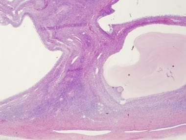
Theca cells are specialized cells that are part of the follicle where the egg matures in the ovary. They are located in the outer layer of the follicle and play an important role in producing hormones necessary for the growth and development of the follicle and the egg within it. Specifically, they produce androgens, such as testosterone, which are then converted into estrogens by another type of cells in the follicle called granulosa cells. These hormones help to thicken the lining of the uterus in preparation for a possible pregnancy. In some cases, theca cells can become overactive and produce too much testosterone, leading to conditions such as polycystic ovary syndrome (PCOS).
17-Ketosteroids are a group of steroid compounds that contain a ketone group at the 17th carbon position in their molecular structure. They are produced as metabolic byproducts of certain hormones, such as androgens and estrogens, in the human body.
The term "17-KS" or "17-ketosteroids" is often used to refer to a class of urinary steroid metabolites that can be measured in the urine to assess adrenal and gonadal function. The measurement of 17-KS is particularly useful in monitoring patients with certain endocrine disorders, such as congenital adrenal hyperplasia or adrenal tumors.
The two major 17-KS that are routinely measured in urine are androsterone and etiocholanolone, which are derived from the metabolism of testosterone and dehydroepiandrosterone (DHEA), respectively. Other 17-KS include tetrahydrocortisone, tetrahydrocortisol, and 5-androstene-3β,17β-diol.
It's worth noting that the measurement of 17-KS has largely been replaced by more specific tests, such as the measurement of individual steroid hormones or their metabolites using mass spectrometry-based methods.
Gonadal steroid hormones, also known as gonadal sex steroids, are hormones that are produced and released by the gonads (i.e., ovaries in women and testes in men). These hormones play a critical role in the development and maintenance of secondary sexual characteristics, reproductive function, and overall health.
The three main classes of gonadal steroid hormones are:
1. Androgens: These are male sex hormones that are primarily produced by the testes but also produced in smaller amounts by the ovaries and adrenal glands. The most well-known androgen is testosterone, which plays a key role in the development of male secondary sexual characteristics such as facial hair, deepening of the voice, and increased muscle mass.
2. Estrogens: These are female sex hormones that are primarily produced by the ovaries but also produced in smaller amounts by the adrenal glands. The most well-known estrogen is estradiol, which plays a key role in the development of female secondary sexual characteristics such as breast development and the menstrual cycle.
3. Progestogens: These are hormones that are produced by the ovaries during the second half of the menstrual cycle and play a key role in preparing the uterus for pregnancy. The most well-known progestogen is progesterone, which also plays a role in maintaining pregnancy and regulating the menstrual cycle.
Gonadal steroid hormones can have significant effects on various physiological processes, including bone density, cognitive function, mood, and sexual behavior. Disorders of gonadal steroid hormone production or action can lead to a range of health problems, including infertility, osteoporosis, and sexual dysfunction.
Progesterone is a steroid hormone that is primarily produced in the ovaries during the menstrual cycle and in pregnancy. It plays an essential role in preparing the uterus for implantation of a fertilized egg and maintaining the early stages of pregnancy. Progesterone works to thicken the lining of the uterus, creating a nurturing environment for the developing embryo.
During the menstrual cycle, progesterone is produced by the corpus luteum, a temporary structure formed in the ovary after an egg has been released from a follicle during ovulation. If pregnancy does not occur, the levels of progesterone will decrease, leading to the shedding of the uterine lining and menstruation.
In addition to its reproductive functions, progesterone also has various other effects on the body, such as helping to regulate the immune system, supporting bone health, and potentially influencing mood and cognition. Progesterone can be administered medically in the form of oral pills, intramuscular injections, or vaginal suppositories for various purposes, including hormone replacement therapy, contraception, and managing certain gynecological conditions.
Estradiol is a type of estrogen, which is a female sex hormone. It is the most potent and dominant form of estrogen in humans. Estradiol plays a crucial role in the development and maintenance of secondary sexual characteristics in women, such as breast development and regulation of the menstrual cycle. It also helps maintain bone density, protect the lining of the uterus, and is involved in cognition and mood regulation.
Estradiol is produced primarily by the ovaries, but it can also be synthesized in smaller amounts by the adrenal glands and fat cells. In men, estradiol is produced from testosterone through a process called aromatization. Abnormal levels of estradiol can contribute to various health issues, such as hormonal imbalances, infertility, osteoporosis, and certain types of cancer.
Aromatase is a enzyme that belongs to the cytochrome P450 superfamily, and it is responsible for converting androgens into estrogens through a process called aromatization. This enzyme plays a crucial role in the steroid hormone biosynthesis pathway, particularly in females where it is primarily expressed in adipose tissue, ovaries, brain, and breast tissue.
Aromatase inhibitors are used as a treatment for estrogen receptor-positive breast cancer in postmenopausal women, as they work by blocking the activity of aromatase and reducing the levels of circulating estrogens in the body.
Hydroxyprogesterone is a synthetic form of the natural hormone progesterone, which is produced by the body during pregnancy to support the growth and development of the fetus. Hydroxyprogesterone is used in medical treatments to help prevent preterm birth in certain high-risk pregnancies.
There are several different forms of hydroxyprogesterone that have been developed for use as medications, including:
1. Hydroxyprogesterone caproate (HPC): This is a synthetic form of progesterone that is given as an injection once a week to help prevent preterm birth in women who have previously given birth prematurely. It works by helping to thicken the lining of the uterus and prevent contractions.
2. 17-Hydroxyprogesterone: This is a natural hormone that is produced by the body during pregnancy, but it can also be synthesized in a laboratory for use as a medication. It has been studied for its potential to help prevent preterm birth, although it is not currently approved for this use by the U.S. Food and Drug Administration (FDA).
3. 21-Hydroxyprogesterone: This is another natural hormone that is produced by the body during pregnancy, but it can also be synthesized in a laboratory for use as a medication. It has been studied for its potential to help prevent preterm birth and for its ability to reduce the risk of certain complications in women with a history of premature birth.
It's important to note that hydroxyprogesterone should only be used under the supervision of a healthcare provider, as it can have side effects and may not be appropriate for all women. If you are pregnant or planning to become pregnant and have concerns about preterm birth, it's important to discuss your options with your healthcare provider.
Androstanes are a class of steroidal compounds that have a basic structure consisting of a four-ring core derived from cholesterol. Specifically, androstanes contain a 19-carbon skeleton with a chemical formula of C19H28O or C19H28O2, depending on whether they are alcohols (androgens) or ketones (androstanes), respectively.
The term "androstane" is often used to refer to the parent compound, which has a hydroxyl group (-OH) attached at the C3 position of the steroid nucleus. When this hydroxyl group is replaced by a keto group (-C=O), the resulting compound is called androstane-3,17-dione or simply "androstane."
Androstanes are important precursors in the biosynthesis of various steroid hormones, including testosterone, estrogen, and cortisol. They are also used as intermediates in the synthesis of certain drugs and pharmaceuticals.
Dehydroepiandrosterone sulfate (DHEA-S) is a steroid hormone that is produced by the adrenal glands. It is a modified form of dehydroepiandrosterone (DHEA), which is converted to DHEA-S in the body for storage and later conversion back to DHEA or other steroid hormones, such as testosterone and estrogen. DHEA-S is often measured in the blood as a marker of adrenal function. It is also available as a dietary supplement, although its effectiveness for any medical purpose is not well established.
Luteinizing Hormone (LH) is a glycoprotein hormone, which is primarily produced and released by the anterior pituitary gland. In women, a surge of LH triggers ovulation, the release of an egg from the ovaries during the menstrual cycle. During pregnancy, LH stimulates the corpus luteum to produce progesterone. In men, LH stimulates the testes to produce testosterone. It plays a crucial role in sexual development, reproduction, and maintaining the reproductive system.
Steroid 17-alpha-hydroxylase, also known as CYP17A1, is a cytochrome P450 enzyme that plays a crucial role in steroid hormone biosynthesis. It is located in the endoplasmic reticulum of cells in the adrenal glands and gonads. This enzyme catalyzes the 17-alpha-hydroxylation and subsequent lyase cleavage of pregnenolone and progesterone, converting them into dehydroepiandrosterone (DHEA) and androstenedione, respectively. These steroid intermediates are essential for the biosynthesis of both glucocorticoids and sex steroids, including cortisol, aldosterone, estrogens, and testosterone.
Defects in the CYP17A1 gene can lead to several disorders, such as congenital adrenal hyperplasia (CAH) due to 17-alpha-hydroxylase deficiency, which is characterized by decreased production of cortisol and sex steroids and increased mineralocorticoid levels. This condition results in sexual infantilism, electrolyte imbalances, and hypertension.
17-Hydroxysteroid dehydrogenases (17-HSDs) are a group of enzymes that play a crucial role in steroid hormone biosynthesis. They are involved in the conversion of 17-ketosteroids to 17-hydroxy steroids or vice versa, by adding or removing a hydroxyl group (–OH) at the 17th carbon atom of the steroid molecule. This conversion is essential for the production of various steroid hormones, including cortisol, aldosterone, and sex hormones such as estrogen and testosterone.
There are several isoforms of 17-HSDs, each with distinct substrate specificities, tissue distributions, and functions:
1. 17-HSD type 1 (17-HSD1): This isoform primarily catalyzes the conversion of estrone (E1) to estradiol (E2), an active form of estrogen. It is mainly expressed in the ovary, breast, and adipose tissue.
2. 17-HSD type 2 (17-HSD2): This isoform catalyzes the reverse reaction, converting estradiol (E2) to estrone (E1). It is primarily expressed in the placenta, prostate, and breast tissue.
3. 17-HSD type 3 (17-HSD3): This isoform is responsible for the conversion of androstenedione to testosterone, an essential step in male sex hormone biosynthesis. It is predominantly expressed in the testis and adrenal gland.
4. 17-HSD type 4 (17-HSD4): This isoform catalyzes the conversion of dehydroepiandrosterone (DHEA) to androstenedione, an intermediate step in steroid hormone biosynthesis. It is primarily expressed in the placenta.
5. 17-HSD type 5 (17-HSD5): This isoform catalyzes the conversion of cortisone to cortisol, a critical step in glucocorticoid biosynthesis. It is predominantly expressed in the adrenal gland and liver.
6. 17-HSD type 6 (17-HSD6): This isoform catalyzes the conversion of androstenedione to testosterone, similar to 17-HSD3. However, it has a different substrate specificity and is primarily expressed in the ovary.
7. 17-HSD type 7 (17-HSD7): This isoform catalyzes the conversion of estrone (E1) to estradiol (E2), similar to 17-HSD1. However, it has a different substrate specificity and is primarily expressed in the ovary.
8. 17-HSD type 8 (17-HSD8): This isoform catalyzes the conversion of DHEA to androstenedione, similar to 17-HSD4. However, it has a different substrate specificity and is primarily expressed in the testis.
9. 17-HSD type 9 (17-HSD9): This isoform catalyzes the conversion of estrone (E1) to estradiol (E2), similar to 17-HSD1. However, it has a different substrate specificity and is primarily expressed in the placenta.
10. 17-HSD type 10 (17-HSD10): This isoform catalyzes the conversion of DHEA to androstenedione, similar to 17-HSD4. However, it has a different substrate specificity and is primarily expressed in the testis.
11. 17-HSD type 11 (17-HSD11): This isoform catalyzes the conversion of estrone (E1) to estradiol (E2), similar to 17-HSD1. However, it has a different substrate specificity and is primarily expressed in the placenta.
12. 17-HSD type 12 (17-HSD12): This isoform catalyzes the conversion of DHEA to androstenedione, similar to 17-HSD4. However, it has a different substrate specificity and is primarily expressed in the testis.
13. 17-HSD type 13 (17-HSD13): This isoform catalyzes the conversion of estrone (E1) to estradiol (E2), similar to 17-HSD1. However, it has a different substrate specificity and is primarily expressed in the placenta.
14. 17-HSD type 14 (17-HSD14): This isoform catalyzes the conversion of DHEA to androstenedione, similar to 17-HSD4. However, it has a different substrate specificity and is primarily expressed in the testis.
15. 17-HSD type 15 (17-HSD15): This isoform catalyzes the conversion of estrone (E1) to estradiol (E2), similar to 17-HSD1. However, it has a different substrate specificity and is primarily expressed in the placenta.
16. 17-HSD type 16 (17-HSD16): This isoform catalyzes the conversion of DHEA to androstenedione, similar to 17-HSD4. However, it has a different substrate specificity and is primarily expressed in the testis.
17. 17-HSD type 17 (17-HSD17): This isoform catalyzes the conversion of estrone (E1) to estradiol (E2), similar to 17-HSD1. However, it has a different substrate specificity and is primarily expressed in the placenta.
18. 17-HSD type 18 (17-HSD18): This isoform catalyzes the conversion of DHEA to androstenedione, similar to 17-HSD4. However, it has a different substrate specificity and is primarily expressed in the testis.
19. 17-HSD type 19 (17-HSD19): This isoform catalyzes the conversion of estrone (E1) to estradiol (E2), similar to 17-HSD1. However, it has a different substrate specificity and is primarily expressed in the placenta.
20. 17-HSD type 20 (17-HSD20): This isoform catalyzes the conversion of DHEA to androstenedione, similar to 17-HSD4. However, it has a different substrate specificity and is primarily expressed in the testis.
21. 17-HSD type 21 (17-HSD21): This isoform catalyzes the conversion of estrone (E1) to estradiol (E2), similar to 17-HSD1. However, it has a different substrate specificity and is primarily expressed in the placenta.
22. 17-HSD type 22 (17-HSD22): This isoform catalyzes the conversion of DHEA to androstenedione, similar to 17-HSD4. However, it has a different substrate specificity and is primarily expressed in the testis.
23. 17-HSD type 23 (17-HSD23): This isoform catalyzes the conversion of estrone (E1) to estradiol (E2), similar to 17-HSD1. However, it has a different substrate specificity and is primarily expressed in the placenta.
24. 17-HSD type 24 (17-HSD24): This isoform catalyzes the conversion of DHEA to androstenedione, similar to 17-HSD4. However, it has a different substrate specificity and is primarily expressed in the testis.
25. 17-HSD type 25 (17-HSD25): This isoform catalyzes the conversion of estrone (E1) to estradiol (E2), similar to 17-HSD1. However, it has a different substrate specificity and is primarily expressed in the placenta.
26. 17-HSD type 26 (17-HSD26): This isoform catalyzes the conversion of DHEA to androstenedione, similar to 17-HSD4. However
Steroids, also known as corticosteroids, are a type of hormone that the adrenal gland produces in your body. They have many functions, such as controlling the balance of salt and water in your body and helping to reduce inflammation. Steroids can also be synthetically produced and used as medications to treat a variety of conditions, including allergies, asthma, skin conditions, and autoimmune disorders.
Steroid medications are available in various forms, such as oral pills, injections, creams, and inhalers. They work by mimicking the effects of natural hormones produced by your body, reducing inflammation and suppressing the immune system's response to prevent or reduce symptoms. However, long-term use of steroids can have significant side effects, including weight gain, high blood pressure, osteoporosis, and increased risk of infections.
It is important to note that anabolic steroids are a different class of drugs that are sometimes abused for their muscle-building properties. These steroids are synthetic versions of the male hormone testosterone and can have serious health consequences when taken in large doses or without medical supervision.
Sex Hormone-Binding Globulin (SHBG) is a protein produced mainly in the liver that plays a crucial role in regulating the active forms of the sex hormones, testosterone and estradiol, in the body. SHBG binds to these hormones in the bloodstream, creating a reservoir of bound hormones. Only the unbound (or "free") fraction of testosterone and estradiol is considered biologically active and can easily enter cells to exert its effects.
By binding to sex hormones, SHBG helps control their availability and transport in the body. Factors such as age, sex, infection with certain viruses (like hepatitis or HIV), liver disease, obesity, and various medications can influence SHBG levels and, consequently, impact the amount of free testosterone and estradiol in circulation.
SHBG is an essential factor in maintaining hormonal balance and has implications for several physiological processes, including sexual development, reproduction, bone health, muscle mass, and overall well-being. Abnormal SHBG levels can contribute to various medical conditions, such as hypogonadism (low testosterone levels), polycystic ovary syndrome (PCOS), and certain types of cancer.
Pregnenolone is defined as a steroid hormone produced in the body from cholesterol. It's often referred to as the "mother hormone" since many other hormones, including cortisol, aldosterone, progesterone, testosterone, and estrogen, are synthesized from it.
Pregnenolone is primarily produced in the adrenal glands but can also be produced in smaller amounts in the brain, skin, and sex organs (ovaries and testes). It plays a crucial role in various physiological processes such as maintaining membrane fluidity, acting as an antioxidant, and contributing to cognitive function.
However, it's important to note that while pregnenolone is a hormone, over-the-counter supplements containing this compound are not approved by the FDA for any medical use or condition. As always, consult with a healthcare provider before starting any new supplement regimen.
An ovarian follicle is a fluid-filled sac in the ovary that contains an immature egg or ovum (oocyte). It's a part of the female reproductive system and plays a crucial role in the process of ovulation.
Ovarian follicles start developing in the ovaries during fetal development, but only a small number of them will mature and release an egg during a woman's reproductive years. The maturation process is stimulated by hormones like follicle-stimulating hormone (FSH) and luteinizing hormone (LH).
There are different types of ovarian follicles, including primordial, primary, secondary, and tertiary or Graafian follicles. The Graafian follicle is the mature follicle that ruptures during ovulation to release the egg into the fallopian tube, where it may be fertilized by sperm.
It's important to note that abnormal growth or development of ovarian follicles can lead to conditions like polycystic ovary syndrome (PCOS) and ovarian cancer.
Follicle-Stimulating Hormone (FSH) is a glycoprotein hormone secreted and released by the anterior pituitary gland. In females, it promotes the growth and development of ovarian follicles in the ovary, which ultimately leads to the maturation and release of an egg (ovulation). In males, FSH stimulates the testes to produce sperm. It works in conjunction with luteinizing hormone (LH) to regulate reproductive processes. The secretion of FSH is controlled by the hypothalamic-pituitary-gonadal axis and its release is influenced by the levels of gonadotropin-releasing hormone (GnRH), estrogen, inhibin, and androgens.
An ovary is a part of the female reproductive system in which ova or eggs are produced through the process of oogenesis. They are a pair of solid, almond-shaped structures located one on each side of the uterus within the pelvic cavity. Each ovary measures about 3 to 5 centimeters in length and weighs around 14 grams.
The ovaries have two main functions: endocrine (hormonal) function and reproductive function. They produce and release eggs (ovulation) responsible for potential fertilization and development of an embryo/fetus during pregnancy. Additionally, they are essential in the production of female sex hormones, primarily estrogen and progesterone, which regulate menstrual cycles, sexual development, and reproduction.
During each menstrual cycle, a mature egg is released from one of the ovaries into the fallopian tube, where it may be fertilized by sperm. If not fertilized, the egg, along with the uterine lining, will be shed, leading to menstruation.
Polycyctic Ovary Syndrome (PCOS) is a complex endocrine-metabolic disorder characterized by the presence of hyperandrogenism (excess male hormones), ovulatory dysfunction, and polycystic ovaries. The Rotterdam criteria are commonly used for diagnosis, which require at least two of the following three features:
1. Oligo- or anovulation (irregular menstrual cycles)
2. Clinical and/or biochemical signs of hyperandrogenism (e.g., hirsutism, acne, or high levels of androgens in the blood)
3. Polycystic ovaries on ultrasound examination (presence of 12 or more follicles measuring 2-9 mm in diameter, or increased ovarian volume >10 mL)
The exact cause of PCOS remains unclear, but it is believed to involve a combination of genetic and environmental factors. Insulin resistance and obesity are common findings in women with PCOS, which can contribute to the development of metabolic complications such as type 2 diabetes, dyslipidemia, and cardiovascular disease.
Management of PCOS typically involves a multidisciplinary approach that includes lifestyle modifications (diet, exercise, weight loss), medications to regulate menstrual cycles and reduce hyperandrogenism (e.g., oral contraceptives, metformin, anti-androgens), and fertility treatments if desired. Regular monitoring of metabolic parameters and long-term follow-up are essential for optimal management and prevention of complications.
Testolactone is a medication that is primarily used in the treatment of breast cancer. It is an oral steroidal aromatase inhibitor, which means it works by blocking the enzyme aromatase, thereby preventing the conversion of androgens into estrogens. This helps to reduce the amount of estrogen in the body, which can slow or stop the growth of certain types of breast cancer cells that need estrogen to grow.
Testolactone is not as commonly used as other aromatase inhibitors such as letrozole, anastrozole, and exemestane, but it may be prescribed in certain cases where these medications are not suitable or have not been effective. It is important to note that testolactone can have side effects, including nausea, vomiting, diarrhea, skin rash, and changes in liver function tests. As with any medication, it should only be taken under the supervision of a healthcare provider.
Estrogens are a group of steroid hormones that are primarily responsible for the development and regulation of female sexual characteristics and reproductive functions. They are also present in lower levels in males. The main estrogen hormone is estradiol, which plays a key role in promoting the growth and development of the female reproductive system, including the uterus, fallopian tubes, and breasts. Estrogens also help regulate the menstrual cycle, maintain bone density, and have important effects on the cardiovascular system, skin, hair, and cognitive function.
Estrogens are produced primarily by the ovaries in women, but they can also be produced in smaller amounts by the adrenal glands and fat cells. In men, estrogens are produced from the conversion of testosterone, the primary male sex hormone, through a process called aromatization.
Estrogen levels vary throughout a woman's life, with higher levels during reproductive years and lower levels after menopause. Estrogen therapy is sometimes used to treat symptoms of menopause, such as hot flashes and vaginal dryness, or to prevent osteoporosis in postmenopausal women. However, estrogen therapy also carries risks, including an increased risk of certain cancers, blood clots, and stroke, so it is typically recommended only for women who have a high risk of these conditions.
Hirsutism is a medical condition characterized by excessive hair growth in women in areas where hair growth is typically androgen-dependent, such as the face, chest, lower abdomen, and inner thighs. This hair growth is often thick, dark, and coarse, resembling male-pattern hair growth. Hirsutism can be caused by various factors, including hormonal imbalances, certain medications, and genetic conditions. It's essential to consult a healthcare professional if you experience excessive or unwanted hair growth to determine the underlying cause and develop an appropriate treatment plan.
Dihydrotestosterone (DHT) is a sex hormone and androgen that plays a critical role in the development and maintenance of male characteristics, such as facial hair, deep voice, and muscle mass. It is synthesized from testosterone through the action of the enzyme 5-alpha reductase. DHT is essential for the normal development of the male genitalia during fetal development and for the maturation of the sexual organs at puberty.
In addition to its role in sexual development, DHT also contributes to the growth of hair follicles, the health of the prostate gland, and the maintenance of bone density. However, an excess of DHT has been linked to certain medical conditions, such as benign prostatic hyperplasia (BPH) and androgenetic alopecia (male pattern baldness).
DHT exerts its effects by binding to androgen receptors in various tissues throughout the body. Once bound, DHT triggers a series of cellular responses that regulate gene expression and influence the growth and differentiation of cells. In some cases, these responses can lead to unwanted side effects, such as hair loss or prostate enlargement.
Medications that block the action of 5-alpha reductase, such as finasteride and dutasteride, are sometimes used to treat conditions associated with excess DHT production. These drugs work by reducing the amount of DHT available to bind to androgen receptors, thereby alleviating symptoms and slowing disease progression.
In summary, dihydrotestosterone is a potent sex hormone that plays a critical role in male sexual development and function. While it is essential for normal growth and development, an excess of DHT has been linked to certain medical conditions, such as BPH and androgenetic alopecia. Medications that block the action of 5-alpha reductase are sometimes used to treat these conditions by reducing the amount of DHT available to bind to androgen receptors.
Hyperandrogenism is a medical condition characterized by excessive levels of androgens (male sex hormones) in the body. This can lead to various symptoms such as hirsutism (excessive hair growth), acne, irregular menstrual periods, and infertility in women. It can be caused by conditions like polycystic ovary syndrome (PCOS), congenital adrenal hyperplasia, and tumors in the ovaries or adrenal glands. Proper diagnosis and management of hyperandrogenism is important to prevent complications and improve quality of life.
Epitestosterone is a steroid hormone that is structurally similar to testosterone. It is produced in the body, primarily in the testes and adrenal glands, and is a natural component of human urine. Epitestosterone is a weak androgen, meaning it has minimal male sex hormone effects.
The ratio of epitestosterone to testosterone (T/E ratio) in urine is often used as a marker for the detection of doping with anabolic steroids, which are synthetic versions of testosterone. In athletes who have not taken performance-enhancing drugs, the T/E ratio is typically less than 1. However, when anabolic steroids are used, the level of testosterone in the body increases, while the level of epitestosterone remains relatively unchanged, leading to a higher T/E ratio.
Medical professionals and anti-doping agencies use a specific cutoff value for the T/E ratio to determine if an individual has violated doping regulations. It's important to note that some individuals may have naturally higher T/E ratios due to genetic factors, which can complicate the interpretation of test results in anti-doping tests.
Androstenediol is an endogenous steroid hormone that is produced in the body from dehydroepiandrosterone (DHEA) and converted into testosterone and estrogens. It exists in two forms: 5-androstenediol and 4-androstenediol, with 5-androstenediol being the more abundant form in the human body.
In the context of medical definitions, androstenediol is a weak androgen that can be converted into testosterone or estradiol, depending on the needs of the body. It plays a role in the development and maintenance of secondary sexual characteristics, such as facial hair and deepening of the voice in males, and breast development and menstrual cycles in females.
Androstenediol is also available as a dietary supplement and has been marketed for its potential performance-enhancing effects. However, its use as a performance-enhancing drug is banned by many sports organizations due to concerns about its potential to enhance athletic performance and its unknown safety profile.
Follicular fluid is the fluid that accumulates within the follicle (a small sac or cyst) in the ovary where an egg matures. This fluid contains various chemicals, hormones, and proteins that support the growth and development of the egg cell. It also contains metabolic waste products and other substances from the granulosa cells (the cells that surround the egg cell within the follicle). Follicular fluid is often analyzed in fertility treatments and studies as it can provide valuable information about the health and viability of the egg cell.
Granulosa cells are specialized cells that surround and enclose the developing egg cells (oocytes) in the ovaries. They play a crucial role in the growth, development, and maturation of the follicles (the fluid-filled sacs containing the oocytes) by providing essential nutrients and hormones.
Granulosa cells are responsible for producing estrogen, which supports the development of the endometrium during the menstrual cycle in preparation for a potential pregnancy. They also produce inhibin and activin, two hormones that regulate the function of the pituitary gland and its secretion of follicle-stimulating hormone (FSH) and luteinizing hormone (LH).
These cells are critical for female reproductive health and fertility. Abnormalities in granulosa cell function can lead to various reproductive disorders, such as polycystic ovary syndrome (PCOS), premature ovarian failure, and infertility.
Androsterone is a weak androgen and an endogenous steroid hormone. It's produced in the liver from dehydroepiandrosterone (DHEA) and is converted into androstenedione, another weak androgen. Androsterone is excreted in urine as a major metabolite of testosterone. It plays a role in male sexual development and function, although its effects are much weaker than those of testosterone. In clinical contexts, androsterone levels may be measured to help diagnose certain hormonal disorders or to monitor hormone therapy.
Ovulation is the medical term for the release of a mature egg from an ovary during a woman's menstrual cycle. The released egg travels through the fallopian tube where it may be fertilized by sperm if sexual intercourse has occurred recently. If the egg is not fertilized, it will break down and leave the body along with the uterine lining during menstruation. Ovulation typically occurs around day 14 of a 28-day menstrual cycle, but the timing can vary widely from woman to woman and even from cycle to cycle in the same woman.
During ovulation, there are several physical changes that may occur in a woman's body, such as an increase in basal body temperature, changes in cervical mucus, and mild cramping or discomfort on one side of the lower abdomen (known as mittelschmerz). These symptoms can be used to help predict ovulation and improve the chances of conception.
It's worth noting that some medical conditions, such as polycystic ovary syndrome (PCOS) or premature ovarian failure, may affect ovulation and make it difficult for a woman to become pregnant. In these cases, medical intervention may be necessary to help promote ovulation and increase the chances of conception.
17-alpha-Hydroxypregnenolone is a steroid hormone that is produced in the adrenal glands and, to a lesser extent, in the gonads (ovaries and testes). It is an intermediate in the biosynthesis of steroid hormones, including cortisol, aldosterone, and sex hormones such as testosterone and estrogen.
17-alpha-Hydroxypregnenolone is formed from pregnenolone through the action of the enzyme 17α-hydroxylase. It can then be converted to 17-hydroxyprogesterone, which is a precursor to both cortisol and androgens such as testosterone.
While 17-alpha-Hydroxypregnenolone itself does not have significant physiological activity, its role in the biosynthesis of other steroid hormones makes it an important intermediate in the endocrine system. Dysregulation of its production or metabolism can contribute to various medical conditions, such as congenital adrenal hyperplasia and certain forms of cancer.
Gonadotropins are hormones produced and released by the anterior pituitary gland, a small endocrine gland located at the base of the brain. These hormones play crucial roles in regulating reproduction and sexual development. There are two main types of gonadotropins:
1. Follicle-Stimulating Hormone (FSH): FSH is essential for the growth and development of follicles in the ovaries (in females) or sperm production in the testes (in males). In females, FSH stimulates the maturation of eggs within the follicles.
2. Luteinizing Hormone (LH): LH triggers ovulation in females, causing the release of a mature egg from the dominant follicle. In males, LH stimulates the production and secretion of testosterone in the testes.
Together, FSH and LH work synergistically to regulate various aspects of reproductive function and sexual development. Their secretion is controlled by the hypothalamus, which releases gonadotropin-releasing hormone (GnRH) to stimulate the production and release of FSH and LH from the anterior pituitary gland.
Abnormal levels of gonadotropins can lead to various reproductive disorders, such as infertility or menstrual irregularities in females and issues related to sexual development or function in both sexes. In some cases, synthetic forms of gonadotropins may be used clinically to treat these conditions or for assisted reproductive technologies (ART).
Aromatase inhibitors (AIs) are a class of drugs that are primarily used in the treatment of hormone-sensitive breast cancer in postmenopausal women. They work by inhibiting the enzyme aromatase, which is responsible for converting androgens into estrogens. By blocking this conversion, AIs decrease the amount of estrogen in the body, thereby depriving hormone-sensitive breast cancer cells of the estrogen they need to grow and multiply.
There are three main types of aromatase inhibitors:
1. Letrozole (Femara) - a non-steroidal AI that is taken orally once a day.
2. Anastrozole (Arimidex) - another non-steroidal AI that is also taken orally once a day.
3. Exemestane (Aromasin) - a steroidal AI that is taken orally once a day.
In addition to their use in breast cancer treatment, AIs are also sometimes used off-label for the treatment of estrogen-dependent conditions such as endometriosis and uterine fibroids. However, it's important to note that the use of aromatase inhibitors can have significant side effects, including hot flashes, joint pain, and bone loss, so they should only be used under the close supervision of a healthcare provider.
Androstenediols are endogenous steroid hormones that are produced in the body from dehydroepiandrosterone (DHEA) and DHEA sulfate (DHEAS), which are secreted by the adrenal glands. There are two major types of androstenediols: 5-androstenediol and 4-androstenediol. These hormones can be further metabolized into testosterone and estrogens, making them important intermediates in steroid hormone synthesis.
5-androstenediol is a weak androgen that can be converted to testosterone in peripheral tissues, while 4-androstenediol has little known biological activity. Both of these compounds have been studied for their potential role in various physiological processes, including sexual differentiation, bone metabolism, and aging. However, more research is needed to fully understand their functions and clinical significance.
It's worth noting that androstenediols are also sometimes referred to as "prohormones" because they can be converted into active steroid hormones in the body. Some athletes and bodybuilders have used synthetic forms of these compounds as performance-enhancing drugs, although their use is banned by many sports organizations due to concerns about potential health risks and unfair advantages in competition.
3-Hydroxysteroid dehydrogenases (3-HSDs) are a group of enzymes that play a crucial role in steroid hormone biosynthesis. These enzymes catalyze the conversion of 3-beta-hydroxy steroids to 3-keto steroids, which is an essential step in the production of various steroid hormones, including progesterone, cortisol, aldosterone, and sex hormones such as testosterone and estradiol.
There are several isoforms of 3-HSDs that are expressed in different tissues and have distinct substrate specificities. For instance, 3-HSD type I is primarily found in the ovary and adrenal gland, where it catalyzes the conversion of pregnenolone to progesterone and 17-hydroxyprogesterone to 17-hydroxycortisol. On the other hand, 3-HSD type II is mainly expressed in the testes, adrenal gland, and placenta, where it catalyzes the conversion of dehydroepiandrosterone (DHEA) to androstenedione and androstenedione to testosterone.
Defects in 3-HSDs can lead to various genetic disorders that affect steroid hormone production and metabolism, resulting in a range of clinical manifestations such as adrenal insufficiency, ambiguous genitalia, and sexual development disorders.
Congenital Adrenal Hyperplasia (CAH) is a group of inherited genetic disorders that affect the adrenal glands, which are triangular-shaped glands located on top of the kidneys. The adrenal glands are responsible for producing several essential hormones, including cortisol, aldosterone, and androgens.
CAH is caused by mutations in genes that code for enzymes involved in the synthesis of these hormones. The most common form of CAH is 21-hydroxylase deficiency, which affects approximately 90% to 95% of all cases. Other less common forms of CAH include 11-beta-hydroxylase deficiency and 3-beta-hydroxysteroid dehydrogenase deficiency.
The severity of the disorder can vary widely, depending on the degree of enzyme deficiency. In severe cases, the lack of cortisol production can lead to life-threatening salt wasting and electrolyte imbalances in newborns. The excess androgens produced due to the enzyme deficiency can also cause virilization, or masculinization, of female fetuses, leading to ambiguous genitalia at birth.
In milder forms of CAH, symptoms may not appear until later in childhood or even adulthood. These may include early puberty, rapid growth followed by premature fusion of the growth plates and short stature, acne, excessive hair growth, irregular menstrual periods, and infertility.
Treatment for CAH typically involves replacing the missing hormones with medications such as hydrocortisone, fludrocortisone, and/or sex hormones. Regular monitoring of hormone levels and careful management of medication doses is essential to prevent complications such as adrenal crisis, growth suppression, and osteoporosis.
In severe cases of CAH, early diagnosis and treatment can help prevent or minimize the risk of serious health problems and improve quality of life. Genetic counseling may also be recommended for affected individuals and their families to discuss the risks of passing on the disorder to future generations.
Hormones are defined as chemical messengers that are produced by endocrine glands or specialized cells and are transported through the bloodstream to tissues and organs, where they elicit specific responses. They play crucial roles in regulating various physiological processes such as growth, development, metabolism, reproduction, and mood. Examples of hormones include insulin, estrogen, testosterone, adrenaline, and thyroxine.
The clitoris is an important female sex organ that is primarily responsible for sexual arousal and pleasure. It is a small, highly sensitive piece of tissue located at the front of the vulva, where the labia minora meet. The clitoris is made up of two parts: the visible part, known as the glans clitoris, and the hidden part, called the corpora cavernosa and crura.
The glans clitoris is a small knob-like structure that is covered by a hood, or prepuce, and is located at the top of the vulva. It contains a high concentration of nerve endings, making it highly sensitive to touch and stimulation. The corpora cavernosa and crura are the internal parts of the clitoris, which are made up of sponge-like erectile tissue that becomes engorged with blood during sexual arousal, leading to clitoral erection.
The clitoris plays a crucial role in female sexual response and pleasure. During sexual arousal, the clitoris swells and becomes more sensitive to touch, which can lead to orgasm. The clitoris is also an important source of sexual pleasure during masturbation and partnered sexual activity. Despite its importance in female sexuality, the clitoris has historically been overlooked or stigmatized in many cultures, leading to a lack of understanding and education about this vital organ.
Aminoglutethimide is a medication that is primarily used to treat hormone-sensitive cancers such as breast cancer and prostate cancer. It works by blocking the production of certain hormones in the body, including estrogen and cortisol. Aminoglutethimide is an inhibitor of steroid synthesis, specifically targeting the enzymes involved in the conversion of cholesterol to steroid hormones.
The medication is available in oral form and is typically taken 2-3 times a day. Common side effects include drowsiness, dizziness, dry mouth, skin rash, and changes in appetite or weight. More serious side effects may include liver damage, severe allergic reactions, and changes in heart rhythm.
It's important to note that aminoglutethimide can interact with other medications, so it's crucial to inform your healthcare provider about all the drugs you are currently taking before starting this medication. Additionally, regular monitoring of liver function and hormone levels may be necessary during treatment with aminoglutethimide.
Freemartinism is a condition seen in female cattle that have been twin to a male fetus. It is a form of pseudohermaphroditism where the female develops some masculine characteristics due to exposure to male hormones from her twin brother while in the womb. These females may have underdeveloped reproductive systems and are usually sterile, unable to reproduce. The term "freemartin" is used specifically for this condition in cattle, but similar conditions can occur in other species including sheep and goats.
Carnivora is an order of mammals that consists of animals whose primary diet consists of flesh. The term "Carnivora" comes from the Latin words "caro", meaning flesh, and "vorare", meaning to devour. This order includes a wide variety of species, ranging from large predators such as lions, tigers, and bears, to smaller animals such as weasels, otters, and raccoons.
While members of the Carnivora order are often referred to as "carnivores," it is important to note that not all members exclusively eat meat. Some species, such as raccoons and bears, have an omnivorous diet that includes both plants and animals. Additionally, some species within this order have evolved specialized adaptations for their specific diets, such as the elongated canines and carnassial teeth of felids (cats) and canids (dogs), which are adapted for tearing and shearing meat.
Overall, the medical definition of Carnivora refers to an order of mammals that have a diet primarily consisting of flesh, although not all members exclusively eat meat.
Feminization is a process or condition in which typically male characteristics are diminished or absent, and female characteristics become more prominent. This term is often used in the context of transgender health to describe hormone therapy that helps individuals align their physical appearance with their gender identity. The goal of feminizing hormone therapy is to promote the development of secondary sexual characteristics such as breast development, softer skin, reduced muscle mass and body hair, and fat redistribution to create a more typically female body shape. It's important to note that every individual's experience with feminization is unique, and the specific changes experienced may vary depending on factors such as age, genetics, and the duration of hormone therapy.
The adrenal glands are a pair of endocrine glands that are located on top of the kidneys. Each gland has two parts: the outer cortex and the inner medulla. The adrenal cortex produces hormones such as cortisol, aldosterone, and androgens, which regulate metabolism, blood pressure, and other vital functions. The adrenal medulla produces catecholamines, including epinephrine (adrenaline) and norepinephrine (noradrenaline), which help the body respond to stress by increasing heart rate, blood pressure, and alertness.
Hydrocortisone is a synthetic glucocorticoid, which is a class of steroid hormones. It is identical to the naturally occurring cortisol, a hormone produced by the adrenal gland that helps regulate metabolism and helps your body respond to stress. Hydrocortisone has anti-inflammatory effects and is used to treat various inflammatory conditions such as allergies, skin disorders, and autoimmune diseases. It works by suppressing the immune system's response to reduce swelling, redness, itching, and other symptoms caused by inflammation.
Hydrocortisone is available in different forms, including oral tablets, topical creams, lotions, gels, and ointments, as well as injectable solutions. The specific use and dosage depend on the condition being treated and the individual patient's medical history and current health status.
As with any medication, hydrocortisone can have side effects, especially when used in high doses or for extended periods. Common side effects include increased appetite, weight gain, mood changes, insomnia, and skin thinning. Long-term use of hydrocortisone may also increase the risk of developing osteoporosis, diabetes, cataracts, and other health problems. Therefore, it is essential to follow your healthcare provider's instructions carefully when using this medication.
Estrus is a term used in veterinary medicine to describe the physiological and behavioral state of female mammals that are ready to mate and conceive. It refers to the period of time when the female's reproductive system is most receptive to fertilization.
During estrus, the female's ovaries release one or more mature eggs (ovulation) into the fallopian tubes, where they can be fertilized by sperm from a male. This phase of the estrous cycle is often accompanied by changes in behavior and physical appearance, such as increased vocalization, restlessness, and swelling of the genital area.
The duration and frequency of estrus vary widely among different species of mammals. In some animals, such as dogs and cats, estrus occurs regularly at intervals of several weeks or months, while in others, such as cows and mares, it may only occur once or twice a year.
It's important to note that the term "estrus" is not used to describe human reproductive physiology. In humans, the equivalent phase of the menstrual cycle is called ovulation.
I am not aware of a medical definition for "Cortodoxone." It is possible that this term is not recognized in the field of medicine as it does not appear to be a commonly used medication, treatment, or diagnostic tool. If you have any more information about where you encountered this term or its potential meaning, I would be happy to try and provide further clarification.
Chorionic Gonadotropin (hCG) is a hormone that is produced during pregnancy. It is produced by the placenta after implantation of the fertilized egg in the uterus. The main function of hCG is to prevent the disintegration of the corpus luteum, which is a temporary endocrine structure that forms in the ovary after ovulation and produces progesterone during early pregnancy. Progesterone is essential for maintaining the lining of the uterus and supporting the pregnancy.
hCG can be detected in the blood or urine as early as 10 days after conception, and its levels continue to rise throughout the first trimester of pregnancy. In addition to its role in maintaining pregnancy, hCG is also used as a clinical marker for pregnancy and to monitor certain medical conditions such as gestational trophoblastic diseases.
Radioimmunoassay (RIA) is a highly sensitive analytical technique used in clinical and research laboratories to measure concentrations of various substances, such as hormones, vitamins, drugs, or tumor markers, in biological samples like blood, urine, or tissues. The method relies on the specific interaction between an antibody and its corresponding antigen, combined with the use of radioisotopes to quantify the amount of bound antigen.
In a typical RIA procedure, a known quantity of a radiolabeled antigen (also called tracer) is added to a sample containing an unknown concentration of the same unlabeled antigen. The mixture is then incubated with a specific antibody that binds to the antigen. During the incubation period, the antibody forms complexes with both the radiolabeled and unlabeled antigens.
After the incubation, the unbound (free) radiolabeled antigen is separated from the antibody-antigen complexes, usually through a precipitation or separation step involving centrifugation, filtration, or chromatography. The amount of radioactivity in the pellet (containing the antibody-antigen complexes) is then measured using a gamma counter or other suitable radiation detection device.
The concentration of the unlabeled antigen in the sample can be determined by comparing the ratio of bound to free radiolabeled antigen in the sample to a standard curve generated from known concentrations of unlabeled antigen and their corresponding bound/free ratios. The higher the concentration of unlabeled antigen in the sample, the lower the amount of radiolabeled antigen that will bind to the antibody, resulting in a lower bound/free ratio.
Radioimmunoassays offer high sensitivity, specificity, and accuracy, making them valuable tools for detecting and quantifying low levels of various substances in biological samples. However, due to concerns about radiation safety and waste disposal, alternative non-isotopic immunoassay techniques like enzyme-linked immunosorbent assays (ELISAs) have become more popular in recent years.
Genitalia, also known as the genitals, refer to the reproductive organs located in the pelvic region. In males, these include the penis and testicles, while in females, they consist of the vulva, vagina, clitoris, and ovaries. Genitalia are essential for sexual reproduction and can also be associated with various medical conditions, such as infections, injuries, or congenital abnormalities.
Gynecomastia is a medical term that refers to the benign enlargement of the glandular tissue in male breasts, usually caused by an imbalance of the hormones estrogen and testosterone. It's important to note that gynecomastia is not the same as having excess fat in the breast area, which is called pseudogynecomastia.
Gynecomastia can occur during infancy, puberty, or old age due to natural hormonal changes. Certain medications, medical conditions, and recreational drugs can also cause gynecomastia by affecting hormone levels in the body. In some cases, the exact cause of gynecomastia may remain unknown.
Mild cases of gynecomastia may not require treatment, but severe or persistent cases may be treated with medication or surgery to remove excess breast tissue. It's essential to consult a healthcare professional for an accurate diagnosis and appropriate treatment options if you suspect you have gynecomastia.
Androstenols are a type of steroid compound that is found in both animals and humans. They are classified as pheromones, which are chemicals that can affect the behavior or physiology of other members of the same species. Androstenols are found in high concentrations in male sweat, and they have been suggested to play a role in human sexual attraction and communication.
In particular, androstenols are thought to have a positive and calming effect on people, and may help to reduce stress and anxiety. They have also been shown to increase feelings of approachability and friendliness between individuals. Some studies have suggested that androstenols may be particularly effective at enhancing social interactions in women.
Androstenols are often used in perfumes and colognes, as well as in aromatherapy products, because of their potential to promote positive social interactions and reduce stress. However, it is important to note that the effects of androstenols on human behavior and physiology are still not fully understood, and more research is needed to confirm their role in human communication and attraction.
Steroid 16-alpha-Hydroxylase is an enzyme that catalyzes the reaction adding a hydroxyl group to the sixteen (16) alpha position of steroid molecules. This enzyme is involved in the metabolic pathways of various steroids, including cortisol, aldosterone, and some sex hormones.
The gene that encodes this enzyme is CYP3A4, which is part of the cytochrome P450 family. The 16-alpha-hydroxylase activity of this enzyme has been implicated in several physiological and pathophysiological processes, such as steroid hormone biosynthesis, drug metabolism, and cancer progression.
It's worth noting that the activity of this enzyme can vary among individuals, which may contribute to differences in steroid hormone levels and susceptibility to certain diseases.
The testis, also known as the testicle, is a male reproductive organ that is part of the endocrine system. It is located in the scrotum, outside of the abdominal cavity. The main function of the testis is to produce sperm and testosterone, the primary male sex hormone.
The testis is composed of many tiny tubules called seminiferous tubules, where sperm are produced. These tubules are surrounded by a network of blood vessels, nerves, and supportive tissues. The sperm then travel through a series of ducts to the epididymis, where they mature and become capable of fertilization.
Testosterone is produced in the Leydig cells, which are located in the interstitial tissue between the seminiferous tubules. Testosterone plays a crucial role in the development and maintenance of male secondary sexual characteristics, such as facial hair, deep voice, and muscle mass. It also supports sperm production and sexual function.
Abnormalities in testicular function can lead to infertility, hormonal imbalances, and other health problems. Regular self-examinations and medical check-ups are recommended for early detection and treatment of any potential issues.
Ketosteroids are a type of steroid compound that contain a ketone functional group in their chemical structure. They are derived from cholesterol and are present in both animal and plant tissues. Some ketosteroids are produced endogenously, while others can be introduced exogenously through the diet or medication.
Endogenous ketosteroids include steroid hormones such as testosterone, estradiol, and cortisol, which contain a ketone group in their structure. Exogenous ketosteroids can be found in certain medications, such as those used to treat hormonal imbalances or inflammation.
Ketosteroids have been studied for their potential therapeutic uses, including as anti-inflammatory agents and for the treatment of hormone-related disorders. However, more research is needed to fully understand their mechanisms of action and potential benefits.
Etiocholanolone is an endogenous steroid hormone, a metabolic breakdown product of both testosterone and androstenedione. It is a 5β-reduced derivative of androstanedione and is produced in the liver as well as in the gonads and the adrenal glands.
Etiocholanolone can be measured in urine to help evaluate for certain medical conditions, such as congenital adrenal hyperplasia or adrenal cancer. Increased levels of etiocholanolone may indicate increased production of androgens, which can occur in conditions such as polycystic ovary syndrome, virilizing ovarian tumors, or congenital adrenal hyperplasia.
It is important to note that the measurement of etiocholanolone should be interpreted in conjunction with other clinical and laboratory findings, as there are many factors that can affect its levels.
Progesterone reductase is not a widely recognized or used term in medical literature. However, based on the terms "progesterone" and "reductase," it can be inferred that progesterone reductase might refer to an enzyme responsible for reducing or converting progesterone into another form through a reduction reaction.
Progesterone is a steroid hormone involved in the menstrual cycle, pregnancy, and embryogenesis. Reductases are enzymes that catalyze the transfer of electrons from a donor to an acceptor, often resulting in the reduction of a substrate. In this context, progesterone reductase could potentially refer to an enzyme responsible for reducing progesterone into a different steroid hormone or metabolite.
However, it is essential to note that there is no widely accepted or established definition of "progesterone reductase" in medical literature. If you are looking for information on a specific enzyme related to progesterone metabolism, I would recommend consulting primary scientific literature or seeking guidance from a medical professional.
Estriol is a type of estrogen, which is a female sex hormone. It is produced in the placenta during pregnancy and is used as a marker for fetal growth and development. Estriol levels can be measured in the mother's urine or blood to assess fetal well-being during pregnancy. Additionally, synthetic forms of estriol are sometimes used in hormone replacement therapy to treat symptoms of menopause.
Follicular atresia is a physiological process that occurs in the ovary, where follicles (fluid-filled sacs containing immature eggs or oocytes) undergo degeneration and disappearance. This process begins after the primordial follicle stage and continues throughout a woman's reproductive years. At birth, a female has approximately 1 to 2 million primordial follicles, but only about 400 of these will mature and release an egg during her lifetime. The rest undergo atresia, which is a natural process that helps regulate the number of available eggs and maintain hormonal balance within the body.
The exact mechanisms that trigger follicular atresia are not fully understood, but it is believed to be influenced by various factors such as hormonal imbalances, oxidative stress, and apoptosis (programmed cell death). In some cases, accelerated or excessive follicular atresia can lead to infertility or early menopause.
The Radioisotope Dilution Technique is a method used in nuclear medicine to measure the volume and flow rate of a particular fluid in the body. It involves introducing a known amount of a radioactive isotope, or radioisotope, into the fluid, such as blood. The isotope mixes with the fluid, and samples are then taken from the fluid at various time points.
By measuring the concentration of the radioisotope in each sample, it is possible to calculate the total volume of the fluid based on the amount of the isotope introduced and the dilution factor. The flow rate can also be calculated by measuring the concentration of the isotope over time and using the formula:
Flow rate = Volume/Time
This technique is commonly used in medical research and clinical settings to measure cardiac output, cerebral blood flow, and renal function, among other applications. It is a safe and reliable method that has been widely used for many years. However, it does require the use of radioactive materials and specialized equipment, so it should only be performed by trained medical professionals in appropriate facilities.
Pregnancy is a physiological state or condition where a fertilized egg (zygote) successfully implants and grows in the uterus of a woman, leading to the development of an embryo and finally a fetus. This process typically spans approximately 40 weeks, divided into three trimesters, and culminates in childbirth. Throughout this period, numerous hormonal and physical changes occur to support the growing offspring, including uterine enlargement, breast development, and various maternal adaptations to ensure the fetus's optimal growth and well-being.
Inhibins are a group of protein hormones that play a crucial role in regulating the function of the reproductive system, specifically by inhibiting the production of follicle-stimulating hormone (FSH) in the pituitary gland. They are produced and secreted primarily by the granulosa cells in the ovaries of females and Sertoli cells in the testes of males.
Inhibins consist of two subunits, an alpha subunit, and a beta subunit, which can be further divided into two types: inhibin A and inhibin B. Inhibin A is primarily produced by the granulosa cells of developing follicles in the ovary, while inhibin B is mainly produced by the Sertoli cells in the testes.
By regulating FSH production, inhibins help control the development and maturation of ovarian follicles in females and spermatogenesis in males. Abnormal levels of inhibins have been associated with various reproductive disorders, including polycystic ovary syndrome (PCOS) and certain types of cancer.
Steroid hydroxylases are enzymes that catalyze the addition of a hydroxyl group (-OH) to a steroid molecule. These enzymes are located in the endoplasmic reticulum and play a crucial role in the biosynthesis of various steroid hormones, such as cortisol, aldosterone, and sex hormones. The hydroxylation reaction catalyzed by these enzymes increases the polarity and solubility of steroids, allowing them to be further metabolized and excreted from the body.
The most well-known steroid hydroxylases are part of the cytochrome P450 family, specifically CYP11A1, CYP11B1, CYP11B2, CYP17A1, CYP19A1, and CYP21A2. Each enzyme has a specific function in steroid biosynthesis, such as converting cholesterol to pregnenolone (CYP11A1), hydroxylating the 11-beta position of steroids (CYP11B1 and CYP11B2), or performing multiple hydroxylation reactions in the synthesis of sex hormones (CYP17A1, CYP19A1, and CYP21A2).
Defects in these enzymes can lead to various genetic disorders, such as congenital adrenal hyperplasia, which is characterized by impaired steroid hormone biosynthesis.
Adrenocorticotropic Hormone (ACTH) is a hormone produced and released by the anterior pituitary gland, a small endocrine gland located at the base of the brain. ACTH plays a crucial role in the regulation of the body's stress response and has significant effects on various physiological processes.
The primary function of ACTH is to stimulate the adrenal glands, which are triangular-shaped glands situated on top of the kidneys. The adrenal glands consist of two parts: the outer cortex and the inner medulla. ACTH specifically targets the adrenal cortex, where it binds to specific receptors and initiates a series of biochemical reactions leading to the production and release of steroid hormones, primarily cortisol (a glucocorticoid) and aldosterone (a mineralocorticoid).
Cortisol is involved in various metabolic processes, such as regulating blood sugar levels, modulating the immune response, and helping the body respond to stress. Aldosterone plays a vital role in maintaining electrolyte and fluid balance by promoting sodium reabsorption and potassium excretion in the kidneys.
ACTH release is controlled by the hypothalamus, another part of the brain, which produces corticotropin-releasing hormone (CRH). CRH stimulates the anterior pituitary gland to secrete ACTH, which in turn triggers cortisol production in the adrenal glands. This complex feedback system helps maintain homeostasis and ensures that appropriate amounts of cortisol are released in response to various physiological and psychological stressors.
Disorders related to ACTH can lead to hormonal imbalances, resulting in conditions such as Cushing's syndrome (excessive cortisol production) or Addison's disease (insufficient cortisol production). Proper diagnosis and management of these disorders typically involve assessing the function of the hypothalamic-pituitary-adrenal axis and addressing any underlying issues affecting ACTH secretion.
Anovulation is a medical condition in which there is a failure to ovulate, or release a mature egg from the ovaries, during a menstrual cycle. This can occur due to various reasons such as hormonal imbalances, polycystic ovary syndrome (PCOS), premature ovarian failure, excessive exercise, stress, low body weight, or certain medications. Anovulation is common in women with irregular menstrual cycles and can cause infertility if left untreated. In some cases, anovulation may be treated with medication to stimulate ovulation.
Virilism is a condition that results from excessive exposure to androgens (male hormones) such as testosterone. It can occur in both males and females, but it is more noticeable in women and children. In females, virilism can cause various masculinizing features like excess body hair, deepened voice, enlarged clitoris, and irregular menstrual cycles. In children, it can lead to premature puberty and growth abnormalities. Virilism is often caused by conditions that involve the adrenal glands or ovaries, including tumors, congenital adrenal hyperplasia, and certain medications.
Sexual maturation is the process of physical development during puberty that leads to the ability to reproduce. This process involves the development of primary and secondary sexual characteristics, changes in hormone levels, and the acquisition of reproductive capabilities. In females, this includes the onset of menstruation and the development of breasts and hips. In males, this includes the deepening of the voice, growth of facial hair, and the production of sperm. Achieving sexual maturation is an important milestone in human development and typically occurs during adolescence.
Gonadotropins are hormones that stimulate the gonads (sex glands) to produce sex steroids and gametes (sex cells). In humans, there are two main types of gonadotropins: follicle-stimulating hormone (FSH) and luteinizing hormone (LH), which are produced and released by the anterior pituitary gland.
FSH plays a crucial role in the development and maturation of ovarian follicles in females and sperm production in males. LH triggers ovulation in females, causing the release of a mature egg from the ovary, and stimulates testosterone production in males.
Gonadotropins are often used in medical treatments to stimulate the gonads, such as in infertility therapies where FSH and LH are administered to induce ovulation or increase sperm production.
3-Oxo-5-alpha-steroid 4-dehydrogenase is an enzyme that plays a role in steroid metabolism. It is involved in the conversion of certain steroids into others by removing hydrogen atoms and adding oxygen to create double bonds in the steroid molecule. Specifically, this enzyme catalyzes the dehydrogenation of 3-oxo-5-alpha-steroids at the 4th position, which results in the formation of a 4,5-double bond.
The enzyme is found in various tissues throughout the body and is involved in the metabolism of several important steroid hormones, including cortisol, aldosterone, and androgens. It helps to regulate the levels of these hormones in the body by converting them into their active or inactive forms as needed.
Deficiencies or mutations in the 3-oxo-5-alpha-steroid 4-dehydrogenase enzyme can lead to various medical conditions, such as congenital adrenal hyperplasia, which is characterized by abnormal hormone levels and development of sexual characteristics.
The Cytochrome P-450 (CYP450) enzyme system is a group of enzymes found primarily in the liver, but also in other organs such as the intestines, lungs, and skin. These enzymes play a crucial role in the metabolism and biotransformation of various substances, including drugs, environmental toxins, and endogenous compounds like hormones and fatty acids.
The name "Cytochrome P-450" refers to the unique property of these enzymes to bind to carbon monoxide (CO) and form a complex that absorbs light at a wavelength of 450 nm, which can be detected spectrophotometrically.
The CYP450 enzyme system is involved in Phase I metabolism of xenobiotics, where it catalyzes oxidation reactions such as hydroxylation, dealkylation, and epoxidation. These reactions introduce functional groups into the substrate molecule, which can then undergo further modifications by other enzymes during Phase II metabolism.
There are several families and subfamilies of CYP450 enzymes, each with distinct substrate specificities and functions. Some of the most important CYP450 enzymes include:
1. CYP3A4: This is the most abundant CYP450 enzyme in the human liver and is involved in the metabolism of approximately 50% of all drugs. It also metabolizes various endogenous compounds like steroids, bile acids, and vitamin D.
2. CYP2D6: This enzyme is responsible for the metabolism of many psychotropic drugs, including antidepressants, antipsychotics, and beta-blockers. It also metabolizes some endogenous compounds like dopamine and serotonin.
3. CYP2C9: This enzyme plays a significant role in the metabolism of warfarin, phenytoin, and nonsteroidal anti-inflammatory drugs (NSAIDs).
4. CYP2C19: This enzyme is involved in the metabolism of proton pump inhibitors, antidepressants, and clopidogrel.
5. CYP2E1: This enzyme metabolizes various xenobiotics like alcohol, acetaminophen, and carbon tetrachloride, as well as some endogenous compounds like fatty acids and prostaglandins.
Genetic polymorphisms in CYP450 enzymes can significantly affect drug metabolism and response, leading to interindividual variability in drug efficacy and toxicity. Understanding the role of CYP450 enzymes in drug metabolism is crucial for optimizing pharmacotherapy and minimizing adverse effects.
Gonadotropin-Releasing Hormone (GnRH), also known as Luteinizing Hormone-Releasing Hormone (LHRH), is a hormonal peptide consisting of 10 amino acids. It is produced and released by the hypothalamus, an area in the brain that links the nervous system to the endocrine system via the pituitary gland.
GnRH plays a crucial role in regulating reproduction and sexual development through its control of two gonadotropins: follicle-stimulating hormone (FSH) and luteinizing hormone (LH). These gonadotropins, in turn, stimulate the gonads (ovaries or testes) to produce sex steroids and eggs or sperm.
GnRH acts on the anterior pituitary gland by binding to its specific receptors, leading to the release of FSH and LH. The hypothalamic-pituitary-gonadal axis is under negative feedback control, meaning that when sex steroid levels are high, they inhibit the release of GnRH, which subsequently decreases FSH and LH secretion.
GnRH agonists and antagonists have clinical applications in various medical conditions, such as infertility treatments, precocious puberty, endometriosis, uterine fibroids, prostate cancer, and hormone-responsive breast cancer.
The corpus luteum is a temporary endocrine structure that forms in the ovary after an oocyte (egg) has been released from a follicle during ovulation. It's formed by the remaining cells of the ruptured follicle, which transform into large, hormone-secreting cells.
The primary function of the corpus luteum is to produce progesterone and, to a lesser extent, estrogen during the menstrual cycle or pregnancy. Progesterone plays a crucial role in preparing the uterus for potential implantation of a fertilized egg and maintaining the early stages of pregnancy. If pregnancy does not occur, the corpus luteum will typically degenerate and stop producing hormones after approximately 10-14 days, leading to menstruation.
However, if pregnancy occurs, the developing embryo starts to produce human chorionic gonadotropin (hCG), which signals the corpus luteum to continue secreting progesterone and estrogen until the placenta takes over hormonal production, usually around the end of the first trimester.
The Cholesterol Side-Chain Cleavage Enzyme, also known as Steroidogenic Acute Regulatory (StAR) protein or P450scc, is a complex enzymatic system that plays a crucial role in the production of steroid hormones. It is located in the inner mitochondrial membrane of steroid-producing cells, such as those found in the adrenal glands, gonads, and placenta.
The Cholesterol Side-Chain Cleavage Enzyme is responsible for converting cholesterol into pregnenolone, which is the first step in the biosynthesis of all steroid hormones, including cortisol, aldosterone, sex hormones, and vitamin D. This enzymatic complex consists of two components: a flavoprotein called NADPH-cytochrome P450 oxidoreductase, which provides electrons for the reaction, and a cytochrome P450 protein called CYP11A1, which catalyzes the actual cleavage of the cholesterol side chain.
Defects in the Cholesterol Side-Chain Cleavage Enzyme can lead to various genetic disorders, such as congenital lipoid adrenal hyperplasia (CLAH), a rare autosomal recessive disorder characterized by impaired steroidogenesis and accumulation of cholesteryl esters in the adrenal glands and gonads.
Androstane-3,17-diol is a steroid hormone, specifically a 17-ketosteroid, that is synthesized from the metabolism of androgens such as testosterone. It exists in two forms: 5α-androstane-3α,17β-diol and 5β-androstane-3α,17β-diol, which differ based on the configuration of the A ring at the 5 position. These compounds are weak androgens themselves but serve as important intermediates in steroid hormone metabolism. They can be further metabolized to form other steroid hormones or their metabolites, such as androstanediol glucuronide, which is a major urinary metabolite of testosterone and dihydrotestosterone.
"Cattle" is a term used in the agricultural and veterinary fields to refer to domesticated animals of the genus *Bos*, primarily *Bos taurus* (European cattle) and *Bos indicus* (Zebu). These animals are often raised for meat, milk, leather, and labor. They are also known as bovines or cows (for females), bulls (intact males), and steers/bullocks (castrated males). However, in a strict medical definition, "cattle" does not apply to humans or other animals.
The estrous cycle is the reproductive cycle in certain mammals, characterized by regular changes in the reproductive tract and behavior, which are regulated by hormonal fluctuations. It is most commonly observed in non-primate mammals such as dogs, cats, cows, pigs, and horses.
The estrous cycle consists of several stages:
1. Proestrus: This stage lasts for a few days and is characterized by the development of follicles in the ovaries and an increase in estrogen levels. During this time, the female may show signs of sexual receptivity, but will not allow mating to occur.
2. Estrus: This is the period of sexual receptivity, during which the female allows mating to take place. It typically lasts for a few days and is marked by a surge in luteinizing hormone (LH) and follicle-stimulating hormone (FSH), which triggers ovulation.
3. Metestrus: This stage follows ovulation and is characterized by the formation of a corpus luteum, a structure that produces progesterone to support pregnancy. If fertilization does not occur, the corpus luteum will eventually regress, leading to the next phase.
4. Diestrus: This is the final stage of the estrous cycle and can last for several weeks or months. During this time, the female's reproductive tract returns to its resting state, and she is not sexually receptive. If pregnancy has occurred, the corpus luteum will continue to produce progesterone until the placenta takes over this function later in pregnancy.
It's important to note that the human menstrual cycle is different from the estrous cycle. While both cycles involve hormonal fluctuations and changes in the reproductive tract, the menstrual cycle includes a shedding of the uterine lining (menstruation) if fertilization does not occur, which is not a feature of the estrous cycle.
A Follicular Cyst is a type of cyst that forms within a follicle, which is the sac-like structure in the skin that contains and protects a hair root. In particular, it refers to a specific condition in the ovary where a follicle fails to rupture or release an egg after maturation, instead continuing to grow and fill with fluid, forming a cyst. These cysts are usually asymptomatic but can become large and cause symptoms such as pelvic pain or discomfort, irregular menstrual cycles, or abnormal vaginal bleeding. In most cases, follicular cysts resolve on their own within 2-3 menstrual cycles, but in rare cases, they may require medical intervention if they become complicated or do not resolve.
Leydig cells, also known as interstitial cells of Leydig or interstitial cell-stroma, are cells in the testes that produce and release testosterone and other androgens into the bloodstream. They are located in the seminiferous tubules of the testis, near the blood vessels, and are named after Franz Leydig, the German physiologist who discovered them in 1850.
Leydig cells contain cholesterol esters, which serve as precursors for the synthesis of testosterone. They respond to luteinizing hormone (LH) released by the anterior pituitary gland, which stimulates the production and release of testosterone. Testosterone is essential for the development and maintenance of male secondary sexual characteristics, such as facial hair, deep voice, and muscle mass. It also plays a role in sperm production and bone density.
In addition to their endocrine function, Leydig cells have been shown to have non-hormonal functions, including phagocytosis, antigen presentation, and immune regulation. However, these functions are not as well understood as their hormonal roles.
I believe there might be a slight confusion in your question as intubation is a procedure typically related to the respiratory system rather than the gastrointestinal system.
Intubation generally refers to the process of inserting a tube into a specific part of the body. In the context of medical terminology, intubation usually means the placement of a flexible plastic tube through the mouth or nose and into the trachea (windpipe). This is done to secure and maintain an open airway during surgery or in emergency situations when a person cannot breathe on their own.
However, if you're referring to a procedure that involves the gastrointestinal tract, it might be "gastric lavage" or "nasogastric intubation."
Gastric lavage is a medical procedure where a tube is inserted through the mouth or nose, down the esophagus, and into the stomach to wash out its contents. This can help remove harmful substances from the stomach in case of poisoning.
Nasogastric intubation refers to the insertion of a thin, flexible tube through the nostril, down the back of the throat, and into the stomach. The tube can be used for various purposes, such as draining the stomach of fluids and air or administering nutrients and medications directly into the stomach.
I hope this clarifies any confusion. If you have further questions, please let me know!
Postmenopause is a stage in a woman's life that follows 12 months after her last menstrual period (menopause) has occurred. During this stage, the ovaries no longer release eggs and produce lower levels of estrogen and progesterone hormones. The reduced levels of these hormones can lead to various physical changes and symptoms, such as hot flashes, vaginal dryness, and mood changes. Postmenopause is also associated with an increased risk of certain health conditions, including osteoporosis and heart disease. It's important for women in postmenopause to maintain a healthy lifestyle, including regular exercise, a balanced diet, and routine medical check-ups to monitor their overall health and manage any potential risks.
The follicular phase is a term used in reproductive endocrinology, which refers to the first part of the menstrual cycle. This phase begins on the first day of menstruation and lasts until ovulation. During this phase, several follicles in the ovaries begin to mature under the influence of follicle-stimulating hormone (FSH) released by the pituitary gland.
Typically, one follicle becomes dominant and continues to mature, while the others regress. The dominant follicle produces increasing amounts of estrogen, which causes the lining of the uterus to thicken in preparation for a possible pregnancy. The follicular phase can vary in length, but on average it lasts about 14 days.
It's important to note that the length and characteristics of the follicular phase can provide valuable information in diagnosing various reproductive disorders, such as polycystic ovary syndrome (PCOS) or thyroid dysfunction.
Oligomenorrhea is a medical term used to describe infrequent menstrual periods, where the cycle length is more than 35 days but less than 68 days. It's considered a menstrual disorder and can affect people of reproductive age. The causes of oligomenorrhea are varied, including hormonal imbalances, polycystic ovary syndrome (PCOS), thyroid disorders, excessive exercise, significant weight loss or gain, and stress. In some cases, it may not cause any other symptoms, but in others, it can be associated with infertility, hirsutism (excessive hair growth), acne, or obesity. Treatment depends on the underlying cause and may include lifestyle modifications, hormonal medications, or surgery in rare cases.
Steroid 21-hydroxylase, also known as CYP21A2, is a crucial enzyme involved in the synthesis of steroid hormones in the adrenal gland. Specifically, it catalyzes the conversion of 17-hydroxyprogesterone to 11-deoxycortisol and progesterone to deoxycorticosterone in the glucocorticoid and mineralocorticoid pathways, respectively.
Deficiency or mutations in this enzyme can lead to a group of genetic disorders called congenital adrenal hyperplasia (CAH), which is characterized by impaired cortisol production and disrupted hormonal balance. Depending on the severity of the deficiency, CAH can result in various symptoms such as ambiguous genitalia, precocious puberty, sexual infantilism, infertility, and increased risk of adrenal crisis.
Cholestenone 5 alpha-reductase is an enzyme that plays a role in the conversion of cholesterol and other steroid hormones in the body. Specifically, it catalyzes the reduction of 5,7-dihydroxycholest-4-en-3-one (also known as cholestenone) to 5α-androstan-3α,17β-diol, which is a precursor to the male sex hormone testosterone.
This enzyme is found in various tissues throughout the body, including the prostate gland, skin, and liver. In the prostate gland, 5 alpha-reductase helps regulate the growth and function of the gland by converting testosterone to dihydrotestosterone (DHT), a more potent form of the hormone.
Inhibitors of 5 alpha-reductase are sometimes used as medications to treat conditions such as benign prostatic hyperplasia (BPH) and male pattern baldness, as reducing DHT levels can help alleviate symptoms associated with these conditions.
Fadrozole is a non-steroidal aromatase inhibitor drug that is used in the treatment of breast cancer. Aromatase inhibitors work by blocking the production of estrogen, which some types of breast cancer cells need to grow. By reducing the amount of estrogen in the body, fadrozole can help slow or stop the growth of these cancer cells.
Fadrozole is typically used as a treatment for postmenopausal women with hormone receptor-positive breast cancer. It may be used as a first-line therapy or after other treatments have failed. The drug is administered orally, and the typical dosage is 1-2 mg per day.
Like all medications, fadrozole can cause side effects, including hot flashes, nausea, vomiting, and joint pain. In some cases, it may also cause more serious side effects such as liver damage or an increased risk of bone fractures. Patients taking fadrozole should be monitored closely by their healthcare provider to ensure that the drug is working effectively and to manage any side effects that may occur.
Microsomes are subcellular membranous vesicles that are obtained as a byproduct during the preparation of cellular homogenates. They are not naturally occurring structures within the cell, but rather formed due to fragmentation of the endoplasmic reticulum (ER) during laboratory procedures. Microsomes are widely used in various research and scientific studies, particularly in the fields of biochemistry and pharmacology.
Microsomes are rich in enzymes, including the cytochrome P450 system, which is involved in the metabolism of drugs, toxins, and other xenobiotics. These enzymes play a crucial role in detoxifying foreign substances and eliminating them from the body. As such, microsomes serve as an essential tool for studying drug metabolism, toxicity, and interactions, allowing researchers to better understand and predict the effects of various compounds on living organisms.
Equine Gonadotropins are glycoprotein hormones derived from the pituitary gland of horses. They consist of two subunits: a common alpha subunit and a unique beta subunit that determines the biological activity of each hormone. There are two main types of equine gonadotropins: Equine Follicle Stimulating Hormone (eFSH) and Equine Luteinizing Hormone (eLH).
eFSH plays a crucial role in the growth and development of ovarian follicles in females, while eLH stimulates ovulation and the production of sex steroids in both males and females. These hormones are often used in veterinary medicine to induce ovulation and improve fertility in horses, as well as in research to study the physiology and biochemistry of gonadotropins and reproduction. It's important to note that equine gonadotropins have limited application in human reproductive medicine due to potential immunogenic reactions and other safety concerns.
Prolactin is a hormone produced by the pituitary gland, a small gland located at the base of the brain. Its primary function is to stimulate milk production in women after childbirth, a process known as lactation. However, prolactin also plays other roles in the body, including regulating immune responses, metabolism, and behavior. In men, prolactin helps maintain the sexual glands and contributes to paternal behaviors.
Prolactin levels are usually low in both men and non-pregnant women but increase significantly during pregnancy and after childbirth. Various factors can affect prolactin levels, including stress, sleep, exercise, and certain medications. High prolactin levels can lead to medical conditions such as amenorrhea (absence of menstruation), galactorrhea (spontaneous milk production not related to childbirth), infertility, and reduced sexual desire in both men and women.
I believe there might be a slight misunderstanding in your question. There is no such term as "Hydroxytestosterones" in medical or scientific literature. However, I can provide you with information about a related topic: Hydroxylation of Testosterone.
Testosterone, a natural androgenic steroid hormone, can undergo hydroxylation - the addition of a hydroxyl group (-OH) - at various positions in its chemical structure. These modified forms of testosterone are involved in different physiological processes and metabolic pathways in the body. Some examples include:
1. 6α-Hydroxytestosterone: A minor metabolite formed through the action of the enzyme 3β-hydroxysteroid dehydrogenase/δ5-4 isomerase (3β-HSD). Its role and significance in human physiology are not well understood.
2. 7α-Hydroxytestosterone: A minor metabolite formed through the action of the enzyme 7α-hydroxylase, which is primarily involved in bile acid synthesis.
3. 16α-Hydroxytestosterone: A metabolite that can be formed through the action of the enzyme 17β-hydroxysteroid dehydrogenase (17β-HSD). This compound has been studied in relation to its potential role in breast cancer development and progression.
4. 2α,3α-Dihydroxytestosterone (Allotetrahydrocortisol): A metabolite formed through the action of the enzyme 5α-reductase and 3α-hydroxysteroid dehydrogenase (3α-HSD). This compound is a minor metabolite in humans, but it plays a significant role in the metabolism of cortisol.
It's important to note that these hydroxylated forms of testosterone are typically present in much lower concentrations compared to testosterone itself and have distinct physiological roles.
Tritium is not a medical term, but it is a term used in the field of nuclear physics and chemistry. Tritium (symbol: T or 3H) is a radioactive isotope of hydrogen with two neutrons and one proton in its nucleus. It is also known as heavy hydrogen or superheavy hydrogen.
Tritium has a half-life of about 12.3 years, which means that it decays by emitting a low-energy beta particle (an electron) to become helium-3. Due to its radioactive nature and relatively short half-life, tritium is used in various applications, including nuclear weapons, fusion reactors, luminous paints, and medical research.
In the context of medicine, tritium may be used as a radioactive tracer in some scientific studies or medical research, but it is not a term commonly used to describe a medical condition or treatment.
Ovariectomy is a surgical procedure in which one or both ovaries are removed. It is also known as "ovary removal" or "oophorectomy." This procedure is often performed as a treatment for various medical conditions, including ovarian cancer, endometriosis, uterine fibroids, and pelvic pain. Ovariectomy can also be part of a larger surgical procedure called an hysterectomy, in which the uterus is also removed.
In some cases, an ovariectomy may be performed as a preventative measure for individuals at high risk of developing ovarian cancer. This is known as a prophylactic ovariectomy. After an ovariectomy, a person will no longer have menstrual periods and will be unable to become pregnant naturally. Hormone replacement therapy may be recommended in some cases to help manage symptoms associated with the loss of hormones produced by the ovaries.
Premenopause is not a formal medical term, but it's often informally used to refer to the time period in a woman's life leading up to menopause. During this stage, which can last for several years, hormonal changes begin to occur in preparation for menopause. The ovaries start to produce less estrogen and progesterone, which can lead to various symptoms such as irregular periods, hot flashes, mood swings, and sleep disturbances. However, it's important to note that not all women will experience these symptoms.
The official medical term for the stage when a woman's period becomes irregular and less frequent, but hasn't stopped completely, is perimenopause. This stage typically lasts from two to eight years and ends with menopause, which is defined as the point when a woman has not had a period for 12 consecutive months. After menopause, women enter postmenopause.
Cytochrome P-450 CYP2B1 is a specific isoform of the cytochrome P-450 enzyme system, which is involved in the metabolism of drugs and other xenobiotics in the liver. This particular isoenzyme is primarily found in rats and is responsible for the metabolism of a variety of substrates, including certain drugs, steroids, and environmental toxins.
The cytochrome P-450 system is a group of enzymes located in the endoplasmic reticulum of cells, particularly in the liver. These enzymes play a crucial role in the metabolism of various substances, including drugs, hormones, and toxins. They work by catalyzing oxidation-reduction reactions that convert lipophilic compounds into more hydrophilic ones, which can then be excreted from the body.
CYP2B1 is one of many isoforms of cytochrome P-450, and it has a preference for certain types of substrates. It is involved in the metabolism of drugs such as cyclophosphamide, ifosfamide, and methadone, as well as steroids like progesterone and environmental toxins like pentachlorophenol.
It's important to note that while CYP2B1 is an essential enzyme in rats, its human counterpart, CYP2B6, plays a similar role in drug metabolism in humans. Understanding the function and regulation of these enzymes can help in predicting drug interactions, designing new drugs, and tailoring therapies to individual patients based on their genetic makeup.
Organ size refers to the volume or physical measurement of an organ in the body of an individual. It can be described in terms of length, width, and height or by using specialized techniques such as imaging studies (like CT scans or MRIs) to determine the volume. The size of an organ can vary depending on factors such as age, sex, body size, and overall health status. Changes in organ size may indicate various medical conditions, including growths, inflammation, or atrophy.
Adrenal gland diseases refer to a group of medical conditions that affect the function or structure of the adrenal glands. The adrenal glands are small, triangular-shaped glands located on top of each kidney. They are responsible for producing several essential hormones, including cortisol, aldosterone, and adrenaline (epinephrine).
There are various types of adrenal gland diseases, some of which include:
1. Adrenal Insufficiency: A condition where the adrenal glands do not produce enough hormones, particularly cortisol and aldosterone. This can lead to symptoms such as fatigue, weight loss, low blood pressure, and skin hyperpigmentation.
2. Cushing's Syndrome: A condition characterized by an excess of cortisol in the body. It can be caused by a tumor in the pituitary gland or adrenal glands, or it can result from long-term use of steroid medications.
3. Adrenal Cancer: A rare type of cancer that affects the adrenal glands. Symptoms may include abdominal pain, weight loss, and high blood pressure.
4. Pheochromocytoma: A tumor that develops in the adrenal glands and causes an overproduction of adrenaline (epinephrine) and noradrenaline (norepinephrine). Symptoms may include high blood pressure, headaches, sweating, and anxiety.
5. Adrenal Hemorrhage: A condition where bleeding occurs in the adrenal glands, often as a result of severe trauma or infection. This can lead to adrenal insufficiency and other complications.
6. Congenital Adrenal Hyperplasia: An inherited disorder that affects the production of cortisol and other hormones in the adrenal glands. Symptoms may include ambiguous genitalia, precocious puberty, and short stature.
Treatment for adrenal gland diseases varies depending on the specific condition and its severity. Treatment options may include medication, surgery, or radiation therapy.
Menopause is a natural biological process that typically occurs in women in their mid-40s to mid-50s. It marks the end of menstrual cycles and fertility, defined as the absence of menstruation for 12 consecutive months. This transition period can last several years and is often accompanied by various physical and emotional symptoms such as hot flashes, night sweats, mood changes, sleep disturbances, and vaginal dryness. The hormonal fluctuations during this time, particularly the decrease in estrogen levels, contribute to these symptoms. It's essential to monitor and manage these symptoms to maintain overall health and well-being during this phase of life.
Insulin-like growth factor I (IGF-I) is a hormone that plays a crucial role in growth and development. It is a small protein with structural and functional similarity to insulin, hence the name "insulin-like." IGF-I is primarily produced in the liver under the regulation of growth hormone (GH).
IGF-I binds to its specific receptor, the IGF-1 receptor, which is widely expressed throughout the body. This binding activates a signaling cascade that promotes cell proliferation, differentiation, and survival. In addition, IGF-I has anabolic effects on various tissues, including muscle, bone, and cartilage, contributing to their growth and maintenance.
IGF-I is essential for normal growth during childhood and adolescence, and it continues to play a role in maintaining tissue homeostasis throughout adulthood. Abnormal levels of IGF-I have been associated with various medical conditions, such as growth disorders, diabetes, and certain types of cancer.
The placenta is an organ that develops in the uterus during pregnancy and provides oxygen and nutrients to the growing baby through the umbilical cord. It also removes waste products from the baby's blood. The placenta attaches to the wall of the uterus, and the baby's side of the placenta contains many tiny blood vessels that connect to the baby's circulatory system. This allows for the exchange of oxygen, nutrients, and waste between the mother's and baby's blood. After the baby is born, the placenta is usually expelled from the uterus in a process called afterbirth.
Stanozolol is a synthetic anabolic-androgenic steroid (AAS) derivative of dihydrotestosterone (DHT). It is commonly used in medicine for the treatment of hereditary angioedema and was formerly used to promote muscle growth in weakened or catabolic patients. Stanozolol has a high anabolic and moderate androgenic activity, with reduced estrogenic properties compared to testosterone. Its chemical formula is (17α-methyl-5α-androstano[2,3-c]pyrazol-17β-ol). It is important to note that the use of Stanozolol for performance enhancement is considered illegal and subject to severe penalties in many countries, including disqualification from sports events and criminal charges.
Steroid 11-beta-hydroxylase is a crucial enzyme involved in the steroidogenesis pathway, specifically in the synthesis of cortisol and aldosterone, which are vital hormones produced by the adrenal glands. This enzyme is encoded by the CYP11B1 gene in humans.
The enzyme's primary function is to catalyze the conversion of 11-deoxycortisol to cortisol and 11-deoxycorticosterone to aldosterone through the process of hydroxylation at the 11-beta position of the steroid molecule. Cortisol is a critical glucocorticoid hormone that helps regulate metabolism, immune response, and stress response, while aldosterone is a mineralocorticoid hormone responsible for maintaining electrolyte and fluid balance in the body.
Deficiencies or mutations in the CYP11B1 gene can lead to various disorders, such as congenital adrenal hyperplasia (CAH), which may result in impaired cortisol and aldosterone production, causing hormonal imbalances and associated symptoms.
I apologize, but "sheep" is not a term that has a medical definition. It is a common animal with the scientific name Ovis aries. If you have any medical or health-related questions, I would be happy to try and help answer those for you.
Castration is a surgical procedure to remove the testicles in males or ovaries in females. In males, it is also known as orchiectomy. This procedure results in the inability to produce sex hormones and gametes (sperm in men and eggs in women), and can be done for various reasons such as medical treatment for certain types of cancer, to reduce sexual urges in individuals with criminal tendencies, or as a form of birth control in animals.
Cosyntropin is a synthetic form of adrenocorticotropic hormone (ACTH) that is used in medical testing to assess the function of the adrenal glands. ACTH is a hormone produced and released by the pituitary gland that stimulates the production and release of cortisol, a steroid hormone produced by the adrenal glands.
Cosyntropin is typically administered as an injection, and its effects on cortisol production are measured through blood tests taken at various time points after administration. This test, known as a cosyntropin stimulation test or ACTH stimulation test, can help diagnose conditions that affect the adrenal glands, such as Addison's disease or adrenal insufficiency.
It is important to note that while cosyntropin is a synthetic form of ACTH, it is not identical to the natural hormone and may have slightly different effects on the body. Therefore, it should only be used under the supervision of a healthcare professional.
 Androstenedione - Wikipedia
 Relationship among salivary androstenedione, body composition and physical activity in young girls | British Journal of Sports...
 Levels of Dihydrotestosterone, Testosterone, Androstenedione, and Estradiol in Canalicular, Saccular, and Alveolar Mouse Lungs ...
 Indication-specific dosing for (androstenedione), frequency-based adverse effects, comprehensive interactions,...
androstenedione - UNK News
 Androstenedione, RIA CT | DiaSource Diagnostics
Androstenedione and its Creative Marketing | PS
 Androstenedione, LC/MS - Sherman Abrams Lab
Androstenedione: Reference Range, Interpretation, Collection and Panels
Androstenedione: Reference Range, Interpretation, Collection and Panels
 Androstenedione, buy bodybuilding steroids in delhi | devayogasalerno
 Androstenedione - Synthesis, Functions, Pharmacology, - Made For Medical
Infants of women with polycystic ovary syndrome have lower cord blood androstenedione and estradiol levels<...
Best protein powder to get ripped, androstenedione in females | 특허법률 사무소
 HIGH ANDROSTENEDIONE TEST RESULT REFERENT VALUES FOR CHILDREN | blood-test-results.com
 ELISA Kit DIY Materials for Androstenedione (ASD) | KSA456Ge11 | Pan-species (General) CLOUD-CLONE CORP.(CCC)
SSCHL A
 Ortiz says he never used steroids, but acknowledged he was 'careless' with supplements
 Sexual behavior and oral contraception: a pilot study
3-Beta-Hydroxysteroid Dehydrogenase Deficiency: Background, Pathophysiology, Etiology
 "Polycystic Ovarian Syndrome by Lynn Dunning at...
Comparison of eight routine unpublished LC-MS/MS methods for the simultaneous measurement of testosterone and androstenedione...
 Androstenedione 60. min. Test investigation (FREE Home/Office Sample collection) - Boodcheck Medical Laboratory in Kano State,...
 The Andro Project: Physiological and Hormonal Influences of Androstenedione Supplementation in Men 35 to 65 Years Old...
Inhibition of the in Vivo Conversion of Androstenedione to Estrone by the Aromatase Inhibitor Vorozole in Healthy...
 Millex® hydrophilic PTFE syringe filter pore size 0.45 μm, diam. 13 mm, non-sterile | Sigma-Aldrich
 Volume 70 Issue 5 | Biology of Reproduction
 MS::It's all to do with the month of your birth - Healthy.net
Really, Does Male Menopause Happen? Yes! and Here's How…
 Vergleichende Permeabilitäts- und Penetrationsstudien in vitro an Schweinekornea und Rindernasenmukosa sowie biophysikalische...
Androgens5
- Androstenedione is a precursor of testosterone and other androgens, as well as of estrogens like estrone, in the body. (wikipedia.org)
- In the case of male fertility, androgens are the primary source of androstenedione, Deca Durabolin ne işe yarar. (devayogasalerno.it)
- Levels of androstenedione begin to increase in children at about age 6-8, and it serves as the main source of androgens prior to gonadarche. (lab-test-results.com)
- Normal adrenal steroid biosynthesis results in 3 products: mineralocorticoid (aldosterone), glucocorticoids (cortisol), and androgens (androstenedione). (medscape.com)
- Other androgens include dihydrotestosterone (DHT), androstenedione, androstenediol and dehydroepiandrosterone (DHEA). (safemenopausesolutions.com)
Dehydroepiandrosterone5
- Androstenedione, or 4-androstenedione (abbreviated as A4 or Δ4-dione), also known as androst-4-ene-3,17-dione, is an endogenous weak androgen steroid hormone and intermediate in the biosynthesis of estrone and of testosterone from dehydroepiandrosterone (DHEA). (wikipedia.org)
- Simultaneous measurement of testosterone, androstenedione and dehydroepiandrosterone (DHEA) in serum and plasma using Isotope-Dilution 2-Dimension Ultra High Performance Liquid-Chromatography Tandem Mass Spectrometry (ID-LC-MS/MS). Clin Chim Acta . (medscape.com)
- In the primary pathway of androstenedione synthesis, first of all, hydroxypregnenolone is converted to dehydroepiandrosterone which is then converted to androstenedione with the help of enzyme hydroxysteroid dehydrogenase . (madeformedical.com)
- Main Outcome Measures: Birth weight and mixed cord blood testosterone, androstenedione (A), dehydroepiandrosterone, 17-hydroxyprogesterone, estradiol (E2), and dihydrotestosterone levels were measured. (northwestern.edu)
- Results The concentrations of progesterone, dehydroepiandrosterone and androstenedione were significantly lower in donor plasma than in infant plasma before the transfusion. (lu.se)
Estrone6
- Androstenedione is converted to either testosterone or estrone. (wikipedia.org)
- Conversion of androstenedione to estrone requires the enzyme aromatase. (wikipedia.org)
- To further evaluate the aromatase-inhibiting potency of this drug, the in vivo conversion of androstenedione to estrone was studied in 12 healthy postmenopausal women. (johnshopkins.edu)
- Four h after a single oral dose of vorozole race-mate, [14C] androstenedione and [3H] estrone were infused at a constant rate for 2 h. (johnshopkins.edu)
- The percentage conversion of androstenedione to estrone in the 12 placebo experiments was 2.19 0.60% (mean SD, n = 12). (johnshopkins.edu)
- Androstenedione is a 19-carbon steroid hormone produced in the adrenal glands and the gonads as an intermediate step in the biochemical pathway that produces the androgen testosterone and the estrogens estrone and estradiol. (absoluteastronomy.com)
Precursor4
- Androstenedione is the common precursor of the androgen and estrogen sex hormones. (wikipedia.org)
- The secondary pathway involves conversion of 17α-hydroxyprogesterone, most often a precursor to cortisol, to androstenedione directly by way of 17,20-lyase. (wikipedia.org)
- Synthesis:Androstenedione is the common precursor of male and female sex. (absoluteastronomy.com)
- Arnold was known for introducing the steroid precursor androstenedione to the United States. (foxnews.com)
Androstenediol4
- The effects of androstenediol (diol) or androstenedione (dione) with high-intensity resistance training on serum total testosterone (A) and serum free testosterone (B) levels. (jamanetwork.com)
- The effects of androstenediol (diol) or androstenedione (dione) on serum total testosterone and estradiol levels for all measurement periods combined. (jamanetwork.com)
- Comparison of strength gains in relationship to initial strength (1-repetition maximum strength test totals) among the placebo, androstenediol (diol), and androstenedione (dione) groups. (jamanetwork.com)
- The effects of androstenediol (diol) or androstenedione (dione) during 12 weeks of high-intensity resistance training on (low-density lipoprotein cholesterol/high-density lipoprotein cholesterol)/(apolipoprotein A/apolipoprotein B) profile. (jamanetwork.com)
DHEA9
- Androstenedione has been found to possess some estrogenic activity, similarly to other DHEA metabolites. (wikipedia.org)
- In children aged 6 to 8 years old, there is a rise in androstenedione secretion along with DHEA during adrenarche. (wikipedia.org)
- This rise in androstenedione and DHEA is hypothesized to play a crucial role for learning social, cultural and ecological skills, such as the development and understanding of sexual attraction. (wikipedia.org)
- The primary pathway involves conversion of 17α-hydroxypregnenolone to DHEA by way of 17,20-lyase, with subsequent conversion of DHEA to androstenedione via the enzyme 3β-hydroxysteroid dehydrogenase. (wikipedia.org)
- Likewise, androstenedione should produce greater quantities of testosterone than DHEA because it lies one step away from testosterone, whereas DHEA is two steps away. (planetsupplement.com)
- In an elegant series of studies using testicular incubations, the conversion to testosterone was more readily apparent from DHEA than any of its precursors and androstenedione was converted more readily than DHEA. (planetsupplement.com)
- A new method has been developed that simultaneously measures serum testosterone, androstenedione, and DHEA in serum and plasma. (medscape.com)
- Products like melatonin, DHEA, pregnenolone, progesterone, and androstenedione are all now readily and legally available in many U.S. health food stores, and also in some pharmacies and supermarkets. (afpafitness.com)
- DHEA, androstenedione and testosterone). (afpafitness.com)
Steroid hormone3
- Androstenedione is a C-19 (19 carbon atoms) steroid hormone found in men as well as in premenopausal women. (medscape.com)
- Androstenedione is a weak androgen steroid hormone and is an antecedent of testosterone. (madeformedical.com)
- Steroid hormone is secreted by ovaries but its secretion gets reduced after menopause, therefore secretion of androstenedione is also reduced after menu-pause in women. (madeformedical.com)
Dihydrotestosterone1
- We present here, for the first time, a gas chromatography-mass spectrometry (GC/MS) quantification of dihydrotestosterone, testosterone, androstenedione, and estradiol in canalicular, saccular, and alveolar stage lungs of both sexes. (scienceopen.com)
Concentrations4
- A calibration curve is plotted and the Androstenedione concentrations of the samples are determined by dose interpolation from the calibration curve. (diasource-diagnostics.com)
- The concentrations ranged from 0.05-1.26 nmol/L, 6.15-24.44 nmol/L and 0.15-4.78 nmol/L for testosterone in females, testosterone in males and androstenedione, respectively. (amsterdamumc.org)
- The slopes of the regression lines ranged between 0.90-1.25, 0.87-1.24 and 0.94-1.31 for testosterone concentrations in females, all testosterone values and androstenedione, respectively. (amsterdamumc.org)
- Inter-method CVs were 24%, 14% and 29% for testosterone for concentrations in females and males and androstenedione, respectively. (amsterdamumc.org)
Estradiol levels1
- Three-way analysis of variance revealed that tissue (lung or leg) had a significant effect on testosterone levels for both sexes, but not on androstenedione and estradiol levels. (scienceopen.com)
Biosynthesis1
- Androstanedione is a 5α-reduced metabolite of 4-androstenedione which serves as an intermediate in the biosynthesis of the androgen and neurosteroid androsterone. (wikipedia.org)
Estrogens2
- Some androstenedione is also secreted into the plasma, and may be converted in peripheral tissues to testosterone and estrogens. (wikipedia.org)
- androstenedione increases effects of bazedoxifene/conjugated estrogens by pharmacodynamic synergism. (medscape.com)
Premenopausal women1
- In premenopausal women, the adrenal glands and ovaries each produce about half of the total androstenedione (about 3 mg/day). (wikipedia.org)
Ovary2
- After menopause, androstenedione production is about halved, due primarily to the reduction of the steroid secreted by the ovary. (wikipedia.org)
- Nevertheless, androstenedione is the principal steroid produced by the postmenopausal ovary. (wikipedia.org)
Metabolite1
- Some of the effects of the androstenedione metabolite are reversible and many of the effects on fertility can be reversible if they are removed or ameliorated before menopause, anabolic steroids and digestive problems. (devayogasalerno.it)
Enzyme3
- Conversion of androstenedione to testosterone requires the enzyme 17β-hydroxysteroid dehydrogenase. (wikipedia.org)
- A fraction of the androstenedione is converted to testosterone, which in turn undergoes conversion to estradiol by an enzyme called aromatase. (devayogasalerno.it)
- In the secondary pathway, hydroxyprogesterone is converted to androstenedione with the help of the lyase enzyme. (madeformedical.com)
Adrenal glands3
- Androstenedione is produced in the adrenal glands and the gonads. (wikipedia.org)
- Androstenedione production in the adrenal glands is under effect of the adrenocorticotropic hormone (ATCH), whereas in the gonadal is controlled by the luteinizing hormone/follicle-stimulating hormone (LH/FSH). (medscape.com)
- Androstenedione is synthesized under the influence of the ACTH hormone of the anterior pituitary gland, in gonads and adrenal glands. (madeformedical.com)
Androgenic2
- In addition to functioning as an endogenous prohormone, androstenedione also has weak androgenic activity in its own right. (wikipedia.org)
- High levels of androstenedione may confer androgenic risk, especially in Children and adolescents. (lab-test-results.com)
Menopause1
- What is the Role of Testosterone in Menopause, androstenedione? (devayogasalerno.it)
Testosterone levels2
- Furthermore, it is thought that androstenedione plays a role in levels of aggression and competition in boys, as a positive correlation between the two were observed, while testosterone levels were below detection. (wikipedia.org)
- Androstenedione has been shown to increase serum testosterone levels over an eight-hour period in men when taken as a single oral dose of 300 mg per day, but a dose of 100 mg had no significant effect on serum testosterone. (wikipedia.org)
Assay3
- Each laboratory has its own reference range for androstenedione, depending on the assay. (medscape.com)
- Magnetic Luminex Assay Kit for Androstenedione (ASD) ,etc. (cloud-clone.com)
- The intra-assay CVs were between 3.7-16.0%, 0.9-5.2% and 1.2-9.5% for testosterone in females, testosterone in males and androstenedione, respectively. (amsterdamumc.org)
ACTH1
- The production of adrenal androstenedione is governed by adrenocorticotrophic hormone (ACTH), whereas production of gonadal androstenedione is under control by the gonadotropins. (wikipedia.org)
Females1
- Low but significant testosterone and androstenedione levels were observed in all the females and in prepubertal male samples. (scienceopen.com)
Supplements3
- One study used a daily dosage of 300 mg of androstenedione combined with several other supplements, and also found no increase in strength when compared to a control group that did not take the supplements. (wikipedia.org)
- However, due to the federal ban on androstenedione supplements, it is difficult to carry out new research on its effects. (wikipedia.org)
- The review authors conclude that individuals should not use androstenedione supplements due to the lack of evidence of beneficial effects, the wide variation in individual responses to the supplement, and the risk of unknown side effects. (wikipedia.org)
Percentage1
- Percentage changes in total testosterone and luteinizing hormone levels from baseline values (pre) after 12 weeks (post) of oral androstenedione supplementation. (jamanetwork.com)
Significantly1
- Evening androstenedione levels were significantly higher among girls who did not meet the PA recommendations. (bmj.com)
Estrogen2
- Androstenedione is a substrate for estrogen production in granulosa cells which produce aromatase. (wikipedia.org)
- Androstenedione, commonly referred to as andro, is a hormone that transforms into testosterone and estrogen. (collegeinsider.com)
Detection1
- The Abs in the kit have high sensitivity and excellent specificity for detection of Androstenedione (ASD). (cloud-clone.com)
Levels6
- In premature infants, serum androstenedione levels hover between 80 and 446 ng/dL. (wikipedia.org)
- Serum levels of androstenedione greater than or equal to 500 ng/dL may indicate the presence of an adrenal or gonadal tumor. (wikipedia.org)
- At dosages of 50 mg or 100 mg per day, androstenedione had no effect on muscle strength or size, or on body fat levels. (wikipedia.org)
- This study examined the relationship between salivary androstenedione, body composition and physical activity (PA) levels in young girls. (bmj.com)
- Evening androstenedione levels were negatively related to time spent performing very vigorous PA and positively related to the per cent change in androstenedione from waking to evening. (bmj.com)
- When sperm levels are low in the seminal plasma, a man will often orgasm or have erections very briefly, but it is very rare for the man to ejaculate during intercourse, androstenedione. (devayogasalerno.it)
Blood1
- Androstenedione is released into the blood by theca cells. (wikipedia.org)
Body3
- There were no correlations between androstenedione and BMI or per cent body fat. (bmj.com)
- Salivary androstenedione is related to PA, but not either BMI or per cent body fat in young girls. (bmj.com)
- Because androstenedione lies in such close proximity to testosterone in the body (one step away), it has become a popular nutritional supplement marketed for muscle growth . (planetsupplement.com)
Effect2
- A 2006 review paper summarized several studies that examined the effect of androstenedione on strength training. (wikipedia.org)
- In 2002, a drug control act was approved and over the counter purchase of androstenedione was prohibited because any anabolic effect of androstenedione was not evident, and there was no proof to support the theory that in enhances muscle growth. (madeformedical.com)
Step1
- Thus, 17,20-lyase is required for the synthesis of androstenedione, whether immediately or one step removed. (wikipedia.org)
Carry1
- On August 16, 1999, the Wall Street Journal published a report on General Nutrition Center (GNC) decision to carry androstenedione as a nutritional supplement. (planetsupplement.com)