Ampulla of Vater
Common Bile Duct Neoplasms
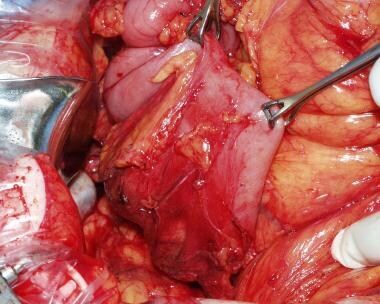
Pancreaticoduodenectomy
Fallopian Tubes
Cholangiopancreatography, Endoscopic Retrograde
Common Bile Duct Diseases
Carcinoma, Signet Ring Cell
Anus, Imperforate
Jaundice
Biliary Tract Neoplasms
Duodenum
Sperm Transport
Adenoma, Villous
Pancreatic Ducts
Krukenberg Tumor
Gallstones
Carcinoid Tumor
Common Bile Duct
Carcinoma, Neuroendocrine
Duodenal Diseases
Ovum Transport
Keratin-7
Neoplasms, Multiple Primary
Hair Cells, Ampulla
Endolymph
Semicircular Canals
Pancreatic Neoplasms
Inhibitory innervation of cat sphincter of Oddi. (1/430)
1 Electrical stimulation with trains of 0.1-0.2 ms pulses of the cat isolated sphincter of Oddi inhibited the spontaneous contractile activity and lowered base-line tension considerably. A contraction usually followed the period of stimulation. 2 These inhibitory effects were prevented by tetrodotoxin 0.1-0.5 mug/ml but were not reduced by hexamethonilm, morphine, or blockade of alpha- or beta-adrenoreceptors of cholinoceptors with phenoxy-benzamine propranolol or atropine, respectively. 3 Adenosine-5'-triphosphate (ATP) and adenosine-5'-diphosphate (ADP) inhibited the spontaneous sphincter activity and caused relaxation thus mimicking the effects of the C-terminal octapeptide of cholecystokinin (C8-CCK), isoprenaline and prostaglandin E1 and E2. 4 ATP alone (greater than 100 mug/ml) or ATP (greater than 10 mug/ml) plus dipyridamole (1 mug/ml), relaxed the sphincter to the same degrees as did the field stimulation. 5 In sphincter maximally contracted by acetylcholine, the effect of stimulation was more marked than that recorded in uncontracted preparations. 6 The present findings suggest that the sphincter of Oddi receives inhibitory nerves that are neither cholinergic nor adrenergic. (+info)Reexploration for periampullary carcinoma: resectability, perioperative results, pathology, and long-term outcome. (2/430)
OBJECTIVE: This single-institution experience retrospectively reviews the outcomes of patients undergoing reexploration for periampullary carcinoma at a high-volume center. SUMMARY BACKGROUND DATA: Many patients are referred to tertiary centers with periampullary carcinoma after their tumors were deemed unresectable at previous laparotomy. In carefully selected patients, tumor resection is often possible; however, the perioperative results and long-term outcome have not been well defined. METHODS: From November 1991 through December 1997, 78 patients who underwent previous exploratory laparotomy and/or palliative surgery for suspected periampullary carcinoma underwent reexploration. The operative outcome, resectability rate, pathology, and long-term survival rate were compared with 690 concurrent patients who had not undergone previous exploratory surgery. RESULTS: Fifty-two of the 78 patients (67%) undergoing reexploration underwent successful resection by pancreaticoduodenectomy; the remaining 26 patients (34%) were deemed to have unresectable disease. Compared with the 690 patients who had not undergone recent related surgery, the patients in the reoperative group were similar with respect to gender, race, and resectability rate but were significantly younger. The distribution of periampullary cancers by site in the reoperative group undergoing pancreaticoduodenectomy (n = 52) was 60%, 19%, 15%, and 6% for pancreatic, ampullary, distal bile duct, and duodenal tumors, respectively. These figures were similar to the 65%, 14%, 16% and 5% for resectable periampullary cancers found in the primary surgery group (n = 460). Intraoperative blood loss and transfusion requirements did not differ between the two groups. However, the mean operative time was 7.4 hours in the reoperative group, significantly longer than in the control group. On pathologic examination, reoperative patients had smaller tumors, and the percentage of patients with positive lymph nodes in the resection specimen was significantly less. The incidence of positive margins was similar between the two groups. Postoperative lengths of stay, complication rates, and perioperative mortality rates were not higher in reoperative patients. The long-term survival rate was similar between the two resected groups, with a median survival of 24 months in the reoperative group and 20 months in those without previous exploration. CONCLUSIONS: These data demonstrate that patients undergoing reoperation for periampullary carcinoma have similar resectability, perioperative morbidity and mortality, and long-term survival rates as patients undergoing initial exploration. The results suggest that selected patients considered to have unresectable disease at previous surgery should undergo restaging and reexploration at specialized high-volume centers. (+info)Prognostic value of MIB-1 index and DNA ploidy in resectable ampulla of Vater carcinoma. (3/430)
OBJECTIVE: To evaluate the prognostic value of the proliferative factors, MIB-1 index, DNA ploidy, and S-phase fraction, and further to determine the independent prognostic factors in ampulla of Vater carcinoma after pancreaticoduodenectomy. SUMMARY BACKGROUND DATA: Cell kinetics are important indicators of the biologic behavior of various human tumors, but only a few authors have reported the application of cell proliferative factors in ampulla of Vater carcinoma. METHODS: Patients undergoing pancreaticoduodenectomy for ampulla of Vater carcinoma were included. Proliferative factors, MIB-1 index, and DNA contents, measured by flow cytometry, were evaluated and compared with the conventional clinicopathologic factors. RESULTS: Ninety resectable ampulla of Vater carcinomas were included. By univariate analysis, MIB-1 index, DNA ploidy, S-phase fraction, stage, and lymph node status were significant prognostic factors. The 5-year survival rate was 40.7% for tumors with MIB-1 index < or =15% and 0% for those with MIB-1 index >15%. Diploid tumors had a significantly better prognosis than aneuploid. Outcomes of stage I and II tumors were more favorable than those of stage III and IV. After multivariate analysis, MIB-1 index, DNA ploidy, and stage remained as the independent prognostic factors. Among the three independent prognostic factors, MIB-1 index was the most powerful. CONCLUSIONS: Both MIB-1 index and DNA ploidy provide important prognostic value and potentially complement the conventional prognostic factors in resectable ampulla of Vater carcinoma. MIB-1 index is the most powerful independent prognostic factor. (+info)Pancreaticoduodenectomy with or without extended retroperitoneal lymphadenectomy for periampullary adenocarcinoma: comparison of morbidity and mortality and short-term outcome. (4/430)
OBJECTIVE: This prospective, randomized, single-institution trial was designed to evaluate the end points of mortality, morbidity, and survival in patients undergoing standard versus radical (extended) pancreaticoduodenectomy (including distal gastrectomy and retroperitoneal lymphadenectomy). SUMMARY BACKGROUND DATA: Numerous retrospective reports and one prospective randomized trial have suggested that the performance of an extended lymphadenectomy in association with a pancreaticoduodenal resection may improve long-term survival for some patients with pancreatic and other periampullary adenocarcinomas. Many of these previously published studies can be criticized for their retrospective and nonrandomized designs, for the inclusion of nonconcurrent control groups, and for their small numbers. METHODS: Between April 1996 and December 1997, 114 patients with periampullary adenocarcinoma were enrolled in an ongoing, prospective, randomized trial at The Johns Hopkins Hospital. After intraoperative verification of completely resected periampullary adenocarcinoma, the patients were randomized to receive either a standard pancreaticoduodenectomy (removing only the peripancreatic lymph nodes en bloc with the specimen) or a radical pancreaticoduodenectomy (standard resection plus distal gastrectomy and retroperitoneal lymphadenectomy). All pathology specimens were reviewed and categorized. The postoperative morbidity, mortality, and short-term outcomes were examined. RESULTS: Of the 114 patients randomized, 56 underwent a standard pancreaticoduodenectomy and 58 a radical pancreaticoduodenectomy. The two groups were statistically similar with regard to age and gender, but there was a higher percentage of white patients in the radical group. All the patients in the radical group underwent distal gastric resection, whereas 86% of the patients in the standard group underwent pylorus preservation. The mean operative time in the radical group was 6.8 hours, compared with 6.2 hours in the standard group. There were no significant differences between the two groups with respect to the intraoperative blood loss, transfusion requirements, location of primary tumor, mean tumor size, positive lymph node status, or positive margin status. There were three deaths in the standard group and two in the radical group. The complication rates were 34% for the standard group and 40% for the radical group. Patients undergoing radical resection had a higher incidence of early delayed gastric emptying but had similar rates of other complications, such as pancreatic fistula, wound infection, intraabdominal abscess, and need for reoperation. The mean total number of lymph nodes resected was higher in the radical group. Of the 58 patients in the radical group, only 10% had metastatic carcinoma in the resected retroperitoneal lymph nodes, and none of those patients had the retroperitoneal nodes as the only site of lymph node involvement. The 1-year actuarial survival rate for patients surviving the immediate postoperative periods was 77% for the standard resection group and 83% for the radical resection group. CONCLUSIONS: These data demonstrate that radical pancreaticoduodenectomy (with the addition of a distal gastrectomy and extended retroperitoneal lymphadenectomy to a standard pancreaticoduodenectomy) can be performed with similar morbidity and mortality to standard pancreaticoduodenectomy. However, the survival data are not sufficiently mature and the numbers of patients enrolled are not adequate to allow firm conclusions to be drawn regarding survival benefit. (+info)Bilateral ovarian carcinoma metastatic from the ampulla of Vater: a rare Krukenberg tumor. (5/430)
Carcinoma of the ampulla of Vater is a relatively rare neoplasm and its longterm survival rate is considerably high. However, because of differences in tumor pathologic features and local invasiveness, a 5-year survival rate differ widely. We present a case of metastatic carcinoma of the ampulla of Vater presenting as a Krukenberg tumor in a 59-year-old woman. Eight months earlier, she had been diagnosed as well-differentiated adenocarcinoma of the ampulla of Vater. Abdominal examination revealed a hard mass with mild tenderness in the RLQ area. The laboratory findings were unremarkable except for mild anemia. CT scan of the abdomen revealed enlargement of both ovaries. An exploratory laparotomy disclosed bilateral ovarian masses, 18 x 12 x 8 cm and 8 x 5.5 x 4 cm in size, respectively. Histologic findings of the both ovarian masses were consistent with metastatic adenocarcinoma from the ampulla of Vater. (+info)Obstructive jaundice and acute cholangitis due to papillary stenosis. (6/430)
Papillary stenosis is characterized by fixed fibrosis leading to structural outflow obstruction and it is usually secondary to inflammation and fibrosis from the chronic passage of gallstones, episodes of acute pancreatitis, chronic pancreatitis, sclerosing cholangitis, peptic ulcer disease, and cholesterolosis. However, obstructive jaundice with or without acute cholangitis which leads the physician to suspect the presence of malignancy as a cause is a rare manifestation of papillary stenosis. We report here a case of papillary stenosis presenting with obstructive jaundice and acute cholangitis. The lesion was so difficult to exclude the presence of malignancy preoperatively and intraoperatively that a pylorus-preserving pancreaticoduodenectomy was performed. Histologic examination of the resected specimen revealed fibrosis, adenomatoid ductal hyperplasia, and mild chronic inflammation of the papilla of Vater and distal common bile duct. (+info)Adenoma of the ampulla of Vater: a genetic condition? (7/430)
The etiology of adenoma of the ampulla of Vater is not well understood. Previous authors reported the association of this neoplasm with polycystic kidney disease of two fraternal sisters. They concluded that these two conditions were somehow related. We describe a case of ampullary adenoma associated with polycystic kidney disease. This presentation raises again the question of a possible link between these two diseases. (+info)Risk of pancreatic and periampullary cancer following cholecystectomy. (8/430)
AIMS: Cholecystectomy has been reported by several investigators to increase the risk of pancreatic cancer. The trophic effect on the gland by increased release of cholecystokinin following cholecystectomy and perturbation of the neurohormonal pancreatic regulation, have been suggested as possible contributing factors. However, while several investigators found that this surgical procedure increases the risk, others have not. In an attempt to clarify whether or not patients who have undergone cholecystectomy are at increased risk for developing pancreatic cancer, we evaluated the frequency with which cholelithiasis and this surgical procedure were present in a large number of patients with pancreatic cancer. METHODS: A total of 720 patients with newly diagnosed pancreatic cancer (415 men and 305 women; mean age 62.6 years, range 22 to 79 years) and 720 controls matched for sex, age, social class, and geographic region, were enrolled in the study. All subjects were interviewed personally and in detail about their clinical history. RESULTS: Cholelithiasis was present in 126 patients with pancreatic cancer (17.5%) and in 95 controls (13.2%), constituting a statistically significant association (odds ratio, 1.39; 95% confidence interval, 1.04 to 1.86). However, considering only the patients and controls in whom the diagnosis of cholelithiasis was made for more than one year before cancer diagnosis or interview, the association was no longer significant (odds ratio, 1.04; 95% confidence interval, 0.75 to 1.44). Cholecystectomy had been performed in 93 patients with pancreatic cancer (12.9%) and in 71 controls (9.9%). When all subjects were considered, the odds ratio was mildly, although not significantly, increased (odds ratio, 1.35; 95% confidence interval, 0.97 to 1.87). When only subjects who underwent cholecystectomy one year or more before the cancer diagnosis or interview were considered, the odds ratio fell to unity (odds ratio, 1.00; 95% confidence interval, 0.70 to 1.43). CONCLUSION: This study, one of the largest on this topic, clearly shows that there is no evidence for an association between cholelithiasis, cholecystectomy, and pancreatic cancer. (+info)The ampulla of Vater, also known as hepatopancreatic ampulla, is a dilated portion of the common bile duct where it joins the main pancreatic duct and empties into the second part of the duodenum. It serves as a conduit for both bile from the liver and digestive enzymes from the pancreas to reach the small intestine, facilitating the digestion and absorption of nutrients. The ampulla of Vater is surrounded by a muscular sphincter, the sphincter of Oddi, which controls the flow of these secretions into the duodenum.
Common bile duct neoplasms refer to abnormal growths that can occur in the common bile duct, which is a tube that carries bile from the liver and gallbladder into the small intestine. These growths can be benign or malignant (cancerous).
Benign neoplasms of the common bile duct include papillomas, adenomas, and leiomyomas. Malignant neoplasms are typically adenocarcinomas, which arise from the glandular cells lining the duct. Other types of malignancies that can affect the common bile duct include cholangiocarcinoma, gallbladder carcinoma, and metastatic cancer from other sites.
Symptoms of common bile duct neoplasms may include jaundice (yellowing of the skin and eyes), abdominal pain, dark urine, and light-colored stools. Diagnosis may involve imaging tests such as CT scans or MRCP (magnetic resonance cholangiopancreatography) and biopsy to confirm the type of neoplasm. Treatment options depend on the type and stage of the neoplasm and may include surgery, radiation therapy, chemotherapy, or a combination of these approaches.
Pancreaticoduodenectomy, also known as the Whipple procedure, is a complex surgical operation that involves the removal of the head of the pancreas, the duodenum (the first part of the small intestine), the gallbladder, and the distal common bile duct. In some cases, a portion of the stomach may also be removed. The remaining parts of the pancreas, bile duct, and intestines are then reconnected to allow for the digestion of food and drainage of bile.
This procedure is typically performed as a treatment for various conditions affecting the pancreas, such as tumors (including pancreatic cancer), chronic pancreatitis, or traumatic injuries. It is a major surgical operation that requires significant expertise and experience to perform safely and effectively.
The Fallopian tubes, also known as uterine tubes or oviducts, are a pair of slender tubular structures in the female reproductive system. They play a crucial role in human reproduction by providing a passageway for the egg (ovum) from the ovary to the uterus (womb).
Each Fallopian tube is typically around 7.6 to 10 centimeters long and consists of four parts: the interstitial part, the isthmus, the ampulla, and the infundibulum. The fimbriated end of the infundibulum, which resembles a fringe or frill, surrounds and captures the released egg from the ovary during ovulation.
Fertilization usually occurs in the ampulla when sperm meets the egg after sexual intercourse. Once fertilized, the zygote (fertilized egg) travels through the Fallopian tube toward the uterus for implantation and further development. The cilia lining the inner surface of the Fallopian tubes help propel the egg and the zygote along their journey.
In some cases, abnormalities or blockages in the Fallopian tubes can lead to infertility or ectopic pregnancies, which are pregnancies that develop outside the uterus, typically within the Fallopian tube itself.
Duodenal neoplasms refer to abnormal growths in the duodenum, which is the first part of the small intestine that receives digestive secretions from the pancreas and bile duct. These growths can be benign or malignant (cancerous).
Benign neoplasms include adenomas, leiomyomas, lipomas, and hamartomas. They are usually slow-growing and do not spread to other parts of the body. However, they may cause symptoms such as abdominal pain, bleeding, or obstruction of the intestine.
Malignant neoplasms include adenocarcinomas, neuroendocrine tumors (carcinoids), lymphomas, and sarcomas. They are more aggressive and can invade surrounding tissues and spread to other parts of the body. Symptoms may include abdominal pain, weight loss, jaundice, anemia, or bowel obstruction.
The diagnosis of duodenal neoplasms is usually made through imaging tests such as CT scans, MRI, or endoscopy with biopsy. Treatment depends on the type and stage of the tumor and may include surgery, chemotherapy, radiation therapy, or a combination of these modalities.
Duodenoscopy is a medical procedure that involves the insertion of a duodenoscope, which is a flexible, lighted tube with a camera and tiny tools on the end, through the mouth and down the throat to examine the upper part of the small intestine (duodenum) and the opening of the bile and pancreatic ducts.
During the procedure, the doctor can take tissue samples for biopsy, remove polyps or other abnormal growths, or perform other interventions as needed. Duodenoscopy is commonly used to diagnose and treat conditions such as gastrointestinal bleeding, inflammation, infection, and cancer.
It's important to note that duodenoscopes have been associated with the spread of antibiotic-resistant bacteria in some cases, so healthcare providers must follow strict cleaning and disinfection protocols to minimize this risk.
Endoscopic retrograde cholangiopancreatography (ERCP) is a medical procedure that combines upper gastrointestinal (GI) endoscopy and fluoroscopy to diagnose and treat certain problems of the bile ducts and pancreas.
During ERCP, a flexible endoscope (a long, thin, lighted tube with a camera on the end) is passed through the patient's mouth and throat, then through the stomach and into the first part of the small intestine (duodenum). A narrow plastic tube (catheter) is then inserted through the endoscope and into the bile ducts and/or pancreatic duct. Contrast dye is injected through the catheter, and X-rays are taken to visualize the ducts.
ERCP can be used to diagnose a variety of conditions affecting the bile ducts and pancreas, including gallstones, tumors, strictures (narrowing of the ducts), and chronic pancreatitis. It can also be used to treat certain conditions, such as removing gallstones from the bile duct or placing stents to keep the ducts open in cases of stricture.
ERCP is an invasive procedure that carries a risk of complications, including pancreatitis, infection, bleeding, and perforation (a tear in the lining of the GI tract). It should only be performed by experienced medical professionals in a hospital setting.
A duodenoscope is a type of endoscope that is used for performing minimally invasive diagnostic and therapeutic procedures in the gastrointestinal tract, specifically in the duodenum, which is the first part of the small intestine. The duodenoscope is a flexible tube with a camera and a light at its tip, allowing physicians to visualize the inside of the duodenum and surrounding organs. It also has channels that can deliver therapies or enable the removal of tissue samples for biopsy. Duodenoscopes are commonly used in procedures such as endoscopic retrograde cholangiopancreatography (ERCP), which involves the examination and treatment of the bile and pancreatic ducts.
Common bile duct diseases refer to conditions that affect the common bile duct, a tube that carries bile from the liver and gallbladder into the small intestine. Some common examples of common bile duct diseases include:
1. Choledocholithiasis: This is the presence of stones (calculi) in the common bile duct, which can cause blockage, inflammation, and infection.
2. Cholangitis: This is an infection or inflammation of the common bile duct, often caused by obstruction due to stones, tumors, or strictures.
3. Common bile duct cancer (cholangiocarcinoma): This is a rare but aggressive cancer that arises from the cells lining the common bile duct.
4. Biliary strictures: These are narrowing or scarring of the common bile duct, which can be caused by injury, inflammation, or surgery.
5. Benign tumors: Non-cancerous growths in the common bile duct can also cause blockage and other symptoms.
Symptoms of common bile duct diseases may include abdominal pain, jaundice (yellowing of the skin and eyes), fever, chills, nausea, vomiting, and dark urine or light-colored stools. Treatment depends on the specific condition and severity but may include medications, endoscopic procedures, surgery, or a combination of these approaches.
Carcinoma, signet ring cell is a type of adenocarcinoma, which is a cancer that begins in glandular cells. In signet ring cell carcinoma, the cancer cells have a characteristic appearance when viewed under a microscope. They contain large amounts of mucin, a substance that causes the nucleus of the cell to be pushed to one side, giving the cell a crescent or "signet ring" shape.
Signet ring cell carcinoma can occur in various organs, including the stomach, colon, rectum, and breast. It is often aggressive and has a poor prognosis, as it tends to grow and spread quickly. Treatment options may include surgery, chemotherapy, and radiation therapy, depending on the location and extent of the cancer.
Imperforate anus is a congenital condition in which the opening of the anus is absent or abnormally closed or narrowed, preventing the normal passage of stool. This results in a blockage in the digestive tract and can lead to serious health complications if not treated promptly.
The anus is the external opening of the rectum, which is the lower end of the digestive tract. During fetal development, the rectum and anus normally connect through a canal called the anal canal or the recto-anal canal. In imperforate anus, this canal may be completely closed or narrowed, or it may not form properly.
Imperforate anus can occur as an isolated condition or as part of a genetic syndrome or other congenital abnormalities. The exact cause is not fully understood, but it is believed to result from a combination of genetic and environmental factors.
Treatment for imperforate anus typically involves surgery to create an opening in the anus and restore normal bowel function. In some cases, additional procedures may be necessary to correct related abnormalities or complications. The prognosis for individuals with imperforate anus depends on the severity of the condition and any associated abnormalities. With prompt and appropriate treatment, most people with imperforate anus can lead normal lives.
Bile duct neoplasms, also known as cholangiocarcinomas, refer to a group of malignancies that arise from the bile ducts. These are the tubes that carry bile from the liver to the gallbladder and small intestine. Bile duct neoplasms can be further classified based on their location as intrahepatic (within the liver), perihilar (at the junction of the left and right hepatic ducts), or distal (in the common bile duct).
These tumors are relatively rare, but their incidence has been increasing in recent years. They can cause a variety of symptoms, including jaundice, abdominal pain, weight loss, and fever. The diagnosis of bile duct neoplasms typically involves imaging studies such as CT or MRI scans, as well as blood tests to assess liver function. In some cases, a biopsy may be necessary to confirm the diagnosis.
Treatment options for bile duct neoplasms depend on several factors, including the location and stage of the tumor, as well as the patient's overall health. Surgical resection is the preferred treatment for early-stage tumors, while chemotherapy and radiation therapy may be used in more advanced cases. For patients who are not candidates for surgery, palliative treatments such as stenting or bypass procedures may be recommended to relieve symptoms and improve quality of life.
Jaundice is a medical condition characterized by the yellowing of the skin, sclera (whites of the eyes), and mucous membranes due to an excess of bilirubin in the bloodstream. Bilirubin is a yellow-orange pigment produced when hemoglobin from red blood cells is broken down. Normally, bilirubin is processed by the liver and excreted through bile into the digestive system. However, if there's an issue with bilirubin metabolism or elimination, it can accumulate in the body, leading to jaundice.
Jaundice can be a symptom of various underlying conditions, such as liver diseases (hepatitis, cirrhosis), gallbladder issues (gallstones, tumors), or blood disorders (hemolysis). It is essential to consult a healthcare professional if jaundice is observed, as it may indicate a severe health problem requiring prompt medical attention.
Biliary tract neoplasms refer to abnormal growths or tumors that develop in the biliary system, which includes the gallbladder, bile ducts inside and outside the liver, and the ducts that connect the liver to the small intestine. These neoplasms can be benign (non-cancerous) or malignant (cancerous).
Malignant biliary tract neoplasms are often referred to as cholangiocarcinoma if they originate in the bile ducts, or gallbladder cancer if they arise in the gallbladder. These cancers are relatively rare but can be aggressive and difficult to treat. They can cause symptoms such as jaundice (yellowing of the skin and eyes), abdominal pain, weight loss, and dark urine.
Risk factors for biliary tract neoplasms include chronic inflammation of the biliary system, primary sclerosing cholangitis, liver cirrhosis, hepatitis B or C infection, parasitic infections, and certain genetic conditions. Early detection and treatment can improve outcomes for patients with these neoplasms.
The duodenum is the first part of the small intestine, immediately following the stomach. It is a C-shaped structure that is about 10-12 inches long and is responsible for continuing the digestion process that begins in the stomach. The duodenum receives partially digested food from the stomach through the pyloric valve and mixes it with digestive enzymes and bile produced by the pancreas and liver, respectively. These enzymes help break down proteins, fats, and carbohydrates into smaller molecules, allowing for efficient absorption in the remaining sections of the small intestine.
Sperm transport refers to the series of events that occur from the production of sperm in the testes to their release into the female reproductive tract during sexual intercourse. This process involves several stages:
1. Spermatogenesis: The production of sperm cells (spermatozoa) takes place in the seminiferous tubules within the testes.
2. Maturation: The newly produced sperm are immature and incapable of fertilization. They undergo a maturation process as they move through the epididymis, where they acquire motility and the ability to fertilize an egg.
3. Ejaculation: During sexual arousal, sperm are mixed with seminal fluid produced by the seminal vesicles, prostate gland, and bulbourethral glands to form semen. This mixture is propelled through the urethra during orgasm (ejaculation) and released from the penis into the female reproductive tract.
4. Transport within the female reproductive tract: Once inside the female reproductive tract, sperm must travel through the cervix, uterus, and fallopian tubes to reach the site of fertilization, the ampullary-isthmic junction of the fallopian tube. This journey can take several hours to a few days.
5. Capacitation: During their transport within the female reproductive tract, sperm undergo further changes called capacitation, which prepares them for fertilization by increasing their motility and making them more responsive to the egg's chemical signals.
6. Acrosome reaction: The final step in sperm transport is the acrosome reaction, where the sperm releases enzymes from the acrosome (a cap-like structure on the head of the sperm) to penetrate and fertilize the egg.
A villous adenoma is a type of polyp (a growth that protrudes from the lining of an organ) found in the colon or rectum. It is named for its appearance under a microscope, which reveals finger-like projections called "villi" on the surface of the polyp.
Villous adenomas are typically larger than other types of polyps and can be several centimeters in size. They are also more likely to be cancerous or precancerous, meaning that they have the potential to develop into colon or rectal cancer over time.
Because of this increased risk, it is important for villous adenomas to be removed surgically if they are found during a colonoscopy or other diagnostic procedure. Regular follow-up colonoscopies may also be recommended to monitor for the development of new polyps or recurrence of previous ones.
The pancreatic ducts are a set of tubular structures within the pancreas that play a crucial role in the digestive system. The main pancreatic duct, also known as the duct of Wirsung, is responsible for transporting pancreatic enzymes and bicarbonate-rich fluid from the pancreas to the duodenum, which is the first part of the small intestine.
The exocrine portion of the pancreas contains numerous smaller ducts called interlobular ducts and intralobular ducts that merge and ultimately join the main pancreatic duct. This system ensures that the digestive enzymes and fluids produced by the pancreas are effectively delivered to the small intestine, where they aid in the breakdown and absorption of nutrients from food.
In addition to the main pancreatic duct, there is an accessory pancreatic duct, also known as Santorini's duct, which can sometimes join the common bile duct before emptying into the duodenum through a shared opening called the ampulla of Vater. However, in most individuals, the accessory pancreatic duct usually drains into the main pancreatic duct before entering the duodenum.
A Krukenberg tumor is a type of metastatic cancer that primarily originates from the stomach (70% of cases) but can also arise from other organs such as the colon, ovary, or breast. It is characterized by the presence of signet-ring cells, which are a specific type of malignant cell with abundant mucin displacing the nucleus to the periphery.
Krukenberg tumors typically involve both ovaries and often present with bilateral ovarian enlargement. They can cause various symptoms such as abdominal pain, bloating, or irregular menstruation. The prognosis for patients with Krukenberg tumors is generally poor due to the advanced stage of the disease at diagnosis.
Gallstones are small, hard deposits that form in the gallbladder, a small organ located under the liver. They can range in size from as small as a grain of sand to as large as a golf ball. Gallstones can be made of cholesterol, bile pigments, or calcium salts, or a combination of these substances.
There are two main types of gallstones: cholesterol stones and pigment stones. Cholesterol stones are the most common type and are usually yellow-green in color. They form when there is too much cholesterol in the bile, which causes it to become saturated and form crystals that eventually grow into stones. Pigment stones are smaller and darker in color, ranging from brown to black. They form when there is an excess of bilirubin, a waste product produced by the breakdown of red blood cells, in the bile.
Gallstones can cause symptoms such as abdominal pain, nausea, vomiting, and bloating, especially after eating fatty foods. In some cases, gallstones can lead to serious complications, such as inflammation of the gallbladder (cholecystitis), infection, or blockage of the bile ducts, which can cause jaundice, a yellowing of the skin and eyes.
The exact cause of gallstones is not fully understood, but risk factors include being female, older age, obesity, a family history of gallstones, rapid weight loss, diabetes, and certain medical conditions such as cirrhosis or sickle cell anemia. Treatment for gallstones may involve medication to dissolve the stones, shock wave therapy to break them up, or surgery to remove the gallbladder.
Adenocarcinoma is a type of cancer that arises from glandular epithelial cells. These cells line the inside of many internal organs, including the breasts, prostate, colon, and lungs. Adenocarcinomas can occur in any of these organs, as well as in other locations where glands are present.
The term "adenocarcinoma" is used to describe a cancer that has features of glandular tissue, such as mucus-secreting cells or cells that produce hormones. These cancers often form glandular structures within the tumor mass and may produce mucus or other substances.
Adenocarcinomas are typically slow-growing and tend to spread (metastasize) to other parts of the body through the lymphatic system or bloodstream. They can be treated with surgery, radiation therapy, chemotherapy, targeted therapy, or a combination of these treatments. The prognosis for adenocarcinoma depends on several factors, including the location and stage of the cancer, as well as the patient's overall health and age.
A carcinoid tumor is a type of slow-growing neuroendocrine tumor that usually originates in the digestive tract, particularly in the small intestine. These tumors can also arise in other areas such as the lungs, appendix, and rarely in other organs. Carcinoid tumors develop from cells of the diffuse endocrine system (also known as the neuroendocrine system) that are capable of producing hormones or biologically active amines.
Carcinoid tumors can produce and release various hormones and bioactive substances, such as serotonin, histamine, bradykinins, prostaglandins, and tachykinins, which can lead to a variety of symptoms. The most common syndrome associated with carcinoid tumors is the carcinoid syndrome, characterized by flushing, diarrhea, abdominal cramping, and wheezing or difficulty breathing.
Carcinoid tumors are typically classified as functional or nonfunctional based on whether they produce and secrete hormones that cause symptoms. Functional carcinoid tumors account for approximately 30% of cases and can lead to the development of carcinoid syndrome, while nonfunctional tumors do not produce significant amounts of hormones and are often asymptomatic until they grow large enough to cause local or distant complications.
Treatment options for carcinoid tumors depend on the location, size, and extent of the tumor, as well as whether it is functional or nonfunctional. Treatment may include surgery, medications (such as somatostatin analogs, chemotherapy, or targeted therapies), and radiation therapy. Regular follow-up with imaging studies and biochemical tests is essential to monitor for recurrence and assess treatment response.
The common bile duct is a duct that results from the union of the cystic duct (which drains bile from the gallbladder) and the common hepatic duct (which drains bile from the liver). The common bile duct transports bile, a digestive enzyme, from the liver and gallbladder to the duodenum, which is the first part of the small intestine.
The common bile duct runs through the head of the pancreas before emptying into the second part of the duodenum, either alone or in conjunction with the pancreatic duct, via a small opening called the ampulla of Vater. The common bile duct plays a crucial role in the digestion of fats by helping to break them down into smaller molecules that can be absorbed by the body.
Carcinoma, neuroendocrine is a type of cancer that arises from the neuroendocrine cells, which are specialized cells that have both nerve and hormone-producing functions. These cells are found throughout the body, but neuroendocrine tumors (NETs) most commonly occur in the lungs, gastrointestinal tract, pancreas, and thyroid gland.
Neuroendocrine carcinomas can be classified as well-differentiated or poorly differentiated based on how closely they resemble normal neuroendocrine cells under a microscope. Well-differentiated tumors tend to grow more slowly and are less aggressive than poorly differentiated tumors.
Neuroendocrine carcinomas can produce and release hormones and other substances that can cause a variety of symptoms, such as flushing, diarrhea, wheezing, and heart palpitations. Treatment for neuroendocrine carcinoma depends on the location and extent of the tumor, as well as the patient's overall health. Treatment options may include surgery, radiation therapy, chemotherapy, targeted therapy, or a combination of these approaches.
Duodenal diseases refer to a range of medical conditions that affect the duodenum, which is the first part of the small intestine. Here are some examples of duodenal diseases:
1. Duodenitis: This is inflammation of the duodenum, which can cause symptoms such as abdominal pain, nausea, vomiting, and bloating. Duodenitis can be caused by bacterial or viral infections, excessive use of nonsteroidal anti-inflammatory drugs (NSAIDs), or chronic inflammation due to conditions like Crohn's disease.
2. Peptic ulcers: These are sores that develop in the lining of the duodenum, usually as a result of infection with Helicobacter pylori bacteria or long-term use of NSAIDs. Symptoms can include abdominal pain, bloating, and heartburn.
3. Duodenal cancer: This is a rare type of cancer that affects the duodenum. Symptoms can include abdominal pain, weight loss, and blood in the stool.
4. Celiac disease: This is an autoimmune disorder that causes the immune system to attack the lining of the small intestine in response to gluten, a protein found in wheat, barley, and rye. This can lead to inflammation and damage to the duodenum.
5. Duodenal diverticulosis: This is a condition in which small pouches form in the lining of the duodenum. While many people with duodenal diverticulosis do not experience symptoms, some may develop complications such as inflammation or infection.
6. Duodenal atresia: This is a congenital condition in which the duodenum does not form properly, leading to blockage of the intestine. This can cause symptoms such as vomiting and difficulty feeding in newborns.
'Ovum transport' refers to the movement of an egg or ovum from the mature follicle within the ovary, through the fallopian tube, and ultimately to the uterus. This process is a critical part of the female reproductive system and occurs during each menstrual cycle.
The ovulation phase of the menstrual cycle triggers the release of a mature egg from the follicle in the ovary. The fimbriated end of the fallopian tube captures the egg and transports it into the tube, where it may encounter sperm for fertilization. Cilia lining the inside of the fallopian tubes create wave-like motions that help propel the egg towards the uterus.
If fertilization occurs, the resulting zygote will continue to travel down the fallopian tube and implant itself into the uterine lining, initiating pregnancy. If fertilization does not occur, the egg will be shed along with the uterine lining during menstruation.
Keratin-7 is not a medical term itself, but it is a specific type of keratin protein that is often used in pathology as a marker for certain types of carcinomas. Keratins are a family of fibrous proteins that make up the structural framework of epithelial cells, which line the surfaces and glands of the body.
Keratin-7 is typically expressed in simple epithelia, such as those found in the gastrointestinal tract, pancreas, bile ducts, and respiratory and genitourinary tracts. It can be used as a marker to help identify carcinomas that arise from these tissues, such as adenocarcinomas of the pancreas or biliary system.
In medical terminology, keratin-7 positivity is often reported in the pathology report of a biopsy or surgical specimen to indicate the presence of this protein in cancer cells. This information can be helpful in determining the origin and behavior of the tumor, as well as guiding treatment decisions.
Multiple primary neoplasms refer to the occurrence of more than one primary malignant tumor in an individual, where each tumor is unrelated to the other and originates from separate cells or organs. This differs from metastatic cancer, where a single malignancy spreads to multiple sites in the body. Multiple primary neoplasms can be synchronous (occurring at the same time) or metachronous (occurring at different times). The risk of developing multiple primary neoplasms increases with age and is associated with certain genetic predispositions, environmental factors, and lifestyle choices such as smoking and alcohol consumption.
Biliary tract surgical procedures refer to a range of operations that involve the biliary system, which includes the liver, gallbladder, and bile ducts. These procedures can be performed for various reasons, including the treatment of gallstones, bile duct injuries, tumors, or other conditions affecting the biliary tract. Here are some examples of biliary tract surgical procedures:
1. Cholecystectomy: This is the surgical removal of the gallbladder, which is often performed to treat symptomatic gallstones or chronic cholecystitis (inflammation of the gallbladder). It can be done as an open procedure or laparoscopically.
2. Bile duct exploration: This procedure involves opening the common bile duct to remove stones, strictures, or tumors. It is often performed during a cholecystectomy if there is suspicion of common bile duct involvement.
3. Hepaticojejunostomy: This operation connects the liver's bile ducts directly to a portion of the small intestine called the jejunum, bypassing a damaged or obstructed segment of the biliary tract. It is often performed for benign or malignant conditions affecting the bile ducts.
4. Roux-en-Y hepaticojejunostomy: This procedure involves creating a Y-shaped limb of jejunum and connecting it to the liver's bile ducts, bypassing the common bile duct and duodenum. It is often performed for complex biliary tract injuries or malignancies.
5. Whipple procedure (pancreaticoduodenectomy): This extensive operation involves removing the head of the pancreas, the duodenum, a portion of the jejunum, the gallbladder, and the common bile duct. It is performed for malignancies involving the pancreas, bile duct, or duodenum.
6. Liver resection: This procedure involves removing a portion of the liver to treat primary liver tumors (hepatocellular carcinoma or cholangiocarcinoma) or metastatic cancer from other organs.
7. Biliary stenting or bypass: These minimally invasive procedures involve placing a stent or creating a bypass to relieve bile duct obstructions caused by tumors, strictures, or stones. They can be performed endoscopically (ERCP) or percutaneously (PTC).
8. Cholecystectomy: This procedure involves removing the gallbladder, often for symptomatic cholelithiasis (gallstones) or cholecystitis (inflammation of the gallbladder). It can be performed laparoscopically or open.
9. Biliary drainage: This procedure involves placing a catheter to drain bile from the liver or bile ducts, often for acute or chronic obstructions caused by tumors, strictures, or stones. It can be performed endoscopically (ERCP) or percutaneously (PTC).
10. Bilioenteric anastomosis: This procedure involves connecting the biliary tract to a portion of the small intestine, often for benign or malignant conditions affecting the bile ducts or pancreas. It can be performed open or laparoscopically.
Hair cells in the ampulla are specialized sensory receptor cells located within the vestibular system of the inner ear. The vestibular system is responsible for detecting movement and maintaining balance. The ampulla is a part of one of the three semicircular canals, fluid-filled structures that sense rotational movements of the head.
Hair cells in the ampulla have hair-like projections called stereocilia on their surface, which are embedded in a gelatinous structure called the cupula. The movement of fluid within the semicircular canal causes the deflection of the stereocilia, leading to the activation of mechanically gated ion channels and generating receptor potentials. These electrical signals are then transmitted to the brain via the vestibular nerve, allowing the brain to interpret head movements and maintain balance.
Damage or loss of hair cells in the ampulla can lead to vestibular dysfunction and balance disorders.
Endolymph is a specific type of fluid that is found within the inner ear, more specifically in the membranous labyrinth of the inner ear. This fluid plays a crucial role in maintaining balance and hearing functions. It helps in the stimulation of hair cells present in the inner ear which then transmit signals to the brain, enabling us to hear and maintain our balance. Any disturbance or changes in the composition or flow of endolymph can lead to various vestibular disorders and hearing problems.
The semicircular canals are part of the vestibular system in the inner ear that contributes to the sense of balance and spatial orientation. They are composed of three fluid-filled tubes, each located in a different plane (anterior, posterior, and horizontal) and arranged at approximately right angles to each other. The semicircular canals detect rotational movements of the head, enabling us to maintain our equilibrium during movement.
When the head moves, the fluid within the semicircular canals moves in response to that motion. At the end of each canal is a structure called the ampulla, which contains hair cells with hair-like projections (stereocilia) embedded in a gelatinous substance. As the fluid moves, it bends the stereocilia, stimulating the hair cells and sending signals to the brain via the vestibular nerve. The brain then interprets these signals to determine the direction and speed of head movement, allowing us to maintain our balance and orientation in space.
Pancreatic neoplasms refer to abnormal growths in the pancreas that can be benign or malignant. The pancreas is a gland located behind the stomach that produces hormones and digestive enzymes. Pancreatic neoplasms can interfere with the normal functioning of the pancreas, leading to various health complications.
Benign pancreatic neoplasms are non-cancerous growths that do not spread to other parts of the body. They are usually removed through surgery to prevent any potential complications, such as blocking the bile duct or causing pain.
Malignant pancreatic neoplasms, also known as pancreatic cancer, are cancerous growths that can invade and destroy surrounding tissues and organs. They can also spread (metastasize) to other parts of the body, such as the liver, lungs, or bones. Pancreatic cancer is often aggressive and difficult to treat, with a poor prognosis.
There are several types of pancreatic neoplasms, including adenocarcinomas, neuroendocrine tumors, solid pseudopapillary neoplasms, and cystic neoplasms. The specific type of neoplasm is determined through various diagnostic tests, such as imaging studies, biopsies, and blood tests. Treatment options depend on the type, stage, and location of the neoplasm, as well as the patient's overall health and preferences.
 Ampulla of Vater
List of German inventors and discoverers
Biliary endoscopic sphincterotomy
Neuroendocrine tumor
Ascending cholangitis
Cholestasis
Pancreaticoduodenectomy
Apudoma
Sphincter of Oddi
Small intestine cancer
CD24
Cholangiocarcinoma
Bile duct
Diverticulum
Courvoisier's law
Common bile duct stone
Sphincter of Boyden
Wu Jin
Gallstone
Deaths in November 2021
Pancreas
Digestive enzyme
2021 in Taiwan
Human feces
Abraham Vater
Ascariasis
Liver
Duodenal atresia
Choledochoduodenostomy
Eugene Lindsay Opie
Ampulla of Vater - Wikipedia
Keywords hepatobiliary + adenocarcinoma + ampulla of vater | PEIR Digital Library
Keywords gross + hepatobiliary + liver + adenocarcinoma + ampulla of vater | PEIR Digital Library
Ampulla of Vater - wikidoc
 Ampulla of Vater | EOD Data SEER*RSA
 Ampulla of Vater - Atlas of Human Anatomy - Centralx
 MedPix Case - adenocarcinoma of the ampulla of Vater.
 Difference Between Sphincter of Oddi and Ampulla of Vater
Ampulla of Vater (excluding Neuroendocrine Tumor) | TNM Data SEER*RSA
 A Case of MALT-lymphoma of the Ampulla of Vater
 Ampullary Carcinoma: Practice Essentials, Background, Pathophysiology
 Cancer Protocols
Laparoscopic liver resection after laparoscopic pancreatoduodenectomy for liver metastases of ampulla of Vater adenocarcinoma |...
 Article - Billing and Coding: MolDX: Next-Generation Sequencing for Solid Tumors (A57831)
 High-Mobility Group Box 1 expression predicts survival of patients after resection of adenocarcinoma of the ampulla of Vater |...
A Case of Bleeding on the Ampulla of Vater Due to Angiodysplasia in a Patient with End Stage Renal Disease
Adenosquamous carcinoma of the ampulla of Vater - a rare disease at unusual location | World Journal of Surgical Oncology |...
 A Rare Case of Adenocarcinoma of Ampulla of Vater - A Case Report
| Asian Journal of Research and Reports in...
A Rare Case of Adenocarcinoma of Ampulla of Vater - A Case Report
| Asian Journal of Research and Reports in...
Saluran pankreas - Wikipedia bahasa Indonesia, ensiklopedia bebas
Internet Scientific Publications
Javier Herrera, MD | Edmonds, WA
 Special Exposure Cohort Employees (SEC) | U.S. Department of Labor
 Rare Cancer Support Forum • Members
Rare Cancer Support Forum • Members
 Edward C. McCarron, MD| Surgical Oncology | MedStar Health
 Histology-World! Histology Testbank
 Online ICD9/ICD9CM codes
 Role of Radiation Therapy in Patients With Resectable Pancreatic Cancer
Hepatopancreatic ampulla5
- The ampulla of Vater, hepatopancreatic ampulla or hepatopancreatic duct is the common duct that is usually formed by a union of the common bile duct and the pancreatic duct within the wall of the duodenum. (wikipedia.org)
- The ampulla of Vater , also known as the hepatopancreatic ampulla , is formed by the union of the pancreatic duct and the common bile duct . (wikidoc.org)
- One possible cause of impaired drainage is blockage of the hepatopancreatic ampulla. (wikidoc.org)
- A dilation of the duodenal papilla that is the opening of the juncture of the COMMON BILE DUCT and the MAIN PANCREATIC DUCT, also known as the hepatopancreatic ampulla. (centralx.com)
- It is also known as the Ampulla of Vater, also called the hepatopancreatic ampulla is a small bulbous structure situated near the junction between the common bile duct as well as the pancreatic drain within the duodenum. (keydifference.in)
Duodenum11
- The ampulla of Vater is an important landmark halfway along the second part of the duodenum marking the transition from foregut to midgut. (wikipedia.org)
- The sphincter of Oddi controls the introduction of bile and pancreatic secretions into the duodenum, as well as preventing the entry of duodenal contents into the Ampulla. (wikidoc.org)
- Its Ampulla which is part of Vater permits the precise release of pancreatic juice and bile to the duodenum. (keydifference.in)
- the Ampulla of Vater is an entry point for pancreatic juice and bile into the duodenum. (keydifference.in)
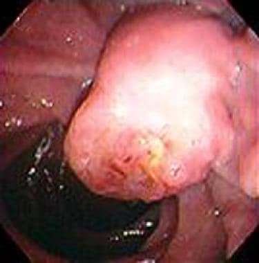
- Ampullary carcinoma is a rare malignant tumor originating at the ampulla of Vater, in the last centimeter of the common bile duct, where it passes through the wall of the duodenum and ampullary papilla (see the image below). (medscape.com)
- Saluran pankreas akan bergabung dengan saluran empedu kemudian memasuki duodenum melalui ampulla hepatopancreas . (wikipedia.org)
- Upper gastrointestinal endoscopy failed to examine the second part of the duodenum and the ampulla of Vater due to pyloric stenosis. (ispub.com)
- The opening of the pancreatic duct into the duodenum is at the ampulla of Vater. (histology-world.com)
- Along its way, closer to the duodenum the duct from the pancreas merges the common bile duct to form what is called the ampuella of vater. (bellaonline.com)
- The common bile duct joins with the pancreatic duct to form the ampulla of Vater, which empties into the duodenum. (merckmanuals.com)
- The terminal part of the CBD is joined by the terminal part of the pancreatic duct in the pancreatic head to form a common channel (called the biliopancreatic ampulla when dilated), which runs through the medial duodenal wall and opens on the dome of the major duodenal papilla, a nipplelike projection on the medial wall of the middle segment of the second part (C loop) of the duodenum. (medscape.com)
C241 Ampulla of Va1
- C241 Ampulla of Vater **Note 1:** This schema is based on the UICC chapter *Ampulla of Vater,* pages 129-131. (cancer.gov)
Adenocarcinoma8
- adenocarcinoma of the ampulla of Vater. (nih.gov)
- A 65-year-old man underwent laparoscopic pancreatoduodenectomy for ampulla of Vater adenocarcinoma. (springeropen.com)
- A 65-year-old man underwent laparoscopic pylorus-preserving PD with modified child reconstruction for ampulla of Vater adenocarcinoma. (springeropen.com)
- He received chemotherapy with gemcitabine plus cisplatin after being diagnosed with liver metastases and para-aortic lymph-node metastases from ampulla of Vater adenocarcinoma. (springeropen.com)
- Patients with adenocarcinoma of the ampulla of Vater who underwent R0 resection with pancreaticoduodenectomy between 2001 and 2011 were included in the present multi-institutional study. (biomedcentral.com)
- High HMGB1 expression is an independent predictor of poor prognosis in patients with adenocarcinoma of the ampulla of Vater not treated with adjuvant chemotherapy. (biomedcentral.com)
- Therefore, the present study was performed to determine the prognostic impact of HMGB1 in adenocarcinoma of the ampulla of Vater. (biomedcentral.com)
- c) Esophagogastroduodenoscopy shows an ulcer at the ampulla of Vater, and the biopsy results of the ulcer indicate adenocarcinoma. (hindawi.com)
Lesions of the ampulla2
- Premalignant lesions of the ampulla of Vater. (journalajrrga.com)
- Neoplastic lesions of the ampulla vary between 0.063 and 0.21% on autopsy investigations but the frequency of their diagnosis is increasing because of the extensive use of flexible endoscopy (1). (ispub.com)
Sphincter4
- citation needed] Various smooth muscle sphincters regulate the flow of bile and pancreatic juice through the ampulla: the sphincter of the pancreatic duct, the sphincter of the bile duct, and the sphincter of Oddi. (wikipedia.org)
- The Sphincter of Oddi and Ampulla of Vater are two important anatomical structures in the digestion system. (keydifference.in)
- We will explore their anatomy, function, and clinical implications to illuminate the differences in the Sphincter that is located in Oddi along with the Ampulla of Vater. (keydifference.in)
- The Sphincter of Oddi and the Ampulla of Vater are vital structures of digestive physiology. (keydifference.in)
Carcinoma5
- Carcinoma of Ampulla Standring, Susan (2020). (wikipedia.org)
- The patient was diagnosed with adenosquamous carcinoma of the ampulla of Vater and underwent pancreaticoduodenectomy. (biomedcentral.com)
- The final diagnosis was adenosquamous carcinoma of the ampulla of Vater, T3N1M0, stage IIB. (biomedcentral.com)
- Upon reviewing the medical records of our institute, we identified 4 patients who were diagnosed with adenosquamous carcinoma of the ampulla of Vater in the past 2 decades. (biomedcentral.com)
- Adenosquamous carcinoma of the ampulla of Vater is a rare disease with a dismal prognosis. (biomedcentral.com)
Endoscopy1
- We report a case of mucin-secreting biliary neoplasm of the ampulla of Vater diagnosed peroperatively because of unsuccessful endoscopy due to pyloric stenosis, and successfully treated with transduodenal local excision. (ispub.com)
Biliary4
- Magnetic resonance cholangiogram showed dilatation of the biliary tree and obstruction at the level of the ampulla. (ispub.com)
- Endoscopic retrograde cholangiography (ERCP) was performed to investigate the biliary or pancreatic duct, but cannulation of the ampulla of Vater could not be performed successfully due to the invasion of the tumor. (hindawi.com)
- These are disorders centered at the ampulla of Vater that can cause symptoms of intermittent biliary obstruction. (mitoaction.org)
- Cholangiocarcinomas (CCCs) are malignancies of the biliary duct system that may originate in the liver and extrahepatic bile ducts, which terminate at the ampulla of Vater. (medscape.com)
Tumors4
- Romiti A. Tumors of ampulla of Vater: A case series and review of chemotherapy options. (journalajrrga.com)
- Tumors of the ampulla of Vater are rare histopathological conditions. (ispub.com)
- Tumors of the ampulla of Vater are rare histopathological conditions and have relatively good prognosis after resection. (ispub.com)
- [ 7 ] Distal extrahepatic tumors are located from the upper border of the pancreas to the ampulla. (medscape.com)
Resection2
- Beger HG, Treitschke F, Gansauge F, Harada N, Hiki N, Mattfeldt T. Tumor of the ampulla of Vater: Experience with local or radical resection in 171 consecutively treated patients. (journalajrrga.com)
- A circumferential resection of duodenal mucosa and ampulla of Vater was undertaken. (ispub.com)
Gallbladder2
- Most cases occur in the gallbladder or in the ampulla of Vater, and such cases in the common bile duct (CBD) are extremely rare. (springer.com)
- They may leave the gallbladder and lodge in the cystic duct, the common bile duct, or the ampulla of Vater. (msdmanuals.com)
Duodenal wall1
- Stay sutures were placed in the duodenal wall, and the ampulla was identified. (ispub.com)
Prognostic1
- Surgical outcomes and prognostic factors for ampulla of vater cancer. (journalajrrga.com)
Tumor1
- Estudio retrospectivo que incluy a los pacientes con tumor periampular en quienes se realiz una pancreatoduodenectom a (PDD) en el HRAEPY entre enero de 2011 y junio de 2014. (medigraphic.com)
Cystic1
- Stones can block the cystic duct, common bile duct, or ampulla of Vater (where the common bile duct and pancreatic duct join). (msdmanuals.com)
Blockage1
- Blockage or dysfunction of this Ampulla of Vater could cause digestive issues and need medical care. (keydifference.in)
Common1
- Pancreatic islets were isolated by injection of collagenase up the common bile duct after clamping of the Ampulla of Vater. (nih.gov)
Bile2
- Macroscopically, papillary tumours spread from the ampulla of Vater to the distal bile duct. (springeropen.com)
- He also had to make sure the Ampulla of vater's bile duct portion was closed off and that the ampulla of vater was still in good shape. (bellaonline.com)
Chapter1
- If directly assigning SS2000, use the *Ampulla of Vater* chapter on page 114 of the [SS2000 on-line manual](https://seer.cancer.gov/tools/ssm/ssm2000/SSSM2000-122012.pdf#page=114). (cancer.gov)
Rare2
Unusual1
- We report here on an unusual case of bleeding from angiodysplasia at the ampulla of Vater in a 58-aged woman with end stage renal failure. (e-ce.org)
Cancer2
- Thomas' sign is the production of silver stools and can be indicative of cancer of the Ampulla of Vater. (wikipedia.org)
- Excitingly, one of these cases has multiple models derived from primary and metastatic ampulla of Vater cancer. (cancer.gov)
Case1
- We report here a case of a 71-year-old man with marginal zone B cell lymphoma of MALT with large B cell lymphoma of the ampulla of Vater that was not associated with Helicobacter pylori . (e-ce.org)
Cases1
- Successful endoscopic extraction of a large impacted choledocholithiasis in the ampulla of vater: two interesting cases. (spg.pt)
Term1
- The eponymic term "ampulla of Vater" is named after Abraham Vater (1684-1751), a German anatomist who first published a description of it in 1720. (wikipedia.org)
Major1
- The ampulla is specifically located at the major duodenal papilla . (wikidoc.org)
Location1
- Axial (A) and coronal (B) T2 MRI shows a solid mass protruding inside the second duodenal portion at the location of the ampulla of Vater (arrowhead). (elsevier.es)