Afferent Pathways
Vagus Nerve
Vagotomy
Reflex
Thermoreceptors
Meprobamate
Urinary Bladder
Urinary Bladder, Overactive
Autonomic Pathways
Solitary Nucleus
Sensation
Rats, Sprague-Dawley
Decerebrate State
Stomach
Mechanoreceptors
Efferent Pathways
Cats
Nociceptors
Nerve Fibers, Unmyelinated
Spinal Cord
Sensory Receptor Cells
Medulla Oblongata
Action Potentials
Pressoreceptors
Neurons
Excitatory Postsynaptic Potentials
Muscle Contraction
Neural Inhibition
Evoked Potentials
Synaptic Transmission
Synapses
Pressure
Atropine
Nerve Fibers
Ganglia, Spinal
Sympathetic Nervous System
Electrophysiology
Electromyography
Brain
On the neural correlates of visual perception. (1/2523)
Neurological findings suggest that the human striate cortex (V1) is an indispensable component of a neural substratum subserving static achromatic form perception in its own right and not simply as a central distributor of retinally derived information to extrastriate visual areas. This view is further supported by physiological evidence in primates that the finest-grained conjoined representation of spatial detail and retinotopic localization that underlies phenomenal visual experience for local brightness discriminations is selectively represented at cortical levels by the activity of certain neurons in V1. However, at first glance, support for these ideas would appear to be undermined by incontrovertible neurological evidence (visual hemineglect and the simultanagnosias) and recent psychophysical results on 'crowding' that confirm that activation of neurons in V1 may, at times, be insufficient to generate a percept. Moreover, a recent proposal suggests that neural correlates of visual awareness must project directly to those in executive space, thus automatically excluding V1 from a related perceptual space because V1 lacks such direct projections. Both sets of concerns are, however, resolved within the context of adaptive resonance theories. Recursive loops, linking the dorsal lateral geniculate nucleus (LGN) through successive cortical visual areas to the temporal lobe by means of a series of ascending and descending pathways, provide a neuronal substratum at each level within a modular framework for mutually consistent descriptions of sensory data. At steady state, such networks obviate the necessity that neural correlates of visual experience project directly to those in executive space because a neural phenomenal perceptual space subserving form vision is continuously updated by information from an object recognition space equivalent to that destined to reach executive space. Within this framework, activity in V1 may engender percepts that accompany figure-ground segregations only when dynamic incongruities are resolved both within and between ascending and descending streams. Synchronous neuronal activity on a short timescale within and across cortical areas, proposed and sometimes observed as perceptual correlates, may also serve as a marker that a steady state has been achieved, which, in turn, may be a requirement for the longer time constants that accompany the emergence and stability of perceptual states compared to the faster dynamics of adapting networks and the still faster dynamics of individual action potentials. Finally, the same consensus of neuronal activity across ascending and descending pathways linking multiple cortical areas that in anatomic sequence subserve phenomenal visual experiences and object recognition may underlie the normal unity of conscious experience. (+info)Neural mapping of direction and frequency in the cricket cercal sensory system. (2/2523)
Primary mechanosensory receptors and interneurons in the cricket cercal sensory system are sensitive to the direction and frequency of air current stimuli. Receptors innervating long mechanoreceptor hairs (>1000 microm) are most sensitive to low-frequency air currents (<150 Hz); receptors innervating medium-length hairs (900-500 microm) are most sensitive to higher frequency ranges (150-400 Hz). Previous studies demonstrated that the projection pattern of the synaptic arborizations of long hair receptor afferents form a continuous map of air current direction within the terminal abdominal ganglion (). We demonstrate here that the projection pattern of the medium-length hair afferents also forms a continuous map of stimulus direction. However, the afferents from the long and medium-length hair afferents show very little spatial segregation with respect to their frequency sensitivity. The possible functional significance of this small degree of spatial segregation was investigated, by calculating the relative overlap between the long and medium-length hair afferents with the dendrites of two interneurons that are known to have different frequency sensitivities. Both interneurons were shown to have nearly equal anatomical overlap with long and medium hair afferents. Thus, the differential overlap of these interneurons with the two different classes of afferents was not adequate to explain the observed frequency selectivity of the interneurons. Other mechanisms such as selective connectivity between subsets of afferents and interneurons and/or differences in interneuron biophysical properties must play a role in establishing the frequency selectivities of these interneurons. (+info)Gabapentin suppresses ectopic nerve discharges and reverses allodynia in neuropathic rats. (3/2523)
Repetitive ectopic discharges from injured afferent nerves play an important role in initiation and maintenance of neuropathic pain. Gabapentin is effective for treatment of neuropathic pain but the sites and mechanisms of its antinociceptive actions remain uncertain. In the present study, we tested a hypothesis that therapeutic doses of gabapentin suppress ectopic afferent discharge activity generated from injured peripheral nerves. Mechanical allodynia, induced by partial ligation of the sciatic nerve in rats, was determined by application of von Frey filaments to the hindpaw. Single-unit afferent nerve activity was recorded proximal to the ligated sciatic nerve site. Intravenous gabapentin, in a range of 30 to 90 mg/kg, significantly attenuated allodynia in nerve-injured rats. Furthermore, gabapentin, in the same therapeutic dose range, dose-dependently inhibited the ectopic discharge activity of 15 injured sciatic afferent nerve fibers through an action on impulse generation. However, the conduction velocity and responses of 12 normal afferent fibers to mechanical stimulation were not affected by gabapentin. Therefore, this study provides electrophysiological evidence that gabapentin is capable of suppressing the ectopic discharge activity from injured peripheral nerves. This action may contribute, at least in part, to the antiallodynic effect of gabapentin on neuropathic pain. (+info)Varying the degree of single-whisker stimulation differentially affects phases of intrinsic signals in rat barrel cortex. (4/2523)
Using intrinsic signal optical imaging (ISI), we have shown previously that the point spread of evoked activity in the rat barrel cortex in response to single-whisker stimulation encompasses a surprisingly large area. Given that our typical stimulation consists of five deflections at 5 Hz, the large area of evoked activity might have resulted from repetitive stimulation. Thus in the present study, we use ISI through the thinned skull to determine whether decreasing the degree of single-whisker stimulation decreases the area of the cortical point spread. We additionally outline a protocol to quantify stimulus-related differences in the temporal characteristics of intrinsic signals at a fine spatial scale. In 10 adult rats, whisker C2 was stimulated randomly with either one or five deflections delivered in a rostral-to-caudal fashion. Each deflection consisted of a 0.5-mm displacement of the whisker as measured at the point of contact, 15 mm from the snout. The number of whisker deflections did not affect the area or peak magnitude of the cortical point spread based on the intrinsic signal activity occurring from 0.5 up to 1.5 s poststimulus onset. In contrast, the magnitude and time course of intrinsic signal activity collected after 1.5-s poststimulus onset did reflect the difference in the degree of stimulation. Thus decreasing the degree of stimulation differentially affected the early and late phases of the evoked intrinsic signal response. The implications of the present results are discussed in respect to probable differences in the signal source underlying the early versus later phases of evoked intrinsic signals. (+info)Contribution of sensory feedback to the generation of extensor activity during walking in the decerebrate Cat. (5/2523)
In this investigation we have estimated the afferent contribution to the generation of activity in the knee and ankle extensor muscles during walking in decerebrate cats by loading and unloading extensor muscles, and by unilateral deafferentation of a hind leg. The total contribution of afferent feedback to extensor burst generation was estimated by allowing one hind leg to step into a hole in the treadmill belt on which the animal was walking. In the absence of ground support the level of activity in knee and ankle extensor muscles was reduced to approximately 70% of normal. Activity in the ankle extensors could be restored during the "foot-in-hole" trials by selectively resisting extension at the ankle. Thus feedback from proprioceptors in the ankle extensor muscles probably makes a large contribution to burst generation in these muscles during weight-bearing steps. Similarly, feedback from proprioceptors in knee extensor appears to contribute substantially to the activation of knee extensor muscles because unloading and loading these muscles, by lifting and dropping the hindquarters, strongly reduced and increased, respectively, the level of activity in the knee extensors. This conclusion was supported by the finding that partial deafferentation of one hind leg by transection of the L4-L6 dorsal roots reduced the level of activity in the knee extensors by approximately 50%, but did not noticeably influence the activity in ankle extensor muscles. However, extending the deafferentation to include the L7-S2 dorsal roots decreased the ankle extensor activity. We conclude that afferent feedback contributes to more than one-half of the input to knee and ankle extensor motoneurons during the stance phase of walking in decerebrate cats. The continuous contribution of afferent feedback to the generation of extensor activity could function to automatically adjust the intensity of activity to meet external demands. (+info)Neuronal activity in somatosensory cortex of monkeys using a precision grip. II. Responses To object texture and weights. (6/2523)
Three monkeys were trained to lift and hold a test object within a 12- to 25-mm position window for 1 s. The activity of single neurons was recorded during performance of the task in which both the weight and surface texture of the object were systematically varied. Whenever possible, each cell was tested with three weights (15, 65, and 115 g) and three textures (smooth metal, fine 200 grit sandpaper, and rough 60 grit sandpaper). Of 386 cells recorded in 3 monkeys, 45 cells had cutaneous receptive fields on the index or thumb or part of the thenar eminence and were held long enough to be tested in all 9 combinations of texture and weight. Recordings were made for the entire anterior-posterior extent of the thumb and index finger areas in somatosensory cortex including area 7b. However, the statistical analysis required a selection of only those cells for which nine complete recording conditions were available limiting the sample to cells in areas 2, 5, and 7b. Significant differences in the grip force accompanied 98% of the changes in texture and 78% of the changes in weight. Increasing the object weight also increased the force tangential to the skin surface as measured by the load or lifting force. The peak discharge during lifting was judged to be the most sensitive index of cell activity and was analyzed with a two-way analysis of variance (ANOVA). In addition, peak cell discharge was normalized to allow comparisons among different combinations of texture and weight as well as comparisons among different neurons. Overall, the peak firing frequency of 87% of the cells was significantly modulated by changes in object texture, but changes in object weight affected the peak activity of only 58% of the cells. Almost all (17/18, 94%) of the static cells were influenced by the object texture, and 81% of the dynamic cells that were active only briefly at grip and lift onset were modulated by texture. For some cells, surface texture had a significant effect on neuronal discharge that was independent of the object weight. In contrast, weight-related responses were never simple main effects of the weight alone and appeared instead as significant interactions between texture and weight. Four neurons either increased or decreased activity in a graded fashion with surface structure (roughness) regardless of the object weight (P < 0.05). Ten other neurons showed increases or decreases in response to one or two textures, which might represent either a graded response or a tuning preference for a specific texture. The firing frequency of the majority (31/45) of neurons reflected an interaction of both texture and weight. The cells with texture-related but weight-independent activities were thought to encode surface characteristics that are largely independent of the grip and lifting forces used to manipulate the object. Such constancies could be used to construct internal representations or mental models for planning and controlling object manipulation. (+info)Distinct populations of NMDA receptors at subcortical and cortical inputs to principal cells of the lateral amygdala. (7/2523)
Fear conditioning involves the transmission of sensory stimuli to the amygdala from the thalamus and cortex. These input synapses are prime candidates for sites of plasticity critical to the learning in fear conditioning. Because N-methyl-D-aspartate (NMDA)-dependent mechanisms have been implicated in fear learning, we investigated the contribution of NMDA receptors to synaptic transmission at putative cortical and thalamic inputs using visualized whole cell recording in amygdala brain slices. Whereas NMDA receptors are present at both of these pathways, differences were observed. First, the alpha-amino-3-hydroxy-5-methyl-4-isoxazolepropionic acid-receptor-mediated component of the synaptic response, relative to the NMDA component, is smaller at thalamic than cortical input synapses. Second, thalamic NMDA responses are more sensitive to Mg2+. These findings suggest that there are distinct populations of NMDA receptors at cortical and thalamic inputs to the lateral amygdala. Differences such as these might underlie unique contributions of the two pathways to fear conditioning. (+info)Gating of afferent input by a central pattern generator. (8/2523)
Intracellular recordings from the sole proprioceptor (the oval organ) in the crab ventilatory system show that the nonspiking afferent fibers from this organ receive a cyclic hyperpolarizing inhibition in phase with the ventilatory motor pattern. Although depolarizing and hyperpolarizing current pulses injected into a single afferent will reset the ventilatory motor pattern, the inhibitory input is of sufficient magnitude to block afferent input to the ventilatory central pattern generator (CPG) for approximately 50% of the cycle period. It is proposed that this inhibitory input serves to gate sensory input to the ventilatory CPG to provide an unambiguous input to the ventilatory CPG. (+info)Afferent pathways, also known as sensory pathways, refer to the neural connections that transmit sensory information from the peripheral nervous system to the central nervous system (CNS), specifically to the brain and spinal cord. These pathways are responsible for carrying various types of sensory information, such as touch, temperature, pain, pressure, vibration, hearing, vision, and taste, to the CNS for processing and interpretation.
The afferent pathways begin with sensory receptors located throughout the body, which detect changes in the environment and convert them into electrical signals. These signals are then transmitted via afferent neurons, also known as sensory neurons, to the spinal cord or brainstem. Within the CNS, the information is further processed and integrated with other neural inputs before being relayed to higher cognitive centers for conscious awareness and response.
Understanding the anatomy and physiology of afferent pathways is essential for diagnosing and treating various neurological conditions that affect sensory function, such as neuropathies, spinal cord injuries, and brain disorders.
Visceral afferents are specialized nerve fibers that carry sensory information from the internal organs (viscera) to the central nervous system. These afferent neurons detect and transmit information about various visceral stimuli, such as pain, temperature, touch, pressure, chemical changes, and the state of organ distension or fullness. The information they relay helps regulate physiological functions, including digestion, respiration, and cardiovascular activity, and contributes to the perception of bodily sensations and visceral pain. Visceral afferents are an essential component of the autonomic nervous system and have their cell bodies located in the dorsal root ganglia or nodose ganglia.
The vagus nerve, also known as the 10th cranial nerve (CN X), is the longest of the cranial nerves and extends from the brainstem to the abdomen. It has both sensory and motor functions and plays a crucial role in regulating various bodily functions such as heart rate, digestion, respiratory rate, speech, and sweating, among others.
The vagus nerve is responsible for carrying sensory information from the internal organs to the brain, and it also sends motor signals from the brain to the muscles of the throat and voice box, as well as to the heart, lungs, and digestive tract. The vagus nerve helps regulate the body's involuntary responses, such as controlling heart rate and blood pressure, promoting relaxation, and reducing inflammation.
Dysfunction in the vagus nerve can lead to various medical conditions, including gastroparesis, chronic pain, and autonomic nervous system disorders. Vagus nerve stimulation (VNS) is a therapeutic intervention that involves delivering electrical impulses to the vagus nerve to treat conditions such as epilepsy, depression, and migraine headaches.
A vagotomy is a surgical procedure that involves cutting or blocking the vagus nerve, which is a parasympathetic nerve that runs from the brainstem to the abdomen and helps regulate many bodily functions such as heart rate, gastrointestinal motility, and digestion. In particular, vagotomy is often performed as a treatment for peptic ulcers, as it can help reduce gastric acid secretion.
There are several types of vagotomy procedures, including:
1. Truncal vagotomy: This involves cutting the main trunks of the vagus nerve as they enter the abdomen. It is a more extensive procedure that reduces gastric acid secretion significantly but can also lead to side effects such as delayed gastric emptying and diarrhea.
2. Selective vagotomy: This involves cutting only the branches of the vagus nerve that supply the stomach, leaving the rest of the nerve intact. It is a less extensive procedure that reduces gastric acid secretion while minimizing side effects.
3. Highly selective vagotomy (HSV): Also known as parietal cell vagotomy, this involves cutting only the branches of the vagus nerve that supply the acid-secreting cells in the stomach. It is a highly targeted procedure that reduces gastric acid secretion while minimizing side effects such as delayed gastric emptying and diarrhea.
Vagotomy is typically performed using laparoscopic or open surgical techniques, depending on the patient's individual needs and the surgeon's preference. While vagotomy can be effective in treating peptic ulcers, it is not commonly performed today due to the development of less invasive treatments such as proton pump inhibitors (PPIs) that reduce gastric acid secretion without surgery.
A reflex is an automatic, involuntary and rapid response to a stimulus that occurs without conscious intention. In the context of physiology and neurology, it's a basic mechanism that involves the transmission of nerve impulses between neurons, resulting in a muscle contraction or glandular secretion.
Reflexes are important for maintaining homeostasis, protecting the body from harm, and coordinating movements. They can be tested clinically to assess the integrity of the nervous system, such as the knee-j jerk reflex, which tests the function of the L3-L4 spinal nerve roots and the sensitivity of the stretch reflex arc.
Urination, also known as micturition, is the physiological process of excreting urine from the urinary bladder through the urethra. It is a complex process that involves several systems in the body, including the urinary system, nervous system, and muscular system.
In medical terms, urination is defined as the voluntary or involuntary discharge of urine from the urethra, which is the final pathway for the elimination of waste products from the body. The process is regulated by a complex interplay between the detrusor muscle of the bladder, the internal and external sphincters of the urethra, and the nervous system.
During urination, the detrusor muscle contracts, causing the bladder to empty, while the sphincters relax to allow the urine to flow through the urethra and out of the body. The nervous system plays a crucial role in coordinating these actions, with sensory receptors in the bladder sending signals to the brain when it is time to urinate.
Urination is essential for maintaining the balance of fluids and electrolytes in the body, as well as eliminating waste products such as urea, creatinine, and other metabolic byproducts. Abnormalities in urination can indicate underlying medical conditions, such as urinary tract infections, bladder dysfunction, or neurological disorders.
Afferent neurons, also known as sensory neurons, are a type of nerve cell that conducts impulses or signals from peripheral receptors towards the central nervous system (CNS), which includes the brain and spinal cord. These neurons are responsible for transmitting sensory information such as touch, temperature, pain, sound, and light to the CNS for processing and interpretation. Afferent neurons have specialized receptor endings that detect changes in the environment and convert them into electrical signals, which are then transmitted to the CNS via synapses with other neurons. Once the signals reach the CNS, they are processed and integrated with other information to produce a response or reaction to the stimulus.
Capsaicin is defined in medical terms as the active component of chili peppers (genus Capsicum) that produces a burning sensation when it comes into contact with mucous membranes or skin. It is a potent irritant and is used topically as a counterirritant in some creams and patches to relieve pain. Capsaicin works by depleting substance P, a neurotransmitter that relays pain signals to the brain, from nerve endings.
Here is the medical definition of capsaicin from the Merriam-Webster's Medical Dictionary:
caпсаісіn : an alkaloid (C18H27NO3) that is the active principle of red peppers and is used in topical preparations as a counterirritant and analgesic.
Thermoreceptors are specialized sensory nerve endings or neurons that are sensitive to changes in temperature. They detect and respond to heat or cold stimuli by converting them into electrical signals that are transmitted to the brain for interpretation. These receptors are found throughout the body, particularly in the skin, mucous membranes, and internal organs. There are two main types of thermoreceptors: warm receptors, which respond to increasing temperatures, and cold receptors, which react to decreasing temperatures. The information provided by thermoreceptors helps maintain homeostasis and protect the body from harmful temperature changes.
Efferent neurons are specialized nerve cells that transmit signals from the central nervous system (CNS), which includes the brain and spinal cord, to effector organs such as muscles or glands. These signals typically result in a response or action, hence the term "efferent," derived from the Latin word "efferre" meaning "to carry away."
Efferent neurons are part of the motor pathway and can be further classified into two types:
1. Somatic efferent neurons: These neurons transmit signals to skeletal muscles, enabling voluntary movements and posture maintenance. They have their cell bodies located in the ventral horn of the spinal cord and send their axons through the ventral roots to innervate specific muscle fibers.
2. Autonomic efferent neurons: These neurons are responsible for controlling involuntary functions, such as heart rate, digestion, respiration, and pupil dilation. They have a two-neuron chain arrangement, with the preganglionic neuron having its cell body in the CNS (brainstem or spinal cord) and synapsing with the postganglionic neuron in an autonomic ganglion near the effector organ. Autonomic efferent neurons can be further divided into sympathetic, parasympathetic, and enteric subdivisions based on their functions and innervation patterns.
In summary, efferent neurons are a critical component of the nervous system, responsible for transmitting signals from the CNS to various effector organs, ultimately controlling and coordinating numerous bodily functions and responses.
Meprobamate is a carbamate derivative and acts as a central nervous system depressant. It is primarily used as an anti-anxiety agent, although it also has muscle relaxant properties. Meprobamate works by enhancing the activity of gamma-aminobutyric acid (GABA), a neurotransmitter that inhibits nerve transmission in the brain, thereby producing a calming effect.
It is important to note that meprobamate has a potential for abuse and dependence, and its use is associated with several side effects, including dizziness, drowsiness, and impaired coordination. Therefore, it should only be used under the close supervision of a healthcare provider.
The urinary bladder is a muscular, hollow organ in the pelvis that stores urine before it is released from the body. It expands as it fills with urine and contracts when emptying. The typical adult bladder can hold between 400 to 600 milliliters of urine for about 2-5 hours before the urge to urinate occurs. The wall of the bladder contains several layers, including a mucous membrane, a layer of smooth muscle (detrusor muscle), and an outer fibrous adventitia. The muscles of the bladder neck and urethra remain contracted to prevent leakage of urine during filling, and they relax during voiding to allow the urine to flow out through the urethra.
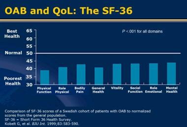
Overactive bladder (OAB) is a urological condition characterized by the involuntary contraction of the detrusor muscle of the urinary bladder, leading to symptoms such as urgency, frequency, and nocturia (the need to wake up at night to urinate), with or without urge incontinence (the involuntary loss of urine associated with a strong desire to void). It is important to note that OAB is not necessarily related to bladder volume or age-related changes, and it can significantly impact an individual's quality of life. The exact cause of OAB is not fully understood, but it may be associated with neurological disorders, certain medications, infections, or other underlying medical conditions. Treatment options for OAB include behavioral modifications, pelvic floor exercises, bladder training, medications, and, in some cases, surgical interventions.
The autonomic nervous system (ANS) is a component of the peripheral nervous system that regulates involuntary physiological functions, such as heart rate, digestion, respiratory rate, pupillary response, urination, and sexual arousal. The autonomic pathways refer to the neural connections and signaling processes that allow the ANS to carry out these functions.
The autonomic pathways consist of two main subdivisions: the sympathetic nervous system (SNS) and the parasympathetic nervous system (PNS). These systems have opposing effects on many organs, with the SNS generally stimulating activity and the PNS inhibiting it. The enteric nervous system, which controls gut function, is sometimes considered a third subdivision of the ANS.
The sympathetic pathway originates in the thoracic and lumbar regions of the spinal cord, with preganglionic neurons synapsing on postganglionic neurons in paravertebral ganglia or prevertebral ganglia. The parasympathetic pathway originates in the brainstem (cranial nerves III, VII, IX, and X) and the sacral region of the spinal cord (S2-S4), with preganglionic neurons synapsing on postganglionic neurons near or within the target organ.
Acetylcholine is the primary neurotransmitter used in both the sympathetic and parasympathetic pathways, although norepinephrine (noradrenaline) is also released by some postganglionic sympathetic neurons. The specific pattern of neural activation and inhibition within the autonomic pathways helps maintain homeostasis and allows for adaptive responses to changes in the internal and external environment.
The solitary nucleus, also known as the nucleus solitarius, is a collection of neurons located in the medulla oblongata region of the brainstem. It plays a crucial role in the processing and integration of sensory information, particularly taste and visceral afferent fibers from internal organs. The solitary nucleus receives inputs from various cranial nerves, including the glossopharyngeal (cranial nerve IX) and vagus nerves (cranial nerve X), and is involved in reflex responses related to swallowing, vomiting, and cardiovascular regulation.
In medical terms, sensation refers to the ability to perceive and interpret various stimuli from our environment through specialized receptor cells located throughout the body. These receptors convert physical stimuli such as light, sound, temperature, pressure, and chemicals into electrical signals that are transmitted to the brain via nerves. The brain then interprets these signals, allowing us to experience sensations like sight, hearing, touch, taste, and smell.
There are two main types of sensations: exteroceptive and interoceptive. Exteroceptive sensations involve stimuli from outside the body, such as light, sound, and touch. Interoceptive sensations, on the other hand, refer to the perception of internal bodily sensations, such as hunger, thirst, heartbeat, or emotions.
Disorders in sensation can result from damage to the nervous system, including peripheral nerves, spinal cord, or brain. Examples include numbness, tingling, pain, or loss of sensation in specific body parts, which can significantly impact a person's quality of life and ability to perform daily activities.
Electric stimulation, also known as electrical nerve stimulation or neuromuscular electrical stimulation, is a therapeutic treatment that uses low-voltage electrical currents to stimulate nerves and muscles. It is often used to help manage pain, promote healing, and improve muscle strength and mobility. The electrical impulses can be delivered through electrodes placed on the skin or directly implanted into the body.
In a medical context, electric stimulation may be used for various purposes such as:
1. Pain management: Electric stimulation can help to block pain signals from reaching the brain and promote the release of endorphins, which are natural painkillers produced by the body.
2. Muscle rehabilitation: Electric stimulation can help to strengthen muscles that have become weak due to injury, illness, or surgery. It can also help to prevent muscle atrophy and improve range of motion.
3. Wound healing: Electric stimulation can promote tissue growth and help to speed up the healing process in wounds, ulcers, and other types of injuries.
4. Urinary incontinence: Electric stimulation can be used to strengthen the muscles that control urination and reduce symptoms of urinary incontinence.
5. Migraine prevention: Electric stimulation can be used as a preventive treatment for migraines by applying electrical impulses to specific nerves in the head and neck.
It is important to note that electric stimulation should only be administered under the guidance of a qualified healthcare professional, as improper use can cause harm or discomfort.
Sprague-Dawley rats are a strain of albino laboratory rats that are widely used in scientific research. They were first developed by researchers H.H. Sprague and R.C. Dawley in the early 20th century, and have since become one of the most commonly used rat strains in biomedical research due to their relatively large size, ease of handling, and consistent genetic background.
Sprague-Dawley rats are outbred, which means that they are genetically diverse and do not suffer from the same limitations as inbred strains, which can have reduced fertility and increased susceptibility to certain diseases. They are also characterized by their docile nature and low levels of aggression, making them easier to handle and study than some other rat strains.
These rats are used in a wide variety of research areas, including toxicology, pharmacology, nutrition, cancer, and behavioral studies. Because they are genetically diverse, Sprague-Dawley rats can be used to model a range of human diseases and conditions, making them an important tool in the development of new drugs and therapies.
A decerebrate state is a medical condition that results from severe damage to the brainstem, specifically to the midbrain and above. This type of injury can cause motor responses characterized by rigid extension of the arms and legs, with the arms rotated outward and the wrists and fingers extended. The legs are also extended and the toes pointed downward. These postures are often referred to as "decerebrate rigidity" or "posturing."
The decerebrate state is typically seen in patients who have experienced severe trauma, such as a car accident or gunshot wound, or who have suffered from a large stroke or other type of brain hemorrhage. It can also occur in some cases of severe hypoxia (lack of oxygen) to the brain, such as during cardiac arrest or drowning.
The decerebrate state is a serious medical emergency that requires immediate treatment. If left untreated, it can lead to further brain damage and even death. Treatment typically involves providing supportive care, such as mechanical ventilation to help with breathing, medications to control blood pressure and prevent seizures, and surgery to repair any underlying injuries or bleeding. In some cases, patients may require long-term rehabilitation to regain lost function and improve their quality of life.
In anatomical terms, the stomach is a muscular, J-shaped organ located in the upper left portion of the abdomen. It is part of the gastrointestinal tract and plays a crucial role in digestion. The stomach's primary functions include storing food, mixing it with digestive enzymes and hydrochloric acid to break down proteins, and slowly emptying the partially digested food into the small intestine for further absorption of nutrients.
The stomach is divided into several regions, including the cardia (the area nearest the esophagus), the fundus (the upper portion on the left side), the body (the main central part), and the pylorus (the narrowed region leading to the small intestine). The inner lining of the stomach, called the mucosa, is protected by a layer of mucus that prevents the digestive juices from damaging the stomach tissue itself.
In medical contexts, various conditions can affect the stomach, such as gastritis (inflammation of the stomach lining), peptic ulcers (sores in the stomach or duodenum), gastroesophageal reflux disease (GERD), and stomach cancer. Symptoms related to the stomach may include abdominal pain, bloating, nausea, vomiting, heartburn, and difficulty swallowing.
Mechanoreceptors are specialized sensory receptor cells that convert mechanical stimuli such as pressure, tension, or deformation into electrical signals that can be processed and interpreted by the nervous system. They are found in various tissues throughout the body, including the skin, muscles, tendons, joints, and internal organs. Mechanoreceptors can detect different types of mechanical stimuli depending on their specific structure and location. For example, Pacinian corpuscles in the skin respond to vibrations, while Ruffini endings in the joints detect changes in joint angle and pressure. Overall, mechanoreceptors play a crucial role in our ability to perceive and interact with our environment through touch, proprioception (the sense of the position and movement of body parts), and visceral sensation (awareness of internal organ activity).
Physical stimulation, in a medical context, refers to the application of external forces or agents to the body or its tissues to elicit a response. This can include various forms of touch, pressure, temperature, vibration, or electrical currents. The purpose of physical stimulation may be therapeutic, as in the case of massage or physical therapy, or diagnostic, as in the use of reflex tests. It is also used in research settings to study physiological responses and mechanisms.
In a broader sense, physical stimulation can also refer to the body's exposure to physical activity or exercise, which can have numerous health benefits, including improving cardiovascular function, increasing muscle strength and flexibility, and reducing the risk of chronic diseases.
Efferent pathways refer to the neural connections that carry signals from the central nervous system (CNS), which includes the brain and spinal cord, to the peripheral effectors such as muscles and glands. These pathways are responsible for the initiation and control of motor responses, as well as regulating various autonomic functions.
Efferent pathways can be divided into two main types:
1. Somatic efferent pathways: These pathways carry signals from the CNS to the skeletal muscles, enabling voluntary movements and postural control. The final common pathway for somatic motor innervation is the alpha-motor neuron, which synapses directly onto skeletal muscle fibers.
2. Autonomic efferent pathways: These pathways regulate the function of internal organs, smooth muscles, and glands. They are further divided into two subtypes: sympathetic and parasympathetic. The sympathetic system is responsible for the 'fight or flight' response, while the parasympathetic system promotes rest and digestion. Both systems use a two-neuron chain to transmit signals from the CNS to the effector organs. The preganglionic neuron has its cell body in the CNS and synapses with the postganglionic neuron in an autonomic ganglion located near the effector organ. The postganglionic neuron then innervates the target organ or tissue.
In summary, efferent pathways are the neural connections that carry signals from the CNS to peripheral effectors, enabling motor responses and regulating various autonomic functions. They can be divided into somatic and autonomic efferent pathways, with further subdivisions within the autonomic system.
"Cat" is a common name that refers to various species of small carnivorous mammals that belong to the family Felidae. The domestic cat, also known as Felis catus or Felis silvestris catus, is a popular pet and companion animal. It is a subspecies of the wildcat, which is found in Europe, Africa, and Asia.
Domestic cats are often kept as pets because of their companionship, playful behavior, and ability to hunt vermin. They are also valued for their ability to provide emotional support and therapy to people. Cats are obligate carnivores, which means that they require a diet that consists mainly of meat to meet their nutritional needs.
Cats are known for their agility, sharp senses, and predatory instincts. They have retractable claws, which they use for hunting and self-defense. Cats also have a keen sense of smell, hearing, and vision, which allow them to detect prey and navigate their environment.
In medical terms, cats can be hosts to various parasites and diseases that can affect humans and other animals. Some common feline diseases include rabies, feline leukemia virus (FeLV), feline immunodeficiency virus (FIV), and toxoplasmosis. It is important for cat owners to keep their pets healthy and up-to-date on vaccinations and preventative treatments to protect both the cats and their human companions.
Nociceptors are specialized peripheral sensory neurons that detect and transmit signals indicating potentially harmful stimuli in the form of pain. They are activated by various noxious stimuli such as extreme temperatures, intense pressure, or chemical irritants. Once activated, nociceptors transmit these signals to the central nervous system (spinal cord and brain) where they are interpreted as painful sensations, leading to protective responses like withdrawing from the harmful stimulus or seeking medical attention. Nociceptors play a crucial role in our perception of pain and help protect the body from further harm.
Unmyelinated nerve fibers, also known as unmyelinated axons or non-myelinated fibers, are nerve cells that lack a myelin sheath. Myelin is a fatty, insulating substance that surrounds the axon of many nerve cells and helps to increase the speed of electrical impulses traveling along the nerve fiber.
In unmyelinated nerve fibers, the axons are surrounded by a thin layer of Schwann cell processes called the endoneurium, but there is no continuous myelin sheath. Instead, the axons are packed closely together in bundles, with several axons lying within the same Schwann cell.
Unmyelinated nerve fibers tend to be smaller in diameter than myelinated fibers and conduct electrical impulses more slowly. They are commonly found in the autonomic nervous system, which controls involuntary functions such as heart rate, blood pressure, and digestion, as well as in sensory nerves that transmit pain and temperature signals.
Gastrointestinal motility refers to the coordinated muscular contractions and relaxations that propel food, digestive enzymes, and waste products through the gastrointestinal tract. This process involves the movement of food from the mouth through the esophagus into the stomach, where it is mixed with digestive enzymes and acids to break down food particles.
The contents are then emptied into the small intestine, where nutrients are absorbed, and the remaining waste products are moved into the large intestine for further absorption of water and electrolytes and eventual elimination through the rectum and anus.
Gastrointestinal motility is controlled by a complex interplay between the autonomic nervous system, hormones, and local reflexes. Abnormalities in gastrointestinal motility can lead to various symptoms such as bloating, abdominal pain, nausea, vomiting, diarrhea, or constipation.
The spinal cord is a major part of the nervous system, extending from the brainstem and continuing down to the lower back. It is a slender, tubular bundle of nerve fibers (axons) and support cells (glial cells) that carries signals between the brain and the rest of the body. The spinal cord primarily serves as a conduit for motor information, which travels from the brain to the muscles, and sensory information, which travels from the body to the brain. It also contains neurons that can independently process and respond to information within the spinal cord without direct input from the brain.
The spinal cord is protected by the bony vertebral column (spine) and is divided into 31 segments: 8 cervical, 12 thoracic, 5 lumbar, 5 sacral, and 1 coccygeal. Each segment corresponds to a specific region of the body and gives rise to pairs of spinal nerves that exit through the intervertebral foramina at each level.
The spinal cord is responsible for several vital functions, including:
1. Reflexes: Simple reflex actions, such as the withdrawal reflex when touching a hot surface, are mediated by the spinal cord without involving the brain.
2. Muscle control: The spinal cord carries motor signals from the brain to the muscles, enabling voluntary movement and muscle tone regulation.
3. Sensory perception: The spinal cord transmits sensory information, such as touch, temperature, pain, and vibration, from the body to the brain for processing and awareness.
4. Autonomic functions: The sympathetic and parasympathetic divisions of the autonomic nervous system originate in the thoracolumbar and sacral regions of the spinal cord, respectively, controlling involuntary physiological responses like heart rate, blood pressure, digestion, and respiration.
Damage to the spinal cord can result in various degrees of paralysis or loss of sensation below the level of injury, depending on the severity and location of the damage.
Neural pathways, also known as nerve tracts or fasciculi, refer to the highly organized and specialized routes through which nerve impulses travel within the nervous system. These pathways are formed by groups of neurons (nerve cells) that are connected in a series, creating a continuous communication network for electrical signals to transmit information between different regions of the brain, spinal cord, and peripheral nerves.
Neural pathways can be classified into two main types: sensory (afferent) and motor (efferent). Sensory neural pathways carry sensory information from various receptors in the body (such as those for touch, temperature, pain, and vision) to the brain for processing. Motor neural pathways, on the other hand, transmit signals from the brain to the muscles and glands, controlling movements and other effector functions.
The formation of these neural pathways is crucial for normal nervous system function, as it enables efficient communication between different parts of the body and allows for complex behaviors, cognitive processes, and adaptive responses to internal and external stimuli.
Sensory receptor cells are specialized structures that convert physical stimuli from our environment into electrical signals, which are then transmitted to the brain for interpretation. These receptors can be found in various tissues throughout the body and are responsible for detecting sensations such as touch, pressure, temperature, taste, and smell. They can be classified into two main types: exteroceptors, which respond to stimuli from the external environment, and interoceptors, which react to internal conditions within the body. Examples of sensory receptor cells include hair cells in the inner ear, photoreceptors in the eye, and taste buds on the tongue.
Denervation is a medical term that refers to the loss or removal of nerve supply to an organ or body part. This can occur as a result of surgical intervention, injury, or disease processes that damage the nerves leading to the affected area. The consequences of denervation depend on the specific organ or tissue involved, but generally, it can lead to changes in function, sensation, and muscle tone. For example, denervation of a skeletal muscle can cause weakness, atrophy, and altered reflexes. Similarly, denervation of an organ such as the heart can lead to abnormalities in heart rate and rhythm. In some cases, denervation may be intentional, such as during surgical procedures aimed at treating chronic pain or spasticity.
The medulla oblongata is a part of the brainstem that is located in the posterior portion of the brainstem and continues with the spinal cord. It plays a vital role in controlling several critical bodily functions, such as breathing, heart rate, and blood pressure. The medulla oblongata also contains nerve pathways that transmit sensory information from the body to the brain and motor commands from the brain to the muscles. Additionally, it is responsible for reflexes such as vomiting, swallowing, coughing, and sneezing.
An action potential is a brief electrical signal that travels along the membrane of a nerve cell (neuron) or muscle cell. It is initiated by a rapid, localized change in the permeability of the cell membrane to specific ions, such as sodium and potassium, resulting in a rapid influx of sodium ions and a subsequent efflux of potassium ions. This ion movement causes a brief reversal of the electrical potential across the membrane, which is known as depolarization. The action potential then propagates along the cell membrane as a wave, allowing the electrical signal to be transmitted over long distances within the body. Action potentials play a crucial role in the communication and functioning of the nervous system and muscle tissue.
Pressoreceptors are specialized sensory nerve endings found in the walls of blood vessels, particularly in the carotid sinus and aortic arch. They respond to changes in blood pressure by converting the mechanical stimulus into electrical signals that are transmitted to the brain. This information helps regulate cardiovascular function and maintain blood pressure homeostasis.
Neurons, also known as nerve cells or neurocytes, are specialized cells that constitute the basic unit of the nervous system. They are responsible for receiving, processing, and transmitting information and signals within the body. Neurons have three main parts: the dendrites, the cell body (soma), and the axon. The dendrites receive signals from other neurons or sensory receptors, while the axon transmits these signals to other neurons, muscles, or glands. The junction between two neurons is called a synapse, where neurotransmitters are released to transmit the signal across the gap (synaptic cleft) to the next neuron. Neurons vary in size, shape, and structure depending on their function and location within the nervous system.
Excitatory postsynaptic potentials (EPSPs) are electrical signals that occur in the dendrites and cell body of a neuron, or nerve cell. They are caused by the activation of excitatory synapses, which are connections between neurons that allow for the transmission of information.
When an action potential, or electrical impulse, reaches the end of an axon, it triggers the release of neurotransmitters into the synaptic cleft, the small gap between the presynaptic and postsynaptic membranes. The excitatory neurotransmitters then bind to receptors on the postsynaptic membrane, causing a local depolarization of the membrane potential. This depolarization is known as an EPSP.
EPSPs are responsible for increasing the likelihood that an action potential will be generated in the postsynaptic neuron. When multiple EPSPs occur simultaneously or in close succession, they can summate and cause a large enough depolarization to trigger an action potential. This allows for the transmission of information from one neuron to another.
It's important to note that there are also inhibitory postsynaptic potentials (IPSPs) which decrease the likelihood that an action potential will be generated in the postsynaptic neuron, by causing a local hyperpolarization of the membrane potential.
Muscle contraction is the physiological process in which muscle fibers shorten and generate force, leading to movement or stability of a body part. This process involves the sliding filament theory where thick and thin filaments within the sarcomeres (the functional units of muscles) slide past each other, facilitated by the interaction between myosin heads and actin filaments. The energy required for this action is provided by the hydrolysis of adenosine triphosphate (ATP). Muscle contractions can be voluntary or involuntary, and they play a crucial role in various bodily functions such as locomotion, circulation, respiration, and posture maintenance.
Neural inhibition is a process in the nervous system that decreases or prevents the activity of neurons (nerve cells) in order to regulate and control communication within the nervous system. It is a fundamental mechanism that allows for the balance of excitation and inhibition necessary for normal neural function. Inhibitory neurotransmitters, such as GABA (gamma-aminobutyric acid) and glycine, are released from the presynaptic neuron and bind to receptors on the postsynaptic neuron, reducing its likelihood of firing an action potential. This results in a decrease in neural activity and can have various effects depending on the specific neurons and brain regions involved. Neural inhibition is crucial for many functions including motor control, sensory processing, attention, memory, and emotional regulation.
Evoked potentials (EPs) are medical tests that measure the electrical activity in the brain or spinal cord in response to specific sensory stimuli, such as sight, sound, or touch. These tests are often used to help diagnose and monitor conditions that affect the nervous system, such as multiple sclerosis, brainstem tumors, and spinal cord injuries.
There are several types of EPs, including:
1. Visual Evoked Potentials (VEPs): These are used to assess the function of the visual pathway from the eyes to the back of the brain. A patient is typically asked to look at a patterned image or flashing light while electrodes placed on the scalp record the electrical responses.
2. Brainstem Auditory Evoked Potentials (BAEPs): These are used to evaluate the function of the auditory nerve and brainstem. Clicking sounds are presented to one or both ears, and electrodes placed on the scalp measure the response.
3. Somatosensory Evoked Potentials (SSEPs): These are used to assess the function of the peripheral nerves and spinal cord. Small electrical shocks are applied to a nerve at the wrist or ankle, and electrodes placed on the scalp record the response as it travels up the spinal cord to the brain.
4. Motor Evoked Potentials (MEPs): These are used to assess the function of the motor pathways in the brain and spinal cord. A magnetic or electrical stimulus is applied to the brain or spinal cord, and electrodes placed on a muscle measure the response as it travels down the motor pathway.
EPs can help identify abnormalities in the nervous system that may not be apparent through other diagnostic tests, such as imaging studies or clinical examinations. They are generally safe, non-invasive procedures with few risks or side effects.
Synaptic transmission is the process by which a neuron communicates with another cell, such as another neuron or a muscle cell, across a junction called a synapse. It involves the release of neurotransmitters from the presynaptic terminal of the neuron, which then cross the synaptic cleft and bind to receptors on the postsynaptic cell, leading to changes in the electrical or chemical properties of the target cell. This process is critical for the transmission of signals within the nervous system and for controlling various physiological functions in the body.
A synapse is a structure in the nervous system that allows for the transmission of signals from one neuron (nerve cell) to another. It is the point where the axon terminal of one neuron meets the dendrite or cell body of another, and it is here that neurotransmitters are released and received. The synapse includes both the presynaptic and postsynaptic elements, as well as the cleft between them.
At the presynaptic side, an action potential travels down the axon and triggers the release of neurotransmitters into the synaptic cleft through exocytosis. These neurotransmitters then bind to receptors on the postsynaptic side, which can either excite or inhibit the receiving neuron. The strength of the signal between two neurons is determined by the number and efficiency of these synapses.
Synapses play a crucial role in the functioning of the nervous system, allowing for the integration and processing of information from various sources. They are also dynamic structures that can undergo changes in response to experience or injury, which has important implications for learning, memory, and recovery from neurological disorders.
In medical terms, pressure is defined as the force applied per unit area on an object or body surface. It is often measured in millimeters of mercury (mmHg) in clinical settings. For example, blood pressure is the force exerted by circulating blood on the walls of the arteries and is recorded as two numbers: systolic pressure (when the heart beats and pushes blood out) and diastolic pressure (when the heart rests between beats).
Pressure can also refer to the pressure exerted on a wound or incision to help control bleeding, or the pressure inside the skull or spinal canal. High or low pressure in different body systems can indicate various medical conditions and require appropriate treatment.
Reaction time, in the context of medicine and physiology, refers to the time period between the presentation of a stimulus and the subsequent initiation of a response. This complex process involves the central nervous system, particularly the brain, which perceives the stimulus, processes it, and then sends signals to the appropriate muscles or glands to react.
There are different types of reaction times, including simple reaction time (responding to a single, expected stimulus) and choice reaction time (choosing an appropriate response from multiple possibilities). These measures can be used in clinical settings to assess various aspects of neurological function, such as cognitive processing speed, motor control, and alertness.
However, it is important to note that reaction times can be influenced by several factors, including age, fatigue, attention, and the use of certain medications or substances.
Atropine is an anticholinergic drug that blocks the action of the neurotransmitter acetylcholine in the central and peripheral nervous system. It is derived from the belladonna alkaloids, which are found in plants such as deadly nightshade (Atropa belladonna), Jimson weed (Datura stramonium), and Duboisia spp.
In clinical medicine, atropine is used to reduce secretions, increase heart rate, and dilate the pupils. It is often used before surgery to dry up secretions in the mouth, throat, and lungs, and to reduce salivation during the procedure. Atropine is also used to treat certain types of nerve agent and pesticide poisoning, as well as to manage bradycardia (slow heart rate) and hypotension (low blood pressure) caused by beta-blockers or calcium channel blockers.
Atropine can have several side effects, including dry mouth, blurred vision, dizziness, confusion, and difficulty urinating. In high doses, it can cause delirium, hallucinations, and seizures. Atropine should be used with caution in patients with glaucoma, prostatic hypertrophy, or other conditions that may be exacerbated by its anticholinergic effects.
Nerve fibers are specialized structures that constitute the long, slender processes (axons) of neurons (nerve cells). They are responsible for conducting electrical impulses, known as action potentials, away from the cell body and transmitting them to other neurons or effector organs such as muscles and glands. Nerve fibers are often surrounded by supportive cells called glial cells and are grouped together to form nerve bundles or nerves. These fibers can be myelinated (covered with a fatty insulating sheath called myelin) or unmyelinated, which influences the speed of impulse transmission.
Sensory thresholds are the minimum levels of stimulation that are required to produce a sensation in an individual, as determined through psychophysical testing. These tests measure the point at which a person can just barely detect the presence of a stimulus, such as a sound, light, touch, or smell.
There are two types of sensory thresholds: absolute and difference. Absolute threshold is the minimum level of intensity required to detect a stimulus 50% of the time. Difference threshold, also known as just noticeable difference (JND), is the smallest change in intensity that can be detected between two stimuli.
Sensory thresholds can vary between individuals and are influenced by factors such as age, attention, motivation, and expectations. They are often used in clinical settings to assess sensory function and diagnose conditions such as hearing or vision loss.
Motor neurons are specialized nerve cells in the brain and spinal cord that play a crucial role in controlling voluntary muscle movements. They transmit electrical signals from the brain to the muscles, enabling us to perform actions such as walking, talking, and swallowing. There are two types of motor neurons: upper motor neurons, which originate in the brain's motor cortex and travel down to the brainstem and spinal cord; and lower motor neurons, which extend from the brainstem and spinal cord to the muscles. Damage or degeneration of these motor neurons can lead to various neurological disorders, such as amyotrophic lateral sclerosis (ALS) and spinal muscular atrophy (SMA).
Spinal ganglia, also known as dorsal root ganglia, are clusters of nerve cell bodies located in the peripheral nervous system. They are situated along the length of the spinal cord and are responsible for transmitting sensory information from the body to the brain. Each spinal ganglion contains numerous neurons, or nerve cells, with long processes called axons that extend into the periphery and innervate various tissues and organs. The cell bodies within the spinal ganglia receive sensory input from these axons and transmit this information to the central nervous system via the dorsal roots of the spinal nerves. This allows the brain to interpret and respond to a wide range of sensory stimuli, including touch, temperature, pain, and proprioception (the sense of the position and movement of one's body).
The sympathetic nervous system (SNS) is a part of the autonomic nervous system that operates largely below the level of consciousness, and it functions to produce appropriate physiological responses to perceived danger. It's often associated with the "fight or flight" response. The SNS uses nerve impulses to stimulate target organs, causing them to speed up (e.g., increased heart rate), prepare for action, or otherwise respond to stressful situations.
The sympathetic nervous system is activated due to stressful emotional or physical situations and it prepares the body for immediate actions. It dilates the pupils, increases heart rate and blood pressure, accelerates breathing, and slows down digestion. The primary neurotransmitter involved in this system is norepinephrine (also known as noradrenaline).
Electrophysiology is a branch of medicine that deals with the electrical activities of the body, particularly the heart. In a medical context, electrophysiology studies (EPS) are performed to assess abnormal heart rhythms (arrhythmias) and to evaluate the effectiveness of certain treatments, such as medication or pacemakers.
During an EPS, electrode catheters are inserted into the heart through blood vessels in the groin or neck. These catheters can record the electrical activity of the heart and stimulate it to help identify the source of the arrhythmia. The information gathered during the study can help doctors determine the best course of treatment for each patient.
In addition to cardiac electrophysiology, there are also other subspecialties within electrophysiology, such as neuromuscular electrophysiology, which deals with the electrical activity of the nervous system and muscles.
Electromyography (EMG) is a medical diagnostic procedure that measures the electrical activity of skeletal muscles during contraction and at rest. It involves inserting a thin needle electrode into the muscle to record the electrical signals generated by the muscle fibers. These signals are then displayed on an oscilloscope and may be heard through a speaker.
EMG can help diagnose various neuromuscular disorders, such as muscle weakness, numbness, or pain, and can distinguish between muscle and nerve disorders. It is often used in conjunction with other diagnostic tests, such as nerve conduction studies, to provide a comprehensive evaluation of the nervous system.
EMG is typically performed by a neurologist or a physiatrist, and the procedure may cause some discomfort or pain, although this is usually minimal. The results of an EMG can help guide treatment decisions and monitor the progression of neuromuscular conditions over time.
The brain is the central organ of the nervous system, responsible for receiving and processing sensory information, regulating vital functions, and controlling behavior, movement, and cognition. It is divided into several distinct regions, each with specific functions:
1. Cerebrum: The largest part of the brain, responsible for higher cognitive functions such as thinking, learning, memory, language, and perception. It is divided into two hemispheres, each controlling the opposite side of the body.
2. Cerebellum: Located at the back of the brain, it is responsible for coordinating muscle movements, maintaining balance, and fine-tuning motor skills.
3. Brainstem: Connects the cerebrum and cerebellum to the spinal cord, controlling vital functions such as breathing, heart rate, and blood pressure. It also serves as a relay center for sensory information and motor commands between the brain and the rest of the body.
4. Diencephalon: A region that includes the thalamus (a major sensory relay station) and hypothalamus (regulates hormones, temperature, hunger, thirst, and sleep).
5. Limbic system: A group of structures involved in emotional processing, memory formation, and motivation, including the hippocampus, amygdala, and cingulate gyrus.
The brain is composed of billions of interconnected neurons that communicate through electrical and chemical signals. It is protected by the skull and surrounded by three layers of membranes called meninges, as well as cerebrospinal fluid that provides cushioning and nutrients.
In the field of medicine, "time factors" refer to the duration of symptoms or time elapsed since the onset of a medical condition, which can have significant implications for diagnosis and treatment. Understanding time factors is crucial in determining the progression of a disease, evaluating the effectiveness of treatments, and making critical decisions regarding patient care.
For example, in stroke management, "time is brain," meaning that rapid intervention within a specific time frame (usually within 4.5 hours) is essential to administering tissue plasminogen activator (tPA), a clot-busting drug that can minimize brain damage and improve patient outcomes. Similarly, in trauma care, the "golden hour" concept emphasizes the importance of providing definitive care within the first 60 minutes after injury to increase survival rates and reduce morbidity.
Time factors also play a role in monitoring the progression of chronic conditions like diabetes or heart disease, where regular follow-ups and assessments help determine appropriate treatment adjustments and prevent complications. In infectious diseases, time factors are crucial for initiating antibiotic therapy and identifying potential outbreaks to control their spread.
Overall, "time factors" encompass the significance of recognizing and acting promptly in various medical scenarios to optimize patient outcomes and provide effective care.
 Neuro-Ophthalmologic Manifestations of Multiple Sclerosis: Practice Essentials, Multiple Sclerosis, Afferent Visual Pathway...
Neuro-Ophthalmologic Manifestations of Multiple Sclerosis: Overview, Multiple Sclerosis, Afferent Visual Pathway Manifestations...
 Modelling spinal circuitry involved in locomotor pattern generation: insights from the effects of afferent stimulation
Normalization as a canonical neural computation
 Pain and pleasure - Wikipedia
 Responses and afferent pathways of C<sub>1</sub>-C<sub>2</sub> spinal neurons to gastric...
 Frontiers | Stress-Induced Chronic Visceral Pain of Gastrointestinal Origin
 Traumatic Stress Promotes Hyperalgesia via Corticotropin-Releasing Factor-1 Receptor (CRFR1) Signaling in Central Amygdala |...
Topographic and Functional Anatomy of the Spinal Cord: Gross Anatomy, Ventral and Dorsal Roots, Descending Spinal Cord Tracts
 Capsicum: MedlinePlus Supplements
 Capsicum Supplement: Uses, Benefits, Side Effects, Dose, Precautions & Warnings
Plus it
 Pathogenesis of Cognitive Dysfunction in Patients with Obstructive Sleep Apnea: A Hypothesis with Emphasis on the Nucleus...
Plus it
 Chronic Inflammatory and Neuropathic Pain | The New York Academy of Sciences
 ERS guidelines on the diagnosis and treatment of chronic cough in adults and children | European Respiratory Society
 SciELO - Brazil - Função do sistema olivococlear medial em crianças com desvio fonológico Função do sistema olivococlear...
Electrophysiologic characterization of the swallowing pattern generator in the brainstem : GI Motility online
 Erowid.org: Erowid Reference 3633 : Mechanism of Lysergic Acid Diethylamide Influence Upon Optically Evoked Potentials :...
 Different Techniques of Acupunctureand#8211;Part of the Traditional Chinese Medicine and and#8220;Evidence Based Medicineand...
ATILIM UNIVERSITY - Atılım University - ECTS INFORMATION GUIDE - ECTS - - School of Medicine Undergraduate Program
Neurogenic Bladder: Overview, Neuroanatomy, Physiology and Pathophysiology
Neurogenic Bladder: Overview, Neuroanatomy, Physiology and Pathophysiology
 Coding strategies in the otolith system differ for translational head motion vs. static orientation relative to gravity | eLife
 PPT - Principles of Sensory Systems PowerPoint presentation | free to view - id: 1e36b8-OThiN
 Peripheral chemoreceptors (video) | Khan Academy
Plus it
 Animals | Free Full-Text | Spinal Locomotion in Cats Following Spinal Cord Injury: A Prospective Study
 evidence investigator Archives - Randox Laboratories
Sensory9
- Sensory afferents have been incorporated in the model to study the effects of afferent stimulation on locomotor phase switching and step cycle period and on the firing patterns of flexor and extensor motoneurones. (nih.gov)
- Some evidence shows that the upper cervical spinal cord might play an important role in propriospinal processing as a sensory filter and modulator for visceral afferents. (okstate.edu)
- Results of these data supported the concept that a group of C 1 -C 2 spinal neurons might play a role in processing sensory information from the stomach that travels in vagal and spinal visceral afferent fibers. (okstate.edu)
- The pathway involving the thalamus is particularly interesting because of the role of the thalamus for multiple sensory integration [85,86]. (researchgate.net)
- They often involve afferent and efferent pathways of the Peripheral Nervous System, and their effects might be reflected in the mechanosensory-motor circuit at different cellular levels (including sensory and motor neurons, glia and muscle dysfunctions), and in the connexion among them. (ibecbarcelona.eu)
- The Togo starburst tarantula's venom struck them as being particularly interesting because it appeared to activate a particular type of sodium channel within sensory nerves that was not a part of known pain pathways. (neurosciencenews.com)
- To evaluate the effect of HDBS in PAG/DRN on the nociceptive pathways related to pain perception, microelectrode recordings were made in cortical areas known to be involved in the sensory-discriminative and affective aspects of pain. (lu.se)
- These results show that granular and high-resolution PAG/DRN stimulation enables potent, specific, safe, and durable analgesia by blocking the nociceptive-evoked motor, sensory and affective responses without significant activation of pathways provoking adverse side effects. (lu.se)
- Its sensory distribution pathway includes the posterior part of the head and extends anteriorly toward the vertex, becoming superficial at the inferolateral aspect of the occipital protuberance. (medscape.com)
Autonomic5
- To approach these issues, we analyzed synaptic function in the brainstem nucleus tractus solitarius (nTS), the principal site for integration of primary visceral afferent inputs to central autonomic pathways and a region in which we found markedly reduced levels of BDNF in Mecp2 mutants. (jneurosci.org)
- Through the pterygopalatine ganglion, this causes ocular vasodilation and activation of ocular trigeminal afferents through the trigemino-autonomic reflex [85]. (researchgate.net)
- Small myelinated fibers transmit preganglionic autonomic efferents (B fibers) and somatic afferents (A delta fibers). (medscape.com)
- Unmyelinated (C) fibers transmit postganglionic autonomic efferents as well as somatic and autonomic afferents. (medscape.com)
- In this review, we examined the efficacy of acupuncture with regard to urinary incontinence, and assessed the levels of the autonomic efferent and afferent pathways and the depth of the needle insertion in the acupuncture. (spandidos-publications.com)
Action Potentials3
- Once stimuli are received, the various afferent action potentials are triggered and pass along various fibers and axons of these nociceptive nerve cells into the dorsal horn of the spinal cord through the dorsal roots. (wikipedia.org)
- Our results demonstrate that the amplitude of spontaneous miniature and evoked EPSCs in nTS neurons is significantly increased in Mecp2 -null mice and, accordingly, that mutant cells are more likely than wild- type cells to fire action potentials in response to primary afferent stimulation. (jneurosci.org)
- The present study demonstrates that nTS relay neurons in Mecp2 -null mice exhibit significantly larger EPSCs, and are more likely to fire action potentials in response to afferent stimulation, than in wild-type controls. (jneurosci.org)
Visceral6
- The aims of this study were to determine (1) the responses of C 1 -C 2 spinal neurons to gastric distension and (2) the relative contribution of vagal and spinal visceral afferent pathways for transmission of gastric input to the upper cervical spinal cord. (okstate.edu)
- Evidence suggests that long term stress facilitates pain perception and sensitizes pain pathways, leading to a feed-forward cycle promoting chronic visceral pain disorders such as irritable bowel syndrome (IBS). (frontiersin.org)
- This review will discuss the potential neuronal pathways and mechanisms responsible for stress-induced exacerbation of chronic visceral pain. (frontiersin.org)
- Here we will briefly review visceral pain pathways and their modulation by (i) stress in adulthood and (ii) following exposure to neonatal stress. (frontiersin.org)
- Chemoreceptors and mechanoreceptors of the general visceral afferent pathway (GVA) from the carotid body and carotid sinus via (the carotid sinus nerve of) the glossopharyngeal nerve (CN IX), and from aortic bodies and sinoatrial node via the vagus nerve (CN X). (wikipedia.org)
- Chemoreceptors and mechanoreceptors of the general visceral afferent pathway (GVA) with endings located in the heart, lungs, airways, gastrointestinal system, pharynx, and liver via the glossopharyngeal and vagus nerves. (wikipedia.org)
Neurons5
- Therefore, we hypothesized that nTS neurons in Mecp2 -null mice would exhibit exaggerated responses to primary afferent stimulation due to loss of this BDNF-dependent modulation. (jneurosci.org)
- Specifically, studies have not revealed the role played by heterogeneity in the resting discharge variability of otolith afferents in the coding strategies employed by these neurons. (elifesciences.org)
- Among motor symptoms and signs, the cardinal ones (bradykinesia, rest tremor, and rigidity) are mainly ascribed to the loss of dopaminergic neurons [ 4 ], but those involving posture, balance, and gait are largely secondary to degeneration of nondopaminergic pathways and significantly contribute to impairment and disability in advanced PD patients [ 5 ]. (hindawi.com)
- Then the afferent and efferent pathways of the Peripheral Nervous System were mimicked in 2D culturing primary neurons involved in the locomotion circuit (motoneurons and dorsal root ganglia) with Schwann cells in the microdevice. (ibecbarcelona.eu)
- [ 6 ] GON block decreases afferent input to the trigeminal nucleus caudalis, resulting in central pain modulation and reducing neuronal hyperexcitability at the level of second-order neurons. (medscape.com)
Neuronal4
- Pain originating from the gastrointestinal (GI) system ascends to the brain via the same tri-neuronal pathways that convey noxious somatic stimuli. (frontiersin.org)
- Perhaps the most promising future developments in pharmacotherapy are drugs which tackle neuronal hypersensitivity by blocking excitability of afferent nerves by inhibiting targets such as the ATP receptor (P2X3). (ersjournals.com)
- In contrast, intraraphe serotonin may increase the tonic inhibitory influence of raphe on LGN, thus decreasing LGN neuronal reaponsiveneasto visual afferent inputs. (erowid.org)
- Such neuronal translocation constitutes an additional not generally recognized clearance pathway for inhaled solid UFP, whose significance for humans, however, still needs to be estab- lished. (cdc.gov)
Vagal3
- Thus, adults with chronic cough now have a firm physical explanation for their symptoms based on vagal afferent hypersensitivity. (ersjournals.com)
- When you move around and exercise, you are firing the frequency of neural pathways that then increases your vagal tone. (spinalresearch.com.au)
- The hiccup reflex is composed of the afferent pathway (through the vagal and phrenic nerves and the sympathetic chain), the central hiccup center (in the hypothalamus), and the efferent pathway (through the phrenic nerve to the diaphragm and the accessory nerves to the intercostal muscles). (lww.com)
Fibers4
- Describe the variety of afferent fibers (axons) used by the subsystems. (powershow.com)
- This is the termination of the afferent auditory pathway after having received fibers from the medial geniculate nucleus. (radiopaedia.org)
- Afferent nerve fibers run from the receptors via the dorsal penile nerve and pudendal nerve into the spinal cord. (urology-textbook.com)
- The neural elements of somatosensory receptors in the hands and feet represent the distal extreme of long afferent fibers, and thus, are par- ticularly vulnerable in the distal axonopathies. (cdc.gov)
Stimuli2
- LSD augmentation may be representative of synaptic facilitation, suggesting increased reactivity of the visual pathway to afferent stimuli. (erowid.org)
- However, how the otolith afferents respond to the naturalistic self-motion stimuli experienced during everyday activities remains unknown. (elifesciences.org)
Auditory1
- As well, the exposure to solvent s can effect the afferent and efferent auditory pathways. (cdc.gov)
Stimulation3
- The model also suggests that the different effects of flexor muscle nerve afferent stimulation observed experimentally (phase prolongation versus resetting) result from opposing influences of flexor group I and II afferents on the PF and RG circuits controlling the activity of flexor and extensor motoneurones. (nih.gov)
- In the absence of stimulation, otolith afferents display differences in the variability of their resting discharge and can be classified as regular or irregular. (elifesciences.org)
- The main aim was to elucidate whether stimulation of individually selected microelectrodes can selectively activate the anti-nociceptive pathways without activating networks that provoke side effects. (lu.se)
Neural3
- Although pharmacologic manipulation of intrinsically photosensitive retinal ganglion cells and the neural pathways that mediate photophobia may be possible in the future, current therapies are directed at the underlying cause of the photophobia and optical modulation of these cells and pathways. (researchgate.net)
- Functional neurorehabilitation promotes neural reorganization by stimulating subjects without deep pain perception, leading to a faster recovery when compared to spontaneous recovery, and achieving fewer compensatory errors, or even deviations to neuropathic or adaptive pain pathways, such as spasticity. (mdpi.com)
- it may result from remodeling of neural pathways in the gut-brain axis. (msdmanuals.com)
Receptors1
- Specific modalities can be associated with unique peripheral receptors, peripheral axons of stereotyped diameter and specific central projection pathways. (cdc.gov)
Nociceptive pathways1
- A neuroanatomical review of the pain pathway, "Afferent pain pathways" by Almeida, describes various specific nociceptive pathways of the spinal cord: spinothalamic tract, spinoreticular tract, spinomesencephalic tract, spinoparabrachial tract, spinohypothalamic tract, spinocervical tract, postsynaptic pathway of the spinal column. (wikipedia.org)
Somatic1
- VELASCO, M. Central influences on afferent conduction in the somatic and visual pathways. (bvsalud.org)
Carotid body1
- This neurotransmitters cause depolarization of the afferent pathway of Glossopharingeal Nerve(carotid body) and Vagus nerve (aortic body). (khanacademy.org)
Trigeminal nucleus caudalis1
- These afferents then project to the trigeminal nucleus caudalis, the thalamus, and to the cortex [85, 86] . (researchgate.net)
Nerve5
- Optic neuritis refers to an inflammatory injury of the optic nerve and represents a common afferent visual pathway manifestation of MS. Less often, homonymous visual field defects caused by retrogeniculate lesions are encountered. (medscape.com)
- The afferent part of the pathway (red) refers to the nerve impulse/message sent from the pupil to the brain along the optic nerve when a light is shone in that eye. (cehjournal.org)
- The efferent part of the pathway (blue) is the impulse/message that is sent from the mid-brain back to both pupils via the ciliary ganglion and the third cranial nerve (the oculomotor nerve), causing both pupils to constrict, even even though only one eye is being stimulated by the light. (cehjournal.org)
- A positive RAPD means there are differences between the two eyes in the afferent pathway due to retinal or optic nerve disease. (cehjournal.org)
- Afferent signals pass via the pudendal nerve to the sacral erection center, and efferent signals reach via the inferior hypogastric plexus the erectile tissue. (urology-textbook.com)
Nervous system1
- The amygdala, and more specifically the central nucleus of the amygdala (CeA), is a convergence point for multiple pathways carrying nociceptive information into the central nervous system. (nature.com)
Modulation1
- Then, there are also the descending pathways for the modulation of pain sensation. (wikipedia.org)
Peripheral2
- There exist several central pathways to convey NAU-induced acupuncture signals, Electroacupuncture (EA) frequency-specific neurochemical effects are related to different peripheral and central pathways transmitting afferent signals from different frequency of NAU stimulation. (qigonginstitute.org)
- Minimally, a recep- tor includes a peripheral axon terminal of one pri- mary afferent neuron, whose cell body is sited proximally in the dorsal root ganglion. (cdc.gov)
Somatosensory1
- Nucleus tractus solitarius (NTS) is the central integration hub for afferents from upper airway (somatosensory/gustatory), respiratory, gastrointestinal, cardiovascular (baroreceptor and chemoreceptor) and other systems. (hindawi.com)
Cortex2
- Describe the output pathway from the ear to the cortex and how sound properties of encoded. (powershow.com)
- The HDBS-induced analgesia could be sustained for at least 4 hours and did not provoke significant side effects on behaviour, spontaneous activity, and brain states and it had a minor effect on the tactile afferent pathway to the cortex. (lu.se)
Mediate2
- Afferents of the SN mediate the gag reflex , the carotid sinus reflex, the aortic reflex, the cough reflex , the baroreflex and chemoreceptor reflexes, several respiratory reflexes and reflexes within the gastrointestinal system regulating motility and secretion. (wikipedia.org)
- at the cellular level they will identify the neurotransmitter pathways and transmitter reuptake mechanisms that mediate cocaine's disruptive effects on clock function. (kent.edu)
Physiological1
- Regular and irregular afferents further display different morphological and physiological features (reviewed in Goldberg, 2000 , Cullen, 2012 and Eatock and Songer, 2011 ). (elifesciences.org)
Gastric1
- The pathways for gastric and gustatory (taste) processes are believed to terminate in different subdivisions of the parabrachial area, but still interact in the SN. (wikipedia.org)
Mechanisms2
- Intermediate and advanced PD stages are characterized by motor fluctuations and dyskinesia, which depend on complex mechanisms secondary to severe nigrostriatal loss and to the problems related to oral levodopa absorption, and motor and nonmotor symptoms and signs that are secondary to marked dopaminergic loss and multisystem neurodegeneration with damage to nondopaminergic pathways. (hindawi.com)
- The purpose of this review is to highlight new discoveries in pathways and mechanisms of pruritus and to summarize up-to-date knowledge about treatment of itching after surgery. (asahq.org)
Reflex3
- Here we show that this CPG structure can be integrated with reflex circuits to reproduce the reorganization of group I reflex pathways occurring during locomotion. (nih.gov)
- The pons relays afferent information from the bladder to higher brain centers, which in turn communicate with the periaqueductal gray matter, a relay station that collects higher brain center intput and processes this in order to signal the PMC to trigger or suppress the voiding reflex. (medscape.com)
- To understand how the pupils react to light, it is important to understand the light reflex pathway (Figure 1). (cehjournal.org)
Spinal1
- CeA projections to periacqueductal gray (PAG) gate descending pathways that modulate nociceptive afferent activity in the dorsal horn of the spinal cord. (nature.com)
Neurochemical1
- There is strong evidence of biological connections between the neurochemical pathways used for the perception of both pain and pleasure, as well as other psychological rewards. (wikipedia.org)
Ocular1
- pathways can produce severe ocular defects such as Data analysis was carried out using Statistical transient monocular visual loss, visual field defect Package for Social Sciences (SPSS) for Windows, 1-3 or ocular motility restrictions. (who.int)
Central1
- The purpose of this review is to facilitate an understanding of the pathways and mediators involved in the development and central transmission of the itch sensation, to describe the methods available to evaluate itch, and to provide a basis for its rational therapy. (asahq.org)
Posterior1
- Some individuals with MS and other CNS inflammatory syndromes also may experience homonymous visual field defects caused by lesions in retrochiasmal or retrogeniculate (posterior) regions of the afferent visual pathway. (medscape.com)
Intrinsic2
- Otolith afferents with higher intrinsic variability transmitted more information overall about translational self-motion than their regular counterparts, owing to stronger nonlinearities that enabled precise spike timing including phase locking. (elifesciences.org)
- Using computational methods, we further demonstrated that coupled increases in intrinsic variability and sensitivity accounted for the observed functional differences between afferent classes. (elifesciences.org)
Assessment1
- But 2D cultures offer many limitations compared to 3D, and the assessment of the afferent pathway separately often means a complication. (ibecbarcelona.eu)
Amygdala1
- An afferent white matter pathway from the pulvinar to the amygdala facilitates fear recognition. (ans.org.au)
Midbrain1
- Here, we tested this hypothesis using in vivo evoked potential recordings from a midbrain nucleus in the ascending electrosensory pathway of mormyrids. (elifesciences.org)
Increases1
- During the extensor phase of fictive locomotion, activation of extensor muscle group I afferents increases extensor motoneurone activity and prolongs the extensor phase. (nih.gov)
Symptoms3
- Affected individuals may experience problems with how they see the world (afferent visual pathway symptoms) and/or how smoothly and synchronously their eyes move together (efferent visual pathway disorders). (medscape.com)
- Neuro-ophthalmic manifestations are frequently encountered in multiple sclerosis (MS). Affected individuals may experience problems with how they see the world (afferent visual pathway symptoms) and/or how their eyes move together (efferent visual pathway disorders). (medscape.com)
- Because patients with MS who have visual symptoms tend to seek ophthalmic attention, eye care experts play a vital role in the localization of afferent and efferent visual pathway problems that may either represent the first clinical manifestation of MS or affect a patient with a well-established diagnosis. (medscape.com)
Visual3
- Efferent visual pathway lesions can create a perception of oscillopsia, a visual disturbance in which objects appear to jiggle or move owing to nystagmus (involuntary eye movements). (medscape.com)
- Both afferent and efferent visual systems are sensitive to brain injury. (researchgate.net)
- Ethical approval was obtained occlusions along the afferent or efferent visual from the Ethical Research Committee of FTHL. (who.int)
Frequency1
- Tracing of the afferent and efferent connections of respective regions and discussion of various brainwave and/or resonant frequency origination. (uschirodirectory.com)
Primary1
- However, this synaptopathy is associated with decreased BDNF availability in the primary afferent pathway and can be rescued by application of exogenous BDNF. (jneurosci.org)
Differences1
- The 'swinging light test' is used to detect a relative afferent pupil defect (RAPD): a means of detecting differences between the two eyes in how they respond to a light shone in one eye at a time. (cehjournal.org)
Contrast2
- By contrast, more regular afferents better discriminated between different static head orientations relative to gravity. (elifesciences.org)
- This pressure contrast is influenced by the combined resistances of the afferent (leading to the glomerulus) and efferent (leading away from the glomerulus) vascular pathways in the kidney. (randox.com)
Input1
- Overall, the brain receives input via afferent pathways that ascend from the bladder and provide feedback on how full the bladder is. (medscape.com)
Study1
- Optogenetics technique can be used in skeletal muscle to induce contraction, mimicking a natural innervation to some length and facilitating the study of the afferent pathway separately. (ibecbarcelona.eu)
Activation1
- In addition, L. lactis NZ9000-SeNPs significantly attenuated the reduction of the expression levels of occludin and claudin-1, dysbiosis of the gut microbiome, and activation of the Toll-like receptor (TLR)/nuclear factor kappa B (NF-κB)-mediated signaling pathway induced by ETEC K88. (bvsalud.org)