Acyl-CoA Oxidase
Acyl Coenzyme A
Diacylglycerol O-Acyltransferase
Sterol O-Acyltransferase
NADPH Oxidase
Acyltransferases
Coenzyme A
Sleep Deprivation
Seizures
Tablets
Electroencephalography
Epilepsy, Absence
Pamphlets
The peroxisome proliferator (PP) response element upstream of the human acyl CoA oxidase gene is inactive among a sample human population: significance for species differences in response to PPs. (1/269)
Peroxisome proliferators (PP) cause peroxisome proliferation, associated with rodent hepatocyte growth perturbation and hepatocarcinogenesis. However, in humans this class of non-genotoxic carcinogens does not appear to have the same adverse effects. The peroxisome proliferator-activated receptor alpha (PPARalpha) mediates the effects of PPs in rodents via peroxisome proliferator response elements (PPREs) upstream of PP-responsive genes such as acyl coenzyme A oxidase (ACO). When the human ACO promoter was cloned previously, it was found to be active and to contain a consensus PPRE (-1918 AGGTCA C TGGTCA -1906). To confirm and extend those original findings, we isolated a 2 kb genomic fragment of the ACO gene promoter from a human liver biopsy and used it to create a beta-galactosidase reporter gene plasmid. The human ACO promoter reporter plasmid was added to both Hepalclc7 and NIH 3T3 cells together with a plasmid expressing mPPARa and assessed for its ability to drive PP-mediated gene transcription. The human ACO promoter fragment was inactive, unlike the equivalent rat ACO promoter fragment used as a positive control. The PPRE within our cloned fragment of the human ACO promoter differed at three positions (5'-AGGTCA G CTGTCA-3') from the previously published active human ACO promoter. Next, we studied the frequency of the inactive versus the active human PPRE within the human population. Using a PCR strategy, we isolated and analysed genomic DNA fragments from 22 unrelated human individuals and from the human hepatoma cell line HepG2. In each case, the PPRE contained the inactive sequence. These data show that the human ACO gene promoter found in a sample human population is inactive. This may explain at the genomic level the lack of response of humans to some of the adverse effects of the PP class of non-genotoxic hepatocarcinogens. (+info)Oxidation of medium-chain acyl-CoA esters by extracts of Aspergillus niger: enzymology and characterization of intermediates by HPLC. (2/269)
The activities of beta-oxidation enzymes were measured in extracts of glucose- and triolein-grown cells of Aspergillus niger. Growth on triolein stimulated increased enzyme activity, especially for acyl-CoA dehydrogenase. No acyl-CoA oxidase activity was detected. HPLC analysis after incubation of triolein-grown cell extracts with decanoyl-CoA showed that beta-oxidation was limited to one cycle. Octanoyl-CoA accumulated as the decanoyl-CoA was oxidized. Beta-oxidation enzymes in isolated mitochondrial fractions were also studied. The results are discussed in the context of methyl ketone production by fungi. (+info)Beneficial effects of fibrates on apolipoprotein A-I metabolism occur independently of any peroxisome proliferative response. (3/269)
BACKGROUND: In humans, fibrates are frequently used normolipidemic drugs. Fibrates act by regulating genes involved in lipoprotein metabolism via activation of the peroxisome proliferator-activated receptor-alpha (PPARalpha) in liver. In rodents, however, fibrates induce a peroxisome proliferation, leading to hepatomegaly and possibly hepatocarcinogenesis. Although this peroxisome proliferative response appears not to occur in humans, it remains controversial whether the beneficial effects of fibrates on lipoprotein metabolism can occur dissociated from such undesirable peroxisomal response. Here, we assessed the influence of fenofibrate on lipoprotein metabolism and peroxisome proliferation in the rabbit, an animal that, contrary to rodents and similar to humans, is less sensitive to peroxisome proliferators. METHODS AND RESULTS: First, we demonstrate that in normal rabbits, fenofibrate given at a high dose for 2 weeks does not influence serum concentrations or intestinal mRNA levels of the HDL apolipoprotein apoA-I. Therefore, the study was continued with human apoA-I transgenic rabbits that overexpress the human apoA-I gene under control of its homologous promoter, including its PPAR-response elements. In these animals, fenofibrate increases serum human apoA-I concentrations via an increased expression of the human apoA-I gene in liver. Interestingly, liver weight or mRNA levels and activity of fatty acyl-CoA oxidase, a rate-limiting and marker enzyme of peroxisomal beta-oxidation, remain unchanged after fenofibrate. CONCLUSIONS: Expression of the human apoA-I transgene in rabbit liver suffices to confer fibrate-mediated induction of serum apoA-I. Furthermore, these data provide in vivo evidence that the beneficial effects of fibrates on lipoprotein metabolism occur mechanistically dissociated from any deleterious activity on peroxisome proliferation and possibly hepatocarcinogenesis. (+info)Absence of spontaneous peroxisome proliferation in enoyl-CoA Hydratase/L-3-hydroxyacyl-CoA dehydrogenase-deficient mouse liver. Further support for the role of fatty acyl CoA oxidase in PPARalpha ligand metabolism. (4/269)
Peroxisomes contain a classical L-hydroxy-specific peroxisome proliferator-inducible beta-oxidation system and also a second noninducible D-hydroxy-specific beta-oxidation system. We previously generated mice lacking fatty acyl-CoA oxidase (AOX), the first enzyme of the L-hydroxy-specific classical beta-oxidation system; these AOX-/- mice exhibited sustained activation of peroxisome proliferator-activated receptor alpha (PPARalpha), resulting in profound spontaneous peroxisome proliferation in liver cells. These observations implied that AOX is responsible for the metabolic degradation of PPARalpha ligands. In this study, the function of enoyl-CoA hydratase/L-3-hydroxyacyl-CoA dehydrogenase (L-PBE), the second enzyme of this peroxisomal beta-oxidation system, was investigated by disrupting its gene. Mutant mice (L-PBE-/-) were viable and fertile and exhibited no detectable gross phenotypic defects. L-PBE-/- mice showed no hepatic steatosis and manifested no spontaneous peroxisome proliferation, unlike that encountered in livers of mice deficient in AOX. These results indicate that disruption of classical peroxisomal fatty acid beta-oxidation system distal to AOX step does not interfere with the inactivation of endogenous ligands of PPARalpha, further confirming that the AOX gene is indispensable for the physiological regulation of this receptor. The absence of appreciable changes in lipid metabolism also indicates that enoyl-CoAs, generated in the classical system in L-PBE-/- mice are diverted to D-hydroxy-specific system for metabolism by D-PBE. When challenged with a peroxisome proliferator, L-PBE-/- mice showed increases in the levels of hepatic mRNAs and proteins that are regulated by PPARalpha except for appreciable blunting of peroxisome proliferative response as compared with that observed in hepatocytes of wild type mice similarly treated. This blunting of peroxisome proliferative response is attributed to the absence of L-PBE protein in L-PBE-/- mouse liver, because all other proteins are induced essentially to the same extent in both wild type and L-PBE-/- mice. (+info)Activation of flavin-containing oxidases underlies light-induced production of H2O2 in mammalian cells. (5/269)
Violet-blue light is toxic to mammalian cells, and this toxicity has been linked with cellular production of H2O2. In this report, we show that violet-blue light, as well as UVA, stimulated H2O2 production in cultured mouse, monkey, and human cells. We found that H2O2 originated in peroxisomes and mitochondria, and it was enhanced in cells overexpressing flavin-containing oxidases. These results support the hypothesis that photoreduction of flavoproteins underlies light-induced production of H2O2 in cells. Because H2O2 and its metabolite, hydroxyl radicals, can cause cellular damage, these reactive oxygen species may contribute to pathologies associated with exposure to UVA, violet, and blue light. They may also contribute to phototoxicity often encountered during light microscopy. Because multiphoton excitation imaging with 1,047-nm wavelength prevented light-induced H2O2 production in cells, possibly by minimizing photoreduction of flavoproteins, this technique may be useful for decreasing phototoxicity during fluorescence microscopy. (+info)Impairment of peroxisomal biogenesis in human colon carcinoma. (6/269)
Peroxisomes and the activities of their enzymes have been reported to be significantly reduced in various types of tumors including the colon carcinoma. Therefore, the present study was designed to investigate the gene expression of several peroxisomal proteins in human colon carcinoma and additionally those of the peroxisome proliferator activated receptor alpha (PPARalpha) and PEX5, a receptor protein involved in the import of most peroxisomal matrix proteins. Samples from adenocarcinomas and adjacent normal colon were analyzed by immunohistochemistry and western blotting. The mRNA content was assessed by a novel sensitive dot blot RNase protection assay and northern blotting. By immunohistochemistry, peroxisomes were distinctly visualized in normal colonocytes but were not detected in colon carcinoma cells. The protein levels of catalase (CAT), acyl-CoA oxidase as well as the 22 and 70 kDa peroxisomal membrane proteins (PMP22 and PMP70) were all significantly decreased in carcinomas. The corresponding mRNAs for CAT and PMP70, however, were unchanged. In contrast, the mRNA of PEX5 was significantly increased. The expression of PPARalpha was not altered in tumors, neither at protein nor mRNA levels. These observations show that the reduction of peroxisomes and their proteins in colon carcinoma is not due to a generalized reduction of transcription of their genes. It seems more likely that this phenomenon is regulated at a post-transcriptional or translational level. Alternatively, and more likely, an impairment of the biogenesis of the organelle could account for the paucity of peroxisomes in colon carcinoma. (+info)Arachidonic acid and PGE2 regulation of hepatic lipogenic gene expression. (7/269)
N-6 polyunsaturated fatty acids (PUFA) suppress hepatic and adipocyte de novo lipogenesis by inhibiting the transcription of genes encoding key lipogenic proteins. In cultured 3T3-L1 adipocytes, arachidonic acid (20:4,n-6) suppression of lipogenic gene expression requires cyclooxygenase (COX) activity. In this study, we found no evidence to support a role for COX-1 or -2 in the 20:4,n-6 inhibition of hepatocyte lipogenic gene expression. In contrast to L1 preadipocytes, adipocytes and rat liver, RT-PCR and Western analyses did not detect COX-1 or COX-2 expression in cultured primary hepatocytes. Moreover, the COX inhibitor, flurbiprofen, did not affect the 20:4,n-6 regulation of lipogenic gene expression in primary hepatocytes. Despite the absence of COX-1 and -2 expression in primary hepatocytes, prostaglandins (PGE2 and PGF2alpha) suppressed fatty acid synthase, l-pyruvate kinase, and the S14 protein mRNA, while having no effect on acyl-CoA oxidase or CYP4A2 mRNA. Using PGE2 receptor agonist, the PGE2 effect on lipogenic gene expression was linked to EP3 receptors. PGE2 inhibited S14CAT activity in transfected primary hepatocytes and targeted the S14 PUFA-response region located -220 to -80 bp upstream from the transcription start site. Taken together, these studies show that COX-1 and COX-2 do not contribute to the n-6 PUFA suppression of hepatocyte lipogenic gene expression. However, cyclooxygenase products from non-parenchymal cells can act on parenchymal cells through a paracrine process and mimic the effects of n-6 PUFA on lipogenic gene expression. (+info)Novel form of lipolysis induced by leptin. (8/269)
Hyperleptinemia causes disappearance of body fat without a rise in free fatty acids (FFA) or ketones, suggesting that leptin can deplete adipocytes of fat without releasing FFA. To test this, we measured FFA and glycerol released from adipocytes obtained from normal lean Zucker diabetic fatty rats (+/+) and incubated for 0, 3, 6, or 24 h in either 20 ng/ml recombinant leptin or 100 nM norepinephrine (NE). Whereas NE increased both FFA and glycerol release from adipocytes of +/+ rats, leptin increased glycerol release in +/+ adipocytes without a parallel increase in FFA release. In adipocytes of obese Zucker diabetic fatty rats (fa/fa) with defective leptin receptors, NE increased both FFA and glycerol release, but leptin had no effect on either. Leptin significantly lowered the mRNA of leptin and fatty acid synthase of adipocytes (FAS) (p < 0.05), and up-regulated the mRNA of peroxisome proliferator-activated receptor (PPAR)-alpha, carnitine palmitoyl transferase-1, (CPT-1), and acyl CoA oxidase (ACO) (p < 0.05). NE (100 nM) also lowered leptin mRNA (p < 0.05) but did not affect FAS, PPARalpha, ACO, or CPT-1 expression. We conclude that in normal adipocytes leptin directly decreases FAS expression, increases PPARalpha and the enzymes of FFA oxidation, and stimulates a novel form of lipolysis in which glycerol is released without a proportional release of FFA. (+info)Acyl-CoA oxidase is an enzyme that plays a crucial role in the breakdown of fatty acids within the body. It is located in the peroxisomes, which are small organelles found in the cells of living organisms. The primary function of acyl-CoA oxidase is to catalyze the initial step in the beta-oxidation of fatty acids, a process that involves the sequential removal of two-carbon units from fatty acid molecules in the form of acetyl-CoA.
The reaction catalyzed by acyl-CoA oxidase is as follows:
acyl-CoA + FAD → trans-2,3-dehydroacyl-CoA + FADH2 + H+
In this reaction, the enzyme removes a hydrogen atom from the fatty acyl-CoA molecule and transfers it to its cofactor, flavin adenine dinucleotide (FAD). This results in the formation of trans-2,3-dehydroacyl-CoA, FADH2, and a proton. The FADH2 produced during this reaction can then be used to generate ATP through the electron transport chain, while the trans-2,3-dehydroacyl-CoA undergoes further reactions in the beta-oxidation pathway.
There are two main isoforms of acyl-CoA oxidase found in humans: ACOX1 and ACOX2. ACOX1 is primarily responsible for oxidizing straight-chain fatty acids, while ACOX2 specializes in the breakdown of branched-chain fatty acids. Mutations in the genes encoding these enzymes can lead to various metabolic disorders, such as peroxisomal biogenesis disorders and Refsum disease.
Acyl Coenzyme A (often abbreviated as Acetyl-CoA or Acyl-CoA) is a crucial molecule in metabolism, particularly in the breakdown and oxidation of fats and carbohydrates to produce energy. It is a thioester compound that consists of a fatty acid or an acetate group linked to coenzyme A through a sulfur atom.
Acyl CoA plays a central role in several metabolic pathways, including:
1. The citric acid cycle (Krebs cycle): In the mitochondria, Acyl-CoA is formed from the oxidation of fatty acids or the breakdown of certain amino acids. This Acyl-CoA then enters the citric acid cycle to produce high-energy electrons, which are used in the electron transport chain to generate ATP (adenosine triphosphate), the main energy currency of the cell.
2. Beta-oxidation: The breakdown of fatty acids occurs in the mitochondria through a process called beta-oxidation, where Acyl-CoA is sequentially broken down into smaller units, releasing acetyl-CoA, which then enters the citric acid cycle.
3. Ketogenesis: In times of low carbohydrate availability or during prolonged fasting, the liver can produce ketone bodies from acetyl-CoA to supply energy to other organs, such as the brain and heart.
4. Protein synthesis: Acyl-CoA is also involved in the modification of proteins by attaching fatty acid chains to them (a process called acetylation), which can influence protein function and stability.
In summary, Acyl Coenzyme A is a vital molecule in metabolism that connects various pathways related to energy production, fatty acid breakdown, and protein modification.
Diacylglycerol O-Acyltransferase (DGAT) is an enzyme that catalyzes the final step in triacylglycerol synthesis, which is the formation of diacylglycerol and fatty acyl-CoA into triacylglycerol. This enzyme plays a crucial role in lipid metabolism and energy storage in cells. There are two main types of DGAT enzymes, DGAT1 and DGAT2, which share limited sequence similarity but have similar functions. Inhibition of DGAT has been explored as a potential therapeutic strategy for the treatment of obesity and related metabolic disorders.
Sterol O-Acyltransferase (SOAT, also known as ACAT for Acyl-CoA:cholesterol acyltransferase) is an enzyme that plays a crucial role in cholesterol homeostasis within cells. Specifically, it catalyzes the reaction of esterifying free cholesterol with fatty acyl-coenzyme A (fatty acyl-CoA) to form cholesteryl esters. This enzymatic activity allows for the intracellular storage of excess cholesterol in lipid droplets, reducing the levels of free cholesterol in the cell and thus preventing its potential toxic effects on membranes and proteins. There are two isoforms of SOAT, SOAT1 and SOAT2, which exhibit distinct subcellular localization and functions. Dysregulation of SOAT activity has been implicated in various pathological conditions, including atherosclerosis and neurodegenerative disorders.
NADPH oxidase is an enzyme complex that plays a crucial role in the production of reactive oxygen species (ROS) in various cell types. The primary function of NADPH oxidase is to catalyze the transfer of electrons from NADPH to molecular oxygen, resulting in the formation of superoxide radicals. This enzyme complex consists of several subunits, including two membrane-bound components (gp91phox and p22phox) and several cytosolic components (p47phox, p67phox, p40phox, and rac1 or rac2). Upon activation, these subunits assemble to form a functional enzyme complex that generates ROS, which serve as important signaling molecules in various cellular processes. However, excessive or uncontrolled production of ROS by NADPH oxidase has been implicated in the pathogenesis of several diseases, such as cardiovascular disorders, neurodegenerative diseases, and cancer.
Acyltransferases are a group of enzymes that catalyze the transfer of an acyl group (a functional group consisting of a carbon atom double-bonded to an oxygen atom and single-bonded to a hydrogen atom) from one molecule to another. This transfer involves the formation of an ester bond between the acyl group donor and the acyl group acceptor.
Acyltransferases play important roles in various biological processes, including the biosynthesis of lipids, fatty acids, and other metabolites. They are also involved in the detoxification of xenobiotics (foreign substances) by catalyzing the addition of an acyl group to these compounds, making them more water-soluble and easier to excrete from the body.
Examples of acyltransferases include serine palmitoyltransferase, which is involved in the biosynthesis of sphingolipids, and cholesteryl ester transfer protein (CETP), which facilitates the transfer of cholesteryl esters between lipoproteins.
Acyltransferases are classified based on the type of acyl group they transfer and the nature of the acyl group donor and acceptor molecules. They can be further categorized into subclasses based on their sequence similarities, three-dimensional structures, and evolutionary relationships.
Coenzyme A, often abbreviated as CoA or sometimes holo-CoA, is a coenzyme that plays a crucial role in several important chemical reactions in the body, particularly in the metabolism of carbohydrates, fatty acids, and amino acids. It is composed of a pantothenic acid (vitamin B5) derivative called pantothenate, an adenosine diphosphate (ADP) molecule, and a terminal phosphate group.
Coenzyme A functions as a carrier molecule for acetyl groups, which are formed during the breakdown of carbohydrates, fatty acids, and some amino acids. The acetyl group is attached to the sulfur atom in CoA, forming acetyl-CoA, which can then be used as a building block for various biochemical pathways, such as the citric acid cycle (Krebs cycle) and fatty acid synthesis.
In summary, Coenzyme A is a vital coenzyme that helps facilitate essential metabolic processes by carrying and transferring acetyl groups in the body.
Coenzyme A (CoA) ligases, also known as CoA synthetases, are a class of enzymes that activate acyl groups, such as fatty acids and amino acids, by forming a thioester bond with coenzyme A. This activation is an essential step in various metabolic pathways, including fatty acid oxidation, amino acid catabolism, and the synthesis of several important compounds like steroids and acetylcholine.
CoA ligases catalyze the following reaction:
acyl group + ATP + CoA ↔ acyl-CoA + AMP + PP~i~
In this reaction, an acyl group (R-) from a carboxylic acid is linked to the thiol (-SH) group of coenzyme A through a high-energy thioester bond. The energy required for this activation is provided by the hydrolysis of ATP to AMP and inorganic pyrophosphate (PP~i~).
CoA ligases are classified into three main types based on the nature of the acyl group they activate:
1. Acyl-CoA synthetases (or long-chain fatty acid CoA ligases) activate long-chain fatty acids, typically containing 12 or more carbon atoms.
2. Aminoacyl-CoA synthetases activate amino acids to form aminoacyl-CoAs, which are essential intermediates in the catabolism of certain amino acids.
3. Short-chain specific CoA ligases activate short-chain fatty acids (up to 6 carbon atoms) and other acyl groups like acetate or propionate.
These enzymes play a crucial role in maintaining cellular energy homeostasis, metabolism, and the synthesis of various essential biomolecules.
Sleep deprivation is a condition that occurs when an individual fails to get sufficient quality sleep or the recommended amount of sleep, typically 7-9 hours for adults. This can lead to various physical and mental health issues. It can be acute, lasting for one night or a few days, or chronic, persisting over a longer period.
The consequences of sleep deprivation include:
1. Fatigue and lack of energy
2. Difficulty concentrating or remembering things
3. Mood changes, such as irritability or depression
4. Weakened immune system
5. Increased appetite and potential weight gain
6. Higher risk of accidents due to decreased reaction time
7. Health problems like high blood pressure, diabetes, and heart disease over time
Sleep deprivation can be caused by various factors, including stress, shift work, sleep disorders like insomnia or sleep apnea, poor sleep hygiene, and certain medications. It's essential to address the underlying causes of sleep deprivation to ensure proper rest and overall well-being.
A seizure is an uncontrolled, abnormal firing of neurons (brain cells) that can cause various symptoms such as convulsions, loss of consciousness, altered awareness, or changes in behavior. Seizures can be caused by a variety of factors including epilepsy, brain injury, infection, toxic substances, or genetic disorders. They can also occur without any identifiable cause, known as idiopathic seizures. Seizures are a medical emergency and require immediate attention.
In the context of medical terminology, tablets refer to pharmaceutical dosage forms that contain various active ingredients. They are often manufactured in a solid, compressed form and can be administered orally. Tablets may come in different shapes, sizes, colors, and flavors, depending on their intended use and the manufacturer's specifications.
Some tablets are designed to disintegrate or dissolve quickly in the mouth, making them easier to swallow, while others are formulated to release their active ingredients slowly over time, allowing for extended drug delivery. These types of tablets are known as sustained-release or controlled-release tablets.
Tablets may contain a single active ingredient or a combination of several ingredients, depending on the intended therapeutic effect. They are typically manufactured using a variety of excipients, such as binders, fillers, and disintegrants, which help to hold the tablet together and ensure that it breaks down properly when ingested.
Overall, tablets are a convenient and widely used dosage form for administering medications, offering patients an easy-to-use and often palatable option for receiving their prescribed treatments.
Electroencephalography (EEG) is a medical procedure that records electrical activity in the brain. It uses small, metal discs called electrodes, which are attached to the scalp with paste or a specialized cap. These electrodes detect tiny electrical charges that result from the activity of brain cells, and the EEG machine then amplifies and records these signals.
EEG is used to diagnose various conditions related to the brain, such as seizures, sleep disorders, head injuries, infections, and degenerative diseases like Alzheimer's or Parkinson's. It can also be used during surgery to monitor brain activity and ensure that surgical procedures do not interfere with vital functions.
EEG is a safe and non-invasive procedure that typically takes about 30 minutes to an hour to complete, although longer recordings may be necessary in some cases. Patients are usually asked to relax and remain still during the test, as movement can affect the quality of the recording.
Absence epilepsy is a type of epilepsy characterized by recurrent brief episodes of "absences," or staring spells, that can last from a few seconds to several minutes. These episodes are often accompanied by subtle body movements such as lip smacking or eyelid flutters. Absence epilepsy is most commonly diagnosed in children and adolescents, and it is more common in girls than boys.
The seizures in absence epilepsy are caused by abnormal electrical activity in the brain, specifically in a part of the brain called the cortex. These abnormal electrical discharges occur in a pattern that involves both sides of the brain simultaneously. This differs from other types of epilepsy, which may involve only one side of the brain or specific areas within a single hemisphere.
Absence seizures are typically brief and do not cause confusion or disorientation after they end. However, if they occur frequently, they can interfere with learning and social development. In some cases, absence epilepsy may be associated with other types of seizures, such as generalized tonic-clonic (grand mal) seizures or myoclonic jerks.
The diagnosis of absence epilepsy is usually made based on the characteristic symptoms and the results of an electroencephalogram (EEG), which can detect the abnormal electrical activity in the brain during a seizure. Treatment typically involves medication to control the seizures, such as ethosuximide or valproic acid. In some cases, a ketogenic diet may also be recommended as an alternative treatment option.
I'm sorry for any confusion, but "pamphlets" is not a medical term. It refers to a small paper booklet or leaflet that can be used to provide information on various topics, including non-medical subjects. If you have any questions about medical terminology or concepts, I'd be happy to help with those!
Neurology is a branch of medicine that deals with the study and treatment of diseases and disorders of the nervous system, which includes the brain, spinal cord, peripheral nerves, muscles, and autonomic nervous system. Neurologists are medical doctors who specialize in this field, diagnosing and treating conditions such as stroke, Alzheimer's disease, epilepsy, Parkinson's disease, multiple sclerosis, and various types of headaches and pain disorders. They use a variety of diagnostic tests, including imaging studies like MRI and CT scans, electrophysiological tests like EEG and EMG, and laboratory tests to evaluate nerve function and identify any underlying conditions or abnormalities. Treatment options may include medication, surgery, rehabilitation, or lifestyle modifications.
 Acyl-CoA oxidase
Acyl-CoA oxidase deficiency
List of OMIM disorder codes
ACOX1
3alpha,7alpha,12alpha-trihydroxy-5beta-cholestanoyl-CoA 24-hydroxylase
Ada regulon
ACOX3
Nutriepigenomics
HSD17B4
Mixed-function oxidase
Brachycephaly
D-bifunctional protein deficiency
Flavin group
Nitroalkane oxidase
Acyl-CoA thioesterase 9
List of MeSH codes (D08)
List of diseases (A)
Long-chain-alcohol oxidase
Rhabdomyolysis
Beta oxidation
Flavin adenine dinucleotide
List of EC numbers (EC 1)
Adenosine triphosphate
Index of molecular biology articles
Mitochondrial matrix
Transporter Classification Database
Macrocephaly
List of diseases (M)
Oxidative phosphorylation
Cytochrome b5
Acyl-CoA oxidase - Wikipedia
 Peroxisomal acyl-CoA oxidase deficiency: MedlinePlus Genetics
 Acyl-CoA oxidase (ACOD) | BioLinksLabSupplies
 Native Arthrobacter sp. acyl-CoA oxidase, Activity: | 20 U/mg - 10KU
Sol Genomics Network
Sol Genomics Network
 RCSB PDB - 3D9E: Nitroalkane oxidase: active site mutant D402N crystallized with 1-nitrooctane
 Program for Monday, August 29th
 Expanded Carrier Screening | Thermo Fisher Scientific - US
 Nε-lysine acetylation in the endoplasmic reticulum - a novel cellular mechanism that regulates proteostasis and autophagy |...
 Peroxisomal Disorders: Background, Pathophysiology, Epidemiology
Malonyl-CoA Signaling, Lipid Partitioning, and Glucolipotoxicity | Diabetes | American Diabetes Association
 overweight
 Ghent University Academic Bibliography
 Feedback on Soma Side Effects and Usage, page 17 - AskDocWeb.com
JCI - Peroxisome proliferator-activated receptor γ coactivator-1 promotes cardiac mitochondrial biogenesis
 Role of peroxisome proliferators-activated receptors in the pathogenesis and treatment of nonalcoholic fatty liver disease
 Frontiers | Vitamin K2 Enhances Fat Degradation to Improve the Survival of C. elegans
Acads MGI Mouse Gene Detail - MGI:87868 - acyl-Coenzyme A dehydrogenase, short chain
 Synthetic Biology Publications
 β-Oxidation enzymes in the mitochondria of Arum and oilseed rape | SpringerLink
 Nutrients | Free Full-Text | Daily Vinegar Ingestion Improves Depression Scores and Alters the Metabolome in Healthy Adults: A...
 Figures and data in The peroxisome counteracts oxidative stresses by suppressing catalase import via Pex14 phosphorylation |...
 Abstract for TR-598
Frontiers | Rats Prone to Obesity Under a High-Carbohydrate Diet have Increased Post-Meal CCK mRNA Expression and...
 Exogenous tetracosahexaenoic acid modifies the fatty acid composition of human primary T lymphocytes and Jurkat T cell leukemia...
ACOX13
- Peroxisomal acyl-CoA oxidase deficiency is caused by mutations in the ACOX1 gene, which provides instructions for making an enzyme called peroxisomal straight-chain acyl-CoA oxidase. (medlineplus.gov)
- ACOX1 gene mutations prevent the peroxisomal straight-chain acyl-CoA oxidase enzyme from breaking down VLCFAs efficiently. (medlineplus.gov)
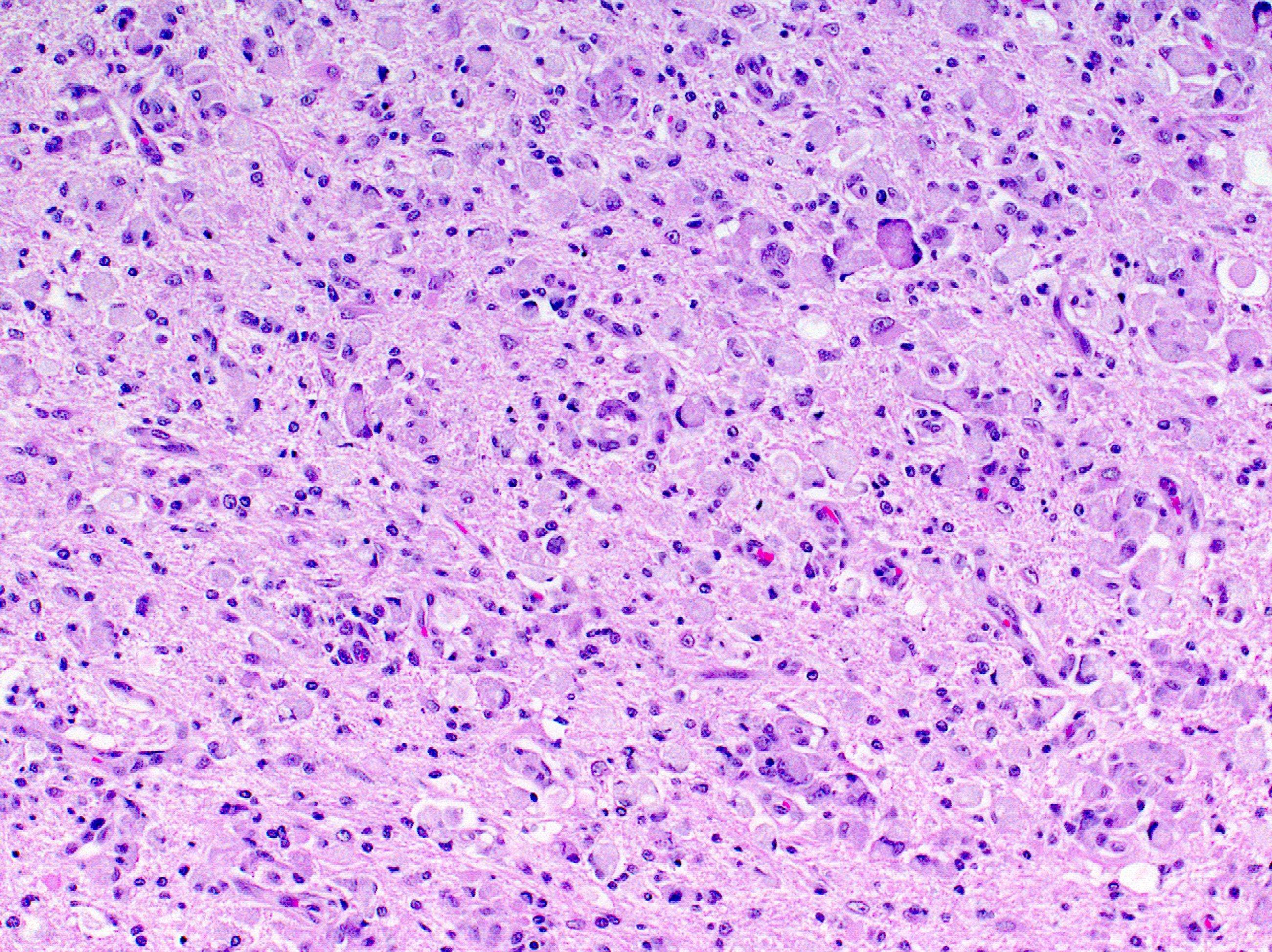
- Methods: Immunohistochemical staining for lipid metabolism-related proteins [fatty acid synthase (FASN), hormone-sensitive lipase (HSL), carnitine palmitoyltransferase IA (CPT-1A), acyl- CoA oxidase 1 (ACOX1), fatty acid binding protein 4 (FABP4,) and perilipin 1 (PLIN1)] was performed using a tissue microarray of 149 cases of metastatic breast cancer (bone metastasis = 39, brain metastasis = 37, liver metastasis = 21, and lung metastasis = 52). (elsevierpure.com)
Enzyme8
- In enzymology, an acyl-CoA oxidase (EC 1.3.3.6) is an enzyme that catalyzes the chemical reaction acyl-CoA + O2 ⇌ {\displaystyle \rightleftharpoons } trans-2,3-dehydroacyl-CoA + H2O2 Thus, the two substrates of this enzyme are acyl-CoA and O2, whereas its two products are trans-2,3-dehydroacyl-CoA and H2O2. (wikipedia.org)
- The systematic name of this enzyme class is acyl-CoA:oxygen 2-oxidoreductase. (wikipedia.org)
- The peroxisomal straight-chain acyl-CoA oxidase enzyme plays a role in the breakdown of certain fat molecules called very long-chain fatty acids (VLCFAs). (medlineplus.gov)
- Growth on triolein stimulated increased enzyme activity, especially for acyl-CoA dehydrogenase. (microbiologyresearch.org)
- The enzymes include acyl-COA oxidase fat and malic enzyme. (rostrum.net)
- Propionic acidemia is an autosomal recessive, inherited, metabolic disorder that is caused by a defective form of the enzyme propionyl-coenzyme A (CoA) carboxylase, which results in the accumulation of propionic acid . (medscape.com)
- The enzyme propionyl-CoA carboxylase, which requires biotin as a cofactor, catalyzes conversion of propionyl-CoA to methylmalonyl-CoA. (medscape.com)
- ACL is an enzyme upstream of 3-hydroxy-3-methyl-glutaryl-coenzyme A (HMG-CoA) reductase in the cholesterol biosynthesis pathway. (medscape.com)
Peroxisomal7
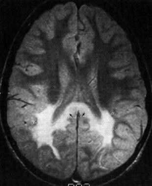
- Peroxisomal acyl-CoA oxidase deficiency is a disorder that causes deterioration of nervous system functions (neurodegeneration) beginning in infancy. (medlineplus.gov)
- Newborns with peroxisomal acyl-CoA oxidase deficiency have weak muscle tone (hypotonia) and seizures. (medlineplus.gov)
- Most babies with peroxisomal acyl-CoA oxidase deficiency learn to walk and begin speaking, but they experience a gradual loss of these skills (developmental regression), usually beginning between the ages of 1 and 3. (medlineplus.gov)
- Most children with peroxisomal acyl-CoA oxidase deficiency do not survive past early childhood. (medlineplus.gov)
- Peroxisomal acyl-CoA oxidase deficiency is a rare disorder. (medlineplus.gov)
- It is unclear exactly how VLCFA accumulation leads to the specific features of peroxisomal acyl-CoA oxidase deficiency. (medlineplus.gov)
- Leukodystrophy is likely involved in the development of the neurological abnormalities that occur in peroxisomal acyl-CoA oxidase deficiency. (medlineplus.gov)
Synthetase2
- acyl-CoA synthetase medium chain famil. (gsea-msigdb.org)
- Bempedoic acid and its active metabolite, ESP15228, require coenzyme A (CoA) activation by very long?chain acyl-CoA synthetase 1 (ACSVL1) to ETC-1002-CoA and ESP15228-CoA, respectively. (medscape.com)
Coenzyme1
- Other names in common use include fatty acyl-CoA oxidase, acyl coenzyme A oxidase, and fatty acyl-coenzyme A oxidase. (wikipedia.org)
Protein1
- Acyl-CoA dehydrogenase domain-containing protein n=1 Tax=Pseudomonas sp. (uma.es)
Peroxisome1
- VK2 enhanced the fatty acid β-oxidation activity in peroxisome to degrade and digest fatty acid-CoA. (frontiersin.org)
Mitochondrial2
- It is apparent that the mitochondrial membrane barrier, which remains intact after sucrose-density-gradient centrifugation, prevents rapid access of acyl-GoA substrates to matrix β oxidation tes. (springer.com)
- Rupturing of the mitochondrial membrane allowed rapid access of acyl CoAs to matrix sites. (springer.com)
Oxidation3
- Nitroalkane oxidase catalyzes the oxidation of neutral nitroalkanes to nitrite and the corresponding aldehydes or ketones. (rcsb.org)
- By comparison, we found that various genes associated with acyl editing, fatty acid β-oxidation, triacylglycerol assembly and oil-body formation had greater expression levels at middle developmental stage (38 DAP), which are consistent with the fast accumulation of EFA in V. galamensis developing seed, implying their fundamental roles in EFA production. (researchsquare.com)
- HPLC analysis after incubation of triolein-grown cell extracts with decanoyl-CoA showed that β-oxidation was limited to one cycle. (microbiologyresearch.org)
Substrates1
- Isoform 2 is active against a much broader range of substrates and shows activity towards very long-chain acyl-CoAs. (swisspalm.org)
Arthrobacter1
- 2112\2112\BAE47462.1\Arthrobacter ureafaciens\Arthrobacter ureafaciens aco gene for acyl-CoA oxidase, completecds. (or.jp)
Catalyzes2
- Catalyzes the desaturation of acyl-CoAs to 2-trans-enoyl-CoAs. (swisspalm.org)
- Catalyzes the desaturation of fatty acyl-CoAs such as palmitoyl-CoA (hexadecanoyl-CoA) to 2-trans-enoyl-CoAs ((2E)-enoyl-CoAs) such as (2E)-hexadecenoyl-CoA, and donates electrons directly to molecular oxygen (O(2)), thereby producing hydrogen peroxide (H(2)O(2)) (PubMed:7876265, PubMed:17458872, PubMed:17603022). (swisspalm.org)
Gene1
- Several genetic mutations, broadly categorized as defects in 2 subunits of the propionyl-CoA carboxylase gene ( PCCA and PCCB ), may give rise to varying levels of functioning propionyl-CoA carboxylase. (medscape.com)
Activity6
- Acyl-CoA oxidase activity in the liver was consistently elevated in males and females (males had higher activity than females) regardless of their exposure during the perinatal period. (nih.gov)
- No acyl-CoA oxidase activity was detected. (microbiologyresearch.org)
- Isoform 1 shows highest activity against medium-chain fatty acyl-CoAs and activity decreases with increasing chain length. (swisspalm.org)
- Shows highest activity against medium-chain fatty acyl-CoAs. (swisspalm.org)
- Shows optimum activity with a chain length of 10 carbons (decanoyl-CoA) in vitro. (swisspalm.org)
- Antisense morpholino oligonucleotides directed at intronic pseudoexons have been shown to increase propionyl-CoA carboxylase activity to normal levels in fibroblast cell lines derived from patients suffering from propionic acidemia. (medscape.com)
19801
- Galliard, T. (1980) Degradation of acyl lipids: hydrolytic and oxidative enzymes. (springer.com)
Desaturation1
- However, our results showed that VK2 can significantly influence the expression of genes related to fat metabolism, including those that regulate fatty acid elongation, desaturation, and synthesis of fatty acid-CoA. (frontiersin.org)
Fatty acids1
- To a lesser degree, cholesterol and odd-chain fatty acids also contribute to propionyl-CoA levels. (medscape.com)
Interproscan2
- Cytochrome c oxidase subunit IV family [Interproscan]. (ntu.edu.sg)
- Zinc-finger domain of monoamine-oxidase A repressor R1 [Interproscan]. (ntu.edu.sg)
Accumulation1
- Accumulation of propionic acid apparently results in an abnormal cytochrome-c oxidase. (medscape.com)
Transporter1
- This process is governed by the ER acetylation machinery: the cytosol:ER-lumen acetyl-CoA transporter AT-1 (also known as SLC33A1), and the ER-resident lysine acetyltransferases ATase1 and ATase2 (also known as NAT8B and NAT8, respectively). (biologists.com)
Peroxisomes1
- This process shortens the VLCFA molecules by two carbon atoms at a time until the VLCFAs are converted to a molecule called acetyl-CoA, which is transported out of the peroxisomes for reuse by the cell. (medlineplus.gov)
Symbol1
- acyl-CoA oxidase 1 [Source:HGNC Symbol. (gsea-msigdb.org)
Member1
- The flavoenzyme nitroalkane oxidase is a member of the acyl-CoA dehydrogenase superfamily. (rcsb.org)
Active1
- Isoform 2 is twice as active as isoform 1 against 16-hydroxy-palmitoyl-CoA and is 25% more active against 1,16-hexadecanodioyl-CoA. (swisspalm.org)
Cycle1
- Consequently, these results provide important structural descriptions of several steps along the nitroalkane oxidase reaction cycle. (rcsb.org)