Acetabulum
Hip Joint
Hip Dislocation, Congenital
Femur Head
Pelvic Bones
Prosthesis Failure
Ischium
Legg-Calve-Perthes Disease
Bone Diseases, Developmental
Femoracetabular Impingement
Osteoarthritis, Hip
Joint Diseases
Cementation
Arthrography
Etretinate
Slipped Capital Femoral Epiphyses
Pelvimetry
Reoperation
Polyethylenes
Ceramics
Traction
Range of Motion, Articular
Bone Cements
Polyethylene
Orthopedic Procedures
Hip
Pelvis
Femur Head Necrosis
Osseointegration
Follow-Up Studies
Tomography, X-Ray Computed
Imaging, Three-Dimensional
Treatment Outcome
Fracture Fixation, Internal
Prosthesis Fitting
Retrospective Studies
Postoperative Complications
Femoral Neck Fractures
Cartilage, Articular
Biomechanical Phenomena
Recovery of Function
Titanium
Long-term results of spherical acetabular osteotomy. (1/945)
We have examined the effect of the Wagner spherical acetabular osteotomy on preserving the joint in 38 hips with a mean follow-up of 17 years. At the time of the initial operation, 55% of patients had clinical symptoms and 30 joints showed minimal or absent radiological signs of osteoarthritis. At follow-up, 54% of patients had a good functional result. The osteotomy improved the mean centre-edge angle from -3 degrees to +15 degrees, the mean anterior centre-edge angle to 23 degrees and the acetabular head index to 75%. The obliquity of the acetabular roof decreased from 28 degrees to 16 degrees. One patient improved, but 14 deteriorated with joint degeneration. Of these, one progressed because of postoperative deep-tissue infection and five due to undercorrection. One patient needed total joint replacement after 14 years. At 17 years after operation, Wagner osteotomy had prevented progression of secondary arthritis in 63% of cases. (+info)Non-operative management of acetabular fractures. The use of dynamic stress views. (2/945)
To assess the stability of the hip after acetabular fracture, dynamic fluoroscopic stress views were taken of 41 acetabular fractures that met the criteria for non-operative management. These included roof arcs of 45 degrees, a subchondral CT arc of 10 mm, displacement of less than 50% of the posterior wall, and congruence on the AP and Judet views of the hip. There were three unstable hips which were treated by open reduction and internal fixation. The remaining 38 fractures were treated non-operatively with early mobilisation and delayed weight-bearing. At a mean follow-up of 2.7 years, the results were good or excellent in 91% of the cases. Three fair results were ascribed to the patients' other injuries. Dynamic stress views can identify subtle instability in patients who would normally be considered for non-operative treatment. (+info)Open reduction and internal fixation of acetabular fractures. (3/945)
Between 1982 and 1995, 84 patients with displaced acetabular fractures underwent open reduction and internal fixation in our institution. The mean follow-up was 5.5 years with a minimum of 2 years. There were 33 simple and 51 complex fractures according to the classification of Judet and Letournels. Reduction after operation was anatomical in 49% of the patients, satisfactory in 24%, and unsatisfactory in 27%. Using Merle d'Aubigne's scale, the clinical results were excellent in 39% of the patients, good in 29%, fair in 8%, and poor in 24%. Factors of statistical significance associated with a poor clinical outcome were T-shaped fractures, unsatisfactory reduction (> 3 mm residual displacement), age > 40 years and development of avascular necrosis. Acetabular surgery is demanding, and a high rate of complications can be expected. Trauma centres should designate a group of surgeons who will consistently treat these fractures in order to obtain more experience and better results. (+info)The influence of weight-bearing on the measurement of polyethylene wear in THA. (4/945)
We have studied the influence of weight-bearing on the measurement of wear of the polyethylene acetabular component in total hip arthroplasty using two techniques. The measured vertical wear was significantly greater when radiographs were taken weight-bearing rather than with the patient supine (p = 0.001, method 1; p = 0.007, method 2). Calculations of rates of linear wear of the acetabular component were significantly underestimated (p < 0.05) when radiographs were taken supine. There are two reasons for this. First, a change in pelvic orientation when bearing weight ensures that the thinnest polyethylene is brought into relief, and secondly, the head of the femoral component assumes the position of maximal displacement along its wear path. Interpretation of previous studies on both linear and volumetric polyethylene wear in total hip arthroplasty should be reassessed in the light of these findings. (+info)Accuracy of EBRA-FCA in the measurement of migration of femoral components of total hip replacement. Einzel-Bild-Rontgen-Analyse-femoral component analysis. (5/945)
Several methods of measuring the migration of the femoral component after total hip replacement have been described, but they use different reference lines, and have differing accuracies, some unproven. Statistical comparison of different studies is rarely possible. We report a study of the EBRA-FCA method (femoral component analysis using Einzel-Bild-Rontgen-Analyse) to determine its accuracy using three independent assessments, including a direct comparison with the results of roentgen stereophotogrammetric analysis (RSA). The accuracy of EBRA-FCA was better than +/- 1.5 mm (95% percentile) with a Cronbach's coefficient alpha for interobserver reliability of 0.84; a very good result. The method had a specificity of 100% and a sensitivity of 78% compared with RSA for the detection of migration of over 1 mm. This is accurate enough to assess the stability of a prosthesis within a relatively limited period. The best reference line for downward migration is between the greater trochanter and the shoulder of the stem, as confirmed by two experimental analyses and a computer-assisted design. (+info)The prediction of failure of the stem in THR by measurement of early migration using EBRA-FCA. Einzel-Bild-Roentgen-Analyse-femoral component analysis. (6/945)
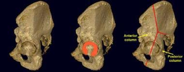
We report the ten-year results for three designs of stem in 240 total hip replacements, for which subsidence had been measured on plain radiographs at regular intervals. Accurate migration patterns could be determined by the method of Einzel-Bild-Roentgen-Analyse-femoral component analysis (EBRA-FCA) for 158 hips (66%). Of these, 108 stems (68%) remained stable throughout, and five (3%) started to migrate after a median of 54 months. Initial migration of at least 1 mm was seen in 45 stems (29%) during the first two years, but these then became stable. We revised 17 stems for aseptic loosening, and 12 for other reasons. Revision for aseptic loosening could be predicted by EBRA-FCA with a sensitivity of 69%, a specificity of 80%, and an accuracy of 79% by the use of a threshold of subsidence of 1.5 mm during the first two years. Similar observations over a five-year period allowed the long-term outcome to be predicted with an accuracy of 91%. We discuss the importance of four different patterns of subsidence and confirm that the early measurement of migration by a reasonably accurate method can help to predict long-term outcome. Such methods should be used to evaluate new and modified designs of prosthesis. (+info)Retroversion of the acetabulum. A cause of hip pain. (7/945)
We describe a little-known variety of hip dysplasia, termed 'acetabular retroversion', in which the alignment of the mouth of the acetabulum does not face the normal anterolateral direction, but inclines more posterolaterally. The condition may be part of a complex dysplasia or a single entity. Other than its retroversion, the acetabulum is sited normally on the side wall of the pelvis, and its articular surface is of normal extent and configuration. The retroverted orientation may give rise to problems of impingement between the femoral neck and anterior acetabular edge. We define the clinical and radiological parameters and discuss pathological changes which may occur in the untreated condition. A technique of management is proposed. (+info)Improving the detection of acetabular osteolysis using oblique radiographs. (8/945)
Visualisation of periacetabular osteolysis by standard anteroposterior (AP) radiographs underestimates the extent of bone loss around a metal-backed acetabular component. We have assessed the effectiveness of standard radiological views in depicting periacetabular osteolysis, and recommend additional projections which make these lesions more visible. This was accomplished using a computerised simulation of radiological views and a radiological analysis of simulated defects placed at regular intervals around the perimeter of a cadaver acetabulum. The AP view alone showed only 38% of the defects over all of the surface of the cup and failed to depict a 3 mm lesion over 83% of the cup. When combined with the AP view, additional 45 degree obturator-oblique and iliac-oblique projections increased the depiction, showing 81% of the defects. The addition of the 60 degree obturator-oblique view further improved the visualisation of posterior defects, increasing the rate of detection to 94%. Based on this analysis, we recommend using at least three radiographic views when assessing the presence and extent of acetabular osteolysis. (+info)The acetabulum is the cup-shaped cavity in the pelvic bone (specifically, the os coxa) where the head of the femur bone articulates to form the hip joint. It provides a stable and flexible connection between the lower limb and the trunk, allowing for a wide range of movements such as flexion, extension, abduction, adduction, rotation, and circumduction. The acetabulum is lined with articular cartilage, which facilitates smooth and frictionless movement of the hip joint. Its stability is further enhanced by various ligaments, muscles, and the labrum, a fibrocartilaginous rim that deepens the socket and increases its contact area with the femoral head.
'Acetabularia' is a genus of large, single-celled marine algae that are commonly found in warm and temperate coastal waters. These algae are characterized by their distinctive umbrella-shaped cap, known as the "acetabulum," which sits atop a long, slender stalk. The acetabulum contains reproductive structures, while the stalk contains the nucleus of the cell. 'Acetabularia' species are notable for their ability to survive and grow even when their nuclei are removed, making them a subject of interest in studies of cell biology and regeneration.
The hip joint, also known as the coxal joint, is a ball-and-socket type synovial joint that connects the femur (thigh bone) to the pelvis. The "ball" is the head of the femur, while the "socket" is the acetabulum, a concave surface on the pelvic bone.
The hip joint is surrounded by a strong fibrous capsule and is reinforced by several ligaments, including the iliofemoral, ischiofemoral, and pubofemoral ligaments. The joint allows for flexion, extension, abduction, adduction, medial and lateral rotation, and circumduction movements, making it one of the most mobile joints in the body.
The hip joint is also supported by various muscles, including the gluteus maximus, gluteus medius, gluteus minimus, iliopsoas, and other hip flexors and extensors. These muscles provide stability and strength to the joint, allowing for weight-bearing activities such as walking, running, and jumping.
Congenital hip dislocation, also known as developmental dysplasia of the hip (DDH), is a condition where the hip joint fails to develop normally in utero or during early infancy. In a healthy hip, the head of the femur (thigh bone) fits snugly into the acetabulum (hip socket). However, in congenital hip dislocation, the femoral head is not held firmly in place within the acetabulum due to abnormal development or laxity of the ligaments that support the joint.
There are two types of congenital hip dislocations:
1. Teratologic dislocation: This type is present at birth and occurs due to abnormalities in the development of the hip joint during fetal growth. The femoral head may be completely outside the acetabulum or partially dislocated.
2. Developmental dysplasia: This type develops after birth, often within the first few months of life, as a result of ligamentous laxity and shallow acetabulum. In some cases, it can progress to a complete hip dislocation if left untreated.
Risk factors for congenital hip dislocation include family history, breech presentation during delivery, and female gender. Early diagnosis and treatment are crucial to prevent long-term complications such as pain, limited mobility, and osteoarthritis. Treatment options may include bracing, closed reduction, or surgical intervention, depending on the severity and age of the child at diagnosis.
The femoral head is the rounded, ball-like top portion of the femur (thigh bone) that fits into the hip socket (acetabulum) to form the hip joint. It has a smooth, articular cartilage surface that allows for smooth and stable articulation with the pelvis. The femoral head is connected to the femoral neck, which is a narrower section of bone that angles downward and leads into the shaft of the femur. Together, the femoral head and neck provide stability and range of motion to the hip joint.
A hip dislocation is a medical emergency that occurs when the head of the femur (thighbone) slips out of its socket in the pelvis. This can happen due to high-energy trauma, such as a car accident or a severe fall. Hip dislocations can also occur in people with certain health conditions that make their hips more prone to displacement, such as developmental dysplasia of the hip.
There are two main types of hip dislocations: posterior and anterior. In a posterior dislocation, the femur head moves out of the back of the socket, which is the most common type. In an anterior dislocation, the femur head moves out of the front of the socket. Both types of hip dislocations can cause severe pain, swelling, and difficulty moving the affected leg.
Immediate medical attention is necessary for a hip dislocation to realign the bones and prevent further damage. Treatment typically involves sedation or anesthesia to relax the muscles around the joint, followed by a closed reduction procedure to gently guide the femur head back into the socket. In some cases, surgery may be required to repair any associated injuries, such as fractures or damaged ligaments. After treatment, physical therapy and rehabilitation are usually necessary to restore strength, mobility, and function to the affected hip joint.
The pelvic bones, also known as the hip bones, are a set of three irregularly shaped bones that connect to form the pelvic girdle in the lower part of the human body. They play a crucial role in supporting the spine and protecting the abdominal and pelvic organs.
The pelvic bones consist of three bones:
1. The ilium: This is the largest and uppermost bone, forming the majority of the hip bone and the broad, flaring part of the pelvis known as the wing of the ilium or the iliac crest, which can be felt on the side of the body.
2. The ischium: This is the lower and back portion of the pelvic bone that forms part of the sitting surface or the "sit bones."
3. The pubis: This is the front part of the pelvic bone, which connects to the other side at the pubic symphysis in the midline of the body.
The pelvic bones are joined together at the acetabulum, a cup-shaped socket that forms the hip joint and articulates with the head of the femur (thigh bone). The pelvic bones also have several openings for the passage of blood vessels, nerves, and reproductive and excretory organs.
The shape and size of the pelvic bones differ between males and females due to their different roles in childbirth and locomotion. Females typically have a wider and shallower pelvis than males to accommodate childbirth, while males usually have a narrower and deeper pelvis that is better suited for weight-bearing and movement.
A hip prosthesis, also known as a total hip replacement, is a surgical implant designed to replace the damaged or diseased components of the human hip joint. The procedure involves replacing the femoral head (the ball at the top of the thigh bone) and the acetabulum (the socket in the pelvis) with artificial parts, typically made from materials such as metal, ceramic, or plastic.
The goal of a hip prosthesis is to relieve pain, improve joint mobility, and restore function, allowing patients to return to their normal activities and enjoy an improved quality of life. The procedure is most commonly performed in individuals with advanced osteoarthritis, rheumatoid arthritis, or other degenerative conditions that have caused significant damage to the hip joint.
There are several different types of hip prostheses available, each with its own unique design and set of benefits and risks. The choice of prosthesis will depend on a variety of factors, including the patient's age, activity level, overall health, and specific medical needs. In general, however, all hip prostheses are designed to provide a durable, long-lasting solution for patients suffering from debilitating joint pain and stiffness.
Osteotomy is a surgical procedure in which a bone is cut to shorten, lengthen, or change its alignment. It is often performed to correct deformities or to realign bones that have been damaged by trauma or disease. The bone may be cut straight across (transverse osteotomy) or at an angle (oblique osteotomy). After the bone is cut, it can be realigned and held in place with pins, plates, or screws until it heals. This procedure is commonly performed on bones in the leg, such as the femur or tibia, but can also be done on other bones in the body.
Hip arthroplasty, also known as hip replacement surgery, is a medical procedure where the damaged or diseased joint surfaces of the hip are removed and replaced with artificial components. These components typically include a metal or ceramic ball that replaces the head of the femur (thigh bone), and a polyethylene or ceramic socket that replaces the acetabulum (hip socket) in the pelvis.
The goal of hip arthroplasty is to relieve pain, improve joint mobility, and restore function to the hip joint. This procedure is commonly performed in patients with advanced osteoarthritis, rheumatoid arthritis, hip fractures, or other conditions that cause significant damage to the hip joint.
There are several types of hip replacement surgeries, including traditional total hip arthroplasty, partial (hemi) hip arthroplasty, and resurfacing hip arthroplasty. The choice of procedure depends on various factors, such as the patient's age, activity level, overall health, and the extent of joint damage.
After surgery, patients typically require rehabilitation to regain strength, mobility, and function in the affected hip. With proper care and follow-up, most patients can expect significant pain relief and improved quality of life following hip arthroplasty.
"Bone retroversion" is not a widely recognized medical term or concept with a specific definition. The term "retroversion" is used in anatomy to describe the position of an organ or structure when it is turned backward or inward. In relation to bones, retroversion typically describes the orientation of a bone or joint when it is angled or positioned in such a way that its posterior (back) aspect faces more anteriorly (toward the front).
However, I was unable to find a widely accepted medical definition for "bone retroversion" as a specific pathological or anatomical condition. It's possible that the term may be used in a more specialized context within certain medical subspecialties. If you have more context or information about where this term is being used, I may be able to provide a more precise answer.
Prosthesis failure is a term used to describe a situation where a prosthetic device, such as an artificial joint or limb, has stopped functioning or failed to meet its intended purpose. This can be due to various reasons, including mechanical failure, infection, loosening of the device, or a reaction to the materials used in the prosthesis.
Mechanical failure can occur due to wear and tear, manufacturing defects, or improper use of the prosthetic device. Infection can also lead to prosthesis failure, particularly in cases where the prosthesis is implanted inside the body. The immune system may react to the presence of the foreign material, leading to inflammation and infection.
Loosening of the prosthesis can also cause it to fail over time, as the device becomes less stable and eventually stops working properly. Additionally, some people may have a reaction to the materials used in the prosthesis, leading to tissue damage or other complications that can result in prosthesis failure.
In general, prosthesis failure can lead to decreased mobility, pain, and the need for additional surgeries or treatments to correct the problem. It is important for individuals with prosthetic devices to follow their healthcare provider's instructions carefully to minimize the risk of prosthesis failure and ensure that the device continues to function properly over time.
The ischium is a part of the pelvic bone, specifically the lower and posterior portion. It is one of the three bones that fuse together to form each half of the pelvis, along with the ilium (the upper and largest portion) and the pubis (anteriorly).
The ischium has a thick, robust structure because it supports our body weight when we sit. Its main parts include:
1. The ischial tuberosity (sitting bone): This is the roughened, weight-bearing portion where you typically feel discomfort after sitting for long periods.
2. The ischial spine: A thin bony projection that serves as an attachment point for various muscles and ligaments.
3. The ramus of the ischium: The slender, curved part that extends downwards and joins with the pubis to form the inferior (lower) portion of the pelvic ring called the obturator foramen.
Together with the other components of the pelvis, the ischium plays a crucial role in providing stability, supporting the lower limbs, and protecting internal organs.
The ilium is the largest and broadest of the three parts that make up the hip bone or coxal bone. It is the uppermost portion of the pelvis and forms the side of the waist. The ilium has a curved, fan-like shape and articulates with the sacrum at the back to form the sacroiliac joint. The large, concave surface on the top of the ilium is called the iliac crest, which can be felt as a prominent ridge extending from the front of the hip to the lower back. This region is significant in orthopedics and physical examinations for its use in assessing various medical conditions and performing certain maneuvers during the physical examination.
Legg-Calve-Perthes disease is a childhood hip disorder that occurs when the blood supply to the ball part of the thigh bone (femoral head) is disrupted. This causes the bone tissue to die, leading to its collapse and deformity. The femoral head then regenerates itself, but often not as round and smooth as it should be, which can lead to hip problems in later life.
The disease is named after three doctors who independently described it: Arthur Legg, Jacques Calve, and Georg Perthes. It typically affects children between the ages of 4 and 10, more commonly boys than girls. Symptoms may include limping, pain in the hip or knee, reduced range of motion in the hip, and muscle wasting. Treatment often involves rest, physical therapy, and sometimes surgery to realign or reshape the femoral head.
Developmental bone diseases are a group of medical conditions that affect the growth and development of bones. These diseases are present at birth or develop during childhood and adolescence, when bones are growing rapidly. They can result from genetic mutations, hormonal imbalances, or environmental factors such as poor nutrition.
Some examples of developmental bone diseases include:
1. Osteogenesis imperfecta (OI): Also known as brittle bone disease, OI is a genetic disorder that affects the body's production of collagen, a protein necessary for healthy bones. People with OI have fragile bones that break easily and may also experience other symptoms such as blue sclerae (whites of the eyes), hearing loss, and joint laxity.
2. Achondroplasia: This is the most common form of dwarfism, caused by a genetic mutation that affects bone growth. People with achondroplasia have short limbs and a large head relative to their body size.
3. Rickets: A condition caused by vitamin D deficiency or an inability to absorb or use vitamin D properly. This leads to weak, soft bones that can bow or bend easily, particularly in children.
4. Fibrous dysplasia: A rare bone disorder where normal bone is replaced with fibrous tissue, leading to weakened bones and deformities.
5. Scoliosis: An abnormal curvature of the spine that can develop during childhood or adolescence. While not strictly a developmental bone disease, scoliosis can be caused by various underlying conditions such as cerebral palsy, muscular dystrophy, or spina bifida.
Treatment for developmental bone diseases varies depending on the specific condition and its severity. Treatment may include medication, physical therapy, bracing, or surgery to correct deformities and improve function. Regular follow-up with a healthcare provider is essential to monitor growth, manage symptoms, and prevent complications.
Prosthesis design is a specialized field in medical device technology that involves creating and developing artificial substitutes to replace a missing body part, such as a limb, tooth, eye, or internal organ. The design process typically includes several stages: assessment of the patient's needs, selection of appropriate materials, creation of a prototype, testing and refinement, and final fabrication and fitting of the prosthesis.
The goal of prosthesis design is to create a device that functions as closely as possible to the natural body part it replaces, while also being comfortable, durable, and aesthetically pleasing for the patient. The design process may involve collaboration between medical professionals, engineers, and designers, and may take into account factors such as the patient's age, lifestyle, occupation, and overall health.
Prosthesis design can be highly complex, particularly for advanced devices such as robotic limbs or implantable organs. These devices often require sophisticated sensors, actuators, and control systems to mimic the natural functions of the body part they replace. As a result, prosthesis design is an active area of research and development in the medical field, with ongoing efforts to improve the functionality, comfort, and affordability of these devices for patients.
Bone transplantation, also known as bone grafting, is a surgical procedure in which bone or bone-like material is transferred from one part of the body to another or from one person to another. The graft may be composed of cortical (hard outer portion) bone, cancellous (spongy inner portion) bone, or a combination of both. It can be taken from different sites in the same individual (autograft), from another individual of the same species (allograft), or from an animal source (xenograft). The purpose of bone transplantation is to replace missing bone, provide structural support, and stimulate new bone growth. This procedure is commonly used in orthopedic, dental, and maxillofacial surgeries to repair bone defects caused by trauma, tumors, or congenital conditions.
Femoroacetabular impingement (FAI) is a medical condition that affects the hip joint. It occurs when there is abnormal contact between the femoral head (the ball at the top of the thigh bone) and the acetabulum (the socket in the pelvis) during normal movement of the hip. This abnormal contact can cause damage to the cartilage and labrum (a ring of cartilage that helps to stabilize the hip joint) leading to pain, stiffness and decreased range of motion.
FAI is classified into two types: cam impingement and pincer impingement. Cam impingement occurs when there is an abnormal shape of the femoral head or neck, which leads to abnormal contact with the acetabulum during hip flexion and internal rotation. Pincer impingement occurs when there is overcoverage of the acetabulum, leading to abnormal contact with the femoral head or neck.
In some cases, both cam and pincer impingement can be present, which is referred to as mixed impingement. Symptoms of FAI may include hip pain, stiffness, limping, and reduced range of motion. Treatment options for FAI may include physical therapy, activity modification, medications, and in some cases, surgery.
Osteoarthritis (OA) of the hip is a degenerative joint disease that affects the articular cartilage and subchondral bone of the hip joint. It is characterized by the progressive loss of cartilage, remodeling of bone, osteophyte formation (bone spurs), cysts, and mild to moderate inflammation. The degenerative process can lead to pain, stiffness, limited range of motion, and crepitus (grating or crackling sound) during movement.
In the hip joint, OA typically affects the femoral head and acetabulum. As the articular cartilage wears away, the underlying bone becomes exposed and can lead to bone-on-bone contact, which is painful. The body responds by attempting to repair the damage through remodeling of the subchondral bone and formation of osteophytes. However, these changes can further limit joint mobility and exacerbate symptoms.
Risk factors for OA of the hip include age, obesity, genetics, previous joint injury or surgery, and repetitive stress on the joint. Treatment options may include pain management (such as NSAIDs, physical therapy, and injections), lifestyle modifications (such as weight loss and exercise), and, in severe cases, surgical intervention (such as hip replacement).
The femur is the medical term for the thigh bone, which is the longest and strongest bone in the human body. It connects the hip bone to the knee joint and plays a crucial role in supporting the weight of the body and allowing movement during activities such as walking, running, and jumping. The femur is composed of a rounded head, a long shaft, and two condyles at the lower end that articulate with the tibia and patella to form the knee joint.
Joint diseases is a broad term that refers to various conditions affecting the joints, including but not limited to:
1. Osteoarthritis (OA): A degenerative joint disease characterized by the breakdown of cartilage and underlying bone, leading to pain, stiffness, and potential loss of function.
2. Rheumatoid Arthritis (RA): An autoimmune disorder causing inflammation in the synovial membrane lining the joints, resulting in swelling, pain, and joint damage if left untreated.
3. Infectious Arthritis: Joint inflammation caused by bacterial, viral, or fungal infections that spread through the bloodstream or directly enter the joint space.
4. Gout: A type of arthritis resulting from the buildup of uric acid crystals in the joints, typically affecting the big toe and characterized by sudden attacks of severe pain, redness, and swelling.
5. Psoriatic Arthritis (PsA): An inflammatory joint disease associated with psoriasis, causing symptoms such as pain, stiffness, and swelling in the joints and surrounding tissues.
6. Juvenile Idiopathic Arthritis (JIA): A group of chronic arthritis conditions affecting children, characterized by joint inflammation, pain, and stiffness.
7. Ankylosing Spondylitis: A form of arthritis primarily affecting the spine, causing inflammation, pain, and potential fusion of spinal vertebrae.
8. Bursitis: Inflammation of the fluid-filled sacs (bursae) that cushion joints, leading to pain and swelling.
9. Tendinitis: Inflammation or degeneration of tendons, which connect muscles to bones, often resulting in pain and stiffness near joints.
These conditions can impact the function and mobility of affected joints, causing discomfort and limiting daily activities. Proper diagnosis and treatment are essential for managing joint diseases and preserving joint health.
In the medical field, cementation refers to the process of using a type of dental cement or bonding agent to attach a dental restoration (such as a crown, bridge, or false tooth) to a natural tooth or implant. The cement helps to create a strong and secure attachment, while also helping to seal the restoration and prevent the entry of bacteria and saliva.
Dental cement can be made from various materials, including glass ionomers, resin-modified glass ionomers, zinc phosphate, and polycarboxylate cements. The choice of cement depends on several factors, such as the type of restoration being attached, the location in the mouth, and the patient's individual needs and preferences.
Cementation is an important step in many dental procedures, as it helps to ensure the longevity and success of the restoration. Proper technique and material selection are crucial for achieving a successful cementation that will last for years to come.
The pubic bone, also known as the pubis or pubic symphysis, is a part of the pelvis - the complex ring-like structure that forms the lower part of the trunk and supports the weight of the upper body. The pubic bone is the anterior (front) portion of the pelvic girdle, located at the bottom of the abdomen, and it connects to the other side at the pubic symphysis, a cartilaginous joint.
The pubic bone plays an essential role in supporting the lower limbs and providing attachment for various muscles involved in movements like walking, running, and jumping. It also protects some abdominal organs and contributes to the structure of the pelvic outlet, which is crucial during childbirth.
Arthrography is a medical imaging technique used to diagnose problems within joints. It involves the injection of a contrast agent, such as a radiopaque dye or air, into the joint space, followed by the use of fluoroscopy or X-ray imaging to visualize the internal structures of the joint. This can help to identify injuries, tears, or other abnormalities in the cartilage, ligaments, tendons, or bones within the joint.
The procedure is typically performed on an outpatient basis and may be used to diagnose conditions such as shoulder dislocations, rotator cuff tears, meniscal tears in the knee, or hip labral injuries. It is a relatively safe and minimally invasive procedure, although there may be some temporary discomfort or swelling at the injection site. Patients are usually advised to avoid strenuous activity for a day or two following the procedure to allow the contrast agent to fully dissipate from the joint.
Etretinate is a oral retinoid medication that is primarily used in the treatment of severe forms of acne, such as recalcitrant cystic acne or nodular acne. It works by decreasing the production of sebum (oil) and promoting the shedding of skin cells, which helps to prevent the formation of comedones (blackheads and whiteheads) and reduce inflammation in the skin.
Etretinate is a derivative of vitamin A and is known for its long-term persistence in the body, with a half-life of approximately 120 days. This means that it can take several months for the drug to be completely eliminated from the body after stopping treatment. As a result, etretinate is usually considered a second-line treatment option for acne and is typically reserved for cases that have not responded to other therapies.
It's important to note that etretinate is a teratogenic medication, which means that it can cause birth defects if taken during pregnancy. Therefore, it should not be used by women who are pregnant or planning to become pregnant, and effective contraception must be used during treatment and for several months after stopping the drug.
Other potential side effects of etretinate include dry skin, dry mouth, nosebleeds, hair loss, muscle aches, and elevated liver enzymes. It may also increase the risk of bone fractures and can interact with other medications, such as tetracyclines, that can increase the risk of intracranial hypertension.
A bone fracture is a medical condition in which there is a partial or complete break in the continuity of a bone due to external or internal forces. Fractures can occur in any bone in the body and can vary in severity from a small crack to a shattered bone. The symptoms of a bone fracture typically include pain, swelling, bruising, deformity, and difficulty moving the affected limb. Treatment for a bone fracture may involve immobilization with a cast or splint, surgery to realign and stabilize the bone, or medication to manage pain and prevent infection. The specific treatment approach will depend on the location, type, and severity of the fracture.
Heterotopic ossification (HO) is a medical condition where bone tissue forms outside the skeleton, in locations where it does not typically exist. This process can occur in various soft tissues, such as muscles, tendons, ligaments, or even inside joint capsules. The abnormal bone growth can lead to pain, stiffness, limited range of motion, and, in some cases, loss of function in the affected area.
There are several types of heterotopic ossification, including:
1. Myositis ossificans - This form is often associated with trauma or injury, such as muscle damage from a fracture, surgery, or direct blow. It typically affects young, active individuals and usually resolves on its own within months to a few years.
2. Neurogenic heterotopic ossification (NHO) - Also known as "traumatic heterotopic ossification," this form is often linked to spinal cord injuries, brain injuries, or central nervous system damage. NHO can cause significant impairment and may require surgical intervention in some cases.
3. Fibrodysplasia ossificans progressiva (FOP) - This rare, genetic disorder causes progressive heterotopic ossification throughout the body, starting in early childhood. The condition significantly impacts mobility and quality of life, with no known cure.
The exact mechanisms behind heterotopic ossification are not fully understood, but it is believed that a combination of factors, including inflammation, tissue injury, and genetic predisposition, contribute to its development. Treatment options may include nonsteroidal anti-inflammatory drugs (NSAIDs), radiation therapy, physical therapy, or surgical removal of the abnormal bone growth, depending on the severity and location of the HO.
Slipped Capital Femoral Epiphyses (SCFE) is a pediatric orthopedic condition that affects the growth plate (epiphysis) at the top of the thigh bone (femur). In SCFE, the epiphysis slips or shifts off the end of the femur, leading to abnormal hip function and potentially causing pain, stiffness, and limping. This condition typically occurs during periods of rapid growth, particularly in early adolescence, and is more common in overweight children. If left untreated, SCFE can result in significant long-term complications such as osteoarthritis or avascular necrosis (death of bone tissue due to lack of blood supply). Early diagnosis and appropriate medical intervention are crucial for optimal outcomes.
Pelvimetry is a medical measurement and evaluation of the size and shape of the pelvis, which can be performed in several ways:
1. Clinical pelvimetry: This involves physical examination to assess the dimensions of the pelvis by palpation and measurement of the distance between bony landmarks.
2. Radiological pelvimetry: This uses X-ray or CT imaging to obtain more accurate measurements of the pelvic diameters, including the anteroposterior, transverse, and oblique dimensions.
3. Magnetic resonance imaging (MRI) pelvimetry: This method is considered the most accurate for assessing the size and shape of the pelvis, as it provides detailed images without radiation exposure.
Pelvimetry is often used in obstetrics to evaluate whether a woman's pelvis can accommodate a fetus during childbirth (known as "obstetric pelvimetry"). It helps healthcare providers determine if a vaginal delivery is possible or if a cesarean section may be necessary. However, the use of pelvimetry in modern obstetrics has become less common due to its limited predictive value and the increasing focus on individualized birth management.
Hip injuries refer to damages or harm caused to the hip joint or its surrounding structures, including bones, muscles, tendons, ligaments, and cartilage. These injuries can occur due to various reasons such as falls, accidents, sports-related activities, or degenerative conditions. Common hip injuries include fractures, dislocations, strains, sprains, bursitis, and labral tears. Symptoms may include pain, swelling, bruising, stiffness, limited mobility, and inability to bear weight on the affected leg. Proper diagnosis and treatment are crucial to ensure optimal recovery and prevent long-term complications.
A reoperation is a surgical procedure that is performed again on a patient who has already undergone a previous operation for the same or related condition. Reoperations may be required due to various reasons, such as inadequate initial treatment, disease recurrence, infection, or complications from the first surgery. The nature and complexity of a reoperation can vary widely depending on the specific circumstances, but it often carries higher risks and potential complications compared to the original operation.
I believe there may be some confusion in your question as Polyethylenes are not a medical term, but rather a category of synthetic polymers commonly used in various industrial and medical applications. Here's a brief overview:
Polyethylene (PE) is a type of thermoplastic polymer made from the monomer ethylene. It is a versatile material with numerous applications due to its chemical resistance, durability, and flexibility. There are several types of polyethylenes, including:
1. Low-density polyethylene (LDPE): This type has a lower density and more branching in its molecular structure, which results in less crystallinity. LDPE is known for its flexibility and is often used in packaging films, bags, and containers.
2. High-density polyethylene (HDPE): HDPE has a higher density and less branching, resulting in greater crystallinity. It is more rigid than LDPE and is commonly used in applications such as bottles, pipes, and containers.
3. Linear low-density polyethylene (LLDPE): This type combines the flexibility of LDPE with some of the strength and rigidity of HDPE. LLDPE has fewer branches than LDPE but more than HDPE. It is often used in film applications, such as stretch wrap and agricultural films.
4. Ultra-high molecular weight polyethylene (UHMWPE): UHMWPE has an extremely high molecular weight, resulting in exceptional wear resistance, impact strength, and chemical resistance. It is commonly used in medical applications, such as orthopedic implants and joint replacements, due to its biocompatibility and low friction coefficient.
While polyethylenes are not a medical term per se, they do have significant medical applications, particularly UHMWPE in orthopedic devices.
In the field of medicine, ceramics are commonly referred to as inorganic, non-metallic materials that are made up of compounds such as oxides, carbides, and nitrides. These materials are often used in medical applications due to their biocompatibility, resistance to corrosion, and ability to withstand high temperatures. Some examples of medical ceramics include:
1. Bioceramics: These are ceramic materials that are used in medical devices and implants, such as hip replacements, dental implants, and bone grafts. They are designed to be biocompatible, which means they can be safely implanted into the body without causing an adverse reaction.
2. Ceramic coatings: These are thin layers of ceramic material that are applied to medical devices and implants to improve their performance and durability. For example, ceramic coatings may be used on orthopedic implants to reduce wear and tear, or on cardiovascular implants to prevent blood clots from forming.
3. Ceramic membranes: These are porous ceramic materials that are used in medical filtration systems, such as hemodialysis machines. They are designed to selectively filter out impurities while allowing essential molecules to pass through.
4. Ceramic scaffolds: These are three-dimensional structures made of ceramic material that are used in tissue engineering and regenerative medicine. They provide a framework for cells to grow and multiply, helping to repair or replace damaged tissues.
Overall, medical ceramics play an important role in modern healthcare, providing safe and effective solutions for a wide range of medical applications.
Traction, in medical terms, refers to the application of a pulling force to distract or align parts of the body, particularly bones, joints, or muscles, with the aim of immobilizing, reducing displacement, or realigning them. This is often achieved through the use of various devices such as tongs, pulleys, weights, or specialized traction tables. Traction may be applied manually or mechanically and can be continuous or intermittent, depending on the specific medical condition being treated. Common indications for traction include fractures, dislocations, spinal cord injuries, and certain neurological conditions.
Articular Range of Motion (AROM) is a term used in physiotherapy and orthopedics to describe the amount of movement available in a joint, measured in degrees of a circle. It refers to the range through which synovial joints can actively move without causing pain or injury. AROM is assessed by measuring the degree of motion achieved by active muscle contraction, as opposed to passive range of motion (PROM), where the movement is generated by an external force.
Assessment of AROM is important in evaluating a patient's functional ability and progress, planning treatment interventions, and determining return to normal activities or sports participation. It is also used to identify any restrictions in joint mobility that may be due to injury, disease, or surgery, and to monitor the effectiveness of rehabilitation programs.
Bone cements are medical-grade materials used in orthopedic and trauma surgery to fill gaps between bone surfaces and implants, such as artificial joints or screws. They serve to mechanically stabilize the implant and provide a smooth, load-bearing surface. The two most common types of bone cement are:
1. Polymethylmethacrylate (PMMA) cement: This is a two-component system consisting of powdered PMMA and liquid methyl methacrylate monomer. When mixed together, they form a dough-like consistency that hardens upon exposure to air. PMMA cement has been widely used for decades in joint replacement surgeries, such as hip or knee replacements.
2. Calcium phosphate (CP) cement: This is a two-component system consisting of a powdered CP compound and an aqueous solution. When mixed together, they form a paste that hardens through a chemical reaction at body temperature. CP cement has lower mechanical strength compared to PMMA but demonstrates better biocompatibility, bioactivity, and the ability to resorb over time.
Both types of bone cements have advantages and disadvantages, and their use depends on the specific surgical indication and patient factors.
I'm sorry for any confusion, but "Polyethylene" is not a medical term. It is a type of synthetic plastic that is commonly used in various industrial and consumer products. Medical definitions typically refer to substances or conditions related to human health, treatment, or disease processes. If you have any questions related to medical terminology or concepts, I'd be happy to help!
A comminuted fracture is a type of bone break where the bone is shattered into three or more pieces. This type of fracture typically occurs after high-energy trauma, such as a car accident or a fall from a great height. Commminuted fractures can also occur in bones that are weakened by conditions like osteoporosis or cancer. Because of the severity and complexity of comminuted fractures, they often require extensive treatment, which may include surgery to realign and stabilize the bone fragments using metal screws, plates, or rods.
Orthopedic procedures are surgical or nonsurgical methods used to treat musculoskeletal conditions, including injuries, deformities, or diseases of the bones, joints, muscles, ligaments, and tendons. These procedures can range from simple splinting or casting to complex surgeries such as joint replacements, spinal fusions, or osteotomies (cutting and repositioning bones). The primary goal of orthopedic procedures is to restore function, reduce pain, and improve the quality of life for patients.
In medical terms, the hip is a ball-and-socket joint where the rounded head of the femur (thigh bone) fits into the cup-shaped socket, also known as the acetabulum, of the pelvis. This joint allows for a wide range of movement in the lower extremities and supports the weight of the upper body during activities such as walking, running, and jumping. The hip joint is surrounded by strong ligaments, muscles, and tendons that provide stability and enable proper functioning.
The pelvis is the lower part of the trunk, located between the abdomen and the lower limbs. It is formed by the fusion of several bones: the ilium, ischium, and pubis (which together form the hip bone on each side), and the sacrum and coccyx in the back. The pelvis has several functions including supporting the weight of the upper body when sitting, protecting the lower abdominal organs, and providing attachment for muscles that enable movement of the lower limbs. In addition, it serves as a bony canal through which the reproductive and digestive tracts pass. The pelvic cavity contains several vital organs such as the bladder, parts of the large intestine, and in females, the uterus, ovaries, and fallopian tubes.
Femoral head necrosis, also known as avascular necrosis of the femoral head, is a medical condition that results from the interruption of blood flow to the femoral head, which is the rounded end of the thigh bone that fits into the hip joint. This lack of blood supply can cause the bone tissue to die, leading to the collapse of the femoral head and eventually resulting in hip joint damage or arthritis.
The condition can be caused by a variety of factors, including trauma, alcohol abuse, corticosteroid use, radiation therapy, and certain medical conditions such as sickle cell disease and lupus. Symptoms may include pain in the hip or groin, limited range of motion, and difficulty walking. Treatment options depend on the severity and progression of the necrosis and may include medication, physical therapy, or surgical intervention.
Osseointegration is a direct structural and functional connection between living bone and the surface of an implant. It's a process where the bone grows in and around the implant, which is typically made of titanium or another biocompatible material. This process provides a solid foundation for dental prosthetics, such as crowns, bridges, or dentures, or for orthopedic devices like artificial limbs. The success of osseointegration depends on various factors, including the patient's overall health, the quality and quantity of available bone, and the surgical technique used for implant placement.
Osteolysis is a medical term that refers to the loss or resorption of bone tissue. It's a process where the body's normal bone remodeling cycle is disrupted, leading to an imbalance between bone formation and bone breakdown. This results in the progressive deterioration and destruction of bone.
Osteolysis can occur due to various reasons such as chronic inflammation, mechanical stress, or certain medical conditions like rheumatoid arthritis, Paget's disease, or bone tumors. It can also be a side effect of some medications, such as those used in cancer treatment or for managing osteoporosis.
In severe cases, osteolysis can lead to weakened bones, increased risk of fractures, and deformities. Treatment typically aims to address the underlying cause and may include medication, surgery, or lifestyle changes.
Follow-up studies are a type of longitudinal research that involve repeated observations or measurements of the same variables over a period of time, in order to understand their long-term effects or outcomes. In medical context, follow-up studies are often used to evaluate the safety and efficacy of medical treatments, interventions, or procedures.
In a typical follow-up study, a group of individuals (called a cohort) who have received a particular treatment or intervention are identified and then followed over time through periodic assessments or data collection. The data collected may include information on clinical outcomes, adverse events, changes in symptoms or functional status, and other relevant measures.
The results of follow-up studies can provide important insights into the long-term benefits and risks of medical interventions, as well as help to identify factors that may influence treatment effectiveness or patient outcomes. However, it is important to note that follow-up studies can be subject to various biases and limitations, such as loss to follow-up, recall bias, and changes in clinical practice over time, which must be carefully considered when interpreting the results.
X-ray computed tomography (CT or CAT scan) is a medical imaging method that uses computer-processed combinations of many X-ray images taken from different angles to produce cross-sectional (tomographic) images (virtual "slices") of the body. These cross-sectional images can then be used to display detailed internal views of organs, bones, and soft tissues in the body.
The term "computed tomography" is used instead of "CT scan" or "CAT scan" because the machines take a series of X-ray measurements from different angles around the body and then use a computer to process these data to create detailed images of internal structures within the body.
CT scanning is a noninvasive, painless medical test that helps physicians diagnose and treat medical conditions. CT imaging provides detailed information about many types of tissue including lung, bone, soft tissue and blood vessels. CT examinations can be performed on every part of the body for a variety of reasons including diagnosis, surgical planning, and monitoring of therapeutic responses.
In computed tomography (CT), an X-ray source and detector rotate around the patient, measuring the X-ray attenuation at many different angles. A computer uses this data to construct a cross-sectional image by the process of reconstruction. This technique is called "tomography". The term "computed" refers to the use of a computer to reconstruct the images.
CT has become an important tool in medical imaging and diagnosis, allowing radiologists and other physicians to view detailed internal images of the body. It can help identify many different medical conditions including cancer, heart disease, lung nodules, liver tumors, and internal injuries from trauma. CT is also commonly used for guiding biopsies and other minimally invasive procedures.
In summary, X-ray computed tomography (CT or CAT scan) is a medical imaging technique that uses computer-processed combinations of many X-ray images taken from different angles to produce cross-sectional images of the body. It provides detailed internal views of organs, bones, and soft tissues in the body, allowing physicians to diagnose and treat medical conditions.
A cadaver is a deceased body that is used for medical research or education. In the field of medicine, cadavers are often used in anatomy lessons, surgical training, and other forms of medical research. The use of cadavers allows medical professionals to gain a deeper understanding of the human body and its various systems without causing harm to living subjects. Cadavers may be donated to medical schools or obtained through other means, such as through consent of the deceased or their next of kin. It is important to handle and treat cadavers with respect and dignity, as they were once living individuals who deserve to be treated with care even in death.
Three-dimensional (3D) imaging in medicine refers to the use of technologies and techniques that generate a 3D representation of internal body structures, organs, or tissues. This is achieved by acquiring and processing data from various imaging modalities such as X-ray computed tomography (CT), magnetic resonance imaging (MRI), ultrasound, or confocal microscopy. The resulting 3D images offer a more detailed visualization of the anatomy and pathology compared to traditional 2D imaging techniques, allowing for improved diagnostic accuracy, surgical planning, and minimally invasive interventions.
In 3D imaging, specialized software is used to reconstruct the acquired data into a volumetric model, which can be manipulated and viewed from different angles and perspectives. This enables healthcare professionals to better understand complex anatomical relationships, detect abnormalities, assess disease progression, and monitor treatment response. Common applications of 3D imaging include neuroimaging, orthopedic surgery planning, cancer staging, dental and maxillofacial reconstruction, and interventional radiology procedures.
Treatment outcome is a term used to describe the result or effect of medical treatment on a patient's health status. It can be measured in various ways, such as through symptoms improvement, disease remission, reduced disability, improved quality of life, or survival rates. The treatment outcome helps healthcare providers evaluate the effectiveness of a particular treatment plan and make informed decisions about future care. It is also used in clinical research to compare the efficacy of different treatments and improve patient care.
Fracture fixation, internal, is a surgical procedure where a fractured bone is fixed using metal devices such as plates, screws, or rods that are implanted inside the body. This technique helps to maintain the alignment and stability of the broken bone while it heals. The implants may be temporarily or permanently left inside the body, depending on the nature and severity of the fracture. Internal fixation allows for early mobilization and rehabilitation, which can result in a faster recovery and improved functional outcome.
Prosthesis fitting is the process of selecting, designing, fabricating, and fitting a prosthetic device to replace a part of an individual's body that is missing due to congenital absence, illness, injury, or amputation. The primary goal of prosthesis fitting is to restore the person's physical function, mobility, and independence, as well as improve their overall quality of life.
The process typically involves several steps:
1. Assessment: A thorough evaluation of the patient's medical history, physical condition, and functional needs is conducted to determine the most appropriate type of prosthesis. This may include measurements, castings, or digital scans of the residual limb.
2. Design: Based on the assessment, a customized design plan is created for the prosthetic device, taking into account factors such as the patient's lifestyle, occupation, and personal preferences.
3. Fabrication: The prosthesis is manufactured using various materials, components, and techniques to meet the specific requirements of the patient. This may involve the use of 3D printing, computer-aided design (CAD), or traditional handcrafting methods.
4. Fitting: Once the prosthesis is fabricated, it is carefully fitted to the patient's residual limb, ensuring optimal comfort, alignment, and stability. Adjustments may be made as needed to achieve the best fit and function.
5. Training: The patient receives training on how to use and care for their new prosthetic device, including exercises to strengthen the residual limb and improve overall mobility. Follow-up appointments are scheduled to monitor progress, make any necessary adjustments, and provide ongoing support.
Retrospective studies, also known as retrospective research or looking back studies, are a type of observational study that examines data from the past to draw conclusions about possible causal relationships between risk factors and outcomes. In these studies, researchers analyze existing records, medical charts, or previously collected data to test a hypothesis or answer a specific research question.
Retrospective studies can be useful for generating hypotheses and identifying trends, but they have limitations compared to prospective studies, which follow participants forward in time from exposure to outcome. Retrospective studies are subject to biases such as recall bias, selection bias, and information bias, which can affect the validity of the results. Therefore, retrospective studies should be interpreted with caution and used primarily to generate hypotheses for further testing in prospective studies.
Bone screws are medical devices used in orthopedic and trauma surgery to affix bone fracture fragments or to attach bones to other bones or to metal implants such as plates, rods, or artificial joints. They are typically made of stainless steel or titanium alloys and have a threaded shaft that allows for purchase in the bone when tightened. The head of the screw may have a hexagonal or star-shaped design to allow for precise tightening with a screwdriver. Bone screws come in various shapes, sizes, and designs, including fully threaded, partially threaded, cannulated (hollow), and headless types, depending on their intended use and location in the body.
Postoperative complications refer to any unfavorable condition or event that occurs during the recovery period after a surgical procedure. These complications can vary in severity and may include, but are not limited to:
1. Infection: This can occur at the site of the incision or inside the body, such as pneumonia or urinary tract infection.
2. Bleeding: Excessive bleeding (hemorrhage) can lead to a drop in blood pressure and may require further surgical intervention.
3. Blood clots: These can form in the deep veins of the legs (deep vein thrombosis) and can potentially travel to the lungs (pulmonary embolism).
4. Wound dehiscence: This is when the surgical wound opens up, which can lead to infection and further complications.
5. Pulmonary issues: These include atelectasis (collapsed lung), pneumonia, or respiratory failure.
6. Cardiovascular problems: These include abnormal heart rhythms (arrhythmias), heart attack, or stroke.
7. Renal failure: This can occur due to various reasons such as dehydration, blood loss, or the use of certain medications.
8. Pain management issues: Inadequate pain control can lead to increased stress, anxiety, and decreased mobility.
9. Nausea and vomiting: These can be caused by anesthesia, opioid pain medication, or other factors.
10. Delirium: This is a state of confusion and disorientation that can occur in the elderly or those with certain medical conditions.
Prompt identification and management of these complications are crucial to ensure the best possible outcome for the patient.
The "femur neck" is the narrow, upper part of the femur (thigh bone) where it connects to the pelvis. It is the region through which the femoral head articulates with the acetabulum to form the hip joint. The femur neck is a common site for fractures, especially in older adults with osteoporosis.
Arthroscopy is a minimally invasive surgical procedure where an orthopedic surgeon uses an arthroscope (a thin tube with a light and camera on the end) to diagnose and treat problems inside a joint. The surgeon makes a small incision, inserts the arthroscope into the joint, and then uses the attached camera to view the inside of the joint on a monitor. They can then insert other small instruments through additional incisions to repair or remove damaged tissue.
Arthroscopy is most commonly used for joints such as the knee, shoulder, hip, ankle, and wrist. It offers several advantages over traditional open surgery, including smaller incisions, less pain and bleeding, faster recovery time, and reduced risk of infection. The procedure can be used to diagnose and treat a wide range of conditions, including torn ligaments or cartilage, inflamed synovial tissue, loose bone or cartilage fragments, and joint damage caused by arthritis.
A femoral neck fracture is a type of hip fracture that occurs in the narrow, vertical section of bone just below the ball of the femur (thigh bone) that connects to the hip socket. This area is called the femoral neck. Femoral neck fractures can be categorized into different types based on their location and the direction of the fractured bone.
These fractures are typically caused by high-energy trauma, such as car accidents or falls from significant heights, in younger individuals. However, in older adults, particularly those with osteoporosis, femoral neck fractures can also result from low-energy trauma, like a simple fall from standing height.
Femoral neck fractures are often serious and require prompt medical attention. Treatment usually involves surgery to realign and stabilize the broken bone fragments, followed by rehabilitation to help regain mobility and strength. Potential complications of femoral neck fractures include avascular necrosis (loss of blood flow to the femoral head), nonunion or malunion (improper healing), and osteoarthritis in the hip joint.
Articular cartilage is the smooth, white tissue that covers the ends of bones where they come together to form joints. It provides a cushion between bones and allows for smooth movement by reducing friction. Articular cartilage also absorbs shock and distributes loads evenly across the joint, protecting the bones from damage. It is avascular, meaning it does not have its own blood supply, and relies on the surrounding synovial fluid for nutrients. Over time, articular cartilage can wear down or become damaged due to injury or disease, leading to conditions such as osteoarthritis.
Biomechanics is the application of mechanical laws to living structures and systems, particularly in the field of medicine and healthcare. A biomechanical phenomenon refers to a observable event or occurrence that involves the interaction of biological tissues or systems with mechanical forces. These phenomena can be studied at various levels, from the molecular and cellular level to the tissue, organ, and whole-body level.
Examples of biomechanical phenomena include:
1. The way that bones and muscles work together to produce movement (known as joint kinematics).
2. The mechanical behavior of biological tissues such as bone, cartilage, tendons, and ligaments under various loads and stresses.
3. The response of cells and tissues to mechanical stimuli, such as the way that bone tissue adapts to changes in loading conditions (known as Wolff's law).
4. The biomechanics of injury and disease processes, such as the mechanisms of joint injury or the development of osteoarthritis.
5. The use of mechanical devices and interventions to treat medical conditions, such as orthopedic implants or assistive devices for mobility impairments.
Understanding biomechanical phenomena is essential for developing effective treatments and prevention strategies for a wide range of medical conditions, from musculoskeletal injuries to neurological disorders.
"Recovery of function" is a term used in medical rehabilitation to describe the process in which an individual regains the ability to perform activities or tasks that were previously difficult or impossible due to injury, illness, or disability. This can involve both physical and cognitive functions. The goal of recovery of function is to help the person return to their prior level of independence and participation in daily activities, work, and social roles as much as possible.
Recovery of function may be achieved through various interventions such as physical therapy, occupational therapy, speech-language therapy, and other rehabilitation strategies. The specific approach used will depend on the individual's needs and the nature of their impairment. Recovery of function can occur spontaneously as the body heals, or it may require targeted interventions to help facilitate the process.
It is important to note that recovery of function does not always mean a full return to pre-injury or pre-illness levels of ability. Instead, it often refers to the person's ability to adapt and compensate for any remaining impairments, allowing them to achieve their maximum level of functional independence and quality of life.
Titanium is not a medical term, but rather a chemical element (symbol Ti, atomic number 22) that is widely used in the medical field due to its unique properties. Medically, it is often referred to as a biocompatible material used in various medical applications such as:
1. Orthopedic implants: Titanium and its alloys are used for making joint replacements (hips, knees, shoulders), bone plates, screws, and rods due to their high strength-to-weight ratio, excellent corrosion resistance, and biocompatibility.
2. Dental implants: Titanium is also commonly used in dental applications like implants, crowns, and bridges because of its ability to osseointegrate, or fuse directly with bone tissue, providing a stable foundation for replacement teeth.
3. Cardiovascular devices: Titanium alloys are used in the construction of heart valves, pacemakers, and other cardiovascular implants due to their non-magnetic properties, which prevent interference with magnetic resonance imaging (MRI) scans.
4. Medical instruments: Due to its resistance to corrosion and high strength, titanium is used in the manufacturing of various medical instruments such as surgical tools, needles, and catheters.
In summary, Titanium is a chemical element with unique properties that make it an ideal material for various medical applications, including orthopedic and dental implants, cardiovascular devices, and medical instruments.
Bone neoplasms are abnormal growths or tumors that develop in the bone. They can be benign (non-cancerous) or malignant (cancerous). Benign bone neoplasms do not spread to other parts of the body and are rarely a threat to life, although they may cause problems if they grow large enough to press on surrounding tissues or cause fractures. Malignant bone neoplasms, on the other hand, can invade and destroy nearby tissue and may spread (metastasize) to other parts of the body.
There are many different types of bone neoplasms, including:
1. Osteochondroma - a benign tumor that develops from cartilage and bone
2. Enchondroma - a benign tumor that forms in the cartilage that lines the inside of the bones
3. Chondrosarcoma - a malignant tumor that develops from cartilage
4. Osteosarcoma - a malignant tumor that develops from bone cells
5. Ewing sarcoma - a malignant tumor that develops in the bones or soft tissues around the bones
6. Giant cell tumor of bone - a benign or occasionally malignant tumor that develops from bone tissue
7. Fibrosarcoma - a malignant tumor that develops from fibrous tissue in the bone
The symptoms of bone neoplasms vary depending on the type, size, and location of the tumor. They may include pain, swelling, stiffness, fractures, or limited mobility. Treatment options depend on the type and stage of the tumor but may include surgery, radiation therapy, chemotherapy, or a combination of these treatments.
"Weight-bearing" is a term used in the medical field to describe the ability of a body part or limb to support the weight or pressure exerted upon it, typically while standing, walking, or performing other physical activities. In a clinical setting, healthcare professionals often use the term "weight-bearing exercise" to refer to physical activities that involve supporting one's own body weight, such as walking, jogging, or climbing stairs. These exercises can help improve bone density, muscle strength, and overall physical function, particularly in individuals with conditions affecting the bones, joints, or muscles.
In addition, "weight-bearing" is also used to describe the positioning of a body part during medical imaging studies, such as X-rays or MRIs. For example, a weight-bearing X-ray of the foot or ankle involves taking an image while the patient stands on the affected limb, allowing healthcare providers to assess any alignment or stability issues that may not be apparent in a non-weight-bearing position.
 Acetabulum (unit) - Wikipedia
 Acetabulum Fractures: Practice Essentials, Anatomy, Pathophysiology
 Patient specific surgical guide improve the accuracy of acetabular component placement in total hip arthroplasty with...
 Timing for Definitive Fixation for Pelvis and Acetabulum Fractures
 SICOT Course on Pelvic and Acetabulum Injuries with Cadaveric Dissection | SICOT
 Combined Acetabulum Fracture and Hip Dislocation in an 18-Year-Old Female at 35-Week Gestation: A Case Report and Review of the...
 Coxal: acetabulum | ArchéoZoothèque
 Insecticidal Effect of Two Lichen Species Extracts Ramalina farinacea (L.) Ach. and Parmelia acetabulum (Neck.) Duby on adults...
 Acetabulum
 ORIF Acetabulum 2
 Salter vs Pemberton: Comparative Radiologic Analysis of Changes in the Acetabulum and Pelvis After Surgical Correction in...
 Helvella acetabulum · paprastasis taurėaukšlis | TYT.LT
 Fractures of the pelvis and acetabulum
 Arthritic Acetabulum Close-up : Medical Illustration
 OITE Review - Trauma 11: Acetabulum - Orthohub
 Acetabulum Archives - PRIVY - PRI Video for You
 BJ5507B Acetabulum reaming drill - Buy Product on bojin
 Seaweed Umbrella alga a ombrello acetabularia acetabulum Intotheblue.it
 Acetabulum Cup System - Lepu Medical Technology(Beijing)Co.,Ltd.
 Glossary | eSkeletons
 wade allison | Adlibris
 View of Functional and radiological outcome in surgically managed posterior wall and column fractures of acetabulum
 Case Report: Osteoid Osteoma of the Acetabulum Treated With Arthroscopy-assisted Radiofrequency Ablation - Fondazione Livio...
 Single cell genome analysis of an uncultured heterotrophic stramenopile | Scientific Reports