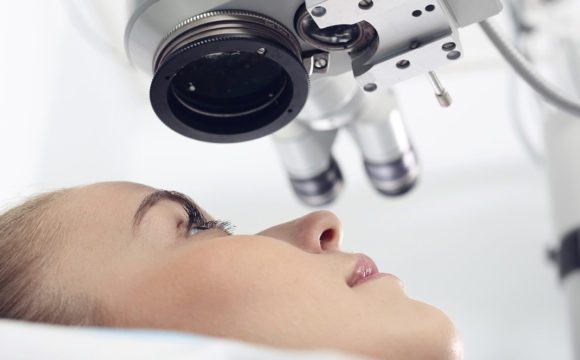
Aberrometry
Corneal Wavefront Aberration
Corneal Topography
Refractive Errors
Impact of scattering and spherical aberration in contrast sensitivity. (1/47)
(+info)Accommodative lag and fluctuations when optical aberrations are manipulated. (2/47)
(+info)Blur on the retina due to higher-order aberrations: comparison of eye growth models to experimental data. (3/47)
(+info)Peripheral optical errors and their change with accommodation differ between emmetropic and myopic eyes. (4/47)
(+info)Higher-order aberrations produce orientation-specific notches in the defocused contrast sensitivity function. (5/47)
(+info)Experimental validation of a Bayesian model of visual acuity. (6/47)
(+info)Myopia and peripheral ocular aberrations. (7/47)
(+info)A contralateral eye study comparing apodized diffractive and full diffractive lenses: wavefront analysis and distance and near uncorrected visual acuity. (8/47)
(+info)Aberrometry is a medical diagnostic technique used to measure the amount and type of aberration or distortion in the optical system of the eye. It is often used to evaluate the quality of vision, particularly in cases where traditional methods of measuring visual acuity are not sufficient.
During an aberrometry test, the patient looks into a specialized instrument called a wavefront sensor while a series of light patterns are projected onto the retina. The sensor then measures how the light is distorted as it passes through the eye's optical system, including the cornea and lens. This information is used to create a detailed map of the eye's aberrations, which can help doctors identify any irregularities that may be contributing to visual symptoms such as blurred vision, glare, or halos around lights.
Aberrometry is often used in conjunction with other diagnostic tests to evaluate patients who are considering refractive surgery, such as LASIK or PRK. By identifying any abnormalities in the eye's optical system, doctors can determine whether a patient is a good candidate for surgery and make more informed decisions about how to proceed with treatment.
Corneal wavefront aberration is a measurement of the irregularities in the shape and curvature of the cornea, which can affect the way light enters the eye and is focused on the retina. A wavefront aberration test uses a device to measure the refraction of light as it passes through the cornea and calculates the degree of any distortions or irregularities in the wavefront of the light. This information can be used to guide treatment decisions, such as the prescription for eyeglasses or contact lenses, or the planning of a surgical procedure to correct the aberration.
Corneal wavefront aberrations can be classified into two types: low-order and high-order aberrations. Low-order aberrations include myopia (nearsightedness), hyperopia (farsightedness), and astigmatism, which are common refractive errors that can be easily corrected with glasses or contact lenses. High-order aberrations are more complex irregularities in the wavefront of light that cannot be fully corrected with traditional eyeglasses or contact lenses. These may include coma, trefoil, and spherical aberration, among others.
High-order corneal wavefront aberrations can affect visual quality, causing symptoms such as glare, halos around lights, and decreased contrast sensitivity. They are often associated with conditions that cause changes in the shape of the cornea, such as keratoconus or corneal surgery. In some cases, high-order aberrations can be corrected with specialized contact lenses or refractive surgery procedures such as wavefront-guided LASIK or PRK.
Corneal topography is a non-invasive medical imaging technique used to create a detailed map of the surface curvature of the cornea, which is the clear, dome-shaped surface at the front of the eye. This procedure provides valuable information about the shape and condition of the cornea, helping eye care professionals assess various eye conditions such as astigmatism, keratoconus, and other corneal abnormalities. It can also be used in contact lens fitting, refractive surgery planning, and post-surgical evaluation.
Refractive errors are a group of vision conditions that include nearsightedness (myopia), farsightedness (hyperopia), astigmatism, and presbyopia. These conditions occur when the shape of the eye prevents light from focusing directly on the retina, causing blurred or distorted vision.
Myopia is a condition where distant objects appear blurry while close-up objects are clear. This occurs when the eye is too long or the cornea is too curved, causing light to focus in front of the retina instead of directly on it.
Hyperopia, on the other hand, is a condition where close-up objects appear blurry while distant objects are clear. This happens when the eye is too short or the cornea is not curved enough, causing light to focus behind the retina.
Astigmatism is a condition that causes blurred vision at all distances due to an irregularly shaped cornea or lens.
Presbyopia is a natural aging process that affects everyone as they get older, usually around the age of 40. It causes difficulty focusing on close-up objects and can be corrected with reading glasses, bifocals, or progressive lenses.
Refractive errors can be diagnosed through a comprehensive eye exam and are typically corrected with eyeglasses, contact lenses, or refractive surgery such as LASIK.
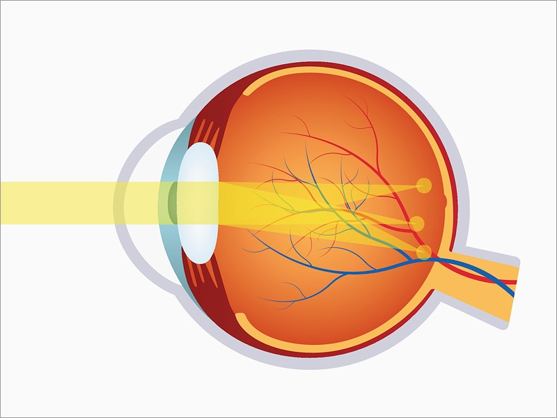
Ocular refraction is a medical term that refers to the bending of light as it passes through the optical media of the eye, including the cornea and lens. This process allows the eye to focus light onto the retina, creating a clear image. The refractive power of the eye is determined by the curvature and transparency of these structures.
In a normal eye, light rays are bent or refracted in such a way that they converge at a single point on the retina, producing a sharp and focused image. However, if the curvature of the cornea or lens is too steep or too flat, the light rays may not converge properly, resulting in a refractive error such as myopia (nearsightedness), hyperopia (farsightedness), or astigmatism.
Ocular refraction can be measured using a variety of techniques, including retinoscopy, automated refraction, and subjective refraction. These measurements are used to determine the appropriate prescription for corrective lenses such as eyeglasses or contact lenses. In some cases, ocular refractive errors may be corrected surgically through procedures such as LASIK or PRK.
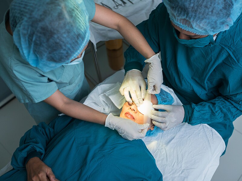
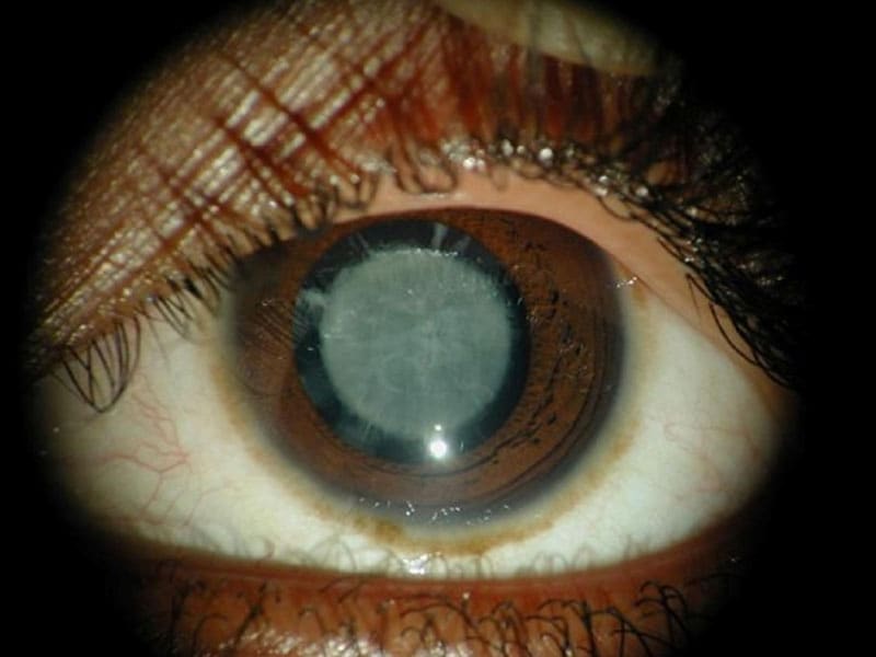
 Cataract surgery
Aberrations of the eye
Allon Barsam
Farhad Hafezi
Intraoperative aberrometry-based aphakia refraction in patients with cataract: status and options | British Journal of...
First clinical results on the feasibility, quality and reproducibility of aberrometry-based intraoperative refraction during...
Anterior lenticonus detected by wavefront aberrometry<...
 Wavefront Aberrometry
Aberrometry | Profiles RNS
 Lecture: Understanding Aberrometry | Cybersight
 Pyramidal Aberrometry in Wavefront-Guided Myopic LASIK.
 Minimizing Astigmatism in Cataract Surgery
 Compressed Wavefront Aberrometry of the Human Eye
Aberrometry and Wavefront Imaging: Historical Perspective, Wavefront Error, Optical Aberrations of the Eye
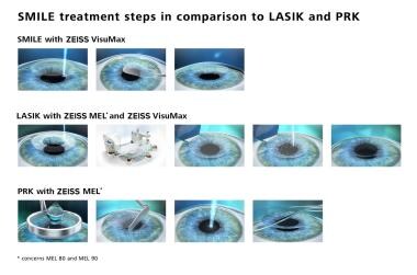
 Randomized Clinical Trial Comparing Femtosecond LASIK and Small-Incision Lenticule Extraction
Cataract surgery - Wikipedia
LASIK Hyperopia: Background, History of the Procedure, Problem
Laser-Assisted Subepithelial Keratectomy (LASEK): Background, History of the Procedure, Problem
 Challenging Cataract Cases - American Academy of Ophthalmology
2020-2021 BCSC Basic and Clinical Science Course™
 Effect of Spherical Aberration on the Optical Quality after Implantation of Two Different Aspherical Intraocular Lenses
 Douglas Donald Koch, M.D. | BCM
 Is wavefront better than LASIK? - Presenternet.com
 YES Intraoperative Techniques and Tools for Success in Refractive Cataract Surgery | ASCRS
 CRSToday | Perfecting Presbyopia- Correcting IOL Surgery
 What are Aberrations of the Eye?
 Evaluation of Anterior and Posterior Corneal Higher Order Aberrations for the Detection of Keratoconus and Suspect Keratoconus...
CRSToday | New Findings in Corneal Refractive Surgery
 Cataract Surgery Following Radial Keratotomy - EyeWiki
 CRSTG | Europe Edition | Sequential Customized Therapeutic Keratectomy
CRSTG | Europe Edition | Satisfaction With a Trifocal IOL in One Eye, Not Both
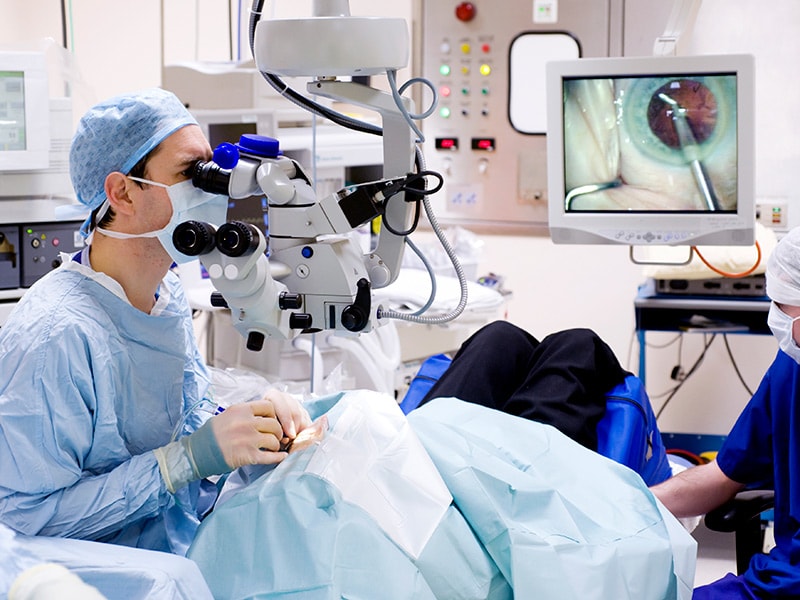
CRSToday | Intraoperative Digital Visualization
 How Kleiman Evangelista Uses Advanced Refractive Technology to Improve Your Vision - KE - Eye Centers of Texas
Corneal topography1
- METHODS: Clinical data, corneal topography, and wavefront aberrometry with separation of corneal and lenticular components of the higher-order aberrations are analyzed in a patient who presented for refractive surgery evaluation. (northwestern.edu)
Cataract4
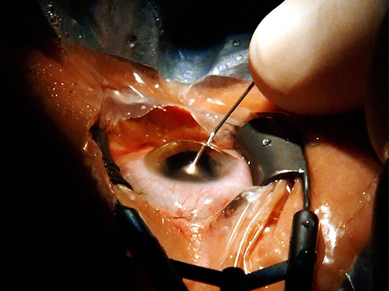
- Numerous studies argue that intraoperative wavefront aberrometry (IWA) may well become an indispensible tool in future cataract surgery. (bmj.com)
- Objective To provide the first clinical data in determining the feasibility, quality and precision of intraoperative wavefront aberrometry (IWA)-based refraction in patients with cataract. (bmj.com)
- Topography, tomography, and aberrometry are used to identify candidates for SCTK before cataract surgery. (crstodayeurope.com)
- If you have not heard this tongue twister previously, let us explain intraoperative wavefront aberrometry & the future of cataract surgery in language you can understand. (southtexaseyeinstitute.com)
Astigmatism3
- That being said, the markerless guidance group showed a statistically significant improvement in mean remaining refractive astigmatism compared with intraoperative aberrometry in this matched cohort. (medscape.com)
- Without intraoperative aberrometry, I find it is difficult to manage astigmatism effectively in premium IOL cases. (crstoday.com)
- Verifying the magnitude and axis of astigmatism with intraoperative aberrometry might also be helpful. (crstodayeurope.com)
Intraoperative wavefront1
- Aim To explore the application of intraoperative wavefront aberrometry (IWA) for aphakia-based biometry using three existing formulae derived from autorefractive retinoscopy and introducing new improved formulae. (bmj.com)
Refraction2
- Investigators found that bilateral wavefront-guided LASIK provided excellent objective outcomes such as visual acuity, refraction, and aberrometry as well as high patient satisfaction and quality of life, as measured by the Refractive Status and Vision Profile questionnaire. (crstoday.com)
- Wavefront aberrometry data generated from the i.Profiler plus® is combined with the binocular aspects of vision from the Subjective Refraction to calculate an ultra precise prescription (i.Scription®) using a proprietary software algorithm. (luzerneoptical.com)
Femtosecond laser1
- This webinar will focus on implementation of the latest intraoperative tools, including femtosecond laser and intraoperative aberrometry. (ascrs.org)
Refractive2
- CONCLUSIONS: Wavefront aberrometry has an adjunctive and distinctive role in the preoperative screening process for refractive surgery candidates and in those with subtle unexplained loss of best-corrected visual acuity. (northwestern.edu)
- Intraoperative aberrometry, which relies not on keratometric power but rather on the total refractive error of the eye, is a good option for determining IOL power in post-LVC eyes. (aao.org)
Preoperative1
- It also facilitates a comparison of the preoperative biometry and intraoperative aberrometry measurements to help ensure that the optimal IOL power is chosen (>Figure 5). (crstoday.com)
Pachymetry1
- Topography, Aberrometry and Pachymetry are pre-operative tests required. (eyeheal.in)
MeSH1
- Aberrometry" is a descriptor in the National Library of Medicine's controlled vocabulary thesaurus, MeSH (Medical Subject Headings) . (ouhsc.edu)
Tomography1
- Each step of SCTK entails the use of tomography, topography, and aberrometry followed by the execution of a corneal wavefront-guided customized ablation pattern and wet PTK (ie, smoothing) with a tomographic quality check at the end. (crstodayeurope.com)
Surgical1
- The two cases presented herein illustrate how intraoperative aberrometry helps me make surgical decisions that benefit my patients receiving presbyopia-correcting IOLs. (crstoday.com)
Clinical4
- RESULTS: Clinical evaluation indicated that the cause of visual loss was lenticular, and wavefront aberrometry indicated high negative spherical aberration, leading to the diagnosis of anterior lenticonus. (northwestern.edu)
- This lecture discusses the principles of aberrometry, and interpretation of aberrometry maps and its clinical uses. (cybersight.org)
- And welcome to one more webinar on the continuation of the topography that we discussed last evening, and aberrometry in clinical practice. (cybersight.org)
- Applications of ocular aberrometry in clinical practice. (omiq.es)
Ocular2
- There are a variety of methods that can be used to assess ocular aberrations including Shack-Hartmann wavefront sensing, Tscherning, spatially resolved refractometry and optical path difference aberrometry. (news-medical.net)
- This review outlines key issues surround- ing the measurement of ocular aberrations for patients with keratoconus, with a particular focus on the possible factors affecting the repeatability of Hartmann-Shack aberrometry measurements. (manchester.ac.uk)
Illustrate1
- PURPOSE: To illustrate the utility of wavefront aberrometry in delineating a subtle lenticular abnormality responsible for decreased best-corrected visual acuity in a patient. (northwestern.edu)
LASIK1
- Pyramidal Aberrometry in Wavefront-Guided Myopic LASIK. (csoitalia.it)
Outcomes1
- Solomon and Ladas compared outcomes using intraoperative aberrometry and intraoperative markerless guidance for toric IOL placement. (medscape.com)
Objective1
- Dynamic stimulation aberrometry enables objective measurement of the range of accommodation after the implantation of accommodating IOL such as an accommodating lens, allowing physicians to determine optimal wavefront correction and individualize treatment. (ophthalmologytimes.com)
Toric2
- Intraoperative aberrometry has become a valuable tool for all toric and presbyopia-correcting IOL cases in my practice. (crstoday.com)
- Osiris data can be combined with the topographic maps from other instruments produced by CSO, combining the total aberrometry with the corneal ones of Antares, Sirius or MS-39 it is possible to calculate the wavefront internal component and, for example, to evaluate the impact of a toric system on vision. (csoitalia.it)
Keratoconus1
- Corneal aberrometry may be of value in screening for keratoconus in populations with a high prevalence of the disease. (preprints.org)
Visual1
- Asimetrías en la topografía e índice de refracción de la superficie corneal que afectan a la agudeza visual. (bvsalud.org)
Technology1
- Wavefront technology, or aberrometry, diagnoses both lower- and higher-order vision errors represented by the way the eye refracts or focuses light. (presenternet.com)
Total1
- This graph shows the total number of publications written about "Aberrometry" by people in this website by year, and whether "Aberrometry" was a major or minor topic of these publications. (ouhsc.edu)
Study1
- This was a consecutive contralateral eye study, where the first eye was randomly assigned to aberrometry or markerless guidance, and the fellow eye received the other modality. (medscape.com)
Quality1
- The intraoperative tomographic scan and aberrometry are used to check the quantity and quality of residual stromal HOAs and evaluate whether residual stromal thickness corresponds with the surgeon's expectations. (crstodayeurope.com)