Abdominal Cavity
Abdominal Neoplasms
Pneumoperitoneum
Peritoneal Lavage
Setaria Nematode
Abdominal Wall
Setariasis
Surgical Sponges
Emphysematous Cholecystitis
Peritoneal Cavity
Laparoscopy
Abdominal Muscles
Omentum
Peritonitis
Cryptorchidism
Abdomen, Acute
Peritoneum
Foreign-Body Migration
Injections, Intraperitoneal
Rupture, Spontaneous
Cholecystectomy, Laparoscopic
Ovarian Neoplasms
Tomography, X-Ray Computed
Nasal Cavity
Fatal Outcome
Treatment Outcome
Postoperative Complications
Disease Models, Animal
Rats, Wistar
Dental Cavity Preparation
Immunohistochemistry
Pleural Cavity
Mouth
Schistosomiasis mansoni in low transmission areas: abdominal ultrasound. (1/97)
In endemic areas with low prevalence and low intensity of infection, the diagnosis of hepatic pathology due to the Schistosoma mansoni infection is very difficult. In order to establish the hepatic morbidity, a double-blind study was achieved in Venezuelan endemic areas, with one group of patients with schistosomiasis and the other one of non-infected people, that were evaluated clinically and by abdominal ultrasound using the Cairo classification. Schistosomiasis diagnosis was established based on parasitologic and serological tests. The increase of the hepatic size at midclavicular and midsternal lines (in hepatometry) and the hard liver consistency were the clinical parameters able to differentiate infected persons from non infected ones, as well as the presence of left lobe hepatomegaly detected by abdominal ultrasound. The periportal thickening, especially the mild form, was frequent in all age groups in both infected and uninfected patients. There was not correlation between the intensity of infection and ultrasound under the current circumstances. Our data suggest that in Venezuela, a low endemic area of transmission of schistosomiasis, the hepatic morbidity is mild and uncommon. The Cairo classification seems to overestimate the prevalence of periportal pathology. The specificity of the method must be improved, especially for the recognition of precocious pathology. Other causes of hepatopathies must be investigated. (+info)Sympathetic neural activation in visceral obesity. (2/97)
BACKGROUND: Muscle sympathetic nerve activity (MSNA) is elevated in obese humans. However, the potential role of abdominal visceral fat as an important adipose tissue depot linking obesity to elevated MSNA has not been explored. Accordingly, we tested the hypothesis that MSNA would be increased in men (age=18 to 40 years, body mass index < or =35 kg/m2) with higher abdominal visceral fat (HAVF; n=13, abdominal visceral fat=118.1+/-15.8 cm2) compared with their age- (28.7+/-2.4 versus 25.5+/-2.0 years, P>0.05), total fat mass-matched (20.6+/-2.1 versus 20.8+/-2.4 kg, P>0.05) and abdominal subcutaneous fat-matched (230.6+/-24.9 versus 261.4+/-34.8 cm(2), P>0.05) peers with lower abdominal visceral fat levels (LAVF; n=13, visceral fat= 73.0+/-6.0 cm2). METHODS AND RESULTS: MSNA (microneurography), body composition (dual energy x-ray absorptiometry), and abdominal visceral and subcutaneous fat (computed tomography) were measured in 37 sedentary men across a wide range of adiposity. MSNA was approximately 55% higher in men with HAVF compared with men with LAVF (33+/-4 versus 21+/-2 bursts/min, P<0.05). Furthermore, MSNA was more closely associated with the level of abdominal visceral fat (r=0.65, P<0.05) than total fat mass (r=0.323, P<0.05) or abdominal subcutaneous fat (r=0.27, P=0.05). The relation between MSNA and abdominal visceral fat was independent of total body fat (r=0.61, P<0.05). CONCLUSIONS: The results of our study indicate that MSNA is elevated in men with visceral obesity. Our observations are consistent with the idea that abdominal visceral fat is an important adipose tissue depot linking obesity with sympathetic neural activation in humans. Furthermore, these findings may have important implications for understanding the increased risk of developing cardiovascular diseases in individuals with visceral obesity. (+info)The effect of intra-abdominal pressure on the generation of 8-iso prostaglandin F2alpha during laparoscopy in rabbits. (3/97)
BACKGROUND: Carbon dioxide pneumoperitoneum induces peritoneal oxidative stress. The aim of this study was to verify the effect of intra-abdominal pressure on oxidative stress in the peritoneum and on post-operative adhesion formation. METHODS: Forty-one rabbits underwent laparoscopic surgery: either gasless, or with CO(2)-pneumoperitoneum at pressures of 5, 10 or 15 mmHg. Serial parietal peritoneal biopsies were taken at various time-points: immediately after reaching the abdominal cavity, 30, 60, 90 and 120 min afterwards, and 15 min after abdominal desufflation. 8-iso prostaglandin F(2alpha) (8-iso PGF(2alpha)), a marker of oxidative stresss, was assayed by enzyme immunoassay and adhesion formation was scored by second-look laparoscopy on day 14. RESULTS: The gasless group showed no significant changes in 8-iso PGF(2alpha). Conversely, significant changes occurred in CO(2)-pneumoperitoneum in a time- and pressure-dependent manner. Adhesions developed only in the CO(2)-pneumoperitoneum groups, and total adhesion score was correlated with the amount of CO(2) insufflated and intra-abdominal pressure, but not with 8-iso PGF(2alpha), which was correlated with intra-abdominal pressure. CONCLUSION: Intra-abdominal pressure increased 8-iso PGF(2alpha) in the parietal peritoneum in a graded fashion, whilst gasless laparoscopy had no impact. It also influenced the frequency and severity of adhesion formation, but no causal link was found between 8-iso PGF(2alpha) and post-operative adhesion formation. (+info)Granulomatous uveitis associated with vaccination in the atlantic salmon. (4/97)
This study addressed histologic and immunopathologic changes in ocular tissues and investigated the distribution of major histocompatibility class II (MHC class II)-positive cells in Atlantic salmon (Salmo salar) suffering from severe postvaccination disease. Twenty-nine fish with generalized inflammation, probably a result of vaccination, were investigated. One individual that had escaped vaccination was included in the study. Material was investigated by cultivation methods for fungi and bacteria. Histology using conventional staining procedures and immunohistochemistry with antisera against MHC class II beta chain were performed. No growth was observed from the cultivation investigations. Histology revealed occlusion of the lumen in the larger choroid vessels and in the choriocapillaris, inflammatory infiltrations and loss of structure in the choroid rete, and, in some cases, aggregations of multinucleated giant cells (MGC) and Splendore-Hoeppli material. Immunohistochemistry demonstrated massive MHC class II+ cellular infiltrations in the uveal tract. Such infiltrations were also seen in the ventral ciliary cleft, a condition that is associated with glaucoma. Immunoreactive cells included dendritelike cells, epithelioid cells, and MGCs. The endothelia of smaller vessels were frequently MHC class II+, and immunoreactive infiltrations were seen in the optic nerve in several individuals. No pathologic changes were detected in the unvaccinated individual. In conclusion, generalized inflammatory reactions in fish may lead to severe ocular inflammation, occlusion of uveal vessels, and perivascular changes with MHC class II+ upregulation in cells in the uveal tract and optic nerve. (+info)Abdominal wall closure with ePTFE--Goretex Dual Mesh after detensive laparotomy for abdominal compartment syndrome. (5/97)
INTRODUCTION: Detensive laparotomy is the first choice treatment for abdominal compartment syndrome (ACS). Tension free closure of the abdominal wall with the use of prosthesis is a broadly diffused technique; the polypropylene and the ePTFE (expanded polytetrafluoroethylene--Goretex Dual Mesh) are the most commonly used materials. MATERIALS AND METHODS: We report our experience on five patients affected by ACS submitted to detensive laparotomy and positioning of a wide Goretex Dual Mesh prosthesis. RESULTS: In our initial experience ACS has been treated with success through detensive laparotomy and there were no complications related to the use of Goretex. DISCUSSION: Even though limited, our initial clinical experience is favorable to the use of Goretex Dual Mesh as first choice material for reconstruction of the abdominal wall after detensive laparotomy for ACS. (+info)Moderate increase in intraabdominal pressure attenuates gastric mucosal oxygen saturation in patients undergoing laparoscopy. (6/97)
BACKGROUND: Perioperative disturbances of microvascular blood flow and oxygenation in the intestinal tract have been hypothesized to play an important role in development of the multiple organ dysfunction syndrome. Herein, increased intra-abdominal pressure (IAP) has been identified as a key factor in the initiation of the pathophysiologic cascade. The authors hypothesized that increasing the IAP by intraperitoneal insufflation of carbon dioxide attenuates microvascular oxygen saturation in gastric mucosa. They tested this hypothesis in a prospective, observational study in 16 patients scheduled to undergo elective diagnostic laparoscopy. METHODS: The authors continuously assessed microvascular oxygen saturation in gastric mucosa by reflectance spectrophotometry. Simultaneously systemic oxygen saturation, heart rate, arterial blood pressure, and ventilation-derived variables were measured noninvasively. During general anesthesia and controlled mechanical ventilation, baseline values were obtained. Thereafter, the IAP was increased to 8 and 12 mmHg, respectively, followed by a control period after desufflation. RESULTS: The increase in IAP from baseline to 8 mmHg decreased microvascular oxygen saturation in gastric mucosa from 69+/-7% (mean +/- SD) to 63+/-8% at 8 mmHg IAP (P <0.05), with a further significant reduction to 54+/-13% at 12 mmHg IAP (P <0.01). Microvascular oxygen saturation in gastric mucosa recovered rapidly to baseline level (66 +/- 10%) after release of increased IAP. In striking contrast to regional mucosal oxygen saturation, systemic oxygenation did not change with either of the interventions. CONCLUSIONS: The results suggest that increasing intraabdominal pressure to moderate levels, commonly applied to induce a surgical pneumoperitoneum, decreases gastric mucosal oxygen saturation. (+info)Intraperitoneal injection of lipopolysaccharide induces dynamic migration of Gr-1high polymorphonuclear neutrophils in the murine abdominal cavity. (7/97)
Intraperitoneal injection of lipopolysaccharide (LPS; 100 microg) in mice resulted in the disappearance of almost all proteose peptone-induced polymorphonuclear neutrophils (PMNs) with high-level fluorescence for the cell surface marker Gr-1 (Gr-1(high)) at 15 min postinjection, followed by doubling of their proportion at 30 min postinjection. High staining levels of 3'-acetyl-2'-carboxyl-6',7'-(dihyropyran-2'-one)-5 or 6-carboxyfluorescein diacethoxylmethyl ester-labeled PMNs injected into the peritoneal cavity were detected in mesenteric lymph nodes 15 min postinjection of LPS. Therefore, the time of decrease of Gr-1(high) PMNs coincided with that of the increase in cell accumulation in mesenteric lymph nodes. Since milk fat globule-EGF factor 8 (MFG-E8), which is secreted by macrophages, bound many PMNs exhibiting Gr-1(high) and Gr-1(medium) at 30 min postinjection of LPS, the staining level of annexin V on those cells was very low because its binding site is the same as the receptor for MFG-E8. At 60 min postinjection of LPS, the proportion of Gr-1(high) PMNs decreased, and almost all Gr-1(medium) PMNs tended to shift to the right compared with those at 30 min postinjection. The geomeans of Toll-like receptor 4 (TLR4) expression on PMNs at 15, 30, and 60 min postinjection of LPS were 63, 66, and 24%, respectively, compared with that on normal PMNs, indicating that the expression of TLR4 decreases in response to exposure to LPS. Our results suggest that LPS induced PMN death and that many PMNs expressing Gr-1(high) undergo apoptosis 180 min postinjection of LPS. (+info)Potential therapeutic role of histatin derivative P-113d in experimental rat models of Pseudomonas aeruginosa sepsis. (8/97)
BACKGROUND: Morbidity and mortality from Pseudomonas aeruginosa sepsis remain high despite the availability of antibiotics to which the microorganism is sensitive. METHODS: The in vitro activity of histatin derivative P-113d was investigated against Pseudomonas aeruginosa. In addition, its in vivo efficacy was studied in 3 rat models of infection: intraperitoneal injection of 1 mg of P. aeruginosa 10 lipopolysachharide, intraperitoneal injection of 2 x 10(10) cfu of P. aeruginosa ATCC 27853, and intra-abdominal sepsis induced by cecal ligation and puncture. Rats received isotonic sodium chloride solution parenterally (control groups), 1 mg of P-113d/kg of body weight, 1 mg of polymyxin B/kg of body weight, or 20 mg of imipenem/kg of body weight. Main outcomes measured were abdominal exudate and plasma bacterial growth, plasma concentrations of endotoxin and tumor necrosis factor (TNF)- alpha, and lethality. RESULTS: The in vivo studies showed that all compounds reduced lethality, when compared with results for the control group. Overall, P-113d exhibited a slightly lower antimicrobial activity than did imipenem, even though P-113d achieved a substantial decrease in plasma concentrations of endotoxin and TNF- alpha, compared with the imipenem. No statistically significant differences for antimicrobial and antiendotoxin activities were noted between P-113d and polymyxin B. DISCUSSION: These results provide evidence for double antiendotoxin and antimicrobial activity for P-113d and point to its potential use for the treatment of severe infections. (+info)The abdominal cavity is the portion of the abdominothoracic cavity that lies between the diaphragm and the pelvic inlet. It contains the stomach, small intestine, colon, liver, pancreas, spleen, kidneys, adrenal glands, and associated blood vessels and nerves. The abdominal cavity is enclosed by the abdominal wall, which consists of muscles, fascia, and skin. It is divided into several compartments by various membranes, including the peritoneum, a serous membrane that lines the walls of the cavity and covers many of the organs within it. The abdominal cavity provides protection and support for the organs it contains, and also serves as a site for the absorption and digestion of food.
Abdominal neoplasms refer to abnormal growths or tumors in the abdomen that can be benign (non-cancerous) or malignant (cancerous). These growths can occur in any of the organs within the abdominal cavity, including the stomach, small intestine, large intestine, liver, pancreas, spleen, and kidneys.
Abdominal neoplasms can cause various symptoms depending on their size, location, and type. Some common symptoms include abdominal pain or discomfort, bloating, changes in bowel habits, unexplained weight loss, fatigue, and fever. In some cases, abdominal neoplasms may not cause any symptoms until they have grown quite large or spread to other parts of the body.
The diagnosis of abdominal neoplasms typically involves a combination of physical exam, medical history, imaging studies such as CT scans or MRIs, and sometimes biopsy to confirm the type of tumor. Treatment options depend on the type, stage, and location of the neoplasm but may include surgery, radiation therapy, chemotherapy, targeted therapy, or a combination of these approaches.
Pneumoperitoneum is a medical condition characterized by the presence of free air or gas within the peritoneal cavity, which is the space between the lining of the abdominal wall and the internal organs. This accumulation of air can occur due to various reasons such as perforation of an organ (e.g., stomach, intestine, or esophagus), recent surgery, or medical procedures involving the introduction of air into the abdomen.
The presence of pneumoperitoneum is often diagnosed through imaging techniques like X-rays or computed tomography (CT) scans, which can reveal the presence of free gas in the peritoneal cavity. The condition may require prompt medical attention, depending on the underlying cause and the patient's symptoms. Treatment typically involves addressing the underlying cause, such as repairing a perforation or managing an infection.
The abdomen refers to the portion of the body that lies between the thorax (chest) and the pelvis. It is a musculo-fascial cavity containing the digestive, urinary, and reproductive organs. The abdominal cavity is divided into several regions and quadrants for medical description and examination purposes. These include the upper and lower abdomen, as well as nine quadrants formed by the intersection of the midline and a horizontal line drawn at the level of the umbilicus (navel).
The major organs located within the abdominal cavity include:
1. Stomach - muscular organ responsible for initial digestion of food
2. Small intestine - long, coiled tube where most nutrient absorption occurs
3. Large intestine - consists of the colon and rectum; absorbs water and stores waste products
4. Liver - largest internal organ, involved in protein synthesis, detoxification, and metabolism
5. Pancreas - secretes digestive enzymes and hormones such as insulin
6. Spleen - filters blood and removes old red blood cells
7. Kidneys - pair of organs responsible for filtering waste products from the blood and producing urine
8. Adrenal glands - sit atop each kidney, produce hormones that regulate metabolism, immune response, and stress response
The abdomen is an essential part of the human body, playing a crucial role in digestion, absorption, and elimination of food and waste materials, as well as various metabolic processes.
Peritoneal lavage is a medical procedure where a sterile fluid is introduced into the peritoneal cavity, which is the space between the lining of the abdominal wall and the organs within it. The fluid is then allowed to mix with any potentially present infectious or inflammatory material in the cavity. Afterward, the fluid is drained out and sent for laboratory analysis to diagnose various conditions such as bacterial peritonitis or other sources of abdominal infection or inflammation.
The procedure can help identify the presence of infection, determine the type of bacteria causing it, and guide appropriate antibiotic therapy. It is an invasive diagnostic test that requires careful monitoring and proper aseptic technique to avoid complications such as infection or bleeding.
Setaria nematodes are a type of roundworm that belongs to the family Setariidae. These parasitic worms primarily infect birds, but some species can also infect mammals, including humans. The most common Setaria nematode that infects humans is Setaria digitata, which is found mainly in Asia and is transmitted through the consumption of raw or undercooked freshwater snails or fish.
In humans, Setaria nematodes typically cause mild symptoms or are asymptomatic. However, in some cases, they can lead to the development of eosinophilic meningitis, an inflammation of the membranes surrounding the brain and spinal cord, which can cause headaches, stiff neck, fever, and other neurological symptoms.
Setaria nematodes have a complex life cycle that involves several hosts, including snails, fish, and birds. Humans can become accidental hosts when they ingest infective larvae present in contaminated food or water. Once inside the human body, the larvae migrate to various tissues, such as the brain, eyes, or subcutaneous tissue, where they mature into adults and produce eggs. The eggs are then excreted from the body through feces or other bodily fluids.
Preventing Setaria nematode infections involves avoiding the consumption of raw or undercooked freshwater snails or fish and practicing good hygiene, such as washing hands thoroughly before eating or preparing food. In cases where infection occurs, treatment typically involves administering anthelmintic drugs to kill the worms and alleviate symptoms.
The abdominal wall refers to the group of muscles, fascia (sheaths of connective tissue), and skin that make up the front and sides of the abdomen, extending from the thorax (chest) to the pelvis. It provides protection to the abdominal organs, supports the trunk, and allows for movement of the torso.
The main muscles of the anterior abdominal wall include:
1. Rectus sheaths (Rectus Abdominis): paired vertical muscles running from the pubic symphysis to the xiphoid process and costal cartilages of ribs 5-7.
2. External obliques: thin, irregular muscles that lie over the lower part of the abdomen and run diagonally downward and forward from the lower ribs to the iliac crest (pelvic bone) and pubic tubercle.
3. Internal obliques: thicker muscles that lie under the external obliques, running diagonally upward and forward from the iliac crest to the lower ribs.
4. Transverse abdominis: deepest of the abdominal muscles, lying horizontally across the abdomen, attaching from the lower ribs to the pelvis.
These muscles are interconnected by various layers of fascia and aponeuroses (flat, broad tendons), forming a complex structure that allows for both stability and mobility. The linea alba, a fibrous band, runs down the midline of the anterior abdominal wall, connecting the rectus sheaths.
Damage to the abdominal wall can occur due to trauma, surgery, or various medical conditions, which may require surgical intervention for repair.
Peritoneal neoplasms refer to tumors or cancerous growths that develop in the peritoneum, which is the thin, transparent membrane that lines the inner wall of the abdomen and covers the organs within it. These neoplasms can be benign (non-cancerous) or malignant (cancerous). Malignant peritoneal neoplasms are often associated with advanced stages of gastrointestinal, ovarian, or uterine cancers and can spread (metastasize) to other parts of the abdomen.
Peritoneal neoplasms can cause various symptoms such as abdominal pain, bloating, nausea, vomiting, loss of appetite, and weight loss. Diagnosis typically involves imaging tests like CT scans or MRIs, followed by a biopsy to confirm the presence of cancerous cells. Treatment options may include surgery, chemotherapy, radiation therapy, or a combination of these approaches, depending on the type, stage, and location of the neoplasm.
Setariasis is a cutaneous and subcutaneous infestation caused by the larval stage of the parasitic worm, Setaria cervi or Setaria digitata. These worms are commonly known as pork tapeworms or cattle threadworms. The larvae typically migrate to various body tissues, including the eyes, brain, and spinal cord, causing a range of symptoms depending on the site of infection.
In humans, setariasis is usually contracted through the consumption of raw or undercooked meat from infected animals. However, it's essential to note that human infections with Setaria are rare, and the condition is more commonly seen in veterinary medicine.
The symptoms of setariasis can vary widely depending on the location and extent of the infestation. In some cases, there may be no noticeable symptoms at all. However, when symptoms do occur, they might include skin rashes or lesions, eye inflammation or vision problems, neurological symptoms such as headaches, seizures, or difficulty coordinating movements, and in severe cases, organ damage or failure.
Diagnosis of setariasis typically involves a combination of clinical evaluation, imaging studies, and laboratory tests to detect the presence of the parasite's larvae in bodily fluids or tissues. Treatment usually involves anti-parasitic medications to kill the worms, as well as supportive care to manage any associated symptoms or complications.
Chylous ascites is a medical condition characterized by the accumulation of milky, fat-containing fluid in the peritoneal cavity, which is the space within the abdomen that contains the intestines, liver, and other organs. The fluid, called chyle, is normally found in the lymphatic system and is formed when dietary fats are absorbed from the small intestine.
Chylous ascites can occur as a result of damage to the lymphatic vessels that transport chyle from the intestines to the bloodstream. This damage can be caused by various conditions, such as trauma, surgery, tumors, inflammation, or congenital abnormalities. When the lymphatic vessels are damaged, chyle leaks into the peritoneal cavity and accumulates there, leading to ascites.
Symptoms of chylous ascites may include abdominal distension, pain, nausea, vomiting, and weight loss. The condition can be diagnosed through various tests, such as imaging studies or analysis of the fluid in the peritoneal cavity. Treatment typically involves addressing the underlying cause of the condition, as well as managing symptoms and preventing complications. This may include dietary modifications, medications to reduce lymphatic flow, or surgical interventions to repair damaged lymphatic vessels.
Surgical sponges are absorbent, sterile materials used in medical procedures to soak up bodily fluids and help maintain a clean surgical field. They are typically made from gauze material and come in various sizes and shapes to accommodate different surgical needs. Surgical sponges are carefully counted before and after a procedure to ensure that none are accidentally left inside the patient's body.
Emphysematous cholecystitis is a type of acute inflammation of the gallbladder, characterized by the presence of gas within the wall and/or lumen of the gallbladder. It is a severe and potentially life-threatening condition, which typically occurs in patients with diabetes or other underlying medical conditions that compromise their immune system.
The gas that accumulates in the gallbladder in emphysematous cholecystitis can come from several sources, including gas-forming bacteria such as Clostridium perfringens and Escherichia coli. These bacteria produce gas as a byproduct of their metabolism, which can lead to the formation of gas bubbles within the gallbladder.
The symptoms of emphysematous cholecystitis are similar to those of other forms of acute cholecystitis and may include abdominal pain, fever, nausea, vomiting, and decreased appetite. However, the presence of gas within the gallbladder can be detected on imaging studies such as X-rays or computed tomography (CT) scans, which can help to confirm the diagnosis.
Treatment of emphysematous cholecystitis typically involves surgical removal of the gallbladder (cholecystectomy), often through a laparoscopic approach. Antibiotic therapy is also administered to treat any underlying bacterial infection. In severe cases, where the patient's condition is too unstable for surgery, percutaneous drainage of the gallbladder may be performed as a temporary measure to help reduce the risk of complications such as gangrene or perforation.
The peritoneal cavity is the potential space within the abdominal and pelvic regions, bounded by the parietal peritoneum lining the inner aspect of the abdominal and pelvic walls, and the visceral peritoneum covering the abdominal and pelvic organs. It contains a small amount of serous fluid that allows for the gliding of organs against each other during normal physiological activities such as digestion and movement. This cavity can become pathologically involved in various conditions, including inflammation, infection, hemorrhage, or neoplasia, leading to symptoms like abdominal pain, distention, or tenderness.
Laparoscopy is a surgical procedure that involves the insertion of a laparoscope, which is a thin tube with a light and camera attached to it, through small incisions in the abdomen. This allows the surgeon to view the internal organs without making large incisions. It's commonly used to diagnose and treat various conditions such as endometriosis, ovarian cysts, infertility, and appendicitis. The advantages of laparoscopy over traditional open surgery include smaller incisions, less pain, shorter hospital stays, and quicker recovery times.
Tissue adhesions, also known as scar tissue adhesions, are abnormal bands of fibrous tissue that form between two or more internal organs, or between organs and the walls of the chest or abdominal cavity. These adhesions can develop after surgery, infection, injury, radiation, or prolonged inflammation. The fibrous bands can cause pain, restrict movement of the organs, and potentially lead to complications such as bowel obstruction. Treatment options for tissue adhesions may include medication, physical therapy, or surgical intervention to remove the adhesions.
The abdominal muscles, also known as the abdominals or abs, are a group of muscles in the anterior (front) wall of the abdominopelvic cavity. They play a crucial role in maintaining posture, supporting the trunk, and facilitating movement of the torso. The main abdominal muscles include:
1. Rectus Abdominis: These are the pair of long, flat muscles that run vertically along the middle of the anterior abdominal wall. They are often referred to as the "six-pack" muscles due to their visible, segmented appearance in well-trained individuals. The primary function of the rectus abdominis is to flex the spine, allowing for actions such as sitting up from a lying down position or performing a crunch exercise.
2. External Obliques: These are the largest and most superficial of the oblique muscles, located on the lateral (side) aspects of the abdominal wall. They run diagonally downward and forward from the lower ribs to the iliac crest (the upper part of the pelvis) and the pubic tubercle (a bony prominence at the front of the pelvis). The external obliques help rotate and flex the trunk, as well as assist in side-bending and exhalation.
3. Internal Obliques: These muscles lie deep to the external obliques and run diagonally downward and backward from the lower ribs to the iliac crest, pubic tubercle, and linea alba (the strong band of connective tissue that runs vertically along the midline of the abdomen). The internal obliques help rotate and flex the trunk, as well as assist in forced exhalation and increasing intra-abdominal pressure during actions such as coughing or lifting heavy objects.
4. Transversus Abdominis: This is the deepest of the abdominal muscles, located inner to both the internal obliques and the rectus sheath (a strong, fibrous covering that surrounds the rectus abdominis). The transversus abdominis runs horizontally around the abdomen, attaching to the lower six ribs, the thoracolumbar fascia (a broad sheet of connective tissue spanning from the lower back to the pelvis), and the pubic crest (the front part of the pelvic bone). The transversus abdominis helps maintain core stability by compressing the abdominal contents and increasing intra-abdominal pressure.
Together, these muscles form the muscular "corset" of the abdomen, providing support, stability, and flexibility to the trunk. They also play a crucial role in respiration, posture, and various movements such as bending, twisting, and lifting.
Artificial pneumoperitoneum is a medical condition that refers to the presence of air or gas in the peritoneal cavity, which is the space between the lining of the abdominal wall and the organs within the abdomen. This condition is typically created intentionally during surgical procedures, such as laparoscopy, to provide a working space for the surgeon to perform the operation.
During laparoscopic surgery, a thin tube called a trocar is inserted through a small incision in the abdominal wall, and carbon dioxide gas is pumped into the peritoneal cavity to create a pneumoperitoneum. This allows the surgeon to insert specialized instruments through other small incisions and perform the surgery while visualizing the operative field with a camera.
While artificial pneumoperitoneum is generally safe, there are potential complications that can arise, such as injury to surrounding organs or blood vessels during trocar insertion, subcutaneous emphysema (air trapped under the skin), or gas embolism (gas in the bloodstream). These risks are typically minimized through careful technique and monitoring during the procedure.
The omentum, in anatomical terms, refers to a large apron-like fold of abdominal fatty tissue that hangs down from the stomach and loops over the intestines. It is divided into two portions: the greater omentum, which is larger and hangs down further, and the lesser omentum, which is smaller and connects the stomach to the liver.
The omentum has several functions in the body, including providing protection and cushioning for the abdominal organs, assisting with the immune response by containing a large number of immune cells, and helping to repair damaged tissues. It can also serve as a source of nutrients and energy for the body during times of starvation or other stressors.
In medical contexts, the omentum may be surgically mobilized and used to wrap around injured or inflamed tissues in order to promote healing and reduce the risk of infection. This technique is known as an "omentopexy" or "omentoplasty."
Peritoneal diseases refer to a group of conditions that affect the peritoneum, which is the thin, transparent membrane that lines the inner wall of the abdomen and covers the organs within it. The peritoneum has several functions, including providing protection and support to the abdominal organs, producing and absorbing fluids, and serving as a site for the immune system's response to infections and other foreign substances.
Peritoneal diseases can be broadly classified into two categories: infectious and non-infectious. Infectious peritoneal diseases are caused by bacterial, viral, fungal, or parasitic infections that spread to the peritoneum from other parts of the body or through contaminated food, water, or medical devices. Non-infectious peritoneal diseases, on the other hand, are not caused by infections but rather by other factors such as autoimmune disorders, cancer, or chemical irritants.
Some examples of peritoneal diseases include:
1. Peritonitis: Inflammation of the peritoneum due to bacterial or fungal infections, often caused by a ruptured appendix, perforated ulcer, or other abdominal injuries or conditions.
2. Tuberculous peritonitis: A form of peritonitis caused by Mycobacterium tuberculosis, the bacterium that causes tuberculosis (TB).
3. Peritoneal dialysis-associated peritonitis: Infection of the peritoneum in patients undergoing peritoneal dialysis, a type of kidney replacement therapy for patients with end-stage renal disease.
4. Malignant peritoneal mesothelioma: A rare and aggressive form of cancer that affects the mesothelial cells lining the peritoneum, often caused by exposure to asbestos.
5. Systemic lupus erythematosus (SLE): An autoimmune disorder that can cause inflammation and scarring of the peritoneum.
6. Peritoneal carcinomatosis: The spread of cancer cells from other parts of the body to the peritoneum, often seen in patients with advanced ovarian or colorectal cancer.
7. Cirrhotic ascites: Fluid accumulation in the peritoneal cavity due to liver cirrhosis and portal hypertension.
8. Meigs' syndrome: A rare condition characterized by the presence of a benign ovarian tumor, ascites, and pleural effusion.
Peritonitis is a medical condition characterized by inflammation of the peritoneum, which is the serous membrane that lines the inner wall of the abdominal cavity and covers the abdominal organs. The peritoneum has an important role in protecting the abdominal organs and providing a smooth surface for them to move against each other.
Peritonitis can occur as a result of bacterial or fungal infection, chemical irritation, or trauma to the abdomen. The most common cause of peritonitis is a rupture or perforation of an organ in the abdominal cavity, such as the appendix, stomach, or intestines, which allows bacteria from the gut to enter the peritoneal cavity.
Symptoms of peritonitis may include abdominal pain and tenderness, fever, nausea and vomiting, loss of appetite, and decreased bowel movements. In severe cases, peritonitis can lead to sepsis, a life-threatening condition characterized by widespread inflammation throughout the body.
Treatment for peritonitis typically involves antibiotics to treat the infection, as well as surgical intervention to repair any damage to the abdominal organs and remove any infected fluid or tissue from the peritoneal cavity. In some cases, a temporary or permanent drain may be placed in the abdomen to help remove excess fluid and promote healing.
A laparotomy is a surgical procedure that involves making an incision in the abdominal wall to gain access to the abdominal cavity. This procedure is typically performed to diagnose and treat various conditions such as abdominal trauma, tumors, infections, or inflammatory diseases. The size of the incision can vary depending on the reason for the surgery and the extent of the condition being treated. Once the procedure is complete, the incision is closed with sutures or staples.
The term "laparotomy" comes from the Greek words "lapara," which means "flank" or "side," and "tome," which means "to cut." Together, they describe the surgical procedure that involves cutting into the abdomen to examine its contents.
Cryptorchidism is a medical condition in which one or both of a male infant's testicles fail to descend from the abdomen into the scrotum before birth or within the first year of life. Normally, the testicles descend from the abdomen into the scrotum during fetal development in the second trimester. If the testicles do not descend on their own, medical intervention may be necessary to correct the condition.
Cryptorchidism is a common birth defect, affecting about 3-5% of full-term and 30% of preterm male infants. In most cases, the testicle will descend on its own within the first six months of life. If it does not, treatment may be necessary to prevent complications such as infertility, testicular cancer, and inguinal hernia.
Treatment for cryptorchidism typically involves surgery to bring the testicle down into the scrotum. This procedure is called orchiopexy and is usually performed before the age of 2. In some cases, hormonal therapy may be used as an alternative to surgery. However, this approach has limited success and is generally only recommended in certain situations.
Overall, cryptorchidism is a treatable condition that can help prevent future health problems if addressed early on. Regular check-ups with a pediatrician or healthcare provider can help ensure timely diagnosis and treatment of this condition.
"Acute abdomen" is a medical term used to describe a sudden and severe abdominal pain that requires immediate medical attention. This condition can be caused by various factors such as inflammation, infection, obstruction, or perforation of the abdominal organs. Common causes of acute abdomen include appendicitis, cholecystitis, diverticulitis, intestinal obstruction, and perforated ulcers.
The symptoms of acute abdomen may include severe and localized or generalized abdominal pain, tenderness, rigidity, rebound tenderness, fever, nausea, vomiting, and loss of appetite. The diagnosis of acute abdomen is usually made based on the patient's history, physical examination, laboratory tests, and imaging studies such as X-rays, ultrasound, or CT scan.
Treatment of acute abdomen depends on the underlying cause and may include antibiotics, intravenous fluids, pain management, and surgery in severe cases. Delayed diagnosis and treatment of acute abdomen can lead to serious complications such as sepsis, peritonitis, and even death.
The peritoneum is the serous membrane that lines the abdominal cavity and covers the abdominal organs. It is composed of a mesothelial cell monolayer supported by a thin, loose connective tissue. The peritoneum has two layers: the parietal peritoneum, which lines the abdominal wall, and the visceral peritoneum, which covers the organs.
The potential space between these two layers is called the peritoneal cavity, which contains a small amount of serous fluid that allows for the smooth movement of the organs within the cavity. The peritoneum plays an important role in the absorption and secretion of fluids and electrolytes, as well as providing a surface for the circulation of immune cells.
In addition, it also provides a route for the spread of infection or malignant cells throughout the abdominal cavity, known as peritonitis. The peritoneum is highly vascularized and innervated, making it sensitive to pain and distention.
Foreign-body migration is a medical condition that occurs when a foreign object, such as a surgical implant, tissue graft, or trauma-induced fragment, moves from its original position within the body to a different location. This displacement can cause various complications and symptoms depending on the type of foreign body, the location it migrated to, and the individual's specific physiological response.
Foreign-body migration may result from insufficient fixation or anchoring of the object during implantation, inadequate wound healing, infection, or an inflammatory reaction. Symptoms can include pain, swelling, redness, or infection at the new location, as well as potential damage to surrounding tissues and organs. Diagnosis typically involves imaging techniques like X-rays, CT scans, or MRIs to locate the foreign body, followed by a surgical procedure to remove it and address any resulting complications.
"Intraperitoneal injection" is a medical term that refers to the administration of a substance or medication directly into the peritoneal cavity, which is the space between the lining of the abdominal wall and the organs contained within it. This type of injection is typically used in clinical settings for various purposes, such as delivering chemotherapy drugs, anesthetics, or other medications directly to the abdominal organs.
The procedure involves inserting a needle through the abdominal wall and into the peritoneal cavity, taking care to avoid any vital structures such as blood vessels or nerves. Once the needle is properly positioned, the medication can be injected slowly and carefully to ensure even distribution throughout the cavity.
It's important to note that intraperitoneal injections are typically reserved for situations where other routes of administration are not feasible or effective, as they carry a higher risk of complications such as infection, bleeding, or injury to surrounding organs. As with any medical procedure, it should only be performed by trained healthcare professionals under appropriate clinical circumstances.
Spontaneous rupture in medical terms refers to the sudden breaking or tearing of an organ, tissue, or structure within the body without any identifiable trauma or injury. This event can occur due to various reasons such as weakening of the tissue over time because of disease or degeneration, or excessive pressure on the tissue.
For instance, a spontaneous rupture of the appendix is called an "appendiceal rupture," which can lead to peritonitis, a serious inflammation of the abdominal cavity. Similarly, a spontaneous rupture of a blood vessel, like an aortic aneurysm, can result in life-threatening internal bleeding.
Spontaneous ruptures are often medical emergencies and require immediate medical attention for proper diagnosis and treatment.
Laparoscopic cholecystectomy is a surgical procedure to remove the gallbladder using a laparoscope, a thin tube with a camera, which allows the surgeon to view the internal structures on a video monitor. The surgery is performed through several small incisions in the abdomen, rather than a single large incision used in open cholecystectomy. This approach results in less postoperative pain, fewer complications, and shorter recovery time compared to open cholecystectomy.
The procedure is typically indicated for symptomatic gallstones or chronic inflammation of the gallbladder (cholecystitis), which can cause severe abdominal pain, nausea, vomiting, and fever. Laparoscopic cholecystectomy has become the standard of care for gallbladder removal due to its minimally invasive nature and excellent outcomes.
Drainage, in medical terms, refers to the removal of excess fluid or accumulated collections of fluids from various body parts or spaces. This is typically accomplished through the use of medical devices such as catheters, tubes, or drains. The purpose of drainage can be to prevent the buildup of fluids that may cause discomfort, infection, or other complications, or to treat existing collections of fluid such as abscesses, hematomas, or pleural effusions. Drainage may also be used as a diagnostic tool to analyze the type and composition of the fluid being removed.
Ovarian neoplasms refer to abnormal growths or tumors in the ovary, which can be benign (non-cancerous) or malignant (cancerous). These growths can originate from various cell types within the ovary, including epithelial cells, germ cells, and stromal cells. Ovarian neoplasms are often classified based on their cell type of origin, histological features, and potential for invasive or metastatic behavior.
Epithelial ovarian neoplasms are the most common type and can be further categorized into several subtypes, such as serous, mucinous, endometrioid, clear cell, and Brenner tumors. Some of these epithelial tumors have a higher risk of becoming malignant and spreading to other parts of the body.
Germ cell ovarian neoplasms arise from the cells that give rise to eggs (oocytes) and can include teratomas, dysgerminomas, yolk sac tumors, and embryonal carcinomas. Stromal ovarian neoplasms develop from the connective tissue cells supporting the ovary and can include granulosa cell tumors, thecomas, and fibromas.
It is essential to diagnose and treat ovarian neoplasms promptly, as some malignant forms can be aggressive and potentially life-threatening if not managed appropriately. Regular gynecological exams, imaging studies, and tumor marker tests are often used for early detection and monitoring of ovarian neoplasms. Treatment options may include surgery, chemotherapy, or radiation therapy, depending on the type, stage, and patient's overall health condition.
X-ray computed tomography (CT or CAT scan) is a medical imaging method that uses computer-processed combinations of many X-ray images taken from different angles to produce cross-sectional (tomographic) images (virtual "slices") of the body. These cross-sectional images can then be used to display detailed internal views of organs, bones, and soft tissues in the body.
The term "computed tomography" is used instead of "CT scan" or "CAT scan" because the machines take a series of X-ray measurements from different angles around the body and then use a computer to process these data to create detailed images of internal structures within the body.
CT scanning is a noninvasive, painless medical test that helps physicians diagnose and treat medical conditions. CT imaging provides detailed information about many types of tissue including lung, bone, soft tissue and blood vessels. CT examinations can be performed on every part of the body for a variety of reasons including diagnosis, surgical planning, and monitoring of therapeutic responses.
In computed tomography (CT), an X-ray source and detector rotate around the patient, measuring the X-ray attenuation at many different angles. A computer uses this data to construct a cross-sectional image by the process of reconstruction. This technique is called "tomography". The term "computed" refers to the use of a computer to reconstruct the images.
CT has become an important tool in medical imaging and diagnosis, allowing radiologists and other physicians to view detailed internal images of the body. It can help identify many different medical conditions including cancer, heart disease, lung nodules, liver tumors, and internal injuries from trauma. CT is also commonly used for guiding biopsies and other minimally invasive procedures.
In summary, X-ray computed tomography (CT or CAT scan) is a medical imaging technique that uses computer-processed combinations of many X-ray images taken from different angles to produce cross-sectional images of the body. It provides detailed internal views of organs, bones, and soft tissues in the body, allowing physicians to diagnose and treat medical conditions.
The nasal cavity is the air-filled space located behind the nose, which is divided into two halves by the nasal septum. It is lined with mucous membrane and is responsible for several functions including respiration, filtration, humidification, and olfaction (smell). The nasal cavity serves as an important part of the upper respiratory tract, extending from the nares (nostrils) to the choanae (posterior openings of the nasal cavity that lead into the pharynx). It contains specialized structures such as turbinate bones, which help to warm, humidify and filter incoming air.
A fatal outcome is a term used in medical context to describe a situation where a disease, injury, or illness results in the death of an individual. It is the most severe and unfortunate possible outcome of any medical condition, and is often used as a measure of the severity and prognosis of various diseases and injuries. In clinical trials and research, fatal outcome may be used as an endpoint to evaluate the effectiveness and safety of different treatments or interventions.
Treatment outcome is a term used to describe the result or effect of medical treatment on a patient's health status. It can be measured in various ways, such as through symptoms improvement, disease remission, reduced disability, improved quality of life, or survival rates. The treatment outcome helps healthcare providers evaluate the effectiveness of a particular treatment plan and make informed decisions about future care. It is also used in clinical research to compare the efficacy of different treatments and improve patient care.
Postoperative complications refer to any unfavorable condition or event that occurs during the recovery period after a surgical procedure. These complications can vary in severity and may include, but are not limited to:
1. Infection: This can occur at the site of the incision or inside the body, such as pneumonia or urinary tract infection.
2. Bleeding: Excessive bleeding (hemorrhage) can lead to a drop in blood pressure and may require further surgical intervention.
3. Blood clots: These can form in the deep veins of the legs (deep vein thrombosis) and can potentially travel to the lungs (pulmonary embolism).
4. Wound dehiscence: This is when the surgical wound opens up, which can lead to infection and further complications.
5. Pulmonary issues: These include atelectasis (collapsed lung), pneumonia, or respiratory failure.
6. Cardiovascular problems: These include abnormal heart rhythms (arrhythmias), heart attack, or stroke.
7. Renal failure: This can occur due to various reasons such as dehydration, blood loss, or the use of certain medications.
8. Pain management issues: Inadequate pain control can lead to increased stress, anxiety, and decreased mobility.
9. Nausea and vomiting: These can be caused by anesthesia, opioid pain medication, or other factors.
10. Delirium: This is a state of confusion and disorientation that can occur in the elderly or those with certain medical conditions.
Prompt identification and management of these complications are crucial to ensure the best possible outcome for the patient.
In the field of medicine, "time factors" refer to the duration of symptoms or time elapsed since the onset of a medical condition, which can have significant implications for diagnosis and treatment. Understanding time factors is crucial in determining the progression of a disease, evaluating the effectiveness of treatments, and making critical decisions regarding patient care.
For example, in stroke management, "time is brain," meaning that rapid intervention within a specific time frame (usually within 4.5 hours) is essential to administering tissue plasminogen activator (tPA), a clot-busting drug that can minimize brain damage and improve patient outcomes. Similarly, in trauma care, the "golden hour" concept emphasizes the importance of providing definitive care within the first 60 minutes after injury to increase survival rates and reduce morbidity.
Time factors also play a role in monitoring the progression of chronic conditions like diabetes or heart disease, where regular follow-ups and assessments help determine appropriate treatment adjustments and prevent complications. In infectious diseases, time factors are crucial for initiating antibiotic therapy and identifying potential outbreaks to control their spread.
Overall, "time factors" encompass the significance of recognizing and acting promptly in various medical scenarios to optimize patient outcomes and provide effective care.
Animal disease models are specialized animals, typically rodents such as mice or rats, that have been genetically engineered or exposed to certain conditions to develop symptoms and physiological changes similar to those seen in human diseases. These models are used in medical research to study the pathophysiology of diseases, identify potential therapeutic targets, test drug efficacy and safety, and understand disease mechanisms.
The genetic modifications can include knockout or knock-in mutations, transgenic expression of specific genes, or RNA interference techniques. The animals may also be exposed to environmental factors such as chemicals, radiation, or infectious agents to induce the disease state.
Examples of animal disease models include:
1. Mouse models of cancer: Genetically engineered mice that develop various types of tumors, allowing researchers to study cancer initiation, progression, and metastasis.
2. Alzheimer's disease models: Transgenic mice expressing mutant human genes associated with Alzheimer's disease, which exhibit amyloid plaque formation and cognitive decline.
3. Diabetes models: Obese and diabetic mouse strains like the NOD (non-obese diabetic) or db/db mice, used to study the development of type 1 and type 2 diabetes, respectively.
4. Cardiovascular disease models: Atherosclerosis-prone mice, such as ApoE-deficient or LDLR-deficient mice, that develop plaque buildup in their arteries when fed a high-fat diet.
5. Inflammatory bowel disease models: Mice with genetic mutations affecting intestinal barrier function and immune response, such as IL-10 knockout or SAMP1/YitFc mice, which develop colitis.
Animal disease models are essential tools in preclinical research, but it is important to recognize their limitations. Differences between species can affect the translatability of results from animal studies to human patients. Therefore, researchers must carefully consider the choice of model and interpret findings cautiously when applying them to human diseases.
"Wistar rats" are a strain of albino rats that are widely used in laboratory research. They were developed at the Wistar Institute in Philadelphia, USA, and were first introduced in 1906. Wistar rats are outbred, which means that they are genetically diverse and do not have a fixed set of genetic characteristics like inbred strains.
Wistar rats are commonly used as animal models in biomedical research because of their size, ease of handling, and relatively low cost. They are used in a wide range of research areas, including toxicology, pharmacology, nutrition, cancer, cardiovascular disease, and behavioral studies. Wistar rats are also used in safety testing of drugs, medical devices, and other products.
Wistar rats are typically larger than many other rat strains, with males weighing between 500-700 grams and females weighing between 250-350 grams. They have a lifespan of approximately 2-3 years. Wistar rats are also known for their docile and friendly nature, making them easy to handle and work with in the laboratory setting.
Dental cavity preparation is the process of removing decayed and damaged tissue from a tooth and shaping the remaining healthy structure in order to prepare it for the placement of a filling or a crown. The goal of cavity preparation is to remove all traces of decay and create a clean, stable surface for the restoration to bond with, while also maintaining as much of the natural tooth structure as possible.
The process typically involves the use of dental drills and other tools to remove the decayed tissue and shape the tooth. The size and depth of the preparation will depend on the extent of the decay and the type of restoration that will be used. After the preparation is complete, the dentist will place the filling or crown, restoring the function and integrity of the tooth.
Immunohistochemistry (IHC) is a technique used in pathology and laboratory medicine to identify specific proteins or antigens in tissue sections. It combines the principles of immunology and histology to detect the presence and location of these target molecules within cells and tissues. This technique utilizes antibodies that are specific to the protein or antigen of interest, which are then tagged with a detection system such as a chromogen or fluorophore. The stained tissue sections can be examined under a microscope, allowing for the visualization and analysis of the distribution and expression patterns of the target molecule in the context of the tissue architecture. Immunohistochemistry is widely used in diagnostic pathology to help identify various diseases, including cancer, infectious diseases, and immune-mediated disorders.
The pleural cavity is the potential space between the visceral and parietal pleura, which are the two membranes that surround the lungs. The visceral pleura covers the outside of the lungs, while the parietal pleura lines the inside of the chest wall. Under normal conditions, these two layers are in contact with each other, and the space between them is virtually nonexistent. However, when air, fluid or inflammation accumulates within this space, it results in the formation of a pleural effusion, which can cause discomfort and difficulty breathing.
In medical terms, the mouth is officially referred to as the oral cavity. It is the first part of the digestive tract and includes several structures: the lips, vestibule (the space enclosed by the lips and teeth), teeth, gingiva (gums), hard and soft palate, tongue, floor of the mouth, and salivary glands. The mouth is responsible for several functions including speaking, swallowing, breathing, and eating, as it is the initial point of ingestion where food is broken down through mechanical and chemical processes, beginning the digestive process.
The liver is a large, solid organ located in the upper right portion of the abdomen, beneath the diaphragm and above the stomach. It plays a vital role in several bodily functions, including:
1. Metabolism: The liver helps to metabolize carbohydrates, fats, and proteins from the food we eat into energy and nutrients that our bodies can use.
2. Detoxification: The liver detoxifies harmful substances in the body by breaking them down into less toxic forms or excreting them through bile.
3. Synthesis: The liver synthesizes important proteins, such as albumin and clotting factors, that are necessary for proper bodily function.
4. Storage: The liver stores glucose, vitamins, and minerals that can be released when the body needs them.
5. Bile production: The liver produces bile, a digestive juice that helps to break down fats in the small intestine.
6. Immune function: The liver plays a role in the immune system by filtering out bacteria and other harmful substances from the blood.
Overall, the liver is an essential organ that plays a critical role in maintaining overall health and well-being.
 Abdominal cavity
Postcholecystectomy syndrome
Three Treasures (traditional Chinese medicine)
Coelom
Raninidae
Hinchey Classification
Peritoneal mesothelioma
Hyporhamphus unifasciatus
List of dangerous snakes
Entomological evidence collection
Suprapubic cystostomy
Gallbladder
Onychophora
Human body
Development of the digestive system
Gastrointestinal perforation
Abdominal aortic aneurysm
Abdomen
White adipose tissue
Yinlong
Splanchnic
Evan O'Neill Kane
Obstetrical bleeding
Paleobiota of the Posidonia Shale
Stomach
Biloma
Abdominal aorta
Human reproduction
Duodenal bulb
Surgery
Abdominal cavity - Wikipedia
 The abdominal cavity
 Immunological impact of graphene oxide sheets in the abdominal cavity is governed by surface reactivity | Archives of Toxicology
 Frontiers | Case report: Filarial infection of a parti-coloured bat: Litomosa sp. adult worms in abdominal cavity and...
 ICD-10 Code for Puncture wound of abdominal wall without foreign body, right lower quadrant without penetration into peritoneal...
2011 ICD-9-CM Diagnosis Code 551 : Other hernia of abdominal cavity with gangrene
2012 ICD-9-CM Diagnosis Code 553 : Other hernia of abdominal cavity without mention of obstruction or gangrene
Abdominal Cavity | ArchivesSpace Public Interface
Table 1 - Spongiform Encephalopathy in a Miniature Zebu - Volume 12, Number 12-December 2006 - Emerging Infectious Diseases...
 Traumatic injury of the bladder and urethra: MedlinePlus Medical Encyclopedia
 Lesson Plans & Worksheets Reviewed by Teachers
 Surgical items retained in the abdominal cavity in diagnostic imaging tests: a series of 10 cases and literature review
 Primary extragastrointestinal stromal tumour of the whole abdominal cavity, omentum, peritoneum and mesentery: a case report...
 Borderline ovarian tumor: Definition and outlook
 Exploratory (Diagnostic) Laparoscopy: Background, Indications, Contraindications
 Single mutation makes Escherichia coli an insect mutualist | Nature Microbiology
 Lycopodium Clavatum - ABC Homeopathy
 Free Medical Flashcards about Ch. 2 terms
 OER Commons
Solid Omental Tumors: Practice Essentials, Anatomy, Pathophysiology
Diverticulosis | Diverticulitis | MedlinePlus
 Biomonitoring of Environmental Status and Trends (BEST) Program: Field procedures for assessing the exposure of fish to...
![Cake Wrecks: Think Theyre Organic? [smirk]](data:image/png;base64,/9j/4AAQSkZJRgABAQAAAQABAAD/2wCEAAkGBwgHBgkIBwgKCgkLDRYPDQwMDRsUFRAWIB0iIiAdHx8kKDQsJCYxJx8fLT0tMTU3Ojo6Iys/RD84QzQ5OjcBCgoKDQwNGg8PGjclHyU3Nzc3Nzc3Nzc3Nzc3Nzc3Nzc3Nzc3Nzc3Nzc3Nzc3Nzc3Nzc3Nzc3Nzc3Nzc3Nzc3N//AABEIABAAEAMBEQACEQEDEQH/xAAWAAADAAAAAAAAAAAAAAAAAAABAwT/xAAgEAACAgEDBQAAAAAAAAAAAAABAgMEEQAFEiExQXHB/8QAFgEBAQEAAAAAAAAAAAAAAAAABAUD/8QAJBEAAQQBAQkBAAAAAAAAAAAAAwECBBEAEiEiMVFxgaHB4QX/2gAMAwEAAhEDEQA/AJdprJYQKXdXAyCI+TesafNM8TLbxwEELCv0u4Ym/ABI4jwSpw56r80gRm7qOWldg5CI11Ydov1aEQaWu7Sg8jIr5J7+D0Gp8+DNO5VEVEbyVK72l30x8KbFCm+Nb5pt8LVdsFrcYbcMy1oDFzyMFs6yi/lyGkY8xEdp9ba+4SYYZjaxt0pn/9k=) Cake Wrecks: Think They're Organic? [smirk]
Cake Wrecks: Think They're Organic? [smirk]
 Necrotizing Enterocolitis in the Newborn | University Hospitals
 Portal Hypertension | Johns Hopkins Medicine
Cake Wrecks: Think They're Organic? [smirk]
 Aikido and the Floating Bridge of Heaven - AikiWeb Aikido Forums
Metastatic ovarian cancer: Signs and more
10 Cancers | Public Health Consequences of E-Cigarettes | The National Academies Press
Peritoneal cavity11
- Between the visceral and parietal peritoneum is the peritoneal cavity, which is a potential space. (wikipedia.org)
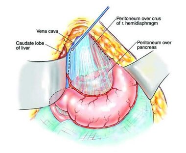
- While long MWCNTs dispersed in 0.5% BSA induced a granulomatous response on the diaphragmatic mesothelium and immune cell recruitment to the peritoneal cavity, GO sheets dispersed under similar conditions did not cause any response, regardless of their lateral dimensions. (springer.com)
- Although the change of dispersion did not alter the impact of GO on the mesothelium (i.e. no granuloma), we observed that, when dispersed in protein-free 5% dextrose solution, s-GO elicited a greater recruitment of monocytic cells to the peritoneal cavity than l-GO, or when dispersed in protein-containing solution. (springer.com)
- were found in the peritoneal cavity, close to the stomach, of the same particoloured bat male dying as a result of dysmicrobia and haemorrhagic gastroenteritis in a wildlife rescue centre. (frontiersin.org)
- A PCR assay targeting cytochrome c oxidase subunit 1 confirmed that adult worms from the peritoneal cavity and testicular microfilariae were of the same filarial species. (frontiersin.org)
- Ultrasound of his abdomen showed multiple nodules of varying sizes in the peritoneal cavity. (biomedcentral.com)
- A computed tomography scan of his abdomen showed numerous nodules of different sizes (1 to 3cm in diameter) filling the whole peritoneal cavity with intense peripheral enhancement. (biomedcentral.com)
- Exploration of his abdomen revealed multiple firm gooseberry-like nodules of different sizes involving the greater omentum, peritoneal cavity and the mesentery. (biomedcentral.com)
- A computed tomography scan of his abdomen showed numerous nodules of different sizes, 1 to 3cm in diameter each, filling the peritoneal cavity and the surrounding bowel loops with intense peripheral enhancement (Figures 1 , 2 and 3 ). (biomedcentral.com)
- Exploration revealed multiple firm gooseberry-like nodules of different sizes ranging between 1 and 5cm in diameter (Figures 4 , 5 , 6 and 7 ), involving the greater omentum, peritoneal cavity and the mesentery, but liver texture was normal. (biomedcentral.com)
- The anterior abdominal wall bolsters the peritoneal cavity, preventing its contents from spilling, and contributes to gravity-dependent distension and bloating through abdominophrenic dyssynergia. (news-medical.net)
Organs21
- The abdominal cavity is a large body cavity in humans and many other animals that contains many organs. (wikipedia.org)
- The peritoneum, by virtue of its connection to the two (parietal and visceral) portions, gives support to the abdominal organs. (wikipedia.org)
- Exploratory laparoscopy (also referred to as diagnostic laparoscopy) is a minimally invasive method for the diagnosis of intra-abdominal diseases through direct inspection of intra-abdominal organs. (medscape.com)
- Although an essential component of all routine physical examinations, the physical examination of the abdomen is the key step in the evaluation of abdominal complaints such as pain, distension, enlarged organs, or masses. (medscape.com)
- In this condition, some of your baby's abdominal organs poke out (protrude) from the belly through the belly button (umbilicus). (uhhospitals.org)
- Or it may be large, with most of the abdominal organs being outside the abdomen. (uhhospitals.org)
- Your baby's abdominal organs and muscles just don't form like they should. (uhhospitals.org)
- Your baby's abdominal organs will poke out (protrude) from the belly through the belly button. (uhhospitals.org)
- If most of your baby's abdominal organs are affected, they will have treatment in stages. (uhhospitals.org)
- A germ-free (sterile), protective sheet is put over your baby's abdominal organs. (uhhospitals.org)
- Your baby's surgeon will close the abdominal wall once all of the organs are inside. (uhhospitals.org)
- Babies with damage to the abdominal organs may have long-term problems. (uhhospitals.org)
- Although the most frequent injuries occur in retroperitoneum, some authors describe injuries of the organs within abdominal cavity. (hindawi.com)
- The nerve impulses relayed from the internal organs to the brain aren't well-localized, so abdominal pain can stem from anatomic, metabolic, malignant, infectious or toxic processes that originate anywhere within the abdomen or even elsewhere in the body. (livestrong.com)
- The abdominal organs include the stomach, the large and small intestines, the liver, the gallbladder, the spleen, the pancreas, the kidneys, the urinary bladder and a host of lesser structures. (livestrong.com)
- One peculiar property of the abdominal organs is that they don't necessarily transmit pain messages when they are sliced or cut, but they do send pain impulses when they are stretched, compressed, inflamed, twisted or, in the case of the hollow organs, when they are dilated. (livestrong.com)
- Although abdominal pain can arise from the tissues of your abdominal wall that surround the abdominal cavity (such as the skin and abdominal wall muscles), the term abdominal pain generally is used to describe pain originating from organs within your abdominal cavity. (rxlist.com)
- In this condition, some of your baby's abdominal organs poke out (protrude) through an opening in the abdominal muscles. (stlouischildrens.org)
- Your baby's abdominal organs will poke out through an opening in his or her abdomen. (stlouischildrens.org)
- Because some or all of the abdominal (belly) organs are outside of the body, babies born with an omphalocele can have other problems. (cdc.gov)
- The abdominal cavity, the space in the body that holds these organs, might not grow to its normal size. (cdc.gov)
Abdomen2
- the abdomen was without signs and symptoms of peritoneal irritation or acute abdominal condition. (hindawi.com)
- In spite of negative clinical exam, CT of abdomen was ordered, which showed pneumoperitoneum and phlegmonous infiltration of abdominal wall. (hindawi.com)
Thoracic and abdominal cavities1
- A simple incision to expose the rib cage, then two more to open the thoracic and abdominal cavities (Figure 1) will reveal whether or not the lungs were inflated (Figure 2) and the brown fat reserves depleted (Figure 3) at death. (msu.edu)
Liver5
- Exposure to high levels of 1,4-dioxane in the air can result in nasal cavity, liver, and kidney damage. (cdc.gov)
- 1,4-Dioxane can be released into the air, water, and of 1,4-dioxane affects mainly the nasal cavity, liver, and soil at places where it is produced or used as kidneys. (cdc.gov)
- Abdominal pain is caused by inflammation (for example, appendicitis, diverticulitis, colitis), by stretching or distention of an organ (for example, obstruction of the intestine, blockage of a bile duct by gallstones, swelling of the liver with hepatitis), or by loss of the supply of blood to an organ (for example, ischemic colitis). (rxlist.com)
- Abdominal com- There he stated that he had recently in which the liver is the organ most fre- puterized tomography showed a 7 × 4 started to experience left lumbar pain. (who.int)
- This ureteropelvic junction ment of the liver in terms of location, abdominal cavity (Figure 1). (who.int)
Pelvic2
- It is located below the thoracic cavity, and above the pelvic cavity. (wikipedia.org)
- DSRCT was first described by Gerald and Rosai in 1989 as a primitive neoplasm of children and young adults that most frequently occurs in the serosa of the pelvic cavity. (medscape.com)
Abdominopelvic cavity2
- It is a part of the abdominopelvic cavity. (wikipedia.org)
- The first lesson plan in a series of 10 introduces the class to the abdominopelvic cavity. (lessonplanet.com)
Diaphragm5
- The lower tip of the heart, called the apex, points toward the left hip and rests on the diaphragm (a membrane of muscle separating the chest cavity from the abdominal cavity). (encyclopedia.com)
- The diaphragm forms the ceiling mount for the abdominal cavity. (news-medical.net)
- The ribcage, spine, support ligaments, and diaphragm act like an abdominal crane to suspend and stabilize the peritoneal contents. (news-medical.net)
- Intraluminal gas causes paradoxical diaphragm contraction and dyssynergic abdominal wall relaxation in many individuals with distension and bloating. (news-medical.net)
- The abdominal cavity is bounded at its upper end by the diaphragm and ribs, at its lower end by the pelvis, at its rear by the spine and in front by the abdominal wall. (livestrong.com)
Etiology2
- In 30-40% of these patients, the etiology of the abdominal pain remains elusive despite laboratory and radiologic investigations. (medscape.com)
- The most common reason for performing a focused examination is to identify the etiology of abdominal pain, which can be subdivided into visceral, somatoparietal, or referred. (medscape.com)
Intestine2
- The three most important mesenteries are mesentery for the small intestine, the transverse mesocolon, which attaches the back portion of the colon to the abdominal wall, and the sigmoid mesocolon which enfolds the sigmoid colon. (wikipedia.org)
- An obstruction of your intestine, for example, initially causes waves of crampy abdominal pain due to contractions of your intestinal muscles and distention of the intestine. (rxlist.com)
Acute abdominal pain3
- Acute abdominal pain is one of the most common indications for an emergency department (ED) visit. (medscape.com)
- When a diagnosis of persistent acute abdominal pain of less than 7 days' duration remains uncertain after baseline diagnostic and radiologic investigations, this condition is termed nonspecific abdominal pain (NSAP). (medscape.com)
- Do you have acute abdominal pain that came on suddenly or did the pain start gradually and worsen? (rxlist.com)
Upper abdominal pain2
- For example, upper abdominal pain that gets better after a meal is often caused by a stomach ulcer, while a severe, intermittent pain that moves from a patient's back, to the flank and finally to the groin could be caused by a kidney stone. (livestrong.com)
- Obstruction of your bile ducts by gallstones typically causes steady (constant) upper abdominal pain. (rxlist.com)
Distention2
- This chain of events leads to abdominal distention, commonly observed in IBS patients. (news-medical.net)
- To complicate matters, however, abdominal pain also can occur for unclear reasons without inflammation, distention, or loss of blood supply. (rxlist.com)
Symptoms2
- Surgeon on-call examined patient but did not find any signs and symptoms of acute abdominal condition. (hindawi.com)
- Individuals with persistent abdominal pain or pain that is accompanied by alarm signs or symptoms should seek medical attention. (livestrong.com)
Incision2
- Another type of biopsy involves inserting instruments through a small abdominal incision. (mayoclinic.org)
- The incision to open the thoracic cavity has been initiated. (msu.edu)
Peritoneum5
- The abdominal cavity is lined with a protective membrane termed the peritoneum. (wikipedia.org)
- The kidneys are located behind the peritoneum, in the retroperitoneum, outside the abdominal cavity. (wikipedia.org)
- The peritoneum divides the cavity into numerous compartments. (wikipedia.org)
- Here we present an unusual case of EGIST that presented with multiple gooseberry-like nodules involving the whole abdominal cavity, the omentum, peritoneum and small bowel mesentery, which makes a radical resection difficult. (biomedcentral.com)
- Lining the abdominal cavity and enclosing its contents is a thin, sensitive envelope called the peritoneum. (livestrong.com)
Anterior abdo1
- Abdominal ultrasound demon- strated a 6 × 4 cm cystic mass between the anterior abdominal wall and ante- rior surface of the left hepatic lobe. (who.int)
Lesions1
- It is performed after the surgeon removes tumors or lesions from the abdominal area, to kill any cancer cells that remain after surgery and reduce the risk for cancer recurrence. (bvsalud.org)
Patient's2
- Gas leak also often taking place, cause abdominal pressure deficiency in the art, forms complication such as subcutaneous emphysema easily.In addition, existing peritoneoscope perforator can't effectively be fixed on patient's operative site, has had a strong impact on operation technique, especially needs to puncture again after the slippage of peritoneoscope perforator, increases patient's wound area, prolongs operating time. (google.com)
- When a physician evaluates a patient with abdominal pain, the patient's history and associated complaints are sometimes just as important as the physical examination itself. (livestrong.com)
Viscera3
- The collection of fluid will cause pressure on the viscera, veins, and thoracic cavity. (wikipedia.org)
- The GI tract forms a functional stack accommodating and resisting downward traction in a way that aligns, bolsters, and suspends the abdominal viscera to optimize function and form. (news-medical.net)
- The author discussed four mechanisms of g-force resistance (suspension system, chassis, ceiling mount, and bolster) in humans, which collectively support the abdominal viscera. (news-medical.net)
Peritonitis1
- Emergency surgery may be done to repair the injury and drain the urine from the abdominal cavity in case of extensive injury or peritonitis (inflammation of the abdominal cavity). (medlineplus.gov)
Wall10
- It can be caused by damage to an organ, or from a contusion to the abdominal wall from the outside or by surgery. (wikipedia.org)
- The bladder may be drained by a catheter through the urethra or the abdominal wall (called a suprapubic tube) over a period of days to weeks. (medlineplus.gov)
- If this cannot be done, a tube will be inserted through the abdominal wall directly into the bladder. (medlineplus.gov)
- The mesentery originates at the lumbar spine along the posterior abdominal wall and functions like a suspension system preventing intestines from collapsing. (news-medical.net)
- The surgeon will also close the opening to your baby's abdominal wall. (uhhospitals.org)
- The abdominal wall consists of skin, fat, muscle and connective tissue. (livestrong.com)
- Omphalocele (pronounced uhm- fa -lo-seal) is a birth defect of the abdominal (belly) wall. (cdc.gov)
- Omphalocele, also known as exomphalos, is a birth defect of the abdominal (belly) wall. (cdc.gov)
- Hydatid cyst can be seen abdominal wall. (who.int)
- In2003,theWorldHealthOrganiza- hydatid cyst of the round ligament was ligated and divided and then the mass tion Informal Working Group on Echi- totally excised without any spillage and no was totally excised from the abdominal nococcosis(WHO-IWGE)proposed risk of infection was added to the elective wall with 1 cm free borders. (who.int)
Infection2
- however without treatment, the infection will spread throughout the cavity. (wikipedia.org)
- On August 8, 2003, it was determined that a sponge had been left inside Robert's abdominal cavity at the conclusion of the surgery on July 24, 2003, and that the sponge probably was the source of the infection. (wicourts.gov)
Patients4
- Diagnostic laparoscopy is useful for making a definitive clinical diagnosis whenever there is a diagnostic dilemma even after routine diagnostic workup, including patients with nonspecific abdominal pain, hemodynamically stable patients who have sustained blunt or penetrating trauma with suspected intra-abdominal injuries, and critically ill intensive care unit (ICU) patients with suspected intra-abdominal sepsis or pathologies. (medscape.com)
- Patients have abdominal pain, sometimes radiating to the shoulder, and tenderness. (msdmanuals.com)
- Nous avons analysé les infections des plaies après un acte chirurgical chez 1200 patients blessés pendant le conflit libyen en 2011 et admis au service des urgences du centre médical de Tripoli. (who.int)
- Les bacilles à Gram-négatif multirésistants représentaient un facteur de complication important pour les infections de plaies associées à des blessures de guerre chez des patients blessés en Libye. (who.int)
Chest1
- Membrane of muscle separating the chest cavity from the abdominal cavity. (encyclopedia.com)
Tenderness1
- Lesser hemorrhage or hematomas cause right upper quadrant abdominal pain and tenderness. (msdmanuals.com)
Ascites2
- When fluid collects in the abdominal cavity, this condition is called ascites. (wikipedia.org)
- Ascites is excess fluid in your abdominal cavity. (hopkinsmedicine.org)
Pain21
- A 65-year-old Sudanese man presented with vague abdominal pain and progressive abdominal distension for 6 months. (biomedcentral.com)
- Here we present a rare case of extragastrointestinal stromal tumour in a patient who presented with vague abdominal pain and progressive abdominal distension. (biomedcentral.com)
- A 65-year-old Sudanese man, who was previously well, presented with vague central abdominal pain. (biomedcentral.com)
- BP was 135/65 and serum glucose 12.0 mmol/L. She was denying any other difficulties or abdominal pain. (hindawi.com)
- Nearly everyone will experience abdominal pain at some time. (livestrong.com)
- Pain arising within the abdominal cavity can be difficult to pinpoint. (livestrong.com)
- Doctors often search for so-called 'alarm signs' to help them decide if a person with abdominal pain needs urgent evaluation. (livestrong.com)
- Abdominal pain that is associated with weight loss is worrisome. (livestrong.com)
- What's Causing Your Abdominal Pain? (rxlist.com)
- What Is Abdominal Pain? (rxlist.com)
- Abdominal pain can range in intensity from a mild stomach ache to severe, acute pain. (rxlist.com)
- There are many different possible causes of abdominal pain. (rxlist.com)
- A primary health care provider or gastroenterologist can help determine the underlying cause of pain in the abdominal area. (rxlist.com)
- How Is the Cause of Abdominal Pain Diagnosed? (rxlist.com)
- When does your abdominal pain occur? (rxlist.com)
- Abdominal pain that occurs after eating may be due to indigestion. (rxlist.com)
- For example, abdominal pain that comes on suddenly may suggest a sudden event such as the interruption of the supply of blood to the colon (ischemia) or obstruction of the bile duct by a gallstone (biliary colic). (rxlist.com)
- Patterns of pain can be helpful in the diagnosis of abdominal pain. (rxlist.com)
- He complained of abdominal fullness and vague pain. (who.int)
- I made my way to the bathroom floor with intense abdominal pain that took my breath away. (cdc.gov)
- 1996), and was characterized by abdominal pain and et al. (bvsalud.org)
Distension1
- It was constant, associated with progressive abdominal distension for the past 6 months and mild loss of weight despite good appetite. (biomedcentral.com)
Pediatric1
- A large cross-sectional study has proposed a scoring system that would utilize a few standard screening laboratories in conjunction with a detailed physical examination to decrease the number of pediatric CT scans after blunt abdominal trauma. (medscape.com)
Ultrasound1
- Your child may need an abdominal X-ray, an ultrasound, or both. (uhhospitals.org)
Injuries1
- The nature and severity of abdominal injuries vary. (msdmanuals.com)
Sterile1
- Peritoneal -A sterile solution enters the abdominal cavity through a tube. (epnet.com)
Involves2
- Involves the tumor spreading to the abdominal cavity. (medicalnewstoday.com)
- A cancer treatment that involves filling the abdominal cavity with heated chemotherapy drugs. (bvsalud.org)
Findings1
- Somatoparietal findings correspond more exactly to the precise underlying abdominal contents. (medscape.com)
Muscles1
- Anti-gravity extensor muscles are constantly strained to support the abdominal load under the pull of gravity. (news-medical.net)
Physical examination1
- Physical examination of the patient revealed a semifixed 6 cm mass on the abdominal epigastric area adjacent to the right inferior ribs. (who.int)
Laparoscopic2
- The advent of laparoscopic surgery represents a landmark in surgery that initiated a shift from the era of open abdominal surgery to the minimally invasive surgery revolution. (medscape.com)
- The literature on the advantages and efficacy of laparoscopic management for specific intra-abdominal diseases (including acute appendicitis, acute cholecystitis, and several others) is vast, and a full survey is beyond the scope of this review. (medscape.com)
Cancers1
- A 2014 National Research Council report on the carcinogenesis of formaldehyde determined that epidemiological evidence was strongest for an association between formaldehyde exposure and cancers of the nasopharyngeal region and sinonasal cavities and myeloid leukemia ( NRC, 2014 ). (nationalacademies.org)
Fluid1
- 4 × 4 cm cystic mass filled with serous based on the active-transitional-inac- pable abdominal lesion made it possible fluid. (who.int)
Drain1
- Your child's healthcare provider (surgeon) may also put a drain into their abdominal cavity. (uhhospitals.org)