2,3-Diphosphoglycerate
Diphosphoglyceric Acids
Bisphosphoglycerate Mutase
Hemoglobin A
Hemoglobins, Abnormal
Hemoglobins
Oxyhemoglobins
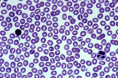
Erythrocytes
Phytic Acid
Glyceric Acids
Oxygen
Phosphoglycerate Mutase
Methanobacteriales
Hemoglobin, Sickle
Phosphoglycerate Kinase
Carboxyhemoglobin
Methemoglobin
Hydrogen-Ion Concentration
Partial Pressure
Phosphorus-Oxygen Lyases
Euryarchaeota
Erythrocyte Aging
Phosphotransferases
Blood Preservation
Erythrocytes, Abnormal
Pyruvate Kinase
Erythrocyte Deformability
Inosine
Mathematics
Adenosine Triphosphate
Glyceraldehyde-3-Phosphate Dehydrogenases
Fetal Hemoglobin
Erythrocyte Membrane
Allosteric Site
Carbon Monoxide
Hematocrit
Anemia, Sickle Cell
Carbon Dioxide
Binding Sites
Magnetic Resonance Spectroscopy
Phosphofructokinase-1
Pyruvates
Hemolysis
Glycolysis
2,3-Diphosphoglycerate (2,3-DPG) is a molecule found in red blood cells that plays a crucial role in regulating the affinity of hemoglobin for oxygen. It is a byproduct of the glycolytic pathway, which is a series of biochemical reactions that convert glucose into energy.
In the tissues where oxygen demand is high, such as muscles and organs, 2,3-DPG concentrations are typically elevated. This molecule binds to deoxygenated hemoglobin at specific sites on the beta chains, reducing its affinity for oxygen and promoting the release of oxygen to the tissues.
Conversely, in the lungs where oxygen is abundant, 2,3-DPG concentrations are lower, allowing hemoglobin to bind more readily to oxygen and load up with oxygen for delivery to the tissues. Therefore, 2,3-DPG helps optimize the matching of oxygen supply and demand in the body.
Diphosphoglycerates (also known as 2,3-diphosphoglycerates or 2,3-DPG) are organic molecules found in red blood cells. They play a crucial role in regulating the affinity of hemoglobin for oxygen. Hemoglobin is the protein in red blood cells that carries oxygen from the lungs to the body's tissues.
When the concentration of diphosphoglycerates in red blood cells increases, it reduces the ability of hemoglobin to bind with oxygen, which allows more oxygen to be released into the tissues. This is particularly important in conditions where there is low oxygen availability, such as at high altitudes or in diseases that cause poor oxygen delivery to the tissues, like heart failure and chronic obstructive pulmonary disease (COPD).
In summary, diphosphoglycerates are essential molecules that help regulate hemoglobin's affinity for oxygen, ensuring optimal oxygen delivery to the body's tissues.
Bisphosphoglycerate mutase (BPGM) is an enzyme that plays a crucial role in the regulation of oxygen transport in red blood cells. The main function of BPGM is to convert 1,3-bisphosphoglycerate (1,3-BPG) into 2,3-bisphosphoglycerate (2,3-BPG), also known as 2,3-diphosphoglycerate (2,3-DPG).
2,3-BPG is essential for modulating the affinity of hemoglobin for oxygen. By increasing the concentration of 2,3-BPG in red blood cells, BPGM reduces the ability of hemoglobin to bind to oxygen, allowing more oxygen to be released from hemoglobin and made available to tissues, particularly under low-oxygen conditions. This is especially important for individuals living at high altitudes or those with chronic lung diseases who may have impaired oxygen transport.
Defects in the BPGM gene can lead to a rare disorder called 2,3-bisphosphoglycerate deficiency, which results in an increased affinity of hemoglobin for oxygen and reduced oxygen delivery to tissues. This condition is characterized by symptoms such as shortness of breath, fatigue, and headaches, particularly during exercise or at high altitudes.
Hemoglobin A is the most common form of hemoglobin, which is the oxygen-carrying protein in red blood cells. Hemoglobin A is a tetramer composed of two alpha and two beta globin chains, each containing a heme group that binds to oxygen. It is typically measured in laboratory tests to assess for various medical conditions such as anemia or diabetes. In the context of diabetes, the measurement of hemoglobin A1c (a form of hemoglobin A that is glycated or bound to glucose) is used to monitor long-term blood sugar control.
Abnormal hemoglobins refer to variants of the oxygen-carrying protein found in red blood cells, which differ from the normal adult hemoglobin (HbA) in terms of their structure and function. These variations can result from genetic mutations that affect the composition of the globin chains in the hemoglobin molecule. Some abnormal hemoglobins are clinically insignificant, while others can lead to various medical conditions such as hemolytic anemia, thalassemia, or sickle cell disease. Examples of abnormal hemoglobins include HbS (associated with sickle cell anemia), HbC, HbE, and HbF (fetal hemoglobin). These variants can be detected through specialized laboratory tests, such as hemoglobin electrophoresis or high-performance liquid chromatography (HPLC).
Hemoglobin (Hb or Hgb) is the main oxygen-carrying protein in the red blood cells, which are responsible for delivering oxygen throughout the body. It is a complex molecule made up of four globin proteins and four heme groups. Each heme group contains an iron atom that binds to one molecule of oxygen. Hemoglobin plays a crucial role in the transport of oxygen from the lungs to the body's tissues, and also helps to carry carbon dioxide back to the lungs for exhalation.
There are several types of hemoglobin present in the human body, including:
* Hemoglobin A (HbA): This is the most common type of hemoglobin, making up about 95-98% of total hemoglobin in adults. It consists of two alpha and two beta globin chains.
* Hemoglobin A2 (HbA2): This makes up about 1.5-3.5% of total hemoglobin in adults. It consists of two alpha and two delta globin chains.
* Hemoglobin F (HbF): This is the main type of hemoglobin present in fetal life, but it persists at low levels in adults. It consists of two alpha and two gamma globin chains.
* Hemoglobin S (HbS): This is an abnormal form of hemoglobin that can cause sickle cell disease when it occurs in the homozygous state (i.e., both copies of the gene are affected). It results from a single amino acid substitution in the beta globin chain.
* Hemoglobin C (HbC): This is another abnormal form of hemoglobin that can cause mild to moderate hemolytic anemia when it occurs in the homozygous state. It results from a different single amino acid substitution in the beta globin chain than HbS.
Abnormal forms of hemoglobin, such as HbS and HbC, can lead to various clinical disorders, including sickle cell disease, thalassemia, and other hemoglobinopathies.
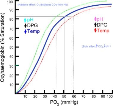
Oxyhemoglobin is the form of hemoglobin that is combined with oxygen in red blood cells. It's created when oxygen molecules bind to the iron-containing heme groups of the hemoglobin protein inside the lungs, allowing for the transportation of oxygen from the lungs to body tissues. The affinity of hemoglobin for oxygen is influenced by factors such as pH, carbon dioxide concentration, and temperature, which can affect the release of oxygen from oxyhemoglobin in different parts of the body based on their specific needs.
Erythrocytes, also known as red blood cells (RBCs), are the most common type of blood cell in circulating blood in mammals. They are responsible for transporting oxygen from the lungs to the body's tissues and carbon dioxide from the tissues to the lungs.
Erythrocytes are formed in the bone marrow and have a biconcave shape, which allows them to fold and bend easily as they pass through narrow blood vessels. They do not have a nucleus or mitochondria, which makes them more flexible but also limits their ability to reproduce or repair themselves.
In humans, erythrocytes are typically disc-shaped and measure about 7 micrometers in diameter. They contain the protein hemoglobin, which binds to oxygen and gives blood its red color. The lifespan of an erythrocyte is approximately 120 days, after which it is broken down in the liver and spleen.
Abnormalities in erythrocyte count or function can lead to various medical conditions, such as anemia, polycythemia, and sickle cell disease.
Phytic acid, also known as phytate in its salt form, is a natural substance found in plant-based foods such as grains, legumes, nuts, and seeds. It's a storage form of phosphorus for the plant and is often referred to as an "anti-nutrient" because it can bind to certain minerals like calcium, iron, magnesium, and zinc in the gastrointestinal tract and prevent their absorption. This can potentially lead to mineral deficiencies if a diet is consistently high in phytic acid-rich foods and low in mineral-rich foods. However, it's important to note that phytic acid also has antioxidant properties and may have health benefits when consumed as part of a balanced diet.
The bioavailability of minerals from phytic acid-rich foods can be improved through various methods such as soaking, sprouting, fermenting, or cooking, which can help break down some of the phytic acid and release the bound minerals.
I believe there might be a slight misunderstanding in your question. "Glyceric acid" is not a widely recognized or established term in medicine or biochemistry. However, glyceric acid can refer to a specific compound with the chemical formula C3H8O4, also known as 2,3-dihydroxypropanoid acid or glycerol-3-phosphate when phosphorylated.
Glyceric acid is an organic compound that plays a crucial role in cellular metabolism, particularly in energy production pathways such as glycolysis and gluconeogenesis. It can be formed from the reduction of dihydroxyacetone phosphate (a glycolytic intermediate) or through the oxidation of glycerol.
If you were referring to a different term or concept, please provide more context so I can give a more accurate answer.
Oxygen is a colorless, odorless, tasteless gas that constitutes about 21% of the earth's atmosphere. It is a crucial element for human and most living organisms as it is vital for respiration. Inhaled oxygen enters the lungs and binds to hemoglobin in red blood cells, which carries it to tissues throughout the body where it is used to convert nutrients into energy and carbon dioxide, a waste product that is exhaled.
Medically, supplemental oxygen therapy may be provided to patients with conditions such as chronic obstructive pulmonary disease (COPD), pneumonia, heart failure, or other medical conditions that impair the body's ability to extract sufficient oxygen from the air. Oxygen can be administered through various devices, including nasal cannulas, face masks, and ventilators.
Phosphoglycerate Mutase (PGM) is an enzyme that plays a crucial role in the glycolytic pathway, which is a metabolic process that converts glucose into pyruvate, producing ATP and NADH as energy currency for the cell.
The enzyme catalyzes the interconversion of 3-phosphoglycerate (3-PG) and 2-phosphoglycerate (2-PG), which is the ninth step in glycolysis. Specifically, PGM transfers a phosphate group from the third carbon atom to the second carbon atom of 3-PG, resulting in the formation of 2-PG and inorganic phosphate.
There are two types of Phosphoglycerate Mutase isoenzymes in humans, including:
1. Phosphoglycerate Mutase 1 (PGAM1): This is a cytosolic enzyme that is widely expressed in various tissues, including skeletal muscle, heart, brain, and liver.
2. Phosphoglycerate Mutase 2 (PGAM2): This is a muscle-specific isoenzyme that is primarily found in cardiac and skeletal muscles.
Mutations in the PGAM1 gene have been associated with hemolytic anemia, neurodevelopmental disorders, and other metabolic abnormalities, while mutations in the PGAM2 gene have been linked to myopathies and other muscle-related disorders.
Methanobacteriales is an order of methanogenic archaea within the kingdom Euryarchaeota. These organisms are characterized by their ability to produce methane as a metabolic byproduct in anaerobic environments. They are commonly found in habitats such as wetlands, digestive tracts of animals, and sewage sludge. The cells of Methanobacteriales are typically rod-shaped and have a Gram-positive stain, although they lack a true cell wall. Some notable genera within this order include Methanobrevibacter, Methanothermobacter, and Methanosphaera.
Glycerophosphates are esters of glycerol and phosphoric acid. In the context of biochemistry and medicine, glycerophosphates often refer to glycerol 3-phosphate (also known as glyceraldehyde 3-phosphate or glycerone phosphate) and its derivatives.
Glycerol 3-phosphate plays a crucial role in cellular metabolism, particularly in the process of energy production and storage. It is an important intermediate in both glycolysis (the breakdown of glucose to produce energy) and gluconeogenesis (the synthesis of glucose from non-carbohydrate precursors).
In addition, glycerophosphates are also involved in the formation of phospholipids, a major component of cell membranes. The esterification of glycerol 3-phosphate with fatty acids leads to the synthesis of phosphatidic acid, which is a key intermediate in the biosynthesis of other phospholipids.
Abnormalities in glycerophosphate metabolism have been implicated in various diseases, including metabolic disorders and neurological conditions.
Hemoglobin S (HbS) is a genetic variant of hemoglobin, which is the oxygen-carrying protein in red blood cells. This abnormal form of hemogllobin results from a mutation in the beta-globin gene, leading to the substitution of valine for glutamic acid at position six of the beta-globin chain.
In individuals with sickle cell disease (a group of inherited red blood cell disorders), both copies of their beta-globin genes carry this mutation, causing the majority of their hemoglobin to be HbS. When deoxygenated, HbS molecules have a tendency to polymerize and form long, rigid rods within the red blood cells, distorting their shape into a characteristic sickle or crescent form.
These sickled red blood cells are less flexible and more prone to rupture (hemolysis), leading to chronic anemia, vaso-occlusive crises, and other disease complications. Sickle cell disease primarily affects people of African, Mediterranean, Middle Eastern, and Indian ancestry, but it can also be found in other populations worldwide.
Phosphoglycerate Kinase (PGK) is an enzyme that plays a crucial role in the glycolytic pathway, which is a series of reactions that convert glucose into pyruvate, producing ATP and NADH as energy-rich compounds. PGK catalyzes the conversion of 1,3-bisphosphoglycerate (1,3-BPG) to 3-phosphoglycerate (3-PG), concomitantly transferring a phosphate group to ADP to form ATP. This reaction is the fourth step in the glycolytic pathway and is reversible under certain conditions.
In humans, there are two isoforms of PGK: PGK1 and PGK2. PGK1 is widely expressed in various tissues, while PGK2 is primarily found in sperm cells. Deficiencies or mutations in the PGK1 gene can lead to a rare metabolic disorder called Phosphoglycerate Kinase Deficiency (PGKD), which can present with hemolytic anemia and neurological symptoms.
Carboxyhemoglobin (COHb) is a form of hemoglobin that has bonded with carbon monoxide (CO), a colorless, odorless gas. Normally, hemoglobin in red blood cells binds with oxygen (O2) to carry it throughout the body. However, when exposed to CO, hemoglobin preferentially binds with it, forming carboxyhemoglobin, which reduces the amount of oxygen that can be carried by the blood. This can lead to hypoxia (lack of oxygen in tissues) and potentially serious medical consequences, including death. Carbon monoxide exposure can occur from sources such as smoke inhalation, vehicle exhaust, or faulty heating systems.
Methemoglobin is a form of hemoglobin in which the iron within the heme group is in the ferric (Fe3+) state instead of the ferrous (Fe2+) state. This oxidation reduces its ability to bind and transport oxygen effectively, leading to methemoglobinemia when methemoglobin levels become too high. Methemoglobin has a limited capacity to release oxygen to tissues, which can result in hypoxia (reduced oxygen supply) and cyanosis (bluish discoloration of the skin and mucous membranes).
Methemoglobin is normally present in small amounts in the blood, but certain factors such as exposure to oxidizing agents, genetic predisposition, or certain medications can increase its levels. Elevated methemoglobin levels can be treated with methylene blue, which helps restore the iron within hemoglobin back to its ferrous state and improves oxygen transport capacity.
Polycythemia is a medical condition characterized by an abnormal increase in the total red blood cell (RBC) mass or hematocrit (the percentage of RBCs in the blood). This results in a higher-than-normal viscosity of the blood, which can lead to various complications such as impaired circulation, increased risk of blood clots, and reduced oxygen supply to the tissues.
There are two main types of polycythemia: primary and secondary. Primary polycythemia, also known as polycythemia vera, is a rare myeloproliferative neoplasm caused by genetic mutations that lead to excessive production of RBCs in the bone marrow. Secondary polycythemia, on the other hand, is a reactive condition triggered by various factors such as chronic hypoxia (low oxygen levels), high altitude, smoking, or certain medical conditions like sleep apnea, heart disease, or kidney tumors.
Symptoms of polycythemia may include fatigue, headaches, dizziness, shortness of breath, itching, and a bluish or reddish tint to the skin (cyanosis). Treatment depends on the underlying cause and severity of the condition and may involve phlebotomy, medications to reduce RBC production, and management of associated complications.
Hemoglobinometry is a method used to measure the amount or concentration of hemoglobin (Hb) in blood. Hemoglobin is a protein in red blood cells that carries oxygen throughout the body. Hemoglobinometry is typically performed on a sample of whole blood and can be done using various methods, including spectrophotometry, colorimetry, or automated analyzers.
The results of hemoglobinometry are reported in units of grams per deciliter (g/dL) or grams per liter (g/L). Normal values for hemoglobin concentration vary depending on factors such as age, sex, and altitude, but in general, a healthy adult male should have a hemoglobin level between 13.5 and 17.5 g/dL, while a healthy adult female should have a level between 12.0 and 15.5 g/dL.
Hemoglobinometry is an important diagnostic tool in the evaluation of various medical conditions, including anemia, polycythemia, and respiratory disorders. It can help identify the cause of symptoms such as fatigue, shortness of breath, or dizziness and guide treatment decisions.
Hydrogen-ion concentration, also known as pH, is a measure of the acidity or basicity of a solution. It is defined as the negative logarithm (to the base 10) of the hydrogen ion activity in a solution. The standard unit of measurement is the pH unit. A pH of 7 is neutral, less than 7 is acidic, and greater than 7 is basic.
In medical terms, hydrogen-ion concentration is important for maintaining homeostasis within the body. For example, in the stomach, a high hydrogen-ion concentration (low pH) is necessary for the digestion of food. However, in other parts of the body such as blood, a high hydrogen-ion concentration can be harmful and lead to acidosis. Conversely, a low hydrogen-ion concentration (high pH) in the blood can lead to alkalosis. Both acidosis and alkalosis can have serious consequences on various organ systems if not corrected.
In the context of medicine, and specifically in physiology and respiratory therapy, partial pressure (P or p) is a measure of the pressure exerted by an individual gas in a mixture of gases. It's commonly used to describe the concentrations of gases in the body, such as oxygen (PO2), carbon dioxide (PCO2), and nitrogen (PN2).
The partial pressure of a specific gas is calculated as the fraction of that gas in the total mixture multiplied by the total pressure of the mixture. This concept is based on Dalton's law, which states that the total pressure exerted by a mixture of gases is equal to the sum of the pressures exerted by each individual gas.
For example, in room air at sea level, the partial pressure of oxygen (PO2) is approximately 160 mmHg (mm of mercury), which represents about 21% of the total barometric pressure (760 mmHg). This concept is crucial for understanding gas exchange in the lungs and how gases move across membranes, such as from alveoli to blood and vice versa.
Phosphorus-Oxygen Lyases are a class of enzymes that catalyze the breakdown of a substrate containing a phosphorus-oxygen bond, releasing a phosphate group and forming a new double bond in the process. This reaction is typically represented by the general formula:
Substrate-P-O + A acceptor ------> Substrate-O=A + P\_i
where "Substrate-P-O" represents the phosphorus-oxygen bond in the substrate, "A acceptor" is the molecule that accepts the phosphate group, and "P\_i" denotes inorganic phosphate. These enzymes play important roles in various biological processes, such as signal transduction, energy metabolism, and biosynthesis.
Examples of Phosphorus-Oxygen Lyases include:
1. Phospholipase D - catalyzes the hydrolysis of phosphatidylcholine to produce phosphatidic acid and choline.
2. ATP sulfurylase - catalyzes the formation of adenosine 5'-phosphosulfate (APS) from ATP and sulfate, which is an important intermediate in the biosynthesis of sulfur-containing amino acids.
3. Inositol polyphosphate 1-phosphatase - catalyzes the dephosphorylation of inositol polyphosphates, which are involved in intracellular signaling pathways.
4. UDP-glucose pyrophosphorylase - catalyzes the reversible conversion of UDP-glucose and pyrophosphate to glucose-1-phosphate and UTP, playing a crucial role in carbohydrate metabolism.
It is important to note that Phosphorus-Oxygen Lyases are distinct from Phosphoric Monoester Hydrolases, which also catalyze the hydrolysis of phosphorus-oxygen bonds but do not form new double bonds in the process.
Euryarchaeota is a phylum within the domain Archaea, which consists of a diverse group of microorganisms that are commonly found in various environments such as soil, oceans, and the digestive tracts of animals. This group includes methanogens, which are archaea that produce methane as a metabolic byproduct, and extreme halophiles, which are archaea that thrive in highly saline environments.
The name Euryarchaeota comes from the Greek words "eury," meaning wide or broad, and "archaios," meaning ancient or primitive. This name reflects the phylum's diverse range of habitats and metabolic capabilities.
Euryarchaeota are characterized by their unique archaeal-type cell walls, which contain a variety of complex polysaccharides and proteins. They also have a distinct type of intracellular membrane called the archaellum, which is involved in motility. Additionally, Euryarchaeota have a unique genetic code that differs from that of bacteria and eukaryotes, with some codons specifying different amino acids.
Overall, Euryarchaeota are an important group of archaea that play a significant role in global carbon and nitrogen cycles, as well as in the breakdown of organic matter in various environments.
Erythrocyte aging, also known as red cell aging, is the natural process of changes and senescence that occur in red blood cells (erythrocytes) over time. In humans, mature erythrocytes are devoid of nuclei and organelles, and have a lifespan of approximately 120 days.
During aging, several biochemical and structural modifications take place in the erythrocyte, including:
1. Loss of membrane phospholipids and proteins, leading to increased rigidity and decreased deformability.
2. Oxidative damage to hemoglobin, resulting in the formation of methemoglobin and heinz bodies.
3. Accumulation of denatured proteins and aggregates, which can impair cellular functions.
4. Changes in the cytoskeleton, affecting the shape and stability of the erythrocyte.
5. Increased expression of surface markers, such as Band 3 and CD47, that signal the spleen to remove aged erythrocytes from circulation.
The spleen plays a crucial role in removing senescent erythrocytes by recognizing and phagocytosing those with altered membrane composition or increased expression of surface markers. This process helps maintain the overall health and functionality of the circulatory system.
Phosphotransferases are a group of enzymes that catalyze the transfer of a phosphate group from a donor molecule to an acceptor molecule. This reaction is essential for various cellular processes, including energy metabolism, signal transduction, and biosynthesis.
The systematic name for this group of enzymes is phosphotransferase, which is derived from the general reaction they catalyze: D-donor + A-acceptor = D-donor minus phosphate + A-phosphate. The donor molecule can be a variety of compounds, such as ATP or a phosphorylated protein, while the acceptor molecule is typically a compound that becomes phosphorylated during the reaction.
Phosphotransferases are classified into several subgroups based on the type of donor and acceptor molecules they act upon. For example, kinases are a subgroup of phosphotransferases that transfer a phosphate group from ATP to a protein or other organic compound. Phosphatases, another subgroup, remove phosphate groups from molecules by transferring them to water.
Overall, phosphotransferases play a critical role in regulating many cellular functions and are important targets for drug development in various diseases, including cancer and neurological disorders.
Blood protein electrophoresis (BPE) is a laboratory test that separates and measures the different proteins in the blood, such as albumin, alpha-1 globulins, alpha-2 globulins, beta globulins, and gamma globulins. This test is often used to help diagnose or monitor conditions related to abnormal protein levels, such as multiple myeloma, macroglobulinemia, and other plasma cell disorders.
In this test, a sample of the patient's blood is placed on a special gel and an electric current is applied. The proteins in the blood migrate through the gel based on their electrical charge and size, creating bands that can be visualized and measured. By comparing the band patterns to reference ranges, doctors can identify any abnormal protein levels or ratios, which may indicate underlying medical conditions.
It's important to note that while BPE is a useful diagnostic tool, it should be interpreted in conjunction with other clinical findings and laboratory tests for accurate diagnosis and management of the patient's condition.
Blood preservation refers to the process of keeping blood viable and functional outside of the body for transfusion purposes. This is typically achieved through the addition of various chemical additives, such as anticoagulants and nutrients, to a storage solution in which the blood is contained. The preserved blood is then refrigerated or frozen until it is needed for transfusion.
The goal of blood preservation is to maintain the structural integrity and functional capacity of the red blood cells, white blood cells, and platelets, as well as the coagulation factors, in order to ensure that the transfused blood is safe and effective. Different storage conditions and additives are used for the preservation of different components of blood, depending on their specific requirements.
It's important to note that while blood preservation extends the shelf life of donated blood, it does not last indefinitely. The length of time that blood can be stored depends on several factors, including the type of blood component and the storage conditions. Regular testing is performed to ensure that the preserved blood remains safe and effective for transfusion.
Abnormal erythrocytes refer to red blood cells that have an abnormal shape, size, or other characteristics. This can include various types of abnormalities such as:
1. Anisocytosis: Variation in the size of erythrocytes.
2. Poikilocytosis: Variation in the shape of erythrocytes, including but not limited to teardrop-shaped cells (dacrocytes), crescent-shaped cells (sickle cells), and spherical cells (spherocytes).
3. Anemia: A decrease in the total number of erythrocytes or a reduction in hemoglobin concentration, which can result from various underlying conditions such as iron deficiency, chronic disease, or blood loss.
4. Hemoglobinopathies: Abnormalities in the structure or function of hemoglobin, the protein responsible for carrying oxygen in erythrocytes, such as sickle cell anemia and thalassemia.
5. Inclusion bodies: Abnormal structures within erythrocytes, such as Heinz bodies (denatured hemoglobin) or Howell-Jolly bodies (nuclear remnants).
These abnormalities can be detected through a complete blood count (CBC) and peripheral blood smear examination. The presence of abnormal erythrocytes may indicate an underlying medical condition, and further evaluation is often necessary to determine the cause and appropriate treatment.
In the context of medicine and pharmacology, "kinetics" refers to the study of how a drug moves throughout the body, including its absorption, distribution, metabolism, and excretion (often abbreviated as ADME). This field is called "pharmacokinetics."
1. Absorption: This is the process of a drug moving from its site of administration into the bloodstream. Factors such as the route of administration (e.g., oral, intravenous, etc.), formulation, and individual physiological differences can affect absorption.
2. Distribution: Once a drug is in the bloodstream, it gets distributed throughout the body to various tissues and organs. This process is influenced by factors like blood flow, protein binding, and lipid solubility of the drug.
3. Metabolism: Drugs are often chemically modified in the body, typically in the liver, through processes known as metabolism. These changes can lead to the formation of active or inactive metabolites, which may then be further distributed, excreted, or undergo additional metabolic transformations.
4. Excretion: This is the process by which drugs and their metabolites are eliminated from the body, primarily through the kidneys (urine) and the liver (bile).
Understanding the kinetics of a drug is crucial for determining its optimal dosing regimen, potential interactions with other medications or foods, and any necessary adjustments for special populations like pediatric or geriatric patients, or those with impaired renal or hepatic function.
Pyruvate kinase is an enzyme that plays a crucial role in the final step of glycolysis, a process by which glucose is broken down to produce energy in the form of ATP (adenosine triphosphate). Specifically, pyruvate kinase catalyzes the transfer of a phosphate group from phosphoenolpyruvate (PEP) to adenosine diphosphate (ADP), resulting in the formation of pyruvate and ATP.
There are several isoforms of pyruvate kinase found in different tissues, including the liver, muscle, and brain. The type found in red blood cells is known as PK-RBC or PK-M2. Deficiencies in pyruvate kinase can lead to a genetic disorder called pyruvate kinase deficiency, which can result in hemolytic anemia due to the premature destruction of red blood cells.
Erythrocyte deformability refers to the ability of red blood cells (erythrocytes) to change shape and bend without rupturing, which is crucial for their efficient movement through narrow blood vessels. This deformability is influenced by several factors including the cell membrane structure, hemoglobin concentration, and intracellular viscosity. A decrease in erythrocyte deformability can negatively impact blood flow and oxygen delivery to tissues, potentially contributing to various pathological conditions such as sickle cell disease, diabetes, and cardiovascular diseases.
Phosphates, in a medical context, refer to the salts or esters of phosphoric acid. Phosphates play crucial roles in various biological processes within the human body. They are essential components of bones and teeth, where they combine with calcium to form hydroxyapatite crystals. Phosphates also participate in energy transfer reactions as phosphate groups attached to adenosine diphosphate (ADP) and adenosine triphosphate (ATP). Additionally, they contribute to buffer systems that help maintain normal pH levels in the body.
Abnormal levels of phosphates in the blood can indicate certain medical conditions. High phosphate levels (hyperphosphatemia) may be associated with kidney dysfunction, hyperparathyroidism, or excessive intake of phosphate-containing products. Low phosphate levels (hypophosphatemia) might result from malnutrition, vitamin D deficiency, or certain diseases affecting the small intestine or kidneys. Both hypophosphatemia and hyperphosphatemia can have significant impacts on various organ systems and may require medical intervention.
Inosine is not a medical condition but a naturally occurring compound called a nucleoside, which is formed from the combination of hypoxanthine and ribose. It is an intermediate in the metabolic pathways of purine nucleotides, which are essential components of DNA and RNA. Inosine has been studied for its potential therapeutic benefits in various medical conditions, including neurodegenerative disorders, cardiovascular diseases, and cancer. However, more research is needed to fully understand its mechanisms and clinical applications.
I'm sorry for any confusion, but there seems to be a misunderstanding. Mathematics is not a medical term; it is a branch of science dedicated to the study of numbers, shapes, and structures. However, mathematics does have many applications in medicine, such as in modeling disease spread, analyzing medical images, or designing clinical trials. If you have any questions related to mathematics in a medical context, I'd be happy to help clarify those for you!
Adenosine Triphosphate (ATP) is a high-energy molecule that stores and transports energy within cells. It is the main source of energy for most cellular processes, including muscle contraction, nerve impulse transmission, and protein synthesis. ATP is composed of a base (adenine), a sugar (ribose), and three phosphate groups. The bonds between these phosphate groups contain a significant amount of energy, which can be released when the bond between the second and third phosphate group is broken, resulting in the formation of adenosine diphosphate (ADP) and inorganic phosphate. This process is known as hydrolysis and can be catalyzed by various enzymes to drive a wide range of cellular functions. ATP can also be regenerated from ADP through various metabolic pathways, such as oxidative phosphorylation or substrate-level phosphorylation, allowing for the continuous supply of energy to cells.
Glyceraldehyde-3-phosphate dehydrogenase (GAPDH) is an enzyme that plays a crucial role in the metabolic pathway of glycolysis. Its primary function is to convert glyceraldehyde-3-phosphate (a triose sugar phosphate) into D-glycerate 1,3-bisphosphate, while also converting nicotinamide adenine dinucleotide (NAD+) into its reduced form NADH. This reaction is essential for the production of energy in the form of adenosine triphosphate (ATP) during cellular respiration. GAPDH has also been implicated in various non-metabolic processes, including DNA replication, repair, and transcription regulation, due to its ability to interact with different proteins and nucleic acids.
Fetal hemoglobin (HbF) is a type of hemoglobin that is produced in the fetus and newborn babies. It is composed of two alpha-like globin chains and two gamma-globin chains, designated as α2γ2. HbF is the primary form of hemoglobin during fetal development, replacing the embryonic hemoglobin (HbG) around the eighth week of gestation.
The unique property of HbF is its higher affinity for oxygen compared to adult hemoglobin (HbA), which helps ensure adequate oxygen supply from the mother to the developing fetus. After birth, as the newborn starts breathing on its own and begins to receive oxygen directly, the production of HbF gradually decreases and is usually replaced by HbA within the first year of life.
In some genetic disorders like sickle cell disease and beta-thalassemia, persistence of HbF into adulthood can be beneficial as it reduces the severity of symptoms due to its higher oxygen-carrying capacity and less polymerization tendency compared to HbS (in sickle cell disease) or unpaired alpha chains (in beta-thalassemia). Treatments like hydroxyurea are used to induce HbF production in these patients as a therapeutic approach.
An erythrocyte, also known as a red blood cell, is a type of cell that circulates in the blood and is responsible for transporting oxygen throughout the body. The erythrocyte membrane refers to the thin, flexible barrier that surrounds the erythrocyte and helps to maintain its shape and stability.
The erythrocyte membrane is composed of a lipid bilayer, which contains various proteins and carbohydrates. These components help to regulate the movement of molecules into and out of the erythrocyte, as well as provide structural support and protection for the cell.
The main lipids found in the erythrocyte membrane are phospholipids and cholesterol, which are arranged in a bilayer structure with the hydrophilic (water-loving) heads facing outward and the hydrophobic (water-fearing) tails facing inward. This arrangement helps to maintain the integrity of the membrane and prevent the leakage of cellular components.
The proteins found in the erythrocyte membrane include integral proteins, which span the entire width of the membrane, and peripheral proteins, which are attached to the inner or outer surface of the membrane. These proteins play a variety of roles, such as transporting molecules across the membrane, maintaining the shape of the erythrocyte, and interacting with other cells and proteins in the body.
The carbohydrates found in the erythrocyte membrane are attached to the outer surface of the membrane and help to identify the cell as part of the body's own immune system. They also play a role in cell-cell recognition and adhesion.
Overall, the erythrocyte membrane is a complex and dynamic structure that plays a critical role in maintaining the function and integrity of red blood cells.
An allosteric site is a distinct and separate binding site on a protein (usually an enzyme) other than the active site where the substrate binds. The binding of a molecule (known as an allosteric modulator or effector) to this site can cause a conformational change in the protein's structure, which in turn affects its activity, either by enhancing (allosteric activation) or inhibiting (allosteric inhibition) its function. This allosteric regulation allows for complex control mechanisms in biological systems and is crucial for many cellular processes.
Carbon monoxide (CO) is a colorless, odorless, and tasteless gas that is slightly less dense than air. It is toxic to hemoglobic animals when encountered in concentrations above about 35 ppm. This compound is a product of incomplete combustion of organic matter, and is a major component of automobile exhaust.
Carbon monoxide is poisonous because it binds to hemoglobin in red blood cells much more strongly than oxygen does, forming carboxyhemoglobin. This prevents the transport of oxygen throughout the body, which can lead to suffocation and death. Symptoms of carbon monoxide poisoning include headache, dizziness, weakness, nausea, vomiting, confusion, and disorientation. Prolonged exposure can lead to unconsciousness and death.
Carbon monoxide detectors are commonly used in homes and other buildings to alert occupants to the presence of this dangerous gas. It is important to ensure that these devices are functioning properly and that they are placed in appropriate locations throughout the building. Additionally, it is essential to maintain appliances and heating systems to prevent the release of carbon monoxide into living spaces.
Hematocrit is a medical term that refers to the percentage of total blood volume that is made up of red blood cells. It is typically measured as part of a complete blood count (CBC) test. A high hematocrit may indicate conditions such as dehydration, polycythemia, or living at high altitudes, while a low hematocrit may be a sign of anemia, bleeding, or overhydration. It is important to note that hematocrit values can vary depending on factors such as age, gender, and pregnancy status.
Sickle cell anemia is a genetic disorder that affects the hemoglobin in red blood cells. Hemoglobin is responsible for carrying oxygen throughout the body. In sickle cell anemia, the hemoglobin is abnormal and causes the red blood cells to take on a sickle shape, rather than the normal disc shape. These sickled cells are stiff and sticky, and they can block blood vessels, causing tissue damage and pain. They also die more quickly than normal red blood cells, leading to anemia.
People with sickle cell anemia often experience fatigue, chronic pain, and jaundice. They may also have a higher risk of infections and complications such as stroke, acute chest syndrome, and priapism. The disease is inherited from both parents, who must both be carriers of the sickle cell gene. It primarily affects people of African descent, but it can also affect people from other ethnic backgrounds.
There is no cure for sickle cell anemia, but treatments such as blood transfusions, medications to manage pain and prevent complications, and bone marrow transplantation can help improve quality of life for affected individuals. Regular medical care and monitoring are essential for managing the disease effectively.
Carbon dioxide (CO2) is a colorless, odorless gas that is naturally present in the Earth's atmosphere. It is a normal byproduct of cellular respiration in humans, animals, and plants, and is also produced through the combustion of fossil fuels such as coal, oil, and natural gas.
In medical terms, carbon dioxide is often used as a respiratory stimulant and to maintain the pH balance of blood. It is also used during certain medical procedures, such as laparoscopic surgery, to insufflate (inflate) the abdominal cavity and create a working space for the surgeon.
Elevated levels of carbon dioxide in the body can lead to respiratory acidosis, a condition characterized by an increased concentration of carbon dioxide in the blood and a decrease in pH. This can occur in conditions such as chronic obstructive pulmonary disease (COPD), asthma, or other lung diseases that impair breathing and gas exchange. Symptoms of respiratory acidosis may include shortness of breath, confusion, headache, and in severe cases, coma or death.
In the context of medical and biological sciences, a "binding site" refers to a specific location on a protein, molecule, or cell where another molecule can attach or bind. This binding interaction can lead to various functional changes in the original protein or molecule. The other molecule that binds to the binding site is often referred to as a ligand, which can be a small molecule, ion, or even another protein.
The binding between a ligand and its target binding site can be specific and selective, meaning that only certain ligands can bind to particular binding sites with high affinity. This specificity plays a crucial role in various biological processes, such as signal transduction, enzyme catalysis, or drug action.
In the case of drug development, understanding the location and properties of binding sites on target proteins is essential for designing drugs that can selectively bind to these sites and modulate protein function. This knowledge can help create more effective and safer therapeutic options for various diseases.
Magnetic Resonance Spectroscopy (MRS) is a non-invasive diagnostic technique that provides information about the biochemical composition of tissues, including their metabolic state. It is often used in conjunction with Magnetic Resonance Imaging (MRI) to analyze various metabolites within body tissues, such as the brain, heart, liver, and muscles.
During MRS, a strong magnetic field, radio waves, and a computer are used to produce detailed images and data about the concentration of specific metabolites in the targeted tissue or organ. This technique can help detect abnormalities related to energy metabolism, neurotransmitter levels, pH balance, and other biochemical processes, which can be useful for diagnosing and monitoring various medical conditions, including cancer, neurological disorders, and metabolic diseases.
There are different types of MRS, such as Proton (^1^H) MRS, Phosphorus-31 (^31^P) MRS, and Carbon-13 (^13^C) MRS, each focusing on specific elements or metabolites within the body. The choice of MRS technique depends on the clinical question being addressed and the type of information needed for diagnosis or monitoring purposes.
Phosphofructokinase-1 (PFK-1) is a rate-limiting enzyme in the glycolytic pathway, which is the metabolic pathway that converts glucose into pyruvate, producing ATP and NADH as energy currency for the cell. PFK-1 plays a crucial role in regulating the rate of glycolysis by catalyzing the phosphorylation of fructose-6-phosphate to fructose-1,6-bisphosphate, using ATP as the phosphate donor.
PFK-1 is allosterically regulated by various metabolites, such as AMP, ADP, and ATP, which act as positive or negative effectors of the enzyme's activity. For example, an increase in the intracellular concentration of AMP or ADP can activate PFK-1, promoting glycolysis and energy production, while an increase in ATP levels can inhibit the enzyme's activity, conserving glucose for use under conditions of low energy demand.
Deficiencies in PFK-1 can lead to a rare genetic disorder called Tarui's disease or glycogen storage disease type VII, which is characterized by exercise intolerance, muscle cramps, and myoglobinuria (the presence of myoglobin in the urine due to muscle damage).
Pyruvate is a negatively charged ion or group of atoms, called anion, with the chemical formula C3H3O3-. It is formed from the decomposition of glucose and other sugars in the process of cellular respiration. Pyruvate plays a crucial role in the metabolic pathways that generate energy for cells.
In the cytoplasm, pyruvate is produced through glycolysis, where one molecule of glucose is broken down into two molecules of pyruvate, releasing energy and producing ATP (adenosine triphosphate) and NADH (reduced nicotinamide adenine dinucleotide).
In the mitochondria, pyruvate can be further metabolized through the citric acid cycle (also known as the Krebs cycle) to produce more ATP. The process involves the conversion of pyruvate into acetyl-CoA, which then enters the citric acid cycle and undergoes a series of reactions that generate energy in the form of ATP, NADH, and FADH2 (reduced flavin adenine dinucleotide).
Overall, pyruvate is an important intermediate in cellular respiration and plays a central role in the production of energy for cells.
Hemolysis is the destruction or breakdown of red blood cells, resulting in the release of hemoglobin into the surrounding fluid (plasma). This process can occur due to various reasons such as chemical agents, infections, autoimmune disorders, mechanical trauma, or genetic abnormalities. Hemolysis may lead to anemia and jaundice, among other complications. It is essential to monitor hemolysis levels in patients undergoing medical treatments that might cause this condition.
Glycolysis is a fundamental metabolic pathway that occurs in the cytoplasm of cells, consisting of a series of biochemical reactions. It's the process by which a six-carbon glucose molecule is broken down into two three-carbon pyruvate molecules. This process generates a net gain of two ATP molecules (the main energy currency in cells), two NADH molecules, and two water molecules.
Glycolysis can be divided into two stages: the preparatory phase (or 'energy investment' phase) and the payoff phase (or 'energy generation' phase). During the preparatory phase, glucose is phosphorylated twice to form glucose-6-phosphate and then converted to fructose-1,6-bisphosphate. These reactions consume two ATP molecules but set up the subsequent breakdown of fructose-1,6-bisphosphate into triose phosphates in the payoff phase. In this second stage, each triose phosphate is further oxidized and degraded to produce one pyruvate molecule, one NADH molecule, and one ATP molecule through substrate-level phosphorylation.
Glycolysis does not require oxygen to proceed; thus, it can occur under both aerobic (with oxygen) and anaerobic (without oxygen) conditions. In the absence of oxygen, the pyruvate produced during glycolysis is further metabolized through fermentation pathways such as lactic acid fermentation or alcohol fermentation to regenerate NAD+, which is necessary for glycolysis to continue.
In summary, glycolysis is a crucial process in cellular energy metabolism, allowing cells to convert glucose into ATP and other essential molecules while also serving as a starting point for various other biochemical pathways.
 Diphosphoglycerate
Phosphoglycolate phosphatase
Santiago Grisolía, 1st Marquess of Grisolía
Luebering-Rapoport pathway
Methanopyrus
3-phosphoglyceroyl-phosphate-polyphosphate phosphotransferase
Blood doping
Phosphoglycerate mutase
Aldolase A deficiency
Hyperthermophile
Blood bank
Blood transfusion
Packed red blood cells
Mitapivat
Bisphosphoglycerate phosphatase
University of Virginia School of Medicine
List of EC numbers (EC 6)
List of MeSH codes (D09)
2,3-Bisphosphoglyceric acid
List of MeSH codes (D02)
DPG
Ecophysiology
List of EC numbers (EC 3)
Refeeding syndrome
List of EC numbers (EC 5)
GAPDHS
List of diseases (D)
Oxygen-hemoglobin dissociation curve
Methylotroph
Acylphosphatase
 2,3-Diphosphoglycerate Mass Spectrum
Age-Related Defects in Erythrocyte 2,3-Diphosphoglycerate Metabolism in Dementia
 Human 2,3-DPG(2,3-Diphosphoglycerate) ELISA Kit
Diphosphoglycerate - Wikipedia
 What is the best method to remove 2,3-diphosphoglycerate from hemoglobin?<...
 View of The Effect of Long Storage of Whole Blood Components on the Level of 2,3 Diphosphoglycerate and Lactic Acid in the...
Adenine nucleotides and 2,3-diphosphoglycerate in the erythrocytes during physical exercise and restitution in healthy subject ...
KEGG COMPOUND: C03339
 Find NHLBI Clinical Trials | NHLBI, NIH
 The importance of the endothelium in atherothrombosis and coronary stenting | Nature Reviews Cardiology
 Trauma and Pregnancy: Overview, Maternal-Fetal Physiology, Maternal Trauma and the Fetus
 Anesthesia Pharmacology: Physics and Chemistry for Anesthesiology
Fainting, Swooning, and Syncope | Psychiatrist.com
 Overcome diabetic circulation issues - NaturalNews.com
 Complications of Transfusion - Hematology and Oncology - Merck Manuals Professional Edition
 Recovery System<sup>TM</sup><span class="mega...
Blood Substitutes: Overview, Characteristics of an Ideal Blood Substitute, Potential Uses
 Group 2
The EPA National Library Catalog | EPA National Library Network | US EPA
 A Review of Inosine Supplements on Exercise Performance- Tiger Fitness
 ENZYME - 3.1.3.28 ADP-phosphoglycerate phosphatase
 Jon Morrow, PhD, MD | Directory of Faculty Research Interests
 ProLineSportsNutrition.com - Carbo-Pro Vantage VO2 150 Caps Improved Formula by SportQuest
How Ozone Works - Biochemical Action of Ozone
Current Science
 Namespace
 Into Thin Air: The Science of Altitude Acclimation - iRunFar
 Effects of Haemorrhage on Thermoregulation, Heart Rate and Blood Constituents in Goats (Capra hircus)
Sodium phosphate as an ergogenic aid - Jost Chemical Co.
Complications of Transfusion - Hematology and Oncology - MSD Manual Professional Edition
Hemoglobin19
- What is the best method to remove 2,3-diphosphoglycerate from hemoglobin? (uni-luebeck.de)
- We have compared the efficiency of four methods to remove 2,3-diphosphoglycerate (DPG) from hemoglobin (Hb), comprising dialysis, repeated ultrafiltration, gel filtration, and ion-exchange chromatography. (uni-luebeck.de)
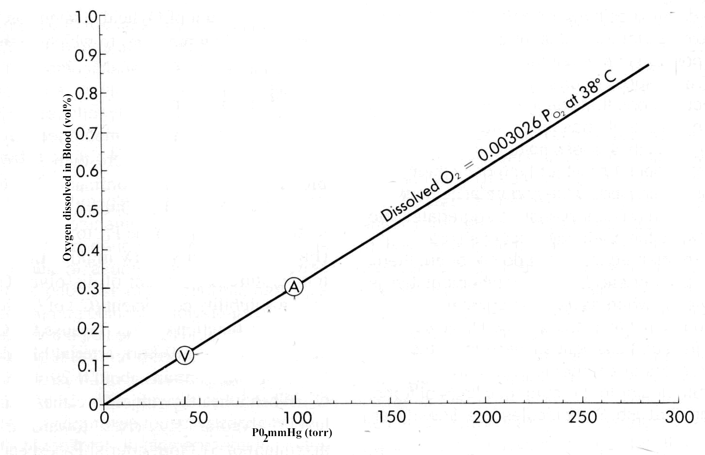
- This quantity is reduced during blood flow through capillaries as oxygen exchanges with tissue, with the extent a reduction being about 5 ml (yielding about 14.4 ml oxygen which would correspond to about 75% hemoglobin saturation (PO 2 of 40 mm Hg). (pharmacology2000.com)
- In this circumstance less than 5 ml O 2 remain associated with hemoglobin per 100 ml blood. (pharmacology2000.com)
- The combination of this cardiac output effect in the magnitude of O 2 desaturation of hemoglobin would yield and up to a 20-fold increase in tissue oxygen transport. (pharmacology2000.com)
- Factors that modify the ability of hemoglobin to bind oxygen include body temperature, pH of blood and the concentration of 2,3-diphosphoglycerate (2,3-DPG). (medscape.com)
- 2,3-diphosphoglycerate (2,3-DPG) is a small molecule found at high concentrations in red blood cells where it binds to and decreases the oxygen affinity of hemoglobin. (anticorps-enligne.fr)
- When fully liganded with carbon monoxide, Hb zeta(2)beta(s)(2) displays a central water cavity, a zeta 1-beta(s)2 (or zeta 2-beta(s)1) interface, intersubunit salt-bridge/hydrogen-bond interactions, C-terminal beta His146 salt-bridge interactions, and a beta-cleft, that are highly unusual for a relaxed hemoglobin structure and are more typical of a tense conformation. (janelia.org)
- 2,3-diphosphoglycerate (2,3-DPG) tends to have a greater affinity towards deoxygenated hemoglobin. (bvsalud.org)
- 2,3-diphosphoglycerate (2,3-DPG) in red blood cells, which causes more oxygen to be released by hemoglobin. (sparrowclinic.com)
- O 2 is carried in blood in two forms: dissolved in solu-tion and in reversible association with hemoglobin. (brainkart.com)
- Even with a Pao 2 of 100 mm Hg, the maximum amount of O 2 dissolved in blood is very small (0.3 mL/dL) com-pared with that bound to hemoglobin. (brainkart.com)
- Each hemoglobin molecule binds up to four O 2 molecules. (brainkart.com)
- The complex interaction between the hemoglobin subunits results in nonlinear (an elon-gated S shape) binding with O 2 ( Figure 23-22 ). (brainkart.com)
- Hemoglobin saturation is the amount of O 2 bound as a percentage of its total O 2 -binding capacity. (brainkart.com)
- Their effect on hemoglobin-O 2 inter-action can be expressed by P 50 , the O 2 tension at which hemoglobin is 50% saturated ( Figure 23-23 ). (brainkart.com)
- The normal P 50 in adults is 26.6 mm Hg (3.4 kPa).An increase in blood hydrogen ion concen-tration reduces O 2 binding to hemoglobin (Bohr effect). (brainkart.com)
- Carbon monoxide, cyanide, nitric acid, and ammo-nia can combine with hemoglobin at O 2 -binding sites. (brainkart.com)
- Carbon monoxide is particularly potent, having 200-300 times the affinity of O 2 for hemoglobin, combining with it to form carboxyhe-moglobin. (brainkart.com)
Ozone2
- Ozone is a more reactive form of oxygen that simulates the production of 2,3-Diphosphoglycerate. (naturalnews.com)
- Ozone administered at a concentration of between 30 and 55 ug/cc causes the greatest increase in the production of interferon and the greatest output of tumor necrosis factor (TNF) and interleukin 2. (silvermedicine.org)
Synthesis3
- These include enhancements in 2,3-Diphosphoglycerate (2,3-DPG) concentrations, myocardial efficiency, buffering capacity and adenosine triphosphate/phosphocreatine synthesis. (jostchemical.com)
- This gene encodes a multifunctional enzyme that catalyzes 2,3-DPG synthesis via its synthetase activity, and 2,3-DPG degradation via its phosphatase activity. (anticorps-enligne.fr)
- PRPP synthesis in the hemolysate during the assay is prevented by added 2,3-diphosphoglyceric acid. (tau.ac.il)
Phosphate4
- Seven biochemical blood parameters were measured pre and post exposure and 20 hours after the second exposure: serum glutathione, red blood cell glutathione reductase, red blood cell glucose-6-phosphate dehydrogenase, lysozyme, serum glutamic oxaloacetic acid transaminase, serum vitamin E and 2,3-diphosphoglycerate. (epa.gov)
- Phosphate supplementation raises the level of 2,3-diphosphoglycerate (2,3-DPG), the enzyme that unloads oxygen into muscle. (prolinesportsnutrition.com)
- For ergogenic purposes, sodium phosphate is supplemented orally in capsule form, at a dose of 3-5 g/day for a period of between 3 and 6 days. (jostchemical.com)
- Van Kempen and co-workers argued that both etiology and symptoms of SIDS can be explained by low concentrations in erythrocytes of phosphate metabolites, mainly 2,3-biphosphoglycerate (2,3-BPG, also known as 2,3-diphosphoglycerate, 2,3-DPG), which in effect would impair muscle function (especially diaphragm) and lead to respiratory failure as Siren and Siren originally stated. (waw.pl)
Concentration4
- After moderate haemorrhage, a low PCV is remarkably well tolerated because of compensatory mechanisms such as increase in concentration of 2,3-diphosphoglycerate in red blood cell (Ganong, 2003). (scialert.net)
- Clinically important factors altering O 2 binding include hydrogen ion concentration, CO 2 tension, temperature, and 2,3-diphosphoglycerate (2,3-DPG) concentration. (brainkart.com)
- The influence of CO 2 tension on hemoglobin's affinity for O 2 is important physiologically and is secondary to the associated rise in hydrogen ion concentration when CO 2 tension increases. (brainkart.com)
- Despite the various benefits, O3 toxicity and clinical utility depends on the concentration and administration to the ap-propriate site.1,2,4,5 One of the major contraindications of O3therapy is lung inhalation. (diegovitello.it)
Metabolism1
- 2,3-DPG is a by-product of glycolysis (the Rapoport-Luebering shunt) and accumulates dur-ing anaerobic metabolism. (brainkart.com)
Erythrocyte1
- Changes in erythrocyte 2,3 diphosphoglycerate in women following short term maximal exercise. (medscape.com)
Erythrocytes3
- The erythrocytes of a patient with the so-called "high ATP syndrome" were characterized by a high ATP content and low 2,3-diphosphoglycerate level. (amsterdamumc.org)
- Here, the 1.95 angstrom resolution crystal structure of human Hb zeta(2)beta(s)(2) that was expressed in complex transgenic knockout mice and purified from their erythrocytes is presented. (janelia.org)
- Application of this method to HGPRT-deficient (hypoxanthine-guanine phosphoribosyltransferase) erythrocytes showed increased PRPP content in 2 subjects with the Lesch-Nyhan syndrome and in 1 out of 2 patients with partial enzyme deficiency. (tau.ac.il)
Inosine1
- Each subject consumed with an inosine supplement (6g per day for 2 days) or placebo. (tigerfitness.com)
Molecule3
- The result is less production of a key molecule called 2,3-Diphosphoglycerate, which stimulates red blood cells to deliver oxygen. (naturalnews.com)
- In HgbA, 2,3 DPG binds to the beta subunits and facilitates the dissociation of oxygen from the HgbA molecule. (pedsurglibrary.com)
- The change in molecular conformation induced by the binding of the first three molecules greatly accelerates bind-ing of the fourth O 2 molecule. (brainkart.com)
Efficiency1
- Increased efficiency in circulation and oxygen delivery can be observed as more 2,3-Diphosphoglycerate is produced. (naturalnews.com)
Affinity3
- When compared with either Hb S or with normal human adult Hb A (alpha(2)beta(2)), Hb zeta(2)beta(s)(2) exhibits atypical properties that include a high oxygen affinity, reduced cooperativity, a weak Bohr effect and blunted 2,3-diphosphoglycerate allostery. (janelia.org)
- On the oxygen dissociation curve, HgbF represents a leftward shift compared to the normal curve because of its lack of affinity to bind 2,3-DPG and therefore a lower p50 compared to normal HgbA. (pedsurglibrary.com)
- a leftward shift increases hemoglobin's affinity for O 2 , reducing its availability to tissues. (brainkart.com)
Adult1
- alpha(2)beta(s)(2)) by exchanging adult alpha-globin with embryonic zeta-globin subunits shows promise as a therapeutic agent for sickle-cell disease (SCD). (janelia.org)
RBCs1
- The aim of this study was to validate the impact of storage time and leuco-depletion on CD47 expression on the RBCs, which could be a prospective marker for detection of RBCs viability and to clarify if the changes in CD47 expression and 2,3-DPG levels are correlated during storage of Packed RBCs. (bvsalud.org)
Cells2
- Figure 2: Postulated G-protein-mediated signal transduction process in normal and regenerating endothelial cells. (nature.com)
- and 3) the molecular basis of diseases that involve spectrin or any of its associated proteins, including contributions of the cortical cytoskeleton to the phenotypic alterations of malignant cells and the molecular pathology of acquired and inherited disorders involving this structure. (yale.edu)
Interleukin2
- The Spectrin-Ankyrin Skeleton Controls CD45 Surface Display and Interleukin-2 Production Pradhan D, Morrow J . The Spectrin-Ankyrin Skeleton Controls CD45 Surface Display and Interleukin-2 Production. (yale.edu)
- The production of interleukin 2 launches an entire cascade of subsequent immunological reactions. (silvermedicine.org)
Temperature1
- The solubility coefficient for O 2 at normal body temperature is 0.003 mL/dL/mm Hg. (brainkart.com)
Tissues4
- Phase 2 involves the use of TORQ Recovery Drink within 15 minutes of exercise completion to rehydrate the tissues of the body, restock muscle and liver glycogen, repair damaged muscle fibres and recharge cellular energy levels. (torqfitness.co.uk)
- This leads to the stimulation of 2,3-diphosphoglycerate (2,3-DPG) which leads to an increase in the amount of oxygen released to the tissues. (silvermedicine.org)
- Increased 2,3-diphosphoglycerate (2.3-DPG) causes oxygen unloading in tissues. (exploremyplan.com)
- Carbon monoxide decreases hemoglo-bin's O 2 -carrying capacity and impairs the release of O 2 to tissues. (brainkart.com)
Metabolic2
- The metabolic oxygen requirement at rest turns out to be approximately 6 volume% which can be reached in the context of a hyperbaric chamber pressurized to about 3 atm. (pharmacology2000.com)
- These argumentsgive rise to the concept of O 2 utilization -- 25% at rest and nominally up to 75%-85% during exercise, with the possibility of achieving100% O 2 utilization in cases of relatively low blood flow rates in combination with high local tissue metabolic requirements. (pharmacology2000.com)
Sickle cell di1
- To participate in this study, you must be at least 2 years old with known or suspected sickle cell disease, sickle cell trait, or other red blood cell disorders. (nih.gov)
Capillaries2
- Any oxygen unloading at tissue capillaries are facilitated by 2,3-DPG, and any alterations in its levels can significantly interfere with oxygen release. (bvsalud.org)
- conversely, the lower CO 2 content in pulmonary capillaries increases hemoglobin's affin-ity for O 2 again, facilitating O 2 uptake from alveoli. (brainkart.com)
Uptake1
- the net result is facilitation of O 2 release to tissue with little impairment in O 2 uptake (unless severe hypoxia is present). (brainkart.com)
Arterial1
- Therefore, arterial blood with a PO 2 of about 95 mm Hg (torr) would contain about 0.29 volume% of dissolved oxygen. (pharmacology2000.com)
Saturation2
- At about 90% saturation, the decrease in available O 2 receptors flattens the curve until full saturation is reached. (brainkart.com)
- They can displace O 2 and shift the saturation curve to the left. (brainkart.com)
Biochemistry1
- Analytical Biochemistry , 75 (2), 382-388. (uni-luebeck.de)
ENZYME1
- All ENZYME / UniProtKB/Swiss-Prot entries corresponding to 3.1.3. (expasy.org)
Contrast1
- In contrast, the gamma subunits in HgbF do not allow 2,3 DPG to bind easily and therefore HgF holds on to O 2 more tightly than HgbA. (pedsurglibrary.com)
Compensatory1
- 2,3-DPG levels may, however, play an important compensatory role in patientswith chronic anemia and may significantly affect the O 2 -carrying capacity of blood transfusions. (brainkart.com)
Diaphragm2
- Effects of an infusion of Na 2 HPO 4 on diaphragm strength, endurance, and magnitude of recovery were evaluated in in situ canine diaphragm strips. (bgu.ac.il)
- In their recent letter Van Kempen and co-workers presented an intriguing hypothesis linking hypophosphatemia and sudden infant death syndrome (SIDS) (1), founded on the work by Siren and Siren on the critical diaphragm failure being the cause of SIDS incidences (2,3). (waw.pl)
Significantly1
- The above condition, changes significantly during heavy exercise, since interstitial fluid PO 2 may fall to 15 mm Hg. (pharmacology2000.com)
Gene1
- The aim of the present study was to evaluate the validity of frizzled-7 (FZD7) and glypican-3 (GPC3) gene expression as potential biomarkers for HCC early diagnosis, and to investigate the association between FZD7 rs2280509 polymorphism and HCC risk. (bvsalud.org)
High1
- In the paralyzed state, the compliance of the chest wall is quite high and often greater than 25 mL/cm H 2 O. The ribs of the newborn infant are made mostly of cartilage and are quite elastic. (pedsurglibrary.com)
Diagnosis1
- 1, 2] A diagnosis of polycythemia vera is made when both major and one minor criterion are present or when the first major criterion is present with any two minor criteria. (medscape.com)
Increase1
- 3-6 As an added bonus, caffeine can increase fat oxidation, which can help with weight management. (sspnutrition.com)
Cell1
- Alteration of both CD47 expression and 2,3-DPG levels during red cell storage may serve as markers in the development of RCSL. (bvsalud.org)
Exercise1
- They then performed three exercise tests consisting of a submaximal warm-up run, a competitive 3-mile treadmill run, and a max treadmill run. (tigerfitness.com)