Uukuniemi virus
Bunyaviridae
Arboviruses
Phlebovirus
Orthobunyavirus
Golgi Apparatus
Cricetinae
Glycoproteins
Chick Embryo
Viral Envelope Proteins
Molecular Sequence Data
Reverse genetics system for Uukuniemi virus (Bunyaviridae): RNA polymerase I-catalyzed expression of chimeric viral RNAs. (1/14)
We describe here the development of a reverse genetics system for the phlebovirus Uukuniemi virus, a member of the Bunyaviridae family, by using RNA polymerase I (pol I)-mediated transcription. Complementary DNAs containing the coding sequence for either chloramphenicol acetyltransferase (CAT) or green fluorescent protein (GFP) (both in antisense orientation) were flanked by the 5'- and 3'-terminal untranslated regions of the Uukuniemi virus sense or complementary RNA derived from the medium-sized (M) RNA segment. This chimeric cDNA (pol I expression cassette) was cloned between the murine pol I promoter and terminator and the plasmid transfected into BHK-21 cells. When such cells were either superinfected with Uukuniemi virus or cotransfected with expression plasmids encoding the L (RNA polymerase), N (nucleoprotein), and NSs (nonstructural protein) viral proteins, strong CAT activity or GFP expression was observed. CAT activity was consistently stronger in cells expressing L plus N than following superinfection. No activity was seen without superinfection, nor was activity detected when either the L or N expression plasmid was omitted. Omitting NSs expression had no effect on CAT activity or GFP expression, indicating that this protein is not needed for viral RNA replication or transcription. CAT activity could be serially passaged to fresh cultures by transferring medium from CAT-expressing cells, indicating that recombinant virus containing the reporter construct had been produced. In summary, we demonstrate that the RNA pol I system, originally developed for influenza virus, which replicates in the nucleus, has strong potential for the development of an efficient reverse genetics system also for Bunyaviridae members, which replicate in the cytoplasm. (+info)Mutational analysis of the Uukuniemi virus (Bunyaviridae family) promoter reveals two elements of functional importance. (2/14)
We have performed an extensive mutational analysis of the proposed promoter region of the phlebovirus Uukuniemi (UUK), a member of the Bunyaviridae family. This was achieved by using a recently developed RNA polymerase I (Pol I)-driven reverse genetics system (R. Flick and R. F. Pettersson, J. Virol. 75:1643-1655, 2001). Chimeric cDNAs containing the coding region for the reporter chloramphenicol acetyltransferase (CAT) in an antisense orientation were flanked by the 5'- and 3'-terminal nontranslated regions of the UUK virus-sense RNA (vRNA) derived from the medium-sized (M) RNA segment. The chimeric cDNAs (Pol I expression cassettes) were cloned between the murine Pol I promoter and terminator, and the plasmids were transfected into BHK-21 cells. CAT activity was determined after cotransfection with viral expression plasmids encoding the RNA-dependent RNA polymerase (L) and the nucleoprotein (N) or, alternatively, after superinfection with UUK virus helper virus. Using oligonucleotide-directed mutagenesis, single point mutations (substitutions, deletions, and insertions) were introduced into the viral promoter region. Differences in CAT activities were interpreted to reflect the efficiency of mRNA transcription from the mutated promoter and the influence on RNA replication. Analysis of 109 mutants allowed us to define two important regulatory regions within the proximal promoter region (site A, positions 3 to 5 and 2 to 4; site B, positions 8 and 8, where underlined nucleotides refer to positions in the vRNA 3' end). Complementary double nucleotide exchanges in the proximal promoter region, which maintained the possibility for base pairing between the 5' and 3' ends, demonstrated that nucleotides in the two described regions are essential for viral polymerase recognition in a base-specific manner. Thus, mere preservation of panhandle base pairing between the 5' and 3' ends is not sufficient for promoter activity. In conclusion, we have been able to demonstrate that both ends of the M RNA segment build up the promoter region and are involved in the specific recognition by the viral polymerase. (+info)Generation and analysis of infectious virus-like particles of uukuniemi virus (bunyaviridae): a useful system for studying bunyaviral packaging and budding. (3/14)
In the present report we describe an infectious virus-like particle (VLP) system for the Uukuniemi (UUK) virus, a member of the Bunyaviridae family. It utilizes our recently developed reverse genetic system based on the RNA polymerase I minigenome system for UUK virus used to study replication, encapsidation, and transcription by monitoring reporter gene expression. Here, we have added the glycoprotein precursor expression plasmid together with the minigenome, nucleoprotein, and polymerase to generate VLPs, which incorporate the minigenome and are released into the supernatant. The particles are able to infect new cells, and reporter gene expression can be monitored if the trans-acting viral proteins (RNA polymerase and nucleoprotein) are also expressed in these cells. No minigenome transfer occurred in the absence of glycoproteins, demonstrating that the glycoproteins are absolutely required for the generation of infectious particles. Moreover, expression of glycoproteins alone was sufficient to produce and release VLPs. We show that the ribonucleoproteins (RNPs) are incorporated into VLPs but are not required for the generation of particles. Morphological analysis of the particles by electron microscopy revealed that VLPs, either with or without minigenomes, display a surface morphology indistinguishable from that of the authentic UUK virus and that they bud into Golgi vesicles in the same way as UUK virus does. This infectious VLP system will be very useful for studying the bunyaviral structural components required for budding and packaging of RNPs and receptor binding and may also be useful for the development of new vaccines for the human pathogens from this family. (+info)The glycoprotein cytoplasmic tail of Uukuniemi virus (Bunyaviridae) interacts with ribonucleoproteins and is critical for genome packaging. (4/14)
We have analyzed the importance of specific amino acids in the cytoplasmic tail of the glycoprotein G(N) for packaging of ribonucleoproteins (RNPs) into virus-like particles (VLPs) of Uukuniemi virus (UUK virus), a member of the Bunyaviridae family. In order to study packaging, we added the G(N)/G(C) glycoprotein precursor (p110) to a polymerase I-driven minigenome rescue system to generate VLPs that are released into the supernatant. These particles can infect new cells, and reporter gene expression can be detected. To determine the role of UUK virus glycoproteins in RNP packaging, we performed an alanine scan of the glycoprotein G(N) cytoplasmic tail (amino acids 1 to 81). First, we discovered three regions in the tail (amino acids 21 to 25, 46 to 50, and 71 to 81) which are important for minigenome transfer by VLPs. Further mutational analysis identified four amino acids that were important for RNP packaging. These amino acids are essential for the binding of nucleoproteins and RNPs to the glycoprotein without affecting the morphology of the particles. No segment-specific interactions between the RNA and the cytoplasmic tail could be observed. We propose that VLP systems are useful tools for analyzing protein-protein interactions important for packaging of viral genome segments, assembly, and budding of other members of the Bunyaviridae family. (+info)The cytoplasmic tails of Uukuniemi Virus (Bunyaviridae) G(N) and G(C) glycoproteins are important for intracellular targeting and the budding of virus-like particles. (5/14)
Functional motifs within the cytoplasmic tails of the two glycoproteins G(N) and G(C) of Uukuniemi virus (UUK) (Bunyaviridae family) were identified with the help of our recently developed virus-like particle (VLP) system for UUK virus (A. K. Overby, V. Popov, E. P. Neve, and R. F. Pettersson, J. Virol. 80:10428-10435, 2006). We previously reported that information necessary for the packaging of ribonucleoproteins into VLPs is located within the G(N) cytoplasmic tail (A. K. Overby, R. F. Pettersson, and E. P. Neve, J. Virol. 81:3198-3205, 2007). The G(N) glycoprotein cytoplasmic tail specifically interacts with the ribonucleoproteins and is critical for genome packaging. In addition, two other regions in the G(N) cytoplasmic tail, encompassing residues 21 to 25 and 46 to 50, were shown to be important for particle generation and release. By the introduction of point mutations within these two regions, we demonstrate that leucines at positions 23 and 24 are crucial for the initiation of VLP budding, while leucine 46, glutamate 47, and leucine 50 are important for efficient exit from the endoplasmic reticulum and subsequent transport to the Golgi complex. We found that budding and particle generation are highly dependent on the intracellular localization of both glycoproteins. The short cytoplasmic tail of UUK G(C) contains a lysine at position -3 from the C terminus that is highly conserved among members of the Phlebovirus, Hantavirus, and Orthobunyavirus genera. Mutating this single amino acid residue in G(C) resulted in the mislocalization of not only G(C) but also G(N) to the plasma membrane, and VLP generation was compromised in cells expressing this mutant. Together, these results demonstrate that the cytoplasmic tails of both G(N) and G(C) contain specific information necessary for efficient virus particle generation. (+info)Insights into bunyavirus architecture from electron cryotomography of Uukuniemi virus. (6/14)
(+info)Entry of bunyaviruses into mammalian cells. (7/14)
(+info)Oligomerization of Uukuniemi virus nucleocapsid protein. (8/14)
(+info)I'm sorry for any confusion, but "Uukuniemi virus" is not a commonly used medical term in the English language. It is a term from virology, referring to a specific type of virus discovered in Finland. Uukuniemi virus is a type of hantavirus, which can cause hemorrhagic fever with renal syndrome in humans. However, it's primarily a term used in research and not something that would typically be used in a medical diagnosis or treatment context. If you have any more specific questions about virology or infectious diseases, I'd be happy to try and help answer them!
Bunyaviridae is a family of enveloped, single-stranded RNA viruses that includes more than 350 different species. These viruses are named after the type species, Bunyamwera virus, which was first isolated in 1943 from mosquitoes in Uganda.
The genome of Bunyaviridae viruses is divided into three segments: large (L), medium (M), and small (S). The L segment encodes the RNA-dependent RNA polymerase, which is responsible for replication and transcription of the viral genome. The M segment encodes two glycoproteins that form the viral envelope and are involved in attachment and fusion to host cells. The S segment encodes the nucleocapsid protein, which packages the viral RNA, and a non-structural protein that is involved in modulation of the host immune response.
Bunyaviridae viruses are transmitted to humans and animals through arthropod vectors such as mosquitoes, ticks, and sandflies. Some members of this family can cause severe disease in humans, including Hantavirus pulmonary syndrome, Crimean-Congo hemorrhagic fever, and Rift Valley fever.
Prevention and control measures for Bunyaviridae viruses include avoiding contact with vectors, using insect repellent and wearing protective clothing, and implementing vector control programs. There are no specific antiviral treatments available for most Bunyaviridae infections, although ribavirin has been shown to be effective against some members of the family. Vaccines are available for a few Bunyaviridae viruses, such as Hantavirus and Crimean-Congo hemorrhagic fever virus, but they are not widely used due to limitations in production and distribution.
Arboviruses are a group of viruses that are primarily transmitted to humans and animals through the bites of infected arthropods, such as mosquitoes, ticks, and sandflies. The term "arbovirus" is short for "arthropod-borne virus."
Arboviruses can cause a wide range of symptoms, depending on the specific virus and the individual host's immune response. Some common symptoms associated with arboviral infections include fever, headache, muscle and joint pain, rash, and fatigue. In severe cases, arboviral infections can lead to serious complications such as encephalitis (inflammation of the brain), meningitis (inflammation of the membranes surrounding the brain and spinal cord), or hemorrhagic fever (bleeding disorders).
There are hundreds of different arboviruses, and they are found in many parts of the world. Some of the most well-known arboviral diseases include dengue fever, chikungunya, Zika virus infection, West Nile virus infection, yellow fever, and Japanese encephalitis.
Prevention of arboviral infections typically involves avoiding mosquito bites and other arthropod vectors through the use of insect repellent, wearing long sleeves and pants, and staying indoors during peak mosquito feeding times. Public health efforts also focus on reducing vector populations through environmental management and the use of larvicides. Vaccines are available for some arboviral diseases, such as yellow fever and Japanese encephalitis.
Phlebovirus is a type of virus that belongs to the family Bunyaviridae. These viruses have a single-stranded, negative-sense RNA genome and are transmitted to humans through the bites of infected insects, such as sandflies or ticks. Some examples of diseases caused by Phleboviruses include sandfly fever, Toscana virus infection, and Rift Valley fever.
The term "Phlebovirus" comes from the Greek word "phleps," which means "vein," reflecting the viruses' tendency to cause febrile illnesses characterized by symptoms such as fever, headache, muscle pain, and rash. The virus was first identified in the 1960s and has since been found in many parts of the world, particularly in areas with warm climates where sandflies and ticks are more common.
Phleboviruses have a complex structure, consisting of three segments of RNA enclosed within a lipid membrane derived from the host cell. The viral membrane contains two glycoproteins, Gn and Gc, which are important for attachment to and entry into host cells. Once inside the cell, the virus uses its RNA-dependent RNA polymerase to replicate its genome and produce new virions, which can then infect other cells or be transmitted to a new host through the bite of an infected insect.
Prevention and treatment of Phlebovirus infections are focused on avoiding exposure to infected insects and reducing symptoms through supportive care. There are no specific antiviral treatments available for these infections, although research is ongoing to develop effective therapies. Vaccines are also being developed for some Phleboviruses, such as Rift Valley fever, which can cause severe illness and death in humans and animals.
Bunyaviridae is a family of viruses that includes several genera capable of causing human disease. These viruses are primarily transmitted to humans through the bite of infected arthropods, such as mosquitoes and ticks, or through contact with infected rodents or their excreta.
Some of the diseases caused by Bunyaviridae infections include:
1. Hantavirus Pulmonary Syndrome (HPS): This is a severe, sometimes fatal, respiratory disease caused by hantaviruses. It is transmitted to humans through contact with infected rodents or their urine and droppings.
2. Crimean-Congo Hemorrhagic Fever (CCHF): This is a serious and often fatal viral hemorrhagic fever caused by the CCHF virus. It is primarily transmitted to humans through the bite of infected ticks, but can also be spread through contact with the blood or tissue of infected animals.
3. Rift Valley Fever (RVF): This is a viral disease that primarily affects animals, but can also infect humans. It is transmitted to humans through contact with the blood or tissue of infected animals, or through the bite of infected mosquitoes.
4. La Crosse Encephalitis: This is a viral disease transmitted to humans through the bite of infected mosquitoes. It primarily affects children and can cause inflammation of the brain (encephalitis).
5. Toscana Virus Infection: This is a viral disease transmitted to humans through the bite of infected sandflies. It can cause symptoms such as fever, headache, and meningitis.
Prevention measures include avoiding contact with rodents and their excreta, using insect repellent and wearing protective clothing to prevent mosquito and tick bites, and seeking prompt medical attention if symptoms of a Bunyaviridae infection develop.
Orthobunyavirus is a genus of viruses in the family Peribunyaviridae, order Bunyavirales. These are enveloped, single-stranded, negative-sense RNA viruses. The genome consists of three segments: large (L), medium (M), and small (S). The L segment encodes the RNA-dependent RNA polymerase, the M segment encodes two glycoproteins (Gn and Gc) and a nonstructural protein (NSm), and the S segment encodes the nucleocapsid protein (N) and a nonstructural protein (NSs).
Orthobunyaviruses are primarily transmitted by arthropods, such as mosquitoes, ticks, and midges, and can cause disease in humans and animals. The diseases caused by orthobunyaviruses range from mild febrile illness to severe hemorrhagic fever and encephalitis. Some of the notable orthobunyaviruses include California encephalitis virus, La Crosse encephalitis virus, Oropouche virus, and Crimean-Congo hemorrhagic fever virus.
The Golgi apparatus, also known as the Golgi complex or simply the Golgi, is a membrane-bound organelle found in the cytoplasm of most eukaryotic cells. It plays a crucial role in the processing, sorting, and packaging of proteins and lipids for transport to their final destinations within the cell or for secretion outside the cell.
The Golgi apparatus consists of a series of flattened, disc-shaped sacs called cisternae, which are stacked together in a parallel arrangement. These stacks are often interconnected by tubular structures called tubules or vesicles. The Golgi apparatus has two main faces: the cis face, which is closest to the endoplasmic reticulum (ER) and receives proteins and lipids directly from the ER; and the trans face, which is responsible for sorting and dispatching these molecules to their final destinations.
The Golgi apparatus performs several essential functions in the cell:
1. Protein processing: After proteins are synthesized in the ER, they are transported to the cis face of the Golgi apparatus, where they undergo various post-translational modifications, such as glycosylation (the addition of sugar molecules) and sulfation. These modifications help determine the protein's final structure, function, and targeting.
2. Lipid modification: The Golgi apparatus also modifies lipids by adding or removing different functional groups, which can influence their properties and localization within the cell.
3. Protein sorting and packaging: Once proteins and lipids have been processed, they are sorted and packaged into vesicles at the trans face of the Golgi apparatus. These vesicles then transport their cargo to various destinations, such as lysosomes, plasma membrane, or extracellular space.
4. Intracellular transport: The Golgi apparatus serves as a central hub for intracellular trafficking, coordinating the movement of vesicles and other transport carriers between different organelles and cellular compartments.
5. Cell-cell communication: Some proteins that are processed and packaged in the Golgi apparatus are destined for secretion, playing crucial roles in cell-cell communication and maintaining tissue homeostasis.
In summary, the Golgi apparatus is a vital organelle involved in various cellular processes, including post-translational modification, sorting, packaging, and intracellular transport of proteins and lipids. Its proper functioning is essential for maintaining cellular homeostasis and overall organismal health.
Cricetinae is a subfamily of rodents that includes hamsters, gerbils, and relatives. These small mammals are characterized by having short limbs, compact bodies, and cheek pouches for storing food. They are native to various parts of the world, particularly in Europe, Asia, and Africa. Some species are popular pets due to their small size, easy care, and friendly nature. In a medical context, understanding the biology and behavior of Cricetinae species can be important for individuals who keep them as pets or for researchers studying their physiology.
Viral proteins are the proteins that are encoded by the viral genome and are essential for the viral life cycle. These proteins can be structural or non-structural and play various roles in the virus's replication, infection, and assembly process. Structural proteins make up the physical structure of the virus, including the capsid (the protein shell that surrounds the viral genome) and any envelope proteins (that may be present on enveloped viruses). Non-structural proteins are involved in the replication of the viral genome and modulation of the host cell environment to favor viral replication. Overall, a thorough understanding of viral proteins is crucial for developing antiviral therapies and vaccines.
A viral RNA (ribonucleic acid) is the genetic material found in certain types of viruses, as opposed to viruses that contain DNA (deoxyribonucleic acid). These viruses are known as RNA viruses. The RNA can be single-stranded or double-stranded and can exist as several different forms, such as positive-sense, negative-sense, or ambisense RNA. Upon infecting a host cell, the viral RNA uses the host's cellular machinery to translate the genetic information into proteins, leading to the production of new virus particles and the continuation of the viral life cycle. Examples of human diseases caused by RNA viruses include influenza, COVID-19 (SARS-CoV-2), hepatitis C, and polio.
Glycoproteins are complex proteins that contain oligosaccharide chains (glycans) covalently attached to their polypeptide backbone. These glycans are linked to the protein through asparagine residues (N-linked) or serine/threonine residues (O-linked). Glycoproteins play crucial roles in various biological processes, including cell recognition, cell-cell interactions, cell adhesion, and signal transduction. They are widely distributed in nature and can be found on the outer surface of cell membranes, in extracellular fluids, and as components of the extracellular matrix. The structure and composition of glycoproteins can vary significantly depending on their function and location within an organism.
A chick embryo refers to the developing organism that arises from a fertilized chicken egg. It is often used as a model system in biological research, particularly during the stages of development when many of its organs and systems are forming and can be easily observed and manipulated. The study of chick embryos has contributed significantly to our understanding of various aspects of developmental biology, including gastrulation, neurulation, organogenesis, and pattern formation. Researchers may use various techniques to observe and manipulate the chick embryo, such as surgical alterations, cell labeling, and exposure to drugs or other agents.
Viral envelope proteins are structural proteins found in the envelope that surrounds many types of viruses. These proteins play a crucial role in the virus's life cycle, including attachment to host cells, fusion with the cell membrane, and entry into the host cell. They are typically made up of glycoproteins and are often responsible for eliciting an immune response in the host organism. The exact structure and function of viral envelope proteins vary between different types of viruses.
A cell line is a culture of cells that are grown in a laboratory for use in research. These cells are usually taken from a single cell or group of cells, and they are able to divide and grow continuously in the lab. Cell lines can come from many different sources, including animals, plants, and humans. They are often used in scientific research to study cellular processes, disease mechanisms, and to test new drugs or treatments. Some common types of human cell lines include HeLa cells (which come from a cancer patient named Henrietta Lacks), HEK293 cells (which come from embryonic kidney cells), and HUVEC cells (which come from umbilical vein endothelial cells). It is important to note that cell lines are not the same as primary cells, which are cells that are taken directly from a living organism and have not been grown in the lab.
RNA viruses are a type of virus that contain ribonucleic acid (RNA) as their genetic material, as opposed to deoxyribonucleic acid (DNA). RNA viruses replicate by using an enzyme called RNA-dependent RNA polymerase to transcribe and replicate their RNA genome.
There are several different groups of RNA viruses, including:
1. Negative-sense single-stranded RNA viruses: These viruses have a genome that is complementary to the mRNA and must undergo transcription to produce mRNA before translation can occur. Examples include influenza virus, measles virus, and rabies virus.
2. Positive-sense single-stranded RNA viruses: These viruses have a genome that can serve as mRNA and can be directly translated into protein after entry into the host cell. Examples include poliovirus, rhinoviruses, and coronaviruses.
3. Double-stranded RNA viruses: These viruses have a genome consisting of double-stranded RNA and use a complex replication strategy involving both transcription and reverse transcription. Examples include rotaviruses and reoviruses.
RNA viruses are known to cause a wide range of human diseases, ranging from the common cold to more severe illnesses such as hepatitis C, polio, and COVID-19. Due to their high mutation rates and ability to adapt quickly to new environments, RNA viruses can be difficult to control and treat with antiviral drugs or vaccines.
Molecular sequence data refers to the specific arrangement of molecules, most commonly nucleotides in DNA or RNA, or amino acids in proteins, that make up a biological macromolecule. This data is generated through laboratory techniques such as sequencing, and provides information about the exact order of the constituent molecules. This data is crucial in various fields of biology, including genetics, evolution, and molecular biology, allowing for comparisons between different organisms, identification of genetic variations, and studies of gene function and regulation.
Vaccinia virus is a large, complex DNA virus that belongs to the Poxviridae family. It is the virus used in the production of the smallpox vaccine. The vaccinia virus is not identical to the variola virus, which causes smallpox, but it is closely related and provides cross-protection against smallpox infection.
The vaccinia virus has a unique replication cycle that occurs entirely in the cytoplasm of infected cells, rather than in the nucleus like many other DNA viruses. This allows the virus to evade host cell defenses and efficiently produce new virions. The virus causes the formation of pocks or lesions on the skin, which contain large numbers of virus particles that can be transmitted to others through close contact.
Vaccinia virus has also been used as a vector for the delivery of genes encoding therapeutic proteins, vaccines against other infectious diseases, and cancer therapies. However, the use of vaccinia virus as a vector is limited by its potential to cause adverse reactions in some individuals, particularly those with weakened immune systems or certain skin conditions.
 Naples phlebovirus
Bunyaviridae nonstructural S proteins
Phlebovirus
Dabie bandavirus
Fusion mechanism
List of MeSH codes (B04)
ArboCat Virus: Uukuniemi (UUKV)
ArboCat Virus: Uukuniemi (UUKV)
Table - Sandfly Fever Sicilian Virus, Algeria - Volume 14, Number 5-May 2008 - Emerging Infectious Diseases journal - CDC
Naples phlebovirus - Wikipedia
 Anna Överby Wernstedt
 "NAMRU-3 Translations of Parasite Literature from Russian, French, Japa" by United States Naval Medical Research Unit Number...
Effects of a point mutation in the 3′ end of the S genome segment of naturally occurring and engineered Bunyamwera viruses |...
Nat - ROCK inhibitor Induces Cell Death in Melanoma Cells
DeCS 2002 - Changed terms
DeCS 2002 - Changed terms
DeCS 2002 - Changed terms
DeCS 2002 - Changed terms
DeCS 2002 - Changed terms
DeCS 2002 - Changed terms
DeCS 2002 - Changed terms
DeCS 2002 - Changed terms
DeCS 2002 - Changed terms
DeCS 2002 - Changed terms
 DeCS 2002 - Changed terms
DeCS 2002 - Changed terms
Body
 Pesquisa | Biblioteca Virtual em Saúde - BRASIL
 Dr Alexander N Freiberg - Kudos: Growing the influence of research
 What's in the Collection | Division of Vector-Borne Diseases | NCEZID | CDC
View of Risk assessment of human infection with a novel bunyavirus in China | Western Pacific Surveillance and Response ...
DeCS 2009 - February 20, 2009 version
 BCCO - Collection of Arboviruses
Phlebovirus3
- Further, studies around the Uukuniemi phlebovirus Gn tail showed that this Gn endodomain plays a crucial role in genome packaging into virus particles (10). (hwupdate.org)
- In June 2009, a novel Phlebovirus from the Bunyaviridae family, subsequently named SFTS virus (SFTSV), was isolated from a patient's blood sample. (who.int)
- Genome characterization and p-distance analyses based on complete open reading frames revealed that the virus is probably a natural reassortant of the Echarate virus (large and small segments) with a yet-unidentified phlebovirus (M segment). (cdc.gov)
Genome5
- It is an enveloped RNA virus with a tripartite genome e Uukuniemi (UUK) serogroup. (wikipedia.org)
- The genome of Bunyamwera virus (BUN) consists of three segments of single-stranded RNA of negative polarity. (microbiologyresearch.org)
- The L protein of Rift Valley fever virus can rescue viral ribonucleoproteins and transcribe synthetic genome-like RNA molecules. (microbiologyresearch.org)
- Characterization of La Crosse virus small genome transcripts. (microbiologyresearch.org)
- Therefore, continuous public health surveillance, including genome characterization as a complementary tool, is critical to identifying novel and emerging viruses of clinical relevance in the Americas. (cdc.gov)
Ticks collected2
Sandfly Fever Sicilian Virus1
- Sandfly fever Sicilian virus isolated from Phlebotomus ariasi sandflies in Algeria. (cdc.gov)
Strains1
- Characteristics of Omsk hemorrhagic fever virus strains isolated from different inhabitants of a natural focus. (unl.edu)
COMPLEX VIRUSES1
- B) Geographic distribution of the Candiru complex viruses in Peru identified from patients with acute febrile illness. (cdc.gov)
Protein2
- Characterization of Bunyamwera S RNA that is transcribed and replicated by the L protein expressed from recombinant vaccinia virus. (microbiologyresearch.org)
- Taken together, these data indicate that the release of glycoproteins into cell supernatants in the form of virus-like structures does not require the participation of the viral N protein. (hwupdate.org)
Characterization2
- Characterization of the 5′ and 3′ ends of the viral messenger RNAs isolated from BHK-21 cells infected with Germiston virus (Bunyavirus). (microbiologyresearch.org)
- We report the identification and characterization of a novel ECHV virus variant isolated from a patient with acute febrile illness (AFI) in Peru. (cdc.gov)
Bunyavirus1
- To assess the public health risk of human infection from a novel bunyavirus - severe fever with thrombocytopenia syndrome virus (SFTSV) - in China. (who.int)
Sandflies1
- The Sandfly group's natural reservoir are sandflies, while the natural reservoir for Uukuniemi is ticks. (wikipedia.org)
Sindbis1
- Noncytopathic Sindbis virus RNA vectors for heterologous gene expression. (microbiologyresearch.org)
Toscana1
- Sandfluevirusmeningitis hos en dansk turist efter ophold i Toscana" [Sandfly virus meningitis in a Danish traveller returning from Tuscany]. (wikipedia.org)
Candiru2
Complementary1
- Rescue of a segmented negative-strand RNA virus entirely from cloned complementary DNAs. (microbiologyresearch.org)
Genus1
- Note the virus list is broken down to genus level classification and only contains BSL2 and BSL3 non select agent pathogens. (cdc.gov)
Species1
- 1 A tick species, Haemaphysalis longicornis , was found to carry the virus. (who.int)
Assembly1
- Previous studies of orthobunyavirus mutant glycoproteins showed that this endodomains of both glycoproteins are required for virus-like particle (VLP) and virus assembly (9). (hwupdate.org)
Entry1
- Click on the PDF icon to the left to view a copy of this virus entry in PDF format. (cdc.gov)
Report1
- Virus Taxonomy: Eighth Report of the International Committee on Taxonomy of Viruses. (wikipedia.org)
Fever with thrombocytopenia syndr2
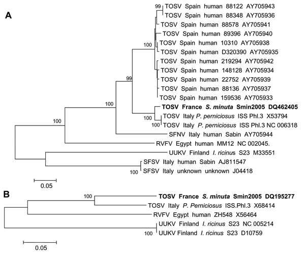
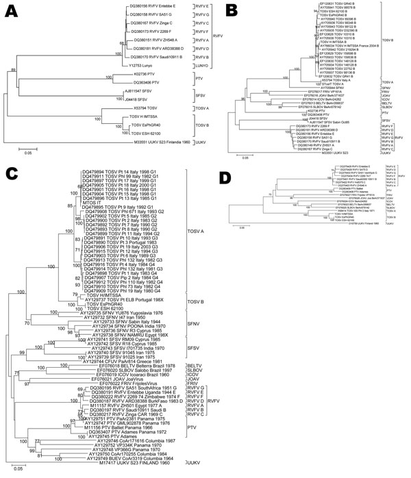
- Although limited sequence similarity was detected between current members of the Uukuniemi group and Severe fever with thrombocytopenia syndrome virus (SFTSV) and Heartland virus, a clear serological reaction was observed between some of them, indicating that SFTSV and Heartland virus should be considered part of the Uukuniemi virus group. (nih.gov)
- To assess the public health risk of human infection from a novel bunyavirus - severe fever with thrombocytopenia syndrome virus (SFTSV) - in China. (who.int)
Bhanja1
- Genetic characterization of Bhanja virus and Palma virus, two tick-borne phleboviruses. (ictv.global)
RVFV2
Ticks2
- First strains were isolated in Finland from ticks collected in June 1959 (Uukuniemi I 1 and Jomala A 21) (1,9). (cdc.gov)
- The Sandfly group's natural reservoir are sandflies, while the natural reservoir for Uukuniemi is ticks. (wikipedia.org)
Tripartite1
- It is an enveloped RNA virus with a tripartite genome e Uukuniemi (UUK) serogroup. (wikipedia.org)
Arbovirus1
- Schmallenberg virus (SBV) is an emerging arbovirus infecting ruminants in Europe. (microbiologyresearch.org)
Sicilian1
- Sandfly fever Sicilian virus isolated from Phlebotomus ariasi sandflies in Algeria. (cdc.gov)
Grand Arbaud1
- and Grand Arbaud virus, Precarious Point virus, and Manawa virus would each be given individual species status. (nih.gov)
Penetration3
- Entry receptors trigger virus entry by endocytosis/pinocytosis or by inducing fusion/penetration. (expasy.org)
- Acid-activated penetration began 15 to 25 min after initiation of virus internalization and relied on maturation of early endosomes to late endosomes. (bvsalud.org)
- Our study also brings a complete toolbox of innovative methods to study each step of the OBV entry program in fixed and living cells, from virus binding and endocytosis to fusion and penetration. (bvsalud.org)
Schmallenberg4
- Schmallenberg virus among female lambs, Belgium, 2012. (microbiologyresearch.org)
- In vivo and in vitro identification of a hypervariable region in Schmallenberg virus. (microbiologyresearch.org)
- Detection of Schmallenberg virus in different Culicoides spp. (microbiologyresearch.org)
- IMPORTANCE Orthobunyaviruses (OBVs), which include La Crosse, Oropouche, and Schmallenberg viruses, represent a growing threat to humans and domestic animals worldwide. (bvsalud.org)
VIRAL1
- Virus entry relied on vacuolar acidification, with an optimal pH for viral membrane fusion at pH 5.5. (bvsalud.org)
Bunyamwera1
- The Bunyamwera virus mRNA transcription signal resides within both the 3′ and the 5′ terminal regions and allows ambisense transcription from a model RNA segment. (microbiologyresearch.org)
Early stages of infection2
- In parallel, to precisely measure intracellular N levels, we employed multiple reaction monitoring mass spectrometry (MRM-MS). Our results show that N binds mostly to host RNAs at early stages of infection, yielding nascent virus particles of reduced infectivity. (mdpi.com)
- Ideally, preventing OBV spread requires approaches that target early stages of infection, i.e., virus entry. (bvsalud.org)
Infectivity1
- Labeling of the virus with fluorescent dyes did not adversely affect its infectivity and allowed the monitoring of single particles in fixed and live cells. (bvsalud.org)
Subsequently1
- The virus entered Rab5a-positive (Rab5a+) early endosomes and, subsequently, late endosomal vacuoles containing Rab7a but not LAMP-1. (bvsalud.org)
Segments2
- The genomes of Mourilyan virus and Wēnzhōu shrimp virus 1 of prawns comprise 4 RNA segments. (ictv.global)
- However, different patterns were observed for the termini of the three segments from the same virus type. (microbiologyresearch.org)
Host1
- However, early virus-host cell interactions and entry mechanisms remain poorly characterized. (bvsalud.org)
HUMAN1
- Investigating iPSC-derived human neurons and cell lines, we found that virus binding to the cell surface was specific, and 50% of bound virions were endocytosed within 10 min. (bvsalud.org)
Analysis2
- Two new putative plant viruses from wood metagenomics analysis of an esca diseased vineyard. (ictv.global)
- Analysis of LaCrosse virus S mRNA 5′ termini in infected mosquito cells and Aedes triseriatus mosquitoes. (microbiologyresearch.org)
Collection2
Disease1
- An un-described virus disease of sheep, cattle and man from East Africa. (ictv.global)
Found1
- Using this approach, we found that endocytic internalization of bound viruses was asynchronous and occurred within 30 to 40 min. (bvsalud.org)