Uterus
Pregnancy
Pregnancy, Animal
Cervix Uteri
Endometrium
Decidua
Estradiol
Progesterone
Pseudopregnancy
Estrus
Fallopian Tubes
Genitalia, Female
Estrous Cycle
Oxytocin
Labor, Obstetric
Mullerian Ducts
Placenta
Ovary
Estrogens
Embryo Implantation, Delayed
Uterine Rupture
Diethylstilbestrol
RNA, Messenger
Blastocyst
In Situ Hybridization
Prostaglandins F
Metrial Gland
Dinoprost
Immunohistochemistry
Sheep
Pregnancy Proteins
Relaxin
Viviparity, Nonmammalian
Receptors, Estrogen
Trophoblasts
Sperm Transport
Receptors, Oxytocin
Epithelium
Placentation
Ovum Transport
Estrogen Antagonists
Embryo Transfer
Adnexa Uteri
Diestrus
Endometrial Hyperplasia
Oviducts
Estrogen Receptor alpha
Endometriosis
Factor VII deficiency rescues the intrauterine lethality in mice associated with a tissue factor pathway inhibitor deficit. (1/6201)
Mice doubly heterozygous for a modified tissue factor pathway inhibitor (TFPI) allele (tfpi delta) lacking its Kunitz-type domain-1 (TFPI+/delta) and for a deficiency of the factor VII gene (FVII+/-) were mated to generate 309 postnatal and 205 embryonic day 17.5 (E17. 5) offspring having all the predicted genotypic combinations. Progeny singly homozygous for the tfpidelta modification but with the wild-type fVII allele (FVII+/+/TFPIdelta/delta), and mice singly homozygous for the fVII deficiency and possessing the wild-type tfpi allele (FVII-/-/TFPI+/+), displayed previously detailed phenotypes (i.e., a high percentage of early embryonic lethality at E9.5 or normal development with severe perinatal bleeding, respectively). Surprisingly, mice of the combined FVII-/-/TFPIdelta/delta genotype were born at the expected mendelian frequency but suffered the fatal perinatal bleeding associated with the FVII-/- genotype. Mice carrying the FVII+/-/TFPIdelta/delta genotype were also rescued from the lethality associated with the FVII+/+/TFPIdelta/delta genotype but succumbed to perinatal consumptive coagulopathy. Thus, the rescue of TFPIdelta/delta embryos, either by an accompanying homozygous or heterozygous FVII deficiency, suggests that diminishment of FVII activity precludes the need for TFPI-mediated inhibition of the FVIIa/tissue factor coagulation pathway during embryogenesis. Furthermore, the phenotypes of these combined deficiency states suggest that embryonic FVII is produced in mice as early as E9.5 and that any level of maternal FVII in early-stage embryos is insufficient to cause a coagulopathy in TFPIdelta/delta mice. (+info)Progesterone inhibits estrogen-induced cyclin D1 and cdk4 nuclear translocation, cyclin E- and cyclin A-cdk2 kinase activation, and cell proliferation in uterine epithelial cells in mice. (2/6201)
The response of the uterine epithelium to female sex steroid hormones provides an excellent model to study cell proliferation in vivo since both stimulation and inhibition of cell proliferation can be studied. Thus, when administered to ovariectomized adult mice 17beta-estradiol (E2) stimulates a synchronized wave of DNA synthesis and cell division in the epithelial cells, while pretreatment with progesterone (P4) completely inhibits this E2-induced cell proliferation. Using a simple method to isolate the uterine epithelium with high purity, we have shown that E2 treatment induces a relocalization of cyclin D1 and, to a lesser extent, cdk4 from the cytoplasm into the nucleus and results in the orderly activation of cyclin E- and cyclin A-cdk2 kinases and hyperphosphorylation of pRb and p107. P4 pretreatment did not alter overall levels of cyclin D1, cdk4, or cdk6 nor their associated kinase activities but instead inhibited the E2-induced nuclear localization of cyclin D1 to below the control level and, to a lesser extent, nuclear cdk4 levels, with a consequent inhibition of pRb and p107 phosphorylation. In addition, it abrogated E2-induced cyclin E-cdk2 activation by dephosphorylation of cdk2, followed by inhibition of cyclin A expression and consequently of cyclin A-cdk2 kinase activity and further inhibition of phosphorylation of pRb and p107. P4 is used therapeutically to oppose the effect of E2 during hormone replacement therapy and in the treatment of uterine adenocarcinoma. This study showing a novel mechanism of cell cycle inhibition by P4 may provide the basis for the development of new antiestrogens. (+info)Daidzein and genistein glucuronides in vitro are weakly estrogenic and activate human natural killer cells at nutritionally relevant concentrations. (3/6201)
Daidzein and genistein glucuronides (DG and GG), major isoflavone metabolites, may be partly responsible for biological effects of isoflavones, such as estrogen receptor binding and natural killer cell (NK) activation or inhibition. DG and GG were synthesized using 3-methylcholanthrene-induced rat liver microsomes. The Km and Vmax for daidzein and genistein were 9.0 and 7.7 micromol/L, and 0.7 and 1.6 micromol/(mg protein. min), respectively. The absence of ultraviolet absorbance maxima shifts in the presence of sodium acetate confirmed that the synthesized products were 7-O-glucuronides. DG and GG were further purified by a Sephadex LH-20 column. DG and GG competed with the binding of 17beta-(3H) estradiol to estrogen receptors of B6D2F1 mouse uterine cytosol. The concentrations required for 50% displacement of 17beta-(3H) estradiol (CB50) were: 17beta-estradiol, 1.34 nmol/L; diethylstilbestrol, 1.46 nmol/L; daidzein, 1.6 micromol/L; DG, 14.7 micromol/L; genistein, 0.154 micromol/L; GG, 7.27 micromol/L. In human peripheral blood NK cells, genistein at <0.5 micromol/L and DG and GG at 0.1-10 micromol/L enhanced NK cell-mediated K562 cancer cell killing significantly (P < 0.05). At > 0.5 micromol/L, genistein inhibited NK cytotoxicity significantly (P < 0.05). The glucuronides only inhibited NK cytotoxicity at 50 micromol/L. Isoflavones, and especially the isoflavone glucuronides, enhanced activation of NK cells by interleukin-2 (IL-2), additively. At physiological concentrations, DG and GG were weakly estrogenic, and they activated human NK cells in nutritionally relevant concentrations in vitro, probably at a site different from IL-2 action. (+info)An ultrastructural study of implantation in the golden hamster. II. Trophoblastic invasion and removal of the uterine epithelium. (4/6201)
Sixty six implantation sites from 18 golden hamsters were examined with light and electron microscopy between 4 and 5 1/2 days of pregnancy (post-ovulation). At 4 days some blastocysts began to invade the uterine epithelium, with trophoblastic processes penetrating and engulfing portions of the uterine epithelium. The majority of epithelial cells appeared normal before invasion, although at two implantation sites three or four adjoining epithelial cells were necrotic before penetration by the trophoblast. In general the epithelial cells were degenerating at the time the trophoblast invaded the epithelium. Inclusions, representing portions of the engulfed epithelium, and varying in size and electron density, were present throughout the invading trophoblast cells at 4 1/2 and 5 days of pregnancy. At 5 1/2 days the uterine epithelium had disappeared and the embryo was now almost completely surrounded by blood lacunae. (+info)Molecular and cellular aspects of endometrial receptivity. (5/6201)
Endocrine and paracrine controls regulate the endometrium during the luteal phase of the cycle to permit implantation. Part of this differentiation process is the production of a specific secretion which fills the intrauterine cavity and glandular lumen. Its molecular composition originates from the gland secretion, from transudations from stroma, from the endometrial blood vessels, and last, but not least, from cellular components of apoptotic and exfoliated cells. We have studied the secretions of all phases during the menstrual cycle using patterns evaluated by SDS-PAGE, by laser densitometry or Western blots. Uterine secretion electrophoresis (USE) permits detailed analyses of the intrauterine micromilieu and allows clinical assessment of the receptive stage of endometrium during the luteal phase. Several individual protein bands have been defined as characteristic markers for such receptive pattern. We have isolated and identified the molecular structure of several of these proteins, e.g. histones, cyclophilin, transthyretin, haptoglobin and uteroglobin. Investigations on the endocrine regulation of these proteins, were carried out on the uterine secretions of patients treated with progesterone antagonists (mifepristone and onapristone). The results demonstrate how progesterone-dependent components produce a receptive pattern, which can serve as a useful and precise marker in the clinical diagnosis of the luteal phase. Essential progesterone-dependent components differentiating during the luteal phase may provide new targets for contraceptive interventions by preventing the physiological changes typical of receptivity. (+info)Mucin expression and function in the female reproductive tract. (6/6201)
Reproductive tract epithelia are characterized by the presence of a thick, apical glycocalyx. This glycoprotein coat is drastically reduced in the uterus of many species during the time of embryo implantation. Recent studies indicate that mucin glycoproteins constitute a large proportion of the apical glycocalyx. One of these mucins, Muc-1, has particularly important functions at the luminal surface of the uterus and other female reproductive tract tissues. Muc-1 appears to play a dominant role in maintaining a functionally non-receptive uterine surface with regard to blastocyst attachment. Conversion to a receptive uterine state is brought about by the concerted actions of ovarian steroid hormones that in several species also strongly modulate Muc-1 protein and mRNA expression. Muc-1 also appears to serve a general function in protecting reproductive tract mucosa since Muc-1 null mice are particularly prone to bacterial infection. Collectively, these studies indicate that mucins, including Muc-1, play important barrier roles in reproductive processes and protection from bacterial pathogenesis in the female reproductive tract. (+info)Human uterine lymphocytes. (7/6201)
During the luteal phase and the early months of pregnancy, there is a dense mucosal infiltration of CD56+ natural killer (NK) cells. These uterine NK cells have a phenotype (CD56bright, CD16-, mCD3-) which distinguishes them from peripheral blood NK cells (CD56dim, CD16bright, mCD3-). The uterine NK cells are in close association with extravillous trophoblast (EVT) cells which infiltrate into the decidua and maternal spiral arteries. This subpopulation of trophoblast expresses two human leukocyte antigen (HLA) class I molecules, HLA-G and HLA-C. Circulating NK cells express receptors for HLA class I molecules. We have recently found evidence that similar receptors are present on decidual NK cells belonging to both the Killer Inhibitory Receptor (KIR) and CD94 families. The repertoire of NK receptors expressed varies between different women. The findings indicate that decidual NK cells do have receptors for trophoblast HLA class I molecules. Experiments are underway to determine the effects of this interaction on NK cell function. (+info)Myometrial zonal differentiation and uterine junctional zone hyperplasia in the non-pregnant uterus. (8/6201)
Human non-gravid myometrium differentiates in response to ovarian sex steroids into a subendometrial layer or junctional zone and an outer myometrial layer. Compared to the outer myometrial layer, the junctional zone myocytes are characterized by higher cellular density and lower cytoplasmic-nuclear ratio. These structural differences allow in-vivo visualization of the myometrial zonal anatomy by T2-weighted magnetic resonance (MR) imaging. The human myometrium is also functionally polarized. Video-vaginosonography studies have shown that propagated myometrial contractions in the non-pregnant uterus originate only from the junctional zone and that the frequency and orientation of these contraction waves are dependent on the phase of the menstrual cycle. The mechanisms underlying zonal myometrial differentiation are not known, but growing evidence suggests that ovarian hormone action may be mediated through cytokines and uterotonins locally released by the basal endometrial layer and endometrio-myometrial T-lymphocytes. Irregular thickening of the junctional zone due to inordinate proliferation of the inner myometrium, junctional zone hyperplasia, is a common MR finding in women suffering from menstrual dysfunction. Preliminary data suggest that junctional zone hyperplasia is further characterized by loss of normal inner myometrial function. Although irregular thickening of the junctional zone has been associated with diffuse uterine adenomyosis, the precise relationship between subendometrial smooth muscle proliferation and myometrial invasion by endometrial glands and stroma remains to be established. (+info)The uterus, also known as the womb, is a hollow, muscular organ located in the female pelvic cavity, between the bladder and the rectum. It has a thick, middle layer called the myometrium, which is composed of smooth muscle tissue, and an inner lining called the endometrium, which provides a nurturing environment for the fertilized egg to develop into a fetus during pregnancy.
The uterus is where the baby grows and develops until it is ready for birth through the cervix, which is the lower, narrow part of the uterus that opens into the vagina. The uterus plays a critical role in the menstrual cycle as well, by shedding its lining each month if pregnancy does not occur.
A uterine contraction is a rhythmic, involuntary muscle tightening that occurs in the uterus. These contractions are primarily caused by the activation of smooth muscle cells within the uterine wall, known as myometrial cells. They play a crucial role in various reproductive processes, including menstruation, implantation of a fertilized egg, and childbirth (labor).
During labor, strong and frequent uterine contractions help to dilate the cervix and efface (thin) the lower part of the uterus. As the contractions become more intense and regular, they assist in moving the baby down through the birth canal, ultimately resulting in delivery. Uterine contractions are regulated by a complex interplay of hormones, neurotransmitters, and other signaling molecules, ensuring proper coordination and timing throughout the reproductive process.
Embryo implantation is the process by which a fertilized egg, or embryo, becomes attached to the wall of the uterus (endometrium) and begins to receive nutrients from the mother's blood supply. This process typically occurs about 6-10 days after fertilization and is a critical step in the establishment of a successful pregnancy.
During implantation, the embryo secretes enzymes that help it to burrow into the endometrium, while the endometrium responds by producing receptors for the embryo's enzymes and increasing blood flow to the area. The embryo then begins to grow and develop, eventually forming the placenta, which will provide nutrients and oxygen to the developing fetus throughout pregnancy.
Implantation is a complex process that requires precise timing and coordination between the embryo and the mother's body. Factors such as age, hormonal imbalances, and uterine abnormalities can affect implantation and increase the risk of miscarriage or difficulty becoming pregnant.
Pregnancy is a physiological state or condition where a fertilized egg (zygote) successfully implants and grows in the uterus of a woman, leading to the development of an embryo and finally a fetus. This process typically spans approximately 40 weeks, divided into three trimesters, and culminates in childbirth. Throughout this period, numerous hormonal and physical changes occur to support the growing offspring, including uterine enlargement, breast development, and various maternal adaptations to ensure the fetus's optimal growth and well-being.
"Animal pregnancy" is not a term that is typically used in medical definitions. However, in biological terms, animal pregnancy refers to the condition where a fertilized egg (or eggs) implants and develops inside the reproductive tract of a female animal, leading to the birth of offspring (live young).
The specific details of animal pregnancy can vary widely between different species, with some animals exhibiting phenomena such as placental development, gestation periods, and hormonal changes that are similar to human pregnancy, while others may have very different reproductive strategies.
It's worth noting that the study of animal pregnancy and reproduction is an important area of biological research, as it can provide insights into fundamental mechanisms of embryonic development, genetics, and evolution.
The myometrium is the middle and thickest layer of the uterine wall, composed mainly of smooth muscle cells. It is responsible for the strong contractions during labor and can also contribute to bleeding during menstruation or childbirth. The myometrium is able to stretch and expand to accommodate a growing fetus and then contract during labor to help push the baby out. It also plays a role in maintaining the structure and shape of the uterus, and in protecting the internal organs within the pelvic cavity.
The cervix uteri, often simply referred to as the cervix, is the lower part of the uterus (womb) that connects to the vagina. It has an opening called the external os through which menstrual blood exits the uterus and sperm enters during sexual intercourse. During childbirth, the cervix dilates or opens to allow for the passage of the baby through the birth canal.
The endometrium is the innermost layer of the uterus, which lines the uterine cavity and has a critical role in the menstrual cycle and pregnancy. It is composed of glands and blood vessels that undergo cyclic changes under the influence of hormones, primarily estrogen and progesterone. During the menstrual cycle, the endometrium thickens in preparation for a potential pregnancy. If fertilization does not occur, it will break down and be shed, resulting in menstruation. In contrast, if implantation takes place, the endometrium provides essential nutrients to support the developing embryo and placenta throughout pregnancy.
Uterine diseases refer to a range of medical conditions that affect the uterus, which is the reproductive organ in females where fetal development occurs. These diseases can be categorized into structural abnormalities, infectious diseases, and functional disorders. Here are some examples:
1. Structural abnormalities: These include congenital malformations such as septate uterus or bicornuate uterus, as well as acquired conditions like endometrial polyps, fibroids (benign tumors of the muscular wall), and adenomyosis (where the endometrial tissue grows into the muscular wall).
2. Infectious diseases: The uterus can be affected by various infections, including bacterial, viral, fungal, or parasitic agents. Examples include pelvic inflammatory disease (PID), tuberculosis, and candidiasis.
3. Functional disorders: These are conditions that affect the normal functioning of the uterus without any apparent structural abnormalities or infections. Examples include dysmenorrhea (painful periods), menorrhagia (heavy periods), and endometriosis (where the endometrial tissue grows outside the uterus).
4. Malignant diseases: Uterine cancer, including endometrial cancer and cervical cancer, are significant health concerns for women.
5. Other conditions: Miscarriage, ectopic pregnancy, and infertility can also be considered as uterine diseases since they involve the abnormal functioning or structural issues of the uterus.
The decidua is a specialized type of tissue that lines the uterus during pregnancy. It forms after the implantation of a fertilized egg (embryo) into the uterine lining, and it plays an important role in supporting the growth and development of the embryo and fetus.
The decidua is composed of several layers, including the decidual capsularis, which surrounds the embryo, and the decidual parietalis, which lines the rest of the uterus. The tissue is rich in blood vessels and contains a variety of immune cells that help to protect the developing fetus from infection.
During pregnancy, the decidua produces various hormones and growth factors that support the growth of the placenta, which provides nutrients and oxygen to the fetus. After the birth of the baby, the decidua is shed along with the placenta in a process called childbirth or parturition.
It's worth noting that abnormalities in the decidua can contribute to pregnancy complications such as preeclampsia, preterm labor, and miscarriage.
Estradiol is a type of estrogen, which is a female sex hormone. It is the most potent and dominant form of estrogen in humans. Estradiol plays a crucial role in the development and maintenance of secondary sexual characteristics in women, such as breast development and regulation of the menstrual cycle. It also helps maintain bone density, protect the lining of the uterus, and is involved in cognition and mood regulation.
Estradiol is produced primarily by the ovaries, but it can also be synthesized in smaller amounts by the adrenal glands and fat cells. In men, estradiol is produced from testosterone through a process called aromatization. Abnormal levels of estradiol can contribute to various health issues, such as hormonal imbalances, infertility, osteoporosis, and certain types of cancer.
Uterine neoplasms refer to abnormal growths in the uterus, which can be benign (non-cancerous) or malignant (cancerous). These growths can originate from different types of cells within the uterus, leading to various types of uterine neoplasms. The two main categories of uterine neoplasms are endometrial neoplasms and uterine sarcomas.
Endometrial neoplasms develop from the endometrium, which is the inner lining of the uterus. Most endometrial neoplasms are classified as endometrioid adenocarcinomas, arising from glandular cells in the endometrium. Other types include serous carcinoma, clear cell carcinoma, and mucinous carcinoma.
Uterine sarcomas, on the other hand, are less common and originate from the connective tissue (stroma) or muscle (myometrium) of the uterus. Uterine sarcomas can be further divided into several subtypes, such as leiomyosarcoma, endometrial stromal sarcoma, and undifferentiated uterine sarcoma.
Uterine neoplasms can cause various symptoms, including abnormal vaginal bleeding or discharge, pelvic pain, and difficulty urinating or having bowel movements. The diagnosis typically involves a combination of imaging tests (such as ultrasound, CT, or MRI scans) and tissue biopsies to determine the type and extent of the neoplasm. Treatment options depend on the type, stage, and patient's overall health but may include surgery, radiation therapy, chemotherapy, or hormone therapy.
Progesterone is a steroid hormone that is primarily produced in the ovaries during the menstrual cycle and in pregnancy. It plays an essential role in preparing the uterus for implantation of a fertilized egg and maintaining the early stages of pregnancy. Progesterone works to thicken the lining of the uterus, creating a nurturing environment for the developing embryo.
During the menstrual cycle, progesterone is produced by the corpus luteum, a temporary structure formed in the ovary after an egg has been released from a follicle during ovulation. If pregnancy does not occur, the levels of progesterone will decrease, leading to the shedding of the uterine lining and menstruation.
In addition to its reproductive functions, progesterone also has various other effects on the body, such as helping to regulate the immune system, supporting bone health, and potentially influencing mood and cognition. Progesterone can be administered medically in the form of oral pills, intramuscular injections, or vaginal suppositories for various purposes, including hormone replacement therapy, contraception, and managing certain gynecological conditions.
Ovariectomy is a surgical procedure in which one or both ovaries are removed. It is also known as "ovary removal" or "oophorectomy." This procedure is often performed as a treatment for various medical conditions, including ovarian cancer, endometriosis, uterine fibroids, and pelvic pain. Ovariectomy can also be part of a larger surgical procedure called an hysterectomy, in which the uterus is also removed.
In some cases, an ovariectomy may be performed as a preventative measure for individuals at high risk of developing ovarian cancer. This is known as a prophylactic ovariectomy. After an ovariectomy, a person will no longer have menstrual periods and will be unable to become pregnant naturally. Hormone replacement therapy may be recommended in some cases to help manage symptoms associated with the loss of hormones produced by the ovaries.
Pseudopregnancy, also known as pseudocyesis or phantom pregnancy, is a psychological condition where an individual (most commonly in women) believes they are pregnant when they are not. This belief is often accompanied by various physical symptoms such as weight gain, abdominal distention, and breast enlargement that mimic those of a genuine pregnancy, despite there being no actual fetal development. These symptoms are caused by the body's hormonal and physiological responses to the individual's strong belief of being pregnant. It is important to note that this condition is rare and can be resolved with proper medical evaluation, counseling, and support.
Estrus is a term used in veterinary medicine to describe the physiological and behavioral state of female mammals that are ready to mate and conceive. It refers to the period of time when the female's reproductive system is most receptive to fertilization.
During estrus, the female's ovaries release one or more mature eggs (ovulation) into the fallopian tubes, where they can be fertilized by sperm from a male. This phase of the estrous cycle is often accompanied by changes in behavior and physical appearance, such as increased vocalization, restlessness, and swelling of the genital area.
The duration and frequency of estrus vary widely among different species of mammals. In some animals, such as dogs and cats, estrus occurs regularly at intervals of several weeks or months, while in others, such as cows and mares, it may only occur once or twice a year.
It's important to note that the term "estrus" is not used to describe human reproductive physiology. In humans, the equivalent phase of the menstrual cycle is called ovulation.
The Fallopian tubes, also known as uterine tubes or oviducts, are a pair of slender tubular structures in the female reproductive system. They play a crucial role in human reproduction by providing a passageway for the egg (ovum) from the ovary to the uterus (womb).
Each Fallopian tube is typically around 7.6 to 10 centimeters long and consists of four parts: the interstitial part, the isthmus, the ampulla, and the infundibulum. The fimbriated end of the infundibulum, which resembles a fringe or frill, surrounds and captures the released egg from the ovary during ovulation.
Fertilization usually occurs in the ampulla when sperm meets the egg after sexual intercourse. Once fertilized, the zygote (fertilized egg) travels through the Fallopian tube toward the uterus for implantation and further development. The cilia lining the inner surface of the Fallopian tubes help propel the egg and the zygote along their journey.
In some cases, abnormalities or blockages in the Fallopian tubes can lead to infertility or ectopic pregnancies, which are pregnancies that develop outside the uterus, typically within the Fallopian tube itself.
Female genitalia refer to the reproductive and sexual organs located in the female pelvic region. They are primarily involved in reproduction, menstruation, and sexual activity. The external female genitalia, also known as the vulva, include the mons pubis, labia majora, labia minora, clitoris, and the external openings of the urethra and vagina. The internal female genitalia consist of the vagina, cervix, uterus, fallopian tubes, and ovaries. These structures work together to facilitate menstruation, fertilization, pregnancy, and childbirth.
A hysterectomy is a surgical procedure that involves the removal of the uterus (womb). Depending on the specific medical condition and necessity, a hysterectomy may also include the removal of the ovaries, fallopian tubes, and surrounding tissues. There are different types of hysterectomies, including:
1. Total hysterectomy: The uterus and cervix are removed.
2. Supracervical (or subtotal) hysterectomy: Only the upper part of the uterus is removed, leaving the cervix intact.
3. Radical hysterectomy: This procedure involves removing the uterus, cervix, surrounding tissues, and the upper part of the vagina. It is typically performed in cases of cervical cancer.
4. Oophorectomy: The removal of one or both ovaries can be performed along with a hysterectomy depending on the patient's medical condition and age.
5. Salpingectomy: The removal of one or both fallopian tubes can also be performed along with a hysterectomy if needed.
The reasons for performing a hysterectomy may include but are not limited to: uterine fibroids, heavy menstrual bleeding, endometriosis, adenomyosis, pelvic prolapse, cervical or uterine cancer, and chronic pelvic pain. The choice of the type of hysterectomy depends on the patient's medical condition, age, and personal preferences.
The estrous cycle is the reproductive cycle in certain mammals, characterized by regular changes in the reproductive tract and behavior, which are regulated by hormonal fluctuations. It is most commonly observed in non-primate mammals such as dogs, cats, cows, pigs, and horses.
The estrous cycle consists of several stages:
1. Proestrus: This stage lasts for a few days and is characterized by the development of follicles in the ovaries and an increase in estrogen levels. During this time, the female may show signs of sexual receptivity, but will not allow mating to occur.
2. Estrus: This is the period of sexual receptivity, during which the female allows mating to take place. It typically lasts for a few days and is marked by a surge in luteinizing hormone (LH) and follicle-stimulating hormone (FSH), which triggers ovulation.
3. Metestrus: This stage follows ovulation and is characterized by the formation of a corpus luteum, a structure that produces progesterone to support pregnancy. If fertilization does not occur, the corpus luteum will eventually regress, leading to the next phase.
4. Diestrus: This is the final stage of the estrous cycle and can last for several weeks or months. During this time, the female's reproductive tract returns to its resting state, and she is not sexually receptive. If pregnancy has occurred, the corpus luteum will continue to produce progesterone until the placenta takes over this function later in pregnancy.
It's important to note that the human menstrual cycle is different from the estrous cycle. While both cycles involve hormonal fluctuations and changes in the reproductive tract, the menstrual cycle includes a shedding of the uterine lining (menstruation) if fertilization does not occur, which is not a feature of the estrous cycle.
Oxytocin is a hormone that is produced in the hypothalamus and released by the posterior pituitary gland. It plays a crucial role in various physiological processes, including social bonding, childbirth, and breastfeeding. During childbirth, oxytocin stimulates uterine contractions to facilitate labor and delivery. After giving birth, oxytocin continues to be released in large amounts during breastfeeding, promoting milk letdown and contributing to the development of the maternal-infant bond.
In social contexts, oxytocin has been referred to as the "love hormone" or "cuddle hormone," as it is involved in social bonding, trust, and attachment. It can be released during physical touch, such as hugging or cuddling, and may contribute to feelings of warmth and closeness between individuals.
In addition to its roles in childbirth, breastfeeding, and social bonding, oxytocin has been implicated in other physiological functions, including regulating blood pressure, reducing anxiety, and modulating pain perception.
The vagina is the canal that joins the cervix (the lower part of the uterus) to the outside of the body. It also is known as the birth canal because babies pass through it during childbirth. The vagina is where sexual intercourse occurs and where menstrual blood exits the body. It has a flexible wall that can expand and retract. During sexual arousal, the vaginal walls swell with blood to become more elastic in order to accommodate penetration.
It's important to note that sometimes people use the term "vagina" to refer to the entire female genital area, including the external structures like the labia and clitoris. But technically, these are considered part of the vulva, not the vagina.
'Labor, Obstetric' refers to the physiological process that occurs during childbirth, leading to the expulsion of the fetus from the uterus. It is divided into three stages:
1. The first stage begins with the onset of regular contractions and cervical dilation and effacement (thinning and shortening) until full dilation is reached (approximately 10 cm). This stage can last from hours to days, particularly in nulliparous women (those who have not given birth before).
2. The second stage starts with complete cervical dilation and ends with the delivery of the baby. During this stage, the mother experiences strong contractions that help push the fetus down the birth canal. This stage typically lasts from 20 minutes to two hours but can take longer in some cases.
3. The third stage involves the delivery of the placenta (afterbirth) and membranes, which usually occurs within 15-30 minutes after the baby's birth. However, it can sometimes take up to an hour for the placenta to be expelled completely.
Obstetric labor is a complex process that requires careful monitoring and management by healthcare professionals to ensure the safety and well-being of both the mother and the baby.
Müllerian ducts are a pair of embryonic structures found in female mammals, including humans. They give rise to the female reproductive system during fetal development. In females, the Müllerian ducts develop into the fallopian tubes, uterus, cervix, and upper part of the vagina.
In males, the regression of Müllerian ducts is induced by a hormone called anti-Müllerian hormone (AMH), produced by the developing testes. In the absence of AMH or if it fails to function properly, the Müllerian ducts may persist and lead to conditions known as persistent Müllerian duct syndrome (PMDS) or Müllerian remnants in males.
In summary, Müllerian ducts are essential structures for female reproductive system development, and their regression is crucial for male reproductive organ formation.
The placenta is an organ that develops in the uterus during pregnancy and provides oxygen and nutrients to the growing baby through the umbilical cord. It also removes waste products from the baby's blood. The placenta attaches to the wall of the uterus, and the baby's side of the placenta contains many tiny blood vessels that connect to the baby's circulatory system. This allows for the exchange of oxygen, nutrients, and waste between the mother's and baby's blood. After the baby is born, the placenta is usually expelled from the uterus in a process called afterbirth.
An ovary is a part of the female reproductive system in which ova or eggs are produced through the process of oogenesis. They are a pair of solid, almond-shaped structures located one on each side of the uterus within the pelvic cavity. Each ovary measures about 3 to 5 centimeters in length and weighs around 14 grams.
The ovaries have two main functions: endocrine (hormonal) function and reproductive function. They produce and release eggs (ovulation) responsible for potential fertilization and development of an embryo/fetus during pregnancy. Additionally, they are essential in the production of female sex hormones, primarily estrogen and progesterone, which regulate menstrual cycles, sexual development, and reproduction.
During each menstrual cycle, a mature egg is released from one of the ovaries into the fallopian tube, where it may be fertilized by sperm. If not fertilized, the egg, along with the uterine lining, will be shed, leading to menstruation.
Estrogens are a group of steroid hormones that are primarily responsible for the development and regulation of female sexual characteristics and reproductive functions. They are also present in lower levels in males. The main estrogen hormone is estradiol, which plays a key role in promoting the growth and development of the female reproductive system, including the uterus, fallopian tubes, and breasts. Estrogens also help regulate the menstrual cycle, maintain bone density, and have important effects on the cardiovascular system, skin, hair, and cognitive function.
Estrogens are produced primarily by the ovaries in women, but they can also be produced in smaller amounts by the adrenal glands and fat cells. In men, estrogens are produced from the conversion of testosterone, the primary male sex hormone, through a process called aromatization.
Estrogen levels vary throughout a woman's life, with higher levels during reproductive years and lower levels after menopause. Estrogen therapy is sometimes used to treat symptoms of menopause, such as hot flashes and vaginal dryness, or to prevent osteoporosis in postmenopausal women. However, estrogen therapy also carries risks, including an increased risk of certain cancers, blood clots, and stroke, so it is typically recommended only for women who have a high risk of these conditions.
Delayed embryo implantation is a medical condition that occurs when the fertilized egg (embryo) does not attach to the uterine lining (endometrium) within the expected time frame, typically within 7-10 days after ovulation. In delayed implantation, the embryo may take longer than usual to implant, which can result in a prolonged menstrual cycle or irregular bleeding.
There are several possible reasons for delayed implantation, including hormonal imbalances, uterine abnormalities, immune system dysfunction, and chromosomal abnormalities in the embryo. In some cases, delayed implantation may be a sign of infertility or recurrent pregnancy loss.
Diagnosis of delayed implantation typically involves monitoring hormone levels and tracking menstrual cycles. Imaging tests such as ultrasound or hysteroscopy may also be used to assess the uterine lining and detect any abnormalities that could be contributing to the delay in implantation.
Treatment for delayed implantation depends on the underlying cause. Hormonal therapies, medications to suppress the immune system, or surgery to correct uterine abnormalities may be recommended in some cases. In vitro fertilization (IVF) with embryo transfer may also be considered as a treatment option for couples experiencing delayed implantation and infertility.
Castration is a surgical procedure to remove the testicles in males or ovaries in females. In males, it is also known as orchiectomy. This procedure results in the inability to produce sex hormones and gametes (sperm in men and eggs in women), and can be done for various reasons such as medical treatment for certain types of cancer, to reduce sexual urges in individuals with criminal tendencies, or as a form of birth control in animals.
Uterine rupture is a serious obstetrical complication characterized by the disruption or tearing of all layers of the uterine wall, including the serosa (outer covering), myometrium (middle layer of muscle), and endometrium (inner lining). This can occur during pregnancy, labor, or delivery. In some cases, it may also involve the rupture of the adjacent structures such as bladder or broad ligament. Uterine rupture is a medical emergency that requires immediate surgical intervention to prevent maternal and fetal mortality or morbidity.
The symptoms of uterine rupture might include severe abdominal pain, vaginal bleeding, loss of fetal heart rate, changes in the mother's vital signs, and shock. The risk factors for uterine rupture include previous cesarean delivery, grand multiparity (having given birth to five or more pregnancies), use of labor-inducing drugs like oxytocin, and instrumental deliveries with vacuum extractors or forceps.
The management of uterine rupture typically involves an emergency laparotomy (open abdominal surgery) to repair the tear and stop any bleeding. In some cases, a hysterectomy (removal of the uterus) may be necessary if the damage is too severe or if there are other complications. The prognosis for both mother and baby depends on various factors like the extent of the injury, timeliness of treatment, and the overall health status of the patient before the event.
Diethylstilbestrol (DES) is a synthetic form of the hormone estrogen that was prescribed to pregnant women from the 1940s until the early 1970s to prevent miscarriage, premature labor, and other complications of pregnancy. However, it was later discovered that DES could cause serious health problems in both the mothers who took it and their offspring.
DES is a non-selective estrogen agonist, meaning that it binds to and activates both estrogen receptors (ERα and ERβ) in the body. It has a higher binding affinity for ERα than for ERβ, which can lead to disruptions in normal hormonal signaling pathways.
In addition to its use as a pregnancy aid, DES has also been used in the treatment of prostate cancer, breast cancer, and other conditions associated with hormonal imbalances. However, due to its potential health risks, including an increased risk of certain cancers, DES is no longer widely used in clinical practice.
Some of the known health effects of DES exposure include:
* In women who were exposed to DES in utero (i.e., their mothers took DES during pregnancy):
+ A rare form of vaginal or cervical cancer called clear cell adenocarcinoma
+ Abnormalities of the reproductive system, such as structural changes in the cervix and vagina, and an increased risk of infertility, ectopic pregnancy, and preterm delivery
+ An increased risk of breast cancer later in life
* In men who were exposed to DES in utero:
+ Undescended testicles
+ Abnormalities of the penis and scrotum
+ A higher risk of testicular cancer
* In both men and women who were exposed to DES in utero or who took DES themselves:
+ An increased risk of certain types of breast cancer
+ A possible increased risk of cardiovascular disease, including high blood pressure and stroke.
It is important for individuals who have been exposed to DES to inform their healthcare providers of this fact, as it may have implications for their medical care and monitoring.
Messenger RNA (mRNA) is a type of RNA (ribonucleic acid) that carries genetic information copied from DNA in the form of a series of three-base code "words," each of which specifies a particular amino acid. This information is used by the cell's machinery to construct proteins, a process known as translation. After being transcribed from DNA, mRNA travels out of the nucleus to the ribosomes in the cytoplasm where protein synthesis occurs. Once the protein has been synthesized, the mRNA may be degraded and recycled. Post-transcriptional modifications can also occur to mRNA, such as alternative splicing and addition of a 5' cap and a poly(A) tail, which can affect its stability, localization, and translation efficiency.
A blastocyst is a stage in the early development of a fertilized egg, or embryo, in mammals. It occurs about 5-6 days after fertilization and consists of an outer layer of cells called trophoblasts, which will eventually form the placenta, and an inner cell mass, which will give rise to the fetus. The blastocyst is characterized by a fluid-filled cavity called the blastocoel. This stage is critical for the implantation of the embryo into the uterine lining.
In situ hybridization (ISH) is a molecular biology technique used to detect and localize specific nucleic acid sequences, such as DNA or RNA, within cells or tissues. This technique involves the use of a labeled probe that is complementary to the target nucleic acid sequence. The probe can be labeled with various types of markers, including radioisotopes, fluorescent dyes, or enzymes.
During the ISH procedure, the labeled probe is hybridized to the target nucleic acid sequence in situ, meaning that the hybridization occurs within the intact cells or tissues. After washing away unbound probe, the location of the labeled probe can be visualized using various methods depending on the type of label used.
In situ hybridization has a wide range of applications in both research and diagnostic settings, including the detection of gene expression patterns, identification of viral infections, and diagnosis of genetic disorders.
Prostaglandin F (PGF) is a type of prostaglandin, which is a group of lipid compounds that are synthesized in the body from fatty acids and have diverse hormone-like effects. Prostaglandin F is a naturally occurring compound that is produced in various tissues throughout the body, including the uterus, lungs, and kidneys.
There are two major types of prostaglandin F: PGF1α and PGF2α. These compounds play important roles in a variety of physiological processes, including:
* Uterine contraction: Prostaglandin F helps to stimulate uterine contractions during labor and childbirth. It is also involved in the shedding of the uterine lining during menstruation.
* Bronchodilation: In the lungs, prostaglandin F can help to relax bronchial smooth muscle and promote bronchodilation.
* Renal function: Prostaglandin F helps to regulate blood flow and fluid balance in the kidneys.
Prostaglandin F is also used as a medication to induce labor, treat postpartum hemorrhage, and manage some types of glaucoma. It is available in various forms, including injections, tablets, and eye drops.
I'm sorry for any confusion, but the term "metrial gland" does not have a recognized medical definition. It seems there might be some misunderstanding or misspelling in your query. If you intended to ask about "mammary glands," which are the milk-producing organs found in female mammals, I would be happy to provide more information on those!
Dinoprost is a synthetic form of prostaglandin F2α, which is a naturally occurring hormone-like substance in the body. It is used in veterinary medicine as a uterotonic agent to induce labor and abortion in various animals such as cows and pigs. In human medicine, it may be used off-label for similar purposes, but its use must be under the close supervision of a healthcare provider due to potential side effects and risks.
It is important to note that Dinoprost is not approved by the FDA for use in humans, and its availability may vary depending on the country or region. Always consult with a licensed healthcare professional before using any medication, including Dinoprost.
Immunohistochemistry (IHC) is a technique used in pathology and laboratory medicine to identify specific proteins or antigens in tissue sections. It combines the principles of immunology and histology to detect the presence and location of these target molecules within cells and tissues. This technique utilizes antibodies that are specific to the protein or antigen of interest, which are then tagged with a detection system such as a chromogen or fluorophore. The stained tissue sections can be examined under a microscope, allowing for the visualization and analysis of the distribution and expression patterns of the target molecule in the context of the tissue architecture. Immunohistochemistry is widely used in diagnostic pathology to help identify various diseases, including cancer, infectious diseases, and immune-mediated disorders.
I apologize, but "sheep" is not a term that has a medical definition. It is a common animal with the scientific name Ovis aries. If you have any medical or health-related questions, I would be happy to try and help answer those for you.
Organ size refers to the volume or physical measurement of an organ in the body of an individual. It can be described in terms of length, width, and height or by using specialized techniques such as imaging studies (like CT scans or MRIs) to determine the volume. The size of an organ can vary depending on factors such as age, sex, body size, and overall health status. Changes in organ size may indicate various medical conditions, including growths, inflammation, or atrophy.
"Pregnancy proteins" is not a standard medical term, but it may refer to specific proteins that are produced or have increased levels during pregnancy. Two common pregnancy-related proteins are:
1. Human Chorionic Gonadotropin (hCG): A hormone produced by the placenta shortly after fertilization. It is often detected in urine or blood tests to confirm pregnancy. Its primary function is to maintain the corpus luteum, which produces progesterone and estrogen during early pregnancy until the placenta takes over these functions.
2. Pregnancy-Specific beta-1 Glycoprotein (SP1): A protein produced by the placental trophoblasts during pregnancy. Its function is not well understood, but it may play a role in implantation, placentation, and protection against the mother's immune system. SP1 levels increase throughout pregnancy and are used as a marker for fetal growth and well-being.
These proteins have clinical significance in monitoring pregnancy progression, detecting potential complications, and diagnosing certain pregnancy-related conditions.
Relaxin is a hormone produced by the ovaries and, during pregnancy, also by the placenta and the fetal membranes. Its primary function is to relax the uterus and pelvic joints in preparation for childbirth, hence its name. It does this by softening the connective tissues and increasing their elasticity, which allows them to stretch more easily. Relaxin also plays a role in the cardiovascular system during pregnancy, helping to maintain healthy blood pressure levels.
Additionally, relaxin has been shown to have effects on other parts of the body, such as reducing muscle stiffness and joint pain, increasing flexibility, and potentially even playing a role in bone metabolism. However, more research is needed to fully understand all of its functions and potential therapeutic uses.
Nonmammalian viviparity is a reproductive strategy in which offspring are born alive and have undergone some degree of embryonic development inside the mother's body, receiving nutrients through a placenta or similar organ. This mode of reproduction is found in certain non-mammal species, such as some reptiles (like some snakes and lizards), fish (like the guppy and platypus), and invertebrates (like certain spiders and insects). In these cases, the offspring are not nourished by milk, which is a key characteristic that differentiates nonmammalian viviparity from mammalian viviparity.
Nafoxidine is not typically included in general medical textbooks or resources as it is a relatively obscure and less commonly used medication. However, I was able to find some information on it in more specialized pharmacological references.
Nafoxidine is a non-selective antagonist of both α- and β-adrenergic receptors. It has been used in the past as an experimental drug for the treatment of various conditions, including alcohol withdrawal syndrome, opioid withdrawal syndrome, and hypertension (high blood pressure). However, due to its significant side effects and limited efficacy compared to other available treatments, it is no longer commonly used in clinical practice.
It's worth noting that Nafoxidine should not be confused with Naloxone or Naltrexone, which are opioid antagonists used for the treatment of opioid overdose and addiction, respectively.
Estrogen receptors (ERs) are a type of nuclear receptor protein that are expressed in various tissues and cells throughout the body. They play a critical role in the regulation of gene expression and cellular responses to the hormone estrogen. There are two main subtypes of ERs, ERα and ERβ, which have distinct molecular structures, expression patterns, and functions.
ERs function as transcription factors that bind to specific DNA sequences called estrogen response elements (EREs) in the promoter regions of target genes. When estrogen binds to the ER, it causes a conformational change in the receptor that allows it to recruit co-activator proteins and initiate transcription of the target gene. This process can lead to a variety of cellular responses, including changes in cell growth, differentiation, and metabolism.
Estrogen receptors are involved in a wide range of physiological processes, including the development and maintenance of female reproductive tissues, bone homeostasis, cardiovascular function, and cognitive function. They have also been implicated in various pathological conditions, such as breast cancer, endometrial cancer, and osteoporosis. As a result, ERs are an important target for therapeutic interventions in these diseases.
Trophoblasts are specialized cells that make up the outer layer of a blastocyst, which is a hollow ball of cells that forms in the earliest stages of embryonic development. In humans, this process occurs about 5-6 days after fertilization. The blastocyst consists of an inner cell mass (which will eventually become the embryo) and an outer layer of trophoblasts.
Trophoblasts play a crucial role in implantation, which is the process by which the blastocyst attaches to and invades the lining of the uterus. Once implanted, the trophoblasts differentiate into two main layers: the cytotrophoblasts (which are closer to the inner cell mass) and the syncytiotrophoblasts (which form a multinucleated layer that is in direct contact with the maternal tissues).
The cytotrophoblasts proliferate and fuse to form the syncytiotrophoblasts, which have several important functions. They secrete enzymes that help to degrade and remodel the extracellular matrix of the uterine lining, allowing the blastocyst to implant more deeply. They also form a barrier between the maternal and fetal tissues, helping to protect the developing embryo from the mother's immune system.
Additionally, trophoblasts are responsible for the formation of the placenta, which provides nutrients and oxygen to the developing fetus and removes waste products. The syncytiotrophoblasts in particular play a key role in this process by secreting hormones such as human chorionic gonadotropin (hCG), which helps to maintain pregnancy, and by forming blood vessels that allow for the exchange of nutrients and waste between the mother and fetus.
Abnormalities in trophoblast development or function can lead to a variety of pregnancy-related complications, including preeclampsia, intrauterine growth restriction, and gestational trophoblastic diseases such as hydatidiform moles and choriocarcinomas.
Sperm transport refers to the series of events that occur from the production of sperm in the testes to their release into the female reproductive tract during sexual intercourse. This process involves several stages:
1. Spermatogenesis: The production of sperm cells (spermatozoa) takes place in the seminiferous tubules within the testes.
2. Maturation: The newly produced sperm are immature and incapable of fertilization. They undergo a maturation process as they move through the epididymis, where they acquire motility and the ability to fertilize an egg.
3. Ejaculation: During sexual arousal, sperm are mixed with seminal fluid produced by the seminal vesicles, prostate gland, and bulbourethral glands to form semen. This mixture is propelled through the urethra during orgasm (ejaculation) and released from the penis into the female reproductive tract.
4. Transport within the female reproductive tract: Once inside the female reproductive tract, sperm must travel through the cervix, uterus, and fallopian tubes to reach the site of fertilization, the ampullary-isthmic junction of the fallopian tube. This journey can take several hours to a few days.
5. Capacitation: During their transport within the female reproductive tract, sperm undergo further changes called capacitation, which prepares them for fertilization by increasing their motility and making them more responsive to the egg's chemical signals.
6. Acrosome reaction: The final step in sperm transport is the acrosome reaction, where the sperm releases enzymes from the acrosome (a cap-like structure on the head of the sperm) to penetrate and fertilize the egg.
Oxytocin receptors are specialized protein structures found on the surface of cells, primarily in the uterus and mammary glands. They bind to the hormone oxytocin, which is produced in the hypothalamus and released into the bloodstream by the posterior pituitary gland.
When oxytocin binds to its receptor, it triggers a series of intracellular signaling events that lead to various physiological responses. In the uterus, oxytocin receptors play a crucial role in promoting contractions during labor and childbirth. In the mammary glands, they stimulate milk letdown and ejection during breastfeeding.
Oxytocin receptors have also been identified in other tissues, including the brain, heart, and kidneys, where they are involved in a variety of functions such as social bonding, sexual behavior, stress response, and cardiovascular regulation. Dysregulation of oxytocin receptor function has been implicated in several pathological conditions, including anxiety disorders, autism spectrum disorder, and hypertension.
Epithelium is the tissue that covers the outer surface of the body, lines the internal cavities and organs, and forms various glands. It is composed of one or more layers of tightly packed cells that have a uniform shape and size, and rest on a basement membrane. Epithelial tissues are avascular, meaning they do not contain blood vessels, and are supplied with nutrients by diffusion from the underlying connective tissue.
Epithelial cells perform a variety of functions, including protection, secretion, absorption, excretion, and sensation. They can be classified based on their shape and the number of cell layers they contain. The main types of epithelium are:
1. Squamous epithelium: composed of flat, scalelike cells that fit together like tiles on a roof. It forms the lining of blood vessels, air sacs in the lungs, and the outermost layer of the skin.
2. Cuboidal epithelium: composed of cube-shaped cells with equal height and width. It is found in glands, tubules, and ducts.
3. Columnar epithelium: composed of tall, rectangular cells that are taller than they are wide. It lines the respiratory, digestive, and reproductive tracts.
4. Pseudostratified epithelium: appears stratified or layered but is actually made up of a single layer of cells that vary in height. The nuclei of these cells appear at different levels, giving the tissue a stratified appearance. It lines the respiratory and reproductive tracts.
5. Transitional epithelium: composed of several layers of cells that can stretch and change shape to accommodate changes in volume. It is found in the urinary bladder and ureters.
Epithelial tissue provides a barrier between the internal and external environments, protecting the body from physical, chemical, and biological damage. It also plays a crucial role in maintaining homeostasis by regulating the exchange of substances between the body and its environment.
Placentation is the process by which the placenta, an organ that provides nutrients and oxygen to the developing fetus and removes waste products, is formed and develops during pregnancy. It involves the attachment of the fertilized egg (embryo) to the uterine wall and the development of specialized structures that facilitate the exchange of gases, nutrients, and waste between the mother and the fetus.
In humans, placentation begins when the embryo implants into the endometrium, or the lining of the uterus, about 6-10 days after fertilization. The outer layer of the embryo, called the trophoblast, invades the endometrial tissue and forms a structure called the placenta.
The placenta consists of both maternal and fetal tissues. The fetal portion of the placenta is derived from the chorionic villi, which are finger-like projections that develop on the surface of the embryo and increase the surface area for exchange. The maternal portion of the placenta is made up of modified endometrial tissue called decidua.
The placenta grows and develops throughout pregnancy, providing a vital connection between the mother and fetus. Proper placentation is essential for a healthy pregnancy and fetal development. Abnormalities in placentation can lead to complications such as preeclampsia, preterm labor, and intrauterine growth restriction.
The postpartum period refers to the time frame immediately following childbirth, typically defined as the first 6-12 weeks. During this time, significant physical and emotional changes occur as the body recovers from pregnancy and delivery. Hormone levels fluctuate dramatically, leading to various symptoms such as mood swings, fatigue, and breast engorgement. The reproductive system also undergoes significant changes, with the uterus returning to its pre-pregnancy size and shape, and the cervix closing.
It is essential to monitor physical and emotional health during this period, as complications such as postpartum depression, infection, or difficulty breastfeeding may arise. Regular check-ups with healthcare providers are recommended to ensure a healthy recovery and address any concerns. Additionally, proper rest, nutrition, and support from family and friends can help facilitate a smooth transition into this new phase of life.
'Ovum transport' refers to the movement of an egg or ovum from the mature follicle within the ovary, through the fallopian tube, and ultimately to the uterus. This process is a critical part of the female reproductive system and occurs during each menstrual cycle.
The ovulation phase of the menstrual cycle triggers the release of a mature egg from the follicle in the ovary. The fimbriated end of the fallopian tube captures the egg and transports it into the tube, where it may encounter sperm for fertilization. Cilia lining the inside of the fallopian tubes create wave-like motions that help propel the egg towards the uterus.
If fertilization occurs, the resulting zygote will continue to travel down the fallopian tube and implant itself into the uterine lining, initiating pregnancy. If fertilization does not occur, the egg will be shed along with the uterine lining during menstruation.
Estrogen antagonists, also known as antiestrogens, are a class of drugs that block the effects of estrogen in the body. They work by binding to estrogen receptors and preventing the natural estrogen from attaching to them. This results in the inhibition of estrogen-mediated activities in various tissues, including breast and uterine tissue.
There are two main types of estrogen antagonists: selective estrogen receptor modulators (SERMs) and pure estrogen receptor downregulators (PERDS), also known as estrogen receptor downregulators (ERDs). SERMs, such as tamoxifen and raloxifene, can act as estrogen agonists or antagonists depending on the tissue type. For example, they may block the effects of estrogen in breast tissue while acting as an estrogen agonist in bone tissue, helping to prevent osteoporosis.
PERDS, such as fulvestrant, are pure estrogen receptor antagonists and do not have any estrogen-like activity. They are used primarily for the treatment of hormone receptor-positive breast cancer in postmenopausal women.
Overall, estrogen antagonists play an important role in the management of hormone receptor-positive breast cancer and other conditions where inhibiting estrogen activity is beneficial.
Embryo transfer is a medical procedure that involves the transfer of an embryo, which is typically created through in vitro fertilization (IVF), into the uterus of a woman with the aim of establishing a pregnancy. The embryo may be created using the intended parent's own sperm and eggs or those from donors. After fertilization and early cell division, the resulting embryo is transferred into the uterus of the recipient mother through a thin catheter that is inserted through the cervix. This procedure is typically performed under ultrasound guidance to ensure proper placement of the embryo. Embryo transfer is a key step in assisted reproductive technology (ART) and is often used as a treatment for infertility.
"Adnexa uteri" is a medical term that refers to the structures closely related to or associated with the uterus. These structures include:
1. The fallopian tubes (also known as oviducts or salpinges): paired tubular structures that transport the egg from the ovary to the uterus during ovulation and provide a site for fertilization.
2. The ovaries (also known as gonads): paired reproductive organs that produce eggs (oocytes) and sex hormones, such as estrogen and progesterone.
3. The broad ligaments: large, double-layered folds of peritoneum (the serous membrane lining the abdominal cavity) that extend from the sides of the uterus and enclose and support the fallopian tubes, ovaries, and surrounding blood vessels.
4. The suspensory ligaments of the ovaries: these are extensions of the broad ligament that suspend the ovaries from the pelvic wall.
5. The round ligaments: these are fibromuscular bands that extend from the uterus to the labia majora and help maintain the position of the uterus within the pelvis.
Anomalies, inflammation (e.g., salpingitis, oophoritis), or other pathologies affecting the adnexa uteri may lead to various gynecological conditions and symptoms, such as pain, infertility, or ectopic pregnancy.
Diestrus is a stage in the estrous cycle of animals, which is similar to the menstrual cycle in humans. It follows the phase of estrus (or heat), during which the animal is receptive to mating. Diestrus is the period of relative sexual quiescence and hormonal stability between cycles. In this phase, the corpus luteum in the ovary produces progesterone, preparing the uterus for potential pregnancy. If fertilization does not occur, the corpus luteum will degenerate, leading to a drop in progesterone levels and the onset of the next estrous cycle. The duration of diestrus varies among species.
In humans, this phase is analogous to the luteal phase of the menstrual cycle. However, since humans do not exhibit estrous behavior, the term 'diestrus' is typically not used in human reproductive physiology discussions.
Endometrial hyperplasia is a condition in which the lining of the uterus (endometrium) becomes thickened due to an overgrowth of cells. This occurs as a result of excessive estrogen stimulation without adequate progesterone to balance it. The thickening of the endometrium can range from mild to severe, and in some cases, it may lead to the development of abnormal or precancerous cells.
There are different types of endometrial hyperplasia, including simple hyperplasia, complex hyperplasia, and atypical hyperplasia. Simple hyperplasia has an increased number of glands but no significant architectural distortion, while complex hyperplasia shows crowded glands with architectural complexity. Atypical hyperplasia is a more serious condition characterized by the presence of abnormal cells, which can increase the risk of developing endometrial cancer if left untreated.
The primary symptoms of endometrial hyperplasia include irregular menstrual periods, heavy or prolonged bleeding, and postmenopausal bleeding. The diagnosis typically involves a transvaginal ultrasound and an endometrial biopsy to evaluate the tissue sample for cell changes. Treatment options depend on the type and severity of hyperplasia, as well as the patient's age and overall health. Hormonal therapy, progestin-based medications, or a hysterectomy (surgical removal of the uterus) may be recommended to manage this condition.
Oviducts, also known as fallopian tubes in humans, are pair of slender tubular structures that serve as the conduit for the ovum (egg) from the ovaries to the uterus. They are an essential part of the female reproductive system, providing a site for fertilization of the egg by sperm and early embryonic development before the embryo moves into the uterus for further growth.
In medical terminology, the term "oviduct" refers to this functional description rather than a specific anatomical structure in all female organisms. The oviducts vary in length and shape across different species, but their primary role remains consistent: to facilitate the transport of the egg and provide a site for fertilization.
Estrogen Receptor alpha (ERα) is a type of nuclear receptor protein that is activated by the hormone estrogen. It is encoded by the gene ESR1 and is primarily expressed in the cells of the reproductive system, breast, bone, liver, heart, and brain tissue.
When estrogen binds to ERα, it causes a conformational change in the receptor, which allows it to dimerize and translocate to the nucleus. Once in the nucleus, ERα functions as a transcription factor, binding to specific DNA sequences called estrogen response elements (EREs) and regulating the expression of target genes.
ERα plays important roles in various physiological processes, including the development and maintenance of female reproductive organs, bone homeostasis, and lipid metabolism. It is also a critical factor in the growth and progression of certain types of breast cancer, making ERα status an important consideration in the diagnosis and treatment of this disease.
Endometriosis is a medical condition in which tissue similar to the lining of the uterus (endometrium) grows outside the uterine cavity, most commonly on the ovaries, fallopian tubes, and the pelvic peritoneum. This misplaced endometrial tissue continues to act as it would inside the uterus, thickening, breaking down, and bleeding with each menstrual cycle. However, because it is outside the uterus, this blood and tissue have no way to exit the body and can lead to inflammation, scarring, and the formation of adhesions (tissue bands that bind organs together).
The symptoms of endometriosis may include pelvic pain, heavy menstrual periods, painful intercourse, and infertility. The exact cause of endometriosis is not known, but several theories have been proposed, including retrograde menstruation (the backflow of menstrual blood through the fallopian tubes into the pelvic cavity), genetic factors, and immune system dysfunction.
Endometriosis can be diagnosed through a combination of methods, such as medical history, physical examination, imaging tests like ultrasound or MRI, and laparoscopic surgery with tissue biopsy. Treatment options for endometriosis include pain management, hormonal therapies, and surgical intervention to remove the misplaced endometrial tissue. In severe cases, a hysterectomy (removal of the uterus) may be recommended, but this is typically considered a last resort due to its impact on fertility and quality of life.
Pregnancy maintenance refers to the ongoing process and care required to support and sustain a healthy pregnancy until childbirth. This includes regular prenatal check-ups to monitor the health of both the mother and the developing fetus, proper nutrition, regular exercise, and avoiding harmful behaviors such as smoking or consuming alcohol. In some cases, pregnancy maintenance may also include medical interventions such as hormone treatments or bed rest. The goal of pregnancy maintenance is to ensure the best possible outcome for both the mother and the baby.
Transplants16
- Gobrecht's case qualified as Uterine Factor Infertility (UFI), which before uterus transplants became a possibility was considered an irreversible condition. (foxnews.com)
- Will Uterus Transplants Eventually Violate All Natural Boundaries? (crosswalk.com)
- The Cleveland Clinic in November announced it would begin a clinical trial of uterus transplants-taking uteri from dead women and transplanting them into healthy women in their twenties and thirties who have ovaries but lack a uterus (an uncommon problem accounting for 3 percent of female infertility). (crosswalk.com)
- Turkey and Sweden have been experimenting with uterus transplants involving living donors, and in September 2014 Sweden celebrated the first live birth following a transplant. (crosswalk.com)
- The uterus transplants from living donors in 2013 were part of the world's first systematic, science-based research project in this area. (gu.se)
- TOKYO -- The Japanese Association of Medical Sciences has begun looking into the pros and cons of allowing uterus transplants in the country to pave the way for wombless women to get pregnant and give birth. (mainichi.jp)
- The panel also plans to compare uterus transplants with surrogate deliveries, which are currently prohibited in Japan, with an eye on lifting the ban in the future. (mainichi.jp)
- In uterus transplants, a woman receives a womb from a donor and seeks to get pregnant after her egg fertilized in-vitro with her partner's sperm is implanted. (mainichi.jp)
- Uterus transplants have been carried out in countries including Sweden and the United States, and 14 children have been born from transplanted uteruses. (mainichi.jp)
- In November last year, a group of researchers at Keio University seeking to perform clinical trials on uterus transplants between relatives submitted a draft plan on such a study to the Japan Society of Obstetrics and Gynecology. (mainichi.jp)
- As the society has yet to determine its rules on uterus transplants, any clinical trials would require approval from an in-house ethical panel at the university. (mainichi.jp)
- While the society had sought to draw up guidelines for ethical screenings of uterus transplants together with the Japan Society for Transplantation, the issue encompassed too wide a range of points for discussion. (mainichi.jp)
- As uterus transplants would inflict heavy physical burdens on living donors, however, there are calls for lifting the ban on surrogate deliveries in Japan. (mainichi.jp)
- I fear if uterus transplants become commonplace this is just one more way the rich will exploit those living in poverty not just overseas but in our own communities. (catholiclane.com)
- Our study doesn't arrive at an opinion as to whether uterus transplants should be carried out. (liu.se)
- There have been about 70 uterus transplants performed worldwide. (foxla.com)
Received a uterus transplant2
- She was one of five to have received a uterus transplant in the program, three of which were successful. (foxnews.com)
- When they received a uterus transplant in 2013, their average age was 29 years. (gu.se)
Cervix7
- In the human, the lower end of the uterus is a narrow part known as the isthmus that connects to the cervix, leading to the vagina. (wikipedia.org)
- The uterus can be divided anatomically into four regions: the fundus - the uppermost rounded portion of the uterus above the openings of the fallopian tubes, the body, the cervix, and the cervical canal. (wikipedia.org)
- Furthermore, the long axis of the body of the uterus is bent forward at the level of the internal os with the long axis of the cervix. (wikipedia.org)
- Hatcher's doctor said only around three out of 1,000 women have a double uterus and double cervix. (yahoo.com)
- Richard Davis, an expert in high-risk pregnancies at the University of Alabama-Birmingham Hospital, told the WVTM that 'way under 1%' of women are born with a double uterus and double cervix. (yahoo.com)
- What we do know, Gagneux explained, is that "after crossing the cervix, millions of sperm-a U.S. population worth of sperm-that arrive in the uterus are faced by a barrage of macrophages and neutrophils. (phys.org)
- The uterus is divided into 3 main parts: the fundus, body, and cervix. (medscape.com)
Transplantation18
- The uterus transplantation research project at the University of Gothenburg started in 1999 and has been evaluated in over 40 scientific articles. (sciencedaily.com)
- It gives us scientific evidence that the concept of uterus transplantation can be used to treat uterine factor infertility, which up to now has remained the last untreatable form of female infertility. (sciencedaily.com)
- The field of uterus transplantation is rapidly evolving, and it's exciting to see what the options may be for women in the future. (foxnews.com)
- This qualitative study of women's feelings about motherhood after uterus transplantation is now published in the scientific journal Human Reproduction. (gu.se)
- Järvholm belongs to the uterus transplantation research group that has attracted international attention for its medical advances, and has also distinguished itself for their research and publications on the long-term psychological impact of transplantation on donors, recipients, and partners. (gu.se)
- Under Japan's Act on Organ Transplantation, uterus donations from deceased donors, including brain-dead patients, are prohibited. (mainichi.jp)
- Uterus transplantation (UTx) is an experimental surgery which can enable women with absolute uterine factor infertility (AUFI) to receive a healthy uterus and experience a pregnancy. (bmj.com)
- Although research into uterus transplantation is still in an early phase, many see the donations as a success. (liu.se)
- Researchers at universities including Linköping University have studied ethical aspects of uterus transplantation. (liu.se)
- The results show that uterus transplantation with living donors is ethically just as problematic as altruistic surrogacy. (liu.se)
- A number of research projects on uterus transplantation are under way around the world. (liu.se)
- If uterus transplantation is to take the step from trials to becoming a reality in the Swedish healthcare system, there must first be an ethical debate on the procedure. (liu.se)
- With this white paper as a starting point, Lisa Guntram analysed the assumption that introducing uterus transplantation would be less problematic than altruistic surrogacy. (liu.se)
- Guntram's and Williams' research shows that many of the arguments against altruistic surrogacy can be applied to uterus transplantation as a treatment for involuntary childlessness. (liu.se)
- As in surrogacy contexts, little is known of the consequences of uterus transplantation for the child, because so few children have been born as a result of such a transplantation. (liu.se)
- The conclusion of Guntram's and Williams' study is thus that uterus transplantation is not necessarily less ethically complicated than altruistic surrogacy. (liu.se)
- If the arguments presented in the study are to apply to altruistic surrogacy, decision-makers should seriously consider whether they shouldn't also apply to uterus transplantation. (liu.se)
- Positioning uterus transplantation as a 'more ethical' alternative to surrogacy: Exploring symmetries between uterus transplantation and surrogacy through analysis of a Swedish government white paper. (liu.se)
Pregnancy12
- It will have divided on its journey to form a blastocyst that will implant itself into the lining of the uterus - the endometrium, where it will receive nutrients and develop into the embryo proper and later fetus for the duration of the pregnancy. (wikipedia.org)
- The research team followed the pregnancy closely, carefully monitoring the growth and development of the foetus with a special focus on the blood supply to the uterus and umbilical cord. (sciencedaily.com)
- But we did not notice anything unusual concerning the function of the uterus and the foetus, and the pregnancy followed all normal curves,' says Brännström. (sciencedaily.com)
- Read on to learn more about ways that doctors detect a bicornuate uterus and how this condition can impact your pregnancy. (healthline.com)
- A bicornuate uterus raises your risk of having a miscarriage later in your pregnancy and delivering your baby early. (healthline.com)
- If you have a bicornuate uterus, your pregnancy will be treated as high-risk. (healthline.com)
- Most often, the condition is detected during an ultrasound that's performed to look at your uterus during pregnancy or when you're experiencing unwanted symptoms. (healthline.com)
- Anyway, I was wondering if anyone had their period for the first couple months of pregnancy from one uterus while pregnant in the other? (babycenter.com)
- And if the woman's body begins rejecting the transplanted uterus during a pregnancy, doctors would either have to remove the uterus or increase medications, both with dire consequences for the baby. (crosswalk.com)
- During pregnancy, the uterus becomes enlarged and the ligaments that hold the uterus in place become weakened. (womens-health.co.uk)
- As with pregnancy, this can cause the ligaments that hold the uterus in place to weaken, allowing it to slip into a retroverted position. (womens-health.co.uk)
- The uterus adjusts to reflect changes in ovarian steroid production during the menstrual cycle and displays rapid growth and specialized contractile activity during pregnancy and childbirth. (medscape.com)
Vagina3
- In most women, the long axis of the uterus is bent forward on the long axis of the vagina, against the urinary bladder. (wikipedia.org)
- Bacteria that normally live in and around your vagina can infect your uterus after delivery. (msdmanuals.com)
- The uterine artery occasionally gives off the vaginal artery (although this is usually a separate branch of the internal iliac around), which supplies the upper vagina, and the arcuate arteries, which surround the uterus. (medscape.com)
Ovary3
- Once the egg has left the ovary it can be fertilized and implant itself in the lining of the uterus. (medlineplus.gov)
- In female fetuses, the uterus is identifiable and immature reproductive cells, called oogonia, are replicating within the ovary. (ehd.org)
- Successful in vitro fertilization following conservative surgery for synchronous endometrioid tumor of ovary and uterus. (bvsalud.org)
Hysterectomy3
- A live donor would have to undergo a radical hysterectomy, he said, which would remove a larger portion of the tissues surrounding the uterus than in a typical hysterectomy, so that those tissues could be connected with tissues of the recipient. (catholiclane.com)
- This paper aims to investigate if hysterectomy, due to the symbolism of the uterus, can compromise the woman's sexuality. (bvsalud.org)
- The research contributed to enrich the knowledge about the values attributed to the uterus and the repercussions of hysterectomy in female sexuality. (bvsalud.org)
Removal of the uterus2
- According to the news release , the clinical trial will last between five and 10 years and include long-term follow-up after birth, as well as the removal of the uterus after delivery. (foxnews.com)
- It consists of the surgical removal of the uterus, female organ commonly associated with affection, fertility and sexuality. (bvsalud.org)
Womb8
- The uterus (from Latin uterus, PL: uteri) or womb (/wuːm/) is the organ in the reproductive system of most female mammals, including humans, that accommodates the embryonic and fetal development of one or more embryos until birth. (wikipedia.org)
- In medicine and related professions the term uterus is consistently used, while the Germanic-derived term womb is commonly used in everyday contexts. (wikipedia.org)
- The shape of your uterus is important if you become pregnant because it affects how a baby lies in your womb. (healthline.com)
- It happens when a baby girl's uterus doesn't develop normally in the womb. (healthline.com)
- The uterus (Latin word for womb ) is a major female hormone-responsive reproductive sex organ of most mammals, including humans. (phys.org)
- For women who were born without a womb or had their womb removed due to a disease or other reasons to have a child, they can either adopt a child, undergo a uterus transplant operation or resort to surrogate delivery. (mainichi.jp)
- Finally, any fetus in the transplanted womb would be in potential danger because of the risk of rejection for the transplanted organ and the difficulty of properly connecting the complex web of blood vessels that support the uterus, which could affect the formation of the placenta. (catholiclane.com)
- Your uterus is your womb. (msdmanuals.com)
Embryos2
- The seven remaining women have in 2014 tried to become pregnant through a process where their own embryos, produced through IVF, are reintroduced to the transplanted uterus. (sciencedaily.com)
- Gobrecht and her husband had already gone through in vitro fertilization while exploring the possibility of using a gestational carrier so they had embryos available to transfer into her new uterus following the successful transplant. (foxnews.com)
Donors7
- Järvholm emphasizes that transplanting the uterus is an advanced form of infertility treatment that extends over many years and includes recipients, parties and donors alike. (gu.se)
- And unlike recipients, for whom receiving a uterus may be life-enhancing, there are no comparable benefits to donors for giving away their uterus. (bmj.com)
- There are, however, logistical challenges associated with UTx using deceased donors , on top of a shortage of suitable uteri. (bmj.com)
- In my view, however, the notion that uterus donors should necessarily be multiparous and/or be related to the recipient is highly problematic. (bmj.com)
- The requirement that otherwise eligible live donors should have previously given birth implies, by default, that women should not make decisions about giving away their uterus unless they have already 'used' it to have their own children. (bmj.com)
- Thus, I argue in my article that those who have a functional uterus but who may not necessarily want it (for whatever reason), ought to be equally considered as potential uterus donors. (bmj.com)
- I should clarify, however, that my point is not to say that these groups (or anyone in general, for that matter) should sign up to be uterus donors just because they may be medically eligible and willing to do so. (bmj.com)
20231
- The artwork is called State Property (2023), a nod to how the uterus has become the site of so much legislation and criminalisation. (theartnewspaper.com)
Pelvis9
- The uterus is held in position within the pelvis by ligaments, which are part of the endopelvic fascia. (wikipedia.org)
- Surrounding the uterus is a layer or band of fibrous and fatty connective tissue called the parametrium that connects the uterus to other tissues of the pelvis. (wikipedia.org)
- The uterus is a hollow muscular organ located in the female pelvis between the bladder and rectum. (medlineplus.gov)
- A retroverted uterus is the name given to a uterus that is tilted backwards inside of the pelvis. (womens-health.co.uk)
- Normally, women are born with a uterus that is located in a straight up and down position inside of the pelvis, or with a uterus that tips slightly forwards, towards the stomach. (womens-health.co.uk)
- Our Deluxe Pocket Uterus, Mini Baby and Pelvis set delivers maximum versatility and teaching impact in a small, easy-to-carry size. (birthinternational.com)
- The Deluxe Pocket Uterus, Mini Baby and Pelvis set is a versatile teaching tool for demonstrating labour. (birthinternational.com)
- The uterus is a pear-shaped organ located in the female pelvis between the urinary bladder anteriorly and the rectum posteriorly (see the image below). (medscape.com)
- The uterus can be in various positions within the pelvis (see the image below). (medscape.com)
Organ6
- The uterus is a hormone-responsive sex organ that contains glands in its lining that secrete uterine milk for embryonic nourishment. (wikipedia.org)
- In the human embryo, the uterus develops from the paramesonephric ducts which fuse into the single organ known as a simplex uterus. (wikipedia.org)
- The uterus is the organ in a woman's body that holds a baby. (healthline.com)
- Many people, including prominent bioethicist Father Tad Pacholczyk who compared a uterus transplant to a kidney transplant , believe that this is just like another other organ transplant and so a worthwhile endeavor. (catholiclane.com)
- Similarly, there is a risk that with time, uteruses can become yet another organ, such as kidneys, on the black market. (liu.se)
- The uterus is a dynamic female reproductive organ that is responsible for several reproductive functions, including menses, implantation, gestation, labor, and delivery. (medscape.com)
Infertility1
- Performing this procedure in women with a bicornuate uterus who experience infertility is controversial in the medical community. (healthline.com)
Fallopian tube1
- The fertilized egg is carried to the uterus along the fallopian tube. (wikipedia.org)
Becoming pregnant1
- In fact, studies have shown that having a bicornuate uterus doesn't lessen a woman's chances of becoming pregnant. (healthline.com)
Fetus3
- The main function of the uterus is to nourish the developing fetus prior to birth. (medlineplus.gov)
- According to the Mayo Clinic , a double uterus occurs in a female fetus when the two small tubes that typically form together to create the uterus instead develop into individual organs. (yahoo.com)
- Labor is a physiologic process during which the fetus, membranes, umbilical cord, and placenta are expelled from the uterus. (medscape.com)
Ultrasound4
- Women with a bicornuate uterus are born with it, but they might not know they have it unless they undergo an ultrasound or other imaging test. (healthline.com)
- I have had 4 pregnancies and just found out my uterus is tilted during an ultrasound. (pregnancy-info.net)
- They did an interv____al ultrasound, which is when the technician told me I had a tilted uterus. (pregnancy-info.net)
- Hatcher, who already has three children, found out she was pregnant in each of her functioning uteri during her first ultrasound last Spring, according to the outlet. (yahoo.com)
Bladder2
- In humans, the uterus is located within the pelvic region immediately behind and almost overlying the bladder, and in front of the sigmoid colon. (wikipedia.org)
- The uterus is mobile and moves posteriorly under the pressure of a full bladder, or anteriorly under the pressure of a full rectum. (wikipedia.org)
20171
- The mother, who is in her mid-30s, received the donated uterus in late 2017 and became pregnant nearly a year later through IVF. (foxnews.com)
Pear-shaped1
- The human uterus is pear-shaped and about 7.6 cm (3.0 in) long, 4.5 cm (1.8 in) broad (side to side), and 3.0 cm (1.2 in) thick. (wikipedia.org)
Carcinosarcoma2
- Carcinosarcoma of the uterus is a biphasic neoplasm composed of malignant epithelial and mesenchymal elements. (medscape.com)
- A retrospective review of 60 patients receiving definitive therapy for carcinosarcoma of the uterus was undertaken at a single institution. (nih.gov)
Reproductive1
- The uterus can also exhibit normal variation in size and shape based on reproductive stage and exposure to ovarian steroid hormones. (medscape.com)
Pregnancies3
- It's only the first step in a path to childbirth that requires multiple surgeries, including the transplant operation and removal of the transplanted uterus after one or two successful pregnancies. (crosswalk.com)
- However, some women with a T-shaped uterus carry their pregnancies without problems. (advancedfertility.com)
- Uterus transplant is the only path to parenthood that will actually allow these women to carry their own pregnancies," she said. (foxla.com)
Underwent2
- Then the woman who received the uterus also underwent invasive surgery and put her own life at risk. (catholiclane.com)
- Jennifer Gobrecht, 33, who was born without a uterus, underwent a 10-hour transplant procedure in 2018. (foxla.com)
Tilts2
- However, some women have a uterus that tilts backwards, pointing towards the spine. (womens-health.co.uk)
- that is, an anteflexed uterus, which is normal, is where the fundus tilts forward, and a retroflexed uterus is tilted backward. (medscape.com)
Cesarean3
- Replacement uterus for PROMPT Flex Cesarean Section Module (LIM-80103). (laerdal.com)
- Cesarean Delivery (C-Section) A C-section is surgery to deliver your baby through a cut made in your belly and uterus. (msdmanuals.com)
- For a cesarean delivery, an incision is made in the abdomen and into the uterus. (msdmanuals.com)
Tumors1
- These types of tumors are called Müllerian tumors, since the uterus is derived from the Müllerian ducts in the embryo. (petmd.com)
Fundus1
- The upper end, the body of the uterus, is connected to the fallopian tubes, at the uterine horns, and the rounded part above the openings to the fallopian tubes is the fundus. (wikipedia.org)
Gobrecht3
- Jennifer Gobrecht, pictured with her husband, was born without a uterus and found out at age 17 that she would never be able to carry a child. (foxnews.com)
- Gobrecht enrolled in Penn Medicine's UNTIL trial and received a uterus from a deceased donor, which enabled her to become pregnant through IVF. (foxnews.com)
- Gobrecht received her uterus from a deceased donor through the Gift of Life program in a 10-hour surgery performed in 2018. (foxnews.com)
Procedure2
- A uterus transplant is a "costly, temporary procedure that poses grave threats to a woman's health … and even greater threats to the life and health of any child born in such an experimental situation," she said. (crosswalk.com)
- Her life was put at risk for a very invasive procedure, the removal of her uterus. (catholiclane.com)
Foetus1
- It is within the uterus that the foetus develops during gestation. (phys.org)
Undergo1
- You might not know they have a bicornuate uterus unless you undergo an imaging test. (healthline.com)
Consists1
- The myometrium of the uterus mostly consists of smooth muscle. (wikipedia.org)
Ovarian1
- Blood is provided to the uterus by the ovarian and uterine arteries, the latter of which arise from the anterior divisions of the internal iliac artery. (medscape.com)
Congenital1
- A bicornuate uterus is a congenital abnormality, which means it's something a woman is born with. (healthline.com)
Sperm2
Ovaries1
- The syndrome, which according to the news release affects 1 out of every 4,500 females, typically means a woman has functional ovaries but does not have a fully formed uterus. (foxnews.com)
Ligaments1
- Secondarily, it is supported by ligaments, including the peritoneal ligament and the broad ligament of uterus. (wikipedia.org)
Embryonic1
- The embryonic origin and development of the uterus is relatively complex. (medscape.com)
Epithelial1
- The endometrium is the inner epithelial layer, along with its mucous membrane, of the mammalian uterus. (wikipedia.org)
Sweden2
- After the first birth in Gothenburg in 2014, another seven births followed before a woman outside Sweden gave birth to a child after a uterus transplant. (gu.se)
- Last week doctors in Sweden announced the birth of a baby after a uterus transplant. (catholiclane.com)
Woman9
- In early September, the woman successfully delivered a baby by caesarean section, making her the first woman in the world to deliver a child from a transplanted uterus. (sciencedaily.com)
- Her uterus was donated by a 61-year-old unrelated woman. (sciencedaily.com)
- A woman in Alabama born with two uteruses is pregnant in both, a development her doctors say is so rare that there aren't really any experts on it. (yahoo.com)
- A woman is not going to die if she does not have a uterus. (catholiclane.com)
- If we can pay a woman to carry a child for us, then how much more would we pay for her uterus so that we can carry the child ourselves? (catholiclane.com)
- Interestingly, these norms together reinforce the notion that if anyone should give away their uterus, it is ideally someone 's mother, if not the recipient's own: a woman who has both already fulfilled the expected duty to bear her own children, and has (allegedly) proved in so doing that her uterus is up to the task. (bmj.com)
- Being able to transfer a uterus from one woman to another, so that an infant is born, can be seen as a success in a medical sense. (liu.se)
- In the Swedish trials, the donated uteruses come from a relative, in most cases the mother of the woman who is involuntarily childless. (liu.se)
- The majority of the interviewed woman associated the uterus to reproduction and pointed negative repercussions in their sexuality after the surgery, such as lack of sexual desire and pleasure. (bvsalud.org)
Women13
- The uterus assumes an anteverted position in 50% of women, a retroverted position in 25% of women, and a midposed position in the remaining 25% of women. (wikipedia.org)
- About 3 percent of women are born with a defect in the size, shape, or structure of their uterus. (healthline.com)
- Additionally, babies born to mothers with a bicornuate uterus have a greater chance of developing birth defects compared to those born to women without the condition. (healthline.com)
- Many women are surprised to find out they have a heart-shaped uterus. (healthline.com)
- Surgery, called a Strassman metroplasty, can sometimes be used to correct a bicornuate uterus in women with a history of miscarriage. (healthline.com)
- However, the majority of women won't need surgery to repair a bicornuate uterus. (healthline.com)
- Unfortunately, a lot of women suffer from prolapse of the uterus. (yogawiz.com)
- Commonly referred to as a tipped uterus, this condition affects more than 20% of women worldwide. (womens-health.co.uk)
- Many women are simply born with a uterus that is tipped in this position, and this is entirely normal. (womens-health.co.uk)
- As a result, many women find that their uterus becomes retroverted after they have delivered their babies. (womens-health.co.uk)
- The present study comprised seven women who had either lacked a uterus since birth or needed to have it surgically removed. (gu.se)
- The chance for massive exploitation of poor women willing to sell their functioning uterus to the highest bidder scares me. (catholiclane.com)
- Because of the risk of rejection, women with transplanted uteruses have hysterectomies after giving birth. (foxla.com)
Kidney2
- I was born with one kidney and didelphys uterus. (babycenter.com)
- First, transplanting a kidney can save a life, but transplanting a uterus converts an essentially "well" person into a patient. (crosswalk.com)
Symptoms3
- If you have a bicornuate uterus, you've had it your whole life, so any potential symptoms might seem usual for you. (healthline.com)
- Generally associated with no health complications, a retroverted uterus can occasionally cause painful symptoms or signal an underlying health disorder. (womens-health.co.uk)
- What are the symptoms of a postpartum uterus infection? (msdmanuals.com)
Doctors3
- When Hatcher does go into labor, Davis said doctors will have to be very careful to monitor each uterus and determine which one is contracting and if they are contracting together or separately, according to WVTM. (yahoo.com)
- How can doctors tell if I have a postpartum uterus infection? (msdmanuals.com)
- How do doctors treat postpartum uterus infections? (msdmanuals.com)
Belongs1
- Even though I want to keep her in my Uterus where she belongs. (emofree.com)
Miscarriages1
- A T-shaped uterus increases the risk for early miscarriages, as well as the risk of preterm labor and delivery. (advancedfertility.com)
Baby5
- Any exercise you know that can help the uterus move forward to allow room for growth of the baby? (pregnancy-info.net)
- the things i've experienced were rectal pressure when i was on my 1st month, then on third month, i had difficulty in urinating, one case that i had to be caterized to drain the urine (that was probably the uterus was about the tilt forward as the baby grows (15 weeks). (pregnancy-info.net)
- In early pg, they do transv____al u/s and the baby should be just as clearly seen as those without T.U. I was just told yesterday at my 7w3d u/s that my uterus was tilted to the back. (pregnancy-info.net)
- Their child, Benjamin, was the first baby born as part of Penn Medicine's 2-year-old uterine transplant trial, and the second baby in the United States to be born to the recipient of a uterus transplant, according to Penn. (foxla.com)
- Your uterus can get infected after the baby is born. (msdmanuals.com)
Weeks pregnant1
- I have retroverted uterus and 6 weeks pregnant. (pregnancy-info.net)


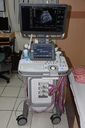


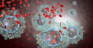
![Recombinant Anti-p53 antibody [E26] KO Tested (ab32389) | Abcam](https://www.abcam.com/ps/products/32/ab32389/Images/ab32389-389819-antip53antibodyimmunocytochemistryimmunofluorescencehap1human.jpg)