Tympanic Membrane Perforation
Tympanoplasty
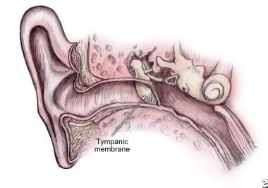
Tympanic Membrane
Maxillary Sinus
Tympanoplasty after war blast lesions of the eardrum: retrospective study. (1/39)
AIM: To establish whether hearing loss after eardrum blast injury could be recovered by tympanoplasty performed immediately after injury and what material is the most suitable for eardrum closure. METHODS: Tympanoplasty was performed in 119 (a total of 181 injuries) out of 651 patients examined for blast injury of the ear between 1991 and 2000. The study included a total of 106 patients who underwent tympanoplasty: 51 patients with unilateral and 55 with bilateral blast eardrum rupture (a total of 161 injuries). Three different materials were used for eardrum rupture closure: temporal fascia in 81, perichondrium in 61, and heterograft in 19 cases. Injuries were divided in 4 groups, according to the time elapsed between the injury and tympanoplasty (0-20, 21-60, 61-180, and 181 days and more). Otomicroscopic finding, audiometry, and tympanometry were used for definitive evaluation of tympanoplasty outcome. RESULTS: Eardrum rupture was successfully closed with temporal fascia in 91%, perichondrium in 92%, and heterograft in 89% of the cases (p=0.429). There were no statistically significant differences in either values of postoperative air- bone gap (p=0.210) or in eardrum perforation closure rate (p=0.951) with respect to the time period between the injury and tympanoplasty. Also, there was no correlation between the postoperative air-bone gap and the number of days elapsed between the rupture and tympanoplasty (r=-0.037, p=0.641). CONCLUSION: Small ruptures of the eardrum should be left to heal spontaneously. The patients with subtotal and total rupture and rupture that did not heal spontaneously in three months should undergo tympanoplasty. Temporal fascia, perichondrium from tragus, and heterograft are equally acceptable materials for eardrum closure after blast injury. (+info)Inner ear damage in children due to noise exposure from toy cap pistols and firecrackers: a retrospective review of 53 cases. (2/39)
This retrospective study presents the findings of inner ear damage documented in 53 children exposed to impulsive sound emitted by toy weapons and firecrackers. There were 49 boys and four girls aged between four and fourteen years. Thirty-nine children were affected unilaterally while fourteen had bilateral hearing loss (total of 67 ears). Most of the hearing loss (>70%) was sensorineural high frequency hearing loss, while only nine out of the 67 injured ears had sensorineural mid frequency hearing loss. Seven children sustained a traumatic ear drum perforation. Dizziness or tinnitus was reported by twenty children, with pathological ENG findings in four of them. This paper re-emphasizes the possibility of inner ear damage in children from exposure to noisy toys. (+info)Benefits of swimming pools in two remote Aboriginal communities in Western Australia: intervention study. (3/39)
OBJECTIVE: To determine the health impact of swimming pools built with the aim of improving quality of life and reducing high rates of pyoderma and otitis media. DESIGN: Intervention study assessing prevalence of ear disease and skin infections before and at six monthly intervals after opening of swimming pools. SETTING: Two remote Aboriginal communities in Western Australia. PARTICIPANTS: 84 boys and 78 girls aged < 17 years. MAIN OUTCOME MEASURES: Changes in prevalence and severity of pyoderma and perforation of tympanic membranes with or without otorrhoea over 18 months after opening of pools. RESULTS: In community A, 61 children were seen before the pool was opened, and 41, 46, and 33 children were seen at the second, third, and fourth surveys. Equivalent figures for community B were 60, 35, 39, and 45. Prevalence of pyoderma declined significantly from 62% to 18% in community A and from 70% to 20% in community B during the 18 months after the pools opened. Over the same period, prevalence of severe pyoderma fell from 30% to 15% in community A and from 48% to 0% in community B. Prevalence of perforations of the tympanic membrane fell from 32% in both communities to 13% in community A and 18% in community B. School attendance improved in community A. CONCLUSION: Swimming pools in remote communities were associated with reduction in prevalence of pyoderma and tympanic membrane perforations, which could result in long term benefits through reduction in chronic disease burden and improved educational and social outcomes. (+info)Traumatic perilymph fistula in infants: a case report. (4/39)
Traumatic perilymph fistula is reported to be rare in infants because of the small size of the infant external meatus. We treated an infant with a traumatic perilymph fistula in the right ear. A metallic wire had penetrated the tympanic membrane. Horizontal-rotatory nystagmus was also observed. Computed tomographic images revealed dislocation of the ossicles. The perilymph fistula was closed under general anesthesia. The incus-stapes joint was separated and the footplate of the stapes was dislocated. Leakage of the perilymph fluid was apparent from the oval window and this fistula was closed with connective tissue. The perforation of the tympanic membrane was closed with temporal fascia. After surgery, the spontaneous nystagmus disappeared. The patient is under observation as an outpatient and is growing normally. (+info)Progressive calvarial and upper cervical pneumatization associated with habitual valsalva maneuver in a 70-year-old man. (5/39)
A 70-year old man with a 15-year-history of chronic daily Valsalva maneuvers for left ear congestion presented with worsening vertigo and calvarial (occipitoparietal) and upper cervical hyperpneumatization. With continued frequent Valsalva maneuvers, subsequent studies demonstrated increased pneumatization with extension of air into the epidural space, causing mass effect on the left parietal lobe. Four months after discontinuing the habitual Valsalva maneuvers, CT demonstrated resorption of the epidural air and partial regression of the calvarial pneumatization. (+info)The clinical course of acute otitis media in high-risk Australian Aboriginal children: a longitudinal study. (6/39)
BACKGROUND: It is unclear why some children with acute otitis media (AOM) have poor outcomes. Our aim was to describe the clinical course of AOM and the associated bacterial nasopharyngeal colonisation in a high-risk population of Australian Aboriginal children. METHODS: We examined Aboriginal children younger than eight years who had a clinical diagnosis of AOM. Pneumatic otoscopy and video-otoscopy of the tympanic membrane (TM) and tympanometry was done every weekday if possible. We followed children for either two weeks (AOM without perforation), or three weeks (AOM with perforation), or for longer periods if the infection persisted. Nasopharyngeal swabs were taken at study entry and then weekly. RESULTS: We enrolled 31 children and conducted a total of 219 assessments. Most children had bulging of the TM or recent middle ear discharge at diagnosis. Persistent signs of suppurative OM (without ear pain) were present in most children 7 days (23/30, 77%), and 14 days (20/26, 77%) later. Episodes of AOM did not usually have a sudden onset or short duration. Six of the 14 children with fresh discharge in their ear canal had an intact or functionally intact TM. Perforation size generally remained very small (<2% of the TM). Healing followed by re-perforation was common. Ninety-three nasophyngeal swabs were taken. Most swabs cultured Streptococcus pneumoniae (82%), Haemophilus influenzae (71%), and Moraxella catarrhalis (95%); 63% of swabs cultured all three pathogens. CONCLUSION: In this high-risk population, AOM was generally painless and persistent. These infections were associated with persistent bacterial colonisation of the nasopharynx and any benefits of antibiotics were modest at best. Systematic follow up with careful examination and review of treatment are required and clinical resolution cannot be assumed. (+info)Health issues for surfers. (7/39)
Surfers are prone to acute injuries as well as conditions resulting from chronic environmental exposure. Sprains, lacerations, strains, and fractures are the most common types of trauma. Injury from the rider's own surfboard may be the prevailing mechanism. Minor wound infections can be treated on an outpatient basis with ciprofloxacin or trimethoprim-sulfamethoxazole. Jellyfish stings are common and may be treated with heat application. Other treatment regimens have had mixed results. Seabather's eruption is a pruritic skin reaction caused by exposure to nematocyst-containing coelenterate larvae. Additional surfing hazards include stingrays, coral reefs, and, occasionally, sharks. Otologic sequelae of surfing include auditory exostoses, tympanic membrane rupture, and otitis externa. Sun exposure and skin cancer risk are inherent dangers of this sport. (+info)Otitis media in young Aboriginal children from remote communities in Northern and Central Australia: a cross-sectional survey. (8/39)
BACKGROUND: Middle ear disease (otitis media) is common and frequently severe in Australian Aboriginal children. There have not been any recent large-scale surveys using clear definitions and a standardised middle ear assessment. The aim of the study was to determine the prevalence of middle ear disease (otitis media) in a high-risk population of young Aboriginal children from remote communities in Northern and Central Australia. METHODS: 709 Aboriginal children aged 6-30 months living in 29 communities from 4 health regions participated in the study between May and November 2001. Otitis media (OM) and perforation of the tympanic membrane (TM) were diagnosed by tympanometry, pneumatic otoscopy, and video-otoscopy. We used otoscopic criteria (bulging TM or recent perforation) to diagnose acute otitis media. RESULTS: 914 children were eligible to participate in the study and 709 were assessed (78%). Otitis media affected nearly all children (91%, 95%CI 88, 94). Overall prevalence estimates adjusted for clustering by community were: 10% (95%CI 8, 12) for unilateral otitis media with effusion (OME); 31% (95%CI 27, 34) for bilateral OME; 26% (95%CI 23, 30) for acute otitis media without perforation (AOM/woP); 7% (95%CI 4, 9) for AOM with perforation (AOM/wiP); 2% (95%CI 1, 3) for dry perforation; and 15% (95%CI 11, 19) for chronic suppurative otitis media (CSOM). The perforation prevalence ranged from 0-60% between communities and from 19-33% between regions. Perforations of the tympanic membrane affected 40% of children in their first 18 months of life. These were not always persistent. CONCLUSION: Overall, 1 in every 2 children examined had otoscopic signs consistent with suppurative ear disease and 1 in 4 children had a perforated tympanic membrane. Some of the children with intact tympanic membranes had experienced a perforation that healed before the survey. In this high-risk population, high rates of tympanic perforation were associated with high rates of bulging of the tympanic membrane. (+info)Tympanic membrane perforation, also known as a ruptured eardrum, is a tear or hole in the tympanic membrane, which separates the outer ear canal and the middle ear. The tympanic membrane plays a crucial role in hearing by transmitting sound vibrations from the outer ear to the inner ear. A perforation can result from various causes such as infection, trauma, pressure changes, or explosive blasts, leading to symptoms like hearing loss, tinnitus, vertigo, and ear discharge. The extent and location of the perforation determine the severity of the symptoms and the course of treatment, which may include observation, antibiotics, or surgical repair.
Tympanoplasty is a surgical procedure performed to reconstruct or repair the tympanic membrane (eardrum) and/or the small bones of the middle ear (ossicles). The primary goal of this surgery is to restore hearing, but it can also help manage chronic middle ear infections, traumatic eardrum perforations, or cholesteatoma (a skin growth in the middle ear).
During the procedure, a surgeon may use various techniques such as grafting tissue from another part of the body to rebuild the eardrum or using prosthetic materials to reconstruct the ossicles. The choice of technique depends on the extent and location of the damage. Tympanoplasty is typically an outpatient procedure, meaning patients can return home on the same day of the surgery.
The tympanic membrane, also known as the eardrum, is a thin, cone-shaped membrane that separates the external auditory canal from the middle ear. It serves to transmit sound vibrations from the air to the inner ear, where they are converted into electrical signals that can be interpreted by the brain as sound. The tympanic membrane is composed of three layers: an outer layer of skin, a middle layer of connective tissue, and an inner layer of mucous membrane. It is held in place by several small bones and muscles and is highly sensitive to changes in pressure.
Otoscopy is a medical examination procedure used to evaluate the external auditory canal and tympanic membrane (eardrum). It involves the use of an otoscope, a tool that consists of a lighted speculum attached to a handle. The speculum is inserted into the ear canal, allowing the healthcare provider to visualize and inspect the eardrum for any abnormalities such as perforations, inflammation, fluid accumulation, or foreign bodies. Otoscopy can help diagnose various conditions including ear infections, middle ear disorders, and hearing loss.
The maxillary sinuses, also known as the antrums of Highmore, are the largest of the four pairs of paranasal sinuses located in the maxilla bones. They are air-filled cavities that surround the nasolacrimal duct and are situated superior to the upper teeth and lateral to the nasal cavity. Each maxillary sinus is lined with a mucous membrane, which helps to warm, humidify, and filter the air we breathe. Inflammation or infection of the maxillary sinuses can result in conditions such as sinusitis, leading to symptoms like facial pain, headaches, and nasal congestion.
Intestinal perforation is a medical condition that refers to a hole or tear in the lining of the intestine. This can occur anywhere along the gastrointestinal tract, including the small intestine, large intestine (colon), or stomach. Intestinal perforation allows the contents of the intestines, such as digestive enzymes and bacteria, to leak into the abdominal cavity, which can lead to a serious inflammatory response known as peritonitis.
Intestinal perforation can be caused by various factors, including:
* Mechanical trauma (e.g., gunshot wounds, stab wounds)
* Inflammatory bowel disease (e.g., Crohn's disease, ulcerative colitis)
* Diverticulitis
* Appendicitis
* Intestinal obstruction
* Infections (e.g., typhoid fever, tuberculosis)
* Certain medications (e.g., nonsteroidal anti-inflammatory drugs, corticosteroids)
* Radiation therapy
* Ischemic bowel disease (lack of blood flow to the intestines)
Symptoms of intestinal perforation may include sudden abdominal pain, nausea, vomiting, fever, and decreased bowel movements. Treatment typically involves surgery to repair the perforation and remove any damaged tissue. Antibiotics are also administered to prevent infection. In severe cases, a temporary or permanent colostomy or ileostomy may be necessary.
 Eardrum
Ear
Perforated eardrum
Myringoplasty
Otitis media
Hearing test
Aspergillus niger
Ear candling
Tympanostomy tube
Earwax
Tympanometry
List of ICD-9 codes 320-389: diseases of the nervous system and sense organs
Stapedectomy
Keratinocyte
Tympanic membrane retraction
Hearing loss with craniofacial syndromes
Tympanoplasty
Joseph Toynbee
Conductive hearing loss
Columella (auditory system)
Pseudoryzomys
Eustachian tube
Horse colic
Myringotomy
Cholesteatoma
Otomycosis
Middle ear barotrauma
Bird
 Middle Ear, Tympanic Membrane, Perforations: Practice Essentials, Epidemiology, Etiology
 EpiDisc Tympanic Membrane Perforation Patch Kit | TM Perforations
 Topical Application of bFGF Alone for the Regeneration of Chronic Tympanic Membrane Perforations: A Preliminary Case Series
 Traumatic Perforation of the Tympanic Membrane - Ear, Nose, and Throat Disorders - MSD Manual Professional Edition
 Pulsatile Tympanic Membrane Perforation with Effusion - WiscMed
 Binaural Tympanic-Membrane Perforations after Blast Injury
 A study on chemical cauterisation for small tympanic membrane perforation
 The Effect of Topical Phenytoin Application on Traumatic Tympanic Membrane Perforation
 Tympanic Membrane Perforation
 Eardrum (Tympanic Membrane) Perforation | Columbia University Medical Center Department of Otolaryngology Head and Neck Surgery
 The role of epidermal growth factor in the healing tympanic membrane following perforation in rats
 View of Causes and Characteristics of Traumatic Tympanic Membrane Perforation in a Tertiary Care Hospital
 Ruptured eardrum: MedlinePlus Medical Encyclopedia
 Apple cider vinegar for ear infection: Does it work?
Eardrum - Wikipedia
Understanding the Anatomy and Function of the Middle Ear
 ICD-10-CM/PCS MS-DRG v39.0 Definitions Manual
 Pediatric Otolaryngology | Johns Hopkins Otolaryngology - Head and Neck Surgery
 Comparison of anterior tab flap and underlay tympanoplasty techniques in anterior tympanic membrane perforations
|...
Comparison of anterior tab flap and underlay tympanoplasty techniques in anterior tympanic membrane perforations
|...
 Tympanometry: Risks, Procedure, Results, and More
 Ruptured Ear Drums - Mankato - Mayo Clinic Health System
 DailyMed - ISOSORBIDE MONONITRATE tablet, extended release
Isosorbide Mononitrate Extended-Release Tablets, USP
8259101/0117
Rx only
 Thieme E-Journals - International Archives of Otorhinolaryngology / Full Text
 Otology/Neurotology & Lateral Skull Base Surgery
 Ruptured eardrum Information | Mount Sinai - New York
 Understand The Implications Of Auditory Damage
Otitis Media Medication: Antimicrobial agents, Vaccines, Inactivated, Bacterial
 Amanda Jo Lott Marcellino, MD | Atrium Health Wake Forest Baptist
Otitis media10
- [ 1 ] Chronic otitis media with perforation may be associated with a chronic draining ear or cholesteatoma. (medscape.com)
- Indigenous Australian children living in remote communities experience high rates of acute otitis media with tympanic membrane perforation (AOMwiP). (edu.au)
- Forty patients with tympanic membrane perforations secondary to chronic otitis media were included, and were randomly assigned to an experimental group (20), treated with a bacterial cellulose graft (BC) and control group (20), treated with autologous temporal fascia ( fascia ). (bvsalud.org)
- Prospective evaluation of the aetiology of acute otitis media with spontaneous tympanic membrane perforation. (medscape.com)
- Chronic suppurative otitis media (CSOM) is a prolonged and often recurring bacterial infection of the middle ear defined by perforation of the tympanic membrane and otorrhoea lasting more than 2 weeks according to the World Health Organization (WHO), although a commonly used clinical definition is 6 weeks. (springer.com)
- Risk factors associated with CSOM include frequent episodes of acute otitis media (AOM), other respiratory tract infections, and traumatic tympanic rupture as well as factors correlating with resource-limited living conditions such as overcrowding, poor nutrition and hygiene, and chronic infectious diseases. (springer.com)
- Tympanic membrane perforation either due to injury or infection of middle ear (otitis media). (myhealth.gov.my)
- Chronic suppurative otitis media is a long-standing, persistently draining perforation of the eardrum (tympanic membrane). (merckmanuals.com)
- Chronic suppurative otitis media may flare up after an infection of the nose and throat, such as the common cold, or after water enters the middle ear through a hole (perforation) in the eardrum while bathing or swimming. (merckmanuals.com)
- A hole or tear in the eardrum, also known as tympanic membrane perforation, can be the result of trauma, injury, otitis media or otitis externa. (pediatrix.com)
Myringoplasty8
- Objective Endoscopic transcanal myringoplasty (ETM) has been an emerging technique for repairing tympanic perforations since the late 1990s. (researchgate.net)
- Objective: Since the 1950s, microscopic myringoplasty has been the standard surgery for repairing a perforated tympanic membrane. (researchgate.net)
- 5. Modified inlay butterfly cartilage myringoplasty: An outpatient minimally invasive procedure for closure of central perforation-graft success rate and hearing outcome. (nih.gov)
- 7. Butterfly myringoplasty for total, subtotal, and annular perforations. (nih.gov)
- 14. Endoscopic Transcanal Myringoplasty for Anterior Perforations of the Tympanic Membrane. (nih.gov)
- 15. Endoscopic butterfly inlay myringoplasty for large perforations. (nih.gov)
- 20. Endoscopic Cartilage Myringoplasty with Inside Out Elevation of a Tympanomeatal Flap for Repairing Anterior Tympanic Membrane Perforations. (nih.gov)
- A myringoplasty is the closure of the perforation of the tympanic membrane in the ear. (watsi.org)
Eardrum perforation1
- Usually, the eardrum perforation can be repaired by a procedure called tympanoplasty. (merckmanuals.com)
Rupture5
- A cadaveric experimental investigation aimed to show the rupture pressure of the tympanic membrane (TM) for otologists to evaluate its tensile strength. (nih.gov)
- Rupture or perforation of the eardrum can lead to conductive hearing loss. (wikipedia.org)
- citation needed] Unintentional perforation (rupture) has been described in blast injuries and air travel, typically in patients experiencing upper respiratory congestion that prevents equalization of pressure in the middle ear. (wikipedia.org)
- Patients with tympanic membrane rupture may experience bleeding, tinnitus, hearing loss, or disequilibrium (vertigo). (wikipedia.org)
- According to the document, Christopher Palacios, a certified physician assistant at Aloha Kona Urgent Care, documented "a perforation of Tanaka's right tympanic membrane, resulting in rupture of about 1/4 (to) 1/3 of the ear drum. (hawaiifreepress.com)
Surface of the tympanic membrane3
- After three weeks, the wound crust is removed from the upper surface of the tympanic membrane to check for regeneration. (endoearboston.com)
- Sensation of the outer surface of the tympanic membrane is supplied mainly by the auriculotemporal nerve, a branch of the mandibular nerve (cranial nerve V3), with contributions from the auricular branch of the vagus nerve (cranial nerve X), the facial nerve (cranial nerve VII), and possibly the glossopharyngeal nerve (cranial nerve IX). (wikipedia.org)
- The inner surface of the tympanic membrane is innervated by the glossopharyngeal nerve. (wikipedia.org)
Normal tympanic membrane1
- To evaluate histologically the progressive development and underlying mechanisms of chronic tympanic membrane perforation (TMP) in a rat model using a two-weeks ventilation tube (VT) treatment combined with topical application of mitomycin C/dexamethasone (VT-M/D), compared with normal tympanic membrane and acute TMPs. (oslomet.no)
Cholesteatoma3
- It is important to retract the fat slightly to prevent the edges of the perforation from growing inward to the middle ear and to avoid the formation of a cholesteatoma. (medscape.com)
- Cholesteatoma "an epithelial pocket that forms on the tympanic membrane. (slideserve.com)
- Risks factors for acquired cholesteatoma include previous infection, a hole in the ear drum (tympanic membrane perforation) and previous otologic surgeries. (singhealth.com.sg)
Tympanoplasty2
Otoscopy1
- Perforation is generally evident on otoscopy. (msdmanuals.com)
Closure8
- The treatment of TM repair was repeated up to 4 times for cases in which complete closure of the TM perforation was not achieved in one treatment cycle. (endoearboston.com)
- Results: Complete closure of the TM perforation was achieved in 89% (n=51/57). (endoearboston.com)
- Promising treatments for tympanic membrane perforation closure have been studied. (bvsalud.org)
- We evaluated the surgical time , hospital stay , time of epithelialization and the rate of tympanic perforation closure. (bvsalud.org)
- The closure of perforations was similar in both groups. (bvsalud.org)
- Bacterial cellulose grafts promoted the closure of the tympanic membrane perforations, and were demonstrated to be innovative, effective, safe, minimally invasive, efficacious and to have a very low cost . (bvsalud.org)
- Closure of perforations of the membrana tympani of traumatic and infectious origin. (nih.gov)
- Closure of perforations of the tympanic membrane. (nih.gov)
Infection5
- Tympanic membrane perforations (TMPs) can result from infection (acute or chronic) or trauma , or be secondary to otologic procedures (iatrogenic). (medscape.com)
- Perforations associated with infection may present with drainage (otorrhea) or pain (otalgia). (medscape.com)
- Fluid in the middle ear without tympanic membrane perforation with or without infection. (myhealth.gov.my)
- This surgery will repair the perforated tympanic membrane, treat the infection, and stop the ear discharge. (watsi.org)
- This is because the opening in the membrane allows bacteria to enter the middle ear and cause infection. (cvs.com)
Otorrhea3
- Medical therapy for perforations is directed at controlling otorrhea. (medscape.com)
- The goal of medical therapy for perforations is otorrhea control. (medscape.com)
- The clinical presentations specifically related to temporal bone trauma include facial nerve paralysis (partial or complete), hearing loss (conductive, sensorineural, or mixed), vertigo , dizziness , otorrhagia, cerebrospinal fluid (CSF) otorrhea , tympanic membrane perforation , and hemotympanum and canal laceration. (medscape.com)
Chronic1
- Perforations can be temporary or chronic, and their effect varies with size, location on the drum surface, and the associated pathologic condition. (medscape.com)
Graft2
- To investigate the effect on healing of direct application of a bacterial cellulose graft on the tympanic membrane compared to the conventional approach with autologous fascia . (bvsalud.org)
- The edges of the graft should extend under the margins of the perforation, and a small part should extend over the posterior canal wall. (watsi.org)
Require otomicroscopy2
- Small perforations may require otomicroscopy for identification. (medscape.com)
- Extremely small perforations may require otomicroscopy or middle ear impedance studies for definitive diagnosis (eg, if perforations do not close). (msdmanuals.com)
Minimally1
- Triton Systems, Inc., in collaboration with experts in the field, propose to further develop a minimally invasive, rapid, and effective method for repairing tympanic membrane perforations in-office with standard tools in Phase II. (sbir.gov)
Ossicular1
- Penetrating injuries of the tympanic membrane may result in dislocations of the ossicular chain, fracture of the stapedial footplate, displacement of fragments of the ossicles, bleeding, a perilymph fistula from the oval or round window resulting in leakage of perilymph into the middle ear space, or facial nerve injury. (msdmanuals.com)
Discharge2
- Tympanic membrane perforation and the associated middle ear discharge . (wikidoc.org)
- Samples from ear discharge and the nasopharynx were collected and cultured from 152 patients with ear discharge and perforation of the tympanic membrane. (springer.com)
Erythema1
- Cloudy , bulging PMID: 22459064 erythema of the tympanic membrane . (wikidoc.org)
Regenerative2
- We developed a new regenerative treatment for Tympanic membrane (TM) perforations without the need for conventional surgical therapy. (endoearboston.com)
- This new regenerative therapy is useful for the patients with not only simple TM perforation but also above diseases without conventional operative procedures. (endoearboston.com)
Canal6
- The fat is then tucked into the perforation, extending both into the canal and into the middle ear space. (medscape.com)
- After removal of lesions through TM perforation, gelatin sponge impregnated with b-FGF was then trimmed to fit the size of the tympanic membrane perforation and placed between the tympanic cavity and the external auditory canal. (endoearboston.com)
- The equivalent ear canal volume (ECV) is an estimate of the volume of air medial to the probe, which includes the volume between the probe tip and the tympanic membrane if the tympanic membrane is intact, or the volume of the ear canal and the middle ear space if the tympanic membrane is perforated" (Fowler & Shanks, 2002, p. 180). (interacoustics.com)
- Tympanometry results showed a large ear canal volume, consistent with a tympanic membrane perforation or patent grommet. (interacoustics.com)
- Inflammation at the external ear canal,erythem a and edema this lead to narrow it also may involve the pinna or tympanic membrane . (wikidoc.org)
- Persistent flare-ups may result in the formation of protruding growths called polyps, which extend from the middle ear through the perforation and into the ear canal. (merckmanuals.com)
Cavity1
- The manubrium (Latin: handle) of the malleus is firmly attached to the medial surface of the membrane as far as its center, drawing it toward the tympanic cavity. (wikipedia.org)
Patients2
- Patients with a large tympanic membrane defect should also be evaluated, because the displaced flaps may need to be repositioned. (msdmanuals.com)
- Patients: Fifty seven patients (Age:9-89,M=26,F=31) were selected from patients with/without TM perforation. (endoearboston.com)
Spontaneously1
- Traumatic perforations often resolve spontaneously, particularly when associated with swab use. (medscape.com)
Stratified squamous e1
- On longitudinal histological examination, compared with normal TM and acute TMP, the perforation edges at the later time points illustrated thickened stratified squamous epithelium rimming around the edges, significant increase in keratin and collagen deposition, increased macrophage infiltration as well as reduced cellular proliferation. (oslomet.no)
Concave2
- The lateral surface of the membrane is thus concave. (wikipedia.org)
- Retracted/concave tympanic membrane with change colour of tympanic membrane (yellow,amber,blue) , and air-fluid levels . (wikidoc.org)
Injuries2
- However, prophylaxis with oral broad-spectrum antibiotics or antibiotic ear drops is necessary if contaminants may have entered through the perforation as occurs in dirty injuries. (msdmanuals.com)
- Eardrum (tympanic membrane) perforation can occur with explosive injuries. (protectear.com)
Defect1
- Hydrogen sulfide and the probabilities of 'inhalation' through a tympanic membrane defect. (cdc.gov)
Occur1
- [ 2 ] Traumatic perforations occur from blows to the ear, severe atmospheric overpressure, exposure to excessive water pressure (eg, in scuba divers), and improper attempts at wax removal or ear cleaning. (medscape.com)
Onset1
- Even if the pathogenesis is multifactorial, the clinical onset is frequently an episode of AOM complicated by tympanic membrane perforation and a subsequent superinfection of the middle ear with bacteria entering through the outer ear channel. (springer.com)
Persistent1
- A temporary or persistent opening in the eardrum ( TYMPANIC MEMBRANE ). (bvsalud.org)
Severe2
- Traumatic perforation of the tympanic membrane causes sudden severe pain sometimes followed by bleeding from the ear, hearing loss, and tinnitus. (msdmanuals.com)
- Tympanic membrane perforation To put it bluntly, it was caused by severe external impact, which caused the tympanic membrane to be damaged. (hearingaid.cc)
Tympanometry2
- Screening tympanometry may reveal abnormalities consistent with perforation. (medscape.com)
- Tympanometry showed normal middle ear pressure with decreased static compliance, consistent with a hypomobile tympanic membrane. (interacoustics.com)
Fluid2
- Usually, this consists of a small hole (perforation), which allows fluid to drain out. (wikipedia.org)
- Usually, this consists of a small hole (perforation), from which fluid can drain. (wikipedia.org)
Trauma1
- Trauma from cotton swabs is a relatively common cause of perforation. (medscape.com)
Middle2
- Static compliance (SC) "is the greatest amount of acoustic energy absorbed by the middle ear system (the vertical peak of the tympanic tracing)" (Onusko, 2004, p. 1716). (interacoustics.com)
- In the anatomy of humans and various other tetrapods, the eardrum, also called the tympanic membrane or myringa, is a thin, cone-shaped membrane that separates the external ear from the middle ear. (wikipedia.org)
Grafts1
- Grafts may be placed medially or laterally to the perforation, or in a combined position. (medscape.com)
Outer ear1
- These bones are separated from the outer ear by the eardrum (tympanic membrane), which vibrates when struck by a sound wave. (mayoclinic.org)
Otalgia1
- If the tympanic membrane is abnormal, the most likely cause of it by primary otalgia . (wikidoc.org)
Ears1
- However, under normal circumstances, small areas Tympanic membrane perforation It won't be embarrassing and our ears have self-healing function. (hearingaid.cc)
Hospital2
- Go to the hospital to check the perforation. (hearingaid.cc)
- If it is not serious, according to The doctor's instructions keep the ear clean, and then go to the hospital one month later to check the healing of the tympanic membrane. (hearingaid.cc)
Hearing3
- The only symptom of a TMP may be associated hearing loss from decreased vibratory function of the tympanic membrane. (medscape.com)
- Tympanic membrane perforation After that, there will be certain Hearing loss In fact, because the tympanic membrane is a function of amplifying the sound, after perforation, it will have some influence on hearing. (hearingaid.cc)
- The cause of this temporary hearing loss is due partly to structural changes of the hair cells (e.g. buckling of the supporting cells and uncoupling of the sterocilia from the tectorial membrane). (protectear.com)
Repair1
- The current standard of care for tympanic membrane repair requires an operating room and general anesthesia, which leads to high patient costs and prolonged time to treatment for servicemembers. (sbir.gov)
Small1
- Such management has the best chance of working when the perforation is small and dry and does not involve the umbo nor the annulus. (medscape.com)