Trophoblasts
Placenta
Placentation
Pregnancy
Chorionic Villi
Choriocarcinoma
Pregnancy Trimester, First
Decidua
Pregnancy Proteins
HLA-G Antigens
Pre-Eclampsia
Giant Cells
Blastocyst
Chorion
Maternal-Fetal Exchange
Placental Lactogen
Uterus
Endometrium
Cells, Cultured
Chorionic Gonadotropin
Cell Differentiation
Pregnancy, Animal
Immunohistochemistry
Gestational Age
Placental Circulation
Pregnancy Trimester, Third
Fibroblast Growth Factor 4
Hydatidiform Mole
RNA, Messenger
Gene Expression Regulation, Developmental
Keratin-7
Trophoblastic Neoplasms
Cell Movement
Fetal Growth Retardation
Embryo, Mammalian
Reverse Transcriptase Polymerase Chain Reaction
Pregnancy, Tubal
Extraembryonic Membranes
Trophoblastic Tumor, Placental Site
Embryo Loss
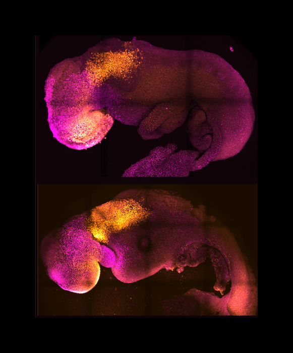
Mrj encodes a DnaJ-related co-chaperone that is essential for murine placental development. (1/2449)
We have identified a novel gene in a gene trap screen that encodes a protein related to the DnaJ co-chaperone in E. coli. The gene, named Mrj (mammalian relative of DnaJ) was expressed throughout development in both the embryo and placenta. Within the placenta, expression was particularly high in trophoblast giant cells but moderate levels were also observed in trophoblast cells of the chorion at embryonic day 8.5, and later in the labyrinth which arises from the attachment of the chorion to the allantois (a process called chorioallantoic fusion). Insertion of the ROSAbetageo gene trap vector into the Mrj gene created a null allele. Homozygous Mrj mutants died at mid-gestation due to a failure of chorioallantoic fusion at embryonic day 8.5, which precluded formation of the mature placenta. At embryonic day 8.5, the chorion in mutants was morphologically normal and expressed the cell adhesion molecule beta4 integrin that is known to be required for chorioallantoic fusion. However, expression of the chorionic trophoblast-specific transcription factor genes Err2 and Gcm1 was significantly reduced. The mutants showed no abnormal phenotypes in other trophoblast cell types or in the embryo proper. This study indicates a previously unsuspected role for chaperone proteins in placental development and represents the first genetic analysis of DnaJ-related protein function in higher eukaryotes. Based on a survey of EST databases representing different mouse tissues and embryonic stages, there are 40 or more DnaJ-related genes in mammals. In addition to Mrj, at least two of these genes are also expressed in the developing mouse placenta. The specificity of the developmental defect in Mrj mutants suggests that each of these genes may have unique tissue and cellular activities. (+info)An ultrastructural study of implantation in the golden hamster. II. Trophoblastic invasion and removal of the uterine epithelium. (2/2449)
Sixty six implantation sites from 18 golden hamsters were examined with light and electron microscopy between 4 and 5 1/2 days of pregnancy (post-ovulation). At 4 days some blastocysts began to invade the uterine epithelium, with trophoblastic processes penetrating and engulfing portions of the uterine epithelium. The majority of epithelial cells appeared normal before invasion, although at two implantation sites three or four adjoining epithelial cells were necrotic before penetration by the trophoblast. In general the epithelial cells were degenerating at the time the trophoblast invaded the epithelium. Inclusions, representing portions of the engulfed epithelium, and varying in size and electron density, were present throughout the invading trophoblast cells at 4 1/2 and 5 days of pregnancy. At 5 1/2 days the uterine epithelium had disappeared and the embryo was now almost completely surrounded by blood lacunae. (+info)Expression of trophinin, tastin, and bystin by trophoblast and endometrial cells in human placenta. (3/2449)
Trophinin, tastin, and bystin comprise a complex mediating a unique homophilic cell adhesion between trophoblast and endometrial epithelial cells at their respective apical cell surfaces. In this study, we prepared mouse monoclonal antibodies specific to each of these molecules. The expression of these molecules in the human placenta was examined immunohistochemically using the antibodies. In placenta from the 6th week of pregnancy, trophinin and bystin were found in the cytoplasm of the syncytiotrophoblast in the chorionic villi, and in endometrial decidual cells at the utero placental interface. Tastin was exclusively present on the apical side of the syncytiotrophoblast. Tissue sections were also examined by in situ hybridization using RNA probes specific to each of these molecules. This analysis showed that trophoblast and endometrial epithelial cells at the utero placental interface express trophinin, tastin, and bystin. In wk 10 placenta, trophinin and bystin were found in the intravillous cytotrophoblast, while tastin was not found in the villi. After wk 10, levels of all three proteins decreased and then disappeared from placental villi. (+info)Human uterine lymphocytes. (4/2449)
During the luteal phase and the early months of pregnancy, there is a dense mucosal infiltration of CD56+ natural killer (NK) cells. These uterine NK cells have a phenotype (CD56bright, CD16-, mCD3-) which distinguishes them from peripheral blood NK cells (CD56dim, CD16bright, mCD3-). The uterine NK cells are in close association with extravillous trophoblast (EVT) cells which infiltrate into the decidua and maternal spiral arteries. This subpopulation of trophoblast expresses two human leukocyte antigen (HLA) class I molecules, HLA-G and HLA-C. Circulating NK cells express receptors for HLA class I molecules. We have recently found evidence that similar receptors are present on decidual NK cells belonging to both the Killer Inhibitory Receptor (KIR) and CD94 families. The repertoire of NK receptors expressed varies between different women. The findings indicate that decidual NK cells do have receptors for trophoblast HLA class I molecules. Experiments are underway to determine the effects of this interaction on NK cell function. (+info)The CTLA-4 gene is expressed in placental fibroblasts. (5/2449)
In order to elucidate the mechanisms that ensure survival of the allogeneic fetus, we are investigating the expression pattern of genes that are involved in peripheral self-tolerance in tissues at the maternal-fetal interface. Cytotoxic T-lymphocyte-associated protein 4 (CTLA-4) is a negative regulator of T cell activation and may modulate peripheral self-tolerance. Previously, we reported the preferential transmission of maternally-inherited shorter alleles at a 3'-UTR microsatellite locus to liveborn children, but random transmission of paternally-inherited alleles, suggesting that CTLA-4 may be involved in the maintenance of tolerance at the maternal-fetal interface. In this report, we demonstrate that CTLA-4 mRNA and protein are indeed expressed in fetal tissues at the maternal-fetal interface throughout gestation. (+info)Role of proteases in implantation. (6/2449)
Implantation of the embryo into the endometrium is a critical step in the establishment of pregnancy and the failure of embryos to implant is a major limiting factor in the success of reproductive technologies. Furthermore, one or more of the molecules of importance at implantation could provide a suitable target for post-coital contraception. While there is considerable species variation in the extent to which the trophoblast invades the maternal endometrium and makes contact with the maternal blood supply, many of the molecular mechanisms are conserved among species. Three families of protease are involved in the matrix degradation required for implantation: the cysteine, serine and matrix metalloproteinases. Other proteases are required for the activation of regulatory molecules. Although trophoblast from all species appears to have a high invasive potential, this is limited by the presence of partner protease inhibitors, the presence of which provides restraint to this invasion. It is the balance between the proteases and their inhibitors at any focal point that determines the site and extent of trophoblast invasion. This review examines the literature regarding proteases and their inhibitors at early implantation sites across a range of species with very different forms of placentation and evaluates their common features and their dissimilarities. (+info)CD9 is expressed in extravillous trophoblasts in association with integrin alpha3 and integrin alpha5. (7/2449)
The CD9 molecule is a 24-27 kDa cell surface glycoprotein, which may be related to Schwann cell migration and adhesion. In this study, we examined the expression of CD9 in human extravillous trophoblasts, which invade into the endometrium during implantation and placentation. CD9 was detected immunohistochemically on the extravillous trophoblasts in the cell columns of first trimester placentae, but not on villous trophoblasts. In the second and third trimester, CD9 was highly expressed on the extravillous trophoblasts in the basal plate of placentae, and in the chorion laeve in the fetal membrane of term placentae. The molecular mass of CD9 in the chorion laeve was shown to be 27 kDa by Western blotting. The mRNA of CD9 was also detected in the chorion laeve by reverse transcription-polymerase chain reaction (RT-PCR). Proteins were purified from chorion laeve by affinity chromatography with anti-integrin alpha3 and alpha5 monoclonal antibodies and Western blotting, revealed that CD9 was associated with both integrins. These findings indicate that CD9 is a differentiation-related molecule present in the extravillous trophoblasts. Since it is associated with integrin alpha5 which has been proposed to regulate trophoblast invasion, CD9 may be implicated in trophoblast invasion at the feto-maternal interface. (+info)CD9 is involved in invasion of human trophoblast-like choriocarcinoma cell line, BeWo cells. (8/2449)
The CD9 molecule is expressed on human extravillous trophoblasts, which invade the endometrium during implantation and placentation. To elucidate the role of CD9 in trophoblastic function, we investigated the expression of CD9 protein and mRNA in BeWo cells, a human trophoblast-like choriocarcinoma cell line, using immunohistochemistry, Western blotting and reverse transcription-polymerase chain reaction (RT-PCR). When BeWo cells were cultured with anti-CD9 monoclonal antibodies (mAb), their invasion through the extracellular matrices was significantly enhanced in a dose-dependent manner. Cell proliferation and human chorionic gonadotrophin production were unaffected. On the other hand, culture in the presence of mAb against integrins alpha3, alpha5 and beta1, which partially block the interaction with the extracellular matrices, inhibited BeWo cell invasion. Anti-CD9 monoclonal antibody had a stimulatory effect on BeWo cell invasion in the presence of anti-integrin alpha3 antibody. In contrast, it had no effect in the presence of mAb against integrins alpha5 and beta1, which were also highly expressed on BeWo cells. These findings suggest that CD9 has a function connected with the invasive properties of BeWo cells, which is partially mediated by integrin alpha5beta1. This may relate to the involvement of CD9 in trophoblastic invasion. (+info)Trophoblasts are specialized cells that make up the outer layer of a blastocyst, which is a hollow ball of cells that forms in the earliest stages of embryonic development. In humans, this process occurs about 5-6 days after fertilization. The blastocyst consists of an inner cell mass (which will eventually become the embryo) and an outer layer of trophoblasts.
Trophoblasts play a crucial role in implantation, which is the process by which the blastocyst attaches to and invades the lining of the uterus. Once implanted, the trophoblasts differentiate into two main layers: the cytotrophoblasts (which are closer to the inner cell mass) and the syncytiotrophoblasts (which form a multinucleated layer that is in direct contact with the maternal tissues).
The cytotrophoblasts proliferate and fuse to form the syncytiotrophoblasts, which have several important functions. They secrete enzymes that help to degrade and remodel the extracellular matrix of the uterine lining, allowing the blastocyst to implant more deeply. They also form a barrier between the maternal and fetal tissues, helping to protect the developing embryo from the mother's immune system.
Additionally, trophoblasts are responsible for the formation of the placenta, which provides nutrients and oxygen to the developing fetus and removes waste products. The syncytiotrophoblasts in particular play a key role in this process by secreting hormones such as human chorionic gonadotropin (hCG), which helps to maintain pregnancy, and by forming blood vessels that allow for the exchange of nutrients and waste between the mother and fetus.
Abnormalities in trophoblast development or function can lead to a variety of pregnancy-related complications, including preeclampsia, intrauterine growth restriction, and gestational trophoblastic diseases such as hydatidiform moles and choriocarcinomas.
The placenta is an organ that develops in the uterus during pregnancy and provides oxygen and nutrients to the growing baby through the umbilical cord. It also removes waste products from the baby's blood. The placenta attaches to the wall of the uterus, and the baby's side of the placenta contains many tiny blood vessels that connect to the baby's circulatory system. This allows for the exchange of oxygen, nutrients, and waste between the mother's and baby's blood. After the baby is born, the placenta is usually expelled from the uterus in a process called afterbirth.
Placentation is the process by which the placenta, an organ that provides nutrients and oxygen to the developing fetus and removes waste products, is formed and develops during pregnancy. It involves the attachment of the fertilized egg (embryo) to the uterine wall and the development of specialized structures that facilitate the exchange of gases, nutrients, and waste between the mother and the fetus.
In humans, placentation begins when the embryo implants into the endometrium, or the lining of the uterus, about 6-10 days after fertilization. The outer layer of the embryo, called the trophoblast, invades the endometrial tissue and forms a structure called the placenta.
The placenta consists of both maternal and fetal tissues. The fetal portion of the placenta is derived from the chorionic villi, which are finger-like projections that develop on the surface of the embryo and increase the surface area for exchange. The maternal portion of the placenta is made up of modified endometrial tissue called decidua.
The placenta grows and develops throughout pregnancy, providing a vital connection between the mother and fetus. Proper placentation is essential for a healthy pregnancy and fetal development. Abnormalities in placentation can lead to complications such as preeclampsia, preterm labor, and intrauterine growth restriction.
Pregnancy is a physiological state or condition where a fertilized egg (zygote) successfully implants and grows in the uterus of a woman, leading to the development of an embryo and finally a fetus. This process typically spans approximately 40 weeks, divided into three trimesters, and culminates in childbirth. Throughout this period, numerous hormonal and physical changes occur to support the growing offspring, including uterine enlargement, breast development, and various maternal adaptations to ensure the fetus's optimal growth and well-being.
Chorionic villi are finger-like projections of the chorion, which is the outermost extraembryonic membrane in a developing embryo. These structures are composed of both fetal and maternal tissues and play a crucial role in the early stages of pregnancy by providing a site for exchange of nutrients and waste products between the mother and the developing fetus.
Chorionic villi contain fetal blood vessels that are surrounded by stromal cells, trophoblasts, and connective tissue. They are formed during the process of implantation, when the fertilized egg attaches to the uterine wall. The chorionic villi continue to grow and multiply as the placenta develops, eventually forming a highly vascular and specialized organ that supports fetal growth and development throughout pregnancy.
One important function of chorionic villi is to serve as the site for the production of human chorionic gonadotropin (hCG), a hormone that can be detected in the mother's blood and urine during early pregnancy. This hormone plays a critical role in maintaining pregnancy by signaling the corpus luteum to continue producing progesterone, which helps to prevent menstruation and support fetal growth.
Abnormalities in chorionic villi can lead to various pregnancy complications, such as miscarriage, stillbirth, or intrauterine growth restriction. For this reason, chorionic villus sampling (CVS) is a diagnostic procedure that may be performed during early pregnancy to obtain fetal cells for genetic testing and diagnosis of chromosomal abnormalities or other genetic disorders.
Choriocarcinoma is a rapidly growing and invasive type of gestational trophoblastic disease (GTD), which are abnormal growths that develop in the tissues that are supposed to become the placenta during pregnancy. It occurs when a malignant tumor develops from trophoblast cells, which are normally found in the developing embryo and help to form the placenta.
Choriocarcinoma can occur after any type of pregnancy, including normal pregnancies, molar pregnancies (a rare mass that forms inside the uterus after conception), or ectopic pregnancies (when a fertilized egg implants outside the uterus). It is characterized by the presence of both trophoblastic and cancerous cells, which can produce human chorionic gonadotropin (hCG) hormone.
Choriocarcinoma can spread quickly to other parts of the body, such as the lungs, liver, brain, or vagina, through the bloodstream. It is important to diagnose and treat choriocarcinoma early to prevent serious complications and improve the chances of a successful treatment outcome. Treatment typically involves surgery, chemotherapy, or radiation therapy.
The first trimester of pregnancy is defined as the period of gestational development that extends from conception (fertilization of the egg by sperm) to the end of the 13th week. This critical phase marks significant transformations in both the mother's body and the growing embryo/fetus.
During the first trimester, the fertilized egg implants into the uterine lining (implantation), initiating a series of complex interactions leading to the formation of the placenta - an organ essential for providing nutrients and oxygen to the developing fetus while removing waste products. Simultaneously, the embryo undergoes rapid cell division and differentiation, giving rise to various organs and systems. By the end of the first trimester, most major structures are present, although they continue to mature and grow throughout pregnancy.
The mother may experience several physiological changes during this time, including:
- Morning sickness (nausea and vomiting)
- Fatigue
- Breast tenderness
- Frequent urination
- Food aversions or cravings
- Mood swings
Additionally, hormonal shifts can cause various symptoms and prepare the body for potential changes in lactation, posture, and pelvic alignment as pregnancy progresses. Regular prenatal care is crucial during this period to monitor both maternal and fetal wellbeing, identify any potential complications early on, and provide appropriate guidance and support throughout the pregnancy.
The decidua is a specialized type of tissue that lines the uterus during pregnancy. It forms after the implantation of a fertilized egg (embryo) into the uterine lining, and it plays an important role in supporting the growth and development of the embryo and fetus.
The decidua is composed of several layers, including the decidual capsularis, which surrounds the embryo, and the decidual parietalis, which lines the rest of the uterus. The tissue is rich in blood vessels and contains a variety of immune cells that help to protect the developing fetus from infection.
During pregnancy, the decidua produces various hormones and growth factors that support the growth of the placenta, which provides nutrients and oxygen to the fetus. After the birth of the baby, the decidua is shed along with the placenta in a process called childbirth or parturition.
It's worth noting that abnormalities in the decidua can contribute to pregnancy complications such as preeclampsia, preterm labor, and miscarriage.
Embryo implantation is the process by which a fertilized egg, or embryo, becomes attached to the wall of the uterus (endometrium) and begins to receive nutrients from the mother's blood supply. This process typically occurs about 6-10 days after fertilization and is a critical step in the establishment of a successful pregnancy.
During implantation, the embryo secretes enzymes that help it to burrow into the endometrium, while the endometrium responds by producing receptors for the embryo's enzymes and increasing blood flow to the area. The embryo then begins to grow and develop, eventually forming the placenta, which will provide nutrients and oxygen to the developing fetus throughout pregnancy.
Implantation is a complex process that requires precise timing and coordination between the embryo and the mother's body. Factors such as age, hormonal imbalances, and uterine abnormalities can affect implantation and increase the risk of miscarriage or difficulty becoming pregnant.
"Pregnancy proteins" is not a standard medical term, but it may refer to specific proteins that are produced or have increased levels during pregnancy. Two common pregnancy-related proteins are:
1. Human Chorionic Gonadotropin (hCG): A hormone produced by the placenta shortly after fertilization. It is often detected in urine or blood tests to confirm pregnancy. Its primary function is to maintain the corpus luteum, which produces progesterone and estrogen during early pregnancy until the placenta takes over these functions.
2. Pregnancy-Specific beta-1 Glycoprotein (SP1): A protein produced by the placental trophoblasts during pregnancy. Its function is not well understood, but it may play a role in implantation, placentation, and protection against the mother's immune system. SP1 levels increase throughout pregnancy and are used as a marker for fetal growth and well-being.
These proteins have clinical significance in monitoring pregnancy progression, detecting potential complications, and diagnosing certain pregnancy-related conditions.
HLA-G antigens are a type of human leukocyte antigen (HLA) class Ib molecule that plays a crucial role in the immune system. HLA molecules are responsible for presenting pieces of proteins from inside the cell to the surface, where they can be recognized by the immune system's T-cells.
HLA-G antigens are primarily expressed in fetal tissues, including trophoblast cells that make up the placenta, and are involved in protecting the fetus from rejection by the mother's immune system during pregnancy. They have also been found to have immunosuppressive effects in other contexts, such as in cancer and transplantation.
HLA-G antigens are highly polymorphic, meaning that there are many different variations or "alleles" of the HLA-G gene that can be inherited from each parent. These genetic differences can affect the structure and function of the HLA-G molecule and may have implications for disease susceptibility and immune responses.
Pre-eclampsia is a pregnancy-related disorder, typically characterized by the onset of high blood pressure (hypertension) and damage to organs, such as the kidneys, after the 20th week of pregnancy. It is often accompanied by proteinuria, which is the presence of excess protein in the urine. Pre-eclampsia can lead to serious complications for both the mother and the baby if left untreated or unmanaged.
The exact causes of pre-eclampsia are not fully understood, but it is believed that placental issues, genetic factors, and immune system problems may contribute to its development. Risk factors include first-time pregnancies, history of pre-eclampsia in previous pregnancies, chronic hypertension, obesity, older age (35 or older), and assisted reproductive technology (ART) pregnancies.
Pre-eclampsia can progress to a more severe form called eclampsia, which is characterized by the onset of seizures. HELLP syndrome, another severe complication, involves hemolysis (breaking down of red blood cells), elevated liver enzymes, and low platelet count.
Early detection and management of pre-eclampsia are crucial to prevent severe complications. Regular prenatal care, including frequent blood pressure checks and urine tests, can help identify early signs of the condition. Treatment typically involves close monitoring, medication to lower blood pressure, corticosteroids to promote fetal lung maturity, and, in some cases, delivery of the baby if the mother's or baby's health is at risk.
Placental diseases, also known as placental pathologies, refer to a group of conditions that affect the development and function of the placenta during pregnancy. The placenta is an organ that develops in the uterus during pregnancy and provides oxygen and nutrients to the developing fetus while removing waste products.
Placental diseases can have serious consequences for both the mother and the fetus, including preterm labor, growth restriction, stillbirth, and long-term health problems for the child. Some common placental diseases include:
1. Placental abruption: This occurs when the placenta separates from the uterine wall before delivery, causing bleeding and potentially harming the fetus.
2. Placental previa: This is a condition where the placenta implants in the lower part of the uterus, covering the cervix. It can cause bleeding and may require cesarean delivery.
3. Preeclampsia: This is a pregnancy-related disorder characterized by high blood pressure and damage to organs such as the liver and kidneys. Placental dysfunction is thought to play a role in its development.
4. Intrauterine growth restriction (IUGR): This occurs when the fetus does not grow properly due to poor placental function, leading to low birth weight and potential health problems.
5. Chorioamnionitis: This is an infection of the membranes surrounding the fetus, which can lead to preterm labor and other complications.
6. Placental infarction: This occurs when a portion of the placenta dies due to a lack of blood flow, which can lead to growth restriction or stillbirth.
Prompt diagnosis and treatment of placental diseases are essential for ensuring the best possible outcomes for both the mother and the fetus.
Giant cells are large, multinucleated cells that result from the fusion of monocytes or macrophages. They can be found in various types of inflammatory and degenerative lesions, including granulomas, which are a hallmark of certain diseases such as tuberculosis and sarcoidosis. There are several types of giant cells, including:
1. Langhans giant cells: These have a horseshoe-shaped or crescentic arrangement of nuclei around the periphery of the cell. They are typically found in granulomas associated with infectious diseases such as tuberculosis and histoplasmosis.
2. Foreign body giant cells: These form in response to the presence of foreign material, such as a splinter or suture, in tissue. The nuclei are usually scattered throughout the cell cytoplasm.
3. Touton giant cells: These are found in certain inflammatory conditions, such as xanthomatosis and granulomatous slack skin. They have a central core of lipid-laden histiocytes surrounded by a ring of nuclei.
4. Osteoclast giant cells: These are multinucleated cells responsible for bone resorption. They can be found in conditions such as giant cell tumors of bone and Paget's disease.
It is important to note that the presence of giant cells alone does not necessarily indicate a specific diagnosis, and their significance must be interpreted within the context of the overall clinical and pathological findings.
A blastocyst is a stage in the early development of a fertilized egg, or embryo, in mammals. It occurs about 5-6 days after fertilization and consists of an outer layer of cells called trophoblasts, which will eventually form the placenta, and an inner cell mass, which will give rise to the fetus. The blastocyst is characterized by a fluid-filled cavity called the blastocoel. This stage is critical for the implantation of the embryo into the uterine lining.
The chorion is the outermost fetal membrane that surrounds the developing conceptus (the embryo or fetus and its supporting structures). It forms early in pregnancy as an extraembryonic structure, meaning it arises from cells that will not become part of the actual body of the developing organism. The chorion plays a crucial role in pregnancy by contributing to the formation of the placenta, which provides nutrients and oxygen to the growing embryo/fetus and removes waste products.
One of the most important functions of the chorion is to produce human chorionic gonadotropin (hCG), a hormone that signals the presence of pregnancy and maintains the corpus luteum, a temporary endocrine structure in the ovary that produces progesterone during early pregnancy. Progesterone is essential for preparing the uterus for implantation and maintaining the pregnancy.
The chorion consists of two layers: an inner cytotrophoblast layer and an outer syncytiotrophoblast layer. The cytotrophoblast layer is made up of individual cells, while the syncytiotrophoblast layer is a multinucleated mass of fused cytotrophoblast cells. These layers interact with the maternal endometrium (the lining of the uterus) to form the placenta and facilitate exchange between the mother and the developing fetus.
In summary, the chorion is a vital extraembryonic structure in pregnancy that contributes to the formation of the placenta, produces hCG, and interacts with the maternal endometrium to support fetal development.
Maternal-fetal exchange, also known as maternal-fetal transport or placental transfer, refers to the physiological process by which various substances are exchanged between the mother and fetus through the placenta. This exchange includes the transfer of oxygen and nutrients from the mother's bloodstream to the fetal bloodstream, as well as the removal of waste products and carbon dioxide from the fetal bloodstream to the mother's bloodstream.
The process occurs via passive diffusion, facilitated diffusion, and active transport mechanisms across the placental barrier, which is composed of fetal capillary endothelial cells, the extracellular matrix, and the syncytiotrophoblast layer of the placenta. The maternal-fetal exchange is crucial for the growth, development, and survival of the fetus throughout pregnancy.
Placental lactogen is a hormone produced by the placenta during pregnancy in humans and some other mammals. It is similar in structure to human growth hormone and prolactin, and has both growth-promoting and lactogenic (milk-producing) properties. Placental lactogen plays an important role in regulating maternal metabolism during pregnancy, promoting the growth and development of the fetus, and preparing the mother's body for lactation after birth. It helps to stimulate the growth of the mammary glands and the production of milk by increasing the availability of nutrients such as glucose, amino acids, and fatty acids in the mother's bloodstream. Placental lactogen also helps to regulate the mother's insulin sensitivity, which can affect her energy levels and the growth of the fetus.
The uterus, also known as the womb, is a hollow, muscular organ located in the female pelvic cavity, between the bladder and the rectum. It has a thick, middle layer called the myometrium, which is composed of smooth muscle tissue, and an inner lining called the endometrium, which provides a nurturing environment for the fertilized egg to develop into a fetus during pregnancy.
The uterus is where the baby grows and develops until it is ready for birth through the cervix, which is the lower, narrow part of the uterus that opens into the vagina. The uterus plays a critical role in the menstrual cycle as well, by shedding its lining each month if pregnancy does not occur.
The endometrium is the innermost layer of the uterus, which lines the uterine cavity and has a critical role in the menstrual cycle and pregnancy. It is composed of glands and blood vessels that undergo cyclic changes under the influence of hormones, primarily estrogen and progesterone. During the menstrual cycle, the endometrium thickens in preparation for a potential pregnancy. If fertilization does not occur, it will break down and be shed, resulting in menstruation. In contrast, if implantation takes place, the endometrium provides essential nutrients to support the developing embryo and placenta throughout pregnancy.
"Cells, cultured" is a medical term that refers to cells that have been removed from an organism and grown in controlled laboratory conditions outside of the body. This process is called cell culture and it allows scientists to study cells in a more controlled and accessible environment than they would have inside the body. Cultured cells can be derived from a variety of sources, including tissues, organs, or fluids from humans, animals, or cell lines that have been previously established in the laboratory.
Cell culture involves several steps, including isolation of the cells from the tissue, purification and characterization of the cells, and maintenance of the cells in appropriate growth conditions. The cells are typically grown in specialized media that contain nutrients, growth factors, and other components necessary for their survival and proliferation. Cultured cells can be used for a variety of purposes, including basic research, drug development and testing, and production of biological products such as vaccines and gene therapies.
It is important to note that cultured cells may behave differently than they do in the body, and results obtained from cell culture studies may not always translate directly to human physiology or disease. Therefore, it is essential to validate findings from cell culture experiments using additional models and ultimately in clinical trials involving human subjects.
Chorionic Gonadotropin (hCG) is a hormone that is produced during pregnancy. It is produced by the placenta after implantation of the fertilized egg in the uterus. The main function of hCG is to prevent the disintegration of the corpus luteum, which is a temporary endocrine structure that forms in the ovary after ovulation and produces progesterone during early pregnancy. Progesterone is essential for maintaining the lining of the uterus and supporting the pregnancy.
hCG can be detected in the blood or urine as early as 10 days after conception, and its levels continue to rise throughout the first trimester of pregnancy. In addition to its role in maintaining pregnancy, hCG is also used as a clinical marker for pregnancy and to monitor certain medical conditions such as gestational trophoblastic diseases.
Cell differentiation is the process by which a less specialized cell, or stem cell, becomes a more specialized cell type with specific functions and structures. This process involves changes in gene expression, which are regulated by various intracellular signaling pathways and transcription factors. Differentiation results in the development of distinct cell types that make up tissues and organs in multicellular organisms. It is a crucial aspect of embryonic development, tissue repair, and maintenance of homeostasis in the body.
"Animal pregnancy" is not a term that is typically used in medical definitions. However, in biological terms, animal pregnancy refers to the condition where a fertilized egg (or eggs) implants and develops inside the reproductive tract of a female animal, leading to the birth of offspring (live young).
The specific details of animal pregnancy can vary widely between different species, with some animals exhibiting phenomena such as placental development, gestation periods, and hormonal changes that are similar to human pregnancy, while others may have very different reproductive strategies.
It's worth noting that the study of animal pregnancy and reproduction is an important area of biological research, as it can provide insights into fundamental mechanisms of embryonic development, genetics, and evolution.
Uterine neoplasms refer to abnormal growths in the uterus, which can be benign (non-cancerous) or malignant (cancerous). These growths can originate from different types of cells within the uterus, leading to various types of uterine neoplasms. The two main categories of uterine neoplasms are endometrial neoplasms and uterine sarcomas.
Endometrial neoplasms develop from the endometrium, which is the inner lining of the uterus. Most endometrial neoplasms are classified as endometrioid adenocarcinomas, arising from glandular cells in the endometrium. Other types include serous carcinoma, clear cell carcinoma, and mucinous carcinoma.
Uterine sarcomas, on the other hand, are less common and originate from the connective tissue (stroma) or muscle (myometrium) of the uterus. Uterine sarcomas can be further divided into several subtypes, such as leiomyosarcoma, endometrial stromal sarcoma, and undifferentiated uterine sarcoma.
Uterine neoplasms can cause various symptoms, including abnormal vaginal bleeding or discharge, pelvic pain, and difficulty urinating or having bowel movements. The diagnosis typically involves a combination of imaging tests (such as ultrasound, CT, or MRI scans) and tissue biopsies to determine the type and extent of the neoplasm. Treatment options depend on the type, stage, and patient's overall health but may include surgery, radiation therapy, chemotherapy, or hormone therapy.
Immunohistochemistry (IHC) is a technique used in pathology and laboratory medicine to identify specific proteins or antigens in tissue sections. It combines the principles of immunology and histology to detect the presence and location of these target molecules within cells and tissues. This technique utilizes antibodies that are specific to the protein or antigen of interest, which are then tagged with a detection system such as a chromogen or fluorophore. The stained tissue sections can be examined under a microscope, allowing for the visualization and analysis of the distribution and expression patterns of the target molecule in the context of the tissue architecture. Immunohistochemistry is widely used in diagnostic pathology to help identify various diseases, including cancer, infectious diseases, and immune-mediated disorders.
Gestational age is the length of time that has passed since the first day of the last menstrual period (LMP) in pregnant women. It is the standard unit used to estimate the age of a pregnancy and is typically expressed in weeks. This measure is used because the exact date of conception is often not known, but the start of the last menstrual period is usually easier to recall.
It's important to note that since ovulation typically occurs around two weeks after the start of the LMP, gestational age is approximately two weeks longer than fetal age, which is the actual time elapsed since conception. Medical professionals use both gestational and fetal age to track the development and growth of the fetus during pregnancy.
Placental circulation refers to the specialized circulatory system that develops during pregnancy to allow for the exchange of nutrients, oxygen, and waste products between the mother's blood and the fetal blood in the placenta. The placenta is a highly vascular organ that grows within the uterus and is connected to the developing fetus via the umbilical cord.
In the maternal side of the placenta, the spiral arteries branch into smaller vessels called the intervillous spaces, where they come in close contact with the fetal blood vessels within the villi (finger-like projections) of the placenta. The intervillous spaces are filled with maternal blood that flows around the villi, allowing for the exchange of gases and nutrients between the two circulations.
On the fetal side, the umbilical cord contains two umbilical arteries that carry oxygen-depleted blood from the fetus to the placenta, and one umbilical vein that returns oxygenated blood back to the fetus. The umbilical arteries branch into smaller vessels within the villi, where they exchange gases and nutrients with the maternal blood in the intervillous spaces.
Overall, the placental circulation is a crucial component of fetal development, allowing for the growing fetus to receive the necessary oxygen and nutrients to support its growth and development.
The third trimester of pregnancy is the final stage of pregnancy that lasts from week 29 until birth, which typically occurs around the 40th week. During this period, the fetus continues to grow and mature, gaining weight rapidly. The mother's body also prepares for childbirth by dilating the cervix and producing milk in preparation for breastfeeding. Regular prenatal care is crucial during this time to monitor the health of both the mother and the developing fetus, as well as to prepare for delivery.
Fibroblast Growth Factor 4 (FGF4) is a growth factor that belongs to the fibroblast growth factor family. It plays a crucial role in various biological processes, including embryonic development, cell survival, proliferation, and differentiation. Specifically, FGF4 has been implicated in the development of the musculoskeletal system, where it helps regulate the growth and patterning of limbs and bones.
FGF4 exerts its effects by binding to specific receptors on the surface of target cells, known as fibroblast growth factor receptors (FGFRs). This interaction triggers a cascade of intracellular signaling events that ultimately lead to changes in gene expression and cell behavior.
In addition to its role in development, FGF4 has also been implicated in various pathological processes, including cancer. For example, elevated levels of FGF4 have been observed in certain types of tumors, where it may contribute to tumor growth and progression by promoting the survival and proliferation of cancer cells.
A hydatidiform mole, also known as a molar pregnancy, is a type of gestational trophoblastic disease (GTD), which is a group of rare disorders that involve abnormal growth of the placental tissue.
In a hydatidiform mole, there is an abnormal fertilization event leading to the growth of a mass of grapelike cysts in the uterus instead of a normal pregnancy. The chromosomes from the sperm and egg do not combine properly, resulting in an extra set of chromosomes, which leads to the development of the mole.
Hydatidiform moles can be complete or partial:
* Complete hydatidiform mole (CHM): This type arises when an egg without a nucleus is fertilized by one or two sperm, leading to the growth of abnormal placental tissue with no embryo. The chromosomes come from the father only, and there are typically 46 chromosomes, all of paternal origin.
* Partial hydatidiform mole (PHM): This type occurs when an egg is fertilized by two sperm or a single sperm that duplicates itself, resulting in an abnormal placenta with some fetal tissue. The chromosomes are of both maternal and paternal origin, and the placental tissue has a mix of normal and abnormal cells.
Hydatidiform moles can cause vaginal bleeding, rapid uterine enlargement, and high levels of human chorionic gonadotropin (hCG) hormone in the blood. They are usually detected during an ultrasound exam and require medical treatment to prevent complications such as gestational trophoblastic neoplasia, a malignant form of GTD that can spread to other organs.
Messenger RNA (mRNA) is a type of RNA (ribonucleic acid) that carries genetic information copied from DNA in the form of a series of three-base code "words," each of which specifies a particular amino acid. This information is used by the cell's machinery to construct proteins, a process known as translation. After being transcribed from DNA, mRNA travels out of the nucleus to the ribosomes in the cytoplasm where protein synthesis occurs. Once the protein has been synthesized, the mRNA may be degraded and recycled. Post-transcriptional modifications can also occur to mRNA, such as alternative splicing and addition of a 5' cap and a poly(A) tail, which can affect its stability, localization, and translation efficiency.
A cell line is a culture of cells that are grown in a laboratory for use in research. These cells are usually taken from a single cell or group of cells, and they are able to divide and grow continuously in the lab. Cell lines can come from many different sources, including animals, plants, and humans. They are often used in scientific research to study cellular processes, disease mechanisms, and to test new drugs or treatments. Some common types of human cell lines include HeLa cells (which come from a cancer patient named Henrietta Lacks), HEK293 cells (which come from embryonic kidney cells), and HUVEC cells (which come from umbilical vein endothelial cells). It is important to note that cell lines are not the same as primary cells, which are cells that are taken directly from a living organism and have not been grown in the lab.
Developmental gene expression regulation refers to the processes that control the activation or repression of specific genes during embryonic and fetal development. These regulatory mechanisms ensure that genes are expressed at the right time, in the right cells, and at appropriate levels to guide proper growth, differentiation, and morphogenesis of an organism.
Developmental gene expression regulation is a complex and dynamic process involving various molecular players, such as transcription factors, chromatin modifiers, non-coding RNAs, and signaling molecules. These regulators can interact with cis-regulatory elements, like enhancers and promoters, to fine-tune the spatiotemporal patterns of gene expression during development.
Dysregulation of developmental gene expression can lead to various congenital disorders and developmental abnormalities. Therefore, understanding the principles and mechanisms governing developmental gene expression regulation is crucial for uncovering the etiology of developmental diseases and devising potential therapeutic strategies.
Keratin-7 is not a medical term itself, but it is a specific type of keratin protein that is often used in pathology as a marker for certain types of carcinomas. Keratins are a family of fibrous proteins that make up the structural framework of epithelial cells, which line the surfaces and glands of the body.
Keratin-7 is typically expressed in simple epithelia, such as those found in the gastrointestinal tract, pancreas, bile ducts, and respiratory and genitourinary tracts. It can be used as a marker to help identify carcinomas that arise from these tissues, such as adenocarcinomas of the pancreas or biliary system.
In medical terminology, keratin-7 positivity is often reported in the pathology report of a biopsy or surgical specimen to indicate the presence of this protein in cancer cells. This information can be helpful in determining the origin and behavior of the tumor, as well as guiding treatment decisions.
Trophoblastic neoplasms are a group of rare tumors that originate from the trophoblast, which is the outer layer of cells that surrounds a developing embryo and helps to form the placenta during pregnancy. These tumors can be benign or malignant and are characterized by their ability to produce human chorionic gonadotropin (hCG), a hormone that is normally produced during pregnancy.
There are several types of trophoblastic neoplasms, including:
1. Hydatidiform mole: A benign growth that forms in the uterus when a fertilized egg implants but does not develop into a normal embryo. There are two types of hydatidiform moles: complete and partial. Complete moles have no fetal tissue, while partial moles have some fetal tissue.
2. Invasive mole: A malignant form of hydatidiform mole that invades the uterine wall and may spread to other parts of the body.
3. Choriocarcinoma: A rapidly growing and highly invasive malignant tumor that can arise from a hydatidiform mole, a normal pregnancy, or an ectopic pregnancy. It can spread quickly to other parts of the body, such as the lungs, liver, and brain.
4. Placental site trophoblastic tumor (PSTT): A rare type of trophoblastic neoplasm that arises from the cells that attach the placenta to the uterine wall. It is usually slow-growing but can be aggressive in some cases.
5. Epithelioid trophoblastic tumor (ETT): Another rare type of trophoblastic neoplasm that arises from the cells that form the placental villi. It is typically low-grade and has a good prognosis, but it can recur in some cases.
The treatment for trophoblastic neoplasms depends on the type and stage of the tumor. Treatment options may include surgery, chemotherapy, radiation therapy, or a combination of these approaches. Regular monitoring of hCG levels is also important to ensure that the tumor has been completely removed and to detect any recurrence early.
Cell movement, also known as cell motility, refers to the ability of cells to move independently and change their location within tissue or inside the body. This process is essential for various biological functions, including embryonic development, wound healing, immune responses, and cancer metastasis.
There are several types of cell movement, including:
1. **Crawling or mesenchymal migration:** Cells move by extending and retracting protrusions called pseudopodia or filopodia, which contain actin filaments. This type of movement is common in fibroblasts, immune cells, and cancer cells during tissue invasion and metastasis.
2. **Amoeboid migration:** Cells move by changing their shape and squeezing through tight spaces without forming protrusions. This type of movement is often observed in white blood cells (leukocytes) as they migrate through the body to fight infections.
3. **Pseudopodial extension:** Cells extend pseudopodia, which are temporary cytoplasmic projections containing actin filaments. These protrusions help the cell explore its environment and move forward.
4. **Bacterial flagellar motion:** Bacteria use a whip-like structure called a flagellum to propel themselves through their environment. The rotation of the flagellum is driven by a molecular motor in the bacterial cell membrane.
5. **Ciliary and ependymal movement:** Ciliated cells, such as those lining the respiratory tract and fallopian tubes, have hair-like structures called cilia that beat in coordinated waves to move fluids or mucus across the cell surface.
Cell movement is regulated by a complex interplay of signaling pathways, cytoskeletal rearrangements, and adhesion molecules, which enable cells to respond to environmental cues and navigate through tissues.
Fetal growth retardation, also known as intrauterine growth restriction (IUGR), is a condition in which a fetus fails to grow at the expected rate during pregnancy. This can be caused by various factors such as maternal health problems, placental insufficiency, chromosomal abnormalities, and genetic disorders. The fetus may be smaller than expected for its gestational age, have reduced movement, and may be at risk for complications during labor and delivery. It is important to monitor fetal growth and development closely throughout pregnancy to detect any potential issues early on and provide appropriate medical interventions.
A mammalian embryo is the developing offspring of a mammal, from the time of implantation of the fertilized egg (blastocyst) in the uterus until the end of the eighth week of gestation. During this period, the embryo undergoes rapid cell division and organ differentiation to form a complex structure with all the major organs and systems in place. This stage is followed by fetal development, which continues until birth. The study of mammalian embryos is important for understanding human development, evolution, and reproductive biology.
Embryonic development is the series of growth and developmental stages that occur during the formation and early growth of the embryo. In humans, this stage begins at fertilization (when the sperm and egg cell combine) and continues until the end of the 8th week of pregnancy. During this time, the fertilized egg (now called a zygote) divides and forms a blastocyst, which then implants into the uterus. The cells in the blastocyst begin to differentiate and form the three germ layers: the ectoderm, mesoderm, and endoderm. These germ layers will eventually give rise to all of the different tissues and organs in the body.
Embryonic development is a complex and highly regulated process that involves the coordinated interaction of genetic and environmental factors. It is characterized by rapid cell division, migration, and differentiation, as well as programmed cell death (apoptosis) and tissue remodeling. Abnormalities in embryonic development can lead to birth defects or other developmental disorders.
It's important to note that the term "embryo" is used to describe the developing organism from fertilization until the end of the 8th week of pregnancy in humans, after which it is called a fetus.
Reverse Transcriptase Polymerase Chain Reaction (RT-PCR) is a laboratory technique used in molecular biology to amplify and detect specific DNA sequences. This technique is particularly useful for the detection and quantification of RNA viruses, as well as for the analysis of gene expression.
The process involves two main steps: reverse transcription and polymerase chain reaction (PCR). In the first step, reverse transcriptase enzyme is used to convert RNA into complementary DNA (cDNA) by reading the template provided by the RNA molecule. This cDNA then serves as a template for the PCR amplification step.
In the second step, the PCR reaction uses two primers that flank the target DNA sequence and a thermostable polymerase enzyme to repeatedly copy the targeted cDNA sequence. The reaction mixture is heated and cooled in cycles, allowing the primers to anneal to the template, and the polymerase to extend the new strand. This results in exponential amplification of the target DNA sequence, making it possible to detect even small amounts of RNA or cDNA.
RT-PCR is a sensitive and specific technique that has many applications in medical research and diagnostics, including the detection of viruses such as HIV, hepatitis C virus, and SARS-CoV-2 (the virus that causes COVID-19). It can also be used to study gene expression, identify genetic mutations, and diagnose genetic disorders.
Tubal pregnancy, also known as an ectopic pregnancy, is a type of pregnancy that occurs outside the uterus, usually in the fallopian tube. The fertilized egg implants and starts to develop in the tube instead of the uterine lining. This condition is not viable and can be life-threatening if not treated promptly.
The symptoms of a tubal pregnancy may include abdominal pain, vaginal bleeding, shoulder pain, dizziness or fainting, and pelvic discomfort or tenderness. If you suspect that you have a tubal pregnancy, it is important to seek medical attention immediately. Treatment options for tubal pregnancies include medication or surgery to remove the embryo and repair or remove the affected fallopian tube.
Extraembryonic membranes are specialized structures that form around the developing embryo in utero and provide vital support and protection during fetal development. There are three main extraembryonic membranes: the amnion, the chorion, and the allantois.
The amnion is the innermost membrane that surrounds the embryo itself, forming a fluid-filled sac known as the amniotic cavity. This sac provides a protective cushion for the developing embryo and helps to regulate its temperature and moisture levels.
The chorion is the outermost of the extraembryonic membranes, and it forms the boundary between the developing fetus and the mother's uterine wall. The chorion contains blood vessels that exchange nutrients and waste products with the mother's circulation, allowing for the growth and development of the fetus.
The allantois is a small membranous sac that arises from the developing fetal gut and eventually becomes part of the umbilical cord. It serves as a reservoir for fetal urine and helps to exchange waste products between the fetal and maternal circulations.
Together, these extraembryonic membranes play a critical role in supporting fetal development and ensuring a healthy pregnancy.
A Trophoblastic Tumor, Placental Site (also known as Placental Site Trophoblastic Tumor or PSTT) is a rare type of gestational trophoblastic disease (GTD), which are tumors that develop from the tissue that would normally become the placenta during pregnancy.
PSTT originates from the intermediate trophoblast cells, which invade the uterine wall and cause bleeding at the site of implantation during a normal pregnancy. These tumors typically occur in women who have had a prior pregnancy, with a median age of diagnosis around 35 years old.
PSTTs are usually slow-growing and may not cause any symptoms for an extended period. However, some common symptoms include abnormal vaginal bleeding, irregular menstrual periods, or pelvic pain. In rare cases, PSTT can metastasize to other organs such as the lungs, liver, or brain.
The diagnosis of PSTT is made through a combination of imaging studies (such as ultrasound, CT scan, or MRI) and histopathological examination of tissue samples obtained via biopsy or curettage. Treatment typically involves surgical removal of the tumor, followed by chemotherapy in cases where there is evidence of metastasis or high-risk features. Regular follow-up with serum beta-human chorionic gonadotropin (β-hCG) levels and imaging studies is essential to monitor for recurrence.
Embryo loss is a medical term that refers to the miscarriage or spontaneous abortion of an embryo, which is the developing offspring from the time of fertilization until the end of the eighth week of pregnancy. Embryo loss can occur at any point during this period and may be caused by various factors such as chromosomal abnormalities, maternal health issues, infections, environmental factors, or lifestyle habits.
Embryo loss is a common occurrence, with up to 30% of pregnancies ending in miscarriage, many of which happen before the woman even realizes she is pregnant. In most cases, embryo loss is a natural process that occurs when the body detects an abnormality or problem with the developing embryo and terminates the pregnancy to prevent further complications. However, recurrent embryo loss can be a sign of underlying medical issues and may require further evaluation and treatment.
An encyclopedia is a comprehensive reference work containing articles on various topics, usually arranged in alphabetical order. In the context of medicine, a medical encyclopedia is a collection of articles that provide information about a wide range of medical topics, including diseases and conditions, treatments, tests, procedures, and anatomy and physiology. Medical encyclopedias may be published in print or electronic formats and are often used as a starting point for researching medical topics. They can provide reliable and accurate information on medical subjects, making them useful resources for healthcare professionals, students, and patients alike. Some well-known examples of medical encyclopedias include the Merck Manual and the Stedman's Medical Dictionary.
 Trophoblast
Extravillous trophoblast
Intermediate trophoblast
C19MC miRNA cluster
Reproductive immunology
Blastocyst
Cell-free fetal DNA
H2BK5ac
Asim Duttaroy
C8orf82
Human embryonic development
Pregnancy Outcome Prediction study
Gestational choriocarcinoma
Pre-eclampsia
Michelle Oyen
TPBG
John Beard (embryologist)
Embryonic diapause
Villitis of unknown etiology
Ashley Moffett
Mir-205
Mir-491 microRNA precursor family
HLA-F
Heparanase
RIPOR2
Uterine serpin
TRPV6
Preimplantation factor
Ernst T. Krebs
Calbindin 1
Trophoblast - Wikipedia
 Decreased junctional adhesion molecule 3 expression induces reactive oxygen species production and apoptosis in trophoblasts†
 The human trophoblast cells - The Human Protein Atlas
 NLRP7 affects trophoblast lineage differentiation, binds to overexpressed YY1 and alters CpG methylation
A distinct pattern in the DNA ploidy histograms of hydatidiform moles and nonmolar abortuses is caused by accumulation of...
 Trophoblast invasion and spiral artery transformation in placenta accreta | ADC Fetal & Neonatal Edition
 COMT inhibitor tolcapone represses the migration and invasion of trophoblast and results in preeclampsia-like phenotypes in...
 BMP4-directed trophoblast differentiation of human embryonic stem cells is mediated through a DeltaNp63+ cytotrophoblast stem...
Regulation of amino acid transporter trafficking by mTORC1 in primary human trophoblast cells is mediated by the ubiquitin...
 The Gabrielle Rizzuto Lab: The immunology of trophoblast tumors | Gerstner Sloan Kettering Graduate School of Biomedical...
Wnt3a Activates the WNT-YAP/TAZ Pathway to Sustain CDX2 Expression in Bovine Trophoblast Stem Cells. | StemBook
Low oxygen enhances trophoblast column growth by potentiating differentiation of the extravillous lineage and promoting LOX...
Maternal nutrition modifies trophoblast giant cell phenotype and fetal growth in mice<...
 Degradation of extracellular matrix by mouse trophoblast outgrowths: a model for implantation | Journal of Cell Biology |...
How To Isolate Trophoblasts
 Senescence In Genotoxic Amniocytes And Stressed Trophoblasts | ISPR
Trophoblast Stem Cells<...
 Complete human day 14 post-implantation embryo models from naive ES cells | Nature
External weblinks | Centre for Trophoblast Research
 trophoblast - Yahoo奇摩 搜尋結果
Spatial multiomics map of trophoblast development in early pregnancy.
 Expression of TAP1 by human trophoblast. - Radcliffe Department of Medicine
Quantifying the syncytialisation of human placental trophoblast BeWo cells grown in vitro - Nuffield Department of Women's &...
An integrated atlas of human placental development delineates essential regulators of trophoblast stem cells | Development |...
Reprogramming roadmap reveals route to human induced trophoblast stem cells
 Springbok (Antidorcas marsupialis) | IVIS
Dorcas gazelle (Gazella dorcas) | IVIS
 Curcumin induces apoptosis in trophoblast model cell line
| Medical Journal of Indonesia
Curcumin induces apoptosis in trophoblast model cell line
| Medical Journal of Indonesia
 "PALMITIC ACID IMPEDES EXTRAVILLOUS TROPHOBLAST ACTIVITY BY INCREASING " by Yunali V. Ashar
Altered protein O‑GlcNAcylation in placentas from mothers with diabetes causes aberrant endocytosis in placental trophoblast...
Embryonic Stem2
- Trophoblast stem cells (TSCs) are cells that can regenerate and they are similar to embryonic stem cells (ESCs) in the fact that they come from early on in the trophoblast lifetime. (wikipedia.org)
- To do so, they combined mouse embryonic stem cells with trophoblast and extraembryonic endoderm cells and cultured them in conditions designed to permit the development seen after implantation. (nih.gov)
Stem Cells8
- In the placenta, these stem cells are able to differentiate into any trophoblast cell because they are pluripotent. (wikipedia.org)
- An Improved Two-Step Protocol for Trophoblast Differentiation of Human Pluripotent Stem Cells. (ca.gov)
- We previously established a two-step protocol for differentiation of human pluripotent stem cells (hPSCs) into trophoblasts, using a StemPro-based minimal medium (EMIM) with bone morphogenetic protein-4 (BMP4). (ca.gov)
- The open conformation is associated with enhanced trophoblast differentiation potency with increased trophoblast gene expression upon induction of differentiation and success in derivation of trophoblast stem cells with bona fide characteristics. (nih.gov)
- Knockout of the Hippo signaling gene, YAP1 abolishes the ability of the embryo-derived EPSC to form trophoblast stem cells. (nih.gov)
- In this study, using human trophoblast stem cells (hTSCs), Wu et al. (nature.com)
- Furthermore, differentiation of trophoblast stem cells in the presence of PFI-3 was markedly enhanced. (ox.ac.uk)
- The authors described a biomimetic placental barrier model from human trophoblast stem cells (hTSCs) in a perfused organ chip system. (stemcellsciencenews.com)
Placental trophoblasts3
- Barrientos, G. Leptin promotes HLA-G expression on placental trophoblasts via the MEK/Erk and PI3K signaling pathways. (uba.ar)
- Varone, C.L. 'Leptin promotes HLA-G expression on placental trophoblasts via the MEK/Erk and PI3K signaling pathways' (2015) Placenta. (uba.ar)
- Here, we explored the regulatory role of AMPK in the GLUT3-dependent uptake of glucose by placental trophoblasts and the viability of the cells. (le.ac.uk)
Embryo6
- After blastulation, the trophoblast is contiguous with the ectoderm of the embryo and is referred to as the trophectoderm. (wikipedia.org)
- After the first differentiation, the cells in the human embryo lose their totipotency because they can no longer form a trophoblast. (wikipedia.org)
- Trophoblasts are specialized cells of the placenta that play an important role in embryo implantation and interaction with the decidualized maternal uterus. (wikipedia.org)
- This blastocyst consists of two distinct parts: the inner cell mass, which will develop into the embryo, and the outer layer called the trophoblast. (worldreportnow.com)
- Little is known about the biochemical basis for the invasive activity of the trophoblasts during early implantation because of the complexity of the uterine endometrium, its inaccessibility to experimental study and the paucity of material comprising the embryo itself. (grantome.com)
- Scholars@Duke publication: T-helper 2 and 3 type immunity to trophoblast in successful in vitro fertilization-embryo transfer. (duke.edu)
Invasion12
- The invasion of a specific type of trophoblast (extravillous trophoblast) into the maternal uterus is a vital stage in the establishment of pregnancy. (wikipedia.org)
- Invasion of the trophoblast too deeply may cause conditions such as placenta accreta, placenta increta, or placenta percreta. (wikipedia.org)
- Trophoblast invasion establishes adequate blood flow between mother and fetus in early placental development. (nih.gov)
- We aimed to identify enhancer elements that are active during trophoblast invasion, and build a trophoblast invasion gene-enhancer network. (nih.gov)
- We use computational analysis to identify putative enhancers, as well as the transcription factor binding sites within them, that are specific to the time point of trophoblast invasion. (nih.gov)
- Fang Y, Zhang J, Zhu D, Mei Q, Liao T, Cheng H, He Y, Cao Y, Wei Z. MANF Promotes Unexplained Recurrent Miscarriages by Interacting with NPM1 and Downregulating Trophoblast Cell Migration and Invasion. (ijbs.com)
- We confirm the interaction between MANF and nucleophosmin 1 (NPM1) in trophoblasts of URM patients, which increases the ubiquitination degradation of NPM1, leading to upregulation of the p53 signaling pathway and inhibition of cell proliferation, migration, and invasion ability. (ijbs.com)
- SUMMARY ANSWER: LMWH increased HBEGF expression and secretion, and HBEGF signaling was required to stimulate trophoblast extravillous differentiation, increase invasion in vitro and reduce trophoblast apoptosis during oxidative stress. (elsevierpure.com)
- Trophoblast invasiveness was assessed in villous explants by measuring outgrowth from villous tips cultured on Matrigel, and by invasion assays with HTR-8/SVneo cells cultured on Matrigel-coated transwell insert. (elsevierpure.com)
- MAIN RESULTS AND THE ROLE OF CHANCE: LMWH induced extravillous differentiation, according to trophoblast invasion assays and integrin (α6β4-α1β1) switching. (elsevierpure.com)
- Experiments using CRM197, ERBB1 and ERBB4 blocking antibodies, pan-ERBB inhibitor and removal of cell surface heparin demonstrated that the effects of LMWH on trophoblast invasion and survival were dependent upon HBEGF signaling. (elsevierpure.com)
- IL-10 regulates LIF and MMP expressions and evolves in the control of invasion and proliferation processes of trophoblast cells. (4science.ge)
Cells37
- Some of these trophoblasts even replace the endothelial cells in the uterine spiral arteries as they remodel these vessels into wide bore conduits that are independent of maternal vasoconstriction. (wikipedia.org)
- Gestational trophoblastic disease is a pregnancy-associated concept, forming from the villous and extravillous trophoblast cells in the placenta. (wikipedia.org)
- May 8, 2009 (Chicago, Illinois) - Findings from a small preliminary study indicate that the number of trophoblast cells collected by Papanicolaou (Pap) test in the first trimester of pregnancy can distinguish between normal intrauterine pregnancy and ectopic pregnancy or blighted ovum. (medscape.com)
- The fetal cells have historically been identified by morphology, specific trophoblast marker, and molecular testing. (medscape.com)
- The current study hypothesized that the number of trophoblast cells in transcervical specimens from patients with tubal ectopic pregnancy or blighted ovum should be lower than that found in normal intrauterine pregnancies. (medscape.com)
- Dr. Imudia noted that histocompatibility antigen, class I, G (HLA-G) cells are markers for trophoblasts, and so the ratio of HLA-G-positive cells to total cells was calculated. (medscape.com)
- Trophoblast cells were observed in almost 95% of the normal intrauterine-pregnancy group, 60% of the ectopic-pregnancy group, and 80% of the blighted-ovum group. (medscape.com)
- This study concluded that trophoblast cells can be collected in the first trimester for immunohistochemical staining for HLA-G, allowing distinction between normal and abnormal pregnancies. (medscape.com)
- We also show that, at the end of the second step, there are distinct populations of terminally differentiated multinucleated human chorionic gonadotropin (hCG)-producing syncytiotrophoblast (STB) and HLAG+ extravillous trophoblast (EVT)-like cells. (ca.gov)
- Results: plCSA-BP binds specifically to trophoblasts and not to other cell types in the placenta or to CSA-expressing cells in other tissues. (visualsonics.com)
- Implantation marks the initial physical connection between mother and the conceptus during pregnancy, and its success is greatly dependent upon the function of the embryonic cells, the trophoblast, that establish themselves in the uterine wall and give rise to the placenta. (grantome.com)
- The objective of the proposed studies is to examine the developmentally regulated interaction of trophoblast cells with components of the uterine extracellular matrix (ECM), focusing on fibronectin which is a major component of the endometrial ECM that is infiltrated by differentiated trophoblasts early during interstitial implantation. (grantome.com)
- The fibronectin-binding characteristics of trophoblast cells at the surface of developing blastocysts will be studied and the corresponding cell surface receptors will be identified. (grantome.com)
- Experiments will also be conducted, using similar methods, to determine whether specific receptors for fibronectin or various other adhesion-promoting ECM components (laminin, vitronectin, hyaluronic acid) are ligand-induced or constitutively expressed by trophoblast cells. (grantome.com)
- Here we show that human dNK cells highly express the antimicrobial peptide granulysin (GNLY) and selectively transfer it via nanotubes to extravillous trophoblasts to kill intracellular Listeria monocytogenes ( Lm ) without killing the trophoblast. (bxcell.com)
- Expression of a wide range of Wnt-related genes was observed in both decidual and trophoblast cells using PCR array, with remarkably similar expression profiles. (edu.au)
- Zika virus modulates mitochondrial dynamics, mitophagy, and mitochondria-derived vesicles to facilitate viral replication in trophoblast cells. (bvsalud.org)
- Since viral infections are known to exploit mitochondria -mediated cellular processes, we investigated the effects of ZIKV infection in trophoblast cells in terms of the different mitochondrial quality control pathways that govern mitochondrial integrity and function. (bvsalud.org)
- Here we demonstrate that ZIKV (PRVABC59) infection of JEG-3 trophoblast cells manipulates mitochondrial dynamics , mitophagy , and formation of mitochondria -derived vesicles (MDVs). (bvsalud.org)
- The pathological effects of Zika virus (ZIKV) infection on placental trophoblast progenitor cells in early human embryos are not well understood. (nature.com)
- During early mammalian embryonic development, trophoblast cells play an essential role in establishing cell-cell interactions at the maternal-fetal interface to ensure a successful pregnancy. (edu.au)
- In a recent study, we showed that human fibroblasts can be reprogrammed into induced trophoblast stem (iTS) cells by transcription factor-mediated nuclear reprogramming using the Yamanaka factors OCT4, KLF4, SOX2 and c-MYC (OKSM) and a selection of TS cell culture conditions. (edu.au)
- Our protocol allows researchers to generate patient-specific iTS cells to interrogate the trophoblast and placenta biology as well as their interactions with embryonic cells in health and diseases. (edu.au)
- In trophoblast cells exposed to homocysteine (Hcy) we observed cellular apoptosis and the inhibition of trophoblast functions. (unicatt.it)
- In primary trophoblast cells we examined the cytosolic release of cytochrome c, both M30 and terminal deoxynucleotidyl transferase-mediated dUDP nick-end labelling (TUNEL) and DNA laddering. (unicatt.it)
- Hcy (20 micromol/l) treatment resulted in cytochrome c release from mitochondria to the cytosol, and an increased number of M30-positive trophoblast cells and TUNEL positive nuclei. (unicatt.it)
- In this study, the level of glycolysis in normal and GDM-complicated placentas was assessed by LC-MS/MS. The trophoblast hyperglycemia model was induced by the incubation of HTR8/SVneo cells with a high glucose concentration. (le.ac.uk)
- These include trophoblast and extraembryonic endoderm cells. (nih.gov)
- There is evidence that LMWH alters the expression of human HBEGF in trophoblast cells, which regulates human trophoblast pathophysiology. (elsevierpure.com)
- The aim of our current study was to evaluate the role of IL-10, TNFα, LIF, MMP-9, and NO in implantation and to establish their possible impact on the functional ability of trophoblast cells under hyperglycemic conditions. (4science.ge)
- For this reason, the experiments were conducted on trophoblast cells isolated from placentas of IL-10 +/+ (C57BL/6J) and IL-10 -/- (B6.129P2- Il10 tm1Cgn /J) pregnant mice. (4science.ge)
- We used an IGF2R knockdown strategy in BeWo cells and placental villous explants to investigate trophoblast proliferation and survival in response to stimulation by IGF. (manchester.ac.uk)
- Consequently, the Rac1-null decidual cells failed to secrete vascular endothelial growth factor A, which is a critical regulator of decidual angiogenesis, and insulin-like growth factor binding protein 4, which regulates the bioavailability of insulin-like growth factors that promote proliferation and differentiation of trophoblast cell lineages in the ectoplacental cone. (nih.gov)
- The lack of secretion of these key factors by Rac1-null decidua gave rise to impaired angiogenesis and dysregulated proliferation of trophoblast cells, which in turn results in overexpansion of the trophoblast giant cell lineage and disorganized placenta development. (nih.gov)
- Mild steel and stainless steel welding fumes elicit pro-inflammatory and pro-oxidant effects in first trimester trophoblast cells. (cdc.gov)
- This study aims to identify the effects of mild steel and stainless steel welding fume exposures on cultured placental trophoblast cells. (cdc.gov)
- Electron microscopy was used to characterize welding fume particle size and the ability of particles to enter extravillous trophoblast cells (HTR-8/SVneo). (cdc.gov)
Pregnancy4
- Trophoblast frequency was not affected by gestational age in the normal intrauterine-pregnancy group. (medscape.com)
- The purpose of this Funding Opportunity Announcement (FOA) is to encourage applications from the scientific community to support outstanding research in the area of trophoblast differentiation and function in relation to fertility and pregnancy, including the role of the immune system. (nih.gov)
- Trophoblast-stimulated PBMC secretion of TGF-beta1 is positively associated with the establishment of successful pregnancy in women undergoing IVF-ET. (duke.edu)
- Our study provides novel evidence to support a faciliatory role of Th-2 and Th-3 type responses to trophoblast in early pregnancy. (duke.edu)
Ovine trophoblast3
- This study characterises expression of genes for cathelicidin and defensin peptides in primary ovine placental tissues, the ovine trophoblast cell line (AH-1) and in response to the TLR-4 ligand LPS, the abortifacient organism Waddlia chondrophila and 1alpha,25-dihydroxyvitamin D3. (napier.ac.uk)
- Expression of cathelicidin in the trophoblast compartment of the ovine placenta and in the ovine trophoblast cell line (AH-1) was also established. (napier.ac.uk)
- This study demonstrates that the ovine trophoblast expresses cathelicidins, but does not upregulate expression of CHDP in response to LPS. (napier.ac.uk)
Syncytiotrophoblast1
- This core is surrounded by two layers of trophoblasts, the cytotrophoblast and the syncytiotrophoblast. (wikipedia.org)
Villous explants1
- STUDY DESIGN, SIZE, DURATION: First-trimester placental explants and the HTR-8/SVneo cell line, established using extravillous trophoblast outgrowths from first-trimester villous explants, were treated in vitro with LMWH to examine the effects on HBEGF signaling and trophoblast function under normal physiological and pathological conditions. (elsevierpure.com)
Apoptosis2
- These results suggest a protective role of folic acid in the prevention of trophoblast apoptosis linked to Hcy. (unicatt.it)
- STUDY QUESTION: Does low molecular weight heparin (LMWH) require heparin-binding epidermal growth factor (EGF)-like growth factor (HBEGF) signaling to induce extravillous trophoblast differentiation and decrease apoptosis during oxidative stress? (elsevierpure.com)
Secretion1
- Collectively, these data suggest that paracrine interactions between decidua and trophoblast stimulate Wnt antagonist secretion leading to increased placental prostaglandin production. (edu.au)
Human6
- Transfer of GNLY, but not other cell death-inducing cytotoxic granule proteins, strongly inhibits Lm in human placental cultures and in mouse and human trophoblast cell lines. (bxcell.com)
- Using laser capture microdissection (LCM), we optimized RNA extraction from human placental trophoblast, the metabolic cellular interface between mother and foetus. (ox.ac.uk)
- This allowed specific transcriptomic profiling of trophoblast isolated from GDM, and obese and normal human placentae. (ox.ac.uk)
- The addition of folic acid (20 nmol/l) resulted in inhibition of the effects of Hcy on human trophoblast. (unicatt.it)
- Kaufmann P, Castellucci M: Extravillous trophoblast in the human placenta: a review. (4science.ge)
- Preparation and characterization of monoclonal antibodies against placental alkaline phosphatase and other human trophoblast-associated determinants. (ox.ac.uk)
Implantation3
- The trophoblast is responsible for implantation, the process by which the blastocyst attaches to the uterine wall. (worldreportnow.com)
- A: The trophoblast is responsible for implantation and the formation of the placenta. (worldreportnow.com)
- The trophoblast is a crucial component of the blastocyst, playing a vital role in implantation and the formation of the placenta. (worldreportnow.com)
Humans2
- Trophoblasts are present four days after fertilization in humans. (wikipedia.org)
- The trophoblast proliferates and differentiates into two cell layers at approximately six days after fertilization for humans. (wikipedia.org)
Vitro1
- Finally, the findings of the in vitro studies will be tested in a newly developed culture system wherein embryos are co-cultured with an acellular preparation of ECM isolated from the receptive endometrium to determine the relative role of trophoblast-fibronectin interactions within the complex ECM encountered in utero. (grantome.com)
Cytotoxic1
- Welding fumes are cytotoxic to extravillous trophoblasts, as is evident by the production of free radicals, pro-inflammatory cytokines, and the observed decrease in invasive capabilities. (cdc.gov)
Glucose transporter2
- Further, at E19.5 labyrinth trophoblast had reduced glucose transporter 1 ( GLUT1 ) and glucocorticoid receptor ( GR ) expression in response to Δ9-THC exposure. (nature.com)
- AMP-activated protein kinase (AMPK) is an important regulator of glucose metabolism, and glucose transporter 3 (GLUT3) is an efficient glucose transporter in trophoblasts. (le.ac.uk)
Fetal1
- E19.5 placentas from Δ9-THC-exposed pregnancies exhibited a phenotype characterized by increased labyrinth area, reduced Epcam expression (marker of labyrinth trophoblast progenitors), altered maternal blood space, decreased fetal capillary area and an increased recruitment of pericytes with greater collagen deposition, when compared to vehicle controls. (nature.com)
Metabolism1
- Thus, the modulation of glucose metabolism in trophoblasts by targeting AMPK might ameliorate the adverse intrauterine environment caused by GDM. (le.ac.uk)
Blastocyst7
- One such topic is the relationship between the trophoblast and the blastocyst. (worldreportnow.com)
- To put it simply, the trophoblast is a component of the blastocyst. (worldreportnow.com)
- Q: Is the trophoblast the same as the blastocyst? (worldreportnow.com)
- A: No, the trophoblast is a component of the blastocyst. (worldreportnow.com)
- The blastocyst consists of both the trophoblast and the inner cell mass. (worldreportnow.com)
- In conclusion, while the trophoblast and the blastocyst are related, they are not the same thing. (worldreportnow.com)
- We will therefore be able to generate biochemical and immunological probes to test the hypothesis 1) that trophoblast binding to fibronectin is mediated by a fibronectin-specific integrin, and 2) that integrin is developmentally regulated during the onset of attachment competence of the mouse blastocyst. (grantome.com)
Spontaneous abortion1
- Therefore, the aim of the study was to investigate the correlation between apoptotic frequency in trophoblast and spontaneous abortion incidences. (qxmd.com)
Uterus1
- Extravillous trophoblasts grow out from the placenta and penetrate into the decidualized uterus. (wikipedia.org)
Recurrent1
- Dysplasia and invasive defects in early trophoblasts contribute to unexplained recurrent miscarriages (URMs). (ijbs.com)
Hyperglycemia2
- Hyperglycemia induced similar changes in trophoblasts, and these changes were rescued by AMPK activation. (le.ac.uk)
- IL-10 is a suppressor of hyperglycemia-induced trophoblast cell dysfunction and LIF, MMP-9, and NO may be critical players in it. (4science.ge)
Survival2
- WHAT IS KNOWN ALREADY: Abnormal trophoblast differentiation and survival contribute to placental insufficiency syndromes, including preeclampsia and intrauterine growth restriction. (elsevierpure.com)
- HBEGF, itself, is capable of increasing trophoblast survival and invasiveness. (elsevierpure.com)
Membrane1
- The PBMCs were cultured in the presence or absence of a protein extract from either a trophoblast cell line or sperm membrane. (duke.edu)
Fertilization1
- A structure that forms about five days after fertilization, consisting of an inner cell mass and an outer layer called the trophoblast. (worldreportnow.com)
Viability1
- Taken together, our findings support the notion that AMPK activation upregulates trophoblast glucose uptake by stimulating GLUT3 translocation, which is beneficial for viability. (le.ac.uk)