Cell Transformation, Neoplastic
Transformation, Genetic
Transformation, Bacterial
Cell Transformation, Viral
Mutation
Cell Line, Transformed
Plasmids
Oncogenes
Molecular Sequence Data
DNA Transformation Competence
Fibroblasts
Genes, ras
Base Sequence
3T3 Cells
Cell Division
Agrobacterium
Cells, Cultured
Phenotype
Transfection
Agrobacterium tumefaciens
Amino Acid Sequence
Oncogene Protein pp60(v-src)
Signal Transduction
Oncogene Proteins, Viral
Cloning, Molecular
DNA-Binding Proteins
Simian virus 40
NIH 3T3 Cells
Genetic Vectors
Proto-Oncogene Proteins
Plants, Genetically Modified
ras Proteins
Biotransformation
Antigens, Polyomavirus Transforming
Transcription Factors
Oncogene Protein p21(ras)
Avian Sarcoma Viruses
DNA
Epithelial Cells
Genes, src
Gene Expression Regulation, Neoplastic
Retroviridae Proteins, Oncogenic
Chick Embryo
Mice, Nude
RNA, Messenger
Proto-Oncogene Proteins c-myc
Transcription, Genetic
Antigens, Viral, Tumor
Phosphorylation
Proto-Oncogene Proteins p21(ras)
Oncogene Proteins v-raf
Sarcoma Viruses, Murine
Tumor Suppressor Protein p53
Gene Expression Regulation
Neoplasms, Experimental
Clone Cells
Kirsten murine sarcoma virus
Carcinogens
Cell Differentiation
Promoter Regions, Genetic
Temperature
Abelson murine leukemia virus
Culture Media
Proto-Oncogenes
Genes, myc
Blotting, Southern
Gene Expression
Methylnitronitrosoguanidine
Retroviridae
Models, Biological
Recombination, Genetic
Polyomavirus
Oncogene Protein p55(v-myc)
Recombinant Fusion Proteins
Embryo, Mammalian
Oncogene Protein p65(gag-jun)
Electroporation
Hygromycin B
Oncogene Proteins v-abl
Tumor Cells, Cultured
Jaagsiekte sheep retrovirus
Escherichia coli
Protoplasts
Restriction Mapping
DNA Primers
Mesocricetus
Protein-Tyrosine Kinases
Protein Binding
Cricetinae
Genetic Engineering
Herpesvirus 4, Human
Proteins
Adenovirus E1A Proteins
Neoplasm Proteins
Blotting, Western
Apoptosis
Protein-Serine-Threonine Kinases
Rhizobium
Nuclear Proteins
Epithelium
Immunohistochemistry
Proto-Oncogene Proteins c-raf
Oncogene Proteins
Lymphocyte Activation
DNA Restriction Enzymes
Genetic Complementation Test
Polymerase Chain Reaction
Genes
Cell Cycle
Cell Survival
Transcriptional Activation
Neoplasm Transplantation
Enzyme Activation
Oncogene Proteins, Fusion
Repressor Proteins
Mutagenesis, Insertional
DNA, Recombinant
Reverse Transcriptase Polymerase Chain Reaction
Cell Nucleus
Neoplasms
Genetics
Gene Expression Profiling
X-Rays
Lymphocytes
Trans-Activators
Precancerous Conditions
Binding Sites
Oncogene Proteins v-rel
Phosphoproteins
B-Lymphocytes
Gene Deletion
Nucleic Acid Hybridization
Oncogene Proteins v-myb
Chromosomes, Bacterial
Species Specificity
Streptomycin
Genes, Homeobox
Papillomavirus E7 Proteins
Drug Resistance, Microbial
Homeodomain Proteins
Plant Somatic Embryogenesis Techniques
Stereoisomerism
Mice, Inbred BALB C
Cell Aging
Ultraviolet Rays
Thymidine
Retinoblastoma Protein
Retroviridae Proteins
Leukoplakia, Oral
Down-Regulation
Biolistics
Phosphatidylinositol 3-Kinases
Mice, Transgenic
Papillomaviridae
Transcription Factor AP-1
Culture Techniques
Fluorescent Antibody Technique
Sequence Homology, Amino Acid
Molecular Structure
Oligonucleotide Array Sequence Analysis
Mammary Glands, Human
Chromosome Mapping
DNA Transposable Elements
Mutagenesis
Blotting, Northern
Disease Progression
Microscopy, Electron
Acinetobacter
Streptococcus pneumoniae
Kanamycin Resistance
Proto-Oncogene Proteins c-jun
Cunninghamella
Biodegradation, Environmental
Mesoderm
Alpharetrovirus
Chromosome Aberrations
Cyclin-Dependent Kinase Inhibitor p16
Mitogen-Activated Protein Kinases
Protein Structure, Tertiary
Oncogene Proteins v-fos
Immunoblotting
Algorithms
RecA-Mediated gene conversion and aminoglycoside resistance in strains heterozygous for rRNA. (1/2249)
Clinical resistance to aminoglycosides in general is due to enzymatic drug modification. Mutational alterations of the small ribosomal subunit rRNA have recently been found to mediate acquired resistance in bacterial pathogens in vivo. In this study we investigated the effect of 16S rRNA heterozygosity (wild-type [wt] and mutant [mut] operons at position 1408 [1408wt/1408mut]) on aminoglycoside resistance. Using an integrative vector, we introduced a single copy of a mutated rRNA operon (1408 A-->G) into Mycobacterium smegmatis, which carries two chromosomal wild-type rRNA operons; the resultant transformants exhibited an aminoglycoside-sensitive phenotype. In contrast, introduction of the mutated rRNA operon into an M. smegmatis rrnB knockout strain carrying a single functional chromosomal wild-type rRNA operon resulted in aminoglycoside-resistant transformants. Subsequent analysis by DNA sequencing and RNase protection assays unexpectedly demonstrated a homozygous mutant genotype, rRNAmut/rRNAmut, in the resistant transformants. To investigate whether RecA-mediated gene conversion was responsible for the aminoglycoside-resistant phenotype in the rRNAwt/rRNAmut strains, recA mutant strains were generated by allelic exchange techniques. Transformation of the recA rrnB M. smegmatis mutant strains with an integrative vector expressing a mutated rRNA operon (Escherichia coli position 1408 A-->G) resulted in transformants with an aminoglycoside-sensitive phenotype. Subsequent analysis showed stable heterozygosity at 16S rRNA position 1408 with a single wild-type allele and a single resistant allele. These results demonstrate that rRNA-mediated mutational resistance to aminoglycosides is recessive. (+info)Reduced pyrazinamidase activity and the natural resistance of Mycobacterium kansasii to the antituberculosis drug pyrazinamide. (2/2249)
Pyrazinamide (PZA), an analog of nicotinamide, is a prodrug that requires conversion to the bactericidal compound pyrazinoic acid (POA) by the bacterial pyrazinamidase (PZase) activity of nicotinamidase to show activity against Mycobacterium tuberculosis. Mutations leading to a loss of PZase activity cause PZA resistance in M. tuberculosis. M. kansasii is naturally resistant to PZA and has reduced PZase activity along with an apparently detectable nicotinamidase activity. The role of the reduction in PZase activity in the natural PZA resistance of M. kansasii is unknown. The MICs of PZA and POA for M. kansasii were determined to be 500 and 125 micrograms/ml, respectively. Using [14C]PZA and [14C]nicotinamide, we found that M. kansasii had about 5-fold-less PZase activity and about 25-fold-less nicotinamidase activity than M. tuberculosis. The M. kansasii pncA gene was cloned on a 1.8-kb BamHI DNA fragment, using M. avium pncA probe. Sequence analysis showed that the M. kansasii pncA gene encoded a protein with homology to its counterparts from M. tuberculosis (69.9%), M. avium (65.6%), and Escherichia coli (28.5%). Transformation of naturally PZA-resistant M. bovis BCG with M. kansasii pncA conferred partial PZA susceptibility. Transformation of M. kansasii with M. avium pncA caused functional expression of PZase and high-level susceptibility to PZA, indicating that the natural PZA resistance in M. kansasii results from a reduced PZase activity. Like M. tuberculosis, M. kansasii accumulated POA in the cells at an acidic pH; however, due to its highly active POA efflux pump, the naturally PZA-resistant species M. smegmatis did not. These findings suggest the existence of a weak POA efflux mechanism in M. kansasii. (+info)Natural competence for DNA transformation by Legionella pneumophila and its association with expression of type IV pili. (3/2249)
We have recently described the expression of two pili of different lengths on the surface of Legionella pneumophila (B. J. Stone and Y. Abu Kwaik, Infect. Immun. 66:1768-1775, 1998). Production of long pili requires a functional pilEL locus, encoding a type IV pilin protein. Since type IV pili in Neisseria gonorrhoeae are associated with competence for DNA transformation, we examined the competence of L. pneumophila for DNA transformation under conditions that allowed the expression of type IV pili. We show that L. pneumophila is naturally competent for DNA transformation by isogenic chromosomal DNA and by plasmid DNA containing L. pneumophila DNA. Many different L. pneumophila loci are able to transform L. pneumophila after addition of plasmid DNA, including gspA, ppa, asd, and pilEL. The transformation frequency is reduced when competing DNA containing either L. pneumophila DNA or vector sequences is added to the bacteria, suggesting that uptake-specific sequences may not be involved in DNA uptake. Competence for DNA transformation correlates with expression of the type IV pili, and a pilEL mutant defective in expression of type IV pili is not competent for DNA transformation. Complementation of the mutant for competence is restored by the reintroduction of a cosmid that restores production of type IV pili. Minimal competence is restored to the mutant by introduction of pilEL alone. We conclude that competence for DNA transformation in L. pneumophila is associated with expression of the type IV pilus and results in recombination of L. pneumophila DNA into the chromosome. Since expression of type IV pili also facilitates attachment of L. pneumophila to mammalian cells and protozoa, we designated the type IV pili CAP (for competence- and adherence-associated pili). (+info)Metabolic engineering of a 1,2-propanediol pathway in Escherichia coli. (4/2249)
1,2-Propanediol (1,2-PD) is a major commodity chemical that is currently derived from propylene, a nonrenewable resource. A goal of our research is to develop fermentation routes to 1,2-PD from renewable resources. Here we report the production of enantiomerically pure R-1,2-PD from glucose in Escherichia coli expressing NADH-linked glycerol dehydrogenase genes (E. coli gldA or Klebsiella pneumoniae dhaD). We also show that E. coli overexpressing the E. coli methylglyoxal synthase gene (mgs) produced 1,2-PD. The expression of either glycerol dehydrogenase or methylglyoxal synthase resulted in the anaerobic production of approximately 0.25 g of 1,2-PD per liter. R-1,2-PD production was further improved to 0.7 g of 1,2-PD per liter when methylglyoxal synthase and glycerol dehydrogenase (gldA) were coexpressed. In vitro studies indicated that the route to R-1,2-PD involved the reduction of methylglyoxal to R-lactaldehyde by the recombinant glycerol dehydrogenase and the reduction of R-lactaldehyde to R-1, 2-PD by a native E. coli activity. We expect that R-1,2-PD production can be significantly improved through further metabolic and bioprocess engineering. (+info)The specific genes for lantibiotic mutacin II biosynthesis in Streptococcus mutans T8 are clustered and can be transferred en bloc. (5/2249)
Mutacin II is a ribosomally synthesized peptide lantibiotic produced by group II Streptococcus mutans. DNA sequencing has revealed that the mutacin II biosynthetic gene cluster consists of seven specific open reading frames: a regulator (mutR), the prepromutacin structural gene (mutA), a modifying protein (mutM), an ABC transporter (mutT), and an immunity cluster (mutFEG). Transformations of a non-mutacin-producing strain, S. mutans UA159, and a mutacin I-producing strain, S. mutans UA140, with chromosomal DNA from S. mutans T8 with an aphIII marker inserted upstream of the mutacin II structural gene yielded transformants producing mutacin II and mutacins I and II, respectively. (+info)Bacterial evolution: bacteria play pass the gene. (6/2249)
DNA transfer between related bacterial species is enhanced by species-specific uptake sequences. These sequences have been used to identify genes that have been transferred from Haemophilus to Neisseria, providing a clear example of interspecific transfer of DNA in the evolution of the pathogenic Neisseria. (+info)Characterization of the recD gene of Neisseria gonorrhoeae MS11 and the effect of recD inactivation on pilin variation and DNA transformation. (7/2249)
Pilin antigenic variation in Neisseria gonorrhoeae may result following intrachromosomal recombination between homologous pil genes. Despite extensive study, recA is the only previously characterized gene known to be involved in this process. In this study, the gonococcal recD gene, encoding one subunit of the putative RecBCD holoenzyme, was characterized and its role in pilin variation assessed. The complete recD gene of N. gonorrhoeae MS11 was cloned and its nucleotide sequence determined. The gonococcal recD gene complemented a defined Escherichia coli recD mutant, based on plaque formation of bacteriophage lambda and the restoration of ATP-dependent nuclease activity. Inactivation of the gonococcal recD gene had no measurable effect on cell viability or survival following UV exposure, but did decrease the frequency of DNA transformation approximately threefold. The frequency at which non-parental pilin phenotypes were spawned was 12-fold greater in MS11 recD mutants compared with the parental MS11 rec+ strain. Similar results were obtained using recD mutants that were not competent for DNA transformation. Complementation of the MS11 recD mutant with a wild-type recD gene copy restored the frequency of pilin phenotypic variation to approximately wild-type levels. The nucleotide changes at pilE in the recD mutants were confined to the variable regions of the gene and were similar to changes previously attributed to gene conversion. (+info)Identification of Haemophilus influenzae Rd transformation genes using cassette mutagenesis. (8/2249)
Genes required for natural transformation of Haemophilus influenzae Rd were identified by a cassette mutagenesis protocol consisting of the following steps: random insertional mutagenesis, phenotypic screening, sequencing of genome sequence tags from the DNA flanking the insertion in the selected mutants and comparison of genome sequence tags to genomic sequence data. The cassette mutagenesis screen for transformation genes resulted in five distinct mutant classes, two of which have been identified in previous studies. Insertions in the three newly identified loci interrupted genes with predicted protein products homologous to a type IV pilin-like protein biogenesis operon, drug-efflux transporters and a phospholipid-biosynthesis enzyme. The most significant finding of this screen is the requirement for type IV pilin-like proteins in genetic transformation of H. influenzae. These surface structures are utilized for DNA uptake in a number of Gram-positive and Gram-negative bacteria, and appear to be the common component among the systems for DNA binding. (+info)Neoplastic cell transformation is a process in which a normal cell undergoes genetic alterations that cause it to become cancerous or malignant. This process involves changes in the cell's DNA that result in uncontrolled cell growth and division, loss of contact inhibition, and the ability to invade surrounding tissues and metastasize (spread) to other parts of the body.
Neoplastic transformation can occur as a result of various factors, including genetic mutations, exposure to carcinogens, viral infections, chronic inflammation, and aging. These changes can lead to the activation of oncogenes or the inactivation of tumor suppressor genes, which regulate cell growth and division.
The transformation of normal cells into cancerous cells is a complex and multi-step process that involves multiple genetic and epigenetic alterations. It is characterized by several hallmarks, including sustained proliferative signaling, evasion of growth suppressors, resistance to cell death, enabling replicative immortality, induction of angiogenesis, activation of invasion and metastasis, reprogramming of energy metabolism, and evading immune destruction.
Neoplastic cell transformation is a fundamental concept in cancer biology and is critical for understanding the molecular mechanisms underlying cancer development and progression. It also has important implications for cancer diagnosis, prognosis, and treatment, as identifying the specific genetic alterations that underlie neoplastic transformation can help guide targeted therapies and personalized medicine approaches.
Genetic transformation is the process by which an organism's genetic material is altered or modified, typically through the introduction of foreign DNA. This can be achieved through various techniques such as:
* Gene transfer using vectors like plasmids, phages, or artificial chromosomes
* Direct uptake of naked DNA using methods like electroporation or chemically-mediated transfection
* Use of genome editing tools like CRISPR-Cas9 to introduce precise changes into the organism's genome.
The introduced DNA may come from another individual of the same species (cisgenic), from a different species (transgenic), or even be synthetically designed. The goal of genetic transformation is often to introduce new traits, functions, or characteristics that do not exist naturally in the organism, or to correct genetic defects.
This technique has broad applications in various fields, including molecular biology, biotechnology, and medical research, where it can be used to study gene function, develop genetically modified organisms (GMOs), create cell lines for drug screening, and even potentially treat genetic diseases through gene therapy.
Bacterial transformation is a natural process by which exogenous DNA is taken up and incorporated into the genome of a bacterial cell. This process was first discovered in 1928 by Frederick Griffith, who observed that dead virulent bacteria could transfer genetic material to live avirulent bacteria, thereby conferring new properties such as virulence to the recipient cells.
The uptake of DNA by bacterial cells typically occurs through a process called "competence," which can be either naturally induced under certain environmental conditions or artificially induced in the laboratory using various methods. Once inside the cell, the exogenous DNA may undergo recombination with the host genome, resulting in the acquisition of new genes or the alteration of existing ones.
Bacterial transformation has important implications for both basic research and biotechnology. It is a powerful tool for studying gene function and for engineering bacteria with novel properties, such as the ability to produce valuable proteins or degrade environmental pollutants. However, it also poses potential risks in the context of genetic engineering and biocontainment, as transformed bacteria may be able to transfer their newly acquired genes to other organisms in the environment.
Cell transformation, viral refers to the process by which a virus causes normal cells to become cancerous or tumorigenic. This occurs when the genetic material of the virus integrates into the DNA of the host cell and alters its regulation, leading to uncontrolled cell growth and division. Some viruses known to cause cell transformation include human papillomavirus (HPV), hepatitis B virus (HBV), and certain types of herpesviruses.
A mutation is a permanent change in the DNA sequence of an organism's genome. Mutations can occur spontaneously or be caused by environmental factors such as exposure to radiation, chemicals, or viruses. They may have various effects on the organism, ranging from benign to harmful, depending on where they occur and whether they alter the function of essential proteins. In some cases, mutations can increase an individual's susceptibility to certain diseases or disorders, while in others, they may confer a survival advantage. Mutations are the driving force behind evolution, as they introduce new genetic variability into populations, which can then be acted upon by natural selection.
A "cell line, transformed" is a type of cell culture that has undergone a stable genetic alteration, which confers the ability to grow indefinitely in vitro, outside of the organism from which it was derived. These cells have typically been immortalized through exposure to chemical or viral carcinogens, or by introducing specific oncogenes that disrupt normal cell growth regulation pathways.
Transformed cell lines are widely used in scientific research because they offer a consistent and renewable source of biological material for experimentation. They can be used to study various aspects of cell biology, including signal transduction, gene expression, drug discovery, and toxicity testing. However, it is important to note that transformed cells may not always behave identically to their normal counterparts, and results obtained using these cells should be validated in more physiologically relevant systems when possible.
A cell line is a culture of cells that are grown in a laboratory for use in research. These cells are usually taken from a single cell or group of cells, and they are able to divide and grow continuously in the lab. Cell lines can come from many different sources, including animals, plants, and humans. They are often used in scientific research to study cellular processes, disease mechanisms, and to test new drugs or treatments. Some common types of human cell lines include HeLa cells (which come from a cancer patient named Henrietta Lacks), HEK293 cells (which come from embryonic kidney cells), and HUVEC cells (which come from umbilical vein endothelial cells). It is important to note that cell lines are not the same as primary cells, which are cells that are taken directly from a living organism and have not been grown in the lab.
A plasmid is a small, circular, double-stranded DNA molecule that is separate from the chromosomal DNA of a bacterium or other organism. Plasmids are typically not essential for the survival of the organism, but they can confer beneficial traits such as antibiotic resistance or the ability to degrade certain types of pollutants.
Plasmids are capable of replicating independently of the chromosomal DNA and can be transferred between bacteria through a process called conjugation. They often contain genes that provide resistance to antibiotics, heavy metals, and other environmental stressors. Plasmids have also been engineered for use in molecular biology as cloning vectors, allowing scientists to replicate and manipulate specific DNA sequences.
Plasmids are important tools in genetic engineering and biotechnology because they can be easily manipulated and transferred between organisms. They have been used to produce vaccines, diagnostic tests, and genetically modified organisms (GMOs) for various applications, including agriculture, medicine, and industry.
Oncogenes are genes that have the potential to cause cancer. They can do this by promoting cell growth and division (cellular proliferation), preventing cell death (apoptosis), or enabling cells to invade surrounding tissue and spread to other parts of the body (metastasis). Oncogenes can be formed when normal genes, called proto-oncogenes, are mutated or altered in some way. This can happen as a result of exposure to certain chemicals or radiation, or through inherited genetic mutations. When activated, oncogenes can contribute to the development of cancer by causing cells to divide and grow in an uncontrolled manner.
Molecular sequence data refers to the specific arrangement of molecules, most commonly nucleotides in DNA or RNA, or amino acids in proteins, that make up a biological macromolecule. This data is generated through laboratory techniques such as sequencing, and provides information about the exact order of the constituent molecules. This data is crucial in various fields of biology, including genetics, evolution, and molecular biology, allowing for comparisons between different organisms, identification of genetic variations, and studies of gene function and regulation.
DNA transformation competence is a state of being in which a cell or organism is capable of taking up and incorporating exogenous (foreign) DNA into its own genome through the process of transformation. This natural process was first discovered in bacteria, particularly strains of Streptococcus pneumoniae and Escherichia coli.
In bacterial DNA transformation, competence is often a transient and regulated developmental stage that certain bacterial populations can enter under specific environmental conditions. During this phase, the bacterial cell membrane becomes more permeable to allow for the uptake of external DNA, typically in the form of short, linear DNA fragments.
Once inside the cell, these exogenous DNA segments can recombine with the host's genome through homologous recombination, leading to genetic alterations. This process has been extensively exploited in molecular biology research and biotechnological applications for cloning, gene editing, and genetic engineering purposes.
It is important to note that DNA transformation competence can also be induced artificially by chemical or physical treatments, such as calcium chloride (CaCl2) treatment or electroporation, which temporarily increase cell membrane permeability to facilitate DNA uptake in various cell types, including eukaryotic cells.
Fibroblasts are specialized cells that play a critical role in the body's immune response and wound healing process. They are responsible for producing and maintaining the extracellular matrix (ECM), which is the non-cellular component present within all tissues and organs, providing structural support and biochemical signals for surrounding cells.
Fibroblasts produce various ECM proteins such as collagens, elastin, fibronectin, and laminins, forming a complex network of fibers that give tissues their strength and flexibility. They also help in the regulation of tissue homeostasis by controlling the turnover of ECM components through the process of remodeling.
In response to injury or infection, fibroblasts become activated and start to proliferate rapidly, migrating towards the site of damage. Here, they participate in the inflammatory response, releasing cytokines and chemokines that attract immune cells to the area. Additionally, they deposit new ECM components to help repair the damaged tissue and restore its functionality.
Dysregulation of fibroblast activity has been implicated in several pathological conditions, including fibrosis (excessive scarring), cancer (where they can contribute to tumor growth and progression), and autoimmune diseases (such as rheumatoid arthritis).
Ras genes are a group of genes that encode for proteins involved in cell signaling pathways that regulate cell growth, differentiation, and survival. Mutations in Ras genes have been associated with various types of cancer, as well as other diseases such as developmental disorders and autoimmune diseases. The Ras protein family includes H-Ras, K-Ras, and N-Ras, which are activated by growth factor receptors and other signals to activate downstream effectors involved in cell proliferation and survival. Abnormal activation of Ras signaling due to mutations or dysregulation can contribute to tumor development and progression.
A base sequence in the context of molecular biology refers to the specific order of nucleotides in a DNA or RNA molecule. In DNA, these nucleotides are adenine (A), guanine (G), cytosine (C), and thymine (T). In RNA, uracil (U) takes the place of thymine. The base sequence contains genetic information that is transcribed into RNA and ultimately translated into proteins. It is the exact order of these bases that determines the genetic code and thus the function of the DNA or RNA molecule.
3T3 cells are a type of cell line that is commonly used in scientific research. The name "3T3" is derived from the fact that these cells were developed by treating mouse embryo cells with a chemical called trypsin and then culturing them in a flask at a temperature of 37 degrees Celsius.
Specifically, 3T3 cells are a type of fibroblast, which is a type of cell that is responsible for producing connective tissue in the body. They are often used in studies involving cell growth and proliferation, as well as in toxicity tests and drug screening assays.
One particularly well-known use of 3T3 cells is in the 3T3-L1 cell line, which is a subtype of 3T3 cells that can be differentiated into adipocytes (fat cells) under certain conditions. These cells are often used in studies of adipose tissue biology and obesity.
It's important to note that because 3T3 cells are a type of immortalized cell line, they do not always behave exactly the same way as primary cells (cells that are taken directly from a living organism). As such, researchers must be careful when interpreting results obtained using 3T3 cells and consider any potential limitations or artifacts that may arise due to their use.
Cell division is the process by which a single eukaryotic cell (a cell with a true nucleus) divides into two identical daughter cells. This complex process involves several stages, including replication of DNA, separation of chromosomes, and division of the cytoplasm. There are two main types of cell division: mitosis and meiosis.
Mitosis is the type of cell division that results in two genetically identical daughter cells. It is a fundamental process for growth, development, and tissue repair in multicellular organisms. The stages of mitosis include prophase, prometaphase, metaphase, anaphase, and telophase, followed by cytokinesis, which divides the cytoplasm.
Meiosis, on the other hand, is a type of cell division that occurs in the gonads (ovaries and testes) during the production of gametes (sex cells). Meiosis results in four genetically unique daughter cells, each with half the number of chromosomes as the parent cell. This process is essential for sexual reproduction and genetic diversity. The stages of meiosis include meiosis I and meiosis II, which are further divided into prophase, prometaphase, metaphase, anaphase, and telophase.
In summary, cell division is the process by which a single cell divides into two daughter cells, either through mitosis or meiosis. This process is critical for growth, development, tissue repair, and sexual reproduction in multicellular organisms.
Bacterial DNA refers to the genetic material found in bacteria. It is composed of a double-stranded helix containing four nucleotide bases - adenine (A), thymine (T), guanine (G), and cytosine (C) - that are linked together by phosphodiester bonds. The sequence of these bases in the DNA molecule carries the genetic information necessary for the growth, development, and reproduction of bacteria.
Bacterial DNA is circular in most bacterial species, although some have linear chromosomes. In addition to the main chromosome, many bacteria also contain small circular pieces of DNA called plasmids that can carry additional genes and provide resistance to antibiotics or other environmental stressors.
Unlike eukaryotic cells, which have their DNA enclosed within a nucleus, bacterial DNA is present in the cytoplasm of the cell, where it is in direct contact with the cell's metabolic machinery. This allows for rapid gene expression and regulation in response to changing environmental conditions.
'Agrobacterium' is a genus of Gram-negative, rod-shaped bacteria that are known for their ability to genetically transform plants. The most well-known species in this genus is 'Agrobacterium tumefaciens,' which causes a plant disease called crown gall. This bacterium has the natural ability to transfer a portion of its own DNA (called T-DNA) into the plant's genome, leading to the overproduction of certain plant hormones and ultimately resulting in the formation of tumor-like growths on the infected plant tissue.
This unique ability to transfer genetic material between species has made 'Agrobacterium' a valuable tool in molecular biology and genetic engineering. Scientists can use this bacterium as a vector to introduce foreign DNA into plants, allowing for the study and manipulation of plant genes. This technique is widely used in research and agriculture to create genetically modified organisms (GMOs) with desired traits such as resistance to pests, improved nutritional content, or increased yield.
"Cells, cultured" is a medical term that refers to cells that have been removed from an organism and grown in controlled laboratory conditions outside of the body. This process is called cell culture and it allows scientists to study cells in a more controlled and accessible environment than they would have inside the body. Cultured cells can be derived from a variety of sources, including tissues, organs, or fluids from humans, animals, or cell lines that have been previously established in the laboratory.
Cell culture involves several steps, including isolation of the cells from the tissue, purification and characterization of the cells, and maintenance of the cells in appropriate growth conditions. The cells are typically grown in specialized media that contain nutrients, growth factors, and other components necessary for their survival and proliferation. Cultured cells can be used for a variety of purposes, including basic research, drug development and testing, and production of biological products such as vaccines and gene therapies.
It is important to note that cultured cells may behave differently than they do in the body, and results obtained from cell culture studies may not always translate directly to human physiology or disease. Therefore, it is essential to validate findings from cell culture experiments using additional models and ultimately in clinical trials involving human subjects.
A phenotype is the physical or biochemical expression of an organism's genes, or the observable traits and characteristics resulting from the interaction of its genetic constitution (genotype) with environmental factors. These characteristics can include appearance, development, behavior, and resistance to disease, among others. Phenotypes can vary widely, even among individuals with identical genotypes, due to differences in environmental influences, gene expression, and genetic interactions.
Transfection is a term used in molecular biology that refers to the process of deliberately introducing foreign genetic material (DNA, RNA or artificial gene constructs) into cells. This is typically done using chemical or physical methods, such as lipofection or electroporation. Transfection is widely used in research and medical settings for various purposes, including studying gene function, producing proteins, developing gene therapies, and creating genetically modified organisms. It's important to note that transfection is different from transduction, which is the process of introducing genetic material into cells using viruses as vectors.
'Agrobacterium tumefaciens' is a gram-negative, soil-dwelling bacterium that is known for its ability to cause plant tumors or crown galls. It does this through the transfer and integration of a segment of DNA called the Ti (Tumor-inducing) plasmid into the plant's genome. This transferred DNA includes genes that encode enzymes for the production of opines, which serve as a nutrient source for the bacterium, and genes that cause unregulated plant cell growth leading to tumor formation.
This unique ability of 'Agrobacterium tumefaciens' to transfer and integrate foreign DNA into plants has been exploited in genetic engineering to create transgenic plants with desired traits. The Ti plasmid is often used as a vector to introduce new genes into the plant genome, making it an essential tool in plant biotechnology.
An amino acid sequence is the specific order of amino acids in a protein or peptide molecule, formed by the linking of the amino group (-NH2) of one amino acid to the carboxyl group (-COOH) of another amino acid through a peptide bond. The sequence is determined by the genetic code and is unique to each type of protein or peptide. It plays a crucial role in determining the three-dimensional structure and function of proteins.
Signal transduction is the process by which a cell converts an extracellular signal, such as a hormone or neurotransmitter, into an intracellular response. This involves a series of molecular events that transmit the signal from the cell surface to the interior of the cell, ultimately resulting in changes in gene expression, protein activity, or metabolism.
The process typically begins with the binding of the extracellular signal to a receptor located on the cell membrane. This binding event activates the receptor, which then triggers a cascade of intracellular signaling molecules, such as second messengers, protein kinases, and ion channels. These molecules amplify and propagate the signal, ultimately leading to the activation or inhibition of specific cellular responses.
Signal transduction pathways are highly regulated and can be modulated by various factors, including other signaling molecules, post-translational modifications, and feedback mechanisms. Dysregulation of these pathways has been implicated in a variety of diseases, including cancer, diabetes, and neurological disorders.
Oncogene proteins, viral, are cancer-causing proteins that are encoded by the genetic material (DNA or RNA) of certain viruses. These viral oncogenes can be acquired through infection with retroviruses, such as human immunodeficiency virus (HIV), human T-cell leukemia virus (HTLV), and certain types of papillomaviruses and polyomaviruses.
When these viruses infect host cells, they can integrate their genetic material into the host cell's genome, leading to the expression of viral oncogenes. These oncogenes may then cause uncontrolled cell growth and division, ultimately resulting in the formation of tumors or cancers. The process by which viruses contribute to cancer development is complex and involves multiple steps, including the alteration of signaling pathways that regulate cell proliferation, differentiation, and survival.
Examples of viral oncogenes include the v-src gene found in the Rous sarcoma virus (RSV), which causes chicken sarcoma, and the E6 and E7 genes found in human papillomaviruses (HPVs), which are associated with cervical cancer and other anogenital cancers. Understanding viral oncogenes and their mechanisms of action is crucial for developing effective strategies to prevent and treat virus-associated cancers.
Molecular cloning is a laboratory technique used to create multiple copies of a specific DNA sequence. This process involves several steps:
1. Isolation: The first step in molecular cloning is to isolate the DNA sequence of interest from the rest of the genomic DNA. This can be done using various methods such as PCR (polymerase chain reaction), restriction enzymes, or hybridization.
2. Vector construction: Once the DNA sequence of interest has been isolated, it must be inserted into a vector, which is a small circular DNA molecule that can replicate independently in a host cell. Common vectors used in molecular cloning include plasmids and phages.
3. Transformation: The constructed vector is then introduced into a host cell, usually a bacterial or yeast cell, through a process called transformation. This can be done using various methods such as electroporation or chemical transformation.
4. Selection: After transformation, the host cells are grown in selective media that allow only those cells containing the vector to grow. This ensures that the DNA sequence of interest has been successfully cloned into the vector.
5. Amplification: Once the host cells have been selected, they can be grown in large quantities to amplify the number of copies of the cloned DNA sequence.
Molecular cloning is a powerful tool in molecular biology and has numerous applications, including the production of recombinant proteins, gene therapy, functional analysis of genes, and genetic engineering.
DNA-binding proteins are a type of protein that have the ability to bind to DNA (deoxyribonucleic acid), the genetic material of organisms. These proteins play crucial roles in various biological processes, such as regulation of gene expression, DNA replication, repair and recombination.
The binding of DNA-binding proteins to specific DNA sequences is mediated by non-covalent interactions, including electrostatic, hydrogen bonding, and van der Waals forces. The specificity of binding is determined by the recognition of particular nucleotide sequences or structural features of the DNA molecule.
DNA-binding proteins can be classified into several categories based on their structure and function, such as transcription factors, histones, and restriction enzymes. Transcription factors are a major class of DNA-binding proteins that regulate gene expression by binding to specific DNA sequences in the promoter region of genes and recruiting other proteins to modulate transcription. Histones are DNA-binding proteins that package DNA into nucleosomes, the basic unit of chromatin structure. Restriction enzymes are DNA-binding proteins that recognize and cleave specific DNA sequences, and are widely used in molecular biology research and biotechnology applications.
Simian Virus 40 (SV40) is a polyomavirus that is found in both monkeys and humans. It is a DNA virus that has been extensively studied in laboratory settings due to its ability to transform cells and cause tumors in animals. In fact, SV40 was discovered as a contaminant of poliovirus vaccines that were prepared using rhesus monkey kidney cells in the 1950s and 1960s.
SV40 is not typically associated with human disease, but there has been some concern that exposure to the virus through contaminated vaccines or other means could increase the risk of certain types of cancer, such as mesothelioma and brain tumors. However, most studies have failed to find a consistent link between SV40 infection and cancer in humans.
The medical community generally agrees that SV40 is not a significant public health threat, but researchers continue to study the virus to better understand its biology and potential impact on human health.
NIH 3T3 cells are a type of mouse fibroblast cell line that was developed by the National Institutes of Health (NIH). The "3T3" designation refers to the fact that these cells were derived from embryonic Swiss mouse tissue and were able to be passaged (i.e., subcultured) more than three times in tissue culture.
NIH 3T3 cells are widely used in scientific research, particularly in studies involving cell growth and differentiation, signal transduction, and gene expression. They have also been used as a model system for studying the effects of various chemicals and drugs on cell behavior. NIH 3T3 cells are known to be relatively easy to culture and maintain, and they have a stable, flat morphology that makes them well-suited for use in microscopy studies.
It is important to note that, as with any cell line, it is essential to verify the identity and authenticity of NIH 3T3 cells before using them in research, as contamination or misidentification can lead to erroneous results.
A genetic vector is a vehicle, often a plasmid or a virus, that is used to introduce foreign DNA into a host cell as part of genetic engineering or gene therapy techniques. The vector contains the desired gene or genes, along with regulatory elements such as promoters and enhancers, which are needed for the expression of the gene in the target cells.
The choice of vector depends on several factors, including the size of the DNA to be inserted, the type of cell to be targeted, and the efficiency of uptake and expression required. Commonly used vectors include plasmids, adenoviruses, retroviruses, and lentiviruses.
Plasmids are small circular DNA molecules that can replicate independently in bacteria. They are often used as cloning vectors to amplify and manipulate DNA fragments. Adenoviruses are double-stranded DNA viruses that infect a wide range of host cells, including human cells. They are commonly used as gene therapy vectors because they can efficiently transfer genes into both dividing and non-dividing cells.
Retroviruses and lentiviruses are RNA viruses that integrate their genetic material into the host cell's genome. This allows for stable expression of the transgene over time. Lentiviruses, a subclass of retroviruses, have the advantage of being able to infect non-dividing cells, making them useful for gene therapy applications in post-mitotic tissues such as neurons and muscle cells.
Overall, genetic vectors play a crucial role in modern molecular biology and medicine, enabling researchers to study gene function, develop new therapies, and modify organisms for various purposes.
Proto-oncogene proteins are normal cellular proteins that play crucial roles in various cellular processes, such as signal transduction, cell cycle regulation, and apoptosis (programmed cell death). They are involved in the regulation of cell growth, differentiation, and survival under physiological conditions.
When proto-oncogene proteins undergo mutations or aberrations in their expression levels, they can transform into oncogenic forms, leading to uncontrolled cell growth and division. These altered proteins are then referred to as oncogene products or oncoproteins. Oncogenic mutations can occur due to various factors, including genetic predisposition, environmental exposures, and aging.
Examples of proto-oncogene proteins include:
1. Ras proteins: Involved in signal transduction pathways that regulate cell growth and differentiation. Activating mutations in Ras genes are found in various human cancers.
2. Myc proteins: Regulate gene expression related to cell cycle progression, apoptosis, and metabolism. Overexpression of Myc proteins is associated with several types of cancer.
3. EGFR (Epidermal Growth Factor Receptor): A transmembrane receptor tyrosine kinase that regulates cell proliferation, survival, and differentiation. Mutations or overexpression of EGFR are linked to various malignancies, such as lung cancer and glioblastoma.
4. Src family kinases: Intracellular tyrosine kinases that regulate signal transduction pathways involved in cell proliferation, survival, and migration. Dysregulation of Src family kinases is implicated in several types of cancer.
5. Abl kinases: Cytoplasmic tyrosine kinases that regulate various cellular processes, including cell growth, differentiation, and stress responses. Aberrant activation of Abl kinases, as seen in chronic myelogenous leukemia (CML), leads to uncontrolled cell proliferation.
Understanding the roles of proto-oncogene proteins and their dysregulation in cancer development is essential for developing targeted cancer therapies that aim to inhibit or modulate these aberrant signaling pathways.
Genetically modified plants (GMPs) are plants that have had their DNA altered through genetic engineering techniques to exhibit desired traits. These modifications can be made to enhance certain characteristics such as increased resistance to pests, improved tolerance to environmental stresses like drought or salinity, or enhanced nutritional content. The process often involves introducing genes from other organisms, such as bacteria or viruses, into the plant's genome. Examples of GMPs include Bt cotton, which has a gene from the bacterium Bacillus thuringiensis that makes it resistant to certain pests, and golden rice, which is engineered to contain higher levels of beta-carotene, a precursor to vitamin A. It's important to note that genetically modified plants are subject to rigorous testing and regulation to ensure their safety for human consumption and environmental impact before they are approved for commercial use.
Ras proteins are a group of small GTPases that play crucial roles as regulators of intracellular signaling pathways in cells. They are involved in various cellular processes, such as cell growth, differentiation, and survival. Ras proteins cycle between an inactive GDP-bound state and an active GTP-bound state to transmit signals from membrane receptors to downstream effectors. Mutations in Ras genes can lead to constitutive activation of Ras proteins, which has been implicated in various human cancers and developmental disorders.
Biotransformation is the metabolic modification of a chemical compound, typically a xenobiotic (a foreign chemical substance found within an living organism), by a biological system. This process often involves enzymatic conversion of the parent compound to one or more metabolites, which may be more or less active, toxic, or mutagenic than the original substance.
In the context of pharmacology and toxicology, biotransformation is an important aspect of drug metabolism and elimination from the body. The liver is the primary site of biotransformation, but other organs such as the kidneys, lungs, and gastrointestinal tract can also play a role.
Biotransformation can occur in two phases: phase I reactions involve functionalization of the parent compound through oxidation, reduction, or hydrolysis, while phase II reactions involve conjugation of the metabolite with endogenous molecules such as glucuronic acid, sulfate, or acetate to increase its water solubility and facilitate excretion.
Polyomavirus transforming antigens refer to specific proteins expressed by polyomaviruses that can induce cellular transformation and lead to the development of cancer. These antigens are called large T antigen (T-Ag) and small t antigen (t-Ag). They manipulate key cellular processes, such as cell cycle regulation and DNA damage response, leading to uncontrolled cell growth and malignant transformation.
The large T antigen is a multifunctional protein that plays a crucial role in viral replication and transformation. It has several domains with different functions:
1. Origin binding domain (OBD): Binds to the viral origin of replication, initiating DNA synthesis.
2. Helicase domain: Unwinds double-stranded DNA during replication.
3. DNA binding domain: Binds to specific DNA sequences and acts as a transcriptional regulator.
4. Protein phosphatase 1 (PP1) binding domain: Recruits PP1 to promote viral DNA replication and inhibit host cell defense mechanisms.
5. p53-binding domain: Binds and inactivates the tumor suppressor protein p53, promoting cell cycle progression and preventing apoptosis.
6. Rb-binding domain: Binds to and inactivates the retinoblastoma protein (pRb), leading to deregulation of the cell cycle and uncontrolled cell growth.
The small t antigen shares a common N-terminal region with large T antigen but lacks some functional domains, such as the OBD and helicase domain. Small t antigen can also bind to and inactivate PP1 and pRb, contributing to transformation. However, its primary role is to stabilize large T antigen by preventing its proteasomal degradation.
Polyomavirus transforming antigens are associated with various human cancers, such as Merkel cell carcinoma (caused by Merkel cell polyomavirus) and some forms of brain tumors, sarcomas, and lymphomas (associated with simian virus 40).
Transcription factors are proteins that play a crucial role in regulating gene expression by controlling the transcription of DNA to messenger RNA (mRNA). They function by binding to specific DNA sequences, known as response elements, located in the promoter region or enhancer regions of target genes. This binding can either activate or repress the initiation of transcription, depending on the properties and interactions of the particular transcription factor. Transcription factors often act as part of a complex network of regulatory proteins that determine the precise spatiotemporal patterns of gene expression during development, differentiation, and homeostasis in an organism.
Avian sarcoma viruses (ASVs) are a group of retroviruses that primarily infect birds and cause various types of tumors, particularly sarcomas. These viruses contain an oncogene, which is a gene that has the ability to transform normal cells into cancerous ones. The oncogene in ASVs is often derived from cellular genes called proto-oncogenes, which are normally involved in regulating cell growth and division.
ASVs can be divided into two main types: non-defective and defective. Non-defective ASVs contain a complete set of viral genes that allow them to replicate independently, while defective ASVs lack some of the necessary viral genes and require assistance from other viruses to replicate.
One well-known example of an avian sarcoma virus is the Rous sarcoma virus (RSV), which was first discovered in chickens by Peyton Rous in 1910. RSV causes a highly malignant form of sarcoma in chickens and has been extensively studied as a model system for cancer research. The oncogene in RSV is called v-src, which is derived from the normal cellular gene c-src.
Avian sarcoma viruses have contributed significantly to our understanding of the molecular mechanisms underlying cancer development and have provided valuable insights into the role of oncogenes in tumorigenesis.
Deoxyribonucleic acid (DNA) is the genetic material present in the cells of organisms where it is responsible for the storage and transmission of hereditary information. DNA is a long molecule that consists of two strands coiled together to form a double helix. Each strand is made up of a series of four nucleotide bases - adenine (A), guanine (G), cytosine (C), and thymine (T) - that are linked together by phosphate and sugar groups. The sequence of these bases along the length of the molecule encodes genetic information, with A always pairing with T and C always pairing with G. This base-pairing allows for the replication and transcription of DNA, which are essential processes in the functioning and reproduction of all living organisms.
In the field of medicine, "time factors" refer to the duration of symptoms or time elapsed since the onset of a medical condition, which can have significant implications for diagnosis and treatment. Understanding time factors is crucial in determining the progression of a disease, evaluating the effectiveness of treatments, and making critical decisions regarding patient care.
For example, in stroke management, "time is brain," meaning that rapid intervention within a specific time frame (usually within 4.5 hours) is essential to administering tissue plasminogen activator (tPA), a clot-busting drug that can minimize brain damage and improve patient outcomes. Similarly, in trauma care, the "golden hour" concept emphasizes the importance of providing definitive care within the first 60 minutes after injury to increase survival rates and reduce morbidity.
Time factors also play a role in monitoring the progression of chronic conditions like diabetes or heart disease, where regular follow-ups and assessments help determine appropriate treatment adjustments and prevent complications. In infectious diseases, time factors are crucial for initiating antibiotic therapy and identifying potential outbreaks to control their spread.
Overall, "time factors" encompass the significance of recognizing and acting promptly in various medical scenarios to optimize patient outcomes and provide effective care.
Epithelial cells are types of cells that cover the outer surfaces of the body, line the inner surfaces of organs and glands, and form the lining of blood vessels and body cavities. They provide a protective barrier against the external environment, regulate the movement of materials between the internal and external environments, and are involved in the sense of touch, temperature, and pain. Epithelial cells can be squamous (flat and thin), cuboidal (square-shaped and of equal height), or columnar (tall and narrow) in shape and are classified based on their location and function.
I'm happy to help! However, I believe there may be a slight mistake in your question. "src" is not a term that is typically used in the context of genetics or genetics research. It is more commonly associated with computer programming and refers to the source code of a program.
On the other hand, genes are indeed an important concept in genetics! Genes are segments of DNA (deoxyribonucleic acid) that contain the instructions for making proteins, which are essential building blocks of all living organisms. Genes can also contain regulatory sequences that control when and where proteins are made.
Each gene has a specific location on a chromosome, and humans have around 20,000-25,000 genes distributed across 23 pairs of chromosomes. Variations in the DNA sequence of genes can lead to differences in traits between individuals, including susceptibility to certain diseases.
If you meant to ask about something else related to genetics or healthcare, please let me know and I'll do my best to provide a helpful answer!
Neoplastic gene expression regulation refers to the processes that control the production of proteins and other molecules from genes in neoplastic cells, or cells that are part of a tumor or cancer. In a normal cell, gene expression is tightly regulated to ensure that the right genes are turned on or off at the right time. However, in cancer cells, this regulation can be disrupted, leading to the overexpression or underexpression of certain genes.
Neoplastic gene expression regulation can be affected by a variety of factors, including genetic mutations, epigenetic changes, and signals from the tumor microenvironment. These changes can lead to the activation of oncogenes (genes that promote cancer growth and development) or the inactivation of tumor suppressor genes (genes that prevent cancer).
Understanding neoplastic gene expression regulation is important for developing new therapies for cancer, as targeting specific genes or pathways involved in this process can help to inhibit cancer growth and progression.
Retroviridae proteins, oncogenic, refer to the proteins expressed by retroviruses that have the ability to transform normal cells into cancerous ones. These oncogenic proteins are typically encoded by viral genes known as "oncogenes," which are acquired through the process of transduction from the host cell's DNA during retroviral replication.
The most well-known example of an oncogenic retrovirus is the Human T-cell Leukemia Virus Type 1 (HTLV-1), which encodes the Tax and HBZ oncoproteins. These proteins manipulate various cellular signaling pathways, leading to uncontrolled cell growth and malignant transformation.
It is important to note that not all retroviruses are oncogenic, and only a small subset of them have been associated with cancer development in humans or animals.
A chick embryo refers to the developing organism that arises from a fertilized chicken egg. It is often used as a model system in biological research, particularly during the stages of development when many of its organs and systems are forming and can be easily observed and manipulated. The study of chick embryos has contributed significantly to our understanding of various aspects of developmental biology, including gastrulation, neurulation, organogenesis, and pattern formation. Researchers may use various techniques to observe and manipulate the chick embryo, such as surgical alterations, cell labeling, and exposure to drugs or other agents.
"Nude mice" is a term used in the field of laboratory research to describe a strain of mice that have been genetically engineered to lack a functional immune system. Specifically, nude mice lack a thymus gland and have a mutation in the FOXN1 gene, which results in a failure to develop a mature T-cell population. This means that they are unable to mount an effective immune response against foreign substances or organisms.
The name "nude" refers to the fact that these mice also have a lack of functional hair follicles, resulting in a hairless or partially hairless phenotype. This feature is actually a secondary consequence of the same genetic mutation that causes their immune deficiency.
Nude mice are commonly used in research because their weakened immune system makes them an ideal host for transplanted tumors, tissues, and cells from other species, including humans. This allows researchers to study the behavior of these foreign substances in a living organism without the complication of an immune response. However, it's important to note that because nude mice lack a functional immune system, they must be kept in sterile conditions and are more susceptible to infection than normal mice.
Messenger RNA (mRNA) is a type of RNA (ribonucleic acid) that carries genetic information copied from DNA in the form of a series of three-base code "words," each of which specifies a particular amino acid. This information is used by the cell's machinery to construct proteins, a process known as translation. After being transcribed from DNA, mRNA travels out of the nucleus to the ribosomes in the cytoplasm where protein synthesis occurs. Once the protein has been synthesized, the mRNA may be degraded and recycled. Post-transcriptional modifications can also occur to mRNA, such as alternative splicing and addition of a 5' cap and a poly(A) tail, which can affect its stability, localization, and translation efficiency.
Proto-oncogene proteins, such as c-Myc, are crucial regulators of normal cell growth, differentiation, and apoptosis (programmed cell death). When proto-oncogenes undergo mutations or alterations in their regulation, they can become overactive or overexpressed, leading to the formation of oncogenes. Oncogenic forms of c-Myc contribute to uncontrolled cell growth and division, which can ultimately result in cancer development.
The c-Myc protein is a transcription factor that binds to specific DNA sequences, influencing the expression of target genes involved in various cellular processes, such as:
1. Cell cycle progression: c-Myc promotes the expression of genes required for the G1 to S phase transition, driving cells into the DNA synthesis and division phase.
2. Metabolism: c-Myc regulates genes associated with glucose metabolism, glycolysis, and mitochondrial function, enhancing energy production in rapidly dividing cells.
3. Apoptosis: c-Myc can either promote or inhibit apoptosis, depending on the cellular context and the presence of other regulatory factors.
4. Differentiation: c-Myc generally inhibits differentiation by repressing genes that are necessary for specialized cell functions.
5. Angiogenesis: c-Myc can induce the expression of pro-angiogenic factors, promoting the formation of new blood vessels to support tumor growth.
Dysregulation of c-Myc is frequently observed in various types of cancer, making it an important therapeutic target for cancer treatment.
Genetic transcription is the process by which the information in a strand of DNA is used to create a complementary RNA molecule. This process is the first step in gene expression, where the genetic code in DNA is converted into a form that can be used to produce proteins or functional RNAs.
During transcription, an enzyme called RNA polymerase binds to the DNA template strand and reads the sequence of nucleotide bases. As it moves along the template, it adds complementary RNA nucleotides to the growing RNA chain, creating a single-stranded RNA molecule that is complementary to the DNA template strand. Once transcription is complete, the RNA molecule may undergo further processing before it can be translated into protein or perform its functional role in the cell.
Transcription can be either "constitutive" or "regulated." Constitutive transcription occurs at a relatively constant rate and produces essential proteins that are required for basic cellular functions. Regulated transcription, on the other hand, is subject to control by various intracellular and extracellular signals, allowing cells to respond to changing environmental conditions or developmental cues.
Antigens are substances that trigger an immune response in the body, leading to the production of antibodies. Antigens can be proteins, polysaccharides, or other molecules found on the surface of cells or viruses.
Viral antigens are antigens that are present on the surface of viruses. When a virus infects a cell, it may display viral antigens on the surface of the infected cell. This can alert the immune system to the presence of the virus and trigger an immune response.
Tumor antigens are antigens that are present on the surface of cancer cells. These antigens may be unique to the cancer cells, or they may be similar to antigens found on normal cells. Tumor antigens can be recognized by the immune system as foreign, leading to an immune response against the cancer cells.
It is important to note that not all viral infections lead to cancer, and not all tumors are caused by viruses. However, some viruses have been linked to an increased risk of certain types of cancer. For example, human papillomavirus (HPV) has been associated with an increased risk of cervical, anal, and oral cancers. In these cases, the virus may introduce viral antigens into the cells it infects, leading to an altered presentation of tumor antigens on the surface of the infected cells. This can potentially trigger an immune response against both the viral antigens and the tumor antigens, which may help to prevent or slow the growth of the cancer.
Phosphorylation is the process of adding a phosphate group (a molecule consisting of one phosphorus atom and four oxygen atoms) to a protein or other organic molecule, which is usually done by enzymes called kinases. This post-translational modification can change the function, localization, or activity of the target molecule, playing a crucial role in various cellular processes such as signal transduction, metabolism, and regulation of gene expression. Phosphorylation is reversible, and the removal of the phosphate group is facilitated by enzymes called phosphatases.
An oncogene protein, specifically the v-Raf protein, is a product of the viral oncogene found in certain retroviruses that are capable of transforming cells and causing cancer. The v-Raf protein is derived from the cellular homolog, c-Raf, which is a serine/threonine kinase that plays a crucial role in regulating cell growth, differentiation, and survival.
The v-Raf protein, when compared to its cellular counterpart, lacks regulatory domains and possesses constitutive kinase activity. This results in uncontrolled signaling through the Ras/MAPK pathway, leading to aberrant cell proliferation and tumorigenesis. The activation of the v-Raf oncogene has been implicated in various types of cancer, including some leukemias and sarcomas. However, it is important to note that mutations in the c-Raf gene can also contribute to cancer development, highlighting the importance of proper regulation of this signaling pathway in maintaining cellular homeostasis.
Sarcoma viruses, murine, are a group of RNA viruses that primarily affect mice and other rodents. They are classified as type C retroviruses, which means they contain an envelope, have reverse transcriptase enzyme activity, and replicate through a DNA intermediate.
The murine sarcoma viruses (MSVs) are associated with the development of various types of tumors in mice, particularly fibrosarcomas, which are malignant tumors that originate from fibroblasts, the cells that produce collagen and other fibers in connective tissue.
The MSVs are closely related to the murine leukemia viruses (MLVs), and together they form a complex called the murine leukemia virus-related viruses (MLVRVs). The MLVRVs can undergo recombination events, leading to the generation of new viral variants with altered biological properties.
The MSVs are important tools in cancer research because they can transform normal cells into tumor cells in vitro and in vivo. The study of these viruses has contributed significantly to our understanding of the molecular mechanisms underlying cancer development and progression.
Tumor suppressor protein p53, also known as p53 or tumor protein p53, is a nuclear phosphoprotein that plays a crucial role in preventing cancer development and maintaining genomic stability. It does so by regulating the cell cycle and acting as a transcription factor for various genes involved in apoptosis (programmed cell death), DNA repair, and cell senescence (permanent cell growth arrest).
In response to cellular stress, such as DNA damage or oncogene activation, p53 becomes activated and accumulates in the nucleus. Activated p53 can then bind to specific DNA sequences and promote the transcription of target genes that help prevent the proliferation of potentially cancerous cells. These targets include genes involved in cell cycle arrest (e.g., CDKN1A/p21), apoptosis (e.g., BAX, PUMA), and DNA repair (e.g., GADD45).
Mutations in the TP53 gene, which encodes p53, are among the most common genetic alterations found in human cancers. These mutations often lead to a loss or reduction of p53's tumor suppressive functions, allowing cancer cells to proliferate uncontrollably and evade apoptosis. As a result, p53 has been referred to as "the guardian of the genome" due to its essential role in preventing tumorigenesis.
'Gene expression regulation' refers to the processes that control whether, when, and where a particular gene is expressed, meaning the production of a specific protein or functional RNA encoded by that gene. This complex mechanism can be influenced by various factors such as transcription factors, chromatin remodeling, DNA methylation, non-coding RNAs, and post-transcriptional modifications, among others. Proper regulation of gene expression is crucial for normal cellular function, development, and maintaining homeostasis in living organisms. Dysregulation of gene expression can lead to various diseases, including cancer and genetic disorders.
Experimental neoplasms refer to abnormal growths or tumors that are induced and studied in a controlled laboratory setting, typically in animals or cell cultures. These studies are conducted to understand the fundamental mechanisms of cancer development, progression, and potential treatment strategies. By manipulating various factors such as genetic mutations, environmental exposures, and pharmacological interventions, researchers can gain valuable insights into the complex processes underlying neoplasm formation and identify novel targets for cancer therapy. It is important to note that experimental neoplasms may not always accurately represent human cancers, and further research is needed to translate these findings into clinically relevant applications.
A clone is a group of cells that are genetically identical to each other because they are derived from a common ancestor cell through processes such as mitosis or asexual reproduction. Therefore, the term "clone cells" refers to a population of cells that are genetic copies of a single parent cell.
In the context of laboratory research, cells can be cloned by isolating a single cell and allowing it to divide in culture, creating a population of genetically identical cells. This is useful for studying the behavior and characteristics of individual cell types, as well as for generating large quantities of cells for use in experiments.
It's important to note that while clone cells are genetically identical, they may still exhibit differences in their phenotype (physical traits) due to epigenetic factors or environmental influences.
The Kirsten murine sarcoma virus (KiMSV) is a type of retrovirus that can cause tumors in mice. It was first discovered in 1968 by Charlotte Kirsten and her colleagues. KiMSV is a complex retrovirus, which means that it contains additional genes beyond the standard gag, pol, and env genes found in simple retroviruses.
In particular, KiMSV contains an oncogene called v-Ki-ras, which encodes a protein that can transform cells and lead to cancer. This oncogene is derived from the host cell's c-Ki-ras gene, which is involved in normal cell signaling pathways. When the viral oncogene is expressed in infected cells, it can cause uncontrolled cell growth and division, leading to the formation of tumors.
KiMSV primarily causes fibrosarcomas, a type of cancer that arises from connective tissue cells called fibroblasts. However, it has also been shown to induce other types of tumors in mice, including leukemias and lymphomas.
While KiMSV is not known to infect humans or cause disease in humans, the study of this virus and its oncogene have provided important insights into the mechanisms of cancer development and progression. The v-Ki-ras oncogene, for example, has been found to be mutated and activated in many human cancers, including lung, colon, and pancreatic cancers.
Carcinogens are agents (substances or mixtures of substances) that can cause cancer. They may be naturally occurring or man-made. Carcinogens can increase the risk of cancer by altering cellular DNA, disrupting cellular function, or promoting cell growth. Examples of carcinogens include certain chemicals found in tobacco smoke, asbestos, UV radiation from the sun, and some viruses.
It's important to note that not all exposures to carcinogens will result in cancer, and the risk typically depends on factors such as the level and duration of exposure, individual genetic susceptibility, and lifestyle choices. The International Agency for Research on Cancer (IARC) classifies carcinogens into different groups based on the strength of evidence linking them to cancer:
Group 1: Carcinogenic to humans
Group 2A: Probably carcinogenic to humans
Group 2B: Possibly carcinogenic to humans
Group 3: Not classifiable as to its carcinogenicity to humans
Group 4: Probably not carcinogenic to humans
This information is based on medical research and may be subject to change as new studies become available. Always consult a healthcare professional for medical advice.
Cell differentiation is the process by which a less specialized cell, or stem cell, becomes a more specialized cell type with specific functions and structures. This process involves changes in gene expression, which are regulated by various intracellular signaling pathways and transcription factors. Differentiation results in the development of distinct cell types that make up tissues and organs in multicellular organisms. It is a crucial aspect of embryonic development, tissue repair, and maintenance of homeostasis in the body.
Promoter regions in genetics refer to specific DNA sequences located near the transcription start site of a gene. They serve as binding sites for RNA polymerase and various transcription factors that regulate the initiation of gene transcription. These regulatory elements help control the rate of transcription and, therefore, the level of gene expression. Promoter regions can be composed of different types of sequences, such as the TATA box and CAAT box, and their organization and composition can vary between different genes and species.
Temperature, in a medical context, is a measure of the degree of hotness or coldness of a body or environment. It is usually measured using a thermometer and reported in degrees Celsius (°C), degrees Fahrenheit (°F), or kelvin (K). In the human body, normal core temperature ranges from about 36.5-37.5°C (97.7-99.5°F) when measured rectally, and can vary slightly depending on factors such as time of day, physical activity, and menstrual cycle. Elevated body temperature is a common sign of infection or inflammation, while abnormally low body temperature can indicate hypothermia or other medical conditions.
Cell proliferation is the process by which cells increase in number, typically through the process of cell division. In the context of biology and medicine, it refers to the reproduction of cells that makes up living tissue, allowing growth, maintenance, and repair. It involves several stages including the transition from a phase of quiescence (G0 phase) to an active phase (G1 phase), DNA replication in the S phase, and mitosis or M phase, where the cell divides into two daughter cells.
Abnormal or uncontrolled cell proliferation is a characteristic feature of many diseases, including cancer, where deregulated cell cycle control leads to excessive and unregulated growth of cells, forming tumors that can invade surrounding tissues and metastasize to distant sites in the body.
The Abelson murine leukemia virus (Abelson murine leukemia virus, or A-MuLV) is a type of retrovirus that can cause cancer in mice. It was first discovered in 1970 and has since been widely studied as a model system for understanding the mechanisms of retroviral infection and cancer development.
A-MuLV is named after Peter Nowell and David A. Harrison, who first described the virus and its ability to cause leukemia in mice. The virus contains an oncogene called "v-abl," which encodes a tyrosine kinase enzyme that can activate various signaling pathways involved in cell growth and division. When the v-abl oncogene is integrated into the genome of an infected mouse cell, it can cause uncontrolled cell growth and division, leading to the development of leukemia.
A-MuLV has been used extensively in laboratory research to study the molecular mechanisms of cancer development and to develop new therapies for treating cancer. It has also been used as a tool for introducing specific genetic modifications into mouse cells, allowing researchers to study the effects of those modifications on cell behavior and function.
Culture media is a substance that is used to support the growth of microorganisms or cells in an artificial environment, such as a petri dish or test tube. It typically contains nutrients and other factors that are necessary for the growth and survival of the organisms being cultured. There are many different types of culture media, each with its own specific formulation and intended use. Some common examples include blood agar, which is used to culture bacteria; Sabouraud dextrose agar, which is used to culture fungi; and Eagle's minimum essential medium, which is used to culture animal cells.
Proto-oncogenes are normal genes that are present in all cells and play crucial roles in regulating cell growth, division, and death. They code for proteins that are involved in signal transduction pathways that control various cellular processes such as proliferation, differentiation, and survival. When these genes undergo mutations or are activated abnormally, they can become oncogenes, which have the potential to cause uncontrolled cell growth and lead to cancer. Oncogenes can contribute to tumor formation through various mechanisms, including promoting cell division, inhibiting programmed cell death (apoptosis), and stimulating blood vessel growth (angiogenesis).
In the context of medicine and pharmacology, "kinetics" refers to the study of how a drug moves throughout the body, including its absorption, distribution, metabolism, and excretion (often abbreviated as ADME). This field is called "pharmacokinetics."
1. Absorption: This is the process of a drug moving from its site of administration into the bloodstream. Factors such as the route of administration (e.g., oral, intravenous, etc.), formulation, and individual physiological differences can affect absorption.
2. Distribution: Once a drug is in the bloodstream, it gets distributed throughout the body to various tissues and organs. This process is influenced by factors like blood flow, protein binding, and lipid solubility of the drug.
3. Metabolism: Drugs are often chemically modified in the body, typically in the liver, through processes known as metabolism. These changes can lead to the formation of active or inactive metabolites, which may then be further distributed, excreted, or undergo additional metabolic transformations.
4. Excretion: This is the process by which drugs and their metabolites are eliminated from the body, primarily through the kidneys (urine) and the liver (bile).
Understanding the kinetics of a drug is crucial for determining its optimal dosing regimen, potential interactions with other medications or foods, and any necessary adjustments for special populations like pediatric or geriatric patients, or those with impaired renal or hepatic function.
I'm sorry for any confusion, but "Genes, myc" is not a recognized medical term or abbreviation. It seems like there might be a misunderstanding or a missing word in the request. "Myc" could refer to the Myc family of transcription factors that are involved in cell growth and division, and are often deregulated in cancer. However, without more context, it's difficult to provide an accurate definition. If you could provide more information or clarify your question, I would be happy to help further!
Southern blotting is a type of membrane-based blotting technique that is used in molecular biology to detect and locate specific DNA sequences within a DNA sample. This technique is named after its inventor, Edward M. Southern.
In Southern blotting, the DNA sample is first digested with one or more restriction enzymes, which cut the DNA at specific recognition sites. The resulting DNA fragments are then separated based on their size by gel electrophoresis. After separation, the DNA fragments are denatured to convert them into single-stranded DNA and transferred onto a nitrocellulose or nylon membrane.
Once the DNA has been transferred to the membrane, it is hybridized with a labeled probe that is complementary to the sequence of interest. The probe can be labeled with radioactive isotopes, fluorescent dyes, or chemiluminescent compounds. After hybridization, the membrane is washed to remove any unbound probe and then exposed to X-ray film (in the case of radioactive probes) or scanned (in the case of non-radioactive probes) to detect the location of the labeled probe on the membrane.
The position of the labeled probe on the membrane corresponds to the location of the specific DNA sequence within the original DNA sample. Southern blotting is a powerful tool for identifying and characterizing specific DNA sequences, such as those associated with genetic diseases or gene regulation.
Gene expression is the process by which the information encoded in a gene is used to synthesize a functional gene product, such as a protein or RNA molecule. This process involves several steps: transcription, RNA processing, and translation. During transcription, the genetic information in DNA is copied into a complementary RNA molecule, known as messenger RNA (mRNA). The mRNA then undergoes RNA processing, which includes adding a cap and tail to the mRNA and splicing out non-coding regions called introns. The resulting mature mRNA is then translated into a protein on ribosomes in the cytoplasm through the process of translation.
The regulation of gene expression is a complex and highly controlled process that allows cells to respond to changes in their environment, such as growth factors, hormones, and stress signals. This regulation can occur at various stages of gene expression, including transcriptional activation or repression, RNA processing, mRNA stability, and translation. Dysregulation of gene expression has been implicated in many diseases, including cancer, genetic disorders, and neurological conditions.
Methylnitronitrosoguanidine (MNNG) is not typically referred to as a medical term, but it is a chemical compound with potential implications in medical research and toxicology. Therefore, I will provide you with a general definition of this compound.
Methylnitronitrosoguanidine (C2H6N4O2), also known as MNNG or nitroso-guanidine, is a nitrosamine compound used primarily in laboratory research. It is an alkylating agent, which means it can introduce alkyl groups into other molecules through chemical reactions. In this case, MNNG is particularly reactive towards DNA and RNA, making it a potent mutagen and carcinogen.
MNNG has been used in research to study the mechanisms of carcinogenesis (the development of cancer) and mutations at the molecular level. However, due to its high toxicity and potential for causing damage to genetic material, its use is strictly regulated and typically limited to laboratory settings.
A cell line that is derived from tumor cells and has been adapted to grow in culture. These cell lines are often used in research to study the characteristics of cancer cells, including their growth patterns, genetic changes, and responses to various treatments. They can be established from many different types of tumors, such as carcinomas, sarcomas, and leukemias. Once established, these cell lines can be grown and maintained indefinitely in the laboratory, allowing researchers to conduct experiments and studies that would not be feasible using primary tumor cells. It is important to note that tumor cell lines may not always accurately represent the behavior of the original tumor, as they can undergo genetic changes during their time in culture.
Retroviridae is a family of viruses that includes human immunodeficiency virus (HIV) and other viruses that primarily use RNA as their genetic material. The name "retrovirus" comes from the fact that these viruses reverse transcribe their RNA genome into DNA, which then becomes integrated into the host cell's genome. This is a unique characteristic of retroviruses, as most other viruses use DNA as their genetic material.
Retroviruses can cause a variety of diseases in animals and humans, including cancer, neurological disorders, and immunodeficiency syndromes like AIDS. They have a lipid membrane envelope that contains glycoprotein spikes, which allow them to attach to and enter host cells. Once inside the host cell, the viral RNA is reverse transcribed into DNA by the enzyme reverse transcriptase, which is then integrated into the host genome by the enzyme integrase.
Retroviruses can remain dormant in the host genome for extended periods of time, and may be reactivated under certain conditions to produce new viral particles. This ability to integrate into the host genome has also made retroviruses useful tools in molecular biology, where they are used as vectors for gene therapy and other genetic manipulations.
Biological models, also known as physiological models or organismal models, are simplified representations of biological systems, processes, or mechanisms that are used to understand and explain the underlying principles and relationships. These models can be theoretical (conceptual or mathematical) or physical (such as anatomical models, cell cultures, or animal models). They are widely used in biomedical research to study various phenomena, including disease pathophysiology, drug action, and therapeutic interventions.
Examples of biological models include:
1. Mathematical models: These use mathematical equations and formulas to describe complex biological systems or processes, such as population dynamics, metabolic pathways, or gene regulation networks. They can help predict the behavior of these systems under different conditions and test hypotheses about their underlying mechanisms.
2. Cell cultures: These are collections of cells grown in a controlled environment, typically in a laboratory dish or flask. They can be used to study cellular processes, such as signal transduction, gene expression, or metabolism, and to test the effects of drugs or other treatments on these processes.
3. Animal models: These are living organisms, usually vertebrates like mice, rats, or non-human primates, that are used to study various aspects of human biology and disease. They can provide valuable insights into the pathophysiology of diseases, the mechanisms of drug action, and the safety and efficacy of new therapies.
4. Anatomical models: These are physical representations of biological structures or systems, such as plastic models of organs or tissues, that can be used for educational purposes or to plan surgical procedures. They can also serve as a basis for developing more sophisticated models, such as computer simulations or 3D-printed replicas.
Overall, biological models play a crucial role in advancing our understanding of biology and medicine, helping to identify new targets for therapeutic intervention, develop novel drugs and treatments, and improve human health.
Genetic recombination is the process by which genetic material is exchanged between two similar or identical molecules of DNA during meiosis, resulting in new combinations of genes on each chromosome. This exchange occurs during crossover, where segments of DNA are swapped between non-sister homologous chromatids, creating genetic diversity among the offspring. It is a crucial mechanism for generating genetic variability and facilitating evolutionary change within populations. Additionally, recombination also plays an essential role in DNA repair processes through mechanisms such as homologous recombinational repair (HRR) and non-homologous end joining (NHEJ).
Polyomavirus is a type of double-stranded DNA virus that belongs to the family Polyomaviridae. These viruses are small, non-enveloped viruses with an icosahedral symmetry. They have a relatively simple structure and contain a circular genome.
Polyomaviruses are known to infect a wide range of hosts, including humans, animals, and birds. In humans, polyomaviruses can cause asymptomatic infections or lead to the development of various diseases, depending on the age and immune status of the host.
There are several types of human polyomaviruses, including:
* JC virus (JCV) and BK virus (BKV), which can cause severe disease in immunocompromised individuals, such as those with HIV/AIDS or organ transplant recipients. JCV is associated with progressive multifocal leukoencephalopathy (PML), a rare but often fatal demyelinating disease of the central nervous system, while BKV can cause nephropathy and hemorrhagic cystitis.
* Merkel cell polyomavirus (MCPyV), which is associated with Merkel cell carcinoma, a rare but aggressive form of skin cancer.
* Trichodysplasia spinulosa-associated polyomavirus (TSV), which is associated with trichodysplasia spinulosa, a rare skin disorder that affects immunocompromised individuals.
Polyomaviruses are typically transmitted through respiratory droplets or direct contact with infected bodily fluids. Once inside the host, they can establish latency in various tissues and organs, where they may remain dormant for long periods of time before reactivating under certain conditions, such as immunosuppression.
Prevention measures include good hygiene practices, such as handwashing and avoiding close contact with infected individuals. There are currently no vaccines available to prevent polyomavirus infections, although research is ongoing to develop effective vaccines against some of the more pathogenic human polyomaviruses.
Contact inhibition is a biological phenomenon primarily observed in cell culture systems, where cells come into contact with each other and stop growing or dividing. This process helps to regulate cell growth and prevent overcrowding, allowing the cells to form a monolayer that covers the surface of the culture dish evenly.
In more detail, when normal animal cells come into contact with neighboring cells during migration or proliferation, they stop growing and dividing, and may even retract their processes or move away from each other. This behavior is thought to be mediated by a variety of mechanisms, including the activation of specific signaling pathways that inhibit cell cycle progression and promote cytoskeletal changes leading to retraction of cellular protrusions.
Contact inhibition plays an important role in maintaining tissue homeostasis and preventing uncontrolled cell growth, which can lead to tumor formation. In some cases, cancer cells may lose contact inhibition, allowing them to continue growing and dividing even when they come into contact with other cells, leading to the formation of tumors and invasive growth patterns.
Recombinant fusion proteins are artificially created biomolecules that combine the functional domains or properties of two or more different proteins into a single protein entity. They are generated through recombinant DNA technology, where the genes encoding the desired protein domains are linked together and expressed as a single, chimeric gene in a host organism, such as bacteria, yeast, or mammalian cells.
The resulting fusion protein retains the functional properties of its individual constituent proteins, allowing for novel applications in research, diagnostics, and therapeutics. For instance, recombinant fusion proteins can be designed to enhance protein stability, solubility, or immunogenicity, making them valuable tools for studying protein-protein interactions, developing targeted therapies, or generating vaccines against infectious diseases or cancer.
Examples of recombinant fusion proteins include:
1. Etaglunatide (ABT-523): A soluble Fc fusion protein that combines the heavy chain fragment crystallizable region (Fc) of an immunoglobulin with the extracellular domain of the human interleukin-6 receptor (IL-6R). This fusion protein functions as a decoy receptor, neutralizing IL-6 and its downstream signaling pathways in rheumatoid arthritis.
2. Etanercept (Enbrel): A soluble TNF receptor p75 Fc fusion protein that binds to tumor necrosis factor-alpha (TNF-α) and inhibits its proinflammatory activity, making it a valuable therapeutic option for treating autoimmune diseases like rheumatoid arthritis, ankylosing spondylitis, and psoriasis.
3. Abatacept (Orencia): A fusion protein consisting of the extracellular domain of cytotoxic T-lymphocyte antigen 4 (CTLA-4) linked to the Fc region of an immunoglobulin, which downregulates T-cell activation and proliferation in autoimmune diseases like rheumatoid arthritis.
4. Belimumab (Benlysta): A monoclonal antibody that targets B-lymphocyte stimulator (BLyS) protein, preventing its interaction with the B-cell surface receptor and inhibiting B-cell activation in systemic lupus erythematosus (SLE).
5. Romiplostim (Nplate): A fusion protein consisting of a thrombopoietin receptor agonist peptide linked to an immunoglobulin Fc region, which stimulates platelet production in patients with chronic immune thrombocytopenia (ITP).
6. Darbepoetin alfa (Aranesp): A hyperglycosylated erythropoiesis-stimulating protein that functions as a longer-acting form of recombinant human erythropoietin, used to treat anemia in patients with chronic kidney disease or cancer.
7. Palivizumab (Synagis): A monoclonal antibody directed against the F protein of respiratory syncytial virus (RSV), which prevents RSV infection and is administered prophylactically to high-risk infants during the RSV season.
8. Ranibizumab (Lucentis): A recombinant humanized monoclonal antibody fragment that binds and inhibits vascular endothelial growth factor A (VEGF-A), used in the treatment of age-related macular degeneration, diabetic retinopathy, and other ocular disorders.
9. Cetuximab (Erbitux): A chimeric monoclonal antibody that binds to epidermal growth factor receptor (EGFR), used in the treatment of colorectal cancer and head and neck squamous cell carcinoma.
10. Adalimumab (Humira): A fully humanized monoclonal antibody that targets tumor necrosis factor-alpha (TNF-α), used in the treatment of various inflammatory diseases, including rheumatoid arthritis, psoriasis, and Crohn's disease.
11. Bevacizumab (Avastin): A recombinant humanized monoclonal antibody that binds to VEGF-A, used in the treatment of various cancers, including colorectal, lung, breast, and kidney cancer.
12. Trastuzumab (Herceptin): A humanized monoclonal antibody that targets HER2/neu receptor, used in the treatment of breast cancer.
13. Rituximab (Rituxan): A chimeric monoclonal antibody that binds to CD20 antigen on B cells, used in the treatment of non-Hodgkin's lymphoma and rheumatoid arthritis.
14. Palivizumab (Synagis): A humanized monoclonal antibody that binds to the F protein of respiratory syncytial virus, used in the prevention of respiratory syncytial virus infection in high-risk infants.
15. Infliximab (Remicade): A chimeric monoclonal antibody that targets TNF-α, used in the treatment of various inflammatory diseases, including Crohn's disease, ulcerative colitis, rheumatoid arthritis, and ankylosing spondylitis.
16. Natalizumab (Tysabri): A humanized monoclonal antibody that binds to α4β1 integrin, used in the treatment of multiple sclerosis and Crohn's disease.
17. Adalimumab (Humira): A fully human monoclonal antibody that targets TNF-α, used in the treatment of various inflammatory diseases, including rheumatoid arthritis, psoriatic arthritis, ankylosing spondylitis, Crohn's disease, and ulcerative colitis.
18. Golimumab (Simponi): A fully human monoclonal antibody that targets TNF-α, used in the treatment of rheumatoid arthritis, psoriatic arthritis, ankylosing spondylitis, and ulcerative colitis.
19. Certolizumab pegol (Cimzia): A PEGylated Fab' fragment of a humanized monoclonal antibody that targets TNF-α, used in the treatment of rheumatoid arthritis, psoriatic arthritis, ankylosing spondylitis, and Crohn's disease.
20. Ustekinumab (Stelara): A fully human monoclonal antibody that targets IL-12 and IL-23, used in the treatment of psoriasis, psoriatic arthritis, and Crohn's disease.
21. Secukinumab (Cosentyx): A fully human monoclonal antibody that targets IL-17A, used in the treatment of psoriasis, psoriatic arthritis, and ankylosing spondylitis.
22. Ixekizumab (Taltz): A fully human monoclonal antibody that targets IL-17A, used in the treatment of psoriasis and psoriatic arthritis.
23. Brodalumab (Siliq): A fully human monoclonal antibody that targets IL-17 receptor A, used in the treatment of psoriasis.
24. Sarilumab (Kevzara): A fully human monoclonal antibody that targets the IL-6 receptor, used in the treatment of rheumatoid arthritis.
25. Tocilizumab (Actemra): A humanized monoclonal antibody that targets the IL-6 receptor, used in the treatment of rheumatoid arthritis, systemic juvenile idiopathic arthritis, polyarticular juvenile idiopathic arthritis, giant cell arteritis, and chimeric antigen receptor T-cell-induced cytokine release syndrome.
26. Siltuximab (Sylvant): A chimeric monoclonal antibody that targets IL-6, used in the treatment of multicentric Castleman disease.
27. Satralizumab (Enspryng): A humanized monoclonal antibody that targets IL-6 receptor alpha, used in the treatment of neuromyelitis optica spectrum disorder.
28. Sirukumab (Plivensia): A human monoclonal antibody that targets IL-6, used in the treatment
A mammalian embryo is the developing offspring of a mammal, from the time of implantation of the fertilized egg (blastocyst) in the uterus until the end of the eighth week of gestation. During this period, the embryo undergoes rapid cell division and organ differentiation to form a complex structure with all the major organs and systems in place. This stage is followed by fetal development, which continues until birth. The study of mammalian embryos is important for understanding human development, evolution, and reproductive biology.
Electroporation is a medical procedure that involves the use of electrical fields to create temporary pores or openings in the cell membrane, allowing for the efficient uptake of molecules, drugs, or genetic material into the cell. This technique can be used for various purposes, including delivering genes in gene therapy, introducing drugs for cancer treatment, or transforming cells in laboratory research. The electrical pulses are carefully controlled to ensure that they are strong enough to create pores in the membrane without causing permanent damage to the cell. After the electrical field is removed, the pores typically close and the cell membrane returns to its normal state.
Hygromycin B is an antibiotic that is primarily used in research and agriculture. It is produced by the bacterium Streptomyces hygroscopicus and is active against both gram-positive and gram-negative bacteria, as well as some eukaryotic cells. In medicine, it is not commonly used due to its toxicity to mammalian cells.
In a laboratory setting, Hygromycin B is often used as a selection agent in molecular biology to ensure the growth of only those cells that have been genetically modified to express resistance to the antibiotic. This is typically achieved through the introduction of a gene that confers resistance to Hygromycin B.
In agriculture, it is used to control bacterial and fungal infections in plants. However, its use is restricted in some countries due to concerns about the development of antibiotic resistance and potential harm to non-target organisms.
An oncogene protein, specifically the v-abl protein, is a tyrosine kinase enzyme that plays a role in cell growth, differentiation, and survival. The v-abl gene was originally discovered in the Abelson murine leukemia virus (Ab-MLV), which is a retrovirus that can cause leukemia in mice. The viral v-abl gene is a truncated and mutated version of the cellular c-abl gene, which is normally involved in important signaling pathways within cells.
The v-abl protein has gained oncogenic potential due to its altered regulation and constitutive activation, leading to uncontrolled cell growth and division, ultimately resulting in cancer. In humans, abnormal expression or activation of the c-abl gene and its protein product have been implicated in several types of cancer, including leukemia and some solid tumors. The oncogenic nature of v-abl has made it an important target for cancer therapy, with drugs like Imatinib mesylate (Gleevec) being developed to inhibit its activity.
'Bacillus subtilis' is a gram-positive, rod-shaped bacterium that is commonly found in soil and vegetation. It is a facultative anaerobe, meaning it can grow with or without oxygen. This bacterium is known for its ability to form durable endospores during unfavorable conditions, which allows it to survive in harsh environments for long periods of time.
'Bacillus subtilis' has been widely studied as a model organism in microbiology and molecular biology due to its genetic tractability and rapid growth. It is also used in various industrial applications, such as the production of enzymes, antibiotics, and other bioproducts.
Although 'Bacillus subtilis' is generally considered non-pathogenic, there have been rare cases of infection in immunocompromised individuals. It is important to note that this bacterium should not be confused with other pathogenic species within the genus Bacillus, such as B. anthracis (causative agent of anthrax) or B. cereus (a foodborne pathogen).
'Tumor cells, cultured' refers to the process of removing cancerous cells from a tumor and growing them in controlled laboratory conditions. This is typically done by isolating the tumor cells from a patient's tissue sample, then placing them in a nutrient-rich environment that promotes their growth and multiplication.
The resulting cultured tumor cells can be used for various research purposes, including the study of cancer biology, drug development, and toxicity testing. They provide a valuable tool for researchers to better understand the behavior and characteristics of cancer cells outside of the human body, which can lead to the development of more effective cancer treatments.
It is important to note that cultured tumor cells may not always behave exactly the same way as they do in the human body, so findings from cell culture studies must be validated through further research, such as animal models or clinical trials.
Jaagsiekte Sheep Retrovirus (JSRV) is a type of retrovirus that primarily affects the respiratory system of sheep and goats. The term "jaagsiekte" comes from the Afrikaans language, meaning "chasing disease," which refers to the labored breathing and increased respiratory rate observed in infected animals.
JSRV is responsible for causing a contagious and fatal lung disease known as ovine pulmonary adenocarcinoma (OPA), also known as jaagsiekte. The virus infects the cells of the lungs, leading to the formation of tumors, which can ultimately result in respiratory failure and death.
JSRV is unique among retroviruses because it encodes an oncogene called env, which plays a crucial role in transforming infected lung cells into cancerous ones. This virally encoded oncogene interacts with host cell receptors, leading to the activation of signaling pathways that promote uncontrolled cell growth and tumor formation.
The virus is primarily transmitted through the respiratory route, either through direct contact with infected animals or by inhaling contaminated aerosols. In addition to its oncogenic properties, JSRV has also been implicated in other respiratory disorders, such as chronic interstitial pneumonia and bronchopneumonia.
Jaagsiekte Sheep Retrovirus is an important model for understanding the mechanisms of retroviral-induced oncogenesis and holds potential implications for the development of novel cancer therapies.
'Escherichia coli' (E. coli) is a type of gram-negative, facultatively anaerobic, rod-shaped bacterium that commonly inhabits the intestinal tract of humans and warm-blooded animals. It is a member of the family Enterobacteriaceae and one of the most well-studied prokaryotic model organisms in molecular biology.
While most E. coli strains are harmless and even beneficial to their hosts, some serotypes can cause various forms of gastrointestinal and extraintestinal illnesses in humans and animals. These pathogenic strains possess virulence factors that enable them to colonize and damage host tissues, leading to diseases such as diarrhea, urinary tract infections, pneumonia, and sepsis.
E. coli is a versatile organism with remarkable genetic diversity, which allows it to adapt to various environmental niches. It can be found in water, soil, food, and various man-made environments, making it an essential indicator of fecal contamination and a common cause of foodborne illnesses. The study of E. coli has contributed significantly to our understanding of fundamental biological processes, including DNA replication, gene regulation, and protein synthesis.
A protoplast is not a term that is typically used in medical definitions, but rather it is a term commonly used in cell biology and botany. A protoplast refers to a plant or bacterial cell that has had its cell wall removed, leaving only the plasma membrane and the cytoplasmic contents, including organelles such as mitochondria, chloroplasts, ribosomes, and other cellular structures.
Protoplasts can be created through enzymatic or mechanical means to isolate the intracellular components for various research purposes, such as studying membrane transport, gene transfer, or cell fusion. In some cases, protoplasts may be used in medical research, particularly in areas related to plant pathology and genetic engineering of plants for medical applications.
Restriction mapping is a technique used in molecular biology to identify the location and arrangement of specific restriction endonuclease recognition sites within a DNA molecule. Restriction endonucleases are enzymes that cut double-stranded DNA at specific sequences, producing fragments of various lengths. By digesting the DNA with different combinations of these enzymes and analyzing the resulting fragment sizes through techniques such as agarose gel electrophoresis, researchers can generate a restriction map - a visual representation of the locations and distances between recognition sites on the DNA molecule. This information is crucial for various applications, including cloning, genome analysis, and genetic engineering.
DNA primers are short single-stranded DNA molecules that serve as a starting point for DNA synthesis. They are typically used in laboratory techniques such as the polymerase chain reaction (PCR) and DNA sequencing. The primer binds to a complementary sequence on the DNA template through base pairing, providing a free 3'-hydroxyl group for the DNA polymerase enzyme to add nucleotides and synthesize a new strand of DNA. This allows for specific and targeted amplification or analysis of a particular region of interest within a larger DNA molecule.
"Mesocricetus" is a genus of rodents, more commonly known as hamsters. It includes several species of hamsters that are native to various parts of Europe and Asia. The best-known member of this genus is the Syrian hamster, also known as the golden hamster or Mesocricetus auratus, which is a popular pet due to its small size and relatively easy care. These hamsters are burrowing animals and are typically solitary in the wild.
Protein-Tyrosine Kinases (PTKs) are a type of enzyme that plays a crucial role in various cellular functions, including signal transduction, cell growth, differentiation, and metabolism. They catalyze the transfer of a phosphate group from ATP to the tyrosine residues of proteins, thereby modifying their activity, localization, or interaction with other molecules.
PTKs can be divided into two main categories: receptor tyrosine kinases (RTKs) and non-receptor tyrosine kinases (NRTKs). RTKs are transmembrane proteins that become activated upon binding to specific ligands, such as growth factors or hormones. NRTKs, on the other hand, are intracellular enzymes that can be activated by various signals, including receptor-mediated signaling and intracellular messengers.
Dysregulation of PTK activity has been implicated in several diseases, such as cancer, diabetes, and inflammatory disorders. Therefore, PTKs are important targets for drug development and therapy.
Protein binding, in the context of medical and biological sciences, refers to the interaction between a protein and another molecule (known as the ligand) that results in a stable complex. This process is often reversible and can be influenced by various factors such as pH, temperature, and concentration of the involved molecules.
In clinical chemistry, protein binding is particularly important when it comes to drugs, as many of them bind to proteins (especially albumin) in the bloodstream. The degree of protein binding can affect a drug's distribution, metabolism, and excretion, which in turn influence its therapeutic effectiveness and potential side effects.
Protein-bound drugs may be less available for interaction with their target tissues, as only the unbound or "free" fraction of the drug is active. Therefore, understanding protein binding can help optimize dosing regimens and minimize adverse reactions.
Cricetinae is a subfamily of rodents that includes hamsters, gerbils, and relatives. These small mammals are characterized by having short limbs, compact bodies, and cheek pouches for storing food. They are native to various parts of the world, particularly in Europe, Asia, and Africa. Some species are popular pets due to their small size, easy care, and friendly nature. In a medical context, understanding the biology and behavior of Cricetinae species can be important for individuals who keep them as pets or for researchers studying their physiology.
Viral genes refer to the genetic material present in viruses that contains the information necessary for their replication and the production of viral proteins. In DNA viruses, the genetic material is composed of double-stranded or single-stranded DNA, while in RNA viruses, it is composed of single-stranded or double-stranded RNA.
Viral genes can be classified into three categories: early, late, and structural. Early genes encode proteins involved in the replication of the viral genome, modulation of host cell processes, and regulation of viral gene expression. Late genes encode structural proteins that make up the viral capsid or envelope. Some viruses also have structural genes that are expressed throughout their replication cycle.
Understanding the genetic makeup of viruses is crucial for developing antiviral therapies and vaccines. By targeting specific viral genes, researchers can develop drugs that inhibit viral replication and reduce the severity of viral infections. Additionally, knowledge of viral gene sequences can inform the development of vaccines that stimulate an immune response to specific viral proteins.
Genetic engineering, also known as genetic modification, is a scientific process where the DNA or genetic material of an organism is manipulated to bring about a change in its characteristics. This is typically done by inserting specific genes into the organism's genome using various molecular biology techniques. These new genes may come from the same species (cisgenesis) or a different species (transgenesis). The goal is to produce a desired trait, such as resistance to pests, improved nutritional content, or increased productivity. It's widely used in research, medicine, and agriculture. However, it's important to note that the use of genetically engineered organisms can raise ethical, environmental, and health concerns.
Medical Definition of "Herpesvirus 4, Human" (Epstein-Barr Virus)
"Herpesvirus 4, Human," also known as Epstein-Barr virus (EBV), is a member of the Herpesviridae family and is one of the most common human viruses. It is primarily transmitted through saliva and is often referred to as the "kissing disease."
EBV is the causative agent of infectious mononucleosis (IM), also known as glandular fever, which is characterized by symptoms such as fatigue, sore throat, fever, and swollen lymph nodes. The virus can also cause other diseases, including certain types of cancer, such as Burkitt's lymphoma, Hodgkin's lymphoma, and nasopharyngeal carcinoma.
Once a person becomes infected with EBV, the virus remains in the body for the rest of their life, residing in certain white blood cells called B lymphocytes. In most people, the virus remains dormant and does not cause any further symptoms. However, in some individuals, the virus may reactivate, leading to recurrent or persistent symptoms.
EBV infection is diagnosed through various tests, including blood tests that detect antibodies against the virus or direct detection of the virus itself through polymerase chain reaction (PCR) assays. There is no cure for EBV infection, and treatment is generally supportive, focusing on relieving symptoms and managing complications. Prevention measures include practicing good hygiene, avoiding close contact with infected individuals, and not sharing personal items such as toothbrushes or drinking glasses.
Proteins are complex, large molecules that play critical roles in the body's functions. They are made up of amino acids, which are organic compounds that are the building blocks of proteins. Proteins are required for the structure, function, and regulation of the body's tissues and organs. They are essential for the growth, repair, and maintenance of body tissues, and they play a crucial role in many biological processes, including metabolism, immune response, and cellular signaling. Proteins can be classified into different types based on their structure and function, such as enzymes, hormones, antibodies, and structural proteins. They are found in various foods, especially animal-derived products like meat, dairy, and eggs, as well as plant-based sources like beans, nuts, and grains.
Bacterial proteins are a type of protein that are produced by bacteria as part of their structural or functional components. These proteins can be involved in various cellular processes, such as metabolism, DNA replication, transcription, and translation. They can also play a role in bacterial pathogenesis, helping the bacteria to evade the host's immune system, acquire nutrients, and multiply within the host.
Bacterial proteins can be classified into different categories based on their function, such as:
1. Enzymes: Proteins that catalyze chemical reactions in the bacterial cell.
2. Structural proteins: Proteins that provide structural support and maintain the shape of the bacterial cell.
3. Signaling proteins: Proteins that help bacteria to communicate with each other and coordinate their behavior.
4. Transport proteins: Proteins that facilitate the movement of molecules across the bacterial cell membrane.
5. Toxins: Proteins that are produced by pathogenic bacteria to damage host cells and promote infection.
6. Surface proteins: Proteins that are located on the surface of the bacterial cell and interact with the environment or host cells.
Understanding the structure and function of bacterial proteins is important for developing new antibiotics, vaccines, and other therapeutic strategies to combat bacterial infections.
Adenovirus E1A proteins are the early region 1A proteins encoded by adenoviruses, a group of viruses that commonly cause respiratory infections in humans. The E1A proteins play a crucial role in the regulation of the viral life cycle and host cell response. They function as transcriptional regulators, interacting with various cellular proteins to modulate gene expression and promote viral replication.
There are two major E1A protein isoforms, 289R and 243R, which differ in their amino-terminal regions due to alternative splicing of the E1A mRNA. The 289R isoform contains an additional 46 amino acids at its N-terminus compared to the 243R isoform. Both isoforms share conserved regions, including a strong transcriptional activation domain and a binding domain for cellular proteins involved in transcriptional regulation, such as retinoblastoma protein (pRb) and p300/CBP.
The interaction between E1A proteins and pRb is particularly important because it leads to the release of E2F transcription factors, which are essential for the initiation of viral DNA replication. By binding and inactivating pRb, E1A proteins promote the expression of cell cycle-regulated genes that facilitate viral replication in dividing cells.
In summary, adenovirus E1A proteins are multifunctional regulatory proteins involved in the control of viral gene expression and host cell response during adenovirus infection. They manipulate cellular transcription factors and pathways to create a favorable environment for viral replication.
A neoplasm is a tumor or growth that is formed by an abnormal and excessive proliferation of cells, which can be benign or malignant. Neoplasm proteins are therefore any proteins that are expressed or produced in these neoplastic cells. These proteins can play various roles in the development, progression, and maintenance of neoplasms.
Some neoplasm proteins may contribute to the uncontrolled cell growth and division seen in cancer, such as oncogenic proteins that promote cell cycle progression or inhibit apoptosis (programmed cell death). Others may help the neoplastic cells evade the immune system, allowing them to proliferate undetected. Still others may be involved in angiogenesis, the formation of new blood vessels that supply the tumor with nutrients and oxygen.
Neoplasm proteins can also serve as biomarkers for cancer diagnosis, prognosis, or treatment response. For example, the presence or level of certain neoplasm proteins in biological samples such as blood or tissue may indicate the presence of a specific type of cancer, help predict the likelihood of cancer recurrence, or suggest whether a particular therapy will be effective.
Overall, understanding the roles and behaviors of neoplasm proteins can provide valuable insights into the biology of cancer and inform the development of new diagnostic and therapeutic strategies.
Western blotting is a laboratory technique used in molecular biology to detect and quantify specific proteins in a mixture of many different proteins. This technique is commonly used to confirm the expression of a protein of interest, determine its size, and investigate its post-translational modifications. The name "Western" blotting distinguishes this technique from Southern blotting (for DNA) and Northern blotting (for RNA).
The Western blotting procedure involves several steps:
1. Protein extraction: The sample containing the proteins of interest is first extracted, often by breaking open cells or tissues and using a buffer to extract the proteins.
2. Separation of proteins by electrophoresis: The extracted proteins are then separated based on their size by loading them onto a polyacrylamide gel and running an electric current through the gel (a process called sodium dodecyl sulfate-polyacrylamide gel electrophoresis or SDS-PAGE). This separates the proteins according to their molecular weight, with smaller proteins migrating faster than larger ones.
3. Transfer of proteins to a membrane: After separation, the proteins are transferred from the gel onto a nitrocellulose or polyvinylidene fluoride (PVDF) membrane using an electric current in a process called blotting. This creates a replica of the protein pattern on the gel but now immobilized on the membrane for further analysis.
4. Blocking: The membrane is then blocked with a blocking agent, such as non-fat dry milk or bovine serum albumin (BSA), to prevent non-specific binding of antibodies in subsequent steps.
5. Primary antibody incubation: A primary antibody that specifically recognizes the protein of interest is added and allowed to bind to its target protein on the membrane. This step may be performed at room temperature or 4°C overnight, depending on the antibody's properties.
6. Washing: The membrane is washed with a buffer to remove unbound primary antibodies.
7. Secondary antibody incubation: A secondary antibody that recognizes the primary antibody (often coupled to an enzyme or fluorophore) is added and allowed to bind to the primary antibody. This step may involve using a horseradish peroxidase (HRP)-conjugated or alkaline phosphatase (AP)-conjugated secondary antibody, depending on the detection method used later.
8. Washing: The membrane is washed again to remove unbound secondary antibodies.
9. Detection: A detection reagent is added to visualize the protein of interest by detecting the signal generated from the enzyme-conjugated or fluorophore-conjugated secondary antibody. This can be done using chemiluminescent, colorimetric, or fluorescent methods.
10. Analysis: The resulting image is analyzed to determine the presence and quantity of the protein of interest in the sample.
Western blotting is a powerful technique for identifying and quantifying specific proteins within complex mixtures. It can be used to study protein expression, post-translational modifications, protein-protein interactions, and more. However, it requires careful optimization and validation to ensure accurate and reproducible results.
Apoptosis is a programmed and controlled cell death process that occurs in multicellular organisms. It is a natural process that helps maintain tissue homeostasis by eliminating damaged, infected, or unwanted cells. During apoptosis, the cell undergoes a series of morphological changes, including cell shrinkage, chromatin condensation, and fragmentation into membrane-bound vesicles called apoptotic bodies. These bodies are then recognized and engulfed by neighboring cells or phagocytic cells, preventing an inflammatory response. Apoptosis is regulated by a complex network of intracellular signaling pathways that involve proteins such as caspases, Bcl-2 family members, and inhibitors of apoptosis (IAPs).
Protein-Serine-Threonine Kinases (PSTKs) are a type of protein kinase that catalyzes the transfer of a phosphate group from ATP to the hydroxyl side chains of serine or threonine residues on target proteins. This phosphorylation process plays a crucial role in various cellular signaling pathways, including regulation of metabolism, gene expression, cell cycle progression, and apoptosis. PSTKs are involved in many physiological and pathological processes, and their dysregulation has been implicated in several diseases, such as cancer, diabetes, and neurodegenerative disorders.
Rhizobium is not a medical term, but rather a term used in microbiology and agriculture. It refers to a genus of gram-negative bacteria that can fix nitrogen from the atmosphere into ammonia, which can then be used by plants as a nutrient. These bacteria live in the root nodules of leguminous plants (such as beans, peas, and clover) and form a symbiotic relationship with them.
The host plant provides Rhizobium with carbon sources and a protected environment within the root nodule, while the bacteria provide the plant with fixed nitrogen. This mutualistic interaction plays a crucial role in maintaining soil fertility and promoting plant growth.
While Rhizobium itself is not directly related to human health or medicine, understanding its symbiotic relationship with plants can have implications for agricultural practices, sustainable farming, and global food security.
Nuclear proteins are a category of proteins that are primarily found in the nucleus of a eukaryotic cell. They play crucial roles in various nuclear functions, such as DNA replication, transcription, repair, and RNA processing. This group includes structural proteins like lamins, which form the nuclear lamina, and regulatory proteins, such as histones and transcription factors, that are involved in gene expression. Nuclear localization signals (NLS) often help target these proteins to the nucleus by interacting with importin proteins during active transport across the nuclear membrane.
Epithelium is the tissue that covers the outer surface of the body, lines the internal cavities and organs, and forms various glands. It is composed of one or more layers of tightly packed cells that have a uniform shape and size, and rest on a basement membrane. Epithelial tissues are avascular, meaning they do not contain blood vessels, and are supplied with nutrients by diffusion from the underlying connective tissue.
Epithelial cells perform a variety of functions, including protection, secretion, absorption, excretion, and sensation. They can be classified based on their shape and the number of cell layers they contain. The main types of epithelium are:
1. Squamous epithelium: composed of flat, scalelike cells that fit together like tiles on a roof. It forms the lining of blood vessels, air sacs in the lungs, and the outermost layer of the skin.
2. Cuboidal epithelium: composed of cube-shaped cells with equal height and width. It is found in glands, tubules, and ducts.
3. Columnar epithelium: composed of tall, rectangular cells that are taller than they are wide. It lines the respiratory, digestive, and reproductive tracts.
4. Pseudostratified epithelium: appears stratified or layered but is actually made up of a single layer of cells that vary in height. The nuclei of these cells appear at different levels, giving the tissue a stratified appearance. It lines the respiratory and reproductive tracts.
5. Transitional epithelium: composed of several layers of cells that can stretch and change shape to accommodate changes in volume. It is found in the urinary bladder and ureters.
Epithelial tissue provides a barrier between the internal and external environments, protecting the body from physical, chemical, and biological damage. It also plays a crucial role in maintaining homeostasis by regulating the exchange of substances between the body and its environment.
Immunohistochemistry (IHC) is a technique used in pathology and laboratory medicine to identify specific proteins or antigens in tissue sections. It combines the principles of immunology and histology to detect the presence and location of these target molecules within cells and tissues. This technique utilizes antibodies that are specific to the protein or antigen of interest, which are then tagged with a detection system such as a chromogen or fluorophore. The stained tissue sections can be examined under a microscope, allowing for the visualization and analysis of the distribution and expression patterns of the target molecule in the context of the tissue architecture. Immunohistochemistry is widely used in diagnostic pathology to help identify various diseases, including cancer, infectious diseases, and immune-mediated disorders.
Proto-oncogene proteins c-RAF, also known as RAF kinases, are serine/threonine protein kinases that play crucial roles in regulating cell growth, differentiation, and survival. They are part of the RAS/RAF/MEK/ERK signaling pathway, which is a key intracellular signaling cascade that conveys signals from various extracellular stimuli, such as growth factors and hormones, to the nucleus.
The c-RAF protein exists in three isoforms: A-RAF, B-RAF, and C-RAF (also known as RAF-1). These isoforms share a common structure, consisting of several functional domains, including an N-terminal regulatory region, a central kinase domain, and a C-terminal autoinhibitory region. In their inactive state, c-RAF proteins are bound to the cell membrane through interactions with RAS GTPases and other regulatory proteins.
Upon activation of RAS GTPases by upstream signals, c-RAF becomes recruited to the plasma membrane, where it undergoes a conformational change that leads to its activation. Activated c-RAF then phosphorylates and activates MEK (MAPK/ERK kinase) proteins, which in turn phosphorylate and activate ERK (Extracellular Signal-Regulated Kinase) proteins. Activated ERK proteins can translocate to the nucleus and regulate the expression of various genes involved in cell growth, differentiation, and survival.
Mutations in c-RAF proto-oncogenes can lead to their constitutive activation, resulting in uncontrolled cell growth and division, which can contribute to the development of various types of cancer. In particular, B-RAF mutations have been identified in several human malignancies, including melanoma, colorectal cancer, and thyroid cancer.
Oncogene proteins are derived from oncogenes, which are genes that have the potential to cause cancer. Normally, these genes help regulate cell growth and division, but when they become altered or mutated, they can become overactive and lead to uncontrolled cell growth and division, which is a hallmark of cancer. Oncogene proteins can contribute to tumor formation and progression by promoting processes such as cell proliferation, survival, angiogenesis, and metastasis. Examples of oncogene proteins include HER2/neu, EGFR, and BCR-ABL.
Lymphocyte activation is the process by which B-cells and T-cells (types of lymphocytes) become activated to perform effector functions in an immune response. This process involves the recognition of specific antigens presented on the surface of antigen-presenting cells, such as dendritic cells or macrophages.
The activation of B-cells leads to their differentiation into plasma cells that produce antibodies, while the activation of T-cells results in the production of cytotoxic T-cells (CD8+ T-cells) that can directly kill infected cells or helper T-cells (CD4+ T-cells) that assist other immune cells.
Lymphocyte activation involves a series of intracellular signaling events, including the binding of co-stimulatory molecules and the release of cytokines, which ultimately result in the expression of genes involved in cell proliferation, differentiation, and effector functions. The activation process is tightly regulated to prevent excessive or inappropriate immune responses that can lead to autoimmunity or chronic inflammation.
DNA restriction enzymes, also known as restriction endonucleases, are a type of enzyme that cut double-stranded DNA at specific recognition sites. These enzymes are produced by bacteria and archaea as a defense mechanism against foreign DNA, such as that found in bacteriophages (viruses that infect bacteria).
Restriction enzymes recognize specific sequences of nucleotides (the building blocks of DNA) and cleave the phosphodiester bonds between them. The recognition sites for these enzymes are usually palindromic, meaning that the sequence reads the same in both directions when facing the opposite strands of DNA.
Restriction enzymes are widely used in molecular biology research for various applications such as genetic engineering, genome mapping, and DNA fingerprinting. They allow scientists to cut DNA at specific sites, creating precise fragments that can be manipulated and analyzed. The use of restriction enzymes has been instrumental in the development of recombinant DNA technology and the Human Genome Project.
A genetic complementation test is a laboratory procedure used in molecular genetics to determine whether two mutated genes can complement each other's function, indicating that they are located at different loci and represent separate alleles. This test involves introducing a normal or wild-type copy of one gene into a cell containing a mutant version of the same gene, and then observing whether the presence of the normal gene restores the normal function of the mutated gene. If the introduction of the normal gene results in the restoration of the normal phenotype, it suggests that the two genes are located at different loci and can complement each other's function. However, if the introduction of the normal gene does not restore the normal phenotype, it suggests that the two genes are located at the same locus and represent different alleles of the same gene. This test is commonly used to map genes and identify genetic interactions in a variety of organisms, including bacteria, yeast, and animals.
Viral DNA refers to the genetic material present in viruses that consist of DNA as their core component. Deoxyribonucleic acid (DNA) is one of the two types of nucleic acids that are responsible for storing and transmitting genetic information in living organisms. Viruses are infectious agents much smaller than bacteria that can only replicate inside the cells of other organisms, called hosts.
Viral DNA can be double-stranded (dsDNA) or single-stranded (ssDNA), depending on the type of virus. Double-stranded DNA viruses have a genome made up of two complementary strands of DNA, while single-stranded DNA viruses contain only one strand of DNA.
Examples of dsDNA viruses include Adenoviruses, Herpesviruses, and Poxviruses, while ssDNA viruses include Parvoviruses and Circoviruses. Viral DNA plays a crucial role in the replication cycle of the virus, encoding for various proteins necessary for its multiplication and survival within the host cell.
Polymerase Chain Reaction (PCR) is a laboratory technique used to amplify specific regions of DNA. It enables the production of thousands to millions of copies of a particular DNA sequence in a rapid and efficient manner, making it an essential tool in various fields such as molecular biology, medical diagnostics, forensic science, and research.
The PCR process involves repeated cycles of heating and cooling to separate the DNA strands, allow primers (short sequences of single-stranded DNA) to attach to the target regions, and extend these primers using an enzyme called Taq polymerase, resulting in the exponential amplification of the desired DNA segment.
In a medical context, PCR is often used for detecting and quantifying specific pathogens (viruses, bacteria, fungi, or parasites) in clinical samples, identifying genetic mutations or polymorphisms associated with diseases, monitoring disease progression, and evaluating treatment effectiveness.
A gene is a specific sequence of nucleotides in DNA that carries genetic information. Genes are the fundamental units of heredity and are responsible for the development and function of all living organisms. They code for proteins or RNA molecules, which carry out various functions within cells and are essential for the structure, function, and regulation of the body's tissues and organs.
Each gene has a specific location on a chromosome, and each person inherits two copies of every gene, one from each parent. Variations in the sequence of nucleotides in a gene can lead to differences in traits between individuals, including physical characteristics, susceptibility to disease, and responses to environmental factors.
Medical genetics is the study of genes and their role in health and disease. It involves understanding how genes contribute to the development and progression of various medical conditions, as well as identifying genetic risk factors and developing strategies for prevention, diagnosis, and treatment.
The cell cycle is a series of events that take place in a cell leading to its division and duplication. It consists of four main phases: G1 phase, S phase, G2 phase, and M phase.
During the G1 phase, the cell grows in size and synthesizes mRNA and proteins in preparation for DNA replication. In the S phase, the cell's DNA is copied, resulting in two complete sets of chromosomes. During the G2 phase, the cell continues to grow and produces more proteins and organelles necessary for cell division.
The M phase is the final stage of the cell cycle and consists of mitosis (nuclear division) and cytokinesis (cytoplasmic division). Mitosis results in two genetically identical daughter nuclei, while cytokinesis divides the cytoplasm and creates two separate daughter cells.
The cell cycle is regulated by various checkpoints that ensure the proper completion of each phase before progressing to the next. These checkpoints help prevent errors in DNA replication and division, which can lead to mutations and cancer.
A bacterial gene is a segment of DNA (or RNA in some viruses) that contains the genetic information necessary for the synthesis of a functional bacterial protein or RNA molecule. These genes are responsible for encoding various characteristics and functions of bacteria such as metabolism, reproduction, and resistance to antibiotics. They can be transmitted between bacteria through horizontal gene transfer mechanisms like conjugation, transformation, and transduction. Bacterial genes are often organized into operons, which are clusters of genes that are transcribed together as a single mRNA molecule.
It's important to note that the term "bacterial gene" is used to describe genetic elements found in bacteria, but not all genetic elements in bacteria are considered genes. For example, some DNA sequences may not encode functional products and are therefore not considered genes. Additionally, some bacterial genes may be plasmid-borne or phage-borne, rather than being located on the bacterial chromosome.
Cell survival refers to the ability of a cell to continue living and functioning normally, despite being exposed to potentially harmful conditions or treatments. This can include exposure to toxins, radiation, chemotherapeutic drugs, or other stressors that can damage cells or interfere with their normal processes.
In scientific research, measures of cell survival are often used to evaluate the effectiveness of various therapies or treatments. For example, researchers may expose cells to a particular drug or treatment and then measure the percentage of cells that survive to assess its potential therapeutic value. Similarly, in toxicology studies, measures of cell survival can help to determine the safety of various chemicals or substances.
It's important to note that cell survival is not the same as cell proliferation, which refers to the ability of cells to divide and multiply. While some treatments may promote cell survival, they may also inhibit cell proliferation, making them useful for treating diseases such as cancer. Conversely, other treatments may be designed to specifically target and kill cancer cells, even if it means sacrificing some healthy cells in the process.
Transcriptional activation is the process by which a cell increases the rate of transcription of specific genes from DNA to RNA. This process is tightly regulated and plays a crucial role in various biological processes, including development, differentiation, and response to environmental stimuli.
Transcriptional activation occurs when transcription factors (proteins that bind to specific DNA sequences) interact with the promoter region of a gene and recruit co-activator proteins. These co-activators help to remodel the chromatin structure around the gene, making it more accessible for the transcription machinery to bind and initiate transcription.
Transcriptional activation can be regulated at multiple levels, including the availability and activity of transcription factors, the modification of histone proteins, and the recruitment of co-activators or co-repressors. Dysregulation of transcriptional activation has been implicated in various diseases, including cancer and genetic disorders.
Cell adhesion refers to the binding of cells to extracellular matrices or to other cells, a process that is fundamental to the development, function, and maintenance of multicellular organisms. Cell adhesion is mediated by various cell surface receptors, such as integrins, cadherins, and immunoglobulin-like cell adhesion molecules (Ig-CAMs), which interact with specific ligands in the extracellular environment. These interactions lead to the formation of specialized junctions, such as tight junctions, adherens junctions, and desmosomes, that help to maintain tissue architecture and regulate various cellular processes, including proliferation, differentiation, migration, and survival. Disruptions in cell adhesion can contribute to a variety of diseases, including cancer, inflammation, and degenerative disorders.
Neoplasm transplantation is not a recognized or established medical procedure in the field of oncology. The term "neoplasm" refers to an abnormal growth of cells, which can be benign or malignant (cancerous). "Transplantation" typically refers to the surgical transfer of living cells, tissues, or organs from one part of the body to another or between individuals.
The concept of neoplasm transplantation may imply the transfer of cancerous cells or tissues from a donor to a recipient, which is not a standard practice due to ethical considerations and the potential harm it could cause to the recipient. In some rare instances, researchers might use laboratory animals to study the transmission and growth of human cancer cells, but this is done for scientific research purposes only and under strict regulatory guidelines.
In summary, there is no medical definition for 'Neoplasm Transplantation' as it does not represent a standard or ethical medical practice.
Enzyme activation refers to the process by which an enzyme becomes biologically active and capable of carrying out its specific chemical or biological reaction. This is often achieved through various post-translational modifications, such as proteolytic cleavage, phosphorylation, or addition of cofactors or prosthetic groups to the enzyme molecule. These modifications can change the conformation or structure of the enzyme, exposing or creating a binding site for the substrate and allowing the enzymatic reaction to occur.
For example, in the case of proteolytic cleavage, an inactive precursor enzyme, known as a zymogen, is cleaved into its active form by a specific protease. This is seen in enzymes such as trypsin and chymotrypsin, which are initially produced in the pancreas as inactive precursors called trypsinogen and chymotrypsinogen, respectively. Once they reach the small intestine, they are activated by enteropeptidase, a protease that cleaves a specific peptide bond, releasing the active enzyme.
Phosphorylation is another common mechanism of enzyme activation, where a phosphate group is added to a specific serine, threonine, or tyrosine residue on the enzyme by a protein kinase. This modification can alter the conformation of the enzyme and create a binding site for the substrate, allowing the enzymatic reaction to occur.
Enzyme activation is a crucial process in many biological pathways, as it allows for precise control over when and where specific reactions take place. It also provides a mechanism for regulating enzyme activity in response to various signals and stimuli, such as hormones, neurotransmitters, or changes in the intracellular environment.
An oncogene protein fusion is a result of a genetic alteration in which parts of two different genes combine to create a hybrid gene that can contribute to the development of cancer. This fusion can lead to the production of an abnormal protein that promotes uncontrolled cell growth and division, ultimately resulting in a malignant tumor. Oncogene protein fusions are often caused by chromosomal rearrangements such as translocations, inversions, or deletions and are commonly found in various types of cancer, including leukemia and sarcoma. These genetic alterations can serve as potential targets for cancer diagnosis and therapy.
Repressor proteins are a type of regulatory protein in molecular biology that suppress the transcription of specific genes into messenger RNA (mRNA) by binding to DNA. They function as part of gene regulation processes, often working in conjunction with an operator region and a promoter region within the DNA molecule. Repressor proteins can be activated or deactivated by various signals, allowing for precise control over gene expression in response to changing cellular conditions.
There are two main types of repressor proteins:
1. DNA-binding repressors: These directly bind to specific DNA sequences (operator regions) near the target gene and prevent RNA polymerase from transcribing the gene into mRNA.
2. Allosteric repressors: These bind to effector molecules, which then cause a conformational change in the repressor protein, enabling it to bind to DNA and inhibit transcription.
Repressor proteins play crucial roles in various biological processes, such as development, metabolism, and stress response, by controlling gene expression patterns in cells.
Insertional mutagenesis is a process of introducing new genetic material into an organism's genome at a specific location, which can result in a change or disruption of the function of the gene at that site. This technique is often used in molecular biology research to study gene function and regulation. The introduction of the foreign DNA is typically accomplished through the use of mobile genetic elements, such as transposons or viruses, which are capable of inserting themselves into the genome.
The insertion of the new genetic material can lead to a loss or gain of function in the affected gene, resulting in a mutation. This type of mutagenesis is called "insertional" because the mutation is caused by the insertion of foreign DNA into the genome. The effects of insertional mutagenesis can range from subtle changes in gene expression to the complete inactivation of a gene.
This technique has been widely used in genetic research, including the study of developmental biology, cancer, and genetic diseases. It is also used in the development of genetically modified organisms (GMOs) for agricultural and industrial applications.
Recombinant DNA is a term used in molecular biology to describe DNA that has been created by combining genetic material from more than one source. This is typically done through the use of laboratory techniques such as molecular cloning, in which fragments of DNA are inserted into vectors (such as plasmids or viruses) and then introduced into a host organism where they can replicate and produce many copies of the recombinant DNA molecule.
Recombinant DNA technology has numerous applications in research, medicine, and industry, including the production of recombinant proteins for use as therapeutics, the creation of genetically modified organisms (GMOs) for agricultural or industrial purposes, and the development of new tools for genetic analysis and manipulation.
It's important to note that while recombinant DNA technology has many potential benefits, it also raises ethical and safety concerns, and its use is subject to regulation and oversight in many countries.
Reverse Transcriptase Polymerase Chain Reaction (RT-PCR) is a laboratory technique used in molecular biology to amplify and detect specific DNA sequences. This technique is particularly useful for the detection and quantification of RNA viruses, as well as for the analysis of gene expression.
The process involves two main steps: reverse transcription and polymerase chain reaction (PCR). In the first step, reverse transcriptase enzyme is used to convert RNA into complementary DNA (cDNA) by reading the template provided by the RNA molecule. This cDNA then serves as a template for the PCR amplification step.
In the second step, the PCR reaction uses two primers that flank the target DNA sequence and a thermostable polymerase enzyme to repeatedly copy the targeted cDNA sequence. The reaction mixture is heated and cooled in cycles, allowing the primers to anneal to the template, and the polymerase to extend the new strand. This results in exponential amplification of the target DNA sequence, making it possible to detect even small amounts of RNA or cDNA.
RT-PCR is a sensitive and specific technique that has many applications in medical research and diagnostics, including the detection of viruses such as HIV, hepatitis C virus, and SARS-CoV-2 (the virus that causes COVID-19). It can also be used to study gene expression, identify genetic mutations, and diagnose genetic disorders.
The cell nucleus is a membrane-bound organelle found in the eukaryotic cells (cells with a true nucleus). It contains most of the cell's genetic material, organized as DNA molecules in complex with proteins, RNA molecules, and histones to form chromosomes.
The primary function of the cell nucleus is to regulate and control the activities of the cell, including growth, metabolism, protein synthesis, and reproduction. It also plays a crucial role in the process of mitosis (cell division) by separating and protecting the genetic material during this process. The nuclear membrane, or nuclear envelope, surrounding the nucleus is composed of two lipid bilayers with numerous pores that allow for the selective transport of molecules between the nucleoplasm (nucleus interior) and the cytoplasm (cell exterior).
The cell nucleus is a vital structure in eukaryotic cells, and its dysfunction can lead to various diseases, including cancer and genetic disorders.
Neoplasms are abnormal growths of cells or tissues in the body that serve no physiological function. They can be benign (non-cancerous) or malignant (cancerous). Benign neoplasms are typically slow growing and do not spread to other parts of the body, while malignant neoplasms are aggressive, invasive, and can metastasize to distant sites.
Neoplasms occur when there is a dysregulation in the normal process of cell division and differentiation, leading to uncontrolled growth and accumulation of cells. This can result from genetic mutations or other factors such as viral infections, environmental exposures, or hormonal imbalances.
Neoplasms can develop in any organ or tissue of the body and can cause various symptoms depending on their size, location, and type. Treatment options for neoplasms include surgery, radiation therapy, chemotherapy, immunotherapy, and targeted therapy, among others.
Genetics is the scientific study of genes, heredity, and variation in living organisms. It involves the analysis of how traits are passed from parents to offspring, the function of genes, and the way genetic information is transmitted and expressed within an organism's biological system. Genetics encompasses various subfields, including molecular genetics, population genetics, quantitative genetics, and genomics, which investigate gene structure, function, distribution, and evolution in different organisms. The knowledge gained from genetics research has significant implications for understanding human health and disease, as well as for developing medical treatments and interventions based on genetic information.
Gene expression profiling is a laboratory technique used to measure the activity (expression) of thousands of genes at once. This technique allows researchers and clinicians to identify which genes are turned on or off in a particular cell, tissue, or organism under specific conditions, such as during health, disease, development, or in response to various treatments.
The process typically involves isolating RNA from the cells or tissues of interest, converting it into complementary DNA (cDNA), and then using microarray or high-throughput sequencing technologies to determine which genes are expressed and at what levels. The resulting data can be used to identify patterns of gene expression that are associated with specific biological states or processes, providing valuable insights into the underlying molecular mechanisms of diseases and potential targets for therapeutic intervention.
In recent years, gene expression profiling has become an essential tool in various fields, including cancer research, drug discovery, and personalized medicine, where it is used to identify biomarkers of disease, predict patient outcomes, and guide treatment decisions.
X-rays, also known as radiographs, are a type of electromagnetic radiation with higher energy and shorter wavelength than visible light. In medical imaging, X-rays are used to produce images of the body's internal structures, such as bones and organs, by passing the X-rays through the body and capturing the resulting shadows or patterns on a specialized film or digital detector.
The amount of X-ray radiation used is carefully controlled to minimize exposure and ensure patient safety. Different parts of the body absorb X-rays at different rates, allowing for contrast between soft tissues and denser structures like bone. This property makes X-rays an essential tool in diagnosing and monitoring a wide range of medical conditions, including fractures, tumors, infections, and foreign objects within the body.
Lymphocytes are a type of white blood cell that is an essential part of the immune system. They are responsible for recognizing and responding to potentially harmful substances such as viruses, bacteria, and other foreign invaders. There are two main types of lymphocytes: B-lymphocytes (B-cells) and T-lymphocytes (T-cells).
B-lymphocytes produce antibodies, which are proteins that help to neutralize or destroy foreign substances. When a B-cell encounters a foreign substance, it becomes activated and begins to divide and differentiate into plasma cells, which produce and secrete large amounts of antibodies. These antibodies bind to the foreign substance, marking it for destruction by other immune cells.
T-lymphocytes, on the other hand, are involved in cell-mediated immunity. They directly attack and destroy infected cells or cancerous cells. T-cells can also help to regulate the immune response by producing chemical signals that activate or inhibit other immune cells.
Lymphocytes are produced in the bone marrow and mature in either the bone marrow (B-cells) or the thymus gland (T-cells). They circulate throughout the body in the blood and lymphatic system, where they can be found in high concentrations in lymph nodes, the spleen, and other lymphoid organs.
Abnormalities in the number or function of lymphocytes can lead to a variety of immune-related disorders, including immunodeficiency diseases, autoimmune disorders, and cancer.
Trans-activators are proteins that increase the transcriptional activity of a gene or a set of genes. They do this by binding to specific DNA sequences and interacting with the transcription machinery, thereby enhancing the recruitment and assembly of the complexes needed for transcription. In some cases, trans-activators can also modulate the chromatin structure to make the template more accessible to the transcription machinery.
In the context of HIV (Human Immunodeficiency Virus) infection, the term "trans-activator" is often used specifically to refer to the Tat protein. The Tat protein is a viral regulatory protein that plays a critical role in the replication of HIV by activating the transcription of the viral genome. It does this by binding to a specific RNA structure called the Trans-Activation Response Element (TAR) located at the 5' end of all nascent HIV transcripts, and recruiting cellular cofactors that enhance the processivity and efficiency of RNA polymerase II, leading to increased viral gene expression.
A precancerous condition, also known as a premalignant condition, is a state of abnormal cellular growth and development that has a higher-than-normal potential to progress into cancer. These conditions are characterized by the presence of certain anomalies in the cells, such as dysplasia (abnormal changes in cell shape or size), which can indicate an increased risk for malignant transformation.
It is important to note that not all precancerous conditions will eventually develop into cancer, and some may even regress on their own. However, individuals with precancerous conditions are often at a higher risk of developing cancer compared to the general population. Regular monitoring and appropriate medical interventions, if necessary, can help manage this risk and potentially prevent or detect cancer at an early stage when it is more treatable.
Examples of precancerous conditions include:
1. Dysplasia in the cervix (cervical intraepithelial neoplasia or CIN)
2. Atypical ductal hyperplasia or lobular hyperplasia in the breast
3. Actinic keratosis on the skin
4. Leukoplakia in the mouth
5. Barrett's esophagus in the digestive tract
Regular medical check-ups, screenings, and lifestyle modifications are crucial for individuals with precancerous conditions to monitor their health and reduce the risk of cancer development.
In the context of medical and biological sciences, a "binding site" refers to a specific location on a protein, molecule, or cell where another molecule can attach or bind. This binding interaction can lead to various functional changes in the original protein or molecule. The other molecule that binds to the binding site is often referred to as a ligand, which can be a small molecule, ion, or even another protein.
The binding between a ligand and its target binding site can be specific and selective, meaning that only certain ligands can bind to particular binding sites with high affinity. This specificity plays a crucial role in various biological processes, such as signal transduction, enzyme catalysis, or drug action.
In the case of drug development, understanding the location and properties of binding sites on target proteins is essential for designing drugs that can selectively bind to these sites and modulate protein function. This knowledge can help create more effective and safer therapeutic options for various diseases.
I'm happy to help! However, I believe there might be a slight confusion in your question. "Oncogene proteins v-rel" is not a standard medical term. Instead, it seems like you are referring to the "v-rel" oncogene protein. Here's a definition:
The v-rel oncogene protein is a viral transcription factor initially discovered in the reticuloendotheliosis virus (REV), which causes avian lymphoma. The v-rel gene shares homology with the cellular c-rel gene, which encodes a member of the NF-κB (nuclear factor kappa-light-chain-enhancer of activated B cells) family of transcription factors.
The v-rel protein is capable of transforming cells and contributing to tumorigenesis due to its ability to constitutively activate gene expression, particularly through the NF-κB signaling pathway. This aberrant activation can lead to uncontrolled cell growth, inhibition of apoptosis (programmed cell death), and ultimately cancer development.
The v-rel protein is an example of a viral oncogene, which are genes that have been acquired by a virus from the host organism and contribute to tumor formation when expressed in the host. Viral oncogenes can provide valuable insights into the mechanisms of cancer development and potential therapeutic targets.
Phosphoproteins are proteins that have been post-translationally modified by the addition of a phosphate group (-PO3H2) onto specific amino acid residues, most commonly serine, threonine, or tyrosine. This process is known as phosphorylation and is mediated by enzymes called kinases. Phosphoproteins play crucial roles in various cellular processes such as signal transduction, cell cycle regulation, metabolism, and gene expression. The addition or removal of a phosphate group can activate or inhibit the function of a protein, thereby serving as a switch to control its activity. Phosphoproteins can be detected and quantified using techniques such as Western blotting, mass spectrometry, and immunofluorescence.
B-lymphocytes, also known as B-cells, are a type of white blood cell that plays a key role in the immune system's response to infection. They are responsible for producing antibodies, which are proteins that help to neutralize or destroy pathogens such as bacteria and viruses.
When a B-lymphocyte encounters a pathogen, it becomes activated and begins to divide and differentiate into plasma cells, which produce and secrete large amounts of antibodies specific to the antigens on the surface of the pathogen. These antibodies bind to the pathogen, marking it for destruction by other immune cells such as neutrophils and macrophages.
B-lymphocytes also have a role in presenting antigens to T-lymphocytes, another type of white blood cell involved in the immune response. This helps to stimulate the activation and proliferation of T-lymphocytes, which can then go on to destroy infected cells or help to coordinate the overall immune response.
Overall, B-lymphocytes are an essential part of the adaptive immune system, providing long-lasting immunity to previously encountered pathogens and helping to protect against future infections.
Gene deletion is a type of mutation where a segment of DNA, containing one or more genes, is permanently lost or removed from a chromosome. This can occur due to various genetic mechanisms such as homologous recombination, non-homologous end joining, or other types of genomic rearrangements.
The deletion of a gene can have varying effects on the organism, depending on the function of the deleted gene and its importance for normal physiological processes. If the deleted gene is essential for survival, the deletion may result in embryonic lethality or developmental abnormalities. However, if the gene is non-essential or has redundant functions, the deletion may not have any noticeable effects on the organism's phenotype.
Gene deletions can also be used as a tool in genetic research to study the function of specific genes and their role in various biological processes. For example, researchers may use gene deletion techniques to create genetically modified animal models to investigate the impact of gene deletion on disease progression or development.
A transgene is a segment of DNA that has been artificially transferred from one organism to another, typically between different species, to introduce a new trait or characteristic. The term "transgene" specifically refers to the genetic material that has been transferred and has become integrated into the host organism's genome. This technology is often used in genetic engineering and biomedical research, including the development of genetically modified organisms (GMOs) for agricultural purposes or the creation of animal models for studying human diseases.
Transgenes can be created using various techniques, such as molecular cloning, where a desired gene is isolated, manipulated, and then inserted into a vector (a small DNA molecule, such as a plasmid) that can efficiently enter the host organism's cells. Once inside the cell, the transgene can integrate into the host genome, allowing for the expression of the new trait in the resulting transgenic organism.
It is important to note that while transgenes can provide valuable insights and benefits in research and agriculture, their use and release into the environment are subjects of ongoing debate due to concerns about potential ecological impacts and human health risks.
Nucleic acid hybridization is a process in molecular biology where two single-stranded nucleic acids (DNA, RNA) with complementary sequences pair together to form a double-stranded molecule through hydrogen bonding. The strands can be from the same type of nucleic acid or different types (i.e., DNA-RNA or DNA-cDNA). This process is commonly used in various laboratory techniques, such as Southern blotting, Northern blotting, polymerase chain reaction (PCR), and microarray analysis, to detect, isolate, and analyze specific nucleic acid sequences. The hybridization temperature and conditions are critical to ensure the specificity of the interaction between the two strands.
Karyotyping is a medical laboratory test used to study the chromosomes in a cell. It involves obtaining a sample of cells from a patient, usually from blood or bone marrow, and then staining the chromosomes so they can be easily seen under a microscope. The chromosomes are then arranged in pairs based on their size, shape, and other features to create a karyotype. This visual representation allows for the identification and analysis of any chromosomal abnormalities, such as extra or missing chromosomes, or structural changes like translocations or inversions. These abnormalities can provide important information about genetic disorders, diseases, and developmental problems.
v-Myb, also known as v-mybl2, is a retroviral oncogene that was originally isolated from the avian myeloblastosis virus (AMV). The protein product of this oncogene shares significant sequence homology with the human c-Myb protein, which is a member of the Myb family of transcription factors.
The c-Myb protein is involved in the regulation of gene expression during normal cell growth, differentiation, and development. However, when its function is deregulated or its expression is altered, it can contribute to tumorigenesis by promoting cell proliferation and inhibiting apoptosis (programmed cell death).
The v-Myb oncogene protein has a higher transforming potential than the c-Myb protein due to the presence of additional sequences that enhance its activity. These sequences allow v-Myb to bind to DNA more strongly, interact with other proteins more efficiently, and promote the expression of target genes involved in cell growth and survival.
Overexpression or mutation of c-Myb has been implicated in various human cancers, including leukemia, lymphoma, and carcinomas of the breast, colon, and prostate. Therefore, understanding the function and regulation of Myb proteins is important for developing new strategies to prevent and treat cancer.
Bacterial chromosomes are typically circular, double-stranded DNA molecules that contain the genetic material of bacteria. Unlike eukaryotic cells, which have their DNA housed within a nucleus, bacterial chromosomes are located in the cytoplasm of the cell, often associated with the bacterial nucleoid.
Bacterial chromosomes can vary in size and structure among different species, but they typically contain all of the genetic information necessary for the survival and reproduction of the organism. They may also contain plasmids, which are smaller circular DNA molecules that can carry additional genes and can be transferred between bacteria through a process called conjugation.
One important feature of bacterial chromosomes is their ability to replicate rapidly, allowing bacteria to divide quickly and reproduce in large numbers. The replication of the bacterial chromosome begins at a specific origin point and proceeds in opposite directions until the entire chromosome has been copied. This process is tightly regulated and coordinated with cell division to ensure that each daughter cell receives a complete copy of the genetic material.
Overall, the study of bacterial chromosomes is an important area of research in microbiology, as understanding their structure and function can provide insights into bacterial genetics, evolution, and pathogenesis.
Species specificity is a term used in the field of biology, including medicine, to refer to the characteristic of a biological entity (such as a virus, bacterium, or other microorganism) that allows it to interact exclusively or preferentially with a particular species. This means that the biological entity has a strong affinity for, or is only able to infect, a specific host species.
For example, HIV is specifically adapted to infect human cells and does not typically infect other animal species. Similarly, some bacterial toxins are species-specific and can only affect certain types of animals or humans. This concept is important in understanding the transmission dynamics and host range of various pathogens, as well as in developing targeted therapies and vaccines.
Streptomycin is an antibiotic drug derived from the actinobacterium Streptomyces griseus. It belongs to the class of aminoglycosides and works by binding to the 30S subunit of the bacterial ribosome, thereby inhibiting protein synthesis and leading to bacterial death.
Streptomycin is primarily used to treat a variety of infections caused by gram-negative and gram-positive bacteria, including tuberculosis, brucellosis, plague, tularemia, and certain types of bacterial endocarditis. It is also used as part of combination therapy for the treatment of multidrug-resistant tuberculosis (MDR-TB).
Like other aminoglycosides, streptomycin has a narrow therapeutic index and can cause ototoxicity (hearing loss) and nephrotoxicity (kidney damage) with prolonged use or high doses. Therefore, its use is typically limited to cases where other antibiotics are ineffective or contraindicated.
It's important to note that the use of streptomycin requires careful monitoring of drug levels and kidney function, as well as regular audiometric testing to detect any potential hearing loss.
Homeobox genes are a specific class of genes that play a crucial role in the development and regulation of an organism's body plan. They encode transcription factors, which are proteins that regulate the expression of other genes. The homeobox region within these genes contains a highly conserved sequence of about 180 base pairs that encodes a DNA-binding domain called the homeodomain. This domain is responsible for recognizing and binding to specific DNA sequences, thereby controlling the transcription of target genes.
Homeobox genes are particularly important during embryonic development, where they help establish the anterior-posterior axis and regulate the development of various organs and body segments. They also play a role in maintaining adult tissue homeostasis and have been implicated in certain diseases, including cancer. Mutations in homeobox genes can lead to developmental abnormalities and congenital disorders.
Some examples of homeobox gene families include HOX genes, PAX genes, and NKX genes, among others. These genes are highly conserved across species, indicating their fundamental role in the development and regulation of body plans throughout the animal kingdom.
Papillomavirus E7 proteins are small, viral regulatory proteins encoded by the E7 gene in papillomaviruses (HPVs). These proteins play a crucial role in the life cycle of HPVs and are associated with the development of various types of cancer, most notably cervical cancer.
The E7 protein functions as a transcriptional activator and can bind to and degrade the retinoblastoma protein (pRb), which is a tumor suppressor. By binding to and inactivating pRb, E7 promotes the expression of genes required for cell cycle progression, leading to uncontrolled cell growth and proliferation.
E7 proteins are also capable of inducing genetic alterations, such as chromosomal instability and DNA damage, which can contribute to the development of cancer. Additionally, E7 has been shown to inhibit apoptosis (programmed cell death) and promote angiogenesis (the formation of new blood vessels), further contributing to tumor growth and progression.
Overall, Papillomavirus E7 proteins are important oncogenic factors that play a central role in the development of HPV-associated cancers.
Microbial drug resistance is a significant medical issue that refers to the ability of microorganisms (such as bacteria, viruses, fungi, or parasites) to withstand or survive exposure to drugs or medications designed to kill them or limit their growth. This phenomenon has become a major global health concern, particularly in the context of bacterial infections, where it is also known as antibiotic resistance.
Drug resistance arises due to genetic changes in microorganisms that enable them to modify or bypass the effects of antimicrobial agents. These genetic alterations can be caused by mutations or the acquisition of resistance genes through horizontal gene transfer. The resistant microbes then replicate and multiply, forming populations that are increasingly difficult to eradicate with conventional treatments.
The consequences of drug-resistant infections include increased morbidity, mortality, healthcare costs, and the potential for widespread outbreaks. Factors contributing to the emergence and spread of microbial drug resistance include the overuse or misuse of antimicrobials, poor infection control practices, and inadequate surveillance systems.
To address this challenge, it is crucial to promote prudent antibiotic use, strengthen infection prevention and control measures, develop new antimicrobial agents, and invest in research to better understand the mechanisms underlying drug resistance.
Bovine papillomavirus 1 (BPV-1) is a species of papillomavirus that primarily infects cattle, causing benign warts or papillomas in the skin and mucous membranes. It is not known to infect humans or cause disease in humans. BPV-1 is closely related to other papillomaviruses that can cause cancer in animals, but its role in human cancer is unclear.
BPV-1 is a double-stranded DNA virus that replicates in the nucleus of infected cells. It encodes several early and late proteins that are involved in viral replication and the transformation of host cells. BPV-1 has been extensively studied as a model system for understanding the molecular mechanisms of papillomavirus infection and oncogenesis.
In addition to its role in animal health, BPV-1 has also been used as a tool in biomedical research. For example, it can be used to transform cells in culture, providing a valuable resource for studying the properties of cancer cells and testing potential therapies. However, it is important to note that BPV-1 is not known to cause human disease and should not be used in any therapeutic context involving humans.
Homeodomain proteins are a group of transcription factors that play crucial roles in the development and differentiation of cells in animals and plants. They are characterized by the presence of a highly conserved DNA-binding domain called the homeodomain, which is typically about 60 amino acids long. The homeodomain consists of three helices, with the third helix responsible for recognizing and binding to specific DNA sequences.
Homeodomain proteins are involved in regulating gene expression during embryonic development, tissue maintenance, and organismal growth. They can act as activators or repressors of transcription, depending on the context and the presence of cofactors. Mutations in homeodomain proteins have been associated with various human diseases, including cancer, congenital abnormalities, and neurological disorders.
Some examples of homeodomain proteins include PAX6, which is essential for eye development, HOX genes, which are involved in body patterning, and NANOG, which plays a role in maintaining pluripotency in stem cells.
Recombinant proteins are artificially created proteins produced through the use of recombinant DNA technology. This process involves combining DNA molecules from different sources to create a new set of genes that encode for a specific protein. The resulting recombinant protein can then be expressed, purified, and used for various applications in research, medicine, and industry.
Recombinant proteins are widely used in biomedical research to study protein function, structure, and interactions. They are also used in the development of diagnostic tests, vaccines, and therapeutic drugs. For example, recombinant insulin is a common treatment for diabetes, while recombinant human growth hormone is used to treat growth disorders.
The production of recombinant proteins typically involves the use of host cells, such as bacteria, yeast, or mammalian cells, which are engineered to express the desired protein. The host cells are transformed with a plasmid vector containing the gene of interest, along with regulatory elements that control its expression. Once the host cells are cultured and the protein is expressed, it can be purified using various chromatography techniques.
Overall, recombinant proteins have revolutionized many areas of biology and medicine, enabling researchers to study and manipulate proteins in ways that were previously impossible.
Oncogenic viruses are a type of viruses that have the ability to cause cancer in host cells. They do this by integrating their genetic material into the DNA of the infected host cell, which can lead to the disruption of normal cellular functions and the activation of oncogenes (genes that have the potential to cause cancer). This can result in uncontrolled cell growth and division, ultimately leading to the formation of tumors. Examples of oncogenic viruses include human papillomavirus (HPV), hepatitis B virus (HBV), and human T-cell leukemia virus type 1 (HTLV-1). It is important to note that only a small proportion of viral infections lead to cancer, and the majority of cancers are not caused by viruses.
Plant somatic embryogenesis techniques refer to the scientific methods used to induce and produce embryos from plant somatic cells, which are not involved in sexual reproduction. These techniques involve the culture of isolated plant cells or tissues on nutrient-rich media under controlled conditions that promote embryo development. The resulting embryos can be germinated into plants, which are genetically identical to the parent plant, a process known as clonal propagation.
Somatic embryogenesis techniques have various applications in plant biotechnology, including large-scale propagation of elite varieties, genetic transformation, and cryopreservation of plant genetic resources. The ability to produce embryos from somatic cells also has potential implications for understanding the fundamental mechanisms of plant development and evolution.
Stereoisomerism is a type of isomerism (structural arrangement of atoms) in which molecules have the same molecular formula and sequence of bonded atoms, but differ in the three-dimensional orientation of their atoms in space. This occurs when the molecule contains asymmetric carbon atoms or other rigid structures that prevent free rotation, leading to distinct spatial arrangements of groups of atoms around a central point. Stereoisomers can have different chemical and physical properties, such as optical activity, boiling points, and reactivities, due to differences in their shape and the way they interact with other molecules.
There are two main types of stereoisomerism: enantiomers (mirror-image isomers) and diastereomers (non-mirror-image isomers). Enantiomers are pairs of stereoisomers that are mirror images of each other, but cannot be superimposed on one another. Diastereomers, on the other hand, are non-mirror-image stereoisomers that have different physical and chemical properties.
Stereoisomerism is an important concept in chemistry and biology, as it can affect the biological activity of molecules, such as drugs and natural products. For example, some enantiomers of a drug may be active, while others are inactive or even toxic. Therefore, understanding stereoisomerism is crucial for designing and synthesizing effective and safe drugs.
BALB/c is an inbred strain of laboratory mouse that is widely used in biomedical research. The strain was developed at the Institute of Cancer Research in London by Henry Baldwin and his colleagues in the 1920s, and it has since become one of the most commonly used inbred strains in the world.
BALB/c mice are characterized by their black coat color, which is determined by a recessive allele at the tyrosinase locus. They are also known for their docile and friendly temperament, making them easy to handle and work with in the laboratory.
One of the key features of BALB/c mice that makes them useful for research is their susceptibility to certain types of tumors and immune responses. For example, they are highly susceptible to developing mammary tumors, which can be induced by chemical carcinogens or viral infection. They also have a strong Th2-biased immune response, which makes them useful models for studying allergic diseases and asthma.
BALB/c mice are also commonly used in studies of genetics, neuroscience, behavior, and infectious diseases. Because they are an inbred strain, they have a uniform genetic background, which makes it easier to control for genetic factors in experiments. Additionally, because they have been bred in the laboratory for many generations, they are highly standardized and reproducible, making them ideal subjects for scientific research.
Cellular aging, also known as cellular senescence, is a natural process that occurs as cells divide and grow older. Over time, cells accumulate damage to their DNA, proteins, and lipids due to various factors such as genetic mutations, oxidative stress, and epigenetic changes. This damage can impair the cell's ability to function properly and can lead to changes associated with aging, such as decreased tissue repair and regeneration, increased inflammation, and increased risk of age-related diseases.
Cellular aging is characterized by several features, including:
1. Shortened telomeres: Telomeres are the protective caps on the ends of chromosomes that shorten each time a cell divides. When telomeres become too short, the cell can no longer divide and becomes senescent or dies.
2. Epigenetic changes: Epigenetic modifications refer to chemical changes to DNA and histone proteins that affect gene expression without changing the underlying genetic code. As cells age, they accumulate epigenetic changes that can alter gene expression and contribute to cellular aging.
3. Oxidative stress: Reactive oxygen species (ROS) are byproducts of cellular metabolism that can damage DNA, proteins, and lipids. Accumulated ROS over time can lead to oxidative stress, which is associated with cellular aging.
4. Inflammation: Senescent cells produce pro-inflammatory cytokines, chemokines, and matrix metalloproteinases that contribute to a low-grade inflammation known as inflammaging. This chronic inflammation can lead to tissue damage and increase the risk of age-related diseases.
5. Genomic instability: DNA damage accumulates with age, leading to genomic instability and an increased risk of mutations and cancer.
Understanding cellular aging is crucial for developing interventions that can delay or prevent age-related diseases and improve healthy lifespan.
According to the medical definition, ultraviolet (UV) rays are invisible radiations that fall in the range of the electromagnetic spectrum between 100-400 nanometers. UV rays are further divided into three categories: UVA (320-400 nm), UVB (280-320 nm), and UVC (100-280 nm).
UV rays have various sources, including the sun and artificial sources like tanning beds. Prolonged exposure to UV rays can cause damage to the skin, leading to premature aging, eye damage, and an increased risk of skin cancer. UVA rays penetrate deeper into the skin and are associated with skin aging, while UVB rays primarily affect the outer layer of the skin and are linked to sunburns and skin cancer. UVC rays are the most harmful but fortunately, they are absorbed by the Earth's atmosphere and do not reach the surface.
Healthcare professionals recommend limiting exposure to UV rays, wearing protective clothing, using broad-spectrum sunscreen with an SPF of at least 30, and avoiding tanning beds to reduce the risk of UV-related health problems.
Thymidine is a pyrimidine nucleoside that consists of a thymine base linked to a deoxyribose sugar by a β-N1-glycosidic bond. It plays a crucial role in DNA replication and repair processes as one of the four nucleosides in DNA, along with adenosine, guanosine, and cytidine. Thymidine is also used in research and clinical settings for various purposes, such as studying DNA synthesis or as a component of antiviral and anticancer therapies.
Retinoblastoma Protein (pRb or RB1) is a tumor suppressor protein that plays a critical role in regulating the cell cycle and preventing uncontrolled cell growth. It is encoded by the RB1 gene, located on chromosome 13. The retinoblastoma protein functions as a regulatory checkpoint in the cell cycle, preventing cells from progressing into the S phase (DNA synthesis phase) until certain conditions are met.
When pRb is in its active state, it binds to and inhibits the activity of E2F transcription factors, which promote the expression of genes required for DNA replication and cell cycle progression. Phosphorylation of pRb by cyclin-dependent kinases (CDKs) leads to the release of E2F factors, allowing them to activate their target genes and drive the cell into S phase.
Mutations in the RB1 gene can result in the production of a nonfunctional or reduced amount of pRb protein, leading to uncontrolled cell growth and an increased risk of developing retinoblastoma, a rare form of eye cancer, as well as other types of tumors.
Retroviridae is a family of viruses that includes HIV (Human Immunodeficiency Virus). Retroviridae proteins refer to the various structural and functional proteins that are encoded by the retroviral genome. These proteins can be categorized into three main groups:
1. Group-specific antigen (Gag) proteins: These proteins make up the viral matrix, capsid, and nucleocapsid. They are involved in the assembly of new virus particles.
2. Polymerase (Pol) proteins: These proteins include the reverse transcriptase, integrase, and protease enzymes. Reverse transcriptase is responsible for converting the viral RNA genome into DNA, which can then be integrated into the host cell's genome by the integrase enzyme. The protease enzyme is involved in processing the polyprotein precursors of Gag and Pol into their mature forms.
3. Envelope (Env) proteins: These proteins are responsible for the attachment and fusion of the virus to the host cell membrane. They are synthesized as a precursor protein, which is then cleaved by a host cell protease to form two distinct proteins - the surface unit (SU) and the transmembrane unit (TM). The SU protein contains the receptor-binding domain, while the TM protein forms the transmembrane anchor.
Retroviral proteins play crucial roles in various stages of the viral life cycle, including entry, reverse transcription, integration, transcription, translation, assembly, and release. Understanding the functions of these proteins is essential for developing effective antiretroviral therapies and vaccines against retroviral infections.
Leukoplakia, oral is a predominantly white patch or plaque that cannot be characterized clinically or pathologically as any other disease. It is an oral potentially malignant disorder (OPMD) and represents a significant risk for the development of squamous cell carcinoma. The lesions are typically caused by chronic irritation, such as smoking or smokeless tobacco use, and are most commonly found on the tongue, floor of the mouth, and buccal mucosa. The diagnosis is confirmed through a biopsy, and management includes removal of causative factors and close monitoring for any signs of malignant transformation.
Down-regulation is a process that occurs in response to various stimuli, where the number or sensitivity of cell surface receptors or the expression of specific genes is decreased. This process helps maintain homeostasis within cells and tissues by reducing the ability of cells to respond to certain signals or molecules.
In the context of cell surface receptors, down-regulation can occur through several mechanisms:
1. Receptor internalization: After binding to their ligands, receptors can be internalized into the cell through endocytosis. Once inside the cell, these receptors may be degraded or recycled back to the cell surface in smaller numbers.
2. Reduced receptor synthesis: Down-regulation can also occur at the transcriptional level, where the expression of genes encoding for specific receptors is decreased, leading to fewer receptors being produced.
3. Receptor desensitization: Prolonged exposure to a ligand can lead to a decrease in receptor sensitivity or affinity, making it more difficult for the cell to respond to the signal.
In the context of gene expression, down-regulation refers to the decreased transcription and/or stability of specific mRNAs, leading to reduced protein levels. This process can be induced by various factors, including microRNA (miRNA)-mediated regulation, histone modification, or DNA methylation.
Down-regulation is an essential mechanism in many physiological processes and can also contribute to the development of several diseases, such as cancer and neurodegenerative disorders.
Biolistics is a term used in the medical and scientific fields to describe a method of delivering biological material, such as DNA or RNA, into cells or tissues using physical force. It is also known as gene gun or particle bombardment. This technique typically involves coating tiny particles, such as gold or tungsten beads, with the desired genetic material and then propelling them at high speeds into the target cells using pressurized gas or an electrical discharge. The particles puncture the cell membrane and release the genetic material inside, allowing it to be taken up by the cell. This technique is often used in research settings for various purposes, such as introducing new genes into cells for study or therapeutic purposes.
Phosphatidylinositol 3-Kinases (PI3Ks) are a family of enzymes that play a crucial role in intracellular signal transduction. They phosphorylate the 3-hydroxyl group of the inositol ring in phosphatidylinositol and its derivatives, which results in the production of second messengers that regulate various cellular processes such as cell growth, proliferation, differentiation, motility, and survival.
PI3Ks are divided into three classes based on their structure and substrate specificity. Class I PI3Ks are further subdivided into two categories: class IA and class IB. Class IA PI3Ks are heterodimers consisting of a catalytic subunit (p110α, p110β, or p110δ) and a regulatory subunit (p85α, p85β, p55γ, or p50γ). They are primarily activated by receptor tyrosine kinases and G protein-coupled receptors. Class IB PI3Ks consist of a catalytic subunit (p110γ) and a regulatory subunit (p101 or p84/87). They are mainly activated by G protein-coupled receptors.
Dysregulation of PI3K signaling has been implicated in various human diseases, including cancer, diabetes, and autoimmune disorders. Therefore, PI3Ks have emerged as important targets for drug development in these areas.
Transgenic mice are genetically modified rodents that have incorporated foreign DNA (exogenous DNA) into their own genome. This is typically done through the use of recombinant DNA technology, where a specific gene or genetic sequence of interest is isolated and then introduced into the mouse embryo. The resulting transgenic mice can then express the protein encoded by the foreign gene, allowing researchers to study its function in a living organism.
The process of creating transgenic mice usually involves microinjecting the exogenous DNA into the pronucleus of a fertilized egg, which is then implanted into a surrogate mother. The offspring that result from this procedure are screened for the presence of the foreign DNA, and those that carry the desired genetic modification are used to establish a transgenic mouse line.
Transgenic mice have been widely used in biomedical research to model human diseases, study gene function, and test new therapies. They provide a valuable tool for understanding complex biological processes and developing new treatments for a variety of medical conditions.
Papillomaviridae is a family of small, non-enveloped DNA viruses that primarily infect the epithelial cells of mammals, birds, and reptiles. The name "papillomavirus" comes from the Latin word "papilla," which means nipple or small projection, reflecting the characteristic wart-like growths (papillomas) that these viruses can cause in infected host tissues.
The family Papillomaviridae includes more than 200 distinct papillomavirus types, with each type being defined by its specific DNA sequence. Human papillomaviruses (HPVs), which are the most well-studied members of this family, are associated with a range of diseases, from benign warts and lesions to malignant cancers such as cervical, anal, penile, vulvar, and oropharyngeal cancers.
Papillomaviruses have a circular, double-stranded DNA genome that is approximately 8 kbp in size. The viral genome encodes several early (E) proteins involved in viral replication and oncogenesis, as well as late (L) proteins that form the viral capsid. The life cycle of papillomaviruses is tightly linked to the differentiation program of their host epithelial cells, with productive infection occurring primarily in the differentiated layers of the epithelium.
In summary, Papillomaviridae is a family of DNA viruses that infect epithelial cells and can cause a variety of benign and malignant diseases. Human papillomaviruses are a significant public health concern due to their association with several cancer types.
Transcription Factor AP-1 (Activator Protein 1) is a heterodimeric transcription factor that belongs to the bZIP (basic region-leucine zipper) family. It is formed by the dimerization of Jun (c-Jun, JunB, JunD) and Fos (c-Fos, FosB, Fra1, Fra2) protein families, or alternatively by homodimers of Jun proteins. AP-1 plays a crucial role in regulating gene expression in various cellular processes such as proliferation, differentiation, and apoptosis. Its activity is tightly controlled through various signaling pathways, including the MAPK (mitogen-activated protein kinase) cascades, which lead to phosphorylation and activation of its components. Once activated, AP-1 binds to specific DNA sequences called TPA response elements (TREs) or AP-1 sites, thereby modulating the transcription of target genes involved in various cellular responses, such as inflammation, immune response, stress response, and oncogenic transformation.
Culture techniques are methods used in microbiology to grow and multiply microorganisms, such as bacteria, fungi, or viruses, in a controlled laboratory environment. These techniques allow for the isolation, identification, and study of specific microorganisms, which is essential for diagnostic purposes, research, and development of medical treatments.
The most common culture technique involves inoculating a sterile growth medium with a sample suspected to contain microorganisms. The growth medium can be solid or liquid and contains nutrients that support the growth of the microorganisms. Common solid growth media include agar plates, while liquid growth media are used for broth cultures.
Once inoculated, the growth medium is incubated at a temperature that favors the growth of the microorganisms being studied. During incubation, the microorganisms multiply and form visible colonies on the solid growth medium or turbid growth in the liquid growth medium. The size, shape, color, and other characteristics of the colonies can provide important clues about the identity of the microorganism.
Other culture techniques include selective and differential media, which are designed to inhibit the growth of certain types of microorganisms while promoting the growth of others, allowing for the isolation and identification of specific pathogens. Enrichment cultures involve adding specific nutrients or factors to a sample to promote the growth of a particular type of microorganism.
Overall, culture techniques are essential tools in microbiology and play a critical role in medical diagnostics, research, and public health.
The Fluorescent Antibody Technique (FAT) is a type of immunofluorescence assay used in laboratory medicine and pathology for the detection and localization of specific antigens or antibodies in tissues, cells, or microorganisms. In this technique, a fluorescein-labeled antibody is used to selectively bind to the target antigen or antibody, forming an immune complex. When excited by light of a specific wavelength, the fluorescein label emits light at a longer wavelength, typically visualized as green fluorescence under a fluorescence microscope.
The FAT is widely used in diagnostic microbiology for the identification and characterization of various bacteria, viruses, fungi, and parasites. It has also been applied in the diagnosis of autoimmune diseases and certain cancers by detecting specific antibodies or antigens in patient samples. The main advantage of FAT is its high sensitivity and specificity, allowing for accurate detection and differentiation of various pathogens and disease markers. However, it requires specialized equipment and trained personnel to perform and interpret the results.
Sequence homology, amino acid, refers to the similarity in the order of amino acids in a protein or a portion of a protein between two or more species. This similarity can be used to infer evolutionary relationships and functional similarities between proteins. The higher the degree of sequence homology, the more likely it is that the proteins are related and have similar functions. Sequence homology can be determined through various methods such as pairwise alignment or multiple sequence alignment, which compare the sequences and calculate a score based on the number and type of matching amino acids.
Molecular structure, in the context of biochemistry and molecular biology, refers to the arrangement and organization of atoms and chemical bonds within a molecule. It describes the three-dimensional layout of the constituent elements, including their spatial relationships, bond lengths, and angles. Understanding molecular structure is crucial for elucidating the functions and reactivities of biological macromolecules such as proteins, nucleic acids, lipids, and carbohydrates. Various experimental techniques, like X-ray crystallography, nuclear magnetic resonance (NMR) spectroscopy, and cryo-electron microscopy (cryo-EM), are employed to determine molecular structures at atomic resolution, providing valuable insights into their biological roles and potential therapeutic targets.
Oligonucleotide Array Sequence Analysis is a type of microarray analysis that allows for the simultaneous measurement of the expression levels of thousands of genes in a single sample. In this technique, oligonucleotides (short DNA sequences) are attached to a solid support, such as a glass slide, in a specific pattern. These oligonucleotides are designed to be complementary to specific target mRNA sequences from the sample being analyzed.
During the analysis, labeled RNA or cDNA from the sample is hybridized to the oligonucleotide array. The level of hybridization is then measured and used to determine the relative abundance of each target sequence in the sample. This information can be used to identify differences in gene expression between samples, which can help researchers understand the underlying biological processes involved in various diseases or developmental stages.
It's important to note that this technique requires specialized equipment and bioinformatics tools for data analysis, as well as careful experimental design and validation to ensure accurate and reproducible results.
Mammary glands in humans are specialized exocrine glands that develop as modified sweat glands. They are primarily responsible for producing milk to feed infants after childbirth. In females, the mammary glands are located in the breast tissue on the chest region and are composed of lobules, ducts, and supportive tissues. During pregnancy, hormonal changes stimulate the growth and development of these glands, preparing them for milk production and lactation after the baby is born.
Chromosome mapping, also known as physical mapping, is the process of determining the location and order of specific genes or genetic markers on a chromosome. This is typically done by using various laboratory techniques to identify landmarks along the chromosome, such as restriction enzyme cutting sites or patterns of DNA sequence repeats. The resulting map provides important information about the organization and structure of the genome, and can be used for a variety of purposes, including identifying the location of genes associated with genetic diseases, studying evolutionary relationships between organisms, and developing genetic markers for use in breeding or forensic applications.
DNA transposable elements, also known as transposons or jumping genes, are mobile genetic elements that can change their position within a genome. They are composed of DNA sequences that include genes encoding the enzymes required for their own movement (transposase) and regulatory elements. When activated, the transposase recognizes specific sequences at the ends of the element and catalyzes the excision and reintegration of the transposable element into a new location in the genome. This process can lead to genetic variation, as the insertion of a transposable element can disrupt the function of nearby genes or create new combinations of gene regulatory elements. Transposable elements are widespread in both prokaryotic and eukaryotic genomes and are thought to play a significant role in genome evolution.
Mutagenesis is the process by which the genetic material (DNA or RNA) of an organism is changed in a way that can alter its phenotype, or observable traits. These changes, known as mutations, can be caused by various factors such as chemicals, radiation, or viruses. Some mutations may have no effect on the organism, while others can cause harm, including diseases and cancer. Mutagenesis is a crucial area of study in genetics and molecular biology, with implications for understanding evolution, genetic disorders, and the development of new medical treatments.
Northern blotting is a laboratory technique used in molecular biology to detect and analyze specific RNA molecules (such as mRNA) in a mixture of total RNA extracted from cells or tissues. This technique is called "Northern" blotting because it is analogous to the Southern blotting method, which is used for DNA detection.
The Northern blotting procedure involves several steps:
1. Electrophoresis: The total RNA mixture is first separated based on size by running it through an agarose gel using electrical current. This separates the RNA molecules according to their length, with smaller RNA fragments migrating faster than larger ones.
2. Transfer: After electrophoresis, the RNA bands are denatured (made single-stranded) and transferred from the gel onto a nitrocellulose or nylon membrane using a technique called capillary transfer or vacuum blotting. This step ensures that the order and relative positions of the RNA fragments are preserved on the membrane, similar to how they appear in the gel.
3. Cross-linking: The RNA is then chemically cross-linked to the membrane using UV light or heat treatment, which helps to immobilize the RNA onto the membrane and prevent it from washing off during subsequent steps.
4. Prehybridization: Before adding the labeled probe, the membrane is prehybridized in a solution containing blocking agents (such as salmon sperm DNA or yeast tRNA) to minimize non-specific binding of the probe to the membrane.
5. Hybridization: A labeled nucleic acid probe, specific to the RNA of interest, is added to the prehybridization solution and allowed to hybridize (form base pairs) with its complementary RNA sequence on the membrane. The probe can be either a DNA or an RNA molecule, and it is typically labeled with a radioactive isotope (such as ³²P) or a non-radioactive label (such as digoxigenin).
6. Washing: After hybridization, the membrane is washed to remove unbound probe and reduce background noise. The washing conditions (temperature, salt concentration, and detergent concentration) are optimized based on the stringency required for specific hybridization.
7. Detection: The presence of the labeled probe is then detected using an appropriate method, depending on the type of label used. For radioactive probes, this typically involves exposing the membrane to X-ray film or a phosphorimager screen and analyzing the resulting image. For non-radioactive probes, detection can be performed using colorimetric, chemiluminescent, or fluorescent methods.
8. Data analysis: The intensity of the signal is quantified and compared to controls (such as housekeeping genes) to determine the relative expression level of the RNA of interest. This information can be used for various purposes, such as identifying differentially expressed genes in response to a specific treatment or comparing gene expression levels across different samples or conditions.
Disease progression is the worsening or advancement of a medical condition over time. It refers to the natural course of a disease, including its development, the severity of symptoms and complications, and the impact on the patient's overall health and quality of life. Understanding disease progression is important for developing appropriate treatment plans, monitoring response to therapy, and predicting outcomes.
The rate of disease progression can vary widely depending on the type of medical condition, individual patient factors, and the effectiveness of treatment. Some diseases may progress rapidly over a short period of time, while others may progress more slowly over many years. In some cases, disease progression may be slowed or even halted with appropriate medical interventions, while in other cases, the progression may be inevitable and irreversible.
In clinical practice, healthcare providers closely monitor disease progression through regular assessments, imaging studies, and laboratory tests. This information is used to guide treatment decisions and adjust care plans as needed to optimize patient outcomes and improve quality of life.
Electron microscopy (EM) is a type of microscopy that uses a beam of electrons to create an image of the sample being examined, resulting in much higher magnification and resolution than light microscopy. There are several types of electron microscopy, including transmission electron microscopy (TEM), scanning electron microscopy (SEM), and reflection electron microscopy (REM).
In TEM, a beam of electrons is transmitted through a thin slice of the sample, and the electrons that pass through the sample are focused to form an image. This technique can provide detailed information about the internal structure of cells, viruses, and other biological specimens, as well as the composition and structure of materials at the atomic level.
In SEM, a beam of electrons is scanned across the surface of the sample, and the electrons that are scattered back from the surface are detected to create an image. This technique can provide information about the topography and composition of surfaces, as well as the structure of materials at the microscopic level.
REM is a variation of SEM in which the beam of electrons is reflected off the surface of the sample, rather than scattered back from it. This technique can provide information about the surface chemistry and composition of materials.
Electron microscopy has a wide range of applications in biology, medicine, and materials science, including the study of cellular structure and function, disease diagnosis, and the development of new materials and technologies.
'Acinetobacter' is a genus of gram-negative, aerobic bacteria that are commonly found in the environment, including water, soil, and healthcare settings. They are known for their ability to survive in a wide range of temperatures and pH levels, as well as their resistance to many antibiotics.
Some species of Acinetobacter can cause healthcare-associated infections, particularly in patients who are hospitalized, have weakened immune systems, or have been exposed to medical devices such as ventilators or catheters. These infections can include pneumonia, bloodstream infections, wound infections, and meningitis.
Acinetobacter baumannii is one of the most common species associated with human infection and is often resistant to multiple antibiotics, making it a significant public health concern. Infections caused by Acinetobacter can be difficult to treat and may require the use of last-resort antibiotics.
Preventing the spread of Acinetobacter in healthcare settings is important and includes practices such as hand hygiene, environmental cleaning, and contact precautions for patients with known or suspected infection.
Streptococcus pneumoniae, also known as the pneumococcus, is a gram-positive, alpha-hemolytic bacterium frequently found in the upper respiratory tract of healthy individuals. It is a leading cause of community-acquired pneumonia and can also cause other infectious diseases such as otitis media (ear infection), sinusitis, meningitis, and bacteremia (bloodstream infection). The bacteria are encapsulated, and there are over 90 serotypes based on variations in the capsular polysaccharide. Some serotypes are more virulent or invasive than others, and the polysaccharide composition is crucial for vaccine development. S. pneumoniae infection can be treated with antibiotics, but the emergence of drug-resistant strains has become a significant global health concern.
Kanamycin resistance is a type of antibiotic resistance in which bacteria have the ability to grow in the presence of kanamycin, a type of aminoglycoside antibiotic. This resistance can be caused by various mechanisms, including:
1. Enzymatic inactivation: Bacteria can produce enzymes that modify or degrade kanamycin, rendering it ineffective.
2. Alteration of the drug target: Changes in the structure or function of the bacterial ribosome, the target of kanamycin, can prevent the antibiotic from binding and inhibiting protein synthesis.
3. Efflux pumps: Overexpression of efflux pumps can lead to increased expulsion of kanamycin from the bacterial cell, reducing its intracellular concentration and effectiveness.
4. Reduced permeability: Decreased uptake of kanamycin into the bacterial cell due to changes in membrane permeability or reduced expression of porin channels can also contribute to resistance.
The development and spread of antibiotic resistance, including kanamycin resistance, pose significant challenges for the treatment of bacterial infections and are a major public health concern.
Proto-oncogene proteins, such as c-Jun, are normal cellular proteins that play crucial roles in various cellular processes including cell growth, differentiation, and apoptosis (programmed cell death). When proto-oncogenes undergo mutations or are overexpressed, they can become oncogenes, promoting uncontrolled cell growth and leading to cancer.
The c-Jun protein is a component of the AP-1 transcription factor complex, which regulates gene expression by binding to specific DNA sequences. It is involved in various cellular responses such as proliferation, differentiation, and survival. Dysregulation of c-Jun has been implicated in several types of cancer, including lung, breast, and colon cancers.
'Cunninghamella' is a genus of fungi that belongs to the family of Mucoraceae. These saprophytic fungi are commonly found in soil, decaying vegetation, and animal feces. They are known for their ability to produce various enzymes and metabolites, which can be useful in biotechnological applications. However, some species of Cunninghamella can also cause opportunistic infections in humans, particularly in immunocompromised individuals.
Environmental biodegradation is the breakdown of materials, especially man-made substances such as plastics and industrial chemicals, by microorganisms such as bacteria and fungi in order to use them as a source of energy or nutrients. This process occurs naturally in the environment and helps to break down organic matter into simpler compounds that can be more easily absorbed and assimilated by living organisms.
Biodegradation in the environment is influenced by various factors, including the chemical composition of the substance being degraded, the environmental conditions (such as temperature, moisture, and pH), and the type and abundance of microorganisms present. Some substances are more easily biodegraded than others, and some may even be resistant to biodegradation altogether.
Biodegradation is an important process for maintaining the health and balance of ecosystems, as it helps to prevent the accumulation of harmful substances in the environment. However, some man-made substances, such as certain types of plastics and industrial chemicals, may persist in the environment for long periods of time due to their resistance to biodegradation, leading to negative impacts on wildlife and ecosystems.
In recent years, there has been increasing interest in developing biodegradable materials that can break down more easily in the environment as a way to reduce waste and minimize environmental harm. These efforts have led to the development of various biodegradable plastics, coatings, and other materials that are designed to degrade under specific environmental conditions.
In medical and embryological terms, the mesoderm is one of the three primary germ layers in the very early stages of embryonic development. It forms between the ectoderm and endoderm during gastrulation, and it gives rise to a wide variety of cell types, tissues, and organs in the developing embryo.
The mesoderm contributes to the formation of structures such as:
1. The connective tissues (including tendons, ligaments, and most of the bones)
2. Muscular system (skeletal, smooth, and cardiac muscles)
3. Circulatory system (heart, blood vessels, and blood cells)
4. Excretory system (kidneys and associated structures)
5. Reproductive system (gonads, including ovaries and testes)
6. Dermis of the skin
7. Parts of the eye and inner ear
8. Several organs in the urogenital system
Dysfunctions or abnormalities in mesoderm development can lead to various congenital disorders and birth defects, highlighting its importance during embryogenesis.
An alpharetrovirus is a type of retrovirus, which is a group of viruses that integrate their genetic material into the DNA of the host cell. Alpharetroviruses are characterized by their ability to cause persistent infections and are associated with various diseases in animals. One well-known example of an alpharetrovirus is the Rous sarcoma virus (RSV), which was the first retrovirus to be discovered and is known to cause cancer in chickens.
Alpharetroviruses have a complex structure, consisting of an outer envelope that contains glycoprotein spikes, and an inner core that contains the viral RNA genome and associated enzymes. The viral RNA genome contains three main genes: gag, pol, and env, which encode for the structural proteins, enzymes, and envelope proteins of the virus, respectively.
Alpharetroviruses are transmitted through various routes, including horizontal transmission (from host to host) and vertical transmission (from parent to offspring). They can cause a range of diseases, depending on the specific virus and the host species. In addition to RSV, other examples of alpharetroviruses include the avian leukosis virus, which causes tumors and immunosuppression in birds, and the Jaagsiekte sheep retrovirus, which causes a wasting disease in sheep.
It's worth noting that while alpharetroviruses are associated with diseases in animals, there are no known alpharetroviruses that infect humans. However, understanding the biology and behavior of these viruses in animal hosts can provide valuable insights into retroviral replication and pathogenesis, which may have implications for human health.
Chromosome aberrations refer to structural and numerical changes in the chromosomes that can occur spontaneously or as a result of exposure to mutagenic agents. These changes can affect the genetic material encoded in the chromosomes, leading to various consequences such as developmental abnormalities, cancer, or infertility.
Structural aberrations include deletions, duplications, inversions, translocations, and rings, which result from breaks and rearrangements of chromosome segments. Numerical aberrations involve changes in the number of chromosomes, such as aneuploidy (extra or missing chromosomes) or polyploidy (multiples of a complete set of chromosomes).
Chromosome aberrations can be detected and analyzed using various cytogenetic techniques, including karyotyping, fluorescence in situ hybridization (FISH), and comparative genomic hybridization (CGH). These methods allow for the identification and characterization of chromosomal changes at the molecular level, providing valuable information for genetic counseling, diagnosis, and research.
Cyclin-Dependent Kinase Inhibitor p16, also known as CDKN2A or INK4a, is a protein that regulates the cell cycle. It functions as an inhibitor of cyclin-dependent kinases (CDKs) 4 and 6, which are enzymes that play a crucial role in regulating the progression of the cell cycle.
The p16 protein is produced in response to various signals, including DNA damage and oncogene activation, and its main function is to prevent the phosphorylation and activation of the retinoblastoma protein (pRb) by CDK4/6. When pRb is not phosphorylated, it binds to and inhibits the E2F transcription factor, which results in the suppression of genes required for cell cycle progression.
Therefore, p16 acts as a tumor suppressor protein by preventing the uncontrolled proliferation of cells that can lead to cancer. Mutations or deletions in the CDKN2A gene, which encodes the p16 protein, have been found in many types of human cancers, including lung, breast, and head and neck cancers.
Mitogen-Activated Protein Kinases (MAPKs) are a family of serine/threonine protein kinases that play crucial roles in various cellular processes, including proliferation, differentiation, transformation, and apoptosis, in response to diverse stimuli such as mitogens, growth factors, hormones, cytokines, and environmental stresses. They are highly conserved across eukaryotes and consist of a three-tiered kinase module composed of MAPK kinase kinases (MAP3Ks), MAPK kinases (MKKs or MAP2Ks), and MAPKs.
Activation of MAPKs occurs through a sequential phosphorylation and activation cascade, where MAP3Ks phosphorylate and activate MKKs, which in turn phosphorylate and activate MAPKs at specific residues (Thr-X-Tyr or Ser-Pro motifs). Once activated, MAPKs can further phosphorylate and regulate various downstream targets, including transcription factors and other protein kinases.
There are four major groups of MAPKs in mammals: extracellular signal-regulated kinases (ERK1/2), c-Jun N-terminal kinases (JNK1/2/3), p38 MAPKs (p38α/β/γ/δ), and ERK5/BMK1. Each group of MAPKs has distinct upstream activators, downstream targets, and cellular functions, allowing for a high degree of specificity in signal transduction and cellular responses. Dysregulation of MAPK signaling pathways has been implicated in various human diseases, including cancer, diabetes, neurodegenerative disorders, and inflammatory diseases.
Deoxyribonucleases (DNases) are a group of enzymes that cleave, or cut, the phosphodiester bonds in the backbone of deoxyribonucleic acid (DNA) molecules. DNases are classified based on their mechanism of action into two main categories: double-stranded DNases and single-stranded DNases.
Double-stranded DNases cleave both strands of the DNA duplex, while single-stranded DNases cleave only one strand. These enzymes play important roles in various biological processes, such as DNA replication, repair, recombination, and degradation. They are also used in research and clinical settings for applications such as DNA fragmentation analysis, DNA sequencing, and treatment of cystic fibrosis.
It's worth noting that there are many different types of DNases with varying specificities and activities, and the medical definition may vary depending on the context.
Tertiary protein structure refers to the three-dimensional arrangement of all the elements (polypeptide chains) of a single protein molecule. It is the highest level of structural organization and results from interactions between various side chains (R groups) of the amino acids that make up the protein. These interactions, which include hydrogen bonds, ionic bonds, van der Waals forces, and disulfide bridges, give the protein its unique shape and stability, which in turn determines its function. The tertiary structure of a protein can be stabilized by various factors such as temperature, pH, and the presence of certain ions. Any changes in these factors can lead to denaturation, where the protein loses its tertiary structure and thus its function.
An oncogene protein, specifically the v-fos protein, is a product of the v-fos gene found in the FBJ murine osteosarcoma virus. This viral oncogene can transform cells and cause cancer in animals. The normal cellular counterpart of v-fos is the c-fos gene, which encodes a nuclear protein that forms a heterodimer with other proteins to function as a transcription factor, regulating the expression of target genes involved in various cellular processes such as proliferation, differentiation, and transformation.
However, when the v-fos gene is integrated into the viral genome and expressed at high levels, it can lead to unregulated and constitutive activation of these cellular processes, resulting in oncogenic transformation and tumor formation. The v-fos protein can interact with other cellular proteins and modify their functions, leading to aberrant signaling pathways that contribute to the development of cancer.
Immunoblotting, also known as western blotting, is a laboratory technique used in molecular biology and immunogenetics to detect and quantify specific proteins in a complex mixture. This technique combines the electrophoretic separation of proteins by gel electrophoresis with their detection using antibodies that recognize specific epitopes (protein fragments) on the target protein.
The process involves several steps: first, the protein sample is separated based on size through sodium dodecyl sulfate-polyacrylamide gel electrophoresis (SDS-PAGE). Next, the separated proteins are transferred onto a nitrocellulose or polyvinylidene fluoride (PVDF) membrane using an electric field. The membrane is then blocked with a blocking agent to prevent non-specific binding of antibodies.
After blocking, the membrane is incubated with a primary antibody that specifically recognizes the target protein. Following this, the membrane is washed to remove unbound primary antibodies and then incubated with a secondary antibody conjugated to an enzyme such as horseradish peroxidase (HRP) or alkaline phosphatase (AP). The enzyme catalyzes a colorimetric or chemiluminescent reaction that allows for the detection of the target protein.
Immunoblotting is widely used in research and clinical settings to study protein expression, post-translational modifications, protein-protein interactions, and disease biomarkers. It provides high specificity and sensitivity, making it a valuable tool for identifying and quantifying proteins in various biological samples.
An algorithm is not a medical term, but rather a concept from computer science and mathematics. In the context of medicine, algorithms are often used to describe step-by-step procedures for diagnosing or managing medical conditions. These procedures typically involve a series of rules or decision points that help healthcare professionals make informed decisions about patient care.
For example, an algorithm for diagnosing a particular type of heart disease might involve taking a patient's medical history, performing a physical exam, ordering certain diagnostic tests, and interpreting the results in a specific way. By following this algorithm, healthcare professionals can ensure that they are using a consistent and evidence-based approach to making a diagnosis.
Algorithms can also be used to guide treatment decisions. For instance, an algorithm for managing diabetes might involve setting target blood sugar levels, recommending certain medications or lifestyle changes based on the patient's individual needs, and monitoring the patient's response to treatment over time.
Overall, algorithms are valuable tools in medicine because they help standardize clinical decision-making and ensure that patients receive high-quality care based on the latest scientific evidence.
Catalysis is the process of increasing the rate of a chemical reaction by adding a substance known as a catalyst, which remains unchanged at the end of the reaction. A catalyst lowers the activation energy required for the reaction to occur, thereby allowing the reaction to proceed more quickly and efficiently. This can be particularly important in biological systems, where enzymes act as catalysts to speed up metabolic reactions that are essential for life.
Gene transfer techniques, also known as gene therapy, refer to medical procedures where genetic material is introduced into an individual's cells or tissues to treat or prevent diseases. This can be achieved through various methods:
1. **Viral Vectors**: The most common method uses modified viruses, such as adenoviruses, retroviruses, or lentiviruses, to carry the therapeutic gene into the target cells. The virus infects the cell and inserts the new gene into the cell's DNA.
2. **Non-Viral Vectors**: These include methods like electroporation (using electric fields to create pores in the cell membrane), gene guns (shooting gold particles coated with DNA into cells), or liposomes (tiny fatty bubbles that can enclose DNA).
3. **Direct Injection**: In some cases, the therapeutic gene can be directly injected into a specific tissue or organ.
The goal of gene transfer techniques is to supplement or replace a faulty gene with a healthy one, thereby correcting the genetic disorder. However, these techniques are still largely experimental and have their own set of challenges, including potential immune responses, issues with accurate targeting, and risks of mutations or cancer development.
Skin neoplasms refer to abnormal growths or tumors in the skin that can be benign (non-cancerous) or malignant (cancerous). They result from uncontrolled multiplication of skin cells, which can form various types of lesions. These growths may appear as lumps, bumps, sores, patches, or discolored areas on the skin.
Benign skin neoplasms include conditions such as moles, warts, and seborrheic keratoses, while malignant skin neoplasms are primarily classified into melanoma, squamous cell carcinoma, and basal cell carcinoma. These three types of cancerous skin growths are collectively known as non-melanoma skin cancers (NMSCs). Melanoma is the most aggressive and dangerous form of skin cancer, while NMSCs tend to be less invasive but more common.
It's essential to monitor any changes in existing skin lesions or the appearance of new growths and consult a healthcare professional for proper evaluation and treatment if needed.
Benzopyrene is a chemical compound that belongs to the class of polycyclic aromatic hydrocarbons (PAHs). It is formed from the incomplete combustion of organic materials, such as tobacco, coal, and gasoline. Benzopyrene is a potent carcinogen, meaning it has the ability to cause cancer in living tissue.
Benzopyrene is able to induce genetic mutations by interacting with DNA and forming bulky adducts that interfere with normal DNA replication. This can lead to the development of various types of cancer, including lung, skin, and bladder cancer. Benzopyrene has also been linked to an increased risk of developing cardiovascular disease.
In the medical field, benzopyrene is often used as a model compound for studying the mechanisms of chemical carcinogenesis. It is also used in research to investigate the effects of PAHs on human health and to develop strategies for reducing exposure to these harmful substances.
Harvey murine sarcoma virus (HMSV) is a type of retrovirus, specifically a sarcoma virus that was first isolated from mice. It is named after J. Harvey, who discovered the virus in 1964. HMSV is closely related to Moloney murine leukemia virus (M-MuLV).
HMSV is a complex retrovirus, which contains several accessory genes that are not required for replication but contribute to viral pathogenesis and oncogenic transformation. The most well-known oncogene carried by HMSV is v-src, which encodes the pp60v-src protein tyrosine kinase. This oncogene was the first cellular oncogene (c-src) to be discovered, and it plays a crucial role in the transformation of cells and the development of sarcomas in infected mice.
HMSV infection typically occurs through the direct introduction of viral particles into susceptible tissues or by the transfer of infected cells. Once inside the host, HMSV integrates its genetic material into the host cell's DNA, leading to the expression of viral genes and the production of new virus particles. The activation of the v-src oncogene results in uncontrolled cell growth and division, ultimately leading to the formation of tumors.
In summary, Harvey murine sarcoma virus is a retrovirus that carries the v-src oncogene, causing uncontrolled cell growth and leading to the development of sarcomas in infected mice.
Membrane proteins are a type of protein that are embedded in the lipid bilayer of biological membranes, such as the plasma membrane of cells or the inner membrane of mitochondria. These proteins play crucial roles in various cellular processes, including:
1. Cell-cell recognition and signaling
2. Transport of molecules across the membrane (selective permeability)
3. Enzymatic reactions at the membrane surface
4. Energy transduction and conversion
5. Mechanosensation and signal transduction
Membrane proteins can be classified into two main categories: integral membrane proteins, which are permanently associated with the lipid bilayer, and peripheral membrane proteins, which are temporarily or loosely attached to the membrane surface. Integral membrane proteins can further be divided into three subcategories based on their topology:
1. Transmembrane proteins, which span the entire width of the lipid bilayer with one or more alpha-helices or beta-barrels.
2. Lipid-anchored proteins, which are covalently attached to lipids in the membrane via a glycosylphosphatidylinositol (GPI) anchor or other lipid modifications.
3. Monotopic proteins, which are partially embedded in the membrane and have one or more domains exposed to either side of the bilayer.
Membrane proteins are essential for maintaining cellular homeostasis and are targets for various therapeutic interventions, including drug development and gene therapy. However, their structural complexity and hydrophobicity make them challenging to study using traditional biochemical methods, requiring specialized techniques such as X-ray crystallography, nuclear magnetic resonance (NMR) spectroscopy, and single-particle cryo-electron microscopy (cryo-EM).
A "reporter gene" is a type of gene that is linked to a gene of interest in order to make the expression or activity of that gene detectable. The reporter gene encodes for a protein that can be easily measured and serves as an indicator of the presence and activity of the gene of interest. Commonly used reporter genes include those that encode for fluorescent proteins, enzymes that catalyze colorimetric reactions, or proteins that bind to specific molecules.
In the context of genetics and genomics research, a reporter gene is often used in studies involving gene expression, regulation, and function. By introducing the reporter gene into an organism or cell, researchers can monitor the activity of the gene of interest in real-time or after various experimental treatments. The information obtained from these studies can help elucidate the role of specific genes in biological processes and diseases, providing valuable insights for basic research and therapeutic development.
Tobacco is not a medical term, but it refers to the leaves of the plant Nicotiana tabacum that are dried and fermented before being used in a variety of ways. Medically speaking, tobacco is often referred to in the context of its health effects. According to the World Health Organization (WHO), "tobacco" can also refer to any product prepared from the leaf of the tobacco plant for smoking, sucking, chewing or snuffing.
Tobacco use is a major risk factor for a number of diseases, including cancer, heart disease, stroke, lung disease, and various other medical conditions. The smoke produced by burning tobacco contains thousands of chemicals, many of which are toxic and can cause serious health problems. Nicotine, one of the primary active constituents in tobacco, is highly addictive and can lead to dependence.
DNA Sequence Analysis is the systematic determination of the order of nucleotides in a DNA molecule. It is a critical component of modern molecular biology, genetics, and genetic engineering. The process involves determining the exact order of the four nucleotide bases - adenine (A), guanine (G), cytosine (C), and thymine (T) - in a DNA molecule or fragment. This information is used in various applications such as identifying gene mutations, studying evolutionary relationships, developing molecular markers for breeding, and diagnosing genetic diseases.
The process of DNA Sequence Analysis typically involves several steps, including DNA extraction, PCR amplification (if necessary), purification, sequencing reaction, and electrophoresis. The resulting data is then analyzed using specialized software to determine the exact sequence of nucleotides.
In recent years, high-throughput DNA sequencing technologies have revolutionized the field of genomics, enabling the rapid and cost-effective sequencing of entire genomes. This has led to an explosion of genomic data and new insights into the genetic basis of many diseases and traits.
Kanamycin Kinase is not a widely recognized medical term, but it is a concept from the field of microbiology. It refers to an enzyme produced by certain bacteria that catalyzes the phosphorylation of kanamycin, an aminoglycoside antibiotic. The phosphorylation of kanamycin inactivates its antibacterial activity, making it less effective against those bacteria that produce this kinase. This is one mechanism by which some bacteria develop resistance to antibiotics.
Herpesvirus 2, Saimiriine (SaHV-2) is a species of herpesvirus that primarily infects the primate species Saimiri sciureus, also known as the squirrel monkey. It is a member of the genus Rhadinovirus in the subfamily Gammaherpesvirinae. SaHV-2 has been associated with lymphoproliferative diseases and lymphomas in its natural host. The virus has a complex structure, consisting of an outer envelope, a protein layer called the capsid, and a DNA genome. It employs a sophisticated replication strategy to establish latency and evade the host's immune response.
It is important to note that SaHV-2 does not infect humans and is primarily studied in the context of comparative primatology and viral pathogenesis research.
DNA replication is the biological process by which DNA makes an identical copy of itself during cell division. It is a fundamental mechanism that allows genetic information to be passed down from one generation of cells to the next. During DNA replication, each strand of the double helix serves as a template for the synthesis of a new complementary strand. This results in the creation of two identical DNA molecules. The enzymes responsible for DNA replication include helicase, which unwinds the double helix, and polymerase, which adds nucleotides to the growing strands.
A fusion protein known as "BCR-ABL" is formed due to a genetic abnormality called the Philadelphia chromosome (derived from a reciprocal translocation between chromosomes 9 and 22). This results in the formation of the oncogenic BCR-ABL tyrosine kinase, which contributes to unregulated cell growth and division, leading to chronic myeloid leukemia (CML) and some types of acute lymphoblastic leukemia (ALL). The BCR-ABL fusion protein has constitutively active tyrosine kinase activity, which results in the activation of various signaling pathways promoting cell proliferation, survival, and inhibition of apoptosis. This genetic alteration is crucial in the development and progression of CML and some types of ALL, making BCR-ABL an important therapeutic target for these malignancies.
Tritium is not a medical term, but it is a term used in the field of nuclear physics and chemistry. Tritium (symbol: T or 3H) is a radioactive isotope of hydrogen with two neutrons and one proton in its nucleus. It is also known as heavy hydrogen or superheavy hydrogen.
Tritium has a half-life of about 12.3 years, which means that it decays by emitting a low-energy beta particle (an electron) to become helium-3. Due to its radioactive nature and relatively short half-life, tritium is used in various applications, including nuclear weapons, fusion reactors, luminous paints, and medical research.
In the context of medicine, tritium may be used as a radioactive tracer in some scientific studies or medical research, but it is not a term commonly used to describe a medical condition or treatment.
Actin is a type of protein that forms part of the contractile apparatus in muscle cells, and is also found in various other cell types. It is a globular protein that polymerizes to form long filaments, which are important for many cellular processes such as cell division, cell motility, and the maintenance of cell shape. In muscle cells, actin filaments interact with another type of protein called myosin to enable muscle contraction. Actins can be further divided into different subtypes, including alpha-actin, beta-actin, and gamma-actin, which have distinct functions and expression patterns in the body.
Tetradecanoylphorbol acetate (TPA) is defined as a pharmacological agent that is a derivative of the phorbol ester family. It is a potent tumor promoter and activator of protein kinase C (PKC), a group of enzymes that play a role in various cellular processes such as signal transduction, proliferation, and differentiation. TPA has been widely used in research to study PKC-mediated signaling pathways and its role in cancer development and progression. It is also used in topical treatments for skin conditions such as psoriasis.
Lymphoma is a type of cancer that originates from the white blood cells called lymphocytes, which are part of the immune system. These cells are found in various parts of the body such as the lymph nodes, spleen, bone marrow, and other organs. Lymphoma can be classified into two main types: Hodgkin lymphoma (HL) and non-Hodgkin lymphoma (NHL).
HL is characterized by the presence of a specific type of abnormal lymphocyte called Reed-Sternberg cells, while NHL includes a diverse group of lymphomas that lack these cells. The symptoms of lymphoma may include swollen lymph nodes, fever, night sweats, weight loss, and fatigue.
The exact cause of lymphoma is not known, but it is believed to result from genetic mutations in the lymphocytes that lead to uncontrolled cell growth and division. Exposure to certain viruses, chemicals, and radiation may increase the risk of developing lymphoma. Treatment options for lymphoma depend on various factors such as the type and stage of the disease, age, and overall health of the patient. Common treatments include chemotherapy, radiation therapy, immunotherapy, and stem cell transplantation.
Cocarcinogenesis is a term used in the field of oncology to describe a process where exposure to certain chemicals or physical agents enhances the tumor-forming ability of a cancer-causing agent (carcinogen). A cocarcinogen does not have the ability to initiate cancer on its own, but it can promote the development and progression of cancer when combined with a carcinogen.
In other words, a cocarcinogen is a substance or factor that acts synergistically with a known carcinogen to increase the likelihood or speed up the development of cancer. This process can occur through various mechanisms, such as suppressing the immune system, promoting inflammation, increasing cell proliferation, or inhibiting apoptosis (programmed cell death).
Examples of cocarcinogens include tobacco smoke, alcohol, certain viruses, and radiation. These agents can interact with carcinogens to increase the risk of cancer in individuals who are exposed to them. It is important to note that while cocarcinogens themselves may not directly cause cancer, they can significantly contribute to its development and progression when combined with other harmful substances or factors.
Tyrosine is an non-essential amino acid, which means that it can be synthesized by the human body from another amino acid called phenylalanine. Its name is derived from the Greek word "tyros," which means cheese, as it was first isolated from casein, a protein found in cheese.
Tyrosine plays a crucial role in the production of several important substances in the body, including neurotransmitters such as dopamine, norepinephrine, and epinephrine, which are involved in various physiological processes, including mood regulation, stress response, and cognitive functions. It also serves as a precursor to melanin, the pigment responsible for skin, hair, and eye color.
In addition, tyrosine is involved in the structure of proteins and is essential for normal growth and development. Some individuals may require tyrosine supplementation if they have a genetic disorder that affects tyrosine metabolism or if they are phenylketonurics (PKU), who cannot metabolize phenylalanine, which can lead to elevated tyrosine levels in the blood. However, it is important to consult with a healthcare professional before starting any supplementation regimen.
Fungal DNA refers to the genetic material present in fungi, which are a group of eukaryotic organisms that include microorganisms such as yeasts and molds, as well as larger organisms like mushrooms. The DNA of fungi, like that of all living organisms, is made up of nucleotides that are arranged in a double helix structure.
Fungal DNA contains the genetic information necessary for the growth, development, and reproduction of fungi. This includes the instructions for making proteins, which are essential for the structure and function of cells, as well as other important molecules such as enzymes and nucleic acids.
Studying fungal DNA can provide valuable insights into the biology and evolution of fungi, as well as their potential uses in medicine, agriculture, and industry. For example, researchers have used genetic engineering techniques to modify the DNA of fungi to produce drugs, biofuels, and other useful products. Additionally, understanding the genetic makeup of pathogenic fungi can help scientists develop new strategies for preventing and treating fungal infections.
Small interfering RNA (siRNA) is a type of short, double-stranded RNA molecule that plays a role in the RNA interference (RNAi) pathway. The RNAi pathway is a natural cellular process that regulates gene expression by targeting and destroying specific messenger RNA (mRNA) molecules, thereby preventing the translation of those mRNAs into proteins.
SiRNAs are typically 20-25 base pairs in length and are generated from longer double-stranded RNA precursors called hairpin RNAs or dsRNAs by an enzyme called Dicer. Once generated, siRNAs associate with a protein complex called the RNA-induced silencing complex (RISC), which uses one strand of the siRNA (the guide strand) to recognize and bind to complementary sequences in the target mRNA. The RISC then cleaves the target mRNA, leading to its degradation and the inhibition of protein synthesis.
SiRNAs have emerged as a powerful tool for studying gene function and have shown promise as therapeutic agents for a variety of diseases, including viral infections, cancer, and genetic disorders. However, their use as therapeutics is still in the early stages of development, and there are challenges associated with delivering siRNAs to specific cells and tissues in the body.
Complementary DNA (cDNA) is a type of DNA that is synthesized from a single-stranded RNA molecule through the process of reverse transcription. In this process, the enzyme reverse transcriptase uses an RNA molecule as a template to synthesize a complementary DNA strand. The resulting cDNA is therefore complementary to the original RNA molecule and is a copy of its coding sequence, but it does not contain non-coding regions such as introns that are present in genomic DNA.
Complementary DNA is often used in molecular biology research to study gene expression, protein function, and other genetic phenomena. For example, cDNA can be used to create cDNA libraries, which are collections of cloned cDNA fragments that represent the expressed genes in a particular cell type or tissue. These libraries can then be screened for specific genes or gene products of interest. Additionally, cDNA can be used to produce recombinant proteins in heterologous expression systems, allowing researchers to study the structure and function of proteins that may be difficult to express or purify from their native sources.
A Structure-Activity Relationship (SAR) in the context of medicinal chemistry and pharmacology refers to the relationship between the chemical structure of a drug or molecule and its biological activity or effect on a target protein, cell, or organism. SAR studies aim to identify patterns and correlations between structural features of a compound and its ability to interact with a specific biological target, leading to a desired therapeutic response or undesired side effects.
By analyzing the SAR, researchers can optimize the chemical structure of lead compounds to enhance their potency, selectivity, safety, and pharmacokinetic properties, ultimately guiding the design and development of novel drugs with improved efficacy and reduced toxicity.
Epidermal Growth Factor (EGF) is a small polypeptide that plays a significant role in various biological processes, including cell growth, proliferation, differentiation, and survival. It primarily binds to the Epidermal Growth Factor Receptor (EGFR) on the surface of target cells, leading to the activation of intracellular signaling pathways that regulate these functions.
EGF is naturally produced in various tissues, such as the skin, and is involved in wound healing, tissue regeneration, and maintaining the integrity of epithelial tissues. In addition to its physiological roles, EGF has been implicated in several pathological conditions, including cancer, where it can contribute to tumor growth and progression by promoting cell proliferation and survival.
As a result, EGF and its signaling pathways have become targets for therapeutic interventions in various diseases, particularly cancer. Inhibitors of EGFR or downstream signaling components are used in the treatment of several types of malignancies, such as non-small cell lung cancer, colorectal cancer, and head and neck cancer.
Adenovirus early proteins refer to the viral proteins that are expressed by adenoviruses during the early phase of their replication cycle. Adenoviruses are a group of viruses that can cause various symptoms, such as respiratory illness, conjunctivitis, and gastroenteritis.
The adenovirus replication cycle is divided into two phases: the early phase and the late phase. During the early phase, which occurs shortly after the virus infects a host cell, the viral genome is transcribed and translated into early proteins that help to prepare the host cell for viral replication. These early proteins play various roles in regulating the host cell's transcription, translation, and DNA replication machinery, as well as inhibiting the host cell's antiviral response.
There are several different adenovirus early proteins that have been identified, each with its own specific function. For example, E1A is an early protein that acts as a transcriptional activator and helps to activate the expression of other viral genes. E1B is another early protein that functions as a DNA-binding protein and inhibits the host cell's apoptosis (programmed cell death) response.
Overall, adenovirus early proteins are critical for the efficient replication of the virus within host cells, and understanding their functions can provide valuable insights into the mechanisms of viral infection and pathogenesis.
A sequence deletion in a genetic context refers to the removal or absence of one or more nucleotides (the building blocks of DNA or RNA) from a specific region in a DNA or RNA molecule. This type of mutation can lead to the loss of genetic information, potentially resulting in changes in the function or expression of a gene. If the deletion involves a critical portion of the gene, it can cause diseases, depending on the role of that gene in the body. The size of the deleted sequence can vary, ranging from a single nucleotide to a large segment of DNA.
Haemophilus influenzae is a gram-negative, coccobacillary bacterium that can cause a variety of infectious diseases in humans. It is part of the normal respiratory flora but can become pathogenic under certain circumstances. The bacteria are named after their initial discovery in 1892 by Richard Pfeiffer during an influenza pandemic, although they are not the causative agent of influenza.
There are six main serotypes (a-f) based on the polysaccharide capsule surrounding the bacterium, with type b (Hib) being the most virulent and invasive. Hib can cause severe invasive diseases such as meningitis, pneumonia, epiglottitis, and sepsis, particularly in children under 5 years of age. The introduction of the Hib conjugate vaccine has significantly reduced the incidence of these invasive diseases.
Non-typeable Haemophilus influenzae (NTHi) strains lack a capsule and are responsible for non-invasive respiratory tract infections, such as otitis media, sinusitis, and exacerbations of chronic obstructive pulmonary disease (COPD). NTHi can also cause invasive diseases but at lower frequency compared to Hib.
Proper diagnosis and antibiotic susceptibility testing are crucial for effective treatment, as Haemophilus influenzae strains may display resistance to certain antibiotics.
Tumor suppressor genes are a type of gene that helps to regulate and prevent cells from growing and dividing too rapidly or in an uncontrolled manner. They play a critical role in preventing the formation of tumors and cancer. When functioning properly, tumor suppressor genes help to repair damaged DNA, control the cell cycle, and trigger programmed cell death (apoptosis) when necessary. However, when these genes are mutated or altered, they can lose their ability to function correctly, leading to uncontrolled cell growth and the development of tumors. Examples of tumor suppressor genes include TP53, BRCA1, and BRCA2.
"Chickens" is a common term used to refer to the domesticated bird, Gallus gallus domesticus, which is widely raised for its eggs and meat. However, in medical terms, "chickens" is not a standard term with a specific definition. If you have any specific medical concern or question related to chickens, such as food safety or allergies, please provide more details so I can give a more accurate answer.
Viral proteins are the proteins that are encoded by the viral genome and are essential for the viral life cycle. These proteins can be structural or non-structural and play various roles in the virus's replication, infection, and assembly process. Structural proteins make up the physical structure of the virus, including the capsid (the protein shell that surrounds the viral genome) and any envelope proteins (that may be present on enveloped viruses). Non-structural proteins are involved in the replication of the viral genome and modulation of the host cell environment to favor viral replication. Overall, a thorough understanding of viral proteins is crucial for developing antiviral therapies and vaccines.
Methylcholanthrene is a polycyclic aromatic hydrocarbon that is used in research to induce skin tumors in mice. It is a potent carcinogen and mutagen, and exposure to it can increase the risk of cancer in humans. It is not typically found in medical treatments or therapies.
Cell cycle proteins are a group of regulatory proteins that control the progression of the cell cycle, which is the series of events that take place in a eukaryotic cell leading to its division and duplication. These proteins can be classified into several categories based on their functions during different stages of the cell cycle.
The major groups of cell cycle proteins include:
1. Cyclin-dependent kinases (CDKs): CDKs are serine/threonine protein kinases that regulate key transitions in the cell cycle. They require binding to a regulatory subunit called cyclin to become active. Different CDK-cyclin complexes are activated at different stages of the cell cycle.
2. Cyclins: Cyclins are a family of regulatory proteins that bind and activate CDKs. Their levels fluctuate throughout the cell cycle, with specific cyclins expressed during particular phases. For example, cyclin D is important for the G1 to S phase transition, while cyclin B is required for the G2 to M phase transition.
3. CDK inhibitors (CKIs): CKIs are regulatory proteins that bind to and inhibit CDKs, thereby preventing their activation. CKIs can be divided into two main families: the INK4 family and the Cip/Kip family. INK4 family members specifically inhibit CDK4 and CDK6, while Cip/Kip family members inhibit a broader range of CDKs.
4. Anaphase-promoting complex/cyclosome (APC/C): APC/C is an E3 ubiquitin ligase that targets specific proteins for degradation by the 26S proteasome. During the cell cycle, APC/C regulates the metaphase to anaphase transition and the exit from mitosis by targeting securin and cyclin B for degradation.
5. Other regulatory proteins: Several other proteins play crucial roles in regulating the cell cycle, such as p53, a transcription factor that responds to DNA damage and arrests the cell cycle, and the polo-like kinases (PLKs), which are involved in various aspects of mitosis.
Overall, cell cycle proteins work together to ensure the proper progression of the cell cycle, maintain genomic stability, and prevent uncontrolled cell growth, which can lead to cancer.
Follicular lymphoma is a specific type of low-grade or indolent non-Hodgkin lymphoma (NHL). It develops from the B-lymphocytes, a type of white blood cell found in the lymphatic system. This lymphoma is characterized by the presence of abnormal follicles or nodules in the lymph nodes and other organs. The neoplastic cells in this subtype exhibit a distinct growth pattern that resembles normal follicular centers, hence the name "follicular lymphoma."
The majority of cases involve a translocation between chromosomes 14 and 18 [t(14;18)], leading to an overexpression of the BCL-2 gene. This genetic alteration contributes to the cancer cells' resistance to programmed cell death, allowing them to accumulate in the body.
Follicular lymphoma is typically slow-growing and may not cause symptoms for a long time. Common manifestations include painless swelling of lymph nodes, fatigue, weight loss, and night sweats. Treatment options depend on various factors such as the stage of the disease, patient's age, and overall health. Watchful waiting, chemotherapy, immunotherapy, targeted therapy, radiation therapy, or a combination of these approaches may be used to manage follicular lymphoma.
Adenoviruses, Human: A group of viruses that commonly cause respiratory illnesses, such as bronchitis, pneumonia, and croup, in humans. They can also cause conjunctivitis (pink eye), cystitis (bladder infection), and gastroenteritis (stomach and intestinal infection).
Human adenoviruses are non-enveloped, double-stranded DNA viruses that belong to the family Adenoviridae. There are more than 50 different types of human adenoviruses, which can be classified into seven species (A-G). Different types of adenoviruses tend to cause specific illnesses, such as respiratory or gastrointestinal infections.
Human adenoviruses are highly contagious and can spread through close personal contact, respiratory droplets, or contaminated surfaces. They can also be transmitted through contaminated water sources. Some people may become carriers of the virus and experience no symptoms but still spread the virus to others.
Most human adenovirus infections are mild and resolve on their own within a few days to a week. However, some types of adenoviruses can cause severe illness, particularly in people with weakened immune systems, such as infants, young children, older adults, and individuals with HIV/AIDS or organ transplants.
There are no specific antiviral treatments for human adenovirus infections, but supportive care, such as hydration, rest, and fever reduction, can help manage symptoms. Preventive measures include practicing good hygiene, such as washing hands frequently, avoiding close contact with sick individuals, and not sharing personal items like towels or utensils.
An antigen is any substance that can stimulate an immune response, particularly the production of antibodies. Viral antigens are antigens that are found on or produced by viruses. They can be proteins, glycoproteins, or carbohydrates present on the surface or inside the viral particle.
Viral antigens play a crucial role in the immune system's recognition and response to viral infections. When a virus infects a host cell, it may display its antigens on the surface of the infected cell. This allows the immune system to recognize and target the infected cells for destruction, thereby limiting the spread of the virus.
Viral antigens are also important targets for vaccines. Vaccines typically work by introducing a harmless form of a viral antigen to the body, which then stimulates the production of antibodies and memory T-cells that can recognize and respond quickly and effectively to future infections with the actual virus.
It's worth noting that different types of viruses have different antigens, and these antigens can vary between strains of the same virus. This is why there are often different vaccines available for different viral diseases, and why flu vaccines need to be updated every year to account for changes in the circulating influenza virus strains.
A Tumor Stem Cell Assay is not a widely accepted or standardized medical definition. However, in the context of cancer research, a tumor stem cell assay generally refers to an experimental procedure used to identify and isolate cancer stem cells (also known as tumor-initiating cells) from a tumor sample.
Cancer stem cells are a subpopulation of cells within a tumor that are believed to be responsible for driving tumor growth, metastasis, and resistance to therapy. They have the ability to self-renew and differentiate into various cell types within the tumor, making them a promising target for cancer therapies.
A tumor stem cell assay typically involves isolating cells from a tumor sample and subjecting them to various tests to identify those with stem cell-like properties. These tests may include assessing their ability to form tumors in animal models or their expression of specific surface markers associated with cancer stem cells. The goal of the assay is to provide researchers with a better understanding of the biology of cancer stem cells and to develop new therapies that target them specifically.
Mutagens are physical or chemical agents that can cause permanent changes in the structure of genetic material, including DNA and chromosomes, leading to mutations. These mutations can be passed down to future generations and may increase the risk of cancer and other diseases. Examples of mutagens include ultraviolet (UV) radiation, tobacco smoke, and certain chemicals found in industrial settings. It is important to note that not all mutations are harmful, but some can have negative effects on health and development.
Leukemia is a type of cancer that originates from the bone marrow - the soft, inner part of certain bones where new blood cells are made. It is characterized by an abnormal production of white blood cells, known as leukocytes or blasts. These abnormal cells accumulate in the bone marrow and interfere with the production of normal blood cells, leading to a decrease in red blood cells (anemia), platelets (thrombocytopenia), and healthy white blood cells (leukopenia).
There are several types of leukemia, classified based on the specific type of white blood cell affected and the speed at which the disease progresses:
1. Acute Leukemias - These types of leukemia progress rapidly, with symptoms developing over a few weeks or months. They involve the rapid growth and accumulation of immature, nonfunctional white blood cells (blasts) in the bone marrow and peripheral blood. The two main categories are:
- Acute Lymphoblastic Leukemia (ALL) - Originates from lymphoid progenitor cells, primarily affecting children but can also occur in adults.
- Acute Myeloid Leukemia (AML) - Develops from myeloid progenitor cells and is more common in older adults.
2. Chronic Leukemias - These types of leukemia progress slowly, with symptoms developing over a period of months to years. They involve the production of relatively mature, but still abnormal, white blood cells that can accumulate in large numbers in the bone marrow and peripheral blood. The two main categories are:
- Chronic Lymphocytic Leukemia (CLL) - Affects B-lymphocytes and is more common in older adults.
- Chronic Myeloid Leukemia (CML) - Originates from myeloid progenitor cells, characterized by the presence of a specific genetic abnormality called the Philadelphia chromosome. It can occur at any age but is more common in middle-aged and older adults.
Treatment options for leukemia depend on the type, stage, and individual patient factors. Treatments may include chemotherapy, targeted therapy, immunotherapy, stem cell transplantation, or a combination of these approaches.
Cell movement, also known as cell motility, refers to the ability of cells to move independently and change their location within tissue or inside the body. This process is essential for various biological functions, including embryonic development, wound healing, immune responses, and cancer metastasis.
There are several types of cell movement, including:
1. **Crawling or mesenchymal migration:** Cells move by extending and retracting protrusions called pseudopodia or filopodia, which contain actin filaments. This type of movement is common in fibroblasts, immune cells, and cancer cells during tissue invasion and metastasis.
2. **Amoeboid migration:** Cells move by changing their shape and squeezing through tight spaces without forming protrusions. This type of movement is often observed in white blood cells (leukocytes) as they migrate through the body to fight infections.
3. **Pseudopodial extension:** Cells extend pseudopodia, which are temporary cytoplasmic projections containing actin filaments. These protrusions help the cell explore its environment and move forward.
4. **Bacterial flagellar motion:** Bacteria use a whip-like structure called a flagellum to propel themselves through their environment. The rotation of the flagellum is driven by a molecular motor in the bacterial cell membrane.
5. **Ciliary and ependymal movement:** Ciliated cells, such as those lining the respiratory tract and fallopian tubes, have hair-like structures called cilia that beat in coordinated waves to move fluids or mucus across the cell surface.
Cell movement is regulated by a complex interplay of signaling pathways, cytoskeletal rearrangements, and adhesion molecules, which enable cells to respond to environmental cues and navigate through tissues.
Epstein-Barr virus nuclear antigens (EBV NA) are proteins found inside the nucleus of cells that have been infected with the Epstein-Barr virus (EBV). EBV is a type of herpesvirus that is best known as the cause of infectious mononucleosis (also known as "mono" or "the kissing disease").
There are two main types of EBV NA: EBNA-1 and EBNA-2. These proteins play a role in the replication and survival of the virus within infected cells. They can be detected using laboratory tests, such as immunofluorescence assays or Western blotting, to help diagnose EBV infection or detect the presence of EBV-associated diseases, such as certain types of lymphoma and nasopharyngeal carcinoma.
EBNA-1 is essential for the maintenance and replication of the EBV genome within infected cells, while EBNA-2 activates viral gene expression and modulates the host cell's immune response to promote virus survival. Both proteins are considered potential targets for the development of antiviral therapies and vaccines against EBV infection.
Protein kinases are a group of enzymes that play a crucial role in many cellular processes by adding phosphate groups to other proteins, a process known as phosphorylation. This modification can activate or deactivate the target protein's function, thereby regulating various signaling pathways within the cell. Protein kinases are essential for numerous biological functions, including metabolism, signal transduction, cell cycle progression, and apoptosis (programmed cell death). Abnormal regulation of protein kinases has been implicated in several diseases, such as cancer, diabetes, and neurological disorders.
Cyclization is a chemical process that involves forming a cyclic structure or ring-shaped molecule from a linear or open-chain compound. In the context of medicinal chemistry and drug design, cyclization reactions are often used to synthesize complex molecules, including drugs, by creating rings or fused ring systems within the molecule's structure.
Cyclization can occur through various mechanisms, such as intramolecular nucleophilic substitution, electrophilic addition, or radical reactions. The resulting cyclized compounds may exhibit different chemical and biological properties compared to their linear precursors, making them valuable targets for drug discovery and development.
In some cases, the cyclization process can lead to the formation of stereocenters within the molecule, which can impact its three-dimensional shape and how it interacts with biological targets. Therefore, controlling the stereochemistry during cyclization reactions is crucial in medicinal chemistry to optimize the desired biological activity.
Overall, cyclization plays a significant role in the design and synthesis of many pharmaceutical compounds, enabling the creation of complex structures that can interact specifically with biological targets for therapeutic purposes.
Site-directed mutagenesis is a molecular biology technique used to introduce specific and targeted changes to a specific DNA sequence. This process involves creating a new variant of a gene or a specific region of interest within a DNA molecule by introducing a planned, deliberate change, or mutation, at a predetermined site within the DNA sequence.
The methodology typically involves the use of molecular tools such as PCR (polymerase chain reaction), restriction enzymes, and/or ligases to introduce the desired mutation(s) into a plasmid or other vector containing the target DNA sequence. The resulting modified DNA molecule can then be used to transform host cells, allowing for the production of large quantities of the mutated gene or protein for further study.
Site-directed mutagenesis is a valuable tool in basic research, drug discovery, and biotechnology applications where specific changes to a DNA sequence are required to understand gene function, investigate protein structure/function relationships, or engineer novel biological properties into existing genes or proteins.
Up-regulation is a term used in molecular biology and medicine to describe an increase in the expression or activity of a gene, protein, or receptor in response to a stimulus. This can occur through various mechanisms such as increased transcription, translation, or reduced degradation of the molecule. Up-regulation can have important functional consequences, for example, enhancing the sensitivity or response of a cell to a hormone, neurotransmitter, or drug. It is a normal physiological process that can also be induced by disease or pharmacological interventions.
Kanamycin is an aminoglycoside antibiotic that is derived from the bacterium Streptomyces kanamyceticus. It works by binding to the 30S subunit of the bacterial ribosome, thereby inhibiting protein synthesis and leading to bacterial cell death. Kanamycin is primarily used to treat serious infections caused by Gram-negative bacteria, such as Pseudomonas aeruginosa, Escherichia coli, and Klebsiella pneumoniae. It is also used in veterinary medicine to prevent bacterial infections in animals.
Like other aminoglycosides, kanamycin can cause ototoxicity (hearing loss) and nephrotoxicity (kidney damage) with prolonged use or high doses. Therefore, it is important to monitor patients closely for signs of toxicity and adjust the dose accordingly. Kanamycin is not commonly used as a first-line antibiotic due to its potential side effects and the availability of safer alternatives. However, it remains an important option for treating multidrug-resistant bacterial infections.
Cytoplasm is the material within a eukaryotic cell (a cell with a true nucleus) that lies between the nuclear membrane and the cell membrane. It is composed of an aqueous solution called cytosol, in which various organelles such as mitochondria, ribosomes, endoplasmic reticulum, Golgi apparatus, lysosomes, and vacuoles are suspended. Cytoplasm also contains a variety of dissolved nutrients, metabolites, ions, and enzymes that are involved in various cellular processes such as metabolism, signaling, and transport. It is where most of the cell's metabolic activities take place, and it plays a crucial role in maintaining the structure and function of the cell.
Cyclin D1 is a type of cyclin protein that plays a crucial role in the regulation of the cell cycle, which is the process by which cells divide and grow. Specifically, Cyclin D1 is involved in the transition from the G1 phase to the S phase of the cell cycle. It does this by forming a complex with and acting as a regulatory subunit of cyclin-dependent kinase 4 (CDK4) or CDK6, which phosphorylates and inactivates the retinoblastoma protein (pRb). This allows the E2F transcription factors to be released and activate the transcription of genes required for DNA replication and cell cycle progression.
Overexpression of Cyclin D1 has been implicated in the development of various types of cancer, as it can lead to uncontrolled cell growth and division. Therefore, Cyclin D1 is an important target for cancer therapy, and inhibitors of CDK4/6 have been developed to treat certain types of cancer that overexpress Cyclin D1.
Agar is a substance derived from red algae, specifically from the genera Gelidium and Gracilaria. It is commonly used in microbiology as a solidifying agent for culture media. Agar forms a gel at relatively low temperatures (around 40-45°C) and remains stable at higher temperatures (up to 100°C), making it ideal for preparing various types of culture media.
In addition to its use in microbiology, agar is also used in other scientific research, food industry, and even in some artistic applications due to its unique gelling properties. It is important to note that although agar is often used in the preparation of food, it is not typically consumed as a standalone ingredient by humans or animals.
Lectins are a type of proteins that bind specifically to carbohydrates and have been found in various plant and animal sources. They play important roles in biological recognition events, such as cell-cell adhesion, and can also be involved in the immune response. Some lectins can agglutinate certain types of cells or precipitate glycoproteins, while others may have a more direct effect on cellular processes. In some cases, lectins from plants can cause adverse effects in humans if ingested, such as digestive discomfort or allergic reactions.
Molecular weight, also known as molecular mass, is the mass of a molecule. It is expressed in units of atomic mass units (amu) or daltons (Da). Molecular weight is calculated by adding up the atomic weights of each atom in a molecule. It is a useful property in chemistry and biology, as it can be used to determine the concentration of a substance in a solution, or to calculate the amount of a substance that will react with another in a chemical reaction.
Glucuronidase is an enzyme that catalyzes the hydrolysis of glucuronic acid from various substrates, including molecules that have been conjugated with glucuronic acid as part of the detoxification process in the body. This enzyme plays a role in the breakdown and elimination of certain drugs, toxins, and endogenous compounds, such as bilirubin. It is found in various tissues and organisms, including humans, bacteria, and insects. In clinical contexts, glucuronidase activity may be measured to assess liver function or to identify the presence of certain bacterial infections.
'C3H' is the name of an inbred strain of laboratory mice that was developed at the Jackson Laboratory in Bar Harbor, Maine. The mice are characterized by their uniform genetic background and have been widely used in biomedical research for many decades.
The C3H strain is particularly notable for its susceptibility to certain types of cancer, including mammary tumors and lymphomas. It also has a high incidence of age-related macular degeneration and other eye diseases. The strain is often used in studies of immunology, genetics, and carcinogenesis.
Like all inbred strains, the C3H mice are the result of many generations of brother-sister matings, which leads to a high degree of genetic uniformity within the strain. This makes them useful for studying the effects of specific genes or environmental factors on disease susceptibility and other traits. However, it also means that they may not always be representative of the genetic diversity found in outbred populations, including humans.
Gene silencing is a process by which the expression of a gene is blocked or inhibited, preventing the production of its corresponding protein. This can occur naturally through various mechanisms such as RNA interference (RNAi), where small RNAs bind to and degrade specific mRNAs, or DNA methylation, where methyl groups are added to the DNA molecule, preventing transcription. Gene silencing can also be induced artificially using techniques such as RNAi-based therapies, antisense oligonucleotides, or CRISPR-Cas9 systems, which allow for targeted suppression of gene expression in research and therapeutic applications.
Protein-kinase B, also known as AKT, is a group of intracellular proteins that play a crucial role in various cellular processes such as glucose metabolism, apoptosis, cell proliferation, transcription, and cell migration. The AKT family includes three isoforms: AKT1, AKT2, and AKT3, which are encoded by the genes PKBalpha, PKBbeta, and PKBgamma, respectively.
Proto-oncogene proteins c-AKT refer to the normal, non-mutated forms of these proteins that are involved in the regulation of cell growth and survival under physiological conditions. However, when these genes are mutated or overexpressed, they can become oncogenes, leading to uncontrolled cell growth and cancer development.
Activation of c-AKT occurs through a signaling cascade that begins with the binding of extracellular ligands such as insulin-like growth factor 1 (IGF-1) or epidermal growth factor (EGF) to their respective receptors on the cell surface. This triggers a series of phosphorylation events that ultimately lead to the activation of c-AKT, which then phosphorylates downstream targets involved in various cellular processes.
In summary, proto-oncogene proteins c-AKT are normal intracellular proteins that play essential roles in regulating cell growth and survival under physiological conditions. However, their dysregulation can contribute to cancer development and progression.
Genetic markers are specific segments of DNA that are used in genetic mapping and genotyping to identify specific genetic locations, diseases, or traits. They can be composed of short tandem repeats (STRs), single nucleotide polymorphisms (SNPs), restriction fragment length polymorphisms (RFLPs), or variable number tandem repeats (VNTRs). These markers are useful in various fields such as genetic research, medical diagnostics, forensic science, and breeding programs. They can help to track inheritance patterns, identify genetic predispositions to diseases, and solve crimes by linking biological evidence to suspects or victims.
C57BL/6 (C57 Black 6) is an inbred strain of laboratory mouse that is widely used in biomedical research. The term "inbred" refers to a strain of animals where matings have been carried out between siblings or other closely related individuals for many generations, resulting in a population that is highly homozygous at most genetic loci.
The C57BL/6 strain was established in 1920 by crossing a female mouse from the dilute brown (DBA) strain with a male mouse from the black strain. The resulting offspring were then interbred for many generations to create the inbred C57BL/6 strain.
C57BL/6 mice are known for their robust health, longevity, and ease of handling, making them a popular choice for researchers. They have been used in a wide range of biomedical research areas, including studies of cancer, immunology, neuroscience, cardiovascular disease, and metabolism.
One of the most notable features of the C57BL/6 strain is its sensitivity to certain genetic modifications, such as the introduction of mutations that lead to obesity or impaired glucose tolerance. This has made it a valuable tool for studying the genetic basis of complex diseases and traits.
Overall, the C57BL/6 inbred mouse strain is an important model organism in biomedical research, providing a valuable resource for understanding the genetic and molecular mechanisms underlying human health and disease.
RNA interference (RNAi) is a biological process in which RNA molecules inhibit the expression of specific genes. This process is mediated by small RNA molecules, including microRNAs (miRNAs) and small interfering RNAs (siRNAs), that bind to complementary sequences on messenger RNA (mRNA) molecules, leading to their degradation or translation inhibition.
RNAi plays a crucial role in regulating gene expression and defending against foreign genetic elements, such as viruses and transposons. It has also emerged as an important tool for studying gene function and developing therapeutic strategies for various diseases, including cancer and viral infections.
Carrier proteins, also known as transport proteins, are a type of protein that facilitates the movement of molecules across cell membranes. They are responsible for the selective and active transport of ions, sugars, amino acids, and other molecules from one side of the membrane to the other, against their concentration gradient. This process requires energy, usually in the form of ATP (adenosine triphosphate).
Carrier proteins have a specific binding site for the molecule they transport, and undergo conformational changes upon binding, which allows them to move the molecule across the membrane. Once the molecule has been transported, the carrier protein returns to its original conformation, ready to bind and transport another molecule.
Carrier proteins play a crucial role in maintaining the balance of ions and other molecules inside and outside of cells, and are essential for many physiological processes, including nerve impulse transmission, muscle contraction, and nutrient uptake.
The term "DNA, neoplasm" is not a standard medical term or concept. DNA refers to deoxyribonucleic acid, which is the genetic material present in the cells of living organisms. A neoplasm, on the other hand, is a tumor or growth of abnormal tissue that can be benign (non-cancerous) or malignant (cancerous).
In some contexts, "DNA, neoplasm" may refer to genetic alterations found in cancer cells. These genetic changes can include mutations, amplifications, deletions, or rearrangements of DNA sequences that contribute to the development and progression of cancer. Identifying these genetic abnormalities can help doctors diagnose and treat certain types of cancer more effectively.
However, it's important to note that "DNA, neoplasm" is not a term that would typically be used in medical reports or research papers without further clarification. If you have any specific questions about DNA changes in cancer cells or neoplasms, I would recommend consulting with a healthcare professional or conducting further research on the topic.
Neoplasm invasiveness is a term used in pathology and oncology to describe the aggressive behavior of cancer cells as they invade surrounding tissues and organs. This process involves the loss of cell-to-cell adhesion, increased motility and migration, and the ability of cancer cells to degrade the extracellular matrix (ECM) through the production of enzymes such as matrix metalloproteinases (MMPs).
Invasive neoplasms are cancers that have spread beyond the original site where they first developed and have infiltrated adjacent tissues or structures. This is in contrast to non-invasive or in situ neoplasms, which are confined to the epithelial layer where they originated and have not yet invaded the underlying basement membrane.
The invasiveness of a neoplasm is an important prognostic factor in cancer diagnosis and treatment, as it can indicate the likelihood of metastasis and the potential effectiveness of various therapies. In general, more invasive cancers are associated with worse outcomes and require more aggressive treatment approaches.
Neisseria gonorrhoeae is a species of gram-negative, aerobic diplococcus that is the etiologic agent of gonorrhea, a sexually transmitted infection. It is commonly found in the mucous membranes of the reproductive tract, including the cervix, urethra, and rectum, as well as the throat and eyes. The bacterium can cause a range of symptoms, including discharge, burning during urination, and, in women, abnormal menstrual bleeding. If left untreated, it can lead to more serious complications, such as pelvic inflammatory disease and infertility. It is important to note that N. gonorrhoeae has developed resistance to many antibiotics over time, making treatment more challenging. A culture or nucleic acid amplification test (NAAT) is used for the diagnosis of this infection.
Morphogenesis is a term used in developmental biology and refers to the process by which cells give rise to tissues and organs with specific shapes, structures, and patterns during embryonic development. This process involves complex interactions between genes, cells, and the extracellular environment that result in the coordinated movement and differentiation of cells into specialized functional units.
Morphogenesis is a dynamic and highly regulated process that involves several mechanisms, including cell proliferation, death, migration, adhesion, and differentiation. These processes are controlled by genetic programs and signaling pathways that respond to environmental cues and regulate the behavior of individual cells within a developing tissue or organ.
The study of morphogenesis is important for understanding how complex biological structures form during development and how these processes can go awry in disease states such as cancer, birth defects, and degenerative disorders.
Enzyme inhibitors are substances that bind to an enzyme and decrease its activity, preventing it from catalyzing a chemical reaction in the body. They can work by several mechanisms, including blocking the active site where the substrate binds, or binding to another site on the enzyme to change its shape and prevent substrate binding. Enzyme inhibitors are often used as drugs to treat various medical conditions, such as high blood pressure, abnormal heart rhythms, and bacterial infections. They can also be found naturally in some foods and plants, and can be used in research to understand enzyme function and regulation.
An allele is a variant form of a gene that is located at a specific position on a specific chromosome. Alleles are alternative forms of the same gene that arise by mutation and are found at the same locus or position on homologous chromosomes.
Each person typically inherits two copies of each gene, one from each parent. If the two alleles are identical, a person is said to be homozygous for that trait. If the alleles are different, the person is heterozygous.
For example, the ABO blood group system has three alleles, A, B, and O, which determine a person's blood type. If a person inherits two A alleles, they will have type A blood; if they inherit one A and one B allele, they will have type AB blood; if they inherit two B alleles, they will have type B blood; and if they inherit two O alleles, they will have type O blood.
Alleles can also influence traits such as eye color, hair color, height, and other physical characteristics. Some alleles are dominant, meaning that only one copy of the allele is needed to express the trait, while others are recessive, meaning that two copies of the allele are needed to express the trait.
Trinitrotoluene (TNT) is not typically considered a medical term, but it is a chemical compound with the formula C6H2(NO2)3CH3. It is a yellow, odorless solid that is used as an explosive. It is not a substance that would be encountered in a medical context, unless discussing topics such as military injuries or environmental exposures.
p53 is a tumor suppressor gene that encodes a protein responsible for controlling cell growth and division. The p53 protein plays a crucial role in preventing the development of cancer by regulating the cell cycle and activating DNA repair processes when genetic damage is detected. If the damage is too severe to be repaired, p53 can trigger apoptosis, or programmed cell death, to prevent the propagation of potentially cancerous cells. Mutations in the TP53 gene, which encodes the p53 protein, are among the most common genetic alterations found in human cancers and are often associated with a poor prognosis.
A chromosome deletion is a type of genetic abnormality that occurs when a portion of a chromosome is missing or deleted. Chromosomes are thread-like structures located in the nucleus of cells that contain our genetic material, which is organized into genes.
Chromosome deletions can occur spontaneously during the formation of reproductive cells (eggs or sperm) or can be inherited from a parent. They can affect any chromosome and can vary in size, from a small segment to a large portion of the chromosome.
The severity of the symptoms associated with a chromosome deletion depends on the size and location of the deleted segment. In some cases, the deletion may be so small that it does not cause any noticeable symptoms. However, larger deletions can lead to developmental delays, intellectual disabilities, physical abnormalities, and various medical conditions.
Chromosome deletions are typically detected through a genetic test called karyotyping, which involves analyzing the number and structure of an individual's chromosomes. Other more precise tests, such as fluorescence in situ hybridization (FISH) or chromosomal microarray analysis (CMA), may also be used to confirm the diagnosis and identify the specific location and size of the deletion.
Myelodysplastic syndromes (MDS) are a group of diverse bone marrow disorders characterized by dysplasia (abnormal development or maturation) of one or more types of blood cells or by ineffective hematopoiesis, resulting in cytopenias (lower than normal levels of one or more types of blood cells). MDS can be classified into various subtypes based on the number and type of cytopenias, the degree of dysplasia, the presence of ring sideroblasts, and cytogenetic abnormalities.
The condition primarily affects older adults, with a median age at diagnosis of around 70 years. MDS can evolve into acute myeloid leukemia (AML) in approximately 30-40% of cases. The pathophysiology of MDS involves genetic mutations and chromosomal abnormalities that lead to impaired differentiation and increased apoptosis of hematopoietic stem and progenitor cells, ultimately resulting in cytopenias and an increased risk of developing AML.
The diagnosis of MDS typically requires a bone marrow aspiration and biopsy, along with cytogenetic and molecular analyses to identify specific genetic mutations and chromosomal abnormalities. Treatment options for MDS depend on the subtype, severity of cytopenias, and individual patient factors. These may include supportive care measures, such as transfusions and growth factor therapy, or more aggressive treatments, such as chemotherapy and stem cell transplantation.
Genetic models are theoretical frameworks used in genetics to describe and explain the inheritance patterns and genetic architecture of traits, diseases, or phenomena. These models are based on mathematical equations and statistical methods that incorporate information about gene frequencies, modes of inheritance, and the effects of environmental factors. They can be used to predict the probability of certain genetic outcomes, to understand the genetic basis of complex traits, and to inform medical management and treatment decisions.
There are several types of genetic models, including:
1. Mendelian models: These models describe the inheritance patterns of simple genetic traits that follow Mendel's laws of segregation and independent assortment. Examples include autosomal dominant, autosomal recessive, and X-linked inheritance.
2. Complex trait models: These models describe the inheritance patterns of complex traits that are influenced by multiple genes and environmental factors. Examples include heart disease, diabetes, and cancer.
3. Population genetics models: These models describe the distribution and frequency of genetic variants within populations over time. They can be used to study evolutionary processes, such as natural selection and genetic drift.
4. Quantitative genetics models: These models describe the relationship between genetic variation and phenotypic variation in continuous traits, such as height or IQ. They can be used to estimate heritability and to identify quantitative trait loci (QTLs) that contribute to trait variation.
5. Statistical genetics models: These models use statistical methods to analyze genetic data and infer the presence of genetic associations or linkage. They can be used to identify genetic risk factors for diseases or traits.
Overall, genetic models are essential tools in genetics research and medical genetics, as they allow researchers to make predictions about genetic outcomes, test hypotheses about the genetic basis of traits and diseases, and develop strategies for prevention, diagnosis, and treatment.
Fungal genes refer to the genetic material present in fungi, which are eukaryotic organisms that include microorganisms such as yeasts and molds, as well as larger organisms like mushrooms. The genetic material of fungi is composed of DNA, just like in other eukaryotes, and is organized into chromosomes located in the nucleus of the cell.
Fungal genes are segments of DNA that contain the information necessary to produce proteins and RNA molecules required for various cellular functions. These genes are transcribed into messenger RNA (mRNA) molecules, which are then translated into proteins by ribosomes in the cytoplasm.
Fungal genomes have been sequenced for many species, revealing a diverse range of genes that encode proteins involved in various cellular processes such as metabolism, signaling, and regulation. Comparative genomic analyses have also provided insights into the evolutionary relationships among different fungal lineages and have helped to identify unique genetic features that distinguish fungi from other eukaryotes.
Understanding fungal genes and their functions is essential for advancing our knowledge of fungal biology, as well as for developing new strategies to control fungal pathogens that can cause diseases in humans, animals, and plants.
A cell membrane, also known as the plasma membrane, is a thin semi-permeable phospholipid bilayer that surrounds all cells in animals, plants, and microorganisms. It functions as a barrier to control the movement of substances in and out of the cell, allowing necessary molecules such as nutrients, oxygen, and signaling molecules to enter while keeping out harmful substances and waste products. The cell membrane is composed mainly of phospholipids, which have hydrophilic (water-loving) heads and hydrophobic (water-fearing) tails. This unique structure allows the membrane to be flexible and fluid, yet selectively permeable. Additionally, various proteins are embedded in the membrane that serve as channels, pumps, receptors, and enzymes, contributing to the cell's overall functionality and communication with its environment.
DNA repair is the process by which cells identify and correct damage to the DNA molecules that encode their genome. DNA can be damaged by a variety of internal and external factors, such as radiation, chemicals, and metabolic byproducts. If left unrepaired, this damage can lead to mutations, which may in turn lead to cancer and other diseases.
There are several different mechanisms for repairing DNA damage, including:
1. Base excision repair (BER): This process repairs damage to a single base in the DNA molecule. An enzyme called a glycosylase removes the damaged base, leaving a gap that is then filled in by other enzymes.
2. Nucleotide excision repair (NER): This process repairs more severe damage, such as bulky adducts or crosslinks between the two strands of the DNA molecule. An enzyme cuts out a section of the damaged DNA, and the gap is then filled in by other enzymes.
3. Mismatch repair (MMR): This process repairs errors that occur during DNA replication, such as mismatched bases or small insertions or deletions. Specialized enzymes recognize the error and remove a section of the newly synthesized strand, which is then replaced by new nucleotides.
4. Double-strand break repair (DSBR): This process repairs breaks in both strands of the DNA molecule. There are two main pathways for DSBR: non-homologous end joining (NHEJ) and homologous recombination (HR). NHEJ directly rejoins the broken ends, while HR uses a template from a sister chromatid to repair the break.
Overall, DNA repair is a crucial process that helps maintain genome stability and prevent the development of diseases caused by genetic mutations.
Translocation, genetic, refers to a type of chromosomal abnormality in which a segment of a chromosome is transferred from one chromosome to another, resulting in an altered genome. This can occur between two non-homologous chromosomes (non-reciprocal translocation) or between two homologous chromosomes (reciprocal translocation). Genetic translocations can lead to various clinical consequences, depending on the genes involved and the location of the translocation. Some translocations may result in no apparent effects, while others can cause developmental abnormalities, cancer, or other genetic disorders. In some cases, translocations can also increase the risk of having offspring with genetic conditions.
Telomerase is an enzyme that adds repetitive DNA sequences (telomeres) to the ends of chromosomes, which are lost during each cell division due to the incomplete replication of the ends of linear chromosomes. Telomerase is not actively present in most somatic cells, but it is highly expressed in germ cells and stem cells, allowing them to divide indefinitely. However, in many types of cancer cells, telomerase is abnormally activated, which leads to the maintenance or lengthening of telomeres, contributing to their unlimited replicative potential and tumorigenesis.
Streptodornase: Also known as streptococcal DNase, is an enzyme produced by certain strains of Streptococcus bacteria. It has the ability to degrade DNA, which makes it useful in some medical applications such as reducing the viscosity of purulent exudates (thick pus) in wounds and respiratory secretions, facilitating their removal and promoting tissue healing.
Streptokinase: Is a protein produced by various streptococcus species. It functions as a thrombolytic agent, which means it can dissolve blood clots. Streptokinase does this by binding to plasminogen, an inactive form of the enzyme plasmin, and converting it into its active form. Activated plasmin then breaks down fibrin, a protein that forms the structural framework of blood clots, leading to their dissolution. Streptokinase is used medically as a treatment for conditions associated with blood clots such as deep vein thrombosis, pulmonary embolism, and myocardial infarction (heart attack).
Breast neoplasms refer to abnormal growths in the breast tissue that can be benign or malignant. Benign breast neoplasms are non-cancerous tumors or growths, while malignant breast neoplasms are cancerous tumors that can invade surrounding tissues and spread to other parts of the body.
Breast neoplasms can arise from different types of cells in the breast, including milk ducts, milk sacs (lobules), or connective tissue. The most common type of breast cancer is ductal carcinoma, which starts in the milk ducts and can spread to other parts of the breast and nearby structures.
Breast neoplasms are usually detected through screening methods such as mammography, ultrasound, or MRI, or through self-examination or clinical examination. Treatment options for breast neoplasms depend on several factors, including the type and stage of the tumor, the patient's age and overall health, and personal preferences. Treatment may include surgery, radiation therapy, chemotherapy, hormone therapy, or targeted therapy.
Developmental gene expression regulation refers to the processes that control the activation or repression of specific genes during embryonic and fetal development. These regulatory mechanisms ensure that genes are expressed at the right time, in the right cells, and at appropriate levels to guide proper growth, differentiation, and morphogenesis of an organism.
Developmental gene expression regulation is a complex and dynamic process involving various molecular players, such as transcription factors, chromatin modifiers, non-coding RNAs, and signaling molecules. These regulators can interact with cis-regulatory elements, like enhancers and promoters, to fine-tune the spatiotemporal patterns of gene expression during development.
Dysregulation of developmental gene expression can lead to various congenital disorders and developmental abnormalities. Therefore, understanding the principles and mechanisms governing developmental gene expression regulation is crucial for uncovering the etiology of developmental diseases and devising potential therapeutic strategies.
HeLa cells are a type of immortalized cell line used in scientific research. They are derived from a cancer that developed in the cervical tissue of Henrietta Lacks, an African-American woman, in 1951. After her death, cells taken from her tumor were found to be capable of continuous division and growth in a laboratory setting, making them an invaluable resource for medical research.
HeLa cells have been used in a wide range of scientific studies, including research on cancer, viruses, genetics, and drug development. They were the first human cell line to be successfully cloned and are able to grow rapidly in culture, doubling their population every 20-24 hours. This has made them an essential tool for many areas of biomedical research.
It is important to note that while HeLa cells have been instrumental in numerous scientific breakthroughs, the story of their origin raises ethical questions about informed consent and the use of human tissue in research.
I'm happy to help! However, I believe there might be a slight mistake in your question. The correct term is "ABL" (Abelson murine leukemia viral oncogene homolog 1), which refers to a specific gene that encodes a tyrosine kinase protein. ABL genes can play a role in the development of certain types of cancer when they become mutated or dysregulated. Here's a brief medical definition:
ABL (Abelson murine leukemia viral oncogene homolog 1) gene:
A gene located on chromosome 9q34.1 that encodes a tyrosine kinase protein involved in various cellular processes, such as regulation of the cell cycle, differentiation, and apoptosis (programmed cell death). The ABL gene can become dysregulated or mutated, leading to the production of an abnormal tyrosine kinase protein that contributes to the development of certain types of cancer, most notably chronic myelogenous leukemia (CML) and acute lymphoblastic leukemia (ALL). The Philadelphia chromosome, a result of a reciprocal translocation between chromosomes 9 and 22, creates the abnormal fusion gene BCR-ABL, which encodes a constitutively active tyrosine kinase that drives the development of CML. Targeted therapy using tyrosine kinase inhibitors, such as imatinib (Gleevec), has been successful in treating CML and some forms of ALL with ABL mutations.
Drug resistance, also known as antimicrobial resistance, is the ability of a microorganism (such as bacteria, viruses, fungi, or parasites) to withstand the effects of a drug that was originally designed to inhibit or kill it. This occurs when the microorganism undergoes genetic changes that allow it to survive in the presence of the drug. As a result, the drug becomes less effective or even completely ineffective at treating infections caused by these resistant organisms.
Drug resistance can develop through various mechanisms, including mutations in the genes responsible for producing the target protein of the drug, alteration of the drug's target site, modification or destruction of the drug by enzymes produced by the microorganism, and active efflux of the drug from the cell.
The emergence and spread of drug-resistant microorganisms pose significant challenges in medical treatment, as they can lead to increased morbidity, mortality, and healthcare costs. The overuse and misuse of antimicrobial agents, as well as poor infection control practices, contribute to the development and dissemination of drug-resistant strains. To address this issue, it is crucial to promote prudent use of antimicrobials, enhance surveillance and monitoring of resistance patterns, invest in research and development of new antimicrobial agents, and strengthen infection prevention and control measures.
Leukemia, myeloid is a type of cancer that originates in the bone marrow, where blood cells are produced. Myeloid leukemia affects the myeloid cells, which include red blood cells, platelets, and most types of white blood cells. In this condition, the bone marrow produces abnormal myeloid cells that do not mature properly and accumulate in the bone marrow and blood. These abnormal cells hinder the production of normal blood cells, leading to various symptoms such as anemia, fatigue, increased risk of infections, and easy bruising or bleeding.
There are several types of myeloid leukemias, including acute myeloid leukemia (AML) and chronic myeloid leukemia (CML). AML progresses rapidly and requires immediate treatment, while CML tends to progress more slowly. The exact causes of myeloid leukemia are not fully understood, but risk factors include exposure to radiation or certain chemicals, smoking, genetic disorders, and a history of chemotherapy or other cancer treatments.
In genetics, sequence alignment is the process of arranging two or more DNA, RNA, or protein sequences to identify regions of similarity or homology between them. This is often done using computational methods to compare the nucleotide or amino acid sequences and identify matching patterns, which can provide insight into evolutionary relationships, functional domains, or potential genetic disorders. The alignment process typically involves adjusting gaps and mismatches in the sequences to maximize the similarity between them, resulting in an aligned sequence that can be visually represented and analyzed.
A dose-response relationship in the context of drugs refers to the changes in the effects or symptoms that occur as the dose of a drug is increased or decreased. Generally, as the dose of a drug is increased, the severity or intensity of its effects also increases. Conversely, as the dose is decreased, the effects of the drug become less severe or may disappear altogether.
The dose-response relationship is an important concept in pharmacology and toxicology because it helps to establish the safe and effective dosage range for a drug. By understanding how changes in the dose of a drug affect its therapeutic and adverse effects, healthcare providers can optimize treatment plans for their patients while minimizing the risk of harm.
The dose-response relationship is typically depicted as a curve that shows the relationship between the dose of a drug and its effect. The shape of the curve may vary depending on the drug and the specific effect being measured. Some drugs may have a steep dose-response curve, meaning that small changes in the dose can result in large differences in the effect. Other drugs may have a more gradual dose-response curve, where larger changes in the dose are needed to produce significant effects.
In addition to helping establish safe and effective dosages, the dose-response relationship is also used to evaluate the potential therapeutic benefits and risks of new drugs during clinical trials. By systematically testing different doses of a drug in controlled studies, researchers can identify the optimal dosage range for the drug and assess its safety and efficacy.
'Drosophila melanogaster' is the scientific name for a species of fruit fly that is commonly used as a model organism in various fields of biological research, including genetics, developmental biology, and evolutionary biology. Its small size, short generation time, large number of offspring, and ease of cultivation make it an ideal subject for laboratory studies. The fruit fly's genome has been fully sequenced, and many of its genes have counterparts in the human genome, which facilitates the understanding of genetic mechanisms and their role in human health and disease.
Here is a brief medical definition:
Drosophila melanogaster (droh-suh-fih-luh meh-lon-guh-ster): A species of fruit fly used extensively as a model organism in genetic, developmental, and evolutionary research. Its genome has been sequenced, revealing many genes with human counterparts, making it valuable for understanding genetic mechanisms and their role in human health and disease.
'Acinetobacter calcoaceticus' is a species of gram-negative, aerobic bacteria that is commonly found in the environment, such as in soil and water. It is a non-motile, oxidase-negative organism that can form biofilms and has the ability to survive in a wide range of temperatures and pH levels.
While 'Acinetobacter calcoaceticus' itself is generally considered to be a low-virulence bacterium, it is closely related to other species within the genus 'Acinetobacter' that are known to cause healthcare-associated infections, particularly in immunocompromised patients or those with underlying medical conditions. These infections can include pneumonia, bloodstream infections, meningitis, and wound infections.
It is important to note that the identification of 'Acinetobacter calcoaceticus' can be challenging due to its tendency to form mixed cultures with other 'Acinetobacter' species, as well as its ability to undergo genetic changes that can make it difficult to distinguish from other members of the genus. Accurate identification and antimicrobial susceptibility testing are critical for appropriate treatment and infection control measures.
Mammary glands are specialized exocrine glands found in mammals, including humans and other animals. These glands are responsible for producing milk, which is used to nurse offspring after birth. The mammary glands are located in the breast region of female mammals and are usually rudimentary or absent in males.
In animals, mammary glands can vary in number and location depending on the species. For example, humans and other primates have two mammary glands, one in each breast. Cows, goats, and sheep, on the other hand, have multiple pairs of mammary glands located in their lower abdominal region.
Mammary glands are made up of several structures, including lobules, ducts, and connective tissue. The lobules contain clusters of milk-secreting cells called alveoli, which produce and store milk. The ducts transport the milk from the lobules to the nipple, where it is released during lactation.
Mammary glands are an essential feature of mammals, as they provide a source of nutrition for newborn offspring. They also play a role in the development and maintenance of the mother-infant bond, as nursing provides opportunities for physical contact and bonding between the mother and her young.
Protein biosynthesis is the process by which cells generate new proteins. It involves two major steps: transcription and translation. Transcription is the process of creating a complementary RNA copy of a sequence of DNA. This RNA copy, or messenger RNA (mRNA), carries the genetic information to the site of protein synthesis, the ribosome. During translation, the mRNA is read by transfer RNA (tRNA) molecules, which bring specific amino acids to the ribosome based on the sequence of nucleotides in the mRNA. The ribosome then links these amino acids together in the correct order to form a polypeptide chain, which may then fold into a functional protein. Protein biosynthesis is essential for the growth and maintenance of all living organisms.
Flow cytometry is a medical and research technique used to measure physical and chemical characteristics of cells or particles, one cell at a time, as they flow in a fluid stream through a beam of light. The properties measured include:
* Cell size (light scatter)
* Cell internal complexity (granularity, also light scatter)
* Presence or absence of specific proteins or other molecules on the cell surface or inside the cell (using fluorescent antibodies or other fluorescent probes)
The technique is widely used in cell counting, cell sorting, protein engineering, biomarker discovery and monitoring disease progression, particularly in hematology, immunology, and cancer research.
Mitogen-activated protein kinase (MAPK) signaling system is a crucial pathway for the transmission and regulation of various cellular responses in eukaryotic cells. It plays a significant role in several biological processes, including proliferation, differentiation, apoptosis, inflammation, and stress response. The MAPK cascade consists of three main components: MAP kinase kinase kinase (MAP3K or MEKK), MAP kinase kinase (MAP2K or MEK), and MAP kinase (MAPK).
The signaling system is activated by various extracellular stimuli, such as growth factors, cytokines, hormones, and stress signals. These stimuli initiate a phosphorylation cascade that ultimately leads to the activation of MAPKs. The activated MAPKs then translocate into the nucleus and regulate gene expression by phosphorylating various transcription factors and other regulatory proteins.
There are four major MAPK families: extracellular signal-regulated kinases (ERK1/2), c-Jun N-terminal kinases (JNK1/2/3), p38 MAPKs (p38α/β/γ/δ), and ERK5. Each family has distinct functions, substrates, and upstream activators. Dysregulation of the MAPK signaling system can lead to various diseases, including cancer, diabetes, cardiovascular diseases, and neurological disorders. Therefore, understanding the molecular mechanisms underlying this pathway is crucial for developing novel therapeutic strategies.
Oxidation-Reduction (redox) reactions are a type of chemical reaction involving a transfer of electrons between two species. The substance that loses electrons in the reaction is oxidized, and the substance that gains electrons is reduced. Oxidation and reduction always occur together in a redox reaction, hence the term "oxidation-reduction."
In biological systems, redox reactions play a crucial role in many cellular processes, including energy production, metabolism, and signaling. The transfer of electrons in these reactions is often facilitated by specialized molecules called electron carriers, such as nicotinamide adenine dinucleotide (NAD+/NADH) and flavin adenine dinucleotide (FAD/FADH2).
The oxidation state of an element in a compound is a measure of the number of electrons that have been gained or lost relative to its neutral state. In redox reactions, the oxidation state of one or more elements changes as they gain or lose electrons. The substance that is oxidized has a higher oxidation state, while the substance that is reduced has a lower oxidation state.
Overall, oxidation-reduction reactions are fundamental to the functioning of living organisms and are involved in many important biological processes.
Nitrosoguanidines are a type of organic compound that contain a nitroso (NO) group and a guanidine group. They are known to be potent nitrosating agents, which means they can release nitrous acid or related nitrosating species. Nitrosation is a reaction that leads to the formation of N-nitroso compounds, some of which have been associated with an increased risk of cancer in humans. Therefore, nitrosoguanidines are often used in laboratory studies to investigate the mechanisms of nitrosation and the effects of N-nitroso compounds on biological systems. However, they are not typically used as therapeutic agents due to their potential carcinogenicity.
In medical terms, the skin is the largest organ of the human body. It consists of two main layers: the epidermis (outer layer) and dermis (inner layer), as well as accessory structures like hair follicles, sweat glands, and oil glands. The skin plays a crucial role in protecting us from external factors such as bacteria, viruses, and environmental hazards, while also regulating body temperature and enabling the sense of touch.
Horizontal gene transfer (HGT), also known as lateral gene transfer, is the movement of genetic material between organisms in a manner other than from parent to offspring (vertical gene transfer). In horizontal gene transfer, an organism can take up genetic material directly from its environment and incorporate it into its own genome. This process is common in bacteria and archaea, but has also been observed in eukaryotes including plants and animals.
Horizontal gene transfer can occur through several mechanisms, including:
1. Transformation: the uptake of free DNA from the environment by a cell.
2. Transduction: the transfer of genetic material between cells by a virus (bacteriophage).
3. Conjugation: the direct transfer of genetic material between two cells in physical contact, often facilitated by a conjugative plasmid or other mobile genetic element.
Horizontal gene transfer can play an important role in the evolution and adaptation of organisms, allowing them to acquire new traits and functions rapidly. It is also of concern in the context of genetically modified organisms (GMOs) and antibiotic resistance, as it can facilitate the spread of genes that confer resistance or other undesirable traits.
Genetic enhancement is not a term that is widely used in the medical community, and its definition can vary depending on the context. However, in general, genetic enhancement refers to the use of genetic engineering technologies to modify or improve certain traits or characteristics beyond their normal range for the purpose of improving an individual's capabilities, performance, or appearance. This may involve altering the genes of embryos, sperm, eggs, or adult cells to create individuals with enhanced physical, cognitive, or behavioral abilities.
It is important to note that genetic enhancement is a controversial topic and is not currently practiced in humans due to ethical concerns and scientific limitations. While some argue that genetic enhancement could lead to significant benefits for society, such as improved health, intelligence, and athletic performance, others worry about the potential risks and negative consequences, including increased inequality, loss of individuality, and unintended health effects.
Scanning electron microscopy (SEM) is a type of electron microscopy that uses a focused beam of electrons to scan the surface of a sample and produce a high-resolution image. In SEM, a beam of electrons is scanned across the surface of a specimen, and secondary electrons are emitted from the sample due to interactions between the electrons and the atoms in the sample. These secondary electrons are then detected by a detector and used to create an image of the sample's surface topography. SEM can provide detailed images of the surface of a wide range of materials, including metals, polymers, ceramics, and biological samples. It is commonly used in materials science, biology, and electronics for the examination and analysis of surfaces at the micro- and nanoscale.
Viral matrix proteins are structural proteins that play a crucial role in the morphogenesis and life cycle of many viruses. They are often located between the viral envelope and the viral genome, serving as a scaffold for virus assembly and budding. These proteins also interact with other viral components, such as the viral genome, capsid proteins, and envelope proteins, to form an infectious virion. Additionally, matrix proteins can have regulatory functions, influencing viral transcription, replication, and host cell responses. The specific functions of viral matrix proteins vary among different virus families.
A cerebral hemorrhage, also known as an intracranial hemorrhage or intracerebral hemorrhage, is a type of stroke that results from bleeding within the brain tissue. It occurs when a weakened blood vessel bursts and causes localized bleeding in the brain. This bleeding can increase pressure in the skull, damage nearby brain cells, and release toxic substances that further harm brain tissues.
Cerebral hemorrhages are often caused by chronic conditions like hypertension (high blood pressure) or cerebral amyloid angiopathy, which weakens the walls of blood vessels over time. Other potential causes include trauma, aneurysms, arteriovenous malformations, illicit drug use, and brain tumors. Symptoms may include sudden headache, weakness, numbness, difficulty speaking or understanding speech, vision problems, loss of balance, and altered level of consciousness. Immediate medical attention is required to diagnose and manage cerebral hemorrhage through imaging techniques, supportive care, and possible surgical interventions.
A precipitin test is a type of immunodiagnostic test used to detect and measure the presence of specific antibodies or antigens in a patient's serum. The test is based on the principle of antigen-antibody interaction, where the addition of an antigen to a solution containing its corresponding antibody results in the formation of an insoluble immune complex known as a precipitin.
In this test, a small amount of the patient's serum is added to a solution containing a known antigen or antibody. If the patient has antibodies or antigens that correspond to the added reagent, they will bind and form a visible precipitate. The size and density of the precipitate can be used to quantify the amount of antibody or antigen present in the sample.
Precipitin tests are commonly used in the diagnosis of various infectious diseases, autoimmune disorders, and allergies. They can also be used in forensic science to identify biological samples. However, they have largely been replaced by more modern immunological techniques such as enzyme-linked immunosorbent assays (ELISAs) and radioimmunoassays (RIAs).
Microbial genetics is the study of heredity and variation in microorganisms, including bacteria, viruses, fungi, and parasites. It involves the investigation of their genetic material (DNA and RNA), genes, gene expression, genetic regulation, mutations, genetic recombination, and genome organization. This field is crucial for understanding the mechanisms of microbial pathogenesis, evolution, ecology, and biotechnological applications. Research in microbial genetics has led to significant advancements in areas such as antibiotic resistance, vaccine development, and gene therapy.
F344 is a strain code used to designate an outbred stock of rats that has been inbreeded for over 100 generations. The F344 rats, also known as Fischer 344 rats, were originally developed at the National Institutes of Health (NIH) and are now widely used in biomedical research due to their consistent and reliable genetic background.
Inbred strains, like the F344, are created by mating genetically identical individuals (siblings or parents and offspring) for many generations until a state of complete homozygosity is reached, meaning that all members of the strain have identical genomes. This genetic uniformity makes inbred strains ideal for use in studies where consistent and reproducible results are important.
F344 rats are known for their longevity, with a median lifespan of around 27-31 months, making them useful for aging research. They also have a relatively low incidence of spontaneous tumors compared to other rat strains. However, they may be more susceptible to certain types of cancer and other diseases due to their inbred status.
It's important to note that while F344 rats are often used as a standard laboratory rat strain, there can still be some genetic variation between individual animals within the same strain, particularly if they come from different suppliers or breeding colonies. Therefore, it's always important to consider the source and history of any animal model when designing experiments and interpreting results.
A tumor virus infection is a condition in which a person's cells become cancerous or transformed due to the integration and disruption of normal cellular functions by a viral pathogen. These viruses are also known as oncoviruses, and they can cause tumors or cancer by altering the host cell's genetic material, promoting uncontrolled cell growth and division, evading immune surveillance, and inhibiting apoptosis (programmed cell death).
Examples of tumor viruses include:
1. DNA tumor viruses: These are double-stranded DNA viruses that can cause cancer in humans. Examples include human papillomavirus (HPV), hepatitis B virus (HBV), and Merkel cell polyomavirus (MCV).
2. RNA tumor viruses: Also known as retroviruses, these single-stranded RNA viruses can cause cancer in humans. Examples include human T-cell leukemia virus type 1 (HTLV-1) and human immunodeficiency virus (HIV).
Tumor virus infections are responsible for approximately 15-20% of all cancer cases worldwide, making them a significant public health concern. Prevention strategies, such as vaccination against HPV and HBV, have been shown to reduce the incidence of associated cancers.
DNA damage refers to any alteration in the structure or composition of deoxyribonucleic acid (DNA), which is the genetic material present in cells. DNA damage can result from various internal and external factors, including environmental exposures such as ultraviolet radiation, tobacco smoke, and certain chemicals, as well as normal cellular processes such as replication and oxidative metabolism.
Examples of DNA damage include base modifications, base deletions or insertions, single-strand breaks, double-strand breaks, and crosslinks between the two strands of the DNA helix. These types of damage can lead to mutations, genomic instability, and chromosomal aberrations, which can contribute to the development of diseases such as cancer, neurodegenerative disorders, and aging-related conditions.
The body has several mechanisms for repairing DNA damage, including base excision repair, nucleotide excision repair, mismatch repair, and double-strand break repair. However, if the damage is too extensive or the repair mechanisms are impaired, the cell may undergo apoptosis (programmed cell death) to prevent the propagation of potentially harmful mutations.
A kidney, in medical terms, is one of two bean-shaped organs located in the lower back region of the body. They are essential for maintaining homeostasis within the body by performing several crucial functions such as:
1. Regulation of water and electrolyte balance: Kidneys help regulate the amount of water and various electrolytes like sodium, potassium, and calcium in the bloodstream to maintain a stable internal environment.
2. Excretion of waste products: They filter waste products from the blood, including urea (a byproduct of protein metabolism), creatinine (a breakdown product of muscle tissue), and other harmful substances that result from normal cellular functions or external sources like medications and toxins.
3. Endocrine function: Kidneys produce several hormones with important roles in the body, such as erythropoietin (stimulates red blood cell production), renin (regulates blood pressure), and calcitriol (activated form of vitamin D that helps regulate calcium homeostasis).
4. pH balance regulation: Kidneys maintain the proper acid-base balance in the body by excreting either hydrogen ions or bicarbonate ions, depending on whether the blood is too acidic or too alkaline.
5. Blood pressure control: The kidneys play a significant role in regulating blood pressure through the renin-angiotensin-aldosterone system (RAAS), which constricts blood vessels and promotes sodium and water retention to increase blood volume and, consequently, blood pressure.
Anatomically, each kidney is approximately 10-12 cm long, 5-7 cm wide, and 3 cm thick, with a weight of about 120-170 grams. They are surrounded by a protective layer of fat and connected to the urinary system through the renal pelvis, ureters, bladder, and urethra.
A "knockout" mouse is a genetically engineered mouse in which one or more genes have been deleted or "knocked out" using molecular biology techniques. This allows researchers to study the function of specific genes and their role in various biological processes, as well as potential associations with human diseases. The mice are generated by introducing targeted DNA modifications into embryonic stem cells, which are then used to create a live animal. Knockout mice have been widely used in biomedical research to investigate gene function, disease mechanisms, and potential therapeutic targets.
Genetic transduction is a process in molecular biology that describes the transfer of genetic material from one bacterium to another by a viral vector called a bacteriophage (or phage). In this process, the phage infects one bacterium and incorporates a portion of the bacterial DNA into its own genetic material. When the phage then infects a second bacterium, it can transfer the incorporated bacterial DNA to the new host. This can result in the horizontal gene transfer (HGT) of traits such as antibiotic resistance or virulence factors between bacteria.
There are two main types of transduction: generalized and specialized. In generalized transduction, any portion of the bacterial genome can be packaged into the phage particle, leading to a random assortment of genetic material being transferred. In specialized transduction, only specific genes near the site where the phage integrates into the bacterial chromosome are consistently transferred.
It's important to note that genetic transduction is not to be confused with transformation or conjugation, which are other mechanisms of HGT in bacteria.
Adaptor proteins are a type of protein that play a crucial role in intracellular signaling pathways by serving as a link between different components of the signaling complex. Specifically, "signal transducing adaptor proteins" refer to those adaptor proteins that are involved in signal transduction processes, where they help to transmit signals from the cell surface receptors to various intracellular effectors. These proteins typically contain modular domains that allow them to interact with multiple partners, thereby facilitating the formation of large signaling complexes and enabling the integration of signals from different pathways.
Signal transducing adaptor proteins can be classified into several families based on their structural features, including the Src homology 2 (SH2) domain, the Src homology 3 (SH3) domain, and the phosphotyrosine-binding (PTB) domain. These domains enable the adaptor proteins to recognize and bind to specific motifs on other signaling molecules, such as receptor tyrosine kinases, G protein-coupled receptors, and cytokine receptors.
One well-known example of a signal transducing adaptor protein is the growth factor receptor-bound protein 2 (Grb2), which contains an SH2 domain that binds to phosphotyrosine residues on activated receptor tyrosine kinases. Grb2 also contains an SH3 domain that interacts with proline-rich motifs on other signaling proteins, such as the guanine nucleotide exchange factor SOS. This interaction facilitates the activation of the Ras small GTPase and downstream signaling pathways involved in cell growth, differentiation, and survival.
Overall, signal transducing adaptor proteins play a critical role in regulating various cellular processes by modulating intracellular signaling pathways in response to extracellular stimuli. Dysregulation of these proteins has been implicated in various diseases, including cancer and inflammatory disorders.
Keratinocytes are the predominant type of cells found in the epidermis, which is the outermost layer of the skin. These cells are responsible for producing keratin, a tough protein that provides structural support and protection to the skin. Keratinocytes undergo constant turnover, with new cells produced in the basal layer of the epidermis and older cells moving upward and eventually becoming flattened and filled with keratin as they reach the surface of the skin, where they are then shed. They also play a role in the immune response and can release cytokines and other signaling molecules to help protect the body from infection and injury.
STAT3 (Signal Transducer and Activator of Transcription 3) is a transcription factor protein that plays a crucial role in signal transduction and gene regulation. It is activated through phosphorylation by various cytokines and growth factors, which leads to its dimerization, nuclear translocation, and binding to specific DNA sequences. Once bound to the DNA, STAT3 regulates the expression of target genes involved in various cellular processes such as proliferation, differentiation, survival, and angiogenesis. Dysregulation of STAT3 has been implicated in several diseases, including cancer, autoimmune disorders, and inflammatory conditions.
Proto-oncogene proteins, such as c-Fos, are normal cellular proteins that play crucial roles in various biological processes including cell growth, differentiation, and survival. They can be activated or overexpressed due to genetic alterations, leading to the formation of cancerous cells. The c-Fos protein is a nuclear phosphoprotein involved in signal transduction pathways and forms a heterodimer with c-Jun to create the activator protein-1 (AP-1) transcription factor complex. This complex binds to specific DNA sequences, thereby regulating the expression of target genes that contribute to various cellular responses, including proliferation, differentiation, and apoptosis. Dysregulation of c-Fos can result in uncontrolled cell growth and malignant transformation, contributing to tumor development and progression.
Dominant genes refer to the alleles (versions of a gene) that are fully expressed in an individual's phenotype, even if only one copy of the gene is present. In dominant inheritance patterns, an individual needs only to receive one dominant allele from either parent to express the associated trait. This is in contrast to recessive genes, where both copies of the gene must be the recessive allele for the trait to be expressed. Dominant genes are represented by uppercase letters (e.g., 'A') and recessive genes by lowercase letters (e.g., 'a'). If an individual inherits one dominant allele (A) from either parent, they will express the dominant trait (A).
SCID mice is an acronym for Severe Combined Immunodeficiency mice. These are genetically modified mice that lack a functional immune system due to the mutation or knockout of several key genes required for immunity. This makes them ideal for studying the human immune system, infectious diseases, and cancer, as well as testing new therapies and treatments in a controlled environment without the risk of interference from the mouse's own immune system. SCID mice are often used in xenotransplantation studies, where human cells or tissues are transplanted into the mouse to study their behavior and interactions with the human immune system.
"Drosophila" is a genus of small flies, also known as fruit flies. The most common species used in scientific research is "Drosophila melanogaster," which has been a valuable model organism for many areas of biological and medical research, including genetics, developmental biology, neurobiology, and aging.
The use of Drosophila as a model organism has led to numerous important discoveries in genetics and molecular biology, such as the identification of genes that are associated with human diseases like cancer, Parkinson's disease, and obesity. The short reproductive cycle, large number of offspring, and ease of genetic manipulation make Drosophila a powerful tool for studying complex biological processes.
Cinnamates are organic compounds that are derived from cinnamic acid. They contain a carbon ring with a double bond and a carboxylic acid group, making them aromatic acids. Cinnamates are widely used in the perfume industry due to their pleasant odor, and they also have various applications in the pharmaceutical and chemical industries.
In a medical context, cinnamates may be used as topical medications for the treatment of skin conditions such as fungal infections or inflammation. For example, cinnamate esters such as cinoxacin and ciclopirox are commonly used as antifungal agents in creams, lotions, and shampoos. These compounds work by disrupting the cell membranes of fungi, leading to their death.
Cinnamates may also have potential therapeutic benefits for other medical conditions. For instance, some studies suggest that cinnamate derivatives may have anti-inflammatory, antioxidant, and neuroprotective properties, making them promising candidates for the development of new drugs to treat diseases such as Alzheimer's and Parkinson's. However, more research is needed to confirm these effects and determine their safety and efficacy in humans.
I'm sorry for any confusion, but "Sarcoma, Experimental" is not a recognized medical term or definition. Sarcomas are a type of cancer that develop in the body's connective tissues, such as bones, muscles, tendons, cartilage, and fat. There are many different types of sarcomas, classified based on the specific type of tissue they originate from.
Experimental, on the other hand, refers to something that is being tested or tried out for the first time, typically as part of a scientific experiment or clinical trial. In the context of cancer treatment, an experimental therapy might refer to a new drug, procedure, or device that is still being studied in clinical trials to determine its safety and effectiveness.
Therefore, "Sarcoma, Experimental" could potentially refer to a clinical trial or research study involving a new treatment for sarcoma, but it would not be a medical definition in and of itself. If you have any specific questions about sarcomas or experimental treatments, I would recommend consulting with a healthcare professional or medical researcher for more accurate information.
Mitogen-Activated Protein Kinase Kinases (MAP2K or MEK) are a group of protein kinases that play a crucial role in intracellular signal transduction pathways. They are so named because they are activated by mitogens, which are substances that stimulate cell division, and other extracellular signals.
MAP2Ks are positioned upstream of the Mitogen-Activated Protein Kinases (MAPK) in a three-tiered kinase cascade. Once activated, MAP2Ks phosphorylate and activate MAPKs, which then go on to regulate various cellular processes such as proliferation, differentiation, survival, and apoptosis.
There are several subfamilies of MAP2Ks, including MEK1/2, MEK3/6 (also known as MKK3/6), MEK4/7 (also known as MKK4/7), and MEK5. Each MAP2K is specific to activating a particular MAPK, and they are activated by different MAP3Ks (MAP kinase kinase kinases) in response to various extracellular signals.
Dysregulation of the MAPK/MAP2K signaling pathways has been implicated in numerous diseases, including cancer, cardiovascular disease, and neurological disorders. Therefore, targeting these pathways with therapeutic agents has emerged as a promising strategy for treating various diseases.
Hematopoietic stem cells (HSCs) are immature, self-renewing cells that give rise to all the mature blood and immune cells in the body. They are capable of both producing more hematopoietic stem cells (self-renewal) and differentiating into early progenitor cells that eventually develop into red blood cells, white blood cells, and platelets. HSCs are found in the bone marrow, umbilical cord blood, and peripheral blood. They have the ability to repair damaged tissues and offer significant therapeutic potential for treating various diseases, including hematological disorders, genetic diseases, and cancer.
Bacteriophages, often simply called phages, are viruses that infect and replicate within bacteria. They consist of a protein coat, called the capsid, that encases the genetic material, which can be either DNA or RNA. Bacteriophages are highly specific, meaning they only infect certain types of bacteria, and they reproduce by hijacking the bacterial cell's machinery to produce more viruses.
Once a phage infects a bacterium, it can either replicate its genetic material and create new phages (lytic cycle), or integrate its genetic material into the bacterial chromosome and replicate along with the bacterium (lysogenic cycle). In the lytic cycle, the newly formed phages are released by lysing, or breaking open, the bacterial cell.
Bacteriophages play a crucial role in shaping microbial communities and have been studied as potential alternatives to antibiotics for treating bacterial infections.
Antipain is a naturally occurring organic compound that is found in various types of streptomyces bacteria. It is classified as a protease inhibitor, which means that it works by blocking the action of certain enzymes called proteases, which are involved in breaking down proteins in the body. Antipain has been shown to have anti-inflammatory and analgesic (pain-relieving) effects, and it is sometimes used in research to study the role of proteases in various biological processes. It is not approved for use as a medication in humans.
Fluorescence microscopy is a type of microscopy that uses fluorescent dyes or proteins to highlight and visualize specific components within a sample. In this technique, the sample is illuminated with high-energy light, typically ultraviolet (UV) or blue light, which excites the fluorescent molecules causing them to emit lower-energy, longer-wavelength light, usually visible light in the form of various colors. This emitted light is then collected by the microscope and detected to produce an image.
Fluorescence microscopy has several advantages over traditional brightfield microscopy, including the ability to visualize specific structures or molecules within a complex sample, increased sensitivity, and the potential for quantitative analysis. It is widely used in various fields of biology and medicine, such as cell biology, neuroscience, and pathology, to study the structure, function, and interactions of cells and proteins.
There are several types of fluorescence microscopy techniques, including widefield fluorescence microscopy, confocal microscopy, two-photon microscopy, and total internal reflection fluorescence (TIRF) microscopy, each with its own strengths and limitations. These techniques can provide valuable insights into the behavior of cells and proteins in health and disease.
"Genetic crosses" refer to the breeding of individuals with different genetic characteristics to produce offspring with specific combinations of traits. This process is commonly used in genetics research to study the inheritance patterns and function of specific genes.
There are several types of genetic crosses, including:
1. Monohybrid cross: A cross between two individuals that differ in the expression of a single gene or trait.
2. Dihybrid cross: A cross between two individuals that differ in the expression of two genes or traits.
3. Backcross: A cross between an individual from a hybrid population and one of its parental lines.
4. Testcross: A cross between an individual with unknown genotype and a homozygous recessive individual.
5. Reciprocal cross: A cross in which the male and female parents are reversed to determine if there is any effect of sex on the expression of the trait.
These genetic crosses help researchers to understand the mode of inheritance, linkage, recombination, and other genetic phenomena.
Hydrogen-ion concentration, also known as pH, is a measure of the acidity or basicity of a solution. It is defined as the negative logarithm (to the base 10) of the hydrogen ion activity in a solution. The standard unit of measurement is the pH unit. A pH of 7 is neutral, less than 7 is acidic, and greater than 7 is basic.
In medical terms, hydrogen-ion concentration is important for maintaining homeostasis within the body. For example, in the stomach, a high hydrogen-ion concentration (low pH) is necessary for the digestion of food. However, in other parts of the body such as blood, a high hydrogen-ion concentration can be harmful and lead to acidosis. Conversely, a low hydrogen-ion concentration (high pH) in the blood can lead to alkalosis. Both acidosis and alkalosis can have serious consequences on various organ systems if not corrected.
A point mutation is a type of genetic mutation where a single nucleotide base (A, T, C, or G) in DNA is altered, deleted, or substituted with another nucleotide. Point mutations can have various effects on the organism, depending on the location of the mutation and whether it affects the function of any genes. Some point mutations may not have any noticeable effect, while others might lead to changes in the amino acids that make up proteins, potentially causing diseases or altering traits. Point mutations can occur spontaneously due to errors during DNA replication or be inherited from parents.
'Drosophila proteins' refer to the proteins that are expressed in the fruit fly, Drosophila melanogaster. This organism is a widely used model system in genetics, developmental biology, and molecular biology research. The study of Drosophila proteins has contributed significantly to our understanding of various biological processes, including gene regulation, cell signaling, development, and aging.
Some examples of well-studied Drosophila proteins include:
1. HSP70 (Heat Shock Protein 70): A chaperone protein involved in protein folding and protection from stress conditions.
2. TUBULIN: A structural protein that forms microtubules, important for cell division and intracellular transport.
3. ACTIN: A cytoskeletal protein involved in muscle contraction, cell motility, and maintenance of cell shape.
4. BETA-GALACTOSIDASE (LACZ): A reporter protein often used to monitor gene expression patterns in transgenic flies.
5. ENDOGLIN: A protein involved in the development of blood vessels during embryogenesis.
6. P53: A tumor suppressor protein that plays a crucial role in preventing cancer by regulating cell growth and division.
7. JUN-KINASE (JNK): A signaling protein involved in stress response, apoptosis, and developmental processes.
8. DECAPENTAPLEGIC (DPP): A member of the TGF-β (Transforming Growth Factor Beta) superfamily, playing essential roles in embryonic development and tissue homeostasis.
These proteins are often studied using various techniques such as biochemistry, genetics, molecular biology, and structural biology to understand their functions, interactions, and regulation within the cell.
A computer simulation is a process that involves creating a model of a real-world system or phenomenon on a computer and then using that model to run experiments and make predictions about how the system will behave under different conditions. In the medical field, computer simulations are used for a variety of purposes, including:
1. Training and education: Computer simulations can be used to create realistic virtual environments where medical students and professionals can practice their skills and learn new procedures without risk to actual patients. For example, surgeons may use simulation software to practice complex surgical techniques before performing them on real patients.
2. Research and development: Computer simulations can help medical researchers study the behavior of biological systems at a level of detail that would be difficult or impossible to achieve through experimental methods alone. By creating detailed models of cells, tissues, organs, or even entire organisms, researchers can use simulation software to explore how these systems function and how they respond to different stimuli.
3. Drug discovery and development: Computer simulations are an essential tool in modern drug discovery and development. By modeling the behavior of drugs at a molecular level, researchers can predict how they will interact with their targets in the body and identify potential side effects or toxicities. This information can help guide the design of new drugs and reduce the need for expensive and time-consuming clinical trials.
4. Personalized medicine: Computer simulations can be used to create personalized models of individual patients based on their unique genetic, physiological, and environmental characteristics. These models can then be used to predict how a patient will respond to different treatments and identify the most effective therapy for their specific condition.
Overall, computer simulations are a powerful tool in modern medicine, enabling researchers and clinicians to study complex systems and make predictions about how they will behave under a wide range of conditions. By providing insights into the behavior of biological systems at a level of detail that would be difficult or impossible to achieve through experimental methods alone, computer simulations are helping to advance our understanding of human health and disease.
COS cells are a type of cell line that are commonly used in molecular biology and genetic research. The name "COS" is an acronym for "CV-1 in Origin," as these cells were originally derived from the African green monkey kidney cell line CV-1. COS cells have been modified through genetic engineering to express high levels of a protein called SV40 large T antigen, which allows them to efficiently take up and replicate exogenous DNA.
There are several different types of COS cells that are commonly used in research, including COS-1, COS-3, and COS-7 cells. These cells are widely used for the production of recombinant proteins, as well as for studies of gene expression, protein localization, and signal transduction.
It is important to note that while COS cells have been a valuable tool in scientific research, they are not without their limitations. For example, because they are derived from monkey kidney cells, there may be differences in the way that human genes are expressed or regulated in these cells compared to human cells. Additionally, because COS cells express SV40 large T antigen, they may have altered cell cycle regulation and other phenotypic changes that could affect experimental results. Therefore, it is important to carefully consider the choice of cell line when designing experiments and interpreting results.
'Mammary neoplasms, experimental' is not a recognized medical term. However, I can provide definitions for the individual terms:
1. Mammary: Pertaining to the breast or mammary glands in females, which are responsible for milk production.
2. Neoplasms: Abnormal growths of tissue, also known as tumors or masses, that can be benign (non-cancerous) or malignant (cancerous).
3. Experimental: Relating to a scientific experiment or study, typically conducted in a controlled setting to test hypotheses and gather data.
In the context of medical research, 'experimental mammary neoplasms' may refer to artificially induced breast tumors in laboratory animals (such as rats or mice) for the purpose of studying the development, progression, treatment, and prevention of breast cancer. These studies can help researchers better understand the biology of breast cancer and develop new therapies and strategies for its diagnosis and management.
A gene in plants, like in other organisms, is a hereditary unit that carries genetic information from one generation to the next. It is a segment of DNA (deoxyribonucleic acid) that contains the instructions for the development and function of an organism. Genes in plants determine various traits such as flower color, plant height, resistance to diseases, and many others. They are responsible for encoding proteins and RNA molecules that play crucial roles in the growth, development, and reproduction of plants. Plant genes can be manipulated through traditional breeding methods or genetic engineering techniques to improve crop yield, enhance disease resistance, and increase nutritional value.
A blast crisis is a severe and life-threatening complication that can occur in patients with certain types of blood cancer, such as chronic myelogenous leukemia (CML) or acute lymphoblastic leukemia (ALL). It is characterized by the rapid growth and accumulation of immature blood cells, known as blasts, in the bone marrow and peripheral blood.
In a blast crisis, the blasts crowd out normal blood-forming cells in the bone marrow, leading to a significant decrease in the production of healthy red blood cells, white blood cells, and platelets. This can result in symptoms such as anemia, fatigue, infection, easy bruising or bleeding, and an enlarged spleen.
Blast crisis is often treated with aggressive chemotherapy, targeted therapy, or stem cell transplantation to eliminate the abnormal blasts and restore normal blood cell production. The prognosis for patients in blast crisis can be poor, depending on the type of leukemia, the patient's age and overall health, and the response to treatment.
Sarcoma viruses in cats, also known as feline sarcoma viruses (FeSVs), are a group of retroviruses that can cause tumors and other diseases in felines. There are two main types of FeSVs: the feline leukemia virus (FeLV)-related sarcoma viruses and the independent feline sarcoma viruses.
The FeLV-related sarcoma viruses are formed when a cat is infected with FeLV, and the FeLV genome integrates into the host's DNA in such a way that it becomes rearranged and acquires new oncogenic properties. These rearranged FeLV proviruses can then cause various types of tumors, including fibrosarcomas, lymphosarcomas, and leukemias.
The independent feline sarcoma viruses, on the other hand, are not associated with FeLV infection. They contain their own unique oncogenes that can induce the formation of fibrosarcomas, a type of soft tissue cancer. These viruses are typically transmitted through direct contact with an infected cat or its saliva and can cause rapidly growing tumors at the site of inoculation.
It is important to note that not all cats infected with FeSVs will develop tumors, and other factors such as the cat's age, immune status, and genetic background may also play a role in the development of disease.
I'm sorry for the confusion, but "Palladium" is not a medical term. It is a chemical element with symbol Pd and atomic number 46. It is a rare and lustrous silvery-white metal discovered in 1803 by William Hyde Wollaston. It's used in various applications, including jewelry, dental work, electronics, and chemical reactions. If you have any medical terms you would like me to define, please let me know!
The breast is the upper ventral region of the human body in females, which contains the mammary gland. The main function of the breast is to provide nutrition to infants through the production and secretion of milk, a process known as lactation. The breast is composed of fibrous connective tissue, adipose (fatty) tissue, and the mammary gland, which is made up of 15-20 lobes that are arranged in a radial pattern. Each lobe contains many smaller lobules, where milk is produced during lactation. The milk is then transported through a network of ducts to the nipple, where it can be expressed by the infant.
In addition to its role in lactation, the breast also has important endocrine and psychological functions. It contains receptors for hormones such as estrogen and progesterone, which play a key role in sexual development and reproduction. The breast is also a source of sexual pleasure and can be an important symbol of femininity and motherhood.
It's worth noting that males also have breast tissue, although it is usually less developed than in females. Male breast tissue consists mainly of adipose tissue and does not typically contain functional mammary glands. However, some men may develop enlarged breast tissue due to conditions such as gynecomastia, which can be caused by hormonal imbalances or certain medications.
I believe there may be some confusion in your question. "Quail" is typically used to refer to a group of small birds that belong to the family Phasianidae and the subfamily Perdicinae. There is no established medical definition for "quail."
However, if you're referring to the verb "to quail," it means to shrink back, draw back, or cower, often due to fear or intimidation. In a medical context, this term could be used metaphorically to describe a patient's psychological response to a threatening situation, such as receiving a difficult diagnosis. But again, "quail" itself is not a medical term.
Gene targeting is a research technique in molecular biology used to precisely modify specific genes within the genome of an organism. This technique allows scientists to study gene function by creating targeted genetic changes, such as insertions, deletions, or mutations, in a specific gene of interest. The process typically involves the use of engineered nucleases, such as CRISPR-Cas9 or TALENs, to introduce double-stranded breaks at desired locations within the genome. These breaks are then repaired by the cell's own DNA repair machinery, often leading to the incorporation of designed changes in the targeted gene. Gene targeting is a powerful tool for understanding gene function and has wide-ranging applications in basic research, agriculture, and therapeutic development.
Genetic techniques refer to a variety of methods and tools used in the field of genetics to study, manipulate, and understand genes and their functions. These techniques can be broadly categorized into those that allow for the identification and analysis of specific genes or genetic variations, and those that enable the manipulation of genes in order to understand their function or to modify them for therapeutic purposes.
Some examples of genetic analysis techniques include:
1. Polymerase Chain Reaction (PCR): a method used to amplify specific DNA sequences, allowing researchers to study small amounts of DNA.
2. Genome sequencing: the process of determining the complete DNA sequence of an organism's genome.
3. Genotyping: the process of identifying and analyzing genetic variations or mutations in an individual's DNA.
4. Linkage analysis: a method used to identify genetic loci associated with specific traits or diseases by studying patterns of inheritance within families.
5. Expression profiling: the measurement of gene expression levels in cells or tissues, often using microarray technology.
Some examples of genetic manipulation techniques include:
1. Gene editing: the use of tools such as CRISPR-Cas9 to modify specific genes or genetic sequences.
2. Gene therapy: the introduction of functional genes into cells or tissues to replace missing or nonfunctional genes.
3. Transgenic technology: the creation of genetically modified organisms (GMOs) by introducing foreign DNA into their genomes.
4. RNA interference (RNAi): the use of small RNA molecules to silence specific genes and study their function.
5. Induced pluripotent stem cells (iPSCs): the creation of stem cells from adult cells through genetic reprogramming, allowing for the study of development and disease in vitro.
Magnetic Resonance Spectroscopy (MRS) is a non-invasive diagnostic technique that provides information about the biochemical composition of tissues, including their metabolic state. It is often used in conjunction with Magnetic Resonance Imaging (MRI) to analyze various metabolites within body tissues, such as the brain, heart, liver, and muscles.
During MRS, a strong magnetic field, radio waves, and a computer are used to produce detailed images and data about the concentration of specific metabolites in the targeted tissue or organ. This technique can help detect abnormalities related to energy metabolism, neurotransmitter levels, pH balance, and other biochemical processes, which can be useful for diagnosing and monitoring various medical conditions, including cancer, neurological disorders, and metabolic diseases.
There are different types of MRS, such as Proton (^1^H) MRS, Phosphorus-31 (^31^P) MRS, and Carbon-13 (^13^C) MRS, each focusing on specific elements or metabolites within the body. The choice of MRS technique depends on the clinical question being addressed and the type of information needed for diagnosis or monitoring purposes.
DNA Mutational Analysis is a laboratory test used to identify genetic variations or changes (mutations) in the DNA sequence of a gene. This type of analysis can be used to diagnose genetic disorders, predict the risk of developing certain diseases, determine the most effective treatment for cancer, or assess the likelihood of passing on an inherited condition to offspring.
The test involves extracting DNA from a patient's sample (such as blood, saliva, or tissue), amplifying specific regions of interest using polymerase chain reaction (PCR), and then sequencing those regions to determine the precise order of nucleotide bases in the DNA molecule. The resulting sequence is then compared to reference sequences to identify any variations or mutations that may be present.
DNA Mutational Analysis can detect a wide range of genetic changes, including single-nucleotide polymorphisms (SNPs), insertions, deletions, duplications, and rearrangements. The test is often used in conjunction with other diagnostic tests and clinical evaluations to provide a comprehensive assessment of a patient's genetic profile.
It is important to note that not all mutations are pathogenic or associated with disease, and the interpretation of DNA Mutational Analysis results requires careful consideration of the patient's medical history, family history, and other relevant factors.
DNA methylation is a process by which methyl groups (-CH3) are added to the cytosine ring of DNA molecules, often at the 5' position of cytospine phosphate-deoxyguanosine (CpG) dinucleotides. This modification is catalyzed by DNA methyltransferase enzymes and results in the formation of 5-methylcytosine.
DNA methylation plays a crucial role in the regulation of gene expression, genomic imprinting, X chromosome inactivation, and suppression of transposable elements. Abnormal DNA methylation patterns have been associated with various diseases, including cancer, where tumor suppressor genes are often silenced by promoter methylation.
In summary, DNA methylation is a fundamental epigenetic modification that influences gene expression and genome stability, and its dysregulation has important implications for human health and disease.
Sequence homology in nucleic acids refers to the similarity or identity between the nucleotide sequences of two or more DNA or RNA molecules. It is often used as a measure of biological relationship between genes, organisms, or populations. High sequence homology suggests a recent common ancestry or functional constraint, while low sequence homology may indicate a more distant relationship or different functions.
Nucleic acid sequence homology can be determined by various methods such as pairwise alignment, multiple sequence alignment, and statistical analysis. The degree of homology is typically expressed as a percentage of identical or similar nucleotides in a given window of comparison.
It's important to note that the interpretation of sequence homology depends on the biological context and the evolutionary distance between the sequences compared. Therefore, functional and experimental validation is often necessary to confirm the significance of sequence homology.
Green Fluorescent Protein (GFP) is not a medical term per se, but a scientific term used in the field of molecular biology. GFP is a protein that exhibits bright green fluorescence when exposed to light, particularly blue or ultraviolet light. It was originally discovered in the jellyfish Aequorea victoria.
In medical and biological research, scientists often use recombinant DNA technology to introduce the gene for GFP into other organisms, including bacteria, plants, and animals, including humans. This allows them to track the expression and localization of specific genes or proteins of interest in living cells, tissues, or even whole organisms.
The ability to visualize specific cellular structures or processes in real-time has proven invaluable for a wide range of research areas, from studying the development and function of organs and organ systems to understanding the mechanisms of diseases and the effects of therapeutic interventions.
Experimental leukemia refers to the stage of research or clinical trials where new therapies, treatments, or diagnostic methods are being studied for leukemia. Leukemia is a type of cancer that affects the blood and bone marrow, leading to an overproduction of abnormal white blood cells.
In the experimental stage, researchers investigate various aspects of leukemia, such as its causes, progression, and potential treatments. They may conduct laboratory studies using cell cultures or animal models to understand the disease better and test new therapeutic approaches. Additionally, clinical trials may be conducted to evaluate the safety and efficacy of novel treatments in human patients with leukemia.
Experimental research in leukemia is crucial for advancing our understanding of the disease and developing more effective treatment strategies. It involves a rigorous and systematic process that adheres to ethical guidelines and scientific standards to ensure the validity and reliability of the findings.
Methylnitrosourea (MNU) is not a medical term per se, but it is a chemical compound that has been widely used in biomedical research, particularly in cancer studies. Therefore, I will provide you with a scientific definition of this compound.
Methylnitrosourea (MNU) is an alkylating agent and a nitrosourea compound. It is known to be highly mutagenic and carcinogenic. MNU acts by transferring its methyl group (-CH3) to DNA, RNA, and proteins, causing damage to these macromolecules. This methylation can lead to point mutations, chromosomal aberrations, and DNA strand breaks, which contribute to genomic instability and cancer initiation and progression.
In research settings, MNU has been used as a model carcinogen to induce tumors in various animal models, primarily rodents, to study the mechanisms of carcinogenesis and evaluate potential chemopreventive or therapeutic agents. However, due to its high toxicity and mutagenicity, handling and use of MNU require strict safety measures and precautions.
Heterologous transplantation is a type of transplantation where an organ or tissue is transferred from one species to another. This is in contrast to allogeneic transplantation, where the donor and recipient are of the same species, or autologous transplantation, where the donor and recipient are the same individual.
In heterologous transplantation, the immune systems of the donor and recipient are significantly different, which can lead to a strong immune response against the transplanted organ or tissue. This is known as a graft-versus-host disease (GVHD), where the immune cells in the transplanted tissue attack the recipient's body.
Heterologous transplantation is not commonly performed in clinical medicine due to the high risk of rejection and GVHD. However, it may be used in research settings to study the biology of transplantation and to develop new therapies for transplant rejection.
Radiation effects refer to the damages that occur in living tissues when exposed to ionizing radiation. These effects can be categorized into two types: deterministic and stochastic. Deterministic effects have a threshold dose below which the effect does not occur, and above which the severity of the effect increases with the dose. Examples include radiation-induced erythema, epilation, and organ damage. Stochastic effects, on the other hand, do not have a threshold dose, and the probability of the effect occurring increases with the dose. Examples include genetic mutations and cancer induction. The severity of the effect is not related to the dose in this case.
Repetitive sequences in nucleic acid refer to repeated stretches of DNA or RNA nucleotide bases that are present in a genome. These sequences can vary in length and can be arranged in different patterns such as direct repeats, inverted repeats, or tandem repeats. In some cases, these repetitive sequences do not code for proteins and are often found in non-coding regions of the genome. They can play a role in genetic instability, regulation of gene expression, and evolutionary processes. However, certain types of repeat expansions have been associated with various neurodegenerative disorders and other human diseases.
RNA (Ribonucleic Acid) is a single-stranded, linear polymer of ribonucleotides. It is a nucleic acid present in the cells of all living organisms and some viruses. RNAs play crucial roles in various biological processes such as protein synthesis, gene regulation, and cellular signaling. There are several types of RNA including messenger RNA (mRNA), ribosomal RNA (rRNA), transfer RNA (tRNA), small nuclear RNA (snRNA), microRNA (miRNA), and long non-coding RNA (lncRNA). These RNAs differ in their structure, function, and location within the cell.
T-lymphocytes, also known as T-cells, are a type of white blood cell that plays a key role in the adaptive immune system's response to infection. They are produced in the bone marrow and mature in the thymus gland. There are several different types of T-cells, including CD4+ helper T-cells, CD8+ cytotoxic T-cells, and regulatory T-cells (Tregs).
CD4+ helper T-cells assist in activating other immune cells, such as B-lymphocytes and macrophages. They also produce cytokines, which are signaling molecules that help coordinate the immune response. CD8+ cytotoxic T-cells directly kill infected cells by releasing toxic substances. Regulatory T-cells help maintain immune tolerance and prevent autoimmune diseases by suppressing the activity of other immune cells.
T-lymphocytes are important in the immune response to viral infections, cancer, and other diseases. Dysfunction or depletion of T-cells can lead to immunodeficiency and increased susceptibility to infections. On the other hand, an overactive T-cell response can contribute to autoimmune diseases and chronic inflammation.
Genomic instability is a term used in genetics and molecular biology to describe a state of increased susceptibility to genetic changes or mutations in the genome. It can be defined as a condition where the integrity and stability of the genome are compromised, leading to an increased rate of DNA alterations such as point mutations, insertions, deletions, and chromosomal rearrangements.
Genomic instability is a hallmark of cancer cells and can also be observed in various other diseases, including genetic disorders and aging. It can arise due to defects in the DNA repair mechanisms, telomere maintenance, epigenetic regulation, or chromosome segregation during cell division. These defects can result from inherited genetic mutations, acquired somatic mutations, exposure to environmental mutagens, or age-related degenerative changes.
Genomic instability is a significant factor in the development and progression of cancer as it promotes the accumulation of oncogenic mutations that contribute to tumor initiation, growth, and metastasis. Therefore, understanding the mechanisms underlying genomic instability is crucial for developing effective strategies for cancer prevention, diagnosis, and treatment.
The oncogene proteins v-erbB are derived from the erbB oncogene, which is a retroviral oncogene first discovered in avian erythroblastosis viruses (AEV). The erbB oncogene is homologous to the human epidermal growth factor receptor 2 (HER2/erbB-2) gene, which encodes a transmembrane tyrosine kinase receptor involved in cell proliferation and differentiation.
The v-erbB oncogene protein is a truncated and mutated version of the normal EGFR/erbB-1 receptor, which has lost its extracellular ligand-binding domain and gained constitutive tyrosine kinase activity. This results in uncontrolled cell growth and division, leading to the development of cancer.
The v-erbB oncogene protein has been extensively studied as a model system for understanding the molecular mechanisms of oncogenesis and has provided valuable insights into the regulation of cell growth and differentiation. Additionally, the study of v-erbB and other oncogenes has led to the development of targeted cancer therapies that inhibit the activity of these aberrant proteins and slow or stop the growth of cancer cells.
The liver is a large, solid organ located in the upper right portion of the abdomen, beneath the diaphragm and above the stomach. It plays a vital role in several bodily functions, including:
1. Metabolism: The liver helps to metabolize carbohydrates, fats, and proteins from the food we eat into energy and nutrients that our bodies can use.
2. Detoxification: The liver detoxifies harmful substances in the body by breaking them down into less toxic forms or excreting them through bile.
3. Synthesis: The liver synthesizes important proteins, such as albumin and clotting factors, that are necessary for proper bodily function.
4. Storage: The liver stores glucose, vitamins, and minerals that can be released when the body needs them.
5. Bile production: The liver produces bile, a digestive juice that helps to break down fats in the small intestine.
6. Immune function: The liver plays a role in the immune system by filtering out bacteria and other harmful substances from the blood.
Overall, the liver is an essential organ that plays a critical role in maintaining overall health and well-being.
RhoA (Ras Homolog Family Member A) is a small GTPase protein that acts as a molecular switch, cycling between an inactive GDP-bound state and an active GTP-bound state. It plays a crucial role in regulating various cellular processes such as actin cytoskeleton organization, gene expression, cell cycle progression, and cell migration.
RhoA GTP-binding protein becomes activated when it binds to GTP, and this activation leads to the recruitment of downstream effectors that mediate its functions. The activity of RhoA is tightly regulated by several proteins, including guanine nucleotide exchange factors (GEFs) that promote the exchange of GDP for GTP, GTPase-activating proteins (GAPs) that stimulate the intrinsic GTPase activity of RhoA to hydrolyze GTP to GDP and return it to an inactive state, and guanine nucleotide dissociation inhibitors (GDIs) that sequester RhoA in the cytoplasm and prevent its association with the membrane.
Mutations or dysregulation of RhoA GTP-binding protein have been implicated in various human diseases, including cancer, neurological disorders, and cardiovascular diseases.
Calcium-calmodulin-dependent protein kinases (CAMKs) are a family of enzymes that play a crucial role in intracellular signaling pathways. They are activated by the binding of calcium ions and calmodulin, a ubiquitous calcium-binding protein, to their regulatory domain.
Once activated, CAMKs phosphorylate specific serine or threonine residues on target proteins, thereby modulating their activity, localization, or stability. This post-translational modification is essential for various cellular processes, including synaptic plasticity, gene expression, metabolism, and cell cycle regulation.
There are several subfamilies of CAMKs, including CaMKI, CaMKII, CaMKIII (also known as CaMKIV), and CaMK kinase (CaMKK). Each subfamily has distinct structural features, substrate specificity, and regulatory mechanisms. Dysregulation of CAMK signaling has been implicated in various pathological conditions, such as neurodegenerative diseases, cancer, and cardiovascular disorders.
p14ARF is a tumor suppressor protein that plays a crucial role in regulating the cell cycle and preventing uncontrolled cell growth, which can lead to cancer. It is encoded by the CDKN2A gene located on chromosome 9p21.3. The p14ARF protein functions by binding to and inhibiting the activity of MDM2, a negative regulator of the tumor suppressor protein p53. By inhibiting MDM2, p14ARF promotes the stabilization and activation of p53, leading to cell cycle arrest or apoptosis in response to oncogenic signals or DNA damage. Mutations or deletions in the CDKN2A gene can result in the loss of p14ARF function, contributing to tumorigenesis.
"Plant proteins" refer to the proteins that are derived from plant sources. These can include proteins from legumes such as beans, lentils, and peas, as well as proteins from grains like wheat, rice, and corn. Other sources of plant proteins include nuts, seeds, and vegetables.
Plant proteins are made up of individual amino acids, which are the building blocks of protein. While animal-based proteins typically contain all of the essential amino acids that the body needs to function properly, many plant-based proteins may be lacking in one or more of these essential amino acids. However, by consuming a variety of plant-based foods throughout the day, it is possible to get all of the essential amino acids that the body needs from plant sources alone.
Plant proteins are often lower in calories and saturated fat than animal proteins, making them a popular choice for those following a vegetarian or vegan diet, as well as those looking to maintain a healthy weight or reduce their risk of chronic diseases such as heart disease and cancer. Additionally, plant proteins have been shown to have a number of health benefits, including improving gut health, reducing inflammation, and supporting muscle growth and repair.
An open reading frame (ORF) is a continuous stretch of DNA or RNA sequence that has the potential to be translated into a protein. It begins with a start codon (usually "ATG" in DNA, which corresponds to "AUG" in RNA) and ends with a stop codon ("TAA", "TAG", or "TGA" in DNA; "UAA", "UAG", or "UGA" in RNA). The sequence between these two points is called a coding sequence (CDS), which, when transcribed into mRNA and translated into amino acids, forms a polypeptide chain.
In eukaryotic cells, ORFs can be located in either protein-coding genes or non-coding regions of the genome. In prokaryotic cells, multiple ORFs may be present on a single strand of DNA, often organized into operons that are transcribed together as a single mRNA molecule.
It's important to note that not all ORFs necessarily represent functional proteins; some may be pseudogenes or result from errors in genome annotation. Therefore, additional experimental evidence is typically required to confirm the expression and functionality of a given ORF.
The Crk protein is a human homolog of the viral oncogene v-crk, which was first discovered in the avian retrovirus CT10. The v-crk oncogene encodes for a truncated and constitutively active version of the Crk protein, which has been shown to contribute to cancer development by promoting cell growth signaling and inhibiting apoptosis (programmed cell death).
The human Crk protein is a cytoplasmic adaptor protein that plays a role in various intracellular signaling pathways. It contains several domains, including an N-terminal Src homology 2 (SH2) domain and two C-terminal Src homology 3 (SH3) domains, which allow it to interact with other signaling proteins and transmit signals from cell surface receptors to downstream effectors.
Crk protein has been implicated in several cellular processes, including cell proliferation, differentiation, migration, and adhesion. Dysregulation of Crk protein function or expression has been associated with various human diseases, including cancer. In particular, overexpression or hyperactivation of Crk protein has been observed in several types of cancer, such as leukemia, lymphoma, and solid tumors, and has been linked to increased cell proliferation, survival, and invasiveness.
Therefore, the oncogene protein v-crk is a truncated and constitutively active version of the Crk protein that contributes to cancer development by promoting aberrant signaling pathways leading to uncontrolled cell growth and inhibition of apoptosis.
A gammaretrovirus is a type of retrovirus, which is a virus that contains RNA as its genetic material and uses the reverse transcriptase enzyme to produce DNA from its RNA genome. Gammaretroviruses are enveloped viruses, meaning they have a lipid membrane derived from the host cell. They are also classified as simple retroviruses because their genome only contains the genes gag, pol, and env.
Gammaretroviruses are known to cause diseases in animals, including leukemias and immunodeficiencies. One example of a gammaretrovirus is the feline leukemia virus (FeLV), which can cause a variety of symptoms in cats, including anemia, lymphoma, and immune suppression.
Gammaretroviruses have also been implicated in some human diseases, although they are not thought to be major causes of human disease. For example, the human T-cell leukemia virus type 1 (HTLV-1) is a retrovirus that is closely related to gammaretroviruses and can cause adult T-cell leukemia/lymphoma and tropical spastic paraparesis/ HTLV-associated myelopathy (TSP/HAM).
It's important to note that the classification of retroviruses has evolved over time, and some viruses that were once classified as gammaretroviruses are now considered to be part of other retrovirus genera.
BALB 3T3 cells are a type of cell line that is derived from mouse embryo fibroblasts. They are commonly used in scientific research, particularly in studies related to cell biology, toxicology, and cancer. BALB 3T3 cells are easy to grow and maintain in culture, making them a convenient tool for researchers.
The name "BALB 3T3" is derived from the strain of mouse (BALB/c) from which the cells were originally isolated, and the fact that they are transformed (immortalized) cells (the "3T" designation). These cells have been widely used in a variety of experiments, including studies on cell proliferation, differentiation, and gene expression. They have also been used to develop assays for measuring the cytotoxicity of chemicals and drugs.
It is important to note that while BALB 3T3 cells are useful for research purposes, they may not always accurately reflect the behavior of human cells or tissues. Therefore, findings from studies using these cells should be interpreted with caution and validated in more complex models when possible.
"Saccharomyces cerevisiae" is not typically considered a medical term, but it is a scientific name used in the field of microbiology. It refers to a species of yeast that is commonly used in various industrial processes, such as baking and brewing. It's also widely used in scientific research due to its genetic tractability and eukaryotic cellular organization.
However, it does have some relevance to medical fields like medicine and nutrition. For example, certain strains of S. cerevisiae are used as probiotics, which can provide health benefits when consumed. They may help support gut health, enhance the immune system, and even assist in the digestion of certain nutrients.
In summary, "Saccharomyces cerevisiae" is a species of yeast with various industrial and potential medical applications.
JNK (c-Jun N-terminal kinase) Mitogen-Activated Protein Kinases are a subgroup of the Ser/Thr protein kinases that are activated by stress stimuli and play important roles in various cellular processes, including inflammation, apoptosis, and differentiation. They are involved in the regulation of gene expression through phosphorylation of transcription factors such as c-Jun. JNKs are activated by a variety of upstream kinases, including MAP2Ks (MKK4/SEK1 and MKK7), which are in turn activated by MAP3Ks (such as ASK1, MEKK1, MLKs, and TAK1). JNK signaling pathways have been implicated in various diseases, including cancer, neurodegenerative disorders, and inflammatory diseases.
Adenovirus E1B proteins are proteins encoded by the early region 1B (E1B) gene of adenoviruses. There are two main E1B proteins, E1B-55kD and E1B-19kD, which play crucial roles during the viral life cycle and in tumorigenesis.
1. E1B-55kD: This protein is a potent transcriptional repressor that inhibits the expression of host cell genes involved in DNA damage response, apoptosis, and antiviral defense mechanisms. By doing so, it creates a favorable environment for viral replication and evades the host's immune surveillance. E1B-55kD also interacts with p53, a tumor suppressor protein, leading to its degradation and further contributing to oncogenesis.
2. E1B-19kD: This protein is involved in blocking apoptosis or programmed cell death, which would otherwise be triggered by the host's defense mechanisms during viral infection. E1B-19kD forms a complex with another adenoviral protein, E4orf6, and together they inhibit the activity of several pro-apoptotic proteins, thus promoting viral replication and persistence in the host cell.
In summary, Adenovirus E1B proteins are essential for the viral life cycle by counteracting host defense mechanisms, particularly through the inhibition of apoptosis and transcriptional repression. Additionally, their interaction with crucial cellular regulatory proteins like p53 contributes to oncogenic transformation in certain contexts.
The cytoskeleton is a complex network of various protein filaments that provides structural support, shape, and stability to the cell. It plays a crucial role in maintaining cellular integrity, intracellular organization, and enabling cell movement. The cytoskeleton is composed of three major types of protein fibers: microfilaments (actin filaments), intermediate filaments, and microtubules. These filaments work together to provide mechanical support, participate in cell division, intracellular transport, and help maintain the cell's architecture. The dynamic nature of the cytoskeleton allows cells to adapt to changing environmental conditions and respond to various stimuli.
In a medical context, "hot temperature" is not a standard medical term with a specific definition. However, it is often used in relation to fever, which is a common symptom of illness. A fever is typically defined as a body temperature that is higher than normal, usually above 38°C (100.4°F) for adults and above 37.5-38°C (99.5-101.3°F) for children, depending on the source.
Therefore, when a medical professional talks about "hot temperature," they may be referring to a body temperature that is higher than normal due to fever or other causes. It's important to note that a high environmental temperature can also contribute to an elevated body temperature, so it's essential to consider both the body temperature and the environmental temperature when assessing a patient's condition.
DNA, or deoxyribonucleic acid, is the genetic material present in the cells of all living organisms, including plants. In plants, DNA is located in the nucleus of a cell, as well as in chloroplasts and mitochondria. Plant DNA contains the instructions for the development, growth, and function of the plant, and is passed down from one generation to the next through the process of reproduction.
The structure of DNA is a double helix, formed by two strands of nucleotides that are linked together by hydrogen bonds. Each nucleotide contains a sugar molecule (deoxyribose), a phosphate group, and a nitrogenous base. There are four types of nitrogenous bases in DNA: adenine (A), guanine (G), cytosine (C), and thymine (T). Adenine pairs with thymine, and guanine pairs with cytosine, forming the rungs of the ladder that make up the double helix.
The genetic information in DNA is encoded in the sequence of these nitrogenous bases. Large sequences of bases form genes, which provide the instructions for the production of proteins. The process of gene expression involves transcribing the DNA sequence into a complementary RNA molecule, which is then translated into a protein.
Plant DNA is similar to animal DNA in many ways, but there are also some differences. For example, plant DNA contains a higher proportion of repetitive sequences and transposable elements, which are mobile genetic elements that can move around the genome and cause mutations. Additionally, plant cells have cell walls and chloroplasts, which are not present in animal cells, and these structures contain their own DNA.
In situ hybridization (ISH) is a molecular biology technique used to detect and localize specific nucleic acid sequences, such as DNA or RNA, within cells or tissues. This technique involves the use of a labeled probe that is complementary to the target nucleic acid sequence. The probe can be labeled with various types of markers, including radioisotopes, fluorescent dyes, or enzymes.
During the ISH procedure, the labeled probe is hybridized to the target nucleic acid sequence in situ, meaning that the hybridization occurs within the intact cells or tissues. After washing away unbound probe, the location of the labeled probe can be visualized using various methods depending on the type of label used.
In situ hybridization has a wide range of applications in both research and diagnostic settings, including the detection of gene expression patterns, identification of viral infections, and diagnosis of genetic disorders.
I believe there may be some confusion in your question. "Rabbits" is a common name used to refer to the Lagomorpha species, particularly members of the family Leporidae. They are small mammals known for their long ears, strong legs, and quick reproduction.
However, if you're referring to "rabbits" in a medical context, there is a term called "rabbit syndrome," which is a rare movement disorder characterized by repetitive, involuntary movements of the fingers, resembling those of a rabbit chewing. It is also known as "finger-chewing chorea." This condition is usually associated with certain medications, particularly antipsychotics, and typically resolves when the medication is stopped or adjusted.
Ascomycota is a phylum in the kingdom Fungi, also known as sac fungi. This group includes both unicellular and multicellular organisms, such as yeasts, mold species, and morel mushrooms. Ascomycetes are characterized by their reproductive structures called ascus, which contain typically eight haploid spores produced sexually through a process called ascogony. Some members of this phylum have significant ecological and economic importance, as they can be decomposers, mutualistic symbionts, or plant pathogens causing various diseases. Examples include the baker's yeast Saccharomyces cerevisiae, ergot fungus Claviceps purpurea, and morel mushroom Morchella esculenta.
Mitogens are substances that stimulate mitosis, or cell division, in particular, the proliferation of cells derived from the immune system. They are often proteins or glycoproteins found on the surface of certain bacteria, viruses, and other cells, which can bind to receptors on the surface of immune cells and trigger a signal transduction pathway that leads to cell division.
Mitogens are commonly used in laboratory research to study the growth and behavior of immune cells, as well as to assess the function of the immune system. For example, mitogens can be added to cultures of lymphocytes (a type of white blood cell) to stimulate their proliferation and measure their response to various stimuli.
Examples of mitogens include phytohemagglutinin (PHA), concanavalin A (ConA), and pokeweed mitogen (PWM). It's important to note that while mitogens can be useful tools in research, they can also have harmful effects if they are introduced into the body in large quantities or inappropriately, as they can stimulate an overactive immune response.
Psychomotor performance refers to the integration and coordination of mental processes (cognitive functions) with physical movements. It involves the ability to perform complex tasks that require both cognitive skills, such as thinking, remembering, and perceiving, and motor skills, such as gross and fine motor movements. Examples of psychomotor performances include driving a car, playing a musical instrument, or performing surgical procedures.
In a medical context, psychomotor performance is often used to assess an individual's ability to perform activities of daily living (ADLs) and instrumental activities of daily living (IADLs), such as bathing, dressing, cooking, cleaning, and managing medications. Deficits in psychomotor performance can be a sign of neurological or psychiatric disorders, such as dementia, Parkinson's disease, or depression.
Assessment of psychomotor performance may involve tests that measure reaction time, coordination, speed, precision, and accuracy of movements, as well as cognitive functions such as attention, memory, and problem-solving skills. These assessments can help healthcare professionals develop appropriate treatment plans and monitor the progression of diseases or the effectiveness of interventions.
Arsenites are inorganic compounds that contain arsenic in the trivalent state (arsenic-III). They are formed by the reaction of arsenic trioxide (As2O3) or other trivalent arsenic compounds with bases such as sodium hydroxide, potassium hydroxide, or ammonia.
The most common and well-known arsenite is sodium arsenite (NaAsO2), which has been used in the past as a wood preservative and pesticide. However, due to its high toxicity and carcinogenicity, its use has been largely discontinued. Other examples of arsenites include potassium arsenite (KAsO2) and calcium arsenite (Ca3(AsO3)2).
Arsenites are highly toxic and can cause a range of health effects, including skin irritation, nausea, vomiting, diarrhea, abdominal pain, and death in severe cases. Long-term exposure to arsenites has been linked to an increased risk of cancer, particularly lung, bladder, and skin cancer.
Virus replication is the process by which a virus produces copies or reproduces itself inside a host cell. This involves several steps:
1. Attachment: The virus attaches to a specific receptor on the surface of the host cell.
2. Penetration: The viral genetic material enters the host cell, either by invagination of the cell membrane or endocytosis.
3. Uncoating: The viral genetic material is released from its protective coat (capsid) inside the host cell.
4. Replication: The viral genetic material uses the host cell's machinery to produce new viral components, such as proteins and nucleic acids.
5. Assembly: The newly synthesized viral components are assembled into new virus particles.
6. Release: The newly formed viruses are released from the host cell, often through lysis (breaking) of the cell membrane or by budding off the cell membrane.
The specific mechanisms and details of virus replication can vary depending on the type of virus. Some viruses, such as DNA viruses, use the host cell's DNA polymerase to replicate their genetic material, while others, such as RNA viruses, use their own RNA-dependent RNA polymerase or reverse transcriptase enzymes. Understanding the process of virus replication is important for developing antiviral therapies and vaccines.
Gene expression regulation, viral, refers to the processes that control the production of viral gene products, such as proteins and nucleic acids, during the viral life cycle. This can involve both viral and host cell factors that regulate transcription, RNA processing, translation, and post-translational modifications of viral genes.
Viral gene expression regulation is critical for the virus to replicate and produce progeny virions. Different types of viruses have evolved diverse mechanisms to regulate their gene expression, including the use of promoters, enhancers, transcription factors, RNA silencing, and epigenetic modifications. Understanding these regulatory processes can provide insights into viral pathogenesis and help in the development of antiviral therapies.
Aneuploidy is a medical term that refers to an abnormal number of chromosomes in a cell. Chromosomes are thread-like structures located inside the nucleus of cells that contain genetic information in the form of genes.
In humans, the normal number of chromosomes in a cell is 46, arranged in 23 pairs. Aneuploidy occurs when there is an extra or missing chromosome in one or more of these pairs. For example, Down syndrome is a condition that results from an extra copy of chromosome 21, also known as trisomy 21.
Aneuploidy can arise during the formation of gametes (sperm or egg cells) due to errors in the process of cell division called meiosis. These errors can result in eggs or sperm with an abnormal number of chromosomes, which can then lead to aneuploidy in the resulting embryo.
Aneuploidy is a significant cause of birth defects and miscarriages. The severity of the condition depends on which chromosomes are affected and the extent of the abnormality. In some cases, aneuploidy may have no noticeable effects, while in others it can lead to serious health problems or developmental delays.
Fibrosarcoma is a type of soft tissue cancer that develops in the fibrous (or connective) tissue found throughout the body, including tendons, ligaments, and muscles. It is characterized by the malignant proliferation of fibroblasts, which are the cells responsible for producing collagen, a structural protein found in connective tissue.
The tumor typically presents as a painless, firm mass that grows slowly over time. Fibrosarcomas can occur at any age but are more common in adults between 30 and 60 years old. The exact cause of fibrosarcoma is not well understood, but it has been linked to radiation exposure, certain chemicals, and genetic factors.
There are several subtypes of fibrosarcoma, including adult-type fibrosarcoma, infantile fibrosarcoma, and dedifferentiated fibrosarcoma. Treatment usually involves surgical removal of the tumor, often followed by radiation therapy and/or chemotherapy to reduce the risk of recurrence. The prognosis for patients with fibrosarcoma depends on several factors, including the size and location of the tumor, the patient's age and overall health, and the presence or absence of metastasis (spread of cancer to other parts of the body).
4-Nitroquinoline-1-oxide is a chemical compound that is often used in laboratory research as a carcinogenic agent. Its molecular formula is C6H4N2O3, and it is known to cause DNA damage and mutations, which can lead to the development of cancer. It is primarily used in scientific research to study the mechanisms of carcinogenesis and to test the effectiveness of potential cancer treatments.
It is important to note that 4-Nitroquinoline-1-oxide is not a medication or a treatment for any medical condition, and it should only be handled by trained professionals in a controlled laboratory setting.
Beta-catenin is a protein that plays a crucial role in gene transcription and cell-cell adhesion. It is a key component of the Wnt signaling pathway, which regulates various processes such as cell proliferation, differentiation, and migration during embryonic development and tissue homeostasis in adults.
In the absence of Wnt signals, beta-catenin forms a complex with other proteins, including adenomatous polyposis coli (APC) and axin, which targets it for degradation by the proteasome. When Wnt ligands bind to their receptors, this complex is disrupted, allowing beta-catenin to accumulate in the cytoplasm and translocate to the nucleus. In the nucleus, beta-catenin interacts with T cell factor/lymphoid enhancer-binding factor (TCF/LEF) transcription factors to activate the transcription of target genes involved in cell fate determination, survival, and proliferation.
Mutations in the genes encoding components of the Wnt signaling pathway, including beta-catenin, have been implicated in various human diseases, such as cancer, developmental disorders, and degenerative conditions.
In the context of medicine and healthcare, "movement" refers to the act or process of changing physical location or position. It involves the contraction and relaxation of muscles, which allows for the joints to move and the body to be in motion. Movement can also refer to the ability of a patient to move a specific body part or limb, which is assessed during physical examinations. Additionally, "movement" can describe the progression or spread of a disease within the body.
Pulmonary Adenomatosis, Ovine, also known as Jaagsiekte or ovine pulmonary carcinoma, is a contagious and fatal disease that affects the lungs of sheep. It is caused by a retrovirus called jaagsiekte sheep retrovirus (JSRV). The virus infects the cells in the lung tissue leading to the formation of tumors known as adenomatosis.
The disease is characterized by progressive respiratory distress, weight loss, and eventual death. It is transmitted through the respiratory route, and infected animals can shed the virus in their saliva, nasal secretions, and feces. The disease has a long incubation period, which can range from several months to years, making it difficult to control.
There is no effective treatment for pulmonary adenomatosis, ovine, and infected animals are usually euthanized to prevent the spread of the virus. Prevention measures include quarantine and testing of new sheep before introducing them into a flock, as well as reducing stress and maintaining good nutrition and overall health in the flock.
A papilloma is a benign (noncancerous) tumor that grows on a stalk, often appearing as a small cauliflower-like growth. It can develop in various parts of the body, but when it occurs in the mucous membranes lining the respiratory, digestive, or genitourinary tracts, they are called squamous papillomas. The most common type is the skin papilloma, which includes warts. They are usually caused by human papillomavirus (HPV) infection and can be removed through various medical procedures if they become problematic or unsightly.
Biotechnology is defined in the medical field as a branch of technology that utilizes biological processes, organisms, or systems to create products that are technologically useful. This can include various methods and techniques such as genetic engineering, cell culture, fermentation, and others. The goal of biotechnology is to harness the power of biology to produce drugs, vaccines, diagnostic tests, biofuels, and other industrial products, as well as to advance our understanding of living systems for medical and scientific research.
The use of biotechnology has led to significant advances in medicine, including the development of new treatments for genetic diseases, improved methods for diagnosing illnesses, and the creation of vaccines to prevent infectious diseases. However, it also raises ethical and societal concerns related to issues such as genetic modification of organisms, cloning, and biosecurity.
The Epidermal Growth Factor Receptor (EGFR) is a type of receptor found on the surface of many cells in the body, including those of the epidermis or outer layer of the skin. It is a transmembrane protein that has an extracellular ligand-binding domain and an intracellular tyrosine kinase domain.
EGFR plays a crucial role in various cellular processes such as proliferation, differentiation, migration, and survival. When EGF (Epidermal Growth Factor) or other ligands bind to the extracellular domain of EGFR, it causes the receptor to dimerize and activate its intrinsic tyrosine kinase activity. This leads to the autophosphorylation of specific tyrosine residues on the receptor, which in turn recruits and activates various downstream signaling molecules, resulting in a cascade of intracellular signaling events that ultimately regulate gene expression and cell behavior.
Abnormal activation of EGFR has been implicated in several human diseases, including cancer. Overexpression or mutation of EGFR can lead to uncontrolled cell growth and division, angiogenesis, and metastasis, making it an important target for cancer therapy.
NF-κB (Nuclear Factor kappa-light-chain-enhancer of activated B cells) is a protein complex that plays a crucial role in regulating the immune response to infection and inflammation, as well as in cell survival, differentiation, and proliferation. It is composed of several subunits, including p50, p52, p65 (RelA), c-Rel, and RelB, which can form homodimers or heterodimers that bind to specific DNA sequences called κB sites in the promoter regions of target genes.
Under normal conditions, NF-κB is sequestered in the cytoplasm by inhibitory proteins known as IκBs (inhibitors of κB). However, upon stimulation by various signals such as cytokines, bacterial or viral products, and stress, IκBs are phosphorylated, ubiquitinated, and degraded, leading to the release and activation of NF-κB. Activated NF-κB then translocates to the nucleus, where it binds to κB sites and regulates the expression of target genes involved in inflammation, immunity, cell survival, and proliferation.
Dysregulation of NF-κB signaling has been implicated in various pathological conditions such as cancer, chronic inflammation, autoimmune diseases, and neurodegenerative disorders. Therefore, targeting NF-κB signaling has emerged as a potential therapeutic strategy for the treatment of these diseases.
Phosphotyrosine is not a medical term per se, but rather a biochemical term used in the field of medicine and life sciences.
Phosphotyrosine is a post-translational modification of tyrosine residues in proteins, where a phosphate group is added to the hydroxyl side chain of tyrosine by protein kinases. This modification plays a crucial role in intracellular signaling pathways and regulates various cellular processes such as cell growth, differentiation, and apoptosis. Abnormalities in phosphotyrosine-mediated signaling have been implicated in several diseases, including cancer and diabetes.
Luciferases are a class of enzymes that catalyze the oxidation of their substrates, leading to the emission of light. This bioluminescent process is often associated with certain species of bacteria, insects, and fish. The term "luciferase" comes from the Latin word "lucifer," which means "light bearer."
The most well-known example of luciferase is probably that found in fireflies, where the enzyme reacts with a compound called luciferin to produce light. This reaction requires the presence of oxygen and ATP (adenosine triphosphate), which provides the energy needed for the reaction to occur.
Luciferases have important applications in scientific research, particularly in the development of sensitive assays for detecting gene expression and protein-protein interactions. By labeling a protein or gene of interest with luciferase, researchers can measure its activity by detecting the light emitted during the enzymatic reaction. This allows for highly sensitive and specific measurements, making luciferases valuable tools in molecular biology and biochemistry.
Genetic suppression is a concept in genetics that refers to the phenomenon where the expression or function of one gene is reduced or silenced by another gene. This can occur through various mechanisms such as:
* Allelic exclusion: When only one allele (version) of a gene is expressed, while the other is suppressed.
* Epigenetic modifications: Chemical changes to the DNA or histone proteins that package DNA can result in the suppression of gene expression.
* RNA interference: Small RNAs can bind to and degrade specific mRNAs (messenger RNAs), preventing their translation into proteins.
* Transcriptional repression: Proteins called transcription factors can bind to DNA and prevent the recruitment of RNA polymerase, which is necessary for gene transcription.
Genetic suppression plays a crucial role in regulating gene expression and maintaining proper cellular function. It can also contribute to diseases such as cancer when genes that suppress tumor growth are suppressed themselves.
'Toxic plants' refer to those species of plants that contain toxic substances capable of causing harmful effects or adverse health reactions in humans and animals when ingested, touched, or inhaled. These toxins can cause a range of symptoms from mild irritation to serious conditions such as organ failure, paralysis, or even death depending on the plant, the amount consumed, and the individual's sensitivity to the toxin.
Toxic plants may contain various types of toxins, including alkaloids, glycosides, proteins, resinous substances, and essential oils. Some common examples of toxic plants include poison ivy, poison oak, nightshade, hemlock, oleander, castor bean, and foxglove. It is important to note that some parts of a plant may be toxic while others are not, and the toxicity can also vary depending on the stage of growth or environmental conditions.
If you suspect exposure to a toxic plant, it is essential to seek medical attention immediately and, if possible, bring a sample of the plant for identification.
"Coturnix" is a genus of birds that includes several species of quails. The most common species is the Common Quail (Coturnix coturnix), which is also known as the European Quail or the Eurasian Quail. This small ground-dwelling bird is found throughout Europe, Asia, and parts of Africa, and it is known for its distinctive call and its migratory habits. Other species in the genus Coturnix include the Rain Quail (Coturnix coromandelica), the Stubble Quail (Coturnix pectoralis), and the Harlequin Quail (Coturnix delegorguei). These birds are all similar in appearance and behavior, with small, round bodies, short wings, and strong legs that are adapted for running and scratching in leaf litter. They are also known for their cryptic coloration, which helps them blend in with their surroundings and avoid predators. Quails are popular game birds and are also kept as pets and for ornamental purposes in some parts of the world.
Antineoplastic agents are a class of drugs used to treat malignant neoplasms or cancer. These agents work by inhibiting the growth and proliferation of cancer cells, either by killing them or preventing their division and replication. Antineoplastic agents can be classified based on their mechanism of action, such as alkylating agents, antimetabolites, topoisomerase inhibitors, mitotic inhibitors, and targeted therapy agents.
Alkylating agents work by adding alkyl groups to DNA, which can cause cross-linking of DNA strands and ultimately lead to cell death. Antimetabolites interfere with the metabolic processes necessary for DNA synthesis and replication, while topoisomerase inhibitors prevent the relaxation of supercoiled DNA during replication. Mitotic inhibitors disrupt the normal functioning of the mitotic spindle, which is essential for cell division. Targeted therapy agents are designed to target specific molecular abnormalities in cancer cells, such as mutated oncogenes or dysregulated signaling pathways.
It's important to note that antineoplastic agents can also affect normal cells and tissues, leading to various side effects such as nausea, vomiting, hair loss, and myelosuppression (suppression of bone marrow function). Therefore, the use of these drugs requires careful monitoring and management of their potential adverse effects.
Concanavalin A (Con A) is a type of protein known as a lectin, which is found in the seeds of the plant Canavalia ensiformis, also known as jack bean. It is often used in laboratory settings as a tool to study various biological processes, such as cell division and the immune response, due to its ability to bind specifically to certain sugars on the surface of cells. Con A has been extensively studied for its potential applications in medicine, including as a possible treatment for cancer and viral infections. However, more research is needed before these potential uses can be realized.
Refractory anemia with excess blasts is a type of blood disorder that is characterized by the presence of increased numbers of immature blood cells, or "blasts," in the bone marrow and peripheral blood. This condition is considered a subtype of myelodysplastic syndrome (MDS), which is a group of disorders caused by abnormalities in the production of blood cells in the bone marrow.
In refractory anemia with excess blasts, the bone marrow fails to produce sufficient numbers of healthy red blood cells, white blood cells, and platelets. This results in anemia (low red blood cell count), neutropenia (low white blood cell count), and thrombocytopenia (low platelet count). Additionally, there is an increased number of blasts in the bone marrow and peripheral blood, which can indicate the development of acute myeloid leukemia (AML), a more aggressive form of blood cancer.
Refractory anemia with excess blasts is considered "refractory" because it does not respond well to treatment, including chemotherapy and stem cell transplantation. The prognosis for this condition varies depending on the severity of the disease and other individual factors, but it is generally poor, with many patients progressing to AML within a few years.
Melanocytes are specialized cells that produce, store, and transport melanin, the pigment responsible for coloring of the skin, hair, and eyes. They are located in the bottom layer of the epidermis (the outermost layer of the skin) and can also be found in the inner ear and the eye's retina. Melanocytes contain organelles called melanosomes, which produce and store melanin.
Melanin comes in two types: eumelanin (black or brown) and pheomelanin (red or yellow). The amount and type of melanin produced by melanocytes determine the color of a person's skin, hair, and eyes. Exposure to UV radiation from sunlight increases melanin production as a protective response, leading to skin tanning.
Melanocyte dysfunction or abnormalities can lead to various medical conditions, such as albinism (lack of melanin production), melasma (excessive pigmentation), and melanoma (cancerous growth of melanocytes).
Transforming Growth Factor-beta (TGF-β) is a type of cytokine, which is a cell signaling protein involved in the regulation of various cellular processes, including cell growth, differentiation, and apoptosis (programmed cell death). TGF-β plays a critical role in embryonic development, tissue homeostasis, and wound healing. It also has been implicated in several pathological conditions such as fibrosis, cancer, and autoimmune diseases.
TGF-β exists in multiple isoforms (TGF-β1, TGF-β2, and TGF-β3) that are produced by many different cell types, including immune cells, epithelial cells, and fibroblasts. The protein is synthesized as a precursor molecule, which is cleaved to release the active TGF-β peptide. Once activated, TGF-β binds to its receptors on the cell surface, leading to the activation of intracellular signaling pathways that regulate gene expression and cell behavior.
In summary, Transforming Growth Factor-beta (TGF-β) is a multifunctional cytokine involved in various cellular processes, including cell growth, differentiation, apoptosis, embryonic development, tissue homeostasis, and wound healing. It has been implicated in several pathological conditions such as fibrosis, cancer, and autoimmune diseases.
9,10-Dimethyl-1,2-benzanthracene (DMBA) is a synthetic, aromatic hydrocarbon that is commonly used in research as a carcinogenic compound. It is a potent tumor initiator and has been widely used to study chemical carcinogenesis in laboratory animals.
DMBA is a polycyclic aromatic hydrocarbon (PAH) with two benzene rings fused together, and two methyl groups attached at the 9 and 10 positions. This structure allows DMBA to intercalate into DNA, causing mutations that can lead to cancer.
Exposure to DMBA has been shown to cause a variety of tumors in different organs, depending on the route of administration and dose. In animal models, DMBA is often applied to the skin or administered orally to induce tumors in the mammary glands, lungs, or digestive tract.
It's important to note that DMBA is not a natural compound found in the environment and is used primarily for research purposes only. It should be handled with care and appropriate safety precautions due to its carcinogenic properties.
Haplorhini is a term used in the field of primatology and physical anthropology to refer to a parvorder of simian primates, which includes humans, apes (both great and small), and Old World monkeys. The name "Haplorhini" comes from the Greek words "haploos," meaning single or simple, and "rhinos," meaning nose.
The defining characteristic of Haplorhini is the presence of a simple, dry nose, as opposed to the wet, fleshy noses found in other primates, such as New World monkeys and strepsirrhines (which include lemurs and lorises). The nostrils of haplorhines are located close together at the tip of the snout, and they lack the rhinarium or "wet nose" that is present in other primates.
Haplorhini is further divided into two infraorders: Simiiformes (which includes apes and Old World monkeys) and Tarsioidea (which includes tarsiers). These groups are distinguished by various anatomical and behavioral differences, such as the presence or absence of a tail, the structure of the hand and foot, and the degree of sociality.
Overall, Haplorhini is a group of primates that share a number of distinctive features related to their sensory systems, locomotion, and social behavior. Understanding the evolutionary history and diversity of this group is an important area of research in anthropology, biology, and psychology.
Tumor markers are substances that can be found in the body and their presence can indicate the presence of certain types of cancer or other conditions. Biological tumor markers refer to those substances that are produced by cancer cells or by other cells in response to cancer or certain benign (non-cancerous) conditions. These markers can be found in various bodily fluids such as blood, urine, or tissue samples.
Examples of biological tumor markers include:
1. Proteins: Some tumor markers are proteins that are produced by cancer cells or by other cells in response to the presence of cancer. For example, prostate-specific antigen (PSA) is a protein produced by normal prostate cells and in higher amounts by prostate cancer cells.
2. Genetic material: Tumor markers can also include genetic material such as DNA, RNA, or microRNA that are shed by cancer cells into bodily fluids. For example, circulating tumor DNA (ctDNA) is genetic material from cancer cells that can be found in the bloodstream.
3. Metabolites: Tumor markers can also include metabolic products produced by cancer cells or by other cells in response to cancer. For example, lactate dehydrogenase (LDH) is an enzyme that is released into the bloodstream when cancer cells break down glucose for energy.
It's important to note that tumor markers are not specific to cancer and can be elevated in non-cancerous conditions as well. Therefore, they should not be used alone to diagnose cancer but rather as a tool in conjunction with other diagnostic tests and clinical evaluations.
Cell growth processes refer to the series of events that occur within a cell leading to an increase in its size, mass, and number of organelles. These processes are essential for the development, maintenance, and reproduction of all living organisms. The main cell growth processes include:
1. Cell Cycle: It is the sequence of events that a eukaryotic cell goes through from one cell division (mitosis) to the next. The cell cycle consists of four distinct phases: G1 phase (growth and preparation for DNA replication), S phase (DNA synthesis), G2 phase (preparation for mitosis), and M phase (mitosis or meiosis).
2. DNA Replication: It is the process by which a cell makes an identical copy of its DNA molecule before cell division. This ensures that each daughter cell receives an exact replica of the parent cell's genetic material.
3. Protein Synthesis: Cells grow by increasing their protein content, which is achieved through the process of protein synthesis. This involves transcribing DNA into mRNA (transcription) and then translating that mRNA into a specific protein sequence (translation).
4. Cellular Metabolism: It refers to the sum total of all chemical reactions that occur within a cell to maintain life. These reactions include catabolic processes, which break down nutrients to release energy, and anabolic processes, which use energy to build complex molecules like proteins, lipids, and carbohydrates.
5. Cell Signaling: Cells communicate with each other through intricate signaling pathways that help coordinate growth, differentiation, and survival. These signals can come from within the cell (intracellular) or from outside the cell (extracellular).
6. Cell Division: Also known as mitosis, it is the process by which a single cell divides into two identical daughter cells. This ensures that each new cell contains an exact copy of the parent cell's genetic material and allows for growth and repair of tissues.
7. Apoptosis: It is a programmed cell death process that helps maintain tissue homeostasis by eliminating damaged or unnecessary cells. Dysregulation of apoptosis can lead to diseases such as cancer and autoimmune disorders.
Gene expression regulation in plants refers to the processes that control the production of proteins and RNA from the genes present in the plant's DNA. This regulation is crucial for normal growth, development, and response to environmental stimuli in plants. It can occur at various levels, including transcription (the first step in gene expression, where the DNA sequence is copied into RNA), RNA processing (such as alternative splicing, which generates different mRNA molecules from a single gene), translation (where the information in the mRNA is used to produce a protein), and post-translational modification (where proteins are chemically modified after they have been synthesized).
In plants, gene expression regulation can be influenced by various factors such as hormones, light, temperature, and stress. Plants use complex networks of transcription factors, chromatin remodeling complexes, and small RNAs to regulate gene expression in response to these signals. Understanding the mechanisms of gene expression regulation in plants is important for basic research, as well as for developing crops with improved traits such as increased yield, stress tolerance, and disease resistance.
Avian leukosis virus (ALV) is a type of retrovirus that primarily affects chickens and other birds. It is responsible for a group of diseases known as avian leukosis, which includes various types of tumors and immunosuppressive conditions. The virus is transmitted horizontally through the shedder's dander, feathers, and vertical transmission through infected eggs.
There are several subgroups of ALV (A, B, C, D, E, and J), each with different host ranges and pathogenicity. Some strains can cause rapid death in young chickens, while others may take years to develop clinical signs. The most common form of the disease is neoplastic, characterized by the development of various types of tumors such as lymphomas, myelomas, and sarcomas.
Avian leukosis virus infection can have significant economic impacts on the poultry industry due to decreased growth rates, increased mortality, and condemnation of infected birds at processing. Control measures include eradication programs, biosecurity practices, vaccination, and breeding for genetic resistance.
Electrophoresis, polyacrylamide gel (EPG) is a laboratory technique used to separate and analyze complex mixtures of proteins or nucleic acids (DNA or RNA) based on their size and electrical charge. This technique utilizes a matrix made of cross-linked polyacrylamide, a type of gel, which provides a stable and uniform environment for the separation of molecules.
In this process:
1. The polyacrylamide gel is prepared by mixing acrylamide monomers with a cross-linking agent (bis-acrylamide) and a catalyst (ammonium persulfate) in the presence of a buffer solution.
2. The gel is then poured into a mold and allowed to polymerize, forming a solid matrix with uniform pore sizes that depend on the concentration of acrylamide used. Higher concentrations result in smaller pores, providing better resolution for separating smaller molecules.
3. Once the gel has set, it is placed in an electrophoresis apparatus containing a buffer solution. Samples containing the mixture of proteins or nucleic acids are loaded into wells on the top of the gel.
4. An electric field is applied across the gel, causing the negatively charged molecules to migrate towards the positive electrode (anode) while positively charged molecules move toward the negative electrode (cathode). The rate of migration depends on the size, charge, and shape of the molecules.
5. Smaller molecules move faster through the gel matrix and will migrate farther from the origin compared to larger molecules, resulting in separation based on size. Proteins and nucleic acids can be selectively stained after electrophoresis to visualize the separated bands.
EPG is widely used in various research fields, including molecular biology, genetics, proteomics, and forensic science, for applications such as protein characterization, DNA fragment analysis, cloning, mutation detection, and quality control of nucleic acid or protein samples.
Keratins are a type of fibrous structural proteins that constitute the main component of the integumentary system, which includes the hair, nails, and skin of vertebrates. They are also found in other tissues such as horns, hooves, feathers, and reptilian scales. Keratins are insoluble proteins that provide strength, rigidity, and protection to these structures.
Keratins are classified into two types: soft keratins (Type I) and hard keratins (Type II). Soft keratins are found in the skin and simple epithelial tissues, while hard keratins are present in structures like hair, nails, horns, and hooves.
Keratin proteins have a complex structure consisting of several domains, including an alpha-helical domain, beta-pleated sheet domain, and a non-repetitive domain. These domains provide keratin with its unique properties, such as resistance to heat, chemicals, and mechanical stress.
In summary, keratins are fibrous structural proteins that play a crucial role in providing strength, rigidity, and protection to various tissues in the body.
Isoenzymes, also known as isoforms, are multiple forms of an enzyme that catalyze the same chemical reaction but differ in their amino acid sequence, structure, and/or kinetic properties. They are encoded by different genes or alternative splicing of the same gene. Isoenzymes can be found in various tissues and organs, and they play a crucial role in biological processes such as metabolism, detoxification, and cell signaling. Measurement of isoenzyme levels in body fluids (such as blood) can provide valuable diagnostic information for certain medical conditions, including tissue damage, inflammation, and various diseases.
Acute myeloid leukemia (AML) is a type of cancer that originates in the bone marrow, the soft inner part of certain bones where new blood cells are made. In AML, the immature cells, called blasts, in the bone marrow fail to mature into normal blood cells. Instead, these blasts accumulate and interfere with the production of normal blood cells, leading to a shortage of red blood cells (anemia), platelets (thrombocytopenia), and normal white blood cells (leukopenia).
AML is called "acute" because it can progress quickly and become severe within days or weeks without treatment. It is a type of myeloid leukemia, which means that it affects the myeloid cells in the bone marrow. Myeloid cells are a type of white blood cell that includes monocytes and granulocytes, which help fight infection and defend the body against foreign invaders.
In AML, the blasts can build up in the bone marrow and spread to other parts of the body, including the blood, lymph nodes, liver, spleen, and brain. This can cause a variety of symptoms, such as fatigue, fever, frequent infections, easy bruising or bleeding, and weight loss.
AML is typically treated with a combination of chemotherapy, radiation therapy, and/or stem cell transplantation. The specific treatment plan will depend on several factors, including the patient's age, overall health, and the type and stage of the leukemia.
"Fast neutrons" are defined in the field of medical physics and nuclear medicine as neutrons that have high kinetic energy, typically greater than 1 MeV (mega-electron volts). These high-energy neutrons can cause ionization and damage to tissues and cells when they interact with matter, including biological tissue. They are produced in various nuclear reactions, such as those occurring in the core of a nuclear reactor or in the detonation of a nuclear weapon. In medical contexts, fast neutrons may be used in radiation therapy for cancer treatment, where they can deposit their energy directly into tumors and cause DNA damage that leads to cell death.
Receptor Protein-Tyrosine Kinases (RTKs) are a type of transmembrane receptors found on the cell surface that play a crucial role in signal transduction and regulation of various cellular processes, including cell growth, differentiation, metabolism, and survival. They are called "tyrosine kinases" because they possess an intrinsic enzymatic activity that catalyzes the transfer of a phosphate group from ATP to tyrosine residues on target proteins, thereby modulating their function.
RTKs are composed of three main domains: an extracellular domain that binds to specific ligands (growth factors, hormones, or cytokines), a transmembrane domain that spans the cell membrane, and an intracellular domain with tyrosine kinase activity. Upon ligand binding, RTKs undergo conformational changes that lead to their dimerization or oligomerization, which in turn activates their tyrosine kinase activity. Activated RTKs then phosphorylate specific tyrosine residues on downstream signaling proteins, initiating a cascade of intracellular signaling events that ultimately result in the appropriate cellular response.
Dysregulation of RTK signaling has been implicated in various human diseases, including cancer, diabetes, and developmental disorders. As such, RTKs are important targets for therapeutic intervention in these conditions.
Myeloproliferative disorders (MPDs) are a group of rare, chronic blood cancers that originate from the abnormal proliferation or growth of one or more types of blood-forming cells in the bone marrow. These disorders result in an overproduction of mature but dysfunctional blood cells, which can lead to serious complications such as blood clots, bleeding, and organ damage.
There are several subtypes of MPDs, including:
1. Chronic Myeloid Leukemia (CML): A disorder characterized by the overproduction of mature granulocytes (a type of white blood cell) in the bone marrow, leading to an increased number of these cells in the blood. CML is caused by a genetic mutation that results in the formation of the BCR-ABL fusion protein, which drives uncontrolled cell growth and division.
2. Polycythemia Vera (PV): A disorder characterized by the overproduction of all three types of blood cells - red blood cells, white blood cells, and platelets - in the bone marrow. This can lead to an increased risk of blood clots, bleeding, and enlargement of the spleen.
3. Essential Thrombocythemia (ET): A disorder characterized by the overproduction of platelets in the bone marrow, leading to an increased risk of blood clots and bleeding.
4. Primary Myelofibrosis (PMF): A disorder characterized by the replacement of normal bone marrow tissue with scar tissue, leading to impaired blood cell production and anemia, enlargement of the spleen, and increased risk of infections and bleeding.
5. Chronic Neutrophilic Leukemia (CNL): A rare disorder characterized by the overproduction of neutrophils (a type of white blood cell) in the bone marrow, leading to an increased number of these cells in the blood. CNL can lead to an increased risk of infections and organ damage.
MPDs are typically treated with a combination of therapies, including chemotherapy, targeted therapy, immunotherapy, and stem cell transplantation. The choice of treatment depends on several factors, including the subtype of MPD, the patient's age and overall health, and the presence of any comorbidities.
Methylation, in the context of genetics and epigenetics, refers to the addition of a methyl group (CH3) to a molecule, usually to the nitrogenous base of DNA or to the side chain of amino acids in proteins. In DNA methylation, this process typically occurs at the 5-carbon position of cytosine residues that precede guanine residues (CpG sites) and is catalyzed by enzymes called DNA methyltransferases (DNMTs).
DNA methylation plays a crucial role in regulating gene expression, genomic imprinting, X-chromosome inactivation, and suppression of repetitive elements. Hypermethylation or hypomethylation of specific genes can lead to altered gene expression patterns, which have been associated with various human diseases, including cancer.
In summary, methylation is a fundamental epigenetic modification that influences genomic stability, gene regulation, and cellular function by introducing methyl groups to DNA or proteins.
I'm sorry for any confusion, but the term "genes, jun" does not appear to be a recognized or established medical term or concept in genetics or molecular biology. It is possible that there may be some misunderstanding or typo in the phrase. If you have any specific questions about genes or genetics, I would be happy to try and help clarify those for you.
In general, a gene is a segment of DNA that contains the instructions for making a particular protein or performing a specific function in the body. Genes are passed down from parents to offspring and can vary between individuals, leading to differences in traits and characteristics.
Molecular models are three-dimensional representations of molecular structures that are used in the field of molecular biology and chemistry to visualize and understand the spatial arrangement of atoms and bonds within a molecule. These models can be physical or computer-generated and allow researchers to study the shape, size, and behavior of molecules, which is crucial for understanding their function and interactions with other molecules.
Physical molecular models are often made up of balls (representing atoms) connected by rods or sticks (representing bonds). These models can be constructed manually using materials such as plastic or wooden balls and rods, or they can be created using 3D printing technology.
Computer-generated molecular models, on the other hand, are created using specialized software that allows researchers to visualize and manipulate molecular structures in three dimensions. These models can be used to simulate molecular interactions, predict molecular behavior, and design new drugs or chemicals with specific properties. Overall, molecular models play a critical role in advancing our understanding of molecular structures and their functions.
RNA (Ribonucleic acid) is a single-stranded molecule similar in structure to DNA, involved in the process of protein synthesis in the cell. It acts as a messenger carrying genetic information from DNA to the ribosomes, where proteins are produced.
A neoplasm, on the other hand, is an abnormal growth of cells, which can be benign or malignant. Benign neoplasms are not cancerous and do not invade nearby tissues or spread to other parts of the body. Malignant neoplasms, however, are cancerous and have the potential to invade surrounding tissues and spread to distant sites in the body through a process called metastasis.
Therefore, an 'RNA neoplasm' is not a recognized medical term as RNA is not a type of growth or tumor. However, there are certain types of cancer-causing viruses known as oncoviruses that contain RNA as their genetic material and can cause neoplasms. For example, human T-cell leukemia virus (HTLV-1) and hepatitis C virus (HCV) are RNA viruses that can cause certain types of cancer in humans.
Polycomb Repressive Complex 1 (PRC1) is a protein complex that plays a crucial role in the epigenetic regulation of gene expression, primarily through the process of histone modification. It is associated with the maintenance of gene repression during development and differentiation. PRC1 facilitates the monoubiquitination of histone H2A at lysine 119 (H2AK119ub1), leading to chromatin compaction and transcriptional silencing. This complex is composed of several core subunits, including BMI1, RING1A/B, and one of the six PCGF proteins, which define different PRC1 variants. Dysregulation of PRC1 has been implicated in various human diseases, such as cancers and developmental disorders.
DNA restriction-modification enzymes are a type of bacterial enzyme that cut double-stranded DNA at specific recognition sites and modify the DNA by methylating it to protect it from being cut by the same enzyme. These enzymes play a crucial role in bacterial defense against foreign DNA, such as viruses and plasmids.
Restriction enzymes recognize specific palindromic sequences of nucleotides in double-stranded DNA and cleave the phosphodiester bond between them, resulting in restriction fragments. There are three types of restriction enzymes based on their cleavage pattern: Type I, Type II, and Type III. Type II restriction enzymes are the most commonly used in molecular biology research because they make precise cuts at specific recognition sites.
Modification enzymes, on the other hand, methylate specific nucleotides within the recognition site of the restriction enzyme to prevent the DNA from being cut. This modification process ensures that the host bacterial DNA is protected from being cleaved by its own restriction enzymes.
Together, these two enzymes form a restriction-modification system that provides bacteria with an immune system against foreign DNA while allowing them to maintain their own genetic integrity. These enzymes have been widely used in molecular biology research for various applications such as gene cloning, DNA mapping, and genome analysis.
Genetic conjugation is a type of genetic transfer that occurs between bacterial cells. It involves the process of one bacterium (the donor) transferring a piece of its DNA to another bacterium (the recipient) through direct contact or via a bridge-like connection called a pilus. This transferred DNA may contain genes that provide the recipient cell with new traits, such as antibiotic resistance or virulence factors, which can make the bacteria more harmful or difficult to treat. Genetic conjugation is an important mechanism for the spread of antibiotic resistance and other traits among bacterial populations.
Gene expression regulation in leukemia refers to the processes that control the production or activation of specific proteins encoded by genes in leukemic cells. These regulatory mechanisms include various molecular interactions that can either promote or inhibit gene transcription and translation. In leukemia, abnormal gene expression regulation can lead to uncontrolled proliferation, differentiation arrest, and accumulation of malignant white blood cells (leukemia cells) in the bone marrow and peripheral blood.
Dysregulated gene expression in leukemia may involve genetic alterations such as mutations, chromosomal translocations, or epigenetic changes that affect DNA methylation patterns and histone modifications. These changes can result in the overexpression of oncogenes (genes with cancer-promoting functions) or underexpression of tumor suppressor genes (genes that prevent uncontrolled cell growth).
Understanding gene expression regulation in leukemia is crucial for developing targeted therapies and improving diagnostic, prognostic, and treatment strategies.
Notch 1 is a type of receptor that belongs to the family of single-transmembrane receptors known as Notch receptors. It is a heterodimeric transmembrane protein composed of an extracellular domain and an intracellular domain, which play crucial roles in cell fate determination, proliferation, differentiation, and apoptosis during embryonic development and adult tissue homeostasis.
The Notch 1 receptor is activated through a conserved mechanism of ligand-receptor interaction, where the extracellular domain of the receptor interacts with the membrane-bound ligands Jagged 1 or 2 and Delta-like 1, 3, or 4 expressed on adjacent cells. This interaction triggers a series of proteolytic cleavages that release the intracellular domain of Notch 1 (NICD) from the membrane. NICD then translocates to the nucleus and interacts with the DNA-binding protein CSL (CBF1/RBPJκ in mammals) and coactivators Mastermind-like proteins to regulate the expression of target genes, including members of the HES and HEY families.
Mutations in NOTCH1 have been associated with various human diseases, such as T-cell acute lymphoblastic leukemia (T-ALL), a type of cancer that affects the immune system's T cells, and vascular diseases, including arterial calcification, atherosclerosis, and aneurysms.
Cyclins are a family of regulatory proteins that play a crucial role in the cell cycle, which is the series of events that take place as a cell grows, divides, and produces two daughter cells. They are called cyclins because their levels fluctuate or cycle during the different stages of the cell cycle.
Cyclins function as subunits of serine/threonine protein kinase complexes, forming an active enzyme that adds phosphate groups to other proteins, thereby modifying their activity. This post-translational modification is a critical mechanism for controlling various cellular processes, including the regulation of the cell cycle.
There are several types of cyclins (A, B, D, and E), each of which is active during specific phases of the cell cycle:
1. Cyclin D: Expressed in the G1 phase, it helps to initiate the cell cycle by activating cyclin-dependent kinases (CDKs) that promote progression through the G1 restriction point.
2. Cyclin E: Active during late G1 and early S phases, it forms a complex with CDK2 to regulate the transition from G1 to S phase, where DNA replication occurs.
3. Cyclin A: Expressed in the S and G2 phases, it associates with both CDK2 and CDK1 to control the progression through the S and G2 phases and entry into mitosis (M phase).
4. Cyclin B: Active during late G2 and M phases, it partners with CDK1 to regulate the onset of mitosis by controlling the breakdown of the nuclear envelope, chromosome condensation, and spindle formation.
The activity of cyclins is tightly controlled through several mechanisms, including transcriptional regulation, protein degradation, and phosphorylation/dephosphorylation events. Dysregulation of cyclin expression or function can lead to uncontrolled cell growth and proliferation, which are hallmarks of cancer.
High-performance liquid chromatography (HPLC) is a type of chromatography that separates and analyzes compounds based on their interactions with a stationary phase and a mobile phase under high pressure. The mobile phase, which can be a gas or liquid, carries the sample mixture through a column containing the stationary phase.
In HPLC, the mobile phase is a liquid, and it is pumped through the column at high pressures (up to several hundred atmospheres) to achieve faster separation times and better resolution than other types of liquid chromatography. The stationary phase can be a solid or a liquid supported on a solid, and it interacts differently with each component in the sample mixture, causing them to separate as they travel through the column.
HPLC is widely used in analytical chemistry, pharmaceuticals, biotechnology, and other fields to separate, identify, and quantify compounds present in complex mixtures. It can be used to analyze a wide range of substances, including drugs, hormones, vitamins, pigments, flavors, and pollutants. HPLC is also used in the preparation of pure samples for further study or use.
A gene product is the biochemical material, such as a protein or RNA, that is produced by the expression of a gene. Gene products are the result of the translation and transcription of genetic information encoded in DNA or RNA.
In the context of "tax," this term is not typically used in a medical definition of gene products. However, it may refer to the concept of taxing or regulating gene products in the context of genetic engineering or synthetic biology. This could involve imposing fees or restrictions on the production, use, or sale of certain gene products, particularly those that are genetically modified or engineered. The regulation of gene products is an important aspect of ensuring their safe and effective use in various applications, including medical treatments, agricultural production, and industrial processes.
Erythromycin is a type of antibiotic known as a macrolide, which is used to treat various types of bacterial infections. It works by inhibiting the bacteria's ability to produce proteins, which are necessary for the bacteria to survive and multiply. Erythromycin is often used to treat respiratory tract infections, skin infections, and sexually transmitted diseases. It may also be used to prevent endocarditis (inflammation of the lining of the heart) in people at risk of this condition.
Erythromycin is generally considered safe for most people, but it can cause side effects such as nausea, vomiting, and diarrhea. It may also interact with other medications, so it's important to tell your doctor about all the drugs you are taking before starting erythromycin.
Like all antibiotics, erythromycin should only be used to treat bacterial infections, as it is not effective against viral infections such as the common cold or flu. Overuse of antibiotics can lead to antibiotic resistance, which makes it harder to treat infections in the future.
Neoplastic stem cells, also known as cancer stem cells (CSCs), are a subpopulation of cells within a tumor that are capable of self-renewal and generating the heterogeneous lineages of cells that comprise the tumor. These cells are believed to be responsible for the initiation, maintenance, and progression of cancer, as well as its recurrence and resistance to therapy.
CSCs share some similarities with normal stem cells, such as their ability to divide asymmetrically and give rise to differentiated progeny. However, they also have distinct characteristics that distinguish them from their normal counterparts, including aberrant gene expression, altered signaling pathways, and increased resistance to apoptosis (programmed cell death).
The existence of CSCs has important implications for cancer diagnosis, treatment, and prevention. Targeting these cells specifically may be necessary to achieve durable remissions and prevent relapse, as they are thought to survive conventional therapies that target the bulk of the tumor. Further research is needed to better understand the biology of CSCs and develop effective strategies for their elimination.
Defective viruses are viruses that have lost the ability to complete a full replication cycle and produce progeny virions independently. These viruses require the assistance of a helper virus, which provides the necessary functions for replication. Defective viruses can arise due to mutations, deletions, or other genetic changes that result in the loss of essential genes. They are often non-infectious and cannot cause disease on their own, but they may interfere with the replication of the helper virus and modulate the course of infection. Defective viruses can be found in various types of viruses, including retroviruses, bacteriophages, and DNA viruses.
A fusion protein known as "gag-onc" is a hybrid protein that results from the combination of the gag and onc genes in a retrovirus. The gag gene typically encodes for structural proteins of the virus, while the onc gene (oncogene) encodes for a protein that can cause cancer or has the potential to transform normal cells into tumorigenic cells.
The fusion of these two genes leads to the production of a single protein that has both gag and onc functions. This fusion protein can play a role in the assembly and release of viral particles, as well as contribute to the transformation of host cells and the development of cancer. The oncogene component of the fusion protein may promote cellular proliferation, inhibit apoptosis, and stimulate angiogenesis, all of which can contribute to tumor growth and progression.
It's important to note that retroviruses can integrate their genetic material into the host cell's genome during infection, and this integration can result in various genetic rearrangements, including gene fusions. These fusion events can have significant consequences for the host cell, leading to altered cellular functions and potential oncogenic transformation.
Large B-cell lymphoma, diffuse is a type of cancer that starts in cells called B-lymphocytes, which are part of the body's immune system. "Large B-cell" refers to the size and appearance of the abnormal cells when viewed under a microscope. "Diffuse" means that the abnormal cells are spread throughout the lymph node or tissue where the cancer has started, rather than being clustered in one area.
This type of lymphoma is typically aggressive, which means it grows and spreads quickly. It can occur almost anywhere in the body, but most commonly affects the lymph nodes, spleen, and bone marrow. Symptoms may include swollen lymph nodes, fever, night sweats, weight loss, and fatigue.
Treatment for large B-cell lymphoma, diffuse typically involves chemotherapy, radiation therapy, or a combination of both. In some cases, stem cell transplantation or targeted therapy may also be recommended. The prognosis varies depending on several factors, including the stage and location of the cancer, as well as the patient's age and overall health.
A multigene family is a group of genetically related genes that share a common ancestry and have similar sequences or structures. These genes are arranged in clusters on a chromosome and often encode proteins with similar functions. They can arise through various mechanisms, including gene duplication, recombination, and transposition. Multigene families play crucial roles in many biological processes, such as development, immunity, and metabolism. Examples of multigene families include the globin genes involved in oxygen transport, the immune system's major histocompatibility complex (MHC) genes, and the cytochrome P450 genes associated with drug metabolism.
Moloney murine sarcoma virus (Mo-MSV) is a type of retrovirus, specifically a sarcoma virus that infects mice. It was first discovered and isolated by John Moloney in 1960. Mo-MSV is a horizontally transmitted virus, meaning it is typically spread through the direct transfer of bodily fluids between infected and uninfected hosts.
Mo-MSV is closely related to Moloney leukemia virus (Mo-MLV), and both viruses are often found as co-infections in mice. Mo-MSV is associated with the development of sarcomas, which are malignant tumors that arise from connective tissues such as bone, cartilage, fat, muscle, or fibrous tissue.
The virus contains an RNA genome and integrates its genetic material into the host cell's DNA upon infection. Mo-MSV is capable of transforming cells by introducing oncogenes into the host cell's genome, which can lead to uncontrolled cell growth and ultimately result in cancer formation.
Mo-MSV has been extensively studied as a model system for retroviral infection and tumorigenesis, contributing significantly to our understanding of oncogene function and the molecular mechanisms underlying cancer development.
Gene expression regulation, enzymologic refers to the biochemical processes and mechanisms that control the transcription and translation of specific genes into functional proteins or enzymes. This regulation is achieved through various enzymatic activities that can either activate or repress gene expression at different levels, such as chromatin remodeling, transcription factor activation, mRNA processing, and protein degradation.
Enzymologic regulation of gene expression involves the action of specific enzymes that catalyze chemical reactions involved in these processes. For example, histone-modifying enzymes can alter the structure of chromatin to make genes more or less accessible for transcription, while RNA polymerase and its associated factors are responsible for transcribing DNA into mRNA. Additionally, various enzymes are involved in post-transcriptional modifications of mRNA, such as splicing, capping, and tailing, which can affect the stability and translation of the transcript.
Overall, the enzymologic regulation of gene expression is a complex and dynamic process that allows cells to respond to changes in their environment and maintain proper physiological function.
Biological metamorphosis is a complex process of transformation that certain organisms undergo during their development from embryo to adult. This process involves profound changes in form, function, and structure of the organism, often including modifications of various body parts, reorganization of internal organs, and changes in physiology.
In metamorphosis, a larval or juvenile form of an animal is significantly different from its adult form, both morphologically and behaviorally. This phenomenon is particularly common in insects, amphibians, and some fish and crustaceans. The most well-known examples include the transformation of a caterpillar into a butterfly or a tadpole into a frog.
The mechanisms that drive metamorphosis are regulated by hormonal signals and genetic programs. In many cases, metamorphosis is triggered by environmental factors such as temperature, moisture, or food availability, which interact with the organism's internal developmental cues to initiate the transformation. The process of metamorphosis allows these organisms to exploit different ecological niches at different stages of their lives and contributes to their evolutionary success.
I'm sorry for any confusion, but "Nickel" is not a medical term. It is a chemical element with the symbol Ni and atomic number 28. Nickel is a hard, silvery-white lustrous metal with a slight golden tinge. It is one of four elements that are ferromagnetic and is used as a common component in various alloys due to its properties such as resistance to corrosion and heat.
However, in a medical context, nickel may refer to:
* Nickel allergy: A type of allergic contact dermatitis caused by an immune system response to the presence of nickel in jewelry, clothing fasteners, or other items that come into contact with the skin. Symptoms can include redness, itching, and rash at the site of exposure.
* Nickel carbonyl: A highly toxic chemical compound (Ni(CO)4) that can cause respiratory and neurological problems if inhaled. It is produced during some industrial processes involving nickel and carbon monoxide and poses a health risk to workers if proper safety measures are not taken.
If you have any concerns about exposure to nickel or symptoms related to nickel allergy, it's best to consult with a healthcare professional for further evaluation and treatment.
Herbicides are a type of pesticide used to control or kill unwanted plants, also known as weeds. They work by interfering with the growth processes of the plant, such as inhibiting photosynthesis, disrupting cell division, or preventing the plant from producing certain essential proteins.
Herbicides can be classified based on their mode of action, chemical composition, and the timing of their application. Some herbicides are selective, meaning they target specific types of weeds while leaving crops unharmed, while others are non-selective and will kill any plant they come into contact with.
It's important to use herbicides responsibly and according to the manufacturer's instructions, as they can have negative impacts on the environment and human health if not used properly.
A dose-response relationship in radiation refers to the correlation between the amount of radiation exposure (dose) and the biological response or adverse health effects observed in exposed individuals. As the level of radiation dose increases, the severity and frequency of the adverse health effects also tend to increase. This relationship is crucial in understanding the risks associated with various levels of radiation exposure and helps inform radiation protection standards and guidelines.
The effects of ionizing radiation can be categorized into two types: deterministic and stochastic. Deterministic effects have a threshold dose below which no effect is observed, and above this threshold, the severity of the effect increases with higher doses. Examples include radiation-induced cataracts or radiation dermatitis. Stochastic effects, on the other hand, do not have a clear threshold and are based on probability; as the dose increases, so does the likelihood of the adverse health effect occurring, such as an increased risk of cancer.
Understanding the dose-response relationship in radiation exposure is essential for setting limits on occupational and public exposure to ionizing radiation, optimizing radiation protection practices, and developing effective medical countermeasures in case of radiation emergencies.
Molecular biology is a branch of biology that deals with the structure, function, and organization of molecules involved in biological processes, especially informational molecules such as DNA, RNA, and proteins. It includes the study of molecular mechanisms of genetic inheritance, gene expression, protein synthesis, and cellular regulation. Molecular biology also involves the use of various experimental techniques to investigate and manipulate these molecules, including recombinant DNA technology, genomic sequencing, protein crystallography, and bioinformatics. The ultimate goal of molecular biology is to understand how biological systems work at a fundamental level and to apply this knowledge to improve human health and the environment.
Differential Thermal Analysis (DTA) is a technique used in thermoanalysis to study the physical and chemical changes that occur in a material as it is heated or cooled. It measures the difference in temperature between a sample and a reference material, both of which are subjected to the same temperature program.
In DTA, the sample and reference material are placed in separate but identical holders, and the temperature of the reference material is kept constant while the temperature of the sample is increased or decreased at a controlled rate. As the sample undergoes physical or chemical changes, such as phase transitions or chemical reactions, it absorbs or releases heat, causing its temperature to change relative to the reference material.
The DTA curve plots the temperature difference between the sample and the reference material against time or temperature. The resulting curve provides information about the thermal behavior of the sample, including any endothermic or exothermic reactions that occur as it is heated or cooled. Endothermic reactions, which require heat input, are indicated by a negative deflection in the DTA curve, while exothermic reactions, which release heat, are indicated by a positive deflection.
DTA is widely used in materials science, chemistry, and physics to study the thermal properties of materials, including their phase transitions, melting points, crystallization behavior, and chemical stability. It can also be used to identify unknown materials or to characterize the purity of a sample.
Ral GTP-binding proteins are a subfamily of the Ras superfamily of small GTPases, which are molecular switches that regulate various cellular processes, including signal transduction, membrane trafficking, and cytoskeleton dynamics. Ral proteins exist in two isoforms, RalA and RalB, which bind to and hydrolyze GTP (guanosine triphosphate) and GDP (guanosine diphosphate).
Ral GTP-binding proteins are activated by guanine nucleotide exchange factors (GEFs), which promote the exchange of GDP for GTP, thereby converting Ral proteins into their active state. Once activated, Ral proteins interact with various downstream effectors to regulate diverse cellular functions, such as cell growth, differentiation, survival, and motility.
Ral GTP-binding proteins have been implicated in several human diseases, including cancer, where they contribute to tumor progression and metastasis by promoting cell invasion, migration, and angiogenesis. Therefore, Ral GTP-binding proteins are considered promising targets for the development of novel anti-cancer therapies.
Helper viruses, also known as "auxiliary" or "satellite" viruses, are defective viruses that depend on the assistance of a second virus, called a helper virus, to complete their replication cycle. They lack certain genes that are essential for replication, and therefore require the helper virus to provide these functions.
Helper viruses are often found in cases of dual infection, where both the helper virus and the dependent virus infect the same cell. The helper virus provides the necessary enzymes and proteins for the helper virus to replicate, package its genome into new virions, and bud off from the host cell.
One example of a helper virus is the hepatitis B virus (HBV), which can serve as a helper virus for hepatitis D virus (HDV) infection. HDV is a defective RNA virus that requires the HBV surface antigen to form an envelope around its nucleocapsid and be transmitted to other cells. In the absence of HBV, HDV cannot replicate or cause disease.
Understanding the role of helper viruses in viral infections is important for developing effective treatments and vaccines against viral diseases.
RAF kinases are a family of serine/threonine protein kinases that play crucial roles in intracellular signal transduction pathways, most notably the RAS-RAF-MEK-ERK signaling cascade. This pathway is essential for regulating various cellular processes such as proliferation, differentiation, and survival. There are three main isoforms of RAF kinases in humans: RAF-1 (CRAF), A-RAF, and B-RAF. These kinases become activated through a series of phosphorylation events, ultimately leading to the activation of MEK and ERK kinases, which then regulate various transcription factors and other downstream targets. Dysregulation of RAF kinases has been implicated in several diseases, particularly cancers.
MAPKKK1 or Mitogen-Activated Protein Kinase Kinase Kinase 1 is a serine/threonine protein kinase that belongs to the MAP3K family. It plays a crucial role in intracellular signal transduction pathways, particularly in the MAPK/ERK cascade, which is involved in various cellular processes such as proliferation, differentiation, and survival.
MAPKKK1 activates MAPKKs (Mitogen-Activated Protein Kinase Kinases) through phosphorylation of specific serine and threonine residues. In turn, activated MAPKKs phosphorylate and activate MAPKs (Mitogen-Activated Protein Kinases), which then regulate the activity of various transcription factors and other downstream targets to elicit appropriate cellular responses.
Mutations in MAPKKK1 have been implicated in several human diseases, including cancer and developmental disorders. Therefore, understanding its function and regulation is essential for developing novel therapeutic strategies to treat these conditions.
Protein isoforms are different forms or variants of a protein that are produced from a single gene through the process of alternative splicing, where different exons (or parts of exons) are included in the mature mRNA molecule. This results in the production of multiple, slightly different proteins that share a common core structure but have distinct sequences and functions. Protein isoforms can also arise from genetic variations such as single nucleotide polymorphisms or mutations that alter the protein-coding sequence of a gene. These differences in protein sequence can affect the stability, localization, activity, or interaction partners of the protein isoform, leading to functional diversity and specialization within cells and organisms.
The Myeloid-Lymphoid Leukemia (MLL) protein, also known as MLL1 or HRX, is a histone methyltransferase that plays a crucial role in the regulation of gene expression. It is involved in various cellular processes, including embryonic development and hematopoiesis (the formation of blood cells).
The MLL protein is encoded by the MLL gene, which is located on chromosome 11q23. This gene is frequently rearranged or mutated in certain types of leukemia, leading to the production of abnormal fusion proteins that contribute to tumor development and progression. These MLL-rearranged leukemias are aggressive and have a poor prognosis, making them an important area of research in the field of oncology.
Streptomyces is a genus of Gram-positive, aerobic, saprophytic bacteria that are widely distributed in soil, water, and decaying organic matter. They are known for their complex morphology, forming branching filaments called hyphae that can differentiate into long chains of spores.
Streptomyces species are particularly notable for their ability to produce a wide variety of bioactive secondary metabolites, including antibiotics, antifungals, and other therapeutic compounds. In fact, many important antibiotics such as streptomycin, neomycin, tetracycline, and erythromycin are derived from Streptomyces species.
Because of their industrial importance in the production of antibiotics and other bioactive compounds, Streptomyces have been extensively studied and are considered model organisms for the study of bacterial genetics, biochemistry, and ecology.
The epidermis is the outermost layer of the skin, composed mainly of stratified squamous epithelium. It forms a protective barrier that prevents water loss and inhibits the entry of microorganisms. The epidermis contains no blood vessels, and its cells are nourished by diffusion from the underlying dermis. The bottom-most layer of the epidermis, called the stratum basale, is responsible for generating new skin cells that eventually move up to replace dead cells on the surface. This process of cell turnover takes about 28 days in adults.
The most superficial part of the epidermis consists of dead cells called squames, which are constantly shed and replaced. The exact rate at which this happens varies depending on location; for example, it's faster on the palms and soles than elsewhere. Melanocytes, the pigment-producing cells, are also located in the epidermis, specifically within the stratum basale layer.
In summary, the epidermis is a vital part of our integumentary system, providing not only physical protection but also playing a crucial role in immunity and sensory perception through touch receptors called Pacinian corpuscles.
Reproducibility of results in a medical context refers to the ability to obtain consistent and comparable findings when a particular experiment or study is repeated, either by the same researcher or by different researchers, following the same experimental protocol. It is an essential principle in scientific research that helps to ensure the validity and reliability of research findings.
In medical research, reproducibility of results is crucial for establishing the effectiveness and safety of new treatments, interventions, or diagnostic tools. It involves conducting well-designed studies with adequate sample sizes, appropriate statistical analyses, and transparent reporting of methods and findings to allow other researchers to replicate the study and confirm or refute the results.
The lack of reproducibility in medical research has become a significant concern in recent years, as several high-profile studies have failed to produce consistent findings when replicated by other researchers. This has led to increased scrutiny of research practices and a call for greater transparency, rigor, and standardization in the conduct and reporting of medical research.
The oncogene proteins v-sis are derived from the simian sarcoma virus (SSV). The v-sis gene in SSV is derived from a cellular gene called c-sis, which encodes for the platelet-derived growth factor B (PDGFB) protein. The v-sis oncogene protein is a truncated and altered version of the PDGFB protein, which has lost its regulatory mechanisms and can lead to uncontrolled cell growth and division, contributing to the development of cancer.
In normal cells, the c-sis gene produces a precursor protein that is cleaved into two identical subunits, forming the functional PDGFB homodimer. This growth factor plays an essential role in the regulation of cell growth, proliferation, and survival, particularly in mesenchymal cells such as fibroblasts and smooth muscle cells.
However, in SSV-infected cells, the v-sis oncogene encodes a fusion protein that includes the viral gag protein and a truncated version of the c-sis gene product. This fusion protein can form homodimers or heterodimers with cellular PDGFB, leading to unregulated activation of PDGF receptors and subsequent intracellular signaling pathways, promoting tumor growth and progression.
In summary, v-sis oncogene proteins are aberrant forms of the platelet-derived growth factor B (PDGFB) that lack proper regulation and contribute to uncontrolled cell growth and division, potentially leading to cancer development.
Chromosomes are thread-like structures that exist in the nucleus of cells, carrying genetic information in the form of genes. They are composed of DNA and proteins, and are typically present in pairs in the nucleus, with one set inherited from each parent. In humans, there are 23 pairs of chromosomes for a total of 46 chromosomes. Chromosomes come in different shapes and forms, including sex chromosomes (X and Y) that determine the biological sex of an individual. Changes or abnormalities in the number or structure of chromosomes can lead to genetic disorders and diseases.
Neoplasm antigens, also known as tumor antigens, are substances that are produced by cancer cells (neoplasms) and can stimulate an immune response. These antigens can be proteins, carbohydrates, or other molecules that are either unique to the cancer cells or are overexpressed or mutated versions of normal cellular proteins.
Neoplasm antigens can be classified into two main categories: tumor-specific antigens (TSAs) and tumor-associated antigens (TAAs). TSAs are unique to cancer cells and are not expressed by normal cells, while TAAs are present at low levels in normal cells but are overexpressed or altered in cancer cells.
TSAs can be further divided into viral antigens and mutated antigens. Viral antigens are produced when cancer is caused by a virus, such as human papillomavirus (HPV) in cervical cancer. Mutated antigens are the result of genetic mutations that occur during cancer development and are unique to each patient's tumor.
Neoplasm antigens play an important role in the immune response against cancer. They can be recognized by the immune system, leading to the activation of immune cells such as T cells and natural killer (NK) cells, which can then attack and destroy cancer cells. However, cancer cells often develop mechanisms to evade the immune response, allowing them to continue growing and spreading.
Understanding neoplasm antigens is important for the development of cancer immunotherapies, which aim to enhance the body's natural immune response against cancer. These therapies include checkpoint inhibitors, which block proteins that inhibit T cell activation, and therapeutic vaccines, which stimulate an immune response against specific tumor antigens.
Plastids are membrane-bound organelles found in the cells of plants and algae. They are responsible for various cellular functions, including photosynthesis, storage of starch, lipids, and proteins, and the production of pigments that give plants their color. The most common types of plastids are chloroplasts (which contain chlorophyll and are involved in photosynthesis), chromoplasts (which contain pigments such as carotenoids and are responsible for the yellow, orange, and red colors of fruits and flowers), and leucoplasts (which do not contain pigments and serve mainly as storage organelles). Plastids have their own DNA and can replicate themselves within the cell.
Transforming growth factors (TGFs) are a family of cytokines, or signaling proteins, that play crucial roles in regulating various cellular processes, including cell growth, differentiation, apoptosis (programmed cell death), and extracellular matrix production. They were initially identified due to their ability to induce the transformation of normal cells into cancerous cells in vitro. However, they also have tumor-suppressive functions under normal conditions.
TGFs are divided into two main classes: TGF-α (Transforming Growth Factor-alpha) and TGF-β (Transforming Growth Factor-beta). TGF-α is a single polypeptide chain, while TGF-β exists as a dimer. Both TGF-α and TGF-β bind to specific transmembrane receptors on the cell surface, leading to the activation of intracellular signaling pathways that ultimately regulate gene expression.
TGF-β is a potent regulator of immune responses, fibrosis, and cancer progression. In the context of cancer, TGF-β can act as both a tumor suppressor and a promoter. Initially, TGF-β inhibits cell proliferation and induces apoptosis in normal cells and early-stage tumor cells. However, in advanced stages of cancer, TGF-β signaling can contribute to tumor progression by promoting angiogenesis (the formation of new blood vessels), invasion, metastasis, and immune evasion.
Dysregulation of TGF-β signaling has been implicated in various diseases, including fibrosis, autoimmune disorders, and cancer. Therefore, understanding the complex roles of TGFs in cellular processes is essential for developing targeted therapies to treat these conditions.
I apologize, but there seems to be a misunderstanding. "Soil microbiology" is not a medical term; rather, it is a branch of microbiology that deals with the study of microorganisms in soil. It involves understanding the diversity, ecology, and biochemistry of bacteria, fungi, algae, protozoa, and other microscopic life forms found in soil. This field has applications in agriculture, environmental science, and waste management but is not directly related to medical definitions or human health.
Statistical models are mathematical representations that describe the relationship between variables in a given dataset. They are used to analyze and interpret data in order to make predictions or test hypotheses about a population. In the context of medicine, statistical models can be used for various purposes such as:
1. Disease risk prediction: By analyzing demographic, clinical, and genetic data using statistical models, researchers can identify factors that contribute to an individual's risk of developing certain diseases. This information can then be used to develop personalized prevention strategies or early detection methods.
2. Clinical trial design and analysis: Statistical models are essential tools for designing and analyzing clinical trials. They help determine sample size, allocate participants to treatment groups, and assess the effectiveness and safety of interventions.
3. Epidemiological studies: Researchers use statistical models to investigate the distribution and determinants of health-related events in populations. This includes studying patterns of disease transmission, evaluating public health interventions, and estimating the burden of diseases.
4. Health services research: Statistical models are employed to analyze healthcare utilization, costs, and outcomes. This helps inform decisions about resource allocation, policy development, and quality improvement initiatives.
5. Biostatistics and bioinformatics: In these fields, statistical models are used to analyze large-scale molecular data (e.g., genomics, proteomics) to understand biological processes and identify potential therapeutic targets.
In summary, statistical models in medicine provide a framework for understanding complex relationships between variables and making informed decisions based on data-driven insights.
Stramenopiles is a group of primarily heterotrophic (i.e., organisms that obtain nutrition by consuming other organisms) eukaryotic microorganisms, including many algae and some parasites. The name "Stramenopiles" comes from the Latin words "stria" meaning "stripe" and "pilus" meaning "hair," which refer to the unique structure of their flagella (whip-like structures used for movement).
Members of this group have two distinct types of flagella, one with tripartite hairs (tinsel flagellum) and the other with smooth or finely haired surfaces (whiplash flagellum). Stramenopiles include a diverse range of organisms such as diatoms, brown algae, golden algae, water molds, and oomycetes.
Some stramenopiles are unicellular and exist as free-living plankton in aquatic environments, while others form complex multicellular structures and can be found in both freshwater and marine ecosystems. Some stramenopiles have evolved to become parasites or pathogens of plants, animals, and other microorganisms.
It is worth noting that the taxonomy and classification of Stramenopiles are still subjects of ongoing research and debate among scientists.
Carcinogenicity tests are a type of toxicity test used to determine the potential of a chemical or physical agent to cause cancer. These tests are typically conducted on animals, such as rats or mice, and involve exposing the animals to the agent over a long period of time, often for the majority of their lifespan. The animals are then closely monitored for any signs of tumor development or other indicators of cancer.
The results of carcinogenicity tests can be used by regulatory agencies, such as the U.S. Environmental Protection Agency (EPA) and the Food and Drug Administration (FDA), to help determine safe exposure levels for chemicals and other agents. The tests are also used by industry to assess the potential health risks associated with their products and to develop safer alternatives.
It is important to note that carcinogenicity tests have limitations, including the use of animals, which may not always accurately predict the effects of a chemical on humans. Additionally, these tests can be time-consuming and expensive, which has led to the development of alternative test methods, such as in vitro (test tube) assays and computational models, that aim to provide more efficient and ethical alternatives for carcinogenicity testing.
Colonic neoplasms refer to abnormal growths in the large intestine, also known as the colon. These growths can be benign (non-cancerous) or malignant (cancerous). The two most common types of colonic neoplasms are adenomas and carcinomas.
Adenomas are benign tumors that can develop into cancer over time if left untreated. They are often found during routine colonoscopies and can be removed during the procedure.
Carcinomas, on the other hand, are malignant tumors that invade surrounding tissues and can spread to other parts of the body. Colorectal cancer is the third leading cause of cancer-related deaths in the United States, and colonic neoplasms are a significant risk factor for developing this type of cancer.
Regular screenings for colonic neoplasms are recommended for individuals over the age of 50 or those with a family history of colorectal cancer or other risk factors. Early detection and removal of colonic neoplasms can significantly reduce the risk of developing colorectal cancer.
Trichloroethylene (TCE) is a volatile, colorless liquid with a chloroform-like odor. In the medical field, it is primarily used as a surgical anesthetic and an analgesic. However, its use in medicine has significantly decreased due to the availability of safer alternatives.
In a broader context, TCE is widely used in various industries as a solvent for cleaning metal parts, degreasing fabrics and other materials, and as a refrigerant. It's also present in some consumer products like paint removers, adhesives, and typewriter correction fluids.
Prolonged or repeated exposure to TCE can lead to various health issues, including neurological problems, liver and kidney damage, and an increased risk of certain cancers. Therefore, its use is regulated by environmental and occupational safety agencies worldwide.
Cytoskeletal proteins are a type of structural proteins that form the cytoskeleton, which is the internal framework of cells. The cytoskeleton provides shape, support, and structure to the cell, and plays important roles in cell division, intracellular transport, and maintenance of cell shape and integrity.
There are three main types of cytoskeletal proteins: actin filaments, intermediate filaments, and microtubules. Actin filaments are thin, rod-like structures that are involved in muscle contraction, cell motility, and cell division. Intermediate filaments are thicker than actin filaments and provide structural support to the cell. Microtubules are hollow tubes that are involved in intracellular transport, cell division, and maintenance of cell shape.
Cytoskeletal proteins are composed of different subunits that polymerize to form filamentous structures. These proteins can be dynamically assembled and disassembled, allowing cells to change their shape and move. Mutations in cytoskeletal proteins have been linked to various human diseases, including cancer, neurological disorders, and muscular dystrophies.
Statistical data interpretation involves analyzing and interpreting numerical data in order to identify trends, patterns, and relationships. This process often involves the use of statistical methods and tools to organize, summarize, and draw conclusions from the data. The goal is to extract meaningful insights that can inform decision-making, hypothesis testing, or further research.
In medical contexts, statistical data interpretation is used to analyze and make sense of large sets of clinical data, such as patient outcomes, treatment effectiveness, or disease prevalence. This information can help healthcare professionals and researchers better understand the relationships between various factors that impact health outcomes, develop more effective treatments, and identify areas for further study.
Some common statistical methods used in data interpretation include descriptive statistics (e.g., mean, median, mode), inferential statistics (e.g., hypothesis testing, confidence intervals), and regression analysis (e.g., linear, logistic). These methods can help medical professionals identify patterns and trends in the data, assess the significance of their findings, and make evidence-based recommendations for patient care or public health policy.
I apologize for any confusion, but "plants" is not a term that has a medical definition. The term "plants" refers to a large and diverse group of organisms that obtain their energy through photosynthesis, which is the process of converting sunlight into chemical energy. Plants are typically characterized by having cells with cell walls containing cellulose, chloroplasts containing the pigment chlorophyll, and the ability to synthesize their own food through photosynthesis.
In a medical or biological context, you might be thinking of "plant-based" or "phytomedicine," which refer to the use of plants or plant extracts as a form of medicine or treatment. Phytomedicines have been used for thousands of years in many traditional systems of medicine, and some plant-derived compounds have been found to have therapeutic benefits in modern medicine as well. However, "plants" itself does not have a medical definition.
Mitosis is a type of cell division in which the genetic material of a single cell, called the mother cell, is equally distributed into two identical daughter cells. It's a fundamental process that occurs in multicellular organisms for growth, maintenance, and repair, as well as in unicellular organisms for reproduction.
The process of mitosis can be broken down into several stages: prophase, prometaphase, metaphase, anaphase, and telophase. During prophase, the chromosomes condense and become visible, and the nuclear envelope breaks down. In prometaphase, the nuclear membrane is completely disassembled, and the mitotic spindle fibers attach to the chromosomes at their centromeres.
During metaphase, the chromosomes align at the metaphase plate, an imaginary line equidistant from the two spindle poles. In anaphase, sister chromatids are pulled apart by the spindle fibers and move toward opposite poles of the cell. Finally, in telophase, new nuclear envelopes form around each set of chromosomes, and the chromosomes decondense and become less visible.
Mitosis is followed by cytokinesis, a process that divides the cytoplasm of the mother cell into two separate daughter cells. The result of mitosis and cytokinesis is two genetically identical cells, each with the same number and kind of chromosomes as the original parent cell.
Neurological models are simplified representations or simulations of various aspects of the nervous system, including its structure, function, and processes. These models can be theoretical, computational, or physical and are used to understand, explain, and predict neurological phenomena. They may focus on specific neurological diseases, disorders, or functions, such as memory, learning, or movement. The goal of these models is to provide insights into the complex workings of the nervous system that cannot be easily observed or understood through direct examination alone.
Cadherins are a type of cell adhesion molecule that play a crucial role in the development and maintenance of intercellular junctions. They are transmembrane proteins that mediate calcium-dependent homophilic binding between adjacent cells, meaning that they bind to identical cadherin molecules on neighboring cells.
There are several types of cadherins, including classical cadherins, desmosomal cadherins, and protocadherins, each with distinct functions and localization in tissues. Classical cadherins, also known as type I cadherins, are the most well-studied and are essential for the formation of adherens junctions, which help to maintain cell-to-cell contact and tissue architecture.
Desmosomal cadherins, on the other hand, are critical for the formation and maintenance of desmosomes, which are specialized intercellular junctions that provide mechanical strength and stability to tissues. Protocadherins are a diverse family of cadherin-related proteins that have been implicated in various developmental processes, including neuronal connectivity and tissue patterning.
Mutations in cadherin genes have been associated with several human diseases, including cancer, neurological disorders, and heart defects. Therefore, understanding the structure, function, and regulation of cadherins is essential for elucidating their roles in health and disease.
Rho GTP-binding proteins are a subfamily of the Ras superfamily of small GTPases, which function as molecular switches in various cellular signaling pathways. These proteins play crucial roles in regulating diverse cellular processes such as actin cytoskeleton dynamics, gene expression, cell cycle progression, and cell migration.
Rho GTP-binding proteins cycle between an active GTP-bound state and an inactive GDP-bound state. In the active state, they interact with various downstream effectors to regulate their respective cellular functions. Guanine nucleotide exchange factors (GEFs) activate Rho GTP-binding proteins by promoting the exchange of GDP for GTP, while GTPase-activating proteins (GAPs) inactivate them by enhancing their intrinsic GTP hydrolysis activity.
There are several members of the Rho GTP-binding protein family, including RhoA, RhoB, RhoC, Rac1, Rac2, Rac3, Cdc42, and Rnd proteins, each with distinct functions and downstream effectors. Dysregulation of Rho GTP-binding proteins has been implicated in various human diseases, including cancer, cardiovascular disease, neurological disorders, and inflammatory diseases.
Oligodeoxyribonucleotides (ODNs) are relatively short, synthetic single-stranded DNA molecules. They typically contain 15 to 30 nucleotides, but can range from 2 to several hundred nucleotides in length. ODNs are often used as tools in molecular biology research for various applications such as:
1. Nucleic acid detection and quantification (e.g., real-time PCR)
2. Gene regulation (antisense, RNA interference)
3. Gene editing (CRISPR-Cas systems)
4. Vaccine development
5. Diagnostic purposes
Due to their specificity and affinity towards complementary DNA or RNA sequences, ODNs can be designed to target a particular gene or sequence of interest. This makes them valuable tools in understanding gene function, regulation, and interaction with other molecules within the cell.
A chemical model is a simplified representation or description of a chemical system, based on the laws of chemistry and physics. It is used to explain and predict the behavior of chemicals and chemical reactions. Chemical models can take many forms, including mathematical equations, diagrams, and computer simulations. They are often used in research, education, and industry to understand complex chemical processes and develop new products and technologies.
For example, a chemical model might be used to describe the way that atoms and molecules interact in a particular reaction, or to predict the properties of a new material. Chemical models can also be used to study the behavior of chemicals at the molecular level, such as how they bind to each other or how they are affected by changes in temperature or pressure.
It is important to note that chemical models are simplifications of reality and may not always accurately represent every aspect of a chemical system. They should be used with caution and validated against experimental data whenever possible.
Alkenes are unsaturated hydrocarbons that contain at least one carbon-carbon double bond in their molecular structure. The general chemical formula for alkenes is CnH2n, where n represents the number of carbon atoms in the molecule.
The double bond in alkenes can undergo various reactions, such as addition reactions, where different types of molecules can add across the double bond to form new compounds. The relative position of the double bond in the carbon chain and the presence of substituents on the carbon atoms can affect the physical and chemical properties of alkenes.
Alkenes are important industrial chemicals and are used as starting materials for the synthesis of a wide range of products, including plastics, resins, fibers, and other chemicals. They are also found in nature, occurring in some plants and animals, and can be produced by certain types of bacteria through fermentation processes.
Chlamydomonas reinhardtii is a species of single-celled, freshwater green algae. It is commonly used as a model organism in scientific research due to its simple unicellular structure and the ease with which it can be genetically manipulated. C. reinhardtii has a single, large chloroplast that contains both photosynthetic pigments and a nucleomorph, a remnant of a secondary endosymbiotic event where another alga was engulfed by an ancestral eukaryote. This species is capable of both phototactic and photophobic responses, allowing it to move towards or away from light sources. Additionally, C. reinhardtii has two flagella for locomotion, making it a popular subject for ciliary and flagellar research. It undergoes closed mitosis within its single, diploid nucleus, which is surrounded by a cell wall composed of glycoproteins. The genome of C. reinhardtii has been fully sequenced, providing valuable insights into the molecular mechanisms underlying photosynthesis, flagellar assembly, and other fundamental biological processes.
SRC homology domains, often abbreviated as SH domains, are conserved protein modules that were first identified in the SRC family of non-receptor tyrosine kinases. These domains are involved in various intracellular signaling processes and mediate protein-protein interactions. There are several types of SH domains, including:
1. SH2 domain: This domain is approximately 100 amino acids long and binds to specific phosphotyrosine-containing motifs in other proteins, thereby mediating signal transduction.
2. SH3 domain: This domain is about 60 amino acids long and recognizes proline-rich sequences in target proteins, playing a role in protein-protein interactions and intracellular signaling.
3. SH1 domain: Also known as the tyrosine kinase catalytic domain, this region contains the active site responsible for transferring a phosphate group from ATP to specific tyrosine residues on target proteins.
4. SH4 domain: This domain is present in some SRC family members and serves as a membrane-targeting module by interacting with lipids or transmembrane proteins.
These SH domains allow SRC kinases and other proteins containing them to participate in complex signaling networks that regulate various cellular processes, such as proliferation, differentiation, survival, and migration.
Phylogeny is the evolutionary history and relationship among biological entities, such as species or genes, based on their shared characteristics. In other words, it refers to the branching pattern of evolution that shows how various organisms have descended from a common ancestor over time. Phylogenetic analysis involves constructing a tree-like diagram called a phylogenetic tree, which depicts the inferred evolutionary relationships among organisms or genes based on molecular sequence data or other types of characters. This information is crucial for understanding the diversity and distribution of life on Earth, as well as for studying the emergence and spread of diseases.
Squamous cell carcinoma is a type of skin cancer that begins in the squamous cells, which are flat, thin cells that form the outer layer of the skin (epidermis). It commonly occurs on sun-exposed areas such as the face, ears, lips, and backs of the hands. Squamous cell carcinoma can also develop in other areas of the body including the mouth, lungs, and cervix.
This type of cancer usually develops slowly and may appear as a rough or scaly patch of skin, a red, firm nodule, or a sore or ulcer that doesn't heal. While squamous cell carcinoma is not as aggressive as some other types of cancer, it can metastasize (spread) to other parts of the body if left untreated, making early detection and treatment important.
Risk factors for developing squamous cell carcinoma include prolonged exposure to ultraviolet (UV) radiation from the sun or tanning beds, fair skin, a history of sunburns, a weakened immune system, and older age. Prevention measures include protecting your skin from the sun by wearing protective clothing, using a broad-spectrum sunscreen with an SPF of at least 30, avoiding tanning beds, and getting regular skin examinations.
Phosphotransferases are a group of enzymes that catalyze the transfer of a phosphate group from a donor molecule to an acceptor molecule. This reaction is essential for various cellular processes, including energy metabolism, signal transduction, and biosynthesis.
The systematic name for this group of enzymes is phosphotransferase, which is derived from the general reaction they catalyze: D-donor + A-acceptor = D-donor minus phosphate + A-phosphate. The donor molecule can be a variety of compounds, such as ATP or a phosphorylated protein, while the acceptor molecule is typically a compound that becomes phosphorylated during the reaction.
Phosphotransferases are classified into several subgroups based on the type of donor and acceptor molecules they act upon. For example, kinases are a subgroup of phosphotransferases that transfer a phosphate group from ATP to a protein or other organic compound. Phosphatases, another subgroup, remove phosphate groups from molecules by transferring them to water.
Overall, phosphotransferases play a critical role in regulating many cellular functions and are important targets for drug development in various diseases, including cancer and neurological disorders.
Medical Definition:
Murine leukemia virus (MLV) is a type of retrovirus that primarily infects and causes various types of malignancies such as leukemias and lymphomas in mice. It is a complex genus of viruses, with many strains showing different pathogenic properties.
MLV contains two identical single-stranded RNA genomes and has the ability to reverse transcribe its RNA into DNA upon infection, integrating this proviral DNA into the host cell's genome. This is facilitated by an enzyme called reverse transcriptase, which MLV carries within its viral particle.
The virus can be horizontally transmitted between mice through close contact with infected saliva, urine, or milk. Vertical transmission from mother to offspring can also occur either in-utero or through the ingestion of infected breast milk.
MLV has been extensively studied as a model system for retroviral pathogenesis and tumorigenesis, contributing significantly to our understanding of oncogenes and their role in cancer development. It's important to note that Murine Leukemia Virus does not infect humans.
CDC42 is a small GTP-binding protein that belongs to the Rho family of GTPases. It acts as a molecular switch, cycling between an inactive GDP-bound state and an active GTP-bound state, and plays a critical role in regulating various cellular processes, including actin cytoskeleton organization, cell polarity, and membrane trafficking.
When CDC42 is activated by Guanine nucleotide exchange factors (GEFs), it interacts with downstream effectors to modulate the assembly of actin filaments and the formation of membrane protrusions, such as lamellipodia and filopodia. These cellular structures are essential for cell migration, adhesion, and morphogenesis.
CDC42 also plays a role in intracellular signaling pathways that regulate gene expression, cell cycle progression, and apoptosis. Dysregulation of CDC42 has been implicated in various human diseases, including cancer, neurodegenerative disorders, and immune disorders.
In summary, CDC42 is a crucial GTP-binding protein involved in regulating multiple cellular processes, and its dysfunction can contribute to the development of several pathological conditions.
Bacteria are single-celled microorganisms that are among the earliest known life forms on Earth. They are typically characterized as having a cell wall and no membrane-bound organelles. The majority of bacteria have a prokaryotic organization, meaning they lack a nucleus and other membrane-bound organelles.
Bacteria exist in diverse environments and can be found in every habitat on Earth, including soil, water, and the bodies of plants and animals. Some bacteria are beneficial to their hosts, while others can cause disease. Beneficial bacteria play important roles in processes such as digestion, nitrogen fixation, and biogeochemical cycling.
Bacteria reproduce asexually through binary fission or budding, and some species can also exchange genetic material through conjugation. They have a wide range of metabolic capabilities, with many using organic compounds as their source of energy, while others are capable of photosynthesis or chemosynthesis.
Bacteria are highly adaptable and can evolve rapidly in response to environmental changes. This has led to the development of antibiotic resistance in some species, which poses a significant public health challenge. Understanding the biology and behavior of bacteria is essential for developing strategies to prevent and treat bacterial infections and diseases.
Primary myelofibrosis (PMF) is a rare, chronic bone marrow disorder characterized by the replacement of normal bone marrow tissue with fibrous scar tissue, leading to impaired production of blood cells. This results in cytopenias (anemia, leukopenia, thrombocytopenia), which can cause fatigue, infection susceptibility, and bleeding tendencies. Additionally, PMF is often accompanied by the proliferation of abnormal megakaryocytes (large, atypical bone marrow cells that produce platelets) and extramedullary hematopoiesis (blood cell formation outside the bone marrow, typically in the spleen and liver).
PMF is a type of myeloproliferative neoplasm (MPN), which is a group of clonal stem cell disorders characterized by excessive proliferation of one or more types of blood cells. PMF can present with various symptoms such as fatigue, weight loss, night sweats, abdominal discomfort due to splenomegaly (enlarged spleen), and bone pain. In some cases, PMF may progress to acute myeloid leukemia (AML).
The exact cause of PMF remains unclear; however, genetic mutations are known to play a significant role in its development. The Janus kinase 2 (JAK2), calreticulin (CALR), and MPL genes have been identified as commonly mutated in PMF patients. These genetic alterations contribute to the dysregulated production of blood cells and the activation of signaling pathways that promote fibrosis.
Diagnosis of PMF typically involves a combination of clinical evaluation, complete blood count (CBC), bone marrow aspiration and biopsy, cytogenetic analysis, and molecular testing to identify genetic mutations. Treatment options depend on the individual patient's symptoms, risk stratification, and disease progression. They may include observation, supportive care, medications to manage symptoms and control the disease (such as JAK inhibitors), and stem cell transplantation for eligible patients.
Human T-lymphotropic virus 1 (HTLV-1) is a complex retrovirus that infects CD4+ T lymphocytes and can cause adult T-cell leukemia/lymphoma (ATLL) and HTLV-1-associated myelopathy/tropical spastic paraparesis (HAM/TSP). The virus is primarily transmitted through breastfeeding, sexual contact, or contaminated blood products. After infection, the virus integrates into the host's genome and can remain latent for years or even decades before leading to disease. HTLV-1 is endemic in certain regions of the world, including Japan, the Caribbean, Central and South America, and parts of Africa.
Basidiomycota is a phylum in the kingdom Fungi that consists of organisms commonly known as club fungi or club mushrooms. The name Basidiomycota is derived from the presence of a characteristic reproductive structure called a basidium, which is where spores are produced.
The basidiomycetes include many familiar forms such as mushrooms, toadstools, bracket fungi, and other types of polypores. They have a complex life cycle that involves both sexual and asexual reproduction. The sexual reproductive stage produces a characteristic fruiting body, which may be microscopic or highly visible, depending on the species.
Basidiomycota fungi play important ecological roles in decomposing organic matter, forming mutualistic relationships with plants, and acting as parasites on other organisms. Some species are economically important, such as edible mushrooms, while others can be harmful or even deadly to humans and animals.
A "gene library" is not a recognized term in medical genetics or molecular biology. However, the closest concept that might be referred to by this term is a "genomic library," which is a collection of DNA clones that represent the entire genetic material of an organism. These libraries are used for various research purposes, such as identifying and studying specific genes or gene functions.
Cinchona is a genus of tropical Cinchona trees or shrubs, native to western South America. The bark of these plants contains a variety of alkaloids, including quinine and cinchonine, which have been used historically in the treatment of malaria and other febrile illnesses.
In medical terminology, "Cinchona" can also refer to the dried bark of these trees or shrubs, which is used to prepare various medicinal preparations. The term may also be used more broadly to describe any medication that contains alkaloids derived from Cinchona plants, including synthetic derivatives such as chloroquine and hydroxychloroquine.
It's important to note that while Cinchona bark and its derivatives have been used for centuries in traditional medicine, their use is not without risks or side effects. In particular, high doses of quinine can be toxic and may cause a range of adverse reactions, including tinnitus, hearing loss, visual disturbances, and cardiac arrhythmias. As such, the use of Cinchona-derived medications should always be supervised by a qualified healthcare professional.
"Gene rearrangement" is a process that involves the alteration of the order, orientation, or copy number of genes or gene segments within an organism's genome. This natural mechanism plays a crucial role in generating diversity and specificity in the immune system, particularly in vertebrates.
In the context of the immune system, gene rearrangement occurs during the development of B-cells and T-cells, which are responsible for adaptive immunity. The process involves breaking and rejoining DNA segments that encode antigen recognition sites, resulting in a unique combination of gene segments and creating a vast array of possible antigen receptors.
There are two main types of gene rearrangement:
1. V(D)J recombination: This process occurs in both B-cells and T-cells. It involves the recombination of variable (V), diversity (D), and joining (J) gene segments to form a functional antigen receptor gene. In humans, there are multiple copies of V, D, and J segments for each antigen receptor gene, allowing for a vast number of possible combinations.
2. Class switch recombination: This process occurs only in mature B-cells after antigen exposure. It involves the replacement of the constant (C) region of the immunoglobulin heavy chain gene with another C region, resulting in the production of different isotypes of antibodies (IgG, IgA, or IgE) that have distinct effector functions while maintaining the same antigen specificity.
These processes contribute to the generation of a diverse repertoire of antigen receptors, allowing the immune system to recognize and respond effectively to a wide range of pathogens.
Mass spectrometry (MS) is an analytical technique used to identify and quantify the chemical components of a mixture or compound. It works by ionizing the sample, generating charged molecules or fragments, and then measuring their mass-to-charge ratio in a vacuum. The resulting mass spectrum provides information about the molecular weight and structure of the analytes, allowing for identification and characterization.
In simpler terms, mass spectrometry is a method used to determine what chemicals are present in a sample and in what quantities, by converting the chemicals into ions, measuring their masses, and generating a spectrum that shows the relative abundances of each ion type.
Streptococcus is a genus of Gram-positive, spherical bacteria that typically form pairs or chains when clustered together. These bacteria are facultative anaerobes, meaning they can grow in the presence or absence of oxygen. They are non-motile and do not produce spores.
Streptococcus species are commonly found on the skin and mucous membranes of humans and animals. Some strains are part of the normal flora of the body, while others can cause a variety of infections, ranging from mild skin infections to severe and life-threatening diseases such as sepsis, meningitis, and toxic shock syndrome.
The pathogenicity of Streptococcus species depends on various virulence factors, including the production of enzymes and toxins that damage tissues and evade the host's immune response. One of the most well-known Streptococcus species is Streptococcus pyogenes, also known as group A streptococcus (GAS), which is responsible for a wide range of clinical manifestations, including pharyngitis (strep throat), impetigo, cellulitis, necrotizing fasciitis, and rheumatic fever.
It's important to note that the classification of Streptococcus species has evolved over time, with many former members now classified as different genera within the family Streptococcaceae. The current classification system is based on a combination of phenotypic characteristics (such as hemolysis patterns and sugar fermentation) and genotypic methods (such as 16S rRNA sequencing and multilocus sequence typing).
Arsenic is a naturally occurring semi-metal element that can be found in the earth's crust. It has the symbol "As" and atomic number 33 on the periodic table. Arsenic can exist in several forms, including inorganic and organic compounds. In its pure form, arsenic is a steel-gray, shiny solid that is brittle and easily pulverized.
Arsenic is well known for its toxicity to living organisms, including humans. Exposure to high levels of arsenic can cause various health problems, such as skin lesions, neurological damage, and an increased risk of cancer. Arsenic can enter the body through contaminated food, water, or air, and it can also be absorbed through the skin.
In medicine, arsenic has been used historically in the treatment of various diseases, including syphilis and parasitic infections. However, its use as a therapeutic agent is limited due to its toxicity. Today, arsenic trioxide is still used as a chemotherapeutic agent for the treatment of acute promyelocytic leukemia (APL), a type of blood cancer. The drug works by inducing differentiation and apoptosis (programmed cell death) in APL cells, which contain a specific genetic abnormality. However, its use is closely monitored due to the potential for severe side effects and toxicity.
Genetically modified animals (GMAs) are those whose genetic makeup has been altered using biotechnological techniques. This is typically done by introducing one or more genes from another species into the animal's genome, resulting in a new trait or characteristic that does not naturally occur in that species. The introduced gene is often referred to as a transgene.
The process of creating GMAs involves several steps:
1. Isolation: The desired gene is isolated from the DNA of another organism.
2. Transfer: The isolated gene is transferred into the target animal's cells, usually using a vector such as a virus or bacterium.
3. Integration: The transgene integrates into the animal's chromosome, becoming a permanent part of its genetic makeup.
4. Selection: The modified cells are allowed to multiply, and those that contain the transgene are selected for further growth and development.
5. Breeding: The genetically modified individuals are bred to produce offspring that carry the desired trait.
GMAs have various applications in research, agriculture, and medicine. In research, they can serve as models for studying human diseases or testing new therapies. In agriculture, GMAs can be developed to exhibit enhanced growth rates, improved disease resistance, or increased nutritional value. In medicine, GMAs may be used to produce pharmaceuticals or other therapeutic agents within their bodies.
Examples of genetically modified animals include mice with added genes for specific proteins that make them useful models for studying human diseases, goats that produce a human protein in their milk to treat hemophilia, and pigs with enhanced resistance to certain viruses that could potentially be used as organ donors for humans.
It is important to note that the use of genetically modified animals raises ethical concerns related to animal welfare, environmental impact, and potential risks to human health. These issues must be carefully considered and addressed when developing and implementing GMA technologies.
Adenocarcinoma is a type of cancer that arises from glandular epithelial cells. These cells line the inside of many internal organs, including the breasts, prostate, colon, and lungs. Adenocarcinomas can occur in any of these organs, as well as in other locations where glands are present.
The term "adenocarcinoma" is used to describe a cancer that has features of glandular tissue, such as mucus-secreting cells or cells that produce hormones. These cancers often form glandular structures within the tumor mass and may produce mucus or other substances.
Adenocarcinomas are typically slow-growing and tend to spread (metastasize) to other parts of the body through the lymphatic system or bloodstream. They can be treated with surgery, radiation therapy, chemotherapy, targeted therapy, or a combination of these treatments. The prognosis for adenocarcinoma depends on several factors, including the location and stage of the cancer, as well as the patient's overall health and age.
Chromosomal instability is a term used in genetics to describe a type of genetic alteration where there are abnormalities in the number or structure of chromosomes within cells. Chromosomes are thread-like structures that contain our genetic material, and they usually exist in pairs in the nucleus of a cell.
Chromosomal instability can arise due to various factors, including errors in DNA replication or repair, problems during cell division, or exposure to environmental mutagens. This instability can lead to an increased frequency of chromosomal abnormalities, such as deletions, duplications, translocations, or changes in the number of chromosomes.
Chromosomal instability is associated with several human diseases, including cancer. In cancer cells, chromosomal instability can contribute to tumor heterogeneity, drug resistance, and disease progression. It is also observed in certain genetic disorders, such as Down syndrome, where an extra copy of chromosome 21 is present, and in some rare inherited syndromes, such as Bloom syndrome and Fanconi anemia, which are characterized by a high risk of cancer and other health problems.
Ras GTPase-activating proteins (GAPs) are a group of regulatory proteins that play an essential role in the intracellular signaling pathways associated with cell growth, differentiation, and survival. They function as negative regulators of Ras small GTPases, which are crucial components of many signal transduction cascades.
Ras GTPases cycle between an active GTP-bound state and an inactive GDP-bound state. Ras GAPs enhance the intrinsic GTPase activity of Ras proteins, promoting the hydrolysis of GTP to GDP and thereby switching off the signal transduction pathway. This conversion from the active to the inactive form of Ras helps maintain proper cellular function and prevent uncontrolled cell growth, which can lead to diseases such as cancer.
There are several families of Ras GAPs, including p120GAP, neurofibromin (NF1), and IQGAPs, among others. Each family has distinct structural features and functions, but they all share the ability to stimulate the GTPase activity of Ras proteins. Dysregulation or mutations in Ras GAPs can result in aberrant Ras signaling, contributing to various pathological conditions, including cancer and developmental disorders.
"Preleukemia" is a term that was used historically to describe conditions characterized by the presence of preleukemic cells or certain genetic changes that could potentially progress into acute leukemia. However, this terminology has largely been replaced in modern medicine.
Currently, the preferred terms are "clonal hematopoiesis" or "clonal cytopenias of undetermined significance (CCUS)" for conditions where there is an expansion of blood cells with certain genetic mutations but without evidence of progression to acute leukemia.
One example of this is a condition called "clonal hematopoiesis of indeterminate potential" (CHIP), which is defined by the presence of certain somatic mutations in hematopoietic stem cells, but without evidence of cytopenias or progression to malignancy.
It's important to note that not all individuals with CHIP will develop leukemia, and many may never experience any symptoms related to this condition. However, the presence of CHIP has been associated with an increased risk of hematologic cancers, as well as cardiovascular disease.
Genotype, in genetics, refers to the complete heritable genetic makeup of an individual organism, including all of its genes. It is the set of instructions contained in an organism's DNA for the development and function of that organism. The genotype is the basis for an individual's inherited traits, and it can be contrasted with an individual's phenotype, which refers to the observable physical or biochemical characteristics of an organism that result from the expression of its genes in combination with environmental influences.
It is important to note that an individual's genotype is not necessarily identical to their genetic sequence. Some genes have multiple forms called alleles, and an individual may inherit different alleles for a given gene from each parent. The combination of alleles that an individual inherits for a particular gene is known as their genotype for that gene.
Understanding an individual's genotype can provide important information about their susceptibility to certain diseases, their response to drugs and other treatments, and their risk of passing on inherited genetic disorders to their offspring.
Fungi, in the context of medical definitions, are a group of eukaryotic organisms that include microorganisms such as yeasts and molds, as well as the more familiar mushrooms. The study of fungi is known as mycology.
Fungi can exist as unicellular organisms or as multicellular filamentous structures called hyphae. They are heterotrophs, which means they obtain their nutrients by decomposing organic matter or by living as parasites on other organisms. Some fungi can cause various diseases in humans, animals, and plants, known as mycoses. These infections range from superficial, localized skin infections to systemic, life-threatening invasive diseases.
Examples of fungal infections include athlete's foot (tinea pedis), ringworm (dermatophytosis), candidiasis (yeast infection), histoplasmosis, coccidioidomycosis, and aspergillosis. Fungal infections can be challenging to treat due to the limited number of antifungal drugs available and the potential for drug resistance.
I'm happy to help! However, I believe there may be a slight misunderstanding in your question. "Pili" is a Filipino word that means "hair," and it is not a medical term. On the other hand, "sex" is a broad term that refers to various biological and social characteristics related to males and females.
If you are asking for a medical definition related to hair and sex, there are several possible options depending on the context. For example, hirsutism is a medical condition where women have excessive hair growth in a male-like pattern, such as on the face, chest, or back. This condition can be caused by hormonal imbalances or genetic factors.
If you could provide more context or clarify your question, I would be happy to try and give you a more specific answer!
Heart valves are specialized structures in the heart that ensure unidirectional flow of blood through its chambers during the cardiac cycle. There are four heart valves: the tricuspid valve and the mitral (bicuspid) valve, located between the atria and ventricles, and the pulmonic (pulmonary) valve and aortic valve, located between the ventricles and the major blood vessels leaving the heart.
The heart valves are composed of thin flaps of tissue called leaflets or cusps, which are supported by a fibrous ring. The aortic and pulmonic valves have three cusps each, while the tricuspid and mitral valves have three and two cusps, respectively.
The heart valves open and close in response to pressure differences across them, allowing blood to flow forward into the ventricles during diastole (filling phase) and preventing backflow of blood into the atria during systole (contraction phase). A properly functioning heart valve ensures efficient pumping of blood by the heart and maintains normal blood circulation throughout the body.
A phase transition in the context of medicine and physiology often refers to the transformation of a substance or matter from one state to another within the body, typically in relation to temperature or pressure changes. However, I couldn't find a widely accepted medical definition for "phase transition."
In physics and chemistry, a phase transition is a process where a thermodynamic system changes from one phase or state of matter to another, such as:
1. Solid to liquid (melting)
2. Liquid to gas (vaporization)
3. Gas to liquid (condensation)
4. Solid to gas (sublimation)
5. Changes between different crystalline structures of the same substance (polymorphic phase transitions)
While not a direct medical definition, these concepts are relevant in various biochemical and physiological processes, such as protein folding, cell membrane fluidity, and temperature regulation in the body.
B-cell lymphoma is a type of cancer that originates from the B-lymphocytes, which are a part of the immune system and play a crucial role in fighting infections. These cells can develop mutations in their DNA, leading to uncontrolled growth and division, resulting in the formation of a tumor.
B-cell lymphomas can be classified into two main categories: Hodgkin's lymphoma and non-Hodgkin's lymphoma. B-cell lymphomas are further divided into subtypes based on their specific characteristics, such as the appearance of the cells under a microscope, the genetic changes present in the cancer cells, and the aggressiveness of the disease.
Some common types of B-cell lymphomas include diffuse large B-cell lymphoma, follicular lymphoma, mantle cell lymphoma, and Burkitt lymphoma. Treatment options for B-cell lymphomas depend on the specific subtype, stage of the disease, and other individual factors. Treatment may include chemotherapy, radiation therapy, immunotherapy, targeted therapy, or stem cell transplantation.
Chronic myelogenous leukemia (CML), BCR-ABL positive is a specific subtype of leukemia that originates in the bone marrow and involves the excessive production of mature granulocytes, a type of white blood cell. It is characterized by the presence of the Philadelphia chromosome, which is formed by a genetic translocation between chromosomes 9 and 22, resulting in the formation of the BCR-ABL fusion gene. This gene encodes for an abnormal protein with increased tyrosine kinase activity, leading to uncontrolled cell growth and division. The presence of this genetic abnormality is used to confirm the diagnosis and guide treatment decisions.
Growth substances, in the context of medical terminology, typically refer to natural hormones or chemically synthesized agents that play crucial roles in controlling and regulating cell growth, differentiation, and division. They are also known as "growth factors" or "mitogens." These substances include:
1. Proteins: Examples include insulin-like growth factors (IGFs), transforming growth factor-beta (TGF-β), platelet-derived growth factor (PDGF), and fibroblast growth factors (FGFs). They bind to specific receptors on the cell surface, activating intracellular signaling pathways that promote cell proliferation, differentiation, and survival.
2. Steroids: Certain steroid hormones, such as androgens and estrogens, can also act as growth substances by binding to nuclear receptors and influencing gene expression related to cell growth and division.
3. Cytokines: Some cytokines, like interleukins (ILs) and hematopoietic growth factors (HGFs), contribute to the regulation of hematopoiesis, immune responses, and inflammation, thus indirectly affecting cell growth and differentiation.
These growth substances have essential roles in various physiological processes, such as embryonic development, tissue repair, and wound healing. However, abnormal or excessive production or response to these growth substances can lead to pathological conditions, including cancer, benign tumors, and other proliferative disorders.
Anti-bacterial agents, also known as antibiotics, are a type of medication used to treat infections caused by bacteria. These agents work by either killing the bacteria or inhibiting their growth and reproduction. There are several different classes of anti-bacterial agents, including penicillins, cephalosporins, fluoroquinolones, macrolides, and tetracyclines, among others. Each class of antibiotic has a specific mechanism of action and is used to treat certain types of bacterial infections. It's important to note that anti-bacterial agents are not effective against viral infections, such as the common cold or flu. Misuse and overuse of antibiotics can lead to antibiotic resistance, which is a significant global health concern.
High Mobility Group AT-Hook 1 (HMGA1) is a non-histone chromosomal protein that belongs to the HMGA family. The HMGA proteins are characterized by their ability to bind to AT-rich regions in the minor groove of DNA and modulate the chromatin structure, thereby regulating gene transcription.
The HMGA1 protein exists in two isoforms, HMGA1a and HMGA1b, which differ in their amino acid sequences due to alternative splicing of the HMGA1 pre-mRNA. The HMGA1a isoform has 108 amino acids, while HMGA1b has 109 amino acids.
HMGA1 proteins play crucial roles in various cellular processes, including proliferation, differentiation, and apoptosis. Dysregulation of HMGA1 expression has been implicated in several human diseases, such as cancer, where it functions as a transcriptional regulator of genes involved in tumorigenesis.
An amino acid substitution is a type of mutation in which one amino acid in a protein is replaced by another. This occurs when there is a change in the DNA sequence that codes for a particular amino acid in a protein. The genetic code is redundant, meaning that most amino acids are encoded by more than one codon (a sequence of three nucleotides). As a result, a single base pair change in the DNA sequence may not necessarily lead to an amino acid substitution. However, if a change does occur, it can have a variety of effects on the protein's structure and function, depending on the nature of the substituted amino acids. Some substitutions may be harmless, while others may alter the protein's activity or stability, leading to disease.
T-cell lymphoma is a type of cancer that affects the T-cells, which are a specific type of white blood cell responsible for immune function. These lymphomas develop from mature T-cells and can be classified into various subtypes based on their clinical and pathological features.
T-cell lymphomas can arise in many different organs, including the lymph nodes, skin, and other soft tissues. They often present with symptoms such as enlarged lymph nodes, fever, night sweats, and weight loss. The diagnosis of T-cell lymphoma typically involves a biopsy of the affected tissue, followed by immunophenotyping and genetic analysis to determine the specific subtype.
Treatment for T-cell lymphomas may include chemotherapy, radiation therapy, immunotherapy, or stem cell transplantation, depending on the stage and aggressiveness of the disease. The prognosis for T-cell lymphoma varies widely depending on the subtype and individual patient factors.
Gene expression regulation in bacteria refers to the complex cellular processes that control the production of proteins from specific genes. This regulation allows bacteria to adapt to changing environmental conditions and ensure the appropriate amount of protein is produced at the right time.
Bacteria have a variety of mechanisms for regulating gene expression, including:
1. Operon structure: Many bacterial genes are organized into operons, which are clusters of genes that are transcribed together as a single mRNA molecule. The expression of these genes can be coordinately regulated by controlling the transcription of the entire operon.
2. Promoter regulation: Transcription is initiated at promoter regions upstream of the gene or operon. Bacteria have regulatory proteins called sigma factors that bind to the promoter and recruit RNA polymerase, the enzyme responsible for transcribing DNA into RNA. The binding of sigma factors can be influenced by environmental signals, allowing for regulation of transcription.
3. Attenuation: Some operons have regulatory regions called attenuators that control transcription termination. These regions contain hairpin structures that can form in the mRNA and cause transcription to stop prematurely. The formation of these hairpins is influenced by the concentration of specific metabolites, allowing for regulation of gene expression based on the availability of those metabolites.
4. Riboswitches: Some bacterial mRNAs contain regulatory elements called riboswitches that bind small molecules directly. When a small molecule binds to the riboswitch, it changes conformation and affects transcription or translation of the associated gene.
5. CRISPR-Cas systems: Bacteria use CRISPR-Cas systems for adaptive immunity against viruses and plasmids. These systems incorporate short sequences from foreign DNA into their own genome, which can then be used to recognize and cleave similar sequences in invading genetic elements.
Overall, gene expression regulation in bacteria is a complex process that allows them to respond quickly and efficiently to changing environmental conditions. Understanding these regulatory mechanisms can provide insights into bacterial physiology and help inform strategies for controlling bacterial growth and behavior.
Centrifugation, Density Gradient is a medical laboratory technique used to separate and purify different components of a mixture based on their size, density, and shape. This method involves the use of a centrifuge and a density gradient medium, such as sucrose or cesium chloride, to create a stable density gradient within a column or tube.
The sample is carefully layered onto the top of the gradient and then subjected to high-speed centrifugation. During centrifugation, the particles in the sample move through the gradient based on their size, density, and shape, with heavier particles migrating faster and further than lighter ones. This results in the separation of different components of the mixture into distinct bands or zones within the gradient.
This technique is commonly used to purify and concentrate various types of biological materials, such as viruses, organelles, ribosomes, and subcellular fractions, from complex mixtures. It allows for the isolation of pure and intact particles, which can then be collected and analyzed for further study or use in downstream applications.
In summary, Centrifugation, Density Gradient is a medical laboratory technique used to separate and purify different components of a mixture based on their size, density, and shape using a centrifuge and a density gradient medium.
Hyperplasia is a medical term that refers to an abnormal increase in the number of cells in an organ or tissue, leading to an enlargement of the affected area. It's a response to various stimuli such as hormones, chronic irritation, or inflammation. Hyperplasia can be physiological, like the growth of breast tissue during pregnancy, or pathological, like in the case of benign or malignant tumors. The process is generally reversible if the stimulus is removed. It's important to note that hyperplasia itself is not cancerous, but some forms of hyperplasia can increase the risk of developing cancer over time.
Gene knockdown techniques are methods used to reduce the expression or function of specific genes in order to study their role in biological processes. These techniques typically involve the use of small RNA molecules, such as siRNAs (small interfering RNAs) or shRNAs (short hairpin RNAs), which bind to and promote the degradation of complementary mRNA transcripts. This results in a decrease in the production of the protein encoded by the targeted gene.
Gene knockdown techniques are often used as an alternative to traditional gene knockout methods, which involve completely removing or disrupting the function of a gene. Knockdown techniques allow for more subtle and reversible manipulation of gene expression, making them useful for studying genes that are essential for cell survival or have redundant functions.
These techniques are widely used in molecular biology research to investigate gene function, genetic interactions, and disease mechanisms. However, it is important to note that gene knockdown can have off-target effects and may not completely eliminate the expression of the targeted gene, so results should be interpreted with caution.
Intracellular signaling peptides and proteins are molecules that play a crucial role in transmitting signals within cells, which ultimately lead to changes in cell behavior or function. These signals can originate from outside the cell (extracellular) or within the cell itself. Intracellular signaling molecules include various types of peptides and proteins, such as:
1. G-protein coupled receptors (GPCRs): These are seven-transmembrane domain receptors that bind to extracellular signaling molecules like hormones, neurotransmitters, or chemokines. Upon activation, they initiate a cascade of intracellular signals through G proteins and secondary messengers.
2. Receptor tyrosine kinases (RTKs): These are transmembrane receptors that bind to growth factors, cytokines, or hormones. Activation of RTKs leads to autophosphorylation of specific tyrosine residues, creating binding sites for intracellular signaling proteins such as adapter proteins, phosphatases, and enzymes like Ras, PI3K, and Src family kinases.
3. Second messenger systems: Intracellular second messengers are small molecules that amplify and propagate signals within the cell. Examples include cyclic adenosine monophosphate (cAMP), cyclic guanosine monophosphate (cGMP), diacylglycerol (DAG), inositol triphosphate (IP3), calcium ions (Ca2+), and nitric oxide (NO). These second messengers activate or inhibit various downstream effectors, leading to changes in cellular responses.
4. Signal transduction cascades: Intracellular signaling proteins often form complex networks of interacting molecules that relay signals from the plasma membrane to the nucleus. These cascades involve kinases (protein kinases A, B, C, etc.), phosphatases, and adapter proteins, which ultimately regulate gene expression, cell cycle progression, metabolism, and other cellular processes.
5. Ubiquitination and proteasome degradation: Intracellular signaling pathways can also control protein stability by modulating ubiquitin-proteasome degradation. E3 ubiquitin ligases recognize specific substrates and conjugate them with ubiquitin molecules, targeting them for proteasomal degradation. This process regulates the abundance of key signaling proteins and contributes to signal termination or amplification.
In summary, intracellular signaling pathways involve a complex network of interacting proteins that relay signals from the plasma membrane to various cellular compartments, ultimately regulating gene expression, metabolism, and other cellular processes. Dysregulation of these pathways can contribute to disease development and progression, making them attractive targets for therapeutic intervention.
Substrate specificity in the context of medical biochemistry and enzymology refers to the ability of an enzyme to selectively bind and catalyze a chemical reaction with a particular substrate (or a group of similar substrates) while discriminating against other molecules that are not substrates. This specificity arises from the three-dimensional structure of the enzyme, which has evolved to match the shape, charge distribution, and functional groups of its physiological substrate(s).
Substrate specificity is a fundamental property of enzymes that enables them to carry out highly selective chemical transformations in the complex cellular environment. The active site of an enzyme, where the catalysis takes place, has a unique conformation that complements the shape and charge distribution of its substrate(s). This ensures efficient recognition, binding, and conversion of the substrate into the desired product while minimizing unwanted side reactions with other molecules.
Substrate specificity can be categorized as:
1. Absolute specificity: An enzyme that can only act on a single substrate or a very narrow group of structurally related substrates, showing no activity towards any other molecule.
2. Group specificity: An enzyme that prefers to act on a particular functional group or class of compounds but can still accommodate minor structural variations within the substrate.
3. Broad or promiscuous specificity: An enzyme that can act on a wide range of structurally diverse substrates, albeit with varying catalytic efficiencies.
Understanding substrate specificity is crucial for elucidating enzymatic mechanisms, designing drugs that target specific enzymes or pathways, and developing biotechnological applications that rely on the controlled manipulation of enzyme activities.
Cell size refers to the volume or spatial dimensions of a cell, which can vary widely depending on the type and function of the cell. In general, eukaryotic cells (cells with a true nucleus) tend to be larger than prokaryotic cells (cells without a true nucleus). The size of a cell is determined by various factors such as genetic makeup, the cell's role in the organism, and its environment.
The study of cell size and its relationship to cell function is an active area of research in biology, with implications for our understanding of cellular processes, evolution, and disease. For example, changes in cell size have been linked to various pathological conditions, including cancer and neurodegenerative disorders. Therefore, measuring and analyzing cell size can provide valuable insights into the health and function of cells and tissues.
In medical terms, "seeds" are often referred to as a small amount of a substance, such as a radioactive material or drug, that is inserted into a tissue or placed inside a capsule for the purpose of treating a medical condition. This can include procedures like brachytherapy, where seeds containing radioactive materials are used in the treatment of cancer to kill cancer cells and shrink tumors. Similarly, in some forms of drug delivery, seeds containing medication can be used to gradually release the drug into the body over an extended period of time.
It's important to note that "seeds" have different meanings and applications depending on the medical context. In other cases, "seeds" may simply refer to small particles or structures found in the body, such as those present in the eye's retina.
Neoplasm metastasis is the spread of cancer cells from the primary site (where the original or primary tumor formed) to other places in the body. This happens when cancer cells break away from the original (primary) tumor and enter the bloodstream or lymphatic system. The cancer cells can then travel to other parts of the body and form new tumors, called secondary tumors or metastases.
Metastasis is a key feature of malignant neoplasms (cancers), and it is one of the main ways that cancer can cause harm in the body. The metastatic tumors may continue to grow and may cause damage to the organs and tissues where they are located. They can also release additional cancer cells into the bloodstream or lymphatic system, leading to further spread of the cancer.
The metastatic tumors are named based on the location where they are found, as well as the type of primary cancer. For example, if a patient has a primary lung cancer that has metastasized to the liver, the metastatic tumor would be called a liver metastasis from lung cancer.
It is important to note that the presence of metastases can significantly affect a person's prognosis and treatment options. In general, metastatic cancer is more difficult to treat than cancer that has not spread beyond its original site. However, there are many factors that can influence a person's prognosis and response to treatment, so it is important for each individual to discuss their specific situation with their healthcare team.
Post-translational protein processing refers to the modifications and changes that proteins undergo after their synthesis on ribosomes, which are complex molecular machines responsible for protein synthesis. These modifications occur through various biochemical processes and play a crucial role in determining the final structure, function, and stability of the protein.
The process begins with the translation of messenger RNA (mRNA) into a linear polypeptide chain, which is then subjected to several post-translational modifications. These modifications can include:
1. Proteolytic cleavage: The removal of specific segments or domains from the polypeptide chain by proteases, resulting in the formation of mature, functional protein subunits.
2. Chemical modifications: Addition or modification of chemical groups to the side chains of amino acids, such as phosphorylation (addition of a phosphate group), glycosylation (addition of sugar moieties), methylation (addition of a methyl group), acetylation (addition of an acetyl group), and ubiquitination (addition of a ubiquitin protein).
3. Disulfide bond formation: The oxidation of specific cysteine residues within the polypeptide chain, leading to the formation of disulfide bonds between them. This process helps stabilize the three-dimensional structure of proteins, particularly in extracellular environments.
4. Folding and assembly: The acquisition of a specific three-dimensional conformation by the polypeptide chain, which is essential for its function. Chaperone proteins assist in this process to ensure proper folding and prevent aggregation.
5. Protein targeting: The directed transport of proteins to their appropriate cellular locations, such as the nucleus, mitochondria, endoplasmic reticulum, or plasma membrane. This is often facilitated by specific signal sequences within the protein that are recognized and bound by transport machinery.
Collectively, these post-translational modifications contribute to the functional diversity of proteins in living organisms, allowing them to perform a wide range of cellular processes, including signaling, catalysis, regulation, and structural support.
Extrachromosomal inheritance refers to the transmission of genetic information that occurs outside of the chromosomes, which are the structures in the cell nucleus that typically contain and transmit genetic material. This type of inheritance is relatively rare and can involve various types of genetic elements, such as plasmids or transposons.
In extrachromosomal inheritance, these genetic elements can replicate independently of the chromosomes and be passed on to offspring through mechanisms other than traditional Mendelian inheritance. This can lead to non-Mendelian patterns of inheritance, where traits do not follow the expected dominant or recessive patterns.
One example of extrachromosomal inheritance is the transmission of mitochondrial DNA (mtDNA), which occurs in the cytoplasm of the cell rather than on the chromosomes. Mitochondria are organelles that produce energy for the cell, and they contain their own small circular genome that is inherited maternally. Mutations in mtDNA can lead to a variety of genetic disorders, including mitochondrial diseases.
Overall, extrachromosomal inheritance is an important area of study in genetics, as it can help researchers better understand the complex ways in which genetic information is transmitted and expressed in living organisms.
I'm sorry for any confusion, but Fourier Analysis is not a medical term. It is a mathematical concept used in various scientific fields, including physics, engineering, and signal processing.
Fourier Analysis is a method to decompose functions into sinusoidal components (sines and cosines) of different frequencies. This allows for the representation of a function or a signal as a sum of these frequency components. It's particularly useful in analyzing periodic functions, understanding signals, and solving partial differential equations.
If you have any medical terms you would like me to define, please let me know!
A chimera, in the context of medicine and biology, is a single organism that is composed of cells with different genetics. This can occur naturally in some situations, such as when fraternal twins do not fully separate in utero and end up sharing some organs or tissues. The term "chimera" can also refer to an organism that contains cells from two different species, which can happen in certain types of genetic research or medical treatments. For example, a patient's cells might be genetically modified in a lab and then introduced into their body to treat a disease; if some of these modified cells mix with the patient's original cells, the result could be a chimera.
It's worth noting that the term "chimera" comes from Greek mythology, where it referred to a fire-breathing monster that was part lion, part goat, and part snake. In modern scientific usage, the term has a specific technical meaning related to genetics and organisms, but it may still evoke images of fantastical creatures for some people.
GTPase-activating proteins (GAPs) are a group of regulatory proteins that play a crucial role in the regulation of intracellular signaling pathways, particularly those involving GTP-binding proteins. GTPases are enzymes that can bind and hydrolyze guanosine triphosphate (GTP) to guanosine diphosphate (GDP). This biochemical reaction is essential for the regulation of various cellular processes, such as signal transduction, vesicle trafficking, and cytoskeleton organization.
GAPs function as negative regulators of GTPases by accelerating the rate of GTP hydrolysis, thereby promoting the inactive GDP-bound state of the GTPase. By doing so, GAPs help terminate GTPase-mediated signaling events and ensure proper control of downstream cellular responses.
There are various families of GAPs, each with specificity towards particular GTPases. Some well-known GAP families include:
1. p50/RhoGAP: Regulates Rho GTPases involved in cytoskeleton organization and cell migration.
2. GIT (G protein-coupled receptor kinase interactor 1) family: Regulates Arf GTPases involved in vesicle trafficking and actin remodeling.
3. IQGAPs (IQ motif-containing GTPase-activating proteins): Regulate Rac and Cdc42 GTPases, which are involved in cell adhesion, migration, and cytoskeleton organization.
In summary, GTPase-activating proteins (GAPs) are regulatory proteins that accelerate the GTP hydrolysis of GTPases, thereby acting as negative regulators of various intracellular signaling pathways and ensuring proper control of downstream cellular responses.
Lysogeny is a process in the life cycle of certain viruses, known as bacteriophages or phages, which can infect bacteria. In lysogeny, the viral DNA integrates into the chromosome of the host bacterium and replicates along with it, remaining dormant and not producing any new virus particles. This state is called lysogeny or the lysogenic cycle.
The integrated viral DNA is known as a prophage. The bacterial cell that contains a prophage is called a lysogen. The lysogen can continue to grow and divide normally, passing the prophage onto its daughter cells during reproduction. This dormant state can last for many generations of the host bacterium.
However, under certain conditions such as DNA damage or exposure to UV radiation, the prophage can be induced to excise itself from the bacterial chromosome and enter the lytic cycle. In the lytic cycle, the viral DNA replicates rapidly, producing many new virus particles, which eventually leads to the lysis (breaking open) of the host cell and the release of the newly formed virions.
Lysogeny is an important mechanism for the spread and survival of bacteriophages in bacterial populations. It also plays a role in horizontal gene transfer between bacteria, as genes carried by prophages can be transferred to other bacteria during transduction.
Bone marrow is the spongy tissue found inside certain bones in the body, such as the hips, thighs, and vertebrae. It is responsible for producing blood-forming cells, including red blood cells, white blood cells, and platelets. There are two types of bone marrow: red marrow, which is involved in blood cell production, and yellow marrow, which contains fatty tissue.
Red bone marrow contains hematopoietic stem cells, which can differentiate into various types of blood cells. These stem cells continuously divide and mature to produce new blood cells that are released into the circulation. Red blood cells carry oxygen throughout the body, white blood cells help fight infections, and platelets play a crucial role in blood clotting.
Bone marrow also serves as a site for immune cell development and maturation. It contains various types of immune cells, such as lymphocytes, macrophages, and dendritic cells, which help protect the body against infections and diseases.
Abnormalities in bone marrow function can lead to several medical conditions, including anemia, leukopenia, thrombocytopenia, and various types of cancer, such as leukemia and multiple myeloma. Bone marrow aspiration and biopsy are common diagnostic procedures used to evaluate bone marrow health and function.
"Pseudomonas" is a genus of Gram-negative, rod-shaped bacteria that are widely found in soil, water, and plants. Some species of Pseudomonas can cause disease in animals and humans, with P. aeruginosa being the most clinically relevant as it's an opportunistic pathogen capable of causing various types of infections, particularly in individuals with weakened immune systems.
P. aeruginosa is known for its remarkable ability to resist many antibiotics and disinfectants, making infections caused by this bacterium difficult to treat. It can cause a range of healthcare-associated infections, such as pneumonia, bloodstream infections, urinary tract infections, and surgical site infections. In addition, it can also cause external ear infections and eye infections.
Prompt identification and appropriate antimicrobial therapy are crucial for managing Pseudomonas infections, although the increasing antibiotic resistance poses a significant challenge in treatment.
The "subtraction technique" is not a widely recognized or established term in medical terminology. It may refer to various methods used in different medical contexts that involve subtracting or comparing measurements, values, or observations to diagnose, monitor, or treat medical conditions. However, without more specific context, it's difficult to provide an accurate medical definition of the term.
In radiology, for example, the subtraction technique is a method used in imaging to enhance the visibility of certain structures by digitally subtracting one image from another. This technique is often used in angiography to visualize blood vessels more clearly.
Therefore, it's essential to provide more context or specify the medical field when using the term "subtraction technique" to ensure accurate communication and understanding.
Animal disease models are specialized animals, typically rodents such as mice or rats, that have been genetically engineered or exposed to certain conditions to develop symptoms and physiological changes similar to those seen in human diseases. These models are used in medical research to study the pathophysiology of diseases, identify potential therapeutic targets, test drug efficacy and safety, and understand disease mechanisms.
The genetic modifications can include knockout or knock-in mutations, transgenic expression of specific genes, or RNA interference techniques. The animals may also be exposed to environmental factors such as chemicals, radiation, or infectious agents to induce the disease state.
Examples of animal disease models include:
1. Mouse models of cancer: Genetically engineered mice that develop various types of tumors, allowing researchers to study cancer initiation, progression, and metastasis.
2. Alzheimer's disease models: Transgenic mice expressing mutant human genes associated with Alzheimer's disease, which exhibit amyloid plaque formation and cognitive decline.
3. Diabetes models: Obese and diabetic mouse strains like the NOD (non-obese diabetic) or db/db mice, used to study the development of type 1 and type 2 diabetes, respectively.
4. Cardiovascular disease models: Atherosclerosis-prone mice, such as ApoE-deficient or LDLR-deficient mice, that develop plaque buildup in their arteries when fed a high-fat diet.
5. Inflammatory bowel disease models: Mice with genetic mutations affecting intestinal barrier function and immune response, such as IL-10 knockout or SAMP1/YitFc mice, which develop colitis.
Animal disease models are essential tools in preclinical research, but it is important to recognize their limitations. Differences between species can affect the translatability of results from animal studies to human patients. Therefore, researchers must carefully consider the choice of model and interpret findings cautiously when applying them to human diseases.
Proto-oncogene protein c-Fli-1 is a transcription factor that belongs to the ETS family and plays crucial roles in hematopoiesis, vascular development, and cell proliferation. The gene encoding this protein, called c-Fli-1, can be mutated or its expression can be dysregulated, leading to the formation of a proto-oncogene. When this happens, the protein can contribute to the development of various types of cancer, such as Ewing's sarcoma and acute myeloid leukemia. In these cases, the protein promotes cell growth and division, inhibits apoptosis (programmed cell death), and increases angiogenesis (the formation of new blood vessels). Overall, c-Fli-1 is an important regulator of normal cellular processes, but when its activity is deregulated, it can contribute to the development of cancer.
A larva is a distinct stage in the life cycle of various insects, mites, and other arthropods during which they undergo significant metamorphosis before becoming adults. In a medical context, larvae are known for their role in certain parasitic infections. Specifically, some helminth (parasitic worm) species use larval forms to infect human hosts. These invasions may lead to conditions such as cutaneous larva migrans, visceral larva migrans, or gnathostomiasis, depending on the specific parasite involved and the location of the infection within the body.
The larval stage is characterized by its markedly different morphology and behavior compared to the adult form. Larvae often have a distinct appearance, featuring unsegmented bodies, simple sense organs, and undeveloped digestive systems. They are typically adapted for a specific mode of life, such as free-living or parasitic existence, and rely on external sources of nutrition for their development.
In the context of helminth infections, larvae may be transmitted to humans through various routes, including ingestion of contaminated food or water, direct skin contact with infective stages, or transmission via an intermediate host (such as a vector). Once inside the human body, these parasitic larvae can cause tissue damage and provoke immune responses, leading to the clinical manifestations of disease.
It is essential to distinguish between the medical definition of 'larva' and its broader usage in biology and zoology. In those fields, 'larva' refers to any juvenile form that undergoes metamorphosis before reaching adulthood, regardless of whether it is parasitic or not.
"Cattle" is a term used in the agricultural and veterinary fields to refer to domesticated animals of the genus *Bos*, primarily *Bos taurus* (European cattle) and *Bos indicus* (Zebu). These animals are often raised for meat, milk, leather, and labor. They are also known as bovines or cows (for females), bulls (intact males), and steers/bullocks (castrated males). However, in a strict medical definition, "cattle" does not apply to humans or other animals.
Streptococcus sanguis is a gram-positive, facultatively anaerobic, beta-hemolytic bacterium that belongs to the Streptococcaceae family. It's part of the viridans group streptococci (VGS) and is commonly found in the oral cavity of humans, residing on the surface of teeth and mucous membranes.
S. sanguis is generally considered a commensal organism; however, it can contribute to dental plaque formation and cause endocarditis, particularly in people with pre-existing heart conditions. It's important to note that there are several subspecies of S. sanguis, including S. sanguis I, II, III, and IV, which may have different characteristics and clinical implications.
Medical Definition: Streptococcus sanguis is a gram-positive, facultatively anaerobic, beta-hemolytic bacterium that belongs to the viridans group streptococci (VGS). It is commonly found in the oral cavity and can cause endocarditis in susceptible individuals.
Chronic myelomonocytic leukemia (CMML) is a type of cancer that affects the blood-forming cells of the bone marrow. It is characterized by an overproduction of white blood cells, specifically monocytes and myeloid cells. These abnormal cells accumulate in the bone marrow and interfere with normal blood cell production, leading to a shortage of red blood cells, platelets, and normal white blood cells.
CMML is considered a myelodysplastic/myeloproliferative neoplasm (MDS/MPN), which means it has features of both myelodysplastic syndromes (MDS) and myeloproliferative neoplasms (MPN). MDS are conditions in which the bone marrow does not produce enough healthy blood cells, while MPNs are conditions in which the bone marrow produces too many abnormal blood cells.
In CMML, the myelomonocytic cells may accumulate in various organs and tissues, leading to enlargement of the spleen (splenomegaly) and liver (hepatomegaly), as well as an increased risk of infections, bleeding, and anemia. The symptoms of CMML can vary widely depending on the severity of the disease and the specific organs affected.
CMML is typically a disease of older adults, with a median age at diagnosis of around 70 years. It is slightly more common in men than in women. The exact cause of CMML is not known, but it is thought to be related to genetic mutations that occur over time and are associated with aging. Treatment options for CMML depend on the stage and severity of the disease, as well as the patient's overall health and preferences.
Immunoenzyme techniques are a group of laboratory methods used in immunology and clinical chemistry that combine the specificity of antibody-antigen reactions with the sensitivity and amplification capabilities of enzyme reactions. These techniques are primarily used for the detection, quantitation, or identification of various analytes (such as proteins, hormones, drugs, viruses, or bacteria) in biological samples.
In immunoenzyme techniques, an enzyme is linked to an antibody or antigen, creating a conjugate. This conjugate then interacts with the target analyte in the sample, forming an immune complex. The presence and amount of this immune complex can be visualized or measured by detecting the enzymatic activity associated with it.
There are several types of immunoenzyme techniques, including:
1. Enzyme-linked Immunosorbent Assay (ELISA): A widely used method for detecting and quantifying various analytes in a sample. In ELISA, an enzyme is attached to either the capture antibody or the detection antibody. After the immune complex formation, a substrate is added that reacts with the enzyme, producing a colored product that can be measured spectrophotometrically.
2. Immunoblotting (Western blot): A method used for detecting specific proteins in a complex mixture, such as a protein extract from cells or tissues. In this technique, proteins are separated by gel electrophoresis and transferred to a membrane, where they are probed with an enzyme-conjugated antibody directed against the target protein.
3. Immunohistochemistry (IHC): A method used for detecting specific antigens in tissue sections or cells. In IHC, an enzyme-conjugated primary or secondary antibody is applied to the sample, and the presence of the antigen is visualized using a chromogenic substrate that produces a colored product at the site of the antigen-antibody interaction.
4. Immunofluorescence (IF): A method used for detecting specific antigens in cells or tissues by employing fluorophore-conjugated antibodies. The presence of the antigen is visualized using a fluorescence microscope.
5. Enzyme-linked immunosorbent assay (ELISA): A method used for detecting and quantifying specific antigens or antibodies in liquid samples, such as serum or culture supernatants. In ELISA, an enzyme-conjugated detection antibody is added after the immune complex formation, and a substrate is added that reacts with the enzyme to produce a colored product that can be measured spectrophotometrically.
These techniques are widely used in research and diagnostic laboratories for various applications, including protein characterization, disease diagnosis, and monitoring treatment responses.
Cell shape refers to the physical form or configuration of a cell, which is determined by the cytoskeleton (the internal framework of the cell) and the extracellular matrix (the external environment surrounding the cell). The shape of a cell can vary widely depending on its type and function. For example, some cells are spherical, such as red blood cells, while others are elongated or irregularly shaped. Changes in cell shape can be indicative of various physiological or pathological processes, including development, differentiation, migration, and disease.
A Ral Guanine Nucleotide Exchange Factor (RalGEF) is a type of enzyme that activates the small GTPase proteins known as Ral by promoting the exchange of GDP for GTP. This activation plays a crucial role in various cellular processes, including cell growth, differentiation, and migration. RalGEFs are often targeted in cancer and other diseases due to their involvement in these important signaling pathways.
Carcinoma is a type of cancer that develops from epithelial cells, which are the cells that line the inner and outer surfaces of the body. These cells cover organs, glands, and other structures within the body. Carcinomas can occur in various parts of the body, including the skin, lungs, breasts, prostate, colon, and pancreas. They are often characterized by the uncontrolled growth and division of abnormal cells that can invade surrounding tissues and spread to other parts of the body through a process called metastasis. Carcinomas can be further classified based on their appearance under a microscope, such as adenocarcinoma, squamous cell carcinoma, and basal cell carcinoma.
High Mobility Group AT-Hook 2 (HMGA2) protein is a non-histone chromatin protein that belongs to the HMGA family. This protein contains structural DNA-binding domains called AT-hooks, which allow it to bind to the minor groove of AT-rich sequences in the promoter or enhancer regions of genes.
HMGA2 protein plays a crucial role in regulating gene transcription, chromatin architecture, and nuclear organization during development and differentiation. It is involved in various cellular processes such as proliferation, apoptosis, and senescence. Moreover, HMGA2 has been implicated in several human diseases, including cancer, where its overexpression is often associated with poor prognosis and aggressive tumor behavior.
In summary, HMGA2 protein is a DNA-binding protein that regulates gene expression and is involved in development, differentiation, and disease, particularly cancer.
Avian myeloblastosis virus (AMV) is a type of retrovirus that primarily infects birds, particularly chickens. It is named after the disease it causes, avian myeloblastosis, which is a malignant condition affecting the bone marrow and blood cells of infected birds.
AMV is classified as an alpharetrovirus and has a single-stranded RNA genome. When the virus infects a host cell, its RNA genome is reverse transcribed into DNA, which then integrates into the host's chromosomal DNA. This integrated viral DNA, known as a provirus, can then direct the production of new virus particles.
AMV has been extensively studied as a model system for retroviruses and has contributed significantly to our understanding of their replication and pathogenesis. The virus is also used in laboratory research as a tool for generating genetically modified animals and for studying the regulation of gene expression. However, it is not known to infect or cause disease in humans or other mammals.
Ketones are organic compounds that contain a carbon atom bound to two oxygen atoms and a central carbon atom bonded to two additional carbon groups through single bonds. In the context of human physiology, ketones are primarily produced as byproducts when the body breaks down fat for energy in a process called ketosis.
Specifically, under conditions of low carbohydrate availability or prolonged fasting, the liver converts fatty acids into ketone bodies, which can then be used as an alternative fuel source for the brain and other organs. The three main types of ketones produced in the human body are acetoacetate, beta-hydroxybutyrate, and acetone.
Elevated levels of ketones in the blood, known as ketonemia, can occur in various medical conditions such as diabetes, starvation, alcoholism, and high-fat/low-carbohydrate diets. While moderate levels of ketosis are generally considered safe, severe ketosis can lead to a life-threatening condition called diabetic ketoacidosis (DKA) in people with diabetes.
I'm happy to help! However, I believe there might be a slight error in your question. The correct term is "FOS" (for immediate-early gene Fos), not "fos genes." Here's the definition of the FOS protein and its associated gene:
FOS is a transcription factor that forms a complex with proteins JUN and JUND, forming the AP-1 (Activator Protein 1) transcription factor complex. The FOS protein plays a crucial role in regulating cell growth, differentiation, and transformation. It binds to specific DNA sequences in the promoter regions of target genes and modulates their expression.
The FOS gene is located on human chromosome 14 (14q21-31) and encodes the FOS protein. The FOS gene belongs to a family of immediate-early genes, which are rapidly activated in response to various extracellular signals such as growth factors, cytokines, and stress. Once activated, these genes regulate the expression of downstream target genes involved in various cellular processes, including proliferation, differentiation, and survival.
I hope this clarifies your question! If you have any more questions or need further information, please don't hesitate to ask.
'Arabidopsis' is a genus of small flowering plants that are part of the mustard family (Brassicaceae). The most commonly studied species within this genus is 'Arabidopsis thaliana', which is often used as a model organism in plant biology and genetics research. This plant is native to Eurasia and Africa, and it has a small genome that has been fully sequenced. It is known for its short life cycle, self-fertilization, and ease of growth, making it an ideal subject for studying various aspects of plant biology, including development, metabolism, and response to environmental stresses.
A plant root is not a medical term per se, but it is a term from botany, which is the study of plants. However, I can certainly provide a definition for you.
Plant roots are the underground organs of a plant that typically grow downward into the soil. They serve several important functions, including:
1. Anchorage: Roots help to stabilize the plant and keep it upright in the ground.
2. Absorption: Roots absorb water and nutrients from the soil, which are essential for the plant's growth and development.
3. Conduction: Roots conduct water and nutrients up to the above-ground parts of the plant, such as the stem and leaves.
4. Vegetative reproduction: Some plants can reproduce vegetatively through their roots, producing new plants from root fragments or specialized structures called rhizomes or tubers.
Roots are composed of several different tissues, including the epidermis, cortex, endodermis, and vascular tissue. The epidermis is the outermost layer of the root, which secretes a waxy substance called suberin that helps to prevent water loss. The cortex is the middle layer of the root, which contains cells that store carbohydrates and other nutrients. The endodermis is a thin layer of cells that surrounds the vascular tissue and regulates the movement of water and solutes into and out of the root. The vascular tissue consists of xylem and phloem, which transport water and nutrients throughout the plant.
Molecular conformation, also known as spatial arrangement or configuration, refers to the specific three-dimensional shape and orientation of atoms that make up a molecule. It describes the precise manner in which bonds between atoms are arranged around a molecular framework, taking into account factors such as bond lengths, bond angles, and torsional angles.
Conformational isomers, or conformers, are different spatial arrangements of the same molecule that can interconvert without breaking chemical bonds. These isomers may have varying energies, stability, and reactivity, which can significantly impact a molecule's biological activity and function. Understanding molecular conformation is crucial in fields such as drug design, where small changes in conformation can lead to substantial differences in how a drug interacts with its target.
Cell surface receptors, also known as membrane receptors, are proteins located on the cell membrane that bind to specific molecules outside the cell, known as ligands. These receptors play a crucial role in signal transduction, which is the process of converting an extracellular signal into an intracellular response.
Cell surface receptors can be classified into several categories based on their structure and mechanism of action, including:
1. Ion channel receptors: These receptors contain a pore that opens to allow ions to flow across the cell membrane when they bind to their ligands. This ion flux can directly activate or inhibit various cellular processes.
2. G protein-coupled receptors (GPCRs): These receptors consist of seven transmembrane domains and are associated with heterotrimeric G proteins that modulate intracellular signaling pathways upon ligand binding.
3. Enzyme-linked receptors: These receptors possess an intrinsic enzymatic activity or are linked to an enzyme, which becomes activated when the receptor binds to its ligand. This activation can lead to the initiation of various signaling cascades within the cell.
4. Receptor tyrosine kinases (RTKs): These receptors contain intracellular tyrosine kinase domains that become activated upon ligand binding, leading to the phosphorylation and activation of downstream signaling molecules.
5. Integrins: These receptors are transmembrane proteins that mediate cell-cell or cell-matrix interactions by binding to extracellular matrix proteins or counter-receptors on adjacent cells. They play essential roles in cell adhesion, migration, and survival.
Cell surface receptors are involved in various physiological processes, including neurotransmission, hormone signaling, immune response, and cell growth and differentiation. Dysregulation of these receptors can contribute to the development of numerous diseases, such as cancer, diabetes, and neurological disorders.
Adenoviridae is a family of viruses that includes many species that can cause various types of illnesses in humans and animals. These viruses are non-enveloped, meaning they do not have a lipid membrane, and have an icosahedral symmetry with a diameter of approximately 70-90 nanometers.
The genome of Adenoviridae is composed of double-stranded DNA, which contains linear chromosomes ranging from 26 to 45 kilobases in length. The family is divided into five genera: Mastadenovirus, Aviadenovirus, Atadenovirus, Siadenovirus, and Ichtadenovirus.
Human adenoviruses are classified under the genus Mastadenovirus and can cause a wide range of illnesses, including respiratory infections, conjunctivitis, gastroenteritis, and upper respiratory tract infections. Some serotypes have also been associated with more severe diseases such as hemorrhagic cystitis, hepatitis, and meningoencephalitis.
Adenoviruses are highly contagious and can be transmitted through respiratory droplets, fecal-oral route, or by contact with contaminated surfaces. They can also be spread through contaminated water sources. Infections caused by adenoviruses are usually self-limiting, but severe cases may require hospitalization and supportive care.
Regeneration in a medical context refers to the process of renewal, restoration, and growth that replaces damaged or missing cells, tissues, organs, or even whole limbs in some organisms. This complex biological process involves various cellular and molecular mechanisms, such as cell proliferation, differentiation, and migration, which work together to restore the structural and functional integrity of the affected area.
In human medicine, regeneration has attracted significant interest due to its potential therapeutic applications in treating various conditions, including degenerative diseases, trauma, and congenital disorders. Researchers are actively studying the underlying mechanisms of regeneration in various model organisms to develop novel strategies for promoting tissue repair and regeneration in humans.
Examples of regeneration in human medicine include liver regeneration after partial hepatectomy, where the remaining liver lobes can grow back to their original size within weeks, and skin wound healing, where keratinocytes migrate and proliferate to close the wound and restore the epidermal layer. However, the regenerative capacity of humans is limited compared to some other organisms, such as planarians and axolotls, which can regenerate entire body parts or even their central nervous system.
Protein conformation refers to the specific three-dimensional shape that a protein molecule assumes due to the spatial arrangement of its constituent amino acid residues and their associated chemical groups. This complex structure is determined by several factors, including covalent bonds (disulfide bridges), hydrogen bonds, van der Waals forces, and ionic bonds, which help stabilize the protein's unique conformation.
Protein conformations can be broadly classified into two categories: primary, secondary, tertiary, and quaternary structures. The primary structure represents the linear sequence of amino acids in a polypeptide chain. The secondary structure arises from local interactions between adjacent amino acid residues, leading to the formation of recurring motifs such as α-helices and β-sheets. Tertiary structure refers to the overall three-dimensional folding pattern of a single polypeptide chain, while quaternary structure describes the spatial arrangement of multiple folded polypeptide chains (subunits) that interact to form a functional protein complex.
Understanding protein conformation is crucial for elucidating protein function, as the specific three-dimensional shape of a protein directly influences its ability to interact with other molecules, such as ligands, nucleic acids, or other proteins. Any alterations in protein conformation due to genetic mutations, environmental factors, or chemical modifications can lead to loss of function, misfolding, aggregation, and disease states like neurodegenerative disorders and cancer.
Bovine Papillomavirus 4 (BPV-4) is a species of papillomavirus that primarily infects cattle, causing benign warts and papillomas in the skin and mucous membranes. It is not known to infect humans or play a role in human health. BPV-4, like other papillomaviruses, contains a circular double-stranded DNA genome and replicates in the nucleus of infected host cells.
It's worth noting that while BPV-4 is not a human pathogen, related papillomaviruses are known to cause various types of cancer in humans, including cervical, anal, penile, and oropharyngeal cancers. Research on BPV-4 and other animal papillomaviruses has contributed significantly to our understanding of the biology and pathogenesis of human papillomaviruses (HPVs).
Genes in insects refer to the hereditary units of DNA that are passed down from parents to offspring and contain the instructions for the development, function, and reproduction of an organism. These genetic materials are located within the chromosomes in the nucleus of insect cells. They play a crucial role in determining various traits such as physical characteristics, behavior, and susceptibility to diseases.
Insect genes, like those of other organisms, consist of exons (coding regions) that contain information for protein synthesis and introns (non-coding regions) that are removed during the process of gene expression. The expression of insect genes is regulated by various factors such as transcription factors, enhancers, and silencers, which bind to specific DNA sequences to activate or repress gene transcription.
Understanding the genetic makeup of insects has important implications for various fields, including agriculture, public health, and evolutionary biology. For example, genes associated with insect pests' resistance to pesticides can be identified and targeted to develop more effective control strategies. Similarly, genes involved in disease transmission by insect vectors such as mosquitoes can be studied to develop novel interventions for preventing the spread of infectious diseases.
Melanoma is defined as a type of cancer that develops from the pigment-containing cells known as melanocytes. It typically occurs in the skin but can rarely occur in other parts of the body, including the eyes and internal organs. Melanoma is characterized by the uncontrolled growth and multiplication of melanocytes, which can form malignant tumors that invade and destroy surrounding tissue.
Melanoma is often caused by exposure to ultraviolet (UV) radiation from the sun or tanning beds, but it can also occur in areas of the body not exposed to the sun. It is more likely to develop in people with fair skin, light hair, and blue or green eyes, but it can affect anyone, regardless of their skin type.
Melanoma can be treated effectively if detected early, but if left untreated, it can spread to other parts of the body and become life-threatening. Treatment options for melanoma include surgery, radiation therapy, chemotherapy, immunotherapy, and targeted therapy, depending on the stage and location of the cancer. Regular skin examinations and self-checks are recommended to detect any changes or abnormalities in moles or other pigmented lesions that may indicate melanoma.
Peptides are short chains of amino acid residues linked by covalent bonds, known as peptide bonds. They are formed when two or more amino acids are joined together through a condensation reaction, which results in the elimination of a water molecule and the formation of an amide bond between the carboxyl group of one amino acid and the amino group of another.
Peptides can vary in length from two to about fifty amino acids, and they are often classified based on their size. For example, dipeptides contain two amino acids, tripeptides contain three, and so on. Oligopeptides typically contain up to ten amino acids, while polypeptides can contain dozens or even hundreds of amino acids.
Peptides play many important roles in the body, including serving as hormones, neurotransmitters, enzymes, and antibiotics. They are also used in medical research and therapeutic applications, such as drug delivery and tissue engineering.
Cell culture is a technique used in scientific research to grow and maintain cells from plants, animals, or humans in a controlled environment outside of their original organism. This environment typically consists of a sterile container called a cell culture flask or plate, and a nutrient-rich liquid medium that provides the necessary components for the cells' growth and survival, such as amino acids, vitamins, minerals, and hormones.
There are several different types of cell culture techniques used in research, including:
1. Adherent cell culture: In this technique, cells are grown on a flat surface, such as the bottom of a tissue culture dish or flask. The cells attach to the surface and spread out, forming a monolayer that can be observed and manipulated under a microscope.
2. Suspension cell culture: In suspension culture, cells are grown in liquid medium without any attachment to a solid surface. These cells remain suspended in the medium and can be agitated or mixed to ensure even distribution of nutrients.
3. Organoid culture: Organoids are three-dimensional structures that resemble miniature organs and are grown from stem cells or other progenitor cells. They can be used to study organ development, disease processes, and drug responses.
4. Co-culture: In co-culture, two or more different types of cells are grown together in the same culture dish or flask. This technique is used to study cell-cell interactions and communication.
5. Conditioned medium culture: In this technique, cells are grown in a medium that has been conditioned by previous cultures of other cells. The conditioned medium contains factors secreted by the previous cells that can influence the growth and behavior of the new cells.
Cell culture techniques are widely used in biomedical research to study cellular processes, develop drugs, test toxicity, and investigate disease mechanisms. However, it is important to note that cell cultures may not always accurately represent the behavior of cells in a living organism, and results from cell culture experiments should be validated using other methods.
Amination is a chemical process or reaction that involves the addition of an amino group (-NH2) to a molecule. This process is often used in organic chemistry to create amines, which are compounds containing a basic nitrogen atom with a lone pair of electrons.
In the context of biochemistry, amination reactions play a crucial role in the synthesis of various biological molecules, including amino acids, neurotransmitters, and nucleotides. For example, the enzyme glutamine synthetase catalyzes the amination of glutamate to form glutamine, an essential amino acid for many organisms.
It is important to note that there are different types of amination reactions, depending on the starting molecule and the specific amino group donor. The precise mechanism and reagents used in an amination reaction will depend on the particular chemical or biological context.
The palate is the roof of the mouth in humans and other mammals, separating the oral cavity from the nasal cavity. It consists of two portions: the anterior hard palate, which is composed of bone, and the posterior soft palate, which is composed of muscle and connective tissue. The palate plays a crucial role in speech, swallowing, and breathing, as it helps to direct food and air to their appropriate locations during these activities.
Inbred strains of mice are defined as lines of mice that have been brother-sister mated for at least 20 consecutive generations. This results in a high degree of homozygosity, where the mice of an inbred strain are genetically identical to one another, with the exception of spontaneous mutations.
Inbred strains of mice are widely used in biomedical research due to their genetic uniformity and stability, which makes them useful for studying the genetic basis of various traits, diseases, and biological processes. They also provide a consistent and reproducible experimental system, as compared to outbred or genetically heterogeneous populations.
Some commonly used inbred strains of mice include C57BL/6J, BALB/cByJ, DBA/2J, and 129SvEv. Each strain has its own unique genetic background and phenotypic characteristics, which can influence the results of experiments. Therefore, it is important to choose the appropriate inbred strain for a given research question.
Circular DNA is a type of DNA molecule that forms a closed loop, rather than the linear double helix structure commonly associated with DNA. This type of DNA is found in some viruses, plasmids (small extrachromosomal DNA molecules found in bacteria), and mitochondria and chloroplasts (organelles found in plant and animal cells).
Circular DNA is characterized by the absence of telomeres, which are the protective caps found on linear chromosomes. Instead, circular DNA has a specific sequence where the two ends join together, known as the origin of replication and the replication terminus. This structure allows for the DNA to be replicated efficiently and compactly within the cell.
Because of its circular nature, circular DNA is more resistant to degradation by enzymes that cut linear DNA, making it more stable in certain environments. Additionally, the ability to easily manipulate and clone circular DNA has made it a valuable tool in molecular biology and genetic engineering.
Lung neoplasms refer to abnormal growths or tumors in the lung tissue. These tumors can be benign (non-cancerous) or malignant (cancerous). Malignant lung neoplasms are further classified into two main types: small cell lung carcinoma and non-small cell lung carcinoma. Lung neoplasms can cause symptoms such as cough, chest pain, shortness of breath, and weight loss. They are often caused by smoking or exposure to secondhand smoke, but can also occur due to genetic factors, radiation exposure, and other environmental carcinogens. Early detection and treatment of lung neoplasms is crucial for improving outcomes and survival rates.
"ErbB-2" is also known as "HER2" or "human epidermal growth factor receptor 2." It is a type of receptor tyrosine kinase (RTK) found on the surface of some cells. ErbB-2 does not bind to any known ligands, but it can form heterodimers with other ErbB family members, such as ErbB-3 and ErbB-4, which do have identified ligands. When a ligand binds to one of these receptors, it causes a conformational change that allows the ErbB-2 receptor to become activated through transphosphorylation. This activation triggers a signaling cascade that regulates cell growth, differentiation, and survival.
Overexpression or amplification of the ERBB2 gene, which encodes the ErbB-2 protein, is observed in approximately 20-30% of breast cancers and is associated with a more aggressive disease phenotype and poorer prognosis. Therefore, ErbB-2 has become an important target for cancer therapy, and several drugs that target this receptor have been developed, including trastuzumab (Herceptin), lapatinib (Tykerb), and pertuzumab (Perjeta).
A mouth neoplasm refers to an abnormal growth or tumor in the oral cavity, which can be benign (non-cancerous) or malignant (cancerous). Malignant mouth neoplasms are also known as oral cancer. They can develop on the lips, gums, tongue, roof and floor of the mouth, inside the cheeks, and in the oropharynx (the middle part of the throat at the back of the mouth).
Mouth neoplasms can have various causes, including genetic factors, tobacco use, alcohol consumption, and infection with human papillomavirus (HPV). Symptoms may include a lump or thickening in the oral soft tissues, white or red patches, persistent mouth sores, difficulty swallowing or speaking, and numbness in the mouth. Early detection and treatment of mouth neoplasms are crucial for improving outcomes and preventing complications.
GTP-binding proteins, also known as G proteins, are a family of molecular switches present in many organisms, including humans. They play a crucial role in signal transduction pathways, particularly those involved in cellular responses to external stimuli such as hormones, neurotransmitters, and sensory signals like light and odorants.
G proteins are composed of three subunits: α, β, and γ. The α-subunit binds GTP (guanosine triphosphate) and acts as the active component of the complex. When a G protein-coupled receptor (GPCR) is activated by an external signal, it triggers a conformational change in the associated G protein, allowing the α-subunit to exchange GDP (guanosine diphosphate) for GTP. This activation leads to dissociation of the G protein complex into the GTP-bound α-subunit and the βγ-subunit pair. Both the α-GTP and βγ subunits can then interact with downstream effectors, such as enzymes or ion channels, to propagate and amplify the signal within the cell.
The intrinsic GTPase activity of the α-subunit eventually hydrolyzes the bound GTP to GDP, which leads to re-association of the α and βγ subunits and termination of the signal. This cycle of activation and inactivation makes G proteins versatile signaling elements that can respond quickly and precisely to changing environmental conditions.
Defects in G protein-mediated signaling pathways have been implicated in various diseases, including cancer, neurological disorders, and cardiovascular diseases. Therefore, understanding the function and regulation of GTP-binding proteins is essential for developing targeted therapeutic strategies.
Gene amplification is a process in molecular biology where a specific gene or set of genes are copied multiple times, leading to an increased number of copies of that gene within the genome. This can occur naturally in cells as a response to various stimuli, such as stress or exposure to certain chemicals, but it can also be induced artificially through laboratory techniques for research purposes.
In cancer biology, gene amplification is often associated with tumor development and progression, where the amplified genes can contribute to increased cell growth, survival, and drug resistance. For example, the overamplification of the HER2/neu gene in breast cancer has been linked to more aggressive tumors and poorer patient outcomes.
In diagnostic and research settings, gene amplification techniques like polymerase chain reaction (PCR) are commonly used to detect and analyze specific genes or genetic sequences of interest. These methods allow researchers to quickly and efficiently generate many copies of a particular DNA sequence, facilitating downstream analysis and detection of low-abundance targets.
Gene dosage, in genetic terms, refers to the number of copies of a particular gene present in an organism's genome. Each gene usually has two copies (alleles) in diploid organisms, one inherited from each parent. An increase or decrease in the number of copies of a specific gene can lead to changes in the amount of protein it encodes, which can subsequently affect various biological processes and phenotypic traits.
For example, gene dosage imbalances have been associated with several genetic disorders, such as Down syndrome (trisomy 21), where an individual has three copies of chromosome 21 instead of the typical two copies, leading to developmental delays and intellectual disabilities. Similarly, in certain cases of cancer, gene amplification (an increase in the number of copies of a particular gene) can result in overexpression of oncogenes, contributing to tumor growth and progression.
The Antennapedia (Antp) homeodomain protein is a transcription factor that plays a crucial role in the development of insects. It is encoded by the Antennapedia gene, which is part of the homeotic complex in the genome of Drosophila melanogaster (fruit flies). The homeodomain is a conserved DNA-binding domain found in many transcription factors and is responsible for recognizing specific sequences of DNA to regulate gene expression.
The Antennapedia protein contains several functional domains, including the homeodomain, a homeobox, and a proline-rich region. The homeodomain binds to DNA, while the homeobox acts as a regulatory domain that interacts with other proteins. The proline-rich region is involved in protein-protein interactions and may play a role in mediating the activity of the Antennapedia protein.
During development, the Antennapedia protein helps regulate the expression of genes that are important for the proper formation of body segments and structures. Mutations in the Antennapedia gene can lead to homeotic transformations, where one body segment is transformed into another. For example, a mutation in the Antennapedia gene can cause the second thoracic segment to develop features normally found in the first thoracic segment, such as legs instead of antennae.
In summary, the Antennapedia homeodomain protein is a transcription factor that plays a critical role in the development of insects by regulating gene expression and helping to ensure proper body segment formation.
Fimbriae proteins are specialized protein structures found on the surface of certain bacteria, including some pathogenic species. Fimbriae, also known as pili, are thin, hair-like appendages that extend from the bacterial cell wall and play a role in the attachment of the bacterium to host cells or surfaces.
Fimbrial proteins are responsible for the assembly and structure of these fimbriae. They are produced by the bacterial cell and then self-assemble into long, thin fibers that extend from the surface of the bacterium. The proteins have a highly conserved sequence at their carboxy-terminal end, which is important for their polymerization and assembly into fimbriae.
Fimbrial proteins can vary widely between different species of bacteria, and even between strains of the same species. Some fimbrial proteins are adhesins, meaning they bind to specific receptors on host cells, allowing the bacterium to attach to and colonize tissues. Other fimbrial proteins may play a role in biofilm formation or other aspects of bacterial pathogenesis.
Understanding the structure and function of fimbrial proteins is important for developing new strategies to prevent or treat bacterial infections, as these proteins can be potential targets for vaccines or therapeutic agents.
The term "Theoretical Models" is used in various scientific fields, including medicine, to describe a representation of a complex system or phenomenon. It is a simplified framework that explains how different components of the system interact with each other and how they contribute to the overall behavior of the system. Theoretical models are often used in medical research to understand and predict the outcomes of diseases, treatments, or public health interventions.
A theoretical model can take many forms, such as mathematical equations, computer simulations, or conceptual diagrams. It is based on a set of assumptions and hypotheses about the underlying mechanisms that drive the system. By manipulating these variables and observing the effects on the model's output, researchers can test their assumptions and generate new insights into the system's behavior.
Theoretical models are useful for medical research because they allow scientists to explore complex systems in a controlled and systematic way. They can help identify key drivers of disease or treatment outcomes, inform the design of clinical trials, and guide the development of new interventions. However, it is important to recognize that theoretical models are simplifications of reality and may not capture all the nuances and complexities of real-world systems. Therefore, they should be used in conjunction with other forms of evidence, such as experimental data and observational studies, to inform medical decision-making.
The brain is the central organ of the nervous system, responsible for receiving and processing sensory information, regulating vital functions, and controlling behavior, movement, and cognition. It is divided into several distinct regions, each with specific functions:
1. Cerebrum: The largest part of the brain, responsible for higher cognitive functions such as thinking, learning, memory, language, and perception. It is divided into two hemispheres, each controlling the opposite side of the body.
2. Cerebellum: Located at the back of the brain, it is responsible for coordinating muscle movements, maintaining balance, and fine-tuning motor skills.
3. Brainstem: Connects the cerebrum and cerebellum to the spinal cord, controlling vital functions such as breathing, heart rate, and blood pressure. It also serves as a relay center for sensory information and motor commands between the brain and the rest of the body.
4. Diencephalon: A region that includes the thalamus (a major sensory relay station) and hypothalamus (regulates hormones, temperature, hunger, thirst, and sleep).
5. Limbic system: A group of structures involved in emotional processing, memory formation, and motivation, including the hippocampus, amygdala, and cingulate gyrus.
The brain is composed of billions of interconnected neurons that communicate through electrical and chemical signals. It is protected by the skull and surrounded by three layers of membranes called meninges, as well as cerebrospinal fluid that provides cushioning and nutrients.
Ewing Sarcoma (EWS) RNA-Binding Protein, also known as EWSR1, is a protein that plays a role in gene expression by binding to RNA. It is a member of the FET family of proteins, which also includes FUS and TAF15. These proteins are involved in various cellular processes such as transcription, splicing, and translation.
Mutations in the EWSR1 gene have been associated with several types of cancer, most notably Ewing sarcoma, a rare tumor that typically affects children and adolescents. In Ewing sarcoma, a fusion protein is formed when EWSR1 combines with another protein, most commonly ETS translocation variant 1 (ETV1), FLI1, ERG or FEV. This fusion protein can lead to abnormal gene expression and tumor formation.
EWSR1 has also been found to be involved in other types of cancer such as acute myeloid leukemia, clear cell sarcoma, desmoplastic small round cell tumors and liposarcomas.
It's important to note that while EWSR1 is a RNA-binding protein, it can also bind to DNA in certain contexts, such as when it forms a fusion protein with an ETS transcription factor in Ewing sarcoma.
According to the National Institutes of Health (NIH), stem cells are "initial cells" or "precursor cells" that have the ability to differentiate into many different cell types in the body. They can also divide without limit to replenish other cells for as long as the person or animal is still alive.
There are two main types of stem cells: embryonic stem cells, which come from human embryos, and adult stem cells, which are found in various tissues throughout the body. Embryonic stem cells have the ability to differentiate into all cell types in the body, while adult stem cells have more limited differentiation potential.
Stem cells play an essential role in the development and repair of various tissues and organs in the body. They are currently being studied for their potential use in the treatment of a wide range of diseases and conditions, including cancer, diabetes, heart disease, and neurological disorders. However, more research is needed to fully understand the properties and capabilities of these cells before they can be used safely and effectively in clinical settings.
Liver neoplasms refer to abnormal growths in the liver that can be benign or malignant. Benign liver neoplasms are non-cancerous tumors that do not spread to other parts of the body, while malignant liver neoplasms are cancerous tumors that can invade and destroy surrounding tissue and spread to other organs.
Liver neoplasms can be primary, meaning they originate in the liver, or secondary, meaning they have metastasized (spread) to the liver from another part of the body. Primary liver neoplasms can be further classified into different types based on their cell of origin and behavior, including hepatocellular carcinoma, cholangiocarcinoma, and hepatic hemangioma.
The diagnosis of liver neoplasms typically involves a combination of imaging studies, such as ultrasound, CT scan, or MRI, and biopsy to confirm the type and stage of the tumor. Treatment options depend on the type and extent of the neoplasm and may include surgery, radiation therapy, chemotherapy, or liver transplantation.
Fibronectin is a high molecular weight glycoprotein that is found in many tissues and body fluids, including plasma, connective tissue, and the extracellular matrix. It is composed of two similar subunits that are held together by disulfide bonds. Fibronectin plays an important role in cell adhesion, migration, and differentiation by binding to various cell surface receptors, such as integrins, and other extracellular matrix components, such as collagen and heparan sulfate proteoglycans.
Fibronectin has several isoforms that are produced by alternative splicing of a single gene transcript. These isoforms differ in their biological activities and can be found in different tissues and developmental stages. Fibronectin is involved in various physiological processes, such as wound healing, tissue repair, and embryonic development, and has been implicated in several pathological conditions, including fibrosis, tumor metastasis, and thrombosis.
Cellular immunity, also known as cell-mediated immunity, is a type of immune response that involves the activation of immune cells, such as T lymphocytes (T cells), to protect the body against infected or damaged cells. This form of immunity is important for fighting off infections caused by viruses and intracellular bacteria, as well as for recognizing and destroying cancer cells.
Cellular immunity involves a complex series of interactions between various immune cells and molecules. When a pathogen infects a cell, the infected cell displays pieces of the pathogen on its surface in a process called antigen presentation. This attracts T cells, which recognize the antigens and become activated. Activated T cells then release cytokines, chemicals that help coordinate the immune response, and can directly attack and kill infected cells or help activate other immune cells to do so.
Cellular immunity is an important component of the adaptive immune system, which is able to learn and remember specific pathogens in order to mount a faster and more effective response upon subsequent exposure. This form of immunity is also critical for the rejection of transplanted organs, as the immune system recognizes the transplanted tissue as foreign and attacks it.
The GRB2 (Growth Factor Receptor-Bound Protein 2) adaptor protein is a cytoplasmic signaling molecule that plays a crucial role in intracellular signal transduction pathways, particularly those involved in cell growth, differentiation, and survival. It acts as a molecular adapter or scaffold, facilitating the interaction between various proteins to form multi-protein complexes and propagate signals from activated receptor tyrosine kinases (RTKs) to downstream effectors.
GRB2 contains several functional domains, including an N-terminal SH3 domain, a central SH2 domain, and a C-terminal SH3 domain. The SH2 domain is responsible for binding to specific phosphotyrosine residues on activated RTKs or other adaptor proteins, while the SH3 domains mediate interactions with proline-rich sequences in partner proteins.
Once GRB2 binds to an activated RTK, it recruits and activates the guanine nucleotide exchange factor SOS (Son of Sevenless), which in turn activates the RAS GTPase. Activated RAS then initiates a signaling cascade involving various kinases such as Raf, MEK, and ERK, ultimately leading to changes in gene expression and cellular responses.
In summary, GRB2 is an essential adaptor protein that facilitates the transmission of signals from activated growth factor receptors to downstream effectors, playing a critical role in regulating various cellular processes.
Uracil is not a medical term, but it is a biological molecule. Medically or biologically, uracil can be defined as one of the four nucleobases in the nucleic acid of RNA (ribonucleic acid) that is linked to a ribose sugar by an N-glycosidic bond. It forms base pairs with adenine in double-stranded RNA and DNA. Uracil is a pyrimidine derivative, similar to thymine found in DNA, but it lacks the methyl group (-CH3) that thymine has at the 5 position of its ring.
A plant tumor, also known as a gall or neoplasm, is an abnormal growth that occurs in plants. These growths can be caused by various factors such as genetic mutations, bacterial or viral infections, and physical injuries. However, the most well-known cause of plant tumors are crown galls, which are induced by the bacterium Agrobacterium tumefaciens.
When this bacterium infects a plant through a wound, it transfers a portion of its DNA (T-DNA) into the plant's cells. The T-DNA contains genes that encode enzymes responsible for the production of auxins and cytokinins, two types of plant hormones that promote cell division and growth. As a result, the infected plant cells start to divide uncontrollably, leading to the formation of a tumor-like growth.
Plant tumors can vary in size and appearance, ranging from small bumps to large, disfigured growths. While they are not typically harmful to the plant, they can reduce its aesthetic value and economic productivity. In some cases, plant tumors may also provide a habitat for pests and diseases, which can further harm the plant.
I believe there may be a slight misunderstanding in your question. "Plant leaves" are not a medical term, but rather a general biological term referring to a specific organ found in plants.
Leaves are organs that are typically flat and broad, and they are the primary site of photosynthesis in most plants. They are usually green due to the presence of chlorophyll, which is essential for capturing sunlight and converting it into chemical energy through photosynthesis.
While leaves do not have a direct medical definition, understanding their structure and function can be important in various medical fields, such as pharmacognosy (the study of medicinal plants) or environmental health. For example, certain plant leaves may contain bioactive compounds that have therapeutic potential, while others may produce allergens or toxins that can impact human health.
'Cercopithecus aethiops' is the scientific name for the monkey species more commonly known as the green monkey. It belongs to the family Cercopithecidae and is native to western Africa. The green monkey is omnivorous, with a diet that includes fruits, nuts, seeds, insects, and small vertebrates. They are known for their distinctive greenish-brown fur and long tail. Green monkeys are also important animal models in biomedical research due to their susceptibility to certain diseases, such as SIV (simian immunodeficiency virus), which is closely related to HIV.
Histochemistry is the branch of pathology that deals with the microscopic localization of cellular or tissue components using specific chemical reactions. It involves the application of chemical techniques to identify and locate specific biomolecules within tissues, cells, and subcellular structures. This is achieved through the use of various staining methods that react with specific antigens or enzymes in the sample, allowing for their visualization under a microscope. Histochemistry is widely used in diagnostic pathology to identify different types of tissues, cells, and structures, as well as in research to study cellular and molecular processes in health and disease.
Computer-assisted image processing is a medical term that refers to the use of computer systems and specialized software to improve, analyze, and interpret medical images obtained through various imaging techniques such as X-ray, CT (computed tomography), MRI (magnetic resonance imaging), ultrasound, and others.
The process typically involves several steps, including image acquisition, enhancement, segmentation, restoration, and analysis. Image processing algorithms can be used to enhance the quality of medical images by adjusting contrast, brightness, and sharpness, as well as removing noise and artifacts that may interfere with accurate diagnosis. Segmentation techniques can be used to isolate specific regions or structures of interest within an image, allowing for more detailed analysis.
Computer-assisted image processing has numerous applications in medical imaging, including detection and characterization of lesions, tumors, and other abnormalities; assessment of organ function and morphology; and guidance of interventional procedures such as biopsies and surgeries. By automating and standardizing image analysis tasks, computer-assisted image processing can help to improve diagnostic accuracy, efficiency, and consistency, while reducing the potential for human error.
The Moloney murine leukemia virus (Mo-MLV) is a type of retrovirus, specifically a gammaretrovirus, that is commonly found in mice. It was first discovered and isolated by John Moloney in 1960. Mo-MLV is known to cause various types of cancerous conditions, particularly leukemia, in susceptible mouse strains.
Mo-MLV has a single-stranded RNA genome that is reverse transcribed into double-stranded DNA upon infection of the host cell. This viral DNA then integrates into the host's genome and utilizes the host's cellular machinery to produce new virus particles. The Mo-MLV genome encodes for several viral proteins, including gag (group-specific antigen), pol (polymerase), and env (envelope) proteins, which are essential for the replication cycle of the virus.
Mo-MLV is widely used in laboratory research as a model retrovirus to study various aspects of viral replication, gene therapy, and oncogenesis. It has also been engineered as a vector for gene delivery applications due to its ability to efficiently integrate into the host genome and deliver large DNA sequences. However, it is important to note that Mo-MLV and other retroviruses have the potential to cause insertional mutagenesis, which can lead to unintended genetic alterations and adverse effects in some cases.
Prognosis is a medical term that refers to the prediction of the likely outcome or course of a disease, including the chances of recovery or recurrence, based on the patient's symptoms, medical history, physical examination, and diagnostic tests. It is an important aspect of clinical decision-making and patient communication, as it helps doctors and patients make informed decisions about treatment options, set realistic expectations, and plan for future care.
Prognosis can be expressed in various ways, such as percentages, categories (e.g., good, fair, poor), or survival rates, depending on the nature of the disease and the available evidence. However, it is important to note that prognosis is not an exact science and may vary depending on individual factors, such as age, overall health status, and response to treatment. Therefore, it should be used as a guide rather than a definitive forecast.
In the context of medical laboratory reporting, "R factors" refer to a set of values that describe the resistance of certain bacteria to different antibiotics. These factors are typically reported as R1, R2, R3, and so on, where each R factor corresponds to a specific antibiotic or class of antibiotics.
An R factor value of "1" indicates susceptibility to the corresponding antibiotic, while an R factor value of "R" (or "R-", depending on the laboratory's reporting practices) indicates resistance. An intermediate category may also be reported as "I" or "I-", indicating that the bacterium is intermediately sensitive to the antibiotic in question.
It's important to note that R factors are just one piece of information used to guide clinical decision-making around antibiotic therapy, and should be interpreted in conjunction with other factors such as the patient's clinical presentation, the severity of their infection, and any relevant guidelines or recommendations from infectious disease specialists.
Uterine cervical neoplasms, also known as cervical cancer or cervical dysplasia, refer to abnormal growths or lesions on the lining of the cervix that have the potential to become cancerous. These growths are usually caused by human papillomavirus (HPV) infection and can be detected through routine Pap smears.
Cervical neoplasms are classified into different grades based on their level of severity, ranging from mild dysplasia (CIN I) to severe dysplasia or carcinoma in situ (CIN III). In some cases, cervical neoplasms may progress to invasive cancer if left untreated.
Risk factors for developing cervical neoplasms include early sexual activity, multiple sexual partners, smoking, and a weakened immune system. Regular Pap smears and HPV testing are recommended for early detection and prevention of cervical cancer.
Thymidine kinase (TK) is an enzyme that plays a crucial role in the synthesis of thymidine triphosphate (dTMP), a nucleotide required for DNA replication and repair. It catalyzes the phosphorylation of thymidine to thymidine monophosphate (dTMP) by transferring a phosphate group from adenosine triphosphate (ATP).
There are two major isoforms of thymidine kinase in humans: TK1 and TK2. TK1 is primarily found in the cytoplasm of proliferating cells, such as those involved in the cell cycle, while TK2 is located mainly in the mitochondria and is responsible for maintaining the dNTP pool required for mtDNA replication and repair.
Thymidine kinase activity has been used as a marker for cell proliferation, particularly in cancer cells, which often exhibit elevated levels of TK1 due to their high turnover rates. Additionally, measuring TK1 levels can help monitor the effectiveness of certain anticancer therapies that target DNA replication.
'Cell lineage' is a term used in biology and medicine to describe the developmental history or relationship of a cell or group of cells to other cells, tracing back to the original progenitor or stem cell. It refers to the series of cell divisions and differentiation events that give rise to specific types of cells in an organism over time.
In simpler terms, cell lineage is like a family tree for cells, showing how they are related to each other through a chain of cell division and specialization events. This concept is important in understanding the development, growth, and maintenance of tissues and organs in living beings.
Medical Definition:
Magnetic Resonance Imaging (MRI) is a non-invasive diagnostic imaging technique that uses a strong magnetic field and radio waves to create detailed cross-sectional or three-dimensional images of the internal structures of the body. The patient lies within a large, cylindrical magnet, and the scanner detects changes in the direction of the magnetic field caused by protons in the body. These changes are then converted into detailed images that help medical professionals to diagnose and monitor various medical conditions, such as tumors, injuries, or diseases affecting the brain, spinal cord, heart, blood vessels, joints, and other internal organs. MRI does not use radiation like computed tomography (CT) scans.
Ornithine decarboxylase (ODC) is a medical/biochemical term that refers to an enzyme (EC 4.1.1.17) involved in the metabolism of amino acids, particularly ornithine. This enzyme catalyzes the decarboxylation of ornithine to form putrescine, which is a precursor for the synthesis of polyamines, such as spermidine and spermine. Polyamines play crucial roles in various cellular processes, including cell growth, differentiation, and gene expression.
Ornithine decarboxylase is a rate-limiting enzyme in polyamine biosynthesis, meaning that its activity regulates the overall production of these molecules. The regulation of ODC activity is tightly controlled at multiple levels, including transcription, translation, and post-translational modifications. Dysregulation of ODC activity has been implicated in several pathological conditions, such as cancer, neurodegenerative disorders, and inflammatory diseases.
Inhibitors of ornithine decarboxylase have been explored as potential therapeutic agents for various diseases, including cancer, due to their ability to suppress polyamine synthesis and cell proliferation. However, the use of ODC inhibitors in clinical settings has faced challenges related to toxicity and limited efficacy.
Chloramphenicol resistance is a type of antibiotic resistance in which bacteria have developed the ability to survive and grow in the presence of the antibiotic Chloramphenicol. This can occur due to genetic mutations or the acquisition of resistance genes from other bacteria through horizontal gene transfer.
There are several mechanisms by which bacteria can become resistant to Chloramphenicol, including:
1. Enzymatic inactivation: Some bacteria produce enzymes that can modify or degrade Chloramphenicol, rendering it ineffective.
2. Efflux pumps: Bacteria may develop efflux pumps that can actively pump Chloramphenicol out of the cell, reducing its intracellular concentration and preventing it from reaching its target site.
3. Target site alteration: Some bacteria may undergo mutations in their ribosomal RNA or proteins, which can prevent Chloramphenicol from binding to its target site and inhibiting protein synthesis.
Chloramphenicol resistance is a significant public health concern because it can limit the effectiveness of this important antibiotic in treating bacterial infections. It is essential to use Chloramphenicol judiciously and follow proper infection control practices to prevent the spread of resistant bacteria.
Antisense RNA is a type of RNA molecule that is complementary to another RNA called sense RNA. In the context of gene expression, sense RNA is the RNA transcribed from a protein-coding gene, which serves as a template for translation into a protein. Antisense RNA, on the other hand, is transcribed from the opposite strand of the DNA and is complementary to the sense RNA.
Antisense RNA can bind to its complementary sense RNA through base-pairing, forming a double-stranded RNA structure. This interaction can prevent the sense RNA from being translated into protein or can target it for degradation by cellular machinery, thereby reducing the amount of protein produced from the gene. Antisense RNA can be used as a tool in molecular biology to study gene function or as a therapeutic strategy to silence disease-causing genes.
Proto-oncogene proteins c-bcr are a group of intracellular signaling proteins that play a role in regulating cell growth, differentiation, and apoptosis (programmed cell death). They are encoded by the c-bcr gene located on chromosome 22. The c-bcr gene can fuse with the c-abl gene (located on chromosome 9) as a result of a chromosomal translocation, leading to the formation of the BCR-ABL fusion protein. This fusion protein has constitutively active tyrosine kinase activity and is associated with the development of certain types of leukemia, such as chronic myelogenous leukemia (CML).
The c-bcr gene can also fuse with other genes, leading to the formation of different fusion proteins that have been implicated in the development of other types of cancer. The normal function of c-bcr proteins is not fully understood, but they are thought to play a role in regulating the actin cytoskeleton and intracellular signaling pathways.
Lichen Planus, Oral is a relatively common inflammatory condition that affects the mucous membranes inside the mouth. It is characterized by the presence of white, lacy patches (called Wickham's striae), papules, or plaques on the oral mucosa, which can be uncomfortable or painful, especially when eating spicy, salty, or acidic foods. The condition can also cause soreness, redness, and ulceration in some cases.
The exact cause of oral lichen planus is not known, but it is believed to be related to an abnormal immune response in which the body's immune system attacks the cells in the oral mucosa. The condition can affect people of any age, but it is most commonly seen in middle-aged adults, and it affects women more often than men.
While there is no cure for oral lichen planus, treatment can help to manage symptoms and prevent complications. Topical corticosteroids are often used to reduce inflammation and relieve pain, while systemic medications may be prescribed in severe cases. It is important to avoid irritants such as tobacco, alcohol, and spicy foods, which can exacerbate symptoms. Regular dental check-ups are also recommended to monitor the condition and prevent any complications.
"Body patterning" is a general term that refers to the process of forming and organizing various tissues and structures into specific patterns during embryonic development. This complex process involves a variety of molecular mechanisms, including gene expression, cell signaling, and cell-cell interactions. It results in the creation of distinct body regions, such as the head, trunk, and limbs, as well as the organization of internal organs and systems.
In medical terminology, "body patterning" may refer to specific developmental processes or abnormalities related to embryonic development. For example, in genetic disorders such as Poland syndrome or Holt-Oram syndrome, mutations in certain genes can lead to abnormal body patterning, resulting in the absence or underdevelopment of certain muscles, bones, or other structures.
It's important to note that "body patterning" is not a formal medical term with a specific definition, but rather a general concept used in developmental biology and genetics.
Ovarian neoplasms refer to abnormal growths or tumors in the ovary, which can be benign (non-cancerous) or malignant (cancerous). These growths can originate from various cell types within the ovary, including epithelial cells, germ cells, and stromal cells. Ovarian neoplasms are often classified based on their cell type of origin, histological features, and potential for invasive or metastatic behavior.
Epithelial ovarian neoplasms are the most common type and can be further categorized into several subtypes, such as serous, mucinous, endometrioid, clear cell, and Brenner tumors. Some of these epithelial tumors have a higher risk of becoming malignant and spreading to other parts of the body.
Germ cell ovarian neoplasms arise from the cells that give rise to eggs (oocytes) and can include teratomas, dysgerminomas, yolk sac tumors, and embryonal carcinomas. Stromal ovarian neoplasms develop from the connective tissue cells supporting the ovary and can include granulosa cell tumors, thecomas, and fibromas.
It is essential to diagnose and treat ovarian neoplasms promptly, as some malignant forms can be aggressive and potentially life-threatening if not managed appropriately. Regular gynecological exams, imaging studies, and tumor marker tests are often used for early detection and monitoring of ovarian neoplasms. Treatment options may include surgery, chemotherapy, or radiation therapy, depending on the type, stage, and patient's overall health condition.
Epithelial-mesenchymal transition (EMT) is a biological process that involves the transformation of epithelial cells into mesenchymal cells. This process is characterized by distinct changes in cell shape, behavior, and molecular markers.
Epithelial cells are typically tightly packed together and have a polarized structure with distinct apical and basal surfaces. In contrast, mesenchymal cells are elongated, spindle-shaped cells that can migrate and invade surrounding tissues.
During EMT, epithelial cells lose their polarity and cell-to-cell adhesion molecules, such as E-cadherin, and acquire mesenchymal markers, such as vimentin and N-cadherin. This transition enables the cells to become more motile and invasive, which is critical for embryonic development, wound healing, and cancer metastasis.
EMT is a complex process that involves various signaling pathways, including TGF-β, Wnt, Notch, and Hedgehog, among others. Dysregulation of EMT has been implicated in several diseases, particularly cancer, where it contributes to tumor progression, metastasis, and drug resistance.
"Nonlinear dynamics is a branch of mathematics and physics that deals with the study of systems that exhibit nonlinear behavior, where the output is not directly proportional to the input. In the context of medicine, nonlinear dynamics can be used to model complex biological systems such as the human cardiovascular system or the brain, where the interactions between different components can lead to emergent properties and behaviors that are difficult to predict using traditional linear methods. Nonlinear dynamic models can help to understand the underlying mechanisms of these systems, make predictions about their behavior, and develop interventions to improve health outcomes."
A fatal outcome is a term used in medical context to describe a situation where a disease, injury, or illness results in the death of an individual. It is the most severe and unfortunate possible outcome of any medical condition, and is often used as a measure of the severity and prognosis of various diseases and injuries. In clinical trials and research, fatal outcome may be used as an endpoint to evaluate the effectiveness and safety of different treatments or interventions.
Tetracycline is a broad-spectrum antibiotic, which is used to treat various bacterial infections. It works by preventing the growth and multiplication of bacteria. It is a part of the tetracycline class of antibiotics, which also includes doxycycline, minocycline, and others.
Tetracycline is effective against a wide range of gram-positive and gram-negative bacteria, as well as some atypical organisms such as rickettsia, chlamydia, mycoplasma, and spirochetes. It is commonly used to treat respiratory infections, skin infections, urinary tract infections, sexually transmitted diseases, and other bacterial infections.
Tetracycline is available in various forms, including tablets, capsules, and liquid solutions. It should be taken orally with a full glass of water, and it is recommended to take it on an empty stomach, at least one hour before or two hours after meals. The drug can cause tooth discoloration in children under the age of 8, so it is generally not recommended for use in this population.
Like all antibiotics, tetracycline should be used only to treat bacterial infections and not viral infections, such as the common cold or flu. Overuse or misuse of antibiotics can lead to antibiotic resistance, which makes it harder to treat infections in the future.
IGF-1R (Insulin-like Growth Factor 1 Receptor) is a transmembrane receptor tyrosine kinase that plays a crucial role in intracellular signaling pathways related to cell growth, differentiation, and survival. IGF-1R is primarily activated by its ligands, IGF-1 (Insulin-like Growth Factor 1) and IGF-2 (Insulin-like Growth Factor 2). Upon binding of the ligand, IGF-1R undergoes autophosphorylation and initiates a cascade of intracellular signaling events, primarily through the PI3K/AKT and RAS/MAPK pathways. These signaling cascades ultimately regulate various cellular processes such as glucose metabolism, protein synthesis, DNA replication, and cell cycle progression. Dysregulation of IGF-1R has been implicated in several diseases, including cancer, diabetes, and growth disorders.
Calcium chloride is an inorganic compound with the chemical formula CaCl2. It is a white, odorless, and tasteless solid that is highly soluble in water. Calcium chloride is commonly used as a de-icing agent, a desiccant (drying agent), and a food additive to enhance texture and flavor.
In medical terms, calcium chloride can be used as a medication to treat hypocalcemia (low levels of calcium in the blood) or hyperkalemia (high levels of potassium in the blood). It is administered intravenously and works by increasing the concentration of calcium ions in the blood, which helps to regulate various physiological processes such as muscle contraction, nerve impulse transmission, and blood clotting.
However, it is important to note that calcium chloride can have adverse effects if not used properly or in excessive amounts. It can cause tissue irritation, cardiac arrhythmias, and other serious complications. Therefore, its use should be monitored carefully by healthcare professionals.
Carcinogenesis is the process by which normal cells are transformed into cancer cells. It is a complex, multi-step process that involves various genetic and epigenetic alterations in the cell's DNA. These changes can be caused by exposure to carcinogens, such as chemicals, radiation, or viruses, and can lead to the uncontrolled growth and division of cells, resulting in the formation of a tumor.
The process of carcinogenesis typically involves several stages: initiation, promotion, and progression. Initiation is the initial damage to the cell's DNA, which can be caused by exposure to a carcinogen. Promotion is the clonal expansion of the initiated cells due to the stimulation of cell growth and division. Progression is the accumulation of additional genetic changes that lead to the development of invasive cancer.
It is important to note that not all exposures to carcinogens will result in cancer, as the process of carcinogenesis depends on a variety of factors, including the dose and duration of exposure, the individual's genetic susceptibility, and the presence of co-carcinogens or protective factors.
Cyclin-dependent kinase inhibitor p21, also known as CDKN1A or p21/WAF1/CIP1, is a protein that regulates the cell cycle. It inhibits the activity of cyclin-dependent kinases (CDKs), which are enzymes that play crucial roles in controlling the progression of the cell cycle.
The binding of p21 to CDKs prevents the phosphorylation and activation of downstream targets, leading to cell cycle arrest. This protein is transcriptionally activated by tumor suppressor protein p53 in response to DNA damage or other stress signals, and it functions as an important mediator of p53-dependent growth arrest.
By inhibiting CDKs, p21 helps to ensure that cells do not proceed through the cell cycle until damaged DNA has been repaired, thereby preventing the propagation of potentially harmful mutations. Additionally, p21 has been implicated in other cellular processes such as apoptosis, differentiation, and senescence. Dysregulation of p21 has been associated with various human diseases, including cancer.
In the context of medical terminology, "light" doesn't have a specific or standardized definition on its own. However, it can be used in various medical terms and phrases. For example, it could refer to:
1. Visible light: The range of electromagnetic radiation that can be detected by the human eye, typically between wavelengths of 400-700 nanometers. This is relevant in fields such as ophthalmology and optometry.
2. Therapeutic use of light: In some therapies, light is used to treat certain conditions. An example is phototherapy, which uses various wavelengths of ultraviolet (UV) or visible light for conditions like newborn jaundice, skin disorders, or seasonal affective disorder.
3. Light anesthesia: A state of reduced consciousness in which the patient remains responsive to verbal commands and physical stimulation. This is different from general anesthesia where the patient is completely unconscious.
4. Pain relief using light: Certain devices like transcutaneous electrical nerve stimulation (TENS) units have a 'light' setting, indicating lower intensity or frequency of electrical impulses used for pain management.
Without more context, it's hard to provide a precise medical definition of 'light'.
Biological evolution is the change in the genetic composition of populations of organisms over time, from one generation to the next. It is a process that results in descendants differing genetically from their ancestors. Biological evolution can be driven by several mechanisms, including natural selection, genetic drift, gene flow, and mutation. These processes can lead to changes in the frequency of alleles (variants of a gene) within populations, resulting in the development of new species and the extinction of others over long periods of time. Biological evolution provides a unifying explanation for the diversity of life on Earth and is supported by extensive evidence from many different fields of science, including genetics, paleontology, comparative anatomy, and biogeography.
Leukemia, T-cell is a type of cancer that affects the T-cells or T-lymphocytes, which are a type of white blood cells responsible for cell-mediated immunity. It is characterized by an excessive and uncontrolled production of abnormal T-cells in the bone marrow, leading to the displacement of healthy cells and impairing the body's ability to fight infections and regulate immune responses.
T-cell leukemia can be acute or chronic, depending on the rate at which the disease progresses. Acute T-cell leukemia progresses rapidly, while chronic T-cell leukemia has a slower course of progression. Symptoms may include fatigue, fever, frequent infections, weight loss, easy bruising or bleeding, and swollen lymph nodes. Treatment typically involves chemotherapy, radiation therapy, stem cell transplantation, or targeted therapy, depending on the type and stage of the disease.
Monoclonal antibodies are a type of antibody that are identical because they are produced by a single clone of cells. They are laboratory-produced molecules that act like human antibodies in the immune system. They can be designed to attach to specific proteins found on the surface of cancer cells, making them useful for targeting and treating cancer. Monoclonal antibodies can also be used as a therapy for other diseases, such as autoimmune disorders and inflammatory conditions.
Monoclonal antibodies are produced by fusing a single type of immune cell, called a B cell, with a tumor cell to create a hybrid cell, or hybridoma. This hybrid cell is then able to replicate indefinitely, producing a large number of identical copies of the original antibody. These antibodies can be further modified and engineered to enhance their ability to bind to specific targets, increase their stability, and improve their effectiveness as therapeutic agents.
Monoclonal antibodies have several mechanisms of action in cancer therapy. They can directly kill cancer cells by binding to them and triggering an immune response. They can also block the signals that promote cancer growth and survival. Additionally, monoclonal antibodies can be used to deliver drugs or radiation directly to cancer cells, increasing the effectiveness of these treatments while minimizing their side effects on healthy tissues.
Monoclonal antibodies have become an important tool in modern medicine, with several approved for use in cancer therapy and other diseases. They are continuing to be studied and developed as a promising approach to treating a wide range of medical conditions.
Alkyl and aryl transferases are a group of enzymes that catalyze the transfer of alkyl or aryl groups from one molecule to another. These enzymes play a role in various biological processes, including the metabolism of drugs and other xenobiotics, as well as the biosynthesis of certain natural compounds.
Alkyl transferases typically catalyze the transfer of methyl or ethyl groups, while aryl transferases transfer larger aromatic rings. These enzymes often use cofactors such as S-adenosylmethionine (SAM) or acetyl-CoA to donate the alkyl or aryl group to a recipient molecule.
Examples of alkyl and aryl transferases include:
1. Methyltransferases: enzymes that transfer methyl groups from SAM to various acceptor molecules, such as DNA, RNA, proteins, and small molecules.
2. Histone methyltransferases: enzymes that methylate specific residues on histone proteins, which can affect chromatin structure and gene expression.
3. N-acyltransferases: enzymes that transfer acetyl or other acyl groups to amino groups in proteins or small molecules.
4. O-acyltransferases: enzymes that transfer acyl groups to hydroxyl groups in lipids, steroids, and other molecules.
5. Arylsulfatases: enzymes that remove sulfate groups from aromatic rings, releasing an alcohol and sulfate.
6. Glutathione S-transferases (GSTs): enzymes that transfer the tripeptide glutathione to electrophilic centers in xenobiotics and endogenous compounds, facilitating their detoxification and excretion.
Histones are highly alkaline proteins found in the chromatin of eukaryotic cells. They are rich in basic amino acid residues, such as arginine and lysine, which give them their positive charge. Histones play a crucial role in packaging DNA into a more compact structure within the nucleus by forming a complex with it called a nucleosome. Each nucleosome contains about 146 base pairs of DNA wrapped around an octamer of eight histone proteins (two each of H2A, H2B, H3, and H4). The N-terminal tails of these histones are subject to various post-translational modifications, such as methylation, acetylation, and phosphorylation, which can influence chromatin structure and gene expression. Histone variants also exist, which can contribute to the regulation of specific genes and other nuclear processes.
An adenoma is a benign (noncancerous) tumor that develops from glandular epithelial cells. These types of cells are responsible for producing and releasing fluids, such as hormones or digestive enzymes, into the surrounding tissues. Adenomas can occur in various organs and glands throughout the body, including the thyroid, pituitary, adrenal, and digestive systems.
Depending on their location, adenomas may cause different symptoms or remain asymptomatic. Some common examples of adenomas include:
1. Colorectal adenoma (also known as a polyp): These growths occur in the lining of the colon or rectum and can develop into colorectal cancer if left untreated. Regular screenings, such as colonoscopies, are essential for early detection and removal of these polyps.
2. Thyroid adenoma: This type of adenoma affects the thyroid gland and may result in an overproduction or underproduction of hormones, leading to conditions like hyperthyroidism (overactive thyroid) or hypothyroidism (underactive thyroid).
3. Pituitary adenoma: These growths occur in the pituitary gland, which is located at the base of the brain and controls various hormonal functions. Depending on their size and location, pituitary adenomas can cause vision problems, headaches, or hormonal imbalances that affect growth, reproduction, and metabolism.
4. Liver adenoma: These rare benign tumors develop in the liver and may not cause any symptoms unless they become large enough to press on surrounding organs or structures. In some cases, liver adenomas can rupture and cause internal bleeding.
5. Adrenal adenoma: These growths occur in the adrenal glands, which are located above the kidneys and produce hormones that regulate stress responses, metabolism, and blood pressure. Most adrenal adenomas are nonfunctioning, meaning they do not secrete excess hormones. However, functioning adrenal adenomas can lead to conditions like Cushing's syndrome or Conn's syndrome, depending on the type of hormone being overproduced.
It is essential to monitor and manage benign tumors like adenomas to prevent potential complications, such as rupture, bleeding, or hormonal imbalances. Treatment options may include surveillance with imaging studies, medication to manage hormonal issues, or surgical removal of the tumor in certain cases.
Sense organs are specialized structures in living organisms that are responsible for receiving and processing various external or internal stimuli, such as light, sound, taste, smell, temperature, and touch. They convert these stimuli into electrical signals that can be interpreted by the nervous system, allowing the organism to interact with and respond to its environment. Examples of sense organs include the eyes, ears, nose, tongue, and skin.
Fungal proteins are a type of protein that is specifically produced and present in fungi, which are a group of eukaryotic organisms that include microorganisms such as yeasts and molds. These proteins play various roles in the growth, development, and survival of fungi. They can be involved in the structure and function of fungal cells, metabolism, pathogenesis, and other cellular processes. Some fungal proteins can also have important implications for human health, both in terms of their potential use as therapeutic targets and as allergens or toxins that can cause disease.
Fungal proteins can be classified into different categories based on their functions, such as enzymes, structural proteins, signaling proteins, and toxins. Enzymes are proteins that catalyze chemical reactions in fungal cells, while structural proteins provide support and protection for the cell. Signaling proteins are involved in communication between cells and regulation of various cellular processes, and toxins are proteins that can cause harm to other organisms, including humans.
Understanding the structure and function of fungal proteins is important for developing new treatments for fungal infections, as well as for understanding the basic biology of fungi. Research on fungal proteins has led to the development of several antifungal drugs that target specific fungal enzymes or other proteins, providing effective treatment options for a range of fungal diseases. Additionally, further study of fungal proteins may reveal new targets for drug development and help improve our ability to diagnose and treat fungal infections.
Mitogen-Activated Protein Kinase 3 (MAPK3), also known as extracellular signal-regulated kinase 1 (ERK1), is a serine/threonine protein kinase that plays a crucial role in intracellular signal transduction pathways. It is involved in the regulation of various cellular processes, including proliferation, differentiation, and survival, in response to extracellular stimuli such as growth factors, hormones, and stress.
MAPK3 is activated through a phosphorylation cascade that involves the activation of upstream MAPK kinases (MKK or MEK). Once activated, MAPK3 can phosphorylate and activate various downstream targets, including transcription factors, to regulate gene expression. Dysregulation of MAPK3 signaling has been implicated in several diseases, including cancer and neurological disorders.
FOS-related antigen-2 (FRA-2) is a protein that is encoded by the FRA2 gene in humans. It belongs to the FOS family of transcription factors, which form heterodimers with proteins of the JUN family to form the activator protein-1 (AP-1) transcription complex. AP-1 regulates gene expression in response to various stimuli such as cytokines, growth factors, and stress. FRA-2 has been implicated in several cellular processes including proliferation, differentiation, and transformation. Mutations in the FRA2 gene have been associated with certain types of cancer.
Phleomycins are a group of antibiotics produced by the fungus Streptomyces verticillus. They are known for their ability to bind to DNA and cause breaks in the double helix, which makes them useful as antitumor agents. Phleomycin D1, also known as bleomycin, is a member of this family that is commonly used in cancer chemotherapy. It can cause damage to both cancerous and normal cells, but its therapeutic effect is due to its greater toxicity towards cancer cells. The main side effects of phleomycins include lung fibrosis, hair loss, and a decrease in the number of white blood cells.
Thin-layer chromatography (TLC) is a type of chromatography used to separate, identify, and quantify the components of a mixture. In TLC, the sample is applied as a small spot onto a thin layer of adsorbent material, such as silica gel or alumina, which is coated on a flat, rigid support like a glass plate. The plate is then placed in a developing chamber containing a mobile phase, typically a mixture of solvents.
As the mobile phase moves up the plate by capillary action, it interacts with the stationary phase and the components of the sample. Different components of the mixture travel at different rates due to their varying interactions with the stationary and mobile phases, resulting in distinct spots on the plate. The distance each component travels can be measured and compared to known standards to identify and quantify the components of the mixture.
TLC is a simple, rapid, and cost-effective technique that is widely used in various fields, including forensics, pharmaceuticals, and research laboratories. It allows for the separation and analysis of complex mixtures with high resolution and sensitivity, making it an essential tool in many analytical applications.
Diploidy is a term used in genetics to describe the state of having two sets of chromosomes in each cell. In diploid organisms, one set of chromosomes is inherited from each parent, resulting in a total of 2 sets of chromosomes.
In humans, for example, most cells are diploid and contain 46 chromosomes arranged in 23 pairs. This includes 22 pairs of autosomal chromosomes and one pair of sex chromosomes (XX in females or XY in males). Diploidy is a characteristic feature of many complex organisms, including animals, plants, and fungi.
Diploid cells can undergo a process called meiosis, which results in the formation of haploid cells that contain only one set of chromosomes. These haploid cells can then combine with other haploid cells during fertilization to form a new diploid organism.
Abnormalities in diploidy can lead to genetic disorders, such as Down syndrome, which occurs when an individual has three copies of chromosome 21 instead of the typical two. This extra copy of the chromosome can result in developmental delays and intellectual disabilities.
Extracellular signal-regulated mitogen-activated protein kinases (ERKs or Extracellular signal-regulated kinases) are a subfamily of the MAPK (mitogen-activated protein kinase) family, which are serine/threonine protein kinases that regulate various cellular processes such as proliferation, differentiation, migration, and survival in response to extracellular signals.
ERKs are activated by a cascade of phosphorylation events initiated by the binding of growth factors, hormones, or other extracellular molecules to their respective receptors. This activation results in the formation of a complex signaling pathway that involves the sequential activation of several protein kinases, including Ras, Raf, MEK (MAPK/ERK kinase), and ERK.
Once activated, ERKs translocate to the nucleus where they phosphorylate and activate various transcription factors, leading to changes in gene expression that ultimately result in the appropriate cellular response. Dysregulation of the ERK signaling pathway has been implicated in a variety of diseases, including cancer, diabetes, and neurological disorders.
Oxidoreductases are a class of enzymes that catalyze oxidation-reduction reactions, which involve the transfer of electrons from one molecule (the reductant) to another (the oxidant). These enzymes play a crucial role in various biological processes, including energy production, metabolism, and detoxification.
The oxidoreductase-catalyzed reaction typically involves the donation of electrons from a reducing agent (donor) to an oxidizing agent (acceptor), often through the transfer of hydrogen atoms or hydride ions. The enzyme itself does not undergo any permanent chemical change during this process, but rather acts as a catalyst to lower the activation energy required for the reaction to occur.
Oxidoreductases are classified and named based on the type of electron donor or acceptor involved in the reaction. For example, oxidoreductases that act on the CH-OH group of donors are called dehydrogenases, while those that act on the aldehyde or ketone groups are called oxidases. Other examples include reductases, peroxidases, and catalases.
Understanding the function and regulation of oxidoreductases is important for understanding various physiological processes and developing therapeutic strategies for diseases associated with impaired redox homeostasis, such as cancer, neurodegenerative disorders, and cardiovascular disease.
SRC-family kinases (SFKs) are a group of non-receptor tyrosine kinases that play important roles in various cellular processes, including cell proliferation, differentiation, survival, and migration. They are named after the founding member, SRC, which was first identified as an oncogene in Rous sarcoma virus.
SFKs share a common structure, consisting of an N-terminal unique domain, a SH3 domain, a SH2 domain, a catalytic kinase domain, and a C-terminal regulatory tail with a negative regulatory tyrosine residue (Y527 in human SRC). In their inactive state, SFKs are maintained in a closed conformation through intramolecular interactions between the SH3 domain, SH2 domain, and the phosphorylated C-terminal tyrosine.
Upon activation by various signals, such as growth factors, cytokines, or integrin engagement, SFKs are activated through a series of events that involve dephosphorylation of the regulatory tyrosine residue, recruitment to membrane receptors via their SH2 and SH3 domains, and trans-autophosphorylation of the activation loop in the kinase domain.
Once activated, SFKs can phosphorylate a wide range of downstream substrates, including other protein kinases, adaptor proteins, and cytoskeletal components, thereby regulating various signaling pathways that control cell behavior. Dysregulation of SFK activity has been implicated in various diseases, including cancer, inflammation, and neurological disorders.
Proto-oncogene proteins c-Myb, also known as MYB proteins, are transcription factors that play crucial roles in the regulation of gene expression during normal cell growth, differentiation, and development. They are named after the avian myeloblastosis virus, which contains an oncogenic version of the c-myb gene.
The human c-Myb protein is encoded by the MYB gene located on chromosome 6 (6q22-q23). This protein contains a highly conserved N-terminal DNA-binding domain, followed by a transcription activation domain and a C-terminal negative regulatory domain. The DNA-binding domain recognizes specific DNA sequences in the promoter regions of target genes, allowing c-Myb to regulate their expression.
Inappropriate activation or overexpression of c-Myb can contribute to oncogenesis, leading to the development of various types of cancer, such as leukemia and lymphoma. This occurs due to uncontrolled cell growth and proliferation, impaired differentiation, and increased resistance to apoptosis (programmed cell death).
Regulation of c-Myb activity is tightly controlled in normal cells through various mechanisms, including post-translational modifications, protein-protein interactions, and degradation. Dysregulation of these control mechanisms can result in the aberrant activation of c-Myb, contributing to oncogenesis.
The endocardium is the innermost layer of tissue that lines the chambers of the heart and the valves between them. It is a thin, smooth membrane that is in contact with the blood within the heart. This layer helps to maintain the heart's internal environment, facilitates the smooth movement of blood through the heart, and provides a protective barrier against infection and other harmful substances. The endocardium is composed of simple squamous epithelial cells called endothelial cells, which are supported by a thin layer of connective tissue.
Methane is not a medical term, but it is a chemical compound that is often mentioned in the context of medicine and health. Medically, methane is significant because it is one of the gases produced by anaerobic microorganisms during the breakdown of organic matter in the gut, leading to conditions such as bloating, cramping, and diarrhea. Excessive production of methane can also be a symptom of certain digestive disorders like irritable bowel syndrome (IBS) and small intestinal bacterial overgrowth (SIBO).
In broader terms, methane is a colorless, odorless gas that is the primary component of natural gas. It is produced naturally by the decomposition of organic matter in anaerobic conditions, such as in landfills, wetlands, and the digestive tracts of animals like cows and humans. Methane is also a potent greenhouse gas with a global warming potential 25 times greater than carbon dioxide over a 100-year time frame.
"Cell count" is a medical term that refers to the process of determining the number of cells present in a given volume or sample of fluid or tissue. This can be done through various laboratory methods, such as counting individual cells under a microscope using a specialized grid called a hemocytometer, or using automated cell counters that use light scattering and electrical impedance techniques to count and classify different types of cells.
Cell counts are used in a variety of medical contexts, including hematology (the study of blood and blood-forming tissues), microbiology (the study of microscopic organisms), and pathology (the study of diseases and their causes). For example, a complete blood count (CBC) is a routine laboratory test that includes a white blood cell (WBC) count, red blood cell (RBC) count, hemoglobin level, hematocrit value, and platelet count. Abnormal cell counts can indicate the presence of various medical conditions, such as infections, anemia, or leukemia.
Radiation-induced neoplasms are a type of cancer or tumor that develops as a result of exposure to ionizing radiation. Ionizing radiation is radiation with enough energy to remove tightly bound electrons from atoms or molecules, leading to the formation of ions. This type of radiation can damage DNA and other cellular structures, which can lead to mutations and uncontrolled cell growth, resulting in the development of a neoplasm.
Radiation-induced neoplasms can occur after exposure to high levels of ionizing radiation, such as that received during radiation therapy for cancer treatment or from nuclear accidents. The risk of developing a radiation-induced neoplasm depends on several factors, including the dose and duration of radiation exposure, the type of radiation, and the individual's genetic susceptibility to radiation-induced damage.
Radiation-induced neoplasms can take many years to develop after initial exposure to ionizing radiation, and they often occur at the site of previous radiation therapy. Common types of radiation-induced neoplasms include sarcomas, carcinomas, and thyroid cancer. It is important to note that while ionizing radiation can increase the risk of developing cancer, the overall risk is still relatively low, especially when compared to other well-established cancer risk factors such as smoking and exposure to certain chemicals.
Anoikis is a medical term that refers to a form of programmed cell death (apoptosis) that occurs when cells are detached from the extracellular matrix (ECM) or the surrounding cells to which they are normally attached. The term "anoikis" comes from the Greek words "an," meaning without, and "oikos," meaning home or house.
In the body, cells are typically anchored to the ECM through integrins and other adhesion molecules. When cells become detached from the ECM, they undergo a series of changes that ultimately lead to apoptosis. This process helps to prevent the spread of cancer cells, as tumor cells that break away from the primary tumor and invade surrounding tissues can be eliminated before they have a chance to form new tumors.
However, some cancer cells are able to evade anoikis and survive in a detached state. These cells may then go on to form metastases in distant organs. Understanding the mechanisms of anoikis and how cancer cells can evade it is an active area of research, as it may lead to new therapies for preventing or treating cancer metastasis.
Mitogen-Activated Protein Kinase 1 (MAPK1), also known as Extracellular Signal-Regulated Kinase 2 (ERK2), is a protein kinase that plays a crucial role in intracellular signal transduction pathways. It is a member of the MAPK family, which regulates various cellular processes such as proliferation, differentiation, apoptosis, and stress response.
MAPK1 is activated by a cascade of phosphorylation events initiated by upstream activators like MAPKK (Mitogen-Activated Protein Kinase Kinase) in response to various extracellular signals such as growth factors, hormones, and mitogens. Once activated, MAPK1 phosphorylates downstream targets, including transcription factors and other protein kinases, thereby modulating their activities and ultimately influencing gene expression and cellular responses.
MAPK1 is widely expressed in various tissues and cells, and its dysregulation has been implicated in several pathological conditions, including cancer, inflammation, and neurodegenerative diseases. Therefore, understanding the regulation and function of MAPK1 signaling pathways has important implications for developing therapeutic strategies to treat these disorders.
IsoPROPYL THIO-galacto-side (IPTG) is a chemical compound used in molecular biology as an inducer of gene transcription. It is a synthetic analog of allolactose, which is the natural inducer of the lac operon in E. coli bacteria. The lac operon contains genes that code for enzymes involved in the metabolism of lactose, and its expression is normally repressed when lactose is not present. However, when lactose or IPTG is added to the growth medium, it binds to the repressor protein (lac repressor) and prevents it from binding to the operator region of the lac operon, thereby allowing transcription of the structural genes.
IPTG is often used in laboratory experiments to induce the expression of cloned genes that have been placed under the control of the lac promoter. When IPTG is added to the bacterial culture, it binds to the lac repressor and allows for the transcription and translation of the gene of interest. This can be useful for producing large quantities of a particular protein or for studying the regulation of gene expression in bacteria.
It's important to note that IPTG is not metabolized by E.coli, so it remains active in the growth medium throughout the experiment and can be added at any point during the growth cycle.
Bacterial fimbriae are thin, hair-like protein appendages that extend from the surface of many types of bacteria. They are involved in the attachment of bacteria to surfaces, other cells, or extracellular structures. Fimbriae enable bacteria to adhere to host tissues and form biofilms, which contribute to bacterial pathogenicity and survival in various environments. These protein structures are composed of several thousand subunits of a specific protein called pilin. Some fimbriae can recognize and bind to specific receptors on host cells, initiating the process of infection and colonization.
Cell communication, also known as cell signaling, is the process by which cells exchange and transmit signals between each other and their environment. This complex system allows cells to coordinate their functions and maintain tissue homeostasis. Cell communication can occur through various mechanisms including:
1. Autocrine signaling: When a cell releases a signal that binds to receptors on the same cell, leading to changes in its behavior or function.
2. Paracrine signaling: When a cell releases a signal that binds to receptors on nearby cells, influencing their behavior or function.
3. Endocrine signaling: When a cell releases a hormone into the bloodstream, which then travels to distant target cells and binds to specific receptors, triggering a response.
4. Synaptic signaling: In neurons, communication occurs through the release of neurotransmitters that cross the synapse and bind to receptors on the postsynaptic cell, transmitting electrical or chemical signals.
5. Contact-dependent signaling: When cells physically interact with each other, allowing for the direct exchange of signals and information.
Cell communication is essential for various physiological processes such as growth, development, differentiation, metabolism, immune response, and tissue repair. Dysregulation in cell communication can contribute to diseases, including cancer, diabetes, and neurological disorders.
Reticuloendotheliosis virus (REV) is not a single virus but a group of related viruses that can cause a variety of diseases in birds, including reticuloendotheliosis, lymphomas, and immunosuppression. These viruses belong to the family Retroviridae and the genus Gammaretrovirus. They have been identified in several bird species, including chickens, turkeys, quails, and pheasants.
Reticuloendotheliosis virus can cause a range of clinical signs, depending on the age and immune status of the infected bird. The virus primarily targets the reticuloendothelial system, which includes cells such as macrophages, lymphocytes, and endothelial cells. Infection with REV can lead to the development of tumors in various organs, including the liver, spleen, and bone marrow.
The virus is transmitted horizontally through direct contact with infected birds or their feces, as well as vertically from infected parents to their offspring. Control measures for reticuloendotheliosis include biosecurity practices, vaccination, and testing and culling of infected birds.
P21-activated kinases (PAKs) are a family of serine/threonine protein kinases that play crucial roles in various cellular processes, including cytoskeletal reorganization, cell motility, and gene transcription. They are activated by binding to small GTPases of the Rho family, such as Cdc42 and Rac, which become active upon stimulation of various extracellular signals. Once activated, PAKs phosphorylate a range of downstream targets, leading to changes in cell behavior and function. Aberrant regulation of PAKs has been implicated in several human diseases, including cancer and neurological disorders.
The prostate is a small gland that is part of the male reproductive system. Its main function is to produce a fluid that, together with sperm cells from the testicles and fluids from other glands, makes up semen. This fluid nourishes and protects the sperm, helping it to survive and facilitating its movement.
The prostate is located below the bladder and in front of the rectum. It surrounds part of the urethra, the tube that carries urine and semen out of the body. This means that prostate problems can affect urination and sexual function. The prostate gland is about the size of a walnut in adult men.
Prostate health is an important aspect of male health, particularly as men age. Common prostate issues include benign prostatic hyperplasia (BPH), which is an enlarged prostate not caused by cancer, and prostate cancer, which is one of the most common types of cancer in men. Regular check-ups with a healthcare provider can help to detect any potential problems early and improve outcomes.
Tretinoin is a form of vitamin A that is used in the treatment of acne vulgaris, fine wrinkles, and dark spots caused by aging or sun damage. It works by increasing the turnover of skin cells, helping to unclog pores and promote the growth of new skin cells. Tretinoin is available as a cream, gel, or liquid, and is usually applied to the affected area once a day in the evening. Common side effects include redness, dryness, and peeling of the skin. It is important to avoid sunlight and use sunscreen while using tretinoin, as it can make the skin more sensitive to the sun.
Regulator genes are a type of gene that regulates the activity of other genes in an organism. They do not code for a specific protein product but instead control the expression of other genes by producing regulatory proteins such as transcription factors, repressors, or enhancers. These regulatory proteins bind to specific DNA sequences near the target genes and either promote or inhibit their transcription into mRNA. This allows regulator genes to play a crucial role in coordinating complex biological processes, including development, differentiation, metabolism, and response to environmental stimuli.
There are several types of regulator genes, including:
1. Constitutive regulators: These genes are always active and produce regulatory proteins that control the expression of other genes in a consistent manner.
2. Inducible regulators: These genes respond to specific signals or environmental stimuli by producing regulatory proteins that modulate the expression of target genes.
3. Negative regulators: These genes produce repressor proteins that bind to DNA and inhibit the transcription of target genes, thereby reducing their expression.
4. Positive regulators: These genes produce activator proteins that bind to DNA and promote the transcription of target genes, thereby increasing their expression.
5. Master regulators: These genes control the expression of multiple downstream target genes involved in specific biological processes or developmental pathways.
Regulator genes are essential for maintaining proper gene expression patterns and ensuring normal cellular function. Mutations in regulator genes can lead to various diseases, including cancer, developmental disorders, and metabolic dysfunctions.
Proto-oncogene proteins c-ets are a family of transcription factors that play crucial roles in regulating various cellular processes, including cell growth, differentiation, and apoptosis. These proteins contain a highly conserved DNA-binding domain known as the ETS domain, which recognizes and binds to specific DNA sequences in the promoter regions of target genes.
The c-ets proto-oncogenes encode for these transcription factors, and they can become oncogenic when they are abnormally activated or overexpressed due to genetic alterations such as chromosomal translocations, gene amplifications, or point mutations. Once activated, c-ets proteins can dysregulate the expression of genes involved in cell cycle control, survival, and angiogenesis, leading to tumor development and progression.
Abnormal activation of c-ets proto-oncogene proteins has been implicated in various types of cancer, including leukemia, lymphoma, breast, prostate, and lung cancer. Therefore, understanding the function and regulation of c-ets proto-oncogene proteins is essential for developing novel therapeutic strategies to treat cancer.
Metaplasia is a term used in pathology to describe the replacement of one differentiated cell type with another differentiated cell type within a tissue or organ. It is an adaptive response of epithelial cells to chronic irritation, inflammation, or injury and can be reversible if the damaging stimulus is removed. Metaplastic changes are often associated with an increased risk of cancer development in the affected area.
For example, in the case of gastroesophageal reflux disease (GERD), chronic exposure to stomach acid can lead to metaplasia of the esophageal squamous epithelium into columnar epithelium, a condition known as Barrett's esophagus. This metaplastic change is associated with an increased risk of developing esophageal adenocarcinoma.
Prostatic neoplasms refer to abnormal growths in the prostate gland, which can be benign or malignant. The term "neoplasm" simply means new or abnormal tissue growth. When it comes to the prostate, neoplasms are often referred to as tumors.
Benign prostatic neoplasms, such as prostate adenomas, are non-cancerous overgrowths of prostate tissue. They usually grow slowly and do not spread to other parts of the body. While they can cause uncomfortable symptoms like difficulty urinating, they are generally not life-threatening.
Malignant prostatic neoplasms, on the other hand, are cancerous growths. The most common type of prostate cancer is adenocarcinoma, which arises from the glandular cells in the prostate. Prostate cancer often grows slowly and may not cause any symptoms for many years. However, some types of prostate cancer can be aggressive and spread quickly to other parts of the body, such as the bones or lymph nodes.
It's important to note that while prostate neoplasms can be concerning, early detection and treatment can significantly improve outcomes for many men. Regular check-ups with a healthcare provider are key to monitoring prostate health and catching any potential issues early on.
Chloramphenicol is an antibiotic medication that is used to treat a variety of bacterial infections. It works by inhibiting the ability of bacteria to synthesize proteins, which essential for their growth and survival. This helps to stop the spread of the infection and allows the body's immune system to clear the bacteria from the body.
Chloramphenicol is a broad-spectrum antibiotic, which means that it is effective against many different types of bacteria. It is often used to treat serious infections that have not responded to other antibiotics. However, because of its potential for serious side effects, including bone marrow suppression and gray baby syndrome, chloramphenicol is usually reserved for use in cases where other antibiotics are not effective or are contraindicated.
Chloramphenicol can be given by mouth, injection, or applied directly to the skin in the form of an ointment or cream. It is important to take or use chloramphenicol exactly as directed by a healthcare provider, and to complete the full course of treatment even if symptoms improve before all of the medication has been taken. This helps to ensure that the infection is fully treated and reduces the risk of antibiotic resistance.
Immunoprecipitation (IP) is a research technique used in molecular biology and immunology to isolate specific antigens or antibodies from a mixture. It involves the use of an antibody that recognizes and binds to a specific antigen, which is then precipitated out of solution using various methods, such as centrifugation or chemical cross-linking.
In this technique, an antibody is first incubated with a sample containing the antigen of interest. The antibody specifically binds to the antigen, forming an immune complex. This complex can then be captured by adding protein A or G agarose beads, which bind to the constant region of the antibody. The beads are then washed to remove any unbound proteins, leaving behind the precipitated antigen-antibody complex.
Immunoprecipitation is a powerful tool for studying protein-protein interactions, post-translational modifications, and signal transduction pathways. It can also be used to detect and quantify specific proteins in biological samples, such as cells or tissues, and to identify potential biomarkers of disease.
Aminopterin is a type of anti-folate drug that is primarily used in cancer treatment and research. It works by inhibiting the enzyme dihydrofolate reductase, which is necessary for the synthesis of nucleotides, the building blocks of DNA and RNA. By blocking this enzyme, aminopterin prevents the growth and multiplication of cancer cells.
In addition to its use in cancer treatment, aminopterin has also been used in experimental studies to investigate the role of folate metabolism in various biological processes, including embryonic development and immune function. However, due to its potent anti-proliferative effects, the use of aminopterin is limited to specialized medical and research settings, and it is not commonly used as a therapeutic agent in clinical practice.
Enzyme induction is a process by which the activity or expression of an enzyme is increased in response to some stimulus, such as a drug, hormone, or other environmental factor. This can occur through several mechanisms, including increasing the transcription of the enzyme's gene, stabilizing the mRNA that encodes the enzyme, or increasing the translation of the mRNA into protein.
In some cases, enzyme induction can be a beneficial process, such as when it helps the body to metabolize and clear drugs more quickly. However, in other cases, enzyme induction can have negative consequences, such as when it leads to the increased metabolism of important endogenous compounds or the activation of harmful procarcinogens.
Enzyme induction is an important concept in pharmacology and toxicology, as it can affect the efficacy and safety of drugs and other xenobiotics. It is also relevant to the study of drug interactions, as the induction of one enzyme by a drug can lead to altered metabolism and effects of another drug that is metabolized by the same enzyme.
Antisense oligonucleotides (ASOs) are short synthetic single stranded DNA-like molecules that are designed to complementarily bind to a specific RNA sequence through base-pairing, with the goal of preventing the translation of the target RNA into protein or promoting its degradation.
The antisense oligonucleotides work by hybridizing to the targeted messenger RNA (mRNA) molecule and inducing RNase H-mediated degradation, sterically blocking ribosomal translation, or modulating alternative splicing of the pre-mRNA.
ASOs have shown promise as therapeutic agents for various genetic diseases, viral infections, and cancers by specifically targeting disease-causing genes. However, their clinical application is still facing challenges such as off-target effects, stability, delivery, and potential immunogenicity.
A biopsy is a medical procedure in which a small sample of tissue is taken from the body to be examined under a microscope for the presence of disease. This can help doctors diagnose and monitor various medical conditions, such as cancer, infections, or autoimmune disorders. The type of biopsy performed will depend on the location and nature of the suspected condition. Some common types of biopsies include:
1. Incisional biopsy: In this procedure, a surgeon removes a piece of tissue from an abnormal area using a scalpel or other surgical instrument. This type of biopsy is often used when the lesion is too large to be removed entirely during the initial biopsy.
2. Excisional biopsy: An excisional biopsy involves removing the entire abnormal area, along with a margin of healthy tissue surrounding it. This technique is typically employed for smaller lesions or when cancer is suspected.
3. Needle biopsy: A needle biopsy uses a thin, hollow needle to extract cells or fluid from the body. There are two main types of needle biopsies: fine-needle aspiration (FNA) and core needle biopsy. FNA extracts loose cells, while a core needle biopsy removes a small piece of tissue.
4. Punch biopsy: In a punch biopsy, a round, sharp tool is used to remove a small cylindrical sample of skin tissue. This type of biopsy is often used for evaluating rashes or other skin abnormalities.
5. Shave biopsy: During a shave biopsy, a thin slice of tissue is removed from the surface of the skin using a sharp razor-like instrument. This technique is typically used for superficial lesions or growths on the skin.
After the biopsy sample has been collected, it is sent to a laboratory where a pathologist will examine the tissue under a microscope and provide a diagnosis based on their findings. The results of the biopsy can help guide further treatment decisions and determine the best course of action for managing the patient's condition.
Ribosomal Protein S6 Kinases, 90-kDa (RSKs) are a group of serine/threonine protein kinases that play a crucial role in signal transduction pathways linked to cell growth, proliferation, and survival. They are so named because they were initially discovered as protein kinases that phosphorylate the 40S ribosomal protein S6, a component of the ribosome involved in translation regulation.
RSKs consist of four isoforms (RSK1-4) encoded by separate genes but sharing similar structures and functions. They have an N-terminal kinase domain, a C-terminal kinase domain, and a linker region containing several regulatory phosphorylation sites. RSKs are activated through the Ras/MAPK (Mitogen-Activated Protein Kinase) signaling cascade, where Ras activates Raf, which in turn activates MEK, ultimately leading to the activation of ERK. Activated ERK then phosphorylates and activates RSKs by promoting a conformational change that allows for autophosphorylation and full kinase activity.
Once activated, RSKs can phosphorylate various substrates involved in transcriptional regulation, cytoskeletal reorganization, protein synthesis, and cell cycle progression. Dysregulation of RSK signaling has been implicated in several diseases, including cancer, where they contribute to tumor growth, metastasis, and drug resistance. Therefore, RSKs are considered potential therapeutic targets for cancer treatment.
Medical Definition:
Mammary tumor virus, mouse (MMTV) is a type of retrovirus that specifically infects mice and is associated with the development of mammary tumors or breast cancer in these animals. The virus is primarily transmitted through mother's milk, leading to a high incidence of mammary tumors in female offspring.
MMTV contains an oncogene, which can integrate into the host's genome and induce uncontrolled cell growth and division, ultimately resulting in the formation of tumors. While MMTV is not known to infect humans, it has been a valuable model for studying retroviral pathogenesis and cancer biology.
Neurons, also known as nerve cells or neurocytes, are specialized cells that constitute the basic unit of the nervous system. They are responsible for receiving, processing, and transmitting information and signals within the body. Neurons have three main parts: the dendrites, the cell body (soma), and the axon. The dendrites receive signals from other neurons or sensory receptors, while the axon transmits these signals to other neurons, muscles, or glands. The junction between two neurons is called a synapse, where neurotransmitters are released to transmit the signal across the gap (synaptic cleft) to the next neuron. Neurons vary in size, shape, and structure depending on their function and location within the nervous system.
Phase-contrast microscopy is a type of optical microscopy that allows visualization of transparent or translucent specimens, such as living cells and their organelles, by increasing the contrast between areas with different refractive indices within the sample. This technique works by converting phase shifts in light passing through the sample into changes in amplitude, which can then be observed as differences in brightness and contrast.
In a phase-contrast microscope, a special condenser and objective are used to create an optical path difference between the direct and diffracted light rays coming from the specimen. The condenser introduces a phase shift for the diffracted light, while the objective contains a phase ring that compensates for this shift in the direct light. This results in the direct light appearing brighter than the diffracted light, creating contrast between areas with different refractive indices within the sample.
Phase-contrast microscopy is particularly useful for observing unstained living cells and their dynamic processes, such as cell division, motility, and secretion, without the need for stains or dyes that might affect their viability or behavior.
Sensitivity and specificity are statistical measures used to describe the performance of a diagnostic test or screening tool in identifying true positive and true negative results.
* Sensitivity refers to the proportion of people who have a particular condition (true positives) who are correctly identified by the test. It is also known as the "true positive rate" or "recall." A highly sensitive test will identify most or all of the people with the condition, but may also produce more false positives.
* Specificity refers to the proportion of people who do not have a particular condition (true negatives) who are correctly identified by the test. It is also known as the "true negative rate." A highly specific test will identify most or all of the people without the condition, but may also produce more false negatives.
In medical testing, both sensitivity and specificity are important considerations when evaluating a diagnostic test. High sensitivity is desirable for screening tests that aim to identify as many cases of a condition as possible, while high specificity is desirable for confirmatory tests that aim to rule out the condition in people who do not have it.
It's worth noting that sensitivity and specificity are often influenced by factors such as the prevalence of the condition in the population being tested, the threshold used to define a positive result, and the reliability and validity of the test itself. Therefore, it's important to consider these factors when interpreting the results of a diagnostic test.
Single-stranded DNA (ssDNA) is a form of DNA that consists of a single polynucleotide chain. In contrast, double-stranded DNA (dsDNA) consists of two complementary polynucleotide chains that are held together by hydrogen bonds.
In the double-helix structure of dsDNA, each nucleotide base on one strand pairs with a specific base on the other strand through hydrogen bonding: adenine (A) with thymine (T), and guanine (G) with cytosine (C). This base pairing provides stability to the double-stranded structure.
Single-stranded DNA, on the other hand, lacks this complementary base pairing and is therefore less stable than dsDNA. However, ssDNA can still form secondary structures through intrastrand base pairing, such as hairpin loops or cruciform structures.
Single-stranded DNA is found in various biological contexts, including viral genomes, transcription bubbles during gene expression, and in certain types of genetic recombination. It also plays a critical role in some laboratory techniques, such as polymerase chain reaction (PCR) and DNA sequencing.
Glycoproteins are complex proteins that contain oligosaccharide chains (glycans) covalently attached to their polypeptide backbone. These glycans are linked to the protein through asparagine residues (N-linked) or serine/threonine residues (O-linked). Glycoproteins play crucial roles in various biological processes, including cell recognition, cell-cell interactions, cell adhesion, and signal transduction. They are widely distributed in nature and can be found on the outer surface of cell membranes, in extracellular fluids, and as components of the extracellular matrix. The structure and composition of glycoproteins can vary significantly depending on their function and location within an organism.
Wnt proteins are a family of secreted signaling molecules that play crucial roles in the regulation of fundamental biological processes, including cell proliferation, differentiation, migration, and survival. They were first discovered in 1982 through genetic studies in Drosophila melanogaster (fruit flies) and have since been found to be highly conserved across various species, from invertebrates to humans.
Wnt proteins exert their effects by binding to specific receptors on the target cell surface, leading to the activation of several intracellular signaling pathways:
1. Canonical Wnt/β-catenin pathway: In the absence of Wnt ligands, β-catenin is continuously degraded by a destruction complex consisting of Axin, APC (Adenomatous polyposis coli), and GSK3β (Glycogen synthase kinase 3 beta). When Wnt proteins bind to their receptors Frizzled and LRP5/6, the formation of a "signalosome" complex leads to the inhibition of the destruction complex, allowing β-catenin to accumulate in the cytoplasm and translocate into the nucleus. Here, it interacts with TCF/LEF (T-cell factor/lymphoid enhancer-binding factor) transcription factors to regulate the expression of target genes involved in cell proliferation, differentiation, and survival.
2. Non-canonical Wnt pathways: These include the Wnt/Ca^2+^ pathway and the planar cell polarity (PCP) pathway. In the Wnt/Ca^2+^ pathway, Wnt ligands bind to Frizzled receptors and activate heterotrimeric G proteins, leading to an increase in intracellular Ca^2+^ levels and activation of downstream targets such as protein kinase C (PKC) and calcium/calmodulin-dependent protein kinase II (CAMKII). These signaling events ultimately regulate cell movement, adhesion, and gene expression. In the PCP pathway, Wnt ligands bind to Frizzled receptors and coreceptor complexes containing Ror2 or Ryk, leading to activation of small GTPases such as RhoA and Rac1, which control cytoskeletal organization and cell polarity.
Dysregulation of Wnt signaling has been implicated in various human diseases, including cancer, developmental disorders, and degenerative conditions. In cancer, aberrant activation of the canonical Wnt/β-catenin pathway contributes to tumor initiation, progression, and metastasis by promoting cell proliferation, survival, and epithelial-mesenchymal transition (EMT). Inhibitors targeting different components of the Wnt signaling pathway are currently being developed as potential therapeutic strategies for cancer treatment.
Rac (Ras-related C3 botulinum toxin substrate) GTP-binding proteins are a subfamily of the Rho family of small GTPases, which function as molecular switches that regulate various cellular processes, including actin cytoskeleton organization, cell adhesion, and gene transcription.
Rac GTP-binding proteins cycle between an inactive GDP-bound state and an active GTP-bound state. When Rac is in its active state, it interacts with downstream effectors to regulate various signaling pathways that control cell behavior. Activation of Rac promotes the formation of lamellipodia and membrane ruffles, which are important for cell migration and invasion.
Rac GTP-binding proteins have been implicated in a variety of physiological and pathological processes, including embryonic development, immune function, and cancer. Dysregulation of Rac signaling has been associated with various diseases, such as inflammatory disorders, neurological disorders, and cancer. Therefore, understanding the regulation and function of Rac GTP-binding proteins is crucial for developing therapeutic strategies to target these diseases.
Organometallic compounds are a type of chemical compound that contain at least one metal-carbon bond. This means that the metal is directly attached to carbon atom(s) from an organic molecule. These compounds can be synthesized through various methods, and they have found widespread use in industrial and medicinal applications, including catalysis, polymerization, and pharmaceuticals.
It's worth noting that while organometallic compounds contain metal-carbon bonds, not all compounds with metal-carbon bonds are considered organometallic. For example, in classical inorganic chemistry, simple salts of metal carbonyls (M(CO)n) are not typically classified as organometallic, but rather as metal carbonyl complexes. The distinction between these classes of compounds can sometimes be subtle and is a matter of ongoing debate among chemists.
Thymus neoplasms are abnormal growths in the thymus gland that result from uncontrolled cell division. The term "neoplasm" refers to any new and abnormal growth of tissue, also known as a tumor. Thymus neoplasms can be benign or malignant (cancerous).
Malignant thymus neoplasms are called thymomas or thymic carcinomas. Thymomas are the most common type and tend to grow slowly, invading nearby tissues and organs. They can also spread (metastasize) to other parts of the body. Thymic carcinomas are rarer and more aggressive, growing and spreading more quickly than thymomas.
Symptoms of thymus neoplasms may include coughing, chest pain, difficulty breathing, or swelling in the neck or upper chest. Treatment options for thymus neoplasms depend on the type, size, location, and stage of the tumor, as well as the patient's overall health. Treatment may include surgery, radiation therapy, chemotherapy, or a combination of these approaches.
RhoB GTP-binding protein is a member of the Rho family of small GTPases, which are involved in regulating various cellular processes such as actin cytoskeleton organization, gene expression, and cell cycle progression. Specifically, RhoB functions as a molecular switch that cycles between an inactive GDP-bound state and an active GTP-bound state.
When RhoB is activated by GTP binding, it interacts with various downstream effectors to regulate the dynamics of the actin cytoskeleton, which is important for cell motility, adhesion, and membrane trafficking. RhoB has been implicated in several physiological processes, including angiogenesis, wound healing, and immune response.
RhoB is unique among the Rho GTPases because it can be localized to both the plasma membrane and endosomal compartments, allowing it to regulate various cellular processes in different subcellular locations. Dysregulation of RhoB has been associated with various pathological conditions, including cancer, inflammation, and neurodegenerative diseases.
In the context of medical definitions, 'carbon' is not typically used as a standalone term. Carbon is an element with the symbol C and atomic number 6, which is naturally abundant in the human body and the environment. It is a crucial component of all living organisms, forming the basis of organic compounds, such as proteins, carbohydrates, lipids, and nucleic acids (DNA and RNA).
Carbon forms strong covalent bonds with various elements, allowing for the creation of complex molecules that are essential to life. In this sense, carbon is a fundamental building block of life on Earth. However, it does not have a specific medical definition as an isolated term.
Cosmids are a type of cloning vector, which are self-replicating DNA molecules that can be used to introduce foreign DNA fragments into a host organism. Cosmids are plasmids that contain the cos site from bacteriophage λ, allowing them to be packaged into bacteriophage heads during an in vitro packaging reaction. This enables the transfer of large DNA fragments (up to 45 kb) into a host cell through transduction. Cosmids are widely used in molecular biology for the construction and analysis of genomic libraries, physical mapping, and DNA sequencing.
Electrophoresis, Agar Gel is a laboratory technique used to separate and analyze DNA, RNA, or proteins based on their size and electrical charge. In this method, the sample is mixed with agarose gel, a gelatinous substance derived from seaweed, and then solidified in a horizontal slab-like format. An electric field is applied to the gel, causing the negatively charged DNA or RNA molecules to migrate towards the positive electrode. The smaller molecules move faster through the gel than the larger ones, resulting in their separation based on size. This technique is widely used in molecular biology and genetics research, as well as in diagnostic testing for various genetic disorders.
MicroRNAs (miRNAs) are a class of small non-coding RNAs, typically consisting of around 20-24 nucleotides, that play crucial roles in post-transcriptional regulation of gene expression. They primarily bind to the 3' untranslated region (3' UTR) of target messenger RNAs (mRNAs), leading to mRNA degradation or translational repression. MicroRNAs are involved in various biological processes, including development, differentiation, proliferation, and apoptosis, and have been implicated in numerous diseases, such as cancers and neurological disorders. They can be found in various organisms, from plants to animals, and are often conserved across species. MicroRNAs are usually transcribed from DNA sequences located in introns or exons of protein-coding genes or in intergenic regions. After transcription, they undergo a series of processing steps, including cleavage by ribonucleases Drosha and Dicer, to generate mature miRNA molecules capable of binding to their target mRNAs.
Automated Pattern Recognition in a medical context refers to the use of computer algorithms and artificial intelligence techniques to identify, classify, and analyze specific patterns or trends in medical data. This can include recognizing visual patterns in medical images, such as X-rays or MRIs, or identifying patterns in large datasets of physiological measurements or electronic health records.
The goal of automated pattern recognition is to assist healthcare professionals in making more accurate diagnoses, monitoring disease progression, and developing personalized treatment plans. By automating the process of pattern recognition, it can help reduce human error, increase efficiency, and improve patient outcomes.
Examples of automated pattern recognition in medicine include using machine learning algorithms to identify early signs of diabetic retinopathy in eye scans or detecting abnormal heart rhythms in electrocardiograms (ECGs). These techniques can also be used to predict patient risk based on patterns in their medical history, such as identifying patients who are at high risk for readmission to the hospital.
The ear auricle, also known as the pinna or outer ear, is the visible external structure of the ear that serves to collect and direct sound waves into the ear canal. It is composed of cartilage and skin and is shaped like a curved funnel. The ear auricle consists of several parts including the helix (the outer rim), antihelix (the inner curved prominence), tragus and antitragus (the small pointed eminences in front of and behind the ear canal opening), concha (the bowl-shaped area that directs sound into the ear canal), and lobule (the fleshy lower part hanging from the ear).
Bone marrow cells are the types of cells found within the bone marrow, which is the spongy tissue inside certain bones in the body. The main function of bone marrow is to produce blood cells. There are two types of bone marrow: red and yellow. Red bone marrow is where most blood cell production takes place, while yellow bone marrow serves as a fat storage site.
The three main types of bone marrow cells are:
1. Hematopoietic stem cells (HSCs): These are immature cells that can differentiate into any type of blood cell, including red blood cells, white blood cells, and platelets. They have the ability to self-renew, meaning they can divide and create more hematopoietic stem cells.
2. Red blood cell progenitors: These are immature cells that will develop into mature red blood cells, also known as erythrocytes. Red blood cells carry oxygen from the lungs to the body's tissues and carbon dioxide back to the lungs.
3. Myeloid and lymphoid white blood cell progenitors: These are immature cells that will develop into various types of white blood cells, which play a crucial role in the body's immune system by fighting infections and diseases. Myeloid progenitors give rise to granulocytes (neutrophils, eosinophils, and basophils), monocytes, and megakaryocytes (which eventually become platelets). Lymphoid progenitors differentiate into B cells, T cells, and natural killer (NK) cells.
Bone marrow cells are essential for maintaining a healthy blood cell count and immune system function. Abnormalities in bone marrow cells can lead to various medical conditions, such as anemia, leukopenia, leukocytosis, thrombocytopenia, or thrombocytosis, depending on the specific type of blood cell affected. Additionally, bone marrow cells are often used in transplantation procedures to treat patients with certain types of cancer, such as leukemia and lymphoma, or other hematologic disorders.
Farnesyltranstransferase (FTase) is an enzyme that plays a role in the post-translational modification of proteins, specifically by adding a farnesyl group to certain protein substrates. This process, known as farnesylation, is essential for the proper localization and function of many proteins, including Ras family GTPases, which are involved in signal transduction pathways that regulate cell growth, differentiation, and survival.
FTase catalyzes the transfer of a farnesyl group from farnesyl pyrophosphate (FPP) to a cysteine residue near the C-terminus of its protein substrates. This modification allows the protein to interact with membranes and other cellular structures, which is critical for their function. Inhibitors of FTase have been developed as potential therapeutic agents for cancer and other diseases associated with aberrant Ras signaling.
Medical definitions typically focus on the relevance of a term to medicine or healthcare, so here's a medical perspective on polycyclic compounds:
Polycyclic compounds are organic substances that contain two or more chemical rings in their structure. While not all polycyclic compounds are relevant to medicine, some can have significant medical implications. For instance, polycyclic aromatic hydrocarbons (PAHs) are a type of polycyclic compound that can be found in tobacco smoke and certain types of air pollution. PAHs have been linked to an increased risk of cancer, particularly lung cancer, due to their ability to damage DNA.
Another example is the class of drugs called steroids, which include hormones like cortisol and sex hormones like testosterone and estrogen. These compounds are polycyclic because they contain several interconnected rings in their structure. Steroid medications are used to treat a variety of medical conditions, including inflammation, asthma, and Addison's disease.
In summary, while not all polycyclic compounds are relevant to medicine, some can have important medical implications, either as harmful environmental pollutants or as useful therapeutic agents.
X-ray diffraction (XRD) is not strictly a medical definition, but it is a technique commonly used in the field of medical research and diagnostics. XRD is a form of analytical spectroscopy that uses the phenomenon of X-ray diffraction to investigate the crystallographic structure of materials. When a beam of X-rays strikes a crystal, it is scattered in specific directions and with specific intensities that are determined by the arrangement of atoms within the crystal. By measuring these diffraction patterns, researchers can determine the crystal structures of various materials, including biological macromolecules such as proteins and viruses.
In the medical field, XRD is often used to study the structure of drugs and drug candidates, as well as to analyze the composition and structure of tissues and other biological samples. For example, XRD can be used to investigate the crystal structures of calcium phosphate minerals in bone tissue, which can provide insights into the mechanisms of bone formation and disease. Additionally, XRD is sometimes used in the development of new medical imaging techniques, such as phase-contrast X-ray imaging, which has the potential to improve the resolution and contrast of traditional X-ray images.
Proto-oncogene proteins c-bcl-2 are a group of proteins that play a role in regulating cell death (apoptosis). The c-bcl-2 gene produces one of these proteins, which helps to prevent cells from undergoing apoptosis. This protein is located on the membrane of mitochondria and endoplasmic reticulum and it can inhibit the release of cytochrome c, a key player in the activation of caspases, which are enzymes that trigger apoptosis.
In normal cells, the regulation of c-bcl-2 protein helps to maintain a balance between cell proliferation and cell death, ensuring proper tissue homeostasis. However, when the c-bcl-2 gene is mutated or its expression is dysregulated, it can contribute to cancer development by allowing cancer cells to survive and proliferate. High levels of c-bcl-2 protein have been found in many types of cancer, including leukemia, lymphoma, and carcinomas, and are often associated with a poor prognosis.
Confocal microscopy is a powerful imaging technique used in medical and biological research to obtain high-resolution, contrast-rich images of thick samples. This super-resolution technology provides detailed visualization of cellular structures and processes at various depths within a specimen.
In confocal microscopy, a laser beam focused through a pinhole illuminates a small spot within the sample. The emitted fluorescence or reflected light from this spot is then collected by a detector, passing through a second pinhole that ensures only light from the focal plane reaches the detector. This process eliminates out-of-focus light, resulting in sharp images with improved contrast compared to conventional widefield microscopy.
By scanning the laser beam across the sample in a raster pattern and collecting fluorescence at each point, confocal microscopy generates optical sections of the specimen. These sections can be combined to create three-dimensional reconstructions, allowing researchers to study cellular architecture and interactions within complex tissues.
Confocal microscopy has numerous applications in medical research, including studying protein localization, tracking intracellular dynamics, analyzing cell morphology, and investigating disease mechanisms at the cellular level. Additionally, it is widely used in clinical settings for diagnostic purposes, such as analyzing skin lesions or detecting pathogens in patient samples.
Platelet-Derived Growth Factor (PDGF) is a dimeric protein with potent mitogenic and chemotactic properties that plays an essential role in wound healing, blood vessel growth, and cellular proliferation and differentiation. It is released from platelets during the process of blood clotting and binds to specific receptors on the surface of target cells, including fibroblasts, smooth muscle cells, and glial cells. PDGF exists in several isoforms, which are generated by alternative splicing of a single gene, and have been implicated in various physiological and pathological processes, such as tissue repair, atherosclerosis, and tumor growth.
Orotidine-5’-phosphate decarboxylase (ODC) is an enzyme that is involved in the synthesis of pyrimidines, which are essential nucleotides required for the production of DNA and RNA. The gene that encodes this enzyme is called UMPS.
ODC catalyzes the decarboxylation of orotidine-5’-phosphate (OMP) to form uridine monophosphate (UMP), which is a precursor to other pyrimidines such as cytidine triphosphate (CTP) and thymidine triphosphate (TTP). This reaction is the fifth step in the de novo synthesis of pyrimidines.
Defects in the ODC enzyme can lead to a rare genetic disorder called orotic aciduria, which is characterized by an accumulation of orotic acid and orotidine in the urine, as well as neurological symptoms such as developmental delay, seizures, and ataxia. Treatment for this condition typically involves supplementation with uridine and a low-protein diet to reduce the production of excess orotic acid.
Alkynes are a type of hydrocarbons that contain at least one carbon-carbon triple bond in their molecular structure. The general chemical formula for alkynes is CnH2n-2, where n represents the number of carbon atoms in the molecule.
The simplest and shortest alkyne is ethyne, also known as acetylene, which has two carbon atoms and four hydrogen atoms (C2H2). Ethyne is a gas at room temperature and pressure, and it is commonly used as a fuel in welding torches.
Alkynes are unsaturated hydrocarbons, meaning that they have the potential to undergo chemical reactions that add atoms or groups of atoms to the molecule. In particular, alkynes can be converted into alkenes (hydrocarbons with a carbon-carbon double bond) through a process called partial reduction, or they can be fully reduced to alkanes (hydrocarbons with only single bonds between carbon atoms) through a process called complete reduction.
Alkynes are important intermediates in the chemical industry and are used to produce a wide range of products, including plastics, resins, fibers, and pharmaceuticals. They can be synthesized from other hydrocarbons through various chemical reactions, such as dehydrogenation, oxidative coupling, or metathesis.
Benzamides are a class of organic compounds that consist of a benzene ring (a aromatic hydrocarbon) attached to an amide functional group. The amide group can be bound to various substituents, leading to a variety of benzamide derivatives with different biological activities.
In a medical context, some benzamides have been developed as drugs for the treatment of various conditions. For example, danzol (a benzamide derivative) is used as a hormonal therapy for endometriosis and breast cancer. Additionally, other benzamides such as sulpiride and amisulpride are used as antipsychotic medications for the treatment of schizophrenia and related disorders.
It's important to note that while some benzamides have therapeutic uses, others may be toxic or have adverse effects, so they should only be used under the supervision of a medical professional.
Cell compartmentation, also known as intracellular compartmentalization, refers to the organization of cells into distinct functional and spatial domains. This is achieved through the separation of cellular components and biochemical reactions into membrane-bound organelles or compartments. Each compartment has its unique chemical composition and environment, allowing for specific biochemical reactions to occur efficiently and effectively without interfering with other processes in the cell.
Some examples of membrane-bound organelles include the nucleus, mitochondria, chloroplasts, endoplasmic reticulum, Golgi apparatus, lysosomes, peroxisomes, and vacuoles. These organelles have specific functions, such as energy production (mitochondria), protein synthesis and folding (endoplasmic reticulum and Golgi apparatus), waste management (lysosomes), and lipid metabolism (peroxisomes).
Cell compartmentation is essential for maintaining cellular homeostasis, regulating metabolic pathways, protecting the cell from potentially harmful substances, and enabling complex biochemical reactions to occur in a controlled manner. Dysfunction of cell compartmentation can lead to various diseases, including neurodegenerative disorders, cancer, and metabolic disorders.
Burkitt lymphoma is a type of aggressive non-Hodgkin lymphoma (NHL), which is a cancer that originates in the lymphatic system. It is named after Denis Parsons Burkitt, an Irish surgeon who first described this form of cancer in African children in the 1950s.
Burkitt lymphoma is characterized by the rapid growth and spread of abnormal B-lymphocytes (a type of white blood cell), which can affect various organs and tissues, including the lymph nodes, spleen, liver, gastrointestinal tract, and central nervous system.
There are three main types of Burkitt lymphoma: endemic, sporadic, and immunodeficiency-associated. The endemic form is most common in equatorial Africa and is strongly associated with Epstein-Barr virus (EBV) infection. The sporadic form occurs worldwide but is rare, accounting for less than 1% of all NHL cases in the United States. Immunodeficiency-associated Burkitt lymphoma is seen in individuals with weakened immune systems due to HIV/AIDS or immunosuppressive therapy after organ transplantation.
Burkitt lymphoma typically presents as a rapidly growing mass, often involving the jaw, facial bones, or abdominal organs. Symptoms may include swollen lymph nodes, fever, night sweats, weight loss, and fatigue. Diagnosis is made through a biopsy of the affected tissue, followed by immunohistochemical staining and genetic analysis to confirm the presence of characteristic chromosomal translocations involving the MYC oncogene.
Treatment for Burkitt lymphoma typically involves intensive chemotherapy regimens, often combined with targeted therapy or immunotherapy. The prognosis is generally good when treated aggressively and promptly, with a high cure rate in children and young adults. However, the prognosis may be poorer in older patients or those with advanced-stage disease at diagnosis.
Dimerization is a process in which two molecules, usually proteins or similar structures, bind together to form a larger complex. This can occur through various mechanisms, such as the formation of disulfide bonds, hydrogen bonding, or other non-covalent interactions. Dimerization can play important roles in cell signaling, enzyme function, and the regulation of gene expression.
In the context of medical research and therapy, dimerization is often studied in relation to specific proteins that are involved in diseases such as cancer. For example, some drugs have been developed to target and inhibit the dimerization of certain proteins, with the goal of disrupting their function and slowing or stopping the progression of the disease.
Photic stimulation is a medical term that refers to the exposure of the eyes to light, specifically repetitive pulses of light, which is used as a method in various research and clinical settings. In neuroscience, it's often used in studies related to vision, circadian rhythms, and brain function.
In a clinical context, photic stimulation is sometimes used in the diagnosis of certain medical conditions such as seizure disorders (like epilepsy). By observing the response of the brain to this light stimulus, doctors can gain valuable insights into the functioning of the brain and the presence of any neurological disorders.
However, it's important to note that photic stimulation should be conducted under the supervision of a trained healthcare professional, as improper use can potentially trigger seizures in individuals who are susceptible to them.
Basic-leucine zipper (bZIP) transcription factors are a family of transcriptional regulatory proteins characterized by the presence of a basic region and a leucine zipper motif. The basic region, which is rich in basic amino acids such as lysine and arginine, is responsible for DNA binding, while the leucine zipper motif mediates protein-protein interactions and dimerization.
BZIP transcription factors play important roles in various cellular processes, including gene expression regulation, cell growth, differentiation, and stress response. They bind to specific DNA sequences called AP-1 sites, which are often found in the promoter regions of target genes. BZIP transcription factors can form homodimers or heterodimers with other bZIP proteins, allowing for combinatorial control of gene expression.
Examples of bZIP transcription factors include c-Jun, c-Fos, ATF (activating transcription factor), and CREB (cAMP response element-binding protein). Dysregulation of bZIP transcription factors has been implicated in various diseases, including cancer, inflammation, and neurodegenerative disorders.
HEK293 cells, also known as human embryonic kidney 293 cells, are a line of cells used in scientific research. They were originally derived from human embryonic kidney cells and have been adapted to grow in a lab setting. HEK293 cells are widely used in molecular biology and biochemistry because they can be easily transfected (a process by which DNA is introduced into cells) and highly express foreign genes. As a result, they are often used to produce proteins for structural and functional studies. It's important to note that while HEK293 cells are derived from human tissue, they have been grown in the lab for many generations and do not retain the characteristics of the original embryonic kidney cells.
Hepatocellular carcinoma (HCC) is the most common type of primary liver cancer in adults. It originates from the hepatocytes, which are the main functional cells of the liver. This type of cancer is often associated with chronic liver diseases such as cirrhosis caused by hepatitis B or C virus infection, alcohol abuse, non-alcoholic fatty liver disease (NAFLD), and aflatoxin exposure.
The symptoms of HCC can vary but may include unexplained weight loss, lack of appetite, abdominal pain or swelling, jaundice, and fatigue. The diagnosis of HCC typically involves imaging tests such as ultrasound, CT scan, or MRI, as well as blood tests to measure alpha-fetoprotein (AFP) levels. Treatment options for Hepatocellular carcinoma depend on the stage and extent of the cancer, as well as the patient's overall health and liver function. Treatment options may include surgery, radiation therapy, chemotherapy, targeted therapy, or liver transplantation.
Physiological adaptation refers to the changes or modifications that occur in an organism's biological functions or structures as a result of environmental pressures or changes. These adaptations enable the organism to survive and reproduce more successfully in its environment. They can be short-term, such as the constriction of blood vessels in response to cold temperatures, or long-term, such as the evolution of longer limbs in animals that live in open environments.
In the context of human physiology, examples of physiological adaptation include:
1. Acclimatization: The process by which the body adjusts to changes in environmental conditions, such as altitude or temperature. For example, when a person moves to a high-altitude location, their body may produce more red blood cells to compensate for the lower oxygen levels, leading to improved oxygen delivery to tissues.
2. Exercise adaptation: Regular physical activity can lead to various physiological adaptations, such as increased muscle strength and endurance, enhanced cardiovascular function, and improved insulin sensitivity.
3. Hormonal adaptation: The body can adjust hormone levels in response to changes in the environment or internal conditions. For instance, during prolonged fasting, the body releases stress hormones like cortisol and adrenaline to help maintain energy levels and prevent muscle wasting.
4. Sensory adaptation: Our senses can adapt to different stimuli over time. For example, when we enter a dark room after being in bright sunlight, it takes some time for our eyes to adjust to the new light level. This process is known as dark adaptation.
5. Aging-related adaptations: As we age, various physiological changes occur that help us adapt to the changing environment and maintain homeostasis. These include changes in body composition, immune function, and cognitive abilities.
Chloramphenicol O-acetyltransferase is an enzyme that is encoded by the cat gene in certain bacteria. This enzyme is responsible for adding acetyl groups to chloramphenicol, which is an antibiotic that inhibits bacterial protein synthesis. When chloramphenicol is acetylated by this enzyme, it becomes inactivated and can no longer bind to the ribosome and prevent bacterial protein synthesis.
Bacteria that are resistant to chloramphenicol often have a plasmid-borne cat gene, which encodes for the production of Chloramphenicol O-acetyltransferase. This enzyme allows the bacteria to survive in the presence of chloramphenicol by rendering it ineffective. The transfer of this plasmid between bacteria can also confer resistance to other susceptible strains.
In summary, Chloramphenicol O-acetyltransferase is an enzyme that inactivates chloramphenicol by adding acetyl groups to it, making it an essential factor in bacterial resistance to this antibiotic.
The thyroid gland is a major endocrine gland located in the neck, anterior to the trachea and extends from the lower third of the Adams apple to the suprasternal notch. It has two lateral lobes, connected by an isthmus, and sometimes a pyramidal lobe. This gland plays a crucial role in the metabolism, growth, and development of the human body through the production of thyroid hormones (triiodothyronine/T3 and thyroxine/T4) and calcitonin. The thyroid hormones regulate body temperature, heart rate, and the production of protein, while calcitonin helps in controlling calcium levels in the blood. The function of the thyroid gland is controlled by the hypothalamus and pituitary gland through the thyroid-stimulating hormone (TSH).
Microinjection is a medical technique that involves the use of a fine, precise needle to inject small amounts of liquid or chemicals into microscopic structures, cells, or tissues. This procedure is often used in research settings to introduce specific substances into individual cells for study purposes, such as introducing DNA or RNA into cell nuclei to manipulate gene expression.
In clinical settings, microinjections may be used in various medical and cosmetic procedures, including:
1. Intracytoplasmic Sperm Injection (ICSI): A type of assisted reproductive technology where a single sperm is injected directly into an egg to increase the chances of fertilization during in vitro fertilization (IVF) treatments.
2. Botulinum Toxin Injections: Microinjections of botulinum toxin (Botox, Dysport, or Xeomin) are used for cosmetic purposes to reduce wrinkles and fine lines by temporarily paralyzing the muscles responsible for their formation. They can also be used medically to treat various neuromuscular disorders, such as migraines, muscle spasticity, and excessive sweating (hyperhidrosis).
3. Drug Delivery: Microinjections may be used to deliver drugs directly into specific tissues or organs, bypassing the systemic circulation and potentially reducing side effects. This technique can be particularly useful in treating localized pain, delivering growth factors for tissue regeneration, or administering chemotherapy agents directly into tumors.
4. Gene Therapy: Microinjections of genetic material (DNA or RNA) can be used to introduce therapeutic genes into cells to treat various genetic disorders or diseases, such as cystic fibrosis, hemophilia, or cancer.
Overall, microinjection is a highly specialized and precise technique that allows for the targeted delivery of substances into small structures, cells, or tissues, with potential applications in research, medical diagnostics, and therapeutic interventions.
GTP (Guanosine Triphosphate) Phosphohydrolases are a group of enzymes that catalyze the hydrolysis of GTP to GDP (Guanosine Diphosphate) and inorganic phosphate. This reaction plays a crucial role in regulating various cellular processes, including signal transduction pathways, protein synthesis, and vesicle trafficking.
The human genome encodes several different types of GTP Phosphohydrolases, such as GTPase-activating proteins (GAPs), GTPase effectors, and G protein-coupled receptors (GPCRs). These enzymes share a common mechanism of action, in which they utilize the energy released from GTP hydrolysis to drive conformational changes that enable them to interact with downstream effector molecules and modulate their activity.
Dysregulation of GTP Phosphohydrolases has been implicated in various human diseases, including cancer, neurodegenerative disorders, and infectious diseases. Therefore, understanding the structure, function, and regulation of these enzymes is essential for developing novel therapeutic strategies to target these conditions.
Pregnancy is a physiological state or condition where a fertilized egg (zygote) successfully implants and grows in the uterus of a woman, leading to the development of an embryo and finally a fetus. This process typically spans approximately 40 weeks, divided into three trimesters, and culminates in childbirth. Throughout this period, numerous hormonal and physical changes occur to support the growing offspring, including uterine enlargement, breast development, and various maternal adaptations to ensure the fetus's optimal growth and well-being.
Tissue Plasminogen Activator (tPA) is a thrombolytic enzyme, which means it dissolves blood clots. It is naturally produced by the endothelial cells that line the interior surface of blood vessels. tPA activates plasminogen, a zymogen, to convert it into plasmin, a protease that breaks down fibrin, the structural protein in blood clots. This enzyme is used medically as a thrombolytic drug under various brand names, such as Activase and Alteplase, to treat conditions like acute ischemic stroke, pulmonary embolism, and deep vein thrombosis by dissolving the clots and restoring blood flow.
Guanine Nucleotide Exchange Factors (GEFs) are a group of regulatory proteins that play a crucial role in the activation of GTPases, which are enzymes that regulate various cellular processes such as signal transduction, cytoskeleton reorganization, and vesicle trafficking.
GEFs function by promoting the exchange of guanosine diphosphate (GDP) for guanosine triphosphate (GTP) on GTPases. GTP is the active form of the GTPase, and its binding to the GTPase leads to a conformational change that activates the enzyme's function.
In the absence of GEFs, GTPases remain in their inactive GDP-bound state, and cellular signaling pathways are not activated. Therefore, GEFs play a critical role in regulating the activity of GTPases and ensuring proper signal transduction in cells.
There are many different GEFs that are specific to various GTPase families, including Ras, Rho, and Arf families. Dysregulation of GEFs has been implicated in various diseases, including cancer and neurological disorders.
Marek's disease is a highly contagious viral infection that primarily affects chickens and other members of the Galliformes order (which includes turkeys, quails, and pheasants). The disease is caused by the alphaherpesvirus known as Gallid herpesvirus 2 or Marek's disease virus (MDV).
The infection primarily targets the chicken's immune system, leading to various clinical manifestations such as:
1. T-cell lymphomas (cancerous growths) in the peripheral nerves, visceral organs, and skin. These tumors can cause paralysis, especially in the legs, and affect the bird's mobility and overall health.
2. Enlarged, pale, or discolored spleens and livers due to the proliferation of infected lymphocytes.
3. Lesions on the feather follicles, skin, and eyes (such as iritis, conjunctivitis, and blindness) caused by viral replication in these areas.
4. Immunosuppression, which makes affected birds more susceptible to secondary bacterial or viral infections, leading to a decline in overall health and production.
Marek's disease is primarily transmitted through the inhalation of dust particles containing infected dander or feather follicle epithelium. The virus can also be spread via contaminated equipment, clothing, and transportation vehicles.
Vaccination is an effective method to control Marek's disease in commercial poultry operations. However, the continuous evolution of more virulent strains poses a challenge for long-term protection and eradication efforts.
I'm sorry for any confusion, but "Rhodium" is not a medical term. It is a chemical element with the symbol Rh and atomic number 45. It is a rare, silvery-white, hard, corrosion-resistant, and chemically inert transition metal. It is found in small quantities in platinum or nickel ores along with some other rare metals.
It's primarily used in industrial applications, such as being a key component in catalytic converters in automobiles, which helps to reduce harmful emissions. It's also used in jewelry, electronics, and scientific instruments due to its properties of resistance to corrosion and heat.
If you have any medical terms or concepts that you would like me to explain, please let me know!
Phorbols are a type of chemical compound that is commonly found in certain plants. They are classified as diterpenes, meaning they are made up of four isoprene units. Phorbols are known for their ability to activate protein kinase C (PKC), a group of enzymes that play a role in various cellular processes such as signal transduction, proliferation, and differentiation.
Phorbol esters, which are derivatives of phorbols, have been widely used in scientific research to study the functions of PKC and other signaling pathways. One of the most well-known phorbol esters is phorbol 12-myristate 13-acetate (PMA), which is a potent activator of PKC. However, it's important to note that phorbols and their derivatives can also have harmful effects on cells, including promoting cancer and inflammation.
Anticarcinogenic agents are substances that prevent, inhibit or reduce the development of cancer. They can be natural or synthetic compounds that interfere with the process of carcinogenesis at various stages, such as initiation, promotion, and progression. Anticarcinogenic agents may work by preventing DNA damage, promoting DNA repair, reducing inflammation, inhibiting cell proliferation, inducing apoptosis (programmed cell death), or modulating immune responses.
Examples of anticarcinogenic agents include chemopreventive agents, such as nonsteroidal anti-inflammatory drugs (NSAIDs) and retinoids; phytochemicals found in fruits, vegetables, and other plant-based foods; and medications used to treat cancer, such as chemotherapy, radiation therapy, and targeted therapies.
It is important to note that while some anticarcinogenic agents have been shown to be effective in preventing or reducing the risk of certain types of cancer, they may also have potential side effects and risks. Therefore, it is essential to consult with a healthcare professional before using any anticarcinogenic agent for cancer prevention or treatment purposes.
A conserved sequence in the context of molecular biology refers to a pattern of nucleotides (in DNA or RNA) or amino acids (in proteins) that has remained relatively unchanged over evolutionary time. These sequences are often functionally important and are highly conserved across different species, indicating strong selection pressure against changes in these regions.
In the case of protein-coding genes, the corresponding amino acid sequence is deduced from the DNA sequence through the genetic code. Conserved sequences in proteins may indicate structurally or functionally important regions, such as active sites or binding sites, that are critical for the protein's activity. Similarly, conserved non-coding sequences in DNA may represent regulatory elements that control gene expression.
Identifying conserved sequences can be useful for inferring evolutionary relationships between species and for predicting the function of unknown genes or proteins.
A teratoma is a type of germ cell tumor, which is a broad category of tumors that originate from the reproductive cells. A teratoma contains developed tissues from all three embryonic germ layers: ectoderm, mesoderm, and endoderm. This means that a teratoma can contain various types of tissue such as hair, teeth, bone, and even more complex organs like eyes, thyroid, or neural tissue.
Teratomas are usually benign (non-cancerous), but they can sometimes be malignant (cancerous) and can spread to other parts of the body. They can occur anywhere in the body, but they're most commonly found in the ovaries and testicles. When found in these areas, they are typically removed surgically.
Teratomas can also occur in other locations such as the sacrum, coccyx (tailbone), mediastinum (the area between the lungs), and pineal gland (a small gland in the brain). These types of teratomas can be more complex to treat due to their location and potential to cause damage to nearby structures.
DNA tumor viruses are a group of viruses that contain double-stranded DNA (dsDNA) as their genetic material and are capable of causing cancer in humans. These viruses typically infect specific cell types and can alter the normal regulation of the cell cycle, leading to uncontrolled cell growth and tumor formation.
There are several families of DNA tumor viruses, including:
1. Papillomaviridae: This family includes human papillomavirus (HPV), which is associated with cervical, anal, and oropharyngeal cancers.
2. Herpesviridae: This family includes Epstein-Barr virus (EBV), which is associated with Burkitt's lymphoma, Hodgkin's lymphoma, and nasopharyngeal carcinoma.
3. Adenoviridae: This family includes adenoviruses, which can cause respiratory infections and are associated with some rare forms of cancer.
4. Polyomaviridae: This family includes simian virus 40 (SV40) and Merkel cell polyomavirus (MCV), which are associated with certain types of cancer.
5. Hepadnaviridae: This family includes hepatitis B virus (HBV), which is associated with liver cancer.
DNA tumor viruses can cause cancer by several mechanisms, including integrating their genetic material into the host cell's DNA, expressing oncogenes that promote cell growth and division, and inhibiting tumor suppressor genes that regulate the cell cycle. Understanding how these viruses contribute to cancer development is important for developing preventative measures and treatments for virus-associated cancers.
Retinoblastoma-like protein p130, also known as RBL2 or p130, is a tumor suppressor protein that belongs to the family of retinoblastoma proteins (pRb, p107, and p130). It is encoded by the RBL2 gene located on chromosome 12q13. This protein plays crucial roles in regulating the cell cycle, differentiation, and apoptosis.
The primary function of p130 is to negatively control the transition from the G1 phase to the S phase of the cell cycle. It does so by forming a complex with E2F4 or E2F5 transcription factors, which results in the repression of genes required for DNA replication and cell cycle progression. The activity of p130 is regulated through phosphorylation by cyclin-dependent kinases (CDKs) during the cell cycle. When p130 is hypophosphorylated, it can bind to E2F4/E2F5 and repress target gene transcription; however, when p130 gets phosphorylated by CDKs, it releases from E2F4/E2F5, leading to the activation of cell cycle-promoting genes.
Retinoblastoma-like protein p130 is often inactivated or downregulated in various human cancers, including retinoblastoma, lung cancer, breast cancer, and others. This loss of function contributes to uncontrolled cell growth and tumorigenesis. Therefore, understanding the role of p130 in cell cycle regulation and its dysfunction in cancer provides valuable insights into potential therapeutic targets for cancer treatment.
"Moraxella" is a genus of gram-negative, aerobic bacteria that are commonly found on the mucous membranes of humans and animals. They are non-motile and catalase-positive. Some species of Moraxella can cause infections in humans, such as M. catarrhalis, which is a common cause of respiratory tract infections like bronchitis and otitis media (middle ear infection) in children. Another species, M. nonliquefaciens, can be found on the skin and mucous membranes of humans and animals, but it's not considered to be pathogenic.
It is worth noting that Moraxella genus was previously classified under the name Neisseria, but based on genetic and biochemical evidence, they are now considered separate genera.
I'm sorry for any confusion, but there seems to be a mistake in the terminology you provided. There is no virus known as "Herpesvirus 2, Gallid." However, I can provide some information on a related topic.
Gallid herpesvirus 2 (GaHV-2), also known as Marek's disease virus (MDV), is a member of the Herpesviridae family and is a significant pathogen in chickens. It causes Marek's disease, a lymphoproliferative disorder that results in various clinical manifestations such as T-cell lymphomas, neurological disorders, and immunosuppression. GaHV-2 is not known to infect or cause disease in humans or other mammalian species.
If you meant to ask about human herpesviruses, please let me know and provide the correct name so I can offer an accurate definition.
Phytohemagglutinins (PHA) are a type of lectin, specifically a mitogen, found in certain plants such as red kidney beans, white kidney beans, and butter beans. They have the ability to agglutinate erythrocytes (red blood cells) and stimulate the proliferation of lymphocytes (a type of white blood cell). PHA is often used in medical research and diagnostics as a means to study immune system function, particularly the activation and proliferation of T-cells. It's also used in some immunological assays. However, it should be noted that ingesting large amounts of raw or undercooked beans containing high levels of PHA can cause adverse gastrointestinal symptoms due to their ability to interact with the cells lining the digestive tract.
Space perception, in the context of neuroscience and psychology, refers to the ability to perceive and understand the spatial arrangement of objects and their relationship to oneself. It involves integrating various sensory inputs such as visual, auditory, tactile, and proprioceptive information to create a coherent three-dimensional representation of our environment.
This cognitive process enables us to judge distances, sizes, shapes, and movements of objects around us. It also helps us navigate through space, reach for objects, avoid obstacles, and maintain balance. Disorders in space perception can lead to difficulties in performing everyday activities and may be associated with neurological conditions such as stroke, brain injury, or neurodevelopmental disorders like autism.
"Serum-free culture media" refers to a type of nutrient medium used in cell culture and tissue engineering that does not contain fetal bovine serum (FBS) or other animal serums. Instead, it is supplemented with defined, chemically-defined components such as hormones, growth factors, vitamins, and amino acids.
The use of serum-free media offers several advantages over traditional media formulations that contain serum. For example, it reduces the risk of contamination with adventitious agents, such as viruses and prions, that may be present in animal serums. Additionally, it allows for greater control over the culture environment, as the concentration and composition of individual components can be carefully regulated. This is particularly important in applications where precise control over cell behavior is required, such as in the production of therapeutic proteins or in stem cell research.
However, serum-free media may not be suitable for all cell types, as some cells require the complex mixture of growth factors and other components found in animal serums to survive and proliferate. Therefore, it is important to carefully evaluate the needs of each specific cell type when selecting a culture medium.
The term "extremities" in a medical context refers to the most distant parts of the body, including the hands and feet (both fingers and toes), as well as the arms and legs. These are the farthest parts from the torso and head. Medical professionals may examine a patient's extremities for various reasons, such as checking circulation, assessing nerve function, or looking for injuries or abnormalities.
Mitochondria are specialized structures located inside cells that convert the energy from food into ATP (adenosine triphosphate), which is the primary form of energy used by cells. They are often referred to as the "powerhouses" of the cell because they generate most of the cell's supply of chemical energy. Mitochondria are also involved in various other cellular processes, such as signaling, differentiation, and apoptosis (programmed cell death).
Mitochondria have their own DNA, known as mitochondrial DNA (mtDNA), which is inherited maternally. This means that mtDNA is passed down from the mother to her offspring through the egg cells. Mitochondrial dysfunction has been linked to a variety of diseases and conditions, including neurodegenerative disorders, diabetes, and aging.
Stat5 (Signal Transducer and Activator of Transcription 5) is a transcription factor that plays a crucial role in various cellular processes, including growth, survival, and differentiation. It exists in two closely related isoforms, Stat5a and Stat5b, which are encoded by separate genes but share significant sequence homology and functional similarity.
When activated through phosphorylation by receptor or non-receptor tyrosine kinases, Stat5 forms homodimers or heterodimers that translocate to the nucleus. Once in the nucleus, these dimers bind to specific DNA sequences called Stat-binding elements (SBEs) in the promoter regions of target genes, leading to their transcriptional activation or repression.
Stat5 is involved in various physiological and pathological conditions, such as hematopoiesis, lactation, immune response, and cancer progression. Dysregulation of Stat5 signaling has been implicated in several malignancies, including leukemias, lymphomas, and breast cancer, making it an attractive therapeutic target for these diseases.
X-ray computed tomography (CT or CAT scan) is a medical imaging method that uses computer-processed combinations of many X-ray images taken from different angles to produce cross-sectional (tomographic) images (virtual "slices") of the body. These cross-sectional images can then be used to display detailed internal views of organs, bones, and soft tissues in the body.
The term "computed tomography" is used instead of "CT scan" or "CAT scan" because the machines take a series of X-ray measurements from different angles around the body and then use a computer to process these data to create detailed images of internal structures within the body.
CT scanning is a noninvasive, painless medical test that helps physicians diagnose and treat medical conditions. CT imaging provides detailed information about many types of tissue including lung, bone, soft tissue and blood vessels. CT examinations can be performed on every part of the body for a variety of reasons including diagnosis, surgical planning, and monitoring of therapeutic responses.
In computed tomography (CT), an X-ray source and detector rotate around the patient, measuring the X-ray attenuation at many different angles. A computer uses this data to construct a cross-sectional image by the process of reconstruction. This technique is called "tomography". The term "computed" refers to the use of a computer to reconstruct the images.
CT has become an important tool in medical imaging and diagnosis, allowing radiologists and other physicians to view detailed internal images of the body. It can help identify many different medical conditions including cancer, heart disease, lung nodules, liver tumors, and internal injuries from trauma. CT is also commonly used for guiding biopsies and other minimally invasive procedures.
In summary, X-ray computed tomography (CT or CAT scan) is a medical imaging technique that uses computer-processed combinations of many X-ray images taken from different angles to produce cross-sectional images of the body. It provides detailed internal views of organs, bones, and soft tissues in the body, allowing physicians to diagnose and treat medical conditions.
Protein Phosphatase 2 (PP2A) is a type of serine/threonine protein phosphatase that plays a crucial role in the regulation of various cellular processes, including signal transduction, cell cycle progression, and metabolism. PP2A is a heterotrimeric enzyme composed of a catalytic subunit (C), a regulatory subunit A (A), and a variable regulatory subunit B (B). The different combinations of the B subunits confer specificity to PP2A, allowing it to regulate a diverse array of cellular targets.
PP2A is responsible for dephosphorylating many proteins that have been previously phosphorylated by protein kinases. This function is essential for maintaining the balance of phosphorylation and dephosphorylation in cells, which is necessary for proper protein function and cell signaling. Dysregulation of PP2A has been implicated in various diseases, including cancer, neurodegenerative disorders, and cardiovascular disease.
Basic Helix-Loop-Helix (bHLH) Leucine Zipper Transcription Factors are a type of transcription factors that share a common structural feature consisting of two amphipathic α-helices connected by a loop. The bHLH domain is involved in DNA binding and dimerization, while the leucine zipper motif mediates further stabilization of the dimer. These transcription factors play crucial roles in various biological processes such as cell fate determination, proliferation, differentiation, and apoptosis. They bind to specific DNA sequences called E-box motifs, which are CANNTG nucleotide sequences, often found in the promoter or enhancer regions of their target genes.
Amino acid motifs are recurring patterns or sequences of amino acids in a protein molecule. These motifs can be identified through various sequence analysis techniques and often have functional or structural significance. They can be as short as two amino acids in length, but typically contain at least three to five residues.
Some common examples of amino acid motifs include:
1. Active site motifs: These are specific sequences of amino acids that form the active site of an enzyme and participate in catalyzing chemical reactions. For example, the catalytic triad in serine proteases consists of three residues (serine, histidine, and aspartate) that work together to hydrolyze peptide bonds.
2. Signal peptide motifs: These are sequences of amino acids that target proteins for secretion or localization to specific organelles within the cell. For example, a typical signal peptide consists of a positively charged n-region, a hydrophobic h-region, and a polar c-region that directs the protein to the endoplasmic reticulum membrane for translocation.
3. Zinc finger motifs: These are structural domains that contain conserved sequences of amino acids that bind zinc ions and play important roles in DNA recognition and regulation of gene expression.
4. Transmembrane motifs: These are sequences of hydrophobic amino acids that span the lipid bilayer of cell membranes and anchor transmembrane proteins in place.
5. Phosphorylation sites: These are specific serine, threonine, or tyrosine residues that can be phosphorylated by protein kinases to regulate protein function.
Understanding amino acid motifs is important for predicting protein structure and function, as well as for identifying potential drug targets in disease-associated proteins.
In the context of medicine, "chemistry" often refers to the field of study concerned with the properties, composition, and structure of elements and compounds, as well as their reactions with one another. It is a fundamental science that underlies much of modern medicine, including pharmacology (the study of drugs), toxicology (the study of poisons), and biochemistry (the study of the chemical processes that occur within living organisms).
In addition to its role as a basic science, chemistry is also used in medical testing and diagnosis. For example, clinical chemistry involves the analysis of bodily fluids such as blood and urine to detect and measure various substances, such as glucose, cholesterol, and electrolytes, that can provide important information about a person's health status.
Overall, chemistry plays a critical role in understanding the mechanisms of diseases, developing new treatments, and improving diagnostic tests and techniques.
Rac1 (Ras-related C3 botulinum toxin substrate 1) is a GTP-binding protein, which belongs to the Rho family of small GTPases. These proteins function as molecular switches that regulate various cellular processes such as actin cytoskeleton organization, gene expression, cell proliferation, and differentiation.
Rac1 cycles between an inactive GDP-bound state and an active GTP-bound state. When Rac1 is in its active form (GTP-bound), it interacts with various downstream effectors to modulate the actin cytoskeleton dynamics, cell adhesion, and motility. Activation of Rac1 has been implicated in several cellular responses, including cell migration, membrane ruffling, and filopodia formation.
Rac1 GTP-binding protein plays a crucial role in many physiological processes, such as embryonic development, angiogenesis, and wound healing. However, dysregulation of Rac1 activity has been associated with various pathological conditions, including cancer, inflammation, and neurological disorders.
A ligand, in the context of biochemistry and medicine, is a molecule that binds to a specific site on a protein or a larger biomolecule, such as an enzyme or a receptor. This binding interaction can modify the function or activity of the target protein, either activating it or inhibiting it. Ligands can be small molecules, like hormones or neurotransmitters, or larger structures, like antibodies. The study of ligand-protein interactions is crucial for understanding cellular processes and developing drugs, as many therapeutic compounds function by binding to specific targets within the body.
Cyclin-dependent kinases (CDKs) are a family of serine/threonine protein kinases that play crucial roles in regulating the cell cycle, transcription, and other cellular processes. They are activated by binding to cyclin proteins, which accumulate and degrade at specific stages of the cell cycle. The activation of CDKs leads to phosphorylation of various downstream target proteins, resulting in the promotion or inhibition of different cell cycle events. Dysregulation of CDKs has been implicated in several human diseases, including cancer, and they are considered important targets for drug development.
Neomycin is an antibiotic drug derived from the bacterium Streptomyces fradiae. It belongs to the class of aminoglycoside antibiotics and works by binding to the 30S subunit of the bacterial ribosome, thereby inhibiting protein synthesis and leading to bacterial cell death. Neomycin is primarily used topically (on the skin or mucous membranes) due to its poor absorption into the bloodstream when taken orally. It is effective against a wide range of gram-positive and gram-negative bacteria. Medical definitions for Neomycin include:
1. An antibiotic (aminoglycoside) derived from Streptomyces fradiae, used primarily for topical application in the treatment of superficial infections, burns, and wounds. It is not usually used systemically due to its potential ototoxicity and nephrotoxicity.
2. A medication (generic name) available as a cream, ointment, solution, or powder, often combined with other active ingredients such as bacitracin and polymyxin B for broader-spectrum antibacterial coverage. Neomycin is used to treat various skin conditions, including eczema, dermatitis, and minor cuts or abrasions.
3. A component of some over-the-counter products (e.g., ear drops, eye drops) intended for the treatment of external otitis, swimmer's ear, or bacterial conjunctivitis. It is crucial to follow the instructions carefully and avoid using neomycin-containing products for extended periods or in larger quantities than recommended, as this may increase the risk of antibiotic resistance and potential side effects.
In summary, Neomycin is an aminoglycoside antibiotic primarily used topically for treating various superficial bacterial infections due to its effectiveness against a wide range of gram-positive and gram-negative bacteria. It should be used cautiously and as directed to minimize the risk of side effects and antibiotic resistance.
Immunoglobulin J (JOINING) recombination signal sequence-binding protein, also known as RAG1 or RAG-1, is a protein that plays a critical role in the adaptive immune system. It is a component of the RAG complex, which also includes RAG2 and several other proteins.
The RAG complex is responsible for initiating the V(D)J recombination process, during which the variable regions of immunoglobulin (antibody) genes and T-cell receptor genes are assembled from gene segments called variable (V), diversity (D), and joining (J) segments. This process generates a diverse repertoire of antigen receptors that enable the immune system to recognize and respond to a wide range of pathogens.
RAG1 is an endonuclease that recognizes and cleaves specific sequences in the DNA called recombination signal sequences (RSSs) that flank the V, D, and J segments. Cleavage of these RSSs by RAG1 and RAG2 creates double-stranded breaks in the DNA, which are then processed by other proteins to form functional antigen receptor genes through a process called non-homologous end joining (NHEJ).
Therefore, Immunoglobulin J recombination signal sequence-binding protein is a crucial player in the adaptive immune system's ability to generate a diverse repertoire of antigen receptors and respond effectively to pathogens.
Mammary neoplasms in animals refer to abnormal growths or tumors that occur in the mammary glands. These tumors can be benign (non-cancerous) or malignant (cancerous). Benign tumors are slow growing and rarely spread to other parts of the body, while malignant tumors are aggressive, can invade surrounding tissues, and may metastasize to distant organs.
Mammary neoplasms are more common in female animals, particularly those that have not been spayed. The risk factors for developing mammary neoplasms include age, reproductive status, hormonal influences, and genetic predisposition. Certain breeds of dogs, such as poodles, cocker spaniels, and dachshunds, are more prone to developing mammary tumors.
Clinical signs of mammary neoplasms may include the presence of a firm, discrete mass in the mammary gland, changes in the overlying skin such as ulceration or discoloration, and evidence of pain or discomfort in the affected area. Diagnosis is typically made through a combination of physical examination, imaging studies (such as mammography or ultrasound), and biopsy with histopathological evaluation.
Treatment options for mammary neoplasms depend on the type, size, location, and stage of the tumor, as well as the animal's overall health status. Surgical removal is often the primary treatment modality, and may be curative for benign tumors or early-stage malignant tumors. Radiation therapy and chemotherapy may also be used in cases where the tumor has spread to other parts of the body. Regular veterinary check-ups and monitoring are essential to ensure early detection and treatment of any recurrence or new mammary neoplasms.
In the context of medical research, "methods" refers to the specific procedures or techniques used in conducting a study or experiment. This includes details on how data was collected, what measurements were taken, and what statistical analyses were performed. The methods section of a medical paper allows other researchers to replicate the study if they choose to do so. It is considered one of the key components of a well-written research article, as it provides transparency and helps establish the validity of the findings.
The G1 phase, or Gap 1 phase, is the first phase of the cell cycle, during which the cell grows in size and synthesizes mRNA and proteins in preparation for subsequent steps leading to mitosis. During this phase, the cell also checks its growth and makes sure that it is large enough to proceed through the cell cycle. If the cell is not large enough, it will arrest in the G1 phase until it has grown sufficiently. The G1 phase is followed by the S phase, during which DNA replication occurs.
Cell dedifferentiation is a process by which a mature, specialized cell reverts back to an earlier stage in its developmental lineage, regaining the ability to divide and differentiate into various cell types. This phenomenon is typically observed in cells that have been damaged or injured, as well as during embryonic development and certain disease states like cancer. In the context of tissue repair and regeneration, dedifferentiation allows for the generation of new cells with the potential to replace lost or damaged tissues. However, uncontrolled dedifferentiation can also contribute to tumor formation and progression.
Platelet-derived growth factor (PDGF) receptors are a group of tyrosine kinase receptors found on the surface of various cells, including fibroblasts, smooth muscle cells, and glial cells. These receptors bind to PDGFs, which are growth factors released by platelets during wound healing and blood vessel formation. Activation of PDGF receptors triggers a cascade of intracellular signaling events that promote cell proliferation, migration, and survival, contributing to the regulation of tissue repair, angiogenesis, and tumor growth. Abnormalities in PDGF signaling have been implicated in several diseases, including cancer, fibrosis, and atherosclerosis.
Pyrimidines are heterocyclic aromatic organic compounds similar to benzene and pyridine, containing two nitrogen atoms at positions 1 and 3 of the six-member ring. They are one of the two types of nucleobases found in nucleic acids, the other being purines. The pyrimidine bases include cytosine (C) and thymine (T) in DNA, and uracil (U) in RNA, which pair with guanine (G) and adenine (A), respectively, through hydrogen bonding to form the double helix structure of nucleic acids. Pyrimidines are also found in many other biomolecules and have various roles in cellular metabolism and genetic regulation.
Aconitic acid is a type of organic acid that is found naturally in some plants, including Aconitum napellus (monkshood or wolf's bane). It is a white crystalline powder with a sour taste and is soluble in water. In the human body, aconitic acid is produced as a byproduct of energy metabolism and can be found in small amounts in various tissues.
Aconitic acid has three carboxylic acid groups, making it a triprotic acid, which means that it can donate three protons (hydrogen ions) in solution. It is a strong acid and is often used as a laboratory reagent for various chemical reactions. In the food industry, aconitic acid may be used as a food additive or preservative.
It's important to note that some species of Aconitum plants contain highly toxic compounds called aconitines, which can cause serious harm or even death if ingested. Therefore, these plants should not be consumed or handled without proper knowledge and precautions.
Computer-assisted image interpretation is the use of computer algorithms and software to assist healthcare professionals in analyzing and interpreting medical images. These systems use various techniques such as pattern recognition, machine learning, and artificial intelligence to help identify and highlight abnormalities or patterns within imaging data, such as X-rays, CT scans, MRI, and ultrasound images. The goal is to increase the accuracy, consistency, and efficiency of image interpretation, while also reducing the potential for human error. It's important to note that these systems are intended to assist healthcare professionals in their decision making process and not to replace them.
Simplexvirus is a genus of viruses in the family Herpesviridae, subfamily Alphaherpesvirinae. This genus contains two species: Human alphaherpesvirus 1 (also known as HSV-1 or herpes simplex virus type 1) and Human alphaherpesvirus 2 (also known as HSV-2 or herpes simplex virus type 2). These viruses are responsible for causing various medical conditions, most commonly oral and genital herpes. They are characterized by their ability to establish lifelong latency in the nervous system and reactivate periodically to cause recurrent symptoms.
I'm sorry for any confusion, but "thermodynamics" is not a term that has a specific medical definition. It is a branch of physics that deals with the relationships between heat and other forms of energy. However, the principles of thermodynamics can be applied to biological systems, including those in the human body, such as in the study of metabolism or muscle function. But in a medical context, "thermodynamics" would not be a term used independently as a diagnosis, treatment, or any medical condition.
Flavonoids are a type of plant compounds with antioxidant properties that are beneficial to health. They are found in various fruits, vegetables, grains, and wine. Flavonoids have been studied for their potential to prevent chronic diseases such as heart disease and cancer due to their ability to reduce inflammation and oxidative stress.
There are several subclasses of flavonoids, including:
1. Flavanols: Found in tea, chocolate, grapes, and berries. They have been shown to improve blood flow and lower blood pressure.
2. Flavones: Found in parsley, celery, and citrus fruits. They have anti-inflammatory and antioxidant properties.
3. Flavanonols: Found in citrus fruits, onions, and tea. They have been shown to improve blood flow and reduce inflammation.
4. Isoflavones: Found in soybeans and legumes. They have estrogen-like effects and may help prevent hormone-related cancers.
5. Anthocyanidins: Found in berries, grapes, and other fruits. They have antioxidant properties and may help improve vision and memory.
It is important to note that while flavonoids have potential health benefits, they should not be used as a substitute for medical treatment or a healthy lifestyle. It is always best to consult with a healthcare professional before starting any new supplement regimen.
Macromolecular substances, also known as macromolecules, are large, complex molecules made up of repeating subunits called monomers. These substances are formed through polymerization, a process in which many small molecules combine to form a larger one. Macromolecular substances can be naturally occurring, such as proteins, DNA, and carbohydrates, or synthetic, such as plastics and synthetic fibers.
In the context of medicine, macromolecular substances are often used in the development of drugs and medical devices. For example, some drugs are designed to bind to specific macromolecules in the body, such as proteins or DNA, in order to alter their function and produce a therapeutic effect. Additionally, macromolecular substances may be used in the creation of medical implants, such as artificial joints and heart valves, due to their strength and durability.
It is important for healthcare professionals to have an understanding of macromolecular substances and how they function in the body, as this knowledge can inform the development and use of medical treatments.
An oligonucleotide probe is a short, single-stranded DNA or RNA molecule that contains a specific sequence of nucleotides designed to hybridize with a complementary sequence in a target nucleic acid (DNA or RNA). These probes are typically 15-50 nucleotides long and are used in various molecular biology techniques, such as polymerase chain reaction (PCR), DNA sequencing, microarray analysis, and blotting methods.
Oligonucleotide probes can be labeled with various reporter molecules, like fluorescent dyes or radioactive isotopes, to enable the detection of hybridized targets. The high specificity of oligonucleotide probes allows for the precise identification and quantification of target nucleic acids in complex biological samples, making them valuable tools in diagnostic, research, and forensic applications.
Zinc fingers are a type of protein structural motif involved in specific DNA binding and, by extension, in the regulation of gene expression. They are so named because of their characteristic "finger-like" shape that is formed when a zinc ion binds to the amino acids within the protein. This structure allows the protein to interact with and recognize specific DNA sequences, thereby playing a crucial role in various biological processes such as transcription, repair, and recombination of genetic material.
The spleen is an organ in the upper left side of the abdomen, next to the stomach and behind the ribs. It plays multiple supporting roles in the body:
1. It fights infection by acting as a filter for the blood. Old red blood cells are recycled in the spleen, and platelets and white blood cells are stored there.
2. The spleen also helps to control the amount of blood in the body by removing excess red blood cells and storing platelets.
3. It has an important role in immune function, producing antibodies and removing microorganisms and damaged red blood cells from the bloodstream.
The spleen can be removed without causing any significant problems, as other organs take over its functions. This is known as a splenectomy and may be necessary if the spleen is damaged or diseased.
The endocardial cushions are a part of the embryonic heart that contributes to the formation of the atrioventricular septum and the valves between the chambers of the heart. They are composed of mesenchymal tissue, which is a type of connective tissue that contains cells called mesenchymal stem cells. During fetal development, these cushions grow and fuse together to form the atrioventricular septum, which separates the upper chambers (atria) from the lower chambers (ventricles) of the heart. The endocardial cushions also give rise to the valves that regulate blood flow between the chambers of the heart. Defects in the development of the endocardial cushions can lead to congenital heart defects, such as atrial septal defect and ventricular septal defect.
Antisense DNA is a segment of DNA that is complementary to a specific RNA molecule. Unlike the sense strand, which carries the genetic information that gets transcribed into RNA, the antisense strand does not directly code for a protein. Instead, it can bind to the corresponding RNA transcript (known as messenger RNA or mRNA) through base-pairing, forming a double-stranded RNA-DNA hybrid. This interaction can prevent the translation of the mRNA into protein, either by blocking the ribosome from binding and initiating translation or by triggering degradation of the mRNA.
Antisense DNA can be used as a tool in molecular biology to study gene function or as a therapeutic strategy to target specific disease-causing genes. In some cases, antisense oligonucleotides (short synthetic single-stranded DNA molecules) are designed to complement and bind to specific mRNA sequences, leading to their degradation or inhibition of translation. This approach has been explored in the treatment of various genetic diseases, viral infections, and cancers.
It's important to note that antisense RNA also exists, which is transcribed from the DNA strand complementary to the coding (or sense) strand. Antisense RNA plays a role in gene regulation by binding to and inhibiting the translation of specific mRNAs or promoting their degradation.
Crystallization is a process in which a substance transitions from a liquid or dissolved state to a solid state, forming a crystal lattice. In the medical context, crystallization can refer to the formation of crystals within the body, which can occur under certain conditions such as changes in pH, temperature, or concentration of solutes. These crystals can deposit in various tissues and organs, leading to the formation of crystal-induced diseases or disorders.
For example, in patients with gout, uric acid crystals can accumulate in joints, causing inflammation, pain, and swelling. Similarly, in nephrolithiasis (kidney stones), minerals in the urine can crystallize and form stones that can obstruct the urinary tract. Crystallization can also occur in other medical contexts, such as in the formation of dental calculus or plaque, and in the development of cataracts in the eye.
Genetic variation refers to the differences in DNA sequences among individuals and populations. These variations can result from mutations, genetic recombination, or gene flow between populations. Genetic variation is essential for evolution by providing the raw material upon which natural selection acts. It can occur within a single gene, between different genes, or at larger scales, such as differences in the number of chromosomes or entire sets of chromosomes. The study of genetic variation is crucial in understanding the genetic basis of diseases and traits, as well as the evolutionary history and relationships among species.
'Bacillus' is a genus of rod-shaped, gram-positive bacteria that are commonly found in soil, water, and the gastrointestinal tracts of animals. Many species of Bacillus are capable of forming endospores, which are highly resistant to heat, radiation, and chemicals, allowing them to survive for long periods in harsh environments. The most well-known species of Bacillus is B. anthracis, which causes anthrax in animals and humans. Other species of Bacillus have industrial or agricultural importance, such as B. subtilis, which is used in the production of enzymes and antibiotics.
Transfer factors are natural immune system components that are passed from one individual to another, usually through blood products. They are small proteins called cytokines that are secreted by certain white blood cells (T-lymphocytes or T-cells) and function to regulate the immune system's response to foreign substances.
Transfer factors can be extracted from human blood and given to individuals with weakened immune systems, such as those undergoing chemotherapy or suffering from immune deficiency disorders, to help enhance their immune response. They have also been used in the treatment of chronic fatigue syndrome, allergies, and certain viral infections.
It's important to note that while transfer factors have shown promise in some studies, more research is needed to fully understand their effectiveness and safety.
Medicinal plants are defined as those plants that contain naturally occurring chemical compounds which can be used for therapeutic purposes, either directly or indirectly. These plants have been used for centuries in various traditional systems of medicine, such as Ayurveda, Chinese medicine, and Native American medicine, to prevent or treat various health conditions.
Medicinal plants contain a wide variety of bioactive compounds, including alkaloids, flavonoids, tannins, terpenes, and saponins, among others. These compounds have been found to possess various pharmacological properties, such as anti-inflammatory, analgesic, antimicrobial, antioxidant, and anticancer activities.
Medicinal plants can be used in various forms, including whole plant material, extracts, essential oils, and isolated compounds. They can be administered through different routes, such as oral, topical, or respiratory, depending on the desired therapeutic effect.
It is important to note that while medicinal plants have been used safely and effectively for centuries, they should be used with caution and under the guidance of a healthcare professional. Some medicinal plants can interact with prescription medications or have adverse effects if used inappropriately.
A Colony-Forming Units (CFU) assay is a type of laboratory test used to measure the number of viable, or living, cells in a sample. It is commonly used to enumerate bacteria, yeast, and other microorganisms. The test involves placing a known volume of the sample onto a nutrient-agar plate, which provides a solid growth surface for the cells. The plate is then incubated under conditions that allow the cells to grow and form colonies. Each colony that forms on the plate represents a single viable cell from the original sample. By counting the number of colonies and multiplying by the known volume of the sample, the total number of viable cells in the sample can be calculated. This information is useful in a variety of applications, including monitoring microbial populations, assessing the effectiveness of disinfection procedures, and studying microbial growth and survival.
In situ hybridization, fluorescence (FISH) is a type of molecular cytogenetic technique used to detect and localize the presence or absence of specific DNA sequences on chromosomes through the use of fluorescent probes. This technique allows for the direct visualization of genetic material at a cellular level, making it possible to identify chromosomal abnormalities such as deletions, duplications, translocations, and other rearrangements.
The process involves denaturing the DNA in the sample to separate the double-stranded molecules into single strands, then adding fluorescently labeled probes that are complementary to the target DNA sequence. The probe hybridizes to the complementary sequence in the sample, and the location of the probe is detected by fluorescence microscopy.
FISH has a wide range of applications in both clinical and research settings, including prenatal diagnosis, cancer diagnosis and monitoring, and the study of gene expression and regulation. It is a powerful tool for identifying genetic abnormalities and understanding their role in human disease.
Gamma rays are a type of ionizing radiation that is released from the nucleus of an atom during radioactive decay. They are high-energy photons, with wavelengths shorter than 0.01 nanometers and frequencies greater than 3 x 10^19 Hz. Gamma rays are electromagnetic radiation, similar to X-rays, but with higher energy levels and the ability to penetrate matter more deeply. They can cause damage to living tissue and are used in medical imaging and cancer treatment.
Regulatory sequences in nucleic acid refer to specific DNA or RNA segments that control the spatial and temporal expression of genes without encoding proteins. They are crucial for the proper functioning of cells as they regulate various cellular processes such as transcription, translation, mRNA stability, and localization. Regulatory sequences can be found in both coding and non-coding regions of DNA or RNA.
Some common types of regulatory sequences in nucleic acid include:
1. Promoters: DNA sequences typically located upstream of the gene that provide a binding site for RNA polymerase and transcription factors to initiate transcription.
2. Enhancers: DNA sequences, often located at a distance from the gene, that enhance transcription by binding to specific transcription factors and increasing the recruitment of RNA polymerase.
3. Silencers: DNA sequences that repress transcription by binding to specific proteins that inhibit the recruitment of RNA polymerase or promote chromatin compaction.
4. Intron splice sites: Specific nucleotide sequences within introns (non-coding regions) that mark the boundaries between exons (coding regions) and are essential for correct splicing of pre-mRNA.
5. 5' untranslated regions (UTRs): Regions located at the 5' end of an mRNA molecule that contain regulatory elements affecting translation efficiency, stability, and localization.
6. 3' untranslated regions (UTRs): Regions located at the 3' end of an mRNA molecule that contain regulatory elements influencing translation termination, stability, and localization.
7. miRNA target sites: Specific sequences in mRNAs that bind to microRNAs (miRNAs) leading to translational repression or degradation of the target mRNA.
"Aspergillus" is a genus of filamentous fungi (molds) that are widely distributed in the environment. These molds are commonly found in decaying organic matter such as leaf litter, compost piles, and rotting vegetation. They can also be found in indoor environments like air conditioning systems, dust, and building materials.
The medical relevance of Aspergillus comes from the fact that some species can cause a range of diseases in humans, particularly in individuals with weakened immune systems or underlying lung conditions. The most common disease caused by Aspergillus is called aspergillosis, which can manifest as allergic reactions, lung infections (like pneumonia), and invasive infections that can spread to other parts of the body.
Aspergillus species produce small, airborne spores called conidia, which can be inhaled into the lungs and cause infection. The severity of aspergillosis depends on various factors, including the individual's immune status, the specific Aspergillus species involved, and the extent of fungal invasion in the body.
Common Aspergillus species that can cause human disease include A. fumigatus, A. flavus, A. niger, and A. terreus. Preventing exposure to Aspergillus spores and maintaining a healthy immune system are crucial steps in minimizing the risk of aspergillosis.
A nonmammalian embryo refers to the developing organism in animals other than mammals, from the fertilized egg (zygote) stage until hatching or birth. In nonmammalian species, the developmental stages and terminology differ from those used in mammals. The term "embryo" is generally applied to the developing organism up until a specific stage of development that is characterized by the formation of major organs and structures. After this point, the developing organism is referred to as a "larva," "juvenile," or other species-specific terminology.
The study of nonmammalian embryos has played an important role in our understanding of developmental biology and evolutionary developmental biology (evo-devo). By comparing the developmental processes across different animal groups, researchers can gain insights into the evolutionary origins and diversification of body plans and structures. Additionally, nonmammalian embryos are often used as model systems for studying basic biological processes, such as cell division, gene regulation, and pattern formation.
Serine is an amino acid, which is a building block of proteins. More specifically, it is a non-essential amino acid, meaning that the body can produce it from other compounds, and it does not need to be obtained through diet. Serine plays important roles in the body, such as contributing to the formation of the protective covering of nerve fibers (myelin sheath), helping to synthesize another amino acid called tryptophan, and taking part in the metabolism of fatty acids. It is also involved in the production of muscle tissues, the immune system, and the forming of cell structures. Serine can be found in various foods such as soy, eggs, cheese, meat, peanuts, lentils, and many others.
I'm sorry for any confusion, but "Molybdenum" is not a medical term. It is an element with the symbol Mo and atomic number 42 on the periodic table. Molybdenum is used in various industries, including medicine, for example in the production of surgical instruments and some prosthetics due to its strength and resistance to corrosion. However, it is not a term used to describe a medical condition or bodily process. If you have any questions related to elements and their uses in medicine, I'd be happy to help with those!
Polyethylene glycols (PEGs) are a family of synthetic, water-soluble polymers with a wide range of molecular weights. They are commonly used in the medical field as excipients in pharmaceutical formulations due to their ability to improve drug solubility, stability, and bioavailability. PEGs can also be used as laxatives to treat constipation or as bowel cleansing agents prior to colonoscopy examinations. Additionally, some PEG-conjugated drugs have been developed for use in targeted cancer therapies.
In a medical context, PEGs are often referred to by their average molecular weight, such as PEG 300, PEG 400, PEG 1500, and so on. Higher molecular weight PEGs tend to be more viscous and have longer-lasting effects in the body.
It's worth noting that while PEGs are generally considered safe for use in medical applications, some people may experience allergic reactions or hypersensitivity to these compounds. Prolonged exposure to high molecular weight PEGs has also been linked to potential adverse effects, such as decreased fertility and developmental toxicity in animal studies. However, more research is needed to fully understand the long-term safety of PEGs in humans.
Glutathione transferases (GSTs) are a group of enzymes involved in the detoxification of xenobiotics and endogenous compounds. They facilitate the conjugation of these compounds with glutathione, a tripeptide consisting of cysteine, glutamic acid, and glycine, which results in more water-soluble products that can be easily excreted from the body.
GSTs play a crucial role in protecting cells against oxidative stress and chemical injury by neutralizing reactive electrophilic species and peroxides. They are found in various tissues, including the liver, kidneys, lungs, and intestines, and are classified into several families based on their structure and function.
Abnormalities in GST activity have been associated with increased susceptibility to certain diseases, such as cancer, neurological disorders, and respiratory diseases. Therefore, GSTs have become a subject of interest in toxicology, pharmacology, and clinical research.
Hydrolysis is a chemical process, not a medical one. However, it is relevant to medicine and biology.
Hydrolysis is the breakdown of a chemical compound due to its reaction with water, often resulting in the formation of two or more simpler compounds. In the context of physiology and medicine, hydrolysis is a crucial process in various biological reactions, such as the digestion of food molecules like proteins, carbohydrates, and fats. Enzymes called hydrolases catalyze these hydrolysis reactions to speed up the breakdown process in the body.
I'm sorry for any confusion, but "soil" is not a term that has a medical definition. Soil is defined as the top layer of earth in which plants grow, a mixture of organic material, clay, sand, and silt. If you have any questions related to medicine or health, I would be happy to try to help answer them for you.
Calcium is an essential mineral that is vital for various physiological processes in the human body. The medical definition of calcium is as follows:
Calcium (Ca2+) is a crucial cation and the most abundant mineral in the human body, with approximately 99% of it found in bones and teeth. It plays a vital role in maintaining structural integrity, nerve impulse transmission, muscle contraction, hormonal secretion, blood coagulation, and enzyme activation.
Calcium homeostasis is tightly regulated through the interplay of several hormones, including parathyroid hormone (PTH), calcitonin, and vitamin D. Dietary calcium intake, absorption, and excretion are also critical factors in maintaining optimal calcium levels in the body.
Hypocalcemia refers to low serum calcium levels, while hypercalcemia indicates high serum calcium levels. Both conditions can have detrimental effects on various organ systems and require medical intervention to correct.
Vimentin is a type III intermediate filament protein that is expressed in various cell types, including mesenchymal cells, endothelial cells, and hematopoietic cells. It plays a crucial role in maintaining cell structure and integrity by forming part of the cytoskeleton. Vimentin is also involved in various cellular processes such as cell division, motility, and intracellular transport.
In addition to its structural functions, vimentin has been identified as a marker for epithelial-mesenchymal transition (EMT), a process that occurs during embryonic development and cancer metastasis. During EMT, epithelial cells lose their polarity and cell-cell adhesion properties and acquire mesenchymal characteristics, including increased migratory capacity and invasiveness. Vimentin expression is upregulated during EMT, making it a potential target for therapeutic intervention in cancer.
In diagnostic pathology, vimentin immunostaining is used to identify mesenchymal cells and to distinguish them from epithelial cells. It can also be used to diagnose certain types of sarcomas and carcinomas that express vimentin.
Exons are the coding regions of DNA that remain in the mature, processed mRNA after the removal of non-coding intronic sequences during RNA splicing. These exons contain the information necessary to encode proteins, as they specify the sequence of amino acids within a polypeptide chain. The arrangement and order of exons can vary between different genes and even between different versions of the same gene (alternative splicing), allowing for the generation of multiple protein isoforms from a single gene. This complexity in exon structure and usage significantly contributes to the diversity and functionality of the proteome.
Brain neoplasms, also known as brain tumors, are abnormal growths of cells within the brain. These growths can be benign (non-cancerous) or malignant (cancerous). Benign brain tumors typically grow slowly and do not spread to other parts of the body. However, they can still cause serious problems if they press on sensitive areas of the brain. Malignant brain tumors, on the other hand, are cancerous and can grow quickly, invading surrounding brain tissue and spreading to other parts of the brain or spinal cord.
Brain neoplasms can arise from various types of cells within the brain, including glial cells (which provide support and insulation for nerve cells), neurons (nerve cells that transmit signals in the brain), and meninges (the membranes that cover the brain and spinal cord). They can also result from the spread of cancer cells from other parts of the body, known as metastatic brain tumors.
Symptoms of brain neoplasms may vary depending on their size, location, and growth rate. Common symptoms include headaches, seizures, weakness or paralysis in the limbs, difficulty with balance and coordination, changes in speech or vision, confusion, memory loss, and changes in behavior or personality.
Treatment for brain neoplasms depends on several factors, including the type, size, location, and grade of the tumor, as well as the patient's age and overall health. Treatment options may include surgery, radiation therapy, chemotherapy, targeted therapy, or a combination of these approaches. Regular follow-up care is essential to monitor for recurrence and manage any long-term effects of treatment.
A codon is a sequence of three adjacent nucleotides in DNA or RNA that specifies the insertion of a particular amino acid during protein synthesis, or signals the beginning or end of translation. In DNA, these triplets are read during transcription to produce a complementary mRNA molecule, which is then translated into a polypeptide chain during translation. There are 64 possible codons in the standard genetic code, with 61 encoding for specific amino acids and three serving as stop codons that signal the termination of protein synthesis.
Genetic linkage is the phenomenon where two or more genetic loci (locations on a chromosome) tend to be inherited together because they are close to each other on the same chromosome. This occurs during the process of sexual reproduction, where homologous chromosomes pair up and exchange genetic material through a process called crossing over.
The closer two loci are to each other on a chromosome, the lower the probability that they will be separated by a crossover event. As a result, they are more likely to be inherited together and are said to be linked. The degree of linkage between two loci can be measured by their recombination frequency, which is the percentage of meiotic events in which a crossover occurs between them.
Linkage analysis is an important tool in genetic research, as it allows researchers to identify and map genes that are associated with specific traits or diseases. By analyzing patterns of linkage between markers (identifiable DNA sequences) and phenotypes (observable traits), researchers can infer the location of genes that contribute to those traits or diseases on chromosomes.
Real-Time Polymerase Chain Reaction (RT-PCR) is a laboratory technique used in molecular biology to amplify and detect specific DNA sequences in real-time. It is a sensitive and specific method that allows for the quantification of target nucleic acids, such as DNA or RNA, through the use of fluorescent reporter molecules.
The RT-PCR process involves several steps: first, the template DNA is denatured to separate the double-stranded DNA into single strands. Then, primers (short sequences of DNA) specific to the target sequence are added and allowed to anneal to the template DNA. Next, a heat-stable enzyme called Taq polymerase adds nucleotides to the annealed primers, extending them along the template DNA until a new double-stranded DNA molecule is formed.
During each amplification cycle, fluorescent reporter molecules are added that bind specifically to the newly synthesized DNA. As more and more copies of the target sequence are generated, the amount of fluorescence increases in proportion to the number of copies present. This allows for real-time monitoring of the PCR reaction and quantification of the target nucleic acid.
RT-PCR is commonly used in medical diagnostics, research, and forensics to detect and quantify specific DNA or RNA sequences. It has been widely used in the diagnosis of infectious diseases, genetic disorders, and cancer, as well as in the identification of microbial pathogens and the detection of gene expression.
Collagen is the most abundant protein in the human body, and it is a major component of connective tissues such as tendons, ligaments, skin, and bones. Collagen provides structure and strength to these tissues and helps them to withstand stretching and tension. It is made up of long chains of amino acids, primarily glycine, proline, and hydroxyproline, which are arranged in a triple helix structure. There are at least 16 different types of collagen found in the body, each with slightly different structures and functions. Collagen is important for maintaining the integrity and health of tissues throughout the body, and it has been studied for its potential therapeutic uses in various medical conditions.
Cadmium compounds refer to combinations of the chemical element cadmium (Cd) with one or more other elements. Cadmium is a naturally occurring heavy metal that is commonly found in zinc ores and is often produced as a byproduct of mining and smelting operations for other metals.
Cadmium compounds can take many forms, including cadmium chloride (CdCl2), cadmium sulfate (CdSO4), cadmium oxide (CdO), and cadmium carbonate (CdCO3). These compounds are often used in a variety of industrial applications, such as electroplating, pigments, and batteries.
Exposure to cadmium compounds can be harmful to human health, as they can accumulate in the body over time and cause damage to the kidneys, liver, bones, and respiratory system. Long-term exposure to cadmium has been linked to an increased risk of cancer, particularly lung cancer. As a result, occupational exposure to cadmium compounds is regulated by various governmental agencies, and efforts are underway to reduce the use of cadmium in consumer products.
I'm sorry for any confusion, but "Avian Sarcoma" is not a widely recognized medical term. The term "sarcoma" is used in pathology to describe a type of cancer that develops from certain types of connective tissue (such as bone, cartilage, muscle, or blood vessels).
On the other hand, "avian" refers to birds or related to birds. In medical literature, avian sarcomas are sometimes used to describe sarcomas that occur in birds. However, specific types of avian sarcomas would be defined by the type of cell from which they originate (like a fibrosarcoma, osteosarcoma, etc.).
If you're asking about a specific medical condition or context, could you please provide more details? I'm here to help!
Cluster analysis is a statistical method used to group similar objects or data points together based on their characteristics or features. In medical and healthcare research, cluster analysis can be used to identify patterns or relationships within complex datasets, such as patient records or genetic information. This technique can help researchers to classify patients into distinct subgroups based on their symptoms, diagnoses, or other variables, which can inform more personalized treatment plans or public health interventions.
Cluster analysis involves several steps, including:
1. Data preparation: The researcher must first collect and clean the data, ensuring that it is complete and free from errors. This may involve removing outlier values or missing data points.
2. Distance measurement: Next, the researcher must determine how to measure the distance between each pair of data points. Common methods include Euclidean distance (the straight-line distance between two points) or Manhattan distance (the distance between two points along a grid).
3. Clustering algorithm: The researcher then applies a clustering algorithm, which groups similar data points together based on their distances from one another. Common algorithms include hierarchical clustering (which creates a tree-like structure of clusters) or k-means clustering (which assigns each data point to the nearest centroid).
4. Validation: Finally, the researcher must validate the results of the cluster analysis by evaluating the stability and robustness of the clusters. This may involve re-running the analysis with different distance measures or clustering algorithms, or comparing the results to external criteria.
Cluster analysis is a powerful tool for identifying patterns and relationships within complex datasets, but it requires careful consideration of the data preparation, distance measurement, and validation steps to ensure accurate and meaningful results.
Hycanthone is not generally considered a medical term, but it is a chemical compound that has been used in medical research and treatment. Hycanthone is a synthetic anti-schistosomal drug, which means it was developed to treat Schistosoma parasitic worm infections, such as schistosomiasis (also known as bilharzia or snail fever).
The substance works by inhibiting the DNA synthesis of the parasite, ultimately leading to its death. However, due to its toxicity and limited therapeutic window, hycanthone is no longer used in clinical settings. It is primarily mentioned in scientific literature related to parasitology and drug development.
In medical terms, the arm refers to the upper limb of the human body, extending from the shoulder to the wrist. It is composed of three major bones: the humerus in the upper arm, and the radius and ulna in the lower arm. The arm contains several joints, including the shoulder joint, elbow joint, and wrist joint, which allow for a wide range of motion. The arm also contains muscles, blood vessels, nerves, and other soft tissues that are essential for normal function.
"Triticum" is the genus name for a group of cereal grains that includes common wheat (T. aestivum), durum wheat (T. durum), and spelt (T. spelta). These grains are important sources of food for humans, providing carbohydrates, proteins, and various nutrients. They are used to make a variety of foods such as bread, pasta, and breakfast cereals. Triticum species are also known as "wheat" in layman's terms.
Chromones are a type of chemical compound that contain a benzopyran ring, which is a structural component made up of a benzene ring fused to a pyran ring. They can be found in various plants and have been used in medicine for their anti-inflammatory, antimicrobial, and antitussive (cough suppressant) properties. Some chromones are also known to have estrogenic activity and have been studied for their potential use in hormone replacement therapy. Additionally, some synthetic chromones have been developed as drugs for the treatment of asthma and other respiratory disorders.
A bacterial genome is the complete set of genetic material, including both DNA and RNA, found within a single bacterium. It contains all the hereditary information necessary for the bacterium to grow, reproduce, and survive in its environment. The bacterial genome typically includes circular chromosomes, as well as plasmids, which are smaller, circular DNA molecules that can carry additional genes. These genes encode various functional elements such as enzymes, structural proteins, and regulatory sequences that determine the bacterium's characteristics and behavior.
Bacterial genomes vary widely in size, ranging from around 130 kilobases (kb) in Mycoplasma genitalium to over 14 megabases (Mb) in Sorangium cellulosum. The complete sequencing and analysis of bacterial genomes have provided valuable insights into the biology, evolution, and pathogenicity of bacteria, enabling researchers to better understand their roles in various diseases and potential applications in biotechnology.
Methionine is an essential amino acid, which means that it cannot be synthesized by the human body and must be obtained through the diet. It plays a crucial role in various biological processes, including:
1. Protein synthesis: Methionine is one of the building blocks of proteins, helping to create new proteins and maintain the structure and function of cells.
2. Methylation: Methionine serves as a methyl group donor in various biochemical reactions, which are essential for DNA synthesis, gene regulation, and neurotransmitter production.
3. Antioxidant defense: Methionine can be converted to cysteine, which is involved in the formation of glutathione, a potent antioxidant that helps protect cells from oxidative damage.
4. Homocysteine metabolism: Methionine is involved in the conversion of homocysteine back to methionine through a process called remethylation, which is essential for maintaining normal homocysteine levels and preventing cardiovascular disease.
5. Fat metabolism: Methionine helps facilitate the breakdown and metabolism of fats in the body.
Foods rich in methionine include meat, fish, dairy products, eggs, and some nuts and seeds.
The v-mos oncogene protein is derived from the retrovirus called Moloney murine sarcoma virus (Mo-MSV). This oncogene encodes for a serine/threonine protein kinase, which is involved in cell proliferation and differentiation. When incorporated into the host genome during viral infection, the v-mos oncogene can cause unregulated cell growth and tumor formation, leading to sarcomas in mice. The normal cellular homolog of v-mos is called c-mos, which plays a crucial role in regulating cell division and is tightly controlled in normal cells. However, mutations or aberrant activation of c-mos can also contribute to oncogenic transformation and tumorigenesis.
Transmission electron microscopy (TEM) is a type of microscopy in which an electron beam is transmitted through a ultra-thin specimen, interacting with it as it passes through. An image is formed from the interaction of the electrons with the specimen; the image is then magnified and visualized on a fluorescent screen or recorded on an electronic detector (or photographic film in older models).
TEM can provide high-resolution, high-magnification images that can reveal the internal structure of specimens including cells, viruses, and even molecules. It is widely used in biological and materials science research to investigate the ultrastructure of cells, tissues and materials. In medicine, TEM is used for diagnostic purposes in fields such as virology and bacteriology.
It's important to note that preparing a sample for TEM is a complex process, requiring specialized techniques to create thin (50-100 nm) specimens. These include cutting ultrathin sections of embedded samples using an ultramicrotome, staining with heavy metal salts, and positive staining or negative staining methods.
Notch receptors are a type of transmembrane receptor proteins that play crucial roles in cell-cell communication and regulation of various biological processes, including cell fate determination, differentiation, proliferation, and apoptosis. These receptors are highly conserved across species and are essential for normal development and tissue homeostasis.
The Notch signaling pathway is initiated when the extracellular domain of a Notch receptor on one cell interacts with its ligand (such as Delta or Jagged) on an adjacent cell. This interaction triggers a series of proteolytic cleavage events that release the intracellular domain of the Notch receptor, which then translocates to the nucleus and regulates gene expression by interacting with transcription factors like CSL (CBF1/RBP-Jκ/Su(H)/Lag-1).
There are four known Notch receptors in humans (Notch1-4) that share a similar structure, consisting of an extracellular domain containing multiple epidermal growth factor (EGF)-like repeats, a transmembrane domain, and an intracellular domain. Mutations or dysregulation of the Notch signaling pathway have been implicated in various human diseases, including cancer, cardiovascular disorders, and developmental abnormalities.
'Zea mays' is the biological name for corn or maize, which is not typically considered a medical term. However, corn or maize can have medical relevance in certain contexts. For example, cornstarch is sometimes used as a diluent for medications and is also a component of some skin products. Corn oil may be found in topical ointments and creams. In addition, some people may have allergic reactions to corn or corn-derived products. But generally speaking, 'Zea mays' itself does not have a specific medical definition.
Chemical water pollutants refer to harmful chemicals or substances that contaminate bodies of water, making them unsafe for human use and harmful to aquatic life. These pollutants can come from various sources, including industrial and agricultural runoff, sewage and wastewater, oil spills, and improper disposal of hazardous materials.
Examples of chemical water pollutants include heavy metals (such as lead, mercury, and cadmium), pesticides and herbicides, volatile organic compounds (VOCs), polychlorinated biphenyls (PCBs), and petroleum products. These chemicals can have toxic effects on aquatic organisms, disrupt ecosystems, and pose risks to human health through exposure or consumption.
Regulations and standards are in place to monitor and limit the levels of chemical pollutants in water sources, with the aim of protecting public health and the environment.
Janus Kinase 2 (JAK2) is a tyrosine kinase enzyme that plays a crucial role in intracellular signal transduction. It is named after the Roman god Janus, who is depicted with two faces, as JAK2 has two similar phosphate-transferring domains. JAK2 is involved in various cytokine receptor-mediated signaling pathways and contributes to hematopoiesis, immune function, and cell growth.
Mutations in the JAK2 gene have been associated with several myeloproliferative neoplasms (MPNs), including polycythemia vera, essential thrombocythemia, and primary myelofibrosis. The most common mutation is JAK2 V617F, which results in a constitutively active enzyme that promotes uncontrolled cell proliferation and survival, contributing to the development of these MPNs.
Diatrizoate is a type of contrast medium that is used during X-ray examinations, such as CT scans and urography, to help improve the visibility of internal body structures. It is a type of iodinated compound, which means it contains iodine atoms. Diatrizoate works by blocking the absorption of X-rays, causing the areas where it is injected or introduced to appear white on X-ray images. This can help doctors to diagnose a variety of medical conditions, including problems with the urinary system and digestive tract.
Like all medications and contrast agents, diatrizoate can have side effects, including allergic reactions, kidney damage, and thyroid problems. It is important for patients to discuss any potential risks and benefits of using this agent with their healthcare provider before undergoing an X-ray examination.
Oxygenases are a class of enzymes that catalyze the incorporation of molecular oxygen (O2) into their substrates. They play crucial roles in various biological processes, including the biosynthesis of many natural products, as well as the detoxification and degradation of xenobiotics (foreign substances).
There are two main types of oxygenases: monooxygenases and dioxygenases. Monooxygenases introduce one atom of molecular oxygen into a substrate while reducing the other to water. An example of this type of enzyme is cytochrome P450, which is involved in drug metabolism and steroid hormone synthesis. Dioxygenases, on the other hand, incorporate both atoms of molecular oxygen into their substrates, often leading to the formation of new carbon-carbon bonds or the cleavage of existing ones.
It's important to note that while oxygenases are essential for many life-sustaining processes, they can also contribute to the production of harmful reactive oxygen species (ROS) during normal cellular metabolism. An imbalance in ROS levels can lead to oxidative stress and damage to cells and tissues, which has been linked to various diseases such as cancer, neurodegeneration, and cardiovascular disease.
Organ specificity, in the context of immunology and toxicology, refers to the phenomenon where a substance (such as a drug or toxin) or an immune response primarily affects certain organs or tissues in the body. This can occur due to various reasons such as:
1. The presence of specific targets (like antigens in the case of an immune response or receptors in the case of drugs) that are more abundant in these organs.
2. The unique properties of certain cells or tissues that make them more susceptible to damage.
3. The way a substance is metabolized or cleared from the body, which can concentrate it in specific organs.
For example, in autoimmune diseases, organ specificity describes immune responses that are directed against antigens found only in certain organs, such as the thyroid gland in Hashimoto's disease. Similarly, some toxins or drugs may have a particular affinity for liver cells, leading to liver damage or specific drug interactions.
A plant disease is a disorder that affects the normal growth and development of plants, caused by pathogenic organisms such as bacteria, viruses, fungi, parasites, or nematodes, as well as environmental factors like nutrient deficiencies, extreme temperatures, or physical damage. These diseases can cause various symptoms, including discoloration, wilting, stunted growth, necrosis, and reduced yield or productivity, which can have significant economic and ecological impacts.
Nitrogen compounds are chemical substances that contain nitrogen, which is a non-metal in group 15 of the periodic table. Nitrogen forms compounds with many other elements due to its ability to form multiple bonds, including covalent bonds with hydrogen, oxygen, carbon, sulfur, and halogens.
Nitrogen can exist in several oxidation states, ranging from -3 to +5, which leads to a wide variety of nitrogen compounds with different properties and uses. Some common examples of nitrogen compounds include:
* Ammonia (NH3), a colorless gas with a pungent odor, used in fertilizers, cleaning products, and refrigeration systems.
* Nitric acid (HNO3), a strong mineral acid used in the production of explosives, dyes, and fertilizers.
* Ammonium nitrate (NH4NO3), a white crystalline solid used as a fertilizer and explosive ingredient.
* Hydrazine (N2H4), a colorless liquid with a strong odor, used as a rocket fuel and reducing agent.
* Nitrous oxide (N2O), a colorless gas used as an anesthetic and laughing gas in dental procedures.
Nitrogen compounds have many important applications in various industries, such as agriculture, pharmaceuticals, chemicals, and energy production. However, some nitrogen compounds can also be harmful or toxic to humans and the environment if not handled properly.
Anaerobiosis is a state in which an organism or a portion of an organism is able to live and grow in the absence of molecular oxygen (O2). In biological contexts, "anaerobe" refers to any organism that does not require oxygen for growth, and "aerobe" refers to an organism that does require oxygen for growth.
There are two types of anaerobes: obligate anaerobes, which cannot tolerate the presence of oxygen and will die if exposed to it; and facultative anaerobes, which can grow with or without oxygen but prefer to grow in its absence. Some organisms are able to switch between aerobic and anaerobic metabolism depending on the availability of oxygen, a process known as "facultative anaerobiosis."
Anaerobic respiration is a type of metabolic process that occurs in the absence of molecular oxygen. In this process, organisms use alternative electron acceptors other than oxygen to generate energy through the transfer of electrons during cellular respiration. Examples of alternative electron acceptors include nitrate, sulfate, and carbon dioxide.
Anaerobic metabolism is less efficient than aerobic metabolism in terms of energy production, but it allows organisms to survive in environments where oxygen is not available or is toxic. Anaerobic bacteria are important decomposers in many ecosystems, breaking down organic matter and releasing nutrients back into the environment. In the human body, anaerobic bacteria can cause infections and other health problems if they proliferate in areas with low oxygen levels, such as the mouth, intestines, or deep tissue wounds.
Mutagenicity tests are a type of laboratory assays used to identify agents that can cause genetic mutations. These tests detect changes in the DNA of organisms, such as bacteria, yeast, or mammalian cells, after exposure to potential mutagens. The most commonly used mutagenicity test is the Ames test, which uses a strain of Salmonella bacteria that is sensitive to mutagens. If a chemical causes an increase in the number of revertants (reversion to the wild type) in the bacterial population, it is considered to be a mutagen. Other tests include the mouse lymphoma assay and the chromosomal aberration test. These tests are used to evaluate the potential genotoxicity of chemicals and are an important part of the safety evaluation process for new drugs, chemicals, and other substances.
Methylococcaceae is a family of bacteria that have the ability to oxidize methane as their source of carbon and energy. These bacteria are also known as methanotrophs. They are gram-negative, aerobic, and typically occur in freshwater and marine environments. The family includes several genera such as Methylococcus, Methylomonas, and Methylothermus. These bacteria play an important role in the global carbon cycle by converting methane, a potent greenhouse gas, into carbon dioxide.
"Oryza sativa" is the scientific name for Asian rice, which is a species of grass and one of the most important food crops in the world. It is a staple food for more than half of the global population, providing a significant source of calories and carbohydrates. There are several varieties of Oryza sativa, including indica and japonica, which differ in their genetic makeup, growth habits, and grain characteristics.
Oryza sativa is an annual plant that grows to a height of 1-2 meters and produces long slender leaves and clusters of flowers at the top of the stem. The grains are enclosed within a tough husk, which must be removed before consumption. Rice is typically grown in flooded fields or paddies, which provide the necessary moisture for germination and growth.
Rice is an important source of nutrition for people around the world, particularly in developing countries where it may be one of the few reliable sources of food. It is rich in carbohydrates, fiber, and various vitamins and minerals, including thiamin, riboflavin, niacin, iron, and magnesium. However, rice can also be a significant source of arsenic, a toxic heavy metal that can accumulate in the grain during growth.
In medical terms, Oryza sativa may be used as a component of nutritional interventions for individuals who are at risk of malnutrition or who have specific dietary needs. It may also be studied in clinical trials to evaluate its potential health benefits or risks.
Beta-galactosidase is an enzyme that catalyzes the hydrolysis of beta-galactosides into monosaccharides. It is found in various organisms, including bacteria, yeast, and mammals. In humans, it plays a role in the breakdown and absorption of certain complex carbohydrates, such as lactose, in the small intestine. Deficiency of this enzyme in humans can lead to a disorder called lactose intolerance. In scientific research, beta-galactosidase is often used as a marker for gene expression and protein localization studies.
Eukaryotic Initiation Factor-4E (eIF4E) is a protein that plays a crucial role in the initiation phase of protein synthesis in eukaryotic cells. It is a subunit of the eIF4F complex, which also includes eIF4A and eIF4G proteins.
The primary function of eIF4E is to recognize and bind to the 5' cap structure (m7GpppN) of messenger RNA (mRNA), a modified guanine nucleotide that is added to the 5' end of mRNA during transcription. This binding event helps recruit other initiation factors, including eIF4A and eIF4G, to form the eIF4F complex, which subsequently binds to the small ribosomal subunit and promotes the scanning of the 5' untranslated region (5' UTR) of mRNA for the start codon (AUG).
The activity of eIF4E is tightly regulated through various post-translational modifications, such as phosphorylation, and interactions with other regulatory proteins. Dysregulation of eIF4E has been implicated in several human diseases, including cancer, where increased eIF4E expression and activity have been associated with poor prognosis and resistance to therapy.
Three-dimensional (3D) imaging in medicine refers to the use of technologies and techniques that generate a 3D representation of internal body structures, organs, or tissues. This is achieved by acquiring and processing data from various imaging modalities such as X-ray computed tomography (CT), magnetic resonance imaging (MRI), ultrasound, or confocal microscopy. The resulting 3D images offer a more detailed visualization of the anatomy and pathology compared to traditional 2D imaging techniques, allowing for improved diagnostic accuracy, surgical planning, and minimally invasive interventions.
In 3D imaging, specialized software is used to reconstruct the acquired data into a volumetric model, which can be manipulated and viewed from different angles and perspectives. This enables healthcare professionals to better understand complex anatomical relationships, detect abnormalities, assess disease progression, and monitor treatment response. Common applications of 3D imaging include neuroimaging, orthopedic surgery planning, cancer staging, dental and maxillofacial reconstruction, and interventional radiology procedures.
Chemical phenomena refer to the changes and interactions that occur at the molecular or atomic level when chemicals are involved. These phenomena can include chemical reactions, in which one or more substances (reactants) are converted into different substances (products), as well as physical properties that change as a result of chemical interactions, such as color, state of matter, and solubility. Chemical phenomena can be studied through various scientific disciplines, including chemistry, biochemistry, and physics.
An ovary is a part of the female reproductive system in which ova or eggs are produced through the process of oogenesis. They are a pair of solid, almond-shaped structures located one on each side of the uterus within the pelvic cavity. Each ovary measures about 3 to 5 centimeters in length and weighs around 14 grams.
The ovaries have two main functions: endocrine (hormonal) function and reproductive function. They produce and release eggs (ovulation) responsible for potential fertilization and development of an embryo/fetus during pregnancy. Additionally, they are essential in the production of female sex hormones, primarily estrogen and progesterone, which regulate menstrual cycles, sexual development, and reproduction.
During each menstrual cycle, a mature egg is released from one of the ovaries into the fallopian tube, where it may be fertilized by sperm. If not fertilized, the egg, along with the uterine lining, will be shed, leading to menstruation.
Protein transport, in the context of cellular biology, refers to the process by which proteins are actively moved from one location to another within or between cells. This is a crucial mechanism for maintaining proper cell function and regulation.
Intracellular protein transport involves the movement of proteins within a single cell. Proteins can be transported across membranes (such as the nuclear envelope, endoplasmic reticulum, Golgi apparatus, or plasma membrane) via specialized transport systems like vesicles and transport channels.
Intercellular protein transport refers to the movement of proteins from one cell to another, often facilitated by exocytosis (release of proteins in vesicles) and endocytosis (uptake of extracellular substances via membrane-bound vesicles). This is essential for communication between cells, immune response, and other physiological processes.
It's important to note that any disruption in protein transport can lead to various diseases, including neurological disorders, cancer, and metabolic conditions.
Spectrophotometry, Ultraviolet (UV-Vis) is a type of spectrophotometry that measures how much ultraviolet (UV) and visible light is absorbed or transmitted by a sample. It uses a device called a spectrophotometer to measure the intensity of light at different wavelengths as it passes through a sample. The resulting data can be used to determine the concentration of specific components within the sample, identify unknown substances, or evaluate the physical and chemical properties of materials.
UV-Vis spectroscopy is widely used in various fields such as chemistry, biology, pharmaceuticals, and environmental science. It can detect a wide range of substances including organic compounds, metal ions, proteins, nucleic acids, and dyes. The technique is non-destructive, meaning that the sample remains unchanged after the measurement.
In UV-Vis spectroscopy, the sample is placed in a cuvette or other container, and light from a source is directed through it. The light then passes through a monochromator, which separates it into its component wavelengths. The monochromatic light is then directed through the sample, and the intensity of the transmitted or absorbed light is measured by a detector.
The resulting absorption spectrum can provide information about the concentration and identity of the components in the sample. For example, if a compound has a known absorption maximum at a specific wavelength, its concentration can be determined by measuring the absorbance at that wavelength and comparing it to a standard curve.
Overall, UV-Vis spectrophotometry is a versatile and powerful analytical technique for quantitative and qualitative analysis of various samples in different fields.
Avian leukosis is a group of viral diseases that primarily affect chickens and other birds. It is caused by retroviruses known as avian leukosis viruses (ALVs) and leads to a variety of clinical signs, including immunosuppression, growth retardation, and the development of tumors in various organs. The disease can be transmitted both horizontally (through direct contact with infected birds or their secretions) and vertically (from infected hens to their offspring through the egg).
There are several subgroups of ALVs, each associated with specific types of tumors and clinical manifestations. For example:
1. ALV-J (Japanese strain): This subgroup is responsible for myelocytomatosis, a condition characterized by the proliferation of immature blood cells in the bone marrow, leading to anemia, leukopenia, and enlarged spleens and livers.
2. ALV-A, ALV-B, and ALV-C (American strains): These subgroups are associated with various types of lymphoid tumors, such as B-cell and T-cell lymphomas, which can affect the bursa of Fabricius, thymus, spleen, and other organs.
3. ALV-E (European strain): This subgroup is linked to erythroblastosis, a condition in which there is an excessive proliferation of red blood cell precursors, resulting in the formation of tumors in the bone marrow and other organs.
Avian leukosis poses significant economic challenges for the poultry industry due to its impact on growth, feed conversion efficiency, and mortality rates. Additionally, some countries have regulations in place to prevent the spread of avian leukosis viruses through the trade of infected birds or their products. Prevention measures include strict biosecurity protocols, vaccination programs, and rigorous screening and eradication strategies for infected flocks.
Cell transdifferentiation is a process in which one type of differentiated cell transforms into another type of differentiated cell, without going through the stage of pluripotent stem cells. This process involves a series of genetic and epigenetic changes that result in the activation of new genetic programs and repression of old ones, leading to the acquisition of a new cell identity.
Transdifferentiation is a rare event in nature, but it has been induced in the laboratory through various methods such as gene transfer, chemical treatment, or nuclear transplantation. This process has potential applications in regenerative medicine, tissue engineering, and disease modeling. However, it also raises ethical concerns related to the generation of chimeric organisms and the possibility of uncontrolled cell growth.
A two-hybrid system technique is a type of genetic screening method used in molecular biology to identify protein-protein interactions within an organism, most commonly baker's yeast (Saccharomyces cerevisiae) or Escherichia coli. The name "two-hybrid" refers to the fact that two separate proteins are being examined for their ability to interact with each other.
The technique is based on the modular nature of transcription factors, which typically consist of two distinct domains: a DNA-binding domain (DBD) and an activation domain (AD). In a two-hybrid system, one protein of interest is fused to the DBD, while the second protein of interest is fused to the AD. If the two proteins interact, the DBD and AD are brought in close proximity, allowing for transcriptional activation of a reporter gene that is linked to a specific promoter sequence recognized by the DBD.
The main components of a two-hybrid system include:
1. Bait protein (fused to the DNA-binding domain)
2. Prey protein (fused to the activation domain)
3. Reporter gene (transcribed upon interaction between bait and prey proteins)
4. Promoter sequence (recognized by the DBD when brought in proximity due to interaction)
The two-hybrid system technique has several advantages, including:
1. Ability to screen large libraries of potential interacting partners
2. High sensitivity for detecting weak or transient interactions
3. Applicability to various organisms and protein types
4. Potential for high-throughput analysis
However, there are also limitations to the technique, such as false positives (interactions that do not occur in vivo) and false negatives (lack of detection of true interactions). Additionally, the fusion proteins may not always fold or localize correctly, leading to potential artifacts. Despite these limitations, two-hybrid system techniques remain a valuable tool for studying protein-protein interactions and have contributed significantly to our understanding of various cellular processes.
Surface antigens are molecules found on the surface of cells that can be recognized by the immune system as being foreign or different from the host's own cells. Antigens are typically proteins or polysaccharides that are capable of stimulating an immune response, leading to the production of antibodies and activation of immune cells such as T-cells.
Surface antigens are important in the context of infectious diseases because they allow the immune system to identify and target infected cells for destruction. For example, viruses and bacteria often display surface antigens that are distinct from those found on host cells, allowing the immune system to recognize and attack them. In some cases, these surface antigens can also be used as targets for vaccines or other immunotherapies.
In addition to their role in infectious diseases, surface antigens are also important in the context of cancer. Tumor cells often display abnormal surface antigens that differ from those found on normal cells, allowing the immune system to potentially recognize and attack them. However, tumors can also develop mechanisms to evade the immune system, making it difficult to mount an effective response.
Overall, understanding the properties and behavior of surface antigens is crucial for developing effective immunotherapies and vaccines against infectious diseases and cancer.
Adult T-cell Leukemia/Lymphoma (ATLL) is a rare and aggressive type of cancer that affects the circulating white blood cells called T-lymphocytes or T-cells. It is caused by the human T-cell leukemia virus type 1 (HTLV-1), which infects CD4+ T-cells and leads to their malignant transformation. The disease can present as either acute or chronic leukemia, or as lymphoma, depending on the clinical features and laboratory findings.
The acute form of ATLL is characterized by the rapid proliferation of abnormal T-cells in the blood, bone marrow, and other organs. Patients with acute ATLL typically have a poor prognosis, with a median survival of only a few months. Symptoms may include skin rashes, lymphadenopathy (swollen lymph nodes), hepatosplenomegaly (enlarged liver and spleen), and hypercalcemia (high levels of calcium in the blood).
The chronic form of ATLL is less aggressive than the acute form, but it can still lead to serious complications. Chronic ATLL is characterized by the accumulation of abnormal T-cells in the blood and lymph nodes, as well as skin lesions and hypercalcemia. The median survival for patients with chronic ATLL is around two years.
ATLL can also present as a lymphoma, which is characterized by the proliferation of abnormal T-cells in the lymph nodes, spleen, and other organs. Lymphoma may occur in isolation or in combination with leukemic features.
The diagnosis of ATLL is based on clinical findings, laboratory tests, and the detection of HTLV-1 antibodies or proviral DNA in the blood or tissue samples. Treatment options for ATLL include chemotherapy, antiretroviral therapy, immunotherapy, and stem cell transplantation. The choice of treatment depends on several factors, including the patient's age, overall health, and the stage and type of ATLL.
I'm sorry for any confusion, but "organizational innovation" is not a term that has a specific medical definition. It is a concept that is more commonly used in the fields of business and management, where it refers to the introduction of new methods, ideas, or products within an organization. This can include things like new processes, structures, or technologies that are designed to improve efficiency, effectiveness, or competitive advantage.
In healthcare organizations, for example, organizational innovation might involve the implementation of new electronic health records systems, the creation of multidisciplinary care teams, or the adoption of novel approaches to patient engagement and empowerment. These types of innovations can help to improve patient outcomes, reduce costs, and enhance the overall quality of care.
Carcinogens are agents that can cause cancer. According to the National Institute of Environmental Health Sciences (NIEHS), environmental carcinogens refer to "cancer-causing agents that people encounter in their daily lives, including substances or exposures in air, water, food, and in the workplace." These carcinogens can increase the risk of cancer by damaging DNA or interfering with cellular processes that control growth.
Examples of environmental carcinogens include:
* Air pollution: Certain pollutants in the air, such as diesel exhaust particles and secondhand smoke, have been linked to an increased risk of lung cancer.
* Radon: A naturally occurring radioactive gas that can accumulate in homes and other buildings, radon is the second leading cause of lung cancer in the United States.
* UV radiation: Exposure to ultraviolet (UV) radiation from the sun or tanning beds can lead to skin cancer.
* Certain chemicals: Some chemicals found in the workplace or in consumer products, such as asbestos, benzene, and vinyl chloride, have been linked to an increased risk of cancer.
* Infectious agents: Certain viruses, bacteria, and parasites can increase the risk of cancer. For example, human papillomavirus (HPV) is a major cause of cervical cancer, and hepatitis B and C viruses are leading causes of liver cancer.
It's important to note that exposure to environmental carcinogens does not guarantee that a person will develop cancer. The risk depends on many factors, including the level and duration of exposure, as well as individual susceptibility. However, reducing exposure to these agents can help reduce the overall risk of cancer.
Reactive Oxygen Species (ROS) are highly reactive molecules containing oxygen, including peroxides, superoxide, hydroxyl radical, and singlet oxygen. They are naturally produced as byproducts of normal cellular metabolism in the mitochondria, and can also be generated by external sources such as ionizing radiation, tobacco smoke, and air pollutants. At low or moderate concentrations, ROS play important roles in cell signaling and homeostasis, but at high concentrations, they can cause significant damage to cell structures, including lipids, proteins, and DNA, leading to oxidative stress and potential cell death.
I'm sorry for any confusion, but the term "Leucine Zippers" is not a medical term or concept. It is a term used in molecular biology to describe a specific structural motif found in some proteins. Leucine zippers are amino acid sequences that contain regularly spaced leucine residues and form coiled-coil structures, which play a role in protein-protein interactions, particularly in DNA binding transcription factors.
If you have any questions related to medical terminology or concepts, I would be happy to help!
Kruppel-like transcription factors (KLFs) are a family of transcription factors that are characterized by their highly conserved DNA-binding domain, known as the Kruppel-like zinc finger domain. This domain consists of approximately 30 amino acids and is responsible for binding to specific DNA sequences, thereby regulating gene expression.
KLFs play important roles in various biological processes, including cell proliferation, differentiation, apoptosis, and inflammation. They are involved in the development and function of many tissues and organs, such as the hematopoietic system, cardiovascular system, nervous system, and gastrointestinal tract.
There are 17 known members of the KLF family in humans, each with distinct functions and expression patterns. Some KLFs act as transcriptional activators, while others function as repressors. Dysregulation of KLFs has been implicated in various diseases, including cancer, cardiovascular disease, and diabetes.
Overall, Kruppel-like transcription factors are crucial regulators of gene expression that play important roles in normal development and physiology, as well as in the pathogenesis of various diseases.
Chromates are the salts or esters of chromic acid (H2CrO4) that contain the chromate ion (CrO4 2-). They are characterized by their yellow or orange color. Chromates are widely used in industry, for example as corrosion inhibitors, pigments, and wood preservatives. However, they are also toxic and carcinogenic, and exposure to chromates can cause a range of health problems, including respiratory issues, skin irritation, and damage to the eyes and mucous membranes. Therefore, their use is regulated in many countries, and appropriate safety measures must be taken when handling them.
Enzymes are complex proteins that act as catalysts to speed up chemical reactions in the body. They help to lower activation energy required for reactions to occur, thereby enabling the reaction to happen faster and at lower temperatures. Enzymes work by binding to specific molecules, called substrates, and converting them into different molecules, called products. This process is known as catalysis.
Enzymes are highly specific and will only catalyze one particular reaction with a specific substrate. The shape of the enzyme's active site, where the substrate binds, determines this specificity. Enzymes can be regulated by various factors such as temperature, pH, and the presence of inhibitors or activators. They play a crucial role in many biological processes, including digestion, metabolism, and DNA replication.
Proto-oncogene proteins, such as c-MDM2, are normal cellular proteins that play crucial roles in regulating various cellular processes, including cell growth, differentiation, and apoptosis (programmed cell death). When these genes undergo mutations or are overexpressed, they can become oncogenes, which contribute to the development of cancer.
The c-MDM2 protein is a key regulator of the cell cycle and is involved in the negative regulation of the tumor suppressor protein p53. Under normal conditions, p53 helps prevent the formation of tumors by inducing cell cycle arrest or apoptosis in response to DNA damage or other stress signals. However, when c-MDM2 is overexpressed or mutated, it can bind and inhibit p53, leading to uncontrolled cell growth and increased risk of cancer development.
In summary, proto-oncogene proteins like c-MDM2 are important regulators of normal cellular processes, but when they become dysregulated through mutations or overexpression, they can contribute to the formation of tumors and cancer progression.
I'm sorry for any confusion, but "social planning" is not a term that has a specific medical definition. It is a term that is more commonly used in the fields of sociology and community development, where it refers to the processes and actions taken to improve the social aspects of a community or population, such as through the development and implementation of policies, programs, and services aimed at addressing social determinants of health and promoting social equity.
However, in the context of healthcare and medicine, "social planning" may refer to the process of coordinating and integrating various social services and supports for patients, particularly those with complex medical and social needs. This can include connecting patients with community resources such as housing, food assistance, transportation, and other support services that can help them manage their health and improve their overall well-being.
In summary, while "social planning" is not a term with a specific medical definition, it is a concept that is relevant to healthcare and medicine in terms of addressing the social determinants of health and coordinating care for patients with complex needs.
Piperazines are a class of heterocyclic organic compounds that contain a seven-membered ring with two nitrogen atoms at positions 1 and 4. They have the molecular formula N-NRR' where R and R' can be alkyl or aryl groups. Piperazines have a wide range of uses in pharmaceuticals, agrochemicals, and as building blocks in organic synthesis.
In a medical context, piperazines are used in the manufacture of various drugs, including some antipsychotics, antidepressants, antihistamines, and anti-worm medications. For example, the antipsychotic drug trifluoperazine and the antidepressant drug nefazodone both contain a piperazine ring in their chemical structure.
However, it's important to note that some piperazines are also used as recreational drugs due to their stimulant and euphoric effects. These include compounds such as BZP (benzylpiperazine) and TFMPP (trifluoromethylphenylpiperazine), which have been linked to serious health risks, including addiction, seizures, and death. Therefore, the use of these substances should be avoided.
Osteosarcoma is defined as a type of cancerous tumor that arises from the cells that form bones (osteoblasts). It's the most common primary bone cancer, and it typically develops in the long bones of the body, such as the arms or legs, near the growth plates. Osteosarcoma can metastasize (spread) to other parts of the body, including the lungs, making it a highly malignant form of cancer. Symptoms may include bone pain, swelling, and fractures. Treatment usually involves a combination of surgery, chemotherapy, and/or radiation therapy.
Acetyltransferases are a type of enzyme that facilitates the transfer of an acetyl group (a chemical group consisting of an acetyl molecule, which is made up of carbon, hydrogen, and oxygen atoms) from a donor molecule to a recipient molecule. This transfer of an acetyl group can modify the function or activity of the recipient molecule.
In the context of biology and medicine, acetyltransferases are important for various cellular processes, including gene expression, DNA replication, and protein function. For example, histone acetyltransferases (HATs) are a type of acetyltransferase that add an acetyl group to the histone proteins around which DNA is wound. This modification can alter the structure of the chromatin, making certain genes more or less accessible for transcription, and thereby influencing gene expression.
Abnormal regulation of acetyltransferases has been implicated in various diseases, including cancer, neurodegenerative disorders, and infectious diseases. Therefore, understanding the function and regulation of these enzymes is an important area of research in biomedicine.
Hydroxyphenylazourea (also known as Hydroxyphenylazouracil) is not a commonly used medical term, and it does not have a specific medical definition in the context of clinical medicine. However, it is a chemical compound that has been studied in various biochemical and pharmacological research settings.
Hydroxyphenylazourea is an aromatic azourea compound, which contains a phenyl group (a benzene ring with a hydroxy group) and an azourea group (-N=N-). It has been used in some laboratory studies as a photosensitizer, a substance that can produce singlet oxygen or other reactive oxygen species when exposed to light. This property has made it a subject of interest in photodynamic therapy, a type of cancer treatment that uses light-activated drugs to kill cancer cells.
It is important to note that Hydroxyphenylazourea is not a drug approved for medical use by regulatory agencies such as the U.S. Food and Drug Administration (FDA). Its use in research and experimental settings should be conducted under appropriate supervision and safety protocols.
In medical terms, the "head" is the uppermost part of the human body that contains the brain, skull, face, eyes, nose, mouth, and ears. It is connected to the rest of the body by the neck and is responsible for many vital functions such as sight, hearing, smell, taste, touch, and thought processing. The head also plays a crucial role in maintaining balance, speech, and eating.
Adenoviruses are a group of viruses that commonly cause respiratory infections, conjunctivitis, and gastroenteritis. The E4 proteins of adenoviruses are non-structural proteins encoded by the early region 4 (E4) of the adenovirus genome. These proteins play important roles during the viral life cycle, including regulation of viral transcription, DNA replication, and host cell response.
There are several E4 proteins expressed by adenoviruses, depending on the serotype, but some of the well-characterized ones include E4 ORF6, E4 ORF3, and E4 ORF1/2. These proteins have been shown to interact with various host cell factors and viral proteins to modulate the intracellular environment for efficient viral replication.
For example, E4 ORF6 interacts with the host cell protein p53 to inhibit its transcriptional activity, which helps to prevent premature apoptosis of infected cells. E4 ORF3 is involved in the regulation of viral DNA replication and also interacts with cellular proteins to modulate the host cell cycle. E4 ORF1/2 forms a complex that functions as a helicase during viral DNA replication.
Overall, adenovirus E4 proteins are important regulators of the viral life cycle and play a significant role in the pathogenesis of adenovirus infections.
Lymphoid Enhancer-Binding Factor 1 (LEF1) is a protein that functions as a transcription factor, playing a crucial role in the Wnt signaling pathway. It is involved in the regulation of gene expression, particularly during embryonic development and immune system function. LEF1 helps control the differentiation and proliferation of certain cells, including B and T lymphocytes, which are essential for adaptive immunity. Mutations in the LEF1 gene have been associated with various human diseases, such as cancer and immunodeficiency disorders.
Urothelium is the specialized type of epithelial tissue that lines the urinary tract, including the renal pelvis, ureters, bladder, and urethra. It is a type of transitional epithelium that can change its shape and size depending on the degree of distension or stretching of the organs it lines.
The main function of urothelium is to provide a barrier against urine, which contains various waste products and potential irritants, while also allowing the exchange of ions and water. The urothelial cells are joined together by tight junctions that prevent the passage of substances through the paracellular space, and they also have the ability to transport ions and water through their cell membranes.
In addition to its barrier function, urothelium is also involved in sensory and immune functions. It contains specialized nerve endings that can detect mechanical and chemical stimuli, such as stretch or irritation, and it expresses various antimicrobial peptides and other defense mechanisms that help protect the urinary tract from infection.
Overall, urothelium plays a critical role in maintaining the health and function of the urinary tract, and its dysfunction has been implicated in various urinary tract disorders, such as interstitial cystitis/bladder pain syndrome and bladder cancer.
Relative Biological Effectiveness (RBE) is a term used in radiation biology and medicine to describe the relative effectiveness of different types or energies of ionizing radiation in causing biological damage, compared to a reference radiation such as high-energy photons (X-rays or gamma rays). RBE takes into account the differences in biological impact between various types of radiation, which can be due to differences in linear energy transfer (LET), quality factor, and other factors. It is used to estimate the biological effects of mixed radiation fields, such as those encountered in radiotherapy treatments that combine different types or energies of radiation. The RBE value for a specific type of radiation is determined through experimental studies that compare its biological impact to that of the reference radiation.
Proto-oncogene proteins, like c-Pim-1, are normal cellular proteins that play crucial roles in various cellular processes such as cell growth, differentiation, and survival. When these genes undergo mutations or are overexpressed, they can become oncogenes, which contribute to the development of cancer.
The c-Pim-1 protein is a serine/threonine kinase that regulates various signaling pathways involved in cell proliferation, survival, and migration. It is encoded by the PIM1 gene located on human chromosome 6. In normal cells, c-Pim-1 expression is tightly regulated and plays a role in hematopoietic stem cell maintenance and T-cell development.
However, deregulation of c-Pim-1 has been implicated in several types of cancer, including leukemia, lymphoma, and solid tumors. Overexpression of c-Pim-1 can lead to uncontrolled cell growth, resistance to apoptosis, and increased cell migration, promoting tumor progression and metastasis. Inhibition of c-Pim-1 kinase activity has been explored as a potential therapeutic strategy in cancer treatment.
Luminescent proteins are a type of protein that emit light through a chemical reaction, rather than by absorbing and re-emitting light like fluorescent proteins. This process is called bioluminescence. The light emitted by luminescent proteins is often used in scientific research as a way to visualize and track biological processes within cells and organisms.
One of the most well-known luminescent proteins is Green Fluorescent Protein (GFP), which was originally isolated from jellyfish. However, GFP is actually a fluorescent protein, not a luminescent one. A true example of a luminescent protein is the enzyme luciferase, which is found in fireflies and other bioluminescent organisms. When luciferase reacts with its substrate, luciferin, it produces light through a process called oxidation.
Luminescent proteins have many applications in research, including as reporters for gene expression, as markers for protein-protein interactions, and as tools for studying the dynamics of cellular processes. They are also used in medical imaging and diagnostics, as well as in the development of new therapies.
Staphylococcus phages do not have a specific medical definition, but I can provide you with some information about both concepts:
1. Staphylococcus: This term refers to a group of bacteria that can cause various infections in humans and animals. The most common species is Staphylococcus aureus, which often colonizes the skin and nasal passages of healthy individuals. However, it can lead to infections when it enters the body through wounds or other breaks in the skin.
2. Phages: These are viruses that infect and kill bacteria. They specifically target and replicate within bacterial cells, using the host's machinery for their reproduction. Once the phage has multiplied sufficiently, it causes the bacterial cell to lyse (burst), releasing new phage particles into the environment. Phages can be specific to certain bacterial species or strains, making them potential alternatives to antibiotics in treating bacterial infections without disrupting the normal microbiota.
When combining these two concepts, Staphylococcus phages refer to viruses that infect and kill Staphylococcus bacteria. These phages can be used as therapeutic agents to treat Staphylococcus infections, particularly those caused by antibiotic-resistant strains like methicillin-resistant Staphylococcus aureus (MRSA). However, it is essential to note that the use of phages as a treatment option is still an experimental approach and requires further research before becoming a widely accepted therapeutic strategy.
Azacitidine is a medication that is primarily used to treat myelodysplastic syndrome (MDS), a type of cancer where the bone marrow does not produce enough healthy blood cells. It is also used to treat acute myeloid leukemia (AML) in some cases.
Azacitidine is a type of drug known as a hypomethylating agent, which means that it works by modifying the way that genes are expressed in cancer cells. Specifically, azacitidine inhibits the activity of an enzyme called DNA methyltransferase, which adds methyl groups to the DNA molecule and can silence the expression of certain genes. By inhibiting this enzyme, azacitidine can help to restore the normal function of genes that have been silenced in cancer cells.
Azacitidine is typically given as a series of subcutaneous (under the skin) or intravenous (into a vein) injections over a period of several days, followed by a rest period of several weeks before the next cycle of treatment. The specific dosage and schedule may vary depending on the individual patient's needs and response to treatment.
Like all medications, azacitidine can have side effects, which may include nausea, vomiting, diarrhea, constipation, fatigue, fever, and decreased appetite. More serious side effects are possible, but relatively rare, and may include bone marrow suppression, infections, and liver damage. Patients receiving azacitidine should be closely monitored by their healthcare provider to manage any side effects that may occur.
I am not aware of a widely accepted medical definition for the term "software," as it is more commonly used in the context of computer science and technology. Software refers to programs, data, and instructions that are used by computers to perform various tasks. It does not have direct relevance to medical fields such as anatomy, physiology, or clinical practice. If you have any questions related to medicine or healthcare, I would be happy to try to help with those instead!
Linear Energy Transfer (LET) is a concept in radiation physics that describes the amount of energy that is transferred from an ionizing particle to a medium per unit length along its path. It is usually expressed in units of keV/μm (kiloelectron volts per micrometer). High-LET radiations, such as alpha particles and heavy ions, transfer more energy to the medium per unit length than low-LET radiations, such as X-rays and gamma rays. This results in a higher probability of producing dense ionizations and causing biological damage along the particle's path. Therefore, LET is an important factor in determining the relative biological effectiveness (RBE) of different types of radiation.
I'm sorry for any confusion, but "hybrid cells" is not a standard medical term with a widely accepted or specific definition in the field of medicine. The term "hybrid" is used in various scientific and medical contexts to describe combinations or mixtures of different elements, such as hybridoma cells (a type of fusion cell used in research, created by combining a B cell and a tumor cell) or hybridization (in genetics, the process of combining DNA from two different sources).
Without more specific context, it's difficult to provide an accurate medical definition for "hybrid cells." If you could provide more information about the context in which this term was used, I would be happy to help you further!
Dimethyl Sulfoxide (DMSO) is an organosulfur compound with the formula (CH3)2SO. It is a polar aprotic solvent, which means it can dissolve both polar and nonpolar compounds. DMSO has a wide range of uses in industry and in laboratory research, including as a cryoprotectant, a solvent for pharmaceuticals, and a penetration enhancer in topical formulations.
In medicine, DMSO is used as a topical analgesic and anti-inflammatory agent. It works by increasing the flow of blood and other fluids to the site of application, which can help to reduce pain and inflammation. DMSO is also believed to have antioxidant properties, which may contribute to its therapeutic effects.
It's important to note that while DMSO has been studied for various medical uses, its effectiveness for many conditions is not well established, and it can have side effects, including skin irritation and a garlic-like taste or odor in the mouth after application. It should be used under the supervision of a healthcare provider.
Chloroplasts are specialized organelles found in the cells of green plants, algae, and some protists. They are responsible for carrying out photosynthesis, which is the process by which these organisms convert light energy from the sun into chemical energy in the form of organic compounds, such as glucose.
Chloroplasts contain the pigment chlorophyll, which absorbs light energy from the sun. They also contain a system of membranes and enzymes that convert carbon dioxide and water into glucose and oxygen through a series of chemical reactions known as the Calvin cycle. This process not only provides energy for the organism but also releases oxygen as a byproduct, which is essential for the survival of most life forms on Earth.
Chloroplasts are believed to have originated from ancient cyanobacteria that were engulfed by early eukaryotic cells and eventually became integrated into their host's cellular machinery through a process called endosymbiosis. Over time, chloroplasts evolved to become an essential component of plant and algal cells, contributing to their ability to carry out photosynthesis and thrive in a wide range of environments.
Aerobiosis is the process of living, growing, and functioning in the presence of oxygen. It refers to the metabolic processes that require oxygen to break down nutrients and produce energy in cells. This is in contrast to anaerobiosis, which is the ability to live and grow in the absence of oxygen.
In medical terms, aerobiosis is often used to describe the growth of microorganisms, such as bacteria and fungi, that require oxygen to survive and multiply. These organisms are called aerobic organisms, and they play an important role in many biological processes, including decomposition and waste breakdown.
However, some microorganisms are unable to grow in the presence of oxygen and are instead restricted to environments where oxygen is absent or limited. These organisms are called anaerobic organisms, and their growth and metabolism are referred to as anaerobiosis.
Human papillomavirus 16 (HPV16) is a specific type of human papillomavirus (HPV). HPV is a DNA virus that infects the skin and mucous membranes, and there are over 200 types of HPV. Some types of HPV can cause warts, while others are associated with an increased risk of certain cancers.
HPV16 is one of the high-risk types of HPV and is strongly associated with several types of cancer, including cervical, anal, penile, vulvar, and oropharyngeal (throat) cancers. HPV16 is responsible for about 50% of all cervical cancers and is the most common high-risk type of HPV found in these cancers.
HPV16 is typically transmitted through sexual contact, and most people who are sexually active will acquire at least one type of HPV at some point in their lives. While HPV infections are often harmless and clear up on their own without causing any symptoms or health problems, high-risk types like HPV16 can lead to cancer if left untreated.
Fortunately, there are vaccines available that protect against HPV16 and other high-risk types of HPV. These vaccines have been shown to be highly effective in preventing HPV-related cancers and precancerous lesions. The Centers for Disease Control and Prevention (CDC) recommends routine HPV vaccination for both boys and girls starting at age 11 or 12, although the vaccine can be given as early as age 9. Catch-up vaccinations are also recommended for older individuals who have not yet been vaccinated.
Experimental liver neoplasms refer to abnormal growths or tumors in the liver that are intentionally created or manipulated in a laboratory setting for the purpose of studying their development, progression, and potential treatment options. These experimental models can be established using various methods such as chemical induction, genetic modification, or transplantation of cancerous cells or tissues. The goal of this research is to advance our understanding of liver cancer biology and develop novel therapies for liver neoplasms in humans. It's important to note that these experiments are conducted under strict ethical guidelines and regulations to minimize harm and ensure the humane treatment of animals involved in such studies.
Cadmium is a toxic heavy metal that is a byproduct of the mining and smelting of zinc, lead, and copper. It has no taste or smell and can be found in small amounts in air, water, and soil. Cadmium can also be found in some foods, such as kidneys, liver, and shellfish.
Exposure to cadmium can cause a range of health effects, including kidney damage, lung disease, fragile bones, and cancer. Cadmium is classified as a known human carcinogen by the International Agency for Research on Cancer (IARC) and the National Toxicology Program (NTP).
Occupational exposure to cadmium can occur in industries that produce or use cadmium, such as battery manufacturing, metal plating, and pigment production. Workers in these industries may be exposed to cadmium through inhalation of cadmium-containing dusts or fumes, or through skin contact with cadmium-containing materials.
The general population can also be exposed to cadmium through the environment, such as by eating contaminated food or breathing secondhand smoke. Smoking is a major source of cadmium exposure for smokers and those exposed to secondhand smoke.
Prevention measures include reducing occupational exposure to cadmium, controlling emissions from industrial sources, and reducing the use of cadmium in consumer products. Regular monitoring of air, water, and soil for cadmium levels can also help identify potential sources of exposure and prevent health effects.
Cytosol refers to the liquid portion of the cytoplasm found within a eukaryotic cell, excluding the organelles and structures suspended in it. It is the site of various metabolic activities and contains a variety of ions, small molecules, and enzymes. The cytosol is where many biochemical reactions take place, including glycolysis, protein synthesis, and the regulation of cellular pH. It is also where some organelles, such as ribosomes and vesicles, are located. In contrast to the cytosol, the term "cytoplasm" refers to the entire contents of a cell, including both the cytosol and the organelles suspended within it.
E2F transcription factors are a family of proteins that play crucial roles in the regulation of the cell cycle, DNA repair, and apoptosis (programmed cell death). These factors bind to specific DNA sequences called E2F responsive elements, located in the promoter regions of target genes. They can act as either transcriptional activators or repressors, depending on which E2F family member is involved, the presence of co-factors, and the phase of the cell cycle.
The E2F family consists of eight members, divided into two groups based on their functions: activator E2Fs (E2F1, E2F2, and E2F3a) and repressor E2Fs (E2F3b, E2F4, E2F5, E2F6, and E2F7). Activator E2Fs promote the expression of genes required for cell cycle progression, DNA replication, and repair. Repressor E2Fs, on the other hand, inhibit the transcription of these same genes as well as genes involved in differentiation and apoptosis.
Dysregulation of E2F transcription factors has been implicated in various human diseases, including cancer. Overexpression or hyperactivation of activator E2Fs can lead to uncontrolled cell proliferation and tumorigenesis, while loss of function or inhibition of repressor E2Fs can result in impaired differentiation and increased susceptibility to malignancies. Therefore, understanding the roles and regulation of E2F transcription factors is essential for developing novel therapeutic strategies against cancer and other diseases associated with cell cycle dysregulation.
Alkanesulfonates are organic compounds that consist of a hydrocarbon chain, typically consisting of alkane molecules, which is bonded to a sulfonate group. The sulfonate group (-SO3-) consists of a sulfur atom bonded to three oxygen atoms, with one of the oxygen atoms carrying a negative charge.
Alkanesulfonates are commonly used as detergents and surfactants due to their ability to reduce surface tension and improve the wetting, emulsifying, and dispersing properties of liquids. They are also used in various industrial applications, such as in the production of paper, textiles, and leather.
In medical terms, alkanesulfonates may be used as topical antimicrobial agents or as ingredients in personal care products. However, some alkanesulfonates have been found to have potential health and environmental hazards, such as irritation of the skin and eyes, respiratory effects, and potential toxicity to aquatic life. Therefore, their use is subject to regulatory oversight and safety assessments.
Heat-shock proteins (HSPs) are a group of conserved proteins that are produced by cells in response to stressful conditions, such as increased temperature, exposure to toxins, or infection. They play an essential role in protecting cells and promoting their survival under stressful conditions by assisting in the proper folding and assembly of other proteins, preventing protein aggregation, and helping to refold or degrade damaged proteins. HSPs are named according to their molecular weight, for example, HSP70 and HSP90. They are found in all living organisms, from bacteria to humans, indicating their fundamental importance in cellular function and survival.
Medical definitions of water generally describe it as a colorless, odorless, tasteless liquid that is essential for all forms of life. It is a universal solvent, making it an excellent medium for transporting nutrients and waste products within the body. Water constitutes about 50-70% of an individual's body weight, depending on factors such as age, sex, and muscle mass.
In medical terms, water has several important functions in the human body:
1. Regulation of body temperature through perspiration and respiration.
2. Acting as a lubricant for joints and tissues.
3. Facilitating digestion by helping to break down food particles.
4. Transporting nutrients, oxygen, and waste products throughout the body.
5. Helping to maintain healthy skin and mucous membranes.
6. Assisting in the regulation of various bodily functions, such as blood pressure and heart rate.
Dehydration can occur when an individual does not consume enough water or loses too much fluid due to illness, exercise, or other factors. This can lead to a variety of symptoms, including dry mouth, fatigue, dizziness, and confusion. Severe dehydration can be life-threatening if left untreated.
Urinary Bladder Neoplasms are abnormal growths or tumors in the urinary bladder, which can be benign (non-cancerous) or malignant (cancerous). Malignant neoplasms can be further classified into various types of bladder cancer, such as urothelial carcinoma, squamous cell carcinoma, and adenocarcinoma. These malignant tumors often invade surrounding tissues and organs, potentially spreading to other parts of the body (metastasis), which can lead to serious health consequences if not detected and treated promptly and effectively.
Tissue culture techniques refer to the methods used to maintain and grow cells, tissues or organs from multicellular organisms in an artificial environment outside of the living body, called an in vitro culture. These techniques are widely used in various fields such as biology, medicine, and agriculture for research, diagnostics, and therapeutic purposes.
The basic components of tissue culture include a sterile growth medium that contains nutrients, growth factors, and other essential components to support the growth of cells or tissues. The growth medium is often supplemented with antibiotics to prevent contamination by microorganisms. The cells or tissues are cultured in specialized containers called culture vessels, which can be plates, flasks, or dishes, depending on the type and scale of the culture.
There are several types of tissue culture techniques, including:
1. Monolayer Culture: In this technique, cells are grown as a single layer on a flat surface, allowing for easy observation and manipulation of individual cells.
2. Organoid Culture: This method involves growing three-dimensional structures that resemble the organization and function of an organ in vivo.
3. Co-culture: In co-culture, two or more cell types are grown together to study their interactions and communication.
4. Explant Culture: In this technique, small pieces of tissue are cultured to maintain the original structure and organization of the cells within the tissue.
5. Primary Culture: This refers to the initial culture of cells directly isolated from a living organism. These cells can be further subcultured to generate immortalized cell lines.
Tissue culture techniques have numerous applications, such as studying cell behavior, drug development and testing, gene therapy, tissue engineering, and regenerative medicine.
I believe there might be a misunderstanding in your question. "Dogs" is not a medical term or condition. It is the common name for a domesticated carnivore of the family Canidae, specifically the genus Canis, which includes wolves, foxes, and other extant and extinct species of mammals. Dogs are often kept as pets and companions, and they have been bred in a wide variety of forms and sizes for different purposes, such as hunting, herding, guarding, assisting police and military forces, and providing companionship and emotional support.
If you meant to ask about a specific medical condition or term related to dogs, please provide more context so I can give you an accurate answer.
Transposases are a type of enzyme that are involved in the process of transposition, which is the movement of a segment of DNA from one location within a genome to another. Transposases recognize and bind to specific sequences of DNA called inverted repeats that flank the mobile genetic element, or transposon, and catalyze the excision and integration of the transposon into a new location in the genome. This process can have significant consequences for the organization and regulation of genes within an organism's genome, and may contribute to genetic diversity and evolution.
A homozygote is an individual who has inherited the same allele (version of a gene) from both parents and therefore possesses two identical copies of that allele at a specific genetic locus. This can result in either having two dominant alleles (homozygous dominant) or two recessive alleles (homozygous recessive). In contrast, a heterozygote has inherited different alleles from each parent for a particular gene.
The term "homozygote" is used in genetics to describe the genetic makeup of an individual at a specific locus on their chromosomes. Homozygosity can play a significant role in determining an individual's phenotype (observable traits), as having two identical alleles can strengthen the expression of certain characteristics compared to having just one dominant and one recessive allele.
Proto-oncogene proteins c-ABL are normal cellular proteins that play crucial roles in various cellular processes, including regulation of cell growth, differentiation, and survival. They belong to the family of non-receptor tyrosine kinases and are encoded by the c-ABL gene located on chromosome 9 in humans.
The c-ABL protein is composed of several functional domains, including an N-terminal cap domain, a SRC homology 3 (SH3) domain, a SRC homology 2 (SH2) domain, and a C-terminal tyrosine kinase domain. These domains enable c-ABL to interact with other proteins and participate in signal transduction pathways that control essential cellular functions.
However, when the c-ABL gene is altered or mutated, it can become an oncogene, leading to the production of a dysregulated c-ABL protein. This abnormal protein can contribute to uncontrolled cell growth and division, ultimately resulting in cancer. One such example is the Philadelphia chromosome, a genetic alteration found in chronic myelogenous leukemia (CML) and some types of acute lymphoblastic leukemia (ALL). This abnormality arises from a reciprocal translocation between chromosomes 9 and 22, resulting in the formation of the BCR-ABL fusion gene. The resulting BCR-ABL fusion protein has constitutively active tyrosine kinase activity, leading to uncontrolled cell growth and division, which is characteristic of leukemia.
In summary, proto-oncogene proteins c-ABL are essential regulators of normal cellular processes. However, when they become dysregulated due to genetic alterations or mutations, they can contribute to the development of cancer.
I'm sorry for any confusion, but there seems to be a misunderstanding. Mathematics is not a medical term; it is a branch of science dedicated to the study of numbers, shapes, and structures. However, mathematics does have many applications in medicine, such as in modeling disease spread, analyzing medical images, or designing clinical trials. If you have any questions related to mathematics in a medical context, I'd be happy to help clarify those for you!
RNA-binding proteins (RBPs) are a class of proteins that selectively interact with RNA molecules to form ribonucleoprotein complexes. These proteins play crucial roles in the post-transcriptional regulation of gene expression, including pre-mRNA processing, mRNA stability, transport, localization, and translation. RBPs recognize specific RNA sequences or structures through their modular RNA-binding domains, which can be highly degenerate and allow for the recognition of a wide range of RNA targets. The interaction between RBPs and RNA is often dynamic and can be regulated by various post-translational modifications of the proteins or by environmental stimuli, allowing for fine-tuning of gene expression in response to changing cellular needs. Dysregulation of RBP function has been implicated in various human diseases, including neurological disorders and cancer.
Radiation genetics is a field of study that focuses on the effects of ionizing radiation on genetic material, including DNA and chromosomes. It examines how exposure to radiation can cause mutations in genes and chromosomes, which can then be passed down from one generation to the next. This field of study is important for understanding the potential health risks associated with exposure to ionizing radiation, such as those experienced by nuclear industry workers, medical professionals who use radiation in their practice, and people living near nuclear power plants or waste disposal sites. It also has applications in cancer treatment, where radiation is used to kill cancer cells but can also cause genetic damage.
Guaiacol is not a medical term per se, but it is a chemical compound with potential applications in the medical field. Here's a general definition:
Guaiacol (also known as 2-methoxyphenol) is an organic compound that belongs to the class of phenols. It is a colorless or slightly yellow oily liquid with a characteristic smoky odor, and it is soluble in alcohol and ether but only sparingly soluble in water. Guaiacol occurs naturally in the smoke of wood fires and is also found in certain plants, such as guaiacum and creosote bush. It has antimicrobial properties and is used in some medical and industrial applications, including as a precursor for the synthesis of other chemicals.
Secologanin tryptamine alkaloids are a type of alkaloid compound that is derived from the combination of secologanin (a metabolite found in certain plants) and tryptamine (an organic compound that is a building block for several neurotransmitters). These alkaloids have been identified in various plant species, including those in the genera *Psychotria* and *Uncaria*, and are known to exhibit a range of pharmacological activities. Some examples of secologanin tryptamine alkaloids include ajmalicine, reserpine, and yohimbine, which have been used in traditional medicine for their sedative, antihypertensive, and aphrodisiac properties, respectively. However, it is important to note that these compounds can also have toxic effects and should only be used under the guidance of a medical professional.
Analysis of Variance (ANOVA) is a statistical technique used to compare the means of two or more groups and determine whether there are any significant differences between them. It is a way to analyze the variance in a dataset to determine whether the variability between groups is greater than the variability within groups, which can indicate that the groups are significantly different from one another.
ANOVA is based on the concept of partitioning the total variance in a dataset into two components: variance due to differences between group means (also known as "between-group variance") and variance due to differences within each group (also known as "within-group variance"). By comparing these two sources of variance, ANOVA can help researchers determine whether any observed differences between groups are statistically significant, or whether they could have occurred by chance.
ANOVA is a widely used technique in many areas of research, including biology, psychology, engineering, and business. It is often used to compare the means of two or more experimental groups, such as a treatment group and a control group, to determine whether the treatment had a significant effect. ANOVA can also be used to compare the means of different populations or subgroups within a population, to identify any differences that may exist between them.
Sprague-Dawley rats are a strain of albino laboratory rats that are widely used in scientific research. They were first developed by researchers H.H. Sprague and R.C. Dawley in the early 20th century, and have since become one of the most commonly used rat strains in biomedical research due to their relatively large size, ease of handling, and consistent genetic background.
Sprague-Dawley rats are outbred, which means that they are genetically diverse and do not suffer from the same limitations as inbred strains, which can have reduced fertility and increased susceptibility to certain diseases. They are also characterized by their docile nature and low levels of aggression, making them easier to handle and study than some other rat strains.
These rats are used in a wide variety of research areas, including toxicology, pharmacology, nutrition, cancer, and behavioral studies. Because they are genetically diverse, Sprague-Dawley rats can be used to model a range of human diseases and conditions, making them an important tool in the development of new drugs and therapies.
Anaplasia is a term used in pathology to describe the loss of differentiation and cellular organization in malignant tumors. It is characterized by the presence of large, pleomorphic cells with high mitotic activity, absence of mature tissue architecture, and lack of functional specialization. Anaplastic tumors are often aggressive and have a poor prognosis due to their rapid growth and tendency to metastasize. The term "anaplasia" is derived from the Greek words "ana," meaning "back" or "against," and "plasis," meaning "formation" or "molding."
Biological transport refers to the movement of molecules, ions, or solutes across biological membranes or through cells in living organisms. This process is essential for maintaining homeostasis, regulating cellular functions, and enabling communication between cells. There are two main types of biological transport: passive transport and active transport.
Passive transport does not require the input of energy and includes:
1. Diffusion: The random movement of molecules from an area of high concentration to an area of low concentration until equilibrium is reached.
2. Osmosis: The diffusion of solvent molecules (usually water) across a semi-permeable membrane from an area of lower solute concentration to an area of higher solute concentration.
3. Facilitated diffusion: The assisted passage of polar or charged substances through protein channels or carriers in the cell membrane, which increases the rate of diffusion without consuming energy.
Active transport requires the input of energy (in the form of ATP) and includes:
1. Primary active transport: The direct use of ATP to move molecules against their concentration gradient, often driven by specific transport proteins called pumps.
2. Secondary active transport: The coupling of the movement of one substance down its electrochemical gradient with the uphill transport of another substance, mediated by a shared transport protein. This process is also known as co-transport or counter-transport.
An acute disease is a medical condition that has a rapid onset, develops quickly, and tends to be short in duration. Acute diseases can range from minor illnesses such as a common cold or flu, to more severe conditions such as pneumonia, meningitis, or a heart attack. These types of diseases often have clear symptoms that are easy to identify, and they may require immediate medical attention or treatment.
Acute diseases are typically caused by an external agent or factor, such as a bacterial or viral infection, a toxin, or an injury. They can also be the result of a sudden worsening of an existing chronic condition. In general, acute diseases are distinct from chronic diseases, which are long-term medical conditions that develop slowly over time and may require ongoing management and treatment.
Examples of acute diseases include:
* Acute bronchitis: a sudden inflammation of the airways in the lungs, often caused by a viral infection.
* Appendicitis: an inflammation of the appendix that can cause severe pain and requires surgical removal.
* Gastroenteritis: an inflammation of the stomach and intestines, often caused by a viral or bacterial infection.
* Migraine headaches: intense headaches that can last for hours or days, and are often accompanied by nausea, vomiting, and sensitivity to light and sound.
* Myocardial infarction (heart attack): a sudden blockage of blood flow to the heart muscle, often caused by a buildup of plaque in the coronary arteries.
* Pneumonia: an infection of the lungs that can cause coughing, chest pain, and difficulty breathing.
* Sinusitis: an inflammation of the sinuses, often caused by a viral or bacterial infection.
It's important to note that while some acute diseases may resolve on their own with rest and supportive care, others may require medical intervention or treatment to prevent complications and promote recovery. If you are experiencing symptoms of an acute disease, it is always best to seek medical attention to ensure proper diagnosis and treatment.
Retinoblastoma genes, often referred to as RB1, are tumor suppressor genes that play a critical role in regulating cell growth and division. When functioning properly, these genes help prevent the development of cancer by ensuring that cells divide and grow in a controlled manner.
Mutations in the Retinoblastoma gene can lead to retinoblastoma, a rare type of eye cancer that typically affects young children. There are two types of retinoblastoma: hereditary and non-hereditary. Hereditary retinoblastoma is caused by an inherited mutation in the RB1 gene, while non-hereditary retinoblastoma is caused by a mutation that occurs spontaneously during development.
When both copies of the RB1 gene are mutated or inactivated in a retinal cell, it can lead to uncontrolled cell growth and division, resulting in the formation of a tumor. Symptoms of retinoblastoma may include an unusual white pupil reflex, crossed eyes, or a lazy eye. If left untreated, retinoblastoma can spread to other parts of the body and be life-threatening.
It is important to note that mutations in the RB1 gene can also increase the risk of developing other types of cancer, such as lung, breast, and bladder cancer, later in life.
Fourier Transform Infrared (FTIR) spectroscopy is a type of infrared spectroscopy that uses the Fourier transform mathematical technique to convert the raw data obtained from an interferometer into a more interpretable spectrum. This technique allows for the simultaneous collection of a wide range of wavelengths, resulting in increased sensitivity and speed compared to traditional dispersive infrared spectroscopy.
FTIR spectroscopy measures the absorption or transmission of infrared radiation by a sample as a function of frequency, providing information about the vibrational modes of the molecules present in the sample. This can be used for identification and quantification of chemical compounds, analysis of molecular structure, and investigation of chemical interactions and reactions.
In summary, FTIR spectroscopy is a powerful analytical technique that uses infrared radiation to study the vibrational properties of molecules, with increased sensitivity and speed due to the use of Fourier transform mathematical techniques and an interferometer.
Intracranial hemorrhage (ICH) is a type of stroke caused by bleeding within the brain or its surrounding tissues. It's a serious medical emergency that requires immediate attention and treatment. The bleeding can occur in various locations:
1. Epidural hematoma: Bleeding between the dura mater (the outermost protective covering of the brain) and the skull. This is often caused by trauma, such as a head injury.
2. Subdural hematoma: Bleeding between the dura mater and the brain's surface, which can also be caused by trauma.
3. Subarachnoid hemorrhage: Bleeding in the subarachnoid space, which is filled with cerebrospinal fluid (CSF) and surrounds the brain. This type of ICH is commonly caused by the rupture of an intracranial aneurysm or arteriovenous malformation.
4. Intraparenchymal hemorrhage: Bleeding within the brain tissue itself, which can be caused by hypertension (high blood pressure), amyloid angiopathy, or trauma.
5. Intraventricular hemorrhage: Bleeding into the brain's ventricular system, which contains CSF and communicates with the subarachnoid space. This type of ICH is often seen in premature infants but can also be caused by head trauma or aneurysm rupture in adults.
Symptoms of intracranial hemorrhage may include sudden severe headache, vomiting, altered consciousness, confusion, seizures, weakness, numbness, or paralysis on one side of the body, vision changes, or difficulty speaking or understanding speech. Rapid diagnosis and treatment are crucial to prevent further brain damage and potential long-term disabilities or death.
Chronic lymphocytic leukemia (CLL) is a type of cancer that starts from cells that become certain white blood cells (called lymphocytes) in the bone marrow. The cancer (leukemia) cells start in the bone marrow but then go into the blood.
In CLL, the leukemia cells often build up slowly. Many people don't have any symptoms for at least a few years. But over time, the cells can spread to other parts of the body, including the lymph nodes, liver, and spleen.
The "B-cell" part of the name refers to the fact that the cancer starts in a type of white blood cell called a B lymphocyte or B cell. The "chronic" part means that this leukemia usually progresses more slowly than other types of leukemia.
It's important to note that chronic lymphocytic leukemia is different from chronic myelogenous leukemia (CML). Although both are cancers of the white blood cells, they start in different types of white blood cells and progress differently.
Haemophilus is a genus of Gram-negative, facultatively anaerobic bacteria that are commonly found as part of the normal microbiota of the human respiratory tract. However, some species can cause infections in humans, particularly in individuals with weakened immune systems or underlying medical conditions.
The most well-known species is Haemophilus influenzae, which was originally identified as a cause of influenza (hence the name), but it is now known that not all strains of H. influenzae cause this disease. In fact, the majority of H. influenzae infections are caused by strains that produce a polysaccharide capsule, which makes them more virulent and able to evade the host's immune system.
Haemophilus influenzae type b (Hib) was once a major cause of serious bacterial infections in children, including meningitis, pneumonia, and epiglottitis. However, since the introduction of vaccines against Hib in the 1980s, the incidence of these infections has decreased dramatically.
Other Haemophilus species that can cause human infections include Haemophilus parainfluenzae, Haemophilus ducreyi (which causes chancroid), and Haemophilus aphrophilus (which can cause endocarditis).
TOR (Target Of Rapamycin) Serine-Threonine Kinases are a family of conserved protein kinases that play crucial roles in the regulation of cell growth, proliferation, and metabolism in response to various environmental cues such as nutrients, growth factors, and energy status. They are named after their ability to phosphorylate serine and threonine residues on target proteins.
Mammalian cells express two distinct TOR kinases, mTORC1 and mTORC2, which have different protein compositions and functions. mTORC1 is rapamycin-sensitive and regulates cell growth, proliferation, and metabolism by phosphorylating downstream targets such as p70S6 kinase and 4E-BP1, thereby controlling protein synthesis, autophagy, and lysosome biogenesis. mTORC2 is rapamycin-insensitive and regulates cell survival, cytoskeleton organization, and metabolism by phosphorylating AGC kinases such as AKT and PKCα.
Dysregulation of TOR Serine-Threonine Kinases has been implicated in various human diseases, including cancer, diabetes, and neurological disorders. Therefore, targeting TOR kinases has emerged as a promising therapeutic strategy for the treatment of these diseases.
Virus integration, in the context of molecular biology and virology, refers to the insertion of viral genetic material into the host cell's genome. This process is most commonly associated with retroviruses, such as HIV (Human Immunodeficiency Virus), which have an enzyme called reverse transcriptase that converts their RNA genome into DNA. This DNA can then integrate into the host's chromosomal DNA, becoming a permanent part of the host's genetic material.
This integration is a crucial step in the retroviral life cycle, allowing the virus to persist within the host cell and evade detection by the immune system. It also means that the viral genome can be passed on to daughter cells when the host cell divides.
However, it's important to note that not all viruses integrate their genetic material into the host's genome. Some viruses, like influenza, exist as separate entities within the host cell and do not become part of the host's DNA.
'Immune sera' refers to the serum fraction of blood that contains antibodies produced in response to an antigenic stimulus, such as a vaccine or an infection. These antibodies are proteins known as immunoglobulins, which are secreted by B cells (a type of white blood cell) and can recognize and bind to specific antigens. Immune sera can be collected from an immunized individual and used as a source of passive immunity to protect against infection or disease. It is often used in research and diagnostic settings to identify or measure the presence of specific antigens or antibodies.
I apologize for any confusion, but "Polyporaceae" is not a medical term. It is a taxonomic category in mycology, which is the branch of biology that deals with the study of fungi.
Polyporaceae is a family of fungi that includes various types of polypores, which are characterized by having pores on the underside of their fruiting bodies (the above-ground reproductive structures) instead of gills. These fungi are mostly saprotrophic, meaning they decompose dead or decaying organic matter to obtain nutrients. Some species can also be parasitic and cause diseases in living trees.
Examples of genera within Polyporaceae include Trametes, Ganoderma, Fomes, and Irpex, among others. If you have any questions related to medical terminology or concepts, please feel free to ask!
Virulence, in the context of medicine and microbiology, refers to the degree or severity of damage or harm that a pathogen (like a bacterium, virus, fungus, or parasite) can cause to its host. It is often associated with the ability of the pathogen to invade and damage host tissues, evade or suppress the host's immune response, replicate within the host, and spread between hosts.
Virulence factors are the specific components or mechanisms that contribute to a pathogen's virulence, such as toxins, enzymes, adhesins, and capsules. These factors enable the pathogen to establish an infection, cause tissue damage, and facilitate its transmission between hosts. The overall virulence of a pathogen can be influenced by various factors, including host susceptibility, environmental conditions, and the specific strain or species of the pathogen.
A viral RNA (ribonucleic acid) is the genetic material found in certain types of viruses, as opposed to viruses that contain DNA (deoxyribonucleic acid). These viruses are known as RNA viruses. The RNA can be single-stranded or double-stranded and can exist as several different forms, such as positive-sense, negative-sense, or ambisense RNA. Upon infecting a host cell, the viral RNA uses the host's cellular machinery to translate the genetic information into proteins, leading to the production of new virus particles and the continuation of the viral life cycle. Examples of human diseases caused by RNA viruses include influenza, COVID-19 (SARS-CoV-2), hepatitis C, and polio.
Isomerism is a term used in chemistry and biochemistry, including the field of medicine, to describe the existence of molecules that have the same molecular formula but different structural formulas. This means that although these isomers contain the same number and type of atoms, they differ in the arrangement of these atoms in space.
There are several types of isomerism, including constitutional isomerism (also known as structural isomerism) and stereoisomerism. Constitutional isomers have different arrangements of atoms, while stereoisomers have the same arrangement of atoms but differ in the spatial arrangement of their atoms in three-dimensional space.
Stereoisomerism can be further divided into subcategories such as enantiomers (mirror-image stereoisomers), diastereomers (non-mirror-image stereoisomers), and conformational isomers (stereoisomers that can interconvert by rotating around single bonds).
In the context of medicine, isomerism can be important because different isomers of a drug may have different pharmacological properties. For example, some drugs may exist as pairs of enantiomers, and one enantiomer may be responsible for the desired therapeutic effect while the other enantiomer may be inactive or even harmful. In such cases, it may be important to develop methods for producing pure enantiomers of the drug in order to maximize its efficacy and minimize its side effects.
The parietal lobe is a region of the brain that is located in the posterior part of the cerebral cortex, covering the upper and rear portions of the brain. It is involved in processing sensory information from the body, such as touch, temperature, and pain, as well as spatial awareness and perception, visual-spatial cognition, and the integration of different senses.
The parietal lobe can be divided into several functional areas, including the primary somatosensory cortex (which receives tactile information from the body), the secondary somatosensory cortex (which processes more complex tactile information), and the posterior parietal cortex (which is involved in spatial attention, perception, and motor planning).
Damage to the parietal lobe can result in various neurological symptoms, such as neglect of one side of the body, difficulty with spatial orientation, problems with hand-eye coordination, and impaired mathematical and language abilities.
Mineral waters are naturally occurring waters that contain various minerals, including salts and gases. These waters can be still or sparkling, and they can vary in mineral content depending on the source. Some common minerals found in mineral waters include calcium, magnesium, sodium, bicarbonate, and sulfates.
Mineral waters are often used for therapeutic purposes, as drinking or bathing in them is believed to have various health benefits. For example, some studies suggest that drinking mineral water can help improve digestion, boost the immune system, and reduce inflammation. Bathing in mineral waters, on the other hand, has been shown to help relieve muscle pain, improve circulation, and promote relaxation.
It's important to note that while mineral waters can have potential health benefits, they should not be used as a substitute for medical treatment or advice from a healthcare professional. Additionally, some mineral waters may contain high levels of minerals like sodium, which may not be suitable for everyone, particularly those with certain medical conditions such as hypertension.
An antigen is a substance (usually a protein) that is recognized as foreign by the immune system and stimulates an immune response, leading to the production of antibodies or activation of T-cells. Antigens can be derived from various sources, including bacteria, viruses, fungi, parasites, and tumor cells. They can also come from non-living substances such as pollen, dust mites, or chemicals.
Antigens contain epitopes, which are specific regions on the antigen molecule that are recognized by the immune system. The immune system's response to an antigen depends on several factors, including the type of antigen, its size, and its location in the body.
In general, antigens can be classified into two main categories:
1. T-dependent antigens: These require the help of T-cells to stimulate an immune response. They are typically larger, more complex molecules that contain multiple epitopes capable of binding to both MHC class II molecules on antigen-presenting cells and T-cell receptors on CD4+ T-cells.
2. T-independent antigens: These do not require the help of T-cells to stimulate an immune response. They are usually smaller, simpler molecules that contain repetitive epitopes capable of cross-linking B-cell receptors and activating them directly.
Understanding antigens and their properties is crucial for developing vaccines, diagnostic tests, and immunotherapies.
Tetrachlorodibenzodioxin (TCDD) is not a common medical term, but it is known in toxicology and environmental health. TCDD is the most toxic and studied compound among a group of chemicals known as dioxins.
Medical-related definition:
Tetrachlorodibenzodioxin (TCDD) is an unintended byproduct of various industrial processes, including waste incineration, chemical manufacturing, and pulp and paper bleaching. It is a highly persistent environmental pollutant that accumulates in the food chain, primarily in animal fat. Human exposure to TCDD mainly occurs through consumption of contaminated food, such as meat, dairy products, and fish. TCDD is a potent toxicant with various health effects, including immunotoxicity, reproductive and developmental toxicity, and carcinogenicity. The severity of these effects depends on the level and duration of exposure.
Skin tests are medical diagnostic procedures that involve the application of a small amount of a substance to the skin, usually through a scratch, prick, or injection, to determine if the body has an allergic reaction to it. The most common type of skin test is the patch test, which involves applying a patch containing a small amount of the suspected allergen to the skin and observing the area for signs of a reaction, such as redness, swelling, or itching, over a period of several days. Another type of skin test is the intradermal test, in which a small amount of the substance is injected just beneath the surface of the skin. Skin tests are used to help diagnose allergies, including those to pollen, mold, pets, and foods, as well as to identify sensitivities to medications, chemicals, and other substances.
A "mutant strain of mice" in a medical context refers to genetically engineered mice that have specific genetic mutations introduced into their DNA. These mutations can be designed to mimic certain human diseases or conditions, allowing researchers to study the underlying biological mechanisms and test potential therapies in a controlled laboratory setting.
Mutant strains of mice are created through various techniques, including embryonic stem cell manipulation, gene editing technologies such as CRISPR-Cas9, and radiation-induced mutagenesis. These methods allow scientists to introduce specific genetic changes into the mouse genome, resulting in mice that exhibit altered physiological or behavioral traits.
These strains of mice are widely used in biomedical research because their short lifespan, small size, and high reproductive rate make them an ideal model organism for studying human diseases. Additionally, the mouse genome has been well-characterized, and many genetic tools and resources are available to researchers working with these animals.
Examples of mutant strains of mice include those that carry mutations in genes associated with cancer, neurodegenerative disorders, metabolic diseases, and immunological conditions. These mice provide valuable insights into the pathophysiology of human diseases and help advance our understanding of potential therapeutic interventions.
"Solanum tuberosum" is the scientific name for a plant species that is commonly known as the potato. According to medical and botanical definitions, Solanum tuberosum refers to the starchy, edible tubers that grow underground from this plant. Potatoes are native to the Andes region of South America and are now grown worldwide. They are an important food source for many people and are used in a variety of culinary applications.
Potatoes contain several essential nutrients, including carbohydrates, fiber, protein, vitamin C, and some B vitamins. However, they can also be high in calories, especially when prepared with added fats like butter or oil. Additionally, potatoes are often consumed in forms that are less healthy, such as French fries and potato chips, which can contribute to weight gain and other health problems if consumed excessively.
In a medical context, potatoes may also be discussed in relation to food allergies or intolerances. While uncommon, some people may have adverse reactions to potatoes, including skin rashes, digestive symptoms, or difficulty breathing. These reactions are typically caused by an immune response to proteins found in the potato plant, rather than the tubers themselves.
I'm sorry for any confusion, but "Impatiens" is not a medical term. It is the name of a genus of flowering plants, also known as touch-me-nots or busy Lizzies. If you have any questions about a medical condition or term, I would be happy to try and help with that instead.
Chromatin is the complex of DNA, RNA, and proteins that make up the chromosomes in the nucleus of a cell. It is responsible for packaging the long DNA molecules into a more compact form that fits within the nucleus. Chromatin is made up of repeating units called nucleosomes, which consist of a histone protein octamer wrapped tightly by DNA. The structure of chromatin can be altered through chemical modifications to the histone proteins and DNA, which can influence gene expression and other cellular processes.
Cyclin-Dependent Kinase Inhibitor p27, also known as CDKN1B or p27Kip1, is a protein that regulates the cell cycle. It inhibits the activity of certain cyclin-dependent kinases (CDKs), which are enzymes that play key roles in regulating the progression of the cell cycle.
The cell cycle is a series of events that cells undergo as they grow and divide. Cyclins and CDKs help to control the different stages of the cell cycle by activating and deactivating various proteins at specific times. The p27 protein acts as a brake on the cell cycle, preventing cells from dividing too quickly or abnormally.
When p27 binds to a CDK-cyclin complex, it prevents the complex from phosphorylating its target proteins, which are necessary for the progression of the cell cycle. By inhibiting CDK activity, p27 helps to ensure that cells divide only when the proper conditions are met.
Mutations in the CDKN1B gene, which encodes p27, have been associated with several types of cancer, including breast, lung, and prostate cancer. These mutations can lead to decreased levels of p27 or impaired function, allowing cells to divide uncontrollably and form tumors.
Conditioned culture media refers to a type of growth medium that has been previously used to culture and maintain the cells of an organism. The conditioned media contains factors secreted by those cells, such as hormones, nutrients, and signaling molecules, which can affect the behavior and growth of other cells that are introduced into the media later on.
When the conditioned media is used for culturing a new set of cells, it can provide a more physiologically relevant environment than traditional culture media, as it contains factors that are specific to the original cell type. This can be particularly useful in studies that aim to understand cell-cell interactions and communication, or to mimic the natural microenvironment of cells in the body.
It's important to note that conditioned media should be handled carefully and used promptly after preparation, as the factors it contains can degrade over time and affect the quality of the results.
Gas Chromatography-Mass Spectrometry (GC-MS) is a powerful analytical technique that combines the separating power of gas chromatography with the identification capabilities of mass spectrometry. This method is used to separate, identify, and quantify different components in complex mixtures.
In GC-MS, the mixture is first vaporized and carried through a long, narrow column by an inert gas (carrier gas). The various components in the mixture interact differently with the stationary phase inside the column, leading to their separation based on their partition coefficients between the mobile and stationary phases. As each component elutes from the column, it is then introduced into the mass spectrometer for analysis.
The mass spectrometer ionizes the sample, breaks it down into smaller fragments, and measures the mass-to-charge ratio of these fragments. This information is used to generate a mass spectrum, which serves as a unique "fingerprint" for each compound. By comparing the generated mass spectra with reference libraries or known standards, analysts can identify and quantify the components present in the original mixture.
GC-MS has wide applications in various fields such as forensics, environmental analysis, drug testing, and research laboratories due to its high sensitivity, specificity, and ability to analyze volatile and semi-volatile compounds.
Macrophages are a type of white blood cell that are an essential part of the immune system. They are large, specialized cells that engulf and destroy foreign substances, such as bacteria, viruses, parasites, and fungi, as well as damaged or dead cells. Macrophages are found throughout the body, including in the bloodstream, lymph nodes, spleen, liver, lungs, and connective tissues. They play a critical role in inflammation, immune response, and tissue repair and remodeling.
Macrophages originate from monocytes, which are a type of white blood cell produced in the bone marrow. When monocytes enter the tissues, they differentiate into macrophages, which have a larger size and more specialized functions than monocytes. Macrophages can change their shape and move through tissues to reach sites of infection or injury. They also produce cytokines, chemokines, and other signaling molecules that help coordinate the immune response and recruit other immune cells to the site of infection or injury.
Macrophages have a variety of surface receptors that allow them to recognize and respond to different types of foreign substances and signals from other cells. They can engulf and digest foreign particles, bacteria, and viruses through a process called phagocytosis. Macrophages also play a role in presenting antigens to T cells, which are another type of immune cell that helps coordinate the immune response.
Overall, macrophages are crucial for maintaining tissue homeostasis, defending against infection, and promoting wound healing and tissue repair. Dysregulation of macrophage function has been implicated in a variety of diseases, including cancer, autoimmune disorders, and chronic inflammatory conditions.
Myristic acid is not typically considered a medical term, but it is a scientific term related to the field of medicine. It is a type of fatty acid that is found in some foods and in the human body. Medically, it may be relevant in discussions of nutrition, metabolism, or lipid disorders.
Here's a definition of myristic acid from a biological or chemical perspective:
Myristic acid is a saturated fatty acid with the chemical formula CH3(CH2)12CO2H. It is a 14-carbon atom chain with a carboxyl group at one end and a methyl group at the other. Myristic acid occurs naturally in some foods, such as coconut oil, palm kernel oil, and dairy products. It is also found in the structural lipids of living cells, where it plays a role in cell signaling and membrane dynamics.
Computational biology is a branch of biology that uses mathematical and computational methods to study biological data, models, and processes. It involves the development and application of algorithms, statistical models, and computational approaches to analyze and interpret large-scale molecular and phenotypic data from genomics, transcriptomics, proteomics, metabolomics, and other high-throughput technologies. The goal is to gain insights into biological systems and processes, develop predictive models, and inform experimental design and hypothesis testing in the life sciences. Computational biology encompasses a wide range of disciplines, including bioinformatics, systems biology, computational genomics, network biology, and mathematical modeling of biological systems.
Cyclin-Dependent Kinase 4 (CDK4) is a type of enzyme, specifically a serine/threonine protein kinase, that plays a crucial role in the regulation of the cell cycle. The cell cycle is the series of events that take place in a cell leading to its division and duplication. CDK4, when activated by binding to cyclin D, helps to promote the transition from the G1 phase to the S phase of the cell cycle. This transition is a critical point in the regulation of cell growth and division, and dysregulation of this process can lead to uncontrolled cell growth and cancer. CDK4 inhibitors are used in the treatment of certain types of cancer, such as breast and lung cancer, to block the activity of CDK4 and prevent tumor cell proliferation.
"Lycopersicon esculentum" is the scientific name for the common red tomato. It is a species of fruit from the nightshade family (Solanaceae) that is native to western South America and Central America. Tomatoes are widely grown and consumed in many parts of the world as a vegetable, although they are technically a fruit. They are rich in nutrients such as vitamin C, potassium, and lycopene, which has been studied for its potential health benefits.
Core Binding Factor Alpha 2 Subunit, also known as CBF-A2 or CEBP-α, is a protein that forms a complex with other proteins to act as a transcription factor. Transcription factors are proteins that help regulate the expression of genes by binding to specific DNA sequences and controlling the rate of transcription of genetic information from DNA to RNA.
CBF-A2 is a member of the CCAAT/enhancer-binding protein (C/EBP) family of transcription factors, which are important in regulating various biological processes such as cell growth, development, and inflammation. CBF-A2 forms a heterodimer with Core Binding Factor Beta (CBF-β) to form the active transcription factor complex known as the core binding factor (CBF).
The CBF complex binds to the CCAAT box, a specific DNA sequence found in the promoter regions of many genes. By binding to this sequence, the CBF complex can either activate or repress the transcription of target genes, depending on the context and the presence of other regulatory factors.
Mutations in the gene encoding CBF-A2 have been associated with several human diseases, including acute myeloid leukemia (AML) and multiple myeloma. In AML, mutations in the CBF-A2 gene can lead to the formation of abnormal CBF complexes that disrupt normal gene expression patterns and contribute to the development of leukemia.
SOS1 (also known as HEA25 or SIRPA adaptor protein) is a protein that in humans is encoded by the SOS1 gene. It is a member of the SOS family of proteins, which are Ras-specific guanine nucleotide exchange factors (GEFs). GEFs are important regulatory molecules that activate small GTPases by promoting the exchange of bound GDP for GTP.
SOS1 protein is composed of several functional domains, including a Dbl homology (DH) domain, a pleckstrin homology (PH) domain, and a proline-rich region. The DH domain is responsible for the GEF activity of SOS1, while the PH domain binds to phospholipids and regulates the localization and activity of the protein. The proline-rich region interacts with various SH3 domain-containing proteins, allowing SOS1 to participate in a variety of signaling pathways.
SOS1 plays important roles in several cellular processes, including cell growth, differentiation, and survival. It is also involved in the regulation of cytoskeletal dynamics and cell motility. Dysregulation of SOS1 has been implicated in various diseases, including cancer and developmental disorders.
I'm sorry for any confusion, but 'Tungsten' is not a medical term. It is a chemical element with the symbol W and atomic number 74. Tungsten is a rare metal found naturally on Earth, and it has many industrial uses due to its hardness, high density, and high melting point.
In the context of medicine or healthcare, tungsten may be encountered in certain medical devices, such as X-ray tubes and electrodes, where its properties are utilized for their durability and heat resistance. However, it is not a term that would typically have a formal medical definition.
Monomeric GTP-binding proteins, also known as small GTPases, are a family of proteins that bind and hydrolyze guanosine triphosphate (GTP) to guanosine diphosphate (GDP). These proteins function as molecular switches, cycling between an inactive GDP-bound state and an active GTP-bound state. They play crucial roles in regulating various cellular processes such as signal transduction, vesicle trafficking, cytoskeleton organization, and cell cycle progression. Examples of monomeric GTP-binding proteins include Ras, Rho, Rab, and Ran families.
Solubility is a fundamental concept in pharmaceutical sciences and medicine, which refers to the maximum amount of a substance (solute) that can be dissolved in a given quantity of solvent (usually water) at a specific temperature and pressure. Solubility is typically expressed as mass of solute per volume or mass of solvent (e.g., grams per liter, milligrams per milliliter). The process of dissolving a solute in a solvent results in a homogeneous solution where the solute particles are dispersed uniformly throughout the solvent.
Understanding the solubility of drugs is crucial for their formulation, administration, and therapeutic effectiveness. Drugs with low solubility may not dissolve sufficiently to produce the desired pharmacological effect, while those with high solubility might lead to rapid absorption and short duration of action. Therefore, optimizing drug solubility through various techniques like particle size reduction, salt formation, or solubilization is an essential aspect of drug development and delivery.
A lethal gene is a type of gene that causes the death of an organism or prevents it from surviving to maturity. This can occur when the gene contains a mutation that disrupts the function of a protein essential for the organism's survival. In some cases, the presence of two copies of a lethal gene (one inherited from each parent) can result in a condition that is incompatible with life, and the organism will not survive beyond embryonic development or shortly after birth.
Lethal genes can also contribute to genetic disorders, where the disruption of protein function caused by the mutation leads to progressive degeneration and ultimately death. In some cases, lethal genes may only cause harm when expressed in certain tissues or at specific stages of development, leading to a range of phenotypes from embryonic lethality to adult-onset disorders.
It's important to note that the term "lethal" is relative and can depend on various factors such as genetic background, environmental conditions, and the presence of modifier genes. Additionally, some lethal genes may be targeted for gene editing or other therapeutic interventions to prevent their harmful effects.
Genetic enhancer elements are DNA sequences that increase the transcription of specific genes. They work by binding to regulatory proteins called transcription factors, which in turn recruit RNA polymerase II, the enzyme responsible for transcribing DNA into messenger RNA (mRNA). This results in the activation of gene transcription and increased production of the protein encoded by that gene.
Enhancer elements can be located upstream, downstream, or even within introns of the genes they regulate, and they can act over long distances along the DNA molecule. They are an important mechanism for controlling gene expression in a tissue-specific and developmental stage-specific manner, allowing for the precise regulation of gene activity during embryonic development and throughout adult life.
It's worth noting that genetic enhancer elements are often referred to simply as "enhancers," and they are distinct from other types of regulatory DNA sequences such as promoters, silencers, and insulators.
Interleukin-3 (IL-3) is a type of cytokine, which is a small signaling protein that modulates the immune response, cell growth, and differentiation. IL-3 is primarily produced by activated T cells and mast cells. It plays an essential role in the survival, proliferation, and differentiation of hematopoietic stem cells, which give rise to all blood cell types. Specifically, IL-3 supports the development of myeloid lineage cells, including basophils, eosinophils, mast cells, megakaryocytes, and erythroid progenitors.
IL-3 binds to its receptor, the interleukin-3 receptor (IL-3R), which consists of two subunits: CD123 (the alpha chain) and CD131 (the beta chain). The binding of IL-3 to its receptor triggers a signaling cascade within the cell that ultimately leads to changes in gene expression, promoting cell growth and differentiation. Dysregulation of IL-3 production or signaling has been implicated in several hematological disorders, such as leukemia and myelodysplastic syndromes.
Methyltransferases are a class of enzymes that catalyze the transfer of a methyl group (-CH3) from a donor molecule to an acceptor molecule, which is often a protein, DNA, or RNA. This transfer of a methyl group can modify the chemical and physical properties of the acceptor molecule, playing a crucial role in various cellular processes such as gene expression, signal transduction, and DNA repair.
In biochemistry, methyltransferases are classified based on the type of donor molecule they use for the transfer of the methyl group. The most common methyl donor is S-adenosylmethionine (SAM), a universal methyl group donor found in many organisms. Methyltransferases that utilize SAM as a cofactor are called SAM-dependent methyltransferases.
Abnormal regulation or function of methyltransferases has been implicated in several diseases, including cancer and neurological disorders. Therefore, understanding the structure, function, and regulation of these enzymes is essential for developing targeted therapies to treat these conditions.
Cell adhesion molecules (CAMs) are a type of protein found on the surface of cells that mediate the attachment or adhesion of cells to either other cells or to the extracellular matrix (ECM), which is the network of proteins and carbohydrates that provides structural and biochemical support to surrounding cells.
CAMs play crucial roles in various biological processes, including tissue development, differentiation, repair, and maintenance of tissue architecture and function. They are also involved in cell signaling, migration, and regulation of the immune response.
There are several types of CAMs, classified based on their structure and function, such as immunoglobulin-like CAMs (IgCAMs), cadherins, integrins, and selectins. Dysregulation of CAMs has been implicated in various diseases, including cancer, inflammation, and neurological disorders.
"Cricetulus" is a genus of rodents that includes several species of hamsters. These small, burrowing animals are native to Asia and have a body length of about 8-15 centimeters, with a tail that is usually shorter than the body. They are characterized by their large cheek pouches, which they use to store food. Some common species in this genus include the Chinese hamster (Cricetulus griseus) and the Daurian hamster (Cricetulus dauuricus). These animals are often kept as pets or used in laboratory research.
Protein Kinase C (PKC) is a family of serine-threonine kinases that play crucial roles in various cellular signaling pathways. These enzymes are activated by second messengers such as diacylglycerol (DAG) and calcium ions (Ca2+), which result from the activation of cell surface receptors like G protein-coupled receptors (GPCRs) and receptor tyrosine kinases (RTKs).
Once activated, PKC proteins phosphorylate downstream target proteins, thereby modulating their activities. This regulation is involved in numerous cellular processes, including cell growth, differentiation, apoptosis, and membrane trafficking. There are at least 10 isoforms of PKC, classified into three subfamilies based on their second messenger requirements and structural features: conventional (cPKC; α, βI, βII, and γ), novel (nPKC; δ, ε, η, and θ), and atypical (aPKC; ζ and ι/λ). Dysregulation of PKC signaling has been implicated in several diseases, such as cancer, diabetes, and neurological disorders.
In the context of cell biology, "S phase" refers to the part of the cell cycle during which DNA replication occurs. The "S" stands for synthesis, reflecting the active DNA synthesis that takes place during this phase. It is preceded by G1 phase (gap 1) and followed by G2 phase (gap 2), with mitosis (M phase) being the final stage of the cell cycle.
During S phase, the cell's DNA content effectively doubles as each chromosome is replicated to ensure that the two resulting daughter cells will have the same genetic material as the parent cell. This process is carefully regulated and coordinated with other events in the cell cycle to maintain genomic stability.
'Aspergillus nidulans' is a species of filamentous fungi that belongs to the genus Aspergillus. It is commonly found in soil, decaying vegetation, and indoor environments such as air conditioning systems and damp buildings. This fungus can produce spores that become airborne and can be inhaled, which can cause respiratory infections in individuals with weakened immune systems.
'Aspergillus nidulans' is also a widely used model organism in scientific research, particularly in the fields of genetics, molecular biology, and cell biology. Its genetic tractability, short life cycle, and ability to grow at a wide range of temperatures make it an ideal system for studying fundamental biological processes such as DNA repair, cell division, and metabolism. Additionally, this fungus is known to produce a variety of secondary metabolites, including pigments, antibiotics, and mycotoxins, which have potential applications in medicine and industry.
Leukemia, lymphoid is a type of cancer that affects the lymphoid cells, which are a vital part of the body's immune system. It is characterized by the uncontrolled production of abnormal white blood cells (leukocytes or WBCs) in the bone marrow, specifically the lymphocytes. These abnormal lymphocytes accumulate and interfere with the production of normal blood cells, leading to a decrease in red blood cells (anemia), platelets (thrombocytopenia), and healthy white blood cells (leukopenia).
There are two main types of lymphoid leukemia: acute lymphoblastic leukemia (ALL) and chronic lymphocytic leukemia (CLL). Acute lymphoblastic leukemia progresses rapidly, while chronic lymphocytic leukemia has a slower onset and progression.
Symptoms of lymphoid leukemia may include fatigue, frequent infections, easy bruising or bleeding, weight loss, swollen lymph nodes, and bone pain. Treatment options depend on the type, stage, and individual patient factors but often involve chemotherapy, radiation therapy, targeted therapy, immunotherapy, or stem cell transplantation.
I'm sorry for any confusion, but "Morpholines" is not a medical term. It is a chemical term that refers to a class of heterocyclic organic compounds containing one nitrogen atom and one oxygen atom in the ring. They are widely used as intermediates in the synthesis of various pharmaceuticals, agrochemicals, and dyes. If you have any questions about a medical issue or term, I'd be happy to try to help answer those for you!
Toluene is not a medical condition or disease, but it is a chemical compound that is widely used in various industrial and commercial applications. Medically, toluene can be relevant as a substance of abuse due to its intoxicating effects when inhaled or sniffed. It is a colorless liquid with a distinctive sweet aroma, and it is a common solvent found in many products such as paint thinners, adhesives, and rubber cement.
In the context of medical toxicology, toluene exposure can lead to various health issues, including neurological damage, cognitive impairment, memory loss, nausea, vomiting, and hearing and vision problems. Chronic exposure to toluene can also cause significant harm to the developing fetus during pregnancy, leading to developmental delays, behavioral problems, and physical abnormalities.
Cell fusion is the process by which two or more cells combine to form a single cell with a single nucleus, containing the genetic material from all of the original cells. This can occur naturally in certain biological processes, such as fertilization (when a sperm and egg cell fuse to form a zygote), muscle development (where multiple muscle precursor cells fuse together to create multinucleated muscle fibers), and during the formation of bone (where osteoclasts, the cells responsible for breaking down bone tissue, are multinucleated).
Cell fusion can also be induced artificially in laboratory settings through various methods, including chemical treatments, electrical stimulation, or viral vectors. Induced cell fusion is often used in research to create hybrid cells with unique properties, such as cybrid cells (cytoplasmic hybrids) and heterokaryons (nuclear hybrids). These hybrid cells can help scientists study various aspects of cell biology, genetics, and disease mechanisms.
In summary, cell fusion is the merging of two or more cells into one, resulting in a single cell with combined genetic material. This process occurs naturally during certain biological processes and can be induced artificially for research purposes.
Wnt1 protein is a member of the Wnt family, which is a group of secreted signaling proteins that play crucial roles in embryonic development and tissue homeostasis in adults. Specifically, Wnt1 is a highly conserved gene that encodes a glycoprotein with a molecular weight of approximately 40 kDa. It is primarily expressed in the developing nervous system, where it functions as a key regulator of neural crest cell migration and differentiation during embryogenesis.
Wnt1 protein mediates its effects by binding to Frizzled receptors on the surface of target cells, leading to the activation of several intracellular signaling pathways, including the canonical Wnt/β-catenin pathway and non-canonical Wnt/planar cell polarity (PCP) pathway. In the canonical pathway, Wnt1 protein stabilizes β-catenin, which then translocates to the nucleus and interacts with TCF/LEF transcription factors to regulate gene expression.
Dysregulation of Wnt1 signaling has been implicated in several human diseases, including cancer. For example, aberrant activation of the Wnt/β-catenin pathway by Wnt1 protein has been observed in various types of tumors, such as medulloblastomas and breast cancers, leading to uncontrolled cell proliferation and tumor growth. Therefore, understanding the molecular mechanisms underlying Wnt1 signaling is essential for developing novel therapeutic strategies for treating these diseases.
The intestinal mucosa is the innermost layer of the intestines, which comes into direct contact with digested food and microbes. It is a specialized epithelial tissue that plays crucial roles in nutrient absorption, barrier function, and immune defense. The intestinal mucosa is composed of several cell types, including absorptive enterocytes, mucus-secreting goblet cells, hormone-producing enteroendocrine cells, and immune cells such as lymphocytes and macrophages.
The surface of the intestinal mucosa is covered by a single layer of epithelial cells, which are joined together by tight junctions to form a protective barrier against harmful substances and microorganisms. This barrier also allows for the selective absorption of nutrients into the bloodstream. The intestinal mucosa also contains numerous lymphoid follicles, known as Peyer's patches, which are involved in immune surveillance and defense against pathogens.
In addition to its role in absorption and immunity, the intestinal mucosa is also capable of producing hormones that regulate digestion and metabolism. Dysfunction of the intestinal mucosa can lead to various gastrointestinal disorders, such as inflammatory bowel disease, celiac disease, and food allergies.
Chromosome disorders are a group of genetic conditions caused by abnormalities in the number or structure of chromosomes. Chromosomes are thread-like structures located in the nucleus of cells that contain most of the body's genetic material, which is composed of DNA and proteins. Normally, humans have 23 pairs of chromosomes, for a total of 46 chromosomes.
Chromosome disorders can result from changes in the number of chromosomes (aneuploidy) or structural abnormalities in one or more chromosomes. Some common examples of chromosome disorders include:
1. Down syndrome: a condition caused by an extra copy of chromosome 21, resulting in intellectual disability, developmental delays, and distinctive physical features.
2. Turner syndrome: a condition that affects only females and is caused by the absence of all or part of one X chromosome, resulting in short stature, lack of sexual development, and other symptoms.
3. Klinefelter syndrome: a condition that affects only males and is caused by an extra copy of the X chromosome, resulting in tall stature, infertility, and other symptoms.
4. Cri-du-chat syndrome: a condition caused by a deletion of part of the short arm of chromosome 5, resulting in intellectual disability, developmental delays, and a distinctive cat-like cry.
5. Fragile X syndrome: a condition caused by a mutation in the FMR1 gene on the X chromosome, resulting in intellectual disability, behavioral problems, and physical symptoms.
Chromosome disorders can be diagnosed through various genetic tests, such as karyotyping, chromosomal microarray analysis (CMA), or fluorescence in situ hybridization (FISH). Treatment for these conditions depends on the specific disorder and its associated symptoms and may include medical interventions, therapies, and educational support.
Synthetic chemistry techniques refer to the methods and processes used in the laboratory to synthesize or create new chemical compounds or molecules. This can involve a wide range of procedures, including various types of reactions, separations, purifications, and characterizations. The goal of synthetic chemistry is often to produce a specific compound with desired properties, such as a drug molecule with therapeutic activity or a materials compound with unique physical or chemical characteristics. Synthetic chemists use their knowledge of organic, inorganic, physical, and analytical chemistry to design and execute efficient and effective syntheses, and they may employ automation, computational modeling, and other advanced tools to aid in their work.
Novobiocin is an antibiotic derived from the actinomycete species Streptomyces niveus. It belongs to the class of drugs known as aminocoumarins, which function by inhibiting bacterial DNA gyrase, thereby preventing DNA replication and transcription. Novobiocin has activity against a narrow range of gram-positive bacteria, including some strains of Staphylococcus aureus (particularly those resistant to penicillin and methicillin), Streptococcus pneumoniae, and certain mycobacteria. It is used primarily in the treatment of serious staphylococcal infections and is administered orally or intravenously.
It's important to note that Novobiocin has been largely replaced by other antibiotics due to its narrow spectrum of activity, potential for drug interactions, and adverse effects. It is not widely used in clinical practice today.
Periodic acid is not a medical term per se, but it is a chemical reagent that is used in some laboratory tests and staining procedures in the field of pathology, which is a medical specialty.
Periodic acid is an oxidizing agent with the chemical formula HIO4 or H5IO6. It is often used in histology (the study of the microscopic structure of tissues) to perform a special staining technique called the periodic acid-Schiff (PAS) reaction. This reaction is used to identify certain types of carbohydrates, such as glycogen and some types of mucins, in tissues.
The periodic acid first oxidizes the carbohydrate molecules, creating aldehydes. These aldehydes then react with a Schiff reagent, which results in a pink or magenta color. This reaction can help pathologists identify and diagnose various medical conditions, such as cancer, infection, and inflammation.
Proto-oncogene proteins c-cbl are a group of E3 ubiquitin ligases that play crucial roles in regulating various cellular processes, including cell survival, proliferation, differentiation, and migration. The c-cbl gene encodes for the c-Cbl protein, which is a member of the Cbl family of proteins that also includes Cbl-b and Cbl-c.
The c-Cbl protein contains several functional domains, including an N-terminal tyrosine kinase binding domain, a RING finger domain, a proline-rich region, and a C-terminal ubiquitin association domain. These domains enable c-Cbl to interact with various signaling molecules, such as receptor tyrosine kinases (RTKs), G protein-coupled receptors (GPCRs), and growth factor receptors, and regulate their activity through ubiquitination.
Ubiquitination is a post-translational modification that involves the addition of ubiquitin molecules to proteins, leading to their degradation or altered function. c-Cbl functions as an E3 ubiquitin ligase, which catalyzes the transfer of ubiquitin from an E2 ubiquitin-conjugating enzyme to a specific target protein.
Proto-oncogene proteins c-cbl can act as tumor suppressors by negatively regulating signaling pathways that promote cell growth and survival. Mutations in the c-cbl gene or dysregulation of c-Cbl function have been implicated in various types of cancer, including leukemia, lymphoma, and solid tumors. These mutations can lead to increased RTK signaling, enhanced cell proliferation, and decreased apoptosis, contributing to tumor development and progression.
MAP Kinase Kinase Kinase 1 (MAP3K1) is a serine/threonine protein kinase that belongs to the MAPKKK family. It plays a crucial role in intracellular signaling pathways, particularly the mitogen-activated protein kinase (MAPK) cascades. These cascades are involved in various cellular processes such as proliferation, differentiation, and apoptosis.
MAP3K1 activates MAPKKs (MAP Kinase Kinases) by phosphorylating them on specific serine and threonine residues. In turn, activated MAPKKs phosphorylate and activate MAPKs, which then regulate the activity of various transcription factors and other downstream targets.
Mutations in MAP3K1 have been implicated in several human diseases, including cancer and developmental disorders. For example, gain-of-function mutations in MAP3K1 can lead to aberrant activation of MAPK signaling pathways, promoting tumor growth and progression. On the other hand, loss-of-function mutations in MAP3K1 have been associated with developmental defects such as craniofacial anomalies and skeletal malformations.
SHC (Src homology 2 domain containing) signaling adaptor proteins are a family of intracellular signaling molecules that play a crucial role in the transduction of signals from various cell surface receptors, including receptor tyrosine kinases (RTKs). These proteins contain several conserved domains, including Src homology 2 (SH2) and phosphotyrosine-binding (PTB) domains, which enable them to bind to specific phosphorylated tyrosine residues on activated receptors or other signaling molecules.
Once bound to the activated receptor, SHC proteins recruit and interact with various downstream signaling proteins, such as growth factor receptor-bound protein 2 (Grb2) and son of sevenless (SOS), thereby initiating intracellular signaling cascades that ultimately regulate diverse cellular processes, including proliferation, differentiation, survival, and migration. There are three main isoforms of SHC proteins in humans: p66Shc, p52Shc, and p46Shc, which differ in their structural organization and functional properties.
Abnormal regulation of SHC signaling adaptor proteins has been implicated in various pathological conditions, including cancer, diabetes, and neurodegenerative diseases. Therefore, understanding the molecular mechanisms underlying SHC-mediated signaling pathways may provide valuable insights into the development of novel therapeutic strategies for these disorders.
"Flammulina" is a genus of fungi in the family Physalacriaceae. It is commonly known as the "Winter Fungus" or "Enoki Mushroom." The most common species is Flammulina velutipes, which has white or pale brown caps and long, slender stems. This mushroom is often cultivated for food in East Asia and can be found in woods during the winter months in cooler climates. It contains various bioactive compounds with potential health benefits, such as antioxidant, immunomodulatory, and antitumor activities.
Polyomaviridae is a family of small, non-enveloped DNA viruses that can infect various species, including humans. In humans, the most well-known polyomaviruses are JC virus (JCV) and BK virus (BKV), which can cause severe disease in individuals with weakened immune systems.
Polyomaviruses have a circular, double-stranded DNA genome that is encapsidated in an icosahedral capsid made up of 72 capsomeres. The virions are typically 40-45 nanometers in diameter.
In immunocompetent individuals, polyomavirus infection usually occurs during childhood and is asymptomatic or causes mild symptoms. However, in immunocompromised individuals, such as those with HIV/AIDS or organ transplant recipients, polyomaviruses can cause severe disease, including nephropathy (BKV) and progressive multifocal leukoencephalopathy (JCV).
It's worth noting that recent studies have identified several new human polyomaviruses, such as trichodysplasia spinulosa-associated polyomavirus (TSV) and Merkel cell polyomavirus (MCPyV), which have been linked to specific diseases. However, more research is needed to fully understand the clinical significance of these newly discovered viruses.
Membrane glycoproteins are proteins that contain oligosaccharide chains (glycans) covalently attached to their polypeptide backbone. They are integral components of biological membranes, spanning the lipid bilayer and playing crucial roles in various cellular processes.
The glycosylation of these proteins occurs in the endoplasmic reticulum (ER) and Golgi apparatus during protein folding and trafficking. The attached glycans can vary in structure, length, and composition, which contributes to the diversity of membrane glycoproteins.
Membrane glycoproteins can be classified into two main types based on their orientation within the lipid bilayer:
1. Type I (N-linked): These glycoproteins have a single transmembrane domain and an extracellular N-terminus, where the oligosaccharides are predominantly attached via asparagine residues (Asn-X-Ser/Thr sequon).
2. Type II (C-linked): These glycoproteins possess two transmembrane domains and an intracellular C-terminus, with the oligosaccharides linked to tryptophan residues via a mannose moiety.
Membrane glycoproteins are involved in various cellular functions, such as:
* Cell adhesion and recognition
* Receptor-mediated signal transduction
* Enzymatic catalysis
* Transport of molecules across membranes
* Cell-cell communication
* Immunological responses
Some examples of membrane glycoproteins include cell surface receptors (e.g., growth factor receptors, cytokine receptors), adhesion molecules (e.g., integrins, cadherins), and transporters (e.g., ion channels, ABC transporters).
Immunophenotyping is a medical laboratory technique used to identify and classify cells, usually in the context of hematologic (blood) disorders and malignancies (cancers), based on their surface or intracellular expression of various proteins and antigens. This technique utilizes specific antibodies tagged with fluorochromes, which bind to the target antigens on the cell surface or within the cells. The labeled cells are then analyzed using flow cytometry, allowing for the detection and quantification of multiple antigenic markers simultaneously.
Immunophenotyping helps in understanding the distribution of different cell types, their subsets, and activation status, which can be crucial in diagnosing various hematological disorders, immunodeficiencies, and distinguishing between different types of leukemias, lymphomas, and other malignancies. Additionally, it can also be used to monitor the progression of diseases, evaluate the effectiveness of treatments, and detect minimal residual disease (MRD) during follow-up care.
Cyclin-Dependent Kinase 3 (CDK3) is a type of enzyme, specifically a serine/threonine protein kinase, that plays a crucial role in the regulation of the cell cycle. CDK3 functions by binding to specific regulatory subunits known as cyclins, forming active complexes that phosphorylate various target proteins involved in cell cycle progression and transcriptional regulation.
CDK3 is primarily active during the G1 phase and the early S phase of the cell cycle. It forms a complex with cyclin C to regulate the transition from the G1 phase to the S phase, where CDK3 helps initiate DNA replication by phosphorylating key proteins involved in this process.
CDK3 is also known to play a role in neuronal differentiation and development, as well as in tumorigenesis when dysregulated or overexpressed. Inhibition of CDK3 activity has been explored as a potential therapeutic strategy for treating certain types of cancer.
The oncogene protein v-maf is a transcription factor that belongs to the basic leucine zipper (bZIP) family. It was originally identified as the viral oncogene product of the avian musculoaponeurotic fibrosarcoma virus (MAFV). The v-maf protein can transform cells and is believed to contribute to tumor development by altering the expression of various genes involved in cell growth, differentiation, and survival.
The v-maf protein contains a basic region that is responsible for DNA binding and a leucine zipper domain that mediates protein-protein interactions. It can form homodimers or heterodimers with other bZIP proteins, allowing it to regulate the transcription of target genes.
The cellular counterpart of v-maf is the maf oncogene, which encodes a family of transcription factors that include MafA, MafB, and NRL. These proteins play important roles in various biological processes, including development, differentiation, and metabolism. Dysregulation of maf gene expression or function has been implicated in the development of several types of cancer.
Pyrithiamine is not typically considered a medical term, but it is a chemical compound that has been used in scientific research. It's an antivitamin, specifically an analog of thiamine (vitamin B1), which means it can interfere with the metabolism of thiamine in the body.
Here's a more specific definition from a biochemical perspective:
Pyrithiamine is a synthetic organic compound with the formula C6H7N2O2S. It is an analog of thiamine, where the aminomethyl group of thiamine is replaced by a pyridine ring. This structural modification makes pyrithiamine unable to act as a vitamin, but it can still interact with the enzymes and transport proteins involved in thiamine metabolism. As a result, pyrithiamine has been used as a tool to study thiamine deficiency and its effects on various organisms, including mammals.
Please note that pyrithiamine is not a term commonly used in clinical medicine or patient care. If you have any concerns about vitamins, nutrition, or health-related topics, it's best to consult a healthcare professional for accurate information and advice tailored to your specific situation.
Spectral karyotyping (SKY) is a molecular cytogenetic technique used to analyze the chromosomal composition and structure of cells. It involves the use of fluorescent probes that bind specifically to each chromosome pair, with each probe labeled with a different color. This allows for the visualization of individual chromosomes in multiple colors throughout the genome, creating a "spectrum" of colors for each chromosome pair.
The technique is particularly useful in identifying complex chromosomal rearrangements, such as translocations, deletions, and duplications, that may be associated with various genetic disorders or cancer. By comparing the spectral karyotype of a patient's cells to a normal reference karyotype, researchers and clinicians can identify abnormalities and gain insights into the underlying genetic causes of diseases.
Overall, spectral karyotyping is an important tool in the field of genetics and genomics, providing a powerful means of visualizing and analyzing chromosomal structure and composition at the molecular level.
Catechols are a type of chemical compound that contain a benzene ring with two hydroxyl groups (-OH) attached to it in the ortho position. The term "catechol" is often used interchangeably with "ortho-dihydroxybenzene." Catechols are important in biology because they are produced through the metabolism of certain amino acids, such as phenylalanine and tyrosine, and are involved in the synthesis of various neurotransmitters and hormones. They also have antioxidant properties and can act as reducing agents. In chemistry, catechols can undergo various reactions, such as oxidation and polymerization, to form other classes of compounds.
Cell migration inhibition refers to the process or agents that restrict the movement of cells, particularly in the context of cancer metastasis. Cell migration is a critical biological process involved in various physiological and pathological conditions, including embryonic development, wound healing, and tumor cell dissemination. Inhibiting cell migration can help prevent the spread of cancer to distant organs, thereby improving treatment outcomes and patient survival rates.
Various factors and mechanisms contribute to cell migration inhibition, such as:
1. Modulation of signaling pathways: Cell migration is regulated by complex intracellular signaling networks that control cytoskeletal rearrangements, adhesion molecules, and other components required for cell motility. Inhibiting specific signaling proteins or pathways can suppress cell migration.
2. Extracellular matrix (ECM) modifications: The ECM provides structural support and biochemical cues that guide cell migration. Altering the composition or organization of the ECM can hinder cell movement.
3. Inhibition of adhesion molecules: Cell-cell and cell-matrix interactions are mediated by adhesion molecules, such as integrins and cadherins. Blocking these molecules can prevent cells from attaching to their surroundings and migrating.
4. Targeting cytoskeletal components: The cytoskeleton is responsible for the mechanical forces required for cell migration. Inhibiting cytoskeletal proteins, such as actin or tubulin, can impair cell motility.
5. Use of pharmacological agents: Several drugs and compounds have been identified to inhibit cell migration, either by targeting specific molecules or indirectly affecting the overall cellular environment. These agents include chemotherapeutic drugs, natural compounds, and small molecule inhibitors.
Understanding the mechanisms underlying cell migration inhibition can provide valuable insights into developing novel therapeutic strategies for cancer treatment and other diseases involving aberrant cell migration.
Sister chromatid exchange (SCE) is a type of genetic recombination that takes place between two identical sister chromatids during the DNA repair process in meiosis or mitosis. It results in an exchange of genetic material between the two chromatids, creating a new combination of genes on each chromatid. This event is a normal part of cell division and helps to increase genetic variability within a population. However, an increased rate of SCEs can also be indicative of exposure to certain genotoxic agents or conditions that cause DNA damage.
Adsorption is a process in which atoms, ions, or molecules from a gas, liquid, or dissolved solid accumulate on the surface of a material. This occurs because the particles in the adsorbate (the substance being adsorbed) have forces that attract them to the surface of the adsorbent (the material that the adsorbate is adhering to).
In medical terms, adsorption can refer to the use of materials with adsorptive properties to remove harmful substances from the body. For example, activated charcoal is sometimes used in the treatment of poisoning because it can adsorb a variety of toxic substances and prevent them from being absorbed into the bloodstream.
It's important to note that adsorption is different from absorption, which refers to the process by which a substance is taken up and distributed throughout a material or tissue.
Brain ischemia is the medical term used to describe a reduction or interruption of blood flow to the brain, leading to a lack of oxygen and glucose delivery to brain tissue. This can result in brain damage or death of brain cells, known as infarction. Brain ischemia can be caused by various conditions such as thrombosis (blood clot formation), embolism (obstruction of a blood vessel by a foreign material), or hypoperfusion (reduced blood flow). The severity and duration of the ischemia determine the extent of brain damage. Symptoms can range from mild, such as transient ischemic attacks (TIAs or "mini-strokes"), to severe, including paralysis, speech difficulties, loss of consciousness, and even death. Immediate medical attention is required for proper diagnosis and treatment to prevent further damage and potential long-term complications.
Cell death is the process by which cells cease to function and eventually die. There are several ways that cells can die, but the two most well-known and well-studied forms of cell death are apoptosis and necrosis.
Apoptosis is a programmed form of cell death that occurs as a normal and necessary process in the development and maintenance of healthy tissues. During apoptosis, the cell's DNA is broken down into small fragments, the cell shrinks, and the membrane around the cell becomes fragmented, allowing the cell to be easily removed by phagocytic cells without causing an inflammatory response.
Necrosis, on the other hand, is a form of cell death that occurs as a result of acute tissue injury or overwhelming stress. During necrosis, the cell's membrane becomes damaged and the contents of the cell are released into the surrounding tissue, causing an inflammatory response.
There are also other forms of cell death, such as autophagy, which is a process by which cells break down their own organelles and proteins to recycle nutrients and maintain energy homeostasis, and pyroptosis, which is a form of programmed cell death that occurs in response to infection and involves the activation of inflammatory caspases.
Cell death is an important process in many physiological and pathological processes, including development, tissue homeostasis, and disease. Dysregulation of cell death can contribute to the development of various diseases, including cancer, neurodegenerative disorders, and autoimmune diseases.
Fermentation is a metabolic process in which an organism converts carbohydrates into alcohol or organic acids using enzymes. In the absence of oxygen, certain bacteria, yeasts, and fungi convert sugars into carbon dioxide, hydrogen, and various end products, such as alcohol, lactic acid, or acetic acid. This process is commonly used in food production, such as in making bread, wine, and beer, as well as in industrial applications for the production of biofuels and chemicals.
I'm sorry for any confusion, but "Trametes" is not a medical term. It is a genus name in the fungal kingdom, specifically referring to a group of wood-decay fungi commonly known as "tough polypores." These fungi are often found growing on dead or dying trees and contribute to the decomposition process in natural ecosystems. They are not associated with human diseases or medical conditions.
Polyploidy is a condition in which a cell or an organism has more than two sets of chromosomes, unlike the typical diploid state where there are only two sets (one from each parent). Polyploidy can occur through various mechanisms such as errors during cell division, fusion of egg and sperm cells that have an abnormal number of chromosomes, or through the reproduction process in plants.
Polyploidy is common in the plant kingdom, where it often leads to larger size, increased biomass, and sometimes hybrid vigor. However, in animals, polyploidy is less common and usually occurs in only certain types of cells or tissues, as most animals require a specific number of chromosomes for normal development and reproduction. In humans, polyploidy is typically not compatible with life and can lead to developmental abnormalities and miscarriage.
Alcohol oxidoreductases are a class of enzymes that catalyze the oxidation of alcohols to aldehydes or ketones, while reducing nicotinamide adenine dinucleotide (NAD+) to NADH. These enzymes play an important role in the metabolism of alcohols and other organic compounds in living organisms.
The most well-known example of an alcohol oxidoreductase is alcohol dehydrogenase (ADH), which is responsible for the oxidation of ethanol to acetaldehyde in the liver during the metabolism of alcoholic beverages. Other examples include aldehyde dehydrogenases (ALDH) and sorbitol dehydrogenase (SDH).
These enzymes are important targets for the development of drugs used to treat alcohol use disorder, as inhibiting their activity can help to reduce the rate of ethanol metabolism and the severity of its effects on the body.
Edetic acid, also known as ethylenediaminetetraacetic acid (EDTA), is not a medical term per se, but a chemical compound with various applications in medicine. EDTA is a synthetic amino acid that acts as a chelating agent, which means it can bind to metallic ions and form stable complexes.
In medicine, EDTA is primarily used in the treatment of heavy metal poisoning, such as lead or mercury toxicity. It works by binding to the toxic metal ions in the body, forming a stable compound that can be excreted through urine. This helps reduce the levels of harmful metals in the body and alleviate their toxic effects.
EDTA is also used in some diagnostic tests, such as the determination of calcium levels in blood. Additionally, it has been explored as a potential therapy for conditions like atherosclerosis and Alzheimer's disease, although its efficacy in these areas remains controversial and unproven.
It is important to note that EDTA should only be administered under medical supervision due to its potential side effects and the need for careful monitoring of its use.
Molecular evolution is the process of change in the DNA sequence or protein structure over time, driven by mechanisms such as mutation, genetic drift, gene flow, and natural selection. It refers to the evolutionary study of changes in DNA, RNA, and proteins, and how these changes accumulate and lead to new species and diversity of life. Molecular evolution can be used to understand the history and relationships among different organisms, as well as the functional consequences of genetic changes.
Chromatin Immunoprecipitation (ChIP) is a molecular biology technique used to analyze the interaction between proteins and DNA in the cell. It is a powerful tool for studying protein-DNA binding, such as transcription factor binding to specific DNA sequences, histone modification, and chromatin structure.
In ChIP assays, cells are first crosslinked with formaldehyde to preserve protein-DNA interactions. The chromatin is then fragmented into small pieces using sonication or other methods. Specific antibodies against the protein of interest are added to precipitate the protein-DNA complexes. After reversing the crosslinking, the DNA associated with the protein is purified and analyzed using PCR, sequencing, or microarray technologies.
ChIP assays can provide valuable information about the regulation of gene expression, epigenetic modifications, and chromatin structure in various biological processes and diseases, including cancer, development, and differentiation.
Sensory feedback refers to the information that our senses (such as sight, sound, touch, taste, and smell) provide to our nervous system about our body's interaction with its environment. This information is used by our brain and muscles to make adjustments in movement, posture, and other functions to maintain balance, coordination, and stability.
For example, when we walk, our sensory receptors in the skin, muscles, and joints provide feedback to our brain about the position and movement of our limbs. This information is used to adjust our muscle contractions and make small corrections in our gait to maintain balance and avoid falling. Similarly, when we touch a hot object, sensory receptors in our skin send signals to our brain that activate the withdrawal reflex, causing us to quickly pull away our hand.
In summary, sensory feedback is an essential component of our nervous system's ability to monitor and control our body's movements and responses to the environment.
Essential thrombocythemia (ET) is a myeloproliferative neoplasm (MPN), a type of blood cancer characterized by the overproduction of platelets (thrombocytosis) in the bone marrow. In ET, there is an excessive proliferation of megakaryocytes, the precursor cells that produce platelets. This leads to increased platelet counts in the peripheral blood, which can increase the risk of blood clots (thrombosis) and bleeding episodes (hemorrhage).
The term "essential" is used to indicate that the cause of this condition is not known or idiopathic. ET is primarily a disease of older adults, but it can also occur in younger individuals. The diagnosis of essential thrombocythemia requires careful evaluation and exclusion of secondary causes of thrombocytosis, such as reactive conditions, inflammation, or other myeloproliferative neoplasms.
The clinical presentation of ET can vary widely among patients. Some individuals may be asymptomatic and discovered only during routine blood tests, while others may experience symptoms related to thrombosis or bleeding. Common symptoms include headaches, visual disturbances, dizziness, weakness, numbness, or tingling in the extremities, if there are complications due to blood clots in the brain or other parts of the body. Excessive bruising, nosebleeds, or blood in the stool can indicate bleeding complications.
Treatment for essential thrombocythemia is aimed at reducing the risk of thrombosis and managing symptoms. Hydroxyurea is a commonly used medication to lower platelet counts, while aspirin may be prescribed to decrease the risk of blood clots. In some cases, interferon-alpha or ruxolitinib might be considered as treatment options. Regular follow-up with a hematologist and monitoring of blood counts are essential for managing this condition and detecting potential complications early.
Polycomb-group proteins (PcG proteins) are a set of conserved epigenetic regulators that play crucial roles in the development and maintenance of multicellular organisms. They were initially identified in Drosophila melanogaster as factors required for maintaining the repressed state of homeotic genes, which are important for proper body segment identity and pattern formation.
PcG proteins function as part of large multi-protein complexes, called Polycomb Repressive Complexes (PRCs), that can be divided into two main types: PRC1 and PRC2. These complexes mediate the trimethylation of histone H3 lysine 27 (H3K27me3), a chromatin modification associated with transcriptionally repressed genes.
PRC2, which contains the core proteins EZH1 or EZH2, SUZ12, and EED, is responsible for depositing H3K27me3 marks. PRC1, on the other hand, recognizes and binds to these H3K27me3 marks through its chromodomain-containing subunit CBX. PRC1 then ubiquitinates histone H2A at lysine 119 (H2AK119ub), further reinforcing the repressed state of target genes.
PcG proteins are essential for normal development, as they help maintain cell fate decisions and prevent the inappropriate expression of genes that could lead to tumorigenesis or other developmental abnormalities. Dysregulation of PcG protein function has been implicated in various human cancers, making them attractive targets for therapeutic intervention.
"Gene knockout techniques" refer to a group of biomedical research methods used in genetics and molecular biology to study the function of specific genes in an organism. These techniques involve introducing a deliberate, controlled genetic modification that results in the inactivation or "knockout" of a particular gene. This is typically achieved through various methods such as homologous recombination, where a modified version of the gene with inserted mutations is introduced into the organism's genome, replacing the original functional gene. The resulting organism, known as a "knockout mouse" or other model organisms, lacks the function of the targeted gene and can be used to study its role in biological processes, disease development, and potential therapeutic interventions.
Ras Guanine Nucleotide Exchange Factors (Ras-GEFs) are a group of proteins that play a crucial role in the activation of Ras signaling pathways. Ras is a small GTPase protein that acts as a molecular switch, cycling between an inactive GDP-bound state and an active GTP-bound state.
Ras-GEFs function as catalysts to promote the exchange of GDP for GTP on Ras, thereby promoting its activation. This activation leads to the initiation of various downstream signaling cascades that regulate diverse cellular processes such as proliferation, differentiation, and survival.
Ras-GEFs can be classified into two main families based on their structure and mechanism of action: the Dbl family and the non-Dbl family. The Dbl family members contain a conserved Dbl homology (DH) domain that is responsible for catalyzing the exchange of GDP for GTP on Ras. In contrast, non-Dbl family members use alternative mechanisms to promote Ras activation.
Abnormal regulation of Ras-GEFs has been implicated in various human diseases, including cancer and developmental disorders. Therefore, understanding the function and regulation of Ras-GEFs is essential for developing novel therapeutic strategies to target these diseases.
I'm assuming you are asking for a definition of "gene" in the context of mosquitoes (Culicidae).
A gene is a hereditary unit that carries genetic information from one generation to the next. Genes are segments of DNA that contain the instructions for the development and function of an organism. In mosquitoes, genes play crucial roles in various biological processes such as growth, development, reproduction, behavior, and resistance to insecticides.
Mosquitoes have a relatively small genome size compared to other insects, with approximately 278 million base pairs organized into three chromosomes. The mosquito genome has been sequenced for several species, including the malaria vector Anopheles gambiae and the dengue fever vector Aedes aegypti, which has facilitated the identification of genes associated with various traits and diseases.
Understanding the genetic basis of mosquito biology is essential for developing effective strategies to control mosquito-borne diseases, such as malaria, dengue fever, yellow fever, Zika virus, and chikungunya.
Neisseria meningitidis is a Gram-negative, aerobic, bean-shaped diplococcus bacterium. It is one of the leading causes of bacterial meningitis and sepsis (known as meningococcal disease) worldwide. The bacteria can be found in the back of the nose and throat of approximately 10-25% of the general population, particularly in children, teenagers, and young adults, without causing any symptoms or illness. However, when the bacterium invades the bloodstream and spreads to the brain or spinal cord, it can lead to life-threatening infections such as meningitis (inflammation of the membranes surrounding the brain and spinal cord) and septicemia (blood poisoning).
Neisseria meningitidis is classified into 12 serogroups based on the chemical structure of their capsular polysaccharides. The six major serogroups that cause most meningococcal disease worldwide are A, B, C, W, X, and Y. Vaccines are available to protect against some or all of these serogroups.
Meningococcal disease can progress rapidly, leading to severe symptoms such as high fever, headache, stiff neck, confusion, nausea, vomiting, and a rash consisting of purple or red spots. Immediate medical attention is required if someone experiences these symptoms, as meningococcal disease can cause permanent disabilities or death within hours if left untreated.
Idoxuridine is an antiviral medication used primarily for the treatment of herpes simplex virus (HSV) infections of the eye, such as keratitis or dendritic ulcers. It works by interfering with the DNA replication of the virus, thereby inhibiting its ability to multiply and spread.
Idoxuridine is available as an ophthalmic solution (eye drops) and is typically applied directly to the affected eye every 1-2 hours while awake, for up to 2 weeks. Common side effects include local irritation, stinging, or burning upon application. Prolonged use of idoxuridine may lead to bacterial resistance or corneal toxicity, so it is important to follow your healthcare provider's instructions carefully when using this medication.
It is essential to note that idoxuridine is not commonly used today due to the development of more effective and less toxic antiviral agents for HSV infections.
Differential scanning calorimetry (DSC) is a thermoanalytical technique used to measure the difference in the amount of heat required to increase the temperature of a sample and a reference as a function of temperature. It is commonly used to study phase transitions, such as melting, crystallization, and glass transition, as well as chemical reactions, in a wide range of materials, including polymers, pharmaceuticals, and biological samples.
In DSC, the sample and reference are placed in separate pans and heated at a constant rate. The heat flow required to maintain this heating rate is continuously measured for both the sample and the reference. As the temperature of the sample changes during a phase transition or chemical reaction, the heat flow required to maintain the same heating rate will change relative to the reference. This allows for the measurement of the enthalpy change (ΔH) associated with the transition or reaction.
Differential scanning calorimetry is a powerful tool in materials science and research as it can provide information about the thermal behavior, stability, and composition of materials. It can also be used to study the kinetics of reactions and phase transitions, making it useful for optimizing processing conditions and developing new materials.
Retrospective studies, also known as retrospective research or looking back studies, are a type of observational study that examines data from the past to draw conclusions about possible causal relationships between risk factors and outcomes. In these studies, researchers analyze existing records, medical charts, or previously collected data to test a hypothesis or answer a specific research question.
Retrospective studies can be useful for generating hypotheses and identifying trends, but they have limitations compared to prospective studies, which follow participants forward in time from exposure to outcome. Retrospective studies are subject to biases such as recall bias, selection bias, and information bias, which can affect the validity of the results. Therefore, retrospective studies should be interpreted with caution and used primarily to generate hypotheses for further testing in prospective studies.
Chlorobenzoates are a group of chemical compounds that consist of a benzene ring substituted with one or more chlorine atoms and a carboxylate group. They are derivatives of benzoic acid, where one or more hydrogen atoms on the benzene ring have been replaced by chlorine atoms.
Chlorobenzoates can be found in various industrial applications, such as solvents, plasticizers, and pesticides. Some chlorobenzoates also have medical uses, for example, as antimicrobial agents or as intermediates in the synthesis of pharmaceuticals.
However, some chlorobenzoates can be toxic and harmful to the environment, so their use is regulated in many countries. It's important to handle and dispose of these substances properly to minimize potential health and environmental risks.
Surface properties in the context of medical science refer to the characteristics and features of the outermost layer or surface of a biological material or structure, such as cells, tissues, organs, or medical devices. These properties can include physical attributes like roughness, smoothness, hydrophobicity or hydrophilicity, and electrical conductivity, as well as chemical properties like charge, reactivity, and composition.
In the field of biomaterials science, understanding surface properties is crucial for designing medical implants, devices, and drug delivery systems that can interact safely and effectively with biological tissues and fluids. Surface modifications, such as coatings or chemical treatments, can be used to alter surface properties and enhance biocompatibility, improve lubricity, reduce fouling, or promote specific cellular responses like adhesion, proliferation, or differentiation.
Similarly, in the field of cell biology, understanding surface properties is essential for studying cell-cell interactions, cell signaling, and cell behavior. Cells can sense and respond to changes in their environment, including variations in surface properties, which can influence cell shape, motility, and function. Therefore, characterizing and manipulating surface properties can provide valuable insights into the mechanisms of cellular processes and offer new strategies for developing therapies and treatments for various diseases.
The thymus gland is an essential organ of the immune system, located in the upper chest, behind the sternum and surrounding the heart. It's primarily active until puberty and begins to shrink in size and activity thereafter. The main function of the thymus gland is the production and maturation of T-lymphocytes (T-cells), which are crucial for cell-mediated immunity, helping to protect the body from infection and cancer.
The thymus gland provides a protected environment where immune cells called pre-T cells develop into mature T cells. During this process, they learn to recognize and respond appropriately to foreign substances while remaining tolerant to self-tissues, which is crucial for preventing autoimmune diseases.
Additionally, the thymus gland produces hormones like thymosin that regulate immune cell activities and contribute to the overall immune response.
Proto-oncogene proteins c-BCL-6, also known as B-cell lymphoma 6 protein, are normal cellular proteins that play a role in regulating gene expression and controlling cell growth and differentiation. They function as transcriptional repressors, which means they bind to DNA and inhibit the transcription of specific genes.
The c-BCL-6 proto-oncogene is located on chromosome 3 (3q27) and encodes a nuclear phosphoprotein that contains several functional domains, including a zinc finger domain, a BTB/POZ domain, and a C-terminal activation domain. These domains allow c-BCL-6 to interact with other proteins and regulate gene expression.
In normal cells, c-BCL-6 is involved in the development and differentiation of B cells, a type of white blood cell that produces antibodies. However, when the c-BCL-6 gene is mutated or its expression is deregulated, it can contribute to the development of cancer. In particular, c-BCL-6 has been implicated in the pathogenesis of several types of B-cell lymphomas, including diffuse large B-cell lymphoma (DLBCL), follicular lymphoma (FL), and Burkitt lymphoma (BL).
In these cancers, c-BCL-6 can act as an oncogene by inhibiting the transcription of tumor suppressor genes and promoting cell survival and proliferation. Overexpression of c-BCL-6 has been associated with poor clinical outcomes in patients with DLBCL and FL, making it a potential target for cancer therapy.
A muscle is a soft tissue in our body that contracts to produce force and motion. It is composed mainly of specialized cells called muscle fibers, which are bound together by connective tissue. There are three types of muscles: skeletal (voluntary), smooth (involuntary), and cardiac. Skeletal muscles attach to bones and help in movement, while smooth muscles are found within the walls of organs and blood vessels, helping with functions like digestion and circulation. Cardiac muscle is the specific type that makes up the heart, allowing it to pump blood throughout the body.
Image enhancement in the medical context refers to the process of improving the quality and clarity of medical images, such as X-rays, CT scans, MRI scans, or ultrasound images, to aid in the diagnosis and treatment of medical conditions. Image enhancement techniques may include adjusting contrast, brightness, or sharpness; removing noise or artifacts; or applying specialized algorithms to highlight specific features or structures within the image.
The goal of image enhancement is to provide clinicians with more accurate and detailed information about a patient's anatomy or physiology, which can help inform medical decision-making and improve patient outcomes.
Research, in the context of medicine, is a systematic and rigorous process of collecting, analyzing, and interpreting information in order to increase our understanding, develop new knowledge, or evaluate current practices and interventions. It can involve various methodologies such as observational studies, experiments, surveys, or literature reviews. The goal of medical research is to advance health care by identifying new treatments, improving diagnostic techniques, and developing prevention strategies. Medical research is typically conducted by teams of researchers including clinicians, scientists, and other healthcare professionals. It is subject to ethical guidelines and regulations to ensure that it is conducted responsibly and with the best interests of patients in mind.
Multiple primary neoplasms refer to the occurrence of more than one primary malignant tumor in an individual, where each tumor is unrelated to the other and originates from separate cells or organs. This differs from metastatic cancer, where a single malignancy spreads to multiple sites in the body. Multiple primary neoplasms can be synchronous (occurring at the same time) or metachronous (occurring at different times). The risk of developing multiple primary neoplasms increases with age and is associated with certain genetic predispositions, environmental factors, and lifestyle choices such as smoking and alcohol consumption.
Magnesium is an essential mineral that plays a crucial role in various biological processes in the human body. It is the fourth most abundant cation in the body and is involved in over 300 enzymatic reactions, including protein synthesis, muscle and nerve function, blood glucose control, and blood pressure regulation. Magnesium also contributes to the structural development of bones and teeth.
In medical terms, magnesium deficiency can lead to several health issues, such as muscle cramps, weakness, heart arrhythmias, and seizures. On the other hand, excessive magnesium levels can cause symptoms like diarrhea, nausea, and muscle weakness. Magnesium supplements or magnesium-rich foods are often recommended to maintain optimal magnesium levels in the body.
Some common dietary sources of magnesium include leafy green vegetables, nuts, seeds, legumes, whole grains, and dairy products. Magnesium is also available in various forms as a dietary supplement, including magnesium oxide, magnesium citrate, magnesium chloride, and magnesium glycinate.
I'm sorry for any confusion, but "flowers" is not a term that has a medical definition. The term "flowers" is commonly used to refer to the reproductive structures of flowering plants (angiosperms), which are characterized by having both male and female reproductive organs or separate male and female flowers.
If you have any questions related to medical terminology or health conditions, I would be happy to try to help answer those for you!
K562 cells are a type of human cancer cell that are commonly used in scientific research. They are derived from a patient with chronic myelogenous leukemia (CML), a type of cancer that affects the blood and bone marrow.
K562 cells are often used as a model system to study various biological processes, including cell signaling, gene expression, differentiation, and apoptosis (programmed cell death). They are also commonly used in drug discovery and development, as they can be used to test the effectiveness of potential new therapies against cancer.
K562 cells have several characteristics that make them useful for research purposes. They are easy to grow and maintain in culture, and they can be manipulated genetically to express or knock down specific genes. Additionally, K562 cells are capable of differentiating into various cell types, such as red blood cells and megakaryocytes, which allows researchers to study the mechanisms of cell differentiation.
It's important to note that while K562 cells are a valuable tool for research, they do not fully recapitulate the complexity of human CML or other cancers. Therefore, findings from studies using K562 cells should be validated in more complex model systems or in clinical trials before they can be translated into treatments for patients.
Proto-oncogene proteins c-MET are a group of proteins that play a crucial role in normal cell growth and development. They are encoded by the c-MET gene, which provides instructions for making a receptor protein called MET. This receptor is located on the surface of certain cells and becomes active when it binds to a specific molecule called hepatocyte growth factor (HGF).
Activation of the MET receptor triggers a series of signaling pathways inside the cell that promote cell growth, survival, and motility. Proto-oncogene proteins c-MET help regulate various biological processes, including embryonic development, tissue repair, and angiogenesis (the formation of new blood vessels).
However, when the c-MET gene undergoes mutations or is abnormally activated, it can lead to the production of excessive or constantly active MET receptors. This results in uncontrolled cell growth and division, contributing to the development and progression of various types of cancer, such as carcinomas, sarcomas, and glioblastomas. Therefore, c-MET and its signaling pathways are attractive targets for cancer therapy.
Bromodeoxyuridine (BrdU) is a synthetic thymidine analog that can be incorporated into DNA during cell replication. It is often used in research and medical settings as a marker for cell proliferation or as a tool to investigate DNA synthesis and repair. When cells are labeled with BrdU and then examined using immunofluorescence or other detection techniques, the presence of BrdU can indicate which cells have recently divided or are actively synthesizing DNA.
In medical contexts, BrdU has been used in cancer research to study tumor growth and response to treatment. It has also been explored as a potential therapeutic agent for certain conditions, such as neurodegenerative diseases, where promoting cell proliferation and replacement of damaged cells may be beneficial. However, its use as a therapeutic agent is still experimental and requires further investigation.
Thyroid neoplasms refer to abnormal growths or tumors in the thyroid gland, which can be benign (non-cancerous) or malignant (cancerous). These growths can vary in size and may cause a noticeable lump or nodule in the neck. Thyroid neoplasms can also affect the function of the thyroid gland, leading to hormonal imbalances and related symptoms. The exact causes of thyroid neoplasms are not fully understood, but risk factors include radiation exposure, family history, and certain genetic conditions. It is important to note that most thyroid nodules are benign, but a proper medical evaluation is necessary to determine the nature of the growth and develop an appropriate treatment plan.
A biological marker, often referred to as a biomarker, is a measurable indicator that reflects the presence or severity of a disease state, or a response to a therapeutic intervention. Biomarkers can be found in various materials such as blood, tissues, or bodily fluids, and they can take many forms, including molecular, histologic, radiographic, or physiological measurements.
In the context of medical research and clinical practice, biomarkers are used for a variety of purposes, such as:
1. Diagnosis: Biomarkers can help diagnose a disease by indicating the presence or absence of a particular condition. For example, prostate-specific antigen (PSA) is a biomarker used to detect prostate cancer.
2. Monitoring: Biomarkers can be used to monitor the progression or regression of a disease over time. For instance, hemoglobin A1c (HbA1c) levels are monitored in diabetes patients to assess long-term blood glucose control.
3. Predicting: Biomarkers can help predict the likelihood of developing a particular disease or the risk of a negative outcome. For example, the presence of certain genetic mutations can indicate an increased risk for breast cancer.
4. Response to treatment: Biomarkers can be used to evaluate the effectiveness of a specific treatment by measuring changes in the biomarker levels before and after the intervention. This is particularly useful in personalized medicine, where treatments are tailored to individual patients based on their unique biomarker profiles.
It's important to note that for a biomarker to be considered clinically valid and useful, it must undergo rigorous validation through well-designed studies, including demonstrating sensitivity, specificity, reproducibility, and clinical relevance.
Myofibroblasts are specialized cells that are present in various tissues throughout the body. They play a crucial role in wound healing and tissue repair, but they can also contribute to the development of fibrosis or scarring when their activation and proliferation persist beyond the normal healing process. Here is a medical definition of myofibroblasts:
Medical Definition of Myofibroblasts:
Myofibroblasts are modified fibroblasts that exhibit features of both smooth muscle cells and fibroblasts, including the expression of alpha-smooth muscle actin stress fibers. They are involved in the contraction of wounds, tissue remodeling, and the production of extracellular matrix components such as collagen, elastin, and fibronectin. Myofibroblasts can differentiate from various cell types, including resident fibroblasts, epithelial cells (epithelial-mesenchymal transition), endothelial cells (endothelial-mesenchymal transition), and circulating fibrocytes. Persistent activation of myofibroblasts can lead to excessive scarring and fibrosis in various organs, such as the lungs, liver, kidneys, and heart.
Oxygen is a colorless, odorless, tasteless gas that constitutes about 21% of the earth's atmosphere. It is a crucial element for human and most living organisms as it is vital for respiration. Inhaled oxygen enters the lungs and binds to hemoglobin in red blood cells, which carries it to tissues throughout the body where it is used to convert nutrients into energy and carbon dioxide, a waste product that is exhaled.
Medically, supplemental oxygen therapy may be provided to patients with conditions such as chronic obstructive pulmonary disease (COPD), pneumonia, heart failure, or other medical conditions that impair the body's ability to extract sufficient oxygen from the air. Oxygen can be administered through various devices, including nasal cannulas, face masks, and ventilators.
Intradermal tests are a type of allergy test that involves the injection of a small amount of allergen extract directly into the skin, usually the forearm or back. This is different from other types of allergy tests such as scratch tests or blood tests, which measure immune system responses to allergens in other ways.
During an intradermal test, a healthcare professional uses a fine needle to inject a small amount of allergen extract just beneath the surface of the skin. This creates a small wheal or bubble, and the area is then observed for signs of a reaction such as redness, swelling, or itching. These reactions indicate that the person has antibodies to the allergen and may be allergic to it.
Intradermal tests are often used when other types of allergy tests have been inconclusive or when a healthcare professional wants to confirm the results of a previous test. They can be used to diagnose a variety of allergies, including those to insect venom, medications, and environmental allergens such as pollen or mold.
It's important to note that intradermal tests carry a higher risk of causing a severe allergic reaction than other types of allergy tests, so they should only be performed by trained healthcare professionals in a medical setting where appropriate treatments are available.
The cervix uteri, often simply referred to as the cervix, is the lower part of the uterus (womb) that connects to the vagina. It has an opening called the external os through which menstrual blood exits the uterus and sperm enters during sexual intercourse. During childbirth, the cervix dilates or opens to allow for the passage of the baby through the birth canal.
BK virus, also known as BK polyomavirus, is a type of virus that belongs to the Polyomaviridae family. It is named after the initials of a patient in whom the virus was first isolated. The BK virus is a common infection in humans and is typically acquired during childhood. After the initial infection, the virus remains dormant in the body, often found in the urinary tract and kidneys.
In immunocompetent individuals, the virus usually does not cause any significant problems. However, in people with weakened immune systems, such as those who have undergone organ transplantation or have HIV/AIDS, BK virus can lead to severe complications. One of the most common manifestations of BK virus infection in immunocompromised individuals is hemorrhagic cystitis, a condition characterized by inflammation and bleeding in the bladder. In transplant recipients, BK virus can also cause nephropathy, leading to kidney damage or even failure.
There is no specific treatment for BK virus infection, but antiviral medications may be used to help control the virus's replication in some cases. Maintaining a strong immune system and monitoring viral load through regular testing are essential strategies for managing BK virus infections in immunocompromised individuals.
Dichloroethylenes are a group of chemical compounds that contain two chlorine atoms and two hydrogen atoms bonded to a pair of carbon atoms. The two carbon atoms are arranged in a double-bonded configuration, resulting in a geometric isomerism known as cis-trans isomerism.
Therefore, there are two main types of dichloroethylenes:
1. cis-1,2-Dichloroethylene (also known as (Z)-1,2-dichloroethylene): This is a colorless liquid with a mild sweet odor. It is used as a solvent and in the production of other chemicals.
2. trans-1,2-Dichloroethylene (also known as (E)-1,2-dichloroethylene): This is also a colorless liquid with a mild sweet odor. It is used as a refrigerant, solvent, and in the production of other chemicals.
Both cis- and trans-1,2-dichloroethylenes can be harmful if ingested, inhaled, or come into contact with the skin. They can cause irritation to the eyes, nose, throat, and lungs, and prolonged exposure can lead to more serious health effects such as damage to the liver and kidneys.
"Pseudomonas stutzeri" is a gram-negative, rod-shaped bacterium that is widely found in various environments such as soil, water, and plants. It is a non-fermentative, motile bacterium that can survive in diverse conditions due to its metabolic versatility. While it is not typically considered a human pathogen, there have been reports of P. stutzeri causing infections in immunocompromised individuals or those with underlying medical conditions. These infections can include respiratory tract infections, urinary tract infections, and bacteremia. However, such cases are relatively rare, and the bacterium is generally considered to have low pathogenic potential for humans.
Autoradiography is a medical imaging technique used to visualize and localize the distribution of radioactively labeled compounds within tissues or organisms. In this process, the subject is first exposed to a radioactive tracer that binds to specific molecules or structures of interest. The tissue is then placed in close contact with a radiation-sensitive film or detector, such as X-ray film or an imaging plate.
As the radioactive atoms decay, they emit particles (such as beta particles) that interact with the film or detector, causing chemical changes and leaving behind a visible image of the distribution of the labeled compound. The resulting autoradiogram provides information about the location, quantity, and sometimes even the identity of the molecules or structures that have taken up the radioactive tracer.
Autoradiography has been widely used in various fields of biology and medical research, including pharmacology, neuroscience, genetics, and cell biology, to study processes such as protein-DNA interactions, gene expression, drug metabolism, and neuronal connectivity. However, due to the use of radioactive materials and potential hazards associated with them, this technique has been gradually replaced by non-radioactive alternatives like fluorescence in situ hybridization (FISH) or immunofluorescence techniques.
Alkaloids are a type of naturally occurring organic compounds that contain mostly basic nitrogen atoms. They are often found in plants, and are known for their complex ring structures and diverse pharmacological activities. Many alkaloids have been used in medicine for their analgesic, anti-inflammatory, and therapeutic properties. Examples of alkaloids include morphine, quinine, nicotine, and caffeine.
Chromosomes are thread-like structures that contain genetic material, i.e., DNA and proteins, present in the nucleus of human cells. In humans, there are 23 pairs of chromosomes, for a total of 46 chromosomes, in each diploid cell. Twenty-two of these pairs are called autosomal chromosomes, which come in identical pairs and contain genes that determine various traits unrelated to sex.
The last pair is referred to as the sex chromosomes (X and Y), which determines a person's biological sex. Females have two X chromosomes (46, XX), while males possess one X and one Y chromosome (46, XY). Chromosomes vary in size, with the largest being chromosome 1 and the smallest being the Y chromosome.
Human chromosomes are typically visualized during mitosis or meiosis using staining techniques that highlight their banding patterns, allowing for identification of specific regions and genes. Chromosomal abnormalities can lead to various genetic disorders, including Down syndrome (trisomy 21), Turner syndrome (monosomy X), and Klinefelter syndrome (XXY).
I'm sorry for any confusion, but "Mink" is not a medical term. It refers to a species of small, semiaquatic carnivorous mammals that are known for their sleek fur. They belong to the family Mustelidae, which also includes otters, weasels, and ferrets. If you have any questions about medical terminology or health-related topics, I'd be happy to help!
p16, also known as CDKN2A, is a tumor suppressor gene that encodes the protein p16INK4a. This protein plays a crucial role in regulating the cell cycle by inhibiting the activity of cyclin-dependent kinases (CDKs) 4 and 6, which are essential for the progression from G1 to S phase.
The p16 gene is located on chromosome 9p21 and is often inactivated or deleted in various types of cancer, including lung, breast, and head and neck cancers. Inactivation of the p16 gene leads to uncontrolled cell growth and division, which can contribute to tumor development and progression.
Therefore, the p16 gene is an important tumor suppressor gene that helps prevent cancer by regulating cell growth and division.
Terpenes are a large and diverse class of organic compounds produced by a variety of plants, including cannabis. They are responsible for the distinctive aromas and flavors found in different strains of cannabis. Terpenes have been found to have various therapeutic benefits, such as anti-inflammatory, analgesic, and antimicrobial properties. Some terpenes may also enhance the psychoactive effects of THC, the main psychoactive compound in cannabis. It's important to note that more research is needed to fully understand the potential medical benefits and risks associated with terpenes.
Thiostrepton is an antibiotic and antiproliferative agent that is derived from the bacterium Streptomyces azureus. It belongs to the family of thiostreptons, which are cyclic oligopeptides with unique structures and various biological activities. Thiostrepton has been used primarily in veterinary medicine for the treatment of infections caused by gram-positive bacteria, such as mastitis in cows.
In addition to its antibacterial properties, thiostrepton has also been found to have antiproliferative and proapoptotic effects on various cancer cells, including breast, ovarian, and colon cancer cells. These effects are thought to be mediated by the inhibition of protein synthesis and the regulation of gene expression. However, its use as a therapeutic agent in humans is still being investigated due to its potential toxicity and limited bioavailability.
It's worth noting that thiostrepton is not commonly used in clinical practice, and its medical definition is mainly related to its chemical structure, antibacterial properties, and potential anticancer effects.
Spectrophotometry is a technical analytical method used in the field of medicine and science to measure the amount of light absorbed or transmitted by a substance at specific wavelengths. This technique involves the use of a spectrophotometer, an instrument that measures the intensity of light as it passes through a sample.
In medical applications, spectrophotometry is often used in laboratory settings to analyze various biological samples such as blood, urine, and tissues. For example, it can be used to measure the concentration of specific chemicals or compounds in a sample by measuring the amount of light that is absorbed or transmitted at specific wavelengths.
In addition, spectrophotometry can also be used to assess the properties of biological tissues, such as their optical density and thickness. This information can be useful in the diagnosis and treatment of various medical conditions, including skin disorders, eye diseases, and cancer.
Overall, spectrophotometry is a valuable tool for medical professionals and researchers seeking to understand the composition and properties of various biological samples and tissues.
Mycosis fungoides is the most common type of cutaneous T-cell lymphoma (CTCL), a rare cancer that affects the skin's immune system. It is characterized by the infiltration of malignant CD4+ T-lymphocytes into the skin, leading to the formation of patches, plaques, and tumors. The disease typically progresses slowly over many years, often starting with scaly, itchy rashes that can be mistaken for eczema or psoriasis. As the disease advances, tumors may form, and the lymphoma may spread to other organs, such as the lymph nodes, lungs, or spleen. Mycosis fungoides is not contagious and cannot be spread from person to person. The exact cause of mycosis fungoides is unknown, but it is thought to result from a combination of genetic, environmental, and immune system factors.
Glycolysis is a fundamental metabolic pathway that occurs in the cytoplasm of cells, consisting of a series of biochemical reactions. It's the process by which a six-carbon glucose molecule is broken down into two three-carbon pyruvate molecules. This process generates a net gain of two ATP molecules (the main energy currency in cells), two NADH molecules, and two water molecules.
Glycolysis can be divided into two stages: the preparatory phase (or 'energy investment' phase) and the payoff phase (or 'energy generation' phase). During the preparatory phase, glucose is phosphorylated twice to form glucose-6-phosphate and then converted to fructose-1,6-bisphosphate. These reactions consume two ATP molecules but set up the subsequent breakdown of fructose-1,6-bisphosphate into triose phosphates in the payoff phase. In this second stage, each triose phosphate is further oxidized and degraded to produce one pyruvate molecule, one NADH molecule, and one ATP molecule through substrate-level phosphorylation.
Glycolysis does not require oxygen to proceed; thus, it can occur under both aerobic (with oxygen) and anaerobic (without oxygen) conditions. In the absence of oxygen, the pyruvate produced during glycolysis is further metabolized through fermentation pathways such as lactic acid fermentation or alcohol fermentation to regenerate NAD+, which is necessary for glycolysis to continue.
In summary, glycolysis is a crucial process in cellular energy metabolism, allowing cells to convert glucose into ATP and other essential molecules while also serving as a starting point for various other biochemical pathways.
Amino acids are organic compounds that serve as the building blocks of proteins. They consist of a central carbon atom, also known as the alpha carbon, which is bonded to an amino group (-NH2), a carboxyl group (-COOH), a hydrogen atom (H), and a variable side chain (R group). The R group can be composed of various combinations of atoms such as hydrogen, oxygen, sulfur, nitrogen, and carbon, which determine the unique properties of each amino acid.
There are 20 standard amino acids that are encoded by the genetic code and incorporated into proteins during translation. These include:
1. Alanine (Ala)
2. Arginine (Arg)
3. Asparagine (Asn)
4. Aspartic acid (Asp)
5. Cysteine (Cys)
6. Glutamine (Gln)
7. Glutamic acid (Glu)
8. Glycine (Gly)
9. Histidine (His)
10. Isoleucine (Ile)
11. Leucine (Leu)
12. Lysine (Lys)
13. Methionine (Met)
14. Phenylalanine (Phe)
15. Proline (Pro)
16. Serine (Ser)
17. Threonine (Thr)
18. Tryptophan (Trp)
19. Tyrosine (Tyr)
20. Valine (Val)
Additionally, there are several non-standard or modified amino acids that can be incorporated into proteins through post-translational modifications, such as hydroxylation, methylation, and phosphorylation. These modifications expand the functional diversity of proteins and play crucial roles in various cellular processes.
Amino acids are essential for numerous biological functions, including protein synthesis, enzyme catalysis, neurotransmitter production, energy metabolism, and immune response regulation. Some amino acids can be synthesized by the human body (non-essential), while others must be obtained through dietary sources (essential).
Transforming Growth Factor beta (TGF-β) receptors are a group of cell surface receptors that bind to TGF-β ligands and transduce signals into the cell. These receptors play crucial roles in regulating various cellular processes, including cell growth, differentiation, apoptosis, and extracellular matrix production.
There are two types of TGF-β receptors: type I and type II. Type I receptors, also known as activin receptor-like kinases (ALKs), have serine/threonine kinase activity and include ALK1, ALK2, ALK3, ALK4, ALK5, and ALK6. Type II receptors are constitutively active serine/threonine kinases and include TGF-β RII, ActRII, and ActRIIB.
When a TGF-β ligand binds to a type II receptor, it recruits and phosphorylates a type I receptor, which in turn phosphorylates downstream signaling molecules called Smads. Phosphorylated Smads form complexes with co-Smad proteins and translocate to the nucleus, where they regulate gene expression.
Abnormalities in TGF-β signaling have been implicated in various human diseases, including fibrosis, cancer, and autoimmune disorders. Therefore, understanding the mechanisms of TGF-β receptor function is essential for developing therapeutic strategies to target these conditions.
'Azotobacter' is a genus of free-living nitrogen-fixing bacteria commonly found in soil and water. These bacteria are capable of converting atmospheric nitrogen into ammonia, a process known as nitrogen fixation, which can then be used by plants for growth. The name 'Azotobacter' comes from the Greek words "azoto," meaning without life, and "bakterion," meaning little rod.
The bacteria are characterized by their ability to form cysts or thick-walled resting stages that allow them to survive in unfavorable conditions such as dryness or high temperatures. They are also known for their large size, typically ranging from 1.5 to 2.5 micrometers in diameter, and their motility, which is powered by a single polar flagellum.
'Azotobacter' species are important contributors to the nitrogen cycle in soil and play a crucial role in maintaining soil fertility. They have also been studied for their potential use in various industrial applications, such as the production of biofuels, bioplastics, and enzymes.
MAP Kinase Kinase 4 (MAP2K4 or MKK4) is a serine/threonine protein kinase that plays a crucial role in intracellular signal transduction pathways, particularly the mitogen-activated protein kinase (MAPK) cascades. These cascades are involved in various cellular processes such as proliferation, differentiation, survival, and apoptosis in response to extracellular stimuli like cytokines, growth factors, and stress signals.
MAP2K4 specifically activates the c-Jun N-terminal kinase (JNK) pathway by phosphorylating and activating JNK proteins. The activation of JNK leads to the phosphorylation and regulation of various transcription factors, ultimately influencing gene expression and cellular responses. Dysregulation of MAP2K4 has been implicated in several diseases, including cancer and inflammatory disorders.
A cycloaddition reaction is a type of chemical reaction involving the formation of one or more rings through the coupling of two unsaturated molecules. This process typically involves the simultaneous formation of new sigma bonds, resulting in the creation of a cyclic structure. Cycloaddition reactions are classified based on the number of atoms involved in each component molecule and the number of sigma bonds formed during the reaction. For example, a [2+2] cycloaddition involves two unsaturated molecules, each containing two atoms involved in the reaction, resulting in the formation of a four-membered ring. These reactions play a significant role in organic synthesis and are widely used to construct complex molecular architectures in various fields, including pharmaceuticals, agrochemicals, and materials science.
Furans are not a medical term, but a class of organic compounds that contain a four-membered ring with four atoms, usually carbon and oxygen. They can be found in some foods and have been used in the production of certain industrial chemicals. Some furan derivatives have been identified as potentially toxic or carcinogenic, but the effects of exposure to these substances depend on various factors such as the level and duration of exposure.
In a medical context, furans may be mentioned in relation to environmental exposures, food safety, or occupational health. For example, some studies have suggested that high levels of exposure to certain furan compounds may increase the risk of liver damage or cancer. However, more research is needed to fully understand the potential health effects of these substances.
It's worth noting that furans are not a specific medical condition or diagnosis, but rather a class of chemical compounds with potential health implications. If you have concerns about exposure to furans or other environmental chemicals, it's best to consult with a healthcare professional for personalized advice and recommendations.
Cell separation is a process used to separate and isolate specific cell types from a heterogeneous mixture of cells. This can be accomplished through various physical or biological methods, depending on the characteristics of the cells of interest. Some common techniques for cell separation include:
1. Density gradient centrifugation: In this method, a sample containing a mixture of cells is layered onto a density gradient medium and then centrifuged. The cells are separated based on their size, density, and sedimentation rate, with denser cells settling closer to the bottom of the tube and less dense cells remaining near the top.
2. Magnetic-activated cell sorting (MACS): This technique uses magnetic beads coated with antibodies that bind to specific cell surface markers. The labeled cells are then passed through a column placed in a magnetic field, which retains the magnetically labeled cells while allowing unlabeled cells to flow through.
3. Fluorescence-activated cell sorting (FACS): In this method, cells are stained with fluorochrome-conjugated antibodies that recognize specific cell surface or intracellular markers. The stained cells are then passed through a laser beam, which excites the fluorophores and allows for the detection and sorting of individual cells based on their fluorescence profile.
4. Filtration: This simple method relies on the physical size differences between cells to separate them. Cells can be passed through filters with pore sizes that allow smaller cells to pass through while retaining larger cells.
5. Enzymatic digestion: In some cases, cells can be separated by enzymatically dissociating tissues into single-cell suspensions and then using various separation techniques to isolate specific cell types.
These methods are widely used in research and clinical settings for applications such as isolating immune cells, stem cells, or tumor cells from biological samples.
14-3-3 proteins are a family of conserved regulatory molecules found in eukaryotic cells. They are involved in various cellular processes, such as signal transduction, cell cycle regulation, and apoptosis (programmed cell death). These proteins bind to specific phosphoserine-containing motifs on their target proteins, thereby modulating their activity, localization, or stability. Dysregulation of 14-3-3 proteins has been implicated in several human diseases, including cancer, neurodegenerative disorders, and diabetes.
Protein kinase inhibitors (PKIs) are a class of drugs that work by interfering with the function of protein kinases. Protein kinases are enzymes that play a crucial role in many cellular processes by adding a phosphate group to specific proteins, thereby modifying their activity, localization, or interaction with other molecules. This process of adding a phosphate group is known as phosphorylation and is a key mechanism for regulating various cellular functions, including signal transduction, metabolism, and cell division.
In some diseases, such as cancer, protein kinases can become overactive or mutated, leading to uncontrolled cell growth and division. Protein kinase inhibitors are designed to block the activity of these dysregulated kinases, thereby preventing or slowing down the progression of the disease. These drugs can be highly specific, targeting individual protein kinases or families of kinases, making them valuable tools for targeted therapy in cancer and other diseases.
Protein kinase inhibitors can work in various ways to block the activity of protein kinases. Some bind directly to the active site of the enzyme, preventing it from interacting with its substrates. Others bind to allosteric sites, changing the conformation of the enzyme and making it inactive. Still, others target upstream regulators of protein kinases or interfere with their ability to form functional complexes.
Examples of protein kinase inhibitors include imatinib (Gleevec), which targets the BCR-ABL kinase in chronic myeloid leukemia, and gefitinib (Iressa), which inhibits the EGFR kinase in non-small cell lung cancer. These drugs have shown significant clinical benefits in treating these diseases and have become important components of modern cancer therapy.
A betaretrovirus is a type of retrovirus, which is a group of viruses that are characterized by their ability to integrate their genetic material into the DNA of the host cell. Betaretroviruses are further classified based on their specific genetic and biological properties. They are enveloped viruses with a single-stranded, positive-sense RNA genome.
Betaretroviruses include several veterinary pathogens, such as mouse mammary tumor virus (MMTV) and jaagsiekte sheep retrovirus (JSRV). These viruses are associated with various types of cancer in their respective host species. For example, MMTV is associated with mammary tumors in mice, while JSRV is associated with a type of lung cancer in sheep.
It's important to note that betaretroviruses are not known to infect humans and there are no human diseases associated with this group of viruses.
Asbestos is a group of naturally occurring mineral fibers that are resistant to heat, chemical reactions, and electrical currents. There are six types of asbestos, but the most common ones are chrysotile, amosite, and crocidolite. Asbestos has been widely used in various construction materials, such as roofing shingles, ceiling and floor tiles, paper products, and cement products.
Exposure to asbestos can cause serious health problems, including lung cancer, mesothelioma (a rare form of cancer that affects the lining of the lungs, heart, or abdomen), and asbestosis (a chronic lung disease characterized by scarring of the lung tissue). These health risks are related to the inhalation of asbestos fibers, which can become lodged in the lungs and cause inflammation and scarring over time.
As a result, the use of asbestos has been heavily regulated in many countries, and its use is banned in several others. Despite these regulations, asbestos remains a significant public health concern due to the large number of buildings and products that still contain it.
Aryl hydrocarbon receptors (AhRs) are a type of intracellular receptor that play a crucial role in the response to environmental contaminants and other xenobiotic compounds. They are primarily found in the cytoplasm of cells, where they bind to aromatic hydrocarbons, including polycyclic aromatic hydrocarbons (PAHs) and polychlorinated biphenyls (PCBs), which are common environmental pollutants.
Once activated by ligand binding, AhRs translocate to the nucleus, where they dimerize with the AhR nuclear translocator (ARNT) protein and bind to specific DNA sequences called xenobiotic response elements (XREs). This complex then regulates the expression of a variety of genes involved in xenobiotic metabolism, including those encoding cytochrome P450 enzymes.
In addition to their role in xenobiotic metabolism, AhRs have been implicated in various physiological processes, such as immune response, cell differentiation, and development. Dysregulation of AhR signaling has been associated with the pathogenesis of several diseases, including cancer, autoimmune disorders, and neurodevelopmental disorders.
Therefore, understanding the mechanisms of AhR activation and regulation is essential for developing strategies to prevent or treat environmental toxicant-induced diseases and other conditions linked to AhR dysfunction.
A neurofibroma is a benign (non-cancerous) tumor that develops from the nerve sheath, which is the protective covering around nerves. These tumors can grow anywhere on the body and can be found under the skin or deep inside the body. Neurofibromas can vary in size, and they may cause symptoms such as pain, numbness, or tingling if they press on nearby nerves.
Neurofibromas are a common feature of neurofibromatosis type 1 (NF1), a genetic disorder that affects approximately 1 in every 3,000 people worldwide. NF1 is characterized by the development of multiple neurofibromas and other tumors, as well as skin changes such as café-au-lait spots and freckling.
It's important to note that while most neurofibromas are benign, they can rarely undergo malignant transformation and become cancerous. If you have a neurofibroma or are concerned about your risk of developing one, it's important to seek medical advice from a healthcare professional who is familiar with this condition.
Hydrogen peroxide (H2O2) is a colorless, odorless, clear liquid with a slightly sweet taste, although drinking it is harmful and can cause poisoning. It is a weak oxidizing agent and is used as an antiseptic and a bleaching agent. In diluted form, it is used to disinfect wounds and kill bacteria and viruses on the skin; in higher concentrations, it can be used to bleach hair or remove stains from clothing. It is also used as a propellant in rocketry and in certain industrial processes. Chemically, hydrogen peroxide is composed of two hydrogen atoms and two oxygen atoms, and it is structurally similar to water (H2O), with an extra oxygen atom. This gives it its oxidizing properties, as the additional oxygen can be released and used to react with other substances.
Cyclin D is a type of cyclin protein that plays a crucial role in the regulation of the cell cycle, which is the process by which cells grow and divide. Specifically, Cyclin D is involved in the G1 phase of the cell cycle and works in conjunction with its partner enzyme, cyclin-dependent kinase 4 (CDK4) or CDK6, to phosphorylate and regulate the activity of several key proteins that control the transition from G1 to S phase.
There are several different types of Cyclin D proteins, including Cyclin D1, Cyclin D2, and Cyclin D3, which are encoded by different genes but share similar structures and functions. Overexpression or dysregulation of Cyclin D has been implicated in the development of various human cancers, as it can lead to uncontrolled cell growth and division. Therefore, understanding the role of Cyclin D in the cell cycle and its regulation is important for developing potential cancer therapies.
Subcellular fractions refer to the separation and collection of specific parts or components of a cell, including organelles, membranes, and other structures, through various laboratory techniques such as centrifugation and ultracentrifugation. These fractions can be used in further biochemical and molecular analyses to study the structure, function, and interactions of individual cellular components. Examples of subcellular fractions include nuclear extracts, mitochondrial fractions, microsomal fractions (membrane vesicles), and cytosolic fractions (cytoplasmic extracts).
Densitometry is a medical technique used to measure the density or degree of opacity of various structures, particularly bones and tissues. It is often used in the diagnosis and monitoring of osteoporosis, a condition characterized by weak and brittle bones. Bone densitometry measures the amount of calcium and other minerals in a segment of bone to determine its strength and density. This information can help doctors assess a patient's risk of fractures and make treatment recommendations. Densitometry is also used in other medical fields, such as mammography, where it is used to measure the density of breast tissue to detect abnormalities and potential signs of cancer.
Nitrates are chemical compounds that consist of a nitrogen atom bonded to three oxygen atoms (NO3-). In the context of medical science, nitrates are often discussed in relation to their use as medications or their presence in food and water.
As medications, nitrates are commonly used to treat angina (chest pain) caused by coronary artery disease. Nitrates work by relaxing and widening blood vessels, which improves blood flow and reduces the workload on the heart. Some examples of nitrate medications include nitroglycerin, isosorbide dinitrate, and isosorbide mononitrate.
In food and water, nitrates are naturally occurring compounds that can be found in a variety of vegetables, such as spinach, beets, and lettuce. They can also be present in fertilizers and industrial waste, which can contaminate groundwater and surface water sources. While nitrates themselves are not harmful, they can be converted into potentially harmful compounds called nitrites under certain conditions, particularly in the digestive system of young children or in the presence of bacteria such as those found in unpasteurized foods. Excessive levels of nitrites can react with hemoglobin in the blood to form methemoglobin, which cannot transport oxygen effectively and can lead to a condition called methemoglobinemia.
Aurora Kinase A is a type of serine/threonine kinase that plays a crucial role in the regulation of cell division and mitosis. It is encoded by the AURKA gene in humans. This enzyme is responsible for proper chromosome alignment and segregation during mitosis, and its dysregulation has been implicated in various types of cancer. Aurora Kinase A is often overexpressed in cancer cells, leading to chromosomal instability and aneuploidy, which contribute to tumor growth and progression. Inhibitors of Aurora Kinase A are being investigated as potential cancer therapeutics.
A DNA probe is a single-stranded DNA molecule that contains a specific sequence of nucleotides, and is labeled with a detectable marker such as a radioisotope or a fluorescent dye. It is used in molecular biology to identify and locate a complementary sequence within a sample of DNA. The probe hybridizes (forms a stable double-stranded structure) with its complementary sequence through base pairing, allowing for the detection and analysis of the target DNA. This technique is widely used in various applications such as genetic testing, diagnosis of infectious diseases, and forensic science.
I'm sorry for any confusion, but "soybeans" are not a medical term. They are a type of legume that is commonly used in agriculture and food production. The medical community might discuss soybeans in the context of nutrition or allergies, but there isn't a formal medical definition for this term.
Here's some general information: Soybeans, scientifically known as Glycine max, are native to East Asia and are now grown worldwide. They are a significant source of plant-based protein and oil. Soybeans contain various nutrients, including essential amino acids, fiber, B vitamins, and minerals like calcium, iron, magnesium, and zinc. They are used in various food products such as tofu, soy milk, tempeh, and miso. Additionally, soybeans are also used in the production of industrial products, including biodiesel, plastics, and inks. Some people may have allergic reactions to soybeans or soy products.
Visual perception refers to the ability to interpret and organize information that comes from our eyes to recognize and understand what we are seeing. It involves several cognitive processes such as pattern recognition, size estimation, movement detection, and depth perception. Visual perception allows us to identify objects, navigate through space, and interact with our environment. Deficits in visual perception can lead to learning difficulties and disabilities.
Carbon radioisotopes are radioactive isotopes of carbon, which is an naturally occurring chemical element with the atomic number 6. The most common and stable isotope of carbon is carbon-12 (^12C), but there are also several radioactive isotopes, including carbon-11 (^11C), carbon-14 (^14C), and carbon-13 (^13C). These radioisotopes have different numbers of neutrons in their nuclei, which makes them unstable and causes them to emit radiation.
Carbon-11 has a half-life of about 20 minutes and is used in medical imaging techniques such as positron emission tomography (PET) scans. It is produced by bombarding nitrogen-14 with protons in a cyclotron.
Carbon-14, also known as radiocarbon, has a half-life of about 5730 years and is used in archaeology and geology to date organic materials. It is produced naturally in the atmosphere by cosmic rays.
Carbon-13 is stable and has a natural abundance of about 1.1% in carbon. It is not radioactive, but it can be used as a tracer in medical research and in the study of metabolic processes.
Ploidy is a term used in genetics to describe the number of sets of chromosomes in a cell or an organism. The ploidy level can have important implications for genetic inheritance and expression, as well as for evolutionary processes such as speciation and hybridization.
In most animals, including humans, the normal ploidy level is diploid, meaning that each cell contains two sets of chromosomes - one set inherited from each parent. However, there are also many examples of polyploidy, in which an organism has more than two sets of chromosomes.
Polyploidy can arise through various mechanisms, such as genome duplication or hybridization between different species. In some cases, polyploidy may confer evolutionary advantages, such as increased genetic diversity and adaptability to new environments. However, it can also lead to reproductive isolation and the formation of new species.
In plants, polyploidy is relatively common and has played a significant role in their evolution and diversification. Many crop plants are polyploids, including wheat, cotton, and tobacco. In some cases, artificial induction of polyploidy has been used to create new varieties with desirable traits for agriculture and horticulture.
Overall, ploidy is an important concept in genetics and evolution, with implications for a wide range of biological processes and phenomena.
To the best of my knowledge, "Normal Distribution" is not a term that has a specific medical definition. It is a statistical concept that describes a distribution of data points in which the majority of the data falls around a central value, with fewer and fewer data points appearing as you move further away from the center in either direction. This type of distribution is also known as a "bell curve" because of its characteristic shape.
In medical research, normal distribution may be used to describe the distribution of various types of data, such as the results of laboratory tests or patient outcomes. For example, if a large number of people are given a particular laboratory test, their test results might form a normal distribution, with most people having results close to the average and fewer people having results that are much higher or lower than the average.
It's worth noting that in some cases, data may not follow a normal distribution, and other types of statistical analyses may be needed to accurately describe and analyze the data.
"Gossypium" is the scientific name for the cotton plant. It belongs to the Malvaceae family and is native to tropical and subtropical regions around the world. The cotton plant produces soft, fluffy fibers that are used to make a wide variety of textiles, including clothing, bedding, and other household items.
The medical community may use the term "Gossypium" in certain contexts, such as when discussing allergic reactions or sensitivities to cotton products. However, it is more commonly used in botany and agriculture than in medical terminology.
Transforming Growth Factor-alpha (TGF-α) is a type of growth factor, specifically a peptide growth factor, that plays a role in cell growth, proliferation, and differentiation. It belongs to the epidermal growth factor (EGF) family of growth factors. TGF-α binds to the EGF receptor (EGFR) on the surface of cells and activates intracellular signaling pathways that promote cellular growth and division.
TGF-α is involved in various biological processes, including embryonic development, wound healing, and tissue repair. However, abnormal regulation of TGF-α has been implicated in several diseases, such as cancer. Overexpression or hyperactivation of TGF-α can contribute to uncontrolled cell growth and tumor progression by stimulating the proliferation of cancer cells and inhibiting their differentiation and apoptosis (programmed cell death).
TGF-α is produced by various cell types, including epithelial cells, fibroblasts, and immune cells. It can be secreted in a membrane-bound form (pro-TGF-α) or as a soluble protein after proteolytic cleavage.
"Penicillium" is not a medical term per se, but it is a genus of mold that is widely used in the field of medicine, specifically in the production of antibiotics. Here's a scientific definition:
Penicillium is a genus of ascomycete fungi that are commonly found in the environment, particularly in soil, decaying vegetation, and food. Many species of Penicillium produce penicillin, a group of antibiotics with activity against gram-positive bacteria. The discovery and isolation of penicillin from Penicillium notatum by Alexander Fleming in 1928 revolutionized the field of medicine and led to the development of modern antibiotic therapy. Since then, various species of Penicillium have been used in the industrial production of penicillin and other antibiotics, as well as in the production of enzymes, organic acids, and other industrial products.
Arabidopsis proteins refer to the proteins that are encoded by the genes in the Arabidopsis thaliana plant, which is a model organism commonly used in plant biology research. This small flowering plant has a compact genome and a short life cycle, making it an ideal subject for studying various biological processes in plants.
Arabidopsis proteins play crucial roles in many cellular functions, such as metabolism, signaling, regulation of gene expression, response to environmental stresses, and developmental processes. Research on Arabidopsis proteins has contributed significantly to our understanding of plant biology and has provided valuable insights into the molecular mechanisms underlying various agronomic traits.
Some examples of Arabidopsis proteins include transcription factors, kinases, phosphatases, receptors, enzymes, and structural proteins. These proteins can be studied using a variety of techniques, such as biochemical assays, protein-protein interaction studies, and genetic approaches, to understand their functions and regulatory mechanisms in plants.
Proline is an organic compound that is classified as a non-essential amino acid, meaning it can be produced by the human body and does not need to be obtained through the diet. It is encoded in the genetic code as the codon CCU, CCC, CCA, or CCG. Proline is a cyclic amino acid, containing an unusual secondary amine group, which forms a ring structure with its carboxyl group.
In proteins, proline acts as a structural helix breaker, disrupting the alpha-helix structure and leading to the formation of turns and bends in the protein chain. This property is important for the proper folding and function of many proteins. Proline also plays a role in the stability of collagen, a major structural protein found in connective tissues such as tendons, ligaments, and skin.
In addition to its role in protein structure, proline has been implicated in various cellular processes, including signal transduction, apoptosis, and oxidative stress response. It is also a precursor for the synthesis of other biologically important compounds such as hydroxyproline, which is found in collagen and elastin, and glutamate, an excitatory neurotransmitter in the brain.
I'm not aware of a specific medical definition for "Avian Proteins." The term "avian" generally refers to birds or their characteristics. Therefore, "avian proteins" would likely refer to proteins that are found in birds or are produced by avian cells. These proteins could have various functions and roles, depending on the specific protein in question.
For example, avian proteins might be of interest in medical research if they have similarities to human proteins and can be used as models to study protein function, structure, or interaction with other molecules. Additionally, some avian proteins may have potential applications in therapeutic development, such as using chicken egg-derived proteins for wound healing or as vaccine components.
However, without a specific context or reference, it's difficult to provide a more precise definition of "avian proteins" in a medical context.
Hexoses are simple sugars (monosaccharides) that contain six carbon atoms. The most common hexoses include glucose, fructose, and galactose. These sugars play important roles in various biological processes, such as serving as energy sources or forming complex carbohydrates like starch and cellulose. Hexoses are essential for the structure and function of living organisms, including humans.
Blood is the fluid that circulates in the body of living organisms, carrying oxygen and nutrients to the cells and removing carbon dioxide and other waste products. It is composed of red and white blood cells suspended in a liquid called plasma. The main function of blood is to transport oxygen from the lungs to the body's tissues and carbon dioxide from the tissues to the lungs. It also transports nutrients, hormones, and other substances to the cells and removes waste products from them. Additionally, blood plays a crucial role in the body's immune system by helping to fight infection and disease.
Cystadenoma is a type of benign tumor (not cancerous), which arises from glandular epithelial cells and is covered by a thin layer of connective tissue. These tumors can develop in various locations within the body, including the ovaries, pancreas, and other organs that contain glands.
There are two main types of cystadenomas: serous and mucinous. Serous cystadenomas are filled with a clear or watery fluid, while mucinous cystadenomas contain a thick, gelatinous material. Although they are generally not harmful, these tumors can grow quite large and cause discomfort or other symptoms due to their size or location. In some cases, cystadenomas may undergo malignant transformation and develop into cancerous tumors, known as cystadenocarcinomas. Regular medical follow-up and monitoring are essential for individuals diagnosed with cystadenomas to ensure early detection and treatment of any potential complications.
Aldehydes are a class of organic compounds characterized by the presence of a functional group consisting of a carbon atom bonded to a hydrogen atom and a double bonded oxygen atom, also known as a formyl or aldehyde group. The general chemical structure of an aldehyde is R-CHO, where R represents a hydrocarbon chain.
Aldehydes are important in biochemistry and medicine as they are involved in various metabolic processes and are found in many biological molecules. For example, glucose is converted to pyruvate through a series of reactions that involve aldehyde intermediates. Additionally, some aldehydes have been identified as toxicants or environmental pollutants, such as formaldehyde, which is a known carcinogen and respiratory irritant.
Formaldehyde is also commonly used in medical and laboratory settings for its disinfectant properties and as a fixative for tissue samples. However, exposure to high levels of formaldehyde can be harmful to human health, causing symptoms such as coughing, wheezing, and irritation of the eyes, nose, and throat. Therefore, appropriate safety measures must be taken when handling aldehydes in medical and laboratory settings.
Myristic acid is not typically considered in the context of medical definitions, but it is instead more commonly referred to in the fields of chemistry and biochemistry. However, I can provide you with some relevant information about myristic acid that may be helpful.
Myristic acid is a type of saturated fatty acid, which means it contains no double bonds between its carbon atoms. The chemical formula for myristic acid is CH3(CH2)12COOH. It has a 14-carbon chain and is named after the nutmeg tree (Myristica fragrans), from which it was first isolated. Myristic acid occurs naturally in various plant and animal sources, including coconut oil, palm kernel oil, butterfat, and breast milk.
In a medical context, myristic acid is sometimes discussed due to its potential role in health and disease. For instance, some studies have suggested that high intake of myristic acid may contribute to an increased risk of cardiovascular disease, as it can raise levels of low-density lipoprotein (LDL) cholesterol, also known as "bad" cholesterol. However, more research is needed to fully understand the health implications of myristic acid consumption.
It's worth noting that medical definitions typically focus on specific substances or processes related to human health, disease, and treatment. Myristic acid, while an essential component in biochemistry, may not have a direct medical definition due to its broader relevance in chemistry and food science.
Spectinomycin is an antibiotic that belongs to the aminoglycoside family. It works by binding to the 30S subunit of the bacterial ribosome, thereby inhibiting protein synthesis and leading to bacterial cell death. Spectinomycin is primarily used to treat infections caused by susceptible strains of Gram-negative and Gram-positive bacteria, including gonorrhea, penicillin-resistant streptococci, and some anaerobes. It is administered parenterally (usually intramuscularly) and has a relatively narrow spectrum of activity compared to other aminoglycosides. Spectinomycin is not commonly used in many countries due to the availability of alternative antibiotics with broader spectra and fewer side effects.
A consensus sequence in genetics refers to the most common nucleotide (DNA or RNA) or amino acid at each position in a multiple sequence alignment. It is derived by comparing and analyzing several sequences of the same gene or protein from different individuals or organisms. The consensus sequence provides a general pattern or motif that is shared among these sequences and can be useful in identifying functional regions, conserved domains, or evolutionary relationships. However, it's important to note that not every sequence will exactly match the consensus sequence, as variations can occur naturally due to mutations or genetic differences among individuals.
Insect hormones are chemical messengers that regulate various physiological and behavioral processes in insects. They are produced and released by endocrine glands and organs, such as the corpora allata, prothoracic glands, and neurosecretory cells located in the brain. Insect hormones play crucial roles in the regulation of growth and development, reproduction, diapause (a state of dormancy), metamorphosis, molting, and other vital functions. Some well-known insect hormones include juvenile hormone (JH), ecdysteroids (such as 20-hydroxyecdysone), and neuropeptides like the brain hormone and adipokinetic hormone. These hormones act through specific receptors, often transmembrane proteins, to elicit intracellular signaling cascades that ultimately lead to changes in gene expression, cell behavior, or organ function. Understanding insect hormones is essential for developing novel strategies for pest management and control, as well as for advancing our knowledge of insect biology and evolution.
Deoxyribonucleases, Type II Site-Specific are a type of enzymes that cleave phosphodiester bonds in DNA molecules at specific recognition sites. They are called "site-specific" because they cut DNA at particular sequences, rather than at random or nonspecific locations. These enzymes belong to the class of endonucleases and play crucial roles in various biological processes such as DNA recombination, repair, and restriction.
Type II deoxyribonucleases are further classified into several subtypes based on their cofactor requirements, recognition site sequences, and cleavage patterns. The most well-known examples of Type II deoxyribonucleases are the restriction endonucleases, which recognize specific DNA motifs in double-stranded DNA and cleave them, generating sticky ends or blunt ends. These enzymes are widely used in molecular biology research for various applications such as genetic engineering, cloning, and genome analysis.
It is important to note that the term "Deoxyribonucleases, Type II Site-Specific" refers to a broad category of enzymes with similar properties and functions, rather than a specific enzyme or family of enzymes. Therefore, providing a concise medical definition for this term can be challenging, as it covers a wide range of enzymes with distinct characteristics and applications.
Aflatoxins are toxic compounds produced by certain types of mold (Aspergillus flavus and Aspergillus parasiticus) that grow on crops such as grains, nuts, and spices. These toxins can contaminate food and animal feed, posing a serious health risk to both humans and animals. Aflatoxin exposure has been linked to various health problems, including liver damage, cancer, immune system suppression, and growth impairment in children. Regular monitoring and control measures are necessary to prevent aflatoxin contamination in food and feed supplies.
Calcium phosphates are a group of minerals that are important components of bones and teeth. They are also found in some foods and are used in dietary supplements and medical applications. Chemically, calcium phosphates are salts of calcium and phosphoric acid, and they exist in various forms, including hydroxyapatite, which is the primary mineral component of bone tissue. Other forms of calcium phosphates include monocalcium phosphate, dicalcium phosphate, and tricalcium phosphate, which are used as food additives and dietary supplements. Calcium phosphates are important for maintaining strong bones and teeth, and they also play a role in various physiological processes, such as nerve impulse transmission and muscle contraction.
The thorax is the central part of the human body, located between the neck and the abdomen. In medical terms, it refers to the portion of the body that contains the heart, lungs, and associated structures within a protective cage made up of the sternum (breastbone), ribs, and thoracic vertebrae. The thorax is enclosed by muscles and protected by the ribcage, which helps to maintain its structural integrity and protect the vital organs contained within it.
The thorax plays a crucial role in respiration, as it allows for the expansion and contraction of the lungs during breathing. This movement is facilitated by the flexible nature of the ribcage, which expands and contracts with each breath, allowing air to enter and exit the lungs. Additionally, the thorax serves as a conduit for major blood vessels, such as the aorta and vena cava, which carry blood to and from the heart and the rest of the body.
Understanding the anatomy and function of the thorax is essential for medical professionals, as many conditions and diseases can affect this region of the body. These may include respiratory disorders such as pneumonia or chronic obstructive pulmonary disease (COPD), cardiovascular conditions like heart attacks or aortic aneurysms, and musculoskeletal issues involving the ribs, spine, or surrounding muscles.
Intercellular signaling peptides and proteins are molecules that mediate communication and interaction between different cells in living organisms. They play crucial roles in various biological processes, including cell growth, differentiation, migration, and apoptosis (programmed cell death). These signals can be released into the extracellular space, where they bind to specific receptors on the target cell's surface, triggering intracellular signaling cascades that ultimately lead to a response.
Peptides are short chains of amino acids, while proteins are larger molecules made up of one or more polypeptide chains. Both can function as intercellular signaling molecules by acting as ligands for cell surface receptors or by being cleaved from larger precursor proteins and released into the extracellular space. Examples of intercellular signaling peptides and proteins include growth factors, cytokines, chemokines, hormones, neurotransmitters, and their respective receptors.
These molecules contribute to maintaining homeostasis within an organism by coordinating cellular activities across tissues and organs. Dysregulation of intercellular signaling pathways has been implicated in various diseases, such as cancer, autoimmune disorders, and neurodegenerative conditions. Therefore, understanding the mechanisms underlying intercellular signaling is essential for developing targeted therapies to treat these disorders.
Genetic selection, also known as natural selection, is a fundamental mechanism of evolution. It refers to the process by which certain heritable traits become more or less common in a population over successive generations due to differential reproduction of organisms with those traits.
In genetic selection, traits that increase an individual's fitness (its ability to survive and reproduce) are more likely to be passed on to the next generation, while traits that decrease fitness are less likely to be passed on. This results in a gradual change in the distribution of traits within a population over time, leading to adaptation to the environment and potentially speciation.
Genetic selection can occur through various mechanisms, including viability selection (differential survival), fecundity selection (differences in reproductive success), and sexual selection (choices made by individuals during mating). The process of genetic selection is driven by environmental pressures, such as predation, competition for resources, and changes in the availability of food or habitat.
Hydroxylation is a biochemical process that involves the addition of a hydroxyl group (-OH) to a molecule, typically a steroid or xenobiotic compound. This process is primarily catalyzed by enzymes called hydroxylases, which are found in various tissues throughout the body.
In the context of medicine and biochemistry, hydroxylation can have several important functions:
1. Drug metabolism: Hydroxylation is a common way that the liver metabolizes drugs and other xenobiotic compounds. By adding a hydroxyl group to a drug molecule, it becomes more polar and water-soluble, which facilitates its excretion from the body.
2. Steroid hormone biosynthesis: Hydroxylation is an essential step in the biosynthesis of many steroid hormones, including cortisol, aldosterone, and the sex hormones estrogen and testosterone. These hormones are synthesized from cholesterol through a series of enzymatic reactions that involve hydroxylation at various steps.
3. Vitamin D activation: Hydroxylation is also necessary for the activation of vitamin D in the body. In order to become biologically active, vitamin D must undergo two successive hydroxylations, first in the liver and then in the kidneys.
4. Toxin degradation: Some toxic compounds can be rendered less harmful through hydroxylation. For example, phenol, a toxic compound found in cigarette smoke and some industrial chemicals, can be converted to a less toxic form through hydroxylation by enzymes in the liver.
Overall, hydroxylation is an important biochemical process that plays a critical role in various physiological functions, including drug metabolism, hormone biosynthesis, and toxin degradation.
A mutant protein is a protein that has undergone a genetic mutation, resulting in an altered amino acid sequence and potentially changed structure and function. These changes can occur due to various reasons such as errors during DNA replication, exposure to mutagenic substances, or inherited genetic disorders. The alterations in the protein's structure and function may have no significant effects, lead to benign phenotypic variations, or cause diseases, depending on the type and location of the mutation. Some well-known examples of diseases caused by mutant proteins include cystic fibrosis, sickle cell anemia, and certain types of cancer.
Nitrogen is not typically referred to as a medical term, but it is an element that is crucial to medicine and human life.
In a medical context, nitrogen is often mentioned in relation to gas analysis, respiratory therapy, or medical gases. Nitrogen (N) is a colorless, odorless, and nonreactive gas that makes up about 78% of the Earth's atmosphere. It is an essential element for various biological processes, such as the growth and maintenance of organisms, because it is a key component of amino acids, nucleic acids, and other organic compounds.
In some medical applications, nitrogen is used to displace oxygen in a mixture to create a controlled environment with reduced oxygen levels (hypoxic conditions) for therapeutic purposes, such as in certain types of hyperbaric chambers. Additionally, nitrogen gas is sometimes used in cryotherapy, where extremely low temperatures are applied to tissues to reduce pain, swelling, and inflammation.
However, it's important to note that breathing pure nitrogen can be dangerous, as it can lead to unconsciousness and even death due to lack of oxygen (asphyxiation) within minutes.
Treatment outcome is a term used to describe the result or effect of medical treatment on a patient's health status. It can be measured in various ways, such as through symptoms improvement, disease remission, reduced disability, improved quality of life, or survival rates. The treatment outcome helps healthcare providers evaluate the effectiveness of a particular treatment plan and make informed decisions about future care. It is also used in clinical research to compare the efficacy of different treatments and improve patient care.
Integrases are enzymes that are responsible for the integration of genetic material into a host's DNA. In particular, integrases play a crucial role in the life cycle of retroviruses, such as HIV (Human Immunodeficiency Virus). These viruses have an RNA genome, which must be reverse-transcribed into DNA before it can be integrated into the host's chromosomal DNA.
The integrase enzyme, encoded by the virus's pol gene, is responsible for this critical step in the retroviral replication cycle. It mediates the cutting and pasting of the viral cDNA into a specific site within the host cell's genome, leading to the formation of a provirus. This provirus can then be transcribed and translated by the host cell's machinery, resulting in the production of new virus particles.
Integrase inhibitors are an important class of antiretroviral drugs used in the treatment of HIV infection. They work by blocking the activity of the integrase enzyme, thereby preventing the integration of viral DNA into the host genome and halting the replication of the virus.
I'm sorry for any confusion, but "Nitroquinolines" is not a recognized term in medical or pharmacological sciences. It seems to be a chemical category that consists of compounds containing a quinoline nucleus (a heterocyclic aromatic compound) with one or more nitro groups attached to it.
Quinoline derivatives have been explored for their potential medicinal properties, including antimalarial and antibacterial activities. However, the term "Nitroquinolines" does not specifically refer to a medical definition or a particular therapeutic application. If you're looking for information on specific quinoline-based compounds with nitro groups, I would be happy to help if you could provide more context or details.
Ubiquitin-protein ligases, also known as E3 ubiquitin ligases, are a group of enzymes that play a crucial role in the ubiquitination process. Ubiquitination is a post-translational modification where ubiquitin molecules are attached to specific target proteins, marking them for degradation by the proteasome or for other regulatory functions.
Ubiquitin-protein ligases catalyze the final step in this process by binding to both the ubiquitin protein and the target protein, facilitating the transfer of ubiquitin from an E2 ubiquitin-conjugating enzyme to the target protein. There are several different types of ubiquitin-protein ligases, each with their own specificity for particular target proteins and regulatory functions.
Ubiquitin-protein ligases have been implicated in various cellular processes such as protein degradation, DNA repair, signal transduction, and regulation of the cell cycle. Dysregulation of ubiquitination has been associated with several diseases, including cancer, neurodegenerative disorders, and inflammatory responses. Therefore, understanding the function and regulation of ubiquitin-protein ligases is an important area of research in biology and medicine.
Oligonucleotides are short sequences of nucleotides, the building blocks of DNA and RNA. They typically contain fewer than 100 nucleotides, and can be synthesized chemically to have specific sequences. Oligonucleotides are used in a variety of applications in molecular biology, including as probes for detecting specific DNA or RNA sequences, as inhibitors of gene expression, and as components of diagnostic tests and therapies. They can also be used in the study of protein-nucleic acid interactions and in the development of new drugs.
The mouth mucosa refers to the mucous membrane that lines the inside of the mouth, also known as the oral mucosa. It covers the tongue, gums, inner cheeks, palate, and floor of the mouth. This moist tissue is made up of epithelial cells, connective tissue, blood vessels, and nerve endings. Its functions include protecting the underlying tissues from physical trauma, chemical irritation, and microbial infections; aiding in food digestion by producing enzymes; and providing sensory information about taste, temperature, and texture.
Focal adhesion protein-tyrosine kinases (FAKs) are a group of non-receptor tyrosine kinases that play crucial roles in the regulation of various cellular processes, including cell adhesion, migration, proliferation, and survival. They are primarily localized at focal adhesions, which are specialized structures formed at the sites of integrin-mediated attachment of cells to the extracellular matrix (ECM).
FAKs consist of two major domains: an N-terminal FERM (4.1 protein, ezrin, radixin, moesin) domain and a C-terminal kinase domain. The FERM domain is responsible for the interaction with various proteins, including integrins, growth factor receptors, and cytoskeletal components, while the kinase domain possesses enzymatic activity that phosphorylates tyrosine residues on target proteins.
FAKs are activated in response to various extracellular signals, such as ECM stiffness, growth factors, and integrin engagement. Once activated, FAKs initiate a cascade of intracellular signaling events that ultimately regulate cell behavior. Dysregulation of FAK signaling has been implicated in several pathological conditions, including cancer, fibrosis, and cardiovascular diseases.
In summary, focal adhesion protein-tyrosine kinases are essential regulators of cellular processes that localize to focal adhesions and modulate intracellular signaling pathways in response to extracellular cues.
Reference values, also known as reference ranges or reference intervals, are the set of values that are considered normal or typical for a particular population or group of people. These values are often used in laboratory tests to help interpret test results and determine whether a patient's value falls within the expected range.
The process of establishing reference values typically involves measuring a particular biomarker or parameter in a large, healthy population and then calculating the mean and standard deviation of the measurements. Based on these statistics, a range is established that includes a certain percentage of the population (often 95%) and excludes extreme outliers.
It's important to note that reference values can vary depending on factors such as age, sex, race, and other demographic characteristics. Therefore, it's essential to use reference values that are specific to the relevant population when interpreting laboratory test results. Additionally, reference values may change over time due to advances in measurement technology or changes in the population being studied.
"Coffea" is the genus name for the Coffea plant, which belongs to the Rubiaceae family. This plant is native to tropical regions of Africa and Asia, and it is widely cultivated for its seeds, commonly known as coffee beans. These beans are used to produce a popular beverage called coffee, which contains caffeine, a stimulant drug that affects the central nervous system.
Coffee has been consumed for centuries and is one of the most traded commodities in the world. It contains several bioactive compounds, including caffeine, chlorogenic acids, diterpenes, and polyphenols, which have been associated with various health benefits, such as improved cognitive function, increased alertness, and reduced risk of certain diseases like type 2 diabetes and Parkinson's disease. However, excessive consumption of coffee can lead to adverse effects, including insomnia, nervousness, restlessness, and rapid heart rate.
It is worth noting that the term "Coffea" refers specifically to the plant genus, while "coffee" refers to the beverage produced from its seeds.
I must clarify that the term 'pupa' is not typically used in medical contexts. Instead, it is a term from the field of biology, particularly entomology, which is the study of insects.
In insect development, a pupa refers to a stage in the life cycle of certain insects undergoing complete metamorphosis. During this phase, the larval body undergoes significant transformation and reorganization within a protective casing called a chrysalis (in butterflies and moths) or a cocoon (in other insects). The old larval tissues are broken down and replaced with new adult structures. Once this process is complete, the pupal case opens, and the adult insect emerges.
Since 'pupa' is not a medical term, I couldn't provide a medical definition for it. However, I hope this explanation helps clarify its meaning in the context of biology.
The extracellular matrix (ECM) is a complex network of biomolecules that provides structural and biochemical support to cells in tissues and organs. It is composed of various proteins, glycoproteins, and polysaccharides, such as collagens, elastin, fibronectin, laminin, and proteoglycans. The ECM plays crucial roles in maintaining tissue architecture, regulating cell behavior, and facilitating communication between cells. It provides a scaffold for cell attachment, migration, and differentiation, and helps to maintain the structural integrity of tissues by resisting mechanical stresses. Additionally, the ECM contains various growth factors, cytokines, and chemokines that can influence cellular processes such as proliferation, survival, and differentiation. Overall, the extracellular matrix is essential for the normal functioning of tissues and organs, and its dysregulation can contribute to various pathological conditions, including fibrosis, cancer, and degenerative diseases.
Glucose is a simple monosaccharide (or single sugar) that serves as the primary source of energy for living organisms. It's a fundamental molecule in biology, often referred to as "dextrose" or "grape sugar." Glucose has the molecular formula C6H12O6 and is vital to the functioning of cells, especially those in the brain and nervous system.
In the body, glucose is derived from the digestion of carbohydrates in food, and it's transported around the body via the bloodstream to cells where it can be used for energy. Cells convert glucose into a usable form through a process called cellular respiration, which involves a series of metabolic reactions that generate adenosine triphosphate (ATP)—the main currency of energy in cells.
Glucose is also stored in the liver and muscles as glycogen, a polysaccharide (multiple sugar) that can be broken down back into glucose when needed for energy between meals or during physical activity. Maintaining appropriate blood glucose levels is crucial for overall health, and imbalances can lead to conditions such as diabetes mellitus.
Human chromosome pair 11 consists of two rod-shaped structures present in the nucleus of each cell in the human body. Each member of the pair is a single chromosome, and together they contain the genetic material that is inherited from both parents. They are located on the eleventh position in the standard karyotype, which is a visual representation of the 23 pairs of human chromosomes.
Chromosome 11 is one of the largest human chromosomes and contains an estimated 135 million base pairs. It contains approximately 1,400 genes that provide instructions for making proteins, as well as many non-coding RNA molecules that play a role in regulating gene expression.
Chromosome 11 is known to contain several important genes and genetic regions associated with various human diseases and conditions. For example, it contains the Wilms' tumor 1 (WT1) gene, which is associated with kidney cancer in children, and the neurofibromatosis type 1 (NF1) gene, which is associated with a genetic disorder that causes benign tumors to grow on nerves throughout the body. Additionally, chromosome 11 contains the region where the ABO blood group genes are located, which determine a person's blood type.
It's worth noting that human chromosomes come in pairs because they contain two copies of each gene, one inherited from the mother and one from the father. This redundancy allows for genetic diversity and provides a backup copy of essential genes, ensuring their proper function and maintaining the stability of the genome.
In medical terms, "beak" is not a term that is used as a general definition or diagnosis. However, there are some specific medical contexts where the term "beak" is used:
1. In forensic medicine, "beak" refers to the pointed end of a broken bone, which can occur when a bone is fractured in a certain way.
2. In respiratory medicine, "beaked nose" is a colloquial term used to describe a nose with a sharply pointed tip.
3. In maxillofacial surgery, "beak deformity" is a rare condition where the upper jaw protrudes excessively, giving the appearance of a bird's beak.
4. In veterinary medicine, "beak trimming" refers to the practice of trimming the beaks of birds to prevent them from injuring themselves or others.
It's important to note that these are very specific medical contexts and may not be relevant to your search for a general medical definition of "beak."
Type C phospholipases, also known as group CIA phospholipases or patatin-like phospholipase domain containing proteins (PNPLAs), are a subclass of phospholipases that specifically hydrolyze the sn-2 ester bond of glycerophospholipids. They belong to the PNPLA family, which includes nine members (PNPLA1-9) with diverse functions in lipid metabolism and cell signaling.
Type C phospholipases contain a patatin domain, which is a conserved region of approximately 240 amino acids that exhibits lipase and acyltransferase activities. These enzymes are primarily involved in the regulation of triglyceride metabolism, membrane remodeling, and cell signaling pathways.
PNPLA1 (adiponutrin) is mainly expressed in the liver and adipose tissue, where it plays a role in lipid droplet homeostasis and triglyceride hydrolysis. PNPLA2 (ATGL or desnutrin) is a key regulator of triglyceride metabolism, responsible for the initial step of triacylglycerol hydrolysis in adipose tissue and other tissues.
PNPLA3 (calcium-independent phospholipase A2 epsilon or iPLA2ε) is involved in membrane remodeling, arachidonic acid release, and cell signaling pathways. Mutations in PNPLA3 have been associated with an increased risk of developing nonalcoholic fatty liver disease (NAFLD), alcoholic liver disease, and hepatic steatosis.
PNPLA4 (lipase maturation factor 1 or LMF1) is involved in the intracellular processing and trafficking of lipases, such as pancreatic lipase and hepatic lipase. PNPLA5 ( Mozart1 or GSPML) has been implicated in membrane trafficking and cell signaling pathways.
PNPLA6 (neuropathy target esterase or NTE) is primarily expressed in the brain, where it plays a role in maintaining neuronal integrity by regulating lipid metabolism. Mutations in PNPLA6 have been associated with neuropathy and cognitive impairment.
PNPLA7 (adiponutrin or ADPN) has been implicated in lipid droplet formation, triacylglycerol hydrolysis, and cell signaling pathways. Mutations in PNPLA7 have been associated with an increased risk of developing NAFLD and hepatic steatosis.
PNPLA8 (diglyceride lipase or DGLα) is involved in the regulation of intracellular triacylglycerol metabolism, particularly in adipocytes and muscle cells. PNPLA9 (calcium-independent phospholipase A2 gamma or iPLA2γ) has been implicated in membrane remodeling, arachidonic acid release, and cell signaling pathways.
PNPLA10 (calcium-independent phospholipase A2 delta or iPLA2δ) is involved in the regulation of intracellular triacylglycerol metabolism, particularly in adipocytes and muscle cells. PNPLA11 (calcium-independent phospholipase A2 epsilon or iPLA2ε) has been implicated in membrane remodeling, arachidonic acid release, and cell signaling pathways.
PNPLA12 (calcium-independent phospholipase A2 zeta or iPLA2ζ) is involved in the regulation of intracellular triacylglycerol metabolism, particularly in adipocytes and muscle cells. PNPLA13 (calcium-independent phospholipase A2 eta or iPLA2η) has been implicated in membrane remodeling, arachidonic acid release, and cell signaling pathways.
PNPLA14 (calcium-independent phospholipase A2 theta or iPLA2θ) is involved in the regulation of intracellular triacylglycerol metabolism, particularly in adipocytes and muscle cells. PNPLA15 (calcium-independent phospholipase A2 iota or iPLA2ι) has been implicated in membrane remodeling, arachidonic acid release, and cell signaling pathways.
PNPLA16 (calcium-independent phospholipase A2 kappa or iPLA2κ) is involved in the regulation of intracellular triacylglycerol metabolism, particularly in adipocytes and muscle cells. PNPLA17 (calcium-independent phospholipase A2 lambda or iPLA2λ) has been implicated in membrane remodeling, arachidonic acid release, and cell signaling pathways.
PNPLA18 (calcium-independent phospholipase A2 mu or iPLA2μ) is involved in the regulation of intracellular triacylglycerol metabolism, particularly in adipocytes and muscle cells. PNPLA19 (calcium-independent phospholipase A2 nu or iPLA2ν) has been implicated in membrane remodeling, arachidonic acid release, and cell signaling pathways.
PNPLA20 (calcium-independent phospholipase A2 xi or iPLA2ξ) is involved in the regulation of intracellular triacylglycerol metabolism, particularly in adipocytes and muscle cells. PNPLA21 (calcium-independent phospholipase A2 omicron or iPLA2ο) has been implicated in membrane remodeling, arachidonic acid release, and cell signaling pathways.
PNPLA22 (calcium-independent phospholipase A2 pi or iPLA2π) is involved in the regulation of intracellular triacylglycerol metabolism, particularly in adipocytes and muscle cells. PNPLA23 (calcium-independent phospholipase A2 rho or iPLA2ρ) has been implicated in membrane remodeling, arachidonic acid release, and cell signaling pathways.
PNPLA24 (calcium-independent phospholipase A2 sigma or iPLA2σ) is involved in the regulation of intracellular triacylglycerol metabolism, particularly in adipocytes and muscle cells. PNPLA25 (calcium-independent phospholipase A2 tau or iPLA2τ) has been implicated in membrane remodeling, arachidonic acid release, and cell signaling pathways.
PNPLA26 (calcium-independent phospholipase A2 upsilon or iPLA2υ) is involved in the regulation of intracellular triacylglycerol metabolism, particularly in adipocytes and muscle cells. PNPLA27 (calcium-independent phospholipase A2 phi or iPLA2φ) has been implicated in membrane remodeling, arachidonic acid release, and cell signaling pathways.
PNPLA28 (calcium-independent phospholipase A2 chi or iPLA2χ) is involved in the regulation of intracellular triacylglycerol metabolism, particularly in adipocytes and muscle cells. PNPLA29 (calcium-independent phospholipase A2 psi or iPLA2ψ) has been implicated in membrane remodeling, arachidonic acid release, and cell signaling pathways.
PNPLA30 (calcium-independent phospholipase A2 omega or iPLA2ω) is involved in the regulation of intracellular triacylglycerol metabolism, particularly in adipocytes and muscle cells. PNPLA31 (calcium-independent phospholipase A2 pi or iPLA2π) has been implicated in membrane remodeling, arachidonic acid release, and cell signaling pathways.
PNPLA32 (calcium-independent phospholipase A2 rho or iPLA2ρ) is involved in the regulation of intracellular triacylglycerol metabolism, particularly in adipocytes and muscle cells. PNPLA33 (calcium-independent phospholipase A2 sigma or iPLA2σ) has been implicated in membrane remodeling, ar
Immunoglobulin heavy chains are proteins that make up the framework of antibodies, which are Y-shaped immune proteins. These heavy chains, along with light chains, form the antigen-binding sites of an antibody, which recognize and bind to specific foreign substances (antigens) in order to neutralize or remove them from the body.
The heavy chain is composed of a variable region, which contains the antigen-binding site, and constant regions that determine the class and function of the antibody. There are five classes of immunoglobulins (IgA, IgD, IgE, IgG, and IgM) that differ in their heavy chain constant regions and therefore have different functions in the immune response.
Immunoglobulin heavy chains are synthesized by B cells, a type of white blood cell involved in the adaptive immune response. The genetic rearrangement of immunoglobulin heavy chain genes during B cell development results in the production of a vast array of different antibodies with unique antigen-binding sites, allowing for the recognition and elimination of a wide variety of pathogens.
Fungal spores are defined as the reproductive units of fungi that are produced by specialized structures called hyphae. These spores are typically single-celled and can exist in various shapes such as round, oval, or ellipsoidal. They are highly resistant to extreme environmental conditions like heat, cold, and dryness, which allows them to survive for long periods until they find a suitable environment to germinate and grow into a new fungal organism. Fungal spores can be found in the air, water, soil, and on various surfaces, making them easily dispersible and capable of causing infections in humans, animals, and plants.
Myristates are fatty acid molecules that contain fourteen carbon atoms and are therefore referred to as myristic acid in its pure form. They are commonly found in various natural sources, including coconut oil, palm kernel oil, and butterfat. Myristates can be esterified with glycerol to form triglycerides, which are the main constituents of fat in animals and plants.
In a medical context, myristates may be relevant in the study of lipid metabolism, membrane biology, and drug delivery systems. For instance, myristoylation is a post-translational modification where myristic acid is covalently attached to proteins, which can affect their function, localization, and stability. However, it's important to note that direct medical applications or implications of myristates may require further research and context.
The platelet-derived growth factor beta (PDGF-β) receptor is a type of cell surface receptor that binds to specific proteins called platelet-derived growth factors (PDGFs). PDGFs are important signaling molecules involved in various biological processes, including cell growth, division, and survival.
The PDGF-β receptor is a transmembrane protein with an extracellular domain that binds to PDGFs and an intracellular domain that activates downstream signaling pathways when activated by PDGF binding. The PDGF-BB isoform specifically binds to the PDGF-β receptor, leading to its activation and initiation of signaling cascades that promote cell proliferation, migration, and survival.
Mutations in the PDGF-β receptor gene have been associated with certain types of cancer and vascular diseases, highlighting its importance in regulating cell growth and division. Inhibitors of the PDGF-β receptor have been developed as potential therapeutic agents for the treatment of various cancers and other diseases.
In medical terms, a hand is the part of the human body that is attached to the forearm and consists of the carpus (wrist), metacarpus, and phalanges. It is made up of 27 bones, along with muscles, tendons, ligaments, and other soft tissues. The hand is a highly specialized organ that is capable of performing a wide range of complex movements and functions, including grasping, holding, manipulating objects, and communicating through gestures. It is also richly innervated with sensory receptors that provide information about touch, temperature, pain, and proprioception (the sense of the position and movement of body parts).
In the context of medicine, particularly in anatomy and physiology, "rotation" refers to the movement of a body part around its own axis or the long axis of another structure. This type of motion is three-dimensional and can occur in various planes. A common example of rotation is the movement of the forearm bones (radius and ulna) around each other during pronation and supination, which allows the hand to be turned palm up or down. Another example is the rotation of the head during mastication (chewing), where the mandible moves in a circular motion around the temporomandibular joint.
Arsenates are salts or esters of arsenic acid (AsO4). They contain the anion AsO4(3-), which consists of an arsenic atom bonded to four oxygen atoms in a tetrahedral arrangement. Arsenates can be found in various minerals, and they have been used in pesticides, wood preservatives, and other industrial applications. However, arsenic is highly toxic to humans and animals, so exposure to arsenates should be limited. Long-term exposure to arsenic can cause skin lesions, cancer, and damage to the nervous system, among other health problems.
Kaempferol is not a medical term, but a chemical compound. It is a type of flavonoid, a class of plant pigments that are known for their antioxidant properties. Kaempferol can be found in various plants and foods such as tea, broccoli, kale, spinach, grapes, and some types of berries.
Medically, kaempferol has been studied for its potential health benefits due to its antioxidant, anti-inflammatory, and anti-cancer properties. However, more research is needed to confirm these effects and determine the appropriate dosages for medical use. It's important to note that consuming foods rich in kaempferol as part of a balanced diet is generally considered safe and beneficial for health.
 Subculture (biology)
Microbial genetics
Genetics
Bacterial growth
PBLU
Bacterial recombination
Bacterial conjugation
Gene delivery
Vibrio
Genetic engineering
Transformation (genetics)
Genetically modified food
Genetically modified animal
Exogenous DNA
Mating system
Homologous recombination
Genetically modified organism
Genetic engineering techniques
Horizontal gene transfer
List of sequenced bacterial genomes
Frederick Griffith
Genome-wide CRISPR-Cas9 knockout screens
Plant transformation vector
Vector (molecular biology)
Gene flow
Gram-negative bacteria
Gram-positive bacteria
Electroporation
Unicellular organism
Legionella pneumophila
 Carolina® Biotechnology Simulator: Bacterial Transformation (1-Year License) | Carolina Biological Supply
Biotechnology: Bacterial Transformation Materials Kits for AP® Example Labs | Carolina Biological Supply
 The Bacterial Transformation of Drug-Resistant
 Bacterial transformation | PPT
pGLO Bacterial Transformation
 bacterial transformation lab report
 Bacterial transformation Archives : Plantlet
 A Summary On ' Pglo Pig Slurry ' - 1506 Words | Bartleby
 Viruses | Free Full-Text | Effective Reduction of SARS-CoV-2 RNA Levels Using a Tailor-Made Oligonucleotide-Based RNA Inhibitor
 Perception of the bacterial PAMP EF-Tu by the receptor EFR restricts Agrobacterium-mediated transformation - UEA Digital...
 Innovating Science AP Biology Investigation Kit No. 8: Bacterial Transformation
| Fisher Scientific
Innovating Science AP Biology Investigation Kit No. 8: Bacterial Transformation
| Fisher Scientific
 Team:Utah State/Protocols - 2009.igem.org
Bacterial growth - Wikipedia
Transformation asymmetry and the evolution of the bacterial accessory genome. - Oxford Big Data Institute
 Therapeutic low-intensity red laser for herpes labialis on plasmid survival and bacterial transformation. - Justlight
 genetics - How is the exogenous DNA protected from degradation during bacterial transformation? - Biology Stack Exchange
 In vitro interaction network of a synthetic gut bacterial community | The ISME Journal
 Frontiers | The Right Place at the Right Time: Seasonal Variation of Bacterial Communities in Arid Avicennia marina Soils in...
Antimicrobial Drug Resistance: "Prediction Is Very Difficult, Especially about the Future" - Volume 11, Number 10-October 2005 ...
Phase variation of Opa proteins of Neisseria meningitidis and the effects of bacterial transformation. - Pandemic Sciences...
 Carnegie Institute of Technology - Eberly Center - Carnegie Mellon University
 Anatomy Model Bacteria Kids
 Metal mobility and mineral transformations driven by bacterial activity in acidic pit lake sediments: evidence from column...
 Natalia Gogoleva - University of Innsbruck
 Femdemic - An Idle World Feminization Game on Steam
NIOSHTIC-2 Search Results - Full View
 The structural basis for dynamic DNA binding and bridging interactions which condense the bacterial centromere | eLife
 Comparison of transplastomic Chlamydomonas reinhardtii and Nicotiana tabacum expression system for the production of a...
Plasmid15
- Click to create a comment or rate a document, "The Processes of Bacterial Transformation", The importance of SOC broth for bacterial transformation, Bacterial transformation and gene expression, Successful Transformation of Bacterial Cells with Different Plasmid, The Processes of Bacterial Transformation. (omgobsessed.com)
- Bacterial Transformation Lab Report Backround: The plasmid pGLO contains an antibiotic-resistance gene, ampR, and the GFP gene is regulated by the control region of the ara operon. (omgobsessed.com)
- Bacterial Transformation of pGLO Plasmid Introduction. (omgobsessed.com)
- Bacterial Transformation of Escherichia Coli Using pGLO Plasmid 03/25/20 Abstract Genetic engineering is the insertion of genetic Introduction. (omgobsessed.com)
- 2012). Using a sterile pipet 300µL of nutrient broth are inserted into both the control and Lux test tubes and 150µL are inserted to the no plasmid test tube to increase bacterial growth. (bartleby.com)
- Students genetically engineer bacteria through transformation with a plasmid that confers antibiotic resistance. (fishersci.com)
- Therapeutic low-intensity red laser for herpes labialis on plasmid survival and bacterial transformation. (justlight.com)
- Plasmid persistence in bacterial populations is strongly influenced by the fitness effects associated with plasmid carriage. (nature.com)
- However, plasmid fitness effects in wild-type bacterial hosts remain largely unexplored. (nature.com)
- Moreover, genomic results showed a link between pOXA-48_K8 fitness effects and bacterial phylogeny, helping to explain plasmid epidemiology. (nature.com)
- Incorporating our fitness results into a simple population dynamics model revealed a new set of conditions for plasmid stability in bacterial communities, with plasmid persistence increasing with bacterial diversity and becoming less dependent on conjugation. (nature.com)
- These fitness costs make it difficult to explain how plasmids are maintained in bacterial populations over the long-term in the absence of selection for plasmid-encoded traits, a puzzle known as "the plasmid-paradox" 10 . (nature.com)
- These examples do not necessarily replicate plasmid fitness effects in natural bacterial hosts, which remain largely unexplored. (nature.com)
- To fully understand plasmid persistence in natural bacterial populations, it will be necessary to address these limitations. (nature.com)
- In this study, we provide a detailed characterisation of the distribution of plasmid fitness effects in wild-type bacterial hosts. (nature.com)
Bacteria11
- From this lab, the bacteria transformation was successful as there was a 3.12% growth when compared to the 0% growth of the bacteria with no alteration. (omgobsessed.com)
- A Lab Report Paper on: pGLO, Bacterial Transformation Kit Pages: 4 (930 words) Bacterial Transformation with pGLO Pages: 5 (1319 words) Identifying Unknown Bacteria Pages: 10 (2993 words) Experiment on E Coli Genetic Transformation Pages: 4 (1173 words) Bacterial Transformation Lab Report Pages: 4 (1175 words) Furthermore, the dead bacteria of different species or the same can act as parent material for fragments of DNA. (omgobsessed.com)
- Genetic transformation is the active uptake of the free DNA by the existing bacteria cell coupled with the incorporation of the underlying genetic information. (omgobsessed.com)
- transformation and gene expression Bacterial transformation is the process of introducing the foreign DNA into the bacteria. (omgobsessed.com)
- Furthermore, our group also expected to see inhibited bacterial growth in the LB/amp +pGLO dish as there was ampicillin in the dish, but some of the bacteria were immune as they possessed immunity to the ampicillin. (bartleby.com)
- Moreover, our group expected that there would be no bacterial growth in the LB/amp -pGLO dish, as the bacteria were exposed to ampicillin and were not immune. (bartleby.com)
- 1] Near the end of the logarithmic phase of a batch culture, competence for natural genetic transformation may be induced, as in Bacillus subtilis and in other bacteria. (wikipedia.org)
- Different bacteria have different modes of transformation and as I understand it is Haemophilus influenzae that takes up the single strand whereas ?Streptococcus [i am not sure which bacteria it is] takes up dsDNA. (stackexchange.com)
- As central microbiota functions are determined by bacterial community networks, it is important to gain insight into the principles that govern bacteria-bacteria interactions. (nature.com)
- It is estimated that about 40 million bacterial cells exist in a single gram of soil, and that the combined biomass of all the bacteria on our planet exceeds that of the animals and plants of our planet. (anatomywarehouse.com)
- Princeton researchers have identified how these bacteria harness the same physical laws that lead to the whorls of a fingerprint to build the structures, which consist of the bacterial cells themselves and secretions that glue the edifice together. (princeton.edu)
Strains4
- Opa PV, including the effect of transformation on PV, was assessed using a panel of Opa-deficient strains of Neisseria meningitidis. (ox.ac.uk)
- The presence of Opa PV in meningococcal populations and high expression of Opa among invasive strains likely indicates the importance of this protein in bacterial colonization in the human nasopharynx. (ox.ac.uk)
- Antimicrobial resistance in bacterial pathogens is a major impediment to successful therapy, and in sever- al instances, bacterial strains have arisen that are refracto- ry to most available antimicrobial treatments (1). (cdc.gov)
- Any drug impairing crucial processes for bacterial life will inevitably lead to the development of drug-resistant strains, whereas the inhibition of biofilm formation might prevent the onset of bacterial resistance. (intechopen.com)
Proteins2
- Phase variation of Opa proteins of Neisseria meningitidis and the effects of bacterial transformation. (ox.ac.uk)
- In this review, we assess the potential role of putative gut microbiota products in the etiopathogeny of Parkinson's disease, with a special emphasis on functional bacterial amyloid proteins, bacterial biosurfactants, endotoxins and short-chain fatty acids. (techscience.com)
Viral3
- It can arise from bacterial, fungal or viral infections. (omgobsessed.com)
- Kuru is not a bacterial or viral disease, though. (howardstern.com)
- Societies where sexual activity starts at a changes of the uterine cervix [ 1 ], together young age and where multiple partners are with viral, bacterial, and fungal infections common are at a higher risk of exposure to of the cervix and vagina. (who.int)
Fungal1
- Infections are mainly bacterial of fungal. (lu.se)
Antimicrobial1
- The bacterial disease that includes antimicrobial obstruction is a rising worldwide danger to general wellbeing. (alliedacademies.org)
Binding protein1
- I remember reading something about a transformation-specific DNA binding protein that coats the entire length of the DNA strand and prevents degradation. (stackexchange.com)
Pathogens1
- Type IV pilus filaments on Gram-negative bacterial pathogens control movement, attachment, immune escape, and natural transformation. (medscape.com)
Genetic4
- Genetic transformation has the potential to improve economically important properties in sugarcane as well as diversify sugarcane beyond traditional applications, such as sucrose production. (frontiersin.org)
- Since the early 1990's, different genetic transformation systems have been successfully developed in sugarcane, including electroporation, Agrobacterium tumefaciens and biobalistics. (frontiersin.org)
- However, genetic transformation of sugarcane is a very laborious process, which relies heavily on intensive and sophisticated tissue culture and plant generation procedures that must be optimized for each new genotype to be transformed. (frontiersin.org)
- The objective of this review is to summarize current techniques and state of the art in sugarcane transformation and provide information on existing and future sugarcane improvement by genetic engineering. (frontiersin.org)
Infection8
- Objectives To evaluate current processes by which young children presenting with a febrile illness but suspected of having serious bacterial infection are diagnosed and treated, and to develop and test a multivariable model to distinguish serious bacterial infections from self limiting non-bacterial illnesses. (bmj.com)
- Main outcome measures Diagnosis of one of three key types of serious bacterial infection (urinary tract infection, pneumonia, and bacteraemia), and the accuracy of both our clinical decision making model and clinician judgment in making these diagnoses. (bmj.com)
- However, 20% (2686/13 557) of children without bacterial infection were also prescribed antibiotics. (bmj.com)
- Physicians' diagnoses of bacterial infection had low sensitivity (10-50%) and high specificity (90-100%), whereas the clinical diagnostic model provided a broad range of values for sensitivity and specificity. (bmj.com)
- Conclusions Emergency department physicians tend to underestimate the likelihood of serious bacterial infection in young children with fever, leading to undertreatment with antibiotics. (bmj.com)
- A clinical diagnostic model could improve decision making by increasing sensitivity for detecting serious bacterial infection, thereby improving early treatment. (bmj.com)
- Antibiotics should be used only if clinical or laboratory evidence suggests bacterial infection. (msdmanuals.com)
- Diphtheria, a vaccine-preventable bacterial infection, is highly contagious and can lead to severe respiratory complications due to the formation of a greyish membrane in the throat. (who.int)
Plasmids2
- Therefore, it is all the more surprising that genes commonly found on plasmids in the latter bacterial genus, such as bla for penicillinase production and aac6 ´- aph2 ´´ for resistance to nearly all commercially available aminoglycosides, have not yet emerged in streptococci. (cdc.gov)
- Some of the most clinically relevant resistance genes, such those encoding carbapenemases (ß-lactamase enzymes able to degrade carbapenem antibiotics), are carried on conjugative plasmids that spread across high-risk bacterial clones 5 , 6 . (nature.com)
Fermentation2
- The project is led by the Centre for Novel Agricultural Products at the University of York, which will convert wastes into sugars for bacterial fermentation into cellulose. (rca.ac.uk)
- The possible roles of molecular hydrogen, a common by-product of bacterial fermentation, are also addressed. (techscience.com)
Electroporation2
- Several methods to make bacterial cells competent exist, such as the calcium chloride method and electroporation. (igem.org)
- How does the DNA cross through bacterial cell wall during electroporation? (stackexchange.com)
Resistance4
- Lab 6A - Bacterial Transformation & Ampicillin Resistance Introduction: Bacterial transformation occurs when a bacterial cell takes up foreign DNA and incorporates it into its own DNA. (omgobsessed.com)
- Each of Recombinetics' calves possessed two antibiotic resistance genes, along with other segments of superfluous bacterial DNA. (greenmedinfo.com)
- Based on lack of persistence of disease symptoms (i.e., polycyclic disease development) in citrus groves, even on a susceptible host, and on the resistance of commercial scion cultivars, citrus bacterial spot in Florida should be considered a minor disease of citrus. (apsnet.org)
- it exposes patients to drug complications without any benefit and contributes to bacterial resistance. (msdmanuals.com)
Infections3
- Serious bacterial infections were confirmed or excluded using standard radiological and microbiological tests and follow-up. (bmj.com)
- 94%) of the children with serious bacterial infections had the appropriate test (urine culture, chest radiograph, or blood culture). (bmj.com)
- Certain bacterial infections (eg, abscesses, infections with foreign bodies) require surgical intervention and do not respond to antibiotics alone. (msdmanuals.com)
Gene1
- A small fraction of bacterial species are known to be naturally competent, meaning that they can engage in this sort of horizontal gene transfer in their natural environments. (stackexchange.com)
Humans2
- Humans cannot affect emergence because it occurs by chance and represents a particular aspect of bacterial evolution. (cdc.gov)
- 1) How could this form of bacterial transformation benefit humans? (scienceteacherprogram.org)
Cells10
- Move of DNA into bacterial cells happens through a few regular techniques like change, transduction, and formation. (alliedacademies.org)
- Our basic survey of the current writing offers help for the view that bacterial change is kept up with for the purpose of genomic preservation that gives direct advantages to both individual bacterial cells and to changeable bacterial populaces. (alliedacademies.org)
- In order to do this, the bacterial cells must first be made "competent. (igem.org)
- Bacterial growth is proliferation of bacterium into two daughter cells, in a process called binary fission. (wikipedia.org)
- 8) Place your microcentrifuge tube with the bacterial cells into a 42 C water bath for 75-90 seconds. (scienceteacherprogram.org)
- Rod-shaped bacterial cells of the species Myxococcus xanthus cooperate by forming packs to hunt for food and build structures called fruiting bodies, which aid in survival. (princeton.edu)
- We call these points topological because if you want to get rid of a single one of these defects, you cannot do it by a smooth transformation - you cannot just perturb the alignment of the cells to get rid of that point where alignment is lost," Alert said. (princeton.edu)
- Myxo bacterial cells behave much like liquid crystals, the rod-shaped molecules that are found in smartphone screens. (princeton.edu)
- The first step is to develop and optimize a tissue culture and transformation system (protocols to introduce foreign DNA to new plants) for UF strawberry lines, so that new plants can be regenerated from cells containing introduced DNA. (ufl.edu)
- The ability of bacterial cells to take up exogenous DNA and be genetically transformed by it. (bvsalud.org)
Species1
- Myxococcus xanthus , or Myxo for short, is a bacterial species capable of surprisingly cooperative behaviors. (princeton.edu)
Occurs2
- Hence, bacterial growth occurs. (wikipedia.org)
- During the lag phase of the bacterial growth cycle, synthesis of RNA, enzymes and other molecules occurs. (wikipedia.org)
Genome2
- Transformation asymmetry and the evolution of the bacterial accessory genome. (ox.ac.uk)
- bacterial genome editing using of 'suicide' vectors for allelic exchange. (uibk.ac.at)
Bacterium2
- Arabidopsis efr mutants show enhanced susceptibility to the bacterium Agrobacterium tumefaciens, as revealed by a higher efficiency of T-DNA transformation. (uea.ac.uk)
- During transformation, a bacterium can take up DNA from its environment. (stackexchange.com)
Exogenous1
- How is the exogenous DNA protected from degradation during bacterial transformation? (stackexchange.com)
Cell4
- the basic means requires bacterial enumeration (cell counting) by direct and individual (microscopic, flow cytometry), direct and bulk (biomass), indirect and individual (colony counting), or indirect and bulk (most probable number, turbidity, nutrient uptake) methods. (wikipedia.org)
- Lead an alien bacterial race that induces male to female body transformations, one cell at a time. (steampowered.com)
- The structures are about a tenth of a millimeter high, or tens to hundreds of times taller than a single bacterial cell. (princeton.edu)
- Pili are attractive targets for vaccines and therapeutics because of the key role they play in bacterial virulence as well as their prominent cell surface exposure, as shown in this scanning electron micrograph of Neisseria gonorrhoeae diplococci. (medscape.com)
Processes2
- The Processes of Bacterial Transformation. (omgobsessed.com)
- An increased understanding of the spatial and temporal variation of bacterial communities is critical when assessing delivery of ecosystem services and their role in soil biogeochemical processes. (frontiersin.org)
Incidence3
- citrumelo, incidence of citrus bacterial spot, and cumulative rainfall adjusted for evapotranspiration were found to be interrelated based on analysis with the Fourier transformation. (apsnet.org)
- In some of the nurseries, disease incidence decreased over time at approximately the same rate irrespective of bacterial strain aggressiveness. (apsnet.org)
- A high incidence of sepsis and of myeloid transformation in patients on long treatment with high dose rhG-CSF. (lu.se)
Microbiota1
- Here, we focused on the growth and metabolic interactions of the Oligo-Mouse-Microbiota (OMM 12 ) synthetic bacterial community, which is increasingly used as a model system in gut microbiome research. (nature.com)
Chloroplast1
- Compared to nuclear transformation, chloroplast transformation generally allows a higher level of accumulation of the recombinant protein of interest. (springer.com)
Genomic2
- Bacterial, Anti-microbial, Genomic. (alliedacademies.org)
- Bacterial transformation can insert or delete genomic islands (GIs), depending on the donor and recipient genotypes, if an homologous recombination spans the GI's integration site and includes sufficiently long flanking homologous arms. (ox.ac.uk)
Substantial1
- We found substantial bacterial DNA contam- ination in animal feed-grade avoparcin. (cdc.gov)
Hosts2
- Your alien bacterial race spreads to new hosts through sex, but there's a catch - All natural-born Earth women are immune. (steampowered.com)
- Our results show that although pOXA-48_K8 produced an overall reduction in bacterial fitness, it produced small effects in most bacterial hosts, and even beneficial effects in several isolates. (nature.com)
Ecological1
- Bacterial change is an amazing asset in hereditary designing and is critical in sub-atomic cloning and ecological microbial science. (alliedacademies.org)
Bacillus1
- Dear Sir I am a master student in fied of biochemistry and one part of my project is transforming of bacillus subtilis until now Ihave been unsuccessful in attempting to transform Bacillus subtilis.I read your answer to a question about it.Do you have any protocols for Bacillus transformation? (bio.net)
Decrease1
- Does it sometimes make sense to decrease bacterial load? (oneradionetwork.com)
Fragments1
- Fragments of two kilobases, typical of transformation event sizes, could drive all these deletions independent of island length. (ox.ac.uk)
Suggests1
- The strong asymmetry of transformation, and its capacity to efficiently remove GIs, suggests non-mobile accessory loci will decline in frequency without preservation by selection. (ox.ac.uk)
Demonstrate1
- These results demonstrate that EFR is the EF-Tu receptor and that plant defense responses induced by PAMPs such as EF-Tu reduce transformation by Agrobacterium. (uea.ac.uk)
Libraries1
- These approaches commonly use phage display, yeast surface display, or bacterial surface display to screen large libraries of antibodies 11-13 . (jove.com)
Efficiency1
- In the light of the different features of these two transplastomic systems, we decided to make a direct comparison of the efficiency of production of a bacterial endoglucanase. (springer.com)
Growth3
- However, if the surviving number exceeds unity on average, the bacterial population undergoes exponential growth. (wikipedia.org)
- This basic batch culture growth model draws out and emphasizes aspects of bacterial growth which may differ from the growth of macrofauna. (wikipedia.org)
- Under this condition, PETN biotransformation was observed in conjunction with the release of nitrites and bacterial growth. (bvsalud.org)
Process1
- Understand the process of bacterial transformation. (scienceteacherprogram.org)
Culture1
- Metabolic network reconstruction in combination with metabolomics analysis of bacterial culture supernatants provided insights into the metabolic potential and activity of the individual community members. (nature.com)
Natural1
- Heather Kittredge and Sarah Evans discuss the environmental conditions that facilitate natural transformation in bacteria's native habitat. (the-scientist.com)
Reduction1
- However, the relationship between bacterial activity (e.g., iron and sulfate reduction), mineral neoformation, and metal mobility is often difficult to establish and has been rarely reported. (port.ac.uk)


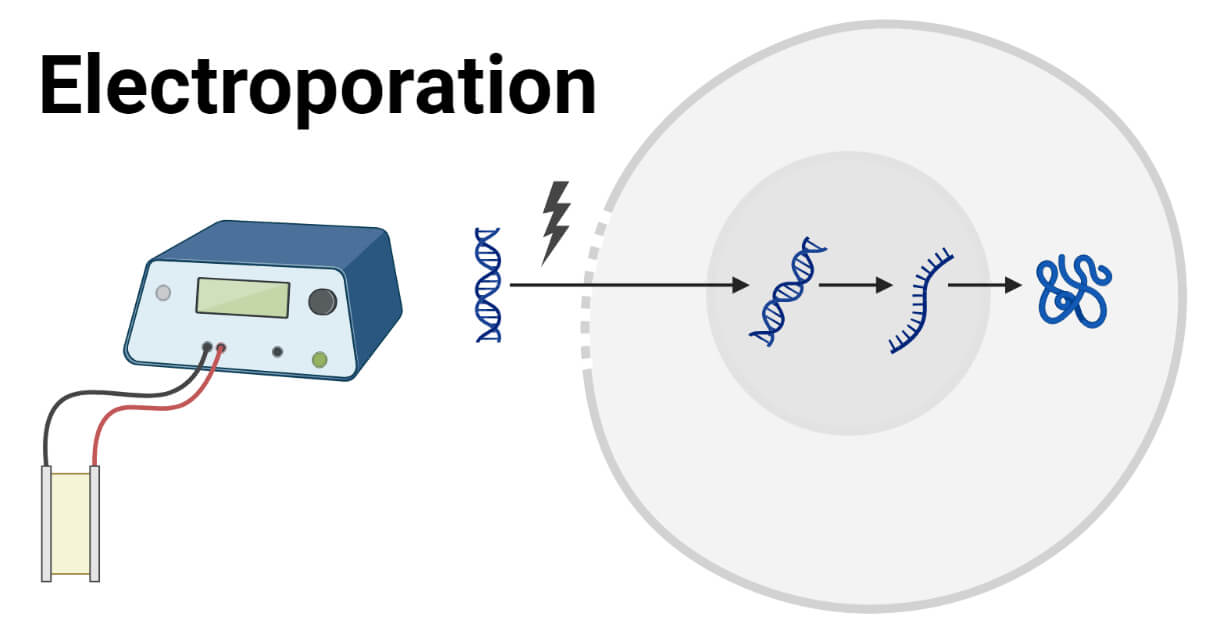


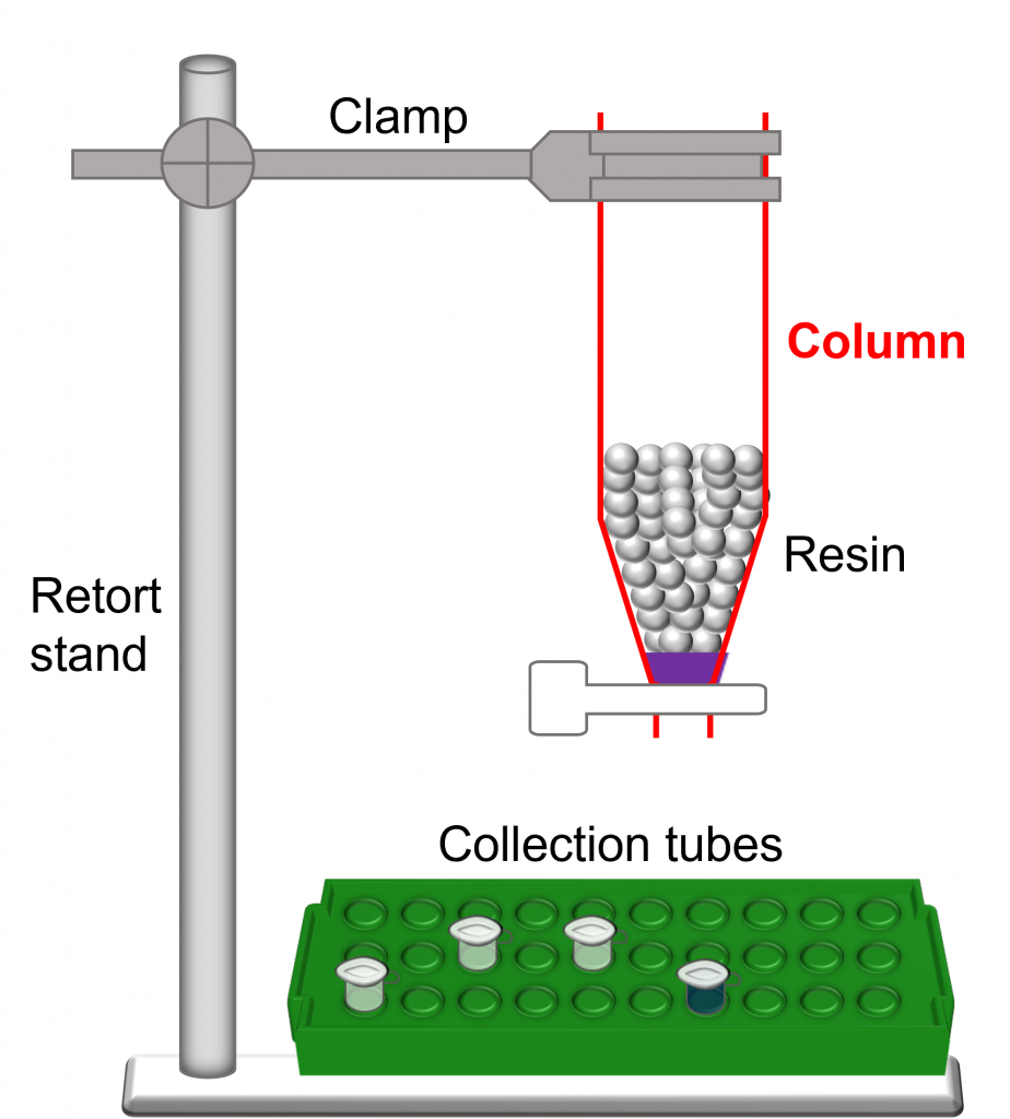


![Improving Health Care Value - The Case for Disease Registries [1/4] - the health strategist](https://joaquimcardoso.blog/wp-content/uploads/2023/09/Captura-de-tela-2023-09-21-195049-110x110.png)