Prenatal diagnosis of tracheal obstruction: possible association with maternal pertussis infection. (1/10)
A fetus with the sonographic appearance of echogenic and enlarged lungs and dilated trachea and bronchi, indicating laryngotracheal obstruction, is reported. Additionally, the fetus had ascites and subcutaneous edema and the amniotic fluid volume was reduced. Doppler flow investigation of the systemic venous circulation revealed signs of heart failure, and color Doppler visualized possible increased pulmonary flow. Following termination of pregnancy, autopsy confirmed the sonographic observations and revealed a hypoplastic thymus. During the present pregnancy the mother suffered from sustained cough, and serological tests revealed acute pertussis infection. Polymerase chain reaction investigation for Bordetella pertussis in the amniotic fluid was negative. The possibilities of pertussis toxins as noxious factors and of an atypical presentation of DiGeorge anomaly are discussed. (+info)Tracheomalacia associated with Mounier-Kuhn syndrome in the Intensive Care Unit: treatment with Freitag stent. A case report. (2/10)
Tracheomalacia is a process characterized by softness of the supporting tracheal cartilages, by the extension of the posterior membranous wall and by reduction of the tracheal antero-posterior diameter. Exceptionally, tracheomalacia can be associated with tracheobronchomegaly or Mounier-Kuhn syndrome. Fibro-bronchoscopy represents the ''gold standard'' for diagnosis. The case of a 79-year-old male observed after hospitalization in a medical ward for chronic pulmonary obstructive disease (COPD) decompensation, and with basal left bronchopulmonary focus, is described. During this period, a progressive worsening of clinical conditions occurred, despite cortisone and antibiotic therapy, and the patient was transferred to the ICU for dyspnea, hypoxia, hypocapnia and with a diagnosis of pulmonary fibrosis. Bronchoscopy, performed during spontaneous breathing, revealed tracheomalacia which was responsible for tracheal dynamic complete stenosis during expiration and dynamic subtotal stenosis of the left primary bronchus in the first tract, together with sputum retention. Moreover, this investigation confirmed the diagnosis of tracheobronchomegaly already seen on CT. It was suggested to place a Freitag stent, since the insertion of another model would not have had enough chance of stability, due to the enormous extension of the tracheal lumen and could not have guaranteed good clearance of the secretions. Seven days after this intervention, performed in an outpatients' setting, the patient was dismissed from the ICU, without the help of O2, with good ventilation, saturation in line with his age and good expectoration. (+info)Mounier-Kuhn syndrome. (3/10)
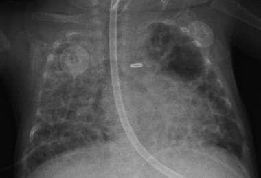
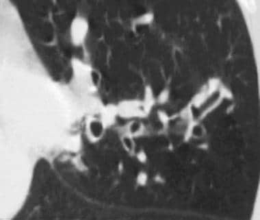
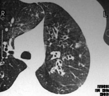
Mounier-Kuhn syndrome, or tracheobronchomegaly, is a rare clinical entity characterized by abnormal dilation of the trachea and main bronchi. The diagnosis can usually be made by measuring the tracheal diameter. We report the case of a 40-year-old black man with refractory lower respiratory tract infection. Tracheobronchomegaly was confirmed through computed tomography. (+info)Mounier-Kuhn syndrome: a rare cause of severe bronchial dilatation with normal pulmonary function test: a case report. (4/10)
Tracheobronchomegaly (TBM) (Mounier-Kuhn syndrome) is dilatation of the trachea and major bronchi because of atrophy or absence of elastic fibers and smooth muscle cells. We present a case of TBM with normal pulmonary function test (PFT). The patient was a 37-year-old man with increasing productive cough and without fever, wheezes, chest pain, weight loss or any respiratory disease. Chest helical computed tomography (CT) scan showed tracheomegaly with transversal diameters of the trachea of 44mm. CT scan showed collapse of the trachea. Few large diverticular out-pouching and openings in the trachea was seen in bronchoscopy. PFT results were normal. PFT in large airway disorders may be normal while abnormalities may indicate underlying small airway disorder. An underlying small airway disorders is responsible for abnormal reports in PFT of these patients. We may need to re-evaluate the role of PFT within follow-up of patients with large airway disorder. (+info)Mounier-Kuhn syndrome in an older patient. (5/10)
(+info)Mounier-Kuhn syndrome: a rare cause of bronchial dilation. (6/10)
Mounier-Kuhn syndrome, or tracheobronchomegaly, is a rare clinical and radiologic condition characterized by marked tracheobronchial dilation and recurrent lower respiratory tract infections. Diagnosis is typically accomplished with the use of computed tomography and bronchoscopy, as well as pulmonary function testing. Patients may be asymptomatic; however, symptoms can range from minimal with preserved lung function to severe respiratory failure. Therapy, if any, is supportive but minimal. Surgery rarely has a place in the treatment of Mounier-Kuhn syndrome.Herein, we report the case of a 58-year-old man with chronic obstructive pulmonary disease who had a chronic cough, increased sputum production, and chest pain. Thoracic computed tomography showed tracheal dilation (diameter, 34 mm) and multiple diverticula in the posterior region of the trachea. Fiberoptic bronchoscopy revealed enlarged main bronchi, the dilated trachea, and prominent tracheal diverticula. Pulmonary function testing disclosed impaired respiratory function. Histopathologic examination of biopsy specimens from the bronchi and the tracheal wall supported the diagnosis of Mounier-Kuhn syndrome. The patient was released from the hospital and his condition was monitored for 2 years, during which time he developed no lower respiratory tract infections.Regardless of radiologic findings that suggest recurrent lower respiratory tract infection, we recommend that Mounier-Kuhn syndrome be considered in the differential diagnosis. (+info)Central airway obstruction due to malignant fibrous histiocytoma metastasis in a case with Mounier-Kuhn syndrome. (7/10)
Malign fibrous histiocytoma is one of the most observed soft tissue sarcomas seen in the adults. The most common metastasis region is the lung and metastasis. Mounier-Kuhn syndrome is characterized by the highly dilatation of the trachea and bronchi. We may encounter with the major airway obstruction in the endoluminal or extraluminal lung and mediastinal masses or those with both components together. In this article, we would like to highlight the occurrence of a rare seen clinical situation secondary to the giant mediastinal malign fibrous histiocytoma metastasis and the clinical difficulties experienced in resolving of the main airway obstruction caused by the mass. Since the lack of the similar studies conducted previously, we found the case worth presenting. (+info)Atypical peritracheobronchial vasculitis and an effective treatment. (8/10)
We report an interesting case of vasculitis in which the inflammatory lesion was limited to the peritracheobronchus. This case showed positive antineutrophil cytoplasmic antibodies, diffuse peritracheobronchial swelling, and vasculitis in its histology. Steroid therapy was effective for both roentgenological and serological findings. Although the biopsy specimen shows only inflammation and does not satisfy the WHO criteria of Wegener's granulomatosis (WG), a possible diagnosis of WG should not be disregarded. (+info)Tracheobronchomegaly is a rare condition characterized by an abnormal dilatation or widening of the trachea and bronchi, which are the airway tubes leading to the lungs. This condition is also known as Mounier-Kuhn syndrome. It is typically associated with recurrent respiratory infections, coughing, and difficulty breathing, especially during physical exertion. The exact cause of tracheobronchomegaly is not well understood, but it may be related to a congenital abnormality or connective tissue disorder. Diagnosis is often made through imaging studies such as chest X-rays or CT scans. Treatment typically involves managing symptoms and preventing complications, and may include bronchodilators, antibiotics, and respiratory therapy. In severe cases, surgery may be necessary to repair or reinforce the airway walls.
 Tracheobronchomegaly
Trachea
Bronchiectasis
List of diseases (T)
List of syndromes
List of MeSH codes (C16)
Tracheobronchomegaly - Wikipedia
 Fetal Surgery for Congenital Diaphragmatic Hernia: Background, Indications, Contraindications
 Imran SATIA | Professor (Assistant) | MA MB BChir (cantab) MRCP PhD | McMaster University, Hamilton | McMaster | Division of...
 Chest radiograph zones | Radiology Reference Article | Radiopaedia.org
Gravity-dependent atelectasis | Radiology Reference Article | Radiopaedia.org
Allergic bronchopulmonary aspergillosis | Radiology Reference Article | Radiopaedia.org
 DeCS
DeCS
Specific PHGKB|Rare Diseases PHGKB|PHGKB
 Pesquisa | Portal Regional da BVS
 treatment with DIPYRIDAMOLE - treatment and prevention
Trachea Anatomy: Overview, Development of the Human Trachea, Gross Anatomy
Trachea Anatomy: Overview, Development of the Human Trachea, Gross Anatomy
 Search | SciELO
 Zyprexa Effects, Zyprexa purchase / Society of Surgical Simulation | Simulation Technology | Surgical Research UK
Pesquisa | Prevenção e Controle de Câncer
 Articles
 Bronchogenic Cyst | Profiles RNS
 Bronchogenic Cyst | Profiles RNS
 Rare Disorder (Concept Id: C0678236)
- MedGen - NCBI
Rare Disorder (Concept Id: C0678236)
- MedGen - NCBI
 Bronchiectasis - Pulmonary Disorders - MSD Manual Professional Edition
Primary Ciliary Dyskinesia (Kartagener Syndrome) Differential Diagnoses
 Michael J. Rutter, MD
Asbestos-related diseases | Radiology Reference Article | Radiopaedia.org
Jugular venous catheters | Radiology Reference Article | Radiopaedia.org
Trachea Anatomy: Overview, Development of the Human Trachea, Gross Anatomy
 Guidelines for the diagnosis and management of cystathionine beta-synthase deficiency - PubMed
DeCS
Code System Concept
 MeSH Browser
MeSH Browser
 1 - Fibo forex review
 ICD-10-CM Alphabetical Index - t
 Bronchiectasis توسع القصبات ( التوسع القصبي ) - العيادة السورية - دليل المعلومات الطبي
Trachea Anatomy: Overview, Development of the Human Trachea, Gross Anatomy
2013年02月第28卷第1期(摘要) | 台灣胸腔暨重症
 Kieran's Medical Notes - Bronchiectasis
Bronchial Spasm | Profiles RNS
 Eduards Krustins - AD Scientific Index 2024
Home | Blog | Infos about the ESR
Comments of : José Vilar and Friends Case 21 (Update: Solution!)
 Mounier-Kuhn Syndrome
Descriptors in 2013 MeSH. Preferred term only. December 14, 2012
MeSH Browser
Tracheal Neoplasms | Profiles RNS
MeSH Browser
Rare2
- Tracheobronchomegaly is a very rare congenital disorder of the lung primarily characterized by an abnormal widening of the upper airways. (wikipedia.org)
- BACKGROUND: Tracheobronchomegaly (TBM) is a rare disorder mainly characterized by dilatation and malacia of the trachea and major bronchi with diverticularization. (bvsalud.org)
Congenital Tracheobronchomegaly1
- congenital tracheobronchomegaly (a.k.a. (radiopaedia.org)
Trachea1
- H: Trachea in tracheobronchomegaly (Mounier-Kuhn syndrome). (medscape.com)
Bronchiectasis1
- First described in 1932, Mounier-Kuhn syndrome is a rare clinical entity characterized by tracheobronchomegaly, which in turn leads to ineffective airway mucociliary clearance, recurrent lower respiratory tract infections, and bronchiectasis. (e-trd.org)
Airways2
- Tracheobronchomegaly is a very rare congenital disorder of the lung primarily characterized by an abnormal widening of the upper airways. (wikipedia.org)
- After this, there is a loss of the architectural framework of the central airways, leading to tracheobronchomegaly, outpouchings of the walls, and the development of blind diverticula. (e-trd.org)