Photons
Tomography, Emission-Computed, Single-Photon
Tomography, X-Ray Computed
Tomography, Emission-Computed
Positron-Emission Tomography
Tomography
Organotechnetium Compounds
Radiopharmaceuticals
Technetium Tc 99m Exametazime
Thallium Radioisotopes
Oximes
Tomography, Optical Coherence
Iofetamine
Fluorodeoxyglucose F18
Technetium
Iodine Radioisotopes
Tomography, Optical
Tomography, Spiral Computed
Technetium Tc 99m Sestamibi
Iodobenzenes
Brain
Xenon Radioisotopes
Sensitivity and Specificity
Multidetector Computed Tomography
Phantoms, Imaging
Magnetic Resonance Imaging
Technetium Tc 99m Pyrophosphate
Image Processing, Computer-Assisted
Diagnostic Imaging
Multimodal Imaging
Cone-Beam Computed Tomography
Electron Microscope Tomography
Otoacoustic Emissions, Spontaneous
Amphetamines
Myocardial Perfusion Imaging
Cardiac-Gated Single-Photon Emission Computer-Assisted Tomography
Reproducibility of Results
Fluorine Radioisotopes
Imaging, Three-Dimensional
Radioisotopes
Predictive Value of Tests
Radiation
Acetazolamide
Tomography Scanners, X-Ray Computed
Vehicle Emissions
Organometallic Compounds
Image Enhancement
Organophosphorus Compounds
Radioactive Tracers
Radiation Dosage
Gamma Cameras
Scattering, Radiation
Exercise Test
Retinal Rod Photoreceptor Cells
Molecular Imaging
Dopamine Plasma Membrane Transport Proteins
Monte Carlo Method
Radionuclide Imaging
Carbon Radioisotopes
Cinanserin
Indium Radioisotopes
Frontal Lobe
Algorithms
Coronary Angiography
Nitrogen Radioisotopes
Technetium Tc 99m Medronate
Radius
Technology, Radiologic
Thalamic Diseases
Nortropanes
Tissue Distribution
Gallium Radioisotopes
Dipyridamole
Spectrometry, Gamma
Vision, Ocular
Flumazenil
Cerebral Revascularization
Retrospective Studies
Spondylolysis
Prospective Studies
Radiometry
Treatment Outcome
Models, Structural
Rhodopsin
Fluorescent Dyes
Coronary Artery Disease
Myocardial Ischemia
Follow-Up Studies
Coronary Disease
Elementary Particles
Night Vision
Technetium Tc 99m Aggregated Albumin
Temporal Lobe
Nuclear Medicine
Models, Theoretical
Greenhouse Effect
Scintillation Counting
Image Interpretation, Computer-Assisted
Photoreceptor Cells
Equipment Failure Analysis
Brain Diseases
Severity of Illness Index
Computer Simulation
Niobium
Moyamoya Disease
ROC Curve
Occipital Lobe
Myocardial Infarction
Microscopy, Fluorescence, Multiphoton
Pyrrolidines
Feasibility Studies
Prognosis
Photoreceptor Cells, Invertebrate
Fluorescence
Gated Blood-Pool Imaging
Brain Neoplasms
3-Iodobenzylguanidine
Luminescent Measurements
Luminescence
Dobutamine
Electrocardiography
Sturge-Weber Syndrome
Radioimmunodetection
Receptors, Dopamine D2
Myocardial Stunning
Reference Values
Optics and Photonics
Radiographic Image Interpretation, Computer-Assisted
Models, Biological
False Positive Reactions
Echocardiography
Corpus Striatum
Artifacts
Sodium Pertechnetate Tc 99m
Atrophy
Radiotherapy Planning, Computer-Assisted
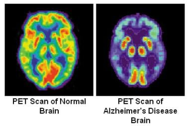
Alzheimer Disease
Ventricular Dysfunction, Left
Meningioma
Minerals
Bufo marinus
Brain Mapping
Carbon Footprint
Dark Adaptation
Cerebral Infarction
Lasers
Particle Accelerators
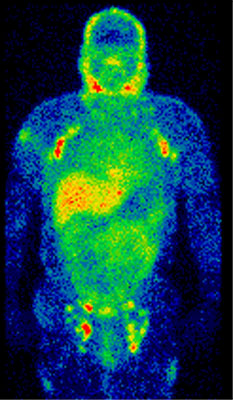
Whole Body Imaging
Brain Ischemia
Myocardium
Radiographic Image Enhancement
Air Pollutants
Isotope Labeling
Chorea
Cerebrovascular Disorders
Pentetic Acid
Semiconductors
Cerebral Arterial Diseases
Electroencephalography
Parkinson Disease
Ventricular Function, Left
Tellurium
Basal Ganglia
Neuropsychological Tests
Methane
Spectrometry, X-Ray Emission
Bone and Bones
Optical Phenomena
Four-Dimensional Computed Tomography
Neurologic Examination
Dementia
Stroke Volume
Cerebral Cortex
Parietal Lobe
Copper Radioisotopes
Spectrum Analysis
Raclopride
Cardiac sympathetic activity estimated by 123I-MIBG myocardial imaging in patients with dilated cardiomyopathy after beta-blocker or angiotensin-converting enzyme inhibitor therapy. (1/4352)
Impaired cardiac sympathetic activity can be evaluated by 123I-metaiodobenzylguanidine (MIBG) imaging. METHODS: We studied the significance of MIBG imaging for 24 patients (age 58+/-12 y) with dilated cardiomyopathy (DCM). We compared 12 patients (group A) treated with metoprolol (dose from 30-60 mg/d) with 12 patients treated with angiotensin-converting enzyme (ACE) inhibitors. Patients were studied before treatment, after 5 mo of treatment (only in group A) and after 1 y of treatment. Cardiac MIBG uptake was assessed as the heart-to-mediastinum activity ratio (H/M) and total defect score (TDS) from anterior planar and SPECT MIBG images, which were acquired in 4 h after tracer injection. New York Heart Association (NYHA) class and left ventricular ejection fraction (LVEF) calculated by echocardiography were also assessed. RESULTS: TDS decreased in both groups (in group A, from 30+/-7 through 23+/-9 to 18+/-10; P < 0.01, in group B, from 30+/-6 to 24+/-8; P < 0.01) and H/M was increased in both groups (in group A, from 1.87+/-0.31 through 2.03+/-0.28 to 2.14+/-0.29; P < 0.01, in group B, from 1.82+/-0.28 to 1.94+/-0.26; P < 0.05). But TDS and H/M were more improved in group A than in group B (P < 0.05). LVEF was significantly increased in only group A (from 38+/-6 through 43+/-8 to 49%+/-9%; P < 0.01). NYHA improved in both groups (in group A, from mean 2.5 through 2.1 to 1.8; P < 0.01, in group B, from mean 2.6 to 2.1; P < 0.05) but was more improved in group A than in group B (P < 0.05). CONCLUSION: Cardiac function, symptom and cardiac sympathetic activity evaluated by MIBG images improved after the beta-blocker therapy more than with the treatment that used ACE inhibitors. (+info)Enhanced myocardial glucose use in patients with a deficiency in long-chain fatty acid transport (CD36 deficiency). (2/4352)
CD36 is a multifunctional, 88 kDa glycoprotein that is expressed on platelets and monocytes/macrophages. CD36 also has high homology with the long-chain fatty acid (LFA) transporter in the myocardium. Although platelet and monocyte CD36 levels can indicate a CD36 deficiency, they cannot predict specific clinical manifestations in the myocardium of a given person. We examined the hypothesis that a deficiency in LFA transport augments myocardial glucose uptake in patients with a type I CD36 deficiency. METHODS: Seven fasting patients with a type I CD36 deficiency and 9 controls were assessed by cardiac radionuclide imaging using beta-methyl-p-iodophenyl-pentadecanoic acid (BMIPP) as a LFA tracer and by PET with 18F-fluorodeoxyglucose (FDG). RESULTS: None of the patients with a CD36 deficiency showed myocardial uptake of BMIPP. The percentage dose uptake of BMIPP in these subjects was significantly lower than that in normal controls (1.31+/-0.24 versus 2.90+/-0.2; P < 0.005). PET studies revealed that myocardial FDG accumulation was substantially increased in patients with a CD36 deficiency. Quantitative analysis showed that the percentage dose uptake of FDG in patients with a CD36 deficiency was significantly higher than that in normal controls (1.28+/-0.35 versus 0.43+/-0.22; P< 0.01). CONCLUSION: CD36 functions as a major myocardial LFA transporter and its absence may cause a compensatory upregulation of myocardial glucose uptake. (+info)Parametric mapping of cerebral blood flow deficits in Alzheimer's disease: a SPECT study using HMPAO and image standardization technique. (3/4352)
This study assessed the accuracy and reliability of Automated Image Registration (AIR) for standardization of brain SPECT images of patients with Alzheimer's disease (AD). Standardized cerebral blood flow (CBF) images of patients with AD and control subjects were then used for group comparison and covariance analyses. METHODS: Thirteen patients with AD at an early stage (age 69.8+/-7.1 y, Clinical Dementia Rating Score 0.5-1.0, Mini-Mental State Examination score 19-23) and 20 age-matched normal subjects (age 69.5+/-8.3 y) participated in this study. 99mTc-hexamethyl propylenamine oxime (HMPAO) brain SPECT and CT scans were acquired for each subject. SPECT images were transformed to a standard size and shape with the help of AIR. Accuracy of AIR for spatial normalization was evaluated by an index calculated on SPECT images. Anatomical variability of standardized target images was evaluated by measurements on corresponding CT scans, spatially normalized using transformations established by the SPECT images. Realigned brain SPECT images of patients and controls were used for group comparison with the help of statistical parameter mapping. Significant differences were displayed on the respective voxel to generate three-dimensional Z maps. CT scans of individual subjects were evaluated by a computer program for brain atrophy. Voxel-based covariance analysis was performed on standardized images with ages and atrophy indices as independent variables. RESULTS: Inaccuracy assessed by functional data was 2.3%. The maximum anatomical variability was 4.9 mm after standardization. Z maps showed significantly decreased regional CBF (rCBF) in the frontal, parietal and temporal regions in the patient group (P < 0.001). Covariance analysis revealed that the effects of aging on rCBF were more pronounced compared with atrophy, especially in intact cortical areas at an early stage of AD. Decrease in rCBF was partly due to senility and atrophy, however these two factors cannot explain all the deficits. CONCLUSION: AIR can transform SPECT images of AD patients with acceptable accuracy without any need for corresponding structural images. The frontal regions of the brain, in addition to parietal and temporal lobes, may show reduced CBF in patients with AD even at an early stage of dementia. The reduced rCBF in the cortical regions cannot be explained entirely by advanced atrophy and fast aging process. (+info)Integrated visualization of functional and anatomic brain data: a validation study. (4/4352)
Two-dimensional SPECT display and three methods for integrated visualization of SPECT and MRI patient data are evaluated in a multiobserver study to determine whether localization of functional data can be improved by adding anatomical information to the display. METHODS: SPECT and MRI data of 30 patients were gathered and presented using four types of display: one of SPECT in isolation, two integrated two-dimensional displays and one integrated three-dimensional display. Cold and hot spots in the peripheral cortex were preselected and indicated on black-and-white hard copies of the image data. Nuclear medicine physicians were asked to assign the corresponding spots in the image data on the computer screen to a lobe and a gyrus and give a confidence rating for both localizations. Interobserver agreement using kappa statistics and average confidence ratings were assessed to interpret the reported observations. RESULTS: Both the interobserver agreement and the confidence of the observers were greater for the integrated two-dimensional displays than for the two-dimensional SPECT display. An additional increase in agreement and confidence was seen with the integrated three-dimensional display. CONCLUSION: Integrated display of SPECT and MR brain images provides better localization of cerebral blood perfusion abnormalities in the peripheral cortex in relation to the anatomy of the brain than single-modality display and increases the confidence of the observer. (+info)Using vascular structure for CT-SPECT registration in the pelvis. (5/4352)
The authors outline a method for three-dimensional registration of pelvic CT and 111In-labeled monoclonal antibody capromab pendetide (111In MoAb 7E11.C5) images using 99mTc-labeled red blood cell SPECT data. METHODS: This method of CT-SPECT registration relies on the identification of major blood vessels in the CT and 99mTc SPECT images. The vessels are segmented from the image datasets by outlining them on transverse planar slices using a mouse-based drawing tool. Stacking the transverse outlines provides a three-dimensional representation of the vascular structures. Registration is performed by matching the surfaces of the segmented volumes. Dual isotope acquisition of 111In and 99mTc activities provides precise SPECT-SPECT registration so that registration in three dimensions of the 111In MoAb and CT images is achieved by applying the same transformation obtained from the 99mTc SPECT-CT registration. RESULTS: This method provided accurate registration of pelvic structures and significantly improved interpretation of 111In MoAb 7E11.C5 exams. Furthermore, sites of involvement by prostate cancer suggested by the 111In MoAb examination could be interpreted with the bony and soft tissue (nodal) anatomy seen on CT. CONCLUSION: This method is a general clinical tool for the registration of pelvic CT and SPECT imaging data. There are immediate applications in conformal radiation therapy treatment planning for certain prostate cancer patients. (+info)MIRD pamphlet no. 16: Techniques for quantitative radiopharmaceutical biodistribution data acquisition and analysis for use in human radiation dose estimates. (6/4352)
This report describes recommended techniques for radiopharmaceutical biodistribution data acquisition and analysis in human subjects to estimate radiation absorbed dose using the Medical Internal Radiation Dose (MIRD) schema. The document has been prepared in a format to address two audiences: individuals with a primary interest in designing clinical trials who are not experts in dosimetry and individuals with extensive experience with dosimetry-based protocols and calculational methodology. For the first group, the general concepts involved in biodistribution data acquisition are presented, with guidance provided for the number of measurements (data points) required. For those with expertise in dosimetry, highlighted sections, examples and appendices have been included to provide calculational details, as well as references, for the techniques involved. This document is intended also to serve as a guide for the investigator in choosing the appropriate methodologies when acquiring and preparing product data for review by national regulatory agencies. The emphasis is on planar imaging techniques commonly available in most nuclear medicine departments and laboratories. The measurement of the biodistribution of radiopharmaceuticals is an important aspect in calculating absorbed dose from internally deposited radionuclides. Three phases are presented: data collection, data analysis and data processing. In the first phase, data collection, the identification of source regions, the determination of their appropriate temporal sampling and the acquisition of data are discussed. In the second phase, quantitative measurement techniques involving imaging by planar scintillation camera, SPECT and PET for the calculation of activity in source regions as a function of time are discussed. In addition, nonimaging measurement techniques, including external radiation monitoring, tissue-sample counting (blood and biopsy) and excreta counting are also considered. The third phase, data processing, involves curve-fitting techniques to integrate the source time-activity curves (determining the area under these curves). For some applications, compartmental modeling procedures may be used. Last, appendices are included that provide a table of symbols and definitions, a checklist for study protocol design, example formats for quantitative imaging protocols, temporal sampling error analysis techniques and selected calculational examples. The utilization of the presented approach should aid in the standardization of protocol design for collecting kinetic data and in the calculation of absorbed dose estimates. (+info)Chronic compartment syndrome affecting the lower limb: MIBI perfusion imaging as an alternative to pressure monitoring: two case reports. (7/4352)
Intracompartmental pressure monitoring remains the primary method of diagnosing chronic compartment syndrome. MIBI perfusion imaging is widely available and offers a radionuclear imaging technique for diagnosing this condition. Although the results are not identical with those from pressure monitoring, MIBI may offer a useful screening test for this condition. (+info)Prognostic value of myocardial perfusion imaging in patients with high exercise tolerance. (8/4352)
BACKGROUND: Although high exercise tolerance is associated with an excellent prognosis, the significance of abnormal myocardial perfusion imaging (MPI) in patients with high exercise tolerance has not been established. This study retrospectively compares the utility of MPI and exercise ECG (EECG) in these patients. METHODS AND RESULTS: Of 388 consecutive patients who underwent exercise MPI and reached at least Bruce stage IV, 157 (40.5%) had abnormal results and 231 (59.5%) had normal results. Follow-up was performed at 18+/-2.7 months. Adverse events, including revascularization, myocardial infarction, and cardiac death, occurred in 40 patients. Nineteen patients had revascularization related to the MPI results or the patient's condition at the time of MPI and were not included in further analysis. Seventeen patients (12.2%) with abnormal MPI and 4 (1.7%) with normal MPI had adverse cardiac events (P<0.001). Cox proportional-hazards regression analysis showed that MPI was an excellent predictor of cardiac events (global chi2=13.2; P<0.001; relative risk=8; 95% CI=3 to 23) but EECG had no predictive power (global chi2=0.05; P=0.8; relative risk=1; 95% CI=0.4 to 3.0). The addition of Duke's treadmill score risk categories did not improve the predictive power of EECG (global chi2=0.17). The predictive power of the combination of EECG (including Duke score categories) and MPI was no better than that of MPI alone (global chi2=13.5). CONCLUSIONS: Unlike EECG, MPI is an excellent prognostic indicator for adverse cardiac events in patients with known or suspected CAD and high exercise tolerance. (+info)A photon is not a term that has a specific medical definition, as it is a fundamental concept in physics. Photons are elementary particles that carry electromagnetic energy, such as light. They have no mass or electric charge and exhibit both particle-like and wave-like properties. In the context of medicine, photons are often discussed in relation to various medical imaging techniques (e.g., X-ray imaging, CT scans, and PET scans) and therapeutic interventions like laser therapy and radiation therapy, where photons are used to diagnose or treat medical conditions.
Emission-Computed Tomography, Single-Photon (SPECT) is a type of nuclear medicine imaging procedure that generates detailed, three-dimensional images of the distribution of radioactive pharmaceuticals within the body. It uses gamma rays emitted by a radiopharmaceutical that is introduced into the patient's body, and a specialized gamma camera to detect these gamma rays and create tomographic images. The data obtained from the SPECT imaging can be used to diagnose various medical conditions, evaluate organ function, and guide treatment decisions. It is commonly used to image the heart, brain, and bones, among other organs and systems.
X-ray computed tomography (CT or CAT scan) is a medical imaging method that uses computer-processed combinations of many X-ray images taken from different angles to produce cross-sectional (tomographic) images (virtual "slices") of the body. These cross-sectional images can then be used to display detailed internal views of organs, bones, and soft tissues in the body.
The term "computed tomography" is used instead of "CT scan" or "CAT scan" because the machines take a series of X-ray measurements from different angles around the body and then use a computer to process these data to create detailed images of internal structures within the body.
CT scanning is a noninvasive, painless medical test that helps physicians diagnose and treat medical conditions. CT imaging provides detailed information about many types of tissue including lung, bone, soft tissue and blood vessels. CT examinations can be performed on every part of the body for a variety of reasons including diagnosis, surgical planning, and monitoring of therapeutic responses.
In computed tomography (CT), an X-ray source and detector rotate around the patient, measuring the X-ray attenuation at many different angles. A computer uses this data to construct a cross-sectional image by the process of reconstruction. This technique is called "tomography". The term "computed" refers to the use of a computer to reconstruct the images.
CT has become an important tool in medical imaging and diagnosis, allowing radiologists and other physicians to view detailed internal images of the body. It can help identify many different medical conditions including cancer, heart disease, lung nodules, liver tumors, and internal injuries from trauma. CT is also commonly used for guiding biopsies and other minimally invasive procedures.
In summary, X-ray computed tomography (CT or CAT scan) is a medical imaging technique that uses computer-processed combinations of many X-ray images taken from different angles to produce cross-sectional images of the body. It provides detailed internal views of organs, bones, and soft tissues in the body, allowing physicians to diagnose and treat medical conditions.
Emission computed tomography (ECT) is a type of tomographic imaging technique in which an emission signal from within the body is detected to create cross-sectional images of that signal's distribution. In Emission-Computed Tomography (ECT), a radionuclide is introduced into the body, usually through injection, inhalation or ingestion. The radionuclide emits gamma rays that are then detected by external gamma cameras.
The data collected from these cameras is then used to create cross-sectional images of the distribution of the radiopharmaceutical within the body. This allows for the identification and quantification of functional information about specific organs or systems within the body, such as blood flow, metabolic activity, or receptor density.
One common type of Emission-Computed Tomography is Single Photon Emission Computed Tomography (SPECT), which uses a single gamma camera that rotates around the patient to collect data from multiple angles. Another type is Positron Emission Tomography (PET), which uses positron-emitting radionuclides and detects the coincident gamma rays emitted by the annihilation of positrons and electrons.
Overall, ECT is a valuable tool in medical imaging for diagnosing and monitoring various diseases, including cancer, heart disease, and neurological disorders.
Positron-Emission Tomography (PET) is a type of nuclear medicine imaging that uses small amounts of radioactive material, called a radiotracer, to produce detailed, three-dimensional images. This technique measures metabolic activity within the body, such as sugar metabolism, to help distinguish between healthy and diseased tissue, identify cancerous cells, or examine the function of organs.
During a PET scan, the patient is injected with a radiotracer, typically a sugar-based compound labeled with a positron-emitting radioisotope, such as fluorine-18 (^18^F). The radiotracer accumulates in cells that are metabolically active, like cancer cells. As the radiotracer decays, it emits positrons, which then collide with electrons in nearby tissue, producing gamma rays. A special camera, called a PET scanner, detects these gamma rays and uses this information to create detailed images of the body's internal structures and processes.
PET is often used in conjunction with computed tomography (CT) or magnetic resonance imaging (MRI) to provide both functional and anatomical information, allowing for more accurate diagnosis and treatment planning. Common applications include detecting cancer recurrence, staging and monitoring cancer, evaluating heart function, and assessing brain function in conditions like dementia and epilepsy.
Tomography is a medical imaging technique used to produce cross-sectional images or slices of specific areas of the body. This technique uses various forms of radiation (X-rays, gamma rays) or sound waves (ultrasound) to create detailed images of the internal structures, such as organs, bones, and tissues. Common types of tomography include Computerized Tomography (CT), Positron Emission Tomography (PET), and Magnetic Resonance Imaging (MRI). The primary advantage of tomography is its ability to provide clear and detailed images of internal structures, allowing healthcare professionals to accurately diagnose and monitor a wide range of medical conditions.
Organotechnetium compounds are chemical substances that contain carbon-technetium bonds, where technetium is an element with the symbol Tc and atomic number 43. These types of compounds are primarily used in medical imaging as radioactive tracers due to the ability of technetium-99m to emit gamma rays. The organotechnetium compounds help in localizing specific organs, tissues, or functions within the body, making them useful for diagnostic purposes in nuclear medicine.
It is important to note that most organotechnetium compounds are synthesized from technetium-99m, which is generated from the decay of molybdenum-99. The use of these compounds requires proper handling and administration by trained medical professionals due to their radioactive nature.
Radiopharmaceuticals are defined as pharmaceutical preparations that contain radioactive isotopes and are used for diagnosis or therapy in nuclear medicine. These compounds are designed to interact specifically with certain biological targets, such as cells, tissues, or organs, and emit radiation that can be detected and measured to provide diagnostic information or used to destroy abnormal cells or tissue in therapeutic applications.
The radioactive isotopes used in radiopharmaceuticals have carefully controlled half-lives, which determine how long they remain radioactive and how long the pharmaceutical preparation remains effective. The choice of radioisotope depends on the intended use of the radiopharmaceutical, as well as factors such as its energy, range of emission, and chemical properties.
Radiopharmaceuticals are used in a wide range of medical applications, including imaging, cancer therapy, and treatment of other diseases and conditions. Examples of radiopharmaceuticals include technetium-99m for imaging the heart, lungs, and bones; iodine-131 for treating thyroid cancer; and samarium-153 for palliative treatment of bone metastases.
The use of radiopharmaceuticals requires specialized training and expertise in nuclear medicine, as well as strict adherence to safety protocols to minimize radiation exposure to patients and healthcare workers.
Technetium Tc 99m Exametazime is a radiopharmaceutical agent used in nuclear medicine imaging procedures. The compound consists of the radioisotope Technetium-99m (^99m^Tc) bonded to Exametazime, also known as HMPAO (hexamethylpropyleneamine oxime).
Once injected into the patient's bloodstream, Technetium Tc 99m Exametazime distributes evenly throughout the brain, crossing the blood-brain barrier and entering cells. The radioactive decay of Technetium-99m emits gamma rays that can be detected by a gamma camera, creating images of the brain's blood flow and distribution of the tracer.
This imaging technique is often used in cerebral perfusion studies to assess conditions such as stroke, epilepsy, or dementia, providing valuable information about regional cerebral blood flow and potential areas of injury or abnormality.
Thallium radioisotopes are radioactive isotopes or variants of the element thallium (Tl), which decays and emits radiation. Thallium has several radioisotopes, with the most commonly used being thallium-201 (^201Tl). This radioisotope is used in medical imaging, specifically in myocardial perfusion scintigraphy, to evaluate blood flow to the heart muscle. It decays by electron capture and emits gamma radiation with a half-life of 73 hours, making it suitable for diagnostic procedures.
It's important to note that handling and using radioisotopes require proper training and safety measures due to their ionizing radiation properties.
Oximes are a class of chemical compounds that contain the functional group =N-O-, where two organic groups are attached to the nitrogen atom. In a clinical context, oximes are used as antidotes for nerve agent and pesticide poisoning. The most commonly used oxime in medicine is pralidoxime (2-PAM), which is used to reactivate acetylcholinesterase that has been inhibited by organophosphorus compounds, such as nerve agents and certain pesticides. These compounds work by forming a bond with the phosphoryl group of the inhibited enzyme, allowing for its reactivation and restoration of normal neuromuscular function.
Optical coherence tomography (OCT) is a non-invasive imaging technique that uses low-coherence light to capture high-resolution cross-sectional images of biological tissues, particularly the retina and other ocular structures. OCT works by measuring the echo time delay of light scattered back from different depths within the tissue, creating a detailed map of the tissue's structure. This technique is widely used in ophthalmology to diagnose and monitor various eye conditions such as macular degeneration, diabetic retinopathy, and glaucoma.
Iofetamine is a radiopharmaceutical agent used in myocardial perfusion imaging, a type of nuclear stress test. It is a derivative of the amphetamine family and functions as a vasoconstrictor when administered. Iofetamine is labeled with technetium-99m (^99mTc) before use, which allows for the detection and imaging of the heart's blood flow and function during rest and stress conditions. This information helps physicians diagnose and assess coronary artery disease and evaluate the effectiveness of treatments.
The medical definition of Iofetamine is:
A radiopharmaceutical agent, (^99mTc)Tc-sestamibi or (^99mTc)Tc-MIBI, used in myocardial perfusion imaging for the assessment of coronary artery disease. Iofetamine is a lipophilic cation that accumulates in myocardial cells in proportion to regional blood flow. The technetium-99m label enables gamma camera detection and imaging, providing information about the heart's blood flow and function during rest and stress conditions.
Fluorodeoxyglucose F18 (FDG-18) is not a medical condition, but a radiopharmaceutical used in medical imaging. It is a type of glucose (a simple sugar) that has been chemically combined with a small amount of a radioactive isotope called fluorine-18.
FDG-18 is used in positron emission tomography (PET) scans to help identify areas of the body where cells are using more energy than normal, such as cancerous tumors. The FDG-18 is injected into the patient's vein and travels throughout the body. Because cancer cells often use more glucose than normal cells, they tend to absorb more FDG-18.
Once inside the body, the FDG-18 emits positrons, which interact with electrons in nearby tissue, producing gamma rays that can be detected by a PET scanner. The resulting images can help doctors locate and assess the size and activity of cancerous tumors, as well as monitor the effectiveness of treatment.
Technetium is not a medical term itself, but it is a chemical element with the symbol Tc and atomic number 43. However, in the field of nuclear medicine, which is a branch of medicine that uses small amounts of radioactive material to diagnose or treat diseases, Technetium-99m (a radioisotope of technetium) is commonly used for various diagnostic procedures.
Technetium-99m is a metastable nuclear isomer of technetium-99, and it emits gamma rays that can be detected outside the body to create images of internal organs or tissues. It has a short half-life of about 6 hours, which makes it ideal for diagnostic imaging since it decays quickly and reduces the patient's exposure to radiation.
Technetium-99m is used in a variety of medical procedures, such as bone scans, lung scans, heart scans, liver-spleen scans, brain scans, and kidney scans, among others. It can be attached to different pharmaceuticals or molecules that target specific organs or tissues, allowing healthcare professionals to assess their function or identify any abnormalities.
Iodine radioisotopes are radioactive isotopes of the element iodine, which decays and emits radiation in the form of gamma rays. Some commonly used iodine radioisotopes include I-123, I-125, I-131. These radioisotopes have various medical applications such as in diagnostic imaging, therapy for thyroid disorders, and cancer treatment.
For example, I-131 is commonly used to treat hyperthyroidism and differentiated thyroid cancer due to its ability to destroy thyroid tissue. On the other hand, I-123 is often used in nuclear medicine scans of the thyroid gland because it emits gamma rays that can be detected by a gamma camera, allowing for detailed images of the gland's structure and function.
It is important to note that handling and administering radioisotopes require specialized training and safety precautions due to their radiation-emitting properties.
Optical Tomography (OT) is a non-invasive imaging technique that uses light to visualize and measure the optical properties of tissue, such as absorption and scattering coefficients. This modality can be used to produce cross-sectional or three-dimensional images of internal structures, providing functional information about tissue physiology. It has applications in various fields including biomedical research, dermatology, and oncology for the detection and monitoring of diseases. There are different types of optical tomography, such as diffuse optical tomography (DOT) and near-infrared spectroscopy (NIRS), which differ in their light sources, detection schemes, and data analysis methods.
Cerebrovascular circulation refers to the network of blood vessels that supply oxygenated blood and nutrients to the brain tissue, and remove waste products. It includes the internal carotid arteries, vertebral arteries, circle of Willis, and the intracranial arteries that branch off from them.
The internal carotid arteries and vertebral arteries merge to form the circle of Willis, a polygonal network of vessels located at the base of the brain. The anterior cerebral artery, middle cerebral artery, posterior cerebral artery, and communicating arteries are the major vessels that branch off from the circle of Willis and supply blood to different regions of the brain.
Interruptions or abnormalities in the cerebrovascular circulation can lead to various neurological conditions such as stroke, transient ischemic attack (TIA), and vascular dementia.
Spiral Computed Tomography (CT), also known as Helical CT, is a type of computed tomography scan in which the X-ray tube and detector rotate around the patient in a spiral path, capturing data as the table moves the patient through the scanner. This continuous spiral motion allows for faster and more detailed volumetric imaging of internal organs and structures, reducing the need for multiple slices and providing improved image reconstruction. It is commonly used to diagnose and monitor various medical conditions, including cancer, heart disease, and trauma injuries.
Technetium Tc 99m Sestamibi is a radiopharmaceutical compound used in medical imaging, specifically in myocardial perfusion scintigraphy. It is a technetium-labeled isonitrile chelate that is taken up by mitochondria in cells with high metabolic activity, such as cardiomyocytes (heart muscle cells).
Once injected into the patient's body, Technetium Tc 99m Sestamibi emits gamma rays, which can be detected by a gamma camera. This allows for the creation of images that reflect the distribution and function of the radiopharmaceutical within the heart muscle. The images can help identify areas of reduced blood flow or ischemia, which may indicate coronary artery disease.
The uptake of Technetium Tc 99m Sestamibi in other organs, such as the breast and thyroid, can also be used for imaging purposes, although its primary use remains in cardiac imaging.
Iodobenzenes are organic compounds that contain a iodine atom (I) attached to a benzene ring. The general formula for iodobenzenes is C6H5I. They can be considered as aryl halides and can undergo various chemical reactions such as nucleophilic substitution, electrophilic aromatic substitution, and reduction. Iodobenzenes are less reactive than other aryl halides due to the larger size and lower electronegativity of iodine compared to other halogens. They are used in organic synthesis as building blocks or reagents for various chemical transformations.
The brain is the central organ of the nervous system, responsible for receiving and processing sensory information, regulating vital functions, and controlling behavior, movement, and cognition. It is divided into several distinct regions, each with specific functions:
1. Cerebrum: The largest part of the brain, responsible for higher cognitive functions such as thinking, learning, memory, language, and perception. It is divided into two hemispheres, each controlling the opposite side of the body.
2. Cerebellum: Located at the back of the brain, it is responsible for coordinating muscle movements, maintaining balance, and fine-tuning motor skills.
3. Brainstem: Connects the cerebrum and cerebellum to the spinal cord, controlling vital functions such as breathing, heart rate, and blood pressure. It also serves as a relay center for sensory information and motor commands between the brain and the rest of the body.
4. Diencephalon: A region that includes the thalamus (a major sensory relay station) and hypothalamus (regulates hormones, temperature, hunger, thirst, and sleep).
5. Limbic system: A group of structures involved in emotional processing, memory formation, and motivation, including the hippocampus, amygdala, and cingulate gyrus.
The brain is composed of billions of interconnected neurons that communicate through electrical and chemical signals. It is protected by the skull and surrounded by three layers of membranes called meninges, as well as cerebrospinal fluid that provides cushioning and nutrients.
Thallium is a chemical element with the symbol Tl and atomic number 81. It is a soft, malleable, silver-like metal that is highly toxic. In the context of medicine, thallium may be used as a component in medical imaging tests, such as thallium stress tests, which are used to evaluate blood flow to the heart and detect coronary artery disease. Thallium-201 is a radioactive isotope of thallium that is used as a radiopharmaceutical in these tests. When administered to a patient, it is taken up by heart muscle tissue in proportion to its blood flow, allowing doctors to identify areas of the heart that may not be receiving enough oxygen-rich blood. However, due to concerns about its potential toxicity and the availability of safer alternatives, thallium stress tests are less commonly used today than they were in the past.
Xenon radioisotopes are unstable isotopes of the element xenon that emit radiation as they decay into more stable forms. These isotopes can be produced through various nuclear reactions and have a wide range of applications, including medical imaging and cancer treatment. Examples of commonly used xenon radioisotopes include xenon-127, xenon-131m, xenon-133, and xenon-135.
It's important to note that the use of radioisotopes in medical settings must be carefully regulated and monitored to ensure safety and minimize potential risks to patients and healthcare workers.
Sensitivity and specificity are statistical measures used to describe the performance of a diagnostic test or screening tool in identifying true positive and true negative results.
* Sensitivity refers to the proportion of people who have a particular condition (true positives) who are correctly identified by the test. It is also known as the "true positive rate" or "recall." A highly sensitive test will identify most or all of the people with the condition, but may also produce more false positives.
* Specificity refers to the proportion of people who do not have a particular condition (true negatives) who are correctly identified by the test. It is also known as the "true negative rate." A highly specific test will identify most or all of the people without the condition, but may also produce more false negatives.
In medical testing, both sensitivity and specificity are important considerations when evaluating a diagnostic test. High sensitivity is desirable for screening tests that aim to identify as many cases of a condition as possible, while high specificity is desirable for confirmatory tests that aim to rule out the condition in people who do not have it.
It's worth noting that sensitivity and specificity are often influenced by factors such as the prevalence of the condition in the population being tested, the threshold used to define a positive result, and the reliability and validity of the test itself. Therefore, it's important to consider these factors when interpreting the results of a diagnostic test.
Multidetector computed tomography (MDCT) is a type of computed tomography (CT) scan that uses multiple rows of detectors to acquire several slices of images simultaneously, thereby reducing the total time required for the scan and improving the spatial resolution. This technology allows for faster scanning of moving organs, such as the heart, and provides high-resolution images with detailed information about various body structures, including bones, soft tissues, and blood vessels. MDCT has numerous applications in diagnostic imaging, interventional procedures, and cancer staging and treatment follow-up.
In the field of medical imaging, "phantoms" refer to physical objects that are specially designed and used for calibration, quality control, and evaluation of imaging systems. These phantoms contain materials with known properties, such as attenuation coefficients or spatial resolution, which allow for standardized measurement and comparison of imaging parameters across different machines and settings.
Imaging phantoms can take various forms depending on the modality of imaging. For example, in computed tomography (CT), a common type of phantom is the "water-equivalent phantom," which contains materials with similar X-ray attenuation properties as water. This allows for consistent measurement of CT dose and image quality. In magnetic resonance imaging (MRI), phantoms may contain materials with specific relaxation times or magnetic susceptibilities, enabling assessment of signal-to-noise ratio, spatial resolution, and other imaging parameters.
By using these standardized objects, healthcare professionals can ensure the accuracy, consistency, and reliability of medical images, ultimately contributing to improved patient care and safety.
Medical Definition:
Magnetic Resonance Imaging (MRI) is a non-invasive diagnostic imaging technique that uses a strong magnetic field and radio waves to create detailed cross-sectional or three-dimensional images of the internal structures of the body. The patient lies within a large, cylindrical magnet, and the scanner detects changes in the direction of the magnetic field caused by protons in the body. These changes are then converted into detailed images that help medical professionals to diagnose and monitor various medical conditions, such as tumors, injuries, or diseases affecting the brain, spinal cord, heart, blood vessels, joints, and other internal organs. MRI does not use radiation like computed tomography (CT) scans.
Technetium Tc 99m Pyrophosphate (Tc-99m PYP) is a radiopharmaceutical agent used in nuclear medicine imaging, specifically myocardial perfusion imaging. It is a complex of technetium-99m, a metastable isotope of technetium, with pyrophosphate, a molecule that accumulates in damaged heart muscle tissue.
When injected into the patient's bloodstream, Tc-99m PYP is taken up by the heart muscle in proportion to its blood flow and the degree of damage or scarring (fibrosis). This allows for the detection and evaluation of conditions such as myocardial infarction (heart attack), cardiomyopathy, and heart transplant rejection.
The imaging procedure involves the injection of Tc-99m PYP, followed by the acquisition of images using a gamma camera, which detects the gamma rays emitted by the technetium-99m isotope. The resulting images provide information about the distribution and extent of heart muscle damage, helping physicians to make informed decisions regarding diagnosis and treatment planning.
Computer-assisted image processing is a medical term that refers to the use of computer systems and specialized software to improve, analyze, and interpret medical images obtained through various imaging techniques such as X-ray, CT (computed tomography), MRI (magnetic resonance imaging), ultrasound, and others.
The process typically involves several steps, including image acquisition, enhancement, segmentation, restoration, and analysis. Image processing algorithms can be used to enhance the quality of medical images by adjusting contrast, brightness, and sharpness, as well as removing noise and artifacts that may interfere with accurate diagnosis. Segmentation techniques can be used to isolate specific regions or structures of interest within an image, allowing for more detailed analysis.
Computer-assisted image processing has numerous applications in medical imaging, including detection and characterization of lesions, tumors, and other abnormalities; assessment of organ function and morphology; and guidance of interventional procedures such as biopsies and surgeries. By automating and standardizing image analysis tasks, computer-assisted image processing can help to improve diagnostic accuracy, efficiency, and consistency, while reducing the potential for human error.
Diagnostic imaging is a medical specialty that uses various technologies to produce visual representations of the internal structures and functioning of the body. These images are used to diagnose injury, disease, or other abnormalities and to monitor the effectiveness of treatment. Common modalities of diagnostic imaging include:
1. Radiography (X-ray): Uses ionizing radiation to produce detailed images of bones, teeth, and some organs.
2. Computed Tomography (CT) Scan: Combines X-ray technology with computer processing to create cross-sectional images of the body.
3. Magnetic Resonance Imaging (MRI): Uses a strong magnetic field and radio waves to generate detailed images of soft tissues, organs, and bones.
4. Ultrasound: Employs high-frequency sound waves to produce real-time images of internal structures, often used for obstetrics and gynecology.
5. Nuclear Medicine: Involves the administration of radioactive tracers to assess organ function or detect abnormalities within the body.
6. Positron Emission Tomography (PET) Scan: Uses a small amount of radioactive material to produce detailed images of metabolic activity in the body, often used for cancer detection and monitoring treatment response.
7. Fluoroscopy: Utilizes continuous X-ray imaging to observe moving structures or processes within the body, such as swallowing studies or angiography.
Diagnostic imaging plays a crucial role in modern medicine, allowing healthcare providers to make informed decisions about patient care and treatment plans.
Multimodal imaging is a medical term that refers to the combination of two or more imaging techniques to obtain complementary information about the structure, function, and/or physiology of tissues, organs, or organ systems. This approach allows for a more comprehensive assessment of normal and abnormal processes in the body than can be achieved with any single imaging modality alone.
Commonly used imaging modalities in multimodal imaging include computed tomography (CT), magnetic resonance imaging (MRI), positron emission tomography (PET), single-photon emission computed tomography (SPECT), ultrasound, and optical imaging techniques. Each modality provides unique information that can be integrated to improve diagnostic accuracy, guide treatment planning, and monitor response to therapy.
For example, a patient with a suspected brain tumor may undergo both MRI and PET scans. The MRI provides detailed anatomical information about the size, shape, and location of the tumor, while the PET scan shows metabolic activity within the tumor, which can help distinguish between benign and malignant lesions.
Multimodal imaging is also used in research settings to study various physiological processes, such as blood flow, oxygenation, and neurotransmission, in both health and disease.
Cone-beam computed tomography (CBCT) is a medical imaging technique that uses a cone-shaped X-ray beam to create detailed, cross-sectional images of the body. In dental and maxillofacial radiology, CBCT is used to produce three-dimensional images of the teeth, jaws, and surrounding bones.
CBCT differs from traditional computed tomography (CT) in that it uses a cone-shaped X-ray beam instead of a fan-shaped beam, which allows for a faster scan time and lower radiation dose. The X-ray beam is rotated around the patient's head, capturing data from multiple angles, which is then reconstructed into a three-dimensional image using specialized software.
CBCT is commonly used in dental implant planning, orthodontic treatment planning, airway analysis, and the diagnosis and management of jaw pathologies such as tumors and fractures. It provides detailed information about the anatomy of the teeth, jaws, and surrounding structures, which can help clinicians make more informed decisions about patient care.
However, it is important to note that CBCT should only be used when necessary, as it still involves exposure to ionizing radiation. The benefits of using CBCT must be weighed against the potential risks associated with radiation exposure.
Electron microscope tomography (EMT) is a 3D imaging technique used in electron microscopy. It involves collecting a series of images of a sample at different tilt angles, and then using computational algorithms to reconstruct the 3D structure of the sample from these images.
In EMT, a sample is prepared and placed in an electron microscope, where it is exposed to a beam of electrons. The electrons interact with the atoms in the sample, producing contrast that allows the features of the sample to be visualized. By tilting the sample and collecting images at multiple angles, a range of perspectives can be obtained, which are then used to create a 3D reconstruction of the sample.
EMT is a powerful tool for studying the ultrastructure of cells and tissues, as it allows researchers to visualize structures that may not be visible using other imaging techniques. It has been used to study a wide range of biological systems, including viruses, bacteria, organelles, and cells.
EMT is a complex technique that requires specialized equipment and expertise to perform. However, it can provide valuable insights into the structure and function of biological systems, making it an important tool in the field of biology and medicine.
Tropane alkaloids are a class of naturally occurring compounds that contain a tropane ring in their chemical structure. This ring is composed of a seven-membered ring with two nitrogen atoms, one of which is part of a piperidine ring. Tropane alkaloids are found in various plants, particularly those in the Solanaceae family, which includes nightshade, belladonna, and datura. Some well-known tropane alkaloids include atropine, scopolamine, and cocaine. These compounds have diverse pharmacological activities, such as anticholinergic, local anesthetic, and central nervous system stimulant effects.
Spontaneous otoacoustic emissions (SOAEs) are low-level sounds that are produced by the inner ear (cochlea) without any external stimulation. They can be recorded in a quiet room using specialized microphones placed inside the ear canal. SOAEs are thought to arise from the motion of the hair cells within the cochlea, which generate tiny currents in response to sound. These currents then cause the surrounding fluid and tissue to vibrate, producing sound waves that can be detected with a microphone.
SOAEs are typically present in individuals with normal hearing, although their presence or absence is not a definitive indicator of hearing ability. They tend to occur at specific frequencies and can vary from person to person. In some cases, SOAEs may be absent or reduced in individuals with hearing loss or damage to the hair cells in the cochlea.
It's worth noting that SOAEs are different from evoked otoacoustic emissions (EOAEs), which are sounds produced by the inner ear in response to external stimuli, such as clicks or tones. Both types of otoacoustic emissions are used in hearing tests and research to assess cochlear function and health.
Amphetamines are a type of central nervous system stimulant drug that increases alertness, wakefulness, and energy levels. They work by increasing the activity of certain neurotransmitters (chemical messengers) in the brain, such as dopamine and norepinephrine. Amphetamines can be prescribed for medical conditions such as attention deficit hyperactivity disorder (ADHD) and narcolepsy, but they are also commonly abused for their ability to produce euphoria, increase confidence, and improve performance in tasks that require sustained attention.
Some common examples of amphetamines include:
* Adderall: a combination of amphetamine and dextroamphetamine, used to treat ADHD and narcolepsy
* Dexedrine: a brand name for dextroamphetamine, used to treat ADHD and narcolepsy
* Vyvanse: a long-acting formulation of lisdexamfetamine, a prodrug that is converted to dextroamphetamine in the body, used to treat ADHD
Amphetamines can be taken orally, snorted, smoked, or injected. Long-term use or abuse of amphetamines can lead to a number of negative health consequences, including addiction, cardiovascular problems, malnutrition, mental health disorders, and memory loss.
Myocardial perfusion imaging (MPI) is a non-invasive nuclear medicine test used to assess the blood flow to the heart muscle (myocardium). It typically involves the injection of a radioactive tracer, such as thallium-201 or technetium-99m sestamibi, into a vein. The tracer is taken up by healthy heart muscle in proportion to blood flow. A special camera then takes images of the distribution of the tracer within the heart, providing information about areas of reduced or blocked blood flow (ischemia) or scarred tissue (infarction). MPI can help diagnose coronary artery disease, assess the effectiveness of treatments, and determine prognosis.
Cardiac-gated single-photon emission computer-assisted tomography (SPECT) is a medical imaging technique used to evaluate the function and perfusion of the heart. This technique combines the principles of cardiac gating, SPECT imaging, and computed tomography (CT) to produce detailed, three-dimensional images of the heart.
In this procedure, a small amount of radioactive tracer is injected into the patient's bloodstream. The tracer accumulates in the heart muscle according to its blood flow, allowing for the assessment of regional myocardial perfusion and function. A specialized gamma camera then captures images of the distribution of this tracer within the heart.
Cardiac gating is a technique that synchronizes image acquisition with the patient's electrocardiogram (ECG) signal, ensuring that data are collected only during specific phases of the cardiac cycle. This allows for the accurate assessment of regional wall motion and thickening, which can help identify areas of the heart affected by ischemia or scarring.
The acquired images are then reconstructed using computer algorithms to create detailed, three-dimensional representations of the heart's structure and function. These images can aid in the diagnosis and management of various cardiac conditions, such as coronary artery disease, cardiomyopathies, and heart failure.
Reproducibility of results in a medical context refers to the ability to obtain consistent and comparable findings when a particular experiment or study is repeated, either by the same researcher or by different researchers, following the same experimental protocol. It is an essential principle in scientific research that helps to ensure the validity and reliability of research findings.
In medical research, reproducibility of results is crucial for establishing the effectiveness and safety of new treatments, interventions, or diagnostic tools. It involves conducting well-designed studies with adequate sample sizes, appropriate statistical analyses, and transparent reporting of methods and findings to allow other researchers to replicate the study and confirm or refute the results.
The lack of reproducibility in medical research has become a significant concern in recent years, as several high-profile studies have failed to produce consistent findings when replicated by other researchers. This has led to increased scrutiny of research practices and a call for greater transparency, rigor, and standardization in the conduct and reporting of medical research.
X-ray tomography, also known as computed tomography (CT) or computerized axial tomography (CAT), is a medical imaging technique that uses X-rays to create detailed cross-sectional images of the body. In this technique, an X-ray source and detectors rotate around the patient, acquiring multiple X-ray projections at different angles. A computer then processes these projections to reconstruct tomographic images (slices) of the internal structures of the body, such as bones, organs, and soft tissues.
The term "tomography" comes from the Greek words "tome," meaning slice or section, and "graphein," meaning to write or record. X-ray tomography allows radiologists and other medical professionals to visualize and diagnose various conditions, such as fractures, tumors, infections, and internal injuries, more accurately and efficiently than with traditional X-ray imaging techniques.
It is important to note that while X-ray tomography provides valuable diagnostic information, it does involve exposure to ionizing radiation. Therefore, the benefits of the examination should outweigh the potential risks, and the use of this technique should be justified based on clinical necessity and patient safety considerations.
Fluorine radioisotopes are radioactive isotopes or variants of the chemical element Fluorine (F, atomic number 9). These radioisotopes have an unstable nucleus that emits radiation in the form of alpha particles, beta particles, or gamma rays. Examples of Fluorine radioisotopes include Fluorine-18 and Fluorine-19.
Fluorine-18 is a positron-emitting radionuclide with a half-life of approximately 110 minutes, making it useful for medical imaging techniques such as Positron Emission Tomography (PET) scans. It is commonly used in the production of fluorodeoxyglucose (FDG), a radiopharmaceutical that can be used to detect cancer and other metabolic disorders.
Fluorine-19, on the other hand, is a stable isotope of Fluorine and does not emit radiation. However, it can be enriched and used as a non-radioactive tracer in medical research and diagnostic applications.
In medical terms, the heart is a muscular organ located in the thoracic cavity that functions as a pump to circulate blood throughout the body. It's responsible for delivering oxygen and nutrients to the tissues and removing carbon dioxide and other wastes. The human heart is divided into four chambers: two atria on the top and two ventricles on the bottom. The right side of the heart receives deoxygenated blood from the body and pumps it to the lungs, while the left side receives oxygenated blood from the lungs and pumps it out to the rest of the body. The heart's rhythmic contractions and relaxations are regulated by a complex electrical conduction system.
Three-dimensional (3D) imaging in medicine refers to the use of technologies and techniques that generate a 3D representation of internal body structures, organs, or tissues. This is achieved by acquiring and processing data from various imaging modalities such as X-ray computed tomography (CT), magnetic resonance imaging (MRI), ultrasound, or confocal microscopy. The resulting 3D images offer a more detailed visualization of the anatomy and pathology compared to traditional 2D imaging techniques, allowing for improved diagnostic accuracy, surgical planning, and minimally invasive interventions.
In 3D imaging, specialized software is used to reconstruct the acquired data into a volumetric model, which can be manipulated and viewed from different angles and perspectives. This enables healthcare professionals to better understand complex anatomical relationships, detect abnormalities, assess disease progression, and monitor treatment response. Common applications of 3D imaging include neuroimaging, orthopedic surgery planning, cancer staging, dental and maxillofacial reconstruction, and interventional radiology procedures.
Radioisotopes, also known as radioactive isotopes or radionuclides, are variants of chemical elements that have unstable nuclei and emit radiation in the form of alpha particles, beta particles, gamma rays, or conversion electrons. These isotopes are formed when an element's nucleus undergoes natural or artificial radioactive decay.
Radioisotopes can be produced through various processes, including nuclear fission, nuclear fusion, and particle bombardment in a cyclotron or other types of particle accelerators. They have a wide range of applications in medicine, industry, agriculture, research, and energy production. In the medical field, radioisotopes are used for diagnostic imaging, radiation therapy, and in the labeling of molecules for research purposes.
It is important to note that handling and using radioisotopes requires proper training, safety measures, and regulatory compliance due to their ionizing radiation properties, which can pose potential health risks if not handled correctly.
The Predictive Value of Tests, specifically the Positive Predictive Value (PPV) and Negative Predictive Value (NPV), are measures used in diagnostic tests to determine the probability that a positive or negative test result is correct.
Positive Predictive Value (PPV) is the proportion of patients with a positive test result who actually have the disease. It is calculated as the number of true positives divided by the total number of positive results (true positives + false positives). A higher PPV indicates that a positive test result is more likely to be a true positive, and therefore the disease is more likely to be present.
Negative Predictive Value (NPV) is the proportion of patients with a negative test result who do not have the disease. It is calculated as the number of true negatives divided by the total number of negative results (true negatives + false negatives). A higher NPV indicates that a negative test result is more likely to be a true negative, and therefore the disease is less likely to be present.
The predictive value of tests depends on the prevalence of the disease in the population being tested, as well as the sensitivity and specificity of the test. A test with high sensitivity and specificity will generally have higher predictive values than a test with low sensitivity and specificity. However, even a highly sensitive and specific test can have low predictive values if the prevalence of the disease is low in the population being tested.
Medical Definition:
Radiation is the emission of energy as electromagnetic waves or as moving subatomic particles, especially high-energy particles that cause ionization, which can occur naturally (e.g., sunlight) or be produced artificially (e.g., x-rays, radioisotopes). In medicine, radiation is used diagnostically and therapeutically in various forms, such as X-rays, gamma rays, and radiopharmaceuticals, to diagnose and treat diseases like cancer. However, excessive exposure to radiation can pose health risks, including radiation sickness and increased risk of cancer.
Acetazolamide is a medication that belongs to a class of drugs called carbonic anhydrase inhibitors. It works by decreasing the production of bicarbonate in the body, which helps to reduce the amount of fluid in the eye and brain, making it useful for treating conditions such as glaucoma and epilepsy.
In medical terms, acetazolamide can be defined as: "A carbonic anhydrase inhibitor that is used to treat glaucoma, epilepsy, altitude sickness, and other conditions. It works by decreasing the production of bicarbonate in the body, which helps to reduce the amount of fluid in the eye and brain."
Acetazolamide may also be used for other purposes not listed here, so it is important to consult with a healthcare provider for specific medical advice.
X-ray computed tomography (CT) scanner is a medical imaging device that uses computer-processed combinations of many X-ray images taken from different angles to produce cross-sectional (tomographic) images (virtual "slices") of the body. These cross-sections can then be manipulated, through either additional computer processing or interactive viewing, to show various bodily structures and functions in 2D or 3D.
In contrast to conventional X-ray imaging, CT scanning provides detailed images of many types of tissue including lung, bone, soft tissue and blood vessels. CT is often used when rapid, detailed images are needed such as in trauma situations or for the detection and diagnosis of stroke, cancer, appendicitis, pulmonary embolism, and musculoskeletal disorders.
CT scanning is associated with some risks, particularly from exposure to ionizing radiation, which can lead to cancer and other diseases. However, the benefits of CT scanning, in particular its ability to detect life-threatening conditions early and accurately, generally outweigh the risks. As a result, it has become an important tool in modern medicine.
In the context of medical terminology, "light" doesn't have a specific or standardized definition on its own. However, it can be used in various medical terms and phrases. For example, it could refer to:
1. Visible light: The range of electromagnetic radiation that can be detected by the human eye, typically between wavelengths of 400-700 nanometers. This is relevant in fields such as ophthalmology and optometry.
2. Therapeutic use of light: In some therapies, light is used to treat certain conditions. An example is phototherapy, which uses various wavelengths of ultraviolet (UV) or visible light for conditions like newborn jaundice, skin disorders, or seasonal affective disorder.
3. Light anesthesia: A state of reduced consciousness in which the patient remains responsive to verbal commands and physical stimulation. This is different from general anesthesia where the patient is completely unconscious.
4. Pain relief using light: Certain devices like transcutaneous electrical nerve stimulation (TENS) units have a 'light' setting, indicating lower intensity or frequency of electrical impulses used for pain management.
Without more context, it's hard to provide a precise medical definition of 'light'.
'Vehicle Emissions' is not a term typically used in medical definitions. However, in a broader context, it refers to the gases and particles released into the atmosphere by vehicles such as cars, trucks, buses, and airplanes. The main pollutants found in vehicle emissions include carbon monoxide (CO), nitrogen oxides (NOx), particulate matter (PM), and volatile organic compounds (VOCs). Exposure to these pollutants can have negative health effects, including respiratory symptoms, cardiovascular disease, and cancer. Therefore, vehicle emissions are a significant public health concern.
In the field of medicine, "time factors" refer to the duration of symptoms or time elapsed since the onset of a medical condition, which can have significant implications for diagnosis and treatment. Understanding time factors is crucial in determining the progression of a disease, evaluating the effectiveness of treatments, and making critical decisions regarding patient care.
For example, in stroke management, "time is brain," meaning that rapid intervention within a specific time frame (usually within 4.5 hours) is essential to administering tissue plasminogen activator (tPA), a clot-busting drug that can minimize brain damage and improve patient outcomes. Similarly, in trauma care, the "golden hour" concept emphasizes the importance of providing definitive care within the first 60 minutes after injury to increase survival rates and reduce morbidity.
Time factors also play a role in monitoring the progression of chronic conditions like diabetes or heart disease, where regular follow-ups and assessments help determine appropriate treatment adjustments and prevent complications. In infectious diseases, time factors are crucial for initiating antibiotic therapy and identifying potential outbreaks to control their spread.
Overall, "time factors" encompass the significance of recognizing and acting promptly in various medical scenarios to optimize patient outcomes and provide effective care.
Organometallic compounds are a type of chemical compound that contain at least one metal-carbon bond. This means that the metal is directly attached to carbon atom(s) from an organic molecule. These compounds can be synthesized through various methods, and they have found widespread use in industrial and medicinal applications, including catalysis, polymerization, and pharmaceuticals.
It's worth noting that while organometallic compounds contain metal-carbon bonds, not all compounds with metal-carbon bonds are considered organometallic. For example, in classical inorganic chemistry, simple salts of metal carbonyls (M(CO)n) are not typically classified as organometallic, but rather as metal carbonyl complexes. The distinction between these classes of compounds can sometimes be subtle and is a matter of ongoing debate among chemists.
Image enhancement in the medical context refers to the process of improving the quality and clarity of medical images, such as X-rays, CT scans, MRI scans, or ultrasound images, to aid in the diagnosis and treatment of medical conditions. Image enhancement techniques may include adjusting contrast, brightness, or sharpness; removing noise or artifacts; or applying specialized algorithms to highlight specific features or structures within the image.
The goal of image enhancement is to provide clinicians with more accurate and detailed information about a patient's anatomy or physiology, which can help inform medical decision-making and improve patient outcomes.
Organophosphorus compounds are a class of chemical substances that contain phosphorus bonded to organic compounds. They are used in various applications, including as plasticizers, flame retardants, pesticides (insecticides, herbicides, and nerve gases), and solvents. In medicine, they are also used in the treatment of certain conditions such as glaucoma. However, organophosphorus compounds can be toxic to humans and animals, particularly those that affect the nervous system by inhibiting acetylcholinesterase, an enzyme that breaks down the neurotransmitter acetylcholine. Exposure to these compounds can cause symptoms such as nausea, vomiting, muscle weakness, and in severe cases, respiratory failure and death.
Radioactive tracers are radioisotopes or radiolabeled compounds that are introduced into a biological system, such as the human body, in very small amounts to allow tracking or monitoring of specific physiological processes or locations. The radiation emitted by the tracer can be detected and measured, providing information about the distribution, metabolism, or binding of the compound within the body. This technique is widely used in medical imaging and research for diagnostic and therapeutic purposes. Examples of radioactive tracers include technetium-99m for bone scans, fluorine-18 for positron emission tomography (PET) scans, and iodine-131 for thyroid studies.
Radiation dosage, in the context of medical physics, refers to the amount of radiation energy that is absorbed by a material or tissue, usually measured in units of Gray (Gy), where 1 Gy equals an absorption of 1 Joule of radiation energy per kilogram of matter. In the clinical setting, radiation dosage is used to plan and assess the amount of radiation delivered to a patient during treatments such as radiotherapy. It's important to note that the biological impact of radiation also depends on other factors, including the type and energy level of the radiation, as well as the sensitivity of the irradiated tissues or organs.
A gamma camera, also known as a scintillation camera, is a device used in nuclear medicine to image gamma-emitting radionuclides in the body. It detects gamma radiation emitted by radioisotopes that have been introduced into the body, usually through injection or ingestion. The camera consists of a large flat crystal (often sodium iodide) that scintillates when struck by gamma rays, producing light flashes that are detected by an array of photomultiplier tubes.
The resulting signals are then processed by a computer to generate images that reflect the distribution and concentration of the radionuclide in the body. Gamma cameras are used in a variety of medical imaging procedures, including bone scans, lung scans, heart scans (such as myocardial perfusion imaging), and brain scans. They can help diagnose conditions such as cancer, heart disease, and neurological disorders.
Radiation scattering is a physical process in which radiation particles or waves deviate from their original direction due to interaction with matter. This phenomenon can occur through various mechanisms such as:
1. Elastic Scattering: Also known as Thomson scattering or Rayleigh scattering, it occurs when the energy of the scattered particle or wave remains unchanged after the collision. In the case of electromagnetic radiation (e.g., light), this results in a change of direction without any loss of energy.
2. Inelastic Scattering: This type of scattering involves an exchange of energy between the scattered particle and the target medium, leading to a change in both direction and energy of the scattered particle or wave. An example is Compton scattering, where high-energy photons (e.g., X-rays or gamma rays) interact with charged particles (usually electrons), resulting in a decrease in photon energy and an increase in electron kinetic energy.
3. Coherent Scattering: In this process, the scattered radiation maintains its phase relationship with the incident radiation, leading to constructive and destructive interference patterns. An example is Bragg scattering, which occurs when X-rays interact with a crystal lattice, resulting in diffraction patterns that reveal information about the crystal structure.
In medical contexts, radiation scattering can have both beneficial and harmful effects. For instance, in diagnostic imaging techniques like computed tomography (CT) scans, radiation scattering contributes to image noise and reduces contrast resolution. However, in radiation therapy for cancer treatment, controlled scattering of therapeutic radiation beams can help ensure that the tumor receives a uniform dose while minimizing exposure to healthy tissues.
An exercise test, also known as a stress test or an exercise stress test, is a medical procedure used to evaluate the heart's function and response to physical exertion. It typically involves walking on a treadmill or pedaling a stationary bike while being monitored for changes in heart rate, blood pressure, electrocardiogram (ECG), and sometimes other variables such as oxygen consumption or gas exchange.
During the test, the patient's symptoms, such as chest pain or shortness of breath, are also closely monitored. The exercise test can help diagnose coronary artery disease, assess the severity of heart-related symptoms, and evaluate the effectiveness of treatments for heart conditions. It may also be used to determine a person's safe level of physical activity and fitness.
There are different types of exercise tests, including treadmill stress testing, stationary bike stress testing, nuclear stress testing, and stress echocardiography. The specific type of test used depends on the patient's medical history, symptoms, and overall health status.
Retinal rod photoreceptor cells are specialized neurons in the retina of the eye that are primarily responsible for vision in low light conditions. They contain a light-sensitive pigment called rhodopsin, which undergoes a chemical change when struck by a single photon of light. This triggers a cascade of biochemical reactions that ultimately leads to the generation of electrical signals, which are then transmitted to the brain via the optic nerve.
Rod cells do not provide color vision or fine detail, but they allow us to detect motion and see in dim light. They are more sensitive to light than cone cells, which are responsible for color vision and detailed sight in bright light conditions. Rod cells are concentrated at the outer edges of the retina, forming a crescent-shaped region called the peripheral retina, with fewer rod cells located in the central region of the retina known as the fovea.
Molecular imaging is a type of medical imaging that provides detailed pictures of what is happening at the molecular and cellular level in the body. It involves the use of specialized imaging devices and radiopharmaceuticals (radiotracers) to visualize and measure biological processes, such as gene expression, protein expression, or metabolic activity, within cells and tissues. This information can be used to detect disease at its earliest stages, monitor response to therapy, and guide the development of new treatments.
Molecular imaging techniques include positron emission tomography (PET), single-photon emission computed tomography (SPECT), magnetic resonance imaging (MRI), and computed tomography (CT). These techniques differ in their ability to provide functional, anatomical, or molecular information about the body.
Overall, molecular imaging is a powerful tool for non-invasively visualizing and understanding biological processes at the molecular level, which can lead to improved diagnosis, treatment planning, and patient outcomes.
Fluorescence spectrometry is a type of analytical technique used to investigate the fluorescent properties of a sample. It involves the measurement of the intensity of light emitted by a substance when it absorbs light at a specific wavelength and then re-emits it at a longer wavelength. This process, known as fluorescence, occurs because the absorbed energy excites electrons in the molecules of the substance to higher energy states, and when these electrons return to their ground state, they release the excess energy as light.
Fluorescence spectrometry typically measures the emission spectrum of a sample, which is a plot of the intensity of emitted light versus the wavelength of emission. This technique can be used to identify and quantify the presence of specific fluorescent molecules in a sample, as well as to study their photophysical properties.
Fluorescence spectrometry has many applications in fields such as biochemistry, environmental science, and materials science. For example, it can be used to detect and measure the concentration of pollutants in water samples, to analyze the composition of complex biological mixtures, or to study the properties of fluorescent nanomaterials.
Equipment design, in the medical context, refers to the process of creating and developing medical equipment and devices, such as surgical instruments, diagnostic machines, or assistive technologies. This process involves several stages, including:
1. Identifying user needs and requirements
2. Concept development and brainstorming
3. Prototyping and testing
4. Design for manufacturing and assembly
5. Safety and regulatory compliance
6. Verification and validation
7. Training and support
The goal of equipment design is to create safe, effective, and efficient medical devices that meet the needs of healthcare providers and patients while complying with relevant regulations and standards. The design process typically involves a multidisciplinary team of engineers, clinicians, designers, and researchers who work together to develop innovative solutions that improve patient care and outcomes.
Dopamine plasma membrane transport proteins, also known as dopamine transporters (DAT), are a type of protein found in the cell membrane that play a crucial role in the regulation of dopamine neurotransmission. They are responsible for the reuptake of dopamine from the synaptic cleft back into the presynaptic neuron, thereby terminating the signal transduction of dopamine and regulating the amount of dopamine available for further release.
Dopamine transporters belong to the family of sodium-dependent neurotransmitter transporters and are encoded by the SLC6A3 gene in humans. Abnormalities in dopamine transporter function have been implicated in several neurological and psychiatric disorders, including Parkinson's disease, attention deficit hyperactivity disorder (ADHD), and substance use disorders.
In summary, dopamine plasma membrane transport proteins are essential for the regulation of dopamine neurotransmission by mediating the reuptake of dopamine from the synaptic cleft back into the presynaptic neuron.
I'm sorry for any confusion, but the "Monte Carlo method" is actually a term from the field of mathematics and computer science, not medicine. It refers to a statistical technique that allows for the modeling of complex systems by running multiple simulations with random inputs. This method is widely used in various fields such as physics, engineering, and finance, but it is not a medical concept or term.
Radionuclide imaging, also known as nuclear medicine, is a medical imaging technique that uses small amounts of radioactive material, called radionuclides or radiopharmaceuticals, to diagnose and treat various diseases and conditions. The radionuclides are introduced into the body through injection, inhalation, or ingestion and accumulate in specific organs or tissues. A special camera then detects the gamma rays emitted by these radionuclides and converts them into images that provide information about the structure and function of the organ or tissue being studied.
Radionuclide imaging can be used to evaluate a wide range of medical conditions, including heart disease, cancer, neurological disorders, gastrointestinal disorders, and bone diseases. The technique is non-invasive and generally safe, with minimal exposure to radiation. However, it should only be performed by qualified healthcare professionals in accordance with established guidelines and regulations.
Carbon radioisotopes are radioactive isotopes of carbon, which is an naturally occurring chemical element with the atomic number 6. The most common and stable isotope of carbon is carbon-12 (^12C), but there are also several radioactive isotopes, including carbon-11 (^11C), carbon-14 (^14C), and carbon-13 (^13C). These radioisotopes have different numbers of neutrons in their nuclei, which makes them unstable and causes them to emit radiation.
Carbon-11 has a half-life of about 20 minutes and is used in medical imaging techniques such as positron emission tomography (PET) scans. It is produced by bombarding nitrogen-14 with protons in a cyclotron.
Carbon-14, also known as radiocarbon, has a half-life of about 5730 years and is used in archaeology and geology to date organic materials. It is produced naturally in the atmosphere by cosmic rays.
Carbon-13 is stable and has a natural abundance of about 1.1% in carbon. It is not radioactive, but it can be used as a tracer in medical research and in the study of metabolic processes.
Cinanserin is a serotonin antagonist, which is a type of drug that blocks the action of serotonin, a neurotransmitter in the brain. Cinanserin has been investigated for its potential use as a treatment for various conditions, including anxiety, depression, and schizophrenia. However, it is not currently approved for use in clinical practice.
Serotonin antagonists like cinanserin work by blocking the action of serotonin at certain receptors in the brain. This can help to reduce the symptoms of various conditions, such as anxiety and depression, by altering the way that neurons communicate with each other. However, the exact mechanism of action of cinanserin is not fully understood, and more research is needed to determine its potential therapeutic uses.
While cinanserin has shown promise in some studies, it has also been associated with a number of side effects, including dizziness, drowsiness, and dry mouth. Additionally, there is some evidence to suggest that cinanserin may increase the risk of certain types of heart problems, such as irregular heart rhythms. As a result, further research is needed to determine the safety and efficacy of this drug before it can be approved for use in clinical practice.
Indium radioisotopes refer to specific types of radioactive indium atoms, which are unstable and emit radiation as they decay. Indium is a chemical element with the symbol In and atomic number 49. Its radioisotopes are often used in medical imaging and therapy due to their unique properties.
For instance, one commonly used indium radioisotope is Indium-111 (^111In), which has a half-life of approximately 2.8 days. It emits gamma rays, making it useful for diagnostic imaging techniques such as single-photon emission computed tomography (SPECT). In clinical applications, indium-111 is often attached to specific molecules or antibodies that target particular cells or tissues in the body, allowing medical professionals to monitor biological processes and identify diseases like cancer.
Another example is Indium-113m (^113mIn), which has a half-life of about 99 minutes. It emits low-energy gamma rays and is used as a source for in vivo counting, typically in the form of indium chloride (InCl3) solution. This radioisotope can be used to measure blood flow, ventilation, and other physiological parameters.
It's important to note that handling and using radioisotopes require proper training and safety measures due to their ionizing radiation properties.
The frontal lobe is the largest lobes of the human brain, located at the front part of each cerebral hemisphere and situated in front of the parietal and temporal lobes. It plays a crucial role in higher cognitive functions such as decision making, problem solving, planning, parts of social behavior, emotional expressions, physical reactions, and motor function. The frontal lobe is also responsible for what's known as "executive functions," which include the ability to focus attention, understand rules, switch focus, plan actions, and inhibit inappropriate behaviors. It is divided into five areas, each with its own specific functions: the primary motor cortex, premotor cortex, Broca's area, prefrontal cortex, and orbitofrontal cortex. Damage to the frontal lobe can result in a wide range of impairments, depending on the location and extent of the injury.
An algorithm is not a medical term, but rather a concept from computer science and mathematics. In the context of medicine, algorithms are often used to describe step-by-step procedures for diagnosing or managing medical conditions. These procedures typically involve a series of rules or decision points that help healthcare professionals make informed decisions about patient care.
For example, an algorithm for diagnosing a particular type of heart disease might involve taking a patient's medical history, performing a physical exam, ordering certain diagnostic tests, and interpreting the results in a specific way. By following this algorithm, healthcare professionals can ensure that they are using a consistent and evidence-based approach to making a diagnosis.
Algorithms can also be used to guide treatment decisions. For instance, an algorithm for managing diabetes might involve setting target blood sugar levels, recommending certain medications or lifestyle changes based on the patient's individual needs, and monitoring the patient's response to treatment over time.
Overall, algorithms are valuable tools in medicine because they help standardize clinical decision-making and ensure that patients receive high-quality care based on the latest scientific evidence.
Coronary angiography is a medical procedure that uses X-ray imaging to visualize the coronary arteries, which supply blood to the heart muscle. During the procedure, a thin, flexible catheter is inserted into an artery in the arm or groin and threaded through the blood vessels to the heart. A contrast dye is then injected through the catheter, and X-ray images are taken as the dye flows through the coronary arteries. These images can help doctors diagnose and treat various heart conditions, such as blockages or narrowing of the arteries, that can lead to chest pain or heart attacks. It is also known as coronary arteriography or cardiac catheterization.
Nitrogen radioisotopes are unstable isotopes of the element nitrogen that emit radiation as they decay into more stable forms. Nitrogen has several radioisotopes, with the most common being nitrogen-13 and nitrogen-15. These isotopes have 7 protons in their nucleus, but differ in the number of neutrons.
Nitrogen-13 has a half-life of about 10 minutes, making it useful for medical imaging studies such as positron emission tomography (PET) scans. When nitrogen-13 decays, it emits a positron, which then collides with an electron and produces gamma rays that can be detected by a PET scanner.
Nitrogen-15, on the other hand, has a half-life of about 3 minutes and is not typically used for medical imaging. However, it is widely used in research settings as a stable isotope tracer to study metabolic processes in the body.
It's important to note that handling and using radioisotopes requires specialized training and equipment due to their potential radiation hazards.
Technetium Tc 99m Medronate is a radiopharmaceutical agent used in nuclear medicine for bone scintigraphy. It is a technetium-labeled bisphosphonate compound, which accumulates in areas of increased bone turnover and metabolism. This makes it useful for detecting and evaluating various bone diseases and conditions, such as fractures, tumors, infections, and arthritis.
The "Tc 99m" refers to the radioisotope technetium-99m, which has a half-life of approximately 6 hours and emits gamma rays that can be detected by a gamma camera. The medronate component is a bisphosphonate molecule that binds to hydroxyapatite crystals in bone tissue, allowing the radiolabeled compound to accumulate in areas of active bone remodeling.
Overall, Technetium Tc 99m Medronate is an important tool in nuclear medicine for diagnosing and managing various musculoskeletal disorders.
Regional blood flow (RBF) refers to the rate at which blood flows through a specific region or organ in the body, typically expressed in milliliters per minute per 100 grams of tissue (ml/min/100g). It is an essential physiological parameter that reflects the delivery of oxygen and nutrients to tissues while removing waste products. RBF can be affected by various factors such as metabolic demands, neural regulation, hormonal influences, and changes in blood pressure or vascular resistance. Measuring RBF is crucial for understanding organ function, diagnosing diseases, and evaluating the effectiveness of treatments.
The radius is one of the two bones in the forearm in humans and other vertebrates. In humans, it runs from the lateral side of the elbow to the thumb side of the wrist. It is responsible for rotation of the forearm and articulates with the humerus at the elbow and the carpals at the wrist. Any medical condition or injury that affects the radius can impact the movement and function of the forearm and hand.
Radiologic technology is a medical term that refers to the use of imaging technologies to diagnose and treat diseases. It involves the application of various forms of radiation, such as X-rays, magnetic fields, sound waves, and radioactive substances, to create detailed images of the internal structures of the body.
Radiologic technologists are healthcare professionals who operate the imaging equipment and work closely with radiologists, who are medical doctors specializing in interpreting medical images. Radiologic technology includes various imaging modalities such as:
1. X-ray radiography: produces images of internal structures by passing X-rays through the body onto a detector.
2. Computed tomography (CT): uses X-rays to create detailed cross-sectional images of the body.
3. Magnetic resonance imaging (MRI): uses magnetic fields and radio waves to produce detailed images of internal structures without using radiation.
4. Ultrasound: uses high-frequency sound waves to create images of internal structures, such as fetuses during pregnancy or organs like the heart and liver.
5. Nuclear medicine: uses small amounts of radioactive substances to diagnose and treat diseases by creating detailed images of the body's internal structures and functions.
Radiologic technology plays a crucial role in modern medicine, enabling healthcare providers to make accurate diagnoses, plan treatments, and monitor patient progress.
Thalamic diseases refer to conditions that affect the thalamus, which is a part of the brain that acts as a relay station for sensory and motor signals to the cerebral cortex. The thalamus plays a crucial role in regulating consciousness, sleep, and alertness. Thalamic diseases can cause a variety of symptoms depending on the specific area of the thalamus that is affected. These symptoms may include sensory disturbances, motor impairment, cognitive changes, and altered levels of consciousness. Examples of thalamic diseases include stroke, tumors, multiple sclerosis, infections, and degenerative disorders such as dementia and Parkinson's disease. Treatment for thalamic diseases depends on the underlying cause and may include medications, surgery, or rehabilitation therapy.
Noradrenergic agents, often referred to as "noradrenalines" or "nortropanes," are a class of medications that work by modulating the noradrenergic system in the body. Noradrenaline, also known as norepinephrine, is a neurotransmitter and hormone that plays a crucial role in regulating various physiological functions such as heart rate, blood pressure, attention, and arousal.
Noradrenergic agents exert their effects by either increasing the release of noradrenaline from nerve terminals, blocking its reuptake into the presynaptic neuron, or antagonizing its interaction with specific receptors. These medications are used in various clinical settings, including the treatment of depression, attention deficit hyperactivity disorder (ADHD), and certain neurological disorders.
Examples of noradrenergic agents include:
* Atomoxetine: a selective norepinephrine reuptake inhibitor used to treat ADHD
* Desipramine: a tricyclic antidepressant that increases the availability of noradrenaline in the synaptic cleft by blocking its reuptake
* Methylphenidate: a stimulant medication used to treat ADHD, which increases the release of both dopamine and noradrenaline in the brain
* Reboxetine: another selective norepinephrine reuptake inhibitor used to treat depression.
It is important to note that while these medications are often referred to as "nortropanes," this term is not a formally recognized medical or pharmacological classification. Instead, it is a colloquial term used to describe drugs that primarily affect the noradrenergic system.
Tissue distribution, in the context of pharmacology and toxicology, refers to the way that a drug or xenobiotic (a chemical substance found within an organism that is not naturally produced by or expected to be present within that organism) is distributed throughout the body's tissues after administration. It describes how much of the drug or xenobiotic can be found in various tissues and organs, and is influenced by factors such as blood flow, lipid solubility, protein binding, and the permeability of cell membranes. Understanding tissue distribution is important for predicting the potential effects of a drug or toxin on different parts of the body, and for designing drugs with improved safety and efficacy profiles.
Gallium radioisotopes refer to specific types of gallium atoms that have unstable nuclei and emit radiation as they decay towards a more stable state. These isotopes are commonly used in medical imaging, such as in gallium scans, to help diagnose conditions like inflammation, infection, or cancer.
Gallium-67 (^67^Ga) is one of the most commonly used radioisotopes for medical purposes. It has a half-life of about 3.26 days and decays by emitting gamma rays. When administered to a patient, gallium-67 binds to transferrin, a protein that carries iron in the blood, and is taken up by cells with increased metabolic activity, such as cancer cells or immune cells responding to infection or inflammation. The distribution of gallium-67 in the body can then be visualized using a gamma camera, providing valuable diagnostic information.
Dipyridamole is a medication that belongs to a class of drugs called antiplatelet agents. It works by preventing platelets in your blood from sticking together to form clots. Dipyridamole is often used in combination with aspirin to prevent stroke and other complications in people who have had a heart valve replacement or a type of irregular heartbeat called atrial fibrillation.
Dipyridamole can also be used as a stress agent in myocardial perfusion imaging studies, which are tests used to evaluate blood flow to the heart. When used for this purpose, dipyridamole is given intravenously and works by dilating the blood vessels in the heart, allowing more blood to flow through them and making it easier to detect areas of reduced blood flow.
The most common side effects of dipyridamole include headache, dizziness, and gastrointestinal symptoms such as diarrhea, nausea, and vomiting. In rare cases, dipyridamole can cause more serious side effects, such as allergic reactions, abnormal heart rhythms, or low blood pressure. It is important to take dipyridamole exactly as directed by your healthcare provider and to report any unusual symptoms or side effects promptly.
Gamma spectrometry is a type of spectrometry used to identify and measure the energy and intensity of gamma rays emitted by radioactive materials. It utilizes a device called a gamma spectrometer, which typically consists of a scintillation detector or semiconductor detector, coupled with electronic circuitry that records and analyzes the energy of each detected gamma ray.
Gamma rays are a form of ionizing radiation, characterized by their high energy and short wavelength. When they interact with matter, such as the detector in a gamma spectrometer, they can cause the ejection of electrons from atoms or molecules, leading to the creation of charged particles that can be detected and measured.
In gamma spectrometry, the energy of each detected gamma ray is used to identify the radioactive isotope that emitted it, based on the characteristic energy levels associated with different isotopes. The intensity of the gamma rays can also be measured, providing information about the quantity or activity of the radioactive material present.
Gamma spectrometry has a wide range of applications in fields such as nuclear medicine, radiation protection, environmental monitoring, and nuclear non-proliferation.
Contrast media are substances that are administered to a patient in order to improve the visibility of internal body structures or processes in medical imaging techniques such as X-rays, CT scans, MRI scans, and ultrasounds. These media can be introduced into the body through various routes, including oral, rectal, or intravenous administration.
Contrast media work by altering the appearance of bodily structures in imaging studies. For example, when a patient undergoes an X-ray examination, contrast media can be used to highlight specific organs, tissues, or blood vessels, making them more visible on the resulting images. In CT and MRI scans, contrast media can help to enhance the differences between normal and abnormal tissues, allowing for more accurate diagnosis and treatment planning.
There are several types of contrast media available, each with its own specific properties and uses. Some common examples include barium sulfate, which is used as a contrast medium in X-ray studies of the gastrointestinal tract, and iodinated contrast media, which are commonly used in CT scans to highlight blood vessels and other structures.
While contrast media are generally considered safe, they can sometimes cause adverse reactions, ranging from mild symptoms such as nausea or hives to more serious complications such as anaphylaxis or kidney damage. As a result, it is important for healthcare providers to carefully evaluate each patient's medical history and individual risk factors before administering contrast media.
Ocular vision refers to the ability to process and interpret visual information that is received by the eyes. This includes the ability to see clearly and make sense of the shapes, colors, and movements of objects in the environment. The ocular system, which includes the eye and related structures such as the optic nerve and visual cortex of the brain, works together to enable vision.
There are several components of ocular vision, including:
* Visual acuity: the clarity or sharpness of vision
* Field of vision: the extent of the visual world that is visible at any given moment
* Color vision: the ability to distinguish different colors
* Depth perception: the ability to judge the distance of objects in three-dimensional space
* Contrast sensitivity: the ability to distinguish an object from its background based on differences in contrast
Disorders of ocular vision can include refractive errors such as nearsightedness or farsightedness, as well as more serious conditions such as cataracts, glaucoma, and macular degeneration. These conditions can affect one or more aspects of ocular vision and may require medical treatment to prevent further vision loss.
Flumazenil is a medication that acts as a competitive antagonist at benzodiazepine receptors. It is primarily used in clinical settings to reverse the effects of benzodiazepines, which are commonly prescribed for their sedative, muscle relaxant, and anxiety-reducing properties. Flumazenil can reverse symptoms such as excessive sedation, respiratory depression, and impaired consciousness caused by benzodiazepine overdose or adverse reactions. It is important to note that flumazenil should be administered with caution, as it can precipitate seizures in individuals who are physically dependent on benzodiazepines.
Cerebral revascularization is a surgical procedure aimed at restoring blood flow to the brain. This is often performed in cases where there is narrowing or blockage of the cerebral arteries, a condition known as cerebrovascular disease. The most common type of cerebral revascularization is called carotid endarterectomy, which involves removing plaque buildup from the carotid artery in the neck to improve blood flow to the brain. Another type is extracranial-intracranial bypass, where a new connection is created between an external carotid artery and an intracranial artery to bypass a blockage.
Coronary circulation refers to the circulation of blood in the coronary vessels, which supply oxygenated blood to the heart muscle (myocardium) and drain deoxygenated blood from it. The coronary circulation system includes two main coronary arteries - the left main coronary artery and the right coronary artery - that branch off from the aorta just above the aortic valve. These arteries further divide into smaller branches, which supply blood to different regions of the heart muscle.
The left main coronary artery divides into two branches: the left anterior descending (LAD) artery and the left circumflex (LCx) artery. The LAD supplies blood to the front and sides of the heart, while the LCx supplies blood to the back and sides of the heart. The right coronary artery supplies blood to the lower part of the heart, including the right ventricle and the bottom portion of the left ventricle.
The veins that drain the heart muscle include the great cardiac vein, the middle cardiac vein, and the small cardiac vein, which merge to form the coronary sinus. The coronary sinus empties into the right atrium, allowing deoxygenated blood to enter the right side of the heart and be pumped to the lungs for oxygenation.
Coronary circulation is essential for maintaining the health and function of the heart muscle, as it provides the necessary oxygen and nutrients required for proper contraction and relaxation of the myocardium. Any disruption or blockage in the coronary circulation system can lead to serious consequences, such as angina, heart attack, or even death.
Retrospective studies, also known as retrospective research or looking back studies, are a type of observational study that examines data from the past to draw conclusions about possible causal relationships between risk factors and outcomes. In these studies, researchers analyze existing records, medical charts, or previously collected data to test a hypothesis or answer a specific research question.
Retrospective studies can be useful for generating hypotheses and identifying trends, but they have limitations compared to prospective studies, which follow participants forward in time from exposure to outcome. Retrospective studies are subject to biases such as recall bias, selection bias, and information bias, which can affect the validity of the results. Therefore, retrospective studies should be interpreted with caution and used primarily to generate hypotheses for further testing in prospective studies.
Spondylolysis is a defect or stress fracture in the pars interarticularis, which is a part of the vertebra in the lower back (lumbar spine). This condition most commonly affects young athletes who participate in sports that involve repetitive hyperextension of the lower back, such as gymnastics, football, and dance. Spondylolysis can cause lower back pain and stiffness, and if left untreated, it may lead to spondylolisthesis, a condition where one vertebra slips forward over the one below it. In some cases, spondylolysis may not cause any symptoms and may be discovered during an imaging test performed for another reason.
Prospective studies, also known as longitudinal studies, are a type of cohort study in which data is collected forward in time, following a group of individuals who share a common characteristic or exposure over a period of time. The researchers clearly define the study population and exposure of interest at the beginning of the study and follow up with the participants to determine the outcomes that develop over time. This type of study design allows for the investigation of causal relationships between exposures and outcomes, as well as the identification of risk factors and the estimation of disease incidence rates. Prospective studies are particularly useful in epidemiology and medical research when studying diseases with long latency periods or rare outcomes.
Radiometry is the measurement of electromagnetic radiation, including visible light. It quantifies the amount and characteristics of radiant energy in terms of power or intensity, wavelength, direction, and polarization. In medical physics, radiometry is often used to measure therapeutic and diagnostic radiation beams used in various imaging techniques and cancer treatments such as X-rays, gamma rays, and ultraviolet or infrared light. Radiometric measurements are essential for ensuring the safe and effective use of these medical technologies.
Treatment outcome is a term used to describe the result or effect of medical treatment on a patient's health status. It can be measured in various ways, such as through symptoms improvement, disease remission, reduced disability, improved quality of life, or survival rates. The treatment outcome helps healthcare providers evaluate the effectiveness of a particular treatment plan and make informed decisions about future care. It is also used in clinical research to compare the efficacy of different treatments and improve patient care.
Structural models in medicine and biology are theoretical or physical representations used to explain the arrangement, organization, and relationship of various components or parts of a living organism or its systems. These models can be conceptual, graphical, mathematical, or computational and are used to understand complex biological structures and processes, such as molecular interactions, cell signaling pathways, organ system functions, and whole-body physiology. Structural models help researchers and healthcare professionals form hypotheses, design experiments, interpret data, and develop interventions for various medical conditions and diseases.
Rhodopsin, also known as visual purple, is a light-sensitive pigment found in the rods of the vertebrate retina. It is a complex protein molecule made up of two major components: an opsin protein and retinal, a form of vitamin A. When light hits the retinal in rhodopsin, it changes shape, which initiates a series of chemical reactions leading to the activation of the visual pathway and ultimately results in vision. This process is known as phototransduction. Rhodopsin plays a crucial role in low-light vision or scotopic vision.
Tissue survival, in the context of medical and surgical sciences, refers to the ability of tissues to maintain their structural and functional integrity after being subjected to various stressors such as injury, surgery, ischemia (restriction in blood supply), or disease. The maintenance of tissue survival is crucial for ensuring proper healing, reducing the risk of complications, and preserving organ function.
Factors that contribute to tissue survival include adequate blood flow, sufficient oxygen and nutrient supply, removal of waste products, maintenance of a healthy cellular environment (pH, temperature, etc.), and minimal exposure to harmful substances or damaging agents. In some cases, therapeutic interventions such as hypothermia, pharmacological treatments, or tissue engineering strategies may be employed to enhance tissue survival in challenging clinical scenarios.
Cerebral dominance is a concept in neuropsychology that refers to the specialization of one hemisphere of the brain over the other for certain cognitive functions. In most people, the left hemisphere is dominant for language functions such as speaking and understanding spoken or written language, while the right hemisphere is dominant for non-verbal functions such as spatial ability, face recognition, and artistic ability.
Cerebral dominance does not mean that the non-dominant hemisphere is incapable of performing the functions of the dominant hemisphere, but rather that it is less efficient or specialized in those areas. The concept of cerebral dominance has been used to explain individual differences in cognitive abilities and learning styles, as well as the laterality of brain damage and its effects on cognition and behavior.
It's important to note that cerebral dominance is a complex phenomenon that can vary between individuals and can be influenced by various factors such as genetics, environment, and experience. Additionally, recent research has challenged the strict lateralization of functions and suggested that there is more functional overlap and interaction between the two hemispheres than previously thought.
Fluorescent dyes are substances that emit light upon excitation by absorbing light of a shorter wavelength. In a medical context, these dyes are often used in various diagnostic tests and procedures to highlight or mark certain structures or substances within the body. For example, fluorescent dyes may be used in imaging techniques such as fluorescence microscopy or fluorescence angiography to help visualize cells, tissues, or blood vessels. These dyes can also be used in flow cytometry to identify and sort specific types of cells. The choice of fluorescent dye depends on the specific application and the desired properties, such as excitation and emission spectra, quantum yield, and photostability.
Coronary artery disease (CAD) is a medical condition in which the coronary arteries, which supply oxygen-rich blood to the heart muscle, become narrowed or blocked due to the buildup of cholesterol, fatty deposits, and other substances, known as plaque. Over time, this buildup can cause the arteries to harden and narrow (a process called atherosclerosis), reducing blood flow to the heart muscle.
The reduction in blood flow can lead to various symptoms and complications, including:
1. Angina (chest pain or discomfort) - This occurs when the heart muscle doesn't receive enough oxygen-rich blood, causing pain, pressure, or discomfort in the chest, arms, neck, jaw, or back.
2. Shortness of breath - When the heart isn't receiving adequate blood flow, it can't pump blood efficiently to meet the body's demands, leading to shortness of breath during physical activities or at rest.
3. Heart attack - If a piece of plaque ruptures or breaks off in a coronary artery, a blood clot can form and block the artery, causing a heart attack (myocardial infarction). This can damage or destroy part of the heart muscle.
4. Heart failure - Chronic reduced blood flow to the heart muscle can weaken it over time, leading to heart failure, a condition in which the heart can't pump blood efficiently to meet the body's needs.
5. Arrhythmias - Reduced blood flow and damage to the heart muscle can lead to abnormal heart rhythms (arrhythmias), which can be life-threatening if not treated promptly.
Coronary artery disease is typically diagnosed through a combination of medical history, physical examination, and diagnostic tests such as electrocardiograms (ECGs), stress testing, cardiac catheterization, and imaging studies like coronary computed tomography angiography (CCTA). Treatment options for CAD include lifestyle modifications, medications, medical procedures, and surgery.
Myocardial ischemia is a condition in which the blood supply to the heart muscle (myocardium) is reduced or blocked, leading to insufficient oxygen delivery and potential damage to the heart tissue. This reduction in blood flow typically results from the buildup of fatty deposits, called plaques, in the coronary arteries that supply the heart with oxygen-rich blood. The plaques can rupture or become unstable, causing the formation of blood clots that obstruct the artery and limit blood flow.
Myocardial ischemia may manifest as chest pain (angina pectoris), shortness of breath, fatigue, or irregular heartbeats (arrhythmias). In severe cases, it can lead to myocardial infarction (heart attack) if the oxygen supply is significantly reduced or cut off completely, causing permanent damage or death of the heart muscle. Early diagnosis and treatment of myocardial ischemia are crucial for preventing further complications and improving patient outcomes.
Follow-up studies are a type of longitudinal research that involve repeated observations or measurements of the same variables over a period of time, in order to understand their long-term effects or outcomes. In medical context, follow-up studies are often used to evaluate the safety and efficacy of medical treatments, interventions, or procedures.
In a typical follow-up study, a group of individuals (called a cohort) who have received a particular treatment or intervention are identified and then followed over time through periodic assessments or data collection. The data collected may include information on clinical outcomes, adverse events, changes in symptoms or functional status, and other relevant measures.
The results of follow-up studies can provide important insights into the long-term benefits and risks of medical interventions, as well as help to identify factors that may influence treatment effectiveness or patient outcomes. However, it is important to note that follow-up studies can be subject to various biases and limitations, such as loss to follow-up, recall bias, and changes in clinical practice over time, which must be carefully considered when interpreting the results.
Coronary artery disease, often simply referred to as coronary disease, is a condition in which the blood vessels that supply oxygen-rich blood to the heart become narrowed or blocked due to the buildup of fatty deposits called plaques. This can lead to chest pain (angina), shortness of breath, or in severe cases, a heart attack.
The medical definition of coronary artery disease is:
A condition characterized by the accumulation of atheromatous plaques in the walls of the coronary arteries, leading to decreased blood flow and oxygen supply to the myocardium (heart muscle). This can result in symptoms such as angina pectoris, shortness of breath, or arrhythmias, and may ultimately lead to myocardial infarction (heart attack) or heart failure.
Risk factors for coronary artery disease include age, smoking, high blood pressure, high cholesterol, diabetes, obesity, physical inactivity, and a family history of the condition. Lifestyle changes such as quitting smoking, exercising regularly, eating a healthy diet, and managing stress can help reduce the risk of developing coronary artery disease. Medical treatments may include medications to control blood pressure, cholesterol levels, or irregular heart rhythms, as well as procedures such as angioplasty or bypass surgery to improve blood flow to the heart.
Elementary particles are the fundamental building blocks that make up all matter and energy in the universe. They are called "elementary" because they cannot be broken down into smaller, simpler components. According to our current understanding of particle physics, there are two main types of elementary particles: fermions and bosons.
Fermions include quarks and leptons, which make up matter. There are six types of each, known as flavors: up and down quarks, charm and strange quarks, top and bottom quarks, and electron, muon, and tau leptons (also called "electron-type," "muon-type," and "tau-type" leptons). Each fermion also has an associated antiparticle.
Bosons are the force carriers that mediate the fundamental forces of nature: gravity, electromagnetism, the strong nuclear force, and the weak nuclear force. These include the photon (which carries the electromagnetic force), the gluon (which carries the strong nuclear force), and the W and Z bosons (which carry the weak nuclear force). The Higgs boson is also a type of boson, associated with the Higgs field that gives other particles their mass.
It's important to note that our understanding of elementary particles and their properties is still evolving, as new experiments and theories continue to shape our knowledge of the universe's smallest constituents.
Night vision refers to the ability to see in low light conditions, typically during night time. In a medical context, it often relates to the functionality of the eye and visual system. There are two types of night vision:
1. Scotopic vision: This is the primary type of night vision, enabled by the rod cells in our retina which are highly sensitive to light but lack color vision. During twilight or night conditions, when light levels are low, the rods take over from the cone cells (which are responsible for color and daytime vision) and provide us with limited vision, typically in shades of gray.
2. Mesopic vision: This is a state between photopic (daytime) and scotopic (night-time) vision, where both rod and cone cells contribute to vision. It allows for better color discrimination and visual acuity compared to scotopic vision alone.
In some cases, night vision can be impaired due to eye conditions such as cataracts, glaucoma, or retinal disorders. There are also medical devices called night vision goggles that amplify available light to enhance a person's ability to see in low-light environments.
Technetium Tc 99m Aggregated Albumin is a radiopharmaceutical preparation used in diagnostic imaging. It consists of radioactive technetium-99m (^99m^Tc) chemically bonded to human serum albumin, which has been aggregated to increase its size and alter its clearance from the body.
The resulting compound is injected into the patient's bloodstream, where it accumulates in the reticuloendothelial system (RES), including the liver, spleen, and bone marrow. The radioactive emission of technetium-99m can then be detected by a gamma camera, producing images that reflect the distribution and function of the RES.
This imaging technique is used to diagnose and monitor various conditions, such as liver disease, inflammation, or tumors. It provides valuable information about the patient's health status and helps guide medical decision-making.
The temporal lobe is one of the four main lobes of the cerebral cortex in the brain, located on each side of the head roughly level with the ears. It plays a major role in auditory processing, memory, and emotion. The temporal lobe contains several key structures including the primary auditory cortex, which is responsible for analyzing sounds, and the hippocampus, which is crucial for forming new memories. Damage to the temporal lobe can result in various neurological symptoms such as hearing loss, memory impairment, and changes in emotional behavior.
Nuclear medicine is a branch of medical imaging that uses small amounts of radioactive material, called radiopharmaceuticals, to diagnose and treat various diseases. The radiopharmaceuticals are taken internally, usually through injection or oral administration, and accumulate in specific organs or tissues. A special camera then detects the radiation emitted by these substances, which helps create detailed images of the body's internal structures and functions.
The images produced in nuclear medicine can help doctors identify abnormalities such as tumors, fractures, infection, or inflammation. Additionally, some radiopharmaceuticals can be used to treat certain conditions, like hyperthyroidism or cancer, by delivering targeted doses of radiation directly to the affected area. Overall, nuclear medicine provides valuable information for the diagnosis, treatment planning, and monitoring of many medical conditions.
The term "Theoretical Models" is used in various scientific fields, including medicine, to describe a representation of a complex system or phenomenon. It is a simplified framework that explains how different components of the system interact with each other and how they contribute to the overall behavior of the system. Theoretical models are often used in medical research to understand and predict the outcomes of diseases, treatments, or public health interventions.
A theoretical model can take many forms, such as mathematical equations, computer simulations, or conceptual diagrams. It is based on a set of assumptions and hypotheses about the underlying mechanisms that drive the system. By manipulating these variables and observing the effects on the model's output, researchers can test their assumptions and generate new insights into the system's behavior.
Theoretical models are useful for medical research because they allow scientists to explore complex systems in a controlled and systematic way. They can help identify key drivers of disease or treatment outcomes, inform the design of clinical trials, and guide the development of new interventions. However, it is important to recognize that theoretical models are simplifications of reality and may not capture all the nuances and complexities of real-world systems. Therefore, they should be used in conjunction with other forms of evidence, such as experimental data and observational studies, to inform medical decision-making.
X-ray microtomography, often referred to as micro-CT, is a non-destructive imaging technique used to visualize and analyze the internal structure of objects with high spatial resolution. It is based on the principles of computed tomography (CT), where multiple X-ray images are acquired at different angles and then reconstructed into cross-sectional slices using specialized software. These slices can be further processed to create 3D visualizations, allowing researchers and clinicians to examine the internal structure and composition of samples in great detail. Micro-CT is widely used in materials science, biology, medicine, and engineering for various applications such as material characterization, bone analysis, and defect inspection.
Light signal transduction is a biological process that refers to the way in which cells convert light signals into chemical or electrical responses. This process typically involves several components, including a light-sensitive receptor (such as a photopigment), a signaling molecule (like a G-protein or calcium ion), and an effector protein that triggers a downstream response.
In the visual system, for example, light enters the eye and activates photoreceptor cells in the retina. These cells contain a light-sensitive pigment called rhodopsin, which undergoes a chemical change when struck by a photon of light. This change triggers a cascade of signaling events that ultimately lead to the transmission of visual information to the brain.
Light signal transduction is also involved in other biological processes, such as the regulation of circadian rhythms and the synthesis of vitamin D. In these cases, specialized cells contain light-sensitive receptors that allow them to detect changes in ambient light levels and adjust their physiology accordingly.
Overall, light signal transduction is a critical mechanism by which organisms are able to sense and respond to their environment.
The greenhouse effect is not a medical term, but rather a term used in the field of environmental science and meteorology. However, I can certainly provide a definition for you:
The greenhouse effect refers to the warming of Earth's surface and lower atmosphere caused by the trapping of heat by certain gases in the atmosphere, known as greenhouse gases. These gases include carbon dioxide (CO2), methane (CH4), nitrous oxide (N2O), and fluorinated gases.
When sunlight reaches Earth's surface, it is absorbed and then re-emitted as heat energy, which would normally escape back into space. However, some of this heat is trapped by greenhouse gases in the atmosphere, causing the planet to warm up. This process is essential for life on Earth, as it helps to maintain a stable temperature that supports plant and animal growth.
However, human activities such as burning fossil fuels, deforestation, and agriculture have led to an increase in greenhouse gas emissions, which has caused the natural greenhouse effect to become amplified. This has resulted in global warming and climate change, with potentially serious consequences for both human health and the environment.
Scintillation counting is a method used in medical physics and nuclear medicine to detect and quantify radioactivity. It relies on the principle that certain materials, known as scintillators, emit light flashes (scintillations) when they absorb ionizing radiation. This light can then be detected and measured to determine the amount of radiation present.
In a scintillation counting system, the sample containing radioisotopes is placed in close proximity to the scintillator. When radiation is emitted from the sample, it interacts with the scintillator material, causing it to emit light. This light is then detected by a photomultiplier tube (PMT), which converts the light into an electrical signal that can be processed and counted by electronic circuits.
The number of counts recorded over a specific period of time is proportional to the amount of radiation emitted by the sample, allowing for the quantification of radioactivity. Scintillation counting is widely used in various applications such as measuring radioactive decay rates, monitoring environmental radiation levels, and analyzing radioisotopes in biological samples.
Computer-assisted image interpretation is the use of computer algorithms and software to assist healthcare professionals in analyzing and interpreting medical images. These systems use various techniques such as pattern recognition, machine learning, and artificial intelligence to help identify and highlight abnormalities or patterns within imaging data, such as X-rays, CT scans, MRI, and ultrasound images. The goal is to increase the accuracy, consistency, and efficiency of image interpretation, while also reducing the potential for human error. It's important to note that these systems are intended to assist healthcare professionals in their decision making process and not to replace them.
Photoreceptor cells are specialized neurons in the retina of the eye that convert light into electrical signals. These cells consist of two types: rods and cones. Rods are responsible for vision at low light levels and provide black-and-white, peripheral, and motion sensitivity. Cones are active at higher light levels and are capable of color discrimination and fine detail vision. Both types of photoreceptor cells contain light-sensitive pigments that undergo chemical changes when exposed to light, triggering a series of electrical signals that ultimately reach the brain and contribute to visual perception.
Equipment Failure Analysis is a process of identifying the cause of failure in medical equipment or devices. This involves a systematic examination and evaluation of the equipment, its components, and operational history to determine why it failed. The analysis may include physical inspection, chemical testing, and review of maintenance records, as well as assessment of design, manufacturing, and usage factors that may have contributed to the failure.
The goal of Equipment Failure Analysis is to identify the root cause of the failure, so that corrective actions can be taken to prevent similar failures in the future. This is important in medical settings to ensure patient safety and maintain the reliability and effectiveness of medical equipment.
Brain diseases, also known as neurological disorders, refer to a wide range of conditions that affect the brain and nervous system. These diseases can be caused by various factors such as genetics, infections, injuries, degeneration, or structural abnormalities. They can affect different parts of the brain, leading to a variety of symptoms and complications.
Some examples of brain diseases include:
1. Alzheimer's disease - a progressive degenerative disorder that affects memory and cognitive function.
2. Parkinson's disease - a movement disorder characterized by tremors, stiffness, and difficulty with coordination and balance.
3. Multiple sclerosis - a chronic autoimmune disease that affects the nervous system and can cause a range of symptoms such as vision loss, muscle weakness, and cognitive impairment.
4. Epilepsy - a neurological disorder characterized by recurrent seizures.
5. Brain tumors - abnormal growths in the brain that can be benign or malignant.
6. Stroke - a sudden interruption of blood flow to the brain, which can cause paralysis, speech difficulties, and other neurological symptoms.
7. Meningitis - an infection of the membranes surrounding the brain and spinal cord.
8. Encephalitis - an inflammation of the brain that can be caused by viruses, bacteria, or autoimmune disorders.
9. Huntington's disease - a genetic disorder that affects muscle coordination, cognitive function, and mental health.
10. Migraine - a neurological condition characterized by severe headaches, often accompanied by nausea, vomiting, and sensitivity to light and sound.
Brain diseases can range from mild to severe and may be treatable or incurable. They can affect people of all ages and backgrounds, and early diagnosis and treatment are essential for improving outcomes and quality of life.
A Severity of Illness Index is a measurement tool used in healthcare to assess the severity of a patient's condition and the risk of mortality or other adverse outcomes. These indices typically take into account various physiological and clinical variables, such as vital signs, laboratory values, and co-morbidities, to generate a score that reflects the patient's overall illness severity.
Examples of Severity of Illness Indices include the Acute Physiology and Chronic Health Evaluation (APACHE) system, the Simplified Acute Physiology Score (SAPS), and the Mortality Probability Model (MPM). These indices are often used in critical care settings to guide clinical decision-making, inform prognosis, and compare outcomes across different patient populations.
It is important to note that while these indices can provide valuable information about a patient's condition, they should not be used as the sole basis for clinical decision-making. Rather, they should be considered in conjunction with other factors, such as the patient's overall clinical presentation, treatment preferences, and goals of care.
Skull neoplasms refer to abnormal growths or tumors that develop within the skull. These growths can be benign (non-cancerous) or malignant (cancerous). They can originate from various types of cells, such as bone cells, nerve cells, or soft tissues. Skull neoplasms can cause various symptoms depending on their size and location, including headaches, seizures, vision problems, hearing loss, and neurological deficits. Treatment options include surgery, radiation therapy, and chemotherapy. It is important to note that a neoplasm in the skull can also refer to metastatic cancer, which has spread from another part of the body to the skull.
A computer simulation is a process that involves creating a model of a real-world system or phenomenon on a computer and then using that model to run experiments and make predictions about how the system will behave under different conditions. In the medical field, computer simulations are used for a variety of purposes, including:
1. Training and education: Computer simulations can be used to create realistic virtual environments where medical students and professionals can practice their skills and learn new procedures without risk to actual patients. For example, surgeons may use simulation software to practice complex surgical techniques before performing them on real patients.
2. Research and development: Computer simulations can help medical researchers study the behavior of biological systems at a level of detail that would be difficult or impossible to achieve through experimental methods alone. By creating detailed models of cells, tissues, organs, or even entire organisms, researchers can use simulation software to explore how these systems function and how they respond to different stimuli.
3. Drug discovery and development: Computer simulations are an essential tool in modern drug discovery and development. By modeling the behavior of drugs at a molecular level, researchers can predict how they will interact with their targets in the body and identify potential side effects or toxicities. This information can help guide the design of new drugs and reduce the need for expensive and time-consuming clinical trials.
4. Personalized medicine: Computer simulations can be used to create personalized models of individual patients based on their unique genetic, physiological, and environmental characteristics. These models can then be used to predict how a patient will respond to different treatments and identify the most effective therapy for their specific condition.
Overall, computer simulations are a powerful tool in modern medicine, enabling researchers and clinicians to study complex systems and make predictions about how they will behave under a wide range of conditions. By providing insights into the behavior of biological systems at a level of detail that would be difficult or impossible to achieve through experimental methods alone, computer simulations are helping to advance our understanding of human health and disease.
I'm sorry for any confusion, but "Niobium" is not a medical term. Niobium is a chemical element with the symbol Nb and atomic number 41. It is a soft, grey, crystalline metal that is naturally found in minerals such as columbite and pyrochlore.
Niobium has some applications in the medical field, particularly in the production of implants and medical devices. For instance, niobium alloys are used in orthopedic implants due to their excellent corrosion resistance and biocompatibility properties. However, niobium itself is not a medical term or concept.
Moyamoya Disease is a rare, progressive cerebrovascular disorder characterized by the narrowing or occlusion (blockage) of the internal carotid artery and its main branches. The name "moyamoya" means "puff of smoke" in Japanese and describes the look of the tangle of tiny vessels formed to compensate for the blockage. Over time, these fragile vessels can become less effective or rupture, leading to transient ischemic attacks (mini-strokes), strokes, bleeding in the brain, or cognitive decline. The exact cause of moyamoya disease is unknown, but it may be associated with genetic factors and certain medical conditions such as Down syndrome, neurofibromatosis type 1, and sickle cell anemia. Treatment options include surgical procedures to improve blood flow to the brain.
A Receiver Operating Characteristic (ROC) curve is a graphical representation used in medical decision-making and statistical analysis to illustrate the performance of a binary classifier system, such as a diagnostic test or a machine learning algorithm. It's a plot that shows the tradeoff between the true positive rate (sensitivity) and the false positive rate (1 - specificity) for different threshold settings.
The x-axis of an ROC curve represents the false positive rate (the proportion of negative cases incorrectly classified as positive), while the y-axis represents the true positive rate (the proportion of positive cases correctly classified as positive). Each point on the curve corresponds to a specific decision threshold, with higher points indicating better performance.
The area under the ROC curve (AUC) is a commonly used summary measure that reflects the overall performance of the classifier. An AUC value of 1 indicates perfect discrimination between positive and negative cases, while an AUC value of 0.5 suggests that the classifier performs no better than chance.
ROC curves are widely used in healthcare to evaluate diagnostic tests, predictive models, and screening tools for various medical conditions, helping clinicians make informed decisions about patient care based on the balance between sensitivity and specificity.
The occipital lobe is the portion of the cerebral cortex that lies at the back of the brain (posteriorly) and is primarily involved in visual processing. It contains areas that are responsible for the interpretation and integration of visual stimuli, including color, form, movement, and recognition of objects. The occipital lobe is divided into several regions, such as the primary visual cortex (V1), secondary visual cortex (V2 to V5), and the visual association cortex, which work together to process different aspects of visual information. Damage to the occipital lobe can lead to various visual deficits, including blindness or partial loss of vision, known as a visual field cut.
Myocardial infarction (MI), also known as a heart attack, is a medical condition characterized by the death of a segment of heart muscle (myocardium) due to the interruption of its blood supply. This interruption is most commonly caused by the blockage of a coronary artery by a blood clot formed on the top of an atherosclerotic plaque, which is a buildup of cholesterol and other substances in the inner lining of the artery.
The lack of oxygen and nutrients supply to the heart muscle tissue results in damage or death of the cardiac cells, causing the affected area to become necrotic. The extent and severity of the MI depend on the size of the affected area, the duration of the occlusion, and the presence of collateral circulation.
Symptoms of a myocardial infarction may include chest pain or discomfort, shortness of breath, nausea, lightheadedness, and sweating. Immediate medical attention is necessary to restore blood flow to the affected area and prevent further damage to the heart muscle. Treatment options for MI include medications, such as thrombolytics, antiplatelet agents, and pain relievers, as well as procedures such as percutaneous coronary intervention (PCI) or coronary artery bypass grafting (CABG).
Fluorescence microscopy is a type of optical microscopy that uses fluorescent probes to highlight and visualize specific components or structures within a sample. When these probes are excited by light of a specific wavelength, they emit light at longer wavelengths, creating a bright contrast against the dark background. This allows for high-resolution imaging of cells, tissues, and subcellular structures.
Multiphoton microscopy is a type of fluorescence microscopy that uses multiple photons of lower energy to excite the fluorophores, rather than a single high-energy photon. This technique offers several advantages over traditional fluorescence microscopy, including reduced photodamage and improved depth penetration in thick samples. Additionally, multiphoton microscopy can be used for techniques such as second harmonic generation (SHG) and third harmonic generation (THG), which provide additional contrast mechanisms for imaging.
In summary, fluorescence multiphoton microscopy is a powerful tool for high-resolution imaging of biological samples, offering improved depth penetration, reduced photodamage, and additional contrast mechanisms compared to traditional fluorescence microscopy.
Pyrrolidines are not a medical term per se, but they are a chemical compound that can be encountered in the field of medicine and pharmacology. Pyrrolidine is an organic compound with the molecular formula (CH2)4NH. It is a cyclic secondary amine, which means it contains a nitrogen atom surrounded by four carbon atoms in a ring structure.
Pyrrolidines can be found in certain natural substances and are also synthesized for use in pharmaceuticals and research. They have been used as building blocks in the synthesis of various drugs, including some muscle relaxants, antipsychotics, and antihistamines. Additionally, pyrrolidine derivatives can be found in certain plants and fungi, where they may contribute to biological activity or toxicity.
It is important to note that while pyrrolidines themselves are not a medical condition or diagnosis, understanding their chemical properties and uses can be relevant to the study and development of medications.
A feasibility study is a preliminary investigation or analysis conducted to determine the viability of a proposed project, program, or product. In the medical field, feasibility studies are often conducted before implementing new treatments, procedures, equipment, or facilities. These studies help to assess the practicality and effectiveness of the proposed intervention, as well as its potential benefits and risks.
Feasibility studies in healthcare typically involve several steps:
1. Problem identification: Clearly define the problem that the proposed project, program, or product aims to address.
2. Objectives setting: Establish specific, measurable, achievable, relevant, and time-bound (SMART) objectives for the study.
3. Literature review: Conduct a thorough review of existing research and best practices related to the proposed intervention.
4. Methodology development: Design a methodology for data collection and analysis that will help answer the research questions and achieve the study's objectives.
5. Resource assessment: Evaluate the availability and adequacy of resources, including personnel, time, and finances, required to carry out the proposed intervention.
6. Risk assessment: Identify potential risks and challenges associated with the implementation of the proposed intervention and develop strategies to mitigate them.
7. Cost-benefit analysis: Estimate the costs and benefits of the proposed intervention, including direct and indirect costs, as well as short-term and long-term benefits.
8. Stakeholder engagement: Engage relevant stakeholders, such as patients, healthcare providers, administrators, and policymakers, to gather their input and support for the proposed intervention.
9. Decision-making: Based on the findings of the feasibility study, make an informed decision about whether or not to proceed with the proposed project, program, or product.
Feasibility studies are essential in healthcare as they help ensure that resources are allocated efficiently and effectively, and that interventions are evidence-based, safe, and beneficial for patients.
Prognosis is a medical term that refers to the prediction of the likely outcome or course of a disease, including the chances of recovery or recurrence, based on the patient's symptoms, medical history, physical examination, and diagnostic tests. It is an important aspect of clinical decision-making and patient communication, as it helps doctors and patients make informed decisions about treatment options, set realistic expectations, and plan for future care.
Prognosis can be expressed in various ways, such as percentages, categories (e.g., good, fair, poor), or survival rates, depending on the nature of the disease and the available evidence. However, it is important to note that prognosis is not an exact science and may vary depending on individual factors, such as age, overall health status, and response to treatment. Therefore, it should be used as a guide rather than a definitive forecast.
"Optical processes" is not a specific medical term, but rather a general term that refers to various phenomena and techniques involving the use of light in physics and engineering, which can have applications in medicine. Here are some examples of optical processes that may be relevant to medical fields:
1. Optical imaging: This refers to the use of light to create images of structures within the body. Examples include endoscopy, microscopy, and ophthalmoscopy.
2. Optical spectroscopy: This involves analyzing the interaction between light and matter to identify the chemical composition or physical properties of a sample. It can be used in medical diagnostics to detect abnormalities in tissues or fluids.
3. Laser therapy: Lasers are highly concentrated beams of light that can be used for a variety of medical applications, including surgery, pain relief, and skin treatments.
4. Optogenetics: This is a technique that involves using light to control the activity of specific cells in living organisms. It has potential applications in neuroscience and other fields of medicine.
5. Photodynamic therapy: This is a treatment that uses light to activate a photosensitizing agent, which then produces a chemical reaction that can destroy abnormal cells or tissues.
Overall, optical processes are an important part of many medical technologies and techniques, enabling doctors and researchers to diagnose and treat diseases with greater precision and effectiveness.
Photoreceptor cells in invertebrates are specialized sensory neurons that convert light stimuli into electrical signals. These cells are primarily responsible for the ability of many invertebrates to detect and respond to light, enabling behaviors such as phototaxis (movement towards or away from light) and vision.
Invertebrate photoreceptor cells typically contain light-sensitive pigments that absorb light at specific wavelengths. The most common type of photopigment is rhodopsin, which consists of a protein called opsin and a chromophore called retinal. When light hits the photopigment, it changes the conformation of the chromophore, triggering a cascade of molecular events that ultimately leads to the generation of an electrical signal.
Invertebrate photoreceptor cells can be found in various locations throughout the body, depending on their function. For example, simple eyespots containing a few photoreceptor cells may be scattered over the surface of the body in some species, while more complex eyes with hundreds or thousands of photoreceptors may be present in other groups. In addition to their role in vision, photoreceptor cells can also serve as sensory organs for regulating circadian rhythms, detecting changes in light intensity, and mediating social behaviors.
Fluorescence is not a medical term per se, but it is widely used in the medical field, particularly in diagnostic tests, medical devices, and research. Fluorescence is a physical phenomenon where a substance absorbs light at a specific wavelength and then emits light at a longer wavelength. This process, often referred to as fluorescing, results in the emission of visible light that can be detected and measured.
In medical terms, fluorescence is used in various applications such as:
1. In-vivo imaging: Fluorescent dyes or probes are introduced into the body to highlight specific structures, cells, or molecules during imaging procedures. This technique can help doctors detect and diagnose diseases such as cancer, inflammation, or infection.
2. Microscopy: Fluorescence microscopy is a powerful tool for visualizing biological samples at the cellular and molecular level. By labeling specific proteins, nucleic acids, or other molecules with fluorescent dyes, researchers can observe their distribution, interactions, and dynamics within cells and tissues.
3. Surgical guidance: Fluorescence-guided surgery is a technique where surgeons use fluorescent markers to identify critical structures such as blood vessels, nerves, or tumors during surgical procedures. This helps ensure precise and safe surgical interventions.
4. Diagnostic tests: Fluorescence-based assays are used in various diagnostic tests to detect and quantify specific biomarkers or analytes. These assays can be performed using techniques such as enzyme-linked immunosorbent assay (ELISA), polymerase chain reaction (PCR), or flow cytometry.
In summary, fluorescence is a physical process where a substance absorbs and emits light at different wavelengths. In the medical field, this phenomenon is harnessed for various applications such as in-vivo imaging, microscopy, surgical guidance, and diagnostic tests.
Gated Blood-Pool Imaging (GBPI) is a type of nuclear medicine test that uses radioactive material and a specialized camera to create detailed images of the heart and its function. In this procedure, a small amount of radioactive tracer is injected into the patient's bloodstream, which then accumulates in the heart muscle and the blood pool within the heart chambers.
The term "gated" refers to the use of an electrocardiogram (ECG) signal to synchronize the image acquisition with the heart's contractions. This allows for the visualization of the heart's motion during different phases of the cardiac cycle, providing valuable information about the size, shape, and contraction of the heart chambers, as well as the movement of the walls of the heart.
GBPI is often used to assess patients with known or suspected heart disease, such as valvular abnormalities, cardiomyopathies, or congenital heart defects. It can help diagnose and evaluate the severity of these conditions, guide treatment decisions, and monitor the effectiveness of therapy.
Brain neoplasms, also known as brain tumors, are abnormal growths of cells within the brain. These growths can be benign (non-cancerous) or malignant (cancerous). Benign brain tumors typically grow slowly and do not spread to other parts of the body. However, they can still cause serious problems if they press on sensitive areas of the brain. Malignant brain tumors, on the other hand, are cancerous and can grow quickly, invading surrounding brain tissue and spreading to other parts of the brain or spinal cord.
Brain neoplasms can arise from various types of cells within the brain, including glial cells (which provide support and insulation for nerve cells), neurons (nerve cells that transmit signals in the brain), and meninges (the membranes that cover the brain and spinal cord). They can also result from the spread of cancer cells from other parts of the body, known as metastatic brain tumors.
Symptoms of brain neoplasms may vary depending on their size, location, and growth rate. Common symptoms include headaches, seizures, weakness or paralysis in the limbs, difficulty with balance and coordination, changes in speech or vision, confusion, memory loss, and changes in behavior or personality.
Treatment for brain neoplasms depends on several factors, including the type, size, location, and grade of the tumor, as well as the patient's age and overall health. Treatment options may include surgery, radiation therapy, chemotherapy, targeted therapy, or a combination of these approaches. Regular follow-up care is essential to monitor for recurrence and manage any long-term effects of treatment.
3-Iodobenzylguanidine (3-IBG) is a radioactive tracer drug that is used in nuclear medicine to help diagnose and evaluate pheochromocytomas and paragangliomas, which are rare tumors of the adrenal glands or nearby nerve tissue. It works by accumulating in the cells of these tumors, allowing them to be detected through imaging techniques such as single-photon emission computed tomography (SPECT) scans.
The drug contains a radioactive isotope of iodine (I-123 or I-131) that emits gamma rays, which can be detected by a gamma camera during the imaging procedure. The 3-IBG molecule also includes a guanidine group, which selectively binds to the norepinephrine transporter (NET) on the surface of the tumor cells, allowing the drug to accumulate within the tumor tissue.
It is important to note that the use of 3-IBG should be under the supervision of a qualified healthcare professional, as it involves exposure to radiation and may have potential side effects.
Luminescent measurements refer to the quantitative assessment of the emission of light from a substance that has been excited, typically through some form of energy input such as electrical energy or radiation. In the context of medical diagnostics and research, luminescent measurements can be used in various applications, including bioluminescence imaging, which is used to study biological processes at the cellular and molecular level.
Bioluminescence occurs when a chemical reaction produces light within a living organism, often through the action of enzymes such as luciferase. By introducing a luciferase gene into cells or organisms, researchers can use bioluminescent measurements to track cellular processes and monitor gene expression in real time.
Luminescent measurements may also be used in medical research to study the properties of materials used in medical devices, such as LEDs or optical fibers, or to develop new diagnostic tools based on light-emitting nanoparticles or other luminescent materials.
In summary, luminescent measurements are a valuable tool in medical research and diagnostics, providing a non-invasive way to study biological processes and develop new technologies for disease detection and treatment.
Luminescence is not a term that has a specific medical definition. However, in general terms, luminescence refers to the emission of light by a substance that has absorbed energy. This phenomenon can occur in some medical contexts, such as in medical imaging techniques like bioluminescence imaging (BLI) and chemiluminescence immunoassays (CLIA).
In BLI, genetically modified organisms or cells are used to produce light at specific wavelengths that can be detected and measured. This technique is often used in preclinical research to study biological processes such as gene expression, cell proliferation, and metastasis.
In CLIA, an enzymatic reaction produces light that is used to detect and quantify the presence of a specific analyte or target molecule. This technique is commonly used in clinical laboratories for the detection of various biomarkers, such as hormones, drugs, and infectious agents.
Therefore, while luminescence is not a medical term per se, it has important applications in medical research and diagnostics.
Dobutamine is a synthetic catecholamine used in medical treatment, specifically as a positive inotrope and vasodilator. It works by stimulating the beta-1 adrenergic receptors of the heart, thereby increasing its contractility and stroke volume. This results in an improved cardiac output, making dobutamine beneficial in treating heart failure, cardiogenic shock, and other conditions where heart function is compromised.
It's important to note that dobutamine should be administered under strict medical supervision due to its potential to cause adverse effects such as arrhythmias, hypotension, or hypertension. The dosage, frequency, and duration of administration are determined by the patient's specific condition and response to treatment.
Electrocardiography (ECG or EKG) is a medical procedure that records the electrical activity of the heart. It provides a graphic representation of the electrical changes that occur during each heartbeat. The resulting tracing, called an electrocardiogram, can reveal information about the heart's rate and rhythm, as well as any damage to its cells or abnormalities in its conduction system.
During an ECG, small electrodes are placed on the skin of the chest, arms, and legs. These electrodes detect the electrical signals produced by the heart and transmit them to a machine that amplifies and records them. The procedure is non-invasive, painless, and quick, usually taking only a few minutes.
ECGs are commonly used to diagnose and monitor various heart conditions, including arrhythmias, coronary artery disease, heart attacks, and electrolyte imbalances. They can also be used to evaluate the effectiveness of certain medications or treatments.
Sturge-Weber syndrome is a rare neurocutaneous disorder characterized by the combination of a facial port-wine birthmark and neurological abnormalities. The facial birthmark, which is typically located on one side of the face, occurs due to the malformation of small blood vessels (capillaries) in the skin and eye.
Neurological features often include seizures that begin in infancy, muscle weakness or paralysis on one side of the body (hemiparesis), developmental delay, and intellectual disability. These neurological symptoms are caused by abnormal blood vessel formation in the brain (leptomeningeal angiomatosis) leading to increased pressure, reduced blood flow, and potential damage to the brain tissue.
Sturge-Weber syndrome can also affect the eyes, with glaucoma being a common occurrence due to increased pressure within the eye. Early diagnosis and appropriate management of this condition are crucial for improving the quality of life and reducing potential complications.
Radioimmunodetection (RID) is a medical diagnostic technique that combines the specificity of antibodies with the sensitivity of radioisotopes to detect and locate antigens or tumor markers within the body. This technique involves labeling antibodies with radioactive isotopes, which are then introduced into the patient's body. The labeled antibodies bind to the target antigens, allowing for their detection and localization using external gamma cameras.
The process typically begins with the production of monoclonal or polyclonal antibodies that specifically recognize and bind to a particular antigen associated with a disease or condition. These antibodies are then labeled with radioisotopes such as technetium-99m, iodine-131, or indium-111, which emit gamma rays that can be detected by external imaging devices.
Once the labeled antibodies have been administered to the patient, they circulate throughout the body and bind to their respective antigens. The bound radioactive antibodies can then be imaged using a gamma camera or single-photon emission computed tomography (SPECT) scanner, providing information about the location, size, and distribution of the target antigens within the body.
Radioimmunodetection has been widely used in the detection and monitoring of various malignancies, including cancerous tumors and metastases, as well as inflammatory and infectious diseases. It offers several advantages over other diagnostic techniques, such as high sensitivity, specificity, and non-invasiveness, making it an essential tool in modern medical imaging and diagnostics.
Dopamine D2 receptor is a type of metabotropic G protein-coupled receptor that binds to the neurotransmitter dopamine. It is one of five subtypes of dopamine receptors (D1-D5) and is encoded by the gene DRD2. The activation of D2 receptors leads to a decrease in the activity of adenylyl cyclase, which results in reduced levels of cAMP and modulation of ion channels.
D2 receptors are widely distributed throughout the central nervous system (CNS) and play important roles in various physiological functions, including motor control, reward processing, emotion regulation, and cognition. They are also involved in several neurological and psychiatric disorders, such as Parkinson's disease, schizophrenia, drug addiction, and Tourette syndrome.
D2 receptors have two main subtypes: D2 short (D2S) and D2 long (D2L). The D2S subtype is primarily located in the presynaptic terminals and functions as an autoreceptor that regulates dopamine release, while the D2L subtype is mainly found in the postsynaptic neurons and modulates intracellular signaling pathways.
Antipsychotic drugs, which are used to treat schizophrenia and other psychiatric disorders, work by blocking D2 receptors. However, excessive blockade of these receptors can lead to side effects such as extrapyramidal symptoms (EPS), tardive dyskinesia, and hyperprolactinemia. Therefore, the development of drugs that selectively target specific subtypes of dopamine receptors is an active area of research in the field of neuropsychopharmacology.
Myocardial stunning is a condition in cardiovascular medicine where the heart muscle (myocardium) temporarily loses its ability to contract effectively after being exposed to a brief, severe episode of ischemia (restriction of blood supply) or reperfusion injury (damage that occurs when blood flow is restored to an organ or tissue after a period of ischemia). This results in a reduction in the heart's pumping function, which can be detected using imaging techniques such as echocardiography.
The stunning phenomenon is believed to be caused by complex biochemical and cellular processes that occur during ischemia-reperfusion injury, including the generation of free radicals, calcium overload, inflammation, and activation of various signaling pathways. These changes can lead to the dysfunction of contractile proteins, mitochondrial damage, and altered gene expression in cardiomyocytes (heart muscle cells).
Myocardial stunning is often observed following procedures such as coronary angioplasty or bypass surgery, where blood flow is temporarily interrupted and then restored to the heart. It can also occur during episodes of unstable angina, acute myocardial infarction, or cardiac arrest. Although the stunning itself is usually reversible within a few days to several weeks, it may contribute to short-term hemodynamic instability and increased risk of adverse events such as heart failure, arrhythmias, or even death.
Management of myocardial stunning typically involves supportive care, optimizing hemodynamics, and addressing any underlying conditions that may have contributed to the ischemic episode. In some cases, medications like inotropes or vasopressors might be used to support cardiac function temporarily. Preventive strategies, such as maintaining adequate blood pressure, heart rate, and oxygenation during procedures, can help reduce the risk of myocardial stunning.
Reference values, also known as reference ranges or reference intervals, are the set of values that are considered normal or typical for a particular population or group of people. These values are often used in laboratory tests to help interpret test results and determine whether a patient's value falls within the expected range.
The process of establishing reference values typically involves measuring a particular biomarker or parameter in a large, healthy population and then calculating the mean and standard deviation of the measurements. Based on these statistics, a range is established that includes a certain percentage of the population (often 95%) and excludes extreme outliers.
It's important to note that reference values can vary depending on factors such as age, sex, race, and other demographic characteristics. Therefore, it's essential to use reference values that are specific to the relevant population when interpreting laboratory test results. Additionally, reference values may change over time due to advances in measurement technology or changes in the population being studied.
I'm sorry for any confusion, but "Optics and Photonics" is a broad field that encompasses various scientific and engineering disciplines. It doesn't have a specific medical definition, as it's not a term typically used in a medical context.
However, I can provide a general definition:
Optics is the study of light and its interactions with matter. This includes how light is produced, controlled, transmitted, and detected. It involves phenomena such as reflection, refraction, diffraction, and interference.
Photonics, on the other hand, is a branch of optics that deals with the generation, detection, and manipulation of individual photons, the basic units of light. Photonics is often applied to technologies such as lasers, fiber optics, and optical communications.
In a medical context, these fields might be used in various diagnostic and therapeutic applications, such as endoscopes, ophthalmic devices, laser surgery, and imaging technologies like MRI and CT scans. But the terms "Optics" and "Photonics" themselves are not medical conditions or treatments.
Computer-assisted radiographic image interpretation is the use of computer algorithms and software to assist and enhance the interpretation and analysis of medical images produced by radiography, such as X-rays, CT scans, and MRI scans. The computer-assisted system can help identify and highlight certain features or anomalies in the image, such as tumors, fractures, or other abnormalities, which may be difficult for the human eye to detect. This technology can improve the accuracy and speed of diagnosis, and may also reduce the risk of human error. It's important to note that the final interpretation and diagnosis is always made by a qualified healthcare professional, such as a radiologist, who takes into account the computer-assisted analysis in conjunction with their clinical expertise and knowledge.
Biological models, also known as physiological models or organismal models, are simplified representations of biological systems, processes, or mechanisms that are used to understand and explain the underlying principles and relationships. These models can be theoretical (conceptual or mathematical) or physical (such as anatomical models, cell cultures, or animal models). They are widely used in biomedical research to study various phenomena, including disease pathophysiology, drug action, and therapeutic interventions.
Examples of biological models include:
1. Mathematical models: These use mathematical equations and formulas to describe complex biological systems or processes, such as population dynamics, metabolic pathways, or gene regulation networks. They can help predict the behavior of these systems under different conditions and test hypotheses about their underlying mechanisms.
2. Cell cultures: These are collections of cells grown in a controlled environment, typically in a laboratory dish or flask. They can be used to study cellular processes, such as signal transduction, gene expression, or metabolism, and to test the effects of drugs or other treatments on these processes.
3. Animal models: These are living organisms, usually vertebrates like mice, rats, or non-human primates, that are used to study various aspects of human biology and disease. They can provide valuable insights into the pathophysiology of diseases, the mechanisms of drug action, and the safety and efficacy of new therapies.
4. Anatomical models: These are physical representations of biological structures or systems, such as plastic models of organs or tissues, that can be used for educational purposes or to plan surgical procedures. They can also serve as a basis for developing more sophisticated models, such as computer simulations or 3D-printed replicas.
Overall, biological models play a crucial role in advancing our understanding of biology and medicine, helping to identify new targets for therapeutic intervention, develop novel drugs and treatments, and improve human health.
A "false positive reaction" in medical testing refers to a situation where a diagnostic test incorrectly indicates the presence of a specific condition or disease in an individual who does not actually have it. This occurs when the test results give a positive outcome, while the true health status of the person is negative or free from the condition being tested for.
False positive reactions can be caused by various factors including:
1. Presence of unrelated substances that interfere with the test result (e.g., cross-reactivity between similar molecules).
2. Low specificity of the test, which means it may detect other conditions or irrelevant factors as positive.
3. Contamination during sample collection, storage, or analysis.
4. Human errors in performing or interpreting the test results.
False positive reactions can have significant consequences, such as unnecessary treatments, anxiety, and increased healthcare costs. Therefore, it is essential to confirm any positive test result with additional tests or clinical evaluations before making a definitive diagnosis.
Echocardiography is a medical procedure that uses sound waves to produce detailed images of the heart's structure, function, and motion. It is a non-invasive test that can help diagnose various heart conditions, such as valve problems, heart muscle damage, blood clots, and congenital heart defects.
During an echocardiogram, a transducer (a device that sends and receives sound waves) is placed on the chest or passed through the esophagus to obtain images of the heart. The sound waves produced by the transducer bounce off the heart structures and return to the transducer, which then converts them into electrical signals that are processed to create images of the heart.
There are several types of echocardiograms, including:
* Transthoracic echocardiography (TTE): This is the most common type of echocardiogram and involves placing the transducer on the chest.
* Transesophageal echocardiography (TEE): This type of echocardiogram involves passing a specialized transducer through the esophagus to obtain images of the heart from a closer proximity.
* Stress echocardiography: This type of echocardiogram is performed during exercise or medication-induced stress to assess how the heart functions under stress.
* Doppler echocardiography: This type of echocardiogram uses sound waves to measure blood flow and velocity in the heart and blood vessels.
Echocardiography is a valuable tool for diagnosing and managing various heart conditions, as it provides detailed information about the structure and function of the heart. It is generally safe, non-invasive, and painless, making it a popular choice for doctors and patients alike.
The corpus striatum is a part of the brain that plays a crucial role in movement, learning, and cognition. It consists of two structures called the caudate nucleus and the putamen, which are surrounded by the external and internal segments of the globus pallidus. Together, these structures form the basal ganglia, a group of interconnected neurons that help regulate voluntary movement.
The corpus striatum receives input from various parts of the brain, including the cerebral cortex, thalamus, and other brainstem nuclei. It processes this information and sends output to the globus pallidus and substantia nigra, which then project to the thalamus and back to the cerebral cortex. This feedback loop helps coordinate and fine-tune movements, allowing for smooth and coordinated actions.
Damage to the corpus striatum can result in movement disorders such as Parkinson's disease, Huntington's disease, and dystonia. These conditions are characterized by abnormal involuntary movements, muscle stiffness, and difficulty initiating or controlling voluntary movements.
An artifact, in the context of medical terminology, refers to something that is created or introduced during a scientific procedure or examination that does not naturally occur in the patient or specimen being studied. Artifacts can take many forms and can be caused by various factors, including contamination, damage, degradation, or interference from equipment or external sources.
In medical imaging, for example, an artifact might appear as a distortion or anomaly on an X-ray, MRI, or CT scan that is not actually present in the patient's body. This can be caused by factors such as patient movement during the scan, metal implants or other foreign objects in the body, or issues with the imaging equipment itself.
Similarly, in laboratory testing, an artifact might refer to a substance or characteristic that is introduced into a sample during collection, storage, or analysis that can interfere with accurate results. This could include things like contamination from other samples, degradation of the sample over time, or interference from chemicals used in the testing process.
In general, artifacts are considered to be sources of error or uncertainty in medical research and diagnosis, and it is important to identify and account for them in order to ensure accurate and reliable results.
Sodium Pertechnetate Tc 99m is a radioactive pharmaceutical preparation used in medical diagnostic imaging. It is a technetium-99m radiopharmaceutical, where technetium-99m is a metastable nuclear isomer of technetium-99, which emits gamma rays and has a half-life of 6 hours. Sodium Pertechnetate Tc 99m is used as a contrast agent in various diagnostic procedures, such as imaging of the thyroid, salivary glands, or the brain, to evaluate conditions like inflammation, tumors, or abnormalities in blood flow. It is typically administered intravenously, and its short half-life ensures that the radiation exposure is limited.
Atrophy is a medical term that refers to the decrease in size and wasting of an organ or tissue due to the disappearance of cells, shrinkage of cells, or decreased number of cells. This process can be caused by various factors such as disuse, aging, degeneration, injury, or disease.
For example, if a muscle is immobilized for an extended period, it may undergo atrophy due to lack of use. Similarly, certain medical conditions like diabetes, cancer, and heart failure can lead to the wasting away of various tissues and organs in the body.
Atrophy can also occur as a result of natural aging processes, leading to decreased muscle mass and strength in older adults. In general, atrophy is characterized by a decrease in the volume or weight of an organ or tissue, which can have significant impacts on its function and overall health.
Computer-assisted radiotherapy planning (CARP) is the use of computer systems and software to assist in the process of creating a treatment plan for radiotherapy. The goal of radiotherapy is to deliver a precise and effective dose of radiation to a tumor while minimizing exposure to healthy tissue. CARP involves using imaging data, such as CT or MRI scans, to create a 3D model of the patient's anatomy. This model is then used to simulate the delivery of radiation from different angles and determine the optimal treatment plan. The use of computers in this process allows for more accurate and efficient planning, as well as the ability to easily adjust the plan as needed.
Alzheimer's disease is a progressive disorder that causes brain cells to waste away (degenerate) and die. It's the most common cause of dementia — a continuous decline in thinking, behavioral and social skills that disrupts a person's ability to function independently.
The early signs of the disease include forgetting recent events or conversations. As the disease progresses, a person with Alzheimer's disease will develop severe memory impairment and lose the ability to carry out everyday tasks.
Currently, there's no cure for Alzheimer's disease. However, treatments can temporarily slow the worsening of dementia symptoms and improve quality of life.
Left ventricular dysfunction (LVD) is a condition characterized by the impaired ability of the left ventricle of the heart to pump blood efficiently during contraction. The left ventricle is one of the four chambers of the heart and is responsible for pumping oxygenated blood to the rest of the body.
LVD can be caused by various underlying conditions, such as coronary artery disease, cardiomyopathy, valvular heart disease, or hypertension. These conditions can lead to structural changes in the left ventricle, including remodeling, hypertrophy, and dilation, which ultimately impair its contractile function.
The severity of LVD is often assessed by measuring the ejection fraction (EF), which is the percentage of blood that is pumped out of the left ventricle during each contraction. A normal EF ranges from 55% to 70%, while an EF below 40% is indicative of LVD.
LVD can lead to various symptoms, such as shortness of breath, fatigue, fluid retention, and decreased exercise tolerance. It can also increase the risk of complications, such as heart failure, arrhythmias, and cardiac arrest. Treatment for LVD typically involves managing the underlying cause, along with medications to improve contractility, reduce fluid buildup, and control heart rate. In severe cases, devices such as implantable cardioverter-defibrillators (ICDs) or left ventricular assist devices (LVADs) may be required.
A meningioma is a type of slow-growing tumor that forms on the membranes (meninges) surrounding the brain and spinal cord. It's usually benign, meaning it doesn't spread to other parts of the body, but it can still cause serious problems if it grows and presses on nearby tissues.
Meningiomas most commonly occur in adults, and are more common in women than men. They can cause various symptoms depending on their location and size, including headaches, seizures, vision or hearing problems, memory loss, and changes in personality or behavior. In some cases, they may not cause any symptoms at all and are discovered only during imaging tests for other conditions.
Treatment options for meningiomas include monitoring with regular imaging scans, surgery to remove the tumor, and radiation therapy to shrink or kill the tumor cells. The best treatment approach depends on factors such as the size and location of the tumor, the patient's age and overall health, and their personal preferences.
In the context of nutrition and health, minerals are inorganic elements that are essential for various bodily functions, such as nerve impulse transmission, muscle contraction, maintaining fluid and electrolyte balance, and bone structure. They are required in small amounts compared to macronutrients (carbohydrates, proteins, and fats) and are obtained from food and water.
Some of the major minerals include calcium, phosphorus, magnesium, sodium, potassium, and chloride, while trace minerals or microminerals are required in even smaller amounts and include iron, zinc, copper, manganese, iodine, selenium, and fluoride.
It's worth noting that the term "minerals" can also refer to geological substances found in the earth, but in medical terminology, it specifically refers to the essential inorganic elements required for human health.
'Bufo marinus' is the scientific name for a species of toad commonly known as the Cane Toad or Giant Toad. This toad is native to Central and South America, but has been introduced to various parts of the world including Florida, Australia, and several Pacific islands. The toad produces a toxic secretion from glands on its back and neck, which can be harmful or fatal if ingested by pets or humans.
Brain mapping is a broad term that refers to the techniques used to understand the structure and function of the brain. It involves creating maps of the various cognitive, emotional, and behavioral processes in the brain by correlating these processes with physical locations or activities within the nervous system. Brain mapping can be accomplished through a variety of methods, including functional magnetic resonance imaging (fMRI), positron emission tomography (PET) scans, electroencephalography (EEG), and others. These techniques allow researchers to observe which areas of the brain are active during different tasks or thoughts, helping to shed light on how the brain processes information and contributes to our experiences and behaviors. Brain mapping is an important area of research in neuroscience, with potential applications in the diagnosis and treatment of neurological and psychiatric disorders.
A "carbon footprint" is not a medical term, but it is an environmental concept that has become widely used in recent years. It refers to the total amount of greenhouse gas emissions produced to directly or indirectly support human activities, usually expressed in equivalent tons of carbon dioxide (CO2).
Greenhouse gases include carbon dioxide, methane, nitrous oxide, and fluorinated gases, among others. These gases trap heat in the atmosphere and contribute to global warming and climate change.
A carbon footprint can be calculated for an individual, organization, event, or product by adding up all the emissions associated with its production, use, and disposal. This includes direct emissions from sources such as transportation, heating, and industrial processes, as well as indirect emissions from electricity generation, supply chain activities, and waste management.
Reducing carbon footprints is an important strategy for mitigating climate change and reducing the negative impacts on human health associated with a warming planet, such as increased heat-related illnesses, respiratory problems, and infectious diseases.
Dark adaptation is the process by which the eyes adjust to low levels of light. This process allows the eyes to become more sensitive to light and see better in the dark. It involves the dilation of the pupils, as well as chemical changes in the rods and cones (photoreceptor cells) of the retina. These changes allow the eye to detect even small amounts of light and improve visual acuity in low-light conditions. Dark adaptation typically takes several minutes to occur fully, but can be faster or slower depending on various factors such as age, prior exposure to light, and certain medical conditions. It is an important process for maintaining good vision in a variety of lighting conditions.
Cerebral infarction, also known as a "stroke" or "brain attack," is the sudden death of brain cells caused by the interruption of their blood supply. It is most commonly caused by a blockage in one of the blood vessels supplying the brain (an ischemic stroke), but can also result from a hemorrhage in or around the brain (a hemorrhagic stroke).
Ischemic strokes occur when a blood clot or other particle blocks a cerebral artery, cutting off blood flow to a part of the brain. The lack of oxygen and nutrients causes nearby brain cells to die. Hemorrhagic strokes occur when a weakened blood vessel ruptures, causing bleeding within or around the brain. This bleeding can put pressure on surrounding brain tissues, leading to cell death.
Symptoms of cerebral infarction depend on the location and extent of the affected brain tissue but may include sudden weakness or numbness in the face, arm, or leg; difficulty speaking or understanding speech; vision problems; loss of balance or coordination; and severe headache with no known cause. Immediate medical attention is crucial for proper diagnosis and treatment to minimize potential long-term damage or disability.
A laser is not a medical term per se, but a physical concept that has important applications in medicine. The term "LASER" stands for "Light Amplification by Stimulated Emission of Radiation." It refers to a device that produces and amplifies light with specific characteristics, such as monochromaticity (single wavelength), coherence (all waves moving in the same direction), and high intensity.
In medicine, lasers are used for various therapeutic and diagnostic purposes, including surgery, dermatology, ophthalmology, and dentistry. They can be used to cut, coagulate, or vaporize tissues with great precision, minimizing damage to surrounding structures. Additionally, lasers can be used to detect and measure physiological parameters, such as blood flow and oxygen saturation.
It's important to note that while lasers are powerful tools in medicine, they must be used by trained professionals to ensure safe and effective treatment.
I'm sorry for any confusion, but "Particle Accelerators" are not a medical term. Particle accelerators are large pieces of scientific equipment that use electromagnetic fields to propel charged particles to high speeds and contain them in well-defined beams. They are used for a variety of purposes, including research in particle physics, nuclear physics, and synchrotron light sources in a wide range of disciplines, such as condensed matter physics, chemistry, biology, and materials science. If you have any questions about medical terminology or concepts, I would be happy to help with those!
Coronary stenosis is a medical condition that refers to the narrowing of the coronary arteries, which supply oxygen-rich blood to the heart muscle. This narrowing is typically caused by the buildup of plaque, made up of fat, cholesterol, and other substances, on the inner walls of the arteries. Over time, as the plaque hardens and calcifies, it can cause the artery to become narrowed or blocked, reducing blood flow to the heart muscle.
Coronary stenosis can lead to various symptoms and complications, including chest pain (angina), shortness of breath, irregular heart rhythms (arrhythmias), and heart attacks. Treatment options for coronary stenosis may include lifestyle changes, medications, medical procedures such as angioplasty or bypass surgery, or a combination of these approaches. Regular check-ups and diagnostic tests, such as stress testing or coronary angiography, can help detect and monitor coronary stenosis over time.
Whole Body Imaging (WBI) is a diagnostic technique that involves obtaining images of the entire body or significant portions of it, typically for the purpose of detecting abnormalities such as tumors, fractures, infections, or other diseases. This can be achieved through various imaging modalities including:
1. Whole Body Computed Tomography (WBCT): This is a series of CT scans taken from head to toe to create detailed cross-sectional images of the body. It's often used in trauma situations to identify internal injuries.
2. Whole Body Magnetic Resonance Imaging (WBMRI): This uses magnetic fields and radio waves to produce detailed images of the body's internal structures. It's particularly useful for detecting soft tissue abnormalities.
3. Positron Emission Tomography - Computed Tomography (PET-CT): This combines PET and CT scans to create detailed, 3D images of the body's functional processes, such as metabolism or blood flow. It's often used in cancer diagnosis and staging.
4. Whole Body Bone Scan: This uses a small amount of radioactive material to highlight areas of increased bone turnover, which can indicate conditions like fractures, tumors, or infections.
5. Whole Body PET: Similar to WBMRI, this uses positron emission tomography to create detailed images of the body's metabolic processes, but it doesn't provide the same level of anatomical detail as PET-CT.
It's important to note that while WBI can be a powerful diagnostic tool, it also involves higher doses of radiation (in the case of WBCT and Whole Body Bone Scan) and greater costs compared to single or limited area imaging studies. Therefore, its use is typically reserved for specific clinical scenarios where the benefits outweigh the risks and costs.
Brain ischemia is the medical term used to describe a reduction or interruption of blood flow to the brain, leading to a lack of oxygen and glucose delivery to brain tissue. This can result in brain damage or death of brain cells, known as infarction. Brain ischemia can be caused by various conditions such as thrombosis (blood clot formation), embolism (obstruction of a blood vessel by a foreign material), or hypoperfusion (reduced blood flow). The severity and duration of the ischemia determine the extent of brain damage. Symptoms can range from mild, such as transient ischemic attacks (TIAs or "mini-strokes"), to severe, including paralysis, speech difficulties, loss of consciousness, and even death. Immediate medical attention is required for proper diagnosis and treatment to prevent further damage and potential long-term complications.
The myocardium is the middle layer of the heart wall, composed of specialized cardiac muscle cells that are responsible for pumping blood throughout the body. It forms the thickest part of the heart wall and is divided into two sections: the left ventricle, which pumps oxygenated blood to the rest of the body, and the right ventricle, which pumps deoxygenated blood to the lungs.
The myocardium contains several types of cells, including cardiac muscle fibers, connective tissue, nerves, and blood vessels. The muscle fibers are arranged in a highly organized pattern that allows them to contract in a coordinated manner, generating the force necessary to pump blood through the heart and circulatory system.
Damage to the myocardium can occur due to various factors such as ischemia (reduced blood flow), infection, inflammation, or genetic disorders. This damage can lead to several cardiac conditions, including heart failure, arrhythmias, and cardiomyopathy.
Coronary vessels refer to the network of blood vessels that supply oxygenated blood and nutrients to the heart muscle, also known as the myocardium. The two main coronary arteries are the left main coronary artery and the right coronary artery.
The left main coronary artery branches off into the left anterior descending artery (LAD) and the left circumflex artery (LCx). The LAD supplies blood to the front of the heart, while the LCx supplies blood to the side and back of the heart.
The right coronary artery supplies blood to the right lower part of the heart, including the right atrium and ventricle, as well as the back of the heart.
Coronary vessel disease (CVD) occurs when these vessels become narrowed or blocked due to the buildup of plaque, leading to reduced blood flow to the heart muscle. This can result in chest pain, shortness of breath, or a heart attack.
Radiographic image enhancement refers to the process of improving the quality and clarity of radiographic images, such as X-rays, CT scans, or MRI images, through various digital techniques. These techniques may include adjusting contrast, brightness, and sharpness, as well as removing noise and artifacts that can interfere with image interpretation.
The goal of radiographic image enhancement is to provide medical professionals with clearer and more detailed images, which can help in the diagnosis and treatment of medical conditions. This process may be performed using specialized software or hardware tools, and it requires a strong understanding of imaging techniques and the specific needs of medical professionals.
Air pollutants are substances or mixtures of substances present in the air that can have negative effects on human health, the environment, and climate. These pollutants can come from a variety of sources, including industrial processes, transportation, residential heating and cooking, agricultural activities, and natural events. Some common examples of air pollutants include particulate matter, nitrogen dioxide, sulfur dioxide, ozone, carbon monoxide, and volatile organic compounds (VOCs).
Air pollutants can cause a range of health effects, from respiratory irritation and coughing to more serious conditions such as bronchitis, asthma, and cancer. They can also contribute to climate change by reacting with other chemicals in the atmosphere to form harmful ground-level ozone and by directly absorbing or scattering sunlight, which can affect temperature and precipitation patterns.
Air quality standards and regulations have been established to limit the amount of air pollutants that can be released into the environment, and efforts are ongoing to reduce emissions and improve air quality worldwide.
Isotope labeling is a scientific technique used in the field of medicine, particularly in molecular biology, chemistry, and pharmacology. It involves replacing one or more atoms in a molecule with a radioactive or stable isotope of the same element. This modified molecule can then be traced and analyzed to study its structure, function, metabolism, or interaction with other molecules within biological systems.
Radioisotope labeling uses unstable radioactive isotopes that emit radiation, allowing for detection and quantification of the labeled molecule using various imaging techniques, such as positron emission tomography (PET) or single-photon emission computed tomography (SPECT). This approach is particularly useful in tracking the distribution and metabolism of drugs, hormones, or other biomolecules in living organisms.
Stable isotope labeling, on the other hand, employs non-radioactive isotopes that do not emit radiation. These isotopes have different atomic masses compared to their natural counterparts and can be detected using mass spectrometry. Stable isotope labeling is often used in metabolic studies, protein turnover analysis, or for identifying the origin of specific molecules within complex biological samples.
In summary, isotope labeling is a versatile tool in medical research that enables researchers to investigate various aspects of molecular behavior and interactions within biological systems.
Chorea is a medical term that describes an involuntary movement disorder characterized by brief, irregular, and abrupt jerky movements. These movements often occur randomly and can affect any part of the body. Chorea can also cause difficulty with coordination and balance, and can sometimes be accompanied by muscle weakness or rigidity.
The term "chorea" comes from the Greek word "χορεία" (khoréia), which means "dance," reflecting the graceful, dance-like movements that are characteristic of this condition. Chorea can occur as a symptom of various underlying medical conditions, including neurological disorders such as Huntington's disease, Sydenham's chorea, and cerebral palsy, as well as metabolic disorders, infections, and certain medications.
Treatment for chorea depends on the underlying cause of the condition and may include medications to help control the involuntary movements, physical therapy to improve coordination and balance, and lifestyle modifications to reduce the risk of injury from falls or other accidents. In some cases, surgery may be recommended as a last resort for severe or refractory chorea.
Cerebrovascular disorders are a group of medical conditions that affect the blood vessels of the brain. These disorders can be caused by narrowing, blockage, or rupture of the blood vessels, leading to decreased blood flow and oxygen supply to the brain. The most common types of cerebrovascular disorders include:
1. Stroke: A stroke occurs when a blood vessel in the brain becomes blocked or bursts, causing a lack of oxygen and nutrients to reach brain cells. This can lead to permanent damage or death of brain tissue.
2. Transient ischemic attack (TIA): Also known as a "mini-stroke," a TIA occurs when blood flow to the brain is temporarily blocked, often by a blood clot. Symptoms may last only a few minutes to a few hours and typically resolve on their own. However, a TIA is a serious warning sign that a full-blown stroke may occur in the future.
3. Aneurysm: An aneurysm is a weakened or bulging area in the wall of a blood vessel. If left untreated, an aneurysm can rupture and cause bleeding in the brain.
4. Arteriovenous malformation (AVM): An AVM is a tangled mass of abnormal blood vessels that connect arteries and veins. This can lead to bleeding in the brain or stroke.
5. Carotid stenosis: Carotid stenosis occurs when the carotid arteries, which supply blood to the brain, become narrowed or blocked due to plaque buildup. This can increase the risk of stroke.
6. Vertebrobasilar insufficiency: This condition occurs when the vertebral and basilar arteries, which supply blood to the back of the brain, become narrowed or blocked. This can lead to symptoms such as dizziness, vertigo, and difficulty swallowing.
Cerebrovascular disorders are a leading cause of disability and death worldwide. Risk factors for these conditions include age, high blood pressure, smoking, diabetes, high cholesterol, and family history. Treatment may involve medications, surgery, or lifestyle changes to reduce the risk of further complications.
Pentetic Acid, also known as DTPA (Diethylenetriaminepentaacetic acid), is not a medication itself but a chelating agent used in the preparation of pharmaceutical products. A chelating agent is a compound that can form multiple bonds with metal ions, allowing them to be excreted from the body.
Pentetic Acid is used in medical treatments to remove or decrease the levels of certain toxic metals, such as lead, plutonium, americium, and curium, from the body. It can be given intravenously or orally, depending on the specific situation and the formulation of the medication.
It is important to note that the use of Pentetic Acid should be under the supervision of a healthcare professional, as it can also bind to essential metals like zinc, calcium, and iron, which can lead to deficiencies if not properly managed.
I'm sorry for any confusion, but "semiconductors" is a term from the field of materials science and electronics rather than medicine. Semiconductors are materials, often silicon-based, that have properties between conductors and insulators. They are used in various electronic devices due to their unique property of controlling the flow of electrical current. If you have any medical questions, I'd be happy to help with those instead!
Cerebral arterial diseases refer to conditions that affect the blood vessels supplying the brain. These diseases can result in reduced blood flow, blockages, or bleeding in the brain. The most common cerebral arterial diseases include:
1. Atherosclerosis: A buildup of plaque made up of fat, cholesterol, and other substances in the inner lining of an artery, which can lead to narrowing or blockage of the artery.
2. Embolism: A blood clot or other particle that forms elsewhere in the body and travels to the brain, where it blocks a cerebral artery.
3. Thrombosis: The formation of a blood clot within a cerebral artery.
4. Aneurysm: A weakened area in the wall of an artery that bulges out and can rupture, causing bleeding in the brain.
5. Arteriovenous malformation (AVM): An abnormal tangle of blood vessels in the brain that can cause bleeding or reduced blood flow to surrounding tissue.
6. Vasculitis: Inflammation of the blood vessels in the brain, which can lead to narrowing, blockage, or weakening of the vessel walls.
These conditions can lead to serious complications such as stroke, transient ischemic attack (TIA), or vascular dementia. Treatment options include medications, surgery, and lifestyle changes to manage risk factors.
Electroencephalography (EEG) is a medical procedure that records electrical activity in the brain. It uses small, metal discs called electrodes, which are attached to the scalp with paste or a specialized cap. These electrodes detect tiny electrical charges that result from the activity of brain cells, and the EEG machine then amplifies and records these signals.
EEG is used to diagnose various conditions related to the brain, such as seizures, sleep disorders, head injuries, infections, and degenerative diseases like Alzheimer's or Parkinson's. It can also be used during surgery to monitor brain activity and ensure that surgical procedures do not interfere with vital functions.
EEG is a safe and non-invasive procedure that typically takes about 30 minutes to an hour to complete, although longer recordings may be necessary in some cases. Patients are usually asked to relax and remain still during the test, as movement can affect the quality of the recording.
Parkinson's disease is a progressive neurodegenerative disorder that affects movement. It is characterized by the death of dopamine-producing cells in the brain, specifically in an area called the substantia nigra. The loss of these cells leads to a decrease in dopamine levels, which results in the motor symptoms associated with Parkinson's disease. These symptoms can include tremors at rest, stiffness or rigidity of the limbs and trunk, bradykinesia (slowness of movement), and postural instability (impaired balance and coordination). In addition to these motor symptoms, non-motor symptoms such as cognitive impairment, depression, anxiety, and sleep disturbances are also common in people with Parkinson's disease. The exact cause of Parkinson's disease is unknown, but it is thought to be a combination of genetic and environmental factors. There is currently no cure for Parkinson's disease, but medications and therapies can help manage the symptoms and improve quality of life.
Left ventricular function refers to the ability of the left ventricle (the heart's lower-left chamber) to contract and relax, thereby filling with and ejecting blood. The left ventricle is responsible for pumping oxygenated blood to the rest of the body. Its function is evaluated by measuring several parameters, including:
1. Ejection fraction (EF): This is the percentage of blood that is pumped out of the left ventricle with each heartbeat. A normal ejection fraction ranges from 55% to 70%.
2. Stroke volume (SV): The amount of blood pumped by the left ventricle in one contraction. A typical SV is about 70 mL/beat.
3. Cardiac output (CO): The total volume of blood that the left ventricle pumps per minute, calculated as the product of stroke volume and heart rate. Normal CO ranges from 4 to 8 L/minute.
Assessment of left ventricular function is crucial in diagnosing and monitoring various cardiovascular conditions such as heart failure, coronary artery disease, valvular heart diseases, and cardiomyopathies.
I'm sorry for any confusion, but "Tellurium" is not a medical term. Tellurium is a chemical element with the symbol "Te" and atomic number 52. It is a rare, silver-white metalloid that is chemically related to selenium and sulfur. It is not used in medicine or healthcare. If you have any medical concerns or questions, I'd be happy to try to help answer those for you!
The basal ganglia are a group of interconnected nuclei, or clusters of neurons, located in the base of the brain. They play a crucial role in regulating motor function, cognition, and emotion. The main components of the basal ganglia include the striatum (made up of the caudate nucleus, putamen, and ventral striatum), globus pallidus (divided into external and internal segments), subthalamic nucleus, and substantia nigra (with its pars compacta and pars reticulata).
The basal ganglia receive input from various regions of the cerebral cortex and other brain areas. They process this information and send output back to the thalamus and cortex, helping to modulate and coordinate movement. The basal ganglia also contribute to higher cognitive functions such as learning, decision-making, and habit formation. Dysfunction in the basal ganglia can lead to neurological disorders like Parkinson's disease, Huntington's disease, and dystonia.
Neuropsychological tests are a type of psychological assessment that measures cognitive functions, such as attention, memory, language, problem-solving, and perception. These tests are used to help diagnose and understand the cognitive impact of neurological conditions, including dementia, traumatic brain injury, stroke, Parkinson's disease, and other disorders that affect the brain.
The tests are typically administered by a trained neuropsychologist and can take several hours to complete. They may involve paper-and-pencil tasks, computerized tasks, or interactive activities. The results of the tests are compared to normative data to help identify any areas of cognitive weakness or strength.
Neuropsychological testing can provide valuable information for treatment planning, rehabilitation, and assessing response to treatment. It can also be used in research to better understand the neural basis of cognition and the impact of neurological conditions on cognitive function.
Radiotherapy dosage refers to the total amount of radiation energy that is absorbed by tissues or organs, typically measured in units of Gray (Gy), during a course of radiotherapy treatment. It is the product of the dose rate (the amount of radiation delivered per unit time) and the duration of treatment. The prescribed dosage for cancer treatments can range from a few Gray to more than 70 Gy, depending on the type and location of the tumor, the patient's overall health, and other factors. The goal of radiotherapy is to deliver a sufficient dosage to destroy the cancer cells while minimizing damage to surrounding healthy tissues.
Methane is not a medical term, but it is a chemical compound that is often mentioned in the context of medicine and health. Medically, methane is significant because it is one of the gases produced by anaerobic microorganisms during the breakdown of organic matter in the gut, leading to conditions such as bloating, cramping, and diarrhea. Excessive production of methane can also be a symptom of certain digestive disorders like irritable bowel syndrome (IBS) and small intestinal bacterial overgrowth (SIBO).
In broader terms, methane is a colorless, odorless gas that is the primary component of natural gas. It is produced naturally by the decomposition of organic matter in anaerobic conditions, such as in landfills, wetlands, and the digestive tracts of animals like cows and humans. Methane is also a potent greenhouse gas with a global warming potential 25 times greater than carbon dioxide over a 100-year time frame.
X-ray emission spectrometry is a technique used to analyze the elements present in a sample by measuring the characteristic X-rays that are emitted when the sample is bombarded with high-energy X-rays or charged particles. The sample is excited to emit X-rays, which have specific energies (wavelengths) that correspond to the energy levels of the electrons in the atoms of the elements present in the sample. These X-ray emissions are then detected and analyzed using a spectrometer, which separates and measures the intensity of the different X-ray energies. The resulting spectrum provides information about the identity and quantity of the elements present in the sample. This technique is widely used in materials analysis, particularly for the identification and quantification of heavy metals and other elements in a variety of samples, including geological, biological, and industrial materials.
"Bone" is the hard, dense connective tissue that makes up the skeleton of vertebrate animals. It provides support and protection for the body's internal organs, and serves as a attachment site for muscles, tendons, and ligaments. Bone is composed of cells called osteoblasts and osteoclasts, which are responsible for bone formation and resorption, respectively, and an extracellular matrix made up of collagen fibers and mineral crystals.
Bones can be classified into two main types: compact bone and spongy bone. Compact bone is dense and hard, and makes up the outer layer of all bones and the shafts of long bones. Spongy bone is less dense and contains large spaces, and makes up the ends of long bones and the interior of flat and irregular bones.
The human body has 206 bones in total. They can be further classified into five categories based on their shape: long bones, short bones, flat bones, irregular bones, and sesamoid bones.
In the context of medicine and pharmacology, "kinetics" refers to the study of how a drug moves throughout the body, including its absorption, distribution, metabolism, and excretion (often abbreviated as ADME). This field is called "pharmacokinetics."
1. Absorption: This is the process of a drug moving from its site of administration into the bloodstream. Factors such as the route of administration (e.g., oral, intravenous, etc.), formulation, and individual physiological differences can affect absorption.
2. Distribution: Once a drug is in the bloodstream, it gets distributed throughout the body to various tissues and organs. This process is influenced by factors like blood flow, protein binding, and lipid solubility of the drug.
3. Metabolism: Drugs are often chemically modified in the body, typically in the liver, through processes known as metabolism. These changes can lead to the formation of active or inactive metabolites, which may then be further distributed, excreted, or undergo additional metabolic transformations.
4. Excretion: This is the process by which drugs and their metabolites are eliminated from the body, primarily through the kidneys (urine) and the liver (bile).
Understanding the kinetics of a drug is crucial for determining its optimal dosing regimen, potential interactions with other medications or foods, and any necessary adjustments for special populations like pediatric or geriatric patients, or those with impaired renal or hepatic function.
Optical phenomena refer to the various observable patterns and effects that occur due to the interaction of light with the environment or with structures in our eye. These can include natural phenomena such as rainbows, mirages, and halos around the sun or moon, as well as visual artifacts created by the eye itself, such as afterimages, floaters, and flashes of light. Some optical phenomena are caused by the refraction, reflection, or interference of light waves, while others may result from abnormalities in the eye's structure or function. Understanding these phenomena can provide insight into the properties of light and the functioning of the visual system.
Four-dimensional computed tomography (4D CT) is not a separate type of imaging technology, but rather an advanced application of standard computed tomography (CT). In 4D CT, the traditional three dimensions of CT images (x, y, and z axes representing width, height, and depth respectively) are combined with a fourth dimension - time. This technique allows for the visualization and analysis of changes in structures or processes over time.
In other words, 4D CT is a series of CT scans taken at multiple time points, creating a dynamic volumetric dataset that can be used to assess temporal changes within anatomy or physiology. This approach has been increasingly applied in various clinical settings such as:
1. Monitoring respiratory motion during radiation therapy planning and treatment delivery.
2. Assessing the function of organs like the heart, lungs, or gastrointestinal tract.
3. Studying the dynamics of blood flow and vascular structures.
4. Evaluating the response to treatments, such as tumor shrinkage or changes in organ size and shape.
Overall, 4D CT provides valuable information for better understanding and managing various medical conditions by capturing the spatial and temporal complexities of biological systems.
A neurological examination is a series of tests used to evaluate the functioning of the nervous system, including both the central nervous system (the brain and spinal cord) and peripheral nervous system (the nerves that extend from the brain and spinal cord to the rest of the body). It is typically performed by a healthcare professional such as a neurologist or a primary care physician with specialized training in neurology.
During a neurological examination, the healthcare provider will assess various aspects of neurological function, including:
1. Mental status: This involves evaluating a person's level of consciousness, orientation, memory, and cognitive abilities.
2. Cranial nerves: There are 12 cranial nerves that control functions such as vision, hearing, smell, taste, and movement of the face and neck. The healthcare provider will test each of these nerves to ensure they are functioning properly.
3. Motor function: This involves assessing muscle strength, tone, coordination, and reflexes. The healthcare provider may ask the person to perform certain movements or tasks to evaluate these functions.
4. Sensory function: The healthcare provider will test a person's ability to feel different types of sensations, such as touch, pain, temperature, vibration, and proprioception (the sense of where your body is in space).
5. Coordination and balance: The healthcare provider may assess a person's ability to perform coordinated movements, such as touching their finger to their nose or walking heel-to-toe.
6. Reflexes: The healthcare provider will test various reflexes throughout the body using a reflex hammer.
The results of a neurological examination can help healthcare providers diagnose and monitor conditions that affect the nervous system, such as stroke, multiple sclerosis, Parkinson's disease, or peripheral neuropathy.
Dementia is a broad term that describes a decline in cognitive functioning, including memory, language, problem-solving, and judgment, severe enough to interfere with daily life. It is not a specific disease but rather a group of symptoms that may be caused by various underlying diseases or conditions. Alzheimer's disease is the most common cause of dementia, accounting for 60-80% of cases. Other causes include vascular dementia, Lewy body dementia, frontotemporal dementia, and Huntington's disease.
The symptoms of dementia can vary widely depending on the cause and the specific areas of the brain that are affected. However, common early signs of dementia may include:
* Memory loss that affects daily life
* Difficulty with familiar tasks
* Problems with language or communication
* Difficulty with visual and spatial abilities
* Misplacing things and unable to retrace steps
* Decreased or poor judgment
* Withdrawal from work or social activities
* Changes in mood or behavior
Dementia is a progressive condition, meaning that symptoms will gradually worsen over time. While there is currently no cure for dementia, early diagnosis and treatment can help slow the progression of the disease and improve quality of life for those affected.
Stroke volume is a term used in cardiovascular physiology and medicine. It refers to the amount of blood that is pumped out of the left ventricle of the heart during each contraction (systole). Specifically, it is the difference between the volume of blood in the left ventricle at the end of diastole (when the ventricle is filled with blood) and the volume at the end of systole (when the ventricle has contracted and ejected its contents into the aorta).
Stroke volume is an important measure of heart function, as it reflects the ability of the heart to pump blood effectively to the rest of the body. A low stroke volume may indicate that the heart is not pumping efficiently, while a high stroke volume may suggest that the heart is working too hard. Stroke volume can be affected by various factors, including heart disease, high blood pressure, and physical fitness level.
The formula for calculating stroke volume is:
Stroke Volume = End-Diastolic Volume - End-Systolic Volume
Where end-diastolic volume (EDV) is the volume of blood in the left ventricle at the end of diastole, and end-systolic volume (ESV) is the volume of blood in the left ventricle at the end of systole.
The cerebral cortex is the outermost layer of the brain, characterized by its intricate folded structure and wrinkled appearance. It is a region of great importance as it plays a key role in higher cognitive functions such as perception, consciousness, thought, memory, language, and attention. The cerebral cortex is divided into two hemispheres, each containing four lobes: the frontal, parietal, temporal, and occipital lobes. These areas are responsible for different functions, with some regions specializing in sensory processing while others are involved in motor control or associative functions. The cerebral cortex is composed of gray matter, which contains neuronal cell bodies, and is covered by a layer of white matter that consists mainly of myelinated nerve fibers.
Calcinosis is a medical condition characterized by the abnormal deposit of calcium salts in various tissues of the body, commonly under the skin or in the muscles and tendons. These calcium deposits can form hard lumps or nodules that can cause pain, inflammation, and restricted mobility. Calcinosis can occur as a complication of other medical conditions, such as autoimmune disorders, kidney disease, and hypercalcemia (high levels of calcium in the blood). In some cases, the cause of calcinosis may be unknown. Treatment for calcinosis depends on the underlying cause and may include medications to manage calcium levels, physical therapy, and surgical removal of large deposits.
The parietal lobe is a region of the brain that is located in the posterior part of the cerebral cortex, covering the upper and rear portions of the brain. It is involved in processing sensory information from the body, such as touch, temperature, and pain, as well as spatial awareness and perception, visual-spatial cognition, and the integration of different senses.
The parietal lobe can be divided into several functional areas, including the primary somatosensory cortex (which receives tactile information from the body), the secondary somatosensory cortex (which processes more complex tactile information), and the posterior parietal cortex (which is involved in spatial attention, perception, and motor planning).
Damage to the parietal lobe can result in various neurological symptoms, such as neglect of one side of the body, difficulty with spatial orientation, problems with hand-eye coordination, and impaired mathematical and language abilities.
Copper radioisotopes are radioactive isotopes or variants of the chemical element copper. These isotopes have an unstable nucleus and emit radiation as they decay over time. Copper has several radioisotopes, including copper-64, copper-67, and copper-60, among others. These radioisotopes are used in various medical applications such as diagnostic imaging, therapy, and research. For example, copper-64 is used in positron emission tomography (PET) scans to help diagnose diseases like cancer, while copper-67 is used in targeted radionuclide therapy for cancer treatment. The use of radioisotopes in medicine requires careful handling and regulation due to their radiation hazards.
Cadmium compounds refer to combinations of the chemical element cadmium (Cd) with one or more other elements. Cadmium is a naturally occurring heavy metal that is commonly found in zinc ores and is often produced as a byproduct of mining and smelting operations for other metals.
Cadmium compounds can take many forms, including cadmium chloride (CdCl2), cadmium sulfate (CdSO4), cadmium oxide (CdO), and cadmium carbonate (CdCO3). These compounds are often used in a variety of industrial applications, such as electroplating, pigments, and batteries.
Exposure to cadmium compounds can be harmful to human health, as they can accumulate in the body over time and cause damage to the kidneys, liver, bones, and respiratory system. Long-term exposure to cadmium has been linked to an increased risk of cancer, particularly lung cancer. As a result, occupational exposure to cadmium compounds is regulated by various governmental agencies, and efforts are underway to reduce the use of cadmium in consumer products.
I'm sorry for any confusion, but "spectrum analysis" is not a commonly used medical term. Spectrum analysis is a term that is more frequently used in the fields of physics, mathematics, and engineering to describe the process of breaking down a signal or a wave into its different frequencies and amplitudes, creating a visual representation called a spectrum.
If you have any concerns about a medical issue, I would recommend consulting with a healthcare professional for accurate information and guidance.
Raclopride is not a medical condition but a drug that belongs to the class of dopamine receptor antagonists. It's primarily used in research and diagnostic settings as a radioligand in positron emission tomography (PET) scans to visualize and measure the distribution and availability of dopamine D2 and D3 receptors in the brain.
In simpler terms, Raclopride is a compound that can be labeled with a radioactive isotope and then introduced into the body to track the interaction between the radioligand and specific receptors (in this case, dopamine D2 and D3 receptors) in the brain. This information can help researchers and clinicians better understand neurochemical processes and disorders related to dopamine dysfunction, such as Parkinson's disease, schizophrenia, and drug addiction.
It is important to note that Raclopride is not used as a therapeutic agent in clinical practice due to its short half-life and the potential for side effects associated with dopamine receptor blockade.
Ocular adaptation is the ability of the eye to adjust and accommodate to changes in visual input and lighting conditions. This process allows the eye to maintain a clear and focused image over a range of different environments and light levels. There are several types of ocular adaptation, including:
1. Light Adaptation: This refers to the eye's ability to adjust to different levels of illumination. When moving from a dark environment to a bright one, the pupils constrict to let in less light, and the sensitivity of the retina decreases. Conversely, when moving from a bright environment to a dark one, the pupils dilate to let in more light, and the sensitivity of the retina increases.
2. Dark Adaptation: This is the process by which the eye adjusts to low light conditions. It involves the dilation of the pupils and an increase in the sensitivity of the rods (specialised cells in the retina that are responsible for vision in low light conditions). Dark adaptation can take several minutes to occur fully.
3. Color Adaptation: This refers to the eye's ability to adjust to changes in the color temperature of light sources. For example, when moving from a room lit by incandescent light to one lit by fluorescent light, the eye may need to adjust its perception of colors to maintain accurate color vision.
4. Accommodation: This is the process by which the eye changes focus from distant to near objects. The lens of the eye changes shape to bend the light rays entering the eye and bring them into sharp focus on the retina.
Overall, ocular adaptation is an essential function that allows us to see clearly and accurately in a wide range of environments and lighting conditions.
The cerebellum is a part of the brain that lies behind the brainstem and is involved in the regulation of motor movements, balance, and coordination. It contains two hemispheres and a central portion called the vermis. The cerebellum receives input from sensory systems and other areas of the brain and spinal cord and sends output to motor areas of the brain. Damage to the cerebellum can result in problems with movement, balance, and coordination.
"Evaluation studies" is a broad term that refers to the systematic assessment or examination of a program, project, policy, intervention, or product. The goal of an evaluation study is to determine its merits, worth, and value by measuring its effects, efficiency, and impact. There are different types of evaluation studies, including formative evaluations (conducted during the development or implementation of a program to provide feedback for improvement), summative evaluations (conducted at the end of a program to determine its overall effectiveness), process evaluations (focusing on how a program is implemented and delivered), outcome evaluations (assessing the short-term and intermediate effects of a program), and impact evaluations (measuring the long-term and broad consequences of a program).
In medical contexts, evaluation studies are often used to assess the safety, efficacy, and cost-effectiveness of new treatments, interventions, or technologies. These studies can help healthcare providers make informed decisions about patient care, guide policymakers in developing evidence-based policies, and promote accountability and transparency in healthcare systems. Examples of evaluation studies in medicine include randomized controlled trials (RCTs) that compare the outcomes of a new treatment to those of a standard or placebo treatment, observational studies that examine the real-world effectiveness and safety of interventions, and economic evaluations that assess the costs and benefits of different healthcare options.
Blood flow velocity is the speed at which blood travels through a specific part of the vascular system. It is typically measured in units of distance per time, such as centimeters per second (cm/s) or meters per second (m/s). Blood flow velocity can be affected by various factors, including cardiac output, vessel diameter, and viscosity of the blood. Measuring blood flow velocity is important in diagnosing and monitoring various medical conditions, such as heart disease, stroke, and peripheral vascular disease.
Aphasia is a medical condition that affects a person's ability to communicate. It is caused by damage to the language areas of the brain, most commonly as a result of a stroke or head injury. Aphasia can affect both spoken and written language, making it difficult for individuals to express their thoughts, understand speech, read, or write.
There are several types of aphasia, including:
1. Expressive aphasia (also called Broca's aphasia): This type of aphasia affects a person's ability to speak and write clearly. Individuals with expressive aphasia know what they want to say but have difficulty forming the words or sentences to communicate their thoughts.
2. Receptive aphasia (also called Wernicke's aphasia): This type of aphasia affects a person's ability to understand spoken or written language. Individuals with receptive aphasia may struggle to follow conversations, comprehend written texts, or make sense of the words they hear or read.
3. Global aphasia: This is the most severe form of aphasia and results from extensive damage to the language areas of the brain. People with global aphasia have significant impairments in both their ability to express themselves and understand language.
4. Anomic aphasia: This type of aphasia affects a person's ability to recall the names of objects, people, or places. Individuals with anomic aphasia can speak in complete sentences but often struggle to find the right words to convey their thoughts.
Treatment for aphasia typically involves speech and language therapy, which aims to help individuals regain as much communication ability as possible. The success of treatment depends on various factors, such as the severity and location of the brain injury, the individual's motivation and effort, and the availability of support from family members and caregivers.
Azetidines are a class of organic compounds that contain a 4-membered saturated ring with two carbon atoms and two nitrogen atoms. The general structure of an azetidine is R-CH2-CH2-N-R', where R and R' can be hydrogen atoms or any other organic substituents.
Azetidines are relatively rare in nature, but they have attracted significant interest in the field of medicinal chemistry due to their unique structure and potential as building blocks for drug design. Some azetidine-containing compounds have been developed as drugs for various therapeutic indications, such as antibiotics, antivirals, and anti-inflammatory agents.
It's worth noting that the term 'azetidines' can also refer to the class of pharmaceutical compounds that contain an azetidine ring in their structure.
Abdominal radiography, also known as a KUB (kidneys, ureters, bladder) X-ray, is a medical imaging technique used to examine the abdominal cavity. It involves using ionizing radiation to produce images of the internal structures of the abdomen, including the bones, organs, and soft tissues.
The procedure typically involves the patient lying down on a table while a specialized X-ray machine captures images of the abdomen from different angles. The images produced can help doctors diagnose and monitor a variety of conditions, such as kidney stones, intestinal obstructions, and abnormalities in the spine or other bones.
Abdominal radiography is a quick, painless, and non-invasive procedure that requires little preparation on the part of the patient. However, it does involve exposure to radiation, so it is typically only used when necessary and when other imaging techniques are not appropriate.
Lung neoplasms refer to abnormal growths or tumors in the lung tissue. These tumors can be benign (non-cancerous) or malignant (cancerous). Malignant lung neoplasms are further classified into two main types: small cell lung carcinoma and non-small cell lung carcinoma. Lung neoplasms can cause symptoms such as cough, chest pain, shortness of breath, and weight loss. They are often caused by smoking or exposure to secondhand smoke, but can also occur due to genetic factors, radiation exposure, and other environmental carcinogens. Early detection and treatment of lung neoplasms is crucial for improving outcomes and survival rates.
In medical terms, the term "atmosphere" is not typically used as a standalone definition or diagnosis. However, in some contexts, it may refer to the physical environment or surroundings in which medical care is provided. For example, some hospitals and healthcare facilities may have different atmospheres depending on their specialties, design, or overall ambiance.
Additionally, "atmosphere" may also be used more broadly to describe the social or emotional climate of a particular healthcare setting. For instance, a healthcare provider might describe a patient's home atmosphere as warm and welcoming, or a hospital ward's atmosphere as tense or chaotic.
It is important to note that "atmosphere" is not a medical term with a specific definition, so its meaning may vary depending on the context in which it is used.
I'm sorry for any confusion, but "pilot projects" is not a medical term per se. Instead, it is a general term used in various fields, including healthcare and medicine, to describe a small-scale initiative that is implemented on a temporary basis to evaluate its feasibility, effectiveness, or impact before deciding whether to expand or continue it.
In the context of healthcare, pilot projects might involve testing new treatment protocols, implementing innovative care models, or introducing technology solutions in a limited setting to assess their potential benefits and drawbacks. The results of these projects can help inform decisions about broader implementation and provide valuable insights for improving the quality and efficiency of healthcare services.
A synchrotron is not a medical term, but rather a type of particle accelerator used in physics and related fields. Therefore, it doesn't have a specific medical definition. However, synchrotrons do have important applications in medicine, particularly in the field of medical imaging and radiation therapy.
In brief, a synchrotron is a large circular accelerator that uses magnetic fields to bend and focus a beam of charged particles (such as electrons) into a narrow, intense beam. The particles are then accelerated to very high speeds using electric fields. As the particles pass through special devices called insertion devices, they emit light in the form of X-rays or other forms of electromagnetic radiation. These X-rays can be used for a variety of scientific and medical applications, including:
1. Medical imaging: Synchrotron X-rays can produce high-resolution images of the body's internal structures, such as bones, tissues, and organs. This is particularly useful in the study of complex anatomical structures or diseases that affect them.
2. Radiation therapy: Synchrotron radiation can be used to deliver highly targeted doses of radiation to cancer cells while minimizing damage to surrounding healthy tissue. This technique, known as synchrotron-based radiotherapy, is still in the experimental stage but shows promise for improving the effectiveness and safety of radiation therapy.
3. Biomedical research: Synchrotron X-rays can be used to study the structure and function of biological molecules, such as proteins and DNA, at a molecular level. This information can help researchers better understand the mechanisms of diseases and develop new drugs and therapies.
In summary, while synchrotrons are not medical terms themselves, they have important applications in medicine, particularly in medical imaging, radiation therapy, and biomedical research.
Perfusion, in medical terms, refers to the process of circulating blood through the body's organs and tissues to deliver oxygen and nutrients and remove waste products. It is a measure of the delivery of adequate blood flow to specific areas or tissues in the body. Perfusion can be assessed using various methods, including imaging techniques like computed tomography (CT) scans, magnetic resonance imaging (MRI), and perfusion scintigraphy.
Perfusion is critical for maintaining proper organ function and overall health. When perfusion is impaired or inadequate, it can lead to tissue hypoxia, acidosis, and cell death, which can result in organ dysfunction or failure. Conditions that can affect perfusion include cardiovascular disease, shock, trauma, and certain surgical procedures.
Relative Biological Effectiveness (RBE) is a term used in radiation biology and medicine to describe the relative effectiveness of different types or energies of ionizing radiation in causing biological damage, compared to a reference radiation such as high-energy photons (X-rays or gamma rays). RBE takes into account the differences in biological impact between various types of radiation, which can be due to differences in linear energy transfer (LET), quality factor, and other factors. It is used to estimate the biological effects of mixed radiation fields, such as those encountered in radiotherapy treatments that combine different types or energies of radiation. The RBE value for a specific type of radiation is determined through experimental studies that compare its biological impact to that of the reference radiation.
A lung is a pair of spongy, elastic organs in the chest that work together to enable breathing. They are responsible for taking in oxygen and expelling carbon dioxide through the process of respiration. The left lung has two lobes, while the right lung has three lobes. The lungs are protected by the ribcage and are covered by a double-layered membrane called the pleura. The trachea divides into two bronchi, which further divide into smaller bronchioles, leading to millions of tiny air sacs called alveoli, where the exchange of gases occurs.
Medical Definition of Rest:
1. A state of motionless, inactivity, or repose of the body.
2. A period during which such a state is experienced, usually as a result of sleep or relaxation.
3. The cessation of mental or physical activity; a pause or interval of rest is a period of time in which one does not engage in work or exertion.
4. In medical contexts, rest may also refer to the treatment or management strategy that involves limiting physical activity or exertion in order to allow an injury or illness to heal, reduce pain or prevent further harm. This can include bed rest, where a person is advised to stay in bed for a certain period of time.
5. In physiology, rest refers to the state of the body when it is not engaged in physical activity and the muscles are at their resting length and tension. During rest, the body's systems have an opportunity to recover from the demands placed on them during activity, allowing for optimal functioning and overall health.
Volatilization, in the context of pharmacology and medicine, refers to the process by which a substance (usually a medication or drug) transforms into a vapor state at room temperature or upon heating. This change in physical state allows the substance to evaporate and be transferred into the air, potentially leading to inhalation exposure.
In some medical applications, volatilization is used intentionally, such as with essential oils for aromatherapy or topical treatments that utilize a vapor action. However, it can also pose concerns when volatile substances are unintentionally released into the air, potentially leading to indoor air quality issues or exposure risks.
It's important to note that in clinical settings, volatilization is not typically used as a route of administration for medications, as other methods such as oral, intravenous, or inhalation via nebulizers are more common and controlled.
An electron is a subatomic particle, symbol e-, with a negative electric charge. Electrons are fundamental components of atoms and are responsible for the chemical bonding between atoms to form molecules. They are located in an atom's electron cloud, which is the outermost region of an atom and contains negatively charged electrons that surround the positively charged nucleus.
Electrons have a mass that is much smaller than that of protons or neutrons, making them virtually weightless on the atomic scale. They are also known to exhibit both particle-like and wave-like properties, which is a fundamental concept in quantum mechanics. Electrons play a crucial role in various physical phenomena, such as electricity, magnetism, and chemical reactions.
I'm sorry for any confusion, but "Pentanes" is not a medical term. It is a chemical term that refers to a group of five-carbon alkane hydrocarbons, including n-pentane and iso-pentane. These substances can be used in medical settings as anesthetics or for medical research, but "Pentanes" itself does not have a specific medical definition.
Thoracic radiography is a type of diagnostic imaging that involves using X-rays to produce images of the chest, including the lungs, heart, bronchi, great vessels, and the bones of the spine and chest wall. It is a commonly used tool in the diagnosis and management of various respiratory, cardiovascular, and thoracic disorders such as pneumonia, lung cancer, heart failure, and rib fractures.
During the procedure, the patient is positioned between an X-ray machine and a cassette containing a film or digital detector. The X-ray beam is directed at the chest, and the resulting image is captured on the film or detector. The images produced can help identify any abnormalities in the structure or function of the organs within the chest.
Thoracic radiography may be performed as a routine screening test for certain conditions, such as lung cancer, or it may be ordered when a patient presents with symptoms suggestive of a respiratory or cardiovascular disorder. It is a safe and non-invasive procedure that can provide valuable information to help guide clinical decision making and improve patient outcomes.
Computer-Aided Design (CAD) is the use of computer systems to aid in the creation, modification, analysis, or optimization of a design. CAD software is used to create and manage designs in a variety of fields, such as architecture, engineering, and manufacturing. It allows designers to visualize their ideas in 2D or 3D, simulate how the design will function, and make changes quickly and easily. This can help to improve the efficiency and accuracy of the design process, and can also facilitate collaboration and communication among team members.
Cerebral angiography is a medical procedure that involves taking X-ray images of the blood vessels in the brain after injecting a contrast dye into them. This procedure helps doctors to diagnose and treat various conditions affecting the blood vessels in the brain, such as aneurysms, arteriovenous malformations, and stenosis (narrowing of the blood vessels).
During the procedure, a catheter is inserted into an artery in the leg and threaded through the body to the blood vessels in the neck or brain. The contrast dye is then injected through the catheter, and X-ray images are taken to visualize the blood flow through the brain's blood vessels.
Cerebral angiography provides detailed images of the blood vessels in the brain, allowing doctors to identify any abnormalities or blockages that may be causing symptoms or increasing the risk of stroke. Based on the results of the cerebral angiography, doctors can develop a treatment plan to address these issues and prevent further complications.
Proton therapy, also known as proton beam therapy, is a type of radiation therapy used in the treatment of various types of cancer. It uses a focused beam of high-energy protons instead of X-rays (photons) to deliver radiation directly to the tumor site, minimizing exposure to healthy tissues surrounding the tumor.
The main advantage of proton therapy is its ability to precisely target the tumor while sparing nearby organs and critical structures, potentially reducing side effects and complications associated with conventional radiation therapy. Proton therapy is particularly beneficial for treating tumors located close to sensitive tissues, such as those found in the brain, base of the skull, spine, eye, or prostate gland.
During proton therapy, a cyclotron or synchrotron accelerates protons to nearly the speed of light, creating a high-energy proton beam. The proton beam is then carefully aimed and directed at the tumor using advanced imaging techniques, such as computed tomography (CT), magnetic resonance imaging (MRI), or positron emission tomography (PET) scans.
The depth of penetration and energy deposition of protons within tissue are controlled by adjusting the beam's intensity and energy. This allows for a highly conformal dose distribution, where most of the radiation is deposited directly within the tumor while minimizing exposure to healthy tissues beyond it. The Bragg peak, a characteristic feature of proton therapy, describes this distinct energy deposition pattern, where the majority of the radiation energy is released at a specific depth, just prior to stopping inside the tumor.
Proton therapy has been shown to be effective in treating various types of cancer, including brain tumors, head and neck cancers, base-of-skull tumors, spinal cord tumors, prostate cancer, lung cancer, liver cancer, and pediatric cancers. While it offers several advantages over conventional radiation therapy, proton therapy is generally more expensive and less widely available. However, its unique properties make it an increasingly popular treatment option for patients with specific types of cancer who may benefit from reduced side effects and improved quality of life during and after treatment.
Deoxyglucose is a glucose molecule that has had one oxygen atom removed, resulting in the absence of a hydroxyl group (-OH) at the 2' position of the carbon chain. It is used in research and medical settings as a metabolic tracer to study glucose uptake and metabolism in cells and organisms.
Deoxyglucose can be taken up by cells through glucose transporters, but it cannot be further metabolized by glycolysis or other glucose-utilizing pathways. This leads to the accumulation of deoxyglucose within the cell, which can interfere with normal cellular processes and cause toxicity in high concentrations.
In medical research, deoxyglucose is sometimes labeled with radioactive isotopes such as carbon-14 or fluorine-18 to create radiolabeled deoxyglucose (FDG), which can be used in positron emission tomography (PET) scans to visualize and measure glucose uptake in tissues. This technique is commonly used in cancer imaging, as tumors often have increased glucose metabolism compared to normal tissue.
Observer variation, also known as inter-observer variability or measurement agreement, refers to the difference in observations or measurements made by different observers or raters when evaluating the same subject or phenomenon. It is a common issue in various fields such as medicine, research, and quality control, where subjective assessments are involved.
In medical terms, observer variation can occur in various contexts, including:
1. Diagnostic tests: Different radiologists may interpret the same X-ray or MRI scan differently, leading to variations in diagnosis.
2. Clinical trials: Different researchers may have different interpretations of clinical outcomes or adverse events, affecting the consistency and reliability of trial results.
3. Medical records: Different healthcare providers may document medical histories, physical examinations, or treatment plans differently, leading to inconsistencies in patient care.
4. Pathology: Different pathologists may have varying interpretations of tissue samples or laboratory tests, affecting diagnostic accuracy.
Observer variation can be minimized through various methods, such as standardized assessment tools, training and calibration of observers, and statistical analysis of inter-rater reliability.
Analysis of Variance (ANOVA) is a statistical technique used to compare the means of two or more groups and determine whether there are any significant differences between them. It is a way to analyze the variance in a dataset to determine whether the variability between groups is greater than the variability within groups, which can indicate that the groups are significantly different from one another.
ANOVA is based on the concept of partitioning the total variance in a dataset into two components: variance due to differences between group means (also known as "between-group variance") and variance due to differences within each group (also known as "within-group variance"). By comparing these two sources of variance, ANOVA can help researchers determine whether any observed differences between groups are statistically significant, or whether they could have occurred by chance.
ANOVA is a widely used technique in many areas of research, including biology, psychology, engineering, and business. It is often used to compare the means of two or more experimental groups, such as a treatment group and a control group, to determine whether the treatment had a significant effect. ANOVA can also be used to compare the means of different populations or subgroups within a population, to identify any differences that may exist between them.
Carbon dioxide (CO2) is a colorless, odorless gas that is naturally present in the Earth's atmosphere. It is a normal byproduct of cellular respiration in humans, animals, and plants, and is also produced through the combustion of fossil fuels such as coal, oil, and natural gas.
In medical terms, carbon dioxide is often used as a respiratory stimulant and to maintain the pH balance of blood. It is also used during certain medical procedures, such as laparoscopic surgery, to insufflate (inflate) the abdominal cavity and create a working space for the surgeon.
Elevated levels of carbon dioxide in the body can lead to respiratory acidosis, a condition characterized by an increased concentration of carbon dioxide in the blood and a decrease in pH. This can occur in conditions such as chronic obstructive pulmonary disease (COPD), asthma, or other lung diseases that impair breathing and gas exchange. Symptoms of respiratory acidosis may include shortness of breath, confusion, headache, and in severe cases, coma or death.
Functional laterality, in a medical context, refers to the preferential use or performance of one side of the body over the other for specific functions. This is often demonstrated in hand dominance, where an individual may be right-handed or left-handed, meaning they primarily use their right or left hand for tasks such as writing, eating, or throwing.
However, functional laterality can also apply to other bodily functions and structures, including the eyes (ocular dominance), ears (auditory dominance), or legs. It's important to note that functional laterality is not a strict binary concept; some individuals may exhibit mixed dominance or no strong preference for one side over the other.
In clinical settings, assessing functional laterality can be useful in diagnosing and treating various neurological conditions, such as stroke or traumatic brain injury, where understanding any resulting lateralized impairments can inform rehabilitation strategies.
Environmental monitoring is the systematic and ongoing surveillance, measurement, and assessment of environmental parameters, pollutants, or other stressors in order to evaluate potential impacts on human health, ecological systems, or compliance with regulatory standards. This process typically involves collecting and analyzing data from various sources, such as air, water, soil, and biota, and using this information to inform decisions related to public health, environmental protection, and resource management.
In medical terms, environmental monitoring may refer specifically to the assessment of environmental factors that can impact human health, such as air quality, water contamination, or exposure to hazardous substances. This type of monitoring is often conducted in occupational settings, where workers may be exposed to potential health hazards, as well as in community-based settings, where environmental factors may contribute to public health issues. The goal of environmental monitoring in a medical context is to identify and mitigate potential health risks associated with environmental exposures, and to promote healthy and safe environments for individuals and communities.
In the context of medical definitions, 'carbon' is not typically used as a standalone term. Carbon is an element with the symbol C and atomic number 6, which is naturally abundant in the human body and the environment. It is a crucial component of all living organisms, forming the basis of organic compounds, such as proteins, carbohydrates, lipids, and nucleic acids (DNA and RNA).
Carbon forms strong covalent bonds with various elements, allowing for the creation of complex molecules that are essential to life. In this sense, carbon is a fundamental building block of life on Earth. However, it does not have a specific medical definition as an isolated term.
The "subtraction technique" is not a widely recognized or established term in medical terminology. It may refer to various methods used in different medical contexts that involve subtracting or comparing measurements, values, or observations to diagnose, monitor, or treat medical conditions. However, without more specific context, it's difficult to provide an accurate medical definition of the term.
In radiology, for example, the subtraction technique is a method used in imaging to enhance the visibility of certain structures by digitally subtracting one image from another. This technique is often used in angiography to visualize blood vessels more clearly.
Therefore, it's essential to provide more context or specify the medical field when using the term "subtraction technique" to ensure accurate communication and understanding.
The macula lutea, often simply referred to as the macula or fovea centralis, is a part of the eye that is responsible for central vision and color perception. It's located in the center of the retina, the light-sensitive tissue at the back of the eye. The macula contains a high concentration of pigments called xanthophylls, which give it a yellowish color and protect the photoreceptor cells in this area from damage by blue light.
The central part of the macula is called the fovea, which is a small depression that contains only cones, the photoreceptor cells responsible for color vision and high visual acuity. The fovea is surrounded by the parafovea and the perifovea, which contain both cones and rods, the photoreceptor cells responsible for low-light vision and peripheral vision.
Damage to the macula can result in a loss of central vision and color perception, a condition known as age-related macular degeneration (AMD), which is a leading cause of blindness in older adults. Other conditions that can affect the macula include macular edema, macular holes, and macular pucker.
Technetium Tc 99m Pentetate is a radioactive pharmaceutical preparation used as a radiopharmaceutical agent in medical imaging. It is a salt of technetium-99m, a metastable nuclear isomer of technetium-99, which emits gamma rays and has a half-life of 6 hours.
Technetium Tc 99m Pentetate is used in various diagnostic procedures, including renal imaging, brain scans, lung perfusion studies, and bone scans. It is distributed throughout the body after intravenous injection and is excreted primarily by the kidneys, making it useful for evaluating renal function and detecting abnormalities in the urinary tract.
The compound itself is a colorless, sterile, pyrogen-free solution that is typically supplied in a lead shielded container to protect against radiation exposure. It should be used promptly after preparation and handled with care to minimize radiation exposure to healthcare workers and patients.
Vasodilator agents are pharmacological substances that cause the relaxation or widening of blood vessels by relaxing the smooth muscle in the vessel walls. This results in an increase in the diameter of the blood vessels, which decreases vascular resistance and ultimately reduces blood pressure. Vasodilators can be further classified based on their site of action:
1. Systemic vasodilators: These agents cause a generalized relaxation of the smooth muscle in the walls of both arteries and veins, resulting in a decrease in peripheral vascular resistance and preload (the volume of blood returning to the heart). Examples include nitroglycerin, hydralazine, and calcium channel blockers.
2. Arterial vasodilators: These agents primarily affect the smooth muscle in arterial vessel walls, leading to a reduction in afterload (the pressure against which the heart pumps blood). Examples include angiotensin-converting enzyme (ACE) inhibitors, angiotensin receptor blockers (ARBs), and direct vasodilators like sodium nitroprusside.
3. Venous vasodilators: These agents primarily affect the smooth muscle in venous vessel walls, increasing venous capacitance and reducing preload. Examples include nitroglycerin and other organic nitrates.
Vasodilator agents are used to treat various cardiovascular conditions such as hypertension, heart failure, angina, and pulmonary arterial hypertension. It is essential to monitor their use carefully, as excessive vasodilation can lead to orthostatic hypotension, reflex tachycardia, or fluid retention.
Neoplasms are abnormal growths of cells or tissues in the body that serve no physiological function. They can be benign (non-cancerous) or malignant (cancerous). Benign neoplasms are typically slow growing and do not spread to other parts of the body, while malignant neoplasms are aggressive, invasive, and can metastasize to distant sites.
Neoplasms occur when there is a dysregulation in the normal process of cell division and differentiation, leading to uncontrolled growth and accumulation of cells. This can result from genetic mutations or other factors such as viral infections, environmental exposures, or hormonal imbalances.
Neoplasms can develop in any organ or tissue of the body and can cause various symptoms depending on their size, location, and type. Treatment options for neoplasms include surgery, radiation therapy, chemotherapy, immunotherapy, and targeted therapy, among others.
Hemodynamics is the study of how blood flows through the cardiovascular system, including the heart and the vascular network. It examines various factors that affect blood flow, such as blood volume, viscosity, vessel length and diameter, and pressure differences between different parts of the circulatory system. Hemodynamics also considers the impact of various physiological and pathological conditions on these variables, and how they in turn influence the function of vital organs and systems in the body. It is a critical area of study in fields such as cardiology, anesthesiology, and critical care medicine.
The lumbar vertebrae are the five largest and strongest vertebrae in the human spine, located in the lower back region. They are responsible for bearing most of the body's weight and providing stability during movement. The lumbar vertebrae have a characteristic shape, with a large body in the front, which serves as the main weight-bearing structure, and a bony ring in the back, formed by the pedicles, laminae, and processes. This ring encloses and protects the spinal cord and nerves. The lumbar vertebrae are numbered L1 to L5, starting from the uppermost one. They allow for flexion, extension, lateral bending, and rotation movements of the trunk.
Photometry is the measurement and study of light, specifically its brightness or luminous intensity. In a medical context, photometry is often used in ophthalmology to describe diagnostic tests that measure the amount and type of light that is perceived by the eye. This can help doctors diagnose and monitor various eye conditions and diseases, such as cataracts, glaucoma, and retinal disorders. Photometry may also be used in other medical fields, such as dermatology, to evaluate the effects of different types of light on skin conditions.
Stress echocardiography is a medical test that uses ultrasound imaging to assess how well your heart muscles are pumping blood and how well they respond to stress. It can help diagnose and evaluate coronary artery disease, valvular heart disease, and other cardiac conditions.
During the test, you will be asked to exercise on a treadmill or stationary bike while your heart rate and blood pressure are monitored. At peak exercise, a healthcare professional will take ultrasound images of your heart to evaluate its structure and function. If you are unable to exercise, medication may be given to simulate the effects of exercise on your heart.
The test can help identify areas of your heart that aren't receiving enough oxygen-rich blood due to blocked or narrowed arteries. It can also assess how well your heart valves are functioning and whether there are any structural abnormalities in your heart. Your healthcare provider will use the results of the test to develop a treatment plan tailored to your individual needs.
Epilepsy is a chronic neurological disorder characterized by recurrent, unprovoked seizures. These seizures are caused by abnormal electrical activity in the brain, which can result in a wide range of symptoms, including convulsions, loss of consciousness, and altered sensations or behaviors. Epilepsy can have many different causes, including genetic factors, brain injury, infection, or stroke. In some cases, the cause may be unknown.
There are many different types of seizures that can occur in people with epilepsy, and the specific type of seizure will depend on the location and extent of the abnormal electrical activity in the brain. Some people may experience only one type of seizure, while others may have several different types. Seizures can vary in frequency, from a few per year to dozens or even hundreds per day.
Epilepsy is typically diagnosed based on the patient's history of recurrent seizures and the results of an electroencephalogram (EEG), which measures the electrical activity in the brain. Imaging tests such as MRI or CT scans may also be used to help identify any structural abnormalities in the brain that may be contributing to the seizures.
While there is no cure for epilepsy, it can often be effectively managed with medication. In some cases, surgery may be recommended to remove the area of the brain responsible for the seizures. With proper treatment and management, many people with epilepsy are able to lead normal, productive lives.
Statistical models are mathematical representations that describe the relationship between variables in a given dataset. They are used to analyze and interpret data in order to make predictions or test hypotheses about a population. In the context of medicine, statistical models can be used for various purposes such as:
1. Disease risk prediction: By analyzing demographic, clinical, and genetic data using statistical models, researchers can identify factors that contribute to an individual's risk of developing certain diseases. This information can then be used to develop personalized prevention strategies or early detection methods.
2. Clinical trial design and analysis: Statistical models are essential tools for designing and analyzing clinical trials. They help determine sample size, allocate participants to treatment groups, and assess the effectiveness and safety of interventions.
3. Epidemiological studies: Researchers use statistical models to investigate the distribution and determinants of health-related events in populations. This includes studying patterns of disease transmission, evaluating public health interventions, and estimating the burden of diseases.
4. Health services research: Statistical models are employed to analyze healthcare utilization, costs, and outcomes. This helps inform decisions about resource allocation, policy development, and quality improvement initiatives.
5. Biostatistics and bioinformatics: In these fields, statistical models are used to analyze large-scale molecular data (e.g., genomics, proteomics) to understand biological processes and identify potential therapeutic targets.
In summary, statistical models in medicine provide a framework for understanding complex relationships between variables and making informed decisions based on data-driven insights.
Serotonin plasma membrane transport proteins, also known as serotonin transporters (SERTs), are membrane-spanning proteins that play a crucial role in the regulation of serotonergic neurotransmission. They are responsible for the reuptake of serotonin (5-hydroxytryptamine or 5-HT) from the synaptic cleft back into the presynaptic neuron, thereby terminating the signal transmission and allowing for its recycling or degradation.
Structurally, SERTs belong to the family of sodium- and chloride-dependent neurotransmitter transporters and contain 12 transmembrane domains with intracellular N- and C-termini. The binding site for serotonin is located within the transmembrane domain, while the substrate-binding site is formed by residues from both the transmembrane and extracellular loops.
Serotonin transporters are important targets for various psychotropic medications, including selective serotonin reuptake inhibitors (SSRIs), tricyclic antidepressants (TCAs), and monoamine oxidase inhibitors (MAOIs). These drugs act by blocking the SERT, increasing synaptic concentrations of serotonin, and enhancing serotonergic neurotransmission. Dysregulation of serotonin transporters has been implicated in several neurological and psychiatric disorders, such as major depressive disorder, anxiety disorders, obsessive-compulsive disorder, and substance abuse.
High-energy radiotherapy, also known as external beam radiation therapy (EBRT), is a type of cancer treatment that uses high-energy radiation beams to destroy cancer cells and shrink tumors. The radiation beams are produced by a machine called a linear accelerator (LINAC) and are directed at the tumor site from outside the body. High-energy radiotherapy can be used to treat many different types of cancer, either alone or in combination with other treatments such as surgery or chemotherapy.
The high-energy radiation beams used in this type of radiotherapy are able to penetrate deep into the body and target large areas, making it an effective treatment for cancers that have spread or are too large to be removed surgically. The dose and duration of treatment will depend on the type and stage of cancer being treated, as well as the patient's overall health.
High-energy radiotherapy works by damaging the DNA of cancer cells, which prevents them from dividing and growing. This ultimately leads to the death of the cancer cells. While radiation therapy can also damage normal cells, they are generally better able to repair themselves compared to cancer cells. Therefore, the goal of high-energy radiotherapy is to deliver a high enough dose to destroy the cancer cells while minimizing harm to surrounding healthy tissue.
It's important to note that high-energy radiotherapy requires careful planning and delivery to ensure that the radiation beams are focused on the tumor site and avoid healthy tissues as much as possible. This is typically done using imaging techniques such as CT, MRI, or PET scans to create a treatment plan that maps out the exact location and shape of the tumor. The patient will then undergo a series of treatments, usually scheduled daily over several weeks.
Dopamine is a type of neurotransmitter, which is a chemical messenger that transmits signals in the brain and nervous system. It plays several important roles in the body, including:
* Regulation of movement and coordination
* Modulation of mood and motivation
* Control of the reward and pleasure centers of the brain
* Regulation of muscle tone
* Involvement in memory and attention
Dopamine is produced in several areas of the brain, including the substantia nigra and the ventral tegmental area. It is released by neurons (nerve cells) and binds to specific receptors on other neurons, where it can either excite or inhibit their activity.
Abnormalities in dopamine signaling have been implicated in several neurological and psychiatric conditions, including Parkinson's disease, schizophrenia, and addiction.
Lutetium is a chemical element with the symbol Lu and atomic number 71. It is a rare earth metal that belongs to the lanthanide series. In its pure form, lutetium is a silvery-white metal that is solid at room temperature.
Medically, lutetium is used in the form of radioactive isotopes for diagnostic and therapeutic purposes. For example, lutetium-177 (^177Lu) is a radiopharmaceutical agent that can be used to treat certain types of cancer, such as neuroendocrine tumors. The radioactivity of ^177Lu can be harnessed to destroy cancer cells while minimizing damage to healthy tissue.
It's important to note that the use of lutetium in medical treatments should only be performed under the supervision of trained medical professionals, and with appropriate safety measures in place to protect patients and healthcare workers from radiation exposure.
Neoplasm staging is a systematic process used in medicine to describe the extent of spread of a cancer, including the size and location of the original (primary) tumor and whether it has metastasized (spread) to other parts of the body. The most widely accepted system for this purpose is the TNM classification system developed by the American Joint Committee on Cancer (AJCC) and the Union for International Cancer Control (UICC).
In this system, T stands for tumor, and it describes the size and extent of the primary tumor. N stands for nodes, and it indicates whether the cancer has spread to nearby lymph nodes. M stands for metastasis, and it shows whether the cancer has spread to distant parts of the body.
Each letter is followed by a number that provides more details about the extent of the disease. For example, a T1N0M0 cancer means that the primary tumor is small and has not spread to nearby lymph nodes or distant sites. The higher the numbers, the more advanced the cancer.
Staging helps doctors determine the most appropriate treatment for each patient and estimate the patient's prognosis. It is an essential tool for communication among members of the healthcare team and for comparing outcomes of treatments in clinical trials.
I'm sorry for any confusion, but "gasoline" is not a medical term. It is a petroleum-derived liquid used as fuel in internal combustion engines. If you have any questions about medical terms or concepts, I would be happy to help with those!
Signal-to-Noise Ratio (SNR) is not a medical term per se, but it is widely used in various medical fields, particularly in diagnostic imaging and telemedicine. It is a measure from signal processing that compares the level of a desired signal to the level of background noise.
In the context of medical imaging (like MRI, CT scans, or ultrasound), a higher SNR means that the useful information (the signal) is stronger relative to the irrelevant and distracting data (the noise). This results in clearer, more detailed, and more accurate images, which can significantly improve diagnostic precision.
In telemedicine and remote patient monitoring, SNR is crucial for ensuring high-quality audio and video communication between healthcare providers and patients. A good SNR ensures that the transmitted data (voice or image) is received with minimal interference or distortion, enabling effective virtual consultations and diagnoses.
Fossil fuels are not a medical term, but rather a term used in the field of earth science and energy production. They refer to fuels formed by natural processes such as anaerobic decomposition of buried dead organisms. The age of the organisms and their resulting fossil fuels is typically millions of years, and sometimes even hundreds of millions of years.
There are three main types of fossil fuels: coal, petroleum, and natural gas. Coal is primarily composed of carbon and hydrogen, and it is formed from the remains of plants that lived hundreds of millions of years ago in swamps and peat bogs. Petroleum, also known as crude oil, is a liquid mixture of hydrocarbons and other organic compounds, formed from the remains of marine organisms such as algae and zooplankton. Natural gas is primarily composed of methane and other light hydrocarbons, and it is found in underground reservoirs, often in association with petroleum deposits.
Fossil fuels are a major source of energy for transportation, heating, and electricity generation, but their combustion also releases large amounts of carbon dioxide and other pollutants into the atmosphere, contributing to climate change and air pollution.
Benzamides are a class of organic compounds that consist of a benzene ring (a aromatic hydrocarbon) attached to an amide functional group. The amide group can be bound to various substituents, leading to a variety of benzamide derivatives with different biological activities.
In a medical context, some benzamides have been developed as drugs for the treatment of various conditions. For example, danzol (a benzamide derivative) is used as a hormonal therapy for endometriosis and breast cancer. Additionally, other benzamides such as sulpiride and amisulpride are used as antipsychotic medications for the treatment of schizophrenia and related disorders.
It's important to note that while some benzamides have therapeutic uses, others may be toxic or have adverse effects, so they should only be used under the supervision of a medical professional.
Myocardial revascularization is a medical term that refers to the restoration of blood flow to the heart muscle (myocardium), typically through a surgical or interventional procedure. This is often performed in patients with coronary artery disease, where the buildup of plaque in the coronary arteries restricts blood flow to the heart muscle, causing symptoms such as chest pain (angina) or shortness of breath, and increasing the risk of a heart attack (myocardial infarction).
There are two main types of myocardial revascularization:
1. Coronary artery bypass grafting (CABG): This is a surgical procedure in which a healthy blood vessel from another part of the body is used to create a detour around the blocked or narrowed coronary artery, allowing blood to flow more freely to the heart muscle.
2. Percutaneous coronary intervention (PCI), also known as angioplasty and stenting: This is a minimally invasive procedure in which a thin catheter is inserted into an artery in the groin or arm and threaded up to the blocked or narrowed coronary artery. A balloon is then inflated to widen the artery, and a stent may be placed to keep it open.
Both procedures aim to improve symptoms, reduce the risk of heart attack, and prolong survival in appropriately selected patients with coronary artery disease.
Oxygen radioisotopes are unstable isotopes of the element oxygen that emit radiation as they decay to a more stable form. These isotopes can be used in medical imaging and treatment, such as positron emission tomography (PET) scans. Common oxygen radioisotopes used in medicine include oxygen-15 and oxygen-18. Oxygen-15 has a very short half-life of about 2 minutes, while oxygen-18 has a longer half-life of about 2 hours. These isotopes can be incorporated into molecules such as water or carbon dioxide, which can then be used to study blood flow, metabolism and other physiological processes in the body.
A dose-response relationship in radiation refers to the correlation between the amount of radiation exposure (dose) and the biological response or adverse health effects observed in exposed individuals. As the level of radiation dose increases, the severity and frequency of the adverse health effects also tend to increase. This relationship is crucial in understanding the risks associated with various levels of radiation exposure and helps inform radiation protection standards and guidelines.
The effects of ionizing radiation can be categorized into two types: deterministic and stochastic. Deterministic effects have a threshold dose below which no effect is observed, and above this threshold, the severity of the effect increases with higher doses. Examples include radiation-induced cataracts or radiation dermatitis. Stochastic effects, on the other hand, do not have a clear threshold and are based on probability; as the dose increases, so does the likelihood of the adverse health effect occurring, such as an increased risk of cancer.
Understanding the dose-response relationship in radiation exposure is essential for setting limits on occupational and public exposure to ionizing radiation, optimizing radiation protection practices, and developing effective medical countermeasures in case of radiation emergencies.
Temporal lobe epilepsy (TLE) is a type of focal (localized) epilepsy that originates from the temporal lobes of the brain. The temporal lobes are located on each side of the brain and are involved in processing sensory information, memory, and emotion. TLE is characterized by recurrent seizures that originate from one or both temporal lobes.
The symptoms of TLE can vary depending on the specific area of the temporal lobe that is affected. However, common symptoms include auras (sensory or emotional experiences that occur before a seizure), strange smells or tastes, lip-smacking or chewing movements, and memory problems. Some people with TLE may also experience automatisms (involuntary movements such as picking at clothes or fumbling with objects) during their seizures.
Treatment for TLE typically involves medication to control seizures, although surgery may be recommended in some cases. The goal of treatment is to reduce the frequency and severity of seizures and improve quality of life.
X-rays, also known as radiographs, are a type of electromagnetic radiation with higher energy and shorter wavelength than visible light. In medical imaging, X-rays are used to produce images of the body's internal structures, such as bones and organs, by passing the X-rays through the body and capturing the resulting shadows or patterns on a specialized film or digital detector.
The amount of X-ray radiation used is carefully controlled to minimize exposure and ensure patient safety. Different parts of the body absorb X-rays at different rates, allowing for contrast between soft tissues and denser structures like bone. This property makes X-rays an essential tool in diagnosing and monitoring a wide range of medical conditions, including fractures, tumors, infections, and foreign objects within the body.
"Energy transfer" is a general term used in the field of physics and physiology, including medical sciences, to describe the process by which energy is passed from one system, entity, or location to another. In the context of medicine, energy transfer often refers to the ways in which cells and organ systems exchange and utilize various forms of energy for proper functioning and maintenance of life.
In a more specific sense, "energy transfer" may refer to:
1. Bioenergetics: This is the study of energy flow through living organisms, including the conversion, storage, and utilization of energy in biological systems. Key processes include cellular respiration, photosynthesis, and metabolic pathways that transform energy into forms useful for growth, maintenance, and reproduction.
2. Electron transfer: In biochemistry, electrons are transferred between molecules during redox reactions, which play a crucial role in energy production and consumption within cells. Examples include the electron transport chain (ETC) in mitochondria, where high-energy electrons from NADH and FADH2 are passed along a series of protein complexes to generate an electrochemical gradient that drives ATP synthesis.
3. Heat transfer: This is the exchange of thermal energy between systems or objects due to temperature differences. In medicine, heat transfer can be relevant in understanding how body temperature is regulated and maintained, as well as in therapeutic interventions such as hyperthermia or cryotherapy.
4. Mechanical energy transfer: This refers to the transmission of mechanical force or motion from one part of the body to another. For instance, muscle contractions generate forces that are transmitted through tendons and bones to produce movement and maintain posture.
5. Radiation therapy: In oncology, ionizing radiation is used to treat cancer by transferring energy to malignant cells, causing damage to their DNA and leading to cell death or impaired function.
6. Magnetic resonance imaging (MRI): This non-invasive diagnostic technique uses magnetic fields and radio waves to excite hydrogen nuclei in the body, which then release energy as they return to their ground state. The resulting signals are used to generate detailed images of internal structures and tissues.
In summary, "energy transfer" is a broad term that encompasses various processes by which different forms of energy (thermal, mechanical, electromagnetic, etc.) are exchanged or transmitted between systems or objects in the context of medicine and healthcare.
Diagnostic techniques in ophthalmology refer to the various methods and tests used by eye specialists (ophthalmologists) to examine, evaluate, and diagnose conditions related to the eyes and visual system. Here are some commonly used diagnostic techniques:
1. Visual Acuity Testing: This is a basic test to measure the sharpness of a person's vision. It typically involves reading letters or numbers from an eye chart at a specific distance.
2. Refraction Test: This test helps determine the correct lens prescription for glasses or contact lenses by measuring how light is bent as it passes through the cornea and lens.
3. Slit Lamp Examination: A slit lamp is a microscope that allows an ophthalmologist to examine the structures of the eye, including the cornea, iris, lens, and retina, in great detail.
4. Tonometry: This test measures the pressure inside the eye (intraocular pressure) to detect conditions like glaucoma. Common methods include applanation tonometry and non-contact tonometry.
5. Retinal Imaging: Several techniques are used to capture images of the retina, including fundus photography, fluorescein angiography, and optical coherence tomography (OCT). These tests help diagnose conditions like macular degeneration, diabetic retinopathy, and retinal detachments.
6. Color Vision Testing: This test evaluates a person's ability to distinguish between different colors, which can help detect color vision deficiencies or neurological disorders affecting the visual pathway.
7. Visual Field Testing: This test measures a person's peripheral (or side) vision and can help diagnose conditions like glaucoma, optic nerve damage, or brain injuries.
8. Pupillary Reactions Tests: These tests evaluate how the pupils respond to light and near objects, which can provide information about the condition of the eye's internal structures and the nervous system.
9. Ocular Motility Testing: This test assesses eye movements and alignment, helping diagnose conditions like strabismus (crossed eyes) or nystagmus (involuntary eye movement).
10. Corneal Topography: This non-invasive imaging technique maps the curvature of the cornea, which can help detect irregularities, assess the fit of contact lenses, and plan refractive surgery procedures.
A case-control study is an observational research design used to identify risk factors or causes of a disease or health outcome. In this type of study, individuals with the disease or condition (cases) are compared with similar individuals who do not have the disease or condition (controls). The exposure history or other characteristics of interest are then compared between the two groups to determine if there is an association between the exposure and the disease.
Case-control studies are often used when it is not feasible or ethical to conduct a randomized controlled trial, as they can provide valuable insights into potential causes of diseases or health outcomes in a relatively short period of time and at a lower cost than other study designs. However, because case-control studies rely on retrospective data collection, they are subject to biases such as recall bias and selection bias, which can affect the validity of the results. Therefore, it is important to carefully design and conduct case-control studies to minimize these potential sources of bias.
Refractometry is a medical laboratory technique used to measure the refractive index of a substance, typically a liquid. The refractive index is the ratio of the speed of light in a vacuum to its speed in the substance being measured. In a clinical setting, refractometry is often used to determine the concentration of total solids in a fluid, such as urine or serum, by measuring the angle at which light passes through the sample. This information can be useful in the diagnosis and monitoring of various medical conditions, including dehydration, kidney disease, and diabetes. Refractometry is also used in the field of optometry to measure the refractive error of the eye, or the amount and type of correction needed to provide clear vision.
Incidental findings are diagnoses or conditions that are discovered unintentionally while evaluating a patient for a different condition or symptom. These findings are not related to the primary reason for the medical examination, investigation, or procedure. They can occur in various contexts such as radiology studies, laboratory tests, or physical examinations.
Incidental findings can sometimes lead to further evaluation and management, depending on their nature and potential clinical significance. However, they also pose challenges related to communication, informed consent, and potential patient anxiety or harm. Therefore, it is essential to have clear guidelines for managing incidental findings in clinical practice.
Hyperemia is a medical term that refers to an increased flow or accumulation of blood in certain capillaries or vessels within an organ or tissue, resulting in its redness and warmth. This can occur due to various reasons such as physical exertion, emotional excitement, local injury, or specific medical conditions.
There are two types of hyperemia: active and passive. Active hyperemia is a physiological response where the blood flow increases as a result of the metabolic demands of the organ or tissue. For example, during exercise, muscles require more oxygen and nutrients, leading to an increase in blood flow. Passive hyperemia, on the other hand, occurs when there is a blockage in the venous outflow, causing the blood to accumulate in the affected area. This can result from conditions like thrombosis or vasoconstriction.
It's important to note that while hyperemia itself is not a disease, it can be a symptom of various underlying medical conditions and should be evaluated by a healthcare professional if it persists or is accompanied by other symptoms.
Somatostatin receptors (SSTRs) are a group of G protein-coupled receptors that bind to the neuropeptide hormone somatostatin. There are five subtypes of SSTRs, named SSTR1 through SSTR5, each with distinct physiological roles and tissue distributions.
Somatostatin is a small peptide that is widely distributed throughout the body, including in the central nervous system, gastrointestinal tract, pancreas, and other endocrine organs. It has multiple functions, including inhibition of hormone release, regulation of cell proliferation, and modulation of neurotransmission.
SSTRs are expressed on the surface of many different types of cells, including neurons, endocrine cells, and immune cells. They play important roles in regulating various physiological processes, such as inhibiting the release of hormones like insulin, glucagon, and growth hormone. SSTRs have also been implicated in a number of pathophysiological conditions, including cancer, neurodegenerative diseases, and inflammatory disorders.
In recent years, SSTRs have become an important target for the development of new therapeutic strategies, particularly in the treatment of neuroendocrine tumors (NETs). Several radiolabeled somatostatin analogues have been developed that can selectively bind to SSTRs on NET cells and deliver targeted radiation therapy. These agents have shown promising results in clinical trials and are now being used as standard of care for patients with advanced NETs.
Bone density refers to the amount of bone mineral content (usually measured in grams) in a given volume of bone (usually measured in cubic centimeters). It is often used as an indicator of bone strength and fracture risk. Bone density is typically measured using dual-energy X-ray absorptiometry (DXA) scans, which provide a T-score that compares the patient's bone density to that of a young adult reference population. A T-score of -1 or above is considered normal, while a T-score between -1 and -2.5 indicates osteopenia (low bone mass), and a T-score below -2.5 indicates osteoporosis (porous bones). Regular exercise, adequate calcium and vitamin D intake, and medication (if necessary) can help maintain or improve bone density and prevent fractures.
Cysteine is a semi-essential amino acid, which means that it can be produced by the human body under normal circumstances, but may need to be obtained from external sources in certain conditions such as illness or stress. Its chemical formula is HO2CCH(NH2)CH2SH, and it contains a sulfhydryl group (-SH), which allows it to act as a powerful antioxidant and participate in various cellular processes.
Cysteine plays important roles in protein structure and function, detoxification, and the synthesis of other molecules such as glutathione, taurine, and coenzyme A. It is also involved in wound healing, immune response, and the maintenance of healthy skin, hair, and nails.
Cysteine can be found in a variety of foods, including meat, poultry, fish, dairy products, eggs, legumes, nuts, seeds, and some grains. It is also available as a dietary supplement and can be used in the treatment of various medical conditions such as liver disease, bronchitis, and heavy metal toxicity. However, excessive intake of cysteine may have adverse effects on health, including gastrointestinal disturbances, nausea, vomiting, and headaches.
Risk assessment in the medical context refers to the process of identifying, evaluating, and prioritizing risks to patients, healthcare workers, or the community related to healthcare delivery. It involves determining the likelihood and potential impact of adverse events or hazards, such as infectious diseases, medication errors, or medical devices failures, and implementing measures to mitigate or manage those risks. The goal of risk assessment is to promote safe and high-quality care by identifying areas for improvement and taking action to minimize harm.
Infrared rays are not typically considered in the context of medical definitions. They are a type of electromagnetic radiation with longer wavelengths than those of visible light, ranging from 700 nanometers to 1 millimeter. In the field of medicine, infrared radiation is sometimes used in therapeutic settings for its heat properties, such as in infrared saunas or infrared therapy devices. However, infrared rays themselves are not a medical condition or diagnosis.
Fatty acids are carboxylic acids with a long aliphatic chain, which are important components of lipids and are widely distributed in living organisms. They can be classified based on the length of their carbon chain, saturation level (presence or absence of double bonds), and other structural features.
The two main types of fatty acids are:
1. Saturated fatty acids: These have no double bonds in their carbon chain and are typically solid at room temperature. Examples include palmitic acid (C16:0) and stearic acid (C18:0).
2. Unsaturated fatty acids: These contain one or more double bonds in their carbon chain and can be further classified into monounsaturated (one double bond) and polyunsaturated (two or more double bonds) fatty acids. Examples of unsaturated fatty acids include oleic acid (C18:1, monounsaturated), linoleic acid (C18:2, polyunsaturated), and alpha-linolenic acid (C18:3, polyunsaturated).
Fatty acids play crucial roles in various biological processes, such as energy storage, membrane structure, and cell signaling. Some essential fatty acids cannot be synthesized by the human body and must be obtained through dietary sources.
A Solitary Pulmonary Nodule (SPN) is a single, round or oval-shaped lung shadow that measures up to 3 cm in diameter on a chest radiograph. It is also known as a "coin lesion" due to its appearance. SPNs are usually discovered incidentally during routine chest X-rays or CT scans. They can be benign or malignant, and their nature is determined through further diagnostic tests such as PET scans, biopsies, or follow-up imaging studies.
The retina is the innermost, light-sensitive layer of tissue in the eye of many vertebrates and some cephalopods. It receives light that has been focused by the cornea and lens, converts it into neural signals, and sends these to the brain via the optic nerve. The retina contains several types of photoreceptor cells including rods (which handle vision in low light) and cones (which are active in bright light and are capable of color vision).
In medical terms, any pathological changes or diseases affecting the retinal structure and function can lead to visual impairment or blindness. Examples include age-related macular degeneration, diabetic retinopathy, retinal detachment, and retinitis pigmentosa among others.
I'm sorry for any confusion, but "Quantum Theory" is not a term that is typically used in medical definitions. Quantum theory, also known as quantum mechanics, is a branch of physics that deals with phenomena on a very small scale, such as molecules, atoms, and subatomic particles like electrons and photons (which are particles of light).
Quantum theory introduces the concept of wave-particle duality, where particles can exhibit both wave-like and particle-like properties. It also includes principles like superposition, which suggests that a physical system—such as an electron in an atom—can exist in multiple states or places at the same time until it is measured.
While quantum mechanics has had profound implications for our understanding of the physical world, its concepts are not directly applicable to medical definitions or human health. If you have any questions related to medicine or health, I'd be happy to help with those instead!
Medical definitions of water generally describe it as a colorless, odorless, tasteless liquid that is essential for all forms of life. It is a universal solvent, making it an excellent medium for transporting nutrients and waste products within the body. Water constitutes about 50-70% of an individual's body weight, depending on factors such as age, sex, and muscle mass.
In medical terms, water has several important functions in the human body:
1. Regulation of body temperature through perspiration and respiration.
2. Acting as a lubricant for joints and tissues.
3. Facilitating digestion by helping to break down food particles.
4. Transporting nutrients, oxygen, and waste products throughout the body.
5. Helping to maintain healthy skin and mucous membranes.
6. Assisting in the regulation of various bodily functions, such as blood pressure and heart rate.
Dehydration can occur when an individual does not consume enough water or loses too much fluid due to illness, exercise, or other factors. This can lead to a variety of symptoms, including dry mouth, fatigue, dizziness, and confusion. Severe dehydration can be life-threatening if left untreated.
The internal carotid artery is a major blood vessel that supplies oxygenated blood to the brain. It originates from the common carotid artery and passes through the neck, entering the skull via the carotid canal in the temporal bone. Once inside the skull, it branches into several smaller vessels that supply different parts of the brain with blood.
The internal carotid artery is divided into several segments: cervical, petrous, cavernous, clinoid, and supraclinoid. Each segment has distinct clinical significance in terms of potential injury or disease. The most common conditions affecting the internal carotid artery include atherosclerosis, which can lead to stroke or transient ischemic attack (TIA), and dissection, which can cause severe headache, neck pain, and neurological symptoms.
It's important to note that any blockage or damage to the internal carotid artery can have serious consequences, as it can significantly reduce blood flow to the brain and lead to permanent neurological damage or even death. Therefore, regular check-ups and screening tests are recommended for individuals at high risk of developing vascular diseases.
Carotid stenosis is a medical condition that refers to the narrowing or constriction of the lumen (inner space) of the carotid artery. The carotid arteries are major blood vessels that supply oxygenated blood to the head and neck. Carotid stenosis usually results from the buildup of plaque, made up of fat, cholesterol, calcium, and other substances, on the inner walls of the artery. This process is called atherosclerosis.
As the plaque accumulates, it causes the artery to narrow, reducing blood flow to the brain. Severe carotid stenosis can increase the risk of stroke, as a clot or debris from the plaque can break off and travel to the brain, blocking a smaller blood vessel and causing tissue damage or death.
Carotid stenosis is typically diagnosed through imaging tests such as ultrasound, CT angiography, or MRI angiography. Treatment options may include lifestyle modifications (such as quitting smoking, controlling blood pressure, and managing cholesterol levels), medications to reduce the risk of clots, or surgical procedures like endarterectomy or stenting to remove or bypass the blockage.
I'm sorry for any confusion, but "power plants" is not a term used in medical definitions. It is a term commonly used to refer to industrial facilities that generate and distribute power, typically in the form of electricity. If you have any questions related to medicine or health, I would be happy to try to help answer those!
Volatile Organic Compounds (VOCs) are organic chemicals that have a low boiling point and easily evaporate at room temperature. They can be liquids or solids. VOCs include a variety of chemicals, such as benzene, toluene, xylene, and formaldehyde, which are found in many household products, including paints, paint strippers, and other solvents; cleaning supplies; pesticides; building materials and furnishings; office equipment such as copiers and printers, correction fluids and carbonless copy paper; and glues and adhesives.
VOCs can cause both short- and long-term health effects. Short-term exposure to high levels of VOCs can cause headaches, dizziness, visual disturbances, and memory problems. Long-term exposure can cause damage to the liver, kidneys, and central nervous system. Some VOCs are also suspected or known carcinogens.
It is important to properly use, store, and dispose of products that contain VOCs to minimize exposure. Increasing ventilation by opening windows and doors or using fans can also help reduce exposure to VOCs.
The Middle Cerebral Artery (MCA) is one of the main blood vessels that supplies oxygenated blood to the brain. It arises from the internal carotid artery and divides into several branches, which supply the lateral surface of the cerebral hemisphere, including the frontal, parietal, and temporal lobes.
The MCA is responsible for providing blood flow to critical areas of the brain, such as the primary motor and sensory cortices, Broca's area (associated with speech production), Wernicke's area (associated with language comprehension), and the visual association cortex.
Damage to the MCA or its branches can result in a variety of neurological deficits, depending on the specific location and extent of the injury. These may include weakness or paralysis on one side of the body, sensory loss, language impairment, and visual field cuts.
I apologize for any confusion, but "coal" is not a medical term. Coal is a black or brownish-black sedimentary rock that is formed from the accumulation and preservation of plant materials, usually in a swamp environment. It is commonly used as a fuel source due to its high carbon content. If you have any questions about medical terminology or health-related topics, I would be happy to help answer them.
Photic stimulation is a medical term that refers to the exposure of the eyes to light, specifically repetitive pulses of light, which is used as a method in various research and clinical settings. In neuroscience, it's often used in studies related to vision, circadian rhythms, and brain function.
In a clinical context, photic stimulation is sometimes used in the diagnosis of certain medical conditions such as seizure disorders (like epilepsy). By observing the response of the brain to this light stimulus, doctors can gain valuable insights into the functioning of the brain and the presence of any neurological disorders.
However, it's important to note that photic stimulation should be conducted under the supervision of a trained healthcare professional, as improper use can potentially trigger seizures in individuals who are susceptible to them.
Nitrous oxide, also known as laughing gas, is a colorless and non-flammable gas with a slightly sweet odor and taste. In medicine, it's commonly used for its anesthetic and pain reducing effects. It is often used in dental procedures, surgery, and childbirth to help reduce anxiety and provide mild sedation. Nitrous oxide works by binding to the hemoglobin in red blood cells, which reduces the oxygen-carrying capacity of the blood, but this effect is usually not significant at the low concentrations used for analgesia and anxiolysis. It's also considered relatively safe when administered by a trained medical professional because it does not cause depression of the respiratory system or cardiovascular function.
An intracranial aneurysm is a localized, blood-filled dilation or bulging in the wall of a cerebral artery within the skull (intracranial). These aneurysms typically occur at weak points in the arterial walls, often at branching points where the vessel divides into smaller branches. Over time, the repeated pressure from blood flow can cause the vessel wall to weaken and balloon out, forming a sac-like structure. Intracranial aneurysms can vary in size, ranging from a few millimeters to several centimeters in diameter.
There are three main types of intracranial aneurysms:
1. Saccular (berry) aneurysm: This is the most common type, characterized by a round or oval shape with a narrow neck and a bulging sac. They usually develop at branching points in the arteries due to congenital weaknesses in the vessel wall.
2. Fusiform aneurysm: These aneurysms have a dilated segment along the length of the artery, forming a cigar-shaped or spindle-like structure. They are often caused by atherosclerosis and can affect any part of the cerebral arteries.
3. Dissecting aneurysm: This type occurs when there is a tear in the inner lining (intima) of the artery, allowing blood to flow between the layers of the vessel wall. It can lead to narrowing or complete blockage of the affected artery and may cause subarachnoid hemorrhage if it ruptures.
Intracranial aneurysms can be asymptomatic and discovered incidentally during imaging studies for other conditions. However, when they grow larger or rupture, they can lead to severe complications such as subarachnoid hemorrhage, stroke, or even death. Treatment options include surgical clipping, endovascular coiling, or flow diversion techniques to prevent further growth and potential rupture of the aneurysm.
Whole-body counting is a non-invasive nuclear medicine technique used for the detection and measurement of radioactivity in the human body. It involves the use of sensitive radiation detectors that can measure the gamma rays emitted by radionuclides present within the body tissues.
The individual lies on a table or sits in a chair with their entire body inside a large detector, which is typically a scintillation camera or a NaI(Tl) crystal. The detector measures the number and energy of gamma rays emitted from the body, allowing for the identification and quantification of specific radionuclides present within the body.
Whole-body counting has several clinical applications, including monitoring patients who have received therapeutic radioisotopes, evaluating the effectiveness of radiation therapy, detecting and measuring internal contamination due to accidental exposure or intentional intake, and assessing the distribution and retention of radionuclides in research studies.
It is important to note that whole-body counting does not provide anatomical information like other imaging techniques (e.g., CT, MRI), but rather offers functional data on the presence and quantity of radioactivity within the body.
Denitrification is a microbial process that involves the reduction and conversion of nitrate (NO3-) or nitrite (NO2-) to gaseous forms of nitrogen, such as molecular nitrogen (N2) or nitrous oxide (N2O). This process occurs in anaerobic environments or in areas with low oxygen levels. It is a significant component of the nitrogen cycle and helps to regulate the amount of fixed nitrogen in the environment. Denitrification can also contribute to the degradation of certain pollutants, such as nitrate-contaminated water.
Intensity-modulated radiotherapy (IMRT) is a type of external beam radiation therapy that uses advanced technology to precisely target tumors while minimizing exposure to healthy tissues. In IMRT, the intensity of the radiation beam is modulated or varied during treatment, allowing for more conformal dose distributions and better sparing of normal structures. This is achieved through the use of computer-controlled linear accelerators that shape the radiation beam to match the three-dimensional shape of the tumor. The result is improved treatment accuracy, reduced side effects, and potentially higher cure rates compared to conventional radiotherapy techniques.
Fluorescence microscopy is a type of microscopy that uses fluorescent dyes or proteins to highlight and visualize specific components within a sample. In this technique, the sample is illuminated with high-energy light, typically ultraviolet (UV) or blue light, which excites the fluorescent molecules causing them to emit lower-energy, longer-wavelength light, usually visible light in the form of various colors. This emitted light is then collected by the microscope and detected to produce an image.
Fluorescence microscopy has several advantages over traditional brightfield microscopy, including the ability to visualize specific structures or molecules within a complex sample, increased sensitivity, and the potential for quantitative analysis. It is widely used in various fields of biology and medicine, such as cell biology, neuroscience, and pathology, to study the structure, function, and interactions of cells and proteins.
There are several types of fluorescence microscopy techniques, including widefield fluorescence microscopy, confocal microscopy, two-photon microscopy, and total internal reflection fluorescence (TIRF) microscopy, each with its own strengths and limitations. These techniques can provide valuable insights into the behavior of cells and proteins in health and disease.
Quantum dots are not a medical term per se, but they are often referred to in the field of medical research and technology. Quantum dots are semiconductor nanocrystals that exhibit unique optical properties, making them useful for various applications in biology and medicine. They can range in size from 1 to 10 nanometers in diameter and can be composed of materials such as cadmium selenide (CdSe), indium arsenide (InAs), or lead sulfide (PbS).
In the medical context, quantum dots have been explored for use in bioimaging, biosensing, and drug delivery. Their small size and tunable optical properties make them ideal for tracking cells, proteins, and other biological molecules in real-time with high sensitivity and specificity. Additionally, quantum dots can be functionalized with various biomolecules, such as antibodies or peptides, to target specific cell types or disease markers.
However, it is important to note that the use of quantum dots in medical applications is still largely in the research stage, and there are concerns about their potential toxicity due to the heavy metals used in their composition. Therefore, further studies are needed to evaluate their safety and efficacy before they can be widely adopted in clinical settings.
Bronchography is a medical imaging technique that involves the injection of a contrast material into the airways (bronchi) of the lungs, followed by X-ray imaging to produce detailed pictures of the bronchial tree. This diagnostic procedure was commonly used in the past to identify abnormalities such as narrowing, blockages, or inflammation in the airways, but it has largely been replaced by newer, less invasive techniques like computed tomography (CT) scans and bronchoscopy.
The process of bronchography involves the following steps:
1. The patient is sedated or given a local anesthetic to minimize discomfort during the procedure.
2. A radiopaque contrast material is introduced into the bronchi through a catheter that is inserted into the trachea, either via a nostril or through a small incision in the neck.
3. Once the contrast material has been distributed throughout the bronchial tree, X-ray images are taken from various angles to capture detailed views of the airways.
4. The images are then analyzed by a radiologist to identify any abnormalities or irregularities in the structure and function of the bronchi.
Although bronchography is considered a relatively safe procedure, it does carry some risks, including allergic reactions to the contrast material, infection, and bleeding. Additionally, the use of ionizing radiation during X-ray imaging should be carefully weighed against the potential benefits of the procedure.
The mediastinum is the medical term for the area in the middle of the chest that separates the two lungs. It contains various vital organs and structures, including:
* The heart and its blood vessels
* The trachea (windpipe) and esophagus (tube connecting the throat to the stomach)
* The thymus gland
* Lymph nodes
* Nerves, including the vagus nerve and phrenic nerves
* Connective tissue and fat
The mediastinum is enclosed by the breastbone in front, the spine in back, and the lungs on either side. Abnormalities in the structures contained within the mediastinum can lead to various medical conditions, such as tumors or infections.
Optical imaging is a non-invasive medical imaging technique that uses light to capture images of internal structures and processes within the body. This method often involves the use of endoscopes, microscopes, or specialized cameras to visualize targeted areas, such as organs, tissues, or cells. Optical imaging can be used for various diagnostic and therapeutic purposes, including monitoring disease progression, guiding surgical procedures, and studying biological functions at the cellular level. Different optical imaging techniques include reflectance imaging, fluorescence imaging, bioluminescence imaging, and optical coherence tomography (OCT).
In summary, optical imaging is a versatile and non-ionizing medical imaging technique that utilizes light to visualize internal body structures and processes for diagnostic and therapeutic applications.
In medical terms, gases refer to the state of matter that has no fixed shape or volume and expands to fill any container it is placed in. Gases in the body can be normal, such as the oxygen, carbon dioxide, and nitrogen that are present in the lungs and blood, or abnormal, such as gas that accumulates in the digestive tract due to conditions like bloating or swallowing air.
Gases can also be used medically for therapeutic purposes, such as in the administration of anesthesia or in the treatment of certain respiratory conditions with oxygen therapy. Additionally, measuring the amount of gas in the body, such as through imaging studies like X-rays or CT scans, can help diagnose various medical conditions.
I'm sorry for any confusion, but "photochemistry" is not a term typically used in medical definitions. Photochemistry is a branch of chemistry that deals with the chemical effects of light. It involves the absorption of light by a substance, which can lead to the promotion of an electron to a higher energy state, and subsequently result in various chemical reactions.
In a medical context, photochemical processes might be discussed in relation to certain therapies or diagnostic techniques, such as photodynamic therapy for cancer treatment, where a photosensitizing agent is used that reacts with light to produce singlet oxygen or other reactive species to destroy nearby cells. However, it's not a term used to define a specific medical condition or concept in the same way that one might define "inflammation" or "metabolism."
Cross-sectional anatomy refers to the study and visualization of the internal structures of the body as if they were cut along a plane, creating a two-dimensional image. This method allows for a detailed examination of the relationships between various organs, tissues, and structures that may not be as easily appreciated through traditional observation or examination.
In cross-sectional anatomy, different imaging techniques such as computed tomography (CT) scans, magnetic resonance imaging (MRI), and ultrasound are used to create detailed images of the body's internal structures at various depths and planes. These images can help medical professionals diagnose conditions, plan treatments, and assess the effectiveness of interventions.
Cross-sectional anatomy is an important tool in modern medicine, as it provides a more comprehensive understanding of the human body than traditional gross anatomy alone. By allowing for a detailed examination of the internal structures of the body, cross-sectional anatomy can help medical professionals make more informed decisions about patient care.
Photosynthesis is not strictly a medical term, but it is a fundamental biological process with significant implications for medicine, particularly in understanding energy production in cells and the role of oxygen in sustaining life. Here's a general biological definition:
Photosynthesis is a process by which plants, algae, and some bacteria convert light energy, usually from the sun, into chemical energy in the form of organic compounds, such as glucose (or sugar), using water and carbon dioxide. This process primarily takes place in the chloroplasts of plant cells, specifically in structures called thylakoids. The overall reaction can be summarized as:
6 CO2 + 6 H2O + light energy → C6H12O6 + 6 O2
In this equation, carbon dioxide (CO2) and water (H2O) are the reactants, while glucose (C6H12O6) and oxygen (O2) are the products. Photosynthesis has two main stages: the light-dependent reactions and the light-independent reactions (Calvin cycle). The light-dependent reactions occur in the thylakoid membrane and involve the conversion of light energy into ATP and NADPH, which are used to power the Calvin cycle. The Calvin cycle takes place in the stroma of chloroplasts and involves the synthesis of glucose from CO2 and water using the ATP and NADPH generated during the light-dependent reactions.
Understanding photosynthesis is crucial for understanding various biological processes, including cellular respiration, plant metabolism, and the global carbon cycle. Additionally, research into artificial photosynthesis has potential applications in renewable energy production and environmental remediation.
Adenosine is a purine nucleoside that is composed of a sugar (ribose) and the base adenine. It plays several important roles in the body, including serving as a precursor for the synthesis of other molecules such as ATP, NAD+, and RNA.
In the medical context, adenosine is perhaps best known for its use as a pharmaceutical agent to treat certain cardiac arrhythmias. When administered intravenously, it can help restore normal sinus rhythm in patients with paroxysmal supraventricular tachycardia (PSVT) by slowing conduction through the atrioventricular node and interrupting the reentry circuit responsible for the arrhythmia.
Adenosine can also be used as a diagnostic tool to help differentiate between narrow-complex tachycardias of supraventricular origin and those that originate from below the ventricles (such as ventricular tachycardia). This is because adenosine will typically terminate PSVT but not affect the rhythm of VT.
It's worth noting that adenosine has a very short half-life, lasting only a few seconds in the bloodstream. This means that its effects are rapidly reversible and generally well-tolerated, although some patients may experience transient symptoms such as flushing, chest pain, or shortness of breath.
Conformal radiotherapy is a type of external beam radiation therapy that uses advanced technology to conform the radiation beam to the shape of the tumor, allowing for more precise and targeted treatment while minimizing exposure to healthy surrounding tissue. This can help reduce the risk of side effects and improve the therapeutic ratio. Conformal radiotherapy techniques include 3D conformal radiation therapy (3D-CRT), intensity-modulated radiation therapy (IMRT), and volumetric modulated arc therapy (VMAT). These techniques use sophisticated imaging and treatment planning systems to create a personalized treatment plan for each patient, based on the size, shape, and location of their tumor.
In anatomical terms, the stomach is a muscular, J-shaped organ located in the upper left portion of the abdomen. It is part of the gastrointestinal tract and plays a crucial role in digestion. The stomach's primary functions include storing food, mixing it with digestive enzymes and hydrochloric acid to break down proteins, and slowly emptying the partially digested food into the small intestine for further absorption of nutrients.
The stomach is divided into several regions, including the cardia (the area nearest the esophagus), the fundus (the upper portion on the left side), the body (the main central part), and the pylorus (the narrowed region leading to the small intestine). The inner lining of the stomach, called the mucosa, is protected by a layer of mucus that prevents the digestive juices from damaging the stomach tissue itself.
In medical contexts, various conditions can affect the stomach, such as gastritis (inflammation of the stomach lining), peptic ulcers (sores in the stomach or duodenum), gastroesophageal reflux disease (GERD), and stomach cancer. Symptoms related to the stomach may include abdominal pain, bloating, nausea, vomiting, heartburn, and difficulty swallowing.
Medical Definition:
"Risk factors" are any attribute, characteristic or exposure of an individual that increases the likelihood of developing a disease or injury. They can be divided into modifiable and non-modifiable risk factors. Modifiable risk factors are those that can be changed through lifestyle choices or medical treatment, while non-modifiable risk factors are inherent traits such as age, gender, or genetic predisposition. Examples of modifiable risk factors include smoking, alcohol consumption, physical inactivity, and unhealthy diet, while non-modifiable risk factors include age, sex, and family history. It is important to note that having a risk factor does not guarantee that a person will develop the disease, but rather indicates an increased susceptibility.
Nerve fibers are specialized structures that constitute the long, slender processes (axons) of neurons (nerve cells). They are responsible for conducting electrical impulses, known as action potentials, away from the cell body and transmitting them to other neurons or effector organs such as muscles and glands. Nerve fibers are often surrounded by supportive cells called glial cells and are grouped together to form nerve bundles or nerves. These fibers can be myelinated (covered with a fatty insulating sheath called myelin) or unmyelinated, which influences the speed of impulse transmission.
The optic disk, also known as the optic nerve head, is the point where the optic nerve fibers exit the eye and transmit visual information to the brain. It appears as a pale, circular area in the back of the eye, near the center of the retina. The optic disk has no photoreceptor cells (rods and cones), so it is insensitive to light. It is an important structure to observe during eye examinations because changes in its appearance can indicate various ocular diseases or conditions, such as glaucoma, optic neuritis, or papilledema.
Cognitive disorders are a category of mental health disorders that primarily affect cognitive abilities including learning, memory, perception, and problem-solving. These disorders can be caused by various factors such as brain injury, degenerative diseases, infection, substance abuse, or developmental disabilities. Examples of cognitive disorders include dementia, amnesia, delirium, and intellectual disability. It's important to note that the specific definition and diagnostic criteria for cognitive disorders may vary depending on the medical source or classification system being used.
In the context of medicine and medical devices, calibration refers to the process of checking, adjusting, or confirming the accuracy of a measurement instrument or system. This is typically done by comparing the measurements taken by the device being calibrated to those taken by a reference standard of known accuracy. The goal of calibration is to ensure that the medical device is providing accurate and reliable measurements, which is critical for making proper diagnoses and delivering effective treatment. Regular calibration is an important part of quality assurance and helps to maintain the overall performance and safety of medical devices.
A transducer is a device that converts one form of energy into another. In the context of medicine and biology, transducers often refer to devices that convert a physiological parameter (such as blood pressure, temperature, or sound waves) into an electrical signal that can be measured and analyzed. Examples of medical transducers include:
1. Blood pressure transducer: Converts the mechanical force exerted by blood on the walls of an artery into an electrical signal.
2. Temperature transducer: Converts temperature changes into electrical signals.
3. ECG transducer (electrocardiogram): Converts the electrical activity of the heart into a visual representation called an electrocardiogram.
4. Ultrasound transducer: Uses sound waves to create images of internal organs and structures.
5. Piezoelectric transducer: Generates an electric charge when subjected to pressure or vibration, used in various medical devices such as hearing aids, accelerometers, and pressure sensors.
Heavy Ion Radiotherapy is a type of external beam radiation therapy used in the treatment of cancer. It uses beams of heavy, charged particles such as carbon or lead ions to deliver high doses of radiation directly to tumor cells while minimizing exposure and damage to surrounding healthy tissues. This is achieved by taking advantage of the unique physical properties of these particles, which can deposit their energy more densely in tissue and stop closer to the tumor site compared to conventional photon or electron beams.
The process involves accelerating the heavy ions to near-light speeds using a particle accelerator, then directing them at the tumor with precision. Upon interaction with the tumor cells, these high-energy particles cause ionization and DNA damage, leading to cell death and shrinkage or eradication of the tumor. Heavy Ion Radiotherapy has been shown to be effective in treating certain types of cancer, including some radioresistant tumors, due to its increased biological effectiveness compared to conventional radiotherapy techniques.
The cochlea is a part of the inner ear that is responsible for hearing. It is a spiral-shaped structure that looks like a snail shell and is filled with fluid. The cochlea contains hair cells, which are specialized sensory cells that convert sound vibrations into electrical signals that are sent to the brain.
The cochlea has three main parts: the vestibular canal, the tympanic canal, and the cochlear duct. Sound waves enter the inner ear and cause the fluid in the cochlea to move, which in turn causes the hair cells to bend. This bending motion stimulates the hair cells to generate electrical signals that are sent to the brain via the auditory nerve.
The brain then interprets these signals as sound, allowing us to hear and understand speech, music, and other sounds in our environment. Damage to the hair cells or other structures in the cochlea can lead to hearing loss or deafness.
"Manure" is not a term typically used in medical definitions. However, it is commonly referred to in agriculture and horticulture. Manure is defined as organic matter, such as animal feces and urine, that is used as a fertilizer to enrich and amend the soil. It is often rich in nutrients like nitrogen, phosphorus, and potassium, which are essential for plant growth. While manure can be beneficial for agriculture and gardening, it can also pose risks to human health if not handled properly due to the potential presence of pathogens and other harmful substances.
I'm sorry for any confusion, but "silicates" is not a term typically used in medical definitions. Silicates are a broad class of minerals that are composed of silicon and oxygen atoms in various arrangements. They are abundant in Earth's crust and are commonly found in sand, quartz, and many types of rocks.
While not directly related to human health, some silicate-based materials can have medical applications. For example, certain forms of magnesium silicate (talc) have been used as a component in some medications for their ability to absorb moisture and help reduce the risk of skin irritation. However, exposure to certain types of silica dust (like crystalline silica) has been linked to lung diseases such as silicosis, bronchitis, and lung cancer, especially in occupational settings like construction, sandblasting, and mining.
If you have any concerns about silicates or their potential impact on your health, I would recommend consulting a healthcare professional for personalized advice based on your specific situation.
Air pollution is defined as the contamination of air due to the presence of substances or harmful elements that exceed the acceptable limits. These pollutants can be in the form of solid particles, liquid droplets, gases, or a combination of these. They can be released from various sources, including industrial processes, vehicle emissions, burning of fossil fuels, and natural events like volcanic eruptions.
Exposure to air pollution can have significant impacts on human health, contributing to respiratory diseases, cardiovascular issues, and even premature death. It can also harm the environment, damaging crops, forests, and wildlife populations. Stringent regulations and measures are necessary to control and reduce air pollution levels, thereby protecting public health and the environment.
Preoperative care refers to the series of procedures, interventions, and preparations that are conducted before a surgical operation. The primary goal of preoperative care is to ensure the patient's well-being, optimize their physical condition, reduce potential risks, and prepare them mentally and emotionally for the upcoming surgery.
Preoperative care typically includes:
1. Preoperative assessment: A thorough evaluation of the patient's overall health status, including medical history, physical examination, laboratory tests, and diagnostic imaging, to identify any potential risk factors or comorbidities that may impact the surgical procedure and postoperative recovery.
2. Informed consent: The process of ensuring the patient understands the nature of the surgery, its purpose, associated risks, benefits, and alternative treatment options. The patient signs a consent form indicating they have been informed and voluntarily agree to undergo the surgery.
3. Preoperative instructions: Guidelines provided to the patient regarding their diet, medication use, and other activities in the days leading up to the surgery. These instructions may include fasting guidelines, discontinuing certain medications, or arranging for transportation after the procedure.
4. Anesthesia consultation: A meeting with the anesthesiologist to discuss the type of anesthesia that will be used during the surgery and address any concerns related to anesthesia risks, side effects, or postoperative pain management.
5. Preparation of the surgical site: Cleaning and shaving the area where the incision will be made, as well as administering appropriate antimicrobial agents to minimize the risk of infection.
6. Medical optimization: Addressing any underlying medical conditions or correcting abnormalities that may negatively impact the surgical outcome. This may involve adjusting medications, treating infections, or managing chronic diseases such as diabetes.
7. Emotional and psychological support: Providing counseling, reassurance, and education to help alleviate anxiety, fear, or emotional distress related to the surgery.
8. Preoperative holding area: The patient is transferred to a designated area near the operating room where they are prepared for surgery by changing into a gown, having intravenous (IV) lines inserted, and receiving monitoring equipment.
By following these preoperative care guidelines, healthcare professionals aim to ensure that patients undergo safe and successful surgical procedures with optimal outcomes.
The postprandial period is the time frame following a meal, during which the body is engaged in the process of digestion, absorption, and assimilation of nutrients. In a medical context, this term generally refers to the few hours after eating when the body is responding to the ingested food, particularly in terms of changes in metabolism and insulin levels.
The postprandial period can be of specific interest in the study and management of conditions such as diabetes, where understanding how the body handles glucose during this time can inform treatment decisions and strategies for maintaining healthy blood sugar levels.
Medical Definition of Optical Fibers:
Optical fibers are thin, transparent strands of glass or plastic fiber that are designed to transmit light along their length. In the medical field, optical fibers are used in various applications such as illumination, imaging, and data transmission. For instance, they are used in flexible endoscopes to provide illumination and visualization inside the body during diagnostic or surgical procedures. They are also used in optical communication systems for transmitting information in the form of light signals within medical devices or between medical facilities. The use of optical fibers allows for minimally invasive procedures, improved image quality, and increased data transmission rates.
In the context of medical terminology, 'color' is not defined specifically with a unique meaning. Instead, it generally refers to the characteristic or appearance of something, particularly in relation to the color that a person may observe visually. For instance, doctors may describe the color of a patient's skin, eyes, hair, or bodily fluids to help diagnose medical conditions or monitor their progression.
For example, jaundice is a yellowing of the skin and whites of the eyes that can indicate liver problems, while cyanosis refers to a bluish discoloration of the skin and mucous membranes due to insufficient oxygen in the blood. Similarly, doctors may describe the color of stool or urine to help diagnose digestive or kidney issues.
Therefore, 'color' is not a medical term with a specific definition but rather a general term used to describe various visual characteristics of the body and bodily fluids that can provide important diagnostic clues for healthcare professionals.
Computer-assisted signal processing is a medical term that refers to the use of computer algorithms and software to analyze, interpret, and extract meaningful information from biological signals. These signals can include physiological data such as electrocardiogram (ECG) waves, electromyography (EMG) signals, electroencephalography (EEG) readings, or medical images.
The goal of computer-assisted signal processing is to automate the analysis of these complex signals and extract relevant features that can be used for diagnostic, monitoring, or therapeutic purposes. This process typically involves several steps, including:
1. Signal acquisition: Collecting raw data from sensors or medical devices.
2. Preprocessing: Cleaning and filtering the data to remove noise and artifacts.
3. Feature extraction: Identifying and quantifying relevant features in the signal, such as peaks, troughs, or patterns.
4. Analysis: Applying statistical or machine learning algorithms to interpret the extracted features and make predictions about the underlying physiological state.
5. Visualization: Presenting the results in a clear and intuitive way for clinicians to review and use.
Computer-assisted signal processing has numerous applications in healthcare, including:
* Diagnosing and monitoring cardiac arrhythmias or other heart conditions using ECG signals.
* Assessing muscle activity and function using EMG signals.
* Monitoring brain activity and diagnosing neurological disorders using EEG readings.
* Analyzing medical images to detect abnormalities, such as tumors or fractures.
Overall, computer-assisted signal processing is a powerful tool for improving the accuracy and efficiency of medical diagnosis and monitoring, enabling clinicians to make more informed decisions about patient care.
Radiation protection, also known as radiation safety, is a field of study and practice that aims to protect people and the environment from harmful effects of ionizing radiation. It involves various measures and techniques used to minimize or eliminate exposure to ionizing radiation, such as:
1. Time: Reducing the amount of time spent near a radiation source.
2. Distance: Increasing the distance between oneself and a radiation source.
3. Shielding: Using materials that can absorb or block radiation to reduce exposure.
4. Containment: Preventing the release of radiation into the environment.
5. Training and education: Providing information and training to individuals who work with radiation sources.
6. Dosimetry and monitoring: Measuring and monitoring radiation doses received by individuals and populations.
7. Emergency planning and response: Developing plans and procedures for responding to radiation emergencies or accidents.
Radiation protection is an important consideration in various fields, including medicine, nuclear energy, research, and manufacturing, where ionizing radiation sources are used or produced.
Postoperative complications refer to any unfavorable condition or event that occurs during the recovery period after a surgical procedure. These complications can vary in severity and may include, but are not limited to:
1. Infection: This can occur at the site of the incision or inside the body, such as pneumonia or urinary tract infection.
2. Bleeding: Excessive bleeding (hemorrhage) can lead to a drop in blood pressure and may require further surgical intervention.
3. Blood clots: These can form in the deep veins of the legs (deep vein thrombosis) and can potentially travel to the lungs (pulmonary embolism).
4. Wound dehiscence: This is when the surgical wound opens up, which can lead to infection and further complications.
5. Pulmonary issues: These include atelectasis (collapsed lung), pneumonia, or respiratory failure.
6. Cardiovascular problems: These include abnormal heart rhythms (arrhythmias), heart attack, or stroke.
7. Renal failure: This can occur due to various reasons such as dehydration, blood loss, or the use of certain medications.
8. Pain management issues: Inadequate pain control can lead to increased stress, anxiety, and decreased mobility.
9. Nausea and vomiting: These can be caused by anesthesia, opioid pain medication, or other factors.
10. Delirium: This is a state of confusion and disorientation that can occur in the elderly or those with certain medical conditions.
Prompt identification and management of these complications are crucial to ensure the best possible outcome for the patient.
Acoustics is a branch of physics that deals with the study of sound, its production, transmission, and effects. In a medical context, acoustics may refer to the use of sound waves in medical procedures such as:
1. Diagnostic ultrasound: This technique uses high-frequency sound waves to create images of internal organs and tissues. It is commonly used during pregnancy to monitor fetal development, but it can also be used to diagnose a variety of medical conditions, including heart disease, cancer, and musculoskeletal injuries.
2. Therapeutic ultrasound: This technique uses low-frequency sound waves to promote healing and reduce pain and inflammation in muscles, tendons, and ligaments. It is often used to treat soft tissue injuries, arthritis, and other musculoskeletal conditions.
3. Otology: Acoustics also plays a crucial role in the field of otology, which deals with the study and treatment of hearing and balance disorders. The shape, size, and movement of the outer ear, middle ear, and inner ear all affect how sound waves are transmitted and perceived. Abnormalities in any of these structures can lead to hearing loss, tinnitus, or balance problems.
In summary, acoustics is an important field of study in medicine that has applications in diagnosis, therapy, and the understanding of various medical conditions related to sound and hearing.
Angiography is a medical procedure in which an x-ray image is taken to visualize the internal structure of blood vessels, arteries, or veins. This is done by injecting a radiopaque contrast agent (dye) into the blood vessel using a thin, flexible catheter. The dye makes the blood vessels visible on an x-ray image, allowing doctors to diagnose and treat various medical conditions such as blockages, narrowing, or malformations of the blood vessels.
There are several types of angiography, including:
* Cardiac angiography (also called coronary angiography) - used to examine the blood vessels of the heart
* Cerebral angiography - used to examine the blood vessels of the brain
* Peripheral angiography - used to examine the blood vessels in the limbs or other parts of the body.
Angiography is typically performed by a radiologist, cardiologist, or vascular surgeon in a hospital setting. It can help diagnose conditions such as coronary artery disease, aneurysms, and peripheral arterial disease, among others.
Dihydroxyphenylalanine is not a medical term per se, but it is a chemical compound that is often referred to in the context of biochemistry and neuroscience. It is also known as levodopa or L-DOPA for short.
L-DOPA is a precursor to dopamine, a neurotransmitter that plays a critical role in regulating movement, emotion, and cognition. In the brain, L-DOPA is converted into dopamine through the action of an enzyme called tyrosine hydroxylase.
L-DOPA is used medically to treat Parkinson's disease, a neurological disorder characterized by motor symptoms such as tremors, rigidity, and bradykinesia (slowness of movement). In Parkinson's disease, the dopamine-producing neurons in the brain gradually degenerate, leading to a deficiency of dopamine. By providing L-DOPA as a replacement therapy, doctors can help alleviate some of the symptoms of the disease.
It is important to note that L-DOPA has potential side effects and risks, including nausea, dizziness, and behavioral changes. Long-term use of L-DOPA can also lead to motor complications such as dyskinesias (involuntary movements) and fluctuations in response to the medication. Therefore, it is typically used in combination with other medications and under the close supervision of a healthcare provider.
"Papio" is a term used in the field of primatology, specifically for a genus of Old World monkeys known as baboons. It's not typically used in human or medical contexts. Baboons are large monkeys with robust bodies and distinctive dog-like faces. They are native to various parts of Africa and are known for their complex social structures and behaviors.
The skull is the bony structure that encloses and protects the brain, the eyes, and the ears. It is composed of two main parts: the cranium, which contains the brain, and the facial bones. The cranium is made up of several fused flat bones, while the facial bones include the upper jaw (maxilla), lower jaw (mandible), cheekbones, nose bones, and eye sockets (orbits).
The skull also provides attachment points for various muscles that control chewing, moving the head, and facial expressions. Additionally, it contains openings for blood vessels, nerves, and the spinal cord to pass through. The skull's primary function is to protect the delicate and vital structures within it from injury and trauma.
I'm sorry for any confusion, but "Germanium" is not a medical term. It is a chemical element with the symbol Ge and atomic number 32. Germanium is a lustrous, hard, grayish-white metalloid in the carbon group, chemically similar to its neighbor silicon.
It's primarily used in the electronics industry for semiconductors and fiber optic systems due to its properties as a semiconductor. If you have any questions related to medical terminology or concepts, I'd be happy to help with those!
Ultrasonography, also known as sonography, is a diagnostic medical procedure that uses high-frequency sound waves (ultrasound) to produce dynamic images of organs, tissues, or blood flow inside the body. These images are captured in real-time and can be used to assess the size, shape, and structure of various internal structures, as well as detect any abnormalities such as tumors, cysts, or inflammation.
During an ultrasonography procedure, a small handheld device called a transducer is placed on the patient's skin, which emits and receives sound waves. The transducer sends high-frequency sound waves into the body, and these waves bounce back off internal structures and are recorded by the transducer. The recorded data is then processed and transformed into visual images that can be interpreted by a medical professional.
Ultrasonography is a non-invasive, painless, and safe procedure that does not use radiation like other imaging techniques such as CT scans or X-rays. It is commonly used to diagnose and monitor conditions in various parts of the body, including the abdomen, pelvis, heart, blood vessels, and musculoskeletal system.
"Energy-generating resources" is a broad term that refers to various methods and technologies used to convert different forms of energy into electricity or other useful forms. While there isn't a specific medical definition for this term, it is often discussed in the context of public health and environmental medicine due to its impact on air quality, climate change, and human health. Here are some examples of energy-generating resources:
1. Fossil fuels: These include coal, oil, and natural gas, which are non-renewable resources. They are burned to produce heat, which is then converted into electricity. The combustion process releases greenhouse gases and pollutants, contributing to climate change and air pollution-related health issues.
2. Nuclear power: This energy source involves the fission of atomic nuclei to generate heat, which is used to produce steam and drive turbines for electricity generation. While nuclear power itself does not emit greenhouse gases, it poses potential risks associated with radioactive waste disposal, accidents, and proliferation.
3. Renewable resources: These are sustainable energy sources that can be replenished naturally over time. Examples include solar power (photovoltaic or concentrated), wind power, hydroelectric power, geothermal energy, and biomass. These resources have lower environmental impacts and contribute less to air pollution and climate change compared to fossil fuels.
4. Hydrogen fuel cells: These devices convert chemical energy from hydrogen into electricity through an electrochemical reaction with oxygen or another oxidizing agent. They are clean energy sources, as the only byproducts are water and heat. However, the production of hydrogen can have environmental impacts depending on the method used (e.g., steam methane reforming vs. electrolysis powered by renewable energy).
5. Energy storage systems: While not a primary source of energy generation, energy storage technologies like batteries and capacitors play an essential role in optimizing the use of energy-generating resources. They can store excess energy produced during periods of low demand or high resource availability (e.g., solar power during the day) and release it during peak demand or resource scarcity, improving overall system efficiency and reducing the need for backup generation from fossil fuels.
In summary, "energy-generating resources" refer to various methods used to convert different forms of energy into electricity or other useful forms. The environmental and health impacts of these resources vary significantly, with renewable sources generally having lower impacts compared to fossil fuel-based options.
The principle of "Conservation of Energy Resources" is not a medical term or concept, but rather it is a fundamental principle in the field of physics and environmental science. It refers to the need to manage and use energy resources in a sustainable way, by avoiding waste and finding ways to reuse or recycle them. This principle has important implications for public health, as the depletion of non-renewable energy sources and the negative impacts of energy production on the environment can have significant effects on human health. For example, air pollution from fossil fuel combustion can contribute to respiratory diseases and other health problems, while climate change caused by greenhouse gas emissions can exacerbate a range of health risks. Therefore, conserving energy resources is an important aspect of promoting public health and preventing disease.
The heart ventricles are the two lower chambers of the heart that receive blood from the atria and pump it to the lungs or the rest of the body. The right ventricle pumps deoxygenated blood to the lungs, while the left ventricle pumps oxygenated blood to the rest of the body. Both ventricles have thick, muscular walls to generate the pressure necessary to pump blood through the circulatory system.
Ammonia is a colorless, pungent-smelling gas with the chemical formula NH3. It is a compound of nitrogen and hydrogen and is a basic compound, meaning it has a pH greater than 7. Ammonia is naturally found in the environment and is produced by the breakdown of organic matter, such as animal waste and decomposing plants. In the medical field, ammonia is most commonly discussed in relation to its role in human metabolism and its potential toxicity.
In the body, ammonia is produced as a byproduct of protein metabolism and is typically converted to urea in the liver and excreted in the urine. However, if the liver is not functioning properly or if there is an excess of protein in the diet, ammonia can accumulate in the blood and cause a condition called hyperammonemia. Hyperammonemia can lead to serious neurological symptoms, such as confusion, seizures, and coma, and is treated by lowering the level of ammonia in the blood through medications, dietary changes, and dialysis.
Glucose is a simple monosaccharide (or single sugar) that serves as the primary source of energy for living organisms. It's a fundamental molecule in biology, often referred to as "dextrose" or "grape sugar." Glucose has the molecular formula C6H12O6 and is vital to the functioning of cells, especially those in the brain and nervous system.
In the body, glucose is derived from the digestion of carbohydrates in food, and it's transported around the body via the bloodstream to cells where it can be used for energy. Cells convert glucose into a usable form through a process called cellular respiration, which involves a series of metabolic reactions that generate adenosine triphosphate (ATP)—the main currency of energy in cells.
Glucose is also stored in the liver and muscles as glycogen, a polysaccharide (multiple sugar) that can be broken down back into glucose when needed for energy between meals or during physical activity. Maintaining appropriate blood glucose levels is crucial for overall health, and imbalances can lead to conditions such as diabetes mellitus.
Temperature, in a medical context, is a measure of the degree of hotness or coldness of a body or environment. It is usually measured using a thermometer and reported in degrees Celsius (°C), degrees Fahrenheit (°F), or kelvin (K). In the human body, normal core temperature ranges from about 36.5-37.5°C (97.7-99.5°F) when measured rectally, and can vary slightly depending on factors such as time of day, physical activity, and menstrual cycle. Elevated body temperature is a common sign of infection or inflammation, while abnormally low body temperature can indicate hypothermia or other medical conditions.
Fluorescence Resonance Energy Transfer (FRET) is not strictly a medical term, but it is a fundamental concept in biophysical and molecular biology research, which can have medical applications. Here's the definition of FRET:
Fluorescence Resonance Energy Transfer (FRET) is a distance-dependent energy transfer process between two fluorophores, often referred to as a donor and an acceptor. The process occurs when the emission spectrum of the donor fluorophore overlaps with the excitation spectrum of the acceptor fluorophore. When the donor fluorophore is excited, it can transfer its energy to the acceptor fluorophore through non-radiative dipole-dipole coupling, resulting in the emission of light from the acceptor at a longer wavelength than that of the donor.
FRET efficiency depends on several factors, including the distance between the two fluorophores, their relative orientation, and the spectral overlap between their excitation and emission spectra. FRET is typically efficient when the distance between the donor and acceptor is less than 10 nm (nanometers), making it a powerful tool for measuring molecular interactions, conformational changes, and distances at the molecular level.
In medical research, FRET has been used to study various biological processes, such as protein-protein interactions, enzyme kinetics, and gene regulation. It can also be used in developing biosensors for detecting specific molecules or analytes in clinical samples, such as blood or tissue.
Myocardial contraction refers to the rhythmic and forceful shortening of heart muscle cells (myocytes) in the myocardium, which is the muscular wall of the heart. This process is initiated by electrical signals generated by the sinoatrial node, causing a wave of depolarization that spreads throughout the heart.
During myocardial contraction, calcium ions flow into the myocytes, triggering the interaction between actin and myosin filaments, which are the contractile proteins in the muscle cells. This interaction causes the myofilaments to slide past each other, resulting in the shortening of the sarcomeres (the functional units of muscle contraction) and ultimately leading to the contraction of the heart muscle.
Myocardial contraction is essential for pumping blood throughout the body and maintaining adequate circulation to vital organs. Any impairment in myocardial contractility can lead to various cardiac disorders, such as heart failure, cardiomyopathy, and arrhythmias.
In the context of medical and health sciences, particle size generally refers to the diameter or dimension of particles, which can be in the form of solid particles, droplets, or aerosols. These particles may include airborne pollutants, pharmaceutical drugs, or medical devices such as nanoparticles used in drug delivery systems.
Particle size is an important factor to consider in various medical applications because it can affect the behavior and interactions of particles with biological systems. For example, smaller particle sizes can lead to greater absorption and distribution throughout the body, while larger particle sizes may be filtered out by the body's natural defense mechanisms. Therefore, understanding particle size and its implications is crucial for optimizing the safety and efficacy of medical treatments and interventions.
Photochemical processes refer to chemical reactions that are initiated or driven by the absorption of light. In these reactions, photons (light particles) interact with molecules, causing electrons in the molecules to become excited and leading to the formation of new chemical bonds or the breaking of existing ones. This results in the creation of different molecular structures or products.
In the context of human health and medicine, photochemical processes can occur both naturally and artificially. For instance, the body uses light-dependent reactions in the process of vision, where light is absorbed by rhodopsin in the retina, triggering a series of chemical events that ultimately lead to visual perception.
Additionally, photochemotherapy is a medical treatment that utilizes photochemical processes to achieve therapeutic effects. In this approach, a photosensitizing agent is administered to a patient, and then exposed to specific wavelengths of light. The light causes the photosensitizer to react with oxygen, generating reactive oxygen species that can destroy targeted cells or tissues, such as cancer cells or bacteria.
Overall, photochemical processes play an essential role in various biological and medical contexts, enabling critical functions like vision and offering promising therapeutic avenues for a range of conditions.
Aniline compounds, also known as aromatic amines, are organic compounds that contain a benzene ring substituted with an amino group (-NH2). Aniline itself is the simplest and most common aniline compound, with the formula C6H5NH2.
Aniline compounds are important in the chemical industry and are used in the synthesis of a wide range of products, including dyes, pharmaceuticals, and rubber chemicals. They can be produced by reducing nitrobenzene or by directly substituting ammonia onto benzene in a process called amination.
It is important to note that aniline compounds are toxic and can cause serious health effects, including damage to the liver, kidneys, and central nervous system. They can also be absorbed through the skin and are known to have carcinogenic properties. Therefore, appropriate safety measures must be taken when handling aniline compounds.
Near-infrared spectroscopy (NIRS) is a non-invasive optical technique that uses the near-infrared region of the electromagnetic spectrum (approximately 700-2500 nanometers) to analyze various chemical and physical properties of materials, primarily in the fields of biomedical research and industry. In medicine, NIRS is often used to measure tissue oxygenation, hemodynamics, and metabolism, providing valuable information about organ function and physiology. This technique is based on the principle that different molecules absorb and scatter near-infrared light differently, allowing for the identification and quantification of specific chromophores, such as oxyhemoglobin, deoxyhemoglobin, and cytochrome c oxidase. NIRS can be employed in a variety of clinical settings, including monitoring cerebral or muscle oxygenation during surgery, assessing tissue viability in wound healing, and studying brain function in neuroscience research.
Luminescent agents, also known as optical imaging agents or fluorescent contrast agents, are substances that emit light upon excitation with external energy sources such as ultraviolet or visible light. In the medical field, these agents are often used in diagnostic and research applications, particularly in medical imaging techniques like fluorescence imaging and bioluminescence imaging.
Luminescent agents can be divided into two main categories: organic and inorganic. Organic luminescent agents include small molecules, dyes, and proteins such as green fluorescent protein (GFP), while inorganic luminescent agents include nanoparticles like quantum dots and upconversion nanoparticles.
These agents are used to enhance the contrast between healthy and diseased tissues or cells, allowing for better visualization of specific structures or processes within the body. They have been used in various medical applications such as cancer detection, atherosclerosis imaging, stem cell tracking, and gene expression analysis. However, it is important to note that the use of luminescent agents in medical imaging requires careful consideration of their potential toxicity, biocompatibility, and pharmacokinetics.
Liver neoplasms refer to abnormal growths in the liver that can be benign or malignant. Benign liver neoplasms are non-cancerous tumors that do not spread to other parts of the body, while malignant liver neoplasms are cancerous tumors that can invade and destroy surrounding tissue and spread to other organs.
Liver neoplasms can be primary, meaning they originate in the liver, or secondary, meaning they have metastasized (spread) to the liver from another part of the body. Primary liver neoplasms can be further classified into different types based on their cell of origin and behavior, including hepatocellular carcinoma, cholangiocarcinoma, and hepatic hemangioma.
The diagnosis of liver neoplasms typically involves a combination of imaging studies, such as ultrasound, CT scan, or MRI, and biopsy to confirm the type and stage of the tumor. Treatment options depend on the type and extent of the neoplasm and may include surgery, radiation therapy, chemotherapy, or liver transplantation.
An optical device is not a medical term per se, but rather a general term that describes any instrument or tool that uses light or electromagnetic radiation in the visible spectrum to observe, measure, or manipulate objects or phenomena. However, there are several optical devices that are commonly used in medical settings and have specific medical definitions. Here are some examples:
1. Ophthalmoscope: A handheld device used by healthcare professionals to examine the interior of the eye, including the retina, optic nerve, and vitreous humor. It typically consists of a handle, a light source, and a set of lenses that can be adjusted to focus on different parts of the eye.
2. Slit lamp: A specialized microscope used in ophthalmology to examine the structures of the eye at high magnification. It uses a narrow beam of light to illuminate the eye and allows the examiner to visualize details such as corneal abrasions, cataracts, and retinal lesions.
3. Microscope: A device that uses a system of lenses or mirrors to magnify objects or images, making them visible to the human eye. Microscopes are used in various medical fields, including pathology, hematology, and microbiology, to examine specimens such as tissues, cells, and microorganisms.
4. Endoscope: A flexible tube equipped with a light source and a camera that can be inserted into body cavities or passages to visualize internal structures. Endoscopes are used in procedures such as colonoscopy, gastroscopy, and laparoscopy to diagnose and treat conditions such as polyps, ulcers, and tumors.
5. Otoscope: A device used by healthcare professionals to examine the ear canal and eardrum. It typically consists of a handle, a light source, and a speculum that can be inserted into the ear canal to visualize the eardrum and identify any abnormalities such as inflammation, infection, or foreign bodies.
6. Refractor: A device used in optometry to measure the refractive error of the eye, or the amount of lens power needed to correct vision. The patient looks through a series of lenses while reading an eye chart, and the optometrist adjusts the lenses until the clearest vision is achieved.
7. Slit lamp: A microscope used in ophthalmology to examine the structures of the eye, including the cornea, iris, lens, and retina. The slit lamp uses a narrow beam of light to illuminate the eye and allow for detailed examination of any abnormalities or diseases.
"Organs at Risk" (OARs) is a term commonly used in the field of radiation oncology. It refers to normal, vital organs and tissues that are located near a tumor or within the path of a radiation beam during cancer treatment. These structures are at risk of being damaged or injured by the radiation therapy, which can lead to side effects and complications. Examples of OARs include the heart, lungs, spinal cord, brain, kidneys, liver, and intestines. The goal of radiation therapy planning is to maximize the dose delivered to the tumor while minimizing the dose to the surrounding OARs.
In the context of medicine, particularly in relation to cancer treatment, protons refer to positively charged subatomic particles found in the nucleus of an atom. Proton therapy, a type of radiation therapy, uses a beam of protons to target and destroy cancer cells with high precision, minimizing damage to surrounding healthy tissue. The concentrated dose of radiation is delivered directly to the tumor site, reducing side effects and improving quality of life during treatment.
The ear canal, also known as the external auditory canal, is the tubular passage that extends from the outer ear (pinna) to the eardrum (tympanic membrane). It is lined with skin and tiny hairs, and is responsible for conducting sound waves from the outside environment to the middle and inner ear. The ear canal is typically about 2.5 cm long in adults and has a self-cleaning mechanism that helps to keep it free of debris and wax.
Confocal microscopy is a powerful imaging technique used in medical and biological research to obtain high-resolution, contrast-rich images of thick samples. This super-resolution technology provides detailed visualization of cellular structures and processes at various depths within a specimen.
In confocal microscopy, a laser beam focused through a pinhole illuminates a small spot within the sample. The emitted fluorescence or reflected light from this spot is then collected by a detector, passing through a second pinhole that ensures only light from the focal plane reaches the detector. This process eliminates out-of-focus light, resulting in sharp images with improved contrast compared to conventional widefield microscopy.
By scanning the laser beam across the sample in a raster pattern and collecting fluorescence at each point, confocal microscopy generates optical sections of the specimen. These sections can be combined to create three-dimensional reconstructions, allowing researchers to study cellular architecture and interactions within complex tissues.
Confocal microscopy has numerous applications in medical research, including studying protein localization, tracking intracellular dynamics, analyzing cell morphology, and investigating disease mechanisms at the cellular level. Additionally, it is widely used in clinical settings for diagnostic purposes, such as analyzing skin lesions or detecting pathogens in patient samples.
In medicine, "absorption" refers to the process by which substances, including nutrients, medications, or toxins, are taken up and assimilated into the body's tissues or bloodstream after they have been introduced into the body via various routes (such as oral, intravenous, or transdermal).
The absorption of a substance depends on several factors, including its chemical properties, the route of administration, and the presence of other substances that may affect its uptake. For example, some medications may be better absorbed when taken with food, while others may require an empty stomach for optimal absorption.
Once a substance is absorbed into the bloodstream, it can then be distributed to various tissues throughout the body, where it may exert its effects or be metabolized and eliminated by the body's detoxification systems. Understanding the process of absorption is crucial in developing effective medical treatments and determining appropriate dosages for medications.
To the best of my knowledge, "Normal Distribution" is not a term that has a specific medical definition. It is a statistical concept that describes a distribution of data points in which the majority of the data falls around a central value, with fewer and fewer data points appearing as you move further away from the center in either direction. This type of distribution is also known as a "bell curve" because of its characteristic shape.
In medical research, normal distribution may be used to describe the distribution of various types of data, such as the results of laboratory tests or patient outcomes. For example, if a large number of people are given a particular laboratory test, their test results might form a normal distribution, with most people having results close to the average and fewer people having results that are much higher or lower than the average.
It's worth noting that in some cases, data may not follow a normal distribution, and other types of statistical analyses may be needed to accurately describe and analyze the data.
A chronic disease is a long-term medical condition that often progresses slowly over a period of years and requires ongoing management and care. These diseases are typically not fully curable, but symptoms can be managed to improve quality of life. Common chronic diseases include heart disease, stroke, cancer, diabetes, arthritis, and COPD (chronic obstructive pulmonary disease). They are often associated with advanced age, although they can also affect children and younger adults. Chronic diseases can have significant impacts on individuals' physical, emotional, and social well-being, as well as on healthcare systems and society at large.
I'm sorry for any confusion, but there seems to be a misunderstanding. Mathematics is not a medical term; it is a branch of science dedicated to the study of numbers, shapes, and structures. However, mathematics does have many applications in medicine, such as in modeling disease spread, analyzing medical images, or designing clinical trials. If you have any questions related to mathematics in a medical context, I'd be happy to help clarify those for you!
Calcium is an essential mineral that is vital for various physiological processes in the human body. The medical definition of calcium is as follows:
Calcium (Ca2+) is a crucial cation and the most abundant mineral in the human body, with approximately 99% of it found in bones and teeth. It plays a vital role in maintaining structural integrity, nerve impulse transmission, muscle contraction, hormonal secretion, blood coagulation, and enzyme activation.
Calcium homeostasis is tightly regulated through the interplay of several hormones, including parathyroid hormone (PTH), calcitonin, and vitamin D. Dietary calcium intake, absorption, and excretion are also critical factors in maintaining optimal calcium levels in the body.
Hypocalcemia refers to low serum calcium levels, while hypercalcemia indicates high serum calcium levels. Both conditions can have detrimental effects on various organ systems and require medical intervention to correct.
Visual acuity is a measure of the sharpness or clarity of vision. It is usually tested by reading an eye chart from a specific distance, such as 20 feet (6 meters). The standard eye chart used for this purpose is called the Snellen chart, which contains rows of letters that decrease in size as you read down the chart.
Visual acuity is typically expressed as a fraction, with the numerator representing the testing distance and the denominator indicating the smallest line of type that can be read clearly. For example, if a person can read the line on the eye chart that corresponds to a visual acuity of 20/20, it means they have normal vision at 20 feet. If their visual acuity is 20/40, it means they must be as close as 20 feet to see what someone with normal vision can see at 40 feet.
It's important to note that visual acuity is just one aspect of overall vision and does not necessarily reflect other important factors such as peripheral vision, depth perception, color vision, or contrast sensitivity.
Photon Absorptiometry is a medical technique used to measure the absorption of photons (light particles) by tissues or materials. In clinical practice, it is often used as a non-invasive method for measuring bone mineral density (BMD). This technique uses a low-energy X-ray beam or gamma ray to penetrate the tissue and then measures the amount of radiation absorbed by the bone. The amount of absorption is related to the density and thickness of the bone, allowing for an assessment of BMD. It can be used to diagnose osteoporosis and monitor treatment response in patients with bone diseases. There are two types of photon absorptiometry: single-photon absorptiometry (SPA) and dual-photon absorptiometry (DPA). SPA uses one energy level, while DPA uses two different energy levels to measure BMD, providing more precise measurements.
A biopsy is a medical procedure in which a small sample of tissue is taken from the body to be examined under a microscope for the presence of disease. This can help doctors diagnose and monitor various medical conditions, such as cancer, infections, or autoimmune disorders. The type of biopsy performed will depend on the location and nature of the suspected condition. Some common types of biopsies include:
1. Incisional biopsy: In this procedure, a surgeon removes a piece of tissue from an abnormal area using a scalpel or other surgical instrument. This type of biopsy is often used when the lesion is too large to be removed entirely during the initial biopsy.
2. Excisional biopsy: An excisional biopsy involves removing the entire abnormal area, along with a margin of healthy tissue surrounding it. This technique is typically employed for smaller lesions or when cancer is suspected.
3. Needle biopsy: A needle biopsy uses a thin, hollow needle to extract cells or fluid from the body. There are two main types of needle biopsies: fine-needle aspiration (FNA) and core needle biopsy. FNA extracts loose cells, while a core needle biopsy removes a small piece of tissue.
4. Punch biopsy: In a punch biopsy, a round, sharp tool is used to remove a small cylindrical sample of skin tissue. This type of biopsy is often used for evaluating rashes or other skin abnormalities.
5. Shave biopsy: During a shave biopsy, a thin slice of tissue is removed from the surface of the skin using a sharp razor-like instrument. This technique is typically used for superficial lesions or growths on the skin.
After the biopsy sample has been collected, it is sent to a laboratory where a pathologist will examine the tissue under a microscope and provide a diagnosis based on their findings. The results of the biopsy can help guide further treatment decisions and determine the best course of action for managing the patient's condition.
I'm sorry for any confusion, but "physics" is not a term that has a medical definition. Physics is a branch of science that deals with the study of matter, energy, and their interactions. It may be studied in the context of medical physics, which involves the application of physical principles to medicine, such as in the use of radiation therapy for cancer treatment or imaging techniques like MRI and CT scans. However, "physics" itself is not a medical term with a specific definition within the field of medicine.
Radiotherapy, also known as radiation therapy, is a medical treatment that uses ionizing radiation to kill cancer cells, shrink tumors, and prevent the growth and spread of cancer. The radiation can be delivered externally using machines or internally via radioactive substances placed in or near the tumor. Radiotherapy works by damaging the DNA of cancer cells, which prevents them from dividing and growing. Normal cells are also affected by radiation, but they have a greater ability to repair themselves compared to cancer cells. The goal of radiotherapy is to destroy as many cancer cells as possible while minimizing damage to healthy tissue.
Cryo-electron microscopy (Cryo-EM) is a type of electron microscopy where the sample is studied at cryogenic temperatures, typically liquid nitrogen temperatures. This technique is used to investigate the structure and shape of biological molecules and complexes, viruses, and other nanoscale particles.
In Cryo-EM, the sample is rapidly frozen to preserve its natural structure and then imaged using a beam of electrons. The images are collected at different angles and then computationally combined to generate a 3D reconstruction of the sample. This technique allows researchers to visualize biological structures in their native environment with near-atomic resolution, providing valuable insights into their function and behavior.
Cryo-EM has become an increasingly popular tool in structural biology due to its ability to image large and complex structures that are difficult or impossible to crystallize for X-ray crystallography. It has been used to determine the structures of many important biological molecules, including membrane proteins, ribosomes, viruses, and protein complexes involved in various cellular processes.
Membrane transport proteins are specialized biological molecules, specifically integral membrane proteins, that facilitate the movement of various substances across the lipid bilayer of cell membranes. They are responsible for the selective and regulated transport of ions, sugars, amino acids, nucleotides, and other molecules into and out of cells, as well as within different cellular compartments. These proteins can be categorized into two main types: channels and carriers (or pumps). Channels provide a passive transport mechanism, allowing ions or small molecules to move down their electrochemical gradient, while carriers actively transport substances against their concentration gradient, requiring energy usually in the form of ATP. Membrane transport proteins play a crucial role in maintaining cell homeostasis, signaling processes, and many other physiological functions.
Linear Energy Transfer (LET) is a concept in radiation physics that describes the amount of energy that is transferred from an ionizing particle to a medium per unit length along its path. It is usually expressed in units of keV/μm (kiloelectron volts per micrometer). High-LET radiations, such as alpha particles and heavy ions, transfer more energy to the medium per unit length than low-LET radiations, such as X-rays and gamma rays. This results in a higher probability of producing dense ionizations and causing biological damage along the particle's path. Therefore, LET is an important factor in determining the relative biological effectiveness (RBE) of different types of radiation.
The liver is a large, solid organ located in the upper right portion of the abdomen, beneath the diaphragm and above the stomach. It plays a vital role in several bodily functions, including:
1. Metabolism: The liver helps to metabolize carbohydrates, fats, and proteins from the food we eat into energy and nutrients that our bodies can use.
2. Detoxification: The liver detoxifies harmful substances in the body by breaking them down into less toxic forms or excreting them through bile.
3. Synthesis: The liver synthesizes important proteins, such as albumin and clotting factors, that are necessary for proper bodily function.
4. Storage: The liver stores glucose, vitamins, and minerals that can be released when the body needs them.
5. Bile production: The liver produces bile, a digestive juice that helps to break down fats in the small intestine.
6. Immune function: The liver plays a role in the immune system by filtering out bacteria and other harmful substances from the blood.
Overall, the liver is an essential organ that plays a critical role in maintaining overall health and well-being.
Anatomic models are three-dimensional representations of body structures used for educational, training, or demonstration purposes. They can be made from various materials such as plastic, wax, or rubber and may depict the entire body or specific regions, organs, or systems. These models can be used to provide a visual aid for understanding anatomy, physiology, and pathology, and can be particularly useful in situations where actual human specimens are not available or practical to use. They may also be used for surgical planning and rehearsal, as well as in medical research and product development.
Oxygen is a colorless, odorless, tasteless gas that constitutes about 21% of the earth's atmosphere. It is a crucial element for human and most living organisms as it is vital for respiration. Inhaled oxygen enters the lungs and binds to hemoglobin in red blood cells, which carries it to tissues throughout the body where it is used to convert nutrients into energy and carbon dioxide, a waste product that is exhaled.
Medically, supplemental oxygen therapy may be provided to patients with conditions such as chronic obstructive pulmonary disease (COPD), pneumonia, heart failure, or other medical conditions that impair the body's ability to extract sufficient oxygen from the air. Oxygen can be administered through various devices, including nasal cannulas, face masks, and ventilators.
Fluorescein angiography is a medical diagnostic procedure used in ophthalmology to examine the blood flow in the retina and choroid, which are the inner layers of the eye. This test involves injecting a fluorescent dye, Fluorescein, into a patient's arm vein. As the dye reaches the blood vessels in the eye, a specialized camera takes rapid sequences of photographs to capture the dye's circulation through the retina and choroid.
The images produced by fluorescein angiography can help doctors identify any damage to the blood vessels, leakage, or abnormal growth of new blood vessels. This information is crucial in diagnosing and managing various eye conditions such as age-related macular degeneration, diabetic retinopathy, retinal vein occlusions, and inflammatory eye diseases.
It's important to note that while fluorescein angiography is a valuable diagnostic tool, it does carry some risks, including temporary side effects like nausea, vomiting, or allergic reactions to the dye. In rare cases, severe adverse reactions can occur, so patients should discuss these potential risks with their healthcare provider before undergoing the procedure.
Interferometry is not specifically a medical term, but it is used in certain medical fields such as ophthalmology and optics research. Here is a general definition:
Interferometry is a physical method that uses the interference of waves to measure the differences in phase between two or more waves. In other words, it's a technique that combines two or more light waves to create an interference pattern, which can then be analyzed to extract information about the properties of the light waves, such as their wavelength, amplitude, and phase.
In ophthalmology, interferometry is used in devices like wavefront sensors to measure the aberrations in the eye's optical system. By analyzing the interference pattern created by the light passing through the eye, these devices can provide detailed information about the shape and curvature of the cornea and lens, helping doctors to diagnose and treat various vision disorders.
In optics research, interferometry is used to study the properties of light waves and materials that interact with them. By analyzing the interference patterns created by light passing through different materials or devices, researchers can gain insights into their optical properties, such as their refractive index, thickness, and surface roughness.
Coronary balloon angioplasty is a minimally invasive medical procedure used to widen narrowed or obstructed coronary arteries (the blood vessels that supply oxygen-rich blood to the heart muscle) and improve blood flow to the heart. This procedure is typically performed in conjunction with the insertion of a stent, a small mesh tube that helps keep the artery open.
During coronary balloon angioplasty, a thin, flexible catheter with a deflated balloon at its tip is inserted into a blood vessel, usually through a small incision in the groin or arm. The catheter is then guided to the narrowed or obstructed section of the coronary artery. Once in position, the balloon is inflated to compress the plaque against the artery wall and widen the lumen (the inner space) of the artery. This helps restore blood flow to the heart muscle.
The procedure is typically performed under local anesthesia and conscious sedation to minimize discomfort. Coronary balloon angioplasty is a relatively safe and effective treatment for many people with coronary artery disease, although complications such as bleeding, infection, or re-narrowing of the artery (restenosis) can occur in some cases.
Aortography is a medical procedure that involves taking X-ray images of the aorta, which is the largest blood vessel in the body. The procedure is usually performed to diagnose or assess various conditions related to the aorta, such as aneurysms, dissections, or blockages.
To perform an aortography, a contrast dye is injected into the aorta through a catheter that is inserted into an artery, typically in the leg or arm. The contrast dye makes the aorta visible on X-ray images, allowing doctors to see its structure and any abnormalities that may be present.
The procedure is usually performed in a hospital or outpatient setting and may require sedation or anesthesia. While aortography can provide valuable diagnostic information, it also carries some risks, such as allergic reactions to the contrast dye, damage to blood vessels, or infection. Therefore, it is typically reserved for situations where other diagnostic tests have been inconclusive or where more invasive treatment may be required.
Chlorophyll is a green pigment found in the chloroplasts of photosynthetic plants, algae, and some bacteria. It plays an essential role in light-dependent reactions of photosynthesis by absorbing light energy, primarily from the blue and red parts of the electromagnetic spectrum, and converting it into chemical energy to fuel the synthesis of carbohydrates from carbon dioxide and water. The structure of chlorophyll includes a porphyrin ring, which binds a central magnesium ion, and a long phytol tail. There are several types of chlorophyll, including chlorophyll a and chlorophyll b, which have distinct absorption spectra and slightly different structures. Chlorophyll is crucial for the process of photosynthesis, enabling the conversion of sunlight into chemical energy and the release of oxygen as a byproduct.
Schizophrenia is a severe mental disorder characterized by disturbances in thought, perception, emotion, and behavior. It often includes hallucinations (usually hearing voices), delusions, paranoia, and disorganized speech and behavior. The onset of symptoms typically occurs in late adolescence or early adulthood. Schizophrenia is a complex, chronic condition that requires ongoing treatment and management. It significantly impairs social and occupational functioning, and it's often associated with reduced life expectancy due to comorbid medical conditions. The exact causes of schizophrenia are not fully understood, but research suggests that genetic, environmental, and neurodevelopmental factors play a role in its development.
'Incineration' is not typically used as a medical term, but it does have relevance to the field of medicine and public health. It generally refers to the process of destroying something, usually waste, through extreme heat and combustion. In the medical context, incineration is often discussed in relation to the disposal of healthcare waste, including infectious materials, sharps, and pharmaceutical products. Proper incineration can help reduce the volume of waste, destroy harmful components, and prevent the spread of infection. However, it's important to note that improper incineration practices can also release toxic emissions into the environment, posing risks to public health.
Cyclic guanosine monophosphate (cGMP) is a important second messenger molecule that plays a crucial role in various biological processes within the human body. It is synthesized from guanosine triphosphate (GTP) by the enzyme guanylyl cyclase.
Cyclic GMP is involved in regulating diverse physiological functions, such as smooth muscle relaxation, cardiovascular function, and neurotransmission. It also plays a role in modulating immune responses and cellular growth and differentiation.
In the medical field, changes in cGMP levels or dysregulation of cGMP-dependent pathways have been implicated in various disease states, including pulmonary hypertension, heart failure, erectile dysfunction, and glaucoma. Therefore, pharmacological agents that target cGMP signaling are being developed as potential therapeutic options for these conditions.
Microscopy is a technical field in medicine that involves the use of microscopes to observe structures and phenomena that are too small to be seen by the naked eye. It allows for the examination of samples such as tissues, cells, and microorganisms at high magnifications, enabling the detection and analysis of various medical conditions, including infections, diseases, and cellular abnormalities.
There are several types of microscopy used in medicine, including:
1. Light Microscopy: This is the most common type of microscopy, which uses visible light to illuminate and magnify samples. It can be used to examine a wide range of biological specimens, such as tissue sections, blood smears, and bacteria.
2. Electron Microscopy: This type of microscopy uses a beam of electrons instead of light to produce highly detailed images of samples. It is often used in research settings to study the ultrastructure of cells and tissues.
3. Fluorescence Microscopy: This technique involves labeling specific molecules within a sample with fluorescent dyes, allowing for their visualization under a microscope. It can be used to study protein interactions, gene expression, and cell signaling pathways.
4. Confocal Microscopy: This type of microscopy uses a laser beam to scan a sample point by point, producing high-resolution images with reduced background noise. It is often used in medical research to study the structure and function of cells and tissues.
5. Scanning Probe Microscopy: This technique involves scanning a sample with a physical probe, allowing for the measurement of topography, mechanical properties, and other characteristics at the nanoscale. It can be used in medical research to study the structure and function of individual molecules and cells.
Optic nerve diseases refer to a group of conditions that affect the optic nerve, which transmits visual information from the eye to the brain. These diseases can cause various symptoms such as vision loss, decreased visual acuity, changes in color vision, and visual field defects. Examples of optic nerve diseases include optic neuritis (inflammation of the optic nerve), glaucoma (damage to the optic nerve due to high eye pressure), optic nerve damage from trauma or injury, ischemic optic neuropathy (lack of blood flow to the optic nerve), and optic nerve tumors. Treatment for optic nerve diseases varies depending on the specific condition and may include medications, surgery, or lifestyle changes.
Magnetic Resonance Angiography (MRA) is a non-invasive medical imaging technique that uses magnetic fields and radio waves to create detailed images of the blood vessels or arteries within the body. It is a type of Magnetic Resonance Imaging (MRI) that focuses specifically on the circulatory system.
MRA can be used to diagnose and evaluate various conditions related to the blood vessels, such as aneurysms, stenosis (narrowing of the vessel), or the presence of plaques or tumors. It can also be used to plan for surgeries or other treatments related to the vascular system. The procedure does not use radiation and is generally considered safe, although people with certain implants like pacemakers may not be able to have an MRA due to safety concerns.
Rubidium radioisotopes are unstable isotopes of the element rubidium that emit radiation as they decay towards a stable state. This means that rubidium atoms with an excess of neutrons in their nuclei will emit subatomic particles (such as beta particles) and/or gamma rays to transform into a more stable form, often resulting in a different element.
Rubidium has two common radioisotopes: Rubidium-82 and Rubidium-87.
* Rubidium-82 (^82Rb) is a positron emitter with a half-life of 1.25 minutes, which is commonly used in medical imaging for myocardial perfusion studies to assess blood flow to the heart muscle. It is produced by the decay of Strontium-82 (^82Sr), typically via a generator system in the hospital's radiopharmacy.
* Rubidium-87 (^87Rb) has a half-life of 48.8 billion years, which is much longer than the age of the universe. It occurs naturally and decays into Strontium-87 (^87Sr) through beta decay. This process can be used for geological dating purposes in rocks and minerals.
It's important to note that radioisotopes, including rubidium isotopes, should only be handled by trained professionals in controlled environments due to their radiation hazards.
Radiation injuries refer to the damages that occur to living tissues as a result of exposure to ionizing radiation. These injuries can be acute, occurring soon after exposure to high levels of radiation, or chronic, developing over a longer period after exposure to lower levels of radiation. The severity and type of injury depend on the dose and duration of exposure, as well as the specific tissues affected.
Acute radiation syndrome (ARS), also known as radiation sickness, is the most severe form of acute radiation injury. It can cause symptoms such as nausea, vomiting, diarrhea, fatigue, fever, and skin burns. In more severe cases, it can lead to neurological damage, hemorrhage, infection, and death.
Chronic radiation injuries, on the other hand, may not appear until months or even years after exposure. They can cause a range of symptoms, including fatigue, weakness, skin changes, cataracts, reduced fertility, and an increased risk of cancer.
Radiation injuries can be treated with supportive care, such as fluids and electrolytes replacement, antibiotics, wound care, and blood transfusions. In some cases, surgery may be necessary to remove damaged tissue or control bleeding. Prevention is the best approach to radiation injuries, which includes limiting exposure through proper protective measures and monitoring radiation levels in the environment.
Nanoparticles are defined in the field of medicine as tiny particles that have at least one dimension between 1 to 100 nanometers (nm). They are increasingly being used in various medical applications such as drug delivery, diagnostics, and therapeutics. Due to their small size, nanoparticles can penetrate cells, tissues, and organs more efficiently than larger particles, making them ideal for targeted drug delivery and imaging.
Nanoparticles can be made from a variety of materials including metals, polymers, lipids, and dendrimers. The physical and chemical properties of nanoparticles, such as size, shape, charge, and surface chemistry, can greatly affect their behavior in biological systems and their potential medical applications.
It is important to note that the use of nanoparticles in medicine is still a relatively new field, and there are ongoing studies to better understand their safety and efficacy.
Firefly luciferin is not a medical term, but a biological term used to describe a compound involved in bioluminescence, specifically in fireflies.
Luciferin is a molecule that can produce light when it reacts with oxygen in the presence of an enzyme called luciferase and adenosine triphosphate (ATP), which is a source of energy in cells. In fireflies, this reaction occurs in specialized cells within the lantern organ of the insect, producing the characteristic glowing light.
While not directly related to medical terminology, bioluminescence and its underlying mechanisms have been studied for potential applications in medical research, such as developing sensitive biosensors for detecting various biological molecules or monitoring cellular processes.
I am not aware of a medical definition for the term "darkness." In general, darkness refers to the absence of light. It is not a term that is commonly used in the medical field, and it does not have a specific clinical meaning. If you have a question about a specific medical term or concept, I would be happy to try to help you understand it.
Lymphatic metastasis is the spread of cancer cells from a primary tumor to distant lymph nodes through the lymphatic system. It occurs when malignant cells break away from the original tumor, enter the lymphatic vessels, and travel to nearby or remote lymph nodes. Once there, these cancer cells can multiply and form new tumors, leading to further progression of the disease. Lymphatic metastasis is a common way for many types of cancer to spread and can have significant implications for prognosis and treatment strategies.
Nanotechnology is not a medical term per se, but it is a field of study with potential applications in medicine. According to the National Nanotechnology Initiative, nanotechnology is defined as "the understanding and control of matter at the nanoscale, at dimensions between approximately 1 and 100 nanometers, where unique phenomena enable novel applications."
In the context of medicine, nanotechnology has the potential to revolutionize the way we diagnose, treat, and prevent diseases. Nanomedicine involves the use of nanoscale materials, devices, or systems for medical applications. These can include drug delivery systems that target specific cells or tissues, diagnostic tools that detect biomarkers at the molecular level, and tissue engineering strategies that promote regeneration and repair.
While nanotechnology holds great promise for medicine, it is still a relatively new field with many challenges to overcome, including issues related to safety, regulation, and scalability.
Misonidazole is defined as a radiosensitizer drug, which is primarily used in the field of radiation oncology. It works by making cancer cells more sensitive to radiation therapy, thereby increasing the effectiveness of the treatment. Misonidazole is an nitroimidazole compound that gets reduced under hypoxic conditions (when there is a lack of oxygen) and forms free radicals, which can damage DNA and kill the cells.
It's important to note that misonidazole is not commonly used in current clinical practice due to its narrow therapeutic index and significant side effects, such as neurotoxicity. Other nitroimidazole radiosensitizers, such as nimorazole, have been developed and are more widely used because they have a lower risk of neurotoxicity.
I believe there might be a misunderstanding in your question. "Electronics" is not a medical term, but rather a branch of physics and engineering that deals with the design, construction, and operation of electronic devices and systems. It involves the study and application of electrical properties of materials, components, and systems, and how they can be used to process, transmit, and store information and energy.
However, electronics have numerous applications in the medical field, such as in diagnostic equipment, monitoring devices, surgical tools, and prosthetics. In these contexts, "electronics" refers to the specific electronic components or systems that are used for medical purposes.
I'm sorry for any confusion, but neutrons are not a medical term. They are a fundamental particle in physics and chemistry. Neutrons are neutral particles found in the nucleus of an atom, alongside protons. Together, they make up the atomic nucleus, while electrons orbit around it.
Neutrons are not typically discussed in the context of medical definitions unless it's in relation to nuclear medicine, radiation therapy or other specialized medical fields that involve ionizing radiation and nuclear reactions. If you have any questions related to medical terminology, I would be happy to help!
Rhodamines are not a medical term, but rather a class of chemical compounds that are commonly used as dyes and fluorescent tracers in various fields, including biology, chemistry, and material science. They absorb light at one wavelength and emit it at another, longer wavelength, which makes them useful for tracking and visualizing processes in living cells and tissues.
In a medical context, rhodamines may be used as part of diagnostic tests or procedures, such as in fluorescence microscopy or flow cytometry, to label and detect specific cells or molecules of interest. However, they are not typically used as therapeutic agents themselves.
Fluorescence Polarization (FP) is not a medical term per se, but a technique used in medical research and diagnostics. Here's a general definition:
Fluorescence Polarization is a biophysical technique used to measure the rotational movement of molecules in solution after they have been excited by polarized light. When a fluorophore (a fluorescent molecule) absorbs light, its electrons become excited and then return to their ground state, releasing energy in the form of light. This emitted light often has different properties than the incident light, one of which can be its polarization. If the fluorophore is large or bound to a large structure, it may not rotate significantly during the time between absorption and emission, resulting in emitted light that maintains the same polarization as the excitation light. Conversely, if the fluorophore is small or unbound, it will rotate rapidly during this period, and the emitted light will be depolarized. By measuring the degree of polarization of the emitted light, researchers can gain information about the size, shape, and mobility of the fluorophore and the molecules to which it is attached. This technique is widely used in various fields including life sciences, biochemistry, and diagnostics.
Particulate Matter (PM) refers to the mixture of tiny particles and droplets in the air that are solid or liquid in nature. These particles vary in size, with some being visible to the naked eye while others can only be seen under a microscope. PM is classified based on its diameter:
* PM10 includes particles with a diameter of 10 micrometers or smaller. These particles are often found in dust, pollen, and smoke.
* PM2.5 includes particles with a diameter of 2.5 micrometers or smaller. These fine particles are produced from sources such as power plants, industrial processes, and vehicle emissions. They can also come from natural sources like wildfires.
Exposure to particulate matter has been linked to various health problems, including respiratory issues, cardiovascular disease, and premature death. The smaller the particle, the deeper it can penetrate into the lungs, making PM2.5 particularly harmful to human health.
Iopamidol is a non-ionic, low-osmolar contrast media (LOCM) used in diagnostic imaging procedures such as X-rays, CT scans, and angiography. It is a type of radiocontrast agent that contains iodine atoms, which absorb X-rays and make the internal structures of the body visible on X-ray images. Iopamidol has a low osmolarity, which means it has fewer particles per unit volume compared to high-osmolar contrast media (HOCM). This makes it safer and more comfortable for patients as it reduces the risk of adverse reactions such as pain, vasodilation, and kidney damage. Iopamidol is elimated from the body primarily through the kidneys and excreted in the urine.
Fiber optic technology in the medical context refers to the use of thin, flexible strands of glass or plastic fibers that are designed to transmit light and images along their length. These fibers are used to create bundles, known as fiber optic cables, which can be used for various medical applications such as:
1. Illumination: Fiber optics can be used to deliver light to hard-to-reach areas during surgical procedures or diagnostic examinations.
2. Imaging: Fiber optics can transmit images from inside the body, enabling doctors to visualize internal structures and tissues. This is commonly used in medical imaging techniques such as endoscopy, colonoscopy, and laparoscopy.
3. Sensing: Fiber optic sensors can be used to measure various physiological parameters such as temperature, pressure, and strain within the body. These sensors can provide real-time data during surgical procedures or for monitoring patients' health status.
Fiber optic technology offers several advantages over traditional medical imaging techniques, including high resolution, flexibility, small diameter, and the ability to bend around corners without significant loss of image quality. Additionally, fiber optics are non-magnetic and can be used in MRI environments without causing interference.
Carbocyanines are a class of organic compounds that contain a polymethine chain, which is a type of carbon-based structure with alternating single and double bonds, and one or more cyanine groups. A cyanine group is a functional group consisting of a nitrogen atom connected to two carbon atoms by double bonds, with the remaining valences on the carbon atoms being satisfied by other groups.
Carbocyanines are known for their strong absorption and fluorescence properties in the visible and near-infrared regions of the electromagnetic spectrum. These properties make them useful as dyes and fluorescent labels in various applications, including biomedical research, clinical diagnostics, and material science.
In medicine, carbocyanines are sometimes used as fluorescent contrast agents for imaging purposes. They can be injected into the body and accumulate in certain tissues or organs, where they emit light when excited by a specific wavelength of light. This allows doctors to visualize the distribution of the agent and potentially detect abnormalities such as tumors or inflammation.
It is important to note that while carbocyanines have potential medical applications, they are not themselves medications or drugs. They are tools used in various medical procedures and research.
Disease progression is the worsening or advancement of a medical condition over time. It refers to the natural course of a disease, including its development, the severity of symptoms and complications, and the impact on the patient's overall health and quality of life. Understanding disease progression is important for developing appropriate treatment plans, monitoring response to therapy, and predicting outcomes.
The rate of disease progression can vary widely depending on the type of medical condition, individual patient factors, and the effectiveness of treatment. Some diseases may progress rapidly over a short period of time, while others may progress more slowly over many years. In some cases, disease progression may be slowed or even halted with appropriate medical interventions, while in other cases, the progression may be inevitable and irreversible.
In clinical practice, healthcare providers closely monitor disease progression through regular assessments, imaging studies, and laboratory tests. This information is used to guide treatment decisions and adjust care plans as needed to optimize patient outcomes and improve quality of life.
I'm sorry for any confusion, but "Hemiterpenes" is not a recognized term in medical or biochemistry terminology. The term "terpene" does refer to a large class of naturally occurring organic hydrocarbons, which are synthesized in various plants and animals. They are built from repeating units of isoprene (a five-carbon molecule), and can be further classified into monoterpenes (two isoprene units), sesquiterpenes (three isoprene units), diterpenes (four isoprene units), and so on.
However, the prefix "hemi-" means "half," which doesn't have a clear application in this context. It's possible there may be a misunderstanding or a typo in your question. If you meant to ask about a specific type of compound or a concept related to terpenes, please provide more context so I can give a more accurate answer.
Molecular probe techniques are analytical methods used in molecular biology and medicine to detect, analyze, and visualize specific biological molecules or cellular structures within cells, tissues, or bodily fluids. These techniques typically involve the use of labeled probes that bind selectively to target molecules, allowing for their detection and quantification.
A molecular probe is a small molecule or biomacromolecule (such as DNA, RNA, peptide, or antibody) that has been tagged with a detectable label, such as a fluorescent dye, radioisotope, enzyme, or magnetic particle. The probe is designed to recognize and bind to a specific target molecule, such as a gene, protein, or metabolite, through complementary base pairing, antigen-antibody interactions, or other forms of molecular recognition.
Molecular probe techniques can be broadly classified into two categories:
1. In situ hybridization (ISH): This technique involves the use of labeled DNA or RNA probes to detect specific nucleic acid sequences within cells or tissues. The probes are designed to complement the target sequence and, upon hybridization, allow for the visualization of the location and quantity of the target molecule using various detection methods, such as fluorescence microscopy, brightfield microscopy, or radioisotopic imaging.
2. Immunohistochemistry (IHC) and immunofluorescence (IF): These techniques utilize antibodies as probes to detect specific proteins within cells or tissues. Primary antibodies are raised against a target protein and, upon binding, can be detected using various methods, such as enzyme-linked secondary antibodies, fluorescent dyes, or gold nanoparticles. IHC is typically used for brightfield microscopy, while IF is used for fluorescence microscopy.
Molecular probe techniques have numerous applications in basic research, diagnostics, and therapeutics, including gene expression analysis, protein localization, disease diagnosis, drug development, and targeted therapy.
Image-guided radiotherapy (IGRT) is a type of radiation therapy that uses medical imaging techniques to improve the precision and accuracy of radiation delivery. It allows for real-time or periodic imaging during the course of radiation treatment, which can be used to confirm the position of the targeted tumor and make any necessary adjustments to the patient's position or the radiation beam. This helps ensure that the radiation is focused on the intended target, while minimizing exposure to surrounding healthy tissue. IGRT may be used to treat a variety of cancer types and can be delivered using various radiation therapy techniques such as 3D-conformal radiotherapy, intensity-modulated radiotherapy (IMRT), or stereotactic body radiotherapy (SBRT).
Cobalt radioisotopes are radioactive forms of the element cobalt, which are used in various medical applications. The most commonly used cobalt radioisotope is Cobalt-60 (Co-60), which has a half-life of 5.27 years.
Co-60 emits gamma rays and beta particles, making it useful for radiation therapy to treat cancer, as well as for sterilizing medical equipment and food irradiation. In radiation therapy, Co-60 is used in teletherapy machines to deliver a focused beam of radiation to tumors, helping to destroy cancer cells while minimizing damage to surrounding healthy tissue.
It's important to note that handling and disposal of cobalt radioisotopes require strict safety measures due to their radioactive nature, as they can pose risks to human health and the environment if not managed properly.
X-ray therapy, also known as radiation therapy, is a medical treatment that uses high-energy radiation to destroy cancer cells and shrink or control the growth of tumors. The radiation used in x-ray therapy can come from a machine outside the body (external beam radiation) or from radioactive material placed in or near the tumor (internal radiation or brachytherapy).
The goal of x-ray therapy is to kill cancer cells while minimizing harm to normal cells. The treatment is carefully planned and tailored to the size, shape, and location of the tumor, as well as the patient's overall health. X-ray therapy can be used alone or in combination with other cancer treatments, such as surgery and chemotherapy.
It is important to note that x-ray therapy itself does not cause cancer, but it can increase the risk of developing secondary cancers in the future. This risk is generally low and will be weighed against the potential benefits of treatment. Patients should discuss any concerns about this risk with their healthcare provider.
According to the World Health Organization (WHO), natural gas is not a medical term and does not have a specific medical definition. However, in general terms, natural gas is a fossil energy source that is primarily methane (CH4) but can also include small amounts of other gases such as ethane, propane, butane, and pentanes. It is formed from the remains of ancient plants and animals that have been buried for millions of years.
While natural gas is not a medical term, it is worth noting that exposure to natural gas or its components can have health effects in certain circumstances. For example, methane itself is not toxic, but it can cause asphyxiation if present in high concentrations in confined spaces, displacing the oxygen needed for breathing. Additionally, natural gas often contains small amounts of other gases such as hydrogen sulfide (H2S), which can be highly toxic even at low concentrations. Exposure to these components of natural gas can cause a range of symptoms, including headaches, dizziness, nausea, and respiratory problems.
It is important for healthcare professionals to be aware of the potential health effects of natural gas exposure in certain situations, particularly when treating patients who work in or live near natural gas extraction sites or who have been exposed to natural gas leaks.
I'm sorry for any confusion, but "physical phenomena" is not a term typically used in medical definitions. It is a broader term that refers to observable events or processes in the natural world that can be explained by physical laws and principles. This might include things like gravity, electromagnetism, or motion.
If you're looking for medical definitions related to physical phenomena, you might consider terms like "physical examination," which refers to the process of evaluating a patient's health status using observation, palpation, percussion, and auscultation to assess various body systems. Or, you might consider "physiological response," which refers to the body's natural reaction to a stimulus or change in environment.
If you meant something different by "physical phenomena" in a medical context, could you please provide more context or clarify your question? I'm here to help!
Macular edema is a medical condition characterized by the accumulation of fluid in the macula, a small area in the center of the retina responsible for sharp, detailed vision. This buildup of fluid causes the macula to thicken and swell, which can distort central vision and lead to vision loss if not treated promptly. Macular edema is often a complication of other eye conditions such as diabetic retinopathy, age-related macular degeneration, retinal vein occlusion, or uveitis. It's important to note that while macular edema can affect anyone, it is more common in people with certain medical conditions like diabetes.
I'm sorry for any confusion, but "Global Warming" is not a medical term. It is a term used in the field of climate science and environmental studies. Global warming refers to the long-term increase in Earth's average temperature. As a result of human activities, especially the release of greenhouse gases into the atmosphere, like carbon dioxide and methane, the planet is experiencing an overall rise in temperatures, leading to various consequences such as melting ice caps, rising sea levels, and changing weather patterns. While not a medical term, it is still a critical issue that can have significant impacts on human health, including increased heat-related illnesses and deaths, changes in disease patterns, and threats to food security.
Mediastinal neoplasms refer to abnormal growths or tumors located in the mediastinum, which is the central compartment of the thoracic cavity that lies between the lungs and contains various vital structures such as the heart, esophagus, trachea, blood vessels, lymph nodes, and nerves. Mediastinal neoplasms can be benign (non-cancerous) or malignant (cancerous), and they can arise from any of the tissues or organs within the mediastinum.
Benign mediastinal neoplasms may include thymomas, lipomas, neurofibromas, or teratomas, among others. These tumors are typically slow-growing and rarely spread to other parts of the body. However, they can still cause symptoms or complications by compressing adjacent structures within the mediastinum, such as the airways, blood vessels, or nerves.
Malignant mediastinal neoplasms are cancerous tumors that can invade and destroy surrounding tissues and may spread (metastasize) to other parts of the body. Common types of malignant mediastinal neoplasms include thymic carcinomas, lymphomas, germ cell tumors, and neuroendocrine tumors. These tumors often require aggressive treatment, such as surgery, radiation therapy, and chemotherapy, to control their growth and spread.
It is important to note that mediastinal neoplasms can present with various symptoms depending on their location, size, and type. Some patients may be asymptomatic, while others may experience cough, chest pain, difficulty breathing, hoarseness, or swallowing difficulties. A thorough diagnostic workup, including imaging studies and biopsies, is necessary to confirm the diagnosis and determine the best course of treatment for mediastinal neoplasms.
Retinal Ganglion Cells (RGCs) are a type of neuron located in the innermost layer of the retina, the light-sensitive tissue at the back of the eye. These cells receive visual information from photoreceptors (rods and cones) via intermediate cells called bipolar cells. RGCs then send this visual information through their long axons to form the optic nerve, which transmits the signals to the brain for processing and interpretation as vision.
There are several types of RGCs, each with distinct morphological and functional characteristics. Some RGCs are specialized in detecting specific features of the visual scene, such as motion, contrast, color, or brightness. The diversity of RGCs allows for a rich and complex representation of the visual world in the brain.
Damage to RGCs can lead to various visual impairments, including loss of vision, reduced visual acuity, and altered visual fields. Conditions associated with RGC damage or degeneration include glaucoma, optic neuritis, ischemic optic neuropathy, and some inherited retinal diseases.
Salicylamides are organic compounds that consist of a salicylic acid molecule (a type of phenolic acid) linked to an amide group. They are derivatives of salicylic acid and are known for their analgesic, anti-inflammatory, and antipyretic properties. Salicylamides have been used in various pharmaceutical and therapeutic applications, including the treatment of pain, fever, and inflammation. However, they have largely been replaced by other compounds such as acetylsalicylic acid (aspirin) due to their lower potency and potential side effects.
Spectrophotometry is a technical analytical method used in the field of medicine and science to measure the amount of light absorbed or transmitted by a substance at specific wavelengths. This technique involves the use of a spectrophotometer, an instrument that measures the intensity of light as it passes through a sample.
In medical applications, spectrophotometry is often used in laboratory settings to analyze various biological samples such as blood, urine, and tissues. For example, it can be used to measure the concentration of specific chemicals or compounds in a sample by measuring the amount of light that is absorbed or transmitted at specific wavelengths.
In addition, spectrophotometry can also be used to assess the properties of biological tissues, such as their optical density and thickness. This information can be useful in the diagnosis and treatment of various medical conditions, including skin disorders, eye diseases, and cancer.
Overall, spectrophotometry is a valuable tool for medical professionals and researchers seeking to understand the composition and properties of various biological samples and tissues.
Diffusion, in the context of medicine and physiology, refers to the process by which molecules move from an area of high concentration to an area of low concentration until they are evenly distributed throughout a space or solution. This passive transport mechanism does not require energy and relies solely on the random motion of particles. Diffusion is a vital process in many biological systems, including the exchange of gases in the lungs, the movement of nutrients and waste products across cell membranes, and the spread of drugs and other substances throughout tissues.
Animal disease models are specialized animals, typically rodents such as mice or rats, that have been genetically engineered or exposed to certain conditions to develop symptoms and physiological changes similar to those seen in human diseases. These models are used in medical research to study the pathophysiology of diseases, identify potential therapeutic targets, test drug efficacy and safety, and understand disease mechanisms.
The genetic modifications can include knockout or knock-in mutations, transgenic expression of specific genes, or RNA interference techniques. The animals may also be exposed to environmental factors such as chemicals, radiation, or infectious agents to induce the disease state.
Examples of animal disease models include:
1. Mouse models of cancer: Genetically engineered mice that develop various types of tumors, allowing researchers to study cancer initiation, progression, and metastasis.
2. Alzheimer's disease models: Transgenic mice expressing mutant human genes associated with Alzheimer's disease, which exhibit amyloid plaque formation and cognitive decline.
3. Diabetes models: Obese and diabetic mouse strains like the NOD (non-obese diabetic) or db/db mice, used to study the development of type 1 and type 2 diabetes, respectively.
4. Cardiovascular disease models: Atherosclerosis-prone mice, such as ApoE-deficient or LDLR-deficient mice, that develop plaque buildup in their arteries when fed a high-fat diet.
5. Inflammatory bowel disease models: Mice with genetic mutations affecting intestinal barrier function and immune response, such as IL-10 knockout or SAMP1/YitFc mice, which develop colitis.
Animal disease models are essential tools in preclinical research, but it is important to recognize their limitations. Differences between species can affect the translatability of results from animal studies to human patients. Therefore, researchers must carefully consider the choice of model and interpret findings cautiously when applying them to human diseases.
Local neoplasm recurrence is the return or regrowth of a tumor in the same location where it was originally removed or treated. This means that cancer cells have survived the initial treatment and started to grow again in the same area. It's essential to monitor and detect any local recurrence as early as possible, as it can affect the prognosis and may require additional treatment.
Spectrophotometry, Infrared is a scientific analytical technique used to measure the absorption or transmission of infrared light by a sample. It involves the use of an infrared spectrophotometer, which directs infrared radiation through a sample and measures the intensity of the radiation that is transmitted or absorbed by the sample at different wavelengths within the infrared region of the electromagnetic spectrum.
Infrared spectroscopy can be used to identify and quantify functional groups and chemical bonds present in a sample, as well as to study the molecular structure and composition of materials. The resulting infrared spectrum provides a unique "fingerprint" of the sample, which can be compared with reference spectra to aid in identification and characterization.
Infrared spectrophotometry is widely used in various fields such as chemistry, biology, pharmaceuticals, forensics, and materials science for qualitative and quantitative analysis of samples.
Tryptophan is an essential amino acid, meaning it cannot be synthesized by the human body and must be obtained through dietary sources. Its chemical formula is C11H12N2O2. Tryptophan plays a crucial role in various biological processes as it serves as a precursor to several important molecules, including serotonin, melatonin, and niacin (vitamin B3). Serotonin is a neurotransmitter involved in mood regulation, appetite control, and sleep-wake cycles, while melatonin is a hormone that regulates sleep-wake patterns. Niacin is essential for energy production and DNA repair.
Foods rich in tryptophan include turkey, chicken, fish, eggs, cheese, milk, nuts, seeds, and whole grains. In some cases, tryptophan supplementation may be recommended to help manage conditions related to serotonin imbalances, such as depression or insomnia, but this should only be done under the guidance of a healthcare professional due to potential side effects and interactions with other medications.
Biophysical phenomena refer to the observable events and processes that occur in living organisms, which can be explained and studied using the principles and methods of physics. These phenomena can include a wide range of biological processes at various levels of organization, from molecular interactions to whole-organism behaviors. Examples of biophysical phenomena include the mechanics of muscle contraction, the electrical activity of neurons, the transport of molecules across cell membranes, and the optical properties of biological tissues. By applying physical theories and techniques to the study of living systems, biophysicists seek to better understand the fundamental principles that govern life and to develop new approaches for diagnosing and treating diseases.
Heterocyclic compounds are organic molecules that contain a ring structure made up of at least one atom that is not carbon, known as a heteroatom. These heteroatoms can include nitrogen, oxygen, sulfur, or other elements. In the case of "1-ring" heterocyclic compounds, the molecule contains a single ring structure composed of these heteroatoms and carbon atoms. Examples of 1-ring heterocyclic compounds include pyridine (contains one nitrogen atom in the ring), furan (contains one oxygen atom in the ring), and thiophene (contains one sulfur atom in the ring). These compounds play important roles in various biological processes and are also found in many drugs, dyes, and materials.
An acute disease is a medical condition that has a rapid onset, develops quickly, and tends to be short in duration. Acute diseases can range from minor illnesses such as a common cold or flu, to more severe conditions such as pneumonia, meningitis, or a heart attack. These types of diseases often have clear symptoms that are easy to identify, and they may require immediate medical attention or treatment.
Acute diseases are typically caused by an external agent or factor, such as a bacterial or viral infection, a toxin, or an injury. They can also be the result of a sudden worsening of an existing chronic condition. In general, acute diseases are distinct from chronic diseases, which are long-term medical conditions that develop slowly over time and may require ongoing management and treatment.
Examples of acute diseases include:
* Acute bronchitis: a sudden inflammation of the airways in the lungs, often caused by a viral infection.
* Appendicitis: an inflammation of the appendix that can cause severe pain and requires surgical removal.
* Gastroenteritis: an inflammation of the stomach and intestines, often caused by a viral or bacterial infection.
* Migraine headaches: intense headaches that can last for hours or days, and are often accompanied by nausea, vomiting, and sensitivity to light and sound.
* Myocardial infarction (heart attack): a sudden blockage of blood flow to the heart muscle, often caused by a buildup of plaque in the coronary arteries.
* Pneumonia: an infection of the lungs that can cause coughing, chest pain, and difficulty breathing.
* Sinusitis: an inflammation of the sinuses, often caused by a viral or bacterial infection.
It's important to note that while some acute diseases may resolve on their own with rest and supportive care, others may require medical intervention or treatment to prevent complications and promote recovery. If you are experiencing symptoms of an acute disease, it is always best to seek medical attention to ensure proper diagnosis and treatment.
Holmium is a chemical element with the symbol Ho and atomic number 67. It's a rare earth metal that belongs to the lanthanide series. In the field of medicine, holmium is used in the form of holmium oxide (HoO) as a component in some medical devices, particularly in laser surgery.
The Holmium:Yttrium-Aluminum-Garnet (Ho:YAG) laser is commonly used in urology for the treatment of kidney stones and various urological conditions. The holmium laser emits light at a wavelength of 2100 nanometers, which is highly absorbed by water and tissue, making it an effective tool for cutting and coagulating tissues with minimal thermal damage to surrounding areas.
It's important to note that direct medical applications of holmium as an element are not common, but rather its use in the form of compounds or medical devices is more prevalent.
Computer-assisted surgery (CAS) refers to the use of computer systems and technologies to assist and enhance surgical procedures. These systems can include a variety of tools such as imaging software, robotic systems, and navigation devices that help surgeons plan, guide, and perform surgeries with greater precision and accuracy.
In CAS, preoperative images such as CT scans or MRI images are used to create a three-dimensional model of the surgical site. This model can be used to plan the surgery, identify potential challenges, and determine the optimal approach. During the surgery, the surgeon can use the computer system to navigate and guide instruments with real-time feedback, allowing for more precise movements and reduced risk of complications.
Robotic systems can also be used in CAS to perform minimally invasive procedures with smaller incisions and faster recovery times. The surgeon controls the robotic arms from a console, allowing for greater range of motion and accuracy than traditional hand-held instruments.
Overall, computer-assisted surgery provides a number of benefits over traditional surgical techniques, including improved precision, reduced risk of complications, and faster recovery times for patients.
Heavy ions, in the context of medicine, typically refer to charged particles that are used in the field of radiation therapy for cancer treatment. These particles are much heavier than electrons and carry a positive charge, unlike the negatively charged electrons or neutral photons used in conventional radiotherapy.
The term "heavy ions" is often associated with carbon ions or other ions like oxygen or neon. The high mass and charge of these particles result in unique physical properties that allow for more targeted and precise cancer treatment compared to traditional radiation therapy methods.
When heavy ions pass through tissue, they deposit most of their energy at the end of their range, creating a narrow, highly-damaging track known as the Bragg peak. This property enables clinicians to concentrate the dose of radiation within the tumor while minimizing exposure to surrounding healthy tissues. The result is a potentially more effective and less toxic treatment option for certain types of cancer, particularly those that are radioresistant or located near critical organs.
It's important to note that heavy ion therapy requires specialized equipment, such as particle accelerators and gantry systems, which limits its availability to a smaller number of medical facilities worldwide.
Cardiac-gated imaging techniques are medical diagnostic procedures that involve synchronizing the acquisition of data with the electrical activity of the heart, typically the R-wave of the electrocardiogram (ECG). This allows for the capture of images during specific phases of the cardiac cycle, reducing motion artifacts and improving image quality. These techniques are commonly used in various imaging modalities such as echocardiography, cardiac magnetic resonance imaging (MRI), and nuclear medicine studies like myocardial perfusion imaging. By obtaining images at specific points in the cardiac cycle, these techniques help assess heart function, wall motion abnormalities, valve function, and myocardial perfusion, ultimately aiding in the diagnosis and management of various cardiovascular diseases.
"Papio anubis" is the scientific name for the Olive Baboon, which is a species of Old World monkey found in savannas, open woodlands, and hills in East Africa. The term "Papio" refers to the genus of baboons, while "anubis" is the specific name for this particular species.
The Olive Baboon is named for its distinctive olive-gray fur, which can vary in color depending on the subspecies. They have a distinct dog-like face with a pink or red area around their mouths and noses. Adult males typically have a large, rough cheek pad on either side of their faces, which they use to display dominance during social interactions.
Olive Baboons are highly social animals that live in large troops consisting of several adult males, females, and their offspring. They have a complex social hierarchy based on age, size, and rank, and engage in various behaviors such as grooming, playing, and communication to maintain social bonds.
While "Papio anubis" is a medical or scientific term, it is not typically used in clinical settings. However, understanding the behavior and ecology of primates like Olive Baboons can provide valuable insights into human evolution, behavior, and disease transmission.