Tight Junctions
Occludin
Zonula Occludens-1 Protein
Intercellular Junctions
Claudins
Claudin-1
Gap Junctions
Adherens Junctions
Zonula Occludens-2 Protein
Membrane Proteins
Claudin-3
Claudin-4
Claudin-5
Freeze Fracturing
Permeability
Epithelial Cells
Junctional Adhesion Molecules
MARVEL Domain Containing 2 Protein
Cell Membrane Permeability
Phosphoproteins
Caco-2 Cells
Zonula Occludens Proteins
Dogs
Epithelium
Cadherins
Cell Polarity
Connexins
Blood-Brain Barrier
Blood-Testis Barrier
Microscopy, Electron
Connexin 43
Claudin-2
Intestinal Mucosa
Cell Communication
Mannitol
Esophagogastric Junction
Blood-Retinal Barrier
Cell Adhesion Molecules
Microscopy, Fluorescence
Cells, Cultured
Sertoli Cells
Microscopy, Confocal
Desmosomes
Holliday Junction Resolvases
Actins
Inulin
Fluorescent Antibody Technique
Lanthanum
alpha Catenin
Molecular Sequence Data
Nucleoside-Phosphate Kinase
Madin Darby Canine Kidney Cells
Capillary Permeability
Cell Membrane
Cytoskeletal Proteins
Desmoplakins
Immunohistochemistry
Myosin-Light-Chain Kinase
Cytoskeleton
Models, Biological
Enterocytes
beta Catenin
Endothelial Cells
Seminiferous Epithelium
Microscopy, Electron, Transmission
Intestines
Microscopy, Immunoelectron
Protein Transport
Kidney
Base Sequence
Signal Transduction
Blotting, Western
Phosphorylation
Amino Acid Sequence
DNA, Cruciform
Catenins
Dextrans
Protein Binding
RNA, Messenger
Immunoblotting
Calcium
Fluorescent Antibody Technique, Indirect
Transfection
PDZ Domains
Mutation
Gene Expression Regulation
Blood-Air Barrier
Fluorescein-5-isothiocyanate
Mice, Inbred C57BL
Biological Transport
Ileum
Colon
Endothelium, Vascular
Rats, Sprague-Dawley
Protein Structure, Tertiary
Microfilament Proteins
Protein Kinase C
Mice, Knockout
Bacterial Translocation
Endothelium
Reverse Transcriptase Polymerase Chain Reaction
Horseradish Peroxidase
Electrophysiology
Diffusion Chambers, Culture
Pigment Epithelium of Eye
Coxsackie and Adenovirus Receptor-Like Membrane Protein
Nephrosis
Neuromuscular Junction Diseases
RNA, Small Interfering
Epidermis
Characterization of ZO-2 as a MAGUK family member associated with tight as well as adherens junctions with a binding affinity to occludin and alpha catenin. (1/1902)
ZO-2, a member of the MAGUK family, was thought to be specific for tight junctions (TJs) in contrast to ZO-1, another MAGUK family member, which is localized at TJs and adherens junctions (AJs) in epithelial and nonepithelial cells, respectively. Mouse ZO-2 cDNA was isolated, and a specific polyclonal antibody was generated using corresponding synthetic peptides as antigens. Immunofluorescence microscopy with this polyclonal antibody revealed that, similarly to ZO-1, in addition to TJs in epithelial cells, ZO-2 was also concentrated at AJs in nonepithelial cells such as fibroblasts and cardiac muscle cells lacking TJs. When NH2-terminal dlg-like and COOH-terminal non-dlg-like domains of ZO-2 (N-ZO-2 and C-ZO-2, respectively) were separately introduced into cultured cells, N-ZO-2 was colocalized with endogenous ZO-1/ZO-2, i.e. at TJs in epithelial cells and at AJs in non-epithelial cells, whereas C-ZO-2 was distributed along actin filaments. Consistently, occludin as well as alpha catenin directly bound to N-ZO-2 as well as the NH2-terminal dlg-like portion of ZO-1 (N-ZO-1) in vitro. Furthermore, immunoprecipitation experiments revealed that the second PDZ domain of ZO-2 was directly associated with N-ZO-1. These findings indicated that ZO-2 forms a complex with ZO-1/occludin or ZO-1/alpha catenin to establish TJ or AJ domains, respectively. (+info)Comparative cytotoxicity of ionic and non-ionic radiocontrast agents on MDCK cell monolayers in vitro. (2/1902)
BACKGROUND: Intravascular radiocontrast agents may cause acute renal failure, particularly in patients with pre-existing renal insufficiency. Direct cytotoxic effects of radiocontrast agents on renal tubular cells may contribute to the pathogenesis of radiocontrast-induced nephropathy. METHODS: We analysed the cytotoxicity of the ionic radiocontrast agents diatrizoate (monomeric) and ioxaglate (dimeric), as well as of the non-ionic radiocontrast agents iohexol (monomeric) and iodixanol (dimeric) on the renal epithelial Madin Darby Canine Kidney (MDCK) cell line grown on permeable supports. The toxicity assays assessed cell viability, transmonolayer resistance and inulin permeability between the apical and basal cell culture compartment. In addition, the distribution of the tight-junction-associated membrane proteins ZO-1 and occludin was analysed using immunofluorescence microscopy. RESULTS: In all assays the high osmolal ionic compound diatrizoate had significant cytotoxic effects that included the partial redistribution of the tight-junction-associated membrane proteins into a cytoplasmic compartment. To a lesser extent this redistribution also occurred with the dimeric ionic compound ioxaglate, but not with the non-ionic radiocontrast agents. With regards to cell viability, transmonolayer resistance and inulin permeability the radiocontrast agents with reduced osmolality were significantly less toxic than diatrizoate, independent of their ionic strength. CONCLUSIONS: Physicochemical factors contribute to the cytotoxicity of radiocontrast agents in vitro. The redistribution of tight-junction-associated membrane proteins by the ionic radiocontrast agents corresponds with the loss of the barrier function of the epithelial cell monolayer, which is a major pathophysiological mechanism in acute renal failure. The radiocontrast agents with reduced osmolality are less cytotoxic than diatrizoate, independent of their ionicity. Hyperosmolality appears to be a more important determinant of the cytotoxicity of diatrizoate than ionic strength. (+info)The isoflavone genistein inhibits internalization of enteric bacteria by cultured Caco-2 and HT-29 enterocytes. (3/1902)
The dietary isoflavone genistein is the focus of much research involving its role as a potential therapeutic agent in a variety of diseases, including cancer and heart disease. However, there is recent evidence that dietary genistein may also have an inhibitory effect on extraintestinal invasion of enteric bacteria. To study the effects of genistein on bacterial adherence and internalization by confluent enterocytes, Caco-2 and HT-29 enterocytes (cultivated for 15-18 d and 21-24 d, respectively) were pretreated for 1 h with 0, 30, 100, or 300 micromol/L genistein, followed by 1-h incubation with pure cultures of Listeria monocytogenes, Salmonella typhimurium, Proteus mirabilis, or Escherichia coli. Pretreatment of Caco-2 and HT-29 enterocytes with genistein inhibited bacterial internalization in a dose-dependent manner (r = 0.60-0.79). Compared to untreated enterocytes, 1-h pretreatment with 300 micromol/L genistein was generally associated with decreased bacterial internalization (P < 0. 05) without a corresponding decrease in bacterial adherence. Using Caco-2 cell cultures, decreased bacterial internalization was associated with increased integrity of enterocyte tight junctions [measured by increased transepithelial electrical resistance (TEER)], with alterations in the distribution of enterocyte perijunctional actin filaments (visualized by fluorescein-labeled phalloidin), and with abrogation of the decreased TEER associated with S. typhimurium and E. coli incubation with the enterocytes (P < 0.01). Thus, genistein was associated with inhibition of enterocyte internalization of enteric bacteria by a mechanism that might be related to the integrity of the enterocyte tight junctions, suggesting that genistein might function as a barrier-sustaining agent, inhibiting extraintestinal invasion of enteric bacteria. (+info)Infectious human immunodeficiency virus can rapidly penetrate a tight human epithelial barrier by transcytosis in a process impaired by mucosal immunoglobulins. (4/1902)
Mucosal surfaces are the main natural site of entry into the body for human immunodeficiency virus (HIV). Herein, an alternative mechanism for virus spread is described. The mechanism, which involves transcytosis of endosome-internalized HIV-particles, was generated by contact of HIV-infected cells with the apical surface of an epithelial cell line. Transcytosed viruses rapidly (in 20-30 min) access the serosal side of the epithelial barrier without infecting the epithelium itself. In turn, transcytosed HIV could infect host submucosal mononucleated target cells, and thus the infection could spread. An investigation was done to determine whether mucosal antibodies could block HIV transcytosis. Both secretory IgA (S-IgA) and IgG that were purified from colostrum from HIV-seropositive women impaired HIV transcytosis, irrespective of the level of the recombinant HIV envelope anti-gp160-specific activities in an ELISA. However, specific S-IgAs were more efficient than IgG. Therefore, mucosal-specific S-IgA to HIV-1 could be relevant to reducing infectivity of HIV-1 in corporeal fluids. (+info)Different behavior of l-afadin and neurabin-II during the formation and destruction of cell-cell adherens junction. (5/1902)
We have recently isolated two novel actin filament-binding proteins, l-afadin and neurabin-II and shown that they are localized at cell-cell adherens junction (AJ) in epithelial cells. We found here that l-afadin, neurabin-II, ZO-1, and E-cadherin showed similar and different behavior during the formation and destruction of cell-cell AJ in MDCK cells. In MDCK cells, the accumulation of both l-afadin and E-cadherin, but not that of ZO-1, changed in parallel depending on Rac small G protein activity. Dissociation of MDCK cells by culturing the cells at 2 microM Ca2+ caused rapid endocytosis of E-cadherin, but not that of l-afadin or ZO-1. Addition of phorbol 12-myristate 13-acetate to these dissociated cells formed a tight junction-like structure where ZO-1 and l-afadin, but not neurabin-II or E-cadherin, accumulated. We furthermore found that, in non-epithelial EL cells, which expressed E-cadherin and attached to each other, l-afadin, neurabin-II, ZO-1 and E-cadherin were all localized at AJ. In cadherin-deficient L cells, I-afadin was mainly localized at cell-cell contact sites, but ZO-1 was mainly localized at the tip area of cell processes. Neurabin-II did not accumulate at the plasma membrane area. Neither l-afadin nor neurabin-II significantly interacted with alpha-, beta-catenin, E-cadherin, ZO-1 or occludin. (+info)Basolateral localization of fiber receptors limits adenovirus infection from the apical surface of airway epithelia. (6/1902)
Recent identification of two receptors for the adenovirus fiber protein, coxsackie B and adenovirus type 2 and 5 receptor (CAR), and the major histocompatibility complex (MHC) Class I alpha-2 domain allows the molecular basis of adenoviral infection to be investigated. Earlier work has shown that human airway epithelia are resistant to infection by adenovirus. Therefore, we examined the expression and localization of CAR and MHC Class I in an in vitro model of well differentiated, ciliated human airway epithelia. We found that airway epithelia express CAR and MHC Class I. However, neither receptor was present in the apical membrane; instead, both were polarized to the basolateral membrane. These findings explain the relative resistance to adenovirus infection from the apical surface. In contrast, when the virus was applied to the basolateral surface, gene transfer was much more efficient because of an interaction of adenovirus fiber with its receptors. In addition, when the integrity of the tight junctions was transiently disrupted, apically applied adenovirus gained access to the basolateral surface and enhanced gene transfer. These data suggest that the receptors required for efficient infection are not available on the apical surface, and interventions that allow access to the basolateral space where fiber receptors are located increase gene transfer efficiency. (+info)Selective localization of the polytopic membrane protein prominin in microvilli of epithelial cells - a combination of apical sorting and retention in plasma membrane protrusions. (7/1902)
Prominin is a recently identified polytopic membrane protein expressed in various epithelial cells, where it is selectively associated with microvilli. When expressed in non-epithelial cells, prominin is enriched in plasma membrane protrusions. This raises the question of whether the selective association of prominin with microvilli in epithelial cells is solely due to its preference for, and stabilization in, plasma membrane protrusions, or is due to both sorting to the apical plasma membrane domain and subsequent enrichment in plasma membrane protrusions. To investigate this question, we have generated stably transfected MDCK cells expressing either full-length or C-terminally truncated forms of mouse prominin. Confocal immunofluorescence and domain-selective cell surface biotinylation experiments on transfected MDCK cells grown on permeable supports demonstrated the virtually exclusive apical localization of prominin at steady state. Pulse-chase experiments in combination with domain-selective cell surface biotinylation showed that newly synthesized prominin was directly targeted to the apical plasma membrane domain. Immunoelectron microscopy revealed that prominin was confined to microvilli rather than the planar region of the apical plasma membrane. Truncation of the cytoplasmic C-terminal tail of prominin impaired neither its apical cell surface expression nor its selective retention in microvilli. Both the apical-specific localization of prominin and its selective retention in microvilli were maintained when MDCK cells were cultured in low-calcium medium, i.e. in the absence of tight junctions. Taken together, our results show that: (i) prominin contains dual targeting information, for direct delivery to the apical plasma membrane domain and for the enrichment in the microvillar subdomain; and (ii) this dual targeting does not require the cytoplasmic C-terminal tail of prominin and still occurs in the absence of tight junctions. The latter observation suggests that entry into, and retention in, plasma membrane protrusions may play an important role in the establishment and maintenance of the apical-basal polarity of epithelial cells. (+info)Ethanol modulation of intestinal epithelial tight junction barrier. (8/1902)
Previous studies have shown that high concentrations of ethanol (>/=40%) cause functional damage of the gastrointestinal epithelial barrier by direct cytotoxic effect on the epithelial cells. The effects of lower noncytotoxic doses of ethanol on epithelial barrier function are unknown. A major function of gastrointestinal epithelial cells is to provide a barrier against the hostile substances in the gastrointestinal lumen. The apicolaterally located tight junctions (TJs) form a paracellular seal between the lateral membranes of adjacent cells and act as a paracellular barrier. In this study, we investigated the effects of lower doses of ethanol on intestinal epithelial TJ barrier function using filter-grown Caco-2 intestinal epithelial monolayers. The Caco-2 TJ barrier function was assessed by measuring epithelial resistance or paracellular permeability of the filter-grown monolayers. Ethanol (0, 1, 2.5, 5, 7.5, and 10%) produced a dose-related drop in Caco-2 epithelial resistance and increase in paracellular permeability. Ethanol also produced a progressive disruption of TJ protein (ZO-1) with separation of ZO-1 proteins from the cellular junctions and formation of large gaps between the adjacent cells. Ethanol, at the doses used (+info)Tight junctions, also known as zonula occludens, are specialized types of intercellular junctions that occur in epithelial and endothelial cells. They are located near the apical side of the lateral membranes of adjacent cells, where they form a continuous belt-like structure that seals off the space between the cells.
Tight junctions are composed of several proteins, including occludin, claudins, and junctional adhesion molecules (JAMs), which interact to form a network of strands that create a tight barrier. This barrier regulates the paracellular permeability of ions, solutes, and water, preventing their uncontrolled movement across the epithelial or endothelial layer.
Tight junctions also play an important role in maintaining cell polarity by preventing the mixing of apical and basolateral membrane components. Additionally, they are involved in various signaling pathways that regulate cell proliferation, differentiation, and survival.
Occludin is a protein that is a component of tight junctions, which are structures that form a barrier between adjacent cells in epithelial and endothelial tissues. Tight junctions help to regulate the movement of molecules between cells and play a crucial role in maintaining the integrity of these tissues.
Occludin is composed of four transmembrane domains, two extracellular loops, and intracellular N- and C-termini. The extracellular loops interact with other tight junction proteins to form the intercellular seal, while the intracellular domains interact with various signaling molecules and cytoskeletal components to regulate the assembly and disassembly of tight junctions.
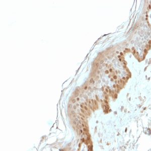
Mutations in the gene that encodes occludin have been associated with various human diseases, including inflammatory bowel disease, liver cirrhosis, and skin disorders. Additionally, changes in occludin expression and localization have been implicated in the development of cancer and neurological disorders.
Tight junction proteins are specialized proteins that play a crucial role in the formation and maintenance of tight junctions, which are intercellular structures that form a barrier to prevent the passage of molecules between cells. These proteins are found in the apical region of epithelial and endothelial cells and help to create a tight seal between adjacent cells.
Tight junction proteins can be classified into two major groups: transmembrane proteins and cytoplasmic plaque proteins. Transmembrane proteins, such as occludin and claudins, span the cell membrane and interact with each other to form the backbone of the tight junction. Cytoplasmic plaque proteins, such as zonula occludens (ZO) proteins, anchor the transmembrane proteins to the cytoskeleton and help to regulate their function.
Tight junction proteins are essential for maintaining the integrity of epithelial and endothelial barriers in various organs, including the gut, lungs, and blood-brain barrier. Dysfunction of these proteins has been implicated in a variety of diseases, such as inflammatory bowel disease, cancer, and neurological disorders.
Zonula Occludens-1 (ZO-1) protein is a tight junction (TJ) protein, which belongs to the membrane-associated guanylate kinase (MAGUK) family. It plays a crucial role in the formation and maintenance of tight junctions, which are complex structures that form a barrier between neighboring cells in epithelial and endothelial tissues.
Tight junctions are composed of several proteins, including transmembrane proteins and cytoplasmic plaque proteins. ZO-1 is one of the major cytoplasmic plaque proteins that interact with both transmembrane proteins (such as occludin and claudins) and other cytoskeletal proteins to form a network of protein interactions that maintain the integrity of tight junctions.
ZO-1 has multiple domains, including PDZ domains, SH3 domains, and a guanylate kinase-like domain, which allow it to interact with various binding partners. It is involved in regulating paracellular permeability, cell polarity, and signal transduction pathways that control cell proliferation, differentiation, and survival.
Mutations or dysfunction of ZO-1 protein have been implicated in several human diseases, including inflammatory bowel disease, cancer, and neurological disorders.
Intercellular junctions are specialized areas of contact between two or more adjacent cells in multicellular organisms. They play crucial roles in maintaining tissue structure and function by regulating the movement of ions, molecules, and even larger cellular structures from one cell to another. There are several types of intercellular junctions, including:
1. Tight Junctions (Zonulae Occludentes): These are the most apical structures in epithelial and endothelial cells, forming a virtually impermeable barrier to prevent the paracellular passage of solutes and water between the cells. They create a tight seal by connecting the transmembrane proteins of adjacent cells, such as occludin and claudins.
2. Adherens Junctions: These are located just below the tight junctions and help maintain cell-to-cell adhesion and tissue integrity. Adherens junctions consist of cadherin proteins that form homophilic interactions with cadherins on adjacent cells, as well as intracellular adaptor proteins like catenins, which connect to the actin cytoskeleton.
3. Desmosomes: These are another type of cell-to-cell adhesion structure, primarily found in tissues that experience mechanical stress, such as the skin and heart. Desmosomes consist of cadherin proteins (desmocadherins) that interact with each other and connect to intermediate filaments (keratin in epithelial cells) via plakoglobin and desmoplakin.
4. Gap Junctions: These are specialized channels that directly connect the cytoplasm of adjacent cells, allowing for the exchange of small molecules, ions, and second messengers. Gap junctions consist of connexin proteins that form hexameric structures called connexons in the plasma membrane of each cell. When two connexons align, they create a continuous pore or channel between the cells.
In summary, intercellular junctions are essential for maintaining tissue structure and function by regulating paracellular transport, cell-to-cell adhesion, and intercellular communication.
Claudins are a group of proteins that play a crucial role in the formation and function of tight junctions, which are specialized structures found in the cell membranes of epithelial and endothelial cells. Tight junctions serve as barriers to regulate the paracellular movement of ions, solutes, and water between cells, and claudins are one of the major components that contribute to their selective permeability.
There are over 20 different types of claudins identified in various tissues throughout the body, with each type having a unique structure and function. Claudins can form homotypic or heterotypic interactions with other claudin molecules, allowing for the formation of tight junction strands with varying pore sizes and charge selectivity. This diversity in claudin composition enables the regulation of paracellular transport across different tissues, such as the blood-brain barrier, intestinal epithelium, and renal tubules.
Mutations or dysregulation of claudins have been implicated in several diseases, including cancer, inflammatory bowel disease, and neurological disorders. For example, altered expression levels of specific claudins can contribute to the development of drug resistance in certain types of cancer cells, making them more difficult to treat. Additionally, changes in claudin composition or distribution can disrupt tight junction function, leading to increased permeability and the onset of various pathological conditions.
Claudin-1 is a protein that is a member of the claudin family, which are important components of tight junctions in cells. Tight junctions are specialized structures that help to regulate the paracellular permeability of liquids and solutes between cells, and play a crucial role in maintaining cell polarity and tissue integrity. Claudin-1 is primarily expressed in epithelial and endothelial cells, where it helps to form tight junctions and regulate the movement of molecules across these barriers. Mutations in the gene that encodes claudin-1 have been associated with various human diseases, including skin disorders and cancer.
Gap junctions are specialized intercellular connections that allow for the direct exchange of ions, small molecules, and electrical signals between adjacent cells. They are composed of arrays of channels called connexons, which penetrate the cell membranes of two neighboring cells and create a continuous pathway for the passage of materials from one cytoplasm to the other. Each connexon is formed by the assembly of six proteins called connexins, which are encoded by different genes and vary in their biophysical properties. Gap junctions play crucial roles in many physiological processes, including the coordination of electrical activity in excitable tissues, the regulation of cell growth and differentiation, and the maintenance of tissue homeostasis. Mutations or dysfunctions in gap junction channels have been implicated in various human diseases, such as cardiovascular disorders, neurological disorders, skin disorders, and cancer.
Adherens junctions are specialized types of cell-cell contacts that play a crucial role in maintaining the integrity and stability of tissues. They are composed of transmembrane cadherin proteins, which connect to the actin cytoskeleton inside the cell through intracellular adaptor proteins such as catenins.
The cadherins on opposing cells interact with each other to form adhesive bonds that help to anchor the cells together and regulate various cellular processes, including cell growth, differentiation, and migration. Adherens junctions are essential for many physiological processes, such as embryonic development, wound healing, and tissue homeostasis, and their dysfunction has been implicated in a variety of diseases, including cancer and degenerative disorders.
Zonula Occludens-2 (ZO-2) protein is a tight junction protein, which belongs to the membrane-associated guanylate kinase homologs (MAGUKs) family. It plays a crucial role in the formation and maintenance of tight junctions, which are complex structures that form a barrier between neighboring cells in epithelial and endothelial tissues.
ZO-2 protein is localized to the cytoplasmic face of the tight junction and interacts with various proteins, including transmembrane proteins such as occludin and claudins, as well as other cytoskeletal proteins. It contains several functional domains that enable it to interact with these proteins, including PDZ (PSD-95/Dlg/ZO-1) domains, SH3 (Src homology 3) domains, and a guanylate kinase-like domain.
ZO-2 protein has been implicated in various cellular processes, including the regulation of tight junction permeability, cell signaling, and gene expression. Mutations in ZO-2 have been associated with several human diseases, including inflammatory bowel disease, cancer, and neurological disorders.
Membrane proteins are a type of protein that are embedded in the lipid bilayer of biological membranes, such as the plasma membrane of cells or the inner membrane of mitochondria. These proteins play crucial roles in various cellular processes, including:
1. Cell-cell recognition and signaling
2. Transport of molecules across the membrane (selective permeability)
3. Enzymatic reactions at the membrane surface
4. Energy transduction and conversion
5. Mechanosensation and signal transduction
Membrane proteins can be classified into two main categories: integral membrane proteins, which are permanently associated with the lipid bilayer, and peripheral membrane proteins, which are temporarily or loosely attached to the membrane surface. Integral membrane proteins can further be divided into three subcategories based on their topology:
1. Transmembrane proteins, which span the entire width of the lipid bilayer with one or more alpha-helices or beta-barrels.
2. Lipid-anchored proteins, which are covalently attached to lipids in the membrane via a glycosylphosphatidylinositol (GPI) anchor or other lipid modifications.
3. Monotopic proteins, which are partially embedded in the membrane and have one or more domains exposed to either side of the bilayer.
Membrane proteins are essential for maintaining cellular homeostasis and are targets for various therapeutic interventions, including drug development and gene therapy. However, their structural complexity and hydrophobicity make them challenging to study using traditional biochemical methods, requiring specialized techniques such as X-ray crystallography, nuclear magnetic resonance (NMR) spectroscopy, and single-particle cryo-electron microscopy (cryo-EM).
Claudin-3 is a protein that belongs to the family of claudins, which are essential components of tight junctions in cells. Tight junctions are specialized structures that serve as barriers between adjacent cells, controlling the paracellular movement of ions, solutes, and water. Claudin-3 is primarily expressed in epithelial tissues, where it helps maintain cell polarity and regulate the permeability of the intercellular space. Mutations or abnormal expression of claudin-3 have been implicated in various pathological conditions, including cancer and inflammatory diseases.
Claudin-4 is a protein that belongs to the family of claudins, which are major components of tight junctions in cells. Tight junctions are specialized structures that serve as barriers between adjacent cells, controlling the paracellular movement of ions, solutes, and water. Claudin-4 is primarily expressed in epithelial tissues, where it plays a crucial role in maintaining cell-to-cell adhesion and regulating the permeability of tight junctions.
Claudin-4 has been identified as a potential biomarker for various cancers, including ovarian, pancreatic, and gastric cancers. Its overexpression is often associated with increased malignancy, invasiveness, and poor prognosis in these cancers. Additionally, claudin-4 is involved in the regulation of cell signaling pathways, inflammation, and immune responses, making it a target for therapeutic interventions in cancer and other diseases.
Claudin-5 is a protein that is a member of the claudin family, which are tight junction proteins. Tight junctions are specialized structures found in epithelial and endothelial cells that help to form a barrier between different cellular compartments. Claudin-5 is specifically expressed in endothelial cells and plays an important role in the formation of tight junctions in the blood-brain barrier, which helps to regulate the movement of molecules between the blood and the brain. Mutations in the gene that encodes claudin-5 have been associated with various neurological disorders.
Freeze fracturing is not a medical term itself, but it is a technique used in the field of electron microscopy, which is a type of imaging commonly used in scientific research and medical fields to visualize structures at a very small scale, such as cells and cellular components.
In freeze fracturing, a sample is rapidly frozen to preserve its structure and then fractured or split along a plane of weakness, often along the membrane of a cell. The freshly exposed surface is then shadowed with a thin layer of metal, such as platinum or gold, to create a replica of the surface. This replica can then be examined using an electron microscope to reveal details about the structure and organization of the sample at the molecular level.
Freeze fracturing is particularly useful for studying membrane structures, such as lipid bilayers and protein complexes, because it allows researchers to visualize these structures in their native state, without the need for staining or other chemical treatments that can alter or damage the samples.
Electric impedance is a measure of opposition to the flow of alternating current (AC) in an electrical circuit or component, caused by both resistance (ohmic) and reactance (capacitive and inductive). It is expressed as a complex number, with the real part representing resistance and the imaginary part representing reactance. The unit of electric impedance is the ohm (Ω).
In the context of medical devices, electric impedance may be used to measure various physiological parameters, such as tissue conductivity or fluid composition. For example, bioelectrical impedance analysis (BIA) uses electrical impedance to estimate body composition, including fat mass and lean muscle mass. Similarly, electrical impedance tomography (EIT) is a medical imaging technique that uses electric impedance to create images of internal organs and tissues.
The neuromuscular junction (NMJ) is the specialized synapse or chemical communication point, where the motor neuron's nerve terminal (presynaptic element) meets the muscle fiber's motor end plate (postsynaptic element). This junction plays a crucial role in controlling muscle contraction and relaxation.
At the NMJ, the neurotransmitter acetylcholine is released from the presynaptic nerve terminal into the synaptic cleft, following an action potential. Acetylcholine then binds to nicotinic acetylcholine receptors on the postsynaptic membrane of the muscle fiber, leading to the generation of an end-plate potential. If sufficient end-plate potentials are generated and summate, they will trigger an action potential in the muscle fiber, ultimately causing muscle contraction.
Dysfunction at the neuromuscular junction can result in various neuromuscular disorders, such as myasthenia gravis, where autoantibodies attack acetylcholine receptors, leading to muscle weakness and fatigue.
In the context of medicine and physiology, permeability refers to the ability of a tissue or membrane to allow the passage of fluids, solutes, or gases. It is often used to describe the property of the capillary walls, which control the exchange of substances between the blood and the surrounding tissues.
The permeability of a membrane can be influenced by various factors, including its molecular structure, charge, and the size of the molecules attempting to pass through it. A more permeable membrane allows for easier passage of substances, while a less permeable membrane restricts the movement of substances.
In some cases, changes in permeability can have significant consequences for health. For example, increased permeability of the blood-brain barrier (a specialized type of capillary that regulates the passage of substances into the brain) has been implicated in a number of neurological conditions, including multiple sclerosis, Alzheimer's disease, and traumatic brain injury.
Epithelial cells are types of cells that cover the outer surfaces of the body, line the inner surfaces of organs and glands, and form the lining of blood vessels and body cavities. They provide a protective barrier against the external environment, regulate the movement of materials between the internal and external environments, and are involved in the sense of touch, temperature, and pain. Epithelial cells can be squamous (flat and thin), cuboidal (square-shaped and of equal height), or columnar (tall and narrow) in shape and are classified based on their location and function.
Junctional Adhesion Molecules (JAMs) are a group of proteins that play crucial roles in cell-cell adhesion, formation and maintenance of tight junctions, and regulation of trafficking of various molecules across the epithelial and endothelial barriers. They belong to the immunoglobulin superfamily and are typically composed of a single transmembrane domain, an extracellular domain with variable numbers of immunoglobulin-like motifs, and a cytoplasmic tail that interacts with intracellular signaling molecules.
JAMs are involved in various cellular processes, such as leukocyte migration, angiogenesis, and maintenance of epithelial polarity. Dysregulation of JAMs has been implicated in several pathological conditions, including inflammatory bowel disease, cancer, and viral infections.
Some examples of Junctional Adhesion Molecules include JAM-A, JAM-B, JAM-C, JAM-4, and coxsackievirus and adenovirus receptor (CAR). These proteins are differentially expressed in various tissues and cells, and they have distinct functions and binding partners.
I'm sorry for any confusion, but "MARVEL Domain Containing 2 Protein" doesn't seem to correspond to a recognized medical term or protein name in human biology. The term "MARVEL" (MAL and related proteins for vesicle trafficking and membrane link) is a domain found in some proteins that are involved in various cellular processes such as membrane trafficking, cell adhesion, and signal transduction. However, without a specific protein name, it's difficult to provide a precise medical definition.
If you meant a specific protein containing the MARVEL domain, please provide the name so I can give a more detailed and accurate description.
Cell membrane permeability refers to the ability of various substances, such as molecules and ions, to pass through the cell membrane. The cell membrane, also known as the plasma membrane, is a thin, flexible barrier that surrounds all cells, controlling what enters and leaves the cell. Its primary function is to protect the cell's internal environment and maintain homeostasis.
The permeability of the cell membrane depends on its structure, which consists of a phospholipid bilayer interspersed with proteins. The hydrophilic (water-loving) heads of the phospholipids face outward, while the hydrophobic (water-fearing) tails face inward, creating a barrier that is generally impermeable to large, polar, or charged molecules.
However, specific proteins within the membrane, called channels and transporters, allow certain substances to cross the membrane. Channels are protein structures that span the membrane and provide a pore for ions or small uncharged molecules to pass through. Transporters, on the other hand, are proteins that bind to specific molecules and facilitate their movement across the membrane, often using energy in the form of ATP.
The permeability of the cell membrane can be influenced by various factors, such as temperature, pH, and the presence of certain chemicals or drugs. Changes in permeability can have significant consequences for the cell's function and survival, as they can disrupt ion balances, nutrient uptake, waste removal, and signal transduction.
Phosphoproteins are proteins that have been post-translationally modified by the addition of a phosphate group (-PO3H2) onto specific amino acid residues, most commonly serine, threonine, or tyrosine. This process is known as phosphorylation and is mediated by enzymes called kinases. Phosphoproteins play crucial roles in various cellular processes such as signal transduction, cell cycle regulation, metabolism, and gene expression. The addition or removal of a phosphate group can activate or inhibit the function of a protein, thereby serving as a switch to control its activity. Phosphoproteins can be detected and quantified using techniques such as Western blotting, mass spectrometry, and immunofluorescence.
Caco-2 cells are a type of human epithelial colorectal adenocarcinoma cell line that is commonly used in scientific research, particularly in the field of drug development and toxicology. These cells are capable of forming a monolayer with tight junctions, which makes them an excellent model for studying intestinal absorption, transport, and metabolism of drugs and other xenobiotic compounds.
Caco-2 cells express many of the transporters and enzymes that are found in the human small intestine, making them a valuable tool for predicting drug absorption and bioavailability in humans. They are also used to study the mechanisms of drug transport across the intestinal epithelium, including passive diffusion and active transport by various transporters.
In addition to their use in drug development, Caco-2 cells are also used to study the toxicological effects of various compounds on human intestinal cells. They can be used to investigate the mechanisms of toxicity, as well as to evaluate the potential for drugs and other compounds to induce intestinal damage or inflammation.
Overall, Caco-2 cells are a widely used and valuable tool in both drug development and toxicology research, providing important insights into the absorption, transport, metabolism, and toxicity of various compounds in the human body.
Zonula occludens proteins (ZO proteins) are a group of tight junction proteins that play a crucial role in the formation and maintenance of tight junctions, which are specialized intercellular junctions found in epithelial and endothelial cells. These proteins are located at the cytoplasmic surface of the plasma membrane and interact with both transmembrane proteins and the cytoskeleton to form a network that helps seal the paracellular space between adjacent cells.
There are three main ZO proteins: ZO-1, ZO-2, and ZO-3. They all share a similar structure, consisting of several protein-protein interaction domains, including PDZ (Postsynaptic density protein (PSD95), Drosophila disc large tumor suppressor (DlgA), and zonula occludens-1 (ZO-1)) domains, SH3 (Src homology 3) domains, and GUK (guanylate kinase-like) domains. These domains enable ZO proteins to interact with various binding partners and regulate tight junction assembly and function.
ZO proteins have multiple functions in the formation and maintenance of tight junctions:
1. Scaffolding: ZO proteins act as scaffolds, organizing and connecting transmembrane proteins like claudins and occludin to the cytoskeleton, thereby maintaining the integrity of tight junctions.
2. Signaling: ZO proteins participate in intracellular signaling pathways that regulate cell proliferation, differentiation, and survival. They can interact with various signaling molecules, such as kinases, phosphatases, and transcription factors, to modulate tight junction dynamics and cell behavior.
3. Barrier function: ZO proteins contribute to the barrier function of tight junctions by regulating paracellular permeability and preventing the uncontrolled passage of molecules between cells.
4. Fence function: Tight junctions also act as a fence, separating the apical and basolateral membrane domains of epithelial cells. ZO proteins help maintain this separation by interacting with cytoskeletal components and restricting the movement of lipids and proteins between these membrane domains.
In summary, ZO proteins are crucial for tight junction formation and function, serving as scaffolds, regulators of signaling pathways, and contributors to barrier and fence functions. Dysregulation of ZO proteins has been implicated in various diseases, including cancer, inflammatory bowel disease, and neurological disorders.
I believe there might be a misunderstanding in your question. "Dogs" is not a medical term or condition. It is the common name for a domesticated carnivore of the family Canidae, specifically the genus Canis, which includes wolves, foxes, and other extant and extinct species of mammals. Dogs are often kept as pets and companions, and they have been bred in a wide variety of forms and sizes for different purposes, such as hunting, herding, guarding, assisting police and military forces, and providing companionship and emotional support.
If you meant to ask about a specific medical condition or term related to dogs, please provide more context so I can give you an accurate answer.
Epithelium is the tissue that covers the outer surface of the body, lines the internal cavities and organs, and forms various glands. It is composed of one or more layers of tightly packed cells that have a uniform shape and size, and rest on a basement membrane. Epithelial tissues are avascular, meaning they do not contain blood vessels, and are supplied with nutrients by diffusion from the underlying connective tissue.
Epithelial cells perform a variety of functions, including protection, secretion, absorption, excretion, and sensation. They can be classified based on their shape and the number of cell layers they contain. The main types of epithelium are:
1. Squamous epithelium: composed of flat, scalelike cells that fit together like tiles on a roof. It forms the lining of blood vessels, air sacs in the lungs, and the outermost layer of the skin.
2. Cuboidal epithelium: composed of cube-shaped cells with equal height and width. It is found in glands, tubules, and ducts.
3. Columnar epithelium: composed of tall, rectangular cells that are taller than they are wide. It lines the respiratory, digestive, and reproductive tracts.
4. Pseudostratified epithelium: appears stratified or layered but is actually made up of a single layer of cells that vary in height. The nuclei of these cells appear at different levels, giving the tissue a stratified appearance. It lines the respiratory and reproductive tracts.
5. Transitional epithelium: composed of several layers of cells that can stretch and change shape to accommodate changes in volume. It is found in the urinary bladder and ureters.
Epithelial tissue provides a barrier between the internal and external environments, protecting the body from physical, chemical, and biological damage. It also plays a crucial role in maintaining homeostasis by regulating the exchange of substances between the body and its environment.
Cadherins are a type of cell adhesion molecule that play a crucial role in the development and maintenance of intercellular junctions. They are transmembrane proteins that mediate calcium-dependent homophilic binding between adjacent cells, meaning that they bind to identical cadherin molecules on neighboring cells.
There are several types of cadherins, including classical cadherins, desmosomal cadherins, and protocadherins, each with distinct functions and localization in tissues. Classical cadherins, also known as type I cadherins, are the most well-studied and are essential for the formation of adherens junctions, which help to maintain cell-to-cell contact and tissue architecture.
Desmosomal cadherins, on the other hand, are critical for the formation and maintenance of desmosomes, which are specialized intercellular junctions that provide mechanical strength and stability to tissues. Protocadherins are a diverse family of cadherin-related proteins that have been implicated in various developmental processes, including neuronal connectivity and tissue patterning.
Mutations in cadherin genes have been associated with several human diseases, including cancer, neurological disorders, and heart defects. Therefore, understanding the structure, function, and regulation of cadherins is essential for elucidating their roles in health and disease.
Cell polarity refers to the asymmetric distribution of membrane components, cytoskeleton, and organelles in a cell. This asymmetry is crucial for various cellular functions such as directed transport, cell division, and signal transduction. The plasma membrane of polarized cells exhibits distinct domains with unique protein and lipid compositions that define apical, basal, and lateral surfaces of the cell.
In epithelial cells, for example, the apical surface faces the lumen or external environment, while the basolateral surface interacts with other cells or the extracellular matrix. The establishment and maintenance of cell polarity are regulated by various factors including protein complexes, lipids, and small GTPases. Loss of cell polarity has been implicated in several diseases, including cancer and neurological disorders.
Connexins are a family of proteins that form the structural units of gap junctions, which are specialized channels that allow for the direct exchange of small molecules and ions between adjacent cells. These channels play crucial roles in maintaining tissue homeostasis, coordinating cellular activities, and enabling communication between cells. In humans, there are 21 different connexin genes that encode for these proteins, with each isoform having unique properties and distributions within the body. Mutations in connexin genes have been linked to a variety of human diseases, including hearing loss, skin disorders, and heart conditions.
The Blood-Brain Barrier (BBB) is a highly specialized, selective interface between the central nervous system (CNS) and the circulating blood. It is formed by unique endothelial cells that line the brain's capillaries, along with tight junctions, astrocytic foot processes, and pericytes, which together restrict the passage of substances from the bloodstream into the CNS. This barrier serves to protect the brain from harmful agents and maintain a stable environment for proper neural function. However, it also poses a challenge in delivering therapeutics to the CNS, as most large and hydrophilic molecules cannot cross the BBB.
The Blood-Testis Barrier (BTB) is a unique structural and functional feature of the seminiferous epithelium in the testes, which forms a tight junction between adjacent Sertoli cells in the semi-niferous tubules. This barrier selectively restricts the passage of molecules, including potentially harmful substances and immune cells, from the systemic circulation into the adluminal compartment of the seminiferous epithelium where spermatogenesis occurs. This helps to maintain a immunologically privileged microenvironment that is essential for the survival and maturation of developing sperm cells, preventing an immune response against them. The BTB also regulates the movement of molecules required for spermatogenesis, such as nutrients, hormones, and signaling molecules, from the basal compartment to the adluminal compartment.
Electron microscopy (EM) is a type of microscopy that uses a beam of electrons to create an image of the sample being examined, resulting in much higher magnification and resolution than light microscopy. There are several types of electron microscopy, including transmission electron microscopy (TEM), scanning electron microscopy (SEM), and reflection electron microscopy (REM).
In TEM, a beam of electrons is transmitted through a thin slice of the sample, and the electrons that pass through the sample are focused to form an image. This technique can provide detailed information about the internal structure of cells, viruses, and other biological specimens, as well as the composition and structure of materials at the atomic level.
In SEM, a beam of electrons is scanned across the surface of the sample, and the electrons that are scattered back from the surface are detected to create an image. This technique can provide information about the topography and composition of surfaces, as well as the structure of materials at the microscopic level.
REM is a variation of SEM in which the beam of electrons is reflected off the surface of the sample, rather than scattered back from it. This technique can provide information about the surface chemistry and composition of materials.
Electron microscopy has a wide range of applications in biology, medicine, and materials science, including the study of cellular structure and function, disease diagnosis, and the development of new materials and technologies.
Connexin 43 is a protein that forms gap junctions, which are specialized channels that allow for the direct communication and transport of small molecules between adjacent cells. Connexin 43 is widely expressed in many tissues, including the heart, brain, and various types of epithelial and connective tissues. In the heart, connexin 43 plays a crucial role in electrical conduction and coordination of contraction between cardiac muscle cells. Mutations in the gene that encodes connexin 43 have been associated with several human diseases, including certain types of cardiac arrhythmias and skin disorders.
Claudin-2 is a protein that is a member of the claudin family, which are tight junction proteins involved in forming tight junctions between cells. Tight junctions are complex structures that serve as barriers to prevent the passage of molecules through the spaces between cells, and they also play a role in cell signaling. Claudin-2 is specifically involved in the formation of paracellular channels, which allow for the selective transport of small ions across the tight junction. It has been found to be permeable to cations, including sodium and calcium ions.
Claudin-2 is expressed in a variety of tissues, including the intestine, kidney, and pancreas. In the intestine, claudin-2 is involved in the regulation of water and ion transport, and mutations in the gene that encodes claudin-2 have been associated with various gastrointestinal disorders, such as inflammatory bowel disease and congenital diarrhea. In the kidney, claudin-2 is expressed in the thick ascending limb of the loop of Henle and the distal convoluted tubule, where it helps to regulate sodium and water reabsorption.
In addition to its role in normal physiology, claudin-2 has also been implicated in various disease processes, including cancer. For example, increased expression of claudin-2 has been observed in some types of cancer, such as colon and pancreatic cancer, and it has been suggested that this may contribute to the development and progression of these cancers by promoting cell proliferation and migration.
The intestinal mucosa is the innermost layer of the intestines, which comes into direct contact with digested food and microbes. It is a specialized epithelial tissue that plays crucial roles in nutrient absorption, barrier function, and immune defense. The intestinal mucosa is composed of several cell types, including absorptive enterocytes, mucus-secreting goblet cells, hormone-producing enteroendocrine cells, and immune cells such as lymphocytes and macrophages.
The surface of the intestinal mucosa is covered by a single layer of epithelial cells, which are joined together by tight junctions to form a protective barrier against harmful substances and microorganisms. This barrier also allows for the selective absorption of nutrients into the bloodstream. The intestinal mucosa also contains numerous lymphoid follicles, known as Peyer's patches, which are involved in immune surveillance and defense against pathogens.
In addition to its role in absorption and immunity, the intestinal mucosa is also capable of producing hormones that regulate digestion and metabolism. Dysfunction of the intestinal mucosa can lead to various gastrointestinal disorders, such as inflammatory bowel disease, celiac disease, and food allergies.
A cell line is a culture of cells that are grown in a laboratory for use in research. These cells are usually taken from a single cell or group of cells, and they are able to divide and grow continuously in the lab. Cell lines can come from many different sources, including animals, plants, and humans. They are often used in scientific research to study cellular processes, disease mechanisms, and to test new drugs or treatments. Some common types of human cell lines include HeLa cells (which come from a cancer patient named Henrietta Lacks), HEK293 cells (which come from embryonic kidney cells), and HUVEC cells (which come from umbilical vein endothelial cells). It is important to note that cell lines are not the same as primary cells, which are cells that are taken directly from a living organism and have not been grown in the lab.
Cell communication, also known as cell signaling, is the process by which cells exchange and transmit signals between each other and their environment. This complex system allows cells to coordinate their functions and maintain tissue homeostasis. Cell communication can occur through various mechanisms including:
1. Autocrine signaling: When a cell releases a signal that binds to receptors on the same cell, leading to changes in its behavior or function.
2. Paracrine signaling: When a cell releases a signal that binds to receptors on nearby cells, influencing their behavior or function.
3. Endocrine signaling: When a cell releases a hormone into the bloodstream, which then travels to distant target cells and binds to specific receptors, triggering a response.
4. Synaptic signaling: In neurons, communication occurs through the release of neurotransmitters that cross the synapse and bind to receptors on the postsynaptic cell, transmitting electrical or chemical signals.
5. Contact-dependent signaling: When cells physically interact with each other, allowing for the direct exchange of signals and information.
Cell communication is essential for various physiological processes such as growth, development, differentiation, metabolism, immune response, and tissue repair. Dysregulation in cell communication can contribute to diseases, including cancer, diabetes, and neurological disorders.
Mannitol is a type of sugar alcohol (a sugar substitute) used primarily as a diuretic to reduce brain swelling caused by traumatic brain injury or other causes that induce increased pressure in the brain. It works by drawing water out of the body through the urine. It's also used before surgeries in the heart, lungs, and kidneys to prevent fluid buildup.
In addition, mannitol is used in medical laboratories as a medium for growing bacteria and other microorganisms, and in some types of chemical research. In the clinic, it is also used as an osmotic agent in eye drops to reduce the pressure inside the eye in conditions such as glaucoma.
It's important to note that mannitol should be used with caution in patients with heart or kidney disease, as well as those who are dehydrated, because it can lead to electrolyte imbalances and other complications.
The esophagogastric junction (EGJ) is the region of the gastrointestinal tract where the esophagus (the tube that carries food from the mouth to the stomach) meets the stomach. It serves as a physiological sphincter, which helps control the direction of flow and prevent reflux of gastric contents back into the esophagus. The EGJ is also known as the gastroesophageal junction or cardia.
The blood-retinal barrier (BRB) is a specialized physiological barrier in the eye that helps regulate the movement of molecules between the retina and the bloodstream. It is made up of tight junctions between the endothelial cells of retinal blood vessels and between the pigment epithelium cells of the retina, which restrict the paracellular diffusion of solutes.
The BRB plays a crucial role in maintaining the health and function of the retina by preventing harmful substances from entering the retina while allowing essential nutrients and oxygen to reach the retinal tissues. Disruption of the BRB has been implicated in various retinal diseases, including diabetic retinopathy, age-related macular degeneration, and retinal vein occlusion.
Cell adhesion molecules (CAMs) are a type of protein found on the surface of cells that mediate the attachment or adhesion of cells to either other cells or to the extracellular matrix (ECM), which is the network of proteins and carbohydrates that provides structural and biochemical support to surrounding cells.
CAMs play crucial roles in various biological processes, including tissue development, differentiation, repair, and maintenance of tissue architecture and function. They are also involved in cell signaling, migration, and regulation of the immune response.
There are several types of CAMs, classified based on their structure and function, such as immunoglobulin-like CAMs (IgCAMs), cadherins, integrins, and selectins. Dysregulation of CAMs has been implicated in various diseases, including cancer, inflammation, and neurological disorders.
Fluorescence microscopy is a type of microscopy that uses fluorescent dyes or proteins to highlight and visualize specific components within a sample. In this technique, the sample is illuminated with high-energy light, typically ultraviolet (UV) or blue light, which excites the fluorescent molecules causing them to emit lower-energy, longer-wavelength light, usually visible light in the form of various colors. This emitted light is then collected by the microscope and detected to produce an image.
Fluorescence microscopy has several advantages over traditional brightfield microscopy, including the ability to visualize specific structures or molecules within a complex sample, increased sensitivity, and the potential for quantitative analysis. It is widely used in various fields of biology and medicine, such as cell biology, neuroscience, and pathology, to study the structure, function, and interactions of cells and proteins.
There are several types of fluorescence microscopy techniques, including widefield fluorescence microscopy, confocal microscopy, two-photon microscopy, and total internal reflection fluorescence (TIRF) microscopy, each with its own strengths and limitations. These techniques can provide valuable insights into the behavior of cells and proteins in health and disease.
"Cells, cultured" is a medical term that refers to cells that have been removed from an organism and grown in controlled laboratory conditions outside of the body. This process is called cell culture and it allows scientists to study cells in a more controlled and accessible environment than they would have inside the body. Cultured cells can be derived from a variety of sources, including tissues, organs, or fluids from humans, animals, or cell lines that have been previously established in the laboratory.
Cell culture involves several steps, including isolation of the cells from the tissue, purification and characterization of the cells, and maintenance of the cells in appropriate growth conditions. The cells are typically grown in specialized media that contain nutrients, growth factors, and other components necessary for their survival and proliferation. Cultured cells can be used for a variety of purposes, including basic research, drug development and testing, and production of biological products such as vaccines and gene therapies.
It is important to note that cultured cells may behave differently than they do in the body, and results obtained from cell culture studies may not always translate directly to human physiology or disease. Therefore, it is essential to validate findings from cell culture experiments using additional models and ultimately in clinical trials involving human subjects.
Sertoli cells, also known as sustentacular cells or nurse cells, are specialized cells in the seminiferous tubules of the testis in mammals. They play a crucial role in supporting and nurturing the development of sperm cells (spermatogenesis). Sertoli cells create a microenvironment within the seminiferous tubules that facilitates the differentiation, maturation, and survival of germ cells.
These cells have several essential functions:
1. Blood-testis barrier formation: Sertoli cells form tight junctions with each other, creating a physical barrier called the blood-testis barrier, which separates the seminiferous tubules into basal and adluminal compartments. This barrier protects the developing sperm cells from the immune system and provides an isolated environment for their maturation.
2. Nutrition and support: Sertoli cells provide essential nutrients and growth factors to germ cells, ensuring their proper development and survival. They also engulf and digest residual bodies, which are byproducts of spermatid differentiation.
3. Phagocytosis: Sertoli cells have phagocytic properties, allowing them to remove debris and dead cells within the seminiferous tubules.
4. Hormone metabolism: Sertoli cells express receptors for various hormones, such as follicle-stimulating hormone (FSH), testosterone, and estradiol. They play a role in regulating hormonal signaling within the testis by metabolizing these hormones or producing inhibins, which modulate FSH secretion from the pituitary gland.
5. Regulation of spermatogenesis: Sertoli cells produce and secrete various proteins and growth factors that influence germ cell development and proliferation. They also control the release of mature sperm cells into the epididymis through a process called spermiation.
Confocal microscopy is a powerful imaging technique used in medical and biological research to obtain high-resolution, contrast-rich images of thick samples. This super-resolution technology provides detailed visualization of cellular structures and processes at various depths within a specimen.
In confocal microscopy, a laser beam focused through a pinhole illuminates a small spot within the sample. The emitted fluorescence or reflected light from this spot is then collected by a detector, passing through a second pinhole that ensures only light from the focal plane reaches the detector. This process eliminates out-of-focus light, resulting in sharp images with improved contrast compared to conventional widefield microscopy.
By scanning the laser beam across the sample in a raster pattern and collecting fluorescence at each point, confocal microscopy generates optical sections of the specimen. These sections can be combined to create three-dimensional reconstructions, allowing researchers to study cellular architecture and interactions within complex tissues.
Confocal microscopy has numerous applications in medical research, including studying protein localization, tracking intracellular dynamics, analyzing cell morphology, and investigating disease mechanisms at the cellular level. Additionally, it is widely used in clinical settings for diagnostic purposes, such as analyzing skin lesions or detecting pathogens in patient samples.
Desmosomes are specialized intercellular junctions that provide strong adhesion between adjacent epithelial cells and help maintain the structural integrity and stability of tissues. They are composed of several proteins, including desmoplakin, plakoglobin, and cadherins, which form complex structures that anchor intermediate filaments (such as keratin) to the cell membrane. This creates a network of interconnected cells that can withstand mechanical stresses. Desmosomes are particularly abundant in tissues subjected to high levels of tension, such as the skin and heart.
Holliday junction resolvases are a type of enzyme that are involved in the process of genetic recombination. They are named after Robin Holliday, who first proposed the existence of a structure called a Holliday junction during genetic recombination.
A Holliday junction is a four-way DNA structure that forms when two DNA molecules exchange genetic material during recombination. The junction is held together by hydrogen bonds between complementary base pairs, and it can move along the DNA molecules through a process called branch migration.
Holliday junction resolvases are responsible for cleaving the DNA strands at the Holliday junction, resolving the structure into two separate DNA molecules. They do this by introducing nicks in the phosphodiester backbone of the DNA strands on either side of the junction and then joining the broken ends together. This results in the exchange of genetic material between the two original DNA molecules.
There are several different types of Holliday junction resolvases, including the bacterial RuvC and RecU enzymes, as well as the eukaryotic Flap endonuclease 1 (FEN1) and XPF/ERCC1 complexes. These enzymes have different specificities for cleaving the DNA strands at the Holliday junction, but they all play important roles in ensuring that genetic recombination occurs accurately and efficiently.
Actin is a type of protein that forms part of the contractile apparatus in muscle cells, and is also found in various other cell types. It is a globular protein that polymerizes to form long filaments, which are important for many cellular processes such as cell division, cell motility, and the maintenance of cell shape. In muscle cells, actin filaments interact with another type of protein called myosin to enable muscle contraction. Actins can be further divided into different subtypes, including alpha-actin, beta-actin, and gamma-actin, which have distinct functions and expression patterns in the body.
Inulin is a soluble fiber that is not digestible by human enzymes. It is a fructan, a type of carbohydrate made up of chains of fructose molecules, and is found in various plants such as chicory root, Jerusalem artichokes, and onions.
Inulin has a number of potential health benefits, including promoting the growth of beneficial bacteria in the gut (prebiotic effect), slowing down the absorption of sugar to help regulate blood glucose levels, and increasing feelings of fullness to aid in weight management. It is often used as a functional food ingredient or dietary supplement for these purposes.
Inulin can also be used as a diagnostic tool in medical testing to measure kidney function, as it is excreted unchanged in the urine.
The Fluorescent Antibody Technique (FAT) is a type of immunofluorescence assay used in laboratory medicine and pathology for the detection and localization of specific antigens or antibodies in tissues, cells, or microorganisms. In this technique, a fluorescein-labeled antibody is used to selectively bind to the target antigen or antibody, forming an immune complex. When excited by light of a specific wavelength, the fluorescein label emits light at a longer wavelength, typically visualized as green fluorescence under a fluorescence microscope.
The FAT is widely used in diagnostic microbiology for the identification and characterization of various bacteria, viruses, fungi, and parasites. It has also been applied in the diagnosis of autoimmune diseases and certain cancers by detecting specific antibodies or antigens in patient samples. The main advantage of FAT is its high sensitivity and specificity, allowing for accurate detection and differentiation of various pathogens and disease markers. However, it requires specialized equipment and trained personnel to perform and interpret the results.
Lanthanum is not a medical term itself, but it is a chemical element with the symbol "La" and atomic number 57. It is a soft, ductile, silvery-white metal that belongs to the lanthanide series in the periodic table.
However, in medical contexts, lanthanum may be mentioned as a component of certain medications or medical devices. For example, lanthanum carbonate (trade name Fosrenol) is a medication used to treat hyperphosphatemia (elevated levels of phosphate in the blood) in patients with chronic kidney disease. Lanthanum carbonate works by binding to phosphate in the gastrointestinal tract, preventing its absorption into the bloodstream.
It is important to note that lanthanum compounds are not biologically active and do not have any specific medical effects on their own. Any medical uses of lanthanum are related to its physical or chemical properties, rather than its biological activity.
Alpha-catenin is a protein that plays a crucial role in cell adhesion and the maintenance of the cytoskeleton. It is a component of the cadherin-catenin complex, which is responsible for forming tight junctions between cells, known as adherens junctions. Alpha-catenin binds to beta-catenin, which in turn interacts with cadherins, transmembrane proteins that mediate cell-cell adhesion. This interaction helps to link the actin cytoskeleton to the cadherin-catenin complex, providing strength and stability to adherens junctions. Additionally, alpha-catenin has been implicated in various signaling pathways related to cell growth, differentiation, and migration.
Molecular sequence data refers to the specific arrangement of molecules, most commonly nucleotides in DNA or RNA, or amino acids in proteins, that make up a biological macromolecule. This data is generated through laboratory techniques such as sequencing, and provides information about the exact order of the constituent molecules. This data is crucial in various fields of biology, including genetics, evolution, and molecular biology, allowing for comparisons between different organisms, identification of genetic variations, and studies of gene function and regulation.
Nucleoside-phosphate kinase (NPK) is an enzyme that plays a crucial role in the synthesis and metabolism of nucleotides, which are the building blocks of DNA and RNA. NPK catalyzes the transfer of a phosphate group from a donor molecule, typically ATP, to a nucleoside or deoxynucleoside, forming a nucleoside monophosphate (NMP) or deoxynucleoside monophosphate (dNMP).
There are several isoforms of NPK found in different cellular compartments and tissues, each with distinct substrate specificities. These enzymes play essential roles in maintaining the balance of nucleotides required for various cellular processes, including DNA replication, repair, and transcription, as well as RNA synthesis and metabolism.
Abnormalities in NPK activity or expression have been implicated in several human diseases, such as cancer, viral infections, and neurological disorders. Therefore, understanding the function and regulation of NPK is crucial for developing novel therapeutic strategies to target these conditions.
Cell adhesion refers to the binding of cells to extracellular matrices or to other cells, a process that is fundamental to the development, function, and maintenance of multicellular organisms. Cell adhesion is mediated by various cell surface receptors, such as integrins, cadherins, and immunoglobulin-like cell adhesion molecules (Ig-CAMs), which interact with specific ligands in the extracellular environment. These interactions lead to the formation of specialized junctions, such as tight junctions, adherens junctions, and desmosomes, that help to maintain tissue architecture and regulate various cellular processes, including proliferation, differentiation, migration, and survival. Disruptions in cell adhesion can contribute to a variety of diseases, including cancer, inflammation, and degenerative disorders.
Madin-Darby Canine Kidney (MDCK) cells are a type of cell line that is derived from the kidney of a normal, healthy female cocker spaniel. They were first established in 1958 by researchers Madin and Darby. These cells are epithelial in origin and have the ability to form tight junctions, which makes them a popular choice for studying the transport of molecules across biological barriers.
MDCK cells are widely used in scientific research, particularly in the fields of cell biology, virology, and toxicology. They can be used to study various aspects of cell behavior, including cell adhesion, migration, differentiation, and polarization. Additionally, MDCK cells are susceptible to a variety of viruses, making them useful for studying viral replication and host-virus interactions.
In recent years, MDCK cells have also become an important tool in the development and production of vaccines. They can be used to produce large quantities of virus particles that can then be purified and used as vaccine antigens. Overall, Madin-Darby Canine Kidney cells are a valuable resource for researchers studying a wide range of biological phenomena.
Capillary permeability refers to the ability of substances to pass through the walls of capillaries, which are the smallest blood vessels in the body. These tiny vessels connect the arterioles and venules, allowing for the exchange of nutrients, waste products, and gases between the blood and the surrounding tissues.
The capillary wall is composed of a single layer of endothelial cells that are held together by tight junctions. The permeability of these walls varies depending on the size and charge of the molecules attempting to pass through. Small, uncharged molecules such as water, oxygen, and carbon dioxide can easily diffuse through the capillary wall, while larger or charged molecules such as proteins and large ions have more difficulty passing through.
Increased capillary permeability can occur in response to inflammation, infection, or injury, allowing larger molecules and immune cells to enter the surrounding tissues. This can lead to swelling (edema) and tissue damage if not controlled. Decreased capillary permeability, on the other hand, can lead to impaired nutrient exchange and tissue hypoxia.
Overall, the permeability of capillaries is a critical factor in maintaining the health and function of tissues throughout the body.
A cell membrane, also known as the plasma membrane, is a thin semi-permeable phospholipid bilayer that surrounds all cells in animals, plants, and microorganisms. It functions as a barrier to control the movement of substances in and out of the cell, allowing necessary molecules such as nutrients, oxygen, and signaling molecules to enter while keeping out harmful substances and waste products. The cell membrane is composed mainly of phospholipids, which have hydrophilic (water-loving) heads and hydrophobic (water-fearing) tails. This unique structure allows the membrane to be flexible and fluid, yet selectively permeable. Additionally, various proteins are embedded in the membrane that serve as channels, pumps, receptors, and enzymes, contributing to the cell's overall functionality and communication with its environment.
Cytoskeletal proteins are a type of structural proteins that form the cytoskeleton, which is the internal framework of cells. The cytoskeleton provides shape, support, and structure to the cell, and plays important roles in cell division, intracellular transport, and maintenance of cell shape and integrity.
There are three main types of cytoskeletal proteins: actin filaments, intermediate filaments, and microtubules. Actin filaments are thin, rod-like structures that are involved in muscle contraction, cell motility, and cell division. Intermediate filaments are thicker than actin filaments and provide structural support to the cell. Microtubules are hollow tubes that are involved in intracellular transport, cell division, and maintenance of cell shape.
Cytoskeletal proteins are composed of different subunits that polymerize to form filamentous structures. These proteins can be dynamically assembled and disassembled, allowing cells to change their shape and move. Mutations in cytoskeletal proteins have been linked to various human diseases, including cancer, neurological disorders, and muscular dystrophies.
Desmoplakins are important proteins that play a crucial role in the structural integrity and function of certain types of cell-to-cell junctions called desmosomes. Desmosomes are specialized structures that connect adjacent cells in tissues that undergo significant mechanical stress, such as the skin, heart, and gut.
Desmoplakins are large proteins that are composed of several domains, including a plakin domain, which interacts with other desmosomal components, and a spectrin-like repeat domain, which binds to intermediate filaments. By linking desmosomes to the intermediate filament network, desmoplakins help to provide mechanical strength and stability to tissues.
Mutations in the genes that encode desmoplakins have been associated with several human genetic disorders, including arrhythmogenic right ventricular cardiomyopathy (ARVC), a heart condition characterized by abnormal heart rhythms and structural changes in the heart muscle, and epidermolysis bullosa simplex (EBS), a skin disorder characterized by blistering and fragility of the skin.
Immunohistochemistry (IHC) is a technique used in pathology and laboratory medicine to identify specific proteins or antigens in tissue sections. It combines the principles of immunology and histology to detect the presence and location of these target molecules within cells and tissues. This technique utilizes antibodies that are specific to the protein or antigen of interest, which are then tagged with a detection system such as a chromogen or fluorophore. The stained tissue sections can be examined under a microscope, allowing for the visualization and analysis of the distribution and expression patterns of the target molecule in the context of the tissue architecture. Immunohistochemistry is widely used in diagnostic pathology to help identify various diseases, including cancer, infectious diseases, and immune-mediated disorders.
Myosin-Light-Chain Kinase (MLCK) is an enzyme that plays a crucial role in muscle contraction. It phosphorylates the regulatory light chains of myosin, a protein involved in muscle contraction, leading to the activation of myosin and the initiation of the contractile process. MLCK is activated by calcium ions and calmodulin, and its activity is essential for various cellular processes, including cytokinesis, cell motility, and maintenance of cell shape. In addition to its role in muscle contraction, MLCK has been implicated in several pathological conditions, such as hypertension, atherosclerosis, and cancer.
The cytoskeleton is a complex network of various protein filaments that provides structural support, shape, and stability to the cell. It plays a crucial role in maintaining cellular integrity, intracellular organization, and enabling cell movement. The cytoskeleton is composed of three major types of protein fibers: microfilaments (actin filaments), intermediate filaments, and microtubules. These filaments work together to provide mechanical support, participate in cell division, intracellular transport, and help maintain the cell's architecture. The dynamic nature of the cytoskeleton allows cells to adapt to changing environmental conditions and respond to various stimuli.
Biological models, also known as physiological models or organismal models, are simplified representations of biological systems, processes, or mechanisms that are used to understand and explain the underlying principles and relationships. These models can be theoretical (conceptual or mathematical) or physical (such as anatomical models, cell cultures, or animal models). They are widely used in biomedical research to study various phenomena, including disease pathophysiology, drug action, and therapeutic interventions.
Examples of biological models include:
1. Mathematical models: These use mathematical equations and formulas to describe complex biological systems or processes, such as population dynamics, metabolic pathways, or gene regulation networks. They can help predict the behavior of these systems under different conditions and test hypotheses about their underlying mechanisms.
2. Cell cultures: These are collections of cells grown in a controlled environment, typically in a laboratory dish or flask. They can be used to study cellular processes, such as signal transduction, gene expression, or metabolism, and to test the effects of drugs or other treatments on these processes.
3. Animal models: These are living organisms, usually vertebrates like mice, rats, or non-human primates, that are used to study various aspects of human biology and disease. They can provide valuable insights into the pathophysiology of diseases, the mechanisms of drug action, and the safety and efficacy of new therapies.
4. Anatomical models: These are physical representations of biological structures or systems, such as plastic models of organs or tissues, that can be used for educational purposes or to plan surgical procedures. They can also serve as a basis for developing more sophisticated models, such as computer simulations or 3D-printed replicas.
Overall, biological models play a crucial role in advancing our understanding of biology and medicine, helping to identify new targets for therapeutic intervention, develop novel drugs and treatments, and improve human health.
Enterocytes are the absorptive cells that line the villi of the small intestine. They are a type of epithelial cell and play a crucial role in the absorption of nutrients from food into the bloodstream. Enterocytes have finger-like projections called microvilli on their apical surface, which increases their surface area and enhances their ability to absorb nutrients. They also contain enzymes that help digest and break down carbohydrates, proteins, and fats into smaller molecules that can be absorbed. Additionally, enterocytes play a role in the absorption of ions, water, and vitamins.
Beta-catenin is a protein that plays a crucial role in gene transcription and cell-cell adhesion. It is a key component of the Wnt signaling pathway, which regulates various processes such as cell proliferation, differentiation, and migration during embryonic development and tissue homeostasis in adults.
In the absence of Wnt signals, beta-catenin forms a complex with other proteins, including adenomatous polyposis coli (APC) and axin, which targets it for degradation by the proteasome. When Wnt ligands bind to their receptors, this complex is disrupted, allowing beta-catenin to accumulate in the cytoplasm and translocate to the nucleus. In the nucleus, beta-catenin interacts with T cell factor/lymphoid enhancer-binding factor (TCF/LEF) transcription factors to activate the transcription of target genes involved in cell fate determination, survival, and proliferation.
Mutations in the genes encoding components of the Wnt signaling pathway, including beta-catenin, have been implicated in various human diseases, such as cancer, developmental disorders, and degenerative conditions.
Endothelial cells are the type of cells that line the inner surface of blood vessels, lymphatic vessels, and heart chambers. They play a crucial role in maintaining vascular homeostasis by controlling vasomotor tone, coagulation, platelet activation, and inflammation. Endothelial cells also regulate the transport of molecules between the blood and surrounding tissues, and contribute to the maintenance of the structural integrity of the vasculature. They are flat, elongated cells with a unique morphology that allows them to form a continuous, nonthrombogenic lining inside the vessels. Endothelial cells can be isolated from various tissues and cultured in vitro for research purposes.
The seminiferous epithelium is a specialized type of epithelial tissue that lines the seminiferous tubules within the testes. It is composed of various cell types, including germ cells in different stages of development (spermatogonia, primary and secondary spermatocytes, spermatids) and supportive cells called Sertoli cells.
The primary function of the seminiferous epithelium is to support sperm production (spermatogenesis). The Sertoli cells provide structural support and nourishment to the developing germ cells, helping them to differentiate into mature spermatozoa (sperm). This process involves a series of complex cellular events, including mitosis, meiosis, and spermiogenesis.
In addition to its role in sperm production, the seminiferous epithelium also plays a crucial part in maintaining the blood-testis barrier, which separates the testicular environment from the systemic circulation. This barrier helps protect developing germ cells from potential immune attacks and maintains an optimal microenvironment for spermatogenesis.
Transmission electron microscopy (TEM) is a type of microscopy in which an electron beam is transmitted through a ultra-thin specimen, interacting with it as it passes through. An image is formed from the interaction of the electrons with the specimen; the image is then magnified and visualized on a fluorescent screen or recorded on an electronic detector (or photographic film in older models).
TEM can provide high-resolution, high-magnification images that can reveal the internal structure of specimens including cells, viruses, and even molecules. It is widely used in biological and materials science research to investigate the ultrastructure of cells, tissues and materials. In medicine, TEM is used for diagnostic purposes in fields such as virology and bacteriology.
It's important to note that preparing a sample for TEM is a complex process, requiring specialized techniques to create thin (50-100 nm) specimens. These include cutting ultrathin sections of embedded samples using an ultramicrotome, staining with heavy metal salts, and positive staining or negative staining methods.
The intestines, also known as the bowel, are a part of the digestive system that extends from the stomach to the anus. They are responsible for the further breakdown and absorption of nutrients from food, as well as the elimination of waste products. The intestines can be divided into two main sections: the small intestine and the large intestine.
The small intestine is a long, coiled tube that measures about 20 feet in length and is lined with tiny finger-like projections called villi, which increase its surface area and enhance nutrient absorption. The small intestine is where most of the digestion and absorption of nutrients takes place.
The large intestine, also known as the colon, is a wider tube that measures about 5 feet in length and is responsible for absorbing water and electrolytes from digested food, forming stool, and eliminating waste products from the body. The large intestine includes several regions, including the cecum, colon, rectum, and anus.
Together, the intestines play a critical role in maintaining overall health and well-being by ensuring that the body receives the nutrients it needs to function properly.
Immunoelectron microscopy (IEM) is a specialized type of electron microscopy that combines the principles of immunochemistry and electron microscopy to detect and localize specific antigens within cells or tissues at the ultrastructural level. This technique allows for the visualization and identification of specific proteins, viruses, or other antigenic structures with a high degree of resolution and specificity.
In IEM, samples are first fixed, embedded, and sectioned to prepare them for electron microscopy. The sections are then treated with specific antibodies that have been labeled with electron-dense markers, such as gold particles or ferritin. These labeled antibodies bind to the target antigens in the sample, allowing for their visualization under an electron microscope.
There are several different methods of IEM, including pre-embedding and post-embedding techniques. Pre-embedding involves labeling the antigens before embedding the sample in resin, while post-embedding involves labeling the antigens after embedding. Post-embedding techniques are generally more commonly used because they allow for better preservation of ultrastructure and higher resolution.
IEM is a valuable tool in many areas of research, including virology, bacteriology, immunology, and cell biology. It can be used to study the structure and function of viruses, bacteria, and other microorganisms, as well as the distribution and localization of specific proteins and antigens within cells and tissues.
Protein transport, in the context of cellular biology, refers to the process by which proteins are actively moved from one location to another within or between cells. This is a crucial mechanism for maintaining proper cell function and regulation.
Intracellular protein transport involves the movement of proteins within a single cell. Proteins can be transported across membranes (such as the nuclear envelope, endoplasmic reticulum, Golgi apparatus, or plasma membrane) via specialized transport systems like vesicles and transport channels.
Intercellular protein transport refers to the movement of proteins from one cell to another, often facilitated by exocytosis (release of proteins in vesicles) and endocytosis (uptake of extracellular substances via membrane-bound vesicles). This is essential for communication between cells, immune response, and other physiological processes.
It's important to note that any disruption in protein transport can lead to various diseases, including neurological disorders, cancer, and metabolic conditions.
A kidney, in medical terms, is one of two bean-shaped organs located in the lower back region of the body. They are essential for maintaining homeostasis within the body by performing several crucial functions such as:
1. Regulation of water and electrolyte balance: Kidneys help regulate the amount of water and various electrolytes like sodium, potassium, and calcium in the bloodstream to maintain a stable internal environment.
2. Excretion of waste products: They filter waste products from the blood, including urea (a byproduct of protein metabolism), creatinine (a breakdown product of muscle tissue), and other harmful substances that result from normal cellular functions or external sources like medications and toxins.
3. Endocrine function: Kidneys produce several hormones with important roles in the body, such as erythropoietin (stimulates red blood cell production), renin (regulates blood pressure), and calcitriol (activated form of vitamin D that helps regulate calcium homeostasis).
4. pH balance regulation: Kidneys maintain the proper acid-base balance in the body by excreting either hydrogen ions or bicarbonate ions, depending on whether the blood is too acidic or too alkaline.
5. Blood pressure control: The kidneys play a significant role in regulating blood pressure through the renin-angiotensin-aldosterone system (RAAS), which constricts blood vessels and promotes sodium and water retention to increase blood volume and, consequently, blood pressure.
Anatomically, each kidney is approximately 10-12 cm long, 5-7 cm wide, and 3 cm thick, with a weight of about 120-170 grams. They are surrounded by a protective layer of fat and connected to the urinary system through the renal pelvis, ureters, bladder, and urethra.
A base sequence in the context of molecular biology refers to the specific order of nucleotides in a DNA or RNA molecule. In DNA, these nucleotides are adenine (A), guanine (G), cytosine (C), and thymine (T). In RNA, uracil (U) takes the place of thymine. The base sequence contains genetic information that is transcribed into RNA and ultimately translated into proteins. It is the exact order of these bases that determines the genetic code and thus the function of the DNA or RNA molecule.
Signal transduction is the process by which a cell converts an extracellular signal, such as a hormone or neurotransmitter, into an intracellular response. This involves a series of molecular events that transmit the signal from the cell surface to the interior of the cell, ultimately resulting in changes in gene expression, protein activity, or metabolism.
The process typically begins with the binding of the extracellular signal to a receptor located on the cell membrane. This binding event activates the receptor, which then triggers a cascade of intracellular signaling molecules, such as second messengers, protein kinases, and ion channels. These molecules amplify and propagate the signal, ultimately leading to the activation or inhibition of specific cellular responses.
Signal transduction pathways are highly regulated and can be modulated by various factors, including other signaling molecules, post-translational modifications, and feedback mechanisms. Dysregulation of these pathways has been implicated in a variety of diseases, including cancer, diabetes, and neurological disorders.
Western blotting is a laboratory technique used in molecular biology to detect and quantify specific proteins in a mixture of many different proteins. This technique is commonly used to confirm the expression of a protein of interest, determine its size, and investigate its post-translational modifications. The name "Western" blotting distinguishes this technique from Southern blotting (for DNA) and Northern blotting (for RNA).
The Western blotting procedure involves several steps:
1. Protein extraction: The sample containing the proteins of interest is first extracted, often by breaking open cells or tissues and using a buffer to extract the proteins.
2. Separation of proteins by electrophoresis: The extracted proteins are then separated based on their size by loading them onto a polyacrylamide gel and running an electric current through the gel (a process called sodium dodecyl sulfate-polyacrylamide gel electrophoresis or SDS-PAGE). This separates the proteins according to their molecular weight, with smaller proteins migrating faster than larger ones.
3. Transfer of proteins to a membrane: After separation, the proteins are transferred from the gel onto a nitrocellulose or polyvinylidene fluoride (PVDF) membrane using an electric current in a process called blotting. This creates a replica of the protein pattern on the gel but now immobilized on the membrane for further analysis.
4. Blocking: The membrane is then blocked with a blocking agent, such as non-fat dry milk or bovine serum albumin (BSA), to prevent non-specific binding of antibodies in subsequent steps.
5. Primary antibody incubation: A primary antibody that specifically recognizes the protein of interest is added and allowed to bind to its target protein on the membrane. This step may be performed at room temperature or 4°C overnight, depending on the antibody's properties.
6. Washing: The membrane is washed with a buffer to remove unbound primary antibodies.
7. Secondary antibody incubation: A secondary antibody that recognizes the primary antibody (often coupled to an enzyme or fluorophore) is added and allowed to bind to the primary antibody. This step may involve using a horseradish peroxidase (HRP)-conjugated or alkaline phosphatase (AP)-conjugated secondary antibody, depending on the detection method used later.
8. Washing: The membrane is washed again to remove unbound secondary antibodies.
9. Detection: A detection reagent is added to visualize the protein of interest by detecting the signal generated from the enzyme-conjugated or fluorophore-conjugated secondary antibody. This can be done using chemiluminescent, colorimetric, or fluorescent methods.
10. Analysis: The resulting image is analyzed to determine the presence and quantity of the protein of interest in the sample.
Western blotting is a powerful technique for identifying and quantifying specific proteins within complex mixtures. It can be used to study protein expression, post-translational modifications, protein-protein interactions, and more. However, it requires careful optimization and validation to ensure accurate and reproducible results.
Phosphorylation is the process of adding a phosphate group (a molecule consisting of one phosphorus atom and four oxygen atoms) to a protein or other organic molecule, which is usually done by enzymes called kinases. This post-translational modification can change the function, localization, or activity of the target molecule, playing a crucial role in various cellular processes such as signal transduction, metabolism, and regulation of gene expression. Phosphorylation is reversible, and the removal of the phosphate group is facilitated by enzymes called phosphatases.
An amino acid sequence is the specific order of amino acids in a protein or peptide molecule, formed by the linking of the amino group (-NH2) of one amino acid to the carboxyl group (-COOH) of another amino acid through a peptide bond. The sequence is determined by the genetic code and is unique to each type of protein or peptide. It plays a crucial role in determining the three-dimensional structure and function of proteins.
"Cruciform DNA" is a term used to describe a specific conformation or structure that a double-stranded DNA molecule can adopt. It is so-called because the structure resembles the shape of a cross or crucifix.
This conformation arises when two inverted repeats of DNA sequence are located close to each other on the same DNA molecule, such that they can pair up and form a stable secondary structure. This results in the formation of a hairpin loop at each end of the inverted repeat sequences, with the loops pointing towards each other and the intervening sequences forming two arms that cross in the middle.
Cruciform structures are important in various biological processes, including DNA replication, repair, and recombination. However, they can also pose challenges to these processes, as the crossing of the DNA strands can create topological constraints that must be resolved before replication or transcription can proceed.
It's worth noting that cruciform structures are not stable in solution and are usually only observed under specific conditions, such as when the DNA is supercoiled or when negative supercoiling is introduced through the action of enzymes like topoisomerases.
Catenins are a type of protein that play a crucial role in cell adhesion and signal transduction. They are named for their ability to link together (or "catenate") proteins called cadherins, which are important for the formation of tight junctions between cells. Catenins help to anchor cadherins to the cytoskeleton, providing structural support and stability to tissues.
There are several different types of catenins, including alpha-catenin, beta-catenin, gamma-catenin (also called plakoglobin), and delta-catenin. Alpha-catenin links cadherins to the actin cytoskeleton, while beta-catenin and gamma-catenin can also interact with transcription factors in the nucleus to regulate gene expression.
Mutations in catenin genes have been associated with various human diseases, including cancer. For example, abnormal activation of the Wnt signaling pathway, which involves beta-catenin, has been implicated in several types of cancer. Additionally, mutations in alpha-E-catenin, a type of alpha-catenin found in epithelial cells, have been linked to colorectal cancer.
Osmium compounds refer to chemical substances that contain the element osmium (Os) in its various oxidation states, combined with other elements or radicals. Osmium is a transition metal and a member of the platinum group, known for its hardness, high melting point, and blue-black color.
Osmium compounds can be found in several forms, including halides (osmium tetroxide, osmium trichloride, osmium pentachloride), oxides (osmium dioxide, osmium trioxide), and organometallic complexes. These compounds have been used in various applications, such as catalysts, staining agents in microscopy, and alloys. However, many osmium compounds are highly toxic and should be handled with care.
Dextrans are a type of complex glucose polymers that are formed by the action of certain bacteria on sucrose. They are branched polysaccharides consisting of linear chains of α-1,6 linked D-glucopyranosyl units with occasional α-1,3 branches.
Dextrans have a wide range of applications in medicine and industry. In medicine, dextrans are used as plasma substitutes, volume expanders, and anticoagulants. They are also used as carriers for drugs and diagnostic agents, and in the manufacture of immunoadsorbents for the removal of toxins and pathogens from blood.
Dextrans can be derived from various bacterial sources, but the most common commercial source is Leuconostoc mesenteroides B-512(F) or L. dextranicum. The molecular weight of dextrans can vary widely, ranging from a few thousand to several million Daltons, depending on the method of preparation and purification.
Dextrans are generally biocompatible and non-toxic, but they can cause allergic reactions in some individuals. Therefore, their use as medical products requires careful monitoring and testing for safety and efficacy.
Protein binding, in the context of medical and biological sciences, refers to the interaction between a protein and another molecule (known as the ligand) that results in a stable complex. This process is often reversible and can be influenced by various factors such as pH, temperature, and concentration of the involved molecules.
In clinical chemistry, protein binding is particularly important when it comes to drugs, as many of them bind to proteins (especially albumin) in the bloodstream. The degree of protein binding can affect a drug's distribution, metabolism, and excretion, which in turn influence its therapeutic effectiveness and potential side effects.
Protein-bound drugs may be less available for interaction with their target tissues, as only the unbound or "free" fraction of the drug is active. Therefore, understanding protein binding can help optimize dosing regimens and minimize adverse reactions.
Messenger RNA (mRNA) is a type of RNA (ribonucleic acid) that carries genetic information copied from DNA in the form of a series of three-base code "words," each of which specifies a particular amino acid. This information is used by the cell's machinery to construct proteins, a process known as translation. After being transcribed from DNA, mRNA travels out of the nucleus to the ribosomes in the cytoplasm where protein synthesis occurs. Once the protein has been synthesized, the mRNA may be degraded and recycled. Post-transcriptional modifications can also occur to mRNA, such as alternative splicing and addition of a 5' cap and a poly(A) tail, which can affect its stability, localization, and translation efficiency.
Immunoblotting, also known as western blotting, is a laboratory technique used in molecular biology and immunogenetics to detect and quantify specific proteins in a complex mixture. This technique combines the electrophoretic separation of proteins by gel electrophoresis with their detection using antibodies that recognize specific epitopes (protein fragments) on the target protein.
The process involves several steps: first, the protein sample is separated based on size through sodium dodecyl sulfate-polyacrylamide gel electrophoresis (SDS-PAGE). Next, the separated proteins are transferred onto a nitrocellulose or polyvinylidene fluoride (PVDF) membrane using an electric field. The membrane is then blocked with a blocking agent to prevent non-specific binding of antibodies.
After blocking, the membrane is incubated with a primary antibody that specifically recognizes the target protein. Following this, the membrane is washed to remove unbound primary antibodies and then incubated with a secondary antibody conjugated to an enzyme such as horseradish peroxidase (HRP) or alkaline phosphatase (AP). The enzyme catalyzes a colorimetric or chemiluminescent reaction that allows for the detection of the target protein.
Immunoblotting is widely used in research and clinical settings to study protein expression, post-translational modifications, protein-protein interactions, and disease biomarkers. It provides high specificity and sensitivity, making it a valuable tool for identifying and quantifying proteins in various biological samples.
Calcium is an essential mineral that is vital for various physiological processes in the human body. The medical definition of calcium is as follows:
Calcium (Ca2+) is a crucial cation and the most abundant mineral in the human body, with approximately 99% of it found in bones and teeth. It plays a vital role in maintaining structural integrity, nerve impulse transmission, muscle contraction, hormonal secretion, blood coagulation, and enzyme activation.
Calcium homeostasis is tightly regulated through the interplay of several hormones, including parathyroid hormone (PTH), calcitonin, and vitamin D. Dietary calcium intake, absorption, and excretion are also critical factors in maintaining optimal calcium levels in the body.
Hypocalcemia refers to low serum calcium levels, while hypercalcemia indicates high serum calcium levels. Both conditions can have detrimental effects on various organ systems and require medical intervention to correct.
Guanylate kinase is an enzyme that plays a crucial role in the synthesis of guanosine triphosphate (GTP) in cells. GTP is a vital energy currency and a key player in various cellular processes, such as protein synthesis, signal transduction, and gene regulation.
The primary function of guanylate kinase is to catalyze the transfer of a phosphate group from adenosine triphosphate (ATP) to guanosine monophosphate (GMP), resulting in the formation of GTP and adenosine diphosphate (ADP). The reaction can be represented as follows:
GMP + ATP → GTP + ADP
There are two main types of guanylate kinases, based on their structure and function:
1. **Classical Guanylate Kinase:** This type of guanylate kinase is found in various organisms, including bacteria, archaea, and eukaryotes. They typically contain around 180-200 amino acids and share a conserved catalytic domain. In humans, there are two classical guanylate kinases (GK1 and GK2) that play essential roles in DNA damage response and neuronal development.
2. **Ubiquitous Guanylate Kinase-like Proteins:** These proteins share structural similarities with the catalytic domain of classical guanylate kinases but lack enzymatic activity. They are involved in various cellular processes, such as transcription regulation and RNA processing.
Guanylate kinase deficiency has been linked to neurological disorders, developmental delays, and seizures in humans. Additionally, inhibiting guanylate kinase activity can be a potential therapeutic strategy for treating certain types of cancer, as it may interfere with the energy production required for uncontrolled cell growth and proliferation.
The Fluorescent Antibody Technique (FAT), Indirect is a type of immunofluorescence assay used to detect the presence of specific antigens in a sample. In this method, the sample is first incubated with a primary antibody that binds to the target antigen. After washing to remove unbound primary antibodies, a secondary fluorescently labeled antibody is added, which recognizes and binds to the primary antibody. This indirect labeling approach allows for amplification of the signal, making it more sensitive than direct methods. The sample is then examined under a fluorescence microscope to visualize the location and amount of antigen based on the emitted light from the fluorescent secondary antibody. It's commonly used in diagnostic laboratories for detection of various bacteria, viruses, and other antigens in clinical specimens.
In the field of medicine, "time factors" refer to the duration of symptoms or time elapsed since the onset of a medical condition, which can have significant implications for diagnosis and treatment. Understanding time factors is crucial in determining the progression of a disease, evaluating the effectiveness of treatments, and making critical decisions regarding patient care.
For example, in stroke management, "time is brain," meaning that rapid intervention within a specific time frame (usually within 4.5 hours) is essential to administering tissue plasminogen activator (tPA), a clot-busting drug that can minimize brain damage and improve patient outcomes. Similarly, in trauma care, the "golden hour" concept emphasizes the importance of providing definitive care within the first 60 minutes after injury to increase survival rates and reduce morbidity.
Time factors also play a role in monitoring the progression of chronic conditions like diabetes or heart disease, where regular follow-ups and assessments help determine appropriate treatment adjustments and prevent complications. In infectious diseases, time factors are crucial for initiating antibiotic therapy and identifying potential outbreaks to control their spread.
Overall, "time factors" encompass the significance of recognizing and acting promptly in various medical scenarios to optimize patient outcomes and provide effective care.
Transfection is a term used in molecular biology that refers to the process of deliberately introducing foreign genetic material (DNA, RNA or artificial gene constructs) into cells. This is typically done using chemical or physical methods, such as lipofection or electroporation. Transfection is widely used in research and medical settings for various purposes, including studying gene function, producing proteins, developing gene therapies, and creating genetically modified organisms. It's important to note that transfection is different from transduction, which is the process of introducing genetic material into cells using viruses as vectors.
PDZ domains are protein interaction modules, which are named after the first letters of three proteins in which they were originally discovered: PSD-95, DLG, and ZO-1. These domains are typically located at the C-terminal region of a protein and have a length of approximately 80-90 amino acids. They play a crucial role in organizing and assembling signaling complexes by binding to specific motifs found on other proteins, such as C-terminal PDZ-binding motifs or internal PDZ-binding sites. This ability to interact with multiple partners enables PDZ domains to function as molecular scaffolds that help regulate various cellular processes, including signal transduction, cell adhesion, and trafficking of proteins to specific subcellular locations.
A mutation is a permanent change in the DNA sequence of an organism's genome. Mutations can occur spontaneously or be caused by environmental factors such as exposure to radiation, chemicals, or viruses. They may have various effects on the organism, ranging from benign to harmful, depending on where they occur and whether they alter the function of essential proteins. In some cases, mutations can increase an individual's susceptibility to certain diseases or disorders, while in others, they may confer a survival advantage. Mutations are the driving force behind evolution, as they introduce new genetic variability into populations, which can then be acted upon by natural selection.
'Gene expression regulation' refers to the processes that control whether, when, and where a particular gene is expressed, meaning the production of a specific protein or functional RNA encoded by that gene. This complex mechanism can be influenced by various factors such as transcription factors, chromatin remodeling, DNA methylation, non-coding RNAs, and post-transcriptional modifications, among others. Proper regulation of gene expression is crucial for normal cellular function, development, and maintaining homeostasis in living organisms. Dysregulation of gene expression can lead to various diseases, including cancer and genetic disorders.
I am not aware of a widely recognized or established medical term called "Blood-Air Barrier." It is possible that you may be referring to a concept or phenomenon that goes by a different name, or it could be a term that is specific to certain context or field within medicine.
In general, the terms "blood" and "air" refer to two distinct and separate compartments in the body, and there are various physiological barriers that prevent them from mixing with each other under normal circumstances. For example, the alveolar-capillary membrane in the lungs serves as a barrier that allows for the exchange of oxygen and carbon dioxide between the air in the alveoli and the blood in the capillaries, while preventing the two from mixing together.
If you could provide more context or clarify what you mean by "Blood-Air Barrier," I may be able to provide a more specific answer.
Fluorescein-5-isothiocyanate (FITC) is not a medical term per se, but a chemical compound commonly used in biomedical research and clinical diagnostics. Therefore, I will provide a general definition of this term:
Fluorescein-5-isothiocyanate (FITC) is a fluorescent dye with an absorption maximum at approximately 492-495 nm and an emission maximum at around 518-525 nm. It is widely used as a labeling reagent for various biological molecules, such as antibodies, proteins, and nucleic acids, to study their structure, function, and interactions in techniques like flow cytometry, immunofluorescence microscopy, and western blotting. The isothiocyanate group (-N=C=S) in the FITC molecule reacts with primary amines (-NH2) present in biological molecules to form a stable thiourea bond, enabling specific labeling of target molecules for detection and analysis.
C57BL/6 (C57 Black 6) is an inbred strain of laboratory mouse that is widely used in biomedical research. The term "inbred" refers to a strain of animals where matings have been carried out between siblings or other closely related individuals for many generations, resulting in a population that is highly homozygous at most genetic loci.
The C57BL/6 strain was established in 1920 by crossing a female mouse from the dilute brown (DBA) strain with a male mouse from the black strain. The resulting offspring were then interbred for many generations to create the inbred C57BL/6 strain.
C57BL/6 mice are known for their robust health, longevity, and ease of handling, making them a popular choice for researchers. They have been used in a wide range of biomedical research areas, including studies of cancer, immunology, neuroscience, cardiovascular disease, and metabolism.
One of the most notable features of the C57BL/6 strain is its sensitivity to certain genetic modifications, such as the introduction of mutations that lead to obesity or impaired glucose tolerance. This has made it a valuable tool for studying the genetic basis of complex diseases and traits.
Overall, the C57BL/6 inbred mouse strain is an important model organism in biomedical research, providing a valuable resource for understanding the genetic and molecular mechanisms underlying human health and disease.
Biological transport refers to the movement of molecules, ions, or solutes across biological membranes or through cells in living organisms. This process is essential for maintaining homeostasis, regulating cellular functions, and enabling communication between cells. There are two main types of biological transport: passive transport and active transport.
Passive transport does not require the input of energy and includes:
1. Diffusion: The random movement of molecules from an area of high concentration to an area of low concentration until equilibrium is reached.
2. Osmosis: The diffusion of solvent molecules (usually water) across a semi-permeable membrane from an area of lower solute concentration to an area of higher solute concentration.
3. Facilitated diffusion: The assisted passage of polar or charged substances through protein channels or carriers in the cell membrane, which increases the rate of diffusion without consuming energy.
Active transport requires the input of energy (in the form of ATP) and includes:
1. Primary active transport: The direct use of ATP to move molecules against their concentration gradient, often driven by specific transport proteins called pumps.
2. Secondary active transport: The coupling of the movement of one substance down its electrochemical gradient with the uphill transport of another substance, mediated by a shared transport protein. This process is also known as co-transport or counter-transport.
The ileum is the third and final segment of the small intestine, located between the jejunum and the cecum (the beginning of the large intestine). It plays a crucial role in nutrient absorption, particularly for vitamin B12 and bile salts. The ileum is characterized by its thin, lined walls and the presence of Peyer's patches, which are part of the immune system and help surveil for pathogens.
The colon, also known as the large intestine, is a part of the digestive system in humans and other vertebrates. It is an organ that eliminates waste from the body and is located between the small intestine and the rectum. The main function of the colon is to absorb water and electrolytes from digested food, forming and storing feces until they are eliminated through the anus.
The colon is divided into several regions, including the cecum, ascending colon, transverse colon, descending colon, sigmoid colon, rectum, and anus. The walls of the colon contain a layer of muscle that helps to move waste material through the organ by a process called peristalsis.
The inner surface of the colon is lined with mucous membrane, which secretes mucus to lubricate the passage of feces. The colon also contains a large population of bacteria, known as the gut microbiota, which play an important role in digestion and immunity.
The endothelium is a thin layer of simple squamous epithelial cells that lines the interior surface of blood vessels, lymphatic vessels, and heart chambers. The vascular endothelium, specifically, refers to the endothelial cells that line the blood vessels. These cells play a crucial role in maintaining vascular homeostasis by regulating vasomotor tone, coagulation, platelet activation, inflammation, and permeability of the vessel wall. They also contribute to the growth and repair of the vascular system and are involved in various pathological processes such as atherosclerosis, hypertension, and diabetes.
Sprague-Dawley rats are a strain of albino laboratory rats that are widely used in scientific research. They were first developed by researchers H.H. Sprague and R.C. Dawley in the early 20th century, and have since become one of the most commonly used rat strains in biomedical research due to their relatively large size, ease of handling, and consistent genetic background.
Sprague-Dawley rats are outbred, which means that they are genetically diverse and do not suffer from the same limitations as inbred strains, which can have reduced fertility and increased susceptibility to certain diseases. They are also characterized by their docile nature and low levels of aggression, making them easier to handle and study than some other rat strains.
These rats are used in a wide variety of research areas, including toxicology, pharmacology, nutrition, cancer, and behavioral studies. Because they are genetically diverse, Sprague-Dawley rats can be used to model a range of human diseases and conditions, making them an important tool in the development of new drugs and therapies.
Tertiary protein structure refers to the three-dimensional arrangement of all the elements (polypeptide chains) of a single protein molecule. It is the highest level of structural organization and results from interactions between various side chains (R groups) of the amino acids that make up the protein. These interactions, which include hydrogen bonds, ionic bonds, van der Waals forces, and disulfide bridges, give the protein its unique shape and stability, which in turn determines its function. The tertiary structure of a protein can be stabilized by various factors such as temperature, pH, and the presence of certain ions. Any changes in these factors can lead to denaturation, where the protein loses its tertiary structure and thus its function.
Microfilament proteins are a type of structural protein that form part of the cytoskeleton in eukaryotic cells. They are made up of actin monomers, which polymerize to form long, thin filaments. These filaments are involved in various cellular processes such as muscle contraction, cell division, and cell motility. Microfilament proteins also interact with other cytoskeletal components like intermediate filaments and microtubules to maintain the overall shape and integrity of the cell. Additionally, they play a crucial role in the formation of cell-cell junctions and cell-matrix adhesions, which are essential for tissue structure and function.
Protein Kinase C (PKC) is a family of serine-threonine kinases that play crucial roles in various cellular signaling pathways. These enzymes are activated by second messengers such as diacylglycerol (DAG) and calcium ions (Ca2+), which result from the activation of cell surface receptors like G protein-coupled receptors (GPCRs) and receptor tyrosine kinases (RTKs).
Once activated, PKC proteins phosphorylate downstream target proteins, thereby modulating their activities. This regulation is involved in numerous cellular processes, including cell growth, differentiation, apoptosis, and membrane trafficking. There are at least 10 isoforms of PKC, classified into three subfamilies based on their second messenger requirements and structural features: conventional (cPKC; α, βI, βII, and γ), novel (nPKC; δ, ε, η, and θ), and atypical (aPKC; ζ and ι/λ). Dysregulation of PKC signaling has been implicated in several diseases, such as cancer, diabetes, and neurological disorders.
A "knockout" mouse is a genetically engineered mouse in which one or more genes have been deleted or "knocked out" using molecular biology techniques. This allows researchers to study the function of specific genes and their role in various biological processes, as well as potential associations with human diseases. The mice are generated by introducing targeted DNA modifications into embryonic stem cells, which are then used to create a live animal. Knockout mice have been widely used in biomedical research to investigate gene function, disease mechanisms, and potential therapeutic targets.
Bacterial translocation is a medical condition that refers to the migration and establishment of bacteria from the gastrointestinal tract to normally sterile sites inside the body, such as the mesenteric lymph nodes, bloodstream, or other organs. This phenomenon is most commonly associated with impaired intestinal barrier function, which can occur in various clinical settings, including severe trauma, burns, sepsis, major surgery, and certain gastrointestinal diseases like inflammatory bowel disease (IBD) and liver cirrhosis.
The translocation of bacteria from the gut to other sites can lead to systemic inflammation, sepsis, and multiple organ dysfunction syndrome (MODS), which can be life-threatening in severe cases. The underlying mechanisms of bacterial translocation are complex and involve several factors, such as changes in gut microbiota, increased intestinal permeability, impaired immune function, and altered intestinal motility.
Preventing bacterial translocation is an important goal in the management of patients at risk for this condition, and strategies may include optimizing nutritional support, maintaining adequate fluid and electrolyte balance, using probiotics or antibiotics to modulate gut microbiota, and promoting intestinal barrier function through various pharmacological interventions.
The endothelium is the thin, delicate tissue that lines the interior surface of blood vessels and lymphatic vessels. It is a single layer of cells called endothelial cells that are in contact with the blood or lymph fluid. The endothelium plays an essential role in maintaining vascular homeostasis by regulating blood flow, coagulation, platelet activation, immune function, and angiogenesis (the formation of new blood vessels). It also acts as a barrier between the vessel wall and the circulating blood or lymph fluid. Dysfunction of the endothelium has been implicated in various cardiovascular diseases, diabetes, inflammation, and cancer.
Reverse Transcriptase Polymerase Chain Reaction (RT-PCR) is a laboratory technique used in molecular biology to amplify and detect specific DNA sequences. This technique is particularly useful for the detection and quantification of RNA viruses, as well as for the analysis of gene expression.
The process involves two main steps: reverse transcription and polymerase chain reaction (PCR). In the first step, reverse transcriptase enzyme is used to convert RNA into complementary DNA (cDNA) by reading the template provided by the RNA molecule. This cDNA then serves as a template for the PCR amplification step.
In the second step, the PCR reaction uses two primers that flank the target DNA sequence and a thermostable polymerase enzyme to repeatedly copy the targeted cDNA sequence. The reaction mixture is heated and cooled in cycles, allowing the primers to anneal to the template, and the polymerase to extend the new strand. This results in exponential amplification of the target DNA sequence, making it possible to detect even small amounts of RNA or cDNA.
RT-PCR is a sensitive and specific technique that has many applications in medical research and diagnostics, including the detection of viruses such as HIV, hepatitis C virus, and SARS-CoV-2 (the virus that causes COVID-19). It can also be used to study gene expression, identify genetic mutations, and diagnose genetic disorders.
Horseradish peroxidase (HRP) is not a medical term, but a type of enzyme that is derived from the horseradish plant. In biological terms, HRP is defined as a heme-containing enzyme isolated from the roots of the horseradish plant (Armoracia rusticana). It is widely used in molecular biology and diagnostic applications due to its ability to catalyze various oxidative reactions, particularly in immunological techniques such as Western blotting and ELISA.
HRP catalyzes the conversion of hydrogen peroxide into water and oxygen, while simultaneously converting a variety of substrates into colored or fluorescent products that can be easily detected. This enzymatic activity makes HRP a valuable tool in detecting and quantifying specific biomolecules, such as proteins and nucleic acids, in biological samples.
Electrophysiology is a branch of medicine that deals with the electrical activities of the body, particularly the heart. In a medical context, electrophysiology studies (EPS) are performed to assess abnormal heart rhythms (arrhythmias) and to evaluate the effectiveness of certain treatments, such as medication or pacemakers.
During an EPS, electrode catheters are inserted into the heart through blood vessels in the groin or neck. These catheters can record the electrical activity of the heart and stimulate it to help identify the source of the arrhythmia. The information gathered during the study can help doctors determine the best course of treatment for each patient.
In addition to cardiac electrophysiology, there are also other subspecialties within electrophysiology, such as neuromuscular electrophysiology, which deals with the electrical activity of the nervous system and muscles.
Diffusion chambers are devices used in tissue culture and microbiology to maintain a sterile environment while allowing for the exchange of nutrients, gases, or other molecules between two separate environments. In the context of cell or tissue culture, diffusion chambers are often used to maintain cells or tissues in a controlled environment while allowing them to interact with other cells, molecules, or drugs present in a separate compartment.
Culture diffusion chambers typically consist of two compartments separated by a semi-permeable membrane that allows for the passive diffusion of small molecules. One compartment contains the cells or tissues of interest, while the other compartment may contain various nutrients, growth factors, drugs, or other substances to be tested.
The use of diffusion chambers in cell and tissue culture has several advantages, including:
1. Maintaining a sterile environment for the cells or tissues being cultured.
2. Allowing for the exchange of nutrients, gases, or other molecules between the two compartments.
3. Enabling the study of cell-cell interactions and the effects of various substances on cell behavior without direct contact between the cells and the test substance.
4. Providing a means to culture sensitive or difficult-to-grow cells in a controlled environment.
Diffusion chambers are widely used in research settings, particularly in the fields of cell biology, tissue engineering, and drug development.
The pigment epithelium of the eye, also known as the retinal pigment epithelium (RPE), is a layer of cells located between the photoreceptor cells of the retina and the choroid, which is the vascular layer of the eye. The RPE plays a crucial role in maintaining the health and function of the photoreceptors by providing them with nutrients, removing waste products, and helping to regulate the light that enters the eye.
The RPE cells contain pigment granules that absorb excess light, preventing it from scattering within the eye and improving visual acuity. They also help to create a barrier between the retina and the choroid, which is important for maintaining the proper functioning of the photoreceptors. Additionally, the RPE plays a role in the regeneration of visual pigments in the photoreceptor cells, allowing us to see in different light conditions.
Damage to the RPE can lead to various eye diseases and conditions, including age-related macular degeneration (AMD), which is a leading cause of vision loss in older adults.
The Coxsackie and Adenovirus Receptor (CAR) is a transmembrane protein that serves as a receptor for several viruses, including Coxsackieviruses and certain types of Adenoviruses. The "Coxsackie and Adenovirus Receptor-Like Membrane Protein" likely refers to a membrane protein that shares structural or functional similarities with the CAR protein.
The CAR protein is a member of the immunoglobulin superfamily and is widely expressed in various tissues, including the heart, lungs, and nervous system. It plays important roles in cell adhesion, tissue development, and repair, as well as serving as an entry point for certain viruses to infect cells.
The CAR-like membrane protein may have similar functions or structures to the CAR protein, but its specific identity and role are not clearly defined in the medical literature. It is possible that it could be a target for viral infection or play a role in cellular processes, but further research is needed to confirm these possibilities.
Nephrosis is an older term that was used to describe a group of kidney diseases, primarily characterized by the damage and loss of function in the glomeruli - the tiny filtering units within the kidneys. This results in the leakage of large amounts of protein (primarily albumin) into the urine, a condition known as proteinuria.
The term "nephrosis" was often used interchangeably with "minimal change nephropathy," which is a specific type of kidney disorder that demonstrates little to no changes in the glomeruli under a microscope, despite significant protein leakage. However, current medical terminology and classifications prefer the use of more precise terms to describe various kidney diseases, such as minimal change disease, focal segmental glomerulosclerosis, or membranous nephropathy, among others.
It is important to consult with a healthcare professional or refer to updated medical resources for accurate and current information regarding kidney diseases and their specific diagnoses.
Neuromuscular junction diseases are a group of disorders that affect the functioning of the neuromuscular junction, which is the site where nerve impulses are transmitted to muscles. These diseases are characterized by muscle weakness and fatigue, and can be caused by various factors such as autoimmune disorders, genetic mutations, or toxins.
Examples of neuromuscular junction diseases include myasthenia gravis, Lambert-Eaton myasthenic syndrome (LEMS), congenital myasthenic syndromes (CMS), and botulism. Myasthenia gravis is an autoimmune disorder that causes the immune system to attack the receptors in the neuromuscular junction, leading to muscle weakness and fatigue. LEMS is a rare autoimmune disorder that affects the nerve endings at the neuromuscular junction, causing muscle weakness and decreased reflexes.
Congenital myasthenic syndromes are genetic disorders that affect the functioning of the neuromuscular junction from birth, leading to muscle weakness and fatigue. Botulism is a rare but serious condition caused by the ingestion of botulinum toxin, which can lead to paralysis of the muscles due to interference with nerve impulse transmission at the neuromuscular junction.
Treatment for neuromuscular junction diseases may include medications such as cholinesterase inhibitors, immunosuppressive drugs, or plasma exchange therapy, depending on the specific diagnosis and severity of the condition.
Small interfering RNA (siRNA) is a type of short, double-stranded RNA molecule that plays a role in the RNA interference (RNAi) pathway. The RNAi pathway is a natural cellular process that regulates gene expression by targeting and destroying specific messenger RNA (mRNA) molecules, thereby preventing the translation of those mRNAs into proteins.
SiRNAs are typically 20-25 base pairs in length and are generated from longer double-stranded RNA precursors called hairpin RNAs or dsRNAs by an enzyme called Dicer. Once generated, siRNAs associate with a protein complex called the RNA-induced silencing complex (RISC), which uses one strand of the siRNA (the guide strand) to recognize and bind to complementary sequences in the target mRNA. The RISC then cleaves the target mRNA, leading to its degradation and the inhibition of protein synthesis.
SiRNAs have emerged as a powerful tool for studying gene function and have shown promise as therapeutic agents for a variety of diseases, including viral infections, cancer, and genetic disorders. However, their use as therapeutics is still in the early stages of development, and there are challenges associated with delivering siRNAs to specific cells and tissues in the body.
The epidermis is the outermost layer of the skin, composed mainly of stratified squamous epithelium. It forms a protective barrier that prevents water loss and inhibits the entry of microorganisms. The epidermis contains no blood vessels, and its cells are nourished by diffusion from the underlying dermis. The bottom-most layer of the epidermis, called the stratum basale, is responsible for generating new skin cells that eventually move up to replace dead cells on the surface. This process of cell turnover takes about 28 days in adults.
The most superficial part of the epidermis consists of dead cells called squames, which are constantly shed and replaced. The exact rate at which this happens varies depending on location; for example, it's faster on the palms and soles than elsewhere. Melanocytes, the pigment-producing cells, are also located in the epidermis, specifically within the stratum basale layer.
In summary, the epidermis is a vital part of our integumentary system, providing not only physical protection but also playing a crucial role in immunity and sensory perception through touch receptors called Pacinian corpuscles.
 Tight junction
Tight junction protein
Tight junction proteins
Tight junction protein 1
Tight junction protein 2
Terminal bar (histology)
Septate junction
CLDN12
CLDN20
CLDN4
CLDN17
CLDN14
CLDN5
CLDN7
CLDN9
CLDN1
CLDN8
CLDN22
CLDN11
CLDN10
CLDN18
CLDN19
CLDN15
CLDN6
CLDN16
CLDN2
Adherens junction
Anti-VGKC-complex encephalitis
Intestinal permeability
Occludin
Tight junction - Wikipedia
 Tight junction properties change during epidermis development
Cellular mechanisms of mainstream cigarette smoke-induced lung epithelial tight junction permeability changes in vitro
 Tight Junction Protein 2 Antibody (NBP1-86850): Novus Biologicals
 Image3 Claudins: Beyond Tight Junctions in Human IBD and Murine Models.JPEG
 TGF-β1 increases permeability of ciliated airway epithelia via redistribution of claudin 3 from tight junction into cell nuclei...
 Place was tight | Tuxedo Junction | Men's Suits, Tuxedos, Formalwear, Menswear and Accessories
Medical Science Monitor | Loss of tight junction proteins (Claudin 1, 4, and 7) correlates with aggressive behavior in...
 Functional Assessment of Intestinal Tight Junction Barrier and Ion Permeability in Native Tissue by Ussing Chamber Technique |...
 Overexpression of netrin‑1 increases the expression of tight junction‑associated proteins, claudin‑5, occludin, and ZO‑1,...
 Defective autophagy degradation and abnormal tight junction-associated signaling drive epithelial dysfunction in cystinosis ...
 Shoichiro Tsukita: a life exploring the molecular architecture of the tight junction | Journal of Cell Biology | Rockefeller...
Unicorn Night Sleep Tight - Railroad Junction Children's Boutique
 Nano-scale architecture of blood-brain barrier tight-junctions<...
 Tight junction protein ZO-1 in Kawasaki disease | Research Square
 A Laboratory Guide to the Tight Junction (Original PDF) - dentalbooks.net
Effects of lymphocytes, macrophages and microparticles on tight junction permeability in Caco-2 cells - Department of...
 The cell junctions called tight, adhering and gap junctions are found in
| Structural Organisation In Animals
The cell junctions called tight, adhering and gap junctions are found in
| Structural Organisation In Animals
 10A marine nylon junction box JXS401 1153/FS water-tight terminal box outlet
Microvillus - Wikipedia
 Frontiers | Environmental cadmium exposure alters the internal microbiota and metabolome of Sprague-Dawley rats
 Matthew Campbell'Department of Genetics - Trinity College Dublin
 Blue light-induced retinal lesions, intraretinal vascular leakage and edema formation in the all-cone mouse retina | Cell Death...
 Liver Metastasis: Biology and Clinical Management | Ebook | Ellibs Ebookstore
The effect of nicotine in vitro on the integrity of tight junctions in Caco-2 cell monolayers<...
Directed self-assembly of herbal small molecules into sustained release hydrogels for treating neural inflammation | Nature...
Emerging multifunctional roles of Claudin tight junction proteins in bone. - Texas A&M University (TAMU) Scholar
 Intestinal Cell Tight Junctions Limit Invasion of Candida albicans through Active Penetration and Endocytosis in the Early...
 Toxins | Free Full-Text | Enteric Pathogens and Their Toxin-Induced Disruption of the Intestinal Barrier through Alteration of...
Campylobacter jejuni inhibits the absorptive transport functions of Caco-2 cells and disrupts cellular tight junctions<...
Permeability9
- Tight junctions (TJ) are essential for establishing the epidermal permeability barrier during embryonic development and modulate normal epidermal development and barrier functions postnatally. (nih.gov)
- Tight junction proteins in the cell organize paracellular permeability and they play a critical role in apical cell-to-cell adhesion and epithelial polarity. (medscimonit.com)
- The apical-most epithelial intercellular junction, i.e., the tight junction, regulates paracellular solute and ion permeability. (jove.com)
- Furthermore, the changes in the expression of these three TJ proteins were consistent with the changes in the BBB permeability, indicating that weakening intercellular junctions leads to BBB opening. (spandidos-publications.com)
- Hirase T, Staddon JM, Saitou M, et al: Occludin as a possible determinant of tight junction permeability in endothelial cells. (spandidos-publications.com)
- Epithelial tight junctions (TJs) maintain the intestinal barrier while regulating the permeability of ions, nutrients, and water. (researchsquare.com)
- Ulcerative colitis is characterised by impairment of the epithelial barrier and tight junction alterations resulting in increased intestinal permeability. (ulster.ac.uk)
- Overall, it appears that nicotine and its metabolites, at concentrations corresponding to those reported in the blood of smokers, can significantly improve tight junction integrity, and thus, decrease epithelial gut permeability. (ulster.ac.uk)
- Previous studies have shown that IL-1β causes an increase in intestinal epithelial tight junction (TJ) permeability both in in vivo animal and in vitro cell culture model systems. (psu.edu)
Occludin9
- The three major transmembrane proteins are occludin, claudins, and junction adhesion molecule (JAM) proteins. (wikipedia.org)
- Haskins J, Gu L, Wittchen ES, Hibbard J and Stevenson BR: ZO-3, a novel member of the MAGUK protein family found at the tight junction, interacts with ZO-1 and occludin. (spandidos-publications.com)
- Early in his career, Tsukita succeeded in isolating and purifying the adherens junction with his wife Sachiko, an accomplishment that he followed up with an impressive series of discoveries of cell adhesion and cytoskeletal molecules, including what may have been his greatest contribution to the field, the identification of occludin and the claudin family of molecules, which were watershed discoveries in the study of the molecular nature of tight junctions. (rupress.org)
- We speculate that this enhanced gut barrier may be the result of increased expression of claudin-l and occludin proteins, which are associated with the formation of tight junctions. (ulster.ac.uk)
- C. jejuni also altered the distribution of the tight junction protein occludin within cell monolayers. (brighton.ac.uk)
- Inhibition of absorptive cell function, changes in epithelial resistance and rearrangement of tight junctional proteins such as occludin represent a potential diarrhoeal mechanism of C. jejuni. (brighton.ac.uk)
- Tight junctions (TJs) function primarily as a barrier against paracellular transport between epithelial cells and are composed mainly of occludin (OLD) and claudins (CLDs). (elsevierpure.com)
- We, therefore, examined the effects of cyclic strain on the expression, modification, and function of 2 pivotal endothelial tight junction components, occludin and ZO-1. (edgehill.ac.uk)
- CONCLUSIONS: Overall, these findings indicate that cyclic strain modulates both the expression and phosphorylation state of occludin and ZO-1 in vascular endothelial cells, with putative consequences for endothelial tight junction assembly and barrier integrity. (edgehill.ac.uk)
Claudin4
- Claudins are major integral membrane proteins of tight junctions, especially Claudin 1, 4, and 7, which are known as the impermeability Claudins. (medscimonit.com)
- Furuse M, Sasaki H and Tsukita S: Manner of interaction of heterogeneous claudin species within and between tight junction strands. (spandidos-publications.com)
- Emerging multifunctional roles of Claudin tight junction proteins in bone. (tamu.edu)
- Claudin heterogeneity and control of lung tight junctions. (bvsalud.org)
Tricellular tight junctions2
- Angulins were discovered in 2011 by visual screening of proteins which localize at tricellular tight junctions. (wikipedia.org)
- They are responsible for establishment of tricellular tight junctions and regulate the paracellular barrier function. (wikipedia.org)
Epithelial cells3
- Tight junctions, also known as occluding junctions or zonulae occludentes (singular, zonula occludens), are multiprotein junctional complexes whose canonical function is to prevent leakage of solutes and water and seals between the epithelial cells. (wikipedia.org)
- Tight junctions between adjacent epithelial cells constitute the physiological barrier to fluid and macromolecules in epithelium. (nih.gov)
- Tight junctions (TJ) are multi-protein complexes that hold epithelial cells together and form structural and functional barriers for maintaining proper biological activities. (biomedcentral.com)
Claudins2
- Claudins are transmembrane proteins constituting one of three tight junction protein families. (figshare.com)
- Recent evidence suggests a significant role for the tight junction proteins, Claudins (Cldns), in the regulation of bone remodeling processes. (tamu.edu)
Leaky2
- Tight junctions may also serve as leaky pathways by forming selective channels for small cations, anions, or water. (wikipedia.org)
- Thus, tight junction integrity is required for fluid transport in Caco-2 cell monolayers as leaky tight junctions cannot maintain support of transported fluid at the basolateral surface of infected cell monolayers. (brighton.ac.uk)
Blood-brain b5
- Tight junctions play this role in maintaining the blood-brain barrier. (wikipedia.org)
- The function of the blood‑brain barrier (BBB) depends on the integrity of tight junction (TJ)‑associated proteins. (spandidos-publications.com)
- Huber JD, Egleton RD and Davis TP: Molecular physiology and pathophysiology of tight junctions in the blood-brain barrier. (spandidos-publications.com)
- Tight junctions (TJs) between blood-brain barrier (BBB) endothelial cells construct a robust physical barrier, whose damage underlies BBB dysfunctions related to several neurodegenerative diseases. (iucc.ac.il)
- Protein-targeted small-molecule discovery projects are also of interest, and current projects include the mechanism of tight junction formation and the transport of beta-amyloid across the blood-brain barrier. (rochester.edu)
Endothelial tight1
- Because endothelium-mediated functions and pathology are driven by hemodynamic forces (cyclic strain and shear stress), we hypothesized a dynamic regulatory link between endothelial tight junction assembly/function and hemodynamic stimuli. (edgehill.ac.uk)
Adherens3
- It would not be long, however, before they turned from the desmosome to the adherens junction, a cell-cell adhesion structure that Shoichiro and Sachiko were the first to isolate intact, from preparations of rat liver hepatocytes ( Tsukita and Tsukita, 1989 ). (rupress.org)
- These purified adherens junctions ( Fig. 1 ) represented something of a mother lode for Tsukita, whose scrutiny of their molecular composition yielded a cornucopia of important discoveries ( Fig. 2 ), and indeed launched him down a path that he would pioneer for the rest of us throughout the remainder of his career. (rupress.org)
- The vascular endothelium is formed by a sheet of endothelial cells tethered together by junctional proteins such as tight and adherens junctions. (bvsalud.org)
Abstract1
- abstract = "Clostridium perfringens enterotoxin (CPE) induces cytolysis very rapidly through binding to its receptors, the tight junction proteins CLDN 3 and 4. (johnshopkins.edu)
Monolayers3
- Using a physiologically relevant air-liquid interface exposure system, electrically tight monolayers of the human bronchial epithelial cell-line Calu-3 were exposed to fresh, whole mainstream cigarette smoke. (nih.gov)
- The aim of this study was thus to determine the effect of nicotine, the major constituent in cigarettes and its metabolites on the integrity of tight junctions in Caco-2 cell monolayers. (ulster.ac.uk)
- C. jejuni infection of fluid-transporting ('doming') Caco-2 cells resulted in a significant reduction in dome number, which correlated with a decrease in tight junction integrity in infected monolayers, when measured as transepithelial electrical resistance. (brighton.ac.uk)
Epithelium1
- Here, we found that sebaceous glands in mice, representative of multicellular exocrine glands of mammals, exhibit a form of polarized stratified epithelium equipped with tight junctions (TJs), and found that holocrine secretion occurred outside the TJ barriers. (elsevierpure.com)
Degradation1
- Background Tight-junction (TJ) proteins degradation is a decisive part of hypoxic blood-brain hurdle (BBB) break down in stroke. (scienceexhibitions.org)
Substances1
- The constrained intracellular pathway exacted by the tight junction barrier system allows precise control over which substances can pass through a particular tissue. (wikipedia.org)
Regulates1
- The Na+, K+-ATPase β1 subunit regulates epithelial tight junctions via MRCKα. (cdc.gov)
Transepithelial electrical resistance1
- The integrity of Caco-2 tight junctions was analysed by measuring the transepithelial electrical resistance (TER) and by tracing the flux of the fluorescent marker fluorescein, after treatment with various concentrations of nicotine or nicotine metabolites over 48 h. (ulster.ac.uk)
Paracellular transport1
- In one study for paracellular transport across the tight junction in kidney proximal tubule, a dual pathway model is proposed: large slit breaks formed by infrequent discontinuities in the TJ complex and numerous small circular pores. (wikipedia.org)
Zonula2
- Anderson J, Fanning A, Lapierre L and Van Itallie CM: Zonula occludens (ZO)-1 and ZO-2: membrane-associated guanylate kinase homologues (MAGuKs) of the tight junction. (spandidos-publications.com)
- In this study, we aimed to investigate the role of tight junction Zonula occludens-1 (Zo-1) in intravenous immunoglobulin (IVIG) treatment response and the occurrence of CAL formation in KD patients. (researchsquare.com)
Tissue4
- TGF-β1 is a major mediator of airway tissue remodelling during atopic asthma and affects tight junctions (TJs) of airway epithelia. (springer.com)
- Using dilution potentials, you can measure the permselectivity and get an understanding of tight junctions in a tissue. (jove.com)
- Clinicians and pathologists interested in tissue barrier diseases will also benefit from the biochemical and biophysical characterization of tight junctions in organ systems, and their connection to human diseases. (dental-book.net)
- In epithelial tissue, at the place, the adjacent cells form ion-rich gap or cell junctions for intercellular communication and chemical exchange. (zigya.com)
Lung2
- Western Blot: Tight Junction Protein 2 Antibody [NBP1-86850] - Primary mouse lung endothelial cells treated with or without control IgG was blotted with the anti-TJP-2 antibody. (novusbio.com)
- Immunocytochemistry/ Immunofluorescence: Tight Junction Protein 2 Antibody [NBP1-86850] - Staining in primary mouse lung endothelial cells. (novusbio.com)
Cellular1
- Everest, Paul H. / Campylobacter jejuni inhibits the absorptive transport functions of Caco-2 cells and disrupts cellular tight junctions . (brighton.ac.uk)
Transmembrane proteins1
- Fanning AS, Mitic LL and Anderson JM: Transmembrane proteins in the tight junction barrier. (spandidos-publications.com)
Inflammation1
- In mice injected with α-synuclein (α-syn) preformed fibrils, we showed that the emergence of α-syn pathology was followed by delayed meningeal lymphatic drainage, loss of tight junctions among meningeal lymphatic endothelial cells and increased inflammation of the meninges. (lu.se)
Patients3
- Patients who received a second dose of IVIG due to a failure to respond to the initial IVIG treatment had a higher serum tight junction Zo-1 level, but also without statistical significance (p value =0.0582). (researchsquare.com)
- Patients who developed a coronary artery lesion had a lower serum tight junction Zo-1 level with statistical significance (p value =0.0275). (researchsquare.com)
- Tight junction ZO-1 levels decrease in KD patients with coronary artery lesions and are associated with the intestinal barrier dysfunction of Kawasaki disease and the occurrence of CAL in KD patients. (researchsquare.com)
Polarity2
- It helps to regulate the paracellular pathway function of tight junctions and is also involved in helping to maintain cell polarity. (wikipedia.org)
- Tight junctions help to maintain the apicobasal polarity of cells by preventing the lateral diffusion of integral membrane proteins between the apical and lateral/basal surfaces, allowing the specialized functions of each surface (for example receptor-mediated endocytosis at the apical surface and exocytosis at the basolateral surface) to be preserved. (wikipedia.org)
Extracellular1
- In this study, we further demonstrated that butyrate treatment could enhance the extracellular tight junction protein complex as effective as the treatments with SL and FSP to the Apc Min/+ mice. (edu.hk)
Abnormal1
- The latter destabilizes tight junctions and activates an abnormal YBX3 (Y box binding protein 3) transcriptional program driving a loss of differentiation and defective apical endocytosis in cystinosis cells. (uzh.ch)
Vitro1
- Rat primary serous-acinar cells express both alpha-amylase and aquaporin-5 proteins and develop significant tight junctions at postconfluence - a feature necessary for chemical transport studies in vitro. (cdc.gov)
Solute1
- OBJECTIVE: The vascular endothelium constitutes a highly effective fluid/solute barrier through the regulated apposition of intercellular tight junction complexes. (edgehill.ac.uk)
Cells7
- Immunohistochemistry-Paraffin: Tight Junction Protein 2 Antibody [NBP1-86850] - Staining of human pancreas shows weak cytoplasmic psotivity in exocrine glandular cells. (novusbio.com)
- Immunohistochemistry-Paraffin: Tight Junction Protein 2 Antibody [NBP1-86850] - Staining of human testis shows strong membranous and cytoplasmic positivity in cells in seminiferous ducts. (novusbio.com)
- Immunohistochemistry-Paraffin: Tight Junction Protein 2 Antibody [NBP1-86850] - Staining of human colon shows moderate cytoplasmic and membranous positivity in glandular cells. (novusbio.com)
- Immunoprecipitation: Tight Junction Protein 2 Antibody [NBP1-86850] - HEK293 cells were lysed and immunoprecipitated wtih anti-TJP2 ab (NBP1-86850) and Protein A/G PLUS-Agarose. (novusbio.com)
- In these processes, cancer cells usually have dysfunctional cell-cell junctions, especially in AJ and TJ [ 3 , 4 ]. (biomedcentral.com)
- Polarized cells must direct proteins from the Golgi apparatus to the appropriate domain since tight junctions prevent proteins from diffusing between the two domains. (bvsalud.org)
- Connexin proteins form channels called gap junctions that permit the transport of nutrients, charged atoms (ions), and signaling molecules between neighboring cells that are in contact with each other. (medlineplus.gov)
Recognized a protein1
- Tsukita set Mikio Furuse, a gifted graduate student in his lab, to the task, and in 1993 the team developed a monoclonal antibody from their AJ preparation that recognized a protein in the tight junction. (rupress.org)
Regulation1
- Therefore, the regulation of cell-cell junctions is a dynamic process during cancer progression, and the regulatory mechanisms are warranted for further studies. (biomedcentral.com)
Strands2
- Tight junctions are composed of a branching network of sealing strands, each strand acting independently from the others. (wikipedia.org)
- Therefore, the efficiency of the junction in preventing ion passage increases exponentially with the number of strands. (wikipedia.org)
Stress1
- Correction of the primary lysosomal defect, neutralization of mitochondrial oxidative stress, or blockage of tight junction-associated YBX3 signaling rescue epithelial function and endocytic uptake. (uzh.ch)
Cell-cell2
- However, cell-cell junctions do not always negatively regulate cancer development. (biomedcentral.com)
- It is localized to many small speckles in the nucleus, and also to cell-cell tight junctions. (cancerindex.org)
Dysfunction1
- A murine model of KD vasculitis showed that the vasculitis depended on intestinal barrier dysfunction, as well as that the tight junctions maintain the intestinal barrier. (researchsquare.com)
Increases1
- L-theanine increases the expression of tight junction protein in jejunum and ileum of weaned piglets. (phoenixrising.me)
Transport2
- This protocol is used to study the transport qualities of tight junctions. (jove.com)
- Gap junctions made with connexin 26 transport potassium ions and certain small molecules. (medlineplus.gov)