Thymosin
Thymus Hormones
Thymus Extracts
Actins
beta-thymosin is required for axonal tract formation in developing zebrafish brain. (1/447)
beta-Thymosins are polypeptides that bind monomeric actin and thereby function as actin buffers in many cells. We show that during zebrafish development, &bgr;-thymosin expression is tightly correlated with neuronal growth and differentiation. It is transiently expressed in a subset of axon-extending neurons, essentially primary neurons that extend long axons, glia and muscle. Non-neuronal expression in the brain is restricted to a subset of glia surrounding newly forming axonal tracts. Skeletal muscle cells in somites, jaw and fin express beta-thymosin during differentiation, coinciding with the time of innervation. Injection of beta-thymosin antisense RNA into zebrafish embryos results in brain defects and impairment of the development of beta-thymosin-associated axon tracts. Furthermore, irregularities in somite formation can be seen in a subset of embryos. Compared to wild-type, antisense-injected embryos show slightly weaker and more diffuse engrailed staining at the midbrain-hindbrain boundary and a strong reduction of Isl-1 labeling in Rohon Beard and trigeminal neurons. The decreased expression is not based on a loss of neurons indicating that beta-thymosin may be involved in the maintenance of the expression of molecules necessary for neuronal differentiation. Taken together, our results strongly indicate that beta-thymosin is an important regulator of development. (+info)Dimerization of profilin II upon binding the (GP5)3 peptide from VASP overcomes the inhibition of actin nucleation by profilin II and thymosin beta4. (2/447)
Profilin II dimers bind the (GP5)3 peptide derived from VASP with an affinity of approximately 0.5 microM. The resulting profilin II-peptide complex overcomes the combined capacity of thymosin beta4 and profilin II to inhibit actin nucleation and restores the extent of filament formation. We do not observe such an effect when barbed filament ends are capped. Neither can profilin I, in the presence of the peptide, promote actin polymerization during its early phase consistent with a lower affinity. Since a Pro17 peptide-profilin II complex only partially restores actin polymerization, the glycine residues in the VASP peptide appear important. (+info)Regulation of thymosin beta10 expression by TSH and other mitogenic signals in the thyroid gland and in cultured thyrocytes. (3/447)
OBJECTIVE: To investigate the expression of thymosin beta10 - a small conserved acidic protein involved in the inhibition of actin polymerization - in human and experimental thyroid goiters as well as the regulation exerted by TSH on thymosin beta10 expression in thyroid follicular cells both in vivo and in vitro. DESIGN: To this aim, we have used 5 bioptic specimens from patients affected by thyroid goiter, a well known experimental model of thyroid goitrogenesis (rat fed with the drug propylthiouracil) and a cultured rat thyroid cell line (PC Cl 3 cells) as a model system. RESULTS: We report that the mRNA expression of thymosin beta10 is markedly enhanced in human goiters compared with normal thyroid. In vivo results showed that the steady-state level of thymosin beta10 mRNA is up-regulated in the thyroid gland of propylthiouracil-fed rats in parallel with follicular cell proliferation: iodide administration to goitrous rats, which induced a marked involution of thyroid hyperplasia, reduced the mRNA level of thymosin beta10. Finally, in vitro studies showed that in cultured rat thyrocytes, the expression of thymosin beta10 mRNA is induced in a time- and dose-dependent manner by the activation of pathways which are mitogenic for thyroid cells (i.e. the protein kinase (PK) A and PKC pathways). CONCLUSION: Taken together, the findings reported here demonstrate that thymosin beta10 expression is regulated by extracellular signals that stimulate growth of thyroid cells both in vitro and in vivo, and suggest a role for this protein in thyroid diseases characterized by proliferation of follicular cells. (+info)Multiple tRNA attachment sites in prothymosin alpha. (4/447)
A covalent complex formed by bacterial tRNAs and prothymosin alpha, an abundant acidic nuclear protein involved in proliferation of mammalian cells, upon production of the recombinant rat protein in Escherichia coli cells was studied. Several tRNA attachment sites were identified in the prothymosin alpha molecule using a combination of deletion analysis of prothymosin alpha and site-specific fragmentation of the protein moiety of the prothymosin alpha-tRNA complex. The electrophoretic mobilities of the tRNA-linked prothymosin alpha and its derivatives are consistent with one tRNA molecule attached to one prothymosin alpha molecule, thus suggesting that alternative tRNA linking to one of several available attachment sites occurs. The possible effect of tRNA attachment on the nuclear uptake of prothymosin alpha is discussed. (+info)Does prothymosin alpha affect the phosphorylation of elongation factor 2? (5/447)
Prothymosin alpha is a small, acidic, essential nuclear protein that plays a poorly defined role in the proliferation and survival of mammalian cells. Recently, Vega et al. proposed that exogenous prothymosin alpha can specifically increase the phosphorylation of eukaryotic elongation factor 2 (eEF-2) in extracts of NIH3T3 cells (Vega, F. V., Vidal, A., Hellman, U., Wernstedt, C., and Dominguez, F. (1998) J. Biol. Chem. 273, 10147-10152). Using similar lysates prepared by four methods (detergent lysis, Dounce homogenization, digitonin permeabilization, and sonication) and three preparations of prothymosin alpha, one of which was purified by gentle means (the native protein, and a histidine-tagged recombinant prothymosin alpha expressed either in bacteria or in COS cells), we failed to find a response. A reconstituted system composed of eEF-2, recombinant eEF-2 kinase, calmodulin, and calcium was also unaffected by prothymosin alpha. However, unlike our optimized buffer, Vega's system included a phosphatase inhibitor, 50 mM fluoride, which when evaluated in our laboratories severely reduced phosphorylation of all species. Under these conditions, any procedure that decreases the effective fluoride concentration will relieve the inhibition and appear to activate. Our data do not support a direct relationship between the function of prothymosin alpha and the phosphorylation of eEF-2. (+info)Bin1 functionally interacts with Myc and inhibits cell proliferation via multiple mechanisms. (6/447)
The tumor suppressor Bin1 was identified through its interaction with the N-terminal region of Myc which harbors its transcriptional activation domain. Here we show that Bin1 and Myc physically and functionally associate in cells and that Bin1 inhibits cell proliferation through both Myc-dependent and Myc-independent mechanisms. Bin1 specifically inhibited transactivation by Myc as assayed from artificial promoters or from the Myc target genes ornithine decarboxylase (ODC) and alpha prothymosin (pT). Inhibition of ODC but not pT required the presence of the Myc binding domain (MBD) of Bin1 suggesting two mechanisms of action. Consistent with this possibility, a non-MBD region of Bin1 was sufficient to recruit a repression function to DNA that was unrelated to histone deacetylase. Regions outside the MBD required for growth inhibition were mapped in Ras cotransformation or HepG2 hepatoma cell growth assays. Bin1 required the N-terminal BAR domain to suppress focus formation by Myc whereas the C-terminal U1 and SH3 domains were required to inhibit adenovirus E1A or mutant p53, respectively. All three domains contributed to Bin1 suppression of tumor cell growth but BAR-C was most crucial. These findings supported functional interaction between Myc and Bin1 in cells and indicated that Bin1 could inhibit malignant cell growth through multiple mechanisms. (+info)Thymosin beta4 accelerates wound healing. (7/447)
Angiogenesis is an essential step in the repair process that occurs after injury. In this study, we investigated whether the angiogenic thymic peptide thymosin beta4 (Tbeta4) enhanced wound healing in a rat full thickness wound model. Addition of Tbeta4 topically or intraperitoneally increased reepithelialization by 42% over saline controls at 4 d and by as much as 61% at 7 d post-wounding. Treated wounds also contracted at least 11% more than controls by day 7. Increased collagen deposition and angiogenesis were observed in the treated wounds. We also found that Tbeta4 stimulated keratinocyte migration in the Boyden chamber assay. After 4-5 h, migration was stimulated 2-3-fold over migration with medium alone when as little as 10 pg of Tbeta4 was added to the assay. These results suggest that Tbeta4 is a potent wound healing factor with multiple activities that may be useful in the clinic. (+info)Thymosin beta-10 gene overexpression is a general event in human carcinogenesis. (8/447)
The beta-thymosins comprise a family of structurally related, highly conserved acidic polypeptides, originally isolated from calf thymus. Recently, we have demonstrated the overexpression of thymosin beta-10 (TB10) in rat thyroid transformed cell lines and in human thyroid carcinoma tissues and cell lines. To verify whether TB10 overexpression is a general event in the process of carcinogenesis, we have analyzed TB10 mRNA levels in human colon carcinomas, germ cell tumors of different histological types, breast carcinomas, ovarian carcinomas, uterine carcinomas, colon and esophageal carcinoma cell lines. Overexpression of the TB10 gene was detected in all of the neoplastic tissues and cell lines compared to the respective normal tissues. Moreover, the mouse model of skin carcinogenesis induced by the combined action of chemical carcinogens and phorbol esters was used to identify the stage of TB10 gene induction. The expression was almost undetectable in normal keratinocytes, its induction occurred even at the papilloma stage, however a further increased expression was observed in the carcinoma derived cell lines. Finally, immunohistochemical analysis of some breast, colon and ovary carcinoma samples by using specific anti-TB10 antibodies revealed the presence of the TB10 protein in all of the neoplastic tissues, but not in the respective normal tissues. Therefore the TB10 detection may be considered a potential tool for the diagnosis of several human neoplasias. (+info)Thymosin is a hormone that is produced by the thymus gland, a small gland located in the upper part of the chest, behind the breastbone. The thymosin hormone plays an important role in the development and maturation of the immune system. It helps to stimulate the production and differentiation of T-cells, which are a type of white blood cell that is crucial for fighting off infections and diseases.
Thymosin has been studied for its potential therapeutic uses in a variety of medical conditions, including cancer, HIV/AIDS, and autoimmune disorders. However, more research is needed to fully understand its mechanisms of action and potential benefits. It's important to note that there are several different forms of thymosin, each with slightly different properties and functions. Therefore, it's essential to specify which form of thymosin one is referring to when discussing its medical definition.
Thymus hormones, also known as thymic factors or thymic humoral factors, refer to the biologically active molecules secreted by the thymus gland. The two main thymus hormones are thymosin and thymopoietin. These hormones play crucial roles in the differentiation, maturation, and function of T-cells, which are a type of white blood cell responsible for cell-mediated immunity. Thymosin is involved in the maturation of T-cells, helping them to distinguish between self and non-self antigens, while thymopoietin contributes to the differentiation of T-cells into their various subsets and supports their proliferation and activation.
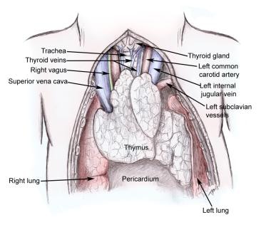
The thymus gland is a primary lymphoid organ located in the upper chest region, anterior to the heart. It plays a critical role in the adaptive immune system, particularly during fetal development and early childhood. The thymus gland begins to atrophy after puberty, leading to a decrease in the production of thymus hormones. This natural decline in thymic function is believed to contribute to the decreased immune response observed in older individuals.
Supplementation with thymus hormones has been explored as a potential therapeutic approach for enhancing immune function in various clinical settings, including immunodeficiency disorders, cancer, and aging. However, more research is needed to fully understand their mechanisms of action and potential benefits and risks.
Thymus extracts are pharmaceutical preparations made from the thymus gland, which is a part of the immune system located in the chest behind the breastbone. The thymus gland plays an essential role in the development and maturation of immune cells called T-lymphocytes or T-cells.
Thymus extracts contain various immunomodulatory substances, including thymosins, thymopoietin, and other peptides, that are believed to help regulate and boost the immune system's function. These extracts have been used in medical research and some clinical applications, particularly in patients with weakened immune systems due to conditions such as primary immunodeficiency disorders, cancer, or HIV/AIDS.
It is important to note that the use of thymus extracts remains controversial, and their efficacy and safety have not been fully established. Therefore, they should only be used under the supervision of a healthcare professional.
Actin is a type of protein that forms part of the contractile apparatus in muscle cells, and is also found in various other cell types. It is a globular protein that polymerizes to form long filaments, which are important for many cellular processes such as cell division, cell motility, and the maintenance of cell shape. In muscle cells, actin filaments interact with another type of protein called myosin to enable muscle contraction. Actins can be further divided into different subtypes, including alpha-actin, beta-actin, and gamma-actin, which have distinct functions and expression patterns in the body.
 Thymosin
 Recombinant Human Thymosin Beta-4 Protects against Mouse Coronavirus Infection
 Modulation of natural killer activity by thymosin alpha 1 and interferon | SpringerLink
 Effects of thymosin β4 on wound healing of rat palatal mucosa
 Healthcare Sales & Marketing Network News: thymosin beta 4
 Thymosin Plus PEG-Interferon in Non-Cirrhotic Hepatitis C Patients Who Did Not Respond to Interferon or Interferon Plus...
 Tumor Progression Is Mediated by Thymosin-β4 through a TGFβ/MRTF Signaling Axis | Molecular Cancer Research | American...
 Thymosin Beta-4 Peptide | By NEXT HEALTH
 Thymosin β4 mediates vascular protection via regulation of Low Density Lipoprotein Related Protein 1 (LRP1): Supplemental...
 Thymosin Beta-4 - GenOracle
 THYMOSIN BETA-4 - jacked.sk
 Europe Pharmaceutical :: Thymosin Beta 4
 Thymosin Alpha-1 - Peptide Guide
 Search - Tag - Thymosin Alpha 1
 THYMOSIN Α1 HGH & Peptides | Deus Medical
 Thymosin alpha
 Thymosin Alpha-1 10mg Peptide | Origin Compounds
 Thymosin Alpha1 10mg (Immune System Peptide) - Innovagen
 Thymosin-β4 inhibits proliferation and induces apoptosis of hepatic stellate cells through PI3K/AKT pathway | Oncotarget
 thymosin alpha 1 dosage reddit Archives - USA PEPTIDES